WO2013158281A1 - Compositions and methods for detection of candida species - Google Patents
Compositions and methods for detection of candida species Download PDFInfo
- Publication number
- WO2013158281A1 WO2013158281A1 PCT/US2013/031774 US2013031774W WO2013158281A1 WO 2013158281 A1 WO2013158281 A1 WO 2013158281A1 US 2013031774 W US2013031774 W US 2013031774W WO 2013158281 A1 WO2013158281 A1 WO 2013158281A1
- Authority
- WO
- WIPO (PCT)
- Prior art keywords
- candida
- probe
- seq
- nucleic acid
- sequence
- Prior art date
Links
Classifications
-
- C—CHEMISTRY; METALLURGY
- C12—BIOCHEMISTRY; BEER; SPIRITS; WINE; VINEGAR; MICROBIOLOGY; ENZYMOLOGY; MUTATION OR GENETIC ENGINEERING
- C12Q—MEASURING OR TESTING PROCESSES INVOLVING ENZYMES, NUCLEIC ACIDS OR MICROORGANISMS; COMPOSITIONS OR TEST PAPERS THEREFOR; PROCESSES OF PREPARING SUCH COMPOSITIONS; CONDITION-RESPONSIVE CONTROL IN MICROBIOLOGICAL OR ENZYMOLOGICAL PROCESSES
- C12Q1/00—Measuring or testing processes involving enzymes, nucleic acids or microorganisms; Compositions therefor; Processes of preparing such compositions
- C12Q1/68—Measuring or testing processes involving enzymes, nucleic acids or microorganisms; Compositions therefor; Processes of preparing such compositions involving nucleic acids
- C12Q1/6876—Nucleic acid products used in the analysis of nucleic acids, e.g. primers or probes
- C12Q1/6888—Nucleic acid products used in the analysis of nucleic acids, e.g. primers or probes for detection or identification of organisms
- C12Q1/6895—Nucleic acid products used in the analysis of nucleic acids, e.g. primers or probes for detection or identification of organisms for plants, fungi or algae
-
- C—CHEMISTRY; METALLURGY
- C12—BIOCHEMISTRY; BEER; SPIRITS; WINE; VINEGAR; MICROBIOLOGY; ENZYMOLOGY; MUTATION OR GENETIC ENGINEERING
- C12Q—MEASURING OR TESTING PROCESSES INVOLVING ENZYMES, NUCLEIC ACIDS OR MICROORGANISMS; COMPOSITIONS OR TEST PAPERS THEREFOR; PROCESSES OF PREPARING SUCH COMPOSITIONS; CONDITION-RESPONSIVE CONTROL IN MICROBIOLOGICAL OR ENZYMOLOGICAL PROCESSES
- C12Q1/00—Measuring or testing processes involving enzymes, nucleic acids or microorganisms; Compositions therefor; Processes of preparing such compositions
- C12Q1/68—Measuring or testing processes involving enzymes, nucleic acids or microorganisms; Compositions therefor; Processes of preparing such compositions involving nucleic acids
- C12Q1/6813—Hybridisation assays
- C12Q1/6816—Hybridisation assays characterised by the detection means
- C12Q1/6818—Hybridisation assays characterised by the detection means involving interaction of two or more labels, e.g. resonant energy transfer
-
- C—CHEMISTRY; METALLURGY
- C12—BIOCHEMISTRY; BEER; SPIRITS; WINE; VINEGAR; MICROBIOLOGY; ENZYMOLOGY; MUTATION OR GENETIC ENGINEERING
- C12Q—MEASURING OR TESTING PROCESSES INVOLVING ENZYMES, NUCLEIC ACIDS OR MICROORGANISMS; COMPOSITIONS OR TEST PAPERS THEREFOR; PROCESSES OF PREPARING SUCH COMPOSITIONS; CONDITION-RESPONSIVE CONTROL IN MICROBIOLOGICAL OR ENZYMOLOGICAL PROCESSES
- C12Q2600/00—Oligonucleotides characterized by their use
- C12Q2600/158—Expression markers
-
- C—CHEMISTRY; METALLURGY
- C12—BIOCHEMISTRY; BEER; SPIRITS; WINE; VINEGAR; MICROBIOLOGY; ENZYMOLOGY; MUTATION OR GENETIC ENGINEERING
- C12Q—MEASURING OR TESTING PROCESSES INVOLVING ENZYMES, NUCLEIC ACIDS OR MICROORGANISMS; COMPOSITIONS OR TEST PAPERS THEREFOR; PROCESSES OF PREPARING SUCH COMPOSITIONS; CONDITION-RESPONSIVE CONTROL IN MICROBIOLOGICAL OR ENZYMOLOGICAL PROCESSES
- C12Q2600/00—Oligonucleotides characterized by their use
- C12Q2600/16—Primer sets for multiplex assays
Definitions
- Candida species are commensal organisms of humans, usually found on the mucous membranes of the gut, oral cavity, and vaginal introitus, and in warm moist skin folds.
- Candida is the most commonly identified causative agent of oral or vaginal thrush.
- Candida can also cause life-threatening infections in hospital patients.
- Candida can cause invasive diseases in hosts with altered immunity, such as in patients with HIV infection, patients that have received organ or bone marrow transplants, and in patients experiencing neutropenia after cancer immunotherapy. Patients on intensive regimens of cancer therapy, patients on prolonged broad spectrum antibiotic therapy, patients using invasive devices, and patients on prolonged hospital stays are also at high risk for such infections.
- compositions, methods, and kits for detecting the presence of Candida species in a biological sample in a timely, accurate, and efficient manner.
- the invention features nucleic acid probes and primers that include the sequence of any one of SEQ ID NOs: 1 to 41 (e.g., nucleic acid probes having the sequence of any one of SEQ ID NOs: 1 tolO, such as any one of SEQ ID NOs: 1 to 5), and nucleic acid probes and primers having at least 90%, 95%, 97%, 99%, or more sequence identity to these probes and primers.
- nucleic acid probes and primers that include the sequence of any one of SEQ ID NOs: 1 to 41 (e.g., nucleic acid probes having the sequence of any one of SEQ ID NOs: 1 tolO, such as any one of SEQ ID NOs: 1 to 5), and nucleic acid probes and primers having at least 90%, 95%, 97%, 99%, or more sequence identity to these probes and primers.
- the nucleic acid probes and primers can be used to detect a Candida species (e.g., Candida albicans, Candida tropicalis, Candida krusei, Candida glabrata, Candida parapsilosis, Candida dubliniensis, Candida lusitaniae, and Candida guillermondi) in a biological sample.
- a Candida species e.g., Candida albicans, Candida tropicalis, Candida krusei, Candida glabrata, Candida parapsilosis, Candida dubliniensis, Candida lusitaniae, and Candida guillermondi
- the nucleic acid probes of the invention include a detection moiety (e.g., a fluorescent label) that is conjugated to the probe.
- the nucleic acid probes are conjugated to a magnetic nanoparticle.
- the probes of the invention may be molecular beacon probes that may include a fluorescent label (e.g., FAM, TAMRA, HEX, TMR, Cy3, Cy5, and other spectrally distinguishable dyes) and a quencher (e.g., DDQ-I, Dabcyl, Eclipse, Iowa Black FQ, BHQ-1, QSY-7, BHQ-2, DDQ-II, Iowa Black RQ, QSY-21, BHQ-3).
- the probes of the invention e.g., SEQ ID NOs: 1 to 5
- the probes of the invention may be a "shared-stem" molecular beacon probe.
- the nucleic acid probes of the invention e.g., SEQ ID NOs: 6 to 10 may be conjugated to magnetic nanoparticles.
- the invention features a method for detecting at least one Candida species (e.g., Candida albicans, Candida tropicalis, Candida krusei, Candida glabrata, Candida parapsilosis, Candida dubliniensis, Candida lusitaniae, and Candida guillermondi) in a biological sample (e.g., soil, water, food, and tissue or fluid sample from an organism, such as a human).
- Candida species e.g., Candida albicans, Candida tropicalis, Candida krusei, Candida glabrata, Candida parapsilosis, Candida dubliniensis, Candida lusitaniae, and Candida guillermondi
- a biological sample e.g., soil, water, food, and tissue or fluid sample from an organism, such as a human.
- the method includes contacting the biological sample with at least one nucleic acid probe having a sequence selected from any one of SEQ ID NOs: 1 to 41 (e.g., SEQ ID NOs: 1- 10, such as SEQ ID NOs: 1-5) under conditions which allow the at least one probe to hybridize to a nucleic acid molecule of the Candida species; and detecting hybridization between the at least one probe and the nucleic acid molecule, thereby detecting the at least one Candida species.
- SEQ ID NOs: 1 to 41 e.g., SEQ ID NOs: 1- 10, such as SEQ ID NOs: 1-5
- the probe is a Candida albicans probe and has the sequence of SEQ ID NO: 1 or SEQ ID NO: 6; the probe is a Candida tropicalis probe and has the sequence of SEQ ID NO: 2 or SEQ ID NO: 7; the probe is a Candida glabrata probe and has the sequence of SEQ ID NO: 3 or SEQ ID NO: 8; the probe is a Candida parapsilosis probe and has the sequence of SEQ ID NO: 4 or SEQ ID NO: 9; the probe is a Candida krusei probe and has the sequence of SEQ ID NO: 5 or SEQ ID NO: 10.
- the method features nucleic acid probes that are fluorescently labeled.
- nucleic acid probes may be molecular beacon probes (e.g., probes having the sequence of any one of SEQ ID NOs: 1 to 5) that further include a quencher.
- hybridization of the nucleic acid probe to target Candida species nucleic acid molecules produces fluorescence which may be detected in, e.g., a fluorescence based assay (e.g, a real- time or end-point PCR assay) using, e.g., an instrument capable of detecting fluorescence (e.g., a realtime or end-point thermal cycler, or a plate reader).
- a fluorescence based assay e.g, a real- time or end-point PCR assay
- an instrument capable of detecting fluorescence e.g., a realtime or end-point thermal cycler, or a plate reader.
- the method features one or more probes (e.g., 2, 3, 4, or 5 or more probes) that may be labeled with the same or different fluorescent labels (e.g., molecular beacon probes labeled with spectrally distinguishable fluors), and the one or more probes may each be contacted with the sample for use in the same assay, e.g., in a multiplex assay to detect up to five different Candida species (e.g., Candida albicans, Candida tropicalis, Candida glabrata, Candida parapsilosis, and Candida krusei) in a biological sample.
- the same or different fluorescent labels e.g., molecular beacon probes labeled with spectrally distinguishable fluors
- the one or more probes may each be contacted with the sample for use in the same assay, e.g., in a multiplex assay to detect up to five different Candida species (e.g., Candida albicans, Candida tropicalis, Candida glabrata, Candida parap
- the invention also features a method of using nucleic acid probes conjugated to magnetic nanoparticles (e.g., SEQ ID NOs: 6 to 10 and 13 to 31) to detect Candida species in, e.g., an NMR based assay (e.g., an aggregation or disaggregation assay) using an NMR instrument.
- nucleic acid probes conjugated to magnetic nanoparticles e.g., SEQ ID NOs: 6 to 10 and 13 to 31
- an NMR based assay e.g., an aggregation or disaggregation assay
- the method includes contacting the biological sample with at least one nucleic acid probe having a sequence selected from any one of SEQ ID NOs: 6 to 31 (e.g., SEQ ID NOs: 6-10) under conditions which allow the at least one probe to hybridize to a nucleic acid molecule of the Candida species; and detecting hybridization between the at least one probe and the nucleic acid molecule, thereby detecting the at least one Candida species.
- at least one nucleic acid probe having a sequence selected from any one of SEQ ID NOs: 6 to 31 (e.g., SEQ ID NOs: 6-10) under conditions which allow the at least one probe to hybridize to a nucleic acid molecule of the Candida species; and detecting hybridization between the at least one probe and the nucleic acid molecule, thereby detecting the at least one Candida species.
- probes conjugated to magnetic nanoparticles can be used in pairwise combinations in an NMR assay to detect Candida species
- a probe having the sequence of SEQ ID NO: 6 can be used with a probe having the sequence of any one of SEQ ID NOs: 13 and 14 to detect Candida albicans
- a probe having the sequence of SEQ ID NO: 7 can be used with a probe having the sequence of any one of SEQ ID NOs: 20 to 25 to detect Candida tropicalis
- a probe having the sequence of SEQ ID NO: 8 can be used with a probe having the sequence of any one of SEQ ID NOs: 18 and 19 to detect Candida glabrata
- a probe having the sequence of SEQ ID NO: 9 can be used with a probe having the sequence of any one of SEQ ID NOs: 20 to 25 to detect Candida parapsilosis
- a probe having the sequence of SEQ ID NO: 10 can be used with a probe having the sequence of any one of SEQ ID NOs: 15
- the invention also features primers for DNA sequencing based detection of Candida species in a biological sample.
- the primer is a Candida krusei primer having the sequence of SEQ ID NO: 32 or SEQ ID NO: 33; the primer is a Candida albicans primer having the sequence of SEQ ID NO: 34 or SEQ ID NO: 35; the primer is a Candida glabrata primer having the sequence of SEQ ID NO: 36 or SEQ ID NO: 37; the primer is a Candida parapsilosis primer having the sequence of SEQ ID NO: 38 or SEQ ID NO: 39; or the primer is a Candida tropicalis primer having the sequence of SEQ ID NO: 40 or SEQ ID NO: 41.
- Sequencing may also be preceded by prior amplification using SEQ ID NO: 11 and SEQ ID NO: 12.
- the sequencing could be done using SEQ ID NO: 11 and SEQ ID NO: 12 and species identification conducted based on analysis of the DNA sequence (i.e., using BLAST).
- the invention also features a method of using Candida specific primers (e.g., one or more of the DNA primers of SEQ ID NOs: 32 to 41) to detect a Candida species in a biological sample.
- the method may include use of the primers one or more of the primers for DNA sequencing.
- the primer is a Candida krusei primer having the sequence of SEQ ID NO: 32 or SEQ ID NO: 33; the primer is a Candida albicans primer having the sequence of SEQ ID NO: 34 or SEQ ID NO: 35; the primer is a Candida glabrata primer having the sequence of SEQ ID NO: 36 or SEQ ID NO: 37; the primer is a Candida parapsilosis primer having the sequence of SEQ ID NO: 38 or SEQ ID NO: 39; or the primer is a Candida tropicalis primer having the sequence of SEQ ID NO: 40 or SEQ ID NO: 41.
- the invention also features fluorescence based methods of using the nucleic acid probes and primers described above (e.g., the nucleic acid molecules of any one or more of SEQ ID NOs: 1 to 41) to detect Candida species in a biological sample.
- These methods include amplifying and sequencing target nucleic acid molecules from Candida species in a biological sample.
- the methods include, e.g., a TaqMan probe based assay (e.g., real-time or end-point PCR based TaqMan assay using probes having sequence of SEQ ID NOs: 1 to 5); strand-displacement probe based assays
- PCR assays using DNA binding dyes e.g., real-time PCR based assay using SYBR ® green dye and probes having sequence of SEQ ID NOs: 1 to 41
- in situ hybridization assays using fluorescently labeled species-specific probes e.g., using probes having sequence of SEQ ID NOs 1 to 41.
- the nucleic acid probes having the sequence of any one of SEQ ID NOs: 6-10, and 13-31 may have a detection label (e.g., a fluorescent label), such that the fluorescence detection is via a secondary step (e.g., a digoxigenin label for antibody-based detection using fluorescently labeled anti-digoxigenin antibodies, or a biotin label for detection using fluorescent strep tavdin).
- a detection label e.g., a fluorescent label
- the detection readout may be non-fluorescent.
- nucleic acid probes having the sequence of any one of SEQ ID NOs: 6-10 and 13-31 may have non-fluorescent detection labels, such as a radioactive isotope for autoradiographic detection, digoxigenin for antibody-based detection using a horse-radish peroxidase (HRP) conjugated anti-digoxigenin antibody, and biotin for streptavidin based detection using HRP conjugated straptividin.
- non-fluorescent detection labels such as a radioactive isotope for autoradiographic detection, digoxigenin for antibody-based detection using a horse-radish peroxidase (HRP) conjugated anti-digoxigenin antibody, and biotin for streptavidin based detection using HRP conjugated straptividin.
- the fluorescence based methods for detecting the presence of at least one Candida species in a biological sample includes the use of molecular beacon probes (e.g., those probes having the sequences of any one of SEQ ID NOs: 1 to 41, and particularly SEQ ID NOs: 1 to 5) having a fluorescent label, which may be the same or different.
- molecular beacon probes e.g., those probes having the sequences of any one of SEQ ID NOs: 1 to 41, and particularly SEQ ID NOs: 1 to 5
- Tm melting temperature
- the molecular beacon probes used in the methods have melting temperatures that vary by at least 1°C, such that a separate melt curve can be obtained for each molecular beacon probe.
- a decrease in fluorescence at a Tm of ⁇ 62-67°C (e.g., ⁇ 64°C) in the presence of the probe having the sequence of SEQ ID NO: 1 indicates the presence of Candida albicans in the sample
- a decrease in fluorescence at a Tm of ⁇ 55-61°C (e.g., ⁇ 58°C) in the presence of the probe having the sequence of SEQ ID NO: 2 indicates the presence of Candida tropicalis in the sample
- a decrease in fluorescence at a Tm of -65-71°C (e.g., ⁇ 67°C) in the presence of the probe having the sequence of SEQ ID NO: 3 indicates the presence of Candida glabrata in the sample
- the melting temperatures indicated above for SEQ ID NOs: 1-5 are not meant to be limiting and may differ depending upon the conditions used in the method.
- one or more of the nucleic acid probes of the invention e.g., any one of the nucleic acid probes having the sequence of SEQ ID NOs: 1 to 10 and 13 to 41
- Each of the methods of the invention may further include steps for processing samples (e.g., biological samples), which may include one or more of the following: mixing the sample with a lysis agent solution (e.g., a detergent or a hypotonic solution); centrifuging the sample to form a supernatant and a pellet; discarding some or all of the supernatant; and/or resuspending the pellet to form an extract containing the fungal cells.
- a lysis agent solution e.g., a detergent or a hypotonic solution
- the processing steps may further include one or more of the following: optionally washing the pellet (e.g., with TE buffer) prior to resuspending the pellet; lysing fungal cells of the extract by chemical methods (e.g., using any combination of enzymes, or detergents, or surfactants), mechanical methods (e.g., using beads such as glass beads, bead beating, use of a finned tube in combination with beads, using beads in an agitation mill, use of beads with a chelating agent, use of glass shards, use of solid particles, use of beads or solid particles with mechanical or magnetic vortex centrifugationuse of ultrasound, and/or use of sonication), or methods involving temperature changes (e.g., use of heat (e.g., a temperature in the range of about 85°C to about 125°C, such as a temperature of about 95°C), use of freeze-thaw, and/or use of freeze-boil) to form a lysate;
- chemical methods
- Other processing steps may optionally include amplifying nucleic acids present in a processed sample to form an amplified lysate solution; adding all or a portion of the amplied lysate solution to a detection tube that includes one or more Candida specific nucleic acid probes or primers and allowing contact between the target nucleic acid molecules in the lysate and the one or more Candida specific nucleic acid probes or primers under conditions that allow hybridization of the nucleic acid probe(s) with the target nucleic acid molecules; and detecting hybridization of the nucleic acid probe(s) or primers to the Candida target nucleic acid molecules whereby the detection of binding of the Candida species-specific probe(s) with Candida target nucleic acid molecules present in the biological sample indicates the presence of the Candida species in the biological sample.
- the amplified lysate solution described above may be further processed to isolate the Candida nucleic acid molecules, e.g., by using an ion exchange column (such as a Qiagen column), glass or silica-base reverse phase adsorption/desorption, SPRI technology, phenol-chloroform extraction and ethanol precipitation, and/or the CTAB method.
- an ion exchange column such as a Qiagen column
- glass or silica-base reverse phase adsorption/desorption SPRI technology
- phenol-chloroform extraction and ethanol precipitation and/or the CTAB method.
- each of the methods of the invention may further include an amplification step for increasing the amount of the target Candida nucleic acid molecules to be detected in the biological sample.
- the amplification may include enzymatic amplification by, e.g., a PCR reaction.
- Primers having the sequence of SEQ ID NOs: 11 and 12 may be used as pan-Candida universal forward and reverse primers, respectively, to produce a Candida amplicon.
- primers having the sequences of SEQ ID NOs: 6 to 41 can be used in combination with the pan-Candida universal primers to produce Candida species amplicons.
- the amplification may be symmetric to produce a double stranded amplicon or the amplification may be asymmetric to produce a single stranded amplicon.
- each of the methods of the invention for detecting at least one Candida species in a biological sample can be completed within at least about 5 hours or less (e.g., within 4.0 hours, 3.5 hours, 3.0 hours, 2.5 hours, 2 hours, 1.5 hours, or 1 hour or less).
- each of the methods of the invention can be used to detect at least one Candida species in a sample when present at a concentration of at least about 4 Candida cells/mL (e.g., 5, 6, 7, 8, 9, 10, 15, 20, 25, 30, 35, 40, 45, or 50 Candida cells/mL).
- the biological sample is, e.g., a soil, water, or food sample or a tissue or fluid sample from an organism, such as a human (e.g., whole blood, sweat, tears, urine, saliva, semen, serum, plasma, cerebrospinal fluid (CSF), feces, vaginal fluid or tissue, sputum, nasopharyngeal aspirate or swab, lacrimal fluid, mucous, or epithelial swab (buccal swab), tissues, organs, bone, teeth, and tumors).
- a human e.g., whole blood, sweat, tears, urine, saliva, semen, serum, plasma, cerebrospinal fluid (CSF), feces, vaginal fluid or tissue, sputum, nasopharyngeal aspirate or swab, lacrimal fluid, mucous, or epithelial swab (buccal swab), tissues
- the invention features a kit which includes at least one nucleic acid probe or primer having a sequence selected from any of SEQ ID NOs: 1 to 41 and further includes one or more reagents for attachment of fluorescent label or magnetic nanoparticle to the probe, and/or one or more reagents for sample lysis, fungal lysis, isolation of Candida nucleic acid, detection of Candida species, and nucleic acid amplification.
- Figures 1A and IB are schematics showing binding of molecular beacon probes to a target nucleic acid molecule.
- Figure 1 A shows the binding of a conventional molecular beacon probe.
- Figure IB shows the binding of a shared- stem molecular beacon probe of the present invention.
- a shared- stem molecular beacon probe is designed so that 1) a stem-loop hairpin is formed by the 5' and 3' ends of the probe, 2) a fluorophore and quencher are attached to the 5' and 3' ends and sufficient quenching will occur when the hairpin structure is formed, 3) the probes have a "loop" sequence that is specific for the target Candida sequence to be identified in the assay, and 4) sufficient fluorophore signal may be detected upon hybridization of the molecular beacon probe to the specific target sequence of Candida.
- the molecular beacon probes are designed to have a melting temperature (Tm) that is about 5 to 10 degrees (e.g., ⁇ 7 degrees) higher than the annealing temperature of amplification primers that may be present with the molecular beacon probes in an assay sample, and the molecular beacon probes are thermodynamically characterized to form no self dimers or heterodimers with other probes or primers (e.g., pan-Candida universal PCR primers) that may be present in an assay sample.
- Tm melting temperature
- Figures 2A and 2B are graphs showing detection of Candida krusei in real-time PCR reactions.
- Figure 2A shows the detection of Candida krusei in a sample using purified genomic DNA.
- Figure 2B shows the detection of Candida krusei in a sample prepared from a cell lysate.
- Figures 3A-3F are graphs showing titration curves for detection of Candida species in real-time PCR reactions.
- Figures 3A-3C are titration curves showing detection of Candida parapsilosis (Figure 3A), Candida tropicalis ( Figure 3B), and Candida krusei (Figure 3C) using various amounts of purified genomic DNA.
- Figures 3D-3F are titration curves showing detection of Candida parapsilosis (Figure 3D), Candida tropicalis (Figure 3E), and Candida krusei (Figure 3F) using a cell lysate.
- Figures 4A-4D are graphs showing titration curves for detection of Candida albicans and Candida glabrata in real-time PCR reactions.
- Figures 4A and 4B are titration curves showing detection of Candida albicans (Figure 4A) and Candida glabrata ( Figure 4B) using various amounts of purified genomic DNA.
- Figures 4C and 4D are titration curves showing detection of Candida albicans ( Figure 4C) and Candida glabrata ( Figure 4D) using a cell lysate.
- Figures 5A-5D are graphs showing the use of multiple molecular beacon probes in a multiplex assay to detect one or more Candida species in a sample.
- the genomic DNA of Candida parapsilosis and Candida tropicalis are present in a single reaction and amplified in the presence of a single beacon ( Figures 5A and 5C, SEQ ID NO: 4 and SEQ ID NO: 2, respectively labeled with FAM and HEX) or with both beacons within the same reaction and emissions are observed in parallel using two channels (Figure 5B and Figure 5D).
- Figure 6 is a schematic showing alignment of Candida amplicon sequences that are generated by amplification using the pan-Candida primers. The alignment shows species- specific differences within this amplicon. Shown are sequences for C. krusei (pan fungal amplicon; SEQ ID NO. 42), C. albicans (pan fungal amplicon; SEQ ID NO: 43), C glabrata (pan fungal amplicon; SEQ ID NO: 44), C.
- Figure 7 is a sequence chromatogram (SEQ ID NO:52) generated from amplification within whole blood lysate using the pan-Candida forward and reverse primers (SEQ ID NOs: 11 and 12), followed by a phenol/chloroform extraction, a chloroform extraction, and then subsequentSanger Dideoxy terminator sequence analysis using Big Dye Vs.31. (Applied Biosystems, Foster City, CA).
- Figures 8A-8E are graphs of titration curves showing detection of Candida albicans (Figure 8A), Candida glabrata ( Figure 8B), Candida krusei (Figure 8C), Candida parapsilosis (Figure 8D), and
- Candida tropicalis ( Figure 8E) in real-time PCR reactions using genomic DNA obtained from Candida cell lysates prepared with cells in an amount ranging from -520 cells to ⁇ 3 cells/mL (see the data in Tables 20, 22, 24, 26, and 28, which was used to produce the graphs of Figures 8A-8E, respectively).
- Figures 9A and 9B are graphs showing nucleic acid denaturation melt curves in a multiplex reaction that includes Candida species-specific nucleic acid molecules from, and Candida species-specific molecular beacon probes for, C. albicans, C. glabrata, C. parapsilosis, and C. tropicalis ( Figure 9A) and for C. albicans and C. krusei ( Figure 9B).
- the Candida-speciiic molecular beacon probes used in the multiplex reactions are all labeled with a HEX fluorophore.
- the presence of nucleic acid molecules for each Candida species is determined by detecting a decrease in fluorescence as the sample is heated through the melting temperature (Tm) of each Candida species-specific molecular beacon probe. A decrease in fluorescence is observed only when hybridized beacon probes melt off their target nucleic acid molecules and the step-loop structure of the probes reforms. No melt curve is observed when the probe- specific target nucleic acid molecules are not present in
- the invention features compositions, methods, and kits for detecting at least one Candida species in a variety of media and biological samples, including, for example, biofluids, tissue samples, culture samples (e.g., a blood culture), food products, water samples, and soil samples.
- the biological sample may include samples which have been processed to remove patient tissue and/or cellular debris or samples that are substantially unprocessed, such as a whole blood sample.
- the biological sample may further include cell suspensions or lysates that are produced by the methods of the invention.
- the biological sample may further include any sample which contains at least one nucleic acid molecule of Candida species; or any sample in which the nucleic acid molecules of Candida species have been substantially isolated, enriched or purified; or any sample in which the nucleic acid molecules of Candida species have been amplified and enriched as an amplicon.
- the methods of the invention for detecting a Candida species can be performed with little to no sample preparation.
- the methods may be performed as a singleplexed assay to detect a single Candida species or as a multiplexed assay that allows for detection of multiple Candida species in a single sample.
- the methods of the invention allow for rapid and accurate detection of Candida in a biological sample (e.g., a tissue or fluid sample from a patient), which may be used to facilitate point-of- care clinical decision making.
- the methods described herein can be used to provide clinically and epidemiologically meaningful targeting of anti-fungal drug therapy to patients in need thereof and to provide optimal therapeutic outcome.
- compositions of the invention for use in the detection of a Candida species include probes (e.g., molecular beacon probes and NMR assay probes) that can be used to detect nucleic acid molecules of the Candida species (e.g., in a real-time probe-based nucleic acid detection assay).
- a Candida species e.g., one or more of Candida albicans, Candida tropicalis, Candida krusei, Candida glabrata, Candida parapsilosis, Candida dubliniensis, Candida lusitaniae, and Candida guillermondi
- probes e.g., molecular beacon probes and NMR assay probes
- the methods of the invention involve contacting a sample that includes a nucleic acid molecule of at least one Candida species with at least one probe that is capable of hybridizing to the nucleic acid molecule and detecting hybridization between the probe and the nucleic acid molecule.
- the sample may be contacted with two or more probes (e.g., probes to any two, three, four, or five or more of the Candida species listed above) in order to detect hybridization between the probes and the nucleic acid molecules.
- the methods of the invention may be fluorescence based detection methods using fluorescently labeled species-specific probes (e.g., molecular beacon probes), such that hybridization between the probe and the nucleic acid molecule of the Candida species may result in a fluorescent signal that can then detected by an appropriate detection device (e.g., a real-time thermal cycler or other fluorescence detection devices known in the art).
- the probes may be fluorescently labeled molecular beacon probes designed to hybridize to Candida species-specific target nucleic acid molecules for detectection of specific Candida species in a biological sample.
- Each Candida species-specific probe may be labeled with a fluorescent probe that distinguishes that probe from the other Candida species-specific probe(s) (e.g., based on differences in the excitation or emission spectra of the different probes).
- the probes may all include the same fluorescent label, in which case distinguishing the hybridization of each Candida species-specific probe to its targets is determined by other techniques, such as differences between the melting temperature (Tm) of each Candida species-specific probe, as is discussed below.
- the methods of the invention may further include amplifying nucleic acid molecules of the Candida species present in the biological sample prior to detection of the Candida nucleic acid molecules.
- Amplification can be used to increase the amount of the Candida nucleic acid molecules present in the biological sample in case the original amount of Candida target nucleic acid molecules in the biological sample is below the detection limit of the probe(s).
- amplification may be done using one or more amplification primers.
- Any nucleic acid amplification method known in the art e.g., polymerase chain reaction (PCR)
- PCR polymerase chain reaction
- the amplification method includes the use of primers that amplify nucleic acid molecules of at least one Candida species present in the biological sample.
- Other methods of the invention include the use of fluorescently labeled probes and/or primers of the invention for detection of at least one Candida species in a biological sample in fluorescence based detection assays, for example, in situ hybridization assays.
- Another detection method employs use of a double strand specific intercalating dye, such as SYBR ® (Molecular Probes, Eugene, OR).
- SYBR ® Molecular Probes, Eugene, OR
- the fluorescence based assays may be performed in conjunction with real-time PCR detection.
- the invention also features one or more probes and primers of the invention for use as TaqMan probes in a real-time PCR assay or an end-point PCR assay.
- Candida specific primers of the invention may be used in sequencing based assays or in array or microarray based platforms in order to detect the presence of at least one Candida species in a biological sample.
- the methods of the invention also include NMR based assays using Candida species-specific probes conjugated to magnetic nanoparticles.
- a sample may be contacted with at least one Candida species-specific probe that is conjugated to a magnetic nanoparticle, such that hybridization between the probe and the nucleic acid molecule of the Candida species may produce an NMR signal that may be detected by NMR spectroscopy.
- Other aspects of the compositions, methods, and kits of the invention are described below.
- compositions and methods for detecting Candida species in biological samples Compositions and methods for detecting Candida species in biological samples.
- the invention features compositions, methods, and kits for detecting at least one Candida species in a biological sample.
- the invention features Candida species-specific nucleic acid probes having the sequence of any one of SEQ ID NOs: 1 to 10 and 13 to 31, and probes having at least 90%, preferably at least 95%, more preferably at least 97%, and most preferably at least 99% or more identity to these sequences, that may be employed in a number of assays to detect one or more (for e.g., 2, 3, 4, 5, 6, 7, 8, or more) Candida species in a biological sample.
- the probes may be used to detect one or more of Candida albicans, Candida tropicalis, Candida krusei, Candida glabrata, Candida parapsilosis, Candida dubliniensis, Candida lusitaniae, and Candida guillermondi, in any combination, in a biological sample.
- the methods of the invention include the use of one or more Candida specific probes and/or primers for detecting the presence of a Candida target nucleic acid molecule present in a biological sample.
- the methods include one or more of the following steps: (a) providing a biological sample; (b) mixing the sample with a lysis agent solution; (c) centrifuging the sample to form a supernatant and a pellet; (d) discarding some or all of the supernatant; and (e) resuspending the pellet to form an extract containing the fungal cells, optionally washing the pellet (e.g., with TE buffer) prior to resuspending the pellet and optionally repeating step (c); (f) lysing cells of the extract by chemical or mechanical methods to form a lysate; (g) placing the lysate of step (f) in a detection tube; (h) optionally amplifying nucleic acids therein to form an amplified lysate solution; (i) adding to the detection tube one or
- the detection of binding of the Candida species-specific probe(s) with Candida target nucleic acid molecules present in the biological sample indicates the presence of the Candida species in the biological sample.
- the methods of the invention provide rapid and accurate readouts of the presence of at least one species of Candida in the sample.
- Two or more probes of the invention may be used in the methods of the invention to perform a multiplexing assay that provides for the detection of two or more Candida species (e.g., 2, 3, 4, or 5 or more of the Candida species described herein) in the biological sample.
- steps (a) through (h) are completed within at least about 5 hours or less (e.g., within 4.0 hours, 3.5 hours, 3.0 hours, 2.5 hours, 2 hours, 1.5 hours, or 1 hour or less).
- the methods allow for (i) at least 95% correct detection at less than or equal to 5 Candida cells/mL in samples spiked into 50 individual healthy patient blood samples; (ii) at least 95% correct detection at less than or equal to 5 Candida cells/mL in samples spiked into 50 individual unhealthy patient blood samples; (iii) greater than or equal to 80% correct detection in clinically positive patient samples (e.g., Candida positive by another technique, such as by cell culture) starting with 2 mL of biological sample; and/or (iv) at least 90% correct detection in biological samples containing at least 5 Candida cells/mL.
- clinically positive patient samples e.g., Candida positive by another technique, such as by cell culture
- the invention provides methods in which Candida species can be detected at pathogen concentration of, e.g., at least 3 Candida cells/mL (e.g., 4, 5, 6, 7, 8, 9, 10, 15, 20, 25, 30, 35, 40, 45, or 50 Candida cells/mL) in the sample with a coefficient of variation of less than 15% (e.g., 10 cells/mL with a coefficient of variation of less than 15%, 10%, 7.5%, or 5%; or 25 cells/mL with a coefficient of variation of less than 15%, 10%, 7.5%, or 5%; or 50 cells/mL with a coefficient of variation of less than 15%, 10%, 7.5%, or 5%; or 100 cells/mL with a coefficient of variation of less than 15%, 10%, 7.5%, or 5%).
- Candida cells/mL e.g., 4, 5, 6, 7, 8, 9, 10, 15, 20, 25, 30, 35, 40, 45, or 50 Candida cells/mL
- a coefficient of variation of less than 15% e.g., 10 cells/mL with a coefficient of variation of less than 15%,
- the methods of the invention may be used to detect the presence of Candida in a biological sample having a volume in the range of, e.g., 0.05 to 10.0 mL (e.g., a biological sample having a volume in the range of 0.05 to 0.25 mL, 0.25 to 0.5 mL, 0.25 to 0.75 mL, 0.4 to 0.8 mL, 0.5 to 0.75 mL, 0.6 to 0.9 mL, 0.65 to 1.25 mL, 1.25 to 2.5 mL, 2.5 to 3.5 mL, 3.0 to 4.0 mL, or 3.0 to 10 mL).
- Species-specific Candida probes of the invention e.g., a biological sample having a volume in the range of 0.05 to 0.25 mL, 0.25 to 0.5 mL, 0.25 to 0.75 mL, 0.4 to 0.8 mL, 0.5 to 0.75 mL, 0.6 to 0.9 mL, 0.65 to 1.25 mL, 1.25 to
- compositions of the invention include Candida species-specific nucleic acid probes that can bind to target sequences within the endogenous nucleic acid sequences of specific Candida species.
- the nucleic acid probes of the invention are designed to hybridize to endogenous nucleic acid molecules of Candida species and to provide accurate detection of at least one Candida species in a biological sample tested.
- the nucleic acid probes can be used individually in a detection assay or two or more nucleic acid probes (e.g., 3, 4, or 5 or more probes) can be used in combination, e.g., in a multiplex fluorescence assay, or in an NMR aggregation assay, to detect one or more Candida species in the biological sample.
- the nucleic acid probes of the invention include: 5'- GOT CAA AGT TTG AAG ATA TAC GTG GTT GAC C-3' (SEQ ID NO: 1) for detecting Candida albicans, 5'-CTA GCA AAA TAA GCG TTT TTG GAT GCT AG-3' (SEQ ID NO: 2) for detecting Candida tropicalis, 5'-CAG CAC GCA CAA AAC ACT CAC TTA TTG CTG-3' (SEQ ID NO: 3) for detecting Candida glabrata, 5'-GTC GAA TTT GGA AGA AGT TTT GGT TTC GAC-3' (SEQ ID NO: 4) for detecting Candida parapsilosis, 5'-CCT GAT TTG AGG TCG AGC TTT TTG TAT CAG G-3' (SEQ ID NO: 5) for detecting Candida krusei, 5' - GGT CAA AGT TTG AAG ATA TAC GTG G-3' (SEQ ID NO: 6) for
- GTCCTACCTGATTTGAGGGCGAA AT-3 ' (SEQ ID NO: 29) for detecting Candida lusitaniae, 5'- GCAAACGCCTAGTCCGACTAAGAGTATCACTCAATACC -3' (SEQ ID NO: 30) and 5'- TGTAAGGCCGGGCCAACAATACCAGAAATATCCCGC-3' (SEQ ID NO: 31) for detecting Candida guillermondi, 5 ' -CCGAGAGCGAGTGTTGCGAGA- 3 ' (SEQ ID NO: 32) and 5'- TCTCGCAACACTCGCTCTCGG-3' (SEQ ID NO: 33) for detecting Candida krusei, 5' - GGTAACGTCC ACC ACGTATATCT-3 ' (SEQ ID NO: 34) and 5' -
- ACGCACAAAACACTCACTTATCCCTCCC-3' (SEQ ID NO: 37) for detecting Candida glabrata
- 5'- GGTAC AAACTCC AAAACTTCTTCC-3 ' (SEQ ID NO: 38) and 5'- GGAAGAAGTTTTGGAGTTTGTACC-3 ' (SEQ ID NO: 39) for detecting Candida parapsilosis
- 5' - GCTAGTGGCC ACC AC AATTTATTTC A-3 ' SEQ ID NO: 40
- TGAAATAAATTGTGGTGGCCACTAGC-3' (SEQ ID NO: 41) for detecting Candida tropicalis.
- the invention features the probes described above, as well as probes having at least 90%, 95%, 97%, 99%, or more sequence identity to these probes.
- one or more of the probes described above may be used in combination with any one of the Candida species-specific probes having the sequences of SEQ ID NOs: 13 to 25, which are described in International Application Nos. PCT/US2011/56933 and PCT/US2011/56936, both of which are incorporated herein by reference.
- probes include: 5' -ACC CAG CGG TTT GAG GGA GAA AC-3' (SEQ ID NO: 13) and 5'-AAA GTT TGA AGA TAT ACG TGG TGG ACG TTA-3' (SEQ ID NO: 14) for detecting Candida albicans; 5'-CGC ACG CGC AAG ATG GAA ACG-3' (SEQ ID NO: 15), 5' -AAG TTC AGC GGG TAT TCC TAC CT-3' (SEQ ID NO: 16) and 5' -AGC TTT TTG TTG TCT CGC AAC ACT CGC-3' (SEQ ID NO: 17) for detecting Candida krusei; 5' -CTA CCA AAC ACA ATG TGT TTG AGA AG-3' (SEQ ID No: 18) and 5' -CCT GAT TTG AGG TCA AAC TTA AAG ACG TCT G-3' (SEQ ID NO: 19) for detecting Candida glabrata; 5'-AG
- reverse complement versions of one or more of the nucleic acid molecules of SEQ ID NOs 1 to 41 may also be used as probes for detecting Candida species in a biological sample or as primers for amplying one or more Candida nucleic acid molecules in a biological sample in an amplification step.
- Candida specific nucleic acid molecules with at least 90% sequence identity (or at least 90%, 95%, 97%, 99%, or more sequence identity) to the sequences of SEQ ID NOs: 1 to 41, or to the reverse complement sequences of SEQ ID NOs: 1 to 41 may be used for detecting, amplifying, or both amplifying and detecting Candida species nucleic acid molecules in a biological sample.
- the Candida species-specific probes of the invention may also include a detection label (e.g., a fluorescent detection label) or may be conjugated to a magnetic nanoparticle for use in the methods of the invention for detecting at least one Candida species in a biological sample. Detection labels and magnetic nanoparticles for use in the present invention, and methods for using probes of the invention that include a detection label or that are conjugated to a magnetic nanoparticle, are described in detail below.
- compositions and methods for fluorescence-based detection of Candida species in biological samples Composition and methods for fluorescence-based detection of Candida species in biological samples
- the invention features Candida species-specific nucleic acid probes that can bind to target sequences within the endogenous nucleic acid sequences of Candida. These probes can be used to detect the presence of specific species of Candida in biological samples.
- the probes may be labeled with a fluorescent label (also referred to as a fluorophore) and applied to a Candida lysate prepared from the biological sample.
- a fluorescent label also referred to as a fluorophore
- the fluorophore-labeled probe emits a fluorescent signal upon hybridization of the probe to its target sequence and a fluorescent signal is not produced by unbound probe, e.g., as described in U.S. Patent Application No. US 2010/0221710A1 (incorporated herein by reference).
- the fluorescent signal produced upon hybridization between probe and target nucleic acid of the Candida species can be detected in a fluorescence detection device (e.g., a real time thermal cycler or other detection device known in the art).
- a fluorescence detection device e.g., a real time thermal cycler or other detection device known in the art.
- Each of the Candida species-specific probes described herein may be differentially labeled with fluorphores with spectrally distinguishable emission spectra for use in a multiplex fluorescence detection assay.
- two or more fluorescently labeled nucleic acid probes e.g., 3, 4, or 5 or more probes
- each having a different fluorophore attached can be used in combination to rapidly and accurately detect multiple different Candida species in a biological sample.
- Candida species-specific probes of the present invention may also be used as "molecular beacons.” Methods to prepare and use molecular beacons are fully described in, e.g., U.S. Pat. Nos.
- molecular beacon probes have a fluorophore attached at the 5' end of the probe and a non-fluorescent quencher attached at the 3' end of the probe.
- Molecular beacons further contain complementary sequences at the 5' and 3' ends allowing these two ends to hybridize. The two ends flank a middle intervening "loop" sequence. The 5' and 3' ends of the molecular beacon can hybridize to each other and form an intramolecular stem structure and the middle intervening sequence region forms a "loop.”
- loop part of the molecular beacon can hybridize to the target nucleic acid.
- the 5' and 3' ends of the probe form the intramolecular stem structure.
- the proximity between the fluorophore and quencher causes the the quencher to absorb any fluorescence emitted by the fluorophore and no fluorescence is produced.
- the stem structure is not formed, the fluorophore is separated from the quencher and, as a result, a fluorescent signal is produced.
- the molecular beacon probes of the present invention are designed to detect specific nucleic acid target sequences in Candida species via species-specific "loop" sequences. Use of different fluorophore labels on the 5' end of each of the species-specific probes can allow use of multiple molecular beacon probes in a multiplex assay to simultaneously and rapidly detect multiple Candida species in a biological sample.
- each of the Candida species-specific probes described herein can be prepared as molecular beacon probes and labeled with the same fluorphore or with fluorophores having overlapping emission spectra for use in a multiplex fluorescence detection assay.
- each of the Candida species-specific molecular beacon probes have a different melting temperature (Tm)
- hybridization of each of the Candida species-specific molecular beacon probes to their target nucleic acid molecules can be detected by assaying fluorescence through a range of temperatures that includes the Tm of each probe used.
- a sample containing nucleic acid molecules from a Candida species may be contacted with a Candida albicans specific molecular beacon probe having a Tm of ⁇ 64°C (e.g., a Tm of 62-67°C) and a HEX fluorophore and a Candida glabrata specific molecule beacon probe having a Tm of ⁇ 67°C (e.g., a Tm of 65-71°C) and a HEX fluorophore.
- Fluorescence in the sample may be due to binding of the C. albicans or the C. glabrata probes or both.
- the temperature of the sample may be raised from, e.g., 40°C to 80°C while detecting changes in fluorescence in the sample. If C. albicans nucleic acid molecules are present in the sample, there will be a change in fluorescence as the sample temperature increases through 64°C. In this case, half of the Candida albicans-speciiic molecular beacon probes will dissociate from their target nucleic acid molecules, which will result in a decrease in fluorescence as the probe melts off and the stem-loop structure reforms and quenches the fluorescence of those probes. If C.
- glabrata nucleic acid molecules are present in the sample, there will be a decrease in fluorescence as the temperature is increased through 67°C. In this case, half of the C. glabrata probes will dissociate from their target nucleic acid molecules, which will result in a decrease in fluorescence as the probe melts off and the stem-loop structure reforms and quenches the fluorescence of those probes. There would be no melt curves in the absence of Candida nucleic acid molecules. Similarly, no change in fluorescence around 64°C would indicate the absence of Candida albicans nucleic acid molecules, while no change in fluorescence around 67°C would indicate the absence of Candida glabrata.
- the amount of Candida specific target nucleic acid molecules present in a biological sample may be sufficient for detection according to the methods of the present invention (e.g., by contact with the species-specific probes of the invention) without the need for amplification of the target nucleic acid molecules.
- it may be preferable to increase the amount of target nucleic acid molecules present in the biological sample prior to detection e.g., if the amount of target nucleic acid molecules present in the biological sample is below the detection limit of the probe(s)).
- an optional amplification step may be performed to amplify the target nucleic acid molecules of the Candida species, which will increase the amount of the target nucleic acid molecules present in the biological sample that may be bound by the probe(s).
- such amplication may be performed prior to contact with the probe by using, e.g., PCR or any other amplification methods known in the art.
- the amplification reaction mixture may include, e.g., (1) the target nucleic acid molecule(s) and (2) forward and/or reverse amplification primers specific for the target nucleic acid molecule(s).
- the forward and/or reverse primers may include, for example, the sequence 5' -GGC ATG CCT GTT TGA GCG TC-3' (SEQ ID NO: 11) and the sequence 5' -GCT TAT TGA TAT GCT TAA GTT CAG CGG GT-3' (SEQ ID NO: 12), each of which is universal to multiple Candida species, as described in International Application Nos. PCT/US2011/56933 and PCT/US2011/56936.
- the forward and/or reverse primers can include primers that recognize species-specific target nucleic acids and can have the sequence of any one of SEQ ID NOs: 6 to 10 and 13 to 25.
- the amplification produces a Candida amplicon in the reaction mixture.
- the amplification may include substantially more forward or reverse primers if the amplification is to be asymmetric, or both a forward and a reverse primer in equimolar ratios, if the amplification is to be symmetric, as described in detail below.
- the detection probe(s) may be added at any time during the amplification step.
- a fluorescently labeled molecular beacon probe (or probes in a multiplex assay) may be added to a sample prior to amplification (e.g., prior to, or substantially simultaneously with, addition of the amplification primer(s)) so that during the amplification step the probe binds to its target and a fluorescent signal can be detected in real time.
- a real-time PCR machine may used in such an assay and allows continuous monitoring of the fluorescence every time the species- specific probe hybridizes with its target nucleic acid sequence.
- a fluorescently labeled molecular beacon probe (or probes in a multiplex assay) can be added to a sample after an amplification step, and under conditions that allow the probe to hybridize with its target Candida amplicon, whereby hybridization of the probe to an amplicon of a Candida target nucleic acid molecule can be detected by recording the resulting fluorescent signal.
- the detection device for detecting a fluorescent signal may include any device that can provide temperature conditions for probe hybridization and that can detect a fluorescent readout when the hybridization occurs.
- a device may include a thermal cycler (e.g., a real-time thermal cycler).
- the device can be a fluorescent plate reader, a flow-cytometer detecting particles with conjugated capture probes, a fluorescence microscope, or a microarray-based capture/detection, each with the ability to maintain the samples at a specific temperature range and the ability to detect and record fluorescent signals.
- Nucleic acid probes of the invention having the sequence of any one of SEQ ID NOs: 1 to 5 (i probes having at least 90%, 95%, 97%, or 99% or more sequence identity to the sequence of SEQ ID NOs: 1 to 5) may be used in an assay to detect at least one Candida species in a biological sample.
- such probes include a detection label, such as a fluorophore.
- the fluorophore can be conjugated to the 5' end or the 3 'end of the probe, or alternatively it can be attached internally within the probe.
- Fluorophores that are known in the art and can be used for conjugation to probes of the invention, as well as the different types of devices that may be used to detect such probes (e.g., different types of thermal cyclers), are described in, e.g., Table 2 of Marras (Methods Mol. Biol. 335:3-16, 2006
- fluorophore for labeling of the probes of the invention include, for example, FAM, TAMRA, HEX, TMR, Cy3, and Cy5.
- fluorophores with spectrally distinguishable emission spectra onto each species-specific probe in order to discriminate between binding of each probe to its respective target.
- multiplex assays may also be performed using multiple different Candida species specific probes that are labeled with the same fluorophore.
- Fluorescently labeled probes may include, without limitation, TaqMan probes (also known in the art as 5' nuclease probes), strand-displacement probes, or molecular beacon probes.
- the fluorescently labeled probes of the invention having sequences of SEQ ID NOS: 1 to 5 can be molecular beacon probes that may further include a quencher attached to the probe in addition to the fluophore, as described above.
- the fluorophore can be attached to the 5' end of the probe and the quencher can be attached to the 3' end of the probe or vice versa.
- the choice of the specific quencher depends on the specific fluorophore that is attached to the probe.
- quenchers that can be conjugated to the molecular beacon probes of the invention include, for example, DDQ-I, Dabcyl, Eclipse, Iowa Black FQ, BHQ-1, QSY-7, BHQ-2, DDQ-II, Iowa Black RQ, QSY-21, BHQ-3.
- preferred fluorescent-quencher pairs used in the art are described in, e.g., Marras (supra; see Table 2 therein), and include, without limitations, FAM-BHQ-1, TET-BHQ-1, HEX-BHQ-1, FAM-Dabcyl, TET-Dabcyl, and HEX-Dabcyl.
- Nucleic acid probes of the invention having the sequence of SEQ ID NOs: 1 to 5 may be conventional molecular beacon probes (see Figure 1A) or "shared-stem” molecular beacon probes (see Figure IB). Shared-stem probes have one or more of the following characteristics:
- nucleotides for e.g., 4, 5, 6, 7, 8, or 9 nucleotides; preferably 5 or 6 nucleotides
- 3' end for e.g., 4, 5, 6, 7, 8, or 9 nucleotides; preferably 5 or 6 nucleotides
- a species-specific sequence of about 17 to 30 nucleotides in length for e.g., 15, 16, 17, 18,
- nucleotides 19, 20, 21, 22, 23, 24, 25, 26, 27, 28, 29, or 30 nucleotides that resides in the middle "loop" region of the probe, that has limited secondary structure, and that can bind to specific target sequences in the endogenous nucleic acid of each Candida species;
- a shared-stem design such that the 5' end of the nucleic acid probe has the ability to hybridize to a target sequence within the endogenous nucleic acid of each Candida species, whereas about 1 to 20 nucleotides (e.g., about 5-6 nucleotides) of the 3' stem region do not hybridize with the endogenous nucleic acid of Candida ; and
- a predicted stem Tm of ⁇ 50°C and a probe/target duplex Tm of between 50°C and 75°C e.g., ⁇ 60°C.
- the probe is a molecular beacon probe that includes a fluorophore and a quencher
- the shared-stem feature of the probe improves signal to background ratios, improves sensitivity due to increased stability of the molecular beacon probe-target duplex, potentially enhances fluorogenic signals, and reduces the secondary structure in the probe backbone.
- Each of the species-specific molecular beacon probes may be labeled with a different fluorophore (e.g., FAM, TAMRA, HEX, TMR, Cy3 and Cy5), such that the molecular beacon probes may be applied simultaneously to a single sample in a multiplex assay that allows simultaneous detection of multiple fluorophore (e.g., FAM, TAMRA, HEX, TMR, Cy3 and Cy5), such that the molecular beacon probes may be applied simultaneously to a single sample in a multiplex assay that allows simultaneous detection of multiple fluorophore (e.g., FAM, TAMRA, HEX, TMR, Cy3 and Cy5), such that the molecular beacon probes may be applied simultaneously to a single sample in a multiplex assay that allows simultaneous detection of multiple fluorophore (e.g., FAM, TAMRA, HEX, TMR, Cy3 and Cy5), such that the molecular beacon probes may be applied simultaneously to a
- the molecular beacon probes are designed to minimize heterodimer formation between the beacons so the detection is amenable to multiplexing using different fluorophores, as described above.
- Probes of the invention having the sequence of SEQ ID NOs: 1 to 5 (or probes having at least 90%, 95%, 97%, or 99% or more sequence identity to the sequence of SEQ ID NOs: 1 to 5) may be used in combination with an optional PCR based amplification step.
- the amplification step when performed in the methods of the invention, may be symmetric or asymmetric PCR.
- Symmetric PCR can be performed using the pan-Candida universal primers as the forward and reverse primers, which can amplify a Candida amplicon to increase the amount of all Candida species target nucleic acid molecules present in the biological sample.
- Symmetric PCR may also utilize the pan-Candida universal forward primer in combination with the complement of one or more of SEQ ID NOs: 6 to 10 and 13 to 25, or the pan-Candida universal reverse primer in combination with the one or more of SEQ ID NOs: 6 to 10 and 13 to 25.
- Asymmetric PCR can be performed using the pan-Candida forward or reverse primer, one or more of SEQ ID NOs: 6 to 10 and 13 to 25 as the forward primer, or one or more of the complement of SEQ ID NOs: 6 to 10 and 13 to 25 as the reverse primer.
- Candida specific target nucleic acid molecules prepared from a suspension of lysed Candida cells can be added to a tube containing an asymmetric PCR master mix that includes the pan Candida primers and the molecular beacon probe(s).
- the reaction tube may then be placed in a real-time thermal cycler machine programmed to perform multiple rounds of amplification of the target nucleic acid molecules characteristic of the Candida species present in the mix. This allows continuous monitoring of the fluorescence every time the probe hybridizes with its target nucleic acid sequence.
- the molecular beacon probes may be designed to have a Tm about 7 degrees (for e.g., 3, 4, 5, 6, 7, 8, 9, or 10 degrees) higher than the annealing temperature of the primers and are thermodynamically characterized to form no self or heterodimers with the pan- Candida universal primers or the primers of SEQ ID NOs: 6 to 10 and 13 to 25.
- the limit of detection of these molecular beacon probes is about 3 cells/ml (for e.g., at least 1, 2, 3, 4, 5, 6, 7, 8, 9, 10, 15, 20, 25, 30, 35, 40, 45, or 50 cells/mL).
- the amplification reaction may also be performed using pairwise combinations of one of the universal primers and a primer selected from any one of SEQ ID NOs: 13 to 25.
- the concentration of one of the primers may be in excess compared to the other for asymmetric PCR.
- Such pairwise combinations may be designed based on the desired Candida species amplicon and the genomic position of the probe and the primer sequences.
- Such pairwise combinations may include, without limitation, the universal forward primer in combination with: a) a reverse primer having the reverse complement of primer of SEQ ID NO: 13 for amplifying a Candida albicans amplicon, b) a reverse primer having the reverse complement sequence of SEQ ID NOs: 20 to 25 for amplifying a Candida tropicalis or Candida parapsilosis amplicon, c) a reverse primer having the reverse complement sequence of SEQ ID NOs: 18 or 19 for amplifying a Candida glabrata amplicon, and d) a reverse primer having the reverse complement sequence of SEQ ID NOs: 15 to 17 for amplifying a Candida krusei amplicon.
- the universal reverse primer may be used in combination with: a) a forward primer having the sequence of SEQ ID NO: 13 for amplifying a Candida albicans amplicon, b) a forward primer having the sequence of SEQ ID NOs: 20 to 25 for amplifying a Candida tropicalis or Candida parapsilosis amplicon, c) a forward primer having the sequence of SEQ ID NOs: 18 or 19 for amplifying a Candida glabrata amplicon, and d) a forward primer having the sequence of SEQ ID NOs: 15 to 17 for amplifying a Candida krusei amplicon.
- probes and primers described above can be used by methods known in the art for detecting, amplifying and sequencing target nucleic acid molecules from Candida species in samples.
- probes having the sequence of any one of SEQ ID NOs: 6 to 10, and 13-41 can be used to detect one or more Candida species in a biological sample in one or more of the following methods: a) a TaqMan probe based assay (e.g., real-time PCR based TaqMan assay), b) strand-displacement probe based assays, c) PCR assays using DNA binding dyes (e.g., real-time PCR based assay using SYBR ® green dye), d) in situ hybridization assays using fluorescently labeled species- specific probes, e) sequencing based assays (e.g., dideoxy-sequencing using probes having SEQ ID NOs: 32 to 41), and f) array or microarray based assays.
- Probes having the sequence of any one of SEQ ID NOs: 6-10, and 13-41 may have detection labels such that the fluorescence detection is via a secondary step.
- the detection labels on the probe may include, without limitation, digoxigenin label for antibody-based detection using fluorescent labeled anti-digoxigenin antibodies, or biotin label for detecting using fluorescent streptavdin.
- Probes having the sequence of any one of SEQ ID NOs: 6-10, and 13-41 may also have non-fluorescent detection labels including, but not limited to, radioactive isotopes for autoradiographic detection, digoxigenin for antibody-based detection using a horse-radish peroxidase (HRP) or alkaline phosphatase conjugated anti-digoxigenin antibody, and biotin for streptavidin based detection using HRP or alkaline phosphatase conjugated streptavidin.
- HRP horse-radish peroxidase
- HRP horse-radish peroxidase conjugated anti-digoxigenin antibody
- Non-fluorescent methods for detecting probe binding such as autoradiographic detection, HRP- or alkaline phosphatase- based detection, colorimetric or chemiluminescent detection, and detection using array or microarray platforms are well known in the art and can be adapted by those of skill in the art to the methods described herein for detecting Candida species in a biological sample.
- Compositions and methods for detecting Candida species in an NMR based assay are well known in the art and can be adapted by those of skill in the art to the methods described herein for detecting Candida species in a biological sample.
- An alternate method for detecting Candida species in a biological sample may include the use of an NMR based assay using species-specific probes that are designed to be compatible with such assays.
- Use of NMR based assays to detect Candida species are described in, e.g., International Application No. PCT/US2011/56933 and International Application No. PCT/US2011/56936.
- the present invention features probes having the sequence of any one of SEQ ID NOs: 6 to 10 and 26 to 31 that are conjugated to magnetic nanoparticles for use in an NMR based assay for detecting at least one Candida species in a biological sample.
- These nucleic acid probes of the invention include, e.g., 5'-GGT CAA AGT TTG AAG ATA TAC GTG G-3' (SEQ ID NO: 6) for detecting Candida albicans, 5' -CTA GCA AAA TAA GCG TTT TTG GA-3' (SEQ ID NO: 7) for detecting Candida tropicalis, 5' -CAG CAC GCA CAA AAC ACT CAC TTA T-3' (SEQ ID NO: 8) for detecting Candida glabrata, 5'-GTC GAA TTT GGA AGA AGT TTT GGT-3' (SEQ ID NO: 9), for detecting Candida parapsilosis, 5' -CCT GAT TTG AGG TCG AGC TTT TTG
- the NMR assay may be an "aggregation assay" in which magnetic nanoparticles can include one or more populations having a first probe and a second probe conjugated to their surface, the first probe operative to bind to a first segment of the target nucleic acid and the second probe operative to bind to a second segment of the target nucleic acid.
- the magnetic particles form aggregates.
- aggregation and “aggregate” are used interchangeably in the context of the magnetic particles described herein and mean that two or more magnetic particles are brought into close proximity to one another as a result of target nucleic aid molecule binding by the nanoparticle-conjugated probes.
- Hybridization between probe and target nucleic acid may be detected in a nuclear magnetic resonance (NMR) instrument by measuring the "NMR relaxation rate," as described in International Application No. PCT/US2011/56933 and International Application No. PCT/US2011/56936.
- NMR relaxation rate refers to a measurement of any of the following in a sample: Ti, T 2 , Ti/T 2 hybrid, T iAo , T 2 0 , and T 2 .
- the methods of the invention are designed to produce an NMR relaxation rate characteristic of whether a target nucleic acid from Candida is present in a biological sample. In some instances the NMR relaxation rate is characteristic of the quantity of target nucleic acid present in the sample.
- the NMR relaxation rate is characteristic of the
- the probes can be used in “disaggregation” assays, according to methods described in International Application No. PCT/US2011/56933 and International Application No. PCT/US2011/56936.
- the Candida lysate containing the target nucleic acid may be contacted with magnetic nanoparticles conjugated to species specific probes having the sequence of any one or more of SEQ ID NOs: 6 to 10 and 26 to 31, or their complements, and used in combination with magnetic nanoparticles conjugated with species-specific probes having the sequence of SEQ ID NOs: 13 to 25.
- the probe of SEQ ID NO: 6 may be used in combination with a probe having the sequence of any one of SEQ ID NO: 13 or 14 for detecting Candida albicans
- the probe of SEQ ID NO: 7 may be used in combination with a probe having the sequence of any one of SEQ ID NOs: 20 to 25 for detecting
- the NMR based detection methods may also include an optional amplification step, which may be performed to amplify the target nucleic acid prior to or substantially during contacting of the biological sample with the probe-nanoparticle conjugate.
- the amplification step of this method may include one or more of the following steps:
- step (e) repeating steps (a)-(d) until a desired amount of amplification is obtained; and (f) on the basis of the result of step (d), quantifying the amplicons present at the corresponding cycle of amplification.
- the amplification can be performed using combinations of the pan-Candida universal primers.
- probes having sequences of any one or more of SEQ ID NOs: 6 to 10 and 13 to 31 may also be used as forward and/or reverse primers (in a symmetric or an asymmetric amplification reaction) or in combination with one of the pan-Candida primers (in a symmetric or an asymmetric amplification reaction) to produce a Candida amplicon.
- the amplification may be performed using pairwise combinations of one of the universal primers and a primer selected from any one of SEQ ID NOs: 6 to 10 and 13 to 31 (or its complement). Such pairwise combinations may be designed based on the desired Candida species amplicon and the genomic positions of the probe and the primer sequences to be used.
- Such pairwise combinations may include, without limitation, use of the universal forward primer in combination with: a) a reverse primer having the complement of any one of SEQ ID NOs: 6 or 13 for amplifying a Candida albicans amplicon; b) a reverse primer having the complement sequence any one of SEQ ID NOs: 7, 9, or 20 to 25 for amplifying a Candida tropicalis or Candida parapsilosis amplicon; c) a reverse primer having the complement sequence of any one of SEQ ID NOs: 8, 18, or 19 for amplifying a Candida glabrata amplicon; d) a reverse primer having the complement sequence of any one of SEQ ID NOs: 10 or 15 to 17 for amplifying a Candida krusei amplicon; e) a reverse primer having the complement sequence of any one of SEQ ID NO: 26 or 27 for amplifying a Candida dubliniensis amplicon; f) a reverse primer having the complement sequence of any one of SEQ ID NO: 28 or 29 for amplifying a
- the universal reverse primer may be used in combination with: a) a forward primer having the sequence of any one of SEQ ID NO: 6 or 13 for amplifying a Candida albicans amplicon; b) a forward primer having the sequence of any one of SEQ ID NOs: 7, 9, or 20 to 25 for amplifying a Candida tropicalis or Candida parapsilosis amplicon; c) a forward primer having the sequence of any one of SEQ ID NOs: 8, 18, or 19 for amplifying a Candida glabrata amplicon; d) a forward primer having the sequence of any one of SEQ ID NOs: 10 or 15 to 17 for amplifying a Candida krusei amplicon; e) a forward primer having the sequence of any one of SEQ ID NOs: 26 or 27 for amplifying a Candida dubliniensis amplicon; f) a forward primer having the sequence of SEQ ID NO: 28 or 29 for amplifying a Candida lusitaniae amplicon; g)
- the probe-nanoparticle conjugate can be added to the amplification mix prior to, or during, the PCR reaction, such that the probe-nanoparticle conjugates are present during the PCR reaction and under appropriate conditions can bind with the target nucleic acid.
- the probe-nanoparticle conjugate can be added to the Candida lysate after the amplification step to detect the amplified Candida target nucleic acid.
- the term "magnetic particle” refers to particles including materials of high positive magnetic susceptibility such as paramagnetic compounds, superparamagnetic compounds, and magnetite, gamma ferric oxide, or metallic iron.
- the magnetic nanoparticles may have a mean diameter of from 150 nm to 1200 nm (for e.g., 150, 250, 350, 450, 550, 650, 750, 850, 950, 1050, 1150, and 1200 nm) and are typically a composite material including multiple metal oxide crystals and an organic matrix.
- the magnetic particles of the invention may have a surface decorated with functional groups that can be conjugated to, e.g., one or more nucleic acid probes of having the sequence of any one of SEQ ID NOs: 6 to 10 and 13-31, their complements, or probes having at least 90%, 95%, 97%, 99%, or more sequence identity to the sequences of SEQ ID NOs: 6 to 10 and 13-31 and their complements.
- the base particle for use in the methods of the invention can be any of the commercially available particles further described in, e.g., Table 2 of the International Application No. PCT/US2011/56933.
- the magnetic nanoparticle maybe modified using protein blockers, as described in the International Application No.
- the nucleic acid probes of the invention can be linked to the metal oxide of the nanoparticle through covalent attachment to a functionalized polymer or to non-polymeric surface-functionalized metal oxides.
- the magnetic particles can be synthesized according to the method of, e.g., Albrecht et al., (Biochimie, 80:379, 1998; incorporated herein by reference), which includes coupling dimercapto-succinic acid to the iron oxide to provide a carboxyl functional group.
- the probes of the invention are attached to magnetic particles via a functionalized polymer associated with the metal oxide, preferably the polymer is hydrophilic.
- the probe-conjugated nanoparticles may be made using probes that have terminal amino, sulfhydryl, or phosphate groups and superparamagnetic iron oxide magnetic particles bearing amino or carboxy groups on a hydrophilic polymer.
- Probes of the invention may also be attached to a magenetic nanoparticle via ligand-protein binding interaction, such as biotin-streptavidin, in which the ligand is covalently attached to the oligonucleotide and the protein to the particle, or vice versa.
- ligand-protein binding interaction such as biotin-streptavidin
- This approach can allow for more rapid reagent preparation for NMR assays. Amplification of nucleic acid molecules of Candida
- the methods of the invention can optionally include an amplification step in which specific regions of the Candida genome are amplified to form a Candida species amplicon.
- Amplification or "amplify” or derivatives thereof as used herein to mean one or more methods known in the art for copying a target or template nucleic acid, thereby increasing the number of copies of the target or template nucleic acid.
- Amplification may be exponential or linear.
- Amplification may also be asymmetric (only the sense or antisense strand is copied) or symmetric (both the sense and antisense strands are copied). Nucleic acid molecules amplified in this manner form an "amplified region" or "amplicon.”
- the amplification reaction increases the number of copies of Candida target nucleic acid molecules present in a Candida lysate solution.
- amplification of the Candida target nucleic acid molecules produces a lysate solution that includes from 40% (w/w) to 95% (w/w) target nucleic acid (e.g., from 40 to 60%, from 60 to 80%, or from 85 to 95% (w/w) target nucleic acid) and from 5% (w/w) to 60% (w/w) nontarget nucleic acid (e.g., from 5 to 20%, from 20 to 40%, or from 40 to 60% (w/w) nontarget nucleic acid).
- the species-specific nucleic acid probes described above can be added to the lysate solution (prior to or after amplification) for hybridization to the target nucleic acid molecules and for detection of specific Candida species present in the biological sample.
- amplification produces Candida species amplicons.
- amplification with the pan-Candida primers produces an amplicon which has regions which differ in sequence between Candida species and regions which share a common sequence between all Candida species. The regions that are different can be used to differentiate between Candida species.
- Primers for amplification can be readily designed by those skilled in the art to target a specific template nucleic acid sequence.
- the resulting amplicons are short and allow for rapid cycling and generation of copies.
- the size of the amplicon can vary as needed to provide the ability to discriminate target nucleic acids from non-target nucleic acids.
- amplicons can be less than about 1,000 nucleotides in length.
- the amplicons are from 100 to 500 nucleotides in length (e.g., 100 to 200, 150 to 250, 300 to 400, 350 to 450, or 400 to 500 nucleotides in length).
- PCR polymerase chain reaction
- numerous other methods are known in the art for amplification of nucleic acids (e.g., isothermal methods, rolling circle methods). Alternate methods for amplification are described in further detail in, e.g., the International Application No. PCT/US2011/56933 and International
- Amplification may be performed by methods including a polymerase chain reaction (PCR) and the amplification reaction mixture may include, e.g., (1) the target nucleic acid molecule(s), (2) forward and/or reverse amplification primers specific for the target nucleic acid molecule(s), and (3) a detection probe(s) that can bind to target sequences within the amplified target nucleic acid (the amplicon).
- PCR polymerase chain reaction
- the invention features the use of polymerase enzymes compatible with complex biological samples such as whole blood, e.g., NEB Hemoklentaq, DNAP Omniklentaq, Kapa Biosystems whole blood enzyme, Thermo-Fisher Finnzymes Phusion enzyme.
- the primers can include the pan- Candida primer sequences where the forward and reverse primer have the sequence 5' -GGC ATG CCT GTT TGA GCG TC-3' (SEQ ID NO:
- specific Candida species amplicons may be amplified by the use of appropriate forward and reverse primers that bind to and produce species-specific Candida amplicons, as described above.
- the amplification may be asymmetric, in which the concentration of one of the primers in the reaction is higher than the concentration of the other primer.
- the concentration of the forward amplification primer may be about 4 times (e.g., 2, 3, 4, 5, 10, 20, 50, times) the concentration of the reverse primer, or vice- versa. This reaction condition results in preferential amplification of single stranded amplicons.
- the amplification may be symmetric. In this case, the PCR reaction may be performed using equal concentrations of the forward and reverse primers, which results in double stranded amplicons.
- pan-Candida universal forward and/or reverse primers can be used in pairwise combinations with any one of the primers having the sequence of SEQ ID NOs: 6-10 and 13-31, or their complements, to produce species-specific Candida amplicons.
- Biological samples that can be tested for the presence of a Candida species can be tested for the presence of a Candida species
- compositions, methods, and kits featured in the invention may be used to detect at least one (and preferably two or more) Candida species that may be present in a biological sample.
- biological sample is meant a variety of media including, but not limited to, biofluids, tissue samples, culture samples (e.g., a blood culture), food products, water samples, and soil samples that contain one or more species of Candida cells.
- the biological sample can be a sample which has been processed to remove cellular and tissue debris and patient nucleic acids, or the biological sample may be a sample that has been processed to partially or fully isolate Candida cells from the sample.
- the biological sample may include, e.g., cell suspensions or cell lysates.
- the biological sample may also be one that has been substantially unprocessed, such as a whole blood sample.
- the biological sample may also be any sample in which the nucleic acid molecules of Candida have been substantially isolated, enriched by amplification or other methods, or purified.
- the biological sample can be any sample that contains at least one genome equivalent of Candida.
- the sample is human in origin, but alternatively it maybe non-human in origin (such as from an animal).
- the biological sample is preferably a fluid sample, and can typically comprise a body fluid, but may also be a solid tissue sample.
- the sample may be, e.g., whole blood, sweat, tears, urine, saliva, semen, serum, plasma, cerebrospinal fluid (CSF), feces, vaginal fluid, sputum, nasopharyngeal aspirate or swab, and lacrimal fluid.
- CSF cerebrospinal fluid
- the sample can also be mucous or epithelial swab (buccal swab), tissues, organs, bone, teeth, and tumors.
- compositions, methods, and kits of the invention can also be used to monitor and diagnose infectious disease in a multiplexed, automated, no sample preparation system.
- pathogens that may be detected using the devices, kits, and methods of the invention include, e.g., Candida, e.g., C. albicans, C. glabrata, C. krusei, C. tropicalis, C. parapsilosis, C. dubliniensis, C. lusitaniae, and C. guillermondi.
- the methods of the invention can be used to identify and monitor the pathogenesis of disease in a subject, to select therapeutic interventions, and to monitor the effectiveness of the selected treatment.
- the methods of the invention an also be used to monitor sepsis or septic shock.
- Sepsis and septic shock are serious medical conditions that are characterized by a whole-body inflammatory state (systemic inflammatory response syndrome or SIRS) and the presence of a known or suspected infection.
- Sepsis is defined as SIRS in the presence of an infection
- septic shock is defined as sepsis with refractory arterial hypotension or hypoperfusion abnormalities in spite of adequate fluid resuscitation
- severe sepsis is defined as sepsis with organ dysfunction, hypoperfusion, or hypotension.
- Antimicrobial and other treatments for sepsis rely on the classification of pathogens at multiple levels, including the identification of an agent as 1) bacterial, viral, fungal, parasitic or otherwise; 2) gram positive, gram negative, yeast, or mold, 3) species, and 4) susceptibility.
- the methods of the present invention can be used to facilitate early diagnosis of fungal (e.g., Candida) infection, and thus can provide improved therapeutic outcomes in patients diagnosed using such methods.
- fungal e.g., Candida
- lysis agent e.g., a lysis buffer, a hypotonic buffer, or a nonionic detergent.
- Lysis buffers which can be used in the methods of the invention include, without limitation, isotonic solutions of ammonium chloride (optionally including carbonate buffer and/or EDTA), and hypotonic solutions.
- the lysis agent can be aqueous solution of one or more detergents (e.g., nonionic, ionic, polymeric, and zwitterionic, such as, (e.g., nonyl phenoxypolyethoxylethanol (NP-40), 4-octylphenol polyethoxylate (Triton-X100), Brij-58), or related surfactants, and mixtures thereof).
- the lysis agent disrupts at least some of the sample cells, allowing a large fraction of certain components to be separated. For example, if the biological sample is whole blood, the lysis agent disrupts at least some of the red blood cells present in the sample, allowing a large fraction of certain components of whole blood (e.g., certain whole blood proteins) to be separated
- Candida fungal cells present in the whold blood sample.
- the Candida fungal cells can then be optionally washed to further remove whole blood lysed debris from the sample.
- the lysed sample may then be centrifuged to produce a supernatant and a pellet.
- the resulting pellet containing the Candida cells may be reconstituted to form a suspension of Candida cells, which may be further treated as discussed below.
- the isolated Candida cells in suspension may be lysed using either chemical or mechanical methods known in the art in order to release the target nucleic acid from the Candida cells.
- chemical lysis methods include, without limitation, treatment of fungal cells with one or more enzymes, such as Zymolyase (see, e.g., Park et al., J. Clin Microbiol. 38: 2829-2839, 2000, incorporated herein by reference), or detergents, or surfactants.
- Candida cells may be lysed using mechanical methods known in the art.
- mechanical lysis methods include, without limitation, use of solid particles, sand, or glass shards, use of glass beads as described in, e.g., Hirose et al., Biotechnology Techniques 13: 571-575, 1999 (incorporated herein by reference), use of beads with sonication or mechanical vortex centrifugation or magnetic vortex centrifugation as described in, e.g., U.S. Patent No. 7,723,095 (incorporated herein by reference), bead beating described in International Application No. PCT/US2011/56933 and International Application No.
- Patent No 7,494,771 B2 (incorporated herein by reference), use of high speed agitation bead mills as described in, e.g., Kula and Schutte, Biotechnology Progress 3(1): 31, 1987 (incorporated herein by reference), use of cell wall breaking devices as described in, e.g., U.S. Patent No. 4,295,613 (incorporated herein by reference), and freeze-boil and freeze-thaw methods (see, e.g., Griffiths et al., J. Med. Microbiol. 55: 1187- 1191, 2006, incorporated herein by reference).
- the beads When beads are used to lyse the Candida cells, the beads may be 0.5 mm glass beads, 0.7 mm silica beads, 0.1 mm silica beads, 0.7 mm silica beads, yttrium stabilized zirconium oxidized beads, or a mixture of differently sized beads made of inert material (see, e.g., Curran and Evans, J. Bacteriol. 43(2):
- the beads are added to the tube containing the Candida cells in suspension and the tube is placed in a vortexer (e.g., a Biospec bead beater) and vortexed at maximum speed for 5-15 minutes (e.g., 5 minutes) so that the Candida cells are lysed and the endogenous target nucleic acids are released.
- a vortexer e.g., a Biospec bead beater
- 5-15 minutes e.g., 5 minutes
- the lysate can be further processed to purify the Candida nucleic acid molecules by methods known in the art which include, without limitations, use of an ion exchange columns, e.g., an anion exchange column, such as a Qiagen column, phenol-chloroform extraction and ethanol precipitation methods as described in Maniatis et al. "Molecular Cloning- A laboratory manual” Cold Spring Harbor Press, 1982 (incorporated herein by reference), use of fungal DNA extraction kits as described in, e.g., Fredricks et al., J. Clin. Microbiol.
- an ion exchange columns e.g., an anion exchange column, such as a Qiagen column
- phenol-chloroform extraction and ethanol precipitation methods as described in Maniatis et al. "Molecular Cloning- A laboratory manual” Cold Spring Harbor Press, 1982 (incorporated herein by reference)
- fungal DNA extraction kits as described in, e.g., Fredricks et al., J. Clin
- Kits of the invention may include one or more probes and primers having the sequence of any of SEQ ID NOs: 1 to 41 (e.g., SEQ ID NOs: 1 to 5) for detecting Candida species in biological samples.
- the kits may further include instructions for using one or more of the probes and primers in methods for detecting a Candida species (e.g., Candida albicans, Candida tropicalis, Candida krusei, Candida glabrata, Candida parapsilosis, Candida dubliniensis, Candida lusitaniae, and Candida guillermondi) in a biological sample.
- a Candida species e.g., Candida albicans, Candida tropicalis, Candida krusei, Candida glabrata, Candida parapsilosis, Candida dubliniensis, Candida lusitaniae, and Candida guillermondi
- kits may include any combination of reagents for sample lysis, fungal cell lysis, amplification of Candida nucleic acid molecules, and reagents for detection of Candida species appropriate for fluorescence detection and/or NMR based detection.
- kits may also include one or more reagents for fluorescently labeling a probe or probes (e.g., probes having SEQ ID NOs: 1 to 5) or the kits may include a probe or probes that are already fluorescently labeled (e.g., the probes of the kits may be molecular beacon probes having the sequence of any one of SEQ ID NOs: 1 to 5).
- the kits may also include reagents for conjugating nanoparticles to a probe or probes of the invention (e.g., a probe having the sequence of any one of SEQ ID NOs: 6 to 10 and 13 to 31) or the kits may include a probe or probes that are already conjugated to nanoparticles for use in NMR based detection assays.
- Candida specific molecular beacon probes described herein can be used in several ways, including the following:
- Candida specific molecular beacon probes can be used in a method to detect and identify a fungal species in the biological matrix
- Candida specific molecular beacon probes can be used to detect the Candida DNA, as well as identify the particular fungal species;
- polymerase for use in an amplification reaction using Candida specific nucleic acid molecules and/or to monitor the activity and reliability of a PCR master mix (lot to lot reproducibility, shelf life stability, material compatibility, and ship stability).
- Example 1 Detection of Candida krusei using a species-specific molecular beacon probe in a real-time PCR assay.
- a general protocol for detecting Candida species using fluorescently labeled molecular beacon probes (e.g., SEQ ID NOs: 1 to 5) is described below.
- For preparation of Candida cell lysate 1 mL of PBS, TE, or YPD broth containing spiked Candida cells at concentrations ranging from 10 4 to 1 Candida cells were added to a tube containing 300 mg of yttrium stabilized zirconium oxide beads. The cells were harvested via centrifugation at 6000G for 5 minutes and the supernatant was removed. The cells were then washed with 150 to 1500 of TE buffer and harvested again via centrifugation at 6000G.
- TE buffer 100 of TE buffer was added to the tubes and the tube was placed in a vortexer (Biospec bead beater) at maximum speed for 5 minutes.
- the resulting Candida lysate was removed and transferred to a clean 1.7 mL polypropylene tube, and was either used directly in a real-time PCR reaction or was further processed using a Qiagen kit to isolate the Candida genomic DNA containing the target nucleic acid molecules.
- the molecular beacon probe (at a concentration of 300 nM; the molecular beacon probe may be used at a concentration of -100-600 nM) was added directly to the asymmetric master mix. Fifty microliters of cell lysate or diluted genomic DNA was added directly to the master mix in a 96-well plate and the plate was loaded onto a real-time PCR thermal cycler (i.e. Roche LightCycler). The real-time cycling parameters used for asymmetric amplification are described in Table 2.
- a molecular beacon probe specific for Candida krusei (having the sequence of SEQ ID NO: 5) was used.
- Real-time PCR data are provided below for reactions containing 0 to 50,000 genomic equivalents of isolated Candida krusei genomic DNA added as template to each reaction.
- the average amplification efficiency and the PCR efficiency for the reactions were also calculated in each case to monitor that the results were not affected by amplification artifacts.
- the Candida krusei molecular beacon probe was used successfully to detect the target nucleic acid molecule in the genomic DNA isolated from Candida krusei.
- the "ND” indicates "not detected” and no Cp was reported. Table 3.
- the Candida krusei specific molecular beacon probe was also used to detect C. krusei in a Candida lysate prepared as described above.
- Real-time PCR data are provided below for PCR reactions containing Candida lysate prepared using different concentrations of Candida cells. The average amplification efficiency and the PCR efficiency for the reactions were also calculated to determine whether use of a lysate as the source of the genetic material affected the amplification and PCR efficiency.
- Example 2 Detection of C. parapsilosis using a species-specific molecular beacon probe in a real-time PCR assay.
- a HEX labeled C. parapsilosis probe (having the sequence of SEQ ID NO: 4) was used at 300 nM concentration in a real-time PCR assay that was performed according to the parameters described in Table 1 above. 100 ⁇ of diluted genomic DNA was added to the PCR reaction mix and 45 cycles of amplification were performed with an annealing temperature of 55°C. A titration using different amounts of genomic DNA template was performed. Real-time PCR data are provided in Table 7 below for reactions containing 0 to 10,000 copies of genomic DNA per reaction. Four replicates for each reaction condition were performed and the average Ct values were calculated. The average amplification efficiency and the PCR efficiency for the reactions were also calculated. Table 7 and Figure 3 A show that the C. parapsilosis molecular beacon probe was used successfully to detect the target nucleic acid molecule using genomic DNA isolated from Candida parapsilosis.
- Candida parapsilosis was successfully detected using the C. parapsilosis specific molecular beacon probe at all genomic equivalents prepared using a Candida lysate.
- Example 3 Detection of C. tropicalis using a species-specific molecular beacon probe in a realtime PCR assay.
- a HEX labeled C. tropicalis probe (having the sequence of SEQ ID NO: 2) was used at 300 nM concentration in a real-time PCR assay that was performed according to the parameters described in Table 1 above. 100 ⁇ of diluted genomic DNA was added to the PCR reaction mix and 45 cycles of amplification were performed with an annealing temperature of 55 °C. A titration using different amounts of genomic DNA template was performed. Real-time PCR data are provided in Table 9 for reactions containing 0 to 10,000 (i.e., 0, 100, 1,000, and 10,000) copies of genomic DNA per reaction. Four replicates for each reaction condition were performed and the average Ct values were calculated. The average amplification efficiency and the PCR efficiency for the reactions were also calculated. Table 9 and Figure 3B show that the C. tropicalis molecular beacon probe was used successfully to detect the target nucleic acid molecule using genomic DNA isolated from Candida tropicalis.
- a titration was performed with 100 ul of a Candida lysate prepared with different concentrations of C. tropicalis cells.
- Real-time PCR data are provided below (Table 10) for PCR reactions containing Candida lysate having 0 to 1000 (i.e., 0, 10, 100, or 1,000) genomic equivalents of Candida tropicalis genomic DNA per reaction.
- Candida tropicalis was successfully detected using the C. tropicalis specific molecular beacon probe at all genomic equivalents prepared using a Candida lysate.
- Example 4 Detection of C. albicans using a species-specific molecular beacon probe in a realtime PCR assay.
- a HEX labeled C. albicans probe (having the sequence of SEQ ID NO: 1) was used at 300 nM concentration in a real-time PCR assay that was performed according to the parameters described in Table 1 above. 100 ⁇ of diluted genomic DNA was added to the PCR reaction mix and 45 cycles of amplification were performed with an annealing temperature of 60 °C. A titration using different amounts of genomic DNA template was performed. Real-time PCR data are provided in Table 11 for reactions containing 0 to 10,000 copies (i.e., 0, 10, 100, 1,000, or 10,000) of genomic DNA per reaction. Four replicates for each reaction condition were performed and the average Ct values were calculated. The average amplification efficiency and the PCR efficiency for the reactions were also calculated. Table 11 and Figure 4A show that the C. albicans molecular beacon probe was used successfully to detect the target nucleic acid molecule using genomic DNA isolated from Candida albicans.
- Candida albicans lysate rather than genomic DNA.
- a titration was performed with 100 ul of a Candida lysate prepared with different concentrations of C. albicans cells.
- Real-time PCR data are provided below (Table 12) for PCR reactions containing Candida lysate which provide 0 to 400 (i.e., 0, 4, 40, or 400) genomic equivalents of Candida albicans genomic DNA per reaction.
- Candida albicans was successfully detected using the C. albicans specific molecular beacon probe at all genomic equivalents prepared using a Candida lysate.
- Example 5 Detection of C glabrata using a species-specific molecular beacon probe in a real- time PCR assay.
- a HEX labeled C. glabrata probe (having the sequence of SEQ ID NO: 3) was used at 300 nM concentration in a real-time PCR assay that was performed according to the parameters described in Table 1 above. 100 ⁇ of diluted genomic DNA was added to the PCR reaction mix and 45 cycles of amplification were performed with an annealing temperature of 60 °C. A titration using different amounts of genomic DNA template was performed. Real-time PCR data are provided in Table 13 for reactions containing 0 to 10,000 (i.e., 0, 10, 100, 1,000, or 10,000) copies of genomic DNA per reaction. Four replicates for each reaction condition were performed and the average Ct values were calculated. The average amplification efficiency and the PCR efficiency for the reactions were also calculated. Table 13 and Figure 4B show that the C. glabrata molecular beacon probe was used successfully to detect the target nucleic acid molecule using genomic DNA isolated from Candida glabrata.
- Candida glabrata lysate rather than genomic DNA.
- a titration was performed with 100 ul of a Candida lysate prepared with different concentrations of C. glabrata cells.
- Real-time PCR data are provided below (Table 14) for PCR reactions containing Candida lysate which provide 0 to 400 (i.e., 0, 4, 40, or 400) genomic equivalents of Candida glabrata genomic DNA per reaction.
- Candida glabrata was successfully detected using the C. glabrata specific molecular beacon probe at all genomic equivalents prepared using a Candida lysate.
- Example 6 Multiplexed real-time PCR assay for detecting multiple Candida species in a sample.
- 300 nM of each species-specific beacon probe (0.3uL per 100 uL reaction) were added to the PCR master mix.
- the C. parapsilosis and C. tropicalis molecular beacons (SEQ ID NO: 4 and SEQ ID NO: 2, respectively) are labeled with a FAM and a HEX fluor, respectively.
- the beacons are multiplexed in the reaction and detected in their respective channels (488- 533 nm for FAM) and (488-610 for HEX).
- C. tropicalis and C. parapsilosis genomic DNA was titrated into reactions at concentrations spanning 1E4 down to 10 copies per 20 uL reaction volume. These reactions included either a single beacon for detection (shown in Table 15 for C. parapsilosis and Table 17 for C. tropicalis) or the two beacons in the same reaction but detected with different channels (Table 16 and Table 18). In both cases the amplification effiencies were similar for both the single detection reactions and multiplexed detection reactions with no measurable impact on detection sensitivity. Thus, both C. tropicalis and C. parapsilosis can be detected within a single PCR reaction and potentially all five beacons could be multiplexed in a single reaction enabling detection of the five most clinically relevant Candida species within the same well. Table 15.
- Example 7 DNA sequencing-based detection of Candida species.
- a probe having the sequence of SEQ ID NO: 32 (specific to C. krusei) was contacted with nucleic acid molecules that were amplified from within whole blood lysate using the pan-Candida forward and reverse primers (SEQ ID NOs: 11 and 12), followed by conducting a phenol/chloroform extraction, a chloroform extraction, and then conducting Sanger Dideoxy Sequencing using Big Dye Terminators on an AB 3730 capillary sequencing instrument.
- the resulting chromatogram which is shown in Figure 7, demonstrates that this method can also be used to of detect Candida species using the probes and primers of this invention.
- the sequencing-based detection method described above can also be performed using a probe having the sequence of_SEQ ID NO: 33 to detect C. krusei; a probe having the sequence of SEQ ID NO: 34 or 35 to detect C. albicans; a probe having the sequence of SEQ ID NO: 36 or 37 to detect C.
- Example 8 Detection of C. albicans using a species-specific molecular beacon probe in a realtime PCR assay.
- a FAM labeled C. albicans probe (having the sequence of SEQ ID NO: 1) was used at 300 nM concentration in a real-time PCR assay that was performed according to the parameters described in Table 1 above. 100 ⁇ of diluted genomic DNA was added to the PCR reaction mix and 45 cycles of amplification were performed with an annealing temperature of 60 °C. A titration using different amounts of genomic DNA template was performed. Real-time PCR data are provided in Table 19 for reactions containing 0 to 50,000 copies (i.e., 0 (no template), 5, 50, 500, 5,000, or 50,000) of genomic DNA per reaction. Four replicates for each reaction condition were performed and the average Cp values were calculated.
- a similar assay was performed using a C. albicans lysate rather than genomic DNA.
- a C. albicans lysate rather than genomic DNA.
- Candida cells were quantified using a Coulter counter and were spiked into PBS at concentrations ranging from -520 cells/mL to ⁇ 3 cells/mL.
- the in-vitro spiked solutions were added to a 1 mL polypropylene tube containing 300 mg of yttrium stabilized zirconium oxide beads. The cells were harvested via centrifugation at 6000xG for 5 minutes and the supernatant was removed.
- C. albicans molecular beacon probe can be used to successfully detect C. albican using either purified genomic DNA as an input in a real-time PCR assay or, alternatively, using a fungal cell lysate as an input in the assay.
- Example 9 Detection of C. glabrata using a species-specific molecular beacon probe in a realtime PCR assay.
- a HEX labeled C. glabrata probe (having the sequence of SEQ ID NO: 3) was used at 300 nM concentration in a real-time PCR assay that was performed according to the parameters described in Table 1 above. 100 ⁇ of diluted genomic DNA was added to the PCR reaction mix and 45 cycles of amplification were performed with an annealing temperature of 60 °C. A titration using different amounts of genomic DNA template was performed. Real-time PCR data are provided in Table 21 for reactions containing 0 to 50,000 copies (i.e., 0 (no template), 5, 50, 500, 5,000, or 50,000) of genomic DNA per reaction. Four replicates for each reaction condition were performed and the average Cp values were calculated.
- C. glabrata molecular beacon probe can be used to successfully detect C. glabrata using either purified genomic DNA as an input in a real-time PCR or, alternatively, using a fungal cell lysate as an input in the assay.
- Example 10 Detection of C. krusei using a species-specific molecular beacon probe in a realtime PCR assay.
- a HEX labeled C. krusei probe (having the sequence of SEQ ID NO: 6) was used at 300 nM concentration in a real-time PCR assay that was performed according to the parameters described in Table 1 above. 100 ⁇ of diluted genomic DNA was added to the PCR reaction mix and 45 cycles of amplification were performed with an annealing temperature of 60 °C. A titration using different amounts of genomic DNA template was performed. Real-time PCR data are provided in Table 23 for reactions containing 0 to 50,000 copies (i.e., 0 (no template), 5, 50, 500, 5,000, or 50,000) of genomic DNA
- C. krusei molecular beacon probe can be used to successfully detect C. krusei using either purified genomic DNA as an input in a real-time PCR assay or, alternatively, using a fungal cell lysate as an input in the assay.
- Example 11 Detection of C. parapsilosis using a species-specific molecular beacon probe in a real-time PCR assay.
- a HEX labeled C. parapsilosis probe (having the sequence of SEQ ID NO: 5) was used at 300 nM concentration in a real-time PCR assay that was performed according to the parameters described in Table 1 above. 100 ⁇ of diluted genomic DNA was added to the PCR reaction mix and 45 cycles of amplification were performed with an annealing temperature of 60 °C. A titration using different amounts of genomic DNA template was performed. Real-time PCR data are provided in Table 25 for reactions containing 0 to 50,000 copies (i.e., 0 (no template), 5, 50, 500, 5,000, or 50,000) of genomic DNA
- C. parapsilosis molecular beacon probe can be used to successfully detect C. parapsilosis using either purified genomic DNA as an input in a real-time PCR assay or, alternatively, using a fungal cell lysate as an input in the assay.
- Example 12 Detection of C. tropicalis using a species-specific molecular beacon probe in a real- time PCR assay.
- a HEX labeled C. tropicalis probe (having the sequence of SEQ ID NO: 2) was used at 300 nM concentration in a real-time PCR assay that was performed according to the parameters described in Table 1 above. 100 ⁇ of diluted genomic DNA was added to the PCR reaction mix and 45 cycles of amplification were performed with an annealing temperature of 60 °C. A titration using different amounts of genomic DNA template was performed. Real-time PCR data are provided in Table 27 for reactions containing 0 to 50,000 copies (i.e., 0 (no template), 5, 50, 500, 5,000, or 50,000) of genomic DNA per reaction. Four replicates for each reaction condition were performed and the average Cp values were calculated.
- Example 13 Detection of Candida albicans in a multiplex reaction with Candida albicans, Candida krusei, and Candida glabrata.
- the molecular beacon probes can also be used to successfully detect Candida target nucleic acid molecules in a multiplexed reaction that includes nucleic acid molecules from two or more Candida species.
- a multiplex reaction was performed using nucleic acid molecules (genomic DNA) from C. albicans, C. krusei, and C. glabrata. 300 nM of each Candida species-specific beacon probe
- amplification efficiency and PCR efficiency in the multiplex reaction was similar to that observed in the singleplex reaction (see Example 8 and Table 19).
- the limit of detection using the molecular beacon probes is not changing even when they are used in combination.
- the presence of multiple probes in the sample is not affecting the efficiency of the nucleic acid amplification reaction.
- Example 14 Detection of C. glabrata in a multiplex reaction with C. albicans, C. krusei, and C. glabrata.
- a molecular beacon probe for C. glabrata can be used to successfully detect C. glabrata nucleic acid molecules in a multiplex reaction that also includes nucleic acid molecules from C. albicans and C. krusei.
- the multiplex reaction was performed as described in Example 13.
- Real-time PCR data are provided in Table 30 for reactions containing 0 to 50,000 copies (i.e., 0 (no template), 5, 50, 500, 5,000, or 50,000) of genomic DNA per reaction.
- Four replicates for each reaction condition were performed and the average Cp values were calculated.
- the average amplification efficiency (2.02) and the PCR efficiency (102%) for the reactions were also calculated. Table 30 shows that the C.
- glabrata molecular beacon probe was used successfully to detect C. glabrata target nucleic acid molecules using genomic DNA isolated from C. glabrata, even in the presence C. albicans and C. krusei nucleic acid molecules, as well as molecular beacons for all of these Candida species. Notably, the amplification efficiency and PCR efficiency in the multiplex reaction was similar to that observed in the singleplex reaction (see Example 9 and Table 21).
- Example 15 Detection of C. krusei in a multiplex reaction with C. albicans, C. krusei, and C. glabrata.
- a molecular beacon probe for Candida krusei can be used to successfully detect C. krusei nucleic acid molecules in a multiplex reaction that also includes nucleic acid molecules from C. albicans and C. glabrata.
- the multiplex reaction was performed as described in Example 13.
- Real-time PCR data are provided in Table 26 for reactions containing 0 to 50,000 copies (i.e., 0 (no template), 5, 50, 500, 5,000, or 50,000) of genomic DNA per reaction.
- Four replicates for each reaction condition were performed and the average Cp values were calculated.
- the average amplification efficiency (2.12) and the PCR efficiency (112%) for the reactions were also calculated. Table 31 shows that the C.
- krusei molecular beacon probe was used successfully to detect C. krusei target nucleic acid molecules using genomic DNA isolated from C. krusei, even in the presence C. albicans and C. glabrata nucleic acid molecules, as well as molecular beacons for all of these Candida species. Notably, the amplification efficiency and PCR efficiency in the multiplex reaction was similar to that observed in the singleplex reaction (see Example 10 and Table 23).
- Example 16 Detection of C. parapsilosis in a multiplex reaction with C. parapsilosis and C. tropicalis.
- a molecular beacon probe for Candida parapsilosis can be used to successfully detect C. parapsilosis nucleic acid molecules in a multiplex reaction that also includes nucleic acid molecules from C. tropicalis.
- the multiplex reaction was performed using nucleic acid molecules (genomic DNA) from C. parapsilosis and C. tropicalis.
- 300 nM of each Candida species-specific beacon probe (corresponding to SEQ ID NOs: 4 and 2, respectively; 0.3uL per 100 uL reaction) were used in a real-time PCR assay that was performed according to the parameters described in Table 1 above.
- Example 17 Detection of C. tropicalis in a multiplex reaction with C. parapsilosis and C.
- a molecular beacon probe for C. tropicalis can be used to successfully detect C. tropicalis nucleic acid molecules in a multiplex reaction that also includes nucleic acid molecules from C. parapsilosis.
- the multiplex reaction was performed as described in Example 16.
- Real-time PCR data are provided in Table 33 for reactions containing 0 to 50,000 copies (i.e., 0 (no template), 5, 50, 500, 5,000, or 50,000) of genomic DNA per reaction.
- Four replicates for each reaction condition were performed and the average Cp values were calculated.
- the average amplification efficiency (2.11) and the PCR efficiency ( 111 %) for the reactions were also calculated. Table 33 shows that the C.
- tropicalis molecular beacon probe was used successfully to detect Candida tropicalis target nucleic acid molecules using genomic DNA isolated from C. tropicalis, even in the presence C. parapsilosis nucleic acid molecules, as well as molecular beacons for both of these Candida species. Notably, the amplification efficiency and PCR efficiency in the multiplex reaction was similar to that observed in the singleplex reaction (see Example 12 and Table 27).
- Example 18 Detection of Candida species using a heat denatured fungal lysate.
- Candida nucleic acid molecules prepared via the PCR described above were detected using Candida specific probes (C. albicans: SEQ ID NOs: 13 and 14; C. glabrata: SEQ ID NOs: 17 and 18; C. krusei: SEQ ID NOs: 15 and 16; C. parapsilosis: SEQ ID NOs: 21 and 22; and C. tropicalis: SEQ ID NOs: 19 and 20) conjugated to magnetic nanoparticles for use in an NMR based assay, as is described above.
- Candida specific probes C. albicans: SEQ ID NOs: 13 and 14; C. glabrata: SEQ ID NOs: 17 and 18; C. krusei: SEQ ID NOs: 15 and 16; C. parapsilosis: SEQ ID NOs: 21 and 22; and C. tropicalis: SEQ ID NOs: 19 and 20 conjugated to magnetic nanoparticles for use in an NMR based assay, as is described above.
- the hybridization induced agglomeration assays is performed by aliquoting 15 of the amplification reaction into 0.2 mL thin walled PCR tubes and incubating within a sodium phosphate hybridization buffer (4xSSPE) with the indicated pairs of oligonucleotide probe derivatized nanoparticles at a final iron concentration of 0.2 mM iron per reaction.
- Hybridization reactions were incubated for 3 minutes at 95°C followed by 30 minutes incubation at 60°C within a shaking incubator set at an agitation speed of 1000 rpm (Vortemp, LabNet International).
- Hybridized samples are then placed in a 37 °C heating block to equilibrate the temperature to that of the MR reader for 3 minutes.
- Each sample is then subjected to a 5 second vortexing step (3000 rpm) and inserted into the MR reader for T2 measurement.
- the baseline T2 signal is -30-40 msec, thus a signal ⁇ 45 indicates no target DNA is
- Example 19 Detection of Candida species using melt-curve analysis.
- Tm of the Candida species-specific molecular beacon probes differ by at least 1 degree, it is possible to multiplex the beacon probes with the same fluorophore and deconvolute the signals using melt curve analysis alone.
- Molecular beacon probes for C. albicans (SEQ ID NO: 1), C. glabrata (SEQ ID NO: 3), C krusei (SEQ ID NO: 6), C parapsilosis (SEQ ID NO: 4), and C tropicalis (SEQ ID NO: 2) were all labeled with a HEX fluorophore and added at 300 nm concentration to a single sample that included target nucleic acid molecules for each of the Candida species.
- Candida species targets was determined within the sample simply by looking for the presence of the species-specific melt curve and using the Tm as a means of species identification.
- Use of the melt-curve analysis abrogates the need to use multiple different fluorophores in a single multiplexed reaction and simplifies the instrument design. It should be noted that there is no melt curve when the target nucleic acid molecule is not present in the reaction.
- the Tm is the temperature at which a decrease in fluorescence is observed and this only occurs when the hybridized beacon probe is melted off the target and the beacon probe stem-loop structure reforms.
- Example 20 Molecular beacon analysis using a post-blood culture.
- the blood culture media typically are composed to promote the growth of either aerobic or anaerobic pathogens.
- culture media is composed of a number of nutrients that feed pathogens and resins that absorb antibiotics/antifungals that might be present in the blood specimen and inhibit growth.
- a sample from this culture bottle is aliquoted onto a species-specific detection methodology, which may be a culture plate or another system that identifies the pathogenic species and assesses its susceptibility to antimicrobials (e.g., Vitek 2, Biomeriuex, Durham, NC). Species identification is typically performed over a day, although it can be significantly longer.
- a species-specific detection methodology may be a culture plate or another system that identifies the pathogenic species and assesses its susceptibility to antimicrobials (e.g., Vitek 2, Biomeriuex, Durham, NC).
- Species identification is typically performed over a day, although it can be significantly longer.
- a singleplex or multiplex reaction for detecting one or more Candida species can be used.
- the protocol involves adding a sample from the positive blood culture bottle to the singleplex or multiplex reaction, centrifuging the sample to pellet the pathogen cells, which may be optionally washed one or more times to remove interferents from the blood culture media, lysing the cells and using the nucleic acid molecules from the cells in a molecular beacon protocol described in one or more of Examples 1-19 above. This process would rapidly and with high fidelity determine the presence and species identification of one or more Candida species in the blood sample; a positive result indicates Candida infection in the patient.
- Example 21 Use of the molecular beacon assay to detect Candida in a whole blood sample.
- the Candida specific molecular beacon probes described herein can be used to detect the presence of Candida in a whole blood sample from a patient suspected of having a Candida infection.
- the protocol involves i) obtaining a 1-10 mL sample of whole blood from the patient; ii) lysing the red blood cells (e.g., using an ammonium chloride iso-osmotic solution, a detergent lysis solution, or a hypotonic lysis solution); iii) centrifuging the lysed sample, e.g., at 3,000g to 12,000g for 5 minutes to pellet the Candida cells; iv) removing and discarding the supernatant; v) washing the pellet with an equal volume of TE (equivalent to the blood specimen volume) or less; vi) centrifuging the resuspended Candida cells at 3,000g to 12,000g for 5 minutes to pellet the Candida cells; vii) removing and discarding the supernatant; viii) optionally repeating steps v)-vii
- Example 22 Use of the molecular beacon assay to detect Candida in a urine sample.
- the Candida specific molecular beacon probes described herein can be used to detect the presence of Candida in a urine sample from a patient suspected of having a Candida infection.
- the protocol involves i) obtaining a 1-10 mL sample of urine from the patient; ii) centrifuging the sample at 3,000g to 12,000g to pellet the Candida cells; iii) removing and discarding the supernatant; iv) washing the pellet with an equal volume of TE or less (equivalent to the urine specimen volume); v) centrifuging at 3,000g to 12,000g for 5 minutes to pellet the Candida cells; vi) removing and discarding the supernatant; vii) optionally repeating steps iv)-vi); viii) adding 50-150 uL of lxTE; ix) adding 300 mg of 500 to 800 micron beads (e.g., silica or zirconium oxide (yttrium stabilized) beads); x) vortexing at maximum power (3000 rpm) for 5-10 minutes to lyse the Candida cells; xi) aliquoting 10 to 50 uL of cell lysate into molecular beacon probe-containing
- Example 1 Example 1); and xii) running an RT-PCR reaction and detecting the presence of Candida according to one or more of the methods described in Examples 1- 19.
- Other Embodiments
Landscapes
- Chemical & Material Sciences (AREA)
- Life Sciences & Earth Sciences (AREA)
- Organic Chemistry (AREA)
- Proteomics, Peptides & Aminoacids (AREA)
- Engineering & Computer Science (AREA)
- Analytical Chemistry (AREA)
- Zoology (AREA)
- Health & Medical Sciences (AREA)
- Wood Science & Technology (AREA)
- Biotechnology (AREA)
- Microbiology (AREA)
- Molecular Biology (AREA)
- Physics & Mathematics (AREA)
- Immunology (AREA)
- Biophysics (AREA)
- Biochemistry (AREA)
- Bioinformatics & Cheminformatics (AREA)
- General Engineering & Computer Science (AREA)
- General Health & Medical Sciences (AREA)
- Genetics & Genomics (AREA)
- Botany (AREA)
- Mycology (AREA)
- Measuring Or Testing Involving Enzymes Or Micro-Organisms (AREA)
Abstract
The invention features compositions and methods for the detection of Candida species in biological samples, such as human tissue samples. The invention features species specific nucleic acid probes that can hybridize to target nucleic acid molecules in Candida species and can be used in a variety of detection assays, such as fluorescence based assays or NMR based assays. The compositions and methods of the invention can be used to rapidly and accurately detect one or more Candida species in patients and can be used to assist in point-of care-decisions for treating Candida infections.
Description
COMPOSITIONS AND METHODS FOR DETECTION OF CANDIDA SPECIES
Cross-Reference to Related Applications
This application claims benefit of U.S. Provisional Application No. 61/636, 110, filed April 20, 2012, which is hereby incorporated by reference in its entirety.
Background of the Invention
Candida species are commensal organisms of humans, usually found on the mucous membranes of the gut, oral cavity, and vaginal introitus, and in warm moist skin folds. Candida is the most commonly identified causative agent of oral or vaginal thrush. Candida can also cause life-threatening infections in hospital patients. In particular, Candida can cause invasive diseases in hosts with altered immunity, such as in patients with HIV infection, patients that have received organ or bone marrow transplants, and in patients experiencing neutropenia after cancer immunotherapy. Patients on intensive regimens of cancer therapy, patients on prolonged broad spectrum antibiotic therapy, patients using invasive devices, and patients on prolonged hospital stays are also at high risk for such infections.
Conventional methods for identifying Candida species in patients include morphology and assimilation tests involving blood cultures. These tests can be time consuming, laborious, and often produce false negative results, which adversely affect the targeting of anti-fungal treatment and related point-of-care decisions. In addition, these methods may require extensive, specialized equipment and highly trained operators. These methods may further be limited by their sensitivity or the number of Candida species that can be detected simultaneously.
Accordingly, there is a need for compositions, methods, and kits for detecting the presence of Candida species in a biological sample in a timely, accurate, and efficient manner.
Summary of the Invention
The invention features nucleic acid probes and primers that include the sequence of any one of SEQ ID NOs: 1 to 41 (e.g., nucleic acid probes having the sequence of any one of SEQ ID NOs: 1 tolO, such as any one of SEQ ID NOs: 1 to 5), and nucleic acid probes and primers having at least 90%, 95%, 97%, 99%, or more sequence identity to these probes and primers.
The nucleic acid probes and primers can be used to detect a Candida species (e.g., Candida albicans, Candida tropicalis, Candida krusei, Candida glabrata, Candida parapsilosis, Candida dubliniensis, Candida lusitaniae, and Candida guillermondi) in a biological sample. In several embodiments, the nucleic acid probes of the invention include a detection moiety (e.g., a fluorescent label) that is conjugated to the probe. In other embodiments, the nucleic acid probes are conjugated to a magnetic nanoparticle. In another embodiment, the probes of the invention (e.g., SEQ ID NOs: 1 to 5)
may be molecular beacon probes that may include a fluorescent label (e.g., FAM, TAMRA, HEX, TMR, Cy3, Cy5, and other spectrally distinguishable dyes) and a quencher (e.g., DDQ-I, Dabcyl, Eclipse, Iowa Black FQ, BHQ-1, QSY-7, BHQ-2, DDQ-II, Iowa Black RQ, QSY-21, BHQ-3). In yet another embodiment, the probes of the invention (e.g., SEQ ID NOs: 1 to 5) may be a "shared-stem" molecular beacon probe. In other embodiments, the nucleic acid probes of the invention (e.g., SEQ ID NOs: 6 to 10) may be conjugated to magnetic nanoparticles.
The invention features a method for detecting at least one Candida species (e.g., Candida albicans, Candida tropicalis, Candida krusei, Candida glabrata, Candida parapsilosis, Candida dubliniensis, Candida lusitaniae, and Candida guillermondi) in a biological sample (e.g., soil, water, food, and tissue or fluid sample from an organism, such as a human). The method includes contacting the biological sample with at least one nucleic acid probe having a sequence selected from any one of SEQ ID NOs: 1 to 41 (e.g., SEQ ID NOs: 1- 10, such as SEQ ID NOs: 1-5) under conditions which allow the at least one probe to hybridize to a nucleic acid molecule of the Candida species; and detecting hybridization between the at least one probe and the nucleic acid molecule, thereby detecting the at least one Candida species. In an embodiment of the method, the probe is a Candida albicans probe and has the sequence of SEQ ID NO: 1 or SEQ ID NO: 6; the probe is a Candida tropicalis probe and has the sequence of SEQ ID NO: 2 or SEQ ID NO: 7; the probe is a Candida glabrata probe and has the sequence of SEQ ID NO: 3 or SEQ ID NO: 8; the probe is a Candida parapsilosis probe and has the sequence of SEQ ID NO: 4 or SEQ ID NO: 9; the probe is a Candida krusei probe and has the sequence of SEQ ID NO: 5 or SEQ ID NO: 10. In several embodiments, the method features nucleic acid probes that are fluorescently labeled. These nucleic acid probes may be molecular beacon probes (e.g., probes having the sequence of any one of SEQ ID NOs: 1 to 5) that further include a quencher. According to the method of the invention, hybridization of the nucleic acid probe to target Candida species nucleic acid molecules produces fluorescence which may be detected in, e.g., a fluorescence based assay (e.g, a real- time or end-point PCR assay) using, e.g., an instrument capable of detecting fluorescence (e.g., a realtime or end-point thermal cycler, or a plate reader). In one embodiment, the method features one or more probes (e.g., 2, 3, 4, or 5 or more probes) that may be labeled with the same or different fluorescent labels (e.g., molecular beacon probes labeled with spectrally distinguishable fluors), and the one or more probes may each be contacted with the sample for use in the same assay, e.g., in a multiplex assay to detect up to five different Candida species (e.g., Candida albicans, Candida tropicalis, Candida glabrata, Candida parapsilosis, and Candida krusei) in a biological sample.
The invention also features a method of using nucleic acid probes conjugated to magnetic nanoparticles (e.g., SEQ ID NOs: 6 to 10 and 13 to 31) to detect Candida species in, e.g., an NMR based assay (e.g., an aggregation or disaggregation assay) using an NMR instrument. The method includes contacting the biological sample with at least one nucleic acid probe having a sequence selected from any one of SEQ ID NOs: 6 to 31 (e.g., SEQ ID NOs: 6-10) under conditions which allow the at least one probe to hybridize to a nucleic acid molecule of the Candida species; and detecting hybridization between
the at least one probe and the nucleic acid molecule, thereby detecting the at least one Candida species. In an alternate embodiment of the method, probes conjugated to magnetic nanoparticles can be used in pairwise combinations in an NMR assay to detect Candida species, e.g., a probe having the sequence of SEQ ID NO: 6 can be used with a probe having the sequence of any one of SEQ ID NOs: 13 and 14 to detect Candida albicans; a probe having the sequence of SEQ ID NO: 7 can be used with a probe having the sequence of any one of SEQ ID NOs: 20 to 25 to detect Candida tropicalis; a probe having the sequence of SEQ ID NO: 8 can be used with a probe having the sequence of any one of SEQ ID NOs: 18 and 19 to detect Candida glabrata; a probe having the sequence of SEQ ID NO: 9 can be used with a probe having the sequence of any one of SEQ ID NOs: 20 to 25 to detect Candida parapsilosis; and/or a probe having the sequence of SEQ ID NO: 10 can be used with a probe having the sequence of any one of SEQ ID NOs: 15 to 17 to detect Candida krusei. Binding of probes to target Candida nucleic acid molecule can be detected by measuring the NMR relaxation rate in an NMR instrument.
The invention also features primers for DNA sequencing based detection of Candida species in a biological sample. In several embodiments, the primer is a Candida krusei primer having the sequence of SEQ ID NO: 32 or SEQ ID NO: 33; the primer is a Candida albicans primer having the sequence of SEQ ID NO: 34 or SEQ ID NO: 35; the primer is a Candida glabrata primer having the sequence of SEQ ID NO: 36 or SEQ ID NO: 37; the primer is a Candida parapsilosis primer having the sequence of SEQ ID NO: 38 or SEQ ID NO: 39; or the primer is a Candida tropicalis primer having the sequence of SEQ ID NO: 40 or SEQ ID NO: 41. Sequencing may also be preceded by prior amplification using SEQ ID NO: 11 and SEQ ID NO: 12. Alternatively, the sequencing could be done using SEQ ID NO: 11 and SEQ ID NO: 12 and species identification conducted based on analysis of the DNA sequence (i.e., using BLAST).
The invention also features a method of using Candida specific primers (e.g., one or more of the DNA primers of SEQ ID NOs: 32 to 41) to detect a Candida species in a biological sample. The method may include use of the primers one or more of the primers for DNA sequencing. In several embodiments, the primer is a Candida krusei primer having the sequence of SEQ ID NO: 32 or SEQ ID NO: 33; the primer is a Candida albicans primer having the sequence of SEQ ID NO: 34 or SEQ ID NO: 35; the primer is a Candida glabrata primer having the sequence of SEQ ID NO: 36 or SEQ ID NO: 37; the primer is a Candida parapsilosis primer having the sequence of SEQ ID NO: 38 or SEQ ID NO: 39; or the primer is a Candida tropicalis primer having the sequence of SEQ ID NO: 40 or SEQ ID NO: 41.
The invention also features fluorescence based methods of using the nucleic acid probes and primers described above (e.g., the nucleic acid molecules of any one or more of SEQ ID NOs: 1 to 41) to detect Candida species in a biological sample. These methods include amplifying and sequencing target nucleic acid molecules from Candida species in a biological sample. In several embodiments, the methods include, e.g., a TaqMan probe based assay (e.g., real-time or end-point PCR based TaqMan assay using probes having sequence of SEQ ID NOs: 1 to 5); strand-displacement probe based assays
(e.g., using probes having sequence of SEQ ID NOs: 1 to 10); PCR assays using DNA binding dyes (e.g., real-time PCR based assay using SYBR® green dye and probes having sequence of SEQ ID NOs: 1 to
41); and in situ hybridization assays using fluorescently labeled species-specific probes (e.g., using probes having sequence of SEQ ID NOs 1 to 41). In several embodiments, the nucleic acid probes having the sequence of any one of SEQ ID NOs: 6-10, and 13-31 may have a detection label (e.g., a fluorescent label), such that the fluorescence detection is via a secondary step (e.g., a digoxigenin label for antibody-based detection using fluorescently labeled anti-digoxigenin antibodies, or a biotin label for detection using fluorescent strep tavdin). Alternatively the detection readout may be non-fluorescent. For example, nucleic acid probes having the sequence of any one of SEQ ID NOs: 6-10 and 13-31 may have non-fluorescent detection labels, such as a radioactive isotope for autoradiographic detection, digoxigenin for antibody-based detection using a horse-radish peroxidase (HRP) conjugated anti-digoxigenin antibody, and biotin for streptavidin based detection using HRP conjugated straptividin.
In other embodiments, the fluorescence based methods for detecting the presence of at least one Candida species in a biological sample includes the use of molecular beacon probes (e.g., those probes having the sequences of any one of SEQ ID NOs: 1 to 41, and particularly SEQ ID NOs: 1 to 5) having a fluorescent label, which may be the same or different. Following contacting of the probe to the sample that includes nucleic acid molecules from the at least one Candida species and hybridization of the probe to the nucleic acid molecules, the presence of the at least one Candida species is detected by heating the sample through a melting temperature (Tm) of at least one of the probes to obtain a melting curve. A decrease in fluorescence of the sample at the Tm of at least one the probes indicates the presence of the at least one Candida species. Preferably, the molecular beacon probes used in the methods have melting temperatures that vary by at least 1°C, such that a separate melt curve can be obtained for each molecular beacon probe. In particular embodiments, e.g., in which the methods are performed using the PCR conditions set for in Example 1, a decrease in fluorescence at a Tm of ~62-67°C (e.g., ~64°C) in the presence of the probe having the sequence of SEQ ID NO: 1 indicates the presence of Candida albicans in the sample; a decrease in fluorescence at a Tm of ~55-61°C (e.g., ~58°C) in the presence of the probe having the sequence of SEQ ID NO: 2 indicates the presence of Candida tropicalis in the sample; a decrease in fluorescence at a Tm of -65-71°C (e.g., ~67°C) in the presence of the probe having the sequence of SEQ ID NO: 3 indicates the presence of Candida glabrata in the sample; a decrease in fluorescence at a Tm of ~60-65°C (e.g., ~62°C) in the presence of the probe having the sequence of SEQ ID NO: 4 indicates the presence of Candida parapsilosis in the sample; and a decrease in fluorescence at a Tm of ~66-72°C (e.g., ~69°C) in the presence of the probe having the sequence of SEQ ID NO: 5 indicates the presence of Candida krusei in the sample. Many conditions may affect the Tm of a probe during a melting curve analysis, e.g., the salt(s) and salt concentration(s) present in the reaction, the presence of intercalating agents, and the pH of the reaction. Accordingly, the melting temperatures indicated above for SEQ ID NOs: 1-5 are not meant to be limiting and may differ depending upon the conditions used in the method.
In yet other embodiments, one or more of the nucleic acid probes of the invention (e.g., any one of the nucleic acid probes having the sequence of SEQ ID NOs: 1 to 10 and 13 to 41) may be used in an array or microarray based platform for detecting one or more Candida species in a biological sample.
Each of the methods of the invention may further include steps for processing samples (e.g., biological samples), which may include one or more of the following: mixing the sample with a lysis agent solution (e.g., a detergent or a hypotonic solution); centrifuging the sample to form a supernatant and a pellet; discarding some or all of the supernatant; and/or resuspending the pellet to form an extract containing the fungal cells. In yet other embodiments, the processing steps may further include one or more of the following: optionally washing the pellet (e.g., with TE buffer) prior to resuspending the pellet; lysing fungal cells of the extract by chemical methods (e.g., using any combination of enzymes, or detergents, or surfactants), mechanical methods (e.g., using beads such as glass beads, bead beating, use of a finned tube in combination with beads, using beads in an agitation mill, use of beads with a chelating agent, use of glass shards, use of solid particles, use of beads or solid particles with mechanical or magnetic vortex centrifugationuse of ultrasound, and/or use of sonication), or methods involving temperature changes (e.g., use of heat (e.g., a temperature in the range of about 85°C to about 125°C, such as a temperature of about 95°C), use of freeze-thaw, and/or use of freeze-boil) to form a lysate; and/or placing the lysate in a detection tube. Other processing steps may optionally include amplifying nucleic acids present in a processed sample to form an amplified lysate solution; adding all or a portion of the amplied lysate solution to a detection tube that includes one or more Candida specific nucleic acid probes or primers and allowing contact between the target nucleic acid molecules in the lysate and the one or more Candida specific nucleic acid probes or primers under conditions that allow hybridization of the nucleic acid probe(s) with the target nucleic acid molecules; and detecting hybridization of the nucleic acid probe(s) or primers to the Candida target nucleic acid molecules whereby the detection of binding of the Candida species-specific probe(s) with Candida target nucleic acid molecules present in the biological sample indicates the presence of the Candida species in the biological sample.
In another embodiment, the amplified lysate solution described above may be further processed to isolate the Candida nucleic acid molecules, e.g., by using an ion exchange column (such as a Qiagen column), glass or silica-base reverse phase adsorption/desorption, SPRI technology, phenol-chloroform extraction and ethanol precipitation, and/or the CTAB method.
In several embodiments, each of the methods of the invention may further include an amplification step for increasing the amount of the target Candida nucleic acid molecules to be detected in the biological sample. The amplification may include enzymatic amplification by, e.g., a PCR reaction. Primers having the sequence of SEQ ID NOs: 11 and 12 may be used as pan-Candida universal forward and reverse primers, respectively, to produce a Candida amplicon. In other embodiments, primers having the sequences of SEQ ID NOs: 6 to 41 can be used in combination with the pan-Candida universal primers to produce Candida species amplicons. The amplification may be symmetric to
produce a double stranded amplicon or the amplification may be asymmetric to produce a single stranded amplicon.
In several embodiments, each of the methods of the invention for detecting at least one Candida species in a biological sample can be completed within at least about 5 hours or less (e.g., within 4.0 hours, 3.5 hours, 3.0 hours, 2.5 hours, 2 hours, 1.5 hours, or 1 hour or less). In still other embodiments, each of the methods of the invention can be used to detect at least one Candida species in a sample when present at a concentration of at least about 4 Candida cells/mL (e.g., 5, 6, 7, 8, 9, 10, 15, 20, 25, 30, 35, 40, 45, or 50 Candida cells/mL).
In another embodiment of the compositions and methods of the invention, the biological sample is, e.g., a soil, water, or food sample or a tissue or fluid sample from an organism, such as a human (e.g., whole blood, sweat, tears, urine, saliva, semen, serum, plasma, cerebrospinal fluid (CSF), feces, vaginal fluid or tissue, sputum, nasopharyngeal aspirate or swab, lacrimal fluid, mucous, or epithelial swab (buccal swab), tissues, organs, bone, teeth, and tumors).
In another embodiment, the invention features a kit which includes at least one nucleic acid probe or primer having a sequence selected from any of SEQ ID NOs: 1 to 41 and further includes one or more reagents for attachment of fluorescent label or magnetic nanoparticle to the probe, and/or one or more reagents for sample lysis, fungal lysis, isolation of Candida nucleic acid, detection of Candida species, and nucleic acid amplification. Brief Description of the Drawings
Figures 1A and IB are schematics showing binding of molecular beacon probes to a target nucleic acid molecule. Figure 1 A shows the binding of a conventional molecular beacon probe. Figure IB shows the binding of a shared- stem molecular beacon probe of the present invention. A shared- stem molecular beacon probe is designed so that 1) a stem-loop hairpin is formed by the 5' and 3' ends of the probe, 2) a fluorophore and quencher are attached to the 5' and 3' ends and sufficient quenching will occur when the hairpin structure is formed, 3) the probes have a "loop" sequence that is specific for the target Candida sequence to be identified in the assay, and 4) sufficient fluorophore signal may be detected upon hybridization of the molecular beacon probe to the specific target sequence of Candida. The molecular beacon probes are designed to have a melting temperature (Tm) that is about 5 to 10 degrees (e.g., ~7 degrees) higher than the annealing temperature of amplification primers that may be present with the molecular beacon probes in an assay sample, and the molecular beacon probes are thermodynamically characterized to form no self dimers or heterodimers with other probes or primers (e.g., pan-Candida universal PCR primers) that may be present in an assay sample. Figures 2A and 2B are graphs showing detection of Candida krusei in real-time PCR reactions. Figure 2A shows the detection of Candida krusei in a sample using purified genomic DNA. Figure 2B shows the detection of Candida krusei in a sample prepared from a cell lysate.
Figures 3A-3F are graphs showing titration curves for detection of Candida species in real-time PCR reactions. Figures 3A-3C are titration curves showing detection of Candida parapsilosis (Figure 3A), Candida tropicalis (Figure 3B), and Candida krusei (Figure 3C) using various amounts of purified genomic DNA. Figures 3D-3F are titration curves showing detection of Candida parapsilosis (Figure 3D), Candida tropicalis (Figure 3E), and Candida krusei (Figure 3F) using a cell lysate.
Figures 4A-4D are graphs showing titration curves for detection of Candida albicans and Candida glabrata in real-time PCR reactions. Figures 4A and 4B are titration curves showing detection of Candida albicans (Figure 4A) and Candida glabrata (Figure 4B) using various amounts of purified genomic DNA. Figures 4C and 4D are titration curves showing detection of Candida albicans (Figure 4C) and Candida glabrata (Figure 4D) using a cell lysate.
Figures 5A-5D are graphs showing the use of multiple molecular beacon probes in a multiplex assay to detect one or more Candida species in a sample. In this case, the genomic DNA of Candida parapsilosis and Candida tropicalis are present in a single reaction and amplified in the presence of a single beacon (Figures 5A and 5C, SEQ ID NO: 4 and SEQ ID NO: 2, respectively labeled with FAM and HEX) or with both beacons within the same reaction and emissions are observed in parallel using two channels (Figure 5B and Figure 5D).
Figure 6 is a schematic showing alignment of Candida amplicon sequences that are generated by amplification using the pan-Candida primers. The alignment shows species- specific differences within this amplicon. Shown are sequences for C. krusei (pan fungal amplicon; SEQ ID NO. 42), C. albicans (pan fungal amplicon; SEQ ID NO: 43), C glabrata (pan fungal amplicon; SEQ ID NO: 44), C.
parapsilosis (pan Candida amplicon; SEQ ID NO. 45), C. tropicalis (pan Candida amplicon; SEQ ID NO: 46), C. parapsilosis (pan Candida amplicon, SEQ ID NO: 47), C. tropicalis (pan Candida amplicon, SEQ ID NO: 48), C. lusitaniae (ITS2; SEQ ID NO: 49), C. guillermondi (amplicon 2; SEQ ID NO: 50), and a majority sequence (SEQ ID NO: 51).
Figure 7 is a sequence chromatogram (SEQ ID NO:52) generated from amplification within whole blood lysate using the pan-Candida forward and reverse primers (SEQ ID NOs: 11 and 12), followed by a phenol/chloroform extraction, a chloroform extraction, and then subsequentSanger Dideoxy terminator sequence analysis using Big Dye Vs.31. (Applied Biosystems, Foster City, CA).
Figures 8A-8E are graphs of titration curves showing detection of Candida albicans (Figure 8A), Candida glabrata (Figure 8B), Candida krusei (Figure 8C), Candida parapsilosis (Figure 8D), and
Candida tropicalis (Figure 8E) in real-time PCR reactions using genomic DNA obtained from Candida cell lysates prepared with cells in an amount ranging from -520 cells to ~3 cells/mL (see the data in Tables 20, 22, 24, 26, and 28, which was used to produce the graphs of Figures 8A-8E, respectively).
Figures 9A and 9B are graphs showing nucleic acid denaturation melt curves in a multiplex reaction that includes Candida species-specific nucleic acid molecules from, and Candida species- specific molecular beacon probes for, C. albicans, C. glabrata, C. parapsilosis, and C. tropicalis (Figure 9A) and for C. albicans and C. krusei (Figure 9B). The Candida-speciiic molecular beacon probes used
in the multiplex reactions are all labeled with a HEX fluorophore. The presence of nucleic acid molecules for each Candida species is determined by detecting a decrease in fluorescence as the sample is heated through the melting temperature (Tm) of each Candida species-specific molecular beacon probe. A decrease in fluorescence is observed only when hybridized beacon probes melt off their target nucleic acid molecules and the step-loop structure of the probes reforms. No melt curve is observed when the probe- specific target nucleic acid molecules are not present in the reaction.
Detailed Description of the Invention
The invention features compositions, methods, and kits for detecting at least one Candida species in a variety of media and biological samples, including, for example, biofluids, tissue samples, culture samples (e.g., a blood culture), food products, water samples, and soil samples. For example, the biological sample may include samples which have been processed to remove patient tissue and/or cellular debris or samples that are substantially unprocessed, such as a whole blood sample. The biological sample may further include cell suspensions or lysates that are produced by the methods of the invention. The biological sample may further include any sample which contains at least one nucleic acid molecule of Candida species; or any sample in which the nucleic acid molecules of Candida species have been substantially isolated, enriched or purified; or any sample in which the nucleic acid molecules of Candida species have been amplified and enriched as an amplicon.
The methods of the invention for detecting a Candida species can be performed with little to no sample preparation. In addition, the methods may be performed as a singleplexed assay to detect a single Candida species or as a multiplexed assay that allows for detection of multiple Candida species in a single sample. The methods of the invention allow for rapid and accurate detection of Candida in a biological sample (e.g., a tissue or fluid sample from a patient), which may be used to facilitate point-of- care clinical decision making. The methods described herein can be used to provide clinically and epidemiologically meaningful targeting of anti-fungal drug therapy to patients in need thereof and to provide optimal therapeutic outcome.
The compositions of the invention for use in the detection of a Candida species (e.g., one or more of Candida albicans, Candida tropicalis, Candida krusei, Candida glabrata, Candida parapsilosis, Candida dubliniensis, Candida lusitaniae, and Candida guillermondi) in a biological sample include probes (e.g., molecular beacon probes and NMR assay probes) that can be used to detect nucleic acid molecules of the Candida species (e.g., in a real-time probe-based nucleic acid detection assay). The methods of the invention involve contacting a sample that includes a nucleic acid molecule of at least one Candida species with at least one probe that is capable of hybridizing to the nucleic acid molecule and detecting hybridization between the probe and the nucleic acid molecule. In multiplexed assays, the sample may be contacted with two or more probes (e.g., probes to any two, three, four, or five or more of the Candida species listed above) in order to detect hybridization between the probes and the nucleic acid molecules.
The methods of the invention may be fluorescence based detection methods using fluorescently labeled species-specific probes (e.g., molecular beacon probes), such that hybridization between the probe and the nucleic acid molecule of the Candida species may result in a fluorescent signal that can then detected by an appropriate detection device (e.g., a real-time thermal cycler or other fluorescence detection devices known in the art). For example, the probes may be fluorescently labeled molecular beacon probes designed to hybridize to Candida species-specific target nucleic acid molecules for detectection of specific Candida species in a biological sample. Each Candida species- specific probe may be labeled with a fluorescent probe that distinguishes that probe from the other Candida species- specific probe(s) (e.g., based on differences in the excitation or emission spectra of the different probes). Alternatively, the probes may all include the same fluorescent label, in which case distinguishing the hybridization of each Candida species-specific probe to its targets is determined by other techniques, such as differences between the melting temperature (Tm) of each Candida species-specific probe, as is discussed below.
The methods of the invention may further include amplifying nucleic acid molecules of the Candida species present in the biological sample prior to detection of the Candida nucleic acid molecules. Amplification can be used to increase the amount of the Candida nucleic acid molecules present in the biological sample in case the original amount of Candida target nucleic acid molecules in the biological sample is below the detection limit of the probe(s). Typically such amplification may be done using one or more amplification primers. Any nucleic acid amplification method known in the art (e.g., polymerase chain reaction (PCR)) may be used. Preferably, the amplification method includes the use of primers that amplify nucleic acid molecules of at least one Candida species present in the biological sample.
Other methods of the invention include the use of fluorescently labeled probes and/or primers of the invention for detection of at least one Candida species in a biological sample in fluorescence based detection assays, for example, in situ hybridization assays. Another detection method employs use of a double strand specific intercalating dye, such as SYBR® (Molecular Probes, Eugene, OR). The fluorescence based assays may be performed in conjunction with real-time PCR detection. The invention also features one or more probes and primers of the invention for use as TaqMan probes in a real-time PCR assay or an end-point PCR assay. Finally, the Candida specific primers of the invention (e.g., any one or more of SEQ ID NOs: 1 to 10 and 13 to 41) may be used in sequencing based assays or in array or microarray based platforms in order to detect the presence of at least one Candida species in a biological sample.
The methods of the invention also include NMR based assays using Candida species-specific probes conjugated to magnetic nanoparticles. For example, in such an assay a sample may be contacted with at least one Candida species-specific probe that is conjugated to a magnetic nanoparticle, such that hybridization between the probe and the nucleic acid molecule of the Candida species may produce an
NMR signal that may be detected by NMR spectroscopy. Other aspects of the compositions, methods, and kits of the invention are described below.
Compositions and methods for detecting Candida species in biological samples.
The invention features compositions, methods, and kits for detecting at least one Candida species in a biological sample. The invention features Candida species-specific nucleic acid probes having the sequence of any one of SEQ ID NOs: 1 to 10 and 13 to 31, and probes having at least 90%, preferably at least 95%, more preferably at least 97%, and most preferably at least 99% or more identity to these sequences, that may be employed in a number of assays to detect one or more (for e.g., 2, 3, 4, 5, 6, 7, 8, or more) Candida species in a biological sample. For example, the probes may be used to detect one or more of Candida albicans, Candida tropicalis, Candida krusei, Candida glabrata, Candida parapsilosis, Candida dubliniensis, Candida lusitaniae, and Candida guillermondi, in any combination, in a biological sample.
The methods of the invention include the use of one or more Candida specific probes and/or primers for detecting the presence of a Candida target nucleic acid molecule present in a biological sample. The methods include one or more of the following steps: (a) providing a biological sample; (b) mixing the sample with a lysis agent solution; (c) centrifuging the sample to form a supernatant and a pellet; (d) discarding some or all of the supernatant; and (e) resuspending the pellet to form an extract containing the fungal cells, optionally washing the pellet (e.g., with TE buffer) prior to resuspending the pellet and optionally repeating step (c); (f) lysing cells of the extract by chemical or mechanical methods to form a lysate; (g) placing the lysate of step (f) in a detection tube; (h) optionally amplifying nucleic acids therein to form an amplified lysate solution; (i) adding to the detection tube one or more Candida specific nucleic acid probes and contacting the target nucleic acid molecules in the lysate with one or more Candida specific nucleic acid probes under conditions that allow hybridization of the probe(s) (also referred to as binding of the probe(s)) to the target nucleic acid molecules, and (j) detecting hybridization of the probe(s) to the Candida target nucleic acid molecules by methods described below. The detection of binding of the Candida species-specific probe(s) with Candida target nucleic acid molecules present in the biological sample indicates the presence of the Candida species in the biological sample. The methods of the invention provide rapid and accurate readouts of the presence of at least one species of Candida in the sample. Two or more probes of the invention may be used in the methods of the invention to perform a multiplexing assay that provides for the detection of two or more Candida species (e.g., 2, 3, 4, or 5 or more of the Candida species described herein) in the biological sample.
In certain embodiments, steps (a) through (h) are completed within at least about 5 hours or less (e.g., within 4.0 hours, 3.5 hours, 3.0 hours, 2.5 hours, 2 hours, 1.5 hours, or 1 hour or less). In particular embodiments, the methods allow for (i) at least 95% correct detection at less than or equal to 5 Candida cells/mL in samples spiked into 50 individual healthy patient blood samples; (ii) at least 95% correct detection at less than or equal to 5 Candida cells/mL in samples spiked into 50 individual unhealthy
patient blood samples; (iii) greater than or equal to 80% correct detection in clinically positive patient samples (e.g., Candida positive by another technique, such as by cell culture) starting with 2 mL of biological sample; and/or (iv) at least 90% correct detection in biological samples containing at least 5 Candida cells/mL. The invention provides methods in which Candida species can be detected at pathogen concentration of, e.g., at least 3 Candida cells/mL (e.g., 4, 5, 6, 7, 8, 9, 10, 15, 20, 25, 30, 35, 40, 45, or 50 Candida cells/mL) in the sample with a coefficient of variation of less than 15% (e.g., 10 cells/mL with a coefficient of variation of less than 15%, 10%, 7.5%, or 5%; or 25 cells/mL with a coefficient of variation of less than 15%, 10%, 7.5%, or 5%; or 50 cells/mL with a coefficient of variation of less than 15%, 10%, 7.5%, or 5%; or 100 cells/mL with a coefficient of variation of less than 15%, 10%, 7.5%, or 5%). The methods of the invention may be used to detect the presence of Candida in a biological sample having a volume in the range of, e.g., 0.05 to 10.0 mL (e.g., a biological sample having a volume in the range of 0.05 to 0.25 mL, 0.25 to 0.5 mL, 0.25 to 0.75 mL, 0.4 to 0.8 mL, 0.5 to 0.75 mL, 0.6 to 0.9 mL, 0.65 to 1.25 mL, 1.25 to 2.5 mL, 2.5 to 3.5 mL, 3.0 to 4.0 mL, or 3.0 to 10 mL). Species-specific Candida probes of the invention
Compositions of the invention include Candida species-specific nucleic acid probes that can bind to target sequences within the endogenous nucleic acid sequences of specific Candida species. The nucleic acid probes of the invention are designed to hybridize to endogenous nucleic acid molecules of Candida species and to provide accurate detection of at least one Candida species in a biological sample tested. The nucleic acid probes can be used individually in a detection assay or two or more nucleic acid probes (e.g., 3, 4, or 5 or more probes) can be used in combination, e.g., in a multiplex fluorescence assay, or in an NMR aggregation assay, to detect one or more Candida species in the biological sample.
The nucleic acid probes of the invention include: 5'- GOT CAA AGT TTG AAG ATA TAC GTG GTT GAC C-3' (SEQ ID NO: 1) for detecting Candida albicans, 5'-CTA GCA AAA TAA GCG TTT TTG GAT GCT AG-3' (SEQ ID NO: 2) for detecting Candida tropicalis, 5'-CAG CAC GCA CAA AAC ACT CAC TTA TTG CTG-3' (SEQ ID NO: 3) for detecting Candida glabrata, 5'-GTC GAA TTT GGA AGA AGT TTT GGT TTC GAC-3' (SEQ ID NO: 4) for detecting Candida parapsilosis, 5'-CCT GAT TTG AGG TCG AGC TTT TTG TAT CAG G-3' (SEQ ID NO: 5) for detecting Candida krusei, 5' - GGT CAA AGT TTG AAG ATA TAC GTG G-3' (SEQ ID NO: 6) for detecting Candida albicans, 5'- CTA GCA AAA TAA GCG TTT TTG GA-3' (SEQ ID NO: 7) for detecting Candida tropicalis, 5'-CAG CAC GCA CAA AAC ACT CAC TTA T-3' (SEQ ID NO: 8) for detecting Candida glabrata, 5' -GTC GAA TTT GGA AGA AGT TTT GGT-3' (SEQ ID NO: 9), for detecting Candida parapsilosis, 5' -CCT GAT TTG AGG TCG AGC TTT TTG T-3' (SEQ ID NO: 10) for detecting Candida krusei, 5'- AATAAAATGGGCGACGCC AGAGACCGCCTT-3 ' (SEQ ID NO: 26) and 5'- GC ATCTCCGCCTT ATACC ACTATC A- 3 ' (SEQ ID NO: 27) for detecting Candida dubliniensis, 5'- GGTTGATATTTCGGAGCAACGCC-3 ' (SEQ ID NO: 28) and 5' -
GTCCTACCTGATTTGAGGGCGAA AT-3 ' (SEQ ID NO: 29) for detecting Candida lusitaniae, 5'-
GCAAACGCCTAGTCCGACTAAGAGTATCACTCAATACC -3' (SEQ ID NO: 30) and 5'- TGTAAGGCCGGGCCAACAATACCAGAAATATCCCGC-3' (SEQ ID NO: 31) for detecting Candida guillermondi, 5 ' -CCGAGAGCGAGTGTTGCGAGA- 3 ' (SEQ ID NO: 32) and 5'- TCTCGCAACACTCGCTCTCGG-3' (SEQ ID NO: 33) for detecting Candida krusei, 5' - GGTAACGTCC ACC ACGTATATCT-3 ' (SEQ ID NO: 34) and 5' -
AGATATACGTGGTGGACGTTACC-3' (SEQ ID NO: 35) for detecting Candida albicans, 5' - GGGAGGGATAAGTGAGTGTTTTGTGCGT-3' (SEQ ID NO: 36) and 5'-
ACGCACAAAACACTCACTTATCCCTCCC-3' (SEQ ID NO: 37) for detecting Candida glabrata, 5'- GGTAC AAACTCC AAAACTTCTTCC-3 ' (SEQ ID NO: 38) and 5'- GGAAGAAGTTTTGGAGTTTGTACC-3 ' (SEQ ID NO: 39) for detecting Candida parapsilosis, and 5' - GCTAGTGGCC ACC AC AATTTATTTC A-3 ' (SEQ ID NO: 40) and 5'-
TGAAATAAATTGTGGTGGCCACTAGC-3' (SEQ ID NO: 41) for detecting Candida tropicalis. The invention features the probes described above, as well as probes having at least 90%, 95%, 97%, 99%, or more sequence identity to these probes.
In a further embodiment, one or more of the probes described above may be used in combination with any one of the Candida species-specific probes having the sequences of SEQ ID NOs: 13 to 25, which are described in International Application Nos. PCT/US2011/56933 and PCT/US2011/56936, both of which are incorporated herein by reference. These probes include: 5' -ACC CAG CGG TTT GAG GGA GAA AC-3' (SEQ ID NO: 13) and 5'-AAA GTT TGA AGA TAT ACG TGG TGG ACG TTA-3' (SEQ ID NO: 14) for detecting Candida albicans; 5'-CGC ACG CGC AAG ATG GAA ACG-3' (SEQ ID NO: 15), 5' -AAG TTC AGC GGG TAT TCC TAC CT-3' (SEQ ID NO: 16) and 5' -AGC TTT TTG TTG TCT CGC AAC ACT CGC-3' (SEQ ID NO: 17) for detecting Candida krusei; 5' -CTA CCA AAC ACA ATG TGT TTG AGA AG-3' (SEQ ID No: 18) and 5' -CCT GAT TTG AGG TCA AAC TTA AAG ACG TCT G-3' (SEQ ID NO: 19) for detecting Candida glabrata; 5'-AGT CCT ACC TGA TTT GAG GTCNitIndAA-3 ' (SEQ ID NO: 20), 5' -CCG NitlndGG GTT TGA GGG AGA AAT-3' (SEQ ID NO:
21), 5' -AAA GTT ATG AAATAA ATT GTG GTG GCC ACT AGC-3' (SEQ ID NO: 22), 5' -ACC CGG GGGTTT GAG GGA GAA A-3' (SEQ ID NO: 23), 5'-AGT CCT ACC TGA TTT GAG GTC GAA-3' (SEQ ID NO: 24), and 5'-CCG AGG GTT TGA GGG AGA AAT-3' (SEQ ID NO: 25) for detecting either Candida parapsilosis or Candida tropicalis. In other embodiments, reverse complement versions of one or more of the nucleic acid molecules of SEQ ID NOs 1 to 41 may also be used as probes for detecting Candida species in a biological sample or as primers for amplying one or more Candida nucleic acid molecules in a biological sample in an amplification step.
In addition, Candida specific nucleic acid molecules with at least 90% sequence identity (or at least 90%, 95%, 97%, 99%, or more sequence identity) to the sequences of SEQ ID NOs: 1 to 41, or to the reverse complement sequences of SEQ ID NOs: 1 to 41, may be used for detecting, amplifying, or both amplifying and detecting Candida species nucleic acid molecules in a biological sample.
The Candida species-specific probes of the invention may also include a detection label (e.g., a fluorescent detection label) or may be conjugated to a magnetic nanoparticle for use in the methods of the invention for detecting at least one Candida species in a biological sample. Detection labels and magnetic nanoparticles for use in the present invention, and methods for using probes of the invention that include a detection label or that are conjugated to a magnetic nanoparticle, are described in detail below.
Composition and methods for fluorescence-based detection of Candida species in biological samples
The invention features Candida species- specific nucleic acid probes that can bind to target sequences within the endogenous nucleic acid sequences of Candida. These probes can be used to detect the presence of specific species of Candida in biological samples. The probes may be labeled with a fluorescent label (also referred to as a fluorophore) and applied to a Candida lysate prepared from the biological sample. Preferably, the fluorophore-labeled probe emits a fluorescent signal upon hybridization of the probe to its target sequence and a fluorescent signal is not produced by unbound probe, e.g., as described in U.S. Patent Application No. US 2010/0221710A1 (incorporated herein by reference). The fluorescent signal produced upon hybridization between probe and target nucleic acid of the Candida species can be detected in a fluorescence detection device (e.g., a real time thermal cycler or other detection device known in the art). Each of the Candida species-specific probes described herein may be differentially labeled with fluorphores with spectrally distinguishable emission spectra for use in a multiplex fluorescence detection assay. In such a multiplex assay, two or more fluorescently labeled nucleic acid probes (e.g., 3, 4, or 5 or more probes), each having a different fluorophore attached, can be used in combination to rapidly and accurately detect multiple different Candida species in a biological sample.
The Candida species-specific probes of the present invention may also be used as "molecular beacons." Methods to prepare and use molecular beacons are fully described in, e.g., U.S. Pat. Nos.
5, 118,801, 5,312,728, and 5,925,517, each of which is incorporated herein by reference. Typically, molecular beacon probes have a fluorophore attached at the 5' end of the probe and a non-fluorescent quencher attached at the 3' end of the probe. Molecular beacons further contain complementary sequences at the 5' and 3' ends allowing these two ends to hybridize. The two ends flank a middle intervening "loop" sequence. The 5' and 3' ends of the molecular beacon can hybridize to each other and form an intramolecular stem structure and the middle intervening sequence region forms a "loop." The
"loop" part of the molecular beacon can hybridize to the target nucleic acid.
Under conditions (e.g., an appropriate temperature range) when the "loop" part of the probe cannot hybridize with the target nucleic acid sequence, the 5' and 3' ends of the probe form the intramolecular stem structure. As a result, the proximity between the fluorophore and quencher causes the the quencher to absorb any fluorescence emitted by the fluorophore and no fluorescence is produced.
However, under different conditions (e.g., an appropriate temperature range) that allow the "loop" part of
the probe to hybridize to the target nucleic acid sequence, the stem structure is not formed, the fluorophore is separated from the quencher and, as a result, a fluorescent signal is produced.
The molecular beacon probes of the present invention are designed to detect specific nucleic acid target sequences in Candida species via species-specific "loop" sequences. Use of different fluorophore labels on the 5' end of each of the species-specific probes can allow use of multiple molecular beacon probes in a multiplex assay to simultaneously and rapidly detect multiple Candida species in a biological sample. Alternatively, each of the Candida species-specific probes described herein can be prepared as molecular beacon probes and labeled with the same fluorphore or with fluorophores having overlapping emission spectra for use in a multiplex fluorescence detection assay. Since each of the Candida species- specific molecular beacon probes have a different melting temperature (Tm), hybridization of each of the Candida species-specific molecular beacon probes to their target nucleic acid molecules can be detected by assaying fluorescence through a range of temperatures that includes the Tm of each probe used.
For example, a sample containing nucleic acid molecules from a Candida species may be contacted with a Candida albicans specific molecular beacon probe having a Tm of ~64°C (e.g., a Tm of 62-67°C) and a HEX fluorophore and a Candida glabrata specific molecule beacon probe having a Tm of ~67°C (e.g., a Tm of 65-71°C) and a HEX fluorophore. Fluorescence in the sample may be due to binding of the C. albicans or the C. glabrata probes or both. To determine which probe(s) are bound to their target nucleic acid molecules, the temperature of the sample may be raised from, e.g., 40°C to 80°C while detecting changes in fluorescence in the sample. If C. albicans nucleic acid molecules are present in the sample, there will be a change in fluorescence as the sample temperature increases through 64°C. In this case, half of the Candida albicans-speciiic molecular beacon probes will dissociate from their target nucleic acid molecules, which will result in a decrease in fluorescence as the probe melts off and the stem-loop structure reforms and quenches the fluorescence of those probes. If C. glabrata nucleic acid molecules are present in the sample, there will be a decrease in fluorescence as the temperature is increased through 67°C. In this case, half of the C. glabrata probes will dissociate from their target nucleic acid molecules, which will result in a decrease in fluorescence as the probe melts off and the stem-loop structure reforms and quenches the fluorescence of those probes. There would be no melt curves in the absence of Candida nucleic acid molecules. Similarly, no change in fluorescence around 64°C would indicate the absence of Candida albicans nucleic acid molecules, while no change in fluorescence around 67°C would indicate the absence of Candida glabrata.
The amount of Candida specific target nucleic acid molecules present in a biological sample may be sufficient for detection according to the methods of the present invention (e.g., by contact with the species-specific probes of the invention) without the need for amplification of the target nucleic acid molecules. However, it may be preferable to increase the amount of target nucleic acid molecules present in the biological sample prior to detection (e.g., if the amount of target nucleic acid molecules present in the biological sample is below the detection limit of the probe(s)). In this case, an optional amplification step may be performed to amplify the target nucleic acid molecules of the Candida species, which will
increase the amount of the target nucleic acid molecules present in the biological sample that may be bound by the probe(s). Typically, such amplication may be performed prior to contact with the probe by using, e.g., PCR or any other amplification methods known in the art.
The amplification reaction mixture may include, e.g., (1) the target nucleic acid molecule(s) and (2) forward and/or reverse amplification primers specific for the target nucleic acid molecule(s). The forward and/or reverse primers may include, for example, the sequence 5' -GGC ATG CCT GTT TGA GCG TC-3' (SEQ ID NO: 11) and the sequence 5' -GCT TAT TGA TAT GCT TAA GTT CAG CGG GT-3' (SEQ ID NO: 12), each of which is universal to multiple Candida species, as described in International Application Nos. PCT/US2011/56933 and PCT/US2011/56936. These primers are referred to herein as "pan-Candida primers." Alternatively, the forward and/or reverse primers can include primers that recognize species-specific target nucleic acids and can have the sequence of any one of SEQ ID NOs: 6 to 10 and 13 to 25. The amplification produces a Candida amplicon in the reaction mixture. The amplification may include substantially more forward or reverse primers if the amplification is to be asymmetric, or both a forward and a reverse primer in equimolar ratios, if the amplification is to be symmetric, as described in detail below.
In a further embodiment, the detection probe(s) may be added at any time during the amplification step. For example, a fluorescently labeled molecular beacon probe (or probes in a multiplex assay) may be added to a sample prior to amplification (e.g., prior to, or substantially simultaneously with, addition of the amplification primer(s)) so that during the amplification step the probe binds to its target and a fluorescent signal can be detected in real time. A real-time PCR machine may used in such an assay and allows continuous monitoring of the fluorescence every time the species- specific probe hybridizes with its target nucleic acid sequence. Alternatively, a fluorescently labeled molecular beacon probe (or probes in a multiplex assay) can be added to a sample after an amplification step, and under conditions that allow the probe to hybridize with its target Candida amplicon, whereby hybridization of the probe to an amplicon of a Candida target nucleic acid molecule can be detected by recording the resulting fluorescent signal.
The detection device for detecting a fluorescent signal may include any device that can provide temperature conditions for probe hybridization and that can detect a fluorescent readout when the hybridization occurs. For example, such a device may include a thermal cycler (e.g., a real-time thermal cycler). Alternatively, the device can be a fluorescent plate reader, a flow-cytometer detecting particles with conjugated capture probes, a fluorescence microscope, or a microarray-based capture/detection, each with the ability to maintain the samples at a specific temperature range and the ability to detect and record fluorescent signals.
Fluorescently labeled species-specific Candida probes
Nucleic acid probes of the invention having the sequence of any one of SEQ ID NOs: 1 to 5 (i probes having at least 90%, 95%, 97%, or 99% or more sequence identity to the sequence of SEQ ID
NOs: 1 to 5) may be used in an assay to detect at least one Candida species in a biological sample.
Preferably, such probes include a detection label, such as a fluorophore. The fluorophore can be conjugated to the 5' end or the 3 'end of the probe, or alternatively it can be attached internally within the probe. Fluorophores that are known in the art and can be used for conjugation to probes of the invention, as well as the different types of devices that may be used to detect such probes (e.g., different types of thermal cyclers), are described in, e.g., Table 2 of Marras (Methods Mol. Biol. 335:3-16, 2006
(incorporated herein by reference)). Preferred examples of a fluorophore for labeling of the probes of the invention include, for example, FAM, TAMRA, HEX, TMR, Cy3, and Cy5. For multiplexing assays utilizing multiple probes for the simultaneous detection of multiple Candida species, it may be preferable to attach fluorophores with spectrally distinguishable emission spectra onto each species-specific probe in order to discriminate between binding of each probe to its respective target. Alternatively, as described above, multiplex assays may also be performed using multiple different Candida species specific probes that are labeled with the same fluorophore. Fluorescently labeled probes may include, without limitation, TaqMan probes (also known in the art as 5' nuclease probes), strand-displacement probes, or molecular beacon probes.
The fluorescently labeled probes of the invention having sequences of SEQ ID NOS: 1 to 5 (or probes having at least 90%, 95%, 97%, or 99% or more sequence identity to the sequence of SEQ ID NOs: 1 to 5) can be molecular beacon probes that may further include a quencher attached to the probe in addition to the fluophore, as described above. For example, the fluorophore can be attached to the 5' end of the probe and the quencher can be attached to the 3' end of the probe or vice versa. The choice of the specific quencher depends on the specific fluorophore that is attached to the probe. Examples of quenchers that can be conjugated to the molecular beacon probes of the invention include, for example, DDQ-I, Dabcyl, Eclipse, Iowa Black FQ, BHQ-1, QSY-7, BHQ-2, DDQ-II, Iowa Black RQ, QSY-21, BHQ-3. Examples of preferred fluorescent-quencher pairs used in the art are described in, e.g., Marras (supra; see Table 2 therein), and include, without limitations, FAM-BHQ-1, TET-BHQ-1, HEX-BHQ-1, FAM-Dabcyl, TET-Dabcyl, and HEX-Dabcyl.
Nucleic acid probes of the invention having the sequence of SEQ ID NOs: 1 to 5 (or probes having at least 90%, 95%, 97%, or 99% or more sequence identity to the sequence of SEQ ID NOs: 1 to 5) may be conventional molecular beacon probes (see Figure 1A) or "shared-stem" molecular beacon probes (see Figure IB). Shared-stem probes have one or more of the following characteristics:
1) About five to ten nucleotides (for e.g., 4, 5, 6, 7, 8, or 9 nucleotides; preferably 5 or 6 nucleotides) at the 5' end that are complementary to about five to ten nucleotides (for e.g., 4, 5, 6, 7, 8, or 9 nucleotides; preferably 5 or 6 nucleotides) at the 3' end such that the 5' and 3' ends of the probe can hybridize to form a "stem" structure;
2) A species- specific sequence of about 17 to 30 nucleotides in length (for e.g., 15, 16, 17, 18,
19, 20, 21, 22, 23, 24, 25, 26, 27, 28, 29, or 30 nucleotides) that resides in the middle "loop" region of
the probe, that has limited secondary structure, and that can bind to specific target sequences in the endogenous nucleic acid of each Candida species;
3) A shared-stem design such that the 5' end of the nucleic acid probe has the ability to hybridize to a target sequence within the endogenous nucleic acid of each Candida species, whereas about 1 to 20 nucleotides (e.g., about 5-6 nucleotides) of the 3' stem region do not hybridize with the endogenous nucleic acid of Candida ; and
4) A predicted stem Tm of ~50°C and a probe/target duplex Tm of between 50°C and 75°C (e.g., ~60°C).
When the probe is a molecular beacon probe that includes a fluorophore and a quencher, the shared-stem feature of the probe improves signal to background ratios, improves sensitivity due to increased stability of the molecular beacon probe-target duplex, potentially enhances fluorogenic signals, and reduces the secondary structure in the probe backbone.
Each of the species-specific molecular beacon probes may be labeled with a different fluorophore (e.g., FAM, TAMRA, HEX, TMR, Cy3 and Cy5), such that the molecular beacon probes may be applied simultaneously to a single sample in a multiplex assay that allows simultaneous detection of multiple
Candida species based on spectral resolution of the emission maxima for the different fluorescent labels. The molecular beacon probes are designed to minimize heterodimer formation between the beacons so the detection is amenable to multiplexing using different fluorophores, as described above.
Probes of the invention having the sequence of SEQ ID NOs: 1 to 5 (or probes having at least 90%, 95%, 97%, or 99% or more sequence identity to the sequence of SEQ ID NOs: 1 to 5) may be used in combination with an optional PCR based amplification step. The amplification step, when performed in the methods of the invention, may be symmetric or asymmetric PCR. Symmetric PCR can be performed using the pan-Candida universal primers as the forward and reverse primers, which can amplify a Candida amplicon to increase the amount of all Candida species target nucleic acid molecules present in the biological sample. Symmetric PCR may also utilize the pan-Candida universal forward primer in combination with the complement of one or more of SEQ ID NOs: 6 to 10 and 13 to 25, or the pan-Candida universal reverse primer in combination with the one or more of SEQ ID NOs: 6 to 10 and 13 to 25. Asymmetric PCR can be performed using the pan-Candida forward or reverse primer, one or more of SEQ ID NOs: 6 to 10 and 13 to 25 as the forward primer, or one or more of the complement of SEQ ID NOs: 6 to 10 and 13 to 25 as the reverse primer.
For example, Candida specific target nucleic acid molecules prepared from a suspension of lysed Candida cells can be added to a tube containing an asymmetric PCR master mix that includes the pan Candida primers and the molecular beacon probe(s). The reaction tube may then be placed in a real-time thermal cycler machine programmed to perform multiple rounds of amplification of the target nucleic acid molecules characteristic of the Candida species present in the mix. This allows continuous monitoring of the fluorescence every time the probe hybridizes with its target nucleic acid sequence.
The molecular beacon probes may be designed to have a Tm about 7 degrees (for e.g., 3, 4, 5, 6, 7, 8, 9, or 10 degrees) higher than the annealing temperature of the primers and are thermodynamically characterized to form no self or heterodimers with the pan- Candida universal primers or the primers of SEQ ID NOs: 6 to 10 and 13 to 25. The limit of detection of these molecular beacon probes is about 3 cells/ml (for e.g., at least 1, 2, 3, 4, 5, 6, 7, 8, 9, 10, 15, 20, 25, 30, 35, 40, 45, or 50 cells/mL).
The amplification reaction may also be performed using pairwise combinations of one of the universal primers and a primer selected from any one of SEQ ID NOs: 13 to 25. The concentration of one of the primers may be in excess compared to the other for asymmetric PCR. Such pairwise combinations may be designed based on the desired Candida species amplicon and the genomic position of the probe and the primer sequences. Such pairwise combinations may include, without limitation, the universal forward primer in combination with: a) a reverse primer having the reverse complement of primer of SEQ ID NO: 13 for amplifying a Candida albicans amplicon, b) a reverse primer having the reverse complement sequence of SEQ ID NOs: 20 to 25 for amplifying a Candida tropicalis or Candida parapsilosis amplicon, c) a reverse primer having the reverse complement sequence of SEQ ID NOs: 18 or 19 for amplifying a Candida glabrata amplicon, and d) a reverse primer having the reverse complement sequence of SEQ ID NOs: 15 to 17 for amplifying a Candida krusei amplicon.
Alternatively, the universal reverse primer may be used in combination with: a) a forward primer having the sequence of SEQ ID NO: 13 for amplifying a Candida albicans amplicon, b) a forward primer having the sequence of SEQ ID NOs: 20 to 25 for amplifying a Candida tropicalis or Candida parapsilosis amplicon, c) a forward primer having the sequence of SEQ ID NOs: 18 or 19 for amplifying a Candida glabrata amplicon, and d) a forward primer having the sequence of SEQ ID NOs: 15 to 17 for amplifying a Candida krusei amplicon.
Alternate methods for detecting Candida species using probes and primers of the invention
In a further embodiment of the invention, all probes and primers described above can be used by methods known in the art for detecting, amplifying and sequencing target nucleic acid molecules from Candida species in samples. For example, probes having the sequence of any one of SEQ ID NOs: 6 to 10, and 13-41can be used to detect one or more Candida species in a biological sample in one or more of the following methods: a) a TaqMan probe based assay (e.g., real-time PCR based TaqMan assay), b) strand-displacement probe based assays, c) PCR assays using DNA binding dyes (e.g., real-time PCR based assay using SYBR® green dye), d) in situ hybridization assays using fluorescently labeled species- specific probes, e) sequencing based assays (e.g., dideoxy-sequencing using probes having SEQ ID NOs: 32 to 41), and f) array or microarray based assays. Probes having the sequence of any one of SEQ ID NOs: 6-10, and 13-41 may have detection labels such that the fluorescence detection is via a secondary step. In such cases, the detection labels on the probe may include, without limitation, digoxigenin label for antibody-based detection using fluorescent labeled anti-digoxigenin antibodies, or biotin label for detecting using fluorescent streptavdin. Probes having the sequence of any one of SEQ ID NOs: 6-10,
and 13-41 may also have non-fluorescent detection labels including, but not limited to, radioactive isotopes for autoradiographic detection, digoxigenin for antibody-based detection using a horse-radish peroxidase (HRP) or alkaline phosphatase conjugated anti-digoxigenin antibody, and biotin for streptavidin based detection using HRP or alkaline phosphatase conjugated streptavidin. Non-fluorescent methods for detecting probe binding, such as autoradiographic detection, HRP- or alkaline phosphatase- based detection, colorimetric or chemiluminescent detection, and detection using array or microarray platforms are well known in the art and can be adapted by those of skill in the art to the methods described herein for detecting Candida species in a biological sample. Compositions and methods for detecting Candida species in an NMR based assay
An alternate method for detecting Candida species in a biological sample may include the use of an NMR based assay using species-specific probes that are designed to be compatible with such assays. Use of NMR based assays to detect Candida species are described in, e.g., International Application No. PCT/US2011/56933 and International Application No. PCT/US2011/56936.
The present invention features probes having the sequence of any one of SEQ ID NOs: 6 to 10 and 26 to 31 that are conjugated to magnetic nanoparticles for use in an NMR based assay for detecting at least one Candida species in a biological sample. These nucleic acid probes of the invention include, e.g., 5'-GGT CAA AGT TTG AAG ATA TAC GTG G-3' (SEQ ID NO: 6) for detecting Candida albicans, 5' -CTA GCA AAA TAA GCG TTT TTG GA-3' (SEQ ID NO: 7) for detecting Candida tropicalis, 5' -CAG CAC GCA CAA AAC ACT CAC TTA T-3' (SEQ ID NO: 8) for detecting Candida glabrata, 5'-GTC GAA TTT GGA AGA AGT TTT GGT-3' (SEQ ID NO: 9), for detecting Candida parapsilosis, 5' -CCT GAT TTG AGG TCG AGC TTT TTG T-3' (SEQ ID NO: 10) for detecting Candida krusei, 5'- AATAAAATGGGCGACGCCAGAGACCGCCTT-3' (SEQ ID NO: 26) and 5'- GC ATCTCCGCCTT ATACC ACTATC A- 3 ' (SEQ ID NO: 27) for detecting Candida dubliniensis, 5'- GGTTGATATTTCGGAGCAACGCC-3 ' (SEQ ID NO: 28) and 5' -
GTCCTACCTGATTTGAGGGCGAA AT-3 ' (SEQ ID NO: 29) for detecting Candida lusitaniae, and 5' - GCAAACGCCTAGTCCGACTAAGAGTATCACTCAATACC -3' (SEQ ID NO: 30) and 5'- TGTAAGGCCGGGCCAACAATACCAGAAATATCCCGC-3' (SEQ ID NO: 31) for detecting Candida guillermondi. Probes having sequences with at least 90%, e.g., at least 95%, 97%, and 99% or more sequence identity to the sequences of SEQ ID NOs: 6 to 10 and 26 to 31 and that are bound to nanoparticles are also included in the present invention.
As further described in International Application No. PCT/US2011/56933 and International Application No. PCT/US2011/56936, the NMR assay may be an "aggregation assay" in which magnetic nanoparticles can include one or more populations having a first probe and a second probe conjugated to their surface, the first probe operative to bind to a first segment of the target nucleic acid and the second probe operative to bind to a second segment of the target nucleic acid. Upon probe hybridization to the target nucleic acid, the magnetic particles form aggregates.
The terms "aggregation" and "aggregate" are used interchangeably in the context of the magnetic particles described herein and mean that two or more magnetic particles are brought into close proximity to one another as a result of target nucleic aid molecule binding by the nanoparticle-conjugated probes. Hybridization between probe and target nucleic acid may be detected in a nuclear magnetic resonance (NMR) instrument by measuring the "NMR relaxation rate," as described in International Application No. PCT/US2011/56933 and International Application No. PCT/US2011/56936. NMR relaxation rate refers to a measurement of any of the following in a sample: Ti, T2, Ti/T2 hybrid, TiAo, T2 0, and T2 . The methods of the invention are designed to produce an NMR relaxation rate characteristic of whether a target nucleic acid from Candida is present in a biological sample. In some instances the NMR relaxation rate is characteristic of the quantity of target nucleic acid present in the sample. In addition to the
"aggregation" assay described above, the probes can be used in "disaggregation" assays, according to methods described in International Application No. PCT/US2011/56933 and International Application No. PCT/US2011/56936.
The Candida lysate containing the target nucleic acid may be contacted with magnetic nanoparticles conjugated to species specific probes having the sequence of any one or more of SEQ ID NOs: 6 to 10 and 26 to 31, or their complements, and used in combination with magnetic nanoparticles conjugated with species-specific probes having the sequence of SEQ ID NOs: 13 to 25. For example, the probe of SEQ ID NO: 6 may be used in combination with a probe having the sequence of any one of SEQ ID NO: 13 or 14 for detecting Candida albicans; the probe of SEQ ID NO: 7 may be used in combination with a probe having the sequence of any one of SEQ ID NOs: 20 to 25 for detecting
Candida tropicalis; the probe of SEQ ID NO: 8 may be used in combination with a probe having the sequence of any one of SEQ ID NO: 18 or 19 for detecting Candida glabrata; the probe of SEQ ID NO: 9 may be used in combination with a probe having the sequence of any one of SEQ ID NOs: 20 to 25 for detecting Candida parapsilosis; and the probe of SEQ ID NO: 10 may be used in combination with a probe having the sequence of any one of SEQ ID NOs: 15 to 17 for detecting Candida krusei.
The NMR based detection methods may also include an optional amplification step, which may be performed to amplify the target nucleic acid prior to or substantially during contacting of the biological sample with the probe-nanoparticle conjugate. The amplification step of this method may include one or more of the following steps:
(a) performing one or more cycles of amplification with one or more of the Candida specific
PCR primers described above in a detection tube,
(b) addition of the probe-nanoparticle conjugate,
(c) exposing the amplification reaction mixture, or an aliquot thereof, to conditions
permitting the aggregation or disaggregation of the superparamagnetic particles, (d) detecting hybridization between probes and target nucleic acid by NMR and measuring the NMR signal from the detection tube,
(e) repeating steps (a)-(d) until a desired amount of amplification is obtained; and
(f) on the basis of the result of step (d), quantifying the amplicons present at the corresponding cycle of amplification.
The amplification can be performed using combinations of the pan-Candida universal primers. Optionally, probes having sequences of any one or more of SEQ ID NOs: 6 to 10 and 13 to 31 may also be used as forward and/or reverse primers (in a symmetric or an asymmetric amplification reaction) or in combination with one of the pan-Candida primers (in a symmetric or an asymmetric amplification reaction) to produce a Candida amplicon. The amplification may be performed using pairwise combinations of one of the universal primers and a primer selected from any one of SEQ ID NOs: 6 to 10 and 13 to 31 (or its complement). Such pairwise combinations may be designed based on the desired Candida species amplicon and the genomic positions of the probe and the primer sequences to be used. Such pairwise combinations may include, without limitation, use of the universal forward primer in combination with: a) a reverse primer having the complement of any one of SEQ ID NOs: 6 or 13 for amplifying a Candida albicans amplicon; b) a reverse primer having the complement sequence any one of SEQ ID NOs: 7, 9, or 20 to 25 for amplifying a Candida tropicalis or Candida parapsilosis amplicon; c) a reverse primer having the complement sequence of any one of SEQ ID NOs: 8, 18, or 19 for amplifying a Candida glabrata amplicon; d) a reverse primer having the complement sequence of any one of SEQ ID NOs: 10 or 15 to 17 for amplifying a Candida krusei amplicon; e) a reverse primer having the complement sequence of any one of SEQ ID NO: 26 or 27 for amplifying a Candida dubliniensis amplicon; f) a reverse primer having the complement sequence of any one of SEQ ID NO: 28 or 29 for amplifying a Candida lusitaniae amplicon; g) a reverse primer having the complement sequence of any one of SEQ ID NO: 30 or 31 for amplifying a Candida guillermondi amplicon.
Alternatively, the universal reverse primer may be used in combination with: a) a forward primer having the sequence of any one of SEQ ID NO: 6 or 13 for amplifying a Candida albicans amplicon; b) a forward primer having the sequence of any one of SEQ ID NOs: 7, 9, or 20 to 25 for amplifying a Candida tropicalis or Candida parapsilosis amplicon; c) a forward primer having the sequence of any one of SEQ ID NOs: 8, 18, or 19 for amplifying a Candida glabrata amplicon; d) a forward primer having the sequence of any one of SEQ ID NOs: 10 or 15 to 17 for amplifying a Candida krusei amplicon; e) a forward primer having the sequence of any one of SEQ ID NOs: 26 or 27 for amplifying a Candida dubliniensis amplicon; f) a forward primer having the sequence of SEQ ID NO: 28 or 29 for amplifying a Candida lusitaniae amplicon; g) a forward primer having the sequence of any one of SEQ ID NO: 30 or 31 for amplifying a Candida guillermondi amplicon.
The probe-nanoparticle conjugate can be added to the amplification mix prior to, or during, the PCR reaction, such that the probe-nanoparticle conjugates are present during the PCR reaction and under appropriate conditions can bind with the target nucleic acid. Alternatively, the probe-nanoparticle conjugate can be added to the Candida lysate after the amplification step to detect the amplified Candida target nucleic acid.
The term "magnetic particle" refers to particles including materials of high positive magnetic susceptibility such as paramagnetic compounds, superparamagnetic compounds, and magnetite, gamma ferric oxide, or metallic iron. The magnetic nanoparticles may have a mean diameter of from 150 nm to 1200 nm (for e.g., 150, 250, 350, 450, 550, 650, 750, 850, 950, 1050, 1150, and 1200 nm) and are typically a composite material including multiple metal oxide crystals and an organic matrix. The magnetic particles of the invention may have a surface decorated with functional groups that can be conjugated to, e.g., one or more nucleic acid probes of having the sequence of any one of SEQ ID NOs: 6 to 10 and 13-31, their complements, or probes having at least 90%, 95%, 97%, 99%, or more sequence identity to the sequences of SEQ ID NOs: 6 to 10 and 13-31 and their complements. The base particle for use in the methods of the invention can be any of the commercially available particles further described in, e.g., Table 2 of the International Application No. PCT/US2011/56933. The magnetic nanoparticle maybe modified using protein blockers, as described in the International Application No.
PCT/US2011/56933, in order to reduce non-specific binding.
The nucleic acid probes of the invention can be linked to the metal oxide of the nanoparticle through covalent attachment to a functionalized polymer or to non-polymeric surface-functionalized metal oxides. In the latter method, the magnetic particles can be synthesized according to the method of, e.g., Albrecht et al., (Biochimie, 80:379, 1998; incorporated herein by reference), which includes coupling dimercapto-succinic acid to the iron oxide to provide a carboxyl functional group.
Where the probes of the invention are attached to magnetic particles via a functionalized polymer associated with the metal oxide, preferably the polymer is hydrophilic. In certain embodiments, the probe-conjugated nanoparticles may be made using probes that have terminal amino, sulfhydryl, or phosphate groups and superparamagnetic iron oxide magnetic particles bearing amino or carboxy groups on a hydrophilic polymer. Several methods for synthesizing carboxy and amino derivatized-magnetic particles are known in the art.
Probes of the invention may also be attached to a magenetic nanoparticle via ligand-protein binding interaction, such as biotin-streptavidin, in which the ligand is covalently attached to the oligonucleotide and the protein to the particle, or vice versa. This approach can allow for more rapid reagent preparation for NMR assays. Amplification of nucleic acid molecules of Candida
As is discussed above, the methods of the invention can optionally include an amplification step in which specific regions of the Candida genome are amplified to form a Candida species amplicon.
The terms "amplification" or "amplify" or derivatives thereof as used herein to mean one or more methods known in the art for copying a target or template nucleic acid, thereby increasing the number of copies of the target or template nucleic acid. Amplification may be exponential or linear. Amplification may also be asymmetric (only the sense or antisense strand is copied) or symmetric (both the sense and
antisense strands are copied). Nucleic acid molecules amplified in this manner form an "amplified region" or "amplicon."
The amplification reaction increases the number of copies of Candida target nucleic acid molecules present in a Candida lysate solution. Preferably, amplification of the Candida target nucleic acid molecules produces a lysate solution that includes from 40% (w/w) to 95% (w/w) target nucleic acid (e.g., from 40 to 60%, from 60 to 80%, or from 85 to 95% (w/w) target nucleic acid) and from 5% (w/w) to 60% (w/w) nontarget nucleic acid (e.g., from 5 to 20%, from 20 to 40%, or from 40 to 60% (w/w) nontarget nucleic acid). The species-specific nucleic acid probes described above can be added to the lysate solution (prior to or after amplification) for hybridization to the target nucleic acid molecules and for detection of specific Candida species present in the biological sample.
In the context of the present invention, amplification produces Candida species amplicons. For example, as shown in Figure 6, amplification with the pan-Candida primers produces an amplicon which has regions which differ in sequence between Candida species and regions which share a common sequence between all Candida species. The regions that are different can be used to differentiate between Candida species.
Primers for amplification can be readily designed by those skilled in the art to target a specific template nucleic acid sequence. In certain preferred embodiments, the resulting amplicons are short and allow for rapid cycling and generation of copies. The size of the amplicon can vary as needed to provide the ability to discriminate target nucleic acids from non-target nucleic acids. For example, amplicons can be less than about 1,000 nucleotides in length. Desirably the amplicons are from 100 to 500 nucleotides in length (e.g., 100 to 200, 150 to 250, 300 to 400, 350 to 450, or 400 to 500 nucleotides in length).
While the exemplary methods described hereinafter relate to amplification using polymerase chain reaction ("PCR"), numerous other methods are known in the art for amplification of nucleic acids (e.g., isothermal methods, rolling circle methods). Alternate methods for amplification are described in further detail in, e.g., the International Application No. PCT/US2011/56933 and International
Application No. PCT/US2011/56936.
Amplification may be performed by methods including a polymerase chain reaction (PCR) and the amplification reaction mixture may include, e.g., (1) the target nucleic acid molecule(s), (2) forward and/or reverse amplification primers specific for the target nucleic acid molecule(s), and (3) a detection probe(s) that can bind to target sequences within the amplified target nucleic acid (the amplicon). In certain embodiments, the invention features the use of polymerase enzymes compatible with complex biological samples such as whole blood, e.g., NEB Hemoklentaq, DNAP Omniklentaq, Kapa Biosystems whole blood enzyme, Thermo-Fisher Finnzymes Phusion enzyme.
In certain embodiments, the primers can include the pan- Candida primer sequences where the forward and reverse primer have the sequence 5' -GGC ATG CCT GTT TGA GCG TC-3' (SEQ ID NO:
11) and the sequence 5' -GCT TAT TGA TAT GCT TAA GTT CAG CGG GT-3' (SEQ ID NO: 12), respectively. In other embodiments, specific Candida species amplicons may be amplified by the use of
appropriate forward and reverse primers that bind to and produce species-specific Candida amplicons, as described above.
In certain embodiments the amplification may be asymmetric, in which the concentration of one of the primers in the reaction is higher than the concentration of the other primer. For example, the concentration of the forward amplification primer may be about 4 times (e.g., 2, 3, 4, 5, 10, 20, 50, times) the concentration of the reverse primer, or vice- versa. This reaction condition results in preferential amplification of single stranded amplicons. In other embodiments, the amplification may be symmetric. In this case, the PCR reaction may be performed using equal concentrations of the forward and reverse primers, which results in double stranded amplicons. Methods for performing asymmetric and symmetric PCR are known in the art (see, for example, Poddar, Molecular and Cellular Probes 14: 25-32, 2000, incorporated herein by reference). In one particular example, the pan-Candida universal forward and/or reverse primers can be used in pairwise combinations with any one of the primers having the sequence of SEQ ID NOs: 6-10 and 13-31, or their complements, to produce species-specific Candida amplicons. Biological samples that can be tested for the presence of a Candida species
The compositions, methods, and kits featured in the invention may be used to detect at least one (and preferably two or more) Candida species that may be present in a biological sample. By biological sample is meant a variety of media including, but not limited to, biofluids, tissue samples, culture samples (e.g., a blood culture), food products, water samples, and soil samples that contain one or more species of Candida cells. For example, the biological sample can be a sample which has been processed to remove cellular and tissue debris and patient nucleic acids, or the biological sample may be a sample that has been processed to partially or fully isolate Candida cells from the sample. The biological sample may include, e.g., cell suspensions or cell lysates. The biological sample may also be one that has been substantially unprocessed, such as a whole blood sample. The biological sample may also be any sample in which the nucleic acid molecules of Candida have been substantially isolated, enriched by amplification or other methods, or purified. The biological sample can be any sample that contains at least one genome equivalent of Candida.
Typically, the sample is human in origin, but alternatively it maybe non-human in origin (such as from an animal). The biological sample is preferably a fluid sample, and can typically comprise a body fluid, but may also be a solid tissue sample. The sample may be, e.g., whole blood, sweat, tears, urine, saliva, semen, serum, plasma, cerebrospinal fluid (CSF), feces, vaginal fluid, sputum, nasopharyngeal aspirate or swab, and lacrimal fluid. The sample can also be mucous or epithelial swab (buccal swab), tissues, organs, bone, teeth, and tumors.
The compositions, methods, and kits of the invention can also be used to monitor and diagnose infectious disease in a multiplexed, automated, no sample preparation system. Examples of pathogens that may be detected using the devices, kits, and methods of the invention include, e.g., Candida, e.g., C. albicans, C. glabrata, C. krusei, C. tropicalis, C. parapsilosis, C. dubliniensis, C. lusitaniae, and C.
guillermondi. The methods of the invention can be used to identify and monitor the pathogenesis of disease in a subject, to select therapeutic interventions, and to monitor the effectiveness of the selected treatment.
The methods of the invention an also be used to monitor sepsis or septic shock. Sepsis and septic shock are serious medical conditions that are characterized by a whole-body inflammatory state (systemic inflammatory response syndrome or SIRS) and the presence of a known or suspected infection. Sepsis is defined as SIRS in the presence of an infection, septic shock is defined as sepsis with refractory arterial hypotension or hypoperfusion abnormalities in spite of adequate fluid resuscitation, and severe sepsis is defined as sepsis with organ dysfunction, hypoperfusion, or hypotension. To determine whether a patient has sepsis, it is necessary to identify the presence of a pathogen and to identify bacterial or fundal origin. To most effectively treat a patient, the earliest initiation of appropriate therapy is important to a satisfactory outcome. Antimicrobial and other treatments for sepsis rely on the classification of pathogens at multiple levels, including the identification of an agent as 1) bacterial, viral, fungal, parasitic or otherwise; 2) gram positive, gram negative, yeast, or mold, 3) species, and 4) susceptibility.
Each of these levels of specificity improves the time to initiation of appropriate therapy, and each step further down the track will lead to a narrowing of therapeutic agents to the most specific set.
Without absolute susceptibility data, empiric approaches to care rely on the information available about the pathogen (at whichever level) and the pattern of pathogen frequency and susceptibility trends in the hospital of another site of care. Thus, certain categories of pathogens are frequently presumed to be causative until there are more data to refine the pairing of pathogen and therapy. The methods of the present invention can be used to facilitate early diagnosis of fungal (e.g., Candida) infection, and thus can provide improved therapeutic outcomes in patients diagnosed using such methods.
Biological sample lysis methods
In any of the methods of the invention for detection of Candida species in a biological sample, the disruption of patient cells present in the biological sample can be carried out using a lysis agent (e.g., a lysis buffer, a hypotonic buffer, or a nonionic detergent). Lysis buffers which can be used in the methods of the invention include, without limitation, isotonic solutions of ammonium chloride (optionally including carbonate buffer and/or EDTA), and hypotonic solutions. Alternatively, the lysis agent can be aqueous solution of one or more detergents (e.g., nonionic, ionic, polymeric, and zwitterionic, such as, (e.g., nonyl phenoxypolyethoxylethanol (NP-40), 4-octylphenol polyethoxylate (Triton-X100), Brij-58), or related surfactants, and mixtures thereof). The lysis agent disrupts at least some of the sample cells, allowing a large fraction of certain components to be separated. For example, if the biological sample is whole blood, the lysis agent disrupts at least some of the red blood cells present in the sample, allowing a large fraction of certain components of whole blood (e.g., certain whole blood proteins) to be separated
(e.g., as supernatant following centrifugation) from the Candida fungal cells present in the whold blood sample. The Candida fungal cells can then be optionally washed to further remove whole blood lysed
debris from the sample. The lysed sample may then be centrifuged to produce a supernatant and a pellet. The resulting pellet containing the Candida cells may be reconstituted to form a suspension of Candida cells, which may be further treated as discussed below. Fungal lysis methods
In any of the methods of the invention for detection of Candida species in a biological sample, the isolated Candida cells in suspension may be lysed using either chemical or mechanical methods known in the art in order to release the target nucleic acid from the Candida cells. For example, chemical lysis methods include, without limitation, treatment of fungal cells with one or more enzymes, such as Zymolyase (see, e.g., Park et al., J. Clin Microbiol. 38: 2829-2839, 2000, incorporated herein by reference), or detergents, or surfactants. Alternatively, Candida cells may be lysed using mechanical methods known in the art. For example, mechanical lysis methods include, without limitation, use of solid particles, sand, or glass shards, use of glass beads as described in, e.g., Hirose et al., Biotechnology Techniques 13: 571-575, 1999 (incorporated herein by reference), use of beads with sonication or mechanical vortex centrifugation or magnetic vortex centrifugation as described in, e.g., U.S. Patent No. 7,723,095 (incorporated herein by reference), bead beating described in International Application No. PCT/US2011/56933 and International Application No. PCT/US2011/56936, use of a finned tube, e.g., use of a finned tube with beads added to the tube for lysing the sample, as described in U.S. Patent Application No. 61/601,842 (incorporated herein by reference), use of ultrasound as described in, e.g., U.S. Patent No. 6,686, 195 (incorporated herein by reference), heating the sample or applying high pressure as described in, e.g., Chisti and Moo-Young, Biotechnology/The Science and Business, Chapter 13, Harwood Academic Publishers, 1999 (incorporated herein by reference), use of solid particles in the presence of chelating agents as described in, e.g., U.S. Patent No 7,494,771 B2 (incorporated herein by reference), use of high speed agitation bead mills as described in, e.g., Kula and Schutte, Biotechnology Progress 3(1): 31, 1987 (incorporated herein by reference), use of cell wall breaking devices as described in, e.g., U.S. Patent No. 4,295,613 (incorporated herein by reference), and freeze-boil and freeze-thaw methods (see, e.g., Griffiths et al., J. Med. Microbiol. 55: 1187- 1191, 2006, incorporated herein by reference).
When beads are used to lyse the Candida cells, the beads may be 0.5 mm glass beads, 0.7 mm silica beads, 0.1 mm silica beads, 0.7 mm silica beads, yttrium stabilized zirconium oxidized beads, or a mixture of differently sized beads made of inert material (see, e.g., Curran and Evans, J. Bacteriol. 43(2):
125, 1942; and Lamanna and Mallette, J. Bacteriol. 67(4): 503, 1954, incorporated herein by reference).
Typically, the beads are added to the tube containing the Candida cells in suspension and the tube is placed in a vortexer (e.g., a Biospec bead beater) and vortexed at maximum speed for 5-15 minutes (e.g., 5 minutes) so that the Candida cells are lysed and the endogenous target nucleic acids are released.
If desired, the lysate can be further processed to purify the Candida nucleic acid molecules by methods known in the art which include, without limitations, use of an ion exchange columns, e.g., an
anion exchange column, such as a Qiagen column, phenol-chloroform extraction and ethanol precipitation methods as described in Maniatis et al. "Molecular Cloning- A laboratory manual" Cold Spring Harbor Press, 1982 (incorporated herein by reference), use of fungal DNA extraction kits as described in, e.g., Fredricks et al., J. Clin. Microbiol. 43 (10): 5122-5128, 2005 (incorporated herein by reference), use of the "CTAB method" as described in Zhang et al., FEMS Microbiology Letters 145(2): 261-265, 1996 (incorporated herein by reference), and spry technology.
Kits for detection of Candida species
Kits of the invention may include one or more probes and primers having the sequence of any of SEQ ID NOs: 1 to 41 (e.g., SEQ ID NOs: 1 to 5) for detecting Candida species in biological samples. The kits may further include instructions for using one or more of the probes and primers in methods for detecting a Candida species (e.g., Candida albicans, Candida tropicalis, Candida krusei, Candida glabrata, Candida parapsilosis, Candida dubliniensis, Candida lusitaniae, and Candida guillermondi) in a biological sample.
In addition, the kits may include any combination of reagents for sample lysis, fungal cell lysis, amplification of Candida nucleic acid molecules, and reagents for detection of Candida species appropriate for fluorescence detection and/or NMR based detection.
The kits may also include one or more reagents for fluorescently labeling a probe or probes (e.g., probes having SEQ ID NOs: 1 to 5) or the kits may include a probe or probes that are already fluorescently labeled (e.g., the probes of the kits may be molecular beacon probes having the sequence of any one of SEQ ID NOs: 1 to 5). The kits may also include reagents for conjugating nanoparticles to a probe or probes of the invention (e.g., a probe having the sequence of any one of SEQ ID NOs: 6 to 10 and 13 to 31) or the kits may include a probe or probes that are already conjugated to nanoparticles for use in NMR based detection assays.
Uses for Candida specific molecular beacon probes
The Candida specific molecular beacon probes described herein can be used in several ways, including the following:
a) If intact fungal cells can be enriched and/or partially or fully purified from a biological matrix (e-g-, a biological sample, such as whole blood, serum, sputum, urine, etc.), the Candida specific molecular beacon probes can be used in a method to detect and identify a fungal species in the biological matrix;
b) If fungal DNA can be isolated from any biological sample, the Candida specific molecular beacon probes can be used to detect the Candida DNA, as well as identify the particular fungal species;
c) In a rapid and convenient method for fungal identification in a post blood-culture sample. This could be through direct detection, using the Candida specific molecular beacon probes, of
Candida within a small aliquot (-5-50 uL) of the blood culture media and/or removal of 5 mL or less of the media, centrifugation and removal of the supernatant, and resuspension of the cells within TE. This potentially could be done at earlier time points during blood culture incubation to maximize time to result;
d) In a robust and quantitative assay for quantification of fungal cells used to generate Candida spiked whole blood samples from frozen cell bullets;
e) In a high throughput and sensitive tool to quantify and ensure lot to lot reproducibility and
stability of Candida external controls; and
f) In a high throughput and sensitive orthogonal assay to monitor the enzymatic activity of a
polymerase for use in an amplification reaction using Candida specific nucleic acid molecules and/or to monitor the activity and reliability of a PCR master mix (lot to lot reproducibility, shelf life stability, material compatibility, and ship stability).
The following examples are meant to illustrate the invention. They are not meant to limit the invention in any way.
Examples
Example 1: Detection of Candida krusei using a species-specific molecular beacon probe in a real-time PCR assay.
A general protocol for detecting Candida species using fluorescently labeled molecular beacon probes (e.g., SEQ ID NOs: 1 to 5) is described below. For preparation of Candida cell lysate, 1 mL of PBS, TE, or YPD broth containing spiked Candida cells at concentrations ranging from 104 to 1 Candida cells were added to a tube containing 300 mg of yttrium stabilized zirconium oxide beads. The cells were harvested via centrifugation at 6000G for 5 minutes and the supernatant was removed. The cells were then washed with 150 to 1500
 of TE buffer and harvested again via centrifugation at 6000G. 100 of TE buffer was added to the tubes and the tube was placed in a vortexer (Biospec bead beater) at maximum speed for 5 minutes. The resulting Candida lysate was removed and transferred to a clean 1.7 mL polypropylene tube, and was either used directly in a real-time PCR reaction or was further processed using a Qiagen kit to isolate the Candida genomic DNA containing the target nucleic acid molecules.
An asymmetric master mix was prepared using pan-Candida forward and reverse primers, as shown in Table 1.
Table 1.

The molecular beacon probe (at a concentration of 300 nM; the molecular beacon probe may be used at a concentration of -100-600 nM) was added directly to the asymmetric master mix. Fifty microliters of cell lysate or diluted genomic DNA was added directly to the master mix in a 96-well plate and the plate was loaded onto a real-time PCR thermal cycler (i.e. Roche LightCycler). The real-time cycling parameters used for asymmetric amplification are described in Table 2.
Table 2.
Pre-incubation: 95C, 5 minutes, ramp rate: 4.4 degrees C/sec
Amplification-45 cycles
Denaturation: 95C, 20 sec, ramp rate: 4.4 degrees C/sec
Annealing: 60C, 40 sec, ramp rate: 2.2 degrees C/sec
Elongation: 68C, 30 sec, ramp rate: 4.4. degrees C/sec 15
Melt Curve
95C, 1 minute, ramp rate: 4.4 degrees C/sec
40C, 1 minute, ramp rate: 2.2 degrees C/sec
80C, continuous, ramp rate: 0.29 degrees C/sec
In this example, a molecular beacon probe specific for Candida krusei (having the sequence of SEQ ID NO: 5) was used. Real-time PCR data are provided below for reactions containing 0 to 50,000 genomic equivalents of isolated Candida krusei genomic DNA added as template to each reaction. Four replicates for each reaction condition were performed and the mean Cp values (cross-over point), standard deviation of Cp (Std. Cp) and CV (coefficient of variation, n = 3 or n = 4) were calculated for each experiment described below. The average amplification efficiency and the PCR efficiency for the reactions were also calculated in each case to monitor that the results were not affected by amplification artifacts.
As shown in Table 3, and Figure 2A, the Candida krusei molecular beacon probe was used successfully to detect the target nucleic acid molecule in the genomic DNA isolated from Candida krusei. The "ND" indicates "not detected" and no Cp was reported. Table 3.

The Candida krusei specific molecular beacon probe was also used to detect C. krusei in a Candida lysate prepared as described above. Real-time PCR data are provided below for PCR reactions containing Candida lysate prepared using different concentrations of Candida cells. The average amplification efficiency and the PCR efficiency for the reactions were also calculated to determine whether use of a lysate as the source of the genetic material affected the amplification and PCR efficiency.
As seen in Table 4 below, the amplification and PCR efficiencies when lysate was used are similar to the results obtained when isolated genomic DNA template was used (see Table 3). Thus, genetic material from a Candida cell lysate can be directly used in a PCR reaction to amplify Candida target nucleic acid molecules. Also, as shown in Figure 2B, Candida krusei was successfully detected in a sample prepared as a Candida lysate using the C. krusei specific molecular beacon probe.
Table 4.

Additional data showing the successful use of a HEX-labeled C. krusei probe with genomic Candida krusei DNA are shown in Table 5 and Figure 3C, and additional data showing the successful use of a HEX-labeled C. krusei probe with Candida krusei are shown in Table 6 and Figure 3F. The volume of diluted genomic DNA or lysate added in these PCR reactions was 100 ul instead of 50 ul as in the reactions described above. Table 5.

Table 6.

A HEX labeled C. parapsilosis probe (having the sequence of SEQ ID NO: 4) was used at 300 nM concentration in a real-time PCR assay that was performed according to the parameters described in Table 1 above. 100 μΐ of diluted genomic DNA was added to the PCR reaction mix and 45 cycles of amplification were performed with an annealing temperature of 55°C. A titration using different amounts of genomic DNA template was performed. Real-time PCR data are provided in Table 7 below for reactions containing 0 to 10,000 copies of genomic DNA per reaction. Four replicates for each reaction condition were performed and the average Ct values were calculated. The average amplification efficiency and the PCR efficiency for the reactions were also calculated. Table 7 and Figure 3 A show that the C. parapsilosis molecular beacon probe was used successfully to detect the target nucleic acid molecule using genomic DNA isolated from Candida parapsilosis.
Table 7.

Next, a similar assay was performed using a Candida parapsilosis lysate rather than genomic DNA. A titration was performed with 100 ul of a Candida lysate prepared with different concentrations of C. parapsilosis cells. Real-time PCR data are provided below (Table 8) for PCR reactions containing Candida lysate having 0 to 1000 (i.e., 0, 10, 100, or 1,000) genomic equivalents of Candida parapsilosis genomic DNA per reaction. As shown in Table 8 and Figure 3D, Candida parapsilosis was successfully detected using the C. parapsilosis specific molecular beacon probe at all genomic equivalents prepared using a Candida lysate.
Table 8.

Example 3: Detection of C. tropicalis using a species-specific molecular beacon probe in a realtime PCR assay.
A HEX labeled C. tropicalis probe (having the sequence of SEQ ID NO: 2) was used at 300 nM concentration in a real-time PCR assay that was performed according to the parameters described in Table 1 above. 100 μΐ of diluted genomic DNA was added to the PCR reaction mix and 45 cycles of amplification were performed with an annealing temperature of 55 °C. A titration using different amounts of genomic DNA template was performed. Real-time PCR data are provided in Table 9 for reactions containing 0 to 10,000 (i.e., 0, 100, 1,000, and 10,000) copies of genomic DNA per reaction. Four replicates for each reaction condition were performed and the average Ct values were calculated. The average amplification efficiency and the PCR efficiency for the reactions were also calculated. Table 9 and Figure 3B show that the C. tropicalis molecular beacon probe was used successfully to detect the target nucleic acid molecule using genomic DNA isolated from Candida tropicalis.
Table 9.
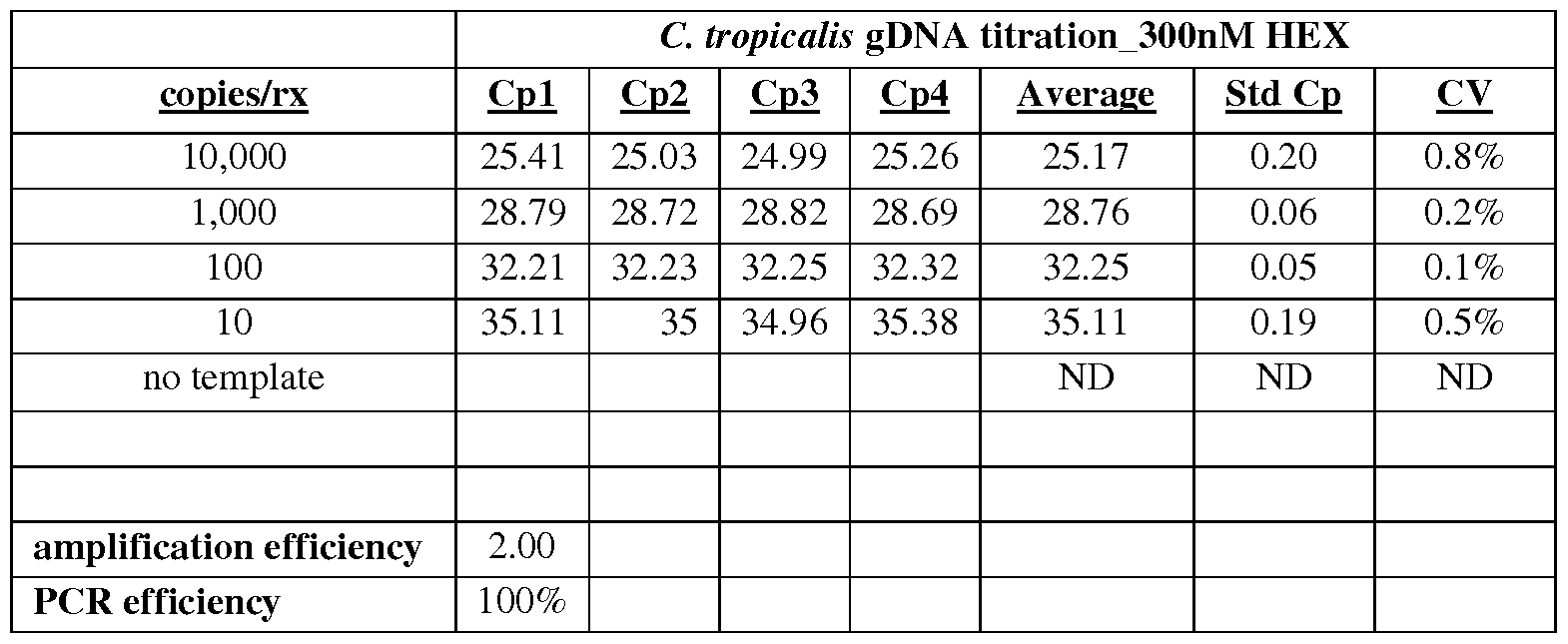
Next, a similar assay was performed using a Candida tropicalis lysate rather than genomic DNA.
A titration was performed with 100 ul of a Candida lysate prepared with different concentrations of C. tropicalis cells. Real-time PCR data are provided below (Table 10) for PCR reactions containing
Candida lysate having 0 to 1000 (i.e., 0, 10, 100, or 1,000) genomic equivalents of Candida tropicalis genomic DNA per reaction. As shown in Table 10 and Figure 3E, Candida tropicalis was successfully detected using the C. tropicalis specific molecular beacon probe at all genomic equivalents prepared using a Candida lysate.
Table 10.
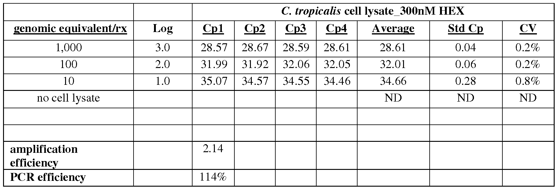
Example 4: Detection of C. albicans using a species-specific molecular beacon probe in a realtime PCR assay.
A HEX labeled C. albicans probe (having the sequence of SEQ ID NO: 1) was used at 300 nM concentration in a real-time PCR assay that was performed according to the parameters described in Table 1 above. 100 μΐ of diluted genomic DNA was added to the PCR reaction mix and 45 cycles of amplification were performed with an annealing temperature of 60 °C. A titration using different amounts of genomic DNA template was performed. Real-time PCR data are provided in Table 11 for reactions containing 0 to 10,000 copies (i.e., 0, 10, 100, 1,000, or 10,000) of genomic DNA per reaction. Four replicates for each reaction condition were performed and the average Ct values were calculated. The average amplification efficiency and the PCR efficiency for the reactions were also calculated. Table 11 and Figure 4A show that the C. albicans molecular beacon probe was used successfully to detect the target nucleic acid molecule using genomic DNA isolated from Candida albicans.
Table 11.

Next, a similar assay was performed using a Candida albicans lysate rather than genomic DNA. A titration was performed with 100 ul of a Candida lysate prepared with different concentrations of C. albicans cells. Real-time PCR data are provided below (Table 12) for PCR reactions containing Candida lysate which provide 0 to 400 (i.e., 0, 4, 40, or 400) genomic equivalents of Candida albicans genomic DNA per reaction. As shown in Table 12 and Figure 4C, Candida albicans was successfully detected using the C. albicans specific molecular beacon probe at all genomic equivalents prepared using a Candida lysate.
Table 12:

Example 5: Detection of C glabrata using a species-specific molecular beacon probe in a real- time PCR assay.
A HEX labeled C. glabrata probe (having the sequence of SEQ ID NO: 3) was used at 300 nM concentration in a real-time PCR assay that was performed according to the parameters described in Table 1 above. 100 μΐ of diluted genomic DNA was added to the PCR reaction mix and 45 cycles of amplification were performed with an annealing temperature of 60 °C. A titration using different amounts of genomic DNA template was performed. Real-time PCR data are provided in Table 13 for
reactions containing 0 to 10,000 (i.e., 0, 10, 100, 1,000, or 10,000) copies of genomic DNA per reaction. Four replicates for each reaction condition were performed and the average Ct values were calculated. The average amplification efficiency and the PCR efficiency for the reactions were also calculated. Table 13 and Figure 4B show that the C. glabrata molecular beacon probe was used successfully to detect the target nucleic acid molecule using genomic DNA isolated from Candida glabrata.
Table 13.
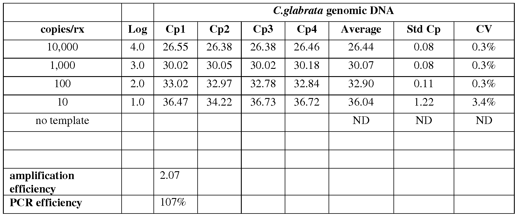
Next, a similar assay was performed using a Candida glabrata lysate rather than genomic DNA. A titration was performed with 100 ul of a Candida lysate prepared with different concentrations of C. glabrata cells. Real-time PCR data are provided below (Table 14) for PCR reactions containing Candida lysate which provide 0 to 400 (i.e., 0, 4, 40, or 400) genomic equivalents of Candida glabrata genomic DNA per reaction. As shown in Table 14 and Figure 4D, Candida glabrata was successfully detected using the C. glabrata specific molecular beacon probe at all genomic equivalents prepared using a Candida lysate.
Table 14.

The above data in Tables 4 to 14 and in Figures 3 and 4 demonstrate that each of the species specific probes can be used to successfully detect specific Candida species using either purified genomic DNA as an input in a real-time PCR assay or, alternatively, using a fungal cell lysate as an input in the assay.
Example 6: Multiplexed real-time PCR assay for detecting multiple Candida species in a sample. For a multiplexed molecular beacon assays, 300 nM of each species-specific beacon probe (0.3uL per 100 uL reaction) were added to the PCR master mix. The C. parapsilosis and C. tropicalis molecular beacons (SEQ ID NO: 4 and SEQ ID NO: 2, respectively) are labeled with a FAM and a HEX fluor, respectively. The beacons are multiplexed in the reaction and detected in their respective channels (488- 533 nm for FAM) and (488-610 for HEX). To ensure that the amplification efficiencies were similar between reactions containing a single beacon and reactions containing multiplexed beacons, C. tropicalis and C. parapsilosis genomic DNA was titrated into reactions at concentrations spanning 1E4 down to 10 copies per 20 uL reaction volume. These reactions included either a single beacon for detection (shown in Table 15 for C. parapsilosis and Table 17 for C. tropicalis) or the two beacons in the same reaction but detected with different channels (Table 16 and Table 18). In both cases the amplification effiencies were similar for both the single detection reactions and multiplexed detection reactions with no measurable impact on detection sensitivity. Thus, both C. tropicalis and C. parapsilosis can be detected within a single PCR reaction and potentially all five beacons could be multiplexed in a single reaction enabling detection of the five most clinically relevant Candida species within the same well.
Table 15.

Table 16.

Table 17.

Table 18.
 Example 7: DNA sequencing-based detection of Candida species.
A probe having the sequence of SEQ ID NO: 32 (specific to C. krusei) was contacted with nucleic acid molecules that were amplified from within whole blood lysate using the pan-Candida forward and reverse primers (SEQ ID NOs: 11 and 12), followed by conducting a phenol/chloroform extraction, a chloroform extraction, and then conducting Sanger Dideoxy Sequencing using Big Dye Terminators on an AB 3730 capillary sequencing instrument. The resulting chromatogram, which is shown in Figure 7, demonstrates that this method can also be used to of detect Candida species using the probes and primers of this invention.
The sequencing-based detection method described above can also be performed using a probe having the sequence of_SEQ ID NO: 33 to detect C. krusei; a probe having the sequence of SEQ ID NO: 34 or 35 to detect C. albicans; a probe having the sequence of SEQ ID NO: 36 or 37 to detect C.
glabrata; a probe having the sequence of SEQ ID NO: 38 or 39 to detect C. parapsilosis; or a probe having the sequence of SEQ ID NO: 40 or 41 to detect C. tropicalis. Example 8: Detection of C. albicans using a species-specific molecular beacon probe in a realtime PCR assay.
A FAM labeled C. albicans probe (having the sequence of SEQ ID NO: 1) was used at 300 nM concentration in a real-time PCR assay that was performed according to the parameters described in Table 1 above. 100 μΐ of diluted genomic DNA was added to the PCR reaction mix and 45 cycles of amplification were performed with an annealing temperature of 60 °C. A titration using different amounts of genomic DNA template was performed. Real-time PCR data are provided in Table 19 for reactions containing 0 to 50,000 copies (i.e., 0 (no template), 5, 50, 500, 5,000, or 50,000) of genomic DNA per reaction. Four replicates for each reaction condition were performed and the average Cp values were calculated. The average amplification efficiency (2.03) and the PCR efficiency (103%) for the reactions were also calculated. Table 19 shows that the C. albicans molecular beacon probe was used successfully to detect the target nucleic acid molecule using genomic DNA isolated from C. albicans.
Table 19:
C. albicans detection in singlepiex reactions

s lope -3.2566
0.31
amplification efficiency= 2.03
PCR efficiency 103%
Next, a similar assay was performed using a C. albicans lysate rather than genomic DNA. To determine the ability of the molecular beacons to detect Candida within crude cell lysates, we prepared lysate from Candida cells spiked in lx phosphate buffered saline (PBS) buffer. Candida cells were quantified using a Coulter counter and were spiked into PBS at concentrations ranging from -520 cells/mL to ~3 cells/mL. The in-vitro spiked solutions were added to a 1 mL polypropylene tube containing 300 mg of yttrium stabilized zirconium oxide beads. The cells were harvested via centrifugation at 6000xG for 5 minutes and the supernatant was removed. 100 uL of IxTE was added to the tubes and the tubes were subjected to bead beating, as described herein, by placement in a vortexer (Biospec bead beater) at maximum speed for 5 minutes. Fifty microliters of lysate was then transferred into an asymmetric PCR master mix containing the molecular beacon probe, as described in Example 1.
Real-time PCR data are provided below (Table 20) for PCR reactions containing C. albicans genomic DNA prepared from C. albicans lysate using the indicated number of cells/mL. The reactions were set up using lysates prepared over multiple days not the same sample lysate detected over multiple days, thus the variation in the entire process is shown. As shown in Table 20 and Figure 8A, C. albicans was successfully detected using the C. albicans specific molecular beacon probe, and semi-quantitative detection of C. albicans was possible using as few as ~3 cells/mL.
Table 20:
C. albicans cetls/mL S20 400 2S0 52 40 28

The above data demonstrate that the C. albicans molecular beacon probe can be used to successfully detect C. albican using either purified genomic DNA as an input in a real-time PCR assay or, alternatively, using a fungal cell lysate as an input in the assay.
Example 9: Detection of C. glabrata using a species-specific molecular beacon probe in a realtime PCR assay.
A HEX labeled C. glabrata probe (having the sequence of SEQ ID NO: 3) was used at 300 nM concentration in a real-time PCR assay that was performed according to the parameters described in Table 1 above. 100 μΐ of diluted genomic DNA was added to the PCR reaction mix and 45 cycles of amplification were performed with an annealing temperature of 60 °C. A titration using different amounts of genomic DNA template was performed. Real-time PCR data are provided in Table 21 for reactions containing 0 to 50,000 copies (i.e., 0 (no template), 5, 50, 500, 5,000, or 50,000) of genomic DNA per reaction. Four replicates for each reaction condition were performed and the average Cp values were calculated. The average amplification efficiency (2.23) and the PCR efficiency (123%) for the reactions were also calculated. Table 21 shows that the C. glabrata molecular beacon probe was used successfully to detect the target nucleic acid molecule using genomic DNA isolated from C. glabrata.
Table 21 :
C. glabrata detection in singleplex reactions

slope -2.87
0.35
amplification
 2.23
2.23
PCR efficiency= 123%
Next, a similar assay was performed using a C. glabrata lysate rather than genomic DNA according to the assay described in Example 8.
Real-time PCR data are provided below (Table 22) for PCR reactions containing C. glabrata genomic DNA prepared from C. glabrata lysate using the indicated number of cells/mL. The reactions were set up using lysates prepared over multiple days not the same sample lysate detected over multiple days, thus the variation in the entire process is shown. As shown in Table 22 and Figure 8B, C. glabrata was successfully detected using the C. glabrata specific molecular beacon probe, and semi-quantitative detection of C. glabrata was possible using as few as ~3 cells/mL.
Table 22:
c.
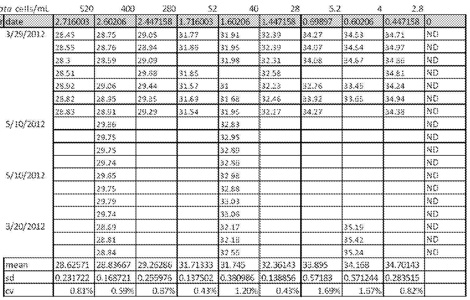
The above data demonstrate that the C. glabrata molecular beacon probe can be used to successfully detect C. glabrata using either purified genomic DNA as an input in a real-time PCR or, alternatively, using a fungal cell lysate as an input in the assay.
Example 10: Detection of C. krusei using a species-specific molecular beacon probe in a realtime PCR assay.
A HEX labeled C. krusei probe (having the sequence of SEQ ID NO: 6) was used at 300 nM concentration in a real-time PCR assay that was performed according to the parameters described in Table 1 above. 100 μΐ of diluted genomic DNA was added to the PCR reaction mix and 45 cycles of amplification were performed with an annealing temperature of 60 °C. A titration using different amounts of genomic DNA template was performed. Real-time PCR data are provided in Table 23 for reactions containing 0 to 50,000 copies (i.e., 0 (no template), 5, 50, 500, 5,000, or 50,000) of genomic
DNA per reaction. Four replicates for each reaction condition were performed and the average Cp values were calculated. The average amplification efficiency (2.05) and the PCR efficiency (105%) for the reactions were also calculated. Table 23 shows that the C. krusei molecular beacon probe was used successfully to detect the target nucleic acid molecule using genomic DNA isolated from C. krusei.
Table 23:
C. krusei detection in singleplex reactions

s lope -3.2019
0.31
amplification efficiency= 2.05
PCR efficiency= 105 %
Next, a similar assay was performed using a C. krusei lysate rather than genomic DNA according to the assay described in Example 8.
Real-time PCR data are provided below (Table 24) for PCR reactions containing C. krusei genomic DNA prepared from C. krusei lysate using the indicated number of cells/mL. The reactions were set up using lysates prepared over multiple days not the same sample lysate detected over multiple days, thus the variation in the entire process is shown. As shown in Table 24 and Figure 8C, C. krusei was successfully detected using the C. krusei specific molecular beacon probe, and semi-quantitative detection of C. krusei was possible using as few as ~3 cells/mL.
Table 24:

The above data demonstrate that the C. krusei molecular beacon probe can be used to successfully detect C. krusei using either purified genomic DNA as an input in a real-time PCR assay or, alternatively, using a fungal cell lysate as an input in the assay.
Example 11: Detection of C. parapsilosis using a species-specific molecular beacon probe in a real-time PCR assay.
A HEX labeled C. parapsilosis probe (having the sequence of SEQ ID NO: 5) was used at 300 nM concentration in a real-time PCR assay that was performed according to the parameters described in Table 1 above. 100 μΐ of diluted genomic DNA was added to the PCR reaction mix and 45 cycles of amplification were performed with an annealing temperature of 60 °C. A titration using different amounts of genomic DNA template was performed. Real-time PCR data are provided in Table 25 for reactions containing 0 to 50,000 copies (i.e., 0 (no template), 5, 50, 500, 5,000, or 50,000) of genomic
DNA per reaction. Four replicates for each reaction condition were performed and the average Cp values were calculated. The average amplification efficiency (2.10) and the PCR efficiency (110%) for the reactions were also calculated. Table 25 shows that the C. parapsilosis molecular beacon probe was used
successfully to detect the target nucleic acid molecule using genomic DNA isolated from Candida parapsilosis.
Table 25:
C. parapsilosis detection in singlepiex rx

slope -3.1079
0.32
amplification efficiency^ 2.10
PCR efficiency= 110%
Next, a similar assay was performed using a C. parapsilosis lysate rather than genomic DNA according to the assay described in Example 8.
Real-time PCR data are provided below (Table 26) for PCR reactions containing C. parapsilosis genomic DNA prepared from C. parapsilosis lysate using the indicated number of cells/mL. The reactions were set up using lysates prepared over multiple days not the same sample lysate detected over multiple days, thus the variation in the entire process is shown. As shown in Table 26 and Figure 8D, C. parapsilosis was successfully detected using the C. parapsilosis specific molecular beacon probe, and semi-quantitative detection of C. parapsilosis was possible using as few as ~3 cells/mL.
able 26:


The above data demonstrate that the C. parapsilosis molecular beacon probe can be used to successfully detect C. parapsilosis using either purified genomic DNA as an input in a real-time PCR assay or, alternatively, using a fungal cell lysate as an input in the assay.
Example 12: Detection of C. tropicalis using a species-specific molecular beacon probe in a real- time PCR assay.
A HEX labeled C. tropicalis probe (having the sequence of SEQ ID NO: 2) was used at 300 nM concentration in a real-time PCR assay that was performed according to the parameters described in Table 1 above. 100 μΐ of diluted genomic DNA was added to the PCR reaction mix and 45 cycles of amplification were performed with an annealing temperature of 60 °C. A titration using different amounts of genomic DNA template was performed. Real-time PCR data are provided in Table 27 for reactions containing 0 to 50,000 copies (i.e., 0 (no template), 5, 50, 500, 5,000, or 50,000) of genomic DNA per reaction. Four replicates for each reaction condition were performed and the average Cp values were calculated. The average amplification efficiency (2.11) and the PCR efficiency (111 %) for the reactions were also calculated. Table 27 shows that the C. tropicalis molecular beacon probe was used successfully to detect the target nucleic acid molecule using genomic DNA isolated from C. tropicalis.
Table 27:
C. tropicalis detection in singlep!ex rx
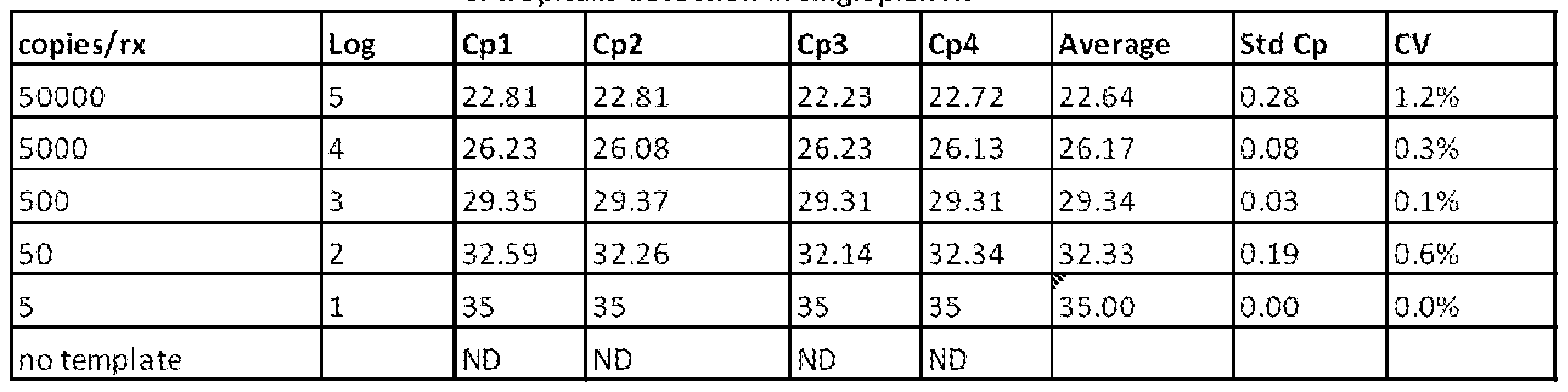
PCR efficiency
Next, a similar assay was performed using a C. tropicalis lysate rather than genomic DNA according to the assay described in Example 8.
Real-time PCR data are provided below (Table 28) for PCR reactions containing C. tropicalis genomic DNA prepared from C. tropicalis lysate using the indicated number of cells/mL. The reactions were set up using lysates prepared over multiple days not the same sample lysate detected over multiple days, thus the variation in the entire process is shown. As shown in Table 28 and Figure 8E, C. tropicalis was successfully detected using the C. tropicalis specific molecular beacon probe, and semi-quantitative detection of C. tropicalis was possible using as few as ~3 cells/mL.
Table 28:

The above data demonstrate that the C. tropicalis molecular beacon probe can be used to successfully detect C. tropicalis using either purified genomic DNA as an input in a real-time PCR assay or, alternatively, using a fungal cell lysate as an input in the assay.
Example 13: Detection of Candida albicans in a multiplex reaction with Candida albicans, Candida krusei, and Candida glabrata.
The molecular beacon probes can also be used to successfully detect Candida target nucleic acid molecules in a multiplexed reaction that includes nucleic acid molecules from two or more Candida species.
A multiplex reaction was performed using nucleic acid molecules (genomic DNA) from C. albicans, C. krusei, and C. glabrata. 300 nM of each Candida species-specific beacon probe
(corresponding to SEQ ID NOs: 1, 6, and 3, respectively; 0.3uL per 100 uL reaction) were used in a realtime PCR assay that was performed according to the parameters described in Table 1 above. The C. albicans, C. krusei, and C. glabrata molecular beacons are each labeled with spectrally distinguishable fluororphors that are detected in their respective channels. 100 μΐ of diluted genomic DNA was added to the PCR reaction mix and 45 cycles of amplification were performed with an annealing temperature of 60
°C. A titration using different amounts of genomic DNA template was performed. Real-time PCR data are provided in Table 29 for reactions containing 0 to 50,000 copies (i.e., 0 (no template), 5, 50, 500, 5,000, or 50,000) of genomic DNA per reaction. Four replicates for each reaction condition were performed and the average Cp values were calculated. The average amplification efficiency (2.00) and the PCR efficiency (100%) for the reactions were also calculated. Table 29 shows that the C. albicans molecular beacon probe was used successfully to detect the C. albicans target nucleic acid molecules using genomic DNA isolated from C. albicans, even in the presence C. krusei and C. glabrata nucleic acid molecules, as well as molecular beacons for all of these Candida species. Notably, the
amplification efficiency and PCR efficiency in the multiplex reaction was similar to that observed in the singleplex reaction (see Example 8 and Table 19). Thus, the limit of detection using the molecular beacon probes is not changing even when they are used in combination. Similarly, the presence of multiple probes in the sample is not affecting the efficiency of the nucleic acid amplification reaction.
Table 29:
C. albicans detection in multiplex rx (C. albicans, C. krusei, and C. glabrata multiplex)

slope -3.32
0.30
amplification efnctency= 2.00
PCR efficiency= 100%
Example 14: Detection of C. glabrata in a multiplex reaction with C. albicans, C. krusei, and C. glabrata.
A molecular beacon probe for C. glabrata (SEQ ID NO: 3) can be used to successfully detect C. glabrata nucleic acid molecules in a multiplex reaction that also includes nucleic acid molecules from C. albicans and C. krusei. The multiplex reaction was performed as described in Example 13. Real-time PCR data are provided in Table 30 for reactions containing 0 to 50,000 copies (i.e., 0 (no template), 5, 50, 500, 5,000, or 50,000) of genomic DNA per reaction. Four replicates for each reaction condition were performed and the average Cp values were calculated. The average amplification efficiency (2.02) and the PCR efficiency (102%) for the reactions were also calculated. Table 30 shows that the C. glabrata molecular beacon probe was used successfully to detect C. glabrata target nucleic acid molecules using genomic DNA isolated from C. glabrata, even in the presence C. albicans and C. krusei nucleic acid molecules, as well as molecular beacons for all of these Candida species. Notably, the amplification
efficiency and PCR efficiency in the multiplex reaction was similar to that observed in the singleplex reaction (see Example 9 and Table 21).
Table 30:
C, glabrata detection in multiplex rx (C, albicans, C. krusei, and C. glabrata multiplex)

sfope -3.27
0.31
amplification efficiency= 2.02
PCR efficiertcy= 102%
Example 15: Detection of C. krusei in a multiplex reaction with C. albicans, C. krusei, and C. glabrata.
A molecular beacon probe for Candida krusei (SEQ ID NO: 6) can be used to successfully detect C. krusei nucleic acid molecules in a multiplex reaction that also includes nucleic acid molecules from C. albicans and C. glabrata. The multiplex reaction was performed as described in Example 13. Real-time PCR data are provided in Table 26 for reactions containing 0 to 50,000 copies (i.e., 0 (no template), 5, 50, 500, 5,000, or 50,000) of genomic DNA per reaction. Four replicates for each reaction condition were performed and the average Cp values were calculated. The average amplification efficiency (2.12) and the PCR efficiency (112%) for the reactions were also calculated. Table 31 shows that the C. krusei molecular beacon probe was used successfully to detect C. krusei target nucleic acid molecules using genomic DNA isolated from C. krusei, even in the presence C. albicans and C. glabrata nucleic acid molecules, as well as molecular beacons for all of these Candida species. Notably, the amplification efficiency and PCR efficiency in the multiplex reaction was similar to that observed in the singleplex reaction (see Example 10 and Table 23).
Table 31 :
C. krusei detection in multiplex rx {C. albicans, C. krusei, and C. giabrata multiplex)

slope -3.07
0.33
2.12
112%
Example 16: Detection of C. parapsilosis in a multiplex reaction with C. parapsilosis and C. tropicalis.
A molecular beacon probe for Candida parapsilosis (SEQ ID NO: 4) can be used to successfully detect C. parapsilosis nucleic acid molecules in a multiplex reaction that also includes nucleic acid molecules from C. tropicalis. The multiplex reaction was performed using nucleic acid molecules (genomic DNA) from C. parapsilosis and C. tropicalis. 300 nM of each Candida species-specific beacon probe (corresponding to SEQ ID NOs: 4 and 2, respectively; 0.3uL per 100 uL reaction) were used in a real-time PCR assay that was performed according to the parameters described in Table 1 above. 100 μΐ of diluted genomic DNA was added to the PCR reaction mix and 45 cycles of amplification were performed with an annealing temperature of 60 °C. A titration using different amounts of genomic DNA template was performed. Real-time PCR data are provided in Table 32 for reactions containing 0 to 50,000 copies (i.e., 0 (no template), 5, 50, 500, 5,000, or 50,000) of genomic DNA per reaction. Four replicates for each reaction condition were performed and the average Cp values were calculated. The average amplification efficiency (2.07) and the PCR efficiency (107%) for the reactions were also calculated. Table 32 shows that the C. parapsilosis molecular beacon probe was used successfully to detect C. parapsilosis target nucleic acid molecules using genomic DNA isolated from C. parapsilosis, even in the presence C. tropicalis nucleic acid molecules, as well as molecular beacons for both of these Candida species. Notably, the amplification efficiency and PCR efficiency in the multiplex reaction was similar to that observed in the singleplex reaction (see Example 11 and Table 25).
Table 32:
C. parapsilosis detection in multiplex rx (C. tropicalis, C. parpasiiosi

slope -3.17
0.32
2.07
107%
Example 17: Detection of C. tropicalis in a multiplex reaction with C. parapsilosis and C.
tropicalis.
A molecular beacon probe for C. tropicalis (SEQ ID NO: 2) can be used to successfully detect C. tropicalis nucleic acid molecules in a multiplex reaction that also includes nucleic acid molecules from C. parapsilosis. The multiplex reaction was performed as described in Example 16. Real-time PCR data are provided in Table 33 for reactions containing 0 to 50,000 copies (i.e., 0 (no template), 5, 50, 500, 5,000, or 50,000) of genomic DNA per reaction. Four replicates for each reaction condition were performed and the average Cp values were calculated. The average amplification efficiency (2.11) and the PCR efficiency ( 111 %) for the reactions were also calculated. Table 33 shows that the C. tropicalis molecular beacon probe was used successfully to detect Candida tropicalis target nucleic acid molecules using genomic DNA isolated from C. tropicalis, even in the presence C. parapsilosis nucleic acid molecules, as well as molecular beacons for both of these Candida species. Notably, the amplification efficiency and PCR efficiency in the multiplex reaction was similar to that observed in the singleplex reaction (see Example 12 and Table 27).
Table 33:
C. tropicalis detection in multiplex rx (C. tr picaiis, C. parpasilosis)
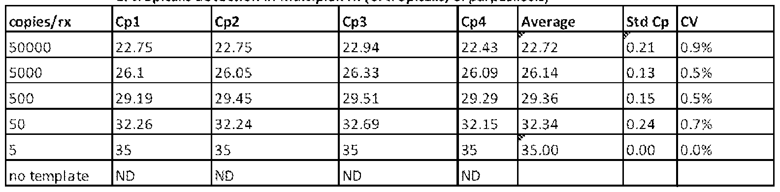
sl ope -3.08
0.33
2.11
1 11%
Example 18: Detection of Candida species using a heat denatured fungal lysate.
To determine if the molecular beacon probes could be used for the detection of Candida cells directly using a 95°C heat denaturation step to lyse the Candida cells instead of mechanical lysis, such as by bead beating, we spiked 50 uL of TE containing Coulter counted Candida cells directly into a 96-well plate. We then added the molecular beacon probe-containing master mix on top of the cells and conducted an RT-PCR using the parameters described in Example 1 above.
Real-time PCR data are provided below (Tables 34-38) for PCR reactions, each of which contains C. albicans, C. glabrata, C. krusei, C. parapsilosis, and C. tropicalis genomic DNA prepared from heat denatured lysates containing the indicated number of cells. As shown in Tables 34-38, the Candida specific molecular beacon probes (C. albicans: SEQ ID NO: 1 ; C. glabrata: SEQ ID NO: 3; C. krusei: SEQ ID NO: 6; C. parapsilosis: SEQ ID NO: 4; and C. tropicalis: SEQ ID NO: 2) successfully detected the indicated Candida nucleic acid molecules following lysis by heat denaturation. Detection was possible even up to 10 cells.
Table 34: Detection of C. albicans in fungal lysate
C. albicans

C. glabrata

Table 36: Detection of C. krusei in fungal lysate
C. krusei
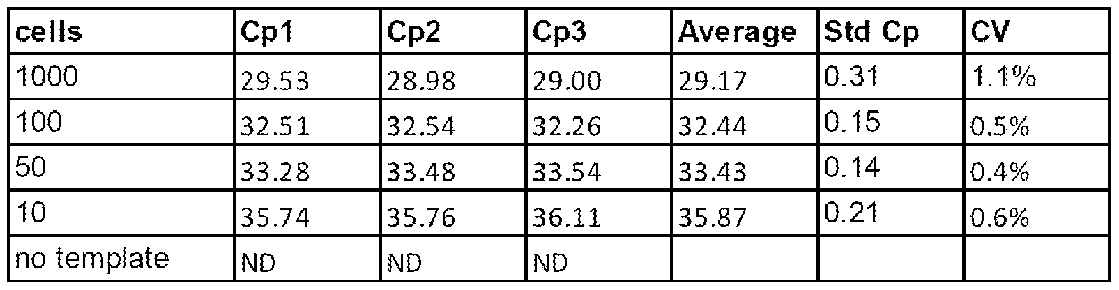
C. parapsilosis

Table 38: Detection of C. tropicalis in fungal lysate
C. tropicalis

As shown in Tables 39-43, there is 97% concordance between the detected Candida cells via the molecular beacon probes and the T2 detection reactions run on the RT-PCR generated amplicons. We observe one instance in which C. glabrata nucleic acid molecules from the sample prepared using 10 cells was not detected via RT-PCR but was detected with the NMR-based T2 assay (compare Cpl in Table 35 to Repl in Table 40), and one sample in which C. tropicalis nucleic acid molecules from the sample prepared using 10 cells was detected via RT-PCR but not by the NMR-based T2 assay (compare Cpl in Table 38 to Repl in Table 43). These discordant results are highlighted in the tables below.
Table 39:
C. albicans RT-PCR product, T2 detection

C. glabrata RT-PCR product, T2 detection

Table 41 :
C. krusei RT-PCR product, T2 detection

Table 42:
C. parapsilosis RT-PCR product, T2 detection

C. tropicalis RT-PCR product, T2 detection

Example 19: Detection of Candida species using melt-curve analysis.
Because the Tm of the Candida species-specific molecular beacon probes differ by at least 1 degree, it is possible to multiplex the beacon probes with the same fluorophore and deconvolute the signals using melt curve analysis alone. Molecular beacon probes for C. albicans (SEQ ID NO: 1), C. glabrata (SEQ ID NO: 3), C krusei (SEQ ID NO: 6), C parapsilosis (SEQ ID NO: 4), and C tropicalis (SEQ ID NO: 2) were all labeled with a HEX fluorophore and added at 300 nm concentration to a single sample that included target nucleic acid molecules for each of the Candida species.
As shown in Figures 9A and 9B, the presence of target nucleic acid molecules for the indicated
Candida species targets was determined within the sample simply by looking for the presence of the species-specific melt curve and using the Tm as a means of species identification. Use of the melt-curve analysis abrogates the need to use multiple different fluorophores in a single multiplexed reaction and simplifies the instrument design. It should be noted that there is no melt curve when the target nucleic acid molecule is not present in the reaction. The Tm is the temperature at which a decrease in fluorescence is observed and this only occurs when the hybridized beacon probe is melted off the target and the beacon probe stem-loop structure reforms.
Example 20: Molecular beacon analysis using a post-blood culture.
To perform a blood culture analysis, up to 10 ml of a blood sample from a patient suspected of having a blood systemic infection are incubated in a blood culture bottle. The blood culture media typically are composed to promote the growth of either aerobic or anaerobic pathogens. Depending on the specific requirements for growth of the suspected pathogen, culture media is composed of a number of nutrients that feed pathogens and resins that absorb antibiotics/antifungals that might be present in the blood specimen and inhibit growth. When a pathogen reaches a concentration of 106 to 108 CFU/mL, it produces enough carbon dioxide through its normal metabolic pathways to activate the blood culture detection system, thus signaling a positive blood culture for the original sample. At this time, a sample from this culture bottle is aliquoted onto a species-specific detection methodology, which may be a culture plate or another system that identifies the pathogenic species and assesses its susceptibility to
antimicrobials (e.g., Vitek 2, Biomeriuex, Durham, NC). Species identification is typically performed over a day, although it can be significantly longer.
In order to expedite the species identification of Candida that may be present in a blood culture, a singleplex or multiplex reaction for detecting one or more Candida species, according to the methods described in, e.g., Examples 1- 19 above, can be used. The protocol involves adding a sample from the positive blood culture bottle to the singleplex or multiplex reaction, centrifuging the sample to pellet the pathogen cells, which may be optionally washed one or more times to remove interferents from the blood culture media, lysing the cells and using the nucleic acid molecules from the cells in a molecular beacon protocol described in one or more of Examples 1-19 above. This process would rapidly and with high fidelity determine the presence and species identification of one or more Candida species in the blood sample; a positive result indicates Candida infection in the patient.
Species idenfication could rapidly assist in appropriate administration of antifungals as several Candida species are more resistant to fluconazole (e.g., Candida glabrata and Candida krusei). Example 21: Use of the molecular beacon assay to detect Candida in a whole blood sample.
The Candida specific molecular beacon probes described herein can be used to detect the presence of Candida in a whole blood sample from a patient suspected of having a Candida infection. Generally, the protocol involves i) obtaining a 1-10 mL sample of whole blood from the patient; ii) lysing the red blood cells (e.g., using an ammonium chloride iso-osmotic solution, a detergent lysis solution, or a hypotonic lysis solution); iii) centrifuging the lysed sample, e.g., at 3,000g to 12,000g for 5 minutes to pellet the Candida cells; iv) removing and discarding the supernatant; v) washing the pellet with an equal volume of TE (equivalent to the blood specimen volume) or less; vi) centrifuging the resuspended Candida cells at 3,000g to 12,000g for 5 minutes to pellet the Candida cells; vii) removing and discarding the supernatant; viii) optionally repeating steps v)-vii); ix) adding 50- 150 uL of lxTE; x) adding 300 mg of 500 to 800 micron beads (e.g., silica or zirconium oxide (yttrium stabilized) beads); xi) vortexing at maximum power (3000 rpm) for 5-10 minutes to lyse the Candida cells; xii) aliquoting 10 to 50 uL of the cell lysate into a molecular beacon probe-containing PCR mastermix on, e.g., a 96 well plate (see, e.g., Example 1); and xiii) running an RT-PCR reaction and detecting the presence of Candida according to one or more of the methods described in Examples 1- 19.
Example 22: Use of the molecular beacon assay to detect Candida in a urine sample.
The Candida specific molecular beacon probes described herein can be used to detect the presence of Candida in a urine sample from a patient suspected of having a Candida infection.
Generally, the protocol involves i) obtaining a 1-10 mL sample of urine from the patient; ii) centrifuging the sample at 3,000g to 12,000g to pellet the Candida cells; iii) removing and discarding the supernatant; iv) washing the pellet with an equal volume of TE or less (equivalent to the urine specimen volume); v) centrifuging at 3,000g to 12,000g for 5 minutes to pellet the Candida cells; vi) removing and discarding the supernatant; vii) optionally repeating steps iv)-vi); viii) adding 50-150 uL of lxTE; ix) adding 300 mg of 500 to 800 micron beads (e.g., silica or zirconium oxide (yttrium stabilized) beads); x) vortexing at maximum power (3000 rpm) for 5-10 minutes to lyse the Candida cells; xi) aliquoting 10 to 50 uL of cell lysate into molecular beacon probe-containing PCR mastermix on, e.g., a 96 well plate (see, e.g,.
Example 1); and xii) running an RT-PCR reaction and detecting the presence of Candida according to one or more of the methods described in Examples 1- 19. Other Embodiments
While certain novel features of this invention shown and described below are pointed out in the annexed claims, the invention is not intended to be limited to the details specified, since a person of ordinary skill in the relevant art will understand that various omissions, modifications, substitutions and changes in the forms and details of the invention illustrated and in its operation may be made without departing in any way from the spirit of the present invention. No feature of the invention is critical or essential unless it is expressly stated as being "critical" or "essential."
Those skilled in the art will recognize or be able to ascertain using no more than routine experimentation many equivalents to the specific embodiments of the invention described herein. Such equivalents are intended to be encompassed in the scope of the present invention.
All publications, patents, and patent applications mentioned in this specification are herein incorporated by reference to the same extent as if each independent publication, patent, or patent application was specifically and individually indicated to be incorporated by reference.
Claims
1. A nucleic acid probe comprising the sequence of SEQ ID NO: 1.
2. A nucleic acid probe comprising the sequence of SEQ ID NO: 2.
3. A nucleic acid probe comprising the sequence of SEQ ID NO: 3.
4. A nucleic acid probe comprising the sequence of SEQ ID NO: 4.
5. A nucleic acid probe comprising the sequence of SEQ ID NO: 5.
6. The nucleic acid probe of any one of claims 1 to 5, wherein said probe comprises a detection moiety that is conjugated to the probe.
7. The nucleic acid probe of claim 6, wherein said detection moiety comprises a fluorescent label.
8. The nucleic acid probe of any one of claims 1 to 5, wherein said probe is a molecular beacon comprising a fluorescent label and a quencher.
9. A method for detecting at least one Candida species in a biological sample, said method comprising: a) contacting said sample with at least one nucleic acid probe having a sequence selected from any one of SEQ ID NOs: 1 to 5 under conditions which allow the at least one probe to hybridize to a nucleic acid molecule of said Candida species; and b) detecting hybridization between said at least one probe and said nucleic acid molecule, thereby detecting said at least one Candida species.
10. The method of claim 9, wherein the probe is a Candida albicans probe and has the sequence of SEQ ID NO: 1.
11. The method of claim 9, wherein the probe is a Candida tropicalis probe and has the sequence of SEQ ID NO: 2.
12. The method of claim 9, wherein the probe is a Candida glabrata probe and has the sequence of SEQ ID NO: 3.
13. The method of claim 9, wherein the probe is a Candida parapsilosis probe and has the sequence of SEQ ID NO: 4.
14. The method of claim 9, wherein the probe is a Candida krusei probe and has the sequence of SEQ ID NO: 5.
15. The method of claim 9, wherein said probe has a sequence selected from any one of SEQ ID NOs: 1 to 5.
16. The method of claim 15, wherein said probe comprises a detection moiety that is conjugated to the probe.
17. The method of claim 16, wherein said detection moiety comprises a fluorescent label.
18. The method of claim 17, wherein said probe is a molecular beacon that further comprises a quencher.
19. The method of claim 9, wherein said method comprises contacting said sample with two or more probes having sequences selected from any of SEQ ID NOs: 1 to 5, whereby two or more Candida species selected from the group consisting of Candida albicans, Candida tropicalis, Candida glabrata, Candida parapsilosis, and Candida krusei are detected.
20. The method of claim 9, wherein said method comprises contacting said sample with up to five probes having sequences selected from any of SEQ ID NOs: 1 to 5, whereby up to five Candida species selected from the group consisting of Candida albicans, Candida tropicalis, Candida glabrata, Candida parapsilosis, and Candida krusei are detected.
21. The method of claim 19 or 20, wherein each said probe is differentially labeled.
22. The method of claim 19 or 20, wherein each said probe comprises the same label.
23. The method of any one of claims 9 to 22, wherein the biological sample is selected from the group consisting of whole blood, sweat, tears, urine, saliva, semen, serum, plasma, cerebrospinal fluid (CSF), feces, vaginal fluid or tissue, sputum, nasopharyngeal aspirate or swab, lacrimal fluid, mucous, or epithelial swab (buccal swab), tissues, organs, bones, teeth, and tumors.
24. The method of any one of claims 9 to 23, wherein the biological sample is a human sample.
25. The method of any one of claims 9 to 24, wherein at least one cell in said biological sample is lysed using a hypotonic solution or a detergent, wherein optionally, said lysis step is followed by centrifuging the lysed sample and removing the resulting supernatant.
26. The method of any one of claims 9 to 25, wherein at least one cell of said Candida species is lysed by chemical lysis.
27. The method of claim 26, wherein said chemical lysis comprises treatment with one or more enzymes.
28. The method of any one of claims 9 to 25, wherein at least one cell of said Candida species is lysed by mechanical lysis.
29. The method of claim 28, wherein said mechanical lysis comprises using any one or more of a finned tube, small particles, ultrasound, freeze-boil, freeze-thaw, and a bead.
30. The method of claim 29, wherein said bead comprises a glass bead.
31. The method of any one of claims 9 to 24, wherein at least one cell of said Candida species is lysed by heat denaturation.
32. The method of claim 31, wherein said heat denaturation comprises heating said at least one cell to about 95°C.
33. The method of any one of claims 9 to 32 further comprising amplifying said nucleic acid molecule of said Candida species prior to said detecting step.
34. The method of claim 33, wherein said amplifying comprises enzymatic amplification.
35. The method of claim 33 or 34, wherein said amplifying comprises asymmetric amplification.
36. The method of any one of claims 9 to 35, wherein said probe comprises a 5' end and a 3' end, wherein said 5' end is capable of hybridizing to said nucleic acid molecule, and wherein about 1 to 20 nucleotides of said 3' end of said probe do not hybridize to said nucleic acid molecule.
37. The method of any one of claims 9 to 36 further comprising placing a container comprising a composition resulting from said contacting step into a device capable of detecting said hybridization between said probe and said nucleic acid molecule.
38. The method of any one of claims 9 to 37, wherein each said probe comprises a fluorescent label and said hybridization is detected by heating said sample through a melting temperature (Tm) of at least one said probe to obtain a melting curve, wherein a decrease in fluorescence of said sample at the Tm of at least one said probe indicates the presence of said at least one Candida species.
39. The method of claim 38, wherein a decrease in fluorescence at a Tm of the probe having the sequence of SEQ ID NO: 1 indicates the presence of Candida albicans in the sample.
40. The method of claim 38, wherein a decrease in fluorescence at a Tm of the probe having the sequence of SEQ ID NO: 2 indicates the presence of Candida tropicalis in the sample.
41. The method of claim 38, wherein a decrease in fluorescence at a Tm of the probe having the sequence of SEQ ID NO: 3 indicates the presence of Candida glabrata in the sample.
42. The method of claim 38, wherein a decrease in fluorescence at a Tm of the probe having the sequence of SEQ ID NO: 4 indicates the presence of Candida parapsilosis in the sample.
43. The method of claim 38, wherein a decrease in fluorescence at a Tm of the probe having the sequence of SEQ ID NO: 5 indicates the presence of Candida krusei in the sample.
44. The method of any one of claims 9 to 43, wherein said hybridization is detected by fluorescence of said probe using a thermal cycler.
45. The method of any one of claims 9 to 44, wherein detection of said at least one Candida species in said sample can be completed within 3 hours.
46. The method of any one of claims 9 to 45, wherein the method is capable of detecting said at least one Candida species in said sample when present at a concentration of at least 3 cells/ml.
47. A kit comprising at least one nucleic acid probe having a sequence selected from any of SEQ ID NOs: 1 to 5.
48. The kit of claim 47 further comprising one or more reagent for sample lysis, fungal lysis, detection of Candida species, and nucleic acid amplification.
Priority Applications (2)
| Application Number | Priority Date | Filing Date | Title |
|---|---|---|---|
| EP13779063.0A EP2839038B1 (en) | 2012-04-20 | 2013-03-14 | Compositions and methods for detection of candida species |
| EP19150640.1A EP3524692A1 (en) | 2012-04-20 | 2013-03-14 | Compositions and methods for detection of candida species |
Applications Claiming Priority (2)
| Application Number | Priority Date | Filing Date | Title |
|---|---|---|---|
| US201261636110P | 2012-04-20 | 2012-04-20 | |
| US61/636,110 | 2012-04-20 |
Publications (1)
| Publication Number | Publication Date |
|---|---|
| WO2013158281A1 true WO2013158281A1 (en) | 2013-10-24 |
Family
ID=49380443
Family Applications (1)
| Application Number | Title | Priority Date | Filing Date |
|---|---|---|---|
| PCT/US2013/031774 WO2013158281A1 (en) | 2012-04-20 | 2013-03-14 | Compositions and methods for detection of candida species |
Country Status (3)
| Country | Link |
|---|---|
| US (3) | US9562271B2 (en) |
| EP (2) | EP2839038B1 (en) |
| WO (1) | WO2013158281A1 (en) |
Cited By (2)
| Publication number | Priority date | Publication date | Assignee | Title |
|---|---|---|---|---|
| CN108779499A (en) * | 2016-01-04 | 2018-11-09 | 简·探针公司 | method and composition for detecting Candida species |
| EP3443124B1 (en) | 2016-04-14 | 2022-08-17 | T2 Biosystems, Inc. | Methods for amplification in complex samples |
Families Citing this family (8)
| Publication number | Priority date | Publication date | Assignee | Title |
|---|---|---|---|---|
| US9938558B2 (en) | 2015-06-25 | 2018-04-10 | Ascus Biosciences, Inc. | Methods, apparatuses, and systems for analyzing microorganism strains from complex heterogeneous communities, predicting and identifying functional relationships and interactions thereof, and selecting and synthesizing microbial ensembles based thereon |
| MX2017016924A (en) | 2015-06-25 | 2018-08-15 | Ascus Biosciences Inc | Methods, apparatuses, and systems for analyzing microorganism strains from complex heterogeneous communities, predicting and identifying functional relationships and interactions thereof, and selecting and synthesizing microbial ensembles based thereon. |
| US10851399B2 (en) | 2015-06-25 | 2020-12-01 | Native Microbials, Inc. | Methods, apparatuses, and systems for microorganism strain analysis of complex heterogeneous communities, predicting and identifying functional relationships and interactions thereof, and selecting and synthesizing microbial ensembles based thereon |
| BR112018013975A2 (en) | 2016-01-07 | 2019-02-05 | Ascus Biosciences Inc | methods for improving milk production by administering microbial consortia |
| USD852977S1 (en) | 2016-11-28 | 2019-07-02 | T2 Biosystems, Inc. | Modular cartridge assembly |
| EP3562954A1 (en) | 2016-12-28 | 2019-11-06 | Ascus Biosciences, Inc. | Methods, apparatuses, and systems for analyzing complete microorganism strains in complex heterogeneous communities, determining functional relationships and interactions thereof, and identifying and synthesizing bioreactive modificators based thereon |
| UY37708A (en) | 2017-04-28 | 2018-10-31 | Ascus Biosciences Inc | METHODS TO COMPLEMENT DIETS WITH HIGH CONSUMPTION OF GRAINS AND / OR HIGH ENERGY VALUE IN RUMINANTS, THROUGH THE ADMINISTRATION OF A SYNTHETIC BIOENSAMBLE OF MICROBIES, OR PURIFIED VINTAGES OF THE SAME |
| CN109971883B (en) * | 2019-05-07 | 2020-07-24 | 丹娜(天津)生物科技有限公司 | Primer-probe combination, kit, detection method and application for candida species classification detection |
Citations (11)
| Publication number | Priority date | Publication date | Assignee | Title |
|---|---|---|---|---|
| US4295613A (en) | 1979-10-03 | 1981-10-20 | Vpi Educational Foundation | Apparatus for breaking bacterial cells |
| US5118801A (en) | 1988-09-30 | 1992-06-02 | The Public Health Research Institute | Nucleic acid process containing improved molecular switch |
| US5631132A (en) | 1993-05-20 | 1997-05-20 | The United States Of America As Represented By The Department Of Health And Human Services | Nucleic acid probes and methods for detecting Candida glabrata DNA in blood |
| US5925517A (en) | 1993-11-12 | 1999-07-20 | The Public Health Research Institute Of The City Of New York, Inc. | Detectably labeled dual conformation oligonucleotide probes, assays and kits |
| US6242178B1 (en) * | 1997-07-30 | 2001-06-05 | Centers For Disease Control And Prevention | Nucleic acid probes for detecting Candida species |
| US6686195B1 (en) | 1999-04-01 | 2004-02-03 | Biomerieux S.A. | Method and apparatus for ultrasonic lysis of biological cells |
| US20060234219A1 (en) * | 2001-05-31 | 2006-10-19 | Tsuneya Ohno | Method for detecting and identifying microorganism causative of infection |
| US7494771B2 (en) | 2001-07-19 | 2009-02-24 | Geneohm Sciences Canada, Inc. | Universal method and composition for the rapid lysis of cells for the release of nucleic acids and their detection |
| US7723095B2 (en) | 2000-07-27 | 2010-05-25 | Biomerieux S.A. | Method for cellular lysis of prokaryotes or eukaryotes or simultaneous cellular lysis of prokaryotes and eukaryotes |
| US20100129821A1 (en) * | 2008-11-26 | 2010-05-27 | Fred Hutchinson Cancer Research Center | Broad range pcr-based compositions and methods for the detection and identification of fungal pathogens |
| US20100221710A1 (en) | 2006-11-02 | 2010-09-02 | The University Of Manchester | Assays for fungal infection |
Family Cites Families (254)
| Publication number | Priority date | Publication date | Assignee | Title |
|---|---|---|---|---|
| US422427A (en) * | 1890-03-04 | Machine for cutting and shocking corn | ||
| JPS5913521B2 (en) | 1975-06-19 | 1984-03-30 | メイトウサンギヨウ カブシキガイシヤ | Method for producing magnetic iron oxide/dextran complex |
| US4067951A (en) | 1975-11-19 | 1978-01-10 | Bactomatic Inc. | Process for making impedance measuring module |
| US4374360A (en) | 1980-05-29 | 1983-02-15 | Sepponen Raimo E | NMR Diagnosis apparatus |
| DE3134787A1 (en) | 1981-09-02 | 1983-03-10 | Boehringer Mannheim Gmbh, 6800 Mannheim | CREATININE ANTIBODIES |
| US4471306A (en) | 1982-02-03 | 1984-09-11 | General Electric Company | Method of NMR imaging which overcomes T2 * effects in an inhomogeneous static magnetic field |
| US4452773A (en) | 1982-04-05 | 1984-06-05 | Canadian Patents And Development Limited | Magnetic iron-dextran microspheres |
| USD285118S (en) | 1983-01-19 | 1986-08-12 | Abbott Laboratories | Sample cup |
| USD284214S (en) | 1983-10-31 | 1986-06-10 | Gamma Biologicals, Inc. | Blood grouping apparatus |
| GB8401368D0 (en) | 1984-01-19 | 1984-02-22 | Amersham Int Plc | Assay method |
| US4920061A (en) | 1984-03-02 | 1990-04-24 | The University Of Texas System | Biological magnetic colloids |
| USD288479S (en) | 1984-08-08 | 1987-02-24 | Tambrands Inc. | Diagnostic test kit |
| US4683202A (en) | 1985-03-28 | 1987-07-28 | Cetus Corporation | Process for amplifying nucleic acid sequences |
| US4683195A (en) | 1986-01-30 | 1987-07-28 | Cetus Corporation | Process for amplifying, detecting, and/or-cloning nucleic acid sequences |
| US4965188A (en) | 1986-08-22 | 1990-10-23 | Cetus Corporation | Process for amplifying, detecting, and/or cloning nucleic acid sequences using a thermostable enzyme |
| US6197563B1 (en) | 1985-03-28 | 2001-03-06 | Roche Molecular Systems, Inc. | Kits for amplifying and detecting nucleic acid sequences |
| US5333675C1 (en) | 1986-02-25 | 2001-05-01 | Perkin Elmer Corp | Apparatus and method for performing automated amplification of nucleic acid sequences and assays using heating and cooling steps |
| US6040166A (en) | 1985-03-28 | 2000-03-21 | Roche Molecular Systems, Inc. | Kits for amplifying and detecting nucleic acid sequences, including a probe |
| US5038852A (en) | 1986-02-25 | 1991-08-13 | Cetus Corporation | Apparatus and method for performing automated amplification of nucleic acid sequences and assays using heating and cooling steps |
| US5656493A (en) | 1985-03-28 | 1997-08-12 | The Perkin-Elmer Corporation | System for automated performance of the polymerase chain reaction |
| US5262176A (en) | 1986-07-03 | 1993-11-16 | Advanced Magnetics, Inc. | Synthesis of polysaccharide covered superparamagnetic oxide colloids |
| US5352600A (en) | 1986-08-22 | 1994-10-04 | Hoffmann-La Roche Inc. | Purified thermostable enzyme |
| US4889818A (en) | 1986-08-22 | 1989-12-26 | Cetus Corporation | Purified thermostable enzyme |
| US5079352A (en) | 1986-08-22 | 1992-01-07 | Cetus Corporation | Purified thermostable enzyme |
| US6127155A (en) | 1986-08-22 | 2000-10-03 | Roche Molecular Systems, Inc. | Stabilized thermostable nucleic acid polymerase compositions containing non-ionic polymeric detergents |
| US4875486A (en) | 1986-09-04 | 1989-10-24 | Advanced Techtronics, Inc. | Instrument and method for non-invasive in vivo testing for body fluid constituents |
| USD303711S (en) | 1987-02-03 | 1989-09-26 | Biocontrol Systems, Inc. | Culture tray |
| US5136095A (en) | 1987-05-19 | 1992-08-04 | Syntex (U.S.A.) Inc. | Reversible agglutination mediators |
| ES2060621T3 (en) | 1987-06-05 | 1994-12-01 | Fujisawa Pharmaceutical Co | ANTI-SUBSTANCE ANTIBODIES FR-900506 AND HIGHLY SENSITIVE ENZYMATIC IMMUNO-ASSAY METHOD. |
| US5066584A (en) | 1988-09-23 | 1991-11-19 | Cetus Corporation | Methods for generating single stranded dna by the polymerase chain reaction |
| US5512439A (en) | 1988-11-21 | 1996-04-30 | Dynal As | Oligonucleotide-linked magnetic particles and uses thereof |
| CA2003508C (en) | 1988-11-21 | 2000-01-25 | Erik Hornes | Monodisperse superparamagnetic particles for use in nucleic acid analyses |
| US5698448A (en) | 1988-12-02 | 1997-12-16 | Soldin; Steven J. | Immunosuppressive drug binding proteins and use |
| USD330085S (en) | 1989-01-11 | 1992-10-06 | Shandon Scientific Limited | Immunostaining workstation |
| US5229297A (en) | 1989-02-03 | 1993-07-20 | Eastman Kodak Company | Containment cuvette for PCR and method of use |
| DE69030829T2 (en) | 1989-03-10 | 1998-01-15 | Hoffmann La Roche | Drugs for the detection of drugs |
| US5049819A (en) | 1989-06-30 | 1991-09-17 | Auburn International, Inc. | Magnetic resonance analysis in real time, industrial usage mode |
| US5231015A (en) | 1989-10-18 | 1993-07-27 | Eastman Kodak Company | Methods of extracting nucleic acids and pcr amplification without using a proteolytic enzyme |
| JP2726520B2 (en) | 1989-10-20 | 1998-03-11 | 名糖産業株式会社 | Organic magnetic composite |
| USD332145S (en) | 1989-12-18 | 1992-12-29 | Kurashiki Boseki Kabushiki Kaisha | Centrifuge tube |
| US5254460A (en) | 1990-05-03 | 1993-10-19 | Advanced Magnetics, Inc. | Solvent mediated relaxation assay system |
| US5164297A (en) | 1990-05-03 | 1992-11-17 | Advanced Magnetics Inc. | Solvent mediated relaxation assay system |
| IL98744A0 (en) | 1990-07-06 | 1992-07-15 | Gen Hospital Corp | Method of studying biological tissue using monocrystalline particles |
| US5210015A (en) | 1990-08-06 | 1993-05-11 | Hoffman-La Roche Inc. | Homogeneous assay system using the nuclease activity of a nucleic acid polymerase |
| US5135875A (en) | 1990-08-15 | 1992-08-04 | Abbott Laboratories | Protein precipitation reagent |
| WO1992008133A1 (en) | 1990-10-29 | 1992-05-14 | Dekalb Plant Genetics | Isolation of biological materials using magnetic particles |
| KR100236506B1 (en) | 1990-11-29 | 2000-01-15 | 퍼킨-엘머시터스인스트루먼츠 | Apparatus for polymerase chain reaction |
| US5994056A (en) | 1991-05-02 | 1999-11-30 | Roche Molecular Systems, Inc. | Homogeneous methods for nucleic acid amplification and detection |
| ATE161546T1 (en) | 1991-06-11 | 1998-01-15 | Meito Sangyo Kk | OXIDIZED COMPOSITION CONTAINING A WATER SOLUBLE CARBOXYPOLYSACCHARIDE AND MAGNETIC IRON OXIDE |
| USD345611S (en) | 1991-07-22 | 1994-03-29 | Pb Diagnostic Systems, Inc. | Sample cup |
| WO1994024568A1 (en) | 1993-04-20 | 1994-10-27 | Abbott Laboratories | Stable aqueous fk506 standards |
| US5252732A (en) | 1991-09-09 | 1993-10-12 | Merck & Co., Inc. | D-heteroaryl, O-alkylheteroaryl, O-alkenylheteroaryl and O-alkynylheteroarylmacrolides having immunosuppressive activity |
| US5247076A (en) | 1991-09-09 | 1993-09-21 | Merck & Co., Inc. | Imidazolidyl macrolides having immunosuppressive activity |
| US5164495A (en) | 1991-09-18 | 1992-11-17 | Abbott Laboratories | Method for preparing a dicarboxylic acid half-acid ester of FK506 |
| USD366529S (en) | 1992-01-21 | 1996-01-23 | Johnson & Johnson Clinical Diagnostics, Inc. | Immunoassay kit for analysis of biological liquids |
| US5445970A (en) | 1992-03-20 | 1995-08-29 | Abbott Laboratories | Magnetically assisted binding assays using magnetically labeled binding members |
| US5445971A (en) | 1992-03-20 | 1995-08-29 | Abbott Laboratories | Magnetically assisted binding assays using magnetically labeled binding members |
| US5637469A (en) | 1992-05-01 | 1997-06-10 | Trustees Of The University Of Pennsylvania | Methods and apparatus for the detection of an analyte utilizing mesoscale flow systems |
| WO1993025711A1 (en) | 1992-06-12 | 1993-12-23 | Gen-Probe Incorporated | Preparation of nucleic acid from blood |
| JP2558420B2 (en) | 1992-10-23 | 1996-11-27 | 扶桑薬品工業株式会社 | Infectious disease diagnostic probe |
| CA2156174A1 (en) | 1993-02-17 | 1994-09-01 | Bruce Oberhardt | Dry chemistry cascade immunoassay and affinity assay |
| US5436149A (en) | 1993-02-19 | 1995-07-25 | Barnes; Wayne M. | Thermostable DNA polymerase with enhanced thermostability and enhanced length and efficiency of primer extension |
| US5837501A (en) | 1993-07-09 | 1998-11-17 | Akzo Nobel N.V. | Nucleic acid quantitation by co-amplification of target with multiple internal controls |
| US5565215A (en) | 1993-07-23 | 1996-10-15 | Massachusettes Institute Of Technology | Biodegradable injectable particles for imaging |
| US6187547B1 (en) | 1993-09-08 | 2001-02-13 | Novartis Ag | Assay kit |
| US5426026A (en) | 1993-09-15 | 1995-06-20 | University Of Pittsburgh | PCR identification of four medically important candida species using one primer pair and four species-specific probes |
| FR2710410B1 (en) | 1993-09-20 | 1995-10-20 | Bio Merieux | Method and device for determining an analyte in a sample. |
| AU686762B2 (en) | 1994-03-10 | 1998-02-12 | Fujisawa Pharmaceutical Co., Ltd. | Method for assaying immunosuppressant |
| US6007845A (en) | 1994-07-22 | 1999-12-28 | Massachusetts Institute Of Technology | Nanoparticles and microparticles of non-linear hydrophilic-hydrophobic multiblock copolymers |
| US5705628A (en) | 1994-09-20 | 1998-01-06 | Whitehead Institute For Biomedical Research | DNA purification and isolation using magnetic particles |
| FI944939A0 (en) | 1994-10-20 | 1994-10-20 | Labsystems Oy | Foerfarande Foer separering av partiklar |
| KR0158232B1 (en) | 1994-10-21 | 1999-01-15 | 오병옥 | Continuous supply device and method of active air |
| US5935825A (en) | 1994-11-18 | 1999-08-10 | Shimadzu Corporation | Process and reagent for amplifying nucleic acid sequences |
| JP3362192B2 (en) | 1994-12-06 | 2003-01-07 | 味の素株式会社 | Test methods and test drugs |
| US6097188A (en) | 1995-01-31 | 2000-08-01 | The Board Of Trustees Of The University Of Illinois | Microcoil based micro-NMR spectrometer and method |
| US6884357B2 (en) | 1995-02-21 | 2005-04-26 | Iqbal Waheed Siddiqi | Apparatus and method for processing magnetic particles |
| US6500343B2 (en) | 1995-02-21 | 2002-12-31 | Iqbal W. Siddiqi | Method for mixing and separation employing magnetic particles |
| US5705059A (en) | 1995-02-27 | 1998-01-06 | Miltenyi; Stefan | Magnetic separation apparatus |
| US5635406A (en) | 1995-06-07 | 1997-06-03 | Abbott Laboratories | Stabilized standards and calibrators containing rapamycin and tacrolimus bound to anti-rapamycin and anti-tacrolimus antibodies |
| US5650288A (en) | 1995-07-14 | 1997-07-22 | Macfarlane; Gordon D. | Immunophilin-bound immunosuppressant assay |
| US6605439B2 (en) | 1995-08-17 | 2003-08-12 | Eberhard-Karls Universität Tübingen Universitätsklinikum | Extraction, amplification and sequential hybridization of fungal cell DNA and process for detection of fungal cells in clinical material |
| US5858534A (en) | 1995-09-05 | 1999-01-12 | Solid Phase Sciences Corp. | Method of making and using derivatized paramagnetic polymer beads |
| US5776696A (en) | 1995-09-15 | 1998-07-07 | Merck & Co., Inc. | High throughput assay using fusion proteins |
| US6297062B1 (en) | 1996-03-07 | 2001-10-02 | Bio-Magnetics Ltd. | Separation by magnetic particles |
| CA2253710A1 (en) | 1996-04-25 | 1997-10-30 | Spectrametrix Inc. | Analyte assay using particulate labels |
| US6143578A (en) | 1996-05-10 | 2000-11-07 | Bayer Corporation | Method and apparatus for wash, resuspension, recollection and localization of magnetizable particles in assays using magnetic separation technology |
| US5910409A (en) | 1996-05-20 | 1999-06-08 | Miami University | Methods and reagents for detecting fungal pathogens in a biological sample |
| AU3374597A (en) | 1996-06-07 | 1998-01-05 | Immunivest Corporation | Magnetic separation employing external and internal gradients |
| US7169556B2 (en) | 1996-07-29 | 2007-01-30 | Nanosphere, Inc. | Nanoparticles having oligonucleotides attached thereto and uses therefor |
| US6361944B1 (en) | 1996-07-29 | 2002-03-26 | Nanosphere, Inc. | Nanoparticles having oligonucleotides attached thereto and uses therefor |
| ES2287956T3 (en) | 1996-07-29 | 2007-12-16 | Nanosphere Inc. | NANOPARTICLES THAT HAVE OLIGONUCLEOTIDES UNITED TO THE SAME AND USES OF THE SAME. |
| JP3249919B2 (en) | 1996-07-30 | 2002-01-28 | 株式会社堀場製作所 | Immunoassay method |
| USD414561S (en) | 1996-08-27 | 1999-09-28 | Medigis Societe Anonyme | Device for biological tests |
| WO1998008899A1 (en) | 1996-08-30 | 1998-03-05 | Meito Sangyo Kabushiki Kaisha | Polysaccharide derivative/magnetic metal oxide composite |
| CA2265474C (en) | 1996-09-16 | 2010-04-13 | The Government Of The United States Of America, Represented By The Secretary, Department Of Health And Human Services | Methods and compositions for the detection of candida spp. |
| JP4025833B2 (en) | 1996-10-25 | 2007-12-26 | アークレイ株式会社 | Gene analysis method, gene analysis kit and gene analysis apparatus used therefor |
| FR2758884B1 (en) | 1997-01-30 | 1999-04-02 | Bio Merieux | METHOD FOR ISOLATING, IN PARTICULAR DETECTING OR QUANTIFYING AN ANALYTE IN A MEDIUM |
| EP0864863A3 (en) | 1997-03-13 | 1999-02-24 | Imtec Immundiagnostika Gmbh | Use of Anti-Creatinine-Antibodies or other Creatinine bonding substances for the determination of Creatinine in biological samples and method of producing thereof |
| USD416330S (en) | 1997-10-21 | 1999-11-09 | Bel-Art Products, Inc. | Multiwell fluid container for microbiological testing |
| DE19822123C2 (en) | 1997-11-21 | 2003-02-06 | Meinhard Knoll | Method and device for the detection of analytes |
| US20090077685A1 (en) | 1998-06-16 | 2009-03-19 | Buehler Robert E | Nucleic acid molecules and other molecules associated with plants |
| US6747137B1 (en) | 1998-02-13 | 2004-06-08 | Genome Therapeutics Corporation | Nucleic acid sequences relating to Candida albicans for diagnostics and therapeutics |
| US6391544B1 (en) | 1998-05-15 | 2002-05-21 | Abbott Laboratories | Method for using unequal primer concentrations for generating nucleic acid amplification products |
| US6130045A (en) | 1998-06-11 | 2000-10-10 | Clontech Laboratories, Inc. | Thermostable polymerase |
| US6194900B1 (en) | 1998-06-19 | 2001-02-27 | Agilent Technologies, Inc. | Integrated miniaturized device for processing and NMR detection of liquid phase samples |
| EP1100579B1 (en) | 1998-07-13 | 2015-09-02 | Inovio Pharmaceuticals, Inc. | Skin and muscle-targeted gene therapy by pulsed electrical field |
| US6346813B1 (en) | 1998-08-13 | 2002-02-12 | Schlumberger Technology Corporation | Magnetic resonance method for characterizing fluid samples withdrawn from subsurface formations |
| US20090119022A1 (en) | 1998-09-25 | 2009-05-07 | Timberlake William E | Emericella Nidulans Genome Sequence On Computer Readable Medium and Uses Thereof |
| US6686454B1 (en) | 1998-10-09 | 2004-02-03 | Isotechnika, Inc. | Antibodies to specific regions of cyclosporine related compounds |
| US5973138A (en) | 1998-10-30 | 1999-10-26 | Becton Dickinson And Company | Method for purification and manipulation of nucleic acids using paramagnetic particles |
| US6927024B2 (en) | 1998-11-30 | 2005-08-09 | Genentech, Inc. | PCR assay |
| US6159378A (en) | 1999-02-23 | 2000-12-12 | Battelle Memorial Institute | Apparatus and method for handling magnetic particles in a fluid |
| WO2000050924A1 (en) | 1999-02-26 | 2000-08-31 | Purdue Research Foundation | Nuclear magnetic resonance analysis of multiple samples |
| DE60043188D1 (en) | 1999-04-09 | 2009-12-03 | Amag Pharmaceuticals Inc | HEAT-RESISTANT WRAPPED COLLOIDAL ICE OXIDE |
| EP1198591B1 (en) | 1999-06-25 | 2011-03-16 | Nanosphere, Inc. | Nanoparticles having oligonucleotides attached thereto and uses therefor |
| US20080008996A1 (en) | 1999-06-29 | 2008-01-10 | Byrum Joseph R | Nucleic acid molecules and other molecules associated with plants |
| US7078495B1 (en) | 1999-08-03 | 2006-07-18 | Dade Behring Inc. | Monoclonal antibodies to tacrolimus and immunoassay methods for tacrolimus |
| JP2003528289A (en) | 1999-08-06 | 2003-09-24 | インスティテュート・フュア・ディアグノスティクフォルシュンク・ゲゼルシャフト・ミット・ベシュレンクテル・ハフツング・アン・デア・フライエン・ウニヴエアズィテート・ベルリン | A method for detecting binding reactions using measurements of the relaxation of the birefringence of magnetic particles |
| CN1379687A (en) | 1999-09-14 | 2002-11-13 | 生物医学阿佩则系统有限公司 | Magnetic nanoparticles having biochemial activity, method for production thereof and their use |
| US6294342B1 (en) | 1999-09-29 | 2001-09-25 | Abbott Laboratories | Magnetically assisted binding assays utilizing a magnetically responsive reagent |
| US6136549A (en) | 1999-10-15 | 2000-10-24 | Feistel; Christopher C. | systems and methods for performing magnetic chromatography assays |
| US6254830B1 (en) | 1999-11-05 | 2001-07-03 | The Board Of Governors For Higher Education, State Of Rhode Island And Providence Plantations | Magnetic focusing immunosensor for the detection of pathogens |
| FR2800635B1 (en) | 1999-11-05 | 2002-07-26 | Bio Merieux | COMPOSITE NANOSPHERES, DERIVATIVE CONJUGATES, METHOD OF PREPARATION AND USES THEREOF |
| USD438632S1 (en) | 1999-12-21 | 2001-03-06 | Compucyte Corporation | Multi-well reagent cartridge for treating a sample |
| US6566086B1 (en) | 2000-01-28 | 2003-05-20 | Fal Diagnostics | Diagnostic kit for detecting creatine levels |
| US20070083945A1 (en) | 2000-03-10 | 2007-04-12 | Byrum Joseph R | Nucleic acid molecules and other molecules associated with plants |
| US7867763B2 (en) | 2004-01-25 | 2011-01-11 | Fluidigm Corporation | Integrated chip carriers with thermocycler interfaces and methods of using the same |
| US6456072B1 (en) | 2000-05-26 | 2002-09-24 | The Board Of Trustees Of The University Of Illinois | Method and apparatus for simultaneous acquisition of high resolution NMR spectra from multiple samples |
| US6872523B1 (en) | 2000-05-30 | 2005-03-29 | The Board Of Regents Of The University Of Nebraska | Materials and methods for molecular detection of clinically relevant pathogenic fungal species |
| US20030069180A1 (en) | 2000-06-09 | 2003-04-10 | Corixa Corporation | Compositions and methods for the therapy and diagnosis of colon cancer |
| USD452740S1 (en) | 2000-06-13 | 2002-01-01 | Dade Behring Inc. | Aliquot holder |
| US6788061B1 (en) | 2000-08-01 | 2004-09-07 | The Board Of Trustees Of The University Of Illinois | Microcoil based micro-NMR spectrometer and method |
| US6489767B1 (en) | 2000-09-06 | 2002-12-03 | Quantum Magnetics, Inc. | Apparatus for and method of single-sided magnetic resonance imaging with palm-size probe |
| EP1340094A2 (en) | 2000-12-01 | 2003-09-03 | Protasis Corporation | Microfluidic device with multiple microcoil nmr detectors |
| WO2002052291A1 (en) | 2000-12-22 | 2002-07-04 | Koninklijke Philips Electronics N.V. | Mri apparatus |
| US6940378B2 (en) | 2001-01-19 | 2005-09-06 | Halliburton Energy Services | Apparatus and method for magnetic resonance measurements in an interior volume |
| US20030054370A1 (en) | 2001-02-27 | 2003-03-20 | Qiandong Zeng | Systematic discovery of new genes and genes discovered thereby |
| US7200430B2 (en) | 2001-03-29 | 2007-04-03 | The Regents Of The University Of California | Localized two-dimensional shift correlated MR spectroscopy of human brain |
| WO2002081981A2 (en) | 2001-04-09 | 2002-10-17 | Research Triangle Institute | Thermoelectric device for dna genomic and proteonic chips and thermo-optical seitching circuits |
| FR2824563B1 (en) | 2001-05-10 | 2004-12-03 | Bio Merieux | COMPOSITE PARTICLES, DERIVATIVES, PREPARATION METHOD AND APPLICATIONS |
| WO2002098364A2 (en) | 2001-06-06 | 2002-12-12 | The General Hospital Corporation | Magnetic-nanoparticle conjugates and methods of use |
| US6890765B2 (en) | 2001-11-02 | 2005-05-10 | Roche Diagnostics Operations, Inc. | Particles for immunoassays and methods for treating the same |
| FR2832507B1 (en) | 2001-11-20 | 2004-02-20 | Stago Diagnostica | METHOD OF DETECTING ANALYTE (S) USING COLLOIDAL MAGNETIC PARTICLES |
| US7018849B2 (en) | 2002-01-15 | 2006-03-28 | Piasio Roger N | Process for (A) separating biological/ligands from dilute solutions and (B) conducting an immunochromatographic assay thereof employing superparamagnetic particles throughtout |
| US20040166492A1 (en) | 2002-02-05 | 2004-08-26 | Engel Stacia R. | Quantitative detection of dekkera and saccharomyces |
| CA2418229A1 (en) | 2002-05-16 | 2003-11-16 | Rohan Dharmakumar | Microbubble construct for sensitivity enhanced mr manometry |
| US6798200B2 (en) | 2002-06-03 | 2004-09-28 | Long-Sheng Fan | Batch-fabricated gradient and RF coils for submicrometer resolution magnetic resonance imaging and manipulation |
| US20040018611A1 (en) | 2002-07-23 | 2004-01-29 | Ward Michael Dennis | Microfluidic devices for high gradient magnetic separation |
| USD492419S1 (en) | 2002-09-20 | 2004-06-29 | Dade Behring Inc. | Stackable aliquot vessel array |
| AU2003282878A1 (en) | 2002-09-27 | 2004-04-19 | Board Of Regents, The University Of Texas System | Diagnosis of invasive mold infection |
| US7459145B2 (en) | 2002-10-25 | 2008-12-02 | Georgia Tech Research Corporation | Multifunctional magnetic nanoparticle probes for intracellular molecular imaging and monitoring |
| US7217542B2 (en) | 2002-10-31 | 2007-05-15 | Hewlett-Packard Development Company, L.P. | Microfluidic system for analyzing nucleic acids |
| EP1567638A1 (en) | 2002-11-08 | 2005-08-31 | Irm, Llc | Apparatus and methods to process substrate surface features |
| US7049558B2 (en) | 2003-01-27 | 2006-05-23 | Arcturas Bioscience, Inc. | Apparatus and method for heating microfluidic volumes and moving fluids |
| US7055695B2 (en) | 2003-06-02 | 2006-06-06 | Caliper Life Sciencee, Inc. | Container providing a controlled hydrated environment |
| US6768305B1 (en) | 2003-09-26 | 2004-07-27 | Varian, Inc. | Temperature controlled sample changer for NMR analysis |
| US7981362B2 (en) | 2003-11-04 | 2011-07-19 | Meso Scale Technologies, Llc | Modular assay plates, reader systems and methods for test measurements |
| US7186518B2 (en) | 2003-11-21 | 2007-03-06 | Dade Behring Inc. | Method and composition useful for determining FK 506 |
| US20080305048A1 (en) | 2003-12-10 | 2008-12-11 | The General Hospital Corporation | Self-Assembling Nanoparticle Conjugates |
| USD516732S1 (en) | 2004-01-19 | 2006-03-07 | Sysmex Corporation | Container |
| US20050176080A1 (en) | 2004-02-10 | 2005-08-11 | Vani Bodepudi | Hapten, immunogens and derivatives of ascomycin useful for preparation of antibodies and immunoassays |
| EP1735668A2 (en) | 2004-04-13 | 2006-12-27 | President And Fellows Of Harvard College | Methods and apparatus for manipulation and/or detection of biological samples and other objects |
| DE102004022687B4 (en) | 2004-05-05 | 2012-01-05 | Bruker Biospin Gmbh | Time-domain method for quantitatively determining the content of at least one component of a sample by means of a low-resolution nuclear magnetic resonance spectrometer |
| EP1751534A1 (en) | 2004-05-18 | 2007-02-14 | Koninklijke Philips Electronics N.V. | Magnetic rotation to improve signal-over-background in biosensing |
| US7462475B2 (en) | 2004-05-20 | 2008-12-09 | Dna Poleymerase Technology, Inc. | Use of whole blood in PCR reactions |
| CN100514313C (en) | 2004-08-05 | 2009-07-15 | 松下电器产业株式会社 | Information receiving terminal and information distributing system |
| US7312035B2 (en) | 2004-09-03 | 2007-12-25 | Affymetrix, Inc. | Methods of genetic analysis of yeast |
| US8501408B2 (en) | 2005-01-06 | 2013-08-06 | Medical Diagnostic Laboratories, Llc | Methods and compositions for detecting and identifying species of Candida |
| US20090054741A1 (en) | 2005-03-29 | 2009-02-26 | Inverness Medical Switzerland Gmbh | Device and method of monitoring a patient |
| ATE456801T1 (en) | 2005-04-06 | 2010-02-15 | Abbott Lab | METHOD FOR MEASURING IMMUNOSUPRESSIVE TACROLIMUS, SIROLIMUS AND CYCLOSPORIN-A COMPLEXES IN A BLOOD SAMPLE |
| US7781228B2 (en) | 2005-04-07 | 2010-08-24 | Menon & Associates, Inc. | Magnetic resonance system and method to detect and confirm analytes |
| US7300631B2 (en) | 2005-05-02 | 2007-11-27 | Bioscale, Inc. | Method and apparatus for detection of analyte using a flexural plate wave device and magnetic particles |
| CN101253416B (en) | 2005-05-09 | 2011-08-24 | 通用医疗公司 | Water relaxation-based sensors |
| US20060286557A1 (en) | 2005-06-15 | 2006-12-21 | Basehore Lee S | Combined lysis and PCR buffer |
| CN101248190A (en) | 2005-08-26 | 2008-08-20 | 皇家飞利浦电子股份有限公司 | Method for detection of micro-organisms and antibiotic resistance markers and nucleic acid oligonucleotides therefor |
| US7564245B2 (en) | 2005-08-31 | 2009-07-21 | T2 Biosystems, Inc. | NMR device for detection of analytes |
| WO2007035871A1 (en) | 2005-09-21 | 2007-03-29 | Massachusetts Institute Of Technology | Systems and methods for tuning properties of nanoparticles |
| US8119788B2 (en) | 2005-09-27 | 2012-02-21 | The United States Of America As Represented By The Secretary Of The Department Of Health And Human Services, Centers For Disease Control And Prevention | Compositions and methods for the detection of Candida species |
| US7575875B2 (en) | 2005-10-13 | 2009-08-18 | Abbott Laboratories, Inc. | Method of tacrolimus extraction and quantification using aqueous detergents |
| EP1954838B1 (en) | 2005-11-14 | 2014-02-26 | Life Technologies Corporation | Coded molecules for detecting target analytes |
| US7560289B2 (en) | 2005-11-16 | 2009-07-14 | Chin-Yih Rex Hong | Methods of quantitatively measuring biomolecules |
| FR2894970B1 (en) | 2005-12-15 | 2008-01-25 | Biomerieux Sa | PROCESS FOR THE PREPARATION OF COMPOSITE PARTICLES, COMPOSITE PARTICLES OBTAINED AND THEIR USE IN A DIAGNOSTIC TEST |
| US7274191B2 (en) | 2005-12-29 | 2007-09-25 | Intel Corporation | Integrated on-chip NMR and ESR device and method for making and using the same |
| EP1976670B1 (en) | 2006-01-18 | 2021-09-15 | Coimmune, Inc. | Systems and methods for processing samples in a closed container, and related devices |
| US20070166730A1 (en) | 2006-01-19 | 2007-07-19 | Menon & Associates, Inc. | Magnetic resonance system and method to detect and confirm analytes |
| CN101025411B (en) | 2006-02-17 | 2010-09-29 | 清华大学 | Apparatus and method for measuring catalyst powder active site concentration |
| JP2009114066A (en) | 2006-02-24 | 2009-05-28 | International Medical Center Of Japan | Organic magnetic nanocomplex having functional molecule introduced therein |
| US7525310B2 (en) | 2006-03-04 | 2009-04-28 | Raju Viswanathan | Signal acquisition and processing method and apparatus for magnetic resonance imaging |
| JP5110542B2 (en) | 2006-03-11 | 2012-12-26 | ジィゴ ナノトゥールズ エルエルシー | Compact and portable low-field pulsed NMR dispersion analyzer |
| KR100829585B1 (en) | 2006-04-07 | 2008-05-14 | 삼성전자주식회사 | Method and apparatus for target cell separation and rapid nucleic acids isolation |
| US8022188B2 (en) | 2006-04-24 | 2011-09-20 | Abbott Laboratories | Immunosuppressant binding antibodies and methods of obtaining and using same |
| WO2007134294A2 (en) | 2006-05-12 | 2007-11-22 | Arizona Board Of Regents, A Body Corporate Of The State Of Az Acting For & On Behalf Of Az State Unviversity | Novel chlorella species and uses therefor |
| FR2901281B1 (en) | 2006-05-19 | 2008-08-15 | Hemosystem Sa | PROCESS FOR EXTRACTING DESOXYRIBONUCLEIC ACIDS (DNA) FROM MICROORGANISMS WHICH MAY BE PRESENT IN A BLOOD SAMPLE |
| WO2007140961A1 (en) | 2006-06-06 | 2007-12-13 | Roche Diagnostics Gmbh | Differential hemolysis of a whole blood sample |
| US7642338B2 (en) | 2006-06-20 | 2010-01-05 | Siemens Healthcare Diagnostics Inc. | Tacrolimus standard and methods of using same |
| AU2007274748A1 (en) | 2006-06-21 | 2008-01-24 | Spinomix S.A. | A device and method for manipulating and mixing magnetic particles in a liquid medium |
| EP2057435A4 (en) | 2006-06-23 | 2011-04-20 | Life Technologies Corp | Systems and methods for cooling in biological analysis instruments |
| JP2009541759A (en) | 2006-07-03 | 2009-11-26 | ゼボ ゲゼルシャフト ミット ベシユレンクテル ハフツング | Biofluid processing method and apparatus for analyte measurement |
| US20080081379A1 (en) | 2006-07-13 | 2008-04-03 | Sigler Gerald F | Homogeneous double receptor agglutination assay for immunosuppressant drugs |
| US7883855B2 (en) | 2006-07-21 | 2011-02-08 | Abbott Laboratories | Immunosuppressant drug extraction reagent for immunoassays |
| US8339135B2 (en) | 2006-08-21 | 2012-12-25 | Stc.Unm | Biological detector and method |
| US9278321B2 (en) | 2006-09-06 | 2016-03-08 | Canon U.S. Life Sciences, Inc. | Chip and cartridge design configuration for performing micro-fluidic assays |
| TWI322793B (en) | 2006-10-02 | 2010-04-01 | Chung Cheng Inst Of Technology Nat Defense University | Functionalized magnetizable microspheres and preparation thereof |
| AU2007317787B2 (en) | 2006-11-08 | 2012-07-12 | T2 Biosystems, Inc. | NMR systems for in vivo detection of analytes |
| KR100877600B1 (en) | 2006-11-30 | 2009-01-08 | 재단법인서울대학교산학협력재단 | Pharmaceutical composition comprising metadoxine and garlic oil for preventing and treating alcohol-induced fatty liver and steatohepatitis |
| CN104764724A (en) | 2006-12-12 | 2015-07-08 | 皇家飞利浦电子股份有限公司 | Microelectronic sensor for detection of label particles |
| JPWO2008084319A1 (en) | 2006-12-18 | 2010-04-30 | 協和発酵キリン株式会社 | New nucleic acid |
| JP2008154493A (en) | 2006-12-22 | 2008-07-10 | Rohm Co Ltd | Separation/purification method and microfluid circuit |
| US7914999B2 (en) | 2006-12-29 | 2011-03-29 | Abbott Laboratories | Non-denaturing lysis reagent |
| WO2008082979A2 (en) | 2006-12-29 | 2008-07-10 | Abbott Laboratories | Diagnostic test for the detection of a molecule or drug in whole blood |
| EP2118657B1 (en) | 2006-12-29 | 2014-05-21 | Abbott Laboratories | Non-denaturing lysis reagent for use with capture-in-solution immunoassay |
| WO2008082982A1 (en) | 2006-12-29 | 2008-07-10 | Abbott Laboratories | Improved assay for immunosuppressant drugs |
| US20100207631A1 (en) | 2007-03-27 | 2010-08-19 | Mcdowell Andrew F | System and Method for Detecting Labeled Entities Using Microcoil Magnetic MRI |
| WO2008137721A2 (en) | 2007-05-03 | 2008-11-13 | Abqmr, Inc. | Microcoil nmr detectors |
| JP5317430B2 (en) | 2007-05-14 | 2013-10-16 | キヤノン株式会社 | Probe set, probe carrier, and fungal discrimination identification method |
| JP5196845B2 (en) | 2007-05-14 | 2013-05-15 | キヤノン株式会社 | Probe set, probe carrier and inspection method |
| ATE535813T1 (en) | 2007-05-24 | 2011-12-15 | Abbott Lab | IMMUNE TESTING PROCEDURES SHOWING REDUCED CROSS-REACTIVITY WITH HYDROPHOBIC DRUG ANALYTE METABOLISM PRODUCTS |
| US7849399B2 (en) | 2007-06-29 | 2010-12-07 | Walter Hoffmann | Method and system for tracking authorship of content in data |
| WO2009005178A1 (en) | 2007-07-04 | 2009-01-08 | Pusan National University Industry-University Cooperation Foundation | Microarrays for detection and identification of microorganisms associated with periodontal diseases and method for diagnosis of infectious oral diseases using the microarray |
| US20110020785A1 (en) | 2007-07-26 | 2011-01-27 | T2 Biosystems, Inc. | Diagnostic Information Generation and Use |
| US7790401B2 (en) | 2007-08-06 | 2010-09-07 | Siemens Healthcare Diagnostics | Methods for detection of immunosuppressant drugs |
| CN101821622B (en) | 2007-08-17 | 2015-04-08 | 韩国科学技术院 | The methods for detecting molecular interactions |
| WO2009026164A1 (en) | 2007-08-17 | 2009-02-26 | The General Hospital Corporation | Magnetic resonance-based viscometers and methods |
| US20110070657A1 (en) | 2007-08-17 | 2011-03-24 | The General Hospital Corporation | Detecting ions and measuring ion concentrations |
| WO2009045354A1 (en) | 2007-09-28 | 2009-04-09 | T2 Biosystems, Inc. | Nmr diagnostics by means of a plastic sample container |
| US20110091987A1 (en) | 2007-10-04 | 2011-04-21 | Ralph Weissleder | Miniaturized Magnetic Resonance Systems and Methods |
| EP2208081A1 (en) | 2007-10-23 | 2010-07-21 | Abqmr, Inc. | Microcoil magnetic resonance detectors |
| WO2009061481A1 (en) | 2007-11-06 | 2009-05-14 | T2 Biosystems, Inc. | Small magnet and rf coil for magnetic resonance relaxometry |
| US8044001B2 (en) | 2007-11-20 | 2011-10-25 | Dorf Ketal Speciality Catalysts, Llc | Solid zirconium-based cross-linking agent and use in oil field applications |
| US7910378B2 (en) | 2007-12-14 | 2011-03-22 | Siemens Healthcare Diagnostics Inc. | Methods for detection of hydrophobic drugs |
| WO2009078875A1 (en) | 2007-12-19 | 2009-06-25 | Abbott Laboratories | Immunosuppressant drug extraction reagent for immunoassays |
| US20120223705A1 (en) | 2007-12-21 | 2012-09-06 | T2 Biosystems, Inc. | Magnetic resonance system with implantable components and methods of use thereof |
| EP2235214B1 (en) | 2007-12-26 | 2014-07-09 | Gen Probe Inc | Compositions and methods to detect candida albicans nucleic acid |
| US9487822B2 (en) | 2008-03-19 | 2016-11-08 | Fluidigm Corporation | Method and apparatus for determining copy number variation using digital PCR |
| US20110245094A1 (en) | 2008-05-02 | 2011-10-06 | Immunetics, Inc. | Detection of microbial nucleic acids |
| WO2010002479A1 (en) | 2008-07-03 | 2010-01-07 | Lowery Thomas Jay Jr | Magnetic nanosensor compositions and bioanalytical assays therefor |
| JP5354566B2 (en) | 2008-07-25 | 2013-11-27 | 名糖産業株式会社 | Cationic polysaccharide magnetic particle complex |
| WO2010034846A1 (en) | 2008-09-26 | 2010-04-01 | Biótica, Bioquímica Analítica, S.L. | Rapid procedure for detection of microorganisms with magnetic particles |
| WO2010039802A2 (en) | 2008-09-30 | 2010-04-08 | Northwestern University | Methods and compositions for isolating nucleic acid |
| AU2009308841B2 (en) | 2008-10-29 | 2014-07-17 | T2 Biosystems, Inc. | NMR detection of coagulation time |
| US20100124766A1 (en) | 2008-11-14 | 2010-05-20 | Life Technologies Corporation | Apparatus and Method for Segmented Thermal Cycler |
| MX352246B (en) | 2009-03-18 | 2017-10-25 | Instituto Potosino De Investig Cientifica Y Tecnologica A C | In vitro method for detecting candida glabrata, diagnosis kit and use thereof. |
| WO2011030091A1 (en) | 2009-09-11 | 2011-03-17 | Myconostica Limited | Assay for candida species |
| WO2011053241A1 (en) | 2009-10-29 | 2011-05-05 | Jonas Blomberg | Multiplex detection |
| USD657473S1 (en) | 2009-12-25 | 2012-04-10 | Hitachi Plant Technologies, Ltd. | Reagent container |
| USD632803S1 (en) | 2010-03-18 | 2011-02-15 | Biotix Inc. | Pipette tip tray assembly |
| WO2011121288A2 (en) | 2010-03-29 | 2011-10-06 | Myconostica Limited | Assays for candida glabrata |
| USD674112S1 (en) | 2010-10-22 | 2013-01-08 | T2 Biosystems, Inc. | Modular cartridge assembly |
| US20170233798A1 (en) | 2010-10-22 | 2017-08-17 | T2 Biosystems, Inc. | Nmr systems and methods for the rapid detection of analytes |
| AU2011317073B2 (en) | 2010-10-22 | 2016-04-07 | T2 Biosystems, Inc. | NMR systems and methods for the rapid detection of analytes |
| US8409807B2 (en) | 2010-10-22 | 2013-04-02 | T2 Biosystems, Inc. | NMR systems and methods for the rapid detection of analytes |
| US8563298B2 (en) | 2010-10-22 | 2013-10-22 | T2 Biosystems, Inc. | NMR systems and methods for the rapid detection of analytes |
| US20130071946A1 (en) | 2011-09-21 | 2013-03-21 | Roche Molecular Systems, Inc. | Suspension Container For Binding Particles For The Isolation Of Biological Material |
| JP6416785B2 (en) | 2013-01-04 | 2018-10-31 | メソ スケール テクノロジーズ エルエルシー | Instrument for electrochemiluminescence analysis using multiwell plate and contact platform |
-
2013
- 2013-03-14 EP EP13779063.0A patent/EP2839038B1/en active Active
- 2013-03-14 US US13/828,637 patent/US9562271B2/en active Active
- 2013-03-14 EP EP19150640.1A patent/EP3524692A1/en active Pending
- 2013-03-14 WO PCT/US2013/031774 patent/WO2013158281A1/en active Application Filing
-
2017
- 2017-01-12 US US15/404,756 patent/US11098378B2/en active Active
-
2021
- 2021-08-24 US US17/410,841 patent/US20220154297A1/en active Pending
Patent Citations (12)
| Publication number | Priority date | Publication date | Assignee | Title |
|---|---|---|---|---|
| US4295613A (en) | 1979-10-03 | 1981-10-20 | Vpi Educational Foundation | Apparatus for breaking bacterial cells |
| US5118801A (en) | 1988-09-30 | 1992-06-02 | The Public Health Research Institute | Nucleic acid process containing improved molecular switch |
| US5312728A (en) | 1988-09-30 | 1994-05-17 | Public Health Research Institute Of The City Of New York, Inc. | Assays and kits incorporating nucleic acid probes containing improved molecular switch |
| US5631132A (en) | 1993-05-20 | 1997-05-20 | The United States Of America As Represented By The Department Of Health And Human Services | Nucleic acid probes and methods for detecting Candida glabrata DNA in blood |
| US5925517A (en) | 1993-11-12 | 1999-07-20 | The Public Health Research Institute Of The City Of New York, Inc. | Detectably labeled dual conformation oligonucleotide probes, assays and kits |
| US6242178B1 (en) * | 1997-07-30 | 2001-06-05 | Centers For Disease Control And Prevention | Nucleic acid probes for detecting Candida species |
| US6686195B1 (en) | 1999-04-01 | 2004-02-03 | Biomerieux S.A. | Method and apparatus for ultrasonic lysis of biological cells |
| US7723095B2 (en) | 2000-07-27 | 2010-05-25 | Biomerieux S.A. | Method for cellular lysis of prokaryotes or eukaryotes or simultaneous cellular lysis of prokaryotes and eukaryotes |
| US20060234219A1 (en) * | 2001-05-31 | 2006-10-19 | Tsuneya Ohno | Method for detecting and identifying microorganism causative of infection |
| US7494771B2 (en) | 2001-07-19 | 2009-02-24 | Geneohm Sciences Canada, Inc. | Universal method and composition for the rapid lysis of cells for the release of nucleic acids and their detection |
| US20100221710A1 (en) | 2006-11-02 | 2010-09-02 | The University Of Manchester | Assays for fungal infection |
| US20100129821A1 (en) * | 2008-11-26 | 2010-05-27 | Fred Hutchinson Cancer Research Center | Broad range pcr-based compositions and methods for the detection and identification of fungal pathogens |
Non-Patent Citations (13)
| Title |
|---|
| CHISTI; MOO-YOUNG: "Biotechnology/The Science and Business", 1999, HARWOOD ACADEMIC PUBLISHERS |
| CURRAN; EVANS, J. BACTERIOL., vol. 43, no. 2, 1942, pages 125 |
| FREDRICKS ET AL., J. CLIN. MICROBIOL., vol. 43, no. 10, pages 5122 - 5128 |
| GRIFFITHS ET AL., J. MED. MICROBIOL., vol. 55, 2006, pages 1187 - 1191 |
| HIROSE ET AL., BIOTECHNOLOGY TECHNIQUES, vol. 13, 1999, pages 571 - 575 |
| KULA; SCHUTTE, BIOTECHNOLOGY PROGRESS, vol. 3, no. 1, 1987, pages 31 |
| LAMANNA; MALLETTE, J. BACTERIOL., vol. 67, no. 4, 1954, pages 503 |
| MANIATIS ET AL.: "Molecular Cloning- A laboratory manual", 1982, COLD SPRING HARBOR PRESS |
| MARRAS, METHODS MOL. BIOL., vol. 335, 2006, pages 3 - 16 |
| PARK ET AL., J. CLIN MICROBIOL., vol. 38, 2000, pages 2829 - 2839 |
| PODDAR, MOLECULAR AND CELLULAR PROBES, vol. 14, 2000, pages 25 - 32 |
| See also references of EP2839038A4 |
| ZHANG ET AL., FEMS MICROBIOLOGY LETTERS, vol. 145, no. 2, 1996, pages 261 - 265 |
Cited By (7)
| Publication number | Priority date | Publication date | Assignee | Title |
|---|---|---|---|---|
| CN108779499A (en) * | 2016-01-04 | 2018-11-09 | 简·探针公司 | method and composition for detecting Candida species |
| US11111549B2 (en) | 2016-01-04 | 2021-09-07 | Gen-Probe Incorporated | Methods and compositions for detecting Candida species |
| US11313002B2 (en) | 2016-01-04 | 2022-04-26 | Gen-Probe Incorporated | Compositions for detecting candida species |
| CN108779499B (en) * | 2016-01-04 | 2023-08-11 | 简·探针公司 | Methods and compositions for detecting candida species |
| EP4249610A3 (en) * | 2016-01-04 | 2023-12-20 | Gen-Probe Incorporated | Methods and compositions for detecting candida species |
| EP4249609A3 (en) * | 2016-01-04 | 2023-12-20 | Gen-Probe Incorporated | Methods and compositions for detecting candida species |
| EP3443124B1 (en) | 2016-04-14 | 2022-08-17 | T2 Biosystems, Inc. | Methods for amplification in complex samples |
Also Published As
| Publication number | Publication date |
|---|---|
| US20170204475A1 (en) | 2017-07-20 |
| US20220154297A1 (en) | 2022-05-19 |
| US9562271B2 (en) | 2017-02-07 |
| US20130280708A1 (en) | 2013-10-24 |
| EP2839038B1 (en) | 2019-01-16 |
| EP2839038A1 (en) | 2015-02-25 |
| US11098378B2 (en) | 2021-08-24 |
| EP2839038A4 (en) | 2016-07-06 |
| EP3524692A1 (en) | 2019-08-14 |
Similar Documents
| Publication | Publication Date | Title |
|---|---|---|
| US20220154297A1 (en) | Compositions and methods for detection of candida species | |
| AU2015273443B2 (en) | Method for detecting and characterising a microorganism | |
| JP6970268B2 (en) | Methods and Compositions for Detecting Bacterial Nucleic Acids and Diagnosing Bacterial Vaginal Inflammation | |
| US20040235010A1 (en) | Quantitative assay for the simultaneous detection and speciation of bacterial infections | |
| US8535888B2 (en) | Compositions and methods for detecting methicillin-resistant S. aureus | |
| WO2002052043A1 (en) | Method of detecting pathogenic microorganism | |
| EP2118306B1 (en) | Methods for detecting methicillin-resistant s. aureus as well as primers, probes and kits for the same | |
| JP4662578B2 (en) | Methods and kits for identifying antibiotic-resistant microorganisms | |
| CA2787958A1 (en) | Methods for identifying drug resistant mycobacterium | |
| US20190284618A1 (en) | Polynucleotides for the amplification and detection of neisseria gonorrhoeae | |
| JP2006525809A5 (en) | ||
| US20210024980A1 (en) | Polynucleotides for the amplification and detection of neisseria gonorrhoeae | |
| JPH06209797A (en) | Specific gene probe for diagnostic research of candida albicans | |
| JPS62266000A (en) | Rapid dna test of bacteria | |
| JP3194943B2 (en) | Nucleic acid probe and method for detecting Cryptococcus neoformans | |
| JP5501220B2 (en) | Peptide nucleic acid probes for analysis of specific Staphylococcus species | |
| US20110189665A1 (en) | Methods for detecting drug-resistant microbes | |
| US20220251630A1 (en) | Polynucleotides for the amplification and detection of neisseria gonorrhoeae | |
| US6686162B2 (en) | Oligonucleotides and methods for detecting Borrelia burgdorferi | |
| US20210164022A1 (en) | Assay for detection of pathogenic leptospira strains | |
| Riahi et al. | A Particle-enhanced double-stranded DNA probe for rapid detection of bacterial 16s rRNA toward urinary tract infection diagnostics | |
| WO2020161651A1 (en) | Early detection of multiple resistances to anti-bacterial treatment | |
| JP2012500019A (en) | Methods for detecting and identifying Candida species |
Legal Events
| Date | Code | Title | Description |
|---|---|---|---|
| 121 | Ep: the epo has been informed by wipo that ep was designated in this application |
Ref document number: 13779063 Country of ref document: EP Kind code of ref document: A1 |
|
| NENP | Non-entry into the national phase |
Ref country code: DE |
|
| WWE | Wipo information: entry into national phase |
Ref document number: 2013779063 Country of ref document: EP |