WO2013144604A1 - Biotinidase resistant biotinyl compounds - Google Patents
Biotinidase resistant biotinyl compounds Download PDFInfo
- Publication number
- WO2013144604A1 WO2013144604A1 PCT/GB2013/050777 GB2013050777W WO2013144604A1 WO 2013144604 A1 WO2013144604 A1 WO 2013144604A1 GB 2013050777 W GB2013050777 W GB 2013050777W WO 2013144604 A1 WO2013144604 A1 WO 2013144604A1
- Authority
- WO
- WIPO (PCT)
- Prior art keywords
- biotinyl
- moiety
- conjugate
- lll
- biotin
- Prior art date
- Legal status (The legal status is an assumption and is not a legal conclusion. Google has not performed a legal analysis and makes no representation as to the accuracy of the status listed.)
- Ceased
Links
- 0 CCc1c[n](*C*CCC*)nn1 Chemical compound CCc1c[n](*C*CCC*)nn1 0.000 description 4
Classifications
-
- A—HUMAN NECESSITIES
- A61—MEDICAL OR VETERINARY SCIENCE; HYGIENE
- A61K—PREPARATIONS FOR MEDICAL, DENTAL OR TOILETRY PURPOSES
- A61K51/00—Preparations containing radioactive substances for use in therapy or testing in vivo
- A61K51/02—Preparations containing radioactive substances for use in therapy or testing in vivo characterised by the carrier, i.e. characterised by the agent or material covalently linked or complexing the radioactive nucleus
- A61K51/04—Organic compounds
- A61K51/0497—Organic compounds conjugates with a carrier being an organic compounds
-
- A—HUMAN NECESSITIES
- A61—MEDICAL OR VETERINARY SCIENCE; HYGIENE
- A61K—PREPARATIONS FOR MEDICAL, DENTAL OR TOILETRY PURPOSES
- A61K47/00—Medicinal preparations characterised by the non-active ingredients used, e.g. carriers or inert additives; Targeting or modifying agents chemically bound to the active ingredient
- A61K47/50—Medicinal preparations characterised by the non-active ingredients used, e.g. carriers or inert additives; Targeting or modifying agents chemically bound to the active ingredient the non-active ingredient being chemically bound to the active ingredient, e.g. polymer-drug conjugates
- A61K47/51—Medicinal preparations characterised by the non-active ingredients used, e.g. carriers or inert additives; Targeting or modifying agents chemically bound to the active ingredient the non-active ingredient being chemically bound to the active ingredient, e.g. polymer-drug conjugates the non-active ingredient being a modifying agent
- A61K47/54—Medicinal preparations characterised by the non-active ingredients used, e.g. carriers or inert additives; Targeting or modifying agents chemically bound to the active ingredient the non-active ingredient being chemically bound to the active ingredient, e.g. polymer-drug conjugates the non-active ingredient being a modifying agent the modifying agent being an organic compound
- A61K47/555—Medicinal preparations characterised by the non-active ingredients used, e.g. carriers or inert additives; Targeting or modifying agents chemically bound to the active ingredient the non-active ingredient being chemically bound to the active ingredient, e.g. polymer-drug conjugates the non-active ingredient being a modifying agent the modifying agent being an organic compound pre-targeting systems involving an organic compound, other than a peptide, protein or antibody, for targeting specific cells
- A61K47/557—Medicinal preparations characterised by the non-active ingredients used, e.g. carriers or inert additives; Targeting or modifying agents chemically bound to the active ingredient the non-active ingredient being chemically bound to the active ingredient, e.g. polymer-drug conjugates the non-active ingredient being a modifying agent the modifying agent being an organic compound pre-targeting systems involving an organic compound, other than a peptide, protein or antibody, for targeting specific cells the modifying agent being biotin
-
- A—HUMAN NECESSITIES
- A61—MEDICAL OR VETERINARY SCIENCE; HYGIENE
- A61K—PREPARATIONS FOR MEDICAL, DENTAL OR TOILETRY PURPOSES
- A61K49/00—Preparations for testing in vivo
- A61K49/06—Nuclear magnetic resonance [NMR] contrast preparations; Magnetic resonance imaging [MRI] contrast preparations
- A61K49/08—Nuclear magnetic resonance [NMR] contrast preparations; Magnetic resonance imaging [MRI] contrast preparations characterised by the carrier
- A61K49/10—Organic compounds
- A61K49/101—Organic compounds the carrier being a complex-forming compound able to form MRI-active complexes with paramagnetic metals
- A61K49/103—Organic compounds the carrier being a complex-forming compound able to form MRI-active complexes with paramagnetic metals the complex-forming compound being acyclic, e.g. DTPA
-
- A—HUMAN NECESSITIES
- A61—MEDICAL OR VETERINARY SCIENCE; HYGIENE
- A61K—PREPARATIONS FOR MEDICAL, DENTAL OR TOILETRY PURPOSES
- A61K49/00—Preparations for testing in vivo
- A61K49/06—Nuclear magnetic resonance [NMR] contrast preparations; Magnetic resonance imaging [MRI] contrast preparations
- A61K49/08—Nuclear magnetic resonance [NMR] contrast preparations; Magnetic resonance imaging [MRI] contrast preparations characterised by the carrier
- A61K49/10—Organic compounds
- A61K49/101—Organic compounds the carrier being a complex-forming compound able to form MRI-active complexes with paramagnetic metals
- A61K49/106—Organic compounds the carrier being a complex-forming compound able to form MRI-active complexes with paramagnetic metals the complex-forming compound being cyclic, e.g. DOTA
Definitions
- the invention relates to biotinylated reagents for delivering a molecule to a target site, and in particular to biotinylated reagents coupled, via a biotinidase resistant coupling, to a chelating group.
- This affinity has lead to the use of biotin-(strept)avidin systems in a number of targeting applications, for example as a molecular "glue” to connect the components used for specific MRI contrast enhancement or pretargeted radioimmunotherapy, or for targeted drug delivery.
- Conjugates of biotin and a therapeutic or diagnostic agent have been used to deliver agents including radionuclides, fluorophores, drug molecules and contrast agents (including MRI contrast agents) to a target, by first labelling the target site with (strept)avidin.
- MRI magnetic resonance imaging
- a chelated paramagnetic metal ion such as Gd(lll), Mn(ll), Mn(lll), Fe(lll), or Cu(ll).
- conventional contrast agents of this type have the tendency to distribute evenly throughout the whole body and so are nonspecific for a particular pathology.
- metal chelates of this type are known to provide only a moderate improvement in MRI signal/noise and offer little selectivity, at the low dosage ranges allowable as a result of the toxicity of heavy metals.
- Analogous limitations may be associated with the delivery of chelated radioisotopes, or other diagnostic or therapeutic agents.
- Boerman et al. Pretargeted radioimmunotherapy of cancer: progress step by step
- the success of this approach was limited by relatively slow and insufficient accumulation of the antibody in the tumour and low clearance rate from the blood stream.
- (Strept)avidinated antibodies may be pretargeted to specific tissues and a biotinylated diagnostic or therapeutic agent subsequently accumulated at the pretargeted site, so as to increase the local concentration of the therapeutic or diagnostic agent whilst keeping the background low.
- the classic biotin-(strept)avidin pretargeting approach may comprise two or three steps. In the two step approach, a (strept)avidinated targeting agent, typically an antibody, specific to the site of interest is administered to a subject and a period of time is allowed to elapse, for the antibody to accumulate in a target such as a tumour, and for the remainder to clear from the subject's circulation.
- a biotinylated i.e.
- biotin- conjugated agent such as a contrast agent or radionuclide
- a biotin-labelled antibody 51 is first administered, and an accumulation period allowed to elapse during for the antibody to accumulate at the target site 52 (e.g. a tumour) during a first accumulation period.
- (Strept)avidin 53 is administered in a second step, selectively binding to the biotin-labelled antibody during a second accumulation period and clearing from the bloodstream, thus providing a (strept)avidinated antibody 54 at the target site.
- a biotinylated agent 55 may then be added, as in the two-step approach, so as to selectively deliver the agent to the target.
- the (strept)avidin functions as a "glue" between the biotinylated agent and the biotin-labelled antibody.
- Biotinidase is a relatively abundant enzyme in human blood serum and tissues and functions to cleave the biotin-lysine amide coupling of biocytin, to form free biotin. Biotinidase is therefore also able to cleave the amide bonds by which biotinyl groups are typically coupled to therapeutic and diagnostic agents, such as the chelating groups of MRI contrast agents or radiotherapy drugs, reducing the effectiveness of the pretargeting approach by freeing the diagnostic/therapeutic agent into the bloodstream.
- Wilbur et al. proposed the modification of the amide linkage by N-methylation (US 7,141 ,676), or substitution at the a-position to the amide bond e.g. by carboxylate or hydroxymethylene ⁇ Bioconjugate Chemistry, 1 1 (4), 569-583, 2000; Bioconjugate Chemistry, 12(4), 616-623, 2001 ), arguing that the increased steric hindrance blocks hydrolysis by biotinidase.
- Sandberg at al. WO2005/051424
- biotinyl-conjugate for use in a method of diagnosis or treatment of a subject, the biotinyl-conjugate having the general structure (A) or (B):
- G is a biotinyl moiety
- Y comprises a moiety which may be an imaging and/or chelating moiety
- G comprising a biotin, norbiotin, bisnorbiotin, desthiobiotin, diaminobiotin, dehydrobiotin, homobiotin or alphamethylbiotin moiety, or a moiety of any other biotin analogue having a high binding affinity to avidin or streptavidin based proteins.
- biotinyl-conjugate has the general structure (1 ) or (2):
- n or n' 1 -4;
- BIO is a biotinyl moiety; and Y comprises a moiety which may be an imaging and/or chelating moiety.
- n or n' 1 or 2.
- n is preferably 1 .
- n' is preferably 2.
- the triazole may be a 1 ,4-substituted triazole or a 1 ,5-substituted triazole, and thus the biotinyl-conjugate may have the general structure (1 a), (1 b), (2a) or (2b).
- the biotinyl-conjugate has the structure (1 a) or (2a).
- BIO may be a 5-substituted (3aS,4S,6afl)-2-oxohexahydro-1 H-thieno[3,4-c/
- BIO may be a moiety of a biotin analogue, for example
- the biotinyl-conjugate may comprise a moiety of any other biotin analogue having a high binding affinity to avidin or streptavidin based proteins.
- BIO is preferably a biotin or desthiobiotin moiety
- Y may comprise a PET imaging moiety (such as a 18 F labelled moiety, including but not limited to a moiety comprising a 18 F labelled carbohydrate, or a 18 F labelled boroaryl group), or an ultrasound contrast agent (such as a micro or nanobubble, including polylactic-co-glycolic acid (PGLA) micro- or nano-bubbles) or a quantum dot for fluorescence imaging.
- PET imaging moiety such as a 18 F labelled moiety, including but not limited to a moiety comprising a 18 F labelled carbohydrate, or a 18 F labelled boroaryl group
- an ultrasound contrast agent such as a micro or nanobubble, including polylactic-co-glycolic acid (PGLA)
- Y comprises a chelating moiety C
- the biotinyl-conjugate may further comprise a complexed metal ion, together forming a biotinyl-chelate complex.
- the biotinyl-chelate conjugate or complex may be used in a method of diagnosis (e.g. as a contrast agent) or in a method of treatment (e.g. as a therapeutic agent for example in a method of radiotherapy).
- the biotinyl-conjugate has a number of advantages over known reagents.
- the 1 ,2,3-triazole coupling group is "bioorthogonal", i.e. not found in any naturally occurring biological systems. Accordingly, the coupling group is generally unreactive in biological conditions (such as are present in vivo), and in particular confers a high degree of biotinidase resistance, in comparison to known reagents.
- the bioorthogonality enables the 1 ,2,3-triazole coupling group to be simply and efficiently synthesised in the presence of other functional groups, reducing the number of additional protection and deprotection steps required in order to couple the biotinyl moiety to a diagnostic or therapeutic agent, such as a chelate or metal complex.
- a diagnostic or therapeutic agent such as a chelate or metal complex.
- the high binding affinity to (strept)avidin is a consequence of the electronic similarity of the triazole linkage, and particularly a 1 ,4-substituted triazole linkage, to an amide coupling, which has previously been found to be compatible with binding to (strept)avidin.
- This similarity arises from the lone pairs of the 2- and 3-nitrogen mimicking those of the carbonyl oxygen of the amide bond.
- the polarized C(5)-H bond can act as a hydrogen bonding donor and the electrophilic and polarized 4-carbon is electronically similar to the carbonyl carbon of an amide (as shown in Figure 2).
- the biotinyl-conjugate comprises a linker between the 1 ,2,3-triazole group and Y, for example to improve solubility or modify the steric effect of Y.
- the linker may be a polyethylene glycol linker of the formula: (OCH 2 CH 2 ) m ', or a straight chain (CH 2 ) g hydrocarbon linker, or any suitable type of linker.
- the biotinyl-conjugate may comprise a coupling group between the 1 ,2,3-triazole group and Y, for example an amide or ester coupling.
- the invention also extends to a biotinyl-conjugate for use in a method of diagnosis or treatment of a subject, the biotinyl-conjugate having the general structure (1 c) or structure (2c):
- BIO is a biotinyl moiety (and preferably a biotin or desthiobiotin moiety);
- Y' comprises a diagnostic or therapeutic moiety (and preferably imaging and/or chelating moiety Y);
- L is a linker and may comprise any suitable linker such as a (CH 2 ) g linker or a PEG linker L having the structure
- X is a coupling group, preferably an amide coupling group.
- the biotinyl-conjugate is for use as a contrast agent, such as a MRI, ultrasound, PET or X-ray contrast agent, in a method of imaging a subject.
- the biotinyl-chelate conjugate may be used as a MRI (magnetic resonance imaging) contrast agent.
- the biotinyl-chelate conjugate may be a complex wherein the metal ion may be selected from the group Fe(lll), Mn(ll), Mn(lll), La(lll), Ce(lll), Gd(lll), Eu(lll), Dy(lll), Cu(ll), or may be any other suitable paramagnetic metal ion.
- the metal ion is Gd(lll).
- Gadolinium has a particularly high number of unpaired electrons (seven) and both the stability constants and toxicity of Gd(lll) in a variety of chelating groups is well known and well understood, in the context of MRI applications.
- the biotinyl-chelate complex may be used as a contrast agent for other applications, for example as an X-ray contrast agent, for use in radiography and computed tomography (CT).
- CT computed tomography
- the metal ion may, for example, be barium.
- the biotinyl-chelate complex may be used as a radiopharmaceutical, for example in a method of PET (positron emission tomography), and may comprise a chelated radioisotope, such as 82 Rb or 82 Sr or any other suitable position-emitter.
- the chelated radioisotope may be a gamma emitter such as 153 Gd, 51 Cr, 99m Tc, 111 In or 67 Ga and the biotinyl-chelate complex may be used in a method of X-ray fluorescence or otherwise as a radioactive tracer.
- the biotinyl-chelate complex may be used in a method of internal (and targeted) radiotherapy and the chelated radioisotope may be an emitter of ionising radiation, and may be selected from the group: 137 Cs, 60 Co, 192 lr, 103 Pd, 106 Ru, 89 Sr, 153 Sm, 90 Y.
- the biotinyl-chelate complex may be neutrally charged, or may be an ionic species (for example having an overall charge of -1 or -2, balanced in solution by a suitable cation), depending on the charge of the chelated metal ion and the charge (if any) of the chelating moiety.
- the biotinyl-chelate complex may be a conjugate acid.
- the use of the biotinyl-conjugate may, in some embodiments, be in a method comprising administering the biotinyl-conjugate to a subject.
- the method may comprise initially providing a subject with an avidin or streptavidin based pretarget, and subsequently administering the biotinyl-conjugate.
- the pretarget may be administered by any suitable method, including injection, ingestion, and transdermal or transmucosal administration.
- avidin or streptavidin based pretarget we mean a pretarget (in the sense of two- step and three-step pretargeting approaches) bound, covalently or otherwise, to avidin, streptavidin or a protein based upon avidin or streptavidin and having a high binding affinity for biotin, including but not limited to Avidin-Ox, and neutral forms such as Extravidin or NeutrAvidin.
- the method may comprise injecting an avidin or streptavidin based protein into a subject, to thereby provide a subject with a pretarget.
- an avidin or streptavidin based protein may be injected into a tumour, so as to provide a pretarget labelled tumour cell or cells and subsequently a biotinyl-chelate complex comprising a chelated radioisotope is administered in order to bind to the pretarget.
- the method may comprise administering a protein-labelled targeting agent (designed to specifically bind to a target, such as a particular cell surface protein) to a subject, wherein the protein-labelled targeting agent is bound covalently or otherwise to said protein label and the protein label is an avidin or streptavidin based protein.
- the targeting agent may be any suitable targeting agent, including but not limited to biologic targeting agents such as an antibody (e.g. a monoclonal antibody), an antibody fragment, a DNA, RNA or peptide aptamer, or a hapten-protein or peptide adduct, and including small molecule targeting agents (i.e. low molecular weight - typically ⁇ 500Da - molecules which bind specifically to one or more target receptors, e.g.
- the targeting agent is preferably an antibody.
- the method preferably comprises administering the biotinyl-conjugate after an accumulation period has elapsed (to allow the protein-labelled targeting agent to accumulate at the target site and to allow non-bound protein-labelled targeting agent to clear from the subject's blood stream).
- the method may comprise administering a biotinylated targeting agent (such as a biotinylated antibody) to a subject, and administering an avidin or streptavidin based protein to a subject, to thereby provide a subject with an avidin or streptavidin based pretarget.
- a biotinylated targeting agent such as a biotinylated antibody
- an avidin or streptavidin based protein to a subject, to thereby provide a subject with an avidin or streptavidin based pretarget.
- the method comprises administering the avidin or streptavidin based protein after an accumulation period has elapsed (to allow the biotinylated targeting agent to accumulate at the target site and non-bound protein- labelled targeting agent to clear from the subject's blood stream).
- the method further comprises acquiring a magnetic resonance image of the whole or part of a subject.
- the method may comprise acquiring an X-ray image, CT-scan or other type of medical image.
- the chelating moiety C may be a derivative of any suitable chelating agent and may optionally be a derivative of a chelating agent selected from the group:
- DOTA (1 ,4,7,10-tetraazacyclododecane-1 ,4,7,10-tetraacetic acid), D03A (1 ,4,7,10- tetraazacyclodecane-1 ,4,7-triacetic acid), NOTA (1 ,4,7-triazacyclononane-1 ,4,7- triacetic acid), TETA (1 ,4,8,1 1 -tetraazacyclotetradecane-1 ,4,8,1 1 -tetraacetic acid), DTPA (diethylenetriaminepentaacetic acid), EDTA (ethylenediaminetetraacetic acid), NTA (trimethylaminetricarboxylic acid), DFO (deferoxamine), EDHPA (ethylenediamine-di(o-hydroxyphenyl)acetic acid), DIPY (dipyridyl), dithiosemicarbazones, hydroxamic acids, porphyrins, bis-aminoethanethiol, polya
- the chelating moiety C is preferably a derivative of DOTA, DTPA or D03A.
- the biotinyl-conjugate may comprise more than one imaging and/or chelating moiety.
- the conjugate may be for use in more than one method of treatment or diagnosis and each said imaging and/or chelating moiety selected accordingly.
- Y is not rapamycin or a rapamycin derivative.
- the biotinyl-chelate complex has the general structure (3) or structure (4); Structure (3) Structure (4)
- BIO is a biotinyl moiety (and preferably a biotin or desthiobiotin moiety)
- Ch comprises the chelating moiety C and a metal ion (or other species) chelated by the chelating moiety.
- the biotinyl-chelate complex may have the general structure (3a) or structure (4a):
- BIO is a biotinyl moiety (and preferably a biotin or desthiobiotin moiety);
- Ch comprises the chelating moiety C and a metal ion (or other species) chelated by the chelating moiety;
- L is a linker and may comprise any suitable linker such as a (CH 2 ) g linker or a PEG linker L having the structure
- m 1 -6, and is preferably 2 or 3; q -6 and is preferably 3; and
- X is a coupling group, preferably an amide coupling group.
- the biotinyl-chelate complex has structure (5a), (5b) or (6).
- M is selected from the group Fe(lll), Mn(ll), Mn(lll), La(lll), Ce(lll), Gd(lll), Eu(lll), Dy(lll), Cu(ll);
- BIO is a biotin or desthiobiotin moiety
- L is a linker
- n' 2
- m 1 to 6
- n' 2
- m 3 or 4
- the biotinyl-chelate complex has structure (7a) or (7b)
- M is selected from the group Fe(lll), Mn(ll), Mn(lll), La(lll), Ce(lll), Gd(lll), Eu(lll), Dy(lll), Cu(ll);
- BIO is a biotin or desthiobiotin moiety
- L is a linker
- m 1 -6, and is preferably 2 or 3.
- the metal ion M is preferably Gd(lll).
- the invention also extends to use of a biotinyl-conjugate for the manufacture of a medicament or a formulation, for the diagnosis or treatment of a subject.
- Structures (5a), (5b), (6), (7a) and (7b) include neutrally charged complexes, ionic species (for example having an overall charge of -1 or -2, balanced in solution by a suitable cation), and conjugate acids (wherein one or more of the carboxylic acid groups are protonated), depending on the charge of the chelated metal ion and conditions (e.g. pH).
- ionic species for example having an overall charge of -1 or -2, balanced in solution by a suitable cation
- conjugate acids wherein one or more of the carboxylic acid groups are protonated
- a biotinylating agent having structure (A1 ) or (A2), or having structure (B1 ) or (B2) to make a diagnostic or therapeutic agent comprising a biotinyl-conjugate having the general structure (A) or (B), respectively;
- n or n' 1 -4;
- G a biotinyl moiety comprising a biotin, norbiotin, bisnorbiotin, desthiobiotin, diaminobiotin, dehydrobiotin, homobiotin or alphamethylbiotin moiety, or a moiety of any other biotin analogue having a high binding affinity to avidin or streptavidin based proteins;
- L is a linker
- A is a coupling moiety or a leaving group.
- the biotinylating agent has structure (8a) or (8b), or structure (9a) or (9b), for use in making a diagnostic or therapeutic agent comprising a biotinyl-conjugate having the general structure (1 ) or (2), respectively;
- BIO is a biotinyl moiety
- L is a linker
- A is a coupling moiety or a leaving group.
- the triazole may be a 1 ,4-substituted triazole or a 1 ,5-substituted triazole (and is preferably a 1 ,4-substituted triazole).
- n or n' 1 or 2.
- n is preferably 1 .
- n' is preferably 2.
- BIO may be a 5-substituted (SaS ⁇ S ⁇ afl ⁇ -oxohexahydro-l - -thieno[3,4-c/
- the biotinylating agent may comprise any other biotin analogue having a high binding affinity to avidin or streptavidin based proteins.
- L may be any suitable linker preferably comprises a (CH 2 ) g linker or PEG linker, L havin the structure
- m 1 -6, and is preferably 2 or 3.
- A may be any suitable coupling or leaving group, for example (but not limited to) group selected from; halide, hydroxyl, alcohol, carbonyl, carboxyl, amine, in particular primary amine, ester, acid anhydride, alkyne, azide.
- the invention also extends to a biotinylating agent having structure (8b) or (9b).
- Biotinylating agents having structure (8a) or (9a) may be used to make biotinylating agents having the structure (8b) or (9b), respectively.
- the biotinylating agent is used to make a contrast agent, such as an MRI contrast agent.
- a biotinylating agent having structure (8a) or (9a) may be used to make a biotinyl-chelate complex having structure (3) or (3a) or the having structure (4) or (4a), respectively.
- a biotinylating agent having the structure (8b) or (9b) may be used to make a biotinyl-chelate complex having structure (3a) or (4a), respectively.
- the invention also extends to use of chelating agent having structure (28) or structure (29) to make a contrast agent (or, in some embodiments, another type of therapeutic or diagnostic agent) comprising a biotinyl-chelate complex having the general structure (3) and (4), respectively.
- Ch-N 3 wherein Ch comprises the chelating moiety C and a paramagnetic metal ion chelated by the chelating moiety C.
- the triazole may be a 1 ,4-substituted triazole or a 1 ,5-substituted triazole, and thus the biotinyl-conjugate may have the general structure (1 a), (1 b), (2a) or (2b).
- the biotinyl-conjugate has the structure (1 a) or (2a).
- the biotinyl-conjugate may have structure (1 c) or (2c).
- BIO may be a 5-substituted (SaS ⁇ S ⁇ afl ⁇ -oxohexahydro-l H-thieno[3,4-c]imidazol-4- yl moiety (i.e.
- BIO may be a moiety of a biotin analogue such as a desthiobiotin moiety, a diaminobiotin moiety, or the biotinylating agent may comprise a moiety of any other biotin analogue having a high binding affinity to avidin or streptavidin based proteins.
- Y may comprise a PET imaging moiety (such as a 18 F labelled moiety), or an ultrasound contrast agent (e.g. a polylactic-co-glycolic acid (PGLA) micro- or nano- bubble) or a quantum dot for fluorescence imaging.
- PGLA polylactic-co-glycolic acid
- Y comprises a chelating moiety C
- the biotinyl-conjugate may further comprise a complexed metal ion, together forming a biotinyl-chelate complex.
- the chelating moiety C may be a derivative of any suitable chelating agent and may optionally be a derivative of a chelating agent selected from the group:
- DOTA (1 ,4,7,10-tetraazacyclododecane-1 ,4,7,10-tetraacetic acid), D03A (1 ,4,7,10- tetraazacyclodecane-1 ,4,7-triacetic acid), NOTA (1 ,4,7-triazacyclononane-1 ,4,7- triacetic acid), TETA (1 ,4,8,1 1 -tetraazacyclotetradecane-1 ,4,8,1 1 -tetraacetic acid), DTPA (diethylenetriaminepentaacetic acid), EDTA (ethylenediaminetetraacetic acid), NTA (trimethylaminetricarboxylic acid), DFO (deferoxamine), EDHPA (ethylenediamine-di(o-hydroxyphenyl)acetic acid), DIPY (dipyridyl), dithiosemicarbazones, hydroxamic acids, porphyrins, bis-aminoethanethiol, polya
- the chelating moiety C is preferably a derivative of DOTA, DTPA or D03A.
- the biotinyl-conjugate comprises a linker between the 1 ,2,3-triazole group and Y, for example to improve solubility or modify the steric effect of Y.
- the linker may be a polyethylene glycol linker of the formula: (OCH 2 CH 2 ) m ', or a straight chain (CH 2 ) g hydrocarbon linker, or any suitable type of linker.
- the biotinyl-conjugate may comprise a coupling group between the 1 ,2,3-triazole group and Y, for example an amide or ester coupling.
- the biotinyl-chelate conjugate has the general formula
- BIO is a biotinyl moiety (and preferably a biotin or desthiobiotin moiety);
- Ch comprises a chelating moiety C.
- the biotinyl-chelate complex may have the general structure:
- BIO is a biotinyl moiety (and preferably a biotin or desthiobiotin moiety);
- Ch comprises the chelating moiety C and a metal ion (or other species) chelated by the chelating moiety;
- L is a linker and, in preferred embodiments, L is a (CH 2 ) g linker, or comprises a PEG linker L having the structure
- X is a coupling group, preferably an amide coupling group.
- the chelating moiety may comprises a derivative of DOTA.
- the biotinyl- chelate conjugate has structure (10a), (10b), (1 1 ), (12a) or (12b), or a conjugate base thereof:
- BIO is a biotin or desthiobiotin moiety
- L is a linker
- n 1 to 6
- BIO is a biotin or desthiobiotin moiety
- L is a linker
- L referably comprises a PEG linker, L having the structure
- n 1 -6, and is preferably 2 or 3.
- the invention also extends to a biotinyl-chelate complex, comprising a chelated species (preferably a metal ion) chelated by the chelating moiety C of the biotinyl- chelate complex.
- the metal ion is a paramagnetic metal ion selected from the group: Fe(lll), Mn(ll), Mn(lll), La(lll), Ce(lll), Gd(lll), Eu(lll), Dy(lll), Cu(ll).
- the complex is preferably a biotinyl-chelate gadolinium (III) complex, for use as a MRI contrast agent.
- the triazole may be a 1 ,4-substituted triazole or a 1 ,5-substituted triazole (and is preferably a 1 ,4-substituted triazole).
- n or n' 1 or 2.
- n is preferably 1 .
- n' is preferably 2.
- BIO may be a 5-substituted (SaS ⁇ S ⁇ afl ⁇ -oxohexahydro-l - -thieno[3,4-c/
- BIO is preferably a biotin moiety or a desthiobiotin moiety.
- BIO is a biotin moiety
- n 2
- the biotinylating agent bears a very close structural similarity to biotin and biocytin, and both the biotinylating agent and compounds biotinylated by the biotinylating agent show a particularly high specificity of binding to avidin, streptavidin and related (strept)avidin based proteins.
- the invention extends in a fifth aspect to a contrast agent comprising or consisting of a biotinyl-chelate complex comprising a biotinyl-chelate conjugate according to the third aspect, and in a sixth aspect to use of a biotinyl-conjugate according to the third aspect, or a biotinyl-chelate complex comprising a biotinyl-chelate conjugate according to the third aspect, in medicine.
- biotinyl-conjugate (or the biotinyl-chelate complex or the biotinyl-chelate conjugate) may be used in the preparation of a composition for administration to a subject.
- a method of diagnosis or treatment comprising administering a biotinyl-conjugate (or a biotinyl-chelate conjugate or complex) according to the third aspect, to a subject.
- the subject may be a human or animal subject, or tissues or parts thereof, cell or cell lines, and thus the biotinyl-chelate complex may administered in vitro or, more preferably, in vivo.
- the biotinyl-conjugate may be administered by any suitable method, including by injection, ingestion, transdermal ⁇ or transmucosally.
- the method is a method of pretargeted delivery of a therapeutic or diagnostic agent, comprising the steps of providing a subject with an avidin or streptavidin based pretarget, and administering the biotinyl-conjugate.
- An avidin or streptavidin based protein may be injected into a subject, to thereby provide a subject with a pretarget.
- the method may comprise administering a protein-labelled targeting agent to a subject, wherein the protein label is an avidin or streptavidin based protein.
- the method comprises administering a biotinylated targeting agent to a subject, and administering an avidin or streptavidin based protein to a subject (preferably after an accumulation period has elapsed), to thereby provide a subject with an avidin or streptavidin based pretarget.
- the biotinyl-conjugate is preferably administered after an accumulation period has elapsed.
- the method may be a method of imaging a subject, comprising pretargeted delivery of the biotinyl-conjugate, and acquiring an image (for example a magnetic resonance image or other type of image, such as an X-ray, ultrasound or CT-scan) of the whole or part of a subject.
- an image for example a magnetic resonance image or other type of image, such as an X-ray, ultrasound or CT-scan
- the method is a method of obtaining a magnetic resonance image of a subject, comprising pretargeted delivery of a biotinyl-chelate complex.
- the biotinyl-chelate complex preferably has structure (3), (3a), (4) or (4a), wherein M is selected from the group Fe(lll), Mn(ll), Mn(lll), La(lll), Ce(lll), Gd(lll), Eu(lll), Dy(lll), Cu(ll).
- the chelating moiety is preferably a derivative of DOTA, D03A or DTPA.
- a method of synthesis of a biotinylating agent having (i) structure (8b) or (ii) structure (9b), comprising the steps of;
- L is a linker; and A is a coupling moiety or a leaving group.
- the biotinylating agent having structure (8b) or (9b) preferably comprises a 1 ,4- substituted 1 ,2,3-triazole, and the method preferably comprises performing a Huisgen 1 ,3-dipolar cycloaddition.
- a ninth aspect of the invention there is provided a method of synthesising a biotinyl-conjugate having the general structure (1 ) or structure (2), the method comprising the steps of:
- Y comprises a moiety which may be an imaging and/or chelating moiety.
- the method may be a method of synthesis of a biotinyl-chelate conjugate or a biotinyl- chelate complex, and the method may comprise providing a chelate azide or a chelate alkyne, as the case may be.
- the chelate alkyne or azide may comprise a derivative of any suitable chelating group C, and in some embodiments C is a derivative of DOTA, DTPA or D03A.
- the chelate-azide has structure (15), or a conjugate base thereof, or the chelate alkyne has structure (16) or a conjugate base thereof:
- the chelating moiety C may further comprise a chelated metal ion (or other chelated species), which may be chelated by the chelating moiety at any stage of synthesis.
- the said chelate alkyne or the said chelate azide may comprise a chelated metal ion, so as to produce a biotinyl-chelate complex when coupled to the biotinyl alkyne or biotinyl azide, as the case may be, or a biotinyl-chelate conjugate may be synthesised and mixing with a metal salt solution to thereby conjugate a metal ion within the said chelating moiety C.
- the metal ion is Gd(lll), but may be any metal ion selected from the group set out in relation to the first aspect.
- a method of synthesising a biotinyl-conjugate having the (i) general structure (18) or (ii) the general structure (19);
- n or n' 1 -4;
- BIO is a biotinyl moiety
- L is a linker
- X is a coupling group, preferably an amide coupling group
- Y' comprises a therapeutic or diagnostic moiety
- B may be any coupling moiety or leaving group which can be caused to react with coupling moiety or leaving group A to form coupling group X.
- B may be any suitable coupling or leaving group, for example (but not limited to) a group selected from; halide, hydroxyl, alcohol, carbonyl, carboxyl, amine, in particular a primary amine, ester, acid anhydride, alkyne, azide.
- Y' may comprise a chelating moiety C, and the method may comprise subsequently chelating a metal ion in the chelating moiety, forming a biotin-chelate complex.
- the method advantageously enables the synthesis of the biotinylating agent (which is typically formed by an alkyne-azide cycloaddition, and preferably a copper (I) catalysed Huisgen 1 ,3-dipolar cycloaddition) to be conducted apart from the chelating moiety, thus preventing any unwanted chelation of reagents used in the synthesis of the biotinylating agent.
- L is referably a (CH 2 ) g linker or comprises a PEG linker, L having the structure
- X is preferably an amide coupling group.
- Y' preferably comprises imaging and/or chelating moiety Y.
- BIO may be a 5-substituted (SaS ⁇ S ⁇ afl ⁇ -oxohexahydro-l H-thieno[3,4-c/]imidazol-4- yl moiety (i.e. a biotin moiety):
- BIO may be a moiety of a biotin analogue, for example
- biotin analogue having a high binding affinity to avidin or streptavidin based proteins.
- BIO is preferably a biotin moiety or a desthiobiotin moiety.
- the method is preferably a method of synthesising a biotinyl-conjugate comprising a 1 ,4-substuituted 1 ,2,3-triazole (i.e. a biotinyl cojnjugate having structure (1 c) or (2c)).
- the invention also extends to a biotinyl-conjugate having the general structure (18) or (19), and to the use thereof as a diagnostic or therapeutic agent.
- Y' is not rapamycin or a rapamycin derivative.
- the invention also extends to a method of synthesis of a biotinyl conjugate having structure (A2) or (B2) comprising the steps of;
- L is a linker; and A is a coupling moiety or a leaving group.
- Y comprises a moiety which may be an imaging and/or chelating moiety.
- G is a biotinyl moiety
- L is a linker
- X is a coupling group, preferably an amide coupling group
- Y' comprises a therapeutic or diagnostic moiety
- the invention extends to a biotynylation kit comprising (i) a biotinyl azide having the structure (8a), or (ii) a biotinyl alkyne having the structure (9a).
- the kit may further comprise (i) an alkyne having the structure Y or (ii) an azide having the structure Y-N 3 .
- the invention also extends to a kit comprising a biotinylating agent having structure (9a) or (9b).
- the kit may further comprise a compound Y'-B, wherein Y' comprises a therapeutic or diagnostic moiety, and B is a coupling moiety or a leaving group.
- Figure 1 shows a schematic representation of representation of 3-step (strept)avidin/biotin based pretargeting
- Figure 2 shows the electronic similarity between (a) a 1 ,2,3-triazole group and (b) an amide group
- Figure 3 shows the chemical structure of biotinylating agents 6 and 8, and of biotinyl- conjugates 9, 10 and 1 1 ;
- Figure 4 shows a plot of the stability to cleavage by partially-purified biotinidase up to 126 h, of compounds 9-1 1 and of N-biotinyl-PABA;
- Figure 5 shows the binding of compounds 9-1 1 to avidin assessed spectrophoto- metrically by displacement of HABA from the HABA-avidin complex.
- Figure 6 shows the chemical structure of compounds S1 -S6, of compounds 12, 14 and 16 and of biotin 1 a and biotin analogues 1 b-1 c;
- Figure 7 shows a reaction scheme for the preparation of a further embodiment of a biotinyl-chelate complex of the present invention.
- Biotinylating agents 6 and 8 and biotinyl-conjugates (and more particularly biotinyl- chelate complexes) 9-11 were prepared by the methods described in detail in examples (1 )-(1 1 ), below. Biotinidase resistance and binding affinity for (strept)avidin of compounds 9-11 was then measured, using the procedures described in examples (12)-(14), below. The chemical structures of compounds 6 and 8-11 are shown in Figure 3.
- Biotinidase activity was assessed by measuring the rate of hydrolysis of the commercially available substrate, A/-biotinyl-p-aminobenzoic acid (A/-biotinyl-PABA).
- A/-biotinyl-p-aminobenzoic acid A/-biotinyl-PABA.
- the p-aminobenzoic acid which results from cleavage is diazotized and derivatized with ⁇ /-1 -naphthylethylenediamine in situ to form a diazo dye with max 545 nm, allowing a quantitative assessment of activity.
- Table 1 shows the percentage displacement of HABA from the HABA-avidin complex by biotin 1 a, biotin derivatives 1 b-c, and biotin-DOTA complexes at a ligand:avidin ratio of 4:1 .
- Gd-DOTA complex 9 (triazole) 68 bisDOTA-C 3 (amine) 66
- biotinyl-chelate complex 10 shows a slightly reduced binding affinity to avidin, in comparison to complexes 9 or 11 due to a steric effect from the short linkage to the Gd-DOTA moiety.
- bioorthogonal biotin-triazole conjugates 9-11 compare well against the previously known amide and amine coupled biotinidase-chelate complexes, whilst also showing substantially no instability in the presence of biotinidase.
- the amide-bond mimicking triazole provides a biotinidase resistant linkage to biotin that may be readily produced from bioorthogonal precursors.
- biotinylating agents such as azide 6 or alkyne 8
- spacer units to a range of functionalities
- biotinidase renders the conjugates of the present invention suitable for a wide range of applications in human medicine, including targeted delivery of therapeutic agents such as radiotherapeutic agents.
- polymersomes novesicles self-assembled from block copolymers
- the biotinylating agents herein disclosed would facilitate more efficient targeting than currently known agents due to higher stability in vivo, in particular in the presence of biotinidase, of the resulting polymersome-biotin conjugation.
- a further embodiment of a biotinylating agent of the present invention, compound 18, may be prepared by the CuAAC coupling of biotinyl alkyne 8 to PEG2-aminoazide (CAS number 166388-57-4) using the general method described in examples (9)-(1 1 ) below.
- Biotinylating agent 18 was then coupled to commercially available D03AtBu-acetate 20 (CAS number 137076-54-1 ) by a conventional amide coupling reaction for example as described in J. Am. Chem. Soc, 201 1 , 133, 16346.
- the tertiary butyl protecting groups were then cleaved and the biotinyl-chelate conjugate 22 mixed with a suitable Gd(lll) solution to yield the biotin-triazole-PEG2-DOTA(Gd) complex 24, as shown in the reaction scheme of Figure 7.
- a biotin-triazole-PEG3-DOTA(Gd) complex was prepared by an analogous method from a biotinylating agent having a PEG3 linker in place of the PEG2 linker of molecule 18.
- Biotinylating agents were prepared as follows:
- Acetyl chloride (2.00 ml_, 28.0 mmol) was added slowly to anhydrous methanol (50 mL) at 0 °C and stirred for 10 min. This solution was added dropwise to D-Biotin 1 (0.500 g, 2.05 mmol) and the solution was heated to reflux for 1 h.
- Triphenylphosphine (0.390 g, 1 .50 mmol) was dissolved in anhydrous DCM (1 -2 mL) under nitrogen and transferred with a syringe to a solution of bromine (77 ⁇ , 1 .50 mmol) in anhydrous DCM (3 mL) at 0°C. This solution was stirred for 10 min. The resulting yellow slurry was transferred with a syringe to a suspension of alcohol S2 (0.230 g, 1 .00 mmol) in anhydrous DCM (2 ml_). After complete dissolution of all reactants the reaction was monitored by TLC.
- Gadolinium(lll) triflate (0.286 g, 0.470 mmol) was dissolved in water (15 mL) and added to this DOTA-alkyne solution. After mixing thoroughly for 15 min, the solution was readjusted to pH 6 using KOH (0.1 M, aq.) and was stirred at 60 °C for 4 h. The solution was once again adjusted to pH 6 and the solvent was removed in vacuo.
- a solution of the DOTA-alkyne (compound 14) (0.129 g, 0.336 mmol) was prepared in water (10 mL) by the method set out in Jauregui, M.; Perry, W. S.; Allain, C; Vidler, L. R.; Willis, M. C. Kenwright, A. M.; Snaith, J. S.; Stasiuk, G. J.; Lowe M. P.; Faulkner S. Dalton Trans. 2009, 6283-6285, and adjusted to pH 7 by addition of KOH (0.1 M).
- Gadolinium(lll) triflate (0.203 g, 0.336 mmol) was dissolved in water (10 mL) and added to the DOTA-alkyne solution. After mixing thoroughly for 15 min, the solution was readjusted to pH 6 using KOH and was stirred at 60 °C for 4 h. The solution was once again adjusted to pH 6 and the solvent was removed in vacuo.
- a solution of the DOTA-azide (compound 16) (0.041 g, 0.078 mmol) was prepared in water (5 mL) by the method set out in Schultz, M. K.; Parameswarappa, S. G.; Pigge, F. C. Org. Lett, 2010, 12, 2398-2401 , and adjusted to pH 7 by addition of KOH (0.1 M).
- Gadolinium(lll) triflate (0.0.47 g, 0.078 mmol) was dissolved in water (10 mL) and added to the DOTA-azide solution. After mixing thoroughly for 15 min, the solution was readjusted to pH 6 using KOH and was stirred at 60 °C for 4 h.
- Gd(lll) may be complexed within the DOTA group at other stages of the reaction.
- Gd-DOTA complex S5 (0.084 g, 0.157 mmol) and TBTA (0.008 g, 0.016 mmol) were dissolved in 'BuOH:H 2 0 (2:1 , 15 mL) and allowed to stir for 15 mins.
- Sodium ascorbate (0.004 g, 0.031 mmol) was then added and allowed to stir for 15 min followed by copper(ll) sulfate (0.004 g, 0.016 mmol).
- biotin azide 6 (0.040 g, 0.157 mmol) was added and the solution stirred under nitrogen at room temperature for 16 h. The solvent was then removed in vacuo to afford crude triazole as a cream solid.
- Biotin alkyne 8 (0.026 g, 0.1 16 mmol) and TBTA (0.008 g, 0.016 mmol) were dissolved in 'BuOH:H 2 0 (2:1 , 15 mL) and allowed to stir for 15 mins.
- Sodium ascorbate (0.004 g, 0.031 mmol) was then added and allowed to stir for 15 min followed by copper(ll) sulfate (0.004 g, 0.016 mmol).
- Gd-DOTA complex S6 (0.050 g, 0.078 mmol) was added and the solution stirred under nitrogen at room temperature for 16 h. The solvent was then removed in vacuo to afford crude triazole as a pale green solid.
- Biotinidase was purified from a fresh human plasma sample on the basis of the purification procedure set out in Chauhan, J.; Dakshinamurti, K. J. Biol. Chem. 1986, 261, 4268-4275. Fractions were monitored using a standard biotinidase activity assay as set out in Kobza, K. A.; Chaiseeda, K.; Sarath, G.; Takacs, J. M.; Zempleni, J. J. Nut. Biochem. 2008, 19, 826-832, and by SDS-PAGE. Total protein amounts were determined by the absorbance at 280 nm.
- Buffer solutions A and B were prepared, having the following compositions: Buffer A Buffer B
- the activity of the biotinidase was determined by measuring the rate of hydrolysis of the commercially available substrate A/-biotinyl-p-aminobenzoic acid (A/-biotinyl-PABA), according to the protocol set out in Kobza, K. A.; Chaiseeda, K.; Sarath, G.; Takacs, J. M.; Zempleni, J. J. Nut. Biochem. 2008, 19, 826-832. Each assay was calibrated against PABA standards as detailed below. Preliminary investigations showed that: i. variation due to the thermal instability of the intermediate diazonium salt was minimized by conducting the derivatization process at 0 °C; ii.
- hydrolysis of the substrate A/-biotinyl-PABA in the alkaline stock solution (0.1 M NaOH) was minimal (-5% over 6 weeks storage);
- iii. assay measurements were reproducible in the concentration range 0.003 to 0.08 mM for up to 120 min after transfer of the assay solution to microcuvettes;
- BSA used as a model of the highly abundant protein HSA
- BSA increased absorbance readings by up to 35%, with a plateau for concentrations >0.5 mg mL "1 , hence BSA (0.5 mg mL "1 ) was added to the standards to mimic any background signal generated by HSA in the partially purified biotinidase samples;
- A/-biotinyl-PABA was added to the PABA standards as well as the enzyme solutions under investigation.
- a 96-well plate was loaded with standard stock solutions of PABA (150 ⁇ _, 0.003 mM - 0.250 mM) in a solution of BSA (0.2 mg mL "1 ) in buffer A, or the enzyme solution (1 5 or 30 ⁇ , depending on expected activity) in a solution of BSA (0.2 mg mL "1 ) in buffer A (135 or 120 ⁇ _).
- A/-biotinyl-PABA (10 ⁇ _, 6.0 mM in 0.1 M NaOH; 60 nmol per well; 0.2 mM final concentration) was added to each well (including PABA standards) and the plates were incubated for 1 h at 37°C, so as to standardize the signal and compensate for the presence of unreacted substrate. Reactions were stopped by addition of HCI (30 [ii, 6 M).
- Buffer solutions of compounds 9-11 , and A/-biotinyl-PABA were prepared, with and without biotinidase.
- Solution compositions are set out in Table 2.
- Each biotinidase solution was incubated at 25° C for up to 126 hours. The samples were removed at regular time intervals and were diluted for HPLC analysis (the results of which are shown in Figure 4). A resourcinol standard was added to each solution immediately prior to HPLC analysis.
- HABA 2-(4'-hydroxyphenylazo)-benzoic acid
Landscapes
- Health & Medical Sciences (AREA)
- Life Sciences & Earth Sciences (AREA)
- Veterinary Medicine (AREA)
- Epidemiology (AREA)
- Chemical & Material Sciences (AREA)
- Animal Behavior & Ethology (AREA)
- General Health & Medical Sciences (AREA)
- Public Health (AREA)
- Nuclear Medicine, Radiotherapy & Molecular Imaging (AREA)
- Medicinal Chemistry (AREA)
- Pharmacology & Pharmacy (AREA)
- Radiology & Medical Imaging (AREA)
- Proteomics, Peptides & Aminoacids (AREA)
- Optics & Photonics (AREA)
- Physics & Mathematics (AREA)
- Immunology (AREA)
- Molecular Biology (AREA)
- Engineering & Computer Science (AREA)
- Bioinformatics & Cheminformatics (AREA)
- Medicines Containing Antibodies Or Antigens For Use As Internal Diagnostic Agents (AREA)
Description
Biotinidase Resistant Biotinyl Compounds
Field of the Invention The invention relates to biotinylated reagents for delivering a molecule to a target site, and in particular to biotinylated reagents coupled, via a biotinidase resistant coupling, to a chelating group.
Background to the Invention
Biotin is known to have an exceptionally strong interaction (Kd = 10~15 M) to tetrameric proteins avidin and streptavidin (commonly referred to collectively as "(strept)avidin"), each of their four subunits offering one binding site to biotin. This affinity has lead to the use of biotin-(strept)avidin systems in a number of targeting applications, for example as a molecular "glue" to connect the components used for specific MRI contrast enhancement or pretargeted radioimmunotherapy, or for targeted drug delivery. Conjugates of biotin and a therapeutic or diagnostic agent have been used to deliver agents including radionuclides, fluorophores, drug molecules and contrast agents (including MRI contrast agents) to a target, by first labelling the target site with (strept)avidin.
The contrast observed in magnetic resonance imaging (MRI) can be enhanced by use of a chelated paramagnetic metal ion such as Gd(lll), Mn(ll), Mn(lll), Fe(lll), or Cu(ll). However, conventional contrast agents of this type have the tendency to distribute evenly throughout the whole body and so are nonspecific for a particular pathology. Thus, metal chelates of this type are known to provide only a moderate improvement in MRI signal/noise and offer little selectivity, at the low dosage ranges allowable as a result of the toxicity of heavy metals. Analogous limitations may be associated with the delivery of chelated radioisotopes, or other diagnostic or therapeutic agents.
To overcome these limitations, methods to specifically visualize distinct sites in the body have been developed. Boerman et al. ("Pretargeted radioimmunotherapy of cancer: progress step by step," Journal of Nuclear Medicine: Official Publication, Society of Nuclear Medicine, 44(3), 400-41 1 , 2003) teaches the use of radiolabeled antibodies to specifically recognize tumour-associated antigens. However, the success
of this approach was limited by relatively slow and insufficient accumulation of the antibody in the tumour and low clearance rate from the blood stream.
"Pretargeting" methods have been developed to address these shortcomings. (Strept)avidinated antibodies may be pretargeted to specific tissues and a biotinylated diagnostic or therapeutic agent subsequently accumulated at the pretargeted site, so as to increase the local concentration of the therapeutic or diagnostic agent whilst keeping the background low. The classic biotin-(strept)avidin pretargeting approach may comprise two or three steps. In the two step approach, a (strept)avidinated targeting agent, typically an antibody, specific to the site of interest is administered to a subject and a period of time is allowed to elapse, for the antibody to accumulate in a target such as a tumour, and for the remainder to clear from the subject's circulation. A biotinylated (i.e. biotin- conjugated) agent such as a contrast agent or radionuclide is then administered. Strong binding to (strept)avidin in the tumour takes place, and unbound material clears rapidly from the blood stream. As a result, the therapeutic/diagnostic agent is specifically targeted to the tumour. In the three-step approach (shown schematically in Figure 1 ), a biotin-labelled antibody 51 is first administered, and an accumulation period allowed to elapse during for the antibody to accumulate at the target site 52 (e.g. a tumour) during a first accumulation period. (Strept)avidin 53 is administered in a second step, selectively binding to the biotin-labelled antibody during a second accumulation period and clearing from the bloodstream, thus providing a (strept)avidinated antibody 54 at the target site. A biotinylated agent 55 may then be added, as in the two-step approach, so as to selectively deliver the agent to the target. Thus, by virtue of the capacity to bind to multiple biotinyl groups, the (strept)avidin functions as a "glue" between the biotinylated agent and the biotin-labelled antibody.
However, these pretargeting approaches may suffer from insufficient clearing of the antibody or (strept)avidin from the circulation (if accumulation periods are too short) and/or degradation of the components during the accumulation period(s).
Biotinidase is a relatively abundant enzyme in human blood serum and tissues and functions to cleave the biotin-lysine amide coupling of biocytin, to form free biotin. Biotinidase is therefore also able to cleave the amide bonds by which biotinyl groups are typically coupled to therapeutic and diagnostic agents, such as the chelating groups of MRI contrast agents or radiotherapy drugs, reducing the effectiveness of the pretargeting approach by freeing the diagnostic/therapeutic agent into the bloodstream.
Efforts to modify coupling to biotin have been proposed, to provide increased resistance to hydrolysis by biotinidase. However, modification of the coupling is either of limited effectiveness, or has a detrimental effect on the biotin-(strept)avidin interaction.
For example, in order to hamper the action of biotinidase and thus achieve a higher stability of biotin conjugates in vivo, Wilbur et al. proposed the modification of the amide linkage by N-methylation (US 7,141 ,676), or substitution at the a-position to the amide bond e.g. by carboxylate or hydroxymethylene {Bioconjugate Chemistry, 1 1 (4), 569-583, 2000; Bioconjugate Chemistry, 12(4), 616-623, 2001 ), arguing that the increased steric hindrance blocks hydrolysis by biotinidase. Sandberg at al. (WO2005/051424) and Axworthy et al. (US 5,608,060) also propose generating biotinidase resistance through either A/-methylation, or the introduction of side-chains at the alpha-carbon. Roseburgh (US 5,807,879) disclose biotinidase resistance conferred by the substitution of the alpha carbon through reaction of the S of cysteine.
However, these and other authors (e.g. Rosebrough, The Journal of Pharmacology and Experimental Therapeutics, 265(1 ), 408-415, 1993) have reported that increased biotinidase resistance, by virtue of bulky adducts, also results in an increased dissociation constant from (strept)avidin.
Further approaches to afford a stable conjugate include the reversion of the amide bond to a NH-CO bond (Foulon et al., Bioconjugate Chemistry, 8(2) 179-186, 1997) or replacement of the amide bond with a secondary amine (Sabatino et al., Journal of Medicinal Chemistry, 46(14), 3170-3173, 2003), in each case again reducing (stept)avidin affinity.
Accordingly, there remains a need for biotin-conjugates having a high degree of in vivo stability and a high (strept)avidin binding affinity.
Summary of the Invention
According to a first aspect of the invention there is provided a biotinyl-conjugate for use in a method of diagnosis or treatment of a subject, the biotinyl-conjugate having the general structure (A) or (B):
Structure (A) Structure (B)

wherein G is a biotinyl moiety;
and Y comprises a moiety which may be an imaging and/or chelating moiety;
G comprising a biotin, norbiotin, bisnorbiotin, desthiobiotin, diaminobiotin, dehydrobiotin, homobiotin or alphamethylbiotin moiety, or a moiety of any other biotin analogue having a high binding affinity to avidin or streptavidin based proteins.
Preferably the biotinyl-conjugate has the general structure (1 ) or (2):
Structure (1 )

wherein n or n' =1 -4;
BIO is a biotinyl moiety;
and Y comprises a moiety which may be an imaging and/or chelating moiety. In some embodiments, n or n' = 1 or 2. n is preferably 1 . n' is preferably 2. The triazole may be a 1 ,4-substituted triazole or a 1 ,5-substituted triazole, and thus the biotinyl-conjugate may have the general structure (1 a), (1 b), (2a) or (2b).
Structure (1 a) Structure (1 b)

Structure (2a) Structure (2b)

Preferably, the biotinyl-conjugate has the structure (1 a) or (2a). BIO may be a 5-substituted (3aS,4S,6afl)-2-oxohexahydro-1 H-thieno[3,4-c/|imidazol-4- yl moiety (i.e. a biotin moiety):
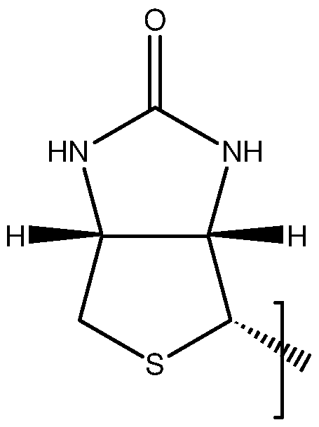
or BIO may be a moiety of a biotin analogue, for example
a desthiobiotin moiety: or a diaminobiotin moiety:

The biotinyl-conjugate may comprise a moiety of any other biotin analogue having a high binding affinity to avidin or streptavidin based proteins. BIO is preferably a biotin or desthiobiotin moiety Y may comprise a PET imaging moiety (such as a 18F labelled moiety, including but not limited to a moiety comprising a 18F labelled carbohydrate, or a 18F labelled boroaryl group), or an ultrasound contrast agent (such as a micro or nanobubble, including polylactic-co-glycolic acid (PGLA) micro- or nano-bubbles) or a quantum dot for fluorescence imaging.
Preferably, Y comprises a chelating moiety C, and the biotinyl-conjugate may further comprise a complexed metal ion, together forming a biotinyl-chelate complex. The biotinyl-chelate conjugate or complex may be used in a method of diagnosis (e.g. as a contrast agent) or in a method of treatment (e.g. as a therapeutic agent for example in a method of radiotherapy).
The biotinyl-conjugate has a number of advantages over known reagents. Firstly, the 1 ,2,3-triazole coupling group is "bioorthogonal", i.e. not found in any naturally occurring
biological systems. Accordingly, the coupling group is generally unreactive in biological conditions (such as are present in vivo), and in particular confers a high degree of biotinidase resistance, in comparison to known reagents. Secondly, the bioorthogonality enables the 1 ,2,3-triazole coupling group to be simply and efficiently synthesised in the presence of other functional groups, reducing the number of additional protection and deprotection steps required in order to couple the biotinyl moiety to a diagnostic or therapeutic agent, such as a chelate or metal complex. Thirdly, it has been found that a very high affinity is retained for avidin, streptavidin and related forms, including Avidin-Ox (a trademark of Sigma-Tau SpA), and neutral forms such as Extravidin (a trademark of Sigma-Aldrich Co.) or NeutrAvidin (a trademark of Pierce Biotechnology, Inc), in comparison to known biotinidase-resistant reagents.
Although not wishing to be bound by theory, it is hypothesized that the high binding affinity to (strept)avidin is a consequence of the electronic similarity of the triazole linkage, and particularly a 1 ,4-substituted triazole linkage, to an amide coupling, which has previously been found to be compatible with binding to (strept)avidin. This similarity arises from the lone pairs of the 2- and 3-nitrogen mimicking those of the carbonyl oxygen of the amide bond. In addition, just like the amide N-H bond, the polarized C(5)-H bond can act as a hydrogen bonding donor and the electrophilic and polarized 4-carbon is electronically similar to the carbonyl carbon of an amide (as shown in Figure 2).
Optionally, the biotinyl-conjugate comprises a linker between the 1 ,2,3-triazole group and Y, for example to improve solubility or modify the steric effect of Y. The linker may be a polyethylene glycol linker of the formula: (OCH2CH2)m', or a straight chain (CH2)g hydrocarbon linker, or any suitable type of linker. Alternatively, or in addition, the biotinyl-conjugate may comprise a coupling group between the 1 ,2,3-triazole group and Y, for example an amide or ester coupling. The invention also extends to a biotinyl-conjugate for use in a method of diagnosis or treatment of a subject, the biotinyl-conjugate having the general structure (1 c) or structure (2c):
Structure (1 c)
 wherein n or n' =1 -4 (and more preferably, n=1 and n =2);
BIO is a biotinyl moiety (and preferably a biotin or desthiobiotin moiety);
Y' comprises a diagnostic or therapeutic moiety (and preferably imaging and/or chelating moiety Y);
L is a linker and may comprise any suitable linker such as a (CH2)g linker or a PEG linker L having the structure

wherein m =1 -6, and is preferably 2 or 3; q=~\ -6 and is preferably 3; and
X is a coupling group, preferably an amide coupling group.
In some embodiments, the biotinyl-conjugate is for use as a contrast agent, such as a MRI, ultrasound, PET or X-ray contrast agent, in a method of imaging a subject.
The biotinyl-chelate conjugate may be used as a MRI (magnetic resonance imaging) contrast agent. Accordingly, the biotinyl-chelate conjugate may be a complex wherein the metal ion may be selected from the group Fe(lll), Mn(ll), Mn(lll), La(lll), Ce(lll), Gd(lll), Eu(lll), Dy(lll), Cu(ll), or may be any other suitable paramagnetic metal ion. In a preferred embodiment, the metal ion is Gd(lll). Gadolinium has a particularly high number of unpaired electrons (seven) and both the stability constants and toxicity of Gd(lll) in a variety of chelating groups is well known and well understood, in the context of MRI applications. The biotinyl-chelate complex may be used as a contrast agent for other applications, for example as an X-ray contrast agent, for use in radiography and computed tomography (CT). The metal ion may, for example, be barium.
The biotinyl-chelate complex may be used as a radiopharmaceutical, for example in a method of PET (positron emission tomography), and may comprise a chelated radioisotope, such as 82Rb or 82Sr or any other suitable position-emitter. The chelated radioisotope may be a gamma emitter such as 153Gd, 51Cr, 99mTc, 111 In or 67Ga and the biotinyl-chelate complex may be used in a method of X-ray fluorescence or otherwise as a radioactive tracer. The biotinyl-chelate complex may be used in a method of internal (and targeted) radiotherapy and the chelated radioisotope may be an emitter of ionising radiation, and may be selected from the group: 137Cs, 60Co, 192lr, 103Pd, 106Ru, 89Sr, 153Sm, 90Y.
The biotinyl-chelate complex may be neutrally charged, or may be an ionic species (for example having an overall charge of -1 or -2, balanced in solution by a suitable cation), depending on the charge of the chelated metal ion and the charge (if any) of the chelating moiety. The biotinyl-chelate complex may be a conjugate acid.
The use of the biotinyl-conjugate may, in some embodiments, be in a method comprising administering the biotinyl-conjugate to a subject. The method may comprise initially providing a subject with an avidin or streptavidin based pretarget, and subsequently administering the biotinyl-conjugate. The pretarget may be administered by any suitable method, including injection, ingestion, and transdermal or transmucosal administration.
By avidin or streptavidin based pretarget, we mean a pretarget (in the sense of two- step and three-step pretargeting approaches) bound, covalently or otherwise, to avidin, streptavidin or a protein based upon avidin or streptavidin and having a high binding affinity for biotin, including but not limited to Avidin-Ox, and neutral forms such as Extravidin or NeutrAvidin.
The method may comprise injecting an avidin or streptavidin based protein into a subject, to thereby provide a subject with a pretarget. For example, an avidin or streptavidin based protein may be injected into a tumour, so as to provide a pretarget labelled tumour cell or cells and subsequently a biotinyl-chelate complex comprising a chelated radioisotope is administered in order to bind to the pretarget.
The method may comprise administering a protein-labelled targeting agent (designed to specifically bind to a target, such as a particular cell surface protein) to a subject, wherein the protein-labelled targeting agent is bound covalently or otherwise to said protein label and the protein label is an avidin or streptavidin based protein. The targeting agent may be any suitable targeting agent, including but not limited to biologic targeting agents such as an antibody (e.g. a monoclonal antibody), an antibody fragment, a DNA, RNA or peptide aptamer, or a hapten-protein or peptide adduct, and including small molecule targeting agents (i.e. low molecular weight - typically <500Da - molecules which bind specifically to one or more target receptors, e.g. kinases), or any other suitable targeting agent known in the art (and disclosed for example by Sharkey and Goldenberg, Advanced Drug Delivery Reviews 60 (2008) 1407-1420, and by Seigneuric et al., Oncotarget. 201 1 July; 2(7): 557-561 ). The targeting agent is preferably an antibody.
The method preferably comprises administering the biotinyl-conjugate after an accumulation period has elapsed (to allow the protein-labelled targeting agent to accumulate at the target site and to allow non-bound protein-labelled targeting agent to clear from the subject's blood stream).
Alternatively, the method may comprise administering a biotinylated targeting agent (such as a biotinylated antibody) to a subject, and administering an avidin or streptavidin based protein to a subject, to thereby provide a subject with an avidin or streptavidin based pretarget. Accordingly, the method comprises administering the
avidin or streptavidin based protein after an accumulation period has elapsed (to allow the biotinylated targeting agent to accumulate at the target site and non-bound protein- labelled targeting agent to clear from the subject's blood stream). In a preferred embodiment (wherein the biotinyl-chelate conjugate is a complex and is, or functions as, a MRI contrast agent), the method further comprises acquiring a magnetic resonance image of the whole or part of a subject. Alternatively, or in addition, the method may comprise acquiring an X-ray image, CT-scan or other type of medical image.
The chelating moiety C may be a derivative of any suitable chelating agent and may optionally be a derivative of a chelating agent selected from the group:
DOTA (1 ,4,7,10-tetraazacyclododecane-1 ,4,7,10-tetraacetic acid), D03A (1 ,4,7,10- tetraazacyclodecane-1 ,4,7-triacetic acid), NOTA (1 ,4,7-triazacyclononane-1 ,4,7- triacetic acid), TETA (1 ,4,8,1 1 -tetraazacyclotetradecane-1 ,4,8,1 1 -tetraacetic acid), DTPA (diethylenetriaminepentaacetic acid), EDTA (ethylenediaminetetraacetic acid), NTA (trimethylaminetricarboxylic acid), DFO (deferoxamine), EDHPA (ethylenediamine-di(o-hydroxyphenyl)acetic acid), DIPY (dipyridyl), dithiosemicarbazones, hydroxamic acids, porphyrins, bis-aminoethanethiol, polyaminopolycarboxylates, dithiocarbamate, salen complexes, or their derivatives, including but not limited to Me-DTPA, CITC-DTPA ((S)-2-(para- isothiocyanatobenzyl)diethylenetriaminepentaacetic acid), DTPA-BMA (1 ,7- bis(methylcarbamoylmethyl)-1 ,4,7-triazaheptane-1 ,4,7-triacetic acid), cyclohexyl-DTPA, BT-D03A (10-(2,3-dihydroxy-1 -hydroxymethylpropyl)-1 ,4,7,10-tetraazacyclododecane- 1 ,4,7-triacetic acid), HP-D03A (10-(2-hydroxypropyl)-1 ,4,7-tetraazacyclododecane- 1 ,4,7-triacetic acid) and D03A-EA (l O-(aminoethyl)- 1 ,4,7,10-tetraazacyclododecane- 1 ,4,7-triacetic acid).
The chelating moiety C is preferably a derivative of DOTA, DTPA or D03A.
The biotinyl-conjugate may comprise more than one imaging and/or chelating moiety. For example, the conjugate may be for use in more than one method of treatment or diagnosis and each said imaging and/or chelating moiety selected accordingly. Y is not rapamycin or a rapamycin derivative.
In some embodiments, the biotinyl-chelate complex has the general structure (3) or structure (4); Structure (3) Structure (4)

BIO is a biotinyl moiety (and preferably a biotin or desthiobiotin moiety)
and Ch comprises the chelating moiety C and a metal ion (or other species) chelated by the chelating moiety.
The biotinyl-chelate complex may have the general structure (3a) or structure (4a):
Structure (3a)
Ch X /

Structure (4a)

BIO is a biotinyl moiety (and preferably a biotin or desthiobiotin moiety);
Ch comprises the chelating moiety C and a metal ion (or other species) chelated by the chelating moiety;
L is a linker and may comprise any suitable linker such as a (CH2)g linker or a PEG linker L having the structure

wherein m =1 -6, and is preferably 2 or 3; q -6 and is preferably 3; and
X is a coupling group, preferably an amide coupling group.
In a preferred embodiment (for example in embodiments wherein the biotinyl-chelate complex is for use in MRI as a MRI contrast agent), the biotinyl-chelate complex has structure (5a), (5b) or (6).
Structure (5a)


Structure (6)

wherein;
M is selected from the group Fe(lll), Mn(ll), Mn(lll), La(lll), Ce(lll), Gd(lll), Eu(lll), Dy(lll), Cu(ll);
BIO is a biotin or desthiobiotin moiety;
L is a linker; and
m = 1 to 6, n' = 1 to 4 and p = 1 or 2 Preferably, n'=2, and m, p=1 .
In some embodiments, the biotinyl-chelate complex has structure (7a) or (7b)
Structure (7a)

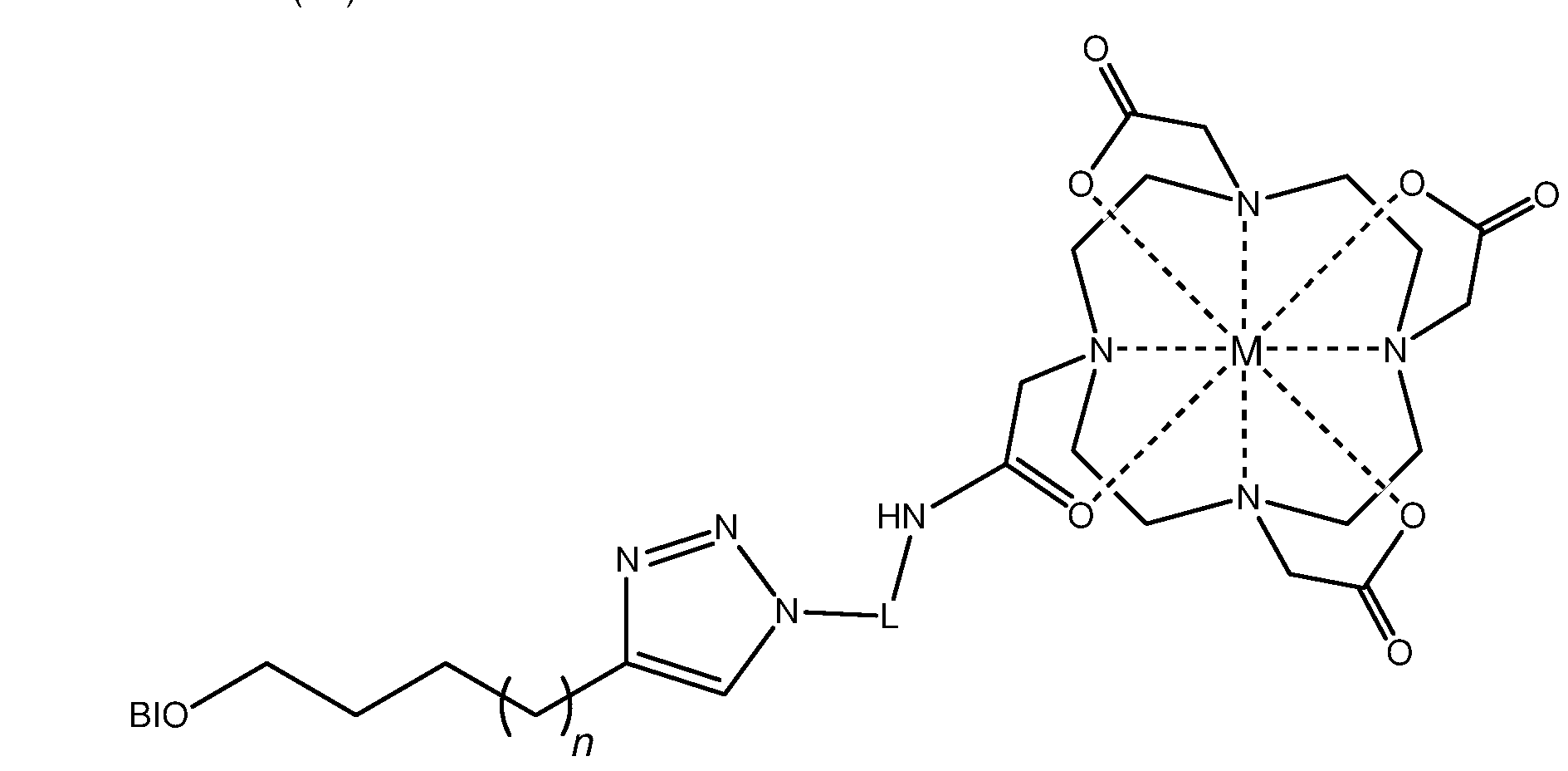
M is selected from the group Fe(lll), Mn(ll), Mn(lll), La(lll), Ce(lll), Gd(lll), Eu(lll), Dy(lll), Cu(ll);
BIO is a biotin or desthiobiotin moiety;
L is a linker; and
q = 1 to 6, n = 1 to 4
L referably comprises a PEG linker, L having the structure

wherein m =1 -6, and is preferably 2 or 3. Preferably, q = 3 and n = 1 .
Embodiments having structures (5a), (5b), (6), (7a) and (7b), wherein n = 1 or n'=2 have a biotinyl moiety having the closest possible structural similarity to the amide coupling of biocytin and to previously known biotinyl-conjugates which have been found to be compatible with (strept)avidin binding, (particularly embodiments having structure (7a) or (7b), due to the orientation of the triazole group in relation to the biotinyl moiety).
In order for the 1 ,2,3-triazole group to coordinate with the metal ion, in embodiments having structure (6), a minimum of angle strain is required wherein p = 1 .
The metal ion M is preferably Gd(lll).
The invention also extends to use of a biotinyl-conjugate for the manufacture of a medicament or a formulation, for the diagnosis or treatment of a subject.
Structures (5a), (5b), (6), (7a) and (7b) include neutrally charged complexes, ionic species (for example having an overall charge of -1 or -2, balanced in solution by a suitable cation), and conjugate acids (wherein one or more of the carboxylic acid groups are protonated), depending on the charge of the chelated metal ion and conditions (e.g. pH).
According to a second aspect of the invention there is provided the use of a biotinylating agent having structure (A1 ) or (A2), or having structure (B1 ) or (B2) to make a diagnostic or therapeutic agent comprising a biotinyl-conjugate having the general structure (A) or (B), respectively;
Structure (A1 ) Structure (A2)

Structure (B1 ) Structure (B2)

G a biotinyl moiety comprising a biotin, norbiotin, bisnorbiotin, desthiobiotin, diaminobiotin, dehydrobiotin, homobiotin or alphamethylbiotin moiety, or a moiety of any other biotin analogue having a high binding affinity to avidin or streptavidin based proteins;
L is a linker; and
A is a coupling moiety or a leaving group.
Preferably, the biotinylating agent has structure (8a) or (8b), or structure (9a) or (9b), for use in making a diagnostic or therapeutic agent comprising a biotinyl-conjugate having the general structure (1 ) or (2), respectively;
Structure (8a) Structure (8b)

Structure (9a) Structure (9b)
 wherein n or n' =1 -4;
BIO is a biotinyl moiety;
L is a linker; and
A is a coupling moiety or a leaving group.
The triazole may be a 1 ,4-substituted triazole or a 1 ,5-substituted triazole (and is preferably a 1 ,4-substituted triazole).
In some embodiments, n or n' = 1 or 2. n is preferably 1 . n' is preferably 2.
BIO may be a 5-substituted (SaS^S^afl^-oxohexahydro-l - -thieno[3,4-c/|imidazol-4- yl moiety (i.e. a biotin moiety), or may be a moiety of a biotin analogue such as a desthiobiotin moiety or a diaminobiotin moiety. The biotinylating agent may comprise any other biotin analogue having a high binding affinity to avidin or streptavidin based proteins.
L may be any suitable linker preferably comprises a (CH2)g linker or PEG linker, L havin the structure

wherein m =1 -6, and is preferably 2 or 3.
And q=~\ -6 and is preferably 3.
A may be any suitable coupling or leaving group, for example (but not limited to) group selected from; halide, hydroxyl, alcohol, carbonyl, carboxyl, amine, in particular primary amine, ester, acid anhydride, alkyne, azide.
The invention also extends to a biotinylating agent having structure (8b) or (9b).
Biotinylating agents having structure (8a) or (9a) may be used to make biotinylating agents having the structure (8b) or (9b), respectively.
Preferably, the biotinylating agent is used to make a contrast agent, such as an MRI contrast agent. A biotinylating agent having structure (8a) or (9a) may be used to make a biotinyl-chelate complex having structure (3) or (3a) or the having structure (4) or (4a), respectively. A biotinylating agent having the structure (8b) or (9b) may be used to make a biotinyl-chelate complex having structure (3a) or (4a), respectively.
Thus, the invention also extends to use of chelating agent having structure (28) or structure (29) to make a contrast agent (or, in some embodiments, another type of therapeutic or diagnostic agent) comprising a biotinyl-chelate complex having the general structure (3) and (4), respectively.
Structure (28)

Structure (29) Ch-N3 wherein Ch comprises the chelating moiety C and a paramagnetic metal ion chelated by the chelating moiety C.
Further preferred and optional features of the second aspect, correspond to preferred and optional features of the first aspect.
According to a third aspect of the invention, there is provided a biotinyl-conjugate having the general structure (1 ) or (2) (wherein n or n' = 1 or 2; and n is preferably 1 and n' is preferably 2).
The triazole may be a 1 ,4-substituted triazole or a 1 ,5-substituted triazole, and thus the biotinyl-conjugate may have the general structure (1 a), (1 b), (2a) or (2b).
Preferably, the biotinyl-conjugate has the structure (1 a) or (2a). The biotinyl-conjugate may have structure (1 c) or (2c).
BIO may be a 5-substituted (SaS^S^afl^-oxohexahydro-l H-thieno[3,4-c]imidazol-4- yl moiety (i.e. a biotin moiety), or BIO may be a moiety of a biotin analogue such as a desthiobiotin moiety, a diaminobiotin moiety, or the biotinylating agent may comprise a moiety of any other biotin analogue having a high binding affinity to avidin or streptavidin based proteins.
Y may comprise a PET imaging moiety (such as a 18F labelled moiety), or an ultrasound contrast agent (e.g. a polylactic-co-glycolic acid (PGLA) micro- or nano- bubble) or a quantum dot for fluorescence imaging.
Preferably, Y comprises a chelating moiety C, and the biotinyl-conjugate may further comprise a complexed metal ion, together forming a biotinyl-chelate complex. The chelating moiety C may be a derivative of any suitable chelating agent and may optionally be a derivative of a chelating agent selected from the group:
DOTA (1 ,4,7,10-tetraazacyclododecane-1 ,4,7,10-tetraacetic acid), D03A (1 ,4,7,10- tetraazacyclodecane-1 ,4,7-triacetic acid), NOTA (1 ,4,7-triazacyclononane-1 ,4,7- triacetic acid), TETA (1 ,4,8,1 1 -tetraazacyclotetradecane-1 ,4,8,1 1 -tetraacetic acid), DTPA (diethylenetriaminepentaacetic acid), EDTA (ethylenediaminetetraacetic acid), NTA (trimethylaminetricarboxylic acid), DFO (deferoxamine), EDHPA (ethylenediamine-di(o-hydroxyphenyl)acetic acid), DIPY (dipyridyl), dithiosemicarbazones, hydroxamic acids, porphyrins, bis-aminoethanethiol, polyaminopolycarboxylates, dithiocarbamate, salen complexes, or their derivatives, including but not limited to Me-DTPA, CITC-DTPA ((S)-2-(para- isothiocyanatobenzyl)diethylenetriaminepentaacetic acid), DTPA-BMA (1 ,7- bis(methylcarbamoylmethyl)-1 ,4,7-triazaheptane-1 ,4,7-triacetic acid), cyclohexyl-DTPA, BT-D03A (10-(2,3-dihydroxy-1 -hydroxymethylpropyl)-1 ,4,7,10-tetraazacyclododecane- 1 ,4,7-triacetic acid), HP-D03A (10-(2-hydroxypropyl)-1 ,4,7-tetraazacyclododecane- 1 ,4,7-triacetic acid) and D03A-EA (l O-(aminoethyl)- 1 ,4,7,10-tetraazacyclododecane- 1 ,4,7-triacetic acid).
The chelating moiety C is preferably a derivative of DOTA, DTPA or D03A.
Optionally, the biotinyl-conjugate comprises a linker between the 1 ,2,3-triazole group and Y, for example to improve solubility or modify the steric effect of Y. The linker may be a polyethylene glycol linker of the formula: (OCH2CH2)m', or a straight chain (CH2)g hydrocarbon linker, or any suitable type of linker. Alternatively, or in addition, the biotinyl-conjugate may comprise a coupling group between the 1 ,2,3-triazole group and Y, for example an amide or ester coupling.
In some embodiments, the biotinyl-chelate conjugate has the general formula;

BIO is a biotinyl moiety (and preferably a biotin or desthiobiotin moiety);
and Ch comprises a chelating moiety C.
The biotinyl-chelate complex may have the general structure:
h

Or:

BIO is a biotinyl moiety (and preferably a biotin or desthiobiotin moiety);
Ch comprises the chelating moiety C and a metal ion (or other species) chelated by the chelating moiety;
L is a linker and, in preferred embodiments, L is a (CH2)g linker, or comprises a PEG linker L having the structure

wherein m =1 -6, and is preferably 2 or 3; q=~\ -6 and is preferably 3; and
X is a coupling group, preferably an amide coupling group.
The chelating moiety may comprises a derivative of DOTA. Preferably, the biotinyl- chelate conjugate has structure (10a), (10b), (1 1 ), (12a) or (12b), or a conjugate base thereof:
Structure (10a)


BIO is a biotin or desthiobiotin moiety; L is a linker; and
m = 1 to 6, n' = 1 to 4 and p = 1 or 2
Structure (12a)

Structure (12b)

BIO is a biotin or desthiobiotin moiety;
L is a linker; and
q = 1 to 6, n = 1 to 4
L referably comprises a PEG linker, L having the structure

wherein m =1 -6, and is preferably 2 or 3. Preferably, n =2, and m, p=1 . Preferably, q = 3 and n = 1 .
The invention also extends to a biotinyl-chelate complex, comprising a chelated species (preferably a metal ion) chelated by the chelating moiety C of the biotinyl- chelate complex. Preferably, the metal ion is a paramagnetic metal ion selected from the group: Fe(lll), Mn(ll), Mn(lll), La(lll), Ce(lll), Gd(lll), Eu(lll), Dy(lll), Cu(ll). The complex is preferably a biotinyl-chelate gadolinium (III) complex, for use as a MRI contrast agent.
In a fourth aspect, the invention extends to a biotinylating agent having structure (8b), or (9b) wherein n or n' =1 -4 or (9a) wherein n = 1 , 3 or 4; and wherein BIO is a biotinyl moiety; L is a linker; and A is a coupling moiety or a leaving group.
The triazole may be a 1 ,4-substituted triazole or a 1 ,5-substituted triazole (and is preferably a 1 ,4-substituted triazole). n or n' = 1 or 2. n is preferably 1 . n' is preferably 2.
BIO may be a 5-substituted (SaS^S^afl^-oxohexahydro-l - -thieno[3,4-c/|imidazol-4- yl moiety (i.e. a biotin moiety), or may be a moiety of a biotin analogue such as a desthiobiotin moiety or a diaminobiotin moiety, or the biotinylating agent may comprise a moiety of any other biotin analogue having a high binding affinity to avidin or streptavidin based proteins.
BIO is preferably a biotin moiety or a desthiobiotin moiety. In embodiments wherein BIO is a biotin moiety, n≠2. Accordingly, in preferred embodiments, the biotinylating agent bears a very close structural similarity to biotin and biocytin, and both the biotinylating agent and compounds biotinylated by the biotinylating agent show a particularly high specificity of binding to avidin, streptavidin and related (strept)avidin based proteins. The invention extends in a fifth aspect to a contrast agent comprising or consisting of a biotinyl-chelate complex comprising a biotinyl-chelate conjugate according to the third aspect, and in a sixth aspect to use of a biotinyl-conjugate according to the third aspect, or a biotinyl-chelate complex comprising a biotinyl-chelate conjugate according to the third aspect, in medicine.
The biotinyl-conjugate (or the biotinyl-chelate complex or the biotinyl-chelate conjugate) may be used in the preparation of a composition for administration to a subject.
According to a seventh aspect of the invention, there is provided a method of diagnosis or treatment, comprising administering a biotinyl-conjugate (or a biotinyl-chelate conjugate or complex) according to the third aspect, to a subject.
The subject may be a human or animal subject, or tissues or parts thereof, cell or cell lines, and thus the biotinyl-chelate complex may administered in vitro or, more preferably, in vivo.
The biotinyl-conjugate may be administered by any suitable method, including by injection, ingestion, transdermal^ or transmucosally. Preferably, the method is a method of pretargeted delivery of a therapeutic or diagnostic agent, comprising the steps of providing a subject with an avidin or streptavidin based pretarget, and administering the biotinyl-conjugate.
An avidin or streptavidin based protein may be injected into a subject, to thereby provide a subject with a pretarget. Alternatively, the method may comprise administering a protein-labelled targeting agent to a subject, wherein the protein label is an avidin or streptavidin based protein.
In some embodiments, the method comprises administering a biotinylated targeting agent to a subject, and administering an avidin or streptavidin based protein to a subject (preferably after an accumulation period has elapsed), to thereby provide a subject with an avidin or streptavidin based pretarget.
The biotinyl-conjugate is preferably administered after an accumulation period has elapsed.
The method may be a method of imaging a subject, comprising pretargeted delivery of the biotinyl-conjugate, and acquiring an image (for example a magnetic resonance image or other type of image, such as an X-ray, ultrasound or CT-scan) of the whole or part of a subject.
Preferably the method is a method of obtaining a magnetic resonance image of a subject, comprising pretargeted delivery of a biotinyl-chelate complex. Thus, the biotinyl-chelate complex preferably has structure (3), (3a), (4) or (4a), wherein M is selected from the group Fe(lll), Mn(ll), Mn(lll), La(lll), Ce(lll), Gd(lll), Eu(lll), Dy(lll), Cu(ll). The chelating moiety is preferably a derivative of DOTA, D03A or DTPA.
According to an eighth aspect of the invention, there is provided a method of synthesis of a biotinylating agent having (i) structure (8b) or (ii) structure (9b), comprising the steps of;
a) Preparing a biotinylating agent having (i) structure (8a) or (ii) structure (9a) b) Preparing (i) an alkyne of structure: L or (ii) an azide of structure L N3
c) Performing an azide-alkyne cycloaddition.
wherein L is a linker; and A is a coupling moiety or a leaving group.
The biotinylating agent having structure (8b) or (9b) preferably comprises a 1 ,4- substituted 1 ,2,3-triazole, and the method preferably comprises performing a Huisgen 1 ,3-dipolar cycloaddition.
According to a ninth aspect of the invention, there is provided a method of synthesising a biotinyl-conjugate having the general structure (1 ) or structure (2), the method comprising the steps of:
(i) preparing a biotinyl-azide having the structure (8a) and an alkyne having the structure Y ;
(ii) performing an azide alkyne cycloaddition (preferably a Huisgen 1 ,3-dipolar cycloaddition) so as to form a biotinyl-conjugate having structure (1 ) (and preferably a biotinyl-conjugate having structure (1 a)). or the steps of;
(i) preparing a biotinyl alkyne having the structure (9a) and an azide having the structure Y-N3;
(ii) performing an azide alkyne cycloaddition (preferably a Huisgen 1 ,3-dipolar cycloaddition) between the said alkyne and the said azide so as to form a biotinyl-conjugate having structure (2) (and preferably a biotinyl-conjugate having structure (2a)).
wherein Y comprises a moiety which may be an imaging and/or chelating moiety.
The method may be a method of synthesis of a biotinyl-chelate conjugate or a biotinyl- chelate complex, and the method may comprise providing a chelate azide or a chelate alkyne, as the case may be.
The chelate alkyne or azide may comprise a derivative of any suitable chelating group C, and in some embodiments C is a derivative of DOTA, DTPA or D03A. Preferably the chelate-azide has structure (15), or a conjugate base thereof, or the chelate alkyne has structure (16) or a conjugate base thereof:
Structure (15)

wherein q = 1 -6 (preferably, q = 3). Structure (16)

wherein m = 1 -6, and preferably m=1 Structure (17)

wherein p = 1 or 2 (and preferably p=1 )
In embodiments comprising synthesis of a biotinyl-chelate complex, the chelating moiety C may further comprise a chelated metal ion (or other chelated species), which may be chelated by the chelating moiety at any stage of synthesis. Thus, the said chelate alkyne or the said chelate azide may comprise a chelated metal ion, so as to produce a biotinyl-chelate complex when coupled to the biotinyl alkyne or biotinyl azide, as the case may be, or a biotinyl-chelate conjugate may be synthesised and mixing with a metal salt solution to thereby conjugate a metal ion within the said chelating moiety C.
Preferably, the metal ion is Gd(lll), but may be any metal ion selected from the group set out in relation to the first aspect.
According to a tenth aspect of the invention, there is provided a method of synthesising a biotinyl-conjugate having the (i) general structure (18) or (ii) the general structure (19);
Structure (18)


wherein n or n' =1 -4;
BIO is a biotinyl moiety;
L is a linker;
X is a coupling group, preferably an amide coupling group; and
Y' comprises a therapeutic or diagnostic moiety,
the method comprising the steps of;
a) preparing a biotinylating agent having (i) structure (8b) or (ii) structure (9b) (preferably by the method of the eighth aspect)
b) coupling the biotinylating agent to a compound Y'-B, wherein Y' comprises a therapeutic or diagnostic moiety, and B is a coupling moiety or a leaving group.
B may be any coupling moiety or leaving group which can be caused to react with coupling moiety or leaving group A to form coupling group X. B may be any suitable coupling or leaving group, for example (but not limited to) a group selected from; halide, hydroxyl, alcohol, carbonyl, carboxyl, amine, in particular a primary amine, ester, acid anhydride, alkyne, azide. Y' may comprise a chelating moiety C, and the method may comprise subsequently chelating a metal ion in the chelating moiety, forming a biotin-chelate complex. The method advantageously enables the synthesis of the biotinylating agent (which is typically formed by an alkyne-azide cycloaddition, and preferably a copper (I) catalysed Huisgen 1 ,3-dipolar cycloaddition) to be conducted apart from the chelating moiety, thus preventing any unwanted chelation of reagents used in the synthesis of the biotinylating agent.
L is referably a (CH2)g linker or comprises a PEG linker, L having the structure

wherein m =1 -6 (and more preferably m'=2 or 3).
and q=~\ -6 (and more preferably q=3).
X is preferably an amide coupling group.
Y' preferably comprises imaging and/or chelating moiety Y.
BIO may be a 5-substituted (SaS^S^afl^-oxohexahydro-l H-thieno[3,4-c/]imidazol-4- yl moiety (i.e. a biotin moiety):

or BIO may be a moiety of a biotin analogue, for example
a desthiobiotin moiety: or a diaminobiotin moiety:

BIO is preferably a biotin moiety or a desthiobiotin moiety.
The method is preferably a method of synthesising a biotinyl-conjugate comprising a 1 ,4-substuituted 1 ,2,3-triazole (i.e. a biotinyl cojnjugate having structure (1 c) or (2c)).
Thus the invention also extends to a biotinyl-conjugate having the general structure (18) or (19), and to the use thereof as a diagnostic or therapeutic agent.
Y' is not rapamycin or a rapamycin derivative.
The invention also extends to a method of synthesis of a biotinyl conjugate having structure (A2) or (B2) comprising the steps of;
a) Preparing a biotinylating agent having (i) structure (A1 ) or (ii) structure (B1 ) b) Preparing (i) an alkyne of structure: L or (ii) an azide of structure L N3
c) Performing an azide-alkyne cycloaddition.
wherein L is a linker; and A is a coupling moiety or a leaving group.
And to a method of synthesising a biotinyl-conjugate having the general structure (A) or structure (B), the method comprising the steps of:
(i) preparing a biotinyl-azide having the structure (A1 ) and an alkyne having the structure Y ;
(ii) performing an azide alkyne cycloaddition (preferably a Huisgen 1 ,3-dipolar cycloaddition) so as to form a biotinyl-conjugate having structure (A); or the steps of;
(i) preparing a biotinyl alkyne having the structure (B1 ) and an azide having the structure Y-N3;
(ii) performing an azide alkyne cycloaddition (preferably a Huisgen 1 ,3-dipolar cycloaddition) between the said alkyne and the said azide so as to form a biotinyl-conjugate having structure (B);
wherein Y comprises a moiety which may be an imaging and/or chelating moiety.
And to a method of synthesising a biotinyl-conjugate having the (i) general structure (A3) or (ii) the general structure (B3);
Structure (A3) Structure (B3)

wherein G is a biotinyl moiety;
L is a linker; X is a coupling group, preferably an amide coupling group; and
Y' comprises a therapeutic or diagnostic moiety,
the method comprising the steps of;
a) preparing a biotinylating agent having (i) structure (A2) or (ii) structure (B2) b) coupling the biotinylating agent to a compound Y'-B, wherein Y' comprises a therapeutic or diagnostic moiety, and B is a coupling moiety or a leaving group.
Further preferred and optional features of the tenth aspect, in particular of groups Y, BIO, X, A, B and L, correspond to preferred and optional features of the first through ninth aspects.
In an eleventh aspect, the invention extends to a biotynylation kit comprising (i) a biotinyl azide having the structure (8a), or (ii) a biotinyl alkyne having the structure (9a).
The kit may further comprise (i) an alkyne having the structure Y or (ii) an azide having the structure Y-N3.
The invention also extends to a kit comprising a biotinylating agent having structure (9a) or (9b). The kit may further comprise a compound Y'-B, wherein Y' comprises a therapeutic or diagnostic moiety, and B is a coupling moiety or a leaving group.
Preferred and optional features of any of the first through eleventh aspects of the invention correspond to preferred and optional features of any other of the first through eleventh aspects.
Description of the Drawings
Figure 1 shows a schematic representation of representation of 3-step (strept)avidin/biotin based pretargeting; Figure 2 shows the electronic similarity between (a) a 1 ,2,3-triazole group and (b) an amide group;
Figure 3 shows the chemical structure of biotinylating agents 6 and 8, and of biotinyl- conjugates 9, 10 and 1 1 ;
Figure 4 shows a plot of the stability to cleavage by partially-purified biotinidase up to 126 h, of compounds 9-1 1 and of N-biotinyl-PABA;
Figure 5 shows the binding of compounds 9-1 1 to avidin assessed spectrophoto- metrically by displacement of HABA from the HABA-avidin complex.
Figure 6 shows the chemical structure of compounds S1 -S6, of compounds 12, 14 and 16 and of biotin 1 a and biotin analogues 1 b-1 c; Figure 7 shows a reaction scheme for the preparation of a further embodiment of a biotinyl-chelate complex of the present invention.
Detailed Description of an Example Embodiment Biotinylating agents 6 and 8, and biotinyl-conjugates (and more particularly biotinyl- chelate complexes) 9-11 were prepared by the methods described in detail in examples (1 )-(1 1 ), below. Biotinidase resistance and binding affinity for (strept)avidin of compounds 9-11 was then measured, using the procedures described in examples (12)-(14), below. The chemical structures of compounds 6 and 8-11 are shown in Figure 3.
Human biotinidase was partially purified from human serum using ammonium sulphate precipitation, anion-exchange chromatography and size-exclusion chromatography.
Biotinidase activity was assessed by measuring the rate of hydrolysis of the commercially available substrate, A/-biotinyl-p-aminobenzoic acid (A/-biotinyl-PABA). The p-aminobenzoic acid which results from cleavage is diazotized and derivatized with Λ/-1 -naphthylethylenediamine in situ to form a diazo dye with max 545 nm, allowing a quantitative assessment of activity. The resistance of triazoles 9-11 to cleavage by biotinidase was determined by incubation with the partially purified biotinidase for up to 126 h. RP-HPLC showed that all three complexes were resistant to cleavage by biotinidase, showing substantially no instability in the presence of biotinidase, in marked contrast to the known substrate A/-biotinyl-PABA which is fully hydrolyzed under the same conditions within 6 h (Figure 4).
As well as displaying resistance to cleavage by biotinidase, the new constructs must also bind strongly to (strept)avidin for their successful application in biotin technologies. The binding affinity of complexes 9-11 to avidin was probed using spectrophotometric competition experiments against the known ligand 4-hydroxyazobenzene-2-carboxylic acid (HABA, KD = 5.8 x 10"6). All of the ligands were shown to inhibit the HABA-avidin complex in a dose-dependent manner (Figure 5). Three additional ligands of known binding affinity were included in this study: biotin 1a KD = 10"15 desthiobiotin 1 b KD = 10" 13 and diaminobiotin 1 c KD = 3 x 10"7. Comparison of the inhibition of the HABA-avidin complex generated by these ligands suggests KD's for complexes 9-11 in the nM range.
Table 1 shows the percentage displacement of HABA from the HABA-avidin complex by biotin 1 a, biotin derivatives 1 b-c, and biotin-DOTA complexes at a ligand:avidin ratio of 4:1 .
Table 1
Derivative or complex (link Displacement
functionality) %
biotin 1a TOO
desthiobiotin 1 b 100
bisDOTA-Lys-(pAB)-C3 (amide) 97
bisDOTA-Lys-C3 (amide) 95
Gd-DOTA complex 11 (triazole) 70
Gd-DOTA complex 9 (triazole) 68
bisDOTA-C3 (amine) 66
Gd-DOTA complex 10 (triazole) 58
diaminobiotin 1 c 41
At the natural 4:1 molar ratio of biotin-DOTA complex:avidin, complexes 9 and 11 bind equally well to avidin. In combination with the results from the biotinidase resistance studies this indicates that both azide 6 and alkyne 8 provide excellent biotinidase- resistant bioorthogonal biotin labels.
Potential imaging agent, biotinyl-chelate complex 10, shows a slightly reduced binding affinity to avidin, in comparison to complexes 9 or 11 due to a steric effect from the short linkage to the Gd-DOTA moiety.
Comparison with the data reported for similar experiments conducted on more traditional (amide- or amine-based) biotinidase-resistant biotin-DOTA conjugates bisDOTA-Lys-(pAB)-C3 (amide coupled) bisDOTA-Lys-C3 (amide coupled) and bisDOTA-C3 (amine coupled), reported in Pratesi, A.; Bucelli, F.; Mori, I.; Chinol, M.; Vedoliva, A.; Paganelli, G.; Rivieccio, V.; Gariboldi, L; Ginanneschi, M. J. Med. Chem. 2010, 53, 432-440, bioorthogonal biotin-triazole conjugates 9-11 compare well against the previously known amide and amine coupled biotinidase-chelate complexes, whilst also showing substantially no instability in the presence of biotinidase. In conclusion, we have demonstrated that the amide-bond mimicking triazole provides a biotinidase resistant linkage to biotin that may be readily produced from bioorthogonal precursors. We anticipate that the use of biotinylating agents such as azide 6 or alkyne 8, optionally coupled via appropriate spacer units to a range of functionalities will enable a diverse spectrum of biotin (strept)avidin based technologies to be more successfully applied under conditions where the presence of the hydrolytic enzyme biotinidase has previously hindered their use.
The resistance to biotinidase renders the conjugates of the present invention suitable for a wide range of applications in human medicine, including targeted delivery of therapeutic agents such as radiotherapeutic agents. In addition, polymersomes (nanovesicles self-assembled from block copolymers) have recently been demonstrated to be able to deliver considerable amounts of cargo to a target site, by
conjugation of targeting ligands to their surface. The biotinylating agents herein disclosed would facilitate more efficient targeting than currently known agents due to higher stability in vivo, in particular in the presence of biotinidase, of the resulting polymersome-biotin conjugation.
A further embodiment of a biotinylating agent of the present invention, compound 18, may be prepared by the CuAAC coupling of biotinyl alkyne 8 to PEG2-aminoazide (CAS number 166388-57-4) using the general method described in examples (9)-(1 1 ) below.
Biotinylating agent 18 was then coupled to commercially available D03AtBu-acetate 20 (CAS number 137076-54-1 ) by a conventional amide coupling reaction for example as described in J. Am. Chem. Soc, 201 1 , 133, 16346. The tertiary butyl protecting groups were then cleaved and the biotinyl-chelate conjugate 22 mixed with a suitable Gd(lll) solution to yield the biotin-triazole-PEG2-DOTA(Gd) complex 24, as shown in the reaction scheme of Figure 7.
In an alternative embodiment (not shown), a biotin-triazole-PEG3-DOTA(Gd) complex was prepared by an analogous method from a biotinylating agent having a PEG3 linker in place of the PEG2 linker of molecule 18.
EXPERIMENTAL EXAMPLES Preparation of biotin azide 6 and biotin alkyne 8
Biotinylating agents were prepared as follows:
(1 ) Synthesis of Compound S1 : Methyl-5-((3aS,4S,6aff)-2-Oxo-hexahvdro-thieno[3,4- c/limidazol-4-yl)-pentanoate
Acetyl chloride (2.00 ml_, 28.0 mmol) was added slowly to anhydrous methanol (50 mL) at 0 °C and stirred for 10 min. This solution was added dropwise to D-Biotin 1 (0.500 g, 2.05 mmol) and the solution was heated to reflux for 1 h. The solvent was removed in vacuo and the residue was purified by column chromatography (DCM:MeOH, 90:10) to
give biotin methyl ester S1 as a colourless solid (0.607 g, quantitative); Rf (DCM:MeOH, 90:10) = 0.3; mp 155 °C; IR 3276 (NH), 1744, 1688 (C=0); 1H NMR δ (400 MHz, CD3OD) 4.52-4.47 (1 H, m, NHCHCH), 4.33-4.28 (1 H, m, NHCHCH2), 3.66 (3H, s, CH3), 3.24-3.17 (1 H, m, SCH), 2.95 (1 H, dd, J = 12.8, 4.9, CH^HBS), 2.70 (1 H, d, J = 12.8, CHAHBS), 2.35 (2H, t, J = 7.3, CH2), 1 .80-1 .54 (4H, m, 2 χ CH2), 1 .50-1 .40 (2H, m, SCHCH2); 13C NMR δ (62.9 MHz, CD3OD) 175.9 (CO), 166.2 (CO), 63.4 (CH), 61 .7 (CH), 57.0 (CH), 52.1 (CH3), 41 .1 (CH2), 34.6 (CH2), 29.7 (CH2), 29.5 (CH2), 26.0 (CH2); m/z (ESI+, MeOH); 281 ([M+Na]+, 35%), 259 ([M+H]+, 100). (2) Synthesis of Compound S2: (3aS,4S,6aff)-4-(5-Hvdroxy-pentyl)-tetrahvdro- thieno[3,4-c/limidazol-2-one
Anhydrous DCM (40 mL) was added to biotin methyl ester S1 (0.529 g, 2.05 mmol) under nitrogen resulting in a colourless suspension. The solution was cooled to -78°C and DIBAL (6.23 mL, 7.12 mmol, 1 .0 M in DCM) was added dropwise. After stirring at -78 °C for 20 min the solution was allowed to warm to room temperature and was stirred for 2 h. The mixture was again cooled to -78°C and the reaction was quenched by the addition of MeOH (4.5 mL), followed by MeOH:H20 (15 mL, 2:1 ). The solvent was removed in vacuo and the residue was purified by column chromatography (DCM:MeOH, 90:10) to give alcohol S2 as a colourless solid (0.570 g, quantitative). Rf (DCM:MeOH, 90:10) = 0.2; mp 161 <C; IR 3415 (OH), 1689 (C=0); 1H NMR δ (400 MHz, CD3OD) 4.49 (1 H, dd, J = 7.5, 4.6, NHCHCH), 4.31 (1 H, dd, J = 7.9, 4.5, NHCHCH2), 3.55 (2H, t, J = 6.5, CH20H), 3.25-3.18 (1 H, m, SCH), 2.94 (1 H, dd, J = 12.8, 5.0,
 2.72 (1 H, d, J = 12.8, CHAHBS), 1 .80-1 .51 (4H, m, CH2), 1 .53-1 .39 (4H, m, CH2); 13C NMR δ (62.9 MHz, CD3OD) 164.8 (CO), 62.0 (CH), 61 .5 (CH), 60.2 (CH), 55.8 (CH2) 39.6 (CH2), 32.0 (CH2), 28.8 (CH2), 28.4 (CH2), 25.5 (CH2); m/z (ESI+, MeOH) 483 ([2M+H]+ 61 %), 253 [M+Na]+, 41 ), 231 ([M+H]+, 49), 213 (23).
(3) Synthesis of Compound S3: (3aS,4S,6afl)-4-(5-Bromo-pentyl)-tetrahvdro- thieno[3,4-c/limidazol-2-one
Triphenylphosphine (0.390 g, 1 .50 mmol) was dissolved in anhydrous DCM (1 -2 mL) under nitrogen and transferred with a syringe to a solution of bromine (77 μΙ, 1 .50 mmol) in anhydrous DCM (3 mL) at 0°C. This solution was stirred for 10 min. The resulting yellow slurry was transferred with a syringe to a suspension of alcohol S2
(0.230 g, 1 .00 mmol) in anhydrous DCM (2 ml_). After complete dissolution of all reactants the reaction was monitored by TLC. After 4 h a white precipitate formed and the reaction was quenched by the addition of Na2S203 solution (10 ml_, 0.1 M, aq) resulting in a colour change from yellow to colourless. Water (10 mL) was added and the organic layer was separated from the aqueous layer. The aqueous layer was extracted with DCM (3 χ 20 mL), and the combined organics were washed with water (3 x 10 mL), dried with Na2S04 and the solvent removed in vacuo. The crude product was purified using column chromatography (DCM:MeOH, 90:10) to yield the bromide S3 as a colourless solid (0.220 g, 74%). R, (DCM:MeOH, 90:10) = 0.4; mp 166 °C; IR 3200 (NH), 1702 (C=0); 1H NMR δ (400 MHz, CD3OD) 4.47 (1 H, dd, J = 7.8, 5.0, NHCHCH), 4.30 (1 H, dd, J = 7.9, 4.5, NHCHCH2), 3.44 (2H, t, J = 6.7, CH2Br), 3.23- 3.18 (1 H, m, SCH), 2.91 (1 H, dd, J = 12.7, 5.0, C¾HBS), 2.72 (1 H, d, J = 12.7, CHAHBS), 1 .86 (2H, qn, J = 7.0, CH2), 1 .80-1 .55 (2H, m, CH2), 1 .54-1 .40 (4H, m, CH2) ; 13C NMR δ (126 MHz, CD3OD) 164.8 (CO), 62.0 (CH), 60.2 (CH), 55.7 (CH), 39.6 (CH2), 32.9 (CH2), 32.4 (CH2), 28.3 (CH2), 28.1 (CH2), 27.8 (CH2); m/z (ESI+, MeOH) 317 ([81 BrM+Na]+, 25%), 315 ([79BrM+Na]+, 27), 213 (100).
(4) Synthesis of Compound 6: (3aS,4S,6aff)-4-(5-Azido-pent-1 -yl)-tetrahvdro- thieno[3,4-c/limidazol-2-one
Sodium azide (0.230 g, 2.00 mmol) in water (10 mL), was added to a solution of bromide S3 (0.220 g, 0.750 mmol) and tetra-n-butylammonium bromide (0.040 g, 0.136 mmol) in toluene (10 mL), and the reaction mixture was heated at 80°C for 16 h. The organic layer was separated from the aqueous layer and the aqueous layer was extracted with DCM (3 χ 20 mL). The combined organic layers were dried with Na2S04 and the volatiles were removed in vacuo. The crude product was purified by column chromatography (DCM:MeOH, 90:10) to give azide 6 as a colourless solid (0.160 g, 84%). Rf (DCM:MeOH, 90:10) = 0.4; mp 146 °C; IR 3213 (NH), 2085 (N3), 1697 (C=0); 1H NMR δ (400 MHz, CD3OD) 4.52 (1 H, dd, J = 7.9, 5.0, NHCHCH), 4.33 (1 H, dd, J = 7.9, 5.0, NHCHCH2), 3.34 (2H, m, CH2N3), 3.24 (1 H, dt, J = 9.9, 5.0, SCH), 2.97 (1 H, dd, J = 12.7, 5.0,
 2.75 (1 H, d, J = 12.7, CHAHBS), 1 .82-1 .56 (4H, m, CH2), 1 .56-1 .42 (4H, m, CH2); 13C NMR δ (126 MHz, CD3OD) 166.2 (CO), 63.4 (CH), 61 .6 (CH), 57.1 (CH), 52.4 (CH2), 41 .0 (CH2), 29.9 (CH2), 29.8 (CH2), 29.7 (CH2), 27.8 (CH2); m/z (ESI+, MeOH) 788 ([3M+Na]+, 30%), 533 ([2M+Na]+, 100), 278 ([M+Na]+, 39).
(5) Synthesis of Compound 8: (3aS,4S,6afl)-4-(hex-5-vn-1 -yl)tetrahvdro-thienof3,4- c/limidazol-2-one Biotin methyl ester S1 (0.320 g, 1 .24 mmol) was dissolved in anhydrous DCM (8 mL), and the solution was cooled to -78 °C. DIBAL-H (2.09 mL, 2.30 mmol, 1 .1 M solution in cyclohexane) was added over 1 0 min, and the mixture stirred at -78 °C for 4 h. The excess DIBAL-H was quenched with anhydrous methanol (5 mL), and the reaction mixture allowed to warm to 0 °C. Potassium carbonate (0.34 g, 2.46 mmol), Ohira- Bestmann reagent (0.29 g, 1 .51 mmol), and anhydrous methanol (5 mL) were added and the reaction stirred for 24 h. Further Ohira-Bestmann reagent (0.29 g, 1 .51 mmol) was added and the reaction was stirred for a further 1 8 h. Rochelle's salt (10 mL) and ethyl acetate (20 mL) were added and the mixture stirred vigorously for 1 h. The organic layer was separated, washed with brine (3 x 20 mL), dried over magnesium sulfate, and the solvent was removed under reduced pressure. Where unreacted biotin methyl ester remained, this was hydrolysed prior to chromatographic purification using LiOH in THF/H20 to aid separation. The crude mixture was purified by column chromatography (DCM:MeOH, 90:1 0) to give terminal biotin alkyne 8 as a colourless solid (0.1 3 g, 0.58 mmol, 47 %). R, (DCM:MeOH, 90:10) = 0.5; mp 1 58-162 °C; IR 3242 (NH), 2108 (C≡CH), 1703 (C=0) ; 1H NMR δ (500 MHz, CDCI3) 4.97 (1 H, s, NHCHCH), 4.89 (1 H, s, NHCHCH2), 4.55 (1 H, dd, J = 7.7, 4.6 Hz, NHCHCH), 4.37 (1 H, dd, J = 7.7, 4.6 Hz, NHCHCH2), 3.21 (1 H, ddd, 8.0, 6.6, 4.6 Hz, SCH), 2.97 (1 H, dd, J = 1 2.8, 5.0 Hz, CHAHBS), 2.77 (1 H, d, J = 1 2.8 Hz, CHAHBS), 2.26 (2H, td, J = 6.5, 2.5 Hz, CH2C≡CH), 2.00 (1 H, t, J = 2.5 Hz, C≡CH), 1 .75-1 .65 (2H, m, CH2), 1 .62-1 .55 (4H, m, 2 x CH2) ; 13C NMR δ (126 MHz, CDCI3) 1 62.9 (CO), 84.1 (C), 68.8 (CH), 61 .9 (CH), 60.1 (CH), 55.3 (CH), 40.6 (CH2), 28.1 (CH2), 28.0 (CH2), 27.9 (CH2), 18.1 (CH2); m/z (ESI+, MeOH) 471 ([2M+Na]+, 100%), 247 ([M+Na]+, 21 ), 225 ([M+H]+, 1 1 ) ; HRMS (ESI+, MeOH) [2M+Na]+ found 471 .1 864, Cn H16N20iS requires 471 .1859. Preparation of Gd-DOTA alkynes S4, S5 and Gd-DOTA azide S6
2.75 (1 H, d, J = 12.7, CHAHBS), 1 .82-1 .56 (4H, m, CH2), 1 .56-1 .42 (4H, m, CH2); 13C NMR δ (126 MHz, CD3OD) 166.2 (CO), 63.4 (CH), 61 .6 (CH), 57.1 (CH), 52.4 (CH2), 41 .0 (CH2), 29.9 (CH2), 29.8 (CH2), 29.7 (CH2), 27.8 (CH2); m/z (ESI+, MeOH) 788 ([3M+Na]+, 30%), 533 ([2M+Na]+, 100), 278 ([M+Na]+, 39).
(5) Synthesis of Compound 8: (3aS,4S,6afl)-4-(hex-5-vn-1 -yl)tetrahvdro-thienof3,4- c/limidazol-2-one Biotin methyl ester S1 (0.320 g, 1 .24 mmol) was dissolved in anhydrous DCM (8 mL), and the solution was cooled to -78 °C. DIBAL-H (2.09 mL, 2.30 mmol, 1 .1 M solution in cyclohexane) was added over 1 0 min, and the mixture stirred at -78 °C for 4 h. The excess DIBAL-H was quenched with anhydrous methanol (5 mL), and the reaction mixture allowed to warm to 0 °C. Potassium carbonate (0.34 g, 2.46 mmol), Ohira- Bestmann reagent (0.29 g, 1 .51 mmol), and anhydrous methanol (5 mL) were added and the reaction stirred for 24 h. Further Ohira-Bestmann reagent (0.29 g, 1 .51 mmol) was added and the reaction was stirred for a further 1 8 h. Rochelle's salt (10 mL) and ethyl acetate (20 mL) were added and the mixture stirred vigorously for 1 h. The organic layer was separated, washed with brine (3 x 20 mL), dried over magnesium sulfate, and the solvent was removed under reduced pressure. Where unreacted biotin methyl ester remained, this was hydrolysed prior to chromatographic purification using LiOH in THF/H20 to aid separation. The crude mixture was purified by column chromatography (DCM:MeOH, 90:1 0) to give terminal biotin alkyne 8 as a colourless solid (0.1 3 g, 0.58 mmol, 47 %). R, (DCM:MeOH, 90:10) = 0.5; mp 1 58-162 °C; IR 3242 (NH), 2108 (C≡CH), 1703 (C=0) ; 1H NMR δ (500 MHz, CDCI3) 4.97 (1 H, s, NHCHCH), 4.89 (1 H, s, NHCHCH2), 4.55 (1 H, dd, J = 7.7, 4.6 Hz, NHCHCH), 4.37 (1 H, dd, J = 7.7, 4.6 Hz, NHCHCH2), 3.21 (1 H, ddd, 8.0, 6.6, 4.6 Hz, SCH), 2.97 (1 H, dd, J = 1 2.8, 5.0 Hz, CHAHBS), 2.77 (1 H, d, J = 1 2.8 Hz, CHAHBS), 2.26 (2H, td, J = 6.5, 2.5 Hz, CH2C≡CH), 2.00 (1 H, t, J = 2.5 Hz, C≡CH), 1 .75-1 .65 (2H, m, CH2), 1 .62-1 .55 (4H, m, 2 x CH2) ; 13C NMR δ (126 MHz, CDCI3) 1 62.9 (CO), 84.1 (C), 68.8 (CH), 61 .9 (CH), 60.1 (CH), 55.3 (CH), 40.6 (CH2), 28.1 (CH2), 28.0 (CH2), 27.9 (CH2), 18.1 (CH2); m/z (ESI+, MeOH) 471 ([2M+Na]+, 100%), 247 ([M+Na]+, 21 ), 225 ([M+H]+, 1 1 ) ; HRMS (ESI+, MeOH) [2M+Na]+ found 471 .1 864, Cn H16N20iS requires 471 .1859. Preparation of Gd-DOTA alkynes S4, S5 and Gd-DOTA azide S6
Chelate alkynes and chelate azides were prepared as follows:
(6) Synthesis of Compound S4: 1 -{2-[(prop-2-vnyl)aminol-2-oxoethyl)-4,7, 10- tris(carboxymethyl)-1 ,4,7, 10-tetraazacyclododecyl qadolinium(ll l)
A solution of the DOTA-alkyne (compound 12) (0.220 g, 0.470 mmol) was prepared in water (15 mL) by the method set out in Viguier R. F. H.; Hulme, A. N. J. Am. Chem. Soc. 2006, 128, 1 1370-1 1371 , and adjusted to pH 7 by addition of KOH (0.1 M, aq.).
Gadolinium(lll) triflate (0.286 g, 0.470 mmol) was dissolved in water (15 mL) and added to this DOTA-alkyne solution. After mixing thoroughly for 15 min, the solution was readjusted to pH 6 using KOH (0.1 M, aq.) and was stirred at 60 °C for 4 h. The solution was once again adjusted to pH 6 and the solvent was removed in vacuo. The colourless solid was dissolved in EtOH, the insoluble salts removed by filtration and the solvent was removed in vacuo to yield Gd-DOTA-complex S4 as a pale yellow hygroscopic solid (0.500 g, quantitative), mp ^ 80 oC (decomp); IR 3018 (NH), 2091 (C≡C), 1685 (C=0), 1620 (C=0); m/z (ESI+, MeOH) 1231 ([2(158GdM)+K]+, 15%) 635 ([158GdM+K]+, 100), 619 ([158GdM+Na]+, 15), 597 ([158GdM+H]+, 10); HRMS (FAB, 3- NOBA) [158GdM+H]+ Ci9H2907N5158Gd requires 597.1308, found 597.1326. The appropriate isotope pattern was observed.
(7) Synthesis of Compound S5: 1 ,4, 7-tris(carboxymethyl)-10-(prop-2-vnyl)-1 ,4,7,10- tetraazacvclododecyl qadolinium(lll)
A solution of the DOTA-alkyne (compound 14) (0.129 g, 0.336 mmol) was prepared in water (10 mL) by the method set out in Jauregui, M.; Perry, W. S.; Allain, C; Vidler, L. R.; Willis, M. C. Kenwright, A. M.; Snaith, J. S.; Stasiuk, G. J.; Lowe M. P.; Faulkner S. Dalton Trans. 2009, 6283-6285, and adjusted to pH 7 by addition of KOH (0.1 M). Gadolinium(lll) triflate (0.203 g, 0.336 mmol) was dissolved in water (10 mL) and added to the DOTA-alkyne solution. After mixing thoroughly for 15 min, the solution was readjusted to pH 6 using KOH and was stirred at 60 °C for 4 h. The solution was once again adjusted to pH 6 and the solvent was removed in vacuo. The colourless solid was dissolved in EtOH, the insoluble salts removed by filtration and the solvent was removed in vacuo to yield Gd-DOTA-complex S5 as a pale yellow hygroscopic solid (0.362 g, quantitative), mp 190-192°C (decomp); IR 1678 (C=0); m/z (ESI+, MeOH) 540 ([158GdM+H]+, 83%), 232 (100); HRMS (ESI+, MeOH) [158GdM+H]+ C17H2606N4 158Gd requires 540.1099, found 540.1095. The appropriate isotope pattern was observed.
(8) Synthesis of Compound S6: 1 -(2-[(3-azidopropyl)aminol-2-oxoethyl)-4,7,10- tris(carboxymethyl)-1 ,4,7,10-tetraazacvclododecyl qadolinium(lll)
A solution of the DOTA-azide (compound 16) (0.041 g, 0.078 mmol) was prepared in water (5 mL) by the method set out in Schultz, M. K.; Parameswarappa, S. G.; Pigge, F. C. Org. Lett, 2010, 12, 2398-2401 , and adjusted to pH 7 by addition of KOH (0.1 M). Gadolinium(lll) triflate (0.0.47 g, 0.078 mmol) was dissolved in water (10 mL) and added to the DOTA-azide solution. After mixing thoroughly for 15 min, the solution was readjusted to pH 6 using KOH and was stirred at 60 °C for 4 h. The solution was once again adjusted to pH 6 and the solvent was removed in vacuo. The colourless solid was dissolved in EtOH, the insoluble salts removed by filtration and the solvent was removed in vacuo to yield Gd-DOTA-complex S6 as a pale yellow hygroscopic solid (0.105 g, quantitative), mp 198-200 <C (decomp); IR 3400 (NH), 2105 (N3), 1682 (C=0), 1626 (C=0); m/z (ESI-, MeOH) 640 ([158GdM-H]~, 100%). HRMS (ESI-, MeOH) [158GdM-H] C19H3o07N8 158Gd requires 640.1484, found 640.1463. The appropriate isotope pattern was observed.
Optionally, Gd(lll) may be complexed within the DOTA group at other stages of the reaction.
Preparation of biotinyl-chelate complexes 9-11
(9) Synthesis of Gadolinium-biotin triazole complex 9 Gd-DOTA complex S4 (0.093 g, 0.157 mmol) and TBTA (0.008 g, 0.016 mmol) were dissolved in 'BuOH:H20 (2:1 , 15 mL) and allowed to stir for 15 mins. Sodium ascorbate (0.004 g, 0.031 mmol) was then added and allowed to stir for 15 min followed by copper(ll) sulfate (0.004 g, 0.016 mmol). After a further 15 min biotin azide 6 (0.040 g, 0.157 mmol) was added and the solution stirred under nitrogen at room temperature for 16 h. The solvent was then removed in vacuo to afford crude triazole as a pale yellow solid. The crude product was purified by preparative reversed phase HPLC using a C18 column and H20/acetonitrile + 0.1 % TFA as mobile phase. HPLC purification yielded contrast agent 9 as a colourless solid (0.034 mg, 16%). Rt = 42 min; mp 300°C (decomp); IR 1670 (C=0), 1593 (C=0); m/z (ESI+, H20) 874 ([158GdM+Na]+, 100%); (ESI-, H20) 850 ([158GdM-H]~, 100%); HRMS (FAB, 3-NOBA) [M+H]+
C29H46Gd158N10O8S requires 852.2462, found 852.2423. The appropriate isotope pattern was observed.
(10) Synthesis of Gadolinium-biotin triazole complex 10
Gd-DOTA complex S5 (0.084 g, 0.157 mmol) and TBTA (0.008 g, 0.016 mmol) were dissolved in 'BuOH:H20 (2:1 , 15 mL) and allowed to stir for 15 mins. Sodium ascorbate (0.004 g, 0.031 mmol) was then added and allowed to stir for 15 min followed by copper(ll) sulfate (0.004 g, 0.016 mmol). After a further 15 min biotin azide 6 (0.040 g, 0.157 mmol) was added and the solution stirred under nitrogen at room temperature for 16 h. The solvent was then removed in vacuo to afford crude triazole as a cream solid. The crude product was purified by preparative reversed phase HPLC using a C18 column and H20/acetonitrile + 0.1 % TFA as mobile phase. HPLC purification yielded contrast agent 10 as a colourless solid (0.041 g, 33%). Rt = 38 min; mp 280 ^ (decomp); IR 3200 (NH), 1682 (C=0), 1614 (C=0); m/z (ESI+, DMSO) 833 ([158GdM+K]+, 19%), 817 ([158GdM+Na]+, 100); (ESI-, DMSO) 831 ([158GdM-2H+Kr, 42%), 793 ([158GdM-H]~, 100%); HRMS (ESI-, DMSO) [M-H] C27H4i 158GdN907S requires 793.2096, found 793.2100. The appropriate isotope pattern was observed. (1 1 ) Synthesis of Gadolinium-biotin triazole complex 1 1
Biotin alkyne 8 (0.026 g, 0.1 16 mmol) and TBTA (0.008 g, 0.016 mmol) were dissolved in 'BuOH:H20 (2:1 , 15 mL) and allowed to stir for 15 mins. Sodium ascorbate (0.004 g, 0.031 mmol) was then added and allowed to stir for 15 min followed by copper(ll) sulfate (0.004 g, 0.016 mmol). After a further 15 min Gd-DOTA complex S6 (0.050 g, 0.078 mmol) was added and the solution stirred under nitrogen at room temperature for 16 h. The solvent was then removed in vacuo to afford crude triazole as a pale green solid. The crude product was purified by preparative reversed phase HPLC using a C18 column and H20/acetonitrile + 0.1 % TFA as mobile phase. HPLC purification yielded contrast agent 11 as a pale blue solid (0.036 g, 54%). Rt = 40 min; mp 296^ (decomp); IR 3369 (NH), 1682 (C=0), 1602 (C=0); m/z (ESI+, H20) 904 ([158GdM+K]+, 22%), 888 ([158GdM+Na]+, 100); m/z (ESI-, H20) 902 ([158GdM-2H+Kr, 15%), 864 ([158GdMr, 100%); HRMS (ESI-, MeOH/H20) [158GdM-H]~ C3oH46158GdN1008S requires 864.2449, found 864.2467. The appropriate isotope pattern was observed.
In alternative embodiments (not shown) analogues of biotinylating agents 6 and 8, and of biotinyl-chelate complexes 9-11 may be prepared, by the methods of examples (1 )-
(1 1 ) , from desthiobiotin 1 b or diaminobiotin 1 c. Purification of Biotinidase and Determination of Activity
(12) Purification of Biotinidase
Biotinidase was purified from a fresh human plasma sample on the basis of the purification procedure set out in Chauhan, J.; Dakshinamurti, K. J. Biol. Chem. 1986, 261, 4268-4275. Fractions were monitored using a standard biotinidase activity assay as set out in Kobza, K. A.; Chaiseeda, K.; Sarath, G.; Takacs, J. M.; Zempleni, J. J. Nut. Biochem. 2008, 19, 826-832, and by SDS-PAGE. Total protein amounts were determined by the absorbance at 280 nm. Reagent and buffer solutions were handled using standard methods, but the solutions of the diazotization agent (sodium nitrite) were freshly prepared immediately prior to the biotinidase activity assay and stock solutions of the derivatization agent A/-1 -naphthylethylenediamine dihydrochloride were stored in the dark. Buffer solutions A and B were prepared, having the following compositions: Buffer A Buffer B
50 mM NaH2P04/Na2HP04, pH 6.0 50 mM NaH2P04/Na2HP04, pH 6.0
1 mM EDTA 1 mM EDTA
5 mM cysteamine hydrochloride
(13) Determination of Biotinidase activity assay
The activity of the biotinidase was determined by measuring the rate of hydrolysis of the commercially available substrate A/-biotinyl-p-aminobenzoic acid (A/-biotinyl-PABA), according to the protocol set out in Kobza, K. A.; Chaiseeda, K.; Sarath, G.; Takacs, J. M.; Zempleni, J. J. Nut. Biochem. 2008, 19, 826-832. Each assay was calibrated against PABA standards as detailed below.
Preliminary investigations showed that: i. variation due to the thermal instability of the intermediate diazonium salt was minimized by conducting the derivatization process at 0 °C; ii. hydrolysis of the substrate A/-biotinyl-PABA in the alkaline stock solution (0.1 M NaOH) was minimal (-5% over 6 weeks storage); iii. assay measurements were reproducible in the concentration range 0.003 to 0.08 mM for up to 120 min after transfer of the assay solution to microcuvettes; iv. BSA (used as a model of the highly abundant protein HSA) increased absorbance readings by up to 35%, with a plateau for concentrations >0.5 mg mL"1 , hence BSA (0.5 mg mL"1) was added to the standards to mimic any background signal generated by HSA in the partially purified biotinidase samples; v. unreacted A/-biotinyl-PABA reduced the absorbance reading by -30%, at a concentration of 0.15 mM which might be expected to remain at the end of the assay, hence A/-biotinyl-PABA was added to the PABA standards as well as the enzyme solutions under investigation. A 96-well plate was loaded with standard stock solutions of PABA (150 μΙ_, 0.003 mM - 0.250 mM) in a solution of BSA (0.2 mg mL"1) in buffer A, or the enzyme solution (1 5 or 30 μΐ, depending on expected activity) in a solution of BSA (0.2 mg mL"1) in buffer A (135 or 120 μΙ_). A/-biotinyl-PABA (10 μΙ_, 6.0 mM in 0.1 M NaOH; 60 nmol per well; 0.2 mM final concentration) was added to each well (including PABA standards) and the plates were incubated for 1 h at 37°C, so as to standardize the signal and compensate for the presence of unreacted substrate. Reactions were stopped by addition of HCI (30 [ii, 6 M).
Then, the following solutions were added at 3 min intervals at CO:
- sodium nitrite (80 [ii, 3 mM, freshly prepared) (diazotizing reagent)
- ammonium sulfamate (15 [ii, 50 mM) (to quench residual nitrite)
- A/-1 -naphthylethylenediamine dihydrochloride (15 iL, 5 mM).
During an incubation time of 1 0 min samples were transferred to 1 .5 mL reaction tubes. Precipitated proteins were removed by centrifugation (max. speed for 10 min), and the supernatant was transferred to microcuvettes. The absorbance was measured at 545 nm.
One unit of biotinidase activity was defined as 1 nmol PABA released per min.
Measurement of Biotinidase resistance and streptavidin binding affinity, of biotinyl-chelate complexes
(14) Resistance to biotinidase
Buffer solutions of compounds 9-11 , and A/-biotinyl-PABA were prepared, with and without biotinidase. Solution compositions are set out in Table 2. Each biotinidase solution was incubated at 25° C for up to 126 hours. The samples were removed at regular time intervals and were diluted for HPLC analysis (the results of which are shown in Figure 4). A resourcinol standard was added to each solution immediately prior to HPLC analysis.
Table 2

All samples were analysed using an Agilent 1 100 HPLC using a reversed phase Luna 5 μ C18(2) column (id 50 mm χ 2mm) at a flow rate of 1 mL min"1. The Biotin-Gd(lll)- DOTA complexes 9-11 and resorcinol were detected at 215 nm. The gradient elution programme shown in Table 3 was used for all experiments
Table 3

(15) Affinity to avidin The binding capacity of biotin derivatives to avidin was determined by spectrophotometric competition with the dye 2-(4'-hydroxyphenylazo)-benzoic acid (HABA). In its bound form HABA has an absorption maximum of 500 nm. Addition of an avidin ligand leads to displacement of HABA from the binding site which can be measured by the resulting decrease in absorbance at 500 nm.
For this experiment HABA/Avidin reagent (obtained from Sigma-Aldrich Company Limited) was used consisting of 0.3 mM HABA, 0.45 mg mL"1 avidin, 0.3 M NaCI, 0.01 M HEPES (A/-[2-hydroxyethyl]piperazine-A/,A/-[2-ethanesulfonic acid], a buffer with pKa=7.5), 0.01 M MgCI2, 0.02% sodium azide (as a preservative), pH 7.3.
Solutions of decreasing concentration for each biotin derivative were prepared by serial dilution in water. Aliquots (50 μΙ) of the diluted biotin derivatives (final concentration range 1 -20 mM) were added to 400 μΙ of HABA/avidin reagent in a disposable UV micro cuvette. The final concentration of avidin in the presence of the biotin derivative was 0.24 mM, and this was used to calculate the biotin derivative:avidin ratio for each sample. After thorough mixing of the solution by inversion the absorbance at 500 nm was measured immediately on a Varian Cary® 50 UV-Vis spectrophotometer. (CARY is a Trade Mark of Agilent Technologies, Inc.). The absorbance at 500 nm in the presence of the biotin derivative was calculated using the equation 500 ~
 . This new A500 value represents the remaining HABA-avidin complex. It was converted to a percentage of the initial HABA-avidin complex and plotted against the biotin derivative:avidin ratio (as shown in Figure 5).
Claims
CLAIMS:
1 . A biotinyl-conjugate for use in a method of diagnosis or treatment of a subject, the biotinyl-conjugate having the general structure (A) or (B):
Structure (B)

wherein G is a biotinyl moiety;
and Y comprises a moiety which may be an imaging and/or chelating moiety; G comprising a biotin, norbiotin, bisnorbiotin, desthiobiotin, diaminobiotin, dehydrobiotin, homobiotin or alphamethylbiotin moiety, or a moiety of any other biotin analogue having a high binding affinity to avidin or streptavidin based proteins.
2. A biotinyl-conjugate according to claim 1 , having the general structure (1 ) or (2):
Structure (1 )

Structure (2)

wherein n or n' =1 -4;
BIO is a biotinyl moiety;
and Y a moiety which may be an imaging and/or chelating moiety.
3. A biotinyl-conjugate according to claim 2, wherein n=1 and n'=2.
A biotinyl-conjugate according to any preceding claim, wherein the triazole is a 1 ,4-substituted triazole and the biotinyl-conjugate has the general structure (1 a) or (2a).
Structure (1 a)


5. A biotinyl-conjugate according to any one preceding claim, wherein BIO is a 5- substituted (3aS,4S,6afl)-2-oxohexahydro-1 H-thieno[3,4-c]imidazol-4-yl moiety:

or a desthiobiotin moiety:
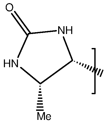
6. A biotinyl-conjugate for use in a method of diagnosis or treatment of a subject, having the general structure (1 c) or structure (2c):
Structure (1 c)
"
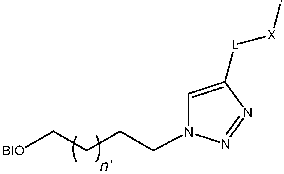
Structure (2c)
Y X I
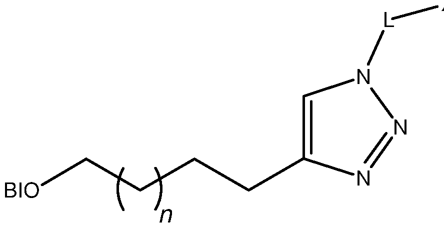
BIO is a biotinyl moiety;
Y' comprises a diagnostic or therapeutic moiety;
L is a linker; and X is a coupling group.
A biotinyl-conjugate according to claim 6, wherein X is an amide coupling group and L is CH2)g or a PEG linker having the structure

wherein m =1 -6; q^1 -6
A biotinyl-conjugate according to any one preceding claim, wherein biotinyl- conjugate is for use as a MRI, ultrasound, PET or X-ray contrast agent, in a method of imaging a subject.
9. A biotinyl-conjugate according to any one preceding claim, wherein Y comprises a PET imaging moiety or an ultrasound contrast agent.
10 A biotinyl-conjugate according to any one of claims 2 to 5, wherein Y comprises a chelating moiety C, and the biotinyl-conjugate further comprises a complexed metal ion, together forming a biotinyl-chelate complex.
A biotinyl-conjugate according to claim 10, wherein the chelating moiety C is a derivative of a chelating agent selected from the group:
DOTA (1 ,4,7,10-tetraazacyclododecane-1 ,4,7,10-tetraacetic acid), D03A (1 ,4,7,10-tetraazacyclodecane-1 ,4,7-triacetic acid), NOTA (1 ,4,7- triazacyclononane-1 ,4,7-triacetic acid), TETA (1 ,4,8,1 1 - tetraazacyclotetradecane-1 ,4,8,1 1 -tetraacetic acid), DTPA
(diethylenetriaminepentaacetic acid), EDTA (ethylenediaminetetraacetic acid), NTA (trimethylaminetricarboxylic acid), DFO (deferoxamine), EDHPA (ethylenediamine-di(o-hydroxyphenyl)acetic acid), DIPY (dipyridyl), dithiosemicarbazones, hydroxamic acids, porphyrins, bis-aminoethanethiol, polyaminopolycarboxylates, dithiocarbamate, salen complexes, or their derivatives, including but not limited to Me-DTPA, CITC-DTPA ((S)-2-(para- isothiocyanatobenzyl)diethylenetriaminepentaacetic acid), DTPA-BMA (1 ,7- bis(methylcarbamoylmethyl)-1 ,4,7-triazaheptane-1 ,4,7-triacetic acid), cyclohexyl-DTPA, BT-D03A (10-(2,3-dihydroxy-1 -hydroxymethylpropyl)- 1 ,4,7,10-tetraazacyclododecane-1 ,4,7-triacetic acid), HP-D03A (10-(2-
hydroxypropyl)-1 ,4,7-tetraazacyclododecane-1 ,4,7-triacetic acid) and D03A-EA (l O-(aminoethyl)- 1 ,4,7, 10-tetraazacyclododecane-1 ,4,7-triacetic acid).
A biotinyl-conjugate according to claim 1 1 , wherein the chelating moiety C is a derivative of DOTA, DTPA or D03A.
A biotinyl-conjugate according to any one of claims 10 to 12, for use as a MRI contrast agent, wherein the metal ion is be selected from the group: Fe(lll), Mn(ll), Mn(lll), La(lll), Ce(lll), Gd(lll), Eu(lll), Dy(lll), Cu(ll).
A biotinyl-conjugate according to any one preceding claim, having the general structure (3) or structure (4);
Structure (3)

Structure (4)

wherein n or n' =1 -4
BIO is a biotinyl moiety
and Ch comprises a chelating moiety C and a metal ion chelated by the chelating moiety.
15. A biotinyl-conjugate according to any one of claims 1 to 13, having the general structure (3a) or structure (4a):
Structure (3a)

Structure (4a)

BIO is a biotinyl moiety;
Ch comprises the chelating moiety C and a metal ion chelated by the chelating moiety; X is a coupling group;
and L is a linker.
16. A biotin-conjugate according to any one of claims 1 to 13, having structure (5a), (5b) or (6).


10
Structure (6)

wherein;
M is selected from the group Fe(lll), Mn(ll), Mn(lll), La(lll), Ce(lll), Gd(lll),
Eu(lll), Dy(lll), Cu(ll);
BIO is a biotin or desthiobiotin moiety;
L is a linker; and
m = 1 to 6, n' = 1 to 4 and p = 1 or 2
A biotin-conjugate according to claim 16, wherein n'=2, and m, p=1 .
A biotin-conjugate according to any one of claims 1 to 13, having structure (7a) or (7b)

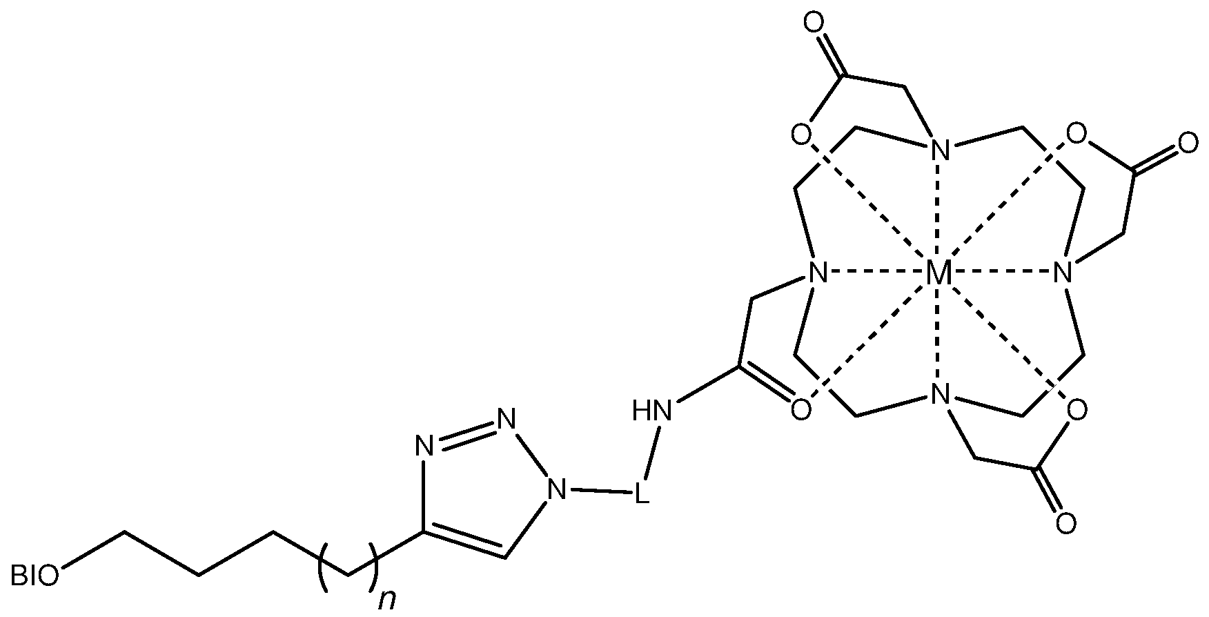
wherein;
M is selected from the group Fe(lll), Mn(ll), Mn(lll), La(lll), Ce(lll), Gd(lll), Eu(lll), Dy(lll), Cu(ll);
BIO is a biotin or desthiobiotin moiety;
L is a linker; and
q = 1 to 6, n = 1 to 4
19. A biotin-conjugate according to claim 18, wherein L is a PEG linker, L having the structure

and; q = 3, n = 1 .
20. A biotinyl-conjugate according to any one preceding claim, wherein the method comprises initially providing a subject with an avidin or streptavidin based pretarget, and subsequently administering the biotinyl-conjugate to the subject.
21 . A biotinyl-conjugate according to claim 20, wherein the method comprises administering a protein-labelled targeting agent to a subject, wherein the protein label is an avidin or streptavidin based protein, and administering the biotinyl- conjugate after an accumulation period has elapsed.
22. A biotinyl-conjugate according to claim 20, wherein the method comprises administering a biotinylated targeting agent to a subject, and administering an avidin or streptavidin based protein to a subject after an accumulation period has elapsed, to thereby provide a subject with an avidin or streptavidin based pretarget, and administering the biotinyl-conjugate after an accumulation period has elapsed.
23. A biotinyl-complex according to any one of claims 20 to 22, wherein the method comprises subsequently administering the biotinyl-complex to the subject, and further comprises acquiring a magnetic resonance image of the whole or part of a subject.
24. Use of a biotinylating agent having structure (8a) (8b), or having structure (9a) or (9b) to make a diagnostic or therapeutic agent comprising a biotinyl- conjugate having the general structure (1 ) or (2), respectively;
Structure (8a) Structure (8b)

BIO is a biotinyl moiety;
L is a linker; and
A is a coupling moiety or a leaving group. 25. Use of a biotinylating agent according to claim 24, wherein the triazole is a 1 ,4- substituted triazole.
26. Use of a biotinylating agent according to claim 24 or 25, wherein n=1 and n'=2. 27. Use of a biotinylating agent according to any one of claims 24 to 26, wherein BIO is a 5-substituted (SaS^S^afl^-oxohexahydro-l - -thieno[3,4-c/|imidazol- 4-yl moiety, or a desthiobiotin moiety.
28. Use of a biotinylating agent having structure (8a) or (9a) to make a biotinylating agent having the structure (8b) or (9b), respectively.
29. A biotinyl-conjugate having the general structure (1 ) or (2):
wherein n or n' =1 -4; BIO is a biotinyl moiety; and Y a moiety which may be an imaging and/or chelating moiety.
A biotinyl-conjugate having the general structure (1 c) or (2c), wherein n, n' =1 - 4; BIO is a biotinyl moiety; Y' comprises a diagnostic or therapeutic moiety; L is a linker; and X is a coupling group, preferably an amide coupling group.
A biotinyl-conjugate according to claim 29 or 30, wherein BIO is a 5-substituted (3aS,4S,6afl)-2-oxohexahydro-1 H-thieno[3,4-c]imidazol-4-yl moiety or a desthiobiotin moiety, n=1 , n'=2 and the triazole is 1 ,4-substituted.
A biotinyl-conjugate according to any one of claims 29 to 31 , wherein Y or Y' comprises a chelating moiety C optionally be a derivative of a chelating agent selected from the group:
DOTA (1 , 4,7, 10-tetraazacyclododecane-1 , 4,7,10-tetraacetic acid), D03A (1 , 4,7,10-tetraazacyclodecane-1 ,4,7-triacetic acid), NOTA (1 ,4,7- triazacyclononane-1 ,4,7-triacetic acid), TETA (1 ,4,8,1 1 - tetraazacyclotetradecane-1 ,4,8,1 1 -tetraacetic acid), DTPA
(diethylenetriaminepentaacetic acid), EDTA (ethylenediaminetetraacetic acid), NTA (trimethylaminetricarboxylic acid), DFO (deferoxamine), EDHPA (ethylenediamine-di(o-hydroxyphenyl)acetic acid), DIPY (dipyridyl), dithiosemicarbazones, hydroxamic acids, porphyrins, bis-aminoethanethiol, polyaminopolycarboxylates, dithiocarbamate, salen complexes, or their derivatives, including but not limited to Me-DTPA, CITC-DTPA ((S)-2-(para- isothiocyanatobenzyl)diethylenetriaminepentaacetic acid), DTPA-BMA (1 ,7- bis(methylcarbamoylmethyl)-1 ,4,7-triazaheptane-1 ,4,7-triacetic acid), cyclohexyl-DTPA, BT-D03A (10-(2,3-dihydroxy-1 -hydroxymethylpropyl)- 1 ,4,7,10-tetraazacyclododecane-1 ,4,7-triacetic acid), HP-D03A (10-(2- hydroxypropyl)-1 ,4,7-tetraazacyclododecane-1 ,4,7-triacetic acid) and D03A-EA (l O-(aminoethyl)- 1 ,4,7,10-tetraazacyclododecane-1 ,4,7-triacetic acid), and the biotinyl-conjugate further comprises a complexed metal ion, together forming a biotinyl-chelate complex.
A biotinyl-conjugate according to any one of claims 29 to 32 having the general structure:
 wherein n or n' =1 -4;
BIO is a biotinyl moiety;
Ch comprises the chelating moiety C;
L is a CH2)g linker, or a PEG linker having the structure

wherein m'=1 -6; q=1 -6; and
X is a coupling group.
A biotinyl-conjugate according to claim 33, wherein the chelating moiety comprises a derivative of DOTA, the biotinyl-conjugate having the structure (10a), (10b), (1 1 ), (12a) or (12b), or a conjugate base thereof:
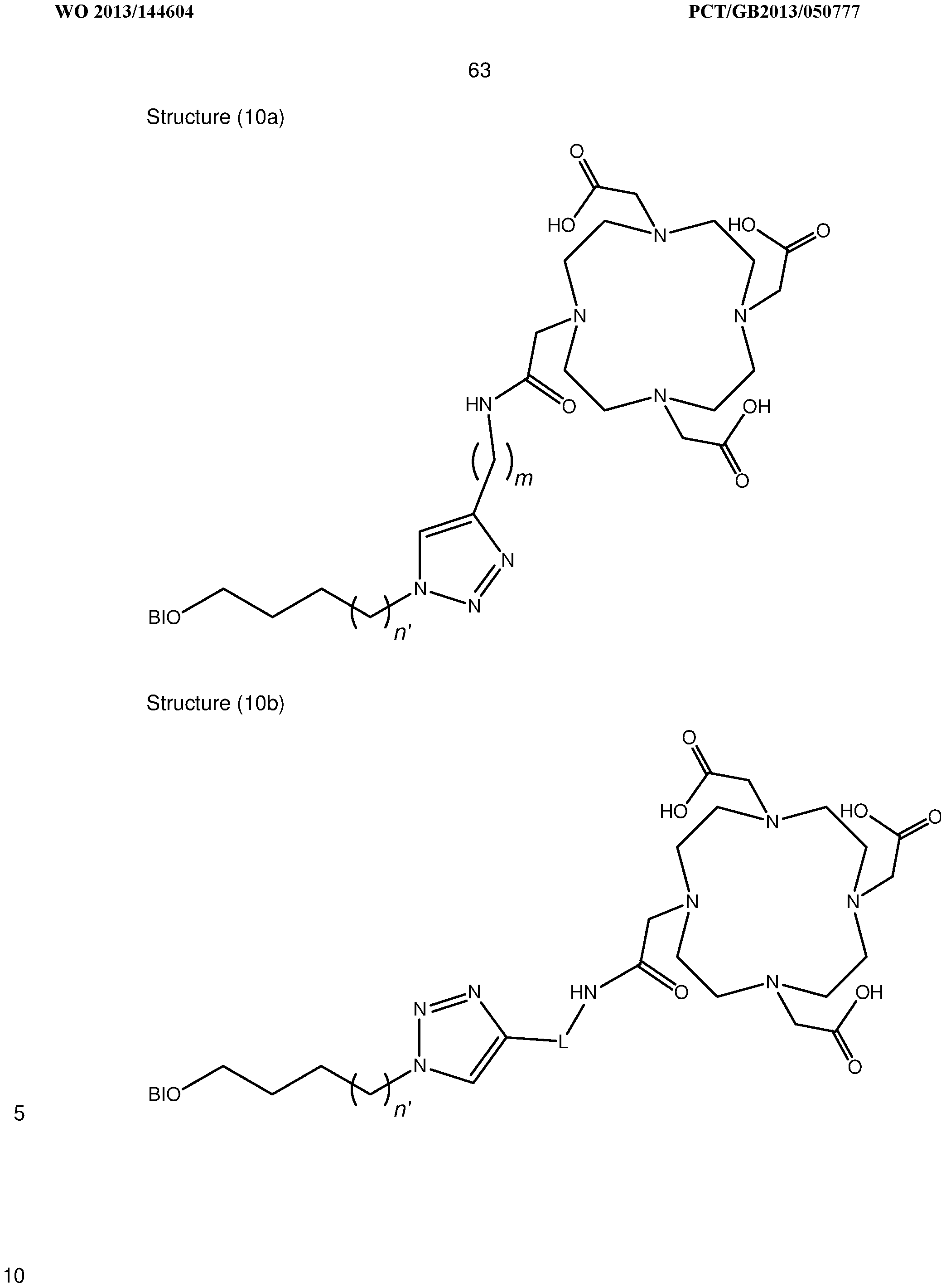
 Structure (12b)

Wherein BIO is a biotin or desthiobiotin moiety;
L is a linker; and
m,q = 1 to 6, n, n' = 1 to 4 and p = 1 or 2
BIO is a biotin or desthiobiotin moiety;
L is a PEG linker havin the structure

wherein m =1 -6.
35. A biotinyl-conjugate according to claim 34, wherein n =2, m, p=1 , q=3 and n=1 .
36. A contrast agent comprising a biotinyl-conjugate according to any one of claims 29 to 35.
37. A biotinylating agent having structure (8b), or (9b) wherein n or n' =1 -4; BIO is a biotinyl moiety; L is a linker; and A is a coupling moiety or a leaving group.
38. A method of synthesis of a biotinylating agent having (i) structure (8b) or (ii) structure (9b), comprising the steps of;
(a) Preparing a biotinylating agent having (i) structure (8a) or (ii) structure (8b)
(b) Preparing (i) an alkyne of structure: L or (ii) an azide of structure L N3
(c) Performing an azide-alkyne cycloaddition.
wherein L is a linker; and A is a coupling moiety or a leaving group.
A method according to claim 38, comprising performing a Huisgen 1 ,3-dipolar cycloaddition, so as to form a biotinylating agent comprising a 1 ,4-substituted 1 ,2,3-triazole.
A method of synthesising a biotinyl-conjugate having the general structure (1 ) or structure (2), the method comprising the steps of:
(i) preparing a biotinyl-azide having the structure (8a) and an alkyne having the structure Y = ;
(ii) performing a Huisgen 1 ,3-dipolar cycloaddition so as to form a biotinyl- conjugate having structure (1 a);
or the steps of;
(i) preparing a biotinyl alkyne having the structure (8b) and an azide having the structure Y-N3;
(ii) performing a Huisgen 1 ,3-dipolar cycloaddition between the said alkyne and the said azide so as to form a biotinyl-conjugate having structure (2a);
wherein Y comprises a moiety which may be an imaging and/or chelating moiety.
A method of synthesising a biotinyl-conjugate having (i) the general structure (18) or (ii) the general structure (19);
Structure (18)


wherein n or n' =1 -4;
BIO is a biotinyl moiety;
L is a linker;
X is a coupling group; and
Y' comprises a therapeutic or diagnostic moiety.
the method comprising the steps of;
(a) preparing a biotinylating agent having (i) structure (8b) or (ii) structure (9b)
(b) coupling the biotinylating agent to a compound Y'-B, wherein Y' comprises a therapeutic or diagnostic moiety, and B is a coupling moiety or a leaving group.
A method according to claim 41 , wherein the biotinylating agent is prepared by the method of claim 38 or 39.
43. A method according to claim 42, wherein Y' comprises a chelating moiety C and the method comprises the step of subsequently chelating a metal ion in the chelating moiety, to form a biotin-chelate complex.
Applications Claiming Priority (2)
| Application Number | Priority Date | Filing Date | Title |
|---|---|---|---|
| GBGB1205360.9A GB201205360D0 (en) | 2012-03-27 | 2012-03-27 | Biotinidase resistant biotinyl compounds |
| GB1205360.9 | 2012-03-27 |
Publications (1)
| Publication Number | Publication Date |
|---|---|
| WO2013144604A1 true WO2013144604A1 (en) | 2013-10-03 |
Family
ID=46087188
Family Applications (1)
| Application Number | Title | Priority Date | Filing Date |
|---|---|---|---|
| PCT/GB2013/050777 Ceased WO2013144604A1 (en) | 2012-03-27 | 2013-03-26 | Biotinidase resistant biotinyl compounds |
Country Status (2)
| Country | Link |
|---|---|
| GB (1) | GB201205360D0 (en) |
| WO (1) | WO2013144604A1 (en) |
Cited By (1)
| Publication number | Priority date | Publication date | Assignee | Title |
|---|---|---|---|---|
| CN107496937A (en) * | 2017-09-07 | 2017-12-22 | 江苏省原子医学研究所 | A kind of pre-targeting drug delivery system and its preparation method and application |
Citations (12)
| Publication number | Priority date | Publication date | Assignee | Title |
|---|---|---|---|---|
| US5608060A (en) | 1992-06-09 | 1997-03-04 | Neorx Corporation | Biotinidase-resistant biotin-DOTA conjugates |
| US5807879A (en) | 1992-03-03 | 1998-09-15 | University Of Rochester | Biotinidase-resistant biotinylated compound and methods of use thereof |
| WO2000002051A1 (en) * | 1998-07-07 | 2000-01-13 | Dept. Of Radiation Oncology, University Of Washington | Trifunctional reagent for conjugation to a biomolecule |
| WO2005051424A1 (en) | 2003-11-28 | 2005-06-09 | Mitra Medical Ab | Targeting of erb antigens |
| US7141676B1 (en) | 1996-02-08 | 2006-11-28 | University Of Washington | Water soluble multi-biotin-containing compounds |
| WO2009004106A1 (en) * | 2007-07-02 | 2009-01-08 | Universidad De Granada | Porous biotin carrier, and associated production methods and uses |
| WO2010008519A2 (en) * | 2008-07-14 | 2010-01-21 | Andrew Metters | In vitro diagnostic markers comprising carbon nanoparticles and kits |
| WO2010093436A2 (en) * | 2009-02-11 | 2010-08-19 | Carson Dennis A | Toll-like receptor modulators and treatment of diseases |
| WO2010106222A2 (en) * | 2009-03-16 | 2010-09-23 | Wallac Oy | Biotinidase assay |
| WO2011032034A2 (en) * | 2009-09-10 | 2011-03-17 | University Of Idaho | Nucleobase-functionalized conformationally restricted nucleotides and oligonucleotides for targeting nucleic acids |
| WO2011068978A1 (en) * | 2009-12-02 | 2011-06-09 | The United States Of America, As Represented By The Secretary, Department Of Health And Human Services | Methanocarba adenosine derivatives and dendrimer conjugates thereof |
| WO2013040647A1 (en) * | 2011-09-23 | 2013-03-28 | Adelaide Research & Innovation Pty Ltd | Novel antimicrobial compounds |
-
2012
- 2012-03-27 GB GBGB1205360.9A patent/GB201205360D0/en not_active Ceased
-
2013
- 2013-03-26 WO PCT/GB2013/050777 patent/WO2013144604A1/en not_active Ceased
Patent Citations (12)
| Publication number | Priority date | Publication date | Assignee | Title |
|---|---|---|---|---|
| US5807879A (en) | 1992-03-03 | 1998-09-15 | University Of Rochester | Biotinidase-resistant biotinylated compound and methods of use thereof |
| US5608060A (en) | 1992-06-09 | 1997-03-04 | Neorx Corporation | Biotinidase-resistant biotin-DOTA conjugates |
| US7141676B1 (en) | 1996-02-08 | 2006-11-28 | University Of Washington | Water soluble multi-biotin-containing compounds |
| WO2000002051A1 (en) * | 1998-07-07 | 2000-01-13 | Dept. Of Radiation Oncology, University Of Washington | Trifunctional reagent for conjugation to a biomolecule |
| WO2005051424A1 (en) | 2003-11-28 | 2005-06-09 | Mitra Medical Ab | Targeting of erb antigens |
| WO2009004106A1 (en) * | 2007-07-02 | 2009-01-08 | Universidad De Granada | Porous biotin carrier, and associated production methods and uses |
| WO2010008519A2 (en) * | 2008-07-14 | 2010-01-21 | Andrew Metters | In vitro diagnostic markers comprising carbon nanoparticles and kits |
| WO2010093436A2 (en) * | 2009-02-11 | 2010-08-19 | Carson Dennis A | Toll-like receptor modulators and treatment of diseases |
| WO2010106222A2 (en) * | 2009-03-16 | 2010-09-23 | Wallac Oy | Biotinidase assay |
| WO2011032034A2 (en) * | 2009-09-10 | 2011-03-17 | University Of Idaho | Nucleobase-functionalized conformationally restricted nucleotides and oligonucleotides for targeting nucleic acids |
| WO2011068978A1 (en) * | 2009-12-02 | 2011-06-09 | The United States Of America, As Represented By The Secretary, Department Of Health And Human Services | Methanocarba adenosine derivatives and dendrimer conjugates thereof |
| WO2013040647A1 (en) * | 2011-09-23 | 2013-03-28 | Adelaide Research & Innovation Pty Ltd | Novel antimicrobial compounds |
Non-Patent Citations (19)
| Title |
|---|
| BIOCONJUGATE CHEMISTRY, vol. 11, no. 4, 2000, pages 569 - 583 |
| BIOCONJUGATE CHEMISTRY, vol. 12, no. 4, 2001, pages 616 - 623 |
| BOERMAN ET AL.: "Pretargeted radioimmunotherapy of cancer: progress step by step", JOURNAL OF NUCLEAR MEDICINE: OFFICIAL PUBLICATION, SOCIETY OF NUCLEAR MEDICINE, vol. 44, no. 3, 2003, pages 400 - 411, XP002292436 |
| CHAUHAN, J.; DAKSHINAMURTI, K., J. BIOL. CHEM., vol. 261, 1986, pages 4268 - 4275 |
| FOULON ET AL., BIOCONJUGATE CHEMISTRY, vol. 8, no. 2, 1997, pages 179 - 186 |
| J. AM. CHEM. SOC., vol. 133, 2011, pages 16346 |
| JAUREGUI, M.; PERRY, W. S.; ALLAIN, C.; VIDLER, L. R.; WILLIS, M. C.; KENWRIGHT, A. M.; SNAITH, J. S.; STASIUK, G. J.; LOWE M. P.;, DALTON TRANS., 2009, pages 6283 - 6285 |
| KOBZA, K. A.; CHAISEEDA, K.; SARATH, G.; TAKACS, J. M.; ZEMPLENI, J. J., NUT. BIOCHEM., vol. 19, 2008, pages 826 - 832 |
| KOBZA, K. A.; CHAISEEDA, K.; SARATH, G.; TAKACS, J. M.; ZEMPLENI, J., J. NUT. BIOCHEM., vol. 19, 2008, pages 826 - 832 |
| PRATESI, A.; BUCELLI, F.; MORI, I.; CHINOL, M.; VEDOLIVA, A.; PAGANELLI, G.; RIVIECCIO, V.; GARIBOLDI, L.; GINANNESCHI, M., J. MED. CHEM., vol. 53, 2010, pages 432 - 440 |
| ROSEBROUGH, THE JOURNAL OF PHARMACOLOGY AND EXPERIMENTAL THERAPEUTICS, vol. 265, no. 1, 1993, pages 408 - 415 |
| SABATINO ET AL., JOURNAL OF MEDICINAL CHEMISTRY, vol. 46, no. 14, 2003, pages 3170 - 3173 |
| SCHULTZ, M. K.; PARAMESWARAPPA, S. G.; PIGGE, F. C., ORG. LETT., vol. 12, 2010, pages 2398 - 2401 |
| SEIGNEURIC ET AL., ONCOTARGET., vol. 2, no. 7, July 2011 (2011-07-01), pages 557 - 561 |
| SHARKEY; GOLDENBERG, ADVANCED DRUG DELIVERY REVIEWS, vol. 60, 2008, pages 1407 - 1420 |
| T. P. SOARES DA COSTA ET AL: "Selective inhibition of Biotin Protein Ligase from Staphylococcus aureus", JOURNAL OF BIOLOGICAL CHEMISTRY, vol. 287, no. 21, 18 May 2012 (2012-05-18), pages 17823 - 17832, XP055064657, ISSN: 0021-9258, DOI: 10.1074/jbc.M112.356576 * |
| UMEDA ET AL: "Supporting Information", JACS, vol. 133, 1 January 2011 (2011-01-01), pages S1 - S21, XP002697921 * |
| UMEDA N ET AL: "A photocleavable rapamycin conjugate for spatiotemporal control of small GTPase activity", JOURNAL OF THE AMERICAN CHEMICAL SOCIETY 20110112 AMERICAN CHEMICAL SOCIETY USA, vol. 133, no. 1, 12 January 2011 (2011-01-12), pages 12 - 14, XP002697921, ISSN: 0002-7863 * |
| VIGUIER R. F. H.; HULME, A. N., J. AM. CHEM. SOC., vol. 128, 2006, pages 11370 - 11371 |
Cited By (2)
| Publication number | Priority date | Publication date | Assignee | Title |
|---|---|---|---|---|
| CN107496937A (en) * | 2017-09-07 | 2017-12-22 | 江苏省原子医学研究所 | A kind of pre-targeting drug delivery system and its preparation method and application |
| CN107496937B (en) * | 2017-09-07 | 2020-09-25 | 江苏省原子医学研究所 | Pre-targeting drug delivery system and preparation method and application thereof |
Also Published As
| Publication number | Publication date |
|---|---|
| GB201205360D0 (en) | 2012-05-09 |
Similar Documents
| Publication | Publication Date | Title |
|---|---|---|
| Kras et al. | Distinct coordination chemistry of Fe (III)-based MRI probes | |
| Stasiuk et al. | The ubiquitous DOTA and its derivatives: the impact of 1, 4, 7, 10-tetraazacyclododecane-1, 4, 7, 10-tetraacetic acid on biomedical imaging | |
| Mindt et al. | A “click chemistry” approach to the efficient synthesis of multiple imaging probes derived from a single precursor | |
| CN102395380B (en) | Pretargeting kit, method and agents used therein | |
| US12419976B2 (en) | Hybrid tracers for targeted cancer imaging and treatment | |
| JP6063384B2 (en) | Pre-target kit, pre-target method and reagent used therefor | |
| US7338651B2 (en) | Multi-use multimodal imaging chelates | |
| CN102438656B (en) | Pretargeting kit, method and agents used therein | |
| Cheng et al. | 64Cu-labeled affibody molecules for imaging of HER2 expressing tumors | |
| Tircsó et al. | (S)-5-(p-nitrobenzyl)-PCTA, a promising bifunctional ligand with advantageous metal ion complexation kinetics | |
| Pellico et al. | Unambiguous detection of atherosclerosis using bioorthogonal nanomaterials | |
| JP2014506869A5 (en) | ||
| US20030228262A1 (en) | Scorpionate-like pendant macrocyclic ligands, complexes and compositions thereof, and methods of using same | |
| JPH07504661A (en) | Conjugates of biotin and deferoxamine for radioimmunoimaging and radioimmunotherapy | |
| Pujales-Paradela et al. | Lanthanide complexes with 1H paraCEST and 19F response for magnetic resonance imaging applications | |
| Saini et al. | Synthesis, conjugation and relaxation studies of gadolinium (III)-4-benzothiazol-2-yl-phenylamine as a potential brain specific MR contrast agent | |
| Xu et al. | Chiral Gd-DOTA as a versatile platform for hepatobiliary and tumor targeting MRI contrast agents | |
| US8580231B2 (en) | Compositions and methods comprising magnetic resonance contrast agents | |
| US7608249B2 (en) | Enhanced substrate imaging by reversible binding to a paramagnetic complex | |
| Mishra et al. | Complexes of Iron (II), Cobalt (II), and Nickel (II) with DOTA-Tetraglycinate for pH and Temperature Imaging Using Hyperfine Shifts of an Amide Moiety | |
| WO2012022932A1 (en) | Process for producing radiohalogenated bioconjugates and products thereof | |
| US9044505B2 (en) | Multimeric biotinidase resistant multimodality probes | |
| Wang et al. | " Click Chemistry" for Molecular Imaging | |
| US20240285808A1 (en) | Molecular probes for in vivo detection of aldehydes | |
| Feng et al. | Development of a novel 99mTc-labeled folate derivative containing phenyl isonitrile to target folate receptor with reduced renal uptake |
Legal Events
| Date | Code | Title | Description |
|---|---|---|---|
| 121 | Ep: the epo has been informed by wipo that ep was designated in this application |
Ref document number: 13715402 Country of ref document: EP Kind code of ref document: A1 |
|
| NENP | Non-entry into the national phase |
Ref country code: DE |
|
| 122 | Ep: pct application non-entry in european phase |
Ref document number: 13715402 Country of ref document: EP Kind code of ref document: A1 |