WO2013121380A1 - Means and methods for assessing an endocrine disease or disorder - Google Patents
Means and methods for assessing an endocrine disease or disorder Download PDFInfo
- Publication number
- WO2013121380A1 WO2013121380A1 PCT/IB2013/051224 IB2013051224W WO2013121380A1 WO 2013121380 A1 WO2013121380 A1 WO 2013121380A1 IB 2013051224 W IB2013051224 W IB 2013051224W WO 2013121380 A1 WO2013121380 A1 WO 2013121380A1
- Authority
- WO
- WIPO (PCT)
- Prior art keywords
- disorder
- endocrine
- subject
- hydrochloride
- group
- Prior art date
Links
- 238000000034 method Methods 0.000 title claims abstract description 203
- 208000017701 Endocrine disease Diseases 0.000 title abstract description 286
- 208000030172 endocrine system disease Diseases 0.000 title abstract description 286
- 150000001875 compounds Chemical class 0.000 claims abstract description 179
- 230000001939 inductive effect Effects 0.000 claims abstract description 51
- 239000000090 biomarker Substances 0.000 claims description 273
- 208000037265 diseases, disorders, signs and symptoms Diseases 0.000 claims description 259
- 208000035475 disorder Diseases 0.000 claims description 148
- 238000012360 testing method Methods 0.000 claims description 114
- 239000000126 substance Substances 0.000 claims description 108
- NKANXQFJJICGDU-QPLCGJKRSA-N Tamoxifen Chemical compound C=1C=CC=CC=1C(/CC)=C(C=1C=CC(OCCN(C)C)=CC=1)/C1=CC=CC=C1 NKANXQFJJICGDU-QPLCGJKRSA-N 0.000 claims description 86
- KORNTPPJEAJQIU-KJXAQDMKSA-N Cabaser Chemical compound C1=CC([C@H]2C[C@H](CN(CC=C)[C@@H]2C2)C(=O)N(CCCN(C)C)C(=O)NCC)=C3C2=CNC3=C1 KORNTPPJEAJQIU-KJXAQDMKSA-N 0.000 claims description 49
- 229960004596 cabergoline Drugs 0.000 claims description 49
- UWFYSQMTEOIJJG-FDTZYFLXSA-N cyproterone acetate Chemical compound C1=C(Cl)C2=CC(=O)[C@@H]3C[C@@H]3[C@]2(C)[C@@H]2[C@@H]1[C@@H]1CC[C@@](C(C)=O)(OC(=O)C)[C@@]1(C)CC2 UWFYSQMTEOIJJG-FDTZYFLXSA-N 0.000 claims description 49
- PPDBOQMNKNNODG-NTEUORMPSA-N (5E)-5-(4-chlorobenzylidene)-2,2-dimethyl-1-(1,2,4-triazol-1-ylmethyl)cyclopentanol Chemical compound C1=NC=NN1CC1(O)C(C)(C)CC\C1=C/C1=CC=C(Cl)C=C1 PPDBOQMNKNNODG-NTEUORMPSA-N 0.000 claims description 48
- 239000005859 Triticonazole Substances 0.000 claims description 48
- PMRYVIKBURPHAH-UHFFFAOYSA-N methimazole Chemical compound CN1C=CNC1=S PMRYVIKBURPHAH-UHFFFAOYSA-N 0.000 claims description 48
- 229960002178 thiamazole Drugs 0.000 claims description 48
- FSCWZHGZWWDELK-UHFFFAOYSA-N 3-(3,5-dichlorophenyl)-5-ethenyl-5-methyl-2,4-oxazolidinedione Chemical compound O=C1C(C)(C=C)OC(=O)N1C1=CC(Cl)=CC(Cl)=C1 FSCWZHGZWWDELK-UHFFFAOYSA-N 0.000 claims description 47
- NHOWDZOIZKMVAI-UHFFFAOYSA-N (2-chlorophenyl)(4-chlorophenyl)pyrimidin-5-ylmethanol Chemical compound C=1N=CN=CC=1C(C=1C(=CC=CC=1)Cl)(O)C1=CC=C(Cl)C=C1 NHOWDZOIZKMVAI-UHFFFAOYSA-N 0.000 claims description 43
- 229960001603 tamoxifen Drugs 0.000 claims description 43
- IDLFZVILOHSSID-OVLDLUHVSA-N corticotropin Chemical compound C([C@@H](C(=O)N[C@@H](CO)C(=O)N[C@@H](CCSC)C(=O)N[C@@H](CCC(O)=O)C(=O)N[C@@H](CC=1NC=NC=1)C(=O)N[C@@H](CC=1C=CC=CC=1)C(=O)N[C@@H](CCCNC(N)=N)C(=O)N[C@@H](CC=1C2=CC=CC=C2NC=1)C(=O)NCC(=O)N[C@@H](CCCCN)C(=O)N1[C@@H](CCC1)C(=O)N[C@@H](C(C)C)C(=O)NCC(=O)N[C@@H](CCCCN)C(=O)N[C@@H](CCCCN)C(=O)N[C@@H](CCCNC(N)=N)C(=O)N[C@@H](CCCNC(N)=N)C(=O)N1[C@@H](CCC1)C(=O)N[C@@H](C(C)C)C(=O)N[C@@H](CCCCN)C(=O)N[C@@H](C(C)C)C(=O)N[C@@H](CC=1C=CC(O)=CC=1)C(=O)N1[C@@H](CCC1)C(=O)N[C@@H](CC(N)=O)C(=O)NCC(=O)N[C@@H](C)C(=O)N[C@@H](CCC(O)=O)C(=O)N[C@@H](CC(O)=O)C(=O)N[C@@H](CCC(O)=O)C(=O)N[C@@H](CO)C(=O)N[C@@H](C)C(=O)N[C@@H](CCC(O)=O)C(=O)N[C@@H](C)C(=O)N[C@@H](CC=1C=CC=CC=1)C(=O)N1[C@@H](CCC1)C(=O)N[C@@H](CC(C)C)C(=O)N[C@@H](CCC(O)=O)C(=O)N[C@@H](CC=1C=CC=CC=1)C(O)=O)NC(=O)[C@@H](N)CO)C1=CC=C(O)C=C1 IDLFZVILOHSSID-OVLDLUHVSA-N 0.000 claims description 42
- 229960000258 corticotropin Drugs 0.000 claims description 42
- 239000000055 Corticotropin-Releasing Hormone Substances 0.000 claims description 41
- ZSJLQEPLLKMAKR-UHFFFAOYSA-N Streptozotocin Natural products O=NN(C)C(=O)NC1C(O)OC(CO)C(O)C1O ZSJLQEPLLKMAKR-UHFFFAOYSA-N 0.000 claims description 41
- VKHAHZOOUSRJNA-GCNJZUOMSA-N mifepristone Chemical compound C1([C@@H]2C3=C4CCC(=O)C=C4CC[C@H]3[C@@H]3CC[C@@]([C@]3(C2)C)(O)C#CC)=CC=C(N(C)C)C=C1 VKHAHZOOUSRJNA-GCNJZUOMSA-N 0.000 claims description 41
- 229960003248 mifepristone Drugs 0.000 claims description 41
- ZSJLQEPLLKMAKR-GKHCUFPYSA-N streptozocin Chemical compound O=NN(C)C(=O)N[C@H]1[C@@H](O)O[C@H](CO)[C@@H](O)[C@@H]1O ZSJLQEPLLKMAKR-GKHCUFPYSA-N 0.000 claims description 41
- 229960001052 streptozocin Drugs 0.000 claims description 41
- IPPAUTOBDWNELX-UHFFFAOYSA-N (2-ethoxy-2-oxoethyl) 5-[2-chloro-4-(trifluoromethyl)phenoxy]-2-nitrobenzoate Chemical group C1=C([N+]([O-])=O)C(C(=O)OCC(=O)OCC)=CC(OC=2C(=CC(=CC=2)C(F)(F)F)Cl)=C1 IPPAUTOBDWNELX-UHFFFAOYSA-N 0.000 claims description 40
- GCKMFJBGXUYNAG-UHFFFAOYSA-N 17alpha-methyltestosterone Natural products C1CC2=CC(=O)CCC2(C)C2C1C1CCC(C)(O)C1(C)CC2 GCKMFJBGXUYNAG-UHFFFAOYSA-N 0.000 claims description 40
- QTBSBXVTEAMEQO-UHFFFAOYSA-M Acetate Chemical compound CC([O-])=O QTBSBXVTEAMEQO-UHFFFAOYSA-M 0.000 claims description 40
- 108010037003 Buserelin Proteins 0.000 claims description 40
- PYMDEDHDQYLBRT-DRIHCAFSSA-N Buserelin acetate Chemical compound CC(O)=O.CCNC(=O)[C@@H]1CCCN1C(=O)[C@H](CCCN=C(N)N)NC(=O)[C@H](CC(C)C)NC(=O)[C@@H](COC(C)(C)C)NC(=O)[C@@H](NC(=O)[C@H](CO)NC(=O)[C@H](CC=1C2=CC=CC=C2NC=1)NC(=O)[C@H](CC=1NC=NC=1)NC(=O)[C@H]1NC(=O)CC1)CC1=CC=C(O)C=C1 PYMDEDHDQYLBRT-DRIHCAFSSA-N 0.000 claims description 40
- 239000005767 Epoxiconazole Substances 0.000 claims description 40
- BFPYWIDHMRZLRN-SLHNCBLASA-N Ethinyl estradiol Chemical compound OC1=CC=C2[C@H]3CC[C@](C)([C@](CC4)(O)C#C)[C@@H]4[C@@H]3CCC2=C1 BFPYWIDHMRZLRN-SLHNCBLASA-N 0.000 claims description 40
- 239000005867 Iprodione Substances 0.000 claims description 40
- 239000005580 Metazachlor Substances 0.000 claims description 40
- GCKMFJBGXUYNAG-HLXURNFRSA-N Methyltestosterone Chemical compound C1CC2=CC(=O)CC[C@]2(C)[C@@H]2[C@@H]1[C@@H]1CC[C@](C)(O)[C@@]1(C)CC2 GCKMFJBGXUYNAG-HLXURNFRSA-N 0.000 claims description 40
- IMONTRJLAWHYGT-ZCPXKWAGSA-N Norethindrone Acetate Chemical compound C1CC2=CC(=O)CC[C@@H]2[C@@H]2[C@@H]1[C@@H]1CC[C@](C#C)(OC(=O)C)[C@@]1(C)CC2 IMONTRJLAWHYGT-ZCPXKWAGSA-N 0.000 claims description 40
- GHUUBYQTCDQWRA-UHFFFAOYSA-N Pioglitazone hydrochloride Chemical compound Cl.N1=CC(CC)=CC=C1CCOC(C=C1)=CC=C1CC1C(=O)NC(=O)S1 GHUUBYQTCDQWRA-UHFFFAOYSA-N 0.000 claims description 40
- 229960002802 bromocriptine Drugs 0.000 claims description 40
- OZVBMTJYIDMWIL-AYFBDAFISA-N bromocriptine Chemical compound C1=CC(C=2[C@H](N(C)C[C@@H](C=2)C(=O)N[C@]2(C(=O)N3[C@H](C(N4CCC[C@H]4[C@]3(O)O2)=O)CC(C)C)C(C)C)C2)=C3C2=C(Br)NC3=C1 OZVBMTJYIDMWIL-AYFBDAFISA-N 0.000 claims description 40
- 229960005064 buserelin acetate Drugs 0.000 claims description 40
- KNHUKKLJHYUCFP-UHFFFAOYSA-N clofibrate Chemical compound CCOC(=O)C(C)(C)OC1=CC=C(Cl)C=C1 KNHUKKLJHYUCFP-UHFFFAOYSA-N 0.000 claims description 40
- 229960001214 clofibrate Drugs 0.000 claims description 40
- UREBDLICKHMUKA-CXSFZGCWSA-N dexamethasone Chemical compound C1CC2=CC(=O)C=C[C@]2(C)[C@]2(F)[C@@H]1[C@@H]1C[C@@H](C)[C@@](C(=O)CO)(O)[C@@]1(C)C[C@@H]2O UREBDLICKHMUKA-CXSFZGCWSA-N 0.000 claims description 40
- 229960003957 dexamethasone Drugs 0.000 claims description 40
- ZBCBWPMODOFKDW-UHFFFAOYSA-N diethanolamine Chemical compound OCCNCCO ZBCBWPMODOFKDW-UHFFFAOYSA-N 0.000 claims description 40
- 229960002074 flutamide Drugs 0.000 claims description 40
- MKXKFYHWDHIYRV-UHFFFAOYSA-N flutamide Chemical compound CC(C)C(=O)NC1=CC=C([N+]([O-])=O)C(C(F)(F)F)=C1 MKXKFYHWDHIYRV-UHFFFAOYSA-N 0.000 claims description 40
- OSVMTWJCGUFAOD-KZQROQTASA-N formestane Chemical compound O=C1CC[C@]2(C)[C@H]3CC[C@](C)(C(CC4)=O)[C@@H]4[C@@H]3CCC2=C1O OSVMTWJCGUFAOD-KZQROQTASA-N 0.000 claims description 40
- 229960004421 formestane Drugs 0.000 claims description 40
- 229940045109 genistein Drugs 0.000 claims description 40
- 235000006539 genistein Nutrition 0.000 claims description 40
- TZBJGXHYKVUXJN-UHFFFAOYSA-N genistein Natural products C1=CC(O)=CC=C1C1=COC2=CC(O)=CC(O)=C2C1=O TZBJGXHYKVUXJN-UHFFFAOYSA-N 0.000 claims description 40
- ZCOLJUOHXJRHDI-CMWLGVBASA-N genistein 7-O-beta-D-glucoside Chemical compound O[C@@H]1[C@@H](O)[C@H](O)[C@@H](CO)O[C@H]1OC1=CC(O)=C2C(=O)C(C=3C=CC(O)=CC=3)=COC2=C1 ZCOLJUOHXJRHDI-CMWLGVBASA-N 0.000 claims description 40
- ZJJXGWJIGJFDTL-UHFFFAOYSA-N glipizide Chemical compound C1=NC(C)=CN=C1C(=O)NCCC1=CC=C(S(=O)(=O)NC(=O)NC2CCCCC2)C=C1 ZJJXGWJIGJFDTL-UHFFFAOYSA-N 0.000 claims description 40
- 229960001381 glipizide Drugs 0.000 claims description 40
- HOMGKSMUEGBAAB-UHFFFAOYSA-N ifosfamide Chemical compound ClCCNP1(=O)OCCCN1CCCl HOMGKSMUEGBAAB-UHFFFAOYSA-N 0.000 claims description 40
- 229960001101 ifosfamide Drugs 0.000 claims description 40
- ONUFESLQCSAYKA-UHFFFAOYSA-N iprodione Chemical compound O=C1N(C(=O)NC(C)C)CC(=O)N1C1=CC(Cl)=CC(Cl)=C1 ONUFESLQCSAYKA-UHFFFAOYSA-N 0.000 claims description 40
- GFIJNRVAKGFPGQ-LIJARHBVSA-N leuprolide Chemical compound CCNC(=O)[C@@H]1CCCN1C(=O)[C@H](CCCNC(N)=N)NC(=O)[C@H](CC(C)C)NC(=O)[C@@H](CC(C)C)NC(=O)[C@@H](NC(=O)[C@H](CO)NC(=O)[C@H](CC=1C2=CC=CC=C2NC=1)NC(=O)[C@H](CC=1N=CNC=1)NC(=O)[C@H]1NC(=O)CC1)CC1=CC=C(O)C=C1 GFIJNRVAKGFPGQ-LIJARHBVSA-N 0.000 claims description 40
- 229960004616 medroxyprogesterone Drugs 0.000 claims description 40
- FRQMUZJSZHZSGN-HBNHAYAOSA-N medroxyprogesterone Chemical compound C([C@@]12C)CC(=O)C=C1[C@@H](C)C[C@@H]1[C@@H]2CC[C@]2(C)[C@@](O)(C(C)=O)CC[C@H]21 FRQMUZJSZHZSGN-HBNHAYAOSA-N 0.000 claims description 40
- STEPQTYSZVCJPV-UHFFFAOYSA-N metazachlor Chemical compound CC1=CC=CC(C)=C1N(C(=O)CCl)CN1N=CC=C1 STEPQTYSZVCJPV-UHFFFAOYSA-N 0.000 claims description 40
- 229960001652 norethindrone acetate Drugs 0.000 claims description 40
- 229960002827 pioglitazone hydrochloride Drugs 0.000 claims description 40
- BKXVVCILCIUCLG-UHFFFAOYSA-N raloxifene hydrochloride Chemical compound [H+].[Cl-].C1=CC(O)=CC=C1C1=C(C(=O)C=2C=CC(OCCN3CCCCC3)=CC=2)C2=CC=C(O)C=C2S1 BKXVVCILCIUCLG-UHFFFAOYSA-N 0.000 claims description 40
- 229960002119 raloxifene hydrochloride Drugs 0.000 claims description 40
- RAPZEAPATHNIPO-UHFFFAOYSA-N risperidone Chemical compound FC1=CC=C2C(C3CCN(CC3)CCC=3C(=O)N4CCCCC4=NC=3C)=NOC2=C1 RAPZEAPATHNIPO-UHFFFAOYSA-N 0.000 claims description 40
- 229960001534 risperidone Drugs 0.000 claims description 40
- SUFUKZSWUHZXAV-BTJKTKAUSA-N rosiglitazone maleate Chemical compound [H+].[H+].[O-]C(=O)\C=C/C([O-])=O.C=1C=CC=NC=1N(C)CCOC(C=C1)=CC=C1CC1SC(=O)NC1=O SUFUKZSWUHZXAV-BTJKTKAUSA-N 0.000 claims description 40
- 229960003271 rosiglitazone maleate Drugs 0.000 claims description 40
- MEHHPFQKXOUFFV-OWSLCNJRSA-N trenbolone Chemical compound C1CC(=O)C=C2CC[C@@H]([C@H]3[C@@](C)([C@H](CC3)O)C=C3)C3=C21 MEHHPFQKXOUFFV-OWSLCNJRSA-N 0.000 claims description 40
- 229960000312 trenbolone Drugs 0.000 claims description 40
- 229960003474 ziprasidone hydrochloride Drugs 0.000 claims description 40
- GIYXAJPCNFJEHY-UHFFFAOYSA-N N-methyl-3-phenyl-3-[4-(trifluoromethyl)phenoxy]-1-propanamine hydrochloride (1:1) Chemical compound Cl.C=1C=CC=CC=1C(CCNC)OC1=CC=C(C(F)(F)F)C=C1 GIYXAJPCNFJEHY-UHFFFAOYSA-N 0.000 claims description 39
- 229960000389 fluoxetine hydrochloride Drugs 0.000 claims description 39
- BFPYWIDHMRZLRN-UHFFFAOYSA-N 17alpha-ethynyl estradiol Natural products OC1=CC=C2C3CCC(C)(C(CC4)(O)C#C)C4C3CCC2=C1 BFPYWIDHMRZLRN-UHFFFAOYSA-N 0.000 claims description 38
- 230000002124 endocrine Effects 0.000 claims description 37
- VZMLEMYJUIIHNF-QURGRASLSA-N [4-[(e)-4-(4-propanoyloxyphenyl)hex-3-en-3-yl]phenyl] propanoate Chemical compound C1=CC(OC(=O)CC)=CC=C1C(\CC)=C(/CC)C1=CC=C(OC(=O)CC)C=C1 VZMLEMYJUIIHNF-QURGRASLSA-N 0.000 claims description 35
- PTQMMNYJKCSPET-OMHQDGTGSA-N mibolerone Chemical compound C1C[C@]2(C)[C@](O)(C)CC[C@H]2[C@@H]2[C@H](C)CC3=CC(=O)CC[C@@H]3[C@H]21 PTQMMNYJKCSPET-OMHQDGTGSA-N 0.000 claims description 34
- 229950006489 mibolerone Drugs 0.000 claims description 34
- 239000003795 chemical substances by application Substances 0.000 claims description 33
- 238000001514 detection method Methods 0.000 claims description 28
- 238000011156 evaluation Methods 0.000 claims description 21
- -1 Vindozolin Chemical compound 0.000 claims description 13
- ZMYFCFLJBGAQRS-IRXDYDNUSA-N (2R,3S)-epoxiconazole Chemical compound C1=CC(F)=CC=C1[C@@]1(CN2N=CN=C2)[C@H](C=2C(=CC=CC=2)Cl)O1 ZMYFCFLJBGAQRS-IRXDYDNUSA-N 0.000 claims 9
- ZCBZSCBNOOIHFP-UHFFFAOYSA-N ziprasidone hydrochloride hydrate Chemical compound [H+].O.[Cl-].C1=CC=C2C(N3CCN(CC3)CCC3=CC=4CC(=O)NC=4C=C3Cl)=NSC2=C1 ZCBZSCBNOOIHFP-UHFFFAOYSA-N 0.000 claims 9
- 239000003814 drug Substances 0.000 abstract description 21
- 229940079593 drug Drugs 0.000 abstract description 18
- 238000013517 stratification Methods 0.000 abstract description 2
- 239000000523 sample Substances 0.000 description 147
- 201000010099 disease Diseases 0.000 description 111
- 229940011871 estrogen Drugs 0.000 description 83
- 239000000262 estrogen Substances 0.000 description 83
- 210000004404 adrenal cortex Anatomy 0.000 description 81
- 229940088597 hormone Drugs 0.000 description 76
- 239000005556 hormone Substances 0.000 description 76
- 239000003163 gonadal steroid hormone Substances 0.000 description 73
- 230000013632 homeostatic process Effects 0.000 description 68
- 230000001988 toxicity Effects 0.000 description 62
- 231100000419 toxicity Toxicity 0.000 description 62
- 230000006870 function Effects 0.000 description 61
- 210000004153 islets of langerhan Anatomy 0.000 description 60
- MUMGGOZAMZWBJJ-DYKIIFRCSA-N Testostosterone Chemical compound O=C1CC[C@]2(C)[C@H]3CC[C@](C)([C@H](CC4)O)[C@@H]4[C@@H]3CCC2=C1 MUMGGOZAMZWBJJ-DYKIIFRCSA-N 0.000 description 56
- 102000014654 Aromatase Human genes 0.000 description 37
- 108010078554 Aromatase Proteins 0.000 description 37
- 101800000414 Corticotropin Proteins 0.000 description 33
- 210000004027 cell Anatomy 0.000 description 33
- 230000000694 effects Effects 0.000 description 33
- 210000001519 tissue Anatomy 0.000 description 32
- ZMYFCFLJBGAQRS-IAGOWNOFSA-N (2S,3R)-epoxiconazole Chemical compound C1=CC(F)=CC=C1[C@]1(CN2N=CN=C2)[C@@H](C=2C(=CC=CC=2)Cl)O1 ZMYFCFLJBGAQRS-IAGOWNOFSA-N 0.000 description 31
- NZDBKBRIBJLNNT-UHFFFAOYSA-N 5-[2-[4-(1,2-benzothiazol-3-yl)piperazin-1-yl]ethyl]-6-chloro-1,3-dihydroindol-2-one;hydron;chloride Chemical compound Cl.C1=CC=C2C(N3CCN(CC3)CCC3=CC=4CC(=O)NC=4C=C3Cl)=NSC2=C1 NZDBKBRIBJLNNT-UHFFFAOYSA-N 0.000 description 31
- 229940043237 diethanolamine Drugs 0.000 description 31
- 239000003098 androgen Substances 0.000 description 30
- 102000005962 receptors Human genes 0.000 description 30
- 108020003175 receptors Proteins 0.000 description 30
- 238000004458 analytical method Methods 0.000 description 29
- 229960003604 testosterone Drugs 0.000 description 28
- 241001465754 Metazoa Species 0.000 description 27
- 239000002207 metabolite Substances 0.000 description 26
- 239000002676 xenobiotic agent Substances 0.000 description 26
- 238000011161 development Methods 0.000 description 25
- 230000018109 developmental process Effects 0.000 description 25
- 230000001919 adrenal effect Effects 0.000 description 23
- 150000003431 steroids Chemical class 0.000 description 23
- 229940030486 androgens Drugs 0.000 description 22
- NOESYZHRGYRDHS-UHFFFAOYSA-N insulin Chemical compound N1C(=O)C(NC(=O)C(CCC(N)=O)NC(=O)C(CCC(O)=O)NC(=O)C(C(C)C)NC(=O)C(NC(=O)CN)C(C)CC)CSSCC(C(NC(CO)C(=O)NC(CC(C)C)C(=O)NC(CC=2C=CC(O)=CC=2)C(=O)NC(CCC(N)=O)C(=O)NC(CC(C)C)C(=O)NC(CCC(O)=O)C(=O)NC(CC(N)=O)C(=O)NC(CC=2C=CC(O)=CC=2)C(=O)NC(CSSCC(NC(=O)C(C(C)C)NC(=O)C(CC(C)C)NC(=O)C(CC=2C=CC(O)=CC=2)NC(=O)C(CC(C)C)NC(=O)C(C)NC(=O)C(CCC(O)=O)NC(=O)C(C(C)C)NC(=O)C(CC(C)C)NC(=O)C(CC=2NC=NC=2)NC(=O)C(CO)NC(=O)CNC2=O)C(=O)NCC(=O)NC(CCC(O)=O)C(=O)NC(CCCNC(N)=N)C(=O)NCC(=O)NC(CC=3C=CC=CC=3)C(=O)NC(CC=3C=CC=CC=3)C(=O)NC(CC=3C=CC(O)=CC=3)C(=O)NC(C(C)O)C(=O)N3C(CCC3)C(=O)NC(CCCCN)C(=O)NC(C)C(O)=O)C(=O)NC(CC(N)=O)C(O)=O)=O)NC(=O)C(C(C)CC)NC(=O)C(CO)NC(=O)C(C(C)O)NC(=O)C1CSSCC2NC(=O)C(CC(C)C)NC(=O)C(NC(=O)C(CCC(N)=O)NC(=O)C(CC(N)=O)NC(=O)C(NC(=O)C(N)CC=1C=CC=CC=1)C(C)C)CC1=CN=CN1 NOESYZHRGYRDHS-UHFFFAOYSA-N 0.000 description 20
- 230000002034 xenobiotic effect Effects 0.000 description 20
- 102000004190 Enzymes Human genes 0.000 description 19
- 108090000790 Enzymes Proteins 0.000 description 19
- HVYWMOMLDIMFJA-DPAQBDIFSA-N cholesterol Chemical compound C1C=C2C[C@@H](O)CC[C@]2(C)[C@@H]2[C@@H]1[C@@H]1CC[C@H]([C@H](C)CCCC(C)C)[C@@]1(C)CC2 HVYWMOMLDIMFJA-DPAQBDIFSA-N 0.000 description 18
- 230000001771 impaired effect Effects 0.000 description 18
- 239000000556 agonist Substances 0.000 description 17
- 150000002632 lipids Chemical class 0.000 description 17
- 231100000027 toxicology Toxicity 0.000 description 17
- 210000004100 adrenal gland Anatomy 0.000 description 15
- 238000013480 data collection Methods 0.000 description 15
- 102000015694 estrogen receptors Human genes 0.000 description 15
- 108010038795 estrogen receptors Proteins 0.000 description 15
- 238000011835 investigation Methods 0.000 description 15
- 210000000496 pancreas Anatomy 0.000 description 15
- 238000011282 treatment Methods 0.000 description 15
- WQZGKKKJIJFFOK-GASJEMHNSA-N Glucose Natural products OC[C@H]1OC(O)[C@H](O)[C@@H](O)[C@@H]1O WQZGKKKJIJFFOK-GASJEMHNSA-N 0.000 description 14
- 241000700159 Rattus Species 0.000 description 14
- 230000009471 action Effects 0.000 description 14
- 108010080146 androgen receptors Proteins 0.000 description 14
- 238000004519 manufacturing process Methods 0.000 description 14
- 230000028327 secretion Effects 0.000 description 14
- 230000001054 cortical effect Effects 0.000 description 13
- 239000008103 glucose Substances 0.000 description 13
- 230000005764 inhibitory process Effects 0.000 description 13
- 238000004949 mass spectrometry Methods 0.000 description 13
- 210000001672 ovary Anatomy 0.000 description 13
- 238000002360 preparation method Methods 0.000 description 13
- VOXZDWNPVJITMN-ZBRFXRBCSA-N 17β-estradiol Chemical compound OC1=CC=C2[C@H]3CC[C@](C)([C@H](CC4)O)[C@@H]4[C@@H]3CCC2=C1 VOXZDWNPVJITMN-ZBRFXRBCSA-N 0.000 description 12
- 239000012491 analyte Substances 0.000 description 12
- 230000015572 biosynthetic process Effects 0.000 description 12
- 210000004369 blood Anatomy 0.000 description 12
- 239000008280 blood Substances 0.000 description 12
- 229930182833 estradiol Natural products 0.000 description 12
- 229960005309 estradiol Drugs 0.000 description 12
- 230000012010 growth Effects 0.000 description 12
- KEMQGTRYUADPNZ-UHFFFAOYSA-N heptadecanoic acid Chemical compound CCCCCCCCCCCCCCCCC(O)=O KEMQGTRYUADPNZ-UHFFFAOYSA-N 0.000 description 12
- 210000000056 organ Anatomy 0.000 description 12
- 230000008569 process Effects 0.000 description 12
- 230000001105 regulatory effect Effects 0.000 description 12
- 238000013500 data storage Methods 0.000 description 11
- 230000001965 increasing effect Effects 0.000 description 11
- 238000005259 measurement Methods 0.000 description 11
- 238000012544 monitoring process Methods 0.000 description 11
- 102000004877 Insulin Human genes 0.000 description 10
- 108090001061 Insulin Proteins 0.000 description 10
- UKMSUNONTOPOIO-UHFFFAOYSA-N docosanoic acid Chemical compound CCCCCCCCCCCCCCCCCCCCCC(O)=O UKMSUNONTOPOIO-UHFFFAOYSA-N 0.000 description 10
- 210000000750 endocrine system Anatomy 0.000 description 10
- 229940125396 insulin Drugs 0.000 description 10
- 231100000647 material safety data sheet Toxicity 0.000 description 10
- 241000894007 species Species 0.000 description 10
- 238000003860 storage Methods 0.000 description 10
- 238000003786 synthesis reaction Methods 0.000 description 10
- 230000002110 toxicologic effect Effects 0.000 description 10
- DNXHEGUUPJUMQT-UHFFFAOYSA-N (+)-estrone Natural products OC1=CC=C2C3CCC(C)(C(CC4)=O)C4C3CCC2=C1 DNXHEGUUPJUMQT-UHFFFAOYSA-N 0.000 description 9
- DNXHEGUUPJUMQT-CBZIJGRNSA-N Estrone Chemical compound OC1=CC=C2[C@H]3CC[C@](C)(C(CC4)=O)[C@@H]4[C@@H]3CCC2=C1 DNXHEGUUPJUMQT-CBZIJGRNSA-N 0.000 description 9
- 108010073521 Luteinizing Hormone Proteins 0.000 description 9
- 102000009151 Luteinizing Hormone Human genes 0.000 description 9
- 239000005557 antagonist Substances 0.000 description 9
- 239000000051 antiandrogen Substances 0.000 description 9
- 230000007423 decrease Effects 0.000 description 9
- 229960003399 estrone Drugs 0.000 description 9
- 230000003902 lesion Effects 0.000 description 9
- 210000004185 liver Anatomy 0.000 description 9
- 229940040129 luteinizing hormone Drugs 0.000 description 9
- 239000000203 mixture Substances 0.000 description 9
- QIQXTHQIDYTFRH-UHFFFAOYSA-N octadecanoic acid Chemical compound CCCCCCCCCCCCCCCCCC(O)=O QIQXTHQIDYTFRH-UHFFFAOYSA-N 0.000 description 9
- 210000002381 plasma Anatomy 0.000 description 9
- 239000002243 precursor Substances 0.000 description 9
- 210000002307 prostate Anatomy 0.000 description 9
- 235000018102 proteins Nutrition 0.000 description 9
- 102000004169 proteins and genes Human genes 0.000 description 9
- 108090000623 proteins and genes Proteins 0.000 description 9
- 229940044551 receptor antagonist Drugs 0.000 description 9
- 239000002464 receptor antagonist Substances 0.000 description 9
- 230000001850 reproductive effect Effects 0.000 description 9
- 208000024891 symptom Diseases 0.000 description 9
- 210000001550 testis Anatomy 0.000 description 9
- 231100000331 toxic Toxicity 0.000 description 9
- 230000002588 toxic effect Effects 0.000 description 9
- 230000032258 transport Effects 0.000 description 9
- YTGJWQPHMWSCST-UHFFFAOYSA-N Tiopronin Chemical compound CC(S)C(=O)NCC(O)=O YTGJWQPHMWSCST-UHFFFAOYSA-N 0.000 description 8
- 102000001307 androgen receptors Human genes 0.000 description 8
- AEMFNILZOJDQLW-QAGGRKNESA-N androst-4-ene-3,17-dione Chemical compound O=C1CC[C@]2(C)[C@H]3CC[C@](C)(C(CC4)=O)[C@@H]4[C@@H]3CCC2=C1 AEMFNILZOJDQLW-QAGGRKNESA-N 0.000 description 8
- 229960005471 androstenedione Drugs 0.000 description 8
- AEMFNILZOJDQLW-UHFFFAOYSA-N androstenedione Natural products O=C1CCC2(C)C3CCC(C)(C(CC4)=O)C4C3CCC2=C1 AEMFNILZOJDQLW-UHFFFAOYSA-N 0.000 description 8
- 230000002280 anti-androgenic effect Effects 0.000 description 8
- 238000006243 chemical reaction Methods 0.000 description 8
- 230000001419 dependent effect Effects 0.000 description 8
- 238000003745 diagnosis Methods 0.000 description 8
- IPCSVZSSVZVIGE-UHFFFAOYSA-N hexadecanoic acid Chemical compound CCCCCCCCCCCCCCCC(O)=O IPCSVZSSVZVIGE-UHFFFAOYSA-N 0.000 description 8
- QRMZSPFSDQBLIX-UHFFFAOYSA-N homovanillic acid Chemical compound COC1=CC(CC(O)=O)=CC=C1O QRMZSPFSDQBLIX-UHFFFAOYSA-N 0.000 description 8
- 230000035800 maturation Effects 0.000 description 8
- WTJKGGKOPKCXLL-RRHRGVEJSA-N phosphatidylcholine Chemical compound CCCCCCCCCCCCCCCC(=O)OC[C@H](COP([O-])(=O)OCC[N+](C)(C)C)OC(=O)CCCCCCCC=CCCCCCCCC WTJKGGKOPKCXLL-RRHRGVEJSA-N 0.000 description 8
- 230000003248 secreting effect Effects 0.000 description 8
- 230000000638 stimulation Effects 0.000 description 8
- QZZGJDVWLFXDLK-UHFFFAOYSA-N tetracosanoic acid Chemical compound CCCCCCCCCCCCCCCCCCCCCCCC(O)=O QZZGJDVWLFXDLK-UHFFFAOYSA-N 0.000 description 8
- 229960004402 tiopronin Drugs 0.000 description 8
- 102000051325 Glucagon Human genes 0.000 description 7
- 206010062767 Hypophysitis Diseases 0.000 description 7
- 238000010171 animal model Methods 0.000 description 7
- YZXBAPSDXZZRGB-DOFZRALJSA-N arachidonic acid Chemical compound CCCCC\C=C/C\C=C/C\C=C/C\C=C/CCCC(O)=O YZXBAPSDXZZRGB-DOFZRALJSA-N 0.000 description 7
- 235000012000 cholesterol Nutrition 0.000 description 7
- OEYIOHPDSNJKLS-UHFFFAOYSA-N choline Chemical compound C[N+](C)(C)CCO OEYIOHPDSNJKLS-UHFFFAOYSA-N 0.000 description 7
- 229960001231 choline Drugs 0.000 description 7
- 238000012790 confirmation Methods 0.000 description 7
- 230000006735 deficit Effects 0.000 description 7
- 239000003112 inhibitor Substances 0.000 description 7
- 230000007170 pathology Effects 0.000 description 7
- 210000003635 pituitary gland Anatomy 0.000 description 7
- 238000012545 processing Methods 0.000 description 7
- NVKAWKQGWWIWPM-ABEVXSGRSA-N 17-β-hydroxy-5-α-Androstan-3-one Chemical compound C1C(=O)CC[C@]2(C)[C@H]3CC[C@](C)([C@H](CC4)O)[C@@H]4[C@@H]3CC[C@H]21 NVKAWKQGWWIWPM-ABEVXSGRSA-N 0.000 description 6
- 102100032187 Androgen receptor Human genes 0.000 description 6
- 102000002004 Cytochrome P-450 Enzyme System Human genes 0.000 description 6
- 229940102550 Estrogen receptor antagonist Drugs 0.000 description 6
- PEDCQBHIVMGVHV-UHFFFAOYSA-N Glycerine Chemical compound OCC(O)CO PEDCQBHIVMGVHV-UHFFFAOYSA-N 0.000 description 6
- OYHQOLUKZRVURQ-HZJYTTRNSA-N Linoleic acid Chemical compound CCCCC\C=C/C\C=C/CCCCCCCC(O)=O OYHQOLUKZRVURQ-HZJYTTRNSA-N 0.000 description 6
- 208000017515 adrenocortical insufficiency Diseases 0.000 description 6
- 230000001270 agonistic effect Effects 0.000 description 6
- 230000008485 antagonism Effects 0.000 description 6
- 239000003886 aromatase inhibitor Substances 0.000 description 6
- 210000000988 bone and bone Anatomy 0.000 description 6
- 238000004590 computer program Methods 0.000 description 6
- 235000014113 dietary fatty acids Nutrition 0.000 description 6
- 230000001076 estrogenic effect Effects 0.000 description 6
- 229930195729 fatty acid Natural products 0.000 description 6
- 239000000194 fatty acid Substances 0.000 description 6
- 150000004665 fatty acids Chemical class 0.000 description 6
- 238000004817 gas chromatography Methods 0.000 description 6
- 230000014509 gene expression Effects 0.000 description 6
- XLXSAKCOAKORKW-AQJXLSMYSA-N gonadorelin Chemical compound C([C@@H](C(=O)NCC(=O)N[C@@H](CC(C)C)C(=O)N[C@@H](CCCNC(N)=N)C(=O)N1[C@@H](CCC1)C(=O)NCC(N)=O)NC(=O)[C@H](CO)NC(=O)[C@H](CC=1C2=CC=CC=C2NC=1)NC(=O)[C@H](CC=1N=CNC=1)NC(=O)[C@H]1NC(=O)CC1)C1=CC=C(O)C=C1 XLXSAKCOAKORKW-AQJXLSMYSA-N 0.000 description 6
- JYGXADMDTFJGBT-VWUMJDOOSA-N hydrocortisone Chemical compound O=C1CC[C@]2(C)[C@H]3[C@@H](O)C[C@](C)([C@@](CC4)(O)C(=O)CO)[C@@H]4[C@@H]3CCC2=C1 JYGXADMDTFJGBT-VWUMJDOOSA-N 0.000 description 6
- 238000012423 maintenance Methods 0.000 description 6
- 238000007726 management method Methods 0.000 description 6
- 230000001404 mediated effect Effects 0.000 description 6
- 230000003287 optical effect Effects 0.000 description 6
- 238000002203 pretreatment Methods 0.000 description 6
- 210000004994 reproductive system Anatomy 0.000 description 6
- 230000004044 response Effects 0.000 description 6
- 238000000926 separation method Methods 0.000 description 6
- 239000003270 steroid hormone Substances 0.000 description 6
- 239000000758 substrate Substances 0.000 description 6
- 230000001225 therapeutic effect Effects 0.000 description 6
- 231100000167 toxic agent Toxicity 0.000 description 6
- GVJHHUAWPYXKBD-IEOSBIPESA-N α-tocopherol Chemical compound OC1=C(C)C(C)=C2O[C@@](CCC[C@H](C)CCC[C@H](C)CCCC(C)C)(C)CCC2=C1C GVJHHUAWPYXKBD-IEOSBIPESA-N 0.000 description 6
- 229940123407 Androgen receptor antagonist Drugs 0.000 description 5
- 206010003694 Atrophy Diseases 0.000 description 5
- 206010006187 Breast cancer Diseases 0.000 description 5
- 208000026310 Breast neoplasm Diseases 0.000 description 5
- YDNKGFDKKRUKPY-JHOUSYSJSA-N C16 ceramide Natural products CCCCCCCCCCCCCCCC(=O)N[C@@H](CO)[C@H](O)C=CCCCCCCCCCCCCC YDNKGFDKKRUKPY-JHOUSYSJSA-N 0.000 description 5
- 108010015742 Cytochrome P-450 Enzyme System Proteins 0.000 description 5
- 108060003199 Glucagon Proteins 0.000 description 5
- 241000282414 Homo sapiens Species 0.000 description 5
- CRJGESKKUOMBCT-VQTJNVASSA-N N-acetylsphinganine Chemical compound CCCCCCCCCCCCCCC[C@@H](O)[C@H](CO)NC(C)=O CRJGESKKUOMBCT-VQTJNVASSA-N 0.000 description 5
- 150000001413 amino acids Chemical class 0.000 description 5
- 239000003936 androgen receptor antagonist Substances 0.000 description 5
- 229940046844 aromatase inhibitors Drugs 0.000 description 5
- 230000037444 atrophy Effects 0.000 description 5
- 238000013475 authorization Methods 0.000 description 5
- 210000000227 basophil cell of anterior lobe of hypophysis Anatomy 0.000 description 5
- 239000012472 biological sample Substances 0.000 description 5
- 210000000481 breast Anatomy 0.000 description 5
- 238000004422 calculation algorithm Methods 0.000 description 5
- 229940106189 ceramide Drugs 0.000 description 5
- ZVEQCJWYRWKARO-UHFFFAOYSA-N ceramide Natural products CCCCCCCCCCCCCCC(O)C(=O)NC(CO)C(O)C=CCCC=C(C)CCCCCCCCC ZVEQCJWYRWKARO-UHFFFAOYSA-N 0.000 description 5
- 230000003247 decreasing effect Effects 0.000 description 5
- 230000007613 environmental effect Effects 0.000 description 5
- 230000035558 fertility Effects 0.000 description 5
- 238000002290 gas chromatography-mass spectrometry Methods 0.000 description 5
- 229960004666 glucagon Drugs 0.000 description 5
- MASNOZXLGMXCHN-ZLPAWPGGSA-N glucagon Chemical compound C([C@@H](C(=O)N[C@H](C(=O)N[C@@H](CCC(N)=O)C(=O)N[C@@H](CC=1C2=CC=CC=C2NC=1)C(=O)N[C@@H](CC(C)C)C(=O)N[C@@H](CCSC)C(=O)N[C@@H](CC(N)=O)C(=O)N[C@@H]([C@@H](C)O)C(O)=O)C(C)C)NC(=O)[C@H](CC(O)=O)NC(=O)[C@H](CCC(N)=O)NC(=O)[C@H](C)NC(=O)[C@H](CCCNC(N)=N)NC(=O)[C@H](CCCNC(N)=N)NC(=O)[C@H](CO)NC(=O)[C@H](CC(O)=O)NC(=O)[C@H](CC(C)C)NC(=O)[C@H](CC=1C=CC(O)=CC=1)NC(=O)[C@H](CCCCN)NC(=O)[C@H](CO)NC(=O)[C@H](CC=1C=CC(O)=CC=1)NC(=O)[C@H](CC(O)=O)NC(=O)[C@H](CO)NC(=O)[C@@H](NC(=O)[C@H](CC=1C=CC=CC=1)NC(=O)[C@@H](NC(=O)CNC(=O)[C@H](CCC(N)=O)NC(=O)[C@H](CO)NC(=O)[C@@H](N)CC=1NC=NC=1)[C@@H](C)O)[C@@H](C)O)C1=CC=CC=C1 MASNOZXLGMXCHN-ZLPAWPGGSA-N 0.000 description 5
- 210000002332 leydig cell Anatomy 0.000 description 5
- 238000004895 liquid chromatography mass spectrometry Methods 0.000 description 5
- 230000004060 metabolic process Effects 0.000 description 5
- VVGIYYKRAMHVLU-UHFFFAOYSA-N newbouldiamide Natural products CCCCCCCCCCCCCCCCCCCC(O)C(O)C(O)C(CO)NC(=O)CCCCCCCCCCCCCCCCC VVGIYYKRAMHVLU-UHFFFAOYSA-N 0.000 description 5
- 230000001575 pathological effect Effects 0.000 description 5
- 231100000614 poison Toxicity 0.000 description 5
- 239000002574 poison Substances 0.000 description 5
- 238000000746 purification Methods 0.000 description 5
- 239000000018 receptor agonist Substances 0.000 description 5
- 229940044601 receptor agonist Drugs 0.000 description 5
- 230000008929 regeneration Effects 0.000 description 5
- 238000011069 regeneration method Methods 0.000 description 5
- 238000012502 risk assessment Methods 0.000 description 5
- 230000008791 toxic response Effects 0.000 description 5
- 210000003368 zona glomerulosa Anatomy 0.000 description 5
- YBJHBAHKTGYVGT-ZKWXMUAHSA-N (+)-Biotin Chemical compound N1C(=O)N[C@@H]2[C@H](CCCCC(=O)O)SC[C@@H]21 YBJHBAHKTGYVGT-ZKWXMUAHSA-N 0.000 description 4
- PROQIPRRNZUXQM-UHFFFAOYSA-N (16alpha,17betaOH)-Estra-1,3,5(10)-triene-3,16,17-triol Natural products OC1=CC=C2C3CCC(C)(C(C(O)C4)O)C4C3CCC2=C1 PROQIPRRNZUXQM-UHFFFAOYSA-N 0.000 description 4
- RYCNUMLMNKHWPZ-SNVBAGLBSA-N 1-acetyl-sn-glycero-3-phosphocholine Chemical compound CC(=O)OC[C@@H](O)COP([O-])(=O)OCC[N+](C)(C)C RYCNUMLMNKHWPZ-SNVBAGLBSA-N 0.000 description 4
- CFFZDZCDUFSOFZ-UHFFFAOYSA-N 3,4-Dihydroxy-phenylacetic acid Chemical compound OC(=O)CC1=CC=C(O)C(O)=C1 CFFZDZCDUFSOFZ-UHFFFAOYSA-N 0.000 description 4
- CIWBSHSKHKDKBQ-JLAZNSOCSA-N Ascorbic acid Chemical compound OC[C@H](O)[C@H]1OC(=O)C(O)=C1O CIWBSHSKHKDKBQ-JLAZNSOCSA-N 0.000 description 4
- 238000002965 ELISA Methods 0.000 description 4
- DHMQDGOQFOQNFH-UHFFFAOYSA-N Glycine Chemical compound NCC(O)=O DHMQDGOQFOQNFH-UHFFFAOYSA-N 0.000 description 4
- 239000000579 Gonadotropin-Releasing Hormone Substances 0.000 description 4
- CQXMAMUUWHYSIY-UHFFFAOYSA-N Lignoceric acid Natural products CCCCCCCCCCCCCCCCCCCCCCCC(=O)OCCC1=CC=C(O)C=C1 CQXMAMUUWHYSIY-UHFFFAOYSA-N 0.000 description 4
- 235000021353 Lignoceric acid Nutrition 0.000 description 4
- 208000001132 Osteoporosis Diseases 0.000 description 4
- 229910019142 PO4 Inorganic materials 0.000 description 4
- CZWCKYRVOZZJNM-UHFFFAOYSA-N Prasterone sodium sulfate Natural products C1C(OS(O)(=O)=O)CCC2(C)C3CCC(C)(C(CC4)=O)C4C3CC=C21 CZWCKYRVOZZJNM-UHFFFAOYSA-N 0.000 description 4
- RJKFOVLPORLFTN-LEKSSAKUSA-N Progesterone Chemical compound C1CC2=CC(=O)CC[C@]2(C)[C@@H]2[C@@H]1[C@@H]1CC[C@H](C(=O)C)[C@@]1(C)CC2 RJKFOVLPORLFTN-LEKSSAKUSA-N 0.000 description 4
- 241000283984 Rodentia Species 0.000 description 4
- 102000034755 Sex Hormone-Binding Globulin Human genes 0.000 description 4
- 108010089417 Sex Hormone-Binding Globulin Proteins 0.000 description 4
- 101000857870 Squalus acanthias Gonadoliberin Proteins 0.000 description 4
- 230000004913 activation Effects 0.000 description 4
- 230000007059 acute toxicity Effects 0.000 description 4
- 231100000403 acute toxicity Toxicity 0.000 description 4
- 230000004075 alteration Effects 0.000 description 4
- 235000001014 amino acid Nutrition 0.000 description 4
- 229940024606 amino acid Drugs 0.000 description 4
- 229960003473 androstanolone Drugs 0.000 description 4
- 210000004556 brain Anatomy 0.000 description 4
- 229910052799 carbon Inorganic materials 0.000 description 4
- 239000000969 carrier Substances 0.000 description 4
- 230000003197 catalytic effect Effects 0.000 description 4
- 230000007665 chronic toxicity Effects 0.000 description 4
- 231100000160 chronic toxicity Toxicity 0.000 description 4
- CZWCKYRVOZZJNM-USOAJAOKSA-N dehydroepiandrosterone sulfate Chemical compound C1[C@@H](OS(O)(=O)=O)CC[C@]2(C)[C@H]3CC[C@](C)(C(CC4)=O)[C@@H]4[C@@H]3CC=C21 CZWCKYRVOZZJNM-USOAJAOKSA-N 0.000 description 4
- 230000004069 differentiation Effects 0.000 description 4
- 239000003085 diluting agent Substances 0.000 description 4
- 230000002357 endometrial effect Effects 0.000 description 4
- 229960001348 estriol Drugs 0.000 description 4
- PROQIPRRNZUXQM-ZXXIGWHRSA-N estriol Chemical compound OC1=CC=C2[C@H]3CC[C@](C)([C@H]([C@H](O)C4)O)[C@@H]4[C@@H]3CCC2=C1 PROQIPRRNZUXQM-ZXXIGWHRSA-N 0.000 description 4
- FARYTWBWLZAXNK-WAYWQWQTSA-N ethyl (z)-3-(methylamino)but-2-enoate Chemical compound CCOC(=O)\C=C(\C)NC FARYTWBWLZAXNK-WAYWQWQTSA-N 0.000 description 4
- 235000019197 fats Nutrition 0.000 description 4
- 238000009472 formulation Methods 0.000 description 4
- 239000003862 glucocorticoid Substances 0.000 description 4
- 229940035638 gonadotropin-releasing hormone Drugs 0.000 description 4
- 201000000079 gynecomastia Diseases 0.000 description 4
- 238000004128 high performance liquid chromatography Methods 0.000 description 4
- 206010020718 hyperplasia Diseases 0.000 description 4
- 230000006698 induction Effects 0.000 description 4
- 230000000670 limiting effect Effects 0.000 description 4
- 238000004811 liquid chromatography Methods 0.000 description 4
- 230000000877 morphologic effect Effects 0.000 description 4
- 210000002826 placenta Anatomy 0.000 description 4
- 150000003071 polychlorinated biphenyls Chemical class 0.000 description 4
- 229950009829 prasterone sulfate Drugs 0.000 description 4
- 239000000651 prodrug Substances 0.000 description 4
- 229940002612 prodrug Drugs 0.000 description 4
- 230000009467 reduction Effects 0.000 description 4
- 210000001625 seminal vesicle Anatomy 0.000 description 4
- 239000000243 solution Substances 0.000 description 4
- WWUZIQQURGPMPG-KRWOKUGFSA-N sphingosine Chemical compound CCCCCCCCCCCCC\C=C\[C@@H](O)[C@@H](N)CO WWUZIQQURGPMPG-KRWOKUGFSA-N 0.000 description 4
- 102000005969 steroid hormone receptors Human genes 0.000 description 4
- 108020003113 steroid hormone receptors Proteins 0.000 description 4
- 238000004885 tandem mass spectrometry Methods 0.000 description 4
- XEZVDURJDFGERA-UHFFFAOYSA-N tricosanoic acid Chemical compound CCCCCCCCCCCCCCCCCCCCCCC(O)=O XEZVDURJDFGERA-UHFFFAOYSA-N 0.000 description 4
- 201000010653 vesiculitis Diseases 0.000 description 4
- 210000001235 zona fasciculata Anatomy 0.000 description 4
- 210000002327 zona reticularis Anatomy 0.000 description 4
- GWHCXVQVJPWHRF-KTKRTIGZSA-N (15Z)-tetracosenoic acid Chemical compound CCCCCCCC\C=C/CCCCCCCCCCCCCC(O)=O GWHCXVQVJPWHRF-KTKRTIGZSA-N 0.000 description 3
- PQSUYGKTWSAVDQ-ZVIOFETBSA-N Aldosterone Chemical compound C([C@@]1([C@@H](C(=O)CO)CC[C@H]1[C@@H]1CC2)C=O)[C@H](O)[C@@H]1[C@]1(C)C2=CC(=O)CC1 PQSUYGKTWSAVDQ-ZVIOFETBSA-N 0.000 description 3
- PQSUYGKTWSAVDQ-UHFFFAOYSA-N Aldosterone Natural products C1CC2C3CCC(C(=O)CO)C3(C=O)CC(O)C2C2(C)C1=CC(=O)CC2 PQSUYGKTWSAVDQ-UHFFFAOYSA-N 0.000 description 3
- 235000021357 Behenic acid Nutrition 0.000 description 3
- 241000283690 Bos taurus Species 0.000 description 3
- OMFXVFTZEKFJBZ-UHFFFAOYSA-N Corticosterone Natural products O=C1CCC2(C)C3C(O)CC(C)(C(CC4)C(=O)CO)C4C3CCC2=C1 OMFXVFTZEKFJBZ-UHFFFAOYSA-N 0.000 description 3
- 241000283086 Equidae Species 0.000 description 3
- LFQSCWFLJHTTHZ-UHFFFAOYSA-N Ethanol Chemical compound CCO LFQSCWFLJHTTHZ-UHFFFAOYSA-N 0.000 description 3
- 206010016654 Fibrosis Diseases 0.000 description 3
- JZNWSCPGTDBMEW-UHFFFAOYSA-N Glycerophosphorylethanolamin Natural products NCCOP(O)(=O)OCC(O)CO JZNWSCPGTDBMEW-UHFFFAOYSA-N 0.000 description 3
- 241000282412 Homo Species 0.000 description 3
- PMMYEEVYMWASQN-DMTCNVIQSA-N Hydroxyproline Chemical compound O[C@H]1CN[C@H](C(O)=O)C1 PMMYEEVYMWASQN-DMTCNVIQSA-N 0.000 description 3
- ONIBWKKTOPOVIA-BYPYZUCNSA-N L-Proline Chemical compound OC(=O)[C@@H]1CCCN1 ONIBWKKTOPOVIA-BYPYZUCNSA-N 0.000 description 3
- OKKJLVBELUTLKV-UHFFFAOYSA-N Methanol Chemical compound OC OKKJLVBELUTLKV-UHFFFAOYSA-N 0.000 description 3
- 241000699670 Mus sp. Species 0.000 description 3
- 238000005481 NMR spectroscopy Methods 0.000 description 3
- 206010028980 Neoplasm Diseases 0.000 description 3
- XJXROGWVRIJYMO-SJDLZYGOSA-N Nervonic acid Natural products O=C(O)[C@@H](/C=C/CCCCCCCC)CCCCCCCCCCCC XJXROGWVRIJYMO-SJDLZYGOSA-N 0.000 description 3
- 102000015731 Peptide Hormones Human genes 0.000 description 3
- 108010038988 Peptide Hormones Proteins 0.000 description 3
- ORNBQBCIOKFOEO-YQUGOWONSA-N Pregnenolone Natural products O=C(C)[C@@H]1[C@@]2(C)[C@H]([C@H]3[C@@H]([C@]4(C)C(=CC3)C[C@@H](O)CC4)CC2)CC1 ORNBQBCIOKFOEO-YQUGOWONSA-N 0.000 description 3
- ONIBWKKTOPOVIA-UHFFFAOYSA-N Proline Natural products OC(=O)C1CCCN1 ONIBWKKTOPOVIA-UHFFFAOYSA-N 0.000 description 3
- 241000282887 Suidae Species 0.000 description 3
- LEHOTFFKMJEONL-UHFFFAOYSA-N Uric Acid Chemical compound N1C(=O)NC(=O)C2=C1NC(=O)N2 LEHOTFFKMJEONL-UHFFFAOYSA-N 0.000 description 3
- TVWHNULVHGKJHS-UHFFFAOYSA-N Uric acid Natural products N1C(=O)NC(=O)C2NC(=O)NC21 TVWHNULVHGKJHS-UHFFFAOYSA-N 0.000 description 3
- CWRILEGKIAOYKP-SSDOTTSWSA-M [(2r)-3-acetyloxy-2-hydroxypropyl] 2-aminoethyl phosphate Chemical compound CC(=O)OC[C@@H](O)COP([O-])(=O)OCCN CWRILEGKIAOYKP-SSDOTTSWSA-M 0.000 description 3
- 239000012190 activator Substances 0.000 description 3
- 230000006978 adaptation Effects 0.000 description 3
- 229960002478 aldosterone Drugs 0.000 description 3
- 229940087168 alpha tocopherol Drugs 0.000 description 3
- DTOSIQBPPRVQHS-PDBXOOCHSA-N alpha-linolenic acid Chemical compound CC\C=C/C\C=C/C\C=C/CCCCCCCC(O)=O DTOSIQBPPRVQHS-PDBXOOCHSA-N 0.000 description 3
- 239000000427 antigen Substances 0.000 description 3
- 108091007433 antigens Proteins 0.000 description 3
- 102000036639 antigens Human genes 0.000 description 3
- 238000003556 assay Methods 0.000 description 3
- 229940116226 behenic acid Drugs 0.000 description 3
- 230000004071 biological effect Effects 0.000 description 3
- 230000033228 biological regulation Effects 0.000 description 3
- 238000000738 capillary electrophoresis-mass spectrometry Methods 0.000 description 3
- 230000001413 cellular effect Effects 0.000 description 3
- GWHCXVQVJPWHRF-UHFFFAOYSA-N cis-tetracosenoic acid Natural products CCCCCCCCC=CCCCCCCCCCCCCCC(O)=O GWHCXVQVJPWHRF-UHFFFAOYSA-N 0.000 description 3
- 230000000875 corresponding effect Effects 0.000 description 3
- OMFXVFTZEKFJBZ-HJTSIMOOSA-N corticosterone Chemical compound O=C1CC[C@]2(C)[C@H]3[C@@H](O)C[C@](C)([C@H](CC4)C(=O)CO)[C@@H]4[C@@H]3CCC2=C1 OMFXVFTZEKFJBZ-HJTSIMOOSA-N 0.000 description 3
- 235000005911 diet Nutrition 0.000 description 3
- 230000037213 diet Effects 0.000 description 3
- PMMYEEVYMWASQN-UHFFFAOYSA-N dl-hydroxyproline Natural products OC1C[NH2+]C(C([O-])=O)C1 PMMYEEVYMWASQN-UHFFFAOYSA-N 0.000 description 3
- 230000007368 endocrine function Effects 0.000 description 3
- 239000003256 environmental substance Substances 0.000 description 3
- 230000004761 fibrosis Effects 0.000 description 3
- 239000000417 fungicide Substances 0.000 description 3
- 210000004907 gland Anatomy 0.000 description 3
- 229960000890 hydrocortisone Drugs 0.000 description 3
- 238000001095 inductively coupled plasma mass spectrometry Methods 0.000 description 3
- 230000002401 inhibitory effect Effects 0.000 description 3
- 230000003914 insulin secretion Effects 0.000 description 3
- 230000035987 intoxication Effects 0.000 description 3
- 231100000566 intoxication Toxicity 0.000 description 3
- 230000007246 mechanism Effects 0.000 description 3
- 235000015097 nutrients Nutrition 0.000 description 3
- 230000037361 pathway Effects 0.000 description 3
- 239000000813 peptide hormone Substances 0.000 description 3
- 230000000144 pharmacologic effect Effects 0.000 description 3
- 239000010452 phosphate Substances 0.000 description 3
- 235000021317 phosphate Nutrition 0.000 description 3
- 230000035790 physiological processes and functions Effects 0.000 description 3
- 230000001817 pituitary effect Effects 0.000 description 3
- 229960000249 pregnenolone Drugs 0.000 description 3
- ORNBQBCIOKFOEO-QGVNFLHTSA-N pregnenolone Chemical compound C1C=C2C[C@@H](O)CC[C@]2(C)[C@@H]2[C@@H]1[C@@H]1CC[C@H](C(=O)C)[C@@]1(C)CC2 ORNBQBCIOKFOEO-QGVNFLHTSA-N 0.000 description 3
- 230000035755 proliferation Effects 0.000 description 3
- 230000000069 prophylactic effect Effects 0.000 description 3
- 238000000009 pyrolysis mass spectrometry Methods 0.000 description 3
- 238000003127 radioimmunoassay Methods 0.000 description 3
- 230000002829 reductive effect Effects 0.000 description 3
- 210000002966 serum Anatomy 0.000 description 3
- 238000002560 therapeutic procedure Methods 0.000 description 3
- 238000001269 time-of-flight mass spectrometry Methods 0.000 description 3
- 229960000984 tocofersolan Drugs 0.000 description 3
- PMMYEEVYMWASQN-IMJSIDKUSA-N trans-4-Hydroxy-L-proline Natural products O[C@@H]1CN[C@H](C(O)=O)C1 PMMYEEVYMWASQN-IMJSIDKUSA-N 0.000 description 3
- 229940116269 uric acid Drugs 0.000 description 3
- 210000002700 urine Anatomy 0.000 description 3
- 210000004291 uterus Anatomy 0.000 description 3
- XLYOFNOQVPJJNP-UHFFFAOYSA-N water Substances O XLYOFNOQVPJJNP-UHFFFAOYSA-N 0.000 description 3
- 235000004835 α-tocopherol Nutrition 0.000 description 3
- 239000002076 α-tocopherol Substances 0.000 description 3
- YKFCISHFRZHKHY-NGQGLHOPSA-N (2s)-2-amino-3-(3,4-dihydroxyphenyl)-2-methylpropanoic acid;trihydrate Chemical compound O.O.O.OC(=O)[C@](N)(C)CC1=CC=C(O)C(O)=C1.OC(=O)[C@](N)(C)CC1=CC=C(O)C(O)=C1 YKFCISHFRZHKHY-NGQGLHOPSA-N 0.000 description 2
- HOBAELRKJCKHQD-UHFFFAOYSA-N (8Z,11Z,14Z)-8,11,14-eicosatrienoic acid Natural products CCCCCC=CCC=CCC=CCCCCCCC(O)=O HOBAELRKJCKHQD-UHFFFAOYSA-N 0.000 description 2
- GHOKWGTUZJEAQD-ZETCQYMHSA-N (D)-(+)-Pantothenic acid Chemical compound OCC(C)(C)[C@@H](O)C(=O)NCCC(O)=O GHOKWGTUZJEAQD-ZETCQYMHSA-N 0.000 description 2
- 108091032973 (ribonucleotides)n+m Proteins 0.000 description 2
- VBICKXHEKHSIBG-UHFFFAOYSA-N 1-monostearoylglycerol Chemical compound CCCCCCCCCCCCCCCCCC(=O)OCC(O)CO VBICKXHEKHSIBG-UHFFFAOYSA-N 0.000 description 2
- IZHVBANLECCAGF-UHFFFAOYSA-N 2-hydroxy-3-(octadecanoyloxy)propyl octadecanoate Chemical compound CCCCCCCCCCCCCCCCCC(=O)OCC(O)COC(=O)CCCCCCCCCCCCCCCCC IZHVBANLECCAGF-UHFFFAOYSA-N 0.000 description 2
- BKAJNAXTPSGJCU-UHFFFAOYSA-N 4-methyl-2-oxopentanoic acid Chemical compound CC(C)CC(=O)C(O)=O BKAJNAXTPSGJCU-UHFFFAOYSA-N 0.000 description 2
- QGXBDMJGAMFCBF-HLUDHZFRSA-N 5α-Androsterone Chemical compound C1[C@H](O)CC[C@]2(C)[C@H]3CC[C@](C)(C(CC4)=O)[C@@H]4[C@@H]3CC[C@H]21 QGXBDMJGAMFCBF-HLUDHZFRSA-N 0.000 description 2
- 208000003200 Adenoma Diseases 0.000 description 2
- 206010001367 Adrenal insufficiency Diseases 0.000 description 2
- 206010001488 Aggression Diseases 0.000 description 2
- QADHLRWLCPCEKT-UHFFFAOYSA-N Androstenediol Natural products C1C(O)CCC2(C)C3CCC(C)(C(CC4)O)C4C3CC=C21 QADHLRWLCPCEKT-UHFFFAOYSA-N 0.000 description 2
- 206010051779 Bone marrow toxicity Diseases 0.000 description 2
- 241000700198 Cavia Species 0.000 description 2
- 108010066551 Cholestenone 5 alpha-Reductase Proteins 0.000 description 2
- KRKNYBCHXYNGOX-UHFFFAOYSA-K Citrate Chemical compound [O-]C(=O)CC(O)(CC([O-])=O)C([O-])=O KRKNYBCHXYNGOX-UHFFFAOYSA-K 0.000 description 2
- WQZGKKKJIJFFOK-QTVWNMPRSA-N D-mannopyranose Chemical compound OC[C@H]1OC(O)[C@@H](O)[C@@H](O)[C@@H]1O WQZGKKKJIJFFOK-QTVWNMPRSA-N 0.000 description 2
- 235000021298 Dihomo-γ-linolenic acid Nutrition 0.000 description 2
- 229940127406 Estrogen Receptor Agonists Drugs 0.000 description 2
- QGXBDMJGAMFCBF-UHFFFAOYSA-N Etiocholanolone Natural products C1C(O)CCC2(C)C3CCC(C)(C(CC4)=O)C4C3CCC21 QGXBDMJGAMFCBF-UHFFFAOYSA-N 0.000 description 2
- 206010049290 Feminisation acquired Diseases 0.000 description 2
- 208000034793 Feminization Diseases 0.000 description 2
- 238000005033 Fourier transform infrared spectroscopy Methods 0.000 description 2
- 206010017076 Fracture Diseases 0.000 description 2
- 239000004471 Glycine Substances 0.000 description 2
- 229920002527 Glycogen Polymers 0.000 description 2
- 229940123502 Hormone receptor antagonist Drugs 0.000 description 2
- 208000035150 Hypercholesterolemia Diseases 0.000 description 2
- 208000012659 Joint disease Diseases 0.000 description 2
- WTDRDQBEARUVNC-LURJTMIESA-N L-DOPA Chemical compound OC(=O)[C@@H](N)CC1=CC=C(O)C(O)=C1 WTDRDQBEARUVNC-LURJTMIESA-N 0.000 description 2
- QNAYBMKLOCPYGJ-REOHCLBHSA-N L-alanine Chemical compound C[C@H](N)C(O)=O QNAYBMKLOCPYGJ-REOHCLBHSA-N 0.000 description 2
- RHGKLRLOHDJJDR-BYPYZUCNSA-N L-citrulline Chemical compound NC(=O)NCCC[C@H]([NH3+])C([O-])=O RHGKLRLOHDJJDR-BYPYZUCNSA-N 0.000 description 2
- 208000000501 Lipidoses Diseases 0.000 description 2
- 206010024585 Lipidosis Diseases 0.000 description 2
- 206010064912 Malignant transformation Diseases 0.000 description 2
- 241000124008 Mammalia Species 0.000 description 2
- 206010054949 Metaplasia Diseases 0.000 description 2
- 241001518671 Multiformis Species 0.000 description 2
- QIAFMBKCNZACKA-UHFFFAOYSA-N N-benzoylglycine Chemical compound OC(=O)CNC(=O)C1=CC=CC=C1 QIAFMBKCNZACKA-UHFFFAOYSA-N 0.000 description 2
- 108010045510 NADPH-Ferrihemoprotein Reductase Proteins 0.000 description 2
- RHGKLRLOHDJJDR-UHFFFAOYSA-N Ndelta-carbamoyl-DL-ornithine Natural products OC(=O)C(N)CCCNC(N)=O RHGKLRLOHDJJDR-UHFFFAOYSA-N 0.000 description 2
- 231100000426 OECD 407 Repeated Dose 28-day Oral Toxicity Study in Rodents Toxicity 0.000 description 2
- ZQPPMHVWECSIRJ-UHFFFAOYSA-N Oleic acid Natural products CCCCCCCCC=CCCCCCCCC(O)=O ZQPPMHVWECSIRJ-UHFFFAOYSA-N 0.000 description 2
- 108010038807 Oligopeptides Proteins 0.000 description 2
- 102000015636 Oligopeptides Human genes 0.000 description 2
- 241000283973 Oryctolagus cuniculus Species 0.000 description 2
- 206010031264 Osteonecrosis Diseases 0.000 description 2
- 102000052651 Pancreatic hormone Human genes 0.000 description 2
- 101800001268 Pancreatic hormone Proteins 0.000 description 2
- 102000013566 Plasminogen Human genes 0.000 description 2
- 108010051456 Plasminogen Proteins 0.000 description 2
- JUJWROOIHBZHMG-UHFFFAOYSA-N Pyridine Chemical compound C1=CC=NC=C1 JUJWROOIHBZHMG-UHFFFAOYSA-N 0.000 description 2
- KAESVJOAVNADME-UHFFFAOYSA-N Pyrrole Chemical compound C=1C=CNC=1 KAESVJOAVNADME-UHFFFAOYSA-N 0.000 description 2
- 230000006819 RNA synthesis Effects 0.000 description 2
- 208000001647 Renal Insufficiency Diseases 0.000 description 2
- 108700008625 Reporter Genes Proteins 0.000 description 2
- 235000021355 Stearic acid Nutrition 0.000 description 2
- YSMRWXYRXBRSND-UHFFFAOYSA-N TOTP Chemical compound CC1=CC=CC=C1OP(=O)(OC=1C(=CC=CC=1)C)OC1=CC=CC=C1C YSMRWXYRXBRSND-UHFFFAOYSA-N 0.000 description 2
- 102000002248 Thyroxine-Binding Globulin Human genes 0.000 description 2
- 108010000259 Thyroxine-Binding Globulin Proteins 0.000 description 2
- KKBBBXRHTSWFQY-CXISOLTFSA-N [(E,2S,3R)-2-amino-1-hydroxyoctadec-4-en-3-yl] formate Chemical compound CCCCCCCCCCCCC\C=C\[C@@H](OC=O)[C@@H](N)CO KKBBBXRHTSWFQY-CXISOLTFSA-N 0.000 description 2
- 230000009692 acute damage Effects 0.000 description 2
- 230000008484 agonism Effects 0.000 description 2
- 235000004279 alanine Nutrition 0.000 description 2
- MBMBGCFOFBJSGT-KUBAVDMBSA-N all-cis-docosa-4,7,10,13,16,19-hexaenoic acid Chemical compound CC\C=C/C\C=C/C\C=C/C\C=C/C\C=C/C\C=C/CCC(O)=O MBMBGCFOFBJSGT-KUBAVDMBSA-N 0.000 description 2
- HIMXGTXNXJYFGB-UHFFFAOYSA-N alloxan Chemical compound O=C1NC(=O)C(=O)C(=O)N1 HIMXGTXNXJYFGB-UHFFFAOYSA-N 0.000 description 2
- 125000000539 amino acid group Chemical group 0.000 description 2
- 239000003263 anabolic agent Substances 0.000 description 2
- 229940070021 anabolic steroids Drugs 0.000 description 2
- QADHLRWLCPCEKT-LOVVWNRFSA-N androst-5-ene-3beta,17beta-diol Chemical compound C1[C@@H](O)CC[C@]2(C)[C@H]3CC[C@](C)([C@H](CC4)O)[C@@H]4[C@@H]3CC=C21 QADHLRWLCPCEKT-LOVVWNRFSA-N 0.000 description 2
- 229950009148 androstenediol Drugs 0.000 description 2
- 229940061641 androsterone Drugs 0.000 description 2
- 229940046836 anti-estrogen Drugs 0.000 description 2
- 230000001263 anti-prolactin effect Effects 0.000 description 2
- 229940030495 antiandrogen sex hormone and modulator of the genital system Drugs 0.000 description 2
- 235000021342 arachidonic acid Nutrition 0.000 description 2
- 229940114079 arachidonic acid Drugs 0.000 description 2
- 238000000149 argon plasma sintering Methods 0.000 description 2
- 210000001367 artery Anatomy 0.000 description 2
- 235000010323 ascorbic acid Nutrition 0.000 description 2
- 229960005070 ascorbic acid Drugs 0.000 description 2
- 239000011668 ascorbic acid Substances 0.000 description 2
- 210000003403 autonomic nervous system Anatomy 0.000 description 2
- 210000000467 autonomic pathway Anatomy 0.000 description 2
- 229960002170 azathioprine Drugs 0.000 description 2
- LMEKQMALGUDUQG-UHFFFAOYSA-N azathioprine Chemical compound CN1C=NC([N+]([O-])=O)=C1SC1=NC=NC2=C1NC=N2 LMEKQMALGUDUQG-UHFFFAOYSA-N 0.000 description 2
- 150000003851 azoles Chemical class 0.000 description 2
- WQZGKKKJIJFFOK-VFUOTHLCSA-N beta-D-glucose Chemical compound OC[C@H]1O[C@@H](O)[C@H](O)[C@@H](O)[C@@H]1O WQZGKKKJIJFFOK-VFUOTHLCSA-N 0.000 description 2
- 238000004166 bioassay Methods 0.000 description 2
- 230000008512 biological response Effects 0.000 description 2
- 238000001574 biopsy Methods 0.000 description 2
- 235000020958 biotin Nutrition 0.000 description 2
- 239000011616 biotin Substances 0.000 description 2
- 229960002685 biotin Drugs 0.000 description 2
- 230000000903 blocking effect Effects 0.000 description 2
- 239000003114 blood coagulation factor Substances 0.000 description 2
- 231100000366 bone marrow toxicity Toxicity 0.000 description 2
- 238000004364 calculation method Methods 0.000 description 2
- 230000004663 cell proliferation Effects 0.000 description 2
- 210000003169 central nervous system Anatomy 0.000 description 2
- 229940083181 centrally acting adntiadrenergic agent methyldopa Drugs 0.000 description 2
- 210000003756 cervix mucus Anatomy 0.000 description 2
- 230000008859 change Effects 0.000 description 2
- 238000004587 chromatography analysis Methods 0.000 description 2
- 229960002173 citrulline Drugs 0.000 description 2
- 235000013477 citrulline Nutrition 0.000 description 2
- 210000004246 corpus luteum Anatomy 0.000 description 2
- DDRJAANPRJIHGJ-UHFFFAOYSA-N creatinine Chemical compound CN1CC(=O)NC1=N DDRJAANPRJIHGJ-UHFFFAOYSA-N 0.000 description 2
- OPTASPLRGRRNAP-UHFFFAOYSA-N cytosine Chemical compound NC=1C=CNC(=O)N=1 OPTASPLRGRRNAP-UHFFFAOYSA-N 0.000 description 2
- 231100000793 de-feminization Toxicity 0.000 description 2
- 230000034994 death Effects 0.000 description 2
- 230000007812 deficiency Effects 0.000 description 2
- FMGSKLZLMKYGDP-USOAJAOKSA-N dehydroepiandrosterone Chemical compound C1[C@@H](O)CC[C@]2(C)[C@H]3CC[C@](C)(C(CC4)=O)[C@@H]4[C@@H]3CC=C21 FMGSKLZLMKYGDP-USOAJAOKSA-N 0.000 description 2
- 229940119740 deoxycorticosterone Drugs 0.000 description 2
- 206010012601 diabetes mellitus Diseases 0.000 description 2
- NLORYLAYLIXTID-ISLYRVAYSA-N diethylstilbestrol diphosphate Chemical compound C=1C=C(OP(O)(O)=O)C=CC=1C(/CC)=C(\CC)C1=CC=C(OP(O)(O)=O)C=C1 NLORYLAYLIXTID-ISLYRVAYSA-N 0.000 description 2
- 102000038379 digestive enzymes Human genes 0.000 description 2
- 108091007734 digestive enzymes Proteins 0.000 description 2
- HOBAELRKJCKHQD-QNEBEIHSSA-N dihomo-γ-linolenic acid Chemical compound CCCCC\C=C/C\C=C/C\C=C/CCCCCCC(O)=O HOBAELRKJCKHQD-QNEBEIHSSA-N 0.000 description 2
- 208000037765 diseases and disorders Diseases 0.000 description 2
- 230000003828 downregulation Effects 0.000 description 2
- 239000003651 drinking water Substances 0.000 description 2
- 235000020188 drinking water Nutrition 0.000 description 2
- 239000003937 drug carrier Substances 0.000 description 2
- 210000001198 duodenum Anatomy 0.000 description 2
- ZQPPMHVWECSIRJ-MDZDMXLPSA-N elaidic acid Chemical compound CCCCCCCC\C=C\CCCCCCCC(O)=O ZQPPMHVWECSIRJ-MDZDMXLPSA-N 0.000 description 2
- 231100000049 endocrine disruptor Toxicity 0.000 description 2
- 239000000598 endocrine disruptor Substances 0.000 description 2
- 230000004205 endocrine pancreatic function Effects 0.000 description 2
- 230000037149 energy metabolism Effects 0.000 description 2
- 230000002255 enzymatic effect Effects 0.000 description 2
- 210000002745 epiphysis Anatomy 0.000 description 2
- 239000000328 estrogen antagonist Substances 0.000 description 2
- 229960002568 ethinylestradiol Drugs 0.000 description 2
- 210000005002 female reproductive tract Anatomy 0.000 description 2
- ZNOLGFHPUIJIMJ-UHFFFAOYSA-N fenitrothion Chemical compound COP(=S)(OC)OC1=CC=C([N+]([O-])=O)C(C)=C1 ZNOLGFHPUIJIMJ-UHFFFAOYSA-N 0.000 description 2
- 238000002124 flame ionisation detection Methods 0.000 description 2
- 210000004186 follicle cell Anatomy 0.000 description 2
- 229960000297 fosfestrol Drugs 0.000 description 2
- 239000012634 fragment Substances 0.000 description 2
- 230000000855 fungicidal effect Effects 0.000 description 2
- ZZUFCTLCJUWOSV-UHFFFAOYSA-N furosemide Chemical compound C1=C(Cl)C(S(=O)(=O)N)=CC(C(O)=O)=C1NCC1=CC=CO1 ZZUFCTLCJUWOSV-UHFFFAOYSA-N 0.000 description 2
- 229960003883 furosemide Drugs 0.000 description 2
- 230000002068 genetic effect Effects 0.000 description 2
- 210000004392 genitalia Anatomy 0.000 description 2
- 230000014101 glucose homeostasis Effects 0.000 description 2
- 229940096919 glycogen Drugs 0.000 description 2
- 230000003779 hair growth Effects 0.000 description 2
- 230000036541 health Effects 0.000 description 2
- 230000010224 hepatic metabolism Effects 0.000 description 2
- 238000000589 high-performance liquid chromatography-mass spectrometry Methods 0.000 description 2
- 239000003689 hormone receptor blocking agent Substances 0.000 description 2
- 238000003018 immunoassay Methods 0.000 description 2
- 238000001727 in vivo Methods 0.000 description 2
- 208000000509 infertility Diseases 0.000 description 2
- 230000036512 infertility Effects 0.000 description 2
- 231100000535 infertility Toxicity 0.000 description 2
- 238000001802 infusion Methods 0.000 description 2
- 239000004615 ingredient Substances 0.000 description 2
- 230000030214 innervation Effects 0.000 description 2
- 229910052500 inorganic mineral Inorganic materials 0.000 description 2
- 230000003993 interaction Effects 0.000 description 2
- 230000002452 interceptive effect Effects 0.000 description 2
- 210000002570 interstitial cell Anatomy 0.000 description 2
- QXJSBBXBKPUZAA-UHFFFAOYSA-N isooleic acid Natural products CCCCCCCC=CCCCCCCCCC(O)=O QXJSBBXBKPUZAA-UHFFFAOYSA-N 0.000 description 2
- 201000006370 kidney failure Diseases 0.000 description 2
- HCZHHEIFKROPDY-UHFFFAOYSA-N kynurenic acid Chemical compound C1=CC=C2NC(C(=O)O)=CC(=O)C2=C1 HCZHHEIFKROPDY-UHFFFAOYSA-N 0.000 description 2
- 239000007788 liquid Substances 0.000 description 2
- 208000019423 liver disease Diseases 0.000 description 2
- 230000005976 liver dysfunction Effects 0.000 description 2
- HQKMJHAJHXVSDF-UHFFFAOYSA-L magnesium stearate Chemical compound [Mg+2].CCCCCCCCCCCCCCCCCC([O-])=O.CCCCCCCCCCCCCCCCCC([O-])=O HQKMJHAJHXVSDF-UHFFFAOYSA-L 0.000 description 2
- 230000031852 maintenance of location in cell Effects 0.000 description 2
- 210000004995 male reproductive system Anatomy 0.000 description 2
- 230000036212 malign transformation Effects 0.000 description 2
- 210000005075 mammary gland Anatomy 0.000 description 2
- 239000000463 material Substances 0.000 description 2
- 230000005906 menstruation Effects 0.000 description 2
- 230000002503 metabolic effect Effects 0.000 description 2
- 230000015689 metaplastic ossification Effects 0.000 description 2
- 208000010658 metastatic prostate carcinoma Diseases 0.000 description 2
- 239000012569 microbial contaminant Substances 0.000 description 2
- 239000011707 mineral Substances 0.000 description 2
- 239000002395 mineralocorticoid Substances 0.000 description 2
- 239000001788 mono and diglycerides of fatty acids Substances 0.000 description 2
- 210000004877 mucosa Anatomy 0.000 description 2
- 230000021332 multicellular organism growth Effects 0.000 description 2
- 238000002552 multiple reaction monitoring Methods 0.000 description 2
- 230000003551 muscarinic effect Effects 0.000 description 2
- 229930014626 natural product Natural products 0.000 description 2
- OQCDKBAXFALNLD-UHFFFAOYSA-N octadecanoic acid Natural products CCCCCCCC(C)CCCCCCCCC(O)=O OQCDKBAXFALNLD-UHFFFAOYSA-N 0.000 description 2
- ZQPPMHVWECSIRJ-KTKRTIGZSA-N oleic acid Chemical compound CCCCCCCC\C=C/CCCCCCCC(O)=O ZQPPMHVWECSIRJ-KTKRTIGZSA-N 0.000 description 2
- 230000005868 ontogenesis Effects 0.000 description 2
- 230000005305 organ development Effects 0.000 description 2
- 239000003986 organophosphate insecticide Substances 0.000 description 2
- 210000003101 oviduct Anatomy 0.000 description 2
- 230000016087 ovulation Effects 0.000 description 2
- 230000027758 ovulation cycle Effects 0.000 description 2
- 239000004025 pancreas hormone Substances 0.000 description 2
- 229940032957 pancreatic hormone Drugs 0.000 description 2
- 230000001734 parasympathetic effect Effects 0.000 description 2
- 210000003899 penis Anatomy 0.000 description 2
- XDRYMKDFEDOLFX-UHFFFAOYSA-N pentamidine Chemical compound C1=CC(C(=N)N)=CC=C1OCCCCCOC1=CC=C(C(N)=N)C=C1 XDRYMKDFEDOLFX-UHFFFAOYSA-N 0.000 description 2
- 229960004448 pentamidine Drugs 0.000 description 2
- 239000008194 pharmaceutical composition Substances 0.000 description 2
- NBIIXXVUZAFLBC-UHFFFAOYSA-K phosphate Chemical compound [O-]P([O-])([O-])=O NBIIXXVUZAFLBC-UHFFFAOYSA-K 0.000 description 2
- 235000017807 phytochemicals Nutrition 0.000 description 2
- 229930000223 plant secondary metabolite Natural products 0.000 description 2
- 230000003389 potentiating effect Effects 0.000 description 2
- 238000004321 preservation Methods 0.000 description 2
- REQCZEXYDRLIBE-UHFFFAOYSA-N procainamide Chemical compound CCN(CC)CCNC(=O)C1=CC=C(N)C=C1 REQCZEXYDRLIBE-UHFFFAOYSA-N 0.000 description 2
- 229960000244 procainamide Drugs 0.000 description 2
- 102000004196 processed proteins & peptides Human genes 0.000 description 2
- 108090000765 processed proteins & peptides Proteins 0.000 description 2
- QXJKBPAVAHBARF-BETUJISGSA-N procymidone Chemical compound O=C([C@]1(C)C[C@@]1(C1=O)C)N1C1=CC(Cl)=CC(Cl)=C1 QXJKBPAVAHBARF-BETUJISGSA-N 0.000 description 2
- 229960003387 progesterone Drugs 0.000 description 2
- 239000000186 progesterone Substances 0.000 description 2
- 238000001243 protein synthesis Methods 0.000 description 2
- 238000005173 quadrupole mass spectroscopy Methods 0.000 description 2
- 229960004622 raloxifene Drugs 0.000 description 2
- GZUITABIAKMVPG-UHFFFAOYSA-N raloxifene Chemical compound C1=CC(O)=CC=C1C1=C(C(=O)C=2C=CC(OCCN3CCCCC3)=CC=2)C2=CC=C(O)C=C2S1 GZUITABIAKMVPG-UHFFFAOYSA-N 0.000 description 2
- 210000005000 reproductive tract Anatomy 0.000 description 2
- 238000007423 screening assay Methods 0.000 description 2
- 238000010845 search algorithm Methods 0.000 description 2
- 230000035945 sensitivity Effects 0.000 description 2
- 210000004999 sex organ Anatomy 0.000 description 2
- 230000009329 sexual behaviour Effects 0.000 description 2
- 230000001568 sexual effect Effects 0.000 description 2
- 210000002027 skeletal muscle Anatomy 0.000 description 2
- 210000000783 smooth endoplasmic reticulum Anatomy 0.000 description 2
- 239000007787 solid Substances 0.000 description 2
- 239000007790 solid phase Substances 0.000 description 2
- 238000004611 spectroscopical analysis Methods 0.000 description 2
- 230000002269 spontaneous effect Effects 0.000 description 2
- 239000008117 stearic acid Substances 0.000 description 2
- 230000010009 steroidogenesis Effects 0.000 description 2
- 238000007920 subcutaneous administration Methods 0.000 description 2
- 229940124530 sulfonamide Drugs 0.000 description 2
- 150000003456 sulfonamides Chemical class 0.000 description 2
- 230000002889 sympathetic effect Effects 0.000 description 2
- 238000007910 systemic administration Methods 0.000 description 2
- 229940037128 systemic glucocorticoids Drugs 0.000 description 2
- VZGDMQKNWNREIO-UHFFFAOYSA-N tetrachloromethane Chemical compound ClC(Cl)(Cl)Cl VZGDMQKNWNREIO-UHFFFAOYSA-N 0.000 description 2
- 229940124597 therapeutic agent Drugs 0.000 description 2
- 229940126585 therapeutic drug Drugs 0.000 description 2
- 239000003451 thiazide diuretic agent Substances 0.000 description 2
- XUIIKFGFIJCVMT-UHFFFAOYSA-N thyroxine-binding globulin Natural products IC1=CC(CC([NH3+])C([O-])=O)=CC(I)=C1OC1=CC(I)=C(O)C(I)=C1 XUIIKFGFIJCVMT-UHFFFAOYSA-N 0.000 description 2
- 239000003440 toxic substance Substances 0.000 description 2
- 239000003053 toxin Substances 0.000 description 2
- 230000014616 translation Effects 0.000 description 2
- 150000003626 triacylglycerols Chemical class 0.000 description 2
- 230000005748 tumor development Effects 0.000 description 2
- 238000002604 ultrasonography Methods 0.000 description 2
- 230000003827 upregulation Effects 0.000 description 2
- 210000001215 vagina Anatomy 0.000 description 2
- WWUZIQQURGPMPG-UHFFFAOYSA-N (-)-D-erythro-Sphingosine Natural products CCCCCCCCCCCCCC=CC(O)C(N)CO WWUZIQQURGPMPG-UHFFFAOYSA-N 0.000 description 1
- XMAYWYJOQHXEEK-OZXSUGGESA-N (2R,4S)-ketoconazole Chemical compound C1CN(C(=O)C)CCN1C(C=C1)=CC=C1OC[C@@H]1O[C@@](CN2C=NC=C2)(C=2C(=CC(Cl)=CC=2)Cl)OC1 XMAYWYJOQHXEEK-OZXSUGGESA-N 0.000 description 1
- MTCFGRXMJLQNBG-REOHCLBHSA-N (2S)-2-Amino-3-hydroxypropansäure Chemical compound OC[C@H](N)C(O)=O MTCFGRXMJLQNBG-REOHCLBHSA-N 0.000 description 1
- BHQCQFFYRZLCQQ-UHFFFAOYSA-N (3alpha,5alpha,7alpha,12alpha)-3,7,12-trihydroxy-cholan-24-oic acid Natural products OC1CC2CC(O)CCC2(C)C2C1C1CCC(C(CCC(O)=O)C)C1(C)C(O)C2 BHQCQFFYRZLCQQ-UHFFFAOYSA-N 0.000 description 1
- WRIDQFICGBMAFQ-UHFFFAOYSA-N (E)-8-Octadecenoic acid Natural products CCCCCCCCCC=CCCCCCCC(O)=O WRIDQFICGBMAFQ-UHFFFAOYSA-N 0.000 description 1
- RTHCYVBBDHJXIQ-MRXNPFEDSA-N (R)-fluoxetine Chemical compound O([C@H](CCNC)C=1C=CC=CC=1)C1=CC=C(C(F)(F)F)C=C1 RTHCYVBBDHJXIQ-MRXNPFEDSA-N 0.000 description 1
- AGNGYMCLFWQVGX-AGFFZDDWSA-N (e)-1-[(2s)-2-amino-2-carboxyethoxy]-2-diazonioethenolate Chemical compound OC(=O)[C@@H](N)CO\C([O-])=C\[N+]#N AGNGYMCLFWQVGX-AGFFZDDWSA-N 0.000 description 1
- KCGKBOFRVKLKOO-CSKARUKUSA-N (ne)-n-(1-hydroxyquinolin-4-ylidene)hydroxylamine Chemical compound C1=CC=C2C(=N/O)/C=CN(O)C2=C1 KCGKBOFRVKLKOO-CSKARUKUSA-N 0.000 description 1
- YQRNVIBATHFHCC-UHFFFAOYSA-N 1,2-dimethylbenzo[a]anthracene Chemical compound C1=CC=C2C=C(C=3C(=CC=C(C=3C)C)C=C3)C3=CC2=C1 YQRNVIBATHFHCC-UHFFFAOYSA-N 0.000 description 1
- WURBVZBTWMNKQT-UHFFFAOYSA-N 1-(4-chlorophenoxy)-3,3-dimethyl-1-(1,2,4-triazol-1-yl)butan-2-one Chemical compound C1=NC=NN1C(C(=O)C(C)(C)C)OC1=CC=C(Cl)C=C1 WURBVZBTWMNKQT-UHFFFAOYSA-N 0.000 description 1
- PZBPKYOVPCNPJY-UHFFFAOYSA-N 1-[2-(allyloxy)-2-(2,4-dichlorophenyl)ethyl]imidazole Chemical compound ClC1=CC(Cl)=CC=C1C(OCC=C)CN1C=NC=C1 PZBPKYOVPCNPJY-UHFFFAOYSA-N 0.000 description 1
- ZESRJSPZRDMNHY-YFWFAHHUSA-N 11-deoxycorticosterone Chemical compound O=C1CC[C@]2(C)[C@H]3CC[C@](C)([C@H](CC4)C(=O)CO)[C@@H]4[C@@H]3CCC2=C1 ZESRJSPZRDMNHY-YFWFAHHUSA-N 0.000 description 1
- ZONJATNKKGGVSU-UHFFFAOYSA-N 14-methylpentadecanoic acid Chemical compound CC(C)CCCCCCCCCCCCC(O)=O ZONJATNKKGGVSU-UHFFFAOYSA-N 0.000 description 1
- UUGXJSBPSRROMU-UHFFFAOYSA-N 2,3-dimethoxy-5-methyl-2-<(all-E)-3',7',11',15',19',23',27',31',35'-nonamethylhexatriaconta-2',6',10',14',18',22',26',30',34',nonaenyl>cyclohexa-2,5-dien-1,4-dion Natural products COC1=C(OC)C(=O)C(CC=C(C)CCC=C(C)CCC=C(C)CCC=C(C)CCC=C(C)CCC=C(C)CCC=C(C)CCC=C(C)CCC=C(C)C)=C(C)C1=O UUGXJSBPSRROMU-UHFFFAOYSA-N 0.000 description 1
- DPYMAXOKJUBANR-UHFFFAOYSA-N 2,6-dimethyl-4-nitrosomorpholine Chemical compound CC1CN(N=O)CC(C)O1 DPYMAXOKJUBANR-UHFFFAOYSA-N 0.000 description 1
- LQJBNNIYVWPHFW-UHFFFAOYSA-N 20:1omega9c fatty acid Natural products CCCCCCCCCCC=CCCCCCCCC(O)=O LQJBNNIYVWPHFW-UHFFFAOYSA-N 0.000 description 1
- CQZIEDXCLQOOEH-UHFFFAOYSA-N 3-bromopropanenitrile Chemical compound BrCCC#N CQZIEDXCLQOOEH-UHFFFAOYSA-N 0.000 description 1
- WHBMMWSBFZVSSR-UHFFFAOYSA-M 3-hydroxybutyrate Chemical compound CC(O)CC([O-])=O WHBMMWSBFZVSSR-UHFFFAOYSA-M 0.000 description 1
- ODHCTXKNWHHXJC-VKHMYHEASA-N 5-oxo-L-proline Chemical compound OC(=O)[C@@H]1CCC(=O)N1 ODHCTXKNWHHXJC-VKHMYHEASA-N 0.000 description 1
- HFDKKNHCYWNNNQ-YOGANYHLSA-N 75976-10-2 Chemical compound C([C@@H](C(=O)N[C@@H]([C@@H](C)CC)C(=O)N[C@@H](CC(N)=O)C(=O)N[C@@H](CCSC)C(=O)N[C@@H](CC(C)C)C(=O)N[C@@H]([C@@H](C)O)C(=O)N[C@@H](CCCNC(N)=N)C(=O)N1[C@@H](CCC1)C(=O)N[C@@H](CCCNC(N)=N)C(=O)N[C@@H](CC=1C=CC(O)=CC=1)C(N)=O)NC(=O)[C@H](CCCNC(N)=N)NC(=O)[C@H](CCCNC(N)=N)NC(=O)[C@H](CC(C)C)NC(=O)[C@H](CC(O)=O)NC(=O)[C@H](C)NC(=O)[C@H](C)NC(=O)[C@H](CC=1C=CC(O)=CC=1)NC(=O)[C@H](CCC(N)=O)NC(=O)[C@H](C)NC(=O)[C@H](CCSC)NC(=O)[C@H](CCC(N)=O)NC(=O)[C@H](CCC(O)=O)NC(=O)[C@H]1N(CCC1)C(=O)[C@@H](NC(=O)[C@H](C)NC(=O)[C@H](CC(N)=O)NC(=O)[C@H](CC(O)=O)NC(=O)CNC(=O)[C@H]1N(CCC1)C(=O)[C@H](CC=1C=CC(O)=CC=1)NC(=O)[C@@H](NC(=O)[C@H]1N(CCC1)C(=O)[C@H](CCC(O)=O)NC(=O)[C@H](CC(C)C)NC(=O)[C@H]1N(CCC1)C(=O)[C@H](C)N)C(C)C)[C@@H](C)O)C1=CC=C(O)C=C1 HFDKKNHCYWNNNQ-YOGANYHLSA-N 0.000 description 1
- HBAQYPYDRFILMT-UHFFFAOYSA-N 8-[3-(1-cyclopropylpyrazol-4-yl)-1H-pyrazolo[4,3-d]pyrimidin-5-yl]-3-methyl-3,8-diazabicyclo[3.2.1]octan-2-one Chemical class C1(CC1)N1N=CC(=C1)C1=NNC2=C1N=C(N=C2)N1C2C(N(CC1CC2)C)=O HBAQYPYDRFILMT-UHFFFAOYSA-N 0.000 description 1
- QSBYPNXLFMSGKH-UHFFFAOYSA-N 9-Heptadecensaeure Natural products CCCCCCCC=CCCCCCCCC(O)=O QSBYPNXLFMSGKH-UHFFFAOYSA-N 0.000 description 1
- NLHHRLWOUZZQLW-UHFFFAOYSA-N Acrylonitrile Chemical compound C=CC#N NLHHRLWOUZZQLW-UHFFFAOYSA-N 0.000 description 1
- 241000251468 Actinopterygii Species 0.000 description 1
- 229920001817 Agar Polymers 0.000 description 1
- GUBGYTABKSRVRQ-XLOQQCSPSA-N Alpha-Lactose Chemical compound O[C@@H]1[C@@H](O)[C@@H](O)[C@@H](CO)O[C@H]1O[C@@H]1[C@@H](CO)O[C@H](O)[C@H](O)[C@H]1O GUBGYTABKSRVRQ-XLOQQCSPSA-N 0.000 description 1
- 201000004002 Aromatase excess syndrome Diseases 0.000 description 1
- 229940122815 Aromatase inhibitor Drugs 0.000 description 1
- 241000271566 Aves Species 0.000 description 1
- SGNBVLSWZMBQTH-FGAXOLDCSA-N Campesterol Natural products O[C@@H]1CC=2[C@@](C)([C@@H]3[C@H]([C@H]4[C@@](C)([C@H]([C@H](CC[C@H](C(C)C)C)C)CC4)CC3)CC=2)CC1 SGNBVLSWZMBQTH-FGAXOLDCSA-N 0.000 description 1
- 241000282472 Canis lupus familiaris Species 0.000 description 1
- 102000053642 Catalytic RNA Human genes 0.000 description 1
- 108090000994 Catalytic RNA Proteins 0.000 description 1
- 241000282693 Cercopithecidae Species 0.000 description 1
- GHOKWGTUZJEAQD-UHFFFAOYSA-N Chick antidermatitis factor Natural products OCC(C)(C)C(O)C(=O)NCCC(O)=O GHOKWGTUZJEAQD-UHFFFAOYSA-N 0.000 description 1
- 239000004380 Cholic acid Substances 0.000 description 1
- 241001137251 Corvidae Species 0.000 description 1
- 241000699800 Cricetinae Species 0.000 description 1
- 241000938605 Crocodylia Species 0.000 description 1
- 102000008130 Cyclic AMP-Dependent Protein Kinases Human genes 0.000 description 1
- 108010049894 Cyclic AMP-Dependent Protein Kinases Proteins 0.000 description 1
- JPIJQSOTBSSVTP-GBXIJSLDSA-N D-threonic acid Chemical compound OC[C@@H](O)[C@H](O)C(O)=O JPIJQSOTBSSVTP-GBXIJSLDSA-N 0.000 description 1
- 101100120663 Drosophila melanogaster fs(1)h gene Proteins 0.000 description 1
- KCXVZYZYPLLWCC-UHFFFAOYSA-N EDTA Chemical compound OC(=O)CN(CC(O)=O)CCN(CC(O)=O)CC(O)=O KCXVZYZYPLLWCC-UHFFFAOYSA-N 0.000 description 1
- 241000196324 Embryophyta Species 0.000 description 1
- 241000282326 Felis catus Species 0.000 description 1
- 108010010803 Gelatin Proteins 0.000 description 1
- 108010015031 Glycochenodeoxycholic Acid Proteins 0.000 description 1
- 102000006771 Gonadotropins Human genes 0.000 description 1
- 108010086677 Gonadotropins Proteins 0.000 description 1
- 102000018997 Growth Hormone Human genes 0.000 description 1
- 108010051696 Growth Hormone Proteins 0.000 description 1
- BTEISVKTSQLKST-UHFFFAOYSA-N Haliclonasterol Natural products CC(C=CC(C)C(C)(C)C)C1CCC2C3=CC=C4CC(O)CCC4(C)C3CCC12C BTEISVKTSQLKST-UHFFFAOYSA-N 0.000 description 1
- 239000012981 Hank's balanced salt solution Substances 0.000 description 1
- 101000919395 Homo sapiens Aromatase Proteins 0.000 description 1
- 101000860835 Homo sapiens Ubiquinone biosynthesis protein COQ9, mitochondrial Proteins 0.000 description 1
- 239000005795 Imazalil Substances 0.000 description 1
- WTDRDQBEARUVNC-UHFFFAOYSA-N L-Dopa Natural products OC(=O)C(N)CC1=CC=C(O)C(O)=C1 WTDRDQBEARUVNC-UHFFFAOYSA-N 0.000 description 1
- GGLZPLKKBSSKCX-YFKPBYRVSA-N L-ethionine Chemical compound CCSCC[C@H](N)C(O)=O GGLZPLKKBSSKCX-YFKPBYRVSA-N 0.000 description 1
- KDXKERNSBIXSRK-YFKPBYRVSA-N L-lysine Chemical compound NCCCC[C@H](N)C(O)=O KDXKERNSBIXSRK-YFKPBYRVSA-N 0.000 description 1
- AYFVYJQAPQTCCC-GBXIJSLDSA-N L-threonine Chemical compound C[C@@H](O)[C@H](N)C(O)=O AYFVYJQAPQTCCC-GBXIJSLDSA-N 0.000 description 1
- QIVBCDIJIAJPQS-VIFPVBQESA-N L-tryptophane Chemical compound C1=CC=C2C(C[C@H](N)C(O)=O)=CNC2=C1 QIVBCDIJIAJPQS-VIFPVBQESA-N 0.000 description 1
- OUYCCCASQSFEME-QMMMGPOBSA-N L-tyrosine Chemical compound OC(=O)[C@@H](N)CC1=CC=C(O)C=C1 OUYCCCASQSFEME-QMMMGPOBSA-N 0.000 description 1
- KZSNJWFQEVHDMF-BYPYZUCNSA-N L-valine Chemical compound CC(C)[C@H](N)C(O)=O KZSNJWFQEVHDMF-BYPYZUCNSA-N 0.000 description 1
- GUBGYTABKSRVRQ-QKKXKWKRSA-N Lactose Natural products OC[C@H]1O[C@@H](O[C@H]2[C@H](O)[C@@H](O)C(O)O[C@@H]2CO)[C@H](O)[C@@H](O)[C@H]1O GUBGYTABKSRVRQ-QKKXKWKRSA-N 0.000 description 1
- 235000010643 Leucaena leucocephala Nutrition 0.000 description 1
- 240000007472 Leucaena leucocephala Species 0.000 description 1
- 239000004472 Lysine Substances 0.000 description 1
- KDXKERNSBIXSRK-UHFFFAOYSA-N Lysine Natural products NCCCCC(N)C(O)=O KDXKERNSBIXSRK-UHFFFAOYSA-N 0.000 description 1
- YLKFDHTUAUWZPQ-UHFFFAOYSA-N N-Nitrosodi-n-propylamine Chemical class CCCN(N=O)CCC YLKFDHTUAUWZPQ-UHFFFAOYSA-N 0.000 description 1
- 102100023897 NADPH-cytochrome P450 reductase Human genes 0.000 description 1
- 206010028851 Necrosis Diseases 0.000 description 1
- 239000005642 Oleic acid Substances 0.000 description 1
- 108091034117 Oligonucleotide Proteins 0.000 description 1
- 102000018886 Pancreatic Polypeptide Human genes 0.000 description 1
- 241000486442 Parastichtis suspecta Species 0.000 description 1
- 241001494479 Pecora Species 0.000 description 1
- 208000037581 Persistent Infection Diseases 0.000 description 1
- 241000288906 Primates Species 0.000 description 1
- WHBMMWSBFZVSSR-UHFFFAOYSA-N R3HBA Natural products CC(O)CC(O)=O WHBMMWSBFZVSSR-UHFFFAOYSA-N 0.000 description 1
- 238000001069 Raman spectroscopy Methods 0.000 description 1
- MTCFGRXMJLQNBG-UHFFFAOYSA-N Serine Natural products OCC(N)C(O)=O MTCFGRXMJLQNBG-UHFFFAOYSA-N 0.000 description 1
- 108020004459 Small interfering RNA Proteins 0.000 description 1
- 102000005157 Somatostatin Human genes 0.000 description 1
- 108010056088 Somatostatin Proteins 0.000 description 1
- 238000000692 Student's t-test Methods 0.000 description 1
- CZMRCDWAGMRECN-UGDNZRGBSA-N Sucrose Chemical compound O[C@H]1[C@H](O)[C@@H](CO)O[C@@]1(CO)O[C@@H]1[C@H](O)[C@@H](O)[C@H](O)[C@@H](CO)O1 CZMRCDWAGMRECN-UGDNZRGBSA-N 0.000 description 1
- 229930006000 Sucrose Natural products 0.000 description 1
- 101000983124 Sus scrofa Pancreatic prohormone precursor Proteins 0.000 description 1
- 241000327799 Thallomys paedulcus Species 0.000 description 1
- AYFVYJQAPQTCCC-UHFFFAOYSA-N Threonine Natural products CC(O)C(N)C(O)=O AYFVYJQAPQTCCC-UHFFFAOYSA-N 0.000 description 1
- 239000004473 Threonine Substances 0.000 description 1
- QIVBCDIJIAJPQS-UHFFFAOYSA-N Tryptophan Natural products C1=CC=C2C(CC(N)C(O)=O)=CNC2=C1 QIVBCDIJIAJPQS-UHFFFAOYSA-N 0.000 description 1
- 102100028230 Ubiquinone biosynthesis protein COQ9, mitochondrial Human genes 0.000 description 1
- KZSNJWFQEVHDMF-UHFFFAOYSA-N Valine Natural products CC(C)C(N)C(O)=O KZSNJWFQEVHDMF-UHFFFAOYSA-N 0.000 description 1
- JLCPHMBAVCMARE-UHFFFAOYSA-N [3-[[3-[[3-[[3-[[3-[[3-[[3-[[3-[[3-[[3-[[3-[[5-(2-amino-6-oxo-1H-purin-9-yl)-3-[[3-[[3-[[3-[[3-[[3-[[5-(2-amino-6-oxo-1H-purin-9-yl)-3-[[5-(2-amino-6-oxo-1H-purin-9-yl)-3-hydroxyoxolan-2-yl]methoxy-hydroxyphosphoryl]oxyoxolan-2-yl]methoxy-hydroxyphosphoryl]oxy-5-(5-methyl-2,4-dioxopyrimidin-1-yl)oxolan-2-yl]methoxy-hydroxyphosphoryl]oxy-5-(6-aminopurin-9-yl)oxolan-2-yl]methoxy-hydroxyphosphoryl]oxy-5-(6-aminopurin-9-yl)oxolan-2-yl]methoxy-hydroxyphosphoryl]oxy-5-(6-aminopurin-9-yl)oxolan-2-yl]methoxy-hydroxyphosphoryl]oxy-5-(6-aminopurin-9-yl)oxolan-2-yl]methoxy-hydroxyphosphoryl]oxyoxolan-2-yl]methoxy-hydroxyphosphoryl]oxy-5-(5-methyl-2,4-dioxopyrimidin-1-yl)oxolan-2-yl]methoxy-hydroxyphosphoryl]oxy-5-(4-amino-2-oxopyrimidin-1-yl)oxolan-2-yl]methoxy-hydroxyphosphoryl]oxy-5-(5-methyl-2,4-dioxopyrimidin-1-yl)oxolan-2-yl]methoxy-hydroxyphosphoryl]oxy-5-(5-methyl-2,4-dioxopyrimidin-1-yl)oxolan-2-yl]methoxy-hydroxyphosphoryl]oxy-5-(6-aminopurin-9-yl)oxolan-2-yl]methoxy-hydroxyphosphoryl]oxy-5-(6-aminopurin-9-yl)oxolan-2-yl]methoxy-hydroxyphosphoryl]oxy-5-(4-amino-2-oxopyrimidin-1-yl)oxolan-2-yl]methoxy-hydroxyphosphoryl]oxy-5-(4-amino-2-oxopyrimidin-1-yl)oxolan-2-yl]methoxy-hydroxyphosphoryl]oxy-5-(4-amino-2-oxopyrimidin-1-yl)oxolan-2-yl]methoxy-hydroxyphosphoryl]oxy-5-(6-aminopurin-9-yl)oxolan-2-yl]methoxy-hydroxyphosphoryl]oxy-5-(4-amino-2-oxopyrimidin-1-yl)oxolan-2-yl]methyl [5-(6-aminopurin-9-yl)-2-(hydroxymethyl)oxolan-3-yl] hydrogen phosphate Polymers Cc1cn(C2CC(OP(O)(=O)OCC3OC(CC3OP(O)(=O)OCC3OC(CC3O)n3cnc4c3nc(N)[nH]c4=O)n3cnc4c3nc(N)[nH]c4=O)C(COP(O)(=O)OC3CC(OC3COP(O)(=O)OC3CC(OC3COP(O)(=O)OC3CC(OC3COP(O)(=O)OC3CC(OC3COP(O)(=O)OC3CC(OC3COP(O)(=O)OC3CC(OC3COP(O)(=O)OC3CC(OC3COP(O)(=O)OC3CC(OC3COP(O)(=O)OC3CC(OC3COP(O)(=O)OC3CC(OC3COP(O)(=O)OC3CC(OC3COP(O)(=O)OC3CC(OC3COP(O)(=O)OC3CC(OC3COP(O)(=O)OC3CC(OC3COP(O)(=O)OC3CC(OC3COP(O)(=O)OC3CC(OC3COP(O)(=O)OC3CC(OC3CO)n3cnc4c(N)ncnc34)n3ccc(N)nc3=O)n3cnc4c(N)ncnc34)n3ccc(N)nc3=O)n3ccc(N)nc3=O)n3ccc(N)nc3=O)n3cnc4c(N)ncnc34)n3cnc4c(N)ncnc34)n3cc(C)c(=O)[nH]c3=O)n3cc(C)c(=O)[nH]c3=O)n3ccc(N)nc3=O)n3cc(C)c(=O)[nH]c3=O)n3cnc4c3nc(N)[nH]c4=O)n3cnc4c(N)ncnc34)n3cnc4c(N)ncnc34)n3cnc4c(N)ncnc34)n3cnc4c(N)ncnc34)O2)c(=O)[nH]c1=O JLCPHMBAVCMARE-UHFFFAOYSA-N 0.000 description 1
- 210000001015 abdomen Anatomy 0.000 description 1
- ODHCTXKNWHHXJC-UHFFFAOYSA-N acide pyroglutamique Natural products OC(=O)C1CCC(=O)N1 ODHCTXKNWHHXJC-UHFFFAOYSA-N 0.000 description 1
- 230000002378 acidificating effect Effects 0.000 description 1
- 239000004480 active ingredient Substances 0.000 description 1
- 230000001154 acute effect Effects 0.000 description 1
- 210000000577 adipose tissue Anatomy 0.000 description 1
- 239000002671 adjuvant Substances 0.000 description 1
- 239000003470 adrenal cortex hormone Substances 0.000 description 1
- 230000001800 adrenalinergic effect Effects 0.000 description 1
- 230000002411 adverse Effects 0.000 description 1
- 238000001042 affinity chromatography Methods 0.000 description 1
- 239000008272 agar Substances 0.000 description 1
- 235000010419 agar Nutrition 0.000 description 1
- 150000007824 aliphatic compounds Chemical class 0.000 description 1
- WQZGKKKJIJFFOK-PHYPRBDBSA-N alpha-D-galactose Chemical compound OC[C@H]1O[C@H](O)[C@H](O)[C@@H](O)[C@H]1O WQZGKKKJIJFFOK-PHYPRBDBSA-N 0.000 description 1
- 229960003437 aminoglutethimide Drugs 0.000 description 1
- ROBVIMPUHSLWNV-UHFFFAOYSA-N aminoglutethimide Chemical compound C=1C=C(N)C=CC=1C1(CC)CCC(=O)NC1=O ROBVIMPUHSLWNV-UHFFFAOYSA-N 0.000 description 1
- 210000003484 anatomy Anatomy 0.000 description 1
- 229940094957 androgens and estrogen Drugs 0.000 description 1
- 150000001448 anilines Chemical class 0.000 description 1
- 230000003042 antagnostic effect Effects 0.000 description 1
- 230000001833 anti-estrogenic effect Effects 0.000 description 1
- 239000003146 anticoagulant agent Substances 0.000 description 1
- 229940127219 anticoagulant drug Drugs 0.000 description 1
- 239000003429 antifungal agent Substances 0.000 description 1
- 210000000709 aorta Anatomy 0.000 description 1
- 238000013459 approach Methods 0.000 description 1
- MXWJVTOOROXGIU-UHFFFAOYSA-N atrazine Chemical compound CCNC1=NC(Cl)=NC(NC(C)C)=N1 MXWJVTOOROXGIU-UHFFFAOYSA-N 0.000 description 1
- 229950011321 azaserine Drugs 0.000 description 1
- AGSPXMVUFBBBMO-UHFFFAOYSA-O beta-ammoniopropionitrile Chemical compound [NH3+]CCC#N AGSPXMVUFBBBMO-UHFFFAOYSA-O 0.000 description 1
- 210000000941 bile Anatomy 0.000 description 1
- 238000012742 biochemical analysis Methods 0.000 description 1
- 230000007321 biological mechanism Effects 0.000 description 1
- 230000006696 biosynthetic metabolic pathway Effects 0.000 description 1
- 210000001124 body fluid Anatomy 0.000 description 1
- 239000010839 body fluid Substances 0.000 description 1
- 230000009045 body homeostasis Effects 0.000 description 1
- SMTOKHQOVJRXLK-UHFFFAOYSA-N butane-1,4-dithiol Chemical compound SCCCCS SMTOKHQOVJRXLK-UHFFFAOYSA-N 0.000 description 1
- WQAQPCDUOCURKW-UHFFFAOYSA-N butanethiol Chemical compound CCCCS WQAQPCDUOCURKW-UHFFFAOYSA-N 0.000 description 1
- OSGAYBCDTDRGGQ-UHFFFAOYSA-L calcium sulfate Chemical compound [Ca+2].[O-]S([O-])(=O)=O OSGAYBCDTDRGGQ-UHFFFAOYSA-L 0.000 description 1
- SGNBVLSWZMBQTH-PODYLUTMSA-N campesterol Chemical compound C1C=C2C[C@@H](O)CC[C@]2(C)[C@@H]2[C@@H]1[C@@H]1CC[C@H]([C@H](C)CC[C@@H](C)C(C)C)[C@@]1(C)CC2 SGNBVLSWZMBQTH-PODYLUTMSA-N 0.000 description 1
- 235000000431 campesterol Nutrition 0.000 description 1
- 201000011510 cancer Diseases 0.000 description 1
- 125000004432 carbon atom Chemical group C* 0.000 description 1
- 230000009084 cardiovascular function Effects 0.000 description 1
- 238000006555 catalytic reaction Methods 0.000 description 1
- 150000003943 catecholamines Chemical class 0.000 description 1
- 125000002091 cationic group Chemical group 0.000 description 1
- 230000024245 cell differentiation Effects 0.000 description 1
- 238000005119 centrifugation Methods 0.000 description 1
- 210000001175 cerebrospinal fluid Anatomy 0.000 description 1
- 231100000481 chemical toxicant Toxicity 0.000 description 1
- 150000001840 cholesterol esters Chemical class 0.000 description 1
- 235000019416 cholic acid Nutrition 0.000 description 1
- BHQCQFFYRZLCQQ-OELDTZBJSA-N cholic acid Chemical compound C([C@H]1C[C@H]2O)[C@H](O)CC[C@]1(C)[C@@H]1[C@@H]2[C@@H]2CC[C@H]([C@@H](CCC(O)=O)C)[C@@]2(C)[C@@H](O)C1 BHQCQFFYRZLCQQ-OELDTZBJSA-N 0.000 description 1
- 229960002471 cholic acid Drugs 0.000 description 1
- 238000013375 chromatographic separation Methods 0.000 description 1
- CCGSUNCLSOWKJO-UHFFFAOYSA-N cimetidine Chemical compound N#CNC(=N/C)\NCCSCC1=NC=N[C]1C CCGSUNCLSOWKJO-UHFFFAOYSA-N 0.000 description 1
- 229960001380 cimetidine Drugs 0.000 description 1
- 229940001468 citrate Drugs 0.000 description 1
- 238000007906 compression Methods 0.000 description 1
- 230000006835 compression Effects 0.000 description 1
- 230000002596 correlated effect Effects 0.000 description 1
- 230000007585 cortical function Effects 0.000 description 1
- 229940109239 creatinine Drugs 0.000 description 1
- 229960000978 cyproterone acetate Drugs 0.000 description 1
- 208000031513 cyst Diseases 0.000 description 1
- 210000000805 cytoplasm Anatomy 0.000 description 1
- 229940104302 cytosine Drugs 0.000 description 1
- 230000001086 cytosolic effect Effects 0.000 description 1
- 230000006378 damage Effects 0.000 description 1
- ZESRJSPZRDMNHY-UHFFFAOYSA-N de-oxy corticosterone Natural products O=C1CCC2(C)C3CCC(C)(C(CC4)C(=O)CO)C4C3CCC2=C1 ZESRJSPZRDMNHY-UHFFFAOYSA-N 0.000 description 1
- 230000007547 defect Effects 0.000 description 1
- 230000002939 deleterious effect Effects 0.000 description 1
- KXGVEGMKQFWNSR-UHFFFAOYSA-N deoxycholic acid Natural products C1CC2CC(O)CCC2(C)C2C1C1CCC(C(CCC(O)=O)C)C1(C)C(O)C2 KXGVEGMKQFWNSR-UHFFFAOYSA-N 0.000 description 1
- DENRZWYUOJLTMF-UHFFFAOYSA-N diethyl sulfate Chemical compound CCOS(=O)(=O)OCC DENRZWYUOJLTMF-UHFFFAOYSA-N 0.000 description 1
- RGLYKWWBQGJZGM-ISLYRVAYSA-N diethylstilbestrol Chemical compound C=1C=C(O)C=CC=1C(/CC)=C(\CC)C1=CC=C(O)C=C1 RGLYKWWBQGJZGM-ISLYRVAYSA-N 0.000 description 1
- 230000003292 diminished effect Effects 0.000 description 1
- LOKCTEFSRHRXRJ-UHFFFAOYSA-I dipotassium trisodium dihydrogen phosphate hydrogen phosphate dichloride Chemical compound P(=O)(O)(O)[O-].[K+].P(=O)(O)([O-])[O-].[Na+].[Na+].[Cl-].[K+].[Cl-].[Na+] LOKCTEFSRHRXRJ-UHFFFAOYSA-I 0.000 description 1
- 238000010494 dissociation reaction Methods 0.000 description 1
- 230000005593 dissociations Effects 0.000 description 1
- 239000012153 distilled water Substances 0.000 description 1
- 239000002934 diuretic Substances 0.000 description 1
- 230000001882 diuretic effect Effects 0.000 description 1
- 229940090949 docosahexaenoic acid Drugs 0.000 description 1
- 235000020669 docosahexaenoic acid Nutrition 0.000 description 1
- 239000002552 dosage form Substances 0.000 description 1
- 238000003255 drug test Methods 0.000 description 1
- 239000012636 effector Substances 0.000 description 1
- 238000000835 electrochemical detection Methods 0.000 description 1
- 238000010828 elution Methods 0.000 description 1
- 239000000839 emulsion Substances 0.000 description 1
- 231100000705 endocrine Disruptor Screening Program Toxicity 0.000 description 1
- 210000003372 endocrine gland Anatomy 0.000 description 1
- 208000027044 endocrine pancreas disease Diseases 0.000 description 1
- 229960002125 enilconazole Drugs 0.000 description 1
- 230000001747 exhibiting effect Effects 0.000 description 1
- 238000000605 extraction Methods 0.000 description 1
- 235000019387 fatty acid methyl ester Nutrition 0.000 description 1
- 230000001135 feminizing effect Effects 0.000 description 1
- 231100000502 fertility decrease Toxicity 0.000 description 1
- 238000001914 filtration Methods 0.000 description 1
- 229930003935 flavonoid Natural products 0.000 description 1
- 235000017173 flavonoids Nutrition 0.000 description 1
- 150000002215 flavonoids Chemical class 0.000 description 1
- 229960002464 fluoxetine Drugs 0.000 description 1
- 235000013305 food Nutrition 0.000 description 1
- 235000021588 free fatty acids Nutrition 0.000 description 1
- 229930182830 galactose Natural products 0.000 description 1
- 239000000499 gel Substances 0.000 description 1
- 239000008273 gelatin Substances 0.000 description 1
- 229920000159 gelatin Polymers 0.000 description 1
- 235000019322 gelatine Nutrition 0.000 description 1
- 235000011852 gelatine desserts Nutrition 0.000 description 1
- 229940074045 glyceryl distearate Drugs 0.000 description 1
- 229940075507 glyceryl monostearate Drugs 0.000 description 1
- 229960002449 glycine Drugs 0.000 description 1
- GHCZAUBVMUEKKP-GYPHWSFCSA-N glycochenodeoxycholic acid Chemical compound C([C@H]1C[C@H]2O)[C@H](O)CC[C@]1(C)[C@@H]1[C@@H]2[C@@H]2CC[C@H]([C@@H](CCC(=O)NCC(O)=O)C)[C@@]2(C)CC1 GHCZAUBVMUEKKP-GYPHWSFCSA-N 0.000 description 1
- 210000002149 gonad Anatomy 0.000 description 1
- 239000002622 gonadotropin Substances 0.000 description 1
- 229940094892 gonadotropins Drugs 0.000 description 1
- 230000002489 hematologic effect Effects 0.000 description 1
- 102000044018 human CYP19A1 Human genes 0.000 description 1
- XNXVOSBNFZWHBV-UHFFFAOYSA-N hydron;o-methylhydroxylamine;chloride Chemical compound Cl.CON XNXVOSBNFZWHBV-UHFFFAOYSA-N 0.000 description 1
- YPQLFJODEKMJEF-UHFFFAOYSA-N hydroxyflutamide Chemical compound CC(C)(O)C(=O)NC1=CC=C([N+]([O-])=O)C(C(F)(F)F)=C1 YPQLFJODEKMJEF-UHFFFAOYSA-N 0.000 description 1
- 230000033444 hydroxylation Effects 0.000 description 1
- 238000005805 hydroxylation reaction Methods 0.000 description 1
- 210000003016 hypothalamus Anatomy 0.000 description 1
- FIKTURVKRGQNQD-UHFFFAOYSA-N icos-2-enoic acid Chemical compound CCCCCCCCCCCCCCCCCC=CC(O)=O FIKTURVKRGQNQD-UHFFFAOYSA-N 0.000 description 1
- 238000005286 illumination Methods 0.000 description 1
- 230000002163 immunogen Effects 0.000 description 1
- 239000000411 inducer Substances 0.000 description 1
- 208000021267 infertility disease Diseases 0.000 description 1
- 238000007918 intramuscular administration Methods 0.000 description 1
- 230000002601 intratumoral effect Effects 0.000 description 1
- 150000002500 ions Chemical class 0.000 description 1
- 238000002955 isolation Methods 0.000 description 1
- 229960004125 ketoconazole Drugs 0.000 description 1
- 210000003734 kidney Anatomy 0.000 description 1
- 230000003907 kidney function Effects 0.000 description 1
- 238000002372 labelling Methods 0.000 description 1
- 239000008101 lactose Substances 0.000 description 1
- 229910052747 lanthanoid Inorganic materials 0.000 description 1
- 239000011133 lead Substances 0.000 description 1
- 229960004502 levodopa Drugs 0.000 description 1
- 235000020778 linoleic acid Nutrition 0.000 description 1
- OYHQOLUKZRVURQ-IXWMQOLASA-N linoleic acid Natural products CCCCC\C=C/C\C=C\CCCCCCCC(O)=O OYHQOLUKZRVURQ-IXWMQOLASA-N 0.000 description 1
- 230000003859 lipid peroxidation Effects 0.000 description 1
- 210000003712 lysosome Anatomy 0.000 description 1
- 230000001868 lysosomic effect Effects 0.000 description 1
- 229920002521 macromolecule Polymers 0.000 description 1
- 235000019359 magnesium stearate Nutrition 0.000 description 1
- 238000002595 magnetic resonance imaging Methods 0.000 description 1
- 239000003550 marker Substances 0.000 description 1
- 238000001819 mass spectrum Methods 0.000 description 1
- 230000004066 metabolic change Effects 0.000 description 1
- 238000007884 metabolite profiling Methods 0.000 description 1
- 108091070501 miRNA Proteins 0.000 description 1
- 239000002679 microRNA Substances 0.000 description 1
- 230000003228 microsomal effect Effects 0.000 description 1
- 210000003470 mitochondria Anatomy 0.000 description 1
- 230000002438 mitochondrial effect Effects 0.000 description 1
- 238000002156 mixing Methods 0.000 description 1
- 230000036651 mood Effects 0.000 description 1
- 230000004660 morphological change Effects 0.000 description 1
- 230000035772 mutation Effects 0.000 description 1
- SYSQUGFVNFXIIT-UHFFFAOYSA-N n-[4-(1,3-benzoxazol-2-yl)phenyl]-4-nitrobenzenesulfonamide Chemical class C1=CC([N+](=O)[O-])=CC=C1S(=O)(=O)NC1=CC=C(C=2OC3=CC=CC=C3N=2)C=C1 SYSQUGFVNFXIIT-UHFFFAOYSA-N 0.000 description 1
- 230000017074 necrotic cell death Effects 0.000 description 1
- 210000001640 nerve ending Anatomy 0.000 description 1
- 210000003061 neural cell Anatomy 0.000 description 1
- 239000012299 nitrogen atmosphere Substances 0.000 description 1
- 231100000252 nontoxic Toxicity 0.000 description 1
- 230000003000 nontoxic effect Effects 0.000 description 1
- 239000003921 oil Substances 0.000 description 1
- 235000019198 oils Nutrition 0.000 description 1
- 238000005457 optimization Methods 0.000 description 1
- 125000004043 oxo group Chemical group O=* 0.000 description 1
- 210000000277 pancreatic duct Anatomy 0.000 description 1
- 210000004923 pancreatic tissue Anatomy 0.000 description 1
- 229940055726 pantothenic acid Drugs 0.000 description 1
- 235000019161 pantothenic acid Nutrition 0.000 description 1
- 239000011713 pantothenic acid Substances 0.000 description 1
- 230000032696 parturition Effects 0.000 description 1
- 239000001814 pectin Substances 0.000 description 1
- 235000010987 pectin Nutrition 0.000 description 1
- 229920001277 pectin Polymers 0.000 description 1
- 230000002093 peripheral effect Effects 0.000 description 1
- 239000000575 pesticide Substances 0.000 description 1
- 239000012071 phase Substances 0.000 description 1
- 238000005191 phase separation Methods 0.000 description 1
- 230000009120 phenotypic response Effects 0.000 description 1
- 239000002953 phosphate buffered saline Substances 0.000 description 1
- 150000003904 phospholipids Chemical class 0.000 description 1
- 150000003013 phosphoric acid derivatives Chemical class 0.000 description 1
- 230000001830 phrenic effect Effects 0.000 description 1
- 230000035479 physiological effects, processes and functions Effects 0.000 description 1
- 239000002504 physiological saline solution Substances 0.000 description 1
- 239000003075 phytoestrogen Substances 0.000 description 1
- 235000015108 pies Nutrition 0.000 description 1
- 230000003169 placental effect Effects 0.000 description 1
- 229940037129 plain mineralocorticoids for systemic use Drugs 0.000 description 1
- 229920000642 polymer Polymers 0.000 description 1
- 102000040430 polynucleotide Human genes 0.000 description 1
- 108091033319 polynucleotide Proteins 0.000 description 1
- 239000002157 polynucleotide Substances 0.000 description 1
- 229920001184 polypeptide Polymers 0.000 description 1
- 230000035935 pregnancy Effects 0.000 description 1
- 230000003449 preventive effect Effects 0.000 description 1
- 239000000583 progesterone congener Substances 0.000 description 1
- 238000004393 prognosis Methods 0.000 description 1
- 230000002062 proliferating effect Effects 0.000 description 1
- 230000002035 prolonged effect Effects 0.000 description 1
- 238000011321 prophylaxis Methods 0.000 description 1
- 230000006920 protein precipitation Effects 0.000 description 1
- UMJSCPRVCHMLSP-UHFFFAOYSA-N pyridine Natural products COC1=CC=CN=C1 UMJSCPRVCHMLSP-UHFFFAOYSA-N 0.000 description 1
- 239000013074 reference sample Substances 0.000 description 1
- 238000011160 research Methods 0.000 description 1
- 230000004043 responsiveness Effects 0.000 description 1
- 238000012552 review Methods 0.000 description 1
- 108091092562 ribozyme Proteins 0.000 description 1
- 210000003296 saliva Anatomy 0.000 description 1
- 238000012216 screening Methods 0.000 description 1
- 210000004739 secretory vesicle Anatomy 0.000 description 1
- 238000012764 semi-quantitative analysis Methods 0.000 description 1
- 238000001542 size-exclusion chromatography Methods 0.000 description 1
- 150000003384 small molecules Chemical class 0.000 description 1
- 239000011877 solvent mixture Substances 0.000 description 1
- NHXLMOGPVYXJNR-ATOGVRKGSA-N somatostatin Chemical compound C([C@H]1C(=O)N[C@H](C(N[C@@H](CO)C(=O)N[C@@H](CSSC[C@@H](C(=O)N[C@@H](CCCCN)C(=O)N[C@@H](CC(N)=O)C(=O)N[C@@H](CC=2C=CC=CC=2)C(=O)N[C@@H](CC=2C=CC=CC=2)C(=O)N[C@@H](CC=2C3=CC=CC=C3NC=2)C(=O)N[C@@H](CCCCN)C(=O)N[C@H](C(=O)N1)[C@@H](C)O)NC(=O)CNC(=O)[C@H](C)N)C(O)=O)=O)[C@H](O)C)C1=CC=CC=C1 NHXLMOGPVYXJNR-ATOGVRKGSA-N 0.000 description 1
- 229960000553 somatostatin Drugs 0.000 description 1
- 210000002325 somatostatin-secreting cell Anatomy 0.000 description 1
- 230000021595 spermatogenesis Effects 0.000 description 1
- LXMSZDCAJNLERA-ZHYRCANASA-N spironolactone Chemical compound C([C@@H]1[C@]2(C)CC[C@@H]3[C@@]4(C)CCC(=O)C=C4C[C@H]([C@@H]13)SC(=O)C)C[C@@]21CCC(=O)O1 LXMSZDCAJNLERA-ZHYRCANASA-N 0.000 description 1
- 229960002256 spironolactone Drugs 0.000 description 1
- 210000000952 spleen Anatomy 0.000 description 1
- 239000003381 stabilizer Substances 0.000 description 1
- 230000004936 stimulating effect Effects 0.000 description 1
- 238000005728 strengthening Methods 0.000 description 1
- 239000005720 sucrose Substances 0.000 description 1
- 230000001629 suppression Effects 0.000 description 1
- 239000006188 syrup Substances 0.000 description 1
- 235000020357 syrup Nutrition 0.000 description 1
- 230000009885 systemic effect Effects 0.000 description 1
- 239000000454 talc Substances 0.000 description 1
- 229910052623 talc Inorganic materials 0.000 description 1
- 235000012222 talc Nutrition 0.000 description 1
- 230000002381 testicular Effects 0.000 description 1
- 238000004809 thin layer chromatography Methods 0.000 description 1
- 238000011200 topical administration Methods 0.000 description 1
- 230000000699 topical effect Effects 0.000 description 1
- 231100001265 toxicological assessment Toxicity 0.000 description 1
- 231100000759 toxicological effect Toxicity 0.000 description 1
- 231100000723 toxicological property Toxicity 0.000 description 1
- 231100000765 toxin Toxicity 0.000 description 1
- 108700012359 toxins Proteins 0.000 description 1
- 238000012546 transfer Methods 0.000 description 1
- 230000009261 transgenic effect Effects 0.000 description 1
- 230000001228 trophic effect Effects 0.000 description 1
- OUYCCCASQSFEME-UHFFFAOYSA-N tyrosine Natural products OC(=O)C(N)CC1=CC=C(O)C=C1 OUYCCCASQSFEME-UHFFFAOYSA-N 0.000 description 1
- UUGXJSBPSRROMU-WJNLUYJISA-N ubiquinone-9 Chemical compound COC1=C(OC)C(=O)C(C\C=C(/C)CC\C=C(/C)CC\C=C(/C)CC\C=C(/C)CC\C=C(/C)CC\C=C(/C)CC\C=C(/C)CC\C=C(/C)CCC=C(C)C)=C(C)C1=O UUGXJSBPSRROMU-WJNLUYJISA-N 0.000 description 1
- 238000000108 ultra-filtration Methods 0.000 description 1
- GHCZAUBVMUEKKP-UHFFFAOYSA-N ursodeoxycholic acid glycine-conjugate Natural products OC1CC2CC(O)CCC2(C)C2C1C1CCC(C(CCC(=O)NCC(O)=O)C)C1(C)CC2 GHCZAUBVMUEKKP-UHFFFAOYSA-N 0.000 description 1
- 238000010200 validation analysis Methods 0.000 description 1
- 239000004474 valine Substances 0.000 description 1
- 230000002792 vascular Effects 0.000 description 1
- 239000002699 waste material Substances 0.000 description 1
- 239000000080 wetting agent Substances 0.000 description 1
Classifications
-
- G—PHYSICS
- G01—MEASURING; TESTING
- G01N—INVESTIGATING OR ANALYSING MATERIALS BY DETERMINING THEIR CHEMICAL OR PHYSICAL PROPERTIES
- G01N33/00—Investigating or analysing materials by specific methods not covered by groups G01N1/00 - G01N31/00
- G01N33/48—Biological material, e.g. blood, urine; Haemocytometers
- G01N33/50—Chemical analysis of biological material, e.g. blood, urine; Testing involving biospecific ligand binding methods; Immunological testing
- G01N33/5005—Chemical analysis of biological material, e.g. blood, urine; Testing involving biospecific ligand binding methods; Immunological testing involving human or animal cells
- G01N33/5008—Chemical analysis of biological material, e.g. blood, urine; Testing involving biospecific ligand binding methods; Immunological testing involving human or animal cells for testing or evaluating the effect of chemical or biological compounds, e.g. drugs, cosmetics
- G01N33/5014—Chemical analysis of biological material, e.g. blood, urine; Testing involving biospecific ligand binding methods; Immunological testing involving human or animal cells for testing or evaluating the effect of chemical or biological compounds, e.g. drugs, cosmetics for testing toxicity
-
- G—PHYSICS
- G01—MEASURING; TESTING
- G01N—INVESTIGATING OR ANALYSING MATERIALS BY DETERMINING THEIR CHEMICAL OR PHYSICAL PROPERTIES
- G01N33/00—Investigating or analysing materials by specific methods not covered by groups G01N1/00 - G01N31/00
- G01N33/48—Biological material, e.g. blood, urine; Haemocytometers
- G01N33/50—Chemical analysis of biological material, e.g. blood, urine; Testing involving biospecific ligand binding methods; Immunological testing
- G01N33/68—Chemical analysis of biological material, e.g. blood, urine; Testing involving biospecific ligand binding methods; Immunological testing involving proteins, peptides or amino acids
- G01N33/6893—Chemical analysis of biological material, e.g. blood, urine; Testing involving biospecific ligand binding methods; Immunological testing involving proteins, peptides or amino acids related to diseases not provided for elsewhere
-
- G—PHYSICS
- G01—MEASURING; TESTING
- G01N—INVESTIGATING OR ANALYSING MATERIALS BY DETERMINING THEIR CHEMICAL OR PHYSICAL PROPERTIES
- G01N33/00—Investigating or analysing materials by specific methods not covered by groups G01N1/00 - G01N31/00
- G01N33/48—Biological material, e.g. blood, urine; Haemocytometers
- G01N33/50—Chemical analysis of biological material, e.g. blood, urine; Testing involving biospecific ligand binding methods; Immunological testing
- G01N33/5005—Chemical analysis of biological material, e.g. blood, urine; Testing involving biospecific ligand binding methods; Immunological testing involving human or animal cells
- G01N33/5008—Chemical analysis of biological material, e.g. blood, urine; Testing involving biospecific ligand binding methods; Immunological testing involving human or animal cells for testing or evaluating the effect of chemical or biological compounds, e.g. drugs, cosmetics
- G01N33/502—Chemical analysis of biological material, e.g. blood, urine; Testing involving biospecific ligand binding methods; Immunological testing involving human or animal cells for testing or evaluating the effect of chemical or biological compounds, e.g. drugs, cosmetics for testing non-proliferative effects
- G01N33/5038—Chemical analysis of biological material, e.g. blood, urine; Testing involving biospecific ligand binding methods; Immunological testing involving human or animal cells for testing or evaluating the effect of chemical or biological compounds, e.g. drugs, cosmetics for testing non-proliferative effects involving detection of metabolites per se
-
- G—PHYSICS
- G01—MEASURING; TESTING
- G01N—INVESTIGATING OR ANALYSING MATERIALS BY DETERMINING THEIR CHEMICAL OR PHYSICAL PROPERTIES
- G01N33/00—Investigating or analysing materials by specific methods not covered by groups G01N1/00 - G01N31/00
- G01N33/48—Biological material, e.g. blood, urine; Haemocytometers
- G01N33/50—Chemical analysis of biological material, e.g. blood, urine; Testing involving biospecific ligand binding methods; Immunological testing
- G01N33/53—Immunoassay; Biospecific binding assay; Materials therefor
- G01N33/5308—Immunoassay; Biospecific binding assay; Materials therefor for analytes not provided for elsewhere, e.g. nucleic acids, uric acid, worms, mites
-
- G—PHYSICS
- G01—MEASURING; TESTING
- G01N—INVESTIGATING OR ANALYSING MATERIALS BY DETERMINING THEIR CHEMICAL OR PHYSICAL PROPERTIES
- G01N2500/00—Screening for compounds of potential therapeutic value
- G01N2500/20—Screening for compounds of potential therapeutic value cell-free systems
-
- G—PHYSICS
- G01—MEASURING; TESTING
- G01N—INVESTIGATING OR ANALYSING MATERIALS BY DETERMINING THEIR CHEMICAL OR PHYSICAL PROPERTIES
- G01N2570/00—Omics, e.g. proteomics, glycomics or lipidomics; Methods of analysis focusing on the entire complement of classes of biological molecules or subsets thereof, i.e. focusing on proteomes, glycomes or lipidomes
-
- G—PHYSICS
- G01—MEASURING; TESTING
- G01N—INVESTIGATING OR ANALYSING MATERIALS BY DETERMINING THEIR CHEMICAL OR PHYSICAL PROPERTIES
- G01N2800/00—Detection or diagnosis of diseases
- G01N2800/04—Endocrine or metabolic disorders
Definitions
- the present invention pertains to the field of diagnostics for an endocrine disease or disorder and assessments for risk stratification of chemical compounds. Specifically, it relates to a method for diagnosing an endocrine disease or disorder. It also relates to a method for determining whether a compound is capable of inducing such an endocrine disease or disorder in a subject and to a method of identifying a drug for treating an endocrine disease or disorder. Furthermore, the present invention relates to a device and a kit for diagnosing an endocrine disease or disorder.
- the endocrine system is involved in various physiological functions of an organism. In particular, it controls body homeostasis and thereby influences metabolism, growth and development, organ or tissue functions such as cardiovascular functions or kidney functions, as well as mood.
- the endocrine system comprises the so-called endocrine glands that secrete hormones into the blood in order to affect the body tissues or organs.
- Diseases or disorders of the endocrine system are usual- ly characterized by improper hormone production and/or release or by impaired responsiveness to hormones of effector tissues.
- Particular endocrine organs or tissues as well as endocrine diseases and disorders are further specified below.
- the adrenal glands in mammals are flattened bi-lobed organs located in close proximity to the kid- neys.
- the adrenal glands receive arterial blood from branches of the aorta or from the phrenic, renal, and lumbar arteries resulting in a subcapsular sinusoidal vascular plexus that drains through the cortex into the medulla.
- the cortex is histologically characterized by defined regions or zones.
- the cortical zones consist of the zona glomerulosa (multiformis), zona fasciculata, and zona reticularis.
- the mineralocorticoid-producing zona glomerulosa (multiformis) comprised about 15% per- cent of the cortex.
- the largest part of the cortex is the zona fasciculata comprising 70% of the cortical width. Cells in this zone are responsible for the secretion of glucocorticoid hormones (e.g., corti- costerone or Cortisol).
- the innermost portion of the cortex is the zona reticularis (15% of the cortex), which secretes glucocorticoids and in some species small amounts of sex steroids, namely, androgens, estrogens, and progestins.
- the adrenal cortical cells contain large cytoplasmic lipid droplets consisting of cholesterol and other steroid hormone precursors. Unlike polypep- tidehormone-secreting cells, there are no secretory granules in the cytoplasm, since there is direct secretion without significant storage of preformed steroid hormones.
- Steroid hormone-producing cells of the adrenal cortex synthesizes a major parent steroid with one to four additional carbon atoms added to the basic 17-carbon steroid nucleus. Since steroid hormones are not stored in any significant amount, a continued rate of synthesis is required to maintain a normal secretory rate.
- Adrenal steroids are synthesized from cholesterol by specific enzyme- catalyzed reactions confined to a specific cytochrome P450. The common biosynthetic pathway from cholesterol is the formation of pregnenolone, the basic precursor for the three major classes of adrenal steroids in the zona fasciculata. Corticosterone is the major glucocorticoid produced in a manner similar to the production of Cortisol.
- pregnenolone is converted to aldosterone
- cells in the zona reticularis also produces small amounts of sex steroids including progesterone, estrogens, and androgens.
- the mineralocorticoids e.g., aldosterone
- the principal control for the production of glucocorticoids by the zona fasciculata and zona reticularis is exerted by adrenocorticotropin (ACTH) produced in the adenohypophysis of the pituitary gland.
- Acute toxicity of the adrenal cortex can have multiple morphologic manifestations as a consequence of impaired steroidogenesis. Chronic toxicity of the adrenal cortex can lead to atrophy, nodular regeneration, fibrosis, or primary proliferation of cortical cells.
- adrenal cortical cells of most animal species contain large stores of lipids used primarily as substrate for steroid genesis. Many adrenal cortical toxic compounds are lipophilic and therefore can accumulate in these lipid- rich cells.
- adrenal cortical cells have enzymes capable of metabolizing of xenobiotic chemicals, including enzymes of the cytochrome P450 family.
- a number of toxic xenobiotic chemicals serve as pseudo substrates for these enzymes and can be metabolized to reactive toxic com- pounds. These reactive compounds result in direct toxic effects.
- Classes of chemicals known to be toxic for the adrenal cortex include short-chain (three- or four- carbon) aliphatic compounds, lipidosis-inducers, and amphiphilic compounds. These compounds frequently produce necrosis, particularly in the zonae fasciculata and reticularis. Examples include acrylonitrile, 3-aminopropionitrile, 3-bromopropionitrile, 1 -butanethiol, and 1 ,4-butanedithiol.
- the zonae reticularis and fasciculata appear to be the principal targets of xenobiotic chemicals in the adrenal cortex. Examples of the compounds causing lipidosis include aminoglutethimide, am- phenone, and anilines.
- Tricresyl phosphate (TCP) and other triaryl phosphates cause a defect in cholesterol metabolism.
- Biologically active cationic amphiphilic compounds produce a generalized phospholipidosis that involves primarily the zonae reticularis and fasciculata and produce microscopic phospholipid rich inclusions. These compounds affect the functional integrity of lysosomes.
- Another class of compounds that affect the adrenal cortex are hormones, particularly natural and synthetic steroids. The administration of exogenous steroid hormones may cause functional inactivity, trophic atrophy following prolonged use or proliferative lesions in the adrenal cortex.
- Examples of these compounds include DDD and DMNM.
- Other compounds cause their effects by means of cytochrome P450 metabolism and the production of toxic metabolites.
- a classic example is the activation of carbon tetrachloride, resulting in lipid peroxidation and covalent binding to cellular macromolecules of the adrenal cortex.
- Many of the chemicals that cause morphologic changes in the adrenal glands also affect cortical function. Chemically induced changes in adrenal function result either from blockage of the action of adrenocorticoids at peripheral sites or by inhibition of synthesis and/or secretion of hormone.
- Xenobiotic chemicals affecting adrenal function often do so by altering steroidogenesis and result in histological and ultrastructural changes in adrenal cortical cells.
- the current methods usually comprise clinical investigations (e.g. ultrasonography), pathological and histopathological investiga- tions as well as a biochemical and hormone analyses.
- biomarkers are rather complex regulated and changes may sometimes occur even at rather progressed stages.
- Major drawbacks of the histopathological assessments are that they are invasive, and even when combined with the clinical pathology measurements or hormone analyses they are less reliable because they are in part based on the individual interpretations of toxicologist carrying out the investigations or the selected methods for hormone measurements (see Capen 2001 , Toxiol. Pathol., 29, 8-33; Rosol 2001 , Toxicol. Pathol., 29, 41-48; Szabo 1989, Toxicol. Pathol., 17, 317-329; Tucker 1998, The endocrine system, in: Target organ pathology, a basic text, Turton J and Hooson J (eds) Taylor & Francis, London, United Kingdom, 1998).
- Aromatase is a cytochrome P450 enzyme complex responsible for estrogen biosynthesis and converts androgens, such as testosterone and androstenedione, into the estrogens estradiol and estrone.
- Estrogens are sex steroid hormones that are necessary for female reproduction and affect the development of secondary sex characteristics of females.
- Estrogens are biosynthesized from cholesterol by a series of enzymatic steps, with the last step involving the conversion of androgens into estrogens by the enzyme aromatase.
- Estrogen biosynthesis occurs primarily in the ovary in mature, premenopausal women. During pregnancy, the placenta is the main source of estrogen biosynthesis and pathways for production change.
- Aromatase is present in the ovary, placenta, uterus, testis, brain, and extraglandular adipose tis- sues. Two proteins, cytochrome P450 and NADPH-cytochrome P450 reductase, are necessary for enzymatic activity, and the enzyme complex is localized in the smooth endoplasmic reticulum. Aromatase is found in breast tissue, and the importance of intratumoral aromatase and local estrogen production is being unraveled. In addition to the extensive investigations on the importance of human aromatase in normal physiology and in cancer, this enzyme complex has also been well studied in other mammalian species (rodents, cows, pigs, horses).
- aromatase expression and/or activity is restricted to the gonads and the brain. Placental aromatase is found only in primates, cattle, horses, and pigs. Aromatase in the adult rat ovary is regulated by the gonadotropins, FSH and LH, and is induced by elevation in cAMP levels and subsequent activation of protein kinase A. Rat testicular tissues also contain aromatase and convert androgens into estrogens locally for spermatogenesis. The importance of aromatase in the brain was first demonstrated in vivo in rodents, and this conversion of androgens to estrogens in the brain is responsible for neural cell differentiation.
- Estrogens are products of the androgens (androstenedione and testosterone), and the reaction is catalyzed by aromatase. Therefore, disturbances in aromatase expression and/or changes in its catalytic activity are expected to exhibit nega- tive effects on reproduction parameters.
- the serum estradiol level is increased. Half of these animals were infertile and had larger testes and a significantly increased incidence of Leydig cell tumour in testes.
- aromatase which catalyzes the biosynthesis of estrogens.
- effective aromatase inhibitors have been developed as therapeutic agents for estrogen-dependent breast cancer to reduce the growth stimulatory effects of estrogens in breast cancer. Investigations on the development of aromatase inhibitors began in the 1970's and have expanded greatly in the past three decades.
- aromatase changes in a toxicological setting may be quite complex and may involve both local as well as systemic manifestations of toxicity and/or pharmacologic response.
- aromatase changes can be classified as either quantitative or qualitative. Alterations in the amount of aromatase present or in the catalytic activity of the enzyme will alter the levels of estrogens in tissues and dramatically disrupt estrogen hormone action. Inhibition of aromatase alters the catalytic activity of the enzyme and results in a rapid decrease in the levels of estrogens. This mechanism of enzyme inhibition is the reason for the therapeutic effectiveness of aromatase inhibitors in the treatment of estrogen-dependent breast cancer and illustrates the importance of estrogen levels to estrogen action.
- aromatase protein levels also can dramatically influence the subsequent levels of estrogens in tissues and effect hormone action.
- Environmental agents, toxicants, and various natural products can act via aromatase inhibition and/or alteration in aromatase protein levels to result in altered levels of estrogen and function as endocrine disruptors.
- aromatase inhibitors covers an increase in joint disorders; an increase in the incidence of osteoporosis and fractures, hypercholesterolemia and osteonecrosis, decreased rate of bone maturation and growth, decreased sperm production, infertility, aggressive behaviour, adrenal insufficiency, kidney failure and liver dysfunction.
- the assessment of effects of aromatase inhibition is a rather complex process.
- the current methods usually comprise hematological investigations, pathological and histopathological investigations as well as a biochemical analysis.
- the biomarkers are rather complex regulated and changes may sometimes occur even at rather progressed stages.
- Androgens are a group of hormones that stimulates or controls the development and maintenance of male characteristics by binding to androgen receptors that primarily influence the growth and development of the male reproductive system including the activity of the accessory male sex organs and development of male secondary sex characteristics. Androgens are also the original anabolic steroids and the precursor of all estrogens, the female sex hormones. The predominant and most active androgen is testosterone, which is produced by the interstitial Leydig cells of the testes.
- the actual secretion of androgens by these cells is controlled by luteinizing hormone (LH) from the pituitary gland.
- LH luteinizing hormone
- the other androgens, which support the functions of testosterone, are produced mainly by the adrenal cortex and only in relatively small quantities includes any of the 19-carbon steroids synthesized by the adrenal cortex, the inner portion of the adrenal gland, that function as weak steroids or steroid precursors, including dehydroepiandrosterone (DHEA), dehydroepiandros- terone sulfate (DHEA-S), androstenedione, androstenediol, androsterone, and dihydrotestosterone (DHT).
- DHEA dehydroepiandrosterone
- DHEA-S dehydroepiandros- terone sulfate
- DHT dihydrotestosterone
- Testosterone is the principal androgen in males, secreted in the response to luteinizing hormone released from the pituitary gland, can directly bind to androgen receptors and is converted in some tissues to dihydrotestosterone, which binds to androgen receptors with higher potency. Activation of androgen receptors results in differentiation of the external genitalia, increased hair growth during puberty, and stimulation of the prostate gland. Testosterone also contributes to the mass and strength of skeletal muscle. Testosterone is also converted to estrogen, which then binds to estrogen receptors and mediates closure of the epiphyses in the bone.
- an androgen antagonist is any of a group of hormone receptor antagonist compounds that are capable of preventing or inhibiting the biologic effects on normally respon- sive tissues in the body.
- Anti-androgens usually work by blocking the appropriate receptors, competing for binding sites on the cell's surface, obstructing the androgens' pathway.
- Xenobiotic chemicals acting as androgen receptor antagonists can disrupt the hypothalamic-pituitary-testis axis at one of several possible sites interfering with negative feedback control, resulting in hyperfunction of the pituitary gland, such as procymidone due to binding to the androgen receptor, increases circu- lating levels of LH and results in stimulation of Leydig cells, leading to an increased incidence of hyperplasia and adenomas in rats.
- Another indicator of reduced plasma testosterone levels is the size of the seminal vesicles and prostate.
- Their secretory function is androgen dependent and very sensitive to circulating concentrations of testosterone. However, secretory function may also be affected by inhibitors of 5-alpha reductase, which metabolizes testosterone to dihydrotestosterone.
- Chemicals that bind to the androgen receptor in an antagonistic fashion include the pharmaceuticals cimetidine, cyproterone acetate, and hydroxyflutamide.
- Ketoconazole an antifungal drug, blocks the synthesis of steroids, including testosterone and Cortisol.
- Spironolactone, a diuretic is also a weak inhibitor of the androgen receptor and a weak inhibitor of testosterone synthesis.
- An- drogen-receptor antagonists can be used in combination with a gonadotropin-releasing hormone (GnRH) analog in the treatment of metastatic prostate cancer.
- GnRH gonadotropin-releasing hormone
- Environmental chemicals that have been shown to act as androgen receptor antagonists include the metabolites of the agricultural fungicide vinclozolin, the DDT metabolite DDE, some hydroxylated PCBs, and the organophosphate insecticide fenitrothion.
- the consequence of androgen receptor antagonism is typically considered demasculinization and in laboratory animal studies have included reductions in the size of the ventral prostate and seminal vesicle weights along with deformities of the penis.
- a weak agonist may bind to a receptor and stimulate some low-level recep- tor-mediated activity in the absence of the endogenous hormone.
- binding of the xenobiotic to the receptor may prevent binding of the endogenous hormone, and if the xenobiotic is a much weaker activator of receptor-mediated activity, then the net effect is loss of activity.
- the xenobiotic functions as a receptor antagonist.
- Whether a weak chemical-agonist functions as an agonist or antagonist depends on the concentration, the binding affinity to the receptor, the concentration of the endogenous hormone to the receptor, and the binding affinity of the endogenous hormone to the receptor.
- chemicals can also act as receptor agonists and stimulate receptor-dependent physio- logical processes in the absence of the endogenous receptor hormone. Such inappropriate stimulation can result in the errant expression of hormone-dependent processes such as gynecomastia in males.
- the assessment is a rather complex process.
- the current methods usually comprise pathological and histopathological investigations as well as a biochemical and hormone analysis.
- the biomarkers are rather complex regulated and changes may sometimes occur even at rather progressed stages.
- Major drawbacks of the histopathological assessments are that they are invasive, and even when combined with the clinical pathology measurements or hormone analysis they are less reliable because they are in part based on the individual interpretations of toxicologist carrying out the investigations or the selected methods for hormone measurements (see Capen 2001 , Toxicol.
- Estrogens are a group of hormones that primarily influence the female reproductive tract in its development, maturation, and function. While estrogens are present in both men and women, they are usually present at significantly higher levels in women of reproductive age. There are three major estrogenic hormones, estradiol as the predominant one and estrone and estriol. The major sources of estrogens are the ovaries and the placenta; additional small amounts are secreted by the adrenal glands, the liver, the breast and by the male testes. The follicle and interstitial cells in the ovaries are the predominant production sites of estrogens in the female. Estrogen levels are highest during ovulation and after menstruation, when the corpus luteum replaces the empty follicle.
- Estradiol the most potent estrogen, is synthesized from testosterone. Estrone can be formed from estradiol, but its major precursor is androstenedione. Estriol, the weakest of the estrogens, is formed from both estrone and estradiol. Estrogens bind reversibly to a protein known as sex hormone-binding globulin and at its target tissues bind to the estrogen receptor. In females, estrogens affect the ovaries, vagina, fallopian tubes, uterus, and mammary glands. Estrogens influ- ence the structural differences between the male and female bodies.
- estrogens In the male, traces of estrogens are present in the blood and urine; estrogens seem to be most evident in the male during puberty and old age. In males, estrogens regulate certain functions of the reproductive system important to the maturation of sperm and are necessary for a healthy libido. Estrogens coordinate the growth and maintenance of the reproductive tract, pituitary, breasts, and other tissues. Estrogens are also responsible for maturation of the skeleton and development of female secondary sex characteristics when females enter puberty. The other important functions of estrogens include modulation of many metabolic processes, e.g., hepatic metabolism.
- estradiens stimulate cellular proliferation, induce RNA and protein synthesis of uterine and ovary tissues, and increase the size of the cells. This effect leads to the growth and regeneration of the endometrial layer and increase in the number and size of endometrial glands. Under the influence of estrogen, vaginal mucosa becomes thicker, as cervical mucus becomes thinner.
- a weak agonist may bind to a receptor and stimulate some low-level receptor-mediated activity in the absence of the endogenous hormone.
- binding of the xenobiotic to the receptor may prevent binding of the endogenous hor- mone, and if the xenobiotic is a much weaker activator of receptor-mediated activity, then the net effect is loss of activity.
- the xenobiotic functions as a receptor antagonist.
- a weak chemical-agonist functions as an agonist or antagonist depends on the concentration, the binding affinity to the receptor, the concentration of the endogenous hormone to the receptor, and the binding affinity of the endogenous hormone to the receptor.
- the drug tamoxifen functions as an estrogen receptor antagonists in reproductive tissue but functions as an agonist with respect to the preservation of bone mineral density and reducing serum cholesterol concentrations. Accordingly tamoxifen can function as a therapeutic agent against the growth of estrogen-responsive breast cancers and osteoporosis via antagonism and agonism, respectively.
- Other drugs that bind to the estrogen receptor as an antagonist or mixed agonist/antagonist include raloxifene.
- Environmental estrogen receptor antagonists include some phytochemicals and PCBs.
- Consequences of estrogen receptor antagonism are typically considered de-feminization.
- estrogen receptor antagonists have been shown in females to disrupt estrous cycles, impair fertility, increase preimplantation loss, and cause em- bryolethality.
- Gynecomastia is a common side effect of estrogenic drugs such as DES and fosfes- trol when administered to adult males.
- the physiological consequences of chemical-estrogenic activity is typically characteristic of feminization, that is, the acquisition of female characteristics.
- the estrogen receptor appears most susceptible to the ago- nistic action of xenobiotics.
- Estrogen receptor agonists are quite diverse in molecular structure but it is not clear why the estrogen receptor would be more susceptible to the agonistic action of xenobiotics as compared to other steroid hormone receptors.
- the estrogen receptor is often referred to as a promiscuous receptor because of this susceptibility to agonistic interactions with xenobiotics. Due to the diversity of possible actions of agents to impair the estrogen function as agonists or antagonists, the assessment is a rather complex process.
- the current methods usually comprise pathological and histopathological investigations as well as a biochemical and hormone analysis.
- the pancreas is a retroperitoneal mixed exocrine-endocrine gland in the upper abdomen, which is located between the loop of the duodenum and the spleen. Most of the pancreatic tissue is devoted to exocrine function, in which digestive enzymes are secreted via the pancreatic ducts into the duodenum. The cells in the pancreas that produce digestive enzymes are called acinar cells.
- the endocrine pancreas consists of the islets of Langerhans. There are approximately one million islets that are scattered throughout the pancreas. Approximately 75 percent of the cells in each islet are insulin-producing beta cells, which are clustered centrally in the islet.
- the alpha cells of the islets of Langerhans produce an opposing hormone, glucagon, which releases glucose from the liver and fatty acids from fat tissue. In turn, glucose and free fatty acids favor insulin release and inhibit glucagon release.
- the delta cells produce somatostatin, a strong inhibitor of somatotropin, insulin, and glucagon; its role in metabolic regulation is not yet clear.
- the remainder of each islet consists of F (or PP) cells, which secrete pancreatic polypeptide, respectively, and are located at the periphery of the islet.
- Each islet is supplied by one or two very small arteries that branch into numerous capillaries. The islets also contain many nerve endings.
- the pancreas receives regulatory innervation via hormones in the blood and through the autonomic nervous system. These two inputs regulate the secretory activity of the pancreas.
- Sympathetic (ad- renergic) a2 decreases secretion from beta cells, increases and secretion from alpha cells while parasympathetic (muscarinic) M3 increases stimulation of alpha cells and beta cells.
- pancreas The principal function of the endocrine pancreas is the secretion of insulin and other polypeptide hormones necessary for the cellular storage or mobilization of glucose, amino acids, and triglycer- ides. Islet function may be regulated by signals initiated by autonomic nerves, circulating metabolites, circulating hormones, or local hormones.
- insulin plays a central role in the regulation of energy metabolism.
- a relative or absolute deficiency of insulin leads to diabetes mellitus.
- Insulin stimulates the transport of glucose into tissues, the transport of amino acids into tissues, and the transport of fatty acids into tissues.
- insulin secretion decreases and glucagon secretion increases.
- Liver glycogen stores, followed later by protein and fat stores, are mobilized to produce glucose.
- the pancreatic hormone glucagon also plays a key role in maintaining glucose homeostasis and in regulating nutrient storage.
- the pancreas may be the site of acute and chronic infections, tumors, and cysts.
- Numerous chemical toxicants have been identified for the endocrine component. These include ethanol, alloxan, azaserine. dimethylbenzo[a]anthracene, ethionine, 4-hydroxyaminoquinoline-1 -oxide, beta-oxidized derivatives of dipropylnitrosamine. oleic acid, and streptozotocin.
- the cellular effects span from acute injury and death, to hyperplasia, metaplasia and malignant trans- formation.
- therapeutic drugs are toxic for the pancreas and comprised of azathioprine, estrogens, furosemide, methyldopa, pentamidine, procainamide, sulfonamides, and thiazide diuretics.
- the current methods usually comprise clinical investigations (e.g. ultrasonography), pathological and histopathological investigations as well as a biochem- ical and hormone analyses.
- biomarkers are rather complex regulated and changes may sometimes occur even at rather progressed stages.
- Major drawbacks of the histopathological assessments are that they are invasive, and even when combined with the clinical pathology measurements or hormone analyses they are less reliable because they are in part based on the individual interpretations of toxicologist carrying out the investigations or the selected methods for hormone measurements (see Scarpelli 1989, Toxicol. Appl. Pharmacol., 101 , 543-554; Turton 1998, (eds) Target organ pathology, a basic text, Taylor & Francis, London, United Kingdom, 1998; Utiger 2010, The pancreas, Anatomy and exocrine and endocrine functions in Encyclopedia Britannica. Retrieved Octo- ber 26, 2010, from Encyclopedia Britannica Online:
- Sensitive and specific methods for determining efficiently and reliably endocrine pancreas disorders and, in particular, the early onset thereof are not available but would, nevertheless, be highly ap- preciated.
- chemical compounds which are used in any kind of industry in the European Community e.g., will now need to comply with REACH (Registration, Evaluation and Authorisation of Chemicals).
- REACH Registration, Evaluation and Authorisation of Chemicals
- similar toxicological risk assessments need to be done, e.g., the Material Safety Data Sheets (MSDS) in the US.
- MSDS Material Safety Data Sheets
- Sensitive and specific methods for assessing the toxicological properties of a chemical compound and, in particular, with respect to their capability of inducing an endocrine disease or disorder, in an efficient and reliable manner are not yet available but would, nevertheless, be highly appreciated.
- the present invention relates to a method for diagnosing an endocrine disease or disorder comprising:
- step (b) comparing the amounts determined in step (a) to a reference, whereby an endocrine disease or disorder is to be diagnosed.
- a method for diagnosing endocrine disease or disorder comprising:
- step (e) comparing the amounts determined in step (d) to a reference;
- step (f) based on the comparison of step (e), diagnose endocrine disease or disorder by monitoring, confirmation or classification of the endocrine disease or disorder or its symptoms.
- the present invention also relates to a method of determining whether a compound is capable of inducing an endocrine disease or disorder in a subject comprising:
- step (b) comparing the amounts determined in step (a) to a reference, whereby the capability of the compound to induce an endocrine disease or disorder is determined.
- a method for determining whether a compound is capable of inducing endocrine disease or disorder in a subject comprising:
- step (e) comparing the amounts determined in step (d) to a reference;
- step (f) based on the comparison of step (e), identifying whether the compound is capable of inducing endocrine disease or disorder, or not.
- said compound is at least one compound selected from the group consisting of: 17-alpha-Ethynylestradiol, 17-alpha-Methyltestosterone, ACTH, Bromocriptine Mesylate, Buserelin acetate s.c, Cabergoline, Clofibrate, Cyproteron Acetate, Dexamethasone, Diethanolamine, Diethylstilboestrol dipropionate, Epoxiconazole, Fenarimol, Fluoroglycofen-ethyl, Fluoxetine hydrochloride, Flutamide, Formestane, Genistein, Glipizide, Ifosfamide, Iprodione, Leuprolide acetate salt, Medroxyprogesterone acetat, Metazachlor, Methi- mazole, Mibolerone, Mifepristone, Naphthylisothiocyanate, Norethindrone acetate, Pi
- said reference is derived from (i) a subject or group of subjects which suffers from an endocrine disease or disorder or (ii) a subject or group of subjects which has been brought into contact with at least one compound selected from the group consisting of: 17-alpha-Ethynylestradiol, 17-alpha-Methyltestosterone, ACTH, Bromocriptine Mesylate, Buserelin acetate s.c, Cabergoline, Clofibrate, Cyproteron Acetate, Dex- amethasone, Diethanolamine, Diethylstilboestrol dipropionate, Epoxiconazole, Fenarimol, Fluoro- glycofen-ethyl, Fluoxetine hydrochloride, Flutamide, Formestane, Genistein, Glipizide, Ifosfamide, Iprodione, Leuprolide acetate salt, Medroxyprogesterone acetat,
- Trenbolone, Triticonazole, Vinclozolin, and Ziprasidone hydrochloride In a more preferred embod- iment of said method essentially identical amounts for the biomarkers in the test sample and the reference are indicative for an endocrine disease or disorder.
- said reference is derived from (i) a subject or group of subjects known to not suffer from an endocrine disease or disorder or (ii) a subject or group of subjects which has not been brought into contact with at least one compound selected from the group consisting of: 17-alpha-Ethynylestradiol, 17-alpha- Methyltestosterone, ACTH, Bromocriptine Mesylate, Buserelin acetate s.c, Cabergoline, Clofibrate, Cyproteron Acetate, Dexamethasone, Diethanolamine, Diethylstilboestrol dipropionate, Epoxiconazole, Fenarimol, Fluoroglycofen-ethyl, Fluoxetine hydrochloride, Flutamide, Formestane, Genistein, Glipizide, Ifosfamide, Iprodione, Leuprolide acetate salt, Medroxyprogesterone acetat, Meta
- said reference is a calculated reference for the biomarkers for a population of subjects.
- amounts for the biomarkers which differ in the test sample in comparison to the reference are indicative for an endocrine disease or disorder.
- the present invention also contemplates a method of identifying a substance for treating an endocrine disease or disorder comprising the steps of:
- step (a) determining in a sample of a subject suffering from an endocrine disease or disorder which has been brought into contact with a candidate substance suspected to be capable of treating an endocrine disease or disorder the amount of at least one biomarker selected from any one of Tables 1 a, 1 b, 2a, 2b, 3a, 3b, 4a, 4b, 4c, 4d, 5a, 5b, 6a, 6b, 7, 8a, 8b, 9, 10a, 10b, 1 1 a, 1 1 b, 12a, 12b, 12c, 12d, 13a, 13b, 14a, 14b, 15a, 15b, 15c, 15d, 16, 17a, or 17b; and (b) comparing the amounts determined in step (a) to a reference, whereby a substance capable of treating an endocrine disease or disorder is to be identified.
- a method for identifying a substance for treating endocrine disease or disorder comprising:
- step (e) comparing the amounts determined in step (d) to a reference;
- step (f) based on the comparison of step (e), identifying and selecting the substance for treating endocrine disease or disorder.
- said reference is derived from (i) a subject or group of subjects which suffers from an endocrine disease or disorder or (ii) a subject or group of subjects which has been brought into contact with at least one compound selected from the group consisting of: 17-alpha-Ethynylestradiol, 17-alpha-Methyltestosterone, ACTH, Bromocrip- tine Mesylate, Buserelin acetate s.c, Cabergoline, Clofibrate, Cyproteron Acetate, Dexame- thasone, Diethanolamine, Diethylstilboestrol dipropionate, Epoxiconazole, Fenarimol, Fluorogly- cofen-ethyl, Fluoxetine hydrochloride, Flutamide, Formestane, Genistein, Glipizide, Ifosfamide, Iprodione, Leuprolide acetate salt, Medroxyprogesterone
- Trenbolone, Triticonazole, Vinclozolin, and Ziprasidone hydrochloride amounts for the biomarkers which differ in the test sample and the reference are indicative for a substance capable of treating an endocrine disease or disorder.
- said reference is derived from (i) a subject or group of subjects known to not suffer from an endocrine disease or disorder or (ii) a subject or group of subjects which has not been brought into contact with at least one compound selected from the group consisting of: 17-alpha-Ethynylestradiol, 17-alpha-Methyltestosterone, ACTH, Bromocriptine Mesylate, Buserelin acetate s.c, Cabergoline, Clofibrate, Cyproteron Acetate, Dex- amethasone, Diethanolamine, Diethylstilboestrol dipropionate, Epoxiconazole, Fenarimol, Fluoro- glycofen-ethyl, Fluoxetine hydrochloride, Flutamide, Formestane, Genistein, Glipizide, Ifosfamide, Iprodione, Leuprolide acetate salt, Medroxyprogesterone acetat
- Trenbolone, Triticonazole, Vinclozolin, and Ziprasidone hydrochloride are indicative for a substance capable of treating an endocrine disease or disorder.
- said reference is a calculated reference for the biomarkers in a population of subjects.
- essentially identical amounts for the biomarkers in the test sample and the reference are indicative for a substance capable of treating an endocrine disease or disorder.
- the present invention also relates to the use of at least one biomarker selected from any one of Tables 1 a, 1 b, 2a, 2b, 3a, 3b, 4a, 4b, 4c, 4d, 5a, 5b, 6a, 6b, 7, 8a, 8b, 9, 10a, 10b, 1 1 a, 1 1 b, 12a, 12b, 12c, 12d, 13a, 13b, 14a, 14b, 15a, 15b, 15c, 15d, 16, 17a, or 17b or a detection agent for the said biomarker for diagnosing an endocrine disease or disorder in a sample of a subject.
- the present invention relates to a device for diagnosing an endocrine disease or disorder in a sample of a subject suspected to suffer therefrom comprising:
- an analyzing unit comprising a detection agent for at least one biomarker selected from any one of Tables 1a, 1 b, 2a, 2b, 3a, 3b, 4a, 4b, 4c, 4d, 5a, 5b, 6a, 6b, 7, 8a, 8b, 9, 10a, 10b, 1 1 a, 1 1 b, 12a, 12b, 12c, 12d, 13a, 13b, 14a, 14b, 15a, 15b, 15c, 15d, 16, 17a, or 17b which allows for determining the amount of the said biomarker present in the sample; and, operatively linked thereto,
- an evaluation unit comprising a stored reference and a data processor which allows for comparing the amount of the said at least one biomarker determined by the analyzing unit to the stored reference, whereby an endocrine disease or disorder is diagnosed.
- said stored reference is a reference derived from a subject or a group of subjects known to suffer from an endocrine disease or disorder or a subject or group of subjects which has been brought into contact with at least one compound selected from the group consisting of 17-alpha-Ethynylestradiol, 17-alpha-Methyltestosterone, ACTH, Bromocriptine Mesylate, Buserelin acetate s.c, Cabergoline, Clofibrate, Cyproteron Acetate, Dex- amethasone, Diethanolamine, Diethylstilboestrol dipropionate, Epoxiconazole, Fenarimol, Fluoro- glycofen-ethyl, Fluoxetine hydrochloride, Flutamide, Formestane, Genistein, Glipizide, Ifosfamide, Iprodione, Leuprolide acetate salt, Medroxyprogesterone acetat, Metaz
- Trenbolone, Triticonazole, Vinclozolin, and Ziprasidone hydrochloride executes instructions for comparing the amount of the at least one biomarker determined by the analyzing unit to the stored reference, wherein an essentially identical amount of the at least one biomarker in the test sample in comparison to the reference is indicative for the presence of an endocrine disease or disorder or wherein an amount of the at least one biomarker in the test sample which differs in comparison to the reference is indicative for the absence of an endocrine disease or disorder.
- said stored reference is a reference derived from a subject or a group of subjects known to not suffer from an endocrine disease or disorder or a subject or group of subjects which has not been brought into contact with at least one compound selected from the group consisting of 17-alpha-Ethynylestradiol, 17-alpha- Methyltestosterone, ACTH, Bromocriptine Mesylate, Buserelin acetate s.c, Cabergoline, Clofibrate, Cyp rote ron Acetate, Dexamethasone, Diethanolamine, Diethylstilboestrol dipropionate, Epoxicona- zole, Fenarimol, Fluoroglycofen-ethyl, Fluoxetine hydrochloride, Flutamide, Formestane, Genistein, Glipizide, Ifosfamide, Iprodione, Leuprolide acetate salt, Medroxyprogesterone acet
- the present invention relates to a kit for diagnosing an endocrine disease or disorder comprising a detection agent for the at least one biomarker selected from any one of Tables 1 a, 1 b, 2a, 2b, 3a, 3b, 4a, 4b, 4c, 4d, 5a, 5b, 6a, 6b, 7, 8a, 8b, 9, 10a, 10b, 11 a, 1 1 b, 12a, 12b, 12c, 12d, 13a, 13b, 14a, 14b, 15a, 15b, 15c, 15d, 16, 17a, or 17b and standards for the at least one biomarker the concentration of which is derived from a subject or a group of subjects known to suffer from an endocrine disease or disorder or derived from a subject or a group of subjects known to not suffer from an endocrine disease or disorder.
- a detection agent for the at least one biomarker selected from any one of Tables 1 a, 1 b, 2a, 2b, 3a
- the present invention relates to a method for diagnosing toxicity of the adrenal cortex comprising: determining the amount of at least one biomarker selected from any one of Tables 1 a, 1 b, 2a, 2b, 3a, 3b, 4a, 4b, 4c, 4d, 5a, 5b, 6a or 6b in a test sample of a subject suspected to suffer from toxicity of the adrenal cortex, and
- step (a) comparing the amounts determined in step (a) to a reference, whereby bone marrow toxicity is to be diagnosed.
- a method for diagnosing toxicity of the adrenal cortex comprising:
- step (e) comparing the amounts determined in step (d) to a reference;
- step (f) based on the comparison of step (e), diagnose toxicity of the adrenal cortex by monitoring, confirmation or classification of the toxicity of the adrenal cortex or its symptoms.
- said subject has been brought into con- tact with a compound suspected to be capable of inducing toxicity of the adrenal cortex.
- the present invention also relates to a method of determining whether a compound is capable of inducing toxicity of the adrenal cortex in a subject comprising:
- step (b) comparing the amounts determined in step (a) to a reference, whereby the capability of the compound to induce bone marrow toxicity is determined.
- a method for determining whether a compound is capable of inducing toxicity of the adrenal cortex in a subject comprising:
- step (c) pre-treating said sample in preparation for analysis; determining the amount of at least one biomarker selected from any one of Tables 1 a, 1 b, 2a, 2b, 3a, 3b, 4a, 4b, 4c, 4d, 5a, 5b, 6a or 6b, in said test sample, and comparing the amounts determined in step (d) to a reference; and
- step (e) identifying whether the compound is capable of in ducing toxicity of the adrenal cortex, or not.
- said compound is at least one compound selected from the group consisting of: ACTH, Cabergoline, Cyproteron Acetate, Dexamethasone, Epoxiconazole, Fenarimol, Formestane, Methimazole, Mifepristone, Risperidone, Triticonazole, and Vinclozolin.
- said reference is derived from (i) a subject or group of subjects which suffers from toxicity of the adrenal cortex or (ii) a subject or group of subjects which has been brought into contact with at least one compound selected from the group consisting of: ACTH, Cabergoline, Cyproteron Acetate, Dexamethasone, Epoxiconazole, Fenarimol, Formestane, Methimazole, Mifepristone, Risperidone, Triticonazole, and Vinclozolin.
- essentially identical amounts for the bi- omarkers in the test sample and the reference are indicative for toxicity of the adrenal cortex.
- said reference is derived from (i) a subject or group of subjects known to not suffer from toxicity of the adrenal cortex or (ii) a subject or group of subjects which has not been brought into contact with at least one compound selected from the group consisting of: ACTH, Cabergoline, Cyproteron Acetate, Dexamethasone, Epoxiconazole, Fenarimol, Formestane, Methimazole, Mifepristone, Risperidone, Triticonazole, and Vinclozolin.
- amounts for the biomarkers which differ in the test sample in comparison to the reference are indicative for toxicity of the adrenal cortex.
- said reference is a calculated reference for the biomarkers for a population of subjects.
- amounts for the biomarkers which differ in the test sample in comparison to the reference are indicative for toxicity of the adrenal cortex.
- the present invention also contemplates a method of identifying a substance for treating toxicity of the adrenal cortex comprising the steps of:
- step (a) determining in a sample of a subject suffering from toxicity of the adrenal cortex which has been brought into contact with a candidate substance suspected to be capable of treating toxicity of the adrenal cortex the amount of at least one biomarker selected from any one of Tables 1 a, 1 b, 2a, 2b, 3a, 3b, 4a, 4b, 4c, 4d, 5a, 5b, 6a or 6b; and (b) comparing the amounts determined in step (a) to a reference, whereby a substance capable of treating toxicity of the adrenal cortex is to be identified.
- a method for identifying a substance for treating toxicity of the adrenal cortex comprising:
- step (e) comparing the amounts determined in step (d) to a reference;
- step (f) based on the comparison of step (e), identifying and selecting the substance for treating toxicity of the adrenal cortex.
- said reference is derived from (i) a subject or group of subjects which suffers from toxicity of the adrenal cortex or (ii) a subject or group of subjects which has been brought into contact with at least one compound selected from the group consisting of: ACTH, Cabergoline, Cyproteron Acetate, Dexamethasone, Epoxiconazole,
- said reference is derived from (i) a subject or group of subjects known to not suffer from toxicity of the adrenal cortex or (ii) a subject or group of subjects which has not been brought into contact with at least one compound selected from the group consisting of: ACTH, Cabergoline, Cyproteron Acetate, Dexamethasone, Epoxiconazole, Fenarimol, Formestane, Methimazole, Mifepristone, Risperidone, Triticonazole, and Vinclozolin.
- essentially identical amounts for the biomarkers in the test sample and the reference are indicative for a substance capable of treating toxicity of the adrenal cortex.
- said reference is a calculated reference for the biomarkers in a population of subjects.
- essentially identical amounts for the biomarkers in the test sample and the reference are indicative for a substance capable of treating toxicity of the adrenal cortex.
- the present invention also relates to the use of at least one biomarker selected from any one of Tables 1 a, 1 b, 2a, 2b, 3a, 3b, 4a, 4b, 4c, 4d, 5a, 5b, 6a or 6b or a detection agent for the said biomarker for diagnosing toxicity of the adrenal cortex in a sample of a subject.
- the present invention relates to a device for diagnosing toxicity of the adrenal cortex in a sample of a subject suspected to suffer therefrom comprising:
- an analyzing unit comprising a detection agent for at least one biomarker selected from any one of Tables 1a, 1 b, 2a, 2b, 3a, 3b, 4a, 4b, 4c, 4d, 5a, 5b, 6a or 6b which allows for determining the amount of the said biomarker present in the sample; and, operative- ly linked thereto,
- an evaluation unit comprising a stored reference and a data processor which allows for comparing the amount of the said at least one biomarker determined by the analyzing unit to the stored reference, whereby toxicity of the adrenal cortex is diagnosed.
- said stored reference is a reference derived from a subject or a group of subjects known to suffer from toxicity of the adrenal cortex or a subject or group of subjects which has been brought into contact with at least one compound selected from the group consisting of ACTH, Cabergoline, Cyproteron Acetate, Dexamethasone, Epoxiconazole, Fenarimol, Formestane, Methimazole, Mifepristone, Risperidone, Triticonazole, and Vinclozolin, and said data processor executes instructions for comparing the amount of the at least one biomarker determined by the analyzing unit to the stored reference, wherein an essentially identical amount of the at least one biomarker in the test sample in comparison to the reference is indicative for the presence of toxicity of the adrenal cortex or wherein an amount of the at least one biomarker in the test sample which differs in comparison to the reference is indicative for the absence of toxicity of the adrenal cortex.
- said stored reference is a reference derived from a subject or a group of subjects known to not suffer from toxicity of the adrenal cortex or a subject or group of subjects which has not been brought into contact with at least one compound selected from the group consisting of ACTH, Cabergoline, Cyproteron Acetate, Dexamethasone, Epoxiconazole, Fenarimol, Formestane, Methimazole, Mifepristone, Risperidone, Triticonazole, and Vinclozolin, and said data processor executes instructions for comparing the amount of the at least one biomarker determined by the analyzing unit to the stored reference, wherein an amount of the at least one biomarker in the test sample which differs in comparison to the reference is indicative for the presence of toxicity of the adrenal cortex or wherein an essential identical amount of the at least one biomarker in the test sample in comparison to the reference is indicative for the absence of toxicity of the adrenal cortex.
- the present invention relates to a kit for diagnosing toxicity of the adrenal cortex comprising a detection agent for the at least one biomarker selected from any one of Tables 1 a, 1 b, 2a, 2b, 3a, 3b, 4a, 4b, 4c, 4d, 5a, 5b, 6a or 6b and standards for the at least one biomarker the concentration of which is derived from a subject or a group of subjects known to suffer from toxicity of the adrenal cortex or derived from a subject or a group of subjects known to not suffer from toxicity of the adrenal cortex.
- a detection agent for the at least one biomarker selected from any one of Tables 1 a, 1 b, 2a, 2b, 3a, 3b, 4a, 4b, 4c, 4d, 5a, 5b, 6a or 6b and standards for the at least one biomarker the concentration of which is derived from a subject or a group of subjects known to suffer from toxicity of the adrenal cortex or derived from a subject or
- the present invention relates to a method for diagnosing a disease or disorder related to improper sex hormone homeostasis comprising:
- step (b) comparing the amounts determined in step (a) to a reference, whereby a disease or disorder related to improper sex hormone homeostasis is to be diagnosed.
- a method for diagnosing a disease or disorder related to improper sex hormone homeostasis comprising:
- step (e) comparing the amounts determined in step (d) to a reference;
- step (f) based on the comparison of step (e), diagnose a disease or disorder related to improper sex hormone homeostasis by monitoring, confirmation or classification of the a disease or disorder related to improper sex hormone homeostasis or its symptoms.
- the present invention also relates to a method of determining whether a compound is capable of inducing a disease or disorder related to improper sex hormone homeostasis in a subject comprising:
- step (a) comparing the amounts determined in step (a) to a reference, whereby the capability of the compound to induce a disease or disorder related to improper sex hormone homeostasis is determined.
- a method for determining whether a compound is capable of inducing a disease or disorder related to improper sex hormone homeostasis in a subject comprising:
- step (e) comparing the amounts determined in step (d) to a reference;
- step (f) based on the comparison of step (e), identifying whether the compound is capable of inducing a disease or disorder related to improper sex hormone homeostasis, or not.
- said compound is at least one compound selected from the group consisting of: 17-alpha-Ethynylestradiol, 17-alpha-Methyltestosterone, Bromocriptine Mesylate, Buserelin acetate s.c, Cabergoline, Cyproteron Acetate, Diethanolamine, Diethylstilboestrol dipropionate, Fluoxetine hydrochloride, Flutamide, Genistein, Ifosfamide, Iprodi- one, Leuprolide acetate salt, Medroxyprogesterone acetat, Metazachlor, Methimazole, Mibolerone, Naphthylisothiocyanate, Norethindrone acetate, Raloxifene Hydrochloride, Tamoxifen, Trenbolone, Triticonazole, and Vinclozolin.
- said reference is derived from (i) a subject or group of subjects which suffers from a disease or disorder related to improper sex hormone homeostasis or (ii) a subject or group of subjects which has been brought into contact with at least one compound selected from the group consisting of: 17-alpha-Ethynylestradiol, 17- alpha-Methyltestosterone, Bromocriptine Mesylate, Buserelin acetate s.c, Cabergoline, Cyproteron Acetate, Diethanolamine, Diethylstilboestrol dipropionate, Fluoxetine hydrochloride, Flutamide, Genistein, Ifosfamide, Iprodione, Leuprolide acetate salt, Medroxyprogesterone acetat, Metaza- chlor, Methimazole, Mibolerone, Naphthylisothiocyanate, Norethindrone acetate, Raloxifene Hydro
- said reference is derived from (i) a subject or group of subjects known to not suffer from a disease or disorder related to improper sex hormone homeostasis or (ii) a subject or group of subjects which has not been brought into contact with at least one compound selected from the group consisting of: 17-alpha-
- amounts for the biomarkers which differ in the test sample in comparison to the reference are indicative for a disease or disorder related to improper sex hormone homeostasis.
- said reference is a calculated reference for the biomarkers for a population of subjects.
- amounts for the biomarkers which differ in the test sample in comparison to the reference are indicative for a disease or disorder related to improper sex hormone homeostasis.
- the present invention also contemplates a method of identifying a substance for treating a disease or disorder related to improper sex hormone homeostasis comprising the steps of:
- step (b) comparing the amounts determined in step (a) to a reference, whereby a substance capable of treating a disease or disorder related to improper sex hormone homeostasis is to be identified.
- a method for identifying a substance for treating a disease or disorder related to improper sex hormone homeostasis comprising: (a1 ) (i) selecting a male or female subject;
- step (e) comparing the amounts determined in step (d) to a reference;
- step (f) based on the comparison of step (e), identifying and selecting the substance for treating a disease or disorder related to improper sex hormone homeostasis.
- said reference is derived from (i) a subject or group of subjects which suffers from a disease or disorder related to improper sex hormone homeostasis or (ii) a subject or group of subjects which has been brought into contact with at least one compound selected from the group consisting of: 17-alpha-Ethynylestradiol, 17-alpha- Methyltestosterone, Bromocriptine Mesylate, Buserelin acetate s.c, Cabergoline, Cyproteron Acetate, Diethanolamine, Diethylstilboestrol dipropionate, Fluoxetine hydrochloride, Flutamide, Genistein, Ifosfamide, Iprodione, Leuprolide acetate salt, Medroxyprogesterone acetat, Metaza- chlor, Methimazole, Mibolerone, Naphthylisothiocyanate, Norethindrone acetate, Raloxifene Hydro
- said reference is derived from (i) a subject or group of subjects known to not suffer from a disease or disorder related to improper sex hormone homeostasis or (ii) a subject or group of subjects which has not been brought into contact with at least one compound selected from the group consisting of: 17-alpha-Ethynylestradiol, 17- alpha-Methyltestosterone, Bromocriptine Mesylate, Buserelin acetate s.c, Cabergoline, Cyproteron Acetate, Diethanolamine, Diethylstilboestrol dipropionate, Fluoxetine hydrochloride, Flutamide, Genistein, Ifosfamide, Iprodione, Leuprolide acetate salt, Medroxyprogesterone acetat, Metaza- chlor, Methimazole, Mibolerone, Naphthylisothiocyanate, Norethindrone acetate, Raloxifene
- said reference is a calculated reference for the biomarkers in a population of subjects.
- essentially identical amounts for the biomarkers in the test sample and the reference are indicative for a substance capable of treating a disease or disorder related to improper sex hormone homeostasis.
- the present invention also relates to the use of at least one biomarker selected from any one of
- the present invention relates to a device for diagnosing a disease or disorder related to improper sex hormone homeostasis in a sample of a subject suspected to suffer therefrom comprising:
- an analyzing unit comprising a detection agent for at least one biomarker selected from any one of Tables 7, 8a, 8b, 9, 10a, 10b, 1 1a, 1 1 b, 12a, 12b, 12c, 12d, 13a, 13b, 14a, 14b, 15a, 15b, 15c, 15d, or 16 which allows for determining the amount of the said biomarker present in the sample; and, operatively linked thereto,
- an evaluation unit comprising a stored reference and a data processor which allows for comparing the amount of the said at least one biomarker determined by the analyzing unit to the stored reference, whereby a disease or disorder related to improper sex hormone homeostasis is diagnosed.
- said stored reference is a reference derived from a subject or a group of subjects known to suffer from a disease or disorder related to improper sex hormone homeostasis or a subject or group of subjects which has been brought into contact with at least one compound selected from the group consisting of 17-alpha-
- said stored reference is a reference derived from a subject or a group of subjects known to not suffer from a disease or disorder related to improper sex hormone homeostasis or a subject or group of subjects which has not been brought into contact with at least one compound selected from the group consisting of 17-alpha- Ethynylestradiol, 17-alpha-Methyltestosterone, Bromocriptine Mesylate, Buserelin acetate s.c, Cabergoline, Cyproteron Acetate, Diethanolamine, Diethylstilboestrol dipropionate, Fluoxetine hy- drochloride, Flutamide, Genistein, Ifosfamide, Iprodione, Leuprolide acetate salt, Medroxyprogesterone acetat, Metazachlor, Methimazole, Mibolerone, Naphthylisothiocyanate, Norethindrone acetate, Ralox
- the present invention relates to a kit for diagnosing a disease or disorder related to improper sex hormone homeostasis comprising a detection agent for the at least one biomarker selected from any one of Tables 7, 8a, 8b, 9, 10a, 10b, 11 a, 1 1 b, 12a, 12b, 12c, 12d, 13a, 13b, 14a, 14b, 15a, 15b, 15c, 15d, or 16 and standards for the at least one biomarker the concentration of which is derived from a subject or a group of subjects known to suffer from a disease or disorder related to improper sex hormone homeostasis or derived from a subject or a group of subjects known to not suffer from a disease or disorder related to improper sex hormone homeostasis.
- a detection agent for the at least one biomarker selected from any one of Tables 7, 8a, 8b, 9, 10a, 10b, 11 a, 1 1 b, 12a, 12b, 12c, 12d, 13a,
- the present invention relates to a method for diagnosing a disease or disorder of the endocrine pancreas comprising:
- step (b) comparing the amounts determined in step (a) to a reference, whereby a disease or disorder of the endocrine pancreas is to be diagnosed.
- a method for diagnosing a disease or disorder of the endocrine pancreas comprising: (a) selecting a male or female subject suspected to suffer from a disease or disorder of the endocrine pancreas;
- step (e) comparing the amounts determined in step (d) to a reference;
- step (f) based on the comparison of step (e), diagnose a disease or disorder of the endocrine pancreas by monitoring, confirmation or classification of the a disease or disorder of the endocrine pancreas or its symptoms.
- said subject has been brought into contact with a compound suspected to be capable of inducing a disease or disorder of the endocrine pancreas.
- the present invention also relates to a method of determining whether a compound is capable of inducing a disease or disorder of the endocrine pancreas in a subject comprising:
- step (b) comparing the amounts determined in step (a) to a reference, whereby the capability of the compound to induce a disease or disorder of the endocrine pancreas is determined.
- a method for determining whether a compound is capable of inducing a disease or disorder of the endocrine pancreas in a subject comprising:
- step (e) comparing the amounts determined in step (d) to a reference; and (f) based on the comparison of step (e), identifying whether the compound is capable of inducing a disease or disorder of the endocrine pancreas, or not.
- said compound is at least one compound selected from the group consisting of: Clofibrate, Fluoroglycofen-ethyl, Glipizide, Pioglitazone hydrochloride, Rosiglitazone maleate, Streptozotocin, and Ziprasidone hydrochloride.
- said reference is derived from (i) a subject or group of subjects which suffers from a disease or disorder of the endocrine pancreas or (ii) a subject or group of subjects which has been brought into contact with at least one compound selected from the group consisting of: Clofibrate, Fluoroglycofen-ethyl, Glipizide, Pioglitazone hydrochloride, Rosiglitazone maleate, Streptozotocin, and Ziprasidone hydrochloride.
- essentially identical amounts for the biomarkers in the test sample and the reference are indicative for a disease or disorder of the endocrine pancre- as.
- said reference is derived from (i) a subject or group of subjects known to not suffer from a disease or disorder of the endocrine pancreas or (ii) a subject or group of subjects which has not been brought into contact with at least one compound selected from the group consisting of: Clofibrate, Fluoroglycofen-ethyl, Glipizide, Pioglitazone hydrochloride, Rosiglitazone maleate, Streptozotocin, and Ziprasidone hydrochloride.
- amounts for the biomarkers which differ in the test sample in comparison to the reference are indicative for a disease or disorder of the endocrine pancreas.
- said reference is a calculated reference for the biomarkers for a population of subjects.
- amounts for the biomarkers which differ in the test sample in comparison to the reference are indicative for a disease or disorder of the endocrine pancreas.
- the present invention also contemplates a method of identifying a substance for treating a disease or disorder of the endocrine pancreas comprising the steps of:
- step (b) comparing the amounts determined in step (a) to a reference, whereby a substance capable of treating a disease or disorder of the endocrine pancreas is to be identified.
- a method for identifying a substance for treating a disease or disorder of the endocrine pancreas comprising:
- step (e) comparing the amounts determined in step (d) to a reference;
- step (f) based on the comparison of step (e), identifying and selecting the substance for treating a disease or disorder of the endocrine pancreas.
- said reference is derived from (i) a subject or group of subjects which suffers from a disease or disorder of the endocrine pancreas or (ii) a subject or group of subjects which has been brought into contact with at least one compound selected from the group consisting of: Clofibrate, Fluoroglycofen-ethyl, Glipizide, Pioglitazone hydrochloride, Rosiglitazone maleate, Streptozotocin, and Ziprasidone hydrochloride.
- amounts for the biomarkers which differ in the test sample and the reference are indicative for a substance capable of treating a disease or disorder of the endocrine pancreas.
- said reference is derived from (i) a subject or group of subjects known to not suffer from a disease or disorder of the endocrine pancreas or (ii) a subject or group of subjects which has not been brought into contact with at least one compound selected from the group consisting of: Clofibrate, Fluoroglycofen-ethyl, Glipizide, Pioglitazone hydrochloride, Rosiglitazone maleate, Streptozotocin, and Ziprasidone hydrochloride.
- essentially identical amounts for the biomarkers in the test sample and the reference are indicative for a substance capable of treating a disease or disorder of the endocrine pancreas.
- said reference is a calculated reference for the biomarkers in a population of subjects.
- essentially identical amounts for the biomarkers in the test sample and the reference are indicative for a substance capable of treating a disease or disorder of the endocrine pancreas.
- the present invention also relates to the use of at least one biomarker selected from any one of Tables 17a or 17b or a detection agent for the said biomarker for a disease or disorder of the endocrine pancreas in a sample of a subject.
- the present invention relates to a device for diagnosing a disease or disorder of the endocrine pancreas in a sample of a subject suspected to suffer therefrom comprising:
- an analyzing unit comprising a detection agent for at least one biomarker selected from any one of Tables 17a or 17b which allows for determining the amount of the said biomarker present in the sample; and, operatively linked thereto,
- an evaluation unit comprising a stored reference and a data processor which allows for comparing the amount of the said at least one biomarker determined by the analyzing unit to the stored reference, whereby a disease or disorder of the endocrine pancreas is diagnosed.
- said stored reference is a reference derived from a subject or a group of subjects known to suffer a disease or disorder of the endocrine pancreas or a subject or group of subjects which has been brought into contact with at least one compound selected from the group consisting of Clofibrate, Fluoroglycofen-ethyl, Glipizide, Pioglitazone hydrochloride, Rosiglitazone maleate, Streptozotocin, and Ziprasidone hydrochloride, and said data processor executes instructions for comparing the amount of the at least one biomarker determined by the analyzing unit to the stored reference, wherein an essentially identical amount of the at least one biomarker in the test sample in comparison to the reference is indicative for the presence of a disease or disorder of the endocrine pancreas or wherein an amount of the at least one biomarker in the test sample which differs in comparison to the reference is indicative for the absence of a disease or disorder of the endocrine pancre
- said stored reference is a reference derived from a subject or a group of subjects known to not suffer from a disease or disorder of the endocrine pancreas or a subject or group of subjects which has not been brought into contact with at least one compound selected from the group consisting of Clofibrate, Fluoroglycofen-ethyl, Glipizide, Pioglitazone hydrochloride, Rosiglitazone maleate, Streptozotocin, and Ziprasidone hydrochloride, and said data processor executes instructions for comparing the amount of the at least one biomarker determined by the analyzing unit to the stored reference, wherein an amount of the at least one biomarker in the test sample which differs in comparison to the reference is indicative for the presence of a disease or disorder of the endocrine pancreas or wherein an essential identical amount of the at least one biomarker in the test sample in comparison to the reference is indicative for the absence of a disease or disorder of the endocrine pan
- the present invention relates to a kit for diagnosing a disease or disorder of the endocrine pancreas comprising a detection agent for the at least one biomarker selected from any one of Tables 17a or 17b and standards for the at least one biomarker the concentration of which is derived from a subject or a group of subjects known to suffer from a disease or disorder of the endocrine pancreas or derived from a subject or a group of subjects known to not suffer from a disease or disorder of the endocrine pancreas.
- present invention contemplates also the following specific methods, uses, devices and kits.
- the methods referred to in accordance with the present invention may essentially consist of the aforementioned steps or may include further steps. Further steps may relate to sample pre- treatment or evaluation of the diagnostic results obtained by the methods. Preferred further evaluation steps are described elsewhere herein.
- the methods may partially or entirely be assisted by automation. For example, steps pertaining to the determination of the amount of a biomarker can be automated by robotic and automated reader devices. Likewise, steps pertaining to a comparison of amounts can be automated by suitable data processing devices, such as a computer, comprising a program code which when being executed carries out the comparison automatically. A reference in such a case will be provided from a stored reference, e.g., from a database. It is to be understood that the method is, preferably, a method carried out ex vivo on a sample of a subject, i.e. not practised on the human or animal body.
- diagnosis refers to assessing the probability according to which a subject is suffering from a condition, such as intoxication, disease or disorder referred to herein, or has a predisposition for such a condition. Diagnosis of a predisposition may sometimes be referred to as prognosis or prediction of the likelihood that a subject will develop the condition within a prede- fined time window in the future. As will be understood by those skilled in the art, such an assessment, although preferred to be, may usually not be correct for 100% of the subjects to be diagnosed. The term, however, requires that a statistically significant portion of subjects can be identified as suffering from the condition or having a predisposition for the condition.
- Whether a portion is statistically significant can be determined without further ado by the person skilled in the art using various well known statistic evaluation tools, e.g., determination of confidence intervals, p-value determination, Student ' s t-test, Mann-Whitney test, etc.. Details are found in Dowdy and Wearden, Statistics for Research, John Wiley & Sons, New York 1983.
- Preferred confidence intervals are at least 50%, at least 60%, at least 70%, at least 80%, at least 90% or at least 95%.
- the p-values are, preferably, 0.2, 0.1 , 0.05.
- Diagnosing according to the present invention also includes monitoring, confirmation, and classification of a condition or its symptoms as well as a predisposition therefor.
- Monitoring refers to keeping track of an already diagnosed condition or predisposition. Monitoring encompasses, e.g., de- termining the progression of the condition or predisposition, determining the influence of a particular treatment on the progression of the condition or the influence of prophylactic measures such as a prophylactic treatment or diet on the development of the condition in a subject having a predisposition. Said treatment, prophylactic measure or diet may be adjusted and the influence of the adjustment may be investigated as an aspect of the monitoring. Moreover, if progression of the condition or a predisposition therefor is monitored, said monitoring may also include determining a monitoring frequency and to recommend and/or carry out additional monitoring measures such as measurement of additional biochemical or other health parameters.
- Confirmation relates to the strengthening or substantiating a diagnosis of the condition or a predisposition for the condition already determined using other indicators or markers. Confirmation may also include in an aspect the admin- istration or adaptation of therapeutic measures based on the confirmed condition or predisposition therefor.
- Classification relates to (i) allocating the condition into different classes, e.g., corresponding to the strength of the symptoms accompanying the condition, or (ii) differentiating between different stages, disease or disorders accompanying the condition. Classification may also include in an aspect the administration or adaptation of therapeutic measures based on the classified condi- tion, symptoms or predisposition therefor.
- a predisposition for the condition can be classified based on the degree of the risk, i.e.
- classification also, preferably, includes allocating a mode of action to a compound to be tested by the methods of the present invention.
- the methods of the present invention allow for determination of a specific mode of action of a compound for which such mode of ac- tion is not yet known. This is, preferably, achieved by comparing the amount determined for the at least one biomarker or a biomarker profile representative for said compound to the amount of the biomarker or biomarker profile determined for a compound for which the mode of action is known as a reference.
- the classification of the mode of action allows an even more reliable assessment of toxicity of a compound because the molecular targets of the compound are identified.
- the methods of the present invention aiming at diagnosing a disease or condition may be used for screening compounds for toxicological effects and reporting thereon as well as in compound development, e.g., in increasing safety or in developing drugs or identifying effective concentrations.
- a compound can also be identified as being capable of inducing endocrine disease or disorder.
- identification preferably, also includes making suggestions for the manufacture, handling, storage and/or transport of the compound and its applications. Such suggestions include establishing safety protocols for manufacture, handling, storage, transport and/or application, labelling the compound according to its toxicity potential, limiting exposure to humans, animals and/or to the environment.
- safety levels such as LD50/LC50 and/or ED50/EC50 values and derived thresholds are, preferably, determined.
- an endocrine disease or disorder as used herein relates to any damage or impairment of an organ or cells of the endocrine system which results in an impaired endocrine function, in particular, impaired adrenal hormone homeostasis or function, sex hormone homeostasis or function or pancreatic hormone homeostasis or function.
- affected by an endocrine disease or disorder are the adreanl cortex, sex hormone producing organs and/or the pancreas.
- an endocrine disease or disorder as used herein encompasses toxicity of the adrenal cortex, diseases and disorders related to improper sex hormone homeostasis, and/or diseases and disorders of the endocrine pancreas, in general.
- an endocrine disease or disorder as used herein is induced by or is the result of the administration of a chemical compound or drug, i.e. so- called toxin-induced endocrine disease or disorder.
- a chemical compound or drug i.e. so- called toxin-induced endocrine disease or disorder.
- the symptoms and clinical signs of the aforementioned manifestations of an endocrine disease or disorder are well known to the person skilled in the art and are described in detail in standard books of toxicology, e.g., H. Marquardt, S. G. Schafer, R. O. McClellan, F. Welsch (eds.), "Toxicology", Chapter 13: The Liver, 1999, Academic Press, London.
- the term "toxicity of the adrenal cortex” used herein refers, preferably, to an impairment of the function of the adrenal cortex.
- toxicity of the adrenal cortex is acute toxicity of the adrenal cortex or chronic toxicity of the adrenal cortex.
- Acute toxicity of the adrenal cortex can have multiple morphologic manifestations as a consequence of impaired steroid genesis.
- Chronic toxicity of the adrenal cortex can lead to atrophy, nodular regeneration, fibrosis, or primary proliferation of cortical cells. The reason the adrenal cortex is predisposed to the toxic effects of xenobiotic chemicals appears to be related to at least two factors. First, adrenal cortical cells of most animal species contain large stores of lipids used primarily as substrate for steroid genesis.
- adrenal cortical toxic compounds are lipo- philic and therefore can accumulate in these lipid-rich cells.
- adrenal cortical cells have enzymes capable of metabolizing of xenobiotic chemicals, including enzymes of the cytochrome P450 family.
- a number of toxic xenobiotic chemicals serve as pseudo substrates for these enzymes and can be metabolized to reactive toxic compounds. These reactive compounds subsequently cause direct toxic effects.
- the at least one biomarker to be determined by the methods of the present invention is selected from any one of Tables 1a, 1 b, 2a, 2b, 3a, 3b, 4a, 4b, 4c, 4d, 5a, 5b, 6a or 6b if the an endocrine disease or disorder is toxicity of the adrenal cortex. More preferably, said toxicity of the adrenal cortex is an impaired adrenal antiglucocorticoid homeostasis, adreanl cortex steroid syntheses inhibition, adrenal aromatase inhibiton or adrenal hypocortisolism.
- said toxicity of the adrenal cortex is characterized by impaired adrenal antigluco- corticoid homeostasis if the at least one biomarker is selected from the biomarkers shown in Table 1 a or 1 b.
- said toxicity of the adrenal cortex is characterized by adreanl cortex steroid syntheses inhibition if the at least one biomarker is selected from the biomarkers shown in Table 2a, 2b, 5a, 5b, 6a or 6b.
- said toxicity of the adrenal cortex is characterized by an adrenal aromatase inhibiton if the at least one biomarker is selected from the biomarkers shown in Table 3a or 3b. More preferably, said toxicity of the adrenal cortex is characterized by adrenal hypocortisolism if the at least one biomarker is selected from the biomarkers shown in Table 4a, 4b, 4c, or 4d.
- disorder or disease related to improper sex hormone homeostasis refers to an impairment of the sex hormone homeostasis and ,thus, an impairment of the sex hormone producing tissues.
- sex hormones refers to androgens and estrogens.
- Estrogens are products of the androgens (an- drostenedione and testosterone), and the reaction is catalyzed by aromatase. Therefore, disturbances in aromatase expression and/or changes in its catalytic activity are expected to exhibit negative effects on reproduction parameters. Improper sex steroid hormone synthesis, preferably, effects fertility, sexual behaviour, and reproductive organ development.
- aro- matase may, preferably, cause an increase in joint disorders; an increase in the incidence of osteoporosis and fractures, hypercholesterolemia and osteonecrosis, decreased rate of bone maturation and growth, decreased sperm production, infertility, aggressive behaviour, adrenal insufficiency, kidney failure and liver dysfunction.
- Androgens are a group of hormones that stimulates or controls the development and maintenance of male characteristics by binding to androgen receptors that primarily influence the growth and development of the male reproductive system including the activity of the accessory male sex organs and development of male secondary sex characteristics. Androgens are also the original anabolic steroids and the precursor of all estrogens, the female sex hormones.
- the predominant and most active androgen is testosterone, which is produced by the interstitial Leydig cells of the testes.
- the actual secretion of androgens by these cells is controlled by luteinizing hormone (LH) from the pituitary gland.
- LH luteinizing hormone
- the other androgens, which support the functions of testosterone, are produced mainly by the adrenal cortex and only in relatively small quantities includes any of the 19-carbon steroids synthesized by the adrenal cortex, the inner portion of the adrenal gland, that function as weak steroids or steroid precursors, including dehydroepiandrosterone (DHEA), dehydroepiandros- terone sulfate (DHEA-S), androstenedione, androstenediol, androsterone, and dihydrotestosterone (DHT).
- DHEA dehydroepiandrosterone
- DHEA-S dehydroepiandros- terone sulfate
- DHT dihydrotest
- Testosterone is the principal androgen in males, secreted in the response to luteinizing hormone released from the pituitary gland, can directly bind to androgen receptors and is converted in some tissues to dihydrotestosterone, which also binds to androgen receptors. Activation of androgen receptors results in differentiation of the external genitalia, increased hair growth during puberty, and stimulation of the prostate gland. Testosterone also contributes to the mass and strength of skeletal muscle. Testosterone is also converted to estrogen, which then binds to estrogen receptors and mediates closure of the epiphyses in the bone.
- an androgen antago- nist is any of a group of hormone receptor antagonist compounds that are capable of preventing or inhibiting the biologic effects on normally responsive tissues in the body. Anti- androgens usually work by blocking the appropriate receptors, competing for binding sites on the cell's surface, obstructing the androgens' pathway.
- Xenobiotic chemicals acting as androgen receptor antagonists can disrupt the hypothalamic- pituitary-testis axis at one of several possible sites interfering with negative feedback control, resulting in hyperfunction of the pituitary gland, such as procymidone due to binding to the androgen receptor, increases circulating levels of LH and results in stimulation of Leydig cells, leading to an increased incidence of hyperplasia and adenomas in rats.
- Another indicator of reduced plasma testosterone levels is the size of the seminal vesicles and prostate. Their secretory function is androgen dependent and very sensitive to circulating concen- trations of testosterone.
- secretory function may also be affected by inhibitors of 5-alpha reductase, which metabolizes testosterone to dihydrotestosterone.
- Androgen-receptor antagonists can be used in combination with a gonadotropin-releasing hormone (GnRH) analog in the treatment of metastatic prostate cancer.
- GnRH gonadotropin-releasing hormone
- Environmental chemicals that have been shown to act as androgen receptor antagonists include the metabolites of the agricultural fungicide vinclozolin, the DDT me- tabolite DDE, some hydroxylated PCBs, and the organophosphate insecticide fenitrothion.
- Estrogens are a group of hormones that primarily influence the female reproductive tract in its development, maturation, and function. While estrogens are present in both male and female, they are usually present at significantly higher levels in women of reproductive age. There are three major estrogenic hormones, estradiol as the predominant one and estrone and estriol. The major sources of estrogens are the ovaries and the placenta; additional small amounts are secreted by the adrenal glands, the liver, the breast and by the male testes. The follicle and interstitial cells in the ovaries are the predominant production sites of estrogens in the female. Estrogen levels are highest during ovulation and after menstruation, when the corpus luteum replaces the empty follicle.
- Estradiol the most potent estrogen, is synthesized from testosterone. Estrone can be formed from estradiol, but its major precursor is androstenedione. Estriol, the weakest of the estro- gens, is formed from both estrone and estradiol. Estrogens bind reversibly to a protein known as sex hormone-binding globulin and at its target tissues binds to the estrogen receptor. In females, estrogens affect the ovaries, vagina, fallopian tubes, uterus, and mammary glands. Estrogens influence the structural differences between the male and female bodies.
- estrogens In the male, traces of estrogens are present in the blood and urine; estrogens seem to be most evident in the male during pu- berty and old age. In males, estrogens regulate certain functions of the reproductive system important to the maturation of sperm and are necessary for a healthy libido. Estrogens coordinate the growth and maintenance of the reproductive tract, pituitary, breasts, and other tissues. Estrogens are also responsible for maturation of the skeleton and development of female secondary sex characteristics when females enter puberty. The other important functions of estrogens include modula- tion of many metabolic processes, e.g., hepatic metabolism.
- estradiens stimulate cellular proliferation, induce RNA and protein synthesis of uterine and ovary tissues, and increase the size of the cells. This effect leads to the growth and regeneration of the endometrial layer and increase in the number and size of endometrial glands.
- vaginal mucosa becomes thicker, as cervical mucus becomes thinner.
- Chemicals often can function as either a receptor agonist or antagonist depending on the level of endogenous hormone.
- a weak agonist may bind to a receptor and stimulate some low-level receptor-mediated activity in the absence of the endogenous hormone.
- the xenobiotic functions as a receptor antagonist.
- a weak chemical-agonist functions as an agonist or antagonist depends on the concentration, the binding affinity to the receptor, the concentration of the endogenous hormone to the receptor, and the binding affinity of the endogenous hormone to the receptor.
- the drug tamoxifen functions as an estrogen receptor antagonists in reproductive tissue but functions as an agonist with respect to the preservation of bone mineral density and reducing serum cholesterol concentrations.
- tamoxifen can function as a prophylactic against the growth of estrogen-responsive breast cancers and osteoporosis via antagonism and agonism, respectively.
- Other drugs that bind to the estrogen receptor as an antagonist or mixed agonist/antagonist include raloxifene.
- Environmental estrogen receptor antagonists include some phytochemicals and PCBs. Consequences of estrogen receptor antagonism are typically considered de-feminization. In laboratory animal studies, estrogen receptor antagonists have been shown in females to disrupt estrous cycles, impair fertility, increase preimplantation loss, and cause embryolethality. Gynecomastia is a common side effect of estrogenic drugs such as DES and fosfestrol when administered to adult males.
- the physiological consequences of chemical- estrogenic activity is typically characteristic of feminization, that is, the acquisition of female characteristics.
- the estrogen receptor appears most susceptible to the agonistic action of xenobiotics.
- Estrogen receptor agonists are quite diverse in molecular struc- ture but it is not clear why the estrogen receptor would be more susceptible to the agonistic action of xenobiotics as compared to other steroid hormone receptors.
- the estrogen receptor is often referred to as a promiscuous receptor because of this susceptibility to agonistic interactions with xenobiotics.
- the at least one biomarker to be determined by the methods of the present invention is selected from any one of Tables 7, 8a, 8b, 9, 10a, 10b, 1 1a, 1 1 b, 12a, 12b, 12c, 12d, 13a, 13b, 14a, 14b, 15a, 15b, 15c, 15d or 16 if the an endocrine disease or disorder is disorder or disease related to improper sex hormone homeostasis. More preferably, said disorder or disease related to improper sex hormone homeostasis is characterized by impaired estrogen function if the at least one biomarker is selected from the biomarkers shown in Table 7, 12a, 12b, 12c, 12d.
- said disorder or disease related to improper sex hormone homeostasis is charac- terized by impaired estrogen receptor modulation if the at least one biomarker is selected from the biomarkers shown in Table 8a or 8b.
- said disorder or disease related to improper sex hormone homeostasis is characterized by GnRH agonist functions if the at least one biomarker is selected from the biomarkers shown in Table 9.
- said disorder or disease related to improper sex hormone homeostasis is characterized by antiandrogenic function if the at least one biomarker is selected from the biomarkers shown in Table 10a or 10b.
- said disorder or disease related to improper sex hormone homeostasis is characterized by antiandrogen receptor antagonist function, prferably on prostate, if the at least one biomarker is selected from the biomarkers shown in Table 1 1 a or 1 1 b. More preferably, said disorder or disease related to improper sex hormone homeostasis is characterized by anti-prolactin function if the at least one biomarker is selected from the biomarkers shown in Table 13a or 13b.
- said disorder or disease related to improper sex hormone homeostasis is characterized by impaired sex hormone function if the at least one biomarker is selected from the biomarkers shown in Table 14a or 14b.
- said disorder or disease related to improper sex hormone homeostasis is characterized by impaired testosterone function if the at least one biomarker is selected from the biomarkers shown in Table 15a, 15b, 15c, or 15d.
- disorder or disease of the endocrine pancreas refers to an impairment of the endocrine pancreas.
- the pancreas receives regulatory innervation via hormones in the blood and through the autonomic nervous system. These two inputs regulate the secretory activity of the pancreas.
- Sympathetic (adrenergic) a2 decreases secretion from beta cells, increases and secretion from alpha cells while parasympathetic (muscarinic) M3 increases stimulation of alpha cells and beta cells.
- the principal function of the endocrine pancreas is the secretion of insu- lin and other polypeptide hormones necessary for the cellular storage or mobilization of glucose, amino acids, and triglycerides.
- Islet function may be regulated by signals initiated by autonomic nerves, circulating metabolites, circulating hormones, or local hormones.
- the importance of the endocrine pancreas is that insulin plays a central role in the regulation of energy metabolism. A relative or absolute deficiency of insulin leads to diabetes mellitus. Insulin stimulates the transport of glucose into tissues, the transport of amino acids into tissues, and the transport of fatty acids into tissues. In the fasting state, insulin secretion decreases and glucagon secretion increases.
- the pancreatic hormone glucagon also plays a key role in maintaining glucose homeostasis and in regulating nu- trient storage. An adequate supply of glucose is required for optimal body growth and development and for the function of the central nervous system, for which glucose is the major source of energy. Intoxication of the pancreas will effect its endocrine functions. The cellular effects of an intoxication span from acute injury and death, to hyperplasia, metaplasia and malignant transformation.
- therapeutic drugs are toxic for the pancreas and comprised of azathioprine, estrogens, furo- semide, methyldopa, pentamidine, procainamide, sulfonamides, and thiazide diuretics.
- the at least one biomarker to be determined by the methods of the present invention is selected from any one of Tables 17a or 17b if the an endocrine disease or disorder is a disorder or disease of the endocrine pancreas. It was found in accordance with the present invention that a combination of more than one of the biomarkers listed in any of the aforementionedTables further strengthen the diagnosis since each of the biomarkers is an apparently statistically independent predictor for the diagnosis. Moreover, the specificity for an endocrine disease or disorder is also significantly increased since influences from other tissues on the marker abundance are counterbalanced.
- all biomarkers recited in any one of the Tables are to be determined in combination in accordance with the methods of the present invention.
- Preferred groups or combinations of biomarkers for an endocrine disease or disorder from the individual tables and for the indications referred to in the tables are as follows:
- Tables 1a and 1 b (Adrenal antiglucocorticoid): Citrulline, Cytosine, dihomo-gamma-Linolenic acid (C20:cis[8, 1 1 , 14]3), Uric acid, Ceramide (d18:1 ,C24:0) or Ascorbic acid.
- Tables 2a and 2b (Adrenal Cortex Steroid synthese inhibition): Tricosanoic acid (C23:0), Ceramide (d18:1 ,C24:1 ), trans-4-Hydroxyproline, 18-Hydroxy-1 1 -deoxycorticosterone or alpha-Tocopherol.
- Tables 3a and 3b (Adrenals aromatase inhibitor): Threonic acid, Lysophosphatidylcholine (C20:4) or Glucose.
- Tables 4a and 4b (Adrenals Hypocortisolism female): Choline plasmalogen No 03, Biotin, alpha- Tocopherol, Citrate, 5-Oxoproline or Ketoleucine.
- Tables 4c and 4d (Adrenals Hypocortisolism male): Lysophosphatidylethanolamine (C22:5), 3,4- Dihydroxyphenylalanine (DOPA), Phosphatidylcholine No 04, Biotin or 21 -Hydroxyprogesterone (1 1 -Deoxycorticosterone).
- Tables 5a and 5b (adrenals cortex synth. inhibitors): 3-O-Methylsphingosine (d18:1 ), threo- Sphingosine (d18: 1 ), erythro-Sphingosine (d18:1 ), 5-O-Methylsphingosine (d18: 1) or Nervonic acid (C24:cis[15]1 ).
- TAG C18:2,C18:3
- TAG TAG
- Phosphatidylcholine C18:0,C20:3
- Coenzyme Q9 Lysophosphatidylcholine
- Lysophosphatidylcholine C17:0
- TAG C18:2,C18:3
- Proline Proline
- Tables 9 Testosterone, Androstenedione, Uric acid, Glycine or Hep- tadecanoic acid (C17:0).
- Tables 10a and 10b (Hormones (antiandrogen): trans-4-Hydroxyproline, threo-Sphingosine (d18: 1 ), 3-O-Methylsphingosine (d18: 1), Lignoceric acid (C24:0) or erythro-Sphingosine (d18:1 ).
- Tables 11 a 11 b (homones (antiandrogen receptor antagonist prostate)): Tryptophan, Threonine, Lignoceric acid (C24:0), Nervonic acid (C24:cis[15]1 ) or Behenic acid (C22:0).
- Tables 12a and 12b (Hormones (estrogens female)): Proline, Choline plasmalogen (C18,C20:4), Phosphatidylcholine (C18:0,C20:4), Sphingomyelin (d18:2,C18:0) or Choline plasmalogen No 02.
- Tables 12c and 12d (Hormones (estrogens male)): Sphingomyelin (d18:2,C16:0), Choline plasmalogen No 02, Mannose, Lysophosphatidylcholine (C18:0) or Lysophosphatidylethanolamine (C22:0).
- Tables 13a and 13b (Hormones (putative anti-prolactin): Ceramide (d18: 1 ,C24:1 ), Cholesterolester No 01 , myo-lnositol-2-phosphate, lipid fraction, Phosphatidylcholine (C18:0,C20:3) or Ceramide (d18: 1 ,C24:0).
- Tables 14a and 14b (Hormones (Sexhormone)): Choline plasmalogen No 02, Cholesterol, erythro- Sphingosine (d18: 1 ), Lignoceric acid (C24:0) or Sphingomyelin (d18:2,C16:0).
- Tables 15a and 15b (hormones (testosterone female)): Lysine, trans-4-Hydroxyproline, Phosphatidylcholine (C16:0/C22:6), Glycine or 3-Hydroxybutyrate.
- Tables 15c and 15d (Hormones (testosterone): Kynurenic acid, Lysophosphatidylethanolamine (C22:5), Sphingomyelin (d18:1 ,C24:0), Homovanillic acid (HVA) or Phosphate, lipid fraction.
- Table 16 hormones (testosterone reduction prostate): Phosphatidylcholine (C18:1 ,C18:2), Pantothenic acid, Tyrosine, 3,4-Dihydroxyphenylacetic acid (DOPAC) or Androstenedione.
- Tables 17a and 17b Pancreas endocrine modulation: Valine, Alanine, Proline, Ketoleucine or Serine.
- the at least one biomarker is at least one biomarker selected from the aforemen- tioned group or the at least one biomarker is a combination of biomarkers consisting or comprising the aforementioned group of biomarkers.
- the aforementioned biomarkers and combinations of biomarkers have been identified as key biomarkers having a particular high diagnostic value as described in more detail in the accompanying Examples.
- other biomarkers or clinical parameters including known metabolites, genetic mutations, transcript and/or protein amounts or enzyme activities may still be determined in addition.
- additional clinical or biochemical parameters which may be determined in accordance with the method of the present invention are well known in the art.
- biomarker refers to a chemical compound whose presence or concentration in a sample is indicative for the presence or absence or strength of a condition, preferably, an endocrine disease or disorder as referred to herein.
- the chemical compound is, preferably, a metabolite or an analyte derived therefrom.
- An analyte is a chemical compound which can be identical to the actual metabolite found in an organism.
- the term also includes derivatives of such metabolites which are either endogenously generated or which are generated during the isolation or sample pre-treatment or as a result of carrying out the methods of the invention, e.g., during the purification and/or determination steps.
- the analyte is further characterized by chemical properties such as solubility. Due to the said properties, the analyte may occur in polar or lipid fractions obtained during the purification and/or determination process. Thus, chemical proper- ties and, preferably, the solubility shall result in the occurrence of an analyte in either polar or lipid fractions obtained during the purification and/or determination process. Accordingly, the said chemical properties and, in particular the solubility taken into account as the occurrence of an analyte in either polar or lipid fractions obtained during the purification and/or determination process shall further characterize the analyte and assist in its identification.
- the analyte represents the metabolite in a qualitative and quantitative manner and, thus, allows inevitably concluding on the presence or absence or the amount of the metabolite in a subject or at least in the test sample of said subject.
- Biomarker, analyte and metabolite are referred to herein in the singular but also include the plurals of the terms, i.e. refer to a plurality of biomarker, analyte or metabolite molecules of the same molecular species.
- a biomarker according to the present invention is not necessarily corresponding to one molecular species.
- the biomarker may comprise stereoisomers or enantiomers of a compound.
- a biomarker can also represent the sum of isomers of a biological class of isomeric molecules. Said isomers shall exhibit identical analytical characteristics in some cases and are, therefore, not dis- tinguishable by various analytical methods including those applied in the accompanying Examples described below. However, the isomers will share at least identical sum formula parameters and, thus, in the case of, e.g., lipids an identical chain length and identical numbers of double bonds in the fatty acid and/or sphingo base moieties
- test sample refers to samples to be used for the diagnosis of an endocrine disease or disorder by the methods of the present invention.
- said test sample is a biological sample.
- Samples from biological sources i.e. biological samples
- Preferred biological samples to be used in the method of the present inven- tion are samples from body fluids, preferably, blood, plasma, serum, saliva, bile, urine or cerebrospinal fluid, or samples derived, e.g. by biopsy, from cells, tissues or organs, preferably from the liver. More preferably, the sample is a blood, plasma or serum sample, most preferably, a plasma sample.
- Biological samples are derived from a subject as specified elsewhere herein. Techniques for obtaining the aforementioned different types of biological samples are well known in the art. For example, blood samples may be obtained by blood taking while tissue or organ samples are to be obtained, e.g. by biopsy.
- the aforementioned samples are, preferably, pre-treated before they are used for the methods of the present invention.
- said pre-treatment may include treat- ments required to release or separate the compounds or to remove excessive material or waste. Suitable techniques comprise centrifugation, extraction, fractioning, ultra-filtration, protein precipitation followed by filtration and purification and/or enrichment of compounds.
- other pre- treatments are carried out in order to provide the compounds in a form or concentration suitable for compound analysis. For example, if gas-chromatography coupled mass spectrometry is used in the method of the present invention, it will be required to derivatize the compounds prior to the said gas chromatography. Suitable and necessary pre-treatments depend on the means used for carrying out the method of the invention and are well known to the person skilled in the art. Pre-treated samples as described before are also comprised by the term "sample" as used in accordance with the present invention.
- subject as used herein relates to animals, preferably to mammals such as mice, rats, guinea pigs, rabbits, hamsters, pigs, sheep, dogs, cats, horses, monkeys, or cows and, also preferably, to humans. More preferably, the subject is a rodent and, most preferably, a rat. Other animals which may be diagnosed applying the methods of the present invention are fishes, birds or reptiles. Preferably, said subject was in or has been brought into contact with a compound suspected to be capable of inducing an endocrine disease or disorder.
- a subject which has been brought into contact with a compound suspected to induce an endocrine disease or disorder may, e.g., be a laboratory animal such as a rat which is used in a screening assay for, e.g., toxicity of compounds.
- a subject suspected to have been in contact with a compound capable of inducing an endocrine disease or disorder may be also a subject to be diagnosed for selecting a suitable therapy.
- a compound capable of inducing an endocrine disease or disorder as used herein is 17-alpha- Ethynylestradiol, 17-alpha-Methyltestosterone, ACTH, Bromocriptine Mesylate, Buserelin acetate s.c., Cabergoline, Clofibrate, Cyproteron Acetate, Dexamethasone, Diethanolamine, Diethylstil- boestrol dipropionate, Epoxiconazole, Fenarimol, Fluoroglycofen-ethyl, Fluoxetine hydrochloride, Flutamide, Formestane, Genistein, Glipizide, Ifosfamide, Iprodione, Leuprolide acetate salt, Medroxyprogesterone acetat, Metazachlor, Methimazole, Mibolerone, Mifepristone, Naphthylisothi- ocyanate, Norethindrone acetate, Pi
- the at least one biomarker to be determined by the methods of the present invention is selected from any one of Tables 1 a, 1 b, 4a, 4b, 6a, 6b, 7, 8a, 8b, 12a, 12b, 13a, 13b, 14a, 15a, 15b, 17a or 17b if the subject is a female.
- the at least one biomarker to be determined by the methods of the present invention is selected from any one of Tables 2a, 2b, 3a, 3b, 4c, 4d, 5a, 5b, 9, 10a, 10b, 1 1 a, 1 1 b, 12c, 12d, 14b, 15c, 15d, or 16 if the subject is a male.
- the term "determining the amount” as used herein refers to determining at least one characteristic feature of the biomarker, i.e. the metabolite or analyte. Characteristic features in accordance with the present invention are features which characterize the physical and/or chemical properties including biochemical properties of a biomarker.
- Such properties include, e.g., molecular weight, viscosity, density, electrical charge, spin, optical activity, colour, fluorescence, chemoluminescence, elementary composition, chemical structure, capability to react with other compounds, capability to elicit a response in a biological read out system (e.g., induction of a reporter gene) and the like.
- Values for said properties may serve as characteristic features and can be determined by techniques well known in the art.
- the characteristic feature may be any feature which is derived from the values of the physical and/or chemical properties of a biomarker by standard opera- tions, e.g., mathematical calculations such as multiplication, division or logarithmic calculus.
- the at least one characteristic feature allows the determination and/or chemical identification of the biomarker and its amount.
- the characteristic value preferably, also comprises information relating to the abundance of the biomarker from which the characteristic value is derived.
- a characteristic value of a biomarker may be a peak in a mass spectrum. Such a peak contains characteristic information of the biomarker, i.e. the m/z (mass to charge ratio) information, as well as an intensity value being related to the abundance of the said biomarker (i.e. its amount) in the sample.
- the at least one biomarker to be determined in accordance with the methods of the present invention may be, preferably, determined quantitatively or semi-quantitatively.
- the absolute or precise amount of the biomarker will be determined or the relative amount of the biomarker will be determined based on the value determined for the characteristic feature(s) referred to herein above.
- the relative amount may be determined in a case were the precise amount of a biomarker can or shall not be determined. In said case, it can be determined whether the amount in which the biomarker is present is enlarged or diminished with respect to a second sample comprising said biomarker in a second amount.
- Quantitatively analysing a biomarker thus, also includes what is sometimes referred to as semi-quantitative analysis of a biomarker.
- determining as used in the methods of the present invention includes using a compound separation step prior to the analysis step referred to before.
- said compound separation step yields a time resolved separation of the at least one biomarker comprised by the sample.
- Suitable techniques for separation to be used preferably in accordance with the present invention include all chromatographic separation techniques such as liquid chromatography (LC), high performance liquid chromatography (HPLC), gas chromatography (GC), thin layer chromatography, size exclusion or affinity chromatography. These techniques are well known in the art and can be applied by the person skilled in the art without further ado. Most preferably, LC and/or GC are chromatographic techniques to be envisaged by the methods of the present invention.
- mass spectrometry is used in particular gas chromatography mass spectrometry (GC-MS), liquid chromatography mass spectrometry (LC-MS), direct infusion mass spectrometry or Fourier transform ion-cyclotrone-resonance mass spectrometry (FT-ICR-MS), capillary electrophoresis mass spectrometry (CE-MS), high-performance liquid chromatography coupled mass spectrometry
- GC-MS gas chromatography mass spectrometry
- LC-MS liquid chromatography mass spectrometry
- FT-ICR-MS Fourier transform ion-cyclotrone-resonance mass spectrometry
- CE-MS capillary electrophoresis mass spectrometry
- HPLC-MS quadrupole mass spectrometry
- any sequentially coupled mass spectrometry such as MS-MS or MS-MS-MS, inductively coupled plasma mass spectrometry (ICP-MS), pyrolysis mass spectrometry (Py-MS), ion mobility mass spectrometry or time of flight mass spectrometry (TOF).
- ICP-MS inductively coupled plasma mass spectrometry
- Py-MS pyrolysis mass spectrometry
- TOF time of flight mass spectrometry
- LC-MS and/or GC-MS are used as described in detail below. Said techniques are disclosed in, e.g., Nissen 1995, Journal of Chromatography A, 703: 37-57, US 4,540,884 or US
- NMR nuclear magnetic resonance
- MRI magnetic resonance imaging
- FT-IR Fourier transform infrared analysis
- UV ultraviolet
- Rl refraction index
- fluorescent detection radiochemical detection
- electrochemical detection electrochemical detection
- LS light scattering
- FID flame ionisation detection
- the method of the present invention shall be, preferably, assisted by automation.
- sample processing or pre-treatment can be automated by robotics.
- Data processing and comparison is, preferably, assisted by suitable computer programs and databases. Automation as described herein before allows using the method of the present invention in high-throughput approaches.
- the biomarker can also be determined by a specific chemical or biological assay.
- Said assay shall comprise means which allow for specifically detecting the biomarker in the sample.
- said means are capable of specifically recognizing the chemical structure of the biomarker or are capable of specifically identifying the biomarker based on its capability to react with other compounds or its capability to elicit a response in a biological read out system (e.g., induction of a reporter gene).
- Means which are capable of specifically recognizing the chemical structure of a biomarker are, preferably, detection agents which specifically bind to the biomarker, more preferably, antibodies or other proteins which specifically interact with chemical structures, such as receptors or enzymes, or aptameres.
- Antibodies as referred to herein include both polyclonal and monoclonal antibodies, as well as fragments thereof, such as Fv, Fab and F(ab)2 fragments that are capable of binding the antigen or hapten.
- the present invention also includes humanized hybrid antibodies wherein amino acid sequences of a non-human donor antibody exhibiting a desired antigen-specificity are combined with sequences of a human acceptor antibody. Moreover, encompassed are single chain antibodies.
- the donor sequences will usually include at least the antigen-binding amino acid residues of the donor but may comprise other struc- turally and/or functionally relevant amino acid residues of the donor antibody as well.
- Suitable proteins which are capable of specifically recognizing the metabolite are, preferably, enzymes which are involved in the metabolic conversion of the said biomarker. Said enzymes may either use the biomarker, e.g., a metabolite, as a substrate or may convert a substrate into the biomarker, e.g., metabolite. Moreover, said anti- bodies may be used as a basis to generate oligopeptides which specifically recognize the biomarker. These oligopeptides shall, for example, comprise the enzyme ' s binding domains or pockets for the said biomarker.
- Suitable antibody and/or enzyme based assays may be RIA (radioimmunoassay), ELISA (enzyme-linked immunosorbent assay), sandwich enzyme immune tests, elec- trochemiluminescence sandwich immunoassays (ECLIA), dissociation-enhanced lanthanide fluoro immuno assay (DELFIA) or solid phase immune tests.
- Aptameres which specifically bind to the biomarker can be generated by methods well known in the art (Ellington 1990, Nature 346:818-822; Vater 2003, Curr Opin Drug Discov Devel 6(2): 253-261).
- the biomarker may also be identified based on its capability to react with other compounds, i.e. by a specific chemical reaction.
- the biomarker may be determined in a sample due to its capability to elicit a response in a biological read out system.
- the biological response shall be detected as read out indicating the presence and/or the amount of the metabolite comprised by the sample.
- the biological response may be, e.g., the induction of gene expression or a phenotypic response of a cell or an organism.
- the term "reference" refers to values of characteristic features of the at least one biomarker and, preferably, values indicative for an amount of the said biomarker which can be correlated to an endocrine disease or disorder.
- references are, preferably, obtained from a sample derived from a subject or group of subjects which suffer from an endocrine disease or disorder or from a sample derived from a subject or group of subjects which have/has been brought into contact with 17-alpha-Ethynylestradiol, 17- alpha-Methyltestosterone, ACTH, Bromocriptine Mesylate, Buserelin acetate s.c, Cabergoline, Clofibrate, Cyproteron Acetate, Dexamethasone, Diethanolamine, Diethylstilboestrol dipropionate, Epoxiconazole, Fenarimol, Fluoroglycofen-ethyl, Fluoxetine hydrochloride, Flutamide, Formestane, Genistein, Glipizide, Ifosfamide, Iprodione, Leuprolide acetate salt, Medroxyprogesterone acetat, Metazachlor, Methimazole, Mibolerone, M
- the aforementioned compounds can be administered to the subject or the indiividuals of the group of subjects from which the reference is derived as described in the accompanying Exam- pies and Tables below.
- ACTH ACTH, Cabergoline, Cyproteron Acetate, Dexamethasone, Epoxiconazole, Fenarimol, Formestane, Methimazole, Mifepristone, Risperidone, Triticonazole, or Vinclozolin shall be capable of inducing toxicity of the adrenal cortex.
- Clofibrate, Fluoroglycofen-ethyl, Glipizide, Pioglitazone hydrochloride, Rosiglitazone maleate, Streptozotocin, or Ziprasidone hydrochloride shall be, preferably, capable of inducing a disease or disorder of the endocrine pancreas.
- the reference may be obtained from sample derived from a subject or group of subjects which has not been brought into contact with 17-alpha- Ethynylestradiol, 17-alpha-Methyltestosterone, ACTH, Bromocriptine Mesylate, Buserelin acetate s.c, Cabergoline, Clofibrate, Cyproteron Acetate, Dexamethasone, Diethanolamine, Diethylstilboestrol dipropionate, Epoxiconazole, Fenarimol, Fluoroglycofen-ethyl, Fluoxetine hydrochloride, Flutamide, Formestane, Genistein, Glipizide, Ifosfamide, Iprodione, Leuprolide acetate salt, Medroxyprogesterone acetat, Metazachlor, Methimazole, Mibolerone, Mifepristone, Naphthylisothi- ocyanate, Norethin
- the reference may be determined as described hereinabove for the amounts of the biomarkers.
- a reference is, preferably, obtained from a sample of a group of subjects as referred to herein by determining the relative or absolute amounts of each of the at least one biomarker(s) in samples from each of the individuals of the group separately and subsequently determining a median or average value for said relative or absolute amounts or any parameter derived therefrom by using statistical techniques referred to elsewhere herein.
- the reference may be, preferably, obtained by determining the relative or absolute amount for each of the at least one bi- omarker in a sample from a mixture of samples of the group of subjects as referred to herein. Such a mixture, preferably, consists of portions of equal volume from samples obtained from each of the individuals of the said group.
- the reference also preferably, could be a calculated reference, most preferably the average or median value, for the relative or absolute amount for each of the at least one biomarker derived from a population of individuals.
- Said population of individuals is the population from which the subject to be investigated by the method of the present invention originates.
- the population of subjects to be investigated for determining a calculated reference preferably, either consist of apparently healthy subjects (e.g. untreated) or comprise a number of apparently healthy subjects which is large enough to be statistically resistant against significant average or median changes due to the presence of the test subject(s) in the said population.
- the absolute or relative amounts of the at least one biomarker of said individuals of the population can be determined as specified elsewhere herein.
- a suitable reference value preferably, the average or median
- Other techniques for calculating a suitable reference include optimization using receiver operating characteristics (ROC) curve calculations which are also well known in the art and which can be performed for an assay system having a given specificity and sensitivity based on a given cohort of subjects without further ado.
- the population or group of subjects referred to before shall comprise a plurality of subjects, preferably, at least 5, 10, 50, 100, 1 ,000 or 10,000 subjects up to the entire population. More preferably, the group of subjects referred to in this context is a group of subjects having a size being statistically representative for a given population, i.e. a statistically representative sample.
- the subject to be diagnosed by the methods of the present invention and the subjects of the said plurality of subjects are of the same species and, preferably, of the same gender. More preferably, the reference will be stored in a suitable data storage medium such as a database and are, thus, also available for future diagnoses. This also allows efficiently diagnosing predisposition for an endocrine disease or disorder because suitable reference results can be identified in the database once it has been confirmed (in the future) that the subject from which the corresponding reference sample was obtained (indeed) developed an endocrine disease or disorder.
- comparing refers to assessing whether the amount of the qualitative or quantitative determination of the at least one biomarker is identical to a reference or differs therefrom.
- reference results are obtained from a sample derived from a subject or group of subjects suffering from an endocrine disease or disorder or a subject or group of subjects which has been brought into contact with 17-alpha-Ethynylestradiol, 17-alpha-Methyltestosterone, ACTH, Bromocriptine Mesylate, Buserelin acetate s.c, Cabergoline, Clofibrate, Cyproteron Acetate, Dex- amethasone, Diethanolamine, Diethylstilboestrol dipropionate, Epoxiconazole, Fenarimol, Fluoro- glycofen-ethyl, Fluoxetine hydrochloride, Flutamide, Formestane, Genistein, Glipizide, Ifosfamide, Iprodione, Leuprolide a
- Identical amounts include those amounts which do not differ in a statistically significant manner and are, preferably, within at least the interval between 1st and 99th percentile, 5th and 95th percentile, 10th and 90th percentile, 20th and 80th percentile, 30th and 70th percentile, 40th and 60th percentile of the reference, more preferably, the 50th, 60th, 70th, 80th, 90th or 95th percentile of the reference.
- an amount of the at least one biomarker which is essentially identical to the reference will be indicative for the presence of an endocrine disease or disorder or a compound which is capable of inducing an endocrine disease or disorder, while an amount of the at least one biomarker which differs from the reference will be indicative for the absence of an endocrine disease or disorder or a compound which is not capable of inducing an endocrine disease or disorder.
- an amount of the at least one biomarker which differs from the reference will be indicative for a substance suitable for treating an endocrine disease or disorder, while an amount of the at least one biomarker which is essentially identical to the reference will be indicative for a substance which is not capable of treating an endo- crine disease or disorder.
- the difference may be an increase in the absolute or relative amount of the at least one biomarker (sometimes referred to as up-regulation of the biomarker; see also Examples) or a decrease in ei- ther of said amounts or the absence of a detectable amount of the biomarker (sometimes referred to as down-regulation of the biomarker; see also Examples).
- the difference in the relative or absolute amount is significant, i.e. outside of the interval between 45 th and 55 th percentile, 40 th and 60 th percentile, 30 th and 70 th percentile, 20 th and 80 th percentile, 10 th and 90 th percentile, 5 th and 95 th percentile, 1 st and 99 th percentile of the reference.
- an amount of the at least one biomarker which differs from the reference will be indicative for the presence of an endocrine disease or disorder or a compound which is capable of inducing an endocrine disease or disorder, while an amount of the at least one biomarker which is essentially identical to the reference will be indicative for the absence of an endocrine disease or disorder or a compound which is not capable of inducing an endocrine disease or disorder.
- an amount of the at least one biomarker which is essentially identical to the reference will be indicative for a substance suitable for treating an endocrine disease or disorder, while an amount of the at least one biomarker which differs from the reference will be indicative for a substance which is not suitable for treating an endocrine disease or disorder.
- Preferred references are those referred to in the accompanying Tables or those which can be generated following the accompanying Examples.
- relative differences, i.e. increases or decreases in the amounts for individual biomarkers are preferably, those recited in the Tables below.
- the extent of an observed difference, i.e. an increase or decrease is prefera- bly, an increase or decrease according to the factor indicated in the Tables, below.
- the at least one biomarker when selected from Tables 1 a, 2a, 3a, 4a, 4c, 5a, 6a, 8a, 10a, 1 1 a, 12a, 12c, 13a, 15a, 15c, or 17a is increased with respect to a reference obtained from a sample derived from a subject or group of subjects which has not been brought into contact with 17-alpha-Ethynylestradiol, 17-alpha-Methyltestosterone, ACTH, Bromocriptine Mesylate, Buserelin acetate s.c, Cabergoline, Clofibrate, Cyproteron Acetate, Dexamethasone, Diethanolamine, Dieth- ylstilboestrol dipropionate, Epoxiconazole, Fenarimol, Fluoroglycofen-ethyl, Fluoxetine hydrochloride, Flutamide, Formestane, Genistein, Glipizide, Ifosfamide,
- the at least one biomarker when selected from Tables 1 b, 2b, 3b, 4b, 4d, 5b, 6b, 7, 8b, 9, 10b, 11 b, 12b, 12d, 13b, 14a, 14b, 15b, 15d, 16, or 17b is decreased with respect to a reference obtained from a sample derived from a subject or group of subjects which has not been brought into contact with 17-alpha-Ethynylestradiol, 17-alpha-Methyltestosterone, ACTH, Bromocriptine Mesylate, Buserelin acetate s.c, Cabergoline, Clofibrate, Cyproteron Acetate, Dexamethasone, Diethanolamine, Diethylstilboestrol dipropionate, Epoxiconazole, Fenarimol, Fluoroglycofen-ethyl, Fluoxetine hydrochloride, Flutamide, Formestane, Genistein, Glipizide,
- the comparison is, preferably, assisted by automation.
- a suitable computer program comprising algorithm for the comparison of two different data sets (e.g., data sets comprising the values of the characteristic feature(s)) may be used.
- Such computer programs and algorithm are well known in the art. Notwithstanding the above, a comparison can also be carried out manually.
- the term "substance for treating an endocrine disease or disorder” refers to compounds which may directly interfere with the biological mechanisms inducing an endocrine disease or disorder referred to elsewhere in this specification Alternatively, but also preferred the compounds may interfere with the development or progression of symptoms associated with the an endocrine disease or disorder.
- Substances to be identified by the method of the present invention may be organic and inorganic chemicals, such as small molecules, polynucleotides, oligonucleotides including siRNA, ribozymes or micro RNA molecules, peptides, polypeptides including antibodies or other artificial or biological polymers, such as aptameres.
- the substances are suitable as drugs, pro-drugs or lead substances for the development of drugs or pro-drugs.
- the method may further include a step comprising identifying and/or confirming the identified and selected substance a drug, pro-drug or drug or pro-drug candidate for further clinical development.
- Such clinical development may, preferably, includes pharmacological studies of the substance, toxicological determinations of the substance, animal and human drug testing, including clinical trials of all phases.
- test samples of a plurality of subjects may be investigated for statistical reasons.
- the metabolome within such a cohort of test subjects shall be as similar as possible in order to avoid differences which are caused, e.g., by factors other than the compound to be investigated.
- Subjects to be used for the said methods are, preferably, laboratory animals such as rodents and more preferably rats. It is to be understood further that the said laboratory animals shall be, preferably, sacrificed after completion of the methods of the present invention. All subjects of a cohort test and reference animals shall be kept under identical conditions to avoid any differential environmental influences. Suitable conditions and methods of providing such animals are described in detail in WO2007/014825. Said conditions are hereby incorporated by reference.
- the methods of the invention aiming at identifying a substance for treating an endo- crine disease or disorder and, in particular, impaired adrenal hormone homeostasis or function, sex hormone homeostasis or function or pancreatic hormone homeostasis or function, preferably, include additional steps.
- further steps include carrying out preclinical studies with the substance in order to identify pharmacological and/or toxicological parameters thereof, such as ED50/EC50 and/or LD50/LC50 thresholds, carrying out clinical trials, e.g., for determining thera- Treatmenticacy and safety of the substance and the formulation of the identified substance in a pharmaceutically acceptable form.
- the substance can, preferably, be formulated for topical or systemic administration.
- a drug will be administered intra-muscular or, subcutaneous. However, depending on the nature and the mode of action of a substance, it may, however, be administered by other routes as well.
- the substance is, preferably, formulated for administration in conventional dosage forms and prepared by combining the identified substance with standard pharmaceutical carriers according to conventional procedures. These procedures may involve mixing, granulating, and compression, or dissolving the ingredients as appropriate to the desired preparation. It will be appreciated that the form and character of the pharmaceutical acceptable carrier or diluent is dictated by the amount of active ingredient with which it is to be combined, the route of administration, and other well-known variables.
- a carrier must be acceptable in the sense of being compatible with the other ingredients of the formulation and being not deleterious to the recipient thereof.
- the pharmaceutical carrier employed may include a solid, a gel, or a liquid.
- examples for solid carriers are lactose, terra alba, sucrose, talc, gelatin, agar, pectin, acacia, magnesium stearate, stearic acid and the like.
- exemplary of liquid carriers are phosphate buffered saline solution, syrup, oil, water, emulsions, various types of wetting agents, and the like.
- the carrier or diluent may include time delay material well known to the art, such as glyceryl mono-stearate or glyceryl distearate alone or with a wax.
- suitable carriers comprise those mentioned above and others well known in the art, see, e.g., Remington ' s Pharmaceutical Sciences, Mack Publishing Company, Easton, Pennsylvania.
- a diluent is selected so as not to affect the biological activity of the combination. Without being limiting, examples of such diluents are distilled water, physiological saline, Ringer's solutions, dextrose solution, and Hank's solution.
- the pharmaceutical composition or formulation may also include other carriers, adjuvants, or non-toxic, non-therapeutic, non-immunogenic stabilizers and the like. It is to be understood that the formulation of a substance as a drug takes place under GMP standardized conditions or the like in order to ensure quality, pharmaceutical security, and effectiveness.
- the methods of the present invention can be, preferably, implemented by the device of the present invention.
- a device as used herein shall comprise at least the aforementioned units. The units of the device are operatively linked to each other. How to link the units in an operating manner will depend on the type of units included into the device.
- the data obtained by said automatically operating unit can be processed by the evaluation unit, e.g., by a computer program which runs on a computer being the data processor in order to facilitate the diagnosis.
- the units are comprised by a single device in such a case.
- the analyzing unit and the evaluation unit may also be physically separate. In such a case operative linkage can be achieved via wire and wireless connections between the units which allow for data transfer.
- a wireless connection may use Wireless LAN (WLAN) or the internet. Wire connections may be achieved by optical and non-optical cable connections between the units.
- WLAN Wireless LAN
- a preferred analyzing unit for determining at least one biomarker comprises a detection agent, such as an antibody, protein or aptamere which specifically recognizes the at least one biomarker as specified elsewhere herein, and a zone for contacting said detection agent with the sample to be tested.
- the detection agent may be immobilized on the zone for contacting or may be applied to the said zone after the sample has been loaded.
- the analyzing unit shall be, preferably, adapted for qualitatively and/or quantitatively determine the amount of complexes of the detection agent and the at least one biomarker.
- the detection agent upon binding of the detection agent to the at least one biomarker, at least one measurable physical or chemical property of either the at least one biomarker, the detection agent or both will be altered such that the said alteration can be measured by a detector, preferably, comprised in the analyzing unit.
- the detector and the analyzing units may be separate components which are brought together only for the measurement.
- the analyzing unit may calculate an intensity value for the at least one biomarker as specified elsewhere herein. Said intensity value can then be transferred for further processing and evaluation to the evaluation unit.
- an analyzing unit as referred to herein, preferably, comprises means for separating biomarkers, such as chromatographic devices, and means for biomarker determination, such as spectrometry devices. Suitable devices have been described in detail above.
- Preferred means for compound separation to be used in the system of the present invention include chromatographic devices, more preferably devices for liquid chromatography, HPLC, and/or gas chromatography.
- Preferred devices for compound determination comprise mass spectrometry devices, more preferably, GC-MS, LC-MS, direct infusion mass spectrometry, FT-ICR-MS, CE-MS, HPLC-MS, quadrupole mass spectrometry, sequentially coupled mass spectrometry (including MS-MS or MS-MS-MS), ICP-MS, Py-MS or TOF.
- the separation and determination means are, preferably, coupled to each other. Most preferably, LC-MS and/or GC-MS is used in the analyzing unit referred to in accordance with the present invention.
- the evaluation unit of the device of the present invention preferably, comprises a data processing device or computer which is adapted to execute rules for carrying out the comparison as specified elsewhere herein.
- the evaluation unit preferably, comprises a database with stored references.
- a database as used herein comprises the data collection on a suitable storage medium.
- the database preferably, further comprises a database management system.
- the database management system is, preferably, a network-based, hierarchical or object-oriented database management system.
- the database may be a federal or integrated database. More preferably, the database will be implemented as a distributed (federal) system, e.g. as a Client- Server-System.
- the database is structured as to allow a search algorithm to compare a test data set with the data sets comprised by the data collection. Specifically, by using such an algorithm, the database can be searched for similar or identical data sets being indicative for an endocrine disease or disorder (e.g. a query search). Thus, if an identical or similar data set can be identified in the data collection, the test data set will be associated with an endocrine disease or disorder.
- the evaluation unit may also preferably comprise or be operatively linked to a further database with recommendations for therapeutic or preventive interventions or life style adaptations based on the established diagnosis of an endocrine disease or disorder. Said further database can be, preferably, automatically searched with the diagnostic result obtained by the evaluation unit in order to identify suitable recommendations for the subject from which the test sample has been obtained in order to treat or prevent an endocrine disease or disorder.
- said stored reference is a reference derived from a subject or a group of subjects known to suffer from an endocrine disease or disorder or a subject or group of subjects which has been brought into contact with at least one compound selected from the group consisting of 17-alpha-Ethynylestradiol, 17-alpha- Methyltestosterone, ACTH, Bromocriptine Mesylate, Buserelin acetate s.c, Cabergoline, Clofibrate, Cyproteron Acetate, Dexamethasone, Diethanolamine, Diethylstilboestrol dipropionate, Epoxicona- zole, Fenarimol, Fluoroglycofen-ethyl, Fluoxetine hydrochloride, Flutamide, Formestane, Genistein, Glipizide, Ifosfamide, Iprodione, Leuprolide acetate salt, Medroxyprogesterone acetat,
- said stored reference is a reference derived from a subject or a group of subjects known not to suffer from an endocrine dis- ease or disorder or a subject or group of subjects which has not been brought into contact with at least one compound selected from the group consisting of 17-alpha-Ethynylestradiol, 17-alpha- Methyltestosterone, ACTH, Bromocriptine Mesylate, Buserelin acetate s.c, Cabergoline, Clofibrate, Cyproteron Acetate, Dexamethasone, Diethanolamine, Diethylstilboestrol dipropionate, Epoxicona- zole, Fenarimol, Fluoroglycofen-ethyl, Fluoxetine hydrochloride, Flutamide, Formestane, Genistein, Glipizide, Ifosfamide, Iprodione, Leuprolide acetate salt, Medroxyprogesterone ace
- the device thus, can also be used without special medical knowledge by medicinal or laboratory staff or patients, in particular when an expert system making recommendations is included.
- the device is also suitable for near-patient applications since the device can be adapted to a portable format.
- kit refers to a collection of the aforementioned components, preferably, provided separately or within a single container.
- the container also comprises instructions for carrying out the method of the present invention. These instructions may be in the form of a manual or may be pro- vided by a computer program code which is capable of carrying out the comparisons referred to in the methods of the present invention and to establish a diagnosis accordingly when implemented on a computer or a data processing device.
- the computer program code may be provided on a data storage medium or device such as an optical or magnetic storage medium (e.g., a Compact Disc (CD), CD-ROM, a hard disk, optical storage media, or a diskette) or directly on a computer or data processing device.
- a "standard” as referred to in connection with the kit of the invention is an amount of the at least one biomarker when present in solution or dissolved in a predefined volume of a solution resembles the amount of the at least one biomarker which is present (i) in a subject or a group of subjects known to suffer from an endocrine disease or disorder or a subject or group of subjects which has been brought into contact with at least one compound selected from the group consisting of 17-alpha-Ethynylestradiol, 17-alpha-Methyltestosterone, ACTH, Bromocriptine Mesylate, Buserelin acetate s.c, Cabergoline, Clofibrate, Cyproteron Acetate, Dexamethasone, Diethan- olamine, Diethylstilboestrol dipropionate, Epoxiconazole, Fenarimol, Fluoroglycofen-ethyl, Fluoxetine hydrochloride, Flutamide, Formestane
- the amount of at least one biomarker as specified herein allows for diagnosing an endocrine disease or disorder, specifically an endocrine disease or disorder induced by 17-alpha-Ethynylestradiol, 17-alpha- Methyltestosterone, ACTH, Bromocriptine Mesylate, Buserelin acetate s.c, Cabergoline, Clofibrate, Cyproteron Acetate, Dexamethasone, Diethanolamine, Diethylstilboestrol dipropionate, Epoxicona- zole, Fenarimol, Fluoroglycofen-ethyl, Fluoxetine hydrochloride, Flutamide, Formestane, Genistein, Glipizide, Ifosfamide, Iprodione, Leuprolide acetate salt, Medroxyprogesterone acetat, Metazachlor, Methimazole, Mibolerone, M
- the specificity and accuracy of the method will be even more improved by determining an increasing number or even all of the aforementioned biomarkers.
- a change in the quantitative and/or qualitative composition of the metabolome with respect to these specific biomarkers is indicative for an endocrine disease or disorder even before other signs of the said toxicity are clinically apparent.
- the morphological, physiological as well as biochemical parameters which are currently used for diagnosing an endo- crine disease or disorder are less specific and less sensitive in comparison to the biomarker determination provided by the present invention. Thanks to the present invention, an endocrine disease or disorder of a compound can be more efficiently and reliably assessed. Moreover, based on the aforementioned findings, screening assays for drugs which are useful for the therapy of an endocrine disease or disorder are feasible.
- the present invention contemplates the use of at least one biomarker in a sample of a subject selected from any one of the Tables 1 a, 1 b, 2a, 2b, 3a, 3b, 4a, 4b, 4c, 4d, 5a, 5b, 6a, 6b, 7, 8a, 8b, 9, 10a, 10b, 1 1a, 1 1 b, 12a, 12b, 12c, 12d, 13a, 13b, 14a, 14b, 15a, 15b, 15c, 15d, 16, 17a, or 17b or a detection agent for said biomarker for diagnosing an endocrine disease or disorder, for determining whether a compound is capable of inducing an endocrine disease or disorder or for identifying a substance capable of treating an endocrine disease or disorder.
- the present invention in general, contemplates the use of the at least one biomarker in a sample of a subject or a detection agent therefor for identifying a subject being susceptible for a treatment of an endocrine disease or disorder.
- Preferred detection agents to be used in this context of the invention are those referred to elsewhere herein.
- the methods of the present invention can be, advantageously, implemented into a device.
- a kit can be provided which allows for carrying out the methods.
- the present invention also relates to a data collection comprising characteristic values for the bi- omarkers recited in any one of Tables 1a, 1 b, 2a, 2b, 3a, 3b, 4a, 4b, 4c, 4d, 5a, 5b, 6a, 6b, 7, 8a, 8b, 9, 10a, 10b, 1 1 a, 1 1 b, 12a, 12b, 12c, 12d, 13a, 13b, 14a, 14b, 15a, 15b, 15c, 15d, 16, 17a, or 17b.
- data collection refers to a collection of data which may be physically and/or logically grouped together.
- the data collection may be implemented in a single data storage medium or in physically separated data storage media being operatively linked to each other.
- the data collection is implemented by means of a database.
- a database as used herein comprises the data collection on a suitable storage medium.
- the database preferably, further comprises a database management system.
- the database management system is, preferably, a network-based, hierarchical or object-oriented database management system.
- the database may be a federal or integrated database.
- the database will be implemented as a distributed (federal) system, e.g. as a Client-Server-System. More preferably, the database is structured as to allow a search algorithm to compare a test data set with the data sets comprised by the data collection.
- the database can be searched for similar or identical data sets being indicative for an endocrine disease or disorder (e.g. a query search).
- a query search e.g. a query search
- the test data set will be associated with an endocrine disease or disorder. Consequently, the information obtained from the data collection can be used to diagnose an endocrine disease or disorder based on a test data set obtained from a subject.
- data storage medium encompasses data storage media which are based on single physical entities such as a CD, a CD-ROM, a hard disk, optical storage media, or a diskette.
- data storage media consisting of physically separated entities which are operatively linked to each other in a manner as to provide the aforementioned data collection, preferably, in a suitable way for a query search.
- the present invention also relates to a system comprising
- the term "system” as used herein relates to different means which are operatively linked to each other. Said means may be implemented in a single device or may be implemented in physically separated devices which are operatively linked to each other.
- the means for comparing characteristic values of the biomarker operate, preferably, based on an algorithm for comparison as mentioned before.
- the data storage medium preferably, comprises the aforementioned data collection or database, wherein each of the stored data sets being indicative for an endocrine disease or disorder.
- the system of the present invention allows identifying whether a test data set is comprised by the data collection stored in the data storage medium. Consequently, the system of the present invention may be applied as a diagnostic means in diagnosing an endocrine disease or disorder.
- means for determining characteristic values of biomakers of a sample are comprised.
- the term "means for determining characteristic values of biomarkers” preferably relates to the aforementioned devices for the determination of biomarkers such as mass spectrometry devices, ELISA devices, NMR devices or devices for carrying out chemical or biological assays for the analytes.
- a group of each 5 male and female rats was dosed once daily with the indicated compounds (see Table 10, below for compounds, applied doses and administeration details) over 28 days. Each dose group in the studies consisted of five rats per sex. Additional groups of each 5 male and female animals served as controls. Before starting the treatment period, animals, which were 62-64 days old when supplied, were acclimatized to the housing and environmental conditions for 7 days. All animals of the animal population were kept under the same constant temperature (20-24 ⁇ 3 °C) and the same constant humidity (30-70 %). The animals of the animal population were fed ad libi- turn. The food to be used was essentially free of chemical or microbial contaminants. Drinking water was also offered ad libitum.
- the water was free of chemical and microbial contaminants as laid down in the European Drinking Water Directive 98/83/EG.
- the illumination period was 12 hours light followed by 12 hours darkness (12 hours light, from 6:00 to 18:00, and 12 hours darkness, from 18:00 to 6:00).
- the studies were performed in an AAALAC-approved laboratory in accordance with the German Animal Welfare Act and the European Council Directive 86/609/EE.
- the test system was arranged according to the OECD 407 guideline for the testing of chemicals for repeated dose 28-day oral toxicity study in rodents.
- the test substances (compounds) in the Tables 1 to 17 below were dosed and administered as described in the Table 18.
- Table 1 a Markers for Adrenal antiglucocorticoid in female rats; Significant up-regulation changes (p-Value ⁇ 0.2) are marked ( * ). For some met lites (marked with #), additional information are provided in table 19. All compounds were administered in high dose.
- Table 1 b Markers for Adrenal antiglucocorticoid in female rats; Significant down-regulation changes (p-Value ⁇ 0.2) are marked ( * ). For some me olites (marked with #), additional information are provided in table 19. All compounds were administered in high dose.
- Table 2a Markers for Adrenal Cortex Steroid synthese inhibition in male rats; Significant up-regulation changes (p-Value ⁇ 0.2) are marked ( * ). For some metabolites (marked with #), additional information are provided in table 19. All compounds were administered in high dose.
- Tricosanoic acid 1.7 1.86 1.45 2.1 1.68 1 .48 1.3 1.39
- Table 2b Markers for Adrenal Cortex Steroid synthese inhibition in male rats; Significant down-regulation changes (p-Value ⁇ 0.2) are marked ( * ). For some metabolites (marked with #), additional information are provided in table 19. All compounds were administered in high dose.
- Table 3a Markers for Adrenals aromatase inhibitor in male rats; Significant up-regulation changes (p-Value ⁇ 0.2) are marked ( * ). For some metabo lites (marked with #), additional information are provided in table 19. All compounds were administered in low dose.
- Table 3b Markers for Adrenals aromatase inhibitor in male rats; Significant down-regulation changes (p-Value ⁇ 0.2) are marked ( * ). For some me tabolites (marked with #), additional information are provided in table 19. All compounds were administered in low dose.
- Table 4a Markers for Adrenals (Hypocortisolism) in female rats; Significant up-regulation changes (p-Value ⁇ 0.2) are marked ( * ). For some metabolites (marked with #), additional information are provided in table 19. All compounds were administered in high dose.
- Table 4b Markers for Adrenals (Hypocortisolism) in female rats; Significant down-regulation changes (p-Value ⁇ 0.2) are marked ( * ). For some metabolites (marked with #), additional information are provided in table 19. All compounds were administered in high dose.
- Table 4c Markers for Adrenals (Hypocortisolism) in male rats; Significant up-regulation changes (p-Value ⁇ 0.2) are marked ( * ). For some metabolites (marked with #), additional information are provided in table 19. All compounds were administered in high dose.
- Table 4d Markers for Adrenals (Hypocortisolism) in male rats; Significant down-regulation changes (p-Value ⁇ 0.2) are marked ( * ). For some metabolites (marked with #), additional information are provided in table 19. All compounds were administered in high dose.
- DOPA 3,4-Dihydroxyphenylalanine
- Table 5a Markers for adrenals cortex synth. inhibitors in male rats; Significant up-regulation changes (p-Value ⁇ 0.2) are marked ( * ). For some metabolites (marked with #), additional information are provided in table 19. All compounds were administered in high dose.
- Table 5b Markers for adrenals cortex synth. inhibitors in male rats; Significant down-regulation changes (p-Value ⁇ 0.2) are marked ( * ). For some metabolites (marked with #), additional information are provided in table 19. All compounds were administered in high dose.
- Table 6a Markers for Adrenals Steroid synth. Inhibitors in female rats; Significant up-regulation changes (p-Value ⁇ 0.2) are marked ( * ). For some metabolites (marked with #), additional information are provided in table 19. All compounds were administered in high dose.
- Linoleic acid 1.78 1.83 1.62 1.87 1.67 1.46 1.66 1.27 1.26
- Lysophosphatidylcholine 1.36 1.34 1.21 1.3 1.23 1.16 1.15 1.13 1.18 (C18:1)# * * * * * * * * * * * * * * * * * * * * * * * * * * * * * * * * * * * * * * * * * * * * * * * * * * * * * * * * * * * * * * * * * * * * * * * * * * * * * * * * * * * * * * * * * * * * * * * * * * * * * * * * * * * * * * * * * * * * * * * * * * * * * * * * * * * * * * * * * * * * * * * * * * * * * * * * * * * * * * * * * * * * * * * * * * * * * * * * * * * * * * * * * * * * * * * * * * * * * * * * * * * * * * * * * * * * * * * * * * * *
- Table 6b Markers for Adrenals Steroid synth. Inhibitors in female rats; Significant down-regulation changes (p-Value ⁇ 0.2) are marked ( * ). For some metabolites (marked with #), additional information are provided in table 19. All compounds were administered in high dose.
- Table 7 Markers for Hormone (estronenic) in female rats; Significant down-regulation changes (p-Value ⁇ 0.2) are marked ( * ). For some metabolites (marked with #), additional information are provided in table 19. All compounds were administered in low dose.
- Table 8a Markers for Hormones (Estrogen Rez. Modulator) in female rats; Significant up-regulation changes (p-Value ⁇ 0.1) are marked ( * ). For some metabolites (marked with #), additional information are provided in table 19. All compounds were administered in high dose.
- Table 8b Markers for Hormones (Estrogen Rez. Modulator) in female rats; Significant down-regulation changes (p-Value ⁇ 0.1 ) are marked ( * ). For some metabolites (marked with #), additional information are provided in table 19. All compounds were administered in high dose.
- Table 9 Markers for Hormones (GnRH agonist) in male rats; Significant down-regulation changes (p-Value ⁇ 0.2) are marked ( * ). For some metabolites (marked with #), additional information are provided in table 19. All compounds were administered in high dose.
- Table 10a Markers for Hormones (antiandrogen) in male rats; Significant up-regulation changes (p-Value ⁇ 0.1 ) are marked ( * ). For some metabolites (marked with #), additional information are provided in table 19. All compounds were administered in high dose.
- Methylsphingosine 1.23 1.28 1.36 2.88 4.31 4.4 3.59 3.98 4.59 1.63 1.39 1.9 1.27 2.23
- Lignoceric acid 1.38 1.6 2.38 2.1 1 2.12 2.82 2.16 1.45 1.25 1 .26 1.43 1.3 1.79
- Methylsphingosine 1.25 1.15 1.4 3.04 4.66 4.51 3.41 4.75 4.37 1.65 1.51 1.72 1.24 1.67 2.39
- Table 10b Markers for Hormones (antiandrogen) in male rats; Significant down-regulation changes (p-Value ⁇ 0.1 ) are marked ( * ). For some metabolites (marked with #), additional information are provided in table 19. All compounds were administered in high dose.
- Table 1 1a Markers for hormones (antiandrogen receptor antagonist prostate) in male rats; Significant up-regulation changes (p-Value ⁇ 0.2) are marked ( * ). For some metabolites (marked with #), additional information are provided in table 19. All compounds were administered in high dose.
- Lignoceric acid 1.46 1.78 1.9 1.43 1.45 1 .25 1.26
- Nervonic acid 1.69 2.55 2.83 1.39 1.46 1.74 1 .93 1.63
- Tricosanoic acid 1.48 1.3 1.39 1.16 1.29 1 .14 (C23:0) * * * * * * 0.9 1.05 * * 1.21
- Methylsphingosine 1.83 2.18 2.54 1.24 1.43 1.65 1 .51 1.72
- Linolenic acid 1.66 2.78 4.01 1.55 1.46 2.58 2.04 1 .59 1.77
- Table 1 1 b Markers for hormones (antiandrogen receptor antagonist prostate) in male rats; Significant down-regulation changes (p-Value ⁇ 0.2) marked ( * ). For some metabolites (marked with #), additional information are provided in table 19. All compounds were administered in high dose.
- Table 12a Markers for Hormones (Estrogens) in female rats; Significant up-regulation changes (p-Value ⁇ 0.2) are marked ( * ). For some metabolites (marked with #), additional information are provided in table 19. All compounds were administered in high dose.
- Table 12b Markers for Hormones (Estrogens) in female rats; Significant down-regulation changes (p-Value ⁇ 0.2) are marked ( * ). For some metabo- lites (marked with #), additional information are provided in table 19. All compounds were administered in high dose.
- Table 12c Markers for Hormones (estrogen) in male rats; Significant up-regulation changes (p-Value ⁇ 0.2) are marked ( * ). For some metabolites (marked with #), additional information are provided in table 19. All compounds were administered in high dose.
- Table 2d Markers for Hormones (estrogen) in male rats; Significant down-regulation changes (p-Value ⁇ 0.2) are marked ( * ). For some metabolites (marked with #), additional information are provided in table 19. All compounds were administered in high dose.
- Table 13a Markers for Hormones (putative anti-prolactin) in female rats; Significant up-regulation changes (p-Value ⁇ 0.2) are marked ( * ). For some metabolites (marked with #), additional information are provided in table 19. All compounds were administered in high dose.
- Table 13b Markers for Hormones (putative anti-prolactin) in female rats; Significant down-regulation changes (p-Value ⁇ 0.2) are marked ( * ). For some metabolites (marked with #), additional information are provided in table 19. All compounds were administered in high dose.
- Tricosanoic acid (C23:0) 0.94 0.75 * * * * * * * * * * * * * * * * * * * * * * * * * * * * * * * * * * * * * * * * * * * * * * * * * * * * * * * * * * * * * * * * * * * * * * * * * * * * * * * * * * * * * * * * * * * * * * *
Abstract
Description


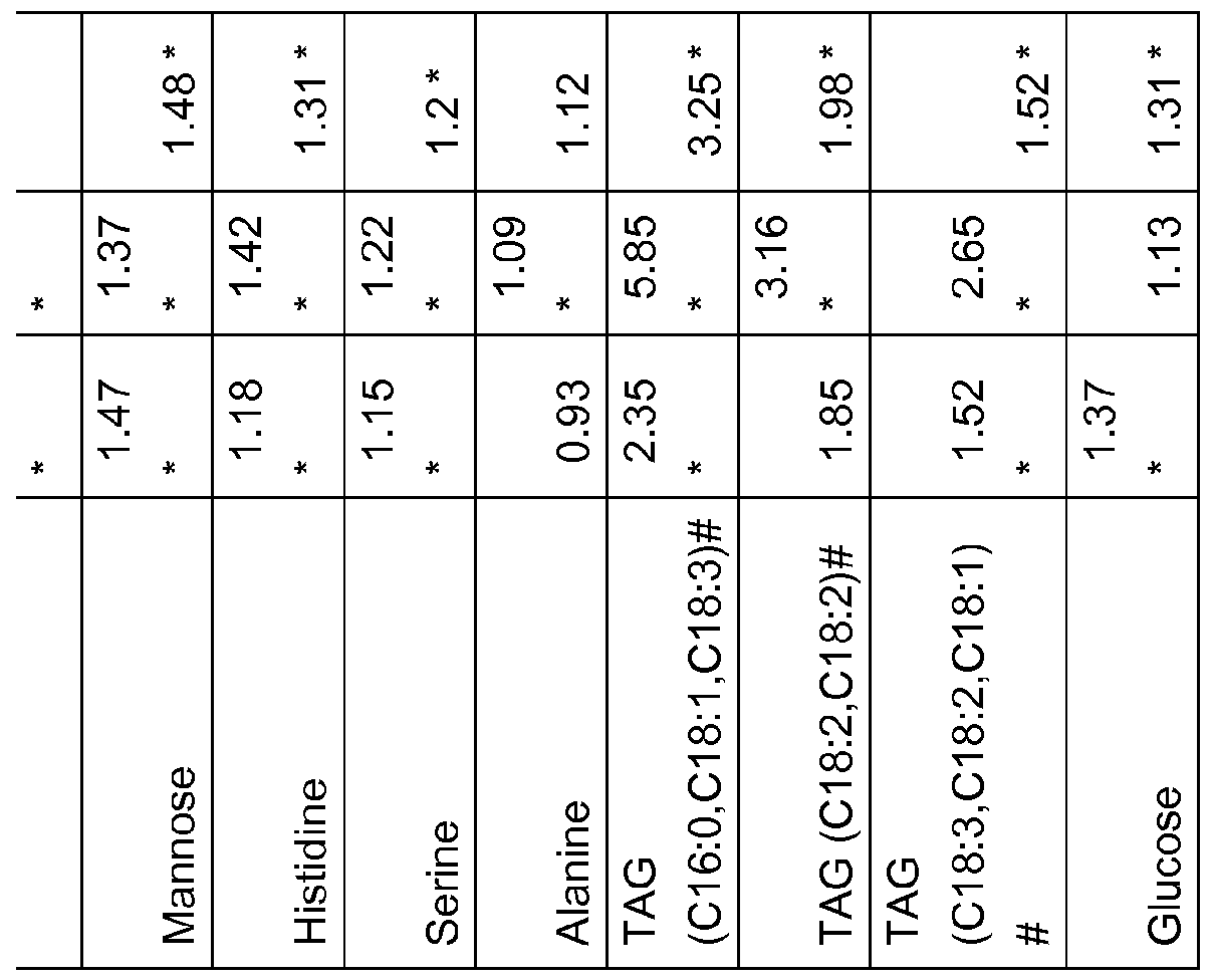


Claims
Priority Applications (4)
| Application Number | Priority Date | Filing Date | Title |
|---|---|---|---|
| EP13748922.5A EP2815236A4 (en) | 2012-02-15 | 2013-02-15 | Means and methods for assessing an endocrine disease or disorder |
| US14/376,921 US20150010928A1 (en) | 2012-02-15 | 2013-02-15 | Means and methods for assessing an endocrine disease or disorder |
| JP2014557154A JP2015512035A (en) | 2012-02-15 | 2013-02-15 | Means and methods for assessing endocrine diseases or disorders |
| CA2863138A CA2863138A1 (en) | 2012-02-15 | 2013-02-15 | Means and methods for assessing an endocrine disease or disorder |
Applications Claiming Priority (4)
| Application Number | Priority Date | Filing Date | Title |
|---|---|---|---|
| US201261598965P | 2012-02-15 | 2012-02-15 | |
| EP12155640 | 2012-02-15 | ||
| EP12155640.1 | 2012-02-15 | ||
| US61/598,965 | 2012-02-15 |
Publications (1)
| Publication Number | Publication Date |
|---|---|
| WO2013121380A1 true WO2013121380A1 (en) | 2013-08-22 |
Family
ID=48983612
Family Applications (1)
| Application Number | Title | Priority Date | Filing Date |
|---|---|---|---|
| PCT/IB2013/051224 WO2013121380A1 (en) | 2012-02-15 | 2013-02-15 | Means and methods for assessing an endocrine disease or disorder |
Country Status (5)
| Country | Link |
|---|---|
| US (1) | US20150010928A1 (en) |
| EP (1) | EP2815236A4 (en) |
| JP (1) | JP2015512035A (en) |
| CA (1) | CA2863138A1 (en) |
| WO (1) | WO2013121380A1 (en) |
Families Citing this family (2)
| Publication number | Priority date | Publication date | Assignee | Title |
|---|---|---|---|---|
| EP2756297A4 (en) * | 2011-09-14 | 2015-10-14 | Basf Se | Means and methods for assessing gonadal toxicity |
| EP2815241A2 (en) * | 2012-02-15 | 2014-12-24 | Basf Se | Means and methods for assessing bone disorders |
Citations (3)
| Publication number | Priority date | Publication date | Assignee | Title |
|---|---|---|---|---|
| WO2010151133A1 (en) * | 2009-06-25 | 2010-12-29 | Erasmus University Medical Center Rotterdam | Methods for establishing and predicting resistance to endocrine therapy using an mirna profile |
| WO2011018288A1 (en) * | 2009-08-13 | 2011-02-17 | Basf Se | Means and methods for diagnosingthyroid disorders |
| WO2011151252A2 (en) * | 2010-06-01 | 2011-12-08 | Metanomics Health Gmbh | Means and methods for diagnosing pancreatic cancer in a subject |
Family Cites Families (1)
| Publication number | Priority date | Publication date | Assignee | Title |
|---|---|---|---|---|
| WO2009153139A2 (en) * | 2008-05-28 | 2009-12-23 | Basf Se | Means and methods for assessing increased peroxisomal proliferation |
-
2013
- 2013-02-15 US US14/376,921 patent/US20150010928A1/en not_active Abandoned
- 2013-02-15 CA CA2863138A patent/CA2863138A1/en not_active Abandoned
- 2013-02-15 JP JP2014557154A patent/JP2015512035A/en not_active Withdrawn
- 2013-02-15 WO PCT/IB2013/051224 patent/WO2013121380A1/en active Application Filing
- 2013-02-15 EP EP13748922.5A patent/EP2815236A4/en not_active Withdrawn
Patent Citations (3)
| Publication number | Priority date | Publication date | Assignee | Title |
|---|---|---|---|---|
| WO2010151133A1 (en) * | 2009-06-25 | 2010-12-29 | Erasmus University Medical Center Rotterdam | Methods for establishing and predicting resistance to endocrine therapy using an mirna profile |
| WO2011018288A1 (en) * | 2009-08-13 | 2011-02-17 | Basf Se | Means and methods for diagnosingthyroid disorders |
| WO2011151252A2 (en) * | 2010-06-01 | 2011-12-08 | Metanomics Health Gmbh | Means and methods for diagnosing pancreatic cancer in a subject |
Non-Patent Citations (1)
| Title |
|---|
| See also references of EP2815236A4 * |
Also Published As
| Publication number | Publication date |
|---|---|
| EP2815236A1 (en) | 2014-12-24 |
| JP2015512035A (en) | 2015-04-23 |
| US20150010928A1 (en) | 2015-01-08 |
| EP2815236A4 (en) | 2015-11-04 |
| CA2863138A1 (en) | 2013-08-22 |
Similar Documents
| Publication | Publication Date | Title |
|---|---|---|
| Leav et al. | Comparative studies of the estrogen receptors β and α and the androgen receptor in normal human prostate glands, dysplasia, and in primary and metastatic carcinoma | |
| Dalenc et al. | Circulating oxysterol metabolites as potential new surrogate markers in patients with hormone receptor-positive breast cancer: Results of the OXYTAM study | |
| Genazzani et al. | Circulating levels of allopregnanolone in humans: gender, age, and endocrine influences | |
| Adamyan et al. | Direct mass spectrometry differentiation of ectopic and eutopic endometrium in patients with endometriosis | |
| Wang et al. | Total testosterone quantitative measurement in serum by LC-MS/MS | |
| Jia et al. | Serum metabolomics analysis of patients with polycystic ovary syndrome by mass spectrometry | |
| Yoo et al. | Quantification of dehydroepiandrosterone, 17β-estradiol, testosterone, and their sulfates in mouse tissues by LC-MS/MS | |
| WO2013038369A1 (en) | Means and methods for assessing kidney toxicity | |
| Flores-Valverde et al. | Methodology for Profiling the Steroid Metabolome in Animal Tissues Using Ultraperformance Liquid Chromatography− Electrospray-Time-of-Flight Mass Spectrometry | |
| Amita et al. | Molecular mechanism of the inhibition of estradiol-induced endometrial epithelial cell proliferation by clomiphene citrate | |
| Housa et al. | Adiponectin as a potential marker of prostate cancer progression: studies in organ-confined and locally advanced prostate cancer. | |
| Benedusi et al. | Liver ERα regulates AgRP neuronal activity in the arcuate nucleus of female mice | |
| EP0547141A1 (en) | Onset of antiestrogen resistance in breast cancer | |
| EP2815236A1 (en) | Means and methods for assessing an endocrine disease or disorder | |
| Vaňková et al. | Reduced sulfotransferase SULT2A1 activity in patients with Alzheimer's disease. | |
| Turathum et al. | Dysregulated sphingolipid metabolism and autophagy in granulosa cells of women with endometriosis | |
| Ding et al. | Follicular fluid lipidomic profiling reveals potential biomarkers of polycystic ovary syndrome: A pilot study | |
| EP1710585B1 (en) | Prediction method for lipidosis | |
| Mandava et al. | Aromatase overexpression transgenic mice model: cell type specific expression and use of letrozole to abrogate mammary hyperplasia without affecting normal physiology | |
| Yang et al. | Simultaneous quantification of oestrogens and androgens in the serum of patients with benign prostatic hyperplasia by liquid chromatography–Tandem mass spectrometry | |
| Meunier et al. | Sex steroids in serum and prostatic tissue of human cancerous prostate (STERKPROSER trial) | |
| Choi | Mass spectrometry-based metabolic signatures of sex steroids in breast cancer | |
| Lee et al. | Sex-related differences in urinary immune-related metabolic profiling of alopecia areata patients | |
| Goss et al. | Liarozole fumarate (R85246): a novel imidazole in the treatment of receptor positive postmenopausal metastatic breast cancer | |
| US20140342379A1 (en) | Means and Methods for Assessing Gonadal Toxicity |
Legal Events
| Date | Code | Title | Description |
|---|---|---|---|
| 121 | Ep: the epo has been informed by wipo that ep was designated in this application |
Ref document number: 13748922 Country of ref document: EP Kind code of ref document: A1 |
|
| ENP | Entry into the national phase |
Ref document number: 2863138 Country of ref document: CA |
|
| WWE | Wipo information: entry into national phase |
Ref document number: 14376921 Country of ref document: US |
|
| ENP | Entry into the national phase |
Ref document number: 2014557154 Country of ref document: JP Kind code of ref document: A |
|
| REEP | Request for entry into the european phase |
Ref document number: 2013748922 Country of ref document: EP |
|
| WWE | Wipo information: entry into national phase |
Ref document number: 2013748922 Country of ref document: EP |
|
| NENP | Non-entry into the national phase |
Ref country code: DE |