WO2013119716A1 - Compositions and methods for using csf1r inhibitors - Google Patents
Compositions and methods for using csf1r inhibitors Download PDFInfo
- Publication number
- WO2013119716A1 WO2013119716A1 PCT/US2013/024998 US2013024998W WO2013119716A1 WO 2013119716 A1 WO2013119716 A1 WO 2013119716A1 US 2013024998 W US2013024998 W US 2013024998W WO 2013119716 A1 WO2013119716 A1 WO 2013119716A1
- Authority
- WO
- WIPO (PCT)
- Prior art keywords
- antibody
- seq
- amino acid
- human
- acid sequence
- Prior art date
Links
Classifications
-
- C—CHEMISTRY; METALLURGY
- C07—ORGANIC CHEMISTRY
- C07K—PEPTIDES
- C07K14/00—Peptides having more than 20 amino acids; Gastrins; Somatostatins; Melanotropins; Derivatives thereof
- C07K14/435—Peptides having more than 20 amino acids; Gastrins; Somatostatins; Melanotropins; Derivatives thereof from animals; from humans
- C07K14/705—Receptors; Cell surface antigens; Cell surface determinants
- C07K14/715—Receptors; Cell surface antigens; Cell surface determinants for cytokines; for lymphokines; for interferons
- C07K14/7153—Receptors; Cell surface antigens; Cell surface determinants for cytokines; for lymphokines; for interferons for colony-stimulating factors [CSF]
-
- A—HUMAN NECESSITIES
- A61—MEDICAL OR VETERINARY SCIENCE; HYGIENE
- A61P—SPECIFIC THERAPEUTIC ACTIVITY OF CHEMICAL COMPOUNDS OR MEDICINAL PREPARATIONS
- A61P1/00—Drugs for disorders of the alimentary tract or the digestive system
- A61P1/04—Drugs for disorders of the alimentary tract or the digestive system for ulcers, gastritis or reflux esophagitis, e.g. antacids, inhibitors of acid secretion, mucosal protectants
-
- A—HUMAN NECESSITIES
- A61—MEDICAL OR VETERINARY SCIENCE; HYGIENE
- A61P—SPECIFIC THERAPEUTIC ACTIVITY OF CHEMICAL COMPOUNDS OR MEDICINAL PREPARATIONS
- A61P19/00—Drugs for skeletal disorders
- A61P19/02—Drugs for skeletal disorders for joint disorders, e.g. arthritis, arthrosis
-
- A—HUMAN NECESSITIES
- A61—MEDICAL OR VETERINARY SCIENCE; HYGIENE
- A61P—SPECIFIC THERAPEUTIC ACTIVITY OF CHEMICAL COMPOUNDS OR MEDICINAL PREPARATIONS
- A61P25/00—Drugs for disorders of the nervous system
-
- A—HUMAN NECESSITIES
- A61—MEDICAL OR VETERINARY SCIENCE; HYGIENE
- A61P—SPECIFIC THERAPEUTIC ACTIVITY OF CHEMICAL COMPOUNDS OR MEDICINAL PREPARATIONS
- A61P29/00—Non-central analgesic, antipyretic or antiinflammatory agents, e.g. antirheumatic agents; Non-steroidal antiinflammatory drugs [NSAID]
-
- A—HUMAN NECESSITIES
- A61—MEDICAL OR VETERINARY SCIENCE; HYGIENE
- A61P—SPECIFIC THERAPEUTIC ACTIVITY OF CHEMICAL COMPOUNDS OR MEDICINAL PREPARATIONS
- A61P37/00—Drugs for immunological or allergic disorders
- A61P37/02—Immunomodulators
- A61P37/06—Immunosuppressants, e.g. drugs for graft rejection
-
- C—CHEMISTRY; METALLURGY
- C07—ORGANIC CHEMISTRY
- C07K—PEPTIDES
- C07K14/00—Peptides having more than 20 amino acids; Gastrins; Somatostatins; Melanotropins; Derivatives thereof
- C07K14/435—Peptides having more than 20 amino acids; Gastrins; Somatostatins; Melanotropins; Derivatives thereof from animals; from humans
- C07K14/52—Cytokines; Lymphokines; Interferons
- C07K14/54—Interleukins [IL]
-
- C—CHEMISTRY; METALLURGY
- C07—ORGANIC CHEMISTRY
- C07K—PEPTIDES
- C07K16/00—Immunoglobulins [IGs], e.g. monoclonal or polyclonal antibodies
- C07K16/18—Immunoglobulins [IGs], e.g. monoclonal or polyclonal antibodies against material from animals or humans
- C07K16/24—Immunoglobulins [IGs], e.g. monoclonal or polyclonal antibodies against material from animals or humans against cytokines, lymphokines or interferons
- C07K16/243—Colony Stimulating Factors
-
- C—CHEMISTRY; METALLURGY
- C07—ORGANIC CHEMISTRY
- C07K—PEPTIDES
- C07K16/00—Immunoglobulins [IGs], e.g. monoclonal or polyclonal antibodies
- C07K16/18—Immunoglobulins [IGs], e.g. monoclonal or polyclonal antibodies against material from animals or humans
- C07K16/24—Immunoglobulins [IGs], e.g. monoclonal or polyclonal antibodies against material from animals or humans against cytokines, lymphokines or interferons
- C07K16/244—Interleukins [IL]
-
- A—HUMAN NECESSITIES
- A61—MEDICAL OR VETERINARY SCIENCE; HYGIENE
- A61K—PREPARATIONS FOR MEDICAL, DENTAL OR TOILETRY PURPOSES
- A61K39/00—Medicinal preparations containing antigens or antibodies
- A61K2039/505—Medicinal preparations containing antigens or antibodies comprising antibodies
-
- A—HUMAN NECESSITIES
- A61—MEDICAL OR VETERINARY SCIENCE; HYGIENE
- A61K—PREPARATIONS FOR MEDICAL, DENTAL OR TOILETRY PURPOSES
- A61K39/00—Medicinal preparations containing antigens or antibodies
- A61K2039/505—Medicinal preparations containing antigens or antibodies comprising antibodies
- A61K2039/507—Comprising a combination of two or more separate antibodies
-
- C—CHEMISTRY; METALLURGY
- C07—ORGANIC CHEMISTRY
- C07K—PEPTIDES
- C07K2317/00—Immunoglobulins specific features
- C07K2317/20—Immunoglobulins specific features characterized by taxonomic origin
- C07K2317/21—Immunoglobulins specific features characterized by taxonomic origin from primates, e.g. man
-
- C—CHEMISTRY; METALLURGY
- C07—ORGANIC CHEMISTRY
- C07K—PEPTIDES
- C07K2317/00—Immunoglobulins specific features
- C07K2317/50—Immunoglobulins specific features characterized by immunoglobulin fragments
- C07K2317/55—Fab or Fab'
-
- C—CHEMISTRY; METALLURGY
- C07—ORGANIC CHEMISTRY
- C07K—PEPTIDES
- C07K2317/00—Immunoglobulins specific features
- C07K2317/50—Immunoglobulins specific features characterized by immunoglobulin fragments
- C07K2317/56—Immunoglobulins specific features characterized by immunoglobulin fragments variable (Fv) region, i.e. VH and/or VL
-
- C—CHEMISTRY; METALLURGY
- C07—ORGANIC CHEMISTRY
- C07K—PEPTIDES
- C07K2317/00—Immunoglobulins specific features
- C07K2317/70—Immunoglobulins specific features characterized by effect upon binding to a cell or to an antigen
- C07K2317/76—Antagonist effect on antigen, e.g. neutralization or inhibition of binding
-
- C—CHEMISTRY; METALLURGY
- C07—ORGANIC CHEMISTRY
- C07K—PEPTIDES
- C07K2317/00—Immunoglobulins specific features
- C07K2317/90—Immunoglobulins specific features characterized by (pharmaco)kinetic aspects or by stability of the immunoglobulin
- C07K2317/92—Affinity (KD), association rate (Ka), dissociation rate (Kd) or EC50 value
-
- Y—GENERAL TAGGING OF NEW TECHNOLOGICAL DEVELOPMENTS; GENERAL TAGGING OF CROSS-SECTIONAL TECHNOLOGIES SPANNING OVER SEVERAL SECTIONS OF THE IPC; TECHNICAL SUBJECTS COVERED BY FORMER USPC CROSS-REFERENCE ART COLLECTIONS [XRACs] AND DIGESTS
- Y02—TECHNOLOGIES OR APPLICATIONS FOR MITIGATION OR ADAPTATION AGAINST CLIMATE CHANGE
- Y02A—TECHNOLOGIES FOR ADAPTATION TO CLIMATE CHANGE
- Y02A50/00—TECHNOLOGIES FOR ADAPTATION TO CLIMATE CHANGE in human health protection, e.g. against extreme weather
- Y02A50/30—Against vector-borne diseases, e.g. mosquito-borne, fly-borne, tick-borne or waterborne diseases whose impact is exacerbated by climate change
Definitions
- the present invention relates to compositions and methods for using CSF1-R pathway inhibitors, including anti-IL-34 antibodies, bispecific IL-34/CSF1 antibodies and CSF1R antibodies.
- Interleukin-34 also known as C16orf77 or UNQ20374 (Clark et al., Genome Res 13: 2265- 2270 (2003)), was recently identified as a second and high-affinity ligand for CSF-IR in a human monocyte proliferation screening (Lin et al., Science 320: 807-811 (2008)). This discovery has long been foreshadowed by the more severe phenotype in CSF-IR null mice, than CSF-1 -deficient CSF-1 op /CSF-l op mice (Dai et al, Blood 99: 111-120 (2002)).
- CSF-1 also known as M-CSF
- IL-34 stimulates phosphorylation of ERK1/2 in human monocytes and promotes the formation of the granulocyte-macrophage progenitor (CFU-GM) and megakaryocyte progenitor (CFU-M) in human bone marrow cultures (Lin et al, Science 320: 807-811 (2008)).
- CFU-GM granulocyte-macrophage progenitor
- CFU-M megakaryocyte progenitor
- the transcript of the proto-oncogene c-fms, IL-34 and CSF-1 serve as the key regulators of the differentiation, proliferation, and survival of the mononuclear phagocyte lineage cell such as monocytes, macrophages and osteoclasts (Droin et al, Journal of leukocyte biology 87: 745-747 (2010)).
- IL-34 The function of IL-34 bears strong resemblance to that of CSF-1, but with several notable differences. Both cytokines support cell growth and survival in cell cultures studies equivalently (Chihara et al, Cell death and differentiation 17: 1917-1927 (2010); Wei et al., Journal of leukocyte biology 88: 495-505 (2010)).
- the IL-34 gene when expressed under the control of the CSF-1 promoter, could rescue the phenotype of CSF-1 -nullizygous CSF- l op /CSF-l op mice (Wei et al, Journal of leukocyte biology 88: 495-505 (2010)).
- IL-34 can also substitute for CSF-1 to support RANKL-induced osteoclastogenesis (Baud'huin et al, The Journal of pathology 221 : 77-86 (2010)).
- the two factors appear different in their ability to induce the production of chemokines such as MCP-1 and eotaxin-2 in primary macrophages, the morphological change in TF-l-fms cells and the migration of J774A.1 cells (Chihara et al, Cell death and differentiation 17: 1917-1927 (2010)).
- IL-34 has been shown to induce a stronger, but transient tyrosine phosphorylation of CSF-IR and downstream effectors, and rapidly downregulates CSF-IR expression (Chihara et al, Cell death and differentiation 17: 1917-1927 (2010)). Moreover, IL-34 and CSF-1 exhibit differential spatiotemporal patterns of expression in both embryonic and adult tissues, which leads to the complementary activation of the CSF-IR (Wei et al., Journal of leukocyte biology 88: 495- 505 (2010)).
- IL-34 but not CSF-1 messenger RNA is detected together with CSF-IR in embryonic brain which could explain why microglia develop in CSF-1 deficient but not CSF-IR deficient mice (Ginhoux et al, Science 330: 841-845 (2010); Mizuno et al, The American journal of pathology 179: 2016-2027 (2011)).
- IL-34 and CSF-1 resemble each other, they are not necessarily identical in their developmental roles, biological activity, and signal activation kinetics or strength.
- IL-34 was proposed by fold recognition methods to be a short-chain helical cytokine belonging to the same family as CSF-1, SCF, and Flt3L (Garceau et al, Journal of leukocyte biology 87: 753-764 (2010)). These latter three dimeric hematopoietic cytokines are unique among helical cytokines in that they have membrane-bound forms (Bazan, Cell 65: 9-10 (1991a); Hannum et al., Nature 368: 643-648 (1994)); IL-34 differs importantly in that it lacks a hydrophobic transmembrane segment.
- CSF-1, SCF and Flt3L cytokine dimers bind to the PDGFR subfamily (type III/V) of the receptor tyrosine kinase (RTK) family (Rosnet et al., Critical reviews in oncogenesis 4: 595-613 (1993)) instead of hematopoietic cytokine receptors (Bazan,
- the CSF-1, SCF and Flt3L cytokine dimers functionally mimic the PDGF and VEGF cystine knot growth factor dimers that are the activating ligands of the RTK family (Sawides et al, Nature structural biology 7: 486-491 (2000); Wiesmann et al, Nature structural biology 7: 440-442 (2000)). All members of this RTK family share a similar overall architecture comprised of multiple Ig-like domains in their extracellular regions, a single transmembrane segment, and a cytoplasmic tyrosine kinase domain with a large insertion.
- CSF-IR Upon stimulation, CSF-IR dimerizes and autophosphorylates certain tyrosine residues in its intracellular domain, which serve as docking sites for SH2- containing effector proteins, which contribute to macrophage differentiation (Pixley et al., Trends in cell biology 14: 628-638 (2004)).
- CSF-1 competes with IL-34 for binding to CSF-1 R (Wei et al, Journal of leukocyte biology 88: 495-505 (2010)), suggesting a common ligand-binding site on CSF-1R.
- CSF-1R a recent comparative sequence study between CSF-1R and its two ligands suggested the CD loop of IL-34, and the junction between D3 and D4 of CSF-1 R share strong sequence conservation correlation coefficients during evolution, and therefore may represent a unique binding mode that is distinct from the binding mode employed by the CSF-l/CSF-lR complex (Garceau et al, Journal of leukocyte biology 87: 753-764 (2010)).
- the invention provides anti-IL-34 antibodies, bispecific antibodies that bind to IL-34 and
- the antibodies of this invention have reduced antibody-dependent cell-mediated cytotoxicity (ADCC) and/or complement dependent cytotoxicity (CDC) activity.
- ADCC antibody-dependent cell-mediated cytotoxicity
- CDC complement dependent cytotoxicity
- the antibodies of this invention have reduced ADCC activity by comprising at least an Fc region substitution at one or more of the following residues 238, 265, 269, 270, 297, 327 and 329 (EU numbering).
- the Fc region substitution to reduce ADCC activity is at residue 297.
- the Fc region substitution to reduce ADCC activity is N297G or N297A.
- the Fc substitution to reduce ADCC is D265A.
- the Fc substitutions to reduce ADCC activit y are the substitution of residues 265 and 297 to alanine.
- the bispecific anti-IL-34/anti-CSF-l antibody is a knob-into-hole bispecific antibody.
- isolated antibodies that bind to human IL-34, which bind to an epitope comprising at least one of amino acid residues Glul03, Leul09, Glnl06, Asnl50, Leul27, Asnl28, Serl84, Leul86, Asnl87, Lys44, Glul21, Aspl07, Glul l l, Serl04, Glnl20, Trpl 16, and Asn61of a human IL-34, where the position of the amino acid residues is based on the position in SEQ ID NO: 1 , and which inhibit the binding between human IL-34 and human CSF-1R.
- isolated antibodies that binds to human IL-34, which bind to an epitope comprising at least one of amino acid residues from Glul03 to Asnl50 of a human IL-34, where the position of the amino acid residues is based on SEQ ID NO: l, and which inhibit the binding between human IL-34 and human CSF- 1 R.
- the antibody binds to an epitope comprising at least one of amino acid residues Glul03, Leul09, Glnl06, and Asnl50 of the human IL-34, where the position of the amino acid residues is based on the position in SEQ ID NO: 1.
- the epitope further comprises at least one of amino acid residues SerlOO, Glul23, Trpl 16, Thrl24, Leul27, Asnl28, Glnl31, and Thrl34 of the human IL-34, where the position of the amino acid residues is based on the position in SEQ ID NO: 1.
- the antibody binds to amino acids within positions 100-108, 116-134, 109 and 150 of the human IL-34, where the position of the amino acid residues is based on the position in SEQ ID NO: l .
- the antibody binds to an epitope comprising at least one of amino acid residues Asnl28, Serl84, Leul86, Asnl87, Lys44, and Glul21 of the human IL-34, where the position of the amino acid residues is based on the position in SEQ ID NO: 1.
- the epitope further comprises at least one of amino acid residues Phe40, Asp43, Leul25, Glnl89, Thr36, and Vail 85 of the human IL-34, where the position of the amino acid residues is based on the position in SEQ ID NO: 1.
- the antibody binds to amino acids within positions 36-44, 121-128, and 184-187 of the human IL- 34, where the position of the amino acid residues is based on the position in SEQ ID NO: 1. In some embodiments, the antibody binds to an epitope comprising at least one of amino acid residues from Glul03-Leul27 of the human IL-34, where the position of the amino acid residues is based on the position in SEQ ID NO: 1.
- the antibody binds to an epitope comprising at least one of amino acid residues Asp 107, Glul 11, Serl04, Glnl20, Glul03, Leul09, Trpl 16, and Asn61 of the human IL-34, where the position of the amino acid residues is based on the position in SEQ ID NO: 1.
- the epitope further comprises at least one of amino acid residues Prol52, Vall08, Leul 10, Glnl06, Glul23, Leul27, Lysl 17, Ile60 and Lys55 of the human IL-34, where the position of the amino acid residues is based on the position in SEQ ID NO: 1.
- the antibody binds to amino acids within positions 55-61, 100-108, 109, 111-127 and 152 of the human IL-34, where the position of the amino acid residues is based on the position in SEQ ID NO: l .
- the antibody comprises a heavy chain variable region sequence of at least 90% sequence identity to the amino acid sequence of SEQ ID NO:3 and/or a light chain variable region sequence of at least 90%> sequence identity to the amino acid sequence of SEQ ID NO:4. In some embodiments, the antibody comprises a heavy chain variable region sequence of the amino acid sequence of SEQ ID NO:3 and/or a light chain variable region sequence of the amino acid sequence of SEQ ID NO:4.
- the antibody comprises (a) a HVR-H3 comprising an amino acid sequence GLGKGSKRGAMDY (SEQ ID NO: 33); (b) a HVR-L3 comprising an amino acid sequence QQSFYFPNT (SEQ ID NO: 39); and (c) a HVR-H2 comprising an amino acid sequence RISPYYYYSDYADSVKG (SEQ ID NO: 52).
- the antibody comprises (a) a HVR-H1 comprising an amino acid sequence STWIH (SEQ ID NO: 59), (b) a HVR-H2 comprising an amino acid sequence RISPYYYYSDYADSVKG (SEQ ID NO: 52); and (c) a HVR-H3 comprising an amino acid sequence GLGKGSKRGAMDY (SEQ ID NO: 33).
- the antibody comprises (a) a HVR-L1 comprising an amino acid sequence RASQDVSTAVA (SEQ ID NO: 50); (b) a HVR-L2 comprising an amino acid sequence SASFLYS (SEQ ID NO: 53); and (c) a HVR-L3 comprising an amino acid sequence QQSFYFPNT (SEQ ID NO: 39).
- the antibody comprises (a) a HVR-H3 comprising an amino acid sequence GLGKGSKRGAMDY (SEQ ID NO: 33) or GINQGSKRGAMDY (SEQ ID NO: 32); (b) a HVR-L3 comprising an amino acid sequence QQSFYFPNT (SEQ ID NO: 39) or QQSYTTPPT (SEQ ID NO: 43) or QQYTALPYT (SEQ ID NO: 49) or QQYSDLPYT (SEQ ID NO: 45) or QQYSDVPYT (SEQ ID NO: 47) or QQSRTARPT (SEQ ID NO: 41); and (c) a HVR-H2 comprising an amino acid sequence RISPYYYYSDYADSVKG (SEQ ID NO: 52) or RISPYSGYTNYADS VKG (SEQ ID NO : 51 ).
- the antibody comprises (a) a HVR-H1 comprising an amino acid sequence STWIH (SEQ ID NO: 59); (b) a HVR-H2 comprising an amino acid sequence RISPYYYYSDYADSVKG (SEQ ID NO: 52) or RISPYSGYTNYADS VKG (SEQ ID NO: 51); and (c) a HVR-H3 comprising an amino acid sequence GLGKGSK GAMDY (SEQ ID NO: 33) or GINQGSK GAMDY (SEQ ID NO: 32).
- the antibody comprises (a) a HVR-L1 comprising an amino acid sequence RASQDVSTAVA (SEQ ID NO: 50); (b) a HVR-L2 comprising an amino acid sequence SASFLYS (SEQ ID NO: 53); and (c) a HVR-L3 comprising an amino acid sequence QQSFYFPNT (SEQ ID NO: 39) or QQSYTTPPT (SEQ ID NO: 43) or
- QQYTALPYT SEQ ID NO: 49 or QQYSDLPYT (SEQ ID NO: 45) or QQYSDVPYT (SEQ ID NO: 47) or QQSRTARPT (SEQ ID NO: 41) or QQSFYFPN (SEQ ID NO: 38) or QQSYTTPP (SEQ ID NO: 42) or QQYTALPY (SEQ ID NO: 48) or QQYSDLPY (SEQ ID NO: 44) or QQYSDVPY (SEQ ID NO: 46) or QQSRTARP (SEQ ID NO: 40).
- the antibody comprises (a) a HVR-H3 comprising an amino acid sequence GLGKGSKRGAMDY (SEQ ID NO: 33); (b) a HVR-L3 comprising an amino acid sequence QQYSDLPYT (SEQ ID NO: 45); and (c) a HVR-H2 comprising an amino acid sequence RISPYSGYTNYADSVKG (SEQ ID NO: 51).
- the antibody comprises (a) a HVR-H1 comprising an amino acid sequence of STWIH (SEQ ID NO: 59); (b) a HVR-H2 comprising an amino acid sequence RISPYSGYTNYADSVKG (SEQ ID NO: 51); and (c) a HVR-H3 comprising an amino acid sequence GLGKGSKRGAMDY (SEQ ID NO: 33).
- the antibody comprises (a) a HVR-L1 comprising an amino acid sequence of RASQDVSTAVA (SEQ ID NO: 50); (b) a HVR-L2 comprising an amino acid sequence SASFLYS (SEQ ID NO: 53); and (c) a HVR-L3 comprising an amino acid sequence QQYSDLPYT (SEQ ID NO: 45).
- the antibody comprises a heavy chain variable region sequence of at least 90% sequence identity to the amino acid sequence of SEQ ID NO:5 and/or a light chain variable region sequence of at least 90%> sequence identity to the amino acid sequence of SEQ ID NO:6. In some embodiments, the antibody comprises a heavy chain variable region sequence of the amino acid sequence of SEQ ID NO:5 and/or a light chain variable region sequence of the amino acid sequence of SEQ ID NO:6. In some embodiments, the antibody comprises a heavy chain variable region sequence of at least 90% sequence identity to the amino acid sequence of SEQ ID NO:7 and/or a light chain variable region sequence of at least 90%) sequence identity to the amino acid sequence of SEQ ID NO:8.
- the antibody comprises a heavy chain variable region sequence of the amino acid sequence of SEQ ID NO:7 and/or a light chain variable region sequence of the amino acid sequence of SEQ ID NO:8. In some embodiments, the antibody comprises a heavy chain variable region sequence of at least 90%> sequence identity to the amino acid sequence of SEQ ID NO: 9 and/or a light chain variable region sequence of at least 90% sequence identity to the amino acid sequence of SEQ ID NO: 10. In some embodiments, the antibody comprises a heavy chain variable region sequence of the amino acid sequence of SEQ ID NO:9 and/or a light chain variable region sequence of the amino acid sequence of SEQ ID NO: 10.
- the antibody comprises a heavy chain variable region sequence of at least 90% sequence identity to the amino acid sequence of SEQ ID NO: 11 and/or a light chain variable region sequence of at least 90%> sequence identity to the amino acid sequence of SEQ ID NO: 12. In some embodiments, the antibody comprises a heavy chain variable region sequence of the amino acid sequence of SEQ ID NO: 11 and/or a light chain variable region sequence of the amino acid sequence of SEQ ID NO: 12. In some embodiments, the antibody comprises a heavy chain variable region sequence of at least 90% sequence identity to the amino acid sequence of SEQ ID NO: 13 and/or a light chain variable region sequence of at least 90%) sequence identity to the amino acid sequence of SEQ ID NO: 14. In some embodiments, the antibody comprises a heavy chain variable region sequence of the amino acid sequence of SEQ ID NO : 13 and/or a light chain variable region sequence of the amino acid sequence of SEQ ID NO: 14.
- the antibody comprises (a) a HVR-H3 comprising an amino acid sequence SRGAYRFAY (SEQ ID NO: 56); (b) a HVR-L3 comprising an amino acid sequence QQSYTTPPT (SEQ ID NO: 43); and (c) a HVR-H2 comprising an amino acid sequence SITPASGDTDYADSVKG (SEQ ID NO: 54).
- the antibody comprises (a) a HVR-H1 comprising an amino acid sequence SNYIH (SEQ ID NO: 55), (b) a HVR-H2 comprising an amino acid sequence SITPASGDTDYADSVKG (SEQ ID NO: 54); and (c) a HVR-H3 comprising an amino acid sequence SRGAYRFAY (SEQ ID NO: 56).
- the antibody comprises (a) a HVR-L1 comprising an amino acid sequence RASQDVSTAVA (SEQ ID NO: 50); (b) a HVR-L2 comprising an amino acid sequence SASFLYS (SEQ ID NO: 53); and (c) a HVR-L3 comprising an amino acid sequence QQSYTTPPT (SEQ ID NO: 43).
- the antibody comprises a heavy chain variable region sequence of at least 90% sequence identity to the amino acid sequence of SEQ ID NO: 15 and/or a light chain variable region sequence of at least 90% sequence identity to the amino acid sequence of SEQ ID NO: 16.
- the antibody comprises a heavy chain variable region sequence of the amino acid sequence of SEQ ID NO: 15 and/or a light chain variable region sequence of the amino acid sequence of SEQ ID NO: 16. In some embodiments, the antibody does not inhibit the binding between human CSF-1 and human CSF-1R.
- the anti-IL-34 antibody described herein binds to a dimer of the IL-34. In some embodiments, the anti-IL-34 antibody described herein binds to an epitope that spans over both protomers of the IL-34 dimer. In some embodiments, the anti-IL-34 antibody described herein neutralizes IL-34 activity. In some embodiments, the anti-IL-34 antibody binds to human IL-34, inhibit the binding between human IL-34 and human CSF-1R, and/or neutralize IL-34 activity.
- the anti-IL-34 antibody described herein is a monoclonal antibody. In some embodiments, the anti-IL-34 antibody described herein a human, humanized or chimeric antibody. In some embodiments, the antibody is a bispecific antibody. In some embodiments, the bispecific antibody comprises a second binding specificity to human CSF- 1.
- bispecific antibodies comprising a first binding specificity to human IL- 34 and a second binding specificity to human CSF-1 and their use in treating myeloid pathogenic immunological diseases and cancers.
- the antibody inhibits binding of human IL-34 to human CSF-1R and inhibits binding of human CSF-1 to human CSF-1 R.
- two polypeptides comprising binding specificity to human IL-34 and the binding specificity to human CSF-1, respectively, each has a heteromultimerization domain that is capable is heterodimerizing with each other.
- the antibody described above is an antibody fragment that binds human IL-34.
- the fragment is a Fab, Fab', Fab'-SH, F(ab') 2 , Fv or scFv fragment.
- the antibody described herein is a one-armed antibody. In some embodiments, the antibody described herein is a linear antibody. In some embodiments, the antibody described herein is a full length IgGl or an IgG4 antibody.
- isolated antibodies that bind human CSF-1 R, which bind to an epitope comprising at least one of amino acid residues Argl44, Gln248, Gln249, Ser250, Phe252, and Asn254 of human CSF-1 R, where the position of amino acid residue is based on the position in SEQ ID NO:2, and which inhibit the binding between human IL-34 and human CSF-1R.
- the antibody binds to an epitope comprising amino acid residue Argl44 of CSF-1R, where the position of amino acid residue is based on the position in SEQ ID NO:2. In some embodiments, the antibody binds to an epitope comprising at least one of amino acid residues Argl44, Argl42, Argl46, and Argl50 of human CSF-IR, where the position of amino acid residues is based on the position in SEQ ID NO:2. In some embodiments, the epitope further comprises at least one of amino acid residues Serl72 and Argl92 of human CSF-IR, where the position of amino acid residues is based on the position in SEQ ID NO:2.
- the epitope further comprises at least one of amino acid residues Argl46, Metl49, Argl50, Phel69, Ilel70, and Glnl73 of human CSF-IR, where the position of amino acid residues is based on the position in SEQ ID NO:2.
- the antibody binds to amino acids within positions 142-150 and 169-173, where the position of amino acid residues is based on the position in SEQ ID NO:2.
- the antibody binds to an epitope comprising at least one of amino acid residues Argl44, Gln248, Gln249, Ser250, Phe252, and Asn254 of human CSF-IR, where the position of amino acid residue is based on the position in SEQ ID NO:2. In some embodiments, the antibody binds to an epitope comprising at least one of amino acid residues Tyr257, Gln248, Gln249, Ser250, Phe252, and Asn254 of human CSF-IR, where the position of amino acid residues is based on the position in SEQ ID NO:2.
- the epitope further comprises at least one of amino acid residues Pro247, Gln258, and Lys259, , where the position of amino acid residues is based on the position in SEQ ID NO:2.
- the epitope further comprises at least one of amino acid residues Val231, Asp251, and Tyr257 of human CSF-IR, where the position of amino acid residue is based on the position in SEQ ID NO:2.
- the antibody binds to amino acid residues within positions 231, 248-252, and 254, where the position of amino acid residues is based on the position in SEQ ID NO:2.
- nucleic acids encoding any of the antibodies described herein.
- vectors comprising the nucleic acid of any of the nucleic acids provided herein.
- host cells comprising the nucleic acid provided herein.
- methods of producing an antibody comprising culturing any of the host cells provided herein, so that the antibody is produced.
- the method further comprises recovering the antibody produced by the host cell.
- compositions comprising any of the antibodies provided herein and a pharmaceutically acceptable carrier.
- antibodies described herein for use as a medicament are also provided herein.
- the antibodies described herein for use in treating a myeloid pathogenic immunological disease are also provided herein.
- the medicament is for treating a myeloid pathogenic immunological disease.
- the medicament is for inhibiting binding between human IL-34 and human CSF-1R.
- myeloid pathogenic immunological disease a myeloid pathogenic component
- methods of treating an individual having an inflammatory disease and/or an autoimmune disease with a myeloid pathogenic component comprising administering to the individual an effective amount of any one of the antibodies provided herein.
- methods of treating an individual having an inflammatory disease and/or an autoimmune disease comprising administering to the individual an effective amount of any one of the antibodies or combination therapies provided herein.
- the antibody is a bispecific antibody which inhibits the activity of human IL-34 and human CSF-1.
- the method comprises administering an effective amount of any of the anti-IL- 34 antibodies provided herein in conjunction with an antibody that binds to human CSF-1.
- the activity of human IL-34 and human CSF-1 is inhibited by a bispecific anti-IL-34 and anti-CSFl antibody.
- the inhibition of activity is by inhibiting the binding of human IL-34 to human CSF-1R, and inhibiting the binding of human CSF-1 and human CSF-1R.
- the myeloid pathogenic immunological disease is rheumatoid arthritis (RA), inflammatory bowel disease (e.g., Crohn's, ulcerative colitis), multiple sclerosis, systemic lupus erythematosus, lupus nephritis, asthma, osteoporosis, Paget' s disease, atherosclerosis, metabolic syndrome, type II diabetes, macrophage activated syndrome (MAS), vasculitis (giant cell artheritis, ANCA associated vasculitis), discoid lupus, sarcoidosis, graft versus host disease, LSDs (lysosomal storage diseases like but not limited to Cytostinosis, Salic acid storage disorder, Gaucher disease), Histyocytosis including but not limited to Rosai-Dorfman disease, Faisalabad histiocytosis, H syndrome, pigmented hypertrichosis with insulin dependent diabetes (PHID)s, vas
- RA rheumato
- the vasculitis is microscopic polyarteritis, CNS vasculitis, necrotizing, cutaneous, or hypersensitivity vasculitis, systemic necrotizing vasculitis, or ANCA-associated vasculitis, such as Churg- Strauss vasculitis or syndrome (CSS)).
- the vasculitis is large vessel vasculitis or medium vessel vasculitis.
- the large vessel vasculitis is polymyalgia rheumatica or giant cell arteritis or Takayasu's arteritis.
- the medium vessel vasculitis is Kawasaki's disease of polyarteritis nodosa.
- an antibody of this invention e.g., IL-34, bispecific IL-34/CSF1 antibody or CSF1R antibody
- DMARD-IR disease- modifying antirheumatic drug
- the DMARD-IR patient has not been previously treated with an anti-TNF agent ("TNF naive").
- the DMARD is methotrexate.
- an antibody of this invention e.g., IL-34 antibody, bispecific IL-34/CSF1 antibody or CSF1R antibody
- DMARD-IR disease-modifying antirheumatic drug
- the DMARD-IR patient has not been previously treated with an anti-TNF agent ("TNF naive").
- an antibody of this invention e.g., IL-34, bispecific IL-34/CSF1 antibody or CSF1R antibody
- is used to treat RA patients who inadequately respond to anti- TNF therapies e.g., TNFR-Fc or anti-TNF antibodies.
- an antibody of this invention e.g., IL-34, bispecific IL-34/CSF1 antibody or CSF1R antibody
- IL-34 e.g., IL-34, bispecific IL-34/CSF1 antibody or CSF1R antibody
- a myeloid pathogenic immunological disease who inadequately responds to anti-TNF therapies (e.g., including, but not limited to, TNFR-Fc, anti-TNF antibodies and small molecule inhibitors of TNF or a TNF receptor).
- the RA patient to be treated with a CSF1-R pathway inhibitor of this invention has a Myeloid subtype and/or Fibroid subtype of RA.
- the invention provides a method of treating rheumatoid arthritis in an individual suffering therefrom comprising administering a CSF1-R pathway inhibitor to a patient who has been determined to have a myeloid subtype and/or a fibroid subtype of RA.
- the Myeloid or Fibroid subtype is determined by measuring the gene expression level or protein expression level of a myeloid subtype or fibroid subtype gene and
- determining whether the RA individual has a myeloid or a fibroid subtype of RA wherein a determination that an RA individual has a myeloid or a fibroid subtype of RA indicates that the RA individual is more likely to respond to a CSF1-R pathway inhibitor.
- the pharmocodynamic effect of an antibody of this invention could be measured by monitoring the reduction in the levels of nonclassical (CD 14+CD 16++) monocytes and/or intermediate (CD 14++CD 16+) monocytes in the blood of a patient after treatment with the antibody.
- articles of manufacture comprising any of the antibodies provided herein.
- the article of manufacture further comprises instructions for administering an effective amount of the antibody to an individual for treating a myeloid pathogenic immunological disease in the individual.
- articles of manufacture comprising any of the anti-IL-34 antibodies provided herein and further comprising an antibody that binds to human CSF-1.
- the article of manufacture further comprises instructions for administering an effective amount of the anti- IL-34 antibody and the antibody that binds to human CSF-1 to an individual for treating a myeloid pathogenic immunological disease in the individual.
- the myeloid pathogenic immunological disease is rheumatoid arthritis, inflammatory bowel disease, multiple sclerosis, systemic lupus erythematosus, lupus nephritis, asthma, osteoporosis, Paget' s disease, atherosclerosis, metabolic syndrome, type II diabetes, LSDs (lysosomal storage diseases like but not limited to Cytostinosis, Salic acid storage disorder, Gaucher disease), Histyocytosis including but not limited to Rosai-Dorfman disease, Faisalabad histiocytosis, H syndrome, pigmented hypertrichosis with insulin dependent diabetes (PHID).
- LSDs lysosomal storage diseases like but not limited to Cytostinosis, Salic acid storage disorder, Gaucher disease
- Histyocytosis including but not limited to Rosai-Dorfman disease, Faisalabad histiocytosis, H syndrome, pigmented hypertrichosis with insulin dependent diabetes (PHID
- This invention provides a method for diagnosing an RA patient to be treated with a CSF1-R pathway inhibitor comprising the step of measuring the gene expression level or protein expression level of a myeloid subtype or fibroid subtype gene and determining whether the RA individual has a myeloid or a fibroid subtype of RA, wherein a determination that an RA individual has a myeloid or a fibroid subtype of RA indicates that the RA individual is more likely to respond to a CSF1-R pathway inhibitor.
- method further comprises the step of measuring the gene or protein expression level of IL-34 and/or CSF-1 in the patient.
- the CSF-1 level is measured in a biological sample from the sera or synovial fluid of an RA patient.
- the IL-34 level is measured in the sera, synovial fluid or tissue biopsy of an RA patient.
- polypeptide comprising the first three IgG domains (i.e., the first, second, and third IgG from the N-terminus) of a CSF-1R, wherein the polypeptide does not comprise other IgG domains from the CSF-1R.
- the polypeptide further comprises a linker between the IgG domains.
- the polypeptide further comprises one or more fusion partners (e.g., an Fc sequence).
- a nucleic acid encoding the polypeptide, a vector comprising the nucleic acid, and a host cell comprising the nucleic acid.
- Also provided herein is a method of producing the polypeptide comprising culturing a host cell that produces the polypeptide. Also provided herein is a method for treating a myeloid pathogenic immunological disease described herein comprising administering to an individual an effective amount of the polypeptide. Also provided herein is an article of manufacture comprising the polypeptide described herein. It is to be understood that one, some, or all of the properties of the various embodiments described herein may be combined to form other embodiments of the present invention. These and other aspects of the invention will become apparent to one of skill in the art.
- Figure 1 shows the structure of the functional core of human IL-34.
- A Schematic representation of the human IL-34. A predicted N-linked glycosylation site is indicated with star. The conserved disulfide bridges in IL-34 sequences across species are shown as dashed lines.
- B hIL-34s is active in promoting human monocyte viability.
- C Ribbon
- Figure 2 shows the biophysical characterization of hIL-34s and hCSF-1 interactions with two different hCSF-IRs containing domains D1-D3 and D1-D5.
- A Analytical size exclusion chromatography analyses of hIL-34s, CSF-1R D1-D3 and D1-D5, and their corresponding complexes. Chromatograms are shown overlaid from independent runs as described in the methods and referenced to molecular weight standards. Inset: SDS-PAGE of samples derived from peak fractions shown on the right.
- Figure 3 shows a comparison of the Site 1 and 2 interfaces for CSF-1R in complex with IL-34 and CSF-1.
- A-D Close-up views of site 1 and site 2 of the IL-34/CSF-1R (A, C) or CSF- 1/CSF-lR (B, D) interfaces. Key cytokine receptor interacting residues are shown as sticks, hydrogen bonds are drawn as dashed lines, and secondary structure elements are marked on the ribbons and strands.
- Figure 4A shows inhibition of IL-34 biological activity by YW404.33.56 Fab in the monocyte viability assay.
- Figure 4B shows a close up view of interactions of CDR-loops (H1-H3, L3) of YW404.33.56 Fab with hIL-34s (cartoon representation). Critical residues involved in the interface interactions are highlighted in stick models.
- Figure 5 shows receptor contacting residues mapped onto the secondary structure of IL-34
- Figure 6 shows a comparison of the human IL-34/CSF-1R (left), murine CSF-l/CSF-lR (middle, PDB 3EJJ) and SCF/Kit (right, PDB 2E9W) signaling complex structures.
- the dimeric four-helical bundle cytokines are shown as cartoons and semitransparent surfaces.
- Receptor ectodomains are rendered as ribbon representation or shown as ovals for CSF-1R
- CSF-1R and Kit are shown as circle and annotated.
- Ailuropoda melanoleuca (SEQ ID NO:71); Equus caballus (SEQ ID NO: 72); Bos taurus
- Consensus Sequence (SEQ ID NO:76)). Numbering and secondary structure is according to the human IL-34 (SEQ ID NO:68). Strictly conserved residues are shaded in dark grey and conserved residues in most of the sequences, as calculated by a similarity score, are boxed.
- IL-34 residues at site 1, site 2 and IL-34 dimerization interface are denoted by solid circles, circles and stars at the bottom, respectively.
- Triangles indicate the disulfide bond pairing and glycosylation site.
- the alignment figures were made using program ESPRIT (Worldwide Web at urgi.ibcp.fr/ESPript/ESPript).
- Figure 8 shows neutralizing activity of anti-IL-34 Ab YW404.33 in the monocyte
- Figure 9 shows neutralizing activity of anti-IL-34 Abs YW404.1, YW404.6, YW404.33, YW405.1, YW405.3, YW406.1, YW406.93 (A) and Abs YW404.33, YW404.33.12 and YW404.33.56 at a concentration of mIL-34 of 100 ng/ml (B) in the monocyte proliferation assay.
- Figure 10 Variable heavy (A) and light (B) chain sequences of anti-IL-34 Abs YW404.1, YW404.3, YW404.33, YW404.33.10, YW404.33.12, YW404.33.i l, YW404.33.56, and YW404.33.93. Amino acid residues targeted for affinity-maturation for these antibodies are surrounded by a box.
- Figure 10A shows the VH amino acid sequences for 404.1 (SEQ ID NO: 15), 404.6 (SEQ ID NO: 77), 405.3 (SEQ ID NO:25), 404.33 (SEQ ID NO:5), 404.33.10 (SEQ ID NO:7), 404.33.12 (SEQ ID NO: l l), 404.33.11 (SEQ ID NO:9), 404.33.56 (SEQ ID NO:3), and 404.33.93 (SEQ ID NO: 13).
- Figure 10B shows the VL amino acid sequences for 404.1 (SEQ ID NO: 16), 404.6 (SEQ ID NO: 78), 405.3 (SEQ ID NO:26), 404.33 (SEQ ID NO:6), 404.33.10 (SEQ ID NO:8), 404.33.12 (SEQ ID NO: 12), 404.33.11 (SEQ ID NO: 10), 404.33.56 (SEQ ID NO:4), and 404.33.93 (SEQ ID NO: 14).
- the heavy chain framework region sequences between Kabat HVRs are FR1 sequence (SEQ ID NO: 17), FR2 sequence (SEQ ID NO: 18), FR3 (SEQ ID NO: 19), and FR4 (SEQ ID NO:20) shown in Figure 10A.
- the light chain framework region sequences between Kabat FJVRs are FR1 sequence (SEQ ID NO:21), FR2 sequence (SEQ ID NO:22), FR3 sequence (SEQ ID NO:23), and FR4 sequence (SEQ ID NO:24) shown in Figure 10B.
- Figure 11 shows the histology score of Balb/c mice with dextran sulfate sodium (DSS) - induced inflammatory bowel disease (IBD) treated with either control antibody (anti- ragweed, a-RW), cyclosporine (CSA), anti-CSF-1 antibody (a-CSF-1), anti-IL-34 antibody (a- IL-34) or a combination of anti-CSF-1 antibody and anti-IL-34 antibody.
- DSS dextran sulfate sodium
- IBD induced inflammatory bowel disease
- Figure 12 shows that serum levels of IL-34 and CSF-1 were elevated in Balb/c mice with DSS-induced IBD treated with control antibody (a-RW) compared to control mice.
- Figure 13 shows CSF-1 and IL-34 are expressed in serum, synovial fluid and tissue from rheumatoid arthritis patients.
- Figure 14 shows that CSF1/IL34 pathway is present in primary and secondary TNF-NR RA patients.
- Figure 15 shows that the treatment o f a combination of aCSFl+aIL34 matches TNFRII-Fc inflammation inhibition and is superior in protecting bone erosions in mouse CIA (myeloid drivers)
- Figure 16 shows the dual blockade of CSF1 and IL-34 inhibits DSS colitis in a model.
- Figure 17 shows that IL-34 is expressed in IBD colon but low/undetectable in serum
- Figure 18 shows that there is no correlation of IL-34/CSF-1 and TNFa expression in synovial fluid from rheumatoid arthritis and osteoarthritis patients.
- Figure 19 shows the shows the reduction of mouse myeloid cells (Mf and monotyes) infiltrating joint synovia after only 7 days of anti-CSFl /IL-34 combination treatment.
- the combinatorial approach of inhibiting both IL-34 and CSF-1 directly to treat myeloid pathogenic immunological diseases is believed to be superior to directly targeting their receptor or either IL-34 and CSF-1 alone.
- Advantages to this approach are predicted to include, but are not limited to, any one or combination of the following, better pharmacokinetic properties, better safety profiles, better efficacy, better potency and a better therapeutic window based on the safety and efficacy considerations above.
- anti-IL-34 antibody and "an antibody that binds to IL-34" refer to an antibody that is capable of binding IL-34 with sufficient affinity such that the antibody is useful as a diagnostic and/or therapeutic agent in targeting IL-34.
- the extent of binding of an anti-IL-34 antibody to an unrelated, non-IL-34 protein is less than about 10% of the binding of the antibody to IL-34 as measured, e.g., by a BIACORE assay or a BLI assay.
- an antibody that binds to IL-34 has a dissociation constant (Kd) of ⁇ ⁇ , ⁇ 500 nM, ⁇ 250 nM, ⁇ 100 nM, ⁇ 10 nM, ⁇ 1 nM, ⁇ 0.1 nM, ⁇ 0.01 nM, or ⁇ 0.001 nM (e.g., 10 ⁇ 8 M or less, e.g., from 10 ⁇ 8 M to 10 "13 M, e.g., from 10 "9 M to 10 "13 M).
- Kd dissociation constant
- an anti-IL-34 antibody binds to an epitope of IL-34 that is conserved among IL-34 from different species.
- IL-34 refers to any native IL-34 from any vertebrate source, including mammals such as primates (e.g., humans) and rodents (e.g., mice and rats), unless otherwise indicated.
- the term encompasses "full-length,” unprocessed IL-34 as well as any form of IL-34 that results from processing in the cell.
- the term also encompasses naturally occurring variants of IL-34, e.g., splice variants or allelic variants.
- the amino acid sequence of an exemplary human IL-34 is shown in SEQ ID NO: 1.
- the human IL-34 comprises the amino acid sequence shown in SEQ ID NO: l, wherein amino acid Q at position 81 is deleted. 1 MPRGFTWLRY LGIFLGVALG NEPLEMWPLT QNEECTVTGF LRDKLQYRSR LQYMKHYFPI
- anti- CSF-1 antibody and "an antibody that binds to CSF-1” refer to an antibody that is capable of binding CSF-1 with sufficient affinity such that the antibody is useful as a diagnostic and/or therapeutic agent in targeting CSF-1.
- the extent of binding of an anti- CSF-1 antibody to an unrelated, non- CSF-1 protein is less than about 10% of the binding of the antibody to CSF-1 as measured, e.g., by a BIACORE assay or a BLI assay.
- an antibody that binds to CSF-1 has a dissociation constant (Kd) of ⁇ ⁇ , ⁇ 500 nM, ⁇ 250 nM, ⁇ 100 nM, ⁇ 10 nM, ⁇ 1 nM, ⁇ 0.1 nM, ⁇ 0.01 nM, or ⁇ 0.001 nM (e.g., 10 ⁇ 8 M or less, e.g., from 10 ⁇ 8 M to 10 "13 M, e.g., from 10 "9 M to 10 "13 M).
- Kd dissociation constant
- an anti-CSF-1 antibody binds to an epitope of CSF-1 that is conserved among CSF-1 from different species.
- CSF-1 refers to any native CSF-1 from any vertebrate source, including mammals such as primates (e.g., humans) and rodents (e.g., mice and rats), unless otherwise indicated.
- the term encompasses "full-length,” unprocessed CSF-1 as well as any form of CSF-1 that results from processing in the cell.
- the term also encompasses naturally occurring variants of CSF-1, e.g., splice variants or allelic variants.
- An exemplary human CSF-1 is described in Takahashi et al, Biochem. Biophys. Res. Commun. 161 (2), 892-901 (1989).
- anti- CSF-1R antibody and "an antibody that binds to CSF-1R” refer to an antibody that is capable of binding CSF-1R with sufficient affinity such that the antibody is useful as a diagnostic and/or therapeutic agent in targeting CSF-1R.
- the extent of binding of an anti- CSF-1R antibody to an unrelated, non- CSF-1R protein is less than about 10% of the binding of the antibody to CSF-1R as measured, e.g., by a
- an antibody that binds to CSF-1 R has a dissociation constant (Kd) of ⁇ ⁇ , ⁇ 500 nM, ⁇ 250 nM, ⁇ 100 nM, ⁇ 10 nM, ⁇ 1 nM, ⁇ 0.1 nM, ⁇ 0.01 nM, or ⁇ 0.001 nM (e.g., 10 "8 M or less, e.g., from 10 "8 M to 10 "13 M,
- an anti- CSF-IR antibody binds to an epitope of CSF-IR that is conserved among IL-34 from different species.
- CSF-IR or “CSF1R” as used herein, refers to any native CSF-IR from any vertebrate source, including mammals such as primates (e.g., humans) and rodents (e.g., mice and rats), unless otherwise indicated.
- the term encompasses "full-length,” unprocessed CSF- IR as well as any form of CSF-IR that results from processing in the cell.
- the term also encompasses naturally occurring variants of CSF-IR, e.g., splice variants or allelic variants.
- the amino acid sequence of an exemplary human CSF-IR is shown in SEQ ID NO:2.
- a therapeutic agent according to this invention includes an agent that can bind to the target identified herein above, such as a polypeptide(s) (e.g., an antibody, an immunoadhesin or a peptibody), an aptamer or a small molecule that can bind to a protein or a nucleic acid molecule that can bind to a nucleic acid molecule encoding a target identified herein (i.e., siRNA).
- a polypeptide(s) e.g., an antibody, an immunoadhesin or a peptibody
- an aptamer or a small molecule that can bind to a protein or a nucleic acid molecule that can bind to a nucleic acid molecule encoding a target identified herein (i.e., siRNA).
- CSFl-R pathway inhibitor refers to a therapeutic agent that inhibits CSFl-R signaling.
- the CSFl-R pathway inhibitor binds to CSF-1, IL-34, CSFl-R or CSF-1 and IL-34.
- the agent that binds CSF-1, IL-34 or CSF-1 and IL- 34 inhibits the binding of such protein(s) to CSFl-R.
- the agent that binds CSFl-R inhibits the binding of CSFl-R to IL-34 and CSF-1.
- a reduction in kinase activity of CSFl-R indicates a reduction in CSF-1R signalling.
- the CSFl-R pathway inhibitor is an antibody of this invention.
- the CSF-1R pathway inhibitor is a small molecule inhibitor of CSFl-R.
- the CSFl-R pathway inhibitor is a CSFl-R extracellular domain fused to an Fc.
- antibody herein is used in the broadest sense and encompasses various antibody structures, including but not limited to monoclonal antibodies, polyclonal antibodies, multispecific antibodies (e.g., bispecific antibodies), and antibody fragments so long as they exhibit the desired antigen-binding activity.
- variable region refers to the domain of an antibody heavy or light chain that is involved in binding the antibody to antigen.
- the variable domains of the heavy chain and light chain (VH and VL, respectively) of a native antibody generally have similar structures, with each domain comprising four conserved framework regions (FRs) and three hypervariable regions (HVRs).
- FRs conserved framework regions
- HVRs hypervariable regions
- VH or VL domain may be sufficient to confer antigen-binding specificity.
- antibodies that bind a particular antigen may be isolated using a VH or VL domain from an antibody that binds the antigen to screen a library of complementary VL or VH domains, respectively. See, e.g., Portolano et al, J. Immunol. 150:880-887 (1993); Clarkson et al, Nature 352:624-628 (1991).
- hypervariable region refers to each of the regions of an antibody variable domain which are hypervariable in sequence and/or form structurally defined loops ("hypervariable loops").
- native four-chain antibodies comprise six HVRs; three in the VH (HI, H2, H3), and three in the VL (LI, L2, L3).
- HVRs generally comprise amino acid residues from the hypervariable loops and/or from the "complementarity determining regions" (CDRs), the latter being of highest sequence variability and/or involved in antigen recognition.
- CDRs complementarity determining regions
- An HVR as used herein can comprise residues located within positions 24-36 (for LI), 46-56 (for L2), 89-97 (for L3), 26-35B (for HI), 47-65 (for H2), and 93-102 (for H3).
- an HVR can include residues in positions described previously:
- HVR residues and other residues in the variable domain are numbered herein according to Kabat et al, supra.
- HVR residues and other residues in the variable domain are numbered herein according to Kabat et al., supra.
- CDRs generally comprise the amino acid residues that form the hypervariable loops.
- CDRs also comprise "specificity determining residues,” or "SDRs,” which are residues that contact antigen. SDRs are contained within regions of the CDRs called abbreviated-CDRs, or a-CDRs.
- Exemplary a-CDRs (a-CDR-Ll, a-CDR-L2, a- CDR-L3, a-CDR-Hl, a-CDR-H2, and a-CDR-H3) occur at amino acid residues 31-34 of LI, 50-55 of L2, 89-96 of L3, 31-35B of HI, 50-58 of H2, and 95-102 of H3.
- HVR residues and other residues in the variable domain are numbered herein according to Kabat et al., supra.
- FR Framework or "FR” refers to variable domain residues other than hypervariable region (HVR) residues.
- the FR of a variable domain generally consists of four FR domains: FR1, FR2, FR3, and FR4. Accordingly, the HVR and FR sequences generally appear in the following sequence in VH (or VL): FR1-H1(L1)-FR2-H2(L2)-FR3-H3(L3)-FR4.
- a "human consensus framework” is a framework which represents the most commonly occurring amino acid residues in a selection of human immunoglobulin VL or VH framework sequences.
- the selection of human immunoglobulin VL or VH sequences is from a subgroup of variable domain sequences.
- the subgroup of sequences is a subgroup as in Kabat et al., Sequences of Proteins of Immunological Interest, Fifth Edition, NIH Publication 91-3242, Bethesda MD (1991), vols. 1-3.
- the subgroup is subgroup kappa I as in Kabat et al, supra.
- the subgroup is subgroup III as in Kabat et al, supra.
- acceptor human framework for the purposes herein is a framework comprising the amino acid sequence of a light chain variable domain (VL) framework or a heavy chain variable domain (VH) framework derived from a human immunoglobulin framework or a human consensus framework, as defined below.
- An acceptor human framework "derived from” a human immunoglobulin framework or a human consensus framework may comprise the same amino acid sequence thereof, or it may contain amino acid sequence changes. In some embodiments, the number of amino acid changes are 10 or less, 9 or less, 8 or less, 7 or less, 6 or less, 5 or less, 4 or less, 3 or less, or 2 or less.
- the VL acceptor human framework is identical in sequence to the VL human immunoglobulin framework sequence or human consensus framework sequence.
- the "class" of an antibody refers to the type of constant domain or constant region possessed by its heavy chain.
- the heavy chain constant domains that correspond to the different classes of immunoglobulins are called ⁇ , ⁇ , ⁇ , ⁇ , and ⁇ , respectively.
- Fc region herein is used to define a C-terminal region of an immunoglobulin heavy chain that contains at least a portion of the constant region.
- the term includes native sequence Fc regions and variant Fc regions.
- a human IgG heavy chain is used to define a C-terminal region of an immunoglobulin heavy chain that contains at least a portion of the constant region.
- the term includes native sequence Fc regions and variant Fc regions.
- Fc region extends from Cys226, or from Pro230, to the carboxyl-terminus of the heavy chain.
- the C-terminal lysine (Lys447) of the Fc region may or may not be present.
- numbering of amino acid residues in the Fc region or constant region is according to the EU numbering system, also called the EU index, as described in Kabat et al., Sequences of Proteins of Immunological Interest, 5th Ed. Public Health Service, National Institutes of Health, Bethesda, MD, 1991.
- Native antibodies refer to naturally occurring immunoglobulin molecules with varying structures.
- native IgG antibodies are heterotetrameric glycoproteins of about 150,000 daltons, composed of two identical light chains and two identical heavy chains that are disulfide-bonded. From N- to C-terminus, each heavy chain has a variable region (VH), also called a variable heavy domain or a heavy chain variable domain, followed by three constant domains (CHI, CH2, and CH3).
- VH variable region
- VL variable region
- the light chain of an antibody may be assigned to one of two types, called kappa ( ⁇ ) and lambda ( ⁇ ), based on the amino acid sequence of its constant domain.
- monoclonal antibody refers to an antibody obtained from a population of substantially homogeneous antibodies, i.e., the individual antibodies comprising the population are identical and/or bind the same epitope, except for possible variant antibodies, e.g., containing naturally occurring mutations or arising during production of a monoclonal antibody preparation, such variants generally being present in minor amounts.
- polyclonal antibody preparations typically include different antibodies directed against different determinants (epitopes)
- each monoclonal antibody of a monoclonal antibody preparation is directed against a single determinant on an antigen.
- the modifier "monoclonal” indicates the character of the antibody as being obtained from a substantially homogeneous population of antibodies, and is not to be construed as requiring production of the antibody by any particular method.
- the monoclonal antibodies to be used in accordance with the present invention may be made by a variety of techniques, including but not limited to the hybridoma method, recombinant DNA methods, phage-display methods, and methods utilizing transgenic animals containing all or part of the human immunoglobulin loci, such methods and other exemplary methods for making monoclonal antibodies being described herein.
- the term “chimeric” antibody refers to an antibody in which a portion of the heavy and/or light chain is derived from a particular source or species, while the remainder of the heavy and/or light chain is derived from a different source or species.
- a “humanized” antibody refers to a chimeric antibody comprising amino acid residues from non-human HVRs and amino acid residues from human FRs.
- a humanized antibody will comprise substantially all of at least one, and typically two, variable domains, in which all or substantially all of the HVRs (e.g., CDRs) correspond to those of a non-human antibody, and all or substantially all of the FRs correspond to those of a human antibody.
- a humanized antibody optionally may comprise at least a portion of an antibody constant region derived from a human antibody.
- a "humanized form" of an antibody, e.g., a non-human antibody refers to an antibody that has undergone humanization.
- a "human antibody” is one which possesses an amino acid sequence which corresponds to that of an antibody produced by a human or a human cell or derived from a non-human source that utilizes human antibody repertoires or other human antibody-encoding sequences. This definition of a human antibody specifically excludes a humanized antibody comprising non- human antigen-binding residues.
- antibody fragment refers to a molecule other than an intact antibody that comprises a portion of an intact antibody that binds the antigen to which the intact antibody binds.
- antibody fragments include but are not limited to Fv, Fab, Fab', Fab'-SH, F(ab') 2 ; diabodies; linear antibodies; single-chain antibody molecules (e.g., scFv); and multispecific antibodies formed from antibody fragments.
- full length antibody “intact antibody,” and “whole antibody” are used herein interchangeably to refer to an antibody having a structure substantially similar to a native antibody structure or having heavy chains that contain an Fc region as defined herein.
- an “isolated” antibody is one which has been separated from a component of its natural environment.
- an antibody is purified to greater than 95% or 99% purity as determined by, for example, electrophoretic (e.g., SDS-PAGE, isoelectric focusing (IEF), capillary electrophoresis) or chromatographic (e.g., ion exchange or reverse phase HPLC).
- electrophoretic e.g., SDS-PAGE, isoelectric focusing (IEF), capillary electrophoresis
- chromatographic e.g., ion exchange or reverse phase HPLC
- an “affinity matured” antibody refers to an antibody with one or more alterations in one or more hypervariable regions (HVRs), compared to a parent antibody which does not possess such alterations, such alterations resulting in an improvement in the affinity of the antibody for antigen.
- HVRs hypervariable regions
- Binding affinity refers to the strength of the sum total of noncovalent interactions between a single binding site of a molecule (e.g., an antibody) and its binding partner (e.g., an antigen). Unless indicated otherwise, as used herein, "binding affinity” refers to intrinsic binding affinity which reflects a 1 : 1 interaction between members of a binding pair (e.g., antibody and antigen).
- the affinity of a molecule X for its partner Y can generally be represented by the dissociation constant (Kd). Affinity can be measured by common methods known in the art, including those described herein. Specific illustrative and exemplary embodiments for measuring binding affinity are described in the following.
- an "antibody that binds to the same epitope” as a reference antibody refers to an antibody that blocks binding of the reference antibody to its antigen in a competition assay by 50% or more, and conversely, the reference antibody blocks binding of the antibody to its antigen in a competition assay by 50% or more.
- An exemplary competition assay is provided herein.
- "Effector functions" refer to those biological activities attributable to the Fc region of an antibody, which vary with the antibody isotype. Examples of antibody effector functions include: Clq binding and complement dependent cytotoxicity (CDC); Fc receptor binding; antibody-dependent cell-mediated cytotoxicity (ADCC); phagocytosis; down regulation of cell surface receptors (e.g., B cell receptor); and B cell activation.
- naked antibody refers to an antibody that is not conjugated to a heterologous moiety (e.g., a cytotoxic moiety) or radiolabel.
- the naked antibody may be present in a pharmaceutical formulation.
- nucleic acid refers to a nucleic acid molecule that has been separated from a component of its natural environment.
- An isolated nucleic acid includes a nucleic acid molecule contained in cells that ordinarily contain the nucleic acid molecule, but the nucleic acid molecule is present extrachromosomally or at a chromosomal location that is different from its natural chromosomal location.
- isolated nucleic acid encoding an anti-IL-34 antibody refers to one or more nucleic acid molecules encoding antibody heavy and light chains (or fragments thereof), including such nucleic acid molecule(s) in a single vector or separate vectors, and such nucleic acid molecule(s) present at one or more locations in a host cell.
- Percent (%) amino acid sequence identity with respect to a reference polypeptide sequence is defined as the percentage of amino acid residues in a candidate sequence that are identical with the amino acid residues in the reference polypeptide sequence, after aligning the sequences and introducing gaps, if necessary, to achieve the maximum percent sequence identity, and not considering any conservative substitutions as part of the sequence identity. Alignment for purposes of determining percent amino acid sequence identity can be achieved in various ways that are within the skill in the art, for instance, using publicly available computer software such as BLAST, BLAST-2, ALIGN or Megalign (DNASTAR) software. Those skilled in the art can determine appropriate parameters for aligning sequences, including any algorithms needed to achieve maximal alignment over the full length of the sequences being compared. For purposes herein, however, % amino acid sequence identity values are generated using the sequence comparison computer program ALIGN-2. The
- ALIGN-2 sequence comparison computer program was authored by Genentech, Inc., and the source code has been filed with user documentation in the U.S. Copyright Office, Washington D.C., 20559, where it is registered under U.S. Copyright Registration No. TXU510087.
- the ALIGN-2 program is publicly available from Genentech, Inc., South San Francisco,
- the ALIGN-2 program should be compiled for use on a UNLX operating system, including digital UNIX V4.0D. All sequence comparison parameters are set by the ALIGN-2 program and do not vary.
- % amino acid sequence identity of a given amino acid sequence A to, with, or against a given amino acid sequence B is calculated as follows:
- vector refers to a nucleic acid molecule capable of propagating another nucleic acid to which it is linked.
- the term includes the vector as a self-replicating nucleic acid structure as well as the vector incorporated into the genome of a host cell into which it has been introduced.
- Certain vectors are capable of directing the expression of nucleic acids to which they are operatively linked. Such vectors are referred to herein as "expression vectors.”
- host cell refers to cells into which exogenous nucleic acid has been introduced, including the progeny of such cells.
- Host cells include “transformants” and “transformed cells,” which include the primary transformed cell and progeny derived therefrom without regard to the number of passages. Progeny may not be completely identical in nucleic acid content to a parent cell, but may contain mutations. Mutant progeny that have the same function or biological activity as screened or selected for in the originally transformed cell are included herein.
- treatment refers to clinical intervention in an attempt to alter the natural course of the individual being treated, and can be performed either for prophylaxis or during the course of clinical pathology. Desirable effects of treatment include, but are not limited to, preventing occurrence or recurrence of disease, alleviation of symptoms, diminishment of any direct or indirect pathological consequences of the disease, preventing metastasis, decreasing the rate of disease progression, amelioration or palliation of the disease state, and remission or improved prognosis.
- antibodies of the invention are used to delay development of a disease or to slow the progression of a disease.
- mammals include, but are not limited to, domesticated animals (e.g., cows, sheep, cats, dogs, and horses), primates (e.g., humans and non-human primates such as monkeys), rabbits, and rodents (e.g., mice and rats).
- domesticated animals e.g., cows, sheep, cats, dogs, and horses
- primates e.g., humans and non-human primates such as monkeys
- rabbits e.g., mice and rats
- rodents e.g., mice and rats.
- the individual or subject is a human.
- pharmaceutical formulation refers to a preparation which is in such form as to permit the biological activity of an active ingredient contained therein to be effective, and which contains no additional components which are unacceptably toxic to a subject to which the formulation would be administered.
- a “pharmaceutically acceptable carrier” refers to an ingredient in a pharmaceutical formulation, other than an active ingredient, which is nontoxic to a subject.
- pharmaceutically acceptable carrier includes, but is not limited to, a buffer, excipient, stabilizer, or preservative.
- An "effective amount" of an agent, e.g., a pharmaceutical formulation refers to an amount effective, at dosages and for periods of time necessary, to achieve the desired therapeutic or prophylactic result.
- an effective amount of a therapeutic agent e.g., an antibody provided herein
- drug, compound, or pharmaceutical composition may or may not be achieved in conjunction with another drug, compound, or pharmaceutical composition.
- an "effective amount" may be considered in the context of administering one or more therapeutic agents, and a single agent may be considered to be given in an effective amount if, in conjunction with one or more other agents, a desirable result may be or is achieved.
- package insert is used to refer to instructions customarily included in commercial packages of therapeutic products, that contain information about the indications, usage, dosage, administration, combination therapy, contraindications and/or warnings concerning the use of such therapeutic products.
- IBD Inflammatory bowel disease
- UC ulcerative colitis
- Crohn's disease ulcerative colitis
- myeloid pathogenic immunological disease refers to an inflammatory disease and/or an autoimmune disease with a myeloid pathogenic component.
- DMARD refers to a disease-modifying antirheumatic drug.
- examples of DMARDs include adalimumab, cloroquine, hydroxychloroquine, sulfasalazine, methotrexate, leflunomide, azathioprine, D-penicillamine, gold salts (sodium aurothiomalate, auraofm), Gold (oral), Gold (intramuscular), minocycline, cyclosporine, etanercept, golimumab, infliximab, minocycline and ritixumab.
- Fl refers to fibroblast-rich type 1 subtype
- F2 refers to fibroblast-rich type 2 subtype
- L refers to lymphoid-rich subtype or lymphoid subtype
- M refers to myeloid-rich subtype or myeloid subtype.
- Fl and F2 subtypes are referred to as the fibroid or "F" subtype.
- the L subtype of RA patients generally have a gene expression pattern characteristic of B cell, plasma cell, T cell, and macrophage involvement and evidence of B and T cell activation, isotype switching, Ig secretion, and cytokine production.
- the Myeloid subtype of RA patients generally have a gene expression pattern characteristic of monocyte, macrophage, neutrophil and lymphocyte involvement and evidence of macrophage activation, phagocytosis, respiratory burst, T cell activation and cytokine production.
- the Fibroid subtype of RA patients generally have a gene expression pattern characteristic of fibroblast and osteoblast involvement and evidence of bone formation, growth and differentiation and vasculogenesis.
- an “antibody” is a reference to from one to many antibodies, such as molar amounts, and includes equivalents thereof known to those skilled in the art, and so forth.
- references to "about” a value or parameter herein includes (and describes) embodiments that are directed to that value or parameter per se. For example, description referring to "about X” includes description of "X.”
- the invention provides antibodies that bind to IL-34, bispecific antibodies with a first binding specificity for IL-34 and a second binding specificity for CSF-1 (further referred to hereine as bispecific anti-IL-34/CSF-l antibodies), and antibodies that bind to CSF-1R.
- Antibodies of the invention are useful, e.g., for the diagnosis or treatment of myeloid pathogenic
- the anti-IL-34 antibodies bind to mammalian (e.g., human) IL-34.
- the bispecific anti-IL-34 binds to mammalian (e.g., human) IL-34.
- 34/CSF-l antibodies comprise a first binding specificity to a mammalian (e.g., human) IL-34 and a second binding specificity to a mammalian (e.g., human) CSF-1.
- the anti-CSF-lR antibodies bind to mammalian (e.g., human) CSF-1R.
- the invention provides isolated antibodies that bind to IL-34 (e.g., human IL- 34).
- the anti-IL-34 antibodies described herein may have one or more of the following characteristics: (i) inhibition of binding of IL-34 (e.g., human IL-34) to CSF-1R (e.g., human CSF-1R); (ii) neutralization of IL-34 activity (e.g., human IL-34 activity); (iii) inhibition of IL-34 induced proliferation of peripheral blood mononuclear cells; (iv) binding to a dimer of IL-34 (e.g., human IL-34); (v) binding to an epitope that spans over both protomers of IL-34 (e.g., human IL-34); (vi) no inhibition of binding of CSF-1 (e.g., human CSF-1) to CSF-1R (e.g., human CSF-1R).
- CSF-1 e.g., human CSF-1
- the extent of binding of an anti-IL-34 antibody to an unrelated, non-IL-34 protein is less than about 10% of the binding of the antibody to IL- 34 as measured, e.g., by a BIACORE assay or a biolayer interferometry (BLI) assay.
- the antibody that binds to IL-34 has a dissociation constant (Kd) of ⁇ ⁇ , ⁇ 500 nM, ⁇ 250 nM, ⁇ ⁇ ⁇ , ⁇ ⁇ ⁇ , ⁇ 1 nM, ⁇ 0.1 nM, ⁇ 0.01 nM, or ⁇ 0.001 nM (e.g., 10 ⁇ 8 M or less, e.g.,from 10 ⁇ 8 M to 10 "13 M, e.g., from 10 "9 M to 10 "13 M).
- Kd dissociation constant
- the anti-IL-34 antibody has a Kd value of less than about 500 nM. In some embodiments, the anti-IL-34 antibody has a Kd value of less than about 100 nM or 10 nM. In some embodiments, the anti-IL-34 antibody has a Kd value of less than about 1 nM. In some embodiments, the IL-34 antibody has a Kd value of less than about 100 pM. In some embodiments, an anti-IL-34 antibody has a Kd of about 100-200 pM, about 100-500 pM, about 100 pM-1 nM, or of about 1 ⁇ -50 ⁇ . In some embodiments, an anti-IL-34 antibody has a Kd of about 17 nM. In some embodiments, an anti-IL-34 antibody has a Kd of about 120 nM. In some embodiments, the anti-IL-34 antibody binds to an epitope of IL-34 that is conserved among IL-34 from different species.
- an anti-IL-34 antibody which binds to an epitope comprising at least any one of one, two, three, four, five, six, seven, eight, nine, ten, eleven, twelve, thirteen, fourteen, fifteen, or sixteen, or seventeen of amino acid residues Glul03, Leul09, Glnl06, Asnl50, Leul27, Asnl28, Serl84, Leul86, Asnl87, Lys44, Glul21, Aspl07, Glul 11, Serl04, Glnl20, Trpl 16, and Asn61 of a human IL-34.
- an anti-IL-34 antibody which binds to an epitope comprising at least one of amino acid residues from Glul 03 to Asnl50 of a human IL-34.
- an anti-IL-34 antibody which binds to an epitope comprising at least any one of one, two, or three, or four of amino acid residues Glul03, Leul09, Glnl06, and Asnl50 of a human IL-34.
- the anti-IL-34 antibody may bind to an epitope further comprising at least any one of one, two, three, four, five, six, or seven, or eight of amino acid residues SerlOO, Glul23, Trpl 16, Thrl24, Leul27, Asnl28, Glnl31, and Thrl34 of a human IL-34.
- the anti-IL-34 antibody binds to amino acids within positions 100-108, 116-134, 109 and 150 of a human IL-34.
- the anti- IL-34 antibody inhibits binding between human IL-34 and human CSF-1R.
- the anti-IL-34 antibody neutralizes human IL-34 activity.
- the anti-IL-34 antibody binds to a dimer of human IL-34.
- the anti-IL-34 antibody binds to an epitope that spans both protomers of human IL-34.
- the anti-IL-34 antibody is a monoclonal antibody.
- the anti-IL-34 antibody is a human, humanized, or chimeric antibody.
- the anti-IL-34 antibody is an antibody fragment that binds to human IL-34.
- the residue position herein corresponds to the residue position in SEQ ID NO: 1.
- an anti-IL-34 antibody which binds to an epitope comprising at least any one of one, two, three, four, five, six, seven, eight, nine, ten, eleven, twelve, thirteen, fourteen, fifteen, or sixteen, or seventeen of amino acid residues Glul03, Leul09, Glnl06, Asnl50, Leul27, Asnl28, Serl84, Leul86, Asnl87, Lys44, Glul21, Aspl07, Glul 11, Serl04, Glnl20, Trpl 16, and Asn61of a human IL-34.
- an anti-IL-34 antibody which binds to an epitope comprising at least any one of one, two, three, four, or five, or six of amino acid residues Asnl28, Serl84, Leul86, Asnl87, Lys44, and Glul21 of a human IL-34.
- the anti-IL-34 antibody may bind to an epitope further comprising at least any one of one, two, three, four, or five, or six of amino acid residues Phe40, Asp43, Leul25, Glnl89, Thr36, and Vall85 of a human IL-34.
- the anti-IL-34 antibody binds to amino acids within positions 36-44, 121-128, and 184-187 of a human IL-34. In some embodiments, the anti-IL- 34 antibody inhibits binding between human IL-34 and human CSF-1R. In some
- the anti-IL-34 antibody neutralizes human IL-34 activity. In some embodiments, the anti-IL-34 antibody neutralizes human IL-34 activity.
- the anti-IL-34 antibody binds to a dimer of human IL-34.
- the anti-IL-34 antibody binds to an epitope that spans both protomers of human IL-34.
- the anti-IL-34 antibody is a monoclonal antibody.
- the anti-IL-34 antibody is a human, humanized, or chimeric antibody.
- the anti-IL-34 antibody is an antibody fragment that binds to human IL-34.
- the residue position herein corresponds to the residue position in SEQ ID NO: 1.
- an anti-IL-34 antibody that binds to an epitope comprising at least one of amino acid residues from Glul03-Leul27 of a human IL-34.
- an anti-IL-34 antibody that binds to an epitope comprising at least any one of one, two, three, four, five, six, or seven, or eight of amino acid residues Asp 107, Glul 11, Serl04, Glnl20, Glul03, Leul09, Trpl 16, and Asn61 of a human IL-34.
- the antibody may bind to an epitope which further comprises at least any one of one, two, three, four, five, six, seven, or eight, or nine of amino acid residues Pro 152, Vall08, Leul 10, Glnl06, Glul23, Leul27, Lysl 17, Ile60 and Lys55 of a human IL-34.
- the antibody binds to amino acids within positions 55-61, 100-108, 109, 111-127 and 152 of a human IL-34.
- the anti-IL-34 antibody inhibits binding between human IL-34 and human CSF-1R.
- the anti-IL-34 antibody neutralizes human IL-34 activity.
- the anti-IL-34 antibody binds to a dimer of human IL-34. In some embodiments, the anti-IL-34 antibody binds to an epitope that spans both protomers of human IL-34. In some embodiments, the anti-IL-34 antibody is a monoclonal antibody. In some embodiments, the anti-IL-34 antibody is a human, humanized, or chimeric antibody. In some embodiments, the anti-IL-34 antibody is an antibody fragment that binds to human IL-34. As used herein, the residue position herein corresponds to the residue position in SEQ ID NO: 1.
- the invention provides an anti-IL-34 antibody comprising at least any one of one, two, three, four, or five, or six HVRs in any combination as shown in Figures 10A and 10B.
- the anti-IL-34 antibody comprises at least any one of one, two, three, four, or five, or six HVRs selected from (a) HVR-H1 comprising an amino acid sequence of STWIH (SEQ ID NO: 59); (b) HVR-H2 comprising an amino acid sequence RISPYYYYSDYADSVKG (SEQ ID NO: 52); (c) HVR-H3 comprising an amino acid sequence GLGKGSKRGAMDY (SEQ ID NO: 33); (d) HVR-L1 comprising an amino acid sequence RASQDVSTAVA (SEQ ID NO: 50); (e) HVR-L2 comprising an amino acid sequence SASFLYS (SEQ ID NO: 53); and (f) HVR-L3 comprising an amino acid sequence QQSFYFPNT (S)
- the anti-IL-34 antibody comprises at least any one of one, two, three, four, or five, or six HVRs selected from (a) HVR-H1 comprising an amino acid sequence STWIH (SEQ ID NO: 59) or GFTFSST (SEQ ID NO: 30) or SSTWIH (SEQ ID NO: 57), (b) HVR-H2 comprising an amino acid sequence
- WVARISPYYYYSD (SEQ ID NO: 62); (c) HVR-H3 comprising an amino acid sequence GLGKGSKRGAMDY (SEQ ID NO: 33) or ARGLGKGSKRGAMD (SEQ ID NO: 28); (d) HVR-L1 comprising an amino acid sequence RASQDVSTAVA (SEQ ID NO: 50) or STAVAWY (SEQ ID NO: 58); (e) HVR-L2 comprising an amino acid sequence SASFLYS (SEQ ID NO: 53) or LLIYSASFLY (SEQ ID NO: 34); and (f) HVR-L3 comprising an amino acid sequence of QQSFYFPNT (SEQ ID NO: 39) or QQSFYFPN (SEQ ID NO: 38).
- the anti-IL-34 antibody comprises at least any one of one, two, three, four, or five, or six HVRs selected from (a) HVR-H1 comprising an amino acid sequence of STWIH (SEQ ID NO: 59); (b) HVR-H2 comprising an amino acid sequence
- HVR-H3 comprising an amino acid sequence GLGKGSKRGAMDY (SEQ ID NO: 33);
- HVR-L1 comprising an amino acid sequence of RASQDVSTAVA (SEQ ID NO: 50);
- HVR-L2 comprising an amino acid sequence SASFLYS (SEQ ID NO: 53); and
- HVR-L3 comprising an amino acid sequence QQYSDLPYT (SEQ ID NO: 45).
- the anti-IL-34 antibody comprises at least any one of one, two, three, four, or five, or six HVRs selected from (a) HVR-H1 comprising an amino acid sequence STWIH (SEQ ID NO: 59) or GFTFSST (SEQ ID NO: 30) or SSTWIH (SEQ ID NO: 57)(b) HVR-H2 comprising an amino acid sequence
- WVARISPYSGYTN (SEQ ID NO : 61 ); (c) HVR-H3 comprising an amino acid sequence GLGKGSKRGAMDY (SEQ ID NO: 33) or ARGLGKGSKRGAMD (SEQ ID NO: 28); (d) HVR-L1 comprising an amino acid sequence of RASQDVSTAVA (SEQ ID NO: 50) or STAVAWY (SEQ ID NO: 58); (e) HVR-L2 comprising the amino acid sequence of
- SASFLYS SEQ ID NO: 53 or LLIYSASFLY (SEQ ID NO: 34); and (f) HVR-L3 comprising an amino acid sequence QQYSDLPYT (SEQ ID NO: 45) or QQYSDLPY (SEQ ID NO: 44).
- the anti-IL-34 antibody comprises at least any one of one, two, three, four, five, or six HVRs selected from (a) a HVR-H1 comprising an amino acid sequence STWIH (SEQ ID NO: 59); (b) a HVR-H2 comprising an amino acid sequence
- RISPYYYYSDYADSVKG SEQ ID NO: 52
- RISPYSGYTNYADSVKG SEQ ID NO: 51
- a HVR-H3 comprising an amino acid sequence GLGKGSKRGAMDY (SEQ ID NO: 33) or GINQGSKRGAMDY (SEQ ID NO: 32)
- a HVR-L1 comprising an amino acid sequence RASQDVSTAVA (SEQ ID NO: 50)
- a HVR-L2 comprising an amino acid sequence SASFLYS (SEQ ID NO: 53
- a HVR-L3 comprising an amino acid sequence QQSFYFPNT (SEQ ID NO: 39) or QQSYTTPPT (SEQ ID NO: 43) or
- the anti-IL-34 antibody comprises (a) a HVR-H3 comprising an amino acid sequence GLGKGSKRGAMDY (SEQ ID NO: 33) or GINQGSKRGAMDY (SEQ ID NO: 32); (b) a HVR-L3 comprising an amino acid sequence QQSFYFPNT (SEQ ID NO: 39) or
- the anti-IL-34 antibody comprises at least any one of one, two, three, four, or five, or six HVRs selected from (a) HVR-H1 comprising an amino acid sequence STWIH (SEQ ID NO: 59) or
- GFTFSST (SEQ ID NO: 30) or SSTWIH (SEQ ID NO: 57); (b) HVR-H2 comprising an amino acid sequence RISPYYYYSDYADSVKG (SEQ ID NO: 52) or
- HVR-H3 comprising an amino acid sequence GLGKGSKRGAMDY (SEQ ID NO: 33) or GINQGSKRGAMDY (SEQ ID NO: 32) or ARGLGKG SKRG AMD (SEQ ID NO: 28) or ARGINQGSKRGAMD (SEQ ID NO: 27);
- HVR-Ll comprising an amino acid sequence RASQDVSTAVA (SEQ ID NO: 50) or STAVAWY (SEQ ID NO: 58);
- HVR- L2 comprising an amino acid sequence SASFLYS (SEQ ID NO: 53) or LLIYSASFLY (SEQ ID NO: 34); and (f) HVR-
- QQYSDLPYT SEQ ID NO: 45
- QQYSDVPYT SEQ ID NO: 47
- QQSRTARPT SEQ ID NO: 41
- QQSFYFPN SEQ ID NO: 38
- QQSYTTPP SEQ ID NO: 42
- QQYTALPY (SEQ ID NO: 48) or QQYSDLPY (SEQ ID NO: 44) or QQYSDVPY (SEQ ID NO: 46) or QQSRTARP (SEQ ID NO: 40).
- the anti-IL-34 antibody comprises at least any one of one, two, three, four, or five, or six HVRs selected from (a) a HVR-H1 comprising an amino acid sequence SNYIH (SEQ ID NO: 55); (b) a HVR-H2 comprising an amino acid sequence
- SITP AS GDTD Y AD S VKG (SEQ ID NO: 54); (c) a HVR-H3 comprising an amino acid sequence SRGAYRFAY (SEQ ID NO: 56); (d) a HVR-Ll comprising an amino acid sequence RASQDVSTAVA (SEQ ID NO: 50); (e) a HVR-L2 comprising an amino acid sequence SASFLYS (SEQ ID NO: 53); and (f) a HVR-L3 comprising an amino acid sequence QQSYTTPPT (SEQ ID NO: 43).
- the anti-IL-34 antibody comprises (a) a HVR-H3 comprising an amino acid sequence SRGAYRFAY (SEQ ID NO: 56); (b) a HVR-L3 comprising an amino acid sequence QQSYTTPPT (SEQ ID NO: 43); and (c) a HVR-H2 comprising an amino acid sequence SITP AS GDTD Y AD S VKG (SEQ ID NO: 54).
- the anti-IL-34 antibody comprises at least any one of one, two, three, four, or five, or six HVRs selected from (a) HVR-H1 comprising an amino acid sequence SNYIH (SEQ ID NO: 55) or GFTFTSN (SEQ ID NO: 31) or TSNYIH (SEQ ID NO: 60); (b) HVR-H2 comprising an amino acid sequence SITP ASGDTDYADS VKG (SEQ ID NO: 54) or PASGD (SEQ ID NO: 35) or WVASITPASGDTD (SEQ ID NO: 63); (c) HVR-H3 comprising an amino acid sequence SRGAYRFAY (SEQ ID NO: 56) or
- ARSRGAYRFA (SEQ ID NO: 29); (d) HVR-L1 comprising an amino acid sequence RASQDVSTAVA (SEQ ID NO: 50) or STAVAWY (SEQ ID NO: 58); (e) HVR-L2 comprising an amino acid sequence SASFLYS (SEQ ID NO: 53) or LLIYSASFLY (SEQ ID NO: 34); and (f) HVR-L3 comprising an amino acid sequence QQSYTTPPT (SEQ ID NO: 43) or QQSYTTPP (SEQ ID NO: 42).
- the invention provides an anti-IL-34 antibody comprising at least one, at least two, or all three VH HVR sequences selected from (a) HVR-H1 comprising an amino acid sequence STWIH (SEQ ID NO: 59); (b) HVR-H2 comprising an amino acid sequence RISPYYYYSDYADSVKG (SEQ ID NO: 52); (c) HVR-H3 comprising an amino acid sequence GLGKGSKRGAMDY (SEQ ID NO: 33).
- the anti-IL-34 antibody comprises HVR-H3 comprising an amino acid sequence GLGKGSKRGAMDY (SEQ ID NO: 33).
- the anti-IL-34 antibody comprises (a) HVR-H3 comprising an amino acid sequence GLGKGSKRGAMDY (SEQ ID NO: 33), and (b) a HVR-L3 comprising an amino acid sequence QQSFYFPNT (SEQ ID NO: 39).
- the anti-IL-34 antibody comprises (a) HVR-H3 comprising an amino acid sequence GLGKGSKRGAMDY (SEQ ID NO: 33); (b) HVR-L3 comprising an amino acid sequence QQSFYFPNT (SEQ ID NO: 39); and (c) HVR-H2 comprising an amino acid sequence RISPYYYYSDYADSVKG (SEQ ID NO: 52).
- the antibody comprises (a) HVR-H1 comprising an amino acid sequence STWIH (SEQ ID NO: 59); (b) HVR-H2 comprising an amino acid sequence RISPYYYYSDYADSVKG (SEQ ID NO: 52); and (c) HVR-H3 comprising an amino acid sequence GLGKGSKRGAMDY (SEQ ID NO: 33).
- the invention provides an anti-IL-34 antibody comprising at least one, at least two, or all three VL HVR sequences selected from (a) HVR-L1 comprising an amino acid sequence RASQDVSTAVA (SEQ ID NO: 50); (b) HVR-L2 comprising an amino acid sequence SASFLYS (SEQ ID NO: 53); and (c) HVR-L3 comprising an amino acid sequence QQSFYFPNT (SEQ ID NO: 39).
- the antibody comprises (a) HVR-Ll comprising an amino acid sequence RASQDVSTAVA (SEQ ID NO: 50); (b) HVR-L2 comprising an amino acid sequence SASFLYS (SEQ ID NO: 53); and (c) HVR-L3 comprising an amino acid sequence QQSFYFPNT (SEQ ID NO: 39).
- an anti-IL-34 antibody of the invention comprises (a) a VH domain comprising at least one, at least two, or all three VH HVR sequences selected from (i) HVR- Hl comprising an amino acid sequence STWIH (SEQ ID NO: 59), (ii) HVR-H2 comprising an amino acid sequence RISPYYYYSDYADSVKG (SEQ ID NO: 52), and (iii) HVR-H3 comprising an amino acid sequence GLGKGSKRGAMDY (SEQ ID NO: 33); and (b) a VL domain comprising at least one, at least two, or all three VL HVR sequences selected from (i) HVR-Ll comprising an amino acid sequence RASQDVSTAVA (SEQ ID NO: 50), (ii) HVR- L2 comprising an amino acid sequence SASFLYS (SEQ ID NO: 53), and (iii) HVR-L3 comprising an amino acid sequence QQSFYFPNT (SEQ ID NO: 39).
- the invention provides an anti-IL-34 antibody comprising (a) HVR-Hl comprising an amino acid sequence of STWIH (SEQ ID NO: 59); (b) HVR-H2 comprising an amino acid sequence RISPYYYYSDYADSVKG (SEQ ID NO: 52); (c) HVR-H3 comprising an amino acid sequence GLGKGSKRGAMDY (SEQ ID NO: 33); (d) HVR-Ll comprising an amino acid sequence RASQDVSTAVA (SEQ ID NO: 50); (e) HVR-L2 comprising an amino acid sequence SASFLYS (SEQ ID NO: 53); and (f) HVR-L3 comprising an amino acid sequence QQSFYFPNT (SEQ ID NO: 39).
- the invention provides an anti-IL-34 antibody comprising at least one, at least two, or all three VH HVR sequences selected from (a) HVR-Hl comprising an amino acid sequence STWIH (SEQ ID NO: 59); (b) HVR-H2 comprising an amino acid sequence RISPYSGYTNYADSVKG (SEQ ID NO: 51); (c) HVR-H3 comprising an amino acid sequence GLGKGSKRGAMDY (SEQ ID NO: 33).
- the anti-IL-34 antibody comprises HVR-H3 comprising an amino acid sequence GLGKGSKRGAMDY (SEQ ID NO: 33).
- the anti-IL-34 antibody comprises (a) HVR-H3 comprising an amino acid sequence GLGKGSKRGAMDY (SEQ ID NO: 33), and (b) a HVR-L3 comprising an amino acid sequence QQYSDLPYT (SEQ ID NO: 45).
- the anti-IL-34 antibody comprises (a) HVR-H3 comprising an amino acid sequence GLGKGSKRGAMDY (SEQ ID NO: 33); (b) HVR-L3 comprising an amino acid sequence QQYSDLPYT (SEQ ID NO: 45); and (c) HVR-H2 comprising an amino acid sequence RISPYSGYTNYADSVKG (SEQ ID NO: 51).
- the antibody comprises (a) HVR-H1 comprising an amino acid sequence STWIH (SEQ ID NO: 59); (b) HVR-H2 comprising an amino acid sequence RISPYSGYTNYADSVKG (SEQ ID NO: 51); and (c) HVR-H3 comprising an amino acid sequence GLGKGSKRGAMDY (SEQ ID NO: 33).
- the invention provides an anti-IL-34 antibody comprising at least one, at least two, or all three VL HVR sequences selected from (a) HVR-L1 comprising an amino acid sequence of RASQDVSTAVA (SEQ ID NO: 50); (b) HVR-L2 comprising an amino acid sequence SASFLYS (SEQ ID NO: 53); and (c) HVR-L3 comprising an amino acid sequence QQYSDLPYT (SEQ ID NO: 45).
- the antibody comprises (a) HVR-L1 comprising an amino acid sequence RASQDVSTAVA (SEQ ID NO: 50); (b) HVR-L2 comprising an amino acid sequence SASFLYS (SEQ ID NO: 53); and (c) HVR-L3 comprising an amino acid sequence QQYSDLPYT (SEQ ID NO: 45).
- an anti-IL-34 antibody of the invention comprises (a) a VH domain comprising at least one, at least two, or all three VH HVR sequences selected from (i) HVR- HI comprising an amino acid sequence STWIH (SEQ ID NO: 59), (ii) HVR-H2 comprising an amino acid sequence RISPYSGYTNYADSVKG (SEQ ID NO: 51), (iii) HVR-H3 comprising an amino acid sequence GLGKGSKRGAMDY (SEQ ID NO: 33); and (b) a VL domain comprising at least one, at least two, or all three VL HVR sequences selected from (i) HVR-L1 comprising an amino acid sequence of RASQDVSTAVA (SEQ ID NO: 50); (ii) HVR-L2 comprising an amino acid sequence SASFLYS (SEQ ID NO: 53); and (iii) HVR-L3 comprising an amino acid sequence QQYSDLPYT (SEQ ID NO: 45).
- the invention provides an anti-IL-34 antibody comprising (a) HVR-H1 comprising an amino acid sequence STWIH (SEQ ID NO: 59); (b) HVR-H2 comprising an amino acid sequence RISPYSGYTNYADSVKG (SEQ ID NO: 51); (c) HVR-H3 comprising an amino acid sequence GLGKGSKRGAMDY (SEQ ID NO: 33); (d) HVR-L1 comprising an amino acid sequence of RASQDVSTAVA (SEQ ID NO: 50); (e) HVR-L2 comprising an amino acid sequence SASFLYS (SEQ ID NO: 53); and (f) HVR-L3 comprising an amino acid sequence QQYSDLPYT (SEQ ID NO: 45).
- the invention provides an anti-IL-34 antibody comprising (a) HVR-H1 comprising an amino acid sequence SNWIH (SEQ ID NO:79), (b) HVR-H2 comprising an amino acid sequence RISPNSGYTDYADSVKG (SEQ ID NO: 80); (c) HVR-H3 comprising an amino acid sequence SMRARRGFDY (SEQ ID NO: 81); (d) HVR-L1 comprising an amino acid sequence of RASQDVSTAVA (SEQ ID NO: 50); (e) HVR-L2 comprising an amino acid sequence SASFLYS (SEQ ID NO: 53); and (f) HVR-L3 comprising an amino acid sequence QQSYTTPPT (SEQ ID NO: 43).
- the invention provides an anti-IL-34 antibody derived from an anti-IL-34 antibody exemplified herein.
- the anti-IL-34 antibody comprises any one or any combination of two, three, four, five, or six of the following HVRs:
- HVR-H1 SXiX 2 IH, wherein Xi is N or T, and X 2 is Y or W (SEQ ID NO: 64); HVR-H2: X 1 ⁇ 2 ⁇ 3 ⁇ 4 ⁇ 5 ⁇ 6 ⁇ 7 ⁇ 8 Y AD S VKG, wherein Xi is S or R; and X 2 is T or S; X 3 is A or Y; X 4 is S or Y; X 5 is G or Y; X 6 is D or Y; X 7 is T or S; and X 8 is D or N (SEQ ID NO: 65);
- HVR-H3 SRGAYRFAY (SEQ ID NO: 56), or GXiX 2 X 3 GSKRGAMDY, wherein Xi is L or I; X 2 is G or N; X 3 is K or Q (SEQ ID NO: 66);
- HVR-L1 RASQDVSTAVA (SEQ ID NO: 50);
- HVR-L2 SASFLYS (SEQ ID NO: 53);
- HVR-L3 QQ XiLX 2 PX 3 X 4 X 5 X 6 T, wherein the Xi is S or Y; and X 2 is Y, T, S, F, or
- X 3 is T, A, D, or Y; X 4 is T, L, V, F, or A; X 5 is P or R; X 6 is P, Y, or N (SEQ ID NO: 67).
- one or more amino acid residues in HVRs may be substituted.
- the substitutions are conservative substitutions, as provided herein.
- an anti-IL-34 antibody is humanized. In some of the above embodiments, an anti-IL-34 antibody is humanized. In some of the above embodiments, an anti-IL-34 antibody is humanized. In some of the above embodiments, an anti-IL-34 antibody is humanized. In some of the above embodiments, an anti-IL-34 antibody is humanized. In some of the above embodiments, an anti-IL-34 antibody is humanized. In some of the above embodiments, an anti-IL-34 antibody is humanized.
- an anti-IL-34 antibody comprises HVRs as in any of the above embodiments, and further comprises an acceptor human framework, e.g., a human immunoglobulin framework or a human consensus framework.
- an anti-IL-34 antibody comprises HVRs as in any of the above embodiments, and further comprises a VH
- an anti-IL-34 antibody comprises a heavy chain variable domain (VH) sequence having at least any one of 90%, 91%, 92%, 93%, 94%, 95%, 96%, 97%, 98%, or 99%), or 100%) sequence identity to the amino acid sequence of SEQ ID NO:3 (VH amino acid sequence of antibody 404.33.56 shown Figure 10A).
- VH sequence having at least any one of 90%, 91%, 92%, 93%, 94%, 95%, 96%, 97%, or 98%, or 99%) identity contains substitutions (e.g., conservative substitutions), insertions, or deletions relative to the reference sequence, but an anti-IL-34 antibody comprising that sequence retains the ability to bind to IL-34.
- the anti-IL-34 antibody comprises the VH sequence in SEQ ID NO:3, including post-translational modifications of that sequence.
- the VH comprises one, two or three HVRs selected from: (a) HVR-H1 comprising an amino acid sequence STWIH (SEQ ID NO: 59); (b) HVR-H2 comprising an amino acid sequence RISPYYYYSDYADSVKG (SEQ ID NO: 52); (c) HVR-H3 comprising an amino acid sequence GLGKGSKRGAMDY (SEQ ID NO: 33).
- an anti-IL-34 antibody comprising a light chain variable domain (VL) having at least any one of 90%, 91%, 92%, 93%, 94%, 95%, 96%o, 97%), 98%o, or 99%, or 100% sequence identity to the amino acid sequence of SEQ ID NO:4 (VL amino acid sequence of antibody 404.33.56 shown Figure 10B).
- VL light chain variable domain
- a VL sequence having at least any one of 90%, 91 %, 92%, 93%, 94%, 95%, 96%o, 97%), or 98%>, or 99% identity contains substitutions (e.g., conservative substitutions), insertions, or deletions relative to the reference sequence, but an anti-IL-34 antibody comprising that sequence retains the ability to bind to IL-34.
- a total of 1 to 10 amino acids have been substituted, inserted and/or deleted in SEQ ID NO:4.
- the substitutions, insertions, or deletions occur in regions outside the HVRs (i.e., in the FRs).
- the anti-IL-34 antibody comprises the VL sequence in SEQ ID NO:4, including post-translational modifications of that sequence.
- the VL comprises one, two or three HVRs selected from (a) HVR-Ll comprising an amino acid sequence RASQDVSTAVA (SEQ ID NO: 50); (b) HVR-L2 comprising an amino acid sequence SASFLYS (SEQ ID NO: 53); and (c) HVR-L3 comprising an amino acid sequence QQSFYFPNT (SEQ ID NO: 39).
- an anti-IL-34 antibody comprises a heavy chain variable domain (VH) sequence having at least any one of 90%, 91%, 92%, 93%, 94%, 95%, 96%, 97%, 98%, or 99%), or 100%) sequence identity to the amino acid sequence of SEQ ID NO: l 1 (VH amino acid sequence of antibody 404.33.12 shown Figure 10A).
- VH heavy chain variable domain
- a VH sequence having at least any one of 90%, 91%, 92%, 93%, 94%, 95%, 96%, 97%, or 98%, or 99%) identity contains substitutions (e.g., conservative substitutions), insertions, or deletions relative to the reference sequence, but an anti-IL-34 antibody comprising that sequence retains the ability to bind to IL-34.
- a total of 1 to 10 amino acids have been substituted, inserted and/or deleted in SEQ ID NO: 11.
- substitutions, insertions, or deletions occur in regions outside the HVRs (i.e., in the FRs).
- the anti-IL-34 antibody comprises the VH sequence in SEQ ID NO: l 1, including post-translational modifications of that sequence.
- the VH comprises one, two or three HVRs selected from: (a) HVR-H1 comprising an amino acid sequence of STWIH (SEQ ID NO: 59); (b) HVR-H2 comprising an amino acid sequence RISPYSGYTNYADSVKG (SEQ ID NO: 51); (c) HVR-H3 comprising an amino acid sequence GLGKGSKRGAMDY (SEQ ID NO: 33).
- an anti-IL-34 antibody comprising a light chain variable domain (VL) having at least any one of 90%, 91%, 92%, 93%, 94%, 95%, 96%o, 97%), 98%o, or 99%, or 100% sequence identity to the amino acid sequence of SEQ ID NO: 12 (VL amino acid sequence of antibody 404.33.12 shown Figure 10B).
- VL light chain variable domain
- a VL sequence having at least any one of 90%>, 91%>, 92%, 93%>, 94%>, 95%, 96%), 97%), or 98%>, or 99% identity contains substitutions (e.g., conservative substitutions), insertions, or deletions relative to the reference sequence, but an anti-IL-34 antibody comprising that sequence retains the ability to bind to IL-34.
- a total of 1 to 10 amino acids have been substituted, inserted and/or deleted in SEQ ID NO: 12.
- the substitutions, insertions, or deletions occur in regions outside the HVRs (i.e., in the FRs).
- the anti-IL-34 antibody comprises the VL sequence in SEQ ID NO: 12, including post-translational modifications of that sequence.
- the VL comprises one, two or three HVRs selected from (a) HVR-Ll comprising an amino acid sequence of RASQDVSTAVA (SEQ ID NO: 50); (b) HVR-L2 comprising an amino acid sequence SASFLYS (SEQ ID NO: 53); and (c) HVR-L3 comprising an amino acid sequence QQYSDLPYT (SEQ ID NO: 45).
- an anti-IL-34 antibody comprising a VH as in any of the embodiments provided above, and a VL as in any of the embodiments provided above.
- the antibody comprises the VH and VL sequences in SEQ ID NO:3 and SEQ ID NO:4, respectively, including post-translational modifications of those sequences.
- the antibody comprises the VH and VL sequences in SEQ ID NO: 11 and SEQ ID NO: 12, respectively, including post-translational modifications of those sequences.
- the antibody comprises the VH and VL sequences in SEQ ID NO:5 and SEQ ID NO:6, respectively, including post-translational modifications of those sequences.
- the antibody comprises the VH and VL sequences in SEQ ID NO: 7 and SEQ ID NO: 8, respectively, including post-translational modifications of those sequences. In some embodiments, the antibody comprises the VH and VL sequences in SEQ ID NO: 9 and SEQ ID NO: 10, respectively, including post-translational modifications of those sequences. In some embodiments, the antibody comprises the VH and VL sequences in SEQ ID NO: 13 and SEQ ID NO: 14, respectively, including post-translational
- the antibody comprises the VH and VL sequences in SEQ ID NO: 15 and SEQ ID NO: 16, respectively, including post- translational modifications of those sequences. In some embodiments, the antibody comprises the VH and VL sequences in SEQ ID NO:77 and SEQ ID NO:78, respectively, including post-translational modifications of those sequences.
- the invention provides an antibody that binds to the same epitope as an anti-IL-34 antibody provided herein.
- an antibody is provided that binds to the same epitope as an anti-IL-34 antibody selected from the of an anti- IL-34 antibody comprising a VH sequence of SEQ ID NO:3 and a VL sequence of SEQ ID NO:4, an anti-IL-34 antibody comprising a VH sequence of SEQ ID NO: 11 and a VL sequence of SEQ ID NO: 12, an anti-IL-34 antibody comprising a VH sequence of SEQ ID NO:5 and a VL sequence of SEQ ID NO:6, an anti-IL-34 antibody comprising a VH sequence of SEQ ID NO:7 and a VL sequence of SEQ ID NO:8, an anti-IL-34 antibody comprising a VH sequence of SEQ ID NO:9 and a VL sequence of SEQ ID NO: 10, an anti-IL-34 antibody comprising a VH sequence of SEQ ID NO:
- the anti-IL-34 antibody binds to the same epitope as an anti-IL-34 antibody comprising a VH sequence of SEQ ID NO:3 and a VL sequence of SEQ ID NO:4. In some embodiments, the anti-IL-34 antibody binds to the same epitope as an anti- IL-34 antibody comprising a VH sequence of SEQ ID NO: 11 and a VL sequence of SEQ ID NO: 12. In some embodiments, the epitope is a conformational epitope In some
- the anti-IL-34 antibody binds to the same epitope as an anti-IL-34 antibody comprising a VH sequence of SEQ ID NO:77 and a VL sequence of SEQ ID NO:78.
- the epitope is a conformational epitope. In some embodiments, the epitope is a linear epitope.
- an anti-IL-34 antibody is a monoclonal antibody, including a chimeric, humanized or human antibody.
- an anti-IL-34 antibody is an antibody fragment, e.g., a Fv, Fab, Fab', scFv, diabody, or F(ab') 2 fragment.
- the antibody is a full length antibody, e.g., an intact IgGl or IgG4 antibody or other antibody class or isotype as defined herein.
- an anti-IL-34 antibody may incorporate any of the features, singly or in combination, as described in Sections 1-7 below:
- the invention provides isolated antibodies that bind to CSF-IR (e.g., human CSF-IR).
- CSF-IR e.g., human CSF-IR
- an anti-CSF-lR antibody which binds to an epitope comprising at least any one of one, two, three, four, or five, or six of amino acid residues Argl44, Gln248, Gln249, Ser250, Phe252, and Asn254 of human CSF-IR.
- an anti-CSF- 1R antibody which binds to an epitope comprising amino acid residue Argl44 of human CSF-IR.
- an anti-CSF- 1R antibody which binds to an epitope comprising at least any one of one, two, or three, or four of amino acid residues Argl44, Argl42, Argl46, and Arg250 of human CSF-IR.
- the anti-CSF- 1R antibody of any of the aspects above may bind to an epitope further comprising at least one, or two of amino acid residues Serl72 and Argl92 of human CSF-IR.
- the anti- CSF-1R antibody of any of the aspects above may bind to an epitope further comprising at least any one of one, two, three, four, or five, or six of amino acid residues Argl46, Metl49, Argl50, Phel69, Ilel70, and Glnl73 of human CSF-IR.
- the anti-CSF- 1R antibody binds to amino acids within positions 142-150 and 169-172 of CSF-IR.
- the residue position herein corresponds to the residue position in SEQ ID NO:2.
- the anti-CSF- 1R antibody inhibits binding between human IL-34 and/or human CSF-1 to human CSF-IR.
- an anti-CSF- 1R antibody which binds to an epitope comprising at least any one of one, two, three, four, or five, or six of amino acid residues Argl44, Gln248, Gln249, Ser250, Phe252, and Asn 254 of human CSF-IR.
- an anti-CSF-lR antibody which binds to an epitope comprising at least any one of one, two, three, or four, or five of amino acid residues Gln248, Gln249, Ser250,
- an anti-CSF-lR antibody which binds to an epitope comprising at least any one of one, two, three, four, or five, or six of amino acid residues Gln248, Gln249, Ser250, Phe252, Asn254, and Tyr257 of human CSF-1R.
- the anti-CSF-lR antibody of any of the aspects above may bind to an epitope further comprising at least one, at least two, or three of amino acid residues Pro247, Gln258, and Lys259 of human CSF-1R.
- the anti-CSF-lR antibody of any of the aspects above may bind to an epitope further comprising at least one, at least two, or three of amino acid residues Val231 , Asp251 , and Tyr257 of human C SF- 1 R.
- the anti-CSF- 1 R antibody binds to amino acids within positions 231 , 248-252 and 254 of CSF- 1R.
- the residue position herein corresponds to the residue position in SEQ ID NO:2.
- the anti-CSF-lR antibody inhibits binding between human IL- 34 and/or human CSF-1 to human CSF-1R.
- an anti-CSF 1R antibody is a monoclonal antibody, including a chimeric, humanized or human antibody.
- anti-CSF-lR antibody is an antibody fragment, e.g., a Fv, Fab, Fab', scFv, diabody, or F(ab') 2 fragment.
- the antibody is a full length antibody, e.g., an intact IgGl or IgG4 antibody or other antibody class or isotype as defined herein.
- an anti-CSF- 1R antibody may incorporate any of the features, singly or in combination, as described in Sections 1-7 below:
- an antibody provided herein has a dissociation constant (Kd) of ⁇ ⁇ , ⁇ 100 nM, ⁇ 10 nM, ⁇ 1 nM, ⁇ 0.1 nM, ⁇ 0.01 nM, or ⁇ 0.001 nM (e.g., 10 "8 M or less, e.g.,from 10 "8 M to 10 "13 M, e.g., from 10 "9 M to 10 "13 M).
- Kd dissociation constant
- Kd is measured by a radiolabeled antigen binding assay (RIA) performed with the Fab version of an antibody of interest and its antigen as described by the following assay.
- RIA radiolabeled antigen binding assay
- Solution binding affinity of Fabs for antigen is measured by equilibrating Fab with a minimal concentration of ( 125 I)-labeled antigen in the presence of a titration series of unlabeled antigen, then capturing bound antigen with an anti-Fab antibody-coated plate (see, e.g., Chen et al, J. Mol. Biol. 293:865-881(1999)).
- MICROTITER multi-well plates (Thermo Scientific) are coated overnight with 5 ⁇ g/ml of a capturing anti-Fab antibody (Cappel Labs) in 50 mM sodium carbonate (pH 9.6), and subsequently blocked with 2% (w/v) bovine serum albumin in PBS for two to five hours at room temperature (approximately 23°C).
- a capturing anti-Fab antibody Cappel Labs
- bovine serum albumin in PBS for two to five hours at room temperature (approximately 23°C).
- a non-adsorbent plate (Nunc #269620)
- pM or 26 pM [ I]-antigen are mixed with serial dilutions of a Fab of interest (e.g., consistent with assessment of the anti-VEGF antibody, Fab-12, in Presta et al., Cancer Res. 57:4593- 4599 (1997)).
- the Fab of interest is then incubated overnight; however, the incubation may continue for a longer period (e.g., about 65 hours) to ensure that equilibrium is reached. Thereafter, the mixtures are transferred to the capture plate for incubation at room
- Kd is measured using surface plasmon resonance assays using a BIACORE ® -2000 or a BIACORE ® -3000 (BIAcore, Inc., Piscataway, NJ) at 25°C with immobilized antigen CM5 chips at -10 response units (RU).
- CM5 carboxymethylated dextran biosensor chips
- EDC N-ethyl-N'- (3- dimethylaminopropyl)-carbodiimide hydrochloride
- NHS N-hydroxysuccinimide
- association rates (k on ) and dissociation rates (k 0 ff) are calculated using a simple one-to-one Langmuir binding model (BIACORE Evaluation Software version 3.2) by simultaneously fitting the association and dissociation sensorgrams.
- the equilibrium dissociation constant (Kd) is calculated as the ratio k 0 ff/k on See, e.g., Chen et al, J. Mol. Biol. 293:865-881 (1999).
- a spectrometer such as a stop-flow equipped spectrophometer (Aviv Instruments) or a 8000-series SLM- AMINCOTM spectrophotometer (ThermoSpectronic) with a stirred cuvette.
- the Kd is measured using a BLI assay, for example, as described herein.
- an antibody provided herein is an antibody fragment.
- Antibody fragments include, but are not limited to, Fab, Fab', Fab'-SH, F(ab') 2 , Fv, and scFv fragments, and other fragments described below.
- Fab fragment antigen
- Fab' fragment antigen binding domain
- Fab'-SH fragment antigen binding domain antigen binding domain antigen binding domain antigen binding domain antigen binding domain antigen binding domains
- Fv fragment antigen binding domain antigen binding
- scFv fragments see, e.g., Pluckthun, in The Pharmacology of Monoclonal Antibodies, vol. 113, Rosenburg and Moore eds., (Springer-Verlag, New York), pp. 269-315 (1994); see also WO 93/16185; and U.S. Patent Nos.
- Diabodies are antibody fragments with two antigen-binding sites that may be bivalent or bispecific. See, for example, EP 404,097; WO 1993/01161; Hudson et al, Nat. Med. 9: 129- 134 (2003); and Hollinger et al, Proc. Natl. Acad. Sci. USA 90: 6444-6448 (1993).
- Single-domain antibodies are antibody fragments comprising all or a portion of the heavy chain variable domain or all or a portion of the light chain variable domain of an antibody.
- a single-domain antibody is a human single-domain antibody (Domantis, Inc., Waltham, MA; see, e.g., U.S. Patent No. 6,248,516 Bl).
- an antibody fragment is a monovalent antibody that has an in vivo half life substantially similar to an intact antibody.
- such an antibody fragment may comprise one antigen binding arm linked to an Fc sequence capable of conferring in vivo stability to the fragment.
- an antibody of the invention is a one-armed antibody as described in WO2005/063816.
- the one-armed antibody comprises Fc mutations constituting "knobs" and "holes” as described in WO2005/063816.
- the antibody fragment may also be a "linear antibody", e.g., as described in U.S. Pat. No. 5,641,870. Such linear antibody fragments may be monospecific or bispecific.
- Antibody fragments can be made by various techniques, including but not limited to proteolytic digestion of an intact antibody as well as production by recombinant host cells (e.g., E. coli or phage), as described herein.
- recombinant host cells e.g., E. coli or phage
- an antibody provided herein is a chimeric antibody.
- Certain chimeric antibodies are described, e.g., in U.S. Patent No. 4,816,567; and Morrison et al, Proc. Natl. Acad. Sci. USA, 81 :6851-6855 (1984)).
- a chimeric antibody comprises a non-human variable region (e.g., a variable region derived from a mouse, rat, hamster, rabbit, or non-human primate, such as a monkey) and a human constant region.
- a chimeric antibody is a "class switched" antibody in which the class or subclass has been changed from that of the parent antibody. Chimeric antibodies include antigen-binding fragments thereof.
- a chimeric antibody is a humanized antibody.
- a non-human antibody is humanized to reduce immunogenicity to humans, while retaining the specificity and affinity of the parental non-human antibody.
- a humanized antibody comprises one or more variable domains in which HVRs, e.g., CDRs, (or portions thereof) are derived from a non-human antibody, and FRs (or portions thereof) are derived from human antibody sequences.
- HVRs e.g., CDRs, (or portions thereof) are derived from a non-human antibody
- FRs or portions thereof
- a humanized antibody optionally will also comprise at least a portion of a human constant region.
- some FR residues in a humanized antibody are substituted with corresponding residues from a non-human antibody (e.g., the antibody from which the HVR residues are derived), e.g., to restore or improve antibody specificity or affinity.
- a non-human antibody e.g., the antibody from which the HVR residues are derived
- Human framework regions that may be used for humanization include but are not limited to: framework regions selected using the "best-fit" method (see, e.g., Sims et al, J. Immunol. 151 :2296 (1993)); framework regions derived from the consensus sequence of human antibodies of a particular subgroup of light or heavy chain variable regions (see, e.g., Carter et al, Proc. Natl. Acad. Sci. USA, 89:4285 (1992); and Presta et al, J. Immunol, 151 :2623 (1993)); human mature (somatically mutated) framework regions or human germline framework regions (see, e.g., Almagro and Fransson, Front. Biosci.
- an antibody provided herein is a human antibody.
- Human antibodies can be produced using various techniques known in the art. Human antibodies are described generally in van Dijk and van de Winkel, Curr. Opin. Pharmacol. 5: 368-74 (2001) and Lonberg, Curr. Opin. Immunol. 20:450-459 (2008).
- Human antibodies may be prepared by administering an immunogen to a transgenic animal that has been modified to produce intact human antibodies or intact antibodies with human variable regions in response to antigenic challenge.
- Such animals typically contain all or a portion of the human immunoglobulin loci, which replace the endogenous immunoglobulin loci, or which are present extrachromosomally or integrated randomly into the animal's chromosomes. In such transgenic mice, the endogenous immunoglobulin loci have generally been inactivated.
- Human antibodies can also be made by hybridoma-based methods. Human myeloma and mouse-human heteromyeloma cell lines for the production of human monoclonal antibodies have been described. (See, e.g., Kozbor J, Immunol, 133: 3001 (1984); Brodeur et al, Monoclonal Antibody Production Techniques and Applications, pp. 51-63 (Marcel Dekker, Inc., New York, 1987); and Boerner et al, J. Immunol, 147: 86 (1991).) Human antibodies generated via human B-cell hybridoma technology are also described in Li et al.. Proc. Natl. Acad, Sci.
- Human antibodies may also be generated by isolating Fv clone variable domain sequences selected from human-derived phage display libraries. Such variable domain sequences may then be combined with a desired human constant domain. Techniques for selecting human antibodies from antibody libraries are described below. Library-Derived Antibodies
- Antibodies of the invention may be isolated by screening combinatorial libraries for antibodies with the desired activity or activities. For example, a variety of methods are known in the art for generating phage display libraries and screening such libraries for antibodies possessing the desired binding characteristics. Such methods are reviewed, e.g., in Hoogenboom et al. m Methods in Molecular Biology 178: 1-37 (O'Brien et al., ed., Human Press, Totowa, NJ, 2001) and further described, e.g., in the McCafferty et al, Nature
- repertoires of VH and VL genes are separately cloned by polymerase chain reaction (PCR) and recombined randomly in phage libraries, which can then be screened for antigen-binding phage as described in Winter et al., Ann. Rev. Immunol., 12: 433-455 (1994).
- Phage typically display antibody fragments, either as single-chain Fv (scFv) fragments or as Fab fragments.
- naive repertoire can be cloned (e.g., from human) to provide a single source of antibodies to a wide range of non-self and also self antigens without any immunization as described by Griffiths et al., EMBO J 12: 725-734 (1993).
- naive libraries can also be made synthetically by cloning unrearranged V-gene segments from stem cells, and using PCR primers containing random sequence to encode the highly variable CDR3 regions and to accomplish rearrangement in vitro, as described by Hoogenboom and Winter, J. Mol. Biol, 227: 381-388 (1992).
- Patent publications describing human antibody phage libraries include, for example: US Patent No. 5,750,373, and US Patent Publication Nos. 2005/0079574, 2005/0119455, 2005/0266000, 2007/0117126, 2007/0160598, 2007/0237764, 2007/0292936, and 2009/0002360.
- Antibodies or antibody fragments isolated from human antibody libraries are considered human antibodies or human antibody fragments herein.
- Bispecific antibodies are monoclonal antibodies that have binding specificities for two different antigens.
- bispecific antibodies are human or humanized antibodies.
- one of the binding specificities is for IL-34 (e.g., human IL-34) and the other is for any other antigen.
- bispecific antibodies may bind to two different epitopes of IL-34 (e.g., human IL-34).
- bispecific antibodies comprise a first binding specificity to IL-34 (e.g., human IL-34) and a second binding specificity to CSF-1 (e.g., human CSF-1).
- bispecific antibodies bind to the same epitope on IL-34 as any of the anti-IL-34 antibodies described herein. In some embodiments, bispecific antibodies comprise at least any one of one, two, three, four, or five or six HVRs of any one of the anti-IL-34 antibodies described herein. Bispecific antibodies can be prepared as full length antibodies or antibody fragments (e.g., F(ab') 2 bispecific antibodies).
- bispecific antibodies are known in the art. Traditionally, the recombinant production of bispecific antibodies is based on the co-expression of two immunoglobulin heavy chain-light chain pairs, where the two heavy chains have different specificities
- the fusion for example, is with an immunoglobulin heavy chain constant domain, comprising at least part of the hinge, CH2, and CH3 regions.
- the first heavy-chain constant region (CHI) containing the site necessary for light chain binding, is present in at least one of the fusions.
- DNAs encoding the immunoglobulin heavy chain fusions and, if desired, the immunoglobulin light chain are inserted into separate expression vectors, and are co-transfected into a suitable host organism. This provides for great flexibility in adjusting the mutual proportions of the three polypeptide fragments in embodiments when unequal ratios of the three polypeptide chains used in the construction provide the optimum yields. It is, however, possible to insert the coding sequences for two or all three polypeptide chains in one expression vector when the expression of at least two polypeptide chains in equal ratios results in high yields or when the ratios are of no particular significance.
- the bispecific antibodies are composed of a hybrid immunoglobulin heavy chain with a first binding specificity in one arm, and a hybrid immunoglobulin heavy chain-light chain pair (providing a second binding specificity) in the other arm. It was found that this asymmetric structure facilitates the separation of the desired bispecific compound from unwanted immunoglobulin chain combinations, as the presence of an immunoglobulin light chain in only one half of the bispecific molecule provides for a facile way of separation. This approach is disclosed in WO 94/04690. For further details of generating bispecific antibodies see, for example, Suresh et al, Methods in Enzymology, 121 :210 (1986).
- the interface between a pair of antibody molecules can be engineered to maximize the percentage of heterodimers which are recovered from
- the interface comprises at least a part of the CH3 domain of an antibody constant domain.
- one or more small amino acid side chains from the interface of the first antibody molecule are replaced with larger side chains (e.g., tyrosine or tryptophan).
- Compensatory "cavities" of identical or similar size to the large side chain(s) are created on the interface of the second antibody molecule by replacing large amino acid side chains with smaller ones (e.g., alanine or threonine). This provides a mechanism for increasing the yield of the heterodimer over other unwanted end-products such as
- Bispecific antibodies include cross-linked or "heteroconjugate" antibodies.
- one of the antibodies in the heteroconjugate can be coupled to avidin, the other to biotin.
- Such antibodies have, for example, been proposed to target immune system cells to unwanted cells (U.S. Patent No. 4,676,980), and for treatment of HIV infection (WO 91/00360, WO
- Heteroconjugate antibodies may be made using any convenient cross-linking method. Suitable cross-linking agents are well known in the art, and are disclosed in U.S. Patent No. 4,676,980, along with a number of cross-linking techniques. Techniques for generating bispecific antibodies from antibody fragments have also been described in the literature. For example, bispecific antibodies can be prepared using chemical linkage. Brennan et al., Science 229: 81 (1985) describe a procedure wherein intact antibodies are proteolytically cleaved to generate F(ab')2' fragments.
- the Fab' fragments generated are then converted to thionitrobenzoate (TNB) derivatives.
- TAB thionitrobenzoate
- One of the Fab'-TNB derivatives is then reconverted to the Fab '-thiol by reduction with mercaptoethylamine and is mixed with an equimolar amount of the other Fab'-TNB derivative to form the bispecific antibody.
- the bispecific antibodies produced can be used as agents for the selective immobilization of enzymes.
- bispecific antibodies have been produced using leucine zippers.
- the leucine zipper peptides from the Fos and Jun proteins were linked to the Fab' portions of two different antibodies by gene fusion.
- the antibody homodimers were reduced at the hinge region to form monomers and then re -oxidized to form the antibody heterodimers. This method can also be utilized for the production of antibody homodimers.
- the fragments comprise a heavy-chain variable domain (VH) connected to a light-chain variable domain (VL) by a linker which is too short to allow pairing between the two domains on the same chain. Accordingly, the VH and VL domains of one fragment are forced to pair with the complementary VL and VH domains of another fragment, thereby forming two antigen- binding sites.
- VH and VL domains of one fragment are forced to pair with the complementary VL and VH domains of another fragment, thereby forming two antigen- binding sites.
- sFv single-chain Fv
- one polypeptide comprising an antigen binding domain of this invention comprises a heterodimerization domain.
- heteromultimerization domain refers to alterations or additions to a biological molecule so as to promote heteromultimer formation and hinder homomultimer formation. Any heterodimerization domain having a strong preference for forming heterodimers over homodimers is within the scope of the invention. Illustrative examples include but are not limited to, for example, US Patent Application 20030078385 (Arathoon et al.; describing knob-into-holes);
- WO2007147901 (Kjsergaard et al; describing ionic interactions); WO 2009089004 (Kannan et al; describing electrostatic steering effects); WO2011/034605 (Christensen et al;
- KnH knock-into-hole
- Fc:Fc binding interfaces CL:CH1 interfaces
- VH/VL interfaces of antibodies e.g.,
- multispecific, e.g., bispecific, antibodies include, but are not limited to, "knob-in-hole” engineering (see, e.g., U.S. Patent No. 5,731,168), engineering using electrostatic steering effects for making antibody Fc-heterodimeric molecules
- Antibodies with more than two valencies are contemplated.
- trispecific antibodies can be prepared. Tutt et al, J. Immunol. 147: 60 (1991).
- Engineered antibodies with three or more functional antigen binding sites including
- Optus antibodies are also included herein (see, e.g., US 2006/0025576A1).
- the antibody or fragment herein also includes a "Dual Acting FAb” or “DAF” comprising an antigen binding site that binds to IL-34 as well as another, different antigen (e.g., CSF-1) (see, US2008/0069820, for example).
- DAF Double Acting FAb
- CSF-1 another, different antigen
- amino acid sequence variants of the antibodies provided herein are contemplated.
- Amino acid sequence variants of an antibody may be prepared by introducing appropriate modifications into the nucleotide sequence encoding the antibody, or by peptide synthesis. Such modifications include, for example, deletions from, and/or insertions into and/or substitutions of residues within the amino acid sequences of the antibody. Any combination of deletion, insertion, and substitution can be made to arrive at the final construct, provided that the final construct possesses the desired characteristics, e.g., antigen-binding.
- antibody variants having one or more amino acid substitutions are provided.
- Sites of interest for substitutional mutagenesis include the HVRs and FRs.
- amino acid side chain classes amino acids having the following amino acid side chain classes.
- Amino acid substitutions may be introduced into an antibody of interest and the products screened for a desired activity, e.g., retained/improved antigen binding, decreased immunogenicity, or improved ADCC or CDC.
- Amino acids may be grouped according to common side-chain properties:
- Non-conservative substitutions will entail exchanging a member of one of these classes for another class.
- substitutional variant involves substituting one or more hypervariable region residues of a parent antibody (e.g., a humanized or human antibody).
- a parent antibody e.g., a humanized or human antibody
- the resulting variant(s) selected for further study will have modifications (e.g., improvements) in certain biological properties (e.g., increased affinity, reduced immunogenicity) relative to the parent antibody and/or will have substantially retained certain biological properties of the parent antibody.
- An exemplary substitutional variant is an affinity matured antibody, which may be conveniently generated, e.g., using phage display-based affinity maturation techniques such as those described herein. Briefly, one or more HVR residues are mutated and the variant antibodies displayed on phage and screened for a particular biological activity (e.g., binding affinity).
- Alterations may be made in HVRs, e.g., to improve antibody affinity. Such alterations may be made in HVR "hotspots," i.e., residues encoded by codons that undergo mutation at high frequency during the somatic maturation process (see, e.g.,
- affinity maturation diversity is introduced into the variable genes chosen for maturation by any of a variety of methods (e.g., error-prone PCR, chain shuffling, or oligonucleotide-directed mutagenesis).
- a secondary library is then created. The library is then screened to identify any antibody variants with the desired affinity.
- Another method to introduce diversity involves HVR-directed approaches, in which several HVR residues (e.g., 4-6 residues at a time) are randomized. HVR residues involved in antigen binding may be specifically identified, e.g., using alanine scanning mutagenesis or modeling. CDR-H3 and CDR-L3 in particular are often targeted.
- substitutions, insertions, or deletions may occur within one or more HVRs so long as such alterations do not substantially reduce the ability of the antibody to bind antigen.
- conservative alterations e.g., conservative substitutions as provided herein
- Such alterations may be outside of HVR "hotspots" or SDRs.
- each HVR either is unaltered, or contains no more than one, two or three amino acid substitutions.
- a useful method for identification of residues or regions of an antibody that may be targeted for mutagenesis is called “alanine scanning mutagenesis” as described by Cunningham and Wells (1989) Science, 244: 1081-1085.
- a residue or group of target residues are called "alanine scanning mutagenesis" as described by Cunningham and Wells (1989) Science, 244: 1081-1085.
- a neutral or negatively charged amino acid e.g., alanine or polyalanine
- a crystal structure of an antigen-antibody complex to identify contact points between the antibody and antigen. Such contact residues and neighboring residues may be targeted or eliminated as candidates for substitution.
- Variants may be screened to determine whether they contain the desired properties.
- Amino acid sequence insertions include amino- and/or carboxyl-terminal fusions ranging in length from one residue to polypeptides containing a hundred or more residues, as well as intrasequence insertions of single or multiple amino acid residues.
- terminal insertions include an antibody with an N-terminal methionyl residue.
- Other insertional variants of the antibody molecule include the fusion to the N- or C-terminus of the antibody to an enzyme (e.g., for ADEPT) or a polypeptide which increases the serum half-life of the antibody.
- an antibody provided herein is altered to increase or decrease the extent to which the antibody is glycosylated.
- Addition or deletion of glycosylation sites to an antibody may be conveniently accomplished by altering the amino acid sequence such that one or more glycosylation sites is created or removed.
- the carbohydrate attached thereto may be altered.
- Native antibodies produced by mammalian cells typically comprise a branched, biantennary oligosaccharide that is generally attached by an N-linkage to Asn297 of the CH2 domain of the Fc region. See, e.g., Wright et al, TIBTECH 15:26-32 (1997).
- the oligosaccharide may include various carbohydrates, e.g., mannose, N-acetyl glucosamine (GlcNAc), galactose, and sialic acid, as well as a fucose attached to a GlcNAc in the "stem" of the biantennary oligosaccharide structure.
- modifications of the oligosaccharide in an antibody of the invention may be made in order to create antibody variants with certain improved properties.
- antibody variants having a carbohydrate structure that lacks fucose attached (directly or indirectly) to an Fc region.
- the amount of fucose in such antibody may be from 1% to 80%, from 1% to 65%, from 5% to 65%> or from 20% to 40%.
- the amount of fucose is determined by calculating the average amount of fucose within the sugar chain at Asn297, relative to the sum of all glycostructures attached to Asn 297 (e.g., complex, hybrid and high mannose structures) as measured by MALDI-TOF mass spectrometry, as described in WO 2008/077546, for example.
- Asn297 refers to the asparagine residue located at about position 297 in the Fc region (Eu numbering of Fc region residues); however, Asn297 may also be located about ⁇ 3 amino acids upstream or downstream of position 297, i.e., between positions 294 and 300, due to minor sequence variations in antibodies. Such fucosylation variants may have improved ADCC function. See, e.g., U.S. Patent Publication Nos. US 2003/0157108 (Presta, L.); U.S. 2004/0093621 (Kyowa Hakko Kogyo Co., Ltd).
- Antibodies variants are further provided with bisected oligosaccharides, e.g., in which a biantennary oligosaccharide attached to the Fc region of the antibody is bisected by GlcNAc. Such antibody variants may have reduced fucosylation and/or improved ADCC function. Examples of such antibody variants are described, e.g., in WO 2003/011878 (Jean-Mairet et al); U.S. Patent No. 6,602,684 (Umana et al); and US 2005/0123546 (Umana et al).
- Antibody variants with at least one galactose residue in the oligosaccharide attached to the Fc region are also provided. Such antibody variants may have improved CDC function. Such antibody variants are described, e.g., in WO 1997/30087 (Patel et al); WO 1998/58964 (Raju, S.); and WO 1999/22764 (Raju, S.).
- one or more amino acid modifications may be introduced into the Fc region of an antibody provided herein, thereby generating an Fc region variant.
- the Fc region variant may comprise a human Fc region sequence (e.g., a human IgGl, IgG2, IgG3 or IgG4 Fc region) comprising an amino acid modification (e.g., a substitution) at one or more amino acid positions.
- the invention contemplates an antibody variant that possesses some but not all effector functions, which make it a desirable candidate for applications in which the half-life of the antibody in vivo is important yet certain effector functions (such as complement and ADCC) are unnecessary or deleterious.
- In vitro and/or in vivo cytotoxicity assays can be conducted to confirm the reduction/depletion of CDC and/or ADCC activities.
- Fc receptor (FcR) binding assays can be conducted to ensure that the antibody lacks FcyR binding (hence likely lacking ADCC activity), but retains FcRn binding ability.
- NK cells express FcyRIII only, whereas monocytes express FcyRI, FcyRII and FcyRIII.
- FcR expression on hematopoietic cells is summarized in Table 3 on page 464 of Ravetch and Kinet, Annu. Rev. Immunol. 9:457-492 (1991).
- Non- limiting examples of in vitro assays to assess ADCC activity of a molecule of interest is described in U.S. Patent No. 5,500,362 (see, e.g., Hellstrom, I. et al, Proc. Nat ⁇ Acad. Sci. USA 83:7059-7063 (1986)) and Hellstrom, I et al, Proc. Nat'l Acad. Sci. USA 82: 1499-1502 (1985); 5,821,337 (see Bruggemann, M. et al., J. Exp. Med. 166: 1351-1361 (1987)).
- non-radioactive assays methods may be employed (see, for example, ACTITM non-radioactive cytotoxicity assay for flow cytometry (CellTechnology, Inc. Mountain View, CA; and CytoTox 96 ® non-radioactive cytotoxicity assay (Promega, Madison, WI).
- Useful effector cells for such assays include peripheral blood mononuclear cells (PBMC) and Natural Killer (NK) cells.
- PBMC peripheral blood mononuclear cells
- NK Natural Killer
- ADCC activity of the molecule of interest may be assessed in vivo, e.g., in an animal model such as that disclosed in Clynes et al, Proc. Nat'l Acad. Sci. USA 95:652-656 (1998).
- Clq binding assays may also be carried out to confirm that the antibody is unable to bind Clq and hence lacks CDC activity. See, e.g., Clq and C3c binding ELISA in WO 2006/029879 and WO 2005/100402.
- a CDC assay may be performed (see, for example, Gazzano-Santoro et al., J.
- Fc region residues 238, 265, 269, 270, 297, 327 and 329 (U.S. Patent No. 6,737,056).
- Fc mutants include Fc mutants with substitutions at two or more of amino acid positions 265, 269, 270, 297 and 327, including the so-called "DANA" Fc mutant with substitution of residues 265 and 297 to alanine (US Patent No. 7,332,581).
- alterations are made in the Fc region that result in altered ⁇ i.e., either improved or diminished) Clq binding and/or Complement Dependent Cytotoxicity (CDC), e.g., as described in U.S. Patent No. 6,194,551, WO 99/51642, and Idusogie et al, J.
- CDC Complement Dependent Cytotoxicity
- Antibodies with increased half-lives and improved binding to the neonatal Fc receptor (FcRn), which is responsible for the transfer of maternal IgGs to the fetus are described in US2005/0014934A1 (Hinton et al.). Those antibodies comprise an Fc region with one or more substitutions therein which improve binding of the Fc region to FcRn.
- Such Fc variants include those with substitutions at one or more of Fc region residues: 238, 256, 265, 272, 286, 303, 305, 307, 311, 312, 317, 340, 356, 360, 362, 376, 378, 380, 382, 413, 424 or 434, e.g., substitution of Fc region residue 434 (U.S. Patent No. 7,371,826).
- cysteine engineered antibodies e.g., "thioMAbs”
- one or more residues of an antibody are substituted with cysteine residues.
- the substituted residues occur at accessible sites of the antibody.
- reactive thiol groups are thereby positioned at accessible sites of the antibody and may be used to conjugate the antibody to other moieties, such as drug moieties or linker-drug moieties, to create an immunoconjugate, as described further herein.
- any one or more of the following residues may be substituted with cysteine: V205 (Kabat numbering) of the light chain; Al 18 (EU numbering) of the heavy chain; and S400 (EU numbering) of the heavy chain Fc region.
- Cysteine engineered antibodies may be generated as described, e.g., in U.S. Patent No.
- an antibody provided herein may be further modified to contain additional nonproteinaceous moieties that are known in the art and readily available.
- the moieties suitable for derivatization of the antibody include but are not limited to water soluble polymers.
- Non-limiting examples of water soluble polymers include, but are not limited to, polyethylene glycol (PEG), copolymers of ethylene glycol/propylene glycol,
- polyethylene glycol propionaldehyde may have advantages in manufacturing due to its stability in water.
- the polymer may be of any molecular weight, and may be branched or unbranched.
- the number of polymers attached to the antibody may vary, and if more than one polymer are attached, they can be the same or different molecules. In general, the number and/or type of polymers used for derivatization can be determined based on considerations including, but not limited to, the particular properties or functions of the antibody to be improved, whether the antibody derivative will be used in a therapy under defined conditions, etc.
- conjugates of an antibody and nonproteinaceous moiety that may be selectively heated by exposure to radiation are provided.
- the nonproteinaceous moiety is a carbon nanotube (Kam et al., Proc. Natl. Acad. Sci. USA 102: 11600-11605 (2005)).
- the radiation may be of any wavelength, and includes, but is not limited to, wavelengths that do not harm ordinary cells, but which heat the nonproteinaceous moiety to a temperature at which cells proximal to the antibody-nonproteinaceous moiety are killed.
- Antibodies may be produced using recombinant methods and compositions, e.g., as described in U.S. Patent No. 4,816,567.
- isolated nucleic acid encoding an anti- IL-34 antibody, a bispecific anti-IL-34/CSF-l antibody or an anti-CSF-lR antibody described herein is provided.
- Such nucleic acid may encode an amino acid sequence comprising the VL and/or an amino acid sequence comprising the VH of the antibody (e.g., the light and/or heavy chains of the antibody).
- one or more vectors e.g., expression vectors
- a host cell comprising such nucleic acid is provided.
- a host cell comprises (e.g., has been transformed with): (1) a vector comprising a nucleic acid that encodes an amino acid sequence comprising the VL of the antibody and an amino acid sequence comprising the VH of the antibody, or (2) a first vector comprising a nucleic acid that encodes an amino acid sequence comprising the VL of the antibody and a second vector comprising a nucleic acid that encodes an amino acid sequence comprising the VH of the antibody.
- the host cell is eukaryotic, e.g., a Chinese Hamster Ovary (CHO) cell or lymphoid cell (e.g., Y0, NSO, Sp20 cell).
- a method of making an anti- IL-34 antibody, a bispecific anti-IL-34/CSF-l antibody or an anti-CSF-lR antibody comprises culturing a host cell comprising a nucleic acid encoding the antibody, as provided above, under conditions suitable for expression of the antibody, and optionally recovering the antibody from the host cell (or host cell culture medium).
- nucleic acid encoding an antibody is isolated and inserted into one or more vectors for further cloning and/or expression in a host cell.
- nucleic acid may be readily isolated and sequenced using conventional procedures (e.g., by using oligonucleotide probes that are capable of binding specifically to genes encoding the heavy and light chains of the antibody).
- Suitable host cells for cloning or expression of antibody-encoding vectors include prokaryotic or eukaryotic cells described herein.
- antibodies may be produced in bacteria, in particular when glycosylation and Fc effector function are not needed.
- U.S. Patent Nos. 5,648,237, 5,789,199, and 5,840,523. See also Charlton, Methods in Molecular Biology, Vol. 248 (B.K.C. Lo, ed., Humana Press, Totowa, NJ, 2003), pp. 245-254, describing expression of antibody fragments in E. coli.
- the antibody may be isolated from the bacterial cell paste in a soluble fraction and can be further purified.
- eukaryotic microbes such as filamentous fungi or yeast are suitable cloning or expression hosts for antibody-encoding vectors, including fungi and yeast strains whose glycosylation pathways have been "humanized,” resulting in the production of an antibody with a partially or fully human glycosylation pattern. See Gerngross, Nat. Biotech. 22: 1409-1414 (2004), and Li et al, Nat. Biotech. 24:210-215 (2006).
- Suitable host cells for the expression of glycosylated antibody are also derived from multicellular organisms (invertebrates and vertebrates). Examples of invertebrate cells include plant and insect cells. Numerous baculoviral strains have been identified which may be used in conjunction with insect cells, particularly for transfection of Spodoptera frugiperda cells.
- Plant cell cultures can also be utilized as hosts. See, e.g., U.S. Patent Nos. 5,959,177, 6,040,498, 6,420,548, 7,125,978, and 6,417,429 (describing PLANTIBODIESTM technology for producing antibodies in transgenic plants).
- Vertebrate cells may also be used as hosts.
- mammalian cell lines that are adapted to grow in suspension may be useful.
- Other examples of useful mammalian host cell lines are monkey kidney CVl line transformed by SV40 (COS-7); human embryonic kidney line (293 or 293 cells as described, e.g., in Graham et al, J. Gen Virol. 36:59 (1977)); baby hamster kidney cells (BHK); mouse Sertoli cells (TM4 cells as described, e.g., in Mather,
- Anti-IL-34 antibodies, bispecific anti-IL-34/CSF-l antibodies and anti-CSF-lR antibodies provided herein may be identified, screened for, or characterized for their physical/chemical properties and/or biological activities by various assays known in the art. Binding assays and other assays
- an antibody of the invention is tested for its antigen binding activity, e.g., by known methods such as ELISA, Western blot, etc.
- competition assays may be used to identify an anti-IL-34 antibody or a bispecific anti-IL-34/CSF-l antibody that competes with, for example, an anti-IL-34 antibody described herein.
- antibodies that compete with an anti-IL-34 antibody comprising aVH sequence of SEQ ID NO: 5 and a VL sequence of SEQ ID NO: 6 for binding to IL-34.
- such a competing antibody binds to the same epitope (e.g., a linear or a conformational epitope) that is bound by, for example, an anti-IL-34 antibody comprising aVH sequence of SEQ ID NO: 5 and a VL sequence of SEQ ID NO:6.
- an anti-IL-34 antibody comprising aVH sequence of SEQ ID NO: 5 and a VL sequence of SEQ ID NO:6.
- immobilized IL-34 is incubated in a solution comprising a first labeled antibody that binds to IL-34 (e.g., an anti-IL-34 antibody comprising aVH sequence of SEQ ID NO: 5 and a VL sequence of SEQ ID NO: 6) and a second unlabeled antibody that is being tested for its ability to compete with the first antibody for binding to IL- 34.
- the second antibody may be present in a hybridoma supernatant.
- immobilized IL-34 is incubated in a solution comprising the first labeled antibody but not the second unlabeled antibody.
- assays are provided for identifying anti-IL-34 antibodies, bispecific anti-IL- 34/CSF-l antibodies or anti-CSFIR antibodies having biological activities.
- Biological activity may include, e.g., inhibition of proliferation of human peripheral blood mononuclear cells (PBMCs), inhibition of binding of IL-34 to CSF-1R, or inhibition of binding of CSF-1 to CSF-1R .
- PBMCs peripheral blood mononuclear cells
- Antibodies having such biological activity in vivo and/or in vitro are also provided.
- an antibody of the invention is tested for such biological activity.
- the neutralizing activity of an anti-IL-34 antibody, a bispecific anti-IL-34/CSF-l antibody or anti-CSF-lR antibody can be measured using a cell proliferation assay by CellTiter-Glo.
- hIL-34 or mIL-34 is combined with serial dilutions of anti-IL-34 mAbs, bispecific anti-IL-34/CSF-l antibodies or anti-CSFl antibodies before adding onto cells, such as peripheral blood mononuclear cells (PBMCs).
- PBMCs peripheral blood mononuclear cells
- the antibody inhibition activity is obtained by measuring RLU after incubating the plates at 37 °C for 72 hours.
- IC50 defined as the concentration of antibody required to yield half maximal inhibition of IL-34 activity on cells, when IL-34 is present at a concentration to elicit 70-80% proliferation response, can be calculated with KaleidaGraph.
- Inhibition of binding of IL-34 or CSF-1 to CSF-1R by an antibody provided herein may be tested in ELISA assays using immobilized IL-34 or CSF-1 and soluble CSF-1R in the presence of serial dilution of the antibody, e.g., an anti-IL-34 antibody, bispecific IL-34/CSF- 1 antibody or anti-CSF-1 antibody.
- an antibody e.g., an anti-IL-34 antibody, bispecific IL-34/CSF- 1 antibody or anti-CSF-1 antibody.
- any of the anti-IL-34 antibodies and anti-CSF-1 R provided herein is useful for detecting the presence of IL-34 or CSF-1 R in a biological sample.
- the term "detecting" as used herein encompasses quantitative or qualitative detection.
- a biological sample comprises a cell or tissue.
- anti-IL-34 antibodies for use in a method of diagnosis or detection is provided.
- a method of detecting the presence of IL-34 in a biological sample comprises contacting the biological sample with an anti- IL-34 antibody as described herein under conditions permissive for binding of the anti- IL-34 antibody to IL-34, and detecting whether a complex is formed between the anti- IL-34 antibody and IL-34.
- Such method may be an in vitro or in vivo method.
- an anti- IL-34 antibody is used to select subjects eligible for therapy with an anti-IL-34 antibody, e.g., where IL-34 is a biomarker for selection of patients.
- anti-CSF-lR antibodies for use in a method of diagnosis or detection is provided.
- a method of detecting the presence of CSF-1R in a biological sample is provided.
- the method comprises contacting the biological sample with an anti- CSF-1R antibody as described herein under conditions permissive for binding of the anti-CSF-lR antibody to CSF-1R, and detecting whether a complex is formed between the anti-CSF-1 R antibody and CSF-1 R.
- Such method may be an in vitro or in vivo method.
- an anti-CSF-lR antibody is used to select subjects eligible for therapy with an anti-CSF-lR antibody, e.g., where CSF-1R is a biomarker for selection of patients.
- exemplary disorders that may be diagnosed using an antibody of the invention include myeloid pathogenic immunological diseases such as rheumatoid arthritis, inflammatory bowel disease, or multiple slerosis.
- labeled anti-IL-34 antibodies and anti-CSF-lR are provided.
- Labels include, but are not limited to, labels or moieties that are detected directly (such as
- labels include, but are not limited to,
- radioisotopes P, C, I, H, and I fluorophores such as rare earth chelates or fluorescein and its derivatives, rhodamine and its derivatives, dansyl, umbelliferone, luceriferases, e.g., firefly luciferase and bacterial luciferase (U.S. Patent No.
- luciferin 2,3-dihydrophthalazinediones
- horseradish peroxidase HRP
- alkaline phosphatase alkaline phosphatase
- ⁇ -galactosidase glucoamylase
- lysozyme saccharide oxidases, e.g., glucose oxidase, galactose oxidase, and glucose-6-phosphate dehydrogenase
- heterocyclic oxidases such as uricase and xanthine oxidase, coupled with an enzyme that employs hydrogen peroxide to oxidize a dye precursor such as HRP, lactoperoxidase, or microperoxidase, biotin/avidin, spin labels, bacteriophage labels, stable free radicals, and the like.
- RA patients to be treated with a CSF1-R pathway inhibitor are those patients that correspond to the Myeloid or Fibroid subtypes (Fl and/or F2) of RA.
- a M subtype therapeutic target is selected from one or a combination of genes listed in Table 6 of WO2011/028945.
- a M subtype therapeutic target is selected from one or a combination of genes listed in Table 2 of WO2011/028945.
- a M subtype therapeutic target is selected from one or a combination of genes listed in Table 11 of WO2011/028945.
- a M subtype therapeutic target is selected from one or a combination of proteins encoded by one or a combination of genes listed in Table 6 of WO2011/028945. In certain embodiments, a M subtype therapeutic target is selected from one or a combination of proteins encoded by one or a combination of genes listed in Table 2 of WO2011/028945. In certain embodiments, a M subtype therapeutic target is selected from one or a combination of proteins encoded by one or a combination of genes listed in Table 11 of WO2011/028945.
- a therapeutic target of M subtype of RA is selected from one or more of CLEC5A, CLEC7A, ALCAM, IL1RAP, IRAKI, NRP2, TREMl, and VEGF.
- methods of diagnosing a certain subtype of RA, described herein as the M subtype comprise measuring the gene expression of one or a combination of genes listed in Table 6 of WO2011/028945, or measuring the amount of protein expressed by one or a combination of genes listed in Table 6 of WO2011/028945.
- one or more of the genes identified in Table 6 of WO2011/028945, or proteins encoded by said genes are biomarkers of the M subtype.
- methods of diagnosing M subtype RA comprise measuring the gene expression of one or a combination of genes listed in Table 2 of WO2011/028945, or measuring the protein expressed by one or a combination of genes listed in Table 2 of WO2011/028945.
- one or more of the genes identified in Table 2 of WO2011/028945, or proteins encoded by said genes are biomarkers of the M subtype.
- methods of diagnosing M subtype RA comprise measuring the gene expression of one or a combination of genes listed in Table 11 of WO2011/028945, or measuring the protein expressed by one or a combination of genes listed in Table 11 of WO2011/028945.
- one or more of the genes identified in Table 11 of WO2011/028945, or proteins encoded by said genes are biomarkers of the M subtype.
- methods of diagnosing M subtype of RA comprise measuring the gene expression or protein expression of one or more of ADAM8, CTSB, CXCL3, KAMI, IL18BP, IL1B, IL8, MMP12, CCL2, VEGFA, and S100A11.
- a F2 subtype therapeutic target is selected from one or a combination of genes listed in Table 7 of WO2011/028945. In certain embodiments, a F2 subtype therapeutic target is selected from one or a combination of genes listed in Table 3 of
- a F2 subtype therapeutic target is selected from one or a combination of genes listed in Table 12 of WO2011/028945.
- a F2 subtype therapeutic target is selected from one or a combination of proteins encoded by one or a combination of genes listed in Table 7 of WO2011/028945. In certain embodiments, a F2 subtype therapeutic target is selected from one or a combination of proteins encoded by one or a combination of genes listed in Table 3 of WO2011/028945. In certain embodiments, a F2 subtype therapeutic target is selected from one or a combination of proteins encoded by one or a combination of genes listed in Table 12 of WO2011/028945. In certain embodiments, a therapeutic target of F2 subtype of RA is selected from one or more of IL17D, IL17RC, TIMP3, and TNFRSF11B.
- methods of diagnosing a certain subtype of RA comprise measuring the gene expression of one or a combination of genes listed in Table 7 of WO2011/028945, or measuring the protein expressed by one or a combination of genes listed in Table 7 of WO2011/028945.
- one or more of the genes identified in Table 7 of WO2011/028945, or proteins encoded by said genes are biomarkers of the F2 subtype.
- methods of diagnosing F2 subtype RA comprise measuring the gene expression of one or a combination of genes listed in Table 3 of WO2011/028945, or measuring the protein expressed by one or a combination of genes listed in Table 3.
- one or more of the genes identified in Table 3 of WO2011/028945, or proteins encoded by said genes are biomarkers of the F2 subtype.
- methods of diagnosing F2 subtype RA comprise measuring the gene expression of one or a combination of genes listed in Table 12 of WO2011/028945, or measuring the protein expressed by one or a combination of genes listed in Table 12 of WO2011/028945.
- one or more of the genes identified in Table 12 of WO2011/028945, or proteins encoded by said genes are biomarkers of the F2 subtype.
- methods of diagnosing F2 subtype of RA comprise measuring the gene expression or protein expression of one or more of FGF10, FGF18, FGF2, LRP6, TGFbeta2, WNT11, BMP6, BTC,CLU, CRLF1, TIMP3, FZD10, FZD7, FZD8, and IL17D.
- a Fl subtype therapeutic target is selected from one or a combination of genes listed in Table 8 of WO2011/028945. In certain embodiments, a Fl subtype therapeutic target is selected from one or a combination of genes listed in Table 4 of
- a Fl subtype therapeutic target is selected from one or a combination of genes listed in Table 13 of WO2011/028945.
- a Fl subtype therapeutic target is selected from one or a combination of proteins encoded by one or a combination of genes listed in Table 8 of WO2011/028945. In certain embodiments, a Fl subtype therapeutic target is selected from one or a combination of proteins encoded by one or a combination of genes listed in Table 4 of WO2011/028945. In certain embodiments, a Fl subtype therapeutic target is selected from one or a combination of proteins encoded by one or a combination of genes listed in Table 13 of WO2011/028945. In certain embodiments, a therapeutic target of Fl subtype of RA is selected from one or more of CDH11 , ITGA11 , and CLEC 11 A.
- methods of diagnosing a certain subtype of RA comprise measuring the gene expression of one or a combination of genes listed in Table 8 of WO2011/028945, or measuring the protein expressed by one or a combination of genes listed in Table 8 of WO2011/028945.
- one or more of the genes identified in Table 8 of WO2011/028945, or proteins encoded by said genes are biomarkers of the Fl subtype.
- methods of diagnosing Fl subtype RA comprises measuring the gene expression of one or a combination of genes listed in Table 4 of WO2011/028945, or measuring the protein expressed by one or a combination of genes listed in Table 4 of WO2011/028945.
- one or more of the genes identified in Table 4 of WO2011/028945, or proteins encoded by said genes are biomarkers of the Fl subtype.
- methods of diagnosing Fl subtype RA comprises measuring the gene expression of one or a combination of genes listed in Table 13 of
- WO2011/028945 or measuring the protein expressed by one or a combination of genes listed in Table 13 of WO2011/028945.
- one or more of the genes identified in Table 13, or proteins encoded by said genes are biomarkers of the Fl subtype.
- methods of diagnosing Fl subtype of RA comprise measuring the gene expression or protein expression of one or more of ITGA11, MMP11, MMP13, MMP16, MMP28, ADAM 12, ADAM22, CTSK, CTHRC1, ENPEP, POSTN, ANGPT2, SFRP2, TIE1, and VWF.
- compositions of an anti-IL-34 antibody, a bispecific anti-IL-34/CSF-l antibody, or an anti-CSF-lR antibody as described herein are prepared by mixing such antibody having the desired degree of purity with one or more optional pharmaceutically acceptable carriers ⁇ Remington's Pharmaceutical Sciences 16th edition, Osol, A. Ed. (1980)), in the form of lyophilized formulations or aqueous solutions.
- Pharmaceutically acceptable carriers are generally nontoxic to recipients at the dosages and concentrations employed, and include, but are not limited to: buffers such as phosphate, citrate, and other organic acids; antioxidants including ascorbic acid and methionine; preservatives (such as
- octadecyldimethylbenzyl ammonium chloride hexamethonium chloride; benzalkonium chloride; benzethonium chloride; phenol, butyl or benzyl alcohol; alkyl parabens such as methyl or propyl paraben; catechol; resorcinol; cyclohexanol; 3-pentanol; and m-cresol); low molecular weight (less than about 10 residues) polypeptides; proteins, such as serum albumin, gelatin, or immunoglobulins; hydrophilic polymers such as polyvinylpyrrolidone; amino acids such as glycine, glutamine, asparagine, histidine, arginine, or lysine; monosaccharides, disaccharides, and other carbohydrates including glucose, mannose, or dextrins; chelating agents such as EDTA; sugars such as sucrose, mannitol, trehalose or
- Exemplary pharmaceutically acceptable carriers herein further include insterstitial drug dispersion agents such as soluble neutral- active hyaluronidase glycoproteins (sHASEGP), for example, human soluble PH-20 hyaluronidase glycoproteins, such as rHuPH20 (HYLENEX ® , Baxter International, Inc.).
- sHASEGP soluble neutral- active hyaluronidase glycoproteins
- rHuPH20 HYLENEX ® , Baxter International, Inc.
- Certain exemplary sHASEGPs and methods of use, including rHuPH20 are described in US Patent Publication Nos. 2005/0260186 and 2006/0104968.
- a sHASEGP is combined with one or more additional glycosaminoglycanases such as chondroitinases.
- Exemplary lyophilized antibody formulations are described in US Patent No. 6,267,958.
- Aqueous antibody formulations include those described in US Patent No. 6,171,586 and WO2006/044908, the latter formulations including a histidine-acetate buffer.
- the formulation herein may also contain more than one active ingredients as necessary for the particular indication being treated, preferably those with complementary activities that do not adversely affect each other.
- active ingredients are suitably present in combination in amounts that are effective for the purpose intended.
- Active ingredients may be entrapped in microcapsules prepared, for example, by coacervation techniques or by interfacial polymerization, for example, hydroxymethylcellulose or gelatin- microcapsules and poly-(methylmethacylate) microcapsules, respectively, in colloidal drug delivery systems (for example, liposomes, albumin microspheres, microemulsions, nano- particles and nanocapsules) or in macroemulsions.
- colloidal drug delivery systems for example, liposomes, albumin microspheres, microemulsions, nano- particles and nanocapsules
- Sustained-release preparations may be prepared. Suitable examples of sustained-release preparations include semipermeable matrices of solid hydrophobic polymers containing the antibody, which matrices are in the form of shaped articles, e.g., films, or microcapsules.
- the formulations to be used for in vivo administration are generally sterile. Sterility may be readily accomplished, e.g., by filtration through sterile filtration membranes.
- anti-IL-34 antibodies Any of the anti-IL-34 antibodies, bispecific anti-IL-34/CSF-l antibodies or anti CSF-1R antibodies provided herein may be used in therapeytic methods.
- an anti-IL-34 antibody or an anti-CSF-lR antibody for use as a medicament is provided.
- an anti-IL-34 antibody or an anti-CSF-lR antibody for use in treating myeloid pathogenic immunological diseases is provided.
- the myeloid pathogenic immunological disease is rheumatoid arthritis, inflammatory bowel disease, multiple sclerosis, systemic lupus erythematosus, lupus nephritis, asthma, osteoporosis, Paget' s disease, atherosclerosis, metabolic syndrome, type II diabetes, LSDs (lysosomal storage diseases like but not limited to Cytostinosis, Salic acid storage disorder, Gaucher disease), Histyocytosis including but not limited to Rosai-Dorfman disease, Faisalabad histiocytosis, H syndrome, pigmented hypertrichosis with insulin dependent diabetes (PHID).
- LSDs lysosomal storage diseases like but not limited to Cytostinosis, Salic acid storage disorder, Gaucher disease
- Histyocytosis including but not limited to Rosai-Dorfman disease, Faisalabad histiocytosis, H syndrome, pigmented hypertrichosis with insulin dependent diabetes (PHID
- an anti-IL-34 antibody or an anti-CSF-lR antibody for use in a method of treatment is provided.
- the invention provides an anti-IL-34 antibody or an anti-CSF-lR antibody for use in a method of treating an individual having a myeloid pathogenic immunological disease (e.g., rheumatoid arthritis, inflammatory bowel disease or multiple sclerosis) comprising administering to the individual an effective amount of the anti-IL-34 antibody or anti-CSF-lR antibody.
- a myeloid pathogenic immunological disease e.g., rheumatoid arthritis, inflammatory bowel disease or multiple sclerosis
- the method further comprises administering to the individual an effective amount of at least one additional therapeutic agent, e.g., as described below.
- the invention provides an anti-IL-34 antibody or an anti-CSF-lR antibody for use in inhibiting binding of IL-34 to CSF-IR.
- the invention provides an anti-IL-34 antibody or an anti-CSF-lR antibody for use in a method of inhibiting binding of IL-34 to CSF-IR in an individual comprising administering to the individual an effective amount of the anti-IL-34 antibody or anti-CSF-lR antibody to inhibit binding of IL-34 to CSF-IR.
- the invention provides an anti-IL-34 antibody or an anti- CSF-1R antibody for use in neutralizing activity of IL-34.
- the invention provides an anti-IL-34 antibody or an anti-CSF-lR antibody for use in a method of neutralizing activity of IL-34 in an individual comprising administering to the individual an effective amount of the anti-IL-34 antibody or anti-CSF-lR antibody to neutralize activity of IL-34.
- An "individual" according to any of the above embodiments is preferably a human.
- the invention provides for the use of an anti-IL-34 antibody or an anti- CSF-1R antibody in the manufacture or preparation of a medicament.
- the medicament is for treatment of myeloid pathogenic immunological disease.
- the myeloid pathogenic immunological disease is rheumatoid arthritis, inflammatory bowel disease, multiple sclerosis, systemic lupus erythematosus, lupus nephritis, asthma, osteoporosis, Paget's disease, atherosclerosis, metabolic syndrome, type II diabetes, LSDs (lysosomal storage diseases like but not limited to Cytostinosis, Salic acid storage disorder, Gaucher disease), Histyocytosis including but not limited to Rosai-Dorfman disease, Faisalabad histiocytosis, H syndrome, pigmented hypertrichosis with insulin dependent diabetes (PHID).
- PID insulin dependent diabetes
- the medicament is for use in a method of treating a myeloid pathogenic immunological disease (e.g., rheumatoid arthritis, inflammatory bowel disease or multiple sclerosis) comprising administering to an individual having a myeloid pathogenic immunological disease (e.g., rheumatoid arthritis, inflammatory bowel disease or multiple sclerosis) an effective amount of the medicament.
- the method further comprises administering to the individual an effective amount of at least one additional therapeutic agent, e.g., as described below.
- the medicament is for inhibiting binding of IL-34 to CSF-1R in an individual.
- the medicament is for use in a method of inhibiting binding of IL-34 to CSF- 1R in an individual comprising administering to the individual an effective amount of the medicament to inhibit binding of IL-34 to CSF-1R in an individual.
- the medicament is for neutralizing the activity of IL-34 in an individual.
- the medicament is for use in a method of neutralizing the activity of IL-34 in an individual comprising administering to the individual an effective amount of the medicament to neutralize the activity of IL-34 in an individual.
- An "individual" according to any of the above embodiments may be a human.
- the invention provides a method for treating a myeloid pathogenic immunological disease.
- the myeloid pathogenic immunological disease is rheumatoid arthritis, inflammatory bowel disease, multiple sclerosis, systemic lupus erythematosus, lupus nephritis, asthma, osteoporosis, Paget's disease, atherosclerosis, metabolic syndrome, type II diabetes, LSDs (lysosomal storage diseases like but not limited to Cytostinosis, Salic acid storage disorder, Gaucher disease), Histyocytosis including but not limited to Rosai-Dorfman disease, Faisalabad histiocytosis, H syndrome, pigmented hypertrichosis with insulin dependent diabetes (PHID).
- PID insulin dependent diabetes
- the method comprises administering to an individual having a myeloid pathogenic immunological disease (e.g., rheumatoid arthritis, inflammatory bowel disease or multiple sclerosis) an effective amount of the anti-IL-34 antibody or anti-CSF-lR antibody.
- the method further comprises administering to the individual an effective amount of at least one additional therapeutic agent, as described below.
- An "individual" according to any of the above embodiments may be a human.
- the invention provides a method for inhibiting binding of IL-34 to CSF-IR in an individual.
- the method comprises administering to the individual an effective amount of an anti-IL-34 antibody or an anti CSF-IR antibody to inhibit binding of IL-34 to CSF-IR in an individual.
- the invention provides a method for neutralizing activity of IL-34 in an individual.
- the method comprises administering to the individual an effective amount of an anti-IL-34 antibody or an anti CSF-IR antibody to neutralize activity of IL-34 in an individual.
- an "individual" is a human.
- a bispecific anti-IL-34/CSF-l antibody for use as a medicament is provided.
- a bispecific anti-IL-34/CSF-l antibody for use in treating myeloid pathogenic immunological disease is provided.
- the myeloid pathogenic immunological disease is rheumatoid arthritis, inflammatory bowel disease, multiple sclerosis, systemic lupus erythematosus, lupus nephritis, asthma, osteoporosis, Paget's disease, atherosclerosis, metabolic syndrome, type II diabetes, LSDs (lysosomal storage diseases like but not limited to Cytostinosis, Salic acid storage disorder, Gaucher disease),
- Histyocytosis including but not limited to Rosai-Dorfman disease, Faisalabad histiocytosis, H syndrome, pigmented hypertrichosis with insulin dependent diabetes (PHID).
- a bispecific anti-IL-34/CSF-l antibody for use in a method of treatment is provided.
- the invention provides a bispecific anti-IL-34/CSF-l antibody for use in a method of treating an individual having myeloid pathogenic
- the invention provides a bispecific anti- IL-34/CSF-1 antibody for use in inhibiting binding of IL-34 to CSF-IR and binding of CSF-1 to CSF-IR.
- the invention provides a bispecific anti-IL-34/CSF-l antibody for use in a method of inhibiting binding of IL-34 to CSF-IR and binding of CSF-1 to CSF-IR in an individual comprising administering to the individual an effective amount of the bispecific anti-IL-34/CSF-l antibody to inhibit binding of IL-34 to CSF-IR and binding of CSF-1 to CSF-IR.
- the invention provides a bispecific anti-IL- 34/CSF-l antibody for use in neutralizing activity of IL-34 and/or CSF-1.
- the invention provides a bispecific anti-IL-34/CSF-l antibody for use in a method of neutralizing activity of IL-34 and/or CSF-1 in an individual comprising
- An "individual" according to any of the above embodiments is preferably a human.
- the invention provides for the use of a bispecific anti-IL-34/CSF-l antibody in the manufacture or preparation of a medicament.
- the medicament is for treatment of myeloid pathogenic immunological disease.
- the myeloid pathogenic immunological disease is rheumatoid arthritis, inflammatory bowel disease, multiple sclerosis, systemic lupus erythematosus, lupus nephritis, asthma, osteoporosis, Paget's disease, atherosclerosis, metabolic syndrome, type II diabetes, LSDs (lysosomal storage diseases like but not limited to Cytostinosis, Salic acid storage disorder, Gaucher disease), Histyocytosis including but not limited to Rosai-Dorfman disease, Faisalabad histiocytosis, H syndrome, pigmented hypertrichosis with insulin dependent diabetes (PHID).
- PID insulin dependent diabetes
- the medicament is for use in a method of treating myeloid pathogenic immunological disease (e.g., rheumatoid arthritis, inflammatory bowel disease or multiple sclerosis) comprising administering to an individual having myeloid pathogenic immunological disease (e.g., rheumatoid arthritis, inflammatory bowel disease or multiple sclerosis) an effective amount of the medicament.
- the medicament is for inhibiting binding of IL-34 to CSF-1R and binding of CSF-1 to CSF- 1R in an individual.
- the medicament is for use in a method of inhibiting binding of IL-34 to CSF-1R and binding of CSF-1 to CSF-1R in an individual comprising administering to the individual an effective amount of the medicament to inhibit binding of IL-34 to CSF-1R and binding of CSF-1 to CSF-1R in an individual.
- the medicament is for neutralizing the activity of IL-34 and/or CSF-1 in an individual.
- the medicament is for use in a method of neutralizing the activity of IL-34 and/or CSF-1 in an individual comprising administering to the individual an effective amount of the medicament to neutralize the activity of IL-34 and/or CSF-1 in an individual.
- an "individual” may be a human.
- the invention provides a method for treating a myeloid pathogenic immunological disease.
- the myeloid pathogenic immunological disease is rheumatoid arthritis, inflammatory bowel disease, multiple sclerosis, systemic lupus erythematosus, lupus nephritis, asthma, osteoporosis, Paget's disease, atherosclerosis, metabolic syndrome, type II diabetes, LSDs (lysosomal storage diseases like but not limited to Cytostinosis, Salic acid storage disorder, Gaucher disease), Histyocytosis including but not limited to Rosai-Dorfman disease, Faisalabad histiocytosis, H syndrome, pigmented hypertrichosis with insulin dependent diabetes (PHID).
- PID insulin dependent diabetes
- the method comprises administering to an individual having a myeloid pathogenic immunological disease (e.g., rheumatoid arthritis, inflammatory bowel disease or multiple sclerosis) an effective amount of the a bispecific anti-IL-34/CSF-l antibody.
- a myeloid pathogenic immunological disease e.g., rheumatoid arthritis, inflammatory bowel disease or multiple sclerosis
- An "individual” according to any of the above embodiments may be a human.
- the invention provides a method for inhibiting binding of IL-34 to CSF-1 and binding of CSF-1 to CSF-1R in an individual.
- the method comprises administering to the individual an effective amount of a bispecific anti-IL-34/CSF- 1 antibody to inhibit binding of IL-34 to CSF-1R and binding of CSF-1 to CSF-1R in an individual.
- the invention provides a method for neutralizing activity of IL- 34 and/or CSF-1 in an individual.
- the method comprises
- an "individual" is a human.
- the invention provides pharmaceutical formulations comprising any of the anti-IL-34 antibodies, bispecific anti-IL-34/CSF-l antibodies, or anti-CSF-lR antibodies provided herein, e.g., for use in any of the above therapeutic methods.
- a pharmaceutical formulation comprises any of the anti-IL-34 antibodies, bispecific anti-IL- 34/CSF-l antibodies, or anti-CSF-lR antibodies provided herein and a pharmaceutically acceptable carrier.
- a pharmaceutical formulation comprises any of the anti-IL-34 antibodies, bispecific anti-IL-34/CSF-l antibodies, or anti-CSF-lR antibodies provided herein and at least one additional therapeutic agent, e.g., as described below.
- Antibodies of the invention can be used either alone or in combination with other agents in a therapy.
- an antibody of the invention may be co-administered with at least one additional therapeutic agent.
- an additional therapeutic agent is an anti- CSF1 -antibody.
- Such combination therapies noted above encompass combined administration (where two or more therapeutic agents are included in the same or separate formulations), and separate administration, in which case, administration of the antibody of the invention can occur prior to, simultaneously, and/or following, administration of the additional therapeutic agent and/or adjuvant.
- an anti-IL-34 antibody and an anti-CSF-1 antibody for use in treating myeloid pathogenic immunological disease are provided.
- the myeloid pathogenic immunological disease is rheumatoid arthritis, inflammatory bowel disease, multiple sclerosis, systemic lupus erythematosus, lupus nephritis, asthma, osteoporosis, Paget's disease, atherosclerosis, metabolic syndrome, type II diabetes, LSDs (lysosomal storage diseases like but not limited to Cytostinosis, Salic acid storage disorder, Gaucher disease), Histyocytosis including but not limited to Rosai-Dorfman disease, Faisalabad histiocytosis, H syndrome, pigmented hypertrichosis with insulin dependent diabetes (PHID).
- PID insulin dependent diabetes
- an anti-IL-34 antibody and an anti-CSF-1 antibody for use in a method of treatment are provided.
- the invention provides an anti-IL-34 antibody and an anti-CSF-1 antibody for use in a method of treating an individual having myeloid pathogenic immunological disease (e.g., rheumatoid arthritis, inflammatory bowel disease or multiple sclerosis) comprising administering to the individual an effective amount of the anti-IL-34 antibody in conjunction with an anti-CSF-1 antibody.
- myeloid pathogenic immunological disease e.g., rheumatoid arthritis, inflammatory bowel disease or multiple sclerosis
- the invention provides an anti-IL-34 antibody and an anti-CSF-1 antibody for use in inhibiting binding of IL-34 to CSF-1R and binding of CSF-1 to CSF-1R.
- the invention provides an anti-IL-34 antibody and an anti-CSF-1 antibody for use in a method of inhibiting binding of IL-34 to CSF-1R and binding of CSF-1 to CSF-1R in an individual comprising administering to the individual an effective amount of the anti-IL-34 antibody in conjunction with an anti-CSF-1 antibody to inhibit binding of IL-34 to CSF-1R and binding of CSF-1 to CSF-1R.
- the invention provides an anti-IL- 34 antibody and an anti-CSF-1 antibody for use in neutralizing activity of IL-34 and/or CSF- 1.
- the invention provides an anti-IL-34 antibody and an anti-CSF-1 antibody for use in a method of neutralizing activity of IL-34 and/or CSF-1 in an individual comprising administering to the individual an effective amount of the anti-IL-34 antibody in conjunction with an anti-CSF-1 antibody to neutralize activity of IL-34 and/or CSF-1.
- An "individual" according to any of the above embodiments is preferably a human.
- the invention provides a method for treating a myeloid pathogenic immunological disease.
- the myeloid pathogenic immunological disease is rheumatoid arthritis, inflammatory bowel disease, multiple sclerosis, systemic lupus erythematosus, lupus nephritis, asthma, osteoporosis, Paget's disease, atherosclerosis, metabolic syndrome, type II diabetes, LSDs (lysosomal storage diseases like but not limited to Cytostinosis, Salic acid storage disorder, Gaucher disease), Histyocytosis including but not limited to Rosai-Dorfman disease, Faisalabad histiocytosis, H syndrome, pigmented hypertrichosis with insulin dependent diabetes (PHID).
- PID insulin dependent diabetes
- the method comprises administering to an individual having a myeloid pathogenic immunological disease (e.g., rheumatoid arthritis, inflammatory bowel disease or multiple sclerosis) an effective amount of an anti-IL-34 antibody in conjunction with an anti-CSF-1 antibody.
- a myeloid pathogenic immunological disease e.g., rheumatoid arthritis, inflammatory bowel disease or multiple sclerosis
- “individual” according to any of the above embodiments may be a human.
- the invention provides a method for inhibiting binding of IL-34 to CSF-IR and binding of CSF-1 to CSF-IR in an individual.
- the method comprises administering to the individual an effective amount of an anti-IL-34 antibody in conjunction with an anti CSF-1 antibody to inhibit binding of IL-34 to CSF-IR and binding of CSF-1 to CSF-IR in an individual.
- an "individual" is a human.
- the invention provides a method for neutralizing activity of IL-34 and/or CSF-1 in an individual.
- the method comprises administering to the individual an effective amount of an anti-IL-34 antibody in conjunction with an anti CSF-1 antibody to neutralize activity of IL-34 and/or CSF-1 in an individual.
- an "individual" is a human.
- the anti-IL-34 antibody, anti-CSF-lR antibody, and bispecific anti-IL-34/CSF-l antibody described herein may be used for targeting the myeloid stroma of a tumor.
- the antibody is used for treating cancer (such as colon, lung, breast, prostate and uterine cancer, etc.).
- the antibody is administered to an individual in conjunction with another anti-cancer therapy for treating the cancer in the individual.
- the anti-cancer therapy is a treatment with Herceptin ® , Avastin ® , or Tarceva ® .
- the anti-IL-34 antibody, anti-CSF-lR antibody, and bispecific anti-IL-34/CSF-l antibody described herein may be also used for treating lysosomal storage disease (LSD), and autoimmune disease including but is not limited to rheumatoid arthritis, inflammatory bowel disease (IBD, e.g., Crohn's, ulcerative colitis), multiple sclerosis, vascular diseases including but not limited to atherosclerosis, myocardial infarction and angina, osteoporosis, Alzheimer's disease, diabetes mellitus (Type 1 and/or Type 2), infectious diseases, and cancer.
- LSD lysosomal storage disease
- IBD inflammatory bowel disease
- multiple sclerosis vascular diseases including but not limited to atherosclerosis, myocardial infarction and angina, osteoporosis, Alzheimer's disease, diabetes mellitus (Type 1 and/or Type 2), infectious diseases, and cancer.
- Lysosomal Storage Disease is a metabolic disorder that results from defects in lysosomal function. Lysosomal storage diseases result when a specific organelle in the body's cells - the lysosome - malfunctions.
- LSDs include, but are not limited to, diseases caused by a protein that is deficient/defective such as, defective/deficient lysosomal hydrolases (e.g., sphingolipidoses like gangliosidosis, Gaucher and various Niemann-Pick diseases), posttranslationally modified sulfatases (e.g., Multiple sulfatase deficiency), defective/deficient membrane transport proteins such as nucleotide/nucleoside transporters or N-acetylglucosamine-1 -phosphate transferase (e.g., mucolipidosis type II and IIIA), defective/deficient enzyme protecting proteins such as cathepsin A (e.g., GM2-AP defic
- transmembrane proteins such as M-CSFR, ENT3, NPC1 and sialin (e.g., Niemann-Pick disease, type CI, Salla disease).
- categories of LSDs include lipid storage disorders (e.g., sphingolipidoses), gangliosidosis (e.g, Tay-Sachs disease), leukodystrophies,
- mucopolysaccharidoses including Hunter syndrome and Hurler disease
- glycoprotein storage disorders e.g., Pompe disease
- mucolipidoses e.g.
- LSDs More common LSDs are known as the following: Activator Deficiency/GM2 Gangliosidosis, Alpha-mannosidosis, Aspartylglucosaminuria, Cholesteryl ester storage disease, Chronic Hexosaminidase A Deficiency, Cystinosis, Danon disease, Fabry disease, Farber disease, Fucosidosis, Galactosialidosis, Gaucher Disease (Type I, Type II, Type III), GM1
- Hexosaminidase A Deficiency Krabbe disease (Infantile Onset, Late Onset), Metachromatic Leukodystrophy, Mucopolysaccharidoses disorders (e.g., Pseudo-Hurler
- polydystrophy/Mucolipidosis IIIA MPSI Hurler Syndrome, MPSI Scheie Syndrome, MPS I Hurler-Scheie Syndrome, MPS II Hunter syndrome, Sanfilippo syndrome Type A/MPS III A, Sanfilippo syndrome Type B/MPS III B, Sanfilippo syndrome Type C/MPS III C , Sanfilippo syndrome Type D/MPS III D, Morquio Type A/MPS IVA, Morquio Type B/MPS TVB, MPS ⁇ Hyaluronidase Deficiency, MPS VI Maroteaux-Lamy, MPS VII Sly Syndrome,
- Lipofuscinoses e.g., CLN6 disease - Atypical Late Infantile, Late Onset variant, Early Juvenile, Batten-Shmeyer-Vogt/Juvenile NCL/CLN3 disease, Finnish Variant Late
- autoimmune disease is a disease or disorder arising from and directed against an individual's own tissues or a co-segregate or manifestation thereof or resulting condition therefrom.
- autoimmune diseases or disorders include, but are not limited to arthritis (rheumatoid arthritis such as acute arthritis, chronic rheumatoid arthritis, gouty arthritis, acute gouty arthritis, chronic inflammatory arthritis, degenerative arthritis, infectious arthritis, Lyme arthritis, proliferative arthritis, psoriatic arthritis, vertebral arthritis, and juvenile-onset rheumatoid arthritis, osteoarthritis, arthritis chronica progrediente, arthritis deformans, polyarthritis chronica primaria, reactive arthritis, and ankylosing spondylitis), inflammatory hyperproliferative skin diseases, psoriasis such as plaque psoriasis, gutatte psoriasis, pustular psoriasis, and psoriasis of the nails, dermatitis including
- membranous GN membranous nephropathy
- idiopathic membranous GN or idiopathic membranous nephropathy membrano- or membranous proliferative GN (MPGN), including Type I and Type II, and rapidly progressive GN
- allergic conditions allergic reaction, eczema including allergic or atopic eczema
- asthma such as asthma bronchiale, bronchial asthma, and auto-immune asthma
- chronic pulmonary inflammatory disease chronic pulmonary inflammatory disease
- autoimmune myocarditis leukocyte adhesion deficiency
- systemic lupus erythematosus (SLE) or systemic lupus erythematodes such as cutaneous SLE, subacute cutaneous lupus erythematosus, neonatal lupus syndrome (NLE), lupus erythematosus disseminatus, lupus (
- polyglandular syndromes or polyglandular endocrinopathy syndromes
- paraneoplastic syndromes including neurologic paraneoplastic syndromes such as Lambert-Eaton myasthenic syndrome or Eaton-Lambert syndrome, stiff-man or stiff-person syndrome, encephalomyelitis such as allergic encephalomyelitis or encephalomyelitis allergica and experimental allergic encephalomyelitis (EAE), myasthenia gravis such as thymoma- associated myasthenia gravis, cerebellar degeneration, neuromyotonia, opsoclonus or opsoclonus myoclonus syndrome (OMS), and sensory neuropathy, multifocal motor neuropathy, Sheehan's syndrome, autoimmune hepatitis, chronic hepatitis, lupoid hepatitis, giant cell hepatitis, chronic active hepatitis or autoimmune chronic active hepatitis, lymphoid interstitial pneumonitis
- Dressler's syndrome alopecia areata, CREST syndrome (calcinosis, Raynaud's phenomenon, esophageal dysmotility, sclerodactyly, and telangiectasia), male and female autoimmune infertility, mixed connective tissue disease, Chagas' disease, rheumatic fever, recurrent abortion, farmer's lung, erythema multiforme, post-cardiotomy syndrome, Cushing's syndrome, bird-fancier's lung, allergic granulomatous angiitis, benign lymphocytic angiitis, Alport's syndrome, alveolitis such as allergic alveolitis and fibrosing alveolitis, interstitial lung disease, transfusion reaction, leprosy, malaria, leishmaniasis, trypanosomiasis, schistosomiasis, ascariasis, aspergillosis, Sampter's syndrome, Caplan's syndrome, dengue, endocarditis, end
- antispermatozoan antobodies non-malignant thymoma, vitiligo, SCID and Epstein-Barr virus- associated diseases, acquired immune deficiency syndrome (AIDS), parasitic diseases such as Lesihmania, toxic-shock syndrome, food poisoning, conditions involving infiltration of T cells, leukocyte-adhesion deficiency, immune responses associated with acute and delayed hypersensitivity mediated by cytokines and T-lymphocytes, diseases involving leukocyte diapedesis, multiple organ injury syndrome, antigen-antibody complex -mediated diseases, antiglomerular basement membrane disease, allergic neuritis, autoimmune polyendocrinopathies, oophoritis, primary myxedema, autoimmune atrophic gastritis, sympathetic ophthalmia, rheumatic diseases, mixed connective tissue disease, nephrotic syndrome, insulitis, polyendocrine failure, peripheral neuropathy, autoimmune polyglandular syndrome type I, adult-onset idiopathic hypo
- An antibody of the invention can be administered by any suitable means, including parenteral, intrapulmonary, and intranasal, and, if desired for local treatment, intralesional administration.
- Parenteral infusions include intramuscular, intravenous, intraarterial, intraperitoneal, or subcutaneous administration. Dosing can be by any suitable route, e.g., by injections, such as intravenous or subcutaneous injections, depending in part on whether the administration is brief or chronic.
- Various dosing schedules including but not limited to single or multiple administrations over various time-points, bolus administration, and pulse infusion are contemplated herein.
- Antibodies of the invention would be formulated, dosed, and administered in a fashion consistent with good medical practice. Factors for consideration in this context include the particular disorder being treated, the particular mammal being treated, the clinical condition of the individual patient, the cause of the disorder, the site of delivery of the agent, the method of administration, the scheduling of administration, and other factors known to medical practitioners.
- the antibody need not be, but is optionally formulated with one or more agents currently used to prevent or treat the disorder in question. The effective amount of such other agents depends on the amount of antibody present in the formulation, the type of disorder or treatment, and other factors discussed above. These are generally used in the same dosages and with administration routes as described herein, or about from 1 to 99% of the dosages described herein, or in any dosage and by any route that is empirically/clinically determined to be appropriate.
- an antibody of the invention when used alone or in combination with one or more other additional therapeutic agents, will depend on the type of disease to be treated, the type of antibody, the severity and course of the disease, whether the antibody is administered for preventive or therapeutic purposes, previous therapy, the patient's clinical history and response to the antibody, and the discretion of the attending physician.
- the antibody is suitably administered to the patient at one time or over a series of treatments.
- about 1 ⁇ g/kg to 15 mg/kg (e.g., O.lmg/kg-lOmg/kg) of antibody can be an initial candidate dosage for administration to the patient, whether, for example, by one or more separate administrations, or by continuous infusion.
- One typical daily dosage might range from about 1 ⁇ g/kg to 100 mg/kg or more, depending on the factors mentioned above. For repeated administrations over several days or longer, depending on the condition, the treatment would generally be sustained until a desired suppression of disease symptoms occurs.
- One exemplary dosage of the antibody would be in the range from about 0.05 mg/kg to about 10 mg/kg.
- one or more doses of about 0.5 mg/kg, 2.0 mg/kg, 4.0 mg/kg or 10 mg/kg (or any combination thereof) may be administered to the patient. Such doses may be
- An initial higher loading dose, followed by one or more lower doses may be administered.
- An exemplary dosing regimen comprises administering. However, other dosage regimens may be useful. The progress of this therapy is easily monitored by conventional techniques and assays.
- an article of manufacture or kit containing materials useful for the treatment, prevention and/or diagnosis of the disorders described above comprises a container and a label or package insert on or associated with the container.
- Suitable containers include, for example, bottles, vials, syringes, IV solution bags, etc.
- the containers may be formed from a variety of materials such as glass or plastic.
- the container holds a composition which is by itself or combined with another composition effective for treating, preventing and/or diagnosing the condition and may have a sterile access port (for example the container may be an intravenous solution bag or a vial having a stopper pierceable by a hypodermic injection needle).
- At least one active agent in the composition is an antibody of the invention.
- the label or package insert indicates that the composition is used for treating the condition of choice.
- the article of manufacture may comprise (a) a first container with a composition contained therein, wherein the composition comprises an antibody of the invention; and (b) a second container with a composition contained therein, wherein the composition comprises a further cytotoxic or otherwise therapeutic agent.
- the article of manufacture in this embodiment of the invention may further comprise a package insert indicating that the compositions can be used to treat a particular condition.
- the article of manufacture comprises an anti-IL-34 antibody as described herein.
- the article of manufacture comprises a bispecific IL-34/CSF-1 antibody as described herein.
- the article of manufacture comprises an anti-CSF-lR antibody as described herein.
- the article of manufacture comprises an anti-I-34 as described herein and an anti-CSF-1 antibody.
- the article of manufacture may further comprise a second (or third) container comprising a pharmaceutically-acceptable buffer, such as bacteriostatic water for injection (BWFI), phosphate-buffered saline, Ringer's solution and dextrose solution. It may further include other materials desirable from a commercial and user standpoint, including other buffers, diluents, filters, needles, and syringes.
- Example 1 Structures of IL-34, IL-34/CSF-1R complexes and IL-34/antibody complexes
- the coding sequences of human IL-34 active core fused with a C-terminal Flag-tag or His6-tag, and the extracellular domains of human CSF-1R (residues 20-299 for domains D1-D3 and residues 20-512 for domains D1-D5) attached with a C-terminal His6-tag, were cloned into the pAcGP67 vector (BD Biosciences).
- Recombinant baculovirus was generated by co- transecting sf9 cells with the pAcGP67A constructs and linearized baculovirus DNA in ESF 921 media (Expression Systems LLC, Woodland, CA) using the BaculoGoldTM Expression System according to manufacturer's instructions (Pharmingen). Virus was generated through three rounds of amplification and 4 ml of the round-3 stock was used to infect 1 liter of Tni.PRO cells at a density of 2 x 10 6 cells/ml. Cells were grown 48 h at 27 °C and removed from the media by centrifugation.
- Supernatant was removed from cells and supplemented with 1 mM NiCl 2 , 5 mM CaCl 2 , in 50 mM Tris-HCl pH 7.5.
- the proteins in the supernatant were captured by Ni-NTA column (Qiagen) using gravity flow, washed with 30 ml wash buffer (50 mM Tris-HCl pH 7.5, 300 mM NaCl, 10 mM imidazole), and eluted off the column with elution buffer (50 mM Tris-HCl pH 7.5, 300 mM NaCl, 300 mM imidazole).
- Protein was concentrated and was further purified over a size exclusion column (HiLoad 16/60 Superdex 200, GE Biosciences) equilibrated with 5 mM Tris-HCl pH 7.5 and 100 mM NaCl. Fractions containing proteins of interest were analyzed by SDS-PAGE, pooled, and concentration was determined by the absorbance at 280 nm.
- Binding assays were conducted using an Octet RED384TM BLI instrument (forteBio).
- Biotinylated IL-34 or CSF-1 were immobilized on streptavidin-coated biosensor, washed, and transferred into reaction buffer (lx Kinetics assay buffer, forteBio #18-5032) containing fourfold serial dilutions of CSF-1RD1-D3 (1600 nM to 25 nM for IL-34 and 3200 nM to 50 nM for CSF-1). Association reactions were monitored until the binding reached steady-state. Subsequently, the bound materials were transferred into reaction buffer and dissociation reactions were monitored to ensure reversible binding between cytokines and CSF-1RD1-D3. Association rates (kon) and dissociation rates (koff) were calculated using a simple single-site binding equation. The equilibrium dissociation constant (Kd) was calculated as the ratio koff / kon. Binding kinetics was measured at 30°C and samples were agitated @ 1000 rpm.
- Antibody affinities were also evaluated using BIACORE 3000. The affinity was generally in line with the BLI measurement.
- Anti-IL-34 human IgGs were captured by mouse anti-human IgG coated on the CM5 sensor chip to achieve approximately 250 response units (RU).
- RU response units
- two-fold serial dilutions of human and mouse IL-34 (3.9nM to 500nM) were injected in PBT buffer (PBS with 0.05% Tween 20) at 25°C with a flow rate of 30ml/min. Association rates (kon) and dissociation rates (koff) were calculated using a simple one-to-one Langmuir binding model (BIAcore Evaluation Software version 3.2). The equilibrium dissociation constant (KD) was calculated as the ratio koff/kon.
- hIL-34s_CFlag Two dimensional plates of hIL-34s_CFlag were obtained at 19 °C by mixing equal volumes of the protein at 10 mg/ml and reservoir solution containing 0.1 M Hepes pH 7.5, 10% w/v Polyethylene glycol 6,000, 5% v/v (+/-)-2-Methyl-2,4-pentanediol (Hampton Research Crystal Screen HT G06 (78)).
- hIL-34s_CHis and hCSF-lR Dl-D3_CHis were co-expressed in insect cells and purified by nickel-affinity and size exclusion chromatography using HiLoad 16/60 Superdex 200 column. The complex was concentrated to 20 mg/ml and screened with the sitting-drop vapor- diffusion technique. Spindle-shaped crystals of hIL-34s_CHis/hCSF-lR D1-D3 CHis complex grew at 19 °C from Qiagen Protein Complex Screen A02 (2) containing 0.1M CaAcet, 0.1M Mes pH 6, 15% PEG 400. hIL-34s_CFlag/YW404.33.56 Fab
- the Fab fragment of YW404.33.56 was mixed with hIL-34s_CFlag at a 1 : 1 molar ratio and further purified by a HiLoad 16/60 Superdex 200 column and concentrated to 20 mg/ml.
- a single hIL-34s_CFlag/YW404.33.56 Fab complex crystal grew after one month at 19 °C over a reservoir solution of 0.2 M Ammonium phosphate monobasic, 0.1 M Tris pH 8.5, 50% v/v (+/-)-2-Methyl-2,4-pentanediol.
- aRsym(I) ⁇ hkl ⁇ i
- ⁇ I(hkl)> is the average intensity of the i observations.
- Rwork
- the hIL-34s_CFlag/YW405.3 Fab complex structure was solved by molecular replacement using the program PHASER (Storoni et al, Acta crystallographica Section D, Biological crystallography 60: 432-438 (2004)) with search models generated from a single Fab (Protein Data Bank (PDB) accession number 2QQN) by searching for the Fv and Fc region of the Fab sequentially.
- PHASER Protein Data Bank
- This partial IL-34 model was fed into the 1.85 A resolution dataset of hlL- 34s_CFlag by molecular replacement and subject to model rebuilding using Coot (Emsley et al., Acta crystallographica Section D, Biological crystallography 66: 486-501 (2010)), and refinement using Refmac (Murshudov et al, Acta crystallographica Section D, Biological crystallography 53: 240-255 (1997)) iteratively.
- Refmac Murshudov et al, Acta crystallographica Section D, Biological crystallography 53: 240-255 (1997)
- N-acetyl- glucosamine groups and 4 mannose moieties were added to residues Asn75 of IL-34 and 486 water molecules were included in the final model.
- the completed IL-34 model was then used for rebuilding and refining the hIL-34s_CFlag/YW405.3 Fab complex structure to
- hIL-34s_CHis/hCSF-lR D1-D3 CHis complex were sequentially located using the molecular replacement program PHASER, with hIL-34s, murine CSF-1R D1-D2 and murine CSF-1R D3 (PDB ID 3EJJ) as the search models.
- CSF- 1R D1-D3 was rebuilt and refined according to the human amino acid sequence and the final hIL-34s_CHis/hCSF-lR D1-D3 CHis model was completed by interactive refinement in Refmac and model-building in Coot.
- the hIL-34s_CFlag in complex with the blocking Fab YW405.33.56 was determined by the molecular replacement method implemented in the program PHASER with the structure of hIL-34s and the Fv and Fc region of a Fab with similar framework, followed by manual fitting of the CDR regions of the YW404.33.56 Fab.
- Several rounds of model rebuilding and structure refinement were carried out using the 3.0 A dataset until convergence was reached.
- a summary of the refinement statistics and the stereochemistry analysis of all four structures are given in Table 2.
- the program MolProbity (Chen et al, Acta crystallographica Section D, Biological crystallography 66: 12-21 (2010)) was used to inspect the quality of the final models.
- PBMC peripheral blood mononuclear cells
- IL-34 full length and short version
- CSF-1 CSF-1 in serial dilutions.
- ATP levels in cells were measured by CellTiter-Glo Luminescent Cell Viability Assay Kit (Promega, cat. #G7571) for determining cell viability / proliferation.
- the proliferation was shown as Relative Luminescent Unit (RLU).
- the optimal concentration of the antibody required to neutralize hIL-34 activity is dependent on the cytokine amount, cell type and the type of assay.
- a cell proliferation assay by CellTiter-Glo. Based on cell response to serial dilutions of IL-34, 100 ng/ml of IL-34 amount was selected for determining the antibody neutralizing activity.
- the Half Maximal Inhibitory Concentration (IC50) is defined as the concentration of antibody required to yield half maximal inhibition of IL-34 activity on cells, when IL-34 is present at a concentration to elicit 70-80% proliferation response.
- hIL-34 or mIL-34 was combined with serial dilutions of anti-IL-34 mAbs or anti-CSFl antibodies before adding onto cells in a total volume of 100 ul.
- the antibody inhibition activity was obtained by measuring RLU after incubating the plates at 37 °C for 72 hours. IC50 values were calculated with KaleidaGraph.
- Murine IL-34 (PRO307278) was used as antigen for library sorting.
- Nunc 96 well Maxisorp immunoplates were coated overnight at 4oC with target antigen (10 ⁇ g/ml) and were blocked for 1 hour at room temperature with phage blocking buffer PBST (phosphate-buffered saline (PBS) and 1% (w/v) bovine serum albumin (BSA) and 0.05% (v/v) tween-20).
- PBST phosphate-buffered saline
- BSA bovine serum albumin
- Human synthetic antibody phage libraries VH see, e.g., Lee et al, J. Immunol. Meth.
- Phagemid pW0703 (derived from phagemid pV0350-2b (Lee, 2004) displaying monovalent Fab on the surface of Ml 3 bacteriophage) served as the library template for grafting light (VL) and heavy (VH) chain variable domains of YW404.33 for affinity maturation. Stop codons were then incorporated in CDR-L3 of the library template. Soft randomization strategy was used for affinity maturation, which introduced a mutation rate of approximately
- phage libraries were subjected to plate sorting for the first round, followed by four rounds of solution sorting.
- three libraries were sorted against murine IL-34 coated plate (NUNC Maxisorp plate) for 2 hours at room temperature.
- four more rounds of solution sorting were carried out together with two methods of increasing selection stringency. The first of which is for on-rate selection by decreasing biotinylated target protein concentration from 50 nM to 0.5 nM, and the second of which is for off-rate selection by adding excess amounts of non-biotinylated target protein (100-500 fold more) to compete off weaker binders at room temperature.
- Each phage library was also incubated with non-biotinylated mIL-34 to serve as background phage binding for estimating the enrichment of each round of panning.
- SA biosensor tips were equilibrated in assay buffer (lx Kinetics assay buffer, forteBio) for lOmin prior to analysis. Binding kinetics were measured at 300C and samples were agitated @ 1000 rpm. SA tips were saturated with
- IL-34 indeed belongs to the short-chain helical cytokine family, with the smallest dimerization interface among the family members.
- the structure of the IL-34/CSF-1R complex showed a similar overall ligand/receptor assembly in terms of the domains involved, as was seen for the CSF-1 complex.
- the receptor domains involved undergo an unexpected
- the mature full-length human IL-34 is comprised of 222 amino acids, but its last -50 residues are predicted to be largely disordered with little discernable secondary structure by Jpred 3(Cole et al, Nucleic acids research 36: W197-201 (2008)) and PsiPRED (McGuffm et al., Bioinformatics 16: 404-405 (2000)). Moreover, constructs of IL-34 containing the first 202 or 182 residues were indistinguishable from the full-length protein in activating the growth of TF-l-fms cells, while constructs encompassing only the first 162 residues showed significantly diminished activity (Chihara et al., Cell death and differentiation 17: 1917-1927 (2010)).
- hIL-34s VI 93 of the mature polypeptide, herein referred to as hIL-34s, was chosen for the subsequent studies.
- hIL-34s was fused to a C-terminal flag-tag, expressed recombinantly in insect cells, purified as described in the methods, and used in all subsequent studies, unless otherwise noted.
- hIL-34s was as active as CSF-1 and slightly more active than recombinant full-length IL-34 in its ability to promote human monocyte viability (Figure IB).
- the human IL-34 gene contains no recognizable membrane-associating regions, while the mouse ortholog exists naturally as GPI-anchored and soluble isoforms as a result of alternative R A splicing and proteolytic processing ( Figure 1A).
- the structure of the active core domain of human IL-34 was determined by molecular replacement from its complex with a non-blocking antibody YW405.3 Fab fragment (Table 6) at 2.6 A resolution, and refined using a 1.85 A resolution dataset collected from a single hIL-34s crystal (Table 2).
- structure analysis indicated that IL-34 has the distinctive antiparallel four-helix bundle cytokine fold consisting of aA, B, aC, and aD (Sprang et al., Curr Opin Struc Biol 3: 815-827 (1993)), but the structure contains a number of notable features outside this core portion (Figure 1C).
- the first disulfide bond (between C35 and CI 80) connects helices aA and aD, while the other disulfide bond (between CI 77 and C191) connects aD to the C-terminal helix a4. Consequently, the C-terminal tail including a4 is inverted, compared with that of CSF-1, and packs onto the surface of aA and aD.
- the other two cysteines CI 68 and CI 79 remain unpaired and consequently are not essential for the proper folding of IL-34.
- IL-34 was shown to be structurally most similar to SCF (root-mean-square deviation (rmsd: 2.6 A), yet more divergent from the functionally realted CSF-1 (rmsd: 3.2 A).
- each subunit buries 656 A2, with the ⁇ - ⁇ loop and aB-aC loop from one monomer interlocking with the reciprocal segments of the other monomer.
- a compact and relatively flat hydrophobic patch centered on P58/P58' is formed by packing the side chains of H56, Y57, F58, P59, and Y62 of one protomer against residues PI 14', HI 13', LI 10', L109', V108', Y62', P59', and F58' from the neighboring monomer.
- These interactions may confer obligate IL-34 dimer formation despite the smaller buried surface area (1310 A2) compared with the related non-covalent SCF and Flt3L dimers, comprised of 1690 A2 and 1640 A2, respectively.
- the residues at the IL-34 dimer interface are highly conserved among orthologs from other species (Figure 7).
- IL-34 observed in hIL-34s crystals is likely to be representative of the organization of the protein in solution as the same "head-to-head" dimeric arrangement was also present in the three IL-34 complex structures discussed herein.
- One predicted N-linked glycosylation site in mature IL-34 located at N76 was included in the construct that was crystallized, and electron density for (GlcNAc)2Man was observed attached to the side chain.
- CSF-1 R D4-D5 Inclusion of the membrane- proximal domains CSF-1 R D4-D5 lead to a large increase in affinity (1.6 and 94 nM K d for D1-D5 and D1-D3, respectively), which is significantly more favorable enthalpically, although slightly disfavored entropically.
- affinity 1.6 and 94 nM K d for D1-D5 and D1-D3, respectively
- CSF-1 R D4-D5 likely contains additional homotypic receptor interaction sites upon formation of the entire cytokine/receptor signaling complex, as seen in the complex structure of SCF bound to the full ectodomains of a KIT receptor dimer (Yuzawa et al, Cell 130: 323-334 (2007)).
- biotinylated IL- 34 or CSF-1 were immobilized on streptavidin-coated biosensor tips in order to measure rates of association and dissociation with serial receptor dilutions using the Bio-Layer Interferometry (BLI) technique.
- Site 1 is formed mainly by receptor D2 domain residues comprising the CD and EF loops (residues 142-150 and 169-173, respectively) which dock onto the rugged surface provided by IL-34 helices B (residues 100-108), aC (116-134), the intervening loop (residue 109), and a3 (residue 150) (Figure 3 A).
- This interface can be further divided into two polar interaction regions, separated by a hydrophobic area formed by IL-34 residues F40 and L125, and CSF-1R residues V231, Y257, and F252.
- the first region is formed by a combination of backbone and side chain hydrogen-bonding interactions between the beginning of the CSF-1R D3 D-strand and IL-34 a4.
- the side chain amide hydrogen and oxygen of N187 of IL-34 participates in up to three hydrogen bonds with the receptor involving the side chain amide of Q249, and the backbone nitrogen and carbonyl oxygen of S248 of CSF-1R. Additionally, two backbone-backbone hydrogen bonds are formed between CSF-1R Q248 and IL-34 SI 84 and LI 86. Deletion of residues comprising the a4 region in IL-34 results in drastic reduction in protein expression levels, and
- the second polar region in Site 2 encompasses three side-chain-mediated hydrogen bonds between the terminal side chain atoms of IL-34 N128, K44 and E121, and the hydroxyl group of Y257, the carbonyl oxygen of F252 and the backbone amide of N254 of CSF-1R. Additional interactions mediated by van der Waals contacts are listed in Table 3.
- phage display technology was used to generate antibodies that specifically antagonize both human and murine IL-34, enabling the interrogation of IL-34 function in both human patients and rodent models.
- YW404.33 was subsequently affinity-matured using a soft randomization strategy as described in the Methods.
- Ka 120 pM
- YW404.33.56 Fab recognizes a largely continuous area at the junction of two IL-34 protomers.
- the majority of the interface (786 A2 or 79%) is contributed by helices B, aC, and their intervening loop, from one protomer, whereas a smaller fraction (215 A 2 or 21%) is contributed by the ⁇ - ⁇ loop of the other IL-34 protomer ( Figure 4B).
- the bulk of the interactions are mediated by the heavy chain CDR-H1 (55 A 2 ), CDR-H2 (267 A 2 ), and CDR-H3 (316 A 2 ) with smaller contributions from the light-chain CDRs (303 A 2 ).
- K100a YW404 3'56 -E103 IL"34 form an "electrostatic zipper" along the groove between B and aC of IL-34.
- This strong electrostatic complementarity between IL-34 and YW404.33.56 Fab was pronounced of the Site 1 charge-charge interactions in the IL-34/CSF-1R complex described above. Additionally, W33TM 433 56 , ⁇ 5 4TM 433 ⁇ 56 , ⁇ 98 TM 433 ⁇ 56 , and
- SI 00TM 40433 ⁇ 56 form side chain specific hydrogen bonds with D 107 ⁇ 34 , D 107 IL"34 , S 104 ⁇ 34 , and Q120 IL ⁇ 34 , respectively.
- other YW404.33.56 backbone carbonyl groups mediate hydrogen bonds, and several other van der Waals interactions as detailed in Table 5.
- the carbohydrate chain of the glycosylated N76 of IL-34 extends away from the interaction interface, and has no direct contact with the YW404.33.56 Fab.
- hydrophobic packing across the interface which often mediates high-affinity protein-protein interactions, is absent in this antibody/antigen system.
- receptor-binding or YW404.33.56-binding When described by the tilt angle of aC, receptor-binding or YW404.33.56-binding induced a 6.6° or 4.6° increase in the angles between IL-34 protomers, respectively. Similar hinge-like rigid-body movements were reported previously in the CSF-l/CSF-lR, SCF/Kit and Flt3L/Flt3 systems. Unexpectedly, another antibody YW405.3 triggered a similar rotation, but along the reverse direction, resulting in 6.4° decrease in the tilt angle. Such high degree of plasticity is unprecedented in other dimeric four helical bundle cytokines, and can likely be attributed to the smaller, and very hydrophobic IL-34 dimerization interface.
- IL-34 and CSF-1 are sparsely similar at the primary sequence level, it was found that they indeed adopt a similar four helical bundle core fold and related dimerization and receptor binding interfaces, with differences apportioning to extra-core loops and structural embellishments - a recurrent finding in the comparison of helical cytokine structures (Bazan, Neuron 7: 197-208 (1991b); Hill et al, Journal of molecular biology 322: 205-233 (2002); Rozwarski et al., Structure 2: 159-173 (1994)).
- a telling remnant of helical cytokine ancestry is the similarity in exon/intron structures of their respective genes (Bazan, Cell 65: 9-10 (1991a); Betts et al, The EMBO journal 20: 5354-5360 (2001)); in this respect, the structure of the IL-34 gene is homologous to the CSF-1, SCF and Flt3L genes. Both IL-34 and CSF-1 bound the CSF-IR receptor with high-affinity and induced similar, if not identical, biological activity (Chihara et al., Cell death and differentiation 17: 1917-1927 (2010); Lin et al., Science 320: 807-811 (2008)).
- CSF-IR utilizes a common "dual interface mode" for its interactions with both cytokines.
- the total solvent-accessible surface area buried at the interface between IL-34 and CSF-IR is approximately 2400 A 2 , significantly larger than the 1700 A 2 buried in the CSF-l/CSF-lR interface.
- the regions of CSF-IR that interact with IL-34 and CSF-1 largely overlap, yet are not identical.
- the anti-CSF-lR mAb clone 12-2D6 that blocked signaling by both cytokines (Chihara et al, Cell death and differentiation 17: 1917- 1927 (2010)), recognizes an epitope between residues 1-308 within the first three domains of the CSF-IR receptor. 12-2D6 most likely functions by binding to a site on the receptor overlapping the IL-34/CSF-1 binding sites, and therefore abrogates receptor signaling irrespective of the ligands.
- mAb clone 2-4A5 blocked CSF-1, but not IL-34 binding to TF-l-fms cells by recognizing an epitope residing between residues 349-512 (Chihara et al, Cell death and differentiation 17: 1917-1927 (2010)). Considering this epitope is remote from the ligand-binding sites of both cytokines, the ability of 2-4A5 to distinguish CSF-1 from IL-34 remains an enigma. Nevertheless, steric hindrance could be created by the specific geometry of this antibody/CSF-lR complex, which affects only CSF-1 binding.
- the salt bridges located at the lower edge of the two connecting loops between K151 CSF_1R , K168 CSF_1R and E78 CSF_1 are absent in IL-34/CSF-1R. Instead a unique hydrogen bond bridging N150 IL"34 and R144 CSF 1R bring IL-34 into close contact with the upper edge of the CD loop.
- the differences between the Site 2 interfaces are even more striking. Wherein an extensive hydrogen-bonding network between the CD loop, D strand and DE loops of CSF-IR and helices A, aC, and a4 in IL-34 forms the core of the interface at Site 2. Yet, such interactions are completely absent in the CSF-l/CSF-lR complex, resulting in a much smaller Site 2 interface. However, hydrophobic interactions involving V231 on the receptor were observed in both complexes.
- the orientation of receptor domains Dl and D2 are conserved in both cytokine/receptor complexes; however the orientation of receptor domain D3 relative to D1-D2 showed a significant 27° change when comparing the two complexes.
- Engagement by IL-34 triggered a rotation between D2 and D3 of CSF-IR, producing an elongated, nearly linear pose that is significantly different from the kinked configuration of the CSF-1 bound form.
- This overall reorientation of CSF-IR could be attributed, at least in part, to the distinct IL-34 molecular surface, which would sterically clash with CSF-IR without inducing the new orientation observed in the structure reported herein.
- the interface between neighboring domains D2 and D3 is minimal in the CSF-l/receptor complex, yet the only salt bridge between D3 E230 and D2 K194 in that complex is broken in the IL-34/receptor complex, allowing D2 and D3 to adopt a more extended, almost linear arrangement upon IL-34 binding.
- the receptor D2-D3 hinge sequence 196 NKVIPGP 202 completely reconfigures itself, using K197 and G201 as the two pivot points, while leaving D1-D2 ending with N196 and D3 starting with P202 with minimal structural perturbation.
- the substantial elbow flexibility between D2 and D3 domains allows the CSF-1R molecule to adapt to the distinct binding surfaces provided by IL-34 and CSF-1 ( Figure 5 A, B).
- IL-34 was reported to induce a stronger but more transient activation of CSF-1 R, and more rapidly downregulate CSF-1 R levels compared to CSF-1 (Chihara et al, Cell death and differentiation 17: 1917-1927 (2010)). Possibly this reorientation of CSF-1 R receptor domains could modulate its signaling potency in
- Receptor-mediated homotypic interactions have been proposed to play an indispensible role in the activation of type III RTKs. Such interactions have been either captured structurally in the case of KIT D4 (Yuzawa et al, Cell 130: 323-334 (2007)) and VEGFR2 D7 (Yang et al, Proceedings of the National Academy of Sciences of the United States of America 107: 1906- 1911 (2010)), or characterized biochemically in the case of PDGFRP (Shim et al.,
- the expected full 2:2 ligand/receptor signaling complex was modeled by applying the 2-fold symmetry between two IL-34 protomers to CSF-IR.
- the distance (60 A) between the two C termini of CSF-IR D3 in this 2:2 IL- 34/CSF-lR model is very similar to that in the 2:2 CSF-l/CSF-lR model (62 A), and to that in the 2:2 SCF/Kit structure (64 A) ( Figure 6).
- Ligand-binding promiscuity in CSF-IR utilizes distinct mechanisms from shared
- cytokine-binding promiscuity has been deciphered for a number of shared cytokine receptors such as the common gamma chain (yc), gpl30 and interferon a receptors (IFNAR1/2), since they all have been captured structurally in at least two different ligand-bound states (Thomas et al, Cell 146: 621-632 (2011); Wang et al, Annual review of immunology 27: 29-60 (2009)).
- yc common gamma chain
- IFNAR1/2 interferon a receptors
- CHR cytokine binding homology region
- cytokine-binding site is housed at the interdomain junction in both shared class I cytokine receptors, no significant elbow movements have been observed by comparing the IL-2/yc quaternary complex with IL-4/yc ternary complex (LaPorte et al., Cell 132: 259-272 (2008); Wang et al, Science 310: 1159-1163 (2005)), or the unliganded gpl30 with three gpl30 family cytokine complexes (Boulanger et al, Molecular cell 12: 577-589 (2003a);
- yc and gpl30 use "chemically inert complementary surfaces", with a shared hydrophobic core region surrounded by peripheral polar patches, to recognize short-chain and long-chain cytokines, respectively (Boulanger et al, Molecular cell 12: 577-589 (2003a); Wang et al, Annual review of immunology 27: 29-60 (2009)).
- the recently determined type 1 1 FN receptor complexes revealed that paralogs IFNa2 and IFNco are sandwiched between the first three FNIII domains of IFNARl (SD1-SD3) and the two FNIII-like domains of the more compact IFNAR2 (Dl, D2) (Thomas et al, Cell 146: 621-632 (2011)).
- IFNARl exhibits nearly identical conformations regardless of the identity of the bound cytokine.
- IFNARl and IFNAR2 rely on a few conserved “anchor point" residues on the surface of type I IFNs for cross-reactivity (Thomas et al, Cell 146: 621-632 (2011)). However a number of less conserved amino acids are interspersed across the constellation of the conserved IFNs binding surface for fine-tuning their individual binding affinity towards the receptor.
- CSF-1R promiscuity is quite different from the aforementioned shared cytokine receptors.
- the core and peripheral binding interface architecture on CSF-1R is somewhat similar to the class I cytokine/receptor recognition paradigm, although CSF-1R uses mainly polar interactions in Site 1 as its core, instead of hydrophobic interactions as observed in yc and gpl30.
- CSF-1R clearly relies to a greater extent on its conformational plasticity to enable cross-reactivity, a structural adaptation not seen in other shared cytokine receptors.
- Epitope mapping by competition ELISA ELISA plates were coated with 1H muIL-34flag (1 ug/ml) in PBS at 4°C overnight or at room temperature for 2 hours and blocked using PBS with 1%BSA and 0.15% Tween20.
- Biotinylated YW404.33 antibody at a concentration of 30 nmolar was added to seven serial dilutions of anti-IL-34 antibody to be tested starting at a concentration of 300 nmolar.
- the antibody mixtures were briefly preincubated at room temperature. The mixtures were then added to the coated ELISA plates and incubated for about 30 minutes to about 1 hour at room temperature. Plates were washed and bound biotinylated antibody was detected with SA- HRP. Anti-muIL-34 and Herceptin were used as controls in this assay.
- Human anti-IL-34 antibodies were generated using phage display technology and tested for their ability to neutralize IL-34 activity in cell based assays as described in Example 1.
- the specificity of these antibodies for human versus murine IL-34 as well as their blocking activity, and binding affinity were compared (Table 7).
- these antibodies were tested in a competition ELISA to determine if they bind to overlapping epitopes on IL-34 (Table 7).
- IC50 values were determined based on the ability of the antibody to neutralize the bioactivity of IL-34 on mononuclear cells MNFS60.
- a cell proliferation assay by CellTiter-Glo® was used to measure the ability of the antibody to neutralize the bioactivity of flag-tagged mIL-34 on MNFS50.
- 50 ng/ml of IL-34 amount was selected for determining the antibody neutralizing activity.
- the Half Maximal Inhibitory Concentration is defined as the concentration of antibody required to yield half maximal inhibition of IL-34 activity on cells, when IL-34 is present at a concentration to elicit 70-80% proliferation response.
- hIL-34 50 ng/ml hIL-34 was combined with serial dilutions of anti-IL34 mAbs before adding onto cells in a total volume of 100 ul.
- the antibody inhibition activity was obtained by measuring RLU after incubating the plates at 37 °C for 72 hours. IC50 was calculated with
- anti IL-34 Abs YW404.33.56, 404.33 and YW 404.33.93 had IC50 values of 20.21 ng/ml, 77.42 ng/ml and 31.62 ng/ml, respectively.
- variable heavy and light chain and the CDR regions of these antibodies are shown in Figures 10A and B.
- Example 3 Inhibition of DSS-induced inflammatory bowel disease in mice using a combination of anti-CSF-1 antibody and anti-IL-34 antibody Methods
- mice were pre-weighed and randomized prior to treatment and treated as follows.
- Groups 2 - 6 were kept on drinking water with 3% DSS (dextran suldium sulfate 3g/100 ml (3%)) from day 0 to day 6 of the experiment.
- groups 2 - 6 received normal drinking water without DSS until sacrificed.
- Group 2 was treated with 400 ug anti-ragweed antibody (a-RW-mIgG2 a) every other day starting one day before day 0 until day 8 as negative control.
- a-RW-mIgG2 a anti-ragweed antibody
- Group 3 was treated daily, starting one day before day 0 until day 8, with 25 mg/kg cyclosporine A in 0.9% sodium chloride (CSA), intraperitoneally.
- Group 4 was treated with 200 ug of an anti-CSF-1 antibody (rat antibody of ATCC #CRL-2702, clone 5A1) and 200 ug a-RW antibody every other day starting one day before day 0 until day 8.
- Group 5 was treated with 200 ug anti-IL-34 antibody (YW
- mice were sacrificed on day 8 and their colitis severity score was determined based on parameters such as cryptloss and infiltration of inflammatory cells including T-cells, B-cells and macrophages.
- mice with DSS-induced IBD that were treated with either the anti-IL-34 antibody or the anti- CSF-1 antibody showed a reduced colitis severity score as compared to mice treated with the control a-RW antibody.
- the inhibition of both, CSF-1 and IL-34 using a combination of the anti-CSF-1 antibody and the anti-IL-34 antibody was even more effective in this IBD model (Figure 11).
- IL-34 and CSF-1 were further supported by measuring serum levels of IL-34 and CSF-1 in control (no DSS) mice and mice with DSS-induced IBD treated with control antibody (a-RW). The levels of CSF-1 and IL-34 were determined using ELISAs. Mice with DSS-induced IBD treated with control antibody showed elevated IL-34 and CSF-1 levels compared to control mice ( Figure 12).
- IL-34 is expressed at lower levels than CSF-1 and TNF alpha in those fluids. IHC staining of synovium tissue from RA patients with anti-IL34 antibody indicates that IL-34 is likely enriched in the tissue/extracellular matrix (ECM).
- ECM tissue/extracellular matrix
- Microaray results measuring gene expression of Myeloid subtype genes in the joints of RA patients who (A) responded to TNF blockade (TNF-R) or did not respond to a TNF blockade (TNF-NR) or (B) who were treated with rituximab after non-responsive treatment with a TNF blockade are shown in Figure 14.
- Rituximab non-responders are "Rituximab-NR” and rituximab responders are "Rituximab-R.”
- the Myeloid subtype enriches for anti-TNF responders (TNF-R).
- the results also indicate that the CSF1/IL34 pathway is involved in primary and secondary TNF-NR RA patients.
- IL34 and CSF1 are present at high levels in TNF-NR and Rituximab-NR it is likely that the IL34/CSF1 pathway significantly contributes to pathogenicity in these patients not responding to anti-TNFa or Rituximab therapy. Blocking both cytokines in these patients is likely to be required for clinical benefit.
- the endpoint PD including longitudinal clinical score, histopathology (paw and related tissues), FACS (tissue monotyes subsets and Mf) and bone volume (uCT) analysis.
- the study shows the comparable or trending better inhibition of clinical scores and histology scores (inflammation, fibroplasia, cartilage) with the combination of anti-CSFl antibody and anti-IL-34 antibody over TNFRII-Fc, especially with the antibodies that have reduced ADCC activity (i.e., DANA mutation is D265A, N297A in the Fc region). Furthermore, the treatment with a combination of aCSFl+aIL34 is clearly superior compared to TNFRII-Fc in protecting against bone erosions, and furthermore, treatment with the combination of anti- CSFl and anti-IL-34 antibodies is superior to either one alone.
- DSS colitis was induced in C57BL/6J Female, 6-8 wk old mice ( Figure 16).
- C57B6 mice were orally administered 3% DSS for 5 days to induce acute colitis, characterized by epithelial damage, reversible weight loss and neutrophilic infiltration in large intestine.
- the antibody treatment started at -1 days with 4 doses total. The study was terminated at day 8 for analysis.
- TNFAARE mice spleen TNFAARE mice produced higher CSF-1 and IL-34 in gut tissue compared to wild-type (wt) mice. Higher CSF-1R, CSFl & IL-34 mR A expression levels were observed in TNFAARE mice ileum compared to wild-type mice.
- Example 7 - IL-34 and CSF-1 in human Crohn's and UC patients
- Mouse CIA was set up as mentioned previously.
- the joint synovial tissue/cells were collected and collagenase digested for single cell suspension. Cells were stained with labeled anti- CD 1 lb, Ly6C, Ly6G and F4/80 antibodies (purchase from BD Biosystem).
Abstract
Description


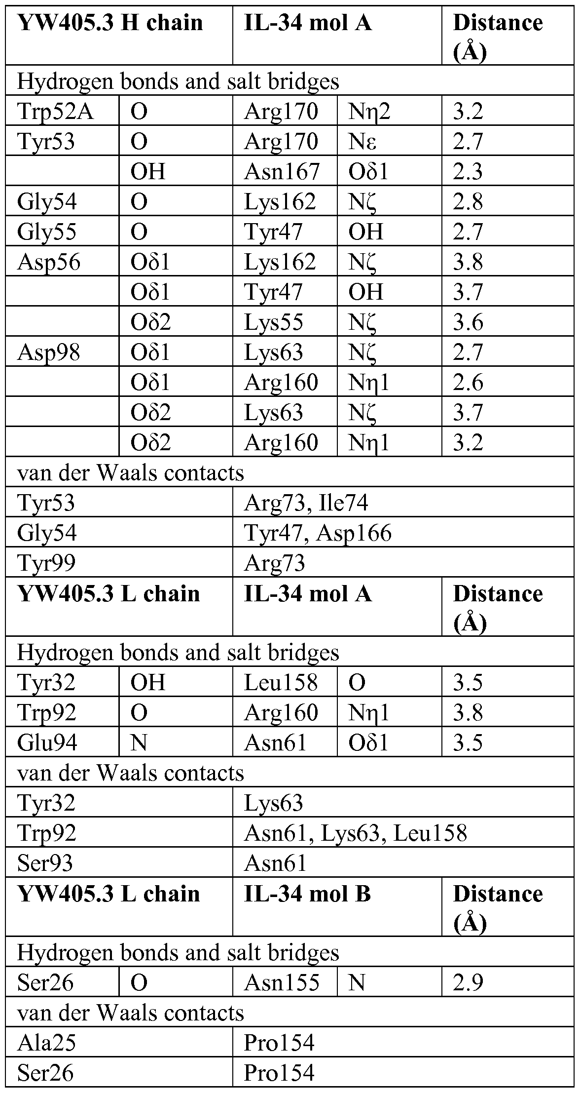

Claims
Priority Applications (9)
| Application Number | Priority Date | Filing Date | Title |
|---|---|---|---|
| EP13746304.8A EP2812355A4 (en) | 2012-02-06 | 2013-02-06 | Compositions and methods for using csf1r inhibitors |
| BR112014018961A BR112014018961A8 (en) | 2012-02-06 | 2013-02-06 | ISOLATED ANTIBODY, BISPECIFIC ANTIBODY, FRAGMENT, ISOLATED NUCLEIC ACID, VECTOR, HOST CELL, ANTIBODY PRODUCTION METHOD, PHARMACEUTICAL COMPOSITION, ANTIBODY USE, TREATMENT METHOD, INHIBITION METHOD AND INDUSTRIALIZED ARTICLE |
| KR1020147024752A KR20140127855A (en) | 2012-02-06 | 2013-02-06 | Compositions and methods for using csf1r inhibitors |
| MX2014008961A MX2014008961A (en) | 2012-02-06 | 2013-02-06 | Compositions and methods for using csf1r inhibitors. |
| CN201380008292.7A CN104093740B (en) | 2012-02-06 | 2013-02-06 | Use the composition and method of CSF1R inhibitor |
| CA2861122A CA2861122A1 (en) | 2012-02-06 | 2013-02-06 | Compositions and methods for using csf1r inhibitors |
| RU2014136332A RU2014136332A (en) | 2012-02-06 | 2013-02-06 | COMPOSITIONS AND METHODS OF APPLICATION OF CSF1R INHIBITORS |
| JP2014555859A JP6416630B2 (en) | 2012-02-06 | 2013-02-06 | Compositions and methods for using CSF1R inhibitors |
| HK15103325.3A HK1202879A1 (en) | 2012-02-06 | 2015-04-01 | Compositions and methods for using csf1r inhibitors csf1r |
Applications Claiming Priority (4)
| Application Number | Priority Date | Filing Date | Title |
|---|---|---|---|
| US201261595658P | 2012-02-06 | 2012-02-06 | |
| US61/595,658 | 2012-02-06 | ||
| US201261680674P | 2012-08-07 | 2012-08-07 | |
| US61/680,674 | 2012-08-07 |
Publications (2)
| Publication Number | Publication Date |
|---|---|
| WO2013119716A1 true WO2013119716A1 (en) | 2013-08-15 |
| WO2013119716A8 WO2013119716A8 (en) | 2014-08-07 |
Family
ID=48947977
Family Applications (1)
| Application Number | Title | Priority Date | Filing Date |
|---|---|---|---|
| PCT/US2013/024998 WO2013119716A1 (en) | 2012-02-06 | 2013-02-06 | Compositions and methods for using csf1r inhibitors |
Country Status (10)
| Country | Link |
|---|---|
| EP (1) | EP2812355A4 (en) |
| JP (2) | JP6416630B2 (en) |
| KR (1) | KR20140127855A (en) |
| CN (1) | CN104093740B (en) |
| BR (1) | BR112014018961A8 (en) |
| CA (1) | CA2861122A1 (en) |
| HK (1) | HK1202879A1 (en) |
| MX (1) | MX2014008961A (en) |
| RU (1) | RU2014136332A (en) |
| WO (1) | WO2013119716A1 (en) |
Cited By (187)
| Publication number | Priority date | Publication date | Assignee | Title |
|---|---|---|---|---|
| WO2015028454A3 (en) * | 2013-08-30 | 2015-05-21 | Ucb Biopharma Sprl | Method for the treatment of fibrotic disease |
| WO2015100282A1 (en) | 2013-12-24 | 2015-07-02 | Bristol-Myers Squibb Company | Tricyclic compounds as anticancer agents |
| US9192667B2 (en) | 2013-04-22 | 2015-11-24 | Hoffmann-La Roche Inc. | Method of treating cancer by administering CSF-1R antibodies and a TLR9 agonist |
| WO2015187835A2 (en) | 2014-06-06 | 2015-12-10 | Bristol-Myers Squibb Company | Antibodies against glucocorticoid-induced tumor necrosis factor receptor (gitr) and uses thereof |
| WO2016016262A1 (en) * | 2014-07-28 | 2016-02-04 | Nogra Pharma Limited | Methods and compositions for diagnosing and treating inflammatory bowel disorders |
| WO2016081748A2 (en) | 2014-11-21 | 2016-05-26 | Bristol-Myers Squibb Company | Antibodies against cd73 and uses thereof |
| WO2016106266A1 (en) | 2014-12-22 | 2016-06-30 | Bristol-Myers Squibb Company | TGFβ RECEPTOR ANTAGONISTS |
| WO2016127052A1 (en) | 2015-02-05 | 2016-08-11 | Bristol-Myers Squibb Company | Cxcl11 and smica as predictive biomarkers for efficacy of anti-ctla4 immunotherapy |
| WO2016140884A1 (en) | 2015-03-02 | 2016-09-09 | Rigel Pharmaceuticals, Inc. | TGF-β INHIBITORS |
| WO2016161279A1 (en) | 2015-04-03 | 2016-10-06 | Bristol-Myers Squibb Company | Inhibitors of indoleamine 2,3-dioxygenase for the treatment of cancer |
| WO2016162505A1 (en) | 2015-04-08 | 2016-10-13 | F-Star Biotechnology Limited | Her2 binding agent therapies |
| WO2016183115A1 (en) | 2015-05-12 | 2016-11-17 | Bristol-Myers Squibb Company | 5h-pyrido[3,2-b]indole compounds as anticancer agents |
| WO2016183118A1 (en) | 2015-05-12 | 2016-11-17 | Bristol-Myers Squibb Company | Tricyclic compounds as anticancer agents |
| WO2016180948A1 (en) * | 2015-05-12 | 2016-11-17 | INSERM (Institut National de la Santé et de la Recherche Médicale) | Methods and kits for labeling, detection and isolation of foxp3+ regulatory t cells, isolated population of foxp3+ regulatory t cells thus obtained and uses thereof |
| WO2016183114A1 (en) | 2015-05-11 | 2016-11-17 | Bristol-Myers Squibb Company | Tricyclic compounds as anticancer agents |
| WO2016189045A1 (en) * | 2015-05-27 | 2016-12-01 | Ucb Biopharma Sprl | Method for the treatment of neurological disease |
| WO2016196679A1 (en) | 2015-06-02 | 2016-12-08 | Genentech, Inc. | Compositions and methods for using anti-il-34 antibodies to treat neurological diseases |
| WO2016196228A1 (en) | 2015-05-29 | 2016-12-08 | Bristol-Myers Squibb Company | Antibodies against ox40 and uses thereof |
| WO2017004016A1 (en) | 2015-06-29 | 2017-01-05 | The Rockefeller University | Antibodies to cd40 with enhanced agonist activity |
| WO2017019757A1 (en) | 2015-07-28 | 2017-02-02 | Bristol-Myers Squibb Company | Tgf beta receptor antagonists |
| WO2017035118A1 (en) | 2015-08-25 | 2017-03-02 | Bristol-Myers Squibb Company | Tgf beta receptor antagonists |
| US9598422B2 (en) | 2014-11-05 | 2017-03-21 | Flexus Biosciences, Inc. | Immunoregulatory agents |
| US9643972B2 (en) | 2014-11-05 | 2017-05-09 | Flexus Biosciences, Inc. | Immunoregulatory agents |
| WO2017087678A2 (en) | 2015-11-19 | 2017-05-26 | Bristol-Myers Squibb Company | Antibodies against glucocorticoid-induced tumor necrosis factor receptor (gitr) and uses thereof |
| WO2017106291A1 (en) | 2015-12-15 | 2017-06-22 | Bristol-Myers Squibb Company | Cxcr4 receptor antagonists |
| WO2017152085A1 (en) | 2016-03-04 | 2017-09-08 | Bristol-Myers Squibb Company | Combination therapy with anti-cd73 antibodies |
| WO2017184619A2 (en) | 2016-04-18 | 2017-10-26 | Celldex Therapeutics, Inc. | Agonistic antibodies that bind human cd40 and uses thereof |
| US20170342148A1 (en) * | 2014-12-19 | 2017-11-30 | Universite De Nantes | Anti il-34 antibodies |
| WO2017210335A1 (en) | 2016-06-01 | 2017-12-07 | Bristol-Myers Squibb Company | Imaging methods using 18f-radiolabeled biologics |
| WO2018013818A2 (en) | 2016-07-14 | 2018-01-18 | Bristol-Myers Squibb Company | Antibodies against tim3 and uses thereof |
| WO2018017633A1 (en) | 2016-07-21 | 2018-01-25 | Bristol-Myers Squibb Company | TGF Beta RECEPTOR ANTAGONISTS |
| US9932380B2 (en) | 2013-03-14 | 2018-04-03 | Amgen Inc. | Interleukin-2 muteins for the expansion of T-regulatory cells |
| WO2018132279A1 (en) | 2017-01-05 | 2018-07-19 | Bristol-Myers Squibb Company | Tgf beta receptor antagonists |
| WO2018136700A1 (en) | 2017-01-20 | 2018-07-26 | Arcus Biosciences, Inc. | Azolopyrimidine for the treatment of cancer-related disorders |
| US10077314B1 (en) | 2009-12-10 | 2018-09-18 | Hoffmann-La Roche Inc. | Antibodies against human CSF-1R and uses thereof |
| WO2018187613A2 (en) | 2017-04-07 | 2018-10-11 | Bristol-Myers Squibb Company | Anti-icos agonist antibodies and uses thereof |
| WO2018195397A2 (en) | 2017-04-21 | 2018-10-25 | Kyn Therapeutics | Indole ahr inhibitors and uses thereof |
| WO2018209049A1 (en) | 2017-05-12 | 2018-11-15 | Bristol-Myers Squibb Company | Inhibitors of indoleamine 2,3-dioxygenase and methods of their use |
| WO2018213377A1 (en) | 2017-05-17 | 2018-11-22 | Arcus Biosciences, Inc. | Quinazoline-pyrazole derivatives for the treatment of cancer-related disorders |
| WO2019006283A1 (en) | 2017-06-30 | 2019-01-03 | Bristol-Myers Squibb Company | Amorphous and crystalline forms of ido inhibitors |
| WO2019023459A1 (en) | 2017-07-28 | 2019-01-31 | Bristol-Myers Squibb Company | Cyclic dinucleotides as anticancer agents |
| WO2019046496A1 (en) | 2017-08-31 | 2019-03-07 | Bristol-Myers Squibb Company | Cyclic dinucleotides as anticancer agents |
| WO2019046500A1 (en) | 2017-08-31 | 2019-03-07 | Bristol-Myers Squibb Company | Cyclic dinucleotides as anticancer agents |
| WO2019046498A1 (en) | 2017-08-31 | 2019-03-07 | Bristol-Myers Squibb Company | Cyclic dinucleotides as anticancer agents |
| WO2019075090A1 (en) | 2017-10-10 | 2019-04-18 | Tilos Therapeutics, Inc. | Anti-lap antibodies and uses thereof |
| WO2019074887A1 (en) | 2017-10-10 | 2019-04-18 | Bristol-Myers Squibb Company | Cyclic dinucleotides as anticancer agents |
| WO2019074824A1 (en) | 2017-10-09 | 2019-04-18 | Bristol-Myers Squibb Company | Inhibitors of indoleamine 2,3-dioxygenase and methods of their use |
| WO2019074822A1 (en) | 2017-10-09 | 2019-04-18 | Bristol-Myers Squibb Company | Inhibitors of indoleamine 2,3-dioxygenase and methods of their use |
| WO2019079261A1 (en) | 2017-10-16 | 2019-04-25 | Bristol-Myers Squibb Company | Cyclic dinucleotides as anticancer agents |
| WO2019089921A1 (en) | 2017-11-01 | 2019-05-09 | Bristol-Myers Squibb Company | Immunostimulatory agonistic antibodies for use in treating cancer |
| WO2019090198A1 (en) | 2017-11-06 | 2019-05-09 | Bristol-Myers Squibb Company | Isofuranone compounds useful as hpk1 inhibitors |
| US10323004B2 (en) | 2016-05-04 | 2019-06-18 | Bristol-Myers Squibb Company | Inhibitors of indoleamine 2,3-dioxygenase and methods of their use |
| US10336830B2 (en) | 2011-12-15 | 2019-07-02 | Hoffmann-La Roche Inc. | Antibodies against human CSF-1R and uses thereof |
| WO2019133747A1 (en) | 2017-12-27 | 2019-07-04 | Bristol-Myers Squibb Company | Anti-cd40 antibodies and uses thereof |
| WO2019136112A1 (en) | 2018-01-05 | 2019-07-11 | Bristol-Myers Squibb Company | Inhibitors of indoleamine 2,3-dioxygenase and methods of their use |
| WO2019140229A1 (en) | 2018-01-12 | 2019-07-18 | Bristol-Myers Squibb Company | Antibodies against tim3 and uses thereof |
| WO2019147982A1 (en) | 2018-01-26 | 2019-08-01 | Celldex Therapeutics, Inc. | Methods of treating cancer with dendritic cell mobilizing agents |
| WO2019160884A1 (en) | 2018-02-13 | 2019-08-22 | Bristol-Myers Squibb Company | Cyclic dinucleotides as anticancer agents |
| WO2019165315A1 (en) | 2018-02-23 | 2019-08-29 | Syntrix Biosystems Inc. | Method for treating cancer using chemokine antagonists alone or in combination |
| WO2019173211A1 (en) * | 2018-03-05 | 2019-09-12 | Massachusetts Eye And Ear Infirmary | Amelioration of autoimmune uveitis through blockade of csf1r |
| WO2019173587A1 (en) | 2018-03-08 | 2019-09-12 | Bristol-Myers Squibb Company | Cyclic dinucleotides as anticancer agents |
| WO2019183040A1 (en) | 2018-03-21 | 2019-09-26 | Five Prime Therapeutics, Inc. | ANTIBODIES BINDING TO VISTA AT ACIDIC pH |
| WO2019191295A1 (en) | 2018-03-28 | 2019-10-03 | Bristol-Myers Squibb Company | Interleukin-2/interleukin-2 receptor alpha fusion proteins and methods of use |
| WO2019200256A1 (en) | 2018-04-12 | 2019-10-17 | Bristol-Myers Squibb Company | Anticancer combination therapy with cd73 antagonist antibody and pd-1/pd-l1 axis antagonist antibody |
| WO2019204257A1 (en) | 2018-04-16 | 2019-10-24 | Arrys Therapeutics, Inc. | Ep4 inhibitors and use thereof |
| WO2019213340A1 (en) | 2018-05-03 | 2019-11-07 | Bristol-Myers Squibb Company | Uracil derivatives as mer-axl inhibitors |
| WO2019243833A1 (en) | 2018-06-22 | 2019-12-26 | Bicycletx Limited | Bicyclic peptide ligands specific for nectin-4 |
| WO2020006018A1 (en) | 2018-06-27 | 2020-01-02 | Bristol-Myers Squibb Company | Substituted naphthyridinone compounds useful as t cell activators |
| WO2020006016A1 (en) | 2018-06-27 | 2020-01-02 | Bristol-Myers Squibb Company | Naphthyridinone compounds useful as t cell activators |
| WO2020014132A2 (en) | 2018-07-09 | 2020-01-16 | Five Prime Therapeutics, Inc. | Antibodies binding to ilt4 |
| WO2020014327A2 (en) | 2018-07-11 | 2020-01-16 | Five Prime Therapeutics, Inc. | Antibodies binding to vista at acidic ph |
| US10544099B2 (en) | 2016-05-04 | 2020-01-28 | Bristol-Myers Squibb Company | Inhibitors of indoleamine 2,3-dioxygenase and methods of their use |
| WO2020023356A1 (en) | 2018-07-23 | 2020-01-30 | Bristol-Myers Squibb Company | Inhibitors of indoleamine 2,3-dioxygenase and methods of their use |
| WO2020023355A1 (en) | 2018-07-23 | 2020-01-30 | Bristol-Myers Squibb Company | Inhibitors of indoleamine 2,3-dioxygenase and methods of their use |
| WO2020051424A1 (en) | 2018-09-07 | 2020-03-12 | Pic Therapeutics | Eif4e inhibitors and uses thereof |
| WO2020076969A2 (en) | 2018-10-10 | 2020-04-16 | Tilos Therapeutics, Inc. | Anti-lap antibody variants and uses thereof |
| US10633342B2 (en) | 2016-05-04 | 2020-04-28 | Bristol-Myers Squibb Company | Inhibitors of indoleamine 2,3-dioxygenase and methods of their use |
| WO2020102501A1 (en) | 2018-11-16 | 2020-05-22 | Bristol-Myers Squibb Company | Anti-nkg2a antibodies and uses thereof |
| US10660909B2 (en) | 2016-11-17 | 2020-05-26 | Syntrix Biosystems Inc. | Method for treating cancer using chemokine antagonists |
| WO2020123444A1 (en) | 2018-12-11 | 2020-06-18 | Celldex Therapeutics, Inc. | Methods of using cd27 antibodies as conditioning treatment for adoptive cell therapy |
| EP3670659A1 (en) | 2018-12-20 | 2020-06-24 | Abivax | Biomarkers, and uses in treatment of viral infections, inflammations, or cancer |
| US10696648B2 (en) | 2016-05-04 | 2020-06-30 | Bristol-Myers Squibb Company | Inhibitors of indoleamine 2,3-dioxygenase and methods of their use |
| US10696650B2 (en) | 2017-08-17 | 2020-06-30 | Ikena Oncology, Inc. | AHR inhibitors and uses thereof |
| WO2020187998A1 (en) | 2019-03-19 | 2020-09-24 | Fundació Privada Institut D'investigació Oncològica De Vall Hebron | Combination therapy with omomyc and an antibody binding pd-1 or ctla-4 for the treatment of cancer |
| US10793563B2 (en) | 2018-01-29 | 2020-10-06 | Merck Patent Gmbh | GCN2 inhibitors and uses thereof |
| WO2020201753A1 (en) | 2019-04-02 | 2020-10-08 | Bicycletx Limited | Bicycle toxin conjugates and uses thereof |
| EP3733698A1 (en) | 2015-09-23 | 2020-11-04 | Bristol-Myers Squibb Company | Glypican-3 binding fibronectin based scafflold molecules |
| WO2020231713A1 (en) | 2019-05-13 | 2020-11-19 | Bristol-Myers Squibb Company | AGONISTS OF ROR GAMMAt |
| WO2020231766A1 (en) | 2019-05-13 | 2020-11-19 | Bristol-Myers Squibb Company | AGONISTS OF ROR GAMMAt |
| US10851144B2 (en) | 2015-04-10 | 2020-12-01 | Amgen Inc. | Interleukin-2 muteins for the expansion of T-regulatory cells |
| WO2020243423A1 (en) | 2019-05-31 | 2020-12-03 | Ikena Oncology, Inc. | Tead inhibitors and uses thereof |
| US10874743B2 (en) | 2017-12-26 | 2020-12-29 | Kymera Therapeutics, Inc. | IRAK degraders and uses thereof |
| WO2021026179A1 (en) | 2019-08-06 | 2021-02-11 | Bristol-Myers Squibb Company | AGONISTS OF ROR GAMMAt |
| WO2021024020A1 (en) | 2019-08-06 | 2021-02-11 | Astellas Pharma Inc. | Combination therapy involving antibodies against claudin 18.2 and immune checkpoint inhibitors for treatment of cancer |
| WO2021041588A1 (en) | 2019-08-28 | 2021-03-04 | Bristol-Myers Squibb Company | Substituted pyridopyrimidinonyl compounds useful as t cell activators |
| WO2021055698A1 (en) | 2019-09-19 | 2021-03-25 | Bristol-Myers Squibb Company | Antibodies binding to vista at acidic ph |
| US10959986B2 (en) | 2018-08-29 | 2021-03-30 | Bristol-Myers Squibb Company | Inhibitors of indoleamine 2,3-dioxygenase and methods of their use |
| US10987322B2 (en) | 2014-06-06 | 2021-04-27 | Flexus Biosciences, Inc. | Immunoregulatory agents |
| US10988477B2 (en) | 2018-01-29 | 2021-04-27 | Merck Patent Gmbh | GCN2 inhibitors and uses thereof |
| WO2021101919A1 (en) | 2019-11-19 | 2021-05-27 | Bristol-Myers Squibb Company | Compounds useful as inhibitors of helios protein |
| US11021481B2 (en) | 2019-09-13 | 2021-06-01 | Nimbus Saturn, Inc. | Substituted isoindolin-1-ones and 2,3-dihydro-1h-pyrrolo[3,4-c]pyridin-1-ones as HPK1 antagonists |
| WO2021108288A1 (en) | 2019-11-26 | 2021-06-03 | Bristol-Myers Squibb Company | Salts/cocrystals of (r)-n-(4-chlorophenyl)-2-((1s,4s)-4-(6-fluoroquinolin-4-yl)cyclohexyl)propanamide |
| WO2021108528A1 (en) | 2019-11-26 | 2021-06-03 | Ikena Oncology, Inc. | Polymorphic carbazole derivatives and uses thereof |
| WO2021133748A1 (en) | 2019-12-23 | 2021-07-01 | Bristol-Myers Squibb Company | Substituted quinolinonyl piperazine compounds useful as t cell activators |
| WO2021133752A1 (en) | 2019-12-23 | 2021-07-01 | Bristol-Myers Squibb Company | Substituted heteroaryl compounds useful as t cell activators |
| WO2021133749A1 (en) | 2019-12-23 | 2021-07-01 | Bristol-Myers Squibb Company | Substituted piperazine derivatives useful as t cell activators |
| WO2021133750A1 (en) | 2019-12-23 | 2021-07-01 | Bristol-Myers Squibb Company | Substituted bicyclic piperidine derivatives useful as t cell activators |
| WO2021133751A1 (en) | 2019-12-23 | 2021-07-01 | Bristol-Myers Squibb Company | Substituted quinazolinyl compounds useful as t cell activators |
| WO2021139682A1 (en) | 2020-01-07 | 2021-07-15 | Hifibio (Hk) Limited | Anti-galectin-9 antibody and uses thereof |
| WO2021141907A1 (en) | 2020-01-06 | 2021-07-15 | Hifibio (Hong Kong) Limited | Anti-tnfr2 antibody and uses thereof |
| US11066383B2 (en) | 2016-05-04 | 2021-07-20 | Bristol-Myers Squibb Company | Inhibitors of indoleamine 2,3-dioxygenase and methods of their use |
| WO2021178488A1 (en) | 2020-03-03 | 2021-09-10 | PIC Therapeutics, Inc. | Eif4e inhibitors and uses thereof |
| US11117889B1 (en) | 2018-11-30 | 2021-09-14 | Kymera Therapeutics, Inc. | IRAK degraders and uses thereof |
| WO2021183428A1 (en) | 2020-03-09 | 2021-09-16 | Bristol-Myers Squibb Company | Antibodies to cd40 with enhanced agonist activity |
| WO2021188769A1 (en) | 2020-03-19 | 2021-09-23 | Arcus Biosciences, Inc. | Tetralin and tetrahydroquinoline compounds as inhibitors of hif-2alpha |
| WO2021194914A1 (en) | 2020-03-23 | 2021-09-30 | Bristol-Myers Squibb Company | Substituted oxoisoindoline compounds for the treatment of cancer |
| WO2021207449A1 (en) | 2020-04-09 | 2021-10-14 | Merck Sharp & Dohme Corp. | Affinity matured anti-lap antibodies and uses thereof |
| WO2021231732A1 (en) | 2020-05-15 | 2021-11-18 | Bristol-Myers Squibb Company | Antibodies to garp |
| WO2021247591A1 (en) | 2020-06-02 | 2021-12-09 | Arcus Biosciences, Inc. | Antibodies to tigit |
| WO2021257643A1 (en) | 2020-06-17 | 2021-12-23 | Arcus Biosciences, Inc. | Crystalline forms of a cd73 inhibitor and uses thereof |
| WO2022008519A1 (en) | 2020-07-07 | 2022-01-13 | BioNTech SE | Therapeutic rna for hpv-positive cancer |
| US11242319B2 (en) | 2014-11-05 | 2022-02-08 | Flexus Biosciences, Inc. | Immunoregulatory agents |
| US11242393B2 (en) | 2018-03-23 | 2022-02-08 | Bristol-Myers Squibb Company | Antibodies against MICA and/or MICB and uses thereof |
| WO2022033419A2 (en) | 2020-08-10 | 2022-02-17 | Shanghai Xbh Biotechnology Co., Ltd. | Compositions and methods for treating autoimmune diseases and cancers by targeting igsf8 |
| US11253525B2 (en) | 2018-08-29 | 2022-02-22 | Bristol-Myers Squibb Company | Inhibitors of indoleamine 2,3-dioxygenase and methods of their use |
| WO2022038158A1 (en) | 2020-08-17 | 2022-02-24 | Bicycletx Limited | Bicycle conjugates specific for nectin-4 and uses thereof |
| US11292792B2 (en) | 2018-07-06 | 2022-04-05 | Kymera Therapeutics, Inc. | Tricyclic CRBN ligands and uses thereof |
| US11337970B2 (en) | 2016-08-26 | 2022-05-24 | Bristol-Myers Squibb Company | Inhibitors of indoleamine 2,3-dioxygenase and methods of their use |
| US11351164B2 (en) | 2016-08-26 | 2022-06-07 | Bristol-Myers Squibb Company | Inhibitors of indoleamine 2,3-dioxygenase and methods of their use |
| WO2022120353A1 (en) | 2020-12-02 | 2022-06-09 | Ikena Oncology, Inc. | Tead inhibitors and uses thereof |
| WO2022120354A1 (en) | 2020-12-02 | 2022-06-09 | Ikena Oncology, Inc. | Tead inhibitors and uses thereof |
| US11358948B2 (en) | 2017-09-22 | 2022-06-14 | Kymera Therapeutics, Inc. | CRBN ligands and uses thereof |
| WO2022135667A1 (en) | 2020-12-21 | 2022-06-30 | BioNTech SE | Therapeutic rna for treating cancer |
| WO2022135666A1 (en) | 2020-12-21 | 2022-06-30 | BioNTech SE | Treatment schedule for cytokine proteins |
| WO2022136266A1 (en) | 2020-12-21 | 2022-06-30 | BioNTech SE | Therapeutic rna for treating cancer |
| WO2022148979A1 (en) | 2021-01-11 | 2022-07-14 | Bicycletx Limited | Methods for treating cancer |
| WO2022167445A1 (en) | 2021-02-02 | 2022-08-11 | Liminal Biosciences Limited | Gpr84 antagonists and uses thereof |
| WO2022167457A1 (en) | 2021-02-02 | 2022-08-11 | Liminal Biosciences Limited | Gpr84 antagonists and uses thereof |
| WO2022169921A1 (en) | 2021-02-04 | 2022-08-11 | Bristol-Myers Squibb Company | Benzofuran compounds as sting agonists |
| WO2022197641A1 (en) | 2021-03-15 | 2022-09-22 | Rapt Therapeutics, Inc. | 1h-pyrazolo[3,4-d]pyrimidin-6-yl-amine derivatives as hematopoietic progenitor kinase 1 (hpk1) modulators and/or inhibitors for the treatment of cancer and other diseases |
| WO2022212876A1 (en) | 2021-04-02 | 2022-10-06 | The Regents Of The University Of California | Antibodies against cleaved cdcp1 and uses thereof |
| WO2022216573A1 (en) | 2021-04-05 | 2022-10-13 | Bristol-Myers Squibb Company | Pyridinyl substituted oxoisoindoline compounds for the treatment of cancer |
| WO2022216644A1 (en) | 2021-04-06 | 2022-10-13 | Bristol-Myers Squibb Company | Pyridinyl substituted oxoisoindoline compounds |
| WO2022221866A1 (en) | 2021-04-16 | 2022-10-20 | Ikena Oncology, Inc. | Mek inhibitors and uses thereof |
| US11485743B2 (en) | 2018-01-12 | 2022-11-01 | Kymera Therapeutics, Inc. | Protein degraders and uses thereof |
| US11485750B1 (en) | 2019-04-05 | 2022-11-01 | Kymera Therapeutics, Inc. | STAT degraders and uses thereof |
| US11498968B2 (en) | 2016-12-22 | 2022-11-15 | Hoffmann-La Roche Inc. | Treatment of tumors with an anti-CSF-1R antibody in combination with an anti-PD-L1 antibody after failure of anti-PD-L1/PD1 treatment |
| WO2022238386A1 (en) | 2021-05-10 | 2022-11-17 | Institut Curie | Methods for the treatment of cancer, inflammatory diseases and autoimmune diseases |
| WO2022246177A1 (en) | 2021-05-21 | 2022-11-24 | Arcus Biosciences, Inc. | Axl compounds |
| WO2022246179A1 (en) | 2021-05-21 | 2022-11-24 | Arcus Biosciences, Inc. | Axl inhibitor compounds |
| US11512080B2 (en) | 2018-01-12 | 2022-11-29 | Kymera Therapeutics, Inc. | CRBN ligands and uses thereof |
| US11512133B2 (en) | 2013-09-12 | 2022-11-29 | Hoffmann-La Roche Inc. | Methods for treating colon cancer or inhibiting cell proliferation by administering a combination of antibodies against human CSF-1R and antibodies against human PD-L1 |
| US11542335B2 (en) | 2016-08-25 | 2023-01-03 | Hoffmann-La Roche Inc. | Method of treating cancer in a patient by administering an antibody which binds colony stimulating factor-1 receptor (CSF-1R) |
| WO2023285552A1 (en) | 2021-07-13 | 2023-01-19 | BioNTech SE | Multispecific binding agents against cd40 and cd137 in combination therapy for cancer |
| US11591332B2 (en) | 2019-12-17 | 2023-02-28 | Kymera Therapeutics, Inc. | IRAK degraders and uses thereof |
| WO2023028238A1 (en) | 2021-08-25 | 2023-03-02 | PIC Therapeutics, Inc. | Eif4e inhibitors and uses thereof |
| WO2023028235A1 (en) | 2021-08-25 | 2023-03-02 | PIC Therapeutics, Inc. | Eif4e inhibitors and uses thereof |
| US11607453B2 (en) | 2017-05-12 | 2023-03-21 | Harpoon Therapeutics, Inc. | Mesothelin binding proteins |
| US11623932B2 (en) | 2017-09-22 | 2023-04-11 | Kymera Therapeutics, Inc. | Protein degraders and uses thereof |
| US11623958B2 (en) | 2016-05-20 | 2023-04-11 | Harpoon Therapeutics, Inc. | Single chain variable fragment CD3 binding proteins |
| WO2023061930A1 (en) | 2021-10-11 | 2023-04-20 | BioNTech SE | Therapeutic rna for lung cancer |
| WO2023077046A1 (en) | 2021-10-29 | 2023-05-04 | Arcus Biosciences, Inc. | Inhibitors of hif-2alpha and methods of use thereof |
| WO2023077042A1 (en) * | 2021-10-29 | 2023-05-04 | Eli Lilly And Company | Compounds and methods targeting interleukin-34 |
| US11649280B2 (en) | 2020-04-30 | 2023-05-16 | Eli Lilly And Company | Antibodies to IL-34 |
| US11679109B2 (en) | 2019-12-23 | 2023-06-20 | Kymera Therapeutics, Inc. | SMARCA degraders and uses thereof |
| WO2023114984A1 (en) | 2021-12-17 | 2023-06-22 | Ikena Oncology, Inc. | Tead inhibitors and uses thereof |
| US11685750B2 (en) | 2020-06-03 | 2023-06-27 | Kymera Therapeutics, Inc. | Crystalline forms of IRAK degraders |
| US11707457B2 (en) | 2019-12-17 | 2023-07-25 | Kymera Therapeutics, Inc. | IRAK degraders and uses thereof |
| WO2023150186A1 (en) | 2022-02-01 | 2023-08-10 | Arvinas Operations, Inc. | Dgk targeting compounds and uses thereof |
| WO2023173057A1 (en) | 2022-03-10 | 2023-09-14 | Ikena Oncology, Inc. | Mek inhibitors and uses thereof |
| WO2023173053A1 (en) | 2022-03-10 | 2023-09-14 | Ikena Oncology, Inc. | Mek inhibitors and uses thereof |
| EP4249066A2 (en) | 2014-12-23 | 2023-09-27 | Bristol-Myers Squibb Company | Antibodies to tigit |
| US11773103B2 (en) | 2021-02-15 | 2023-10-03 | Kymera Therapeutics, Inc. | IRAK4 degraders and uses thereof |
| WO2023211889A1 (en) | 2022-04-25 | 2023-11-02 | Ikena Oncology, Inc. | Polymorphic compounds and uses thereof |
| US11807692B2 (en) | 2018-09-25 | 2023-11-07 | Harpoon Therapeutics, Inc. | DLL3 binding proteins and methods of use |
| WO2023215719A1 (en) | 2022-05-02 | 2023-11-09 | Arcus Biosciences, Inc. | Anti-tigit antibodies and uses of the same |
| WO2023230205A1 (en) | 2022-05-25 | 2023-11-30 | Ikena Oncology, Inc. | Mek inhibitors and uses thereof |
| US11857535B2 (en) | 2020-07-30 | 2024-01-02 | Kymera Therapeutics, Inc. | Methods of treating mutant lymphomas |
| WO2024015251A1 (en) | 2022-07-15 | 2024-01-18 | Arcus Biosciences, Inc. | Inhibitors of hpk1 and methods of use thereof |
| WO2024020034A1 (en) | 2022-07-20 | 2024-01-25 | Arcus Biosciences, Inc. | Cbl-b inhibitors and methods of use thereof |
| WO2024028364A1 (en) | 2022-08-02 | 2024-02-08 | Liminal Biosciences Limited | Aryl-triazolyl and related gpr84 antagonists and uses thereof |
| WO2024028363A1 (en) | 2022-08-02 | 2024-02-08 | Liminal Biosciences Limited | Heteroaryl carboxamide and related gpr84 antagonists and uses thereof |
| WO2024028365A1 (en) | 2022-08-02 | 2024-02-08 | Liminal Biosciences Limited | Substituted pyridone gpr84 antagonists and uses thereof |
| WO2024036100A1 (en) | 2022-08-08 | 2024-02-15 | Bristol-Myers Squibb Company | Substituted tetrazolyl compounds useful as t cell activators |
| WO2024036101A1 (en) | 2022-08-09 | 2024-02-15 | Bristol-Myers Squibb Company | Tertiary amine substituted bicyclic compounds useful as t cell activators |
| US11926625B2 (en) | 2021-03-05 | 2024-03-12 | Nimbus Saturn, Inc. | HPK1 antagonists and uses thereof |
| US11932624B2 (en) | 2020-03-19 | 2024-03-19 | Kymera Therapeutics, Inc. | MDM2 degraders and uses thereof |
Families Citing this family (3)
| Publication number | Priority date | Publication date | Assignee | Title |
|---|---|---|---|---|
| JP6490419B2 (en) * | 2014-12-24 | 2019-03-27 | 学校法人関西医科大学 | Regulatory T cell differentiation inducer |
| JP6621252B2 (en) * | 2015-06-17 | 2019-12-18 | 国立大学法人北海道大学 | Treatment resistance reducing agent for treatment resistant cancer |
| GB201803226D0 (en) * | 2018-02-28 | 2018-04-11 | Ultrahuman Twelve Ltd | CSF1R Binding agents |
Citations (1)
| Publication number | Priority date | Publication date | Assignee | Title |
|---|---|---|---|---|
| US20110243947A1 (en) * | 2010-04-01 | 2011-10-06 | Imclone Llc | Antibodies against csf-1r |
Family Cites Families (2)
| Publication number | Priority date | Publication date | Assignee | Title |
|---|---|---|---|---|
| CN101970496B (en) * | 2008-03-14 | 2014-04-16 | 特朗斯吉有限公司 | Antibody against the csf-1 r |
| TWI595008B (en) * | 2010-05-04 | 2017-08-11 | 戊瑞治療有限公司 | Antibodies that bind csf1r |
-
2013
- 2013-02-06 CA CA2861122A patent/CA2861122A1/en not_active Abandoned
- 2013-02-06 WO PCT/US2013/024998 patent/WO2013119716A1/en active Application Filing
- 2013-02-06 BR BR112014018961A patent/BR112014018961A8/en not_active IP Right Cessation
- 2013-02-06 RU RU2014136332A patent/RU2014136332A/en not_active Application Discontinuation
- 2013-02-06 MX MX2014008961A patent/MX2014008961A/en unknown
- 2013-02-06 KR KR1020147024752A patent/KR20140127855A/en not_active Application Discontinuation
- 2013-02-06 EP EP13746304.8A patent/EP2812355A4/en not_active Withdrawn
- 2013-02-06 JP JP2014555859A patent/JP6416630B2/en not_active Expired - Fee Related
- 2013-02-06 CN CN201380008292.7A patent/CN104093740B/en not_active Expired - Fee Related
-
2015
- 2015-04-01 HK HK15103325.3A patent/HK1202879A1/en not_active IP Right Cessation
-
2017
- 2017-06-13 JP JP2017116326A patent/JP2017200482A/en active Pending
Patent Citations (1)
| Publication number | Priority date | Publication date | Assignee | Title |
|---|---|---|---|---|
| US20110243947A1 (en) * | 2010-04-01 | 2011-10-06 | Imclone Llc | Antibodies against csf-1r |
Non-Patent Citations (4)
| Title |
|---|
| CHIHARA. T ET AL.: "IL-34 and M-CSF share the receptor Fms but are not identical in biological activity and signal activation.", CELL DEATH AND DIFFERENTIATION, vol. 17, 2010, pages 1917 - 1927, XP055036155 * |
| HWANG. S-J ET AL.: "Interleukin-34 produced by human fibroblast-like synovial cells in rheumatoid arthritis supports osteoclastogenesis.", ARTHRITIS RESEARCH & THERAPY, vol. 14, no. R14, 2012, pages 1 - 10, XP021132205 * |
| MA, X ET AL.: "Structural Basis for the Dual Recognition of Helical Cytokines IL-34 and CSF-1 by CSF-1R.", STRUCTURE, vol. 20, April 2012 (2012-04-01), pages 676 - 687, XP055155969 * |
| See also references of EP2812355A4 * |
Cited By (250)
| Publication number | Priority date | Publication date | Assignee | Title |
|---|---|---|---|---|
| US10077314B1 (en) | 2009-12-10 | 2018-09-18 | Hoffmann-La Roche Inc. | Antibodies against human CSF-1R and uses thereof |
| US10287358B2 (en) | 2009-12-10 | 2019-05-14 | Hoffmann-La Roche Inc. | Antibodies against human CSF-1R and uses thereof |
| US10336830B2 (en) | 2011-12-15 | 2019-07-02 | Hoffmann-La Roche Inc. | Antibodies against human CSF-1R and uses thereof |
| US10093711B2 (en) | 2013-03-14 | 2018-10-09 | Amgen Inc. | Interleukin-2 muteins for the expansion of T-regulatory cells |
| US10829535B2 (en) | 2013-03-14 | 2020-11-10 | Amgen Inc. | Interleukin-2 muteins for the expansion of T-regulatory cells |
| US9932380B2 (en) | 2013-03-14 | 2018-04-03 | Amgen Inc. | Interleukin-2 muteins for the expansion of T-regulatory cells |
| US10562950B2 (en) | 2013-03-14 | 2020-02-18 | Amgen Inc. | Interleukin-2 muteins for the expansion of T-regulatory cells |
| US9192667B2 (en) | 2013-04-22 | 2015-11-24 | Hoffmann-La Roche Inc. | Method of treating cancer by administering CSF-1R antibodies and a TLR9 agonist |
| WO2015028454A3 (en) * | 2013-08-30 | 2015-05-21 | Ucb Biopharma Sprl | Method for the treatment of fibrotic disease |
| US11512133B2 (en) | 2013-09-12 | 2022-11-29 | Hoffmann-La Roche Inc. | Methods for treating colon cancer or inhibiting cell proliferation by administering a combination of antibodies against human CSF-1R and antibodies against human PD-L1 |
| EP3466949A1 (en) | 2013-12-24 | 2019-04-10 | Bristol-Myers Squibb Company | Tricyclic compound as anticancer agents |
| WO2015100282A1 (en) | 2013-12-24 | 2015-07-02 | Bristol-Myers Squibb Company | Tricyclic compounds as anticancer agents |
| EP3998079A1 (en) | 2014-06-06 | 2022-05-18 | Bristol-Myers Squibb Company | Antibodies against glucocorticoid-induced tumor necrosis factor receptor (gitr) and uses thereof |
| US10987322B2 (en) | 2014-06-06 | 2021-04-27 | Flexus Biosciences, Inc. | Immunoregulatory agents |
| EP3610924A1 (en) | 2014-06-06 | 2020-02-19 | Bristol-Myers Squibb Company | Antibodies against glucocorticoid-induced tumor necrosis factor receptor (gitr) and uses thereof |
| WO2015187835A2 (en) | 2014-06-06 | 2015-12-10 | Bristol-Myers Squibb Company | Antibodies against glucocorticoid-induced tumor necrosis factor receptor (gitr) and uses thereof |
| WO2016016262A1 (en) * | 2014-07-28 | 2016-02-04 | Nogra Pharma Limited | Methods and compositions for diagnosing and treating inflammatory bowel disorders |
| CN106573056A (en) * | 2014-07-28 | 2017-04-19 | 诺格尔制药有限公司 | Methods and compositions for diagnosing and treating inflammatory bowel disorders |
| US9643972B2 (en) | 2014-11-05 | 2017-05-09 | Flexus Biosciences, Inc. | Immunoregulatory agents |
| US10206893B2 (en) | 2014-11-05 | 2019-02-19 | Flexus Biosciences, Inc. | Immunoregulatory agents |
| EP3854394A1 (en) | 2014-11-05 | 2021-07-28 | Flexus Biosciences, Inc. | Immunoregulatory agents |
| US11242319B2 (en) | 2014-11-05 | 2022-02-08 | Flexus Biosciences, Inc. | Immunoregulatory agents |
| US9598422B2 (en) | 2014-11-05 | 2017-03-21 | Flexus Biosciences, Inc. | Immunoregulatory agents |
| US11932601B2 (en) | 2014-11-05 | 2024-03-19 | Flexus Biosciences, Inc. | Immunoregulatory agents |
| US10533014B2 (en) | 2014-11-05 | 2020-01-14 | Flexus Biosciences, Inc. | Immunoregulatory agents |
| US10106546B2 (en) | 2014-11-05 | 2018-10-23 | Flexus Biosciences, Inc. | Immunoregulatory agents |
| EP3725808A1 (en) | 2014-11-21 | 2020-10-21 | Bristol-Myers Squibb Company | Antibodies against cd73 and uses thereof |
| WO2016081748A2 (en) | 2014-11-21 | 2016-05-26 | Bristol-Myers Squibb Company | Antibodies against cd73 and uses thereof |
| JP2018508467A (en) * | 2014-12-19 | 2018-03-29 | ユニヴェルシテ・ドゥ・ナント | Anti-IL-34 antibody |
| US10703813B2 (en) * | 2014-12-19 | 2020-07-07 | Universite De Nantes | Anti IL-34 antibodies |
| US20170342148A1 (en) * | 2014-12-19 | 2017-11-30 | Universite De Nantes | Anti il-34 antibodies |
| WO2016106266A1 (en) | 2014-12-22 | 2016-06-30 | Bristol-Myers Squibb Company | TGFβ RECEPTOR ANTAGONISTS |
| EP4249066A2 (en) | 2014-12-23 | 2023-09-27 | Bristol-Myers Squibb Company | Antibodies to tigit |
| WO2016127052A1 (en) | 2015-02-05 | 2016-08-11 | Bristol-Myers Squibb Company | Cxcl11 and smica as predictive biomarkers for efficacy of anti-ctla4 immunotherapy |
| WO2016140884A1 (en) | 2015-03-02 | 2016-09-09 | Rigel Pharmaceuticals, Inc. | TGF-β INHIBITORS |
| US9790169B2 (en) | 2015-04-03 | 2017-10-17 | Bristol-Myers Squibb Company | IDO inhibitors |
| WO2016161269A1 (en) | 2015-04-03 | 2016-10-06 | Bristol-Myers Squibb Company | Inhibitors of indoleamine 2,3-dioxygenase for the treatment of cancer |
| US10399933B2 (en) | 2015-04-03 | 2019-09-03 | Bristol-Myers Squibb Company | Inhibitors of indoleamine-2,3-dioxygenase for the treatment of cancer |
| US10399932B2 (en) | 2015-04-03 | 2019-09-03 | Bristol-Myers Squibb Company | Inhibitors of indoleamine-2,3-dioxygenase for the treatment of cancer |
| US10167254B2 (en) | 2015-04-03 | 2019-01-01 | Bristol-Myers Squibb Company | IDO inhibitors |
| WO2016161279A1 (en) | 2015-04-03 | 2016-10-06 | Bristol-Myers Squibb Company | Inhibitors of indoleamine 2,3-dioxygenase for the treatment of cancer |
| WO2016162505A1 (en) | 2015-04-08 | 2016-10-13 | F-Star Biotechnology Limited | Her2 binding agent therapies |
| US10851144B2 (en) | 2015-04-10 | 2020-12-01 | Amgen Inc. | Interleukin-2 muteins for the expansion of T-regulatory cells |
| WO2016183114A1 (en) | 2015-05-11 | 2016-11-17 | Bristol-Myers Squibb Company | Tricyclic compounds as anticancer agents |
| US10591465B2 (en) | 2015-05-12 | 2020-03-17 | INSERM (Institut National de la Santé et de la Recherche Médicale) | Methods and kits for labeling, detection and isolation of Foxp3+ regulatory T cells, isolated population of Foxp3+ regulatory T cells thus obtained and uses thereof |
| WO2016183115A1 (en) | 2015-05-12 | 2016-11-17 | Bristol-Myers Squibb Company | 5h-pyrido[3,2-b]indole compounds as anticancer agents |
| WO2016183118A1 (en) | 2015-05-12 | 2016-11-17 | Bristol-Myers Squibb Company | Tricyclic compounds as anticancer agents |
| WO2016180948A1 (en) * | 2015-05-12 | 2016-11-17 | INSERM (Institut National de la Santé et de la Recherche Médicale) | Methods and kits for labeling, detection and isolation of foxp3+ regulatory t cells, isolated population of foxp3+ regulatory t cells thus obtained and uses thereof |
| US10316097B2 (en) | 2015-05-27 | 2019-06-11 | Ucb Biopharma Sprl | Method for the treatment of epilepsy, epileptogenesis, seizures or convulsions by an anti-colony-stimulating factor 1 receptor (CSF-1R) antibody |
| WO2016189045A1 (en) * | 2015-05-27 | 2016-12-01 | Ucb Biopharma Sprl | Method for the treatment of neurological disease |
| EA039951B1 (en) * | 2015-05-27 | 2022-03-31 | Юсб Биофарма Спрл | Inhibitor of csf-1r activity for use in the treatment or prophylaxis of epilepsy or parkinson's disease and pharmaceutical composition thereof |
| WO2016196228A1 (en) | 2015-05-29 | 2016-12-08 | Bristol-Myers Squibb Company | Antibodies against ox40 and uses thereof |
| WO2016196679A1 (en) | 2015-06-02 | 2016-12-08 | Genentech, Inc. | Compositions and methods for using anti-il-34 antibodies to treat neurological diseases |
| WO2017004016A1 (en) | 2015-06-29 | 2017-01-05 | The Rockefeller University | Antibodies to cd40 with enhanced agonist activity |
| WO2017019757A1 (en) | 2015-07-28 | 2017-02-02 | Bristol-Myers Squibb Company | Tgf beta receptor antagonists |
| WO2017035118A1 (en) | 2015-08-25 | 2017-03-02 | Bristol-Myers Squibb Company | Tgf beta receptor antagonists |
| EP3733698A1 (en) | 2015-09-23 | 2020-11-04 | Bristol-Myers Squibb Company | Glypican-3 binding fibronectin based scafflold molecules |
| WO2017087678A2 (en) | 2015-11-19 | 2017-05-26 | Bristol-Myers Squibb Company | Antibodies against glucocorticoid-induced tumor necrosis factor receptor (gitr) and uses thereof |
| WO2017106291A1 (en) | 2015-12-15 | 2017-06-22 | Bristol-Myers Squibb Company | Cxcr4 receptor antagonists |
| WO2017152085A1 (en) | 2016-03-04 | 2017-09-08 | Bristol-Myers Squibb Company | Combination therapy with anti-cd73 antibodies |
| WO2017184619A2 (en) | 2016-04-18 | 2017-10-26 | Celldex Therapeutics, Inc. | Agonistic antibodies that bind human cd40 and uses thereof |
| US11066383B2 (en) | 2016-05-04 | 2021-07-20 | Bristol-Myers Squibb Company | Inhibitors of indoleamine 2,3-dioxygenase and methods of their use |
| US10323004B2 (en) | 2016-05-04 | 2019-06-18 | Bristol-Myers Squibb Company | Inhibitors of indoleamine 2,3-dioxygenase and methods of their use |
| US10544099B2 (en) | 2016-05-04 | 2020-01-28 | Bristol-Myers Squibb Company | Inhibitors of indoleamine 2,3-dioxygenase and methods of their use |
| US10696648B2 (en) | 2016-05-04 | 2020-06-30 | Bristol-Myers Squibb Company | Inhibitors of indoleamine 2,3-dioxygenase and methods of their use |
| US10633342B2 (en) | 2016-05-04 | 2020-04-28 | Bristol-Myers Squibb Company | Inhibitors of indoleamine 2,3-dioxygenase and methods of their use |
| US11623958B2 (en) | 2016-05-20 | 2023-04-11 | Harpoon Therapeutics, Inc. | Single chain variable fragment CD3 binding proteins |
| WO2017210335A1 (en) | 2016-06-01 | 2017-12-07 | Bristol-Myers Squibb Company | Imaging methods using 18f-radiolabeled biologics |
| WO2018013818A2 (en) | 2016-07-14 | 2018-01-18 | Bristol-Myers Squibb Company | Antibodies against tim3 and uses thereof |
| US11591392B2 (en) | 2016-07-14 | 2023-02-28 | Bristol-Myers Squibb Company | Antibodies against TIM3 and uses thereof |
| US10533052B2 (en) | 2016-07-14 | 2020-01-14 | Bristol-Myers Squibb Company | Antibodies against TIM3 and uses thereof |
| US10077306B2 (en) | 2016-07-14 | 2018-09-18 | Bristol-Myers Squibb Company | Antibodies against TIM3 and uses thereof |
| WO2018017633A1 (en) | 2016-07-21 | 2018-01-25 | Bristol-Myers Squibb Company | TGF Beta RECEPTOR ANTAGONISTS |
| US11542335B2 (en) | 2016-08-25 | 2023-01-03 | Hoffmann-La Roche Inc. | Method of treating cancer in a patient by administering an antibody which binds colony stimulating factor-1 receptor (CSF-1R) |
| US11337970B2 (en) | 2016-08-26 | 2022-05-24 | Bristol-Myers Squibb Company | Inhibitors of indoleamine 2,3-dioxygenase and methods of their use |
| US11351164B2 (en) | 2016-08-26 | 2022-06-07 | Bristol-Myers Squibb Company | Inhibitors of indoleamine 2,3-dioxygenase and methods of their use |
| US10660909B2 (en) | 2016-11-17 | 2020-05-26 | Syntrix Biosystems Inc. | Method for treating cancer using chemokine antagonists |
| US11498968B2 (en) | 2016-12-22 | 2022-11-15 | Hoffmann-La Roche Inc. | Treatment of tumors with an anti-CSF-1R antibody in combination with an anti-PD-L1 antibody after failure of anti-PD-L1/PD1 treatment |
| WO2018132279A1 (en) | 2017-01-05 | 2018-07-19 | Bristol-Myers Squibb Company | Tgf beta receptor antagonists |
| EP4310082A2 (en) | 2017-01-20 | 2024-01-24 | Arcus Biosciences, Inc. | Azolopyrimidine for the treatment of cancer-related disorders |
| WO2018136700A1 (en) | 2017-01-20 | 2018-07-26 | Arcus Biosciences, Inc. | Azolopyrimidine for the treatment of cancer-related disorders |
| WO2018187613A2 (en) | 2017-04-07 | 2018-10-11 | Bristol-Myers Squibb Company | Anti-icos agonist antibodies and uses thereof |
| US10570138B2 (en) | 2017-04-21 | 2020-02-25 | Kyn Therapeutics | Indole AHR inhibitors and uses thereof |
| US11358969B2 (en) | 2017-04-21 | 2022-06-14 | Ikena Oncology, Inc. | Indole AHR inhibitors and uses thereof |
| WO2018195397A2 (en) | 2017-04-21 | 2018-10-25 | Kyn Therapeutics | Indole ahr inhibitors and uses thereof |
| US10689388B1 (en) | 2017-04-21 | 2020-06-23 | Ikena Oncology, Inc. | Indole AHR inhibitors and uses thereof |
| US11066392B2 (en) | 2017-05-12 | 2021-07-20 | Bristol-Myers Squibb Company | Inhibitors of indoleamine 2,3-dioxygenase and methods of their use |
| US11607453B2 (en) | 2017-05-12 | 2023-03-21 | Harpoon Therapeutics, Inc. | Mesothelin binding proteins |
| WO2018209049A1 (en) | 2017-05-12 | 2018-11-15 | Bristol-Myers Squibb Company | Inhibitors of indoleamine 2,3-dioxygenase and methods of their use |
| WO2018213377A1 (en) | 2017-05-17 | 2018-11-22 | Arcus Biosciences, Inc. | Quinazoline-pyrazole derivatives for the treatment of cancer-related disorders |
| WO2019006283A1 (en) | 2017-06-30 | 2019-01-03 | Bristol-Myers Squibb Company | Amorphous and crystalline forms of ido inhibitors |
| US11236049B2 (en) | 2017-06-30 | 2022-02-01 | Bristol-Myers Squibb Company | Amorphous and crystalline forms of IDO inhibitors |
| WO2019023459A1 (en) | 2017-07-28 | 2019-01-31 | Bristol-Myers Squibb Company | Cyclic dinucleotides as anticancer agents |
| US11555026B2 (en) | 2017-08-17 | 2023-01-17 | Ikena Oncology, Inc. | AHR inhibitors and uses thereof |
| US10696650B2 (en) | 2017-08-17 | 2020-06-30 | Ikena Oncology, Inc. | AHR inhibitors and uses thereof |
| WO2019046496A1 (en) | 2017-08-31 | 2019-03-07 | Bristol-Myers Squibb Company | Cyclic dinucleotides as anticancer agents |
| WO2019046500A1 (en) | 2017-08-31 | 2019-03-07 | Bristol-Myers Squibb Company | Cyclic dinucleotides as anticancer agents |
| WO2019046498A1 (en) | 2017-08-31 | 2019-03-07 | Bristol-Myers Squibb Company | Cyclic dinucleotides as anticancer agents |
| US11358948B2 (en) | 2017-09-22 | 2022-06-14 | Kymera Therapeutics, Inc. | CRBN ligands and uses thereof |
| US11623932B2 (en) | 2017-09-22 | 2023-04-11 | Kymera Therapeutics, Inc. | Protein degraders and uses thereof |
| WO2019074824A1 (en) | 2017-10-09 | 2019-04-18 | Bristol-Myers Squibb Company | Inhibitors of indoleamine 2,3-dioxygenase and methods of their use |
| WO2019074822A1 (en) | 2017-10-09 | 2019-04-18 | Bristol-Myers Squibb Company | Inhibitors of indoleamine 2,3-dioxygenase and methods of their use |
| US11649212B2 (en) | 2017-10-09 | 2023-05-16 | Bristol-Myers Squibb Company | Inhibitors of indoleamine 2,3-dioxygenase and methods of their use |
| US11203592B2 (en) | 2017-10-09 | 2021-12-21 | Bristol-Myers Squibb Company | Inhibitors of indoleamine 2,3-dioxygenase and methods of their use |
| US11230601B2 (en) | 2017-10-10 | 2022-01-25 | Tilos Therapeutics, Inc. | Methods of using anti-lap antibodies |
| WO2019074887A1 (en) | 2017-10-10 | 2019-04-18 | Bristol-Myers Squibb Company | Cyclic dinucleotides as anticancer agents |
| WO2019075090A1 (en) | 2017-10-10 | 2019-04-18 | Tilos Therapeutics, Inc. | Anti-lap antibodies and uses thereof |
| WO2019079261A1 (en) | 2017-10-16 | 2019-04-25 | Bristol-Myers Squibb Company | Cyclic dinucleotides as anticancer agents |
| WO2019089921A1 (en) | 2017-11-01 | 2019-05-09 | Bristol-Myers Squibb Company | Immunostimulatory agonistic antibodies for use in treating cancer |
| WO2019090198A1 (en) | 2017-11-06 | 2019-05-09 | Bristol-Myers Squibb Company | Isofuranone compounds useful as hpk1 inhibitors |
| US11318205B1 (en) | 2017-12-26 | 2022-05-03 | Kymera Therapeutics, Inc. | IRAK degraders and uses thereof |
| US11723980B2 (en) | 2017-12-26 | 2023-08-15 | Kymera Therapeutics, Inc. | IRAK degraders and uses thereof |
| US10874743B2 (en) | 2017-12-26 | 2020-12-29 | Kymera Therapeutics, Inc. | IRAK degraders and uses thereof |
| WO2019133747A1 (en) | 2017-12-27 | 2019-07-04 | Bristol-Myers Squibb Company | Anti-cd40 antibodies and uses thereof |
| US11952427B2 (en) | 2017-12-27 | 2024-04-09 | Bristol-Myers Squibb Company | Anti-CD40 antibodies and uses thereof |
| US11306149B2 (en) | 2017-12-27 | 2022-04-19 | Bristol-Myers Squibb Company | Anti-CD40 antibodies and uses thereof |
| US11447449B2 (en) | 2018-01-05 | 2022-09-20 | Bristol-Myers Squibb Company | Inhibitors of indoleamine 2,3-dioxygenase and methods of their use |
| WO2019136112A1 (en) | 2018-01-05 | 2019-07-11 | Bristol-Myers Squibb Company | Inhibitors of indoleamine 2,3-dioxygenase and methods of their use |
| US11485743B2 (en) | 2018-01-12 | 2022-11-01 | Kymera Therapeutics, Inc. | Protein degraders and uses thereof |
| US11512080B2 (en) | 2018-01-12 | 2022-11-29 | Kymera Therapeutics, Inc. | CRBN ligands and uses thereof |
| US11932635B2 (en) | 2018-01-12 | 2024-03-19 | Kymera Therapeutics, Inc. | CRBN ligands and uses thereof |
| WO2019140229A1 (en) | 2018-01-12 | 2019-07-18 | Bristol-Myers Squibb Company | Antibodies against tim3 and uses thereof |
| WO2019147982A1 (en) | 2018-01-26 | 2019-08-01 | Celldex Therapeutics, Inc. | Methods of treating cancer with dendritic cell mobilizing agents |
| US10988477B2 (en) | 2018-01-29 | 2021-04-27 | Merck Patent Gmbh | GCN2 inhibitors and uses thereof |
| US10793563B2 (en) | 2018-01-29 | 2020-10-06 | Merck Patent Gmbh | GCN2 inhibitors and uses thereof |
| WO2019160884A1 (en) | 2018-02-13 | 2019-08-22 | Bristol-Myers Squibb Company | Cyclic dinucleotides as anticancer agents |
| WO2019165315A1 (en) | 2018-02-23 | 2019-08-29 | Syntrix Biosystems Inc. | Method for treating cancer using chemokine antagonists alone or in combination |
| WO2019173211A1 (en) * | 2018-03-05 | 2019-09-12 | Massachusetts Eye And Ear Infirmary | Amelioration of autoimmune uveitis through blockade of csf1r |
| WO2019173587A1 (en) | 2018-03-08 | 2019-09-12 | Bristol-Myers Squibb Company | Cyclic dinucleotides as anticancer agents |
| WO2019183040A1 (en) | 2018-03-21 | 2019-09-26 | Five Prime Therapeutics, Inc. | ANTIBODIES BINDING TO VISTA AT ACIDIC pH |
| US11242393B2 (en) | 2018-03-23 | 2022-02-08 | Bristol-Myers Squibb Company | Antibodies against MICA and/or MICB and uses thereof |
| WO2019191295A1 (en) | 2018-03-28 | 2019-10-03 | Bristol-Myers Squibb Company | Interleukin-2/interleukin-2 receptor alpha fusion proteins and methods of use |
| WO2019200256A1 (en) | 2018-04-12 | 2019-10-17 | Bristol-Myers Squibb Company | Anticancer combination therapy with cd73 antagonist antibody and pd-1/pd-l1 axis antagonist antibody |
| WO2019204257A1 (en) | 2018-04-16 | 2019-10-24 | Arrys Therapeutics, Inc. | Ep4 inhibitors and use thereof |
| WO2019213340A1 (en) | 2018-05-03 | 2019-11-07 | Bristol-Myers Squibb Company | Uracil derivatives as mer-axl inhibitors |
| WO2019243832A1 (en) | 2018-06-22 | 2019-12-26 | Bicycletx Limited | Bicyclic peptide ligands specific for nectin-4 |
| US11453702B2 (en) | 2018-06-22 | 2022-09-27 | Bicycletx Limited | Bicyclic peptide ligands specific for Nectin-4 |
| US11912792B2 (en) | 2018-06-22 | 2024-02-27 | Bicycletx Limited | Bicyclic peptide ligands specific for nectin-4 |
| WO2019243833A1 (en) | 2018-06-22 | 2019-12-26 | Bicycletx Limited | Bicyclic peptide ligands specific for nectin-4 |
| US11180531B2 (en) | 2018-06-22 | 2021-11-23 | Bicycletx Limited | Bicyclic peptide ligands specific for Nectin-4 |
| WO2020006018A1 (en) | 2018-06-27 | 2020-01-02 | Bristol-Myers Squibb Company | Substituted naphthyridinone compounds useful as t cell activators |
| WO2020006016A1 (en) | 2018-06-27 | 2020-01-02 | Bristol-Myers Squibb Company | Naphthyridinone compounds useful as t cell activators |
| US11292792B2 (en) | 2018-07-06 | 2022-04-05 | Kymera Therapeutics, Inc. | Tricyclic CRBN ligands and uses thereof |
| US11897882B2 (en) | 2018-07-06 | 2024-02-13 | Kymera Therapeutics, Inc. | Tricyclic crbn ligands and uses thereof |
| WO2020014132A2 (en) | 2018-07-09 | 2020-01-16 | Five Prime Therapeutics, Inc. | Antibodies binding to ilt4 |
| US11401328B2 (en) | 2018-07-09 | 2022-08-02 | Five Prime Therapeutics, Inc. | Antibodies binding to ILT4 |
| WO2020014327A2 (en) | 2018-07-11 | 2020-01-16 | Five Prime Therapeutics, Inc. | Antibodies binding to vista at acidic ph |
| WO2020023355A1 (en) | 2018-07-23 | 2020-01-30 | Bristol-Myers Squibb Company | Inhibitors of indoleamine 2,3-dioxygenase and methods of their use |
| WO2020023356A1 (en) | 2018-07-23 | 2020-01-30 | Bristol-Myers Squibb Company | Inhibitors of indoleamine 2,3-dioxygenase and methods of their use |
| US11253525B2 (en) | 2018-08-29 | 2022-02-22 | Bristol-Myers Squibb Company | Inhibitors of indoleamine 2,3-dioxygenase and methods of their use |
| US10959986B2 (en) | 2018-08-29 | 2021-03-30 | Bristol-Myers Squibb Company | Inhibitors of indoleamine 2,3-dioxygenase and methods of their use |
| WO2020051424A1 (en) | 2018-09-07 | 2020-03-12 | Pic Therapeutics | Eif4e inhibitors and uses thereof |
| US11807692B2 (en) | 2018-09-25 | 2023-11-07 | Harpoon Therapeutics, Inc. | DLL3 binding proteins and methods of use |
| US11130802B2 (en) | 2018-10-10 | 2021-09-28 | Tilos Therapeutics, Inc. | Anti-lap antibody variants |
| WO2020076969A2 (en) | 2018-10-10 | 2020-04-16 | Tilos Therapeutics, Inc. | Anti-lap antibody variants and uses thereof |
| WO2020102501A1 (en) | 2018-11-16 | 2020-05-22 | Bristol-Myers Squibb Company | Anti-nkg2a antibodies and uses thereof |
| US11807636B2 (en) | 2018-11-30 | 2023-11-07 | Kymera Therapeutics, Inc. | IRAK degraders and uses thereof |
| US11117889B1 (en) | 2018-11-30 | 2021-09-14 | Kymera Therapeutics, Inc. | IRAK degraders and uses thereof |
| US11352350B2 (en) | 2018-11-30 | 2022-06-07 | Kymera Therapeutics, Inc. | IRAK degraders and uses thereof |
| WO2020123444A1 (en) | 2018-12-11 | 2020-06-18 | Celldex Therapeutics, Inc. | Methods of using cd27 antibodies as conditioning treatment for adoptive cell therapy |
| EP3670659A1 (en) | 2018-12-20 | 2020-06-24 | Abivax | Biomarkers, and uses in treatment of viral infections, inflammations, or cancer |
| WO2020127853A1 (en) | 2018-12-20 | 2020-06-25 | Abivax | Biomarkers, and uses in treatment of viral infections, inflammations, or cancer |
| WO2020187998A1 (en) | 2019-03-19 | 2020-09-24 | Fundació Privada Institut D'investigació Oncològica De Vall Hebron | Combination therapy with omomyc and an antibody binding pd-1 or ctla-4 for the treatment of cancer |
| WO2020201753A1 (en) | 2019-04-02 | 2020-10-08 | Bicycletx Limited | Bicycle toxin conjugates and uses thereof |
| US11485750B1 (en) | 2019-04-05 | 2022-11-01 | Kymera Therapeutics, Inc. | STAT degraders and uses thereof |
| US11746120B2 (en) | 2019-04-05 | 2023-09-05 | Kymera Therapeutics, Inc. | Stat degraders and uses thereof |
| WO2020231766A1 (en) | 2019-05-13 | 2020-11-19 | Bristol-Myers Squibb Company | AGONISTS OF ROR GAMMAt |
| WO2020231713A1 (en) | 2019-05-13 | 2020-11-19 | Bristol-Myers Squibb Company | AGONISTS OF ROR GAMMAt |
| WO2020243423A1 (en) | 2019-05-31 | 2020-12-03 | Ikena Oncology, Inc. | Tead inhibitors and uses thereof |
| WO2021026179A1 (en) | 2019-08-06 | 2021-02-11 | Bristol-Myers Squibb Company | AGONISTS OF ROR GAMMAt |
| WO2021024020A1 (en) | 2019-08-06 | 2021-02-11 | Astellas Pharma Inc. | Combination therapy involving antibodies against claudin 18.2 and immune checkpoint inhibitors for treatment of cancer |
| WO2021025177A1 (en) | 2019-08-06 | 2021-02-11 | Astellas Pharma Inc. | Combination therapy involving antibodies against claudin 18.2 and immune checkpoint inhibitors for treatment of cancer |
| WO2021041588A1 (en) | 2019-08-28 | 2021-03-04 | Bristol-Myers Squibb Company | Substituted pyridopyrimidinonyl compounds useful as t cell activators |
| US11034694B2 (en) | 2019-09-13 | 2021-06-15 | Nimbus Saturn, Inc. | Substituted isoindolin-1-ones and 2,3-dihydro-1H-pyrrolo[3,4-c]pyridin-1-ones as HPK1 antagonists |
| US11078201B2 (en) | 2019-09-13 | 2021-08-03 | Nimbus Saturn, Inc. | Substituted isoindolin-1-ones and 2,3-dihydro-1H-pyrrol[3,4-c]pyridin-1-ones as HPK1 antagonists |
| US11548890B1 (en) | 2019-09-13 | 2023-01-10 | Nimbus Saturn, Inc. | HPK1 antagonists and uses thereof |
| US11028085B2 (en) | 2019-09-13 | 2021-06-08 | Nimbus Saturn, Inc. | Substituted isoindolin-1-ones and 2,3-dihydro-1h-pyrrolo[3,4-c]pyridin-1-ones as hpk1 antagonists |
| US11021481B2 (en) | 2019-09-13 | 2021-06-01 | Nimbus Saturn, Inc. | Substituted isoindolin-1-ones and 2,3-dihydro-1h-pyrrolo[3,4-c]pyridin-1-ones as HPK1 antagonists |
| WO2021055698A1 (en) | 2019-09-19 | 2021-03-25 | Bristol-Myers Squibb Company | Antibodies binding to vista at acidic ph |
| WO2021101919A1 (en) | 2019-11-19 | 2021-05-27 | Bristol-Myers Squibb Company | Compounds useful as inhibitors of helios protein |
| US11591339B2 (en) | 2019-11-26 | 2023-02-28 | Ikena Oncology, Inc. | Solid forms of (R)-N-(2-(5-fluoropyridin-3-yl)-8-isopropylpyrazolo[ 1,5-a][1,3,5]triazin-4-yl)-2,3,4,9-tetrahydro-1H-carbazol-3-amine maleate as aryl hydrocarbon receptor (AHR) inhibitors |
| WO2021108528A1 (en) | 2019-11-26 | 2021-06-03 | Ikena Oncology, Inc. | Polymorphic carbazole derivatives and uses thereof |
| WO2021108288A1 (en) | 2019-11-26 | 2021-06-03 | Bristol-Myers Squibb Company | Salts/cocrystals of (r)-n-(4-chlorophenyl)-2-((1s,4s)-4-(6-fluoroquinolin-4-yl)cyclohexyl)propanamide |
| US11591332B2 (en) | 2019-12-17 | 2023-02-28 | Kymera Therapeutics, Inc. | IRAK degraders and uses thereof |
| US11779578B2 (en) | 2019-12-17 | 2023-10-10 | Kymera Therapeutics, Inc. | IRAK degraders and uses thereof |
| US11707457B2 (en) | 2019-12-17 | 2023-07-25 | Kymera Therapeutics, Inc. | IRAK degraders and uses thereof |
| US11679109B2 (en) | 2019-12-23 | 2023-06-20 | Kymera Therapeutics, Inc. | SMARCA degraders and uses thereof |
| WO2021133748A1 (en) | 2019-12-23 | 2021-07-01 | Bristol-Myers Squibb Company | Substituted quinolinonyl piperazine compounds useful as t cell activators |
| WO2021133752A1 (en) | 2019-12-23 | 2021-07-01 | Bristol-Myers Squibb Company | Substituted heteroaryl compounds useful as t cell activators |
| WO2021133749A1 (en) | 2019-12-23 | 2021-07-01 | Bristol-Myers Squibb Company | Substituted piperazine derivatives useful as t cell activators |
| WO2021133750A1 (en) | 2019-12-23 | 2021-07-01 | Bristol-Myers Squibb Company | Substituted bicyclic piperidine derivatives useful as t cell activators |
| WO2021133751A1 (en) | 2019-12-23 | 2021-07-01 | Bristol-Myers Squibb Company | Substituted quinazolinyl compounds useful as t cell activators |
| WO2021141907A1 (en) | 2020-01-06 | 2021-07-15 | Hifibio (Hong Kong) Limited | Anti-tnfr2 antibody and uses thereof |
| WO2021139682A1 (en) | 2020-01-07 | 2021-07-15 | Hifibio (Hk) Limited | Anti-galectin-9 antibody and uses thereof |
| WO2021178488A1 (en) | 2020-03-03 | 2021-09-10 | PIC Therapeutics, Inc. | Eif4e inhibitors and uses thereof |
| WO2021183428A1 (en) | 2020-03-09 | 2021-09-16 | Bristol-Myers Squibb Company | Antibodies to cd40 with enhanced agonist activity |
| WO2021188769A1 (en) | 2020-03-19 | 2021-09-23 | Arcus Biosciences, Inc. | Tetralin and tetrahydroquinoline compounds as inhibitors of hif-2alpha |
| US11932624B2 (en) | 2020-03-19 | 2024-03-19 | Kymera Therapeutics, Inc. | MDM2 degraders and uses thereof |
| WO2021194914A1 (en) | 2020-03-23 | 2021-09-30 | Bristol-Myers Squibb Company | Substituted oxoisoindoline compounds for the treatment of cancer |
| WO2021207449A1 (en) | 2020-04-09 | 2021-10-14 | Merck Sharp & Dohme Corp. | Affinity matured anti-lap antibodies and uses thereof |
| US11649280B2 (en) | 2020-04-30 | 2023-05-16 | Eli Lilly And Company | Antibodies to IL-34 |
| WO2021231732A1 (en) | 2020-05-15 | 2021-11-18 | Bristol-Myers Squibb Company | Antibodies to garp |
| WO2021247591A1 (en) | 2020-06-02 | 2021-12-09 | Arcus Biosciences, Inc. | Antibodies to tigit |
| US11685750B2 (en) | 2020-06-03 | 2023-06-27 | Kymera Therapeutics, Inc. | Crystalline forms of IRAK degraders |
| WO2021257643A1 (en) | 2020-06-17 | 2021-12-23 | Arcus Biosciences, Inc. | Crystalline forms of a cd73 inhibitor and uses thereof |
| WO2022008519A1 (en) | 2020-07-07 | 2022-01-13 | BioNTech SE | Therapeutic rna for hpv-positive cancer |
| US11857535B2 (en) | 2020-07-30 | 2024-01-02 | Kymera Therapeutics, Inc. | Methods of treating mutant lymphomas |
| WO2022033419A2 (en) | 2020-08-10 | 2022-02-17 | Shanghai Xbh Biotechnology Co., Ltd. | Compositions and methods for treating autoimmune diseases and cancers by targeting igsf8 |
| WO2022038158A1 (en) | 2020-08-17 | 2022-02-24 | Bicycletx Limited | Bicycle conjugates specific for nectin-4 and uses thereof |
| WO2022120354A1 (en) | 2020-12-02 | 2022-06-09 | Ikena Oncology, Inc. | Tead inhibitors and uses thereof |
| WO2022120353A1 (en) | 2020-12-02 | 2022-06-09 | Ikena Oncology, Inc. | Tead inhibitors and uses thereof |
| WO2022136257A1 (en) | 2020-12-21 | 2022-06-30 | BioNTech SE | Therapeutic rna for treating cancer |
| WO2022135666A1 (en) | 2020-12-21 | 2022-06-30 | BioNTech SE | Treatment schedule for cytokine proteins |
| WO2022135667A1 (en) | 2020-12-21 | 2022-06-30 | BioNTech SE | Therapeutic rna for treating cancer |
| WO2022136266A1 (en) | 2020-12-21 | 2022-06-30 | BioNTech SE | Therapeutic rna for treating cancer |
| WO2022136255A1 (en) | 2020-12-21 | 2022-06-30 | BioNTech SE | Treatment schedule for cytokine proteins |
| WO2022148979A1 (en) | 2021-01-11 | 2022-07-14 | Bicycletx Limited | Methods for treating cancer |
| WO2022167445A1 (en) | 2021-02-02 | 2022-08-11 | Liminal Biosciences Limited | Gpr84 antagonists and uses thereof |
| WO2022167457A1 (en) | 2021-02-02 | 2022-08-11 | Liminal Biosciences Limited | Gpr84 antagonists and uses thereof |
| WO2022169921A1 (en) | 2021-02-04 | 2022-08-11 | Bristol-Myers Squibb Company | Benzofuran compounds as sting agonists |
| US11773103B2 (en) | 2021-02-15 | 2023-10-03 | Kymera Therapeutics, Inc. | IRAK4 degraders and uses thereof |
| US11926625B2 (en) | 2021-03-05 | 2024-03-12 | Nimbus Saturn, Inc. | HPK1 antagonists and uses thereof |
| WO2022197641A1 (en) | 2021-03-15 | 2022-09-22 | Rapt Therapeutics, Inc. | 1h-pyrazolo[3,4-d]pyrimidin-6-yl-amine derivatives as hematopoietic progenitor kinase 1 (hpk1) modulators and/or inhibitors for the treatment of cancer and other diseases |
| WO2022212876A1 (en) | 2021-04-02 | 2022-10-06 | The Regents Of The University Of California | Antibodies against cleaved cdcp1 and uses thereof |
| WO2022216573A1 (en) | 2021-04-05 | 2022-10-13 | Bristol-Myers Squibb Company | Pyridinyl substituted oxoisoindoline compounds for the treatment of cancer |
| WO2022216644A1 (en) | 2021-04-06 | 2022-10-13 | Bristol-Myers Squibb Company | Pyridinyl substituted oxoisoindoline compounds |
| WO2022221866A1 (en) | 2021-04-16 | 2022-10-20 | Ikena Oncology, Inc. | Mek inhibitors and uses thereof |
| WO2022238386A1 (en) | 2021-05-10 | 2022-11-17 | Institut Curie | Methods for the treatment of cancer, inflammatory diseases and autoimmune diseases |
| WO2022246179A1 (en) | 2021-05-21 | 2022-11-24 | Arcus Biosciences, Inc. | Axl inhibitor compounds |
| WO2022246177A1 (en) | 2021-05-21 | 2022-11-24 | Arcus Biosciences, Inc. | Axl compounds |
| WO2023285552A1 (en) | 2021-07-13 | 2023-01-19 | BioNTech SE | Multispecific binding agents against cd40 and cd137 in combination therapy for cancer |
| WO2023028238A1 (en) | 2021-08-25 | 2023-03-02 | PIC Therapeutics, Inc. | Eif4e inhibitors and uses thereof |
| WO2023028235A1 (en) | 2021-08-25 | 2023-03-02 | PIC Therapeutics, Inc. | Eif4e inhibitors and uses thereof |
| WO2023061930A1 (en) | 2021-10-11 | 2023-04-20 | BioNTech SE | Therapeutic rna for lung cancer |
| WO2023077042A1 (en) * | 2021-10-29 | 2023-05-04 | Eli Lilly And Company | Compounds and methods targeting interleukin-34 |
| WO2023077046A1 (en) | 2021-10-29 | 2023-05-04 | Arcus Biosciences, Inc. | Inhibitors of hif-2alpha and methods of use thereof |
| WO2023114984A1 (en) | 2021-12-17 | 2023-06-22 | Ikena Oncology, Inc. | Tead inhibitors and uses thereof |
| WO2023150186A1 (en) | 2022-02-01 | 2023-08-10 | Arvinas Operations, Inc. | Dgk targeting compounds and uses thereof |
| WO2023173053A1 (en) | 2022-03-10 | 2023-09-14 | Ikena Oncology, Inc. | Mek inhibitors and uses thereof |
| WO2023173057A1 (en) | 2022-03-10 | 2023-09-14 | Ikena Oncology, Inc. | Mek inhibitors and uses thereof |
| WO2023211889A1 (en) | 2022-04-25 | 2023-11-02 | Ikena Oncology, Inc. | Polymorphic compounds and uses thereof |
| WO2023215719A1 (en) | 2022-05-02 | 2023-11-09 | Arcus Biosciences, Inc. | Anti-tigit antibodies and uses of the same |
| WO2023230205A1 (en) | 2022-05-25 | 2023-11-30 | Ikena Oncology, Inc. | Mek inhibitors and uses thereof |
| WO2024015251A1 (en) | 2022-07-15 | 2024-01-18 | Arcus Biosciences, Inc. | Inhibitors of hpk1 and methods of use thereof |
| WO2024020034A1 (en) | 2022-07-20 | 2024-01-25 | Arcus Biosciences, Inc. | Cbl-b inhibitors and methods of use thereof |
| WO2024028364A1 (en) | 2022-08-02 | 2024-02-08 | Liminal Biosciences Limited | Aryl-triazolyl and related gpr84 antagonists and uses thereof |
| WO2024028363A1 (en) | 2022-08-02 | 2024-02-08 | Liminal Biosciences Limited | Heteroaryl carboxamide and related gpr84 antagonists and uses thereof |
| WO2024028365A1 (en) | 2022-08-02 | 2024-02-08 | Liminal Biosciences Limited | Substituted pyridone gpr84 antagonists and uses thereof |
| WO2024036100A1 (en) | 2022-08-08 | 2024-02-15 | Bristol-Myers Squibb Company | Substituted tetrazolyl compounds useful as t cell activators |
| WO2024036101A1 (en) | 2022-08-09 | 2024-02-15 | Bristol-Myers Squibb Company | Tertiary amine substituted bicyclic compounds useful as t cell activators |
Also Published As
| Publication number | Publication date |
|---|---|
| MX2014008961A (en) | 2014-10-14 |
| HK1202879A1 (en) | 2015-10-09 |
| BR112014018961A2 (en) | 2017-06-20 |
| KR20140127855A (en) | 2014-11-04 |
| CN104093740A (en) | 2014-10-08 |
| CN104093740B (en) | 2018-01-09 |
| EP2812355A4 (en) | 2016-03-02 |
| JP2015510510A (en) | 2015-04-09 |
| BR112014018961A8 (en) | 2017-07-11 |
| WO2013119716A8 (en) | 2014-08-07 |
| CA2861122A1 (en) | 2013-08-15 |
| RU2014136332A (en) | 2016-03-27 |
| EP2812355A1 (en) | 2014-12-17 |
| JP2017200482A (en) | 2017-11-09 |
| JP6416630B2 (en) | 2018-10-31 |
Similar Documents
| Publication | Publication Date | Title |
|---|---|---|
| EP2812355A1 (en) | Compositions and methods for using csf1r inhibitors | |
| CA2834879C (en) | Fgfr1 agonists and methods of use | |
| EP2609117B1 (en) | Antibodies against il-18r1 and uses thereof | |
| JP7141336B2 (en) | Anti-myostatin antibodies and methods of use | |
| TW201726727A (en) | Anti-PD1 antibodies and methods of use | |
| TW201321414A (en) | Anti-HtrA1 antibodies and methods of use | |
| TW201335187A (en) | Anti-LRP5 antibodies and methods of use | |
| TW201531483A (en) | Antibodies and methods of use | |
| KR20180069069A (en) | Anti-HtrA1 antibodies and methods of use thereof | |
| WO2013059531A1 (en) | Anti-gcgr antibodies and uses thereof | |
| WO2012010582A1 (en) | Anti-cxcr5 antibodies and methods of use | |
| JP2022130392A (en) | Anti-polyubiquitin multispecific antibodies | |
| WO2012171996A1 (en) | Anti-human epo receptor antibodies and methods of use | |
| EP4351582A1 (en) | Combination of a particular braf inhibitor (paradox breaker) and a pd-1 axis binding antagonist for use in the treatment of cancer | |
| WO2022187863A1 (en) | Anti-vista constructs and uses thereof | |
| TW201326212A (en) | Anti-MCSP antibodies | |
| US20230340081A1 (en) | Anti-hbv antibodies and methods of use | |
| WO2023086989A1 (en) | Transthyretin (ttr) monomer binding antibodies |
Legal Events
| Date | Code | Title | Description |
|---|---|---|---|
| 121 | Ep: the epo has been informed by wipo that ep was designated in this application |
Ref document number: 13746304 Country of ref document: EP Kind code of ref document: A1 |
|
| WWE | Wipo information: entry into national phase |
Ref document number: 2013746304 Country of ref document: EP |
|
| ENP | Entry into the national phase |
Ref document number: 2861122 Country of ref document: CA |
|
| WWE | Wipo information: entry into national phase |
Ref document number: MX/A/2014/008961 Country of ref document: MX |
|
| ENP | Entry into the national phase |
Ref document number: 2014555859 Country of ref document: JP Kind code of ref document: A |
|
| NENP | Non-entry into the national phase |
Ref country code: DE |
|
| ENP | Entry into the national phase |
Ref document number: 20147024752 Country of ref document: KR Kind code of ref document: A |
|
| WWE | Wipo information: entry into national phase |
Ref document number: 2014136332 Country of ref document: RU |
|
| REG | Reference to national code |
Ref country code: BR Ref legal event code: B01A Ref document number: 112014018961 Country of ref document: BR |
|
| ENP | Entry into the national phase |
Ref document number: 112014018961 Country of ref document: BR Kind code of ref document: A2 Effective date: 20140731 |