WO2013077458A1 - Anti-human trop-2 antibody exhibiting antitumor activity in vivo - Google Patents
Anti-human trop-2 antibody exhibiting antitumor activity in vivo Download PDFInfo
- Publication number
- WO2013077458A1 WO2013077458A1 PCT/JP2012/080800 JP2012080800W WO2013077458A1 WO 2013077458 A1 WO2013077458 A1 WO 2013077458A1 JP 2012080800 W JP2012080800 W JP 2012080800W WO 2013077458 A1 WO2013077458 A1 WO 2013077458A1
- Authority
- WO
- WIPO (PCT)
- Prior art keywords
- antibody
- human
- cancer
- tumor
- amino acid
- Prior art date
- Legal status (The legal status is an assumption and is not a legal conclusion. Google has not performed a legal analysis and makes no representation as to the accuracy of the status listed.)
- Ceased
Links
Images


Classifications
-
- C—CHEMISTRY; METALLURGY
- C07—ORGANIC CHEMISTRY
- C07K—PEPTIDES
- C07K16/00—Immunoglobulins [IG], e.g. monoclonal or polyclonal antibodies
- C07K16/18—Immunoglobulins [IG], e.g. monoclonal or polyclonal antibodies against material from animals or humans
- C07K16/28—Immunoglobulins [IG], e.g. monoclonal or polyclonal antibodies against material from animals or humans against receptors, cell surface antigens or cell surface determinants
- C07K16/30—Immunoglobulins [IG], e.g. monoclonal or polyclonal antibodies against material from animals or humans against receptors, cell surface antigens or cell surface determinants from tumour cells
- C07K16/3053—Skin, nerves, brain
-
- A—HUMAN NECESSITIES
- A61—MEDICAL OR VETERINARY SCIENCE; HYGIENE
- A61K—PREPARATIONS FOR MEDICAL, DENTAL OR TOILETRY PURPOSES
- A61K39/00—Medicinal preparations containing antigens or antibodies
- A61K39/395—Antibodies; Immunoglobulins; Immune serum, e.g. antilymphocytic serum
-
- A—HUMAN NECESSITIES
- A61—MEDICAL OR VETERINARY SCIENCE; HYGIENE
- A61K—PREPARATIONS FOR MEDICAL, DENTAL OR TOILETRY PURPOSES
- A61K39/00—Medicinal preparations containing antigens or antibodies
- A61K39/395—Antibodies; Immunoglobulins; Immune serum, e.g. antilymphocytic serum
- A61K39/39533—Antibodies; Immunoglobulins; Immune serum, e.g. antilymphocytic serum against materials from animals
- A61K39/39558—Antibodies; Immunoglobulins; Immune serum, e.g. antilymphocytic serum against materials from animals against tumor tissues, cells, antigens
-
- A—HUMAN NECESSITIES
- A61—MEDICAL OR VETERINARY SCIENCE; HYGIENE
- A61K—PREPARATIONS FOR MEDICAL, DENTAL OR TOILETRY PURPOSES
- A61K47/00—Medicinal preparations characterised by the non-active ingredients used, e.g. carriers or inert additives; Targeting or modifying agents chemically bound to the active ingredient
- A61K47/50—Medicinal preparations characterised by the non-active ingredients used, e.g. carriers or inert additives; Targeting or modifying agents chemically bound to the active ingredient the non-active ingredient being chemically bound to the active ingredient, e.g. polymer-drug conjugates
- A61K47/51—Medicinal preparations characterised by the non-active ingredients used, e.g. carriers or inert additives; Targeting or modifying agents chemically bound to the active ingredient the non-active ingredient being chemically bound to the active ingredient, e.g. polymer-drug conjugates the non-active ingredient being a modifying agent
- A61K47/68—Medicinal preparations characterised by the non-active ingredients used, e.g. carriers or inert additives; Targeting or modifying agents chemically bound to the active ingredient the non-active ingredient being chemically bound to the active ingredient, e.g. polymer-drug conjugates the non-active ingredient being a modifying agent the modifying agent being an antibody, an immunoglobulin or a fragment thereof, e.g. an Fc-fragment
- A61K47/6835—Medicinal preparations characterised by the non-active ingredients used, e.g. carriers or inert additives; Targeting or modifying agents chemically bound to the active ingredient the non-active ingredient being chemically bound to the active ingredient, e.g. polymer-drug conjugates the non-active ingredient being a modifying agent the modifying agent being an antibody, an immunoglobulin or a fragment thereof, e.g. an Fc-fragment the modifying agent being an antibody or an immunoglobulin bearing at least one antigen-binding site
- A61K47/6851—Medicinal preparations characterised by the non-active ingredients used, e.g. carriers or inert additives; Targeting or modifying agents chemically bound to the active ingredient the non-active ingredient being chemically bound to the active ingredient, e.g. polymer-drug conjugates the non-active ingredient being a modifying agent the modifying agent being an antibody, an immunoglobulin or a fragment thereof, e.g. an Fc-fragment the modifying agent being an antibody or an immunoglobulin bearing at least one antigen-binding site the antibody targeting a determinant of a tumour cell
-
- A—HUMAN NECESSITIES
- A61—MEDICAL OR VETERINARY SCIENCE; HYGIENE
- A61P—SPECIFIC THERAPEUTIC ACTIVITY OF CHEMICAL COMPOUNDS OR MEDICINAL PREPARATIONS
- A61P35/00—Antineoplastic agents
-
- A—HUMAN NECESSITIES
- A61—MEDICAL OR VETERINARY SCIENCE; HYGIENE
- A61P—SPECIFIC THERAPEUTIC ACTIVITY OF CHEMICAL COMPOUNDS OR MEDICINAL PREPARATIONS
- A61P35/00—Antineoplastic agents
- A61P35/04—Antineoplastic agents specific for metastasis
-
- C—CHEMISTRY; METALLURGY
- C07—ORGANIC CHEMISTRY
- C07K—PEPTIDES
- C07K16/00—Immunoglobulins [IG], e.g. monoclonal or polyclonal antibodies
- C07K16/18—Immunoglobulins [IG], e.g. monoclonal or polyclonal antibodies against material from animals or humans
- C07K16/28—Immunoglobulins [IG], e.g. monoclonal or polyclonal antibodies against material from animals or humans against receptors, cell surface antigens or cell surface determinants
- C07K16/30—Immunoglobulins [IG], e.g. monoclonal or polyclonal antibodies against material from animals or humans against receptors, cell surface antigens or cell surface determinants from tumour cells
- C07K16/3023—Lung
-
- C—CHEMISTRY; METALLURGY
- C07—ORGANIC CHEMISTRY
- C07K—PEPTIDES
- C07K16/00—Immunoglobulins [IG], e.g. monoclonal or polyclonal antibodies
- C07K16/18—Immunoglobulins [IG], e.g. monoclonal or polyclonal antibodies against material from animals or humans
- C07K16/28—Immunoglobulins [IG], e.g. monoclonal or polyclonal antibodies against material from animals or humans against receptors, cell surface antigens or cell surface determinants
- C07K16/30—Immunoglobulins [IG], e.g. monoclonal or polyclonal antibodies against material from animals or humans against receptors, cell surface antigens or cell surface determinants from tumour cells
- C07K16/303—Liver or Pancreas
-
- C—CHEMISTRY; METALLURGY
- C07—ORGANIC CHEMISTRY
- C07K—PEPTIDES
- C07K16/00—Immunoglobulins [IG], e.g. monoclonal or polyclonal antibodies
- C07K16/18—Immunoglobulins [IG], e.g. monoclonal or polyclonal antibodies against material from animals or humans
- C07K16/28—Immunoglobulins [IG], e.g. monoclonal or polyclonal antibodies against material from animals or humans against receptors, cell surface antigens or cell surface determinants
- C07K16/30—Immunoglobulins [IG], e.g. monoclonal or polyclonal antibodies against material from animals or humans against receptors, cell surface antigens or cell surface determinants from tumour cells
- C07K16/3038—Kidney, bladder
-
- C—CHEMISTRY; METALLURGY
- C07—ORGANIC CHEMISTRY
- C07K—PEPTIDES
- C07K16/00—Immunoglobulins [IG], e.g. monoclonal or polyclonal antibodies
- C07K16/18—Immunoglobulins [IG], e.g. monoclonal or polyclonal antibodies against material from animals or humans
- C07K16/28—Immunoglobulins [IG], e.g. monoclonal or polyclonal antibodies against material from animals or humans against receptors, cell surface antigens or cell surface determinants
- C07K16/30—Immunoglobulins [IG], e.g. monoclonal or polyclonal antibodies against material from animals or humans against receptors, cell surface antigens or cell surface determinants from tumour cells
- C07K16/3046—Stomach, Intestines
-
- C—CHEMISTRY; METALLURGY
- C07—ORGANIC CHEMISTRY
- C07K—PEPTIDES
- C07K16/00—Immunoglobulins [IG], e.g. monoclonal or polyclonal antibodies
- C07K16/18—Immunoglobulins [IG], e.g. monoclonal or polyclonal antibodies against material from animals or humans
- C07K16/28—Immunoglobulins [IG], e.g. monoclonal or polyclonal antibodies against material from animals or humans against receptors, cell surface antigens or cell surface determinants
- C07K16/30—Immunoglobulins [IG], e.g. monoclonal or polyclonal antibodies against material from animals or humans against receptors, cell surface antigens or cell surface determinants from tumour cells
- C07K16/3069—Reproductive system, e.g. ovaria, uterus, testes, prostate
-
- C—CHEMISTRY; METALLURGY
- C07—ORGANIC CHEMISTRY
- C07K—PEPTIDES
- C07K16/00—Immunoglobulins [IG], e.g. monoclonal or polyclonal antibodies
- C07K16/18—Immunoglobulins [IG], e.g. monoclonal or polyclonal antibodies against material from animals or humans
- C07K16/28—Immunoglobulins [IG], e.g. monoclonal or polyclonal antibodies against material from animals or humans against receptors, cell surface antigens or cell surface determinants
- C07K16/30—Immunoglobulins [IG], e.g. monoclonal or polyclonal antibodies against material from animals or humans against receptors, cell surface antigens or cell surface determinants from tumour cells
- C07K16/3076—Immunoglobulins [IG], e.g. monoclonal or polyclonal antibodies against material from animals or humans against receptors, cell surface antigens or cell surface determinants from tumour cells against structure-related tumour-associated moieties
-
- C—CHEMISTRY; METALLURGY
- C12—BIOCHEMISTRY; BEER; SPIRITS; WINE; VINEGAR; MICROBIOLOGY; ENZYMOLOGY; MUTATION OR GENETIC ENGINEERING
- C12N—MICROORGANISMS OR ENZYMES; COMPOSITIONS THEREOF; PROPAGATING, PRESERVING, OR MAINTAINING MICROORGANISMS; MUTATION OR GENETIC ENGINEERING; CULTURE MEDIA
- C12N15/00—Mutation or genetic engineering; DNA or RNA concerning genetic engineering, vectors, e.g. plasmids, or their isolation, preparation or purification; Use of hosts therefor
- C12N15/09—Recombinant DNA-technology
-
- G—PHYSICS
- G01—MEASURING; TESTING
- G01N—INVESTIGATING OR ANALYSING MATERIALS BY DETERMINING THEIR CHEMICAL OR PHYSICAL PROPERTIES
- G01N33/00—Investigating or analysing materials by specific methods not covered by groups G01N1/00 - G01N31/00
- G01N33/48—Biological material, e.g. blood, urine; Haemocytometers
- G01N33/50—Chemical analysis of biological material, e.g. blood, urine; Testing involving biospecific ligand binding methods; Immunological testing
- G01N33/53—Immunoassay; Biospecific binding assay; Materials therefor
- G01N33/575—Immunoassay; Biospecific binding assay; Materials therefor for cancer
- G01N33/57525—Immunoassay; Biospecific binding assay; Materials therefor for cancer of the liver or pancreas
-
- G—PHYSICS
- G01—MEASURING; TESTING
- G01N—INVESTIGATING OR ANALYSING MATERIALS BY DETERMINING THEIR CHEMICAL OR PHYSICAL PROPERTIES
- G01N33/00—Investigating or analysing materials by specific methods not covered by groups G01N1/00 - G01N31/00
- G01N33/48—Biological material, e.g. blood, urine; Haemocytometers
- G01N33/50—Chemical analysis of biological material, e.g. blood, urine; Testing involving biospecific ligand binding methods; Immunological testing
- G01N33/53—Immunoassay; Biospecific binding assay; Materials therefor
- G01N33/575—Immunoassay; Biospecific binding assay; Materials therefor for cancer
- G01N33/5758—Immunoassay; Biospecific binding assay; Materials therefor for cancer involving compounds serving as markers for tumours, cancers or neoplasias, e.g. cellular determinants, receptors, heat shock/stress proteins, A-protein, oligosaccharides or metabolites
- G01N33/57585—Immunoassay; Biospecific binding assay; Materials therefor for cancer involving compounds serving as markers for tumours, cancers or neoplasias, e.g. cellular determinants, receptors, heat shock/stress proteins, A-protein, oligosaccharides or metabolites involving compounds identifiable in body fluids
-
- G—PHYSICS
- G01—MEASURING; TESTING
- G01N—INVESTIGATING OR ANALYSING MATERIALS BY DETERMINING THEIR CHEMICAL OR PHYSICAL PROPERTIES
- G01N33/00—Investigating or analysing materials by specific methods not covered by groups G01N1/00 - G01N31/00
- G01N33/48—Biological material, e.g. blood, urine; Haemocytometers
- G01N33/50—Chemical analysis of biological material, e.g. blood, urine; Testing involving biospecific ligand binding methods; Immunological testing
- G01N33/53—Immunoassay; Biospecific binding assay; Materials therefor
- G01N33/575—Immunoassay; Biospecific binding assay; Materials therefor for cancer
- G01N33/5758—Immunoassay; Biospecific binding assay; Materials therefor for cancer involving compounds serving as markers for tumours, cancers or neoplasias, e.g. cellular determinants, receptors, heat shock/stress proteins, A-protein, oligosaccharides or metabolites
- G01N33/5759—Immunoassay; Biospecific binding assay; Materials therefor for cancer involving compounds serving as markers for tumours, cancers or neoplasias, e.g. cellular determinants, receptors, heat shock/stress proteins, A-protein, oligosaccharides or metabolites involving compounds localised on the membrane of tumour or cancer cells
-
- A—HUMAN NECESSITIES
- A61—MEDICAL OR VETERINARY SCIENCE; HYGIENE
- A61K—PREPARATIONS FOR MEDICAL, DENTAL OR TOILETRY PURPOSES
- A61K39/00—Medicinal preparations containing antigens or antibodies
- A61K2039/505—Medicinal preparations containing antigens or antibodies comprising antibodies
-
- A—HUMAN NECESSITIES
- A61—MEDICAL OR VETERINARY SCIENCE; HYGIENE
- A61K—PREPARATIONS FOR MEDICAL, DENTAL OR TOILETRY PURPOSES
- A61K39/00—Medicinal preparations containing antigens or antibodies
- A61K2039/545—Medicinal preparations containing antigens or antibodies characterised by the dose, timing or administration schedule
-
- C—CHEMISTRY; METALLURGY
- C07—ORGANIC CHEMISTRY
- C07K—PEPTIDES
- C07K2317/00—Immunoglobulins specific features
- C07K2317/20—Immunoglobulins specific features characterized by taxonomic origin
- C07K2317/24—Immunoglobulins specific features characterized by taxonomic origin containing regions, domains or residues from different species, e.g. chimeric, humanized or veneered
-
- C—CHEMISTRY; METALLURGY
- C07—ORGANIC CHEMISTRY
- C07K—PEPTIDES
- C07K2317/00—Immunoglobulins specific features
- C07K2317/30—Immunoglobulins specific features characterized by aspects of specificity or valency
- C07K2317/33—Crossreactivity, e.g. for species or epitope, or lack of said crossreactivity
-
- C—CHEMISTRY; METALLURGY
- C07—ORGANIC CHEMISTRY
- C07K—PEPTIDES
- C07K2317/00—Immunoglobulins specific features
- C07K2317/30—Immunoglobulins specific features characterized by aspects of specificity or valency
- C07K2317/34—Identification of a linear epitope shorter than 20 amino acid residues or of a conformational epitope defined by amino acid residues
-
- C—CHEMISTRY; METALLURGY
- C07—ORGANIC CHEMISTRY
- C07K—PEPTIDES
- C07K2317/00—Immunoglobulins specific features
- C07K2317/50—Immunoglobulins specific features characterized by immunoglobulin fragments
- C07K2317/56—Immunoglobulins specific features characterized by immunoglobulin fragments variable (Fv) region, i.e. VH and/or VL
-
- C—CHEMISTRY; METALLURGY
- C07—ORGANIC CHEMISTRY
- C07K—PEPTIDES
- C07K2317/00—Immunoglobulins specific features
- C07K2317/70—Immunoglobulins specific features characterized by effect upon binding to a cell or to an antigen
- C07K2317/73—Inducing cell death, e.g. apoptosis, necrosis or inhibition of cell proliferation
- C07K2317/732—Antibody-dependent cellular cytotoxicity [ADCC]
-
- C—CHEMISTRY; METALLURGY
- C07—ORGANIC CHEMISTRY
- C07K—PEPTIDES
- C07K2317/00—Immunoglobulins specific features
- C07K2317/90—Immunoglobulins specific features characterized by (pharmaco)kinetic aspects or by stability of the immunoglobulin
- C07K2317/92—Affinity (KD), association rate (Ka), dissociation rate (Kd) or EC50 value
Definitions
- the present invention relates to an anti-human TROP-2 antibody having anti-tumor activity, and particularly to an anti-human TROP-2 antibody having anti-tumor activity in vivo.
- the present invention also relates to a hybridoma that produces the antibody and uses of the antibody.
- hTROP-2 Human TROP-2 (Tacstd2, GA733-1, EGP-1) (hereinafter also referred to as hTROP-2) is known to be overexpressed in various epithelial cell carcinoma types, 323 amino acid residues ( A single-transmembrane type 1 cell membrane protein consisting of SEQ ID NO: 2).
- Non-Patent Document 1 the existence of cell membrane proteins involved in immune resistance common to human trophoblasts and cancer cells, but mice against the cell membrane proteins of human choriocarcinoma cell line BeWo have been suggested.
- An antigen molecule recognized by a monoclonal antibody (162-25.3, 162-46.2) was identified and named as Trop-2 as one of the molecules expressed in human trophoblasts (non-patented) Reference 2). Later, the same molecule was discovered by other researchers and recognized by the mouse monoclonal antibody GA733 obtained by immunization with gastric cancer cell SW948 (Non-patent Document 3), non-small cell lung cancer cell immunity It was also called epithelial glycoprotein (epithelial / carcinoma antigen, EGP-1) (Non-patent Document 4) recognized by the mouse monoclonal antibody RS7-3G11 obtained by the above, but the gene for Trop-2 was cloned in 1995.
- EGP-1 epithelial glycoprotein
- Non-patent Document 5 the function which amplifies an intracellular calcium signal in a cancer cell is clarified (nonpatent literature 6), and it is also called tumor-associatecalcium signal transducer 2, TACSTD2.
- TACSTD2 tumor-associatecalcium signal transducer 2
- the hTROP-2 gene maps to chromosome 1p32 and together with GA733-2 (also known as TACSTD1, epithelial glycoprotein EGP-2, EpCAM and Trop-1) with about 50% homology (Non-patent document 7).
- the hTROP-2 protein (323 amino acid residues; SEQ ID NO: 2) has a molecular weight of approximately 36 kilodaltons, a hydrophilic signal peptide (amino acids 1 to 26), and an extracellular domain (positions 27 to 274). Amino acid), a transmembrane domain (amino acids 275 to 297) and an intracellular domain (amino acids 298 to 323). There are four heterogeneous N-linked glycosylation sites in the extracellular domain, and the apparent molecular weight increases by 11 to 13 kilodaltons by glycosylation (Non-patent Document 5).
- the TACSTD gene family has a thyroglobulin (TY) sequence characteristic of the extracellular domain, and is considered to be involved in cancer cell proliferation, invasion, and metastasis. So far, the physiological ligand of hTROP-2 has not been identified and the molecular function has not been clarified, but it has been shown to transmit calcium signals in tumor cells (Non-patent Document 6), and Ca Protein kinase C (PKC), which is a 2 + -dependent kinase, phosphorylates intracellular serine 303 residue (Non-patent Document 4) and has a PIP2-binding sequence in the intracellular domain. A signal transduction function has been suggested (Non-patent Document 8).
- TY thyroglobulin
- hTROP-2 Overexpression of hTROP-2 in many epithelial-derived carcinomas such as gastric cancer, lung cancer, colon cancer, ovarian cancer, breast cancer, prostate cancer, pancreatic cancer, liver cancer, esophageal cancer by analysis such as immunohistochemistry (IHC) and flow cytometry Has been reported. In contrast, expression in normal tissues is limited to cells in the epithelial region, and the expression level is also shown to be lower than that of carcinoma, suggesting that TROP-2 is involved in tumor formation (Patent Documents 1 to 5). 3 and 9).
- Non-Patent Documents 10 and 11 the expression of hTROP-2 as a biomarker in clinical specimens correlates with the malignancy of colorectal cancer (Non-Patent Documents 10 and 11), pancreatic cancer (Non-Patent Document 12) and oral cancer (Non-Patent Document 13). It has been shown that when hTROP-2 is highly expressed, metastasis and recurrence of cancer is significantly high. In addition, large-scale gene expression analysis using cDNA microarray technology was identified as one of the most highly overexpressed genes in severe ovarian papillary adenocarcinoma compared to normal ovarian epithelium ( Non-patent document 14).
- Non-patent Document 15 Since the expression of hTROP-2 promotes anchorage-independent cell growth of tumor cells and is necessary for tumor formation and growth of cancer cells transplanted subcutaneously in immunodeficient mice, a novel treatment as a functional tumor antigen The possibility of being a target was shown. So far, studies on antitumor effects have been reported for several anti-hTROP-2 antibodies.
- the RS7 antibody Patent Document 1 has been tested in an in vivo model using an antibody labeled with a radioactive substance and has shown antitumor activity in a nude mouse xenograft model. No tumor effect has been reported.
- cytotoxicity was reported in vitro in human cancer cell lines H3619, H2987, MCF-7, H3396 and H2981 by anti-hTROP-2 monoclonal antibody BR110 (Patent Document 2) conjugated with cytotoxin.
- Patent Document 2 conjugated with cytotoxin.
- none of the in vivo data disclosed a naked antibody or BR110 immunoconjugate.
- an isolated monoclonal antibody produced by hybridoma cell lines AR47A6.4.2 or AR52A301.5 obtained by immunizing mice with human ovarian cancer tissue binds to hTROP-2 and is the first naked antibody.
- Patent Documents 3 and 4 In addition to in vitro cytotoxicity, it has been reported to show antitumor activity in a nude mouse xenograft model (Patent Documents 3 and 4). However, in these documents, the anti-tumor effect of the antibody alone in the mouse xenograft model transplanted with pancreatic cancer cell lines BxPC-3 and PL45, prostate cancer cell PC-3, breast cancer cell MCF-7 and colon cancer cell Colo205 Although it has been shown that the therapeutic effect has been shown in BxPC-3 cell transplantation, it has been shown to inhibit tumor formation and growth partially (about 40-60%) by prophylactic administration of antibodies. In addition, the amount of antibody required was a very high dose of about 20 mg / kg.
- an anti-hTROP-2 antibody (anti-hTROP-2 monoclonal antibody) having high antitumor activity in vivo, specifically, an antibody alone as a naked antibody has an antitumor effect in vivo.
- development of anti-hTROP-2 antibodies having the effect at a low dose, particularly those antibodies that are humanized antibodies has been desired.
- the present invention has been made in view of the above situation, and includes the following anti-hTROP-2 antibody (anti-hTROP-2 monoclonal antibody), a hybridoma producing the antibody, a fragment of the antibody, the antibody and the like and a drug.
- an antibody against human TROP-2 wherein the amino acid sequence of the H chain V region consists of the amino acid sequence shown by SEQ ID NO: 92 or 98 and the amino acid sequence of the L chain V region consists of the amino acid sequence shown by SEQ ID NO: 93.
- the amino acid sequences of CDRs 1 to 3 of the H chain V region are the amino acid sequences represented by SEQ ID NOs: 36 to 38, respectively, and / or CDR1 of the L chain V region of the antibody.
- the amino acid sequences of 3 to 3 are the amino acid sequences represented by SEQ ID NOs: 41 to 43, respectively.
- An antibody against human TROP-2 wherein the amino acid sequence of the H chain V region consists of the amino acid sequence shown by SEQ ID NO: 94 or 95, and the amino acid sequence of the L chain V region consists of the amino acid sequence shown by SEQ ID NO: 96.
- the amino acid sequences of CDRs 1 to 3 of the H chain V region are the amino acid sequences represented by SEQ ID NOs: 66 to 68, respectively, and / or CDR1 of the L chain V region of the antibody.
- the amino acid sequences of 3 to 3 are the amino acid sequences represented by SEQ ID NOs: 71 to 73, respectively.
- Examples of the antibodies (1) and (2) include those that are humanized antibodies. Examples of the antibodies (1) and (2) include those having antitumor activity in vivo. Examples of the antibodies (1) and (2) include those that exhibit a tumor growth inhibitory activity of 50% or more at a dose of 5 to 20 mg / kg body weight. Here, the frequency
- Kd value dissociation constant
- Examples of the antibodies (1) and (2) include those that are monoclonal antibodies.
- examples of the tumor include at least one selected from the group consisting of human pancreatic cancer, human prostate cancer, human colorectal cancer, human breast cancer, human ovarian cancer, human lung cancer, and human bile duct cancer.
- Preferred is at least one selected from the group consisting of pancreatic cancer, human colon cancer, human breast cancer, human lung cancer and human ovarian cancer.
- Examples of the tumor include recurrent cancer or metastatic cancer.
- Examples of the tumor cell lines include human pancreatic cancer cell line PK-59, human pancreatic cancer cell line BxPC-3, human pancreatic cancer cell line KP-3L, human pancreatic cancer cell line KP-2, and human pancreatic cancer cell line PK-1.
- the antibody fragment of (3) above includes, for example, the amino acid sequence represented by SEQ ID NO: 92 or 98 and / or the amino acid sequence represented by SEQ ID NO: 93, or the amino acid sequence represented by SEQ ID NO: 94 or 95 Examples include the sequence and / or the amino acid sequence represented by SEQ ID NO: 96.
- An antibody-drug complex comprising the antibody of (1) and (2) above and a substance having antitumor activity and / or cell killing activity.
- An antibody fragment-drug complex comprising the antibody fragment of (3) above and a substance having antitumor activity and / or cell killing activity.
- the tumor is, for example, at least selected from the group consisting of human pancreatic cancer, human prostate cancer, human colon cancer, human breast cancer, human ovarian cancer, human lung cancer and human bile duct cancer Among these, at least one selected from the group consisting of human pancreatic cancer, human colon cancer, human breast cancer, human lung cancer and human ovarian cancer is preferable.
- examples of the tumor include recurrent cancer or metastatic cancer.
- a pharmaceutical composition comprising at least one selected from the group consisting of the antibodies (1) and (2), the antibody fragment (3), and the complex (4) and (5). Examples of the pharmaceutical composition (6) include those used for tumor treatment, those having no side effect of weight loss, and those used for tumor diagnosis.
- examples of the tumor include at least one selected from the group consisting of human pancreatic cancer, human prostate cancer, human colon cancer, human breast cancer, human ovarian cancer, human lung cancer, and human bile duct cancer. Preferred is at least one selected from the group consisting of human colon cancer, human breast cancer, human lung cancer and human ovarian cancer.
- examples of the tumor include recurrent cancer or metastatic cancer.
- a tumor therapeutic agent comprising at least one selected from the group consisting of the antibodies of (1) and (2), the antibody fragment of (3), and the complex of (4) and (5). Examples of the tumor therapeutic agent (7) include those having no side effect of weight loss.
- examples of the tumor include at least one selected from the group consisting of human pancreatic cancer, human prostate cancer, human colon cancer, human breast cancer, human ovarian cancer, human lung cancer, and human bile duct cancer.
- Preferred is at least one selected from the group consisting of human colon cancer, human breast cancer, human lung cancer and human ovarian cancer.
- a tumor diagnostic agent comprising at least one selected from the group consisting of the antibodies of (1) and (2), the antibody fragment of (3), and the complex of (4) and (5).
- the tumor is, for example, at least one selected from the group consisting of human pancreatic cancer, human prostate cancer, human colon cancer, human breast cancer, human ovarian cancer, human lung cancer and human bile duct cancer.
- At least one selected from the group consisting of human pancreatic cancer, human colon cancer, human breast cancer, human lung cancer and human ovarian cancer is preferable.
- At least one selected from the group consisting of the antibodies (1) and (2), the antibody fragment (3), and the complex (4) and (5), and collected from a living body A method for detecting a tumor, comprising reacting a sample and detecting a signal of the reacted antibody and / or antibody fragment.
- the tumor is, for example, at least one selected from the group consisting of human pancreatic cancer, human prostate cancer, human colon cancer, human breast cancer, human ovarian cancer, human lung cancer and human bile duct cancer Among them, at least one selected from the group consisting of human pancreatic cancer, human colon cancer, human breast cancer, human lung cancer and human ovarian cancer is preferable.
- a tumor treatment comprising at least one selected from the group consisting of the antibodies (1) and (2), the antibody fragment (3), and the complex (4) and (5).
- examples of the tumor include human pancreatic cancer, human prostate cancer, human colon cancer, human breast cancer, human ovarian cancer, and human. At least one selected from the group consisting of lung cancer and human cholangiocarcinoma is mentioned, and at least one selected from the group consisting of human pancreatic cancer, human colon cancer, human breast cancer, human lung cancer and human ovarian cancer is particularly preferable.
- (11) A polynucleotide encoding the antibody of (1) and (2) above.
- (12) A polynucleotide encoding the antibody fragment of (3) above.
- a recombinant vector comprising the polynucleotide of (11) or (12) above.
- a transformant comprising the recombinant vector of (13) above.
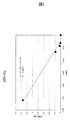
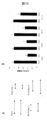
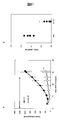
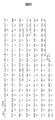
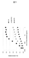
- FIG. 1 is a diagram showing antigen affinity (Kd: dissociation constant) measurement of an anti-hTROP-2 monoclonal antibody (K5-70).
- Abt Antibody (total)
- Agf Antigen (free)
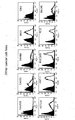
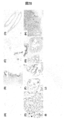
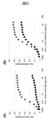
- FIG. 2 is a diagram showing the reactivity of HuH-7 cells (hTROP-2 negative) and HuH-7-hTROP-2 cells in a hybridoma culture supernatant that produces an anti-hTROP-2 monoclonal antibody.
- the black histogram shows HuH-7 cells and the black line histogram shows HuH-7-hTROP-2 cells.
- FIG. 1 is a diagram showing antigen affinity (Kd: dissociation constant) measurement of an anti-hTROP-2 monoclonal antibody (K5-70).
- Abt Antibody (total)
- Agf Antigen (free)
- FIG. 2 is a diagram showing the reactivity of HuH-7 cells (hTROP-2 negative) and HuH-7-hTROP-2 cells in a hybridoma
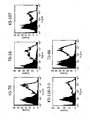
- FIG. 3 is a diagram showing the reactivity of an anti-hTROP-2 monoclonal antibody with a human pancreatic cancer cell line (PK-59 cell) that endogenously expresses hTROP-2 on the cell surface.
- the black histogram shows only the secondary antibody (PE-labeled anti-mouse IgG), and the black line histogram shows the reaction with each anti-hTROP-2 monoclonal antibody.
- FIG. 4 is a diagram showing the reactivity of an anti-hTROP-2 monoclonal antibody with a human pancreatic cancer cell line (BxPC-3 cell) that endogenously expresses hTROP-2 on the cell surface.
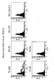
- FIG. 5 is a view showing the reactivity of an anti-hTROP-2 monoclonal antibody (K5-70) with a human pancreatic cancer cell line.
- the black histogram shows only the secondary antibody (PE-labeled anti-mouse IgG), and the black line histogram shows the one reacted with the anti-hTROP-2 monoclonal antibody.
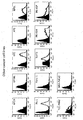
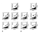
- FIG. 6 shows human colon cancer cell lines (Colo320, CACO2, SW480, DLD1, CW2, HCT 116), human breast cancer (JIMT-1, HCC1143) and human prostate cancer of anti-hTROP-2 monoclonal antibody (K5-70). It is a figure which shows the reactivity with a cell line (PC-3, DU145). The black histogram shows only the secondary antibody (PE-labeled anti-mouse IgG), and the black line histogram shows the one reacted with the anti-hTROP-2 monoclonal antibody.
- FIG. 7 shows the cross-reactivity of anti-hTROP-2 monoclonal antibody with mouse TROP-2.
- the cells used were cells in which the mouse TROP-2 gene was transiently expressed in CHO-K1 cells, and a T2-102 antibody (mouse IgG1) that crossed with mouse TROP-2 was used as a positive control antibody. .
- the black histogram shows only the secondary antibody (PE-labeled anti-mouse IgG), and the black line histogram shows the reaction with each anti-hTROP-2 monoclonal antibody.
- FIG. 8 shows the cross-reactivity of anti-hTROP-2 monoclonal antibody with human EpCAM / TROP-1.
- the cells used were cells in which human EpCAM / TROP-1 gene was transiently expressed in CHO-K1 cells, and PE-labeled anti-human EpCAM monoclonal antibody (Becton Dickinson) was used as a positive control antibody.
- the black histogram shows only the secondary antibody (PE-labeled anti-mouse IgG), and the black line histogram shows the reaction with each anti-hTROP-2 monoclonal antibody.
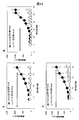
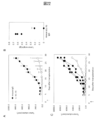
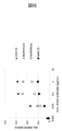
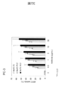
- FIG. 9 is a graph showing the cell growth inhibitory activity of anti-hTROP-2 antibodies (T6-16, T5-86, K5-70, K5-107) of human pancreatic cancer cell lines (PK-59 cells).
- FIG. 10 is a diagram showing a scratch assay of an anti-hTROP-2 antibody (T6-16, K5-70) in a human pancreatic cancer cell line (PK-59 cells).
- a representative example of a photograph of a scratch region of PK-59 cells is shown.
- Day0 A representative example immediately after scratching is shown.
- mIgG Day 1: A photograph of a control antibody (mouse IgG, 1 ⁇ g / mL) added to the medium after scratching and one day later (24 hours later).
- K5-70 Day 1: Photo of K5-70 antibody (1 ⁇ g / mL) added to the medium after scratching and one day later (24 hours later).
- T6-16 (Day 1): A photograph of T6-16 antibody (1 ⁇ g / mL) added to the medium after scratching and one day later (24 hours later). The arrow shown in the photograph indicates the width of the scratch area.
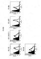
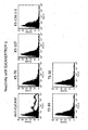
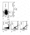
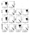
- FIG. 11 is a FACS showing the expression of stem cell markers of human pancreatic cancer cell line, PK-59 cells.
- A FACS showing the expression of EpCAM in PK-59 cells. The black histogram shows only the secondary antibody (PE-labeled anti-mouse IgG), and the black line shows the histogram when reacted with the anti-human EpCAM antibody (Becton Dickinson).
- B, C FACS showing the expression of P-glycoprotein / MCR1 (B) and ABCG2 (C) in PK-59 cells. Blue histogram shows secondary antibody only, red histogram shows anti-human P-glycoprotein / MDR1 antibody (BD Farmingen) (B), anti-human ABCG2 antibody (BD Farmingen) (C) Indicates.
- D FACS of double staining of PK-59 cells with a pancreatic cancer stem cell marker FITC-labeled anti-human CD24 antibody (BD Farmingen) and PE-labeled anti-human CD44 antibody (BD Farmingen). The number in the figure of D shows the abundance ratio of cells in each fraction.
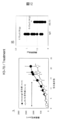
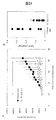
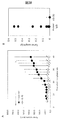
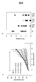
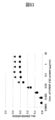
- FIG. 12 is a diagram showing an evaluation of the antitumor activity of a novel anti-hTROP-2 monoclonal antibody clone K5-70 (mouse IgG2a) in a xenograft treatment model using PK-59 cells.
- A Tumor growth of the control group ( ⁇ : mouse IgG) and the K5-70 antibody (10 mg / kg body weight) administration group ( ⁇ ) was expressed over time (mean ⁇ standard deviation). The arrow represents the antibody administration period.
- B It is the figure which plotted the tumor weight in each mouse
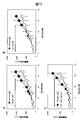
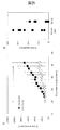
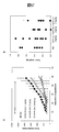
- FIG. 13 is a graph showing the results of A.X. Clone K5-107, B.I. Clone T6-16, C.I. It is the figure which evaluated the antitumor activity of clone K5-116-2-1.
- ⁇ represents a control group (mouse IgG), and ⁇ represents an anti-hTROP-2 antibody (10 mg / kg body weight) administration group.
- the arrows in the graph represent the antibody administration period, and the numerical values shown on each plot are mean values ⁇ standard deviation. * P ⁇ 0.05 (by Student's t-test)
- FIG. 13 is a graph showing the results of A.X. Clone K5-107, B.I. Clone T6-16, C.I. It is the figure which evaluated the antitumor activity of clone K5-116-2-1.
- ⁇ represents a control group (mouse IgG)
- ⁇ represents an anti-hTROP-2 antibody (10 mg / kg body weight) administration group.
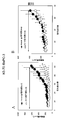
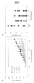
- FIG. 14 evaluates the antitumor activity of clones K5-70 (A), clone T6-16 (B), and clone K5-116-2-1 (C) in a xenograft prevention model using PK-59 cells.
- FIG. ⁇ represents a control group (mouse IgG), and ⁇ represents an anti-hTROP-2 antibody (10 mg / kg body weight) administration group.
- the arrows in the graph represent the antibody administration period, and the numerical values shown on each plot are mean values ⁇ standard deviation. ** P ⁇ 0.01 (by Student's t-test)
- FIG. 15 is a diagram evaluating the antitumor activity of clone K5-70 in the xenograft prevention and treatment models using BxPC-3 cells.
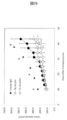
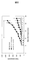
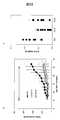
- FIG. 16 shows the dose-dependent antitumor activity of clone K5-70 in the xenograft prevention model using PK-59 cells.
- Tumor volume was expressed as mean ⁇ standard deviation.
- A Tumor growth in the control group ( ⁇ : mouse IgG) and the K5-70 antibody administration group ( ⁇ : 1 mg / kg body weight, ⁇ : 5 mg / kg body weight) at each dose is expressed over time (average value ⁇ standard deviation) ).
- the arrow represents the antibody administration period.
- B It is the figure which plotted the tumor weight in each mouse
- FIG. 17 is a schematic diagram of the human / mouse chimeric TROP-2 protein used in the experiment.
- SP signal sequence
- TY domain thyroglobulin type 1 region
- TM transmembrane region
- C intracellular region
- the region represented by solid black is a polypeptide derived from hTROP-2.
- the region shown in white is a polypeptide derived from mouse TROP-2.
- the numbers shown in the upper part of the schematic diagram of the chimeric protein are the mouse TROP-2 protein, and the lower part is the amino acid number of the hTROP-2 protein.
- FIG. 18 shows the results of identification of an anti-hTROP-2 monoclonal antibody binding region using human / mouse chimeric TROP-2.
- FIG. 19 shows the results of identification of the antibody binding region of the anti-hTROP-2 monoclonal antibody.
- the hTROP-2 gene and each human / mouse chimeric TROP-2 gene were introduced into HEK293 cells, and FACS analysis was performed using the cells transiently expressed.
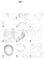
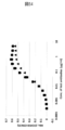
- FIG. 21 is a diagram showing the expression of hTROP-2 in cancer tissues. Immunostaining of human cancer tissue array was performed using anti-hTROP-2 monoclonal antibody clone K5-63-17.
- FIG. 22 is a diagram showing antitumor activity of a single administration of clone K5-70 in a xenograft prevention model using PK-59 cells.
- A Tumor formation in the control group ( ⁇ : mouse IgG) and the K5-70 antibody (10 mg / kg body weight) administration group ( ⁇ ) is expressed over time (mean ⁇ standard deviation). Arrows represent antibody administration.
- FIG. 23 is a diagram showing the antitumor activity of clone K5-70 in a xenograft treatment model using human colon cancer cells SW480 cells.
- FIG. 24 is a diagram showing the antitumor activity of clone K5-116-2-1 in a xenograft treatment model using SW480 cells.
- FIG. 25 is a diagram showing the antitumor activity of clone T6-16 in a xenograft treatment model using SW480 cells.
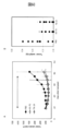
- FIG. 26 is a diagram showing the dose-dependent antitumor activity of clone K5-70 in a xenograft treatment model using SW480 cells.
- A Tumor formation in the control group ( ⁇ : mouse IgG) and the K5-70 antibody administration group ( ⁇ : 1 mg / kg body weight, ⁇ : 5 mg / kg body weight, ⁇ : 10 mg / kg body weight) was expressed over time (average) Value ⁇ standard deviation).
- the arrow represents the antibody administration period.
- A It is a figure which shows the anti-tumor activity of clone K5-70 in the xenograft treatment model using SW480 cell.
- A It is a figure which shows the anti-tumor activity of K5-70 antibody in the administration interval once a week. Tumor formation in the control group ( ⁇ : mouse IgG) and the K5-70 antibody (10 mg / kg) administration group ( ⁇ ) was expressed over time (mean ⁇ standard deviation). Arrowheads (Day 10, 17, 24, 31, 38) indicate administration of K5-70 antibody. * P ⁇ 0.05 by Student's t-test.
- B It is a figure which shows the anti-tumor activity of the K5-70 antibody in the administration interval of once every 10 days (q10d) or once every two weeks (q14d).
- FIG. 28 is a diagram showing the dose-dependent antitumor activity of clone T6-16 in a xenograft treatment model using SW480 cells.
- A Tumor formation in the control group ( ⁇ : mouse IgG) and the T6-16 antibody administration group ( ⁇ : 1 mg / kg body weight, ⁇ : 5 mg / kg body weight, ⁇ : 10 mg / kg body weight) was expressed over time (average) Value ⁇ standard deviation).
- the arrow represents the antibody administration period. ** P ⁇ 0.01 (by Student's t-test) B: It is the figure which plotted the tumor weight in each mouse
- FIG. 29 is a diagram showing the antitumor activity of clone T6-16 in a xenograft treatment model using SW480 cells.
- Tumor formation in the control group ⁇ : mouse IgG 10 mg / kg body weight
- the T6-16 antibody (10 mg / kg body weight) administration group ⁇ : q7d, ⁇ : q10d
- Arrow heads Day 10, 17, 24, 31, 38
- arrows Day 10, 20, 30, 40
- the control group was administered once every 3 days. * P ⁇ 0.05, ** P ⁇ 0.01 by Student's t-test.
- FIG. 30 shows the antitumor activity of clone K5-70 in a xenograft prevention model using human prostate cancer cells DU-145 cells.
- A Tumor formation in the control group ( ⁇ : mouse IgG) and the K5-70 antibody (10 mg / kg body weight) administration group ( ⁇ ) was expressed over time (mean ⁇ standard deviation). The arrow represents the antibody administration period.
- B In the test of A, it is the figure which plotted the tumor weight in each mouse
- FIG. 31 is a diagram showing the metastasis-suppressing activity of clone K5-70 using the liver metastasis model of PK-59 cells.
- A The extracted liver images 6 weeks after cell transplantation in the control group ( ⁇ : mouse IgG) and B: K5-70 antibody (10 mg / kg body weight) administration group are shown. Arrows indicate liver metastases.
- FIG. 32 is a diagram showing the antitumor activity of K5-70 in a cancer recurrence model after irinotecan administration in a xenograft model using SW480 cells.
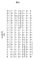
- FIG. 33 shows the cDNA base sequence (SEQ ID NO: 34) and deduced amino acid sequence (SEQ ID NO: 35) of clone K5-70 heavy chain variable region (VH). Signal peptides are italicized. Double underlined glutamine (Q) indicates the N-terminal amino acid residue of the mature peptide.
- CDR sequences (underlined; IYWIN, NIYPSDYTNYNQKKD, TSMADY) are defined in accordance with Kabat et al. Were determined. The amino acid sequences of CDR1 to CDR3 of clone K5-70VH are shown in SEQ ID NOs: 36 to 38, respectively.
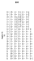
- FIG. 34 shows the cDNA base sequence (SEQ ID NO: 39) and deduced amino acid sequence (SEQ ID NO: 40) of clone K5-70 L chain variable region (VL). Signal peptides are italicized. Double underlined aspartic acid (D) indicates the N-terminal amino acid residue of the mature peptide.
- CDR sequences underlined; RASQSIGTSH, YASESIS, QQSNSWPFT
- the amino acid sequences of CDRs 1 to 3 of clone K5-70 VL are shown in SEQ ID NOs: 41 to 43, respectively.
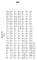
- FIG. 35 is a diagram showing the cDNA base sequence (SEQ ID NO: 44) and deduced amino acid sequence (SEQ ID NO: 45) of clone K5-107 heavy chain variable region (VH). Signal peptides are italicized. Double underlined glutamine (Q) indicates the N-terminal amino acid residue of the mature peptide.
- CDR sequences underlined; SYWMH, NIYPGGGYTNYDEKFKS, SSVFDY) were determined according to the definition of Kabat et al. (Supra; US Department of Health and Human Services, 1991). The amino acid sequences of CDRs 1 to 3 of clone K5-107 VH are shown in SEQ ID NOs: 46 to 48, respectively.
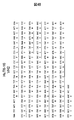
- FIG. 36 is a drawing showing the cDNA base sequence (SEQ ID NO: 49) and deduced amino acid sequence (SEQ ID NO: 50) of clone K5-107 L chain variable region (VL). Signal peptides are italicized. Double underlined aspartic acid (D) indicates the N-terminal amino acid residue of the mature peptide.
- CDR sequences underlined; RASQNIGTSIH, YASESIS, QQSNSWPFT
- the amino acid sequences of CDRs 1 to 3 of clone K5-107 VL are shown in SEQ ID NOs: 510 to 53, respectively.
- FIG. 37 shows the cDNA base sequence (SEQ ID NO: 54) and deduced amino acid sequence (SEQ ID NO: 55) of clone K5-116-2-1 heavy chain variable region (VH). Signal peptides are italicized. Double underlined glutamine (Q) indicates the N-terminal amino acid residue of the mature peptide.
- CDR sequences underlined; SYWIT, NIYPSDSYTNYNQKFRD, LFDY
- the amino acid sequences of CDRs 1 to 3 of clone K5-116-2-1 VH are shown in SEQ ID NOs: 56 to 58, respectively.
- FIG. 38 shows the cDNA base sequence (SEQ ID NO: 59) and deduced amino acid sequence (SEQ ID NO: 60) of clone K5-116-2-1 L chain variable region (VL). Signal peptides are italicized. Double underlined aspartic acid (D) indicates the N-terminal amino acid residue of the mature peptide.
- CDR sequences underlined; RASQSIGTSH, YASESIS, QQSNSWPFT
- the amino acid sequences of CDRs 1 to 3 of clone K5-116-2-1 VL are shown in SEQ ID NOs: 61 to 63, respectively.
- CDR sequences (underlined; DYNMMH, YIYPYNGGTGYNQRFKS, EDYGSSPSYAMDY) were determined according to the definition of Kabat et al. (Supra; US Department of Health and Human Services, 1991).
- the amino acid sequences of CDRs 1 to 3 of clone T6-16 VH are shown in SEQ ID NOs: 66 to 68, respectively.
- FIG. 40 shows the cDNA base sequence (SEQ ID NO: 69) and deduced amino acid sequence (SEQ ID NO: 70) of clone T6-16 L chain variable region (VL). Signal peptides are italicized. Double underlined aspartic acid (D) indicates the N-terminal amino acid residue of the mature peptide.
- CDR sequences underlined; RSSQSLVNGGNTYLH, KVSNRRFS, SQTHVVPT
- the amino acid sequences of CDRs 1 to 3 of clone T6-16 VL are shown in SEQ ID NOs: 71 to 73, respectively.
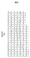
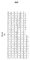
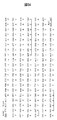
- sequence 41 shows the amino acid sequence (sequence number 35) of the heavy chain variable region (K5-70 VH) of clone K5-70, and the amino acid sequence (sequence of humanized K5-70 (HuK5-70 VH)). No. 75) and an alignment of the amino acid sequence (SEQ ID NO: 85) of the heavy chain variable region (DA980102 VH) of the acceptor (Genbank accession No. DA980102; SEQ ID NO: 84) used for the production of the humanized antibody. (Note that any amino acid sequence in the figure is a part of the amino acid sequence of each heavy chain variable region (specifically, the amino acid sequence of the mature protein portion excluding the signal peptide portion)).
- the amino acid sequence shown underlined in K5-70 VH represents the CDR sequence defined by Kabat et al. (Supra; US Department of Health and Human Services, 1991). The number written at the top of the amino acid sequence indicates the position number of the amino acid according to the definition of Kabat et al. Each CDR sequence in DA980102 VH is indicated as --- in the figure, and is omitted from the description. Since the amino acids shown underlined in HuK5-70 VH are expected to be important for maintaining the structure of the CDR, the sequence of K5-70 VH was maintained.
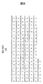
- FIG. 42 shows the amino acid sequence (SEQ ID NO: 40) of the L chain variable region (K5-70VL) of clone K5-70, and the amino acid sequence (SEQ ID NO: 40) of the humanized K5-70 L chain variable region (HuK5-70 VL). 77), and the amino acid sequence (SEQ ID NO: 87; Genbank accession No.
- each amino acid sequence in a figure is a part of amino acid sequence of each L chain variable region (specifically, the amino acid sequence of the mature protein part except a signal peptide part). is there.).
- the amino acid sequence shown underlined in K5-70 VL represents the CDR sequence defined by Kabat et al. (Supra; US Department of Health and Human Services, 1991). The number written at the top of the amino acid sequence indicates the position number of the amino acid according to the definition of Kabat et al.
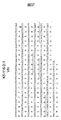
- FIG. 43 shows the amino acid sequence (SEQ ID NO: 65) of the heavy chain variable region (T6-16VH) of clone T6-16, two types of humanized T6-16 heavy chain variable regions (HuT6-16 VH1, HuT6-16). VH2) amino acid sequence (SEQ ID NO: 79 and SEQ ID NO: 81, respectively), and the amino acid of the heavy chain variable region (DA935238 VH) of the acceptor (Genbank accession No.
- DA935238 SEQ ID NO: 88 used to produce the humanized antibody It is a figure which shows alignment about a sequence
- any amino acid sequence in a figure is a part of amino acid sequence of each H chain variable region (specifically, a signal peptide part is excluded).
- Amino acid sequence of the mature protein portion The amino acid sequence shown underlined in HuT6-16 VH represents the CDR sequence defined by Kabat et al. (Supra; US Department of Health and Human Services, 1991). The number written at the top of the amino acid sequence indicates the position number of the amino acid according to the definition of Kabat et al.
- Each CDR sequence in DA935238 VH is indicated as --- in the figure, and is not shown.
- the amino acids shown underlined in HuT6-16 VH1 and HuT6-16 VH2 were predicted to be important for maintaining the structure of the CDRs, and therefore maintained the T6-16 VH sequence.
- the lysine (K) at position 73 in HuT6-16 VH1 was replaced with threonine (T) derived from DA935238 VH, which is an acceptor sequence in HuT6-16 VH2.
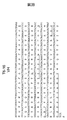
- FIG. 44 shows the amino acid sequence (SEQ ID NO: 70) of the light chain variable region (T6-16 VL) of clone T6-16, and the amino acid sequence (sequence of humanized T6-16 L chain variable region (HuT6-16 VL)). No. 83) and the amino acid sequence (SEQ ID NO: 91; Genbank accession No. AAA60341) of the L chain variable region (M99608 VL) of the acceptor (Genbank accession No. M99608; SEQ ID NO: 90) used for the production of the humanized antibody FIG.
- FIG. 4 is a diagram showing alignment (note that any amino acid sequence in the figure is part of the amino acid sequence of each L chain variable region (specifically, the amino acid sequence of the mature protein portion excluding the signal peptide portion) .)
- the amino acid sequence shown underlined in T6-16 VL represents the CDR sequence defined by Kabat et al. (Supra; US Department of Health and Human Services, 1991). The number written at the top of the amino acid sequence indicates the position number of the amino acid according to the definition of Kabat et al.
- Each CDR sequence in M99608 VL is indicated as --- in the figure, and is not shown.
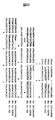
- FIG. 45 shows the gene sequence (SEQ ID NO: 74) and amino acid sequence (SEQ ID NO: 75) of HuK5-70 VH.
- each row shows the gene sequence (cDNA sequence), and the lower row shows the amino acid sequence.
- the signal peptide portion is underlined by a broken line
- each CDR sequence (CDR1 to CDR3) is underlined by a solid line (the amino acid sequence of only the mature protein portion excluding the signal peptide portion is the sequence Number 92).
- An EcoRI site (GAA TTC) and Kozak sequence (ACC ACC) were added to the 5 ′ end of the HuK5-70 VH gene, and an NheI site (GCT AGC) was added to the 3 ′ end.
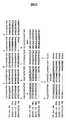
- FIG. 46 shows the gene sequence (SEQ ID NO: 76) and amino acid sequence (SEQ ID NO: 77) of HuK5-70 VL.
- each row shows the gene sequence (cDNA sequence), and the lower row shows the amino acid sequence.
- the signal peptide portion is underlined by a broken line
- each CDR sequence (CDR1 to CDR3) is underlined by a solid line (the amino acid sequence of only the mature protein portion excluding the signal peptide portion is the sequence No. 93).
- An AgeI site (ACC GGT) and Kozak sequence (ACC ACC) were added to the 5 ′ end of the HuK5-70 VL gene, and a BsiWI site (CGT ACG) was added to the 3 ′ end.
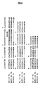
- each row shows the gene sequence (SEQ ID NO: 78) and amino acid sequence (SEQ ID NO: 79) of HuT6-16 VH1.
- the upper row of each row shows the gene sequence (cDNA sequence), and the lower row shows the amino acid sequence.
- the signal peptide portion is underlined by a broken line, and each CDR sequence (CDR1 to CDR3) is underlined by a solid line (the amino acid sequence of only the mature protein portion excluding the signal peptide portion is the sequence No. 94).
- An EcoRI site (GAA TTC) and Kozak sequence (ACC ACC) were added to the 5 ′ end of the HuT6-16 VH1 gene, and an NheI site (GCT AGC) was added to the 3 ′ end.
- FIG. 48 shows the gene sequence (SEQ ID NO: 80) and amino acid sequence (SEQ ID NO: 81) of HuT6-16 VH2.
- the upper row of each row shows the gene sequence (cDNA sequence), and the lower row shows the amino acid sequence.
- the signal peptide portion is underlined by a broken line, and each CDR sequence (CDR1 to CDR3) is underlined by a solid line (the amino acid sequence of only the mature protein portion excluding the signal peptide portion is the sequence Number 95).
- An EcoRI site (GAA TTC) and a Kozak sequence (ACC ACC) were added to the 5 ′ end of the HuT6-16 VH2 gene, and an NheI site (GCT AGC) was added to the 3 ′ end.
- FIG. 49 shows the gene sequence (SEQ ID NO: 82) and amino acid sequence (SEQ ID NO: 83) of HuT6-16 VL.
- the upper row of each row shows the gene sequence (cDNA sequence), and the lower row shows the amino acid sequence.
- the signal peptide portion is underlined by a broken line, and each CDR sequence (CDR1 to CDR3) is underlined by a solid line (the amino acid sequence of only the mature protein portion excluding the signal peptide portion is the sequence Number 96).
- An AgeI site (ACC GGT) and Kozak sequence (ACC ACC) were added to the 5 ′ end of the HuT6-16 VL gene, and a BsiWI site (CGT ACG) was added to the 3 ′ end.
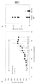
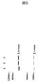
- FIG. 50 shows the results of confirming the expression of HuK5-70 antibody, HuT6-16-1 antibody, and HuT6-16-2 antibody.
- A Expression vectors pFUSE-CHIg-HuK5-70 and pFUSE2-CLIg-HuK5-70 were introduced into 293F cells, and expression of HuK5-70 antibody in the culture supernatant was examined by Western blot. Lane 1 is a culture supernatant (negative control) of 293F cells in which no gene was introduced, and Lane 2 is a culture supernatant of 293F cells into which the above expression vector was introduced.
- the heavy and light chain proteins of the HuK5-70 antibody are biotinylated anti-human IgGF (ab ′) 2 Detected with antibody.
- FIG. 51 shows the results of Coomassie staining of purified HuK5-70 antibody, HuT6-16-1 antibody, and HuT6-16-2 antibody. 1 ⁇ g of each of the purified HuK5-70 antibody (lane 1), HuT6-16-1 antibody (lane 2) and HuT6-16-2 antibody (lane 3) was developed by SDS-PAGE and coomassie staining was performed.
- FIG. 52 is a diagram showing the results of analysis of antigen-binding ability of HuK5-70 antibody, HuT6-16-1 antibody, and HuT6-16-2 antibody using a flow cytometer.
- FIG. 53 is a diagram showing the results of measuring the antigen binding ability of the HuK5-70 antibody using the ELISA method.
- the antigen-binding ability of the K5-70 antibody and the HuK5-70 antibody was examined by an antigen-immobilized ELISA method.
- ⁇ represents the measurement result of K5-70 antibody
- ⁇ represents the measurement result of HuK5-70 antibody.
- FIG. 54 is a diagram showing the measurement results of the antigen binding ability of HuT6-16-1 antibody and HuT6-16-2 antibody using ELISA method. Antigen-binding ability of T6-16 antibody, HuT6-16-1 antibody, and HuT6-16-2 antibody was examined by an antigen-immobilized ELISA method. ⁇ indicates the measurement result of the T6-16 antibody, ⁇ indicates the measurement result of the HuT6-16-1 antibody, and ⁇ indicates the measurement result of the HuT6-16-2 antibody.
- FIG. 55 is a diagram showing the antitumor activity of a humanized anti-hTROP-2 antibody (HuK5-70 antibody) in a xenograft treatment model using human colon cancer cells SW480 cells.
- FIG. 56 is a diagram showing the dose-dependent antitumor activity of a humanized anti-hTROP-2 antibody (HuK5-70) in a xenograft treatment model using human colon cancer cells SW480 cells.
- A Tumor formation in the control group ( ⁇ : PBS) and the HuK5-70 antibody administration group ( ⁇ ; 1 mg / kg body weight, ⁇ ; 5 mg / kg body weight, ⁇ ; 10 mg / kg body weight) was expressed over time (average value) ⁇ standard deviation).
- the arrow represents the antibody administration period. ** P ⁇ 0.01 (by Student's t-test)
- B It is the figure which plotted the tumor weight in each mouse
- 57 is a view showing the dose-dependent antitumor activity of a humanized anti-hTROP-2 antibody (HuT6-16-2) in an xxenograft treatment model using human colon cancer cells SW480 cells.
- the arrow represents the antibody administration period.
- FIG. 58 shows the antitumor activity of mouse anti-hTROP-2 antibodies (K5-70 and T6-16) in a xenograft treatment model using human ovarian cancer cells SK-OV-3 cells.
- A Tumor formation of the control group ( ⁇ : PBS), the K5-70 antibody (10 mg / kg body weight) administration group ( ⁇ ), and the T6-16 antibody (10 mg / kg body weight) administration group ( ⁇ ) over time.
- FIG. 59 shows the antitumor activity of mouse anti-hTROP-2 antibodies (K5-70 and T6-16) in the xxenograft treatment model using human breast cancer cells MDA-MB-468 cells.
- A Tumor formation of the control group ( ⁇ : PBS), the K5-70 antibody (10 mg / kg body weight) administration group ( ⁇ ), and the T6-16 antibody (10 mg / kg body weight) administration group ( ⁇ ) over time. (Mean ⁇ standard deviation). The arrow represents the antibody administration period. ** P ⁇ 0.01 (by Student's t-test) B: It is the figure which plotted the tumor weight in each mouse
- 60 is a view showing the antitumor activity of mouse anti-hTROP-2 antibodies (K5-70 and T6-16) in a xenograft treatment model using human lung cancer cells Calu-3 cells.
- A Tumor formation of the control group ( ⁇ : PBS), the K5-70 antibody (10 mg / kg body weight) administration group ( ⁇ ), and the T6-16 antibody (10 mg / kg body weight) administration group ( ⁇ ) over time. (Mean ⁇ standard deviation).
- the arrow represents the antibody administration period.
- FIG. 61 shows the antitumor activity of mouse anti-hTROP-2 antibody K5-70 in the xenograft prevention model using human cholangiocarcinoma cells TFK-1 cells.
- A Tumor formation in the control group ( ⁇ : PBS) and the K5-70 antibody (10 mg / kg body weight) administration group ( ⁇ ) was expressed over time (mean ⁇ standard deviation).
- FIG. 62 shows the results of examining the binding activity of HuK5-70 and HuT6-16-2 antibodies using a low concentration antigen-immobilized ELISA.
- FIG. 63 shows the results of ELISA showing the binding activity of hTROP-2 to K5-70 and HuK5-70 antibodies.
- FIG. 64 is a diagram showing the base sequence of the K5-70 VH gene (upper; SEQ ID NO: 99) and the amino acid sequence of K5-70 VH (lower: SEQ ID NO: 35) prepared by gene synthesis.
- FIG. 65 is a diagram showing the base sequence of the K5-70 VL gene (upper sequence: SEQ ID NO: 100) and the amino acid sequence of K5-70 VL (lower sequence: SEQ ID NO: 40) prepared by gene synthesis.
- an AgeI site ACC GGT
- a Kozak sequence ACC ACC
- CCT ACG BsiWI site
- Double underlined aspartic acid indicates the N-terminal amino acid residue of the mature peptide.
- CDR sequences underlined; RASQSIGTSH, YASESIS, QQSNSWPFT) were determined according to the definition of Kabat et al. (Supra; US Department of Health and Human Services, 1991).
- the amino acid sequences of CDRs 1 to 3 of clone K5-70 VL are shown in SEQ ID NOs: 41 to 43, respectively.
- FIG. 5 is a view showing the binding activity of an antibody to hTROP-2.
- FIG. 67 shows the amino acid sequences of HuK5-70 VH and its amino acid substitution mutant. Amino acids are shown in single letter code.
- FIG. 68 shows ChK5-70, HuK5-70, HuK5-70 VH A40R (a mutant in which the 40th alanine of VH of HuK5-70 antibody is substituted with arginine) and HuK5-70 VH R44G antibody (HuK5-70 antibody). It is the figure which showed the binding activity with respect to hTROP-2 of the 44th arginine of VH substituted with glycine.
- a 96-well plate was coated with 0.1 ⁇ g / mL recombinant hTACSTD2-Fc-His protein and the test antibodies (ChK5-70, HuK5-70, HuK5-70 VH A40R and HuK5-70 VH R44G antibodies) were transiently applied.
- a 2-fold dilution series (6 points) was prepared from 0.5 ⁇ g / mL of the culture supernatant of the expressed cells and allowed to react.
- FIG. 70 is a diagram of SDS-PAGE of purified HuK5-70-2 antibody.
- FIG. 71 shows the binding activity of K5-70, HuK5-70 and HuK5-70-2 antibodies to hTROP-2.
- a 96-well plate was coated with 0.1 ⁇ g / mL recombinant hTACSTD2-Fc-His protein, and purified test antibodies (K5-70, HuK5-70 and HuK5-70-2 antibodies) were diluted 2 times from 1 ⁇ g / mL.
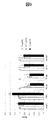
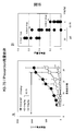
- FIG. 72A is a diagram showing ADCC activity using a humanized anti-hTROP-2 antibody (white column: HuK5-70, black column: HuT6-16-2).
- the human colorectal cancer cell line SW480, HuK5-70 antibody and HuT6-16-2 antibody both 0, 0.1, 0.3, 1, 3, 10 ⁇ g / mL
- FIG. 72B is a diagram showing ADCC activity using a humanized anti-hTROP-2 antibody (white column: HuK5-70, gray column: HuK5-70-2, black column: HuT6-16-2).
- An antibody concentration of 0 indicates no antibody added.
- FIG. 72C is a diagram showing ADCC activity using a humanized anti-hTROP-2 antibody (white column: HuK5-70, gray column: HuK5-70-2, black column: HuT6-16-2).
- the hTROP-2 gene and its gene product are expressed in various cancer cells.
- anti-hTROP-2 monoclonal antibody anti-hTROP-2 monoclonal antibody
- the present inventor screened from an extremely large number of clones. As a result, a clone having high antitumor activity in vivo was successfully obtained.
- the present invention provides a monoclonal antibody that specifically recognizes the extracellular region of hTROP-2 in vivo, particularly a monoclonal antibody having a high picomolar (pM) affinity.
- the antibody of the present invention has a remarkable tumor growth inhibitory activity at a smaller dose (for example, 1/20 dose) compared to an existing anti-hTROP-2 antibody by administration of a naked antibody alone, This is extremely useful in that it is an anti-hTROP-2 monoclonal antibody (particularly a humanized antibody) that exhibits remarkable tumor growth inhibitory activity in a tumor-bearing mouse treatment model using human cancer cells.
- SEQ ID NO: 2 Preparation of antigen Information on the amino acid sequence of hTROP-2 (SEQ ID NO: 2) is published as, for example, “Accession number: NP002344” on the NCBI (GenBank) website (http://www.ncbi.nlm.nih.gov/). Yes.
- a polypeptide or peptide (also simply referred to as a peptide) containing at least a part (all or a part) of the amino acid sequence of hTROP-2 can be used.
- an extracellular region of hTROP-2 is used.
- Peptides containing at least part (all or part) of the amino acid sequence can be used.
- the extracellular region of hTROP-2 (including the signal peptide) is a region containing the first to 274th amino acids of the amino acid sequence represented by SEQ ID NO: 2 (signal peptide: 1st to 26th amino acids). Amino acid).
- the above “at least part of the amino acid sequence” is not particularly limited in length, but for example, the first to the first amino acid sequences shown in SEQ ID NO: 2
- a region containing the 145th amino acid, a region containing the 146th to 274th amino acids in the amino acid sequence, and the like are preferable.
- a method for producing a peptide used as an antigen may be chemical synthesis or synthesis by genetic engineering using E.
- peptide When the peptide is chemically synthesized, it can be synthesized by a well-known method of peptide synthesis. In addition, the solid phase synthesis method and the liquid phase synthesis method can be applied to the synthesis. A commercially available peptide synthesizer (eg, Shimadzu Corporation: PSSM-8) may be used.
- PSSM-8 Shimadzu Corporation
- a peptide is synthesized by genetic engineering, first, a DNA encoding the peptide is designed and synthesized. The design and synthesis can be performed by PCR using, for example, a primer designed to synthesize a desired DNA region using a vector containing the full-length hTROP-2 gene as a template.
- a recombinant vector for protein expression is obtained by ligating the DNA to an appropriate vector, and a transformant is obtained by introducing this recombinant vector into a host so that the target gene can be expressed (Sambrook J Et al., Molecular Cloning, A Laboratory Manual, 3rd edition, Cold Spring Harbor Laboratory Press, 2001).
- a phage or a plasmid capable of autonomously growing in a host microorganism is used.
- animal virus and insect virus vectors can also be used.
- the purified DNA may be cleaved with an appropriate restriction enzyme, inserted into a restriction enzyme site or the like of an appropriate vector DNA, and linked to the vector.
- the host used for transformation is not particularly limited as long as it can express the target gene. Examples include bacteria (E. coli, Bacillus subtilis, etc.), yeast, animal cells (COS cells, CHO cells, etc.), insect cells or insects. It is also possible to use a mammal such as a goat as a host. Methods for introducing a recombinant vector into a host are known. Then, the transformant is cultured, and a peptide used as an antigen is collected from the culture. "Culture” means any of (a) culture supernatant, (b) cultured cells or cultured cells, or crushed materials thereof. After the culture, when the target peptide is produced in cells or cells, the peptide is extracted by disrupting the cells or cells.
- the culture solution is used as it is, or the cells or cells are removed by centrifugation or the like.
- a general biochemical method used for peptide isolation and purification for example, ammonium sulfate precipitation, gel filtration, ion exchange chromatography, affinity chromatography, and the like, can be used alone or in combination as appropriate. It can be separated and purified.
- a peptide serving as an antigen can also be obtained by in vitro translation using a cell-free synthesis system. In this case, two methods, a method using RNA as a template and a method using DNA as a template (transcription / translation), can be used.
- Cell-free synthesis systems include commercially available systems such as Expressway. TM A system (Invitrogen), PURESYSTEM (registered trademark; Post Genome Research Institute), TNT system (registered trademark; Promega), etc. can be used.
- the peptide obtained as described above can be bound to an appropriate carrier protein such as bovine serum albumin (BSA), keyhole limpet hemocyanin (KLH), human thyroglobulin, chicken gamma globulin and the like.
- BSA bovine serum albumin
- KLH keyhole limpet hemocyanin
- human thyroglobulin chicken gamma globulin and the like.
- the antigen may be a peptide consisting of an amino acid sequence of hTROP-2 (SEQ ID NO: 2) or an amino acid sequence in which one or more amino acids are deleted, substituted or added in the partial sequence described above.
- amino acids of the amino acid sequence of hTROP-2 or a partial sequence thereof are deleted.
- genes to be introduced into cells and the like include genes encoding hTROP-2 protein or a partial fragment thereof, or a mutant protein or fragment thereof.
- a gene having the base sequence shown in SEQ ID NO: 1 or a partial sequence thereof can be used.
- a gene for introduction into a cell or the like a base sequence encoding a protein that hybridizes with a sequence complementary to the base sequence shown in SEQ ID NO: 1 under stringent conditions and has hTROP-2 activity, Alternatively, a partial sequence thereof can be used.
- the “stringent conditions” are conditions at the time of washing after hybridization, wherein the salt (sodium) concentration of the buffer is 10 to 500 mM, the temperature is 42 ° C. to 72 ° C., preferably the above salt The condition is that the concentration is 50 to 300 mM and the temperature is 55 to 68 ° C.
- a mutation introduction kit using, for example, a site-directed mutagenesis method, for example, GeneTailor, by a known method such as the Kunkel method or the Gapped duplex method.
- TM Site-Directed Mutagenesis System manufactured by Invitrogen
- TaKaRa Site-Directed Mutagenesis System manufactured by Takara Bio Inc.
- the prepared antigen is administered to a mammal for immunization. Mammals are not particularly limited, and examples include rats, mice, and rabbits. Among these, mice are preferable.
- the dose of the antigen per animal can be appropriately set depending on the presence or absence of an adjuvant.
- adjuvants include Freund's complete adjuvant (FCA), Freund's incomplete adjuvant (FIA), and aluminum hydroxide adjuvant.
- FCA Freund's complete adjuvant
- FIA Freund's incomplete adjuvant
- Aluminum hydroxide adjuvant examples include aluminum hydroxide adjuvant.
- Immunization can be performed mainly by injecting intravenously, footpads, subcutaneously, intraperitoneally, and the like. Further, the immunization interval is not particularly limited, and immunization is performed 1 to 10 times, preferably 2 to 3 times at intervals of several days to several weeks, preferably at one week intervals.
- the antibody titer is measured by enzyme immunoassay (ELISA or EIA), radioimmunoassay (RIA), etc., and blood is collected on the day when the desired antibody titer is shown.
- Antiserum can be obtained.
- ELISA or EIA enzyme immunoassay
- RIA radioimmunoassay
- blood is collected on the day when the desired antibody titer is shown.
- Antiserum can be obtained.
- a known method such as ammonium sulfate salting-out method, ion exchange chromatography, gel filtration chromatography, affinity chromatography or the like is appropriately selected, or these are used. By combining, it can be purified. Thereafter, the reactivity of the polyclonal antibody in the antiserum is measured by an ELISA method or the like.
- the anti-hTROP-2 antibody of the present invention is not limited, but is preferably a monoclonal antibody.
- the prepared antigen is administered to mammals such as rats, mice and rabbits for immunization.
- the dose of the antigen per animal can be appropriately set depending on the presence or absence of an adjuvant.
- the adjuvant is the same as described above.
- the immunization technique is the same as described above.
- antibody-producing cells are collected 1 to 60 days, preferably 1 to 14 days after the final immunization day. Examples of antibody-producing cells include spleen cells, lymph node cells and peripheral blood cells, among which lymph node cells and spleen cells are preferred.
- (3-2) Cell fusion In order to obtain a hybridoma (antibody-producing cell line), cell fusion between antibody-producing cells and myeloma cells is performed.
- myeloma cells to be fused with antibody-producing cells generally available cell lines of animals such as mice can be used.
- a cell line to be used it has drug selectivity and cannot survive in a HAT selection medium (including hypoxanthine, aminopterin and thymidine) in an unfused state, but can survive only in a state fused with antibody-producing cells. What has is preferable.
- myeloma cells include P3-X63-Ag8.653, P3-X63-Ag8 (X63), P3-X63-Ag8.
- mice myeloma cell lines such as U1 (P3U1), P3 / NS I / 1-Ag4-1 (NS1), and Sp2 / 0-Ag14 (Sp2 / 0).
- the selection of myeloma cells can be performed by appropriately considering compatibility with antibody-producing cells.
- myeloma cells and antibody-producing cells are fused.
- Cell fusion is performed in animal cell culture media such as serum free DMEM and RPMI-1640 media at 1 ⁇ 10 ⁇ . 6 ⁇ 1 ⁇ 10 7 Cells / mL of antibody-producing cells and 2 ⁇ 10 5 ⁇ 2 ⁇ 10 6 Mix with cells / mL myeloma cells.
- the cell ratio of antibody-producing cells to myeloma cells is not limited, but is usually preferably 1: 1 to 10: 1, more preferably 3: 1.
- a fusion reaction is performed in the presence of a cell fusion promoter.
- the cell fusion promoter for example, polyethylene glycol having an average molecular weight of 1,000 to 6,000 daltons (D) can be used.
- antibody-producing cells and myeloma cells can be fused using a commercially available cell fusion device utilizing electrical stimulation (for example, electroporation). (3-3) Selection and cloning of hybridomas The target hybridoma is selected from the cells after cell fusion treatment.
- the cell suspension is appropriately diluted with, for example, fetal bovine serum-containing RPMI-1640 medium, and then spread on a microtiter plate.
- a selective medium is added to each well, and thereafter the selective medium is appropriately replaced. Incubate.
- cells that grow from about 14 days after the start of culture in the selective medium can be obtained as hybridomas.
- Hybridoma screening may be carried out in accordance with ordinary methods, and is not particularly limited.
- a part of the culture supernatant contained in a well grown as a hybridoma can be collected and screened by ELISA, EIA, RIA or the like.
- Cloning of the fused cells can be performed by a limiting dilution method or the like.
- An antibody showing strong reactivity with hTROP-2 is determined by flow cytometry or the like, and a hybridoma producing the antibody is selected and established as a clone.
- (3-4) Collection of monoclonal antibody As a method for culturing the established hybridoma and collecting the monoclonal antibody from the obtained culture, a normal cell culture method, ascites formation method or the like can be employed.
- “Cultivation” means growing the hybridoma in a culture dish or bottle, or growing the hybridoma in the peritoneal cavity of an animal as described below.
- the hybridoma is cultured in an animal cell culture medium such as RPMI-1640 medium containing 10% fetal bovine serum, MEM medium, or serum-free medium (eg, 37 ° C., 5% CO 2). 2
- the antibody can be obtained from the culture supernatant.
- a hybridoma is about 1 ⁇ 10 6 in the peritoneal cavity of a mammal derived from a myeloma cell and the same strain. 7 Individually administered, the hybridoma is proliferated in large quantities.
- the anti-hTROP-2 antibody of the present invention is an antibody having antitumor activity in vivo.
- the “antitumor activity” means an activity of killing tumor cells (cancer cells) or an activity of inhibiting tumor growth.
- Preferred examples of the antitumor activity include cancer cell growth inhibitory activity and tumor angiogenesis inhibitory activity.
- human tumors tumor cells
- examples of human tumors (tumor cells) to which the antibody of the present invention can exhibit antitumor activity include various known human tumors whose hTROP-2 expression has been confirmed, and are not particularly limited.
- various human tumors such as human pancreatic cancer, human prostate cancer, human colorectal cancer, human breast cancer, human ovarian cancer, human lung cancer and human bile duct cancer are preferable, and human pancreatic cancer is more preferable.
- Human colon cancer, human prostate cancer, human colon cancer, human breast cancer, human ovarian cancer and human lung cancer and more preferably human pancreatic cancer and / or human colon cancer.
- the type of tumor may be recurrent cancer or metastatic cancer, and the antibody of the present invention can exert excellent antitumor activity against these tumors.
- In vivo anti-tumor activity can be confirmed, for example, by using a tumor-bearing mouse (tumor-bearing animal treatment model) in which a desired tumor cell is transplanted subcutaneously in a mouse, and administering the antibody obtained as described above to this mouse Can be done.
- antibody administration may be performed immediately after transplantation of tumor cells (prevention model), or may be performed after confirming that the tumor has reached a predetermined volume for transplantation (treatment model).
- the administration method is not limited, but for example, once every 3 days, 1 week, 10 days or 2 weeks or once (only once), 5 to 20 mg / kg body weight, intraperitoneal administration, etc. it can.
- the presence / absence and level of antitumor activity can be evaluated based on the tumor formation frequency and tumor volume.
- the presence and level of antitumor activity can be evaluated by the tumor volume.
- the amino acid sequences of CDR1 to CDR3 of the H chain V region are the amino acid sequences represented by SEQ ID NOs: 36 to 38, respectively.
- the amino acid sequences of CDRs 1 to 3 of the L chain V region are the amino acid sequences represented by SEQ ID NOs: 41 to 43, respectively, are preferable.
- the H chain V region is preferably composed of an amino acid sequence represented by SEQ ID NO: 35
- the L chain V region is preferably composed of an amino acid sequence represented by SEQ ID NO: 40, for example.
- the amino acid sequences of CDRs 1 to 3 of the H chain V region are the amino acid sequences represented by SEQ ID NOs: 46 to 48, respectively, and / or Preferred are those in which the amino acid sequences of CDRs 1 to 3 of the L chain V region are the amino acid sequences represented by SEQ ID NOs: 51 to 53, respectively.
- the H chain V region is preferably composed of an amino acid sequence represented by SEQ ID NO: 45
- the L chain V region is preferably composed of an amino acid sequence represented by SEQ ID NO: 50, for example.
- the amino acid sequences of CDRs 1 to 3 of the H chain V region are the amino acid sequences represented by SEQ ID NOs: 56 to 58, respectively, and Preferred are those in which the amino acid sequences of CDRs 1 to 3 of the L chain V region are the amino acid sequences represented by SEQ ID NOs: 61 to 63, respectively.
- the H chain V region is preferably composed of an amino acid sequence represented by SEQ ID NO: 55
- the L chain V region is preferably composed of an amino acid sequence represented by SEQ ID NO: 60, for example.
- the amino acid sequences of CDRs 1 to 3 of the H chain V region are the amino acid sequences represented by SEQ ID NOs: 66 to 68, respectively, and Preferred are those in which the amino acid sequences of CDRs 1 to 3 of the L chain V region are the amino acid sequences represented by SEQ ID NOs: 71 to 73, respectively, in that order.
- the H chain V region is preferably composed of an amino acid sequence represented by SEQ ID NO: 65
- the L chain V region is preferably composed of an amino acid sequence represented by SEQ ID NO: 70, for example.
- the anti-hTROP-2 antibody having anti-tumor activity in vivo is more specifically, for example, an anti-hTROP-2 monoclonal antibody (clone) produced by a hybridoma whose receipt number is FERM BP-11251.
- anti-hTROP-2 monoclonal antibody (clone name: K5-107) produced by a hybridoma whose receipt number is FERM BP-11252, produced by a hybridoma whose receipt number is FERM BP-11253
- Anti-hTROP-2 monoclonal antibody (clone name: K5-116-2-1)
- anti-hTROP-2 monoclonal antibody (clone name: T6-16) produced by a hybridoma whose receipt number is FERM BP-11346, and receipt number FERM Anti hTROP-2 monoclonal antibody produced by a hybridoma which is P-11254 (clone name: T5-86), and the like preferably.
- the hybridoma whose receipt number is FERM BP-11251 is referred to as “Mouse-Mouse Hybridoma K5-70”, and the hybridoma whose receipt number is FERM BP-11252 is “Mouse-Mouse”.
- Hybridoma K5-107 The hybridoma referred to as “Hybridoma K5-107” dated May 12, 2010 and having a receipt number of FERM BP-11253, referred to as “Mouse-Mouse Hybridoma K5-116-2-1”, dated May 12, 2010,
- a hybridoma whose receipt number is FERM BP-11346 is referred to as “Mouse-Mouse Hybridoma T6-16”
- the hybridoma whose receipt number is FERM BP-11254 is “Mouse-Mouse Hybridoma T6-16”.
- anti-hTROP-2 antibody of the present invention for example, a monoclonal produced by a hybridoma whose receipt number is FERM BP-11251, FERM BP-11252, FERM BP-11253, FERM BP-11346 or FERM BP-11254.
- An anti-hTROP-2 antibody that binds to a site (for example, an epitope) to which the antibody binds (recognizes) is also preferred.
- epitope examples include those exemplified in the following (3-6).
- epitope of anti-hTROP-2 antibody The epitope (antigenic determinant) of the anti-hTROP-2 antibody of the present invention is not limited as long as it is at least part of the antigen hTROP-2.
- the amino acid sequence of hTROP-2 shown in SEQ ID NO: 2 It is preferably at least a part of the region excluding the region consisting of the 252nd to 260th amino acids, more preferably at least a part of the region consisting of the 1st to 69th amino acids or A region consisting of the 100th to 274th amino acids (excluding the region consisting of the 252nd to 260th amino acids), more preferably a region consisting of the 27th to 69th amino acids Or at least part of the region consisting of the 109th to 206th amino acids.
- a region consisting of the 171st to 183rd amino acids, a region consisting of the 109th to 120th amino acids, a region consisting of the 193rd to 206th amino acids, and the 43rd to 56th amino acids A region and a portion including these, particularly preferred are a region consisting of the 43rd to 65th amino acids, a region consisting of the 152nd to 165th amino acids, and the 171st to 183rd amino acids.
- an anti-hTROP-2 antibody that recognizes the region (binding to the region or a part containing the region) has high internalization activity into tumor cells, for example, and is extremely useful for uses such as immunoconjugates described later.
- the anti-hTROP-2 antibody of the present invention is an antibody having high in vivo antitumor activity at a low dose.
- a dose of 20 mg / kg body weight or less (preferably 10 mg / kg body weight or less, more preferably 5 mg / kg body weight or less, more preferably 1 mg / kg body weight or less) to a cancer-bearing animal model (nude) 50% or more (preferably 80% or more, more preferably 90% or more, still more preferably 95% or more, particularly preferably almost 100% (for example, 98% or more or 99% or more))
- the tumor growth inhibitory activity (%) can be calculated by, for example, the following formula.
- Tumor growth inhibitory activity (%) 100 ⁇ [(tumor volume or tumor weight of antibody administration group) ⁇ (tumor volume or tumor weight of control group)] ⁇ 100
- the anti-hTROP-2 antibody of the present invention preferably has antitumor activity against two or more human tumor cell lines.
- human tumor cell lines include, but are not limited to, various human pancreatic cancer cell lines, human prostate cancer cell lines, human colon cancer cell lines, human breast cancer cell lines, human ovarian cancer cell lines, human lung cancer cell lines, and human bile ducts
- Examples include at least two types selected from the group consisting of cancer cell lines, specifically, human pancreatic cancer cell line PK-59, human pancreatic cancer cell line BxPC-3, human pancreatic cancer cell line KP-3L, and human pancreatic cancer cell line KP.
- human pancreatic cancer cell line PK-1 human pancreatic cancer cell line PK-45H, human pancreatic cancer cell line PK-45P, human pancreatic cancer cell line TCC-PAN2, human pancreatic cancer cell line SUIT-2, human colon cancer cell line CACO- 2, human colon cancer cell line SW480, human colon cancer cell line DLD-1, human colon cancer cell line HCT116, human breast cancer cell line JIMT-1, human breast cancer cell line HCC1143, human breast cancer cells MCF-7, human breast cancer cell line MDA-MB-468, human prostate cancer cell line DU145, human prostate cancer cell line PC-3, human ovarian cancer cell line SK-OV-3, human lung cancer cell line Calu-3, and Preferable examples include at least two selected from the group consisting of human cholangiocarcinoma cell line TFK-1.
- the two or more human tumor cell lines include human pancreatic cancer cell line PK-59, human pancreatic cancer cell line BxPC-3, human colon cancer cell line SW480, human lung cancer cell line Calu-3, human breast cancer cell line MBA. More preferred are at least two selected from the group consisting of -MD-468 and human ovarian cancer cell line SK-OV-3.
- the anti-hTROP-2 antibody of the present invention has a dissociation constant (Kd value) of 1.0 ⁇ 10 -10 M or less is preferred, more preferably 1.0 ⁇ 10 -11 M or less, more preferably 1.0 ⁇ 10 -12 M or less.
- the binding ability (affinity) of the antibody is determined, for example, by a Scatchard analysis or a surface plasmon resonance sensor called Biacore, a dissociation constant (Kd value), a dissociation rate constant (Kdis [1 / Sec]), a binding rate. It can be measured as a constant (Kass [1 / M.Sec]).
- the Biacore apparatus include Biacore 3000, Biacore 2000, Biacore X, Biacore J, Biacore Q (all of which are Biacore).
- the antibody is preferable in that the smaller the dissociation constant (Kd value), the higher the binding ability (affinity).
- the method as described in the Example (specifically Example 10) mentioned later can also be employ
- (4) Recombinant antibodies and antibody fragments (4-1) Recombinant antibody
- One preferred embodiment of the anti-hTROP-2 antibody of the present invention is a recombinant antibody.
- the recombinant antibody include, but are not limited to, a chimeric antibody, a humanized antibody, and a human antibody.
- a chimeric antibody ie, a human chimeric antibody
- a human chimeric antibody is an antibody obtained by linking (joining) a variable region of a mouse-derived antibody to a human-derived constant region (Proc. Natl. Acad. Sci. USA). 81 , 6851-6855, (1984), etc.), when a chimera is produced, it can be easily constructed by a gene recombination technique so as to obtain an antibody linked in such a manner.
- CDR complementarity determining region
- FR framework region
- CDR CDR grafting
- the CDR sequence derived from the mouse that can be used for the humanized anti-hTROP-2 antibody of the present invention is not limited, but for example, CDRs 1 to 3 of the H chain V region (VH) (each in order)
- the amino acid sequence shown in SEQ ID NOs: 36 to 38 and the amino acid sequence shown in SEQ ID NOs: 66 to 68 are the amino acid sequences shown in SEQ ID NOs: 41 to 43 as CDRs 1 to 3 of the L chain V region (VL) (respectively in order)
- Preferred examples include the amino acid sequences represented by SEQ ID NOs: 71 to 73.
- the amino acid sequence of the H chain V region is, for example, an amino acid sequence represented by SEQ ID NO: 92 (a CDR1 consisting of an amino acid sequence represented by SEQ ID NOs: 36 to 38).
- the amino acid sequence further comprising a signal peptide is represented by SEQ ID NO: 75), the amino acid sequence represented by SEQ ID NO: 98 (including CDRs 1 to 3 consisting of the amino acid sequences represented by SEQ ID NOs: 36 to 38; The amino acid sequence comprising SEQ ID NO: 97), the amino acid sequence represented by SEQ ID NO: 94 (including CDRs 1 to 3 consisting of the amino acid sequences represented by SEQ ID NOs: 66 to 68; and the amino acid sequence comprising the signal peptide is SEQ ID NO: 79) and the amino acid sequence shown in SEQ ID NO: 95 (amino acids shown in SEQ ID NOs: 66 to 68) Preferred examples include CDRs 1 to 3 comprising a nonacid sequence; and an amino acid sequence further comprising a signal peptide is represented by SEQ ID NO: 81).
- the amino acid sequence of the H chain V region represented by SEQ ID NO: 98 the 44th arginine (R) of the amino acid sequence of the H chain V region represented by SEQ ID NO: 92 is replaced with glycine (G).
- G glycine
- the amino acid sequence of the L chain V region is, for example, the amino acid sequence represented by SEQ ID NO: 93 (the CDR1 comprising the amino acid sequence represented by SEQ ID NOs: 41 to 43).
- amino acid sequence further including a signal peptide is represented by SEQ ID NO: 77), and an amino acid sequence represented by SEQ ID NO: 96 (including CDRs 1 to 3 consisting of amino acid sequences represented by SEQ ID NOs: 71 to 73;
- the amino acid sequence including the signal peptide is preferably represented by SEQ ID NO: 83).
- the amino acid sequence of the H chain V region consists of the amino acid sequence represented by SEQ ID NO: 92, and the L chain V region
- the amino acid sequence consists of the amino acid sequence shown by SEQ ID NO: 93
- the amino acid sequence of the H chain V region consists of the amino acid sequence shown by SEQ ID NO: 98
- the amino acid sequence of the L chain V region is SEQ ID NO: What consists of an amino acid sequence shown by 93 is preferable.
- the humanized anti-hTROP-2 antibody of (ii) above in which the amino acid sequence of the H chain V region is the amino acid sequence represented by SEQ ID NO: 98, is avidity (ie, movement of two antigen-binding arms). It is particularly preferable because it has improved flexibility and high antigen binding activity.
- the amino acid sequence of the H chain V region consists of the amino acid sequence represented by SEQ ID NO: 94, and the L chain V
- the amino acid sequence of the region consists of the amino acid sequence shown by SEQ ID NO: 96
- the amino acid sequence of the H chain V region consists of the amino acid sequence shown by SEQ ID NO: 95
- the amino acid sequence of the L chain V region is What consists of an amino acid sequence shown by sequence number 96 is preferable.
- a human antibody (fully human antibody) generally has the same structure as a human antibody in the hypervariable region, which is the antigen-binding site of the V region, the other part of the V region, and the constant region. is there. However, the hypervariable site may be derived from another animal. Techniques for producing human antibodies are also known, and methods for producing gene sequences common to humans by genetic engineering techniques have been established.
- the human antibody is obtained, for example, by a method using a human antibody-producing mouse having a human chromosome fragment containing H and L chain genes of human antibody (Tomizuka, K. et al., Nature Genetics, (1977) 16, 133-. Kuroiwa, Y. et al., Nuc.
- the chimeric antibody, human antibody and human antibody are preferably those in which the N-glycoside-linked complex sugar chain in the antibody Fc region is a sugar chain in which fucose is not bound to N-acetylglucosamine at the reducing end of the sugar chain.
- the antibody fragment of the present invention has a binding activity to hTROP-2 (ie, can bind to hTROP-2) and is antitumor in vivo. It has activity.
- the antibody fragment means a partial region of an anti-hTROP-2 polyclonal antibody or an anti-hTROP-2 monoclonal antibody (ie, an antibody fragment derived from the anti-hTROP-2 antibody of the present invention), for example, Fab, Fab ', F (ab') 2 , Fv (variable fragment of antibody), single chain antibody (H chain, L chain, H chain V region, L chain V region, etc.), scFv, diabody (scFv dimer), dsFv (disulfide stabilized V region) ), And a peptide including a complementarity determining region (CDR) at least in part.
- CDR complementarity determining region
- Fab has an antigen-binding activity with a molecular weight of about 50,000, in which about half of the N-terminal side of the H chain and the entire L chain are linked by a disulfide bond among fragments obtained by treating antibody molecules with the protease papain. It is an antibody fragment.
- a Fab may be produced by inserting a DNA encoding an antibody Fab into a prokaryotic expression vector or a eukaryotic expression vector and introducing the vector into a prokaryotic or eukaryotic organism. it can.
- F (ab ') 2 Is an antibody fragment having an antigen binding activity with a molecular weight of about 100,000, which is slightly larger than that obtained by treating the antibody molecule with the protease pepsin and having Fab bound through a disulfide bond in the hinge region. is there. Moreover, it can also produce by making Fab mentioned later into a thioether bond or a disulfide bond.
- Fab ′ is the above F (ab ′) 2 An antibody fragment having a molecular weight of about 50,000 and having an antigen-binding activity, which is obtained by cleaving the disulfide bond in the hinge region.
- DNA encoding the Fab ′ fragment of the antibody is inserted into a prokaryotic expression vector or eukaryotic expression vector, and the vector is introduced into prokaryotic or eukaryotic cells to be expressed to produce Fab ′.
- scFv is a VH-P-VL or VL-P-VH in which one H chain V region (VH) and one L chain V region (VL) are linked using an appropriate peptide linker (P).
- scFv obtains cDNA encoding antibody VH and VL, constructs DNA encoding scFv, inserts the DNA into a prokaryotic expression vector or eukaryotic expression vector, and the expression vector Can be expressed and produced by introduction into prokaryotic or eukaryotic organisms.
- Diabody is an antibody fragment in which scFv is dimerized and is an antibody fragment having a bivalent antigen-binding activity.
- the bivalent antigen binding activity can be the same, or one can be a different antigen binding activity.
- Diabodies obtains cDNA encoding the VH and VL of the antibody, constructs the DNA encoding scFv so that the length of the amino acid sequence of P is 8 residues or less, and uses the DNA for prokaryotes It can be produced by inserting into an expression vector or an expression vector for eukaryotes and introducing the expression vector into a prokaryote or eukaryote.
- dsFv refers to a polypeptide in which one amino acid residue in each of VH and VL is substituted with a cysteine residue, which are bonded via a disulfide bond between the cysteine residues.
- the amino acid residue substituted for the cysteine residue can be selected based on the three-dimensional structure prediction of the antibody according to the method shown by Reiter et al. (Protein Engineering, 7, 697-704, 1994).
- dsFv obtains cDNA encoding the antibody VH and VL, constructs a DNA encoding dsFv, inserts the DNA into a prokaryotic expression vector or eukaryotic expression vector, and inserts the expression vector into a prokaryotic expression vector. It can be expressed and produced by introduction into an organism or eukaryote.
- Peptides containing CDRs are composed of at least one region of CDRs of VH (CDR1-3) and CDRs of VL (CDR1-3), all of which contain CDRs of VH, and all of CDRs of VL More preferably, it includes all VH and VL CDRs (total of 6 regions).
- Examples of the amino acid sequence of CDR include the amino acid sequences shown in SEQ ID NOs: 36 to 38, 41 to 43, 46 to 48, 51 to 53, 56 to 58, 61 to 63, 66 to 68, and 71 to 73.
- Peptides containing multiple CDRs can be linked directly or via a suitable peptide linker.
- the peptide containing CDR can also be manufactured by chemical synthesis methods, such as Fmoc method (fluorenylmethyloxycarbonyl method) and tBoc method (t-butyloxycarbonyl method).
- the antibody fragment of the present invention includes a part of an antibody Fc region in which an N-glycoside-linked complex type sugar chain is a sugar chain in which fucose is not bound to N-acetylglucosamine at the reducing end of the sugar chain
- the antibody Fc region may be an antibody fragment containing all of the above, or an antibody Fc region in which the above-described antibody fragment and an N-glycoside-linked complex type sugar chain are sugar chains in which fucose is not bound to N-acetylglucosamine at the reducing end of the sugar chain. It may be a part or all of the fusion protein.
- Such an antibody fragment is preferable because ADCC activity can be dramatically improved.
- the above-described antibody fragment is also included in the anti-hTROP-2 antibody of the present invention.
- a polynucleotide (gene, DNA) encoding the above-described anti-hTROP-2 antibody of the present invention or an antibody fragment thereof can also be provided.
- the polynucleotide is preferably a polynucleotide containing a base sequence encoding each amino acid sequence shown as an example of the above-described anti-hTROP-2 antibody or antibody fragment of the present invention.
- the polynucleotide of the present invention may be composed only of the polynucleotide encoding the anti-hTROP-2 antibody or antibody fragment of the present invention, or may include the polynucleotide in part and other gene expression. It may also contain necessary known base sequences (transcription promoter, SD sequence, Kozak sequence, terminator, etc.), and is not limited.
- codons corresponding to individual amino acids after translation are not particularly limited, and are generally used in mammals such as humans after transcription. Or a codon commonly used in microorganisms such as Escherichia coli and yeast, plants, etc.
- a recombinant vector containing the polynucleotide of the present invention and a transformant containing the recombinant vector can also be provided.
- the polynucleotide (gene, DNA) to be incorporated into the expression vector used as a recombinant vector is preliminarily upstream of a transcription promoter, an SD sequence (when the host is a prokaryotic cell) and a Kozak sequence (the host is a eukaryotic cell).
- a terminator may be linked downstream, and an enhancer, a splicing signal, a poly A addition signal, a selection marker, and the like may be linked.
- Each element necessary for gene expression such as the above transcription promoter may be included in the polynucleotide from the beginning, or may be used when originally included in the expression vector.
- the use mode of is not particularly limited.
- As a method for incorporating the polynucleotide into the expression vector various methods using known gene recombination techniques such as a method using a restriction enzyme and a method using topoisomerase can be employed.
- the expression vector examples include a polynucleotide (gene, DNA) encoding the anti-hTROP-2 antibody of the present invention or an antibody fragment thereof such as plasmid DNA, bacteriophage DNA, retrotransposon DNA, retroviral vector, artificial chromosome DNA and the like. ) Can be used, and a vector suitable for the host cell to be used can be appropriately selected and used. Subsequently, the constructed recombinant vector is introduced into a host to obtain a transformant, which is cultured, whereby the anti-hTROP-2 antibody of the present invention or an antibody fragment thereof can be expressed.
- a polynucleotide gene, DNA
- DNA encoding the anti-hTROP-2 antibody of the present invention or an antibody fragment thereof
- an antibody fragment thereof such as plasmid DNA, bacteriophage DNA, retrotransposon DNA, retroviral vector, artificial chromosome DNA and the like.
- the “transformant” as used in the present invention means a gene into which a foreign gene has been introduced into the host, for example, a gene into which a foreign gene has been introduced by introducing plasmid DNA or the like into the host (transformation), Also included are those in which a foreign gene has been introduced by infecting a host with various viruses and phages (transduction).
- the host is not limited as long as it can express the anti-hTROP-2 antibody of the present invention or an antibody fragment thereof after the introduction of the recombinant vector, and can be appropriately selected.
- animal cells for example, human fibroblasts, human embryonic kidney cells, HEK293 cells, 293F cells, CHO cells, monkey cells COS-7, Vero, mouse L cells, rat GH3, human FL cells, etc. Used. Insect cells such as Sf9 cells and Sf21 cells can also be used.
- bacteria for example, Escherichia coli, Bacillus subtilis and the like are used.
- yeast is used as a host, for example, Saccharomyces cerevisiae and Schizosaccharomyces pombe are used.
- plant cells for example, tobacco BY-2 cells are used.
- the method for obtaining the transformant is not limited and can be appropriately selected in consideration of the combination of the host and expression vector.
- electroporation, lipofection, heat shock, PEG, Preferred examples include calcium phosphate method, DEAE dextran method, and methods of infecting various viruses such as DNA virus and RNA virus.
- the codon type of the polynucleotide contained in the recombinant vector may be the same as or different from the codon type of the host used, and is not limited. 4).
- an antibody-drug complex comprising the antibody and a substance (compound etc.) having antitumor activity and / or cell killing activity.
- a substance compound etc.
- an immunoconjugate what is obtained by preparing the antibody molecule and a substance having antitumor activity and / or cell killing activity in advance and then combining them is generally called an immunoconjugate.
- a protein toxin as a substance having antitumor activity and / or cell killing activity is linked to an antibody gene on a gene and expressed as a single protein (fusion protein). Is generally referred to as immunotoxin.
- Examples of the substance having antitumor activity include doxorubicin, calicheamicin, mitomycin C, Auristatin E, radioisotope (RI) and the like.
- Examples of the substance having cell killing activity include saporin, ricin, Pseudomonas aeruginosa exotoxin, diphtheria toxin, radioisotope (RI), etc. Among them, saporin and Pseudomonas aeruginosa exotoxin are preferably used.
- the RI having antitumor activity and / or cell killing activity is not limited.
- a method for producing the antibody-drug complex is not limited, and examples thereof include a method of coupling an antibody and a drug by a disulfide bond or a hydrazone bond.
- the anti-hTROP-2 antibody of the present invention is excellent in internalization activity into target tumor cells that express hTROP-2. Therefore, by combining in advance substances having antitumor activity and / or cell killing activity, these substances can be directly and highly selectively acted on tumor cells.
- the antibody-drug conjugate of the present invention is extremely excellent in drug delivery ability to target tumor cells.
- the internalization activity into the cell can be evaluated by fluorescently labeling the antibody with rhodamine or the like, and observing the transfer behavior into the cell and the localization of the antibody using a fluorescence microscope or the like.
- an antibody fragment-drug complex using the above-described antibody fragment instead of the antibody can also be provided.
- the above description of the antibody-drug complex can be applied as appropriate.
- an antibody fragment-drug complex is also included in the antibody-drug complex of the present invention. 5.
- Pharmaceutical composition The anti-hTROP-2 antibody and antibody-drug complex of the present invention are useful as an active ingredient contained in a pharmaceutical composition.
- the pharmaceutical composition is useful as a pharmaceutical composition for tumor treatment and / or diagnosis.
- the anti-hTROP-2 antibody of the present invention and the antibody-drug complex containing the antibody may be used for tumor therapy. preferable. That is, the anti-hTROP-2 antibody and antibody-drug complex of the present invention are useful as active ingredients contained in tumor therapeutic agents and tumor diagnostic agents.
- the above-mentioned tumor treatment includes the meaning of tumor growth inhibition and growth suppression. Specifically, for example, in the case of a tumor therapeutic agent, the form of a tumor growth inhibitor and growth suppression agent Shall also be included.
- the pharmaceutical composition of the present invention may be provided in the form of a pharmaceutical composition comprising the anti-hTROP-2 antibody and / or antibody-drug complex of the present invention as an active ingredient, and further comprising a pharmaceutically acceptable carrier.
- the pharmaceutical composition of the present invention can be used in combination with known antitumor agents. A higher antitumor effect can be obtained by the combined use.
- the disease (tumor) to which the pharmaceutical composition of the present invention is applied include the various known human tumors described above in which the expression of hTROP-2 has been confirmed.
- one or more of various human tumors such as human pancreatic cancer, human prostate cancer, human colon cancer, human breast cancer, human ovarian cancer, human lung cancer and human bile duct cancer are particularly preferred. These diseases may be singly or two or more of them may be combined.
- the target tumor may be recurrent cancer or metastatic cancer, and the pharmaceutical composition of the present invention (and thus the anti-hTROP-2 antibody and / or antibody-drug complex of the present invention) It can also be used effectively as a therapeutic agent and diagnostic agent for metastatic cancer.
- “Pharmaceutically acceptable carrier” refers to excipients, diluents, extenders, disintegrants, stabilizers, preservatives, buffers, emulsifiers, fragrances, colorants, sweeteners, thickeners, taste masking Agents, solubilizers or other additives.
- pharmaceutical compositions in the form of injections, solutions, capsules, suspensions, emulsions or syrups can be prepared. These pharmaceutical compositions can be administered orally or parenterally. Other forms for parenteral administration include injections that contain one or more active substances and are prescribed by conventional methods.
- an injection it can be produced by dissolving or suspending it in a pharmaceutically acceptable carrier such as physiological saline or commercially available distilled water for injection.
- a colloidal dispersion system can be used in addition to the above.
- the colloidal dispersion system is expected to have an effect of enhancing the in vivo stability of the compound (antibody fragment) and an effect of efficiently transporting the compound to a specific organ, tissue or cell.
- the colloidal dispersion system is not particularly limited as long as it is usually used, but polyethylene glycol, polymer composite, polymer aggregate, nanocapsule, microsphere, bead, and oil-in-water emulsifier, micelle, mixed micelle And lipid-based dispersion systems including liposomes, preferably a plurality of liposomes, artificial membrane vesicles that are effective in efficiently transporting compounds to specific organs, tissues or cells.
- liposomes preferably a plurality of liposomes, artificial membrane vesicles that are effective in efficiently transporting compounds to specific organs, tissues or cells.
- the dosage of the pharmaceutical composition of the present invention is the patient's age, sex, weight and symptoms, therapeutic effect, administration method, treatment time, or anti-hTROP-2 antibody and antibody-drug of the present invention contained in the pharmaceutical composition. It depends on the type of complex. Usually, it can be administered within a range of 600 ⁇ g to 6000 mg per adult per person, but is not limited to this range.
- injection when administered by injection, it may be administered to a human patient in an amount of 100 ⁇ g to 100 mg per kg body weight per administration, once to several times per day, preferably 3 days, It is also possible to adopt a mode in which administration is performed once a week, 10 days or 2 weeks, or a single administration (total administration frequency is once).
- administration form include intravenous injection, subcutaneous injection, intradermal injection, intramuscular injection, and intraperitoneal injection, and intravenous injection is preferred.
- injections can be prepared as non-aqueous diluents (eg, polyethylene glycol, vegetable oils such as olive oil, alcohols such as ethanol), suspensions, and emulsions.
- Such sterilization of the injection can be performed by filtration sterilization using a filter, blending of a bactericide, and the like.
- the injection can be produced as a form prepared at the time of use. That is, it can be used as a sterile solid composition by lyophilization, etc., and dissolved in sterile water for injection or other solvent before use.
- the present invention also provides use of the anti-hTROP-2 antibody and / or antibody-drug complex of the present invention for producing a medicament (drug) for treating and / or diagnosing a tumor.
- the present invention also provides the anti-hTROP-2 antibody and / or antibody-drug complex of the present invention for treating and / or diagnosing a tumor.
- the present invention provides a method for treating and / or diagnosing a tumor, characterized by using the anti-hTROP-2 antibody and / or antibody-drug complex of the present invention (ie, administering to a patient). It also provides the use of the anti-hTROP-2 antibody and / or antibody-drug conjugate of the present invention for treating and / or diagnosing a tumor. 6).
- Tumor detection method The tumor detection method of the present invention comprises reacting the anti-hTROP-2 antibody of the present invention with a sample collected from a living body (hereinafter referred to as a biological sample), and detecting the signal of the reacted antibody. Is the method. As described above, hTROP-2 has been confirmed to be specifically expressed in various tumor cells.
- hTROP-2 particularly free hTROP-2 (extracellular region of hTROP-2) is expressed in various tumor markers. It can be used as Among these, it is preferable to use as a marker for human pancreatic cancer, human prostate cancer, human colon cancer and human breast cancer.
- the tumor can be detected by reacting the anti-hTROP-2 antibody of the present invention with a biological sample and detecting the signal of the reacted antibody.
- the obtained antibody signal serves as an index of the amount of antigen in the biological sample (that is, the amount of hTROP-2 or the amount of free hTROP-2).
- a biological sample collected from a subject as a specimen, such as a tissue piece or blood to be examined, and the antibody of the present invention are bound by an antigen-antibody reaction. Then, based on the measurement result of the amount of bound antibody, the target antigen amount in the biological sample is measured.
- the measurement may be performed according to a known immunological measurement method. For example, an immunoprecipitation method, an immunoagglutination method, a labeled immunoassay method, an immunospecific suspension method, a Western blot method, a flow cytometry method, or the like may be used. it can.
- the antibody signal may be represented by the amount of label directly detected using the labeled antibody, or may be relatively represented by using an antibody having a known concentration or a known antibody titer as a standard solution. That is, the standard solution and the specimen can be measured with a measuring meter, and the antibody signal in the biological sample can be relatively represented based on the value of the standard solution.
- the labeled immunoassay include an ELISA method, an EI method, an RIA method, a fluorescence immunoassay (FIA) method, and a chemiluminescence immunoassay method (Luminescence immunoassay). Of these, the ELISA method is particularly preferable in terms of simplicity and high sensitivity.
- the state of the tumor can be evaluated or diagnosed using the detection result obtained by the detection method as an index. For example, if the detection result exceeds a predetermined reference value, the tumor is positive. If the detection result is less than the predetermined reference value, the tumor is negative. If the detection result is positive, it is determined that any tumor may have developed. Tumor status can be assessed.
- the state of the tumor means the presence / absence or progression of the tumor, and includes the presence / absence, progression, malignancy, presence / absence of metastasis, and / or recurrence of the tumor.
- one tumor condition may be selected, or a plurality may be selected in combination.
- the evaluation of the presence or absence of a tumor can be performed by determining whether or not the tumor is affected by using a predetermined reference value as a boundary based on the obtained detection result.
- the degree of malignancy of the tumor is an index indicating how much the cancer has progressed.
- the stage is classified and evaluated, or early cancer or advanced cancer is determined. It is also possible to classify and evaluate. For example, it can also be evaluated as early cancer or advanced cancer using the detection result as an index.
- Tumor metastasis can be evaluated based on whether or not a neoplasm has appeared at a site away from the position of the primary lesion, using the detection result as an index.
- the recurrence can be evaluated by whether the detection result again exceeds a predetermined reference value after the intermittent period or remission. 7).
- Tumor detection or diagnostic kit The anti-hTROP-2 antibody of the present invention can be provided in the form of a tumor detection or diagnostic kit.
- the kit of the present invention includes an antibody, but may additionally include a labeling substance, a solid phase reagent immobilizing the antibody or a labeled product thereof, and the like.
- the antibody labeling substance means a substance labeled with an enzyme, a radioisotope, a fluorescent compound, a chemiluminescent compound, or the like.
- the kit of the present invention includes other reagents for carrying out the detection of the present invention, such as an enzyme substrate (chromogenic substrate, etc.) when the label is an enzyme label, an enzyme substrate solution
- an enzyme reaction stop solution, a sample dilution solution, or the like may be included.
- various buffers, sterilized water, various cell culture containers, various reaction containers (eppendorf tubes, etc.), blocking agents (serum components such as Bovine Serum Albumin (BSA), Skim milk, Goat serum), detergents, surfactants, Various plates, preservatives such as sodium azide, and experimental operation manuals (instructions) may be included.
- the kit of the present invention can be used effectively for carrying out the detection method of the present invention, and is extremely useful.
- the present invention will be described more specifically with reference to examples. However, the present invention is not limited to these examples.
- hTROP-2 gene The full length hTROP-2 gene was isolated from human fetal liver (embryonic week 10) by RT-PCR. First, the following PCR primers were designed based on the sequence of the hTROP-2 gene (Genbank accession No. NM_002353). At this time, a stop codon was removed from the reverse primer, and a restriction enzyme digestion sequence with Xho I was added. PCR reaction was performed using these primers and cDNA synthesized from total RNA (TAKARA) prepared from human fetal liver (fetal 10 weeks) as a template.
- TAKARA total RNA
- pCRII-hTROP2 a pCRII vector (Invitrogen)
- the cloned hTROP-2 cDNA was confirmed by sequencing.
- the construction of the expression vector consists of pCRII-hTROP2 to Eco containing the hTROP-2 gene. It was performed by inserting into EcoRI / Xho I site (pcDNA4-hTROP2-myc / His).
- a Hind III / Pme I fragment containing the hTROP-2 gene was excised from pcDNA4-hTROP2-myc / His (the Hind III cleavage site was blunted) and moved to the Pme I site of the pcDNA3.1 (+) vector (Invitrogen).
- An expression vector having a neomycin resistance gene was constructed by insertion (pcDNA3.1-hTROP2-myc / His).
- the purified DNA fragment was subcloned into pCR Blunt vector (Invitrogen) (pCRB-hTROP-2 EC), and the gene sequence was confirmed. Next, an Eco RI / Xho I fragment containing a gene fragment encoding the extracellular region of hTROP-2 from pCRB-hTROP-2 EC was obtained. Inserted (pcDNA4mH-hTROP-2 EC). Furthermore, in order to prepare a Nru I restriction enzyme cleavage site at the Bam HI / Eco RI site of pcDNA4mH-hTROP-2 EC, the following oligonucleotides were associated and inserted.
- pBgl II linker (TAKARA) was inserted into the Pme I site of pcDNA4mH-hTROP-2 EC (pcDNA4mH-NB-hTROP-2 EC).
- pcDNA4mH-NB-hTROP-2 EC Pme I site of pcDNA4mH-hTROP-2 EC
- pPSC8 vector Nippon Agricultural Industry
- recombinant proteins in the extracellular region of hTROP-2 using baculovirus was outsourced to Nippon Agricultural Industry. Purification of recombinant protein in the extracellular region of hTROP-2 was performed as follows. Ni Sepharose 6 Fast Flow (GE Healthcare) was added to the culture supernatant containing the recombinant protein and allowed to bind at 4 ° C. for 2 hours. Then, it was washed with a phosphate buffer containing 20 mM imidazole using an Econo column (BIORAD), and purified by elution with a phosphate buffer containing 300 mM imidazole.
- Ni Sepharose 6 Fast Flow GE Healthcare
- pCRII-hEpCAM pCRII vector
- the cloned cDNA of human EpCAM was confirmed by sequencing.
- the construction of the expression vector is from pCRII-hEpCAM to Eco containing the human EpCAM gene. It was carried out by inserting into the EcoRI / Xho I site (pcDNA4-hEpCAM-myc / His).
- hTROP-2 expression stable cell lines HEK293-hTROP-2 cells, CHO-K1-hTROP-2 cells, 7E2-C-hTROP-2 cells
- hTROP-2 protein endogenously on the cell surface An expressed human pancreatic cancer cell line (PK-59, RCB1901; purchased from RIKEN cell bank) or a recombinant protein in the extracellular region of hTROP-2 prepared by the above method was used.
- hTROP-2 expression stable cell line 1 ⁇ 10 7 cells
- 20 ⁇ g of the protein is mixed with the immune adjuvant TiterMax Gold (Funakoshi Co., Ltd.) in a ratio of 1: 1.
- Emulsions were prepared and injected into mice (C57 / BL6, Balb / c) on both footpads or intraperitoneally (primary immunization).
- booster immunization was performed 3 days and 10 days after the initial immunization, and on both days after the final immunization, both plaster lymph nodes were collected to prepare lymphocytes.
- booster immunization was performed once a week from the initial immunization (performed for 1 to 2 months), and B cells were isolated from the spleen according to a conventional method.
- a cell suspension of 5 ⁇ 10 6 cells in PBS was used for cell immunization, and 5 ⁇ g of PBS solution was used for protein antigen.
- the prepared lymphocytes and mouse myeloma cell line (P3-X63-Ag8.653) were mixed at a ratio of 3: 1, and cell fusion was carried out by the polyethylene glycol method to obtain HAT (hypoxanthine, aminopterin, thymidine).
- a single colony of hybridomas grown on a methylcellulose medium (trade name: ClonaCell-HY Cloning Medium D; Stem Cell) for 7 to 28 days and then proliferating is picked up on a 96-well flat-bottom plate, and HAT was cultured in a 5% CO 2 incubator.
- a methylcellulose medium trade name: ClonaCell-HY Cloning Medium D; Stem Cell
- HAT was cultured in a 5% CO 2 incubator.
- FACS analysis using HuH-7-hTROP-2 cells, PK-59 the culture supernatant of the hybridoma derived from a single colony that has grown, 300 types of hybridomas producing anti-hTROP-2 monoclonal antibodies that recognize hTROP-2 protein expressed on the cell surface of living cells were established.
- CHO-K1 cells hTROP-2 negative control: purchased from Human Science Promotion Corporation
- CHO-K1-hTROP-2 cells or HuH-7 cells (hTROP-2 negative) Control: purchased from Human Science Promotion Corporation) and HuH-7-hTROP-2 cells
- HuH-7-hTROP-2 cells were alternately seeded at 3 ⁇ 10 4 cells / well and cultured at 5% CO 2 and 37 ° C. for 1-2 days.
- the cell culture medium was removed by decantation, then washed with ice-cold PBS, fixed with 5% paraformaldehyde-PBS for 5 minutes, and washed with ice-cold PBS to prepare an ELISA plate.
- an ELISA method was performed according to a conventional method. Specifically, it is as follows. First, blocking with a 2% skim milk-PBS solution was performed at room temperature for 30 minutes to 1 hour. Next, the hybridoma culture supernatant was added, reacted at room temperature for 1 hour, and then washed three times with a 0.1% Tween 20-PBS solution. As a secondary antibody, Horseradish peroxidase (HRP) -labeled anti-mouse IgG (GE Healthcare Bioscience) diluted 1000 times with a blocking solution was added and reacted at room temperature for 1 hour, and then with a 0.1% Tween20-PBS solution. Washed 3 times.
- HRP horseradish peroxidase
- a TMB (3,3 ′, 5,5′-tetramethylbenzidine: SIGMA) substrate solution was added to perform a color reaction, and 1M sulfuric acid was added to stop the reaction.
- Absorbance (405 nm) was measured using a Microplate reader Model 550 (Bio-Rad).
- the hybridoma corresponding to the hybridoma culture supernatant showing a high absorbance value was expanded in a 24-well flat-bottom plate against the negative control, and transferred to secondary screening (described later) using FACS analysis.
- the cells were detached from the culture dish by trypsin treatment to prepare a cell suspension (cell density 2 ⁇ 10 6 cells / mL).
- Hybridoma culture supernatant that showed a positive reaction in the primary screening using Cell ELISA and 100 ⁇ L of the cell suspension were reacted at 4 ° C. for 20 minutes.
- PE-labeled anti-mouse IgG BD Farmingen
- FACSCalibur Becton Dickinson
- the isotype of the prepared anti-hTROP-2 monoclonal antibody was determined according to the method attached to the kit using MOUSE MONOCLONAL ANTIBODY ISOTYPING TEST KIT (Serotec).
- Antigen affinity (Kd value) of the prepared anti-hTROP-2 monoclonal antibody was calculated by a method using ELISA (Djava-Ohanceance L. et al (1996), In Antibody Engineering, Chapter 4, pp77-97.IRL). Press, Oxford). Specifically, the purified recombinant hTROP-2 protein (0.1 ⁇ g / mL) was added to a 96-well culture plate (Corning) to immobilize the antigen (room temperature, 1 hour, or 4 ° C., overnight) ). Next, the plate was washed 3 times with PBS, and blocked by adding 2% skim milk (PBS solution) (room temperature, 1 hour).
- Antigen-antibody reaction is based on the law of mass action
- Agf is the concentration of free antigen
- Abf is the concentration of free antibody
- Ag-Ab is the concentration of antigen-antibody complex
- the free antibody concentration is Equation (2) is Multiplying both terms in equation (4) by x / Kd ⁇ Abt
- X x / Abt
- Y x / Abt ⁇ Agf
- the Kd value was calculated from the equation (6).
- K5-70 mouse IgG2a
- T6-16 mouse IgG2a
- K5-107 mouse IgG1
- K5-116-2- The Kd values of 1 (mouse IgG2a) and T5-86 (mouse IgG1) were 6.8 ⁇ 10 ⁇ 12 (M), 4.3 ⁇ 10 ⁇ 12 (M), and 4.7 ⁇ 10 ⁇ , respectively.
- Human cancer cell lines (human tumor cell lines) are human science promotion corporation (HSRRB), RIKEN cell bank (RIKEN), ATCC (American Type Culture Collection), ECACC (European Collection of Cell Cultures), DSMZG What was obtained from Microorganisms and Cell Cultures) was used. Specifically, it is as listed below.
- huH-1 (HSRRB), HUH-6 (HSRRB), HuH-7 (HSRRB), JHH-5 (HSRRB), JHH-6 (HSRRB), JHH-7 (HSRRB), HLE (HSRRB), HLF (HSRRB) ), HepG2 (HSRRB), Alexander (HSRRB), KP-1N (HSRRB), KP-1NL (HSRRB), KP-2 (HSRRB), KP-3 (HSRRB), KP-3L (HSRRB), PK-1 (RIKEN), PANC-1 (RIKEN), MIA PaCa-2 (HSRRB), PK-59 (RIKEN), PK-45H (RIKEN), PK-45P (RIKEN), BxPC-3 (ATCC), SUIT-2 (HSRRB), TCC-PAN2 (HSRRB), SW480 (ATCC), DLD-1 (HSRRB), LoVo (HSRRB), COLO-320 (RIKEN), CACO-2 (RIKEN), CW-2 (RIK
- Anti-hTROP-2 monoclonal antibody was added to 100 ⁇ L of cell suspension (0.1 ⁇ g) and reacted at 4 ° C. for 20 minutes. After washing with PBS, it was reacted with PE-labeled anti-mouse IgG (BD Farmingen) (0.1 ⁇ g) (4 ° C., 30 minutes), and then analyzed by FACSCalibur (Becton Dickinson). None of the prepared anti-hTROP-2 antibodies binds to the human hepatoma cell line HuH-7 that does not express hTROP-2 endogenously, whereas HuH is a stable cell line that expresses the hTROP-2 gene. -7-hTROP-2 cells showed binding (FIG. 2).
- hTROP-2 monoclonal antibodies 300 kinds of prepared anti-hTROP-2 monoclonal antibodies were prepared.
- the hTROP-2 monoclonal antibodies all showed binding to human pancreatic cancer cell lines (PK-59, BxPC-3), and in particular, K5-70, T6-16, which showed tumor growth inhibitory activity in vivo.
- the K5-107, K5-116-2-1, and T5-86 antibodies have a higher average fluorescence intensity in PK-59 cells than when reacted with PE-labeled anti-mouse IgG (BD Farmingen) alone.
- KP-1N, KP-1NL, KP-3, PANC-1, and MIA-PaCa2 did not bind (FIG. 5).
- human colon cancer cell lines it bound to CACO-2, SW480, DLD-1, and HCT116, but not to COLO-320 and CW-2 (FIG. 6).
- it binds to JIMT-1, HCC1143 (all are human breast cancer cell lines), PC-3, DU145 (all are human prostate cancer cell lines), and is endogenously expressed on the surface of many human cancer cell lines.
- HTROP-2 protein was recognized (FIG. 6).
- mouse TROP-2 protein showing 80% homology in amino acid sequence with hTROP-2 protein and 50% homology in amino acid sequence with hTROP-2 protein
- the reactivity with human TROP-1 / EpCAM indicating the above was performed by FACS analysis.
- an expression vector (mouse TROP-2-pcDNA3.1 (+) containing the full-length cDNA of mouse TROP-2 gene (GenBank accession No.
- NM — 020047, Y08830 provided by the Institute for Molecular Cell Biology, University of Tokyo
- an expression vector pcDNA3.1-hEpCAM-myc / His
- a full-length cDNA of human TROP-1 / EpCAM gene GenBank accession No. NM_002354
- Lipofectamine 2000 reagent Invitrogen
- the CHO-K1 cells were transfected with the gene, and after 24 to 48 hours, the cells were detached from the culture dish by trypsin treatment to prepare a cell suspension.
- the prepared anti-hTROP-2 monoclonal antibody (0 1 [mu] g), was allowed to react sequentially with PE-labeled anti-mouse IgG, and analyzed by FACSCalibur.
- the T2-102 antibody (mouse IgG1) that cross-reacts with mouse TROP-2 used as a positive control showed strong binding, while K5 The -70, T6-16, K5-107, K5-116-2-1 and T5-86 antibodies did not show cross-reactivity with mouse TROP-2 (FIG. 7).
- mouse IgG negative control
- anti-hTROP-2 monoclonal antibody final concentration 0.1, 1 ⁇ g / mL
- a commercially available anti-hTROP-2 monoclonal antibody clone YY01, Santa Cruz
- Tetracolor One Teikagaku Corporation
- T6-16, T5-86, K5-70, and K5-107 have been confirmed to have a cell growth inhibitory activity of 20% to 40% compared to mouse IgG (negative control), and these anti-hTROP-2 antibodies It was revealed that it has an activity of binding to hTROP-2 protein expressed on the surface of human cancer cells, neutralizing the function of hTROP-2 protein, and inhibiting the growth of cancer cells (FIG. 9).
- Anti-hTROP-2 monoclonal antibody and mouse IgG as a negative control were added to the culture medium so that the final concentration was 0.1 and 1 ⁇ g / mL, and cultured for 24 hours, before antibody addition (Day 0) and for 24 hours.
- a photograph of the peeled area after (Day 1) was taken, and the intercellular distance was measured. Also, the area of the peeled area was quantified with Scion Image software. The test was performed in 8 wells in each group, and a significant difference test was performed by Student's t-test, and P ⁇ 0.05 was determined to be statistically significant.
- the effect of hTROP-2 antibody on the motility of cells infiltrating the scratch region was examined. The medicinal antibodies were evaluated as in the cell growth inhibition assay.
- mice On the day of cancer cell transplantation (Day 1), mice were divided into groups, and antibody administration (1, 5, or 10 mg / kg body weight, intraperitoneal administration) was started. Thereafter, the same administration was performed at an interval of once every 3 days. Antitumor activity was evaluated by tumor formation frequency and tumor volume. The following calculation formula was used to measure the tumor volume.
- Tumor volume (mm 3 ) (minor axis) 2 ⁇ (major axis) ⁇ ⁇ / 6 treatment model
- a pancreatic cancer cell line (PK-59, BxPC-3) expressing hTROP-2 was removed by trypsin treatment, and a cell suspension of 1 ⁇ 10 8 cells / mL was prepared with PBS. Mixed with Matrigel (BD Farmingen).
- mice Sixteen week old female nude mice (Balb / c, nu / nu) were injected subcutaneously in the right flank with 100 ⁇ L (5 ⁇ 10 6 cells) using a 26 G syringe. Five to six days after cancer cell transplantation, mice that have grown to a tumor volume of 50 to 150 mm 3 (average value of about 100 mm 3 ) are divided into groups. did. The antibody was administered intraperitoneally at an interval of once every 3 days (10 mg / kg body weight). Antitumor activity was assessed by measuring tumor volume. The significant difference test was performed by Student's t-test, and P ⁇ 0.05 was determined to be statistically significant.
- CD24 and CD44 co-positive cell fraction (8.93%), which is characteristic of pancreatic cancer stem cells ( 11D) and is presumed to be a high-grade human pancreatic cancer cell line (C enwei Li, et al.Cancer Res 2007; 67: (3) .1030-1037, Jane E.Visvader and Geoffrey J.Lindeman.Nat Rev Cancer.Vol.8 (10): 755-68,2008).
- clones K5-70, T6-16, K5-107, T5-86 and K5-116-2-1 were obtained.
- the tumor growth rate was statistically significantly inhibited.
- the tumor volume at the start of antibody administration is 1.0
- the tumor volume on Day 21 is 12.5 in the control group, whereas 7.8 in the clone K5-70 administration group. (FIG. 12A).
- the excised tumor weight was also 0.73 ⁇ 0.26 g in the control group, whereas 0.43 ⁇ 0.14 g (P ⁇ 0.01 by Student's t-test) in the clone K5-70 administration group. ) And showed about 60% inhibition (FIG. 12B).
- clone K5-107 (mouse IgG1) administration group (N 8)
- clone T5-86 mouse IgG1 and clone K5-16-2
- the tumor growth rate was statistically significantly inhibited.
- the human pancreatic cancer cell line PL45 cell inhibits almost 100% of the tumor growth, but the pancreatic cancer cell line BxPC-3 is about 50%, the prostate cancer cell line PC-3 is about 40%, and the breast cancer cell.
- the anti-hTROP-2 antibody of the present invention has a half dose (10 mg / day), whereas the MCF-7 strain has a tumor growth inhibitory effect of about 60% and the colon cancer cell line Colo205 has about 40%. kg body weight) exerted a stronger tumor growth inhibitory effect.
- the hmTROP-2-A chimeric protein is a chimeric protein composed of a polypeptide from the N-terminal to the 69th of the hTROP-2 protein and a polypeptide from the 64th to the C-terminal of the mouse TROP-2 protein.
- the hmTROP2-B chimeric protein is a chimeric protein composed of a polypeptide from the N-terminal to the 101st of the hTROP-2 protein and a polypeptide from the 96th to the C-terminal of the mouse TROP-2 protein.
- the hmTROP2-C chimeric protein is a chimeric protein composed of a polypeptide from the N-terminal to the 145th of the hTROP-2 protein and a polypeptide from the 140th to the C-terminal of the mouse TROP-2 protein.
- the mhTROP-2-D chimeric protein is a chimeric protein composed of a polypeptide from the N-terminal to the 139th of the mouse TROP-2 protein and a polypeptide from the 146th to the C-terminal of the hTROP-2 protein.
- the mhTROP-2-E chimeric protein is a chimeric protein composed of a polypeptide from the N-terminal to the 187th of the mouse TROP-2 protein and a polypeptide from the 194th to the C-terminal of the hTROP-2 protein.
- the mhTROP-2-F chimeric protein is a chimeric protein comprising a polypeptide from the N-terminal to the 227th of the mouse TROP-2 protein and a polypeptide from the 234th to the C-terminal of the hTROP-2 protein.
- the expression vector for producing the chimeric protein was constructed by the following method.
- PCR was performed using the hTROP-2 gene as a template and the hTROP-2 forward primer and the human / mouse TROP-2-A primer Y613. Similarly, PCR was performed using the mouse TROP-2 gene as a template and human / mouse TROP-2-A primer Y612 and mouse TROP-2 reverse primer. DNA fragments amplified by PCR were developed using acrylamide gel and recovered by extraction of the target band. Next, the two extracted DNA fragments were mixed to form a template, and PCR was performed using the hTROP-2 forward primer and the mouse TROP-2 reverse primer. The PCR product was developed by agarose gel electrophoresis, and the target DNA fragment was extracted.
- the extracted DNA fragment was cloned into pCR (registered trademark) -Blunt vector (Invitrogen) (pCRB-hmTROP-2-A), and the gene sequence was confirmed.
- the expression vector for animal cells is the hmTROP-2-A chimeric gene prepared from pCRB-hmTROP-2-A by removing the hTROP-2 gene from pcDNA3.1-hTROP-2-myc / His by digestion with Eco RI / Xho I. It was prepared by inserting an Eco RI / Xho I fragment containing (pcDNA3.1-hmTROP-2-A-myc / His).
- hmTROP-2-B using human TROP-2 forward primer, human / mouse TROP-2-B primer Y615, human / mouse TROP-2-B primer Y614 and mouse TROP-2 reverse primer
- hmTROP- 2-C using human TROP-2 forward primer, human / mouse TROP-2-C primer Y607 and human / mouse TROP-2-C primer Y606, mouse TROP-2 reverse primer
- mhTROP-2-D Using mouse TROP-2 forward primer, mouse / human TROP-2-D primer Y609 and mouse / human TROP-2-D primer Y608, human TROP-2 reverse primer
- mhTROP-2-E mimouse TROP 2 forward primer, mouse / human TROP-2-E primer Y617 and mouse / human TROP-2-E primer Y616, human TROP-2 reverse primer
- mhTROP-2-F (mouse TROP-2 forward side)
- mouse / human TROP-2-F primer Y619 For the primer, mouse
- HEK293 cell line constitutively expressing hTROP-2, human / mouse chimeric TROP-2-C, and mouse / human chimeric TROP-2-D protein The expression vector pcDNA3.1-hTROP-2- Each of myc / His, pcDNA3.1-hmTROP-2-C-myc / His and pcDNA3.1-mhTROP-2-D-myc / His was introduced into the gene. Selection with antibiotic G418 (Calbiochem) was performed to establish HEK293 cell lines that constitutively express hTROP-2 protein, hmTROP-2-C chimeric protein and mhTROP-2-D chimeric protein.
- anti-hTROP-2 monoclonal antibodies K5-70, T5-86, K5-107, T6-4, T6-16, and K5-1116-2 that showed medicinal effects
- the binding region was identified for -1.
- HEK293 cells constitutively expressing hmTROP-2-C and mhTROP-2-D and their respective chimeric proteins the reactivity of the anti-hTROP-2 monoclonal antibody which showed medicinal effects was examined by FACS analysis (Fig. 18).
- FACS analysis Fig. 18
- K5-70, K5-107, T5-86 and K5-116-2-1 antibodies reacted with hmTROP-2-C but not mhTROP-2-D.
- T6-4 and T6-16 antibodies reacted with mhTROP-2-D but not hmTROP-2-C. From these facts, the binding regions of the K5-70, K5-107, T5-84 and K5-116-2-1 antibodies are the region from the N-terminal to the 145th amino acid of hTROP-2, T6-4 and T6-16 The binding region of the antibody was limited to the region from the 146th amino acid of hTROP-2 to the C-terminus (FIG. 18).
- hmTROP-2-A, hmTROP-2-B, mhTROP-2-E, and mhTROP-2-F chimeric protein expression vectors were prepared, and anti-hTROP-2 exhibiting medicinal effects The reactivity with the monoclonal antibody was examined (FIG. 19).
- the newly prepared chimeric protein expression vector was transfected into HEK293 cells, and FACS analysis was performed using the transiently expressed cells.
- K5-70, K5-107, T5-86 and K5-116-2-1 antibodies reacted with hmTROP-2-A but not mhTROP-2-B. All six monoclonal antibodies examined reacted with hTROP-2.
- the normal and cancer tissue arrays used for immunohistochemical staining are as follows.
- Human normal tissue array Human, Normal organs in duplicates (Catalog No .: AB1, Super Bio Chips) Normal issues more than one spots (Catalog No .: A103 (VI), ISU ABXIS)
- Lung cancer tissue array Human Lung cancer-metastasis-normal (Catalog No .: CCA3, Super Bio Chips) Human lung carcinoma tissue with marsissue tissue, 2 location cores (Catalog No .: OD-CT-RsLug03-002, Shanghai Outdo Biotech)
- Pancreatic cancer tissue array Human Pancreas carcinoma tissue with mono-pathological type from 60cases, 2location cores (Catalog No .: OD-CT-DgPan03-001, Shanghai Haitech Biotech)
- Liver cancer tissue array Hepatocyte cellular carcinoma, grade I to III with normal tissue controls, 63cases tissue arrays (Catalog No .: OD-CT-Dg
- tissue array paraffin sections of human normal tissue and cancer tissue were subjected to protease treatment with pepsin for 5 minutes at 37 ° C. after deparaffinization, and used for immunostaining using anti-hTROP-2 monoclonal antibody.
- DAB 3,3′-diaminobenzidine
- nuclear staining with hematoxylin was performed as counterstaining. More specifically, these operations were performed as follows.
- the paraffin-embedded section was deparaffinized and then treated with protease with pepsin (DAKO) at 37 ° C for 5 minutes.
- endogenous peroxidase activity was removed by treatment with a solution obtained by adding hydrogen peroxide to methanol to a final concentration of 0.3% at room temperature for 20 minutes. After washing twice with PBS for 5 minutes at room temperature, blocking is performed for 30 minutes at room temperature using a PBS solution containing 1.5% normal horse serum (DAKO) to block nonspecific binding sites in the tissue. The operation was performed. Next, the anti-hTROP-2 monoclonal antibody clone K5-63-17 (final concentration 10 ⁇ g / ml) diluted with a PBS solution containing 1.5% normal horse serum was reacted at room temperature for 1 hour, and then at room temperature for 5 hours with PBS.
- DAKO normal horse serum
- the human normal tissue array (Catalog No .: AB1, Super BioChips) was deparaffinized and hydrophilized, and then the antigen was activated with the proteolytic enzyme pepsin, and the anti-hTROP-2 monoclonal antibody clone K5-63 Immunostaining was performed with -17 (FIG. 20).
- staining was seen in the skin, esophagus, kidney (cortex and medulla), pancreas, prostate, bladder, and tonsils. Most stained images showed localization in the cell membrane (FIGS. 20A, B, C, D, F, GH), but some expression was also observed in the cytoplasm (FIGS. 20.E, H). .
- prostate cancer had the highest positive rate (92.1%), lung cancer (65.4%), esophageal cancer (76.7%), bladder cancer (71.2). %) And so on. The lowest positive rate was 7.61% for liver cancer.
- stained images in which hTROP-2 was strongly localized in the cell membrane were observed in cancer cells (FIGS. 21A to F, H, and I). Some were also expressed in the cytoplasm (Fig. 21. A, B, E, G).
- the positive rate of hTROP-2 in pancreatic cancer was 41.9%.
- the relationship between the hTROP-2 positive rate at this time and the grade (differentiation degree) of pancreatic cancer was examined. As a result, it was highly expressed in high-grade, ie, poorly differentiated pancreatic cancer (Table 5).
- the tumor volume at Day 28 was 144.4 ⁇ 176.9 mm 3 (P ⁇ 0.05 by Student's t-test) and 81.5% tumor growth inhibitory activity (FIG. 22A).
- the control group in Day 28 was 0.59 ⁇ 0.06 g
- the clone K5-70 antibody administration group was 0.07 ⁇ 0.10 g (P ⁇ 0.01 by by Student's). t-test)) and 88% inhibitory activity (FIG. 22B).
- tumor formation was completely inhibited by administration of 10 mg / kg body weight once in 2 out of 3 individuals in the K5-70 antibody administration group (FIG. 22C).
- Anti-tumor activity of anti-hihTROP-2 monoclonal antibody in human colon cancer cell line SW480 cell xenograft treatment model The antitumor activity of anti-hTROP-2 monoclonal antibodies (clone K5-70, K5-116-2-1 and T6-16) was examined in a xenograft treatment model using human colon cancer cell line SW480. 5 ⁇ 10 6 cells of SW480 cells were transplanted subcutaneously into the right flank of 6-week-old female NOD-scid mice (Day 1), and grouping was performed when the average tumor volume reached 100 mm 3 (Day 7 or Day 10). , Day 7 or Day 10 started the intraperitoneal administration of the antibody at an administration interval of once every 3 days.
- the antitumor activity evaluation of clone K5-70 antibody and the antitumor activity evaluation of clone K5-116-2-1 antibody and clone T6-16 antibody were evaluated in independent tests.
- the group administered with K5-70 antibody (10 mg / kg body weight) 27.4 ⁇ 29.4 mm 3 (P ⁇ 0.01 by Student's t-test) and marked tumor formation was inhibited (inhibition rate 92.5%) (FIG. 23A).
- the control group was 0.11 ⁇ 0.07 g
- the K5-70 antibody administration group was 0.005 ⁇ 0.007 (g) (P ⁇ 0.01 by Student's t-test) and 95.5% inhibition (FIG. 23B).
- the K5-70 antibody administration group in 2 of 8 individuals, tumor formation was completely inhibited and tumors could not be confirmed.
- the tumor volume of the control group at Day 42 was 713.8 ⁇ 354.5 mm 3 , whereas a dose-dependent tumor formation inhibitory activity was observed in the K5-70 antibody administration group, and in the 1 mg / kg body weight administration group 485.0 ⁇ 207.3 mm 3 (inhibition rate 32.1%), 5 mg / kg body weight administration group, 339.5 ⁇ 253.2 mm 3 (inhibition rate 52.4%), 10 mg / kg body weight administration group It was 355.4 ⁇ 202.8 mm 3 (inhibition rate 50.2%, P ⁇ 0.05 by Student's t-test) (FIG. 26A).
- the control group was 0.39 ⁇ 0.19 g
- the K5-70 antibody 1 mg / kg body weight administration group was 0.24 ⁇ 0.11 g (inhibition). Rate 37.8%), 5 mg / kg body weight administration group, 0.17 ⁇ 0.14 g (inhibition rate 55.8%, P ⁇ 0.05 by Student's t-test), 10 mg / kg body weight administration group
- the dose was 0.20 ⁇ 0.13 g (inhibition rate 47.1%), confirming the dose-dependent antitumor activity (FIG. 26B).
- the tumor volume of the control group at Day 42 was 713.8 ⁇ 354.5 mm 3 , whereas in the K5-70 antibody administration group (administered once a week), 332.3 ⁇ 239.9 mm 3 (inhibition rate 55) %, P ⁇ 0.05 by Student's t-test) (FIG. 27A).
- the tumor volume of the control group at Day 39 was 956.9 ⁇ 367.8 mm 3
- the K5-70 antibody administration group once every two weeks, 525.4 ⁇ 180.6 mm 3 (inhibition rate 45.1%, P ⁇ 0.01 by Student's t-test)
- it was 459.4 ⁇ 217.6 mm 3 (inhibition rate 52.0%, P ⁇ 0.01 by Student's t-test) (FIG. 27B).
- the tumor volume of the control group at Day 43 was 473.5 ⁇ 137.0 mm 3 , whereas a dose-dependent tumor formation inhibitory activity was observed in the T6-16 antibody administration group, and in the 1 mg / kg body weight administration group 397.9 ⁇ 97.5 mm 3 (inhibition rate 16.0%), in the 5 mg / kg body weight administration group, 195.9 ⁇ 89.7 mm 3 (inhibition rate 58.7%, P ⁇ 0.01 by Student ') s t-test), in the 10 mg / kg body weight administration group, it was 190.2 ⁇ 56.5 mm 3 (inhibition rate 59.8%, P ⁇ 0.01 by Student's t-test) (FIG. 28A). .
- the tumor weight in Day 43 was 0.19 ⁇ 0.07 g in the control group, whereas it was 0.20 ⁇ 0.08 g, 5 mg / kg in the T6-16 antibody-administered group at 1 mg / kg body weight.
- 0.08 ⁇ 0.04 g inhibition rate 57.9%, P ⁇ 0.01 by Student's t-test
- 0.09 ⁇ 0 in the administration group of 10 mg / kg body weight 0.04 g inhibition rate 52.6%, P ⁇ 0.01 by Student's t-test
- FIG. 28B dose-dependent antitumor activity
- the tumor volume of the control group at Day 43 was 473.5 ⁇ 137.0 mm 3 , whereas that of the T6-16 antibody administration group once a week was 243.7 ⁇ 65.3 mm 3 (inhibition rate 48.5).
- %, P ⁇ 0.01 by Student's t-test in the T6-16 antibody administration group once every 10 days, 297.8 ⁇ 54.4 mm 3 (inhibition rate 37.1%, P ⁇ 0. 05 by Student's t-test) (FIG. 29).
- Prior art inhibits xenograft treatment model using pancreatic cancer cell line (BxPC-3) at a dose of 20 mg / kg body weight 3 times a week (2 days between administrations) While the anti-tumor activity is shown to be 50-60%, the T6-16 antibody is administered at a dose of half of the prior art and once every 10 days (dose interval 9 days). ) Showed remarkable antitumor activity. Thus, it was revealed that the T6-16 antibody exhibits significant antitumor activity at least at a total dose of 1/8 of the prior art.
- the tumor volume in the control group was 368.2 ⁇ 307.8 mm 3
- the K5-70 antibody administration group 30.6 ⁇ 29.6 mm 3 (P ⁇ 0.05 by Student's t -Test) and about 90% of tumor formation inhibitory activity (FIG. 30A).
- Further significant antitumor activity was observed in tumor weight.
- the tumor weight of the control group at Day 40 was 0.18 ⁇ 0.18 g, whereas the tumor disappeared in all 8 individuals in the K5-70 antibody administration group, and tumor formation was completely inhibited. (FIG. 30B). From the above results, it was revealed that the anti-human TROP-2 monoclonal antibody clone K5-70 exhibits strong antitumor activity also against human prostate cancer cells.
- TROP-2 has been confirmed to be expressed in many carcinomas, it has been shown to be highly expressed particularly in metastases (Br. J. Cancer (2008); 99: 1290-1295, Clin. Cancer. (2006); 12: 3057-3063, Mod.Pathol. (2008); 21: 186-191).
- PK-59 Inhibition of the migration ability of the human pancreatic cancer cell line PK-59 has been shown by an in vitro scratch assay, and the possibility of suppressing cancer metastasis also in vivo was considered. Therefore, PK-59 cells were injected from the spleen of nude mice, and the anti-hTROP-2 mouse monoclonal antibody metastasis inhibitory effect was verified using a model that causes liver metastasis. The day before cancer cell transplantation, mice were divided into groups, and anti-hTROP-2 monoclonal antibody K5-70 or control antibody (purified mouse IgG) was intraperitoneally administered at 10 mg / kg body weight.
- a human pancreatic cancer cell line that endogenously expresses hTROP-2 was detached by trypsin treatment, and a cell suspension of 2 ⁇ 10 7 cells / mL was prepared with PBS. The cell suspension was stored on ice until the time of transplantation.
- a 6-7 week old female nude mouse (Balb / c, nu / nu) was anesthetized by intraperitoneal administration of pentobarbital, and the left abdomen was incised 10-15 mm under anesthesia, and the spleen was drawn out of the abdominal cavity. 50 ⁇ L of cell suspension (1 ⁇ 10 6 cell) was injected into the spleen.
- the splenic hilum was ligated with 5-0 silk, and the spleen was removed.
- the incised peritoneum was closed with 4-0 silk and closed using Wound Clips (AUTOCLIP 9 mm, Becton Dickinson).
- K5-70 antibody and control antibody were further administered at 10 mg / kg body weight.
- the mice were euthanized by cervical dislocation, the liver was removed, and the presence of metastatic lesions was confirmed.
- irinotecan hydrochloride topotecin, Daiichi Sankyo Co., Ltd.
- a topoisomerase inhibitory effect has been clinically applied in recent years in addition to 5-FU and platinum preparations.
- Antitumor effects have been reported in mouse models transplanted with various human tumor cells (Cancer Chemother Pharmacol. (1991); 28 (3): 192-8).
- anti-hTROP-2 antibody clone K5-70 mouse IgG2a
- the K5-70 antibody administration group tumor volume regression than when grouping (126.0 ⁇ 28.0mm 3) in Day47, individuals becomes less than 100 mm 3 was observed 4 of 8 patients. From these results, it was revealed that the anti-hTROP-2 antibody K5-70 has a very strong inhibitory effect on tumor recurrence after irinotecan hydrochloride administration.
- PEPSCAN card 455 peptide / card
- the PEPSCAN card is an anti-human TROP-2 monoclonal antibody (K5-70, K5-107, K5) diluted to 1 ⁇ g / mL with blocking buffer (phosphate buffer containing 4% horse serum, 5% ovalbumin, 1% Tween). -116-2-1, T5-86, T6-16).
- amino acid number means the amino acid number in the amino acid sequence shown in SEQ ID NO: 2 (hTROP-2 protein (323 amino acid residues)).
- the analysis results for the K5-70 antibody are shown in Table 7 below. As a result, it was revealed that 33 peptide showed strong binding with K5-70 antibody.
- a sequence including VCSPDPGPGRCQCRALGSGMAVD amino acid numbers 43-65) (peptide Nos. 1-7, 9 described in Table 7) and a sequence including HHIILIDRHRPTAG (amino acids number 152-165) Peptide Nos.
- VHYEQPTIQIELRQ amino acid number 193-206 described in Table 7 (peptide Nos. 8, 10, 12, 13, 18, 20, 21, 21 described in Table 7) 23, 26, 28, 30, 32), and the sequence (peptide Nos. 11, 16, 18, 19, 21, 22, 29, 31 described in Table 7) including DLDAELRRLFRER (amino acid numbers 171-183) Appeared repeatedly.
- the K5-70 antibody specifically bound strongly to sequences including VCSPDGPGGRCQCRALGSGMAVD. From these results, it was suggested that the above four types of peptide sequence regions in the hTROP-2 protein may be epitopes of the K5-70 antibody.
- Table 8 The analysis results for K5-107 are shown in Table 8 below.
- DPEGRFKARQCN amino acid number 109-120
- VCSPDGPGGRCQCRA amino acid number 43-57
- HHILIDLRHRPTAG amino acid numbers 152-165
- clone K5-70, clone K5-107, and clone K5-116-2-1 using the SMARTer TM RACE cDNA Amplification kit (Clontech), the mouse IgG heavy chain specific primer (5 CDNA was synthesized using '-TCCAKAGTTCCA-3' (SEQ ID NO: 24)) and an L chain specific primer (5'-GCTGTCCTGATC-3 '(SEQ ID NO: 25)).
- cDNA was synthesized using an oligo dT primer using GeneRacer Kit (Invitrogen) according to the method attached to the kit.
- variable regions (VH, VL) of the heavy and light chains of clone K5-70 (mouse IgG2a), clone K5-107 (mouse IgG1), and clone K5-116-2-1 (mouse IgG2a) are The cloned cDNA was cloned using the PCR method using the synthesized cDNA as a template. In this case, 10 ⁇ Universal Primer A Mix (UPM) attached to SMARTer TM RACE cDNA Amplification kit was used as the 5′-primer.
- UPM Universal Primer A Mix
- the 3′-primer one having a sequence specific to mouse IgG H chain is used as the 3′-primer for VH amplification, and specific to mouse IgG L chain is used as the 3′-primer for VL amplification.
- the one with the correct sequence was used.
- 3'-primer (R primer) PCR using each of the above primers was carried out under the following reaction solution composition and reaction conditions.
- the R primer for VH cDNA amplification was used by mixing the above two sequences in equimolar numbers. ⁇ Reaction solution composition> ⁇ Reaction conditions> After reacting at 94 ° C.
- 33 and 34 show the consensus cDNA base sequences of K5-70 VH and VL, and the deduced amino acid sequence.
- 35 and 36 show the VH and VL consensus cDNA base sequences of K5-107 and the deduced amino acid sequence.
- 37 and 38 show the VH and VL consensus cDNA base sequences of K5-116-2-1 and the deduced amino acid sequence.
- the gene encoding the variable region (VH, VL) of the H chain and L chain of clone T6-16 was cloned by PCR using the synthesized cDNA as a template. At this time, the 5'-primer attached to the GeneRacer Kit was used.
- 3′-primer a 3′-primer for VH amplification having a sequence specific to mouse IgG H chain is used, and as the 3′-primer for VL amplification, specific to mouse IgG L chain. Those with sequences were used.
- ⁇ Reaction solution composition> ⁇ Reaction conditions> “Heat denaturation / dissociation: 98 ° C. (10 sec) ⁇ annealing: 57 ° C. (10 sec) ⁇ synthesis / elongation: 72 ° C. (60 sec)” is 35 cycles in total.
- the synthesized VH and VL cDNAs were subcloned into pCR4 Blunt-TOPO vector (Invitrogen), and the nucleotide sequences were determined.
- the nucleotide sequences of a plurality of VH clones and VL clones were decoded, and typical nucleotide sequences were identified in the variable regions of the mouse H chain and L chain. 39 and 40 show the VH and VL consensus cDNA base sequences of T6-16 and the deduced amino acid sequence.
- variable regions (VH, VL) were humanized according to the method of Queen et al. (Proc. Natl. Acad. Sci. USA 86: 10029-10033, 1989). It went as follows. First, molecular modeling of the three-dimensional structure of the K5-70 antibody variable region is performed by computer, and then a framework (FR) necessary for humanization of K5-70 VH by homology search with the variable region sequence of the human antibody gene. GenBank accession number selected the DA980102 cDNA sequence (DA980192 VH) as an acceptor providing the region (Genome Res. 16: 55-65, 2006).
- GenBank accession number selected the cDNA sequence of L41174 (L41174 VL) as an acceptor providing the framework (FR) region necessary for humanization of K5-70 VL (J. Biol. Chem. 270: 12457). -12465, 1995).
- humanization of K5-70 VH the CDR sequence of K5-70 VH was replaced with the corresponding position of DA980102 VH, which was an acceptor, and inserted.
- Amino acid residues 48th isoleucine (I), 66th lysine (K) adjacent to the CDR of K5-70 VH and considered to play an important role in maintaining the structure by three-dimensional structural analysis by computer modeling , 67th alanine (A), 71st valine (V), 93rd threonine (T)) were retained for K5-70 VH, and the other FR regions were replaced with acceptor sequences.
- the amino acid residue position numbers in VH and VL were defined according to Kabat et al. (Sequences of Proteins of Immunological Intersect, Fifthition, NIH Publication No. 91-3242, US Department of Health Heal.
- methionine (M) which is the 82nd amino acid residue of acceptor sequence DA980102 VH, is considered to be highly likely to be a special amino acid residue due to somatic hypermutation. Therefore, in order to reduce the potential antigenicity, the most common leucine (L) as the 82nd amino acid residue was substituted.
- the amino acid sequence alignment of humanized K5-70 VH (HuK5-70 VH), K5-70 VH and DA980102 VH designed as described above is shown in FIG.
- the humanized K5-70 VL design was similarly grafted with CDR sequences, and the amino acid residue important for maintaining the CDR structure (49th lysine (K)) retains that of K5-70 VL.
- the other FR regions were replaced with acceptor sequences (HuK5-70 VL).
- the amino acid sequence alignment of HuK5-70 VL, K5-70 VL and L41174 VL is shown in FIG.
- variable region (VH, VL) was humanized according to the method of Queen et al. (Proc. Natl. Acad. Sci. USA 86: 10029-10033, 1989). It went as follows. First, molecular modeling of the three-dimensional structure of the T6-16 antibody variable region is performed by computer, and then a framework (FR) required for humanization of T6-16 VH by homology search with the variable region sequence of the human antibody gene. GenBank accession number selected the DA935238 cDNA sequence (DA935238 VH) as an acceptor providing the region (Genome Res. 16: 55-65, 2006).
- GenBank accession number selected the M99608 cDNA sequence (M99608 VL) as an acceptor providing the framework (FR) region necessary for humanization of T6-16 VL (J. Immunol. 149: 2518-2529). 1992).
- humanization of T6-16 VH the CDR sequence of T6-16 VH was replaced with the corresponding position of DA935238 VH as an acceptor and inserted.
- Amino acid residues 48th isoleucine (I), 67th alanine (A)
- I isoleucine
- A 67th alanine
- 73rd lysine (K) was retained for T6-16 VH, and the other FR regions were replaced with acceptor sequences (HuT6-16 VH1).
- the 73rd lysine residue (K) may have little influence on the proper formation of the antigen binding site, and the lysine residue of HuT6-16 VH1 is a more permissive amino acid residue, threonine (T).
- the amino acid sequence replaced with was separately designed (HuT6-16 VH2).
- the amino acid sequence alignment of the designed humanized T6-16 VH (HuT6-16 VH1 and HuT6-16 VH2), T6-16 VH and DA980102 VH is shown in FIG.
- HuK5-70 VH and VL gene synthesis Genes encoding HuK5-70 VH and HuK5-70 VL were prepared as follows. Gene synthesis was performed based on the amino acid sequences in which signal peptide sequences derived from K5-70 VH and VL were added to the N-terminal sides of HuK5-70 VH and VL designed as described above (Operon). At that time, a Kozak sequence (ACC ACC) was added to the 5 ′ end of each of the synthesized HuK5-70 VH and HuK5-70 VL gene sequences, and a restriction enzyme site was added to the 5 ′ end of HuK5-70 VH.
- ACC ACC Kozak sequence
- HuT6-16 VH1, T6-16 VH2 and T6-16 VL gene synthesis Genes encoding HuT6-16 VH1, HuT6-16 VH2 and HuT6-16 VL were prepared as follows. An amino acid obtained by adding a signal peptide sequence derived from T6-16 VH to the N-terminal side of HuT6-16 VH1 and HuT6-16 VH2 and a signal peptide sequence derived from T6-16 VL to the N-terminal side of HuT6-16 VL Gene synthesis was performed based on the sequence (Operon).
- a Kozak sequence (ACC ACC) was added to the 5 ′ terminal side of the gene sequences of HuT6-16 VH1, HuT6-16 VH2 and HuT6-16 VL, respectively, and HuT6-16 VH1 and HuT6-16 VH2.
- An EcoRI site (GAA TTC) was added as a restriction enzyme site to the 5 ′ end, and a NheI site (GCT AGC) was added to the 3 ′ end.
- GCT AGC NheI site
- an Age I site (ACC GGT) was added as a restriction enzyme site to the 5 ′ end of humanized T6-16 VL, and a BsiWI site (CGT ACG) was added to the 3 ′ end.
- HuT6-16 VH1 gene, HuT6-16 VH2 gene and HuT6-16 VL gene were incorporated into pCR2.1 vector (Invitrogen) by TA cloning.
- the gene sequences of HuT6-16 VH1, HuT6-16 VH2 and HuT6-16 VL prepared by gene synthesis are shown in FIGS. 47 to 49, respectively.
- HuK5-70 VH and VL genes incorporated into the pCR2.1 vector were digested with restriction enzymes EcoRI and Nhe I, Age I and BsiWI, respectively, and the gene fragments were recovered.
- the excised HuK5-70 VH gene is inserted into the EcoRI / NheI site of pFUSE-CHIg-hG1 vector (InvivoGen), which is an animal cell expression vector for expression of human IgG1 form (pFUSE-CHIg-HuK5- 70), and the HuK5-70 VL gene was inserted into the Age I / BsiWI site (pFUSE2-CLIg-HuK5-70) of the pFUSE2-CLIg-hk vector (InvivoGen), which is a human Ig kappa form expression vector, respectively.
- InvivoGen is an animal cell expression vector for expression of human IgG1 form
- pFUSE2-CLIg-HuK5- 70 HuK5-70 VL gene was inserted into the Age I / BsiWI site (pFUSE2-CLIg-HuK5-70) of the pFUSE2-CLIg-hk vector (InvivoGen), which is a human Ig kappa form expression vector, respectively
- T6-16 VH1, T6-16 VH2 and T6-16 VL gene expression vectors [Construction of humanized T6-16 VH1, T6-16 VH2 and T6-16 VL gene expression vectors]
- the T6-16 VH1 and T6-16 VH2 genes incorporated into the pCR2.1 vector (Invitrogen) are digested with restriction enzymes EcoRI and Nhe I, and the T6-16 VL gene is digested with Age I and BsiWI, and the gene fragments are recovered. Went.
- the excised T6-16 VH1 gene was inserted into the EcoRI / Nhe I site of the pFUSE-CHIg-hG1 vector (InvivoGen) (pFUSE-CHIg-HuT6-16-1), and the T6-16 VH2 gene was similarly pFUSE.
- 293F cells [Establishment of 293F cell lines stably expressing HuK5-70 antibody, HuT6-16-1 antibody and HuT6-16-2 antibody] 293F cells (Invitrogen) were maintained and cultured in FreeStyle 293 Expression Medium (Invitrogen). Gene introduction into 293F cells was performed using 293fectin reagent (Invitrogen) according to the attached protocol. Cells that stably express the HuK5-70 antibody by introducing pFUSE-CHIg-HuK5-70 and pFUSE2-CLIg-HuK5-70 into 293F cells and performing drug selection with Zeocin (InvivoGen) and Blasticidin (InvivoGen) A stock was established.
- a cell line that stably expresses HuT6-16-1 antibody by introducing both pFUSE-CHIg-HuT6-16-1 and pFUSE2-CLIg-HuT6-16 into 293F cells and performing the above drug selection. was established.
- Lane 3 is the supernatant of 293F cells in which pFUSE-CHIg-HuT6-16-1 and pFUsE2-cLIg-HuT6-16 were introduced into 293F cells, and lane 4 is pFUSE-CHIg-HuT6-16-2 and pFUSE2-CLIg-HuT6.
- the heavy chain proteins of HuT6-16-1 antibody and HuT6-16-2 antibody are biotin-labeled anti-human IgG Fc antibody (Rockland), and HuT6-16-1 antibody
- the light chain protein of the HuT6-16-2 antibody was detected with a biotin-labeled anti-human IgG F (ab ′) 2 antibody (Rockland), respectively.
- ab ′ biotin-labeled anti-human IgG F
- FIG. 51 shows a diagram in which purified HuK5-70 antibody, HuT6-16-1 antibody and HuT6-16-2 antibody are developed by SDS-PAGE and then stained with CBB. Under reducing conditions, a heavy chain of about 50 kD and a light chain of about 25 kD were both detected, and bands were confirmed at the same positions as the heavy and light chains detected by the Western blot described above. From the above results, production of HuK5-70 antibody protein, HuT6-16-1 antibody protein and HuT6-16-2 antibody protein was confirmed.
- the sample containing labeled cells was suspended in 1 ml of PBS containing 1% FCS and 2 mM EDTA, and analyzed using FACSCalibur (Becton Dickinson).
- FACSCalibur Becton Dickinson
- the HuK5-70 antibody showed the same reactivity as the mouse K5-70 antibody.
- HuT6-16-1 antibody and HuT6-16-2 antibody showed the same reactivity as T6-16 antibody (FIG. 52).
- antigen affinity was verified by ELISA. The ELISA was performed using an ELISA plate on which the recombinant protein of the hTROP-2 extracellular region shown in Example 3 was immobilized.
- a recombinant protein of hTROP-2 extracellular region obtained by diluting 96 well plate (BD FALCON) with PBS to 0.5 ⁇ g / ml was coated with 50 ⁇ l / well (4 ° C., overnight). After washing with a washing buffer (PBS containing 0.05% Tween 20), blocking was performed by adding a blocking buffer (2% skim milk, PBS containing 0.05% Tween 20) at 200 ⁇ l / well (room temperature, 1 hour). After washing with the washing buffer, HuK5-70 antibody, HuT6-16-1 antibody, HuT6-16-2 antibody, K5-70 antibody and T6-16 antibody were added to ELISA buffer (containing 1% skim milk, 0.025% Tween 20).
- a washing buffer PBS containing 0.05% Tween 20
- HuK5-70 antibody, HuT6-16-1 antibody, HuT6-16-2 antibody, K5-70 antibody and T6-16 antibody were added to ELISA buffer (containing 1% skim milk, 0.025% Tween
- a 2-fold dilution series sample was prepared in the range of 3.05 ⁇ 10 ⁇ 4 to 5 ⁇ g / ml and added at 50 ⁇ l / well (room temperature, 2 hours). After washing with the washing buffer, HRP-labeled goat anti-human ⁇ chain antibody (SouthernBiotech) diluted 2000-fold with ELISA buffer or HRP-labeled sheep anti-mouse IgG antibody (GE Healthcare) diluted 2000-fold with 50 ⁇ l / well as an antibody for detection. Added (room temperature, 1 hour).
- TMB (3,3 ′, 5,5′-tetramethylbenzidine, SIGMA) was added as a substrate solution at 50 ⁇ l / well to perform a color reaction.
- the absorbance at 450 nm was measured using Microplate reader Model 550 (BioRad) with the absorbance at 655 nm as a reference.
- the reaction curves of K5-70 and HuK5-70 almost overlapped, and EC50s were 27 ng / ml and 22 ng / ml, respectively (FIG. 53).
- the reaction curves of T6-16, HuT6-16-1 and HuT6-16-2 almost overlapped, and the EC50s were 30 ng / ml, 27 ng / ml and 27 ng / ml, respectively (FIG. 54). From these results, the HuK5-70 antibody, the HuT6-16-1 antibody and the HuT6-16-2 antibody have the same antigen affinity as the K5-70 or T6-16 antibody, which is the parent antibody before humanization, respectively. It became clear to show.
- the HuK5-70 antibody administration group (10 mg / day)
- the control group was 0.509 ⁇ 0.161 g
- the HuK5-70 antibody administration group was 0.272 ⁇ 0.162 g
- the inhibition rate was 46.6% (FIG. 55B).
- the tumor weight in Day 48 was 0.422 ⁇ 0.201 g in the control group, whereas in the HuK5-70 antibody administration group, 0.301 ⁇ 0.160 g (inhibition in the 1 mg / kg body weight administration group). Rate 28.7%), 5 mg / kg body weight administration group, 0.115 ⁇ 0.083 g (inhibition rate 72.7%, P ⁇ 0.01 by Student's t-test), 10 mg / kg body weight administration group And 0.244 ⁇ 0.181 g (inhibition rate: 42.2%) (FIG. 56B).
- Anti-tumor activity of anti-hTROP-2 mouse monoclonal antibodies K5-70 and T6-16 in human ovarian cancer cell line SK-OV-3 cell xenograft treatment model The in vivo anti-tumor activity of the parent antibodies K5-70 and T6-16 was demonstrated in a xenograft treatment model using the human ovarian cancer cell line SK-OV-3 that endogenously expresses hTROP-2 on the cell surface. investigated. 5 ⁇ 10 6 cells of SK-OV-3 cells were transplanted subcutaneously into the right flank of a 7-week-old female nude mouse (Day 1), and clear tumor formation was confirmed on the 11th day (Day 11) after cancer cell transplantation.
- the individuals (average tumor volume of about 50 mm 3 ) were divided into groups, and antibodies were intraperitoneally administered at an administration interval of twice a week from Day 11.
- the case of kg body weight, N 8) tumor formation was significantly inhibited as 253.7 ⁇ 137.3 mm 3 (P ⁇ 0.01 by Student's t-test) (inhibition rate 61.1%).
- the individuals (average tumor volume of about 50 mm 3 ) were divided into groups, and antibodies were intraperitoneally administered at an administration interval of twice a week from Day 12.
- the case of kg body weight, N 8) tumor formation was significantly inhibited as 70.2 ⁇ 37.4 mm 3 (P ⁇ 0.01 by Student's t-test) (inhibition rate 67.9%).
- the tumor formation was significantly inhibited as 120.7 ⁇ 125.6 mm 3 (P ⁇ 0.01 by Student's t-test) (inhibition rate 69.5%).
- the 16 antibody administration group administration group (10 mg / kg body weight, N 8) tumor formation was significantly inhibited as 146.3 ⁇ 128.4 mm 3 (P ⁇ 0.05 by Student's t-test) ( Inhibition rate 63.0%) (FIG. 60A).
- the tumor weight was 0.301 ⁇ 0.189 g in the control group, while 0.08 ⁇ 0.085 (g) in the K5-70 antibody administration group (P ⁇ 0.01 by Student's). t-test) and 73.5% inhibition rate, 0.16 ⁇ 0.096 (g) (P ⁇ 0.05 by Student's t-test) and 64.9% in the T6-16 antibody administration group It was the inhibition rate (FIG. 60B).
- the tumor formation was significantly inhibited (inhibition rate 80.3%) (Fig. 61A).
- the tumor weight was 0.443 ⁇ 0.070 g in the control group, while 0.063 ⁇ 0.052 (g) in the K5-70 antibody administration group (P ⁇ 0.01 by Student's). t-test) and an inhibition rate of 85.8% (FIG. 61B).
- the test antibody was used by preparing a 2-fold dilution series (15 points) from 20 ⁇ g / mL with ELISA buffer (PBS containing 1% skim milk and 0.025% Tween 20).
- ELISA buffer PBS containing 1% skim milk and 0.025% Tween 20
- the HuK5-70 antibody has an EC50 value of K5-70. This was about 20 times that of the antibody (222 ng / mL and 12 ng / mL, respectively, FIG. 62).
- the antigen binding activity of the HuK5-70 and K5-70 antibodies was examined by ELISA for analyzing monovalent antigen-antibody reaction.
- Goat anti-human IgG Fc ⁇ specific (Southern Biotech) diluted to 1 ⁇ g / mL with 0.1 M acetate buffer (pH 5.3) in 96-well plate and Goat anti-mouse IgG ( ⁇ chain specific) diluted to 3 ⁇ g / mL ) (Southern Biotech) was added at 50 ⁇ L / well and coated overnight at 4 ° C. After washing with a washing buffer (PBS containing 0.05% Tween 20), blocking was performed by adding 200 ⁇ L / well of blocking buffer (PBS containing 2% skim milk and 0.05% Tween 20) (room temperature, 1 hour).
- a washing buffer PBS containing 0.05% Tween 20
- blocking buffer PBS containing 2% skim milk and 0.05% Tween 20
- a test antibody diluted 1 ⁇ g / mL with ELISA buffer PBS containing 1% skim milk and 0.025% Tween 20 was added at 50 ⁇ L / well.
- HuK5-70 antibody was added to wells coated with Goat anti-human IgG (Fc ⁇ specific), and K5-70 antibody was added to wells coated with Goat anti-mouse IgG ( ⁇ chain specific).
- hTROP-2 EC recombinant protein of hTROP-2 extracellular region shown in Example 3 is diluted with ELISA buffer from 5 micrograms / mL to a 3-fold dilution series (10 Dots) were prepared and added at 50 ⁇ L / well each. After leaving still at room temperature for 1 hour, it wash
- EC50 calculated from the binding curves of hTROP-2EC protein with K5-70 and HuK5-70 antibodies were 7 ng / mL and 6 ng / mL, respectively (FIG. 63).
- the ELISA method antigen-immobilized ELISA using a 96-well plate in which a high-density antigen (0.5 ⁇ g / mL) was immobilized, the antigens of K5-70 antibody and HuK5-70 antibody Since the affinity was equivalent (FIG.
- the antigen binding activity of the HuK5-70 antibody was relative to that of the K5-70 antibody in the ELISA method in which a low-density antigen (0.1 ⁇ g / mL) was immobilized.
- a low-density antigen 0.1 ⁇ g / mL
- the HuK7-50 antibody was considered to have a relatively low flexibility regarding the motility of the two antigen-binding arms, so-called “Avidity”, compared to the K5-70 antibody. .
- K5-70 VH and K5-70 VL genes incorporated into the pCR2.1 vector were digested with restriction enzymes EcoRI and Nhe I, Age I and BsiWI, respectively, and the gene fragments were collected.
- the cut-out K5-70 VH gene is a human IgG1 form expression vector
- K5-70 VL gene is a human Ig ⁇ form expression vector
- a mouse-human chimeric construct was completed by inserting the vector into the Age I / BsiWI site of the vector pFUSE2-CLIg-hk vector (InvivoGen) (pFUSE-CHIg-MuK5-70, pFUSE2-CLIg-MuK5-70).
- the construct prepared above and the HuK5-70 heavy chain expression vector (pFUSE-CHIg-HuK5-70) and HuK5-70 light chain expression vector (pFUSE2-CLIg-HuK5-70) prepared in Example 36 are shown in the table below.
- 293F cells (Invitrogen) were co-expressed in combinations of 1 to 4.
- Transfection into 293F cells (Invitrogen) followed the attached method using NeoFection reagent (Astec). After transfection, the culture supernatant was recovered after culturing in a CO 2 incubator at 37 ° C. and a CO 2 concentration of 8% using FreeStyle 293 Expression Medium (Invitrogen) for 5 days.
- the antibody concentration in the culture supernatant was measured by sandwich ELISA.
- Goat anti-human IgG Fc ⁇ specific
- PBS Goat anti-human IgG
- blocking buffer PBS containing 2% skim milk and 0.05% Tween 20
- ELISA buffer PBS containing 1% skim milk and 0.025% Tween 20
- the HuK5-70 antibody was used as a standard product. After washing with a washing buffer, HRP-labeled Goat anti-human kappa ( ⁇ chain specific) (Southern Biotech) diluted 1,000 times with ELISA buffer as an antibody for detection was added at 50 ⁇ L / well and allowed to react at room temperature for 1 hour. It was. After washing with the washing buffer, a TMB (3,3 ′, 5,5′-tetramethylbenzidine, SIGMA) substrate solution was added at 50 ⁇ L / well to perform a color reaction.
- HRP-labeled Goat anti-human kappa ( ⁇ chain specific) Southernn Biotech
- ELISA buffer as an antibody for detection was added at 50 ⁇ L / well and allowed to react at room temperature for 1 hour. It was. After washing with the washing buffer, a TMB (3,3 ′, 5,5′-tetramethylbenzidine, SIGMA) substrate solution was added at 50 ⁇ L / well to perform a color reaction.
- the absorbance at 450 nm was measured using an iMark Microplate reader (Bio Rad) with the absorbance at 655 nm as a reference.
- the binding between the antibodies in the four types of culture supernatant and hTROP-2 was measured by the low-density antigen-immobilized ELISA described above.
- the binding of MuVH / HuVL antibody composed of mouse K5-70VH and HuK5-70 VL to hTROP-2 was equivalent to that of ChK5-70 antibody, but composed of HuK5-70 VH and mouse K5-70 VL.
- the amino acid numbers are 5, 7, 11, 12, 13, 20, 38, 40, 44, 73, 75, 81,
- a total of 17 amino acids at positions 82c, 83, 87, 108 and 109 differ between HuK5-70 VH and K5-70 VH (amino acid numbers according to Kabat et al. Definition (1991)). Mutants in which these amino acids of HuK5-70 VH were replaced with corresponding K5-70 VH amino acids one by one were prepared by gene synthesis to prepare an expression vector (pFUSE-CHIg-HuK5-70 mutant). Furthermore, according to a report by Landolfi et al. (J. Immunol.
- FIG. 67 shows the names and amino acid sequences of the 18 kinds of HuK5-70 VH mutants prepared.
- the produced 18 types of HuK5-70 VH mutants (pFUSE-CHIg-HuK5-70 mutant expression vectors) were combined with HuK5-70 L chain expression vectors (pFUSE2-CLIg-HuK5-70), respectively, to form HEK293 cells.
- the culture supernatant was used to examine the binding activity between each amino acid-substituted antibody and hTROP-2 by low-density antigen-immobilized ELISA.
- HuK5-70 VH mutant HuK5-70 R44G
- the 44th R (arginine) of HuK5-70 VH was replaced with G (glycine).
- G G (glycine).
- HuK5-70-2 antibody which is a HuK5-70 R44G mutant antibody, was purified as follows. pFUSE-CHIg-HuK5-70 R44G and pFUSE2-CLIg-HuK5-70 were transfected into 293F cells and cultured for 5 days, and then the culture supernatant was collected. The HuK5-70-2 antibody was purified from the collected culture supernatant using rProtein A Sepharose Fast Flow (GE Healthcare).
- the purified HuK5-70-2 antibody was developed by SDS-PAGE under reducing conditions, an approximately 50 kDa H chain and an approximately 25 kDa L chain were confirmed, and the purity of each antibody was 95% or more (Fig. 70).
- the binding activity of the purified HuK5-70-2 and HuK5-70 antibodies to hTROP-2 was examined by high-density antigen-immobilized ELISA and low-density antigen-immobilized ELISA.
- EC 50 values are 11.4 ng / mL for K5-70 antibody, 33.4 ng / mL for HuK5-70 antibody, 11.4 ng / mL for HuK5-70-2 antibody, and HuK5-70.
- the -2 antibody has improved avidity as compared with the HuK5-70 antibody, and was shown to have the same activity as the K5-70 antibody.
- Target cell solution human colon cancer cell line SW480, human pancreatic cancer cell line PK-59, and human prostate cancer cell line PC-3 that endogenously express hTROP-2 were used.
- Target cells cultured in a 10 cm cell culture dish were detached from the plate by trypsin treatment and suspended in assay medium.
- As a medium for ADCC assay Leivovitz L-15 medium (SW480 cell) or RPMI-1640 medium (PK-59, PC-3 cell) supplemented with 0.5% FBS was used.
- humanized anti-hTROP-2 antibodies (HuK5-70, HuK5-70-2 and HuT6-16-2 antibodies) were added as test antibodies so that the final concentration was 0.1 to 30 ⁇ g / mL, CO 2 incubator the total amount of 200 ⁇ L (37 °C, 5% CO 2) was subjected to 6 hours at. Lactic acid dehydration released from the cytoplasm of target cells damaged by effector cells from the cytoplasm to the culture supernatant using Cytotoxicity Detection Kit (LDH) (Roche, Cat. No. 11 644 793001) according to the protocol attached to the kit.
- LDH Cytotoxicity Detection Kit
- the HuK5-70 and HuT6-16-2 antibodies are human colorectal cancer cell lines SW480 (FIG. 72A) that express hTROP-2 on the cell surface, human pancreatic cancer cell line PK-59 (FIG. 72).
- 72B) and human prostate cancer cell line PC-3 (FIG. 72C) were revealed to exhibit ADCC activity in a dose-dependent manner.
- the HuK5-70-2 antibody exerts stronger ADCC activity than the HuK5-70 antibody against the human pancreatic cancer cell line PK-59 and the human prostate cancer cell line PC-3 cells. It became clear to do.
- the HuK5-70-2 antibody is an antibody having improved binding ability (avidity) to hTROP-2 by replacing the 44th R (arginine) of HuK5-70 VH with G (glycine). It was shown that the avidity improved compared to the HuK5-70 antibody is reflected in the ADCC activity. Therefore, it is estimated that the HuK5-70-2 antibody has excellent antitumor activity in vivo as well as the HuK5-70 antibody (preferably, it can have higher antitumor activity than the HuK5-70 antibody). Is done. From the above results, it was suggested that the HuK5-70, HuK5-70-2 and HuT6-16-2 antibodies are effective therapeutic antibodies for cancers that express hTROP-2 on the cell surface.
- an antibody that specifically reacts with hTROP-2 and has a high antitumor activity in vivo specifically, a monoclonal antibody having a high antitumor activity in vivo at a low dose
- an antibody that is a humanized antibody can be provided.
- the present invention relates to a hybridoma producing the antibody, a fragment of the antibody, a complex of the antibody and the like and various drugs, a pharmaceutical composition for diagnosis or treatment of a tumor, a method for detecting a tumor, a detection or diagnosis for a tumor Kits can be provided.
- Sequence number 3 Synthetic DNA Sequence number 4: Synthetic DNA Sequence number 5: Synthetic DNA Sequence number 6: Synthetic DNA SEQ ID NO: 7: synthetic DNA Sequence number 8: Synthetic DNA SEQ ID NO: 9: synthetic DNA SEQ ID NO: 10: synthetic DNA SEQ ID NO: 11: synthetic DNA SEQ ID NO: 12: synthetic DNA SEQ ID NO: 13: synthetic DNA SEQ ID NO: 14: Synthetic DNA SEQ ID NO: 15: synthetic DNA SEQ ID NO: 16: synthetic DNA Sequence number 17: Synthetic DNA Sequence number 18: Synthetic DNA Sequence number 19: Synthetic DNA SEQ ID NO: 20: synthetic DNA SEQ ID NO: 21: synthetic DNA SEQ ID NO: 22: synthetic DNA SEQ ID NO: 23: synthetic DNA SEQ ID NO: 24: Synthetic DNA SEQ ID NO: 25: synthetic DNA SEQ ID NO: 26: Synthetic DNA SEQ ID NO: 27: synthetic DNA SEQ ID NO: 28: synthetic DNA SEQ ID NO: 29: synthetic DNA SEQ ID NO: 30: synthetic DNA SEQ ID NO: 31: synthetic
Landscapes
- Health & Medical Sciences (AREA)
- Life Sciences & Earth Sciences (AREA)
- Chemical & Material Sciences (AREA)
- Immunology (AREA)
- Engineering & Computer Science (AREA)
- Medicinal Chemistry (AREA)
- Molecular Biology (AREA)
- Organic Chemistry (AREA)
- General Health & Medical Sciences (AREA)
- Cell Biology (AREA)
- Biochemistry (AREA)
- Biomedical Technology (AREA)
- Urology & Nephrology (AREA)
- Hematology (AREA)
- Genetics & Genomics (AREA)
- Biophysics (AREA)
- Proteomics, Peptides & Aminoacids (AREA)
- Microbiology (AREA)
- Biotechnology (AREA)
- Physics & Mathematics (AREA)
- Analytical Chemistry (AREA)
- Pathology (AREA)
- General Physics & Mathematics (AREA)
- Food Science & Technology (AREA)
- Veterinary Medicine (AREA)
- Public Health (AREA)
- Animal Behavior & Ethology (AREA)
- Pharmacology & Pharmacy (AREA)
- Bioinformatics & Cheminformatics (AREA)
- Epidemiology (AREA)
- Nuclear Medicine, Radiotherapy & Molecular Imaging (AREA)
- Chemical Kinetics & Catalysis (AREA)
- General Chemical & Material Sciences (AREA)
- Oncology (AREA)
- Mycology (AREA)
- General Engineering & Computer Science (AREA)
- Zoology (AREA)
- Wood Science & Technology (AREA)
- Gastroenterology & Hepatology (AREA)
- Neurology (AREA)
Abstract
Description
本発明は、抗腫瘍活性を有する抗ヒトTROP−2抗体、特にin vivoで抗腫瘍活性を有する抗ヒトTROP−2抗体に関する。また本発明は、当該抗体を産生するハイブリドーマ、及び当該抗体の用途に関する。 The present invention relates to an anti-human TROP-2 antibody having anti-tumor activity, and particularly to an anti-human TROP-2 antibody having anti-tumor activity in vivo. The present invention also relates to a hybridoma that produces the antibody and uses of the antibody.
ヒトTROP−2(Tacstd2,GA733−1,EGP−1)(以下、hTROP−2とも表記する。)は、種々の上皮細胞癌種において過剰発現していることが知られる、323アミノ酸残基(配列番号2参照)からなる1回膜貫通型の1型細胞膜タンパク質である。以前より、ヒト栄養膜細胞(trophoblasts)と癌細胞に共通する免疫抵抗性に関与する細胞膜タンパク質の存在(非特許文献1)が示唆されていたが、ヒト絨毛癌細胞株BeWoの細胞膜タンパク質に対するマウスモノクローナル抗体(162−25.3,162−46.2)により認識される抗原分子が特定され、ヒト栄養膜細胞(trophoblasts)に発現する分子の一つとしてTrop−2と命名された(非特許文献2)。後に、同じ分子が、他の研究者によって発見され、胃癌細胞SW948を免疫して得られたマウスモノクローナル抗体GA733によって認識される腫瘍抗原GA733−1(非特許文献3)、非小細胞肺癌細胞免疫によって得られたマウスモノクローナル抗体RS7−3G11により認識される上皮糖タンパク質(epithelial/carcinoma antigen,EGP−1)(非特許文献4)とも呼ばれたが、1995年にTrop−2の遺伝子がクローニングされ、これらが同一の分子であることが確認された(非特許文献5)。また、癌細胞において細胞内カルシウムシグナルを増幅させる機能が明らかにされ(非特許文献6)、tumor−associatedcalcium signal transducer 2,TACSTD2とも呼ばれる。
hTROP−2遺伝子は、染色体1p32にマッピングされ、約50%の相同性を持つGA733−2(TACSTD1、上皮糖タンパク質EGP−2、EpCAM及びTrop−1としても知られている)とともにTACSTD遺伝子ファミリーを構成している(非特許文献7)。hTROP−2タンパク質(323アミノ酸残基;配列番号2)は、およそ36キロダルトンの分子量を有し、親水性のシグナルペプチド(第1~26番目のアミノ酸)、細胞外ドメイン(第27~274番目のアミノ酸)、細胞膜貫通ドメイン(第275~297番目のアミノ酸)ならびに細胞内ドメイン(第298~323番目のアミノ酸)からなる。細胞外ドメインには4つの不均質N結合グリコシル化部位があり、糖鎖付加により見かけ上の分子量は11~13キロダルトン増加する(非特許文献5)。TACSTD遺伝子ファミリーでは、細胞外ドメインに特徴的なthyroglobulin(TY)配列を有し、癌細胞の増殖や浸潤、転移に関与すると考えられている。
これまでに、hTROP−2の生理学的リガンドは同定されておらず、分子機能は明らかにされていないが、腫瘍細胞においてカルシウムシグナルを伝達することが示され(非特許文献6)、また、Ca2+依存性のキナーゼであるプロテインキナーゼC(PKC)により、細胞内セリン303残基がリン酸化されること(非特許文献4)や、細胞内ドメインにPIP2結合配列を有することから、腫瘍細胞におけるシグナル伝達機能が示唆されている(非特許文献8)。
免疫組織化学(IHC)及びフローサイトメトリーなどの解析により、胃癌、肺癌、大腸癌、卵巣癌、乳癌、前立腺癌、膵癌、肝癌、食道癌などの多くの上皮由来癌腫においてhTROP−2の過剰発現が報告されている。これに対し、正常組織における発現は上皮領域の細胞に限定され、発現レベルも癌腫に比較して低いことが示され、TROP−2の腫瘍形成への関与が示唆されている(特許文献1~3及び9)。
また、臨床検体におけるバイオマーカーとしてのhTROP−2の発現は、大腸癌(非特許文献10及び11)、膵臓癌(非特許文献12)や口腔癌(非特許文献13)の悪性度と相関し、hTROP−2が高発現している場合には癌の転移や再発が有意に高いことが示されている。また、cDNAマイクロアレイ技術を使用した大規模遺伝子発現分析において、正常な卵巣上皮と比較して卵巣の重篤な乳頭状腺癌に最も高度に過剰発現している遺伝子群のひとつとして同定された(非特許文献14)。
さらに、近年、腫瘍形成におけるhTROP−2の重要な役割が大腸癌細胞を用いたモデルにより証明された(非特許文献15)。hTROP−2の発現が腫瘍細胞の足場非依存的細胞増殖を促進し、免疫不全マウスの皮下に移植した癌細胞の腫瘍形成及び増殖に必要であることから、機能的な腫瘍抗原として新たな治療標的となる可能性を示した。
これまでに、いくつかの抗hTROP−2抗体について抗腫瘍効果の検討が報告されている。RS7抗体(特許文献1)について、放射性物質で標識した抗体を用いたin vivoモデルにおいて試験され、ヌードマウス異種移植モデルにおける抗腫瘍活性が示されているが、抗体単独(裸抗体)での抗腫瘍効果は報告されていない。
この他に、細胞毒素を結合させた抗hTROP−2モノクローナル抗体BR110(特許文献2)による、ヒト癌細胞株H3619、H2987、MCF−7、H3396及びH2981に対するin vitro実験での細胞障害性が報告されているが、in vivoデータでは裸抗体又はBR110の免疫複合体について開示されているものはなかった。
近年、ヒト卵巣癌組織をマウスに免疫することで得られたハイブリドーマ細胞株AR47A6.4.2あるいはAR52A301.5により産生される単離モノクローナル抗体が、hTROP−2と結合し、裸抗体として初めて、in vitroでの細胞障害性に加え、ヌードマウス異種移植モデルにおける抗腫瘍活性を示すことが報告された(特許文献3及び4)。しかしながら、これら文献中では、膵臓癌細胞株BxPC−3及びPL45、前立腺癌細胞PC−3、乳癌細胞MCF−7並びに大腸癌細胞Colo205を移植したマウス異種移植モデルにおいて抗体単独での抗腫瘍効果が示されているが、BxPC−3細胞移植において治療効果が示されている以外は、抗体の予防的投与により腫瘍形成及び増殖を部分的に(約40~60%)抑制することが示されているに留まり、また、必要とされる抗体量も、20mg/kg程度と非常に高用量であった。
以上の従来の知見から、抗hTROP−2抗体の抗腫瘍抗体としての可能性が示唆されているが、同時に、抗hTROP−2抗体のすべてが裸抗体として抗体単独でin vivoにおいて抗腫瘍効果を示すものではなく、抗体の結合部位や親和性、モノクローナル抗体の性質により、hTROP−2に対する作用が異なることを示している。
The hTROP-2 gene maps to chromosome 1p32 and together with GA733-2 (also known as TACSTD1, epithelial glycoprotein EGP-2, EpCAM and Trop-1) with about 50% homology (Non-patent document 7). The hTROP-2 protein (323 amino acid residues; SEQ ID NO: 2) has a molecular weight of approximately 36 kilodaltons, a hydrophilic signal peptide (
So far, the physiological ligand of hTROP-2 has not been identified and the molecular function has not been clarified, but it has been shown to transmit calcium signals in tumor cells (Non-patent Document 6), and Ca Protein kinase C (PKC), which is a 2 + -dependent kinase, phosphorylates intracellular serine 303 residue (Non-patent Document 4) and has a PIP2-binding sequence in the intracellular domain. A signal transduction function has been suggested (Non-patent Document 8).
Overexpression of hTROP-2 in many epithelial-derived carcinomas such as gastric cancer, lung cancer, colon cancer, ovarian cancer, breast cancer, prostate cancer, pancreatic cancer, liver cancer, esophageal cancer by analysis such as immunohistochemistry (IHC) and flow cytometry Has been reported. In contrast, expression in normal tissues is limited to cells in the epithelial region, and the expression level is also shown to be lower than that of carcinoma, suggesting that TROP-2 is involved in tumor formation (
In addition, the expression of hTROP-2 as a biomarker in clinical specimens correlates with the malignancy of colorectal cancer (
Furthermore, in recent years, an important role of hTROP-2 in tumorigenesis has been proved by a model using colon cancer cells (Non-patent Document 15). Since the expression of hTROP-2 promotes anchorage-independent cell growth of tumor cells and is necessary for tumor formation and growth of cancer cells transplanted subcutaneously in immunodeficient mice, a novel treatment as a functional tumor antigen The possibility of being a target was shown.
So far, studies on antitumor effects have been reported for several anti-hTROP-2 antibodies. The RS7 antibody (Patent Document 1) has been tested in an in vivo model using an antibody labeled with a radioactive substance and has shown antitumor activity in a nude mouse xenograft model. No tumor effect has been reported.
In addition, cytotoxicity was reported in vitro in human cancer cell lines H3619, H2987, MCF-7, H3396 and H2981 by anti-hTROP-2 monoclonal antibody BR110 (Patent Document 2) conjugated with cytotoxin. However, none of the in vivo data disclosed a naked antibody or BR110 immunoconjugate.
In recent years, an isolated monoclonal antibody produced by hybridoma cell lines AR47A6.4.2 or AR52A301.5 obtained by immunizing mice with human ovarian cancer tissue binds to hTROP-2 and is the first naked antibody. In addition to in vitro cytotoxicity, it has been reported to show antitumor activity in a nude mouse xenograft model (
The above conventional findings suggest the possibility of anti-hTROP-2 antibody as an anti-tumor antibody, but at the same time, all of anti-hTROP-2 antibodies as naked antibodies alone have anti-tumor effects in vivo. It does not indicate that the action on hTROP-2 differs depending on the binding site and affinity of the antibody and the nature of the monoclonal antibody.
このような状況下において、in vivoにおいて高い抗腫瘍活性を有する抗hTROP−2抗体(抗hTROP−2モノクローナル抗体)、具体的には、裸抗体として抗体単独でin vivoにおける抗腫瘍効果を有し、しかも低用量で当該効果を有する抗hTROP−2抗体、特に当該抗体であってヒト型化抗体であるもの等の開発が望まれていた。
本発明は、上記状況を考慮してなされたもので、以下に示す、抗hTROP−2抗体(抗hTROP−2モノクローナル抗体)、当該抗体を産生するハイブリドーマ、当該抗体の断片、当該抗体等と薬剤との複合体(イムノコンジュゲート)、腫瘍の診断用又は治療用医薬組成物、腫瘍の検出方法、腫瘍の検出用又は診断用キット等を提供するものである。
(1)H鎖V領域のアミノ酸配列が配列番号92又は98で示されるアミノ酸配列からなり、L鎖V領域のアミノ酸配列が配列番号93で示されるアミノ酸配列からなるヒトTROP−2に対する抗体。
上記(1)の抗体は、そのH鎖V領域のCDR1~3のアミノ酸配列が、それぞれ順に、配列番号36~38で示されるアミノ酸配列であり、及び/又は、抗体のL鎖V領域のCDR1~3のアミノ酸配列が、それぞれ順に、配列番号41~43で示されるアミノ酸配列である。
(2)H鎖V領域のアミノ酸配列が配列番号94又は95で示されるアミノ酸配列からなり、L鎖V領域のアミノ酸配列が配列番号96で示されるアミノ酸配列からなる、ヒトTROP−2に対する抗体。
上記(2)の抗体は、そのH鎖V領域のCDR1~3のアミノ酸配列が、それぞれ順に、配列番号66~68で示されるアミノ酸配列であり、及び/又は、抗体のL鎖V領域のCDR1~3のアミノ酸配列が、それぞれ順に、配列番号71~73で示されるアミノ酸配列である。
上記(1)及び(2)の抗体は、例えば、ヒト型化抗体であるものが挙げられる。
上記(1)及び(2)の抗体は、例えば、in vivoで抗腫瘍活性を有するものが挙げられる。
上記(1)及び(2)の抗体は、例えば、5~20mg/kg体重の投与量で50%以上の腫瘍成長阻害活性を示すものが挙げられる。ここで、当該腫瘍成長阻害活性を示すための投与回数は、例えば、多くても1週間に1回である。
上記(1)及び(2)の抗体は、例えば、10mg/kg体重の単回投与で50%以上の腫瘍成長阻害活性を示すものが挙げられる。
上記(1)及び(2)の抗体は、例えば、2種以上のヒト腫瘍細胞株に対して抗腫瘍活性を有するものが挙げられる。
上記(1)及び(2)の抗体は、例えば、解離定数(Kd値)が1.0×10⁻¹⁰M以下であるものが挙げられる。
上記(1)及び(2)の抗体は、例えば、モノクローナル抗体であるものが挙げられる。
ここで、上記腫瘍としては、例えば、ヒト膵癌、ヒト前立腺癌、ヒト大腸癌、ヒト乳癌、ヒト卵巣癌、ヒト肺癌及びヒト胆管癌からなる群より選ばれる少なくとも1種が挙げられ、なかでもヒト膵癌、ヒト大腸癌、ヒト乳癌、ヒト肺癌及びヒト卵巣癌からなる群より選ばれる少なくとも1種が好ましく挙げられる。
また、上記腫瘍としては、例えば、再発癌又は転移癌が挙げられる。
また、上記腫瘍細胞株としては、例えば、ヒト膵癌細胞株PK−59、ヒト膵癌細胞株BxPC−3、ヒト膵癌細胞株KP−3L、ヒト膵癌細胞株KP−2、ヒト膵癌細胞株PK−1、ヒト膵癌細胞株PK−45H、ヒト膵癌細胞株PK−45P、ヒト膵癌細胞株TCC−PAN2、ヒト膵癌細胞株SUIT−2、ヒト大腸癌細胞株CACO−2、ヒト大腸癌細胞株SW480、ヒト大腸癌細胞株DLD−1、ヒト大腸癌細胞株HCT 116、ヒト乳癌細胞株JIMT−1、ヒト乳癌細胞株HCC1143、ヒト乳癌細胞株MCF−7、ヒト乳癌細胞株MBA−MD−468、ヒト前立腺癌細胞株DU145及びヒト前立腺癌細胞株PC−3、ヒト肺癌細胞株Calu−3、ヒト卵巣癌細胞株SK−OV−3、及びヒト胆管癌細胞株TFK−1からなる群より選ばれる少なくとも2種が挙げられ、なかでも、ヒト膵癌細胞株PK−59、ヒト膵癌細胞株BxPC−3、ヒト大腸癌細胞株SW480、ヒト肺癌細胞株Calu−3、ヒト乳癌細胞株MBA−MD−468及びヒト卵巣癌細胞株SK−OV−3からなる群より選ばれる少なくとも2種が好ましく挙げられる。
(3)上記(1)及び(2)の抗体に由来する抗体断片。
上記(3)の抗体断片は、例えば、配列番号92または98で示されるアミノ酸配列、及び/又は、配列番号93で示されるアミノ酸配列を含むものや、あるいは、配列番号94又は95で示されるアミノ酸配列、及び/又は、配列番号96で示されるアミノ酸配列を含むものが挙げられる。
(4)上記(1)及び(2)の抗体と、抗腫瘍活性及び/又は殺細胞活性を有する物質とを含む、抗体−薬剤複合体。
(5)上記(3)の抗体断片と、抗腫瘍活性及び/又は殺細胞活性を有する物質とを含む、抗体断片−薬剤複合体。
上記(4)及び(5)の複合体において、腫瘍としては、例えば、ヒト膵癌、ヒト前立腺癌、ヒト大腸癌、ヒト乳癌、ヒト卵巣癌、ヒト肺癌及びヒト胆管癌からなる群より選ばれる少なくとも1種が挙げられ、なかでもヒト膵癌、ヒト大腸癌、ヒト乳癌、ヒト肺癌及びヒト卵巣癌からなる群より選ばれる少なくとも1種が好ましく挙げられる。また、腫瘍としては、例えば、再発癌又は転移癌も挙げられる。
(6)上記(1)及び(2)の抗体、上記(3)の抗体断片、及び上記(4)及び(5)の複合体からなる群より選ばれる少なくとも1種を含む、医薬組成物。
上記(6)の医薬組成物は、例えば、腫瘍の治療に用いるものや、体重減少の副作用を有しないものや、腫瘍の診断に用いるものが挙げられる。ここで、腫瘍としては、例えば、ヒト膵癌、ヒト前立腺癌、ヒト大腸癌、ヒト乳癌、ヒト卵巣癌、ヒト肺癌及びヒト胆管癌からなる群より選ばれる少なくとも1種が挙げられ、なかでもヒト膵癌、ヒト大腸癌、ヒト乳癌、ヒト肺癌及びヒト卵巣癌からなる群より選ばれる少なくとも1種が好ましく挙げられる。また、腫瘍としては、例えば、再発癌又は転移癌も挙げられる。
(7)上記(1)及び(2)の抗体、上記(3)の抗体断片、及び上記(4)及び(5)の複合体からなる群より選ばれる少なくとも1種を含む、腫瘍治療剤。
上記(7)の腫瘍治療剤は、例えば、体重減少の副作用を有しないものが挙げられる。ここで、腫瘍としては、例えば、ヒト膵癌、ヒト前立腺癌、ヒト大腸癌、ヒト乳癌、ヒト卵巣癌、ヒト肺癌及びヒト胆管癌からなる群より選ばれる少なくとも1種が挙げられ、なかでもヒト膵癌、ヒト大腸癌、ヒト乳癌、ヒト肺癌及びヒト卵巣癌からなる群より選ばれる少なくとも1種が好ましく挙げられる。
(8)上記(1)及び(2)の抗体、上記(3)の抗体断片、及び上記(4)及び(5)の複合体からなる群より選ばれる少なくとも1種を含む、腫瘍診断剤。
上記(8)の腫瘍診断剤において、腫瘍としては、例えば、ヒト膵癌、ヒト前立腺癌、ヒト大腸癌、ヒト乳癌、ヒト卵巣癌、ヒト肺癌及びヒト胆管癌からなる群より選ばれる少なくとも1種が挙げられ、なかでもヒト膵癌、ヒト大腸癌、ヒト乳癌、ヒト肺癌及びヒト卵巣癌からなる群より選ばれる少なくとも1種が好ましく挙げられる。
(9)上記(1)及び(2)の抗体、上記(3)の抗体断片、及び上記(4)及び(5)の複合体からなる群より選ばれる少なくとも1種と、生体から採取された試料とを反応させ、反応した抗体及び/又は抗体断片のシグナルを検出することを特徴とする、腫瘍の検出方法。
上記(9)の腫瘍の検出方法において、腫瘍としては、例えば、ヒト膵癌、ヒト前立腺癌、ヒト大腸癌、ヒト乳癌、ヒト卵巣癌、ヒト肺癌及びヒト胆管癌からなる群より選ばれる少なくとも1種が挙げられ、なかでもヒト膵癌、ヒト大腸癌、ヒト乳癌、ヒト肺癌及びヒト卵巣癌からなる群より選ばれる少なくとも1種が好ましく挙げられる。
(10)上記(1)及び(2)の抗体、上記(3)の抗体断片、及び上記(4)及び(5)の複合体からなる群より選ばれる少なくとも1種を含む、腫瘍の治療用、診断用又は検出用キット
上記(10)の腫瘍の治療用、診断用又は検出用キットにおいて、腫瘍としては、例えば、ヒト膵癌、ヒト前立腺癌、ヒト大腸癌、ヒト乳癌、ヒト卵巣癌、ヒト肺癌及びヒト胆管癌からなる群より選ばれる少なくとも1種が挙げられ、なかでもヒト膵癌、ヒト大腸癌、ヒト乳癌、ヒト肺癌及びヒト卵巣癌からなる群より選ばれる少なくとも1種が好ましく挙げられる。
(11)上記(1)及び(2)の抗体をコードするポリヌクレオチド。
(12)上記(3)の抗体断片をコードするポリヌクレオチド。
(13)上記(11)又は(12)のポリヌクレオチドを含む組換えベクター。
(14)上記(13)の組換えベクターを含む形質転換体。
Under such circumstances, an anti-hTROP-2 antibody (anti-hTROP-2 monoclonal antibody) having high antitumor activity in vivo, specifically, an antibody alone as a naked antibody has an antitumor effect in vivo. Moreover, development of anti-hTROP-2 antibodies having the effect at a low dose, particularly those antibodies that are humanized antibodies has been desired.
The present invention has been made in view of the above situation, and includes the following anti-hTROP-2 antibody (anti-hTROP-2 monoclonal antibody), a hybridoma producing the antibody, a fragment of the antibody, the antibody and the like and a drug. And a pharmaceutical composition for diagnosis or treatment of a tumor, a method for detecting a tumor, a kit for detection or diagnosis of a tumor, and the like.
(1) An antibody against human TROP-2, wherein the amino acid sequence of the H chain V region consists of the amino acid sequence shown by SEQ ID NO: 92 or 98 and the amino acid sequence of the L chain V region consists of the amino acid sequence shown by SEQ ID NO: 93.
In the antibody of (1) above, the amino acid sequences of
(2) An antibody against human TROP-2, wherein the amino acid sequence of the H chain V region consists of the amino acid sequence shown by SEQ ID NO: 94 or 95, and the amino acid sequence of the L chain V region consists of the amino acid sequence shown by SEQ ID NO: 96.
In the antibody of (2) above, the amino acid sequences of
Examples of the antibodies (1) and (2) include those that are humanized antibodies.
Examples of the antibodies (1) and (2) include those having antitumor activity in vivo.
Examples of the antibodies (1) and (2) include those that exhibit a tumor growth inhibitory activity of 50% or more at a dose of 5 to 20 mg / kg body weight. Here, the frequency | count of administration for showing the said tumor growth inhibitory activity is at most once a week, for example.
Examples of the antibodies (1) and (2) include those that exhibit 50% or more tumor growth inhibitory activity after a single dose of 10 mg / kg body weight.
Examples of the antibodies (1) and (2) include those having antitumor activity against two or more human tumor cell lines.
Examples of the antibodies (1) and (2) above include those having a dissociation constant (Kd value) of 1.0 × 10 −10 M or less.
Examples of the antibodies (1) and (2) include those that are monoclonal antibodies.
Here, examples of the tumor include at least one selected from the group consisting of human pancreatic cancer, human prostate cancer, human colorectal cancer, human breast cancer, human ovarian cancer, human lung cancer, and human bile duct cancer. Preferred is at least one selected from the group consisting of pancreatic cancer, human colon cancer, human breast cancer, human lung cancer and human ovarian cancer.
Examples of the tumor include recurrent cancer or metastatic cancer.
Examples of the tumor cell lines include human pancreatic cancer cell line PK-59, human pancreatic cancer cell line BxPC-3, human pancreatic cancer cell line KP-3L, human pancreatic cancer cell line KP-2, and human pancreatic cancer cell line PK-1. Human pancreatic cancer cell line PK-45H, human pancreatic cancer cell line PK-45P, human pancreatic cancer cell line TCC-PAN2, human pancreatic cancer cell line SUIT-2, human colon cancer cell line CACO-2, human colon cancer cell line SW480, human Colon cancer cell line DLD-1, human colon cancer cell line HCT 116, human breast cancer cell line JIMT-1, human breast cancer cell line HCC1143, human breast cancer cell line MCF-7, human breast cancer cell line MBA-MD-468, human prostate Cancer cell line DU145 and human prostate cancer cell line PC-3, human lung cancer cell line Calu-3, human ovarian cancer cell line SK-OV-3, and human cholangiocarcinoma cell line TFK- At least two selected from the group consisting of 1, human pancreatic cancer cell line PK-59, human pancreatic cancer cell line BxPC-3, human colon cancer cell line SW480, human lung cancer cell line Calu-3, human breast cancer Preferable examples include at least two selected from the group consisting of cell line MBA-MD-468 and human ovarian cancer cell line SK-OV-3.
(3) An antibody fragment derived from the antibodies (1) and (2) above.
The antibody fragment of (3) above includes, for example, the amino acid sequence represented by SEQ ID NO: 92 or 98 and / or the amino acid sequence represented by SEQ ID NO: 93, or the amino acid sequence represented by SEQ ID NO: 94 or 95 Examples include the sequence and / or the amino acid sequence represented by SEQ ID NO: 96.
(4) An antibody-drug complex comprising the antibody of (1) and (2) above and a substance having antitumor activity and / or cell killing activity.
(5) An antibody fragment-drug complex comprising the antibody fragment of (3) above and a substance having antitumor activity and / or cell killing activity.
In the complex of (4) and (5) above, the tumor is, for example, at least selected from the group consisting of human pancreatic cancer, human prostate cancer, human colon cancer, human breast cancer, human ovarian cancer, human lung cancer and human bile duct cancer Among these, at least one selected from the group consisting of human pancreatic cancer, human colon cancer, human breast cancer, human lung cancer and human ovarian cancer is preferable. In addition, examples of the tumor include recurrent cancer or metastatic cancer.
(6) A pharmaceutical composition comprising at least one selected from the group consisting of the antibodies (1) and (2), the antibody fragment (3), and the complex (4) and (5).
Examples of the pharmaceutical composition (6) include those used for tumor treatment, those having no side effect of weight loss, and those used for tumor diagnosis. Here, examples of the tumor include at least one selected from the group consisting of human pancreatic cancer, human prostate cancer, human colon cancer, human breast cancer, human ovarian cancer, human lung cancer, and human bile duct cancer. Preferred is at least one selected from the group consisting of human colon cancer, human breast cancer, human lung cancer and human ovarian cancer. In addition, examples of the tumor include recurrent cancer or metastatic cancer.
(7) A tumor therapeutic agent comprising at least one selected from the group consisting of the antibodies of (1) and (2), the antibody fragment of (3), and the complex of (4) and (5).
Examples of the tumor therapeutic agent (7) include those having no side effect of weight loss. Here, examples of the tumor include at least one selected from the group consisting of human pancreatic cancer, human prostate cancer, human colon cancer, human breast cancer, human ovarian cancer, human lung cancer, and human bile duct cancer. Preferred is at least one selected from the group consisting of human colon cancer, human breast cancer, human lung cancer and human ovarian cancer.
(8) A tumor diagnostic agent comprising at least one selected from the group consisting of the antibodies of (1) and (2), the antibody fragment of (3), and the complex of (4) and (5).
In the tumor diagnostic agent of (8) above, the tumor is, for example, at least one selected from the group consisting of human pancreatic cancer, human prostate cancer, human colon cancer, human breast cancer, human ovarian cancer, human lung cancer and human bile duct cancer. Among them, at least one selected from the group consisting of human pancreatic cancer, human colon cancer, human breast cancer, human lung cancer and human ovarian cancer is preferable.
(9) At least one selected from the group consisting of the antibodies (1) and (2), the antibody fragment (3), and the complex (4) and (5), and collected from a living body A method for detecting a tumor, comprising reacting a sample and detecting a signal of the reacted antibody and / or antibody fragment.
In the tumor detection method of (9) above, the tumor is, for example, at least one selected from the group consisting of human pancreatic cancer, human prostate cancer, human colon cancer, human breast cancer, human ovarian cancer, human lung cancer and human bile duct cancer Among them, at least one selected from the group consisting of human pancreatic cancer, human colon cancer, human breast cancer, human lung cancer and human ovarian cancer is preferable.
(10) A tumor treatment comprising at least one selected from the group consisting of the antibodies (1) and (2), the antibody fragment (3), and the complex (4) and (5). Diagnosis or detection kit In the tumor treatment, diagnosis or detection kit of (10) above, examples of the tumor include human pancreatic cancer, human prostate cancer, human colon cancer, human breast cancer, human ovarian cancer, and human. At least one selected from the group consisting of lung cancer and human cholangiocarcinoma is mentioned, and at least one selected from the group consisting of human pancreatic cancer, human colon cancer, human breast cancer, human lung cancer and human ovarian cancer is particularly preferable.
(11) A polynucleotide encoding the antibody of (1) and (2) above.
(12) A polynucleotide encoding the antibody fragment of (3) above.
(13) A recombinant vector comprising the polynucleotide of (11) or (12) above.
(14) A transformant comprising the recombinant vector of (13) above.
図1は、抗hTROP−2モノクローナル抗体(K5−70)の、抗原親和性(Kd:解離定数)測定を示す図である。Abt:Antibody(total),Agf:Antigen(free)
図2は、抗hTROP−2モノクローナル抗体を産生するハイブリドーマ培養上清の、HuH−7細胞(hTROP−2陰性)とHuH−7−hTROP−2細胞との反応性を示す図である。黒塗りのヒストグラムはHuH−7細胞を示し、黒のラインのヒストグラムはHuH−7−hTROP−2細胞を示す。
図3は、抗hTROP−2モノクローナル抗体の、内在的にhTROP−2を細胞表面に発現している、ヒト膵臓癌細胞株(PK−59細胞)との反応性を示す図である。黒塗りのヒストグラムは二次抗体(PE標識抗マウスIgG)のみ、黒ラインのヒストグラムは、各抗hTROP−2モノクローナル抗体と反応させたものを示す
図4は、抗hTROP−2モノクローナル抗体の、内在的にhTROP−2を細胞表面に発現している、ヒト膵臓癌細胞株(BxPC−3細胞)との反応性を示す図である。黒塗りのヒストグラムは二次抗体(PE標識抗マウスIgG)のみ、黒ラインのヒストグラムは、各抗hTROP−2モノクローナル抗体と反応させたものを示す。
図5は、抗hTROP−2モノクローナル抗体(K5−70)の、ヒト膵臓癌細胞株との反応性を示す図である。黒塗りのヒストグラムは二次抗体(PE標識抗マウスIgG)のみ、黒ラインのヒストグラムは、抗hTROP−2モノクローナル抗体と反応させたものを示す。
図6は、抗hTROP−2モノクローナル抗体(K5−70)の、ヒト大腸癌細胞株(Colo320,CACO2,SW480,DLD1,CW2,HCT 116),ヒト乳癌(JIMT−1,HCC1143)およびヒト前立腺癌細胞株(PC−3,DU145)との反応性を示す図である。黒塗りのヒストグラムは二次抗体(PE標識抗マウスIgG)のみ、黒ラインのヒストグラムは、抗hTROP−2モノクローナル抗体と反応させたものを示す。
図7は、抗hTROP−2モノクローナル抗体の、マウスTROP−2との交差反応性を調べた図である。細胞は、CHO−K1細胞に、マウスTROP−2遺伝子を一過性発現させた細胞を用い、陽性対照抗体として、マウスTROP−2と交差性を示すT2−102抗体(マウスIgG1)を用いた。黒塗りのヒストグラムは二次抗体(PE標識抗マウスIgG)のみ、黒ラインのヒストグラムは、各抗hTROP−2モノクローナル抗体と反応させたものを示す。
図8は、抗hTROP−2モノクローナル抗体の、ヒトEpCAM/TROP−1との交差反応性を調べた図である。細胞は、CHO−K1細胞にヒトEpCAM/TROP−1遺伝子を一過性発現させた細胞を用い、陽性対照抗体として、PE標識抗ヒトEpCAMモノクローナル抗体(ベクトンディッキンソン)を用いた。黒塗りのヒストグラムは二次抗体(PE標識抗マウスIgG)のみ、黒ラインのヒストグラムは、各抗hTROP−2モノクローナル抗体と反応させたものを示す。
図9は、抗hTROP−2抗体(T6−16,T5−86,K5−70,K5−107)のヒト膵臓癌細胞株(PK−59細胞)の細胞増殖阻害活性を示す図である。mIgGはコントロール抗体(マウスIgG),YY01:市販の抗hTROP−2抗体(Santa Cruz)を示す。白カラム:0μg/mL,灰色カラム:0.1μg/mL,黒カラム:1μg/mL。抗体添加をしていない(0μg/mL)時の値に対するratioで表した。エラーバーは標準偏差を示す。*P<0.05,**P<0.01(by Student’s t−test)
図10は、抗hTROP−2抗体(T6−16,K5−70)のヒト膵臓癌細胞株(PK−59細胞)のスクラッチアッセイを示す図である。
A.PK−59細胞のスクラッチ領域の写真の代表例を示す。Day0:スクラッチ直後の代表例を示す。mIgG(Day1):スクラッチ後、コントロール抗体(マウスIgG、1μg/mL)を培地に添加し、1日後(24時間後)の写真。K5−70(Day1):スクラッチ後、K5−70抗体(1μg/mL)を培地に添加し、1日後(24時間後)の写真。T6−16(Day1):スクラッチ後、T6−16抗体(1μg/mL)を培地に添加し、1日後(24時間後)の写真。写真中に示した矢印は、スクラッチ領域の幅を示した。
B.画像解析ソフト(Scion Image)でスクラッチ領域の面積を解析し、その値を基に、コントロール抗体(mIgG)添加群のDay0を基準値1として、各被検抗体における値を算出した。
*P<0.05,**P<0.01(by Student’s t−test)
図11は、ヒト膵臓癌細胞株,PK−59細胞の幹細胞マーカーの発現を示すFACSの図である。A:PK−59細胞におけるEpCAMの発現を示すFACSの図である。黒塗りのヒストグラムは二次抗体(PE標識抗マウスIgG)のみ、黒ラインは、抗ヒトEpCAM抗体(ベクトンディッキンソン)と反応させた場合のヒストグラムを示す。B,C:PK−59細胞におけるP−glycoprotein/MCR1(B),およびABCG2(C)の発現を示すFACSの図である。青のヒストグラムは、二次抗体のみ、赤のヒストグラムが抗ヒトP−glycoprotein/MDR1抗体(BDファーミンジェン)(B),抗ヒトABCG2抗体(BDファーミンジェン)(C)と反応させた場合を示す。D:PK−59細胞を膵臓癌幹細胞マーカーであるFITC標識抗ヒトCD24抗体(BDファーミンジェン)とPE標識抗ヒトCD44抗体(BDファーミンジェン)で二重染色したFACSの図である。Dの図中の数字は、それぞれの画分の細胞の存在比率を示す。
図12は、PK−59細胞を用いたxenograft treatmentモデルにおいて、新規な抗hTROP−2モノクローナル抗体クローンK5−70(マウスIgG2a)の抗腫瘍活性を評価した図である。
A:コントロール群(●:マウスIgG)とK5−70抗体(10mg/kg体重)投与群(○)の腫瘍成長を経時的に表した(平均値±標準偏差)。矢印は、抗体投与期間を表す。*P<0.05,**P<0.01(by Student’s t−test)
B:Aの試験において、21日目(Day21)(実験最終日)の時点での各マウスにおける腫瘍重量をプロットした図である。各プロット上に示す数値は、平均値±標準偏差。**P<0.01(by Student’s t−test)
図13は、PK−59細胞を用いたxenograft treatmentモデルにおける、A.クローンK5−107,B.クローンT6−16,C.クローンK5−116−2−1の抗腫瘍活性を評価した図である。●はコントロール群(マウスIgG)、○は抗hTROP−2抗体(10mg/kg体重)投与群を示す。グラフ中の矢印は抗体投与期間を表し、各プロット上に示す数値は、平均値±標準偏差。*P<0.05(by Student’s t−test)
図14は、PK−59細胞を用いたxenograft preventionモデルにおける、クローンK5−70(A)、クローンT6−16(B)及び,クローンK5−116−2−1(C)の抗腫瘍活性を評価した図である。●はコントロール群(マウスIgG)、○は抗hTROP−2抗体(10mg/kg体重)投与群を示す。グラフ中の矢印は抗体投与期間を表し、各プロット上に示す数値は、平均値±標準偏差。**P<0.01(by Student’s t−test)
図15は、BxPC−3細胞を用いたxenograft prevention及びtreatmentモデルにおける、クローンK5−70の抗腫瘍活性を評価した図である。A: preventionモデルにおけるコントロール群(●:マウスIgG)とK5−70抗体(10mg/kg体重)投与群(○)の腫瘍成長を経時的に表した(平均値±標準偏差)。矢印は、抗体投与期間を表す。**P<0.01(by Student’s t−test)B: treatmentモデルにおけるコントロール群(●:マウスIgG)とK5−70抗体(10mg/kg体重)投与群(○)の腫瘍成長を経時的に表した(平均値±標準偏差)。矢印は、抗体投与期間を表す。*P<0.05(by Student’s t−test)
図16は、PK−59細胞を用いたxenograft preventionモデルにおける、クローンK5−70の用量依存的な抗腫瘍活性を示した図である。腫瘍体積は平均値±標準偏差で表した。
A:コントロール群(●:マウスIgG)と各用量でのK5−70抗体投与群(□:1mg/kg体重、△:5mg/kg体重)の腫瘍成長を経時的に表す(平均値±標準偏差)。矢印は、抗体投与期間を表す。*P<0.05(by Student’s t−test),**P<0.01(by Student’s t−test)
B:Aの試験において、17日目(Day17)(実験最終日)の時点での各マウスにおける腫瘍重量をプロットした図である。各プロット上に示す数値は、平均値±標準偏差。**P<0.01(by Student’s−t−test)
図17は、実験に使用したヒト/マウスキメラTROP−2タンパク質の模式図である。SP:シグナル配列、TY domain:サイログロブリンタイプ1領域、TM:膜貫通領域、C:細胞内領域黒の塗りつぶしで表している領域はhTROP−2由来のポリペプチドである。白抜きで表している領域は、マウスTROP−2由来のポリペプチドである。キメラタンパク質の模式図の上段に示してある数字は、マウスTROP−2タンパク質の、下段はhTROP−2タンパク質のアミノ酸番号をそれぞれ示している。
図18は、ヒト/マウスキメラTROP−2を用いた抗hTROP−2モノクローナル抗体結合領域の同定の結果を示す図である。ヒト/マウスキメラTROP2−C(hmTROP2−C)及びマウス/ヒトキメラTROP2−D(mhTROP2−D)タンパク質を恒常的に発現するHEK293細胞を用い、図中に示した抗hTROP−2モノクローナル抗体との反応性を検討した。陰性コントロールはmouse IgG2bを用いた。
図19は、抗hTROP−2モノクローナル抗体の抗体結合領域の同定の結果を示す図である。
hTROP−2遺伝子及び各ヒト/マウスキメラTROP−2遺伝子をHEK293細胞に導入し、一過性に発現させた細胞を用いてFACS解析を行った。(A)K5−70、K5−107、T5−86およびK5−116−2−1抗体について、hTROP−2(上段)、hmTROP−2−A(中段)及びhmTROP−2−B(下段)との反応性を検討した。mouse IgG2bは陰性コントロールとして用いた。(B)T6−4およびT6−16抗体について、hTROP−2(上段)、mhTROP−2−E(中段)及びmhTROP−2−F(下段)との反応性を検討した。mouse IgG2bは陰性コントロールとして用いた。
図20は、ヒト正常組織におけるhTROP−2の発現を示す図である。抗hTROP−2モノクローナル抗体クローンK5−63−17を用いてヒト正常組織アレイの免疫染色を行った。(A)皮膚、(B)食道、(C)腎臓(皮質)、(D)腎臓(髄質)、(E)膵臓、(F)前立腺、(G)膀胱、(H)扁桃腺、(I)心臓、(J)肝臓 (倍率:×200)
図21は、癌組織でのhTROP−2の発現を示す図である。抗hTROP−2モノクローナル抗体クローンK5−63−17を用いてヒト癌組織アレイの免疫染色を行った。(A)乳癌、(B)肺癌、(C)食道癌、(D)胃癌、(E)膵臓癌、(F)大腸癌、(G)膀胱癌、(H)前立腺癌、(I)卵巣癌 (倍率:×100)
図22は、PK−59細胞を用いたxenograft preventionモデルにおける、クローンK5−70の単回投与による抗腫瘍活性を示した図である。
A.コントロール群(●:マウスIgG)とK5−70抗体(10mg/kg体重)投与群(○)の腫瘍形成を経時的に表す(平均値±標準偏差)。矢印は、抗体投与を表す。*P<0.05(by Student’ s t−test),**P<0.01(by Student’s t−test).
B.Aの試験において、28日目(Day28)(実験最終日)の時点での各マウスにおける腫瘍重量をプロットした図である。**P<0.01(by Student’s−t−test).
C.コントロール群(●:マウスIgG)とK5−70抗体(10mg/kg体重)投与群(○)の、各個体の腫瘍形成を経時的に表す。矢印は抗体投与を示す。
図23は、ヒト大腸癌細胞SW480細胞を用いたxenograft treatmentモデルにおける、クローンK5−70の抗腫瘍活性を示す図である。
A:コントロール群(●:マウスIgG)とK5−70抗体(10mg/kg体重)投与群(○)の腫瘍形成を経時的に表した(平均値±標準偏差)。矢印は、抗体投与期間を表す。**P<0.01(by Student’s t−test)
B:Aの試験において、44日目(Day44)(実験最終日)の時点での各マウスにおける腫瘍重量をプロットした図である。**P<0.01(by Student’s t−test)
図24は、SW480細胞を用いたxenograft treatmentモデルにおける、クローンK5−116−2−1の抗腫瘍活性を示す図である。
A:コントロール群(●:マウスIgG)とK5−116−2−1抗体(10mg/kg体重)投与群(○)の腫瘍形成を経時的に表した(平均値±標準偏差)。矢印は、抗体投与期間を表す。**P<0.01(by Student’s t−test)
B:Aの試験において、42日目(Day42)(実験最終日)の時点での各マウスにおける腫瘍重量をプロットした図である。**P<0.01(by Student’s t−test)
図25は、SW480細胞を用いたxenograft treatmentモデルにおける、クローンT6−16の抗腫瘍活性を示す図である。
A:コントロール群(●:マウスIgG)とT6−16抗体(10mg/kg体重)投与群(○)の腫瘍形成を経時的に表した(平均値±標準偏差)。矢印は、抗体投与期間を表す。*P<0.05(by Student’s t−test)
B:Aの試験において、42日目(Day42)(実験最終日)の時点での各マウスにおける腫瘍重量をプロットした図である。*P<0.05(by Student’s t−test)
図26は、SW480細胞を用いたxenograft treatmentモデルにおける、クローンK5−70の用量依存的な抗腫瘍活性を示す図である。
A:コントロール群(●:マウスIgG)とK5−70抗体投与群(○:1mg/kg体重,△:5mg/kg体重,□:10mg/kg体重)の腫瘍形成を経時的に表した(平均値±標準偏差)。矢印は、抗体投与期間を表す。*P<0.05(by Student’s t−test)
B:Aの試験において、42日目(Day42)(実験最終日)の時点での各マウスにおける腫瘍重量をプロットした図である。*P<0.05(by Student’s t−test)
図27は、.SW480細胞を用いたxenograft treatmentモデルにおける、クローンK5−70の抗腫瘍活性を示す図である。
A:1週間に1回の投与間隔でのK5−70抗体の抗腫瘍活性を示す図である。コントロール群(●:マウスIgG)とK5−70抗体(10mg/kg)投与群(○)の腫瘍形成を経時的に表した(平均値±標準偏差)。矢頭(Day10,17,24,31,38)は、K5−70抗体の投与を示す。*P<0.05 by Student’s t−test.
B:10日に1回(q10d)、または2週間に1回(q14d)の投与間隔でのK5−70抗体の抗腫瘍活性を示す図である。コントロール群(●:マウスIgG、10mg/kg)とK5−70抗体投与群(○:q10d、10mg/kg,△:q14d、10mg/kg)の腫瘍形成を経時的に表した(平均値±標準偏差)。黒塗りの矢頭(▼;Day9,19,29)と白塗りの矢頭(▽;Day9,23,37)は、K5−70抗体の投与を示す。*P<0.05,**P<0.01 by Student’s t−test.
図28は、SW480細胞を用いたxenograft treatmentモデルにおける、クローンT6−16の用量依存的な抗腫瘍活性を示す図である。
A:コントロール群(●:マウスIgG)とT6−16抗体投与群(○:1mg/kg体重,△:5mg/kg体重,□:10mg/kg体重)の腫瘍形成を経時的に表した(平均値±標準偏差)。矢印は、抗体投与期間を表す。**P<0.01(by Student’s t−test)
B:Aの試験において、43日目(Day43)(実験最終日)の時点での各マウスにおける腫瘍重量をプロットした図である。**P<0.01(by Student’s t−test)
図29は、SW480細胞を用いたxenograft treatmentモデルにおける、クローンT6−16の抗腫瘍活性を示す図である。コントロール群(●:マウスIgG10mg/kg体重)、T6−16抗体(10mg/kg 体重)投与群(○:q7d,△:q10d)の腫瘍形成を経時的に表した(平均値±標準偏差)。矢頭(Day10,17,24,31,38)、及び矢印(Day10,20,30,40)は、T6−16抗体の投与を示す。コントロール群は3日に1回投与を行った。*P<0.05,**P<0.01 by Student’s t−test.
図30は、ヒト前立腺癌細胞DU−145細胞を用いたxenograft preventionモデルにおける、クローンK5−70の抗腫瘍活性を示す図である。
A:コントロール群(●:マウスIgG)とK5−70抗体(10mg/kg体重)投与群(○)の腫瘍形成を経時的に表した(平均値±標準偏差)。矢印は、抗体投与期間を表す。*P<0.05(by Student’s t−test)
B:Aの試験において、40日目(Day40)(実験最終日)の時点での各マウスにおける腫瘍重量をプロットした図である。*P<0.05(by Student’s t−test)
図31は、PK−59細胞の肝転移モデルを用いたクローンK5−70の転移抑制活性を示す図である。
A:コントロール群(●:マウスIgG)とB: K5−70抗体(10mg/kg体重)投与群の細胞移植6週間後の摘出肝臓像を示す。矢印は、肝転移巣を示す。
図32は、SW480細胞を用いたxenograftモデルにおける塩酸イリノテカン投与後の癌再発モデルにおけるK5−70の抗腫瘍活性を示す図である。無処置群(◆)、と塩酸イリノテカン(40mg/kg体重)投与後K5−70抗体(○:10mg/kg体重)、あるいはマウスIgG投与群(●:10mg/kg体重)の腫瘍形成を経時的に表した図である(平均値±標準偏差)。矢頭(Day11,14,17)は、塩酸イリノテカンの投与を示す。K5−70抗体あるいはマウスIgGはDay20より3日に1回投与を行った。矢印は抗体投与期間を示す。*P<0.05,**P<0.01 by Student’s t−test.
図33は、クローンK5−70H鎖可変領域(VH)のcDNA塩基配列(配列番号34)と推定アミノ酸配列(配列番号35)を示す図である。シグナルペプチドはイタリックで表している。二重下線を引いたグルタミン(Q)は、成熟ペプチドのN末端アミノ酸残基を示す。CDR配列(下線;IYWIN,NIYPSDSYTNYNQKFKD,TSMADY)は、Kabatら(Sequences of Proteins of Immunological Interests,Fifth edition,NIH Publication No.91−3242,U.S.Department of Health and Human Services,1991)の定義に従って決定した。クローンK5−70VHのCDR1~3のアミノ酸配列は、それぞれ順に、配列番号36~38に示す。
図34は、クローンK5−70 L鎖可変領域(VL)のcDNA塩基配列(配列番号39)と推定アミノ酸配列(配列番号40)を示す図である。シグナルペプチドはイタリックで表している。二重下線を引いたアスパラギン酸(D)は、成熟ペプチドのN末端アミノ酸残基を示す。CDR配列(下線;RASQSIGTSIH,YASESIS,QQSNSWPFT)は、Kabatら(前掲;U.S.Department of Health and Human Services,1991)の定義に従って決定した。クローンK5−70 VLのCDR1~3のアミノ酸配列は、それぞれ順に、配列番号41~43に示す。
図35は、クローンK5−107 H鎖可変領域(VH)のcDNA塩基配列(配列番号44)と推定アミノ酸配列(配列番号45)を示す図である。シグナルペプチドはイタリックで表している。二重下線を引いたグルタミン(Q)は、成熟ペプチドのN末端アミノ酸残基を示す。CDR配列(下線;SYWMH,NIYPGGGYTNYDEKFKS,SSVFDY)は、Kabatら(前掲;U.S.Department of Health and Human Services,1991)の定義に従って決定した。クローンK5−107 VHのCDR1~3のアミノ酸配列は、それぞれ順に、配列番号46~48に示す。
図36は、クローンK5−107 L鎖可変領域(VL)のcDNA塩基配列(配列番号49)と推定アミノ酸配列(配列番号50)を示す図である。シグナルペプチドはイタリックで表している。二重下線を引いたアスパラギン酸(D)は、成熟ペプチドのN末端アミノ酸残基を示す。CDR配列(下線;RASQNIGTSIH,YASESIS,QQSNSWPFT)は、Kabatら(前掲;U.S.Department of Health and Human Services,1991)の定義に従って決定した。クローンK5−107 VLのCDR1~3のアミノ酸配列は、それぞれ順に、配列番510~53に示す。
図37は、クローンK5−116−2−1H鎖可変領域(VH)のcDNA塩基配列(配列番号54)と推定アミノ酸配列(配列番号55)を示す図である。シグナルペプチドはイタリックで表している。二重下線を引いたグルタミン(Q)は、成熟ペプチドのN末端アミノ酸残基を示す。CDR配列(下線;SYWIT,NIYPSDSYTNYNQKFRD,LFDY)は、Kabatら(前掲;U.S.Department of Health and Human Services,1991)の定義に従って決定した。クローンK5−116−2−1 VHのCDR1~3のアミノ酸配列は、それぞれ順に、配列番号56~58に示す。
図38は、クローンK5−116−2−1 L鎖可変領域(VL)のcDNA塩基配列(配列番号59)と推定アミノ酸配列(配列番号60)を示す図である。シグナルペプチドはイタリックで表している。二重下線を引いたアスパラギン酸(D)は、成熟ペプチドのN末端アミノ酸残基を示す。CDR配列(下線;RASQSIGTSIH,YASESIS,QQSNSWPFT)は、Kabatら(前掲;U.S.Department of Health and Human Services,1991)の定義に従って決定した。クローンK5−116−2−1 VLのCDR1~3のアミノ酸配列は、それぞれ順に、配列番号61~63に示す。
図39は、クローンT6−16 H鎖可変領域(VH)のcDNA塩基配列(配列番号64)と推定アミノ酸配列(配列番号65)を示す図である。シグナルペプチドはイタリックで表している。二重下線を引いたグルタミン酸(E)は、成熟ペプチドのN末端アミノ酸残基を示す。CDR配列(下線;DYNMH,YIYPYNGGTGYNQRFKS,EDYGSSPSYAMDY)は、Kabatら(前掲;U.S.Department of Health and Human Services,1991)の定義に従って決定した。クローンT6−16 VHのCDR1~3のアミノ酸配列は、それぞれ順に、配列番号66~68に示す。
図40は、クローンT6−16 L鎖可変領域(VL)のcDNA塩基配列(配列番号69)と推定アミノ酸配列(配列番号70)を示す図である。シグナルペプチドはイタリックで表している。二重下線を引いたアスパラギン酸(D)は、成熟ペプチドのN末端アミノ酸残基を示す。CDR配列(下線;RSSQSLVHGNGNTYLH,KVSNRFS,SQTTHVPT)は、Kabatら(前掲;U.S.Department of Health and Human Services,1991)の定義に従って決定した。クローンT6−16 VLのCDR1~3のアミノ酸配列は、それぞれ順に、配列番号71~73に示す。
図41は、クローンK5−70のH鎖可変領域(K5−70 VH)のアミノ酸配列(配列番号35)、ヒト型化K5−70のH鎖可変領域(HuK5−70 VH)のアミノ酸配列(配列番号75)、及びヒト型化抗体作製に用いたアクセプター(Genbank accession No.DA980102;配列番号84)のH鎖可変領域(DA980102 VH)のアミノ酸配列(配列番号85)についての、アライメントを示す図である(なお、図中のいずれのアミノ酸配列も、各々のH鎖可変領域のアミノ酸配列の一部(具体的には、シグナルペプチド部分を除いた成熟タンパク質部分のアミノ酸配列)である。)。
K5−70 VH中にアンダーラインで示したアミノ酸配列はKabatら(前掲;U.S.Department of Health and Human Services,1991)により定義されたCDR配列を表している。また、アミノ酸配列上部に記された番号は上記Kabatらの定義に従ったアミノ酸の位置番号を示している。DA980102 VH中の各CDR配列は、図中では−−−と表記し、記載を省略している。HuK5−70 VH中のアンダーラインで示したアミノ酸は、CDRの構造維持に重要であると予想されるため、K5−70 VHの配列を維持した。また、HuK5−70 VH中の二重下線で示したアミノ酸は、対応するDA980102 VHのアミノ酸(メチオニン(M))がこの位置では稀にしか認められないものであり、抗原性を低下させる目的で同様のサブグループに属する典型的なアミノ酸であるロイシン(L)に置換した。
図42は、クローンK5−70のL鎖可変領域(K5−70VL)のアミノ酸配列(配列番号40)、ヒト型化K5−70のL鎖可変領域(HuK5−70 VL)のアミノ酸配列(配列番号77)、及びヒト型化抗体作製に用いたアクセプター(Genbank accession No.L41174;配列番号86)のL鎖可変領域(L41174 VL)のアミノ酸配列(配列番号87;Genbank accession No.AAA64877)についての、アライメントを示す図である(なお、図中のいずれのアミノ酸配列も、各々のL鎖可変領域のアミノ酸配列の一部(具体的には、シグナルペプチド部分を除いた成熟タンパク質部分のアミノ酸配列)である。)。
K5−70 VL中にアンダーラインで示したアミノ酸配列はKabatら(前掲;U.S.Department of Health and Human Services,1991)により定義されたCDR配列を表している。また、アミノ酸配列上部に記された番号は上記Kabatらの定義に従ったアミノ酸の位置番号を示している。L41174 VL中の各CDR配列は、図中では−−−と表記し、記載を省略している。HuK5−70 VL中のアンダーラインで示したアミノ酸は、CDRの構造維持に重要であると予想されるため、K5−70 VLの配列を維持した。
図43は、クローンT6−16のH鎖可変領域(T6−16VH)のアミノ酸配列(配列番号65)、2種類のヒト型化T6−16のH鎖可変領域(HuT6−16 VH1、HuT6−16 VH2)のアミノ酸配列(それぞれ順に、配列番号79、配列番号81)、及びヒト型化抗体作製に用いたアクセプター(Genbank accession No.DA935238;配列番号88)のH鎖可変領域(DA935238 VH)のアミノ酸配列(配列番号89)についての、アライメントを示す図である(なお、図中のいずれのアミノ酸配列も、各々のH鎖可変領域のアミノ酸配列の一部(具体的には、シグナルペプチド部分を除いた成熟タンパク質部分のアミノ酸配列)である。)。
HuT6−16 VH中にアンダーラインで示したアミノ酸配列はKabatら(前掲;U.S.Department of Health and Human Services,1991)により定義されたCDR配列を表している。また、アミノ酸配列上部に記された番号は上記Kabatらの定義に従ったアミノ酸の位置番号を示している。DA935238 VH中の各CDR配列は、図中では−−−と表記し、記載を省略している。HuT6−16 VH1およびHuT6−16 VH2中のアンダーラインで示したアミノ酸は、CDRの構造維持に重要であると予想されるため、T6−16 VHの配列を維持した。また、HuT6−16 VH1中の73番目の位置にあるリジン(K)は、HuT6−16 VH2ではアクセプター配列であるDA935238 VHに由来するスレオニン(T)に置換した。
図44は、クローンT6−16のL鎖可変領域(T6−16 VL)のアミノ酸配列(配列番号70)、ヒト型化T6−16のL鎖可変領域(HuT6−16 VL)のアミノ酸配列(配列番号83)、及びヒト型化抗体作製に用いたアクセプター(Genbank accession No.M99608;配列番号90)のL鎖可変領域(M99608 VL)のアミノ酸配列(配列番号91;Genbank accession No.AAA60341)についての、アライメントを示す図である(なお、図中のいずれのアミノ酸配列も、各々のL鎖可変領域のアミノ酸配列の一部(具体的には、シグナルペプチド部分を除いた成熟タンパク質部分のアミノ酸配列)である。)。
T6−16 VL中にアンダーラインで示したアミノ酸配列はKabatら(前掲;U.S.Department of Health and Human Services,1991)により定義されたCDR配列を表している。また、アミノ酸配列上部に記された番号は上記Kabatらの定義に従ったアミノ酸の位置番号を示している。M99608 VL中の各CDR配列は、図中では−−−と表記し、記載を省略している。
図45は、HuK5−70 VHの遺伝子配列(配列番号74)及びアミノ酸配列(配列番号75)を示す図である。
各行の上段は遺伝子配列(cDNA配列)、下段はアミノ酸配列を示す。当該アミノ酸配列中、シグナルペプチド部分には破線の下線を、各CDR配列(CDR1~3)には実線の下線を引いている(当該シグナルペプチド部分を除いた成熟タンパク質部分のみのアミノ酸配列は、配列番号92に示す)。なお、HuK5−70 VH遺伝子の5’末端にはEcoRI部位(GAA TTC)およびKozak配列(ACC ACC)、3’末端にはNheI部位(GCT AGC)を付加した。
図46は、HuK5−70 VLの遺伝子配列(配列番号76)及びアミノ酸配列(配列番号77)を示す図である。
各行の上段は遺伝子配列(cDNA配列)、下段はアミノ酸配列を示す。当該アミノ酸配列中、シグナルペプチド部分には破線の下線を、各CDR配列(CDR1~3)には実線の下線を引いている(当該シグナルペプチド部分を除いた成熟タンパク質部分のみのアミノ酸配列は、配列番号93に示す)。なお、HuK5−70 VL遺伝子の5’末端にはAgeI部位(ACC GGT)およびKozak配列(ACC ACC)、3’末端にはBsiWI部位(CGT ACG)を付加した。
図47は、HuT6−16 VH1の遺伝子配列(配列番号78)及びアミノ酸配列(配列番号79)を示す図である。
各行の上段は遺伝子配列(cDNA配列)、下段はアミノ酸配列を示す。当該アミノ酸配列中、シグナルペプチド部分には破線の下線を、各CDR配列(CDR1~3)には実線の下線を引いている(当該シグナルペプチド部分を除いた成熟タンパク質部分のみのアミノ酸配列は、配列番号94に示す)。なお、HuT6−16 VH1遺伝子の5’末端にはEcoRI部位(GAA TTC)およびKozak配列(ACC ACC)、3’末端にはNheI部位(GCT AGC)を付加した。
図48は、HuT6−16 VH2の遺伝子配列(配列番号80)及びアミノ酸配列(配列番号81)を示す図である。
各行の上段は遺伝子配列(cDNA配列)、下段はアミノ酸配列を示す。当該アミノ酸配列中、シグナルペプチド部分には破線の下線を、各CDR配列(CDR1~3)には実線の下線を引いている(当該シグナルペプチド部分を除いた成熟タンパク質部分のみのアミノ酸配列は、配列番号95に示す)。なお、HuT6−16 VH2遺伝子の5’末端にはEcoRI部位(GAA TTC)およびKozak配列(ACC ACC)、3’末端にはNheI部位(GCT AGC)を付加した。
図49は、HuT6−16 VLの遺伝子配列(配列番号82)及びアミノ酸配列(配列番号83)を示す図である。
各行の上段は遺伝子配列(cDNA配列)、下段はアミノ酸配列を示す。当該アミノ酸配列中、シグナルペプチド部分には破線の下線を、各CDR配列(CDR1~3)には実線の下線を引いている(当該シグナルペプチド部分を除いた成熟タンパク質部分のみのアミノ酸配列は、配列番号96に示す)。なお、HuT6−16 VL遺伝子の5’末端にはAgeI部位(ACC GGT)およびKozak配列(ACC ACC)、3’末端にはBsiWI部位(CGT ACG)を付加した。
図50は、HuK5−70抗体、HuT6−16−1抗体およびHuT6−16−2抗体の発現を確認した結果を示す図である。
(A)293F細胞に発現ベクターpFUSE−CHIg−HuK5−70およびpFUSE2−CLIg−HuK5−70を導入し、培養上清中のHuK5−70抗体の発現をウエスタンブロットにより検討した。レーン1は遺伝子導入を行っていない293F細胞の培養上清(ネガティブコントロール)、レーン2は上記発現ベクターを導入した293F細胞の培養上清。HuK5−70抗体の重鎖および軽鎖タンパク質はビオチン標識した抗ヒトIgGF(ab’)2抗体で検出した。
(B)293F細胞に発現ベクターpFUSE−CHIg−HuT6−16−1およびpFUSE2−CLIg−HuT6−16(レーン3)、pFUSE−CHIg−HuT6−16−2およびpFUSE2−CLIg−HuT6−16(レーン4)の組み合わせでそれぞれ導入し、HuT6−16−1抗体およびHuT6−16−2抗体の発現をウエスタンブロットにより検討した。HuT6−16−1抗体およびHuT6−16−2抗体の重鎖タンパク質はビオチン標識した抗ヒトIgG Fc抗体で、HuT6−16−1抗体およびHuT6−16−2抗体の軽鎖タンパク質はビオチン標識した抗ヒトIgG F(ab’)2抗体でそれぞれ検出した。
図51は、精製したHuK5−70抗体、HuT6−16−1抗体およびHuT6−16−2抗体のクマシー染色の結果を示す図である。
精製したHuK5−70抗体(レーン1)、HuT6−16−1抗体(レーン2)およびHuT6−16−2抗体(レーン3)それぞれ1μgをSDS−PAGEにより展開し、クマシー染色を行った。
図52は、フローサイトメーターを用いたHuK5−70抗体、HuT6−16−1抗体およびHuT6−16−2抗体の抗原結合能の解析結果を示す図である。
図に示した各抗体を用いてHEK293−hTROP−2細胞(A)およびPK−59細胞(B)に対する反応性をFACS解析により検討した。ネガティブコントロールは二次抗体のみとし(黒塗り)、各抗体の反応性をグレーラインで示した。
図53は、ELISA法を用いたHuK5−70抗体の抗原結合能の測定結果を示す図である。
抗原固相化ELISA法によりK5−70抗体およびHuK5−70抗体の抗原結合能について検討した。▲はK5−70抗体、●はHuK5−70抗体の測定結果を表す。
図54は、ELISA法を用いたHuT6−16−1抗体およびHuT6−16−2抗体の抗原結合能の測定結果を示す図である。
抗原固相化ELISA法によりT6−16抗体、HuT6−16−1抗体およびHuT6−16−2抗体の抗原結合能について検討した。▲はT6−16抗体、●はHuT6−16−1抗体、■はHuT6−16−2抗体の測定結果を表す。
図55は、ヒト大腸癌細胞SW480細胞を用いたxenograft treatmentモデルにおける、ヒト型化抗hTROP−2抗体(HuK5−70抗体)の抗腫瘍活性を示す図である。
A:コントロール群(●:PBS)とHuK5−70抗体(10mg/kg体重)投与群(○)の腫瘍形成を経時的に表した(平均値±標準偏差)。矢印は、抗体投与期間を表す。*P<0.05,**P<0.01(by Student’s t−test)
B:Aの試験において、39日目(Day39)(実験最終日)の時点での各マウスにおける腫瘍重量をプロットした図である。*P<0.05(by Student’s t−test)
図56は、ヒト大腸癌細胞SW480細胞を用いたxenograft treatmentモデルにおける、ヒト型化抗hTROP−2抗体(HuK5−70)の用量依存的な抗腫瘍活性を示す図である。
A:コントロール群(●:PBS)とHuK5−70抗体投与群(○;1mg/kg体重,△;5mg/kg体重,□;10mg/kg体重)の腫瘍形成を経時的に表した(平均値±標準偏差)。矢印は、抗体投与期間を表す。 **P<0.01(by Student’s t−test)
B:Aの試験において、癌細胞移植から48日目(Day48)(実験最終日)の時点での各マウスにおける腫瘍重量をプロットした図である。**P<0.01(by Student’s t−test)
図57は、ヒト大腸癌細胞SW480細胞を用いたxxenograft treatmentモデルにおける、ヒト型化抗hTROP−2抗体(HuT6−16−2)の用量依存的な抗腫瘍活性を示す図である。
A:コントロール群(●:PBS)とHuT6−16−2抗体投与群(○;1mg/kg体重,△;5mg/kg体重,□;10mg/kg体重)の腫瘍形成を経時的に表した(平均値±標準偏差)。矢印は、抗体投与期間を表す。**P<0.01(by Student’s t−test)
B:Aの試験において、癌細胞移植から48日目(Day48)(実験最終日)の時点での各マウスにおける腫瘍重量をプロットした図である。*P<0.05(by Student’s t−test)
図58は、ヒト卵巣癌細胞SK−OV−3細胞を用いたxenograft treatmentモデルにおける、マウス抗hTROP−2抗体(K5−70およびT6−16)の抗腫瘍活性を示す図である。
A:コントロール群(●:PBS)、K5−70抗体(10mg/kg体重)投与群(○)、およびT6−16抗体(10mg/kg体重)投与群(△)の腫瘍形成を経時的に表した(平均値±標準偏差)。矢印は、抗体投与期間を表す。**P<0.01(by Student’s t−test)
B:Aの試験において、癌細胞移植から56日目(Day56)(実験最終日)の時点での各マウスにおける腫瘍重量をプロットした図である。*P<0.05(by Student’s t−test)
図59は、ヒト乳癌細胞MDA−MB−468細胞を用いたxxenograft treatmentモデルにおける、マウス抗hTROP−2抗体(K5−70およびT6−16)の抗腫瘍活性を示す図である。
A:コントロール群(●:PBS)、K5−70抗体(10mg/kg体重)投与群(○)、およびT6−16抗体(10mg/kg体重)投与群(△)の腫瘍形成を経時的に表した(平均値±標準偏差)。矢印は、抗体投与期間を表す。**P<0.01(by Student’s t−test)
B:Aの試験において、癌細胞移植から54日目(Day54)(実験最終日)の時点での各マウスにおける腫瘍重量をプロットした図である。*P<0.05,**P<0.01(by Student’s t−test)
図60は、ヒト肺癌細胞Calu−3細胞を用いたxenograft treatmentモデルにおける、マウス抗hTROP−2抗体(K5−70およびT6−16)の抗腫瘍活性を示す図である。
A:コントロール群(●:PBS)、K5−70抗体(10mg/kg体重)投与群(○)、およびT6−16抗体(10mg/kg体重)投与群(△)の腫瘍形成を経時的に表した(平均値±標準偏差)。矢印は、抗体投与期間を表す。*P<0.05,**P<0.01(by Student’s t−test)
B:Aの試験において、癌細胞移植から41日目(Day41)(実験最終日)の時点での各マウスにおける腫瘍重量をプロットした図である。*P<0.05,**P<0.01(by Student’s t−test)
図61は、ヒト胆管癌細胞TFK−1細胞を用いたxenograft preventionモデルにおける、マウス抗hTROP−2抗体K5−70の抗腫瘍活性を示す図である。
A:コントロール群(●:PBS)、およびK5−70抗体(10mg/kg体重)投与群(○)の腫瘍形成を経時的に表した(平均値±標準偏差)。矢印は、抗体投与期間を表す。**P<0.01(by Student’s t−test)
B:Aの試験において、癌細胞移植から31日目(Day31)(実験最終日)の時点での各マウスにおける腫瘍重量をプロットした図である。**P<0.01(by Student’s t−test)
図62は、HuK5−70およびHuT6−16−2抗体の結合活性を、低濃度抗原固相化ELISAを用いて検討した結果である。96穴プレートを0.1μg/mLのリコンビナントhTACSTD2−Fc−Hisタンパク質でコーティングし、20μg/mLから2倍ずつの希釈系列を調製した被検抗体(K5−70、HuK5−70、T6−16およびHuT6−16−2抗体)を反応させた。(A)はK5−70(▲)およびHuK5−70(●)抗体を、(B)はT6−16(▲)およびHuT6−16−2抗体(●)をそれぞれ被験抗体として使用した。
図63は、hTROP−2のK5−70およびHuK5−70抗体に対する結合活性を示すELISAの結果である。96穴プレートにanti−mouse IgG(γ chain specific)およびanti−human IgG1(Fcγ specific)を介してK5−70およびHuK5−70抗体を固相化し、5μg/mLから2倍希釈系列を調製したhTROP−2−EC−Hisタンパク質を反応させた。hTROP−2−EC−Hisタンパク質の結合は、抗Hisタグ抗体を用いて検出した。(▲)K5−70抗体、(●)HuK5−70抗体
図64は、遺伝子合成により作製したK5−70 VH遺伝子の塩基配列(上段;配列番号99)とK5−70 VHのアミノ酸配列(下段;配列番号35)を示す図である。当該塩基配列においては、5’末端にEcoRI部位(GAA TTC)およびKozak配列(ACC ACC)を付加し、3’末端にNheI部位(GCT AGC)を付加している。当該アミノ酸配列は、一文字表記で示しており、N末端側のシグナルペプチドはイタリック体で表している。二重下線を引いたグルタミン(Q)は、成熟ペプチドのN末端アミノ酸残基を示す。CDR配列(下線;IYWIN,NIYPSDSYTNYNQKFKD,TSMADY)は、Kabatら(Sequences of Proteins of Immunological Interests,Fifth edition,NIH Publication No.91−3242,U.S.Department of Health and Human Services,1991)の定義に従って決定した。クローンK5−70 VHのCDR1~3のアミノ酸配列は、それぞれ順に、配列番号36~38に示す。
図65は、遺伝子合成により作製したK5−70 VL遺伝子の塩基配列(上段;配列番号100)とK5−70 VLのアミノ酸配列(下段;配列番号40)を示す図である。当該塩基配列においては、5’末端にAgeI部位(ACC GGT)およびKozak配列(ACC ACC)を付加し、3’末端にBsiWI部位(CGT ACG)を付加している。当該アミノ酸配列は、一文字表記で示しており、N末端のシグナルペプチドはイタリック体で表している。二重下線を引いたアスパラギン酸(D)は、成熟ペプチドのN末端アミノ酸残基を示す。CDR配列(下線;RASQSIGTSIH,YASESIS,QQSNSWPFT)は、Kabatら(前掲;U.S.Department of Health and Human Services,1991)の定義に従って決定した。クローンK5−70 VLのCDR1~3のアミノ酸配列は、それぞれ順に、配列番号41~43に示す。
図66は、ChK5−70、HuK5−70、HuVH/MuVL(HuK5−70抗体のVLをK5−70抗体のVLに置換したもの)およびMuVH/HuVL(HuK5−70抗体のVHをK5−70抗体のVHに置換したもの)抗体のhTROP−2に対する結合活性を示した図である。96穴プレートを0.1μg/mLのリコンビナントhTACSTD2−Fc−Hisタンパク質でコーティングし、被検抗体(ChK5−70、HuK5−70、HuVH/MuVLおよびMuVH/HuVL抗体)を一過性発現させた細胞の培養上清を1、0.1、0.01、0.001μg/mLの抗体濃度になるように希釈し反応させた。(▲)ChK5−70抗体、(△)MuVH/HuVL抗体、(○)HuVH/MuVL抗体、(●)HuK5−70抗体
図67は、HuK5−70 VHおよびそのアミノ酸置換変異体のアミノ酸配列を示した図である。アミノ酸は一文字表記で示した。各アミノ酸置換変異体では、HuK5−70 VHのアミノ酸と同じアミノ酸については “−“で示し、置換したアミノ酸についてのみ一文字表記で示した。配列上部の数字はアミノ酸番号を示す(Kabatら1991)。
図68は、ChK5−70、HuK5−70、HuK5−70 VH A40R(HuK5−70抗体のVHの40番目のアラニンをアルギニンに置換した変異体)およびHuK5−70 VH R44G抗体(HuK5−70抗体のVHの44番目のアルギニンをグリシンに置換した変異体)のhTROP−2に対する結合活性を示した図である。96穴プレートを0.1μg/mLのリコンビナントhTACSTD2−Fc−Hisタンパク質でコーティングし、被検抗体(ChK5−70、HuK5−70、HuK5−70 VH A40RおよびHuK5−70 VH R44G抗体)を一過性発現させた細胞の培養上清を0.5μg/mLから2倍希釈系列(6点)を作製し反応させた。(▲)ChK5−70抗体、(△)HuK5−70 VH R44G抗体、(○)HuK5−70 VH A40R抗体、(●)HuK5−70抗体
図69は、遺伝子合成により作製したHuK5−70 VH R44G遺伝子の塩基配列(上段)とアミノ酸配列(下段)を示す。5’末端にEcoRI部位(GAA TTC)およびKozak配列(ACC ACC)、3’末端にNheI部位(GCT AGC)を付加した。アミノ酸配列は一文字表記で示した。N末端のシグナルペプチドはイタリック体で示し、成熟VHのN末端側のアミノ酸(Q:グルタミン)を二重下線で示し、CDR配列(Kabatら、1991)を下線で示した。
図70は、精製HuK5−70−2抗体のSDS−PAGEの図である。HuK5−70−2抗体1μgを還元条件下で11%SDS−PAGEゲルで展開した。レーン1:分子量マーカー(プレシジョンPlusデュアルスタンダード(BIO−RAD)),レーン2:HuK5−70−2抗体。図の左側の数値は分子量を示す。
図71は、K5−70、HuK5−70およびHuK5−70−2抗体のhTROP−2に対する結合活性を示した図である。96穴プレートを0.1μg/mLのリコンビナントhTACSTD2−Fc−Hisタンパク質でコーティングし、精製した被検抗体(K5−70、HuK5−70およびHuK5−70−2抗体)を1μg/mLから2倍希釈系列(10点)を作製し反応させた。(■)K5−70抗体、(△)HuK5−70−2抗体、(●)HuK5−70抗体
図72Aは、ヒト型化抗hTROP−2抗体(白カラム:HuK5−70,黒カラム:HuT6−16−2)を用いたADCC活性を示す図である。詳しくは、ヒト大腸癌細胞株SW480に、HuK5−70抗体及びHuT6−16−2抗体(いずれも0,0.1,0.3,1,3,10μg/mL)と健常人末梢血単核球を加え、6時間培養し、培養上清中に放出されるLDHの活性を測定することによりADCC活性を測定した結果を示す(平均値±標準誤差(N=3),:エフェクター/ターゲット(E/T)=40)。抗体濃度0は抗体非添加を示す。*P<0.05,**P<0.01(by Student’s t−test)
図72Bは、ヒト型化抗hTROP−2抗体(白カラム:HuK5−70,灰色カラム:HuK5−70−2,黒カラム:HuT6−16−2)を用いたADCC活性を示す図である。詳しくは、ヒト膵臓癌細胞株(PK−59)に、HuK5−70抗体,HuK5−70−2抗体及びHuT6−16−2抗体(いずれも0,0.3,1,3,10,30μg/mL)と健常人末梢血単核球を加え、6時間培養し、培養上清中に放出されるLDHの活性を測定することによりADCC活性を測定した結果を示す(平均値±標準誤差(N=3),:エフェクター/ターゲット(E/T)=40)。抗体濃度0は抗体非添加であることを示す。**P<0.01(byStudent’s t−test)
図72Cは、ヒト型化抗hTROP−2抗体(白カラム:HuK5−70,灰色カラム:HuK5−70−2,黒カラム:HuT6−16−2)を用いたADCC活性を示す図である。詳しくは、ヒト前立腺癌細胞株(PC−3)に、HuK5−70抗体,HuK5−70−2抗体及びHuT6−16−2抗体(いずれも0,0.3,1,3,10,30μg/mL)と健常人末梢血単核球を加え、6時間培養し、培養上清中に放出されるLDHの活性を測定することによりADCC活性を測定した結果を示す(平均値±標準誤差(N=3),:エフェクター/ターゲット(E/T)=40)。抗体濃度0は抗体非添加であることを示す。**P<0.01(by Student’s t−test)。
FIG. 1 is a diagram showing antigen affinity (Kd: dissociation constant) measurement of an anti-hTROP-2 monoclonal antibody (K5-70). Abt: Antibody (total), Agf: Antigen (free)
FIG. 2 is a diagram showing the reactivity of HuH-7 cells (hTROP-2 negative) and HuH-7-hTROP-2 cells in a hybridoma culture supernatant that produces an anti-hTROP-2 monoclonal antibody. The black histogram shows HuH-7 cells and the black line histogram shows HuH-7-hTROP-2 cells.
FIG. 3 is a diagram showing the reactivity of an anti-hTROP-2 monoclonal antibody with a human pancreatic cancer cell line (PK-59 cell) that endogenously expresses hTROP-2 on the cell surface. The black histogram shows only the secondary antibody (PE-labeled anti-mouse IgG), and the black line histogram shows the reaction with each anti-hTROP-2 monoclonal antibody.
FIG. 4 is a diagram showing the reactivity of an anti-hTROP-2 monoclonal antibody with a human pancreatic cancer cell line (BxPC-3 cell) that endogenously expresses hTROP-2 on the cell surface. The black histogram shows only the secondary antibody (PE-labeled anti-mouse IgG), and the black line histogram shows the reaction with each anti-hTROP-2 monoclonal antibody.
FIG. 5 is a view showing the reactivity of an anti-hTROP-2 monoclonal antibody (K5-70) with a human pancreatic cancer cell line. The black histogram shows only the secondary antibody (PE-labeled anti-mouse IgG), and the black line histogram shows the one reacted with the anti-hTROP-2 monoclonal antibody.
FIG. 6 shows human colon cancer cell lines (Colo320, CACO2, SW480, DLD1, CW2, HCT 116), human breast cancer (JIMT-1, HCC1143) and human prostate cancer of anti-hTROP-2 monoclonal antibody (K5-70). It is a figure which shows the reactivity with a cell line (PC-3, DU145). The black histogram shows only the secondary antibody (PE-labeled anti-mouse IgG), and the black line histogram shows the one reacted with the anti-hTROP-2 monoclonal antibody.
FIG. 7 shows the cross-reactivity of anti-hTROP-2 monoclonal antibody with mouse TROP-2. The cells used were cells in which the mouse TROP-2 gene was transiently expressed in CHO-K1 cells, and a T2-102 antibody (mouse IgG1) that crossed with mouse TROP-2 was used as a positive control antibody. . The black histogram shows only the secondary antibody (PE-labeled anti-mouse IgG), and the black line histogram shows the reaction with each anti-hTROP-2 monoclonal antibody.
FIG. 8 shows the cross-reactivity of anti-hTROP-2 monoclonal antibody with human EpCAM / TROP-1. The cells used were cells in which human EpCAM / TROP-1 gene was transiently expressed in CHO-K1 cells, and PE-labeled anti-human EpCAM monoclonal antibody (Becton Dickinson) was used as a positive control antibody. The black histogram shows only the secondary antibody (PE-labeled anti-mouse IgG), and the black line histogram shows the reaction with each anti-hTROP-2 monoclonal antibody.
FIG. 9 is a graph showing the cell growth inhibitory activity of anti-hTROP-2 antibodies (T6-16, T5-86, K5-70, K5-107) of human pancreatic cancer cell lines (PK-59 cells). mIgG indicates a control antibody (mouse IgG), YY01: a commercially available anti-hTROP-2 antibody (Santa Cruz). White column: 0 μg / mL, gray column: 0.1 μg / mL, black column: 1 μg / mL. It was expressed as a ratio to the value when no antibody was added (0 μg / mL). Error bars indicate standard deviation. * P <0.05, ** P <0.01 (by Student's t-test)
FIG. 10 is a diagram showing a scratch assay of an anti-hTROP-2 antibody (T6-16, K5-70) in a human pancreatic cancer cell line (PK-59 cells).
A. A representative example of a photograph of a scratch region of PK-59 cells is shown. Day0: A representative example immediately after scratching is shown. mIgG (Day 1): A photograph of a control antibody (mouse IgG, 1 μg / mL) added to the medium after scratching and one day later (24 hours later). K5-70 (Day 1): Photo of K5-70 antibody (1 μg / mL) added to the medium after scratching and one day later (24 hours later). T6-16 (Day 1): A photograph of T6-16 antibody (1 μg / mL) added to the medium after scratching and one day later (24 hours later). The arrow shown in the photograph indicates the width of the scratch area.
B. The area of the scratch region was analyzed with image analysis software (Scion Image), and based on the value, the value of each test antibody was calculated with
* P <0.05, ** P <0.01 (by Student's t-test)
FIG. 11 is a FACS showing the expression of stem cell markers of human pancreatic cancer cell line, PK-59 cells. A: FACS showing the expression of EpCAM in PK-59 cells. The black histogram shows only the secondary antibody (PE-labeled anti-mouse IgG), and the black line shows the histogram when reacted with the anti-human EpCAM antibody (Becton Dickinson). B, C: FACS showing the expression of P-glycoprotein / MCR1 (B) and ABCG2 (C) in PK-59 cells. Blue histogram shows secondary antibody only, red histogram shows anti-human P-glycoprotein / MDR1 antibody (BD Farmingen) (B), anti-human ABCG2 antibody (BD Farmingen) (C) Indicates. D: FACS of double staining of PK-59 cells with a pancreatic cancer stem cell marker FITC-labeled anti-human CD24 antibody (BD Farmingen) and PE-labeled anti-human CD44 antibody (BD Farmingen). The number in the figure of D shows the abundance ratio of cells in each fraction.
FIG. 12 is a diagram showing an evaluation of the antitumor activity of a novel anti-hTROP-2 monoclonal antibody clone K5-70 (mouse IgG2a) in a xenograft treatment model using PK-59 cells.
A: Tumor growth of the control group (●: mouse IgG) and the K5-70 antibody (10 mg / kg body weight) administration group (◯) was expressed over time (mean ± standard deviation). The arrow represents the antibody administration period. * P <0.05, ** P <0.01 (by Student's t-test)
B: It is the figure which plotted the tumor weight in each mouse | mouth at the time of 21st day (Day21) (experiment last day) in the test of A. FIG. The numerical value shown on each plot is an average value ± standard deviation. ** P <0.01 (by Student's t-test)
FIG. 13 is a graph showing the results of A.X. Clone K5-107, B.I. Clone T6-16, C.I. It is the figure which evaluated the antitumor activity of clone K5-116-2-1. ● represents a control group (mouse IgG), and ○ represents an anti-hTROP-2 antibody (10 mg / kg body weight) administration group. The arrows in the graph represent the antibody administration period, and the numerical values shown on each plot are mean values ± standard deviation. * P <0.05 (by Student's t-test)
FIG. 14 evaluates the antitumor activity of clones K5-70 (A), clone T6-16 (B), and clone K5-116-2-1 (C) in a xenograft prevention model using PK-59 cells. FIG. ● represents a control group (mouse IgG), and ○ represents an anti-hTROP-2 antibody (10 mg / kg body weight) administration group. The arrows in the graph represent the antibody administration period, and the numerical values shown on each plot are mean values ± standard deviation. ** P <0.01 (by Student's t-test)
FIG. 15 is a diagram evaluating the antitumor activity of clone K5-70 in the xenograft prevention and treatment models using BxPC-3 cells. A: Tumor growth of the control group (●: mouse IgG) and the K5-70 antibody (10 mg / kg body weight) administration group (◯) in the prevention model was expressed over time (mean value ± standard deviation). The arrow represents the antibody administration period. ** P <0.01 (by Student's t-test) B: The tumor growth of the control group (●: mouse IgG) and the K5-70 antibody (10 mg / kg body weight) administration group (◯) in the treatment model over time Expressed (mean ± standard deviation). The arrow represents the antibody administration period. * P <0.05 (by Student's t-test)
FIG. 16 shows the dose-dependent antitumor activity of clone K5-70 in the xenograft prevention model using PK-59 cells. Tumor volume was expressed as mean ± standard deviation.
A: Tumor growth in the control group (●: mouse IgG) and the K5-70 antibody administration group (□: 1 mg / kg body weight, Δ: 5 mg / kg body weight) at each dose is expressed over time (average value ± standard deviation) ). The arrow represents the antibody administration period. * P <0.05 (by Student's t-test), ** P <0.01 (by Student's t-test)
B: It is the figure which plotted the tumor weight in each mouse | mouth in the test of A at the time of the 17th day (Day17) (experiment last day). The numerical value shown on each plot is an average value ± standard deviation. ** P <0.01 (by Student's-t-test)
FIG. 17 is a schematic diagram of the human / mouse chimeric TROP-2 protein used in the experiment. SP: signal sequence, TY domain:
FIG. 18 shows the results of identification of an anti-hTROP-2 monoclonal antibody binding region using human / mouse chimeric TROP-2. Reaction with the anti-hTROP-2 monoclonal antibody shown in the figure using HEK293 cells constitutively expressing human / mouse chimeric TROP2-C (hmTROP2-C) and mouse / human chimeric TROP2-D (mhTROP2-D) proteins The sex was examined. As a negative control, mouse IgG2b was used.
FIG. 19 shows the results of identification of the antibody binding region of the anti-hTROP-2 monoclonal antibody.
The hTROP-2 gene and each human / mouse chimeric TROP-2 gene were introduced into HEK293 cells, and FACS analysis was performed using the cells transiently expressed. (A) For K5-70, K5-107, T5-86 and K5-116-2-1 antibodies, hTROP-2 (upper), hmTROP-2-A (middle) and hmTROP-2-B (lower) The reactivity of was examined. mouse IgG2b was used as a negative control. (B) The reactivity of T6-4 and T6-16 antibodies with hTROP-2 (upper), mhTROP-2-E (middle) and mhTROP-2-F (lower) was examined. mouse IgG2b was used as a negative control.
FIG. 20 shows hTROP-2 expression in normal human tissues. Immunostaining of human normal tissue array was performed using anti-hTROP-2 monoclonal antibody clone K5-63-17. (A) skin, (B) esophagus, (C) kidney (cortex), (D) kidney (medulla), (E) pancreas, (F) prostate, (G) bladder, (H) tonsil, (I) Heart, (J) liver (magnification: × 200)
FIG. 21 is a diagram showing the expression of hTROP-2 in cancer tissues. Immunostaining of human cancer tissue array was performed using anti-hTROP-2 monoclonal antibody clone K5-63-17. (A) Breast cancer, (B) Lung cancer, (C) Esophageal cancer, (D) Gastric cancer, (E) Pancreatic cancer, (F) Colon cancer, (G) Bladder cancer, (H) Prostate cancer, (I) Ovarian cancer (Magnification: × 100)
FIG. 22 is a diagram showing antitumor activity of a single administration of clone K5-70 in a xenograft prevention model using PK-59 cells.
A. Tumor formation in the control group (●: mouse IgG) and the K5-70 antibody (10 mg / kg body weight) administration group (◯) is expressed over time (mean ± standard deviation). Arrows represent antibody administration. * P <0.05 (by Student's t-test), ** P <0.01 (by Student's t-test).
B. In the test of A, it is the figure which plotted the tumor weight in each mouse | mouth at the time of the 28th (Day28) (experiment last day). ** P <0.01 (by Student's-t-test).
C. The tumor formation of each individual in the control group (●: mouse IgG) and the K5-70 antibody (10 mg / kg body weight) administration group (◯) is shown over time. Arrows indicate antibody administration.
FIG. 23 is a diagram showing the antitumor activity of clone K5-70 in a xenograft treatment model using human colon cancer cells SW480 cells.
A: Tumor formation in the control group (●: mouse IgG) and the K5-70 antibody (10 mg / kg body weight) administration group (◯) was expressed over time (mean ± standard deviation). The arrow represents the antibody administration period. ** P <0.01 (by Student's t-test)
B: It is the figure which plotted the tumor weight in each mouse | mouth at the time of the 44th day (Day44) (experiment last day) in the test of A. FIG. ** P <0.01 (by Student's t-test)
FIG. 24 is a diagram showing the antitumor activity of clone K5-116-2-1 in a xenograft treatment model using SW480 cells.
A: Tumor formation in the control group (●: mouse IgG) and the K5-116-2-1 antibody (10 mg / kg body weight) administration group (◯) was expressed over time (mean value ± standard deviation). The arrow represents the antibody administration period. ** P <0.01 (by Student's t-test)
B: It is the figure which plotted the tumor weight in each mouse | mouth at the time of the 42nd day (Day42) (experiment last day) in the test of A. FIG. ** P <0.01 (by Student's t-test)
FIG. 25 is a diagram showing the antitumor activity of clone T6-16 in a xenograft treatment model using SW480 cells.
A: Tumor formation of the control group (●: mouse IgG) and the T6-16 antibody (10 mg / kg body weight) administration group (◯) was expressed over time (mean ± standard deviation). The arrow represents the antibody administration period. * P <0.05 (by Student's t-test)
B: It is the figure which plotted the tumor weight in each mouse | mouth at the time of the 42nd day (Day42) (experiment last day) in the test of A. FIG. * P <0.05 (by Student's t-test)
FIG. 26 is a diagram showing the dose-dependent antitumor activity of clone K5-70 in a xenograft treatment model using SW480 cells.
A: Tumor formation in the control group (●: mouse IgG) and the K5-70 antibody administration group (◯: 1 mg / kg body weight, Δ: 5 mg / kg body weight, □: 10 mg / kg body weight) was expressed over time (average) Value ± standard deviation). The arrow represents the antibody administration period. * P <0.05 (by Student's t-test)
B: It is the figure which plotted the tumor weight in each mouse | mouth at the time of the 42nd day (Day42) (experiment last day) in the test of A. FIG. * P <0.05 (by Student's t-test)
FIG. It is a figure which shows the anti-tumor activity of clone K5-70 in the xenograft treatment model using SW480 cell.
A: It is a figure which shows the anti-tumor activity of K5-70 antibody in the administration interval once a week. Tumor formation in the control group (●: mouse IgG) and the K5-70 antibody (10 mg / kg) administration group (◯) was expressed over time (mean ± standard deviation). Arrowheads (
B: It is a figure which shows the anti-tumor activity of the K5-70 antibody in the administration interval of once every 10 days (q10d) or once every two weeks (q14d). Tumor formation in the control group (●: mouse IgG, 10 mg / kg) and the K5-70 antibody administration group (◯: q10d, 10 mg / kg, Δ: q14d, 10 mg / kg) was expressed over time (mean ± standard). deviation). Black arrows (▼;
FIG. 28 is a diagram showing the dose-dependent antitumor activity of clone T6-16 in a xenograft treatment model using SW480 cells.
A: Tumor formation in the control group (●: mouse IgG) and the T6-16 antibody administration group (◯: 1 mg / kg body weight, Δ: 5 mg / kg body weight, □: 10 mg / kg body weight) was expressed over time (average) Value ± standard deviation). The arrow represents the antibody administration period. ** P <0.01 (by Student's t-test)
B: It is the figure which plotted the tumor weight in each mouse | mouth at the time of the 43rd day (Day43) (experiment last day) in the test of A. FIG. ** P <0.01 (by Student's t-test)
FIG. 29 is a diagram showing the antitumor activity of clone T6-16 in a xenograft treatment model using SW480 cells. Tumor formation in the control group (●:
FIG. 30 shows the antitumor activity of clone K5-70 in a xenograft prevention model using human prostate cancer cells DU-145 cells.
A: Tumor formation in the control group (●: mouse IgG) and the K5-70 antibody (10 mg / kg body weight) administration group (◯) was expressed over time (mean ± standard deviation). The arrow represents the antibody administration period. * P <0.05 (by Student's t-test)
B: In the test of A, it is the figure which plotted the tumor weight in each mouse | mouth at the time of the 40th day (Day40) (experiment last day). * P <0.05 (by Student's t-test)
FIG. 31 is a diagram showing the metastasis-suppressing activity of clone K5-70 using the liver metastasis model of PK-59 cells.
A: The extracted
FIG. 32 is a diagram showing the antitumor activity of K5-70 in a cancer recurrence model after irinotecan administration in a xenograft model using SW480 cells. Tumor formation in the untreated group (♦) and irinotecan hydrochloride (40 mg / kg body weight) followed by K5-70 antibody (◯: 10 mg / kg body weight) or mouse IgG administration group (●: 10 mg / kg body weight) over time (Mean value ± standard deviation). Arrowheads (
FIG. 33 shows the cDNA base sequence (SEQ ID NO: 34) and deduced amino acid sequence (SEQ ID NO: 35) of clone K5-70 heavy chain variable region (VH). Signal peptides are italicized. Double underlined glutamine (Q) indicates the N-terminal amino acid residue of the mature peptide. CDR sequences (underlined; IYWIN, NIYPSDYTNYNQKKD, TSMADY) are defined in accordance with Kabat et al. Were determined. The amino acid sequences of CDR1 to CDR3 of clone K5-70VH are shown in SEQ ID NOs: 36 to 38, respectively.
FIG. 34 shows the cDNA base sequence (SEQ ID NO: 39) and deduced amino acid sequence (SEQ ID NO: 40) of clone K5-70 L chain variable region (VL). Signal peptides are italicized. Double underlined aspartic acid (D) indicates the N-terminal amino acid residue of the mature peptide. CDR sequences (underlined; RASQSIGTSH, YASESIS, QQSNSWPFT) were determined according to the definition of Kabat et al. (Supra; US Department of Health and Human Services, 1991). The amino acid sequences of
FIG. 35 is a diagram showing the cDNA base sequence (SEQ ID NO: 44) and deduced amino acid sequence (SEQ ID NO: 45) of clone K5-107 heavy chain variable region (VH). Signal peptides are italicized. Double underlined glutamine (Q) indicates the N-terminal amino acid residue of the mature peptide. CDR sequences (underlined; SYWMH, NIYPGGGYTNYDEKFKS, SSVFDY) were determined according to the definition of Kabat et al. (Supra; US Department of Health and Human Services, 1991). The amino acid sequences of
FIG. 36 is a drawing showing the cDNA base sequence (SEQ ID NO: 49) and deduced amino acid sequence (SEQ ID NO: 50) of clone K5-107 L chain variable region (VL). Signal peptides are italicized. Double underlined aspartic acid (D) indicates the N-terminal amino acid residue of the mature peptide. CDR sequences (underlined; RASQNIGTSIH, YASESIS, QQSNSWPFT) were determined according to the definition of Kabat et al. (Supra; US Department of Health and Human Services, 1991). The amino acid sequences of
FIG. 37 shows the cDNA base sequence (SEQ ID NO: 54) and deduced amino acid sequence (SEQ ID NO: 55) of clone K5-116-2-1 heavy chain variable region (VH). Signal peptides are italicized. Double underlined glutamine (Q) indicates the N-terminal amino acid residue of the mature peptide. CDR sequences (underlined; SYWIT, NIYPSDSYTNYNQKFRD, LFDY) were determined according to the definition of Kabat et al. (Supra; US Department of Health and Human Services, 1991). The amino acid sequences of
FIG. 38 shows the cDNA base sequence (SEQ ID NO: 59) and deduced amino acid sequence (SEQ ID NO: 60) of clone K5-116-2-1 L chain variable region (VL). Signal peptides are italicized. Double underlined aspartic acid (D) indicates the N-terminal amino acid residue of the mature peptide. CDR sequences (underlined; RASQSIGTSH, YASESIS, QQSNSWPFT) were determined according to the definition of Kabat et al. (Supra; US Department of Health and Human Services, 1991). The amino acid sequences of
FIG. 39 shows the cDNA base sequence (SEQ ID NO: 64) and deduced amino acid sequence (SEQ ID NO: 65) of clone T6-16 heavy chain variable region (VH). Signal peptides are italicized. Double underlined glutamic acid (E) indicates the N-terminal amino acid residue of the mature peptide. CDR sequences (underlined; DYNMMH, YIYPYNGGTGYNQRFKS, EDYGSSPSYAMDY) were determined according to the definition of Kabat et al. (Supra; US Department of Health and Human Services, 1991). The amino acid sequences of
FIG. 40 shows the cDNA base sequence (SEQ ID NO: 69) and deduced amino acid sequence (SEQ ID NO: 70) of clone T6-16 L chain variable region (VL). Signal peptides are italicized. Double underlined aspartic acid (D) indicates the N-terminal amino acid residue of the mature peptide. CDR sequences (underlined; RSSQSLVNGGNTYLH, KVSNRRFS, SQTHVVPT) were determined according to the definition of Kabat et al. (Supra; US Department of Health and Human Services, 1991). The amino acid sequences of
FIG. 41 shows the amino acid sequence (sequence number 35) of the heavy chain variable region (K5-70 VH) of clone K5-70, and the amino acid sequence (sequence of humanized K5-70 (HuK5-70 VH)). No. 75) and an alignment of the amino acid sequence (SEQ ID NO: 85) of the heavy chain variable region (DA980102 VH) of the acceptor (Genbank accession No. DA980102; SEQ ID NO: 84) used for the production of the humanized antibody. (Note that any amino acid sequence in the figure is a part of the amino acid sequence of each heavy chain variable region (specifically, the amino acid sequence of the mature protein portion excluding the signal peptide portion)).
The amino acid sequence shown underlined in K5-70 VH represents the CDR sequence defined by Kabat et al. (Supra; US Department of Health and Human Services, 1991). The number written at the top of the amino acid sequence indicates the position number of the amino acid according to the definition of Kabat et al. Each CDR sequence in DA980102 VH is indicated as --- in the figure, and is omitted from the description. Since the amino acids shown underlined in HuK5-70 VH are expected to be important for maintaining the structure of the CDR, the sequence of K5-70 VH was maintained. In addition, the amino acid indicated by the double underline in HuK5-70 VH is the amino acid corresponding to DA980102 VH (methionine (M)) which is rarely found at this position, and is intended to reduce antigenicity. Substitution was made with leucine (L), a typical amino acid belonging to the same subgroup.
FIG. 42 shows the amino acid sequence (SEQ ID NO: 40) of the L chain variable region (K5-70VL) of clone K5-70, and the amino acid sequence (SEQ ID NO: 40) of the humanized K5-70 L chain variable region (HuK5-70 VL). 77), and the amino acid sequence (SEQ ID NO: 87; Genbank accession No. AAA64877) of the L chain variable region (L41174 VL) of the acceptor (Genbank accession No. L41174; SEQ ID NO: 86) used for producing the humanized antibody, It is a figure which shows alignment (In addition, each amino acid sequence in a figure is a part of amino acid sequence of each L chain variable region (specifically, the amino acid sequence of the mature protein part except a signal peptide part). is there.).
The amino acid sequence shown underlined in K5-70 VL represents the CDR sequence defined by Kabat et al. (Supra; US Department of Health and Human Services, 1991). The number written at the top of the amino acid sequence indicates the position number of the amino acid according to the definition of Kabat et al. Each CDR sequence in L41174 VL is expressed as --- in the figure, and is omitted from the description. Since the amino acids shown underlined in HuK5-70 VL are expected to be important for maintaining the structure of the CDR, the sequence of K5-70 VL was maintained.
FIG. 43 shows the amino acid sequence (SEQ ID NO: 65) of the heavy chain variable region (T6-16VH) of clone T6-16, two types of humanized T6-16 heavy chain variable regions (HuT6-16 VH1, HuT6-16). VH2) amino acid sequence (SEQ ID NO: 79 and SEQ ID NO: 81, respectively), and the amino acid of the heavy chain variable region (DA935238 VH) of the acceptor (Genbank accession No. DA935238; SEQ ID NO: 88) used to produce the humanized antibody It is a figure which shows alignment about a sequence | arrangement (sequence number 89) (In addition, any amino acid sequence in a figure is a part of amino acid sequence of each H chain variable region (specifically, a signal peptide part is excluded). Amino acid sequence of the mature protein portion).
The amino acid sequence shown underlined in HuT6-16 VH represents the CDR sequence defined by Kabat et al. (Supra; US Department of Health and Human Services, 1991). The number written at the top of the amino acid sequence indicates the position number of the amino acid according to the definition of Kabat et al. Each CDR sequence in DA935238 VH is indicated as --- in the figure, and is not shown. The amino acids shown underlined in HuT6-16 VH1 and HuT6-16 VH2 were predicted to be important for maintaining the structure of the CDRs, and therefore maintained the T6-16 VH sequence. The lysine (K) at position 73 in HuT6-16 VH1 was replaced with threonine (T) derived from DA935238 VH, which is an acceptor sequence in HuT6-16 VH2.
44 shows the amino acid sequence (SEQ ID NO: 70) of the light chain variable region (T6-16 VL) of clone T6-16, and the amino acid sequence (sequence of humanized T6-16 L chain variable region (HuT6-16 VL)). No. 83) and the amino acid sequence (SEQ ID NO: 91; Genbank accession No. AAA60341) of the L chain variable region (M99608 VL) of the acceptor (Genbank accession No. M99608; SEQ ID NO: 90) used for the production of the humanized antibody FIG. 4 is a diagram showing alignment (note that any amino acid sequence in the figure is part of the amino acid sequence of each L chain variable region (specifically, the amino acid sequence of the mature protein portion excluding the signal peptide portion) .)
The amino acid sequence shown underlined in T6-16 VL represents the CDR sequence defined by Kabat et al. (Supra; US Department of Health and Human Services, 1991). The number written at the top of the amino acid sequence indicates the position number of the amino acid according to the definition of Kabat et al. Each CDR sequence in M99608 VL is indicated as --- in the figure, and is not shown.
FIG. 45 shows the gene sequence (SEQ ID NO: 74) and amino acid sequence (SEQ ID NO: 75) of HuK5-70 VH.
The upper row of each row shows the gene sequence (cDNA sequence), and the lower row shows the amino acid sequence. In the amino acid sequence, the signal peptide portion is underlined by a broken line, and each CDR sequence (CDR1 to CDR3) is underlined by a solid line (the amino acid sequence of only the mature protein portion excluding the signal peptide portion is the sequence Number 92). An EcoRI site (GAA TTC) and Kozak sequence (ACC ACC) were added to the 5 ′ end of the HuK5-70 VH gene, and an NheI site (GCT AGC) was added to the 3 ′ end.
FIG. 46 shows the gene sequence (SEQ ID NO: 76) and amino acid sequence (SEQ ID NO: 77) of HuK5-70 VL.
The upper row of each row shows the gene sequence (cDNA sequence), and the lower row shows the amino acid sequence. In the amino acid sequence, the signal peptide portion is underlined by a broken line, and each CDR sequence (CDR1 to CDR3) is underlined by a solid line (the amino acid sequence of only the mature protein portion excluding the signal peptide portion is the sequence No. 93). An AgeI site (ACC GGT) and Kozak sequence (ACC ACC) were added to the 5 ′ end of the HuK5-70 VL gene, and a BsiWI site (CGT ACG) was added to the 3 ′ end.
FIG. 47 shows the gene sequence (SEQ ID NO: 78) and amino acid sequence (SEQ ID NO: 79) of HuT6-16 VH1.
The upper row of each row shows the gene sequence (cDNA sequence), and the lower row shows the amino acid sequence. In the amino acid sequence, the signal peptide portion is underlined by a broken line, and each CDR sequence (CDR1 to CDR3) is underlined by a solid line (the amino acid sequence of only the mature protein portion excluding the signal peptide portion is the sequence No. 94). An EcoRI site (GAA TTC) and Kozak sequence (ACC ACC) were added to the 5 ′ end of the HuT6-16 VH1 gene, and an NheI site (GCT AGC) was added to the 3 ′ end.
FIG. 48 shows the gene sequence (SEQ ID NO: 80) and amino acid sequence (SEQ ID NO: 81) of HuT6-16 VH2.
The upper row of each row shows the gene sequence (cDNA sequence), and the lower row shows the amino acid sequence. In the amino acid sequence, the signal peptide portion is underlined by a broken line, and each CDR sequence (CDR1 to CDR3) is underlined by a solid line (the amino acid sequence of only the mature protein portion excluding the signal peptide portion is the sequence Number 95). An EcoRI site (GAA TTC) and a Kozak sequence (ACC ACC) were added to the 5 ′ end of the HuT6-16 VH2 gene, and an NheI site (GCT AGC) was added to the 3 ′ end.
FIG. 49 shows the gene sequence (SEQ ID NO: 82) and amino acid sequence (SEQ ID NO: 83) of HuT6-16 VL.
The upper row of each row shows the gene sequence (cDNA sequence), and the lower row shows the amino acid sequence. In the amino acid sequence, the signal peptide portion is underlined by a broken line, and each CDR sequence (CDR1 to CDR3) is underlined by a solid line (the amino acid sequence of only the mature protein portion excluding the signal peptide portion is the sequence Number 96). An AgeI site (ACC GGT) and Kozak sequence (ACC ACC) were added to the 5 ′ end of the HuT6-16 VL gene, and a BsiWI site (CGT ACG) was added to the 3 ′ end.
FIG. 50 shows the results of confirming the expression of HuK5-70 antibody, HuT6-16-1 antibody, and HuT6-16-2 antibody.
(A) Expression vectors pFUSE-CHIg-HuK5-70 and pFUSE2-CLIg-HuK5-70 were introduced into 293F cells, and expression of HuK5-70 antibody in the culture supernatant was examined by Western blot.
(B) Expression vectors pFUSE-CHIg-HuT6-16-1 and pFUSE2-CLIg-HuT6-16 (lane 3), pFUSE-CHIg-HuT6-16-2 and pFUSE2-CLIg-HuT6-16 (lane 4) ), And the expression of the HuT6-16-1 antibody and the HuT6-16-2 antibody was examined by Western blot. The heavy chain protein of HuT6-16-1 antibody and HuT6-16-2 antibody is a biotin-labeled anti-human IgG Fc antibody, and the light chain protein of HuT6-16-1 antibody and HuT6-16-2 antibody is a biotin-labeled anti-human IgG Fc antibody. Human IgG F (ab ′) 2 Each was detected with an antibody.
FIG. 51 shows the results of Coomassie staining of purified HuK5-70 antibody, HuT6-16-1 antibody, and HuT6-16-2 antibody.
1 μg of each of the purified HuK5-70 antibody (lane 1), HuT6-16-1 antibody (lane 2) and HuT6-16-2 antibody (lane 3) was developed by SDS-PAGE and coomassie staining was performed.
FIG. 52 is a diagram showing the results of analysis of antigen-binding ability of HuK5-70 antibody, HuT6-16-1 antibody, and HuT6-16-2 antibody using a flow cytometer.
Reactivity to HEK293-hTROP-2 cells (A) and PK-59 cells (B) was examined by FACS analysis using each antibody shown in the figure. The negative control was only the secondary antibody (black), and the reactivity of each antibody was shown by a gray line.
FIG. 53 is a diagram showing the results of measuring the antigen binding ability of the HuK5-70 antibody using the ELISA method.
The antigen-binding ability of the K5-70 antibody and the HuK5-70 antibody was examined by an antigen-immobilized ELISA method. ▲ represents the measurement result of K5-70 antibody, and ● represents the measurement result of HuK5-70 antibody.
FIG. 54 is a diagram showing the measurement results of the antigen binding ability of HuT6-16-1 antibody and HuT6-16-2 antibody using ELISA method.
Antigen-binding ability of T6-16 antibody, HuT6-16-1 antibody, and HuT6-16-2 antibody was examined by an antigen-immobilized ELISA method. ▲ indicates the measurement result of the T6-16 antibody, ● indicates the measurement result of the HuT6-16-1 antibody, and ■ indicates the measurement result of the HuT6-16-2 antibody.
FIG. 55 is a diagram showing the antitumor activity of a humanized anti-hTROP-2 antibody (HuK5-70 antibody) in a xenograft treatment model using human colon cancer cells SW480 cells.
A: Tumor formation in the control group (●: PBS) and the HuK5-70 antibody (10 mg / kg body weight) administration group (◯) was expressed over time (mean ± standard deviation). The arrow represents the antibody administration period. * P <0.05, ** P <0.01 (by Student's t-test)
B: It is the figure which plotted the tumor weight in each mouse | mouth at the time of the 39th day (Day39) (experiment last day) in the test of A. FIG. * P <0.05 (by Student's t-test)
FIG. 56 is a diagram showing the dose-dependent antitumor activity of a humanized anti-hTROP-2 antibody (HuK5-70) in a xenograft treatment model using human colon cancer cells SW480 cells.
A: Tumor formation in the control group (●: PBS) and the HuK5-70 antibody administration group (◯; 1 mg / kg body weight, Δ; 5 mg / kg body weight, □; 10 mg / kg body weight) was expressed over time (average value) ± standard deviation). The arrow represents the antibody administration period. ** P <0.01 (by Student's t-test)
B: It is the figure which plotted the tumor weight in each mouse | mouth at the time of the 48th day (Day48) (experiment last day) from cancer cell transplantation in the test of A. FIG. ** P <0.01 (by Student's t-test)
FIG. 57 is a view showing the dose-dependent antitumor activity of a humanized anti-hTROP-2 antibody (HuT6-16-2) in an xxenograft treatment model using human colon cancer cells SW480 cells.
A: Tumor formation in the control group (●: PBS) and the HuT6-16-2 antibody administration group (◯; 1 mg / kg body weight, Δ; 5 mg / kg body weight, □; 10 mg / kg body weight) was expressed over time ( (Mean ± standard deviation). The arrow represents the antibody administration period. ** P <0.01 (by Student's t-test)
B: It is the figure which plotted the tumor weight in each mouse | mouth at the time of the 48th day (Day48) (experiment last day) from cancer cell transplantation in the test of A. FIG. * P <0.05 (by Student's t-test)
FIG. 58 shows the antitumor activity of mouse anti-hTROP-2 antibodies (K5-70 and T6-16) in a xenograft treatment model using human ovarian cancer cells SK-OV-3 cells.
A: Tumor formation of the control group (●: PBS), the K5-70 antibody (10 mg / kg body weight) administration group (◯), and the T6-16 antibody (10 mg / kg body weight) administration group (Δ) over time. (Mean ± standard deviation). The arrow represents the antibody administration period. ** P <0.01 (by Student's t-test)
B: It is the figure which plotted the tumor weight in each mouse | mouth at the time of the 56th day (Day56) (experiment last day) from a cancer cell transplant in the test of A. FIG. * P <0.05 (by Student's t-test)
FIG. 59 shows the antitumor activity of mouse anti-hTROP-2 antibodies (K5-70 and T6-16) in the xxenograft treatment model using human breast cancer cells MDA-MB-468 cells.
A: Tumor formation of the control group (●: PBS), the K5-70 antibody (10 mg / kg body weight) administration group (◯), and the T6-16 antibody (10 mg / kg body weight) administration group (Δ) over time. (Mean ± standard deviation). The arrow represents the antibody administration period. ** P <0.01 (by Student's t-test)
B: It is the figure which plotted the tumor weight in each mouse | mouth at the time of the 54th day (Day54) (experiment last day) from a cancer cell transplant in the test of A. FIG. * P <0.05, ** P <0.01 (by Student's t-test)
FIG. 60 is a view showing the antitumor activity of mouse anti-hTROP-2 antibodies (K5-70 and T6-16) in a xenograft treatment model using human lung cancer cells Calu-3 cells.
A: Tumor formation of the control group (●: PBS), the K5-70 antibody (10 mg / kg body weight) administration group (◯), and the T6-16 antibody (10 mg / kg body weight) administration group (Δ) over time. (Mean ± standard deviation). The arrow represents the antibody administration period. * P <0.05, ** P <0.01 (by Student's t-test)
B: In the test of A, it is the figure which plotted the tumor weight in each mouse | mouth at the time of the 41st day (Day41) (experiment last day) from cancer cell transplantation. * P <0.05, ** P <0.01 (by Student's t-test)
FIG. 61 shows the antitumor activity of mouse anti-hTROP-2 antibody K5-70 in the xenograft prevention model using human cholangiocarcinoma cells TFK-1 cells.
A: Tumor formation in the control group (●: PBS) and the K5-70 antibody (10 mg / kg body weight) administration group (◯) was expressed over time (mean ± standard deviation). The arrow represents the antibody administration period. ** P <0.01 (by Student's t-test)
B: In the test of A, it is the figure which plotted the tumor weight in each mouse | mouth at the time of the 31st day (Day31) (experiment last day) from cancer cell transplantation. ** P <0.01 (by Student's t-test)
FIG. 62 shows the results of examining the binding activity of HuK5-70 and HuT6-16-2 antibodies using a low concentration antigen-immobilized ELISA. A 96-well plate was coated with 0.1 μg / mL recombinant hTACSTD2-Fc-His protein, and a test antibody (K5-70, HuK5-70, T6-16 (HuT6-16-2 antibody) was reacted. (A) used K5-70 (() and HuK5-70 (●) antibodies, and (B) used T6-16 (▲) and HuT6-16-2 antibodies (●) as test antibodies, respectively.
FIG. 63 shows the results of ELISA showing the binding activity of hTROP-2 to K5-70 and HuK5-70 antibodies. HTROP in which a 2-fold dilution series was prepared from 5 μg / mL by immobilizing K5-70 and HuK5-70 antibodies on 96-well plates via anti-mouse IgG (γ chain specific) and anti-human IgG1 (Fcγ specific) -2-EC-His protein was reacted. The binding of hTROP-2-EC-His protein was detected using an anti-His tag antibody. (▲) K5-70 antibody, (●) HuK5-70 antibody
FIG. 64 is a diagram showing the base sequence of the K5-70 VH gene (upper; SEQ ID NO: 99) and the amino acid sequence of K5-70 VH (lower: SEQ ID NO: 35) prepared by gene synthesis. In the base sequence, an EcoRI site (GAA TTC) and a Kozak sequence (ACC ACC) are added to the 5 ′ end, and an NheI site (GCT AGC) is added to the 3 ′ end. The amino acid sequence is shown in single letter code, and the signal peptide on the N-terminal side is shown in italics. Double underlined glutamine (Q) indicates the N-terminal amino acid residue of the mature peptide. CDR sequences (underlined; IYWIN, NIYPSDYTNYNQKKD, TSMADY) are defined in accordance with Kabat et al. Were determined. The amino acid sequences of
FIG. 65 is a diagram showing the base sequence of the K5-70 VL gene (upper sequence: SEQ ID NO: 100) and the amino acid sequence of K5-70 VL (lower sequence: SEQ ID NO: 40) prepared by gene synthesis. In this base sequence, an AgeI site (ACC GGT) and a Kozak sequence (ACC ACC) are added to the 5 ′ end, and a BsiWI site (CGT ACG) is added to the 3 ′ end. The amino acid sequence is shown in single letter code, and the N-terminal signal peptide is shown in italics. Double underlined aspartic acid (D) indicates the N-terminal amino acid residue of the mature peptide. CDR sequences (underlined; RASQSIGTSH, YASESIS, QQSNSWPFT) were determined according to the definition of Kabat et al. (Supra; US Department of Health and Human Services, 1991). The amino acid sequences of
FIG. 66 shows ChK5-70, HuK5-70, HuVH / MuVL (in which VL of HuK5-70 antibody is replaced with VL of K5-70 antibody) and MuVH / HuVL (VH of HuK5-70 antibody is K5-70 antibody) FIG. 5 is a view showing the binding activity of an antibody to hTROP-2. 96-well plate coated with 0.1 μg / mL recombinant hTACSTD2-Fc-His protein and transiently expressing test antibodies (ChK5-70, HuK5-70, HuVH / MuVL and MuVH / HuVL antibodies) The culture supernatant was diluted and reacted at antibody concentrations of 1, 0.1, 0.01, and 0.001 μg / mL. (▲) ChK5-70 antibody, (△) MuVH / HuVL antibody, (◯) HuVH / MuVL antibody, (●) HuK5-70 antibody
FIG. 67 shows the amino acid sequences of HuK5-70 VH and its amino acid substitution mutant. Amino acids are shown in single letter code. In each amino acid substitution mutant, the same amino acid as that of HuK5-70 VH is indicated by “-”, and only the substituted amino acid is indicated by a single letter. The number at the top of the sequence indicates the amino acid number (Kabat et al. 1991).
FIG. 68 shows ChK5-70, HuK5-70, HuK5-70 VH A40R (a mutant in which the 40th alanine of VH of HuK5-70 antibody is substituted with arginine) and HuK5-70 VH R44G antibody (HuK5-70 antibody). It is the figure which showed the binding activity with respect to hTROP-2 of the 44th arginine of VH substituted with glycine. A 96-well plate was coated with 0.1 μg / mL recombinant hTACSTD2-Fc-His protein and the test antibodies (ChK5-70, HuK5-70, HuK5-70 VH A40R and HuK5-70 VH R44G antibodies) were transiently applied. A 2-fold dilution series (6 points) was prepared from 0.5 μg / mL of the culture supernatant of the expressed cells and allowed to react. (▲) ChK5-70 antibody, (△) HuK5-70 VH R44G antibody, (◯) HuK5-70 VH A40R antibody, (●) HuK5-70 antibody
FIG. 69 shows the base sequence (upper) and amino acid sequence (lower) of the HuK5-70 VH R44G gene prepared by gene synthesis. An EcoRI site (GAA TTC) and Kozak sequence (ACC ACC) were added to the 5 ′ end, and an NheI site (GCT AGC) was added to the 3 ′ end. Amino acid sequences are shown in single letter code. The N-terminal signal peptide is shown in italics, the amino acid on the N-terminal side of mature VH (Q: glutamine) is double underlined, and the CDR sequence (Kabat et al., 1991) is underlined.
FIG. 70 is a diagram of SDS-PAGE of purified HuK5-70-2 antibody. 1 μg of HuK5-70-2 antibody was developed on an 11% SDS-PAGE gel under reducing conditions. Lane 1: molecular weight marker (Precision Plus Dual Standard (BIO-RAD)), Lane 2: HuK5-70-2 antibody. The numerical value on the left side of the figure indicates the molecular weight.
FIG. 71 shows the binding activity of K5-70, HuK5-70 and HuK5-70-2 antibodies to hTROP-2. A 96-well plate was coated with 0.1 μg / mL recombinant hTACSTD2-Fc-His protein, and purified test antibodies (K5-70, HuK5-70 and HuK5-70-2 antibodies) were diluted 2 times from 1 μg / mL. A series (10 points) was prepared and reacted. (■) K5-70 antibody, (Δ) HuK5-70-2 antibody, (●) HuK5-70 antibody
FIG. 72A is a diagram showing ADCC activity using a humanized anti-hTROP-2 antibody (white column: HuK5-70, black column: HuT6-16-2). Specifically, the human colorectal cancer cell line SW480, HuK5-70 antibody and HuT6-16-2 antibody (both 0, 0.1, 0.3, 1, 3, 10 μg / mL) and normal human peripheral blood mononuclear The results of measuring ADCC activity by adding spheres, culturing for 6 hours, and measuring the activity of LDH released into the culture supernatant are shown (mean ± standard error (N = 3), effector / target ( E / T) = 40). An antibody concentration of 0 indicates no addition of antibody. * P <0.05, ** P <0.01 (by Student's t-test)
FIG. 72B is a diagram showing ADCC activity using a humanized anti-hTROP-2 antibody (white column: HuK5-70, gray column: HuK5-70-2, black column: HuT6-16-2). Specifically, the human pancreatic cancer cell line (PK-59) was treated with HuK5-70 antibody, HuK5-70-2 antibody and HuT6-16-2 antibody (all 0, 0.3, 1, 3, 10, 30 μg / mL) and normal human peripheral blood mononuclear cells were added, cultured for 6 hours, and the ADCC activity was measured by measuring the activity of LDH released into the culture supernatant (mean ± standard error (N = 3) ,: Effector / target (E / T) = 40). An antibody concentration of 0 indicates no antibody added. ** P <0.01 (byStudent's t-test)
FIG. 72C is a diagram showing ADCC activity using a humanized anti-hTROP-2 antibody (white column: HuK5-70, gray column: HuK5-70-2, black column: HuT6-16-2). Specifically, the human prostate cancer cell line (PC-3) was treated with HuK5-70 antibody, HuK5-70-2 antibody and HuT6-16-2 antibody (all 0, 0.3, 1, 3, 10, 30 μg / mL) and normal human peripheral blood mononuclear cells were added, cultured for 6 hours, and the ADCC activity was measured by measuring the activity of LDH released into the culture supernatant (mean ± standard error (N = 3) ,: Effector / target (E / T) = 40). An antibody concentration of 0 indicates no antibody added. ** P <0.01 (by Student's t-test).
以下、本発明を詳細に説明する。本発明の範囲はこれらの説明に拘束されることはなく、以下の例示以外についても、本発明の趣旨を損なわない範囲で適宜変更し実施し得る。
なお、本明細書は、本願優先権主張の基礎となる米国仮出願61/562,672号明細書(2011年11月22日出願)の全体を包含する。また、本明細書において引用された全ての刊行物、例えば先行技術文献、及び公開公報、特許公報その他の特許文献は、参照として本明細書に組み込まれる。
1.本発明の概要
ヒトTROP−2(hTROP−2)は、前述したように、全長323アミノ酸残基からなる1回膜貫通型の1型膜タンパク質である。そして、hTROP−2遺伝子及びその遺伝子産物は、種々の癌細胞において発現していることが知られている。
前述したように、in vivoにおいて高い抗腫瘍活性を有する抗hTROP−2抗体(抗hTROP−2モノクローナル抗体)などの開発が望まれていた状況の下、本発明者は、極めて多数のクローンからスクリーニングを行うことにより、in vivoで高い抗腫瘍活性を有するクローンを取得することに成功した。具体的には、本発明は、in vivoにおいてhTROP−2の細胞外領域を特異的に認識するモノクローナル抗体、特にピコモルオーダー(pM)の高い親和性を示すモノクローナル抗体を提供するものである。本発明の抗体は、裸抗体の単独投与で、既存の抗hTROP−2抗体と比較して、より少ない用量(例えば1/20投与量)で顕著な腫瘍成長阻害活性を有し、かつ複数のヒト癌細胞を用いた担癌マウス治療モデルにおいて顕著な腫瘍成長阻害活性を示す、抗hTROP−2モノクローナル抗体(特にヒト型化抗体)である点で、極めて有用なものである。
2.抗hTROP−2抗体の作製
(1)抗原の調製
hTROP−2のアミノ酸配列(配列番号2)の情報は、例えばNCBI(GenBank)のウェブサイト(http://www.ncbi.nlm.nih.gov/)に「Accession number:NP002344」として公表されている。なお、hTROP−2のアミノ酸配列をコードする塩基配列(配列番号1)の情報は、同ウェブサイトに「Accession number:NM002353」として公表されている。
抗原としては、hTROP−2のアミノ酸配列の少なくとも一部(全部又は一部)を含むポリペプチド又はペプチド(単にペプチドともいう)を使用することができ、好ましくは、hTROP−2の細胞外領域のアミノ酸配列の少なくとも一部(全部又は一部)を含むペプチドを使用することができる。hTROP−2の細胞外領域(シグナルペプチドを含む)は、配列番号2で示されるアミノ酸配列のうちの第1番目~第274番目のアミノ酸を含む領域(シグナルペプチド:第1番目~第26番目のアミノ酸)のことを言う。ここで、抗原に用いるペプチドにつき、上記「アミノ酸配列の少なくとも一部」とは、長さが特に限定されるわけではないが、例えば、配列番号2で示されるアミノ酸配列のうちの第1番目~第145番目のアミノ酸を含む領域や、当該アミノ酸配列のうちの第146番目~第274番目のアミノ酸を含む領域などが好ましい。
抗原とするペプチドの作製方法は、化学合成でも、大腸菌などを用いる遺伝子工学的手法による合成でもよく、当業者に周知の方法を用いることができる。
ペプチドの化学合成を行う場合は、ペプチドの合成の周知方法によって合成することができる。また、その合成は、固相合成法及び液相合成法のいずれをも適用することができる。市販のペプチド合成装置(例えば、島津製作所製:PSSM−8など)を使用してもよい。
ペプチドを遺伝子工学的に合成する場合は、まず、当該ペプチドをコードするDNAを設計し合成する。当該設計及び合成は、例えば、全長hTROP−2遺伝子を含むベクター等を鋳型とし、所望のDNA領域を合成し得るように設計したプライマーを用いて、PCR法により行うことができる。そして、上記DNAを適当なベクターに連結することによってタンパク質発現用組換えベクターを得、この組換えベクターを目的遺伝子が発現し得るように宿主中に導入することによって形質転換体を得る(Sambrook J.et al.,Molecular Cloning,A Laboratory Manual,3rd edition,Cold Spring Harbor Laboratory Press,2001)。
ベクターには、宿主微生物で自律的に増殖し得るファージ又はプラスミドが使用される。さらに、動物ウイルス、昆虫ウイルスベクターを用いることもできる。組換えベクターの作製は、精製されたDNAを適当な制限酵素で切断し、適当なベクターDNAの制限酵素部位等に挿入してベクターに連結すればよい。形質転換に使用する宿主としては、目的の遺伝子を発現できるものであれば特に限定されるものではない。例えば、細菌(大腸菌、枯草菌等)、酵母、動物細胞(COS細胞、CHO細胞等)、昆虫細胞又は昆虫が挙げられる。ヤギ等の哺乳動物を宿主として使用することも可能である。宿主への組換えベクターの導入方法は公知である。
そして、前記形質転換体を培養し、その培養物から抗原として使用されるペプチドを採取する。「培養物」とは、(a)培養上清、(b)培養細胞若しくは培養菌体又はその破砕物のいずれをも意味するものである。
培養後、目的ペプチドが菌体内又は細胞内に生産される場合には、菌体又は細胞を破砕することによりペプチドを抽出する。また、目的ペプチドが菌体外又は細胞外に生産される場合には、培養液をそのまま使用するか、遠心分離等により菌体又は細胞を除去する。その後、ペプチドの単離精製に用いられる一般的な生化学的方法、例えば硫酸アンモニウム沈殿、ゲル濾過、イオン交換クロマトグラフィー、アフィニティークロマトグラフィー等を単独で又は適宜組み合わせて用いることにより、目的のペプチドを単離精製することができる。
本発明においては、無細胞合成系を用いたin vitro翻訳により抗原となるペプチドを得ることもできる。この場合は、RNAを鋳型にする方法とDNAを鋳型にする方法(転写/翻訳)の2通りの方法を用いることができる。無細胞合成系としては、市販のシステム、例えばExpresswayTMシステム(インビトロジェン社)、PURESYSTEM(登録商標;ポストゲノム研究所)、TNTシステム(登録商標;プロメガ社)等を用いることができる。
上記のごとく得られたペプチドは、適当なキャリアタンパク質、例えば牛血清アルブミン(BSA)、キーホールリンペットヘモシアニン(KLH)、ヒトチログロブリン、ニワトリガンマグロブリン等に結合することも可能である。
また抗原は、hTROP−2のアミノ酸配列(配列番号2)又は前述したその部分配列において1又は複数のアミノ酸が欠失、置換又は付加されたアミノ酸配列からなるペプチドであってもよい。例えば、hTROP−2のアミノ酸配列又はその部分配列のうち1又は複数個(好ましくは1個又は数個(例えば1個~10個、さらに好ましくは1個~5個))のアミノ酸が欠失しており、1又は複数個(好ましくは1個又は数個(例えば1個~10個、さらに好ましくは1個~5個))のアミノ酸が他のアミノ酸で置換されており、あるいは、1又は複数個(好ましくは1個又は数個(例えば1個~10個、さらに好ましくは1個~5個))の他のアミノ酸が付加されたアミノ酸配列からなるペプチドを使用することもできる。
本発明において、細胞等に導入するための遺伝子としては、hTROP−2タンパク質若しくはその部分断片又はこれらの変異型のタンパク質又は断片をコードする遺伝子が挙げられる。そのような遺伝子としては、例えば、配列番号1に示される塩基配列又はその部分配列を有するものを使用することができる。
また、細胞等に導入するための遺伝子としては、配列番号1に示される塩基配列に相補的な配列とストリンジェントな条件下でハイブリダイズし且つhTROP−2活性を有するタンパク質をコードする塩基配列、又はその部分配列を使用することも可能である。
「ストリンジェントな条件」とは、ハイブリダイズさせた後の洗浄時の条件であって、バッファーの塩(ナトリウム)濃度が10~500mMであり、温度が42℃~72℃、好ましくは、上記塩濃度が50~300mMであり、温度が55~68℃での条件をいう。
遺伝子に変異を導入するには、Kunkel法やGapped duplex法等の公知手法により、例えば部位特異的突然変異誘発法を利用した変異導入用キット、例えばGeneTailorTM Site−Directed Mutagenesis System(インビトロジェン社製)、TaKaRa Site−Directed Mutagenesis System(Mutan−K、Mutan−Super Express Km等:タカラバイオ社製)を用いて行うことができる。
(2)ポリクローナル抗体の作製
調製した抗原を、免疫のため哺乳動物に投与する。哺乳動物は特に限定はされず、例えばラット、マウス及びウサギなどを挙げることができ、なかでもマウスが好ましい。
抗原の動物一匹あたりの投与量は、アジュバントの有無により適宜設定することができる。アジュバントとしては、フロイント完全アジュバント(FCA)、フロイント不完全アジュバント(FIA)、水酸化アルミニウムアジュバント等が挙げられる。免疫は、主として静脈内、足蹠、皮下、腹腔内等に注入することにより行うことができる。また、免疫の間隔については、特に限定されず、数日から数週間間隔、好ましくは1週間間隔で、1~10回、好ましくは2~3回免疫を行う。そして、最終の免疫日から3~7日後に、酵素免疫測定法(ELISA又はEIA)や放射性免疫測定法(RIA)等で抗体価を測定し、所望の抗体価を示した日に採血して、抗血清を得ることができる。上記抗体の採取方法において、抗体の精製が必要とされる場合は、硫安塩析法、イオン交換クロマトグラフィー、ゲル濾過クロマトグラフィー、アフィニティークロマトグラフィー等の公知の方法を適宜選択して、又はこれらを組み合わせることにより、精製することができる。その後は、抗血清中のポリクローナル抗体の反応性をELISA法などで測定する。
(3)モノクローナル抗体の作製
(3−1)抗体産生細胞の採取
本発明の抗hTROP−2抗体は、限定はされないが、モノクローナル抗体であることが好ましい。
調製した抗原を、免疫のため哺乳動物、例えばラット、マウス及びウサギなどに投与する。抗原の動物一匹あたりの投与量は、アジュバントの有無により適宜設定することができる。アジュバントとしては上記と同様である。免疫手法も前記と同様である。そして、最終の免疫日から1~60日後、好ましくは1~14日後に、抗体産生細胞を採取する。抗体産生細胞としては、脾臓細胞、リンパ節細胞及び末梢血細胞などが挙げられるが、なかでもリンパ節細胞及び脾臓細胞が好ましい。
(3−2)細胞融合
ハイブリドーマ(抗体産生細胞株)を得るため、抗体産生細胞とミエローマ細胞との細胞融合を行う。抗体産生細胞と融合させるミエローマ細胞として、マウスなどの動物の一般に入手可能な株化細胞を用いることができる。用いる細胞株としては、薬剤選択性を有し、未融合の状態ではHAT選択培地(ヒポキサンチン、アミノプテリン及びチミジンを含む)で生存できず、抗体産生細胞と融合した状態でのみ生存できる性質を有するものが好ましい。
ミエローマ細胞としては、例えば、P3−X63−Ag8.653、P3−X63−Ag8(X63)、P3−X63−Ag8.U1(P3U1)、P3/NS I/1−Ag4−1(NS1)及びSp2/0−Ag14(Sp2/0)等のマウスミエローマ細胞株が挙げられる。ミエローマ細胞の選択は、抗体産生細胞との適合性を適宜考慮して行うことができる。
次いで、ミエローマ細胞と抗体産生細胞とを細胞融合させる。細胞融合は、血清を含まないDMEM及びRPMI−1640培地などの動物細胞用培地中で、1×10⁶~1×10⁷個/mLの抗体産生細胞と2×105~2×106個/mLのミエローマ細胞とを混合する。抗体産生細胞とミエローマ細胞との細胞比(抗体産生細胞:ミエローマ細胞)は、限定はされないが、通常、1:1~10:1とすることが好ましく、より好ましくは3:1である。次に、細胞融合促進剤の存在下で融合反応を行う。細胞融合促進剤として、例えば、平均分子量1,000~6,000ダルトン(D)のポリエチレングリコールなどを使用することができる。また、電気刺激(例えばエレクトロポレーション)を利用した市販の細胞融合装置を用いて、抗体産生細胞とミエローマ細胞とを融合させることもできる。
(3−3)ハイブリドーマの選別及びクローニング
細胞融合処理後の細胞から目的とするハイブリドーマを選別する。その方法として、細胞懸濁液を、例えばウシ胎児血清含有RPMI−1640培地などに適当に希釈後、マイクロタイタープレート上に播き、各ウェルに選択培地を加え、以後適当に選択培地を交換して培養を行う。その結果、選択培地で培養開始後、14日前後から生育してくる細胞をハイブリーマとして得ることができる。
次に、増殖してきたハイブリドーマの培養上清中に、hTROP−2に反応する抗体が存在するか否かをスクリーニングする。ハイブリドーマのスクリーニングは、通常の方法に従えばよく、特に限定はされない。例えば、ハイブリドーマとして生育したウェルに含まれる培養上清の一部を採取し、ELISA、EIA及びRIAなどによってスクリーニングすることができる。
融合細胞のクローニングは、限界希釈法等により行うことができる。hTROP−2に強い反応性を示す抗体をフローサイトメトリー等により判定し、これを産生するハイブリドーマを選択し、クローンとして樹立する。
(3−4)モノクローナル抗体の採取
樹立したハイブリドーマを培養し、得られる培養物からモノクローナル抗体を採取する方法として、通常の細胞培養法、又は腹水形成法等を採用することができる。「培養」とは、ハイブリドーマを培養皿又は培養ボトル中で生育させること、あるいはハイブリドーマを下記のように動物の腹腔内で増殖させることを意味する。
細胞培養法においては、ハイブリドーマを10%ウシ胎児血清含有RPMI−1640培地、MEM培地又は無血清培地等の動物細胞培養培地中で、通常の培養条件(例えば37℃、5%CO2濃度)で7~14日間培養し、その培養上清から抗体を取得することができる。
腹水形成法の場合は、ミエローマ細胞由来の哺乳動物と同種系動物の腹腔内にハイブリドーマを約1x10⁷個投与し、ハイブリドーマを大量に増殖させる。そして、2~3週間後に腹水を採取することが好ましい。
上記抗体の採取方法において、抗体の精製が必要とされる場合は、硫安塩析法、イオン交換クロマトグラフィー、ゲル濾過、アフィニティークロマトグラフィー等の公知の方法を適宜選択して、又はこれらを組み合わせることにより精製することができる。
(3−5)抗腫瘍活性を有するクローンの選別
本発明の抗hTROP−2抗体は、in vivoで抗腫瘍活性を有する抗体である。
ここで、「抗腫瘍活性」とは、腫瘍細胞(癌細胞)を死滅させる活性又は腫瘍成長を阻害する活性を意味する。抗腫瘍活性としては、例えば、癌細胞の増殖阻害活性や腫瘍血管新生阻害活性が好ましく挙げられる。また、本発明の抗体が抗腫瘍活性を発揮しうるヒト腫瘍(腫瘍細胞)の種類としては、hTROP−2の発現が確認されている公知の各種ヒト腫瘍が挙げられ、特に限定はされないが、例えば、ヒト膵癌、ヒト前立腺癌、ヒト大腸癌、ヒト乳癌、ヒト卵巣癌、ヒト肺癌及びヒト胆管癌などの各種ヒト腫瘍のうちの1種又は2種以上が好ましく挙げられ、より好ましくはヒト膵癌、ヒト大腸癌、ヒト前立腺癌、ヒト大腸癌、ヒト乳癌、ヒト卵巣癌及びヒト肺癌のうちの1種又は2種以上であり、さらに好ましくはヒト膵癌及び/又はヒト大腸癌である。さらに、上記腫瘍の種類としては、再発癌や転移癌であってもよく、本発明の抗体はこれらに腫瘍に対しても優れた抗腫瘍活性を発揮し得る。
in vivoでの抗腫瘍活性の確認は、例えば、所望の腫瘍細胞をマウスの皮下に移植した担癌マウス(担癌動物治療モデル)を用い、このマウスに前記のとおり得られた抗体を投与することにより行うことができる。この場合、抗体の投与は、腫瘍細胞の移植直後から行ってもよいし(preventionモデル)、移植に腫瘍が所定の体積になったのを確認してから行ってもよい(treatmentモデル)。投与方法は、限定はされないが、例えば、3日、1週間,10日又は2週間に1回あるいは単回(1回のみ)で、5~20mg/kg体重、腹腔内投与等とすることができる。preventionモデルの場合は、腫瘍形成頻度と腫瘍体積により抗腫瘍活性の有無及びレベルを評価することができる。treatmentモデルの場合は、腫瘍体積により抗腫瘍活性の有無及びレベルを評価することができる。
本発明において、in vivoで抗腫瘍活性を有する抗hTROP−2抗体としては、例えば、H鎖V領域のCDR1~3のアミノ酸配列が、それぞれ順に、配列番号36~38で示されるアミノ酸配列であり、及び/又は、L鎖V領域のCDR1~3のアミノ酸配列が、それぞれ順に、配列番号41~43で示されるアミノ酸配列であるものが、好ましく挙げられる。当該H鎖V領域としては、例えば、配列番号35で示されるアミノ酸配列からなるものが好ましく、当該L鎖V領域としては、例えば、配列番号40で示されるアミノ酸配列からなるものが好ましい。
また本発明の抗hTROP−2抗体の他の態様としては、例えば、H鎖V領域のCDR1~3のアミノ酸配列が、それぞれ順に、配列番号46~48で示されるアミノ酸配列であり、及び/又は、L鎖V領域のCDR1~3のアミノ酸配列が、それぞれ順に、配列番号51~53で示されるアミノ酸配列であるものが、好ましく挙げられる。当該H鎖V領域としては、例えば、配列番号45で示されるアミノ酸配列からなるものが好ましく、当該L鎖V領域としては、例えば、配列番号50で示されるアミノ酸配列からなるものが好ましい。
同様に、本発明の抗hTROP−2抗体の他の態様としては、例えば、H鎖V領域のCDR1~3のアミノ酸配列が、それぞれ順に、配列番号56~58で示されるアミノ酸配列であり、及び/又は、L鎖V領域のCDR1~3のアミノ酸配列が、それぞれ順に、配列番号61~63で示されるアミノ酸配列であるものが、好ましく挙げられる。当該H鎖V領域としては、例えば、配列番号55で示されるアミノ酸配列からなるものが好ましく、当該L鎖V領域としては、例えば、配列番号60で示されるアミノ酸配列からなるものが好ましい。
同様に、本発明の抗hTROP−2抗体の他の態様としては、例えば、H鎖V領域のCDR1~3のアミノ酸配列が、それぞれ順に、配列番号66~68で示されるアミノ酸配列であり、及び/又は、L鎖V領域のCDR1~3のアミノ酸配列が、それぞれ順に、配列番号71~73で示されるアミノ酸配列であるものが、好ましく挙げられる。当該H鎖V領域としては、例えば、配列番号65で示されるアミノ酸配列からなるものが好ましく、当該L鎖V領域としては、例えば、配列番号70で示されるアミノ酸配列からなるものが好ましい。
本発明において、in vivoで抗腫瘍活性を有する抗hTROP−2抗体としては、より具体的には、例えば、受領番号がFERM BP−11251であるハイブリドーマにより産生される抗hTROP−2モノクローナル抗体(クローン名:K5−70)、受領番号がFERM BP−11252であるハイブリドーマにより産生される抗hTROP−2モノクローナル抗体(クローン名:K5−107)、受領番号がFERM BP−11253であるハイブリドーマにより産生される抗hTROP−2モノクローナル抗体(クローン名:K5−116−2−1)、受領番号がFERM BP−11346であるハイブリドーマにより産生される抗hTROP−2モノクローナル抗体(クローン名:T6−16)及び受領番号がFERM BP−11254であるハイブリドーマにより産生される抗hTROP−2モノクローナル抗体(クローン名:T5−86)などが好ましく挙げられる。
ここで、受領番号がFERM BP−11251であるハイブリドーマは、「Mouse−Mouse Hybridoma K5−70」と称し2010年5月12日付で、受領番号がFERM BP−11252であるハイブリドーマは、「Mouse−Mouse Hybridoma K5−107」と称し2010年5月12日付で、受領番号がFERM BP−11253であるハイブリドーマは、「Mouse−Mouse Hybridoma K5−116−2−1」と称し2010年5月12日付で、受領番号がFERM BP−11346であるハイブリドーマは、「Mouse−Mouse Hybridoma T6−16」と称し2011年3月1日付で、受領番号がFERM BP−11254であるハイブリドーマは、「Mouse−Mouse Hybridoma T5−86」と称し2010年5月12日付で、いずれも独立行政法人産業技術総合研究所 特許生物寄託センター(〒305−8566 茨城県つくば市東1−1−1中央第6)に寄託されたものである。
さらに、本発明の抗hTROP−2抗体としては、例えば、受領番号がFERM BP−11251、FERM BP−11252、FERM BP−11253、FERM BP−11346又はFERM BP−11254であるハイブリドーマにより産生されるモノクローナル抗体が結合(認識)する部位(例えばエピトープ)に結合する抗hTROP−2抗体も好ましく挙げられる。エピトープとしては、下記(3−6)項において例示するものが好ましく挙げられる。
(3−6)抗hTROP−2抗体のエピトープ
本発明の抗hTROP−2抗体のエピトープ(抗原決定基)は、抗原であるhTROP−2の少なくとも一部であればよく限定はされないが、例えば、配列番号2に示されるhTROP−2のアミノ酸配列中の、第252番目~第260番目のアミノ酸からなる領域を除いた領域の少なくとも一部であることが好ましく、より好ましくは、第1番目~第69番目のアミノ酸からなる領域の少なくとも一部又は第100番目~第274番目のアミノ酸からなる領域(第252番目~第260番目のアミノ酸からなる領域を除く)の少なくとも一部であり、さらに好ましくは第27番目~第69番目のアミノ酸からなる領域の少なくとも一部又は第109番目~第206番目のアミノ酸からなる領域の少なくとも一部である。上記エピトープとして、特に好ましくは、配列番号2に示されるhTROP−2のアミノ酸配列中の、第43番目~第65番目のアミノ酸からなる領域、第152番目~第165番目のアミノ酸からなる領域、第171番目~第183番目のアミノ酸からなる領域、第109番目~第120番目のアミノ酸からなる領域、第193番目~第206番目のアミノ酸からなる領域、及び第43番目~第56番目のアミノ酸からなる領域、並びにこれらを含む部分であり、中でも特に好ましいのは、第43番目~第65番目のアミノ酸からなる領域、第152番目~第165番目のアミノ酸からなる領域、第171番目~第183番目のアミノ酸からなる領域、及び第109番目~第120番目のアミノ酸からなる領域、並びにこれらを含む部分である。当該領域を認識する(当該領域又はそれを含む部分と結合する)抗hTROP−2抗体は、例えば、腫瘍細胞内へのインターナリゼーション活性が高く、後述するイムノコンジュゲート等の用途に極めて有用なものである。
(3−7)抗hTROP−2抗体の特性
本発明の抗hTROP−2抗体は、前述の通り、低用量で高いin vivo抗腫瘍活性を有する抗体である。具体的には、担癌動物モデルに対して、20mg/kg体重以下(好ましくは10mg/kg体重以下、より好ましくは5mg/kg体重以下、さらに好ましくは1mg/kg体重以下)の投与量(裸抗体)で50%以上(好ましくは80%以上、より好ましくは90%以上、さらに好ましくは95%以上、特に好ましくはほぼ100%(例えば98%以上又は99%以上))の腫瘍成長阻害活性を示すものであることが好ましい。
ここで、腫瘍成長阻害活性(%)は、例えば、下記式により算出することができる。
腫瘍成長阻害活性(%)=100−〔(抗体投与群の腫瘍体積又は腫瘍重量)÷(対照群の腫瘍体積又は腫瘍重量)〕×100
また、本発明の抗hTROP−2抗体は、2種以上のヒト腫瘍細胞株に対して抗腫瘍活性を有するものであることが好ましい。ヒト腫瘍細胞株としては、限定はされないが、例えば、各種ヒト膵癌細胞株、ヒト前立腺癌細胞株、ヒト大腸癌細胞株、ヒト乳癌細胞株、ヒト卵巣癌細胞株、ヒト肺癌細胞株及びヒト胆管癌細胞株からなる群より選ばれる少なくとも2種が挙げられ、具体的には、ヒト膵癌細胞株PK−59、ヒト膵癌細胞株BxPC−3、ヒト膵癌細胞株KP−3L、ヒト膵癌細胞株KP−2、ヒト膵癌細胞株PK−1、ヒト膵癌細胞株PK−45H、ヒト膵癌細胞株PK−45P、ヒト膵癌細胞株TCC−PAN2、ヒト膵癌細胞株SUIT−2、ヒト大腸癌細胞株CACO−2、ヒト大腸癌細胞株SW480、ヒト大腸癌細胞株DLD−1、ヒト大腸癌細胞株HCT116、ヒト乳癌細胞株JIMT−1、ヒト乳癌細胞株HCC1143、ヒト乳癌細胞株MCF−7、ヒト乳癌細胞株MDA−MB−468、ヒト前立腺癌細胞株DU145、ヒト前立腺癌細胞株PC−3、ヒト卵巣癌細胞株SK−OV−3、ヒト肺癌細胞株Calu−3、及びヒト胆管癌細胞株TFK−1からなる群より選ばれる少なくとも2種が好ましく挙げられる。中でも、前記2種以上のヒト腫瘍細胞株としては、ヒト膵癌細胞株PK−59、ヒト膵癌細胞株BxPC−3、ヒト大腸癌細胞株SW480、ヒト肺癌細胞株Calu−3、ヒト乳癌細胞株MBA−MD−468、及びヒト卵巣癌細胞株SK−OV−3からなる群より選ばれる少なくとも2種がより好ましく挙げられる。
さらに、本発明の抗hTROP−2抗体は、解離定数(Kd値)が1.0×10⁻¹⁰M以下であることが好ましく、より好ましくは1.0×10⁻¹¹M以下であり、さらに好ましくは1.0×10⁻¹²M以下である。ここで、抗体の結合能(親和性)は、例えば、スキャッチャード解析やBiacoreと呼ばれる表面プラズモン共鳴センサーにより、解離定数(Kd値)、解離速度定数(Kdiss[1/Sec])、結合速度定数(Kass[1/M.Sec])として測定することができる。Biacore装置としては、例えばBiacore 3000、Biacore 2000、Biacore X、Biacore J、Biacore Q(いずれもBiacore社)などが挙げられる。抗体は、解離定数(Kd値)が小さい値であるほど結合能(親和性)が高いという点で好ましい。Kd値は、Kdiss及びKassの2つのパラメーターにより決定され、例えば、式:Kd[M]=Kdiss/Kassにより表すことができる。なお、Kd値の算出方法については、後述する実施例(具体的には実施例10)に記載の方法も好ましく採用することができる。
(4)遺伝子組換え抗体、及び抗体断片
(4−1)遺伝子組換え抗体
本発明の抗hTROP−2抗体の好ましい態様の一つとして、遺伝子組換え抗体が挙げられる。遺伝子組換え抗体としては、限定はされないが、例えば、キメラ抗体、ヒト型化抗体及びヒト抗体等が挙げられる。
キメラ抗体(すなわちヒト型キメラ抗体)は、マウス由来抗体の可変領域をヒト由来の定常領域に連結(接合)した抗体であり(Proc.Natl.Acad.Sci.U.S.A.81,6851−6855,(1984)等を参照)、キメラを作製する場合は、そのように連結した抗体が得られるよう、遺伝子組換え技術によって容易に構築できる。
ヒト型化抗体を作製する場合は、マウス抗体の可変領域から相補性決定領域(CDR)をヒト可変領域に移植して、フレームワーク領域(FR)はヒト由来のものでCDRはマウス由来のものからなる、再構成した可変領域を作製する(いわゆるCDRグラフティング(CDR移植))。次に、これらのヒト型化された再構成ヒト可変領域をヒト定常領域に連結する。このようなヒト型化抗体の作製法は、当分野において周知である(Nature,321,522−525(1986);J.Mol.Biol.,196,901−917(1987);Queen C et al.,Proc.Natl.Acad.Sci.USA,86:10029−10033(1989);特表平4−502408号公報(特許第2828340号公報;クイーンら)等を参照)。
ここで、本発明のヒト型化抗hTROP−2抗体に用いることができるマウス由来のCDR配列としては、限定はされないが、例えば、H鎖V領域(VH)のCDR1~3として(それぞれ順に)配列番号36~38に示されるアミノ酸配列や配列番号66~68で示されるアミノ酸配列が、L鎖V領域(VL)のCDR1~3として(それぞれ順に)配列番号41~43に示されるアミノ酸配列や配列番号71~73で示されるアミノ酸配列が、好ましく挙げられる。
また、ヒト型化された再構成ヒト可変領域のうち、H鎖V領域のアミノ酸配列としては、例えば、配列番号92で示されるアミノ酸配列(配列番号36~38に示されるアミノ酸配列からなるCDR1~3を含む;さらにシグナルペプチドを含むアミノ酸配列は配列番号75で示される)、配列番号98で示されるアミノ酸配列(配列番号36~38に示されるアミノ酸配列からなるCDR1~3を含む;さらにシグナルペプチドを含むアミノ酸配列は配列番号97で示される)、配列番号94で示されるアミノ酸配列(配列番号66~68に示されるアミノ酸配列からなるCDR1~3を含む;さらにシグナルペプチドを含むアミノ酸配列は配列番号79で示される)、及び配列番号95で示されるアミノ酸配列(配列番号66~68に示されるアミノ酸配列からなるCDR1~3を含む;さらにシグナルペプチドを含むアミノ酸配列は配列番号81で示される)が好ましく挙げられる。ここで、上記配列番号98で示されるH鎖V領域のアミノ酸配列は、上記の配列番号92で示されるH鎖V領域のアミノ酸配列の第44番目のアルギニン(R)をグリシン(G)に置換した改変型のアミノ酸配列である。
同様に、ヒト型化された再構成ヒト可変領域のうち、L鎖V領域のアミノ酸配列としては、例えば、配列番号93で示されるアミノ酸配列(配列番号41~43に示されるアミノ酸配列からなるCDR1~3を含む;さらにシグナルペプチドを含むアミノ酸配列は配列番号77で示される)、及び配列番号96で示されるアミノ酸配列(配列番号71~73に示されるアミノ酸配列からなるCDR1~3を含む;さらにシグナルペプチドを含むアミノ酸配列は配列番号83で示される)が好ましく挙げられる。
ここで、本発明のヒト型化抗hTROP−2抗体の態様としては、例えば、(i)H鎖V領域のアミノ酸配列が配列番号92で示されるアミノ酸配列からなり、かつ、L鎖V領域のアミノ酸配列が配列番号93で示されるアミノ酸配列からなるものや、(ii)H鎖V領域のアミノ酸配列が配列番号98で示されるアミノ酸配列からなり、かつ、L鎖V領域のアミノ酸配列が配列番号93で示されるアミノ酸配列からなるものが好ましい。なかでも、H鎖V領域のアミノ酸配列が配列番号98で示されるアミノ酸配列である上記(ii)のヒト型化抗hTROP−2抗体は、アビディティー(Avidity)(すなわち2つの抗原結合アームの運動能に関するフレキシビィリティー)がより向上したものであり、抗原結合活性が高いものであるため、特に好ましい。
同様に、本発明のヒト型化抗hTROP−2抗体の他の態様としては、例えば、(iii)H鎖V領域のアミノ酸配列が配列番号94で示されるアミノ酸配列からなり、かつ、L鎖V領域のアミノ酸配列が配列番号96で示されるアミノ酸配列からなるものや、(iv)H鎖V領域のアミノ酸配列が配列番号95で示されるアミノ酸配列からなり、かつ、L鎖V領域のアミノ酸配列が配列番号96で示されるアミノ酸配列からなるものが好ましい。
ヒト抗体(完全ヒト抗体)は、一般にV領域の抗原結合部位である超可変領域(Hyper Variable region)、V領域のその他の部分及び定常領域の構造が、ヒトの抗体と同じ構造を有するものである。但し、超可変部位は他の動物由来であってもよい。ヒト抗体を作製する技術も公知であり、ヒトに共通の遺伝子配列については遺伝子工学的手法によって作製する方法が確立されている。ヒト抗体は、例えば、ヒト抗体のH鎖及びL鎖の遺伝子を含むヒト染色体断片を有するヒト抗体産生マウスを用いた方法(Tomizuka,K.et al.,Nature Genetics,(1977)16,133−143;Kuroiwa,Y.et.al.,Nuc.Acids Res.,(1998)26,3447−3448;Yoshida,H.et.al.,Animal Cell Technology:Basic and Applied Aspects,(1999)10,69−73(Kitagawa,Y.,Matuda,T.and Iijima,S.eds.),Kluwer Academic Publishers;Tomizuka,K.et.al.,Proc.Natl.Acad.Sci.USA,(2000)97,722−727等を参照)や、ヒト抗体ライブラリーより選別したファージディスプレイ由来のヒト抗体を取得する方法(Wormstone,I.M.et.al,Investigative Ophthalmology & Visual Science.,(2002)43(7),2301−8;Carmen,S.et.al.,Briefings in Functional Genomics and Proteomics,(2002)1(2),189−203;Siriwardena,D.et.al.,Opthalmology,(2002)109(3),427−431等を参照)により取得することができる。
上記キメラ抗体、ヒト型抗体及びヒト抗体は、抗体Fc領域におけるN−グリコシド結合複合型糖鎖が該糖鎖の還元末端のN−アセチルグルコサミンにフコースが結合していない糖鎖であるものが好ましく、詳しくは、該フコースの1位がN−グリコシド結合複合型糖鎖還元末端のN−アセチルグルコサミンの6位にα結合していない糖鎖を抗体分子のFc領域に有する遺伝子組換え抗体分子からなる抗体が挙げられる。このような抗体であれば、ADCC活性を飛躍的に向上させることができる。なお、この点(抗体Fc領域におけるN−グリコシド結合複合型糖鎖の特徴)は、前述したポリクローナル抗体及びモノクローナル抗体についても同様に好ましい。
(4−2)抗体断片
本発明の抗hTROP−2抗体の断片(部分断片)も、本発明の抗体に含まれるものとする。ここで、本発明の抗体断片は、本発明の抗hTROP−2抗体と同様に、hTROP−2に対する結合活性を有するもの(すなわちhTROP−2に結合し得るもの)であってin vivoで抗腫瘍活性を有するものである。
当該抗体の断片としては、抗hTROP−2ポリクローナル抗体又は抗hTROP−2モノクローナル抗体の一部分の領域(すなわち、本発明の抗hTROP−2抗体に由来する抗体断片)を意味し、例えば、Fab、Fab’、F(ab’)2、Fv(variable fragment of antibody)、一本鎖抗体(H鎖、L鎖、H鎖V領域、及びL鎖V領域等)、scFv、diabody(scFv二量体)、dsFv(ジスルフィド安定化V領域)、並びに、相補性決定領域(complementarity determining region:CDR)を少なくとも一部に含むペプチド等が挙げられる。
Fabは、抗体分子をタンパク質分解酵素パパインで処理して得られる断片のうち、H鎖のN末端側約半分とL鎖全体とがジスルフィド結合で結合した、分子量約5万の抗原結合活性を有する抗体断片である。また、抗体のFabをコードするDNAを、原核生物用発現ベクター又は真核生物用発現ベクターに挿入し、該ベクターを原核生物又は真核生物へ導入することにより発現させ、Fabを製造することもできる。
F(ab’)2は、抗体分子をタンパク質分解酵素ペプシンで処理して得られる断片のうち、Fabがヒンジ領域のジスルフィド結合を介して結合されたものよりやや大きい、分子量約10万の抗原結合活性を有する抗体断片である。また、後述するFabをチオエーテル結合あるいはジスルフィド結合させて、作製することもできる。
Fab’は、上記F(ab’)2のヒンジ領域のジスルフィド結合を切断した、分子量約5万の抗原結合活性を有する抗体断片である。また、抗体のFab’断片をコードするDNAを、原核生物用発現ベクター又は真核生物用発現ベクターに挿入し、該ベクターを原核生物又は真核生物へ導入することにより発現させ、Fab’を製造することもできる。
scFvは、1本のH鎖V領域(VH)と1本のL鎖V領域(VL)とを適当なペプチドリンカー(P)を用いて連結した、VH−P−VLないしはVL−P−VHポリペプチドで、抗原結合活性を有する抗体断片である。scFvは、抗体のVHおよひVLをコードするcDNAを取得し、scFvをコードするDNAを構築して、該DNAを原核生物用発現ベクター又は真核生物用発現ベクターに挿入し、該発現ベクターを原核生物又は真核生物へ導入することにより発現させて、製造することができる。
diabodyは、scFvが二量体化した抗体断片で、二価の抗原結合活性を有する抗体断片である。二価の抗原結合活性は、同一であることもできるし、一方を異なる抗原結合活性とすることもできる。diabodyは、抗体のVHおよひVLをコードするcDNAを取得し、scFvをコードするDNAをPのアミノ酸配列の長さが8残基以下となるように構築して、該DNAを原核生物用発現ベクター又は真核生物用発現ベクターに挿入し、該発現ベクターを原核生物又は真核生物へ導入することにより発現させて、製造することができる。
dsFvは、VHおよびVL中のそれぞれ1アミノ酸残基をシステイン残基に置換したポリペプチドを、該システイン残基間のジスルフィド結合を介して結合させたものをいう。システイン残基に置換するアミノ酸残基は、Reiterらにより示された方法(Protein Engineering,7,697−704,1994)に従って、抗体の立体構造予測に基づいて選択することができる。dsFvは、抗体のVHおよびVLをコードするcDNAを取得し、dsFvをコードするDNAを構築して、該DNAを原核生物用発現ベクター又は真核生物用発現ベクターに挿入し、該発現ベクターを原核生物又は真核生物へ導入することにより発現させて、製造することができる。
CDRを含むペプチドは、VHのCDR(CDR1~3)及びVLのCDR(CDR1~3)の中の少なくとも1領域以上を含んで構成され、VHのCDRをすべて含むもの、及びVLのCDRをすべて含むものがより好ましく、VH及びVLのCDR(計6領域)をすべて含むものが特に好ましい。CDRのアミノ酸配列としては、例えば、前述した配列番号36~38、41~43、46~48、51~53、56~58、61~63、66~68及び71~73に示されるアミノ酸配列が好ましく挙げられる。複数のCDRを含むペプチドは、直接又は適当なペプチドリンカーを介して結合させることができる。CDRを含むペプチドは、抗体のVHおよびVLのCDRをコードするDNAを構築し、該DNAを原核生物用発現ベクター又は真核生物用発現ベクターに挿入して、該発現ベクターを原核生物又は真核生物へ導入することにより発現させて、製造することができる。また、CDRを含むペプチドは、Fmoc法(フルオレニルメチルオキシカルボニル法)及びtBoc法(t−ブチルオキシカルボニル法)等の化学合成法によって製造することもできる。
本発明の抗体断片としては、そのままの形状でN−グリコシド結合複合型糖鎖が該糖鎖の還元末端のN−アセチルグルコサミンにフコースが結合していない糖鎖である抗体Fc領域の一部または全部を含む抗体断片でもよく、また、上述した抗体断片とN−グリコシド結合複合型糖鎖が該糖鎖の還元末端のN−アセチルグルコサミンにフコースが結合していない糖鎖である抗体Fc領域の一部または全部との融合タンパク質であってもよい。このような抗体断片であれば、ADCC活性を飛躍的に向上させることができるため、好ましい。
以下、本明細書中の説明においては、上述した抗体断片も、本発明の抗hTROP−2抗体に含まれるものとする。
3.ポリヌクレオチド、組換えベクター及び形質転換体
本発明においては、上述した本発明の抗hTROP−2抗体やその抗体断片をコードするポリヌクレオチド(遺伝子、DNA)も提供することができる。当該ポリヌクレオチドとしては、具体的には、上述した本発明の抗hTROP−2抗体や抗体断片の例示として示した各アミノ酸配列をコードする塩基配列を含むポリヌクレオチドであることが好ましい。また、本発明のポリヌクレオチドは、本発明の抗hTROP−2抗体や抗体断片をコードするポリヌクレオチドのみからなるものであってもよいし、当該ポリヌクレオチドを一部に含み、その他に遺伝子発現に必要な公知の塩基配列(転写プロモーター、SD配列、Kozak配列、ターミネーター等)をも含むものであってもよく、限定はされない。
本発明の抗hTROP−2抗体や抗体断片をコードするポリヌクレオチドとしては、翻訳後の個々のアミノ酸に対応するコドンは、特に限定はされず、転写後、ヒト等の哺乳類において一般的に用いられているコドン(好ましくは使用頻度の高いコドン)を示すヌクレオチドDNAを含むものであってもよいし、また、大腸菌や酵母等の微生物や、植物等において一般的に用いられているコドン(好ましくは使用頻度の高いコドン)を示すヌクレオチドDNAを含むものであってもよい。
本発明においては、上記本発明のポリヌクレオチドを含む組換えベクターや、当該組換えベクターを含む形質転換体を提供することもできる。
組換えベクターとして用いる発現ベクターに組込むポリヌクレオチド(遺伝子、DNA)には、必要に応じて、予め、上流に転写プロモーター、SD配列(宿主が原核細胞の場合)及びKozak配列(宿主が真核細胞の場合)を連結しておいてもよいし、下流にターミネーターを連結しておいてもよく、その他、エンハンサー、スプライシングシグナル、ポリA付加シグナル、選択マーカー等を連結しておくこともできる。なお、上記転写プロモーター等の遺伝子発現に必要な各要素は、初めから当該ポリヌクレオチドに含まれていてもよいし、もともと発現ベクターに含まれている場合はそれを利用してもよく、各要素の使用態様は特に限定されない。
発現ベクターに当該ポリヌクレオチドを組込む方法としては、例えば、制限酵素を用いる方法や、トポイソメラーゼを用いる方法など、公知の遺伝子組換え技術を利用した各種方法が採用できる。また、発現ベクターとしては、例えば、プラスミドDNA、バクテリオファージDNA、レトロトランスポゾンDNA、レトロウイルスベクター、人工染色体DNAなど、本発明の抗hTROP−2抗体やその抗体断片をコードするポリヌクレオチド(遺伝子、DNA)を保持し得るものであれば、限定はされず、使用する宿主細胞に適したベクターを適宜選択して使用することができる。
次いで、構築した上記組換えベクターを宿主に導入して形質転換体を得、これを培養することにより、本発明の抗hTROP−2抗体やその抗体断片を発現させることができる。なお、本発明で言う「形質転換体」とは宿主に外来遺伝子が導入されたものを意味し、例えば、宿主にプラスミドDNA等を導入すること(形質転換)で外来遺伝子が導入されたもの、並びに、宿主に各種ウイルス及びファージを感染させること(形質導入)で外来遺伝子が導入されたものが含まれる。
宿主としては、上記組換えベクターが導入された後、本発明の抗hTROP−2抗体やその抗体断片を発現し得るものであれば、限定はされず、適宜選択することができるが、例えば、ヒトやマウス等の各種動物細胞、各種植物細胞、細菌、酵母、植物細胞等の公知の宿主が挙げられる。
動物細胞を宿主とする場合は、例えば、ヒト繊維芽細胞、ヒト胎児腎細胞、HEK293細胞、293F細胞、CHO細胞、サル細胞COS−7、Vero、マウスL細胞、ラットGH3、ヒトFL細胞等が用いられる。また、Sf9細胞、Sf21細胞等の昆虫細胞を用いることもできる。
細菌を宿主とする場合、例えば、大腸菌、枯草菌等が用いられる。
酵母を宿主とする場合は、例えば、サッカロミセス・セレビシエ(Saccharomyces cerevisiae)、シゾサッカロミセス・ポンベ(Schizosaccharomyces pombe)等が用いられる。
植物細胞を宿主とする場合は、例えば、タバコBY−2細胞等が用いられる。
形質転換体を得る方法は、限定はされず、宿主と発現ベクターとの種類の組み合わせを考慮し、適宜選択することができるが、例えば、電気穿孔法、リポフェクション法、ヒートショック法、PEG法、リン酸カルシウム法、DEAEデキストラン法、並びに、DNAウイルスやRNAウイルス等の各種ウイルスを感染させる方法などが好ましく挙げられる。
得られる形質転換体においては、組換えベクターに含まれるポリヌクレオチドのコドン型は、用いた宿主のコドン型と一致していても異なっていてもよく、限定はされない。
4.抗体−薬剤複合体の作製
上記本発明の抗hTROP−2抗体を用いたイムノコンジュゲート等として、当該抗体と、抗腫瘍活性及び/又は殺細胞活性を有する物質(化合物等)とを含む、抗体−薬剤複合体を提供することができる。なお、予め、当該抗体分子と、抗腫瘍活性及び/又は殺細胞活性を有する物質とを、それぞれ調製したのち、これらを複合化させて得られたものは、一般に、イムノコンジュゲートと称される。また、遺伝子組換え技術を用い、抗腫瘍活性及び/又は殺細胞活性を有する物質としてのタンパク質トキシンを、遺伝子上で抗体遺伝子と連結させて、1つのタンパク質(融合タンパク質)として発現させて得られたものは、一般に、イムノトキシンと称される。
抗腫瘍活性を有する物質としては、例えば、ドキソルビシン、カリケアマイシン、マイトマイシシC、Auristatin E、放射性同位体(RI)などが挙げられる。殺細胞活性を有する物質としては、例えば、サポリン、リシン、緑膿菌外毒素、ジフテリアトキシン、放射性同位体(RI)等が挙げられ、なかでもサポリン及び緑膿菌外毒素が好ましく用いられる。なお、抗腫瘍活性及び/又は殺細胞活性を有するRIとしては、限定はされないが、例えば、90Y、111In、125I、3H、35S、14C、186Re、188Re、189Re、177Lu、67Cu、212Bi、213Bi、211At、198Au、224Ac、126I、133I、77Br、113mIn、95Ru、97Ru、103Ru、105Ru、107Hg、203Hg、94mTc、121mTe、122mTe、125mTe、165Tm、167Tm、168Tm、111Ag、197Pt、109Pd、32P、33P、47Sc、153Sm、177Lu、105Rh、142Pr、143Pr、161Tb、166Ho、199Au、57Co、58Co、51Cr、59Fe、18F、75Se、201Tl、225Ac、76Br、86Y、169Yb、166Dy、212Pb及び223Ra等が挙げられる。
抗体−薬剤複合体の作製方法としては、限定はされないが、例えば、ジスルフィド結合やヒドラゾン結合によって抗体と薬剤とをカップリングする方法などが挙げられる。
上記本発明の抗hTROP−2抗体は、hTROP−2を発現する標的腫瘍細胞内へのインターナリゼーション活性に優れたものである。そのため、予め、抗腫瘍活性及び/又は殺細胞活性を有する物質を複合化させておくことにより、これら物質を腫瘍細胞に直接かつ高選択的に作用させることができる。本発明の抗体−薬剤複合体は、標的腫瘍細胞への薬剤送達能に極めて優れたものである。
なお、細胞内へのインターナリゼーション活性は、抗体をローダミン等により蛍光標識し、細胞内への移行挙動及び抗体の局在性について蛍光顕微鏡等を用いて観察することにより評価することができる。
また本発明においては、抗体−薬剤複合体において、抗体の代わりに前述の抗体断片を用いた抗体断片−薬剤複合体を提供することもできる。抗体断片−薬剤複合体の詳細については、上述の抗体−薬剤複合体の説明を適宜適用することができる。
以下、本明細書中の説明においては、抗体断片−薬剤複合体も、本発明の抗体−薬剤複合体に含まれるものとする。
5.医薬組成物
本発明の抗hTROP−2抗体及び抗体−薬剤複合体は、医薬組成物に含まれる有効成分として有用である。
当該医薬組成物は、腫瘍の治療用及び/又は診断用の医薬組成物として有用である。特に、本発明の抗hTROP−2抗体、及び該抗体を含む抗体−薬剤複合体は、抗腫瘍活性として優れた腫瘍成長阻害活性を有するものであるため、腫瘍の治療用に使用されることが好ましい。すなわち、本発明の抗hTROP−2抗体及び抗体−薬剤複合体は、腫瘍治療剤及び腫瘍診断剤に含まれる有効成分として有用なものである。なお、本発明において、上記腫瘍の治療には、腫瘍の成長阻害及び成長抑制の意味も含むものとし、具体的には、例えば腫瘍治療剤であれば、腫瘍の成長阻害剤及び成長抑制剤の形態も含むものとする。
本発明の医薬組成物は、本発明の抗hTROP−2抗体及び/又は抗体−薬剤複合体を有効成分として含み、さらに薬学的に許容される担体を含む医薬組成物の形態で提供することが好ましい。また、本発明の医薬組成物は、公知の抗腫瘍剤との併用もすることができる。併用によりさらに高い抗腫瘍効果が得られる。
本発明の医薬組成物の適用の対象となる疾患(腫瘍)としては、hTROP−2の発現が確認されている前述した公知の各種ヒト腫瘍が挙げられる。なかでも特に、ヒト膵癌、ヒト前立腺癌、ヒト大腸癌、ヒト乳癌、ヒト卵巣癌、ヒト肺癌及びヒト胆管癌などの各種ヒト腫瘍のうちの1種又は2種以上が好ましく挙げられる。これらの疾患は単独であってもよく、2つ以上が併発してもよい。また、対象となる腫瘍は、再発癌や転移癌であってもよく、本発明の医薬組成物(ひいては、本発明の抗hTROP−2抗体及び/又は抗体−薬剤複合体)は、再発癌や転移癌の治療剤及び診断剤としても有効に用いることができる。
「薬学的に許容され得る担体」とは、賦形剤、希釈剤、増量剤、崩壊剤、安定剤、保存剤、緩衝剤、乳化剤、芳香剤、着色剤、甘味剤、粘稠剤、矯味剤、溶解補助剤あるいはその他の添加剤等が挙げられる。そのような担体の1種以上を用いることにより、注射剤、液剤、カプセル剤、懸濁剤、乳剤あるいはシロップ剤等の形態の医薬組成物を調製することができる。これらの医薬組成物は、経口あるいは非経口的に投与することができる。非経口投与のためのその他の形態としては、1つ以上の活性物質を含み、常法により処方される注射剤などが含まれる。注射剤の場合には、生理食塩水又は市販の注射用蒸留水等の薬学的に許容される担体中に溶解または懸濁することにより製造することができる。
特に、本発明の抗hTROP−2抗体由来の抗体断片(中でも低分子のもの)を生体内に投与する場合は、上記に加えて、コロイド分散系を用いることができる。コロイド分散系は化合物(抗体断片)の生体内の安定性を高める効果や、特定の臓器、組織又は細胞へ化合物を効率的に輸送する効果が期待される。コロイド分散系は、通常用いられるものであればよく限定はされないが、ポリエチレングリコール、高分子複合体、高分子凝集体、ナノカプセル、ミクロスフェア、ビーズ、及び水中油系の乳化剤、ミセル、混合ミセル及びリポソームを包含する脂質をベースとする分散系を挙げることができ、好ましくは、特定の臓器、組織又は細胞へ化合物を効率的に輸送する効果のある、複数のリポソーム、人工膜の小胞である(Mannino et al.,Biotechniques,1988,6,682;Blume and Cevc,Biochem.et Biophys.Acta,1990,1029,91;Lappalainen et al.,Antiviral Res.,1994,23,119;Chonn and Cullis,Current Op.Biotech.,1995,6,698)。
本発明の医薬組成物の投与量は、患者の年齢、性別、体重及び症状、治療効果、投与方法、処理時間、あるいは医薬組成物に含有される本発明の抗hTROP−2抗体及び抗体−薬剤複合体の種類などにより異なる。通常、成人一人あたり、一回につき600μgから6000mgの範囲で投与することができるが、この範囲に限定されるものではない。
例えば注射剤により投与する場合は、ヒト患者に対し、1回の投与において1kg体重あたり、100μg~100mgの量を、1日平均あたり1回~数回投与してもよく、好ましくは3日、1週間、10日又は2週間に1回投与したり、あるいは単回(合計投与回数が1回)投与する態様も採ることができる。投与の形態としては、静脈内注射、皮下注射、皮内注射、筋肉内注射あるいは腹腔内注射などが挙げられるが、好ましくは静脈内注射である。また、注射剤は、場合により、非水性の希釈剤(例えばポリエチレングリコール、オリーブ油等の植物油、エタノール等のアルコール類など)、懸濁剤あるいは乳濁剤として調製することもできる。そのような注射剤の無菌化は、フィルターによる濾過滅菌、殺菌剤の配合等により行うことができる。注射剤は、用時調製の形態として製造することができる。すなわち、凍結乾燥法などによって無菌の固体組成物とし、使用前に無菌の注射用蒸留水または他の溶媒に溶解して使用することができる。
なお、本発明は、腫瘍を治療及び/又は診断する医薬(薬剤)を製造するための、前記本発明の抗hTROP−2抗体及び/又は抗体−薬剤複合体の使用を提供するものでもある。また、本発明は、腫瘍を治療用及び/又は診断用の、前記本発明の抗hTROP−2抗体及び/又は抗体−薬剤複合体を提供するものでもある。
さらに、本発明は、前記本発明の抗hTROP−2抗体及び/又は抗体−薬剤複合体を用いる(すなわち患者に投与する)ことを特徴とする腫瘍の治療及び/又は診断方法を提供するものであり、また、腫瘍を治療及び/又は診断するための、前記本発明の抗hTROP−2抗体及び/又は抗体−薬剤複合体の使用を提供するものでもある。
6.腫瘍の検出方法
本発明の腫瘍の検出方法は、前記本発明の抗hTROP−2抗体と、生体から採取された試料(以下、生体試料)とを反応させ、反応した抗体のシグナルを検出することを特徴とする方法である。
前述したように、hTROP−2は各種腫瘍細胞において特異的に発現することが確認されているため、hTROP−2、特に遊離hTROP−2(hTROP−2の細胞外領域部分)は、各種腫瘍マーカーとして利用することが可能である。なかでも、ヒト膵癌、ヒト前立腺癌、ヒト大腸癌及びヒト乳癌のマーカーとして利用することが好ましい。
そこで、本発明の抗hTROP−2抗体を生体試料と反応させ、反応した抗体のシグナルを検出することにより、腫瘍を検出することができる。得られた抗体のシグナルは、生体試料中の抗原量(すなわちhTROP−2量又は遊離hTROP−2量)の指標となる。本発明の抗体を用いた腫瘍の検出は、まず、検体として被験者から採取した生体試料、例えば検査対象とする組織片又は血液等と、本発明の抗体とを、抗原抗体反応によって結合させる。次いで、結合した抗体量の測定結果に基づいて、生体試料中の目的とする抗原量を測定することにより行う。当該測定は、公知の免疫学的測定法に従って行えばよく、例えば、免疫沈降法、免疫凝集法、標識免疫測定法、免疫比懸濁法、ウエスタンブロット法、フローサイトメトリー法などを用いることができる。標識免疫測定法では、抗体のシグナルは、標識抗体を用いて直接検出した標識量で表すほか、既知濃度あるいは既知抗体価の抗体を標準液として相対的に表してもよい。すなわち、標準液と検体を測定計により測定し、標準液の値を基準にして生体試料中の抗体シグナルを相対的に表すことができる。標識免疫測定法としては、例えばELISA法、EI法、RIA法、蛍光免疫測定(FIA)法、化学発光免疫測定法(Luminescence immunoassay)等が挙げられる。なかでも特に、ELISA法が、簡便かつ高感度という点で好ましい。
本発明においては、上記検出方法により得られた検出結果を指標として腫瘍の状態を評価又は診断することができる。例えば、検出結果が所定の基準値を超えるものを腫瘍陽性、所定の基準値以下のものを腫瘍陰性とし、陽性の場合には、いずれかの腫瘍を発症している可能性があると判断し、腫瘍の状態を評価することができる。ここで、腫瘍の状態とは、腫瘍の罹患の有無又はその進行度を意味し、腫瘍の発症の有無、進行度、悪性度、転移の有無及び再発の有無等が挙げられる。
上記評価にあたり、腫瘍の状態としては1つを選択してもよいし、複数個を組み合わせて選択してもよい。腫瘍の有無の評価は、得られた検出結果に基づいて、所定の基準値を境界として腫瘍に罹患しているか否かを判断することにより行うことができる。腫瘍の悪性度は、癌がどの程度進行しているのかを示す指標となるものであり、検出結果に基づいて、病期(Stage)を分類して評価したり、あるいは早期癌や進行癌を分類して評価したりすることも可能である。例えば、検出結果を指標として早期癌又は進行癌であると評価することもできる。腫瘍の転移は、検出結果を指標として、原発巣の位置から離れた部位に新生物が出現しているか否かにより評価することができる。再発は、間欠期又は寛解の後に検出結果が再び所定の基準値を超えたか否かにより評価することができる。
7.腫瘍の検出用又は診断用キット
本発明の抗hTROP−2抗体は、腫瘍の検出用又は診断用キットの形態で提供することができる。本発明のキットは抗体を含むが、その他に、標識物質、あるいは抗体又はその標識物を固定した固相化試薬などを含めることができる。抗体の標識物質とは、酵素、放射性同位体、蛍光化合物及び化学発光化合物等によって標識されたものを意味する。本発明のキットは、上記の構成要素のほか、本発明の検出を実施するための他の試薬、例えば標識物が酵素標識物の場合は、酵素基質(発色性基質等)、酵素基質溶解液、酵素反応停止液、あるいは検体用希釈液等を含んでいてもよい。また、各種バッファー、滅菌水、各種細胞培養容器、各種反応容器(エッペンドルフチューブ等)、ブロッキング剤(Bovine Serum Albumin(BSA),Skim milk,Goat血清等の血清成分)、洗浄剤、界面活性剤、各種プレート、アジ化ナトリウム等の防腐剤、及び実験操作マニュアル(説明書)等を含んでいてもよい。
本発明のキットは、上記本発明の検出方法を行うために有効に用いることができ、極めて有用性が高いものである
以下に、実施例を挙げて本発明をより具体的に説明するが、本発明はこれらに限定されるものではない。
Hereinafter, the present invention will be described in detail. The scope of the present invention is not limited to these explanations, and other than the following examples, the scope of the present invention can be appropriately changed and implemented without departing from the spirit of the present invention.
In addition, this specification includes the entire US provisional application 61 / 562,672 (filed on November 22, 2011), which is the basis for claiming priority of the present application. In addition, all publications cited in the present specification, for example, prior art documents, and publications, patent publications and other patent documents are incorporated herein by reference.
1. Summary of the present invention
As described above, human TROP-2 (hTROP-2) is a single-
As described above, in the situation where development of an anti-hTROP-2 antibody having high antitumor activity in vivo (anti-hTROP-2 monoclonal antibody) or the like has been desired, the present inventor screened from an extremely large number of clones. As a result, a clone having high antitumor activity in vivo was successfully obtained. Specifically, the present invention provides a monoclonal antibody that specifically recognizes the extracellular region of hTROP-2 in vivo, particularly a monoclonal antibody having a high picomolar (pM) affinity. The antibody of the present invention has a remarkable tumor growth inhibitory activity at a smaller dose (for example, 1/20 dose) compared to an existing anti-hTROP-2 antibody by administration of a naked antibody alone, This is extremely useful in that it is an anti-hTROP-2 monoclonal antibody (particularly a humanized antibody) that exhibits remarkable tumor growth inhibitory activity in a tumor-bearing mouse treatment model using human cancer cells.
2. Production of anti-hTROP-2 antibody
(1) Preparation of antigen
Information on the amino acid sequence of hTROP-2 (SEQ ID NO: 2) is published as, for example, “Accession number: NP002344” on the NCBI (GenBank) website (http://www.ncbi.nlm.nih.gov/). Yes. Information on the base sequence (SEQ ID NO: 1) encoding the amino acid sequence of hTROP-2 is published as “Accession number: NM002353” on the same website.
As the antigen, a polypeptide or peptide (also simply referred to as a peptide) containing at least a part (all or a part) of the amino acid sequence of hTROP-2 can be used. Preferably, an extracellular region of hTROP-2 is used. Peptides containing at least part (all or part) of the amino acid sequence can be used. The extracellular region of hTROP-2 (including the signal peptide) is a region containing the first to 274th amino acids of the amino acid sequence represented by SEQ ID NO: 2 (signal peptide: 1st to 26th amino acids). Amino acid). Here, with respect to the peptide used for the antigen, the above “at least part of the amino acid sequence” is not particularly limited in length, but for example, the first to the first amino acid sequences shown in SEQ ID NO: 2 A region containing the 145th amino acid, a region containing the 146th to 274th amino acids in the amino acid sequence, and the like are preferable.
A method for producing a peptide used as an antigen may be chemical synthesis or synthesis by genetic engineering using E. coli or the like, and methods well known to those skilled in the art can be used.
When the peptide is chemically synthesized, it can be synthesized by a well-known method of peptide synthesis. In addition, the solid phase synthesis method and the liquid phase synthesis method can be applied to the synthesis. A commercially available peptide synthesizer (eg, Shimadzu Corporation: PSSM-8) may be used.
When a peptide is synthesized by genetic engineering, first, a DNA encoding the peptide is designed and synthesized. The design and synthesis can be performed by PCR using, for example, a primer designed to synthesize a desired DNA region using a vector containing the full-length hTROP-2 gene as a template. Then, a recombinant vector for protein expression is obtained by ligating the DNA to an appropriate vector, and a transformant is obtained by introducing this recombinant vector into a host so that the target gene can be expressed (Sambrook J Et al., Molecular Cloning, A Laboratory Manual, 3rd edition, Cold Spring Harbor Laboratory Press, 2001).
As the vector, a phage or a plasmid capable of autonomously growing in a host microorganism is used. Furthermore, animal virus and insect virus vectors can also be used. For the production of the recombinant vector, the purified DNA may be cleaved with an appropriate restriction enzyme, inserted into a restriction enzyme site or the like of an appropriate vector DNA, and linked to the vector. The host used for transformation is not particularly limited as long as it can express the target gene. Examples include bacteria (E. coli, Bacillus subtilis, etc.), yeast, animal cells (COS cells, CHO cells, etc.), insect cells or insects. It is also possible to use a mammal such as a goat as a host. Methods for introducing a recombinant vector into a host are known.
Then, the transformant is cultured, and a peptide used as an antigen is collected from the culture. "Culture" means any of (a) culture supernatant, (b) cultured cells or cultured cells, or crushed materials thereof.
After the culture, when the target peptide is produced in cells or cells, the peptide is extracted by disrupting the cells or cells. When the target peptide is produced outside the cells or cells, the culture solution is used as it is, or the cells or cells are removed by centrifugation or the like. Thereafter, a general biochemical method used for peptide isolation and purification, for example, ammonium sulfate precipitation, gel filtration, ion exchange chromatography, affinity chromatography, and the like, can be used alone or in combination as appropriate. It can be separated and purified.
In the present invention, a peptide serving as an antigen can also be obtained by in vitro translation using a cell-free synthesis system. In this case, two methods, a method using RNA as a template and a method using DNA as a template (transcription / translation), can be used. Cell-free synthesis systems include commercially available systems such as Expressway. TM A system (Invitrogen), PURESYSTEM (registered trademark; Post Genome Research Institute), TNT system (registered trademark; Promega), etc. can be used.
The peptide obtained as described above can be bound to an appropriate carrier protein such as bovine serum albumin (BSA), keyhole limpet hemocyanin (KLH), human thyroglobulin, chicken gamma globulin and the like.
Further, the antigen may be a peptide consisting of an amino acid sequence of hTROP-2 (SEQ ID NO: 2) or an amino acid sequence in which one or more amino acids are deleted, substituted or added in the partial sequence described above. For example, one or more (preferably one or several (eg, 1 to 10, more preferably 1 to 5)) amino acids of the amino acid sequence of hTROP-2 or a partial sequence thereof are deleted. One or more (preferably one or several (for example, 1 to 10, more preferably 1 to 5)) amino acids are substituted with other amino acids, or one or more Peptides having an amino acid sequence to which other amino acids (preferably one or several (for example, 1 to 10, more preferably 1 to 5)) are added can also be used.
In the present invention, examples of genes to be introduced into cells and the like include genes encoding hTROP-2 protein or a partial fragment thereof, or a mutant protein or fragment thereof. As such a gene, for example, a gene having the base sequence shown in SEQ ID NO: 1 or a partial sequence thereof can be used.
In addition, as a gene for introduction into a cell or the like, a base sequence encoding a protein that hybridizes with a sequence complementary to the base sequence shown in SEQ ID NO: 1 under stringent conditions and has hTROP-2 activity, Alternatively, a partial sequence thereof can be used.
The “stringent conditions” are conditions at the time of washing after hybridization, wherein the salt (sodium) concentration of the buffer is 10 to 500 mM, the temperature is 42 ° C. to 72 ° C., preferably the above salt The condition is that the concentration is 50 to 300 mM and the temperature is 55 to 68 ° C.
In order to introduce a mutation into a gene, a mutation introduction kit using, for example, a site-directed mutagenesis method, for example, GeneTailor, by a known method such as the Kunkel method or the Gapped duplex method. TM Site-Directed Mutagenesis System (manufactured by Invitrogen), TaKaRa Site-Directed Mutagenesis System (Mutan-K, Mutan-Super Express Km, etc .: manufactured by Takara Bio Inc.) can be used.
(2) Preparation of polyclonal antibody
The prepared antigen is administered to a mammal for immunization. Mammals are not particularly limited, and examples include rats, mice, and rabbits. Among these, mice are preferable.
The dose of the antigen per animal can be appropriately set depending on the presence or absence of an adjuvant. Examples of adjuvants include Freund's complete adjuvant (FCA), Freund's incomplete adjuvant (FIA), and aluminum hydroxide adjuvant. Immunization can be performed mainly by injecting intravenously, footpads, subcutaneously, intraperitoneally, and the like. Further, the immunization interval is not particularly limited, and immunization is performed 1 to 10 times, preferably 2 to 3 times at intervals of several days to several weeks, preferably at one week intervals. Then, 3 to 7 days after the final immunization day, the antibody titer is measured by enzyme immunoassay (ELISA or EIA), radioimmunoassay (RIA), etc., and blood is collected on the day when the desired antibody titer is shown. Antiserum can be obtained. In the above antibody collection method, when antibody purification is required, a known method such as ammonium sulfate salting-out method, ion exchange chromatography, gel filtration chromatography, affinity chromatography or the like is appropriately selected, or these are used. By combining, it can be purified. Thereafter, the reactivity of the polyclonal antibody in the antiserum is measured by an ELISA method or the like.
(3) Production of monoclonal antibodies
(3-1) Collection of antibody-producing cells
The anti-hTROP-2 antibody of the present invention is not limited, but is preferably a monoclonal antibody.
The prepared antigen is administered to mammals such as rats, mice and rabbits for immunization. The dose of the antigen per animal can be appropriately set depending on the presence or absence of an adjuvant. The adjuvant is the same as described above. The immunization technique is the same as described above. Then, antibody-producing cells are collected 1 to 60 days, preferably 1 to 14 days after the final immunization day. Examples of antibody-producing cells include spleen cells, lymph node cells and peripheral blood cells, among which lymph node cells and spleen cells are preferred.
(3-2) Cell fusion
In order to obtain a hybridoma (antibody-producing cell line), cell fusion between antibody-producing cells and myeloma cells is performed. As myeloma cells to be fused with antibody-producing cells, generally available cell lines of animals such as mice can be used. As a cell line to be used, it has drug selectivity and cannot survive in a HAT selection medium (including hypoxanthine, aminopterin and thymidine) in an unfused state, but can survive only in a state fused with antibody-producing cells. What has is preferable.
Examples of myeloma cells include P3-X63-Ag8.653, P3-X63-Ag8 (X63), P3-X63-Ag8. Examples include mouse myeloma cell lines such as U1 (P3U1), P3 / NS I / 1-Ag4-1 (NS1), and Sp2 / 0-Ag14 (Sp2 / 0). The selection of myeloma cells can be performed by appropriately considering compatibility with antibody-producing cells.
Next, myeloma cells and antibody-producing cells are fused. Cell fusion is performed in animal cell culture media such as serum free DMEM and RPMI-1640 media at 1 × 10 ×. 6 ~ 1 × 10 7 Cells / mL of antibody-producing cells and 2 × 10 5 ~ 2 × 10 6 Mix with cells / mL myeloma cells. The cell ratio of antibody-producing cells to myeloma cells (antibody-producing cells: myeloma cells) is not limited, but is usually preferably 1: 1 to 10: 1, more preferably 3: 1. Next, a fusion reaction is performed in the presence of a cell fusion promoter. As the cell fusion promoter, for example, polyethylene glycol having an average molecular weight of 1,000 to 6,000 daltons (D) can be used. In addition, antibody-producing cells and myeloma cells can be fused using a commercially available cell fusion device utilizing electrical stimulation (for example, electroporation).
(3-3) Selection and cloning of hybridomas
The target hybridoma is selected from the cells after cell fusion treatment. As a method for this, the cell suspension is appropriately diluted with, for example, fetal bovine serum-containing RPMI-1640 medium, and then spread on a microtiter plate. A selective medium is added to each well, and thereafter the selective medium is appropriately replaced. Incubate. As a result, cells that grow from about 14 days after the start of culture in the selective medium can be obtained as hybridomas.
Next, it is screened whether the antibody which reacts with hTROP-2 exists in the culture supernatant of the hybridoma which has propagated. Hybridoma screening may be carried out in accordance with ordinary methods, and is not particularly limited. For example, a part of the culture supernatant contained in a well grown as a hybridoma can be collected and screened by ELISA, EIA, RIA or the like.
Cloning of the fused cells can be performed by a limiting dilution method or the like. An antibody showing strong reactivity with hTROP-2 is determined by flow cytometry or the like, and a hybridoma producing the antibody is selected and established as a clone.
(3-4) Collection of monoclonal antibody
As a method for culturing the established hybridoma and collecting the monoclonal antibody from the obtained culture, a normal cell culture method, ascites formation method or the like can be employed. “Cultivation” means growing the hybridoma in a culture dish or bottle, or growing the hybridoma in the peritoneal cavity of an animal as described below.
In the cell culture method, the hybridoma is cultured in an animal cell culture medium such as RPMI-1640 medium containing 10% fetal bovine serum, MEM medium, or serum-free medium (eg, 37 ° C., 5% CO 2). 2 The antibody can be obtained from the culture supernatant.
In the case of the ascites formation method, a hybridoma is about 1 × 10 6 in the peritoneal cavity of a mammal derived from a myeloma cell and the same strain. 7 Individually administered, the hybridoma is proliferated in large quantities. And it is preferable to collect ascites after 2 to 3 weeks.
When purification of the antibody is required in the antibody collection method, a known method such as ammonium sulfate salting-out method, ion exchange chromatography, gel filtration, affinity chromatography, etc. may be appropriately selected or combined. Can be purified.
(3-5) Selection of clones having antitumor activity
The anti-hTROP-2 antibody of the present invention is an antibody having antitumor activity in vivo.
Here, the “antitumor activity” means an activity of killing tumor cells (cancer cells) or an activity of inhibiting tumor growth. Preferred examples of the antitumor activity include cancer cell growth inhibitory activity and tumor angiogenesis inhibitory activity. In addition, examples of human tumors (tumor cells) to which the antibody of the present invention can exhibit antitumor activity include various known human tumors whose hTROP-2 expression has been confirmed, and are not particularly limited. For example, one or more of various human tumors such as human pancreatic cancer, human prostate cancer, human colorectal cancer, human breast cancer, human ovarian cancer, human lung cancer and human bile duct cancer are preferable, and human pancreatic cancer is more preferable. , Human colon cancer, human prostate cancer, human colon cancer, human breast cancer, human ovarian cancer and human lung cancer, and more preferably human pancreatic cancer and / or human colon cancer. Furthermore, the type of tumor may be recurrent cancer or metastatic cancer, and the antibody of the present invention can exert excellent antitumor activity against these tumors.
In vivo anti-tumor activity can be confirmed, for example, by using a tumor-bearing mouse (tumor-bearing animal treatment model) in which a desired tumor cell is transplanted subcutaneously in a mouse, and administering the antibody obtained as described above to this mouse Can be done. In this case, antibody administration may be performed immediately after transplantation of tumor cells (prevention model), or may be performed after confirming that the tumor has reached a predetermined volume for transplantation (treatment model). The administration method is not limited, but for example, once every 3 days, 1 week, 10 days or 2 weeks or once (only once), 5 to 20 mg / kg body weight, intraperitoneal administration, etc. it can. In the case of the prevention model, the presence / absence and level of antitumor activity can be evaluated based on the tumor formation frequency and tumor volume. In the case of the treatment model, the presence and level of antitumor activity can be evaluated by the tumor volume.
In the present invention, as an anti-hTROP-2 antibody having antitumor activity in vivo, for example, the amino acid sequences of CDR1 to CDR3 of the H chain V region are the amino acid sequences represented by SEQ ID NOs: 36 to 38, respectively. And / or those in which the amino acid sequences of
In another embodiment of the anti-hTROP-2 antibody of the present invention, for example, the amino acid sequences of
Similarly, as another aspect of the anti-hTROP-2 antibody of the present invention, for example, the amino acid sequences of
Similarly, as another aspect of the anti-hTROP-2 antibody of the present invention, for example, the amino acid sequences of
In the present invention, the anti-hTROP-2 antibody having anti-tumor activity in vivo is more specifically, for example, an anti-hTROP-2 monoclonal antibody (clone) produced by a hybridoma whose receipt number is FERM BP-11251. Name: K5-70), anti-hTROP-2 monoclonal antibody (clone name: K5-107) produced by a hybridoma whose receipt number is FERM BP-11252, produced by a hybridoma whose receipt number is FERM BP-11253 Anti-hTROP-2 monoclonal antibody (clone name: K5-116-2-1), anti-hTROP-2 monoclonal antibody (clone name: T6-16) produced by a hybridoma whose receipt number is FERM BP-11346, and receipt number FERM Anti hTROP-2 monoclonal antibody produced by a hybridoma which is P-11254 (clone name: T5-86), and the like preferably.
Here, the hybridoma whose receipt number is FERM BP-11251 is referred to as “Mouse-Mouse Hybridoma K5-70”, and the hybridoma whose receipt number is FERM BP-11252 is “Mouse-Mouse”. The hybridoma referred to as “Hybridoma K5-107” dated May 12, 2010 and having a receipt number of FERM BP-11253, referred to as “Mouse-Mouse Hybridoma K5-116-2-1”, dated May 12, 2010, A hybridoma whose receipt number is FERM BP-11346 is referred to as “Mouse-Mouse Hybridoma T6-16”, and the hybridoma whose receipt number is FERM BP-11254 is “Mouse-Mouse Hybridoma T6-16”. "use-Mouse Hybridoma T5-86", dated May 12, 2010, both National Institute of Advanced Industrial Science and Technology, Patent Biological Depositary Center (Central 1-1, Higashi 1-1-1 Tsukuba City, Ibaraki Prefecture 305-8565) Have been deposited.
Furthermore, as the anti-hTROP-2 antibody of the present invention, for example, a monoclonal produced by a hybridoma whose receipt number is FERM BP-11251, FERM BP-11252, FERM BP-11253, FERM BP-11346 or FERM BP-11254. An anti-hTROP-2 antibody that binds to a site (for example, an epitope) to which the antibody binds (recognizes) is also preferred. Preferred examples of the epitope include those exemplified in the following (3-6).
(3-6) Epitope of anti-hTROP-2 antibody
The epitope (antigenic determinant) of the anti-hTROP-2 antibody of the present invention is not limited as long as it is at least part of the antigen hTROP-2. For example, the amino acid sequence of hTROP-2 shown in SEQ ID NO: 2 It is preferably at least a part of the region excluding the region consisting of the 252nd to 260th amino acids, more preferably at least a part of the region consisting of the 1st to 69th amino acids or A region consisting of the 100th to 274th amino acids (excluding the region consisting of the 252nd to 260th amino acids), more preferably a region consisting of the 27th to 69th amino acids Or at least part of the region consisting of the 109th to 206th amino acids. As the epitope, particularly preferably, a region consisting of the 43rd to 65th amino acids, a region consisting of the 152nd to 165th amino acids in the amino acid sequence of hTROP-2 shown in SEQ ID NO: 2, A region consisting of the 171st to 183rd amino acids, a region consisting of the 109th to 120th amino acids, a region consisting of the 193rd to 206th amino acids, and the 43rd to 56th amino acids A region and a portion including these, particularly preferred are a region consisting of the 43rd to 65th amino acids, a region consisting of the 152nd to 165th amino acids, and the 171st to 183rd amino acids. It is a region consisting of amino acids, a region consisting of the 109th to 120th amino acids, and a part containing these. An anti-hTROP-2 antibody that recognizes the region (binding to the region or a part containing the region) has high internalization activity into tumor cells, for example, and is extremely useful for uses such as immunoconjugates described later. Is.
(3-7) Characteristics of anti-hTROP-2 antibody
As described above, the anti-hTROP-2 antibody of the present invention is an antibody having high in vivo antitumor activity at a low dose. Specifically, a dose of 20 mg / kg body weight or less (preferably 10 mg / kg body weight or less, more preferably 5 mg / kg body weight or less, more preferably 1 mg / kg body weight or less) to a cancer-bearing animal model (nude) 50% or more (preferably 80% or more, more preferably 90% or more, still more preferably 95% or more, particularly preferably almost 100% (for example, 98% or more or 99% or more)) It is preferable that it is shown.
Here, the tumor growth inhibitory activity (%) can be calculated by, for example, the following formula.
Tumor growth inhibitory activity (%) = 100 − [(tumor volume or tumor weight of antibody administration group) ÷ (tumor volume or tumor weight of control group)] × 100
The anti-hTROP-2 antibody of the present invention preferably has antitumor activity against two or more human tumor cell lines. Examples of human tumor cell lines include, but are not limited to, various human pancreatic cancer cell lines, human prostate cancer cell lines, human colon cancer cell lines, human breast cancer cell lines, human ovarian cancer cell lines, human lung cancer cell lines, and human bile ducts Examples include at least two types selected from the group consisting of cancer cell lines, specifically, human pancreatic cancer cell line PK-59, human pancreatic cancer cell line BxPC-3, human pancreatic cancer cell line KP-3L, and human pancreatic cancer cell line KP. -2, human pancreatic cancer cell line PK-1, human pancreatic cancer cell line PK-45H, human pancreatic cancer cell line PK-45P, human pancreatic cancer cell line TCC-PAN2, human pancreatic cancer cell line SUIT-2, human colon cancer cell line CACO- 2, human colon cancer cell line SW480, human colon cancer cell line DLD-1, human colon cancer cell line HCT116, human breast cancer cell line JIMT-1, human breast cancer cell line HCC1143, human breast cancer cells MCF-7, human breast cancer cell line MDA-MB-468, human prostate cancer cell line DU145, human prostate cancer cell line PC-3, human ovarian cancer cell line SK-OV-3, human lung cancer cell line Calu-3, and Preferable examples include at least two selected from the group consisting of human cholangiocarcinoma cell line TFK-1. Among these, the two or more human tumor cell lines include human pancreatic cancer cell line PK-59, human pancreatic cancer cell line BxPC-3, human colon cancer cell line SW480, human lung cancer cell line Calu-3, human breast cancer cell line MBA. More preferred are at least two selected from the group consisting of -MD-468 and human ovarian cancer cell line SK-OV-3.
Furthermore, the anti-hTROP-2 antibody of the present invention has a dissociation constant (Kd value) of 1.0 × 10 -10 M or less is preferred, more preferably 1.0 × 10 -11 M or less, more preferably 1.0 × 10 -12 M or less. Here, the binding ability (affinity) of the antibody is determined, for example, by a Scatchard analysis or a surface plasmon resonance sensor called Biacore, a dissociation constant (Kd value), a dissociation rate constant (Kdis [1 / Sec]), a binding rate. It can be measured as a constant (Kass [1 / M.Sec]). Examples of the Biacore apparatus include Biacore 3000,
(4) Recombinant antibodies and antibody fragments
(4-1) Recombinant antibody
One preferred embodiment of the anti-hTROP-2 antibody of the present invention is a recombinant antibody. Examples of the recombinant antibody include, but are not limited to, a chimeric antibody, a humanized antibody, and a human antibody.
A chimeric antibody (ie, a human chimeric antibody) is an antibody obtained by linking (joining) a variable region of a mouse-derived antibody to a human-derived constant region (Proc. Natl. Acad. Sci. USA). 81 , 6851-6855, (1984), etc.), when a chimera is produced, it can be easily constructed by a gene recombination technique so as to obtain an antibody linked in such a manner.
When preparing a humanized antibody, the complementarity determining region (CDR) is transplanted from the variable region of the mouse antibody to the human variable region, the framework region (FR) is derived from human and the CDR is derived from mouse A reconstructed variable region consisting of (so-called CDR grafting (CDR grafting)) is prepared. These humanized reshaped human variable regions are then linked to human constant regions. Methods for producing such humanized antibodies are well known in the art (Nature, 321 522-525 (1986); Mol. Biol. , 196 901-917 (1987); Queen C et al. , Proc. Natl. Acad. Sci. USA, 86: 10029-10033 (1989); see Japanese translation of PCT publication No. 4-502408 (Patent No. 28828340; Queen et al.).
Here, the CDR sequence derived from the mouse that can be used for the humanized anti-hTROP-2 antibody of the present invention is not limited, but for example,
Among the humanized reshaped human variable regions, the amino acid sequence of the H chain V region is, for example, an amino acid sequence represented by SEQ ID NO: 92 (a CDR1 consisting of an amino acid sequence represented by SEQ ID NOs: 36 to 38). The amino acid sequence further comprising a signal peptide is represented by SEQ ID NO: 75), the amino acid sequence represented by SEQ ID NO: 98 (including
Similarly, among the humanized reshaped human variable regions, the amino acid sequence of the L chain V region is, for example, the amino acid sequence represented by SEQ ID NO: 93 (the CDR1 comprising the amino acid sequence represented by SEQ ID NOs: 41 to 43). An amino acid sequence further including a signal peptide is represented by SEQ ID NO: 77), and an amino acid sequence represented by SEQ ID NO: 96 (including
Here, as an aspect of the humanized anti-hTROP-2 antibody of the present invention, for example, (i) the amino acid sequence of the H chain V region consists of the amino acid sequence represented by SEQ ID NO: 92, and the L chain V region The amino acid sequence consists of the amino acid sequence shown by SEQ ID NO: 93, or (ii) the amino acid sequence of the H chain V region consists of the amino acid sequence shown by SEQ ID NO: 98, and the amino acid sequence of the L chain V region is SEQ ID NO: What consists of an amino acid sequence shown by 93 is preferable. Among them, the humanized anti-hTROP-2 antibody of (ii) above, in which the amino acid sequence of the H chain V region is the amino acid sequence represented by SEQ ID NO: 98, is avidity (ie, movement of two antigen-binding arms). It is particularly preferable because it has improved flexibility and high antigen binding activity.
Similarly, as another aspect of the humanized anti-hTROP-2 antibody of the present invention, for example, (iii) the amino acid sequence of the H chain V region consists of the amino acid sequence represented by SEQ ID NO: 94, and the L chain V The amino acid sequence of the region consists of the amino acid sequence shown by SEQ ID NO: 96, or (iv) the amino acid sequence of the H chain V region consists of the amino acid sequence shown by SEQ ID NO: 95, and the amino acid sequence of the L chain V region is What consists of an amino acid sequence shown by
A human antibody (fully human antibody) generally has the same structure as a human antibody in the hypervariable region, which is the antigen-binding site of the V region, the other part of the V region, and the constant region. is there. However, the hypervariable site may be derived from another animal. Techniques for producing human antibodies are also known, and methods for producing gene sequences common to humans by genetic engineering techniques have been established. The human antibody is obtained, for example, by a method using a human antibody-producing mouse having a human chromosome fragment containing H and L chain genes of human antibody (Tomizuka, K. et al., Nature Genetics, (1977) 16, 133-. Kuroiwa, Y. et al., Nuc. Acids Res., (1998) 26, 3447-3448; Yoshida, H. et al., Animal Cell Technology: Basic and Applied Aspects, (1999) 10, 69. -73 (Kitagawa, Y., Matsuda, T. and Iijima, S. eds.), Kluwer Academic Publishers; Tomizuka, K. et.al., Proc. Natl. USA, (2000) 97, 722-727, etc.) and a method for obtaining a human antibody derived from phage display selected from a human antibody library (Wormstone, IM et al, Investigative Ophthalmology & Visual Science). , (2002) 43 (7), 2301-8; Carmen, S. et al., Briefings in Functional Genomics and Proteomics, (2002) 1 (2), 189-203; Siriwardena, D. et.al. , Opthalmology, (2002) 109 (3), 427-431, etc.).
The chimeric antibody, human antibody and human antibody are preferably those in which the N-glycoside-linked complex sugar chain in the antibody Fc region is a sugar chain in which fucose is not bound to N-acetylglucosamine at the reducing end of the sugar chain. Specifically, from a genetically engineered antibody molecule in which the 1-position of the fucose has a sugar chain in the Fc region of the antibody molecule that is not α-bonded to the 6-position of N-acetylglucosamine at the N-glycoside-linked complex sugar chain reducing end The antibody which becomes is mentioned. With such an antibody, ADCC activity can be dramatically improved. This point (characteristics of the N-glycoside-linked complex type sugar chain in the antibody Fc region) is also preferable for the aforementioned polyclonal antibody and monoclonal antibody.
(4-2) Antibody fragment
A fragment (partial fragment) of the anti-hTROP-2 antibody of the present invention is also included in the antibody of the present invention. Here, like the anti-hTROP-2 antibody of the present invention, the antibody fragment of the present invention has a binding activity to hTROP-2 (ie, can bind to hTROP-2) and is antitumor in vivo. It has activity.
The antibody fragment means a partial region of an anti-hTROP-2 polyclonal antibody or an anti-hTROP-2 monoclonal antibody (ie, an antibody fragment derived from the anti-hTROP-2 antibody of the present invention), for example, Fab, Fab ', F (ab') 2 , Fv (variable fragment of antibody), single chain antibody (H chain, L chain, H chain V region, L chain V region, etc.), scFv, diabody (scFv dimer), dsFv (disulfide stabilized V region) ), And a peptide including a complementarity determining region (CDR) at least in part.
Fab has an antigen-binding activity with a molecular weight of about 50,000, in which about half of the N-terminal side of the H chain and the entire L chain are linked by a disulfide bond among fragments obtained by treating antibody molecules with the protease papain. It is an antibody fragment. Alternatively, a Fab may be produced by inserting a DNA encoding an antibody Fab into a prokaryotic expression vector or a eukaryotic expression vector and introducing the vector into a prokaryotic or eukaryotic organism. it can.
F (ab ') 2 Is an antibody fragment having an antigen binding activity with a molecular weight of about 100,000, which is slightly larger than that obtained by treating the antibody molecule with the protease pepsin and having Fab bound through a disulfide bond in the hinge region. is there. Moreover, it can also produce by making Fab mentioned later into a thioether bond or a disulfide bond.
Fab ′ is the above F (ab ′) 2 An antibody fragment having a molecular weight of about 50,000 and having an antigen-binding activity, which is obtained by cleaving the disulfide bond in the hinge region. Moreover, DNA encoding the Fab ′ fragment of the antibody is inserted into a prokaryotic expression vector or eukaryotic expression vector, and the vector is introduced into prokaryotic or eukaryotic cells to be expressed to produce Fab ′. You can also
scFv is a VH-P-VL or VL-P-VH in which one H chain V region (VH) and one L chain V region (VL) are linked using an appropriate peptide linker (P). An antibody fragment that is a polypeptide and has antigen-binding activity. scFv obtains cDNA encoding antibody VH and VL, constructs DNA encoding scFv, inserts the DNA into a prokaryotic expression vector or eukaryotic expression vector, and the expression vector Can be expressed and produced by introduction into prokaryotic or eukaryotic organisms.
Diabody is an antibody fragment in which scFv is dimerized and is an antibody fragment having a bivalent antigen-binding activity. The bivalent antigen binding activity can be the same, or one can be a different antigen binding activity. Diabodies obtains cDNA encoding the VH and VL of the antibody, constructs the DNA encoding scFv so that the length of the amino acid sequence of P is 8 residues or less, and uses the DNA for prokaryotes It can be produced by inserting into an expression vector or an expression vector for eukaryotes and introducing the expression vector into a prokaryote or eukaryote.
dsFv refers to a polypeptide in which one amino acid residue in each of VH and VL is substituted with a cysteine residue, which are bonded via a disulfide bond between the cysteine residues. The amino acid residue substituted for the cysteine residue can be selected based on the three-dimensional structure prediction of the antibody according to the method shown by Reiter et al. (Protein Engineering, 7, 697-704, 1994). dsFv obtains cDNA encoding the antibody VH and VL, constructs a DNA encoding dsFv, inserts the DNA into a prokaryotic expression vector or eukaryotic expression vector, and inserts the expression vector into a prokaryotic expression vector. It can be expressed and produced by introduction into an organism or eukaryote.
Peptides containing CDRs are composed of at least one region of CDRs of VH (CDR1-3) and CDRs of VL (CDR1-3), all of which contain CDRs of VH, and all of CDRs of VL More preferably, it includes all VH and VL CDRs (total of 6 regions). Examples of the amino acid sequence of CDR include the amino acid sequences shown in SEQ ID NOs: 36 to 38, 41 to 43, 46 to 48, 51 to 53, 56 to 58, 61 to 63, 66 to 68, and 71 to 73. Preferably mentioned. Peptides containing multiple CDRs can be linked directly or via a suitable peptide linker. The peptide containing CDR constructs DNA encoding the CDR of antibody VH and VL, inserts the DNA into a prokaryotic expression vector or eukaryotic expression vector, and the expression vector is inserted into prokaryotic or eukaryotic. It can be expressed and produced by introduction into a living organism. Moreover, the peptide containing CDR can also be manufactured by chemical synthesis methods, such as Fmoc method (fluorenylmethyloxycarbonyl method) and tBoc method (t-butyloxycarbonyl method).
The antibody fragment of the present invention includes a part of an antibody Fc region in which an N-glycoside-linked complex type sugar chain is a sugar chain in which fucose is not bound to N-acetylglucosamine at the reducing end of the sugar chain, The antibody Fc region may be an antibody fragment containing all of the above, or an antibody Fc region in which the above-described antibody fragment and an N-glycoside-linked complex type sugar chain are sugar chains in which fucose is not bound to N-acetylglucosamine at the reducing end of the sugar chain. It may be a part or all of the fusion protein. Such an antibody fragment is preferable because ADCC activity can be dramatically improved.
Hereinafter, in the description in the present specification, the above-described antibody fragment is also included in the anti-hTROP-2 antibody of the present invention.
3. Polynucleotide, recombinant vector and transformant
In the present invention, a polynucleotide (gene, DNA) encoding the above-described anti-hTROP-2 antibody of the present invention or an antibody fragment thereof can also be provided. Specifically, the polynucleotide is preferably a polynucleotide containing a base sequence encoding each amino acid sequence shown as an example of the above-described anti-hTROP-2 antibody or antibody fragment of the present invention. In addition, the polynucleotide of the present invention may be composed only of the polynucleotide encoding the anti-hTROP-2 antibody or antibody fragment of the present invention, or may include the polynucleotide in part and other gene expression. It may also contain necessary known base sequences (transcription promoter, SD sequence, Kozak sequence, terminator, etc.), and is not limited.
As the polynucleotide encoding the anti-hTROP-2 antibody or antibody fragment of the present invention, codons corresponding to individual amino acids after translation are not particularly limited, and are generally used in mammals such as humans after transcription. Or a codon commonly used in microorganisms such as Escherichia coli and yeast, plants, etc. (preferably, codons that are frequently used) It may contain a nucleotide DNA showing a frequently used codon).
In the present invention, a recombinant vector containing the polynucleotide of the present invention and a transformant containing the recombinant vector can also be provided.
The polynucleotide (gene, DNA) to be incorporated into the expression vector used as a recombinant vector is preliminarily upstream of a transcription promoter, an SD sequence (when the host is a prokaryotic cell) and a Kozak sequence (the host is a eukaryotic cell). In this case, a terminator may be linked downstream, and an enhancer, a splicing signal, a poly A addition signal, a selection marker, and the like may be linked. Each element necessary for gene expression such as the above transcription promoter may be included in the polynucleotide from the beginning, or may be used when originally included in the expression vector. The use mode of is not particularly limited.
As a method for incorporating the polynucleotide into the expression vector, various methods using known gene recombination techniques such as a method using a restriction enzyme and a method using topoisomerase can be employed. Examples of the expression vector include a polynucleotide (gene, DNA) encoding the anti-hTROP-2 antibody of the present invention or an antibody fragment thereof such as plasmid DNA, bacteriophage DNA, retrotransposon DNA, retroviral vector, artificial chromosome DNA and the like. ) Can be used, and a vector suitable for the host cell to be used can be appropriately selected and used.
Subsequently, the constructed recombinant vector is introduced into a host to obtain a transformant, which is cultured, whereby the anti-hTROP-2 antibody of the present invention or an antibody fragment thereof can be expressed. The “transformant” as used in the present invention means a gene into which a foreign gene has been introduced into the host, for example, a gene into which a foreign gene has been introduced by introducing plasmid DNA or the like into the host (transformation), Also included are those in which a foreign gene has been introduced by infecting a host with various viruses and phages (transduction).
The host is not limited as long as it can express the anti-hTROP-2 antibody of the present invention or an antibody fragment thereof after the introduction of the recombinant vector, and can be appropriately selected. Well-known hosts, such as various animal cells, such as a human and a mouse | mouth, various plant cells, bacteria, yeast, a plant cell, are mentioned.
When animal cells are used as hosts, for example, human fibroblasts, human embryonic kidney cells, HEK293 cells, 293F cells, CHO cells, monkey cells COS-7, Vero, mouse L cells, rat GH3, human FL cells, etc. Used. Insect cells such as Sf9 cells and Sf21 cells can also be used.
When bacteria are used as hosts, for example, Escherichia coli, Bacillus subtilis and the like are used.
When yeast is used as a host, for example, Saccharomyces cerevisiae and Schizosaccharomyces pombe are used.
When plant cells are used as hosts, for example, tobacco BY-2 cells are used.
The method for obtaining the transformant is not limited and can be appropriately selected in consideration of the combination of the host and expression vector. For example, electroporation, lipofection, heat shock, PEG, Preferred examples include calcium phosphate method, DEAE dextran method, and methods of infecting various viruses such as DNA virus and RNA virus.
In the resulting transformant, the codon type of the polynucleotide contained in the recombinant vector may be the same as or different from the codon type of the host used, and is not limited.
4). Preparation of antibody-drug complex
Provided as an immunoconjugate using the anti-hTROP-2 antibody of the present invention is an antibody-drug complex comprising the antibody and a substance (compound etc.) having antitumor activity and / or cell killing activity. be able to. In addition, what is obtained by preparing the antibody molecule and a substance having antitumor activity and / or cell killing activity in advance and then combining them is generally called an immunoconjugate. . In addition, using a genetic recombination technique, a protein toxin as a substance having antitumor activity and / or cell killing activity is linked to an antibody gene on a gene and expressed as a single protein (fusion protein). Is generally referred to as immunotoxin.
Examples of the substance having antitumor activity include doxorubicin, calicheamicin, mitomycin C, Auristatin E, radioisotope (RI) and the like. Examples of the substance having cell killing activity include saporin, ricin, Pseudomonas aeruginosa exotoxin, diphtheria toxin, radioisotope (RI), etc. Among them, saporin and Pseudomonas aeruginosa exotoxin are preferably used. The RI having antitumor activity and / or cell killing activity is not limited. 90 Y, 111 In, 125 I, 3 H, 35 S, 14 C, 186 Re, 188 Re, 189 Re, 177 Lu, 67 Cu, 212 Bi, 213 Bi, 211 At, 198 Au, 224 Ac, 126 I, 133 I, 77 Br, 113m In, 95 Ru, 97 Ru, 103 Ru, 105 Ru, 107 Hg, 203 Hg, 94m Tc, 121m Te, 122m Te, 125m Te, 165 Tm, 167 Tm, 168 Tm, 111 Ag, 197 Pt, 109 Pd, 32 P, 33 P, 47 Sc, 153 Sm, 177 Lu, 105 Rh, 142 Pr, 143 Pr, 161 Tb, 166 Ho, 199 Au, 57 Co, 58 Co, 51 Cr, 59 Fe, 18 F, 75 Se, 201 Tl, 225 Ac, 76 Br, 86 Y, 169 Yb, 166 Dy, 212 Pb and 223 Ra etc. are mentioned.
A method for producing the antibody-drug complex is not limited, and examples thereof include a method of coupling an antibody and a drug by a disulfide bond or a hydrazone bond.
The anti-hTROP-2 antibody of the present invention is excellent in internalization activity into target tumor cells that express hTROP-2. Therefore, by combining in advance substances having antitumor activity and / or cell killing activity, these substances can be directly and highly selectively acted on tumor cells. The antibody-drug conjugate of the present invention is extremely excellent in drug delivery ability to target tumor cells.
The internalization activity into the cell can be evaluated by fluorescently labeling the antibody with rhodamine or the like, and observing the transfer behavior into the cell and the localization of the antibody using a fluorescence microscope or the like.
In the present invention, in the antibody-drug complex, an antibody fragment-drug complex using the above-described antibody fragment instead of the antibody can also be provided. Regarding the details of the antibody fragment-drug complex, the above description of the antibody-drug complex can be applied as appropriate.
Hereinafter, in the description in the present specification, an antibody fragment-drug complex is also included in the antibody-drug complex of the present invention.
5. Pharmaceutical composition
The anti-hTROP-2 antibody and antibody-drug complex of the present invention are useful as an active ingredient contained in a pharmaceutical composition.
The pharmaceutical composition is useful as a pharmaceutical composition for tumor treatment and / or diagnosis. In particular, since the anti-hTROP-2 antibody of the present invention and the antibody-drug complex containing the antibody have excellent tumor growth inhibitory activity as antitumor activity, they may be used for tumor therapy. preferable. That is, the anti-hTROP-2 antibody and antibody-drug complex of the present invention are useful as active ingredients contained in tumor therapeutic agents and tumor diagnostic agents. In the present invention, the above-mentioned tumor treatment includes the meaning of tumor growth inhibition and growth suppression. Specifically, for example, in the case of a tumor therapeutic agent, the form of a tumor growth inhibitor and growth suppression agent Shall also be included.
The pharmaceutical composition of the present invention may be provided in the form of a pharmaceutical composition comprising the anti-hTROP-2 antibody and / or antibody-drug complex of the present invention as an active ingredient, and further comprising a pharmaceutically acceptable carrier. preferable. In addition, the pharmaceutical composition of the present invention can be used in combination with known antitumor agents. A higher antitumor effect can be obtained by the combined use.
Examples of the disease (tumor) to which the pharmaceutical composition of the present invention is applied include the various known human tumors described above in which the expression of hTROP-2 has been confirmed. Of these, one or more of various human tumors such as human pancreatic cancer, human prostate cancer, human colon cancer, human breast cancer, human ovarian cancer, human lung cancer and human bile duct cancer are particularly preferred. These diseases may be singly or two or more of them may be combined. The target tumor may be recurrent cancer or metastatic cancer, and the pharmaceutical composition of the present invention (and thus the anti-hTROP-2 antibody and / or antibody-drug complex of the present invention) It can also be used effectively as a therapeutic agent and diagnostic agent for metastatic cancer.
"Pharmaceutically acceptable carrier" refers to excipients, diluents, extenders, disintegrants, stabilizers, preservatives, buffers, emulsifiers, fragrances, colorants, sweeteners, thickeners, taste masking Agents, solubilizers or other additives. By using one or more of such carriers, pharmaceutical compositions in the form of injections, solutions, capsules, suspensions, emulsions or syrups can be prepared. These pharmaceutical compositions can be administered orally or parenterally. Other forms for parenteral administration include injections that contain one or more active substances and are prescribed by conventional methods. In the case of an injection, it can be produced by dissolving or suspending it in a pharmaceutically acceptable carrier such as physiological saline or commercially available distilled water for injection.
In particular, when an antibody fragment derived from the anti-hTROP-2 antibody of the present invention (particularly a low molecular weight one) is administered in vivo, a colloidal dispersion system can be used in addition to the above. The colloidal dispersion system is expected to have an effect of enhancing the in vivo stability of the compound (antibody fragment) and an effect of efficiently transporting the compound to a specific organ, tissue or cell. The colloidal dispersion system is not particularly limited as long as it is usually used, but polyethylene glycol, polymer composite, polymer aggregate, nanocapsule, microsphere, bead, and oil-in-water emulsifier, micelle, mixed micelle And lipid-based dispersion systems including liposomes, preferably a plurality of liposomes, artificial membrane vesicles that are effective in efficiently transporting compounds to specific organs, tissues or cells. (Manno et al., Biotechniques, 1988, 6, 682; Blume and Cevc, Biochem. Et Biophys. Acta, 1990, 1029, 91; Lappalainen et al., Antivirus Res., 1994, 23, 1n; is, Current Op. Biotech., 1995, 6, 698).
The dosage of the pharmaceutical composition of the present invention is the patient's age, sex, weight and symptoms, therapeutic effect, administration method, treatment time, or anti-hTROP-2 antibody and antibody-drug of the present invention contained in the pharmaceutical composition. It depends on the type of complex. Usually, it can be administered within a range of 600 μg to 6000 mg per adult per person, but is not limited to this range.
For example, when administered by injection, it may be administered to a human patient in an amount of 100 μg to 100 mg per kg body weight per administration, once to several times per day, preferably 3 days, It is also possible to adopt a mode in which administration is performed once a week, 10 days or 2 weeks, or a single administration (total administration frequency is once). Examples of the administration form include intravenous injection, subcutaneous injection, intradermal injection, intramuscular injection, and intraperitoneal injection, and intravenous injection is preferred. In addition, injections can be prepared as non-aqueous diluents (eg, polyethylene glycol, vegetable oils such as olive oil, alcohols such as ethanol), suspensions, and emulsions. Such sterilization of the injection can be performed by filtration sterilization using a filter, blending of a bactericide, and the like. The injection can be produced as a form prepared at the time of use. That is, it can be used as a sterile solid composition by lyophilization, etc., and dissolved in sterile water for injection or other solvent before use.
The present invention also provides use of the anti-hTROP-2 antibody and / or antibody-drug complex of the present invention for producing a medicament (drug) for treating and / or diagnosing a tumor. The present invention also provides the anti-hTROP-2 antibody and / or antibody-drug complex of the present invention for treating and / or diagnosing a tumor.
Furthermore, the present invention provides a method for treating and / or diagnosing a tumor, characterized by using the anti-hTROP-2 antibody and / or antibody-drug complex of the present invention (ie, administering to a patient). It also provides the use of the anti-hTROP-2 antibody and / or antibody-drug conjugate of the present invention for treating and / or diagnosing a tumor.
6). Tumor detection method
The tumor detection method of the present invention comprises reacting the anti-hTROP-2 antibody of the present invention with a sample collected from a living body (hereinafter referred to as a biological sample), and detecting the signal of the reacted antibody. Is the method.
As described above, hTROP-2 has been confirmed to be specifically expressed in various tumor cells. Therefore, hTROP-2, particularly free hTROP-2 (extracellular region of hTROP-2) is expressed in various tumor markers. It can be used as Among these, it is preferable to use as a marker for human pancreatic cancer, human prostate cancer, human colon cancer and human breast cancer.
Thus, the tumor can be detected by reacting the anti-hTROP-2 antibody of the present invention with a biological sample and detecting the signal of the reacted antibody. The obtained antibody signal serves as an index of the amount of antigen in the biological sample (that is, the amount of hTROP-2 or the amount of free hTROP-2). In the detection of a tumor using the antibody of the present invention, first, a biological sample collected from a subject as a specimen, such as a tissue piece or blood to be examined, and the antibody of the present invention are bound by an antigen-antibody reaction. Then, based on the measurement result of the amount of bound antibody, the target antigen amount in the biological sample is measured. The measurement may be performed according to a known immunological measurement method. For example, an immunoprecipitation method, an immunoagglutination method, a labeled immunoassay method, an immunospecific suspension method, a Western blot method, a flow cytometry method, or the like may be used. it can. In the labeled immunoassay method, the antibody signal may be represented by the amount of label directly detected using the labeled antibody, or may be relatively represented by using an antibody having a known concentration or a known antibody titer as a standard solution. That is, the standard solution and the specimen can be measured with a measuring meter, and the antibody signal in the biological sample can be relatively represented based on the value of the standard solution. Examples of the labeled immunoassay include an ELISA method, an EI method, an RIA method, a fluorescence immunoassay (FIA) method, and a chemiluminescence immunoassay method (Luminescence immunoassay). Of these, the ELISA method is particularly preferable in terms of simplicity and high sensitivity.
In the present invention, the state of the tumor can be evaluated or diagnosed using the detection result obtained by the detection method as an index. For example, if the detection result exceeds a predetermined reference value, the tumor is positive. If the detection result is less than the predetermined reference value, the tumor is negative. If the detection result is positive, it is determined that any tumor may have developed. Tumor status can be assessed. Here, the state of the tumor means the presence / absence or progression of the tumor, and includes the presence / absence, progression, malignancy, presence / absence of metastasis, and / or recurrence of the tumor.
In the above evaluation, one tumor condition may be selected, or a plurality may be selected in combination. The evaluation of the presence or absence of a tumor can be performed by determining whether or not the tumor is affected by using a predetermined reference value as a boundary based on the obtained detection result. The degree of malignancy of the tumor is an index indicating how much the cancer has progressed. Based on the detection result, the stage is classified and evaluated, or early cancer or advanced cancer is determined. It is also possible to classify and evaluate. For example, it can also be evaluated as early cancer or advanced cancer using the detection result as an index. Tumor metastasis can be evaluated based on whether or not a neoplasm has appeared at a site away from the position of the primary lesion, using the detection result as an index. The recurrence can be evaluated by whether the detection result again exceeds a predetermined reference value after the intermittent period or remission.
7). Tumor detection or diagnostic kit
The anti-hTROP-2 antibody of the present invention can be provided in the form of a tumor detection or diagnostic kit. The kit of the present invention includes an antibody, but may additionally include a labeling substance, a solid phase reagent immobilizing the antibody or a labeled product thereof, and the like. The antibody labeling substance means a substance labeled with an enzyme, a radioisotope, a fluorescent compound, a chemiluminescent compound, or the like. In addition to the above-described components, the kit of the present invention includes other reagents for carrying out the detection of the present invention, such as an enzyme substrate (chromogenic substrate, etc.) when the label is an enzyme label, an enzyme substrate solution In addition, an enzyme reaction stop solution, a sample dilution solution, or the like may be included. In addition, various buffers, sterilized water, various cell culture containers, various reaction containers (eppendorf tubes, etc.), blocking agents (serum components such as Bovine Serum Albumin (BSA), Skim milk, Goat serum), detergents, surfactants, Various plates, preservatives such as sodium azide, and experimental operation manuals (instructions) may be included.
The kit of the present invention can be used effectively for carrying out the detection method of the present invention, and is extremely useful.
Hereinafter, the present invention will be described more specifically with reference to examples. However, the present invention is not limited to these examples.
[hTROP−2遺伝子のクローニング]
hTROP−2遺伝子全長の単離は、ヒト胎児肝臓(胎生10週)からRT−PCR法により行った。まずhTROP−2遺伝子(Genbank accession No.NM_002353)の配列をもとに以下に示すPCRプライマーを設計した。

発現ベクターの構築は、pCRII−hTROP2からhTROP−2遺伝子を含むEco
![]()
The full length hTROP-2 gene was isolated from human fetal liver (embryonic week 10) by RT-PCR. First, the following PCR primers were designed based on the sequence of the hTROP-2 gene (Genbank accession No. NM_002353).
The construction of the expression vector consists of pCRII-hTROP2 to Eco containing the hTROP-2 gene.
![]()
[hTROP−2遺伝子安定細胞株の構築]
上記の方法で作製したhTROP−2の全長cDNAをコードした発現ベクター(pcDNA3.1−hTROP2−myc/His)を、HEK293細胞(RIKEN)、HuH−7細胞(HSRRB)、7E2−C細胞(WO 2005/052156に記載)、CHO−K1細胞(HSRRB)にリポフェクタミン2000試薬(Invitrogen)を用いて遺伝子導入し、抗生物質G418(geneticin,GIBCO BRL)による選択を行った後、hTROP−2を安定して発現している細胞株を樹立することにより得た。
[Construction of hTROP-2 gene stable cell line]
Expression vectors (pcDNA3.1-hTROP2-myc / His) encoding the full-length cDNA of hTROP-2 prepared by the above method were used in HEK293 cells (RIKEN), HuH-7 cells (HSRRB), 7E2-C cells (WO 2005/052156), transfection of CHO-K1 cells (HSRRB) using
[hTROP−2細胞外領域のリコンビナントタンパク質作製]
hTROP−2細胞外領域の一部(具体的には、配列番号2で示されるアミノ酸配列のうちの第1番目~第263番目のアミノ酸からなる領域)をコードする遺伝子断片はPCR法により増幅した。増幅に使用したプライマーは以下の通りである。

![]()

hTROP−2細胞外領域のリコンビナントタンパク質の精製は以下のように行った。リコンビナントタンパク質を含む培養上清にNi Sepharose 6 Fast Flow(GE Healthcare)を加え、4℃、2時間結合させた。その後、エコノカラム(BIORAD)を用いて20mMイミダゾールを含むリン酸緩衝液で洗浄、300mMイミダゾールを含むリン酸緩衝液で溶出を行い精製した。
[Preparation of recombinant protein in the extracellular region of hTROP-2]
A gene fragment encoding a part of the hTROP-2 extracellular region (specifically, the region consisting of the first to 263rd amino acids in the amino acid sequence represented by SEQ ID NO: 2) was amplified by the PCR method. . Primers used for amplification are as follows.
![]()
Purification of recombinant protein in the extracellular region of hTROP-2 was performed as follows.
[ヒトEpCAM cDNAの単離と発現ベクターの構築]
ヒトEpCAM遺伝子全長の単離は、ヒト胎児肝臓(胎生10週)からRT−PCR法により行った。まずヒトEpCAM遺伝子(Genbank accession No.NM002354)の配列をもとに以下に示すPCRプライマーを設計した。

発現ベクターの構築は、pCRII−hEpCAMからヒトEpCAM遺伝子を含むEco
![]()
The full length of the human EpCAM gene was isolated from human fetal liver (embryonic week 10) by RT-PCR. First, the following PCR primers were designed based on the sequence of the human EpCAM gene (Genbank accession No. NM002354).
The construction of the expression vector is from pCRII-hEpCAM to Eco containing the human EpCAM gene.
![]()
[抗hTROP−2モノクローナル抗体の作製]
免疫原としては、hTROP−2発現安定細胞株(HEK293−hTROP−2細胞、CHO−K1−hTROP−2細胞、7E2−C−hTROP−2細胞)、内在的にhTROP−2タンパク質を細胞表面に発現している、ヒト膵臓癌細胞株(PK−59,RCB1901;RIKEN cell bankより購入)、または上記の方法で作製した、hTROP−2の細胞外領域のリコンビナントタンパク質を用いた。
hTROP−2発現安定細胞株の場合は1×10⁷細胞、リコンビナントhTROP−2タンパク質の場合は、20μgのタンパク質を、それぞれ免疫補助剤TiterMax Gold(フナコシ株式会社)と、1:1で混合してエマルジョンを調製し、マウス(C57/BL6,Balb/c)の両足蹠、または腹腔内に注射した(初回免疫)。両足蹠への短期免疫の場合は、初回免疫から3日後及び10日後に追加免疫を行い、最終免疫の翌日に、両膝膏リンパ節を採取し、リンパ球を調製した。腹腔内への長期免疫の場合は、初回免疫から1週間に1回の割合で、追加免疫を行い(1~2ヵ月間実施)、常法に従い、脾臓よりB細胞を単離した。追加免疫は、細胞免疫では5×106細胞のPBSによる細胞懸濁液を用い、タンパク質抗原の場合は、5μgのPBS溶液を用いた。
調製したリンパ球とマウスミエローマ細胞株(P3−X63−Ag8.653)とを、3:1の割合で混合し、ポリエチレングリコール法で細胞融合を行い、HAT(ヒポキサンチン、アミノプテリン、チミジン)を含むメチルセルロース培地(商品名:ClonaCell−HY Cloning Medium D;Stem Cell)で7~28日間培養し、増殖してきたハイブリドーマの単一コロニーを、1つ1つを96穴平底プレートにピックアップして、HATを含む液体選択培地を用い、5% CO2インキュベーターで培養した。増殖してきた単一コロニー由来のハイブリドーマの培養上清を、Cell ELISA(後述)による一次スクリーニング、HuH−7−hTROP−2細胞,PK−59を用いたFACS解析で2次スクリーニングを行うことで、生細胞の細胞表面に発現しているhTROP−2タンパク質を認識する抗hTROP−2モノクローナル抗体を産生するハイブリドーマを300種類樹立した。
[Preparation of anti-hTROP-2 monoclonal antibody]
As immunogens, hTROP-2 expression stable cell lines (HEK293-hTROP-2 cells, CHO-K1-hTROP-2 cells, 7E2-C-hTROP-2 cells), hTROP-2 protein endogenously on the cell surface An expressed human pancreatic cancer cell line (PK-59, RCB1901; purchased from RIKEN cell bank) or a recombinant protein in the extracellular region of hTROP-2 prepared by the above method was used.
In the case of the hTROP-2 expression stable cell line, 1 × 10 7 cells, and in the case of the recombinant hTROP-2 protein, 20 μg of the protein is mixed with the immune adjuvant TiterMax Gold (Funakoshi Co., Ltd.) in a ratio of 1: 1. Emulsions were prepared and injected into mice (C57 / BL6, Balb / c) on both footpads or intraperitoneally (primary immunization). In the case of short-term immunization to both footpads, booster immunization was performed 3 days and 10 days after the initial immunization, and on both days after the final immunization, both plaster lymph nodes were collected to prepare lymphocytes. In the case of long-term immunization into the peritoneal cavity, booster immunization was performed once a week from the initial immunization (performed for 1 to 2 months), and B cells were isolated from the spleen according to a conventional method. For booster immunization, a cell suspension of 5 × 10 6 cells in PBS was used for cell immunization, and 5 μg of PBS solution was used for protein antigen.
The prepared lymphocytes and mouse myeloma cell line (P3-X63-Ag8.653) were mixed at a ratio of 3: 1, and cell fusion was carried out by the polyethylene glycol method to obtain HAT (hypoxanthine, aminopterin, thymidine). A single colony of hybridomas grown on a methylcellulose medium (trade name: ClonaCell-HY Cloning Medium D; Stem Cell) for 7 to 28 days and then proliferating is picked up on a 96-well flat-bottom plate, and HAT Was cultured in a 5% CO 2 incubator. By performing the secondary screening by the primary screening by Cell ELISA (described later), FACS analysis using HuH-7-hTROP-2 cells, PK-59, the culture supernatant of the hybridoma derived from a single colony that has grown, 300 types of hybridomas producing anti-hTROP-2 monoclonal antibodies that recognize hTROP-2 protein expressed on the cell surface of living cells were established.
[Cell ELISAを用いた一次スクリーニング]
96穴培養プレート(BDファルコン)に、CHO−K1細胞(hTROP−2陰性コントロール:ヒューマンサイエンス振興事業団より購入)とCHO−K1−hTROP−2細胞(または、HuH−7細胞(hTROP−2陰性コントロール:ヒューマンサイエンス振興事業団より購入)とHuH−7−hTROP−2細胞)を3×10⁴細胞/ウェルで交互に播種し、5% CO2、37℃で1~2日間培養した。デカンテーションで細胞培養液を除去し、次いで、氷冷PBSで洗浄後、4%パラホルムアルデヒド−PBSで5分間処理することにより細胞を固定し、氷冷PBSで洗浄後ELISAプレートとした。以後、常法に従い、ELISA法を行った。具体的には以下のとおりである。
まず、2%スキムミルク−PBS溶液によるブロッキングを室温で30分~1時間行った。次に、ハイブリドーマ培養上清を加え、室温で1時間反応させた後、0.1%Tween20−PBS溶液で3回洗浄した。二次抗体として、ブロッキング溶液で1000倍希釈した、Horseradish peroxidase(HRP)標識抗マウスIgG(GEヘルスケアバイオサイエンス)を加え、室温で1時間反応させた後、0.1%Tween20−PBS溶液で3回洗浄した。TMB(3,3’,5,5’−tetramethylbenzidine:SIGMA)基質溶液を添加して発色反応を行い、1M硫酸を加えて反応を停止させた。Microplate reader Model 550(バイオ・ラッド)を用いて、吸光度(405nm)を測定した。陰性コントロールに対して、高い吸光度の値を示したハイブリドーマ培養上清に対応するハイブリドーマを24穴平底プレートに拡大培養を行い、FACS解析を用いた二次スクリーニング(後述)に移行した。
[Primary screening using Cell ELISA]
In 96-well culture plate (BD Falcon), CHO-K1 cells (hTROP-2 negative control: purchased from Human Science Promotion Corporation) and CHO-K1-hTROP-2 cells (or HuH-7 cells (hTROP-2 negative) Control: purchased from Human Science Promotion Corporation) and HuH-7-hTROP-2 cells) were alternately seeded at 3 × 10 4 cells / well and cultured at 5% CO 2 and 37 ° C. for 1-2 days. The cell culture medium was removed by decantation, then washed with ice-cold PBS, fixed with 5% paraformaldehyde-PBS for 5 minutes, and washed with ice-cold PBS to prepare an ELISA plate. Thereafter, an ELISA method was performed according to a conventional method. Specifically, it is as follows.
First, blocking with a 2% skim milk-PBS solution was performed at room temperature for 30 minutes to 1 hour. Next, the hybridoma culture supernatant was added, reacted at room temperature for 1 hour, and then washed three times with a 0.1% Tween 20-PBS solution. As a secondary antibody, Horseradish peroxidase (HRP) -labeled anti-mouse IgG (GE Healthcare Bioscience) diluted 1000 times with a blocking solution was added and reacted at room temperature for 1 hour, and then with a 0.1% Tween20-PBS solution. Washed 3 times. A TMB (3,3 ′, 5,5′-tetramethylbenzidine: SIGMA) substrate solution was added to perform a color reaction, and 1M sulfuric acid was added to stop the reaction. Absorbance (405 nm) was measured using a Microplate reader Model 550 (Bio-Rad). The hybridoma corresponding to the hybridoma culture supernatant showing a high absorbance value was expanded in a 24-well flat-bottom plate against the negative control, and transferred to secondary screening (described later) using FACS analysis.
[FACS解析を用いた二次スクリーニング]
上記のCell ELISAを用いた一次スクリーニングで陽性であったハイブリドーマについて、FACS解析を用いた二次スクリーニングを行った。細胞は、hTROP−2を発現していないヒト肝癌細胞株であるHuH−7細胞を陰性コントロールとして、hTROP−2を発現させた安定細胞株であるHuH−7−hTROP−2細胞に対する反応性を指標に評価し、次に内在的にhTROP−2タンパク質を細胞表面に発現しているヒト膵臓癌細胞株であるPK−59細胞(RCB1901;RIKEN cell bankより購入)との反応性で評価した。
細胞はトリプシン処理によって培養皿から剥がし、細胞懸濁液(細胞密度2×10⁶cells/mL)を調製した。Cell ELISAを用いた一次スクリーニングで陽性反応を示したハイブリドーマ培養上清と細胞懸濁液100μLとを、4℃で20分間反応させた。PBSで洗浄後、PE標識抗マウスIgG(BDファーミンゲン)(0.1μg)と反応(4℃、30分)させた後、FACSCalibur(ベクトンディッキンソン)により解析した。
最終的に、生細胞の細胞表面に発現しているhTROP−2タンパク質を認識する抗hTROP−2モノクローナル抗体を産生するハイブリドーマを約300種類樹立した。
[Secondary screening using FACS analysis]
The hybridomas that were positive in the primary screening using the above Cell ELISA were subjected to secondary screening using FACS analysis. The cells were tested for reactivity to HuH-7-hTROP-2 cells, which are stable cell lines expressing hTROP-2, using HuH-7 cells, which are human liver cancer cell lines not expressing hTROP-2, as a negative control. Evaluation was performed using an index, and then the reactivity with PK-59 cells (RCB1901; purchased from RIKEN cell bank), which is a human pancreatic cancer cell line that endogenously expresses hTROP-2 protein on the cell surface, was evaluated.
The cells were detached from the culture dish by trypsin treatment to prepare a cell suspension (
Finally, about 300 kinds of hybridomas producing anti-hTROP-2 monoclonal antibodies that recognize hTROP-2 protein expressed on the cell surface of living cells were established.
[アイソタイプの同定]
作製した抗hTROP−2モノクローナル抗体のアイソタイプは、MOUSE MONOCLONAL ANTIBODY ISOTYPING TEST KIT(Serotec社)を用いて、キット添付の方法に従って行った。
[Identification of isotype]
The isotype of the prepared anti-hTROP-2 monoclonal antibody was determined according to the method attached to the kit using MOUSE MONOCLONAL ANTIBODY ISOTYPING TEST KIT (Serotec).
[TROP−2抗体の腹水化及び抗体精製]
上記方法で作製したハイブリドーマのクローンを、予め(7日前)2,6,10,14−テトラメチルペンタデカン(プリスタン)を投与されたBALB/cヌードマウスの腹腔内に3×106個投与し、2週間後の腹水を採取した。さらに、この腹水から、カプリル酸沈澱、プロテインGカラム(HiTrap protein G;GEヘルスケアバイオサイエンス)、またはプロテインAカラム(HiTrap protein A;GEヘルスケアバイオサイエンス)を用いてアフィニティー精製を行うことで、各ハイブリドーマクローンが産生する抗hTROP−2モノクローナル抗体を得た。
[Ascites and antibody purification of TROP-2 antibody]
3 × 10 6 hybridoma clones prepared by the above method were administered intraperitoneally to BALB / c nude mice that had been administered (6 days ago) 2,6,10,14-tetramethylpentadecane (pristan), Ascites was collected after 2 weeks. Furthermore, from this ascites, by carrying out affinity purification using caprylic acid precipitation, protein G column (HiTrap protein G; GE Healthcare Bioscience), or protein A column (HiTrap protein A; GE Healthcare Bioscience), An anti-hTROP-2 monoclonal antibody produced by each hybridoma clone was obtained.
[抗原親和性の測定(Kd値の測定)]
作製した抗hTROP−2モノクローナル抗体の抗原親和性(Kd値)は、ELISAを用いた方法で算出した(Djavadi−Ohaniance L.et al(1996),In Antibody Engineering,Chapter 4,pp77−97.IRL Press,Oxford)。
具体的には、96穴培養プレート(コーニング)に、精製したリコンビナントhTROP−2タンパク質(0.1μg/mL)を添加して抗原を固相化した(室温,1時間、または4℃,一晩)。次に、PBSで3回洗浄し、2%スキムミルク(PBS溶液)を加えてブロッキングを行った(室温、1時間)。PBSで2回洗浄後、予め抗原溶液(精製hTROP−2タンパク質;50,25,12.5,6.25,3.125nM)と抗hTROP−2モノクローナル抗体の各クローン(0.5nM)とを混合し平衡化させておいた抗原−抗体複合体を、上記ELISAプレートに添加して反応させた(室温、1時間)。PBSで3回洗浄した後、ブロッキング液で希釈したHRP標識抗マウスIgG(最終濃度1μg/mL)(GEヘルスケアバイオサイエンス)と反応させた(室温、1時間)。次いで、0.1%Tween20−PBS溶液で3回洗浄した後に、TMB(3,3’,5,5’−tetramethylbenzidine:SIGMA)基質溶液を添加して発色反応を行い、1M硫酸を加えて反応を停止させた。Microplate reader Model 550(バイオ・ラッド)を用いて、吸光度を測定した。
解離定数(Kd)の計算式は以下の通りとした。
抗原抗体反応は質量作用の法則により、

![]()

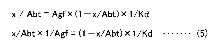
![]()
上記の方法で、作製した抗hTROP−2モノクローナル抗体300クローンのKd値を測定した結果、1×10⁻¹⁰(M)以下のKd値を示すクローンが133クローン、1×10⁻¹¹(M)以下のKd値を示すクローンが59クローン、1×10⁻¹²(M)以下のKd値を示すクローンが2クローンであった。
in vivoで腫瘍成長阻害活性を示した、抗hTROP−2モノクローナル抗体のうち、K5−70(マウスIgG2a)、T6−16(マウスIgG2a)、K5−107(マウスIgG1)、K5−116−2−1(マウスIgG2a)、及びT5−86(マウスIgG1)のKd値は、それぞれ順に、6.8×10⁻¹²(M)、4.3×10⁻¹²(M)、4.7×10⁻¹²(M)、2.69×10⁻¹¹(M)、及び8.49×10⁻¹¹(M)であった(図1、表1)。
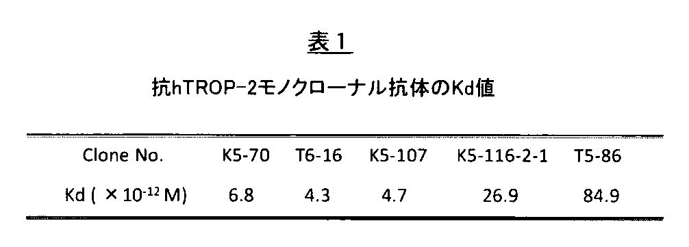
Antigen affinity (Kd value) of the prepared anti-hTROP-2 monoclonal antibody was calculated by a method using ELISA (Djava-Ohanceance L. et al (1996), In Antibody Engineering,
Specifically, the purified recombinant hTROP-2 protein (0.1 μg / mL) was added to a 96-well culture plate (Corning) to immobilize the antigen (room temperature, 1 hour, or 4 ° C., overnight) ). Next, the plate was washed 3 times with PBS, and blocked by adding 2% skim milk (PBS solution) (room temperature, 1 hour). After washing twice with PBS, an antigen solution (purified hTROP-2 protein; 50, 25, 12.5, 6.25, 3.125 nM) and each clone of anti-hTROP-2 monoclonal antibody (0.5 nM) were previously prepared. The antigen-antibody complex that had been mixed and allowed to equilibrate was added to the ELISA plate and allowed to react (room temperature, 1 hour). After washing 3 times with PBS, it was reacted with HRP-labeled anti-mouse IgG diluted in blocking solution (
The formula for calculating the dissociation constant (Kd) was as follows.
Antigen-antibody reaction is based on the law of mass action
![]()
![]()
As a result of measuring the Kd value of 300 clones of the anti-hTROP-2 monoclonal antibody prepared by the above method, 133 clones showing a Kd value of 1 × 10 −10 (M) or less were 1 × 10 −11 (M). 59 clones showing the following Kd values were 2 clones showing Kd values of 1 × 10 −12 (M) or less.
Among the anti-hTROP-2 monoclonal antibodies that showed tumor growth inhibitory activity in vivo, K5-70 (mouse IgG2a), T6-16 (mouse IgG2a), K5-107 (mouse IgG1), K5-116-2- The Kd values of 1 (mouse IgG2a) and T5-86 (mouse IgG1) were 6.8 × 10 −12 (M), 4.3 × 10 −12 (M), and 4.7 × 10 −, respectively. 12 (M), 2.69 × 10 −11 (M), and 8.49 × 10 −11 (M) (FIG. 1, Table 1).
[抗hTROP−2モノクローナル抗体のヒト癌細胞株との反応性]
ヒト癌細胞株(ヒト腫瘍細胞株)は、ヒューマンサイエンス振興事業団(HSRRB)、RIKEN cell bank(RIKEN),ATCC(American Type Culture Collection)、ECACC(European Collection of Cell Cultures)、DSMZ(German Collection of Microorganisms and Cell Cultures)より入手したものを使用した。具体的には、以下に列挙した通りである。
huH−1(HSRRB),HUH−6(HSRRB),HuH−7(HSRRB),JHH−5(HSRRB),JHH−6(HSRRB),JHH−7(HSRRB),HLE(HSRRB),HLF(HSRRB),HepG2(HSRRB),Alexander(HSRRB),KP−1N(HSRRB),KP−1NL(HSRRB),KP−2(HSRRB),KP−3(HSRRB),KP−3L(HSRRB),PK−1(RIKEN),PANC−1(RIKEN),MIA PaCa−2(HSRRB),PK−59(RIKEN),PK−45H(RIKEN),PK−45P(RIKEN),BxPC−3(ATCC),SUIT−2(HSRRB),TCC−PAN2(HSRRB),SW480(ATCC),DLD−1(HSRRB),LoVo(HSRRB),COLO−320(RIKEN),CACO−2(RIKEN),CW−2(RIKEN),HCT 116(ATCC),HCC−56(HSRRB),MCF−7(HSRRB),JIMT−1(DSMZ),HCC1143(ATCC),A549(HSRRB),DU145(RIKEN),PC−3(HSRRB)
癌細胞はトリプシン処理によって培養皿から剥がし、細胞懸濁液(細胞密度2×10⁶cells/mL)を調製した。細胞懸濁液100μLに抗hTROP−2モノクローナル抗体を加え(0.1μg)、4℃で20分間反応させた。PBSで洗浄後、PE標識抗マウスIgG(BDファーミンゲン)(0.1μg)と反応(4℃、30分)させた後、FACSCalibur(ベクトンディッキンソン)により解析した。
作製した抗hTROP−2抗体は、いずれも内在的にhTROP−2を発現していないヒト肝癌細胞株HuH−7とは結合せず、一方hTROP−2遺伝子を発現させた安定細胞株であるHuH−7−hTROP−2細胞には結合を示した(図2)。次に、作製した抗hTROP−2モノクローナル抗体のヒト癌細胞株(内在的に細胞表面に発現しているhTROP−2タンパク質)との反応性をFACS解析により検討した結果、作製した300種類の抗hTROP−2モノクローナル抗体は、全て、ヒト膵臓癌細胞株(PK−59、BxPC−3)との結合を示し、特に、in vivoで腫瘍成長阻害活性を示した、K5−70,T6−16,K5−107,K5−116−2−1,T5−86抗体は、PE標識抗マウスIgG(BDファーミンジェン)のみと反応させた場合と比較して、PK−59細胞では、平均蛍光強度でK5−70(44倍)、T6−16(59倍)、K5−107(89倍)、K5−116−2−1(122倍)、T5−86(15倍)(図3)、BxPC−3細胞では、K5−70(45倍)、T6−16(25倍)、K5−107(90倍)、K5−116−2−1(121倍)、T5−86(10倍)と、いずれの抗体も高い結合を示した(図4)。
PK−59とBxPC−3以外のヒト癌細胞株では、膵臓癌細胞株12種類のうち、KP−2,KP−3L,PK−1,PK−45H、SUIT−2、TCC−PAN2に結合し、KP−1N,KP−1NL,KP−3,PANC−1,MIA−PaCa2には結合しなかった(図5)。ヒト大腸癌細胞株では、CACO−2,SW480,DLD−1,HCT 116に結合し、COLO−320,CW−2には結合しなかった(図6)。その他、JIMT−1,HCC1143(いずれもヒト乳癌細胞株)、PC−3,DU145(いずれもヒト前立腺癌細胞株)と結合し、多くのヒト癌細胞株の細胞表面に内在的に発現しているhTROP−2タンパク質を認識した(図6)。
[Reactivity of anti-hTROP-2 monoclonal antibody with human cancer cell line]
Human cancer cell lines (human tumor cell lines) are human science promotion corporation (HSRRB), RIKEN cell bank (RIKEN), ATCC (American Type Culture Collection), ECACC (European Collection of Cell Cultures), DSMZG What was obtained from Microorganisms and Cell Cultures) was used. Specifically, it is as listed below.
huH-1 (HSRRB), HUH-6 (HSRRB), HuH-7 (HSRRB), JHH-5 (HSRRB), JHH-6 (HSRRB), JHH-7 (HSRRB), HLE (HSRRB), HLF (HSRRB) ), HepG2 (HSRRB), Alexander (HSRRB), KP-1N (HSRRB), KP-1NL (HSRRB), KP-2 (HSRRB), KP-3 (HSRRB), KP-3L (HSRRB), PK-1 (RIKEN), PANC-1 (RIKEN), MIA PaCa-2 (HSRRB), PK-59 (RIKEN), PK-45H (RIKEN), PK-45P (RIKEN), BxPC-3 (ATCC), SUIT-2 (HSRRB), TCC-PAN2 (HSRRB), SW480 (ATCC), DLD-1 (HSRRB), LoVo (HSRRB), COLO-320 (RIKEN), CACO-2 (RIKEN), CW-2 (RIKEN), HCT 116 (ATCC), HCC-56 (HSRRB), MCF-7 ( HSRRB), JIMT-1 (DSMZ), HCC1143 (ATCC), A549 (HSRRB), DU145 (RIKEN), PC-3 (HSRRB)
Cancer cells were detached from the culture dish by trypsin treatment to prepare a cell suspension (
None of the prepared anti-hTROP-2 antibodies binds to the human hepatoma cell line HuH-7 that does not express hTROP-2 endogenously, whereas HuH is a stable cell line that expresses the hTROP-2 gene. -7-hTROP-2 cells showed binding (FIG. 2). Next, as a result of examining the reactivity of the prepared anti-hTROP-2 monoclonal antibody with a human cancer cell line (hTROP-2 protein endogenously expressed on the cell surface) by FACS analysis, 300 kinds of prepared anti-hTROP-2 monoclonal antibodies were prepared. The hTROP-2 monoclonal antibodies all showed binding to human pancreatic cancer cell lines (PK-59, BxPC-3), and in particular, K5-70, T6-16, which showed tumor growth inhibitory activity in vivo. The K5-107, K5-116-2-1, and T5-86 antibodies have a higher average fluorescence intensity in PK-59 cells than when reacted with PE-labeled anti-mouse IgG (BD Farmingen) alone. K5-70 (44 times), T6-16 (59 times), K5-107 (89 times), K5-116-2-1 (122 times), T5-86 (15 times) (FIG. 3), BxPC- In 3 cells, K5-70 (45 times), T6-16 (25 times), K5-107 (90 times), K5-116-2-1 (121 times), T5-86 (10 times), Also showed high binding (FIG. 4).
Human cancer cell lines other than PK-59 and BxPC-3 bind to KP-2, KP-3L, PK-1, PK-45H, SUIT-2, TCC-PAN2 among 12 types of pancreatic cancer cell lines. , KP-1N, KP-1NL, KP-3, PANC-1, and MIA-PaCa2 did not bind (FIG. 5). In human colon cancer cell lines, it bound to CACO-2, SW480, DLD-1, and HCT116, but not to COLO-320 and CW-2 (FIG. 6). In addition, it binds to JIMT-1, HCC1143 (all are human breast cancer cell lines), PC-3, DU145 (all are human prostate cancer cell lines), and is endogenously expressed on the surface of many human cancer cell lines. HTROP-2 protein was recognized (FIG. 6).
[マウスTROP−2タンパク質、ヒトTROP−1/EpCAMタンパク質との交差反応性]
作製した抗hTROP−2モノクローナル抗体の特異性を調べる目的で、hTROP−2タンパク質とアミノ酸配列で80%の相同性を示すマウスTROP−2タンパク質、hTROP−2タンパク質とアミノ酸配列で50%の相同性を示すヒトTROP−1/EpCAMとの反応性をFACS解析にて行った。
具体的には、マウスTROP−2遺伝子(GenBank accession No.NM_020047、Y08830)の全長cDNAを含む発現ベクター(マウスTROP−2−pcDNA3.1(+)、東京大学・分子細胞生物学研究所より供与)、およびヒトTROP−1/EpCAM遺伝子(GenBank accession No.NM_002354)の全長cDNAを含む発現ベクター(pcDNA3.1−hEpCAM−myc/His)を、それぞれLipofectamine2000試薬(Invitrogen)を用いて、一過性にCHO−K1細胞に遺伝子導入を行い、24~48時間後に細胞をトリプシン処理で培養皿から剥がして細胞懸濁液を調整し、作製した抗hTROP−2モノクローナル抗体(0.1μg)、PE標識抗マウスIgGと順次反応させた後、FACSCaliburにより解析した。
マウスTROP−2遺伝子を一過性に発現させたCHO−K1細胞において、陽性対照として用いたマウスTROP−2と交差反応を示すT2−102抗体(マウスIgG1)は強い結合を示し、一方、K5−70,T6−16,K5−107,K5−116−2−1,T5−86抗体はマウスTROP−2との交差反応は示さなかった(図7)。
同様に、ヒトEpCAM/TROP−1遺伝子を一過性に発現させたCHO−K1細胞において、陽性対照として用いた抗ヒトEpCAMモノクローナル抗体(BDファーミンジェン)は強い結合を示し、一方、K5−70,T6−16,K5−107,K5−116−2−1,T5−86抗体はヒトEpCAM/TROP−1との交差反応は示さなかった(図8)。
以上の結果は、作製した抗hTROP−2モノクローナル抗体、特にin vivoで抗腫瘍活性を示したK5−70,T6−16,K5−107,K5−116−2−1,T5−86抗体は、hTROP−2に特異的に結合することが示された。
[Cross-reactivity with mouse TROP-2 protein and human TROP-1 / EpCAM protein]
For the purpose of investigating the specificity of the prepared anti-hTROP-2 monoclonal antibody, mouse TROP-2 protein showing 80% homology in amino acid sequence with hTROP-2 protein and 50% homology in amino acid sequence with hTROP-2 protein The reactivity with human TROP-1 / EpCAM indicating the above was performed by FACS analysis.
Specifically, an expression vector (mouse TROP-2-pcDNA3.1 (+) containing the full-length cDNA of mouse TROP-2 gene (GenBank accession No. NM — 020047, Y08830), provided by the Institute for Molecular Cell Biology, University of Tokyo ) And an expression vector (pcDNA3.1-hEpCAM-myc / His) containing a full-length cDNA of human TROP-1 / EpCAM gene (GenBank accession No. NM_002354), respectively, using
In CHO-K1 cells in which the mouse TROP-2 gene was transiently expressed, the T2-102 antibody (mouse IgG1) that cross-reacts with mouse TROP-2 used as a positive control showed strong binding, while K5 The -70, T6-16, K5-107, K5-116-2-1 and T5-86 antibodies did not show cross-reactivity with mouse TROP-2 (FIG. 7).
Similarly, in CHO-K1 cells in which the human EpCAM / TROP-1 gene was transiently expressed, the anti-human EpCAM monoclonal antibody (BD Farmingen) used as a positive control showed strong binding, while K5-
The above results show that the prepared anti-hTROP-2 monoclonal antibodies, particularly the K5-70, T6-16, K5-107, K5-116-2-1, and T5-86 antibodies that showed antitumor activity in vivo, It was shown to bind specifically to hTROP-2.
[細胞増殖阻害活性の測定]
抗hTROP−2モノクローナル抗体のhTROP−2の機能阻害を調べる方法の1つとして、内在的にhTROP−2を細胞表面に発現しているヒト癌細胞の細胞増殖に対する影響を、テトラカラーワン(生化学工業)を用いた生細胞数の測定に基づいて評価した。具体的には、PK−59細胞を0.5%のウシ胎児血清(BioWest社製)を含むRPMI1640培地に2×10⁵細胞/mLの細胞濃度となるように細胞懸濁液を調製し、96穴培養プレートに100μLずつ播種した。次いで、マウスIgG(陰性コントロール)、抗hTROP−2モノクローナル抗体(最終濃度0.1,1μg/mL)を添加し、37℃、5% CO2インキュベーターで72時間培養した。比較対照として、市販の抗hTROP−2モノクローナル抗体(クローン YY01,Santa Cruz)を使用した。細胞にテトラカラーワン(生化学工業)を添加し、5% CO2インキュベーターで1~2時間反応させた。反応後の96穴プレートを、そのままマイクロプレートリーダーを用いて波長490nm(対照波長:655nm)の吸光度を測定した。各群3ウェルで実施し、Student’s t検定(Student’s t−test)で有意差検定を行い、P<0.05を統計的に有意と判定した。
これまでに自社で作製した抗hTROP−2モノクローナル抗体のうち、約160クローンについて、上記の方法でPK−59細胞の細胞増殖に与える効果を調べたところ、in vivoで腫瘍成長阻害活性を示した、T6−16,T5−86,K5−70,K5−107は、マウスIgG(陰性コントロール)と比較して、20%~40%の細胞増殖阻害活性が確認され、これら抗hTROP−2抗体は、ヒト癌細胞表面に発現するhTROP−2タンパク質に結合し、hTROP−2タンパク質の機能を中和し、癌細胞の増殖を阻害する活性を有することが明らかとなった(図9)。
[Measurement of cell growth inhibitory activity]
As one of the methods for examining hTROP-2 function inhibition of an anti-hTROP-2 monoclonal antibody, the effect on the cell proliferation of a human cancer cell that endogenously expresses hTROP-2 on the cell surface was examined using tetracolor one (live Evaluation was performed based on the measurement of the number of living cells using Chemical Industries. Specifically, a cell suspension of PK-59 cells was prepared in RPMI 1640 medium containing 0.5% fetal bovine serum (BioWest) to a cell concentration of 2 × 10 5 cells / mL, 100 μL each was seeded in a 96-well culture plate. Subsequently, mouse IgG (negative control) and anti-hTROP-2 monoclonal antibody (final concentration 0.1, 1 μg / mL) were added, and cultured at 37 ° C. in a 5% CO 2 incubator for 72 hours. As a comparative control, a commercially available anti-hTROP-2 monoclonal antibody (clone YY01, Santa Cruz) was used. Tetracolor One (Seikagaku Corporation) was added to the cells and reacted in a 5% CO 2 incubator for 1-2 hours. After the reaction, the absorbance at a wavelength of 490 nm (control wavelength: 655 nm) was directly measured using a microplate reader. The test was carried out in 3 wells in each group, and a significant difference test was performed by Student's t-test, and P <0.05 was determined to be statistically significant.
About 160 clones of anti-hTROP-2 monoclonal antibodies prepared in-house so far were examined for the effect on cell proliferation of PK-59 cells by the above-mentioned method, and showed tumor growth inhibitory activity in vivo. , T6-16, T5-86, K5-70, and K5-107 have been confirmed to have a cell growth inhibitory activity of 20% to 40% compared to mouse IgG (negative control), and these anti-hTROP-2 antibodies It was revealed that it has an activity of binding to hTROP-2 protein expressed on the surface of human cancer cells, neutralizing the function of hTROP-2 protein, and inhibiting the growth of cancer cells (FIG. 9).
[スクラッチアッセイ]
抗hTROP−2モノクローナル抗体のヒト癌細胞の移動能をスクラッチアッセイで評価した。PK−59細胞を10%のウシ胎児血清を含むRPMI1640培地に3×10⁵cell/mLの細胞濃度となるように細胞懸濁液を調製し、96穴培養プレートに100μLずつ播種した。細胞がコンフルエントに達した段階で、チップの先端で縦に線を引くようにして単層培養した細胞の一部を剥離した。抗hTROP−2モノクローナル抗体、および陰性コントロールとしてマウスIgGを最終濃度が0.1、および1μg/mLとなるように培地に添加し、24時間培養を行い、抗体添加前(Day0)と24時間培養後(Day1)の剥離した領域の写真を撮影し、細胞間距離を測定した。また、剥離領域の面積をScion Imageソフトウェアで定量化した。各群8ウェルで実施し、Student’s t−testで有意差検定を行い、P<0.05を統計的に有意と判定した。
スクラッチ領域に浸潤する細胞の運動能に対するhTROP−2抗体の効果を調べた。細胞増殖阻害アッセイと同様に薬効のあった抗体を評価した。評価方法として、Day0(抗体添加時)およびDay1(24時間経過後)の細胞を撮影し、移動距離(μm)および、スクラッチ領域の面積を画像解析した。結果、図10に示すように、細胞の運動能に明らかな違いが見られた。本試験に使用したT6−16およびK5−70はコントロールと比較し、どちらも有意な増殖阻害が見られ、再現性試験においても同様の傾向が確認できた。特にT6−16についてはP<0.01(by Student’s t−test)であり、in vivoの試験との相関性が認められた。
[Scratch assay]
The ability of anti-hTROP-2 monoclonal antibody to migrate human cancer cells was evaluated by a scratch assay. A cell suspension was prepared from PK-59 cells in RPMI 1640 medium containing 10% fetal bovine serum so that the cell concentration was 3 × 10 5 cells / mL, and 100 μL was seeded in a 96-well culture plate. When the cells reached confluence, a part of the monolayer cultured cells was detached by drawing a vertical line at the tip of the chip. Anti-hTROP-2 monoclonal antibody and mouse IgG as a negative control were added to the culture medium so that the final concentration was 0.1 and 1 μg / mL, and cultured for 24 hours, before antibody addition (Day 0) and for 24 hours. A photograph of the peeled area after (Day 1) was taken, and the intercellular distance was measured. Also, the area of the peeled area was quantified with Scion Image software. The test was performed in 8 wells in each group, and a significant difference test was performed by Student's t-test, and P <0.05 was determined to be statistically significant.
The effect of hTROP-2 antibody on the motility of cells infiltrating the scratch region was examined. The medicinal antibodies were evaluated as in the cell growth inhibition assay. As an evaluation method, cells of Day 0 (when antibody was added) and Day 1 (after 24 hours) were photographed, and the moving distance (μm) and the area of the scratch region were subjected to image analysis. As a result, as shown in FIG. 10, a clear difference was observed in the motility of the cells. T6-16 and K5-70 used in this test both showed significant growth inhibition compared to the control, and the same tendency could be confirmed in the reproducibility test. Especially for T6-16, P <0.01 (by Student's t-test), and a correlation with the in vivo test was confirmed.
[担癌マウスでの抗hTROP−2モノクローナル抗体の薬効評価]
preventionモデル
hTROP−2を発現する膵癌細胞株(PK−59,BxPC−3)をトリプシン処理で剥がし、PBSで1×10⁸cells/mLの細胞懸濁液を調製し、氷上にて等量のマトリゲル(BDファーミンゲン)と混合した。6週齢の雌のヌードマウス(Balb/c,nu/nu)の右脇腹の皮下に26Gのシリンジを用いて100μL(5×106cell)注射した。癌細胞移植当日(Day1)に、マウスを群分し、抗体の投与(1,5,または10mg/kg体重、腹腔内投与)を開始した。以降は、3日に1回の間隔で同様の投与を行った。抗腫瘍活性は、腫瘍形成頻度及び腫瘍体積により評価した。腫瘍体積の計測は、以下の計算式を用いた。
腫瘍体積(mm3)=(短径)2×(長径)×π/6
treatmentモデル
hTROP−2を発現する膵癌細胞株(PK−59,BxPC−3)をトリプシン処理で剥がし、PBSで1×10⁸cells/mLの細胞懸濁液を調製し、氷上にて等量のマトリゲル(BDファーミンゲン)と混合した。6週齢の雌のヌードマウス(Balb/c,nu/nu)の右脇腹の皮下に26Gのシリンジを用いて100μL(5×106cells)注射した。癌細胞移植から5~6日後、腫瘍体積が50~150mm3(平均値で約100mm3)に成長したマウスを群分し、群分した当日を1日目(Day1)として、抗体投与を開始した。抗体は3日に1回の間隔で腹腔内投与を行った(10mg/kg体重)。抗腫瘍活性は、腫瘍体積を計測することによって評価した。有意差検定は、Student’s t−testで行い、P<0.05を統計的に有意と判定した。
[Evaluation of efficacy of anti-hTROP-2 monoclonal antibody in tumor-bearing mice]
prevention model A pancreatic cancer cell line (PK-59, BxPC-3) expressing hTROP-2 was detached by trypsin treatment, and a cell suspension of 1 × 10 8 cells / mL was prepared with PBS. Mixed with Matrigel (BD Farmingen). Sixteen-week-old female nude mice (Balb / c, nu / nu) were injected subcutaneously in the right flank with 100 μL (5 × 10 6 cells) using a 26 G syringe. On the day of cancer cell transplantation (Day 1), mice were divided into groups, and antibody administration (1, 5, or 10 mg / kg body weight, intraperitoneal administration) was started. Thereafter, the same administration was performed at an interval of once every 3 days. Antitumor activity was evaluated by tumor formation frequency and tumor volume. The following calculation formula was used to measure the tumor volume.
Tumor volume (mm 3 ) = (minor axis) 2 × (major axis) × π / 6
treatment model A pancreatic cancer cell line (PK-59, BxPC-3) expressing hTROP-2 was removed by trypsin treatment, and a cell suspension of 1 × 10 8 cells / mL was prepared with PBS. Mixed with Matrigel (BD Farmingen). Sixteen week old female nude mice (Balb / c, nu / nu) were injected subcutaneously in the right flank with 100 μL (5 × 10 6 cells) using a 26 G syringe. Five to six days after cancer cell transplantation, mice that have grown to a tumor volume of 50 to 150 mm 3 (average value of about 100 mm 3 ) are divided into groups. did. The antibody was administered intraperitoneally at an interval of once every 3 days (10 mg / kg body weight). Antitumor activity was assessed by measuring tumor volume. The significant difference test was performed by Student's t-test, and P <0.05 was determined to be statistically significant.
[ヒト膵癌細胞xenograftモデルにおける抗hTROP−2モノクローナル抗体のin vivoにおける抗腫瘍活性の検討]
hTROP−2を標的とした癌治療用抗体は、xenograft treatmentモデルにおいて、hTROP−2を発現する腫瘍組織を特異的に死滅させるか、腫瘍の成長を阻害する活性を示すことが必須である。
本発明において新たに作製した抗hTROP−2モノクローナル抗体(約160クローン)を、膵癌細胞株PK−59細胞のxenograft treatmentモデルで評価した。PK−59細胞は、膵臓癌幹細胞マーカーであるEpCAMを発現し(図11A)(Chenwei Li,et al.Cancer Res 2007;67:(3).1030−1037),薬剤耐性に関わるABC トランスポーターであるP−glycoprotein/MDR1(図11B),ABCG2/CDw338(図11C)を細胞表面に発現している(Chen,C.J.et al.Cell 47(3),381−389(1986),Allikmets,R.,et al.Hum.Mol.Genet.5(10),1649−1655(1996)。また、膵臓癌幹細胞の特徴であるCD24とCD44共陽性の細胞画分(8.93%)(図11D)を含み、悪性度の高いヒト膵臓癌細胞株であると推測される(Chenwei Li,et al.Cancer Res 2007;67:(3).1030−1037、Jane E.Visvader and Geoffrey J.Lindeman.Nat Rev Cancer.Vol.8(10):755−68,2008)。
新規に作製した約160クローンのうち、大部分のクローンはPK−59細胞xenograft treatmentモデルでは薬効を示さなかったが、その中でも有意な腫瘍成長阻害活性を示す以下のクローン、すなわちクローンK5−70,T6−16,K5−107,T5−86及びK5−116−2−1が得られた。
クローンK5−70(マウスIgG2a)投与群では、腫瘍成長速度が統計的に有意に阻害され、投与開始後21日目(Day21)において、コントロール群(N=14)の腫瘍体積が1200.8±377.3mm3に対し、クローンK5−70投与群では748.7±175.0mm3(P<0.01 by Student’s t−test)であった(図12A)。抗体投与開始時点での腫瘍体積を1.0としたときの、21日目(Day21)における腫瘍体積は、コントロール群が12.5であるのに対し、クローンK5−70投与群では7.8であった(図12A)。摘出した腫瘍重量も、コントロール群が0.73±0.26gであったのに対し、クローンK5−70投与群では0.43±0.14g(P<0.01 by Student’s t−test)であり、約60%の阻害を示した(図12B)。
同様に、クローンK5−107(マウスIgG1)投与群(N=8)、クローンT6−16(マウスIgG2a)投与群(N=8)、クローンT5−86(マウスIgG1)及びクローンK5−116−2−1(マウスIgG2a)投与群(N=8)でも、腫瘍成長速度が統計的に有意に阻害された。クローンK5−107投与群(N=8)並びにクローンT6−16投与群(N=8)では、投与開始後17日目(Day17)において、コントロール群(N=8)の腫瘍体積が1039.3±271.6mm3に対し、それぞれ698.2±175.9mm3(P<0.05 by Student’s t−test)、707.2±254.5mm3(P<0.05 by Student’s t−test)であった。同様に、クローンK5−116−2−1投与群(N=8)では、投与開始後16日目(Day16)において、コントロール群(N=8)の腫瘍体積が797.0±172.9mm3に対し、508.5±225.2mm3(P<0.05 by Student’s t−test)であった(図13)。
一方、クローンT5−86については、投与開始後15日目(Day15)において、コントロール群(N=8)の腫瘍体積が1033.2±319.4mm3に対し、クローンT5−86投与群(N=8)では744.1±289.1mm3であり、腫瘍体積の比較では有意差はつかなかったものの、同日における腫瘍重量の比較では、コントロール群が0.62±0.14gであったのに対し、クローンT5−86投与群では0.44±0.13g(P<0.05 by Student’s t−test)であり有意な阻害が示された。
また、下記表2に、各クローン抗体投与群の実験最終日におけるコントロール群との比率(T/C)を、腫瘍体積および腫瘍重量のそれぞれについて示した。表2に示した通り、各クローン抗体投与群において、それぞれT/C=62~72%の有意な阻害が得られた。

下記表3に、各クローン抗体投与群の、実験最終日におけるコントロール群との比率(T/C)を、腫瘍体積および腫瘍重量それぞれについて示した。表3に示したとおり、各クローン抗体投与群において、有意な腫瘍成長阻害が見られ、特にクローンK5−70投与群では、T/C=10%以下の顕著な効果が確認された。
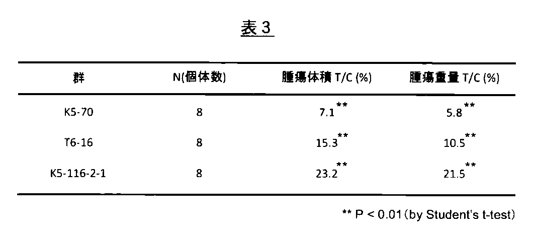
It is essential that an antibody for cancer treatment targeting hTROP-2 exhibits an activity of specifically killing a tumor tissue expressing hTROP-2 or inhibiting tumor growth in a xenograft treatment model.
A newly prepared anti-hTROP-2 monoclonal antibody (about 160 clones) in the present invention was evaluated by a xenograft treatment model of pancreatic cancer cell line PK-59 cells. PK-59 cells express EpCAM, a pancreatic cancer stem cell marker (FIG. 11A) (Chenwei Li, et al. Cancer Res 2007; 67: (3). 1030-1037), and are ABC transporters involved in drug resistance. Certain P-glycoprotein / MDR1 (FIG. 11B) and ABCG2 / CDw338 (FIG. 11C) are expressed on the cell surface (Chen, CJ et al. Cell 47 (3), 381-389 (1986), Allikmets. , R., et al.Hum.Mol.Genet.5 (10), 1649-1655 (1996) In addition, CD24 and CD44 co-positive cell fraction (8.93%), which is characteristic of pancreatic cancer stem cells ( 11D) and is presumed to be a high-grade human pancreatic cancer cell line (C enwei Li, et al.Cancer Res 2007; 67: (3) .1030-1037, Jane E.Visvader and Geoffrey J.Lindeman.Nat Rev Cancer.Vol.8 (10): 755-68,2008).
Of the approximately 160 newly created clones, most of the clones did not show efficacy in the PK-59 cell xenograft treatment model, but among them the following clones showing significant tumor growth inhibitory activity, namely clones K5-70, T6-16, K5-107, T5-86 and K5-116-2-1 were obtained.
In the clone K5-70 (mouse IgG2a) administration group, the tumor growth rate was statistically significantly inhibited. On the 21st day (Day 21) after the start of administration, the tumor volume of the control group (N = 14) was 1200.8 ± In contrast to 377.3 mm 3 , it was 748.7 ± 175.0 mm 3 (P <0.01 by Student's t-test) in the clone K5-70 administration group (FIG. 12A). When the tumor volume at the start of antibody administration is 1.0, the tumor volume on Day 21 (Day 21) is 12.5 in the control group, whereas 7.8 in the clone K5-70 administration group. (FIG. 12A). The excised tumor weight was also 0.73 ± 0.26 g in the control group, whereas 0.43 ± 0.14 g (P <0.01 by Student's t-test) in the clone K5-70 administration group. ) And showed about 60% inhibition (FIG. 12B).
Similarly, clone K5-107 (mouse IgG1) administration group (N = 8), clone T6-16 (mouse IgG2a) administration group (N = 8), clone T5-86 (mouse IgG1) and clone K5-16-2 In the -1 (mouse IgG2a) administration group (N = 8), the tumor growth rate was statistically significantly inhibited. In the clone K5-107 administration group (N = 8) and the clone T6-16 administration group (N = 8), the tumor volume of the control group (N = 8) was 1039.3 on the 17th day (Day 17) after the start of administration. For ± 271.6 mm 3 , 698.2 ± 175.9 mm 3 (P <0.05 by Student's t-test) and 707.2 ± 254.5 mm 3 (P <0.05 by Student's, respectively) t-test). Similarly, in the clone K5-116-2-1 administration group (N = 8), the tumor volume of the control group (N = 8) was 797.0 ± 172.9 mm 3 on the 16th day after the start of administration (Day 16). On the other hand, it was 508.5 ± 225.2 mm 3 (P <0.05 by Student's t-test) (FIG. 13).
On the other hand, for the clone T5-86, on the 15th day after the start of administration (Day 15), the tumor volume of the control group (N = 8) was 1033.2 ± 319.4 mm 3 while the clone T5-86 administration group (N = 8) was 744.1 ± 289.1 mm 3 and there was no significant difference in the tumor volume comparison, but in the tumor weight comparison on the same day, the control group was 0.62 ± 0.14 g In contrast, in the clone T5-86 administration group, 0.44 ± 0.13 g (P <0.05 by Student's t-test), indicating significant inhibition.
Table 2 below shows the ratio (T / C) of each cloned antibody administration group to the control group on the last day of the experiment for each of the tumor volume and tumor weight. As shown in Table 2, significant inhibition of T / C = 62 to 72% was obtained in each clone antibody administration group.
Table 3 below shows the ratio (T / C) of each clone antibody administration group to the control group on the last day of the experiment for each tumor volume and tumor weight. As shown in Table 3, significant tumor growth inhibition was observed in each clone antibody administration group, and in particular, in the clone K5-70 administration group, a remarkable effect of T / C = 10% or less was confirmed.
[ヒト膵癌細胞株BxPC−3細胞のxenograftモデル(preventionモデルおよびtreatmentモデル)における抗腫瘍活性の検討]
上記のヒト膵癌細胞株PK−59細胞のxenograft treatmentモデルを用いた場合と同様に、ヒト膵癌細胞株BxPC−3細胞のxenograft preventionモデル及びxenograft treatmentモデルでの抗腫瘍活性を、クローンK5−70について検討した。
クローンK5−70投与群(N=8)では、コントロール群(N=8)と比較して、腫瘍成長が有意に阻害され、52日目(Day52)において、コントロール群(N=8)の腫瘍体積が616.3±266.8mm3であったのに対し、クローンK5−70投与群(N=8)では236.0±136.4mm3と61.7%の腫瘍成長阻害効果を示した(P<0.01 by Student’s t−test)(図15)。
以上の結果より、抗hTROP−2モノクローナル抗体は、少なくとも2つ以上の複数の癌細胞種に対して、in vivoでの有意な腫瘍成長阻害活性を示すことが明らかとなった。
[Study of antitumor activity in human pancreatic cancer cell line BxPC-3 cells in xenograft model (prevention model and treatment model)]
Similar to the case of using the xenograft treatment model of the human pancreatic cancer cell line PK-59 cell described above, the antitumor activity of the human pancreatic cancer cell line BxPC-3 cell in the xenograft prevention model and the xenograph treatment model is shown for clone K5-70. investigated.
In the clone K5-70 administration group (N = 8), tumor growth was significantly inhibited as compared with the control group (N = 8). On day 52 (Day 52), tumors in the control group (N = 8) While the volume was 616.3 ± 266.8 mm 3 , the clone K5-70 administration group (N = 8) showed a tumor growth inhibitory effect of 236.0 ± 136.4 mm 3 and 61.7%. (P <0.01 by Student's t-test) (FIG. 15).
From the above results, it was revealed that the anti-hTROP-2 monoclonal antibody exhibits significant tumor growth inhibitory activity in vivo against at least two or more cancer cell types.
[抗hTROP2抗体(クローンK5−70)の、hTROP2発現膵癌細胞株(PK−59細胞)xenograft preventionモデルにおける用量依存的な抗腫瘍活性]
抗hTROP−2抗体のin vivoにおける腫瘍成長阻害活性を、より詳細に検討する目的で用量依存性試験を行った。図16に示したとおり、PK−59細胞の腫瘍の成長はK5−70抗体の投与によって用量依存的に阻害され、抗体投与後21日目(Day21)において、コントロール群(N=8の腫瘍体積が937.8±295.3mm3であったのに対し、K5−70抗体(1mg/kg体重)投与群(N=8)が493.5±305.1mm3と50%の阻害率を示し、K5−70抗体(5mg/kg体重)投与群(N=8)が124.7±89.0mm3と90%の阻害効果を示し、公知の抗TROP−2抗体AR47A6.4.2(米国特許第7420041号)と比較して、20分の1の投与量でほぼ同等のin vivoにおける腫瘍成長阻害効果を示し、4分の1の投与量では、より高い90%の阻害効果を示すことが明らかになった。
[Dose-dependent antitumor activity of anti-hTROP2 antibody (clone K5-70) in an hTROP2-expressing pancreatic cancer cell line (PK-59 cell) xenograft prevention model]
In order to examine the tumor growth inhibitory activity of anti-hTROP-2 antibody in vivo in more detail, a dose-dependent test was conducted. As shown in FIG. 16, tumor growth of PK-59 cells was inhibited in a dose-dependent manner by administration of the K5-70 antibody, and on the 21st day after the antibody administration (Day 21), the control group (N = 8 tumor volume) Was 537.8 ± 295.3 mm 3 whereas the K5-70 antibody (1 mg / kg body weight) administration group (N = 8) showed an inhibition rate of 493.5 ± 305.1 mm 3 and 50%. , K5-70 antibody (5 mg / kg body weight) administration group (N = 8) showed an inhibitory effect of 124.7 ± 89.0 mm 3 and 90%, known anti-TROP-2 antibody AR47A 6.4.2 (USA) Compared with Japanese Patent No. 7420041), the tumor growth inhibitory effect in vivo is almost the same at 1/20 dose, and the higher 90% inhibitory effect is shown at 1/4 dose. Became clear.
[エピトープアッセイ]
ヒト/マウスキメラTROP−2タンパク質の作製
ヒト/マウスキメラTROP−2遺伝子はPCR法を用いて作製した。ヒトTROP−2遺伝子配列およびマウスTROP−2遺伝子配列(Genbank accession No.NM_020047)をもとに以下に示すPCRプライマーを設計した。
ヒト/マウスTROP−2−Cプライマー
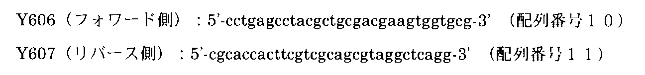


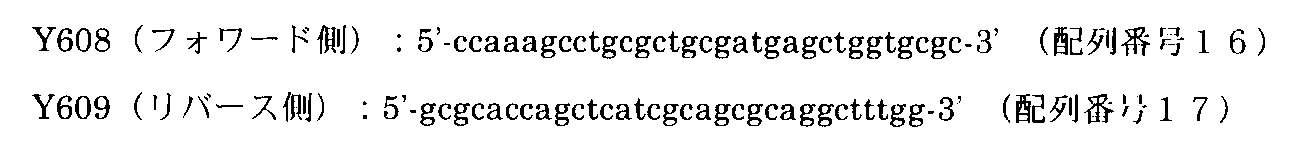
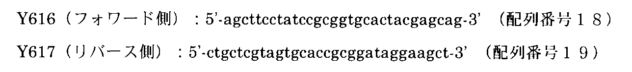

hmTROP−2−Aキメラタンパク質は、hTROP−2タンパク質のN末端から69番目までのポリペプチドとマウスTROP−2タンパク質の64番目からC末端までのポリペプチドから成るキメラタンパク質である。hmTROP2−Bキメラタンパク質は、hTROP−2タンパク質のN末端から101番目までのポリペプチドとマウスTROP−2タンパク質の96番目からC末端までのポリペプチドから成るキメラタンパク質である。hmTROP2−Cキメラタンパク質は、hTROP−2タンパク質のN末端から145番目までのポリペプチドとマウスTROP−2タンパク質の140番目からC末端までのポリペプチドから成るキメラタンパク質である。mhTROP−2−Dキメラタンパク質は、マウスTROP−2タンパク質のN末端から139番目までのポリペプチドとhTROP−2タンパク質の146番目からC末端までのポリペプチドから成るキメラタンパク質である。mhTROP−2−Eキメラタンパク質は、マウスTROP−2タンパク質のN末端から187番目までのポリペプチドとhTROP−2タンパク質の194番目からC末端までのポリペプチドから成るキメラタンパク質である。mhTROP−2−Fキメラタンパク質は、マウスTROP−2タンパク質のN末端から227番目までのポリペプチドとhTROP−2タンパク質の234番目からC末端までのポリペプチドから成るキメラタンパク質である。
上記キメラタンパク質を作製するための発現ベクターは、具体的には以下の方法で構築した。hmTROP−2−Aキメラ遺伝子を作製するためhTROP−2遺伝子を鋳型にして、hTROP−2フォワード側プライマー及びヒト/マウスTROP−2−AプライマーY613を用いてPCRを行った。同様にマウスTROP−2遺伝子を鋳型にして、ヒト/マウスTROP−2−AプライマーY612及びマウスTROP−2リバース側プライマーを用いてPCRを行った。PCRにより増幅したDNA断片はアクリルアミドゲルを用いて展開、目的バンドの抽出により回収した。次に抽出した2種類のDNA断片を混合して鋳型とし、hTROP−2フォワード側プライマー及びマウスTROP−2リバース側プライマーを用いてPCRを行った。PCR産物はアガロースゲル電気泳動により展開し、目的のDNA断片を抽出した。抽出したDNA断片はpCR(登録商標)−Bluntベクター(Invitrogen)にクローニングし(pCRB−hmTROP−2−A)、遺伝子配列を確認した。動物細胞用発現ベクターは、pcDNA3.1−hTROP−2−myc/HisからEco RI/Xho I消化によりhTROP−2遺伝子を除き、pCRB−hmTROP−2−Aから調製したhmTROP−2−Aキメラ遺伝子を含むEco RI/Xho I断片を挿入することで作製した(pcDNA3.1−hmTROP−2−A−myc/His)。その他hmTROP−2−B(ヒトTROP−2フォワード側プライマー、ヒト/マウスTROP−2−BプライマーY615、ヒト/マウスTROP−2−BプライマーY614及びマウスTROP−2リバース側プライマーを使用)、hmTROP−2−C(ヒトTROP−2フォワード側プライマー、ヒト/マウスTROP−2−CプライマーY607及びヒト/マウスTROP−2−CプライマーY606、マウスTROP−2リバース側プライマーを使用)、mhTROP−2−D(マウスTROP−2フォワード側プライマー、マウス/ヒトTROP−2−DプライマーY609及びマウス/ヒトTROP−2−DプライマーY608、ヒトTROP−2リバース側プライマーを使用)、mhTROP−2−E(マウスTROP−2フォワード側プライマー、マウス/ヒトTROP−2−EプライマーY617及びマウス/ヒトTROP−2−EプライマーY616、ヒトTROP−2リバース側プライマーを使用)、mhTROP−2−F(マウスTROP−2フォワード側プライマー、マウス/ヒトTROP−2−FプライマーY619及びマウス/ヒトTROP−2−FプライマーY618、ヒトTROP−2リバース側プライマーを使用)についても同様の方法でキメラ遺伝子を作製し、発現ベクターを構築した(pcDNA3.1−hmTROP−2−B−myc/His、pcDNA3.1−hmTROP−2−C−myc/His、pcDNA3.1−mhTROP−2−D−myc/His、pcDNA3.1−mhTROP−2−E−myc/His、pcDNA3.1−mhTROP−2−F−myc/His)。
hTROP−2、ヒト/マウスキメラTROP−2−C、及びマウス/ヒトキメラTROP−2−Dタンパク質を恒常的に発現するHEK293細胞株の樹立
HEK293細胞株に上記発現ベクターpcDNA3.1−hTROP−2−myc/His、pcDNA3.1−hmTROP−2−C−myc/His及びpcDNA3.1−mhTROP−2−D−myc/Hisをそれぞれ遺伝子導入した。抗生物質G418(Calbiochem)による選択を行い、hTROP−2タンパク質、hmTROP−2−Cキメラタンパク質およびmhTROP−2−Dキメラタンパク質を恒常的に発現するHEK293細胞株を樹立した。
膵臓癌細胞株PK−59を用いたxenograft treatmentモデルにおいて、薬効を示した抗hTROP−2モノクローナル抗体K5−70、T5−86、K5−107、T6−4、T6−16およびK5−116−2−1について結合領域の同定を行った。まずhmTROP−2−C、mhTROP−2−D、それぞれのキメラタンパク質を恒常的に発現するHEK293細胞を用いて、薬効を示した抗hTROP−2モノクローナル抗体の反応性をFACS解析により検討した(図18)。その結果、K5−70、K5−107、T5−86およびK5−116−2−1抗体はhmTROP−2−Cと反応したがmhTROP−2−Dとは反応しなかった。一方、T6−4およびT6−16抗体はmhTROP−2−Dと反応したがhmTROP−2−Cとは反応しなかった。これらのことからK5−70、K5−107、T5−84およびK5−116−2−1抗体の結合領域はhTROP−2のN末端から145番目のアミノ酸までの領域、T6−4およびT6−16抗体の結合領域はhTROP−2の146番目のアミノ酸からC末端までの領域に限定された(図18)。
さらに詳細に結合領域の解析を行うため、hmTROP−2−A、hmTROP−2−B、mhTROP−2−E及びmhTROP−2−Fキメラタンパク質発現用ベクターを作製し、薬効を示す抗hTROP−2モノクローナル抗体との反応性を検討した(図19)。新たに作製したキメラタンパク発現用ベクターをHEK293細胞に遺伝子導入し、一過性に発現させた細胞を用いてFACS解析を行った。K5−70、K5−107、T5−86およびK5−116−2−1抗体はhmTROP−2−Aとは反応したがmhTROP−2−Bとは反応しなかった。調べた6種類のモノクローナル抗体はすべてhTROP−2とは反応を示した。このことからK5−70、K5−107、T5−86およびK5−116−2−1抗体の結合領域はhTROP−2のN末端から69番目のアミノ酸までの領域に存在することが明らかとなった。またT6−4およびT6−16抗体はmhTROP−2−E、mhTROP−2−F共に反応しなかったことから、hTROP−2の146番目から193番目のアミノ酸までの領域を認識することが示唆された。
[Epitope assay]
Preparation of human / mouse chimeric TROP-2 protein The human / mouse chimeric TROP-2 gene was prepared using the PCR method. The PCR primers shown below were designed based on the human TROP-2 gene sequence and the mouse TROP-2 gene sequence (Genbank accession No. NM — 020047).
Human / mouse TROP-2-C primer
The hmTROP-2-A chimeric protein is a chimeric protein composed of a polypeptide from the N-terminal to the 69th of the hTROP-2 protein and a polypeptide from the 64th to the C-terminal of the mouse TROP-2 protein. The hmTROP2-B chimeric protein is a chimeric protein composed of a polypeptide from the N-terminal to the 101st of the hTROP-2 protein and a polypeptide from the 96th to the C-terminal of the mouse TROP-2 protein. The hmTROP2-C chimeric protein is a chimeric protein composed of a polypeptide from the N-terminal to the 145th of the hTROP-2 protein and a polypeptide from the 140th to the C-terminal of the mouse TROP-2 protein. The mhTROP-2-D chimeric protein is a chimeric protein composed of a polypeptide from the N-terminal to the 139th of the mouse TROP-2 protein and a polypeptide from the 146th to the C-terminal of the hTROP-2 protein. The mhTROP-2-E chimeric protein is a chimeric protein composed of a polypeptide from the N-terminal to the 187th of the mouse TROP-2 protein and a polypeptide from the 194th to the C-terminal of the hTROP-2 protein. The mhTROP-2-F chimeric protein is a chimeric protein comprising a polypeptide from the N-terminal to the 227th of the mouse TROP-2 protein and a polypeptide from the 234th to the C-terminal of the hTROP-2 protein.
Specifically, the expression vector for producing the chimeric protein was constructed by the following method. In order to prepare the hmTROP-2-A chimeric gene, PCR was performed using the hTROP-2 gene as a template and the hTROP-2 forward primer and the human / mouse TROP-2-A primer Y613. Similarly, PCR was performed using the mouse TROP-2 gene as a template and human / mouse TROP-2-A primer Y612 and mouse TROP-2 reverse primer. DNA fragments amplified by PCR were developed using acrylamide gel and recovered by extraction of the target band. Next, the two extracted DNA fragments were mixed to form a template, and PCR was performed using the hTROP-2 forward primer and the mouse TROP-2 reverse primer. The PCR product was developed by agarose gel electrophoresis, and the target DNA fragment was extracted. The extracted DNA fragment was cloned into pCR (registered trademark) -Blunt vector (Invitrogen) (pCRB-hmTROP-2-A), and the gene sequence was confirmed. The expression vector for animal cells is the hmTROP-2-A chimeric gene prepared from pCRB-hmTROP-2-A by removing the hTROP-2 gene from pcDNA3.1-hTROP-2-myc / His by digestion with Eco RI / Xho I. It was prepared by inserting an Eco RI / Xho I fragment containing (pcDNA3.1-hmTROP-2-A-myc / His). Other hmTROP-2-B (using human TROP-2 forward primer, human / mouse TROP-2-B primer Y615, human / mouse TROP-2-B primer Y614 and mouse TROP-2 reverse primer), hmTROP- 2-C (using human TROP-2 forward primer, human / mouse TROP-2-C primer Y607 and human / mouse TROP-2-C primer Y606, mouse TROP-2 reverse primer), mhTROP-2-D (Using mouse TROP-2 forward primer, mouse / human TROP-2-D primer Y609 and mouse / human TROP-2-D primer Y608, human TROP-2 reverse primer), mhTROP-2-E (
Establishment of HEK293 cell line constitutively expressing hTROP-2, human / mouse chimeric TROP-2-C, and mouse / human chimeric TROP-2-D protein The expression vector pcDNA3.1-hTROP-2- Each of myc / His, pcDNA3.1-hmTROP-2-C-myc / His and pcDNA3.1-mhTROP-2-D-myc / His was introduced into the gene. Selection with antibiotic G418 (Calbiochem) was performed to establish HEK293 cell lines that constitutively express hTROP-2 protein, hmTROP-2-C chimeric protein and mhTROP-2-D chimeric protein.
In the xenograft treatment model using the pancreatic cancer cell line PK-59, anti-hTROP-2 monoclonal antibodies K5-70, T5-86, K5-107, T6-4, T6-16, and K5-1116-2 that showed medicinal effects The binding region was identified for -1. First, using HEK293 cells constitutively expressing hmTROP-2-C and mhTROP-2-D and their respective chimeric proteins, the reactivity of the anti-hTROP-2 monoclonal antibody which showed medicinal effects was examined by FACS analysis (Fig. 18). As a result, K5-70, K5-107, T5-86 and K5-116-2-1 antibodies reacted with hmTROP-2-C but not mhTROP-2-D. On the other hand, T6-4 and T6-16 antibodies reacted with mhTROP-2-D but not hmTROP-2-C. From these facts, the binding regions of the K5-70, K5-107, T5-84 and K5-116-2-1 antibodies are the region from the N-terminal to the 145th amino acid of hTROP-2, T6-4 and T6-16 The binding region of the antibody was limited to the region from the 146th amino acid of hTROP-2 to the C-terminus (FIG. 18).
In order to analyze the binding region in more detail, hmTROP-2-A, hmTROP-2-B, mhTROP-2-E, and mhTROP-2-F chimeric protein expression vectors were prepared, and anti-hTROP-2 exhibiting medicinal effects The reactivity with the monoclonal antibody was examined (FIG. 19). The newly prepared chimeric protein expression vector was transfected into HEK293 cells, and FACS analysis was performed using the transiently expressed cells. K5-70, K5-107, T5-86 and K5-116-2-1 antibodies reacted with hmTROP-2-A but not mhTROP-2-B. All six monoclonal antibodies examined reacted with hTROP-2. This revealed that the binding regions of the K5-70, K5-107, T5-86 and K5-116-2-1 antibodies exist in the region from the N-terminal to the 69th amino acid of hTROP-2. . The T6-4 and T6-16 antibodies did not react with either mhTROP-2-E or mhTROP-2-F, suggesting that the region from the 146th amino acid to the 193rd amino acid of hTROP-2 was recognized. It was.
[免疫組織化学]
<材料・方法>
免疫組織染色に使用した正常および癌組織アレイは以下の通りである。
ヒト正常組織アレイ:
Human,Normal organs in duplicates(Catalog No.:AB1,Super Bio Chips)
Normal tissues more than single spots(Catalog No.:A103(VI),ISU ABXIS)
肺癌組織アレイ:
Human Lung cancer−metastasis−normal(Catalog No.:CCA3,Super Bio Chips)
Human lung carcinoma tissue with margin tissue,2location cores(Catalog No.:OD−CT−RsLug03−002,Shanghai Outdo Biotech)
膵臓癌組織アレイ:
Human Pancreas carcinoma tissue with mono−pathological type from 60cases,2location cores(Catalog No.:OD−CT−DgPan03−001,Shanghai Outdo Biotech)
肝臓癌組織アレイ:
Hepatocellular carcinoma,grade I~III with normal tissue controls,63cases tissue arrays(Catalog No.:CS03−01−002U,Cybrdi)
Human liver carcinoma tissue with mono−pathological type from 30cases,2location cores(Catalog No.:OD−CT−DgLiv02−002,Shanghai Outdo Biotech)
大腸癌組織アレイ:
Human,Colorectal cancer(Catalog No.:CD3,Super Bio Chips)
Human colon carcinoma with margin tissue,2 location cores(Catalog No.:OD−CT−DgCol03−002,Shanghai Outdo Biotech)
大腸癌リンパ節転移、肝転移組織アレイ:
Colorectal(colon and rectum)cancer with matched lymph node metastasis tissue array,44cases/99cores,trial slide(Catalog No.:CO991t,Biomax us)
Colorectal(colon and rectum)cancer with matched lymph node metastasis and normal adjacent tissue array,43cases/99cores(Catalog No.:CO992,Biomax us)
Colon cancer tissues_liver metastasis(Catalog No.:A203(IV),ISU ABXIS)
乳癌組織アレイ:
Human,Breast cancer−metastasis−normal(Catalog No.:CBA3,Super Bio Chips)
Human breast carcinoma with margin tissue,2location cores(Catalog No.:OD−CT−RpBre03−002,Shanghai Outdo Biotech)
胃癌組織アレイ:
Human,Stomach cancer(Catalog No.:CQ1,Super Bio Chips)
Human gastric carcinoma with margin tissue,2location cores(Catalog No.:OD−CT−DgStm03−002,Shanghai Outdo Biotech)
食道癌組織アレイ:
Human,Esophagus cancer(Catalog No.:CR1,Super Bio Chips)
Human esophagus carcinoma with margin tissue,2location cores(Catalog No.:OD−CT−DgEso03−002,Shanghai Outdo Biotech)
卵巣癌組織アレイ:
Human,Ovary cancer(Catalog No.:CJ1,Super Bio Chips
前立腺癌組織アレイ:
Human,Prostate cancer−normal(Catalog No.:CA3,Super Bio Chips)
膀胱癌組織アレイ:
Bladder carcinoma/transitional cell carcinoma,grade I~III with normal tissue arrays(Catalog No.:CC12−01−001U,Cybrdi)
上記組織アレイの患者情報および臨床情報は添付のデータシートおよび各社のホームページから入手した。
免疫組織染色法
ヒト正常組織および癌組織の組織アレイパラフィン切片は、脱パラフィン処理後、37℃で5分間ペプシンによるプロテアーゼ処理を行い、抗hTROP−2モノクローナル抗体を用いた免疫染色に使用した。DAB(3,3’−ジアミノベンチジン)を基質とて発色反応を行った後、対比染色としてヘマトキシリンによる核染色を行った。
これらの操作はより具体的には次のようにして行なった。パラフィン包埋された切片を脱パラフィン処理した後、37℃で5分間ペプシン(DAKO)によるプロテアーゼ処理を行った。抗原賦活化後、メタノールに終濃度0.3%となるように過酸化水素水を加えた溶液により、室温で20分間処理し内因性のペルオキシダーゼ活性を除いた。PBSで室温5分間の洗浄を2回行った後、1.5%の正常ウマ血清(DAKO)を含むPBS溶液を用いて室温30分間のブロッキングを行い、組織中の非特異的結合部位をふさぐ操作を行った。次に1.5%の正常ウマ血清を含むPBS溶液により希釈した抗hTROP−2モノクローナル抗体clone K5−63−17(終濃度10μg/ml)を室温で1時間反応させた後、PBSで室温5分間の洗浄を3回行い、続いて1.5%の正常ウマ血清を含むPBS溶液により200倍に希釈したビオチン化抗マウスIgG抗体(Vector)を室温で30分間反応させた。PBSによる室温5分間の洗浄を3回行った後、Vectastain ABCキット(Vector)の試薬を説明書通りに混合しABCコンプレックスを作り、これを室温で30分間反応させた。PBSで室温5分間の洗浄を3回行った後、ヒストファインペルオキシダーゼ基質シンプルステインDAB溶液(ニチレイバイオサイエンス)によって発色を行った。発色を確認した後、脱イオン水で5分間洗浄し、マイヤーヘマトキシリン溶液(和光純薬)によって核を染色し、その後アルコールで脱水、キシレンで透徹してエンテランニュー(メルク・ジャパン株式会社)で封入した。
<結果>
ヒト正常組織におけるhTROP−2の発現
ヒト正常組織におけるhTROP−2の発現パターンについて、抗hTROP−2モノクローナル抗体クローンK5−63−17を用いて解析した。ヒト正常組織組織アレイ(Catalog No.:AB1,Super Bio Chips)を脱パラフィン、親水化処理を行った後、タンパク質分解酵素ペプシンによる抗原の賦活化を行い、抗hTROP−2モノクローナル抗体クローンK5−63−17で免疫染色を行った(図20)。その結果、皮膚、食道、腎臓(皮質及び髄質)、膵臓、前立腺、膀胱、扁桃腺で染色が見られた。ほとんどの染色像は細胞膜への局在を示していたが(図20A,B,C,D,F,G.H)、一部細胞質にも発現が見られた(図20.E,H)。一方、心臓、肝臓、胃、小腸、大腸、骨格筋、肺、脾臓、胸腺などでは染色されなかった(図20.I,J)。
ヒト癌組織におけるhTROP−2の発現
ヒト癌組織におけるhTROP−2の発現(hTROP−2の陽性率)を調べるため、抗hTROP−2モノクローナル抗体 クローン K5−63−17を用いて、ヒトの各癌種の癌組織アレイの免疫染色を行った。癌細胞の10%以上が染色される組織切片をhTROP−2陽性とした。この染色結果を表4に示した。

膵臓癌でのhTROP−2の陽性率は41.9%であった。このときのhTROP−2陽性率と膵臓癌のグレード(分化度)との関係について検討した。その結果、グレードが高い、すなわち低分化膵臓癌で高頻度に発現していた(表5)。
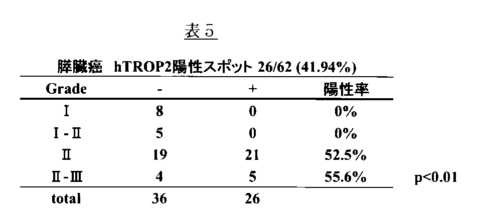
<Materials and methods>
The normal and cancer tissue arrays used for immunohistochemical staining are as follows.
Human normal tissue array:
Human, Normal organs in duplicates (Catalog No .: AB1, Super Bio Chips)
Normal issues more than one spots (Catalog No .: A103 (VI), ISU ABXIS)
Lung cancer tissue array:
Human Lung cancer-metastasis-normal (Catalog No .: CCA3, Super Bio Chips)
Human lung carcinoma tissue with marsissue tissue, 2 location cores (Catalog No .: OD-CT-RsLug03-002, Shanghai Outdo Biotech)
Pancreatic cancer tissue array:
Human Pancreas carcinoma tissue with mono-pathological type from 60cases, 2location cores (Catalog No .: OD-CT-DgPan03-001, Shanghai Haitech Biotech)
Liver cancer tissue array:
Hepatocyte cellular carcinoma, grade I to III with normal tissue controls, 63cases tissue arrays (Catalog No .: CS03-01-002U, Cybrdi)
Human live carcinoma with mono-pathological type from 30cases, 2location cores (Catalog No .: OD-CT-DgLive02-002, Shanghai Haitech Biotech)
Colon cancer tissue array:
Human, Collective cancer (Catalog No .: CD3, Super Bio Chips)
Human colon carcinoma with margin tissue, 2 location cores (Catalog No .: OD-CT-DgCol03-002, Shanghai Haido Biotech)
Colon cancer lymph node metastasis, liver metastasis tissue array:
Color (colon and electum) cancer with matched lymph node metastasis tissue array, 44cases / 99cores, triad slide (Catalog No .: CO991t, Biomax us)
Color (colon and electum) cancer with matched lymph node metastasis and normal adjuice tissue array, 43cases / 99cores (Catalog No .: CO992, Biomax us)
Colon cancer tissues_live metastasis (Catalog No .: A203 (IV), ISU ABXIS)
Breast cancer tissue array:
Human, Breast cancer-metastasis-normal (Catalog No .: CBA3, Super Bio Chips)
Human breast carcinoma with margin tissue, 2 location cores (Catalog No .: OD-CT-RpBre03-002, Shanghai Haido Biotech)
Gastric cancer tissue array:
Human, Storm cancer (Catalog No .: CQ1, Super Bio Chips)
Human gastricoma with marsine tissue, 2 location cores (Catalog No .: OD-CT-DgStm03-002, Shanghai Haido Biotech)
Esophageal cancer tissue array:
Human, Esophagas cancer (Catalog No .: CR1, Super Bio Chips)
Human esophagus carcinoma with margin tissue, 2 location cores (Catalog No .: OD-CT-DgEso03-002, Shanghai Haido Biotech)
Ovarian cancer tissue array:
Human, Ovary cancer (Catalog No .: CJ1, Super Bio Chips)
Prostate cancer tissue array:
Human, Prostate cancer-normal (Catalog No .: CA3, Super Bio Chips)
Bladder cancer tissue array:
Blader carcinoma / transitional cell carcinoma, grade I to III with normal tissue arrays (Catalog No .: CC12-01-001U, Cybrdi)
Patient information and clinical information of the tissue array were obtained from the attached data sheets and the websites of each company.
Immunohistochemical staining The tissue array paraffin sections of human normal tissue and cancer tissue were subjected to protease treatment with pepsin for 5 minutes at 37 ° C. after deparaffinization, and used for immunostaining using anti-hTROP-2 monoclonal antibody. After performing a color reaction using DAB (3,3′-diaminobenzidine) as a substrate, nuclear staining with hematoxylin was performed as counterstaining.
More specifically, these operations were performed as follows. The paraffin-embedded section was deparaffinized and then treated with protease with pepsin (DAKO) at 37 ° C for 5 minutes. After the antigen activation, endogenous peroxidase activity was removed by treatment with a solution obtained by adding hydrogen peroxide to methanol to a final concentration of 0.3% at room temperature for 20 minutes. After washing twice with PBS for 5 minutes at room temperature, blocking is performed for 30 minutes at room temperature using a PBS solution containing 1.5% normal horse serum (DAKO) to block nonspecific binding sites in the tissue. The operation was performed. Next, the anti-hTROP-2 monoclonal antibody clone K5-63-17 (
<Result>
Expression of hTROP-2 in human normal tissue The expression pattern of hTROP-2 in human normal tissue was analyzed using anti-hTROP-2 monoclonal antibody clone K5-63-17. The human normal tissue array (Catalog No .: AB1, Super BioChips) was deparaffinized and hydrophilized, and then the antigen was activated with the proteolytic enzyme pepsin, and the anti-hTROP-2 monoclonal antibody clone K5-63 Immunostaining was performed with -17 (FIG. 20). As a result, staining was seen in the skin, esophagus, kidney (cortex and medulla), pancreas, prostate, bladder, and tonsils. Most stained images showed localization in the cell membrane (FIGS. 20A, B, C, D, F, GH), but some expression was also observed in the cytoplasm (FIGS. 20.E, H). . On the other hand, it was not stained in the heart, liver, stomach, small intestine, large intestine, skeletal muscle, lung, spleen, thymus, etc. (FIGS. 20.I and J).
Expression of hTROP-2 in human cancer tissue In order to examine the expression of hTROP-2 in human cancer tissue (positive rate of hTROP-2), anti-hTROP-2 monoclonal antibody clone K5-63-17 was used. Species cancer tissue arrays were immunostained. A tissue section in which 10% or more of the cancer cells were stained was regarded as hTROP-2 positive. The staining results are shown in Table 4.
The positive rate of hTROP-2 in pancreatic cancer was 41.9%. The relationship between the hTROP-2 positive rate at this time and the grade (differentiation degree) of pancreatic cancer was examined. As a result, it was highly expressed in high-grade, ie, poorly differentiated pancreatic cancer (Table 5).
[K5−70抗体の単回投与による、ヒト膵癌細胞株PK−59xenograft preventionモデルでの抗腫瘍効果]
クローンK5−70(マウスIgG2a)のin vivoにおける強い抗腫瘍活性は、ヒト膵癌細胞株PK−59を用いたxenograft preventionモデルにおいて、10mg/kg体重の投与量で1回投与した場合においても示された。コントロール群(マウスIgG 10mg/kg体重,N=3)では、全ての個体において腫瘍形成が観察され、細胞移植後28日目(Day28)における腫瘍体積は781.7±74.5mm3であったのに対して、癌細胞移植日(Day1)に1回K5−70抗体を投与した群では(10mg/kg体重、N=3)、Day28における腫瘍体積は144.4±176.9mm3(P<0.05 by Student’s t−test)と81.5%の腫瘍成長阻害活性を示した(図22A)。腫瘍重量においては、Day28においてコントロール群が0.59±0.06gであったのに対し、クローンK5−70抗体投与群では0.07±0.10g(P<0.01 by by Student’s t−test))と88%の阻害活性を示した(図22B)。腫瘍体積、腫瘍重量のいずれにおいても、K5−70抗体投与群では3個体中2個体では、1回の10mg/kg体重の投与で、完全に腫瘍形成が阻害された(図22C)。
[Anti-tumor effect in human pancreatic cancer cell line PK-59 xenograft prevention model by single administration of K5-70 antibody]
In vivo strong antitumor activity of clone K5-70 (mouse IgG2a) is also shown in the xenograft prevention model using human pancreatic cancer cell line PK-59, even when administered once at a dose of 10 mg / kg body weight. It was. In the control group (
[ヒト大腸癌細胞株SW480細胞xenograft treatmentモデルにおける抗ヒhTROP−2モノクローナル抗体の抗腫瘍活性]
抗hTROP−2モノクローナル抗体(クローンK5−70,K5−116−2−1およびT6−16)の抗腫瘍活性を、ヒト大腸癌細胞株SW480を用いたxenograft treatmentモデルで検討した。5×10⁶cellsのSW480細胞を6週齢雌のNOD−scidマウスの右脇腹皮下に移植(Day1)し、平均の腫瘍体積が100mm3に達した段階で群分けを行い(Day7又はDay10),Day7又はDay10から3日に1回の投与間隔で、抗体の腹腔内投与を開始した。クローンK5−70抗体の抗腫瘍活性評価と、クローンK5−116−2−1抗体およびクローンT6−16抗体の抗腫瘍活性評価は、それぞれ独立した試験で評価した。K5−70抗体の抗腫瘍活性評価試験においては、癌細胞移植から44日目(Day44)におけるコントロール群(マウスIgG(10mg/kg体重),N=8)の腫瘍体積が365.4±214.6mm3であったのに対して、K5−70抗体(10mg/kg体重)投与群では、27.4±29.4mm3(P<0.01 by Student’s t−test)と著しく腫瘍形成が阻害された(阻害率92.5%)(図23A)。腫瘍重量においては、コントロール群が0.11±0.07gであったのに対して、K5−70抗体投与群では0.005±0.007(g)(P<0.01 by Student’s t−test)と95.5%の阻害率であった(図23B)。特に、K5−70抗体投与群では8個体中2個体では、完全に腫瘍形成が阻害され腫瘍の確認が出来なかった。
別に行ったK5−116−2−1抗体およびT6−16抗体の抗腫瘍活性評価試験においては、Day42におけるコントロール群の腫瘍体積が713.8±354.5mm3(N=8)であったのに対して、K5−116−2−1抗体投与群(10mg/kg体重)では、188.9±97.4mm3(N=8,P<0.01 by Student’s t−test)(図24A),T6−16抗体投与群(10mg/kg体重)では、292.8±199.7mm3(N=8,P<0.05 by Student’s t−test)(図25A)と、それぞれ73.5%,59.0%の阻害率であった。腫瘍重量についても同様に、コントロール群では0.39±0.19gであったのに対して、K5−116−2−1抗体投与群、T6−16抗体投与群では、0.10±0.07g(P<0.01 by Student’s t−test),0.17±0.14g(P<0.05 by Student’s t−test)と、それぞれ72.2%,56.4%の阻害率を示した(図24B,図25B)。
[Anti-tumor activity of anti-hihTROP-2 monoclonal antibody in human colon cancer cell line SW480 cell xenograft treatment model]
The antitumor activity of anti-hTROP-2 monoclonal antibodies (clone K5-70, K5-116-2-1 and T6-16) was examined in a xenograft treatment model using human colon cancer cell line SW480. 5 × 10 6 cells of SW480 cells were transplanted subcutaneously into the right flank of 6-week-old female NOD-scid mice (Day 1), and grouping was performed when the average tumor volume reached 100 mm 3 (
In the anti-tumor activity evaluation test of K5-116-2-1 antibody and T6-16 antibody conducted separately, the tumor volume of the control group at Day 42 was 713.8 ± 354.5 mm 3 (N = 8). On the other hand, in the K5-116-2-1 antibody administration group (10 mg / kg body weight), 188.9 ± 97.4 mm 3 (N = 8, P <0.01 by Student's t-test) (FIG. 24A), T6-16 antibody administration group (10 mg / kg body weight), 292.8 ± 199.7 mm 3 (N = 8, P <0.05 by Student's t-test) (FIG. 25A), The inhibition rates were 73.5% and 59.0%. Similarly, the tumor weight was 0.39 ± 0.19 g in the control group, whereas it was 0.10 ± 0.00 in the K5-1162-1 antibody administration group and the T6-16 antibody administration group. 07 g (P <0.01 by Student's t-test) and 0.17 ± 0.14 g (P <0.05 by Student's t-test), 72.2% and 56.4%, respectively. The inhibition rate was shown (FIG. 24B, FIG. 25B).
[ヒト大腸癌細胞株SW480細胞xenograft treatmentモデルにおけるクローンK5−70の用量依存的な抗腫瘍活性]
次に、K5−70抗体の用量依存的な抗腫瘍活性を、ヒト大腸癌細胞株SW480を用いたxenograft treatmentモデルにおいて検討した。5×10⁶cellsのSW480細胞を6週齢雌のNOD−scidマウスの右脇腹皮下に移植し、移植から10日後(Day10)に平均の腫瘍体積が100mm3に達した段階でコントロール群(マウスIgG 10mg/kg体重投与群、N=8,104.4±17.6mm3),1mg/kg体重のK5−70抗体投与群(N=8,104.3±16.1mm3),5mg/kg体重のK5−70抗体投与群(N=8,104.6±15.9mm3),10mg/kg体重のK5−70抗体投与群(N=8,104.8±14.9mm3)に群分を行い、3日に1回の腹腔内投与を行った。Day42におけるコントロール群の腫瘍体積が713.8±354.5mm3であったのに対して、K5−70抗体投与群では用量依存的な腫瘍形成阻害活性が観察され、1mg/kg体重投与群では、485.0±207.3mm3(阻害率32.1%),5mg/kg体重投与群では、339.5±253.2mm3(阻害率52.4%),10mg/kg体重投与群では、355.4±202.8mm3(阻害率50.2%,P<0.05 by Student’s t−test)であった(図26A)。同様に、Day42における腫瘍重量については、コントロール群が0.39±0.19gであったのに対して、K5−70抗体の1mg/kg体重 投与群では,0.24±0.11g(阻害率37.8%),5mg/kg体重投与群では、0.17±0.14g(阻害率55.8%,P<0.05 by Student’s t−test),10mg/kg体重投与群では、0.20±0.13g(阻害率47.1%)となり、用量依存的な抗腫瘍活性が確認された(図26B)。
[Dose-dependent antitumor activity of clone K5-70 in human colon cancer cell line SW480 cell xenograft treatment model]
Next, the dose-dependent antitumor activity of the K5-70 antibody was examined in a xenograft treatment model using the human colon cancer cell line SW480. 5 × 10 6 cells of SW480 cells were transplanted subcutaneously into the right flank of 6-week-old female NOD-scid mice, and 10 days after the transplantation (Day 10), when the average tumor volume reached 100 mm 3 , the control group (
[ヒト大腸癌細胞株SW480細胞xenograft treatmentモデルにおけるK5−70抗体の投与間隔の検討]
さらに、K5−70抗体の至適投与間隔を検討するために、週に1回(7日に1回)投与を行った際の抗腫瘍活性を、ヒト大腸癌細胞株SW480を用いたxenograft treatmentモデルにおいて検討した。5×10⁶cellsのSW480細胞を6週齢雌のNOD−scidマウスの右脇腹皮下に移植し、移植から10日後(Day10)に平均の腫瘍体積が100mm3に達した段階でコントロール群(マウスIgG,10mg/kg体重、N=8,104.42±15.1mm3),K5−70抗体投与群(10mg/kg体重、N=8,104.3±16.1mm3)に群分を行い、7日に1回の腹腔内投与を行った。Day42におけるコントロール群の腫瘍体積が713.8±354.5mm3であったのに対して、K5−70抗体投与群(週1回投与)では、332.3±239.9mm3(阻害率55%,P<0.05 by Student’s t−test)となった(図27A)。更に、投与間隔を空け10日に1回および2週間に1回投与した場合、Day39におけるコントロール群の腫瘍体積が956.9±367.8mm3であったのに対して、10日に1回のK5−70抗体投与群では、525.4±180.6mm3(阻害率45.1%,P<0.01 by Student’s t−test)2週間に1回のK5−70抗体投与群では459.4±217.6mm3(阻害率52.0%,P<0.01 by Student’s t−test)となった(図27B)。先行技術(US 7420040,US 7420041)では、膵臓癌細胞株(BxPC−3)を用いたxenograft treatmentモデルにおいて、20mg/kg体重、週に3回の抗体投与(投与間隔2日間)で、阻害率50−60%の抗腫瘍活性を示しているのに対し、K5−70抗体は1回の投与量が半分(10mg/kg体重)、かつ2週間に1回の投与(投与間隔13日間)で、先行技術に匹敵する抗腫瘍活性を示した。従ってK50−70抗体は少なくとも先行技術の12分の1の総投与量で顕著な抗腫瘍活性を示すことが明らかとなった。
[Examination of administration interval of K5-70 antibody in human colon cancer cell line SW480 cell xenograft treatment model]
Furthermore, in order to examine the optimal administration interval of the K5-70 antibody, the antitumor activity when administered once a week (once every 7 days) was measured using xenograft treatment using human colon cancer cell line SW480. Considered in the model. 5 × 10 6 cells of SW480 cells were transplanted subcutaneously into the right flank of 6-week-old female NOD-scid mice, and 10 days after the transplantation (Day 10), when the average tumor volume reached 100 mm 3 , the control group (mouse IgG, 10 mg / kg body weight, N = 8, 104.42 ± 15.1 mm 3 ), K5-70 antibody administration group (10 mg / kg body weight, N = 8, 104.3 ± 16.1 mm 3 ) And administered intraperitoneally once every 7 days. The tumor volume of the control group at Day 42 was 713.8 ± 354.5 mm 3 , whereas in the K5-70 antibody administration group (administered once a week), 332.3 ± 239.9 mm 3 (inhibition rate 55) %, P <0.05 by Student's t-test) (FIG. 27A). Further, when administered once every 10 days and once every two weeks with a dosing interval, the tumor volume of the control group at Day 39 was 956.9 ± 367.8 mm 3 , whereas once every 10 days In the K5-70 antibody administration group, the K5-70 antibody administration group once every two weeks, 525.4 ± 180.6 mm 3 (inhibition rate 45.1%, P <0.01 by Student's t-test) Then, it was 459.4 ± 217.6 mm 3 (inhibition rate 52.0%, P <0.01 by Student's t-test) (FIG. 27B). In the prior art (US 7402040, US 7420041), in the xenograft treatment model using a pancreatic cancer cell line (BxPC-3), the inhibition rate is 20 mg / kg body weight, administration of the antibody three times a week (
[ヒト大腸癌細胞株SW480細胞xenograft treatmentモデルにおけるT6−16抗体の用量依存的な抗腫瘍活性]
次に、T6−16抗体の用量依存的な抗腫瘍活性を、ヒト大腸癌細胞株SW480を用いたxenograft treatmentモデルにおいて検討した。5×10⁶cellsのSW480細胞を6週齢雌のNOD−scidマウスの右脇腹皮下に移植し、移植から10日後(Day10)に平均の腫瘍体積が100mm3に達した段階でコントロール群(マウスIgG 10mg/kg体重、N=8,105.8±9.9mm3),1mg/kg体重のT6−16抗体投与群(N=8,104.4±13.3mm3),5mg/kg体重のT6−16抗体投与群(N=8,104.7±13.0mm3),10mg/kg体重のT6−16抗体投与群(N=8,104.8±12.4mm3)に群分を行い、3日に1回の腹腔内投与を行った。Day43におけるコントロール群の腫瘍体積が473.5±137.0mm3であったのに対して、T6−16抗体投与群では用量依存的な腫瘍形成阻害活性が観察され、1mg/kg体重投与群では、397.9±97.5mm3(阻害率16.0%),5mg/kg体重投与群では、195.9±89.7mm3(阻害率58.7%,P<0.01 by Student’s t−test),10mg/kg体重投与群では、190.2±56.5mm3(阻害率59.8%,P<0.01 by Student’s t−test)であった(図28A)。同様に、Day43における腫瘍重量は、コントロール群が0.19±0.07gであったのに対して、1mg/kg体重のT6−16抗体投与群では、0.20±0.08g,5mg/kg体重の投与群では、0.08±0.04g(阻害率57.9%,P<0.01 by Student’s t−test),10mg/kg体重の投与群では、0.09±0.04g(阻害率52.6%,P<0.01 by Student’s t−test)となり、用量依存的な抗腫瘍活性が確認された(図28B)。
[Dose-dependent antitumor activity of T6-16 antibody in human colon cancer cell line SW480 cell xenograft treatment model]
Next, the dose-dependent antitumor activity of the T6-16 antibody was examined in a xenograft treatment model using the human colon cancer cell line SW480. 5 × 10 6 cells of SW480 cells were transplanted subcutaneously into the right flank of 6-week-old female NOD-scid mice, and 10 days after the transplantation (Day 10), when the average tumor volume reached 100 mm 3 , the control group (
[ヒト大腸癌細胞株SW480細胞xenograft treatmentモデルにおけるT6−16抗体の投与間隔の検討]
さらに、T6−16抗体の至適投与間隔を検討するために、週に1回(7日に1回)および10日に1回の投与間隔での抗腫瘍活性を、ヒト大腸癌細胞株SW480を用いたxenograft treatmentモデルにおいて検討した。5×10⁶cellsのSW480細胞を6週齢雌のNOD−scidマウスの右脇腹皮下に移植し、移植から10日後(Day10)に平均の腫瘍体積が100mm3に達した段階でコントロール群(マウスIgG,10mg/kg体重、N=8,105.8±9.9mm3),週1回のT6−16抗体投与群(10mg/kg体重、N=8,105.0±11.6mm3),10日に1回のT6−16抗体投与群(10mg/kg体重、N=5,130.8±2.4mm3)に群分けを行い、投与を開始した。Day43におけるコントロール群の腫瘍体積が473.5±137.0mm3であったのに対して、週1回のT6−16抗体投与群では、243.7±65.3mm3(阻害率48.5%,P<0.01 by Student’s t−test),10日に1回のT6−16抗体投与群では、297.8±54.4mm3(阻害率37.1%,P<0.05 by Student’s t−test)となった(図29)。先行技術(US 7420040,US 7420041)が膵臓癌細胞株(BxPC−3)を用いたxenograft treatmentモデルにおいて、20mg/kg体重の投与量で、週に3回投与(投与間隔2日間)で、阻害率50−60%の抗腫瘍活性を示しているのに対し、T6−16抗体は、1回の投与量が、先行技術の半分の用量かつ、10日に1回の投与(投与間隔9日間)で、顕著な抗腫瘍活性を示した。従って、T6−16抗体は少なくとも先行技術の8分の1の総投与量で顕著な抗腫瘍活性を示すことが明らかとなった。
[Examination of administration interval of T6-16 antibody in human colon cancer cell line SW480 cell xenograft treatment model]
Furthermore, in order to examine the optimal administration interval of the T6-16 antibody, the antitumor activity at an administration interval of once a week (once every 7 days) and once every 10 days was determined using the human colon cancer cell line SW480. The xenograft treatment model was used. 5 × 10 6 cells of SW480 cells were transplanted subcutaneously into the right flank of 6-week-old female NOD-scid mice, and 10 days after the transplantation (Day 10), when the average tumor volume reached 100 mm 3 , the control group (mouse IgG, 10 mg / kg body weight, N = 8, 105.8 ± 9.9 mm 3 ), weekly T6-16 antibody administration group (10 mg / kg body weight, N = 8, 105.0 ± 11.6 mm 3 ) The group was divided into T6-16 antibody administration groups (10 mg / kg body weight, N = 5, 130.8 ± 2.4 mm 3 ) once every 10 days, and administration was started. The tumor volume of the control group at Day 43 was 473.5 ± 137.0 mm 3 , whereas that of the T6-16 antibody administration group once a week was 243.7 ± 65.3 mm 3 (inhibition rate 48.5). %, P <0.01 by Student's t-test), in the T6-16 antibody administration group once every 10 days, 297.8 ± 54.4 mm 3 (inhibition rate 37.1%, P <0. 05 by Student's t-test) (FIG. 29). Prior art (US 7402040, US 7420041) inhibits xenograft treatment model using pancreatic cancer cell line (BxPC-3) at a dose of 20 mg /
[ヒト前立腺癌細胞株DU−145 xenograft preventionモデルにおける抗腫瘍活性の検討]
クローンK5−70のヒト前立腺癌に対する抗腫瘍活性を、DU−145細胞(RIKEN Cell Bank,RCB2143)を用いたxenograft preventionモデルで評価した。5×10⁶細胞のDU−145細胞を6週齢の雌のヌードマウス(Balb/c,nu/nu)の皮下に移植し、移植当日をDay1とし、コントロール群(マウスIgG)(N=8)とK5−70抗体投与群(N=8)に群分けを行い、Day1から3日に1回の頻度で10mg/kg体重の投与量で腹腔内投与した。Day40において、コントロール群における腫瘍体積が368.2±307.8mm3であったのに対し、K5−70抗体投与群では30.6±29.6mm3(P<0.05 by Student’s t−test)と約90%の腫瘍形成阻害活性を示した(図30A)。腫瘍重量においては、更に顕著な抗腫瘍活性が認めらた。Day40におけるコントロール群の腫瘍重量が0.18±0.18gであったのに対して、K5−70抗体投与群では8個体の全てにおいて腫瘍が消失しており、完全に腫瘍形成が阻害された(図30B)。以上の結果から、抗ヒトTROP−2モノクローナル抗体クローンK5−70は、ヒト前立腺癌細胞に対しても強い抗腫瘍活性を示すことが明らかとなった。
[Study of antitumor activity in human prostate cancer cell line DU-145 xenograft prevention model]
The antitumor activity of clone K5-70 against human prostate cancer was evaluated in a xenograft prevention model using DU-145 cells (RIKEN Cell Bank, RCB2143). 5 × 10 6 cells of DU-145 cells were transplanted subcutaneously into 6-week-old female nude mice (Balb / c, nu / nu). ) And K5-70 antibody administration group (N = 8), and intraperitoneally administered at a dose of 10 mg / kg body weight at a frequency of once every 3 days from
[ヒト膵癌細胞株PK−59細胞の肝転移モデルを用いたK5−70抗体の転移抑制活性]
消化器癌の治療において、癌の転移は臨床上の予後を左右する重要な因子であり、転移をコントロールすることは治療上重要な意味を有する。 TROP−2を標的とした癌治療用抗体投与により、腫瘍形成の抑制のみならず、癌細胞の他臓器への転移を抑制することが可能であれば、高い臨床上の有用性が期待されるため、癌治療抗体として望ましい特性である。
TROP−2は多くの癌腫における発現が確認されているが、特に転移巣において高発現していることが示されている(Br.J.Cancer(2008);99:1290−1295,Clin.Cancer Res.(2006);12:3057−3063,Mod.Pathol.(2008);21:186−191)。さらに、Trop−2遺伝子を導入した癌細胞を経脾的あるいは経膵的にヌードマウスに移植した場合に、肝転移の発症率が増加すること(WO 2010/089782、Molecular Cancer(2010);9:253)が報告され、TROP−2の癌転移過程における重要性が示唆されている。しかしながら、TROP−2を標的とした抗体を用いてin vivoで転移抑制作用を具体的に示した報告はこれまでに認められない。
本発明により見出された抗hTROP−2マウスモノクローナル抗体K5−70は、ヒト膵臓癌細胞を皮下に移植したゼノグラフトモデルにおいて高い治療効果が示されているが、癌細胞の増殖抑制効果に加え、ヒト膵癌細胞株PK−59の遊走能を抑制することが、in vitroのスクラッチアッセイで示されており、癌の転移をin vivoにおいても抑制する可能性が考えられた。そこで、PK−59細胞をヌードマウスの脾臓から注入し、肝転移を発生させるモデルを用いて抗hTROP−2マウスモノクローナル抗体の転移抑制効果を検証した。
癌細胞移植の前日に、マウスを群分し、抗hTROP−2モノクローナル抗体K5−70あるいは対照抗体(精製マウスIgG)を10mg/kg体重にて腹腔内投与した。翌日、hTROP−2を内在的に発現するヒト膵癌細胞株(PK−59)をトリプシン処理で剥がし、PBSで2×107cells/mLの細胞懸濁液を調製した。細胞懸濁液は移植時まで氷上に保管した。6~7週齢の雌のヌードマウス(Balb/c,nu/nu)をペントバルビタールの腹腔内投与により麻酔し、麻酔下で左側腹部を10~15mm切開し、脾臓を腹腔外に引き出し、27Gのシリンジを用いて50μLの細胞懸濁液(1 x 106cell)を脾臓内に注入した。細胞注入4分後に脾門部を5−0silkにて結紮し、脾臓を摘出した。切開した腹膜を4−0silkで閉創し、Wound Clips(AUTOCLIP 9mm,Becton Dickinson)を用いて閉腹した。癌細胞移植7日後に、さらにK5−70抗体および対照抗体を10mg/kg体重で投与した。癌細胞移植から4~6週間後に頸椎脱臼によりマウスを安楽死させ、肝臓を摘出し、転移巣の有無を確認した。
コントロールとして用いたマウスIgGを投与した群においては、PK−59細胞を移植した6匹のマウス中、4匹において、移植後4から6週間後に肝葉辺縁部への明らかな転移巣(2~7個)が認められた(図31A、転移発生率67%,表6)。これに対し、K5−70抗体投与群では、PK−59細胞を移植した4匹のマウスにおいて、いずれの個体でもまったく肝臓への転移巣が認められず、転移発生率は0%であった(図31B,表6)。
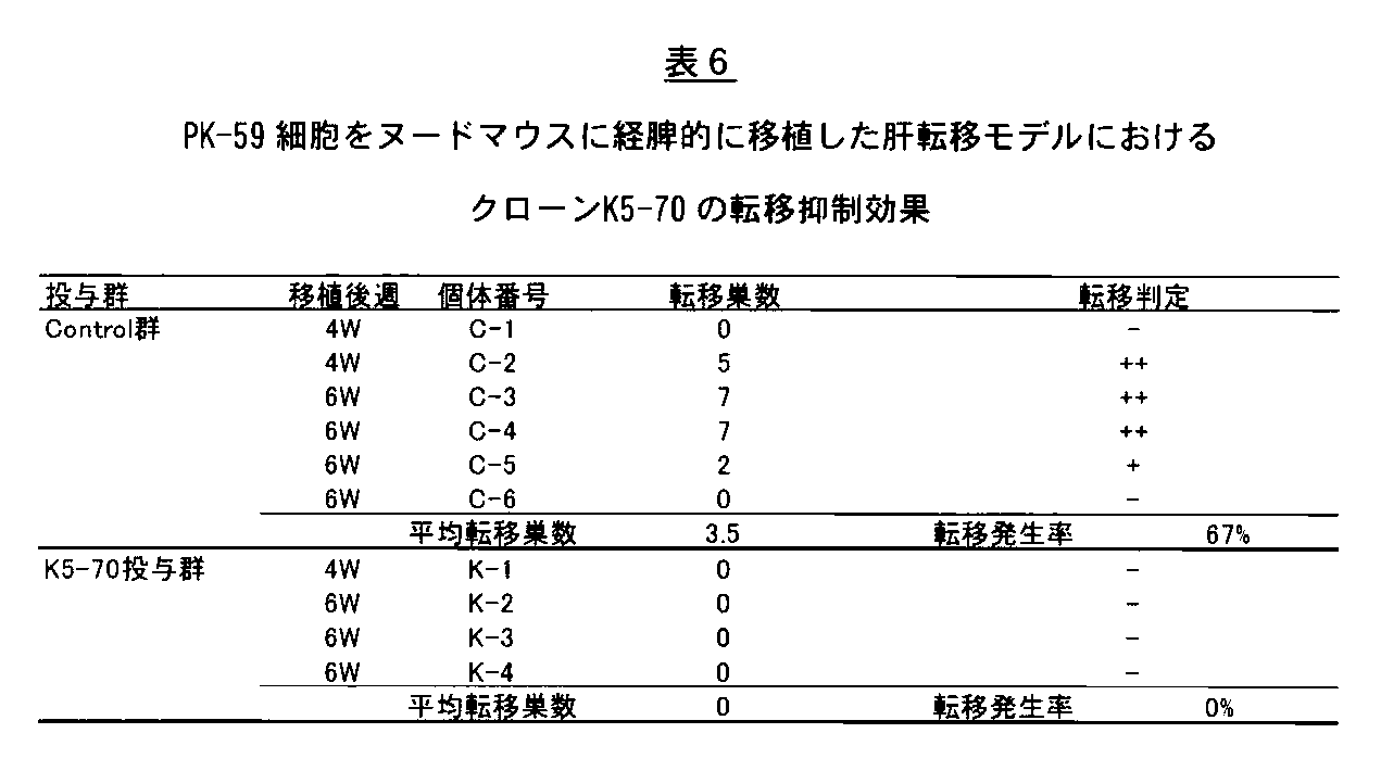
In the treatment of gastrointestinal cancer, metastasis of cancer is an important factor affecting clinical prognosis, and controlling metastasis has an important therapeutic significance. High clinical utility is expected if administration of antibody for cancer treatment targeting TROP-2 can suppress tumor formation as well as metastasis of cancer cells to other organs. Therefore, it is a desirable property as a cancer therapeutic antibody.
Although TROP-2 has been confirmed to be expressed in many carcinomas, it has been shown to be highly expressed particularly in metastases (Br. J. Cancer (2008); 99: 1290-1295, Clin. Cancer. (2006); 12: 3057-3063, Mod.Pathol. (2008); 21: 186-191). Furthermore, when cancer cells introduced with the Trop-2 gene are transplanted into nude mice transspleenally or pancreatically, the incidence of liver metastasis increases (WO 2010/089782, Molecular Cancer (2010); 9 : 253) has been reported, suggesting the importance of TROP-2 in the process of cancer metastasis. However, there have been no reports that specifically show the metastasis-suppressing action in vivo using an antibody targeting TROP-2.
The anti-hTROP-2 mouse monoclonal antibody K5-70 found by the present invention has been shown to have a high therapeutic effect in a xenograft model in which human pancreatic cancer cells are transplanted subcutaneously. Inhibition of the migration ability of the human pancreatic cancer cell line PK-59 has been shown by an in vitro scratch assay, and the possibility of suppressing cancer metastasis also in vivo was considered. Therefore, PK-59 cells were injected from the spleen of nude mice, and the anti-hTROP-2 mouse monoclonal antibody metastasis inhibitory effect was verified using a model that causes liver metastasis.
The day before cancer cell transplantation, mice were divided into groups, and anti-hTROP-2 monoclonal antibody K5-70 or control antibody (purified mouse IgG) was intraperitoneally administered at 10 mg / kg body weight. On the next day, a human pancreatic cancer cell line (PK-59) that endogenously expresses hTROP-2 was detached by trypsin treatment, and a cell suspension of 2 × 10 7 cells / mL was prepared with PBS. The cell suspension was stored on ice until the time of transplantation. A 6-7 week old female nude mouse (Balb / c, nu / nu) was anesthetized by intraperitoneal administration of pentobarbital, and the left abdomen was incised 10-15 mm under anesthesia, and the spleen was drawn out of the abdominal cavity. 50 μL of cell suspension (1 × 10 6 cell) was injected into the spleen. Four minutes after cell injection, the splenic hilum was ligated with 5-0 silk, and the spleen was removed. The incised peritoneum was closed with 4-0 silk and closed using Wound Clips (AUTOCLIP 9 mm, Becton Dickinson). Seven days after cancer cell transplantation, K5-70 antibody and control antibody were further administered at 10 mg / kg body weight. Four to six weeks after cancer cell transplantation, the mice were euthanized by cervical dislocation, the liver was removed, and the presence of metastatic lesions was confirmed.
In the group administered with mouse IgG used as a control, 4 out of 6 mice transplanted with PK-59 cells showed an obvious metastatic focus to the
[ヒト大腸癌細胞株SW480xenograftモデルにおける塩酸イリノテカン投与後の癌再発モデルにおけるK5−70抗体の抗腫瘍活性]
癌治療剤として近年、癌細胞の増殖を抑制する化学療法剤が多く開発され、一定の治療成績を上げているが、癌細胞以外の正常細胞に対する増殖抑制作用に伴う副作用と、治療中止後の癌の再発が大きな問題となっている。したがって、TROP−2を標的とした癌治療用抗体投与により、化学療法剤による治療後の腫瘍再発を抑制することが可能であれば、高い臨床上の有用性が期待されるため、癌治療抗体として望ましい特性である。
大腸癌の治療剤としては、5−FU,白金製剤に加え、トポイソメラーゼ阻害効果を有する塩酸イリノテカン(トポテシン、第一三共株式会社)が近年臨床適応され、動物モデルにおいても、大腸癌をはじめとした種々のヒト腫瘍細胞を移植したマウスモデルにおける抗腫瘍効果が報告されている(Cancer Chemother Pharmacol.(1991);28(3):192−8)。そこで、塩酸イリノテカン投与後の腫瘍再発に対する抗hTROP−2抗体クローンK5−70(マウスIgG2a)の再発予防効果について、ヒト大腸癌細胞株SW480を用いたxenograftモデルにおいて検討した。5×10⁶cellsのSW480細胞を8週齢雌のNOD−scidマウスの右脇腹皮下に移植し、移植から11日後(Day11)に平均の腫瘍体積が100mm3以上に達した段階で無処置群(生理食塩水投与群、N=8,130.7±16.2mm3),塩酸イリノテカン(CPT−11,トポテシン、第一三共株式会社)投与群(N=16,123.0±21.4mm3)に群分けを行い、塩酸イリノテカンを40mg/kg体重で3日に1回、合計3回(Day11,14,17)腹腔内投与した。塩酸イリノテカンの最終投与から3日目(Day20)に、無処置群の腫瘍体積は232.1±21.1mm3に達したのに対し、塩酸イリノテカン投与群は126.6.±26.6mm3(P<0.01 by Student’s t−test)であり、明らかな腫瘍抑制効果が認められた。この段階で腫瘍サイズに基づき塩酸イリノテカン投与群を2群に分け、一方はK5−70抗体投与群(10mg/kg体重,N=8,Day20の腫瘍体積126.0±28.0mm3)とし、もう一方はマウスIgG投与群(10mg/kg体重、N=8,Day20の腫瘍体積127.2±27.0mm3)とした。それぞれ3日に1回の抗体の腹腔内投与と腫瘍体積の測定を行い腫瘍の再発を評価した(図32)。マウスIgG投与群では、塩酸イリノテカンの最終投与から18日目(Day35)より腫瘍体積が300mm3を超える明らかな再発腫瘍を有する個体が複数認められ、塩酸イリノテカンの最終投与から30日目(Day47)においては、8例中5例で300mm3を超える腫瘍が認められた(腫瘍平均体積401.7±172.7mm3)。これに対し、K5−70抗体投与群では顕著に腫瘍再発が抑制され、腫瘍平均体積は、180.5±142.1mm3,(P<0.05 by Student’s t−test)であった(図32)。特に、K5−70抗体投与群では、Day47における腫瘍体積が群分け時(126.0±28.0mm3)よりも退縮し、100mm3未満となった個体が8例中4例認められた。これらの結果から、抗hTROP−2抗体K5−70は、塩酸イリノテカン投与後の腫瘍再発に対しても極めて強い抑制作用を有することが明らかとなった。
[Anti-tumor activity of K5-70 antibody in cancer recurrence model after administration of irinotecan hydrochloride in human colon cancer cell line SW480 xenograft model]
In recent years, many chemotherapeutic agents that suppress the growth of cancer cells have been developed as cancer treatment agents, and have achieved a certain level of treatment results, but the side effects associated with the growth inhibitory action on normal cells other than cancer cells and Cancer recurrence is a major problem. Therefore, if it is possible to suppress tumor recurrence after treatment with a chemotherapeutic agent by administration of a cancer therapeutic antibody targeting TROP-2, it is expected to have high clinical utility. As desirable characteristics.
As a therapeutic agent for colorectal cancer, irinotecan hydrochloride (topotecin, Daiichi Sankyo Co., Ltd.) having a topoisomerase inhibitory effect has been clinically applied in recent years in addition to 5-FU and platinum preparations. Antitumor effects have been reported in mouse models transplanted with various human tumor cells (Cancer Chemother Pharmacol. (1991); 28 (3): 192-8). Thus, the recurrence prevention effect of anti-hTROP-2 antibody clone K5-70 (mouse IgG2a) on tumor recurrence after irinotecan hydrochloride administration was examined in a xenograft model using human colon cancer cell line SW480. 5 × 10 6 cells of SW480 cells were transplanted subcutaneously into the right flank of 8-week-old female NOD-scid mice, and 11 days after transplantation (Day 11), when the average tumor volume reached 100 mm 3 or more, no treatment group (Saline administration group, N = 8, 130.7 ± 16.2 mm 3 ), irinotecan hydrochloride (CPT-11, topotecin, Daiichi Sankyo Co., Ltd.) administration group (N = 16, 123.0 ± 21. The group was divided into 4 mm 3 ), and irinotecan hydrochloride was administered intraperitoneally at a dose of 40 mg / kg body weight once every three days (
[CLIPS技術を用いたエピトープマッピング]
<材料と方法>
ペプチド合成
本実験で使用した直鎖型15−mer及び30−merのTROP−2細胞外領域に由来するペプチドは、Fmoc(9−Fluorenylmethoxycarbonyl)法による固相合成により取得した。また不連続エピトープ解析のため、両端にシステイン残基を付加したTROP−2細胞外領域に由来する17−merのペプチドを合成し、CLIPS技術(Chemically Linked Peptides on Scaffolds technology)によりループ構造を一つまたは二つ有する立体構造を再構築した。付加したシステイン残基近傍に別のシステイン残基が存在する場合、アラニンに置換した。
エピトープスクリーニングELISA
5034種類の合成したペプチドをPEPSCANカード(455ペプチド/カード)へ共有結合させ、ELISA法により合成ペプチドと抗体の結合を検証した。PEPSCANカードはブロッキングバッファー(4%ウマ血清、5%卵白アルブミン、1%Tweenを含むリン酸緩衝液)で1μg/mLに希釈した抗ヒトTROP−2モノクローナル抗体(K5−70、K5−107、K5−116−2−1、T5−86、T6−16)と反応させた。洗浄後、1000倍希釈したペルオキシダーゼ−二次抗体複合体と25℃、1時間反応させた。洗浄後、基質溶液(2,2’−azino−di−3−ethylbenzthiazoline sulfonate(ABTS)と2μLの3%過酸化水素水を含む溶液)を加え、1時間発色反応を行った。抗体結合活性はCCDカメラで撮影し、画像解析を行い定量した。
<結果>
薬効を示した抗hTROP−2モノクローナル抗体K5−70、K5−107、K5−116−2−1、T5−86、T6−16についてCLIPS(Chemically Linked Peptides on Scaffolds)技術を利用しエピトープ解析を行った。なお、以下、本実施例において「アミノ酸番号」とは、配列番号2に示されるアミノ酸配列(hTROP−2タンパク質(323アミノ酸残基))中のアミノ酸番号を意味する。
K5−70抗体についての解析結果を下記表7に示す。その結果、33ペプチドがK5−70抗体と強い結合を示すことが明らかとなった。これら33ペプチドの中で、VCSPDGPGGRCQCRALGSGMAVD(アミノ酸番号43−65)を含む配列(表7中に記載のペプチドNo.1−7,9)、HHILIDLRHRPTAG(アミノ酸番号152−165)を含む配列(表7中に記載のペプチドNo.14,22−24,28)、VHYEQPTIQIELRQ(アミノ酸番号193−206)を含む配列(表7中に記載のペプチドNo.8,10,12,13,18,20,21,23,26,28,30,32)、DLDAELRRLFRER(アミノ酸番号171−183)を含む配列(表7中に記載のペプチドNo.11,16,18,19,21,22,29,31)は、繰り返し出現した。K5−70抗体は、特にVCSPDGPGGRCQCRALGSGMAVDを含む配列に強く結合した。これらの結果から、hTROP−2タンパク質中、上記4種類のペプチド配列領域がK5−70抗体のエピトープである可能性が示唆された。

従って、hTROP−2タンパク質中、上記VCSPDGPGGRCQCRALGSGMAVDのペプチド配列領域がK5−107抗体のエピトープであることが示唆された。


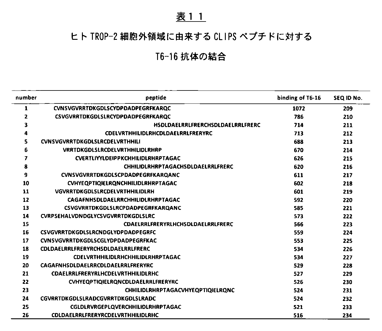
<Materials and methods>
Peptide synthesis The peptides derived from the linear 15-mer and 30-mer TROP-2 extracellular regions used in this experiment were obtained by solid-phase synthesis by the Fmoc (9-Fluorenylmethycarbonyl) method. In addition, for discontinuous epitope analysis, a 17-mer peptide derived from the TROP-2 extracellular region with cysteine residues added at both ends was synthesized, and one loop structure was formed using CLIPS technology (Chemically Linked Peptides on Scaffolds technology) Or a three-dimensional structure having two was reconstructed. When another cysteine residue was present near the added cysteine residue, it was substituted with alanine.
Epitope screening ELISA
5034 kinds of synthesized peptides were covalently bound to a PEPSCAN card (455 peptide / card), and the binding between the synthetic peptide and the antibody was verified by ELISA. The PEPSCAN card is an anti-human TROP-2 monoclonal antibody (K5-70, K5-107, K5) diluted to 1 μg / mL with blocking buffer (phosphate buffer containing 4% horse serum, 5% ovalbumin, 1% Tween). -116-2-1, T5-86, T6-16). After washing, it was reacted with a peroxidase-secondary antibody complex diluted 1000 times for 1 hour at 25 ° C. After washing, a substrate solution (2,2′-azino-di-3-ethylbenzthiazine sulfate (ABTS) and a solution containing 2 μL of 3% hydrogen peroxide solution) was added and a color reaction was performed for 1 hour. The antibody binding activity was photographed with a CCD camera, quantified by image analysis.
<Result>
Epitope analysis was performed for the anti-hTROP-2 monoclonal antibodies K5-70, K5-107, K5-116-2-1, T5-86, and T6-16 that showed medicinal effects using CLIPS (Chemically Linked Peptides on Scaffolds) technology. It was. In the following examples, “amino acid number” means the amino acid number in the amino acid sequence shown in SEQ ID NO: 2 (hTROP-2 protein (323 amino acid residues)).
The analysis results for the K5-70 antibody are shown in Table 7 below. As a result, it was revealed that 33 peptide showed strong binding with K5-70 antibody. Among these 33 peptides, a sequence including VCSPDPGPGRCQCRALGSGMAVD (amino acid numbers 43-65) (peptide Nos. 1-7, 9 described in Table 7) and a sequence including HHIILIDRHRPTAG (amino acids number 152-165) Peptide Nos. 14, 22-24, 28), VHYEQPTIQIELRQ (amino acid number 193-206) described in Table 7 (peptide Nos. 8, 10, 12, 13, 18, 20, 21, 21 described in Table 7) 23, 26, 28, 30, 32), and the sequence (peptide Nos. 11, 16, 18, 19, 21, 22, 29, 31 described in Table 7) including DLDAELRRLFRER (amino acid numbers 171-183) Appeared repeatedly. The K5-70 antibody specifically bound strongly to sequences including VCSPDGPGGRCQCRALGSGMAVD. From these results, it was suggested that the above four types of peptide sequence regions in the hTROP-2 protein may be epitopes of the K5-70 antibody.
Therefore, it was suggested that the peptide sequence region of the VCSPDGPGGRCQCRALGSGMAVD in the hTROP-2 protein is an epitope of the K5-107 antibody.
[マウス抗hTROP−2抗体(クローンK5−70,K5−107,K5−116−2−1およびT6−16)の抗体遺伝子の可変領域の配列決定]
3×10⁶個のマウス抗TROP−2モノクローナル抗体産生ハイブリドーマから、TRIzol試薬(Invitrogen)を用いて全RNAを抽出した。クローンK5−70,クローンK5−107およびクローンK5−116−2−1については、SMARTerTM RACE cDNA Amplification kit(Clontech)を用いて、キット添付の方法に従い、マウスIgGのH鎖特異的プライマー(5’−TCCAKAGTTCCA−3’(配列番号24))と、L鎖特異的プライマー(5’−GCTGTCCTGATC−3’(配列番号25))を用いてcDNAを合成した。クローンT6−16については,GeneRacer Kit(Invitrogen)を用いて、キット添付の方法に従い、オリゴdTプライマーを用いてcDNAを合成した。クローンK5−70(マウスIgG2a),クローンK5−107(マウスIgG1)、およびクローンK5−116−2−1(マウスIgG2a)のH鎖及びL鎖の可変領域(VH,VL)をコードする遺伝子は、上記合成後のcDNAを鋳型として、PCR法を用いてクローニングした。この際、5’−プライマーはSMARTerTM RACE cDNA Amplification kitに添付されている10×Universal Primer A Mix(UPM)を使用した。一方、3’−プライマーは、VH増幅用3’−プライマーとしてはマウスIgG H鎖に特異的な配列を有するものを使用し、VL増幅用3’−プライマーとしてはマウスIgG L鎖に特異的なな配列を有するものを使用した。
5’−プライマー(10×Universal Primer A Mix(UPM)):
Long(0.4μM)

![]()

<反応液組成>
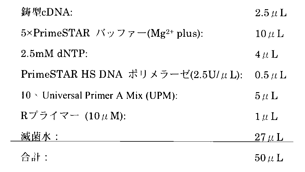
94℃(10sec)反応させた後、「熱変性・解離:98℃(10sec)→アニーリング:60℃(5sec)→合成・伸長:72℃(60sec)」を1サイクルとして計30サイクル反応させた後、最後に72℃(3min)反応させた。
合成したVH及びVLのcDNAを、pMD20−T vector(タカラバイオ)にサブクローニングして、塩基配列を決定した。複数のVHクローン及びVLクローンの塩基配列を解読し、マウスH鎖及びL鎖の可変領域に典型的な塩基配列を同定した。図33及び図34に、K5−70のVH及びVLのコンセンサスcDNA塩基配列、ならびに推定アミノ酸配列を示した。図35及び図36に、K5−107のVH及びVLのコンセンサスcDNA塩基配列、ならびに推定アミノ酸配列を示した。図37及び図38に、K5−116−2−1のVH及びVLのコンセンサスcDNA塩基配列、ならびに推定アミノ酸配列を示した。
クローンT6−16のH鎖及びL鎖の可変領域(VH,VL)をコードする遺伝子は、上記合成後のcDNAを鋳型として、PCR法を用いてクローニングした。この際、この際、5’−プライマーはGeneRacer Kitに添付されているものを使用した。一方、3’−プライマーは、VH増幅用3’−プライマーとしてはマウスIgG H鎖に特異的な配列を有するものを使用し、VL増幅用3’−プライマーとしてはマウスIgG L鎖に特異的な配列を有するものを使用した。
5’−プライマー(Fプライマー):
![]()
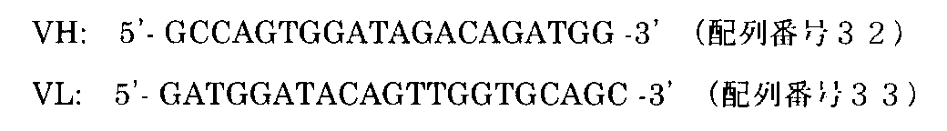
<反応液組成>
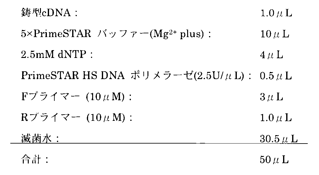
「熱変性・解離:98℃(10sec)→アニーリング:57℃(10sec)→合成・伸長:72℃(60sec)」を1サイクルとして計35サイクル。
合成したVH及びVLのcDNAを、pCR4Blunt−TOPO vector(Invitrogen)にサブクローニングして、塩基配列を決定した。複数のVHクローン及びVLクローンの塩基配列を解読し、マウスH鎖及びL鎖の可変領域に典型的な塩基配列を同定した。図39及び図40に、T6−16のVH及びVLのコンセンサスcDNA塩基配列、ならびに推定アミノ酸配列を示した。
[Sequence determination of variable region of antibody gene of mouse anti-hTROP-2 antibody (clone K5-70, K5-107, K5-116-2-1 and T6-16)]
Total RNA was extracted from 3 × 10 6 mouse anti-TROP-2 monoclonal antibody-producing hybridomas using TRIzol reagent (Invitrogen). For clone K5-70, clone K5-107, and clone K5-116-2-1, using the SMARTer ™ RACE cDNA Amplification kit (Clontech), the mouse IgG heavy chain specific primer (5 CDNA was synthesized using '-TCCAKAGTTCCA-3' (SEQ ID NO: 24)) and an L chain specific primer (5'-GCTGTCCTGATC-3 '(SEQ ID NO: 25)). For clone T6-16, cDNA was synthesized using an oligo dT primer using GeneRacer Kit (Invitrogen) according to the method attached to the kit. The genes encoding the variable regions (VH, VL) of the heavy and light chains of clone K5-70 (mouse IgG2a), clone K5-107 (mouse IgG1), and clone K5-116-2-1 (mouse IgG2a) are The cloned cDNA was cloned using the PCR method using the synthesized cDNA as a template. In this case, 10 × Universal Primer A Mix (UPM) attached to SMARTer ™ RACE cDNA Amplification kit was used as the 5′-primer. On the other hand, as the 3′-primer, one having a sequence specific to mouse IgG H chain is used as the 3′-primer for VH amplification, and specific to mouse IgG L chain is used as the 3′-primer for VL amplification. The one with the correct sequence was used.
5′-Primer (10 × Universal Primer A Mix (UPM)):
Long (0.4μM)
![]()
<Reaction solution composition>
After reacting at 94 ° C. (10 sec), “thermal denaturation / dissociation: 98 ° C. (10 sec) → annealing: 60 ° C. (5 sec) → synthesis / elongation: 72 ° C. (60 sec)” was performed for 30 cycles in total. Thereafter, the reaction was finally carried out at 72 ° C. (3 min).
The synthesized VH and VL cDNAs were subcloned into pMD20-T vector (Takara Bio), and the nucleotide sequences were determined. The nucleotide sequences of a plurality of VH clones and VL clones were decoded, and typical nucleotide sequences were identified in the variable regions of the mouse H chain and L chain. 33 and 34 show the consensus cDNA base sequences of K5-70 VH and VL, and the deduced amino acid sequence. 35 and 36 show the VH and VL consensus cDNA base sequences of K5-107 and the deduced amino acid sequence. 37 and 38 show the VH and VL consensus cDNA base sequences of K5-116-2-1 and the deduced amino acid sequence.
The gene encoding the variable region (VH, VL) of the H chain and L chain of clone T6-16 was cloned by PCR using the synthesized cDNA as a template. At this time, the 5'-primer attached to the GeneRacer Kit was used. On the other hand, as the 3′-primer, a 3′-primer for VH amplification having a sequence specific to mouse IgG H chain is used, and as the 3′-primer for VL amplification, specific to mouse IgG L chain. Those with sequences were used.
5'-primer (F primer):
![]()
<Reaction solution composition>
“Heat denaturation / dissociation: 98 ° C. (10 sec) → annealing: 57 ° C. (10 sec) → synthesis / elongation: 72 ° C. (60 sec)” is 35 cycles in total.
The synthesized VH and VL cDNAs were subcloned into pCR4 Blunt-TOPO vector (Invitrogen), and the nucleotide sequences were determined. The nucleotide sequences of a plurality of VH clones and VL clones were decoded, and typical nucleotide sequences were identified in the variable regions of the mouse H chain and L chain. 39 and 40 show the VH and VL consensus cDNA base sequences of T6-16 and the deduced amino acid sequence.
[ヒト型化K5−70抗体のデザイン]
先の実施例で作製したK5−70抗体について、その可変領域(VH,VL)のヒト型化を、Queenらの方法(Proc.Natl.Acad.Sci.USA 86:10029−10033,1989)に従って以下のように行った。まず、K5−70抗体可変領域の立体構造の分子モデリングをコンピュータで行い、次いで、ヒト抗体遺伝子の可変領域配列とのホモロジー検索によって、K5−70 VHのヒト型化に必要なフレームワーク(FR)領域を提供するアクセプターとしてGenBank accession numberがDA980102のcDNA配列(DA980192 VH)を選択した(Genome Res.16:55−65,2006)。同様に、K5−70 VLのヒト型化に必要なフレームワーク(FR)領域を提供するアクセプターとしてGenBank accession numberがL41174のcDNA配列(L41174 VL)を選択した(J.Biol.Chem.270:12457−12465,1995)。
K5−70 VHのヒト型化では、K5−70 VHのCDR配列をアクセプターであるDA980102 VHの対応する位置に置き換えて挿入した。コンピュータモデリングによる立体構造解析により、K5−70 VHのCDRに隣接し、その構造維持に重要な役割を果たしていると考えられるアミノ酸残基(48番目のイソロイシン(I)、66番目のリジン(K)、67番目のアラニン(A)、71番目のバリン(V)、93番目のスレオニン(T))についてはK5−70 VHのものを保持し、それ以外のFR領域をアクセプター配列に置き換えた。VHおよびVL内のアミノ酸残基位置番号はKabatらの定義(Sequences of Proteins of Immunological Interests,Fifthedition,NIH Publication No.91−3242,U.S.Department of Health and Human Services,1991)に準拠した。
また、アクセプター配列DA980102 VHの82番目のアミノ酸残基であるメチオニン(M)は、somatic hypermutationによる特殊なアミノ酸残基である可能性が高いと考えられる。そこで潜在的抗原性を小さくするために、82番目のアミノ酸残基として最も一般的なロイシン(L)に置き換えた。上述の通りデザインしたヒト型化K5−70 VH(HuK5−70 VH)、K5−70 VHおよびDA980102 VHのアミノ酸配列アライメントを図41に示した。
ヒト型化K5−70 VLのデザインついても同様にCDR配列の移植を行い、CDRの構造維持に重要なアミノ酸残基(49番目のリジン(K))はK5−70 VLのものを保持し、それ以外のFR領域をアクセプター配列に置き換えた(HuK5−70 VL)。HuK5−70 VL、K5−70 VLおよびL41174 VLのアミノ酸配列アライメントを図42に示した。
[Design of humanized K5-70 antibody]
For the K5-70 antibody prepared in the previous example, the variable regions (VH, VL) were humanized according to the method of Queen et al. (Proc. Natl. Acad. Sci. USA 86: 10029-10033, 1989). It went as follows. First, molecular modeling of the three-dimensional structure of the K5-70 antibody variable region is performed by computer, and then a framework (FR) necessary for humanization of K5-70 VH by homology search with the variable region sequence of the human antibody gene. GenBank accession number selected the DA980102 cDNA sequence (DA980192 VH) as an acceptor providing the region (Genome Res. 16: 55-65, 2006). Similarly, GenBank accession number selected the cDNA sequence of L41174 (L41174 VL) as an acceptor providing the framework (FR) region necessary for humanization of K5-70 VL (J. Biol. Chem. 270: 12457). -12465, 1995).
In humanization of K5-70 VH, the CDR sequence of K5-70 VH was replaced with the corresponding position of DA980102 VH, which was an acceptor, and inserted. Amino acid residues (48th isoleucine (I), 66th lysine (K)) adjacent to the CDR of K5-70 VH and considered to play an important role in maintaining the structure by three-dimensional structural analysis by computer modeling , 67th alanine (A), 71st valine (V), 93rd threonine (T)) were retained for K5-70 VH, and the other FR regions were replaced with acceptor sequences. The amino acid residue position numbers in VH and VL were defined according to Kabat et al. (Sequences of Proteins of Immunological Intersect, Fifthition, NIH Publication No. 91-3242, US Department of Health Heal.
Further, methionine (M), which is the 82nd amino acid residue of acceptor sequence DA980102 VH, is considered to be highly likely to be a special amino acid residue due to somatic hypermutation. Therefore, in order to reduce the potential antigenicity, the most common leucine (L) as the 82nd amino acid residue was substituted. The amino acid sequence alignment of humanized K5-70 VH (HuK5-70 VH), K5-70 VH and DA980102 VH designed as described above is shown in FIG.
The humanized K5-70 VL design was similarly grafted with CDR sequences, and the amino acid residue important for maintaining the CDR structure (49th lysine (K)) retains that of K5-70 VL. The other FR regions were replaced with acceptor sequences (HuK5-70 VL). The amino acid sequence alignment of HuK5-70 VL, K5-70 VL and L41174 VL is shown in FIG.
[ヒト型化T6−16抗体のデザイン]
先の実施例で作製したT6−16抗体について、その可変領域(VH,VL)のヒト型化を、Queenらの方法(Proc.Natl.Acad.Sci.USA 86:10029−10033,1989)に従って以下のように行った。まず、T6−16抗体可変領域の立体構造の分子モデリングをコンピュータで行い、次いで、ヒト抗体遺伝子の可変領域配列とのホモロジー検索によって、T6−16 VHのヒト型化に必要なフレームワーク(FR)領域を提供するアクセプターとしてGenBank accession numberがDA935238のcDNA配列(DA935238 VH)を選択した(Genome Res.16:55−65,2006)。同様に、T6−16 VLのヒト型化に必要なフレームワーク(FR)領域を提供するアクセプターとしてGenBank accession numberがM99608のcDNA配列(M99608 VL)を選択した(J.Immunol.149:2518−2529,1992)。
T6−16 VHのヒト型化では、T6−16 VHのCDR配列をアクセプターであるDA935238 VHの対応する位置に置き換えて挿入した。コンピュータモデリングによる立体構造解析により、T6−16 VHのCDRに隣接し、その構造維持に重要な役割を果たしていると考えられるアミノ酸残基(48番目のイソロイシン(I)、67番目のアラニン(A)、73番目のリジン(K))についてはT6−16 VHのものを保持し、それ以外のFR領域をアクセプター配列に置き換えた(HuT6−16 VH1)。また73番目のリジン残基(K)については、抗原結合部位の適切な形成に対する影響が少ない可能性があり、HuT6−16 VH1のリジン残基をより寛容なアミノ酸残基であるスレオニン(T)に置き換えたアミノ酸配列を別途デザインした(HuT6−16 VH2)。デザインしたヒト型化T6−16 VH(HuT6−16 VH1およびHuT6−16 VH2)、T6−16 VHおよびDA980102 VHのアミノ酸配列アライメントを図43に示した。
ヒト型化T6−16 VLのデザインについても、同様にCDR配列の移植を行った。CDRの構造維持に重要なアミノ酸残基はアクセプター配列でも保持されており、FR配列はアクセプター配列と同じ配列を用いた(HuT6−16 VL)。HuT6−16VL、T6−16 VLおよびL41174 VLのアミノ酸配列アライメントを図44に示した。
[Design of humanized T6-16 antibody]
For the T6-16 antibody prepared in the previous example, the variable region (VH, VL) was humanized according to the method of Queen et al. (Proc. Natl. Acad. Sci. USA 86: 10029-10033, 1989). It went as follows. First, molecular modeling of the three-dimensional structure of the T6-16 antibody variable region is performed by computer, and then a framework (FR) required for humanization of T6-16 VH by homology search with the variable region sequence of the human antibody gene. GenBank accession number selected the DA935238 cDNA sequence (DA935238 VH) as an acceptor providing the region (Genome Res. 16: 55-65, 2006). Similarly, GenBank accession number selected the M99608 cDNA sequence (M99608 VL) as an acceptor providing the framework (FR) region necessary for humanization of T6-16 VL (J. Immunol. 149: 2518-2529). 1992).
In humanization of T6-16 VH, the CDR sequence of T6-16 VH was replaced with the corresponding position of DA935238 VH as an acceptor and inserted. Amino acid residues (48th isoleucine (I), 67th alanine (A)) that are considered to play an important role in maintaining the structure of the T6-16 VH CDR by the three-dimensional structure analysis by computer modeling. , 73rd lysine (K)) was retained for T6-16 VH, and the other FR regions were replaced with acceptor sequences (HuT6-16 VH1). The 73rd lysine residue (K) may have little influence on the proper formation of the antigen binding site, and the lysine residue of HuT6-16 VH1 is a more permissive amino acid residue, threonine (T). The amino acid sequence replaced with was separately designed (HuT6-16 VH2). The amino acid sequence alignment of the designed humanized T6-16 VH (HuT6-16 VH1 and HuT6-16 VH2), T6-16 VH and DA980102 VH is shown in FIG.
For the design of humanized T6-16 VL, CDR sequences were similarly transplanted. The amino acid residues important for maintaining the structure of the CDR are also retained in the acceptor sequence, and the FR sequence was the same as the acceptor sequence (HuT6-16 VL). The amino acid sequence alignment of HuT6-16VL, T6-16 VL and L41174 VL is shown in FIG.
[ヒト型化K5−70 VHおよびVL遺伝子合成]
HuK5−70 VHおよびHuK5−70 VLをコードする遺伝子は以下のように作製した。上述のようにデザインしたHuK5−70 VHおよびVLのN末端側に、それぞれK5−70 VH、VL由来のシグナルペプチド配列を付加したアミノ酸配列をもとに遺伝子合成を行った(Operon社)。その際、それぞれ合成したHuK5−70 VHおよびHuK5−70 VLの遺伝子配列の5’末端側にKozak配列(ACC ACC)を付加し、またHuK5−70 VHの5’末端には、制限酵素部位としてEcoRIの部位(GAA TTC)を付加し、3’末端にはNheI(GCT AGC)の部位を付加した。同様に、HuK5−70 VLの5’末端には、制限酵素部位としてAge Iの部位(ACC GGT)を付加し、3’末端にはBsiWIの部位(CGT ACG)を付加した。合成したHuK5−70 VH遺伝子、およびHuK5−70 VL遺伝子は、pCR2.1ベクター(Invitrogen)にTAクローニングにより組み込んだ。遺伝子合成により作製したHuK5−70 VHおよびVLの遺伝子配列をそれぞれ図45及び46に示した。
[Humanized K5-70 VH and VL gene synthesis]
Genes encoding HuK5-70 VH and HuK5-70 VL were prepared as follows. Gene synthesis was performed based on the amino acid sequences in which signal peptide sequences derived from K5-70 VH and VL were added to the N-terminal sides of HuK5-70 VH and VL designed as described above (Operon). At that time, a Kozak sequence (ACC ACC) was added to the 5 ′ end of each of the synthesized HuK5-70 VH and HuK5-70 VL gene sequences, and a restriction enzyme site was added to the 5 ′ end of HuK5-70 VH. An EcoRI site (GAA TTC) was added, and a NheI (GCT AGC) site was added to the 3 ′ end. Similarly, an Age I site (ACC GGT) was added as a restriction enzyme site to the 5 ′ end of HuK5-70 VL, and a BsiWI site (CGT ACG) was added to the 3 ′ end. The synthesized HuK5-70 VH gene and HuK5-70 VL gene were incorporated into pCR2.1 vector (Invitrogen) by TA cloning. The gene sequences of HuK5-70 VH and VL prepared by gene synthesis are shown in FIGS. 45 and 46, respectively.
[ヒト型化T6−16 VH1、T6−16 VH2およびT6−16 VL遺伝子合成]
HuT6−16 VH1、HuT6−16 VH2およびHuT6−16 VLをコードする遺伝子は以下のように作製した。デザインしたHuT6−16 VH1、HuT6−16 VH2のN末端側にT6−16 VH由来のシグナルペプチド配列を、HuT6−16 VLのN末端側にT6−16 VL由来のシグナルペプチド配列をそれぞれ付加したアミノ酸配列をもとに遺伝子合成を行った(Operon社)。その際、それぞれ合成したHuT6−16 VH1、HuT6−16 VH2およびHuT6−16 VLの遺伝子配列の5’末端側にKozak配列(ACC ACC)を付加し、またHuT6−16 VH1およびHuT6−16 VH2の5’末端には、制限酵素部位としてEcoRIの部位(GAA TTC)を付加し、3’末端にはNheIの部位(GCT AGC)を付加した。同様に、ヒト型化T6−16 VLの5’末端には、制限酵素部位としてAge Iの部位(ACC GGT)を付加し、3’末端にはBsiWIの部位(CGT ACG)を付加した。合成したHuT6−16 VH1遺伝子およびHuT6−16 VH2遺伝子ならびにHuT6−16 VL遺伝子は、pCR2.1ベクター(Invitrogen)にTAクローニングにより組み込んだ。
遺伝子合成により作製したHuT6−16 VH1、HuT6−16 VH2およびHuT6−16 VLの遺伝子配列をそれぞれ図47~49に示した。
[Humanized T6-16 VH1, T6-16 VH2 and T6-16 VL gene synthesis]
Genes encoding HuT6-16 VH1, HuT6-16 VH2 and HuT6-16 VL were prepared as follows. An amino acid obtained by adding a signal peptide sequence derived from T6-16 VH to the N-terminal side of HuT6-16 VH1 and HuT6-16 VH2 and a signal peptide sequence derived from T6-16 VL to the N-terminal side of HuT6-16 VL Gene synthesis was performed based on the sequence (Operon). At that time, a Kozak sequence (ACC ACC) was added to the 5 ′ terminal side of the gene sequences of HuT6-16 VH1, HuT6-16 VH2 and HuT6-16 VL, respectively, and HuT6-16 VH1 and HuT6-16 VH2. An EcoRI site (GAA TTC) was added as a restriction enzyme site to the 5 ′ end, and a NheI site (GCT AGC) was added to the 3 ′ end. Similarly, an Age I site (ACC GGT) was added as a restriction enzyme site to the 5 ′ end of humanized T6-16 VL, and a BsiWI site (CGT ACG) was added to the 3 ′ end. The synthesized HuT6-16 VH1 gene, HuT6-16 VH2 gene and HuT6-16 VL gene were incorporated into pCR2.1 vector (Invitrogen) by TA cloning.
The gene sequences of HuT6-16 VH1, HuT6-16 VH2 and HuT6-16 VL prepared by gene synthesis are shown in FIGS. 47 to 49, respectively.
[ヒト型化K5−70 VHおよびVL遺伝子発現ベクター構築]
pCR2.1ベクター(Invitrogen)に組み込まれたHuK5−70 VHおよびVL遺伝子は、それぞれ制限酵素EcoRI及びNhe I、Age I及びBsiWIによって消化し、遺伝子断片の回収を行った。次に、切り出したHuK5−70 VH遺伝子はヒトIgG1フォームの発現用の動物細胞発現ベクターである、pFUSE−CHIg−hG1ベクター(InvivoGen)のEcoRI/Nhe I部位に挿入し(pFUSE−CHIg−HuK5−70)、HuK5−70 VL遺伝子はヒトIg κフォーム発現用ベクターであるpFUSE2−CLIg−hkベクター(InvivoGen)のAge I/BsiWI部位に挿入して(pFUSE2−CLIg−HuK5−70)、それぞれ、コンストラクトを完成させた。
[Construction of humanized K5-70 VH and VL gene expression vectors]
The HuK5-70 VH and VL genes incorporated into the pCR2.1 vector (Invitrogen) were digested with restriction enzymes EcoRI and Nhe I, Age I and BsiWI, respectively, and the gene fragments were recovered. Next, the excised HuK5-70 VH gene is inserted into the EcoRI / NheI site of pFUSE-CHIg-hG1 vector (InvivoGen), which is an animal cell expression vector for expression of human IgG1 form (pFUSE-CHIg-HuK5- 70), and the HuK5-70 VL gene was inserted into the Age I / BsiWI site (pFUSE2-CLIg-HuK5-70) of the pFUSE2-CLIg-hk vector (InvivoGen), which is a human Ig kappa form expression vector, respectively. Was completed.
[ヒト型化T6−16 VH1、T6−16 VH2およびT6−16 VL遺伝子発現ベクター構築]
pCR2.1ベクター(Invitrogen)に組み込まれたT6−16 VH1、T6−16 VH2遺伝子は制限酵素EcoRI及びNhe Iで消化し、T6−16 VL遺伝子はAge I及びBsiWIで消化し、遺伝子断片の回収を行った。次に、切り出したT6−16 VH1遺伝子はpFUSE−CHIg−hG1ベクター(InvivoGen)のEcoRI/Nhe I部位に挿入し(pFUSE−CHIg−HuT6−16−1)、T6−16 VH2遺伝子も同様にpFUSE−CHIg−hG1ベクター(InvivoGen)のEcoRI/Nhe I部位に挿入し(pFUSE−CHIg−HuT6−16−2)、T6−16 VL遺伝子はpFUSE2−CLIg−hkベクター(InvivoGen)のAgeI/BsiWI部位にに挿入して(pFUSE2−CLIg−HuT6−16)、それぞれ、コンストラクトを完成させた。
[Construction of humanized T6-16 VH1, T6-16 VH2 and T6-16 VL gene expression vectors]
The T6-16 VH1 and T6-16 VH2 genes incorporated into the pCR2.1 vector (Invitrogen) are digested with restriction enzymes EcoRI and Nhe I, and the T6-16 VL gene is digested with Age I and BsiWI, and the gene fragments are recovered. Went. Next, the excised T6-16 VH1 gene was inserted into the EcoRI / Nhe I site of the pFUSE-CHIg-hG1 vector (InvivoGen) (pFUSE-CHIg-HuT6-16-1), and the T6-16 VH2 gene was similarly pFUSE. -Insert into the EcoRI / NheI site of the CHIg-hG1 vector (InvivoGen) (pFUSE-CHIg-HuT6-16-2) and the T6-16 VL gene into the AgeI / BsiWI site of the pFUSE2-CLIg-hk vector (InvivoGen) (PFUSE2-CLIg-HuT6-16) to complete each construct.
[HuK5−70抗体、HuT6−16−1抗体およびHuT6−16−2抗体を安定的に発現する293F細胞株の樹立]
293F細胞(Invitrogen)は、FreeStyle 293 Expression Medium(Invitrogen)で維持、培養した。293F細胞への遺伝子導入は293 fectin試薬(Invitrogen)を用い、添付のプロトコルに従い行った。pFUSE−CHIg−HuK5−70およびpFUSE2−CLIg−HuK5−70を共に293F細胞へ導入し、Zeocin(InvivoGen)およびBlasticidin(InvivoGen)による薬剤選択を行うことでHuK5−70抗体を安定的に発現する細胞株を樹立した。同様に、pFUSE−CHIg−HuT6−16−1およびpFUSE2−CLIg−HuT6−16を共に293F細胞へ導入し、上記の薬剤選択を行うことでHuT6−16−1抗体を安定的に発現する細胞株を樹立した。また同様に、pFUSE−CHIg−HuT6−16−2およびpFUSE2−CLIg−HuT6−16を共に293F細胞へ導入し、上記の薬剤選択を行うことでHuT6−16−2抗体を安定的に発現する細胞株を樹立した。
[Establishment of 293F cell lines stably expressing HuK5-70 antibody, HuT6-16-1 antibody and HuT6-16-2 antibody]
293F cells (Invitrogen) were maintained and cultured in FreeStyle 293 Expression Medium (Invitrogen). Gene introduction into 293F cells was performed using 293fectin reagent (Invitrogen) according to the attached protocol. Cells that stably express the HuK5-70 antibody by introducing pFUSE-CHIg-HuK5-70 and pFUSE2-CLIg-HuK5-70 into 293F cells and performing drug selection with Zeocin (InvivoGen) and Blasticidin (InvivoGen) A stock was established. Similarly, a cell line that stably expresses HuT6-16-1 antibody by introducing both pFUSE-CHIg-HuT6-16-1 and pFUSE2-CLIg-HuT6-16 into 293F cells and performing the above drug selection. Was established. Similarly, cells that stably express HuT6-16-2 antibody by introducing both pFUSE-CHIg-HuT6-16-2 and pFUSE2-CLIg-HuT6-16 into 293F cells and selecting the drug as described above. A stock was established.
[HuK5−70抗体、HuT6−16−1抗体およびHuT6−16−2抗体タンパク質の精製]
樹立した各抗体発現細胞株は1~2.5 x 10⁵cells/mlでFreeStyle 293 Expression Medium(Invitrogen)に接種し、6~8日間ローラーボトル培養を行った。培養上清を回収した後、rProtein A Sepharose Fast Flow(GE Healthcare)を用いて定法に従い各ヒト型化抗体の精製を行った。
図50に、HuK5−70抗体、HuT6−16−1抗体及びHuT6−16−2抗体を発現させた293F細胞の培養上清中の、各ヒト型化抗体タンパク質の発現をウエスタンブロットで確認した図を示した。具体的には、SDS−PAGEを行った後、PVDFメンブレン(Immobilon−P,Millipore,IPVH00010)に転写した。メンブレンは5%スキムミルクを含むTBS(Tris−buffered saline)を用いて室温、30分間ブロッキングを行った。0.1%TBST(0.1%Tween20含有TBS)で5分間、3回洗浄した後、1次抗体との反応を行った。
レーン1は遺伝子導入を行っていない293F細胞の培養上清(ネガティブコントロール)、レーン2はpFUSE−CHIg−HuK5−70およびpFUSE2−CLIg−HuK5−70を導入した293F細胞の培養上清を示す。HuK5−70抗体の重鎖および軽鎖タンパク質の検出はビオチン標識した抗ヒトIgGF(ab’)2抗体(Rockland)を用いた。レーン3は293F細胞にpFUSE−CHIg−HuT6−16−1およびpFUsE2−cLIg−HuT6−16を導入した293F細胞の上清、レーン4はpFUSE−CHIg−HuT6−16−2およびpFUSE2−CLIg−HuT6−16を導入した293F細胞の上清を示し、HuT6−16−1抗体およびHuT6−16−2抗体の重鎖タンパク質はビオチン標識した抗ヒトIgG Fc抗体(Rockland)で、HuT6−16−1抗体およびHuT6−16−2抗体の軽鎖タンパク質はビオチン標識した抗ヒトIgG F(ab’)2抗体(Rockland)でそれぞれ検出した。
その結果、HuK5−70抗体、HuT6−16−1抗体及びHuT6−16−2抗体のいずれにおいても、重鎖および軽鎖タンパク質の培養上清中での発現が確認された。
また、図51に、精製したHuK5−70抗体、HuT6−16−1抗体及びHuT6−16−2抗体を、SDS−PAGEで展開後、CBB染色した図を示した。還元条件下で、いずれも約50kDの重鎖と約25kDの軽鎖が検出され、上述したウエスタンブロットで検出された各重鎖および軽鎖と同じ位置にバンドが確認された。以上の結果から、HuK5−70抗体タンパク質、HuT6−16−1抗体タンパク質及びHuT6−16−2抗体タンパク質の産生が確認された。
[Purification of HuK5-70 antibody, HuT6-16-1 antibody and HuT6-16-2 antibody protein]
Each of the established antibody-expressing cell lines was inoculated into FreeStyle 293 Expression Medium (Invitrogen) at 1 to 2.5 × 10 5 cells / ml and cultured in a roller bottle for 6 to 8 days. After collecting the culture supernatant, each humanized antibody was purified using rProtein A Sepharose Fast Flow (GE Healthcare) according to a conventional method.
Figure 50, HuK5-70 antibodies in the culture supernatant of 293F cells expressing HuT6-16-1 antibodies and HuT6-16-2 antibody was confirmed by Western blot expression of the humanized antibody protein The figure is shown. Specifically, after SDS-PAGE, it was transferred to a PVDF membrane (Immobilon-P, Millipore, IPVH00010). The membrane was blocked for 30 minutes at room temperature using TBS (Tris-buffered saline) containing 5% skim milk. After washing with 0.1% TBST (0.1% Tween20-containing TBS) for 5 minutes three times, the reaction with the primary antibody was performed.
As a result, the expression of heavy and light chain proteins in the culture supernatant was confirmed in any of the HuK5-70 antibody, HuT6-16-1 antibody and HuT6-16-2 antibody.
Further, FIG. 51 shows a diagram in which purified HuK5-70 antibody, HuT6-16-1 antibody and HuT6-16-2 antibody are developed by SDS-PAGE and then stained with CBB. Under reducing conditions, a heavy chain of about 50 kD and a light chain of about 25 kD were both detected, and bands were confirmed at the same positions as the heavy and light chains detected by the Western blot described above. From the above results, production of HuK5-70 antibody protein, HuT6-16-1 antibody protein and HuT6-16-2 antibody protein was confirmed.
[ヒト型化K5−70抗体(HuK5−70)及びヒト型化T6−16抗体(HuT6−16−1,HuT6−16−2)の抗原親和性]
精製したHuK5−70抗体、HuT6−16−1抗体及びHuT6−16−2抗体の抗原親和性をFACS、及びELISAを用いた方法で検討した。
FACSは、実施例5に記載したHEK293細胞にヒト全長TROP−2遺伝子を安定的に発現させたHEK293−hTROP−2細胞と内在的にヒトTROP−2タンパク質を細胞表面に発現している膵臓癌細胞株PK−59を用いて行った。トリプシン処理によって培養皿から剥がした細胞(HEK293−hTROP−2細胞、又はPK−59細胞)の懸濁液(5 x 10⁵cells)に対し、1次抗体として10% FCSを含む培地で1μg/mlに希釈した抗体溶液100μl加え、4℃で20分間incubationした。その後、1mlの10% FCSを含む培地で洗浄し、フィコエリスリン(PE)標識anti−mouse IgG抗体(BD Pharmingen)およびビオチン標識anti−human IgG Fc抗体(Rockland)をそれぞれ200倍、2000倍希釈した2次抗体溶液100μlを加えた。4℃で20分間incubationした後、再び1mlの10% FCSを含む培地で洗浄した。2次抗体としてビオチン標識anti−human IgG Fc抗体を用いた場合は、蛍光標識試薬としてストレプトアビジン標識PE(BD Pharmingen)を400倍希釈した標識溶液を100μl加え、4℃で20分間incubationした後、1mlの10%FCSを含む培地で洗浄した。その後、標識細胞を含むサンプルは1mlの1% FCS、2mM EDTA入りのPBSに懸濁し、FACSCalibur(Becton Dickinson)を用いて解析した。その結果、HEK293−hTROP−2細胞においても、PK−59細胞においても、HuK5−70抗体は、マウスK5−70抗体と同等の反応性を示した。同様にHuT6−16−1抗体およびHuT6−16−2抗体はT6−16抗体と同等の反応性を示した(図52)。
更にELISA法により抗原親和性について検証した。ELISAは、実施例3で示したhTROP−2細胞外領域のリコンビナントタンパク質を固相化したELISAプレートを使用して行った。具体的には、96well plate(BD FALCON)をPBSで0.5μg/mlに希釈したhTROP−2細胞外領域のリコンビナントタンパク質を50μl/wellでコーティングした(4℃、一晩)。洗浄バッファー(0.05%Tween20を含むPBS)で洗浄後、ブロッキングバッファー(2%スキムミルク、0.05%Tween20を含むPBS)を200μl/wellで添加しブロッキングを行った(室温、1時間)。洗浄バッファーで洗浄した後、HuK5−70抗体、HuT6−16−1抗体、HuT6−16−2抗体、K5−70抗体およびT6−16抗体をELISAバッファー(1%スキムミルク、0.025%Tween20を含むPBS)で3.05 x 10−4~5μg/mlの範囲で2倍希釈系列サンプルを調製し、各50μl/wellで添加した(室温、2時間)。洗浄バッファーで洗浄した後、検出用抗体としてELISAバッファーで2000倍希釈したHRP標識goat抗ヒトκ鎖抗体(SouthernBiotech)もしくは2000倍希釈したHRP標識sheep抗マウスIgG抗体(GE Healthcare)を50μl/wellで添加した(室温、1時間)。洗浄バッファーで洗浄後、TMB(3,3’,5,5’−tetramethylbenzidine、SIGMA)を基質溶液として50μl/wellで添加し、発色反応を行った。1M硫酸を25μl/wellで加え反応を停止させた後、Microplate reader Model 550(BioRad)を用いて、655nmの吸光度をリファレンスとして450nmの吸光度を測定した。その結果、K5−70およびHuK5−70の反応曲線はほぼ重なり、EC50はぞれぞれ27ng/ml、22ng/mlであった(図53)。同様に、T6−16、HuT6−16−1及びHuT6−16−2の反応曲線もほぼ重なり、EC50はそれぞれ30ng/ml、27ng/ml、27ng/mlであった(図54)。これらの結果からHuK5−70抗体、HuT6−16−1抗体およびHuT6−16−2抗体は、それぞれ、ヒト型化する前の親抗体であるK5−70又はT6−16抗体と同等の抗原親和性を示すことが明らかとなった。
[Antigen affinity of humanized K5-70 antibody (HuK5-70) and humanized T6-16 antibodies (HuT6-16-1, HuT6-16-2)]
The antigen affinity of the purified HuK5-70 antibody, HuT6-16-1 antibody and HuT6-16-2 antibody was examined by a method using FACS and ELISA.
FACS consists of HEK293-hTROP-2 cells in which human full-length TROP-2 gene is stably expressed in HEK293 cells described in Example 5 and pancreatic cancer in which human TROP-2 protein is endogenously expressed on the cell surface The cell line PK-59 was used. For a suspension (5 × 10 5 cells) of cells (HEK293-hTROP-2 cells or PK-59 cells) detached from the culture dish by trypsin treatment, 1 μg / medium with a medium containing 10% FCS as a
Furthermore, antigen affinity was verified by ELISA. The ELISA was performed using an ELISA plate on which the recombinant protein of the hTROP-2 extracellular region shown in Example 3 was immobilized. Specifically, a recombinant protein of hTROP-2 extracellular region obtained by diluting 96 well plate (BD FALCON) with PBS to 0.5 μg / ml was coated with 50 μl / well (4 ° C., overnight). After washing with a washing buffer (PBS containing 0.05% Tween 20), blocking was performed by adding a blocking buffer (2% skim milk, PBS containing 0.05% Tween 20) at 200 μl / well (room temperature, 1 hour). After washing with the washing buffer, HuK5-70 antibody, HuT6-16-1 antibody, HuT6-16-2 antibody, K5-70 antibody and T6-16 antibody were added to ELISA buffer (containing 1% skim milk, 0.025% Tween 20). PBS), a 2-fold dilution series sample was prepared in the range of 3.05 × 10 −4 to 5 μg / ml and added at 50 μl / well (room temperature, 2 hours). After washing with the washing buffer, HRP-labeled goat anti-human κ chain antibody (SouthernBiotech) diluted 2000-fold with ELISA buffer or HRP-labeled sheep anti-mouse IgG antibody (GE Healthcare) diluted 2000-fold with 50 μl / well as an antibody for detection. Added (room temperature, 1 hour). After washing with a washing buffer, TMB (3,3 ′, 5,5′-tetramethylbenzidine, SIGMA) was added as a substrate solution at 50 μl / well to perform a color reaction. After stopping the reaction by adding 1 M sulfuric acid at 25 μl / well, the absorbance at 450 nm was measured using Microplate reader Model 550 (BioRad) with the absorbance at 655 nm as a reference. As a result, the reaction curves of K5-70 and HuK5-70 almost overlapped, and EC50s were 27 ng / ml and 22 ng / ml, respectively (FIG. 53). Similarly, the reaction curves of T6-16, HuT6-16-1 and HuT6-16-2 almost overlapped, and the EC50s were 30 ng / ml, 27 ng / ml and 27 ng / ml, respectively (FIG. 54). From these results, the HuK5-70 antibody, the HuT6-16-1 antibody and the HuT6-16-2 antibody have the same antigen affinity as the K5-70 or T6-16 antibody, which is the parent antibody before humanization, respectively. It became clear to show.
[ヒト型化抗hTROP−2抗体(HuK5−70)のin vivoにおける抗腫瘍活性]
次に、HuK5−70抗体のin vivoにおける抗腫瘍活性を、内在的にヒトTROP−2を細胞表面に発現するヒト大腸癌細胞株SW480を用いたxenograft treatmentモデルで検討した。5×10⁶cellsのSW480細胞を7週齢雌のNOD−scidマウスの右脇腹皮下に移植(Day1)し、平均の腫瘍体積が100mm3に達した段階で群分けを行い(Day9),Day9から3日に1回の投与間隔で、抗体の腹腔内投与を開始した。癌細胞移植から39日目(Day39)におけるコントロール群(PBS投与,N=8)の腫瘍体積が824.3±188.8mm3であったのに対して、HuK5−70抗体投与群(10mg/kg体重,N=8)では、455.5±208.6mm3(P<0.01 by Student’s t−test)であり有意に腫瘍形成が阻害された(阻害率44.7%)(図55A)。腫瘍重量においては、コントロール群が0.509±0.161gであったのに対して、HuK5−70抗体投与群では0.272±0.162g(P<0.05 by Student’s t−test)であり46.6%の阻害率であった(図55B)。
[Anti-tumor activity of humanized anti-hTROP-2 antibody (HuK5-70) in vivo]
Next, the in vivo antitumor activity of the HuK5-70 antibody was examined in a xenograft treatment model using the human colon cancer cell line SW480 that endogenously expresses human TROP-2 on the cell surface. 5 × 10 6 cells of SW480 cells were implanted subcutaneously into the right flank of 7-week-old female NOD-scid mice (Day 1), and grouping was performed when the average tumor volume reached 100 mm 3 (Day 9). Then, intraperitoneal administration of the antibody was started at an administration interval of once every 3 days. On the 39th day after cancer cell transplantation (Day 39), the tumor volume of the control group (PBS administration, N = 8) was 824.3 ± 188.8 mm 3 , whereas the HuK5-70 antibody administration group (10 mg / day) In kg body weight, N = 8), it was 455.5 ± 208.6 mm 3 (P <0.01 by Student's t-test), and tumor formation was significantly inhibited (inhibition rate 44.7%) ( FIG. 55A). In the tumor weight, the control group was 0.509 ± 0.161 g, whereas the HuK5-70 antibody administration group was 0.272 ± 0.162 g (P <0.05 by Student's t-test). And the inhibition rate was 46.6% (FIG. 55B).
[ヒト型化抗hTROP−2抗体(HuK5−70及びHuT6−16−2)のヒト大腸癌細胞株SW480細胞xenograft treatmentモデルにおける用量依存的な抗腫瘍活性]
HuK5−70及びHuT6−16−2抗体の用量依存的な抗腫瘍活性を、ヒト大腸癌細胞株SW480を用いたxenograft treatmentモデルにおいて検討した。5×106cellsのSW480細胞を7週齢雌のNOD−scidマウスの右脇腹皮下に移植(Day1)し、癌細胞移植から9日目(Day9)に平均の腫瘍体積が100mm3に達した段階でコントロール群(PBS投与群、N=8,101.65±8.35mm3),1mg/kg体重のHuK5−70抗体投与群(N=8,103.18±9.86mm3),5mg/kg体重のHuK5−70抗体投与群(N=8,101.34±8.94mm3),10mg/kg体重のHuK5−70抗体投与群(N=8,101.53±8.98mm3),1mg/kg体重のHuT6−16−2抗体)投与群(N=8,103.18±9.86mm3),5mg/kg体重のHuT6−16−2抗体投与群(N=8,101.34±8.94mm3),10mg/kg体重のHuT6−16−2抗体投与群(N=8,101.53±8.98mm3)に群分を行い、Day9より3日に1回の間隔で抗体の腹腔内投与を行った。癌細胞移植から48日目(Day48)におけるコントロール群の腫瘍体積が754.67±276.05mm3であったのに対して、HuK5−70抗体投与群では、1mg/kg体重投与群では、521.81±183.45mm3(阻害率30.9%),5mg/kg体重投与群では、258.78±137.02mm3(阻害率65.7%,P<0.01 by Student’s t−test),10mg/kg体重投与群では、314.60±152.89mm3(阻害率58.3%,P<0.01 by Student’s t−test)であった(図56A)。HuT6−16−2抗体投与群では、1mg/kg体重投与群では、600.41±319.84mm3(阻害率20.4%),5mg/kg体重投与群では、315.32±189.02mm3(阻害率58.2%,P<0.01 by Student’s t−test),10mg/kg体重投与群では、270.79±266.71mm3(阻害率64.1%,P<0.01 by Student’s t−test)であった(図57A)。Day48における腫瘍重量については、コントロール群が0.422±0.201gであったのに対して、HuK5−70抗体投与群では、1mg/kg体重投与群で,0.301±0.160g(阻害率28.7%),5mg/kg体重投与群で、0.115±0.083g(阻害率72.7%,P<0.01 by Student’s t−test),10mg/kg体重投与群で、0.244±0.181g(阻害率42.2%)となった(図56B)。HuT6−16−2抗体投与群では、1mg/kg体重投与群で,0.422±0.255g(阻害率0%),5mg/kg体重投与群で、0.247±0.151g(阻害率41.5%),10mg/kg体重投与群で、0.190±0.190g(阻害率53.1%,P<0.01 by Student’s t−test)となった(図57B)。これらの結果から、HuK5−70及びHuT6−16−2抗体の用量依存的な抗腫瘍活性が確認された。
[Dose-dependent antitumor activity of humanized anti-hTROP-2 antibodies (HuK5-70 and HuT6-16-2) in human colorectal cancer cell line SW480 cell xenograft treatment model]
The dose-dependent antitumor activity of the HuK5-70 and HuT6-16-2 antibodies was examined in a xenograft treatment model using the human colon cancer cell line SW480. 5 × 10 6 cells of SW480 cells were transplanted into the right flank of 7-week-old female NOD-scid mice (Day 1), and the average tumor volume reached 100 mm 3 on the 9th day (Day 9) after cancer cell transplantation. control group in step (PBS administration group, N = 8,101.65 ± 8.35mm 3) , 1mg / kg HuK5-70 antibody administration group body weight (N = 8,103.18 ± 9.86mm 3) , 5mg / Kg body weight HuK5-70 antibody administration group (N = 8,101.34 ± 8.94 mm 3 ), 10 mg / kg body weight HuK5-70 antibody administration group (N = 8,101.53 ± 8.98 mm 3 ) , 1 mg / kg body weight HuT6-16-2 antibody) administration group (N = 8, 103.18 ± 9.86 mm 3 ), 5 mg / kg body weight HuT6-16-2 antibody administration group (N = 8, 101. 34 ± 8.94 mm 3), it performs group content to 10mg / kg HuT6-16-2 antibody administration group body weight (N = 8,101.53 ± 8.98mm 3) , in one interval three days from Day9 The antibody was administered intraperitoneally. On the 48th day after cancer cell transplantation (Day 48), the tumor volume in the control group was 754.67 ± 276.05 mm 3 , whereas in the HuK5-70 antibody administration group, 521 in the 1 mg / kg body weight administration group. .81 ± 183.45 mm 3 (inhibition rate 30.9%), in the 5 mg / kg body weight administration group, 258.78 ± 137.02 mm 3 (inhibition rate 65.7%, P <0.01 by Student's t -Test), 10 mg / kg body weight administration group was 314.60 ± 152.89 mm 3 (inhibition rate 58.3%, P <0.01 by Student's t-test) (FIG. 56A). In the HuT6-16-2 antibody administration group, in the 1 mg / kg body weight administration group, 600.41 ± 319.84 mm 3 (inhibition rate 20.4%), in the 5 mg / kg body weight administration group, 315.32 ± 189.02 mm 3 (inhibition rate 58.2%, P <0.01 by Student's t-test), in the 10 mg / kg body weight administration group, 270.79 ± 266.71 mm 3 (inhibition rate 64.1%, P <0 .01 by Student's t-test) (FIG. 57A). The tumor weight in Day 48 was 0.422 ± 0.201 g in the control group, whereas in the HuK5-70 antibody administration group, 0.301 ± 0.160 g (inhibition in the 1 mg / kg body weight administration group). Rate 28.7%), 5 mg / kg body weight administration group, 0.115 ± 0.083 g (inhibition rate 72.7%, P <0.01 by Student's t-test), 10 mg / kg body weight administration group And 0.244 ± 0.181 g (inhibition rate: 42.2%) (FIG. 56B). In the HuT6-16-2 antibody administration group, 0.422 ± 0.255 g (
[抗hTROP−2マウスモノクローナル抗体K5−70及びT6−16のヒト卵巣癌細胞株SK−OV−3細胞xenograft treatmentモデルにおける抗腫瘍活性]
親抗体であるK5−70及びT6−16抗体のin vivoにおける抗腫瘍活性を、内在的にhTROP−2を細胞表面に発現するヒト卵巣癌細胞株SK−OV−3を用いたxenograft treatmentモデルで検討した。5×10⁶cellsのSK−OV−3細胞を7週齢雌のヌードマウスの右脇腹皮下に移植(Day1)し、癌細胞移植から11日目(Day11)に、明らかな腫瘍形成が確認された個体(平均の腫瘍体積約50mm3)について群分けを行い,Day11から週に2回の投与間隔で、抗体の腹腔内投与を行った。癌細胞移植から56日目(Day56)におけるコントロール群(PBS投与,N=8)の腫瘍体積が652.6±349.1mm3であったのに対して、K5−70抗体投与群(10mg/kg体重,N=8)では、253.7±137.3mm3(P<0.01 by Student’s t−test)と有意に腫瘍形成が阻害され(阻害率61.1%)、T6−16抗体投与群(10mg/kg体重,N=8)では、214.6±98.6mm3(P<0.01 by Student’s t−test)と有意に腫瘍形成が阻害された(阻害率67.1%)(図58A)。腫瘍重量においては、コントロール群が0.413±0.218gであったのに対して、K5−70抗体投与群では0.194±0.112(g)(P<0.05 by Student’s t−test)と53.0%の阻害率、T6−16抗体投与群では0.183±0.093(g)(P<0.05 by Student’s t−test)と55.7%の阻害率であった(図58B)。
[Anti-tumor activity of anti-hTROP-2 mouse monoclonal antibodies K5-70 and T6-16 in human ovarian cancer cell line SK-OV-3 cell xenograft treatment model]
The in vivo anti-tumor activity of the parent antibodies K5-70 and T6-16 was demonstrated in a xenograft treatment model using the human ovarian cancer cell line SK-OV-3 that endogenously expresses hTROP-2 on the cell surface. investigated. 5 × 10 6 cells of SK-OV-3 cells were transplanted subcutaneously into the right flank of a 7-week-old female nude mouse (Day 1), and clear tumor formation was confirmed on the 11th day (Day 11) after cancer cell transplantation. The individuals (average tumor volume of about 50 mm 3 ) were divided into groups, and antibodies were intraperitoneally administered at an administration interval of twice a week from
[抗hTROP−2マウスモノクローナル抗体K5−70及びT6−16のヒト乳癌細胞株MDA−MB−468細胞xenograft treatmentモデルにおける抗腫瘍活性]
同様に、K5−70ならびにT6−16抗体のin vivoにおける抗腫瘍活性を、内在的にhTROP−2を細胞表面に発現するヒト乳癌細胞株MDA−MB−468を用いたxenograft treatmentモデルで検討した。5×10⁶cellsのMDA−MB−468細胞を7週齢雌のヌードマウスの右脇腹皮下に移植(Day1)し、癌細胞移植から12日目(Day12)に、明らかな腫瘍形成が確認された個体(平均の腫瘍体積約50mm3)について群分けを行い,Day12から週に2回の投与間隔で、抗体の腹腔内投与を行った。癌細胞移植から54日目(Day54)におけるコントロール群(PBS投与,N=8)の腫瘍体積が218.6±75.5mm3であったのに対して、K5−70抗体投与群(10mg/kg体重,N=8)では、70.2±37.4mm3(P<0.01 by Student’s t−test)と有意に腫瘍形成が阻害され(阻害率67.9%)、T6−16抗体投与群投与群(10mg/kg体重,N=8)では、88.3±42.9mm3(P<0.01 by Student’s t−test)と有意に腫瘍形成が阻害された(阻害率59.6%)(図59A)。Day54における腫瘍重量においては、コントロール群が0.142±0.049gであったのに対して、K5−70抗体投与群では0.050±0.033(g)(P<0.01 by Student’s t−test)と64.8%の阻害率、T6−16抗体投与群では0.077±0.046(g)(P<0.05 by Student’s t−test)と45.8%の阻害率であった(図59B)。
[Anti-tumor activity of anti-hTROP-2 mouse monoclonal antibodies K5-70 and T6-16 in human breast cancer cell line MDA-MB-468 cell xenograft treatment model]
Similarly, in vivo antitumor activity of K5-70 and T6-16 antibodies was examined in a xenograft treatment model using the human breast cancer cell line MDA-MB-468 that endogenously expresses hTROP-2 on the cell surface. . 5 × 10 6 cells of MDA-MB-468 cells were transplanted subcutaneously into the right flank of a 7-week-old female nude mouse (Day 1), and clear tumor formation was confirmed on the 12th day (Day 12) after cancer cell transplantation. The individuals (average tumor volume of about 50 mm 3 ) were divided into groups, and antibodies were intraperitoneally administered at an administration interval of twice a week from Day 12. On the 54th day after cancer cell transplantation (Day 54), the tumor volume of the control group (PBS administration, N = 8) was 218.6 ± 75.5 mm 3 whereas the K5-70 antibody administration group (10 mg / day). In the case of kg body weight, N = 8), tumor formation was significantly inhibited as 70.2 ± 37.4 mm 3 (P <0.01 by Student's t-test) (inhibition rate 67.9%). In the 16 antibody administration group administration group (10 mg / kg body weight, N = 8), tumor formation was significantly inhibited as 88.3 ± 42.9 mm 3 (P <0.01 by Student's t-test) ( Inhibition rate 59.6%) (Figure 59A). The tumor weight in Day 54 was 0.142 ± 0.049 g in the control group, but 0.050 ± 0.033 (g) in the K5-70 antibody administration group (P <0.01 by Student). 's t-test) and an inhibition rate of 64.8%, and in the T6-16 antibody administration group, 0.077 ± 0.046 (g) (P <0.05 by Student's t-test) and 45.8. % Inhibition (Figure 59B).
[抗hTROP−2マウスモノクローナル抗体K5−70及びT6−16のヒト肺癌細胞株Calu−3細胞xenograft treatmentモデルにおける抗腫瘍活性]
同様に、K5−70抗体のin vivoにおける抗腫瘍活性を、内在的にhTROP−2を細胞表面に発現するヒト肺癌細胞株Calu−3を用いたxenograft treatmentモデルで検討した。5×10⁶cellsのCalu−3細胞を7週齢雌のヌードマウスの右脇腹皮下に移植(Day1)し、癌細胞移植から9日目(Day9)に、明らかな腫瘍形成が確認された個体(平均の腫瘍体積約100mm3)について群分けを行い,Day9から週に2回の投与間隔で、抗体の腹腔内投与を行った。癌細胞移植から41日目(Day41)におけるコントロール群(PBS投与,N=8)の腫瘍体積が395.7±221.2mm3であったのに対して、K5−70抗体投与群(10mg/kg体重,N=8)では、120.7±125.6mm3(P<0.01 by Student’s t−test)と有意に腫瘍形成が阻害され(阻害率69.5%)、T6−16抗体投与群投与群(10mg/kg体重,N=8)では、146.3±128.4mm3(P<0.05 by Student’s t−test)と有意に腫瘍形成が阻害された(阻害率63.0%)(図60A)。腫瘍重量においては、コントロール群が0.301±0.189gであったのに対して、K5−70抗体投与群では0.08±0.085(g)(P<0.01 by Student’s t−test)と73.5%の阻害率、T6−16抗体投与群では0.106±0.096(g)(P<0.05 by Student’s t−test)と64.9%の阻害率であった(図60B)。
[Anti-tumor activity of anti-hTROP-2 mouse monoclonal antibodies K5-70 and T6-16 in human lung cancer cell line Calu-3 cell xenograft treatment model]
Similarly, the in vivo antitumor activity of the K5-70 antibody was examined in a xenograft treatment model using the human lung cancer cell line Calu-3 that endogenously expresses hTROP-2 on the cell surface. 5 × 10 6 cells of Calu-3 cells transplanted into the right flank of a 7 week old female nude mouse (Day 1) Grouping was performed for (average tumor volume of about 100 mm 3 ), and antibodies were intraperitoneally administered at an administration interval of twice a week from
[抗hTROP−2マウスモノクローナル抗体K5−70のヒト胆管癌細胞株TFK−1細胞xenograft preventionモデルにおける抗腫瘍活性]
同様に、K5−70抗体のin vivoにおける抗腫瘍活性を、内在的にhTROP−2を細胞表面に発現するヒト胆管癌細胞株TFK−1を用いたxenograft preventiontモデルで検討した。5×10⁶cellsのTFK−1細胞を7週齢雌のヌードマウスの右脇腹皮下に移植(Day1)し、同日より、週に2回の投与間隔で、抗体の腹腔内投与を開始した。癌細胞移植から31日目(Day31)におけるコントロール群(PBS投与,N=5)の腫瘍体積が1000.4±268.9mm3であったのに対して、K5−70抗体投与群(10mg/kg体重,N=5)では、197.2±215.5mm3(P<0.01 by Student’s t−test)と有意に腫瘍形成が阻害された(阻害率80.3%)(図61A)。腫瘍重量においては、コントロール群が0.443±0.070gであったのに対して、K5−70抗体投与群では0.063±0.052(g)(P<0.01 by Student’s t−test)と85.8%の阻害率であった(図61B)。
[Anti-tumor activity of anti-hTROP-2 mouse monoclonal antibody K5-70 in human cholangiocarcinoma cell line TFK-1 cell xenograft prevention model]
Similarly, the antitumor activity of the K5-70 antibody in vivo was examined using a xenograft prevention model using the human cholangiocarcinoma cell line TFK-1 that endogenously expresses hTROP-2 on the cell surface. 5 × 10 6 cells of TFK-1 cells were implanted subcutaneously into the right flank of a 7-week-old female nude mouse (Day 1), and intraperitoneal administration of the antibody was started at the administration interval twice a week from the same day. On the 31st day after cancer cell transplantation (Day 31), the tumor volume of the control group (PBS administration, N = 5) was 1000.4 ± 268.9 mm 3 whereas the K5-70 antibody administration group (10 mg / day). In the case of kg body weight, N = 5), 197.2 ± 215.5 mm 3 (P <0.01 by Student's t-test), the tumor formation was significantly inhibited (inhibition rate 80.3%) (Fig. 61A). The tumor weight was 0.443 ± 0.070 g in the control group, while 0.063 ± 0.052 (g) in the K5-70 antibody administration group (P <0.01 by Student's). t-test) and an inhibition rate of 85.8% (FIG. 61B).
[HuK5−70およびHuT6−16−2抗体のavidity解析]
HuK5−70およびHuT6−16−2抗体の抗原結合活性について、低密度の抗原を固相化した96well plateを用いたELISA法(低密度抗原固相化ELISA)により検討した。96穴プレートに0.1M酢酸バッファー(pH5.3)で0.1μg/mLに調製したリコンビナントhTACSTD2−Fc−Hisタンパク(Creative BioMart)を50μL/ウェルで添加し、4℃で一晩コーティングした後、実施例40と同様の解析をおこなった。被験抗体はELISAバッファー(1%スキムミルク、0.025%Tween20を含むPBS)で20μg/mLから2倍希釈系列(15点)を作製し使用した。その結果、HuT6−16−2抗体はT6−16抗体とほぼ同等の結合活性であるのに対し(EC50値はそれぞれ49ng/mLと41ng/mL)、HuK5−70抗体のEC50値はK5−70抗体の約20倍となった(それぞれ222ng/mL、12ng/mL、図62)。
次にHuK5−70およびK5−70抗体の抗原結合活性について、、一価の抗原—抗体反応を解析するELISAにより検討した。96穴プレートに0.1M酢酸バッファー(pH5.3)で1μg/mLに希釈したGoat anti−human IgG(Fcγ specific)(Southern Biotech)および3μg/mLに希釈したGoat anti−mouse IgG(γ chain specific)(Southern Biotech)を50μL/ウェルで添加し、4℃で一晩コーティングした。洗浄バッファー(0.05%Tween20を含むPBS)で洗浄後、ブロッキングバッファー(2%スキムミルク、0.05%Tween20を含むPBS)を200μL/ウェルで添加しブロッキングを行った(室温、1時間)。洗浄バッファーで洗浄した後、ELISAバッファー(1%スキムミルク、0.025%Tween20を含むPBS)で1μg/mL希釈した試験抗体を50μL/ウェルで添加した。この際、Goat anti−human IgG(Fcγ specific)をコートしたウェルにはHuK5−70抗体を添加し、Goat anti−mouse IgG(γ chain specific)をコートしたウェルにはK5−70抗体を添加した。室温で1時間静置した後、洗浄バッファーで洗浄し、実施例3で示したhTROP−2細胞外領域のリコンビナントタンパク質(hTROP−2 EC)をELISAバッファーで5μg/mLから3倍希釈系列(10点)を作製し、各50μL/ウェルで添加した。室温で1時間静置した後、洗浄バッファーで洗浄し、1次抗体としてELISAバッファーで2μg/mLに希釈したanti−His(G−18)(Santa Cruz)を50μL/ウェルで添加した。室温で1時間静置した後、洗浄バッファーで洗浄し、2次抗体としてELISAバッファーで1000倍希釈したHRP標識anti−rabbit IgG(GE Healthcare)を50μL/ウェルで添加した。室温で1時間静置した後、洗浄バッファーで洗浄し、TMB(3,3’,5,5’−tetramethylbenzidine,SIGMA)を基質溶液として50μL/ウェルで添加し、発色反応を行った。1M硫酸を25μL/ウェルで加え反応を停止させた後、Microplate reader Model 550(Bio Rad)を用いて、655nmの吸光度をリファレンスとして450nmの吸光度を測定した。その結果、hTROP−2ECタンパク質のK5−70およびHuK5−70抗体との結合カーブから算出されるEC50はそれぞれ7ng/mL、6ng/mLとなった(図63)。このことから一価の抗原—抗体反応においてはHuK5−70抗体とK5−70抗体は同等の抗原親和性(アフィニティー)であることが示された。
実施例40で示したとおり、高密度の抗原(0.5μg/mL)を固相化した96well plateを用いたELISA法(抗原固相化ELISA)ではK5−70抗体とHuK5−70抗体の抗原親和性が同等であったことから(図53)、低密度の抗原(0.1μg/mL)を固相化したELISA法でHuK5−70抗体の抗原結合活性がK5−70抗体と比べて相対的に低い原因として、HuK7−50抗体では2つの抗原結合アームの運動能に関するフレキシビィリティー、いわゆる“アビディティー(Avidity)”が、K5−70抗体と比べて相対的に低いことが考えられた。
[Avidity analysis of HuK5-70 and HuT6-16-2 antibodies]
The antigen binding activity of the HuK5-70 and HuT6-16-2 antibodies was examined by an ELISA method using a 96-well plate in which a low-density antigen was immobilized (low-density antigen-immobilized ELISA). Recombinant hTACSTD2-Fc-His protein (Creative BioMart) adjusted to 0.1 μg / mL with 0.1 M acetate buffer (pH 5.3) was added to a 96-well plate at 50 μL / well and coated overnight at 4 ° C. The same analysis as in Example 40 was performed. The test antibody was used by preparing a 2-fold dilution series (15 points) from 20 μg / mL with ELISA buffer (PBS containing 1% skim milk and 0.025% Tween 20). As a result, while the HuT6-16-2 antibody has almost the same binding activity as the T6-16 antibody (EC50 values are 49 ng / mL and 41 ng / mL, respectively), the HuK5-70 antibody has an EC50 value of K5-70. This was about 20 times that of the antibody (222 ng / mL and 12 ng / mL, respectively, FIG. 62).
Next, the antigen binding activity of the HuK5-70 and K5-70 antibodies was examined by ELISA for analyzing monovalent antigen-antibody reaction. Goat anti-human IgG (Fcγ specific) (Southern Biotech) diluted to 1 μg / mL with 0.1 M acetate buffer (pH 5.3) in 96-well plate and Goat anti-mouse IgG (γ chain specific) diluted to 3 μg / mL ) (Southern Biotech) was added at 50 μL / well and coated overnight at 4 ° C. After washing with a washing buffer (PBS containing 0.05% Tween 20), blocking was performed by adding 200 μL / well of blocking buffer (PBS containing 2% skim milk and 0.05% Tween 20) (room temperature, 1 hour). After washing with the washing buffer, a test antibody diluted 1 μg / mL with ELISA buffer (PBS containing 1% skim milk and 0.025% Tween 20) was added at 50 μL / well. At this time, HuK5-70 antibody was added to wells coated with Goat anti-human IgG (Fcγ specific), and K5-70 antibody was added to wells coated with Goat anti-mouse IgG (γ chain specific). After leaving still at room temperature for 1 hour, it wash | cleans with the washing | cleaning buffer, The recombinant protein (hTROP-2 EC) of hTROP-2 extracellular region shown in Example 3 is diluted with ELISA buffer from 5 micrograms / mL to a 3-fold dilution series (10 Dots) were prepared and added at 50 μL / well each. After leaving still at room temperature for 1 hour, it wash | cleaned with the washing | cleaning buffer, Anti-His (G-18) (Santa Cruz) diluted to 2 microgram / mL with the ELISA buffer as a primary antibody was added at 50 microliters / well. After leaving still at room temperature for 1 hour, it wash | cleaned with the washing | cleaning buffer, HRP label | marker anti-rabbit IgG (GE Healthcare) diluted 1000 times with the ELISA buffer as a secondary antibody was added at 50 microliters / well. After leaving still at room temperature for 1 hour, it wash | cleaned with the washing | cleaning buffer, TMB (3,3 ', 5,5'-tetramethylbenzidine, SIGMA) was added as a substrate solution at 50 microliters / well, and the color reaction was performed. After stopping the reaction by adding 1 M sulfuric acid at 25 μL / well, the absorbance at 450 nm was measured using Microplate reader Model 550 (Bio Rad) with the absorbance at 655 nm as a reference. As a result, EC50 calculated from the binding curves of hTROP-2EC protein with K5-70 and HuK5-70 antibodies were 7 ng / mL and 6 ng / mL, respectively (FIG. 63). This indicates that in the monovalent antigen-antibody reaction, the HuK5-70 antibody and the K5-70 antibody have equivalent antigen affinity.
As shown in Example 40, in the ELISA method (antigen-immobilized ELISA) using a 96-well plate in which a high-density antigen (0.5 μg / mL) was immobilized, the antigens of K5-70 antibody and HuK5-70 antibody Since the affinity was equivalent (FIG. 53), the antigen binding activity of the HuK5-70 antibody was relative to that of the K5-70 antibody in the ELISA method in which a low-density antigen (0.1 μg / mL) was immobilized. As a low cause, the HuK7-50 antibody was considered to have a relatively low flexibility regarding the motility of the two antigen-binding arms, so-called “Avidity”, compared to the K5-70 antibody. .
[ヒト型化K5−70抗体の変異体の作製とキャラクラリゼーション]
HuK5−70抗体の“アビディティー(Avidity)”を向上させることを目的として、以下の実験を行った。
前述のとおりHuK5−70抗体の“アビディティー(Avidity)”が相対的に低いことが、VHとVLのどちらに起因するのかを以下の実験で検討した。まず、親抗体であるK5−70抗体のH鎖可変領域(K5−70 VH),およびL鎖可変領域(K5−70 VL)をコードする遺伝子を遺伝子合成により作製した(Operon社)。その際、K5−70 VHおよびK5−70 VLの遺伝子配列の5’末端側にKozak配列(ACC ACC)を付加した。さらにK5−70 VHの5’末端には制限酵素部位としてEcoRIの部位(GAA TTC)を、3’末端にはNheIの部位(GCT AGC)を付加した。同様に、K5−70 VLの5’末端には制限酵素部位としてAge Iの部位(ACC GGT)を、3’末端にはBsiWIの部位(CGT ACG)を付加した。合成したK5−70 VH遺伝子、およびK5−70 VL遺伝子は、pCR2.1ベクター(Invitrogen)に組み込んだ。遺伝子合成により作製したK5−70 VHおよびVLの遺伝子配列をそれぞれ図64と図65に示した。pCR2.1ベクターに組み込まれたK5−70 VHおよびK5−70 VL遺伝子は、それぞれ制限酵素EcoRIおよびNhe I、Age IおよびBsiWIによって消化し、遺伝子断片の回収を行った。次に、切り出したK5−70 VH遺伝子は、ヒトIgG1フォーム発現用ベクターである、pFUSE−CHIg−hG1ベクター(InvivoGen)のEcoRI/Nhe I部位に、K5−70 VL遺伝子はヒトIg κフォーム発現用ベクターであるpFUSE2−CLIg−hkベクター(InvivoGen)のAge I/BsiWI部位にそれぞれ挿入し、マウス−ヒトキメラコンストラクトを完成させた(pFUSE−CHIg−MuK5−70、pFUSE2−CLIg−MuK5−70)。
上記で作製したコンストラクトと、実施例36で作製したHuK5−70H鎖発現ベクター(pFUSE−CHIg−HuK5−70)およびHuK5−70 L鎖発現ベクター(pFUSE2−CLIg−HuK5−70)を、下記表の1~4のとおりの組み合わせで293F細胞(Invitrogen)に共発現させた。
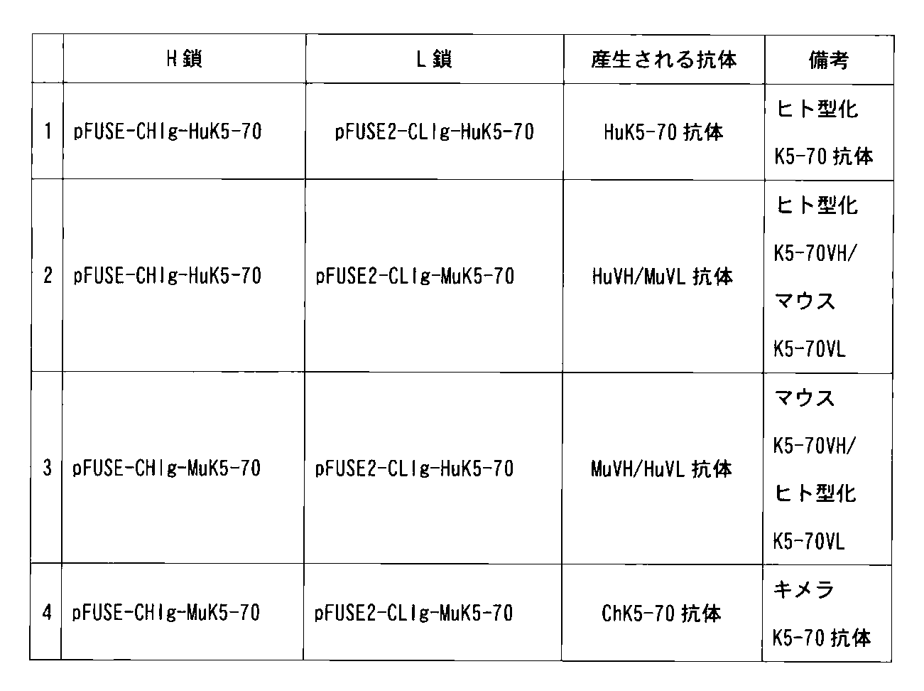
作製した18種類のHuK5−70 VH変異体(pFUSE−CHIg−HuK5−70変異体の発現ベクター)を、それぞれHuK5−70 L鎖発現ベクター(pFUSE2−CLIg−HuK5−70)と組み合わせて、HEK293細胞にトランスフェクションし、その培養上清を用いて各アミノ酸置換抗体とhTROP−2との結合活性を低密度抗原固相化ELISAで検討した。その結果、18種類のHuK5−70 VH変異体のうち、HuK5−70 VHの44番目のR(アルギニン)をG(グリシン)に置換したR44G変異体(HuK5−70 R44G;以下HuK5−70−2)において、明らかな抗原結合活性の向上が認められた(図68)。HuK5−70 VH R44G(以下HuK5−70 VH2)遺伝子の配列は図69に示した。
[Preparation and characterization of humanized K5-70 antibody mutant]
The following experiment was conducted for the purpose of improving the “Avidity” of the HuK5-70 antibody.
As described above, whether the relatively low “Avidity” of the HuK5-70 antibody is caused by VH or VL was examined in the following experiment. First, genes encoding the H-chain variable region (K5-70 VH) and L-chain variable region (K5-70 VL) of the parent antibody K5-70 antibody were prepared by gene synthesis (Operon). At that time, a Kozak sequence (ACC ACC) was added to the 5 ′ end side of the gene sequences of K5-70 VH and K5-70 VL. Further, an EcoRI site (GAA TTC) was added as a restriction enzyme site to the 5 ′ end of K5-70 VH, and an NheI site (GCT AGC) was added to the 3 ′ end. Similarly, an Age I site (ACC GGT) was added as a restriction enzyme site to the 5 ′ end of K5-70 VL, and a BsiWI site (CGT ACG) was added to the 3 ′ end. The synthesized K5-70 VH gene and K5-70 VL gene were incorporated into a pCR2.1 vector (Invitrogen). The gene sequences of K5-70 VH and VL prepared by gene synthesis are shown in FIGS. 64 and 65, respectively. The K5-70 VH and K5-70 VL genes incorporated into the pCR2.1 vector were digested with restriction enzymes EcoRI and Nhe I, Age I and BsiWI, respectively, and the gene fragments were collected. Next, the cut-out K5-70 VH gene is a human IgG1 form expression vector, pFUSE-CHIg-hG1 vector (InvivoGen) EcoRI / Nhe I site, K5-70 VL gene is a human Ig κ form expression vector A mouse-human chimeric construct was completed by inserting the vector into the Age I / BsiWI site of the vector pFUSE2-CLIg-hk vector (InvivoGen) (pFUSE-CHIg-MuK5-70, pFUSE2-CLIg-MuK5-70).
The construct prepared above and the HuK5-70 heavy chain expression vector (pFUSE-CHIg-HuK5-70) and HuK5-70 light chain expression vector (pFUSE2-CLIg-HuK5-70) prepared in Example 36 are shown in the table below. 293F cells (Invitrogen) were co-expressed in combinations of 1 to 4.
The produced 18 types of HuK5-70 VH mutants (pFUSE-CHIg-HuK5-70 mutant expression vectors) were combined with HuK5-70 L chain expression vectors (pFUSE2-CLIg-HuK5-70), respectively, to form HEK293 cells. The culture supernatant was used to examine the binding activity between each amino acid-substituted antibody and hTROP-2 by low-density antigen-immobilized ELISA. As a result, among the 18 kinds of HuK5-70 VH mutants, the R44G mutant (HuK5-70 R44G; hereinafter referred to as HuK5-70-2) in which the 44th R (arginine) of HuK5-70 VH was replaced with G (glycine). ), A clear improvement in antigen binding activity was observed (FIG. 68). The sequence of the HuK5-70 VH R44G (hereinafter HuK5-70 VH2) gene is shown in FIG.
[HuK5−70−2抗体の精製およびキャラクラリゼーション]
HuK5−70 R44G変異型抗体であるHuK5−70−2抗体は、以下のように精製した。pFUSE−CHIg−HuK5−70 R44GとpFUSE2−CLIg−HuK5−70を293F細胞にトランスフェクションし、5日間培養を行った後、培養上清を回収した。回収した培養上清からrProtein A sepharose Fast Flow(GE Healthcare)を用いて、HuK5−70−2抗体を精製した。精製したHuK5−70−2抗体を還元条件下でSDS−PAGEにより展開したところ、約50kDaのH鎖と約25kDaのL鎖が確認され、それぞれの抗体の純度は95%以上であった(図70)。
精製したHuK5−70−2およびHuK5−70抗体のhTROP−2との結合活性を高密度抗原固相化ELISAおよび低密度抗原固相化ELISAで検討した。高密度抗原固相化ELISAで(1μg/mLのhTROP−2を固相化した96ウェルプレートを使用)、アフィニティーの検証を行った結果、HuK5−70抗体とHuK5−70−2抗体の結合曲線はほぼ重なり、両者のアフィニティーは同等であり、次に低密度抗原固相化ELISA(0.1μg/mLのhTROP−2を固相化した96ウェルプレートを使用)によりアビディティーの検証を行った結果、培養上清を用いた場合の結果と同様、HuK5−70−2抗体の抗原結合活性はHuK5−70抗体より明らかに高かった(図71)。具体的には、EC50値は、K5−70抗体が11.4ng/mL、HuK5−70抗体が33.4ng/mL、HuK5−70−2抗体が11.4ng/mLであり、HuK5−70−2抗体は、HuK5−70抗体と比べて“アビディティー”が向上し、K5−70抗体と同等の活性を有するものであることが示された。
[Purification and characterization of HuK5-70-2 antibody]
The HuK5-70-2 antibody, which is a HuK5-70 R44G mutant antibody, was purified as follows. pFUSE-CHIg-HuK5-70 R44G and pFUSE2-CLIg-HuK5-70 were transfected into 293F cells and cultured for 5 days, and then the culture supernatant was collected. The HuK5-70-2 antibody was purified from the collected culture supernatant using rProtein A Sepharose Fast Flow (GE Healthcare). When the purified HuK5-70-2 antibody was developed by SDS-PAGE under reducing conditions, an approximately 50 kDa H chain and an approximately 25 kDa L chain were confirmed, and the purity of each antibody was 95% or more (Fig. 70).
The binding activity of the purified HuK5-70-2 and HuK5-70 antibodies to hTROP-2 was examined by high-density antigen-immobilized ELISA and low-density antigen-immobilized ELISA. As a result of affinity verification using a high-density antigen-immobilized ELISA (using a 96-well plate immobilizing 1 μg / mL hTROP-2), binding curves of HuK5-70 antibody and HuK5-70-2 antibody The affinities were verified by low-density antigen-immobilized ELISA (using a 96-well plate on which 0.1 μg / mL hTROP-2 was immobilized). As a result, the antigen binding activity of the HuK5-70-2 antibody was clearly higher than that of the HuK5-70 antibody, as in the case of using the culture supernatant (FIG. 71). Specifically, EC 50 values are 11.4 ng / mL for K5-70 antibody, 33.4 ng / mL for HuK5-70 antibody, 11.4 ng / mL for HuK5-70-2 antibody, and HuK5-70. The -2 antibody has improved avidity as compared with the HuK5-70 antibody, and was shown to have the same activity as the K5-70 antibody.
[ヒト型化抗hTROP−2抗体の抗体依存性細胞傷害(ADCC)活性の測定]
(1)標的細胞溶液の調整
標的細胞としては、内在的にhTROP−2を発現するヒト大腸癌細胞株SW480、ヒト膵臓癌細胞株PK−59、ヒト前立腺癌細胞株PC−3を用いた。10cm細胞培養ディッシュで培養したターゲット細胞をトリプシン処理によってプレートから剥離し、アッセイ培地に懸濁した。ADCCアッセイ用の培地は,0.5% FBSを加えたLeivovitz L−15培地(SW480細胞)またはRPMI−1640培地(PK−59、PC−3細胞)を用いた。遠心後(1000rpm,3分、室温)、ペレットを同じ培地で2×10⁵cell/mLの細胞密度になるように調製し、標的細胞溶液とした。
(2)ヒト末梢血単核細胞の分離
健常人静脈血をヘパリン採血し、PBSで2倍に希釈後、Lymphoprep(第一化学薬品)上に重層し遠心した(室温、750rpm,5分、次に2000rpm,20分)。遠心後、中間層分画にある単核細胞(健常人末梢血単核球)を回収し、PBSで3回洗浄後、アッセイ培地で細胞懸濁液を調製し、エフェクター細胞として使用した。
(3)ヒト型化抗hTROP−2抗体(HuK5−70,HuK5−70−2およびHuT6−16−2抗体)のADCC活性
96穴平底プレート(FALCON社製)に調製した標的細胞溶液の100μL(2×10⁴cell/ウェル)を分注した。次いで、ヒト末梢血単核細胞(エフェクター細胞)を、エフェクター細胞:ターゲット細胞比が40:1となるように添加した。更に、被験抗体として、ヒト型化抗hTROP−2抗体(HuK5−70,HuK5−70−2およびHuT6−16−2抗体)を最終濃度が0.1~30μg/mLとなるように加えて、全量を200μLとしてCO2インキュベーター内(37℃、5%CO2)で6時間培養を行った。培養後、Cytotoxicity Detection Kit(LDH)(Roche,Cat.No.11 644 793001)を用い、Kit添付のプロトコールに従って、エフェクター細胞により傷害を受けたターゲット細胞の細胞質から培養上清へ放出される乳酸脱水素酵素(LDH)の活性を測定し、この測定結果を指標としてADCC活性を評価した。
図72A~72Cに示したとおり、HuK5−70、およびHuT6−16−2抗体はhTROP−2を細胞表面に発現するヒト大腸癌細胞株SW480(図72A)、ヒト膵癌細胞株PK−59(図72B)、ヒト前立腺癌細胞株PC−3(図72C)に対して、用量依存的にADCC活性を発揮することが明らかとなった。また図72Bおよび図72Cに示したとおり、HuK5−70−2抗体はヒト膵癌細胞株PK−59およびヒト前立腺癌細胞株PC−3細胞に対して、HuK5−70抗体よりも強いADCC活性を発揮することが明らかとなった。HuK5−70−2抗体は、前述のとおり、HuK5−70 VHの44番目のR(アルギニン)をG(グリシン)に置換することにより、hTROP−2に対する結合能(アビディティー)が向上した抗体であり、HuK5−70抗体に比べて向上したアビディティーがADCC活性に反映していることが示された。したがって、HuK5−70−2抗体は、in vivoにおいても、HuK5−70抗体と同様に優れた抗腫瘍活性を有する(好ましくは、HuK5−70抗体より高い抗腫瘍活性を有し得る)ものと推認される。以上の結果から、HuK5−70、HuK5−70−2及びHuT6−16−2抗体は、hTROP−2を細胞表面に発現する癌に対して有効な治療用抗体となることが示唆された。
[Measurement of Antibody Dependent Cytotoxicity (ADCC) Activity of Humanized Anti-hTROP-2 Antibody]
(1) Preparation of target cell solution As target cells, human colon cancer cell line SW480, human pancreatic cancer cell line PK-59, and human prostate cancer cell line PC-3 that endogenously express hTROP-2 were used. Target cells cultured in a 10 cm cell culture dish were detached from the plate by trypsin treatment and suspended in assay medium. As a medium for ADCC assay, Leivovitz L-15 medium (SW480 cell) or RPMI-1640 medium (PK-59, PC-3 cell) supplemented with 0.5% FBS was used. After centrifugation (1000 rpm, 3 minutes, room temperature), the pellet was prepared in the same medium to a cell density of 2 × 10 5 cells / mL, and used as a target cell solution.
(2) Isolation of human peripheral blood mononuclear cells Healthy human venous blood was collected with heparin, diluted twice with PBS, layered on Lymphoprep (Daiichi Kagaku) and centrifuged (room temperature, 750 rpm, 5 minutes, next) 2000 rpm, 20 minutes). After centrifugation, mononuclear cells (normal human peripheral blood mononuclear cells) in the intermediate layer fraction were collected, washed three times with PBS, a cell suspension was prepared in assay medium, and used as effector cells.
(3) ADCC activity of humanized anti-hTROP-2 antibodies (HuK5-70, HuK5-70-2 and HuT6-16-2 antibodies) 100 μL of the target cell solution prepared on a 96-well flat bottom plate (manufactured by FALCON) ( 2 × 10 4 cells / well) were dispensed. Subsequently, human peripheral blood mononuclear cells (effector cells) were added so that the effector cell: target cell ratio was 40: 1. Furthermore, humanized anti-hTROP-2 antibodies (HuK5-70, HuK5-70-2 and HuT6-16-2 antibodies) were added as test antibodies so that the final concentration was 0.1 to 30 μg / mL, CO 2 incubator the total amount of 200μL (37 ℃, 5% CO 2) was subjected to 6 hours at. Lactic acid dehydration released from the cytoplasm of target cells damaged by effector cells from the cytoplasm to the culture supernatant using Cytotoxicity Detection Kit (LDH) (Roche, Cat. No. 11 644 793001) according to the protocol attached to the kit. Elementary enzyme (LDH) activity was measured, and ADCC activity was evaluated using this measurement result as an index.
As shown in FIGS. 72A to 72C, the HuK5-70 and HuT6-16-2 antibodies are human colorectal cancer cell lines SW480 (FIG. 72A) that express hTROP-2 on the cell surface, human pancreatic cancer cell line PK-59 (FIG. 72). 72B) and human prostate cancer cell line PC-3 (FIG. 72C) were revealed to exhibit ADCC activity in a dose-dependent manner. As shown in FIGS. 72B and 72C, the HuK5-70-2 antibody exerts stronger ADCC activity than the HuK5-70 antibody against the human pancreatic cancer cell line PK-59 and the human prostate cancer cell line PC-3 cells. It became clear to do. As described above, the HuK5-70-2 antibody is an antibody having improved binding ability (avidity) to hTROP-2 by replacing the 44th R (arginine) of HuK5-70 VH with G (glycine). It was shown that the avidity improved compared to the HuK5-70 antibody is reflected in the ADCC activity. Therefore, it is estimated that the HuK5-70-2 antibody has excellent antitumor activity in vivo as well as the HuK5-70 antibody (preferably, it can have higher antitumor activity than the HuK5-70 antibody). Is done. From the above results, it was suggested that the HuK5-70, HuK5-70-2 and HuT6-16-2 antibodies are effective therapeutic antibodies for cancers that express hTROP-2 on the cell surface.
本発明によれば、hTROP−2と特異的に反応し、in vivoにおいて高い抗腫瘍活性を有する抗体、具体的には、低用量でin vivoでの高い抗腫瘍活性を有するモノクローナル抗体、特に当該抗体のうちヒト型化抗体であるものを提供することができる。さらに本発明は、当該抗体を産生するハイブリドーマ、当該抗体の断片、当該抗体等と各種薬剤との複合体、腫瘍の診断用又は治療用医薬組成物、腫瘍の検出方法、腫瘍の検出用又は診断用キットを提供することができる。 According to the present invention, an antibody that specifically reacts with hTROP-2 and has a high antitumor activity in vivo, specifically, a monoclonal antibody having a high antitumor activity in vivo at a low dose, in particular An antibody that is a humanized antibody can be provided. Furthermore, the present invention relates to a hybridoma producing the antibody, a fragment of the antibody, a complex of the antibody and the like and various drugs, a pharmaceutical composition for diagnosis or treatment of a tumor, a method for detecting a tumor, a detection or diagnosis for a tumor Kits can be provided.
配列番号3:合成DNA
配列番号4:合成DNA
配列番号5:合成DNA
配列番号6:合成DNA
配列番号7:合成DNA
配列番号8:合成DNA
配列番号9:合成DNA
配列番号10:合成DNA
配列番号11:合成DNA
配列番号12:合成DNA
配列番号13:合成DNA
配列番号14:合成DNA
配列番号15:合成DNA
配列番号16:合成DNA
配列番号17:合成DNA
配列番号18:合成DNA
配列番号19:合成DNA
配列番号20:合成DNA
配列番号21:合成DNA
配列番号22:合成DNA
配列番号23:合成DNA
配列番号24:合成DNA
配列番号25:合成DNA
配列番号26:合成DNA
配列番号27:合成DNA
配列番号28:合成DNA
配列番号29:合成DNA
配列番号30:合成DNA
配列番号31:合成DNA
配列番号32:合成DNA
配列番号33:合成DNA
配列番号74:組換えDNA
配列番号75:合成コンストラクト(組換えタンパク質)
配列番号76:組換えDNA
配列番号77:合成コンストラクト(組換えタンパク質)
配列番号78:組換えDNA
配列番号79:合成コンストラクト(組換えタンパク質)
配列番号80:組換えDNA
配列番号81:合成コンストラクト(組換えタンパク質)
配列番号82:組換えDNA
配列番号83:合成コンストラクト(組換えタンパク質)
配列番号92:組換えタンパク質
配列番号93:組換えタンパク質
配列番号94:組換えタンパク質
配列番号95:組換えタンパク質
配列番号96:組換えタンパク質
配列番号97:組換えタンパク質
配列番号98:組換えタンパク質
配列番号99:組換えDNA
配列番号100:組換えDNA
Sequence number 3: Synthetic DNA
Sequence number 4: Synthetic DNA
Sequence number 5: Synthetic DNA
Sequence number 6: Synthetic DNA
SEQ ID NO: 7: synthetic DNA
Sequence number 8: Synthetic DNA
SEQ ID NO: 9: synthetic DNA
SEQ ID NO: 10: synthetic DNA
SEQ ID NO: 11: synthetic DNA
SEQ ID NO: 12: synthetic DNA
SEQ ID NO: 13: synthetic DNA
SEQ ID NO: 14: Synthetic DNA
SEQ ID NO: 15: synthetic DNA
SEQ ID NO: 16: synthetic DNA
Sequence number 17: Synthetic DNA
Sequence number 18: Synthetic DNA
Sequence number 19: Synthetic DNA
SEQ ID NO: 20: synthetic DNA
SEQ ID NO: 21: synthetic DNA
SEQ ID NO: 22: synthetic DNA
SEQ ID NO: 23: synthetic DNA
SEQ ID NO: 24: Synthetic DNA
SEQ ID NO: 25: synthetic DNA
SEQ ID NO: 26: Synthetic DNA
SEQ ID NO: 27: synthetic DNA
SEQ ID NO: 28: synthetic DNA
SEQ ID NO: 29: synthetic DNA
SEQ ID NO: 30: synthetic DNA
SEQ ID NO: 31: synthetic DNA
SEQ ID NO: 32: synthetic DNA
SEQ ID NO: 33: synthetic DNA
SEQ ID NO: 74: recombinant DNA
SEQ ID NO: 75: synthetic construct (recombinant protein)
SEQ ID NO: 76: recombinant DNA
SEQ ID NO: 77: synthetic construct (recombinant protein)
SEQ ID NO: 78: recombinant DNA
SEQ ID NO: 79: synthetic construct (recombinant protein)
SEQ ID NO: 80: recombinant DNA
SEQ ID NO: 81: synthetic construct (recombinant protein)
SEQ ID NO: 82: recombinant DNA
SEQ ID NO: 83: Synthetic construct (recombinant protein)
SEQ ID NO: 92: recombinant protein SEQ ID NO: 93: recombinant protein SEQ ID NO: 94: recombinant protein SEQ ID NO: 95: recombinant protein SEQ ID NO: 96: recombinant protein SEQ ID NO: 97: recombinant protein SEQ ID NO: 98: recombinant protein sequence Number 99: Recombinant DNA
SEQ ID NO: 100: recombinant DNA
Claims (44)
Priority Applications (13)
| Application Number | Priority Date | Filing Date | Title |
|---|---|---|---|
| EA201491006A EA035953B1 (en) | 2011-11-22 | 2012-11-21 | ANTIBODY AGAINST HUMAN TROP-2 WITH ANTITUMOR ACTIVITY IN VIVO |
| NZ623464A NZ623464B2 (en) | 2011-11-22 | 2012-11-21 | Anti-human trop-2 antibody having an antitumor activity in vivo |
| MX2017001860A MX383886B (en) | 2011-11-22 | 2012-11-21 | HUMAN ANTI-TROP-2 ANTIBODY THAT HAS ANTI-TUMORAL ACTIVITY IN VIVO. |
| EP12852269.5A EP2799452B1 (en) | 2011-11-22 | 2012-11-21 | Anti-human trop-2 antibody exhibiting antitumor activity in vivo |
| CN201280057258.4A CN104114580B (en) | 2011-11-22 | 2012-11-21 | Anti-human TROP‑2 antibody with anti-tumor activity in vivo |
| JP2013545988A JP6043725B2 (en) | 2011-11-22 | 2012-11-21 | Anti-human TROP-2 antibody having antitumor activity in vivo |
| MX2014006037A MX362720B (en) | 2011-11-22 | 2012-11-21 | ANTI-TROP-2 HUMAN ANTI-BODY THAT HAS AN IN VIVO ANTITUMORAL ACTIVITY. |
| AU2012341450A AU2012341450B2 (en) | 2011-11-22 | 2012-11-21 | Anti-human TROP-2 antibody having antitumor activity in vivo |
| BR112014012119-2A BR112014012119A2 (en) | 2011-11-22 | 2012-11-21 | human anti-trop-2 antibody having in vivo anti-tumor activity |
| KR1020147015979A KR101972625B1 (en) | 2011-11-22 | 2012-11-21 | Anti-human trop-2 antibody having an antitumor activity in vivo |
| EP18203986.7A EP3521313B1 (en) | 2011-11-22 | 2012-11-21 | Anti-human trop-2 antibody having an antitumor activity in vivo |
| CA2855699A CA2855699C (en) | 2011-11-22 | 2012-11-21 | Anti-human trop-2 antibody having an antitumor activity in vivo |
| ES12852269T ES2750358T3 (en) | 2011-11-22 | 2012-11-21 | Anti-human TROP-2 antibody showing anti-tumor activity in vivo |
Applications Claiming Priority (2)
| Application Number | Priority Date | Filing Date | Title |
|---|---|---|---|
| US201161562672P | 2011-11-22 | 2011-11-22 | |
| US61/562,672 | 2011-11-22 |
Publications (1)
| Publication Number | Publication Date |
|---|---|
| WO2013077458A1 true WO2013077458A1 (en) | 2013-05-30 |
Family
ID=48469894
Family Applications (1)
| Application Number | Title | Priority Date | Filing Date |
|---|---|---|---|
| PCT/JP2012/080800 Ceased WO2013077458A1 (en) | 2011-11-22 | 2012-11-21 | Anti-human trop-2 antibody exhibiting antitumor activity in vivo |
Country Status (12)
| Country | Link |
|---|---|
| US (2) | US9427464B2 (en) |
| EP (2) | EP2799452B1 (en) |
| JP (1) | JP6043725B2 (en) |
| KR (1) | KR101972625B1 (en) |
| CN (2) | CN104114580B (en) |
| AU (1) | AU2012341450B2 (en) |
| BR (1) | BR112014012119A2 (en) |
| CA (2) | CA2855699C (en) |
| EA (2) | EA035947B1 (en) |
| ES (2) | ES2750358T3 (en) |
| MX (2) | MX362720B (en) |
| WO (1) | WO2013077458A1 (en) |
Cited By (57)
| Publication number | Priority date | Publication date | Assignee | Title |
|---|---|---|---|---|
| WO2015098099A1 (en) | 2013-12-25 | 2015-07-02 | 第一三共株式会社 | Anti-trop2 antibody-drug conjugate |
| JP2018500930A (en) * | 2014-12-04 | 2018-01-18 | アブルッツォ・セラノスティックス・エッセ・エッレ・エッレ | Humanized anti-TROP-2 monoclonal antibody and use thereof |
| WO2020040245A1 (en) | 2018-08-23 | 2020-02-27 | 第一三共株式会社 | Sensitivity marker for antibody-drug conjugate |
| WO2020094670A1 (en) | 2018-11-05 | 2020-05-14 | Synaffix B.V. | Antibody-conjugates for targeting of tumours expressing trop-2 |
| WO2020240467A1 (en) | 2019-05-29 | 2020-12-03 | Daiichi Sankyo Company, Limited | Dosage of an antibody-drug conjugate |
| US10906974B2 (en) | 2017-01-17 | 2021-02-02 | Daiichi Sankyo Company, Limited | Anti-GPR20 antibody and anti-GPR20 antibody-drug conjugate |
| US11077202B2 (en) | 2017-05-15 | 2021-08-03 | Daiichi Sankyo Company, Limited | Anti-CDH6 antibody and anti-CDH6 antibody-drug conjugate |
| WO2021226440A1 (en) | 2020-05-08 | 2021-11-11 | Bolt Biotherapeutics, Inc. | Elastase-substrate, peptide linker immunoconjugates, and uses thereof |
| US11173213B2 (en) | 2015-06-29 | 2021-11-16 | Daiichi Sankyo Company, Limited | Method for selectively manufacturing antibody-drug conjugate |
| WO2022036101A1 (en) | 2020-08-13 | 2022-02-17 | Bolt Biotherapeutics, Inc. | Pyrazoloazepine immunoconjugates, and uses thereof |
| US11273155B2 (en) | 2016-12-12 | 2022-03-15 | Daiichi Sankyo Company, Limited | Combination of antibody-drug conjugate and immune checkpoint inhibitor |
| US11318212B2 (en) | 2017-08-31 | 2022-05-03 | Daiichi Sankyo Company, Limited | Method for producing antibody-drug conjugate |
| WO2022204536A1 (en) | 2021-03-26 | 2022-09-29 | Bolt Biotherapeutics, Inc. | 2-amino-4-carboxamide-benzazepine immunoconjugates, and uses thereof |
| WO2022204528A1 (en) | 2021-03-26 | 2022-09-29 | Bolt Biotherapeutics, Inc. | 2-amino-4-carboxamide-benzazepine immunoconjugates, and uses thereof |
| WO2022261310A1 (en) | 2021-06-11 | 2022-12-15 | Gilead Sciences, Inc. | Combination mcl-1 inhibitors with anti-body drug conjugates |
| WO2022261301A1 (en) | 2021-06-11 | 2022-12-15 | Gilead Sciences, Inc. | Combination mcl-1 inhibitors with anti-cancer agents |
| JP2022553645A (en) * | 2019-10-11 | 2022-12-26 | マブウェル (シャンハイ) バイオサイエンス カンパニー リミテッド | Anti-human Trop-2 antibody and uses thereof |
| US11584800B2 (en) | 2014-01-31 | 2023-02-21 | Daiichi Sankyo Company, Limited | Method of treating cancer comprising administration of anti-HER2 antibody-drug conjugate |
| WO2023076599A1 (en) | 2021-10-29 | 2023-05-04 | Bolt Biotherapeutics, Inc. | Tlr agonist immunoconjugates with cysteine-mutant antibodies, and uses thereof |
| WO2023077030A1 (en) | 2021-10-29 | 2023-05-04 | Gilead Sciences, Inc. | Cd73 compounds |
| WO2023076983A1 (en) | 2021-10-28 | 2023-05-04 | Gilead Sciences, Inc. | Pyridizin-3(2h)-one derivatives |
| WO2023100829A1 (en) | 2021-11-30 | 2023-06-08 | 第一三共株式会社 | Protease-degradable musk antibody |
| WO2023122615A1 (en) | 2021-12-22 | 2023-06-29 | Gilead Sciences, Inc. | Ikaros zinc finger family degraders and uses thereof |
| WO2023122581A2 (en) | 2021-12-22 | 2023-06-29 | Gilead Sciences, Inc. | Ikaros zinc finger family degraders and uses thereof |
| WO2023147418A1 (en) | 2022-01-28 | 2023-08-03 | Gilead Sciences, Inc. | Parp7 inhibitors |
| WO2023153442A1 (en) | 2022-02-09 | 2023-08-17 | 第一三共株式会社 | Environmentally responsive masked antibody and use thereof |
| EP4245756A1 (en) | 2022-03-17 | 2023-09-20 | Gilead Sciences, Inc. | Ikaros zinc finger family degraders and uses thereof |
| WO2023180485A1 (en) | 2022-03-23 | 2023-09-28 | Synaffix B.V. | Antibody-conjugates for targeting of tumours expressing trop-2 |
| WO2023201268A1 (en) | 2022-04-13 | 2023-10-19 | Gilead Sciences, Inc. | Combination therapy for treating tumor antigen expressing cancers |
| WO2023201267A1 (en) | 2022-04-13 | 2023-10-19 | Gilead Sciences, Inc. | Combination therapy for treating trop-2 expressing cancers |
| WO2023205719A1 (en) | 2022-04-21 | 2023-10-26 | Gilead Sciences, Inc. | Kras g12d modulating compounds |
| WO2024005204A1 (en) | 2022-06-30 | 2024-01-04 | 株式会社カイオム・バイオサイエンス | Fusion protein |
| WO2024006929A1 (en) | 2022-07-01 | 2024-01-04 | Gilead Sciences, Inc. | Cd73 compounds |
| US11872289B2 (en) | 2018-05-18 | 2024-01-16 | Daiichi Sankyo Co., Ltd. | Anti-MUC1 antibody-drug conjugate |
| US11945882B2 (en) | 2017-08-31 | 2024-04-02 | Daiichi Sankyo Company, Limited | Method for producing antibody-drug conjugate |
| WO2024097812A1 (en) | 2022-11-04 | 2024-05-10 | Gilead Sciences, Inc. | Therapy for treating bladder cancer |
| WO2024137852A1 (en) | 2022-12-22 | 2024-06-27 | Gilead Sciences, Inc. | Prmt5 inhibitors and uses thereof |
| WO2024138128A2 (en) | 2022-12-23 | 2024-06-27 | Genentech, Inc. | Cereblon degrader conjugates, and uses thereof |
| WO2024153127A1 (en) | 2023-01-18 | 2024-07-25 | 泰励生物科技(上海)有限公司 | Antibody-drug conjugate and use thereof |
| WO2024173387A1 (en) | 2023-02-14 | 2024-08-22 | Bolt Biotherapeutics, Inc. | Aza-benzazepine immunoconjugates, and uses thereof |
| WO2024186626A1 (en) | 2023-03-03 | 2024-09-12 | Bolt Biotherapeutics, Inc. | Aza-bicyclic sting agonist immunoconjugates, and uses thereof |
| WO2024215754A1 (en) | 2023-04-11 | 2024-10-17 | Gilead Sciences, Inc. | Kras modulating compounds |
| WO2024220917A1 (en) | 2023-04-21 | 2024-10-24 | Gilead Sciences, Inc. | Prmt5 inhibitors and uses thereof |
| WO2024226410A1 (en) | 2023-04-24 | 2024-10-31 | Merck Sharp & Dohme Llc | Trop2 binders and conjugates thereof |
| WO2025006720A1 (en) | 2023-06-30 | 2025-01-02 | Gilead Sciences, Inc. | Kras modulating compounds |
| WO2025024811A1 (en) | 2023-07-26 | 2025-01-30 | Gilead Sciences, Inc. | Parp7 inhibitors |
| WO2025024663A1 (en) | 2023-07-26 | 2025-01-30 | Gilead Sciences, Inc. | Parp7 inhibitors |
| US12220604B2 (en) | 2018-07-31 | 2025-02-11 | Daiichi Sankyo Company, Limited | Treatment of metastatic brain tumor by administration of an antibody-drug conjugate |
| WO2025054347A1 (en) | 2023-09-08 | 2025-03-13 | Gilead Sciences, Inc. | Kras g12d modulating compounds |
| WO2025054530A1 (en) | 2023-09-08 | 2025-03-13 | Gilead Sciences, Inc. | Pyrimidine-containing polycyclic derivatives as kras g12d modulating compounds |
| CN119736243A (en) * | 2024-12-04 | 2025-04-01 | 北京肿瘤医院(北京大学肿瘤医院) | Colorectal cancer high-metastasis model construction and verification method based on metastasis selection pressure |
| WO2025096589A1 (en) | 2023-11-03 | 2025-05-08 | Gilead Sciences, Inc. | Prmt5 inhibitors and uses thereof |
| WO2025137640A1 (en) | 2023-12-22 | 2025-06-26 | Gilead Sciences, Inc. | Azaspiro wrn inhibitors |
| US12358999B2 (en) | 2018-07-27 | 2025-07-15 | Daiichi Sankyo Company, Limited | Protein recognizing drug moiety of antibody-drug conjugate |
| WO2025245003A1 (en) | 2024-05-21 | 2025-11-27 | Gilead Sciences, Inc. | Prmt5 inhibitors and uses thereof |
| WO2026039365A1 (en) | 2024-08-12 | 2026-02-19 | Gilead Sciences, Inc. | Kras modulating compounds |
| WO2026055185A1 (en) | 2024-09-04 | 2026-03-12 | Bolt Biotherapeutics, Inc. | Sting agonist immunoconjugates and uses thereof |
Families Citing this family (20)
| Publication number | Priority date | Publication date | Assignee | Title |
|---|---|---|---|---|
| US9770517B2 (en) | 2002-03-01 | 2017-09-26 | Immunomedics, Inc. | Anti-Trop-2 antibody-drug conjugates and uses thereof |
| CA2478047C (en) | 2002-03-01 | 2014-01-21 | Immunomedics, Inc. | Rs7 antibodies |
| US9745380B2 (en) | 2002-03-01 | 2017-08-29 | Immunomedics, Inc. | RS7 antibodies |
| AU2017231833B2 (en) | 2016-03-10 | 2024-03-14 | Viela Bio, Inc. | ILT7 binding molecules and methods of using the same |
| CN107446050A (en) * | 2017-08-11 | 2017-12-08 | 百奥泰生物科技(广州)有限公司 | The compound and method of Trop2 positive diseases treatment |
| WO2019196117A1 (en) * | 2018-04-13 | 2019-10-17 | Dingfu Biotarget Co., Ltd. | Anti-cd27 antibodies and use thereof |
| CN112243443B (en) * | 2019-05-10 | 2023-03-17 | 江苏豪森药业集团有限公司 | anti-TROP-2 antibodies, antigen-binding fragments thereof, and medical uses thereof |
| US20210093730A1 (en) * | 2019-10-01 | 2021-04-01 | Immunomedics, Inc. | Biomarkers for antibody-drug conjugate monotherapy or combination therapy |
| CN116234573A (en) | 2020-10-14 | 2023-06-06 | 上海翰森生物医药科技有限公司 | Anti-TROP-2 antibody, its antigen-binding fragment or its mutant, and its medical use |
| CN114685669A (en) * | 2020-12-30 | 2022-07-01 | 和铂医药(苏州)有限公司 | Antibodies that bind TROP2 and uses thereof |
| CN113122494B (en) * | 2021-04-25 | 2023-01-31 | 上海交通大学医学院附属第九人民医院 | Construction method of human prostate epithelial cell finasteride drug-resistant model |
| CA3232425A1 (en) | 2021-09-23 | 2023-03-30 | Shanghai Hansoh Biomedical Co., Ltd. | Antibody-drug conjugate, preparation method therefor, and pharmaceutical use thereof |
| EP4408893A4 (en) | 2021-09-29 | 2026-02-18 | Kisoji Biotechnology Inc | Binding agents targeting TROP2-expressing tumor cells |
| CN114195888B (en) * | 2021-12-07 | 2023-06-09 | 江苏中新医药有限公司 | Core sequence of anti-novel crown antibody medicine |
| US20250034275A1 (en) * | 2021-12-07 | 2025-01-30 | Chia Tai Tianqing Pharmaceutical Group Co., Ltd. | Anti-trop-2 antibody or antigen-binding fragment thereof |
| EP4460564A4 (en) * | 2022-01-08 | 2026-02-11 | Carogen Corp | Multiantigenic therapeutic vaccines for the treatment or prevention of chronic hepatitis B virus infection |
| US20250206841A1 (en) * | 2022-03-28 | 2025-06-26 | Adept Therapeutics Inc. | Anti-trophoblast cell surface antigen 2 (trop-2) antibodies |
| CN117430710B (en) * | 2022-07-21 | 2025-07-04 | 中国医学科学院医药生物技术研究所 | A monoclonal antibody targeting Trop2 and its application |
| KR20260012304A (en) | 2023-04-17 | 2026-01-26 | 피크 바이오, 인크. | Antibodies and antibody-drug conjugates and methods of use and synthetic processes and intermediates |
| WO2025137257A1 (en) * | 2023-12-21 | 2025-06-26 | The Regents Of The University Of California | Trop2 as a biomarker for detection of epithelial cancers and predicting and monitoring responses to therapies |
Citations (9)
| Publication number | Priority date | Publication date | Assignee | Title |
|---|---|---|---|---|
| JPH04502408A (en) | 1988-12-28 | 1992-05-07 | プロテイン デザイン ラブス,インコーポレイティド | Chimeric immunoglobulin specific for p55 Tac protein of IL-2 receptor |
| US5840854A (en) | 1995-10-19 | 1998-11-24 | Bristol-Myers Squibb Company | Monoclonal antibody BR110 and uses thereof |
| US6653104B2 (en) | 1996-10-17 | 2003-11-25 | Immunomedics, Inc. | Immunotoxins, comprising an internalizing antibody, directed against malignant and normal cells |
| WO2005052156A1 (en) | 2003-11-28 | 2005-06-09 | Kanagawa Academy Of Science And Technology | Method of detecting liver cancer, diagnostic for liver cancer and remedy for cancer |
| JP2006502698A (en) * | 2002-03-01 | 2006-01-26 | イミューノメディクス、インコーポレイテッド | RS7 antibody |
| US20070202113A1 (en) * | 2006-02-24 | 2007-08-30 | Arius Research, Inc. | Cytotoxicity mediation of cells evidencing surface expression of TROP-2 |
| WO2010089782A1 (en) | 2009-02-05 | 2010-08-12 | Saverio Alberti | Anti-trop-2 monoclonal antibodies and uses thereof in the treatment and diagnosis of tumors |
| JP2010528056A (en) * | 2007-05-30 | 2010-08-19 | エフ.ホフマン−ラ ロシュ アーゲー | Humanized anti-TROP-2 antibody and chimeric anti-TROP-2 antibody that mediate cell killing effect of cancer cells |
| WO2011145744A1 (en) * | 2010-05-17 | 2011-11-24 | 株式会社リブテック | Anti-human trop-2 antibody having antitumor activity in vivo |
-
2012
- 2012-11-20 US US13/682,319 patent/US9427464B2/en active Active
- 2012-11-21 ES ES12852269T patent/ES2750358T3/en active Active
- 2012-11-21 CA CA2855699A patent/CA2855699C/en active Active
- 2012-11-21 ES ES18203986T patent/ES2927090T3/en active Active
- 2012-11-21 EA EA201692042A patent/EA035947B1/en not_active IP Right Cessation
- 2012-11-21 EP EP12852269.5A patent/EP2799452B1/en active Active
- 2012-11-21 CN CN201280057258.4A patent/CN104114580B/en active Active
- 2012-11-21 EP EP18203986.7A patent/EP3521313B1/en active Active
- 2012-11-21 EA EA201491006A patent/EA035953B1/en not_active IP Right Cessation
- 2012-11-21 CN CN201710618845.0A patent/CN107236043B/en active Active
- 2012-11-21 MX MX2014006037A patent/MX362720B/en active IP Right Grant
- 2012-11-21 AU AU2012341450A patent/AU2012341450B2/en active Active
- 2012-11-21 BR BR112014012119-2A patent/BR112014012119A2/en not_active Application Discontinuation
- 2012-11-21 KR KR1020147015979A patent/KR101972625B1/en active Active
- 2012-11-21 JP JP2013545988A patent/JP6043725B2/en active Active
- 2012-11-21 CA CA3027417A patent/CA3027417C/en active Active
- 2012-11-21 MX MX2017001860A patent/MX383886B/en unknown
- 2012-11-21 WO PCT/JP2012/080800 patent/WO2013077458A1/en not_active Ceased
-
2016
- 2016-05-31 US US15/169,109 patent/US10202461B2/en active Active
Patent Citations (14)
| Publication number | Priority date | Publication date | Assignee | Title |
|---|---|---|---|---|
| JPH04502408A (en) | 1988-12-28 | 1992-05-07 | プロテイン デザイン ラブス,インコーポレイティド | Chimeric immunoglobulin specific for p55 Tac protein of IL-2 receptor |
| JP2828340B2 (en) | 1988-12-28 | 1998-11-25 | プロテイン デザイン ラブス,インコーポレイティド | Chimeric immunoglobulin specific for the IL-2 receptor p55 Tac protein |
| US5840854A (en) | 1995-10-19 | 1998-11-24 | Bristol-Myers Squibb Company | Monoclonal antibody BR110 and uses thereof |
| US6653104B2 (en) | 1996-10-17 | 2003-11-25 | Immunomedics, Inc. | Immunotoxins, comprising an internalizing antibody, directed against malignant and normal cells |
| JP2006502698A (en) * | 2002-03-01 | 2006-01-26 | イミューノメディクス、インコーポレイテッド | RS7 antibody |
| WO2005052156A1 (en) | 2003-11-28 | 2005-06-09 | Kanagawa Academy Of Science And Technology | Method of detecting liver cancer, diagnostic for liver cancer and remedy for cancer |
| US20070202113A1 (en) * | 2006-02-24 | 2007-08-30 | Arius Research, Inc. | Cytotoxicity mediation of cells evidencing surface expression of TROP-2 |
| US7420041B2 (en) | 2006-02-24 | 2008-09-02 | Arius Research Inc. | Cytotoxicity mediation of cells evidencing surface expression of TROP-2 |
| US7420040B2 (en) | 2006-02-24 | 2008-09-02 | Arius Research Inc. | Cytotoxicity mediation of cells evidencing surface expression of TROP-2 |
| JP2009527230A (en) * | 2006-02-24 | 2009-07-30 | アリアス リサーチ、インコーポレイテッド | Cytotoxic mediation of cells demonstrating surface expression of TROP-2 |
| JP2009528995A (en) * | 2006-02-24 | 2009-08-13 | アリアス リサーチ、インコーポレイテッド | Cancerous disease modifying antibody 141205-05 |
| JP2010528056A (en) * | 2007-05-30 | 2010-08-19 | エフ.ホフマン−ラ ロシュ アーゲー | Humanized anti-TROP-2 antibody and chimeric anti-TROP-2 antibody that mediate cell killing effect of cancer cells |
| WO2010089782A1 (en) | 2009-02-05 | 2010-08-12 | Saverio Alberti | Anti-trop-2 monoclonal antibodies and uses thereof in the treatment and diagnosis of tumors |
| WO2011145744A1 (en) * | 2010-05-17 | 2011-11-24 | 株式会社リブテック | Anti-human trop-2 antibody having antitumor activity in vivo |
Non-Patent Citations (50)
| Title |
|---|
| ALLIKMETS, R. ET AL., HUM. MOL. GENET., vol. 5, no. 10, 1996, pages 1649 - 1655 |
| BASU A ET AL., INT. J. CANCER, vol. 62, no. 4, 1995, pages 472 - 479 |
| BLUME; CEVC, BIOCHEM. ET BIOPHYS. ACTA, vol. 1029, 1990, pages 91 |
| BR. J. CANCER, vol. 99, 2008, pages 1290 - 1295 |
| CALABRESE G ET AL., CELL GENET., vol. 92, no. 1-2, 2001, pages 164 - 165 |
| CANCER CHEMOTHER PHARMACOL., vol. 28, no. 3, 1991, pages 192 - 8 |
| CARMEN, S., BRIEFINGS IN FUNCTIONAL GENOMICS AND PROTEOMICS, vol. 1, no. 2, 2002, pages 189 - 203 |
| CHEN, C. J. ET AL., CELL, vol. 47, no. 3, 1986, pages 381 - 389 |
| CHENWEI LI ET AL., CANCER RES, vol. 67, no. 3, 2007, pages 1030 - 1037 |
| CHONN; CULLIS, CURRENT OP. BIOTECH., vol. 6, 1995, pages 698 |
| CLIN. CANCER RES., vol. 12, 2006, pages 3057 - 3063 |
| CUBAS R ET AL., BIOCHIM. BIOPHYS. ACTA, vol. 1796, no. 2, 2009, pages 309 - 314 |
| DJAVADI-OHANIANCE L. ET AL.: "Antibody Engineering", 1996, IRL PRESS, article "4", pages: 77 - 97 |
| EL SEWEDY T ET AL., INT. J. CANCER, vol. 75, no. 2, 1998, pages 324 - 330 |
| FANG YJ ET AL., INT. J. COLORECTAL DIS., vol. 24, no. 8, 2009, pages 875 - 884 |
| FAULK WP ET AL., PROC. NATL. ACAD. SCI. U.S.A., vol. 75, no. 4, 1978, pages 1947 - 1951 |
| FONG D ET AL., BR. J. CANCER, vol. 99, no. 8, 2008, pages 1290 - 1295 |
| FONG D ET AL., MOD. PATHOL., vol. 21, no. 2, 2008, pages 186 - 191 |
| FORNARO M ET AL., INT. J. CANCER, vol. 62, no. 5, 1995, pages 610 - 618 |
| GENOME RES., vol. 16, 2006, pages 55 - 65 |
| J. BIOL. CHEM., vol. 270, 1995, pages 12457 - 12465 |
| J. IMMUNOL., vol. 149, 1992, pages 2518 - 2529 |
| J. MOL. BIOL., vol. 196, 1987, pages 901 - 917 |
| JANE E. VISVADER; GEOFFREY J. LINDEMAN, NAT REV CANCER., vol. 8, no. 10, 2008, pages 755 - 68 |
| KABAT ET AL.: "Sequences of Proteins of Immunological Interests", 1991, NIH PUBLICATION NO. 91-3242, U.S. DEPARTMENT OF HEALTH AND HUMAN SERVICES |
| KUROIWA, Y., NUC. ACIDS RES., vol. 26, 1998, pages 3447 - 3448 |
| LANDOLFI ET AL., J. IMMUNOL., vol. 166, 2001, pages 1748 |
| LAPPALAINEN ET AL., ANTIVIRAL RES., vol. 23, 1994, pages 119 |
| LI ET AL., CANCER RES, vol. 67, no. 3, 2007, pages 1030 - 1037 |
| LINNENBACH AJ ET AL., PROC. NATL. ACAD. SCI. U.S.A., vol. 86, no. 1, 1989, pages 27 - 31 |
| LIPINSKI M ET AL., PROC. NATL. ACAD. SCI. U.S.A., vol. 78, no. 8, 1981, pages 5147 - 5150 |
| MANNINO ET AL., BIOTECHNIQUES, vol. 6, 1988, pages 682 |
| MOD. PATHOL., vol. 21, 2008, pages 186 - 191 |
| MOLECULAR CANCER, vol. 9, 2010, pages 253 |
| NATURE, vol. 321, 1986, pages 522 - 525 |
| OHMACHI T ET AL., CLIN. CANCER RES., vol. 12, no. 10, 2006, pages 3057 - 3063 |
| PROC. NATL. ACAD. SCI. U.S.A., vol. 81, 1984, pages 6851 - 6855 |
| QUEEN C ET AL., PROC. NATL. ACAD. SCI. USA, vol. 86, 1989, pages 10029 - 10033 |
| QUEEN ET AL., PROC. NATL. ACAD. SCI. USA, vol. 86, 1989, pages 10029 - 10033 |
| REITER ET AL., PROTEIN ENGINEERING, vol. 7, 1994, pages 697 - 704 |
| RIPANI E ET AL., INT. J. CANCER, vol. 76, no. 5, 1998, pages 671 - 676 |
| SAMBROOK J. ET AL.: "Molecular Cloning, A Laboratory Manual, 3rd edition", 2001, COLD SPRING HARBOR LABORATORY PRESS |
| SANTIN AD ET AL., INT. J. CANCER, vol. 112, no. 1, 2004, pages 14 - 25 |
| See also references of EP2799452A4 |
| SIRIWARDENA, D., OPTHALMOLOGY, vol. 109, no. 3, 2002, pages 427 - 431 |
| TOMIZUKA, K. ET AL., NATURE GENETICS, vol. 16, 1977, pages 133 - 143 |
| TOMIZUKA, K., PROC. NATL. ACAD. SCI. USA, vol. 97, 2000, pages 722 - 727 |
| WANG J ET AL., MOL. CANCER THER., vol. 7, no. 2, 2008, pages 280 - 285 |
| WORMSTONE, I. M., INVESTIGATIVE OPHTHALMOLOGY & VISUAL SCIENCE, vol. 43, no. 7, 2002, pages 2301 - 8 |
| YOSHIDA, H.: "Animal Cell Technology: Basic and Applied Aspects", vol. 10, 1999, KLUWER ACADEMIC PUBLISHERS, pages: 69 - 73 |
Cited By (91)
| Publication number | Priority date | Publication date | Assignee | Title |
|---|---|---|---|---|
| KR20210041105A (en) | 2013-12-25 | 2021-04-14 | 다이이찌 산쿄 가부시키가이샤 | Anti-trop2 antibody-drug conjugate |
| WO2015098099A1 (en) | 2013-12-25 | 2015-07-02 | 第一三共株式会社 | Anti-trop2 antibody-drug conjugate |
| US9850312B2 (en) | 2013-12-25 | 2017-12-26 | Daiichi Sankyo Company, Limited | Anti-TROP2 antibody-drug conjugate |
| KR20220132020A (en) | 2013-12-25 | 2022-09-29 | 다이이찌 산쿄 가부시키가이샤 | Anti-TROP2 Antibody-Drug Conjugate |
| EP3424955A1 (en) | 2013-12-25 | 2019-01-09 | Daiichi Sankyo Company, Limited | Anti-trop2 antibody-drug conjugate |
| KR20190006087A (en) | 2013-12-25 | 2019-01-16 | 다이이찌 산쿄 가부시키가이샤 | Anti-trop2 antibody-drug conjugate |
| US10227417B2 (en) | 2013-12-25 | 2019-03-12 | Daiichi Sankyo Company, Limited | Anti-TROP2 antibody-drug conjugate |
| KR20230018530A (en) | 2013-12-25 | 2023-02-07 | 다이이찌 산쿄 가부시키가이샤 | Anti-trop2 antibody-drug conjugate |
| KR20200028047A (en) | 2013-12-25 | 2020-03-13 | 다이이찌 산쿄 가부시키가이샤 | Anti-trop2 antibody-drug conjugate |
| EP4538372A2 (en) | 2013-12-25 | 2025-04-16 | Daiichi Sankyo Company, Limited | Anti-trop2 antibody |
| KR20220068269A (en) | 2013-12-25 | 2022-05-25 | 다이이찌 산쿄 가부시키가이샤 | Anti-trop2 antibody-drug conjugate |
| US11008398B2 (en) | 2013-12-25 | 2021-05-18 | Daiichi Sankyo Company, Limited | Anti-TROP2 antibody-drug conjugate |
| KR20160101915A (en) | 2013-12-25 | 2016-08-26 | 다이이찌 산쿄 가부시키가이샤 | Anti-trop2 antibody-drug conjugate |
| KR20220009500A (en) | 2013-12-25 | 2022-01-24 | 다이이찌 산쿄 가부시키가이샤 | Anti-trop2 antibody-drug conjugate |
| KR20230078820A (en) | 2013-12-25 | 2023-06-02 | 다이이찌 산쿄 가부시키가이샤 | Anti-trop2 antibody-drug conjugate |
| KR20210100207A (en) | 2013-12-25 | 2021-08-13 | 다이이찌 산쿄 가부시키가이샤 | Anti-trop2 antibody-drug conjugate |
| KR20250156860A (en) | 2013-12-25 | 2025-11-03 | 다이이찌 산쿄 가부시키가이샤 | Anti-trop2 antibody-drug conjugate |
| US11584800B2 (en) | 2014-01-31 | 2023-02-21 | Daiichi Sankyo Company, Limited | Method of treating cancer comprising administration of anti-HER2 antibody-drug conjugate |
| US11795236B2 (en) | 2014-01-31 | 2023-10-24 | Daiichi Sankyo Company, Limited | Method for treating cancer comprising administration of anti-HER2 antibody-drug conjugate |
| JP2018500930A (en) * | 2014-12-04 | 2018-01-18 | アブルッツォ・セラノスティックス・エッセ・エッレ・エッレ | Humanized anti-TROP-2 monoclonal antibody and use thereof |
| US11173213B2 (en) | 2015-06-29 | 2021-11-16 | Daiichi Sankyo Company, Limited | Method for selectively manufacturing antibody-drug conjugate |
| US11273155B2 (en) | 2016-12-12 | 2022-03-15 | Daiichi Sankyo Company, Limited | Combination of antibody-drug conjugate and immune checkpoint inhibitor |
| US12319736B2 (en) | 2016-12-12 | 2025-06-03 | Daiichi Sankyo Company, Limited | Combination of antibody-drug conjugate and immune checkpoint inhibitor |
| US10906974B2 (en) | 2017-01-17 | 2021-02-02 | Daiichi Sankyo Company, Limited | Anti-GPR20 antibody and anti-GPR20 antibody-drug conjugate |
| US11434289B2 (en) | 2017-01-17 | 2022-09-06 | Daiichi Sankyo Company, Limited | Anti-GPR20 antibody and anti-GPR20 antibody-drug conjugate |
| US12233155B2 (en) | 2017-05-15 | 2025-02-25 | Daiichi Sankyo Company, Limited | Polynucleotides encoding antibodies which bind the EC3 domain of cadherin-6 (CDH6) and possess internalization ability |
| US11446386B2 (en) | 2017-05-15 | 2022-09-20 | Daiichi Sankyo Company, Limited | Anti-CDH6 antibody and method of producing an anti-CDH6 antibody-drug conjugate |
| US11077202B2 (en) | 2017-05-15 | 2021-08-03 | Daiichi Sankyo Company, Limited | Anti-CDH6 antibody and anti-CDH6 antibody-drug conjugate |
| US12503489B2 (en) | 2017-08-31 | 2025-12-23 | Daiichi Sankyo Company, Limited | Method for producing antibody-drug conjugate |
| US11945882B2 (en) | 2017-08-31 | 2024-04-02 | Daiichi Sankyo Company, Limited | Method for producing antibody-drug conjugate |
| US11318212B2 (en) | 2017-08-31 | 2022-05-03 | Daiichi Sankyo Company, Limited | Method for producing antibody-drug conjugate |
| US12479926B2 (en) | 2018-05-18 | 2025-11-25 | Daiichi Sankyo Co., Ltd. | Anti-MUC1 antibody-drug conjugate |
| US11872289B2 (en) | 2018-05-18 | 2024-01-16 | Daiichi Sankyo Co., Ltd. | Anti-MUC1 antibody-drug conjugate |
| US12291577B2 (en) | 2018-05-18 | 2025-05-06 | Daiichi Sankyo Co., Ltd. | Anti-MUC1 antibody-drug conjugate |
| US12297289B2 (en) | 2018-05-18 | 2025-05-13 | Glycotope Gmbh | Anti-MUC1 antibody |
| US12358999B2 (en) | 2018-07-27 | 2025-07-15 | Daiichi Sankyo Company, Limited | Protein recognizing drug moiety of antibody-drug conjugate |
| US12220604B2 (en) | 2018-07-31 | 2025-02-11 | Daiichi Sankyo Company, Limited | Treatment of metastatic brain tumor by administration of an antibody-drug conjugate |
| WO2020040245A1 (en) | 2018-08-23 | 2020-02-27 | 第一三共株式会社 | Sensitivity marker for antibody-drug conjugate |
| US12312641B2 (en) | 2018-08-23 | 2025-05-27 | Daiichi Sankyo Company, Limited | Sensitivity marker for antibody-drug conjugate |
| US12397063B2 (en) | 2018-11-05 | 2025-08-26 | Synaffix B.V. | Antibody-conjugates for targeting of tumours expressing Trop-2 |
| WO2020094670A1 (en) | 2018-11-05 | 2020-05-14 | Synaffix B.V. | Antibody-conjugates for targeting of tumours expressing trop-2 |
| WO2020240467A1 (en) | 2019-05-29 | 2020-12-03 | Daiichi Sankyo Company, Limited | Dosage of an antibody-drug conjugate |
| JP2022553645A (en) * | 2019-10-11 | 2022-12-26 | マブウェル (シャンハイ) バイオサイエンス カンパニー リミテッド | Anti-human Trop-2 antibody and uses thereof |
| WO2021226440A1 (en) | 2020-05-08 | 2021-11-11 | Bolt Biotherapeutics, Inc. | Elastase-substrate, peptide linker immunoconjugates, and uses thereof |
| WO2022036101A1 (en) | 2020-08-13 | 2022-02-17 | Bolt Biotherapeutics, Inc. | Pyrazoloazepine immunoconjugates, and uses thereof |
| WO2022204536A1 (en) | 2021-03-26 | 2022-09-29 | Bolt Biotherapeutics, Inc. | 2-amino-4-carboxamide-benzazepine immunoconjugates, and uses thereof |
| WO2022204528A1 (en) | 2021-03-26 | 2022-09-29 | Bolt Biotherapeutics, Inc. | 2-amino-4-carboxamide-benzazepine immunoconjugates, and uses thereof |
| WO2022261301A1 (en) | 2021-06-11 | 2022-12-15 | Gilead Sciences, Inc. | Combination mcl-1 inhibitors with anti-cancer agents |
| US11957693B2 (en) | 2021-06-11 | 2024-04-16 | Gilead Sciences, Inc. | Combination MCL-1 inhibitors with anti-cancer agents |
| US11931424B2 (en) | 2021-06-11 | 2024-03-19 | Gilead Sciences, Inc. | Combination MCL-1 inhibitors with anti-body drug conjugates |
| WO2022261310A1 (en) | 2021-06-11 | 2022-12-15 | Gilead Sciences, Inc. | Combination mcl-1 inhibitors with anti-body drug conjugates |
| WO2023076983A1 (en) | 2021-10-28 | 2023-05-04 | Gilead Sciences, Inc. | Pyridizin-3(2h)-one derivatives |
| WO2023077030A1 (en) | 2021-10-29 | 2023-05-04 | Gilead Sciences, Inc. | Cd73 compounds |
| WO2023076599A1 (en) | 2021-10-29 | 2023-05-04 | Bolt Biotherapeutics, Inc. | Tlr agonist immunoconjugates with cysteine-mutant antibodies, and uses thereof |
| WO2023100829A1 (en) | 2021-11-30 | 2023-06-08 | 第一三共株式会社 | Protease-degradable musk antibody |
| EP4667056A1 (en) | 2021-12-22 | 2025-12-24 | Gilead Sciences, Inc. | Ikaros zinc finger family degraders and uses thereof |
| WO2023122581A2 (en) | 2021-12-22 | 2023-06-29 | Gilead Sciences, Inc. | Ikaros zinc finger family degraders and uses thereof |
| WO2023122615A1 (en) | 2021-12-22 | 2023-06-29 | Gilead Sciences, Inc. | Ikaros zinc finger family degraders and uses thereof |
| WO2023147418A1 (en) | 2022-01-28 | 2023-08-03 | Gilead Sciences, Inc. | Parp7 inhibitors |
| WO2023153442A1 (en) | 2022-02-09 | 2023-08-17 | 第一三共株式会社 | Environmentally responsive masked antibody and use thereof |
| EP4464703A2 (en) | 2022-03-17 | 2024-11-20 | Gilead Sciences, Inc. | Ikaros zinc finger family degraders and uses thereof |
| WO2023178181A1 (en) | 2022-03-17 | 2023-09-21 | Gilead Sciences, Inc. | Ikaros zinc finger family degraders and uses thereof |
| EP4245756A1 (en) | 2022-03-17 | 2023-09-20 | Gilead Sciences, Inc. | Ikaros zinc finger family degraders and uses thereof |
| WO2023180485A1 (en) | 2022-03-23 | 2023-09-28 | Synaffix B.V. | Antibody-conjugates for targeting of tumours expressing trop-2 |
| WO2023201268A1 (en) | 2022-04-13 | 2023-10-19 | Gilead Sciences, Inc. | Combination therapy for treating tumor antigen expressing cancers |
| WO2023201267A1 (en) | 2022-04-13 | 2023-10-19 | Gilead Sciences, Inc. | Combination therapy for treating trop-2 expressing cancers |
| WO2023205719A1 (en) | 2022-04-21 | 2023-10-26 | Gilead Sciences, Inc. | Kras g12d modulating compounds |
| WO2024005204A1 (en) | 2022-06-30 | 2024-01-04 | 株式会社カイオム・バイオサイエンス | Fusion protein |
| WO2024006929A1 (en) | 2022-07-01 | 2024-01-04 | Gilead Sciences, Inc. | Cd73 compounds |
| WO2024097812A1 (en) | 2022-11-04 | 2024-05-10 | Gilead Sciences, Inc. | Therapy for treating bladder cancer |
| WO2024137852A1 (en) | 2022-12-22 | 2024-06-27 | Gilead Sciences, Inc. | Prmt5 inhibitors and uses thereof |
| WO2024138128A2 (en) | 2022-12-23 | 2024-06-27 | Genentech, Inc. | Cereblon degrader conjugates, and uses thereof |
| WO2024153127A1 (en) | 2023-01-18 | 2024-07-25 | 泰励生物科技(上海)有限公司 | Antibody-drug conjugate and use thereof |
| EP4653017A1 (en) | 2023-01-18 | 2025-11-26 | Tyligand Bioscience (Shanghai) Limited | Antibody-drug conjugate and use thereof |
| WO2024173384A1 (en) | 2023-02-14 | 2024-08-22 | Bolt Biotherapeutics, Inc. | Aza-benzazepine immunoconjugates, and uses thereof |
| WO2024173387A1 (en) | 2023-02-14 | 2024-08-22 | Bolt Biotherapeutics, Inc. | Aza-benzazepine immunoconjugates, and uses thereof |
| WO2024186626A1 (en) | 2023-03-03 | 2024-09-12 | Bolt Biotherapeutics, Inc. | Aza-bicyclic sting agonist immunoconjugates, and uses thereof |
| WO2024215754A1 (en) | 2023-04-11 | 2024-10-17 | Gilead Sciences, Inc. | Kras modulating compounds |
| WO2024220917A1 (en) | 2023-04-21 | 2024-10-24 | Gilead Sciences, Inc. | Prmt5 inhibitors and uses thereof |
| WO2024226410A1 (en) | 2023-04-24 | 2024-10-31 | Merck Sharp & Dohme Llc | Trop2 binders and conjugates thereof |
| WO2025006720A1 (en) | 2023-06-30 | 2025-01-02 | Gilead Sciences, Inc. | Kras modulating compounds |
| WO2025024663A1 (en) | 2023-07-26 | 2025-01-30 | Gilead Sciences, Inc. | Parp7 inhibitors |
| WO2025024811A1 (en) | 2023-07-26 | 2025-01-30 | Gilead Sciences, Inc. | Parp7 inhibitors |
| WO2025054530A1 (en) | 2023-09-08 | 2025-03-13 | Gilead Sciences, Inc. | Pyrimidine-containing polycyclic derivatives as kras g12d modulating compounds |
| WO2025054347A1 (en) | 2023-09-08 | 2025-03-13 | Gilead Sciences, Inc. | Kras g12d modulating compounds |
| WO2025096589A1 (en) | 2023-11-03 | 2025-05-08 | Gilead Sciences, Inc. | Prmt5 inhibitors and uses thereof |
| WO2025137640A1 (en) | 2023-12-22 | 2025-06-26 | Gilead Sciences, Inc. | Azaspiro wrn inhibitors |
| WO2025245003A1 (en) | 2024-05-21 | 2025-11-27 | Gilead Sciences, Inc. | Prmt5 inhibitors and uses thereof |
| WO2026039365A1 (en) | 2024-08-12 | 2026-02-19 | Gilead Sciences, Inc. | Kras modulating compounds |
| WO2026055185A1 (en) | 2024-09-04 | 2026-03-12 | Bolt Biotherapeutics, Inc. | Sting agonist immunoconjugates and uses thereof |
| CN119736243A (en) * | 2024-12-04 | 2025-04-01 | 北京肿瘤医院(北京大学肿瘤医院) | Colorectal cancer high-metastasis model construction and verification method based on metastasis selection pressure |
Also Published As
Similar Documents
| Publication | Publication Date | Title |
|---|---|---|
| JP6043725B2 (en) | Anti-human TROP-2 antibody having antitumor activity in vivo | |
| JP5859434B2 (en) | Anti-human TROP-2 antibody having antitumor activity in vivo | |
| JPWO2008056833A1 (en) | Anti-human Dlk-1 antibody having antitumor activity in vivo | |
| NZ623464B2 (en) | Anti-human trop-2 antibody having an antitumor activity in vivo | |
| NZ716839B2 (en) | Anti-human trop-2 antibody having an antitumor activity in vivo | |
| NZ716839A (en) | Anti-human trop-2 antibody having an antitumor activity in vivo | |
| HK1198042B (en) | Anti-human trop-2 antibody exhibiting antitumor activity in vivo | |
| HK1198042A (en) | Anti-human trop-2 antibody exhibiting antitumor activity in vivo | |
| HK1186741A (en) | Anti-human trop-2 antibody having antitumor activity in vivo |
Legal Events
| Date | Code | Title | Description |
|---|---|---|---|
| 121 | Ep: the epo has been informed by wipo that ep was designated in this application |
Ref document number: 12852269 Country of ref document: EP Kind code of ref document: A1 |
|
| WWE | Wipo information: entry into national phase |
Ref document number: 2012852269 Country of ref document: EP |
|
| ENP | Entry into the national phase |
Ref document number: 2012341450 Country of ref document: AU Date of ref document: 20121121 Kind code of ref document: A Ref document number: 2013545988 Country of ref document: JP Kind code of ref document: A |
|
| ENP | Entry into the national phase |
Ref document number: 2855699 Country of ref document: CA |
|
| WWE | Wipo information: entry into national phase |
Ref document number: MX/A/2014/006037 Country of ref document: MX |
|
| NENP | Non-entry into the national phase |
Ref country code: DE |
|
| ENP | Entry into the national phase |
Ref document number: 20147015979 Country of ref document: KR Kind code of ref document: A |
|
| WWE | Wipo information: entry into national phase |
Ref document number: 201491006 Country of ref document: EA |
|
| REG | Reference to national code |
Ref country code: BR Ref legal event code: B01A Ref document number: 112014012119 Country of ref document: BR |
|
| ENP | Entry into the national phase |
Ref document number: 112014012119 Country of ref document: BR Kind code of ref document: A2 Effective date: 20140520 |