WO2013049770A2 - Methods of treating cancer - Google Patents
Methods of treating cancer Download PDFInfo
- Publication number
- WO2013049770A2 WO2013049770A2 PCT/US2012/058188 US2012058188W WO2013049770A2 WO 2013049770 A2 WO2013049770 A2 WO 2013049770A2 US 2012058188 W US2012058188 W US 2012058188W WO 2013049770 A2 WO2013049770 A2 WO 2013049770A2
- Authority
- WO
- WIPO (PCT)
- Prior art keywords
- alkyl
- ezh2
- substituted
- unsubstituted
- mutation
- Prior art date
Links
- FKSFKBQGSFSOSM-QFIPXVFZSA-N CC[C@H](C)[n]1c2cc(-c3ccc(N4CCNCC4)nc3)cc(C(NCC3=C(C)C=C(C)NC3=O)=O)c2c(C)c1 Chemical compound CC[C@H](C)[n]1c2cc(-c3ccc(N4CCNCC4)nc3)cc(C(NCC3=C(C)C=C(C)NC3=O)=O)c2c(C)c1 FKSFKBQGSFSOSM-QFIPXVFZSA-N 0.000 description 1
Classifications
-
- A—HUMAN NECESSITIES
- A61—MEDICAL OR VETERINARY SCIENCE; HYGIENE
- A61K—PREPARATIONS FOR MEDICAL, DENTAL OR TOILETRY PURPOSES
- A61K31/00—Medicinal preparations containing organic active ingredients
- A61K31/33—Heterocyclic compounds
- A61K31/395—Heterocyclic compounds having nitrogen as a ring hetero atom, e.g. guanethidine or rifamycins
- A61K31/495—Heterocyclic compounds having nitrogen as a ring hetero atom, e.g. guanethidine or rifamycins having six-membered rings with two or more nitrogen atoms as the only ring heteroatoms, e.g. piperazine or tetrazines
- A61K31/496—Non-condensed piperazines containing further heterocyclic rings, e.g. rifampin, thiothixene
-
- A—HUMAN NECESSITIES
- A61—MEDICAL OR VETERINARY SCIENCE; HYGIENE
- A61K—PREPARATIONS FOR MEDICAL, DENTAL OR TOILETRY PURPOSES
- A61K45/00—Medicinal preparations containing active ingredients not provided for in groups A61K31/00 - A61K41/00
- A61K45/06—Mixtures of active ingredients without chemical characterisation, e.g. antiphlogistics and cardiaca
-
- A—HUMAN NECESSITIES
- A61—MEDICAL OR VETERINARY SCIENCE; HYGIENE
- A61P—SPECIFIC THERAPEUTIC ACTIVITY OF CHEMICAL COMPOUNDS OR MEDICINAL PREPARATIONS
- A61P35/00—Antineoplastic agents
-
- A—HUMAN NECESSITIES
- A61—MEDICAL OR VETERINARY SCIENCE; HYGIENE
- A61P—SPECIFIC THERAPEUTIC ACTIVITY OF CHEMICAL COMPOUNDS OR MEDICINAL PREPARATIONS
- A61P35/00—Antineoplastic agents
- A61P35/02—Antineoplastic agents specific for leukemia
-
- A—HUMAN NECESSITIES
- A61—MEDICAL OR VETERINARY SCIENCE; HYGIENE
- A61P—SPECIFIC THERAPEUTIC ACTIVITY OF CHEMICAL COMPOUNDS OR MEDICINAL PREPARATIONS
- A61P43/00—Drugs for specific purposes, not provided for in groups A61P1/00-A61P41/00
-
- C—CHEMISTRY; METALLURGY
- C12—BIOCHEMISTRY; BEER; SPIRITS; WINE; VINEGAR; MICROBIOLOGY; ENZYMOLOGY; MUTATION OR GENETIC ENGINEERING
- C12Q—MEASURING OR TESTING PROCESSES INVOLVING ENZYMES, NUCLEIC ACIDS OR MICROORGANISMS; COMPOSITIONS OR TEST PAPERS THEREFOR; PROCESSES OF PREPARING SUCH COMPOSITIONS; CONDITION-RESPONSIVE CONTROL IN MICROBIOLOGICAL OR ENZYMOLOGICAL PROCESSES
- C12Q1/00—Measuring or testing processes involving enzymes, nucleic acids or microorganisms; Compositions therefor; Processes of preparing such compositions
- C12Q1/68—Measuring or testing processes involving enzymes, nucleic acids or microorganisms; Compositions therefor; Processes of preparing such compositions involving nucleic acids
- C12Q1/6876—Nucleic acid products used in the analysis of nucleic acids, e.g. primers or probes
- C12Q1/6883—Nucleic acid products used in the analysis of nucleic acids, e.g. primers or probes for diseases caused by alterations of genetic material
- C12Q1/6886—Nucleic acid products used in the analysis of nucleic acids, e.g. primers or probes for diseases caused by alterations of genetic material for cancer
-
- G—PHYSICS
- G01—MEASURING; TESTING
- G01N—INVESTIGATING OR ANALYSING MATERIALS BY DETERMINING THEIR CHEMICAL OR PHYSICAL PROPERTIES
- G01N33/00—Investigating or analysing materials by specific methods not covered by groups G01N1/00 - G01N31/00
- G01N33/48—Biological material, e.g. blood, urine; Haemocytometers
- G01N33/50—Chemical analysis of biological material, e.g. blood, urine; Testing involving biospecific ligand binding methods; Immunological testing
- G01N33/53—Immunoassay; Biospecific binding assay; Materials therefor
- G01N33/574—Immunoassay; Biospecific binding assay; Materials therefor for cancer
- G01N33/57407—Specifically defined cancers
-
- G—PHYSICS
- G01—MEASURING; TESTING
- G01N—INVESTIGATING OR ANALYSING MATERIALS BY DETERMINING THEIR CHEMICAL OR PHYSICAL PROPERTIES
- G01N33/00—Investigating or analysing materials by specific methods not covered by groups G01N1/00 - G01N31/00
- G01N33/48—Biological material, e.g. blood, urine; Haemocytometers
- G01N33/50—Chemical analysis of biological material, e.g. blood, urine; Testing involving biospecific ligand binding methods; Immunological testing
- G01N33/53—Immunoassay; Biospecific binding assay; Materials therefor
- G01N33/574—Immunoassay; Biospecific binding assay; Materials therefor for cancer
- G01N33/57484—Immunoassay; Biospecific binding assay; Materials therefor for cancer involving compounds serving as markers for tumor, cancer, neoplasia, e.g. cellular determinants, receptors, heat shock/stress proteins, A-protein, oligosaccharides, metabolites
- G01N33/57496—Immunoassay; Biospecific binding assay; Materials therefor for cancer involving compounds serving as markers for tumor, cancer, neoplasia, e.g. cellular determinants, receptors, heat shock/stress proteins, A-protein, oligosaccharides, metabolites involving intracellular compounds
-
- C—CHEMISTRY; METALLURGY
- C12—BIOCHEMISTRY; BEER; SPIRITS; WINE; VINEGAR; MICROBIOLOGY; ENZYMOLOGY; MUTATION OR GENETIC ENGINEERING
- C12Q—MEASURING OR TESTING PROCESSES INVOLVING ENZYMES, NUCLEIC ACIDS OR MICROORGANISMS; COMPOSITIONS OR TEST PAPERS THEREFOR; PROCESSES OF PREPARING SUCH COMPOSITIONS; CONDITION-RESPONSIVE CONTROL IN MICROBIOLOGICAL OR ENZYMOLOGICAL PROCESSES
- C12Q2600/00—Oligonucleotides characterized by their use
- C12Q2600/106—Pharmacogenomics, i.e. genetic variability in individual responses to drugs and drug metabolism
-
- C—CHEMISTRY; METALLURGY
- C12—BIOCHEMISTRY; BEER; SPIRITS; WINE; VINEGAR; MICROBIOLOGY; ENZYMOLOGY; MUTATION OR GENETIC ENGINEERING
- C12Q—MEASURING OR TESTING PROCESSES INVOLVING ENZYMES, NUCLEIC ACIDS OR MICROORGANISMS; COMPOSITIONS OR TEST PAPERS THEREFOR; PROCESSES OF PREPARING SUCH COMPOSITIONS; CONDITION-RESPONSIVE CONTROL IN MICROBIOLOGICAL OR ENZYMOLOGICAL PROCESSES
- C12Q2600/00—Oligonucleotides characterized by their use
- C12Q2600/154—Methylation markers
-
- C—CHEMISTRY; METALLURGY
- C12—BIOCHEMISTRY; BEER; SPIRITS; WINE; VINEGAR; MICROBIOLOGY; ENZYMOLOGY; MUTATION OR GENETIC ENGINEERING
- C12Q—MEASURING OR TESTING PROCESSES INVOLVING ENZYMES, NUCLEIC ACIDS OR MICROORGANISMS; COMPOSITIONS OR TEST PAPERS THEREFOR; PROCESSES OF PREPARING SUCH COMPOSITIONS; CONDITION-RESPONSIVE CONTROL IN MICROBIOLOGICAL OR ENZYMOLOGICAL PROCESSES
- C12Q2600/00—Oligonucleotides characterized by their use
- C12Q2600/156—Polymorphic or mutational markers
-
- G—PHYSICS
- G01—MEASURING; TESTING
- G01N—INVESTIGATING OR ANALYSING MATERIALS BY DETERMINING THEIR CHEMICAL OR PHYSICAL PROPERTIES
- G01N2333/00—Assays involving biological materials from specific organisms or of a specific nature
- G01N2333/90—Enzymes; Proenzymes
- G01N2333/91—Transferases (2.)
- G01N2333/91005—Transferases (2.) transferring one-carbon groups (2.1)
- G01N2333/91011—Methyltransferases (general) (2.1.1.)
- G01N2333/91017—Methyltransferases (general) (2.1.1.) with definite EC number (2.1.1.-)
Definitions
- This invention relates to methods of treating cancer in a subject in need thereof.
- EGFR inhibitors are selectively useful in those tumors harboring EGFR mutations).
- expansive panels of diverse tumor derived cell lines could recapitulate an 'all comers' efficacy trial; thereby identifying which histologies and specific tumor genotypes are most likely to benefit from treatment.
- Numerous specific molecular markers are now used to identify patients most likely to benefit in a clinical setting.
- EZH2 (enhancer of zeste homolog 2; human EZH2 gene: Cardoso, C, et al; European J of Human Genetics, Vol. 8, No. 3 Pages 174-180, 2000) is the catalytic subunit of the Polycomb Repressor Complex 2 (PRC2) which functions to silence target genes by tri- methylating lysine 27 of histone H3 (H3K27me3).
- PRC2 Polycomb Repressor Complex 2
- Histone H3 is one of the five main histone proteins involved in the structure of chromatin in eukaryotic cells. Featuring a main globular domain and a long N-terminal tail, Histones are involved with the structure of the
- Histone proteins are highly post-translationally modified however Histone H3 is the most extensively modified of the five histones.
- Histone H3 alone is purposely ambiguous in that it does not distinguish between sequence variants or modification state.
- Histone H3 is an important protein in the emerging field of epigenetics, where its sequence variants and variable modification states are thought to play a role in the dynamic and long term regulation of genes.
- EZH2 inhibitors that are useful in treating cancer have been reported in PCT applications PCT/US2011/035336, PCT/US2011/035340, and PCT/US2011/035344, which are incorporated by reference herein. It is desirable to identify genotypes that are more likely to respond to these compounds.
- the present invention provides methods of treating cancer in a human in need thereof, comprising determining at least one of the following in a sample from said human:
- FIG. 1 A subset of lymphoma cell lines exhibit grossly elevated H3K27me3 levels.
- FIG. 1 The Pfeiffer lymphoma cell line harbors a heterozygous A677G mutation in EZH2.
- A Sequence traces from Sanger sequencing of EZH2 in the Pfeiffer DLBCL cell line and a primary DLBCL patient sample. Heterozygous non-synonymous missense mutation of C2045C/G (asterisks) translates to A677A/G (residue numbering based on NM 001203247).
- B EZH2 domain architecture (Uniprot Q 15910). Mutations identified in Pfeiffer cells and primary tumors are highlighted.
- FIG. 3 The A677G EZH2 mutant exhibits activity for all H3K27 methylation states. k cat (min l ) for wild type, A677G, and Y64 IN EZH2 using H3K27meO (black bars),
- H3K27mel (gray bars), or H3K27me2 (white bars) as substrates.
- MCF-7 breast cancer cells were transiently transfected with expression constructs encoding either wild- type or mutant forms of EZH2.
- A. Western blot analysis of H3K27me3, total H3, and EZH2. Actin serves as a loading control.
- B. H3K27me3 levels normalized to histone H3 as a percentage of cells transfected with the empty vector.
- FIG. 5 The A677G EZH2 mutation alters the lysine binding pocket through effects on Y641.
- a homology model of wild type EZH2 was generated using the crystal structure of GLP/EHMT1 bound to an H3K9me2 peptide substrate. Modeled structure of the active site region in (A) wild-type, (B) Y641N, and (C) A677G EZH2.
- FIG. 6 High levels of H3K27me3 and EZH2 mutation status predict sensitivity to EZH2 inhibition. Lymphoma cell lines were grown in the presence of the EZH2 inhibitor Compound A for 6 days. The concentration of Compound A at which a 50% inhibition in growth occurred is indicated as the growth IC50 and is represented as a line. The H3K27me3 levels are represented as a percentage of the H3K27me3 level obtained with the cell line Pfeiffer (bar graph). Cell lines which have a mutation in EZH2 either at Y641 or A677 are indicated as black bars while those that are wildtype at these positions are indicated as grey bars. Figure 7 Biochemical and cellular mechanistic activity of GSK126. (A) Structure of GSK126.
- A A homology model of EZH2 and predicted binding mode of Compound B (GSK126). GSK126 bound in the SAM binding site is overlaid with SAH. The H3K27me2 peptide substrate, the SET domain, and the post-SET domain, and the residue differences between EZH2 and EZH1 within 10 A of the predicted binding mode of GSK126 are indicated.
- B A zoomed in view of the binding mode of GSK126 is depicted. Specific hydrogen bond and arene-H interactions are represented as dashed lines. The binding site surface contributed by residues from the post-SET domain is shown.
- C A 2D ligand interaction diagram highlighting specific interactions between residues of EZH2 and GSK126.
- D Diagram of EZH2 functional domains (UniProt Q 15910) with the position of the A677 and Y641 activating mutations highlighted within the SET domain.
- KARPAS-422, Pfeiffer, andSU-DHL-8 B-cell lymphoma cell lines were treated with a 3-fold dilution series of GSK126. The concentration of GSK126 required to reduce
- H3K27me3 levels by 50% was determined by ELISA (n>2; mean values ⁇ s.d. are shown).
- C Evaluation of H3K27me3, H3K27me2, and H3K27mel following treatment for 72 hours. Total histone H3 is shown as a loading control.
- Figure 10 Western blot analysis Western blotting of EZH2, SUZ12, and EED following treatment of EZH2 mutant (a,b) or WT (c,d) lymphoma cell lines with 0.1% DMSO (vehicle control), 25nM, 150nM, 500nM, or 2 ⁇ GSK126 for 72 hrs. Actin is included as a loading control.
- FIG. 11 GSK126 inhibits the proliferation of several EZH2 mutant lymphoma cell lines.
- A The effect of GSK126 on the growth of 46 lymphoma cell lines after 6 days represented as the concentration of GSK126 required to inhibit 50% of growth (growth IC 50 ).
- DLBCL diffuse large B-cell lymphoma.
- BL Burkitt lymphoma.
- BCBL AIDS body cavity- based lymphoma.
- FL follicular lymphoma.
- HL Hodgkin's lymphoma.
- NHL Non-Hodgkin's lymphoma.
- B Potency of GSK126 on growth of Pfeiffer and KARPAS-422 over time represented as growth IC 50 .
- C,F Dose-dependent effects of GSK126 on cell proliferation over time in Pfeiffer or KARPAS-422. Growth is expressed as a percentage of CTG at time zero (To).
- D,G DNA content histograms showing the effect of GSK126 on the cell cycle after 72 hours.
- FIG. 12 Composite dose- response curves demonstrating the effect of GSK126 on the growth of 18 DLBCL cell lines. Cell lines were treated with varying concentrations of GSK126 for 6days before cell growth was evaluated with Cell Titer-Glo (Promega). The y- axis represents the percent of growth relative to the vehicle control (0.15%DMSO).
- FIG. 13 Correlation analysis between inhibition of H3K27me3, cell growth and EZH2 levels
- A Cell growth IC50 values for GSK126 from Table 6 plotted against H3K27me3 IC50 values for GSK126 from Figure 7c. Pearson correlation value is indicated.
- B A representative western blot of EZH2 and actin from whole cell extracts of lymphoma cell lines. Western blot signal intensities for EZH2 and actin were quantified using Li-Cor Odyssey software.
- C EZH2 signal intensities were normalized for total actin levels and plotted against cell growth IC50 values for GSK126 in a 6 day proliferation assay from Table Figure 14 Phenotypic effects of EZH2 knockdown by shRNA.
- A Cell proliferation over a 6 day period of KARPAS-422 (left) and Pfeiffer (right) expressing an shRNA toEZH2 or an on-targeting control shRNA.
- CTG signal at each timepoint is represented as a percentage of cells at dayO (TO).
- B Caspase3/7 activity over time in KARPAS-422 (left) and Pfeiffer (right) expressing an shRNA to EZH2 or a non-targeting control shRNA. Caspase3/7 activity at each time point is represented as a percentage of activity at day 0 (TO).
- C Western blot analysis of EZH2, H3K27me3, H3K27me2, H3K27mel,and total histone H3 following shRNA knockdown of EZH2. Actin is included as a loading control.
- FIG. 15 GSK126 induces transcriptional activation in sensitive cell lines.
- B Basal H3K27me3 ChlP-seq enrichment profiles of genes up-regulated , down-regulated , or all human transcripts following GSK126 treatment.
- D The overlap of up- and down-regulated probe sets between 10 DLBCL cell lines using a 2-fold expression change cut-off.
- FIG. 16 Expression analysis of DLBCL cell lines.
- A Gene expression heatmaps of normalized gene expression data for differentially expressed probe sets following treatment with GSK126 for 72 hours. Darker coloring indicates higher expression.
- B The number of probe sets exhibiting significantly altered gene expression (>1.5 fold) following treatment of 10 DLBCL cell lines in duplicate for 72 hours with 500nM GSK126 compared with 0.1% DMSO (vehicle control).
- C Correlation between the number of up-regulated probe sets and basal H3K27me3 levels intranscriptionally responsive and unresponsive mutantE ZH2 DLBCLcell lines (Pfeiffer,WSU-DLCL2, KARPAS-422,SU-DHL-10, DB,and SU-DHL-4).
- H3K27me3 levels are normalized to total histone H3 and are expressed as a percentage of those levels observed in the Pfeiffer cell line. Transcriptionally responsive and unresponsive cell lines are circled. (D) The number of common probe sets within indicated cell lines exhibiting a 1.5 or 2-fold increase in expression with GSK126 treatment.
- Figure 17 Genes up-regulated in response to GSK126 are enriched for H3K27me3. Probe sets that were significantly up-regulated, down-regulated, or unchanged identified in Pfeiffer, WSU-DLCL2,and KARPAS 422 cells following 72 hours with 500nM GSK126 were mapped to individual genes and H3K27me3 enrichment determined for each gene and ⁇ 10kb from H3K27me3 ChIP- seqdata. Relative H3K27me3 enrichment is represented as a white to gray gradient with white representing no enrichment and gray representing the highest enrichment. Each row represents a unique gene.
- Figure 18 Geneontology enrichment analysis.
- Figure 19 In vivo inhibition of H3K27me3 and tumor growth response with GSK126.
- A Response of H3K27me3 in tumor xenografts following 10 days of QD dosing with GSK126.
- FIG. 20 Pharmacokinetic analysis of GSK126.
- A Blood and tumor distribution following intraperitoneal administration of 50mg/kg GSK126 of female beige SCID mice bearing Pfeiffer xenografts.Three mice were evaluated at each timepoint.
- B Are a under the curve (AUC0- 1440), tumor/blood AUC ratio, maximum concentration achieved (Cmax), and time of maximum concentration (Tmax) for the data presented in a. N/A, not applicable.
- FIG. 21 GSK126 inhibits tumor growth in vivo.
- A Efficacy of GSK126 on the growth of subcutaneous Pfeiffer xenografts.
- Figure 22 Effect of GSK126 on bodyweight and peripheral blood.
- A-C Average body weight measurements of mice bearing Pfeiffer (A) or KARPAS-422 (B,C) subcutaneous xenografts during treatment with vehicle or GSK126.
- Figure 23 Principal component and correlation analysis of gene expression profiling data.
- A PCA plot of data from biological replicates of 10 DLBCL cell lines treated for 72 hours with vehicle or 500nM GSK126.
- B Correlation of biological replicates of DLBCL cell lines with robust transcriptional changes.
- K KARPAS-422; P, Pfeiffer; W, WSU- DLCL2;S10, SU-DHL-10; S6, SU-DHL-6.
- the EZH2 gene encodes a SET domain-containing lysine methyltransferase that along with EED, SUZ12, RbAp48, and AEBP2 forms the Polycomb Repressive Complex 2 (PRC2) (7, 8).
- EZH2 is responsible for methylation of the histone H3 lysine 27 (H3K27) residue which is generally associated with transcriptional repression when present in the di- or tri- methylated states (7-9).
- EZH2 is highly expressed in pro-B cells and progressively decreases in expression as cells progress into pre-B cells, immature B cells, and re-circulating B cells (10).
- EZH2 expression is required in the bone marrow for progression of pro-B cells into pre- B cells and immature B cells as genetic inactivation of EZH2 leads to an accumulation of cells at the pro-B cell stage (10).
- additional maturation steps are not hindered suggesting that EZH2 functions early in B-cell differentiation (10).
- multiple groups have shown EZH2 to play an important role in the maintenance of hematopoietic stem and progenitor cells (11, 12).
- EZH2 over-expression in hematopoietic stem cells (HSC) leads to continued self-renewal capacity in serial transplantation models suggesting that EZH2 contributes to repopulating potential and helps cells resist replicative stress (11).
- EZH2 is frequently amplified and/or over-expressed in most solid tumor types (13); however, this does not appear to be the case in lymphomas perhaps owing to the high basal expression of EZH2 in normal proliferating B-cells. Instead EZH2 has been reported to harbor recurrent mutation of the tyrosine 641 (Y641) residue in 22% of germinal center B-cell (GCB) diffuse large B-cell lymphoma (DLBCL) and in 7% of follicular lymphomas (FL) (3). Although initially reported to be a loss-of-function mutation (3), subsequent biochemical work demonstrated a novel gain-of-function activity for this Y641 mutant EZH2 (14, 15).
- GCB germinal center B-cell
- DLBCL diffuse large B-cell lymphoma
- FL follicular lymphomas
- H3K27me3 levels correlate with progression-free survival in renal cell carcinoma (16) and disease severity and poor tumor differentiation in esophageal squamous cell carcinoma (17).
- additional mechanisms for deregulation of H3K27me3 include inactivating mutations of the H3K27 demethylase UTX (4, 18, 19) and over- expression of EZH2 due to multiple mechanisms including decreased miR-101 levels (20, 21), aberrant E2F activity (22), and chromosomal amplification (23).
- the present invention provides methods for treating cancer in a human in need thereof, comprising determining at least one of the following in a sample from said human:
- an EZH2 inhibitor e.g. a compound of the invention described herein, or a pharmaceutically acceptable salt thereof if at least one of said A677 mutation, Y641 mutation, or increased level of H3K27me3 is present in said sample.
- a, b, and c are determined, e.g. a and b, a and c, or b and c, in any order.
- a, b, and c are each determined, and an EZH2 inhibitor, such as a compound of the invention as described herein, is administered if it is determined that any one of the A677 mutation, the Y641 mutation, or an increased level of H3K27me3 as compared to a control, is present.
- the presence or absence of a Y641 mutation is determined and the Y641 mutation is Y641F, Y641 S, Y641H, Y641N, or Y641C. In other further embodiments, the Y641 mutation is Y641F, Y641 S, Y641H, Y641N, or Y641C. In other further embodiments, the Y641 mutation is Y641F, Y641 S, Y641H, Y641N, or Y641C. In other further further
- the presence or absence of an A677 mutation is determined and the A677 mutation is A677G.
- an increase in the level of global methylation of a cancer cell or tumor cell is determined.
- the level of H3K27 methylation are determined.
- H3K27me3 are determined and an increase in the level of H3K27me3 suggests treatment with an EZH2 inhibitor.
- the levels of methylation are compared to a control, and relative increase in methylation relative to a control suggests treatment with an EZH2 inhibitor.
- Methods of detecting a mutation in EZH2 at Y641 or A677 are well known to one of skill in the art and are described herein in the detailed description and Examples.
- Methods of determining an increased level of methylation, e.g, H3K27me3, relative to a control are well known in the art and shown in the Examples, and include using an antibody specific for trimethylated lysine 27 of Histone 3.
- a control can be any one of skill in the art would choose, such as a matched cell from a human, a matched tissue from a human, a cell of the same origin as the tumor but known to have wild type EZH2, or a devised control that correlates with what is seen in non-cancerous cells of the same origin or in cells with wild- type EZH2.
- the sample comprises at least one cancer cell.
- the sample is a biological sample.
- the cancer is lymphoma.
- the lymphoma is selected from the group consisting of: germinal center B-cell (GCB), Diffuse Large B-cell Lymphoma (DLBCL), Splenic marginal zone lymphoma (SMZL), Waldenstrom's macroglobulinemia
- WM lymphoplasmacytic lymphoma
- FL Follicular lymphoma
- MCL Mantle Cell Lymphoma
- the A677 mutation and/or the Y641 mutation is a somatic mutation.
- treatment comprises an increased response rate and/or an improved progression free survival, as compared to an untreated human.
- the present invention provides methods of treating cancer in a human which comprises the following steps: (a) detecting the level of H3K27me3 from at least one tumor cell from said human and (b) administering to said human an effective amount of an EZH2 inhibitor or a pharmaceutically acceptable salt thereof in a pharmaceutical composition if said at least one tumor cell has a high level of H3K27me3.
- the present invention also relates to a method of treating cancer in a human which comprises the following steps: (a) performing a genotyping technique on a biological sample from the subject tumor to determine whether said tumor has somatic mutations of EZH2 at the tyrosine 641 (Y641) residue; and (b) correlating the detection of said mutations with increased likelihood of increased response rate and/or prolonged progression free survival when administered an EZH2 inhibitor.
- the present invention also relates to a method of treating cancer in a human which comprises the following steps: (a) performing a genotyping technique on a biological sample from the subject tumor to determine whether said tumor has somatic mutations of EZH2 at the tyrosine 641 (Y641) residue; and (b) administer an effective amount of an EZH2 inhibitor or a pharmaceutically acceptable salt thereof to said human if said tumor has a mutation of EZH2 at the tyrosine 641 residue.
- the present invention also relates to a method of treating cancer in a human which comprises the following steps: (a) performing a genotyping technique on a biological sample from the subject tumor to determine whether said tumor has somatic mutations of EZH2 at the tyrosine 641 (Y641) residue; and (b) administer an effective amount of an EZH2 inhibitor or a pharmaceutically acceptable salt thereof to said human if said tumor has a mutation of EZH2 at the tyrosine 641 residue, wherein such EZH2 mutation is Y641N, Y641F, Y641S, Y641H, or Y641C.
- the present invention also relates to a method of treating cancer in a human which comprises the following steps: (a) performing a genotyping technique on a biological sample from the subject tumor to determine whether said tumor has somatic mutations of EZH2 at the A677 residue; and (b) correlating the detection of said mutations with increased likelihood of increased response rate and/or prolonged progression free survival when administered an EZH2 inhibitor.
- the present invention also relates to a method of treating cancer in a human which comprises the following steps: (a) performing a genotyping technique on a biological sample from the subject tumor to determine whether said tumor has somatic mutations of EZH2 at the A677 residue; and (b) administering an effective amount of an EZH2 inhibitor or a pharmaceutically acceptable salt thereof to said human if said tumor has a mutation of EZH2 at the A677 residue.
- the present invention also relates to a method of treating cancer in a human which comprises the following steps: (a) performing a genotyping technique on a biological sample from the subject tumor to determine whether said tumor has somatic mutations of EZH2 at the A677 residue; and (b) administering an effective amount of an EZH2 inhibitor or a pharmaceutically acceptable salt thereof to said human if said tumor has a mutation of EZH2 at the A677 residue; wherein such EZH2 mutation is A677G.
- wild type refers to a polypeptide
- a "variant" includes a polypeptide or polynucleotide sequence having at least one modification to an amino acid or nucleic acid compared to the
- SNP Single Nucleotide Polymorphism
- genetic modification or “genetically modified” or grammatical variations thereof refers to, but is not limited to, any suppression, substitution, amplification, deletion and/or insertion of one or more bases into DNA sequence(s).
- genetically modified can refer to a gene encoding a polypeptide or a polypeptide having at least one deletion, substitution or suppression of a nucleic acid or amino acid, respectively.
- Genetic variants and/or SNPs can be identified by known methods. For example, wild type or SNPs can be identified by DNA amplification and sequencing techniques, DNA and RNA detection techniques, including, but not limited to Northern and Southern blot, respectively, and/or various biochip and array technologies.
- WT and mutant polypeptides can be detected by a variety of techniques including, but not limited to immunodiagnostic techniques such as ELISA and western Blot.
- immunodiagnostic techniques such as ELISA and western Blot.
- the process of detecting an allele or polymorphism includes but is not limited to serologic and genetic methods.
- the allele or polymorphism detected may be functionally involved in affecting an individual's phenotype, or it may be an allele or polymorphism that is in linkage disequilibrium with a functional
- polymorphism/allele Polymorphisms/alleles are evidenced in the genomic DNA of a subject, but may also be detectable from RNA, cDNA or protein sequences transcribed or translated from this region, as will be apparent to one skilled in the art.
- nucleotide and related amino acid sequences obtained from different sources for the same gene may vary both in the numbering scheme and in the precise sequence. Such differences may be due to numbering schemes, inherent sequence variability within the gene, and/or to sequencing errors. Accordingly, reference herein to a particular polymorphic site by number will be understood by those of skill in the art to include those polymorphic sites that correspond in sequence and location within the gene, even where different numbering/nomenclature schemes are used to describe them.
- genotyping a subject (or DNA or other sample) for a polymorphic allele of a gene(s) or a mutation in at least one polypeptide or gene encoding at least one polypeptide means detecting which mutated, allelic or polymorphic form(s) of the gene(s) or gene expression products (e.g., hnRNA, mRNA or protein) are present or absent in a subject (or a sample).
- Related RNA or protein expressed from such gene may also be used to detect mutant or polymorphic variation.
- an individual may be heterozygous or homozygous for a particular allele. More than two allelic forms may exist, thus there may be more than three possible genotypes.
- an allele may be 'detected' when other possible allelic variants have been ruled out; e.g., where a specified nucleic acid position is found to be neither adenine (A), thymine (T) or cytosine (C), it can be concluded that guanine (G) is present at that position (i.e., G is 'detected' or 'diagnosed' in a subject).
- Sequence variations may be detected directly (by, e.g. , sequencing) or indirectly (e.g. , by restriction fragment length polymorphism analysis, or detection of the hybridization of a probe of known sequence, or reference strand conformation polymorphism), or by using other known methods.
- a "genetic subset" of a population consists of those members of the population having a particular genotype or a tumor having at least one somatic mutation.
- a population can potentially be divided into three subsets: homozygous for allele 1 (1 ,1), heterozygous (1 ,2), and homozygous for allele 2 (2,2).
- a 'population' of subjects may be defined using various criteria.
- a human that is in need of treatment for cancer may be "predisposed to” or "at increased risk of a particular phenotypic response based on genotyping will be more likely to display that phenotype than an individual with a different genotype at the target polymorphic locus (or loci).
- the phenotypic response is based on a multi-allelic polymorphism, or on the genotyping of more than one gene, the relative risk may differ among the multiple possible genotypes.
- a human that is in need of treatment for cancer may alternatively have a tumor or cancer cells with somatic mutations, and genotyping or other detection of the mutations can be performs.
- response to treatment and grammatical variations thereof, includes but is not limited to an improved clinical condition of a patient after the patient received medication. Response can also mean that a patient's condition does not worsen upon that start of treatment. Response can be defined by the measurement of certain manifestations of a disease or disorder. With respect to cancer, response can mean, but is not limited to, a reduction of the size and or number of tumors and/or tumor cells in a patient. Response can also be defined by a other endpoints such as a reduction or attenuation in the number of pre- tumorous cells in a patient.
- Genetic testing also called genetic screening as used herein refers to the testing of a biological sample from a subject to determine the subject's genotype; and may be utilized to determine if the subject's genotype comprises alleles that either cause, or increase
- Samples e.g. biological samples, for testing or determining of one or more mutations may be selected from the group of proteins, nucleotides, cellular blebs or components, serum, cells, blood, blood components, urine and saliva. Testing for mutations may be conducted by several techniques known in the art and/or described herein.
- sequence of any nucleic acid including a gene or PCR product or a fragment or portion thereof may be sequenced by any method known in the art (e.g., chemical sequencing or enzymatic sequencing).
- “Chemical sequencing” of DNA may denote methods such as that of Maxam and Gilbert (1977) (Proc. Natl. Acad. Sci. USA 74:560), in which DNA is randomly cleaved using individual base-specific reactions.
- “Enzymatic sequencing” of DNA may denote methods such as that of Sanger (Sanger, et al., (1977) Proc. Natl. Acad. Sci. USA 74:5463).
- PNA affinity assay is a derivative of traditional hybridization assays (Nielsen et al., Science 254: 1497-1500 (1991); Egholm et al., J. Am.
- PNAs are structural DNA mimics that follow Watson-Crick base pairing rules, and are used in standard DNA hybridization assays. PNAs display greater specificity in hybridization assays because a PNA DNA mismatch is more destabilizing than a DNA DNA mismatch and complementary PNA DNA strands form stronger bonds than complementary DNA/DNA strands.
- DNA microarrays have been developed to detect genetic variations and
- DNA microarrays are fabricated by high-speed robotics, on glass or nylon substrates, and contain DNA fragments with known identities ("the probe”). The microarrays are used for matching known and unknown DNA fragments ("the target”) based on traditional base-pairing rules.
- polypeptide and "protein” are used interchangeably and are used herein as a generic term to refer to native protein, fragments, peptides, or analogs of a polypeptide sequence. Hence, native protein, fragments, and analogs are species of the polypeptide genus.
- X#Y in the context of a mutation in a polypeptide sequence is art- recognized, where "#” indicates the location of the mutation in terms of the amino acid number of the polypeptide, "X” indicates the amino acid found at that position in the wild- type amino acid sequence, and "Y” indicates the mutant amino acid at that position.
- the notation "G12S” with reference to the K-ras polypeptide indicates that there is a glycine at amino acid number 12 of the wild-type K-ras sequence, and that glycine is replaced with a serine in the mutant K-ras sequence.
- a “mutation" in a polypeptide or a gene encoding a polypeptide and grammatical variations thereof means a polypeptide or gene encoding a polypeptide having one or more allelic variants, splice variants, derivative variants, substitution variants, deletion variants, and/or insertion variants, fusion polypeptides, orthologs, and/or interspecies homologs.
- at least one mutation of EZH2 would include an EZH2 in which part of all of the sequence of a polypeptide or gene encoding the polypeptide is absent or not expressed in the cell for at least one of the EZH2 proteins produced in the cell.
- an EZH2 protein may be produced by a cell in a truncated form and the sequence of the truncated form may be wild type over the sequence of the truncate.
- a deletion may mean the absence of all or part of a gene or protein encoded by a gene.
- An EZH2 mutation also means a mutation at a single base in a polynucleotide, or a single amino acid substitution. Additionally, some of a protein expressed in or encoded by a cell may be mutated, e.g., at a single amino acid, while other copies of the same protein produced in the same cell may be wild type.
- Mutations may be detected in the polynucleotide or translated protein by a number of methods well known in the art. These methods include, but are not limited to, sequencing, RT-PCR, and in situ hybridization, such as fluorescence-based in situ hybridization (FISH), antibody detection, protein degradation sequencing, etc. Epigenetic changes, such as methylation states, may also result in mutations and/or lack of expression of part or all of a protein from the corresponding polynucleotide encoding it.
- FISH fluorescence-based in situ hybridization
- genetic abnormality is meant a deletion, substitution, addition, translocation, amplification and the like relative to the normal native nucleic acid content of a cell of a subject.
- gene encoding an EZH2 protein means any part of a gene or polynucleotide encoding any EZH2 protein. Included within the meaning of this term are exons encoding EZH2.
- Gene encoding EZH2 proteins include but are not limited to genes encoding part or all of EZH2.
- polynucleotide as referred to herein means a polymeric form of nucleotides of at least 10 bases in length, either ribonucleotides or deoxynucleotides or a modified form of either type of nucleotide.
- the term includes single and double stranded forms of DNA.
- oligonucleotide includes naturally occurring and modified nucleotides linked together by naturally occurring, and non-naturally occurring oligonucleotide linkages.
- Oligonucleotides are a polynucleotide subset generally comprising a length of 200 bases or fewer. Preferably oligonucleotides are 10 to 60 bases in length and most preferably 12, 13, 14, 15, 16, 17, 18, 19, or 20 to 40 bases in length.
- Oligonucleotides are usually single stranded, e.g. for probes, although oligonucleotides may be double stranded, e.g. for use in the construction of a gene mutant. Oligonucleotides can be either sense or antisense oligonucleotides.
- An oligonucleotide probe, or probe is a nucleic acid molecule which typically ranges in size from about 8 nucleotides to several hundred nucleotides in length. Such a molecule is typically used to identify a target nucleic acid sequence in a sample by hybridizing to such target nucleic acid sequence under stringent hybridization conditions. Hybridization conditions have been described in detail above.
- PCR primers are also nucleic acid sequences, although PCR primers are typically oligonucleotides of fairly short length which are used in polymerase chain reactions. PCR primers and hybridization probes can readily be developed and produced by those of skill in the art, using sequence information from the target sequence. (See, for example, Sambrook et al., supra or Glick et al., supra).
- overexpressed and “overexpression” and grammatical variations thereof means that a given cell produces an increased number of a certain protein relative to a normal cell. For instance, some tumor cells are known to overexpress Her2 or Erb2 on the cell surface compared with cells from normal breast tissue. Gene transfer experiments have shown that overexpression of HER2 will transform NIH 3T3 cells and also cause an increase in resistance to the toxic macrophage cytokine tumor necrosis factor. Hudziak et al.,
- Expression levels of a polypeptide in a particular cell can be effected by, but not limited to, mutations, deletions and/or substitutions of various regulatory elements and/or non-encoding sequence in the cell genome.
- treatment means any manner in which one or more symptoms associated with the disorder are beneficially altered. Accordingly, the term includes healing or amelioration of a symptom or side effect of the disorder or a decrease in the rate of advancement of the disorder.
- cancer As used herein, the terms “cancer,” “neoplasm,” and “tumor,” are used interchangeably and in either the singular or plural form, refer to cells that have undergone a malignant transformation that makes them pathological to the host organism.
- Primary cancer cells that is, cells obtained from near the site of malignant transformation
- the definition of a cancer cell includes not only a primary cancer cell, but any cell derived from a cancer cell ancestor. This includes metastasized cancer cells, and in vitro cultures and cell lines derived from cancer cells.
- a "clinically detectable" tumor is one that is detectable on the basis of tumor mass; e.g., by procedures such as CAT scan, MR imaging, X-ray, ultrasound or palpation, and/or which is detectable because of the expression of one or more cancer-specific antigens in a sample obtainable from a patient.
- Tumors may be hematopoietic tumor, for example, tumors of blood cells or the like.
- Specific examples of clinical conditions based on such a tumor include leukemia such as chronic myelocytic leukemia or acute myelocytic leukemia; myeloma such as multiple myeloma; lymphoma and the like.
- the cancer may be any cancer in which an abnormal number of blast cells are present or that is diagnosed as a haemato logical cancer or dysplasia, such as leukemia, myeloid malignancy or myeloid dysplasia, including but not limited to, undifferentiated acute myelogenous leukemia, myeloblastic leukemia, myeloblastic leukemia, promyelocytic leukemia, myelomonocytic leukemia, monocytic leukemia, erythroleukemia and
- the cancer is a myeloid malignancy cancer.
- the cancer is leukemia.
- the leukemia may be acute lymphocytic leukemia, acute non- lymphocytic leukemia, acute myeloid leukemia (AML), chronic lymphocytic leukemia, chronic myelogenous (or myeloid) leukemia (CML), and chronic myelomonocytic leukemia (CMML).
- the human has agnogenic myeloid metaplasia and/or poor-risk myelodysplasia (MDS).
- MDS myelodysplasia
- the cancer is relapsed or refractory.
- Hematopoietic cancers also include lymphoid malignancies, which may affect the lymph nodes, spleens, bone marrow, peripheral blood, and/or extranodal sites.
- Lymphoid cancers include B-cell malignancies, which include, but are not limited to, B-cell non- Hodgkin's lymphomas (B-NHLs).
- B-NHLs may be indolent (or low-grade), intermediate- grade (or aggressive) or high-grade (very aggressive).
- Indolent B cell lymphomas include follicular lymphoma (FL); small lymphocytic lymphoma (SLL); marginal zone lymphoma (MZL) including nodal MZL, extranodal MZL, splenic MZL and splenic MZL with villous lymphocytes; lymphoplasmacytic lymphoma (LPL); and mucosa-associated-lymphoid tissue (MALT or extranodal marginal zone) lymphoma.
- FL follicular lymphoma
- SLL small lymphocytic lymphoma
- MZL marginal zone lymphoma
- LPL lymphoplasmacytic lymphoma
- MALT mucosa-associated-lymphoid tissue
- Intermediate-grade B-NHLs include mantle cell lymphoma (MCL) with or without leukemic involvement, diffuse large cell lymphoma (DLBCL), follicular large cell (or grade 3 or grade 3B) lymphoma, and primary mediastinal lymphoma (PML).
- MCL mantle cell lymphoma
- DLBCL diffuse large cell lymphoma
- follicular large cell or grade 3 or grade 3B lymphoma
- PML primary mediastinal lymphoma
- High-grade B-NHLs include Burkitt's lymphoma (BL), Burkitt-like lymphoma, small non-cleaved cell lymphoma (SNCCL) and lymphoblastic lymphoma.
- B-NHLs include immunoblastic lymphoma (or immunocytoma), primary effusion lymphoma, HIV associated (or AIDS related) lymphomas, and post-transplant lymphoproliferative disorder (PTLD) or lymphoma.
- B-cell malignancies also include, but are not limited to, chronic lymphocytic leukemia (CLL), prolymphocytic leukemia (PLL), Waldenstrom's macroglobulinemia (WM), hairy cell leukemia (HCL), large granular lymphocyte (LGL) leukemia, acute lymphoid (or lymphocytic or lymphoblastic) leukemia, and Castleman's disease.
- CLL chronic lymphocytic leukemia
- PLL prolymphocytic leukemia
- WM Waldenstrom's macroglobulinemia
- HCL hairy cell leukemia
- LGL large granular lymphocyte
- LAman's disease Castleman's disease.
- NHL may also include T-cell non-Hodgkin's lymphoma s(T-NHLs), which include, but are not limited to T-cell non-Hodgkin's lymphoma not otherwise specified (NOS), peripheral T-cell lymphoma (PTCL), anaplastic large cell lymphoma (ALCL),
- T-NHLs T-cell non-Hodgkin's lymphoma s
- NOS T-cell non-Hodgkin's lymphoma not otherwise specified
- PTCL peripheral T-cell lymphoma
- ALCL anaplastic large cell lymphoma
- angioimmunoblastic lymphoid disorder (AILD), nasal natural killer (NK) cell / T-cell lymphoma, gamma/delta lymphoma, cutaneous T cell lymphoma, mycosis fungoides, and Sezary syndrome.
- AILD angioimmunoblastic lymphoid disorder
- NK nasal natural killer
- gamma/delta lymphoma cutaneous T cell lymphoma
- mycosis fungoides and Sezary syndrome.
- Hematopoietic cancers also include Hodgkin's lymphoma (or disease) including classical Hodgkin's lymphoma, nodular sclerosing Hodgkin's lymphoma, mixed cellularity Hodgkin's lymphoma, lymphocyte predominant (LP) Hodgkin's lymphoma, nodular LP Hodgkin's lymphoma,and lymphocyte depleted Hodgkin's lymphoma.
- Hematopoietic cancers also include plasma cell diseases or cancers such as multiple myeloma (MM) including smoldering MM, monoclonal gammopathy of undetermined (or unknown or unclear) significance (MGUS), plasmacytoma (bone, extramedullary), lymphoplasmacytic lymphoma (LPL), Waldenstrom's Macroglobulinemia, plasma cell leukemia, and primary amyloidosis (AL).
- MM multiple myeloma
- MGUS monoclonal gammopathy of undetermined (or unknown or unclear) significance
- MGUS monoclonal gammopathy of undetermined (or unknown or unclear) significance
- plasmacytoma bone, extramedullary
- LPL lymphoplasmacytic lymphoma
- Waldenstrom's Macroglobulinemia plasma cell leukemia
- plasma cell leukemia and primary amyloidosis
- AL primary amyloidosis
- Hematopoietic cancers may also
- Tissues which include hematopoietic cells referred herein to as "hematopoietic cell tissues” include bone marrow; peripheral blood; thymus; and peripheral lymphoid tissues, such as spleen, lymph nodes, lymphoid tissues associated with mucosa (such as the gut-associated lymphoid tissues), tonsils, Peyer's patches and appendix, and lymphoid tissues associated with other mucosa, for example, the bronchial linings.
- hematopoietic cell tissues include bone marrow; peripheral blood; thymus; and peripheral lymphoid tissues, such as spleen, lymph nodes, lymphoid tissues associated with mucosa (such as the gut-associated lymphoid tissues), tonsils, Peyer's patches and appendix, and lymphoid tissues associated with other mucosa, for example, the bronchial linings.
- the sample is selected from the group consisting of cancer cells, tumor cells, cells, blood, blood components, urine and saliva.
- Compounds of the invention are selected from the group consisting of cancer cells, tumor cells, cells, blood, blood components, urine and saliva.
- the EZH2 inhibitor is of Formula I:.
- W is N or CR 2 ;
- X and Z are each independently selected from the group consisting of hydrogen, (Ci- Cs)alkyl, (C 2 -Cs)alkenyl, (C 2 -Cs)alkynyl, unsubstituted or substituted (C 3 -C 8 )cycloalkyl, unsubstituted or substituted (C3-C 8 )cycloalkyl-(Ci-C 8 )alkyl or -(C 2 -C 8 )alkenyl, unsubstituted or substituted (C5-C 8 )cycloalkenyl, unsubstituted or substituted (Cs-C 8 )cycloalkenyl-(Ci- C 8 )alkyl or -(C 2 -C 8 )alkenyl, (C6-Cio)bicycloalkyl, unsubstituted or substituted heterocycloalkyl, unsubstituted or substituted heterocycloalkyl-(C
- Y is hydrogen or halogen
- R 1 is (Ci-C 8 )alkyl, (C 2 -C 8 )alkenyl, (C 2 -C 8 )alkynyl, unsubstituted or substituted (C 3 - C 8 )cycloalkyl, unsubstituted or substituted (C 3 -C 8 )cycloalkyl-(Ci-C 8 )alkyl or -(C 2 -C 8 )alkenyl, unsubstituted or substituted (Cs-C 8 )cycloalkenyl, unsubstituted or substituted (C5- C 8 )cycloalkenyl-(Ci-C 8 )alkyl or -(C 2 -Cs)alkenyl, unsubstituted or substituted (C 6 - Cio)bicycloalkyl, unsubstituted or substituted heterocycloalkyl or -(C 2 -C 8 )alkenyl, unsubsti
- R 2 is hydrogen, (Ci-Cs)alkyl, trifluoromethyl, alkoxy, or halogen, in which said (Ci-Cs)alkyl may be substituted with one to two groups selected from amino and (C i -C 3 )alkylamino;
- R 7 is hydrogen, (Ci-Cs)alkyl, or alkoxy
- R 3 is hydrogen, (Ci-Cs)alkyl, cyano, trifluoromethyl, -NR a R b , or halogen
- R 6 is selected from the group consisting of hydrogen, halo, (Ci-Cs)alkyl,
- heterocycloalkyl unsubstituted or substituted heterocycloalkyl-(Ci-C 8 )alkyl, unsubstituted or substituted aryl, unsubstituted or substituted aryl-(Ci-C 8 )alkyl, unsubstituted or substituted heteroaryl, unsubstituted or substituted heteroaryl-(Ci-C 8 )alkyl, cyano, -COR a , CC> 2 R a , - CONR a R b , CONR a NR a R b , SR a , SOR a , S0 2 R a , S0 2 NR a R b , nitro, NR a R b , NR a C(0)R b , NR a C(0)NR a R b , NR a C(0)OR a , NR a S0 2 R b , NR a S0 2
- any (Ci-C 8 )alkyl, (C 2 -C 8 )alkenyl, (C 2 -C 8 )alkynyl, cycloalkyl, cycloalkenyl, bicycloalkyl, heterocycloalkyl, aryl, or heteroaryl group is optionally substituted by 1,
- NR a C(0)NR a R b NR a C(0)OR a , NR a S0 2 R b , NR a S0 2 NR a R b , OR a , OC(0)R a , OC(0)NR a R b , heterocycloalkyl, aryl, heteroaryl, aryl(Ci-C4)alkyl, and heteroaryl(C i -C 4 )alkyl;
- any aryl or heteroaryl moiety of said aryl, heteroaryl, aryl(Ci-C 4 )alkyl, or heteroaryl(Ci-C 4 )alkyl is optionally substituted by 1, 2 or 3 groups independently selected from the group consisting of halogen, (Ci-C 6 )alkyl, (C 3 -C 8 )cycloalkyl, (C 5 -C 8 )cycloalkenyl, (Ci-C 6 )haloalkyl, cyano, -COR a , - C0 2 R a , CONR a R b , SR a , SOR a , S0 2 R a , S0 2 NR a R b , nitro, NR a R b , NR a C(0)R b , NR a C(0)NR a R b , NR a C(0)OR a , NR a S0 2 R b ,
- each R c is independently (Ci-C 4 )alkylamino, NR a S0 2 R b , -SOR a , -S0 2 R a , - NR a C(0)OR a , NR a R b , or C0 2 R a ;
- R a and R b are each independently hydrogen, (Ci-C 8 )alkyl, (C 2 -Cs)alkenyl,
- heterocycloalkyl aryl, heteroaryl, wherein said (Ci-Cs)alkyl, (C 2 -Cs)alkenyl, (C 2 -C 8 )alkynyl, cycloalkyl, cycloalkenyl, bicycloalkyl, heterocycloalkyl, aryl, or heteroaryl group is optionally substituted by 1 , 2 or 3 groups independently selected from halogen, hydroxyl, (Ci-C 4 )alkoxy, amino, (Ci-C 4 )alkylamino, ((Ci-C 4 )alkyl)((Ci-C 4 )alkyl)amino, -C0 2 H, - C0 2 (Ci-C 4 )alkyl, -CONH 2 ,-CONH(Ci-C 4 )alkyl, -CON((Ci-C 4 )alkyl)((Ci-C 4 )alkyl), - S
- R a and R b taken together with the nitrogen to which they are attached represent a 6- to 10-membered bridged bicyclic ring system optionally fused to a (C 3 -C 8 )cycloalkyl, heterocycloalkyl, aryl, or heteroaryl ring;
- EZH2 inhibitor is a compound of Formula (I) wherein W CR 2 ' or a pharmaceutically acceptable salt thereof.
- the EZH2 inhibitor is a Compound of Formula I having Formul
- the EZH2 inhibitor is Compound A having formula 1-(1- methylethyl)-N-[(6-methyl-2-oxo-4-propyl-l,2-dihydro-3-pyridinyl)methyl]-6-[6-(4-methyl- l-piperazinyl)-3-pyridinyl]-lH-indazole-4-carboxamide;
- EZH2 inhibitors are well known in the art.
- EZH2 inhibitors are disclosed in WO 201 1/140324, WO 201 1/140325 and WO 2012/075080, each of which is incorporated by reference herein in its entirety.
- the EZH2 inhibitor may be a compound disclosed in WO 2011/140324, WO 2011/140325 or WO 2012/075080.
- substituted means substituted by one or more defined groups.
- groups may be selected from a number of alternative groups the selected groups may be the same or different.
- an “effective amount” means that amount of a drug or pharmaceutical agent that will elicit the biological or medical response of a tissue, system, animal or human that is being sought, for instance, by a researcher or clinician.
- therapeutically effective amount means any amount which, as compared to a corresponding subject who has not received such amount, results in improved treatment, healing, prevention, or amelioration of a disease, disorder, or side effect, or a decrease in the rate of advancement of a disease or disorder.
- the term also includes within its scope amounts effective to enhance normal physiological function.
- alkyl refers to a straight- or branched-chain hydrocarbon radical having the specified number of carbon atoms, so for example, as used herein, the terms “CiCsalkyl” refers to an alkyl group having at least 1 and up to 8 carbon atoms respectively.
- Examples of such branched or straight-chained alkyl groups useful in the present invention include, but are not limited to, methyl, ethyl, n-propyl, isopropyl, isobutyl, n-butyl, t-butyl, n-pentyl, isopentyl, n-hexyl, n-heptyl, and n-octyl and branched analogs of the latter 5 normal alkanes.
- alkoxy as used herein means -O(CiCsalkyl) including -OCH3, -OCH 2 CH 3 and -OC(CH3)3 and the like per the definition of alkyl above.
- alkylthio as used herein is meant -S(CiC 8 alkyl) including -SCH3, -
- acyloxy means -OC(0)CiCsalkyl and the like per the definition of alkyl above.
- Acylamino means-N(H)C(0)CiC 8 alkyl and the like per the definition of alkyl above.
- Aryloxy means -O(aryl), -0(substituted aryl), -O(heteroaryl) or -0(substituted heteroaryl).
- Arylamino means -NH(aryl), -NH(substituted aryl), -NH(heteroaryl) or - NH(substituted heteroaryl), and the like.
- alkenyl refers to straight or branched hydrocarbon chains containing the specified number of carbon atoms and at least 1 and up to 5 carbon-carbon double bonds. Examples include ethenyl (or ethenylene) and propenyl (or propenylene).
- alkynyl refers to straight or branched hydrocarbon chains containing the specified number of carbon atoms and at least 1 and up to 5 carbon-carbon triple bonds. Examples include ethynyl (or ethynylene) and propynyl (or propynylene).
- Haloalkyl refers to an alkyl group group that is substituted with one or more halogen substituents, suitably from 1 to 6 substituents. Haloalkyl includes trifluoromethyl.
- cycloalkyl refers to a non-aromatic, saturated, cyclic hydrocarbon ring containing the specified number of carbon atoms. So, for example, the term “C 3 _ Cscycloalkyl” refers to a non-aromatic cyclic hydrocarbon ring having from three to eight carbon atoms. Exemplary "C 3 -C 8 cycloalkyl” groups useful in the present invention include, but are not limited to, cyclopropyl, cyclobutyl, cyclopentyl, cyclohexyl, cycloheptyl and cyclooctyl.
- CsCscycloalkenyl refers to a non-aromatic monocyclic carboxycyclic ring having the specified number of carbon atoms and up to 3 carbon-carbon double bonds.
- Cycloalkenyl includes by way of example cyclopentenyl and cyclohexenyl.
- C 3 C8heterocycloalkyl means a non-aromatic heterocyclic ring containing the specified number of ring atoms being, saturated or having one or more degrees of unsaturation and containing one or more heteroatom substitutions independently selected from O, S and N. Such a ring may be optionally fused to one or more other "heterocyclic" ring(s) or cycloalkyl ring(s). Examples are given herein below.
- Aryl refers to optionally substituted monocyclic or polycarbocyclic unfused or fused groups having 6 to 14 carbon atoms and having at least one aromatic ring that complies with Hiickel's Rule.
- aryl groups are phenyl, biphenyl, naphthyl, anthracenyl, phenanthrenyl, and the like, as further illustrated below.
- Heteroaryl means an optionally substituted aromatic monocyclic ring or polycarbocyclic fused ring system wherein at least one ring complies with Hiickel's Rule, has the specified number of ring atoms, and that ring contains at least one heteratom independently selected from N, O and S. Examples of “heteroaryl” groups are given herein below.
- event(s) may or may not occur, and includes both event(s), which occur, and events that do not occur.
- pharmaceutically-acceptable salts refers to salts that retain the desired biological activity of the subject compound and exhibit minimal undesired toxicological effects. These pharmaceutically-acceptable salts may be prepared in situ during the final isolation and purification of the compound, or by separately reacting the purified compound in its free acid or free base form with a suitable base or acid, respectively.
- compositions which include therapeutically effective amounts of a compound of Formula (I), or Compound A, or Compound B and one or more pharmaceutically acceptable carriers, diluents, or excipients.
- the carrier(s), diluent(s) or excipient(s) must be acceptable in the sense of being compatible with the other ingredients of the formulation and not deleterious to the recipient thereof.
- a process for the preparation of a pharmaceutical formulation including admixing a compound of Formula I, Compound A, or Compound B with one or more pharmaceutically acceptable carriers, diluents or excipients.
- compositions may be presented in unit dose forms containing a predetermined amount of active ingredient per unit dose.
- a unit may contain, for example, 0.5mg to lg, preferably lmg to 800mg, of a compound of the formula (I) depending on the condition being treated, the route of administration and the age, weight and condition of the patient.
- Preferred unit dosage formulations are those containing a daily dose or sub- dose, as herein above recited, or an appropriate fraction thereof, of an active ingredient.
- Such pharmaceutical formulations may be prepared by any of the methods well known by one of skill in the art, e.g. in the pharmacy art
- compositions may be adapted for administration by any appropriate route, for example by the oral (including buccal or sublingual), rectal, nasal, topical (including buccal, sublingual or transdermal), vaginal or parenteral (including subcutaneous, intramuscular, intravenous or intradermal) route.
- Such formulations may be prepared by any method known in the art of pharmacy, for example by bringing into association the active ingredient with the carrier(s) or excipient(s).
- compositions adapted for oral administration may be presented as discrete units such as capsules or tablets; powders or granules; solutions or suspensions in aqueous or non-aqueous liquids; edible foams or whips; or oil-in-water liquid emulsions or water-in-oil liquid emulsions.
- the active drug component can be combined with an oral, non-toxic pharmaceutically acceptable inert carrier such as ethanol, glycerol, water and the like.
- an oral, non-toxic pharmaceutically acceptable inert carrier such as ethanol, glycerol, water and the like.
- Powders are prepared by comminuting the compound to a suitable fine size and mixing with a similarly comminuted pharmaceutical carrier such as an edible carbohydrate, as, for example, starch or mannitol. Flavoring, preservative, dispersing and coloring agent can also be present.
- Capsules are made by preparing a powder mixture as described above, and filling formed gelatin sheaths.
- Glidants and lubricants such as colloidal silica, talc, magnesium stearate, calcium stearate or solid polyethylene glycol can be added to the powder mixture before the filling operation.
- a disintegrating or solubilizing agent such as agar-agar, calcium carbonate or sodium carbonate can also be added to improve the availability of the medicament when the capsule is ingested.
- suitable binders include starch, gelatin, natural sugars such as glucose or beta-lactose, corn sweeteners, natural and synthetic gums such as acacia, tragacanth or sodium alginate, carboxymethylcellulose, polyethylene glycol, waxes and the like.
- Lubricants used in these dosage forms include sodium oleate, sodium stearate, magnesium stearate, sodium benzoate, sodium acetate, sodium chloride and the like.
- Disintegrators include, without limitation, starch, methyl cellulose, agar, bentonite, xanthan gum and the like.
- Tablets are formulated, for example, by preparing a powder mixture, granulating or slugging, adding a lubricant and disintegrant and pressing into tablets.
- a powder mixture is prepared by mixing the compound, suitably comminuted, with a diluent or base as described above, and optionally, with a binder such as carboxymethylcellulose, an aliginate, gelatin, or polyvinyl pyrrolidone, a solution retardant such as paraffin, a resorption accelerator such as a quaternary salt and/or an absorption agent such as bentonite, kaolin or dicalcium phosphate.
- a binder such as carboxymethylcellulose, an aliginate, gelatin, or polyvinyl pyrrolidone
- a solution retardant such as paraffin
- a resorption accelerator such as a quaternary salt
- an absorption agent such as bentonite, kaolin or dicalcium phosphate.
- the powder mixture can be granulated by wetting with a binder such as syrup, starch paste, acadia mucilage or solutions of cellulosic or polymeric materials and forcing through a screen.
- a binder such as syrup, starch paste, acadia mucilage or solutions of cellulosic or polymeric materials and forcing through a screen.
- the powder mixture can be run through the tablet machine and the result is imperfectly formed slugs broken into granules.
- the granules can be lubricated to prevent sticking to the tablet forming dies by means of the addition of stearic acid, a stearate salt, talc or mineral oil.
- the lubricated mixture is then compressed into tablets.
- the compounds of the present invention can also be combined with a free flowing inert carrier and compressed into tablets directly without going through the granulating or slugging steps.
- a clear or opaque protective coating consisting of a sealing coat of shellac, a coating of
- Oral fluids such as solution, syrups and elixirs can be prepared in dosage unit form so that a given quantity contains a predetermined amount of the compound.
- Syrups can be prepared by dissolving the compound in a suitably flavored aqueous solution, while elixirs are prepared through the use of a non-toxic alcoholic vehicle.
- Suspensions can be formulated by dispersing the compound in a non-toxic vehicle.
- Solubilizers and emulsifiers such as ethoxylated isostearyl alcohols and polyoxy ethylene sorbitol ethers, preservatives, flavor additives such as peppermint oil or natural sweeteners or saccharin or other artificial sweeteners, and the like can also be added.
- dosage unit formulations for oral administration can be microencapsulated.
- the formulation can also be prepared to prolong or sustain the release as for example by coating or embedding particulate material in polymers, wax or the like.
- Dosage unit forms can also be in the form for i.v. delivery, of which one of skill in the art is capable of providing.
- Dosage unit forms can also be in the form of liposome delivery systems, such as small unilamellar vesicles, large unilamellar vesicles and multilamellar vesicles.
- Liposomes can be formed from a variety of phospholipids, such as cholesterol, stearylamine or phosphatidylcholines or other forms familiar to one of skill in the art.
- the formulations may include other agents conventional in the art having regard to the type of formulation in question, for example those suitable for oral administration may include flavouring agents.
- a therapeutically effective amount of a compound of formula (I) or a pharmaceutically acceptable salt thereof will depend upon a number of factors including, for example, the age and weight of the animal, the precise condition requiring treatment and its severity, the nature of the formulation, and the route of administration, and will ultimately be at the discretion of the attendant physician or veterinarian.
- an effective amount of a compound of formula (I) or a salt thereof for the treatment of a cancerous condition will generally be in the range of 0.1 to 100 mg/kg body weight of recipient (mammal) per day and more usually in the range of 1 to 12 mg/kg body weight per day.
- the actual amount per day would usually be from 70 to 840 mg and this amount may be given in a single dose per day or more usually in a number (such as two, three, four, five or six) of sub-doses per day such that the total daily dose is the same.
- An effective amount of a salt or solvate thereof may be determined as a proportion of the effective amount of the compound of formula (I) per se. It is envisaged that similar dosages would be appropriate for treatment of the other conditions referred to above.
- the amount of administered or prescribed compound according to these aspects of the present invention will depend upon a number of factors including, for example, the age and weight of the patient, the precise condition requiring treatment, the severity of the condition, the nature of the formulation, and the route of administration. Ultimately, the amount will be at the discretion of the attendant physician.
- the methods of the present invention further comprise administering one or more additional anti-neoplastic agents.
- EZH2 inhibitor such as, but not limited to, Formula I, Compound A, or Compound B
- co-administering and derivatives thereof as used herein is meant either simultaneous administration or any manner of separate sequential administration of an EZH2 inhibiting compound, as described herein, and a further active ingredient or ingredients, known to be useful in the treatment of cancer, including chemotherapy and radiation treatment.
- further active ingredient or ingredients includes any compound or therapeutic agent known to or that demonstrates advantageous properties when administered to a patient in need of treatment for cancer. If the administration is not simultaneous, the compounds are administered in a close time proximity to each other. Furthermore, it does not matter if the compounds are administered in the same dosage form, e.g. one compound may be administered topically or intraveneously (i.v.) and another compound may be administered orally.
- any anti-neoplastic agent that has activity versus a susceptible tumor or cancer (e.g. lymphoma) being treated may be co-administered in the treatment of cancer in the present invention.
- a susceptible tumor or cancer e.g. lymphoma
- examples of such agents can be found in Cancer Principles and Practice f Oncology by V.T. Devita and S. Hellman (editors), 6 th edition (February 15, 2001), Lippincott Williams & Wilkins Publishers.
- a person of ordinary skill in the art would be able to discern which combinations of agents would be useful based on the particular characteristics of the drugs and the cancer involved.
- Typical anti-neoplastic agents useful in the present invention include, but are not limited to, any treatment for lymphoma, such as: R-CHOP, the five component treatment for non-Hodgkin's lymphoma, comprising: Rituximab,
- Cyclophosphamide a DNA alkylating cross-linking agent
- Hydroxydaunorubicin i.e.
- doxorubicin or Adriamycin a DNA intercalating agent
- Oncovin (vincristine), which inhibits cell division by binding to the protein tubulin, and the corticosteroids Prednisone or prednisolone
- CHOP R-CVP (similar to R-CHOP, comprises rituximab, cyclophosphamide, vincristine, and prednisolone/prednisone), CVP; bortezomib; bendamustin; alemtuzumab; and radioimmunotherapy (e.. ibritumomab (Zevalin), tositumomab (Bexxar)).
- HDAC histone deacetylase
- DNA methylase inhibitors e.g. decitabine or azacitidine
- HAT histone acetyltransferase
- anti-microtubule agents such as diterpenoids and vinca alkaloids; platinum coordination complexes; alkylating agents such as nitrogen mustards, oxazaphosphorines, alkylsulfonates, nitrosoureas, and triazenes; antibiotic agents such as anthracyclins, actinomycins and bleomycins; topoisomerase II inhibitors such as epipodophyllotoxins; antimetabolites such as purine and pyrimidine analogues and anti-folate compounds; topoisomerase I inhibitors such as camptothecins; hormones and hormonal analogues; signal transduction pathway inhibitors; non-receptor tyrosine kinase angiogenesis inhibitors; immunotherapeutic agents; proapoptotic agents; and cell cycle signaling inhibitors.
- alkylating agents such as nitrogen mustards, oxazaphosphorines, alkylsulfonates, nitrosoureas, and triazenes
- Examples of a further active ingredient or ingredients for use in combination or co- administered with the present EZH2 inhibiting compounds are chemotherapeutic agents.
- Anti-microtubule or anti-mitotic agents are phase specific agents active against the microtubules of tumor cells during M or the mitosis phase of the cell cycle.
- anti-microtubule agents include, but are not limited to, diterpenoids and vinca alkaloids.
- Diterpenoids which are derived from natural sources, are phase specific anti -cancer agents that operate at the G 2 /M phases of the cell cycle. It is believed that the diterpenoids stabilize the ⁇ -tubulin subunit of the microtubules, by binding with this protein. Disassembly of the protein appears then to be inhibited with mitosis being arrested and cell death following. Examples of diterpenoids include, but are not limited to, paclitaxel and its analog docetaxel.
- Paclitaxel 5 ,20-epoxy-l,2a,4,7 ,10 ,13a-hexa-hydroxytax-l l-en-9-one 4,10- diacetate 2-benzoate 13-ester with (2R,3S)-N-benzoyl-3-phenylisoserine; is a natural diterpene product isolated from the Pacific yew tree Taxus brevifolia and is commercially available as an injectable solution TAXOL®. It is a member of the taxane family of terpenes. It was first isolated in 1971 by Wani et al. J. Am. Chem, Soc, 93:2325. 1971), who characterized its structure by chemical and X-ray crystallographic methods.
- Paclitaxel has been approved for clinical use in the treatment of refractory ovarian cancer in the United States (Markman et al., Yale Journal of Biology and Medicine, 64:583, 1991; McGuire et al., Ann. Intern, Med., 11 1 :273,1989) and for the treatment of breast cancer (Holmes et al., J. Nat. Cancer Inst., 83: 1797,1991.) It is a potential candidate for treatment of neoplasms in the skin (Einzig et. al., Proc. Am. Soc. Clin. Oncol., 20:46) and head and neck carcinomas (Forastire et. al., Sem. Oncol., 20:56, 1990).
- the compound also shows potential for the treatment of polycystic kidney disease (Woo et. al., Nature, 368:750. 1994), lung cancer and malaria.
- Treatment of patients with paclitaxel results in bone marrow suppression (multiple cell lineages, Ignoff, R.J. et. al, Cancer Chemotherapy Pocket Guide .! 1998) related to the duration of dosing above a threshold concentration (50nM) (Kearns, CM. et. al., Seminars in Oncology, 3(6) p.16-23, 1995).
- Docetaxel (2R,3S)- N-carboxy-3-phenylisoserine, N-feri-butyl ester, 13-ester with 5 -20-epoxy-l,2a,4,7 ,10 ,13a-hexahydroxytax-l l-en-9-one 4-acetate 2-benzoate, trihydrate; is commercially available as an injectable solution as TAXOTERE®.
- Docetaxel is indicated for the treatment of breast cancer.
- Docetaxel is a semisynthetic derivative of paclitaxel q.v., prepared using a natural precursor, 10-deacetyl-baccatin III, extracted from the needle of the European Yew tree. The dose limiting toxicity of docetaxel is neutropenia.
- Vinca alkaloids are phase specific anti-neoplastic agents derived from the periwinkle plant. Vinca alkaloids act at the M phase (mitosis) of the cell cycle by binding specifically to tubulin. Consequently, the bound tubulin molecule is unable to polymerize into microtubules. Mitosis is believed to be arrested in metaphase with cell death following. Examples of vinca alkaloids include, but are not limited to, vinblastine, vincristine, and vinorelbine.
- Vinblastine vincaleukoblastine sulfate
- VELBAN® an injectable solution.
- testicular cancer and various lymphomas including Hodgkin's Disease; and lymphocytic and histiocytic lymphomas.
- Myelosuppression is the dose limiting side effect of vinblastine.
- Vincristine, vincaleukoblastine, 22-oxo-, sulfate, is commercially available as
- ONCOVIN® as an injectable solution.
- Vincristine is indicated for the treatment of acute leukemias and has also found use in treatment regimens for Hodgkin's and non-Hodgkin's malignant lymphomas.
- Alopecia and neurologic effects are the most common side effect of vincristine and to a lesser extent myelosupression and gastrointestinal mucositis effects occur.
- Vinorelbine 3 ',4'-didehydro -4'-deoxy-C'-norvincaleukoblastine [R-(R*,R*)-2,3- dihydroxybutanedioate (l :2)(salt)], commercially available as an injectable solution of vinorelbine tartrate (NAVELBINE®), is a semisynthetic vinca alkaloid.
- Vinorelbine is indicated as a single agent or in combination with other chemotherapeutic agents, such as cisplatin, in the treatment of various solid tumors, particularly non-small cell lung, advanced breast, and hormone refractory prostate cancers. Myelosuppression is the most common dose limiting side effect of vinorelbine.
- Platinum coordination complexes are non-phase specific anti-cancer agents, which are interactive with DNA.
- the platinum complexes enter tumor cells, undergo, aquation and form intra- and interstrand crosslinks with DNA causing adverse biological effects to the tumor.
- Examples of platinum coordination complexes include, but are not limited to, cisplatin and carboplatin.
- Cisplatin cis-diamminedichloroplatinum
- PLATINOL® an injectable solution.
- Cisplatin is primarily indicated in the treatment of metastatic testicular and ovarian cancer and advanced bladder cancer.
- the primary dose limiting side effects of cisplatin are nephrotoxicity, which may be controlled by hydration and diuresis, and ototoxicity.
- Carboplatin platinum, diammine [l ,l-cyclobutane-dicarboxylate(2-)-0,0'], is commercially available as PARAPLATIN® as an injectable solution.
- Carboplatin is primarily indicated in the first and second line treatment of advanced ovarian carcinoma. Bone marrow suppression is the dose limiting toxicity of carboplatin.
- Alkylating agents are non-phase anti-cancer specific agents and strong electrophiles. Typically, alkylating agents form covalent linkages, by alkylation, to DNA through nucleophilic moieties of the DNA molecule such as phosphate, amino, sulfhydryl, hydroxyl, carboxyl, and imidazole groups. Such alkylation disrupts nucleic acid function leading to cell death.
- alkylating agents include, but are not limited to, nitrogen mustards such as cyclophosphamide, melphalan, and chlorambucil; alkyl sulfonates such as busulfan;
- nitrosoureas such as carmustine
- triazenes such as dacarbazine
- Cyclophosphamide 2-[bis(2-chloroethyl)amino]tetrahydro-2H- 1 ,3,2- oxazaphosphorine 2-oxide monohydrate, is commercially available as an injectable solution or tablets as CYTOXAN®. Cyclophosphamide is indicated as a single agent or in
- chemotherapeutic agents in the treatment of malignant lymphomas, multiple myeloma, and leukemias.
- Alopecia, nausea, vomiting and leukopenia are the most common dose limiting side effects of cyclophosphamide.
- Melphalan 4-[bis(2-chloroethyl)amino]-L-phenylalanine, is commercially available as an injectable solution or tablets as ALKERAN®. Melphalan is indicated for the palliative treatment of multiple myeloma and non-resectable epithelial carcinoma of the ovary. Bone marrow suppression is the most common dose limiting side effect of melphalan.
- Chlorambucil 4-[bis(2-chloroethyl)amino]benzenebutanoic acid, is commercially available as LEUKERAN® tablets. Chlorambucil is indicated for the palliative treatment of chronic lymphatic leukemia, and malignant lymphomas such as lymphosarcoma, giant follicular lymphoma, and Hodgkin's disease. Bone marrow suppression is the most common dose limiting side effect of chlorambucil.
- Busulfan 1 ,4-butanediol dimethanesulfonate, is commercially available as
- Busulfan is indicated for the palliative treatment of chronic myelogenous leukemia. Bone marrow suppression is the most common dose limiting side effects of busulfan.
- Carmustine, l,3-[bis(2-chloroethyl)-l-nitrosourea, is commercially available as single vials of lyophilized material as BiCNU®.
- Carmustine is indicated for the palliative treatment as a single agent or in combination with other agents for brain tumors, multiple myeloma, Hodgkin's disease, and non-Hodgkin's lymphomas. Delayed myelosuppression is the most common dose limiting side effects of carmustine.
- dacarbazine 5-(3,3-dimethyl-l-triazeno)-imidazole-4-carboxamide, is commercially available as single vials of material as DTIC-Dome®.
- dacarbazine is indicated for the treatment of metastatic malignant melanoma and in combination with other agents for the second line treatment of Hodgkin's Disease. Nausea, vomiting, and anorexia are the most common dose limiting side effects of dacarbazine.
- Antibiotic anti-neoplastics are non-phase specific agents, which bind or intercalate with DNA. Typically, such action results in stable DNA complexes or strand breakage, which disrupts ordinary function of the nucleic acids leading to cell death.
- antibiotic anti-neoplastic agents include, but are not limited to, actinomycins such as dactinomycin, anthrocyclins such as daunorubicin and doxorubicin; and bleomycins.
- Dactinomycin also know as Actinomycin D, is commercially available in injectable form as COSMEGEN®. Dactinomycin is indicated for the treatment of Wilm's tumor and rhabdomyosarcoma. Nausea, vomiting, and anorexia are the most common dose limiting side effects of dactinomycin.
- Daunorubicin (8S-cis-)-8-acetyl-10-[(3-amino-2,3,6-trideoxy-a-L-lyxo- hexopyranosyl)oxy]-7,8,9, 10-tetrahydro-6,8, 11 -trihydroxy- 1 -methoxy-5, 12 naphthacenedione hydrochloride, is commercially available as a liposomal injectable form as DAUNOXOME® or as an injectable as CERUBIDINE®. Daunorubicin is indicated for remission induction in the treatment of acute nonlymphocytic leukemia and advanced HIV associated Kaposi's sarcoma. Myelosuppression is the most common dose limiting side effect of daunorubicin.
- ADRIAMYCI RDF® Doxorubicin is primarily indicated for the treatment of acute lymphoblastic leukemia and acute myeloblastic leukemia, but is also a useful component in the treatment of some solid tumors and lymphomas. Myelosuppression is the most common dose limiting side effect of doxorubicin.
- Bleomycin a mixture of cytotoxic glycopeptide antibiotics isolated from a strain of
- Streptomyces verticillus is commercially available as BLENOXANE®.
- Bleomycin is indicated as a palliative treatment, as a single agent or in combination with other agents, of squamous cell carcinoma, lymphomas, and testicular carcinomas. Pulmonary and cutaneous toxicities are the most common dose limiting side effects of bleomycin.
- Topoisomerase II inhibitors include, but are not limited to, epipodophyllotoxins.
- Epipodophyllotoxins are phase specific anti-neoplastic agents derived from the mandrake plant. Epipodophyllotoxins typically affect cells in the S and G 2 phases of the cell cycle by forming a ternary complex with topoisomerase II and DNA causing DNA strand breaks. The strand breaks accumulate and cell death follows. Examples of
- epipodophyllotoxins include, but are not limited to, etoposide and teniposide.
- Etoposide, 4'-demethyl-epipodophyllotoxin 9[4,6-0-(R)-ethylidene- -D- glucopyranoside] is commercially available as an injectable solution or capsules as
- VePESID® and is commonly known as VP- 16.
- Etoposide is indicated as a single agent or in combination with other chemotherapy agents in the treatment of testicular and non-small cell lung cancers. Myelosuppression is the most common side effect of etoposide. The incidence of leucopenia tends to be more severe than thrombocytopenia.
- Teniposide 4'-demethyl-epipodophyllotoxin 9[4,6-0-(R)-thenylidene- -D- glucopyranoside] , is commercially available as an injectable solution as VUMON® and is commonly known as VM-26.
- Teniposide is indicated as a single agent or in combination with other chemotherapy agents in the treatment of acute leukemia in children. Myelosuppression is the most common dose limiting side effect of teniposide.
- Teniposide can induce both leucopenia and thrombocytopenia.
- Antimetabolite neoplastic agents are phase specific anti-neoplastic agents that act at S phase (DNA synthesis) of the cell cycle by inhibiting DNA synthesis or by inhibiting purine or pyrimidine base synthesis and thereby limiting DNA synthesis. Consequently, S phase does not proceed and cell death follows.
- Examples of antimetabolite anti-neoplastic agents include, but are not limited to, fluorouracil, methotrexate, cytarabine, mecaptopurine, thioguanine, and gemcitabine.
- 5-fluorouracil 5-fluoro-2,4- (1H,3H) pyrimidinedione
- fluorouracil is commercially available as fluorouracil.
- Administration of 5-fluorouracil leads to inhibition of thymidylate synthesis and is also incorporated into both RNA and DNA. The result typically is cell death.
- 5-fluorouracil is indicated as a single agent or in combination with other chemotherapy agents in the treatment of carcinomas of the breast, colon, rectum, stomach and pancreas.
- fluoropyrimidine analogs include 5-fluoro deoxyuridine (floxuridine) and 5- fluorodeoxyuridine monophosphate.
- Cytarabine 4-amino-l- -D-arabinofuranosyl-2 (lH)-pyrimidinone, is commercially available as CYTOSAR-U® and is commonly known as Ara-C. It is believed that cytarabine exhibits cell phase specificity at S-phase by inhibiting DNA chain elongation by terminal incorporation of cytarabine into the growing DNA chain. Cytarabine is indicated as a single agent or in combination with other chemotherapy agents in the treatment of acute leukemia. Other cytidine analogs include 5-azacytidine and 2',2'-difluorodeoxycytidine (gemcitabine). Cytarabine induces leucopenia, thrombocytopenia, and mucositis.
- Mercaptopurine l,7-dihydro-6H-purine-6-thione monohydrate
- PURINETHOL® is commercially available as PURINETHOL®.
- Mercaptopurine exhibits cell phase specificity at S-phase by inhibiting DNA synthesis by an as of yet unspecified mechanism.
- Mercaptopurine is indicated as a single agent or in combination with other chemotherapy agents in the treatment of acute leukemia. Myelosuppression and gastrointestinal mucositis are expected side effects of mercaptopurine at high doses.
- a useful mercaptopurine analog is azathioprine.
- Thioguanine 2-amino-l,7-dihydro-6H-purine-6-thione, is commercially available as TABLOID®.
- Thioguanine exhibits cell phase specificity at S-phase by inhibiting DNA synthesis by an as of yet unspecified mechanism.
- Thioguanine is indicated as a single agent or in combination with other chemotherapy agents in the treatment of acute leukemia.
- Myelosuppression including leucopenia, thrombocytopenia, and anemia, is the most common dose limiting side effect of thioguanine administration. However, gastrointestinal side effects occur and can be dose limiting.
- Other purine analogs include pentostatin,
- erythrohydroxynonyladenine erythrohydroxynonyladenine
- fludarabine phosphate fludarabine phosphate
- cladribine erythrohydroxynonyladenine
- Gemcitabine 2'-deoxy-2', 2'-difluorocytidine monohydrochloride ( ⁇ -isomer), is commercially available as GEMZAR®. Gemcitabine exhibits cell phase specificity at S- phase and by blocking progression of cells through the Gl/S boundary. Gemcitabine is indicated in combination with cisp latin in the treatment of locally advanced non-small cell lung cancer and alone in the treatment of locally advanced pancreatic cancer.
- Myelosuppression including leucopenia, thrombocytopenia, and anemia, is the most common dose limiting side effect of gemcitabine administration.
- Methotrexate N-[4[[(2,4-diamino-6-pteridinyl) methyl] methylamino] benzoyl]-L- glutamic acid, is commercially available as methotrexate sodium. Methotrexate exhibits cell phase effects specifically at S-phase by inhibiting DNA synthesis, repair and/or replication through the inhibition of dyhydrofolic acid reductase which is required for synthesis of purine nucleotides and thymidylate.
- Methotrexate is indicated as a single agent or in combination with other chemotherapy agents in the treatment of choriocarcinoma, meningeal leukemia, non-Hodgkin's lymphoma, and carcinomas of the breast, head, neck, ovary and bladder.
- Myelosuppression (leucopenia, thrombocytopenia, and anemia) and mucositis are expected side effect of methotrexate administration.
- Camptothecins including, camptothecin and camptothecin derivatives are available or under development as Topoisomerase I inhibitors. Camptothecins cytotoxic activity is believed to be related to its Topoisomerase I inhibitory activity. Examples of camptothecins include, but are not limited to irinotecan, topotecan, and the various optical forms of 7-(4- methylpiperazino-methylene)-10,l l-ethylenedioxy-20-camptothecin described below.
- Irinotecan is a derivative of camptothecin which binds, along with its active metabolite
- irintecan DNA: irintecan or SN-38 ternary complex with replication enzymes.
- Irinotecan is indicated for treatment of metastatic cancer of the colon or rectum.
- the dose limiting side effects of irinotecan HC1 are myelosuppression, including neutropenia, and GI effects, including diarrhea.
- Topotecan HC1 (S)-10-[(dimethylamino)methyl]-4-ethyl-4,9-dihydroxy-lH- pyrano[3 ',4',6,7]indolizino[l,2-b]quinoline-3,14-(4H,12H)-dione monohydrochloride, is commercially available as the injectable solution HYCAMTIN®.
- Topotecan is a derivative of camptothecin which binds to the topoisomerase I - DNA complex and prevents religation of singles strand breaks caused by Topoisomerase I in response to torsional strain of the DNA molecule.
- Topotecan is indicated for second line treatment of metastatic carcinoma of the ovary and small cell lung cancer.
- the dose limiting side effect of topotecan HC1 is myelosuppression, primarily neutropenia.
- camptothecin derivative of formula F following, currently under development, including the racemic mixture R,S) form as well as the R and S enantiomers:
- Hormones and hormonal analogues are useful compounds for treating cancers in which there is a relationship between the hormone(s) and growth and/or lack of growth of the cancer.
- hormones and hormonal analogues useful in cancer treatment include, but are not limited to, adrenocorticosteroids such as prednisone and prednisolone which are useful in the treatment of malignant lymphoma and acute leukemia in children;
- aminoglutethimide and other aromatase inhibitors such as anastrozole, letrazole, vorazole, and exemestane useful in the treatment of adrenocortical carcinoma and hormone dependent breast carcinoma containing estrogen receptors
- progestrins such as megestrol acetate useful in the treatment of hormone dependent breast cancer and endometrial carcinoma
- estrogens, androgens, and anti-androgens such as flutamide, nilutamide, bicalutamide, cyproterone acetate and 5a-reductases such as finasteride and dutasteride, useful in the treatment of prostatic carcinoma and benign prostatic hypertrophy
- anti-estrogens such as tamoxifen, toremifene, raloxifene, droloxifene, iodoxyfene, as well as selective estrogen receptor modulators (SERMS) such those described in U.S.
- SERMS selective estrogen receptor modulators
- GnRH gonadotropin-releasing hormone
- LH leutinizing hormone
- FSH follicle stimulating hormone
- Letrozole (trade name Femara) is an oral non-steroidal aromatase inhibitor for the treatment of hormonally-responsive breast cancer after surgery. Estrogens are produced by the conversion of androgens through the activity of the aromatase enzyme. Estrogens then bind to an estrogen receptor, which causes cells to divide. Letrozole prevents the aromatase from producing estrogens by competitive, reversible binding to the heme of its cytochrome P450 unit. The action is specific, and letrozole does not reduce production of mineralo- or corticosteroids.
- Signal transduction pathway inhibitors are those inhibitors, which block or inhibit a chemical process which evokes an intracellular change. As used herein this change is cell proliferation or differentiation.
- Signal tranduction inhibitors useful in the present invention include inhibitors of receptor tyrosine kinases, non-receptor tyrosine kinases,
- SH2/SH3domain blockers serine/threonine kinases, phosphotidyl inositol-3 kinases, myoinositol signaling, and Ras oncogenes.
- protein tyrosine kinases catalyse the phosphorylation of specific tyrosyl residues in various proteins involved in the regulation of cell growth.
- protein tyrosine kinases can be broadly classified as receptor or non-receptor kinases.
- Receptor tyrosine kinases are transmembrane proteins having an extracellular ligand binding domain, a transmembrane domain, and a tyrosine kinase domain. Receptor tyrosine kinases are involved in the regulation of cell growth and are generally termed growth factor receptors. Inappropriate or uncontrolled activation of many of these kinases, i.e. aberrant kinase growth factor receptor activity, for example by over-expression or mutation, has been shown to result in uncontrolled cell growth. Accordingly, the aberrant activity of such kinases has been linked to malignant tissue growth. Consequently, inhibitors of such kinases could provide cancer treatment methods.
- Growth factor receptors include, for example, epidermal growth factor receptor (EGFr), platelet derived growth factor receptor (PDGFr), erbB2, erbB4, vascular endothelial growth factor receptor (VEGFr), tyrosine kinase with immunoglobulin- like and epidermal growth factor homology domains (TIE-2), insulin growth factor -I (IGFI) receptor, macrophage colony stimulating factor (cfms), BTK, ckit, cmet, fibroblast growth factor (FGF) receptors, Trk receptors (TrkA, TrkB, and TrkC), ephrin (eph) receptors, and the RET protooncogene.
- EGFr epidermal growth factor receptor
- PDGFr platelet derived growth factor receptor
- erbB2 erbB4
- VEGFr vascular endothelial growth factor receptor
- TIE-2 epidermal growth factor homology domains
- IGFI insulin growth factor
- inhibitors of growth receptors include ligand antagonists, antibodies, tyrosine kinase inhibitors and anti- sense oligonucleotides.
- Growth factor receptors and agents that inhibit growth factor receptor function are described, for instance, in Kath, John C, Exp. Opin. Ther. Patents (2000) 10(6):803-818; Shawver et al DDT Vol 2, No. 2 February 1997; and Lofts, F. J. et al, "Growth factor receptors as targets", New Molecular Targets for Cancer Chemotherapy, ed. Workman, Paul and Kerr, David, CRC press 1994, London.
- Non-receptor tyrosine kinases which are targets or potential targets of anti-cancer drugs, include cSrc, Lck, Fyn, Yes, Jak, cAbl, FAK (Focal adhesion kinase), Brutons tyrosine kinase, and Bcr-Abl.
- Such non-receptor kinases and agents which inhibit non-receptor tyrosine kinase function are described in Sinh, S.
- SH2/SH3 domain blockers are agents that disrupt SH2 or SH3 domain binding in a variety of enzymes or adaptor proteins including, PI3-K p85 subunit, Src family kinases, adaptor molecules (She, Crk, Nek, Grb2) and Ras-GAP.
- SH2/SH3 domains as targets for anti-cancer drugs are discussed in Smithgall, T.E. (1995), Journal of Pharmacological and Toxicological Methods. 34(3) 125-32.
- Inhibitors of Serine/Threonine Kinases including MAP kinase cascade blockers which include blockers of Raf kinases (rafk), Mitogen or Extracellular Regulated Kinase (MEKs), and Extracellular Regulated Kinases (ERKs); and Protein kinase C family member blockers including blockers of PKCs (alpha, beta, gamma, epsilon, mu, lambda, iota, zeta).
- IkB kinase family IKKa, IKKb
- PKB family kinases AKT kinase family members
- TGF beta receptor kinases TGF beta receptor kinases.
- Serine/Threonine kinases and inhibitors thereof are described in Yamamoto, T., Taya, S., Kaibuchi, K., (1999), Journal of Biochemistry. 126 (5) 799-803; Brodt, P, Samani, A., and Navab, R. (2000), Biochemical Pharmacology, 60. 1101-1 107; Massague, J., Weis-Garcia, F. (1996) Cancer Surveys. 27:41-64; Philip, P.A., and Harris, A.L. (1995), Cancer Treatment and Research. 78: 3-27, Lackey, K. et al Bioorganic and Medicinal Chemistry Letters, (10), 2000, 223-226; U.S. Patent No. 6,268,391; and Martinez- Iacaci, L., et al, Int. J. Cancer (2000), 88(1), 44-52.
- Inhibitors of Phosphotidyl inositol-3 Kinase family members including blockers of PI3 -kinase, ATM, DNA-PK, and Ku are also useful in the present invention.
- Such kinases are discussed in Abraham, R.T. (1996), Current Opinion in Immunology. 8 (3) 412-8; Canman, C.E., Lim, D.S. (1998), Oncogene 17 (25) 3301-3308; Jackson, S.P. (1997), International Journal of Biochemistry and Cell Biology. 29 (7):935-8; and Zhong, H. et al, Cancer res, (2000) 60(6), 1541-1545.
- Myo-inositol signaling inhibitors such as phospholipase C blockers and Myoinositol analogues.
- signal inhibitors are described in Powis, G., and Kozikowski A., (1994) New Molecular Targets for Cancer Chemotherapy ed., Paul Workman and David Kerr, CRC press 1994, London.
- Ras Oncogene inhibitors include inhibitors of farnesyltransferase, geranyl-geranyl transferase, and CAAX proteases as well as anti-sense oligonucleotides, ribozymes and immunotherapy. Such inhibitors have been shown to block ras activation in cells containing wild type mutant ras, thereby acting as antiproliferation agents.
- Ras oncogene inhibition is discussed in Scharovsky, O.G., Rozados, V.R., Gervasoni, S.I. Matar, P. (2000), Journal of Biomedical Science. 7(4) 292-8; Ashby, M.N. (1998), Current Opinion in Lipidology. 9 (2) 99 - 102; and Bennett, C.F. and Cowsert, L.M. BioChim. Biophys. Acta, (1999) 1489(1): 19- 30.
- antibody antagonists to receptor kinase ligand binding may also serve as signal transduction inhibitors.
- This group of signal transduction pathway inhibitors includes the use of humanized antibodies to the extracellular ligand binding domain of receptor tyrosine kinases.
- Imclone C225 EGFR specific antibody see Green, M.C. et al, Monoclonal Antibody Therapy for Solid Tumors, Cancer Treat.
- Herceptin ® erbB2 antibody see Tyrosine Kinase Signalling in Breast cancenerbB Family Receptor Tyrosine Kniases, Breast cancer Res., 2000, 2(3), 176-183
- 2CB VEGFR2 specific antibody see Brekken, R.A. et al, Selective Inhibition of VEGFR2 Activity by a monoclonal Anti-VEGF antibody blocks tumor growth in mice, Cancer Res. (2000) 60, 5117-5124).
- Non-receptor kinase angiogenesis inhibitors may also find use in the present invention.
- Inhibitors of angiogenesis related VEGFR and TIE2 are discussed above in regard to signal transduction inhibitors (both receptors are receptor tyrosine kinases).
- Angiogenesis in general is linked to erbB2/EGFR signaling since inhibitors of erbB2 and EGFR have been shown to inhibit angiogenesis, primarily VEGF expression.
- an erbB2/EGFR inhibitor with an inhibitor of angiogenesis makes sense.
- nonreceptor tyrosine kinase inhibitors may be used in combination with the EGFR erbB2 inhibitors of the present invention.
- anti-VEGF antibodies which do not recognize VEGFR (the receptor tyrosine kinase), but bind to the ligand; small molecule inhibitors of integrin (alpha v beta 3 ) that will inhibit angiogenesis; endostatin and angiostatin (non-RTK) may also prove useful in combination with the disclosed erb family inhibitors.
- VEGFR the receptor tyrosine kinase
- small molecule inhibitors of integrin alpha v beta 3
- endostatin and angiostatin non-RTK
- Pazopanib which commercially available as VOTRIENT® is a tyrosine kinase inhibitor (TKI).
- TKI tyrosine kinase inhibitor
- Pazopanib is presented as the hydrochloride salt, with the chemical name 5- [ [4- [(2 ,3 -dimethyl-2H-indazol-6-yl)methylamino] -2-pyrimidinyl] amino] -2- methylbenzenesulfonamide monohydrochloride.
- Pazoponib is approved for treatment of patients with advanced renal cell carcinoma.
- Bevacisumab which is commercially available as AVASTI ® is a humanized monoclonal antibody that blocks VEGF-A.
- AVASTIN® is approved form the treatment of various cancers including colorectal, lung, breast, kidney, and glioblastomas.
- mTOR inhibitors include but are not limited to rapamycin (FK506) and rapalogs, RADOOl or everolimus (Afmitor), CCI-779 or temsirolimus, AP23573, AZD8055, WYE-354, WYE-600, WYE-687 and Ppl21.
- Everolimus is sold as Afinitor® by Novartis and is the 40-O-(2-hydroxyethyl) derivative of ⁇ n i nus and works similarly to sirolimus as an mTOR ( ji ji L ::: ⁇ i.. ⁇ i rapamycin) inhibitor. It is currently used as an immunosuppressant to prevent rejection of on ⁇ ajQjxajQjiD ⁇ and treatment of renal cell cancer. Much research has also been conducted on everolimus and other mTOR inhibitors for use in a number of cancers. It has the following chemical structure (formula V) and chemical name:
- Bexarotene is sold as Targretin® and is a member of a subclass of retinoids that selectively activate retinoid X receptors (RXRs). These retinoid receptors have biologic activity distinct from that of retinoic acid receptors (RARs).
- RXRs retinoid X receptors
- RARs retinoic acid receptors
- the chemical name is 4-[l- (5,6,7,8-tetrahydro-3,5,5,8,8-pentamethyl-2-naphthalenyl) ethenyl] benzoic acid.
- Bexarotene is used to treat cutaneous T-cell lymphoma (CTCL, a type of skin cancer) in people whose disease could not be treated successfully with at least one other medication.
- CTCL cutaneous T-cell lymphoma
- Sorafenib marketed as Nexavar® is in a class of medications called multikinase inhibitors. Its chemical name is 4-[4-[[4-chloro-3-(trifluoromethyl)phenyl]carbamoylamino] phenoxy]-N-methyl-pyridine-2-carboxamide. Sorafenib is used to treat advanced renal cell carcinoma (a type of cancer that begins in the kidneys). Sorafenib is also used to treat unresectable hepatocellular carcinoma (a type of liver cancer that cannot be treated with surgery).
- Agents used in immunotherapeutic regimens may also be useful in combination with the compounds of formula (I).
- immunologic strategies to generate an immune response against erbB2 or EGFR. These strategies are generally in the realm of tumor vaccinations.
- the efficacy of immunologic approaches may be greatly enhanced through combined inhibition of erbB2/EGFR signaling pathways using a small molecule inhibitor. Discussion of the immunologic/tumor vaccine approach against erbB2/EGFR are found in ReiUy RT et al. (2000), Cancer Res. 60: 3569-3576; and Chen Y, Hu D, Eling DJ, Robbins J, and Kipps TJ. (1998), Cancer Res. 58: 1965-1971.
- erbB inhibitors examples include lapatinib, erlotinib, and gefitinib.
- the free base, HC1 salts, and ditosylate salts of the compound of formula (VI) may be prepared according to the procedures disclosed in WO 99/35146, published July 15, 1999; and WO 02/02552 published January 10, 2002.
- the free base and HC1 salt of erlotinib may be prepared, for example, according to
- Gefitinib which is commercially available under the trade name IRESSA® (Astra-Zenenca) is an erbB- 1 inhibitor that is indicated as monotherapy for the treatment of patients with locally advanced or metastatic non-small-cell lung cancer after failure of both platinum-based and docetaxel chemotherapies.
- the free base, HC1 salts, and diHCl salts of gefitinib may be prepared according to the procedures of International Patent Application No.
- trastuzumab (HEREPTiN®) is a humanized monoclonal antibody that binds to the HER2 receptor. It original indication is HER2 positive breast cancer.
- Cetuximab (ERBITUX®) is a chimeric mouse human antibody that inhibits epidermal growth factor receptor (EGFR).
- Pertuzumab also called 2C4, trade name Omnitarg
- HER dimerization inhibitors By binding to HER2, it inhibits the dimerization of HER2 with other HER receptors, which is hypothesized to result in slowed tumor growth.
- Pertuzumab is described in WO01/00245 published January 4, 2001.
- Rituximab is a chimeric monoclonal antibody which is sold as RITUXAN® and MABTHERA®.
- Rituximab binds to CD20 on B cells and causes cell apoptosis.
- Rituximab is administered intravenously and is approved for treatment of rheumatoid arthritis and B-cell non-Hodgkin's lymphoma.
- Ofatumumab is a fully human monoclonal antibody which is sold as ARZERRA®.
- Ofatumumab binds to CD20 on B cells and is used to treat chronic lymphocytic leukemia (CLL; a type of cancer of the white blood cells) in adults who are refractory to treatment with fludarabine (Fludara) and alemtuzumab (Campath).
- CLL chronic lymphocytic leukemia
- Agents used in proapoptotic regimens may also be used in the combination of the present invention.
- Members of the Bcl-2 family of proteins block apoptosis. Upregulation of bcl-2 has therefore been linked to chemoresistance.
- EGF epidermal growth factor
- Cell cycle signaling inhibitors inhibit molecules involved in the control of the cell cycle.
- a family of protein kinases called cyclin dependent kinases (CDKs) and their interaction with a family of proteins termed cyclins controls progression through the eukaryotic cell cycle. The coordinate activation and inactivation of different cyclin/CDK complexes is necessary for normal progression through the cell cycle.
- CDKs cyclin dependent kinases
- Several inhibitors of cell cycle signalling are under development. For instance, examples of cyclin dependent kinases, including CDK2, CDK4, and CDK6 and inhibitors for the same are described in, for instance, Rosania et al, Exp. Opin. Ther. Patents (2000) 10(2):215-230.
- any of the cancer treatment methods of the claimed invention may further comprise treatment with at least one additional anti-neoplastic agent, such as one selected from the group consisting of anti-microtubule agents, platinum coordination complexes, alkylating agents, antibiotic agents, topoisomerase II inhibitors, antimetabolites, topoisomerase I inhibitors, hormones and hormonal analogues, signal transduction pathway inhibitors, non- receptor tyrosine kinase angiogenesis inhibitors, immunotherapeutic agents, proapoptotic agents, and cell cycle signaling inhibitors if one of a mutation in EZH2 at Y641 or A677 or an increased level of H3K27me3 is detected.
- additional anti-neoplastic agent such as one selected from the group consisting of anti-microtubule agents, platinum coordination complexes, alkylating agents, antibiotic agents, topoisomerase II inhibitors, antimetabolites, topoisomerase I inhibitors, hormones and hormonal analogues, signal transduction pathway inhibitors, non-
- EXAMPLE 1 Cell culture and primary tumor specimens.
- the Pfeiffer DLBCL cell line (ATCC) was maintained in RPMI- 1640 media (MediaTech) supplemented with 10% fetal bovine serum (FBS) (Sigma- Aldrich). All other cell line were obtained from either ATCC or DSMZ and maintained in the recommended cell culture media.
- Genomic DNA for 41 lymphoma patients was sourced from OriGene (Rockville, MD). DNA was extracted from frozen blocks of pathology-verified tumor tissue with high tumor content (>40%). Tissues were collected for molecular analyses in accordance with IRB approval and patient informed consent. Detailed clinical characteristics of the tumor specimens are presented in Table 2.
- EXAMPLE 2 ELISA-based quantitation of histone H3 and H3K27me3 levels.
- Cell lines were harvested, rinsed with IX PBS, and centrifuged. Cell pellets were lysed with 0.2N HC1 for 30 minutes to extract histones. The acid insoluble portion was pelleted by centrifugation and the supernatant was neutralized with neutralization buffer (1M Na 2 HP0 , pH 12.5;
- ActiveMotif containing protease inhibitors (Roche). Protein lysates and recombinant H3K27me3 (ActiveMotif) were titrated in neutralized lysis buffer and added to Maxisorp ELISA plates (Nunc) in duplicate on each of two plates. Blocking buffer (1% BSA) was added and plates were incubated for 1 hour prior to being washed 4 times with imidazole buffered saline containing Tween-20 (KPL). Plates were incubated with primary antibodies for H3K27me3 (Cell Signaling Technology) or total H3 (ActiveMotif), washed, and incubated with secondary anti -rabbit IgG HRP-linked antibody (Cell Signaling Technology).
- Luminata Forte substrate (Millipore) was added 5 minutes before quantifying chemiluminescence using an En Vision multi-label plate reader (PerkinElmer). H3K27me3 and total H3 levels per cell line were calculated using the recombinant H3K27me3 signals as a standard control and H3K27me3 values were normalized to total H3 values.
- EXAMPLE 3 Western blot analysis of H3K27 methylation status.
- the membranes were blocked with Odyssey blocking buffer (Li- Cor) and probed with antibodies recognizing EZH2 (BD Transduction Labs), histone H3 (Abeam), H3K27mel , H3K27me3 (Cell Signaling Technology), or Actin (Sigma). After washing with PBS containing 0.1% Tween-20 (PBST), membranes were hybridized with fluorescent secondary antibodies (Li-Cor), washed with PBST, and imaged using an Odyssey infrared imaging system (Li-Cor).
- Genomic DNA was isolated from cell pellets using the Maxwell 16 Cell DNA Purification Kit (Promega). Total RNA was prepared using a modified RNeasy kit procedure (Qiagen) and converted into cDNA using the HiFi First Strand cDNA Synthesis Kit protocol (Roche Diagnostics). PCR reactions were carried out using primers (Table 3) tailed with the Ml 3 universal sequencing primer sequences (Integrated DNA Technologies) and HotstarTaq DNA polymerase (Qiagen). DNA (50ng) was amplified and PCR products were purified using AmPure (Agencourt Bioscience).
- MCF-7 breast cancer cells (3xl0 5 ; ATCC) were seeded into 6-well tissue culture plates in RPMI-1640 media supplemented with 10% FBS the day before transfection. Following the manufacturer's recommendations, 2 ⁇ g plasmid DNA and 6 ⁇ Lipofectamine 2000
- EXAMPLE 6 Cloning, expression, and purification of EZH2 wild-type and mutant 5- member PRC2.
- Human EED1 M 003797
- SUZ12 NP 056170
- RbAp48 RBBP4; NP 005601
- AEBP2 NP 694939
- Human EZH2 NP 001190176 was cloned into pENTR/TEV/D-TOPO and sub-cloned into pDEST8 containing an N-terminal FLAG epitope tag.
- Human EZH2 in pENTR/TEV/D-TOPO was mutagenized to introduce single amino acid changes of A677G, Y641N, Y641F, Y641C, Y641H, or Y641 S by site-directed mutagenesis (QuikChange II XL, Agilent Technologies) (Table 4) and sub-cloned into pDEST8 containing an N-terminal FLAG epitope tag. The entire coding region of all mutants was confirmed by double stranded sequencing. For mammalian expression studies, wild-type human EZH2 was sub-cloned into pIRES2-ZsGreenl (Clontech). Site-directed mutagenesis was then utilized as described above to obtain the A677 and Y641 mutants.
- PRC2 complexes (EZH2, EED, SUZ12, AEBP2, RbAp48) containing flag-tagged wild- type EZH2 or mutant EZH2 (A677G, Y641F, Y641N, Y641 S, Y641H, or Y641C) were purified from 1 L Sf9 cell paste lysate at 4°C using 10 ml anti-FLAG M2 resin (Sigma).
- Resin was packed into a XK26 column and washed with Buffer A (50 mM Tris HC1 pH7.5, 250 mM NaCl, 2 mM DTT). PRC2 complex was then eluted with Buffer A containing 100 ⁇ g/mL FLAG peptide (California Peptide Research). Fractions containing EZH2 were then pooled and applied to a Superdex 200 column (16/60) (GE Pharmacia) equilibrated with Buffer A. Monomer PRC2 complexes were collected, purity was assessed by SDS-PAGE, and all components and EZH2 mutations were confirmed by peptide mapping analysis.
- Buffer A 50 mM Tris HC1 pH7.5, 250 mM NaCl, 2 mM DTT
- Buffer A 50 mM Tris HC1 pH7.5, 250 mM NaCl, 2 mM DTT
- PRC2 complex was then eluted with Buffer A containing 100 ⁇
- ATKAARKSAPATGGVKKPHRYRPGG[K-Ahx-biotin]-amide) with lysine 27 unmodified, mono-methylated, or di-methylated were purchased from 21 st Century Biochemicals
- MicroScint20, streptavidin SPA bead (RPNQ0261 ), and [ 3 H-] S-adenosyl-methionine (SAM) were purchased from PerkinElmer.
- EZH2 (20 nM) was added to a 96-well reaction plate (Corning) containing varying concentrations of the H3K27 peptide and [ H-]SAM. Reactions were quenched during the linear portion of their progress curves with the addition of 0.1 mM unlabeled SAM. The quenched reaction mixture was transferred to a 96-well multi-screen HTS filter plate (MiUipore MSPHNXB50) that was pre-washed with 0.2 M NH 4 HCO 3 , pH 8.0 (wash buffer 1), and incubated for 30 minutes to allow for capture. Filter plates were then washed with four additional 150 ⁇ aliquots of wash buffer 1 , and allowed to dry.
- MicroScint20 ( ⁇ ) was added, sealed plates were incubated for 30 minutes, and quantitation of captured [ 3 H-]Me-peptide or [ 3 H-]Me-nucleosomes was monitored in a TopCount liquid scintillation counter. Output DPM's were normalized to reaction standards placed onto individual plates following filtration.
- the molecular weight of an individual mono-nucleosome unit within the heterogeneous native nucleosome preparation was estimated based on the cumulative weights of the individual histones [2xH2A (2x14.0 kDa) + 2xH2B (2x13.8 kDa) + 2xH3 (2x15.2 kDa) + 2xH4 (2x1 1.2 kDa) + 200base pairs dsDNA (200x0.66 kDa)].
- Equation (1) Rate data obtained from substrate co-titrations were fitted to Equation (1), which conforms to a sequential kinetic mechanism.
- V is the maximal velocity
- kcat represents V normalized to enzyme concentration
- a and B represent substrate
- ⁇ [ ⁇ is the dissociation constant for A
- ⁇ ⁇ and K b are Michaelis constants for substrates A and B, respectively.
- EXAMPLE 8 Structural modeling of EZH2.
- the homology model was constructed using the 2009 version of Molecular Operating Environment (MOE) from
- EXAMPLE 9 Assay protocol to evaluate effects on proliferation and calculate growth IC50S (gIC50s).
- Cells were cultured in appropriate medium in flasks to 80-100% confluency. Cells were harvested, counted and plated at a pre-determined optimal seeding density ranging from 50 cells per well to 4000 cells per well depending on the growth characteristics of individual cell lines in 384-well tissue culture plates (Greiner #781090). Plates were then incubated overnight at 37°C, 5% C0 2 . Cells were treated with compound using a 20 point 1 :2 fold dilution scheme with a final concentration of 0.15% DMSO. One column per plate contained cells treated with 0.15% DMSO only, and one column contained no cells to serve as background.
- Compound A is an EZH2 inhibitor.
- EXAMPLE 10 Aberrantly elevated H3K27me3 levels in Y641 wild-type lymphoma cell line.
- a variety of activating and inactivating mutations of EZH2 have been described in primary tumors derived from GCB DLBCL, FL, and myelodysplastic syndrome (MDS) (1-3, 24-26). The end result of these mutations is increased or decreased methylation of H3K27 (14, 15, 24, 26).
- MDS myelodysplastic syndrome
- H3K27me3 levels were increased at the expense of H3K27mel which were reduced relative to lymphoma cell lines with lower global H3K27me3 levels.
- EXAMPLE 11 Mutation of the A677 residue of EZH2 to glycine in a lymphoma cell line with aberrantly elevated H3K27me3. Based upon recent findings demonstrating a hyper-trimethylation phenotype for H3K27 in lymphoma cells harboring mutation of the Y641 residue of EHZ2 to either phenylalanine (F), asparagine (N), serine (S), or histidine (H), we hypothesized that the lymphoma cell lines with the highest levels of H3K27me3 may have activating mutations at Y641.
- F phenylalanine
- N asparagine
- S serine
- H histidine
- EXAMPLE 12 Occurrence of the A677G EZH2 mutation in primary lymphoma samples. To establish whether the mutation identified in the Pfeiffer cell line occurs in primary human lymphomas, this residue was sequenced in a panel of 41 lymphoma specimens. This panel consisted of 30 DLBCLs, 6 FL, 1 extra nodal marginal zone B-cell lymphoma of mucosa assoicated lymphoid tissue (MALT), 1 mantle cell lymphoma (MCL), 1 splenic marginal zone lymphoma (SMZL), and 2 Waldenstrom's macroglobulinemia or lymphoplasmacytic lymphomas (WM) (Table 2).
- MALT mucosa assoicated lymphoid tissue
- MCL mantle cell lymphoma
- SZL splenic marginal zone lymphoma
- WM Waldenstrom's macroglobulinemia or lymphoplasmacytic lymphomas
- EXAMPLE 14 Expression of A677G EZH2 is sufficient to drive hyper-trimethylation of H3K27.
- wild-type and mutant versions of EZH2 were transiently expressed in cells prior to evaluation of H3K27mel and H3K27me3 levels.
- MCF-7 cells were selected for this analysis as they exhibit relatively low levels of H3K27me3 (26% of Pfeiffer H3K27me3 levels; Figure 1A) and are easily transfected.
- the Y641 residue of EZH2 has a dual purpose participating in the orientation of unmethylated and mono-methylated lysine while at the same time sterically restricting tri-methylation.
- the two methyl groups of H3Kme2 also make advantageous hydrophobic interactions in the lysine tunnel that help to further orient the di-methylated lysine. This interpretation is consistent with data presented by us and others demonstrating that Y641 mutants do not efficiently utilize an unmethylated lysine, yet have acquired robust activity with di-methylated substrates.
- EXAMPLE 16 Cell line sensitivity to EZH2 inhibitors in Proliferation Assay (Assay 1).
- EXAMPLE 17 Discussion of EZH2 mutations, cancer, and treatment with EZH2 inhibitors
- Elevated levels of EZH2 is a hallmark of many cancers and appears to be required for the proliferation of some lymphoma cells as knockdown of EZH2 in the SU-DHL-4 DLBCL cell line results in growth arrest at the Gl/S transition (30).
- Recent studies have identified mutations in EZH2 at Y641 in lymphoma which result in H3K27 hyper- trimethylation (1, 3, 14, 15).
- the A677G mutation appears to similarly increase the dimensions of the lysine tunnel through exchange of alanine for a smaller amino acid; however, since alanine is already the second smallest amino acid, it may only be exchanged for a glycine.
- alanine is already the second smallest amino acid, it may only be exchanged for a glycine.
- At the nucleotide level only 1 of 9 single nucleotide mutations within the A677 codon will achieve a glycine residue, whereas 5 of 9 achieve an activating mutation at Y641.
- the apparently low incidence of the EZH2 A677G mutation may simply be due to the extremely limited number of possible alterations for this particular site.
- the SET domain is highly conserved across orthologous and homologous methyltransferases readily permits translation of findings from one methyltransferase to another.
- the effect of changes at the Y641 residue of EZH2 are predictable based on mutational analyses of other SET domain methyltransferases whose biochemical properties have been more extensively studied.
- the human SET7/9 methyltransferase normally mono-methylates H3K4, however, when the SET7/9 Y245 residue (the equivalent of EZH2 Y641) is mutated to alanine, the mutant can no longer modify a H3K4meO substrate, but instead gains the ability to di- and tri-methylate an H3K4mel peptide substrate (31).
- EZH2 A677 mutant e.g. EZH2 A677G is a novel biomarker for use in therapy.
- this study is the first report to examine the biochemical and cellular activity of the A677 residue of EZH2 which is conserved across multiple PKMTases. Interestingly, however, the structurally related SET domain containing DIM-5 from N.
- EXAMPLE 19 Overview of EZH2 inhibitor Compound B treatment in EZH2 mutants in vitro and in vivo
- EZH2 is the catalytic subunit of the Polycomb Repressive Complex 2 (PRC2) and is responsible for repressing gene expression through methylation of histone H3 on lysine 27 (H3K27).
- PRC2 Polycomb Repressive Complex 2
- H3K27 histone H3 on lysine 27
- EZH2 over-expression is implicated in tumorigenesis and correlates with poor prognosis in multiple tumor types (21 , 36-39).
- somatic heterozygous mutations of Y641 and A677 residues within the catalytic SET domain of EZH2 occur in diffuse large B-cell lymphoma (DLBCL) and follicular lymphoma (FL) (1, 3, 40-42).
- DLBCL diffuse large B-cell lymphoma
- FL follicular lymphoma
- the Y641 residue is the most frequently mutated residue, with up to 22% of GCB (Germinal Cell B-cell) DLBCL and FL harboring mutations at this site.
- GCB Global Cell B-cell
- H3K27me3 H3K27 tri-methylation
- GSK126 a potent, highly-selective, S-adenosyl-methionine (SAM)-competitive, small molecule inhibitor of EZH2 methyltransferase activity, decreases global H3K27me3 levels and reactivates silenced PRC2 target genes.
- SAM S-adenosyl-methionine
- EZH2 homology model (41) combined with enzyme mechanism-of-action and inhibitor structure-activity relationship data, in silico docking revealed the SAM binding pocket as the most plausible docking site for GSK126. Here it is predicted to make extensive contacts with the post-SET domain which forms one side of the SAM binding pocket (FIG.
- EXAMPLE 22 Compound B inhibits growth of B cell lymphoma cells
- Example 22 and Table 6 better reflect the inhibition of HZH2 inhibitors on cell lines, e.g. the lymphoma cell lines.
- DLBCL cell lines were the most sensitive to EZH2 inhibition (Fig. 11a).
- Six of the seven most sensitive DLBCL cell lines harbored Y641N, Y641F, or A677G EZH2 mutations (growth IC 50 28-861 nM) (Fig. 1 la, Table 6, and Figure 12).
- HT harbors a mutation in UTX (Rl 11 1C), a H3K27 demethylase frequently inactivated in multiple tumor types (19).
- Only 2 of the 1 1 remaining DLBCL cell lines harbored EZH2 mutations suggesting that, in most cases, DLBCL cell lines with mutant EZH2 are dependent on EZH2 activity for cell growth; however, in some situations co-occurring alterations may override the cell's dependence on EZH2 activity making it less sensitive to EZH2 inhibition.
- Evaluation of GSK126 in additional lymphoma cell lines and extensive genomic and epigenomic characterization will be required to fully elucidate the determinants of sensitivity among lymphoma subtypes. Both cytostatic and cytotoxic responses were observed among the most sensitive cell lines (Table 6); therefore, the timing of GSK126-induced effects on proliferation and cell death was examined in detail in two of the most sensitive cell lines.
- EXAMPLE 24 Compound B inhibits tumor growth in vivo.
- GSK126 was well tolerated at the doses and schedules examined as measured by little to no decrease in body weight, normal grooming and behavior, and vastly improved survival in mice carrying KARPAS-422 xenografts (FIG. 22a-c and FIG. 19d).
- EZH2 in normal hematopoiesis and the identification of EZH2 loss-of- function mutations in myeloid malignancies (49-52)
- GSK126 also provides a means to evaluate whether EZH2 activity is required for the survival of tumors where EZH2 over- expression has been linked to poor prognosis (36-39), and tumors harboring loss-of-function mutations in UTX (19, 50, 55).
- GSK126 While we do not expect GSK126 to be effective in treating myeloid malignancies bearing loss-of-function mutations in EZH2 (50-52), GSK126 should be an important tool to assess the role of EZH2 in normal myeloid development and to understand the oncogenic role of EZH2 in myeloproliferative neoplasms. Lastly, the identification of a selective EZH2 inhibitor which does not lead to degradation of the PRC2 complex provides a useful tool to understand the role of EZH2 methyltransferase activity versus its scaffolding role in development, tumorigenesis, and tumor progression that could not be elucidated through conventional genetic manipulation studies.
- Biochemical assays utilized the 5-member PRC2 complex (human Flag-EZH2, EED, SUZ12, AEBP2, RbAp48) containing either WT or mutant EZH2, [ 3 H-]SAM and the indicated peptide substrate; reactions were incubated for 30 minutes.
- Global histone modification levels were determined by ELISA or western blot methods using antibodies specific for total histone H3, H3K27mel, H3K27me2, or H3K27me3.
- Cell proliferation and Caspase-3/7 activity were assessed using CellTiter-Glo and Caspase-Glo 3/7 (Promega), respectively.
- Gene expression profiling was conducted using Affymetrix Human Genome U133 Plus 2.0 microarrays.
- Differentially-expressed probe sets were determined by fitting the data to a linear model using the limma statistical package (http://www.bioconductor.org) and carrying out pair-wise contrasts of treated versus control.
- Significant probe sets were filtered for detection (log 2 signal threshold of 8), an average fold-change > 2 or ⁇ -2, or >1.5 or ⁇ -1.5, where indicated, with p-values adjusted for multiple testing correction by FDR (Benjamini Hochberg) ⁇ 0.1.
- H3K27me3 ChIP reads were aligned using Bowtie (56). H3K27me3 enrichment peaks were identified using SICER (57) with optimized parameters.
- the 5 member PRC2 complex (Flag-EZH2, EED, SUZ12, AEBP2, RbAp48) containing either WT or mutant (A677G, Y641N, Y641C, Y641H, Y641 S, or Y641F) EZH2 was prepared as previously described (41).
- GSK126 was dissolved in DMSO and tested at concentrations of 0.6 nM to 300 nM with a final DMSO concentration of 2.5%.
- EZH2 Y641 mutants prefer H3K27me2 and have little activity with H3K27meO or H3K27mel.
- the A677G mutant is distinct from both the wild-type and Y641 mutant forms of EZH2 in that it efficiently methylates H3K27meO, H3K27mel , and H3K27me2; therefore, histone H3 peptides (residues 21-44; 10 ⁇ final) with either K27me0 (WT, A677G EZH2), K27mel (A677G EZH2), or K27me2 (A677G, Y641N, Y641C, Y641H, Y641S, and Y641F EZH2) were used as methyltransferase substrates.
- GSK126 was added to plates followed by addition of 6 nM EZH2 complex and peptide.
- IC 50 values were determined for GSK126 inhibition of EZH2 at several SAM concentrations ranging from 0.1 ⁇ to 15 ⁇ and then separately at several peptide concentrations ranging from 16 ⁇ to 60 ⁇ using the assay conditions described above. The resulting IC 50 values were plotted against the [SAM]/ ⁇ m ratio or the [peptide]/ ⁇ ratio respectively.
- Cell culture and immunoblotting were obtained from the American Type Culture Collection or the Deutsche Sammlung von Mikroorganismen und Zellbulturen and maintained in the recommended cell culture media at 37 °C in 5% C0 2 .
- Cells were lysed with radioimmunoprecipitation (RIP A) buffer (ThermoScientific) and western blot analysis was conducted as previously described (41).
- Antibodies were obtained as previously described (41) or from Cell Signaling Technology (SUZ12, 3737), or Santa Cruz Biotechnology (EED, sc-28701). H3K27 methylation status and PRC2 components following GSK126 treatment.
- Cells (2xl0 5 /well) were seeded into six-well tissue culture plates in the appropriate cell culture media 24 hours before treatment.
- the optimal cell seeding was determined empirically for all cell lines by examining the growth of a wide range of seeding densities in a 384- well format to identify conditions that permitted proliferation for 6 days. Cells were then plated at the optimal seeding density 24 hours prior to treatment (in duplicate) with a 20-point 2-fold dilution series of GSK126 or 0.15% DMSO. Plates were incubated for 6 days at 37°C in 5% C0 2 . Cells were then lysed with CellTiter-Glo (Promega) and chemiluminescent signal was detected with a TECAN Safire2 microplate reader. In addition, an untreated plate of cells was harvested at the time of compound addition (To) to quantify the starting number of cells.
- To compound addition
- Cell cycle analysis Cell cycle phase distribution was examined by flow cytometry. Twenty- four hours after seeding cells in a 6-well culture plate, cells were treated with GSK126 or 0.1% DMSO (vehicle) for 3 days. Cells were washed with PBS, pelleted in BD buffer solution, flash frozen, and stored at -80 °C. CycleTestTM PLUS DNA reagent kit (Becton Dickinson, 340242) was used according to the manufacturer's instructions to prepare and stain nuclei with propidium iodide. Samples were evaluated using a FACSCalibur flow cytometer (Becton Dickinson) and data were analyzed using Flow Jo software (Tree Star).
- Affymetrix gene chip data analysis CEL files, corresponding to individual samples, were processed by the Micro Array Suite 5.0 (MAS5) algorithm (ht.tp://wmv.affymetTi.x.c m. /support/index.affx) where signal values were scaled to a target intensity of 500 and log 2 transformed. Differentially expressed probe sets were determined by fitting the data to a linear model and carrying out pair-wise contrasts of treated versus control. Significant probe sets were filtered for detection (log 2 signal threshold of 8), an average fold-change > 2 or ⁇ -2, or >1.5 or ⁇ -1.5, where indicated, with p-values adjusted for multiple testing correction by FDR (Benjamini Hochberg) ⁇ 0.1.
- the resulting cDNA was diluted and used along with TaqMan gene expression assays (Applied Biosystems; GAPDH, Hs03929097 gl; TNFRSF21, Hs00205419 ml ; TXNIP, Hs00197750 ml).
- TaqMan Gene Expression Master Mix (Applied BioSystems), and a ViiA 7 Real-Time PCR System (Applied BioSystems) according to the manufacturer's recommendations to quantify gene expression.
- ChlP-Seq Cells (5x10 7 ) were maintained in the appropriate cell culture media for 24 hours prior to fixation. Cells were fixed for 15 minutes at room temperature with freshly prepared formaldehyde solution (final concentrations 1 % formaldehyde, 10 mM NaCl, 0.1 mM EDTA pH 8.0, 5 mM HEPES pH 7.9) followed by the addition of glycine to 125mM. Fixed cells were rinsed twice in PBS containing 0.5% Igepal CA-630 (Sigma) and cell pellets were flash frozen. ChIP assays were performed using a custom assay protocol at ActiveMotif Inc. (San Diego, CA).
- H3K27me3 ChIP and input libraries were prepared for 35 nucleotide single-end sequencing on an Illumina GAIIx sequencer according to manufacturer's instructions. These data are accessible through GEO via accession number GSE40970. Reads were assessed for quality (base quality ⁇ 20 were excluded) and aligned to human reference sequence (hgl9 build) using the Bowtie 27 algorithm allowing for up to 2 mismatches. Only uniquely mapped reads were utilized for subsequent analyses.
- ChlP-seq analysis The average basal H3K27me3 ChlP-seq tag count was quantified across genes that were up-regulated, down-regulated, or unchanged following treatment with GSK126 using a custom PERL script. In addition to the gene body, a region encompassing 10 kb upstream of the transcription initiation site and 10 kb downstream of the transcription termination site were evaluated. All genes were oriented by strand, and the variable length of gene bodies were standardized to 10,000 bins. After averaging the numbers of sequence tags at each base pair the values were normalized to the total number of mapped sequence tags per ChlP. A 500 base pair centered moving average was then applied to highlight larger trends and smooth out short-range fluctuations.
- Peaks of H3K27me3 enrichment were identified using the peak calling software SICER 28 with the following parameters: fragment size: 250bp; effective genome size fraction: 0.86; window size: 750bp; gap size: 3; redundancy threshold: 1; FDR: 0.001.
- Statistically significant peaks (FDR ⁇ 0.001) enriched in the ChlP sample relative to its corresponding input sample were annotated for genomic location and were assigned to genes within +/- lOkb from transcription start site (TSS) to identify target genes : upstream (-10 to 2.5kb relative to TSS); promoter (-2.5kb to +2.5kb); 5'UTR; coding region; 3'UTR. All genes were considered in the 5'->3' orientation.
- Bedtools was used for manipulation and analysis of data and IGV http://www.broadinstitute.org/igv was used for visualization.
- Annotation files were downloaded from UCSC. Functional analyses of differentially expressed probe sets were performed using DAVID (http://david.abcc.ncifcrf.gov).
- RNA isolation from tumor xenografts Qiazol (300 ⁇ /mg tumor) (Qiagen) was added to tumor xenograft tissue. The tumor was lysed and homogenized using the Qiagen TissueLyzer and stainless steel beads. Chloroform was added to the Qiazol lysate. The Qiazol/chloroform homogenate was then added to a Qiagen MaXtract High Density tube (Qiagen). The aqueous phase was transferred to a fresh tube and mixed with an equal volume of 70% EtOH and applied to a Qiagen R easy column (Qiagen). The remaining RNA isolation was carried out according to the manufacturer's protocol. In vivo studies.
- GSK126 or vehicle was administered intraperitoneally at a dose volume of 0.2 mL/20 g body weight in 20% captisol adjusted to pH 4-4.5 with 1 N acetic acid.
- Pfeiffer or KARPAS-422 cells (lxl 0 7 ) in 100% matrigel (BD Biosciences) were implanted subcutaneously in female beige SCID mice. Tumors were measured with calipers, block randomized according to tumor size into treatment groups.
- mice were randomized in each treatment group prior to the initiation of dosing and GSK126 treatment was initiated once the tumor volumes were approximately 200 mm 3 in the Pfeiffer and KARPAS-422 studies (Fig. 10c and Fig. 21 a) and 500 mm 3 in the KARPAS-422 intermittent dosing study (Fig. 21b).
- Mice were weighed and tumors measured with calipers twice weekly.
- tumor and blood samples were harvested from euthanized mice at the indicated time. Blood and tumor homogenates were flash frozen and subsequently analyzed by HPLC/MS/MS to evaluate the concentration of GSK126.
- RNA isolation For pharmacodynamic studies, a portion of each tumor was frozen for H3K27me3/H3 ELISAs or placed in R Alater (Ambion) for RNA isolation.
- blood was harvested via cardiac puncture from euthanized, immunocompetent CD-I mice (3 mice/group) on day 18. Blood was immediately placed into a Microtainer EDTA tube (BD) and gently mixed by inverting. A complete blood count analysis was conducted using the Advia 2120 hematology analyzer (Siemens Medical Solutions) using multi-species software as per manufacturer's instructions. References
- Trimethylated lysine 9 of histone H3 is a mark for DNA methylation in Neurospora crassa. Nat Genet 2003;34:75-9.
- H3K27me2 0.21 ⁇ 0.06 2.42 ⁇ 0.45 0.016 ⁇ 0.001 0.08 ⁇ 0.02 0.007 ⁇ 0.002 nucleosomes 0.10 ⁇ 0.02 0.08 ⁇ 0.01 0.10 ⁇ 0.01 0.99 ⁇ 0.33 1.22 ⁇ 0.23
- H3K27me2 0.95 ⁇ 0.16 5.18 ⁇ 0.84 0.56 ⁇ 0.04 0.59 ⁇ 0.24 0.11 ⁇ 0.05 nucleosomes 0.20 ⁇ 0.0 4 0.06 ⁇ 0.01 0.17 ⁇ 0.02 0.85 ⁇ 0.50 3.04 ⁇ 0.95
- H3K27me2 0.76 ⁇ 0.09 2.72 ⁇ 0.27 1.00 ⁇ 0.04 1 ,31 + 0.48 0.37 + 0.16 nucleosomes 0.06 ⁇ 0.01 0.05 ⁇ 0.01 0.073 ⁇ 004 .28 ⁇ 0,29 1.49 ⁇ 0.35
- H3K27me2 0.59 ⁇ 0.11 4.05 ⁇ 0.65 0.16 ⁇ 0.01 0.26 + 0.10 0.04 ⁇ 0.02 nucleosomes 0.06 ⁇ 0.01 0.04 ⁇ 0.01 0.04 ⁇ 0.002 0.67 + 0.20 1.00 + 0.17
- H3K27me2 2.84 ⁇ 0.62 11.99 ⁇ 2.72 0.58 ⁇ 0.07 0.20 + 0,12 0.05 ⁇ 0.03 nucleosomes 0.17 ⁇ 0.04 0.04 ⁇ 0.01 0.03 ⁇ 0.002 0.18 + 0,05 0.68 ⁇ 0.21
- Diffuse Large B-cell Lymphoma Diffuse Large B-cell Lymphoma (DLBCL); Splenic marginal zone lymphoma (SMZL); Waldenstrom's macroglobulinemia or lymphoplasmacytic lymphoma (WM);
- Follicular lymphoma FL
- Mantle Cell Lymphoma MCL
- Extra nodal marginal zone B-cell lymphoma of mucosa assoicated lymphoid tissue MALT
- Wild-type Wild-type
- SNP single nucleotide polymorphism
- Hom Homozygous
- Het Heterozygous
- Table 5 H3K27me3 levels, EZH2 mutation status and growth IC50 values in a panel of lymphoma cell lines
Abstract
Description


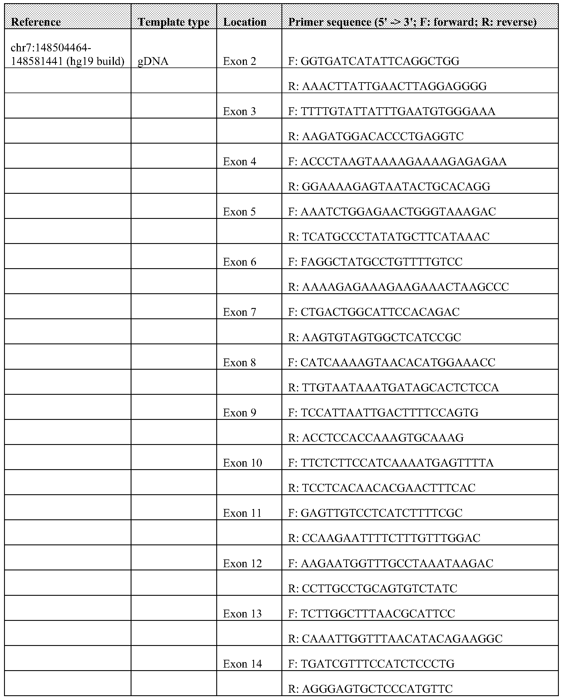


Claims

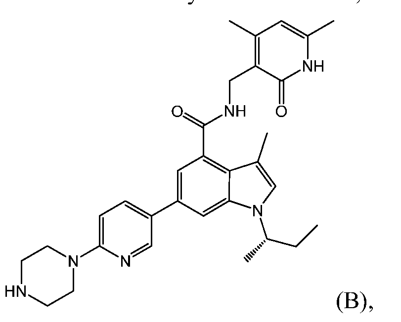
Priority Applications (10)
| Application Number | Priority Date | Filing Date | Title |
|---|---|---|---|
| EP12836842.0A EP2760452A4 (en) | 2011-09-30 | 2012-09-30 | Methods of treating cancer |
| BR112014007603A BR112014007603A2 (en) | 2011-09-30 | 2012-09-30 | cancer treatment methods |
| CA2850570A CA2850570A1 (en) | 2011-09-30 | 2012-09-30 | Methods of treating cancer |
| CN201280059070.3A CN103987842A (en) | 2011-09-30 | 2012-09-30 | Methods of treating cancer |
| JP2014533460A JP2014534178A (en) | 2011-09-30 | 2012-09-30 | Cancer treatment methods |
| US14/346,359 US9730925B2 (en) | 2011-09-30 | 2012-09-30 | Methods of treating cancer |
| RU2014117632/10A RU2014117632A (en) | 2011-09-30 | 2012-09-30 | METHODS FOR TREATING A MALIGNANT TUMOR |
| AU2012315566A AU2012315566A1 (en) | 2011-09-30 | 2012-09-30 | Methods of treating cancer |
| KR1020147011147A KR20140082742A (en) | 2011-09-30 | 2012-09-30 | Methods of treating cancer |
| US15/616,306 US20170348306A1 (en) | 2011-09-30 | 2017-06-07 | Methods of treating cancer |
Applications Claiming Priority (2)
| Application Number | Priority Date | Filing Date | Title |
|---|---|---|---|
| US201161541304P | 2011-09-30 | 2011-09-30 | |
| US61/541,304 | 2011-09-30 |
Related Child Applications (2)
| Application Number | Title | Priority Date | Filing Date |
|---|---|---|---|
| US14/346,359 A-371-Of-International US9730925B2 (en) | 2011-09-30 | 2012-09-30 | Methods of treating cancer |
| US15/616,306 Division US20170348306A1 (en) | 2011-09-30 | 2017-06-07 | Methods of treating cancer |
Publications (2)
| Publication Number | Publication Date |
|---|---|
| WO2013049770A2 true WO2013049770A2 (en) | 2013-04-04 |
| WO2013049770A3 WO2013049770A3 (en) | 2014-05-15 |
Family
ID=47996781
Family Applications (1)
| Application Number | Title | Priority Date | Filing Date |
|---|---|---|---|
| PCT/US2012/058188 WO2013049770A2 (en) | 2011-09-30 | 2012-09-30 | Methods of treating cancer |
Country Status (10)
| Country | Link |
|---|---|
| US (2) | US9730925B2 (en) |
| EP (1) | EP2760452A4 (en) |
| JP (1) | JP2014534178A (en) |
| KR (1) | KR20140082742A (en) |
| CN (1) | CN103987842A (en) |
| AU (1) | AU2012315566A1 (en) |
| BR (1) | BR112014007603A2 (en) |
| CA (1) | CA2850570A1 (en) |
| RU (1) | RU2014117632A (en) |
| WO (1) | WO2013049770A2 (en) |
Cited By (49)
| Publication number | Priority date | Publication date | Assignee | Title |
|---|---|---|---|---|
| WO2013138361A1 (en) * | 2012-03-12 | 2013-09-19 | Epizyme, Inc. | Inhibitors of human ezh2, and methods of use thereof |
| US8691507B2 (en) | 2010-09-10 | 2014-04-08 | Epizyme, Inc. | Inhibitors of human EZH2 and methods of use thereof |
| WO2014062720A3 (en) * | 2012-10-15 | 2014-06-19 | Epizyme, Inc. | Methods of treating cancer |
| WO2014153030A2 (en) | 2013-03-14 | 2014-09-25 | Genentech, Inc. | Methods of treating cancer and preventing cancer drug resistance |
| WO2014177982A1 (en) * | 2013-04-30 | 2014-11-06 | Glaxosmithkline Intellectual Property (No.2) Limited | Enhancer of zeste homolog 2 inhibitors |
| WO2014190035A2 (en) * | 2013-05-21 | 2014-11-27 | Dana-Farber Cancer Institute, Inc. | Compositions and methods for identification, assessment, prevention, and treatment of cancer using histone h3k27me2 biomarkers and modulators |
| WO2015004618A1 (en) * | 2013-07-10 | 2015-01-15 | Glaxosmithkline Intellectual Property (No.2) Limited | Enhancer of zeste homolog 2 inhibitors |
| WO2015052147A1 (en) * | 2013-10-09 | 2015-04-16 | Roche Diagnostics Gmbh | Methods and compositions for detecting mutation in the human ezh2 gene |
| US9040515B2 (en) | 2012-12-21 | 2015-05-26 | Pfizer Inc. | Aryl and heteroaryl fused lactams |
| WO2015077194A1 (en) * | 2013-11-22 | 2015-05-28 | Bristol-Myers Squibb Company | Inhibitors of lysine methyl transferase |
| US9051269B2 (en) | 2011-11-18 | 2015-06-09 | Constellation Pharmaceuticals, Inc. | Modulators of methyl modifying enzymes, compositions and uses thereof |
| US9085583B2 (en) | 2012-02-10 | 2015-07-21 | Constellation—Pharmaceuticals, Inc. | Modulators of methyl modifying enzymes, compositions and uses thereof |
| RU2565548C1 (en) * | 2014-08-05 | 2015-10-20 | Дмитрий Андреевич Журавлёв | Method of producing of pluripotent stem cells |
| US9175331B2 (en) | 2010-09-10 | 2015-11-03 | Epizyme, Inc. | Inhibitors of human EZH2, and methods of use thereof |
| WO2015143424A3 (en) * | 2014-03-21 | 2015-11-12 | Glaxosmithkline Intellectual Property (No. 2) Limited | Methods of treating cancer |
| WO2015196064A1 (en) | 2014-06-19 | 2015-12-23 | Memorial Sloan-Kettering Cancer Center | Biomarkers for response to ezh2 inhibitors |
| WO2015193768A1 (en) * | 2014-06-17 | 2015-12-23 | Pfizer Inc. | Aryl fused lactams as ezh2 modulators |
| EP2934531A4 (en) * | 2012-12-19 | 2016-06-15 | Glaxosmithkline Llc | Combination |
| US9481666B2 (en) | 2014-06-17 | 2016-11-01 | Pfizer Inc. | Substituted dihydroisoquinolinone compounds |
| WO2016201328A1 (en) | 2015-06-10 | 2016-12-15 | Epizyme, Inc. | Ezh2 inhibitors for treating lymphoma |
| CN106255693A (en) * | 2014-03-07 | 2016-12-21 | 葛兰素史密斯克莱知识产权(第2 号)有限公司 | The inhibitor of Zeste enhancer congener 2 |
| US9527837B2 (en) | 2014-12-05 | 2016-12-27 | Eli Lilly And Company | Inhibitors of EZH2 |
| US9718838B2 (en) | 2015-08-27 | 2017-08-01 | Eli Lilly And Company | Inhibitors of EZH2 |
| US9745305B2 (en) | 2013-03-15 | 2017-08-29 | Constellation Pharmaceuticals, Inc. | Modulators of methyl modifying enzymes, compositions and uses thereof |
| US9969716B2 (en) | 2013-08-15 | 2018-05-15 | Constellation Pharmaceuticals, Inc. | Indole derivatives as modulators of methyl modifying enzymes, compositions and uses thereof |
| WO2018210296A1 (en) * | 2017-05-18 | 2018-11-22 | 江苏恒瑞医药股份有限公司 | Use of ezh2 inhibitor combined with btk inhibitor in preparing drug for treating tumor |
| WO2018210302A1 (en) | 2017-05-18 | 2018-11-22 | 江苏恒瑞医药股份有限公司 | Crystal of benzofuran derivative free base and preparation method |
| US10155002B2 (en) | 2011-04-13 | 2018-12-18 | Epizyme, Inc. | Aryl- or heteroaryl-substituted benzene compounds |
| WO2019014191A1 (en) * | 2017-07-10 | 2019-01-17 | Constellation Pharmaceuticals, Inc. | Ezh2 inhibitor-induced gene expression |
| US10220036B2 (en) | 2014-12-23 | 2019-03-05 | Novartis Ag | Triazolopyrimidine compounds and uses thereof |
| US20190134025A1 (en) * | 2016-05-04 | 2019-05-09 | The Wistar Institute Of Anatomy And Biology | Methods of Treating Cancers Overexpressing Carm1 With EZH2 Inhibitors and Platinum-Based Antineoplastic Drugs |
| WO2019097369A1 (en) | 2017-11-14 | 2019-05-23 | Pfizer Inc. | Ezh2 inhibitor combination therapies |
| US10457640B2 (en) | 2016-10-19 | 2019-10-29 | Constellation Pharmaceuticals, Inc. | Synthesis of inhibitors of EZH2 |
| US10463671B2 (en) | 2013-12-06 | 2019-11-05 | Epizyme, Inc. | Combination therapy for treating cancer |
| US10493076B2 (en) | 2015-08-24 | 2019-12-03 | Epizyme, Inc. | Method for treating cancer |
| US10577350B2 (en) | 2015-08-28 | 2020-03-03 | Constellation Pharmaceuticals, Inc. | Crystalline forms of (R)-N-((4-methoxy-6-methyl-2-oxo-1,2-dihydropyridin-3-yl)methyl)-2-methyl-1-(1-(1-(2,2,2-trifluoroethyl)piperidin-4-yl)ethyl)-1H-indole-3-carboxamide |
| US10633371B2 (en) | 2016-04-22 | 2020-04-28 | Dana-Farber Cancer Institute, Inc. | EZH2 inhibitors and uses thereof |
| US10676479B2 (en) | 2016-06-20 | 2020-06-09 | Novartis Ag | Imidazolepyridine compounds and uses thereof |
| US10689378B2 (en) | 2016-06-20 | 2020-06-23 | Novartis Ag | Triazolopyridine compounds and uses thereof |
| US10759787B2 (en) | 2015-11-19 | 2020-09-01 | Jiangsu Hengrui Medicine Co., Ltd. | Benzofuran derivative, preparation method thereof and use thereof in medicine |
| US11091489B2 (en) | 2016-06-20 | 2021-08-17 | Novartis Ag | Crystalline forms of a triazolopyrimidine compound |
| EP3885343A1 (en) | 2014-11-06 | 2021-09-29 | Dana-Farber Cancer Institute, Inc. | Indole compounds as ezh2 inhibitors and uses thereof |
| US11173160B2 (en) | 2019-04-22 | 2021-11-16 | University of Pittsburgh—of the Commonwealth System of Higher Education | Use of TG02 for treating gliomas in pediatric subjects |
| US11326212B2 (en) | 2010-06-23 | 2022-05-10 | British Columbia Cancer Agency Branch | Biomarkers for non-hodgkin lymphomas and uses thereof |
| WO2023025856A1 (en) | 2021-08-25 | 2023-03-02 | INSERM (Institut National de la Santé et de la Recherche Médicale) | Use of ezh2 inhibitors for the treatment of aortic valve stenosis |
| US11642347B2 (en) | 2014-06-17 | 2023-05-09 | Epizyme, Inc. | EZH2 inhibitors for treating lymphoma |
| US11642348B2 (en) | 2012-10-15 | 2023-05-09 | Epizyme, Inc. | Substituted benzene compounds |
| WO2023244918A1 (en) | 2022-06-13 | 2023-12-21 | Treeline Biosciences, Inc. | Quinolone bcl6 bifunctional degraders |
| WO2023244917A1 (en) | 2022-06-13 | 2023-12-21 | Treeline Biosciences, Inc. | 1,8-naphthyridin-2-one heterobifunctional bcl6 degraders |
Families Citing this family (19)
| Publication number | Priority date | Publication date | Assignee | Title |
|---|---|---|---|---|
| US20180021331A1 (en) * | 2014-12-30 | 2018-01-25 | University Of Utah Research Foundation | Hdac1,2 inhibitors and methods of using the same |
| US10961532B2 (en) | 2015-04-07 | 2021-03-30 | The General Hospital Corporation | Methods for reactivating genes on the inactive X chromosome |
| EA201792304A1 (en) * | 2015-04-20 | 2018-03-30 | Эпизайм, Инк. | COMBINED THERAPY FOR CANCER TREATMENT |
| EP3524602A1 (en) | 2016-09-07 | 2019-08-14 | Shanghai Haihe Pharmaceutical Co., Ltd. | Pyrido five-element aromatic ring compound, preparation method therefor and use thereof |
| AU2017351638A1 (en) | 2016-10-26 | 2019-06-13 | Genea Biocells USA (Holdings), Inc. | Improved generation of muscle lineage cells and therapeutic uses thereof |
| KR101951289B1 (en) * | 2017-02-24 | 2019-02-22 | 사회복지법인 삼성생명공익재단 | Markers for predicting survivalin a patient with lungcancer |
| US10266542B2 (en) | 2017-03-15 | 2019-04-23 | Mirati Therapeutics, Inc. | EZH2 inhibitors |
| US20230201212A1 (en) * | 2017-06-13 | 2023-06-29 | Epizyme, Inc. | Inhibitors of ezh2 and methods of use thereof |
| EP4043466A1 (en) | 2018-01-31 | 2022-08-17 | Mirati Therapeutics, Inc. | Prc2 inhibitors |
| CN110229151B (en) * | 2018-03-06 | 2021-09-10 | 上海海和药物研究开发股份有限公司 | Indolizine compound, preparation method and application thereof |
| AU2019251357B2 (en) | 2018-04-09 | 2023-10-05 | Portland Technology Holdings Llc | Hemp extract for treatment of pain in animals |
| CA3111802A1 (en) * | 2018-09-06 | 2020-03-12 | The Council Of The Queensland Institute Of Medical Research | Biomarkers for cancer therapy |
| US10800339B2 (en) | 2018-10-16 | 2020-10-13 | Ford Global Technologies, Llc | Vehicle rack assembly and rack accessing method |
| US20220010307A1 (en) * | 2018-12-10 | 2022-01-13 | The Regents Of The University Of California | Compositions and methods for modulating transcriptional activity of amplified oncogenes contained on extrachromosomal dna |
| KR20210151833A (en) | 2019-03-15 | 2021-12-14 | 풀크럼 쎄러퓨틱스, 인코포레이티드 | Macrocyclic azolopyridine derivatives as EED and PRC2 modulators |
| MX2022000027A (en) * | 2019-07-02 | 2022-04-11 | Ellevet Sciences | Hemp extract for treatment of pain, cancer and epilepsy in animals. |
| CN114555112A (en) | 2019-08-22 | 2022-05-27 | 朱诺治疗学股份有限公司 | Combination therapy of T cell therapy and ZESTE enhancer homolog 2(EZH2) inhibitors and related methods |
| CN116466071A (en) * | 2020-12-28 | 2023-07-21 | 重庆生命知源科技有限公司 | Application of loading buffer solution and trace western blotting detection method |
| WO2023249714A1 (en) * | 2022-06-24 | 2023-12-28 | Memorial Sloan-Kettering Cancer Center | Therapeutic compositions and methods for activating double stranded rna response in cancer patients via dnmt1 targeted therapy |
Citations (3)
| Publication number | Priority date | Publication date | Assignee | Title |
|---|---|---|---|---|
| WO2011140324A1 (en) | 2010-05-07 | 2011-11-10 | Glaxosmithkline Llc | Indoles |
| WO2011140325A1 (en) | 2010-05-07 | 2011-11-10 | Glaxosmithkline Llc | Indazoles |
| WO2012075080A1 (en) | 2010-12-01 | 2012-06-07 | Glaxosmithkline Llc | Indoles |
Family Cites Families (6)
| Publication number | Priority date | Publication date | Assignee | Title |
|---|---|---|---|---|
| US20090012031A1 (en) | 2007-07-03 | 2009-01-08 | The Regents Of The University Of Michigan | EZH2 Cancer Markers |
| NZ582586A (en) * | 2007-07-21 | 2011-12-22 | Albany Molecular Res Inc | 5-pyridinone substituted indazoles |
| RU2618475C2 (en) * | 2010-09-10 | 2017-05-03 | Эпизайм, Инк. | Human ezh2 inhibitors and methods of application thereof |
| US9175331B2 (en) * | 2010-09-10 | 2015-11-03 | Epizyme, Inc. | Inhibitors of human EZH2, and methods of use thereof |
| EP3323820B1 (en) | 2011-02-28 | 2023-05-10 | Epizyme, Inc. | Substituted 6,5-fused bicyclic heteroaryl compounds |
| JO3438B1 (en) * | 2011-04-13 | 2019-10-20 | Epizyme Inc | Aryl- or heteroaryl-substituted benzene compounds |
-
2012
- 2012-09-30 BR BR112014007603A patent/BR112014007603A2/en unknown
- 2012-09-30 WO PCT/US2012/058188 patent/WO2013049770A2/en active Application Filing
- 2012-09-30 JP JP2014533460A patent/JP2014534178A/en active Pending
- 2012-09-30 RU RU2014117632/10A patent/RU2014117632A/en not_active Application Discontinuation
- 2012-09-30 EP EP12836842.0A patent/EP2760452A4/en not_active Withdrawn
- 2012-09-30 US US14/346,359 patent/US9730925B2/en not_active Expired - Fee Related
- 2012-09-30 CA CA2850570A patent/CA2850570A1/en not_active Abandoned
- 2012-09-30 CN CN201280059070.3A patent/CN103987842A/en active Pending
- 2012-09-30 AU AU2012315566A patent/AU2012315566A1/en not_active Abandoned
- 2012-09-30 KR KR1020147011147A patent/KR20140082742A/en not_active Application Discontinuation
-
2017
- 2017-06-07 US US15/616,306 patent/US20170348306A1/en not_active Abandoned
Patent Citations (3)
| Publication number | Priority date | Publication date | Assignee | Title |
|---|---|---|---|---|
| WO2011140324A1 (en) | 2010-05-07 | 2011-11-10 | Glaxosmithkline Llc | Indoles |
| WO2011140325A1 (en) | 2010-05-07 | 2011-11-10 | Glaxosmithkline Llc | Indazoles |
| WO2012075080A1 (en) | 2010-12-01 | 2012-06-07 | Glaxosmithkline Llc | Indoles |
Non-Patent Citations (7)
| Title |
|---|
| BODOR C. ET AL.: "EZH2 Y641 mutations in follicular lymphoma", LEUKEMIA, vol. 25, pages 726 - 729 |
| FATHI AT; ABDEL-WAHAB O: "Mutations in epigenetic modifiers in myeloid malignancies and the prospect of novel epigenetic-targeted therapy", ADV HEMATOL, 2012, pages 469592 |
| GUI Y. Y ET AL.: "Frequent mutations of chromatin remodeling genes in transitional cell carcinoma of the bladder", NAT GENET. |
| MORIN RD ET AL.: "Frequent mutation of histone-modifying genes in non-Hodgkin lymphoma", NATURE, vol. 476, 2011, pages 298 - 303, XP055294553, DOI: doi:10.1038/nature10351 |
| MORIN RD ET AL.: "Somatic mutations altering EZH2 (Tyr641) in follicular and diffuse large B-cell lymphomas of germinal-center origin", NAT GENET, vol. 42, pages 181 - 185, XP055101779, DOI: doi:10.1038/ng.518 |
| See also references of EP2760452A4 |
| STRANSKY N. ET AL.: "The mutational landscape of head and neck squamous cell carcinoma", SCIENCE, vol. 333, pages 1157 - 1160, XP055078258, DOI: doi:10.1126/science.1208130 |
Cited By (111)
| Publication number | Priority date | Publication date | Assignee | Title |
|---|---|---|---|---|
| US11326212B2 (en) | 2010-06-23 | 2022-05-10 | British Columbia Cancer Agency Branch | Biomarkers for non-hodgkin lymphomas and uses thereof |
| US8691507B2 (en) | 2010-09-10 | 2014-04-08 | Epizyme, Inc. | Inhibitors of human EZH2 and methods of use thereof |
| US9334527B2 (en) | 2010-09-10 | 2016-05-10 | Epizyme, Inc. | Inhibitors of human EZH2, and methods of use thereof |
| US9333217B2 (en) | 2010-09-10 | 2016-05-10 | Epizyme, Inc. | Inhibitors of human EZH2, and methods of use thereof |
| US8895245B2 (en) | 2010-09-10 | 2014-11-25 | Epizyme, Inc. | Inhibitors of human EZH2 and methods of use thereof |
| US9949999B2 (en) | 2010-09-10 | 2018-04-24 | Epizyme, Inc. | Inhibitors of human EZH2, and methods of use thereof |
| US9175331B2 (en) | 2010-09-10 | 2015-11-03 | Epizyme, Inc. | Inhibitors of human EZH2, and methods of use thereof |
| US11052093B2 (en) | 2011-04-13 | 2021-07-06 | Epizyme, Inc. | Aryl-or heteroaryl-substituted benzene compounds |
| US10420775B2 (en) | 2011-04-13 | 2019-09-24 | Epizyme, Inc. | Aryl-or heteroaryl-substituted benzene compounds |
| US10155002B2 (en) | 2011-04-13 | 2018-12-18 | Epizyme, Inc. | Aryl- or heteroaryl-substituted benzene compounds |
| US9051269B2 (en) | 2011-11-18 | 2015-06-09 | Constellation Pharmaceuticals, Inc. | Modulators of methyl modifying enzymes, compositions and uses thereof |
| US9409865B2 (en) | 2011-11-18 | 2016-08-09 | Constellation_Pharmaceuticals, Inc. | Modulators of methyl modifying enzymes, compositions and uses thereof |
| USRE47428E1 (en) | 2012-02-10 | 2019-06-11 | Constellation Pharmaceuticals, Inc. | Modulators of methyl modifying enzymes, compositions and uses thereof |
| US9085583B2 (en) | 2012-02-10 | 2015-07-21 | Constellation—Pharmaceuticals, Inc. | Modulators of methyl modifying enzymes, compositions and uses thereof |
| US9469646B2 (en) | 2012-02-10 | 2016-10-18 | Constellation Pharmaceuticals, Inc. | Modulators of methyl modifying enzymes, compositions and uses thereof |
| US9371331B2 (en) | 2012-02-10 | 2016-06-21 | Constellation Pharmaceuticals, Inc. | Modulators of methyl modifying enzymes, compositions and uses thereof |
| US10016405B2 (en) | 2012-02-10 | 2018-07-10 | Constellation Pharmaceuticals, Inc. | Modulators of methyl modifying enzymes, compositions and uses thereof |
| US9980952B2 (en) | 2012-02-10 | 2018-05-29 | Constellation Pharmaceuticals, Inc. | Modulators of methyl modifying enzymes, compositions and uses thereof |
| EP3536314A1 (en) * | 2012-03-12 | 2019-09-11 | Epizyme, Inc. | Inhibitors of human ezh2, and methods of use thereof |
| WO2013138361A1 (en) * | 2012-03-12 | 2013-09-19 | Epizyme, Inc. | Inhibitors of human ezh2, and methods of use thereof |
| AU2013232229B2 (en) * | 2012-03-12 | 2017-11-23 | Epizyme, Inc. | Inhibitors of human EZH2, and methods of use thereof |
| JP2015511603A (en) * | 2012-03-12 | 2015-04-20 | エピザイム インコーポレイテッド | Inhibitors of human EZH2 and methods of use thereof |
| EP3659606A1 (en) * | 2012-10-15 | 2020-06-03 | Epizyme, Inc. | Methods of treating cancer |
| US11642348B2 (en) | 2012-10-15 | 2023-05-09 | Epizyme, Inc. | Substituted benzene compounds |
| US9688665B2 (en) | 2012-10-15 | 2017-06-27 | Epizyme, Inc. | Methods of treating cancer |
| WO2014062720A3 (en) * | 2012-10-15 | 2014-06-19 | Epizyme, Inc. | Methods of treating cancer |
| EP2934531A4 (en) * | 2012-12-19 | 2016-06-15 | Glaxosmithkline Llc | Combination |
| US10246433B2 (en) | 2012-12-21 | 2019-04-02 | Pfizer Inc. | Aryl and heteroaryl fused lactams |
| US9040515B2 (en) | 2012-12-21 | 2015-05-26 | Pfizer Inc. | Aryl and heteroaryl fused lactams |
| CN105307683A (en) * | 2013-03-14 | 2016-02-03 | 基因泰克公司 | Methods of treating cancer and preventing cancer drug resistance |
| WO2014153030A2 (en) | 2013-03-14 | 2014-09-25 | Genentech, Inc. | Methods of treating cancer and preventing cancer drug resistance |
| WO2014153030A3 (en) * | 2013-03-14 | 2015-08-20 | Genentech, Inc. | Methods of treating cancer and preventing cancer drug resistance |
| US9745305B2 (en) | 2013-03-15 | 2017-08-29 | Constellation Pharmaceuticals, Inc. | Modulators of methyl modifying enzymes, compositions and uses thereof |
| JP2016517878A (en) * | 2013-04-30 | 2016-06-20 | グラクソスミスクライン、インテレクチュアル、プロパティー、ナンバー2、リミテッドGlaxosmithkline Intellectual Property No.2 Limited | ZESTE homolog 2 enhancer inhibitor |
| EA030196B1 (en) * | 2013-04-30 | 2018-07-31 | Глэксосмитклайн Интеллекчуал Проперти (No.2) Лимитед | Enhancer of zeste homolog 2 inhibitors |
| US9505745B2 (en) | 2013-04-30 | 2016-11-29 | Glaxosmithkline Intellectual Property (No. 2) Limited | Enhancer of zeste homolog 2 inhibitors |
| US9790212B2 (en) | 2013-04-30 | 2017-10-17 | Glaxosmithkline Intellectual Property (No. 2) Limited | Enhancer of zeste homolog 2 inhibitors |
| CN105308038B (en) * | 2013-04-30 | 2018-05-29 | 葛兰素史密斯克莱知识产权(第2 号)有限公司 | The inhibitor of Zeste enhancers homologue 2 |
| WO2014177982A1 (en) * | 2013-04-30 | 2014-11-06 | Glaxosmithkline Intellectual Property (No.2) Limited | Enhancer of zeste homolog 2 inhibitors |
| AU2014261075B2 (en) * | 2013-04-30 | 2017-01-19 | Glaxosmithkline Intellectual Property (No.2) Limited | Enhancer of zeste homolog 2 inhibitors |
| CN105308038A (en) * | 2013-04-30 | 2016-02-03 | 葛兰素史密斯克莱知识产权(第2号)有限公司 | Enhancer of zeste homolog 2 inhibitors |
| WO2014190035A2 (en) * | 2013-05-21 | 2014-11-27 | Dana-Farber Cancer Institute, Inc. | Compositions and methods for identification, assessment, prevention, and treatment of cancer using histone h3k27me2 biomarkers and modulators |
| WO2014190035A3 (en) * | 2013-05-21 | 2015-01-22 | Dana-Farber Cancer Institute, Inc. | Compositions and methods for identification, assessment, prevention, and treatment of cancer using histone h3k27me2 biomarkers and modulators |
| AU2014288839B2 (en) * | 2013-07-10 | 2017-02-02 | Glaxosmithkline Intellectual Property (No.2) Limited | Enhancer of Zeste Homolog 2 inhibitors |
| US9556157B2 (en) | 2013-07-10 | 2017-01-31 | Glaxosmithkline Intellectual Property (No.2) Limited | Enhancer of zeste homolog 2 inhibitors |
| CN105473580A (en) * | 2013-07-10 | 2016-04-06 | 葛兰素史密斯克莱知识产权(第2号)有限公司 | Enhancer of Zeste homolog 2 inhibitors |
| WO2015004618A1 (en) * | 2013-07-10 | 2015-01-15 | Glaxosmithkline Intellectual Property (No.2) Limited | Enhancer of zeste homolog 2 inhibitors |
| JP2016523955A (en) * | 2013-07-10 | 2016-08-12 | グラクソスミスクライン、インテレクチュアル、プロパティー、ナンバー2、リミテッドGlaxosmithkline Intellectual Property No.2 Limited | ZESTE homolog 2 enhancer inhibitor |
| US9969716B2 (en) | 2013-08-15 | 2018-05-15 | Constellation Pharmaceuticals, Inc. | Indole derivatives as modulators of methyl modifying enzymes, compositions and uses thereof |
| CN105593378A (en) * | 2013-10-09 | 2016-05-18 | 豪夫迈·罗氏有限公司 | Methods and compositions for detecting mutation in the human EZH2 gene |
| WO2015052147A1 (en) * | 2013-10-09 | 2015-04-16 | Roche Diagnostics Gmbh | Methods and compositions for detecting mutation in the human ezh2 gene |
| JP2016533708A (en) * | 2013-10-09 | 2016-11-04 | エフ.ホフマン−ラ ロシュ アーゲーF. Hoffmann−La Roche Aktiengesellschaft | Methods and compositions for detecting mutations in the human EZH2 gene |
| US10260102B2 (en) | 2013-10-09 | 2019-04-16 | Roche Molecular Systems, Inc. | Methods and compositions for detecting mutation in the human EZH2 gene |
| US9822103B2 (en) | 2013-11-22 | 2017-11-21 | Bristol-Myers Squibb Company | Inhibitors of lysine methyl transferase |
| WO2015077194A1 (en) * | 2013-11-22 | 2015-05-28 | Bristol-Myers Squibb Company | Inhibitors of lysine methyl transferase |
| US10463671B2 (en) | 2013-12-06 | 2019-11-05 | Epizyme, Inc. | Combination therapy for treating cancer |
| CN106255693A (en) * | 2014-03-07 | 2016-12-21 | 葛兰素史密斯克莱知识产权(第2 号)有限公司 | The inhibitor of Zeste enhancer congener 2 |
| US20170105997A1 (en) * | 2014-03-21 | 2017-04-20 | Northwestern University | Methods of treating cancer |
| EP3119390A4 (en) * | 2014-03-21 | 2017-09-20 | Glaxosmithkline Intellectual Property (No. 2) Limited | Methods of treating cancer |
| WO2015143424A3 (en) * | 2014-03-21 | 2015-11-12 | Glaxosmithkline Intellectual Property (No. 2) Limited | Methods of treating cancer |
| US10570121B2 (en) | 2014-06-17 | 2020-02-25 | Pfizer Inc. | Substituted dihydroisoquinolinone compounds |
| WO2015193768A1 (en) * | 2014-06-17 | 2015-12-23 | Pfizer Inc. | Aryl fused lactams as ezh2 modulators |
| US11642347B2 (en) | 2014-06-17 | 2023-05-09 | Epizyme, Inc. | EZH2 inhibitors for treating lymphoma |
| US9481666B2 (en) | 2014-06-17 | 2016-11-01 | Pfizer Inc. | Substituted dihydroisoquinolinone compounds |
| CN106795561A (en) * | 2014-06-19 | 2017-05-31 | 纪念斯隆-凯特琳癌症中心 | For the biomarker of the response to EZH2 inhibitor |
| WO2015196064A1 (en) | 2014-06-19 | 2015-12-23 | Memorial Sloan-Kettering Cancer Center | Biomarkers for response to ezh2 inhibitors |
| CN106795561B (en) * | 2014-06-19 | 2021-04-16 | 纪念斯隆-凯特琳癌症中心 | Biomarkers for response to EZH2 inhibitors |
| EA036889B1 (en) * | 2014-06-19 | 2021-01-12 | Мемориал Слоан-Кеттеринг Кэнсер Сентер | Biomarkers for response to ezh2 inhibitors |
| RU2565548C1 (en) * | 2014-08-05 | 2015-10-20 | Дмитрий Андреевич Журавлёв | Method of producing of pluripotent stem cells |
| US11236082B2 (en) | 2014-11-06 | 2022-02-01 | Dana-Farber Cancer Institute, Inc. | EZH2 inhibitors and uses thereof |
| EP3885343A1 (en) | 2014-11-06 | 2021-09-29 | Dana-Farber Cancer Institute, Inc. | Indole compounds as ezh2 inhibitors and uses thereof |
| US9527837B2 (en) | 2014-12-05 | 2016-12-27 | Eli Lilly And Company | Inhibitors of EZH2 |
| US10220036B2 (en) | 2014-12-23 | 2019-03-05 | Novartis Ag | Triazolopyrimidine compounds and uses thereof |
| US11931363B2 (en) | 2014-12-23 | 2024-03-19 | Novartis Ag | Triazolopyrimidine compounds and uses thereof |
| US11202781B2 (en) | 2015-06-10 | 2021-12-21 | Epizyme, Inc. | EZH2 inhibitors for treating lymphoma |
| EP3307713A4 (en) * | 2015-06-10 | 2019-01-23 | Epizyme, Inc. | Ezh2 inhibitors for treating lymphoma |
| US11951109B2 (en) | 2015-06-10 | 2024-04-09 | Epizyme, Inc. | EZH2 inhibitors for treating lymphoma |
| WO2016201328A1 (en) | 2015-06-10 | 2016-12-15 | Epizyme, Inc. | Ezh2 inhibitors for treating lymphoma |
| US10493076B2 (en) | 2015-08-24 | 2019-12-03 | Epizyme, Inc. | Method for treating cancer |
| US11642349B2 (en) | 2015-08-24 | 2023-05-09 | Epizyme, Inc. | Method for treating cancer |
| US10898490B2 (en) | 2015-08-24 | 2021-01-26 | Epizyme, Inc. | Method for treating cancer |
| US9718838B2 (en) | 2015-08-27 | 2017-08-01 | Eli Lilly And Company | Inhibitors of EZH2 |
| US10577350B2 (en) | 2015-08-28 | 2020-03-03 | Constellation Pharmaceuticals, Inc. | Crystalline forms of (R)-N-((4-methoxy-6-methyl-2-oxo-1,2-dihydropyridin-3-yl)methyl)-2-methyl-1-(1-(1-(2,2,2-trifluoroethyl)piperidin-4-yl)ethyl)-1H-indole-3-carboxamide |
| US11059811B2 (en) | 2015-11-19 | 2021-07-13 | Jiangsu Hengrui Medicine Co., Ltd. | Benzofuran derivative, preparation method thereof and use thereof in medicine |
| US10759787B2 (en) | 2015-11-19 | 2020-09-01 | Jiangsu Hengrui Medicine Co., Ltd. | Benzofuran derivative, preparation method thereof and use thereof in medicine |
| US10633371B2 (en) | 2016-04-22 | 2020-04-28 | Dana-Farber Cancer Institute, Inc. | EZH2 inhibitors and uses thereof |
| US10987353B2 (en) * | 2016-05-04 | 2021-04-27 | The Wistar Institute Of Anatomy And Biology | Methods of treating cancers overexpressing CARM1 with EZH2 inhibitors and platinum-based antineoplastic drugs |
| US20210251987A1 (en) * | 2016-05-04 | 2021-08-19 | The Wistar Institute Of Anatomy And Biology | Methods of Treating Cancers Overexpressing Carm1 With EZH2 Inhibitors and Platinum-Based Antineoplastic Drugs |
| US20190134025A1 (en) * | 2016-05-04 | 2019-05-09 | The Wistar Institute Of Anatomy And Biology | Methods of Treating Cancers Overexpressing Carm1 With EZH2 Inhibitors and Platinum-Based Antineoplastic Drugs |
| US10689378B2 (en) | 2016-06-20 | 2020-06-23 | Novartis Ag | Triazolopyridine compounds and uses thereof |
| US10676479B2 (en) | 2016-06-20 | 2020-06-09 | Novartis Ag | Imidazolepyridine compounds and uses thereof |
| US11091489B2 (en) | 2016-06-20 | 2021-08-17 | Novartis Ag | Crystalline forms of a triazolopyrimidine compound |
| US11548897B2 (en) | 2016-06-20 | 2023-01-10 | Novartis Ag | Crystalline forms of a triazolopyrimidine compound |
| US10457640B2 (en) | 2016-10-19 | 2019-10-29 | Constellation Pharmaceuticals, Inc. | Synthesis of inhibitors of EZH2 |
| RU2762893C2 (en) * | 2017-05-18 | 2021-12-23 | Цзянсу Хэнжуй Медсин Ко., Лтд. | Application of an ezh2 inhibitor in combination with a btk inhibitor in production of a medicinal product for treating a tumour |
| US11065239B2 (en) | 2017-05-18 | 2021-07-20 | Jiangsu Hengrui Medicine Co., Ltd. | Use of EZH2 inhibitor combined with BTK inhibitor in preparing drug for treating tumor |
| EP3626715A4 (en) * | 2017-05-18 | 2021-03-24 | Jiangsu Hengrui Medicine Co., Ltd. | Crystal of benzofuran derivative free base and preparation method |
| AU2018269268B2 (en) * | 2017-05-18 | 2022-01-06 | Jiangsu Hengrui Medicine Co., Ltd. | Crystal of benzofuran derivative free base and preparation method |
| WO2018210296A1 (en) * | 2017-05-18 | 2018-11-22 | 江苏恒瑞医药股份有限公司 | Use of ezh2 inhibitor combined with btk inhibitor in preparing drug for treating tumor |
| US11155537B2 (en) | 2017-05-18 | 2021-10-26 | Jiangsu Hengrui Medicine Co., Ltd. | Crystal of benzofuran derivative free base and preparation method |
| KR102635949B1 (en) | 2017-05-18 | 2024-02-14 | 지앙수 헨그루이 파마슈티컬스 컴퍼니 리미티드 | Use of a combination of EZH2 inhibitors and BTK inhibitors in the manufacture of drugs for the treatment of tumors |
| KR20200007851A (en) * | 2017-05-18 | 2020-01-22 | 지앙수 헨그루이 메디슨 컴퍼니 리미티드 | Use of a combination of an EZH2 inhibitor and a BTK inhibitor in the manufacture of a drug for the treatment of tumors |
| WO2018210302A1 (en) | 2017-05-18 | 2018-11-22 | 江苏恒瑞医药股份有限公司 | Crystal of benzofuran derivative free base and preparation method |
| AU2018269262B2 (en) * | 2017-05-18 | 2021-12-02 | Jiangsu Hengrui Medicine Co., Ltd. | Use of EZH2 inhibitor combined with BTK inhibitor in preparing drug for treating tumor |
| WO2019014191A1 (en) * | 2017-07-10 | 2019-01-17 | Constellation Pharmaceuticals, Inc. | Ezh2 inhibitor-induced gene expression |
| US11529344B2 (en) | 2017-11-14 | 2022-12-20 | Pfizer Inc. | EZH2 inhibitor combination therapies |
| WO2019097369A1 (en) | 2017-11-14 | 2019-05-23 | Pfizer Inc. | Ezh2 inhibitor combination therapies |
| US11173160B2 (en) | 2019-04-22 | 2021-11-16 | University of Pittsburgh—of the Commonwealth System of Higher Education | Use of TG02 for treating gliomas in pediatric subjects |
| WO2023025856A1 (en) | 2021-08-25 | 2023-03-02 | INSERM (Institut National de la Santé et de la Recherche Médicale) | Use of ezh2 inhibitors for the treatment of aortic valve stenosis |
| WO2023244918A1 (en) | 2022-06-13 | 2023-12-21 | Treeline Biosciences, Inc. | Quinolone bcl6 bifunctional degraders |
| WO2023244917A1 (en) | 2022-06-13 | 2023-12-21 | Treeline Biosciences, Inc. | 1,8-naphthyridin-2-one heterobifunctional bcl6 degraders |
Also Published As
| Publication number | Publication date |
|---|---|
| RU2014117632A (en) | 2015-11-10 |
| EP2760452A2 (en) | 2014-08-06 |
| WO2013049770A3 (en) | 2014-05-15 |
| EP2760452A4 (en) | 2015-04-01 |
| US20140378470A1 (en) | 2014-12-25 |
| CN103987842A (en) | 2014-08-13 |
| JP2014534178A (en) | 2014-12-18 |
| US9730925B2 (en) | 2017-08-15 |
| CA2850570A1 (en) | 2013-04-04 |
| KR20140082742A (en) | 2014-07-02 |
| AU2012315566A1 (en) | 2014-04-17 |
| US20170348306A1 (en) | 2017-12-07 |
| BR112014007603A2 (en) | 2017-06-13 |
Similar Documents
| Publication | Publication Date | Title |
|---|---|---|
| US9730925B2 (en) | Methods of treating cancer | |
| US20160361309A1 (en) | Methods of treating cancer patients responding to ezh2 inhibitor gsk126 | |
| US20170105997A1 (en) | Methods of treating cancer | |
| AU2012275315B2 (en) | Method of administration and treatment | |
| US20130231346A1 (en) | Methods of treating cancer | |
| AU2014203270B2 (en) | Method of treating cancer | |
| WO2014093750A1 (en) | Method of administration and treatment | |
| AU2013214254B2 (en) | Method of treating cancer | |
| WO2015059677A1 (en) | Methods of treating cancer |
Legal Events
| Date | Code | Title | Description |
|---|---|---|---|
| 121 | Ep: the epo has been informed by wipo that ep was designated in this application |
Ref document number: 12836842 Country of ref document: EP Kind code of ref document: A2 |
|
| WWE | Wipo information: entry into national phase |
Ref document number: 14346359 Country of ref document: US |
|
| ENP | Entry into the national phase |
Ref document number: 2014533460 Country of ref document: JP Kind code of ref document: A Ref document number: 2850570 Country of ref document: CA |
|
| ENP | Entry into the national phase |
Ref document number: 2012315566 Country of ref document: AU Date of ref document: 20120930 Kind code of ref document: A |
|
| WWE | Wipo information: entry into national phase |
Ref document number: 2012836842 Country of ref document: EP |
|
| ENP | Entry into the national phase |
Ref document number: 20147011147 Country of ref document: KR Kind code of ref document: A |
|
| ENP | Entry into the national phase |
Ref document number: 2014117632 Country of ref document: RU Kind code of ref document: A |
|
| REG | Reference to national code |
Ref country code: BR Ref legal event code: B01A Ref document number: 112014007603 Country of ref document: BR |
|
| REG | Reference to national code |
Ref country code: BR Ref legal event code: B01E Ref document number: 112014007603 Country of ref document: BR |
|
| ENPW | Started to enter national phase and was withdrawn or failed for other reasons |
Ref document number: 112014007603 Country of ref document: BR |