WO2012108069A1 - Method for producing regenerative organ primordium provided with guide for transplantation, composition containing regenerative organ primordium provided with guide for transplantation produced thereby, and method for transplanting regenerative organ primordium provided with guide for transplantation - Google Patents
Method for producing regenerative organ primordium provided with guide for transplantation, composition containing regenerative organ primordium provided with guide for transplantation produced thereby, and method for transplanting regenerative organ primordium provided with guide for transplantation Download PDFInfo
- Publication number
- WO2012108069A1 WO2012108069A1 PCT/JP2011/067045 JP2011067045W WO2012108069A1 WO 2012108069 A1 WO2012108069 A1 WO 2012108069A1 JP 2011067045 W JP2011067045 W JP 2011067045W WO 2012108069 A1 WO2012108069 A1 WO 2012108069A1
- Authority
- WO
- WIPO (PCT)
- Prior art keywords
- primordium
- regenerated
- cells
- guide
- organ
- Prior art date
Links
Images


Classifications
-
- C—CHEMISTRY; METALLURGY
- C12—BIOCHEMISTRY; BEER; SPIRITS; WINE; VINEGAR; MICROBIOLOGY; ENZYMOLOGY; MUTATION OR GENETIC ENGINEERING
- C12N—MICROORGANISMS OR ENZYMES; COMPOSITIONS THEREOF; PROPAGATING, PRESERVING, OR MAINTAINING MICROORGANISMS; MUTATION OR GENETIC ENGINEERING; CULTURE MEDIA
- C12N5/00—Undifferentiated human, animal or plant cells, e.g. cell lines; Tissues; Cultivation or maintenance thereof; Culture media therefor
- C12N5/06—Animal cells or tissues; Human cells or tissues
- C12N5/0697—Artificial constructs associating cells of different lineages, e.g. tissue equivalents
-
- C—CHEMISTRY; METALLURGY
- C12—BIOCHEMISTRY; BEER; SPIRITS; WINE; VINEGAR; MICROBIOLOGY; ENZYMOLOGY; MUTATION OR GENETIC ENGINEERING
- C12N—MICROORGANISMS OR ENZYMES; COMPOSITIONS THEREOF; PROPAGATING, PRESERVING, OR MAINTAINING MICROORGANISMS; MUTATION OR GENETIC ENGINEERING; CULTURE MEDIA
- C12N5/00—Undifferentiated human, animal or plant cells, e.g. cell lines; Tissues; Cultivation or maintenance thereof; Culture media therefor
- C12N5/06—Animal cells or tissues; Human cells or tissues
- C12N5/0602—Vertebrate cells
-
- A—HUMAN NECESSITIES
- A61—MEDICAL OR VETERINARY SCIENCE; HYGIENE
- A61L—METHODS OR APPARATUS FOR STERILISING MATERIALS OR OBJECTS IN GENERAL; DISINFECTION, STERILISATION OR DEODORISATION OF AIR; CHEMICAL ASPECTS OF BANDAGES, DRESSINGS, ABSORBENT PADS OR SURGICAL ARTICLES; MATERIALS FOR BANDAGES, DRESSINGS, ABSORBENT PADS OR SURGICAL ARTICLES
- A61L27/00—Materials for grafts or prostheses or for coating grafts or prostheses
- A61L27/36—Materials for grafts or prostheses or for coating grafts or prostheses containing ingredients of undetermined constitution or reaction products thereof, e.g. transplant tissue, natural bone, extracellular matrix
- A61L27/38—Materials for grafts or prostheses or for coating grafts or prostheses containing ingredients of undetermined constitution or reaction products thereof, e.g. transplant tissue, natural bone, extracellular matrix containing added animal cells
- A61L27/3804—Materials for grafts or prostheses or for coating grafts or prostheses containing ingredients of undetermined constitution or reaction products thereof, e.g. transplant tissue, natural bone, extracellular matrix containing added animal cells characterised by specific cells or progenitors thereof, e.g. fibroblasts, connective tissue cells, kidney cells
- A61L27/3813—Epithelial cells, e.g. keratinocytes, urothelial cells
-
- A—HUMAN NECESSITIES
- A61—MEDICAL OR VETERINARY SCIENCE; HYGIENE
- A61L—METHODS OR APPARATUS FOR STERILISING MATERIALS OR OBJECTS IN GENERAL; DISINFECTION, STERILISATION OR DEODORISATION OF AIR; CHEMICAL ASPECTS OF BANDAGES, DRESSINGS, ABSORBENT PADS OR SURGICAL ARTICLES; MATERIALS FOR BANDAGES, DRESSINGS, ABSORBENT PADS OR SURGICAL ARTICLES
- A61L27/00—Materials for grafts or prostheses or for coating grafts or prostheses
- A61L27/36—Materials for grafts or prostheses or for coating grafts or prostheses containing ingredients of undetermined constitution or reaction products thereof, e.g. transplant tissue, natural bone, extracellular matrix
- A61L27/38—Materials for grafts or prostheses or for coating grafts or prostheses containing ingredients of undetermined constitution or reaction products thereof, e.g. transplant tissue, natural bone, extracellular matrix containing added animal cells
- A61L27/3804—Materials for grafts or prostheses or for coating grafts or prostheses containing ingredients of undetermined constitution or reaction products thereof, e.g. transplant tissue, natural bone, extracellular matrix containing added animal cells characterised by specific cells or progenitors thereof, e.g. fibroblasts, connective tissue cells, kidney cells
- A61L27/3834—Cells able to produce different cell types, e.g. hematopoietic stem cells, mesenchymal stem cells, marrow stromal cells, embryonic stem cells
-
- A—HUMAN NECESSITIES
- A61—MEDICAL OR VETERINARY SCIENCE; HYGIENE
- A61L—METHODS OR APPARATUS FOR STERILISING MATERIALS OR OBJECTS IN GENERAL; DISINFECTION, STERILISATION OR DEODORISATION OF AIR; CHEMICAL ASPECTS OF BANDAGES, DRESSINGS, ABSORBENT PADS OR SURGICAL ARTICLES; MATERIALS FOR BANDAGES, DRESSINGS, ABSORBENT PADS OR SURGICAL ARTICLES
- A61L27/00—Materials for grafts or prostheses or for coating grafts or prostheses
- A61L27/36—Materials for grafts or prostheses or for coating grafts or prostheses containing ingredients of undetermined constitution or reaction products thereof, e.g. transplant tissue, natural bone, extracellular matrix
- A61L27/38—Materials for grafts or prostheses or for coating grafts or prostheses containing ingredients of undetermined constitution or reaction products thereof, e.g. transplant tissue, natural bone, extracellular matrix containing added animal cells
- A61L27/3886—Materials for grafts or prostheses or for coating grafts or prostheses containing ingredients of undetermined constitution or reaction products thereof, e.g. transplant tissue, natural bone, extracellular matrix containing added animal cells comprising two or more cell types
-
- A—HUMAN NECESSITIES
- A61—MEDICAL OR VETERINARY SCIENCE; HYGIENE
- A61P—SPECIFIC THERAPEUTIC ACTIVITY OF CHEMICAL COMPOUNDS OR MEDICINAL PREPARATIONS
- A61P17/00—Drugs for dermatological disorders
-
- C—CHEMISTRY; METALLURGY
- C12—BIOCHEMISTRY; BEER; SPIRITS; WINE; VINEGAR; MICROBIOLOGY; ENZYMOLOGY; MUTATION OR GENETIC ENGINEERING
- C12N—MICROORGANISMS OR ENZYMES; COMPOSITIONS THEREOF; PROPAGATING, PRESERVING, OR MAINTAINING MICROORGANISMS; MUTATION OR GENETIC ENGINEERING; CULTURE MEDIA
- C12N5/00—Undifferentiated human, animal or plant cells, e.g. cell lines; Tissues; Cultivation or maintenance thereof; Culture media therefor
- C12N5/06—Animal cells or tissues; Human cells or tissues
- C12N5/0602—Vertebrate cells
- C12N5/0625—Epidermal cells, skin cells; Cells of the oral mucosa
- C12N5/0627—Hair cells
-
- C—CHEMISTRY; METALLURGY
- C12—BIOCHEMISTRY; BEER; SPIRITS; WINE; VINEGAR; MICROBIOLOGY; ENZYMOLOGY; MUTATION OR GENETIC ENGINEERING
- C12N—MICROORGANISMS OR ENZYMES; COMPOSITIONS THEREOF; PROPAGATING, PRESERVING, OR MAINTAINING MICROORGANISMS; MUTATION OR GENETIC ENGINEERING; CULTURE MEDIA
- C12N5/00—Undifferentiated human, animal or plant cells, e.g. cell lines; Tissues; Cultivation or maintenance thereof; Culture media therefor
- C12N5/06—Animal cells or tissues; Human cells or tissues
- C12N5/0602—Vertebrate cells
- C12N5/0625—Epidermal cells, skin cells; Cells of the oral mucosa
- C12N5/0633—Cells of secretory glands, e.g. parotid gland, salivary glands, sweat glands, lacrymal glands
-
- A—HUMAN NECESSITIES
- A61—MEDICAL OR VETERINARY SCIENCE; HYGIENE
- A61K—PREPARATIONS FOR MEDICAL, DENTAL OR TOILETRY PURPOSES
- A61K35/00—Medicinal preparations containing materials or reaction products thereof with undetermined constitution
- A61K35/12—Materials from mammals; Compositions comprising non-specified tissues or cells; Compositions comprising non-embryonic stem cells; Genetically modified cells
-
- A—HUMAN NECESSITIES
- A61—MEDICAL OR VETERINARY SCIENCE; HYGIENE
- A61L—METHODS OR APPARATUS FOR STERILISING MATERIALS OR OBJECTS IN GENERAL; DISINFECTION, STERILISATION OR DEODORISATION OF AIR; CHEMICAL ASPECTS OF BANDAGES, DRESSINGS, ABSORBENT PADS OR SURGICAL ARTICLES; MATERIALS FOR BANDAGES, DRESSINGS, ABSORBENT PADS OR SURGICAL ARTICLES
- A61L2430/00—Materials or treatment for tissue regeneration
- A61L2430/18—Materials or treatment for tissue regeneration for hair reconstruction
-
- C—CHEMISTRY; METALLURGY
- C12—BIOCHEMISTRY; BEER; SPIRITS; WINE; VINEGAR; MICROBIOLOGY; ENZYMOLOGY; MUTATION OR GENETIC ENGINEERING
- C12N—MICROORGANISMS OR ENZYMES; COMPOSITIONS THEREOF; PROPAGATING, PRESERVING, OR MAINTAINING MICROORGANISMS; MUTATION OR GENETIC ENGINEERING; CULTURE MEDIA
- C12N2502/00—Coculture with; Conditioned medium produced by
- C12N2502/13—Coculture with; Conditioned medium produced by connective tissue cells; generic mesenchyme cells, e.g. so-called "embryonic fibroblasts"
- C12N2502/1352—Mesenchymal stem cells
- C12N2502/1376—Mesenchymal stem cells from hair follicles
-
- C—CHEMISTRY; METALLURGY
- C12—BIOCHEMISTRY; BEER; SPIRITS; WINE; VINEGAR; MICROBIOLOGY; ENZYMOLOGY; MUTATION OR GENETIC ENGINEERING
- C12N—MICROORGANISMS OR ENZYMES; COMPOSITIONS THEREOF; PROPAGATING, PRESERVING, OR MAINTAINING MICROORGANISMS; MUTATION OR GENETIC ENGINEERING; CULTURE MEDIA
- C12N2533/00—Supports or coatings for cell culture, characterised by material
- C12N2533/50—Proteins
- C12N2533/54—Collagen; Gelatin
Definitions
- the invention of this application relates to a method for producing a transplanted organ primordium in which regenerative organ primordium is appropriately connected to and functions in the epithelial tissue on the recipient side in organ replacement regenerative medicine using the regenerative organ primordia
- the present invention relates to a composition comprising a regenerative organ primordium for transplantation having a guide and a method for transplanting a regenerative organ primordium for transplantation having a guide.
- Non-patent Document 1 As a next-generation medical technology that complements transplantation medicine, regenerative medicine is expected that replaces organs and organs that have failed due to various diseases and trauma with regenerated organs and organs (Non-patent Document 1).
- stem cell transfer therapy is being promoted by transferring stem cells and progenitor cells to injured tissues and partially impaired organs to restore their functions.
- tissue regeneration technology is also practical in which cells, such as skin, corneal epithelial cells, and cardiomyocytes, are cultured and organized in sheet form using cell sheet technology. It is near the stage of conversion.
- skin tissue can regenerate functional skin by layering mesenchymal cells, fibroblasts and skin epidermis cells, and artificially reproducing the histologically appropriate layer structure. And is clinically applied in the treatment of severe burns.
- an organ has a three-dimensional arrangement of multiple types of functional cells and expresses a unique function. Almost all organs are generated by the interaction of fetal epithelial cells and mesenchymal cells, and exhibit their unique morphology and organ function. In the current regenerative medicine technology, it is difficult to arrange a plurality of types of cells in a three-dimensional manner, and a regenerative organ construction that can function immediately in vitro has not been developed yet. In recent years, research aimed at organ regeneration by regenerating the organ primordium and reproducing the development process in epithelial appendages such as teeth and salivary glands and skin appendages as hair follicles has been promoted.
- organ loss and dysfunction examples include tooth loss due to caries and injury, tooth germ formation failure, salivary secretion disorders associated with aging, hair loss due to androgenetic alopecia and hair follicle formation failure, and the like.
- organ loss and dysfunction greatly affect QOL, and therefore functional recovery by organ regeneration is highly expected.
- Regeneration of functional teeth having all the physiological functions of the teeth is shown by transplanting the regenerated tooth unit regenerated and functional units having teeth, alveolar bone and periodontal ligament (see, for example, Patent Document 1) ).
- oral epithelial cells and their primary cultured cells are used as epithelial cells (for example, see Patent Document 2)
- amnion-derived cells are used as mesenchymal cells (for example, see Patent Document 3)
- mesenchymal cells for example, see Patent Document 3
- mesenchymal cells for example, see Patent Document 3
- mesenchymal cells for example, see Patent Document 4
- regenerated tooth embryos and regenerated tooth units having specific cell arrangement and directionality can be obtained. It is shown. From these research results, it can be said that the possibility of functional organ regeneration by transplantation of regenerative organ primordia or organs regenerated from regenerative organ primordia was demonstrated. There is a demand for organ replacement regenerative medicine.
- epithelial appendages such as hair follicles, sebaceous glands, lacrimal glands, and salivary glands are affected by the interaction between ectoderm-derived or endoderm-derived epithelial cells and mesodermal cells derived from mesoderm or nerve lining in the fetal stage. It is known to occur. It is known that hair follicles, which are skin appendages, repeat growth and regression (hair cycle) throughout the life of an individual, and regeneration of the hair bulb during the growth phase is induced by the same molecular mechanism as in the hair follicle organ development phase. It has been.
- regeneration of the hair bulb part in such a hair cycle is induced
- hair follicle epithelial stem cells are induced to differentiate by hair papilla cells that are mesenchymal cells and the hair bulb part is regenerated.
- the niche of nerve-derived stem cells exists in the bulge region and the lower region, the hair follicle is considered to hold a plurality of stem cell niches and function as a stem cell pool. A method for culturing dermal papilla cells that retain the ability to induce hair follicle formation has been reported.
- the stem cell niche of hair follicle mesenchymal cells has not yet been definitively reported, but the dermal papilla contains cells that can differentiate into chondrocytes, blood cells, and adipocytes. Can obtain SKIPs cells that regenerate hair follicles and dermal dermis, suggesting the presence of hair follicle mesenchymal stem cells.
- the regenerated hair follicle has a normal tissue structure, and hairs having a hair shaft suitable for the transplant site are formed and elongated.
- stem cells can be maintained by stem cell niche peculiar to hair follicles, and hair follicles can be induced by the hair cycle. Is expected.
- Hair follicle formation by mesenchymal cells with the ability to regenerate hair follicle variable regions and hair follicle inducing ability by replacing mesenchymal cells (hair papilla cells and dermal root sheath cells) for hair follicle regeneration so far Attempts have been made to reconstruct hair follicles using epithelial and mesenchymal cells.
- mesenchymal cells hair papilla cells and dermal root sheath cells
- Hair which is an appendage of the skin, is a modified skin, and is produced from hair follicles formed by the interaction of two types of cells: epithelial cells and mesenchymal cells . Therefore, the basic strategy is to regenerate the hair follicle by transplanting these two types of cells into the target skin region.
- the hair follicles are transplanted into a graft formed by mixing cultured hair papilla cells with neonatal rat epidermal cells and removing the entire skin of nude mouse back (graft chamber method).
- graft chamber method A method of regenerating the entire skin including it has been reported.
- this method requires the formation of a large graft so as to regenerate the entire skin, and is extremely invasive, making it difficult to apply it widely as a medical technique.
- Patent Document 1 the organ primordial method has been filed as a prior patent (Patent Document 1).
- This prior patent provides a method for regenerating an organ from a minute organ primordia that has been artificially produced.
- regenerated teeth having normal function erupt in the oral cavity from the regenerated tooth primordia transplanted in the jawbone.
- the regenerated dental epithelial cells and the oral mucosal epithelium are not directly connected, and are thought to be erupted autonomously by the growth of the regenerated teeth.
- it is necessary to connect with the skin epidermis layer like a hair follicle it is necessary to grow hair on the body surface from the regenerated hair follicle. There was a problem of cyst formation.
- Skin appendages such as hair and sebaceous glands are ubiquitous in the skin and there are a large number of them.
- the presence of a large number of hairs at a specific site exhibits its aesthetic and functional value.
- hair follicles which are minute organs that produce hair, function by connecting hair with an appropriate direction through the epidermis layer and pores in the skin and growing hair (stem) from the body surface.
- the present invention is a method for producing a transplanted regenerative organ primordium having a guide, which is substantially composed of a first cell aggregate substantially composed of mesenchymal cells and epithelial cells.
- the present invention relates to a production method comprising a step of preparing a regenerative organ primordium by bringing the second cell aggregate into close contact with each other and culturing in a support, and a step of inserting a guide into the regenerative organ primordium.
- the method for producing a transplanted regenerative organ primordium having a guide according to the present invention, the method further comprises the step of culturing the regenerative organ primordium for several days after the step of inserting the guide.
- At least one of the first cell aggregate and the second cell aggregate is an organ to be regenerated. It is derived from.
- the first cell aggregate and the second cell aggregate are both derived from an organ to be regenerated.
- the regenerative organ primordia is an epithelial appendage primordia.
- the regenerative organ primordium is a regenerated hair follicle primordium, a regenerated sweat gland primordium, a regenerated sebaceous gland primordium, a regenerated salivary gland primordium, It is an organ primordium selected from the group consisting of a regenerated mammary gland primordium, a regenerated kidney nephron primordium, a regenerated lacrimal gland primordium, and a regenerated endocrine gland primordium.
- the regenerative organ primordium is a regenerated hair follicle primordium, and the epithelial cells are bulge region epithelial cells or hair matrix bases. It is an epithelial cell.
- the regenerative organ primordium is a regenerated hair follicle primordium, and the mesenchymal cells are hair papilla cells or dermal root sheath cells. It is characterized by being.
- the regenerative organ primordium is a regenerated salivary gland primordium
- the epithelial cell is an epithelial cell derived from the salivary gland
- the mesenchymal cells are salivary gland-derived mesenchymal cells.
- the regenerative organ primordium is a regenerative lacrimal gland primordium
- the epithelial cell is an epithelial cell derived from the lacrimal gland
- the mesenchymal cell is a mesenchymal cell derived from the lacrimal gland.
- the guide is a chemical fiber. In one embodiment of the method for producing a transplantable regenerative organ primordium having a guide according to the present invention, the guide is bioabsorbable. In one embodiment of the method for producing a transplantable regenerative organ primordium having a guide according to the present invention, the guide is a nylon thread.
- the transplant includes a step of transplanting a regenerative organ primordium for transplantation having a guide produced by the method for producing a regenerative organ primordium for transplantation having a guide to a target site.
- the present invention relates to a method for transplanting regenerative organ primordia.
- the transplanted regenerative organ primordial epithelial cell is maintained by maintaining the guide protruding from the transplantation site.
- the side portion and the target epithelial cell extend along a guide and are connected.
- the regenerative organ primordium is an organ primordium intended for regeneration of an organ having a conduit, and the regenerative organ
- the guide is inserted into a conduit existing in the target site, and the epithelial cell side portion of the regenerative organ primordium is connected to the conduit, thereby causing the epithelium of the regenerative organ primordium A part on the system cell side extends along the guide and is connected to the conduit of the target site.
- regeneration for transplantation that can secure the direction of polarity of the regenerated organ primordium and improve adhesion while maintaining appropriate orientation with respect to the epithelial tissue on the recipient side after transplantation.
- Organ primordia can be provided. Further, since the regenerative organ primordium for transplantation produced according to the present invention has a guide, the operation at the time of transplantation can be simplified, and the depth at the time of transplantation, the direction of transplantation, etc. can be adjusted easily. It is possible.
- the regenerative organ primordium prepared according to the present invention can be applied to epithelial appendages including hair follicles, sweat glands, salivary glands, etc., and is not limited to the surface of the body, such as the oral cavity, intestinal tract, or endocrine tissue.
- the present invention can also be applied to regeneration of organs that connect to various lumens via a luminal structure. Since it can be applied to the regeneration of these organs and tissues, it provides a technique applicable not only to skin appendages but also to organs and tissue regeneration medicine.
- FIG. 1 is a schematic diagram showing a flow of producing a regenerated hair follicle primordium for transplantation using mouse embryonic epithelium-derived epithelial cells and mouse embryonic epithelial-derived mesenchymal cells, which is an embodiment of the present invention.
- FIG. 2 shows the state of the 3rd day and the 10th day immediately after transplanting the regenerated hair follicle primordium for transplantation produced from mouse fetal epithelium-derived epithelial cells and mesenchymal cells into the recipient epithelium.
- the upper row shows a photograph of the epithelium, and the lower row shows the result of HE staining of a section of the transplant site and observation with a fluorescence microscope.
- FIG. 1 is a schematic diagram showing a flow of producing a regenerated hair follicle primordium for transplantation using mouse embryonic epithelium-derived epithelial cells and mouse embryonic epithelial-derived mesenchymal cells, which is
- FIG. 3 is a schematic diagram showing a flow of producing a regenerated hair follicle primordium for transplantation using bulge region epithelial cells derived from an adult mouse pup and cultured papillae cells derived from an adult mouse, which is an embodiment of the present invention.
- FIG. 4A shows a state in which a guide is inserted into a regenerated hair follicle primordium during culture.
- FIG. 4B shows the transplantation site (arrow) immediately after regenerative hair follicle primordium transplantation.
- FIG. 4C shows the transplant site 21 days after transplantation.
- FIG. 5 shows the result of HE staining of a tissue section of a hair follicle regenerated by transplanting a regenerated hair follicle primordium derived from an adult mouse fistula (upper stage), and the result of observing the GFP fluorescence of the tissue section of the regenerated hair follicle ( (Lower).
- FIG. 6a shows a photograph of a cross section of the regenerated wrinkle taken with an optical microscope after transplanting the regenerated hair follicle for transplantation derived from an adult mouse wrinkle according to one embodiment of the present invention.
- FIG. 6b shows a photograph of the hair shaft surface of the regenerated eyelid taken with an electron microscope.
- FIG. 7 shows the states of the 27th, 31st, 41st, and 46th days after transplantation of the mouse regenerated wrinkles that have become hairy by transplanting the regenerative hair follicle according to one embodiment of the present invention.
- the growth of individual regenerated hairs (identified regenerated hairs are indicated by arrows and arrowheads) was followed.
- FIG. 8 is a graph showing the hair cycle of regenerated wrinkles, showing the period of each growth phase and regression phase from the first hair growth to the third hair growth.
- control shows the average of 3 hair cycles tracked after transplanting mouse smallpox on the back of a nude mouse and recurring hair.
- FIG. 9 shows a regenerated salivary gland primordium for transplantation using epithelial cells derived from mouse fetal submandibular gland epithelial tissue and mesenchymal cells derived from mouse fetal submandibular gland mesenchymal tissue, which is an embodiment of the present invention. It is a schematic diagram which shows a preparation flow. In FIG. 9, the epithelial tissue separated from the extracted submandibular gland primordia is shown in the upper photograph, and the mesenchymal tissue separated from the submandibular gland primordia is shown in the lower photograph. In the photograph showing the regenerated salivary gland primordia produced in FIG.
- FIG. 9 shows a regenerative salivary gland for transplantation prepared using a submandibular gland extracted from a mouse fetus, epithelial cells derived from mouse fetal submandibular gland epithelial tissue, and mesenchymal cells derived from mouse fetal submandibular gland mesenchymal tissue.
- FIG. 11 shows a bioabsorbable thread guided to a regenerative salivary gland primordium for transplantation produced using epithelial cells derived from mouse fetal submandibular gland epithelial tissue and mesenchymal cells derived from mouse fetal submandibular gland mesenchymal tissue.
- FIG. 12 shows the regeneration salivary gland primordium for transplantation having a guide prepared using epithelial cells derived from mouse fetal submandibular gland epithelial tissue and mesenchymal cells derived from mouse fetal submandibular gland mesenchymal tissue. It is a photograph which shows each process at the time of transplanting to a parotid gland conduit part (fixation, exposure of the parotid gland conduit part, parotid gland excision, gel fixation, and suturing).
- fixation exposure of the parotid gland conduit part, parotid gland excision, gel fixation, and suturing
- FIG. 13 shows immediately before transplantation of a regenerative salivary gland primordium for transplantation having a guide prepared using epithelial cells derived from mouse fetal submandibular gland epithelial tissue and mesenchymal cells derived from mouse fetal submandibular gland mesenchymal tissue.
- FIG. 13 shows the result of HE staining of the regenerated submandibular gland 34 days after transplantation.
- the arrow in a figure shows a serous acinar cell-like cell.
- FIG. 14 shows a photograph of Evans Blue injected from the duct side into the regenerated salivary gland primordium on the 30th day after transplantation into an adult mouse.
- A indicates the site where Evans Blue was injected
- B indicates the transplanted site of the regenerative submandibular gland.
- FIG. 15 shows a comparison of changes in body weight and survival rate between a salivary gland deficient mouse in which all salivary glands have been removed and a mouse in which a regenerated salivary gland primordium having a guide of the present invention is transplanted to the submandibular gland after removing all salivary glands. It is a graph to show.
- FIG. 14 shows a photograph of Evans Blue injected from the duct side into the regenerated salivary gland primordium on the 30th day after transplantation into an adult mouse.
- A indicates the site where Evans Blue was injected
- B indicates the transplanted site of the regenerative submandibular gland.
- FIG. 15 shows a comparison of changes
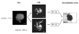
- FIG. 16 shows a production flow of a regenerating lacrimal gland primordium for transplantation using epithelial cells derived from mouse fetal lacrimal gland epithelial tissue and mesenchymal cells derived from mouse fetal lacrimal gland mesenchymal tissue, which is an embodiment of the present invention. It is a schematic diagram. In the left photograph, the lacrimal gland primordium removed from a fetal mouse of 16.5 to 17.5 was photographed under a stereomicroscope.
- FIG. 17 shows a lacrimal gland primordia extracted from a mouse fetus and a regenerative lacrimal gland primordium for transplantation prepared using epithelial cells derived from mouse fetal lacrimal gland epithelial tissue and mesenchymal cells derived from mouse fetal lacrimal gland mesenchymal tissue.
- FIG. 18 shows a state immediately before transplantation of a regenerated salivary gland primordium for transplantation having a guide prepared using epithelial cells derived from mouse fetal lacrimal gland epithelial tissue and mesenchymal cells derived from mouse fetal lacrimal gland mesenchymal tissue, and lacrimal gland
- A indicates a portion where the transplanted regenerated salivary lacrimal gland primordia have been engrafted
- B indicates the direction of the eye
- dt indicates the duct of the lacrimal gland.
- a first aspect of the production method according to the present invention is a method for producing a regenerative organ primordium for transplantation for transplantation into a mammal (eg, human), in which mesenchymal cells and epithelial cells are cultured in organ culture. And a step of inserting a guide into a regenerative organ primordium in culture produced by the method.
- a mammal eg, human
- mesenchymal cells and epithelial cells are cultured in organ culture.
- mesenchymal cell means a cell derived from mesenchymal tissue and a cell obtained by culturing the cell
- epithelial cell refers to a cell derived from epithelial tissue and its cell. It means cells obtained by culturing.
- an “epithelial appendage” is an organ formed by the interrelation between an ectoderm-derived or endoderm-derived epithelial cell and a mesenchymal cell derived from a mesoderm or a nerve lining.
- the “ectodermal accessory organ” is an ectoderm-derived organ among the “epithelial accessory organs” and means epidermal appendages such as hair follicles, sweat glands, lacrimal glands, sebaceous glands, etc. To do.
- the regenerative organ primordium used in the method of the present invention will be described.
- the regenerative organ primordium is formed by adhering a first cell aggregate substantially composed of mesenchymal cells and a second cell aggregate substantially composed of epithelial cells inside the support. It can be produced by a method including a step of culturing.
- the first cell aggregate and the second cell aggregate are substantially composed of only mesenchymal cells or epithelial cells, respectively.
- the phrase “substantially composed only of mesenchymal cells” means that, in the present invention, a certain cell aggregate performs the same function as when composed of only mesenchymal cells. Preferably, it refers to a state that contains as little as possible cells other than cells that become mesenchymal cells. In addition, different types of cells may be included as long as they are mesenchymal cells. The same applies to the case of “substantially only epithelial cells”.
- the cell aggregate refers to a state in which the cells are in close contact, and may be a tissue or a cell aggregate prepared from discrete cells.
- Using a tissue has an advantage that an organ having a correct cell arrangement and shape can be easily obtained, but the amount available can be limited. Since cultured cells can also be used for the preparation of cell aggregates, when using cultured cells, cell aggregates are relatively easy to obtain, and at least that is preferable.
- mesenchymal cells and epithelial cells used in the present invention is derived from an organ to be regenerated (including a tissue belonging to the organ).
- an organ can be easily formed using the cell which has already been directed to the target organ.
- the mesenchymal cells and the epithelial cells are both derived from the target organ.
- Examples of the regenerative organ primordium targeted by the present invention can include, but are not limited to, the organ primordium of epithelial appendages, such as a regenerated hair follicle primordium, a regenerated sweat gland primordium, and a regenerated sebaceous gland primordium. And regenerated salivary gland primordium, regenerated mammary gland primordium, regenerated kidney nephron primordium, regenerated lacrimal gland primordium, and regenerated endocrine gland primordium.
- the organ primordium of epithelial appendages such as a regenerated hair follicle primordium, a regenerated sweat gland primordium, and a regenerated sebaceous gland primordium.
- regenerated salivary gland primordium regenerated mammary gland primordium, regenerated kidney nephron primordium, regenerated lacrimal gland primordium, and regenerated endocrine gland primordium
- mesenchymal cells or epithelial cells derived from the target organ can be used, and mesenchymal cells or epithelial cells obtained by induction of undifferentiated cells Can also be used.
- mesenchymal cells or epithelial cells obtained by induction of undifferentiated cells can also be used.
- hair follicle primordia hair follicle mesenchyme derived from hair papilla cells, dermal root sheath cells, skin mesenchymal cells in development, iPS cells or ES cells as mesenchymal cells Lineage cells can be used, and epithelial cells can be outer follicle sheath outermost layer cells in the bulge region, epithelial cells in the base of the hair matrix, and hair follicle epithelial cells derived from iPS cells or ES cells.
- mesenchymal cells are mesenchymal cells in the developing stage, dermal conglomerate cells of hair follicles, skin cells derived from iPS cells or ES cells.
- Systemic cells can be used, and as epithelial cells, epithelial cells of the skin in the developing stage, sweat gland epithelial cells derived from iPS cells or ES cells can be used.
- mesenchymal cells can be mesenchymal cells in the developing stage, salivary gland mesenchymal cells derived from iPS cells or ES cells.
- epithelial cells salivary gland interstitial epithelial cells, salivary gland epithelial cells derived from iPS cells or ES cells can be used.
- mesenchymal cells can be used lacrimal gland mesenchymal cells in the developing stage, lacrimal gland mesenchymal cells derived from iPS cells or ES cells.
- epithelial cells epithelial cells of the lacrimal gland primordium in the developing stage, salivary gland epithelial cells derived from iPS cells or ES cells can be used.
- the organ from which mesenchymal cells and epithelial cells are collected preferably retains the ability to regenerate in normal adults from the viewpoint of juvenileity and homogeneity in the differentiation stage of the cells. .
- those that can be regenerated by artificial surgical operation, drug administration, or gene transfer can be used.
- mesenchymal cells derived from other than the organ to be regenerated cells derived from other mesenchymal tissues in the living body can be used.
- bone marrow cells and mesenchymal cells that do not contain blood cells more preferably oral mesenchymal cells, bone marrow cells in the jawbone, mesenchymal cells derived from head neural crest cells, and these mesenchymal cells
- mesenchymal progenitor cells and stem cells that can be differentiated into systemic cells.
- Patent Document 3 An example of using an amnion-derived cell as a mesenchymal cell is described in Patent Document 3, and an example of using a cell in which differentiation of a totipotent stem cell is induced is described in Patent Document 4, the disclosure of which is generally incorporated herein by reference. Incorporated.
- epithelial cells derived from other than the organ to be regenerated cells derived from other epithelial tissues in the living body can be used.
- epithelial cells of the skin and oral mucosa and gingiva more preferably immature epithelial progenitor cells that can differentiate into differentiated, eg, keratinized or keratinized epithelial cells, such as skin and mucous membranes,
- non-keratinized epithelial cells and stem cells thereof For example, non-keratinized epithelial cells and stem cells thereof.
- An example in which oral epithelial cells and their primary cultured cells are used as epithelial cells is described in Patent Document 2, the disclosure of which is incorporated herein by reference in its entirety.
- mesenchymal cells and epithelial cells it is preferable to use mesenchymal cells and epithelial cells to be transplanted or a tissue containing these cells.
- Mesenchymal cells and epithelial cells for producing regenerative organ primordia or tissues containing these cells include mammalian primates (eg, humans, monkeys, etc.), ungulates (eg, pigs, cows, horses, etc.) ), Small mammal rodents (eg, mice, rats, rabbits, etc.), as well as various animals such as dogs and cats.
- mammalian primates eg, humans, monkeys, etc.
- ungulates eg, pigs, cows, horses, etc.
- Small mammal rodents eg, mice, rats, rabbits, etc.
- the conditions usually used for the collection of the tissue may be applied as they are, and they may be removed under aseptic conditions and stored in an appropriate preservation solution.
- Preparation of mesenchymal cells and epithelial cells from organs to be regenerated including tissues belonging to the organs
- organs to be regenerated including tissues belonging to the organs
- an enzyme may be used for easy separation.
- the enzyme include known ones such as dispase, collagenase, and trypsin, and those skilled in the art can appropriately use preferable enzymes.
- the cell aggregate in the present invention means an aggregate of cells derived from mesenchymal tissue or epithelial tissue, a cell obtained by dispersing mesenchymal tissue or epithelial tissue apart, or the primary or subculture of the cell. It can be prepared by aggregating cells obtained by culture.
- the culture medium used for the culture is generally a medium used for culturing animal cells, such as Dulbecco. Modified Eagle's medium (DMEM) or the like can be used. Serum for promoting cell growth may be added, or as a substitute for serum, cell growth factors such as FGF, EGF, and PDGF, and known serum components such as transferrin may be added. In addition, although the density
- the cell suspension may be centrifuged.
- the cell aggregates of mesenchymal cells and epithelial cells are preferably kept in a high density state so that the cells can reliably interact when they are brought into close contact.
- the high density state means that the density is equivalent to the density at the time of composing the tissue. For example, 5 ⁇ 10 7 pieces / ml to 1 ⁇ 10 9 pieces / ml, preferably 1 ⁇ 10 8 pieces / ml. 1 ⁇ 10 9 cells / ml, most preferably 2 ⁇ 10 8 cells / ml to 8 ⁇ 10 8 cells / ml.
- the method for increasing the density of the cell aggregate is not particularly limited, and for example, it can be performed by a method in which cells are aggregated and precipitated by centrifugation. Centrifugation is preferable because it can be easily densified without impairing cell activity. Centrifugation may be performed for 3 to 10 minutes at a rotational speed that applies a centrifugal force of 300 ⁇ g to 1200 ⁇ g, preferably 500 ⁇ g to 1000 ⁇ g. Centrifugation lower than 300 ⁇ g tends to prevent the cell density from becoming sufficiently high, while centrifugation higher than 1200 ⁇ g may damage cells.
- a cell suspension is usually prepared in a container such as a tube used for centrifuging cells, and then centrifuged. After centrifugation, the supernatant may be removed as much as possible while leaving the cells as a precipitate.
- components other than the target cell for example, a culture solution, a buffer solution, etc.
- the component other than the target cell is not included. If such high-density cell aggregates are brought into close contact with each other in the support carrier by the method described later, the cells come into close contact with each other, and the cell-cell interaction is effectively exhibited.
- the support carrier is not particularly limited as long as the cells can be cultured inside, but for example, gel, fiber, solid is preferable, and it is further regenerated in vivo by using such a support carrier. It is possible to prevent excessive pressure on the organ primordia.
- the support carrier used in the present invention include collagen, agarose gel, carboxymethylcellulose, gelatin, agar, hydrogel, cell matrix (trade name), meviol gel (trade name), matrigel (trade name), elastin, fibrin, Examples include laminin, extracellular matrix mixture, polyglycolic acid (PGA), polylactic acid (PLA), and lactic acid / glycolic acid copolymer (PLGA).
- the support carrier may be a liquid and may be cured after the regenerative organ primordium is arranged.
- a collagen gel drop is prepared on a culture dish, a regenerative organ primordium is placed in the collagen drop, and then cultured in a CO 2 incubator at 37 ° C., thereby allowing the collagen to gel.
- the support carrier used for the purpose of culturing the first and second cell aggregates preferably has a holding force capable of maintaining the adhesion state of the cell aggregates without the cells being dispersed.
- the close contact state means that the above-described high-density mesenchymal cell and epithelial cell aggregates maintain the same high density near the contact surface between mesenchymal cells and epithelial cells. Means the state.
- the support carrier capable of maintaining the close contact state is a final concentration of 2 mg / ml to 3 mg / ml, that is, a method according to JIS-K6503-1996 (pressing 4 mm with a 12.7 mm diameter plunger) Use at a concentration that results in a jelly strength of 120 g to 250 g depending on the load required to provide a suitable hardness.
- Other types of support carriers are also preferably used as the support carrier of the present invention if they have the same strength by the same evaluation method.
- the method of disposing the first and second cell aggregates in the support carrier is not particularly limited.
- the cell aggregate is a cell aggregate
- the precipitate obtained by the above-mentioned centrifugation is collected with a microsyringe or the like. It can be placed in a support carrier.
- the cell aggregate is a tissue, it can be placed at any position in the support carrier using the tip of a syringe needle or the like.
- the method for arranging the first and second cell aggregates in close contact with the support carrier is not particularly limited.
- the other cell aggregate is By arranging so as to press against it, both can be brought into close contact with each other. More specifically, one cell aggregate can be pressed against the other cell aggregate by appropriately changing the position of the tip of the syringe needle in the support carrier.
- epithelial tissue or mesenchymal tissue is used as the cell aggregate, the tissue was in contact with the mesenchymal tissue or epithelial tissue in the original organ (including tissues belonging to the organ), respectively. It is preferable to arrange the surface in contact with the other cell aggregate.
- a cell can further aggregate and it can be set as a higher density state.
- a cell can be solidified by allowing it to stand for several minutes to several tens of minutes at the culture temperature. At this time, the smaller the components other than the cells in the cell aggregate, the higher the density state is realized.
- the culture period varies depending on the number of cells arranged in the support carrier, the state of the cell aggregate, the culture conditions, the animal species, etc., and can be appropriately selected by those skilled in the art. Moreover, it can change suitably also by the organ used as reproduction
- the regenerated hair follicle primordium when a regenerated hair follicle primordium is transplanted, in order to obtain functional hair, the regenerated hair follicle primordium is preferably cultured for at least one day, and more preferably cultured for two or more days.
- the regenerated salivary gland primordium or a regenerated lacrimal gland primordium when transplanting a regenerated salivary gland primordium or a regenerated lacrimal gland primordium to obtain a functional salivary gland or lacrimal gland, it is preferable to culture the regenerated salivary gland primordium or regenerated lacrimal gland primordium for at least one day. Is more preferable.
- the support carrier enclosing the first and second cell aggregates may be cultured alone, or may be cultured in the presence of other animal cells or the like.
- the culture conditions can be those used for culturing general animal cells.
- mammal-derived serum may be added to the culture, and various cellular factors known to be effective for the growth and differentiation of these cells may be added. Examples of such cellular factors include FGF and BMP.
- the culture inside the support carrier is organ culture. It is preferable that In organ culture, generally, a porous membrane is floated on a medium suitable for the growth of animal cells, and a support carrier containing the first and second cell aggregates is placed on the membrane and cultured.
- the porous membrane used here preferably has a large number of pores of about 0.3 to 5 ⁇ m, and examples thereof include a cell culture insert (trade name) and an isopore filter (trade name). .
- a regenerative organ primordium is constructed from epithelial cells and mesenchymal cells, and then a guide is inserted into the regenerative organ primordium.
- the “guide” used in the present invention is inserted into a regenerative organ primorum in culture constructed by organ culture, and after transplantation of the regenerative organ primordium, the part on the epithelial cell side of the regenerative organ primordium and the recipient.
- the guide used in the present invention may have a hollow fiber shape.
- the diameter and length of the guide can be appropriately designed depending on the organ to be reproduced.
- the diameter is preferably 5 to 100 ⁇ m, more preferably 20 to 50 ⁇ m.
- the length is preferably 1 to 10 mm, more preferably 4 to 6 mm.
- the guide diameter is preferably 5 ⁇ m to 60 ⁇ m, and preferably 20 ⁇ m to 40 ⁇ m. Is more preferable.
- the length of the guide is preferably 1 mm to 6 mm, and more preferably 2 mm to 3 mm.
- a guide that has a smooth surface so that the guide can be easily inserted into the conduit and that is hard enough not to damage the tissue is preferable.
- a monofilament material is preferable to a multifilament such as a blade.
- the insertion of the guide does not destroy the structure of the cell aggregate that is the primary organ of the regenerative organ, particularly the contact surface between the epithelial cells and the mesenchymal cells, and the epithelial cell fraction and the mesenchymal cell fraction It is inserted from the epithelial cell side of the cell mass that becomes the regenerative organ primordium so as to penetrate vertically.
- the guide can be inserted into the cell mass that is the regenerative organ primordium immediately after the start of organ culture, that is, immediately after the cell aggregate of epithelial cells and mesenchymal cells is placed in the medium. it can.
- the epithelial cells of the regenerated hair follicle primordium increase in strength due to cell adhesion by organ culture, so that the penetration strength is increased by sharpening the guide material strength (for example, using a stainless steel wire) and the tip of the guide. By increasing the number, it can be inserted 1-2 days after the start of culture.
- a flexible material with low foreign body response to a living body such as nylon thread can be used.
- the regenerative organ primordium can be cultured with the guide inserted.
- the culture period after insertion of the guide can be appropriately set depending on the organ to be regenerated. For example, when producing a regenerated hair follicle primordium, it is preferably cultured for 1 to 4 days, and 1.5 to It is more preferable to culture for 2 days. In addition, by culturing for 2 days after inserting the guide, the adhesion between the guide and the regenerated organ becomes strong, and it does not easily come off at the time of transplantation.
- the regenerative organ primordium on the epithelial cell side can be extended along the guide by culturing after the guide is inserted. Such elongation can improve the efficiency and stability of autonomous adhesion between the regenerative organ primordium portion on the epithelial cell side and the recipient epithelial cell after transplantation of the regenerative organ primordium.
- a regenerative organ primordium for transplantation having a guide produced by the production method of the present invention can be transplanted to a target site.
- the regenerative organ primordium for transplantation into which the guide is inserted can be transplanted to a target site by a method known to those skilled in the art.
- a Shapiro-type flocking method flocking using a choy type flocking device, an implanter using air pressure, or the like.
- Shapiro hair transplantation is a method in which a transplant site is made with a micro knife or the like and then transplanted using tweezers.
- the transplantation depth of the regenerating organ primordium can be appropriately changed depending on the organ to be regenerated.
- it is preferably 0.05 to 5 mm, more preferably 0.1 to 1 mm, and most preferably 0.3 to 0.5 mm.
- it is preferable to transplant it into the dermis layer, and more preferably, the interface between the dermis and the subcutaneous tissue with excellent hair follicle formation and subsequent hair growth efficiency. It is preferable to set it further upward.
- transplantation depth it is preferable to adjust the transplantation depth so that the upper end of the epithelial cell component of the regenerated hair follicle primordium is exposed at the upper end of the transplanted wound because the continuity with the epithelial cells of the recipient can be further increased.
- transplanting organ primordia that require a guide to be inserted into the recipient's conduit such as when transplanting regenerated salivary gland primordia, keep the recipient's conduit long. Therefore, it is possible to prevent the guide from coming off after transplantation, which is preferable.
- the transplant site is close to the vena cava.
- the guide and the site to be transplanted can be fixed with a skin bonding tape, band, suture, or the like so that the guide does not come off.
- the guide can be removed from the transplant site after continuity between the epithelial cells on the recipient side and the epithelial cell-derived side of the regenerative organ primordium has been ensured for a while after transplanting the regenerative organ primordium.
- the regenerated hair follicle primordium when transplanted, it is preferably removed from the transplant site 3 to 7 days after the transplantation.
- the guide can be left until it is naturally removed from the transplant site.
- the guide of bioabsorbable material can be left naturally until it is removed from the transplant site or decomposed / absorbed.
- a bioabsorbable guide is preferably used when the guide is embedded in the recipient's skin together with the regenerative organ primordium after transplantation, such as the regenerated salivary gland primordium.
- the guide by holding the guide to the regenerative organ primordium for transplantation, cells derived from the epithelial cells of the regenerative organ primordium extend along the guide.
- continuity between the epithelial cells on the recipient side after transplantation and the epithelial cells side of the regenerative organ primordium can be improved.
- the guide is maintained outside the epidermis at the transplant site, such as in hair follicles and sweat glands, the epithelial cells on the recipient side are placed inside the transplant site along the guide so as to eliminate foreign objects. Therefore, continuity can be further improved.
- the insertion of the guide is preferable because it can improve the maintenance of the polarity of epithelial cells and mesenchymal cells in the regenerative organ primordium during culture.
- the direction at the time of transplantation can be made easy.
- continuity between the regenerated hair follicle primordium and the recipient epithelial cells can be secured, and hair follicle formation can be promoted in the intended direction. .
- the hair growth rate from the regenerated hair follicle primordium can be improved and the direction of hair growth can be controlled.
- Embodiments of the present invention may be described with reference to schematic diagrams, but in the case of schematic diagrams, they may be exaggerated for clarity of explanation.
- terms such as first, second, etc. are used to represent various elements, it is understood that these elements should not be limited by those terms. These terms are only used to distinguish one element from another, for example, the first element is referred to as the second element, and similarly, the second element is the first element. Can be made without departing from the scope of the present invention.
- the separated dermal papilla is seeded on a 3.5 cm culture plastic dish (Nippon Becton Dickinson), and in DMEM10 containing 10 ng / ml FGF2 (Wako Pure Chemical Industries), in an environment of 5% CO2, 37 ° C. and humidity of 95%.
- the primary culture was performed. Primary cultured dermal papilla cells were used after changing the medium on the 4th and 8th days and culturing for 9 days. Primary cultured dermal papilla cells were washed 3 times with PBS (-), then detached with 10 mM EDTA solution (GIBCO) containing 0.05% trypsin, neutralized with trypsin with DMEM10, washed thoroughly, and then at ice temperature. Stored until use.
- the separated bulge region epithelial tissue was subjected to enzyme treatment with 0.05% Trypsin (Invitrogen, Carlsbad, US) for 1 hour in an incubator, and made into a single cell through a 35 ⁇ mpore cell strainer.
- the cultured dermal papilla cells obtained in (2) were collected with 0.05% Trypsin (Invitrogen, Carlsbad, US), and made into singulated cells through a 35 ⁇ mpore cell strainer.
- Regenerated hair follicle primordium was prepared according to the organ primordium method. Details of the procedure are described below.
- the obtained single bulge region cells derived from bulge region epithelial tissue and cultured dermal papilla cells are separately transferred to a 1.5 ml microtube (Eppendorf) coated with silicone grease, collected as a precipitate by centrifugation, and centrifuged. The supernatant of the subsequent culture solution was completely removed using 0.5-20 ml (Eppendorf) of GELoder Tip.
- Cellmatrix type IA (Nita gelatin, Osaka, Japan) was dropped on a Petri dish coated with silicon grease (Toray Dow Corning) to prepare a collagen gel drop, and the cultured hair prepared above About 0.1 ml of nipple cells were injected using a 0.1-10 ml pipette tip (Quality Scientific plastics) to produce a cell aggregate. Subsequently, the bulge region epithelial cells prepared above were put into the same gel drop using 0.1-10 ml pipette tips (Quality Scientific plastics) so as to adhere to the aggregates of cultured hair papilla cells. Approximately 2 ml was injected to prepare a cell aggregate.
- a 5 mm long nylon thread (Matsuda Medical Industry Co., Ltd.)
- the contact surface of the cultured dermal papilla cells and the bulge region epithelial cell fraction are inserted through the microscope under a stereomicroscope so as to penetrate vertically, and then at 37 ° C. for 5 minutes.
- the regenerated hair follicle primordium having a guide was produced by allowing the gel drop to solidify and solidifying the bond between epithelial cells and mesenchymal cells.
- the epithelial layer was treated twice with 100 units / ml collagenase I at 37 ° C. for 40 minutes to remove contaminating mesenchymal cells, and further, a 0.25% trypsin solution containing 100 units / ml collagenase I was treated at 37 ° C. for 10 minutes, followed by singularization treatment to obtain epithelial cells.
- cell aggregates of fetal skin mesenchymal cells and fetal skin epithelial cells having a guide were prepared by the same method as described in (4).
- the cell aggregates of epithelial cells and mesenchymal cells prepared in the gel were placed on a 0.4 ml pore size Cell Culture Insert (Becton Dickinson) set in a 6-well plate (Becton Dickinson) with 1 ml of DMEM10 added.
- the whole collagen gel was transferred to the tissue, and organ culture was performed for 2 days under conditions of 37 ° C., 5% CO 2, and humidity 95% to prepare a regenerated hair follicle primordium.
- the transplanted wound had a depth of about 400 ⁇ m in the vertical direction from the body surface, and the horizontal direction was about 1 mm.
- a nylon thread guide was fixed to the surface of the skin adjacent to the graft with a steristrip (3M), and then the graft was protected with a nurse van and surgical tape (3M). The protective tape was removed 5-7 days after the transplantation, and the nylon thread guide was left at the transplantation site. If it remained one day later, it was removed.
- Follow-up was performed after the engraftment of the implant was judged visually or with a fluorescent stereomicroscope.
- the regenerated hair follicle primordium was obtained from 18.5 day old mouse skin cells. Prepared and transplanted into nude mouse skin. After transplantation, the skin was removed, paraffin sections were prepared, and HE staining was performed. As a result, the transplantation position was in the skin dermis layer, and the average depth from the body surface was 393 ⁇ m. The results immediately after transplantation, 3 days after transplantation, and 10 days after transplantation are shown in FIG. As shown in FIG.
- FIG. 4 shows the results of transplanting a regenerated hair follicle primordium derived from an adult cheek to the back of a nude mouse so as to obtain a similar transplantation depth.
- FIG. 4A shows a photograph in which a guide is inserted into the regenerated hair follicle primordium, E shows a part derived from bulge region epithelial cells, DP shows a part derived from cultured papillary cells, and G shows a guide.
- FIG. 4A shows a photograph in which a guide is inserted into the regenerated hair follicle primordium
- E shows a part derived from bulge region epithelial cells
- DP shows a part derived from cultured papillary cells
- G shows a guide.
- FIG. 4B shows a photograph of the transplant site traced with a fluorescent stereomicroscope.
- FIG. 6 shows photographs observed with an optical microscope and a scanning electron microscope. In the dotted line portion, a spiral hair pulp (M) characteristic of wrinkle-like bristles was observed (FIG. 6a), and a cuticle structure equivalent to that of a normal cheek fold was observed (FIG. 6b). As described above, the regenerated wrinkle had the same pulp and cortex as natural hair, and had a clear wrinkle shape.
- the excised submandibular gland primordium was reacted with Dispase II (Becton Dickson) solution at a final concentration of 50 U / ml for 1.5 minutes at 25 ° C., and then submandibular gland epithelial tissue and submandibular using a 25 G injection needle Separated into glandular mesenchymal tissue. Further, the mesenchymal tissue was subjected to enzyme treatment for 10 minutes in a 37 ° C. warm bath with Collagenase (Worthington, Lakewood, NJ) solution at a final concentration of 50 U / ml and Trypsin (Invitrogen, Carlsbad, US) solution with a final concentration of 0.25%.
- Dispase II Becton Dickson
- Completely singulated mesenchymal cells were obtained through a 22 ⁇ pore cell strainer.
- the epithelial tissue was treated twice with a collagenase (Worthington, Lakewood, NJ) solution at a final concentration of 100 U / ml for 15 minutes in a 37 ° C. warm bath, and then trypsin (Invitrogen, Carlsbad, US, 0.25% final concentration). )
- the solution was subjected to enzyme treatment in a 37 ° C. warm bath for 5 minutes, and completely singulated epithelial cells were obtained through a 22 ⁇ pore cell strainer.
- regenerated salivary gland primordia were prepared in collagen gel by the organ primordia method (FIG. 9). Details of the procedure are described below.
- the obtained singulated epithelial cells and singulated mesenchymal cells were separately transferred to 1.5 ml microtubes (Eppendorf) coated with silicone grease and centrifuged. The supernatant of the culture broth after centrifugation was completely removed using 0.5-20 ml of GELoader Tip (Eppendorf), and singled epithelial cells and singled mesenchymal cells were recovered as precipitates.
- Cellmatrix type IA (Nita gelatin, Osaka, Japan) was dropped on a Petri dish coated with silicon grease (Toray Dow Corning) to produce a collagen gel drop.
- About 0.1 ⁇ l of the mesenchymal cells were injected using a 0.1-10 ml pipette tip (Quality Scientific plastics) to produce a cell aggregate.
- the singulated epithelial cells prepared above are brought into close contact with the aggregates of mesenchymal cells using 0.1-10 ml pipette tips (Quality Scientific plastics) in the same gel drop.
- About 0.2 ⁇ l was injected to produce a cell aggregate of epithelial cells and mesenchymal cells derived from the submandibular gland primordia.
- FIG. 10 shows the state at each elapsed time when the submandibular gland removed from the fetal mouse was cultured in the organ as it was removed, and the lower photograph in FIG. The state in each elapsed time at the time of organ culture of the regenerated salivary gland primordia constructed by the method.
- Analysis by hematoxylin-eosin staining (HE staining) revealed that the tip of the invaded epithelial tissue was branched 72 hours after the start of culture (FIG. 10).
- the regenerated salivary gland primordium can be produced by using the organ primordium method.
- submandibular gland primordium having a different developmental stage was obtained from a 13.5-day-old mouse fetus (refer to the middle and late photographs in FIG. 9). From any submandibular gland primordium at any stage of development, regenerated salivary gland primordia reconstructed from epithelial cells and mesenchymal cells could be prepared by the above method.
- collagen gel containing the regenerated salivary gland primordium and the masseter muscle were sutured with 8-0 nylon suture thread (Bearmedic, Chiba, Japan) so that the transplanted regenerated salivary gland primordium would not move (FIG. 12).
- Regenerated salivary gland primordium 30 days after transplantation was evaluated histologically by HE staining. From the HE-stained image, duct cells and serous acinar cell-like cells were observed in the regenerated salivary gland primordia as in the case of the natural submandibular gland (FIG. 13. In the lower photograph in FIG. 13, the arrow indicates the serous gland. Shows tuft-like cells). Thus, salivary glands could be reconstructed by the regenerative organ primordium method. In addition, even the regenerated salivary gland primordium derived from the submandibular gland was able to reconstruct the submandibular gland in the parotid gland, which is another salivary gland.
- regenerated lacrimal gland primordium was prepared using the organ primordia method as follows. Lacrimal gland primordia were surgically removed from embryonic day 16.5 to 17.5 C57BL / 6 mouse fetuses. The excised lacrimal gland primordium was reacted with Dispase II (Becton Dickson) solution at a final concentration of 50 U / ml for 1.5 minutes at 25 ° C., and then separated into lacrimal epithelial tissue and lacrimal gland mesenchymal tissue using a 25 G injection needle did.
- Dispase II Becton Dickson
- mesenchymal tissue was subjected to enzyme treatment for 10 minutes in a 37 ° C. warm bath with Collagenase (Worthington, Lakewood, NJ) solution at a final concentration of 50 U / ml and Trypsin (Invitrogen, Carlsbad, US) solution with a final concentration of 0.25%.
- Collagenase Worthington, Lakewood, NJ
- Trypsin Invitrogen, Carlsbad, US
- Completely singulated mesenchymal cells were obtained through a 22 ⁇ pore cell strainer.
- the epithelial tissue was treated twice with a collagenase (Worthington, Lakewood, NJ) solution at a final concentration of 100 U / ml for 15 minutes in a 37 ° C. warm bath, and then trypsin (Invitrogen, Carlsbad, US, 0.25% final concentration).
- the solution was subjected to enzyme treatment in a 37 ° C. warm bath for 5 minutes, and completely singulated epithelial cells were obtained through a 22 ⁇ pore cell strainer.
- regenerated lacrimal gland primordium was prepared in collagen gel by the organ primordium method. Details of the procedure are described below.
- the obtained singulated epithelial cells and singulated mesenchymal cells were separately transferred to 1.5 ml microtubes (Eppendorf) coated with silicone grease and centrifuged.
- the supernatant of the culture broth after centrifugation was completely removed using 0.5-20 ml of GELoader Tip (Eppendorf), and singled epithelial cells and singled mesenchymal cells were recovered as precipitates.
- 30 ⁇ l of Cellmatrix type IA (Nita gelatin, Osaka, Japan) was dropped on a Petri dish coated with silicon grease (Toray Dow Corning) to prepare a collagen gel drop, and the single prepared above About 0.1 ⁇ l of the mesenchymal cells were injected using a 0.1-10 ml pipette tip (Quality Scientific plastics) to produce a cell aggregate.
- the singulated epithelial cells prepared above are brought into close contact with the aggregates of mesenchymal cells using 0.1-10 ml pipette tips (Quality Scientific plastics) in the same gel drop. About 0.2 ⁇ l was injected to produce a cell aggregate of epithelial cells and mesenchymal cells derived from the lacrimal gland primordia (FIG. 16).
- Cell aggregates of epithelial cells derived from lacrimal gland primordia and mesenchymal cells derived from lacrimal gland primordia prepared in gel were 0.4 ml pore size set in 6 well Plate (Becton Dickinson) with 1 ml DMEM10 added. The whole collagen gel was transferred onto Cell Culture Insert (Becton Dickinson), and organ culture was performed under conditions of 37 ° C., 5% CO 2, and humidity 95% to prepare a regenerated lacrimal gland primordia.
- the regenerative organ primordium having a guide according to the invention of this application was reliably connected to the recipient epithelial cells, and became a regenerative organ having a function after transplantation.
- the regenerated hair follicle primordium grows from the body surface at a high frequency. It was observed that the regenerated hair follicle continued to pass through the recipient epidermis layer and the pore portion even after passing through the hair cycle, and repeated hair growth at the same location.
- the transplantation procedure of the regenerated hair follicle primordia with a guide was extremely simple and widely applicable.
- the regenerative organ primordium for transplantation produced by the present invention can provide new organ replacement regenerative medicine that can be widely used in the medical industry.
Abstract
Description
最近になり、上皮性付属器官である歯や唾液腺、皮膚付属器官である毛包において、器官原基を再生し、発生過程を再現することによって器官再生を目指した研究が進められている。これらの器官は生命の維持に直結するわけではないものの、器官喪失や機能不全に陥ることが知られている。このような例としては、う蝕や傷害、歯胚形成不全による歯の喪失や、高齢化に伴う唾液分泌障害、男性型脱毛症や毛包形成不全による毛髪の喪失などが挙げられる。このような、器官喪失や機能不全は、QOLに大きな影響を与えるため、器官再生による機能回復が大いに期待されている。なかでも歯の再生において、胎児性の歯胚由来の上皮系細胞と胎児性の歯胚由来の間葉系細胞を適切に三次元配置させた再生歯胚の移植や、生体内で異所的に再生した再生歯ユニットの移植や、歯と歯槽骨、歯根膜を有する機能的ユニットの移植により歯の生理機能をすべて有する機能的な歯の再生が示されている(例えば、特許文献1参照)。さらに、口腔内上皮細胞やその初代培養細胞を上皮系細胞として利用した場合(例えば、特許文献2参照)、羊膜由来細胞を間葉系細胞として利用した場合(例えば、特許文献3参照)、全能性幹細胞を分化誘導した細胞を間葉系細胞として利用した場合(例えば、特許文献4参照)にも、同様に特有の細胞配置と方向性を備えた再生歯胚や再生歯ユニットが得られることが示されている。
これらの研究成果から、再生器官原基、あるいは再生器官原基より再生した器官の移植による機能的な器官再生の実現可能性が示されたといえ、その他の上皮性付属器官についても、再生器官原基による器官置換的再生医療が望まれている。 On the other hand, it is known that an organ has a three-dimensional arrangement of multiple types of functional cells and expresses a unique function. Almost all organs are generated by the interaction of fetal epithelial cells and mesenchymal cells, and exhibit their unique morphology and organ function. In the current regenerative medicine technology, it is difficult to arrange a plurality of types of cells in a three-dimensional manner, and a regenerative organ construction that can function immediately in vitro has not been developed yet.
In recent years, research aimed at organ regeneration by regenerating the organ primordium and reproducing the development process in epithelial appendages such as teeth and salivary glands and skin appendages as hair follicles has been promoted. Although these organs are not directly linked to sustaining life, they are known to suffer organ loss and dysfunction. Examples of this include tooth loss due to caries and injury, tooth germ formation failure, salivary secretion disorders associated with aging, hair loss due to androgenetic alopecia and hair follicle formation failure, and the like. Such organ loss and dysfunction greatly affect QOL, and therefore functional recovery by organ regeneration is highly expected. In particular, in the regeneration of teeth, transplantation of regenerated tooth germs in which fetal tooth germ-derived epithelial cells and fetal tooth germ-derived mesenchymal cells are appropriately arranged in three dimensions, or ectopic in vivo. Regeneration of functional teeth having all the physiological functions of the teeth is shown by transplanting the regenerated tooth unit regenerated and functional units having teeth, alveolar bone and periodontal ligament (see, for example, Patent Document 1) ). Furthermore, when oral epithelial cells and their primary cultured cells are used as epithelial cells (for example, see Patent Document 2), amnion-derived cells are used as mesenchymal cells (for example, see Patent Document 3), totipotency Even when cells in which sex stem cells have been induced to differentiate are used as mesenchymal cells (see, for example, Patent Document 4), regenerated tooth embryos and regenerated tooth units having specific cell arrangement and directionality can be obtained. It is shown.
From these research results, it can be said that the possibility of functional organ regeneration by transplantation of regenerative organ primordia or organs regenerated from regenerative organ primordia was demonstrated. There is a demand for organ replacement regenerative medicine.
皮膚付属器官である毛包は、個体の生涯にわたって成長と退行(毛周期)を繰り返し、成長期における毛球部の再生は、毛包器官発生期と同様な分子機構により誘導されることが知られている。また、このような毛周期における毛球部の再生は、間葉系細胞である毛乳頭細胞により誘導されると考えられている。そして、成長期において、毛包上皮幹細胞が間葉系細胞である毛乳頭細胞により分化誘導され毛球部が再生されると考えられている。また、バルジ領域および下方領域には、神経提由来幹細胞のニッチが存在することから、毛包は複数の幹細胞ニッチを保持し、幹細胞プールとして機能していると考えられている。毛包形成誘導能を保持した毛乳頭細胞については、その培養法が報告されている。毛包間葉系細胞の幹細胞ニッチについては未だに確定的な報告はされていないが、毛乳頭には軟骨細胞、血球系細胞、脂肪細胞に分化可能な細胞が含まれており、成体皮膚真皮組織からは毛包および皮膚の真皮を再生するSKIPs細胞を得ることができることから、毛包間葉系幹細胞の存在が示唆されている。これらのことより、他の器官では実現されていない成体由来細胞による完全な器官再生の可能性が示唆され、臨床応用可能な毛包再生技術の実用化が期待されている。 In general, epithelial appendages such as hair follicles, sebaceous glands, lacrimal glands, and salivary glands are affected by the interaction between ectoderm-derived or endoderm-derived epithelial cells and mesodermal cells derived from mesoderm or nerve lining in the fetal stage. It is known to occur.
It is known that hair follicles, which are skin appendages, repeat growth and regression (hair cycle) throughout the life of an individual, and regeneration of the hair bulb during the growth phase is induced by the same molecular mechanism as in the hair follicle organ development phase. It has been. Moreover, it is thought that the reproduction | regeneration of the hair bulb part in such a hair cycle is induced | guided | derived by the hair papilla cell which is a mesenchymal cell. In the growth phase, it is considered that hair follicle epithelial stem cells are induced to differentiate by hair papilla cells that are mesenchymal cells and the hair bulb part is regenerated. In addition, since the niche of nerve-derived stem cells exists in the bulge region and the lower region, the hair follicle is considered to hold a plurality of stem cell niches and function as a stem cell pool. A method for culturing dermal papilla cells that retain the ability to induce hair follicle formation has been reported. The stem cell niche of hair follicle mesenchymal cells has not yet been definitively reported, but the dermal papilla contains cells that can differentiate into chondrocytes, blood cells, and adipocytes. Can obtain SKIPs cells that regenerate hair follicles and dermal dermis, suggesting the presence of hair follicle mesenchymal stem cells. These facts suggest the possibility of complete organ regeneration by adult-derived cells that have not been realized in other organs, and the practical application of clinically applicable hair follicle regeneration technology is expected.
ここで、本発明のガイドを有する移植用再生器官原基の製造方法の一実施態様においては、ガイドを挿入する工程後、さらに数日、前記再生器官原基を培養する工程を含むことを特徴とする。
本発明のガイドを有する移植用再生器官原基の製造方法の一実施態様においては、前記第1の細胞集合体と前記第2の細胞集合体のうち、少なくともいずれか一方が、再生対象の器官由来であることを特徴とする。
本発明のガイドを有する移植用再生器官原基の製造方法の一実施態様においては、前記第1の細胞集合体および前記第2の細胞集合体が、ともに再生対象の器官由来であることを特徴とする。
本発明のガイドを有する移植用再生器官原基の製造方法の一実施態様においては、前記再生器官原基が、上皮性付属器官原基であることを特徴とする。
本発明のガイドを有する移植用再生器官原基の製造方法の一実施態様においては、前記再生器官原基が、再生毛包原基、再生汗腺原基、再生皮脂腺原基、再生唾液腺原基、再生乳腺原基、再生腎臓ネフロン原基、再生涙腺原基、および、再生内分泌腺原基からなる群より選択されるいずれか一つの器官原基であることを特徴とする。
本発明のガイドを有する移植用再生器官原基の製造方法の一実施態様においては、前記再生器官原基が再生毛包原基であり、前記上皮系細胞がバルジ領域上皮細胞または毛母基底部上皮細胞であることを特徴とする。
本発明のガイドを有する移植用再生器官原基の製造方法の一実施態様においては、前記再生器官原基が再生毛包原基であり、前記間葉系細胞が毛乳頭細胞または真皮毛根鞘細胞であることを特徴とする。
本発明のガイドを有する移植用再生器官原基の製造方法の一実施態様においては、前記再生器官原基が再生唾液腺原基であり、前記上皮系細胞が唾液腺由来の上皮系細胞であり、前記間葉系細胞が唾液腺由来の間葉系細胞であることを特徴とする。
本発明のガイドを有する移植用再生器官原基の製造方法の一実施態様においては、前記再生器官原基が再生涙腺原基であり、前記上皮系細胞が涙腺由来の上皮系細胞であり、前記間葉系細胞が涙腺由来の間葉系細胞であることを特徴とする。
本発明のガイドを有する移植用再生器官原基の製造方法の一実施態様においては、前記ガイドが、化学繊維であることを特徴とする。
本発明のガイドを有する移植用再生器官原基の製造方法の一実施態様においては、前記ガイドが、生体吸収性であることを特徴とする。
本発明のガイドを有する移植用再生器官原基の製造方法の一実施態様においては、前記ガイドが、ナイロン糸であることを特徴とする。 That is, the present invention is a method for producing a transplanted regenerative organ primordium having a guide, which is substantially composed of a first cell aggregate substantially composed of mesenchymal cells and epithelial cells. The present invention relates to a production method comprising a step of preparing a regenerative organ primordium by bringing the second cell aggregate into close contact with each other and culturing in a support, and a step of inserting a guide into the regenerative organ primordium.
Here, in one embodiment of the method for producing a transplanted regenerative organ primordium having a guide according to the present invention, the method further comprises the step of culturing the regenerative organ primordium for several days after the step of inserting the guide. And
In one embodiment of the method for producing a transplanted regenerative organ primordium having a guide according to the present invention, at least one of the first cell aggregate and the second cell aggregate is an organ to be regenerated. It is derived from.
In one embodiment of the method for producing a transplantable regenerative organ primordium having a guide of the present invention, the first cell aggregate and the second cell aggregate are both derived from an organ to be regenerated. And
In one embodiment of the method for producing a transplanted regenerative organ primordium having a guide according to the present invention, the regenerative organ primordia is an epithelial appendage primordia.
In one embodiment of the method for producing a regenerative organ primordium for transplantation having a guide of the present invention, the regenerative organ primordium is a regenerated hair follicle primordium, a regenerated sweat gland primordium, a regenerated sebaceous gland primordium, a regenerated salivary gland primordium, It is an organ primordium selected from the group consisting of a regenerated mammary gland primordium, a regenerated kidney nephron primordium, a regenerated lacrimal gland primordium, and a regenerated endocrine gland primordium.
In one embodiment of the method for producing a regenerative organ primordium for transplantation having a guide of the present invention, the regenerative organ primordium is a regenerated hair follicle primordium, and the epithelial cells are bulge region epithelial cells or hair matrix bases. It is an epithelial cell.
In one embodiment of the method for producing a transplanted regenerative organ primordium having a guide according to the present invention, the regenerative organ primordium is a regenerated hair follicle primordium, and the mesenchymal cells are hair papilla cells or dermal root sheath cells. It is characterized by being.
In one embodiment of the method for producing a regenerative organ primordium for transplantation having a guide of the present invention, the regenerative organ primordium is a regenerated salivary gland primordium, the epithelial cell is an epithelial cell derived from the salivary gland, The mesenchymal cells are salivary gland-derived mesenchymal cells.
In one embodiment of the method for producing a regenerative organ primordium for transplantation having a guide of the present invention, the regenerative organ primordium is a regenerative lacrimal gland primordium, the epithelial cell is an epithelial cell derived from the lacrimal gland, The mesenchymal cell is a mesenchymal cell derived from the lacrimal gland.
In one embodiment of the method for producing a transplantable regenerative organ primordium having a guide according to the present invention, the guide is a chemical fiber.
In one embodiment of the method for producing a transplantable regenerative organ primordium having a guide according to the present invention, the guide is bioabsorbable.
In one embodiment of the method for producing a transplantable regenerative organ primordium having a guide according to the present invention, the guide is a nylon thread.
また、本発明の別の態様によれば、ガイドを有する移植用再生器官原基の製造方法により作製されたガイドを有する移植用再生器官原基を、対象の部位へ移植する工程を含む、移植用再生器官原基の移植方法に関する。
ここで、本発明のガイドを有する移植用再生器官原基の移植方法の一実施態様においては、前記ガイドを移植部位より突出した状態で維持することにより、移植した再生器官原基の上皮系細胞側の部分と前記対象の上皮系細胞とがガイドに沿って伸長し、連結することを特徴とする。
また、本発明のガイドを有する移植用再生器官原基の移植方法の一実施態様においては、前記再生器官原基が、導管を有する器官の再生を目的とした器官原基であり、前記再生器官原基の移植時に、前記ガイドを前記対象部位に存在する導管内に挿入し、前記再生器官原基の上皮系細胞側の部分と前記導管とを連絡させることにより、前記再生器官原基の上皮系細胞側の部分がガイドに沿って伸長し、前記対象部位の導管と連結することを特徴とする。 Moreover, according to another aspect of this invention, it is related with the composition containing the regenerative organ primordium for a transplant which has a guide produced by the manufacturing method of the regenerative organ primordium for a transplant which has a guide.
According to another aspect of the present invention, the transplant includes a step of transplanting a regenerative organ primordium for transplantation having a guide produced by the method for producing a regenerative organ primordium for transplantation having a guide to a target site. The present invention relates to a method for transplanting regenerative organ primordia.
Here, in one embodiment of the transplantation method of the transplantable regenerative organ primordium having the guide of the present invention, the transplanted regenerative organ primordial epithelial cell is maintained by maintaining the guide protruding from the transplantation site. The side portion and the target epithelial cell extend along a guide and are connected.
Further, in one embodiment of the transplantation method of the transplantable regenerative organ primordium having the guide of the present invention, the regenerative organ primordium is an organ primordium intended for regeneration of an organ having a conduit, and the regenerative organ At the time of transplantation of the primordium, the guide is inserted into a conduit existing in the target site, and the epithelial cell side portion of the regenerative organ primordium is connected to the conduit, thereby causing the epithelium of the regenerative organ primordium A part on the system cell side extends along the guide and is connected to the conduit of the target site.
また、ガイドに沿って再生器官原基の上皮系細胞とレシピエント側の上皮層(上皮系細胞)とが連結することにより、特に毛包形成の過程においては、再生器官原基由来の嚢胞形成を阻止することができる。また、唾液腺のように、導管を有する外分泌腺等においては、上皮系細胞がガイドに沿って伸長しているため、レシピエント側の導管との連結効率を向上させることができる。
さらに、本発明により作製される再生器官原基は、毛包、汗腺、唾液腺等を含む上皮性付属器官に対して適用可能であり、体表面にかぎらず口腔内や腸管、あるいは内分泌組織のような内腔に管腔構造を介して接続する器官の再生にも適用可能である。これらの器官および組織の再生においても、適用できることから、皮膚付属器官のみならず、広く器官および組織再生医療に応用可能な技術を提供する。 According to the present invention, regeneration for transplantation that can secure the direction of polarity of the regenerated organ primordium and improve adhesion while maintaining appropriate orientation with respect to the epithelial tissue on the recipient side after transplantation. Organ primordia can be provided. Further, since the regenerative organ primordium for transplantation produced according to the present invention has a guide, the operation at the time of transplantation can be simplified, and the depth at the time of transplantation, the direction of transplantation, etc. can be adjusted easily. It is possible.
In addition, by connecting the epithelial cells of the regenerating organ primordia and the recipient epithelial layer (epithelial cells) along the guide, cyst formation derived from the regenerating organ primordium, especially in the process of hair follicle formation Can be prevented. Moreover, in an exocrine gland having a conduit, such as a salivary gland, since the epithelial cells extend along the guide, the connection efficiency with the conduit on the recipient side can be improved.
Furthermore, the regenerative organ primordium prepared according to the present invention can be applied to epithelial appendages including hair follicles, sweat glands, salivary glands, etc., and is not limited to the surface of the body, such as the oral cavity, intestinal tract, or endocrine tissue. The present invention can also be applied to regeneration of organs that connect to various lumens via a luminal structure. Since it can be applied to the regeneration of these organs and tissues, it provides a technique applicable not only to skin appendages but also to organs and tissue regeneration medicine.
本発明に係る製造方法の第一の態様は、哺乳動物(例えば、ヒト)に移植するための移植用再生器官原基の製造方法であって、間葉系細胞と上皮系細胞とを器官培養することにより作製した培養中の再生器官原基へガイドを挿入する工程を含むことを特徴とする。 Hereinafter, embodiments of this application will be described in detail.
A first aspect of the production method according to the present invention is a method for producing a regenerative organ primordium for transplantation for transplantation into a mammal (eg, human), in which mesenchymal cells and epithelial cells are cultured in organ culture. And a step of inserting a guide into a regenerative organ primordium in culture produced by the method.
また、本明細書において、「上皮性付属器官」とは、外胚葉由来または内胚葉由来の上皮細胞と、中胚葉または神経提由来の間葉系細胞との相互関係により形成される器官であり、例えば、内胚葉性上皮由来の器官や、外胚葉性付属器官、内分泌腺、導管を介して他の上皮組織と接続する外分泌腺等の器官を意味し、より具体的には、例えば、毛包、汗腺、涙腺、皮脂腺等の表皮付属器官や、口腔内、腸管、もしくは内分泌組織のような内腔に管腔構造を介して接続する器官を意味する。
また、本明細書において、「外胚葉性付属器官」とは、「上皮性付属器官」のうち、外胚葉由来の器官であり、毛包、汗腺、涙腺、皮脂腺等の表皮付属器官等を意味する。 As used herein, “mesenchymal cell” means a cell derived from mesenchymal tissue and a cell obtained by culturing the cell, and “epithelial cell” refers to a cell derived from epithelial tissue and its cell. It means cells obtained by culturing.
In the present specification, an “epithelial appendage” is an organ formed by the interrelation between an ectoderm-derived or endoderm-derived epithelial cell and a mesenchymal cell derived from a mesoderm or a nerve lining. Means, for example, an organ derived from an endoderm epithelium, an ectodermal appendage, an endocrine gland, an organ such as an exocrine gland connected to other epithelial tissues via a conduit, and more specifically, for example, a hair It means an epidermis appendage such as a sac, sweat gland, lacrimal gland, sebaceous gland, and an organ connected to a lumen such as the oral cavity, intestinal tract, or endocrine tissue via a luminal structure.
Further, in the present specification, the “ectodermal accessory organ” is an ectoderm-derived organ among the “epithelial accessory organs” and means epidermal appendages such as hair follicles, sweat glands, lacrimal glands, sebaceous glands, etc. To do.
再生器官原基は、間葉系細胞から実質的に構成される第1の細胞集合体と、上皮系細胞から実質的に構成される第2の細胞集合体とを密着させて支持体内部で培養する工程を含む方法によって作製することができる。 Next, the regenerative organ primordium used in the method of the present invention will be described.
The regenerative organ primordium is formed by adhering a first cell aggregate substantially composed of mesenchymal cells and a second cell aggregate substantially composed of epithelial cells inside the support. It can be produced by a method including a step of culturing.
なお、本明細書において、上記のような方法により、例えば、毛包原基を作製した場合には、作製された毛包原基を「再生毛包原基」という。 “The step of bringing the first cell aggregate substantially composed of mesenchymal cells and the second cell aggregate substantially composed of epithelial cells into close contact and culturing inside the support” For example, it is described in
In the present specification, for example, when a hair follicle primordium is produced by the method as described above, the produced hair follicle primordium is referred to as “regenerated hair follicle primordium”.
ここで、細胞集合体とは、細胞が密着した状態をいい、組織であっても、ばらばらの細胞から調製された細胞凝集塊であってもよい。組織を用いれば、細胞配置や形状の正しい器官が得られやすいという利点があるが、入手できる量に制約がある場合もある。細胞凝集塊の調製には、培養細胞も用いることができるので、培養細胞を用いる場合には、細胞集合体を比較的入手しやすく、少なくともその点においては好ましい。 The first cell aggregate and the second cell aggregate are substantially composed of only mesenchymal cells or epithelial cells, respectively. The phrase “substantially composed only of mesenchymal cells” means that, in the present invention, a certain cell aggregate performs the same function as when composed of only mesenchymal cells. Preferably, it refers to a state that contains as little as possible cells other than cells that become mesenchymal cells. In addition, different types of cells may be included as long as they are mesenchymal cells. The same applies to the case of “substantially only epithelial cells”.
Here, the cell aggregate refers to a state in which the cells are in close contact, and may be a tissue or a cell aggregate prepared from discrete cells. Using a tissue has an advantage that an organ having a correct cell arrangement and shape can be easily obtained, but the amount available can be limited. Since cultured cells can also be used for the preparation of cell aggregates, when using cultured cells, cell aggregates are relatively easy to obtain, and at least that is preferable.
また、間葉系細胞及び上皮系細胞を採取する器官は、細胞の分化段階の幼若性と均質性の観点から、正常な成体内で再生する能力を保持しているものであることが好ましい。または、人為的な外科的操作や薬剤投与、遺伝子導入により再生可能となったものも使用することができる。 Examples of the regenerative organ primordium targeted by the present invention can include, but are not limited to, the organ primordium of epithelial appendages, such as a regenerated hair follicle primordium, a regenerated sweat gland primordium, and a regenerated sebaceous gland primordium. And regenerated salivary gland primordium, regenerated mammary gland primordium, regenerated kidney nephron primordium, regenerated lacrimal gland primordium, and regenerated endocrine gland primordium. When producing these regenerative organ primordia, mesenchymal cells or epithelial cells derived from the target organ can be used, and mesenchymal cells or epithelial cells obtained by induction of undifferentiated cells Can also be used. For example, when producing regenerated hair follicle primordia, hair follicle mesenchyme derived from hair papilla cells, dermal root sheath cells, skin mesenchymal cells in development, iPS cells or ES cells as mesenchymal cells Lineage cells can be used, and epithelial cells can be outer follicle sheath outermost layer cells in the bulge region, epithelial cells in the base of the hair matrix, and hair follicle epithelial cells derived from iPS cells or ES cells. . In addition, when preparing regenerated sweat gland primordia, mesenchymal cells are mesenchymal cells in the developing stage, dermal conglomerate cells of hair follicles, skin cells derived from iPS cells or ES cells. Systemic cells can be used, and as epithelial cells, epithelial cells of the skin in the developing stage, sweat gland epithelial cells derived from iPS cells or ES cells can be used. In addition, when producing regenerated salivary gland primordia, mesenchymal cells can be mesenchymal cells in the developing stage, salivary gland mesenchymal cells derived from iPS cells or ES cells. As epithelial cells, salivary gland interstitial epithelial cells, salivary gland epithelial cells derived from iPS cells or ES cells can be used. In addition, when preparing regenerated lacrimal gland primordia, mesenchymal cells can be used lacrimal gland mesenchymal cells in the developing stage, lacrimal gland mesenchymal cells derived from iPS cells or ES cells. As epithelial cells, epithelial cells of the lacrimal gland primordium in the developing stage, salivary gland epithelial cells derived from iPS cells or ES cells can be used.
In addition, the organ from which mesenchymal cells and epithelial cells are collected preferably retains the ability to regenerate in normal adults from the viewpoint of juvenileity and homogeneity in the differentiation stage of the cells. . Alternatively, those that can be regenerated by artificial surgical operation, drug administration, or gene transfer can be used.
本発明で用いられる支持担体としては、例えば、コラーゲン、アガロースゲル、カルボキシメチルセルロース、ゼラチン、寒天、ハイドロゲル、セルマトリクス(商品名)、メビオールゲル(商品名)、マトリゲル(商品名)、エラスチン、フィブリン、ラミニン、細胞外マトリクス混合物、ポリグリコール酸(PGA)、ポリ乳酸(PLA)、乳酸/グリコール酸共重合体(PLGA)等を挙げることができる。中でも、適切な硬さや保持力を有するコラーゲン、アガロースゲル、カルボキシメチルセルロース、ゼラチン、寒天、ハイドロゲル、セルマトリクス、メビオールゲル、マトリゲル、細胞外マトリクス混合物、エラスチン、フィブリン、ラミニンが好ましい。
例えば、支持担体は液状のものを用い、再生器官原基を配置した後に硬化させてもよい。例えば培養用のディッシュ上にコラーゲンゲルドロップを作製し、コラーゲンドロップ内に再生器官原基を配置した後、37℃のCO2インキュベータ内で培養することにより、コラーゲンをゲル化させることができる。 The support carrier is not particularly limited as long as the cells can be cultured inside, but for example, gel, fiber, solid is preferable, and it is further regenerated in vivo by using such a support carrier. It is possible to prevent excessive pressure on the organ primordia.
Examples of the support carrier used in the present invention include collagen, agarose gel, carboxymethylcellulose, gelatin, agar, hydrogel, cell matrix (trade name), meviol gel (trade name), matrigel (trade name), elastin, fibrin, Examples include laminin, extracellular matrix mixture, polyglycolic acid (PGA), polylactic acid (PLA), and lactic acid / glycolic acid copolymer (PLGA). Among these, collagen, agarose gel, carboxymethylcellulose, gelatin, agar, hydrogel, cell matrix, meviol gel, matrigel, extracellular matrix mixture, elastin, fibrin, and laminin having appropriate hardness and holding power are preferable.
For example, the support carrier may be a liquid and may be cured after the regenerative organ primordium is arranged. For example, a collagen gel drop is prepared on a culture dish, a regenerative organ primordium is placed in the collagen drop, and then cultured in a CO 2 incubator at 37 ° C., thereby allowing the collagen to gel.
また、配置した後に、支持担体を固化する工程を設けることも好ましい。これにより、細胞がさらに凝集して、より高密度な状態とすることができる。例えば、コラーゲンゲルの場合、培養温度下で数分~数十分間静置することによって固化することができる。このとき、細胞集合体内に細胞以外の成分が少なければ少ないほど、より高密度な状態が実現される。 In the present invention, the method for arranging the first and second cell aggregates in close contact with the support carrier is not particularly limited. For example, after one cell aggregate is arranged in the support carrier, the other cell aggregate is By arranging so as to press against it, both can be brought into close contact with each other. More specifically, one cell aggregate can be pressed against the other cell aggregate by appropriately changing the position of the tip of the syringe needle in the support carrier. When epithelial tissue or mesenchymal tissue is used as the cell aggregate, the tissue was in contact with the mesenchymal tissue or epithelial tissue in the original organ (including tissues belonging to the organ), respectively. It is preferable to arrange the surface in contact with the other cell aggregate.
It is also preferable to provide a step of solidifying the support carrier after the placement. Thereby, a cell can further aggregate and it can be set as a higher density state. For example, in the case of collagen gel, it can be solidified by allowing it to stand for several minutes to several tens of minutes at the culture temperature. At this time, the smaller the components other than the cells in the cell aggregate, the higher the density state is realized.
培養期間は、長くすることによって、再生器官原基の形成をより進行させることができる。所望の状態を得るために、例えば、6日以上、30日以上、50日以上、100日以上、又は300日以上培養してもよく、培養の途中で、培地や培養条件を変更することもできる。
例えば、再生毛包原基を移植した際に、機能的な毛髪を得るためには、再生毛包原基を少なくとも1日培養することが好ましく、2日以上培養することがより好ましい。
再生唾液腺原基や再生涙腺原基を移植し、機能的な唾液腺や涙腺を得る際にも、再生唾液腺原基または再生涙腺原基を少なくとも1日培養することが好ましく、2日以上培養することがより好ましい。 The culture period varies depending on the number of cells arranged in the support carrier, the state of the cell aggregate, the culture conditions, the animal species, etc., and can be appropriately selected by those skilled in the art. Moreover, it can change suitably also by the organ used as reproduction | regeneration object.
By increasing the culture period, the formation of regenerative organ primordia can be further advanced. In order to obtain a desired state, for example, the culture may be performed for 6 days or more, 30 days or more, 50 days or more, 100 days or more, or 300 days or more, and the medium and culture conditions may be changed during the culture. it can.
For example, when a regenerated hair follicle primordium is transplanted, in order to obtain functional hair, the regenerated hair follicle primordium is preferably cultured for at least one day, and more preferably cultured for two or more days.
When transplanting a regenerated salivary gland primordium or a regenerated lacrimal gland primordium to obtain a functional salivary gland or lacrimal gland, it is preferable to culture the regenerated salivary gland primordium or regenerated lacrimal gland primordium for at least one day. Is more preferable.
支持担体を単独で培養する場合、培養条件は、一般的な動物細胞の培養に用いられる条件とすることができる。また、培養には、哺乳動物由来の血清を添加してもよく、またこれらの細胞の増殖や分化に有効であることが知られている各種細胞因子を添加してもよい。このような細胞因子としては、FGF、BMP等を挙げることができる。 In the culturing step inside the support carrier, the support carrier enclosing the first and second cell aggregates may be cultured alone, or may be cultured in the presence of other animal cells or the like.
When the support carrier is cultured alone, the culture conditions can be those used for culturing general animal cells. In addition, mammal-derived serum may be added to the culture, and various cellular factors known to be effective for the growth and differentiation of these cells may be added. Examples of such cellular factors include FGF and BMP.
また、再生唾液腺原基のように、移植時にレシピエント側の導管へガイドを挿入する必要がある場合には、例えば、ガイドの直径を5μm~60μmとすることが好ましく、20μm~40μmとすることがより好ましい。また、ガイドの長さは、1mm~6mmとすることが好ましく、2mm~3mmとすることがより好ましい。さらに、導管へガイドを挿入しやすいように表面が滑らかであり、また、組織を傷つけない程度の固さを保持するガイドが好ましい。このようなガイドとしては、ブレードのようなマルチフィラメントよりも、モノフィラメントの素材が好ましい。 The “guide” used in the present invention is inserted into a regenerative organ primorum in culture constructed by organ culture, and after transplantation of the regenerative organ primordium, the part on the epithelial cell side of the regenerative organ primordium and the recipient. There is no particular limitation as long as it can promote the connection with the side epithelial cells, for example, a fiber made of a polymer such as nylon, a synthetic or natural bioabsorbable polymer, a metal fiber such as stainless steel , Carbon fibers, and chemical fibers such as glass fibers, and natural animal fibers and plant fibers, and more specifically, nylon threads and stainless steel wires. In particular, when a guide is used for the regenerated hair follicle primordium, living body-derived hair can also be used as a guide. In addition, the guide used in the present invention may have a hollow fiber shape. The diameter and length of the guide can be appropriately designed depending on the organ to be reproduced. For example, in the case of a guide used for the regenerated hair follicle primordium, the diameter is preferably 5 to 100 μm, more preferably 20 to 50 μm. In the case of the guide used for the regenerated hair follicle primordium, the length is preferably 1 to 10 mm, more preferably 4 to 6 mm.
In addition, when it is necessary to insert a guide into the recipient's duct at the time of transplantation, such as a regenerated salivary gland primordia, for example, the guide diameter is preferably 5 μm to 60 μm, and preferably 20 μm to 40 μm. Is more preferable. The length of the guide is preferably 1 mm to 6 mm, and more preferably 2 mm to 3 mm. Furthermore, a guide that has a smooth surface so that the guide can be easily inserted into the conduit and that is hard enough not to damage the tissue is preferable. As such a guide, a monofilament material is preferable to a multifilament such as a blade.
また、再生器官原基となる細胞塊へのガイドの挿入は、器官培養開始直後、すなわち、上皮系細胞と間葉系細胞との細胞集合体を培地内へ配置した後すぐに挿入することができる。また、再生毛包原基の上皮系細胞は器官培養により細胞接着により強度を増すことから、ガイドの素材強度(たとえばステンレス線などを用いたり)とガイドの先端を鋭利とする等により貫通力を増すことで、培養開始後1~2日目に挿入することもできる。ガイド挿入のタイミングを、器官原基作成直後とすることにより、ナイロン糸などの生体に対する異物応答が低くフレキシブルな素材を用いることができる点で好ましい。 The insertion of the guide does not destroy the structure of the cell aggregate that is the primary organ of the regenerative organ, particularly the contact surface between the epithelial cells and the mesenchymal cells, and the epithelial cell fraction and the mesenchymal cell fraction It is inserted from the epithelial cell side of the cell mass that becomes the regenerative organ primordium so as to penetrate vertically.
In addition, the guide can be inserted into the cell mass that is the regenerative organ primordium immediately after the start of organ culture, that is, immediately after the cell aggregate of epithelial cells and mesenchymal cells is placed in the medium. it can. In addition, the epithelial cells of the regenerated hair follicle primordium increase in strength due to cell adhesion by organ culture, so that the penetration strength is increased by sharpening the guide material strength (for example, using a stainless steel wire) and the tip of the guide. By increasing the number, it can be inserted 1-2 days after the start of culture. By setting the guide insertion timing immediately after the organ primordia creation, it is preferable in that a flexible material with low foreign body response to a living body such as nylon thread can be used.
ガイドを挿入した移植用再生器官原基は、当業者に公知の方法で対象とする部位に移植することができる。例えば、再生毛包原基を移植する際には、シャピロ式植毛術やチョイ式植毛器を用いた植毛、空気圧を利用したインプランター等を使用し、移植することができる。シャピロ式植毛術とは、移植部位をマイクロメス等で移植創を作った後に、ピンセットを用いて移植する方法である。このような植毛術を適用する際には、本発明により提供される移植用再生器官原基がガイドを有しているため、ガイドを摘むことにより、再生毛包原基を直接触れることなく操作可能であり、簡便に操作することができる。 Moreover, according to another aspect of the present invention, a regenerative organ primordium for transplantation having a guide produced by the production method of the present invention can be transplanted to a target site.
The regenerative organ primordium for transplantation into which the guide is inserted can be transplanted to a target site by a method known to those skilled in the art. For example, when transplanting a regenerated hair follicle primordia, it can be transplanted using a Shapiro-type flocking method, flocking using a choy type flocking device, an implanter using air pressure, or the like. Shapiro hair transplantation is a method in which a transplant site is made with a micro knife or the like and then transplanted using tweezers. When applying such hair transplantation, since the regenerative organ primordium for transplantation provided by the present invention has a guide, the operation can be performed without touching the regenerated hair follicle primordium by picking the guide. It is possible and can be operated easily.
また、例えば、再生唾液腺原基の移植時のように、レシピエント側の導管へガイドを挿入する必要のある器官原基を移植する際には、レシピエント側の導管を長く保存しておくことで、移植後にガイドが抜けるのを防ぐことができ好ましい。また、栄養供給の向上の観点からも、移植部位を大静脈の近くにすることが好ましい。その際、レシピエント側の導管を長く保存しておくことで、移植部位を調節しやすくなる。
また、再生器官原基を移植後は、ガイドが抜けないように皮膚接合用のテープやバンド、縫合等により、ガイドと移植対象部位とを固定することもできる。 The transplantation depth of the regenerating organ primordium can be appropriately changed depending on the organ to be regenerated. For example, when transplanting the regenerated hair follicle primordium, it is preferably 0.05 to 5 mm, more preferably 0.1 to 1 mm, and most preferably 0.3 to 0.5 mm. . In particular, when transplanting the regenerated hair follicle primordia to the recipient, it is preferable to transplant it into the dermis layer, and more preferably, the interface between the dermis and the subcutaneous tissue with excellent hair follicle formation and subsequent hair growth efficiency. It is preferable to set it further upward. In addition, it is preferable to adjust the transplantation depth so that the upper end of the epithelial cell component of the regenerated hair follicle primordium is exposed at the upper end of the transplanted wound because the continuity with the epithelial cells of the recipient can be further increased.
Also, when transplanting organ primordia that require a guide to be inserted into the recipient's conduit, such as when transplanting regenerated salivary gland primordia, keep the recipient's conduit long. Therefore, it is possible to prevent the guide from coming off after transplantation, which is preferable. Also, from the viewpoint of improving nutrient supply, it is preferable that the transplant site is close to the vena cava. At that time, it is easier to adjust the transplant site by preserving the recipient's conduit for a long time.
In addition, after transplanting the regenerative organ primordium, the guide and the site to be transplanted can be fixed with a skin bonding tape, band, suture, or the like so that the guide does not come off.
また、本明細書において用いられる「含む」との用語は、文脈上明らかに異なる理解をすべき場合を除き、記述された事項(部材、ステップ、要素、数字など)が存在することを意図するものであり、それ以外の事項(部材、ステップ、要素、数字など)が存在することを排除しない。
異なる定義が無い限り、ここに用いられるすべての用語(技術用語及び科学用語を含む。)は、本発明が属する技術の当業者によって広く理解されるのと同じ意味を有する。ここに用いられる用語は、異なる定義が明示されていない限り、本明細書及び関連技術分野における意味と整合的な意味を有するものとして解釈されるべきであり、理想化され、又は、過度に形式的な意味において解釈されるべきではない。
本発明の実施態様は模式図を参照しつつ説明される場合があるが、模式図である場合、説明を明確にするために、誇張されて表現されている場合がある。
第一の、第二のなどの用語が種々の要素を表現するために用いられるが、これらの要素はそれらの用語によって限定されるべきではないことが理解される。これらの用語は一つの要素を他の要素と区別するためのみに用いられているのであり、例えば、第一の要素を第二の要素と記し、同様に、第二の要素は第一の要素と記すことは、本発明の範囲を逸脱することなく可能である。 The terms used in the present specification are used to describe specific embodiments, and are not intended to limit the invention.
In addition, the term “comprising” as used herein is intended to mean that there is a matter (member, step, element, number, etc.) described, unless the context clearly requires a different understanding. It does not exclude the presence of other items (members, steps, elements, numbers, etc.).
Unless otherwise defined, all terms used herein (including technical and scientific terms) have the same meaning as commonly understood by one of ordinary skill in the art to which this invention belongs. Terms used herein should be interpreted as having a meaning consistent with the meaning in this specification and the related technical field, unless otherwise defined, idealized, or overly formal. It should not be interpreted in a general sense.
Embodiments of the present invention may be described with reference to schematic diagrams, but in the case of schematic diagrams, they may be exaggerated for clarity of explanation.
Although terms such as first, second, etc. are used to represent various elements, it is understood that these elements should not be limited by those terms. These terms are only used to distinguish one element from another, for example, the first element is referred to as the second element, and similarly, the second element is the first element. Can be made without departing from the scope of the present invention.
1. 材料と方法
(1)実験動物
7-8週齢のC57BL/6マウス(日本クレア)およびC57BL/6 6-TgN (act-EGFP)マウスより毛包を採取した。また、胎齢18.0-18.5日齢のC57BL/6 6-TgN (act-EGFP)マウス(SLC)より、体毛原基を含む皮膚を採取した。また、下記実験手法により作製した再生毛包原基は6-8週齢のBalb/c nu/nuマウス(SLC)に移植した。動物の飼育および実験は、関連法規、省令、および指針を遵守し、東京理科大学動物実験倫理審査会の承認のもとに実施した。 (I. Production of regenerated hair follicle)
1. Materials and Methods (1) Experimental Animals Hair follicles were collected from 7-8 week old C57BL / 6 mice (CLEA Japan) and C57BL / 6 6-TgN (act-EGFP) mice. In addition, skin containing a hair primordium was collected from a C57BL / 6 6-TgN (act-EGFP) mouse (SLC) aged 18.0 to 18.5 days. In addition, the regenerated hair follicle primordium prepared by the following experimental procedure was transplanted into 6-8 week old Balb / c nu / nu mice (SLC). Animal breeding and experiments were conducted with the approval of the Tokyo University of Science Animal Experimentation Ethics Committee, in compliance with relevant laws, regulations, and guidelines.
C57BL/6のEGFPマウスを頸椎脱臼により安楽死させた後に、毛球部を傷つけないように頬部皮膚全層および皮下組織を採取した。頬髭周囲の皮下組織を除去した後に、毛包を分離した。成長期I-IV期の頬髭毛包を選択し、25G注射針を用いてコラーゲン鞘を取り除き、毛包を露出させた。毛球部を分離し、毛乳頭を摘出した。摘出した毛包および毛乳頭は、10%牛胎児血清と1%ペニシリン・ストレプトマイシン溶液を含むDMEM培地(DMEM10)中で保存した。分離した毛乳頭は、3.5cm培養プラスチックディッシュ(日本ベクトン・ディッキンソン)に播種し、10ng/mlのFGF2(和光純薬)を含むDMEM10にて、5% CO2、37℃、湿度95%環境下にて初代培養を行った。初代培養毛乳頭細胞は4日目および8日目に培地交換し、9日間培養したのちに使用した。初代培養毛乳頭細胞は、PBS(-)で3回洗浄した後に、0.05%トリプシンを含む10 mM EDTA溶液(GIBCO)で剥離し、DMEM10でトリプシン中和し、十分洗浄した後に氷温下で使用時まで保存した。 (2) Culture of hair papilla cells After C57BL / 6 EGFP mice were euthanized by cervical dislocation, the cheek skin full thickness and subcutaneous tissue were collected so as not to damage the hair bulb. After removing the subcutaneous tissue around the cheek fold, the hair follicles were separated. The growth phase I-IV cheek follicle follicles were selected and the collagen sheath was removed using a 25G needle to expose the hair follicles. The hair bulb was separated and the hair papilla was removed. The extracted hair follicle and hair papilla were stored in DMEM medium (DMEM10) containing 10% fetal bovine serum and 1% penicillin / streptomycin solution. The separated dermal papilla is seeded on a 3.5 cm culture plastic dish (Nippon Becton Dickinson), and in DMEM10 containing 10 ng / ml FGF2 (Wako Pure Chemical Industries), in an environment of 5% CO2, 37 ° C. and humidity of 95%. The primary culture was performed. Primary cultured dermal papilla cells were used after changing the medium on the 4th and 8th days and culturing for 9 days. Primary cultured dermal papilla cells were washed 3 times with PBS (-), then detached with 10 mM EDTA solution (GIBCO) containing 0.05% trypsin, neutralized with trypsin with DMEM10, washed thoroughly, and then at ice temperature. Stored until use.
摘出した頬髭組織より、25G注射針を用いてコラーゲン鞘を取り除き、バルジ領域に細分化した。バルジ領域組織は終濃度4.8U/mlのDispase II (Becton Dickson)及び100U/mlのCollagenase (Worthington, Lakewood, NJ)溶液にて37℃で4分間反応させ、その後、25G注射針を用いて外科的にバルジ領域上皮組織とバルジ周囲の間葉組織に分離した。分離したバルジ領域上皮組織は0.05% Trypsin (Invitrogen, Carlsbad, US)でインキュベータにて1時間酵素処理を行い、35μmporeセルストレイナーを通して単一化細胞とした。また、(2)で得た培養毛乳頭細胞は0.05% Trypsin (Invitrogen, Carlsbad, US)にて回収し、35μmpore セルストレイナーを通して単一化細胞とした。 (3) Acquisition of hair follicle bulge epithelial cells From the extracted cheek tissue, the collagen sheath was removed using a 25G injection needle and subdivided into bulge regions. The bulge region tissue was reacted with Dispase II (Becton Dickson) at a final concentration of 4.8 U / ml and 100 U / ml Collagenase (Worthington, Lakewood, NJ) solution at 37 ° C. for 4 minutes, and then using a 25 G injection needle. It was surgically separated into bulge area epithelial tissue and mesenchymal tissue surrounding the bulge. The separated bulge region epithelial tissue was subjected to enzyme treatment with 0.05% Trypsin (Invitrogen, Carlsbad, US) for 1 hour in an incubator, and made into a single cell through a 35 μmpore cell strainer. In addition, the cultured dermal papilla cells obtained in (2) were collected with 0.05% Trypsin (Invitrogen, Carlsbad, US), and made into singulated cells through a 35 μmpore cell strainer.
再生毛包原基は器官原基法に従って作製した。以下に手順の詳細を記載する。取得したバルジ領域上皮組織由来の単一化バルジ領域細胞と培養毛乳頭細胞とを、シリコングリースを塗布した1.5mlマイクロチューブ (エッペンドルフ)にそれぞれ別々に移し、遠心分離により沈殿として回収し、遠心後の培養液の上清をGELoader Tip 0.5-20ml(エッペンドルフ)を用いて完全に除去した。次に、シリコングリース(東レ・ダウコーニング)を塗布したペトリディッシュ上にCellmatrix type I-A (Nitta gelatin, Osaka, Japan)を30ml滴下してコラーゲンゲルドロップを作製し、上記にて調製した培養毛乳頭細胞を0.1-10mlのピペットチップ (Quality Scientific plastics)を用いて、0.2ml程度注入し細胞凝集塊を作製した。続いて、同ゲルドロップ内へ、上記にて調製したバルジ領域上皮細胞を0.1-10mlのピペットチップ(Quality Scientific plastics)を用いて、培養毛乳頭細胞の凝集塊に密着させるように0.2ml程度注入し細胞凝集塊を作製した。さらに培養毛乳頭細胞とバルジ領域上皮細胞との細胞凝集塊のバルジ領域上皮細胞側より、全長5mmのナイロン糸(松田医科工業)を、細胞凝集塊の構造(特に上皮細胞と間葉系細胞との接触面)を壊さず培養毛乳頭細胞の細胞画分とバルジ領域上皮細胞画分との接触面を垂直に貫くように実体顕微鏡下で確認のうえで挿入し、その後37℃にて5分間静置し、ゲルドロップを固化させることにより、上皮系細胞と間葉系細胞の結合をより強固にすることでガイドを有する再生毛包原基を作製した。 (4) Production of regenerated hair follicle primordium and organ culture Regenerated hair follicle primordium was prepared according to the organ primordium method. Details of the procedure are described below. The obtained single bulge region cells derived from bulge region epithelial tissue and cultured dermal papilla cells are separately transferred to a 1.5 ml microtube (Eppendorf) coated with silicone grease, collected as a precipitate by centrifugation, and centrifuged. The supernatant of the subsequent culture solution was completely removed using 0.5-20 ml (Eppendorf) of GELoder Tip. Next, 30 ml of Cellmatrix type IA (Nita gelatin, Osaka, Japan) was dropped on a Petri dish coated with silicon grease (Toray Dow Corning) to prepare a collagen gel drop, and the cultured hair prepared above About 0.1 ml of nipple cells were injected using a 0.1-10 ml pipette tip (Quality Scientific plastics) to produce a cell aggregate. Subsequently, the bulge region epithelial cells prepared above were put into the same gel drop using 0.1-10 ml pipette tips (Quality Scientific plastics) so as to adhere to the aggregates of cultured hair papilla cells. Approximately 2 ml was injected to prepare a cell aggregate. Furthermore, from the bulge region epithelial cell side of the cell aggregate of cultured hair papilla cells and bulge region epithelial cells, a 5 mm long nylon thread (Matsuda Medical Industry Co., Ltd.) The contact surface of the cultured dermal papilla cells and the bulge region epithelial cell fraction are inserted through the microscope under a stereomicroscope so as to penetrate vertically, and then at 37 ° C. for 5 minutes. The regenerated hair follicle primordium having a guide was produced by allowing the gel drop to solidify and solidifying the bond between epithelial cells and mesenchymal cells.
胎齢18.0-18.5日齢のEGFPマウス胎児より背部皮膚を採取し、中尾等が報告した方法(Nakao Ket al., Nat. Methods, 4(3), 227-30, 2007)を一部改変し、ディスパーゼ処理を1時間、4℃、55 rpm震とう条件で行い、上皮層と真皮層を分離した。真皮層をさらに、37℃で40分間の100units/mlのコラゲナーゼI処理を2回行い、その後、単一化処理を行い、間葉系細胞を取得した。また、上皮層を37℃で40分間100units/mlのコラゲナーゼI処理を2回行って、混入している間葉系細胞を除去し、さらに100units/mlのコラゲナーゼIを含む0.25%トリプシン溶液で37℃、10分間処理し、その後、単一化処理を行い、上皮系細胞を取得した。
また培養毛乳頭細胞の代わりに胎児皮膚間葉細胞をコラーゲンゲルドロップ中に注入し、かつ、バルジ領域細胞の代わりに胎児皮膚上皮細胞をコラーゲンゲルドロップ中の胎児皮膚間葉細胞塊に密着するように注入することにより、(4)に記載の方法と同様の方法により、ガイドを有する胎児皮膚間葉細胞と胎児皮膚上皮細胞との細胞凝集塊を作製した。 (5) Acquisition of fetal skin epithelial cells and mesenchymal cells Dorsal skin was collected from fetal-aged 18.0 to 18.5-day-old EGFP mouse fetuses, and a method reported by Nakao et al. (Nakao Ket al., Nat. Methods , 4 (3), 227-30, 2007) was partially modified, and dispase treatment was performed for 1 hour under shaking at 4 ° C. and 55 rpm to separate the epithelial layer from the dermal layer. The dermis layer was further subjected to 100 units / ml collagenase I treatment at 37 ° C. for 40 minutes twice, and then singulated to obtain mesenchymal cells. In addition, the epithelial layer was treated twice with 100 units / ml collagenase I at 37 ° C. for 40 minutes to remove contaminating mesenchymal cells, and further, a 0.25% trypsin solution containing 100 units / ml collagenase I Was treated at 37 ° C. for 10 minutes, followed by singularization treatment to obtain epithelial cells.
Inject fetal skin mesenchymal cells instead of cultured hair papilla cells into collagen gel drop, and adhere fetal skin epithelial cells to fetal skin mesenchymal cell mass in collagen gel drop instead of bulge area cells. By injecting into the cells, cell aggregates of fetal skin mesenchymal cells and fetal skin epithelial cells having a guide were prepared by the same method as described in (4).
胎児皮膚上皮細胞および間葉細胞より作製した再生毛包原基と、成体髭由来のバルジ領域上皮細胞および培養乳頭際細胞より作製した再生毛包原基とを、下記のようにして、ヌードマウスの皮内へ移植した。
ヌードマウスを定法に従ってペントバルビタール麻酔を行い、背部をイソジン消毒した後に自然横臥位をとらせた。Vランスマイクロメス(日本アルコン)を用いて穿刺し、皮膚表皮層から真皮層下層部に至る移植創を形成した。移植創は体表面より垂直方向に約400μmの深度までとし、水平方向は約1mm程度とした。ナイロン糸製ガイド(8-0ナイロン縫合糸相当、長さ約5mm)を挿入した再生毛包原基よりコラーゲンゲルを除去し、移植創の体表側に上皮系細胞成分が向くように、先鋭ピンセットNo.5(夏目製作所)を用いて挿入した。移植創上端部に再生毛包原基の上皮系細胞成分の上端部が露出するよう移植深度を調節し、ナイロン糸製ガイドが体表面に露出するように位置させた。ナイロン糸製ガイドを移植創に近接した皮膚表面にステリストリップ(スリーエム)で固定し、その後、ナースバンおよびサージカルテープ(スリーエム)で移植創を保護した。移植後5-7日で保護テープを除去し、ナイロン糸製ガイドを移植部位に残存させ、1日後に残存している場合は抜去した。移植物の生着を目視または蛍光実体顕微鏡で判定した後に経過観察を行った。 (6) Nude mouse skin transplantation of regenerated hair follicle primordium Regenerated hair follicle primordium prepared from fetal skin epithelial cells and mesenchymal cells, bulge region epithelial cells derived from adult wrinkles and cultured papillary cells The wrapping primordia were transplanted into the skin of nude mice as follows.
Nude mice were anesthetized with pentobarbital according to a standard method, and the back was disinfected with isodine and allowed to take a natural recumbent position. Puncture was performed using a V-lance micro knife (Nippon Alcon) to form a transplant wound from the skin epidermis layer to the lower layer of the dermis layer. The transplanted wound had a depth of about 400 μm in the vertical direction from the body surface, and the horizontal direction was about 1 mm. Remove the collagen gel from the regenerated hair follicle primordium with a nylon thread guide (equivalent to 8-0 nylon suture, approximately 5 mm in length), and sharp tweezers so that the epithelial cell components face the body surface of the graft. No. 5 (Natsume Seisakusho) was inserted. The implantation depth was adjusted so that the upper end of the epithelial cell component of the regenerated hair follicle primordium was exposed at the upper end of the transplanted wound, and the nylon thread guide was positioned so as to be exposed on the body surface. A nylon thread guide was fixed to the surface of the skin adjacent to the graft with a steristrip (3M), and then the graft was protected with a nurse van and surgical tape (3M). The protective tape was removed 5-7 days after the transplantation, and the nylon thread guide was left at the transplantation site. If it remained one day later, it was removed. Follow-up was performed after the engraftment of the implant was judged visually or with a fluorescent stereomicroscope.
再生毛包原基の移植部位を目視および蛍光実体顕微鏡にて観察し、発毛を評価した。発毛後に、定期的な写真撮影にて毛成長と退行を評価した。採取した組織はマイルドホルム10N(和光純薬)で固定した後に、定法に従ってパラフィン包埋ブロックまたは凍結ブロックとした。連続切片を作製し、HE染色またはHoechstによる蛍光核染色を施し、組織学的解析を行った。 (7) Observation of hair growth and histological analysis Hair transplantation was evaluated by visually and fluorescently observing the transplantation site of the regenerated hair follicle primordium. After hair growth, hair growth and regression were evaluated by regular photography. The collected tissue was fixed with mild form 10N (Wako Pure Chemical Industries) and then used as a paraffin-embedded block or a frozen block according to a conventional method. Serial sections were prepared and subjected to HE staining or fluorescent nucleus staining with Hoechst, and histological analysis was performed.
(1)再生毛包原基の皮内移植による発毛
皮内移植した再生毛包原基の移植位置を解析するために、胎齢18.5日齢のマウス皮膚細胞より再生毛包原基を作製し、ヌードマウス皮膚内に移植した。移植後に皮膚を摘出し、パラフィン切片を作製してHE染色を行った。その結果、移植位置は皮膚真皮層内であり、体表面よりの深度は平均393μmであった。移植直後、移植後3日目、および10日目の結果を図2に示す。図2に示すように、皮膚内に再生毛包原基を移植した際には、ガイドが体表面より突出した状態でガイドを固定していた(Day 0の上段の写真、矢印はガイドを示す)。また、移植時には、再生毛包原基とレシピエント上皮とは接続していなかった(図2中、Day 0の下段のHE染色した切片の写真)。移植後3日目までに再生毛包原基の上皮成分と、レシピエント皮膚表皮層はガイドを介して接続していた(図2中、Day 3の下段のHE染色した切片の写真、矢印は接続した部分を示す)。移植後10日までに毛包を再生し、平均14日で体表面より再生毛が発毛し(図2中、Day 10)、その発毛頻度は90%であった。
また、同様の移植深度となるように成体頬髭由来の再生毛包原基をヌードマウス背部へ移植した結果を図4に示す。図4Aは、再生毛包原基へガイドを挿入した写真を示し、Eは、バルジ領域上皮細胞由来の部分を示し、DPは、培養乳頭細胞由来の部分を示し、Gはガイドを示す。図4Bは、移植部位を蛍光実体顕微鏡で追跡した写真を示す。成体髭由来の再生毛包原基を移植したところ、移植後平均21日で再生毛が体表面より発毛した(図4C)。また、その発毛頻度は74%であった。 2. Results (1) In order to analyze the transplantation position of the regenerated hair follicle primordium implanted into the fur by intradermal implantation of the regenerated hair follicle primordium, the regenerated hair follicle primordium was obtained from 18.5 day old mouse skin cells. Prepared and transplanted into nude mouse skin. After transplantation, the skin was removed, paraffin sections were prepared, and HE staining was performed. As a result, the transplantation position was in the skin dermis layer, and the average depth from the body surface was 393 μm. The results immediately after transplantation, 3 days after transplantation, and 10 days after transplantation are shown in FIG. As shown in FIG. 2, when the regenerated hair follicle primordium was implanted into the skin, the guide was fixed in a state where the guide protruded from the body surface (the upper photograph of
In addition, FIG. 4 shows the results of transplanting a regenerated hair follicle primordium derived from an adult cheek to the back of a nude mouse so as to obtain a similar transplantation depth. FIG. 4A shows a photograph in which a guide is inserted into the regenerated hair follicle primordium, E shows a part derived from bulge region epithelial cells, DP shows a part derived from cultured papillary cells, and G shows a guide. FIG. 4B shows a photograph of the transplant site traced with a fluorescent stereomicroscope. When the regenerated hair follicle primordium derived from adult wrinkles was transplanted, the regenerated hairs grew from the body surface on average 21 days after transplantation (FIG. 4C). The hair growth frequency was 74%.
再生した毛包を採取し、組織分析を行った。結果を図5に示す。左の弱拡大顕微鏡像中の四角い枠の部分を拡大したものが、写真中央および右である。再生毛包はGFP蛍光により周辺のヌードマウス毛包と識別された(図5の左下の写真)。再生した毛包の上皮組織はレシピエント皮膚表皮層と連結し、連結部の表皮基底層はレシピエント由来細胞が優性であった。また、再生毛上部の拡大写真において、皮脂腺の再生が確認された(図5の中央の上下の写真において、矢印の部分)。皮脂腺は再生毛包原基由来とレシピエント細胞由来が混在していたが、再生毛包の皮脂腺より下方は移植した再生毛包原基由来であった。さらに、図5の右側の写真は毛球部を示すが、右下の写真において、毛乳頭(DP)部位におけるGFPの蛍光を確認できたことから、毛乳頭は移植した再生毛包原基由来であることが示された。このように、再生毛包は組織学的に正常な毛包構造を示し、また、成体頬髭由来の再生毛包は体毛より明らかに大型であった。 (2) Tissue analysis of regenerated hair follicles Regenerated hair follicles were collected and subjected to tissue analysis. The results are shown in FIG. The center of the photograph and the right are magnified portions of the square frame in the left weakly enlarged microscopic image. Regenerated hair follicles were distinguished from surrounding nude mouse hair follicles by GFP fluorescence (lower left photo in FIG. 5). The epithelial tissue of the regenerated hair follicle was connected to the recipient skin epidermis layer, and the recipient-derived cells were dominant in the epidermis basal layer of the connecting part. In addition, regeneration of sebaceous glands was confirmed in the enlarged photograph of the upper part of the regenerated hair (the part indicated by the arrow in the upper and lower photographs in the center of FIG. 5). The sebaceous gland was derived from the regenerated hair follicle primordium and the recipient cell, but the part below the sebaceous gland of the regenerated hair follicle was derived from the transplanted regenerated hair follicle primordium. Further, the right-side photograph in FIG. 5 shows the hair bulb, but in the lower-right photograph, since GFP fluorescence was confirmed at the hair papilla (DP) site, the hair papilla was derived from the transplanted regenerated hair follicle primordia. It was shown that. Thus, the regenerated hair follicle showed a histologically normal hair follicle structure, and the regenerated hair follicle derived from the adult cheek was clearly larger than the body hair.
胎児体毛由来の再生毛は100%黒色毛であり、成体頬髭由来の再生毛は95.5%が白色毛であった。図6は、光学顕微鏡および走査型電子顕微鏡により観察した写真を示す。点線部内には髭様硬毛に特徴的な螺旋状の毛髄(M)が認められ(図6a)、かつ、正常な頬髭と同等なキューティクル構造を認めた(図6b)。このように、再生髭は天然毛と同等の毛髄と皮質を呈し、明らかな髭状を呈していた。 (3) Analysis of regenerated hair shaft Regenerated hair derived from fetal hair was 100% black hair, and 95.5% of regenerated hair derived from adult cheek was white hair. FIG. 6 shows photographs observed with an optical microscope and a scanning electron microscope. In the dotted line portion, a spiral hair pulp (M) characteristic of wrinkle-like bristles was observed (FIG. 6a), and a cuticle structure equivalent to that of a normal cheek fold was observed (FIG. 6b). As described above, the regenerated wrinkle had the same pulp and cortex as natural hair, and had a clear wrinkle shape.
再生髭の毛幹成長と退行を追跡したところ、毛周期の進展に伴い退行し脱毛した毛穴部位より新たな毛幹が成長していた(図7)。また、その毛周期の期間は天然髭の毛周期と同等であり、有意差は認められなかった(図8)。この結果より、再生毛は毛周期を経てもレシピエント表皮層との連結を保持し、毛成長と退行を繰り返すことが明らかとなった。 (4) Hair cycle of regenerated hair When the hair shaft growth and regression of regenerated wrinkles were tracked, a new hair shaft was growing from the pore site that was degenerated and depilated with the progress of the hair cycle (FIG. 7). Moreover, the period of the hair cycle was equivalent to the hair cycle of smallpox, and a significant difference was not recognized (FIG. 8). From this result, it has been clarified that the regenerated hair retains the connection with the recipient epidermal layer even after passing through the hair cycle, and repeats hair growth and regression.
(1)再生唾液腺原基の作製および器官培養
唾液腺の再生を目的として、器官原基法を用いた唾液腺原基の作製を行なった。
胎齢13.5~14.5日のC57BL/6マウス胎仔から顎下腺原基を外科的に摘出した。摘出した顎下腺原基を終濃度50U/mlのDispase II(Becton Dickson)溶液にて、25℃で1.5分間反応させ、その後、25G注射針を用いて顎下腺上皮組織と顎下腺間葉組織に分離した。さらに、間葉組織は終濃度50U/mlのCollagenase(Worthington, Lakewood, NJ)溶液と終濃度0.25%のTrypsin(Invitrogen, Carlsbad, US)溶液で37℃温浴にて10分間酵素処理を行い、22μporeセルストレイナーを通して、完全に単一化された間葉系細胞を取得した。上皮組織は終濃度100U/mlのCollagenase(Worthington, Lakewood, NJ)溶液で37℃温浴にて15分間の酵素処理を2回行った後、終濃度0.25%のTrypsin(Invitrogen, Carlsbad, US)溶液で37℃温浴にて5分間酵素処理を行い、22μporeセルストレイナーを通して、完全に単一化された上皮系細胞を取得した。
得られた上皮系細胞と間葉系細胞とを用いて、器官原基法により、コラーゲンゲル中に再成唾液腺原基を作製した (図9)。以下に手順の詳細を記載する。取得した単一化上皮系細胞と単一化間葉系細胞とを、シリコングリースを塗布した1.5mlマイクロチューブ (エッペンドルフ)にそれぞれ別々に移し遠心分離にかけた。遠心分離後の培養液の上清をGELoader Tip 0.5-20ml(エッペンドルフ)を用いて完全に除去し、沈殿として単一化上皮系細胞および単一化間葉系細胞をそれぞれ回収した。次に、シリコングリース(東レ・ダウコーニング)を塗布したペトリディッシュ上にCellmatrix type I-A (Nitta gelatin, Osaka, Japan)を30μl滴下してコラーゲンゲルドロップを作製し、上記にて調製した単一化間葉系細胞を0.1-10mlのピペットチップ (Quality Scientific plastics)を用いて、0.3μl程度注入し細胞凝集塊を作製した。続いて、同ゲルドロップ内へ、上記にて調製した単一化上皮系細胞を0.1-10mlのピペットチップ(Quality Scientific plastics)を用いて、間葉系細胞の凝集塊に密着させるように0.2μl程度注入し、顎下腺原基由来の上皮系細胞と間葉系細胞との細胞凝集塊を作製した。 (II. Production of regenerated salivary glands)
(1) Preparation of regenerated salivary gland primordia and organ culture For the purpose of regenerating salivary glands, a salivary gland primordium was prepared using the organ primordia method.
Submandibular gland primordia were surgically removed from C57BL / 6 mouse fetuses at 13.5 to 14.5 days of gestation. The excised submandibular gland primordium was reacted with Dispase II (Becton Dickson) solution at a final concentration of 50 U / ml for 1.5 minutes at 25 ° C., and then submandibular gland epithelial tissue and submandibular using a 25 G injection needle Separated into glandular mesenchymal tissue. Further, the mesenchymal tissue was subjected to enzyme treatment for 10 minutes in a 37 ° C. warm bath with Collagenase (Worthington, Lakewood, NJ) solution at a final concentration of 50 U / ml and Trypsin (Invitrogen, Carlsbad, US) solution with a final concentration of 0.25%. Completely singulated mesenchymal cells were obtained through a 22 μpore cell strainer. The epithelial tissue was treated twice with a collagenase (Worthington, Lakewood, NJ) solution at a final concentration of 100 U / ml for 15 minutes in a 37 ° C. warm bath, and then trypsin (Invitrogen, Carlsbad, US, 0.25% final concentration). ) The solution was subjected to enzyme treatment in a 37 ° C. warm bath for 5 minutes, and completely singulated epithelial cells were obtained through a 22 μpore cell strainer.
Using the obtained epithelial cells and mesenchymal cells, regenerated salivary gland primordia were prepared in collagen gel by the organ primordia method (FIG. 9). Details of the procedure are described below. The obtained singulated epithelial cells and singulated mesenchymal cells were separately transferred to 1.5 ml microtubes (Eppendorf) coated with silicone grease and centrifuged. The supernatant of the culture broth after centrifugation was completely removed using 0.5-20 ml of GELoader Tip (Eppendorf), and singled epithelial cells and singled mesenchymal cells were recovered as precipitates. Next, 30 μl of Cellmatrix type IA (Nita gelatin, Osaka, Japan) was dropped on a Petri dish coated with silicon grease (Toray Dow Corning) to produce a collagen gel drop. About 0.1 μl of the mesenchymal cells were injected using a 0.1-10 ml pipette tip (Quality Scientific plastics) to produce a cell aggregate. Subsequently, the singulated epithelial cells prepared above are brought into close contact with the aggregates of mesenchymal cells using 0.1-10 ml pipette tips (Quality Scientific plastics) in the same gel drop. About 0.2 μl was injected to produce a cell aggregate of epithelial cells and mesenchymal cells derived from the submandibular gland primordia.
上記の結果より、器官原基法を用いることで再成唾液腺原基の作製が可能であることが示された。
なお、図9に示すように、胎齢13.5日のマウス胎児からは、発生段階に差がある顎下腺原基が得られたが(図9中の前記、中期、後期の写真参照)、いずれの発生段階における顎下腺原基からも、上記の方法により、上皮系細胞および間葉系細胞から再構築した再生唾液腺原基を作製することができた。 When the regenerated salivary gland primordium was organ-cultured, it was observed that epithelial cells began to invade into the
From the above results, it was shown that the regenerated salivary gland primordium can be produced by using the organ primordium method.
In addition, as shown in FIG. 9, submandibular gland primordium having a different developmental stage was obtained from a 13.5-day-old mouse fetus (refer to the middle and late photographs in FIG. 9). From any submandibular gland primordium at any stage of development, regenerated salivary gland primordia reconstructed from epithelial cells and mesenchymal cells could be prepared by the above method.
上記(1)方法で作製した再成唾液腺原基に対して、上皮系細胞側より、全長約3mmの生体吸収性糸(グンゼ社製)を、再生唾液腺原基の構造(特に上皮系細胞と間葉系細胞との接触面)を壊さず上皮系細胞部分と間葉系細胞部分との接触面を垂直に貫くように、実体顕微鏡下で確認のうえで挿入した。ガイド挿入後は、60時間器官培養を行なった。 (2) Preparation of regenerated salivary gland primordium with guide thread For the regenerated salivary gland primordium prepared by the method (1) above, a bioabsorbable thread (Gunze) with a total length of about 3 mm from the epithelial cell side, Confirmed under a stereomicroscope so that the structure of the regenerated salivary gland primordium (particularly the contact surface between epithelial cells and mesenchymal cells) does not break and penetrates the contact surface between the epithelial cell portion and the mesenchymal cell portion vertically. Inserted on top. After the guide was inserted, organ culture was performed for 60 hours.
外科的に、成体マウスの耳下腺の腺房部分を除去し、耳下腺へ連結していた導管部分を露出させた。次に、上記(2)の方法で作製した、2日間器官培養したガイドを有する再成唾液腺原基をコラーゲンゲルとともに、耳下腺導管部へ移植した。移植の際、導管の切断面と再成唾液腺原基の上皮組織とが近接するようにガイド糸の位置を調製し、レシピエント側の耳下腺導管にガイドを挿入した。また、移植した再成唾液腺原基が動かないように再生唾液腺原基を含むコラーゲンゲルと咬筋を8-0ナイロン縫合糸(ベアーメディック、千葉、日本)で縫合した(図12)。 (3) Transplantation of regenerated salivary gland primordia into the parotid gland conduit The surgically the parotid gland of the adult mouse was removed to expose the duct portion connected to the parotid gland. Next, the regenerated salivary gland primordium having a guide cultured for 2 days, prepared by the method of (2) above, was transplanted together with the collagen gel into the parotid gland conduit. At the time of transplantation, the position of the guide thread was adjusted so that the cut surface of the conduit was close to the epithelial tissue of the regenerated salivary gland primordium, and the guide was inserted into the parotid conduit on the recipient side. Further, the collagen gel containing the regenerated salivary gland primordium and the masseter muscle were sutured with 8-0 nylon suture thread (Bearmedic, Chiba, Japan) so that the transplanted regenerated salivary gland primordium would not move (FIG. 12).
このように、再生器官原基法により唾液腺を再構築することができた。また、顎下腺由来の再生唾液腺原基であっても、別の唾液腺である、耳下腺部位において、顎下腺を再構築すること可能であった。 Regenerated salivary gland primordium 30 days after transplantation was evaluated histologically by HE staining. From the HE-stained image, duct cells and serous acinar cell-like cells were observed in the regenerated salivary gland primordia as in the case of the natural submandibular gland (FIG. 13. In the lower photograph in FIG. 13, the arrow indicates the serous gland. Shows tuft-like cells).
Thus, salivary glands could be reconstructed by the regenerative organ primordium method. In addition, even the regenerated salivary gland primordium derived from the submandibular gland was able to reconstruct the submandibular gland in the parotid gland, which is another salivary gland.
顎下腺由来の再生唾液腺原基と成体マウスの耳下腺導管が結合し、唾液分泌経路が保持されていることを明らにすることを目的とし、再生唾液腺原基の移植後30日目において、外科的手法により、移植部位および耳下腺導管を露出させ、再生唾液腺原基の移植部位よりも口腔側の導管から再生唾液腺原基に向けてエバンスブルー溶液をマイクロインジェクションにより注入した。 (4) Evaluation of the salivary secretion pathway of the regenerated salivary gland The purpose of this study is to clarify that the regenerated salivary gland primordium derived from the submandibular gland and the parotid duct of the adult mouse are connected, and that the salivary secretion pathway is maintained. On the 30th day after transplantation of the regenerated salivary gland primordium, the transplantation site and the parotid gland conduit are exposed by a surgical technique, and the regenerative salivary gland primordium is directed to the regenerated salivary gland primordium from the oral cavity side of the transplantation site Evans blue solution was injected by microinjection.
また、唾液腺を全て除去した唾液腺欠損マウスと、唾液腺を全て除去した後にガイドを有する再生唾液腺原基を顎下腺部位に移植したマウス(移植マウス)との、体重および生存率の推移の比較したところ、唾液腺欠損マウスは、唾液腺摘出日より日を追うごとに体重が減少し続けたのに対して、移植マウスは、移植後約3、4日目から、次第に体重が回復し、約6日目には、ほぼ正常な体重まで回復した(図15)。また、唾液腺欠損マウスは、唾液腺摘出日より4日目以降において生存率が20%以下であったのに対して、移植マウスでは、移植日より4日目以降において約70%の生存率を示した(図15)。 As a result, it was observed from the observation with a stereomicroscope that the injected Evans blue solution flows through the parotid duct and into the transplanted regenerated salivary gland primordia (FIG. 14). This suggests that the regenerated salivary gland and parotid gland conduit are combined and the salivary secretion pathway is maintained.
Comparison of changes in body weight and survival rate between salivary gland-deficient mice with all salivary glands removed and mice with transplanted regenerated salivary gland primordia with a guide after subtracting all salivary glands (transplanted mice) However, the salivary gland-deficient mice continued to lose weight each time the day after the salivary gland excision, whereas the transplanted mice gradually recovered their body weights from about 3 and 4 days after transplantation, about 6 days. The eyes recovered to almost normal weight (FIG. 15). In addition, the salivary gland deficient mice had a survival rate of 20% or less after the 4th day from the date of salivary gland excision, whereas the transplanted mice showed a survival rate of about 70% after the 4th day from the date of transplantation. (FIG. 15).
(1)再生涙腺原基の作製および器官培養
涙腺の再生を目的として、器官原基法を用いて再生涙腺原基の作製を下記のように行なった。
胎齢16.5~17.5日のC57BL/6マウス胎仔から涙腺原基を外科的に摘出した。摘出した涙腺原基を終濃度50U/mlのDispase II(Becton Dickson)溶液にて、25℃で1.5分間反応させ、その後、25G注射針を用いて涙腺上皮組織と涙腺間葉組織に分離した。さらに、間葉組織は終濃度50U/mlのCollagenase(Worthington, Lakewood, NJ)溶液と終濃度0.25%のTrypsin(Invitrogen, Carlsbad, US)溶液で37℃温浴にて10分間酵素処理を行い、22μporeセルストレイナーを通して、完全に単一化された間葉系細胞を取得した。上皮組織は終濃度100U/mlのCollagenase(Worthington, Lakewood, NJ)溶液で37℃温浴にて15分間の酵素処理を2回行った後、終濃度0.25%のTrypsin(Invitrogen, Carlsbad, US)溶液で37℃温浴にて5分間酵素処理を行い、22μporeセルストレイナーを通して、完全に単一化された上皮系細胞を取得した。
得られた上皮系細胞と間葉系細胞とを用いて、器官原基法により、コラーゲンゲル中に再成涙腺原基を作製した。以下に手順の詳細を記載する。取得した単一化上皮系細胞と単一化間葉系細胞とを、シリコングリースを塗布した1.5mlマイクロチューブ (エッペンドルフ)にそれぞれ別々に移し遠心分離にかけた。遠心分離後の培養液の上清をGELoader Tip 0.5-20ml(エッペンドルフ)を用いて完全に除去し、沈殿として単一化上皮系細胞および単一化間葉系細胞をそれぞれ回収した。次に、シリコングリース(東レ・ダウコーニング)を塗布したペトリディッシュ上にCellmatrix type I-A (Nitta gelatin, Osaka, Japan)を30μl滴下してコラーゲンゲルドロップを作製し、上記にて調製した単一化間葉系細胞を0.1-10mlのピペットチップ (Quality Scientific plastics)を用いて、0.3μl程度注入し細胞凝集塊を作製した。続いて、同ゲルドロップ内へ、上記にて調製した単一化上皮系細胞を0.1-10mlのピペットチップ(Quality Scientific plastics)を用いて、間葉系細胞の凝集塊に密着させるように0.2μl程度注入し、涙腺原基由来の上皮系細胞と間葉系細胞との細胞凝集塊を作製した(図16)。 (III. Production of regenerating lacrimal gland)
(1) Preparation of regenerated lacrimal gland primordium and organ culture For the purpose of regenerating lacrimal gland, regenerated lacrimal gland primordium was prepared using the organ primordia method as follows.
Lacrimal gland primordia were surgically removed from embryonic day 16.5 to 17.5 C57BL / 6 mouse fetuses. The excised lacrimal gland primordium was reacted with Dispase II (Becton Dickson) solution at a final concentration of 50 U / ml for 1.5 minutes at 25 ° C., and then separated into lacrimal epithelial tissue and lacrimal gland mesenchymal tissue using a 25 G injection needle did. Further, the mesenchymal tissue was subjected to enzyme treatment for 10 minutes in a 37 ° C. warm bath with Collagenase (Worthington, Lakewood, NJ) solution at a final concentration of 50 U / ml and Trypsin (Invitrogen, Carlsbad, US) solution with a final concentration of 0.25%. Completely singulated mesenchymal cells were obtained through a 22 μpore cell strainer. The epithelial tissue was treated twice with a collagenase (Worthington, Lakewood, NJ) solution at a final concentration of 100 U / ml for 15 minutes in a 37 ° C. warm bath, and then trypsin (Invitrogen, Carlsbad, US, 0.25% final concentration). ) The solution was subjected to enzyme treatment in a 37 ° C. warm bath for 5 minutes, and completely singulated epithelial cells were obtained through a 22 μpore cell strainer.
Using the obtained epithelial cells and mesenchymal cells, regenerated lacrimal gland primordium was prepared in collagen gel by the organ primordium method. Details of the procedure are described below. The obtained singulated epithelial cells and singulated mesenchymal cells were separately transferred to 1.5 ml microtubes (Eppendorf) coated with silicone grease and centrifuged. The supernatant of the culture broth after centrifugation was completely removed using 0.5-20 ml of GELoader Tip (Eppendorf), and singled epithelial cells and singled mesenchymal cells were recovered as precipitates. Next, 30 μl of Cellmatrix type IA (Nita gelatin, Osaka, Japan) was dropped on a Petri dish coated with silicon grease (Toray Dow Corning) to prepare a collagen gel drop, and the single prepared above About 0.1 μl of the mesenchymal cells were injected using a 0.1-10 ml pipette tip (Quality Scientific plastics) to produce a cell aggregate. Subsequently, the singulated epithelial cells prepared above are brought into close contact with the aggregates of mesenchymal cells using 0.1-10 ml pipette tips (Quality Scientific plastics) in the same gel drop. About 0.2 μl was injected to produce a cell aggregate of epithelial cells and mesenchymal cells derived from the lacrimal gland primordia (FIG. 16).
上記(1)の方法で作製した再成涙腺原基に対して、上皮系細胞側より、全長約3mmの生体吸収性糸(グンゼ社製)を、再生唾液腺原基の構造(特に上皮系細胞と間葉系細胞との接触面)を壊さず上皮系細胞部分と間葉系細胞部分との接触面を垂直に貫くように、実体顕微鏡下で確認のうえで挿入した。ガイド挿入後は、2日間器官培養を行なった。
器官培養を行った後、成体マウスの涙腺の腺房部分を除去して導管部分を露出させ、再生唾液腺原基を移植する場合と同様の方法で、再生涙腺原基を涙腺導管部へ移植した。 (2) Preparation and transplantation of regenerative lacrimal gland primordia with guide thread Compared to the regenerated lacrimal gland primordia prepared by the method of (1) above, a bioabsorbable thread (Gunze Co., Ltd.) having a total length of about 3 mm from the epithelial cell side ) Through a stereomicroscope so that the structure of the regenerated salivary gland primordium (particularly the contact surface between epithelial cells and mesenchymal cells) does not break and the contact surface between the epithelial cell portion and the mesenchymal cell portion penetrates vertically. Inserted after checking below. After the guide was inserted, organ culture was performed for 2 days.
After organ culture, remove the acinar part of the lacrimal gland of the adult mouse, expose the conduit part, and transplant the regenerated lacrimal gland primordia to the lacrimal gland duct part in the same way as when transplanting the regenerated salivary gland primordia .
以上の効果を提供することにより、本発明により作製される移植用再生器官原基は、医療産業分野において広く活用することが出来る、新たな器官置換的再生医療を提供することができる。 As described above in detail, the regenerative organ primordium having a guide according to the invention of this application was reliably connected to the recipient epithelial cells, and became a regenerative organ having a function after transplantation. In particular, it was found that the regenerated hair follicle primordium grows from the body surface at a high frequency. It was observed that the regenerated hair follicle continued to pass through the recipient epidermis layer and the pore portion even after passing through the hair cycle, and repeated hair growth at the same location. In addition, the transplantation procedure of the regenerated hair follicle primordia with a guide was extremely simple and widely applicable. In addition, in a regenerative organ having a conduit, after transplantation into a recipient, the portion derived from the epithelial cells of the regenerative organ primordium and the recipient's side conduit can be stably combined, and the organ functions through the conduit. It became clear that it was possible to demonstrate.
By providing the above effects, the regenerative organ primordium for transplantation produced by the present invention can provide new organ replacement regenerative medicine that can be widely used in the medical industry.
Claims (17)
- ガイドを有する移植用再生器官原基の製造方法であって、
間葉系細胞から実質的に構成される第1の細胞集合体と、上皮系細胞から実質的に構成される第2の細胞集合体とを密着させて支持体内部で培養することにより再生器官原基を調製する工程と、
前記再生器官原基にガイドを挿入する工程と、
を含む、製造方法。 A method for producing a regenerative organ primordium for transplantation having a guide,
A regenerative organ is obtained by intimately culturing a first cell aggregate substantially composed of mesenchymal cells and a second cell aggregate substantially composed of epithelial cells in a support. Preparing a primordial; and
Inserting a guide into the regenerative organ primordium;
Manufacturing method. - ガイドを挿入する工程後、さらに数日前記再生器官原基を培養する工程を含む、請求項1に記載の製造方法。 The method according to claim 1, further comprising a step of culturing the regenerated organ primordium for several days after the step of inserting the guide.
- 前記第1の細胞集合体と前記第2の細胞集合体のうち、少なくともいずれか一方が、再生対象の器官由来である、請求項1または2に記載の製造方法。 The production method according to claim 1 or 2, wherein at least one of the first cell aggregate and the second cell aggregate is derived from an organ to be regenerated.
- 前記第1の細胞集合体および前記第2の細胞集合体が、ともに再生対象の器官由来である請求項1または2に記載の製造方法。 The method according to claim 1 or 2, wherein the first cell aggregate and the second cell aggregate are both derived from an organ to be regenerated.
- 前記再生器官原基が、上皮性付属器官原基である請求項1~4のいずれか一項に記載の製造方法。 The production method according to any one of claims 1 to 4, wherein the regenerative organ primordium is an epithelial appendage primordium.
- 前記再生器官原基が、再生毛包原基、再生汗腺原基、再生皮脂腺原基、再生唾液腺原基、再生乳腺原基、再生腎臓ネフロン原基、再生涙腺原基、および、再生内分泌腺原基からなる群より選択されるいずれか一つの器官原基である請求項1~4に記載の製造方法。 The regenerative organ primordium is a regenerated hair follicle primordium, a regenerated sweat gland primordium, a regenerated sebaceous gland primordium, a regenerated salivary gland primordium, a regenerated mammary gland primordium, a regenerated kidney nephron primordium, a regenerated lacrimal gland primordium, and a regenerated endocrine gland primordium. The production method according to claims 1 to 4, which is any one organ primordium selected from the group consisting of groups.
- 前記再生器官原基が再生毛包原基であり、前記上皮系細胞がバルジ領域上皮細胞または毛母基底部上皮細胞である請求項6に記載の製造方法。 The production method according to claim 6, wherein the regenerated organ primordium is a regenerated hair follicle primordium, and the epithelial cells are bulge region epithelial cells or hair matrix basal epithelial cells.
- 前記再生器官原基が再生毛包原基であり、前記間葉系細胞が毛乳頭細胞または真皮毛根鞘細胞である請求項6または7に記載の製造方法。 The production method according to claim 6 or 7, wherein the regenerated organ primordium is a regenerated hair follicle primordium, and the mesenchymal cells are hair papilla cells or dermal root sheath cells.
- 前記再生器官原基が再生唾液腺原基であり、前記上皮系細胞が唾液腺由来の上皮系細胞であり、前記間葉系細胞が唾液腺由来の間葉系細胞である請求項6に記載の製造方法。 The production method according to claim 6, wherein the regenerative organ primordium is a regenerated salivary gland primordium, the epithelial cells are epithelial cells derived from salivary glands, and the mesenchymal cells are mesenchymal cells derived from salivary glands. .
- 前記再生器官原基が再生涙腺原基であり、前記上皮系細胞が涙腺由来の上皮系細胞であり、前記間葉系細胞が涙腺由来の間葉系細胞である請求項6に記載の製造方法。 The production method according to claim 6, wherein the regenerative organ primordium is a regenerative lacrimal gland primordium, the epithelial cells are epithelial cells derived from the lacrimal gland, and the mesenchymal cells are mesenchymal cells derived from the lacrimal gland. .
- 前記ガイドが、化学繊維である請求項1~10のいずれか一項に記載の製造方法。 The production method according to any one of claims 1 to 10, wherein the guide is a chemical fiber.
- 前記ガイドが、生体吸収性である請求項1~11のいずれか一項に記載の製造方法。 The method according to any one of claims 1 to 11, wherein the guide is bioabsorbable.
- 前記ガイドが、ナイロン糸である請求項1~10のいずれか一項に記載の製造方法。 The method according to any one of claims 1 to 10, wherein the guide is a nylon thread.
- 請求項1~13のいずれか一項に記載の方法によって作製される、ガイドを有する移植用再生器官原基を含む組成物。 A composition comprising a regenerative organ primordium for transplantation having a guide, produced by the method according to any one of claims 1 to 13.
- 請求項1~13に記載の製造方法により作製されたガイドを有する移植用再生器官原基を、対象の部位へ移植する工程を含む、ガイドを有する移植用再生器官原基の移植方法。 A method for transplanting a regenerative organ primordium for transplantation having a guide, comprising a step of transplanting a regenerative organ primordium for transplantation having a guide produced by the production method according to any one of claims 1 to 13 to a target site.
- 請求項15に記載の移植方法であって、前記ガイドを移植部位より突出した状態で維持することにより、移植した再生器官原基の上皮系細胞側の部分と前記対象の上皮系細胞とがガイドに沿って伸長し、連結することを特徴とする、ガイドを有する移植用再生器官原基の移植方法。 16. The transplantation method according to claim 15, wherein the guide is maintained in a state protruding from the transplantation site, so that a portion of the transplanted regenerative organ primordium on the epithelial cell side and the target epithelial cell are guided. A method for transplanting a regenerative organ primordium for transplantation having a guide, characterized in that the guide organ is extended and connected.
- 請求項15に記載の移植方法であって、
前記再生器官原基が、導管を有する器官の再生を目的とした器官原基であり、
前記再生器官原基の移植時に、前記ガイドを前記対象部位に存在する導管内に挿入し、前記再生器官原基の上皮系細胞側の部分と前記導管とを連絡させることにより、前記再生器官原基の上皮系細胞側の部分がガイドに沿って伸長し、前記対象部位の導管と連結することを特徴とする、ガイドを有する移植用再生器官原基の移植方法。 The transplantation method according to claim 15, comprising:
The regenerative organ primordium is an organ primordium intended for regeneration of an organ having a conduit;
At the time of transplantation of the regenerative organ primordium, the guide is inserted into a conduit existing at the target site, and the portion of the regenerative organ primordium on the epithelial cell side is connected to the conduit, thereby causing the regenerative organ primordium to communicate. A method of transplanting a regenerative organ primordium for transplantation having a guide, characterized in that a portion of the group on the epithelial cell side extends along the guide and is connected to the conduit of the target site.
Priority Applications (10)
| Application Number | Priority Date | Filing Date | Title |
|---|---|---|---|
| US13/983,701 US9982238B2 (en) | 2011-02-09 | 2011-07-07 | Method for producing regenerative organ primordium provided with guide for transplantation, composition containing regenerative organ primordium provided with guide for transplantation produced thereby, and method for transplanting regenerative organ primordium provided with guide for transplantation |
| EP11858349.1A EP2674484B1 (en) | 2011-02-09 | 2011-07-27 | Method for producing regenerative organ primordium provided with guide for transplantation |
| SG2013059605A SG192276A1 (en) | 2011-02-09 | 2011-07-27 | Method for producing regenerative organ primordium provided with guide for transplantation, composition containing regenerative organ primordium provided with guide for transplantation produced thereby, and method for transplanting regenerative organ primordium provided with guide for transplantation |
| RU2013140441/15A RU2013140441A (en) | 2011-02-09 | 2011-07-27 | METHOD FOR OBTAINING A REGENERATIVE BEGINNING OF A BODY EQUIPPED BY AN ADAPTER FOR TRANSPLANTATION, A COMPOSITION CONTAINING A REGENERATIVE BATTERY OF A BODY PROVIDED BY AN ADAPTER FOR A TRANSPLANT ANALYZER, AND |
| AU2011358695A AU2011358695B2 (en) | 2011-02-09 | 2011-07-27 | Method for producing regenerative organ primordium provided with guide for transplantation, composition containing regenerative organ primordium provided with guide for transplantation produced thereby, and method for transplanting regenerative organ primordium provided with guide for transplantation |
| KR1020137022894A KR101840078B1 (en) | 2011-02-09 | 2011-07-27 | Method for producing regenerative organ primordium provided with guide for transplantation, composition containing regenerative organ primordium provided with guide for transplantation produced thereby, and method for transplanting regenerative organ primordium provided with guide for transplantation |
| CA2826063A CA2826063C (en) | 2011-02-09 | 2011-07-27 | Method for producing regenerative organ primordium provided with guide for transplantation, composition produced thereby, and method for transplantation |
| JP2012556747A JP5932671B2 (en) | 2011-02-09 | 2011-07-27 | Method for producing regenerative organ primordium for transplantation having guide, composition containing regenerative organ primordium for transplantation having guide produced by the method, and method for transplanting regenerative organ primordium for transplantation having guide |
| ES11858349T ES2717637T3 (en) | 2011-02-09 | 2011-07-27 | Method to produce a primordium of regenerative organ equipped with a guide for transplant |
| CN201180066942.4A CN103476925B (en) | 2011-02-09 | 2011-07-27 | There is the manufacture method of the former base of transplanting Regeneration organ of introducer, comprise the implantation method of the composition with the former base of transplanting Regeneration organ of introducer manufactured by it and the former base of transplanting Regeneration organ with introducer |
Applications Claiming Priority (2)
| Application Number | Priority Date | Filing Date | Title |
|---|---|---|---|
| JP2011026614 | 2011-02-09 | ||
| JP2011-026614 | 2011-02-09 |
Publications (1)
| Publication Number | Publication Date |
|---|---|
| WO2012108069A1 true WO2012108069A1 (en) | 2012-08-16 |
Family
ID=46638304
Family Applications (1)
| Application Number | Title | Priority Date | Filing Date |
|---|---|---|---|
| PCT/JP2011/067045 WO2012108069A1 (en) | 2011-02-09 | 2011-07-27 | Method for producing regenerative organ primordium provided with guide for transplantation, composition containing regenerative organ primordium provided with guide for transplantation produced thereby, and method for transplanting regenerative organ primordium provided with guide for transplantation |
Country Status (12)
| Country | Link |
|---|---|
| US (1) | US9982238B2 (en) |
| EP (1) | EP2674484B1 (en) |
| JP (1) | JP5932671B2 (en) |
| KR (1) | KR101840078B1 (en) |
| CN (1) | CN103476925B (en) |
| AU (1) | AU2011358695B2 (en) |
| CA (1) | CA2826063C (en) |
| ES (1) | ES2717637T3 (en) |
| RU (1) | RU2013140441A (en) |
| SG (1) | SG192276A1 (en) |
| TW (1) | TW201233799A (en) |
| WO (1) | WO2012108069A1 (en) |
Cited By (12)
| Publication number | Priority date | Publication date | Assignee | Title |
|---|---|---|---|---|
| WO2017221870A1 (en) | 2016-06-21 | 2017-12-28 | 株式会社オーガンテクノロジーズ | Method for producing artificial skin having hair follicles, sebaceous glands, and pores |
| WO2018151223A1 (en) * | 2017-02-16 | 2018-08-23 | 国立大学法人京都大学 | Cell evaluation method, cell evaluation device, and cell evaluation program |
| WO2019064653A1 (en) | 2017-09-29 | 2019-04-04 | 凸版印刷株式会社 | Cell transplantation device and cell transplantation unit |
| WO2019093047A1 (en) * | 2017-11-10 | 2019-05-16 | 学校法人昭和大学 | Method for producing functional exocrine gland in vitro, and exocrine gland produced thereby |
| JP2019198267A (en) * | 2018-05-16 | 2019-11-21 | 国立大学法人横浜国立大学 | Cell-containing hydrogel body and method for manufacturing the same |
| WO2019235375A1 (en) | 2018-06-04 | 2019-12-12 | 株式会社オーガンテクノロジーズ | Method for producing regenerative hair follicle germs |
| WO2020066818A1 (en) * | 2018-09-26 | 2020-04-02 | 株式会社オーガンテクノロジーズ | In vitro growth method for hair follicular epithelial stem cells |
| JP2020145990A (en) * | 2019-03-14 | 2020-09-17 | 国立大学法人横浜国立大学 | Culture vessel, and method for producing cell-support complex |
| US10865373B2 (en) | 2015-10-30 | 2020-12-15 | National University Corporation Yokohama National University | Regenerated hair follicle primordium aggregation manufacturing method, hair follicle tissue-containing sheet, and method for manufacturing hair follicle tissue-containing sheet |
| JPWO2020262554A1 (en) * | 2019-06-28 | 2020-12-30 | ||
| JPWO2021065841A1 (en) * | 2019-09-30 | 2021-04-08 | ||
| JPWO2021065831A1 (en) * | 2019-09-30 | 2021-04-08 |
Families Citing this family (6)
| Publication number | Priority date | Publication date | Assignee | Title |
|---|---|---|---|---|
| KR20190077407A (en) * | 2016-11-10 | 2019-07-03 | 오가노보, 인크. | Bio-printed hair follicles and uses thereof |
| FR3072972B1 (en) * | 2017-10-30 | 2023-03-10 | Oreal | METHOD FOR THE IN VITRO PREPARATION OF DERMAL PAPILLA AND HAIR FOLLICLE EQUIVALENTS |
| CN108324993B (en) * | 2018-01-15 | 2020-11-03 | 朱剑虹 | Stem cell complex for inducing hair regeneration, preparation method and application thereof |
| CN113825503A (en) * | 2019-03-14 | 2021-12-21 | 桑福德伯纳姆普利斯医学发现研究所 | Biodegradable scaffolds for hair growth and methods of use thereof |
| EP3950931A4 (en) * | 2019-03-29 | 2022-11-30 | Nagasaki University | Cultured tissue and method for producing same |
| CN116472338A (en) | 2020-11-26 | 2023-07-21 | 东曹株式会社 | Method for producing cell mass |
Citations (6)
| Publication number | Priority date | Publication date | Assignee | Title |
|---|---|---|---|---|
| WO2005014774A1 (en) * | 2003-08-11 | 2005-02-17 | National Institute Of Agrobiological Sciences | Carrier for culturing animal cell, and method for culturing or transplanting animal cell using said carrier for culture |
| WO2006129672A1 (en) | 2005-05-30 | 2006-12-07 | Tokyo University Of Science Educational Foundation Administrative Organization | Method of producing tooth and method of producing teeth and tissue |
| JP2008029757A (en) | 2006-07-31 | 2008-02-14 | Tokyo Univ Of Science | Method for producing tooth |
| JP2008029756A (en) | 2006-07-31 | 2008-02-14 | Tokyo Univ Of Science | Method for producing tooth |
| WO2008090826A1 (en) * | 2007-01-22 | 2008-07-31 | Organ Technologies Inc. | Method for production of mesenchymal cell, method for production of tooth, and mesenchymal cell for formation of tooth |
| WO2008105499A1 (en) * | 2007-02-28 | 2008-09-04 | Organ Technologies Inc. | Method for production of tooth, and tooth produced by the method |
Family Cites Families (3)
| Publication number | Priority date | Publication date | Assignee | Title |
|---|---|---|---|---|
| EP1407791A1 (en) * | 2001-07-13 | 2004-04-14 | Mebiol Inc. | Material for tissue/organ regeneration and method of tissue/organ regeneration |
| US7597885B2 (en) * | 2004-03-26 | 2009-10-06 | Aderans Research Institute, Inc. | Tissue engineered biomimetic hair follicle graft |
| US8198086B2 (en) * | 2007-03-30 | 2012-06-12 | Kyushu University, National University Corporation | Method for production of three-dimensional structure of cells |
-
2011
- 2011-07-07 US US13/983,701 patent/US9982238B2/en active Active
- 2011-07-27 CA CA2826063A patent/CA2826063C/en not_active Expired - Fee Related
- 2011-07-27 CN CN201180066942.4A patent/CN103476925B/en active Active
- 2011-07-27 JP JP2012556747A patent/JP5932671B2/en active Active
- 2011-07-27 KR KR1020137022894A patent/KR101840078B1/en active IP Right Grant
- 2011-07-27 AU AU2011358695A patent/AU2011358695B2/en not_active Ceased
- 2011-07-27 SG SG2013059605A patent/SG192276A1/en unknown
- 2011-07-27 ES ES11858349T patent/ES2717637T3/en active Active
- 2011-07-27 RU RU2013140441/15A patent/RU2013140441A/en not_active Application Discontinuation
- 2011-07-27 WO PCT/JP2011/067045 patent/WO2012108069A1/en active Application Filing
- 2011-07-27 EP EP11858349.1A patent/EP2674484B1/en active Active
- 2011-07-29 TW TW100127028A patent/TW201233799A/en unknown
Patent Citations (8)
| Publication number | Priority date | Publication date | Assignee | Title |
|---|---|---|---|---|
| WO2005014774A1 (en) * | 2003-08-11 | 2005-02-17 | National Institute Of Agrobiological Sciences | Carrier for culturing animal cell, and method for culturing or transplanting animal cell using said carrier for culture |
| WO2006129672A1 (en) | 2005-05-30 | 2006-12-07 | Tokyo University Of Science Educational Foundation Administrative Organization | Method of producing tooth and method of producing teeth and tissue |
| JP2008029757A (en) | 2006-07-31 | 2008-02-14 | Tokyo Univ Of Science | Method for producing tooth |
| JP2008029756A (en) | 2006-07-31 | 2008-02-14 | Tokyo Univ Of Science | Method for producing tooth |
| WO2008090826A1 (en) * | 2007-01-22 | 2008-07-31 | Organ Technologies Inc. | Method for production of mesenchymal cell, method for production of tooth, and mesenchymal cell for formation of tooth |
| JP2008200033A (en) | 2007-01-22 | 2008-09-04 | Tokyo Univ Of Science | Method for producing mesenchymal cell, method for producing tooth and mesenchymal cell for formation of tooth |
| WO2008105499A1 (en) * | 2007-02-28 | 2008-09-04 | Organ Technologies Inc. | Method for production of tooth, and tooth produced by the method |
| JP2008206500A (en) | 2007-02-28 | 2008-09-11 | Tokyo Univ Of Science | Method for producing tooth and tooth obtained by the same |
Non-Patent Citations (4)
| Title |
|---|
| KOEI TOYOSHIMA ET AL.: "Kikan Genki-ho ni yoru Mohatsu no Saisei I Sai Kosei Moho Genki no Hinai Ishoku ni yoru Mohatsu Saisei", REGENERATIVE MEDICINE, vol. 10, 1 February 2011 (2011-02-01), pages 276 - 2P-141, XP008169592 * |
| NAKAO K ET AL., NAT. METHODS, vol. 4, no. 3, 2007, pages 227 - 30 |
| SHARPE PT; YOUNG CS, TEST-TUBE TEETH. SC. AM., vol. 293, 2005, pages 34 - 41 |
| SONOYAMA W ET AL.: "Mesenchymal stem cell-mediated functional tooth regeneration in swine.", PLOS ONE, vol. 1, 2006, pages E79, XP055116639, Retrieved from the Internet <URL:http://dx.doi.org/10.1371/journal.pone.0000079> * |
Cited By (27)
| Publication number | Priority date | Publication date | Assignee | Title |
|---|---|---|---|---|
| US10865373B2 (en) | 2015-10-30 | 2020-12-15 | National University Corporation Yokohama National University | Regenerated hair follicle primordium aggregation manufacturing method, hair follicle tissue-containing sheet, and method for manufacturing hair follicle tissue-containing sheet |
| JPWO2017221870A1 (en) * | 2016-06-21 | 2019-04-11 | 株式会社オーガンテクノロジーズ | Method for producing artificial skin having hair follicle, sebaceous gland, and pore |
| WO2017221870A1 (en) | 2016-06-21 | 2017-12-28 | 株式会社オーガンテクノロジーズ | Method for producing artificial skin having hair follicles, sebaceous glands, and pores |
| JPWO2018151223A1 (en) * | 2017-02-16 | 2019-12-26 | 国立大学法人京都大学 | Cell evaluation method, cell evaluation device, and cell evaluation program |
| WO2018151223A1 (en) * | 2017-02-16 | 2018-08-23 | 国立大学法人京都大学 | Cell evaluation method, cell evaluation device, and cell evaluation program |
| US11436719B2 (en) | 2017-02-16 | 2022-09-06 | Kyoto University | Cell evaluation method, cell evaluation device, and recording medium storing cell evaluation program |
| WO2019064653A1 (en) | 2017-09-29 | 2019-04-04 | 凸版印刷株式会社 | Cell transplantation device and cell transplantation unit |
| JP2019150592A (en) * | 2017-09-29 | 2019-09-12 | 凸版印刷株式会社 | Cell transplantation device and unit for cell transplantation |
| JPWO2019064653A1 (en) * | 2017-09-29 | 2019-11-14 | 凸版印刷株式会社 | Cell transplant unit |
| US11717659B2 (en) | 2017-09-29 | 2023-08-08 | Toppan Printing Co., Ltd. | Cell transplantation device and cell transplantation unit |
| WO2019093047A1 (en) * | 2017-11-10 | 2019-05-16 | 学校法人昭和大学 | Method for producing functional exocrine gland in vitro, and exocrine gland produced thereby |
| WO2019220843A1 (en) * | 2018-05-16 | 2019-11-21 | 国立大学法人横浜国立大学 | Cell-containing hydrogel body and method for producing same |
| JP2019198267A (en) * | 2018-05-16 | 2019-11-21 | 国立大学法人横浜国立大学 | Cell-containing hydrogel body and method for manufacturing the same |
| JP7142840B2 (en) | 2018-05-16 | 2022-09-28 | 国立大学法人横浜国立大学 | Cell-containing hydrogel and method for producing the same |
| WO2019235375A1 (en) | 2018-06-04 | 2019-12-12 | 株式会社オーガンテクノロジーズ | Method for producing regenerative hair follicle germs |
| CN112543805A (en) * | 2018-06-04 | 2021-03-23 | 株式会社器官再生工学 | Method for producing regenerated hair blastocyst |
| WO2020066818A1 (en) * | 2018-09-26 | 2020-04-02 | 株式会社オーガンテクノロジーズ | In vitro growth method for hair follicular epithelial stem cells |
| JP2020145990A (en) * | 2019-03-14 | 2020-09-17 | 国立大学法人横浜国立大学 | Culture vessel, and method for producing cell-support complex |
| JP7303962B2 (en) | 2019-03-14 | 2023-07-06 | 国立大学法人横浜国立大学 | Culture vessel and method for producing cell-carrier complex |
| WO2020262554A1 (en) | 2019-06-28 | 2020-12-30 | 京セラ株式会社 | Culture container and method for producing regenerative hair follicle germ using culture container |
| JP7283687B2 (en) | 2019-06-28 | 2023-05-30 | 京セラ株式会社 | Culture vessel, method for producing regenerated hair follicle primordium using culture vessel |
| JPWO2020262554A1 (en) * | 2019-06-28 | 2020-12-30 | ||
| JPWO2021065831A1 (en) * | 2019-09-30 | 2021-04-08 | ||
| WO2021065831A1 (en) * | 2019-09-30 | 2021-04-08 | 京セラ株式会社 | Container |
| WO2021065841A1 (en) * | 2019-09-30 | 2021-04-08 | 京セラ株式会社 | Container |
| JPWO2021065841A1 (en) * | 2019-09-30 | 2021-04-08 | ||
| JP7372621B2 (en) | 2019-09-30 | 2023-11-01 | 京セラ株式会社 | container |
Also Published As
| Publication number | Publication date |
|---|---|
| JPWO2012108069A1 (en) | 2014-07-03 |
| CA2826063C (en) | 2020-07-07 |
| JP5932671B2 (en) | 2016-06-08 |
| US9982238B2 (en) | 2018-05-29 |
| EP2674484A1 (en) | 2013-12-18 |
| KR101840078B1 (en) | 2018-03-19 |
| KR20140006014A (en) | 2014-01-15 |
| CN103476925A (en) | 2013-12-25 |
| EP2674484A4 (en) | 2015-01-14 |
| EP2674484B1 (en) | 2019-02-27 |
| SG192276A1 (en) | 2013-09-30 |
| CN103476925B (en) | 2016-02-17 |
| CA2826063A1 (en) | 2012-08-16 |
| US20140037592A1 (en) | 2014-02-06 |
| AU2011358695A1 (en) | 2013-08-15 |
| AU2011358695B2 (en) | 2016-01-28 |
| TW201233799A (en) | 2012-08-16 |
| ES2717637T3 (en) | 2019-06-24 |
| RU2013140441A (en) | 2015-03-10 |
Similar Documents
| Publication | Publication Date | Title |
|---|---|---|
| JP5932671B2 (en) | Method for producing regenerative organ primordium for transplantation having guide, composition containing regenerative organ primordium for transplantation having guide produced by the method, and method for transplanting regenerative organ primordium for transplantation having guide | |
| JP5887333B2 (en) | Method for producing regenerated hair follicle primordium for transplantation with controlled hair color, composition containing regenerated hair follicle primordium for transplantation, and method for transplanting the same | |
| JP4991531B2 (en) | Teeth manufacturing method, tooth assembly and tissue manufacturing method | |
| JP2008206500A (en) | Method for producing tooth and tooth obtained by the same | |
| JP2024051005A (en) | Method for producing regenerated hair follicle primordia | |
| JP4991204B2 (en) | Teeth manufacturing method | |
| JP2019088194A (en) | Production method of cultured skin having reproduction hair follicle anlage and use thereof | |
| RU2523559C2 (en) | Method for tooth creation | |
| JP2018082638A (en) | Method for producing assembly of regenerative hair follicle germ having microvascular structure, hair follicle tissue-containing sheet, and method for producing hair follicle tissue-containing sheet |
Legal Events
| Date | Code | Title | Description |
|---|---|---|---|
| WWE | Wipo information: entry into national phase |
Ref document number: 201180066942.4 Country of ref document: CN |
|
| 121 | Ep: the epo has been informed by wipo that ep was designated in this application |
Ref document number: 11858349 Country of ref document: EP Kind code of ref document: A1 |
|
| ENP | Entry into the national phase |
Ref document number: 2826063 Country of ref document: CA |
|
| ENP | Entry into the national phase |
Ref document number: 2012556747 Country of ref document: JP Kind code of ref document: A |
|
| NENP | Non-entry into the national phase |
Ref country code: DE |
|
| ENP | Entry into the national phase |
Ref document number: 2011358695 Country of ref document: AU Date of ref document: 20110727 Kind code of ref document: A |
|
| WWE | Wipo information: entry into national phase |
Ref document number: 2011858349 Country of ref document: EP |
|
| ENP | Entry into the national phase |
Ref document number: 20137022894 Country of ref document: KR Kind code of ref document: A |
|
| ENP | Entry into the national phase |
Ref document number: 2013140441 Country of ref document: RU Kind code of ref document: A |
|
| WWE | Wipo information: entry into national phase |
Ref document number: 13983701 Country of ref document: US |