WO2012036094A1 - Lung cancer identification marker - Google Patents
Lung cancer identification marker Download PDFInfo
- Publication number
- WO2012036094A1 WO2012036094A1 PCT/JP2011/070635 JP2011070635W WO2012036094A1 WO 2012036094 A1 WO2012036094 A1 WO 2012036094A1 JP 2011070635 W JP2011070635 W JP 2011070635W WO 2012036094 A1 WO2012036094 A1 WO 2012036094A1
- Authority
- WO
- WIPO (PCT)
- Prior art keywords
- lung cancer
- glycoprotein
- sugar chain
- differentiation marker
- antibody
- Prior art date
Links
Images


Classifications
-
- G—PHYSICS
- G01—MEASURING; TESTING
- G01N—INVESTIGATING OR ANALYSING MATERIALS BY DETERMINING THEIR CHEMICAL OR PHYSICAL PROPERTIES
- G01N33/00—Investigating or analysing materials by specific methods not covered by groups G01N1/00 - G01N31/00
- G01N33/48—Biological material, e.g. blood, urine; Haemocytometers
- G01N33/50—Chemical analysis of biological material, e.g. blood, urine; Testing involving biospecific ligand binding methods; Immunological testing
- G01N33/68—Chemical analysis of biological material, e.g. blood, urine; Testing involving biospecific ligand binding methods; Immunological testing involving proteins, peptides or amino acids
- G01N33/6893—Chemical analysis of biological material, e.g. blood, urine; Testing involving biospecific ligand binding methods; Immunological testing involving proteins, peptides or amino acids related to diseases not provided for elsewhere
-
- C—CHEMISTRY; METALLURGY
- C07—ORGANIC CHEMISTRY
- C07K—PEPTIDES
- C07K14/00—Peptides having more than 20 amino acids; Gastrins; Somatostatins; Melanotropins; Derivatives thereof
- C07K14/435—Peptides having more than 20 amino acids; Gastrins; Somatostatins; Melanotropins; Derivatives thereof from animals; from humans
- C07K14/46—Peptides having more than 20 amino acids; Gastrins; Somatostatins; Melanotropins; Derivatives thereof from animals; from humans from vertebrates
- C07K14/47—Peptides having more than 20 amino acids; Gastrins; Somatostatins; Melanotropins; Derivatives thereof from animals; from humans from vertebrates from mammals
- C07K14/4701—Peptides having more than 20 amino acids; Gastrins; Somatostatins; Melanotropins; Derivatives thereof from animals; from humans from vertebrates from mammals not used
- C07K14/4748—Tumour specific antigens; Tumour rejection antigen precursors [TRAP], e.g. MAGE
-
- C—CHEMISTRY; METALLURGY
- C07—ORGANIC CHEMISTRY
- C07K—PEPTIDES
- C07K16/00—Immunoglobulins [IGs], e.g. monoclonal or polyclonal antibodies
- C07K16/18—Immunoglobulins [IGs], e.g. monoclonal or polyclonal antibodies against material from animals or humans
- C07K16/28—Immunoglobulins [IGs], e.g. monoclonal or polyclonal antibodies against material from animals or humans against receptors, cell surface antigens or cell surface determinants
- C07K16/30—Immunoglobulins [IGs], e.g. monoclonal or polyclonal antibodies against material from animals or humans against receptors, cell surface antigens or cell surface determinants from tumour cells
- C07K16/3023—Lung
-
- G—PHYSICS
- G01—MEASURING; TESTING
- G01N—INVESTIGATING OR ANALYSING MATERIALS BY DETERMINING THEIR CHEMICAL OR PHYSICAL PROPERTIES
- G01N33/00—Investigating or analysing materials by specific methods not covered by groups G01N1/00 - G01N31/00
- G01N33/48—Biological material, e.g. blood, urine; Haemocytometers
- G01N33/50—Chemical analysis of biological material, e.g. blood, urine; Testing involving biospecific ligand binding methods; Immunological testing
- G01N33/53—Immunoassay; Biospecific binding assay; Materials therefor
- G01N33/574—Immunoassay; Biospecific binding assay; Materials therefor for cancer
- G01N33/57407—Specifically defined cancers
- G01N33/57423—Specifically defined cancers of lung
-
- G—PHYSICS
- G01—MEASURING; TESTING
- G01N—INVESTIGATING OR ANALYSING MATERIALS BY DETERMINING THEIR CHEMICAL OR PHYSICAL PROPERTIES
- G01N2440/00—Post-translational modifications [PTMs] in chemical analysis of biological material
- G01N2440/38—Post-translational modifications [PTMs] in chemical analysis of biological material addition of carbohydrates, e.g. glycosylation, glycation
-
- G—PHYSICS
- G01—MEASURING; TESTING
- G01N—INVESTIGATING OR ANALYSING MATERIALS BY DETERMINING THEIR CHEMICAL OR PHYSICAL PROPERTIES
- G01N33/00—Investigating or analysing materials by specific methods not covered by groups G01N1/00 - G01N31/00
- G01N33/48—Biological material, e.g. blood, urine; Haemocytometers
- G01N33/50—Chemical analysis of biological material, e.g. blood, urine; Testing involving biospecific ligand binding methods; Immunological testing
- G01N33/53—Immunoassay; Biospecific binding assay; Materials therefor
- G01N33/574—Immunoassay; Biospecific binding assay; Materials therefor for cancer
Definitions
- the present invention relates to a lung cancer differentiation marker glycoprotein having a sugar chain or a glycoprotein fragment thereof, and a method for determining the morbidity or lung cancer tissue type using the same.
- Lung cancer is a representative example of refractory cancer, accounting for first place in male cancer death in Japan and second place in female cancer death in 2009. Lung cancer is roughly divided into small cell cancer (lung small cell cancer), which accounts for about 10%, and non-small cell cancer, which accounts for about 90%. Non-small cell cancer is further classified into adenocarcinoma (lung adenocarcinoma) (60%). Squamous cell carcinoma (lung squamous cell carcinoma) (25%) and large cell carcinoma (large cell lung cancer) (5%).
- small cell lung cancer is a very high-grade cancer, it has a strong tendency to metastasize at an early stage, and it is highly likely that it has spread to the whole body at the time of discovery from previous cases. Therefore, usually non-surgical treatment is often selected even if metastasis to lymph or other tissues cannot be confirmed. On the other hand, because this cancer is highly sensitive to chemotherapy and radiation, chemotherapy is the main treatment among non-surgical treatments.
- non-small cell lung cancer which accounts for the majority of lung cancer, is less sensitive to chemotherapy and radiation, so it is important to detect it relatively early and remove the lesion by surgery. .
- lung cancer incidence determination that examines whether or not there is a possibility of being affected by lung cancer, and (2) if there is a possibility of being affected, it is lung cancer (3)
- lung cancer When lung cancer is confirmed, it can be roughly divided into three types: determination of the degree of progression of lung cancer and examination of the degree of progression of the cancer.
- chest X-ray examinations and CT examinations are used to confirm abnormal shadows in the lungs, followed by pathological diagnosis of biopsy samples using bronchoscopy or biopsy, etc.
- pathological diagnosis of biopsy samples using bronchoscopy or biopsy etc.
- a method of making a judgment is adopted.
- small cell cancer and non-small cell cancer coexist and in border region cancers, there are cases where the results of differentiation differ among pathologists, and an accurate definitive diagnosis has not yet been made.
- a tumor marker is a substance produced by cancer cells or a substance produced by cells in response to cancer cells. Since the amount of the tumor marker included in the serum reflects the tumor amount and tissue type, the tumor marker can be a discriminator for cancer determination, etc., assisting diagnosis, estimating the tissue type and the degree of progression, treatment It can be used to determine the effect, predict the recurrence, and estimate the prognosis.
- lung cancer several tumor markers such as CEA, CYFRA, NSE, ProGRP, SCC and SLX are currently known (Non-Patent Documents 1 to 3).
- each tumor marker is based on the difference in the expression level of protein in blood or tissue between healthy subjects and lung cancer patients, that is, based on the expression level, and is usually expressed in normal cells. Specificity is low. Therefore, it included the problem that the reliability and detection sensitivity of the obtained results were low. Furthermore, a useful lung cancer marker for determining which tissue type lung cancer belongs to has not been known so far.
- An object of the present invention is to develop a lung cancer differentiation marker that can easily and highly sensitively determine the onset of lung cancer without depending only on the amount of protein expression in cancer patients and healthy individuals. More specifically, an object of the present invention is to develop a lung cancer-specific differential marker glycoprotein and an glycoprotein fragment thereof that serve as an index when lung cancer is afflicted.
- Another object of the present invention is to develop a lung cancer differentiation marker that can distinguish tissue types in lung cancer.
- Another object of the present invention is to develop a glycan probe for distinguishing lung cancer and further distinguishing the tissue type in tissue staining.
- composition and structural diversity of sugar chains on proteins secreted from cells are controlled based on the expression balance of hundreds of sugar chain-related genes, and vary depending on the degree of cell differentiation and cancer progression.
- Glycoproteins whose sugar chain structure changes can be used as markers for disease state indicators including tumor markers.
- search for sugar chain-related tumor markers based on such proteomics has been actively pursued.
- candidate molecules are identified by large-scale analysis.
- candidate molecules are verified by quantitative analysis, and the candidates are narrowed down. Further, as phase 3, a validation test is performed.
- the present inventors searched for a lung cancer differentiation marker using glycoproteomics based on the marker search pipeline.
- a novel glycoprotein group or glycopeptide group having a lung cancer-specific structure detected in the lung cancer culture cell supernatant could be identified.
- the morbidity of lung cancer and the histological type of lung cancer can be distinguished using these glycoprotein groups or glycopeptide groups.
- the present invention is based on these findings and provides the following.
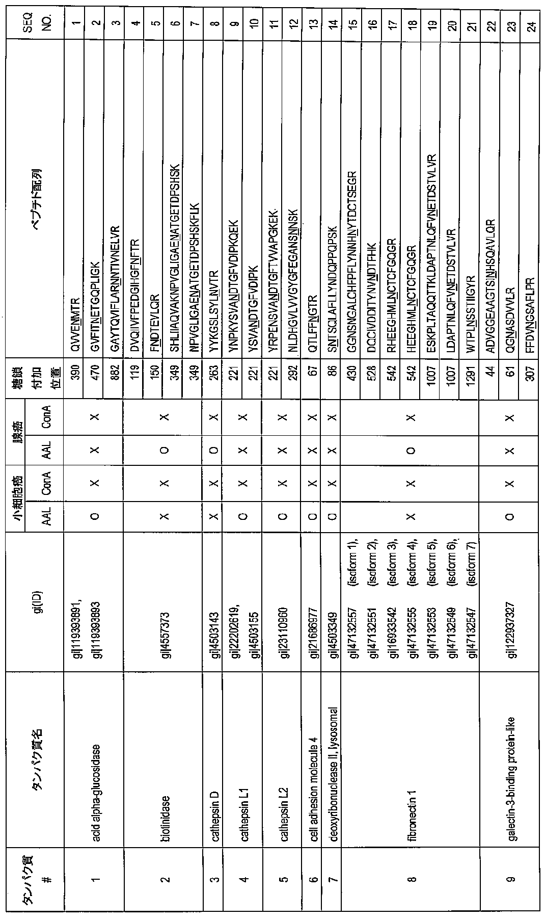
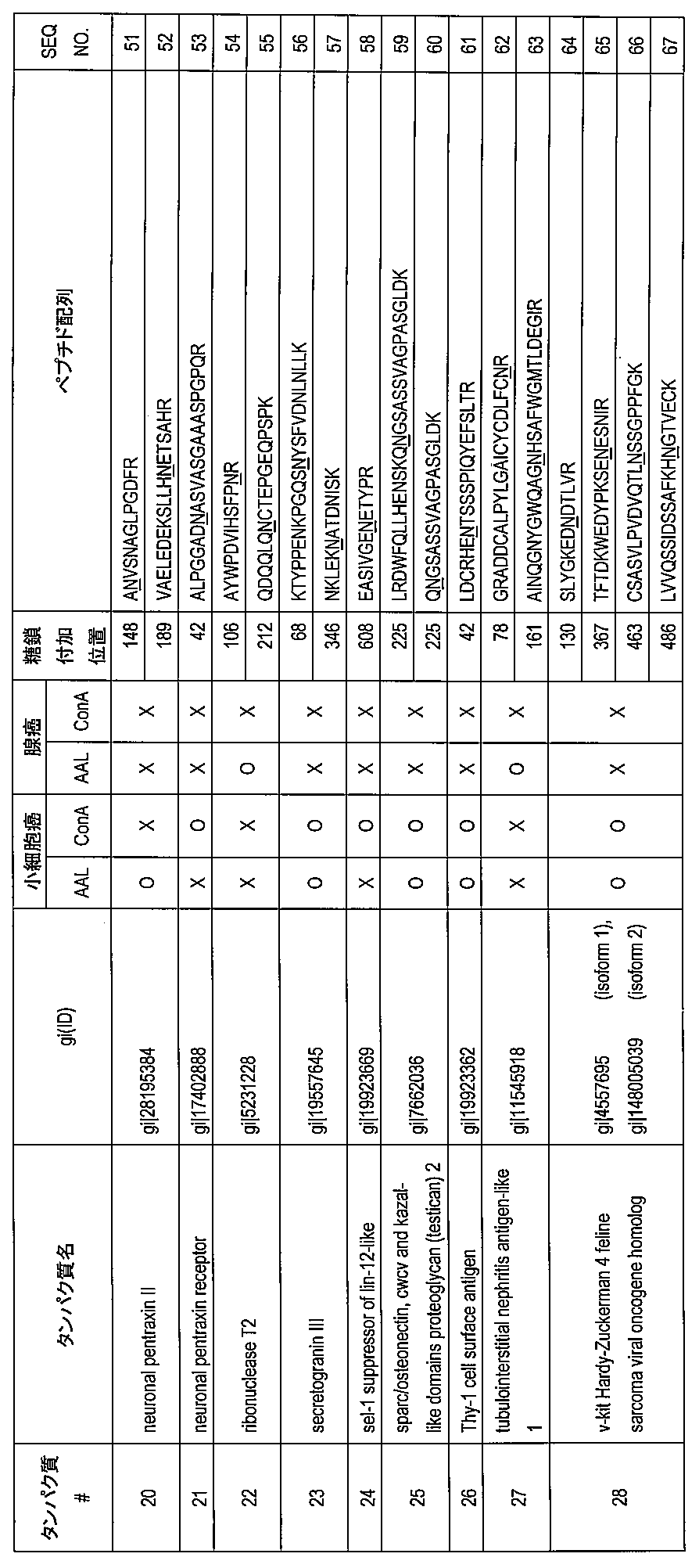
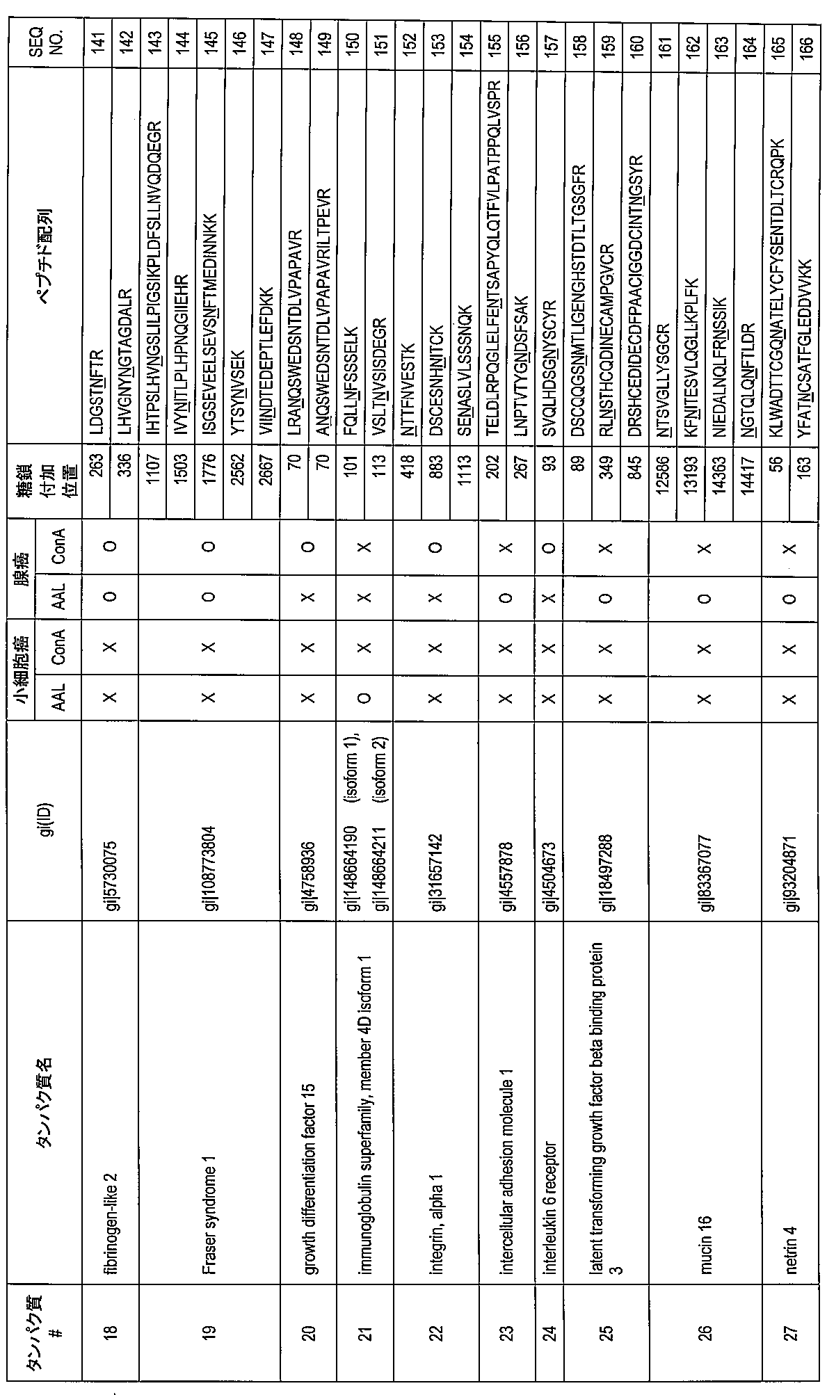
- glycoprotein identified in Table 1 or Table 2 wherein the sugar chain is added to the asparagine residue at the sugar addition position shown in Table 1 or Table 2.
- the sugar chain is selected from the group consisting of a sugar chain with fucose modification, a high mannose sugar chain, a hybrid sugar chain, a complex double chain sugar chain, chitin, polylactosamine and a ⁇ 1-3 galactose epitope
- the lung cancer differentiation marker glycoprotein according to (1) which is one or more sugar chains.
- the lung cancer differentiation marker glycoprotein according to (2) which is used for differentiation of small cell lung cancer or lung adenocarcinoma.
- the glycoprotein is selected from the group consisting of neural cell adhesion molecule (NCAM1), secretogranin III and insulin-like growth factor binding protein-L1 (IGFBP-L1)
- NCAM1 neural cell adhesion molecule
- IGFBP-L1 insulin-like growth factor binding protein-L1
- the lung cancer differentiation marker glycoprotein according to (3) which is one or more glycoproteins.
- the lung cancer differentiation marker glycoprotein according to (3) which is for lung adenocarcinoma differentiation, wherein the glycoprotein is fibronectin 1.
- one or more lung cancer differentiation marker glycoproteins according to Table 1 or Table 2 in which a sugar chain is added to an asparagine residue at the sugar addition position shown in Table 1 or Table 2 from a sample collected from a subject; and By detecting at least one glycoprotein fragment containing at least one asparagine residue at the glycosylation position shown in Table 1 or Table 2 to which a sugar chain has been added, the subject suffers from lung cancer.
- the sugar chain probe binds to a sugar chain with fucose modification, a high mannose sugar chain, a hybrid sugar chain, a complex double chain sugar chain, chitin, polylactosamine or ⁇ 1-3 galactose epitope.
- sugar chain probe is a lectin, an antibody or a phage antibody.
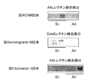
- the lung cancer differentiation marker glycoprotein is a nerve cell adhesion molecule (NCAM1), and it is determined that the tissue type of lung cancer is small cell cancer by detecting binding with AAL. the method of.
- the lung cancer differentiation marker glycoprotein is secretogranin III, and the binding to AAL and / or ConA is detected to determine that the lung cancer tissue type is small cell cancer. (11) The method described.
- the lung cancer differentiation marker glycoprotein is insulin-like growth factor binding protein-L1 (IGFBP-L1), and the tissue type of lung cancer is small cell cancer by detecting binding with ConA and / or PWM.
- lung cancer differentiation marker glycoprotein is fibronectin 1
- tissue type of lung cancer is determined to be adenocarcinoma by detecting binding with AAL and / or PNA.
- Binding result of the sugar chain probe to a sugar chain of the lung cancer differentiation marker glycoprotein and / or a fragment of the glycoprotein and the lung cancer differentiation marker glycoprotein and / or glycoprotein thereof described in Table 1 or Table 2 The method according to any one of (8) to (11), wherein the tissue type of lung cancer is determined to be a small cell cancer or an adenocarcinoma based on a binding mode of the fragment to the sugar chain probe.
- Table 1 or Table 2 wherein a sugar chain is added to the asparagine residue at the sugar addition position shown in Table 1 or Table 2, and / or a sugar chain added to the lung cancer differentiation marker glycoprotein of Table 1
- a lung cancer cell differentiation antibody for tissue staining that differentiates lung cancer by binding to the glycoprotein fragment containing at least one asparagine residue at the glycosylation position shown in Table 2.
- the lung cancer discrimination marker glycoprotein is a neuronal pentraxin receptor (NPR), and the lung cancer cell tissue type is a cell derived from small cell carcinoma. antibody.
- the presence or absence of lung cancer can be determined easily and with high reliability by examining body fluids, cells or lung lavage fluid. Furthermore, according to the lung cancer differentiation marker of the present application, the tissue type of lung cancer can be differentiated.
- lung cancer morbidity and lung cancer tissue type can be determined from body fluid, cells or lung lavage fluid with a higher accuracy than conventional tumor markers and with minimal invasiveness.
- lung cancer and further its tissue type can be differentiated in tissue staining.
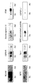
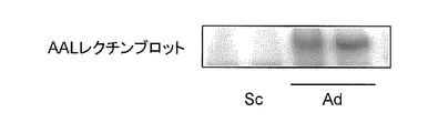
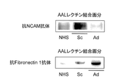
- Blotting diagram Lung cancer differentiation marker glycoprotein (NCAM, fibronectin 1) in the serum of small cell lung cancer patients (Sc) and lung adenocarcinoma patients (Ad) fractionated by AAL lectin was detected with each anti-lung cancer differentiation marker glycoprotein antibody
- NPR anti-neuropentraxin receptor
- Lung cancer differentiation marker glycoprotein and glycoprotein fragment thereof The first embodiment of the present invention is the lung cancer differentiation marker glycoprotein and the glycoprotein fragment thereof described in Table 1 or Table 2.
- the “lung cancer differential marker glycoprotein” of the present embodiment corresponds to the proteins shown as protein numbers # 1 to # 31 in Table 1 and protein # 1 to 48 in Table 2, respectively. All of these proteins have lung cancer in which a sugar chain is added to an asparagine residue at least at the position indicated by “sugar chain addition position” in the table (the position of the starting amino acid residue is 1). It is a specific glycoprotein.
- sugar chains are added to at least 390, 470 and 882 asparagine residues on the amino acid sequence of the protein. .
- glycoprotein a protein to which a sugar chain is added.
- Gi (ID) in the table indicates the ID number of the glycoprotein of the present embodiment. When multiple gi (ID) are registered in one protein, they are all listed in the table. Moreover, even when multiple isoforms exist in one protein, their gi (ID) is described together with their isoform numbers.
- the sugar chain added to the asparagine residue is not particularly limited as long as it is a sugar chain specifically added to lung cancer.
- examples include sugar chains with fucose modification (fucosylation), high mannose sugar chains, hybrid sugar chains, complex double-chain sugar chains, chitin, polylactosamine, or ⁇ 1-3 galactose epitopes.
- a sugar chain specifically added to lung cancer refers to a sugar chain added only to a protein derived from lung cancer cells at the asparagine residue indicated by “position of sugar chain addition” in the above table. Therefore, in principle, the lung cancer differentiation marker glycoprotein of the present embodiment is a glycoprotein produced from lung cancer cells. Therefore, using a sugar chain probe that recognizes these glycoproteins, for example, by detecting the presence or absence of the glycoprotein in the serum of a subject, it is determined that the individual having the glycoprotein is suffering from lung cancer. can do.
- the “sugar chain probe” refers to a discriminator that specifically recognizes and binds to a specific sugar chain and / or complex carbohydrate such as a glycoprotein.
- lectin, antibody or phage antibody can be mentioned.
- lung cancer is known to have a tissue type composed of small cell cancer and non-small cell adenocarcinoma, squamous cell carcinoma and large cell carcinoma.
- neuroendocrine cancers in the lung are also known, and many of them are classified as small cell cancers, but cases of other tissue type cancers are also known.
- the lung cancer differentiation marker glycoprotein of this embodiment can be identified as to which tissue type corresponds to the type of sugar chain added to the predetermined asparagine residue.
- acid alpha-glucosidase (acid ⁇ -glucosidase) shown as protein # 1 in Table 1
- a sugar chain with fucose modification is added to the predetermined asparagine residue only in a glycoprotein derived from small cell lung cancer.
- acid alpha-glucosidase can distinguish lung cancer as a lung cancer differentiation marker glycoprotein, and the lung cancer must be a small cell cancer. It can also be a marker for differentiating.
- biotinidase (biotinidase) indicated by protein # 2 in Table 1
- a sugar chain with fucose modification is added to the predetermined asparagine residue only in a glycoprotein derived from lung adenocarcinoma. Therefore, by using a lectin or antibody that binds to and recognizes a sugar chain with fucose modification, biotinidase can distinguish lung cancer as a lung cancer differentiation marker glycoprotein, and a marker that distinguishes that lung cancer is an adenocarcinoma Can also be.
- Sugar chains with fucose modification can be detected by AAL lectin
- high mannose-type sugar chains, mixed-type sugar chains and complex-type double-chain sugar chains can be detected by ConA lectin
- chitin and polylacto Samine can be detected by PWM lectin
- ⁇ 1-3 galactose epitope can be detected by PNA lectin.
- Table 1 when the lung cancer differentiation marker glycoprotein binds to AAL lectin or ConA lectin, it is indicated by “ ⁇ ”, and when it does not bind, it is indicated by “x”.
- each lung cancer differentiation marker glycoprotein (including the lung cancer differentiation marker glycoprotein fragment) shown in Tables 1 and 2 is AAL lectin and ConA for “small cell cancer” and “adenocarcinoma” in Tables 1 and 2. Based on the mode of binding to lectin, it can be a marker for differentiation of small cell lung cancer or lung adenocarcinoma.
- Lung Cancer Differential Marker Glycoprotein Fragment is an oligopeptide or polypeptide fragment consisting of a part of the lung cancer differential marker glycoprotein, and its amino acid sequence It contains at least one asparagine residue at the sugar addition position shown in Table 1 or Table 2, and the asparagine residue contains the lung cancer-specific sugar chain described in “1-1. Lung Cancer Differential Marker Glycoprotein”. It is added.
- the amino acid length of the lung cancer differentiation marker glycoprotein fragment is not particularly limited, but is preferably 5 to 100 amino acids, 8 to 80 amino acids, or 8 to 50 amino acids.
- the lung cancer differentiation marker glycoprotein fragment is hereinafter often referred to as “glycoprotein fragment” or “glycopeptide”.
- lung cancer differentiation marker when the lung cancer differentiation marker glycoprotein and its glycoprotein fragments are collectively expressed, they are hereinafter referred to as “lung cancer differentiation marker”.
- the lung cancer differentiation marker glycoprotein fragment include glycopeptides consisting of amino acid sequences represented by SEQ ID NOs: 1 to 223 described in Tables 1 and 2. These are glycoprotein fragments obtained by the IGOT method described later when identifying the lung cancer differentiation marker glycoprotein of the present embodiment, both as the lung cancer differentiation marker glycoprotein, It is possible to discriminate which tissue type corresponds to whether or not the patient is affected by lung cancer and the type of sugar chain added to the predetermined asparagine residue.
- the underlined asparagine residue (N) indicates an asparagine residue to which a sugar chain is added.
- 1-3-1 Large-scale identification of lung cancer-specific candidate glycoproteins
- Large-scale selective collection and enrichment of lung cancer-specific glycoproteins can be broadly divided into methods using probes having affinity for sugar chains, methods using chemical reactions with sugar chains (Zhang H. et al .Nat Biotechnol 21, 660-666 (2003)), and any method such as a method for introducing an affinity tag to a sugar chain can be used.
- the lung cancer differentiation marker sugars in Table 1 and Table 2 are used. A method using the probe used for protein acquisition will be described.
- a lectin or sugar chain antibody that reacts with a sugar chain characteristically produced by cancer cells is selected as a probe.
- Probe lectins can be selected by statistical analysis of sugar chain profiles using lectin microarrays.
- literature information (some of which can predict a probe lectin to be used, such as increased fucosylation associated with canceration) and tissue type discrimination can be selected. Basically, it is selected from statistical analysis of the profile, and the validity of the selection is judged from the binding specificity of the selected lectin.
- AAL lectin derived from Aleuria aurantia which can detect fucosylation, high mannose sugar chain, mixed sugar chain or complex double chain sugar bean Canavalia ensiformis
- An antibody probe or a phage antibody probe can be prepared after clarifying the structure of an antigen (sugar chain), but it is not a necessary condition and can be prepared even if the structure of the antigen sugar chain or glycopeptide remains unknown. .
- large-scale identification of a candidate glycoprotein is performed by first using a sugar chain probe (probe lectin and / or antibody probe, phage antibody probe) from a culture supernatant of a cultured cell line derived from lung cancer. To collect.
- a sugar chain probe probe lectin and / or antibody probe, phage antibody probe
- cultured cell supernatant derived from lung cancer it is possible to identify cancer cell-derived glycoproteins, so that it is possible to easily detect differences from sugar chains present on glycoproteins derived from serum of healthy individuals This can be useful information for selective acquisition of candidate molecules from serum.
- any lung cancer histological type of small cell cancer or non-small cell cancer (adenocarcinoma, squamous cell carcinoma, large cell carcinoma) may be used.
- a tissue type non-specific lung cancer differentiation marker and a tissue type specific lung cancer differentiation marker can also be obtained.
- the collected candidate glycoprotein for lung cancer differentiation marker is based on the IGOT method (isotope-coded glycosylation site-specific tagging) and mass spectrometry. Processed, the core glycopeptide of the candidate glycoprotein is identified. For example, JP 2004-233303 (Patent No. 4220257), Kaji H, et al. Mass spectrometric identification of N-linked glycopeptides using lectin-mediated affinity capture and glycosylation site-specific stable isotope tagging. Nature Protocols 1, 3019-3027 The glycopeptide can be analyzed by the Lec-IGOT-LC / MS method described in (2006). A specific method will be described below with an example.
- (1) Glycanectomy and IGOT method The candidate glycopeptide group obtained by digesting the candidate glycoprotein group collected with a probe lectin or probe antibody with a protease and using the same probe as a candidate glycopeptide group as a sample from the obtained peptide group Re-collect.
- the crude protein mixture can be directly digested with a probe lectin or a probe antibody from the obtained crude peptide group after protease digestion. Subsequently, the obtained candidate glycopeptide is labeled by the IGOT method.
- a sugar chain is dissociated by treatment with an enzyme such as glycopeptidase in water labeled with isotope oxygen, and asparagine at the sugar chain binding site becomes aspartic acid by this treatment.
- isotope oxygen (18 O) is incorporated into the candidate glycopeptide.
- Xaa is Lys / Arg and the identified peptide sequence is excised at this position, refer to the amino acid sequence of the entire protein, and the next residue of Xaa is [Ser / Thr].
- the candidate glycopeptide is also included in the glycoprotein fragment.
- the consensus sequence is a sugar chain binding position, but a site not following this has also been reported.
- the lung cancer differentiation marker glycoprotein fragment group selected by the above method and the lung cancer differentiation marker glycoprotein containing the fragment identified based on the amino acid sequence are the glycoprotein fragment group and glycoprotein shown in Tables 1 and 2. is there.
- glycoprotein fragment group and glycoprotein can be used as a lung cancer differentiation marker for determining the presence of lung cancer in a subject by, for example, verifying the presence or absence in the serum of the subject using a lectin or antibody that recognizes the fragment.
- 1-3-3 Verification of Lung Cancer Differential Marker
- the significance of the lung cancer differential marker selected in the section 1-3-2 can be further verified.
- Western blotting or immunoprecipitation of glycoproteins and / or glycoprotein fragments collected with the probe lectin from the lung cancer cultured cell supernatant using an antibody that specifically recognizes the above-identified lung cancer differentiation marker Can be detected and confirmed.
- 31 types of lung cancer differentiation marker glycoproteins and glycoprotein fragments thereof shown in Table 1 correspond to lung cancer differentiation markers further selected by the antibody detection.
- the lung cancer differentiation marker selected by the antibody detection may be further verified.
- it can be collected from the serum of lung cancer patients using a probe lectin and detected using the specific antibody.
- the nerve cell adhesion molecule (NCAM1) (protein # 19 in Table 1) and fibronectin 1 (protein # 8 in Table 1) described in Table 1 shown in FIG. 5 and secretogranin III shown in FIGS. (Protein # 23 in Table 1) is a lung cancer differentiation marker glycoprotein whose usefulness has been confirmed by this further verification.
- the nerve cell adhesion molecule binds to AAL lectin only in small cell lung cancer according to Table 1, it can be a lung cancer differentiation marker capable of distinguishing small cell lung cancer.
- secretogranin III binds to AAL lectin and ConA lectin only in small cell lung cancer according to Table 1, it can be a lung cancer differentiation marker that can distinguish small cell lung cancer.
- fibronectin 1 binds to AAL lectin only in lung adenocarcinoma from Table 1, it can be a lung cancer differentiation marker that can distinguish lung adenocarcinoma.
- the specific antibody may be collected from the culture supernatant of a lung cancer cell line and detected using a lectin array.
- IGFBP-L1 insulin-like growth factor binding protein-L1 shown in Table 1 shown in FIG.
- fibronectin 1 shown in Table 1 shown in FIG. 10 is a lung cancer differentiation marker glycoprotein whose usefulness has been confirmed by the further verification.
- insulin-like growth factor-binding protein-L1 IGFBP-L1
- IGFBP-L1 insulin-like growth factor-binding protein-L1
- fibronectin 1 can detect the fluorescent signal on the PNA lectin spot only in lung adenocarcinoma from FIG. 10, it can be a lung cancer differentiation marker that can distinguish lung adenocarcinoma.
- Lung cancer differentiation marker glycopeptide and glycoprotein can be detected using a mass spectrometer as a detector for a sample collected with a probe lectin or the like that binds to a sugar chain.
- Lung cancer differentiation marker glycopeptides are preferably detected by excision of the collected glycopeptides, followed by separation by liquid chromatography (LC), and the eluted peptides sequentially and directly on-line by a mass spectrometer (MS ) And can be detected.
- mass spectra can be acquired by acquiring MS / MS spectra using a fracture method such as collision-induced dissociation (CID), and only when preselected ions are detected. It is also possible to detect a plurality of fragment ions that are broken by a method such as single reaction monitoring or multi-reaction monitoring.
- CID collision-induced dissociation
- the core peptide part of the lung cancer differentiation marker glycopeptide was synthesized in the analysis sample, and the target peptide that caused a mass difference by incorporating a stable isotope into a part of the peptide was added, and the relative signal intensity was compared by comparing the respective signal intensities. Or absolute quantitative analysis can be performed. In a simple manner, the signal intensity of the detected ions can be easily quantified by comparing between a plurality of samples or a standard sample.
- the collected protein can be separated using one-dimensional or two-dimensional gel electrophoresis, and the detection intensity (staining, fluorescence, etc.) of the target spot can be compared with a standard sample and quantified.
- a detection method using a mass spectrometer a collected protein group can be digested with a protease, and the resulting peptide can be analyzed and detected by an LC / MS method.
- Lectin microarray (1) Glycan profiling by lectin microarray (a) Lectin microarray (also simply called lectin array) A lectin array is a glycan probe in which multiple types of lectins with different specificities are immobilized in parallel on one substrate (arrayed), and how much lectin interacts with the complex carbohydrate to be analyzed. Can be analyzed all at once. By using a lectin array, information necessary for sugar chain structure estimation can be acquired by a single analysis, and the operation steps from sample preparation to scanning can be performed quickly and easily.
- glycoproteins cannot be analyzed as they are, and must be processed in advance to the state of glycopeptides and free sugar chains.
- a lectin microarray has an advantage that analysis can be performed as it is by simply introducing a phosphor directly into the core protein portion.
- the lectin microarray technology has been developed by the present inventors, and its principle and basis are described in, for example, Kuno A., et al. Nat. Methods 2,851-856 (2005).
- Typical lectins used in the lectin array include, for example, those described in Table 3 below.
- a lectin array (LecChip manufactured by GP Bioscience) in which 45 types of lectins including the above 43 types of lectins are immobilized is already commercially available.
- the platform of the lectin microarray is basically as described above.
- the sample of the subject is not directly labeled with fluorescence, but indirectly with an antibody via a fluorescent group, etc.
- This is an application method that can simplify and speed up the analysis of multiple samples simultaneously by introducing the sample into the sample of the subject (“Kuno A, Kato Y, Matsuda A, Kaneko MK, Ito H, Amano K, Chiba Y, Narimatsu H, Hirabayashi J. Mol Cell Proteomics.
- the sugar chain part is recognized by the lectin on the lectin microarray, so that the test glycoprotein can be labeled by overlaying with an antibody against the core protein part. Or, it can be detected specifically with high sensitivity without being highly purified.
- Lectin overlay / antibody microarray method In place of the lectin microarray, an antibody microarray in which antibodies against the core protein are immobilized (arrayed) in parallel on a substrate such as a glass substrate is used. Only as many antibodies as the marker to be examined are needed. It is necessary to determine in advance a lectin for detecting a sugar chain change.
- a simple and inexpensive sandwich detection method can be designed based on the results of the lectin array. Basically, among the protocols used in the sandwich detection method using two kinds of antibodies, it can be applied by replacing one antibody with a lectin. Therefore, this method can also be applied to automation using an existing automatic immunodetection device. The only thing to consider is the reaction between the antibody used in the sandwich and the lectin. The antibody has at least two N-linked sugar chains. Therefore, when the lectin used recognizes the sugar chain on the antibody, background noise due to the binding reaction occurs during sandwich detection.
- a method of introducing a modification into the sugar chain part on the antibody or a method of using only a Fab that does not contain a sugar chain part can be considered.
- methods for modifying the sugar chain include Chen S et al. Nat Methods. 4, 437-44 (2007) and Consale MA et al. J Proteome Res. 8, 595-602 (2009). Include, for example, Matsumoto H et al. Clin Chem Lab Med 48, 505-512 (2010).
- Antibody overlay lectin arrays are the best way to statistically find lectins that best reflect disease-specific glycosylation on lung cancer differential marker glycoproteins.
- antibodies capable of immunoprecipitation and overlay detection are essential. However, such antibodies are not always available. Therefore, as a means for utilizing more candidate molecules for lung cancer detection, it is common to perform immunological quantitative detection of the target glycoprotein with respect to the glycoprotein collected by the probe lectin. Specifically, SDS-PAGE is performed, and immunological detection is performed by Western blot after membrane transfer. By comparing the signal intensities of the obtained bands, fluctuations between the samples can be quantitatively estimated.
- the AAL lectin used in the candidate molecule identification step is generally used as a probe protein even during verification.
- a report of Liu Y et al. J Proteome Res. 9, 798-805 (2010) can be cited as an example of such a strategy.
- serum proteins have different N-linked sugar chain structures (degree of branching, etc.) and fucose modifications (core fucose, blood group antigens, etc.) depending on the type. Therefore, it has been reported that even the same molecule may undergo different fucose modifications.
- ⁇ 1 antitrypsin is subjected to different fucose modifications. Because these differ in the type of disease, whether there is an increase depending on the degree of progression, and the timing, almost all fucose modifications can be recognized and collected using AAL as a probe to collect the entire fucose-containing glycoprotein Comparison is not ideal. Therefore, we considered separation and fractionation by continuous chromatography using two different fucose recognition lectins, and quantitative comparison analysis of each fraction.
- the lectins used at this time are LCA and AAL.
- LCA is known to be a lectin that recognizes N-linked sugar chains that have undergone low-branching core fucose modification based on lectin specificity analysis.
- AAL can recognize any branched core fucose of an N-linked sugar chain, and can also recognize fucose modifications on the non-reducing end side typified by ABO and Lewis antigens. . That is, LCA is highly specific and AAL is low. Therefore, as the first step, LCA column chromatography is performed to capture fucose-containing glycoprotein that binds to LCA. This is referred to as LCA-binding fucose-containing glycoprotein. At the time of chromatography, fucose-containing glycoproteins that do not have a low-branched N-linked sugar chain that has undergone core fucose modification do not bind to the LCA column but are fractionated into unbound fractions.
- AAL column chromatography is performed on the LCA-unbound fraction.
- a group of glycoproteins captured by AAL is defined as LCA non-binding / AAL-binding fucose-containing glycoprotein.
- the second embodiment of the present invention is a lung cancer incidence determination method.
- the method of this embodiment is characterized in that it is determined that the subject is suffering from lung cancer by detecting the lung cancer differentiation marker described in the first embodiment from a sample collected from the subject.
- subject refers to a person who is subjected to a test, that is, a person who provides a sample to be described later.
- the subject may be either a patient having some disease or a healthy person. Preferred are those who may have lung cancer or lung cancer patients.
- sample is collected from the subject and used in the determination method of the present embodiment, and corresponds to, for example, a body fluid, a cell such as a cancer tissue, or a lung lavage fluid collected during surgery.
- Body fluid refers to a liquid biological sample collected from a subject.
- blood including serum, plasma and interstitial fluid
- lymph fluid extract of each tissue or cell
- pleural effusion sputum
- cerebrospinal fluid tears
- nasal discharge saliva, urine, vaginal fluid, semen and the like
- the body fluid may be used after performing treatment such as dilution or concentration of a sample collected from a subject, addition of a blood coagulation inhibitor such as heparin as necessary, or such pretreatment. It may be used as it is.
- the body fluid may be collected based on a known method in the field.
- a known blood collection method may be followed.
- peripheral blood it can be collected by injection into a peripheral vein or the like.
- the bodily fluid may be used immediately after collection, or may be used after being frozen or refrigerated for a certain period of time and then subjected to processing such as thawing as necessary.
- a sufficient amount of lung cancer differential marker can be detected by using a volume of 10 ⁇ L to 100 ⁇ L, 20 ⁇ L to 80 ⁇ L, 30 ⁇ L to 70 ⁇ L, 40 ⁇ L to 60 ⁇ L, or 45 ⁇ L to 55 ⁇ L.
- the lung cancer marker used for detection in the determination method of the present embodiment is a lung cancer marker glycoprotein according to Table 1 or Table 2 in which a sugar chain is added to the asparagine residue at the sugar addition position shown in Table 1 or Table 2.
- Any lung cancer differentiation marker can be used as long as it is a glycoprotein fragment containing at least one asparagine residue at the sugar addition position shown in Table 1 or Table 2 to which a sugar chain has been added.
- These lung cancer differentiation markers may be used alone in the determination method of the present embodiment, or may be used in combination of two or more. For example, two or more different lung cancer differentiation marker glycoproteins may be used, or two or more different lung cancer differentiation marker glycoprotein fragments in the same lung cancer differentiation marker glycoprotein may be used.
- a lung cancer differentiation marker in Table 1 is used, and more preferably, a lung cancer differentiation marker having the same or different differentiation characteristics for small cell lung cancer and lung adenocarcinoma.
- a lung cancer differentiation marker having the same or different differentiation characteristics for small cell lung cancer and lung adenocarcinoma For example, neural cell adhesion molecule (NCAM1), secretogranin III and / or insulin-like growth factor binding protein-L1 (IGFBP-L1) listed in Table 1 for differentiation of small cell lung cancer, and lung adenocarcinoma differentiation Examples include a method of combining fibronectin 1 listed in Table 1 as a lung cancer differentiation marker of the present embodiment. This is convenient because it is possible to simultaneously determine whether the lung cancer is a small cell cancer or an adenocarcinoma at the same time as determining whether the subject has lung cancer.
- a lectin or antibody that binds to a sugar chain with fucose modification or a high-mannose sugar chain, a hybrid sugar chain, a complex double-chain sugar chain, chitin, polylactosamine and / or ⁇ 1-3 galactose epitope from a sample of a subject are used alone or in combination, and the lung cancer differentiation markers listed in Table 1 or 2 are detected by any method, the subject is suffering from or very likely to have lung cancer. Can be determined.
- AAL lectin that specifically binds to sugar chains with fucose modification can be detected, and specifically, it can be used to detect high-mannose sugar chains, mixed sugar chains, and complex double-chain sugar chains.
- the binding ConA lectin can be used to detect chitin and polylactosamine, PWM lectin specifically binding to it, or the detection of ⁇ 1-3 galactose epitope can be PNA lectin specifically binding to it. .
- the binding result of the lectin to the sugar chain of the lung cancer differentiation marker and the binding of the lung cancer differentiation marker glycoprotein and / or its glycoprotein fragment shown in Table 1 or Table 2 to the lectin Based on the embodiment, it can also be determined that the lung cancer tissue type in the subject is small cell carcinoma or adenocarcinoma.
- the sugar chain of acid alpha-glucosidase represented by protein # 1 binds to AAL lectin only in small cell lung cancer.
- the subject is determined to have lung cancer and the tissue type is small cell carcinoma. can do.
- the lung cancer differentiation marker glycoprotein is a neural cell adhesion molecule (NCAM1) or a glycoprotein fragment thereof and binding to AAL lectin is detected, it is determined that the lung cancer tissue type is small cell cancer. Can do.
- the lung cancer differentiation marker glycoprotein is fibronectin 1 or a glycoprotein fragment thereof, and by detecting the binding to the AAL lectin or antibody, it can be determined that the lung cancer tissue type is adenocarcinoma.
- Lung Cancer Differential Marker Detection Method As a method for detecting a lung cancer differential marker, for example, there are two methods, namely, a lectin that specifically binds to a sugar chain of a lung cancer differential marker (in this specification, for the sake of convenience, hereinafter referred to as lectin A).
- a combination of a method for selecting a lung cancer differential marker having the sugar chain and a method for detecting the lung cancer differential marker by detecting a portion (core protein) other than the sugar chain of the lung cancer differential marker, or a lectin A specific antibody against a lung cancer differentiation marker having a sugar chain that specifically binds to A, and a method for detecting the target lung cancer differentiation marker using an antibody having an epitope near the sugar chain binding portion can be mentioned .
- the method for detecting a sugar chain that specifically binds to lectin A and the method for detecting a core protein are respectively a method for measuring a sugar chain that specifically binds to lectin A, and a core protein. It may be a method.
- a lung cancer differentiation marker using an antibody against a core protein and lectin A
- lung cancer patients can be detected separately from healthy individuals.
- an antibody overlay method using a lectin array (Kuno A, Kato Y, Matsuda A, Kaneko MK, Ito H, Amano K, Chiba Y, Narimatsu H, Hirabayashi J. Mol Cell Proteomics. 8, 99-108 (2009 ))can be used.
- a lectin-antibody sandwich immunological detection method can be used.
- a lung cancer differentiation marker in serum obtained from a subject is separated using lectin A.
- a protein group having a sugar chain that specifically binds to lectin A is selected.
- the lung cancer differentiation marker detection is performed using an anti-lung cancer differentiation marker glycoprotein antibody that specifically recognizes a portion other than the sugar chain that specifically binds to lectin A.
- an anti-lung cancer differentiation marker glycoprotein antibody that specifically recognizes a portion other than the sugar chain that specifically binds to lectin A.
- a lung cancer differentiation marker having a sugar chain that specifically binds to the target lectin A can be detected. If this marker is detected, it is determined that the subject is suffering from lung cancer. .
- a lung cancer differentiation marker having a sugar chain that specifically reacts with lectin A for example, a method of measuring a sugar chain that specifically binds to lectin A, specifically, a column or an array on which lectin A is immobilized And a means for measuring a lung cancer differentiation marker, specifically, an antibody against a novel lung cancer differentiation marker glycoprotein or a fragment thereof.
- a lectin-antibody sandwich ELISA or an antibody overlay lectin array method can be used.
- LC-MS enzyme immunoassay using a monoclonal antibody or polyclonal antibody specific for a novel lung cancer differential marker glycoprotein having a sugar chain that specifically reacts with lectin A or a fragment thereof
- two-antibody sandwich ELISA method gold colloid method, radioimmunoassay method, latex agglutination immunoassay method, fluorescence immunoassay method, western blotting method, immunohistochemistry method, surface plasmon resonance method (SPR method) or quartz crystal microbalance (QCM) method
- SPR method surface plasmon resonance method
- QCM quartz crystal microbalance
- an anti-lung cancer differentiation marker glycopeptide polyclonal antibody can be prepared using a well-known method. Specifically, an adjuvant is added to the obtained lung cancer differentiation marker glycoprotein or glycopeptide for antigen.
- an adjuvant As the antigen, a lung cancer differential marker glycopeptide containing a sugar chain binding site (asparagine) may be synthesized and used. Examples of the adjuvant include Freund's complete adjuvant, Freund's incomplete adjuvant, and the like, and these can also be used as a mixture.
- An antibody-producing animal can be co-inoculated with an adjuvant together with the antigen to boost antibody production.
- this peptide may be covalently bound to commercially available keyhole limpet hemocyanin (KLH) and inoculated into antibody-producing animals.
- KLH keyhole limpet hemocyanin
- GM-CSF granulocyte-macrophage colony stimulating factor
- Mammals such as mice, rats, horses, monkeys, rabbits, goats, sheep and the like can be used as antibody-producing animals to inoculate the antigen.
- Any method can be used for immunization as long as it is an existing method, but it is mainly carried out by intravenous injection, subcutaneous injection, intraperitoneal injection or the like. Immunization intervals are not particularly limited, and immunization is performed at intervals of several days to several weeks, preferably at intervals of 4 to 21 days.
- a polyclonal antibody can be prepared by collecting whole blood from an immunized animal 2-3 days after the final immunization and separating the serum.
- an anti-lung cancer marker glycopeptide monoclonal antibody can be prepared by the method of Keller and Milstein (Nature Vol. 256, pp 495-497 (1975)).
- a hybridoma is prepared by cell fusion between an antibody-producing cell obtained from an animal immunized with an antigen and a myeloma cell, and a clone that produces an anti-lung cancer differential marker glycopeptide monoclonal antibody is selected from the resulting hybridoma. be able to.
- antibody production cells are collected 2 to 3 days after the last immunization in the production of the polyclonal antibody.
- antibody-producing cells include spleen cells, lymph node cells, and peripheral blood cells.
- myeloma (myeloma) cells to be fused with antibody-producing cells cell lines derived from various animals such as mice, rats, humans, and the like, which are generally available to those skilled in the art, are used.
- the cell line to be used those that have drug resistance and have the property that they cannot survive in a selective medium (for example, HAT medium) in an unfused state but can survive only in a fused state.
- An 8-azaguanine resistant strain is generally used, and this cell strain is deficient in hypoxanthine-guanine-phosphoribosyltransferase and cannot grow in hypoxanthine / aminopterin / thymidine (HAT) medium.
- HAT hypoxanthine-guanine-phosphoribosyltransferase
- Myeloma cells are various known cell lines such as P3 (P3x63Ag8.653) (J. Immunol. 123, 1548-1550 1979)), P3x63Ag8U.1 (Current Topics in Microbiology and Immunology 81, 1-7 ( 1978)), NS-1 (Kohler, G. and Milstein, C., Eur. J. Immunol. 6,511-519 (1976)), MPC-11 (Margulies, DH et al., Cell 8,405-415 (1976) ), SP2 / 0 (Shulman, M. et al., Nature 276,269-270 (1978)), FO (de St. Groth, SF et al., J. Immunol.
- cell fusion is performed between the myeloma cells and antibody-producing cells.
- Cell fusion is performed by mixing myeloma cells and antibody-producing cells in an animal cell culture medium such as MEM, DMEM, and RPMI-1640 medium at a mixing ratio of 1: 1 to 1:10 in the presence of a fusion promoter. By contacting at 37 ° C. for 1-15 minutes.
- a fusion promoter or fusion virus such as polyethylene glycol having an average molecular weight of 1,000 to 6,000, polyvinyl alcohol, or Sendai virus can be used.
- antibody-producing cells and myeloma cells can be fused using a commercially available cell fusion device utilizing electrical stimulation (for example, electroporation).
- Select the desired hybridoma from the cells after cell fusion treatment examples include a method utilizing selective growth of cells in a selective medium. That is, after the cell suspension is diluted with an appropriate medium, it is spread on a microtiter plate, a selective medium (HAT medium or the like) is added to each well, and thereafter, the selective medium is appropriately replaced and cultured. As a result, growing cells can be obtained as hybridomas.
- a selective medium HAT medium or the like
- Hybridoma screening is performed by the limiting dilution method, the fluorescence excitation cell sorter method, etc., and finally a monoclonal antibody-producing hybridoma is obtained.
- Examples of a method for collecting a monoclonal antibody from the obtained hybridoma include a normal cell culture method and ascites formation method.
- the third embodiment of the present invention is a lung cancer cell differentiation antibody for tissue staining.
- the antibody of the present embodiment is one or more of the lung cancer differentiation markers described in the first embodiment, that is, one or more of the sugar chains added to the asparagine residues at the sugar addition positions shown in Table 1 or Table 2. Specifically recognize one or more glycoprotein fragments comprising at least one asparagine residue at the glycosylation position shown in Table 1 or Table 2, to which is added a lung cancer differentiation marker glycoprotein and / or a sugar chain. It is an antibody that can specifically differentiate lung cancer cells in tissue staining by binding to it.
- the epitope of the lung cancer differentiation marker described in the first embodiment recognized by the antibody of the present embodiment is not particularly limited.
- it is a core protein or a core protein fragment thereof. More preferably, it is a part including both a sugar chain and a peptide sequence around the sugar chain addition.
- the length of the peptide sequence is 5-15 amino acids, 5-10 amino acids, or 5-8 amino acids.
- a phage antibody probe using a phage or the like can be a sugar chain probe for lung cancer cell identification.
- the antibody of the present embodiment can distinguish the tissue type of lung cancer cells.
- an antibody against a neuropentraxin receptor (NPR) represented by protein # 21 in Table 1 R & D, SYSTEM, Anti-human, Neuronal, Pentraxin, Receptor, and Antibody
- NPR neuropentraxin receptor
- FIG. 7 this anti-neuropentraxin receptor antibody can be specifically differentiated from cells derived from small cell carcinoma, and thus can be an antibody for differentiation of small cell carcinoma cells.
- the proteins collected and identified by the cancer sugar chain probe lectin usually have very little expression in other tissues and normal sites. Or what can become a tissue marker only by a phage antibody is also contained.
- Example 1 Selection of glycopeptide markers by glycoproteomics (IGOT-LC / MS method) Preparation method of human lung cancer cell supernatant Three types of lung adenocarcinoma (H358, H1975, LX-1) and three types of small cell lung cancer (H2171, H524, H526) were prepared with high glucose-containing 90% DMEM and 10% FBS, respectively. Using a medium of (PS +), the cells were cultured for 3 days in a dish having a diameter of 14 cm to be 80-90% confluent. The FBS serum-containing culture medium was aspirated and discarded, and washed with 10 mL of serum-free medium / dish (100% DMEM-high glucose, no additive).
- Serum-free medium was added at 30 mL / dish and cultured for 48 hours. After incubation, the mixture was centrifuged at 1000 rpm ⁇ 30 minutes, and the supernatant was further collected. The supernatant (culture supernatant) was collected at 3000 rpm ⁇ 30 minutes, and stored frozen at ⁇ 80 ° C. The culture supernatant stored at the above temperature was thawed before use, filtered through a 0.45 ⁇ m filter, and used in the following examples. NaN 3 was added so that the final concentration was 0.1%.
- the supernatant extract was recovered by high-speed centrifugation. Nitrogen gas was passed through or blown through the extract to remove dissolved oxygen, and then dithiothreitol (DTT) in an amount equal to the protein weight was dissolved in powder or a small amount of solubilization buffer and added. The reaction was carried out at room temperature for 1-2 hours in order to reduce disulfide bonds while passing nitrogen gas or under nitrogen gas atmosphere. Next, for S-alkylation, 2.5 times the protein weight of iodoacetamide was added, and the mixture was allowed to react at room temperature for 1-2 hours in the dark. After the reaction, dialyzed at 4 ° C.
- DTT dithiothreitol
- the candidate glycopeptide was eluted with 50% ethanol (v / v). Concentrate all candidate glycopeptide fractions by transferring a small amount of the candidate glycopeptide fraction to a microtube containing 2 ⁇ L of glycerol, removing the water by vacuum centrifugation, that is, vacuum centrifugation, and repeating the operation. did.
- IGOT reaction solution was diluted with 0.1% formic acid and subjected to LC / MS shotgun analysis.
- a nano LC system based on a direct nanoflow pump was used.
- the injected candidate glycopeptide is once collected on a trap column (reverse phase C18 silica gel-based carrier) for desalting, washed, and then a fritless microcolumn (in the form of a spray tip packed with the same resin) Using an inner diameter of 150 ⁇ m ⁇ 50 mL), separation was performed by the acetonitrile concentration gradient method.
- Mass spectrometry performed tandem mass spectrometry by collision induced dissociation (CID) while selecting up to two ions in a data dependent mode.
- CID collision induced dissociation
- the probability of identification (probability of coincidence: Expect value) is 0.05 or less.
- the number of fragment ions contributing to the identification is 4 or more.
- the identified peptide has a consensus sequence and has fewer than that number of Asn modifications (conversion to Asp and incorporation of 18 O).
- a glycoprotein containing this sequence can be defined. More specifically, based on the “peptide sequence” of the lung cancer differential marker glycopeptide represented by SEQ ID NOs: 1 to 223 in Tables 1 and 2, the entire amino acid sequence of the corresponding lung cancer differential marker glycoprotein from the amino acid sequence database NCBI-Refseq was identified.
- the lung cancer differentiation marker glycoprotein obtained by the above process is shown in Table 2.
- 5% skim milk or 5% BSA dissolved in PBSTx in which 1% Triton X-100 was added to PBS (Dulbecco) was used and blocked at room temperature for 1 hour.
- the membrane was reacted with a primary antibody (Table 4) biotinylated in advance with Biotin Labeling Kit-NH 2 (Dojindo Laboratories) for 1 hour.
- the mixture was reacted with the secondary antibody HRP-conjugated streptavidin (1: 3000 dilution, GE Healthcare) for 1 hour.
- the lung cancer differentiation marker glycoprotein obtained by the above process is shown in Table 1, FIG. 1 and FIG.
- the culture supernatant was fractionated using AAL lectin. Specifically, the AAL resin was washed 5 times with 3 times the amount of PBS and then prepared into a 50% slurry solution. 30 ⁇ L of the culture supernatant was added to 30 ⁇ L of the prepared AAL resin, and the mixture was shaken at 4 ° C. for 5 hours. After centrifugation (2,000 rpm, 2 minutes), the supernatant was removed, 50 ⁇ L of Wash Buffer (0.1% SDS in PBST, 0.1% Triton X-100) was added, and centrifugation (2,000 rpm, 2 minutes) was repeated twice.
- Wash Buffer (0.1% SDS in PBST, 0.1% Triton X-100
- the reacted culture supernatant and antibody were transferred to magnetic beads and shaken at 1,400 rpm for 1 hour at 20 ° C.
- the antibody bound to the magnetic beads was recovered using a magnetic stand.
- the collected magnetic beads were washed three times with 1 mL of TBSTx. After adding 20 ul of elution buffer (0.2% SDS in TBS) and stirring, the mixture was heated twice at 98 ° C. or 60 ° C. for 5 minutes for elution.
- 40 ⁇ L of magnetic beads washed in the same manner were added to the eluted sample and shaken at 1,400 rpm for 2 hours at 20 ° C.
- a sample obtained by removing antibody-magnetic beads with a magnetic stand was used as a sample.
- the prepared sample was developed by SDS-PAGE and then lectin blotted.
- Serum column fractionation with AAL lectin was fractionated using AAL lectin. Specifically, 1 mL of AAL resin is packed in a commercially available column, washed with 10 times the amount of resin TBS, twice the amount of resin 0.5 M NaCl, and washed with 10 times the amount of resin TBS. An AAL column was prepared. After the serum was added to the AAL column, the mixture was reacted in the resin for 1 hour, and then washed with 5 times the amount of TBS.
- Serum Fractionation by Continuous Chromatography Method Serum fractionation using continuous chromatography was performed at low temperature using LCA agarose (J-Oil Mills) and AAL agarose (J-Oil Mills). 100 ⁇ L of serum was diluted 4 times with PBS, and the serum sample was first developed in 5 mL of LCA agarose (0.7 ⁇ 13 cm) packed in a column. Next, the obtained unbound serum fraction was developed into 2.5 mL of AAL agarose (0.7 ⁇ 5.5 cm) packed in a column, washed thoroughly with PBS, and then eluted with PBS containing 0.2 M fucose. . The eluted fraction was concentrated with an ultra-free centrifugal concentration device (cut-off: 30 kDa, Millipore), and the obtained sample was subjected to comparative analysis of candidate molecules by immunoblotting as described above.
- the obtained unbound serum fraction was exposed to 250 ⁇ L of AAL agarose (0.8 ⁇ 0.5 cm) packed in an open column, washed thoroughly with PBS, and then eluted with PBS containing 0.02 M fucose. .
- the eluted fraction was concentrated with an ultra-free centrifugal concentration device (cut-off: 10 kDa, Millipore), and among the obtained samples, 10 ⁇ L of serum was subjected to comparative analysis of candidate molecules by immunoblotting as described above.
- Antibody overlay / lectin array Take an appropriate amount of the glycoprotein solution obtained in “5. Fractionation by immunoprecipitation” above and use 1% Triton X-100-containing Phosphate-buffered saline (PBSTx) as a lectin array reaction buffer. Prepared to 60 ⁇ L. This solution was added to each reaction vessel of the lectin array in which 8 reaction vessels were formed per glass, and reacted at 20 ° C. for 10 hours or more. The lectin array substrate composed of these eight reaction vessels was prepared according to the method of Uchiyama et al. (Proteomics 8, 3042-3050 (2008)).
- each reaction vessel was washed three times with 60 ⁇ L of PBSTx, and then array scan was performed with an array scanner GlycoStation manufactured by Moritex, and the fluorescence intensity on the lectin spot that specifically reacted with lung cancer tissue was compared.
- the lung cancer differentiation marker glycoprotein obtained by the above process is shown in FIG. 9 and FIG.
- Example 2 Tissue staining of lung cancer cells using lung cancer differentiation markers Tissue staining with antibodies was examined using lung cancer differentiation markers.
- paraffin covering a tissue section of a formalin-fixed lung cancer tissue section (5 ⁇ m thickness) on the tissue section was deparaffinized according to a conventional method.
- the deparaffinized tissue section was washed with PBS, air-dried, immersed in 10 mM citrate buffer, and autoclaved at 121 ° C. for 15 minutes to dissociate the intermolecular (intramolecular) cross-linking by formalin.
- the treated sections were allowed to stand at room temperature for a while and then immersed in PBS three times for 5 minutes to wash the tissue surface.
- the endogenous peroxidase blocking reaction was performed by treatment with 0.3% H 2 O 2 -MeOH for 10 minutes at room temperature.
- the lung cancer differentiation marker glycoprotein obtained by the above process is shown in FIG. It can be seen that only small cell carcinoma is stained specifically.
Abstract
The present invention addresses the problem of developing and providing a lung cancer identification marker capable of simply determining the presence of lung cancer at a high sensitivity without relying only on the protein expression amount in a cancer patient and a healthy person. Moreover, the present invention addresses the problem of developing and providing a sugar chain marker capable of determining the histological type of lung cancer. Among the serum glycoproteins, a group of glycopeptides and glycoproteins in which the sugar chain structure is specifically changed in a culture supernatant of lung cancer cells is identified and is provided as the lung cancer identification marker.
Description
本願発明は、糖鎖を有する肺癌鑑別マーカー糖タンパク質又はその糖タンパク質断片、及びそれを用いた肺癌の罹患又は肺癌の組織型を判定する方法に関する。
The present invention relates to a lung cancer differentiation marker glycoprotein having a sugar chain or a glycoprotein fragment thereof, and a method for determining the morbidity or lung cancer tissue type using the same.
肺癌は、難治性癌の代表例であり、2009年の日本における男性癌死の第一位、また女性癌死の第二位を占めている。肺癌は、約10%を占める小細胞癌(肺小細胞癌)と約90%を占める非小細胞癌に大別され、非小細胞癌は、さらに腺癌(肺腺癌)(60%)、扁平上皮癌(肺扁平上皮癌)(25%)及び大細胞癌(肺大細胞癌)(5%)に分類される。
Lung cancer is a representative example of refractory cancer, accounting for first place in male cancer death in Japan and second place in female cancer death in 2009. Lung cancer is roughly divided into small cell cancer (lung small cell cancer), which accounts for about 10%, and non-small cell cancer, which accounts for about 90%. Non-small cell cancer is further classified into adenocarcinoma (lung adenocarcinoma) (60%). Squamous cell carcinoma (lung squamous cell carcinoma) (25%) and large cell carcinoma (large cell lung cancer) (5%).
肺小細胞癌は、非常に悪性度の高い癌であるため、早期においても転移傾向が強く、これまでの症例から発見時には既に全身へと転移している可能性が高い。したがって、通常はリンパや他の組織への転移が確認できなくても、非手術療法が選択されることが多い。一方でこの癌は、化学療法や放射線に対する感受性が高いことから、非手術療法のうち化学療法が治療の中心となる。
Since small cell lung cancer is a very high-grade cancer, it has a strong tendency to metastasize at an early stage, and it is highly likely that it has spread to the whole body at the time of discovery from previous cases. Therefore, usually non-surgical treatment is often selected even if metastasis to lymph or other tissues cannot be confirmed. On the other hand, because this cancer is highly sensitive to chemotherapy and radiation, chemotherapy is the main treatment among non-surgical treatments.
これに対して、肺癌の大部分を占める肺非小細胞癌は、化学療法や放射線に対する感受性が低いため、治療には比較的に早期に発見し、手術療法によって病巣を取り除くことが重要となる。
In contrast, non-small cell lung cancer, which accounts for the majority of lung cancer, is less sensitive to chemotherapy and radiation, so it is important to detect it relatively early and remove the lesion by surgery. .
肺癌の検査は、その検査目的によって、(1)肺癌に罹患している可能性があるか否かを検査する肺癌罹患判定、(2)罹患の可能性がある場合、それが肺癌であることを確認し、結論付ける肺癌罹患確定、(3)肺癌が確定した場合、その癌組織型及び進行度がどの程度であるかを検査する肺癌進行度決定の3つに大別することができる。
According to the purpose of the examination of lung cancer, (1) lung cancer incidence determination that examines whether or not there is a possibility of being affected by lung cancer, and (2) if there is a possibility of being affected, it is lung cancer (3) When lung cancer is confirmed, it can be roughly divided into three types: determination of the degree of progression of lung cancer and examination of the degree of progression of the cancer.
これらの検査では、胸部X線検査やCT検査による肺野異常影の確認を行い、続いて、気管支鏡検査やバイオプシー等を用いた生検サンプルの病理診断によって癌種の確定及び進行度を総合的に判定していく方法が、一般的には採用されている。しかし、小細胞癌と非小細胞癌が混在する症例や境界領域の癌においては、病理医の間でも鑑別結果が分かれるケースもあり、未だに正確な確定診断を行うには至っていない。
In these examinations, chest X-ray examinations and CT examinations are used to confirm abnormal shadows in the lungs, followed by pathological diagnosis of biopsy samples using bronchoscopy or biopsy, etc. Generally, a method of making a judgment is adopted. However, in cases where small cell cancer and non-small cell cancer coexist and in border region cancers, there are cases where the results of differentiation differ among pathologists, and an accurate definitive diagnosis has not yet been made.
また、近年では、腫瘍マーカーが癌の予後診断等に用いられている。腫瘍マーカーとは、癌細胞が産生する物質又は癌細胞に反応して細胞が産生する物質である。血清中に包含される当該腫瘍マーカーの量は、腫瘍量や組織型を反映することから、腫瘍マーカーは、癌判定等の鑑別子となり得、診断の補助、組織型や進行度の推測、治療の効果判定や再発予測、予後の推定等に利用することができる。肺癌においても、現在、CEA、CYFRA、NSE、ProGRP、SCC及びSLX等のいくつかの腫瘍マーカーが知られている(非特許文献1~3)。しかし、いずれの腫瘍マーカーも、健常者と肺癌患者における血液中又は組織中のタンパク質の発現量の差異、すなわち、発現の多寡に基づくものであり、通常、正常細胞でも発現しているため、肺癌特異性は低い。それ故、得られた結果の信頼性や検出感度も低いという問題を包含していた。さらに、肺癌がいずれの組織型に属するのかを判定する有用な肺癌マーカーは、これまで知られていなかった。
In recent years, tumor markers have been used for cancer prognosis and the like. A tumor marker is a substance produced by cancer cells or a substance produced by cells in response to cancer cells. Since the amount of the tumor marker included in the serum reflects the tumor amount and tissue type, the tumor marker can be a discriminator for cancer determination, etc., assisting diagnosis, estimating the tissue type and the degree of progression, treatment It can be used to determine the effect, predict the recurrence, and estimate the prognosis. In lung cancer, several tumor markers such as CEA, CYFRA, NSE, ProGRP, SCC and SLX are currently known (Non-Patent Documents 1 to 3). However, each tumor marker is based on the difference in the expression level of protein in blood or tissue between healthy subjects and lung cancer patients, that is, based on the expression level, and is usually expressed in normal cells. Specificity is low. Therefore, it included the problem that the reliability and detection sensitivity of the obtained results were low. Furthermore, a useful lung cancer marker for determining which tissue type lung cancer belongs to has not been known so far.
本願発明は、癌患者と健常者におけるタンパク質の発現量の多寡にのみ依存することなく、簡便で高感度に肺癌の罹患を判定できる肺癌鑑別マーカーの開発を課題とする。より具体的には、肺癌罹患時の指標となる肺癌特異的な鑑別マーカー糖タンパク質及びその糖タンパク質断片の開発を課題とする。
An object of the present invention is to develop a lung cancer differentiation marker that can easily and highly sensitively determine the onset of lung cancer without depending only on the amount of protein expression in cancer patients and healthy individuals. More specifically, an object of the present invention is to develop a lung cancer-specific differential marker glycoprotein and an glycoprotein fragment thereof that serve as an index when lung cancer is afflicted.
また、本願発明は、肺癌における組織型を鑑別できる肺癌鑑別マーカーの開発も課題とする。
Another object of the present invention is to develop a lung cancer differentiation marker that can distinguish tissue types in lung cancer.
さらに、本願発明は、組織染色において肺癌、さらにはその組織型を鑑別する肺癌鑑別用糖鎖プローブの開発も課題とする。
Furthermore, another object of the present invention is to develop a glycan probe for distinguishing lung cancer and further distinguishing the tissue type in tissue staining.
細胞から分泌されるタンパク質上の糖鎖の組成及び構造多様性は、数百もの糖鎖関連遺伝子の発現バランスに基づき制御され、細胞の分化、癌の進展の程度に応じて変動する。この糖鎖構造が変化する糖タンパク質は、腫瘍マーカーを含む疾患病態指標マーカーとして利用できる。近年、このようなプロテオミクスを基盤とした糖鎖関連腫瘍マーカーの探索が盛んに進められている。前記マーカー探索のパイプラインでは、まず、フェーズ1として、大規模解析により候補分子が同定される。続いて、フェーズ2として、定量的解析により候補分子が検証され、その候補が絞り込まれる。さらに、フェーズ3として、バリデーション試験がなされる。
The composition and structural diversity of sugar chains on proteins secreted from cells are controlled based on the expression balance of hundreds of sugar chain-related genes, and vary depending on the degree of cell differentiation and cancer progression. Glycoproteins whose sugar chain structure changes can be used as markers for disease state indicators including tumor markers. In recent years, search for sugar chain-related tumor markers based on such proteomics has been actively pursued. In the marker search pipeline, first, in phase 1, candidate molecules are identified by large-scale analysis. Subsequently, in phase 2, candidate molecules are verified by quantitative analysis, and the candidates are narrowed down. Further, as phase 3, a validation test is performed.
本発明者らは、前記課題を解決するために、上記マーカー探索パイプラインをベースにしたグライコプロテオミクスを利用して、肺癌鑑別マーカーの探索を行った。その結果、血清糖タンパク質のうち、肺癌培養細胞上清において検出された肺癌特異的な構造を有する新規の糖タンパク質群又は糖ペプチド群を同定することができた。また、これらの糖タンパク質群又は糖ペプチド群を用いて、肺癌の罹患及び肺癌の組織型を鑑別できることが明らかとなった。本発明は、これらの知見に基づくものであって、以下を提供する。
In order to solve the above-mentioned problems, the present inventors searched for a lung cancer differentiation marker using glycoproteomics based on the marker search pipeline. As a result, among glycoproteins, a novel glycoprotein group or glycopeptide group having a lung cancer-specific structure detected in the lung cancer culture cell supernatant could be identified. Moreover, it became clear that the morbidity of lung cancer and the histological type of lung cancer can be distinguished using these glycoprotein groups or glycopeptide groups. The present invention is based on these findings and provides the following.
(1)表1又は表2に記載の肺癌鑑別マーカー糖タンパク質であって、表1又は表2に示す糖付加位置のアスパラギン残基に糖鎖が付加された前記糖タンパク質。
(1) The glycoprotein identified in Table 1 or Table 2, wherein the sugar chain is added to the asparagine residue at the sugar addition position shown in Table 1 or Table 2.


(2)前記糖鎖がフコース修飾を伴う糖鎖、高マンノース型糖鎖、混成型糖鎖、複合型二本鎖糖鎖、キチン、ポリラクトサミン及びβ1-3ガラクトースエピトープからなる群より選択される一以上の糖鎖である、(1)に記載の肺癌鑑別マーカー糖タンパク質。
(2) The sugar chain is selected from the group consisting of a sugar chain with fucose modification, a high mannose sugar chain, a hybrid sugar chain, a complex double chain sugar chain, chitin, polylactosamine and a β1-3 galactose epitope The lung cancer differentiation marker glycoprotein according to (1), which is one or more sugar chains.
(3)肺小細胞癌又は肺腺癌の鑑別用である、(2)に記載の肺癌鑑別マーカー糖タンパク質。
(3) The lung cancer differentiation marker glycoprotein according to (2), which is used for differentiation of small cell lung cancer or lung adenocarcinoma.
(4)肺小細胞癌鑑別用であって、前記糖タンパク質が神経細胞接着分子(NCAM1)、セクレトグラニンIII及びインスリン様成長因子結合タンパク質-L1(IGFBP-L1)からなる群より選択される一以上の糖タンパク質である、(3)に記載の肺癌鑑別マーカー糖タンパク質。
(4) For differentiation of small cell lung cancer, the glycoprotein is selected from the group consisting of neural cell adhesion molecule (NCAM1), secretogranin III and insulin-like growth factor binding protein-L1 (IGFBP-L1) The lung cancer differentiation marker glycoprotein according to (3), which is one or more glycoproteins.
(5)肺腺癌鑑別用であって、前記糖タンパク質がフィブロネクチン1である、(3)に記載の肺癌鑑別マーカー糖タンパク質。
(5) The lung cancer differentiation marker glycoprotein according to (3), which is for lung adenocarcinoma differentiation, wherein the glycoprotein is fibronectin 1.
(6)糖鎖が付加された、表1又は表2に示す糖付加位置のアスパラギン残基を少なくとも一つ含む(1)~(5)のいずれかに記載の肺癌鑑別マーカー糖タンパク質の断片。
(6) A fragment of the lung cancer differentiation marker glycoprotein according to any one of (1) to (5), which contains at least one asparagine residue at the sugar addition position shown in Table 1 or Table 2 to which a sugar chain has been added.
(7)被験者より採取された試料から、表1又は表2に示す糖付加位置のアスパラギン残基に糖鎖が付加された表1又は表2に記載の一以上の肺癌鑑別マーカー糖タンパク質、及び/又は糖鎖が付加された、表1又は表2に示す糖付加位置のアスパラギン残基を少なくとも一つ含む一以上の前記糖タンパク質の断片を検出することによって、該被験者が肺癌に罹患していると判定する、肺癌罹患判定方法。
(7) one or more lung cancer differentiation marker glycoproteins according to Table 1 or Table 2 in which a sugar chain is added to an asparagine residue at the sugar addition position shown in Table 1 or Table 2 from a sample collected from a subject; and By detecting at least one glycoprotein fragment containing at least one asparagine residue at the glycosylation position shown in Table 1 or Table 2 to which a sugar chain has been added, the subject suffers from lung cancer. A method for determining the incidence of lung cancer.
(8)前記肺癌鑑別マーカー糖タンパク質及び/又はその糖タンパク質の断片を、前記糖鎖に結合する一以上の糖鎖プローブを用いて検出する、(7)に記載の方法。
(8) The method according to (7), wherein the lung cancer differentiation marker glycoprotein and / or a fragment of the glycoprotein is detected using one or more sugar chain probes that bind to the sugar chain.
(9)前記糖鎖プローブがフコース修飾を伴う糖鎖、高マンノース型糖鎖、混成型糖鎖、複合型二本鎖糖鎖、キチン、ポリラクトサミン又はβ1-3ガラクトースエピトープに結合する、(8)に記載の方法。
(9) The sugar chain probe binds to a sugar chain with fucose modification, a high mannose sugar chain, a hybrid sugar chain, a complex double chain sugar chain, chitin, polylactosamine or β1-3 galactose epitope. The method according to 8).
(10)前記糖鎖プローブがレクチン、抗体又はファージ抗体である、(8)又は(9)に記載の方法。
(10) The method according to (8) or (9), wherein the sugar chain probe is a lectin, an antibody or a phage antibody.
(11)前記レクチンがAAL、ConA、PWM又はPNAである、(10)に記載の方法。
(11) The method according to (10), wherein the lectin is AAL, ConA, PWM, or PNA.
(12)前記肺癌鑑別マーカー糖タンパク質が神経細胞接着分子(NCAM1)であり、AALとの結合を検出することによって、肺癌の組織型が小細胞癌であることを判定する、(11)に記載の方法。
(12) The lung cancer differentiation marker glycoprotein is a nerve cell adhesion molecule (NCAM1), and it is determined that the tissue type of lung cancer is small cell cancer by detecting binding with AAL. the method of.
(13)前記肺癌鑑別マーカー糖タンパク質がセクレトグラニンIIIであり、AAL及び/又はConAとの結合を検出することによって、肺癌の組織型が小細胞癌であることを判定する、(11)に記載の方法。
(13) The lung cancer differentiation marker glycoprotein is secretogranin III, and the binding to AAL and / or ConA is detected to determine that the lung cancer tissue type is small cell cancer. (11) The method described.
(14)前記肺癌鑑別マーカー糖タンパク質がインスリン様成長因子結合タンパク質-L1(IGFBP-L1)であり、ConA及び/又はPWMとの結合を検出することによって、肺癌の組織型が小細胞癌であることを判定する、(11)に記載の方法。
(14) The lung cancer differentiation marker glycoprotein is insulin-like growth factor binding protein-L1 (IGFBP-L1), and the tissue type of lung cancer is small cell cancer by detecting binding with ConA and / or PWM. The method according to (11), wherein the determination is made.
(15)前記肺癌鑑別マーカー糖タンパク質がフィブロネクチン1であり、AAL及び/又はPNAとの結合を検出することによって、肺癌の組織型が腺癌であることを判定する、(11)に記載の方法。
(15) The method according to (11), wherein the lung cancer differentiation marker glycoprotein is fibronectin 1, and the tissue type of lung cancer is determined to be adenocarcinoma by detecting binding with AAL and / or PNA. .
(16)前記肺癌鑑別マーカー糖タンパク質及び/又はその糖タンパク質の断片の糖鎖に対する前記糖鎖プローブの結合結果及び表1又は表2に記載の前記肺癌鑑別マーカー糖タンパク質及び/又はその糖タンパク質の断片の前記糖鎖プローブに対する結合態様に基づいて、肺癌の組織型が小細胞癌又は腺癌であることを判定する、(8)~(11)のいずれかに記載の方法。
(16) Binding result of the sugar chain probe to a sugar chain of the lung cancer differentiation marker glycoprotein and / or a fragment of the glycoprotein and the lung cancer differentiation marker glycoprotein and / or glycoprotein thereof described in Table 1 or Table 2 The method according to any one of (8) to (11), wherein the tissue type of lung cancer is determined to be a small cell cancer or an adenocarcinoma based on a binding mode of the fragment to the sugar chain probe.
(17)前記試料が体液、細胞又は肺洗浄液である、(7)~(16)のいずれかに記載の方法。
(17) The method according to any one of (7) to (16), wherein the sample is a body fluid, a cell, or a lung lavage fluid.
(18)前記体液が、血液(血清、血漿及び間質液を含む)、リンパ液、細胞の抽出液、痰又は胸水である、(17)に記載の方法。
(18) The method according to (17), wherein the body fluid is blood (including serum, plasma and interstitial fluid), lymph, cell extract, sputum or pleural effusion.
(19)表1又は表2に示す糖付加位置のアスパラギン残基に糖鎖が付加された表1又は表2に記載の肺癌鑑別マーカー糖タンパク質、及び/又は糖鎖が付加された、表1又は表2に示す糖付加位置のアスパラギン残基を少なくとも一つ含む前記糖タンパク質断片に結合して、肺癌を鑑別する組織染色用肺癌細胞鑑別抗体。
(19) Table 1 or Table 2, wherein a sugar chain is added to the asparagine residue at the sugar addition position shown in Table 1 or Table 2, and / or a sugar chain added to the lung cancer differentiation marker glycoprotein of Table 1 Alternatively, a lung cancer cell differentiation antibody for tissue staining that differentiates lung cancer by binding to the glycoprotein fragment containing at least one asparagine residue at the glycosylation position shown in Table 2.
(20)肺癌細胞の組織型を鑑別可能な、(19)に記載の抗体。
(20) The antibody according to (19), wherein the tissue type of lung cancer cells can be differentiated.
(21)前記肺癌鑑別マーカー糖タンパク質が神経ペントラキシン受容体(neuronal pentraxin receptor; NPR)であり、かつ肺癌細胞の組織型が小細胞癌由来の細胞であることを鑑別する、(20)に記載の抗体。
(21) The lung cancer discrimination marker glycoprotein is a neuronal pentraxin receptor (NPR), and the lung cancer cell tissue type is a cell derived from small cell carcinoma. antibody.
本明細書は本願の優先権の基礎である日本国特許出願2010-209932号の明細書および/または図面に記載される内容を包含する。
This specification includes the contents described in the specification and / or drawings of Japanese Patent Application No. 2010-209932, which is the basis of the priority of the present application.
本願発明の肺癌鑑別マーカーによれば、体液、細胞又は肺洗浄液を検査することにより簡便に、高い信頼性で肺癌の罹患の有無を判定することができる。さらに、本願の肺癌鑑別マーカーによれば、肺癌の組織型を鑑別することができる。
According to the lung cancer differentiation marker of the present invention, the presence or absence of lung cancer can be determined easily and with high reliability by examining body fluids, cells or lung lavage fluid. Furthermore, according to the lung cancer differentiation marker of the present application, the tissue type of lung cancer can be differentiated.
また、本願発明の方法によれば、体液、細胞又は肺洗浄液から既存の腫瘍マーカーよりも高い正診率で、かつ低侵襲性で、肺癌罹患及び肺癌組織型を判定することができる。
Further, according to the method of the present invention, lung cancer morbidity and lung cancer tissue type can be determined from body fluid, cells or lung lavage fluid with a higher accuracy than conventional tumor markers and with minimal invasiveness.
さらに、本願発明の肺癌鑑別用糖鎖プローブによれば、組織染色において肺癌、さらにはその組織型を鑑別することができる。
Furthermore, according to the sugar chain probe for distinguishing lung cancer of the present invention, lung cancer and further its tissue type can be differentiated in tissue staining.
1.肺癌鑑別マーカー糖タンパク質及びその糖タンパク質断片
本発明の第1の実施形態は、前記表1又は表2に記載の肺癌鑑別マーカー糖タンパク質及びその糖タンパク質の断片である。 1. Lung cancer differentiation marker glycoprotein and glycoprotein fragment thereof The first embodiment of the present invention is the lung cancer differentiation marker glycoprotein and the glycoprotein fragment thereof described in Table 1 or Table 2.
本発明の第1の実施形態は、前記表1又は表2に記載の肺癌鑑別マーカー糖タンパク質及びその糖タンパク質の断片である。 1. Lung cancer differentiation marker glycoprotein and glycoprotein fragment thereof The first embodiment of the present invention is the lung cancer differentiation marker glycoprotein and the glycoprotein fragment thereof described in Table 1 or Table 2.
1-1.肺癌鑑別マーカー糖タンパク質
本実施形態の「肺癌鑑別マーカー糖タンパク質」は、表1においてタンパク質番号#1~31で、及び表2においてタンパク質#1~48で、それぞれ示されるタンパク質が該当する。これらのタンパク質は、いずれもそのアミノ酸配列において、少なくとも表中の「糖鎖付加位置」で示される位置(開始アミノ酸残基の位置を1とする)のアスパラギン残基に糖鎖が付加された肺癌特異的な糖タンパク質である。例えば、表1のタンパク質#1のacid alpha-glucosidase(酸性αグルコシダーゼ)であれば、そのタンパク質のアミノ酸配列上の少なくとも390位、470位及び882位のアスパラギン残基に糖鎖が付加されている。以降本明細書では、糖鎖が付加されたタンパク質を「糖タンパク質」として表す。 1-1. Lung Cancer Differential Marker Glycoprotein The “lung cancer differential marker glycoprotein” of the present embodiment corresponds to the proteins shown asprotein numbers # 1 to # 31 in Table 1 and protein # 1 to 48 in Table 2, respectively. All of these proteins have lung cancer in which a sugar chain is added to an asparagine residue at least at the position indicated by “sugar chain addition position” in the table (the position of the starting amino acid residue is 1). It is a specific glycoprotein. For example, in the case of acid alpha-glucosidase (acid alpha-glucosidase) of protein # 1 in Table 1, sugar chains are added to at least 390, 470 and 882 asparagine residues on the amino acid sequence of the protein. . Hereinafter, in the present specification, a protein to which a sugar chain is added is represented as “glycoprotein”.
本実施形態の「肺癌鑑別マーカー糖タンパク質」は、表1においてタンパク質番号#1~31で、及び表2においてタンパク質#1~48で、それぞれ示されるタンパク質が該当する。これらのタンパク質は、いずれもそのアミノ酸配列において、少なくとも表中の「糖鎖付加位置」で示される位置(開始アミノ酸残基の位置を1とする)のアスパラギン残基に糖鎖が付加された肺癌特異的な糖タンパク質である。例えば、表1のタンパク質#1のacid alpha-glucosidase(酸性αグルコシダーゼ)であれば、そのタンパク質のアミノ酸配列上の少なくとも390位、470位及び882位のアスパラギン残基に糖鎖が付加されている。以降本明細書では、糖鎖が付加されたタンパク質を「糖タンパク質」として表す。 1-1. Lung Cancer Differential Marker Glycoprotein The “lung cancer differential marker glycoprotein” of the present embodiment corresponds to the proteins shown as
表中の「gi(ID)」は、本実施形態の糖タンパク質におけるID番号を示す。一つのタンパク質に複数のgi(ID)が登録されている場合には、それらを全て表中に記載している。また、一つのタンパク質に複数のアイソフォームが存在する場合にも、それらのgi(ID)をそのアイソフォーム番号と共に記載している。
“Gi (ID)” in the table indicates the ID number of the glycoprotein of the present embodiment. When multiple gi (ID) are registered in one protein, they are all listed in the table. Moreover, even when multiple isoforms exist in one protein, their gi (ID) is described together with their isoform numbers.
上記アスパラギン残基に付加された糖鎖は、肺癌特異的に付加された糖鎖であれば特に制限はしない。例えば、フコース修飾(フコシル化)を伴う糖鎖、高マンノース型糖鎖、混成型糖鎖、複合型二本鎖糖鎖、キチン、ポリラクトサミン又はβ1-3ガラクトースエピトープ等が挙げられる。ここで、「肺癌特異的に付加された糖鎖」とは、前記表中の「糖鎖付加位置」で示されるアスパラギン残基において、肺癌細胞由来のタンパク質にのみ付加される糖鎖をいう。それ故、原則として、本実施形態の肺癌鑑別マーカー糖タンパク質とは、肺癌細胞から産生される糖タンパク質である。したがって、これらの糖タンパク質を認識する糖鎖プローブを用いて、例えば、被験者の血清中におけるその糖タンパク質の有無を検出することで、その糖タンパク質を有する個体は、肺癌に罹患していると判定することができる。
The sugar chain added to the asparagine residue is not particularly limited as long as it is a sugar chain specifically added to lung cancer. Examples include sugar chains with fucose modification (fucosylation), high mannose sugar chains, hybrid sugar chains, complex double-chain sugar chains, chitin, polylactosamine, or β1-3 galactose epitopes. Here, “a sugar chain specifically added to lung cancer” refers to a sugar chain added only to a protein derived from lung cancer cells at the asparagine residue indicated by “position of sugar chain addition” in the above table. Therefore, in principle, the lung cancer differentiation marker glycoprotein of the present embodiment is a glycoprotein produced from lung cancer cells. Therefore, using a sugar chain probe that recognizes these glycoproteins, for example, by detecting the presence or absence of the glycoprotein in the serum of a subject, it is determined that the individual having the glycoprotein is suffering from lung cancer. can do.
本明細書において「糖鎖プローブ」とは、特定の糖鎖及び/又は糖タンパク質等の複合糖質を特異的に認識し、それに結合する判定子をいう。例えば、レクチン、抗体又はファージ抗体が挙げられる。
In the present specification, the “sugar chain probe” refers to a discriminator that specifically recognizes and binds to a specific sugar chain and / or complex carbohydrate such as a glycoprotein. For example, lectin, antibody or phage antibody can be mentioned.
「肺癌」には、前述のように、小細胞癌と、非小細胞癌である腺癌、扁平上皮癌及び大細胞癌からなる組織型が知られている。また、肺において神経内分泌型の癌も知られており、その多くは小細胞癌に分類されるが、その他の組織型の癌でも同様に見られる場合が知られている。本実施形態の肺癌鑑別マーカー糖タンパク質は、前記所定のアスパラギン残基に付加された糖鎖の種類によって、いずれの組織型に該当するかを鑑別することができる。例えば、表1においてタンパク質#1で示されるacid alpha-glucosidase(酸性αグルコシダーゼ)では、肺小細胞癌由来の糖タンパク質のみに前記所定のアスパラギン残基にフコース修飾を伴う糖鎖が付加されている。したがって、フコース修飾を伴う糖鎖に結合し、それを認識できるレクチン又は抗体を用いれば、acid alpha-glucosidaseは、肺癌鑑別マーカー糖タンパク質として肺癌を鑑別できると共に、その肺癌が小細胞癌であることを鑑別するマーカーにもなり得る。また、表1においてタンパク質#2で示されるbiotinidase(ビオチニダーゼ)では、肺腺癌由来の糖タンパク質でのみ前記所定のアスパラギン残基にフコース修飾を伴う糖鎖が付加されている。したがって、フコース修飾を伴う糖鎖に結合し、それを認識できるレクチン又は抗体を用いれば、biotinidaseは、肺癌鑑別マーカー糖タンパク質として肺癌を鑑別できると共に、その肺癌が腺癌であることを鑑別するマーカーにもなり得る。
As described above, “lung cancer” is known to have a tissue type composed of small cell cancer and non-small cell adenocarcinoma, squamous cell carcinoma and large cell carcinoma. In addition, neuroendocrine cancers in the lung are also known, and many of them are classified as small cell cancers, but cases of other tissue type cancers are also known. The lung cancer differentiation marker glycoprotein of this embodiment can be identified as to which tissue type corresponds to the type of sugar chain added to the predetermined asparagine residue. For example, in acid alpha-glucosidase (acid α-glucosidase) shown as protein # 1 in Table 1, a sugar chain with fucose modification is added to the predetermined asparagine residue only in a glycoprotein derived from small cell lung cancer. . Therefore, by using a lectin or antibody that binds to and recognizes a sugar chain with fucose modification, acid alpha-glucosidase can distinguish lung cancer as a lung cancer differentiation marker glycoprotein, and the lung cancer must be a small cell cancer. It can also be a marker for differentiating. Moreover, in biotinidase (biotinidase) indicated by protein # 2 in Table 1, a sugar chain with fucose modification is added to the predetermined asparagine residue only in a glycoprotein derived from lung adenocarcinoma. Therefore, by using a lectin or antibody that binds to and recognizes a sugar chain with fucose modification, biotinidase can distinguish lung cancer as a lung cancer differentiation marker glycoprotein, and a marker that distinguishes that lung cancer is an adenocarcinoma Can also be.
フコース修飾を伴う糖鎖は、AALレクチンによって検出することができ、高マンノース型糖鎖、混成型糖鎖及び複合型二本鎖糖鎖は、ConAレクチンによって検出することができ、キチン及びポリラクトサミンは、PWMレクチンによって検出することができ、そしてβ1-3ガラクトースエピトープは、PNAレクチンによって検出することができる。表1では、肺癌鑑別マーカー糖タンパク質がAALレクチン又はConAレクチンと結合する場合には「○」で、結合しない場合には「×」で、それぞれ示している。タンパク質#1で示されるacid alpha-glucosidaseを例に挙げれば、肺小細胞癌由来の糖タンパク質のみがフコース修飾を伴う糖鎖を所定のアスパラギン残基に有することから、「小細胞癌」の「AAL」のみが「○」で示され、他は「×」となっている。表1及び表2における他の肺癌鑑別マーカー糖タンパク質についても同様である。したがって、表1及び表2で示した各肺癌鑑別マーカー糖タンパク質(その肺癌鑑別マーカー糖タンパク質断片を含む)は、表1及び表2の「小細胞癌」と「腺癌」に対するAALレクチンとConAレクチンとの結合態様に基づいて、肺小細胞癌又は肺腺癌の鑑別用マーカーとなり得る。
Sugar chains with fucose modification can be detected by AAL lectin, high mannose-type sugar chains, mixed-type sugar chains and complex-type double-chain sugar chains can be detected by ConA lectin, chitin and polylacto Samine can be detected by PWM lectin and β1-3 galactose epitope can be detected by PNA lectin. In Table 1, when the lung cancer differentiation marker glycoprotein binds to AAL lectin or ConA lectin, it is indicated by “◯”, and when it does not bind, it is indicated by “x”. Taking acid alpha-glucosidase represented by protein # 1 as an example, only a glycoprotein derived from small cell lung cancer has a sugar chain with fucose modification at a predetermined asparagine residue. Only “AAL” is indicated by “O”, and the others are “X”. The same applies to other lung cancer differentiation marker glycoproteins in Tables 1 and 2. Accordingly, each lung cancer differentiation marker glycoprotein (including the lung cancer differentiation marker glycoprotein fragment) shown in Tables 1 and 2 is AAL lectin and ConA for “small cell cancer” and “adenocarcinoma” in Tables 1 and 2. Based on the mode of binding to lectin, it can be a marker for differentiation of small cell lung cancer or lung adenocarcinoma.
1-2.肺癌鑑別マーカー糖タンパク質の断片
本実施形態の「肺癌鑑別マーカー糖タンパク質(の)断片」とは、前記肺癌鑑別マーカー糖タンパク質の一部からなるオリゴペプチド又はポリペプチドの断片であって、そのアミノ酸配列中に表1又は表2に示す糖付加位置のアスパラギン残基を少なくとも一つ含み、かつそのアスパラギン残基に前記「1-1.肺癌鑑別マーカー糖タンパク質」で説明した肺癌特異的な糖鎖が付加されていることを特徴とする。 1-2. Fragment of Lung Cancer Differential Marker Glycoprotein “Lung Cancer Differential Marker Glycoprotein Fragment” of the present embodiment is an oligopeptide or polypeptide fragment consisting of a part of the lung cancer differential marker glycoprotein, and its amino acid sequence It contains at least one asparagine residue at the sugar addition position shown in Table 1 or Table 2, and the asparagine residue contains the lung cancer-specific sugar chain described in “1-1. Lung Cancer Differential Marker Glycoprotein”. It is added.
本実施形態の「肺癌鑑別マーカー糖タンパク質(の)断片」とは、前記肺癌鑑別マーカー糖タンパク質の一部からなるオリゴペプチド又はポリペプチドの断片であって、そのアミノ酸配列中に表1又は表2に示す糖付加位置のアスパラギン残基を少なくとも一つ含み、かつそのアスパラギン残基に前記「1-1.肺癌鑑別マーカー糖タンパク質」で説明した肺癌特異的な糖鎖が付加されていることを特徴とする。 1-2. Fragment of Lung Cancer Differential Marker Glycoprotein “Lung Cancer Differential Marker Glycoprotein Fragment” of the present embodiment is an oligopeptide or polypeptide fragment consisting of a part of the lung cancer differential marker glycoprotein, and its amino acid sequence It contains at least one asparagine residue at the sugar addition position shown in Table 1 or Table 2, and the asparagine residue contains the lung cancer-specific sugar chain described in “1-1. Lung Cancer Differential Marker Glycoprotein”. It is added.
肺癌鑑別マーカー糖タンパク質断片のアミノ酸の長さは、特に限定はしないが、好ましくは、5~100アミノ酸、8~80アミノ酸又は8~50アミノ酸である。本明細書では、肺癌鑑別マーカー糖タンパク質断片を、以降しばしば「糖タンパク質断片」又は「糖ペプチド」として表す。また、前記肺癌鑑別マーカー糖タンパク質とその糖タンパク質断片をまとめて表す場合、以降「肺癌鑑別マーカー」とする。
The amino acid length of the lung cancer differentiation marker glycoprotein fragment is not particularly limited, but is preferably 5 to 100 amino acids, 8 to 80 amino acids, or 8 to 50 amino acids. In the present specification, the lung cancer differentiation marker glycoprotein fragment is hereinafter often referred to as “glycoprotein fragment” or “glycopeptide”. Further, when the lung cancer differentiation marker glycoprotein and its glycoprotein fragments are collectively expressed, they are hereinafter referred to as “lung cancer differentiation marker”.
肺癌鑑別マーカー糖タンパク質断片の具体的な例としては、表1及び表2に記載の配列番号1~223で示されるアミノ酸配列からなる糖ペプチドが挙げられる。これらは、本実施形態の肺癌鑑別マーカー糖タンパク質を同定する際に、後述するIGOT法で得られた糖タンパク質断片であって、いずれも前記肺癌鑑別マーカー糖タンパク質と同様に、肺癌鑑別マーカーとして、肺癌に罹患しているか否かを、また、前記所定のアスパラギン残基に付加された糖鎖の種類によって、いずれの組織型に該当するかを、鑑別することができる。表1及び表2に示したアミノ酸配列中、下線を引いたアスパラギン残基(N)は、糖鎖が付加されているアスパラギン残基であることを示す。
Specific examples of the lung cancer differentiation marker glycoprotein fragment include glycopeptides consisting of amino acid sequences represented by SEQ ID NOs: 1 to 223 described in Tables 1 and 2. These are glycoprotein fragments obtained by the IGOT method described later when identifying the lung cancer differentiation marker glycoprotein of the present embodiment, both as the lung cancer differentiation marker glycoprotein, It is possible to discriminate which tissue type corresponds to whether or not the patient is affected by lung cancer and the type of sugar chain added to the predetermined asparagine residue. In the amino acid sequences shown in Table 1 and Table 2, the underlined asparagine residue (N) indicates an asparagine residue to which a sugar chain is added.
1-3.肺癌鑑別マーカー糖タンパク質の取得
本実施形態の肺癌鑑別マーカー糖タンパク質断片及び肺癌鑑別マーカー糖タンパク質を取得及び同定した方法について、以下で説明をする。 1-3. Acquisition of Lung Cancer Differential Marker Glycoprotein A method for acquiring and identifying a lung cancer differential marker glycoprotein fragment and a lung cancer differential marker glycoprotein of the present embodiment will be described below.
本実施形態の肺癌鑑別マーカー糖タンパク質断片及び肺癌鑑別マーカー糖タンパク質を取得及び同定した方法について、以下で説明をする。 1-3. Acquisition of Lung Cancer Differential Marker Glycoprotein A method for acquiring and identifying a lung cancer differential marker glycoprotein fragment and a lung cancer differential marker glycoprotein of the present embodiment will be described below.
1-3-1.肺癌特異的候補糖タンパク質の大規模同定
肺癌特異的糖タンパク質の大規模な選択的捕集、濃縮は、大きく分けて、糖鎖との親和性を有するプローブを用いる方法、糖鎖との化学反応を利用する方法(Zhang H.et al.Nat Biotechnol 21、660-666 (2003))、糖鎖への親和性タグを導入する方法等、いずれの方法も利用することができるが、ここでは、表1及び表2の肺癌鑑別マーカー糖タンパク質の取得に用いたプローブを用いる方法について説明する。 1-3-1. Large-scale identification of lung cancer-specific candidate glycoproteins
Large-scale selective collection and enrichment of lung cancer-specific glycoproteins can be broadly divided into methods using probes having affinity for sugar chains, methods using chemical reactions with sugar chains (Zhang H. et al .Nat Biotechnol 21, 660-666 (2003)), and any method such as a method for introducing an affinity tag to a sugar chain can be used. Here, the lung cancer differentiation marker sugars in Table 1 and Table 2 are used. A method using the probe used for protein acquisition will be described.
まず、癌細胞が特徴的に産生する糖鎖に反応するレクチン又は糖鎖抗体をプローブとして選択する。
First, a lectin or sugar chain antibody that reacts with a sugar chain characteristically produced by cancer cells is selected as a probe.
プローブレクチンは、レクチンマイクロアレイを用いた糖鎖プロファイルの統計解析により選出することができる。その他に、文献情報(癌化に伴うフコシル化の亢進等、使うべきプローブレクチンを予想できるものもある)や、組織型鑑別性を加味して選択することもできる。基本的にはプロファイルの統計解析から選択し、選択されたレクチンの結合特異性から選択の妥当性を判断する。肺癌を標的とする場合には、例えば、フコシル化を検出できるヒイロチャワンタケ(Aleuria aurantia)由来のAALレクチン、高マンノース型糖鎖、混成型糖鎖又は複合型二本鎖糖鎖を検出できるタチナタマメ(Canavalia ensiformis)由来のConAレクチン、キチン又はポリラクトサミンを検出できるヨウシュヤマゴボウ(Phytolacca americana)由来のPWMレクチン、及び/又はβ1-3ガラクトースエピトープを検出できるラッカセイ(Arachis hypogaea)由来のPNAレクチン等を用いることができる。
Probe lectins can be selected by statistical analysis of sugar chain profiles using lectin microarrays. In addition, literature information (some of which can predict a probe lectin to be used, such as increased fucosylation associated with canceration) and tissue type discrimination can be selected. Basically, it is selected from statistical analysis of the profile, and the validity of the selection is judged from the binding specificity of the selected lectin. In the case of targeting lung cancer, for example, AAL lectin derived from Aleuria aurantia, which can detect fucosylation, high mannose sugar chain, mixed sugar chain or complex double chain sugar bean Canavalia ensiformis) ConA lectin, chitin or polylactosamine larvae (Phytolacca americana) PWM lectin and / or peanut (Arachis hypogaea) PNA lectin that can detect β1-3 galactose epitope Can be used.
抗体プローブ又はファージ抗体プローブは、抗原(糖鎖)の構造を明白にしてから作製することもできるが、必要条件ではなく、抗原糖鎖又は糖ペプチドの構造が未知のままでも作製することもできる。
An antibody probe or a phage antibody probe can be prepared after clarifying the structure of an antigen (sugar chain), but it is not a necessary condition and can be prepared even if the structure of the antigen sugar chain or glycopeptide remains unknown. .
候補糖タンパク質の大規模同定は、具体的には、まず、前記糖鎖プローブ(プローブレクチン及び/又は抗体プローブ、ファージ抗体プローブ)を用いて肺癌由来の培養細胞株の培地上清から候補糖タンパク質を捕集する。肺癌由来の培養細胞上清を用いることで、癌細胞由来の糖タンパク質を特定することが出来るため、健常者の血清由来の糖タンパク質上に存在する糖鎖との違いを容易に検出することができ、これは血清から候補分子の選択的取得を行うために有用な情報となり得る。肺癌は、小細胞癌又は非小細胞癌(腺癌、扁平上皮癌、大細胞癌)のいずれの肺癌の組織型を用いてもよい。それぞれの組織型から候補糖タンパク質を捕集し、その結果を対比し、分析することで、組織型非特異的肺癌鑑別マーカーや組織型特異的肺癌鑑別マーカーを取得することもできる。
Specifically, large-scale identification of a candidate glycoprotein is performed by first using a sugar chain probe (probe lectin and / or antibody probe, phage antibody probe) from a culture supernatant of a cultured cell line derived from lung cancer. To collect. By using cultured cell supernatant derived from lung cancer, it is possible to identify cancer cell-derived glycoproteins, so that it is possible to easily detect differences from sugar chains present on glycoproteins derived from serum of healthy individuals This can be useful information for selective acquisition of candidate molecules from serum. As the lung cancer, any lung cancer histological type of small cell cancer or non-small cell cancer (adenocarcinoma, squamous cell carcinoma, large cell carcinoma) may be used. By collecting candidate glycoproteins from each tissue type, comparing and analyzing the results, a tissue type non-specific lung cancer differentiation marker and a tissue type specific lung cancer differentiation marker can also be obtained.
1-3-2.IGOT法による糖ペプチドの同定
捕集された肺癌鑑別マーカー候補糖タンパクは、次に、IGOT法(isotope-coded glycosylation site-specific tagging:糖鎖付加位置安定同位体標識法)と質量分析法に基づく方法で処理され、その候補糖タンパクのコア糖ペプチドが同定される。例えば、特開2004-233303号公報(特許第4220257号)やKaji H,ほかMass spectrometric identification of N-linked glycopeptides using lectin-mediated affinity capture and glycosylation site-specific stable isotope tagging. Nature Protocols 1, 3019-3027 (2006)に記載のLec-IGOT-LC/MS法により前記糖ペプチドを分析することができる。具体的な方法について、一例を挙げて、以下で説明をする。 1-3-2. Identification of glycopeptides by IGOT method The collected candidate glycoprotein for lung cancer differentiation marker is based on the IGOT method (isotope-coded glycosylation site-specific tagging) and mass spectrometry. Processed, the core glycopeptide of the candidate glycoprotein is identified. For example, JP 2004-233303 (Patent No. 4220257), Kaji H, et al. Mass spectrometric identification of N-linked glycopeptides using lectin-mediated affinity capture and glycosylation site-specific stable isotope tagging.Nature Protocols 1, 3019-3027 The glycopeptide can be analyzed by the Lec-IGOT-LC / MS method described in (2006). A specific method will be described below with an example.
捕集された肺癌鑑別マーカー候補糖タンパクは、次に、IGOT法(isotope-coded glycosylation site-specific tagging:糖鎖付加位置安定同位体標識法)と質量分析法に基づく方法で処理され、その候補糖タンパクのコア糖ペプチドが同定される。例えば、特開2004-233303号公報(特許第4220257号)やKaji H,ほかMass spectrometric identification of N-linked glycopeptides using lectin-mediated affinity capture and glycosylation site-specific stable isotope tagging. Nature Protocols 1, 3019-3027 (2006)に記載のLec-IGOT-LC/MS法により前記糖ペプチドを分析することができる。具体的な方法について、一例を挙げて、以下で説明をする。 1-3-2. Identification of glycopeptides by IGOT method The collected candidate glycoprotein for lung cancer differentiation marker is based on the IGOT method (isotope-coded glycosylation site-specific tagging) and mass spectrometry. Processed, the core glycopeptide of the candidate glycoprotein is identified. For example, JP 2004-233303 (Patent No. 4220257), Kaji H, et al. Mass spectrometric identification of N-linked glycopeptides using lectin-mediated affinity capture and glycosylation site-specific stable isotope tagging.
(1)糖鎖切除とIGOT法
プローブレクチン又はプローブ抗体で捕集された前記候補糖タンパク質群をプロテアーゼで消化して、得られたペプチド群から試料となる候補糖ペプチド群を、同じプローブを用いて再捕集する。又は、試料粗タンパク質混合物から候補糖タンパク質群を分離することなく、粗タンパク質混合物をプロテアーゼ消化して、得られた粗ペプチド群より、直接プローブレクチン又はプローブ抗体で捕集することもできる。続いて、得られた候補糖ペプチドをIGOT法により標識する。IGOT法では、同位体酸素でラベルされた水の中でグリコペプチダーゼ等の酵素で処理して糖鎖を解離させ、この処理によって、糖鎖結合部位のアスパラギンがアスパラギン酸となり、このときに水中の同位体酸素(18O)が候補糖ペプチドに取り込まれる。 (1) Glycanectomy and IGOT method The candidate glycopeptide group obtained by digesting the candidate glycoprotein group collected with a probe lectin or probe antibody with a protease and using the same probe as a candidate glycopeptide group as a sample from the obtained peptide group Re-collect. Alternatively, without separating the candidate glycoprotein group from the sample crude protein mixture, the crude protein mixture can be directly digested with a probe lectin or a probe antibody from the obtained crude peptide group after protease digestion. Subsequently, the obtained candidate glycopeptide is labeled by the IGOT method. In the IGOT method, a sugar chain is dissociated by treatment with an enzyme such as glycopeptidase in water labeled with isotope oxygen, and asparagine at the sugar chain binding site becomes aspartic acid by this treatment. isotope oxygen (18 O) is incorporated into the candidate glycopeptide.
プローブレクチン又はプローブ抗体で捕集された前記候補糖タンパク質群をプロテアーゼで消化して、得られたペプチド群から試料となる候補糖ペプチド群を、同じプローブを用いて再捕集する。又は、試料粗タンパク質混合物から候補糖タンパク質群を分離することなく、粗タンパク質混合物をプロテアーゼ消化して、得られた粗ペプチド群より、直接プローブレクチン又はプローブ抗体で捕集することもできる。続いて、得られた候補糖ペプチドをIGOT法により標識する。IGOT法では、同位体酸素でラベルされた水の中でグリコペプチダーゼ等の酵素で処理して糖鎖を解離させ、この処理によって、糖鎖結合部位のアスパラギンがアスパラギン酸となり、このときに水中の同位体酸素(18O)が候補糖ペプチドに取り込まれる。 (1) Glycanectomy and IGOT method The candidate glycopeptide group obtained by digesting the candidate glycoprotein group collected with a probe lectin or probe antibody with a protease and using the same probe as a candidate glycopeptide group as a sample from the obtained peptide group Re-collect. Alternatively, without separating the candidate glycoprotein group from the sample crude protein mixture, the crude protein mixture can be directly digested with a probe lectin or a probe antibody from the obtained crude peptide group after protease digestion. Subsequently, the obtained candidate glycopeptide is labeled by the IGOT method. In the IGOT method, a sugar chain is dissociated by treatment with an enzyme such as glycopeptidase in water labeled with isotope oxygen, and asparagine at the sugar chain binding site becomes aspartic acid by this treatment. isotope oxygen (18 O) is incorporated into the candidate glycopeptide.
(2)標識ペプチドのLC/MSショットガン分析
IGOT法で標識された候補糖ペプチドを液体カラムクロマトグラフィー(LC)で分離し、質量分析(MS)に導入し、タンデム質量分析法により、そのアミノ酸配列を網羅的に同定する。 (2) LC / MS shotgun analysis of labeled peptides Candidate glycopeptides labeled by the IGOT method are separated by liquid column chromatography (LC), introduced into mass spectrometry (MS), and their amino acids are analyzed by tandem mass spectrometry. The sequence is comprehensively identified.
IGOT法で標識された候補糖ペプチドを液体カラムクロマトグラフィー(LC)で分離し、質量分析(MS)に導入し、タンデム質量分析法により、そのアミノ酸配列を網羅的に同定する。 (2) LC / MS shotgun analysis of labeled peptides Candidate glycopeptides labeled by the IGOT method are separated by liquid column chromatography (LC), introduced into mass spectrometry (MS), and their amino acids are analyzed by tandem mass spectrometry. The sequence is comprehensively identified.
(3)候補糖ペプチドの同定
例えば、標準技術集(特許庁編)、質量分析の3-6-2-2アミノ酸配列解析に示されるMS/MSイオンサーチ法により、得られた各候補糖ペプチドのMS/MSペプチド測定結果に対して、データベースに登録された全タンパク質のアミノ酸配列から、実際に使用したプロテアーゼの特異性にしたがって予測される全ペプチドの仮想的MS/MSスペクトルと比較して、検索することができる。検索においては、アミノ酸の修飾、すなわち、メチオニン残基側鎖の酸化、アミノ末端グルタミンのピロ化(脱アミド化、環化)、アミノ末端の脱アミノ化(カルバミドメチルシステイン)アスパラギン残基側鎖の脱アミド化(ただし、安定同位体酸素の取り込みが生じたもの)を勘案する。その候補糖ペプチドを含む候補糖タンパク質の同定は、検索ソフトMascotにより行う。 (3) Identification of candidate glycopeptides Each candidate glycopeptide obtained by the MS / MS ion search method shown in, for example, Standard Technology Collection (Edited by JPO), 3-6-2-2 amino acid sequence analysis of mass spectrometry Compared to the virtual MS / MS spectrum of all peptides predicted from the amino acid sequences of all proteins registered in the database according to the specificity of the protease actually used, You can search. In the search, amino acid modification, ie, oxidation of methionine residue side chain, amino terminal glutamine pyrolation (deamidation, cyclization), amino terminal deamination (carbamidomethylcysteine) asparagine residue side chain Consider deamidation (however, stable isotope oxygen uptake occurred). The candidate glycoprotein containing the candidate glycopeptide is identified by the search software Mascot.
例えば、標準技術集(特許庁編)、質量分析の3-6-2-2アミノ酸配列解析に示されるMS/MSイオンサーチ法により、得られた各候補糖ペプチドのMS/MSペプチド測定結果に対して、データベースに登録された全タンパク質のアミノ酸配列から、実際に使用したプロテアーゼの特異性にしたがって予測される全ペプチドの仮想的MS/MSスペクトルと比較して、検索することができる。検索においては、アミノ酸の修飾、すなわち、メチオニン残基側鎖の酸化、アミノ末端グルタミンのピロ化(脱アミド化、環化)、アミノ末端の脱アミノ化(カルバミドメチルシステイン)アスパラギン残基側鎖の脱アミド化(ただし、安定同位体酸素の取り込みが生じたもの)を勘案する。その候補糖ペプチドを含む候補糖タンパク質の同定は、検索ソフトMascotにより行う。 (3) Identification of candidate glycopeptides Each candidate glycopeptide obtained by the MS / MS ion search method shown in, for example, Standard Technology Collection (Edited by JPO), 3-6-2-2 amino acid sequence analysis of mass spectrometry Compared to the virtual MS / MS spectrum of all peptides predicted from the amino acid sequences of all proteins registered in the database according to the specificity of the protease actually used, You can search. In the search, amino acid modification, ie, oxidation of methionine residue side chain, amino terminal glutamine pyrolation (deamidation, cyclization), amino terminal deamination (carbamidomethylcysteine) asparagine residue side chain Consider deamidation (however, stable isotope oxygen uptake occurred). The candidate glycoprotein containing the candidate glycopeptide is identified by the search software Mascot.
(4)糖鎖結合位置の同定
MS/MSイオンサーチ法によって同定された候補糖ペプチドのうち、アスパラギン残基側鎖に脱アミド化(安定同位体取り込み)が生じ、かつN結合型糖鎖付加のコンセンサス配列(Asn-Xaa-[Ser/Thr]、ただしXaaはProでない)を含む候補糖ペプチドを本実施形態の糖タンパク質断片とする。また、得られた糖タンパク質断片のコンセンサス配列中のアスパラギン残基を糖鎖結合部位とする。なお、XaaがLys/Argであり、同定されたペプチド配列がこの位置で切除されている場合には、そのタンパク質全体のアミノ酸配列を参照し、Xaaの次の残基が[Ser/Thr]であれば、その候補糖ペプチドも糖タンパク質断片に含めるものとする。原則的にはコンセンサス配列が糖鎖結合位置であるが、これに則らない部位も報告されている。本願ではペプチド-N-グリカナーゼ(グリコペプチダーゼF、PNGase)処理で切除される糖鎖結合位置を同定することができる。 (4) Identification of the sugar chain binding position Among the candidate glycopeptides identified by the MS / MS ion search method, deamidation (stable isotope incorporation) occurs in the side chain of the asparagine residue, and N-linked glycosylation is added. A candidate glycopeptide containing the consensus sequence (Asn-Xaa- [Ser / Thr], where Xaa is not Pro) is a glycoprotein fragment of this embodiment. In addition, an asparagine residue in the consensus sequence of the obtained glycoprotein fragment is used as a sugar chain binding site. If Xaa is Lys / Arg and the identified peptide sequence is excised at this position, refer to the amino acid sequence of the entire protein, and the next residue of Xaa is [Ser / Thr]. If present, the candidate glycopeptide is also included in the glycoprotein fragment. In principle, the consensus sequence is a sugar chain binding position, but a site not following this has also been reported. In the present application, it is possible to identify the sugar chain binding position excised by peptide-N-glycanase (glycopeptidase F, PNGase) treatment.
MS/MSイオンサーチ法によって同定された候補糖ペプチドのうち、アスパラギン残基側鎖に脱アミド化(安定同位体取り込み)が生じ、かつN結合型糖鎖付加のコンセンサス配列(Asn-Xaa-[Ser/Thr]、ただしXaaはProでない)を含む候補糖ペプチドを本実施形態の糖タンパク質断片とする。また、得られた糖タンパク質断片のコンセンサス配列中のアスパラギン残基を糖鎖結合部位とする。なお、XaaがLys/Argであり、同定されたペプチド配列がこの位置で切除されている場合には、そのタンパク質全体のアミノ酸配列を参照し、Xaaの次の残基が[Ser/Thr]であれば、その候補糖ペプチドも糖タンパク質断片に含めるものとする。原則的にはコンセンサス配列が糖鎖結合位置であるが、これに則らない部位も報告されている。本願ではペプチド-N-グリカナーゼ(グリコペプチダーゼF、PNGase)処理で切除される糖鎖結合位置を同定することができる。 (4) Identification of the sugar chain binding position Among the candidate glycopeptides identified by the MS / MS ion search method, deamidation (stable isotope incorporation) occurs in the side chain of the asparagine residue, and N-linked glycosylation is added. A candidate glycopeptide containing the consensus sequence (Asn-Xaa- [Ser / Thr], where Xaa is not Pro) is a glycoprotein fragment of this embodiment. In addition, an asparagine residue in the consensus sequence of the obtained glycoprotein fragment is used as a sugar chain binding site. If Xaa is Lys / Arg and the identified peptide sequence is excised at this position, refer to the amino acid sequence of the entire protein, and the next residue of Xaa is [Ser / Thr]. If present, the candidate glycopeptide is also included in the glycoprotein fragment. In principle, the consensus sequence is a sugar chain binding position, but a site not following this has also been reported. In the present application, it is possible to identify the sugar chain binding position excised by peptide-N-glycanase (glycopeptidase F, PNGase) treatment.
上記方法によって選択された肺癌鑑別マーカー糖タンパク質断片群及びそのアミノ酸配列に基づいて同定されたその断片を含む肺癌鑑別マーカー糖タンパク質が、表1及び表2に示した糖タンパク質断片群及び糖タンパク質である。
The lung cancer differentiation marker glycoprotein fragment group selected by the above method and the lung cancer differentiation marker glycoprotein containing the fragment identified based on the amino acid sequence are the glycoprotein fragment group and glycoprotein shown in Tables 1 and 2. is there.
上記糖タンパク質断片群及び糖タンパク質は、それを認識するレクチン又は抗体を用いて、例えば、被験者の血清中の有無を検証することで、その被験者の肺癌の罹患を判定する肺癌鑑別マーカーとなり得る。
The above-mentioned glycoprotein fragment group and glycoprotein can be used as a lung cancer differentiation marker for determining the presence of lung cancer in a subject by, for example, verifying the presence or absence in the serum of the subject using a lectin or antibody that recognizes the fragment.
1-3-3.肺癌鑑別マーカーの検証
前記1-3-2の項で絞り込まれた肺癌鑑別マーカーについては、その有意性をさらに検証することができる。例えば、肺癌培養細胞上清中からプローブレクチンで捕集した糖タンパク質及び/又は糖タンパク質断片を、前記分離・同定された肺癌鑑別マーカーを特異的に認識する抗体を用いて、ウェスタンブロッティング又は免疫沈降によって検出し、確認することができる。本明細書においては、表1に示した31種の肺癌鑑別マーカー糖タンパク質及びその糖タンパク質断片が、上記抗体検出によって、さらに選択された肺癌鑑別マーカーに該当する。 1-3-3. Verification of Lung Cancer Differential Marker The significance of the lung cancer differential marker selected in the section 1-3-2 can be further verified. For example, Western blotting or immunoprecipitation of glycoproteins and / or glycoprotein fragments collected with the probe lectin from the lung cancer cultured cell supernatant using an antibody that specifically recognizes the above-identified lung cancer differentiation marker Can be detected and confirmed. In the present specification, 31 types of lung cancer differentiation marker glycoproteins and glycoprotein fragments thereof shown in Table 1 correspond to lung cancer differentiation markers further selected by the antibody detection.
前記1-3-2の項で絞り込まれた肺癌鑑別マーカーについては、その有意性をさらに検証することができる。例えば、肺癌培養細胞上清中からプローブレクチンで捕集した糖タンパク質及び/又は糖タンパク質断片を、前記分離・同定された肺癌鑑別マーカーを特異的に認識する抗体を用いて、ウェスタンブロッティング又は免疫沈降によって検出し、確認することができる。本明細書においては、表1に示した31種の肺癌鑑別マーカー糖タンパク質及びその糖タンパク質断片が、上記抗体検出によって、さらに選択された肺癌鑑別マーカーに該当する。 1-3-3. Verification of Lung Cancer Differential Marker The significance of the lung cancer differential marker selected in the section 1-3-2 can be further verified. For example, Western blotting or immunoprecipitation of glycoproteins and / or glycoprotein fragments collected with the probe lectin from the lung cancer cultured cell supernatant using an antibody that specifically recognizes the above-identified lung cancer differentiation marker Can be detected and confirmed. In the present specification, 31 types of lung cancer differentiation marker glycoproteins and glycoprotein fragments thereof shown in Table 1 correspond to lung cancer differentiation markers further selected by the antibody detection.
また、前記抗体検出によって選択された肺癌鑑別マーカーをさらに検証してもよい。例えば、肺癌患者の血清中からプローブレクチンを用いて捕集し、前記特異的抗体を用いて検出することもできる。一例として、図5で示す表1に記載の神経細胞接着分子(NCAM1)(表1におけるタンパク質#19)及びフィブロネクチン1(表1におけるタンパク質#8)、並びに図6及び8で示すセクレトグラニンIII(表1におけるタンパク質#23)は、このさらなる検証によってその有用性が確認された肺癌鑑別マーカー糖タンパク質である。ここで、神経細胞接着分子は、表1より肺小細胞癌でのみAALレクチンと結合することから、肺小細胞癌を鑑別可能な肺癌鑑別マーカーとなり得る。また、セクレトグラニンIIIは、表1より肺小細胞癌でのみAALレクチン及びConAレクチンと結合することから、肺小細胞癌を鑑別可能な肺癌鑑別マーカーとなり得る。一方、フィブロネクチン1は、表1より肺腺癌でのみAALレクチンと結合することから、肺腺癌を鑑別可能な肺癌鑑別マーカーとなり得る。 あるいは、例えば、肺癌細胞株の培養上清中から前記特異的抗体を用いて捕集し、レクチンアレイを用いて検出してもよい。一例として、図9で示す表1に記載のインスリン様成長因子結合タンパク質-L1(IGFBP-L1)(表1におけるタンパク質#12)、図10で示す表1に記載のフィブロネクチン1(表1におけるタンパク質#8)は、当該さらなる検証によってその有用性が確認された肺癌鑑別マーカー糖タンパク質である。ここで、インスリン様成長因子結合タンパク質-L1(IGFBP-L1)は、図9より肺小細胞癌で強いPWMレクチンスポット上の蛍光シグナルを検出できることから、肺小細胞癌を鑑別可能な肺癌鑑別マーカーとなり得る。一方、フィブロネクチン1は、図10より肺腺癌でのみPNAレクチンスポット上の蛍光シグナルを検出できることから、肺腺癌を鑑別可能な肺癌鑑別マーカーとなり得る。
Further, the lung cancer differentiation marker selected by the antibody detection may be further verified. For example, it can be collected from the serum of lung cancer patients using a probe lectin and detected using the specific antibody. As an example, the nerve cell adhesion molecule (NCAM1) (protein # 19 in Table 1) and fibronectin 1 (protein # 8 in Table 1) described in Table 1 shown in FIG. 5 and secretogranin III shown in FIGS. (Protein # 23 in Table 1) is a lung cancer differentiation marker glycoprotein whose usefulness has been confirmed by this further verification. Here, since the nerve cell adhesion molecule binds to AAL lectin only in small cell lung cancer according to Table 1, it can be a lung cancer differentiation marker capable of distinguishing small cell lung cancer. Moreover, since secretogranin III binds to AAL lectin and ConA lectin only in small cell lung cancer according to Table 1, it can be a lung cancer differentiation marker that can distinguish small cell lung cancer. On the other hand, since fibronectin 1 binds to AAL lectin only in lung adenocarcinoma from Table 1, it can be a lung cancer differentiation marker that can distinguish lung adenocarcinoma. Alternatively, for example, the specific antibody may be collected from the culture supernatant of a lung cancer cell line and detected using a lectin array. As an example, insulin-like growth factor binding protein-L1 (IGFBP-L1) shown in Table 1 shown in FIG. 9 (protein # 12 in Table 1), fibronectin 1 shown in Table 1 shown in FIG. 10 (protein in Table 1) # 8) is a lung cancer differentiation marker glycoprotein whose usefulness has been confirmed by the further verification. Here, insulin-like growth factor-binding protein-L1 (IGFBP-L1) can detect a fluorescent signal on a strong PWM lectin spot in small cell lung cancer as shown in FIG. 9, and thus a lung cancer differentiation marker that can distinguish small cell lung cancer. Can be. On the other hand, since fibronectin 1 can detect the fluorescent signal on the PNA lectin spot only in lung adenocarcinoma from FIG. 10, it can be a lung cancer differentiation marker that can distinguish lung adenocarcinoma.
1-4.肺癌鑑別マーカー糖ペプチド及び糖タンパク質の検出
1-4-1.質量分析法
肺癌鑑別マーカー糖ペプチド及び糖タンパク質は、糖鎖に結合するプローブレクチン等で捕集した試料について、質量分析計を検出器として検出することができる。 1-4. Detection of Lung Cancer Differential Marker Glycopeptide and Glycoprotein 1-4-1. Mass Spectrometry Lung cancer differentiation marker glycopeptide and glycoprotein can be detected using a mass spectrometer as a detector for a sample collected with a probe lectin or the like that binds to a sugar chain.
肺癌鑑別マーカー糖ペプチドの検出は、好適には捕集した糖ペプチドの糖鎖を切除処理した後、液体クロマトグラフィー(LC)で分離し、溶出したペプチドを順次、直接オンラインで質量分析計(MS)に導入し、検出することができる。質量スペクトルの収集は、単に質量スペクトルを取得することに加え、衝突誘起解離(CID)等の破断法を利用したMS/MSスペクトルの取得、さらには、予め選択したイオンが検出された場合のみCID等で破断し、生じた複数のフラグメントイオンを検出すること(シングルリアクションモニタリング、あるいはマルチリアクションモニタリングと呼ばれる手法)もできる。さらに分析試料に、肺癌鑑別マーカー糖ペプチドのコアペプチド部分を合成し、その一部に安定同位体を取り込ませて質量差を生じさせた対象ペプチドを加え、それぞれのシグナル強度を比較することで相対的あるいは絶対的定量分析を行うこともできる。簡易的には検出されたイオンのシグナル強度を複数の試料間あるいは標準試料と比較し、簡易定量することもできる。
Lung cancer differentiation marker glycopeptides are preferably detected by excision of the collected glycopeptides, followed by separation by liquid chromatography (LC), and the eluted peptides sequentially and directly on-line by a mass spectrometer (MS ) And can be detected. In addition to acquiring mass spectra, mass spectra can be acquired by acquiring MS / MS spectra using a fracture method such as collision-induced dissociation (CID), and only when preselected ions are detected. It is also possible to detect a plurality of fragment ions that are broken by a method such as single reaction monitoring or multi-reaction monitoring. Furthermore, the core peptide part of the lung cancer differentiation marker glycopeptide was synthesized in the analysis sample, and the target peptide that caused a mass difference by incorporating a stable isotope into a part of the peptide was added, and the relative signal intensity was compared by comparing the respective signal intensities. Or absolute quantitative analysis can be performed. In a simple manner, the signal intensity of the detected ions can be easily quantified by comparing between a plurality of samples or a standard sample.
肺癌鑑別マーカー糖タンパク質の検出には、様々な公知のプロテオミクスの手法を用いることができる。例えば、捕集したタンパク質を一次元あるいは二次元のゲル電気泳動を用いて分離し、標的スポットの検出強度(染色、蛍光等)を標準試料と比較し、定量することができる。質量分析計を利用した検出法としては、捕集したタンパク質群をプロテアーゼで消化し、生じたペプチドをLC/MS法で分析検出することができる。定量するには安定同位体標識を利用した各種方法(ICAT、MCAT、iTRAQ、SILAC法等)や非標識の簡易定量法(ペプチドカウント法、エリア積算法等)を併用することができる。さらに後述するように、ELISA法によって定量することも可能である。
Various known proteomic techniques can be used to detect the lung cancer differentiation marker glycoprotein. For example, the collected protein can be separated using one-dimensional or two-dimensional gel electrophoresis, and the detection intensity (staining, fluorescence, etc.) of the target spot can be compared with a standard sample and quantified. As a detection method using a mass spectrometer, a collected protein group can be digested with a protease, and the resulting peptide can be analyzed and detected by an LC / MS method. For quantification, various methods using stable isotope labeling (ICAT, MCAT, iTRAQ, SILAC method, etc.) and unlabeled simple quantification methods (peptide counting method, area integration method, etc.) can be used in combination. Furthermore, as will be described later, it is also possible to quantify by ELISA.
1-4-2.レクチンマイクロアレイ
(1)レクチンマイクロアレイによる糖鎖プロファイリング
(a)レクチンマイクロアレイ(単にレクチンアレイとも呼ぶ)
レクチンアレイは、糖鎖プローブとして、複数種の特異性の異なるレクチンを1つの基板上に並列に固定(アレイ化)したもので、分析対象となる複合糖質にどのレクチンがどれだけ相互作用したかを一斉に解析できるものである。レクチンアレイを用いることで、糖鎖構造推定に必要な情報が一度の分析で取得でき、かつ、サンプル調製からスキャンまでの操作工程は迅速かつ簡便にできる。質量分析等の糖鎖プロファイリングシステムでは、糖タンパク質をそのまま分析することはできず、あらかじめ糖ペプチドや遊離糖鎖の状態にまで処理をしなければならない。一方、レクチンマイクロアレイでは、例えば、コアタンパク質部分へ直接蛍光体を導入するだけで、そのまま分析できるという利点がある。レクチンマイクロアレイ技術は、本発明者等が開発したもので、その原理・基礎は、例えば、Kuno A., et al. Nat. Methods 2,851-856(2005).に記載されている。 1-4-2. Lectin microarray (1) Glycan profiling by lectin microarray (a) Lectin microarray (also simply called lectin array)
A lectin array is a glycan probe in which multiple types of lectins with different specificities are immobilized in parallel on one substrate (arrayed), and how much lectin interacts with the complex carbohydrate to be analyzed. Can be analyzed all at once. By using a lectin array, information necessary for sugar chain structure estimation can be acquired by a single analysis, and the operation steps from sample preparation to scanning can be performed quickly and easily. In sugar chain profiling systems such as mass spectrometry, glycoproteins cannot be analyzed as they are, and must be processed in advance to the state of glycopeptides and free sugar chains. On the other hand, a lectin microarray has an advantage that analysis can be performed as it is by simply introducing a phosphor directly into the core protein portion. The lectin microarray technology has been developed by the present inventors, and its principle and basis are described in, for example, Kuno A., et al. Nat. Methods 2,851-856 (2005).
レクチンアレイに用いる代表的なレクチンとしては、例えば、次の表3に記載のものが挙げられる。

Typical lectins used in the lectin array include, for example, those described in Table 3 below.
例えば、上記43種類のレクチンを含む45種類のレクチンを基盤に固定化したレクチンアレイ(GPバイオサイエンス社製のLecChip)が既に商用上入手可能である。
For example, a lectin array (LecChip manufactured by GP Bioscience) in which 45 types of lectins including the above 43 types of lectins are immobilized is already commercially available.
(2)レクチンアレイによる糖鎖プロファイルの統計解析
レクチンアレイは、現在では、精製標品だけでなく、血清や細胞ライセート等の混合試料の定量比較糖鎖プロファイリングができる実用化技術にまで発展してきている。特に細胞表層糖鎖の比較糖鎖プロファイリングはその発展がめざましい(Ebe, Y. et al. J. Biochem. 139, 323-327(2006)、Pilobello, K.T. et al. Proc Natl Acad Sci USA.104,11534-11539(2007)、Tateno, H. et al. Glycobiology 17, 1138-1146(2007))。 (2) Statistical analysis of glycan profiles using lectin arrays Lectin arrays are now being developed into practical technologies that allow quantitative comparative glycan profiling of mixed samples such as serum and cell lysate as well as purified samples. Yes. In particular, comparative glycan profiling of cell surface glycans is remarkable (Ebe, Y. et al. J. Biochem. 139, 323-327 (2006), Pilobello, KT et al. Proc Natl Acad Sci USA. 104, 11534-11539 (2007), Tateno, H. et al. Glycobiology 17, 1138-1146 (2007)).
レクチンアレイは、現在では、精製標品だけでなく、血清や細胞ライセート等の混合試料の定量比較糖鎖プロファイリングができる実用化技術にまで発展してきている。特に細胞表層糖鎖の比較糖鎖プロファイリングはその発展がめざましい(Ebe, Y. et al. J. Biochem. 139, 323-327(2006)、Pilobello, K.T. et al. Proc Natl Acad Sci USA.104,11534-11539(2007)、Tateno, H. et al. Glycobiology 17, 1138-1146(2007))。 (2) Statistical analysis of glycan profiles using lectin arrays Lectin arrays are now being developed into practical technologies that allow quantitative comparative glycan profiling of mixed samples such as serum and cell lysate as well as purified samples. Yes. In particular, comparative glycan profiling of cell surface glycans is remarkable (Ebe, Y. et al. J. Biochem. 139, 323-327 (2006), Pilobello, KT et al. Proc Natl Acad Sci USA. 104, 11534-11539 (2007), Tateno, H. et al. Glycobiology 17, 1138-1146 (2007)).
また、糖鎖プロファイルの統計解析によるデータマイニングについては、例えば、「Kuno A, et al. J Proteomics Bioinform. 1, 68-72(2008).」、あるいは、「日本糖質学会2008/8/18レクチンマイクロアレイ応用技術開発~生体試料の比較糖鎖プロファイリングと統計解析~久野敦、松田厚志、板倉陽子、松崎英樹、成松久、平林淳」及び「Matsuda A, et al. Biochem Biophys Res Commun. 370, 259-263(2008).」に示される方法で行うことができる。
For data mining by statistical analysis of glycan profiles, see, for example, “Kuno A, et al. J Proteomics Bioinform. 1, 68-72 (2008).” Applied lectin microarray technology-Comparative glycan profiling and statistical analysis of biological samples-Atsushi Kuno, Atsushi Matsuda, Yoko Itakura, Hideki Matsuzaki, Hisashi Narimatsu, Satoshi Hirabayashi and "Matsuda A, et al. Biochem Biophys Res Commun. 370, 259-263 (2008). "
(3)抗体オーバーレイ・レクチンマイクロアレイ法
レクチンマイクロアレイのプラットフォームは基本的に上記の通りとし、検出に際しては上記被験者の試料を直接蛍光等で標識するのではなく、抗体を介して間接的に蛍光基等を被験者の試料に導入することで、一斉に多検体に対する分析を簡便、高速化することができる応用法である(「Kuno A, Kato Y, Matsuda A, Kaneko MK, Ito H, Amano K, Chiba Y, Narimatsu H, Hirabayashi J. Mol Cell Proteomics. 8, 99-108(2009)」、「平林淳、久野敦、内山昇「レクチンマイクロアレイを用いた糖鎖プロファイリング応用技術の開発」、実験医学増刊「分子レベルから迫る癌診断研究~臨床応用への挑戦~」、羊土社、Vol25(17)164-171(2007)」、久野敦、平林淳「レクチンマイクロアレイによる糖鎖プロファイリングシステムの糖鎖バイオマーカー探索への活用」、遺伝子医学MOOK11号「臨床糖鎖バイオマーカーの開発と糖鎖機能の解明」、pp.34-39、メディカルドゥ(2008)参照)。 (3) Antibody overlay / lectin microarray method The platform of the lectin microarray is basically as described above. For detection, the sample of the subject is not directly labeled with fluorescence, but indirectly with an antibody via a fluorescent group, etc. This is an application method that can simplify and speed up the analysis of multiple samples simultaneously by introducing the sample into the sample of the subject (“Kuno A, Kato Y, Matsuda A, Kaneko MK, Ito H, Amano K, Chiba Y, Narimatsu H, Hirabayashi J. Mol Cell Proteomics. 8, 99-108 (2009) ”,“ Hirabayashi Satoshi, Kuno Satoshi, Uchiyama Noboru “Development of Glycan Profiling Technology Using Lectin Microarrays”, Experimental Medicine Extra Number “ Cancer diagnostic research approaching from the molecular level-Challenge to clinical application ", Yodosha, Vol25 (17) 164-171 (2007)", Satoshi Kuno, Satoshi Hirabayashi "Glycan chaining of sugar chain profiling system by lectin microarray Elucidation of use ", the development and the sugar chain function of the gene medicine No. MOOK11" clinical sugar chain biomarkers to the marker search ", pp.34-39, see Medical Du (2008)).
レクチンマイクロアレイのプラットフォームは基本的に上記の通りとし、検出に際しては上記被験者の試料を直接蛍光等で標識するのではなく、抗体を介して間接的に蛍光基等を被験者の試料に導入することで、一斉に多検体に対する分析を簡便、高速化することができる応用法である(「Kuno A, Kato Y, Matsuda A, Kaneko MK, Ito H, Amano K, Chiba Y, Narimatsu H, Hirabayashi J. Mol Cell Proteomics. 8, 99-108(2009)」、「平林淳、久野敦、内山昇「レクチンマイクロアレイを用いた糖鎖プロファイリング応用技術の開発」、実験医学増刊「分子レベルから迫る癌診断研究~臨床応用への挑戦~」、羊土社、Vol25(17)164-171(2007)」、久野敦、平林淳「レクチンマイクロアレイによる糖鎖プロファイリングシステムの糖鎖バイオマーカー探索への活用」、遺伝子医学MOOK11号「臨床糖鎖バイオマーカーの開発と糖鎖機能の解明」、pp.34-39、メディカルドゥ(2008)参照)。 (3) Antibody overlay / lectin microarray method The platform of the lectin microarray is basically as described above. For detection, the sample of the subject is not directly labeled with fluorescence, but indirectly with an antibody via a fluorescent group, etc. This is an application method that can simplify and speed up the analysis of multiple samples simultaneously by introducing the sample into the sample of the subject (“Kuno A, Kato Y, Matsuda A, Kaneko MK, Ito H, Amano K, Chiba Y, Narimatsu H, Hirabayashi J. Mol Cell Proteomics. 8, 99-108 (2009) ”,“ Hirabayashi Satoshi, Kuno Satoshi, Uchiyama Noboru “Development of Glycan Profiling Technology Using Lectin Microarrays”, Experimental Medicine Extra Number “ Cancer diagnostic research approaching from the molecular level-Challenge to clinical application ", Yodosha, Vol25 (17) 164-171 (2007)", Satoshi Kuno, Satoshi Hirabayashi "Glycan chaining of sugar chain profiling system by lectin microarray Elucidation of use ", the development and the sugar chain function of the gene medicine No. MOOK11" clinical sugar chain biomarkers to the marker search ", pp.34-39, see Medical Du (2008)).
例えば、糖タンパク質が被験者の試料であれば糖鎖部分はレクチンマイクロアレイ上のレクチンによって認識されるため、コアタンパク質部分に対する抗体をその上から被せる(オーバーレイ)ことによって、被検糖タンパク質を標識したり、あるいは高度に精製したりすることなく、特定的に感度高く検出することができる。
For example, if the glycoprotein is a sample of a subject, the sugar chain part is recognized by the lectin on the lectin microarray, so that the test glycoprotein can be labeled by overlaying with an antibody against the core protein part. Or, it can be detected specifically with high sensitivity without being highly purified.
(4)レクチンオーバーレイ・抗体マイクロアレイ法
レクチンマイクロアレイの代わりにコアタンパク質に対する抗体をガラス基板等の基板上に並列に固定(アレイ化)した抗体マイクロアレイを用いる方法である。調べるマーカーに対するだけの数の抗体が必要である。糖鎖変化を検出するレクチンをあらかじめ確定することが必要である。 (4) Lectin overlay / antibody microarray method In place of the lectin microarray, an antibody microarray in which antibodies against the core protein are immobilized (arrayed) in parallel on a substrate such as a glass substrate is used. Only as many antibodies as the marker to be examined are needed. It is necessary to determine in advance a lectin for detecting a sugar chain change.
レクチンマイクロアレイの代わりにコアタンパク質に対する抗体をガラス基板等の基板上に並列に固定(アレイ化)した抗体マイクロアレイを用いる方法である。調べるマーカーに対するだけの数の抗体が必要である。糖鎖変化を検出するレクチンをあらかじめ確定することが必要である。 (4) Lectin overlay / antibody microarray method In place of the lectin microarray, an antibody microarray in which antibodies against the core protein are immobilized (arrayed) in parallel on a substrate such as a glass substrate is used. Only as many antibodies as the marker to be examined are needed. It is necessary to determine in advance a lectin for detecting a sugar chain change.
1-4-3 レクチン―抗体サンドイッチ免疫学的検出
レクチンアレイの結果をもとに簡易で安価なサンドイッチ検出法をデザインできる。基本的には2種の抗体を用いたサンドイッチ検出法に用いられるプロトコルのうち、一方の抗体をレクチンに置き換えるだけで適用できる。したがって、この手法は既存の自動免疫検出装置を用いた自動化にも適用可能である。唯一考慮しなければならない点は、サンドイッチに用いる抗体とレクチン間の反応である。抗体は、少なくとも2本のN結合型糖鎖を有する。したがって、使用するレクチンが抗体上の糖鎖を認識する場合は、サンドイッチ検出時にその結合反応に起因するバックグランドノイズを生じてしまう。このノイズシグナルの発生を抑制するのに抗体上の糖鎖部分に修飾を導入する方法や、糖鎖部分を含まないFabのみを用いる方法が考えられるが、これらは公知の手法を用いればよい。糖鎖部分への修飾方法としては、例えばChen SらNat Methods. 4, 437-44 (2007)やComunale MAらJ Proteome Res. 8, 595-602 (2009)等があり、Fabを用いる方法としては例えばMatsumoto HらClin Chem Lab Med 48, 505-512 (2010)等がある。 1-4-3 Lectin-antibody sandwich immunological detection A simple and inexpensive sandwich detection method can be designed based on the results of the lectin array. Basically, among the protocols used in the sandwich detection method using two kinds of antibodies, it can be applied by replacing one antibody with a lectin. Therefore, this method can also be applied to automation using an existing automatic immunodetection device. The only thing to consider is the reaction between the antibody used in the sandwich and the lectin. The antibody has at least two N-linked sugar chains. Therefore, when the lectin used recognizes the sugar chain on the antibody, background noise due to the binding reaction occurs during sandwich detection. In order to suppress the generation of the noise signal, a method of introducing a modification into the sugar chain part on the antibody or a method of using only a Fab that does not contain a sugar chain part can be considered. Examples of methods for modifying the sugar chain include Chen S et al. Nat Methods. 4, 437-44 (2007) and Comunale MA et al. J Proteome Res. 8, 595-602 (2009). Include, for example, Matsumoto H et al. Clin Chem Lab Med 48, 505-512 (2010).
レクチンアレイの結果をもとに簡易で安価なサンドイッチ検出法をデザインできる。基本的には2種の抗体を用いたサンドイッチ検出法に用いられるプロトコルのうち、一方の抗体をレクチンに置き換えるだけで適用できる。したがって、この手法は既存の自動免疫検出装置を用いた自動化にも適用可能である。唯一考慮しなければならない点は、サンドイッチに用いる抗体とレクチン間の反応である。抗体は、少なくとも2本のN結合型糖鎖を有する。したがって、使用するレクチンが抗体上の糖鎖を認識する場合は、サンドイッチ検出時にその結合反応に起因するバックグランドノイズを生じてしまう。このノイズシグナルの発生を抑制するのに抗体上の糖鎖部分に修飾を導入する方法や、糖鎖部分を含まないFabのみを用いる方法が考えられるが、これらは公知の手法を用いればよい。糖鎖部分への修飾方法としては、例えばChen SらNat Methods. 4, 437-44 (2007)やComunale MAらJ Proteome Res. 8, 595-602 (2009)等があり、Fabを用いる方法としては例えばMatsumoto HらClin Chem Lab Med 48, 505-512 (2010)等がある。 1-4-3 Lectin-antibody sandwich immunological detection A simple and inexpensive sandwich detection method can be designed based on the results of the lectin array. Basically, among the protocols used in the sandwich detection method using two kinds of antibodies, it can be applied by replacing one antibody with a lectin. Therefore, this method can also be applied to automation using an existing automatic immunodetection device. The only thing to consider is the reaction between the antibody used in the sandwich and the lectin. The antibody has at least two N-linked sugar chains. Therefore, when the lectin used recognizes the sugar chain on the antibody, background noise due to the binding reaction occurs during sandwich detection. In order to suppress the generation of the noise signal, a method of introducing a modification into the sugar chain part on the antibody or a method of using only a Fab that does not contain a sugar chain part can be considered. Examples of methods for modifying the sugar chain include Chen S et al. Nat Methods. 4, 437-44 (2007) and Comunale MA et al. J Proteome Res. 8, 595-602 (2009). Include, for example, Matsumoto H et al. Clin Chem Lab Med 48, 505-512 (2010).
1-4-4.連続クロマトグラフィーを用いる方法
抗体オーバーレイ・レクチンアレイは、肺癌鑑別マーカー糖タンパク質上の疾患特異的糖鎖変化を最も反映するレクチンを、統計学的に見出す最善の手法である。一方で、免疫沈降及びオーバーレイ検出が可能な抗体が必須である。しかしながら、そのような抗体が必ずしも入手できるとは限らない。したがって、より多くの候補分子を肺癌検出に利用するための手段として、プローブレクチンで捕集した糖タンパク質に対する、標的糖タンパク質の免疫学的定量検出を行うのが一般的である。具体的にはSDS-PAGEを行い、膜転写後にウェスタンブロットにより免疫学的な検出をする。得られたバンドのシグナル強度の比較より、各試料間での変動を定量的に見積もることができる。癌性糖鎖を持つタンパク質の量的変動から各マーカー候補の有意性を確証した絞り込みができる。本実施形態では、候補分子の同定工程において使用したAALレクチンを、検証時においてもプローブタンパク質として用いるのが一般的である。例えば、その様な戦略で実施している例として、Liu YらJ Proteome Res. 9, 798-805 (2010)の報告等が挙げられる。血清中のタンパク質は、その種類によってN結合型糖鎖の構造(分岐の度合い等)、フコース修飾(コアフコース、血液型抗原等)は異なることが知られている。それ故、同一分子であっても異なるフコース修飾を受ける場合があることも報じられている。例えば、Nakagawa TらJ. Biol. Chem. 281, 29797-29806 (2006)では、α1アンチトリプシンが異なるフコース修飾を受けていることが開示されている。これらは、疾患の種類、進展度合いによる増加の有無、タイミングを異にするため、ほぼすべてのフコース修飾を認識し、捕集できるAALをプローブとして用い、フコース含有糖タンパク質全体を捕集し、量的比較するのは理想的とはいえない。そこでわれわれは2つの異なるフコース認識レクチンを用いて連続クロマトグラフィーにより分離分画し、それぞれの画分を定量比較解析することを考えた。このときに使用するレクチンはLCAとAALである。これまでのレクチン特異性解析によりLCAはN結合型糖鎖のうち低分岐型のコアフコース修飾を受けたものを認識するレクチンであることが知られている。AALはN結合型糖鎖のどのような分岐のコアフコースでも認識することができ、かつABOやルイス抗原等に代表される非還元末端側のフコース修飾をも認識することができることも知られている。つまり、LCAは特異性が高く、AALは低い。そこで第一段階としてLCAカラムクロマトグラフィーを行い、LCAに結合するフコース含有糖タンパク質を捕獲する。これをLCA結合性フコース含有糖タンパク質とする。クロマトグラフィー時において、コアフコース修飾を受けている低分岐型N結合型糖鎖を有さないフコース含有糖タンパク質はLCAカラムには結合せず、非結合画分に分画される。このようなフコース含有糖タンパク質群をLCA非結合画分から捕獲するために、LCA非結合画分に対しAALカラムクロマトフラフィーを行う。この時にAALに捕獲された糖タンパク質群をLCA非結合/AAL結合性フコース含有糖タンパク質とする。この操作により、疾患に伴う同一タンパク質上のフコシル化の増減を修飾の種類ごとに評価することができると推測される。 1-4-4. Methods using continuous chromatography Antibody overlay lectin arrays are the best way to statistically find lectins that best reflect disease-specific glycosylation on lung cancer differential marker glycoproteins. On the other hand, antibodies capable of immunoprecipitation and overlay detection are essential. However, such antibodies are not always available. Therefore, as a means for utilizing more candidate molecules for lung cancer detection, it is common to perform immunological quantitative detection of the target glycoprotein with respect to the glycoprotein collected by the probe lectin. Specifically, SDS-PAGE is performed, and immunological detection is performed by Western blot after membrane transfer. By comparing the signal intensities of the obtained bands, fluctuations between the samples can be quantitatively estimated. It is possible to narrow down by confirming the significance of each marker candidate from quantitative variation of proteins having cancerous sugar chains. In this embodiment, the AAL lectin used in the candidate molecule identification step is generally used as a probe protein even during verification. For example, a report of Liu Y et al. J Proteome Res. 9, 798-805 (2010) can be cited as an example of such a strategy. It is known that serum proteins have different N-linked sugar chain structures (degree of branching, etc.) and fucose modifications (core fucose, blood group antigens, etc.) depending on the type. Therefore, it has been reported that even the same molecule may undergo different fucose modifications. For example, Nakagawa T et al. J. Biol. Chem. 281, 29797-29806 (2006) discloses that α1 antitrypsin is subjected to different fucose modifications. Because these differ in the type of disease, whether there is an increase depending on the degree of progression, and the timing, almost all fucose modifications can be recognized and collected using AAL as a probe to collect the entire fucose-containing glycoprotein Comparison is not ideal. Therefore, we considered separation and fractionation by continuous chromatography using two different fucose recognition lectins, and quantitative comparison analysis of each fraction. The lectins used at this time are LCA and AAL. LCA is known to be a lectin that recognizes N-linked sugar chains that have undergone low-branching core fucose modification based on lectin specificity analysis. It is also known that AAL can recognize any branched core fucose of an N-linked sugar chain, and can also recognize fucose modifications on the non-reducing end side typified by ABO and Lewis antigens. . That is, LCA is highly specific and AAL is low. Therefore, as the first step, LCA column chromatography is performed to capture fucose-containing glycoprotein that binds to LCA. This is referred to as LCA-binding fucose-containing glycoprotein. At the time of chromatography, fucose-containing glycoproteins that do not have a low-branched N-linked sugar chain that has undergone core fucose modification do not bind to the LCA column but are fractionated into unbound fractions. In order to capture such a fucose-containing glycoprotein group from the LCA-unbound fraction, AAL column chromatography is performed on the LCA-unbound fraction. At this time, a group of glycoproteins captured by AAL is defined as LCA non-binding / AAL-binding fucose-containing glycoprotein. By this operation, it is presumed that the increase / decrease in fucosylation on the same protein accompanying the disease can be evaluated for each type of modification.
抗体オーバーレイ・レクチンアレイは、肺癌鑑別マーカー糖タンパク質上の疾患特異的糖鎖変化を最も反映するレクチンを、統計学的に見出す最善の手法である。一方で、免疫沈降及びオーバーレイ検出が可能な抗体が必須である。しかしながら、そのような抗体が必ずしも入手できるとは限らない。したがって、より多くの候補分子を肺癌検出に利用するための手段として、プローブレクチンで捕集した糖タンパク質に対する、標的糖タンパク質の免疫学的定量検出を行うのが一般的である。具体的にはSDS-PAGEを行い、膜転写後にウェスタンブロットにより免疫学的な検出をする。得られたバンドのシグナル強度の比較より、各試料間での変動を定量的に見積もることができる。癌性糖鎖を持つタンパク質の量的変動から各マーカー候補の有意性を確証した絞り込みができる。本実施形態では、候補分子の同定工程において使用したAALレクチンを、検証時においてもプローブタンパク質として用いるのが一般的である。例えば、その様な戦略で実施している例として、Liu YらJ Proteome Res. 9, 798-805 (2010)の報告等が挙げられる。血清中のタンパク質は、その種類によってN結合型糖鎖の構造(分岐の度合い等)、フコース修飾(コアフコース、血液型抗原等)は異なることが知られている。それ故、同一分子であっても異なるフコース修飾を受ける場合があることも報じられている。例えば、Nakagawa TらJ. Biol. Chem. 281, 29797-29806 (2006)では、α1アンチトリプシンが異なるフコース修飾を受けていることが開示されている。これらは、疾患の種類、進展度合いによる増加の有無、タイミングを異にするため、ほぼすべてのフコース修飾を認識し、捕集できるAALをプローブとして用い、フコース含有糖タンパク質全体を捕集し、量的比較するのは理想的とはいえない。そこでわれわれは2つの異なるフコース認識レクチンを用いて連続クロマトグラフィーにより分離分画し、それぞれの画分を定量比較解析することを考えた。このときに使用するレクチンはLCAとAALである。これまでのレクチン特異性解析によりLCAはN結合型糖鎖のうち低分岐型のコアフコース修飾を受けたものを認識するレクチンであることが知られている。AALはN結合型糖鎖のどのような分岐のコアフコースでも認識することができ、かつABOやルイス抗原等に代表される非還元末端側のフコース修飾をも認識することができることも知られている。つまり、LCAは特異性が高く、AALは低い。そこで第一段階としてLCAカラムクロマトグラフィーを行い、LCAに結合するフコース含有糖タンパク質を捕獲する。これをLCA結合性フコース含有糖タンパク質とする。クロマトグラフィー時において、コアフコース修飾を受けている低分岐型N結合型糖鎖を有さないフコース含有糖タンパク質はLCAカラムには結合せず、非結合画分に分画される。このようなフコース含有糖タンパク質群をLCA非結合画分から捕獲するために、LCA非結合画分に対しAALカラムクロマトフラフィーを行う。この時にAALに捕獲された糖タンパク質群をLCA非結合/AAL結合性フコース含有糖タンパク質とする。この操作により、疾患に伴う同一タンパク質上のフコシル化の増減を修飾の種類ごとに評価することができると推測される。 1-4-4. Methods using continuous chromatography Antibody overlay lectin arrays are the best way to statistically find lectins that best reflect disease-specific glycosylation on lung cancer differential marker glycoproteins. On the other hand, antibodies capable of immunoprecipitation and overlay detection are essential. However, such antibodies are not always available. Therefore, as a means for utilizing more candidate molecules for lung cancer detection, it is common to perform immunological quantitative detection of the target glycoprotein with respect to the glycoprotein collected by the probe lectin. Specifically, SDS-PAGE is performed, and immunological detection is performed by Western blot after membrane transfer. By comparing the signal intensities of the obtained bands, fluctuations between the samples can be quantitatively estimated. It is possible to narrow down by confirming the significance of each marker candidate from quantitative variation of proteins having cancerous sugar chains. In this embodiment, the AAL lectin used in the candidate molecule identification step is generally used as a probe protein even during verification. For example, a report of Liu Y et al. J Proteome Res. 9, 798-805 (2010) can be cited as an example of such a strategy. It is known that serum proteins have different N-linked sugar chain structures (degree of branching, etc.) and fucose modifications (core fucose, blood group antigens, etc.) depending on the type. Therefore, it has been reported that even the same molecule may undergo different fucose modifications. For example, Nakagawa T et al. J. Biol. Chem. 281, 29797-29806 (2006) discloses that α1 antitrypsin is subjected to different fucose modifications. Because these differ in the type of disease, whether there is an increase depending on the degree of progression, and the timing, almost all fucose modifications can be recognized and collected using AAL as a probe to collect the entire fucose-containing glycoprotein Comparison is not ideal. Therefore, we considered separation and fractionation by continuous chromatography using two different fucose recognition lectins, and quantitative comparison analysis of each fraction. The lectins used at this time are LCA and AAL. LCA is known to be a lectin that recognizes N-linked sugar chains that have undergone low-branching core fucose modification based on lectin specificity analysis. It is also known that AAL can recognize any branched core fucose of an N-linked sugar chain, and can also recognize fucose modifications on the non-reducing end side typified by ABO and Lewis antigens. . That is, LCA is highly specific and AAL is low. Therefore, as the first step, LCA column chromatography is performed to capture fucose-containing glycoprotein that binds to LCA. This is referred to as LCA-binding fucose-containing glycoprotein. At the time of chromatography, fucose-containing glycoproteins that do not have a low-branched N-linked sugar chain that has undergone core fucose modification do not bind to the LCA column but are fractionated into unbound fractions. In order to capture such a fucose-containing glycoprotein group from the LCA-unbound fraction, AAL column chromatography is performed on the LCA-unbound fraction. At this time, a group of glycoproteins captured by AAL is defined as LCA non-binding / AAL-binding fucose-containing glycoprotein. By this operation, it is presumed that the increase / decrease in fucosylation on the same protein accompanying the disease can be evaluated for each type of modification.
2.肺癌罹患判定方法
本発明の第2の実施形態は、肺癌罹患判定方法である。本実施形態の方法は、被験者より採取された試料から、前記実施形態1に記載の肺癌鑑別マーカーを検出することによって、該被験者が肺癌に罹患していると判定することを特徴とする。 2. Lung cancer incidence determination method The second embodiment of the present invention is a lung cancer incidence determination method. The method of this embodiment is characterized in that it is determined that the subject is suffering from lung cancer by detecting the lung cancer differentiation marker described in the first embodiment from a sample collected from the subject.
本発明の第2の実施形態は、肺癌罹患判定方法である。本実施形態の方法は、被験者より採取された試料から、前記実施形態1に記載の肺癌鑑別マーカーを検出することによって、該被験者が肺癌に罹患していると判定することを特徴とする。 2. Lung cancer incidence determination method The second embodiment of the present invention is a lung cancer incidence determination method. The method of this embodiment is characterized in that it is determined that the subject is suffering from lung cancer by detecting the lung cancer differentiation marker described in the first embodiment from a sample collected from the subject.
2-1.定義及び概要
本明細書において「被験者」とは、検査に供される者、すなわち後述する試料を提供する者を指す。被験者は、何らかの疾患を有する患者又は健常者のいずれであってもよい。好ましくは、肺癌に罹患している可能性がある者又は肺癌患者である。 2-1. Definitions and Overview As used herein, “subject” refers to a person who is subjected to a test, that is, a person who provides a sample to be described later. The subject may be either a patient having some disease or a healthy person. Preferred are those who may have lung cancer or lung cancer patients.
本明細書において「被験者」とは、検査に供される者、すなわち後述する試料を提供する者を指す。被験者は、何らかの疾患を有する患者又は健常者のいずれであってもよい。好ましくは、肺癌に罹患している可能性がある者又は肺癌患者である。 2-1. Definitions and Overview As used herein, “subject” refers to a person who is subjected to a test, that is, a person who provides a sample to be described later. The subject may be either a patient having some disease or a healthy person. Preferred are those who may have lung cancer or lung cancer patients.
「試料」とは、前記被験者から採取され、本実施形態の判定方法に供されるものであって、例えば、体液、癌組織等の細胞又は手術中に採取された肺洗浄液が該当する。
The “sample” is collected from the subject and used in the determination method of the present embodiment, and corresponds to, for example, a body fluid, a cell such as a cancer tissue, or a lung lavage fluid collected during surgery.
「体液」とは、被験者から採取された液体状の生体試料をいう。例えば、血液(血清、血漿及び間質液を含む)、リンパ液、各組織若しくは細胞の抽出液、胸水、痰、髄液、涙液、鼻汁、唾液、尿、膣液、精液等が挙げられる。好ましくは、血液、リンパ液、胸水又は痰である。体液は、被験者から採取したものを必要に応じて希釈若しくは濃縮、又はヘパリンのような血液凝固阻止剤を添加する等の処理を行なった後に使用してもよいし、そのような前処理を行なうことなく、そのまま使用してもよい。体液の採取は、当該分野の公知の方法に基づいて行なえばよい。例えば、血液やリンパ液であれば、公知の採血方法に従えばよい。具体的には、末梢血であれば、末梢部の静脈等に注射をして採取することができる。体液は、採取後直ちに利用してもよいし、冷凍又は冷蔵により一定期間保存した後、必要に応じて解凍等の処理を行ない利用することもできる。本実施形態において、血清を用いる場合には、10μL~100μL、20μL~80μL、30μL~70μL、40μL~60μL又は45μL~55μLの容量を用いれば、十分量の肺癌鑑別マーカーを検出することができる。
“Body fluid” refers to a liquid biological sample collected from a subject. For example, blood (including serum, plasma and interstitial fluid), lymph fluid, extract of each tissue or cell, pleural effusion, sputum, cerebrospinal fluid, tears, nasal discharge, saliva, urine, vaginal fluid, semen and the like can be mentioned. Preferably, it is blood, lymph, pleural effusion or sputum. The body fluid may be used after performing treatment such as dilution or concentration of a sample collected from a subject, addition of a blood coagulation inhibitor such as heparin as necessary, or such pretreatment. It may be used as it is. The body fluid may be collected based on a known method in the field. For example, in the case of blood or lymph, a known blood collection method may be followed. Specifically, in the case of peripheral blood, it can be collected by injection into a peripheral vein or the like. The bodily fluid may be used immediately after collection, or may be used after being frozen or refrigerated for a certain period of time and then subjected to processing such as thawing as necessary. In this embodiment, when serum is used, a sufficient amount of lung cancer differential marker can be detected by using a volume of 10 μL to 100 μL, 20 μL to 80 μL, 30 μL to 70 μL, 40 μL to 60 μL, or 45 μL to 55 μL.
本実施形態の判定方法において検出に使用する肺癌鑑別マーカーは、表1又は表2に示す糖付加位置のアスパラギン残基に糖鎖が付加された表1又は表2に記載の肺癌マーカー糖タンパク質又は糖鎖が付加された、表1又は表2に示す糖付加位置のアスパラギン残基を少なくとも一つ含む糖タンパク質断片であれば、いずれの肺癌鑑別マーカーも用いることができる。これらの肺癌鑑別マーカーは、本実施形態の判定方法で単独で用いてもよいし、二以上を組合せて用いてもよい。例えば、二以上の異なる肺癌鑑別マーカー糖タンパク質を用いてもよいし、同一肺癌鑑別マーカー糖タンパク質における二以上の異なるその肺癌鑑別マーカー糖タンパク質断片を用いてもよい。好ましくは、表1における肺癌鑑別マーカーを用いることであり、より好ましくは、肺小細胞癌と肺腺癌に対して同一の又は異なる鑑別特性を有する肺癌鑑別マーカーを用いることである。例えば、肺小細胞癌鑑別用の表1に記載の神経細胞接着分子(NCAM1)、セクレトグラニンIII及び/又はインスリン様成長因子結合タンパク質-L1(IGFBP-L1)と、肺腺癌鑑別用の表1に記載のフィブロネクチン1を本実施形態の肺癌鑑別マーカーとして組み合わせる方法が挙げられる。これにより、被験者の肺癌罹患を判定すると同時に、その肺癌が小細胞癌であるか腺癌であるかを同時に判定することができるので便利である。被験者の試料から、フコース修飾を伴う糖鎖又は高マンノース型糖鎖、混成型糖鎖、複合型2本鎖糖鎖、キチン、ポリラクトサミン及び/又はβ1-3ガラクトースエピトープに結合するレクチン又は抗体を単独で又は組合せて用いて、いずれかの方法により表1又は表2に記載の肺癌鑑別マーカーが検出された場合には、その被験者は肺癌に罹患しているか、その可能性が非常に高いと判定することができる。例えば、フコース修飾を伴う糖鎖の検出には、それに特異的に結合するAALレクチンを、高マンノース型糖鎖、混成型糖鎖、複合型2本鎖糖鎖の検出には、それに特異的に結合するConAレクチンを、キチン及びポリラクトサミンの検出には、それに特異的に結合するPWMレクチンを、又はβ1-3ガラクトースエピトープの検出には、それに特異的に結合するPNAレクチンを用いることができる。
The lung cancer marker used for detection in the determination method of the present embodiment is a lung cancer marker glycoprotein according to Table 1 or Table 2 in which a sugar chain is added to the asparagine residue at the sugar addition position shown in Table 1 or Table 2. Any lung cancer differentiation marker can be used as long as it is a glycoprotein fragment containing at least one asparagine residue at the sugar addition position shown in Table 1 or Table 2 to which a sugar chain has been added. These lung cancer differentiation markers may be used alone in the determination method of the present embodiment, or may be used in combination of two or more. For example, two or more different lung cancer differentiation marker glycoproteins may be used, or two or more different lung cancer differentiation marker glycoprotein fragments in the same lung cancer differentiation marker glycoprotein may be used. Preferably, a lung cancer differentiation marker in Table 1 is used, and more preferably, a lung cancer differentiation marker having the same or different differentiation characteristics for small cell lung cancer and lung adenocarcinoma. For example, neural cell adhesion molecule (NCAM1), secretogranin III and / or insulin-like growth factor binding protein-L1 (IGFBP-L1) listed in Table 1 for differentiation of small cell lung cancer, and lung adenocarcinoma differentiation Examples include a method of combining fibronectin 1 listed in Table 1 as a lung cancer differentiation marker of the present embodiment. This is convenient because it is possible to simultaneously determine whether the lung cancer is a small cell cancer or an adenocarcinoma at the same time as determining whether the subject has lung cancer. A lectin or antibody that binds to a sugar chain with fucose modification or a high-mannose sugar chain, a hybrid sugar chain, a complex double-chain sugar chain, chitin, polylactosamine and / or β1-3 galactose epitope from a sample of a subject Are used alone or in combination, and the lung cancer differentiation markers listed in Table 1 or 2 are detected by any method, the subject is suffering from or very likely to have lung cancer. Can be determined. For example, AAL lectin that specifically binds to sugar chains with fucose modification can be detected, and specifically, it can be used to detect high-mannose sugar chains, mixed sugar chains, and complex double-chain sugar chains. The binding ConA lectin can be used to detect chitin and polylactosamine, PWM lectin specifically binding to it, or the detection of β1-3 galactose epitope can be PNA lectin specifically binding to it. .
一方、被験者より採取された試料から、前記肺癌鑑別マーカーの糖鎖に対する前記レクチンの結合結果及び表1又は表2に記載の前記肺癌鑑別マーカー糖タンパク質及び/又はその糖タンパク質断片の前記レクチンに対する結合態様に基づいて、該被験者における肺癌の組織型が小細胞癌又は腺癌であることを判定することもできる。例えば、表1のレクチン結合態様によれば、タンパク質#1で示されるacid alpha-glucosidaseの糖鎖は、肺小細胞癌においてのみAALレクチンと結合する。したがって、被験者より採取された試料から、AALレクチンに結合するacid alpha-glucosidaseが検出された場合には、その被験者は、肺癌に罹患しており、しかもその組織型は小細胞癌であると判定することができる。また、肺癌鑑別マーカー糖タンパク質が神経細胞接着分子(NCAM1)又はその糖タンパク質断片で、AALレクチンとの結合が検出された場合には、肺癌の組織型が小細胞癌であることを判定することができる。さらに、肺癌鑑別マーカー糖タンパク質がフィブロネクチン1又はその糖タンパク質断片で、AALレクチン又は抗体との結合を検出することによって、肺癌の組織型が腺癌であることを判定することができる。
On the other hand, from the sample collected from the subject, the binding result of the lectin to the sugar chain of the lung cancer differentiation marker and the binding of the lung cancer differentiation marker glycoprotein and / or its glycoprotein fragment shown in Table 1 or Table 2 to the lectin Based on the embodiment, it can also be determined that the lung cancer tissue type in the subject is small cell carcinoma or adenocarcinoma. For example, according to the lectin binding mode of Table 1, the sugar chain of acid alpha-glucosidase represented by protein # 1 binds to AAL lectin only in small cell lung cancer. Therefore, if acid-alpha-glucosidase binding to AAL lectin is detected from a sample collected from the subject, the subject is determined to have lung cancer and the tissue type is small cell carcinoma. can do. In addition, if the lung cancer differentiation marker glycoprotein is a neural cell adhesion molecule (NCAM1) or a glycoprotein fragment thereof and binding to AAL lectin is detected, it is determined that the lung cancer tissue type is small cell cancer. Can do. Furthermore, the lung cancer differentiation marker glycoprotein is fibronectin 1 or a glycoprotein fragment thereof, and by detecting the binding to the AAL lectin or antibody, it can be determined that the lung cancer tissue type is adenocarcinoma.
2-2.肺癌鑑別マーカーの検出方法
肺癌鑑別マーカーの検出方法としては、例えば、2つの方法、すなわち、肺癌鑑別マーカーの糖鎖と特異的に結合するレクチン(本明細書では、便宜的に、以下、レクチンAとする)を用いて当該糖鎖を有する肺癌鑑別マーカーを選択する方法と肺癌鑑別マーカーの糖鎖以外の部分(コアタンパク質)を検出することによって当該肺癌鑑別マーカーを検出する方法の組合せ、又はレクチンAと特異的に結合する糖鎖を有する肺癌鑑別マーカーに対する特異的な抗体であって、糖鎖結合部分近傍をエピトープとする抗体を用いて目的の肺癌鑑別マーカーを検出する方法を挙げることができる。 2-2. Lung Cancer Differential Marker Detection Method As a method for detecting a lung cancer differential marker, for example, there are two methods, namely, a lectin that specifically binds to a sugar chain of a lung cancer differential marker (in this specification, for the sake of convenience, hereinafter referred to as lectin A). A combination of a method for selecting a lung cancer differential marker having the sugar chain and a method for detecting the lung cancer differential marker by detecting a portion (core protein) other than the sugar chain of the lung cancer differential marker, or a lectin A specific antibody against a lung cancer differentiation marker having a sugar chain that specifically binds to A, and a method for detecting the target lung cancer differentiation marker using an antibody having an epitope near the sugar chain binding portion can be mentioned .
肺癌鑑別マーカーの検出方法としては、例えば、2つの方法、すなわち、肺癌鑑別マーカーの糖鎖と特異的に結合するレクチン(本明細書では、便宜的に、以下、レクチンAとする)を用いて当該糖鎖を有する肺癌鑑別マーカーを選択する方法と肺癌鑑別マーカーの糖鎖以外の部分(コアタンパク質)を検出することによって当該肺癌鑑別マーカーを検出する方法の組合せ、又はレクチンAと特異的に結合する糖鎖を有する肺癌鑑別マーカーに対する特異的な抗体であって、糖鎖結合部分近傍をエピトープとする抗体を用いて目的の肺癌鑑別マーカーを検出する方法を挙げることができる。 2-2. Lung Cancer Differential Marker Detection Method As a method for detecting a lung cancer differential marker, for example, there are two methods, namely, a lectin that specifically binds to a sugar chain of a lung cancer differential marker (in this specification, for the sake of convenience, hereinafter referred to as lectin A). A combination of a method for selecting a lung cancer differential marker having the sugar chain and a method for detecting the lung cancer differential marker by detecting a portion (core protein) other than the sugar chain of the lung cancer differential marker, or a lectin A specific antibody against a lung cancer differentiation marker having a sugar chain that specifically binds to A, and a method for detecting the target lung cancer differentiation marker using an antibody having an epitope near the sugar chain binding portion can be mentioned .
ここで、前記レクチンAと特異的に結合する糖鎖を検出する方法及びコアタンパク質を検出する方法は、それぞれ、レクチンAと特異的に結合する糖鎖を測定する方法、及びコアタンパク質を測定する方法であってもよい。例えば、コアタンパク質に対する抗体及びレクチンAを用いて、肺癌鑑別マーカーを検出することにより、肺癌患者を健常者と区別して検出することができる。好適には、レクチンアレイを用いる抗体オーバーレイ法(Kuno A, Kato Y, Matsuda A, Kaneko MK, Ito H, Amano K, Chiba Y, Narimatsu H, Hirabayashi J. Mol Cell Proteomics. 8, 99-108(2009))を用いることができる。より簡易に検出するための方法としては、レクチン‐抗体サンドイッチ免疫学的検出法を用いることができる。
Here, the method for detecting a sugar chain that specifically binds to lectin A and the method for detecting a core protein are respectively a method for measuring a sugar chain that specifically binds to lectin A, and a core protein. It may be a method. For example, by detecting a lung cancer differentiation marker using an antibody against a core protein and lectin A, lung cancer patients can be detected separately from healthy individuals. Preferably, an antibody overlay method using a lectin array (Kuno A, Kato Y, Matsuda A, Kaneko MK, Ito H, Amano K, Chiba Y, Narimatsu H, Hirabayashi J. Mol Cell Proteomics. 8, 99-108 (2009 )) Can be used. As a method for simpler detection, a lectin-antibody sandwich immunological detection method can be used.
レクチンAと特異的に結合する糖鎖を有する肺癌鑑別マーカーを用いて肺癌の罹患を判定する具体的な方法として、例えば、以下の方法が挙げられる。
As a specific method for determining the morbidity of lung cancer using a lung cancer differentiation marker having a sugar chain that specifically binds to lectin A, for example, the following method may be mentioned.
(1)被験者から得られた血清中の肺癌鑑別マーカーを、レクチンAを用いて分離する。これによって、レクチンAと特異的に結合する糖鎖を有するタンパク質群が選択される。
(1) A lung cancer differentiation marker in serum obtained from a subject is separated using lectin A. As a result, a protein group having a sugar chain that specifically binds to lectin A is selected.
(2)続いて、前記肺癌鑑別マーカーにおいて、レクチンAと特異的に結合する糖鎖以外の部分を特異的に認識する抗肺癌鑑別マーカー糖タンパク質抗体を用いて検出する。これによって、目的とするレクチンAと特異的に結合する糖鎖を有する肺癌鑑別マーカーを検出することができ、このマーカーが検出された場合には、その被験者は肺癌に罹患していると判定する。
(2) Subsequently, in the lung cancer differentiation marker, detection is performed using an anti-lung cancer differentiation marker glycoprotein antibody that specifically recognizes a portion other than the sugar chain that specifically binds to lectin A. As a result, a lung cancer differentiation marker having a sugar chain that specifically binds to the target lectin A can be detected. If this marker is detected, it is determined that the subject is suffering from lung cancer. .
レクチンAと特異的に反応する糖鎖を有する肺癌鑑別マーカーの選択には、例えば、レクチンAに特異的に結合する糖鎖を測定する方法、具体的には、レクチンAを固定したカラムやアレイ、及び肺癌鑑別マーカーを測定する手段、具体的には、新規肺癌鑑別マーカー糖タンパク質又はその断片に対する抗体を用いて行なうことができる。好適には、レクチン‐抗体サンドイッチELISAや抗体オーバーレイ・レクチンアレイ法を用いることができる。
For selecting a lung cancer differentiation marker having a sugar chain that specifically reacts with lectin A, for example, a method of measuring a sugar chain that specifically binds to lectin A, specifically, a column or an array on which lectin A is immobilized And a means for measuring a lung cancer differentiation marker, specifically, an antibody against a novel lung cancer differentiation marker glycoprotein or a fragment thereof. Preferably, a lectin-antibody sandwich ELISA or an antibody overlay lectin array method can be used.
また、レクチンAと特異的に反応する糖鎖を有する肺癌鑑別マーカーの濃度を計測することもでき、これには、レクチンアレイを用いた抗体オーバーレイ・レクチンアレイ法、LC-MS、免疫学的測定法、酵素活性測定法、キャピラリー電気泳動法等が挙げられる。好適には、LC-MS、レクチンAと特異的に反応する糖鎖を有する新規肺癌鑑別マーカー糖タンパク質又はその断片に特異的なモノクローナル抗体又はポリクローナル抗体を用いた、酵素免疫測定法、二抗体サンドイッチELISA法、金コロイド法、放射免疫測定法、ラテックス凝集免疫測定法、蛍光免疫測定法、ウェスタンブロッティング法、免疫組織化学法、表面プラズモン共鳴法(SPR法)又は水晶振動子マイクロバランス(QCM)法等による定性的又は定量的手法を用いることができる。
It can also measure the concentration of lung cancer differential markers with sugar chains that specifically react with lectin A, including antibody overlay and lectin array methods using lectin arrays, LC-MS, and immunological measurements. Method, enzyme activity measurement method, capillary electrophoresis method and the like. Preferably, LC-MS, enzyme immunoassay using a monoclonal antibody or polyclonal antibody specific for a novel lung cancer differential marker glycoprotein having a sugar chain that specifically reacts with lectin A or a fragment thereof, two-antibody sandwich ELISA method, gold colloid method, radioimmunoassay method, latex agglutination immunoassay method, fluorescence immunoassay method, western blotting method, immunohistochemistry method, surface plasmon resonance method (SPR method) or quartz crystal microbalance (QCM) method Qualitative or quantitative techniques such as can be used.
2-3.肺癌鑑別マーカー糖タンパク質又はその糖ペプチドを用いた特異的ポリクローナル抗体及び/又はモノクローナル抗体の調製
新規肺癌鑑別マーカー糖タンパク質又はその断片である糖ペプチドを利用する肺癌の検出方法において、前記鑑別マーカー糖タンパク質又はその糖ペプチドに特異的なポリクローナル抗体及び/又はモノクローナル抗体用いてもよい。例えば、市販の抗体のように、前記糖タンパク質等に対する特異的抗体を容易に入手することができる場合には、それらを用いることができる。しかし、容易に入手できない場合は、例えば、以下の方法で調製できる。 2-3. Preparation of Specific Polyclonal Antibody and / or Monoclonal Antibody Using Lung Cancer Differential Marker Glycoprotein or its Glycopeptide In the method for detecting lung cancer using a novel lung cancer differential marker glycoprotein or a glycopeptide which is a fragment thereof, the differential marker glycoprotein Alternatively, a polyclonal antibody and / or a monoclonal antibody specific for the glycopeptide may be used. For example, when a specific antibody against the glycoprotein or the like can be easily obtained, such as a commercially available antibody, they can be used. However, when it is not easily available, it can be prepared, for example, by the following method.
新規肺癌鑑別マーカー糖タンパク質又はその断片である糖ペプチドを利用する肺癌の検出方法において、前記鑑別マーカー糖タンパク質又はその糖ペプチドに特異的なポリクローナル抗体及び/又はモノクローナル抗体用いてもよい。例えば、市販の抗体のように、前記糖タンパク質等に対する特異的抗体を容易に入手することができる場合には、それらを用いることができる。しかし、容易に入手できない場合は、例えば、以下の方法で調製できる。 2-3. Preparation of Specific Polyclonal Antibody and / or Monoclonal Antibody Using Lung Cancer Differential Marker Glycoprotein or its Glycopeptide In the method for detecting lung cancer using a novel lung cancer differential marker glycoprotein or a glycopeptide which is a fragment thereof, the differential marker glycoprotein Alternatively, a polyclonal antibody and / or a monoclonal antibody specific for the glycopeptide may be used. For example, when a specific antibody against the glycoprotein or the like can be easily obtained, such as a commercially available antibody, they can be used. However, when it is not easily available, it can be prepared, for example, by the following method.
例えば、抗肺癌鑑別マーカー糖ペプチドポリクローナル抗体は、周知の方法を用いて調製することができる。具体的には、得られた抗原用の肺癌鑑別マーカー糖タンパク質又は糖ペプチドにアジュバントを添加する。抗原は、糖鎖結合部位(アスパラギン)を含む肺癌鑑別マーカー糖ペプチドを合成し、それを用いてもよい。アジュバントとしては、フロイント完全アジュバント、フロイントの不完全アジュバント等が挙げられ、それらを混合して用いることもできる。前記抗原と共にアジュバントを抗体産生動物に同時接種して抗体の生成をブーストすることもできる。また、このペプチドを市販キーホール・リンペット(スカシ貝)ヘモシアニン(keyhole limpet hemocyanin、KLH)に共有結合させ、抗体産生動物に接種してもよい。なお、このとき顆粒球マクロファージコロニー刺激因子(granulocyte-macrophage colony stimulating factor, GM-CSF)を同時に投与して抗体産生をブーストすることもできる。抗原を接種する抗体産生動物は、哺乳動物、例えばマウス、ラット、ウマ、サル、ウサギ、ヤギ、ヒツジ等を利用することができる。免疫は、既存の方法であれば、何れの方法をも用いることができるが、主として静脈内注射、皮下注射、腹腔内注射等により行う。また、免疫の間隔は特に限定されず、数日から数週間間隔で、好ましくは4~21日間隔で免疫する。
For example, an anti-lung cancer differentiation marker glycopeptide polyclonal antibody can be prepared using a well-known method. Specifically, an adjuvant is added to the obtained lung cancer differentiation marker glycoprotein or glycopeptide for antigen. As the antigen, a lung cancer differential marker glycopeptide containing a sugar chain binding site (asparagine) may be synthesized and used. Examples of the adjuvant include Freund's complete adjuvant, Freund's incomplete adjuvant, and the like, and these can also be used as a mixture. An antibody-producing animal can be co-inoculated with an adjuvant together with the antigen to boost antibody production. Alternatively, this peptide may be covalently bound to commercially available keyhole limpet hemocyanin (KLH) and inoculated into antibody-producing animals. At this time, granulocyte-macrophage colony stimulating factor (GM-CSF) can be simultaneously administered to boost antibody production. Mammals such as mice, rats, horses, monkeys, rabbits, goats, sheep and the like can be used as antibody-producing animals to inoculate the antigen. Any method can be used for immunization as long as it is an existing method, but it is mainly carried out by intravenous injection, subcutaneous injection, intraperitoneal injection or the like. Immunization intervals are not particularly limited, and immunization is performed at intervals of several days to several weeks, preferably at intervals of 4 to 21 days.
最終の免疫日から2~3日後に免疫動物から全血を採取し、血清を分離後、ポリクローナル抗体を調製することができる。
A polyclonal antibody can be prepared by collecting whole blood from an immunized animal 2-3 days after the final immunization and separating the serum.
また、例えば、抗肺癌マーカー糖ペプチドモノクローナル抗体は、ケラーとミルシュタインの方法で調製することができる(Nature Vol.256,pp495-497 (1975))。例えば、抗原で免疫した動物から得られる抗体産生細胞と、ミエローマ細胞との細胞融合によりハイブリドーマを調製し、得られるハイブリドーマから抗肺癌鑑別マーカー糖ペプチドモノクローナル抗体を産生するクローンを選択することにより調製することができる。
For example, an anti-lung cancer marker glycopeptide monoclonal antibody can be prepared by the method of Keller and Milstein (Nature Vol. 256, pp 495-497 (1975)). For example, a hybridoma is prepared by cell fusion between an antibody-producing cell obtained from an animal immunized with an antigen and a myeloma cell, and a clone that produces an anti-lung cancer differential marker glycopeptide monoclonal antibody is selected from the resulting hybridoma. be able to.
具体的には、前記ポリクローナル抗体の作製で、最終の免疫日から2~3日後に抗体産生細胞を採集する。抗体産生細胞としては、脾臓細胞、リンパ節細胞、末梢血細胞が挙げられる。
Specifically, antibody production cells are collected 2 to 3 days after the last immunization in the production of the polyclonal antibody. Examples of antibody-producing cells include spleen cells, lymph node cells, and peripheral blood cells.
抗体産生細胞と融合させるミエローマ(骨髄腫)細胞として、マウス、ラット、ヒト等種々の動物に由来し、当業者が一般に入手可能な株化細胞を使用する。使用する細胞株としては、薬剤抵抗性を有し、未融合の状態では選択培地(例えばHAT培地)で生存できず、融合した状態でのみ生存できる性質を有するものが用いられる。一般的に8-アザグアニン耐性株が用いられ、この細胞株は、ヒポキサンチン-グアニン-ホスホリボシルトランスフェラーゼを欠損し、ヒポキサンチン・アミノプテリン・チミジン(HAT)培地に生育できないものである。
As myeloma (myeloma) cells to be fused with antibody-producing cells, cell lines derived from various animals such as mice, rats, humans, and the like, which are generally available to those skilled in the art, are used. As the cell line to be used, those that have drug resistance and have the property that they cannot survive in a selective medium (for example, HAT medium) in an unfused state but can survive only in a fused state. An 8-azaguanine resistant strain is generally used, and this cell strain is deficient in hypoxanthine-guanine-phosphoribosyltransferase and cannot grow in hypoxanthine / aminopterin / thymidine (HAT) medium.
ミエローマ細胞は、既に公知の種々の細胞株、例えば、P3(P3x63Ag8.653)(J. Immunol. 123, 1548-1550 1979))、P3x63Ag8U.1(Current Topics in Microbiology and Immunology 81, 1-7 (1978))、NS-1(Kohler, G. and Milstein, C., Eur. J. Immunol. 6,511-519(1976))、MPC-11 (Margulies, D.H. et al., Cell 8,405-415(1976))、SP2/0(Shulman, M. et al., Nature 276,269-270(1978)) 、FO(de St.Groth, S.F. et al., J. Immunol. Methods 35, 1-21(1980))、S194(Trowbridge, I.S., J. Exp. Med. 148, 313-323(1978))、R210(Galfre, G. et al.,Nature 277, 131-133(1979))等が好適に使用される。
Myeloma cells are various known cell lines such as P3 (P3x63Ag8.653) (J. Immunol. 123, 1548-1550 1979)), P3x63Ag8U.1 (Current Topics in Microbiology and Immunology 81, 1-7 ( 1978)), NS-1 (Kohler, G. and Milstein, C., Eur. J. Immunol. 6,511-519 (1976)), MPC-11 (Margulies, DH et al., Cell 8,405-415 (1976) ), SP2 / 0 (Shulman, M. et al., Nature 276,269-270 (1978)), FO (de St. Groth, SF et al., J. Immunol. Methods 35, 1-21 (1980)), S194 (Trowbridge, IS, J. Exp. Med. 148, 313-323 (1978)), R210 (Galfre, G. et al., Nature 277, 131-133 (1979)) and the like are preferably used.
次に、上記ミエローマ細胞と抗体産生細胞とを細胞融合させる。細胞融合は、MEM、DMEM、RPMI-1640培地等の動物細胞培養用培地中で、ミエローマ細胞と抗体産生細胞とを、混合比1:1~1:10で融合促進剤の存在下、30~37℃で1~15分間接触させることによって行われる。細胞融合を促進させるためには、平均分子量1,000~6,000のポリエチレングリコール、ポリビニルアルコール又はセンダイウイルス等の融合促進剤や融合ウイルスを使用することができる。また、電気刺激(例えばエレクトロポレーション)を利用した市販の細胞融合装置を用いて抗体産生細胞とミエローマ細胞とを融合させることもできる。
Next, cell fusion is performed between the myeloma cells and antibody-producing cells. Cell fusion is performed by mixing myeloma cells and antibody-producing cells in an animal cell culture medium such as MEM, DMEM, and RPMI-1640 medium at a mixing ratio of 1: 1 to 1:10 in the presence of a fusion promoter. By contacting at 37 ° C. for 1-15 minutes. In order to promote cell fusion, a fusion promoter or fusion virus such as polyethylene glycol having an average molecular weight of 1,000 to 6,000, polyvinyl alcohol, or Sendai virus can be used. Alternatively, antibody-producing cells and myeloma cells can be fused using a commercially available cell fusion device utilizing electrical stimulation (for example, electroporation).
細胞融合処理後の細胞から目的とするハイブリドーマを選別する。その方法として、選択培地における細胞の選択的増殖を利用する方法等が挙げられる。すなわち、細胞懸濁液を適切な培地で希釈後、マイクロタイタープレート上にまき、各ウェルに選択培地(HAT培地等)を加え、以後適当に選択培地を交換して培養を行う。その結果、生育してくる細胞をハイブリドーマとして得ることができる。
• Select the desired hybridoma from the cells after cell fusion treatment. Examples of the method include a method utilizing selective growth of cells in a selective medium. That is, after the cell suspension is diluted with an appropriate medium, it is spread on a microtiter plate, a selective medium (HAT medium or the like) is added to each well, and thereafter, the selective medium is appropriately replaced and cultured. As a result, growing cells can be obtained as hybridomas.
ハイブリドーマのスクリーニングは、限界希釈法、蛍光励起セルソーター法等により行い、最終的にモノクローナル抗体産生ハイブリドーマを取得する。取得したハイブリドーマからモノクローナル抗体を採取する方法としては、通常の細胞培養法や腹水形成法等が挙げられる。
Hybridoma screening is performed by the limiting dilution method, the fluorescence excitation cell sorter method, etc., and finally a monoclonal antibody-producing hybridoma is obtained. Examples of a method for collecting a monoclonal antibody from the obtained hybridoma include a normal cell culture method and ascites formation method.
3.組織染色用肺癌細胞鑑別抗体
本発明の第3の実施形態は、組織染色用肺癌細胞鑑別抗体である。本実施形態の抗体は、前記実施形態1に記載の肺癌鑑別マーカー、すなわち表1又は表2に示す糖付加位置のアスパラギン残基に糖鎖が付加された表1又は表2に記載の一以上の肺癌鑑別マーカー糖タンパク質、及び/又は糖鎖が付加された、表1又は表2に示す糖付加位置のアスパラギン残基を少なくとも一つ含む一以上の前記糖タンパク質断片を特異的に認識して、それに結合することで、組織染色において肺癌細胞を特異的に鑑別することのできる抗体である。 3. Lung Cancer Cell Differentiation Antibody for Tissue Staining The third embodiment of the present invention is a lung cancer cell differentiation antibody for tissue staining. The antibody of the present embodiment is one or more of the lung cancer differentiation markers described in the first embodiment, that is, one or more of the sugar chains added to the asparagine residues at the sugar addition positions shown in Table 1 or Table 2. Specifically recognize one or more glycoprotein fragments comprising at least one asparagine residue at the glycosylation position shown in Table 1 or Table 2, to which is added a lung cancer differentiation marker glycoprotein and / or a sugar chain. It is an antibody that can specifically differentiate lung cancer cells in tissue staining by binding to it.
本発明の第3の実施形態は、組織染色用肺癌細胞鑑別抗体である。本実施形態の抗体は、前記実施形態1に記載の肺癌鑑別マーカー、すなわち表1又は表2に示す糖付加位置のアスパラギン残基に糖鎖が付加された表1又は表2に記載の一以上の肺癌鑑別マーカー糖タンパク質、及び/又は糖鎖が付加された、表1又は表2に示す糖付加位置のアスパラギン残基を少なくとも一つ含む一以上の前記糖タンパク質断片を特異的に認識して、それに結合することで、組織染色において肺癌細胞を特異的に鑑別することのできる抗体である。 3. Lung Cancer Cell Differentiation Antibody for Tissue Staining The third embodiment of the present invention is a lung cancer cell differentiation antibody for tissue staining. The antibody of the present embodiment is one or more of the lung cancer differentiation markers described in the first embodiment, that is, one or more of the sugar chains added to the asparagine residues at the sugar addition positions shown in Table 1 or Table 2. Specifically recognize one or more glycoprotein fragments comprising at least one asparagine residue at the glycosylation position shown in Table 1 or Table 2, to which is added a lung cancer differentiation marker glycoprotein and / or a sugar chain. It is an antibody that can specifically differentiate lung cancer cells in tissue staining by binding to it.
本実施形態の抗体が認識する前記実施形態1に記載の肺癌鑑別マーカーのエピトープは、特に限定はしない。好ましくはコアタンパク質又はそのコアタンパク質断片である。より好ましくは糖鎖および糖鎖付加周辺のペプチド配列のいずれも包含する部分である。この場合、ペプチド配列の長さは5~15アミノ酸、5~10アミノ酸又は5~8アミノ酸である。プローブとしては上記抗体プローブの他に、ファージを用いたファージ抗体プローブ等も肺癌細胞識別用糖鎖プローブとなり得る。
The epitope of the lung cancer differentiation marker described in the first embodiment recognized by the antibody of the present embodiment is not particularly limited. Preferably, it is a core protein or a core protein fragment thereof. More preferably, it is a part including both a sugar chain and a peptide sequence around the sugar chain addition. In this case, the length of the peptide sequence is 5-15 amino acids, 5-10 amino acids, or 5-8 amino acids. As a probe, in addition to the above antibody probe, a phage antibody probe using a phage or the like can be a sugar chain probe for lung cancer cell identification.
本実施形態の抗体は、肺癌細胞の組織型を鑑別することができる。例えば、表1においてタンパク質#21で示す神経ペントラキシン受容体(NPR)に対する抗体 (R&D SYSTEMS Anti-human Neuronal Pentraxin Receptor Antibody)が挙げられる。この抗神経ペントラキシン受容体抗体は、図7で示すように、小細胞癌由来の細胞を特異的に鑑別できることから、小細胞癌細胞鑑別用抗体となり得る。このように、癌性糖鎖プローブレクチンで捕集、同定されたタンパク質は、通常、他の組織や正常部位での発現が極めて少ないことから、レクチン等による由来細胞の区別を要することなく、抗体又はファージ抗体のみで組織マーカーとなり得るものも含まれる。
The antibody of the present embodiment can distinguish the tissue type of lung cancer cells. For example, an antibody against a neuropentraxin receptor (NPR) represented by protein # 21 in Table 1 (R & D, SYSTEM, Anti-human, Neuronal, Pentraxin, Receptor, and Antibody) can be mentioned. As shown in FIG. 7, this anti-neuropentraxin receptor antibody can be specifically differentiated from cells derived from small cell carcinoma, and thus can be an antibody for differentiation of small cell carcinoma cells. Thus, the proteins collected and identified by the cancer sugar chain probe lectin usually have very little expression in other tissues and normal sites. Or what can become a tissue marker only by a phage antibody is also contained.
<実施例1>
グライコプロテオミクス(IGOT-LC/MS法)による糖ペプチドマーカーの選択
1.ヒト肺癌培養細胞上清の調製法
肺腺癌3種(H358、H1975、LX-1)及び肺小細胞癌3種(H2171、H524、H526)を、それぞれ高グルコース含有90%DMEM、10%FBS(PS+)の培地を用いて、直径14cmのディッシュで3日間培養し、80~90%コンフルエントとした。前記FBS血清含有培培地を吸引して廃棄し、無血清培地10mL/ディッシュ(100%DMEM-高グルコース、添加物無し)で洗浄した。無血清培地を30mL/ディッシュで添加して、48時間培養した。培養後、1000rpm×30分で遠心し、さらに上清を回収し3000rpm×30分で上清(培養上清)を回収し、-80℃で凍結保存した。前記温度下で保存した培養上清は、使用前に融解し、0.45μmフィルターろ過後、以下の実施例に用いた。なお、終濃度0.1%となるようにNaN3を添加した。 <Example 1>
Selection of glycopeptide markers by glycoproteomics (IGOT-LC / MS method) Preparation method of human lung cancer cell supernatant Three types of lung adenocarcinoma (H358, H1975, LX-1) and three types of small cell lung cancer (H2171, H524, H526) were prepared with high glucose-containing 90% DMEM and 10% FBS, respectively. Using a medium of (PS +), the cells were cultured for 3 days in a dish having a diameter of 14 cm to be 80-90% confluent. The FBS serum-containing culture medium was aspirated and discarded, and washed with 10 mL of serum-free medium / dish (100% DMEM-high glucose, no additive). Serum-free medium was added at 30 mL / dish and cultured for 48 hours. After incubation, the mixture was centrifuged at 1000 rpm × 30 minutes, and the supernatant was further collected. The supernatant (culture supernatant) was collected at 3000 rpm × 30 minutes, and stored frozen at −80 ° C. The culture supernatant stored at the above temperature was thawed before use, filtered through a 0.45 μm filter, and used in the following examples. NaN 3 was added so that the final concentration was 0.1%.
2.糖タンパク質の大規模同定法
(1)ペプチド試料の調製
前記調製した培養上清に、終濃度10%となるようトリクロロ酢酸(TCA、100%飽和水溶液)を加え、氷上で10~60分間冷却し、タンパク質を沈殿させた。4℃で高速遠心分離して前記沈殿物を回収した。沈殿物は、氷冷したアセトンに懸濁し、2回の洗浄によってTCAを除去した。沈殿物に可溶化緩衝液(0.5Mトリス-塩酸緩衝液、pH8-8.5、7Mグアニジン塩酸、10mM EDTAを含む)を、タンパク質濃度がおよそ5~10mg/mLとなるように加え、溶解した。別法として、培養上清を4℃、分子量1万カットの限外濾過膜により、タンパク質を濃縮し、これに可溶化緩衝液を加え、再度濾過することにより、試料タンパク質溶液とした。 2. Large-scale identification method of glycoprotein (1) Preparation of peptide sample To the prepared culture supernatant, trichloroacetic acid (TCA, 100% saturated aqueous solution) is added to a final concentration of 10%, and cooled on ice for 10 to 60 minutes. The protein was precipitated. The precipitate was recovered by high-speed centrifugation at 4 ° C. The precipitate was suspended in ice-cold acetone, and TCA was removed by washing twice. Solubilization buffer (0.5 M Tris-HCl buffer, pH 8-8.5, containing 7 M guanidine hydrochloride, 10 mM EDTA) was added to the precipitate so that the protein concentration was approximately 5 to 10 mg / mL and dissolved. Alternatively, the protein was concentrated from the culture supernatant with an ultrafiltration membrane having a molecular weight of 10,000 cut at 4 ° C., a solubilization buffer was added thereto, and filtered again to obtain a sample protein solution.
高速遠心分離により、上清の抽出液を回収した。抽出液に窒素ガスを通すか又は吹きつけて、溶存酸素を除いた後、タンパク質重量と等量のジチオスレイトール(DTT)を粉末又は少量の可溶化緩衝液に溶かして加えた。窒素ガスを通しながら又は窒素ガス雰囲気下で、ジスルフィド結合を還元させるため室温で1~2時間反応させた。次いで、S-アルキル化のため、タンパク質重量の2.5倍のヨード酢酸アミドを加え、遮光下、室温で1~2時間反応させた。反応後、50~100倍量の緩衝液(一般的には10mM 重炭酸アンモニウム緩衝液pH8.6)を外液として、4℃(冷室)で透析した。外液を適当な時間をおいて3-5回交換し、変性剤(グアニジン塩酸)や過剰の試薬を除いた。タンパク質が部分的に沈殿することがあったが、懸濁液のままタンパク質定量した。タンパク質量の1/100~1/50重量のトリプシン(シークエンスグレード以上)を加え、37℃、終夜(約16時間)消化した。消化の進行はSDS-ゲル電気泳動法により確認し、十分に消化されていると判断されたときに、終濃度5mM のフッ化フェニルメタンスルフォニル(PMSF)を加え、反応を停止した。
The supernatant extract was recovered by high-speed centrifugation. Nitrogen gas was passed through or blown through the extract to remove dissolved oxygen, and then dithiothreitol (DTT) in an amount equal to the protein weight was dissolved in powder or a small amount of solubilization buffer and added. The reaction was carried out at room temperature for 1-2 hours in order to reduce disulfide bonds while passing nitrogen gas or under nitrogen gas atmosphere. Next, for S-alkylation, 2.5 times the protein weight of iodoacetamide was added, and the mixture was allowed to react at room temperature for 1-2 hours in the dark. After the reaction, dialyzed at 4 ° C. (cold room) using 50 to 100 times volume of buffer (generally 10 mM ammonium bicarbonate buffer pH 8.6) as an external solution. The external solution was changed 3-5 times at an appropriate time to remove the denaturant (guanidine hydrochloride) and excess reagent. The protein sometimes partially precipitated, but the protein was quantified in the suspension. 1/100 to 1/50 weight protein trypsin (sequence grade or higher) was added and digested at 37 ° C. overnight (about 16 hours). The progress of the digestion was confirmed by SDS-gel electrophoresis. When it was judged that the digestion was sufficient, the reaction was stopped by adding phenylmethylenesulfonyl fluoride (PMSF) having a final concentration of 5 mM.
(2)候補糖ペプチドの捕集・精製
前記工程で調製された試料ペプチドを、プローブレクチン(AALレクチン又はConAレクチン)を固定化したカラムに供し、洗浄後、レクチンの特異性に応じた方法、すなわち、AALレクチンについては、5mMフコースを含有する緩衝液により、ConAレクチンについては、0.2Mメチルマンノシドを含む緩衝液により、候補糖ペプチドを溶出した。得られた候補糖ペプチド溶液に等容量のエタノール及び4倍容量の1-ブタノールを加え、水:エタノール:1-ブタノール(1:1:4(v/v))で予め平衡化したSepharoseカラムに供した。カラムを同平衡化溶媒で洗浄後、50%エタノール(v/v)で候補糖ペプチドを溶出した。候補糖ペプチド画分を、2μLのグリセロールの入ったマイクロチューブに少量ずつ移し、減圧遠心濃縮、すなわち、減圧遠心によって水を除去し、その操作を繰り返すことによって、全ての候補糖ペプチド画分を濃縮した。 (2) Collection and purification of candidate glycopeptide The sample peptide prepared in the above step is applied to a column on which a probe lectin (AAL lectin or ConA lectin) is immobilized, and after washing, a method according to the specificity of the lectin, That is, the candidate glycopeptide was eluted with a buffer containing 5 mM fucose for AAL lectin and with a buffer containing 0.2 M methyl mannoside for ConA lectin. An equal volume of ethanol and 4-fold volume of 1-butanol were added to the obtained candidate glycopeptide solution, and the mixture was pre-equilibrated with water: ethanol: 1-butanol (1: 1: 4 (v / v)). Provided. After washing the column with the equilibration solvent, the candidate glycopeptide was eluted with 50% ethanol (v / v). Concentrate all candidate glycopeptide fractions by transferring a small amount of the candidate glycopeptide fraction to a microtube containing 2 μL of glycerol, removing the water by vacuum centrifugation, that is, vacuum centrifugation, and repeating the operation. did.
前記工程で調製された試料ペプチドを、プローブレクチン(AALレクチン又はConAレクチン)を固定化したカラムに供し、洗浄後、レクチンの特異性に応じた方法、すなわち、AALレクチンについては、5mMフコースを含有する緩衝液により、ConAレクチンについては、0.2Mメチルマンノシドを含む緩衝液により、候補糖ペプチドを溶出した。得られた候補糖ペプチド溶液に等容量のエタノール及び4倍容量の1-ブタノールを加え、水:エタノール:1-ブタノール(1:1:4(v/v))で予め平衡化したSepharoseカラムに供した。カラムを同平衡化溶媒で洗浄後、50%エタノール(v/v)で候補糖ペプチドを溶出した。候補糖ペプチド画分を、2μLのグリセロールの入ったマイクロチューブに少量ずつ移し、減圧遠心濃縮、すなわち、減圧遠心によって水を除去し、その操作を繰り返すことによって、全ての候補糖ペプチド画分を濃縮した。 (2) Collection and purification of candidate glycopeptide The sample peptide prepared in the above step is applied to a column on which a probe lectin (AAL lectin or ConA lectin) is immobilized, and after washing, a method according to the specificity of the lectin, That is, the candidate glycopeptide was eluted with a buffer containing 5 mM fucose for AAL lectin and with a buffer containing 0.2 M methyl mannoside for ConA lectin. An equal volume of ethanol and 4-fold volume of 1-butanol were added to the obtained candidate glycopeptide solution, and the mixture was pre-equilibrated with water: ethanol: 1-butanol (1: 1: 4 (v / v)). Provided. After washing the column with the equilibration solvent, the candidate glycopeptide was eluted with 50% ethanol (v / v). Concentrate all candidate glycopeptide fractions by transferring a small amount of the candidate glycopeptide fraction to a microtube containing 2 μL of glycerol, removing the water by vacuum centrifugation, that is, vacuum centrifugation, and repeating the operation. did.
(3)糖鎖切除と糖鎖付加位置安定同位体標識法(IGOT法)
精製した候補糖ペプチド(グリセロール溶液)に必要量の緩衝液を加え、再度減圧遠心濃縮した後、安定同位体酸素-18(18O)で標識した水(H2 18O)を加え、溶解した(グリセロール濃度は、10%以下とする)。これに標識水で調製したペプチド-N-グリカナーゼ(グリコペプチダーゼF、PNGase)を加え、37℃で終夜反応させた。この反応により、糖鎖結合部位のアスパラギンがアスパラギン酸となり、このときに水中の同位体酸素(18O)が候補糖ペプチドに取り込まれることで、候補糖ペプチドが標識される。 (3) Sugar chain excision and sugar chain addition position stable isotope labeling (IGOT method)
Adding buffer required amount of purified candidate glycopeptide (glycerol solution), the pressure was reduced centrifuged again concentrated stable isotopes oxygen -18 (18 O) labeled with water (H 2 18 O) were added and dissolved (Glycerol concentration should be 10% or less). Peptide-N-glycanase (glycopeptidase F, PNGase) prepared with labeled water was added thereto and reacted at 37 ° C. overnight. This reaction asparagine glycosylation site is aspartic acid, isotopes of oxygen in the water at this time (18 O) is that incorporated into the candidate glycopeptide candidate glycopeptide is labeled.
(4)標識ペプチドのLC/MSショットガン分析
IGOT反応液を0.1%ギ酸で希釈し、LC/MSショットガン分析した。このとき高分離能、高再現性、高感度に検出するため、ダイレクトナノフローポンプを基礎とするナノLCシステムを利用した。インジェクトした候補糖ペプチドは脱塩を目的としたトラップカラム(逆相C18シリカゲル系の担体)上に一旦捕集し、洗浄後、同じ樹脂を詰めたスプレーチップの形状をしたフリットレス微小カラム(内径150μm×50mL)を用い、アセトニトリルの濃度グラジェント法により分離した。溶出液は、エレクトロスプレーインターフェースを介してイオン化し、直接質量分析機に導入した。質量分析はデータ依存モードで最大2つのイオンを選択しながら、衝突誘起解離(CID)によるタンデム質量分析を行った。 (4) LC / MS shotgun analysis of labeled peptide The IGOT reaction solution was diluted with 0.1% formic acid and subjected to LC / MS shotgun analysis. In order to detect with high resolution, high reproducibility, and high sensitivity, a nano LC system based on a direct nanoflow pump was used. The injected candidate glycopeptide is once collected on a trap column (reverse phase C18 silica gel-based carrier) for desalting, washed, and then a fritless microcolumn (in the form of a spray tip packed with the same resin) Using an inner diameter of 150 μm × 50 mL), separation was performed by the acetonitrile concentration gradient method. The eluate was ionized via an electrospray interface and introduced directly into the mass spectrometer. Mass spectrometry performed tandem mass spectrometry by collision induced dissociation (CID) while selecting up to two ions in a data dependent mode.
IGOT反応液を0.1%ギ酸で希釈し、LC/MSショットガン分析した。このとき高分離能、高再現性、高感度に検出するため、ダイレクトナノフローポンプを基礎とするナノLCシステムを利用した。インジェクトした候補糖ペプチドは脱塩を目的としたトラップカラム(逆相C18シリカゲル系の担体)上に一旦捕集し、洗浄後、同じ樹脂を詰めたスプレーチップの形状をしたフリットレス微小カラム(内径150μm×50mL)を用い、アセトニトリルの濃度グラジェント法により分離した。溶出液は、エレクトロスプレーインターフェースを介してイオン化し、直接質量分析機に導入した。質量分析はデータ依存モードで最大2つのイオンを選択しながら、衝突誘起解離(CID)によるタンデム質量分析を行った。 (4) LC / MS shotgun analysis of labeled peptide The IGOT reaction solution was diluted with 0.1% formic acid and subjected to LC / MS shotgun analysis. In order to detect with high resolution, high reproducibility, and high sensitivity, a nano LC system based on a direct nanoflow pump was used. The injected candidate glycopeptide is once collected on a trap column (reverse phase C18 silica gel-based carrier) for desalting, washed, and then a fritless microcolumn (in the form of a spray tip packed with the same resin) Using an inner diameter of 150 μm × 50 mL), separation was performed by the acetonitrile concentration gradient method. The eluate was ionized via an electrospray interface and introduced directly into the mass spectrometer. Mass spectrometry performed tandem mass spectrometry by collision induced dissociation (CID) while selecting up to two ions in a data dependent mode.
(5)MS/MS-イオンサーチ法による候補糖ペプチドの検索
得られた数千のMS/MSスペクトルについて、個々にスムージング、中心化処理を行ってピークリストを作成し、このデータを元にタンパク質アミノ酸配列データベースを用いてMS/MSイオンサーチ法で、候補糖ペプチドを同定した。検索エンジンにはマトリックスサイエンス社のMascotを用いた。検索条件のパラメータとして、使用した断片化法(トリプシン消化)、ミスクリーベージ許容数:2、固定する修飾:システインのカルバミドメチル化、変動的な修飾:N末端アミノ基の脱アミノ化(末端グルタミン)、酸化(メチオニン)、18Oを取り込んだ脱アミド化(アスパラギン:糖鎖付加部位)、MSスペクトルの許容誤差:500ppm、MS/MSスペクトルの許容誤差:0.5Daを用いた。 (5) Search for candidate glycopeptides by MS / MS-ion search method. Thousands of obtained MS / MS spectra are individually smoothed and centered to create a peak list. Candidate glycopeptides were identified by MS / MS ion search using an amino acid sequence database. The search engine was Mascot from Matrix Science. Fragmentation method used (trypsin digestion), miscleavage tolerance: 2, fixed modification: cysteine carbamidomethylation, variable modification: N-terminal amino group deamination (terminal glutamine) ), Oxidation (methionine), deamidation incorporating 18 O (asparagine: glycosylation site), MS spectrum tolerance: 500 ppm, MS / MS spectrum tolerance: 0.5 Da.
得られた数千のMS/MSスペクトルについて、個々にスムージング、中心化処理を行ってピークリストを作成し、このデータを元にタンパク質アミノ酸配列データベースを用いてMS/MSイオンサーチ法で、候補糖ペプチドを同定した。検索エンジンにはマトリックスサイエンス社のMascotを用いた。検索条件のパラメータとして、使用した断片化法(トリプシン消化)、ミスクリーベージ許容数:2、固定する修飾:システインのカルバミドメチル化、変動的な修飾:N末端アミノ基の脱アミノ化(末端グルタミン)、酸化(メチオニン)、18Oを取り込んだ脱アミド化(アスパラギン:糖鎖付加部位)、MSスペクトルの許容誤差:500ppm、MS/MSスペクトルの許容誤差:0.5Daを用いた。 (5) Search for candidate glycopeptides by MS / MS-ion search method. Thousands of obtained MS / MS spectra are individually smoothed and centered to create a peak list. Candidate glycopeptides were identified by MS / MS ion search using an amino acid sequence database. The search engine was Mascot from Matrix Science. Fragmentation method used (trypsin digestion), miscleavage tolerance: 2, fixed modification: cysteine carbamidomethylation, variable modification: N-terminal amino group deamination (terminal glutamine) ), Oxidation (methionine), deamidation incorporating 18 O (asparagine: glycosylation site), MS spectrum tolerance: 500 ppm, MS / MS spectrum tolerance: 0.5 Da.
(6)候補糖ペプチドの同定
上述の条件で検索し、得られた結果より、下記の同定確認処理を行った。得られた候補糖ペプチドを肺癌鑑別マーカー糖ペプチド(肺癌鑑別マーカー糖タンパク質断片)とした。 (6) Identification of candidate glycopeptides The search was performed under the above-mentioned conditions, and the following identification confirmation process was performed from the obtained results. The obtained candidate glycopeptide was used as a lung cancer differentiation marker glycopeptide (lung cancer differentiation marker glycoprotein fragment).
上述の条件で検索し、得られた結果より、下記の同定確認処理を行った。得られた候補糖ペプチドを肺癌鑑別マーカー糖ペプチド(肺癌鑑別マーカー糖タンパク質断片)とした。 (6) Identification of candidate glycopeptides The search was performed under the above-mentioned conditions, and the following identification confirmation process was performed from the obtained results. The obtained candidate glycopeptide was used as a lung cancer differentiation marker glycopeptide (lung cancer differentiation marker glycoprotein fragment).
(i)同定の確からしさのスコア(偶然の一致の確率:Expect値)が0.05以下であること。
(I) The probability of identification (probability of coincidence: Expect value) is 0.05 or less.
(ii)同定に寄与したフラグメントイオンの数が4つ以上であること。
(Ii) The number of fragment ions contributing to the identification is 4 or more.
(iii)誤差(ppm)が系統的誤差より大きくずれていないこと(質量誤差が0.5Da以下であること)。
(Iii) The error (ppm) is not greatly different from the systematic error (the mass error is 0.5 Da or less).
(iv)同定されたペプチドにコンセンサス配列があり、その数以下のAsn修飾(Aspへの変換、及び18Oの取り込み)があること。
(Iv) The identified peptide has a consensus sequence and has fewer than that number of Asn modifications (conversion to Asp and incorporation of 18 O).
(7)肺癌鑑別マーカー糖タンパク質の同定
選別された肺癌鑑別マーカー糖ペプチドの配列から、この配列を含む糖タンパク質を規定することができる。より具体的には、表1及び表2において配列番号1~223で示す肺癌鑑別マーカー糖ペプチドの「ペプチド配列」に基づいてアミノ酸配列データベースNCBI-Refseqより対応する肺癌鑑別マーカー糖タンパク質の全アミノ酸配列を同定した。 (7) Identification of Lung Cancer Differential Marker Glycoprotein From the sequence of the selected lung cancer differential marker glycopeptide, a glycoprotein containing this sequence can be defined. More specifically, based on the “peptide sequence” of the lung cancer differential marker glycopeptide represented by SEQ ID NOs: 1 to 223 in Tables 1 and 2, the entire amino acid sequence of the corresponding lung cancer differential marker glycoprotein from the amino acid sequence database NCBI-Refseq Was identified.
選別された肺癌鑑別マーカー糖ペプチドの配列から、この配列を含む糖タンパク質を規定することができる。より具体的には、表1及び表2において配列番号1~223で示す肺癌鑑別マーカー糖ペプチドの「ペプチド配列」に基づいてアミノ酸配列データベースNCBI-Refseqより対応する肺癌鑑別マーカー糖タンパク質の全アミノ酸配列を同定した。 (7) Identification of Lung Cancer Differential Marker Glycoprotein From the sequence of the selected lung cancer differential marker glycopeptide, a glycoprotein containing this sequence can be defined. More specifically, based on the “peptide sequence” of the lung cancer differential marker glycopeptide represented by SEQ ID NOs: 1 to 223 in Tables 1 and 2, the entire amino acid sequence of the corresponding lung cancer differential marker glycoprotein from the amino acid sequence database NCBI-Refseq Was identified.
(結果)
上記工程によって得られた肺癌鑑別マーカー糖タンパク質を表2に示す。 (result)
The lung cancer differentiation marker glycoprotein obtained by the above process is shown in Table 2.
上記工程によって得られた肺癌鑑別マーカー糖タンパク質を表2に示す。 (result)
The lung cancer differentiation marker glycoprotein obtained by the above process is shown in Table 2.
3.培養上清由来のタンパク質のイムノブロットによる検証
無血清培地で24時間培養した肺癌細胞株(肺腺癌3種:H358、H1975、LX-1;肺小細胞癌3種:H2171、H524、H526)の培養上清をAmicon Ultra-154 centrifugal filter units (10kDaカットオフ、ミリポア社)で10倍濃縮し、そのサンプル10μLをXV PANTERA SYSYTEM の10%、12.5%若しくは17%アクリルアミドゲル(丸幸商会社)を用いてSDS-PAGEで展開した後、200 mA、40minでPVDF膜(Amarsham社)に転写した。ブロッキング剤は、PBS(ダルベッコ社)に1% Triton X-100を加えたPBSTxに溶解した5%スキムミルク又は5% BSAを使用し、室温で1時間ブロッキングした。PBSTxで10分間の洗浄を3回行った後、予めBiotin Labeling Kit-NH2(同仁化学研究所)でビオチン化した一次抗体(表4)とメンブレンを1時間反応させた。再びPBSTxで10分間の洗浄を3回行った後、二次抗体HRP-conjugated streptavidin(1:3000希釈、GEヘルスケア社) で1時間反応させた。PBSTxで10分間の洗浄を3回行った後、Western Lightning Chemiluminescence Reagent Plus (パーキンエルマー社)によってHRPの酵素反応を行い、得られたシグナルをAmersham Hyperfilm ECL (GEヘルスケア社)に現像し、肺腺癌と肺小細胞癌の細胞株由来の培養上清に存在しているタンパク量について比較解析を行った。
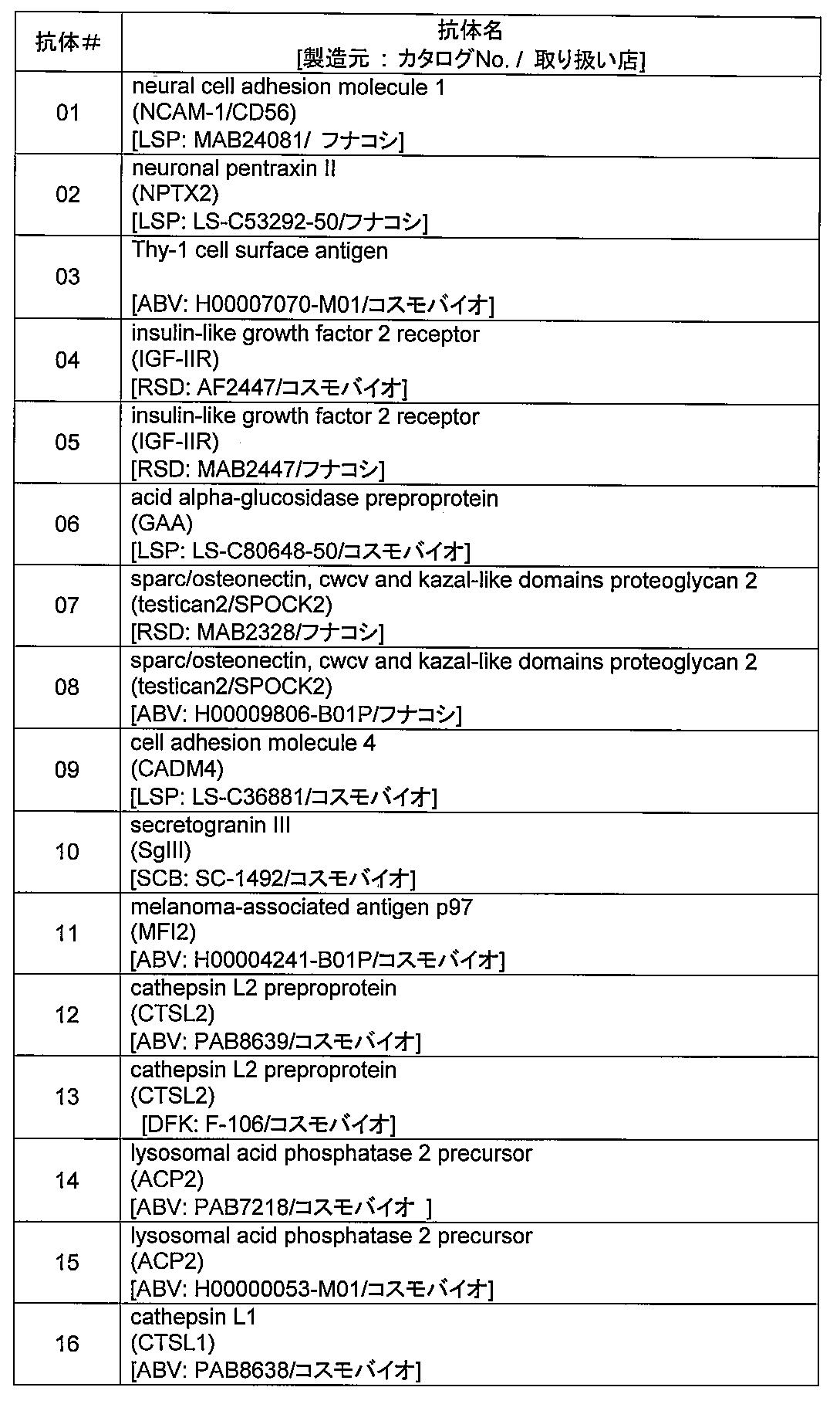
3. Examination of protein derived from culture supernatant by immunoblot Lung cancer cell lines cultured in serum-free medium for 24 hours (3 types of lung adenocarcinoma: H358, H1975, LX-1; 3 types of small cell lung cancer: H2171, H524, H526) The culture supernatant was concentrated 10 times with Amicon Ultra-154 centrifugal filter units (10 kDa cutoff, Millipore), and 10 μL of the sample was subjected to 10%, 12.5%, or 17% acrylamide gel (Maruyuki Shosha) of XV PANTERA SYSYTEM. After developing using SDS-PAGE, it was transferred to a PVDF membrane (Amarsham) at 200 mA for 40 min. As the blocking agent, 5% skim milk or 5% BSA dissolved in PBSTx in which 1% Triton X-100 was added to PBS (Dulbecco) was used and blocked at room temperature for 1 hour. After washing with PBSTx three times for 10 minutes, the membrane was reacted with a primary antibody (Table 4) biotinylated in advance with Biotin Labeling Kit-NH 2 (Dojindo Laboratories) for 1 hour. After washing again with PBSTx for 10 minutes three times, the mixture was reacted with the secondary antibody HRP-conjugated streptavidin (1: 3000 dilution, GE Healthcare) for 1 hour. After washing with PBSTx for 3 minutes for 10 minutes, HRP enzyme reaction was performed with Western Lightning Chemiluminescence Reagent Plus (Perkin Elmer), and the resulting signal was developed into Amersham Hyperfilm ECL (GE Healthcare). A comparative analysis was performed on the amount of protein present in the culture supernatant derived from adenocarcinoma and small cell lung cancer cell lines.
無血清培地で24時間培養した肺癌細胞株(肺腺癌3種:H358、H1975、LX-1;肺小細胞癌3種:H2171、H524、H526)の培養上清をAmicon Ultra-154 centrifugal filter units (10kDaカットオフ、ミリポア社)で10倍濃縮し、そのサンプル10μLをXV PANTERA SYSYTEM の10%、12.5%若しくは17%アクリルアミドゲル(丸幸商会社)を用いてSDS-PAGEで展開した後、200 mA、40minでPVDF膜(Amarsham社)に転写した。ブロッキング剤は、PBS(ダルベッコ社)に1% Triton X-100を加えたPBSTxに溶解した5%スキムミルク又は5% BSAを使用し、室温で1時間ブロッキングした。PBSTxで10分間の洗浄を3回行った後、予めBiotin Labeling Kit-NH2(同仁化学研究所)でビオチン化した一次抗体(表4)とメンブレンを1時間反応させた。再びPBSTxで10分間の洗浄を3回行った後、二次抗体HRP-conjugated streptavidin(1:3000希釈、GEヘルスケア社) で1時間反応させた。PBSTxで10分間の洗浄を3回行った後、Western Lightning Chemiluminescence Reagent Plus (パーキンエルマー社)によってHRPの酵素反応を行い、得られたシグナルをAmersham Hyperfilm ECL (GEヘルスケア社)に現像し、肺腺癌と肺小細胞癌の細胞株由来の培養上清に存在しているタンパク量について比較解析を行った。


(結果)
上記工程によって得られた肺癌鑑別マーカー糖タンパク質を表1、図1、図2に示す。 (result)
The lung cancer differentiation marker glycoprotein obtained by the above process is shown in Table 1, FIG. 1 and FIG.
上記工程によって得られた肺癌鑑別マーカー糖タンパク質を表1、図1、図2に示す。 (result)
The lung cancer differentiation marker glycoprotein obtained by the above process is shown in Table 1, FIG. 1 and FIG.
4.AALレクチンによる培養上清のバッチフラクショネーション
AALレクチンを用いて培養上清を分画した。具体的にはAALレジンは3倍量のPBSで5回洗浄を行った後、50%スラリー溶液に調製した。調製したAALレジン30μLに対し培養上清30μLを加え4℃で5時間震盪させた。遠心後(2,000rpm、2分間)上清を除去した後、Wash Buffer(0.1% SDS in PBST,0.1% Triton X-100)50μLを加え遠心(2,000rpm、2分間)を2回繰り返した後Wash Bufferを500μL加え、遠心(2,000rpm、2分間)し、上清を除去洗浄した。洗浄後の溶出には0.02% SDS を含む0.2 M Fucose in PBSを15μL加え4℃で5時間震盪させた。遠心後(2,000rpm、2分間)上清を回収しさらにElution Bufferを15μL加え遠心(2,000rpm、2分間)を行いAAL結合画分として溶出した。溶出した画分10μLをそれぞれSDS-PAGEに展開してウェスタンブロットで検出した。ConAレクチンを用いる場合も同様な操作を行った。ただし、溶出には0.5M メチルマンノシドを用いた。 4). Batch fractionation of culture supernatant with AAL lectin The culture supernatant was fractionated using AAL lectin. Specifically, the AAL resin was washed 5 times with 3 times the amount of PBS and then prepared into a 50% slurry solution. 30 μL of the culture supernatant was added to 30 μL of the prepared AAL resin, and the mixture was shaken at 4 ° C. for 5 hours. After centrifugation (2,000 rpm, 2 minutes), the supernatant was removed, 50 μL of Wash Buffer (0.1% SDS in PBST, 0.1% Triton X-100) was added, and centrifugation (2,000 rpm, 2 minutes) was repeated twice. 500 μL of Buffer was added, centrifuged (2,000 rpm, 2 minutes), and the supernatant was removed and washed. For elution after washing, 15 μL of 0.2 M Fucose in PBS containing 0.02% SDS was added and shaken at 4 ° C. for 5 hours. After centrifugation (2,000 rpm, 2 minutes), the supernatant was recovered, and 15 μL of Elution Buffer was further added, followed by centrifugation (2,000 rpm, 2 minutes) and elution as an AAL-binding fraction. 10 μL of the eluted fraction was developed on SDS-PAGE and detected by Western blot. The same operation was performed when ConA lectin was used. However, 0.5M methyl mannoside was used for elution.
AALレクチンを用いて培養上清を分画した。具体的にはAALレジンは3倍量のPBSで5回洗浄を行った後、50%スラリー溶液に調製した。調製したAALレジン30μLに対し培養上清30μLを加え4℃で5時間震盪させた。遠心後(2,000rpm、2分間)上清を除去した後、Wash Buffer(0.1% SDS in PBST,0.1% Triton X-100)50μLを加え遠心(2,000rpm、2分間)を2回繰り返した後Wash Bufferを500μL加え、遠心(2,000rpm、2分間)し、上清を除去洗浄した。洗浄後の溶出には0.02% SDS を含む0.2 M Fucose in PBSを15μL加え4℃で5時間震盪させた。遠心後(2,000rpm、2分間)上清を回収しさらにElution Bufferを15μL加え遠心(2,000rpm、2分間)を行いAAL結合画分として溶出した。溶出した画分10μLをそれぞれSDS-PAGEに展開してウェスタンブロットで検出した。ConAレクチンを用いる場合も同様な操作を行った。ただし、溶出には0.5M メチルマンノシドを用いた。 4). Batch fractionation of culture supernatant with AAL lectin The culture supernatant was fractionated using AAL lectin. Specifically, the AAL resin was washed 5 times with 3 times the amount of PBS and then prepared into a 50% slurry solution. 30 μL of the culture supernatant was added to 30 μL of the prepared AAL resin, and the mixture was shaken at 4 ° C. for 5 hours. After centrifugation (2,000 rpm, 2 minutes), the supernatant was removed, 50 μL of Wash Buffer (0.1% SDS in PBST, 0.1% Triton X-100) was added, and centrifugation (2,000 rpm, 2 minutes) was repeated twice. 500 μL of Buffer was added, centrifuged (2,000 rpm, 2 minutes), and the supernatant was removed and washed. For elution after washing, 15 μL of 0.2 M Fucose in PBS containing 0.02% SDS was added and shaken at 4 ° C. for 5 hours. After centrifugation (2,000 rpm, 2 minutes), the supernatant was recovered, and 15 μL of Elution Buffer was further added, followed by centrifugation (2,000 rpm, 2 minutes) and elution as an AAL-binding fraction. 10 μL of the eluted fraction was developed on SDS-PAGE and detected by Western blot. The same operation was performed when ConA lectin was used. However, 0.5M methyl mannoside was used for elution.
(結果)
上記工程によって得られた肺癌鑑別マーカー糖タンパク質を図3に示す。 (result)
The lung cancer differentiation marker glycoprotein obtained by the above process is shown in FIG.
上記工程によって得られた肺癌鑑別マーカー糖タンパク質を図3に示す。 (result)
The lung cancer differentiation marker glycoprotein obtained by the above process is shown in FIG.
5.免疫沈降による分画
表1に記載の全ての候補糖タンパク質抗体についてビオチン化されていないものについてはBiotin Labeling Kit-NH2(同仁化学研究所)を用いて、マニュアルに従いビオチン化した。培養上清100μLに対してビオチン化抗体を1μg加え、20℃で2時間1,400rpmで振盪した。磁気ビーズ(invitrogen社)の洗浄は、20μLの磁気ビーズに対して100μLのTBSTx(50mM Tris-HCl (pH 8.0)、150mM NaCl、1% Triton X-100)を加えて、撹拌後遠心(10,000 rpm、3秒)し、上清の除去を3回行った。反応させた培養上清と抗体を磁気ビーズに移し、20℃で1時間1,400 rpmで振盪した。磁気スタンドを用いて磁気ビーズと結合した抗体を回収した。回収した磁気ビーズは1mLのTBSTxで3回洗浄した。溶出バッファー(0.2% SDS in TBS)20 ulを加え撹拌後98℃または60℃で5分加熱を2回行い、溶出した。溶出したサンプルの抗体を除去するために同様に洗浄した40μLの磁気ビーズを溶出サンプルに加え20℃で2時間1,400rpmで振盪した。磁気スタンドで抗体-磁気ビーズを取り除いたものをサンプルとして用いた。調製したサンプルをSDS-PAGEで展開した後レクチンブロットを行った。 5. Fractionation by immunoprecipitation All candidate glycoprotein antibodies listed in Table 1 were biotinylated using Biotin Labeling Kit-NH 2 (Dojindo Laboratories) according to the manual. 1 μg of biotinylated antibody was added to 100 μL of the culture supernatant, and shaken at 1,400 rpm for 2 hours at 20 ° C. To wash magnetic beads (Invitrogen), add 100 μL TBSTx (50 mM Tris-HCl (pH 8.0), 150 mM NaCl, 1% Triton X-100) to 20 μL magnetic beads, and centrifuge after stirring (10,000 rpm). 3 seconds), and the supernatant was removed three times. The reacted culture supernatant and antibody were transferred to magnetic beads and shaken at 1,400 rpm for 1 hour at 20 ° C. The antibody bound to the magnetic beads was recovered using a magnetic stand. The collected magnetic beads were washed three times with 1 mL of TBSTx. After adding 20 ul of elution buffer (0.2% SDS in TBS) and stirring, the mixture was heated twice at 98 ° C. or 60 ° C. for 5 minutes for elution. In order to remove the antibody in the eluted sample, 40 μL of magnetic beads washed in the same manner were added to the eluted sample and shaken at 1,400 rpm for 2 hours at 20 ° C. A sample obtained by removing antibody-magnetic beads with a magnetic stand was used as a sample. The prepared sample was developed by SDS-PAGE and then lectin blotted.
表1に記載の全ての候補糖タンパク質抗体についてビオチン化されていないものについてはBiotin Labeling Kit-NH2(同仁化学研究所)を用いて、マニュアルに従いビオチン化した。培養上清100μLに対してビオチン化抗体を1μg加え、20℃で2時間1,400rpmで振盪した。磁気ビーズ(invitrogen社)の洗浄は、20μLの磁気ビーズに対して100μLのTBSTx(50mM Tris-HCl (pH 8.0)、150mM NaCl、1% Triton X-100)を加えて、撹拌後遠心(10,000 rpm、3秒)し、上清の除去を3回行った。反応させた培養上清と抗体を磁気ビーズに移し、20℃で1時間1,400 rpmで振盪した。磁気スタンドを用いて磁気ビーズと結合した抗体を回収した。回収した磁気ビーズは1mLのTBSTxで3回洗浄した。溶出バッファー(0.2% SDS in TBS)20 ulを加え撹拌後98℃または60℃で5分加熱を2回行い、溶出した。溶出したサンプルの抗体を除去するために同様に洗浄した40μLの磁気ビーズを溶出サンプルに加え20℃で2時間1,400rpmで振盪した。磁気スタンドで抗体-磁気ビーズを取り除いたものをサンプルとして用いた。調製したサンプルをSDS-PAGEで展開した後レクチンブロットを行った。 5. Fractionation by immunoprecipitation All candidate glycoprotein antibodies listed in Table 1 were biotinylated using Biotin Labeling Kit-NH 2 (Dojindo Laboratories) according to the manual. 1 μg of biotinylated antibody was added to 100 μL of the culture supernatant, and shaken at 1,400 rpm for 2 hours at 20 ° C. To wash magnetic beads (Invitrogen), add 100 μL TBSTx (50 mM Tris-HCl (pH 8.0), 150 mM NaCl, 1% Triton X-100) to 20 μL magnetic beads, and centrifuge after stirring (10,000 rpm). 3 seconds), and the supernatant was removed three times. The reacted culture supernatant and antibody were transferred to magnetic beads and shaken at 1,400 rpm for 1 hour at 20 ° C. The antibody bound to the magnetic beads was recovered using a magnetic stand. The collected magnetic beads were washed three times with 1 mL of TBSTx. After adding 20 ul of elution buffer (0.2% SDS in TBS) and stirring, the mixture was heated twice at 98 ° C. or 60 ° C. for 5 minutes for elution. In order to remove the antibody in the eluted sample, 40 μL of magnetic beads washed in the same manner were added to the eluted sample and shaken at 1,400 rpm for 2 hours at 20 ° C. A sample obtained by removing antibody-magnetic beads with a magnetic stand was used as a sample. The prepared sample was developed by SDS-PAGE and then lectin blotted.
(結果)
上記工程によって得られた肺癌鑑別マーカー糖タンパク質を図4に示す。 (result)
The lung cancer differentiation marker glycoprotein obtained by the above process is shown in FIG.
上記工程によって得られた肺癌鑑別マーカー糖タンパク質を図4に示す。 (result)
The lung cancer differentiation marker glycoprotein obtained by the above process is shown in FIG.
6.レクチンブロットによる評価
免疫沈降したサンプル10μLを10% SDS-PAGEに展開した。SDS-PAGE後タンパク質をPVDF膜(Amersham)に転写した。PVDF膜を5% BSAで1時間室温にてブロッキングした後、すでにビオチン化されたAALレクチン(生化学工業)で1時間室温にて反応させた。その後二次抗体HRP-conjugated streptavidinで1時間室温にて反応させた。Western Lightningを用い検出した。レクチンの希釈にはPBS-Tを用いた。 6). Evaluation by lectin blot 10 μL of the immunoprecipitated sample was developed on 10% SDS-PAGE. After SDS-PAGE, the protein was transferred to a PVDF membrane (Amersham). The PVDF membrane was blocked with 5% BSA for 1 hour at room temperature, and then reacted with an already biotinylated AAL lectin (Seikagaku Corporation) for 1 hour at room temperature. Thereafter, it was reacted with the secondary antibody HRP-conjugated streptavidin for 1 hour at room temperature. Detection was performed using Western Lightning. PBS-T was used for lectin dilution.
免疫沈降したサンプル10μLを10% SDS-PAGEに展開した。SDS-PAGE後タンパク質をPVDF膜(Amersham)に転写した。PVDF膜を5% BSAで1時間室温にてブロッキングした後、すでにビオチン化されたAALレクチン(生化学工業)で1時間室温にて反応させた。その後二次抗体HRP-conjugated streptavidinで1時間室温にて反応させた。Western Lightningを用い検出した。レクチンの希釈にはPBS-Tを用いた。 6). Evaluation by lectin blot 10 μL of the immunoprecipitated sample was developed on 10% SDS-PAGE. After SDS-PAGE, the protein was transferred to a PVDF membrane (Amersham). The PVDF membrane was blocked with 5% BSA for 1 hour at room temperature, and then reacted with an already biotinylated AAL lectin (Seikagaku Corporation) for 1 hour at room temperature. Thereafter, it was reacted with the secondary antibody HRP-conjugated streptavidin for 1 hour at room temperature. Detection was performed using Western Lightning. PBS-T was used for lectin dilution.
(結果)
上記工程によって得られた肺癌鑑別マーカー糖タンパク質を図4に示す。 (result)
The lung cancer differentiation marker glycoprotein obtained by the above process is shown in FIG.
上記工程によって得られた肺癌鑑別マーカー糖タンパク質を図4に示す。 (result)
The lung cancer differentiation marker glycoprotein obtained by the above process is shown in FIG.
7.AALレクチンによる血清のカラムフラクショネーション
AALレクチンを用いて血清を分画した。具体的には市販のカラムにAALレジンを1mL充填し、レジンの10倍量のTBSで洗浄、レジンの2倍量の0.5MのNaClで洗浄、レジンの10倍量のTBSで洗浄を行い、AALカラムを調製した。血清をAALカラムに加えた後、レジン中で1時間反応させた後、レジンの5倍量のTBSで洗浄を行った。溶出には20 mM Fucoseを1mL加えレジン中で1時間反応させた後さらに20mM Fucoseを4mL加え全量5mLで溶出した。 7. Serum column fractionation with AAL lectin Serum was fractionated using AAL lectin. Specifically, 1 mL of AAL resin is packed in a commercially available column, washed with 10 times the amount of resin TBS, twice the amount of resin 0.5 M NaCl, and washed with 10 times the amount of resin TBS. An AAL column was prepared. After the serum was added to the AAL column, the mixture was reacted in the resin for 1 hour, and then washed with 5 times the amount of TBS. For elution, 1 mL of 20 mM Fucose was added and reacted in the resin for 1 hour, and then 4 mL of 20 mM Fucose was added and eluted in a total volume of 5 mL.
AALレクチンを用いて血清を分画した。具体的には市販のカラムにAALレジンを1mL充填し、レジンの10倍量のTBSで洗浄、レジンの2倍量の0.5MのNaClで洗浄、レジンの10倍量のTBSで洗浄を行い、AALカラムを調製した。血清をAALカラムに加えた後、レジン中で1時間反応させた後、レジンの5倍量のTBSで洗浄を行った。溶出には20 mM Fucoseを1mL加えレジン中で1時間反応させた後さらに20mM Fucoseを4mL加え全量5mLで溶出した。 7. Serum column fractionation with AAL lectin Serum was fractionated using AAL lectin. Specifically, 1 mL of AAL resin is packed in a commercially available column, washed with 10 times the amount of resin TBS, twice the amount of resin 0.5 M NaCl, and washed with 10 times the amount of resin TBS. An AAL column was prepared. After the serum was added to the AAL column, the mixture was reacted in the resin for 1 hour, and then washed with 5 times the amount of TBS. For elution, 1 mL of 20 mM Fucose was added and reacted in the resin for 1 hour, and then 4 mL of 20 mM Fucose was added and eluted in a total volume of 5 mL.
(結果)
上記工程によって得られた肺癌鑑別マーカー糖タンパク質を図5に示す。 (result)
The lung cancer differentiation marker glycoprotein obtained by the above process is shown in FIG.
上記工程によって得られた肺癌鑑別マーカー糖タンパク質を図5に示す。 (result)
The lung cancer differentiation marker glycoprotein obtained by the above process is shown in FIG.
8.連続クロマトグラフィー方法よる血清のフラクショネーション
連続クロマトグラフィーを用いた血清の分画は、LCAアガロース(J-オイルミルズ社)とAALアガロース(J-オイルミルズ社)を用いて低温下で行った。100μLの血清をPBSで4倍希釈して、まず、カラムに充填した5mLのLCAアガロース(0.7×13cm)に血清サンプルを興じた。次に、得られた非結合血清画分をカラムに充填した2.5mLのAALアガロース(0.7×5.5cm)に興じ、PBSで十分に洗浄を行った後0.2M フコースを含むPBSで溶出を行った。溶出画分をウルトラフリー遠心濃縮デバイス(カットオフ: 30kDa、ミリポア社)で濃縮し、得られたサンプルを上記と同様にイムノブロットにより候補分子の比較解析を行った。 8). Serum Fractionation by Continuous Chromatography Method Serum fractionation using continuous chromatography was performed at low temperature using LCA agarose (J-Oil Mills) and AAL agarose (J-Oil Mills). 100 μL of serum was diluted 4 times with PBS, and the serum sample was first developed in 5 mL of LCA agarose (0.7 × 13 cm) packed in a column. Next, the obtained unbound serum fraction was developed into 2.5 mL of AAL agarose (0.7 × 5.5 cm) packed in a column, washed thoroughly with PBS, and then eluted with PBS containing 0.2 M fucose. . The eluted fraction was concentrated with an ultra-free centrifugal concentration device (cut-off: 30 kDa, Millipore), and the obtained sample was subjected to comparative analysis of candidate molecules by immunoblotting as described above.
連続クロマトグラフィーを用いた血清の分画は、LCAアガロース(J-オイルミルズ社)とAALアガロース(J-オイルミルズ社)を用いて低温下で行った。100μLの血清をPBSで4倍希釈して、まず、カラムに充填した5mLのLCAアガロース(0.7×13cm)に血清サンプルを興じた。次に、得られた非結合血清画分をカラムに充填した2.5mLのAALアガロース(0.7×5.5cm)に興じ、PBSで十分に洗浄を行った後0.2M フコースを含むPBSで溶出を行った。溶出画分をウルトラフリー遠心濃縮デバイス(カットオフ: 30kDa、ミリポア社)で濃縮し、得られたサンプルを上記と同様にイムノブロットにより候補分子の比較解析を行った。 8). Serum Fractionation by Continuous Chromatography Method Serum fractionation using continuous chromatography was performed at low temperature using LCA agarose (J-Oil Mills) and AAL agarose (J-Oil Mills). 100 μL of serum was diluted 4 times with PBS, and the serum sample was first developed in 5 mL of LCA agarose (0.7 × 13 cm) packed in a column. Next, the obtained unbound serum fraction was developed into 2.5 mL of AAL agarose (0.7 × 5.5 cm) packed in a column, washed thoroughly with PBS, and then eluted with PBS containing 0.2 M fucose. . The eluted fraction was concentrated with an ultra-free centrifugal concentration device (cut-off: 30 kDa, Millipore), and the obtained sample was subjected to comparative analysis of candidate molecules by immunoblotting as described above.
(結果)
上記工程によって得られた肺癌鑑別マーカー糖タンパク質を図6に示す。 (result)
The lung cancer differentiation marker glycoprotein obtained by the above process is shown in FIG.
上記工程によって得られた肺癌鑑別マーカー糖タンパク質を図6に示す。 (result)
The lung cancer differentiation marker glycoprotein obtained by the above process is shown in FIG.
9.連続クロマトグラフィー方法よる多検体血清のフラクショネーション
多検体血清の比較解析を行う際に、少量の血清サンプルを短時間でフラクショネーションする必要があるため、高スループットな連続クロマトグラフィー方法を確立した。連続クロマトグラフィーを用いた血清の分画は、LCAアガロース(J-オイルミルズ社)とAALアガロース(Vector社)を用いて行った。チップカラムに充填した300μLのLCAアガロース(0.37×2.3cm)に50μLの血清サンプルを興じた。次に、得られた非結合血清画分をオープンカラムに充填した250μLのAALアガロース(0.8×0.5cm)に興じ、PBSで十分に洗浄を行った後0.02M フコースを含むPBSで溶出を行った。溶出画分をウルトラフリー遠心濃縮デバイス(カットオフ: 10kDa、ミリポア社)で濃縮し、得られたサンプルのうち血清10μL相当を上記と同様にイムノブロットにより候補分子の比較解析を行った。 9. Fractionation of multi-sample sera by continuous chromatography method When performing comparative analysis of multi-sample sera, it is necessary to fractionate a small amount of serum sample in a short time, so a high-throughput continuous chromatography method was established. . Serum fractionation using continuous chromatography was performed using LCA agarose (J-Oil Mills) and AAL agarose (Vector). 50 μL of a serum sample was served in 300 μL of LCA agarose (0.37 × 2.3 cm) packed in a chip column. Next, the obtained unbound serum fraction was exposed to 250 μL of AAL agarose (0.8 × 0.5 cm) packed in an open column, washed thoroughly with PBS, and then eluted with PBS containing 0.02 M fucose. . The eluted fraction was concentrated with an ultra-free centrifugal concentration device (cut-off: 10 kDa, Millipore), and among the obtained samples, 10 μL of serum was subjected to comparative analysis of candidate molecules by immunoblotting as described above.
多検体血清の比較解析を行う際に、少量の血清サンプルを短時間でフラクショネーションする必要があるため、高スループットな連続クロマトグラフィー方法を確立した。連続クロマトグラフィーを用いた血清の分画は、LCAアガロース(J-オイルミルズ社)とAALアガロース(Vector社)を用いて行った。チップカラムに充填した300μLのLCAアガロース(0.37×2.3cm)に50μLの血清サンプルを興じた。次に、得られた非結合血清画分をオープンカラムに充填した250μLのAALアガロース(0.8×0.5cm)に興じ、PBSで十分に洗浄を行った後0.02M フコースを含むPBSで溶出を行った。溶出画分をウルトラフリー遠心濃縮デバイス(カットオフ: 10kDa、ミリポア社)で濃縮し、得られたサンプルのうち血清10μL相当を上記と同様にイムノブロットにより候補分子の比較解析を行った。 9. Fractionation of multi-sample sera by continuous chromatography method When performing comparative analysis of multi-sample sera, it is necessary to fractionate a small amount of serum sample in a short time, so a high-throughput continuous chromatography method was established. . Serum fractionation using continuous chromatography was performed using LCA agarose (J-Oil Mills) and AAL agarose (Vector). 50 μL of a serum sample was served in 300 μL of LCA agarose (0.37 × 2.3 cm) packed in a chip column. Next, the obtained unbound serum fraction was exposed to 250 μL of AAL agarose (0.8 × 0.5 cm) packed in an open column, washed thoroughly with PBS, and then eluted with PBS containing 0.02 M fucose. . The eluted fraction was concentrated with an ultra-free centrifugal concentration device (cut-off: 10 kDa, Millipore), and among the obtained samples, 10 μL of serum was subjected to comparative analysis of candidate molecules by immunoblotting as described above.
(結果)
上記工程によって得られた肺癌鑑別マーカー糖タンパク質を図8に示す。 (result)
The lung cancer differentiation marker glycoprotein obtained by the above process is shown in FIG.
上記工程によって得られた肺癌鑑別マーカー糖タンパク質を図8に示す。 (result)
The lung cancer differentiation marker glycoprotein obtained by the above process is shown in FIG.
10.抗体オーバーレイ・レクチンアレイ
上記「5.免疫沈降による分画」で得られた糖タンパク質溶液を適当量とり、レクチンアレイ反応バッファーである1% Triton X-100含有Phosphate-buffered saline (PBSTx)で60μLに調製した。この溶液をガラス1枚当たり8つの反応槽が形成されているレクチンアレイの各反応槽へ添加し、20℃で10時間以上反応した。この8つの反応槽からなるレクチンアレイ基盤の作製は、内山ら(Proteomics 8, 3042-3050 (2008))の手法に従った。これにより糖タンパク質上の糖鎖とアレイ基板上に固定されている43種のレクチンとの結合反応が平衡状態に達する。未反応の基板上のレクチンに検出用抗体の糖鎖が結合して、ノイズを発生することを防止するため、その後、ヒト血清由来IgG溶液(シグマ社製)を2μL加えて、30分反応させた。60μLのPBSTxで各反応槽を3回洗浄した後、再度ヒト血清由来IgG溶液を2μL加え、若干撹拌した。続いて、検出用の該糖タンパク質に対する抗体(ビオチン化物)を100ng相当加え、20℃で1時間反応させた。抗原抗体反応後、60μLのPBSTxで各反応槽を3回洗浄した後、モリテックス社製アレイスキャナーGlycoStationによりアレイスキャンを行い、肺癌組織特異的に反応するレクチンスポット上の蛍光強度について比較を行った。 10. Antibody overlay / lectin array Take an appropriate amount of the glycoprotein solution obtained in “5. Fractionation by immunoprecipitation” above and use 1% Triton X-100-containing Phosphate-buffered saline (PBSTx) as a lectin array reaction buffer. Prepared to 60 μL. This solution was added to each reaction vessel of the lectin array in which 8 reaction vessels were formed per glass, and reacted at 20 ° C. for 10 hours or more. The lectin array substrate composed of these eight reaction vessels was prepared according to the method of Uchiyama et al. (Proteomics 8, 3042-3050 (2008)). As a result, the binding reaction between the sugar chain on the glycoprotein and the 43 lectins immobilized on the array substrate reaches an equilibrium state. To prevent the detection antibody sugar chains from binding to the lectins on the unreacted substrate, add 2 μL of human serum-derived IgG solution (manufactured by Sigma) and react for 30 minutes. It was. Each reaction vessel was washed 3 times with 60 μL of PBSTx, and then 2 μL of human serum-derived IgG solution was added again and stirred slightly. Subsequently, 100 ng of an antibody (biotinylated product) against the glycoprotein for detection was added and reacted at 20 ° C. for 1 hour. After the antigen-antibody reaction, each reaction vessel was washed three times with 60 μL of PBSTx, and then array scan was performed with an array scanner GlycoStation manufactured by Moritex, and the fluorescence intensity on the lectin spot that specifically reacted with lung cancer tissue was compared.
上記「5.免疫沈降による分画」で得られた糖タンパク質溶液を適当量とり、レクチンアレイ反応バッファーである1% Triton X-100含有Phosphate-buffered saline (PBSTx)で60μLに調製した。この溶液をガラス1枚当たり8つの反応槽が形成されているレクチンアレイの各反応槽へ添加し、20℃で10時間以上反応した。この8つの反応槽からなるレクチンアレイ基盤の作製は、内山ら(Proteomics 8, 3042-3050 (2008))の手法に従った。これにより糖タンパク質上の糖鎖とアレイ基板上に固定されている43種のレクチンとの結合反応が平衡状態に達する。未反応の基板上のレクチンに検出用抗体の糖鎖が結合して、ノイズを発生することを防止するため、その後、ヒト血清由来IgG溶液(シグマ社製)を2μL加えて、30分反応させた。60μLのPBSTxで各反応槽を3回洗浄した後、再度ヒト血清由来IgG溶液を2μL加え、若干撹拌した。続いて、検出用の該糖タンパク質に対する抗体(ビオチン化物)を100ng相当加え、20℃で1時間反応させた。抗原抗体反応後、60μLのPBSTxで各反応槽を3回洗浄した後、モリテックス社製アレイスキャナーGlycoStationによりアレイスキャンを行い、肺癌組織特異的に反応するレクチンスポット上の蛍光強度について比較を行った。 10. Antibody overlay / lectin array Take an appropriate amount of the glycoprotein solution obtained in “5. Fractionation by immunoprecipitation” above and use 1% Triton X-100-containing Phosphate-buffered saline (PBSTx) as a lectin array reaction buffer. Prepared to 60 μL. This solution was added to each reaction vessel of the lectin array in which 8 reaction vessels were formed per glass, and reacted at 20 ° C. for 10 hours or more. The lectin array substrate composed of these eight reaction vessels was prepared according to the method of Uchiyama et al. (Proteomics 8, 3042-3050 (2008)). As a result, the binding reaction between the sugar chain on the glycoprotein and the 43 lectins immobilized on the array substrate reaches an equilibrium state. To prevent the detection antibody sugar chains from binding to the lectins on the unreacted substrate, add 2 μL of human serum-derived IgG solution (manufactured by Sigma) and react for 30 minutes. It was. Each reaction vessel was washed 3 times with 60 μL of PBSTx, and then 2 μL of human serum-derived IgG solution was added again and stirred slightly. Subsequently, 100 ng of an antibody (biotinylated product) against the glycoprotein for detection was added and reacted at 20 ° C. for 1 hour. After the antigen-antibody reaction, each reaction vessel was washed three times with 60 μL of PBSTx, and then array scan was performed with an array scanner GlycoStation manufactured by Moritex, and the fluorescence intensity on the lectin spot that specifically reacted with lung cancer tissue was compared.
(結果)
上記工程によって得られた肺癌鑑別マーカー糖タンパク質を図9及び図10に示す。 (result)
The lung cancer differentiation marker glycoprotein obtained by the above process is shown in FIG. 9 and FIG.
上記工程によって得られた肺癌鑑別マーカー糖タンパク質を図9及び図10に示す。 (result)
The lung cancer differentiation marker glycoprotein obtained by the above process is shown in FIG. 9 and FIG.
今回の実験により、AAL又はConA及びこれらのリガンドに関連する特異性をもつレクチン以外においても、PNAやPWMのように肺癌鑑別に有効なレクチンが存在することが明らかになった。これは、糖タンパク質上の複数のレクチンシグナルを組み合わせ、その組み合わせ方にバリエーションを持たせることにより、肺癌組織型の鑑別をより精度高く検出できる可能性を意味している。
This experiment revealed that there are lectins that are effective for differentiating lung cancer, such as PNA and PWM, in addition to lectins having specificities related to AAL or ConA and their ligands. This means that a combination of a plurality of lectin signals on a glycoprotein and a variation in the combination method can detect lung cancer tissue type with higher accuracy.
<実施例2>肺癌鑑別マーカーを用いた肺癌細胞の組織染色
肺癌鑑別マーカーを用いて抗体による組織染色について検証した。 <Example 2> Tissue staining of lung cancer cells using lung cancer differentiation markers Tissue staining with antibodies was examined using lung cancer differentiation markers.
肺癌鑑別マーカーを用いて抗体による組織染色について検証した。 <Example 2> Tissue staining of lung cancer cells using lung cancer differentiation markers Tissue staining with antibodies was examined using lung cancer differentiation markers.
まず、ホルマリン固定肺癌組織切片(5μm厚)を定法に従い組織切片上に被覆しているパラフィンを脱パラフィン化した。脱パラフィン化した組織切片はPBSにて洗浄し、風乾後、10mMクエン酸バッファー中に浸し、121℃、15分間オートクレーブすることで、ホルマリンによる分子間(分子内)架橋を解離した。処理後の切片は室温でしばらく放置した後、PBSに5分間3回繰り返し浸し、組織表面を洗浄した。次いで0.3% H2O2-MeOHにて10分間、室温で処理することで、内在性ペルオキシダーゼのブロッキング反応を行った。PBSにて洗浄 (5分、3回)後、一次抗体溶液(R&D社製。抗NPR 3μg/mLになるようPBSで懸濁) を組織切片上へ添加し、保湿箱中にて20℃で2時間結合反応した。PBS にて洗浄 (5分、3回)した後、次いで二次抗体として抗ヒツジFITC conjugate(SantaCruz社)と、酵素標識抗体としてanti-FITC HRP conjugate(タカラバイオ株式会社)を用いて発色反応した。反応を停止するため、ミリQ水に5分間3回浸した。最後にヘマトキシリンで、室温、1分間処理し、核酸を染色後、流水にて洗浄した。
First, paraffin covering a tissue section of a formalin-fixed lung cancer tissue section (5 μm thickness) on the tissue section was deparaffinized according to a conventional method. The deparaffinized tissue section was washed with PBS, air-dried, immersed in 10 mM citrate buffer, and autoclaved at 121 ° C. for 15 minutes to dissociate the intermolecular (intramolecular) cross-linking by formalin. The treated sections were allowed to stand at room temperature for a while and then immersed in PBS three times for 5 minutes to wash the tissue surface. Next, the endogenous peroxidase blocking reaction was performed by treatment with 0.3% H 2 O 2 -MeOH for 10 minutes at room temperature. After washing with PBS (5 min, 3 times), add the primary antibody solution (R & D, suspended in PBS to give anti-NPR 3 μg / mL) on the tissue section, and in a moist box at 20 ° C. The binding reaction was performed for 2 hours. After washing with PBS (5 minutes, 3 times), color reaction was then performed using anti-sheep FITC conjugate (SantaCruz) as the secondary antibody and anti-FITC HRP conjugate (Takara Bio Inc.) as the enzyme-labeled antibody. . To stop the reaction, it was immersed in Milli-Q water three times for 5 minutes. Finally, it was treated with hematoxylin at room temperature for 1 minute, and the nucleic acid was stained and washed with running water.
(結果)
上記工程によって得られた肺癌鑑別マーカー糖タンパク質を図7に示す。小細胞癌にのみ特異的に染色されているのがわかる。 (result)
The lung cancer differentiation marker glycoprotein obtained by the above process is shown in FIG. It can be seen that only small cell carcinoma is stained specifically.
上記工程によって得られた肺癌鑑別マーカー糖タンパク質を図7に示す。小細胞癌にのみ特異的に染色されているのがわかる。 (result)
The lung cancer differentiation marker glycoprotein obtained by the above process is shown in FIG. It can be seen that only small cell carcinoma is stained specifically.
本明細書で引用した全ての刊行物、特許および特許出願をそのまま参考として本明細書にとり入れるものとする。
All publications, patents and patent applications cited in this specification shall be incorporated into the present specification as they are.
Claims (21)
- 表1又は表2に記載の肺癌鑑別マーカー糖タンパク質であって、表1又は表2に示す糖付加位置のアスパラギン残基に糖鎖が付加された前記糖タンパク質。

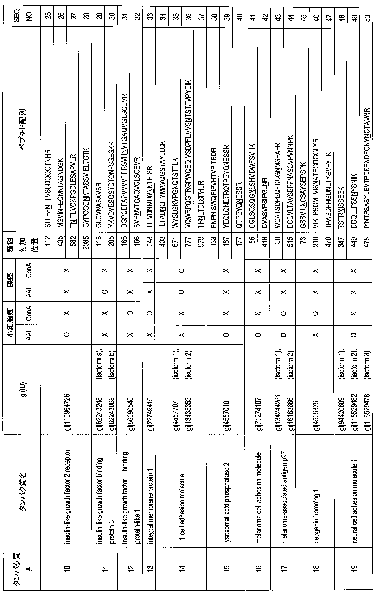

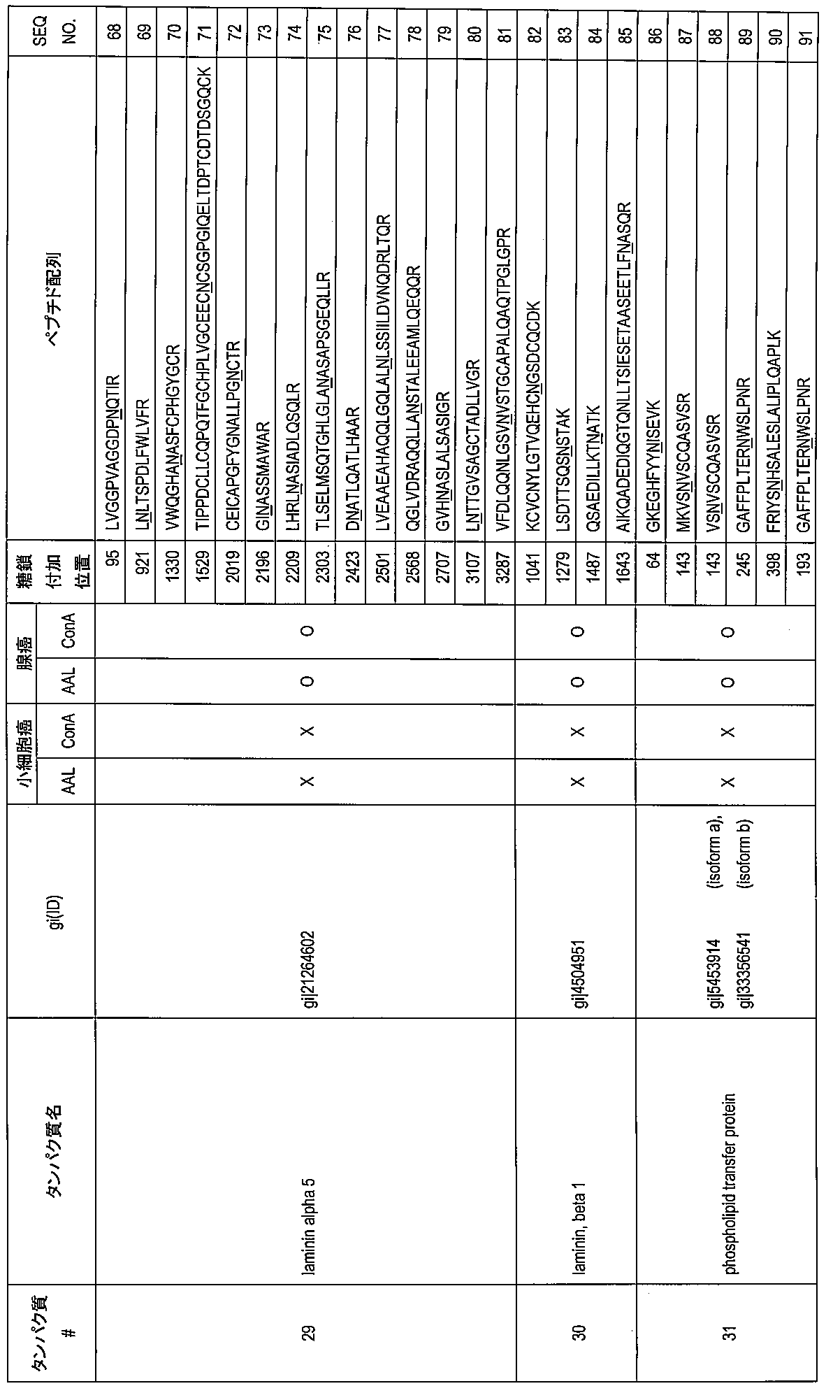


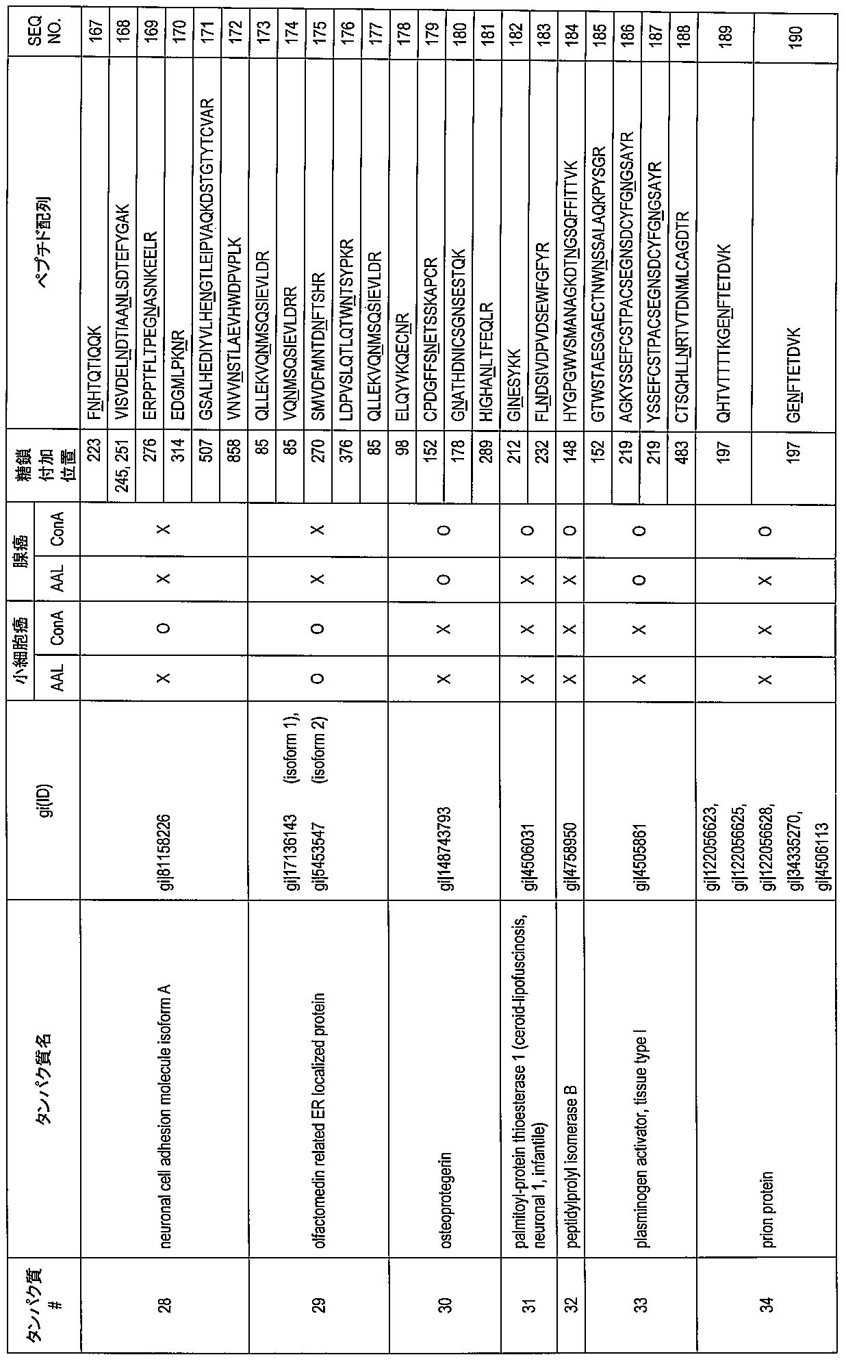
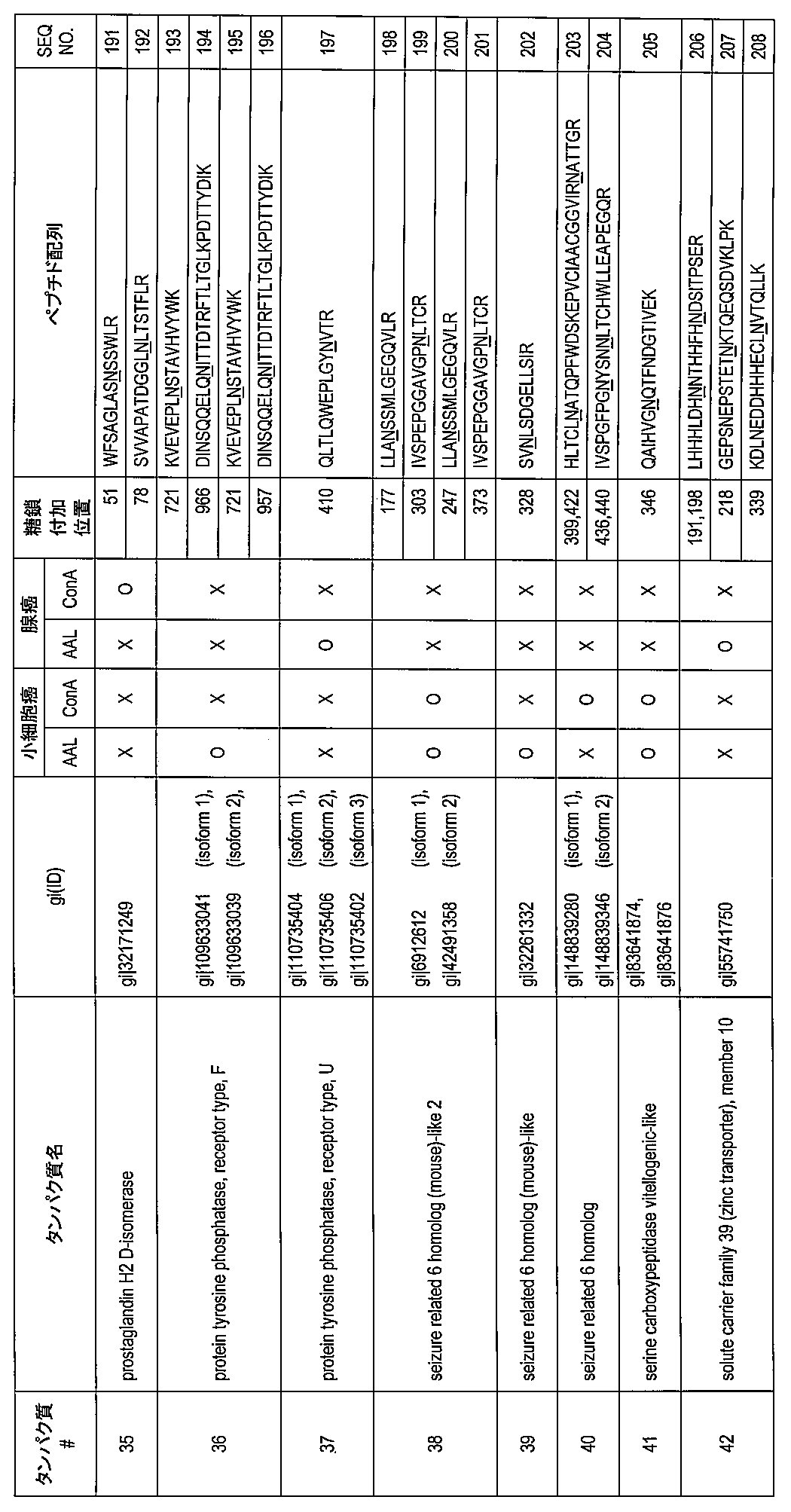 The glycoprotein according to Table 1 or Table 2, wherein the sugar chain is added to the asparagine residue at the sugar addition position shown in Table 1 or Table 2.
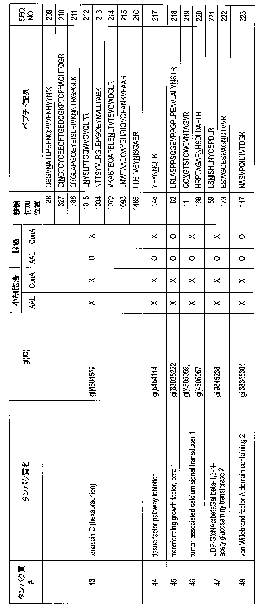
- 前記糖鎖がフコース修飾を伴う糖鎖、高マンノース型糖鎖、混成型糖鎖、複合型二本鎖糖鎖、キチン、ポリラクトサミン及びβ1-3ガラクトースエピトープからなる群より選択される一以上の糖鎖である、請求項1に記載の肺癌鑑別マーカー糖タンパク質。 One or more selected from the group consisting of a sugar chain with fucose modification, a high mannose sugar chain, a hybrid sugar chain, a complex double chain sugar chain, chitin, polylactosamine and β1-3 galactose epitope The lung cancer differential marker glycoprotein according to claim 1, wherein
- 肺小細胞癌又は肺腺癌の鑑別用である、請求項2に記載の肺癌鑑別マーカー糖タンパク質。 The lung cancer differentiation marker glycoprotein according to claim 2, which is used for differentiation of small cell lung cancer or lung adenocarcinoma.
- 肺小細胞癌鑑別用であって、前記糖タンパク質が神経細胞接着分子(NCAM1)、セクレトグラニンIII及びインスリン様成長因子結合タンパク質-L1(IGFBP-L1)からなる群より選択される一以上の糖タンパク質である、請求項3に記載の肺癌鑑別マーカー糖タンパク質。 One or more selected from the group consisting of neural cell adhesion molecule (NCAM1), secretogranin III and insulin-like growth factor binding protein-L1 (IGFBP-L1) The lung cancer differentiation marker glycoprotein according to claim 3, which is a glycoprotein.
- 肺腺癌鑑別用であって、前記糖タンパク質がフィブロネクチン1である、請求項3に記載の肺癌鑑別マーカー糖タンパク質。 The lung cancer differentiation marker glycoprotein according to claim 3, wherein the glycoprotein is fibronectin 1 for lung adenocarcinoma differentiation.
- 糖鎖が付加された、表1又は表2に示す糖付加位置のアスパラギン残基を少なくとも一つ含む請求項1~5のいずれか一項に記載の肺癌鑑別マーカー糖タンパク質の断片。 The fragment of the lung cancer differentiation marker glycoprotein according to any one of claims 1 to 5, comprising at least one asparagine residue at the sugar addition position shown in Table 1 or Table 2 to which a sugar chain has been added.
- 被験者より採取された試料から、表1又は表2に示す糖付加位置のアスパラギン残基に糖鎖が付加された表1又は表2に記載の一以上の肺癌鑑別マーカー糖タンパク質、及び/又は糖鎖が付加された、表1又は表2に示す糖付加位置のアスパラギン残基を少なくとも一つ含む一以上の前記糖タンパク質の断片を検出することによって、該被験者が肺癌に罹患していると判定する、肺癌罹患判定方法。 One or more lung cancer differentiation marker glycoproteins and / or sugars described in Table 1 or Table 2 in which a sugar chain is added to an asparagine residue at the sugar addition position shown in Table 1 or Table 2 from a sample collected from a subject It is determined that the subject suffers from lung cancer by detecting one or more fragments of the glycoprotein containing at least one asparagine residue at the glycosylation position shown in Table 1 or Table 2 to which a chain has been added. A method for determining the incidence of lung cancer.
- 前記肺癌鑑別マーカー糖タンパク質及び/又はその糖タンパク質の断片を、前記糖鎖に結合する一以上の糖鎖プローブを用いて検出する、請求項7に記載の方法。 The method according to claim 7, wherein the lung cancer differentiation marker glycoprotein and / or a fragment of the glycoprotein is detected using one or more sugar chain probes that bind to the sugar chain.
- 前記糖鎖プローブがフコース修飾を伴う糖鎖、高マンノース型糖鎖、混成型糖鎖、複合型二本鎖糖鎖、キチン、ポリラクトサミン又はβ1-3ガラクトースエピトープに結合する、請求項8に記載の方法。 9. The sugar chain probe binds to a sugar chain with fucose modification, a high mannose sugar chain, a hybrid sugar chain, a complex double chain sugar chain, chitin, polylactosamine or β1-3 galactose epitope. The method described.
- 前記糖鎖プローブがレクチン、抗体又はファージ抗体である、請求項8又は9に記載の方法。 The method according to claim 8 or 9, wherein the sugar chain probe is a lectin, an antibody or a phage antibody.
- 前記レクチンがAAL、ConA、PWM又はPNAである、請求項10に記載の方法。 The method according to claim 10, wherein the lectin is AAL, ConA, PWM or PNA.
- 前記肺癌鑑別マーカー糖タンパク質が神経細胞接着分子(NCAM1)であり、AALとの結合を検出することによって、肺癌の組織型が小細胞癌であることを判定する、請求項11に記載の方法。 The method according to claim 11, wherein the lung cancer differentiation marker glycoprotein is a nerve cell adhesion molecule (NCAM1), and the tissue type of lung cancer is determined to be small cell cancer by detecting binding with AAL.
- 前記肺癌鑑別マーカー糖タンパク質がセクレトグラニンIIIであり、AAL及び/又はConAとの結合を検出することによって、肺癌の組織型が小細胞癌であることを判定する、請求項11に記載の方法。 12. The method according to claim 11, wherein the lung cancer differentiation marker glycoprotein is secretogranin III, and the lung cancer tissue type is determined to be a small cell cancer by detecting binding with AAL and / or ConA. .
- 前記肺癌鑑別マーカー糖タンパク質がインスリン様成長因子結合タンパク質-L1(IGFBP-L1)であり、ConA及び/又はPWMとの結合を検出することによって、肺癌の組織型が小細胞癌であることを判定する、請求項11に記載の方法。 The lung cancer differentiation marker glycoprotein is insulin-like growth factor binding protein-L1 (IGFBP-L1), and by detecting binding with ConA and / or PWM, it is determined that the lung cancer tissue type is small cell carcinoma The method of claim 11.
- 前記肺癌鑑別マーカー糖タンパク質がフィブロネクチン1であり、AAL及び/又はPNAとの結合を検出することによって、肺癌の組織型が腺癌であることを判定する、請求項11に記載の方法。 The method according to claim 11, wherein the lung cancer differentiation marker glycoprotein is fibronectin 1, and the lung cancer tissue type is determined to be adenocarcinoma by detecting binding with AAL and / or PNA.
- 前記肺癌鑑別マーカー糖タンパク質及び/又はその糖タンパク質の断片の糖鎖に対する前記糖鎖プローブの結合結果及び表1又は表2に記載の前記肺癌鑑別マーカー糖タンパク質及び/又はその糖タンパク質の断片の前記糖鎖プローブに対する結合態様に基づいて、肺癌の組織型が小細胞癌又は腺癌であることを判定する、請求項8~11のいずれか一項に記載の方法。 Results of binding of the sugar chain probe to a sugar chain of the lung cancer differentiation marker glycoprotein and / or a fragment of the glycoprotein and the lung cancer differentiation marker glycoprotein and / or the fragment of the glycoprotein of Table 1 or Table 2 The method according to any one of claims 8 to 11, wherein the lung cancer tissue type is determined to be a small cell cancer or an adenocarcinoma based on a binding mode to the sugar chain probe.
- 前記試料が体液、細胞又は肺洗浄液である、請求項7~16のいずれか一項に記載の方法。 The method according to any one of claims 7 to 16, wherein the sample is a body fluid, a cell, or a lung lavage fluid.
- 前記体液が、血液(血清、血漿及び間質液を含む)、リンパ液、細胞の抽出液、痰又は胸水である、請求項17に記載の方法。 The method according to claim 17, wherein the body fluid is blood (including serum, plasma and interstitial fluid), lymph, cell extract, sputum or pleural effusion.
- 表1又は表2に示す糖付加位置のアスパラギン残基に糖鎖が付加された表1又は表2に記載の肺癌鑑別マーカー糖タンパク質、及び/又は糖鎖が付加された、表1又は表2に示す糖付加位置のアスパラギン残基を少なくとも一つ含む前記糖タンパク質断片に結合して、肺癌を鑑別する組織染色用肺癌細胞鑑別抗体。 Table 1 or Table 2 in which a sugar chain is added to the asparagine residue at the sugar addition position shown in Table 1 or Table 2 and / or a sugar chain is added to the lung cancer differentiation marker glycoprotein described in Table 2 A lung cancer cell differentiation antibody for tissue staining that differentiates lung cancer by binding to the glycoprotein fragment containing at least one asparagine residue at the sugar addition position shown in FIG.
- 肺癌細胞の組織型を鑑別可能な、請求項19に記載の抗体。 20. The antibody according to claim 19, which is capable of distinguishing the tissue type of lung cancer cells.
- 前記肺癌鑑別マーカー糖タンパク質が神経ペントラキシン受容体であり、かつ肺癌細胞の組織型が小細胞癌由来の細胞であることを鑑別する、請求項20に記載の抗体。 21. The antibody according to claim 20, wherein the lung cancer differentiation marker glycoprotein is a neuropentraxin receptor, and the lung cancer cell tissue type is a cell derived from a small cell carcinoma.
Priority Applications (7)
| Application Number | Priority Date | Filing Date | Title |
|---|---|---|---|
| CN201180054286.6A CN103221419B (en) | 2010-09-17 | 2011-09-09 | Lung cancer identification marker |
| US13/823,922 US9696320B2 (en) | 2010-09-17 | 2011-09-09 | Lung cancer differential marker |
| JP2012533980A JPWO2012036094A1 (en) | 2010-09-17 | 2011-09-09 | Lung cancer differentiation marker |
| EP17158414.7A EP3199542B1 (en) | 2010-09-17 | 2011-09-09 | Lung cancer differential marker |
| EP18194507.2A EP3444267B1 (en) | 2010-09-17 | 2011-09-09 | Lung cancer differential marker |
| EP11825097.6A EP2617730B1 (en) | 2010-09-17 | 2011-09-09 | Lung cancer differential marker |
| US15/620,214 US10539576B2 (en) | 2010-09-17 | 2017-06-12 | Lung cancer differential marker |
Applications Claiming Priority (2)
| Application Number | Priority Date | Filing Date | Title |
|---|---|---|---|
| JP2010-209932 | 2010-09-17 | ||
| JP2010209932 | 2010-09-17 |
Related Child Applications (2)
| Application Number | Title | Priority Date | Filing Date |
|---|---|---|---|
| US13/823,922 A-371-Of-International US9696320B2 (en) | 2010-09-17 | 2011-09-09 | Lung cancer differential marker |
| US15/620,214 Division US10539576B2 (en) | 2010-09-17 | 2017-06-12 | Lung cancer differential marker |
Publications (1)
| Publication Number | Publication Date |
|---|---|
| WO2012036094A1 true WO2012036094A1 (en) | 2012-03-22 |
Family
ID=45831557
Family Applications (1)
| Application Number | Title | Priority Date | Filing Date |
|---|---|---|---|
| PCT/JP2011/070635 WO2012036094A1 (en) | 2010-09-17 | 2011-09-09 | Lung cancer identification marker |
Country Status (5)
| Country | Link |
|---|---|
| US (2) | US9696320B2 (en) |
| EP (3) | EP3199542B1 (en) |
| JP (2) | JPWO2012036094A1 (en) |
| CN (2) | CN103221419B (en) |
| WO (1) | WO2012036094A1 (en) |
Cited By (8)
| Publication number | Priority date | Publication date | Assignee | Title |
|---|---|---|---|---|
| WO2014010055A1 (en) * | 2012-07-12 | 2014-01-16 | 独立行政法人産業技術総合研究所 | Epithelial ovarian cancer differentiation marker |
| WO2014132869A1 (en) * | 2013-02-26 | 2014-09-04 | 和光純薬工業株式会社 | Breast-cancer determination method |
| JP2015105951A (en) * | 2013-11-28 | 2015-06-08 | コリア ベーシック サイエンス インスティテュートKorea Basic Science Institute | Blood-derived peptide marker for cancer diagnosis and method for cancer diagnosis using the same |
| WO2015098663A1 (en) * | 2013-12-25 | 2015-07-02 | 東ソー株式会社 | Method for determining site having n-linked sugar chain added thereto or proportion of said addition |
| JP2016531915A (en) * | 2013-08-28 | 2016-10-13 | ステムセントリックス, インコーポレイテッド | Site-specific antibody conjugation methods and compositions |
| JP2017502048A (en) * | 2014-01-08 | 2017-01-19 | ザ ボード オブ トラスティーズ オブ ザ レランド スタンフォード ジュニア ユニバーシティー | Targeted therapy for small cell lung cancer |
| WO2017142025A1 (en) * | 2016-02-19 | 2017-08-24 | 国立大学法人宮崎大学 | Adenocarcinoma detection method |
| CN107771183A (en) * | 2015-05-06 | 2018-03-06 | 加马玛布斯制药公司 | Anti-human HER3 antibody and application thereof |
Families Citing this family (17)
| Publication number | Priority date | Publication date | Assignee | Title |
|---|---|---|---|---|
| EP3199542B1 (en) | 2010-09-17 | 2018-11-14 | National Institute of Advanced Industrial Science and Technology | Lung cancer differential marker |
| JP2012121820A (en) * | 2010-12-06 | 2012-06-28 | National Institute Of Advanced Industrial Science & Technology | Protein having sulfated saccharide chain |
| US9993566B2 (en) | 2013-08-28 | 2018-06-12 | Abbvie Stemcentrx Llc | SEZ6 modulators and methods of use |
| CN104655852B (en) * | 2013-11-21 | 2017-02-08 | 苏州中赢医疗科技有限公司 | Diagnostic marker for systemic lupus erythematosus |
| TW201617368A (en) | 2014-09-05 | 2016-05-16 | 史坦森特瑞斯公司 | Novel anti-MFI2 antibodies and methods of use |
| WO2017047813A1 (en) * | 2015-09-18 | 2017-03-23 | 国立研究開発法人産業技術総合研究所 | Method for predicting prognosis and risk of developing hepatocellular carcinoma in liver cirrhosis patient |
| CN105628934B (en) * | 2016-02-05 | 2017-07-28 | 西北大学 | Purposes of the specific agglutinin combination in terms of preparing to differentiate Lung Cancer Types and test carrier by stages |
| JP6834747B2 (en) * | 2016-05-18 | 2021-02-24 | 東ソー株式会社 | Methods and kits for detecting lung cancer |
| US20190310258A1 (en) * | 2016-10-28 | 2019-10-10 | Japanese Foundation For Cancer Research | Biomarker, method for searching disease-related gene, and renal cancer marker |
| CN111148759B (en) * | 2017-09-30 | 2023-09-19 | 合肥立方制药股份有限公司 | Proteins binding to fibronectin B domain |
| CN109752546B (en) * | 2017-11-03 | 2022-03-11 | 上海交通大学 | Application of serum immunoglobulin G sugar chain modification in early diagnosis of pulmonary vitreous nodule |
| CN109470859A (en) * | 2018-11-04 | 2019-03-15 | 华东医院 | A kind of excretion body protein is as identifying the good pernicious marker of Lung neoplasm and its application |
| JPWO2020175688A1 (en) * | 2019-02-28 | 2020-09-03 | ||
| EP3933405A4 (en) * | 2019-02-28 | 2023-03-15 | Sysmex Corporation | Method for detecting target substance, reagent for detecting target substance, and reagent kit for detecting target substance |
| US20240092841A1 (en) * | 2021-01-19 | 2024-03-21 | University Of Louisville Research Foundation, Inc. | Actinohivin variant polypeptides and related methods |
| CN115078738A (en) * | 2022-06-30 | 2022-09-20 | 复旦大学 | Application of inhibitor of L1CAM protein N979 site-specific core fucosylation |
| CN115856300A (en) * | 2022-08-01 | 2023-03-28 | 南京大学 | Lung cancer-related plasma exosome protein marker, antibody and application thereof |
Citations (2)
| Publication number | Priority date | Publication date | Assignee | Title |
|---|---|---|---|---|
| JP2004233303A (en) | 2003-01-31 | 2004-08-19 | Hiroyuki Kaji | Method for specifying sugar-chain bonding site of glycopeptide |
| JP2010209932A (en) | 2009-03-06 | 2010-09-24 | Mitsubishi Agricult Mach Co Ltd | Working vehicle |
Family Cites Families (8)
| Publication number | Priority date | Publication date | Assignee | Title |
|---|---|---|---|---|
| US5185432A (en) * | 1986-02-26 | 1993-02-09 | Oncogen | Monoclonal antibodies and antigen for human non-small cell lung carcinoma and other certain human carcinomas |
| AU3049189A (en) * | 1988-02-08 | 1989-08-25 | John Muir Cancer & Aging Institute | Monoclonal antibody specific to a novel mucin-like glycoprotein surface antigen on human carcinoma cells |
| JP4287147B2 (en) * | 2000-11-02 | 2009-07-01 | イミュノセルラー・セラピューティクス・リミテッド | Monoclonal antibodies and cell surface antigens for the detection and treatment of small cell lung cancer (SCLC) |
| CN1705753A (en) * | 2002-09-30 | 2005-12-07 | 肿瘤疗法科学股份有限公司 | Method for diagnosing non-small cell lung cancers |
| JP2009535372A (en) * | 2006-05-03 | 2009-10-01 | バイエル・シエーリング・ファーマ アクチエンゲゼルシャフト | Combination of anti-EDB fibronectin domain antibody L19-SIP and anti-EGFR-antibody |
| RU2524425C2 (en) * | 2008-07-22 | 2014-07-27 | Й-Оил Миллс, Инц | L-FUCOSE α1→6 SPECIFIC LECTIN |
| CN101806805A (en) * | 2010-03-16 | 2010-08-18 | 华中师范大学 | Blood serum metabolism biological marker for lung cancer patients |
| EP3199542B1 (en) | 2010-09-17 | 2018-11-14 | National Institute of Advanced Industrial Science and Technology | Lung cancer differential marker |
-
2011
- 2011-09-09 EP EP17158414.7A patent/EP3199542B1/en active Active
- 2011-09-09 CN CN201180054286.6A patent/CN103221419B/en active Active
- 2011-09-09 CN CN201510755642.7A patent/CN105424942B/en active Active
- 2011-09-09 WO PCT/JP2011/070635 patent/WO2012036094A1/en active Application Filing
- 2011-09-09 JP JP2012533980A patent/JPWO2012036094A1/en active Pending
- 2011-09-09 EP EP18194507.2A patent/EP3444267B1/en active Active
- 2011-09-09 US US13/823,922 patent/US9696320B2/en active Active
- 2011-09-09 EP EP11825097.6A patent/EP2617730B1/en active Active
-
2015
- 2015-04-20 JP JP2015085977A patent/JP6029218B2/en active Active
-
2017
- 2017-06-12 US US15/620,214 patent/US10539576B2/en active Active
Patent Citations (3)
| Publication number | Priority date | Publication date | Assignee | Title |
|---|---|---|---|---|
| JP2004233303A (en) | 2003-01-31 | 2004-08-19 | Hiroyuki Kaji | Method for specifying sugar-chain bonding site of glycopeptide |
| JP4220257B2 (en) | 2003-01-31 | 2009-02-04 | 裕之 梶 | Method for identifying sugar chain binding site of glycopeptide |
| JP2010209932A (en) | 2009-03-06 | 2010-09-24 | Mitsubishi Agricult Mach Co Ltd | Working vehicle |
Non-Patent Citations (37)
| Title |
|---|
| ATSUSHI KUNO; ATSUSHI MATSUDA; YOKO ITAKURA; HIDEKI MATSUZAKI; HISASHI NARIMATSU; JUN HIRABAYASHI: "Development of Application Technique for Lectin Microarray - Comparative Glycan Profiling and Statistical Analysis of Biological Sample", THE JAPANESE SOCIETY OF CARBOHYDRATE RESEARCH, 18 August 2008 (2008-08-18) |
| ATSUSHI KUNO; JUN HIRABAYASHI: "Development of Clinical Glycan Biomarker and Elucidation of Glycan Function", 2008, MEDICAL DO, INC., article "Application of Glycan Profiling System Based On Lectin Microarray to Glycan Biomarker Search'', and Genetic Medicine MOOK", pages: 34 - 39 |
| CHEN S. ET AL., NAT METHODS., vol. 4, 2007, pages 437 - 44 |
| COMUNALE MA ET AL., J PROTEOME RES., vol. 8, 2009, pages 595 - 602 |
| CURRENT TOPICS IN MICROBIOLOGY AND IMMUNOLOGY, vol. 81, 1978, pages 1 - 7 |
| EBE, Y ET AL., J. BIOCHEM., vol. 139, 2006, pages 323 - 327 |
| GALFRE, G. ET AL., NATURE, vol. 277, 1979, pages 131 - 133 |
| HERMANS M.M.P. ET AL.: "Human lysosomal alpha- glucosidase: functional characterization of the glycosylation sites.", BIOCHEM. J., vol. 289, 1993, pages 681 - 686, XP000609463 * |
| HOLDENRIEDER S ET AL., CLIN CANCER RES, vol. 14, 2008, pages 7813 - 7821 |
| J. IMMUNOL., vol. 123, 1979, pages 1548 - 1550 |
| JUN HIRABAYASHI; ATSUSHI KUNO; NOBORU UCHIYAMA: "Experimental Medicine, extra number ''Study on Cancer Diagnosis at Molecular Level - Challenge to Clinical Application", vol. 25, 2007, YODOSHA CO., LTD., article "Development of Application Technology for Glycan Profiling Using Lectin Microarray", pages: 164 - 171 |
| JUN IWAKI ET AL.: "Haigan Tosa Biomaker Kaihatsu ni Muketa Koho Bunshi no Dotei Oyobi Hatsugen no Hikaku Kaiseki", DAI 83 KAI ANNUAL MEETING OF THE JAPANESE BIOCHEMICAL SOCIETY DAI 33 KAI ANNUAL MEETING OF THE MOLECULAR BIOLOGY SOCIETY OF JAPAN GODO TAIKAI SHOROKU CD, December 2010 (2010-12-01), XP008166798 * |
| KAJI H ET AL.: "Mass spectrometric identification of N-linked glycopeptides using lectin-mediated affinity capture and glycosylation site-specific stable isotope tagging", NATURE PROTOCOLS, vol. 1, 2006, pages 3019 - 3027 |
| KISHNANI P.S. ET AL.: "Chinese hamster ovary cell-derived recombinant human acid alpha- glucosidase in infantile-onset Pompe disease.", J. PEDIATR., vol. 149, no. 1, 2006, pages 89 - 97, XP005581269 * |
| KOHLER, G.; MILSTEIN, C., EUR. J. IMMUNOL., vol. 6, 1976, pages 511 - 519 |
| KOHLER; MILSTEIN, NATURE, vol. 256, 1975, pages 495 - 497 |
| KOJI UEDA ET AL.: "Haigan ni Okeru Tosa Hyoteki Shuyo Marker no Tansaku", EXPERIMENTAL MEDICINE, vol. 25, no. 17, 2007, pages 2747 - 2753 * |
| KUNO A ET AL., J PROTEOMICS BIOINFORM., vol. 1, 2008, pages 68 - 72 |
| KUNO A. ET AL., NAT. METHODS, vol. 2, 2005, pages 851 - 856 |
| KUNO A; KATO Y; MATSUDA A; KANEKO MK; ITO H; AMANO K; CHIBA Y; NARIMATSU H; HIRABAYASHI J., MOL CELL PROTEOMICS., vol. 8, 2009, pages 99 - 108 |
| KUNO A; KATO Y; MATSUDA A; KANEKO MK; ITO H; AMANO K; CHIBA Y; NARIMATSU H; HIRABAYASHI, J. MOL CELL PROTEOMICS., vol. 8, 2009, pages 99 - 108 |
| LIU Y ET AL., J PROTEOME RES., vol. 9, 2010, pages 798 - 805 |
| MARGULIES, D.H. ET AL., CELL, vol. 8, 1976, pages 405 - 415 |
| MATSUDA A ET AL., BIOCHEM BIOPHYS RES COMMUN., vol. 370, 2008, pages 259 - 263 |
| MATSUMOTO H. ET AL., CLIN CHEM LAB MED, vol. 48, 2010, pages 505 - 512 |
| MIZUGUCHI S ET AL., ANN THORAC SURG, vol. 83, 2007, pages 216 - 221 |
| MOLINA R ET AL., ANTICANCER RES, vol. 25, 2005, pages 1773 - 1778 |
| NAKAGAWA T. ET AL., J. BIOL. CHEM., vol. 281, 2006, pages 29797 - 29806 |
| NARIMATSU H. ET AL.: "A strategy for discovery of cancer glyco-biomarkers in serum using newly developed technologies for glycoproteomics", FEBS J., vol. 277, no. 1, January 2010 (2010-01-01), pages 95 - 105, XP055082552 * |
| PILOBELLO, K.T. ET AL., PROC NATL ACAD SCI USA., vol. 104, 2007, pages 11534 - 11539 |
| See also references of EP2617730A4 |
| SHULMAN, M. ET AL., NATURE, vol. 276, 1978, pages 269 - 270 |
| ST. GROTH, S.F. ET AL., J. IMMUNOL. METHODS, vol. 35, 1980, pages 1 - 21 |
| TATENO, H. ET AL., GLYCOBIOLOGY, vol. 17, 2007, pages 1138 - 1146 |
| TROWBRIDGE, I.S., J. EXP. MED., vol. 148, 1978, pages 313 - 323 |
| UCHIYAMA ET AL., PROTEOMICS, vol. 8, 2008, pages 3042 - 3050 |
| ZHANG H. ET AL., NAT BIOTECHNOL, vol. 21, 2003, pages 660 - 666 |
Cited By (15)
| Publication number | Priority date | Publication date | Assignee | Title |
|---|---|---|---|---|
| WO2014010055A1 (en) * | 2012-07-12 | 2014-01-16 | 独立行政法人産業技術総合研究所 | Epithelial ovarian cancer differentiation marker |
| WO2014132869A1 (en) * | 2013-02-26 | 2014-09-04 | 和光純薬工業株式会社 | Breast-cancer determination method |
| JPWO2014132869A1 (en) * | 2013-02-26 | 2017-02-02 | 和光純薬工業株式会社 | Breast cancer determination method |
| JP2016531915A (en) * | 2013-08-28 | 2016-10-13 | ステムセントリックス, インコーポレイテッド | Site-specific antibody conjugation methods and compositions |
| JP2015105951A (en) * | 2013-11-28 | 2015-06-08 | コリア ベーシック サイエンス インスティテュートKorea Basic Science Institute | Blood-derived peptide marker for cancer diagnosis and method for cancer diagnosis using the same |
| US9896713B2 (en) | 2013-12-25 | 2018-02-20 | Tosoh Corporation | Method for determining site having N-linked sugar chain added thereto or proportion of said addition |
| JP2015142555A (en) * | 2013-12-25 | 2015-08-06 | 東ソー株式会社 | Method for measurement of portion added with n-type sugar chain or ratio thereof |
| WO2015098663A1 (en) * | 2013-12-25 | 2015-07-02 | 東ソー株式会社 | Method for determining site having n-linked sugar chain added thereto or proportion of said addition |
| JP2017502048A (en) * | 2014-01-08 | 2017-01-19 | ザ ボード オブ トラスティーズ オブ ザ レランド スタンフォード ジュニア ユニバーシティー | Targeted therapy for small cell lung cancer |
| JP2019196383A (en) * | 2014-01-08 | 2019-11-14 | ザ ボード オブ トラスティーズ オブ ザ レランド スタンフォード ジュニア ユニバーシティー | Targeted therapy for small cell lung cancer |
| US11046763B2 (en) | 2014-01-08 | 2021-06-29 | The Board Of Trustees Of The Leland Stanford Junior University | Targeted therapy for small cell lung cancer |
| JP7316862B2 (en) | 2014-01-08 | 2023-07-28 | ザ ボード オブ トラスティーズ オブ ザ レランド スタンフォード ジュニア ユニバーシティー | Targeted therapy for small cell lung cancer |
| CN107771183A (en) * | 2015-05-06 | 2018-03-06 | 加马玛布斯制药公司 | Anti-human HER3 antibody and application thereof |
| WO2017142025A1 (en) * | 2016-02-19 | 2017-08-24 | 国立大学法人宮崎大学 | Adenocarcinoma detection method |
| JPWO2017142025A1 (en) * | 2016-02-19 | 2018-12-27 | 国立大学法人 宮崎大学 | Detection method of adenocarcinoma |
Also Published As
| Publication number | Publication date |
|---|---|
| JP6029218B2 (en) | 2016-11-24 |
| EP2617730A1 (en) | 2013-07-24 |
| EP3444267A1 (en) | 2019-02-20 |
| CN105424942B (en) | 2017-07-18 |
| EP2617730B1 (en) | 2018-03-07 |
| EP3199542B1 (en) | 2018-11-14 |
| US10539576B2 (en) | 2020-01-21 |
| US20130288272A1 (en) | 2013-10-31 |
| JP2015172583A (en) | 2015-10-01 |
| CN103221419B (en) | 2015-12-09 |
| US9696320B2 (en) | 2017-07-04 |
| US20170283506A1 (en) | 2017-10-05 |
| EP3444267B1 (en) | 2020-09-09 |
| CN105424942A (en) | 2016-03-23 |
| EP2617730A4 (en) | 2014-01-01 |
| CN103221419A (en) | 2013-07-24 |
| JPWO2012036094A1 (en) | 2014-02-03 |
| EP3199542A1 (en) | 2017-08-02 |
Similar Documents
| Publication | Publication Date | Title |
|---|---|---|
| JP6029218B2 (en) | Lung cancer differentiation marker | |
| JP6028960B2 (en) | Glycan markers for liver disease condition indicators | |
| US8557602B2 (en) | Method for detecting and distinguishing intrahepatic cholangiocarcinoma | |
| WO2014010055A1 (en) | Epithelial ovarian cancer differentiation marker | |
| JP5906447B2 (en) | Epithelial ovarian cancer differentiation marker | |
| JPWO2016013597A1 (en) | Hepatocellular carcinoma marker | |
| CN108351359B (en) | Method for predicting risk and prognosis of hepatocellular carcinoma in patients with cirrhosis | |
| WO2008054764A2 (en) | Biomarkers for colorectal cancer | |
| JP6060425B2 (en) | Diagnosis of bladder cancer | |
| KR20150129932A (en) | A kit for pancreatic cancer diagnosis comprising complememt factor b-specific binding antibody | |
| WO2009034562A2 (en) | A method of assessing lung squamous cell carcinoma status in an individual | |
| JP6168625B2 (en) | Epithelial ovarian cancer differentiation marker | |
| WO2021246153A1 (en) | Method and reagent for detecting pancreatic cancers | |
| KR102131860B1 (en) | Biomarker Composition for Diagnosing Colorectal Cancer Specifically Binding to Arginine-methylated Gamma-glutamyl Transferase 1 | |
| KR101535717B1 (en) | Composition for pancreatic cancer diagnosis comprising complememt factor i-specific binding polypeptide or antibody | |
| US20120214169A1 (en) | Differential levels of haptoglodin isoforms in small cell lung cancer | |
| Chen et al. | Research Elevated level of anterior gradient-2 in pancreatic juice from patients with pre-malignant pancreatic neoplasia |
Legal Events
| Date | Code | Title | Description |
|---|---|---|---|
| 121 | Ep: the epo has been informed by wipo that ep was designated in this application |
Ref document number: 11825097 Country of ref document: EP Kind code of ref document: A1 |
|
| WWE | Wipo information: entry into national phase |
Ref document number: 2012533980 Country of ref document: JP |
|
| NENP | Non-entry into the national phase |
Ref country code: DE |
|
| WWE | Wipo information: entry into national phase |
Ref document number: 2011825097 Country of ref document: EP |
|
| WWE | Wipo information: entry into national phase |
Ref document number: 13823922 Country of ref document: US |