LIPOSOME COMPOSITIONS AND METHODS OF USE THEREOF
RELATED APPLICATIONS
This application claims the benefit of priority under 35 U.S.C. § 119(e) to U.S. Provisional Application NO: 61/373,660, filed August 13, 2010, which is incorporated herein by reference in its entirety.
STATEMENT AS TO FEDERALLY SPONSORED RESEARCH
This invention made with U.S. Government support under Grant Number BCRP CDMRP BC061356 and W81XWH-07- 1-0498 from the Department of Defense, and RCA 125280A, ROl 133890, 5R21CA125280-02, and GM073857 from the National Institutes of Health. The Government has certain rights in the invention.
FIELD OF THE INVENTION
The invention relates to compositions and methods for delivery of molecules to cells.
BACKGROUND
Despite many advances in the field of cancer diagnosis and treatment, a reliable method of identifying and treating cancer cells while sparing non-cancerous cells has been elusive. One of the limitations is the heterogeneity of human cancers. It has therefore been problematic to rely on any single tumor biomarker even for one type of cancer. Selective and efficient targeting and delivery of therapeutic agents to tumor cells remains a challenge. As such, there is a pressing need to develop new strategies for the introduction of various agents to cells. SUMMARY OF THE INVENTION
The invention is based on the surprising discovery that pH (Low) Insertion Peptide (pHLIP) liposomes target acidic tissue, and release liposome content, i.e., cargo into a cell. The
compositions and methods of the invention provide a liposome comprising a pHLIP polypeptide, wherein a hydrophobic region of the lipid bilayer of the liposome is substantially free of the pHLIP polypeptide. pHLIP is directly attached to the polar headgroup of the phospholipid or is attached to a polymer (PEG), which in turn is attached to the polar headgroup, but it does not span the
hydrophobic phospholipid tail region of the pHLIP-liposome. Optionally, pHLIP can be attached directly to the lipid bilayer.
In some cases, the pHLIP polypeptide is conjugated to a pharmaceutically acceptable polymer (e.g., polymer of polyethylene glycol (PEG), tetrafunctional polyethylene oxide (PEO), polypropylene oxide (PPO), or ethylenediamine block copolymer). For example, the pHLIP polypeptide is attached to polymer-phospholipid (e.g., PEG- phospholipid). The polymer- phospholipid is attached to a terminal end of the pHLIP polypeptide. The PEG serves a dual purpose of protecting the liposome against immune destruction (e.g. , uptake and clearance by macrophages) and holding the pHLIP away from the lipid bilayer of the liposome. Optionally, the amino-terminal end of the pHLIP is attached to the PEG portion of the PEG-phospholipid, and the carboxy-terminal end of the pHLIP is located outside of liposome's lipid bilayer, i.e., the pHLIP does not span the lipid bilayer area of the liposome. Alternatively, a reversible peptide such as pHLIP reverse sequence, TEDADVLLALDLLLLPTTFLWDAYRAWYPNQECA (SEQ ID NO: 41), is used. In that case, the carboxy-terminal end of the pHLIP is attached to the PEG portion of the PEG-phospholipid, and the amino-terminal end of the pHLIP is located outside the liposome's lipid bilayer. In another example, pHLIP is attached directly to a phospholipid in the lipid bilayer. In either example, pHLIP decorates the outside of the liposome.
In some embodiments, the liposome further comprises a cargo inside of the liposome or inside of the lipid bilayer. pHLIP liposomes target diseased tissue in a pH-dependent manner and release liposome content, i.e., cargo into diseased cells. The cargo may comprise any molecule. For example, the cargo comprises a therapeutic compound, such as a polar composition or a non-polar composition. Polar cargo is encapsulated within the liposome, while non-polar cargo is contained in the lipid bilayer of the liposome. Polar cargo molecules include a polar toxin, a small interfering ribonucleic acid (siRNA), a deoxyribonucleic acid (DNA), a phallotoxin, or a polar inhibitor. Non- polar molecules including non-polar inhibitors are also suitable for the methods described herein.
The liposome may further comprise a lipid bilayer-tethered cargo. The tethered cargo is attached to a lipid by a cleavable or non-cleavable bond. In some embodiments, the tethered cargo is attached to a lipid by a S-S bond. In another embodiment, the liposome further comprises hydrophobic cargo incorporated into the lipid bilayer of the liposome. By a "hydrophobic" molecule is meant a molecule having little or no affinity for water. For example, paclitaxel (Taxol®) is an exemplary hydrophobic cargo.
Other exemplary cargo molecules include a DNA-binding agent, ceramide, doxorubicin, Doxil® (a pegylated (polyethylene glycol coated) liposome-encapsulated form of doxorubicin), and Myocet™ (a non-pegylated liposome-encapsulated for of doxorubicin).
Without pHLIP-liposomes, many polar and hydrophobic agents, e.g.,
chemotherapeutic/antitumor drugs, are only inefficiently taken up by target cells. For example, very polar molecules include sulfonates and phosphonates.
Polar Surface Area (PSA) is a commonly used medicinal chemistry metric for the optimisation of a drug's ability to permeate cells. Molecules with a polar surface area of greater than 140 angstroms squared tend to be poor at permeating cell membranes. A significant advantage of the invention is that even compounds that in the past have been characterized as being poor at permeating cells are successfully delivered into cells and released inside the cells using pHLIP- liposomes.
A method of delivering a cargo into a target cell is carried out by contacting the target cell with a cargo-loaded pHLIP-decorated (pHLIP+ ) liposome. Preferably, hydrophobic region of the lipid bilayer of the pHLIP+ liposome is substantially free of pHLIP polypeptide. The use of pHLIP- containing liposomes leads to at least 1%, 5%, 10%, 25%, 50%, or 2-fold, 5-fold, 10-fold or more of cargo delivered to the cytoplasm of the target cell compared to the amount delivered using pHLIP" liposome (i.e., a liposome that does not contain a pHLIP). pHLIP+ liposomes deliver their cargo to cells by fusion with the cell membrane, by endocytosis, or both. The target cell may be characterized by a microenvironment comprising a low pH.
In some embodiments, the pHLIP+ liposome fuses with a cell membrane of the target cell. In some cases, the pHLIP+ liposome both fuses with a cell membrane of the target cell and is taken up by the cell by endocytosis. For example, the pHLIP+ liposome preferentially fuses with a membrane of an endosomal and/or a lysosomal compartment of the target cell after uptake by endocytosis.
For example, the target cell is a tumor cell or other cell characterized by a local
microenvironment of low pH, e.g., cells of a diseased tissue with a naturally acidic extracellular environment or cells of a tissue with an artificially induced acidic extracellular environment relative to normal physiological pH. Many diseased tissues are characterized by an acidic
microenvironment. However, acidity in tumors or non-tumor target tissues is optionally induced by co-injection of glucose or a diluted solution of acid at the tissue site at which therapy using the
compositions is desired. For example, an acidifying composition (e.g., glucose or dilute acid) is administered, e.g., injected subcutaneously, before delivery of the pH sensitive compositions (30 s, 1 min., 5 min., 10 min., 30 min., 1 hr., 2 hrs, 6 hrs, 12 hrs, 24 hrs, 48 hrs, or more prior to administration of the environmentally sensitive composition to the target tissue site). Alternatively, the tissue acidifying agent and the pHLIP composition are co-administered. For example, the diseased tissue is selected from the group consisting of cancer, inflammation inflamed tissue, ischemia/ischemic tissue, tissue affected by stroke, arthritis, infection with a microorganism (e.g., a bacteria, virus, or fungus), or atherosclerotic plaques.
pHLIP-liposomes are also useful to deliver therapeutic agents or diagnostic agents to cell surfaces in a diseased tissue with a naturally acidic extracellular environment or in a tissue with an artificially induced acidic extracellular environment relative to normal physiological pH.
Administration of a neutralizing agent to an acidic site, e.g., a bicarbonate solution, is used to reduce pHLIP binding/insertion and pHLIP labeling or targeting of cells at that site.
Pharmaceutical compositions comprising the liposomal structures according to the present embodiments are also within the invention
pHLIP peptides comprise a membrane sequence that comprises at least 8 amino acids.
Preferably, the length of the peptide does not exceed 50 amino acids (excluding the cargo moiety). pHLIP peptides are characterized by pH-dependent membrane-binding or membrane-inserting activity. A membrane sequence is an amino acid sequence of a peptide that associates with or inserts into a lipid bilayer. For example, the membrane sequence of the peptide spans a cell membrane structure. The membrane sequence mediates translocation of a composition (e.g., cargo compounds) that is attached to, e.g., conjugated to, the membrane sequence. Translocation means translocation of cargo across a membrane of an artificial lipid bilayer structure and/or that of a cell. The peptide component of the composition (e.g., membrane sequence) is monomeric and non-pore forming, i.e., a peptide comprising the membrane sequence does not assemble into a multimeric pore or channel structure in a lipid bilayer or cell membrane. For example, insertion of the membrane sequence of the composition into a lipid membrane does not cause calcium release out of lipid vescicles and does not cause hemoglobin leakage out of red blood cells.
The membrane sequence comprises greater than 8 and less than 50 residues. Preferably, the range is 13-25 residues. At least 6 of the 8 amino acids of the insertion sequence are non-polar. In some embodiments, the 6 non-polar amino acids of the membrane sequence are contiguous. At
least one of the 8 amino acids of the insertion sequence is protonatable. The protonatable amino acid is located within 10 amino acids (e.g., within 2, 3, 4, 5, 6, 7, 8, or 9 residues) of the non-polar amino acids (not immediately contiguous to a non-polar amino acid). The peptide comprises naturally-occuring amino acids, non-naturally occurring amino acids, amino acids that are DNA- encoded as well as those that are not encoded by DNA or RNA. The peptide includes L-amino acids as well as D-amino acids.
All polynucleotides and polypeptides of the invention are purified and/or isolated. Specifically, as used herein, an "isolated" or "purified" nucleic acid molecule, polynucleotide, polypeptide, or protein, is substantially free of other cellular material, or culture medium when produced by recombinant techniques, or chemical precursors or other chemicals when chemically synthesized. Purified compounds are at least 60% by weight (dry weight) the compound of interest. Preferably, the preparation is at least 75%, more preferably at least 90%, and most preferably at least 99%, by weight the compound of interest. For example, a purified compound is one that is at least 90%, 91%, 92%, 93%, 94%, 95%, 98%, 99%, or 100% (w/w) of the desired compound by weight. Purity is measured by any appropriate standard method, for example, by column chromatography, thin layer chromatography, or high-performance liquid chromatography (HPLC) analysis. A purified or isolated polynucleotide (ribonucleic acid (RNA) or deoxyribonucleic acid (DNA)) is free of the genes or sequences that flank it in its naturally-occurring state. Purified also defines a degree of sterility that is safe for administration to a human subject, e.g., lacking infectious or toxic agents.
Similarly, by "substantially pure" is meant a nucleotide or polypeptide that has been separated from the components that naturally accompany it. Typically, the nucleotides and polypeptides are substantially pure when they are at least 60%>, 70%, 80%, 90%, 95%, or even 99%, by weight, free from the proteins and naturally-occurring organic molecules with they are naturally associated.
An "isolated nucleic acid" is a nucleic acid, the structure of which is not identical to that of any naturally occurring nucleic acid, or to that of any fragment of a naturally occurring genomic nucleic acid spanning more than three separate genes. The term covers, for example: (a) a DNA which is part of a naturally occurring genomic DNA molecule, but is not flanked by both of the nucleic acid sequences that flank that part of the molecule in the genome of the organism in which it naturally occurs; (b) a nucleic acid incorporated into a vector or into the genomic DNA of a prokaryote or eukaryote in a manner, such that the resulting molecule is not identical to any
naturally occurring vector or genomic DNA; (c) a separate molecule such as a cDNA, a genomic fragment, a fragment produced by polymerase chain reaction (PCR), or a restriction fragment; and (d) a recombinant nucleotide sequence that is part of a hybridgene, i.e., a gene encoding a fusion protein. Isolated nucleic acid molecules according to the present invention further include molecules produced synthetically, as well as any nucleic acids that have been altered chemically and/or that have modified backbones.
Although the phrase "nucleic acid molecule" primarily refers to the physical nucleic acid molecule and the phrase "nucleic acid sequence" refers to the sequence of the nucleotides the nucleic acid molecule, the two phrases can be used interchangeably.
The transitional term "comprising," which is synonymous with "including," "containing," or
"characterized by," is inclusive or open-ended and does not exclude additional, unrecited elements or method steps. By contrast, the transitional phrase "consisting of excludes any element, step, or ingredient not specified in the claim. The transitional phrase "consisting essentially of limits the scope of a claim to the specified materials or steps "and those that do not materially affect the basic and novel characteristic(s)" of the claimed invention.
Other features and advantages of the invention will be apparent from the following description of the preferred embodiments thereof, and from the claims. Unless otherwise defined, all technical and scientific terms used herein have the same meaning as commonly understood by one of ordinary skill in the art to which this invention belongs. Although methods and materials similar or equivalent to those described herein can be used in the practice or testing of the present invention, suitable methods and materials are described below. All published foreign patents and patent applications cited herein are incorporated herein by reference. Specifically,
PCT/US2011/043928, filed July 13, 2011 is incorporated herein by reference. Genbank and CBI submissions indicated by accession number cited herein are incorporated herein by reference. All other published references, documents, manuscripts and scientific literature cited herein are incorporated herein by reference. In the case of conflict, the present specification, including definitions, will control. In addition, the materials, methods, and examples are illustrative only and not intended to be limiting.
BRIEF DESCRIPTION OF THE DRAWINGS
Figure 1 is a schematic presentation of pHLIP-mediated fusion or endocytotic uptake of liposomes. The triangle represents the carboxy (COOH) end of the pHLIP, which is most distant from the lipid bilayer of the liposome. The amino (NH2) end of pHLIP is attached to the PEG portion of the PEG-phospholipid.
Figure 2 is a series of line graphs, a dot plot, a bar chart, and schematics showing that pHLIP- induced inter-liposome fusion in low pH solution. pHLIP-mediated inter-liposome fusion was studied by Octadecyl rhodamine B (R18) self-quenching assay. Liposomes labeled with R18 were mixed with various concentrations of unlabeled liposomes (POPC). During the process of dropping pH of solution, rhodamine fluorescence was monitored on the spectrofluoro meter. The rhodamine fluorescence of pHLIP-liposomes (DOPE-pHLIP or DOPC-pHLIP) increase
significantly after dropping pH (Figure 2A1 and 2A2). 200uM of R18-labeld liposome was mixed with 6mM of unlabeled POPC. 200uM of R18-labeld liposome was mixed with elevated concentration of unlabeled POPC (0-6mM; Figure 2B). The amount of PEG-lipid or pHLIP- conjugated lipid containing in liposome affects the percentage of fusion (Figure 2C). Schematic of the lipid fusion assay (Figure 2D). Schematic of the liposome composition (Figure 2E).
Figure 3 is a series of bar charts demonstrating that pHLIP promotes cellular uptake of liposomes. Cellular uptake of pHLIP-decorated liposomes containing different fluorescent lipids (Rho-FA(R18), Rhodamine-PE, Fluorescein-DHPE) in comparison with the uptake of the same liposomes without pHLIP is shown.
Figure 4 is a series of bar charts showing cellular uptake of pHLIP-decorated liposomes at neutral and low pHs. pHLIP promotes cellular uptake of liposomes at low pH.
Figure 5 is a bar chart demonstrating that cellular uptake of liposome increases with increase of amount pHLIP on the surface of liposome (DOPC lipids were used in study).
Figure 6 is a bar chart showing cellular uptake of liposomes decorated with different amounts of PEG polymer (DOPC lipids were used in study).
Figure 7 is a bar chart showing the results of a trypan-blue fusion assay. After treatment of liposomes containing Fluorescein-lipid, the fluorescein fluorescence of cells was counted before and after adding trypan blue. Cell-impermeable trypan blue can quench fluorescein fluorescence only if fluorescein dye is located on the outer leaflet of cellular membrane facing to the extracellular space, which might occur only in a result of liposome-cell membrane fusion.
Figure 8 is a series of bar charts showing the results of an endocytosis assay. Cell-liposome incubation was done for 15 or 60 min at 4°C or 37°C in different media (PBS, DMEM, ATP- depletion medium). Low temperature and ATP-depletion medium were used to reduce endocytotic uptake.
Figure 9 is a series of photomicrographs demonstrating cellular localization of fluorescent fatty acids (R18) incorporated into liposomes containing PEG polymers and pHLIP or no pHLIP on the surface (non-fusogenic DOPC lipids were used in study). In case of liposomes, fluorescent signal was mostly localized in endosomes, while pHLIP promotes distortion of plasma and endosome membranes and release of Rl 8-labeld FA into cytoplasm and targeting of mitochondria. All images are taken from live cells under inverted fluorescence microscopy at 60x objective magnification.
Figure 10 is a series of photomicrographs showing cellular localization of fluorescent lipids
(Rho-PE) incorporated into liposomes containing PEG polymers and pHLIP or no pHLIP on the surface (non-fusogenic DOPC lipids were used in study). In case of liposomes, fluorescent signal was mostly localized in endosomes, while pHLIP promotes distortion of plasma and endosome membranes and release of lipids into cytoplasm. All images are taken from live cells under inverted fluorescence microscopy at 60x objective magnification.
Figure 1 1 is a series of photomicrographs showing cellular localization of fluorescent lipids
(FITC (fluorescein)-PE) incorporated into liposomes containing PEG polymers and pHLIP.
Figures 1 1A and 1 IB are phase contrast and fluorescent images of cells treated with FITC- liposome-pHLIP. Staining of plasma membrane is evident. Co-localization of fluorescein fluorescence (Figure 11C) and plasma membrane staining of red- fluorescent Alexa Fluor594 wheat germ agglutinin (Figure 1 ID).
Figure 12 is a series of photomicrographs showing pHLIP mediated release of PI from liposomes. The propidium iodide (PI) was encapsulated in Fluorescein-labeled liposomes. 10 nmol of liposome were incubated with cells attached to the collagen in lOOuL of low pH media for lhr at
37C.
Figure 13 is a series of photomicrographs showing that pHLIP promotes liposome uptake in low pH extracellular environment of tumors. Liposomes containing Rho-PE lipids, were given as a single intro-tumoral injection into mice with tumors established by subcutaneous injection of HeLa-
GFP cancer cells. Mice were sacrificed at 24 hours post-injection, and tumors were collected. Whole-body and tumor images were taken on Kodak Imager.
Figure 14 is a schematic diagram showing that pHLIP (pH-Low-Insertion-Peptide) insertion into membrane occurs as a result of protonation of Asp/Glu residues due to a decrease of pH.
Protonation enhances peptide hydrophobicity and increases its affinity for a lipid bilayer, which triggers peptide insertion and formation of transmembrane helix. Since many pathological states are associated with the development of elevated level of extracellular acidity (or low extracellular pH) pHLIP could be used for selective delivery of diagnostic and therapeutic agents to the cancer cells. Attachment of cargo molecules to the N-terminus.
Figure 15 is a schematic showing that liposomes are artificial vesicles primarily composed of phospholipid bilayers. Liposomes can be filled with drugs, and used to deliver drugs for cancer and other diseases. pHLIP technology may be used for selective delivery of liposomes to cancer cells.
Figure 16 shows two types of liposomes (100 nm in diameter), one of which was carrying 5 mol% of pHLIP peptides and the other was not. Both liposomes contained 5 mol% of nanogold- lipids and 10 mol% PEGylated lipids. Cells were treated with two types of liposomes separately, washed, fixed. After fixation, cells were treated with silver enhancement solution and analyzed under the light microscope. Nanogold-lipids were mostly localized on the plasma and nuclear membranes of cells treated with pHLIP-liposomes
Figure 17A is a series of photomicrographs showing pHLIP mediated cell staining by nanogold. HeLa-GFP cells were incubated with pHLIP -nanogold and nanogold particles at neutral and low pHs, washed, fixed and enhanced by silver then visualized under light microscope. The highest uptake was observed at low pH in presence of pHLIP.
Figure 17B is a series of photomicrographs demonstrating nanogold particles distribution in tumors. Tumor sections collected from mice that received a single iv injection of pHLIP-nanogold. Nanogold particles were treated with silver enhancement solution and visualized under the microscope. Nanogold particles delivered to tumor by pHLIP were localized on cancer cells identified by GFP fluorescence.
Figure 18 provides an illustration of a liposome of one embodiment in which the lipid bilayer of the liposome is comprised of a polymer-pHLIP (e.g., PEG-pHLIP) is anchored into the
liposomal membrane. The polymer is attached to the N-terminus of the pHLIP polypeptide. The pHLIP polypeptide is wholly outside the lipid bilayer.
Figure 19A is a photomicrograph of a Cryo-TEM image of DOPE liposomes. Figure 19B is a photomicrograph of a Cryo-TEM image of DOPE-pHLIP liposomes. Figure 19C is a
photomicrograph of a Cryo-TEM image of DOPC liposomes. Figure 19D is a photomicrograph of a Cryo-TEM image of DOPC-pHLIP liposomes.
Figure 20 is a series of photomicrographs showing pHLIP liposome cellular uptake. The light images (Figure 20A and 20C) and fluorescent images (Figure 20B and 20D) were taken after 4 days incubation.
Figure 21 is a series of photomicrographs showing ER labeling, mitochondria staining, and
R18-liposome uptake in A549 cell suspension treated with R18 containing liposome.
Figure 22 is a series of photomicrographs of cryo-TEM images of ceramide-containing liposomes.
Figure 23A is a line graph showing the diameter of control-liposome and pHLIP-liposome are 104 nm and 125 nm, respectively. Figure 23B is a dot plot showing that the size of control- liposome is stable after 3 days, while the size of pHLIP-liposome increases slightly.
Figure 24A-B are scans showing that there is no fusion of liposomes if pHLIP is not attached to the surface (no increase of fluorescence at low pH). Figure 24B shows that there is increase of fluorescence when pHLIP-coated liposomes are mixed with POPC liposomes, which indicates fusion of liposomes. Thus, pHLIP promotes fusion only at low pH. Figure 24C is a line graph showing a summary of the data in Figure 24A and 24B. The black line shows no increase of fluorescence for the control experiment, and the red line shows that fluorescence increases in case of pHLIP-coated liposomes.
Figure 25 is a schematic of pHLIP-mediated delivery of liposomal ceramide to cells.
Figure 26 is a schematic of pHLIP-mediated delivery of liposomal ceramide to a cell suspension.
Figure 27A and 27B are a series of bar charts demonstrating the results of pHLIP-mediated delivery of liposomal ceramide to cell suspension. Delivery of ceramide (C6) using pHLIP- liposomes lead to a significantly greater amount of cell death at low pH compared to the level of cell death at high pH. Moreover, ceramide (C6) pHLIP-liposomes lead to a significantly greater
amount of cell death at low pH compared to ceramide liposomes alone (in the absence of pHLIP) at low pH.
Figure 28 is a diagram of states I, II, and III of pHLIP.
Figure 29 is a schematic depicting the hydrophilic polar headgroup region and hydrophobic phospholipid tail region of a liposome.
DETAILED DESCRIPTION OF THE INVENTION
The invention is based on the discovery that pHLIP- liposomes target the acidic
microenvironment of a tissue, and release liposome content, i.e., cargo, into a cell. Because pHLIP does not insert into cellular membranes at normal pH, pHLIP allows for the selective delivery of cargo molecules to diseased tissue with low extracellular pH by preventing the entry of cargo molecules into a healthy cell.
Prior to the invention described herein, the release of liposome content into cells was problematic due to the entrapment of liposomes and their contents within the endosomal
compartments after endocytosis. As described herein, pHLIP promotes the fusion of liposomes with cellular membranes or the fusion of liposomes with endosome membranes after endocytotic uptake of pHLIP liposomes, thereby releasing the contents of the liposomes into the cell. These two mechanisms of action are illustrated in Figure 1.
The hydrophobic region of the lipid bilayer of an exemplary pHLIP-liposome is
substantially free of the pHLIP polypeptide. pHLIP is directly attached to the polar headgroup of the phospholipid or is attached to a polymer (PEG), which in turn is attached to the polar headgroup, but pHLIP peptide does not span the hydrophobic phospholipid tail region of the pHLIP-liposome.
Alternatively or optionally, pHLIP is attached directly to the lipid bilayer. A schematic depicting the hydrophilic polar headgroup region and hydrophobic phospholipid tail region of a liposome is provided in Figure 29.
In some cases, pHLIP liposomes deliver molecules to the inside of a cell by inserting into a cellular lipid bilayer and transporting C-terminal cargo molecules across the plasma membrane.
Any molecule is a suitable cargo molecule. Exemplary functional cargo molecules include peptide nucleic acid (PNA), phalloidin, doxorubicin, and paclitaxel.
Peptide nucleic acid (PNA) is an artificially synthesized polymer similar to DNA or RNA; however, the backbone of PNA is composed of repeating N-(2-aminoethyl)-glycine units linked by
peptide bonds. Since the backbone of PNA contains no charged phosphate groups, the binding between PNA and DNA strands is stronger than between DNA DNA strands due to the lack of electrostatic repulsion. In this manner, PNA acts as a gene regulation agent by exhibiting antisense activity. Although PNA itself has poor membrane permeability, pHLIP liposomes significantly enhance its translocation and antisense activity.
Phalloidin, a cytotoxin isolated from the Death Cap mushroom Amanita phalloides, is a polar, cell-impermeable, cyclic heptapeptide (An et ah, 2010 PNAS, 107(47): 20246-20250).
Because phalloidin is cell-impermeable, prior to the invention described herein, phalloidin was not suitable for therapeutic purposes. As described herein, pHLIP liposomes deliver phalloidin into the cytoplasm of cells, thereby preventing cell migration and metastasis.
Doxorubicin intercalates DNA, and is commonly used in the treatment of a wide range of cancers. Similarly, paclitaxel (Taxol®) is a mitotic inhibitor used in cancer chemotherapy. pHLIP liposomes selectively deliver cancer agents into the cytoplasm of diseased cells with low
extracellular pH. In this manner, drug efficacy is enhanced, and the side effects of anti-cancer therapy are reduced.
Numerous pHLIP peptide sequences are described in WO 2006/078816 A2, herein incorporated by reference. The invention is based on the surprising discovery that a liposome comprising a pHLIP peptide is useful for enhanced delivery of agents (particularly agents that are difficult to deliver using other methods) to target cells characterized by a low pH micro environment, e.g., tumor cells.
As described above, an acidic environment triggers insertion of pHIP into synthetic lipid bilayer structures or cellular membrane in vitro and in vivo. As described herein, since acidity is associated with many pathological states, including cancer, pHLIP is used as a disease-targeting acid-specific peptide. Described herein is the selective delivery of gold nanoparticles and pHLIP liposomes to cancer cells in vivo. Gold nanospheres and nanorods are used for the enhancement of radiation therapy and for thermal ablation of tumors.
A major challenge is to selectively deliver enough gold material to cancer cells to produce the desirable effect. The compositions and methods of the invention overcome the drawbacks and challenges associated with previous methods. The in vivo data described herein shows high uptake of pHLIP-labeled liposomes by cancer cells and efficient delivery of cargo to such cells.
Liposomal Structures
The liposomes of some embodiments comprise polymer-phospholipids (e.g., PEG- phospholipid). In some embodiments, the pHLIP polypeptide is attached to polymer-phospholipid (e.g., PEG- phospholipid). The polymer-phospholipid may be attached at the terminal end of the pHLIP polypeptide. In some embodiments, the amino -terminal end of pHLIP is attached to the PEG-phospholipid and the carboxy-terminal end of pHLIP is located outside of the liposome.
In some embodiments, the bilayer of the liposome comprises at least 1 , 2, 5, 8 or 10% polymer (e.g., at least 15%, at least 20%, at least 25%, at least 30%, at least 40%, at least 50%, at least 60%, at least 70%, at least 80%, at least 90%, at least 95%). In some embodiments, the inner lipid bilayer contains less than 40% of total polymer (less than 30%, 25%, 20%, 15%, 10%, or 5%). In some embodiments, the outer lipid bilayer contains at least 60% of total polymer (more than 60%, 65%, 70%, 75%, 80%, 90%, or 95%) contained in the liposome.
In some embodiments, the bilayer of the liposome comprises at least 10% PEG (e.g., at least 15%, at least 20%, at least 25%, at least 30%, at least 40%, at least 50%, at least 60%, at least 70%, at least 80%, at least 90%, at least 95%). In some embodiments, the inner lipid bilayer contains less than 40% of total PEG (less than 30%, 25%, 20%, 15%, 10%, or 5%). In some embodiments, the outer lipid bilayer contains at least 60% of total PEG (more than 60%, 65%, 70%, 75%, 80%, 90%, or 95%) contained in the liposome. The PEG may have a molecular weight of MW about 350, 550, 750, 1000, 2000, 5000, or 10000.
pHLIP promotes the following activities: endocytotic uptake of liposomes with up to 10 mol% of PEG polymer (5 mol% is on the surface of liposome in the other leaflet) at low pH;
disruption of endosome and lysozome and release of lipids from liposome and liposomal content into cytoplasm; fusion between cell membranes and lipid bilayer of liposome at low pH; delivery of R18 (Rhodamine- fatty acid) to mitochondria in low pH extracellular environment; release of DNA- targeting dye PI (propidium iodide) encapsulated into liposome; delivery of gold nanoparticles to internal cellular membranes at low pH. R18 has affinity to mitochondria membrane, but when delivered by regular liposomes it could not reach mitochondria. Using pHLIP-liposomes, such compounds, and in fact, any mitochondria-targeting compound are readily delivered to intracellular organelles such as mitochondria. In another example, pH-dependent delivery of any polar compounds, e.g., polar agents used for imaging or therapy suitable for encapsulation in liposomes,
and any non-polar molecules optionally trapped within lipids of liposome is enhanced using pHLIP- liposomes compared to conventional liposomes (i.e., liposomes that do not contain pHLIP).
pHLIP sequences
Tables 1-2 provide a summary of exemplary pHLIP sequences used in pHLIP-liposomes. Table 1 includes long pHLIP sequences. The sequences of Table 1, if they insert into a membrane, go across with their C-terminus and leave N-terminus in the extracellular space.
TABLE 1
Name Sequence
WT-la GEQNPIYWARYADWLFTTPLLLLDLALLVDADEG SEQ ID NO: 1
WT-lb ACEQNPIYWARYWARYADWLFTTPLLLLDLALLVDADEGT SEQ ID NO: 2
WT-1 GGEQNPIYWARYADWLFTTPLLLLDLALLVDADEGT SEQ ID NO: 3
WT-2 ACEQNPIYWARYADWLFTTPLLLLDLALLVDADET SEQ ID NO: 4
WT-Cys1 AAEQNPIYWARYADWLFTTPLLLLDLALLVDADEGTCG SEQ ID NO: 5
WT-Cys2 AEQNPIYWARYADWLFTTPLLLLDLALLVDADEGCT SEQ ID NO: 5
WT-Cys3 GGEQNPIYWARYADWLFTTPLLLLDLALLVDADEGTCG SEQ ID NO: 6
Cys-WT1 ACEQNPIYWARYADWLFTTPLLLLDLALLVDADEGTG SEQ ID NO: 7
Cys-WT2 ACEQNPIYWARYADWLFTTPLLLLDLALLVDADEGT SEQ ID NO: 8
Lys-WT A EQNPIYWARYADWLFTTPLLLLDLALLVDADEGT SEQ ID NO: 9
WT-KC AAEQNPIYWARYADWLFTTPLLLLDLALLVDADEGT CG SEQ ID NO: 10
K-WT-C A EQNPIYWARYADWLFTTPLLLLDLALLVDADECT SEQ ID NO: 1 1
N-pHLIP ACEQNPIYWARYANWLFTTPLLLLNLALLVDADEGTG SEQ ID NO: 12
K-pHLIP ACEQNPIYWARYAKWLFTTPLLLLKLALLVDADEGTG SEQ ID NO: 13
NNQ GGEQNPIYWARYADWLFTTPLLLLDLALLVNANQGT SEQ ID NO: 14
D25A AAEQNPIYWARYADWLFTTPLLLLALALLVDADEGT SEQ ID NO: 15
D14A AAEQNPIYWARYAAWLFTTPLLLLDLALLVDADEGT SEQ ID NO: 16
P20A AAEQNPIYWARYADWLFTTALLLLDLALLVDADEGT SEQ ID NO: 17
D25E AAEQNPIYWARYADWLFTTPLLLLELALLVDADEGT SEQ ID NO: 18
D14E AAEQNPIYWARYAEWLFTTPLLLLDLALLVDADEGT SEQ ID NO: 19
3D AAEQNPIIYWARYADWLFTDLPLLLLDLLALLVDADEGT SEQ ID NO: 20
R11Q GEQNPIYWAQYADWLFTTPLLLLDLALLVDADEGTQG SEQ ID NO: 21
D25Up GGEQNPIYWARYADWLFTTPLLLDLLALLVDADEGTQG SEQ ID NO: 22
D25Down GGEQNPIYWARYADWLFTTPLLLLLDALLVDADEGTQG SEQ ID NO: 23
D14Up GGEQNPIYWARYDAWLFTTPLLLLDLALLVDADEGTQG SEQ ID NO: 24
D14Down GGEQNPIYWARYAWDLFTTPLLLLDLALLVDADEGTCG SEQ ID NO: 25
P20G AAEQNPIYWARYADWLFTTGJ.LLLDLALLVDADEGT SEQ ID NO: 26
H1 DDDEDNPIYWARYADWLFTTPLLLLHGALLVDADECT SEQ ID NO: 27
H2 DDDEDNPIYWARYAHWLFTTPLLLLHGALLVDADEGCT SEQ ID NO: 28
H2N DDDEDNPIYWARYAHWLFTTPLLLLHGALLVNADECJ SEQ ID NO: 29
H2N2 DDDEDNPIYWARYAHWLFTTPLLLLHGALLVNANECT SEQ ID NO: 30
1a-Trp AEQNPIYWARYADFLFTTPLLLLDLALLVDADET SEQ ID NO: 31
1 b-Trp AEQNPIYFARYADWLFTTPLLLLDLALLVDADEGT SEQ ID NO: 32
1 c-Trp AEQNPIYFARYADFLFTTPLLLLDLALLWDADET SEQ ID NO: 33
Fast-1 AKEDQNPYWARYADWLFTTPLLLLDLALLVDG SEQ ID NO: 34
Cys-Fast1 AQEDQNPYWARYADWLFTTPLLLLDLALLVDG SEQ ID NO: 35
Fasti -Cys AEDQNPYWARYADWLFTTPLLLLDLALLVDQG SEQ ID NO: 36
Fasti -E-Cys AEDQNPYWARYADWLFTTPLLLLELALLVEQG SEQ ID NO: 37
Fast2 AKEDQNPYWRAYADLFTPLTLLDLLALWDG SEQ ID NO: 38
Cys-Fast2 ACEDQNPYWRAYADLFTPLTLLDLLALWDG SEQ ID NO: 39
Fastest AKEDQNDPYWARYADWLFTTPLLLLDLALLVG SEQ ID NO: 40
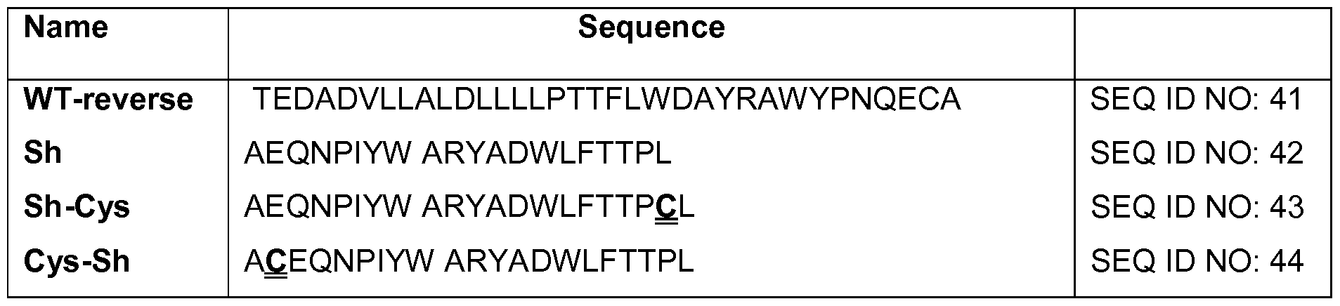
Table 2 includes sequences termed short and medium pHLIP sequences. They all insert in membrane in a pH-dependent manner, while they do not have C-terminal flanking sequence. Double underline indicates residues (Cys or Lys), which are used to conjugate pHLIPs with cargo molecules. pHLIP sequences contain L-amino acids; alternatively, the pHLIP comprises D-amino acids.
Table 2
Sh-1Trp AEQNPIYFARYADWLFTTPL SEQ ID NO: 45
Sh-1D KEDQNPWARYADLLFPTTLAW SEQ ID NO: 46
Cys-Sh-1D AQEDQNPWARYADLLFPTTLAW SEQ ID NO: 47
Cys-Med-2D ACEDQNPWARYADWLFPTTLLLLD SEQ ID NO: 48
Cys-Sh-1E ACEEQNPWARYAELLFPTTLAW SEQ ID NO: 49
Cys-Med-2E ACEEQNPWARYAEWLFPTTLLLLE SEQ ID NO: 50
Cys-Med-3E ACEEQNPWARYLEWLFPTETLLLEL SEQ ID NO: 51
DNA-encoded and non-coded amino acids are described below in Table 3. Additional non- natural amino acids that can be used are known in the art, e.g. , as described in Hendrickson et al. , 2004, Ann. Rev. Biochem. 73: 147-176; hereby incorporated by reference.
Table 3
Coded and Non-Coded Amino Acids
Thr threonine
Trp tryptophan
Tyr tyrosine
Val valine
Acpa Aminocaprylic acid
Aecys (S)-2-aminoethyl-L-cysteine»HCI
Afa Aminophenyl acetate
Aiba -aminoisobytyric acid
Aile alloisoleucine
Alg L-allylglycine
Aba amlnobutyric acid
Aphe p - aminophenylalanine
Bat -alanine
Brphe p-bromophenylalanine
Cha cyclohexylalanine
Cit citrulline
Clala -chloroalanine
Cie cycioleucine
Clphe p- chiorophenylalanine
Cya cysteic acid
Dab 2,4-diamino-butyric acid
Dap 2,3- diaminopropionic acid
Dhp 3 ,4-dehydro-pro line
Dhphe 3,4-, dihydroxy-phenyl-alanine
Fphe p- fluorophenylalanine
Gaa D-glucose-aminic acid
Hag Homo-arginine
Hlys hydroxyl-lysine'HCI
Hnvl DL-hydroxynorvaline
Hog Homoglutamine
Hoph homophenylalanlne
Has homoserine
Hpr hydroxyl-pro line
Iphe p-lodophenylalanine
Ise isoserine
Mle -methyl-leucine
Msmet DL- methionine-s-methylsulfo-niumchloride
INala 3-(l- naphthyl)alanine
2Nala 3 -(2- naphthyl)alanine
Nle norleucine (or 2-aminohexanoic acid)
Nmala N-methyl-alanine
Nva norvaline (or 2-aminopentanoic acid)
Obser 0- benzylserine
Obtyr 0-benzyl-tyrosine
Oetyr O-ethyltyrosine
Omser 0-methylserine
Omthr 0-methyt-hreonine
Omtyr 0-methyl- tyrosine
Orn ornithine
Pen penicillamine
Pga pyroglutamic acid
Pip pipecolic acid
Sar sarcosine
Tfa 3,3,3- trifluoro alanine
Thphe 6- hydroxydopa
72 Vig L-vinylglycine
73 Aaspa (-)-(2R)-2- amino-3-(2- aminoethylsulfonyl)propanoic acid dihydrochloride
74 Ahdna (2S)-2- amino-9-hydroxy-4,7-dioxanonanolc acid
75 Ahoha (2S)-2- amino-6-hydroxy-4-oxahexanoic acid
76 Ahsopa (-)-(2R)-2- amino-3-(2-hydroxyethyls ulfonyl)propanoic acid
A "conservative amino acid substitution" is one in which the amino acid residue is replaced with an amino acid residue having a similar side chain. Families of amino acid residues having similar side chains have been defined in the art. These families include amino acids with basic side chains (e.g., lysine, arginine, histidine), acidic side chains (e.g., aspartic acid, glutamic acid), uncharged polar side chains (e.g., glycine, asparagine, glutamine, serine, threonine, tyrosine, cysteine), nonpolar side chains (e.g., alanine, valine, leucine, isoleucine, proline, phenylalanine, methionine, tryptophan), beta-branched side chains (e.g., threonine, valine, isoleucine) and aromatic side chains (e.g., tyrosine, phenylalanine, tryptophan, histidine). Thus, a residue in a pHLIP sequence (corresponding to a location relative to SEQ ID NO: 3) is replaced with another amino acid residue from the same side chain family.
pHLIP peptide is monomeric
pHLIP peptides, e.g., (SEQ ID NO: 4) are a water-soluble polypeptides based on the the bacteriorhodopsin C helix, which was found to insert across a membrane to form a stable transmembrane alpha helix. Peptide folding and membrane insertion are driven by a drop of pH from neutral or high (>7.4) to slightly acidic (7.0-6.5 and less) pHs. The apparent pK of insertion was found to be 6.0. pHLIP is a monomer in each of its three major states: unstructured and soluble in water (state I) at neutral pH, unstructured and bound to the surface of a membrane at neutral pH (state II), and inserted across the membrane as an a-helix at low pH (state III). In contrast, all pore forming peptides first form aggregates on the membrane surface and then "fall" into membrane and form pores. Thus, an additional advantage of the environmentally-sensitive compositions is their monomeric nature, e.g. , they do not require assembly into a multimeric suprastructure like pore formers.
Delivery of cargo using pHLIP-liposomes
Although gold nanospheres and nanorods have been used for the enhancement of radiation therapy and for thermal ablation of tumors, delivery of enough gold material to cancer cells to produce the desirable effect has been a challenge. Gold nanoparticles attached to the N-terminus of pHLIP have been successfully delivered to tumors and accumulate on the surface of membrane of cancer cells. Another even more efficient way to deliver gold material (or other compounds) to tumor (or other cells characterized by low pH) is to use pHLIP-liposomes. pHLIP-liposomes are useful deliver to cells in a pH-dependent manner any compound, e.g., polar or hydrophobic compounds, tht have been difficult to get into cells using other methods. In contrast to fusiogenic liposomes developed before for delivery, which can fuse with cellular membrane only in the absence of PEG coating, pHLIP can mediate fusion between lipid bilayer of plasma membrane or membrane of endosome/lysozome and liposomes made of non-fusogenic lipids and containing 10 mol% of PEG. pHLIP conjugated to the pegylated liposomes promotes pH-modulated: i) endocytotic uptake of liposomes by targeted cell, distortion of endosome compartment and release of lipids or liposome content into cytoplasm; and ii) direct liposomal fusion with plasma membrane and release of liposomal content into cytoplasm. pHLIP promotes mitochondrial delivery of R18 incorporated into liposome. As described in detail below, various assays were performed on liposomes in solution and on live cells to demonstrate that pHLIP mediates uptake of liposomes. The in vivo data shows high uptake of pHLIP-labeled liposomes by cancer cells.
In some embodiments, the present invention relates to the use of pHLIP technology for selective delivery of gold nanoparticles and liposomes to cancer cells in vivo. The data described herein demonstrate that gold nanoparticles attached to the N-terminus of pHLIP are delivered to tumors and accumulate on the surface of membrane of cancer cells. Distribution of gold nanoparticles in tumor was investigated by light microscopy after silver enhancement.
Toxicity
Toxicity is one of the most critical issues in the selection of any delivery agent. For example, the use of pore-forming membrane peptides as delivery agents is complicated by the toxicity associated with the formation of pores in cellular membranes in vivo. By contrast, the interaction of pHLIP with liposomes and cellular membranes at both neutral and low pHs does not lead to membrane leakage, and no cellular toxicity was seen over a range of peptide concentrations.
Selectivity of targeting
The pH-dependent interaction of pHLIP with membranes allows selectivity in the targeting of acidic (less than pH 7.0) diseased tissue. As noted above, acidity and hypoxia are considered as universal cancer biomarkers, and pHLIP is used as an acidity-targeting probe. Besides cancer, many other pathological states, such as inflammation, ischemia, stroke, arthritis and others are characterized by acidity in the extracellular space, which may broaden the potential applications of pHLIP. In vivo fluorescence imaging in mice and rats demonstrated that pHLIP can target acidic tissues, such as kidneys, tumors of various sizes and origins, and anatomical sites of inflammation, e.g., arthritis, infection, atherosclerotic plaques. In addition to fluorescence imaging, PET (positron emission tomography) imaging of the acidic environment in human prostate tumors was performed using 64Cu-DOTA conjugated to pHLIP. PET studies demonstrated that the construct avidly accumulated in LNCaP and PC-3 tumors and that tumor uptake correlates with the differences in the bulk extracellular pH (pHe) measured by MR spectroscopy. To manipulate the acidity of tissues, a buffer solution is administered to the subject systemically or local to the area in which a pH change is desired. In this manner, pHLIP-liposome-mediated delivery of a cargo compound is regulated, e.g., reduced or stopped. For example, administering bicarbonated water, which increases tissue pH, results in a reduction of tumor targeting by pHLIP.
Molecular mechanism of pH-dependent membrane insertion of pHLIP
The transmembrane (TM) part of exemplary pHLIP peptides contain two Asp residues. At neutral pH these charged residues enhance peptide solubility and serve as anchors keeping the peptide at the surface of membrane, thereby preventing pHLIP partitioning into the hydrophobic membrane bilayer. A reduction of pH induces protonation of Asp residues, and as a result, the overall hydrophobicity of the peptide increases, enhancing the affinity of the peptide for the lipid bilayer core and triggering peptide folding and insertion. The replacement of the key Asp residues by Lys, Ala or Asn leads to the loss of peptide of pH-dependent membrane insertion, as measured in liposomes, red blood cells and confirmed by in vivo fluorescence imaging. The K-pHLIP peptide, where the two Asp residues in the transmembrane region are replaced with Lys residues, does not demonstrate tumor targeting. The Ala substitutions yield a peptide that aggregates in solution (but de-aggregates when it becomes diluted in bodily fluids or tissue upon administration to a subject), while the Lys and Asn substitutions give peptides that are too polar to insert either at neutral or low pH. The replacement of one of the Asp residues in the TM part of the peptide by a Glu residue
results in a shift of pH of membrane insertion from 6.0 to 6.5. Replacement of both Asp residues by Glu results in enhancement of peptide aggregation and formation of elements of secondary structure on the bilayer surface at neutral pH (see Tables 1 and 2).
Data obtained using liposomes, cultured cells and mice confirmed that the mechanism of membrane entry of pHLIP is not mediated by endocytosis, interactions with cell receptors or pore formation; rather, the mechanism is the formation of a helix across the lipid bilayer, triggered by the increase of peptide hydrophobicity due to the protonation of negatively charged residues induced by low pH.
Solubility and stability of pHLIP in blood
Poor solubility due to aggregation is a typical property of membrane peptides, which has complicated studies and applications. Isolated or purified pHLIP, as any membrane peptide, also has a tendency to aggregate, especially at high concentrations and/or low pH. However, in aqueous solution at neutral pH pHLIP exists as a monomer at concentrations less than 30 μg/mL (~7.0 μΜ), as studied by fluorescence and CD spectroscopy measurements, size exclusion chromatography coupled with "on-line" laser light scattering, ultraviolet and refractive index detection (SEC-
LS/UV/RI) and analytical ultracentrifugation experiments. When the solubility of the peptide is compromised as a result of mutations, the affinity of the peptide for a membrane and its overall conformational properties change. Thus, studies were undertaken to design pHLIP peptides that are optimized for clinical diagnostic and therapeutic use.
The oligomeric state of the peptide on the surface of a membrane (state II) and inserted into the lipid bilayer (state III) were evaluated by FRET performed with two different donor-acceptor probes attached to the N-terminus of the peptide. The data demonstrate that, at low concentrations, the peptide is monomeric in both states II and III (Figure 28).
Peptide interactions with proteins, especially plasma proteins, and membranes determine the pharmacokinetics of the peptide at neutral pH. pHLIP demonstrates prolonged circulation in the blood (several hours), which is consistent with its ability to bind weakly to membrane surfaces at neutral and high pH, preventing the rapid clearance by the kidney expected for a small, soluble peptide. pHLIP binding to membranes is driven by hydrophobic interactions. If the peptide sequence were made more hydrophobic, tighter binding to red blood cells and epithelial cells and more aggregation in solution, and slower clearance and reduced bioavailability would occur.
Making the peptide less hydrophobic accelerates clearance and prevents the peptide from finding its targets. Therefore, fine tuning of the solubility is an important property to optimize pHLIP performance in vivo.
Another important property is the stability of peptides in the blood, since proteases in the serum can degrade peptides consisting of L-amino acids within minutes. While polypeptides made from D-amino acids are much more stable, they are often unsuitable for specific receptor binding applications as a consequence of their altered chirality. Since the mechanism of pHLIP involves relatively nonspecific interactions with a fluid lipid bilayer, pHLIP peptides composed of L- or D- amino acids demonstrate the same biophysical and tumor targeting properties. This observation confirms the evidence that the pHLIP targeting does not require any specific molecular binding event. The only conspicuous difference is that D-pHLIPs form left-handed helices across membranes rather than the right-handed helices formed by L-pHLIPs.
EXAMPLES
Example 1 : pHLIP-Mediated Delivery of Liposomes
The following regents and methods were used to generate the data described herein.
Lipids:
DOPE
(l,2-dioleoyl-s«-glycero-3-phosphoethanolamine )
DOPC
(l,2-dioleoyl-sn-glycero-3-phosphocholine )
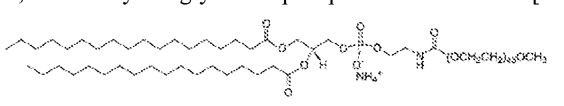
DSPE-PEG(2000) Maleimide
l,2-distearoyl-sn-glycero-3-phosphoethanolamine-N-[maleimide(polyethylene glycol)-2000] (ammonium salt)
DSPE-PEG(2000)
l,2-distearoyl-sn-glycero-3-phosphoethanolamine-N-[methoxy(polyethylene glycol)-2000]
Fluorescein DHPE
N-(fluorescein-5-thiocarbamoyl)- 1 ,2-dihexadecanoyl-sn-glycero-3-phosphoethanolamine, triethylammonium salt
Rhod PE
l,2-dioleoyl-sn-glycero-3-phosphoethanolamine-N-(lissamine rhodamine B sulfonyl
R18 (octadecyl rhodamine B chloride)
Conjugation of pHLIP with lipids
pHLIP containing Cys on the N-terminus and DSPE-PEG(2000) Maleimide lipids. Standard maleimide chemistry reaction was applied in methanol. (Reaction of thiol with maleimide.)
' V--'-1 ' ' ' ' ' -'-1--,,.,,.-.;:
0 , where R1- DSPE-PEG(2000) Maleimide (MW 2941)
. "- cys-pHLIP (MW 4150)
R1 : \ = 1 : 1.5 in DMF or 1 : 1.2 in Methanol.
The reaction product was verified by SELDI-TOF masspec. Expected mass of about 7kDa was observed.
Liposome compositions used in experiments with cells
DOPC: DOPC 85 mol%
DSPE-PEG 5-10 mo 1%
Fluorescent-lipids 5 mol%
DOPC-pHLIP: DOPC 85 mol%
DSPE-PEG 0-5 mol%
Fluorescent-lipid 5 mol%
DSPE-PEG-pHLIP 0-5 mol%
DOPE: DOPE 85 mol%
DSPE-PEG 5-10 mol%
Fluorescent-lipid 5 mol%
DOPE-pHLIP: DOPE 85 mol%
DSPE-PEG 0-5 mol%
Fluorescent-lipid 5 mol%
DSPE-PEG-pHLIP 0-5 mol%
Liposome preparation
Liposomes were prepared by the thin film method (extrusion). A chloroform solution of the desired lipids (1 μταοΐ) was evaporated using rotary evaporator, producing an even, thin film. The film was placed under a vacuum overnight to remove trace solvent impurities. This film was then hydrated in 1 mL 10 mM phosphate, 150mM NaCl stock buffer solution via 10 freeze-thaw-vortex cycles. The resulting multilamellar liposome solution was then extruded 15 times through 100 nm polycarbonate filters and sterilized by filtering through 0.2 μηι filter.
Cryo-electron microscopy
Cryo-electron microscopy (cryo-EM) is a form of transmission electron microscopy (TEM) where the sample is studied at cryogenic temperatures (generally liquid nitrogen temperatures). It allows the observation of specimens that have not been stained or fixed in any way, showing them in their native environment.
FEI Vitrobot™ Mark Γν is a fully automated vitrification robot for plunge-freezing of aqueous (colloidal) samples. For sample preparation, vitrification in liquid ethane was performed via Vitribot apparatus, with a single blot of 3 sec, an offset of - 1 , and drain and wait time of 1 sec.
For imaging, sample was kept at -175°C during imaging in a JEOL 2100 TEM with an accelerating voltage of 200kV. Images were taken at 20,000x and 40,000x.
Cryo-TEM images of liposomes (DOPE, DOPE-pHLIP, DOPC, and DOPC-pHLIP) are shown in Figure 19.
Inter- Liposome Fusion Assay
pHLIP-mediated inter-liposome fusion was studied by an Octadecyl rhodamine B (R18) self-quenching assay. Liposomes labeled with R18 were mixed with various concentrations of unlabeled liposomes (POPC). During the process of dropping pH of solution, rhodamine fluorescence was monitored on the spectrofluoro meter (Figure 2).
The spectrofluoro meter instrument was utilized. Slow kinetics - emission intensity measurement (excitation/emission: 556nm/590nm).
Steps: sample liposome m |xi ng un |a be| ed | iposome ^
labeled with Kle ► ( POPC, various * fusion observation
(50uL, 200uM) concentration)
The rhodamine fluorescence of pHLIP-liposomes (DOPE-pHLIP or DOPC-pHLIP) increased significantly after dropping pH from 8 to 4. 200 μΜ of R18-labeld liposome was mixed with 6mM of unlabeled POPC (Figure 2A1-2A2). 200 μΜ of R18-labeld liposome was mixed with elevated concentration of unlabeled POPC (0-6mM) at pH 4 (Figure 2B). The amount of PEG- conjugated lipid containing in liposome affects the percentage of fusion (Figure 2C). For Figure 2C, 200 μΜ of R18-labeled liposome was mixed with 6 mM of unlabeled POPC. The percentage calculations are as follows: Percentage of fusion = ! where FLo is initial fluorescent intensity of liposome mixture at pH 8, FLPH is fluorescent intensity of liposome mixture at pH 4, FLMAX is fluorescent intensity of liposome mixture at pH 4 after freeze-thaw cycle.
The scheme of the fusion assay and liposome composition used in study are provided in Figure 2D and 2E.
Cell suspension
Trypsinized cells were counted using a hemacytometer and diluted to 2xl05 cells/ml in serum-free low pH media. 20 nmol liposomes were incubated with lxlO5 cells in 500 μΐ^ serum- free low pH media for 15 min or lhr at 4°C or 37°C. The cells were then pelleted by centrifugation
(2000 rpm, 4min) at 4°C or 37°C. The cells were resuspended in 500μί fresh serum-free low pH media and centrifuged a second time. This second pellet was resuspended in ΙΟΟμί same media. The cells were counted using cellometer: The sample was mixed well, and 2\iL of trypan blue was added to 18μί of sample. 20 \iL of this solution was loaded into disposable counting chamber (slide). The chamber was inserted into cellometer, and software was used to count cells. The cells were reseeded in collagen-coated cell dish for microscopy imaging.
Example 2: pHLIP Enhances Uptake of Liposomes by Cells
pHLIP (pH-Low-Insertion-Peptide) insertion into membrane occurs as a result of protonation of Asp/Glu residues due to a decrease of pH. Protonation enhances peptide
hydrophobicity and increases its affinity for a lipid bilayer, which triggers peptide insertion and formation of transmembrane helix. Since many pathological states are associated with the development of elevated level of extracellular acidity (or low extracellular pH), pHLIP-liposomes are ideally suited for selective delivery of diagnostic and therapeutic agents to the cancer cells. Attachment of cargo molecules to the N-terminus (Figure 14).
One approach to deliver gold material (or cytotoxic compounds) to tumor is to use liposomes. In contrast to fusiogenic liposomes developed before for delivery, which can fuse with cellular membrane only in the absence of PEG coating, pHLIP mediates fusion between lipid bilayer of plasma membrane or membrane of endosome/lysozome and liposomes made of non- fusogenic lipids and containing 10 mol% of PEG. pHLIP conjugated to the pegylated liposomes promotes pH-modulated: i) endocytotic uptake of liposomes by targeted cell, distortion of endosome compartment and release of lipids or liposome content into cytoplasm; and ii) direct liposomal fusion with plasma membrane and release of liposomal content into cytoplasm. pHLIP promotes mitochondrial delivery of R18, incorporated into liposome. pHLIP was found to mediate uptake of liposomes. The in vivo data demonstrate high uptake of pHLIP-labeled liposomes by cancer cells.
Cell uptake studies were performed as follows: cells in suspension were treated with 40uM of fluorescently labeled pHLIP-liposomes (DOPE-pHLIP or DOPC-pHLIP) and control-liposomes (DOPE or DOPC) under different conditions, after washing, fluorescence of cells was counted by cellometer. (a) Liposomes containing different fluorescent lipid (Rho-FA(R18), Rhodamine-PE, Fluorescein-DHPE) were tested (Figure 3). pHLIP-liposmes show high cell uptake, (b) Cells were incubated with R18-labled liposomes in different pH of medium (Figure 4). Liposome containing different amount of pHLIP- conjugated lipids (c; Figure 5) and PEG-lipids (d; Figure 6) were also
investigated, (e) Trypan blue quenching assay: After treatment of liposomes containing Fluorescein-lipid, the FITC-fluorescence of cells was counted before and after adding trypan blue (Figure 7). Cell-impermeable trypan blue can quench FITC fluorescence only if FITC dye is located on the outer leaflet of cellular membrane facing to the extracellular space, which might occur only in a result of liposome-cell membrane fusion, (f) Endocytosis assay: cells in suspension were incubated with liposomes for 15 or 60 min at 4°C or 37°C in different media (PBS, DMEM, ATP-depletion medium). Low temperature and ATP-depletion medium are used to reduce endocytotic uptake. The results are presented in Figure 8. Thus, the data indicated that pHLIP enhanced uptake of liposomes by cells, and the primary pathway of liposome uptake was endocytosis.
A549 cell suspension (10 x 105) was treated with R18 containing liposome (20 nmol) in 500 μL· of serum-free low pH media for 1 hour at 37C. The cells were pelleted by centrifugation (2000rpm, 4min) and resuspended in fresh DMEM. The cells were reseeded in collagen-coated cell dishes (Figure 20). The light images (a, c) and fluorescent images (b, d) were taken after 4 days incubation. pHLIP-containing liposome (d) showed much higher cell uptake than the control liposome (b), which did not contain pHLIP.
Example 3: pHLIP Promotes Distortion of Plasma and Endosome Membranes, the Release of R18- Labeled FA into the Cytoplasm, and Targeting of Mitochondria
After cellometer counting, cells were reseeded in collagen-coated cell dishes for microscopy imaging. Cellular localization of fluorescent fatty acids (Rl 8) incorporated into liposomes containing PEG polymers and pHLIP or no pHLIP on the surface (non-fusogenic DOPC lipids were used in study). In case of liposomes, fluorescent signal was mostly localized in endosomes, while pHLIP promotes distortion of plasma and endosome membranes and release of R18-labeld FA into cytoplasm and targeting of mitochondria. Figure 9 shows the localization of Rho-labeled liposome in cells.
Figure 1 1 shows images of Fluorescein-labeled liposomes fused with a cellular membrane. Figure 1 1(a) phase contrast; (b) FITC. Co-localization of FITC-liposome (c) and plasma membrane staining of red- fluorescent Alexa Fluor594 wheat germ agglutinin (d). The data demonstrate that lipids are exchanged as a result of fusion with the plasma membrane or membrane of the endosomal compartment, thereby reaching the plasma membrane. Thus, the methods described herein promote
the delivery and release of agents that are encapsulated inside a pHLIP-liposome or attached to lipids of the pHLIP-liposome to the cytoplasm of a cell.
Figure 12 shows the results of a liposome encapsulation experiment (delivery of propidium iodide to the nucleus). The propidium iodide (PI; 4 mM) was encapsulated in Fluorescein-labeled liposomes (Figure 12). 10 nmol of liposome were incubated with cells attached to the collagen- coated cell dish in 100 of low pH media for lhr at 37°C. The release of PI from pHLIP- liposomes was observed.
Figure 16 shows two types of liposomes (100 nm in diameter), one of which was carrying 5 mol% of pHLIP peptides and the other was not. Both liposomes contained 5 mol% of nanogold- lipids and 10 mol% PEGylated lipids. Cells were treated with two types of liposomes separately, washed, fixed. After fixation, cells were treated with silver enhancement solution and analyzed under the light microscope. Nanogold-lipids were mostly localized on the plasma and nuclear membranes of cells treated with pHLIP-liposomes (Figure 16).
A549 cell suspension (10 x 105) was treated with R18 containing liposome (20 nmol) in 500uL of PBS (pH6.2) for 15 min at 37C. The cells were pelleted by centrifugation (2000rpm, 4min) and resuspended in fresh DMEM. Then the cells were reseeded in collagen-coated cell dishes. After 4 days incubation, endoplasmic reticulum (ER) and mitochondria were labeled by fluorescent dyes of ER-Tracker and Mito -Tracker, respectively. The fluorescent images were taken with the filter setting of GFP, Cy5 and TRITC, corresponding to ER labeling, mitochondria staining and R18-liposome uptake (Figure 21).
Example 4: pHLIP Promotes Uptake of Liposome in Low pH Extracellular Environment of Tumors
Liposomes, containing Rho-PE lipids, were given as a single intra-tumoral injection into mice with tumors established by subcutaneous injection of HeLa-GFP cancer cells. Mice were sacrificed at 24 hours post-injection, and tumors were collected. Whole-body and tumor images were taken on Kodak in vivo imaging system. As shown in Figure 13, pHLIP promoted liposome uptake in low pH extracellular environment of tumors, following IV injection of the fluorescent- and gold-containing liposomes.
HeLa-GFP cells were incubated with pHLIP-nanogold and nanogold particles at neutral and low pHs, washed, fixed and enhanced by silver then visualized under light microscope. The highest uptake was observed at low pH in presence of pHLIP (Figure 17A). Tumor sections collected from mice received single iv injection of pHLIP-nanogold and nanogold particles were treated with silver
enhancement solution and visualized under the microscope. Nanogold particles delivered to tumor by pHLIP were localized on cancer cells identified by GFP fluorescence (Figure 17B).
These data indicate that pHLIP-liposomes demonstrate enhanced uptake by cells in environments characterized by low pH (pH <7)compared to liposomes that do not contain pHLIP. Example 5: pHLIP-Mediated Delivery of Lipsomal Ceramide to Cancer Cells
An exemplary ceramide formulation is provided below. Cryo-TEM images of ceramide- containing liposomes are shown in Figure 22. The size and shape (round) of the particles indicate that ceramide liposomes were formed.
Control-liposome pHUP-liposome
DOPC 37 mol% 37 mol%
DOPE 17.5 mol% 17.5 mol%
DSPE-P EG2000
C8-P EG750 7.5 mol% 7.5 mol%
C6-ceramide 30 mol% 30 mol%
R18 0.5 mol% 0.5 mol%
Liposome size measurement
The size of liposome was measured by using Dynamic Light Scattering (Zetasizer Nano ZS). The diameters of control-liposome and pHLIP-liposome are 104 nm and 125 nm, respectively (Figure 23 A). After 3 days monitoring, the size of control-liposome is stable, while the size of pHLIP-liposome increased slightly (Figure 23B).
Inter-liposome fusion assay of ceramide liposome
The fusion assay was performed as described in Example 1 and Figure 2D.
ISS Settings:
dropping pH
t i A P H SS?O mixing unlabeled liposome from 7.4 to 4 fusion abe ed w,th R 1 8 (50u L POPC. various observation
(50u L. 1 .6mM) concentration)
The results are presented in Figure 24. Figure 24A shows that there is no fusion of liposomes if pHLIP is not attached to the surface (no increase of fluorescence at low pH).
Figure 24B shows that there is an increase of fluorescence when pHLIP-coated liposomes are mixed with POPC liposomes, which indicates fusion of liposomes. Thus, pHLIP promotes fusion only at low pH. Figure 24C is a summary of Figure 24A and 24B. The black line shows no increase of fluorescence for control experiment, and the red line shows that fluorescence increases in case of pHLIP-coated liposomes. The methods for this experiment were described in relation to Figure 2 above; however, the amount of Rho-FA is much less (0.5 mol%) for Figure 24 than Figure 2
(5mol%). Therefore, the increase of fluorescence is much less in Figure 24 comparison to Figure 2.
A schematic of pHLIP-mediated Delivery of Liposomal Ceramide to Cells is provided in Figure 25.
A schematic of pHLIP-mediated delivery of liposomal ceramide to cell suspension is provided in Figure 26. Figure 27A and 27B are a series of bar charts demonstrating the results of pHLIP-mediated delivery of liposomal ceramide to cell suspension. Delivery of ceramide (C6) using pHLIP-liposomes led to a significantly greater amount of cell death at low pH compared to the level of cell death at high pH. Moreover, ceramide (C6) pHLIP-liposomes led to a significantly greater amount of cell death at low pH compared to ceramide liposomes alone (in the absence of pHLIP) at low pH.
OTHER EMBODIMENTS
While the invention has been described in conjunction with the detailed description thereof, the foregoing description is intended to illustrate and not limit the scope of the invention, which is defined by the scope of the appended claims. Other aspects, advantages, and modifications are within the scope of the following claims.
The patent and scientific literature referred to herein establishes the knowledge that is available to those with skill in the art. All United States patents and published or unpublished
United States patent applications cited herein are incorporated by reference. All published foreign patents and patent applications cited herein are hereby incorporated by reference. Genbank and NCBI submissions indicated by accession number cited herein are hereby incorporated by reference. All other published references, documents, manuscripts and scientific literature cited herein are hereby incorporated by reference.
While this invention has been particularly shown and described with references to preferred embodiments thereof, it will be understood by those skilled in the art that various changes in form and details may be made therein without departing from the scope of the invention encompassed by the appended claims.