WO2012014996A1 - Reagent and reagent kit for measurement of fdp, and measurement method - Google Patents
Reagent and reagent kit for measurement of fdp, and measurement method Download PDFInfo
- Publication number
- WO2012014996A1 WO2012014996A1 PCT/JP2011/067326 JP2011067326W WO2012014996A1 WO 2012014996 A1 WO2012014996 A1 WO 2012014996A1 JP 2011067326 W JP2011067326 W JP 2011067326W WO 2012014996 A1 WO2012014996 A1 WO 2012014996A1
- Authority
- WO
- WIPO (PCT)
- Prior art keywords
- fraction
- fdp
- reagent
- antibody
- monoclonal antibody
- Prior art date
Links
Images


Classifications
-
- G—PHYSICS
- G01—MEASURING; TESTING
- G01N—INVESTIGATING OR ANALYSING MATERIALS BY DETERMINING THEIR CHEMICAL OR PHYSICAL PROPERTIES
- G01N33/00—Investigating or analysing materials by specific methods not covered by groups G01N1/00 - G01N31/00
- G01N33/48—Biological material, e.g. blood, urine; Haemocytometers
- G01N33/50—Chemical analysis of biological material, e.g. blood, urine; Testing involving biospecific ligand binding methods; Immunological testing
- G01N33/58—Chemical analysis of biological material, e.g. blood, urine; Testing involving biospecific ligand binding methods; Immunological testing involving labelled substances
- G01N33/585—Chemical analysis of biological material, e.g. blood, urine; Testing involving biospecific ligand binding methods; Immunological testing involving labelled substances with a particulate label, e.g. coloured latex
- G01N33/586—Liposomes, microcapsules or cells
-
- C—CHEMISTRY; METALLURGY
- C07—ORGANIC CHEMISTRY
- C07K—PEPTIDES
- C07K16/00—Immunoglobulins [IGs], e.g. monoclonal or polyclonal antibodies
- C07K16/18—Immunoglobulins [IGs], e.g. monoclonal or polyclonal antibodies against material from animals or humans
- C07K16/36—Immunoglobulins [IGs], e.g. monoclonal or polyclonal antibodies against material from animals or humans against blood coagulation factors
-
- C—CHEMISTRY; METALLURGY
- C07—ORGANIC CHEMISTRY
- C07K—PEPTIDES
- C07K14/00—Peptides having more than 20 amino acids; Gastrins; Somatostatins; Melanotropins; Derivatives thereof
- C07K14/435—Peptides having more than 20 amino acids; Gastrins; Somatostatins; Melanotropins; Derivatives thereof from animals; from humans
- C07K14/745—Blood coagulation or fibrinolysis factors
- C07K14/75—Fibrinogen
-
- C—CHEMISTRY; METALLURGY
- C07—ORGANIC CHEMISTRY
- C07K—PEPTIDES
- C07K16/00—Immunoglobulins [IGs], e.g. monoclonal or polyclonal antibodies
- C07K16/18—Immunoglobulins [IGs], e.g. monoclonal or polyclonal antibodies against material from animals or humans
- C07K16/28—Immunoglobulins [IGs], e.g. monoclonal or polyclonal antibodies against material from animals or humans against receptors, cell surface antigens or cell surface determinants
- C07K16/2803—Immunoglobulins [IGs], e.g. monoclonal or polyclonal antibodies against material from animals or humans against receptors, cell surface antigens or cell surface determinants against the immunoglobulin superfamily
-
- G—PHYSICS
- G01—MEASURING; TESTING
- G01N—INVESTIGATING OR ANALYSING MATERIALS BY DETERMINING THEIR CHEMICAL OR PHYSICAL PROPERTIES
- G01N33/00—Investigating or analysing materials by specific methods not covered by groups G01N1/00 - G01N31/00
- G01N33/48—Biological material, e.g. blood, urine; Haemocytometers
- G01N33/50—Chemical analysis of biological material, e.g. blood, urine; Testing involving biospecific ligand binding methods; Immunological testing
- G01N33/53—Immunoassay; Biospecific binding assay; Materials therefor
- G01N33/577—Immunoassay; Biospecific binding assay; Materials therefor involving monoclonal antibodies binding reaction mechanisms characterised by the use of monoclonal antibodies; monoclonal antibodies per se are classified with their corresponding antigens
-
- G—PHYSICS
- G01—MEASURING; TESTING
- G01N—INVESTIGATING OR ANALYSING MATERIALS BY DETERMINING THEIR CHEMICAL OR PHYSICAL PROPERTIES
- G01N33/00—Investigating or analysing materials by specific methods not covered by groups G01N1/00 - G01N31/00
- G01N33/48—Biological material, e.g. blood, urine; Haemocytometers
- G01N33/50—Chemical analysis of biological material, e.g. blood, urine; Testing involving biospecific ligand binding methods; Immunological testing
- G01N33/68—Chemical analysis of biological material, e.g. blood, urine; Testing involving biospecific ligand binding methods; Immunological testing involving proteins, peptides or amino acids
- G01N33/6893—Chemical analysis of biological material, e.g. blood, urine; Testing involving biospecific ligand binding methods; Immunological testing involving proteins, peptides or amino acids related to diseases not provided for elsewhere
-
- G—PHYSICS
- G01—MEASURING; TESTING
- G01N—INVESTIGATING OR ANALYSING MATERIALS BY DETERMINING THEIR CHEMICAL OR PHYSICAL PROPERTIES
- G01N33/00—Investigating or analysing materials by specific methods not covered by groups G01N1/00 - G01N31/00
- G01N33/48—Biological material, e.g. blood, urine; Haemocytometers
- G01N33/50—Chemical analysis of biological material, e.g. blood, urine; Testing involving biospecific ligand binding methods; Immunological testing
- G01N33/86—Chemical analysis of biological material, e.g. blood, urine; Testing involving biospecific ligand binding methods; Immunological testing involving blood coagulating time or factors, or their receptors
-
- G—PHYSICS
- G01—MEASURING; TESTING
- G01N—INVESTIGATING OR ANALYSING MATERIALS BY DETERMINING THEIR CHEMICAL OR PHYSICAL PROPERTIES
- G01N2333/00—Assays involving biological materials from specific organisms or of a specific nature
- G01N2333/435—Assays involving biological materials from specific organisms or of a specific nature from animals; from humans
- G01N2333/745—Assays involving non-enzymic blood coagulation factors
- G01N2333/75—Fibrin; Fibrinogen
-
- G—PHYSICS
- G01—MEASURING; TESTING
- G01N—INVESTIGATING OR ANALYSING MATERIALS BY DETERMINING THEIR CHEMICAL OR PHYSICAL PROPERTIES
- G01N2800/00—Detection or diagnosis of diseases
- G01N2800/10—Musculoskeletal or connective tissue disorders
- G01N2800/101—Diffuse connective tissue disease, e.g. Sjögren, Wegener's granulomatosis
- G01N2800/102—Arthritis; Rheumatoid arthritis, i.e. inflammation of peripheral joints
-
- G—PHYSICS
- G01—MEASURING; TESTING
- G01N—INVESTIGATING OR ANALYSING MATERIALS BY DETERMINING THEIR CHEMICAL OR PHYSICAL PROPERTIES
- G01N2800/00—Detection or diagnosis of diseases
- G01N2800/22—Haematology
- G01N2800/224—Haemostasis or coagulation
-
- G—PHYSICS
- G01—MEASURING; TESTING
- G01N—INVESTIGATING OR ANALYSING MATERIALS BY DETERMINING THEIR CHEMICAL OR PHYSICAL PROPERTIES
- G01N2800/00—Detection or diagnosis of diseases
- G01N2800/22—Haematology
- G01N2800/226—Thrombotic disorders, i.e. thrombo-embolism irrespective of location/organ involved, e.g. renal vein thrombosis, venous thrombosis
-
- G—PHYSICS
- G01—MEASURING; TESTING
- G01N—INVESTIGATING OR ANALYSING MATERIALS BY DETERMINING THEIR CHEMICAL OR PHYSICAL PROPERTIES
- G01N2800/00—Detection or diagnosis of diseases
- G01N2800/32—Cardiovascular disorders
- G01N2800/324—Coronary artery diseases, e.g. angina pectoris, myocardial infarction
Definitions
- the present invention relates to a reagent and reagent kit for measuring FDP, which is a degradation product of fibrin and fibrinogen, and a method for measuring FDP.
- a fibrinolytic reaction takes place in the body to remove it.
- the fibrinolytic reaction that dissolves the thrombus is called secondary fibrinolysis, and fibrin in the thrombus is decomposed by the action of an enzyme such as plasmin to produce a fibrin degradation product (FbnDP; secondary fibrinolysis) in the blood.
- FbnDP fibrin degradation product
- a fibrinolytic reaction called primary fibrinolysis that occurs without thrombus formation also occurs.
- fibrinogen is decomposed by the action of an enzyme such as plasmin to produce a fibrinogen degradation product (FbgDP; primary fibrinolysis) in the blood.
- FbgDP fibrinogen degradation product
- FDP fibrinogen and fibrin degradation products
- FDP fibrinolysis
- cardiovascular disorders cardiovascular disorders
- hyperfibrinolysis abnormal bleeding
- DIC disseminated intravascular coagulation syndrome
- it is required to measure the total amount of FDP regardless of the type of FDP.
- reagents using immunoturbidimetry are commercially available.
- an FDP measurement reagent there is a reagent using a polyclonal antibody such as an anti-human fibrinogen antibody.
- the FDP measurement value becomes a pseudo high value unless a biological sample from which fibrinogen is removed, such as serum, is used as a specimen.
- the work of preparing serum is complicated.
- blood coagulation tests such as PT (prothrombin time), APTT (activated partial thromboplastin time), and Fbg (fibrinogen concentration
- plasma is used as a specimen.
- the plasma FDP measurement reagent used is preferred (see Patent Documents 1 to 5).
- Reagent for FDP measurement using serum reacts equally to primary and secondary fibrinolysis.
- a difference is observed between the reactivity of the monoclonal antibody used to the primary fibrinolysis and the reactivity to the secondary fibrinolysis. That is, in the conventional reagent for measuring plasma FDP, the reactivity with respect to the secondary fibrinolysis tends to be higher than the reactivity with respect to the primary fibrinolysis.
- fibrinogen does not decompose under physiological conditions. Therefore, since the secondary fibrinolysis occupies most of the FDP, the above difference in reactivity does not cause a problem in normal measurement.
- the activity of plasmin in blood is excessive and primary fibrinolysis is enhanced.
- the biological sample obtained from the subject in such a state contains primary fibrinolysis. Therefore, the conventional plasma FDP measurement reagent with low reactivity to the primary fibrinolysis may not be able to accurately measure the total amount of FDP of the subject in the fibrinolysis-enhanced state.
- An object of the present invention is to provide a reagent and a reagent kit capable of accurately measuring the FDP of a subject in a fibrinolysis-enhanced state by reacting uniformly with both primary and secondary fibrinolysis. . Furthermore, this invention aims at providing the measuring method of FDP using this reagent and a reagent kit.
- the present inventors have used a reagent containing a suspension of carrier particles sensitized with at least two types of monoclonal antibodies having different reactivities to FDP, so that the carrier particles are biological samples.
- the present invention has been completed by finding that it reacts uniformly with the primary and secondary fibrinolysis therein.
- a reagent for FDP measurement comprising a carrier sensitized with at least two types of monoclonal antibodies having different reactivity with respect to each other.
- an FDP measurement reagent kit comprising a first reagent comprising a buffer solution and a second reagent comprising a carrier sensitized with at least two types of monoclonal antibodies having different reactivity with respect to the FDP.
- a step of mixing a suspension of carrier particles sensitized with at least two types of monoclonal antibodies having different reactivity to FDP and a biological sample, and an antigen-antibody reaction And a method for measuring the degree of aggregation of the carrier particles.
- FDP can be obtained with high accuracy even for biological samples containing primary fibrinolysis that have been difficult to accurately measure with conventional FDP measurement reagents. It can be measured.
- the reagent and reagent kit for FDP measurement and the measurement method of the present invention reacts equally to the primary and secondary fibrinolysis, so both serum and plasma are used as specimens. Can do.
- Fibrin degradation product is also called secondary fibrinolysis, and stabilized fibrin, which is a polymer formed by coagulation of fibrinogen in blood by the action of an enzyme such as thrombin, It is a protein group produced by being decomposed by the enzyme.
- fibrin degradation products include DD fraction, DD / E fraction, XDP fraction and the like.
- a DD / E fraction multimer for example, a DD / EY fraction that is a trimer of the DD / E fraction, a YXY / DXXXD fraction that is a pentamer of the DD / E fraction, And DXXD / YXXY fraction, which is a 7-mer of the DD / E fraction.
- the XDP fraction is also collectively referred to as D dimer.
- the “fibrinogen degradation product” is also referred to as a primary fibrinolysis, and is a group of proteins produced by the degradation of fibrinogen present in blood by an enzyme such as plasmin. Examples of fibrinogen degradation products include the X fraction, the Y fraction, the D fraction, and the E fraction.
- reactivity to FDP is defined by the type of FDP to which the anti-FDP monoclonal antibody specifically reacts, or a carrier sensitized with the anti-FDP monoclonal antibody and an antigen antibody with FDP It is defined by the degree of aggregation caused by the reaction. Therefore, when the types of FDP that can specifically react between one anti-FDP monoclonal antibody and another anti-FDP monoclonal antibody are different from each other, the reactivity of these antibodies with respect to FDP is determined to be different from each other.
- the degree of aggregation is mutually different.
- the reactivity of these antibodies to FDP is determined to be different from each other.
- the monoclonal antibodies used in the FDP measurement reagent of the present invention are at least two types of monoclonal antibodies having different reactivities to FDP.
- a monoclonal antibody it is preferable to select at least two kinds from the following first monoclonal antibody, second monoclonal antibody and third monoclonal antibody.
- the first monoclonal antibody is an antibody that does not react with the D fraction but reacts with the XDP fraction, the DD / E fraction, the DD fraction, the X fraction, and the Y fraction.
- the second monoclonal antibody is an antibody that does not react with the X fraction and the D fraction but reacts with the XDP fraction, the DD / E fraction, the DD fraction, and the Y fraction.
- the third monoclonal antibody does not react with the D fraction, but reacts with the XDP fraction, the DD / E fraction, the DD fraction, the X fraction and the Y fraction, and has the first reactivity with FDP. It is an antibody different from a monoclonal antibody.
- Examples of the combination of monoclonal antibodies contained in the reagent of the present invention include, for example, “first monoclonal antibody and second monoclonal antibody”, “first monoclonal antibody and third monoclonal antibody”, “second monoclonal antibody” And “third monoclonal antibody” and “first monoclonal antibody, second monoclonal antibody, and third monoclonal antibody”.
- the above-mentioned first, second and third monoclonal antibodies may be antibodies derived from any mammals such as mice, rats, hamsters, rabbits, goats, horses, etc. Among them, mice are preferred.
- the antibody isotype may be IgG, IgM, IgE, IgA or the like.
- Antibodies also include antibody fragments and derivatives thereof. Specific examples include Fab fragments and F (ab ′) 2 fragments.
- the above first, second and third monoclonal antibodies can be obtained by immunological techniques known in the art. That is, FDP (primary fibrinolysis and / or secondary fibrinolysis) as an antigen and an adjuvant are arbitrarily mixed to immunize an animal, and B lymphocytes obtained from the animal and appropriate myeloma cells To produce a hybridoma, and purify the culture supernatant of the hybridoma to obtain a monoclonal antibody.
- FDP primary fibrinolysis and / or secondary fibrinolysis
- the first, second and third monoclonal antibodies used in the reagent of the present invention can be obtained by the following method.
- FDP used as an antigen can be obtained by acting an enzyme capable of degrading fibrin and fibrinogen such as plasmin on fibrin and fibrinogen.
- Fibrin and fibrinogen as raw materials for FDP are commercially available.
- Fibrin can be obtained by reacting fibrinogen with thrombin, factor XIII and calcium salt.
- An antigen can be immunized with an antigen solution obtained by arbitrarily mixing the antigen obtained as described above with an adjuvant and dissolving or suspending it in an appropriate buffer solution.
- concentration of the antigen in the antigen solution is preferably about 50 to 500 ⁇ g / ml.
- a carrier protein such as albumin or keyhole limpet hemocyanin may optionally be bound to the antigen.
- adjuvants known in the art can be used as the adjuvant.
- adjuvants include Freund's complete adjuvant (FCA), Freund's incomplete adjuvant (FIA), Ribi (MPL), Ribi (TDM), Ribi (MPL + TDM), pertussis vaccine (Bordetella pertussis vaccine), Muramyl Dipeptide (MPD), aluminum adjuvant (ALUM) and combinations thereof.
- FCA Freund's complete adjuvant
- FIA Freund's incomplete adjuvant
- MPL MPL
- TDM Ribi
- MPL + TDM Ribi
- pertussis vaccine Bordetella pertussis vaccine
- MPD Muramyl Dipeptide
- ALUM aluminum adjuvant
- the animal to be immunized may be a mouse, rat, hamster, horse, goat, rabbit or the like, preferably a mouse, more preferably a BALB / c mouse.
- the immunization method can be appropriately selected depending on the type of antigen used and the presence or absence of an adjuvant. For example, in the case of using a mouse, 0.05 to 1 ml of an adjuvant mixed antigen solution (antigen 10 to 200 ⁇ g) is injected intraperitoneally, subcutaneously, intramuscularly or into the tail vein, and boosted 1 to 4 times about every 4 to 21 days from the first immunization. The final immunization is performed after about 1 to 4 weeks.
- Immunization may be performed without using an adjuvant in the antigen solution by intraperitoneal injection with an increased amount of antigen. Blood is collected approximately 5-10 days after the booster and antibody titer is measured. The antibody titer can be measured according to a method known in the art such as an antibody titer assay described later. Approximately 3 to 5 days after the final immunization, the spleen can be removed from the immunized animal, and the spleen cells can be isolated to obtain antibody-producing cells.
- Monoclonal antibodies can be produced according to methods known in the art, such as those described in Kohler and Milstein, Nature, 256, 495-497 (1975).
- the myeloma cell to be used may be a cell derived from any mammal such as mouse, rat and human, for example, mouse myeloma P3X63-Ag8, P3X63-Ag8-U1, P3NS1-Ag4, SP2 / o-Ag14, P3X63 -Examples include established myeloma cells such as Ag8 / 653.
- myeloma cells there is a myeloma cell type that produces an immunoglobulin light chain.
- the immunoglobulin heavy chain produced by the antibody-producing cell and this light chain are randomly bound. Sometimes. Therefore, it is preferable to use myeloma cells that do not produce an immunoglobulin light chain, such as P3X63-Ag8 ⁇ 653, SP2 / o-Ag14.
- the antibody-producing cells and myeloma cells are preferably cells derived from the same species, particularly from the same strain.
- Examples of methods for producing hybridomas by fusing antibody-producing cells and myeloma cells include a method using polyethylene glycol (PEG), a method using Sendai virus, and a method using an electrofusion device.
- PEG polyethylene glycol
- spleen cells and myeloma cells are placed in an appropriate medium or buffer containing about 30-60% PEG (average molecular weight 1000-6000), 1-10: 1, preferably 5-10: 1.
- the reaction may be carried out for about 30 seconds to 3 minutes under conditions of a temperature of about 25 to 37 ° C. and a pH of 6 to 8. After completion of the reaction, the cells are washed, removed from the PEG-containing solution, resuspended in a medium, seeded on a microtiter plate, and cultured.
- the hybridoma can be selected by culturing the cells fused as described above on a selective medium.
- the selective medium may be any medium that allows only fused cells to grow.
- hypoxanthine-aminopterin-thymidine (HAT) medium is used.
- Hybridomas are usually selected by exchanging a part of the medium, preferably about half of the medium, with the selective medium 1 to 7 days after cell fusion, and further culturing while repeating the medium exchange every 2 to 3 days. Thereafter, it can be carried out by selecting a well in which a hybridoma colony is grown by microscopic observation.
- Whether or not the hybridoma obtained in this way produces the desired antibody can be confirmed by collecting the culture supernatant of the hybridoma and performing an antibody titer assay.
- Antibody titer assays can be performed by methods known in the art. For example, a secondary antibody (antiglobulin antibody, anti-IgG antibody, anti-IgM) added with a culture supernatant diluted serially to an antigen protein immobilized on a solid phase and labeled with a fluorescent substance, an enzyme, or a radioisotope (RI) The antibody can be detected by reacting the antibody.
- the hybridoma confirmed to produce the desired antibody by the above antibody titer assay can be separated into single clones by a limiting dilution method, a soft agar method, a method using a fluorescence excitation cell sorter, or the like.
- a hybridoma producing the target antibody can be isolated by serially diluting a hybridoma colony with a medium so as to be about 1 cell / well.
- the method for obtaining a monoclonal antibody from a hybridoma can be appropriately selected depending on the required amount of the monoclonal antibody and the properties of the hybridoma. For example, a method of obtaining from the ascites of a mouse transplanted with the hybridoma, a method of obtaining from a culture supernatant by cell culture, and the like can be mentioned.
- a hybridoma capable of growing in the abdominal cavity of a mouse can obtain a high concentration monoclonal antibody of several mg / ml from ascites.
- monoclonal antibodies can be obtained from the culture supernatant of cell culture. In this case, although the amount of antibody production is low, purification is easy because there is little contamination with immunoglobulins and other contaminants.
- an antibody is obtained from the intraperitoneal area of a mouse transplanted with a hybridoma, for example, into the intraperitoneal area of a BALB / c mouse that has been pre-administered with an immunosuppressive substance such as pristane (2, 6, 10, 14-tetramethylpentadecane).
- an immunosuppressive substance such as pristane (2, 6, 10, 14-tetramethylpentadecane).
- a hybridoma (about 1 ⁇ 10 6 or more) is transplanted, and ascites collected after about 1 to 3 weeks is collected.
- hybridomas are cultured by a high-density culture method or spinner flask culture method to obtain a culture supernatant containing the antibody. be able to.
- serum is added to the culture medium, impurities such as other antibodies and albumin are contained, and antibody purification is often complicated. Therefore, it is preferable to reduce the amount of serum added to the culture medium as much as possible.
- the hybridoma is acclimated to a serum-free medium by a conventional method and cultured in a serum-free medium. This facilitates antibody purification.
- Purification of monoclonal antibodies from ascites and culture supernatant can be performed by known methods, such as fractionation by salting out using salts such as ammonium sulfate and sodium sulfate, polyethylene glycol fractionation, ethanol fractionation, Examples include methods based on DEAE ion exchange chromatography and gel filtration chromatography.
- the antibody can be purified by affinity chromatography using a protein A binding carrier or an anti-mouse immunoglobulin binding carrier.
- the first monoclonal antibody used in the reagent of the present invention for example, as the accession number NITE BP-950, the National Institute of Technology and Evaluation Microbiology Deposit Center (Postal code 292-0818, Kazusa-Kama, Kisarazu City, Chiba Prefecture, Japan) 2-5-8) includes an antibody produced by the hybridoma “FDP3-797” commissioned on June 1, 2010 (hereinafter, also referred to as “FDP3-797 antibody”).
- the second monoclonal antibody used in the reagent of the present invention for example, under the accession number NITE BP-951, the National Institute of Technology and Evaluation Microbiological Deposits Center (Postal code 292-0818, Kazusa-Kama, Kisarazu City, Chiba Prefecture, Japan) 2-5-8) includes an antibody produced by the hybridoma “FDP3-2935” commissioned on June 1, 2010 (hereinafter also referred to as “FDP3-2935 antibody”).
- the third monoclonal antibody used in the reagent of the present invention for example, as the accession number NITE BP-952, the National Institute of Technology and Evaluation Microbiological Deposits Center (Postal code 292-0818, Kazusa-Kama, Kisarazu City, Chiba Prefecture, Japan) 2-5-8) includes an antibody produced by hybridoma “DD-M1051” commissioned on June 1, 2010 (hereinafter also referred to as “DD-M1051 antibody”).
- the concentration ratio between the monoclonal antibodies is not particularly limited, and can be appropriately determined by those skilled in the art.
- the reagent of the present invention includes a carrier sensitized with at least two types of monoclonal antibodies selected from the first, second and third monoclonal antibodies described above and having different reactivity with FDP.
- a carrier include organic polymer compounds, inorganic compounds, and red blood cells.
- Organic polymer compounds include insoluble agarose, insoluble dextran, cellulose, latex, polystyrene, styrene-methacrylic acid copolymer, styrene-glycidyl (meth) acrylate copolymer, styrene-styrene sulfonate copolymer, methacrylic acid.
- Examples thereof include polymers, acrylic acid polymers, acrylonitrile butadiene styrene copolymers, vinyl chloride-acrylic acid ester copolymers, and polyvinyl acetate acrylate.
- examples of inorganic compounds include silica and alumina.
- the shape of the carrier is not particularly limited, and may be any shape such as a spherical shape or a planar shape.
- the average diameter of the carrier particles can be appropriately selected according to the measuring instrument etc., but usually 0.05 to 0.5 ⁇ m is appropriate.
- Latex is particularly preferable as the material of the particles.
- any of physical adsorption methods and chemical binding methods known in the art may be used, but the operation is simple.
- a physical adsorption method is preferred.
- the carrier particle When the carrier is a particle, the carrier particle may be a carrier particle sensitized with the above-described two or three kinds of monoclonal antibodies, or a carrier particle with each monoclonal antibody sensitized individually. It may be a mixture of When the sensitization conditions to the carrier particles are different between the first, second and third monoclonal antibodies, it is preferable to sensitize each antibody to the carrier particles individually.
- the carrier particles sensitized with the monoclonal antibody are suspended in an appropriate buffer.
- the concentration of latex particles in the suspension is preferably 0.5 to 10 mg / ml, more preferably 0.75 to 5 mg / ml.
- the total concentration of the monoclonal antibody in the suspension is preferably 10 to 100 ⁇ g / ml, more preferably 20 to 50 ⁇ g / ml.
- a buffer solution having a buffering action at pH 5 to 10, preferably pH 6 to 9 can be mentioned.
- Specific examples include phosphate buffer, imidazole buffer, triethanolamine-hydrochloric acid, Good buffer, and the like.
- Good buffers include MES, Bis-Tris, ADA, PIPES, Bis-Tris-Propane, ACES, MOPS, MOPSO, BES, TES, HEPES, HEPPS, Tricine, Tris, Bicine, TAPS, etc. .
- MOPSO is preferable.
- the above buffer solution includes a protein stabilizer (for example, BSA), a preservative (for example, sodium azide, phenylmethanesulfonyl fluoride, etc.), a pH adjuster, a sensitizer (for example, polyvinylpyrrolidone, polyanion, polyethylene glycol, Sugars, etc.), inorganic salts (eg, sodium chloride, calcium chloride, etc.), background inhibitors (eg, human anti-mouse antibody (HAMA) absorbers, etc.) and the like may further be included.
- a protein stabilizer for example, BSA
- a preservative for example, sodium azide, phenylmethanesulfonyl fluoride, etc.
- a pH adjuster for example, polyvinylpyrrolidone, polyanion, polyethylene glycol, Sugars, etc.
- inorganic salts eg, sodium chloride, calcium chloride, etc.
- background inhibitors eg, human anti-
- an FDP measurement reagent kit (hereinafter also referred to as the reagent kit of the present invention) can be mentioned.
- the reagent kit of the present invention comprises a suspension of carrier particles sensitized with a first reagent comprising a buffer and at least two monoclonal antibodies selected from the first, second and third monoclonal antibodies.
- the reagent kit of the present invention can be used to detect FDP in a sample by immunoassay, for example, an assay (latex agglutination method) in which latex particles sensitized with at least two monoclonal antibodies are reacted with FDP in a biological sample.
- This is a reagent kit for detection.
- Examples of the first, second and third monoclonal antibodies used in the reagent kit of the present invention include FDP3-797 antibody, FDP3-2935 antibody and DD-M1051 antibody, respectively.
- the form of the reagent kit of the present invention is a two-reagent type consisting of the first reagent and the second reagent as described above, but may be a one-reagent type consisting of one reagent.
- the reagent kit is preferably a two-reagent type consisting of a first reagent and a second reagent. More preferably, the reagent kit of the present invention is in a form including a first reagent containing a buffer solution and a second reagent comprising the above FDP measurement reagent of the present invention.
- Examples of the buffer solution that can be used for the first reagent constituting the reagent kit of the present invention include the same buffer solution that can be used for suspending carrier particles in the reagent of the present invention.
- the first reagent may contain additives such as the above-mentioned protein stabilizer, preservative, pH adjuster, sensitizer, and inorganic salt.
- the FDP measurement method of the present invention can be carried out by using the FDP measurement reagent or reagent kit of the present invention.
- a method for measuring FDP in a biological sample using the FDP measurement reagent kit of the present invention will be specifically described below.
- a first reagent containing a buffer solution and a biological sample are mixed and incubated.
- the biological sample include serum, plasma and urine obtained from a subject.
- the volume ratio for mixing the first reagent and the biological sample may be about 5: 1 to 50: 1.
- the incubation time may be about 1 to 10 minutes.
- a second mixture containing a suspension of carrier particles sensitized with at least two monoclonal antibodies selected from the first, second, and third monoclonal antibodies described above in a mixture of the first reagent and the biological sample Add reagent.
- the volume ratio when mixing the mixture and the second reagent may be about 1: 0.05 to 1: 1.5.
- the degree of aggregation is measured as the amount of change in absorbance per minute.
- This measurement is preferably performed with an optical instrument capable of measuring scattered light intensity, absorbance, or transmitted light intensity.
- the measurement wavelength can be selected from the range of 300 to 2400 nm, preferably 300 to 1000 nm, more preferably 500 to 1000 nm.
- the concentration and / or amount of FDP in a biological sample can be calculated from the amount of change in absorbance measured using a calibration curve obtained by measuring an FDP standard substance having a known concentration.
- the reagent kit of the present invention can also be used for a method of optically measuring the degree of aggregation of carrier particles by mixing a first reagent and a second reagent and then adding a biological sample to the mixture of both reagents.
- the first, second and third monoclonal antibodies used in the FDP measurement method of the present invention include FDP3-797 antibody, FDP3-2935 antibody and DD-M1051 antibody, respectively.
- Test Example 1 Examination of Reactivity of Each Monoclonal Antibody
- FDP3-797 antibody, FDP3-2935 antibody and DD-M1051 antibody were used respectively, and fibrin and fibrinogen of each antibody. Differences in reactivity to degradation products were examined by the following ELISA method.
- FDP fibrinogen degradation product
- FbgDP fibrinogen degradation product
- plasmin plasmin
- the resulting reaction solution was centrifuged at 12000 ⁇ g for 20 minutes, and the resulting supernatant was loaded onto a lysine-sepharose 4B column (volume 8 ml) equilibrated with 50 mM Tris buffer (pH 7.4) and chromatographed. After performing the chromatography, Sepharose was removed with a spin column to prepare an FbgDP solution. The protein concentration of the obtained FbgDP solution was measured using a protein quantification reagent (Bio-Rad). A part of the FbgDP solution was used to confirm the reactivity of the reagent for FDP measurement of the present invention described later to FDP.
- the obtained FbgDP solution was replaced twice with a sample buffer (62.5 mM Tris, 192 mM glycine, 1% SDS (pH 6.8)) using an ultrafiltration centrifuge tube (Amicon 15 50K; Millipore).
- the obtained solution was filled in Sephacryl S-300 (GE Healthcare), and flowed at a flow rate of 70-80 ⁇ l / min using a peristaltic pump, and fractions were collected every 10 minutes.
- Each of the obtained fractions was analyzed by SDS-PAGE using a molecular weight marker, and fractions containing the X fraction, the Y fraction, and the D fraction were collected. This was replaced twice with phosphate buffer (PBS) by Amicon 15 50K (Millipore) to obtain FbgDP antigen.
- PBS phosphate buffer
- Fibrin gel was crushed using a 50 ml syringe. Fibrin gel was resuspended in 4.6 ml of TBS (pH 7.4). Plasmin was added to the suspension to a final concentration of 75 mU / ml and reacted at 37 ° C. for 6 hours. Thereafter, aprotinin was added to a final concentration of 1 U / ml to stop the degradation reaction. The obtained reaction solution was centrifuged at 12000 ⁇ g for 20 minutes, and the resulting supernatant was loaded onto a lysine-sepharose 4B column (volume 3.5 ml) equilibrated with 50 mM Tris buffer (pH 7.4).
- FbnDP solution After chromatography, Sepharose was removed with a spin column to prepare an FbnDP solution. The protein concentration of the obtained FbnDP solution was measured using a protein quantification reagent (Bio-Rad). A part of the FbnDP solution was used for confirming the reactivity of the reagent for FDP measurement of the present invention described later to FDP.
- the obtained FbnDP solution was replaced three times with a sample buffer (62.5 mM Tris, 192 mM mM glycine, 1% SDS (pH 6.8)) using an ultrafiltration centrifuge tube (Amicon 15-50K; Millipore).
- the obtained solution was filled in Sephacryl S-300 (GE Healthcare) and flowed at a flow rate of 2 ml / min using a peristaltic pump, and fractions were collected every 30 seconds.
- Each of the obtained fractions was analyzed by SDS-PAGE using a molecular weight marker, and the fractions containing the XDP fraction, the DD / E fraction, and the DD fraction were collected. This was replaced twice with phosphate buffer (PBS) by Amicon 15-50K (Millipore) to obtain FbnDP antigen.
- PBS phosphate buffer
- Each anti-FDP monoclonal antibody solution was diluted to 0.5 ⁇ g / ml with PBS, and 100 ⁇ l each was dispensed into wells of a 96-well microtiter plate and allowed to stand at 4 ° C. for 18 hours. Thereafter, the wells were washed three times with 0.05 mM Tween20-containing 10 mM phosphate buffer (pH 7.0) (hereinafter referred to as a washing solution). Subsequently, the well was filled with 1% BSA-containing 10 mM phosphate buffer (hereinafter referred to as blocking buffer) to obtain an antibody solid phase of anti-FDP monoclonal antibody.
- blocking buffer 1% BSA-containing 10 mM phosphate buffer
- Example 1 Manufacture of FDP measurement reagent and reagent kit (1) Manufacture of first reagent containing buffer solution A buffer solution prepared by mixing each reagent so as to have a final concentration shown in Table 2 was prepared with a 1M sodium hydroxide aqueous solution. After adjusting the pH to 7.1, a buffer solution was prepared by measuring up to 1 liter with ultrapure water.
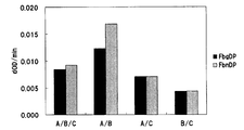
- FIGS. suspensions of latex particles sensitized with FDP3-797 antibody, FDP3-2935 antibody and DD-M1051 antibody are shown in FIGS. Are referred to as A, B and C, respectively. These suspensions are referred to as “A and B” (A / B in FIGS. 1 and 2), “A and C” (A / C in FIGS. 1 and 2), “B and C” (FIGS. 1 and 2).
- a and B A / B in FIGS. 1 and 2
- a and C A / C in FIGS. 1 and 2
- B and C FIGGS. 1 and 2
- 2 or 3 monoclonal antibodies are sensitized by mixing equal amounts of combinations of B / C in Fig. 2 and "A, B and C" (A / B / C in Figs. 1 and 2).
- the reagent for FDP measurement containing the obtained carrier particles was obtained.
- these FDP measurement reagents were used as second reagents.
- Example 2 Confirmation of reactivity of FDP measurement reagent of the present invention to FDP by latex agglutination method
- the reaction ratio of the FDP measurement reagent produced in Example 1 above to the fibrin degradation product and fibrinogen degradation product was determined as follows. The procedure was examined by latex agglomeration.
- the FbgDP solution prepared in Test Example 1 was diluted with a TBS buffer to obtain an FbgDP sample (protein concentration 100 ⁇ g / ml).
- an FbnDP sample protein concentration of 100 ⁇ g / ml
- the FDP measurement reagent produced in Example 1 (2) above is a reagent containing carrier particles sensitized with at least two types of monoclonal antibodies having different reactivities to FDP. Further, from FIG.
- the FDP measurement reagent and reagent kit of the present invention can measure FDP with high accuracy even for biological samples containing primary fibrinolysis.
- Example 3 Comparison of FDP Measurement Reagent Kit of the Present Invention and Competitor's Product
- FDP in plasma was measured using the FDP measurement reagent kit of the present invention produced in Example 1 above and the other company's product.
- the first reagent of the reagent kit for FDP measurement of the present invention the reagent produced in Example 1 (1) above was used.
- the reagent containing the suspension of the latex particle which sensitized each of 3 types of antibodies manufactured in said Example 1 (2) was used as a 2nd reagent.
- an equivalent mixture of the FbgDP sample and the FbnDP sample used in Example 2 was used as panel plasma.
Abstract
Description
D画分とは反応しないが、XDP画分、DD/E画分、DD画分、X画分及びY画分とは反応する第1のモノクローナル抗体、
X画分及びD画分とは反応しないが、XDP画分、DD/E画分、DD画分及びY画分とは反応する第2のモノクローナル抗体、及び
D画分とは反応しないが、XDP画分、DD/E画分、DD画分、X画分及びY画分とは反応し、FDPに対する反応性が第1のモノクローナル抗体とは異なる第3のモノクローナル抗体
から選択される、FDPに対する反応性が互いに異なる少なくとも2種のモノクローナル抗体を感作した担体を含むFDP測定用試薬が提供される。 That is, according to the present invention,
A first monoclonal antibody that does not react with the D fraction but reacts with the XDP fraction, the DD / E fraction, the DD fraction, the X fraction and the Y fraction,
Although it does not react with the X and D fractions, it does not react with the second monoclonal antibody that reacts with the XDP fraction, the DD / E fraction, the DD fraction and the Y fraction, and the D fraction. FDP selected from a third monoclonal antibody that reacts with the XDP fraction, the DD / E fraction, the DD fraction, the X fraction and the Y fraction and has a different reactivity to FDP from the first monoclonal antibody. There is provided a reagent for FDP measurement comprising a carrier sensitized with at least two types of monoclonal antibodies having different reactivity with respect to each other.
さらに、本発明によれば、上記のFDPに対する反応性が互いに異なる少なくとも2種のモノクローナル抗体を感作した担体粒子の懸濁液と、生体試料とを混合する工程と、抗原抗体反応により生じる、前記担体粒子の凝集の度合を測定する工程とを含むFDP測定方法が提供される。 According to the present invention, there is provided an FDP measurement reagent kit comprising a first reagent comprising a buffer solution and a second reagent comprising a carrier sensitized with at least two types of monoclonal antibodies having different reactivity with respect to the FDP. Provided.
Furthermore, according to the present invention, a step of mixing a suspension of carrier particles sensitized with at least two types of monoclonal antibodies having different reactivity to FDP and a biological sample, and an antigen-antibody reaction, And a method for measuring the degree of aggregation of the carrier particles.
また、本発明のFDP測定用試薬及び試薬キット、並びに測定方法によれば、一次線溶物及び二次線溶物に対して均等に反応するので、検体として血清及び血漿の両方を使用することができる。 According to the FDP measurement reagent, reagent kit, and measurement method of the present invention, FDP can be obtained with high accuracy even for biological samples containing primary fibrinolysis that have been difficult to accurately measure with conventional FDP measurement reagents. It can be measured.
In addition, according to the reagent and reagent kit for FDP measurement and the measurement method of the present invention, it reacts equally to the primary and secondary fibrinolysis, so both serum and plasma are used as specimens. Can do.
本明細書において、「フィブリン分解産物」とは、二次線溶物とも呼ばれ、トロンビンなどの酵素の作用により血液中のフィブリノゲンが凝固されて形成されるポリマーである安定化フィブリンが、プラスミンなどの酵素によって分解されて生じるタンパク質群である。フィブリン分解産物としては、DD画分、DD/E画分、XDP画分などが挙げられる。XDP画分としては、DD/E画分の多量体、例えばDD/E画分の3量体であるDXD/YY画分、DD/E画分の5量体であるYXY/DXXD画分、DD/E画分の7量体であるDXXD/YXXY画分などが挙げられる。また、当該技術においては、XDP画分はDダイマーとも総称される。
本明細書において、「フィブリノゲン分解産物」とは、一次線溶物とも呼ばれ、血液中に存在するフィブリノゲンがプラスミンなどの酵素によって分解されて生じるタンパク質群である。フィブリノゲン分解産物としてはX画分、Y画分、D画分及びE画分が挙げられる。 As used herein, “FDP” means both fibrin degradation products and fibrinogen degradation products.
In the present specification, “fibrin degradation product” is also called secondary fibrinolysis, and stabilized fibrin, which is a polymer formed by coagulation of fibrinogen in blood by the action of an enzyme such as thrombin, It is a protein group produced by being decomposed by the enzyme. Examples of fibrin degradation products include DD fraction, DD / E fraction, XDP fraction and the like. As the XDP fraction, a DD / E fraction multimer, for example, a DD / EY fraction that is a trimer of the DD / E fraction, a YXY / DXXXD fraction that is a pentamer of the DD / E fraction, And DXXD / YXXY fraction, which is a 7-mer of the DD / E fraction. In this technique, the XDP fraction is also collectively referred to as D dimer.
In the present specification, the “fibrinogen degradation product” is also referred to as a primary fibrinolysis, and is a group of proteins produced by the degradation of fibrinogen present in blood by an enzyme such as plasmin. Examples of fibrinogen degradation products include the X fraction, the Y fraction, the D fraction, and the E fraction.
したがって、ある抗FDPモノクローナル抗体と、別の抗FDPモノクローナル抗体との間で、特異的に反応できるFDPの種類が互いに異なるとき、これらの抗体のFDPに対する反応性は互いに異なると決定される。また、互いに同じ種類のFDPと反応する場合は、ある抗FDPモノクローナル抗体を感作した担体と、別の抗FDPモノクローナル抗体を感作した担体とをそれぞれ用いたFDP測定において、凝集の度合いが互いに有意に異なるとき、これらの抗体のFDPに対する反応性は互いに異なると決定される。 In the present specification, “reactivity to FDP” is defined by the type of FDP to which the anti-FDP monoclonal antibody specifically reacts, or a carrier sensitized with the anti-FDP monoclonal antibody and an antigen antibody with FDP It is defined by the degree of aggregation caused by the reaction.
Therefore, when the types of FDP that can specifically react between one anti-FDP monoclonal antibody and another anti-FDP monoclonal antibody are different from each other, the reactivity of these antibodies with respect to FDP is determined to be different from each other. In the case of reacting with the same type of FDP, in the FDP measurement using a carrier sensitized with one anti-FDP monoclonal antibody and a carrier sensitized with another anti-FDP monoclonal antibody, the degree of aggregation is mutually different. When significantly different, the reactivity of these antibodies to FDP is determined to be different from each other.
第1のモノクローナル抗体は、D画分とは反応しないが、XDP画分、DD/E画分、DD画分、X画分及びY画分とは反応する抗体である。
第2のモノクローナル抗体は、X画分及びD画分とは反応しないが、XDP画分、DD/E画分、DD画分及びY画分とは反応する抗体である。
第3のモノクローナル抗体は、D画分とは反応しないが、XDP画分、DD/E画分、DD画分、X画分及びY画分とは反応し、FDPに対する反応性が第1のモノクローナル抗体とは異なる抗体である。 The monoclonal antibodies used in the FDP measurement reagent of the present invention (hereinafter also referred to as the reagent of the present invention) are at least two types of monoclonal antibodies having different reactivities to FDP. As such a monoclonal antibody, it is preferable to select at least two kinds from the following first monoclonal antibody, second monoclonal antibody and third monoclonal antibody.
The first monoclonal antibody is an antibody that does not react with the D fraction but reacts with the XDP fraction, the DD / E fraction, the DD fraction, the X fraction, and the Y fraction.
The second monoclonal antibody is an antibody that does not react with the X fraction and the D fraction but reacts with the XDP fraction, the DD / E fraction, the DD fraction, and the Y fraction.
The third monoclonal antibody does not react with the D fraction, but reacts with the XDP fraction, the DD / E fraction, the DD fraction, the X fraction and the Y fraction, and has the first reactivity with FDP. It is an antibody different from a monoclonal antibody.
(抗原の取得)
抗原として用いるFDPは、プラスミンのようなフィブリン及びフィブリノゲンを分解できる酵素をフィブリン及びフィブリノゲンに作用させて得ることができる。なお、FDPの原料となるフィブリン及びフィブリノゲンは市販されている。また、フィブリンは、フィブリノゲンにトロンビン、第XIII因子及びカルシウム塩を作用させて得ることができる。 Specifically, the first, second and third monoclonal antibodies used in the reagent of the present invention can be obtained by the following method.
(Acquisition of antigen)
FDP used as an antigen can be obtained by acting an enzyme capable of degrading fibrin and fibrinogen such as plasmin on fibrin and fibrinogen. Fibrin and fibrinogen as raw materials for FDP are commercially available. Fibrin can be obtained by reacting fibrinogen with thrombin, factor XIII and calcium salt.
上記のようにして得られる抗原を、アジュバントと任意に混合し、適当な緩衝液に溶解又は懸濁して得られる抗原液で、動物を免疫することができる。該抗原液中の抗原の濃度は、50~500μg/ml程度が好ましい。抗原の免疫原性が低い場合は、アルブミン、キーホールリンペットヘモシアニンのようなキャリアータンパク質を任意に抗原と結合させてもよい。 (Immune method)
An antigen can be immunized with an antigen solution obtained by arbitrarily mixing the antigen obtained as described above with an adjuvant and dissolving or suspending it in an appropriate buffer solution. The concentration of the antigen in the antigen solution is preferably about 50 to 500 μg / ml. When the immunogenicity of the antigen is low, a carrier protein such as albumin or keyhole limpet hemocyanin may optionally be bound to the antigen.
免疫法は、使用する抗原の種類やアジュバントの有無により適宜選択することができる。例えばマウスを用いる場合、アジュバント混合抗原液0.05~1ml(抗原10~200μg)を腹腔内、皮下、筋肉内又は尾静脈内に注射し、初回免疫から約4~21日毎に1~4回追加免疫を行い、さらに約1~4週間後に最終免疫を行う。抗原量を多くして腹腔内注射することにより、抗原液にアジュバントを用いずに免疫を行ってもよい。追加免疫の約5~10日後に血液を採取して抗体価を測定する。抗体価は、後述する抗体価アッセイのような当該技術において公知の方法にしたがって測定できる。最終免疫から約3~5日後に、免疫された動物から脾臓を摘出し、脾臓細胞を分離して抗体産生細胞を得ることができる。 The animal to be immunized may be a mouse, rat, hamster, horse, goat, rabbit or the like, preferably a mouse, more preferably a BALB / c mouse.
The immunization method can be appropriately selected depending on the type of antigen used and the presence or absence of an adjuvant. For example, in the case of using a mouse, 0.05 to 1 ml of an adjuvant mixed antigen solution (antigen 10 to 200 μg) is injected intraperitoneally, subcutaneously, intramuscularly or into the tail vein, and boosted 1 to 4 times about every 4 to 21 days from the first immunization. The final immunization is performed after about 1 to 4 weeks. Immunization may be performed without using an adjuvant in the antigen solution by intraperitoneal injection with an increased amount of antigen. Blood is collected approximately 5-10 days after the booster and antibody titer is measured. The antibody titer can be measured according to a method known in the art such as an antibody titer assay described later. Approximately 3 to 5 days after the final immunization, the spleen can be removed from the immunized animal, and the spleen cells can be isolated to obtain antibody-producing cells.
モノクローナル抗体は、当該技術において公知の方法、例えばKohler及びMilstein, Nature, 256, 495-497 (1975)に記載の方法にしたがって作製できる。
用いる骨髄腫細胞は、マウス、ラット、ヒトなどいずれの哺乳動物に由来する細胞であってもよく、例えばマウスミエローマP3X63-Ag8、P3X63-Ag8-U1、P3NS1-Ag4、SP2/o-Ag14、P3X63-Ag8・653などの株化骨髄腫細胞が挙げられる。骨髄腫細胞の中には免疫グロブリン軽鎖を産生する種類の骨髄腫細胞があり、これを融合対象として用いると、抗体産生細胞が産生する免疫グロブリン重鎖とこの軽鎖とがランダムに結合することがある。したがって、免疫グロブリン軽鎖を産生しない骨髄腫細胞、例えばP3X63-Ag8・653、SP2/o-Ag14などを用いるのが好ましい。抗体産生細胞と骨髄腫細胞とは、同種動物、特に同系統の動物由来の細胞が好ましい。 (Production of monoclonal antibodies)
Monoclonal antibodies can be produced according to methods known in the art, such as those described in Kohler and Milstein, Nature, 256, 495-497 (1975).
The myeloma cell to be used may be a cell derived from any mammal such as mouse, rat and human, for example, mouse myeloma P3X63-Ag8, P3X63-Ag8-U1, P3NS1-Ag4, SP2 / o-Ag14, P3X63 -Examples include established myeloma cells such as Ag8 / 653. Among myeloma cells, there is a myeloma cell type that produces an immunoglobulin light chain. When this is used as a fusion target, the immunoglobulin heavy chain produced by the antibody-producing cell and this light chain are randomly bound. Sometimes. Therefore, it is preferable to use myeloma cells that do not produce an immunoglobulin light chain, such as P3X63-Ag8 · 653, SP2 / o-Ag14. The antibody-producing cells and myeloma cells are preferably cells derived from the same species, particularly from the same strain.
本発明の試薬に用いられる第2のモノクローナル抗体としては、例えば受託番号NITE BP-951として独立行政法人製品評価技術基盤機構特許微生物寄託センター(郵便番号292-0818、日本国千葉県木更津市かずさ鎌足2-5-8)に2010年6月1日付けで受託されたハイブリドーマ「FDP3-2935」により産生される抗体(以下、「FDP3-2935抗体」ともいう)が挙げられる。
本発明の試薬に用いられる第3のモノクローナル抗体としては、例えば受託番号NITE BP-952として独立行政法人製品評価技術基盤機構特許微生物寄託センター(郵便番号292-0818、日本国千葉県木更津市かずさ鎌足2-5-8)に2010年6月1日付けで受託されたハイブリドーマ「DD-M1051」により産生される抗体(以下、「DD-M1051抗体」ともいう)が挙げられる。 As the first monoclonal antibody used in the reagent of the present invention, for example, as the accession number NITE BP-950, the National Institute of Technology and Evaluation Microbiology Deposit Center (Postal code 292-0818, Kazusa-Kama, Kisarazu City, Chiba Prefecture, Japan) 2-5-8) includes an antibody produced by the hybridoma “FDP3-797” commissioned on June 1, 2010 (hereinafter, also referred to as “FDP3-797 antibody”).
As the second monoclonal antibody used in the reagent of the present invention, for example, under the accession number NITE BP-951, the National Institute of Technology and Evaluation Microbiological Deposits Center (Postal code 292-0818, Kazusa-Kama, Kisarazu City, Chiba Prefecture, Japan) 2-5-8) includes an antibody produced by the hybridoma “FDP3-2935” commissioned on June 1, 2010 (hereinafter also referred to as “FDP3-2935 antibody”).
As the third monoclonal antibody used in the reagent of the present invention, for example, as the accession number NITE BP-952, the National Institute of Technology and Evaluation Microbiological Deposits Center (Postal code 292-0818, Kazusa-Kama, Kisarazu City, Chiba Prefecture, Japan) 2-5-8) includes an antibody produced by hybridoma “DD-M1051” commissioned on June 1, 2010 (hereinafter also referred to as “DD-M1051 antibody”).
本発明の試薬キットは、イムノアッセイ、例えば上記の少なくとも2種のモノクローナル抗体を感作したラテックス粒子と、生体試料中のFDPとを反応させるアッセイ(ラテックス凝集法)などにより、該試料中のFDPを検出するための試薬キットである。
本発明の試薬キットに用いられる第1、第2及び第3のモノクローナル抗体の一例としては、それぞれFDP3-797抗体、FDP3-2935抗体及びDD-M1051抗体が挙げられる。 As one embodiment of the reagent of the present invention, an FDP measurement reagent kit (hereinafter also referred to as the reagent kit of the present invention) can be mentioned. The reagent kit of the present invention comprises a suspension of carrier particles sensitized with a first reagent comprising a buffer and at least two monoclonal antibodies selected from the first, second and third monoclonal antibodies. A second reagent containing.
The reagent kit of the present invention can be used to detect FDP in a sample by immunoassay, for example, an assay (latex agglutination method) in which latex particles sensitized with at least two monoclonal antibodies are reacted with FDP in a biological sample. This is a reagent kit for detection.
Examples of the first, second and third monoclonal antibodies used in the reagent kit of the present invention include FDP3-797 antibody, FDP3-2935 antibody and DD-M1051 antibody, respectively.
まず、緩衝液を含む第1試薬と生体試料とを混合してインキュベートする。ここで、生体試料としては被験者から得られる血清、血漿、尿などが挙げられる。第1試薬と生体試料とを混合する際の容量比は、5:1~50:1程度であればよい。また、インキュベート時間は1~10分間程度であればよい。 The FDP measurement method of the present invention can be carried out by using the FDP measurement reagent or reagent kit of the present invention. As an embodiment of the FDP measurement method of the present invention, a method for measuring FDP in a biological sample using the FDP measurement reagent kit of the present invention will be specifically described below.
First, a first reagent containing a buffer solution and a biological sample are mixed and incubated. Here, examples of the biological sample include serum, plasma and urine obtained from a subject. The volume ratio for mixing the first reagent and the biological sample may be about 5: 1 to 50: 1. The incubation time may be about 1 to 10 minutes.
生体試料中のFDPの濃度及び/又は量は、濃度既知のFDP標準物質の測定により得られる検量線を用いて、測定した吸光度の変化量から算出できる。 When the second reagent is added and mixed, aggregation of FDP and carrier particles in the second reagent occurs due to an antigen-antibody reaction. The degree of aggregation is measured as the amount of change in absorbance per minute. This measurement is preferably performed with an optical instrument capable of measuring scattered light intensity, absorbance, or transmitted light intensity. The measurement wavelength can be selected from the range of 300 to 2400 nm, preferably 300 to 1000 nm, more preferably 500 to 1000 nm.
The concentration and / or amount of FDP in a biological sample can be calculated from the amount of change in absorbance measured using a calibration curve obtained by measuring an FDP standard substance having a known concentration.
なお、本発明のFDP測定方法に用いられる第1、第2及び第3のモノクローナル抗体の一例として、それぞれFDP3-797抗体、FDP3-2935抗体及びDD-M1051抗体が挙げられる。 The reagent kit of the present invention can also be used for a method of optically measuring the degree of aggregation of carrier particles by mixing a first reagent and a second reagent and then adding a biological sample to the mixture of both reagents. .
Examples of the first, second and third monoclonal antibodies used in the FDP measurement method of the present invention include FDP3-797 antibody, FDP3-2935 antibody and DD-M1051 antibody, respectively.
第1、第2及び第3のモノクローナル抗体として、それぞれFDP3-797抗体、FDP3-2935抗体及びDD-M1051抗体を用いて、各抗体のフィブリン及びフィブリノゲン分解産物に対する反応性の違いを、以下のようなELISA法により検討した。 Test Example 1: Examination of Reactivity of Each Monoclonal Antibody As the first, second and third monoclonal antibodies, FDP3-797 antibody, FDP3-2935 antibody and DD-M1051 antibody were used respectively, and fibrin and fibrinogen of each antibody. Differences in reactivity to degradation products were examined by the following ELISA method.
(7-1)フィブリノゲン分解産物(FbgDP)の調製
フィブリノゲン(Sigma社)241 mg(39.7 mg/ml)に、プラスミン(Sigma社)を終濃度60 mU/mlとなるように添加して、37℃で8時間反応させた。その後、アプロチニンを終濃度1U/mlとなるように加えて、分解反応を停止させた。得られた反応液を12000×gで20分間遠心し、得られた上清を、50 mMトリス緩衝液(pH7.4)で平衡化したリジン-sepharose 4Bカラム(ボリューム8ml)に充填してクロマトグラフィーを行った後、スピンカラムにてセファロースを除き、FbgDP溶液を調製した。得られたFbgDP溶液のタンパク質濃度を、タンパク質定量試薬(Bio-Rad社)を用いて測定した。また、FbgDP溶液の一部を、後述する本発明のFDP測定用試薬のFDPへの反応性の確認に用いた。
得られたFbgDP溶液を、限外ろ過用遠心チューブ(アミコン15 50K;ミリポア社)によりサンプル緩衝液(62.5 mM Tris、192 mMグリシン、1%SDS(pH6.8))で2回置換した。得られた溶液をSephacryl S-300(GE Healthcare社)に充填し、ペリスタポンプを用いて流速70~80μl/分で流して、10分ごとにフラクションを回収した。得られた各フラクションについて分子量マーカーを用いるSDS-PAGEにより解析して、X画分、Y画分及びD画分が含まれるフラクションをそれぞれ回収した。これをアミコン15 50K(ミリポア社)によりリン酸緩衝液(PBS)で2回置換して、FbgDP抗原とした。 (1) Preparation of FDP (7-1) Preparation of fibrinogen degradation product (FbgDP) Fibrinogen (Sigma) 241 mg (39.7 mg / ml) and plasmin (Sigma) to a final concentration of 60 mU / ml The mixture was added and reacted at 37 ° C. for 8 hours. Thereafter, aprotinin was added to a final concentration of 1 U / ml to stop the degradation reaction. The resulting reaction solution was centrifuged at 12000 × g for 20 minutes, and the resulting supernatant was loaded onto a lysine-sepharose 4B column (volume 8 ml) equilibrated with 50 mM Tris buffer (pH 7.4) and chromatographed. After performing the chromatography, Sepharose was removed with a spin column to prepare an FbgDP solution. The protein concentration of the obtained FbgDP solution was measured using a protein quantification reagent (Bio-Rad). A part of the FbgDP solution was used to confirm the reactivity of the reagent for FDP measurement of the present invention described later to FDP.
The obtained FbgDP solution was replaced twice with a sample buffer (62.5 mM Tris, 192 mM glycine, 1% SDS (pH 6.8)) using an ultrafiltration centrifuge tube (Amicon 15 50K; Millipore). The obtained solution was filled in Sephacryl S-300 (GE Healthcare), and flowed at a flow rate of 70-80 μl / min using a peristaltic pump, and fractions were collected every 10 minutes. Each of the obtained fractions was analyzed by SDS-PAGE using a molecular weight marker, and fractions containing the X fraction, the Y fraction, and the D fraction were collected. This was replaced twice with phosphate buffer (PBS) by Amicon 15 50K (Millipore) to obtain FbgDP antigen.
フィブリノゲン(Sigma社)92 mg(23 mg/ml)に、塩化カルシウム、ヒトトロンビン(三菱ウェルファーマ社)及び第XIII因子(ニプロ社)をそれぞれ終濃度25 mM、4U/ml及び0.05 U/mlとなるよう加え、37℃で一晩反応させて、フィブリノゲンをフィブリンに変換させた。反応液中に生じたフィブリンゲルをトリス緩衝液(TBS(pH7.4))50 mlで洗浄し、4℃、3000×gで10分間遠心し、フィブリンゲルを回収した。この操作を2度繰り返した後、50 mlシリンジを用いてフィブリンゲルを砕いた。フィブリンゲルをTBS(pH7.4)4.6 mlに再懸濁した。懸濁液にプラスミンを終濃度75 mU/mlとなるように添加して、37℃で6時間反応させた。その後、アプロチニンを終濃度1U/mlとなるように加えて、分解反応を停止させた。得られた反応液を12000×gで20分間遠心し、得られた上清を、50 mMトリス緩衝液(pH7.4)で平衡化したリジン-sepharose 4Bカラム(ボリューム3.5 ml)に充填してクロマトグラフィーを行った後、スピンカラムにてセファロースを除き、FbnDP溶液を調製した。得られたFbnDP溶液のタンパク質濃度を、タンパク質定量試薬(Bio-Rad社)を用いて測定した。また、FbnDP溶液の一部を、後述する本発明のFDP測定用試薬のFDPへの反応性の確認に用いた。 (1-2) Preparation of fibrin degradation product (FbnDP) Fibrinogen (Sigma) 92 mg (23 mg / ml), calcium chloride, human thrombin (Mitsubishi Pharma) and factor XIII (Nipro) final concentrations They were added to 25 mM, 4 U / ml and 0.05 U / ml, and reacted overnight at 37 ° C. to convert fibrinogen into fibrin. The fibrin gel generated in the reaction solution was washed with 50 ml of Tris buffer (TBS (pH 7.4)) and centrifuged at 3000 × g for 10 minutes at 4 ° C. to recover the fibrin gel. After this operation was repeated twice, the fibrin gel was crushed using a 50 ml syringe. Fibrin gel was resuspended in 4.6 ml of TBS (pH 7.4). Plasmin was added to the suspension to a final concentration of 75 mU / ml and reacted at 37 ° C. for 6 hours. Thereafter, aprotinin was added to a final concentration of 1 U / ml to stop the degradation reaction. The obtained reaction solution was centrifuged at 12000 × g for 20 minutes, and the resulting supernatant was loaded onto a lysine-sepharose 4B column (volume 3.5 ml) equilibrated with 50 mM Tris buffer (pH 7.4). After chromatography, Sepharose was removed with a spin column to prepare an FbnDP solution. The protein concentration of the obtained FbnDP solution was measured using a protein quantification reagent (Bio-Rad). A part of the FbnDP solution was used for confirming the reactivity of the reagent for FDP measurement of the present invention described later to FDP.
ウェルを洗浄液で3回洗浄した後、該抗体固相の各ウェルに、上記で調製したFbgDP各抗原及びFbnDP各抗原を100μlずつ加え、室温で30分間反応させた。反応終了後、ウェルを洗浄液で3回洗浄した後、ペルオキシダーゼ標識抗フィブリノゲン抗体(DAKO社)を100μlずつ各ウェルに加え、室温で1時間反応させた。反応終了後、ウェルを洗浄液で3回洗浄した後、ODP基質液(国際試薬株式会社)を100μlずつ各ウェルに加え、室温で15分間反応させた。続いて、2N硫酸を100μlずつ各ウェルに加えて、反応を停止し、490 nmにおける吸光度を測定した。
結果を表1に示す。 Each anti-FDP monoclonal antibody solution was diluted to 0.5 μg / ml with PBS, and 100 μl each was dispensed into wells of a 96-well microtiter plate and allowed to stand at 4 ° C. for 18 hours. Thereafter, the wells were washed three times with 0.05 mM Tween20-containing 10 mM phosphate buffer (pH 7.0) (hereinafter referred to as a washing solution). Subsequently, the well was filled with 1% BSA-containing 10 mM phosphate buffer (hereinafter referred to as blocking buffer) to obtain an antibody solid phase of anti-FDP monoclonal antibody.
After the wells were washed three times with a washing solution, 100 μl of each FbgDP antigen and FbnDP antigen prepared above was added to each well of the antibody solid phase and allowed to react at room temperature for 30 minutes. After completion of the reaction, the well was washed three times with a washing solution, and then 100 μl of peroxidase-labeled anti-fibrinogen antibody (DAKO) was added to each well and reacted at room temperature for 1 hour. After completion of the reaction, the wells were washed three times with a washing solution, and then 100 μl of ODP substrate solution (Kokusai Reagent Co., Ltd.) was added to each well and reacted at room temperature for 15 minutes. Subsequently, 100 μl of 2N sulfuric acid was added to each well to stop the reaction, and the absorbance at 490 nm was measured.
The results are shown in Table 1.

表1より、FDP3-797抗体及びDD-M1051抗体は、D画分とは反応しないが、XDP画分、DD/E画分、DD画分、X画分及びY画分とは反応することがわかった。また、FDP3-2935抗体は、X画分及びD画分とは反応しないが、XDP画分、DD/E画分、DD画分及びY画分とは反応することがわかった。 In Table 1, “+” indicates that the antibody reacts with the fraction, and “−” indicates that the antibody does not react with the fraction.
From Table 1, FDP3-797 antibody and DD-M1051 antibody do not react with D fraction, but react with XDP fraction, DD / E fraction, DD fraction, X fraction and Y fraction. I understood. Further, it was found that the FDP3-2935 antibody does not react with the X fraction and the D fraction, but reacts with the XDP fraction, the DD / E fraction, the DD fraction, and the Y fraction.
(1)緩衝液を含む第1試薬の製造
各試薬を表2に示される終濃度となるように混合した緩衝液を、1M水酸化ナトリウム水溶液でpHを7.1に調整した後、超純水で1リットルにメスアップすることにより緩衝液を製造した。 Example 1: Manufacture of FDP measurement reagent and reagent kit (1) Manufacture of first reagent containing buffer solution A buffer solution prepared by mixing each reagent so as to have a final concentration shown in Table 2 was prepared with a 1M sodium hydroxide aqueous solution. After adjusting the pH to 7.1, a buffer solution was prepared by measuring up to 1 liter with ultrapure water.

(2-1)FDP3-797抗体のラテックス粒子への感作
FDP3-797抗体の終濃度が1mg/mlとなるように、50 mM 2-ヒドロキシ-3-モルホリノプロパンスルホン酸/150 mM NaCl溶液に混合した。そして、この混合液と20%(重量比)ラテックス懸濁液(粒径0.238μm;JSR株式会社)とを混合した。
得られた混合液に、50 mM 2-ヒドロキシ-3-モルホリノプロパンスルホン酸/150 mM NaCl溶液/2%BSA溶液を等量加えて混合した後、10℃、38400×gで30分間遠心した。上澄みを除去し、得られた沈殿物に、上澄みと等量の50 mM 2-ヒドロキシ-3-モルホリノプロパンスルホン酸/150 mM NaCl溶液/2%BSA/1.5%シュークロース溶液を添加して混合した。
得られた混合液を、氷冷条件で超音波破砕機(大岳社製)、超音波処理装置(Dr. Hielscher Gmbh UP-200S相当品)を用いソニケーションを実施し、FDP3-797抗体を感作したラテックス粒子の懸濁液(抗体濃度39μg/ml)を得た。 (2) Production of reagent for FDP measurement containing suspension of carrier particles sensitized with anti-FDP monoclonal antibody (2-1) Sensitization of FDP3-797 antibody to latex particles Final concentration of FDP3-797 antibody is 1 mg The mixture was mixed with 50 mM 2-hydroxy-3-morpholinopropanesulfonic acid / 150 mM NaCl solution so as to be / ml. Then, this mixed solution and a 20% (weight ratio) latex suspension (particle size 0.238 μm; JSR Corporation) were mixed.
An equal amount of 50 mM 2-hydroxy-3-morpholinopropanesulfonic acid / 150 mM NaCl solution / 2% BSA solution was added to the obtained mixed solution and mixed, and then centrifuged at 10 ° C. and 38400 × g for 30 minutes. The supernatant was removed, and the resulting precipitate was mixed with the same amount of 50 mM 2-hydroxy-3-morpholinopropanesulfonic acid / 150 mM NaCl solution / 2% BSA / 1.5% sucrose solution as the supernatant. .
The resulting mixture is sonicated under ice-cooling conditions using an ultrasonic crusher (manufactured by Otake) and an ultrasonic treatment device (Dr. Hielscher Gmbh UP-200S equivalent) to sense the FDP3-797 antibody. A suspension of the prepared latex particles (antibody concentration 39 μg / ml) was obtained.
FDP3-2935抗体の終濃度が1mg/mlとなるように、50 mM 2-ヒドロキシ-3-モルホリノプロパンスルホン酸/150 mM NaCl溶液に混合した。以下、上記の(2-1)FDP3-797抗体のラテックス粒子への感作において述べたことと同様にして、FDP3-2935抗体を感作したラテックス粒子の懸濁液(抗体濃度39μg/ml)を得た。 (2-2) Sensitization of FDP3-2935 antibody to latex particles In 50 mM 2-hydroxy-3-morpholinopropanesulfonic acid / 150 mM NaCl solution, the final concentration of FDP3-2935 antibody is 1 mg / ml. Mixed. The suspension of latex particles sensitized with FDP3-2935 antibody in the same manner as described above in (2-1) Sensitization of latex particles with FDP3-797 antibody (antibody concentration 39 μg / ml) Got.
DD-M1051抗体の終濃度が1mg/mlとなるように、50 mM 2-ヒドロキシ-3-モルホリノプロパンスルホン酸/150 mM NaCl溶液に混合した。以下、上記の(2-1)FDP3-797抗体のラテックス粒子への感作において述べたことと同様にして、DD-M1051抗体を感作したラテックス粒子の懸濁液(抗体濃度39μg/ml)を得た。 (2-3) Sensitization of DD-M1051 antibody to latex particles In a 50 mM 2-hydroxy-3-morpholinopropanesulfonic acid / 150 mM NaCl solution, the final concentration of DD-M1051 antibody is 1 mg / ml. Mixed. The suspension of latex particles sensitized with DD-M1051 antibody in the same manner as described in (2-1) Sensitization of latex particles with FDP3-797 antibody (antibody concentration 39 μg / ml) Got.
ここで、FDP3-797抗体、FDP3-2935抗体及びDD-M1051抗体の各抗体を感作したラテックス粒子の懸濁液を、後述する図1及び図2において、それぞれA、B及びCと称する。これらの懸濁液を、「AとB」(図1及び図2のA/B)、「AとC」(図1及び図2のA/C)、「BとC」(図1及び図2のB/C)及び「AとBとC」(図1及び図2のA/B/C)の組み合わせでそれぞれ等量混合することにより、2種又は3種のモノクローナル抗体を感作した担体粒子を含むFDP測定用試薬を得た。以下、これらのFDP測定用試薬を第2試薬とした。 (2-4) Production of second reagent Here, suspensions of latex particles sensitized with FDP3-797 antibody, FDP3-2935 antibody and DD-M1051 antibody are shown in FIGS. Are referred to as A, B and C, respectively. These suspensions are referred to as “A and B” (A / B in FIGS. 1 and 2), “A and C” (A / C in FIGS. 1 and 2), “B and C” (FIGS. 1 and 2). 2 or 3 monoclonal antibodies are sensitized by mixing equal amounts of combinations of B / C in Fig. 2 and "A, B and C" (A / B / C in Figs. 1 and 2). The reagent for FDP measurement containing the obtained carrier particles was obtained. Hereinafter, these FDP measurement reagents were used as second reagents.
上記の実施例1で製造したFDP測定用試薬のフィブリン分解物及びフィブリノゲン分解産物に対する反応比率を、以下の手順によるラテックス凝集法により検討した。
上記の試験例1で調製したFbgDP溶液をTBSバッファーで希釈して、FbgDP検体(タンパク質濃度100μg/ml)とした。FbnDP溶液についても同様にして、FbnDP検体(タンパク質濃度100μg/ml)を調製した。
FbgDP及びFbnDPの各検体6μlの等量混合物と、上記の実施例1(1)で調製した第1試薬84μlとを混合し、37℃で20秒間反応させた。得られた反応液と、上記の実施例1(2)で調製した各第2試薬84μlとを混合し、ラテックス凝集反応を開始させた。反応開始から1分後及び2分後の波長800 nmにおける吸光度を、CS-2000i(シスメックス株式会社)を用いて測定した。これらの測定結果から、1分間あたりの吸光度の変化量を求めた。また、FbgDP及びFbnDPのそれぞれに対する反応性から反応比率を算出した。これらの結果をそれぞれ図1および図2に示す。 Example 2: Confirmation of reactivity of FDP measurement reagent of the present invention to FDP by latex agglutination method The reaction ratio of the FDP measurement reagent produced in Example 1 above to the fibrin degradation product and fibrinogen degradation product was determined as follows. The procedure was examined by latex agglomeration.
The FbgDP solution prepared in Test Example 1 was diluted with a TBS buffer to obtain an FbgDP sample (
An equal volume mixture of 6 μl of each specimen of FbgDP and FbnDP and 84 μl of the first reagent prepared in Example 1 (1) above were mixed and reacted at 37 ° C. for 20 seconds. The obtained reaction solution and 84 μl of each second reagent prepared in Example 1 (2) above were mixed to initiate a latex agglutination reaction. Absorbance at a wavelength of 800 nm after 1 minute and 2 minutes after the start of the reaction was measured using CS-2000i (Sysmex Corporation). From these measurement results, the amount of change in absorbance per minute was determined. Moreover, the reaction ratio was calculated from the reactivity with respect to each of FbgDP and FbnDP. These results are shown in FIGS. 1 and 2, respectively.
したがって、上記の実施例1(2)で製造したFDP測定用試薬は、FDPに対する反応性が互いに異なる少なくとも2種のモノクローナル抗体を感作した担体粒子を含む試薬である。
また、図2より、いずれの抗体の組合せの測定試薬においても、FbgDPとFbnDPとに対しておおむね均一に反応することがわかった。これにより、本発明のFDP測定用試薬及び試薬キットは、一次線溶物が含まれる生体試料についてもFDPを高精度に測定可能であることが示された。 In FIG. 1, when comparing the FDP3-797 antibody / FDP3-2935 antibody combination (A / B in FIG. 1) with the FDP3-2935 antibody / DD-M1051 antibody combination (B / C in FIG. 1), the absorbance is compared. Since the amount of change in each other was different from each other, it was shown that the FDP3-797 antibody and the DD-M1051 antibody have different reactivity to FDP.
Therefore, the FDP measurement reagent produced in Example 1 (2) above is a reagent containing carrier particles sensitized with at least two types of monoclonal antibodies having different reactivities to FDP.
Further, from FIG. 2, it was found that the measurement reagent of any antibody combination reacts almost uniformly with FbgDP and FbnDP. Thus, it was shown that the FDP measurement reagent and reagent kit of the present invention can measure FDP with high accuracy even for biological samples containing primary fibrinolysis.
上記の実施例1で製造した本発明のFDP測定用試薬キットと、他社製品とを用いて血漿中のFDPを測定した。
本発明のFDP測定用試薬キットの第1試薬として、上記の実施例1(1)で製造した試薬を用いた。また、第2試薬として、上記の実施例1(2)で製造した、3種の抗体をそれぞれ個別に感作したラテックス粒子の懸濁液を含む試薬を用いた。また、検体としては、上記の実施例2で用いたFbgDP検体とFbnDP検体との等量混合物をパネル血漿として用いた。 Example 3 Comparison of FDP Measurement Reagent Kit of the Present Invention and Competitor's Product FDP in plasma was measured using the FDP measurement reagent kit of the present invention produced in Example 1 above and the other company's product.
As the first reagent of the reagent kit for FDP measurement of the present invention, the reagent produced in Example 1 (1) above was used. Moreover, the reagent containing the suspension of the latex particle which sensitized each of 3 types of antibodies manufactured in said Example 1 (2) was used as a 2nd reagent. In addition, as a sample, an equivalent mixture of the FbgDP sample and the FbnDP sample used in Example 2 was used as panel plasma.
よって、本発明のFDP測定用試薬及び試薬キットでは、他社製品に比べて、一次線溶物が含まれる生体試料についてもFDP濃度をより高精度に測定可能であることが示された。 From FIG. 3, it was found that the reagent for measuring FDP of the present invention reacted uniformly to FbgDP and FbnDP, but other companies' products showed stronger reactivity to FbnDP than FbgDP.
Therefore, it was shown that the FDP concentration reagent and reagent kit of the present invention can measure the FDP concentration with higher accuracy even for biological samples containing primary fibrinolysis than products of other companies.
Claims (9)
- D画分とは反応しないが、XDP画分、DD/E画分、DD画分、X画分及びY画分とは反応する第1のモノクローナル抗体、
X画分及びD画分とは反応しないが、XDP画分、DD/E画分、DD画分及びY画分とは反応する第2のモノクローナル抗体、及び
D画分とは反応しないが、XDP画分、DD/E画分、DD画分、X画分及びY画分とは反応し、FDPに対する反応性が前記第1のモノクローナル抗体とは異なる第3のモノクローナル抗体
から選択される、FDPに対する反応性が互いに異なる少なくとも2種のモノクローナル抗体を感作した担体を含むFDP測定用試薬。 A first monoclonal antibody that does not react with the D fraction but reacts with the XDP fraction, the DD / E fraction, the DD fraction, the X fraction and the Y fraction,
Although it does not react with the X and D fractions, it does not react with the second monoclonal antibody that reacts with the XDP fraction, the DD / E fraction, the DD fraction and the Y fraction, and the D fraction. Selected from a third monoclonal antibody that reacts with the XDP fraction, the DD / E fraction, the DD fraction, the X fraction and the Y fraction, and has a reactivity to FDP different from that of the first monoclonal antibody, A reagent for FDP measurement comprising a carrier sensitized with at least two kinds of monoclonal antibodies having different reactivity to FDP. - 前記担体が、粒子である請求項1に記載のFDP測定用試薬。 The reagent for FDP measurement according to claim 1, wherein the carrier is a particle.
- 前記担体粒子が、各モノクローナル抗体を個別に感作した担体粒子の混合物である請求項1又は請求項2に記載のFDP測定用試薬。 3. The reagent for FDP measurement according to claim 1 or 2, wherein the carrier particles are a mixture of carrier particles sensitized with each monoclonal antibody individually.
- 前記第1のモノクローナル抗体が、受託番号NITE BP-950として独立行政法人製品評価技術基盤機構特許微生物寄託センターに2010年6月1日付けで受託されたハイブリドーマにより産生される抗体である請求項1~3のいずれか1項に記載の抗FDPモノクローナル抗体。 The first monoclonal antibody is an antibody produced by a hybridoma commissioned on June 1, 2010, to the Patent Microorganism Depositary, National Institute of Technology and Evaluation, under the accession number NITE BP-950. 4. The anti-FDP monoclonal antibody according to any one of items 1 to 3.
- 前記第2のモノクローナル抗体が、受託番号NITE BP-951として独立行政法人製品評価技術基盤機構特許微生物寄託センターに2010年6月1日付けで受託されたハイブリドーマにより産生される抗体である請求項1~4のいずれか1項に記載の抗FDPモノクローナル抗体。 The second monoclonal antibody is an antibody produced by a hybridoma commissioned on June 1, 2010 to the Patent Microorganism Depositary, National Institute of Technology and Evaluation, under the accession number NITE BP-951. 5. The anti-FDP monoclonal antibody according to any one of 1 to 4.
- 前記第3のモノクローナル抗体が、受託番号NITE BP-952として独立行政法人製品評価技術基盤機構特許微生物寄託センターに2010年6月1日付けで受託されたハイブリドーマにより産生される抗体である請求項1~5のいずれか1項に記載の抗FDPモノクローナル抗体。 The third monoclonal antibody is an antibody produced by a hybridoma commissioned on June 1, 2010 at the Patent Microorganism Depositary, National Institute of Technology and Evaluation, under the accession number NITE BP-952. 6. The anti-FDP monoclonal antibody according to any one of 5 to 5.
- 緩衝液からなる第1試薬;並びに
D画分とは反応しないが、XDP画分、DD/E画分、DD画分、X画分及びY画分とは反応する第1のモノクローナル抗体、
X画分及びD画分とは反応しないが、XDP画分、DD/E画分、DD画分及びY画分とは反応する第2のモノクローナル抗体、及び
D画分とは反応しないが、XDP画分、DD/E画分、DD画分、X画分及びY画分とは反応し、FDPに対する反応性が前記第1のモノクローナル抗体とは異なる第3のモノクローナル抗体から選択される、FDPに対する反応性が互いに異なる少なくとも2種のモノクローナル抗体を感作した担体粒子の懸濁液を含む第2試薬;
を含むFDP測定用試薬キット。 A first reagent comprising a buffer; and a first monoclonal antibody that does not react with the D fraction but reacts with the XDP fraction, the DD / E fraction, the DD fraction, the X fraction and the Y fraction,
Although it does not react with the X and D fractions, it does not react with the second monoclonal antibody that reacts with the XDP fraction, the DD / E fraction, the DD fraction and the Y fraction, and the D fraction. Selected from a third monoclonal antibody that reacts with the XDP fraction, the DD / E fraction, the DD fraction, the X fraction and the Y fraction, and has a reactivity to FDP different from that of the first monoclonal antibody, A second reagent comprising a suspension of carrier particles sensitized with at least two monoclonal antibodies having different reactivity to FDP;
A reagent kit for FDP measurement comprising - D画分とは反応しないが、XDP画分、DD/E画分、DD画分、X画分及びY画分とは反応する第1のモノクローナル抗体、
X画分及びD画分とは反応しないが、XDP画分、DD/E画分、DD画分及びY画分とは反応する第2のモノクローナル抗体、及び
D画分とは反応しないが、XDP画分、DD/E画分、DD画分、X画分及びY画分とは反応し、FDPに対する反応性が前記第1のモノクローナル抗体とは異なる第3のモノクローナル抗体
から選択される、FDPに対する反応性が互いに異なる少なくとも2種のモノクローナル抗体を感作した担体粒子の懸濁液と、生体試料とを混合する工程と、
抗原抗体反応により生じる、前記担体粒子の凝集の度合を測定する工程と
を含むFDP測定方法。 A first monoclonal antibody that does not react with the D fraction but reacts with the XDP fraction, the DD / E fraction, the DD fraction, the X fraction and the Y fraction,
Although it does not react with the X and D fractions, it does not react with the second monoclonal antibody that reacts with the XDP fraction, the DD / E fraction, the DD fraction and the Y fraction, and the D fraction. Selected from a third monoclonal antibody that reacts with the XDP fraction, the DD / E fraction, the DD fraction, the X fraction and the Y fraction, and has a reactivity to FDP different from that of the first monoclonal antibody, Mixing a suspension of carrier particles sensitized with at least two types of monoclonal antibodies having different reactivity with FDP and a biological sample;
And a step of measuring the degree of aggregation of the carrier particles generated by the antigen-antibody reaction. - 前記凝集の度合の測定が、吸光度の変化の測定である請求項8に記載のFDP測定方法。 The method for measuring FDP according to claim 8, wherein the measurement of the degree of aggregation is a measurement of a change in absorbance.
Priority Applications (4)
| Application Number | Priority Date | Filing Date | Title |
|---|---|---|---|
| JP2012526572A JP5984670B2 (en) | 2010-07-30 | 2011-07-28 | Reagent for FDP measurement, reagent kit, and measurement method |
| CN201180036452.XA CN103026231B (en) | 2010-07-30 | 2011-07-28 | Reagent and reagent kit for measurement of FDP, and measurement method |
| EP11812585.5A EP2600151B1 (en) | 2010-07-30 | 2011-07-28 | Reagent and reagent kit for measurement of fdp, and measurement method |
| US13/753,179 US8865425B2 (en) | 2010-07-30 | 2013-01-29 | Reagent and reagent kit for measurement of FDP, and measurement method |
Applications Claiming Priority (2)
| Application Number | Priority Date | Filing Date | Title |
|---|---|---|---|
| JP2010172279 | 2010-07-30 | ||
| JP2010-172279 | 2010-07-30 |
Related Child Applications (1)
| Application Number | Title | Priority Date | Filing Date |
|---|---|---|---|
| US13/753,179 Continuation US8865425B2 (en) | 2010-07-30 | 2013-01-29 | Reagent and reagent kit for measurement of FDP, and measurement method |
Publications (1)
| Publication Number | Publication Date |
|---|---|
| WO2012014996A1 true WO2012014996A1 (en) | 2012-02-02 |
Family
ID=45530196
Family Applications (1)
| Application Number | Title | Priority Date | Filing Date |
|---|---|---|---|
| PCT/JP2011/067326 WO2012014996A1 (en) | 2010-07-30 | 2011-07-28 | Reagent and reagent kit for measurement of fdp, and measurement method |
Country Status (5)
| Country | Link |
|---|---|
| US (1) | US8865425B2 (en) |
| EP (1) | EP2600151B1 (en) |
| JP (1) | JP5984670B2 (en) |
| CN (1) | CN103026231B (en) |
| WO (1) | WO2012014996A1 (en) |
Cited By (1)
| Publication number | Priority date | Publication date | Assignee | Title |
|---|---|---|---|---|
| JP2017223624A (en) * | 2016-06-17 | 2017-12-21 | シスメックス株式会社 | Method for blood analysis, blood analyzer, computer program, calibrator set, and method for fabricating calibrator set |
Families Citing this family (5)
| Publication number | Priority date | Publication date | Assignee | Title |
|---|---|---|---|---|
| JP6640494B2 (en) | 2015-08-28 | 2020-02-05 | シスメックス株式会社 | Blood sample analysis method, blood sample analyzer, and computer program |
| JP6713478B2 (en) * | 2015-10-07 | 2020-06-24 | 富士レビオ株式会社 | Method for measuring PIVKA-II, and method for producing PIVKA-II immunoassay reagent or kit |
| US11287432B2 (en) * | 2018-06-05 | 2022-03-29 | New York University | System and method for detection of trauma |
| CN111337694A (en) * | 2020-03-07 | 2020-06-26 | 苏州德沃生物技术有限公司 | Quality control product of fibrinogen degradation product and preparation method thereof |
| CN117089529B (en) * | 2023-10-16 | 2024-03-15 | 深圳普门科技股份有限公司 | Hybridoma cell, preparation method thereof, monoclonal antibody and kit |
Citations (8)
| Publication number | Priority date | Publication date | Assignee | Title |
|---|---|---|---|---|
| JPS59183696A (en) * | 1983-03-17 | 1984-10-18 | エイジェン・バイオメディカル・リミテッド | Specific monoclonal antibody of crosslinked fibrin derivative |
| JPH0538906A (en) | 1991-08-07 | 1993-02-19 | Yokohama Rubber Co Ltd:The | Automotive radial tire |
| JPH0746104A (en) | 1994-02-04 | 1995-02-14 | Kawasaki Steel Corp | Programmable input circuit |
| JPH11349600A (en) * | 1998-06-02 | 1999-12-21 | Teikoku Hormone Mfg Co Ltd | Monoclonal antibody and measurement of fdp |
| JP2000193663A (en) * | 1998-12-25 | 2000-07-14 | Kyokuto Seiyaku Kogyo Kk | Measuring reagent kit for human d-dimer |
| JP2001354700A (en) | 2000-06-12 | 2001-12-25 | Bio Links Kk | Monoclonal antibody and method for measuring human fibrin degradation product |
| JP2002372536A (en) | 2001-06-15 | 2002-12-26 | Dai Ichi Pure Chem Co Ltd | Immunological measuring method |
| JP2006105633A (en) * | 2004-09-30 | 2006-04-20 | Sysmex Corp | D-dimer measuring reagent, and monoclonal antibody used in same |
Family Cites Families (9)
| Publication number | Priority date | Publication date | Assignee | Title |
|---|---|---|---|---|
| US4208479A (en) * | 1977-07-14 | 1980-06-17 | Syva Company | Label modified immunoassays |
| JPH0746104B2 (en) | 1986-09-22 | 1995-05-17 | 株式会社ヤトロン | FDP measurement method |
| US5091512A (en) * | 1988-06-13 | 1992-02-25 | American Biogenetic Sciences, Inc. | Fibrinogen-specific monoclonal antibody |
| EP0661906A1 (en) * | 1990-01-19 | 1995-07-05 | Sony Corporation | Headphone device |
| JP3517754B2 (en) * | 1993-11-02 | 2004-04-12 | 株式会社三菱化学ヤトロン | Anti-human soluble fibrin antibody, hybridoma and immunoassay |
| FR2813395B1 (en) * | 2000-08-28 | 2003-01-24 | Stago Diagnostica | METHOD OF IN VITRO DETERMINATION OF SOLUBLE FIBRIN BY GENERATION OF SPECIFIC DEGRADATION PRODUCTS |
| CN101351707B (en) * | 2005-12-28 | 2014-03-26 | 积水医疗株式会社 | Reagent for measuring aggregation and method of measuring aggregation |
| JP5860394B2 (en) * | 2010-04-01 | 2016-02-16 | 株式会社Lsiメディエンス | Immunoassay for novel monoclonal antibody and D-dimer |
| JP5903381B2 (en) * | 2010-07-30 | 2016-04-13 | シスメックス株式会社 | Anti-FDP monoclonal antibody, reagent and reagent kit for FDP measurement using the same, and FDP measurement method |
-
2011
- 2011-07-28 JP JP2012526572A patent/JP5984670B2/en active Active
- 2011-07-28 EP EP11812585.5A patent/EP2600151B1/en active Active
- 2011-07-28 WO PCT/JP2011/067326 patent/WO2012014996A1/en active Application Filing
- 2011-07-28 CN CN201180036452.XA patent/CN103026231B/en active Active
-
2013
- 2013-01-29 US US13/753,179 patent/US8865425B2/en active Active
Patent Citations (9)
| Publication number | Priority date | Publication date | Assignee | Title |
|---|---|---|---|---|
| JPS59183696A (en) * | 1983-03-17 | 1984-10-18 | エイジェン・バイオメディカル・リミテッド | Specific monoclonal antibody of crosslinked fibrin derivative |
| JPH0538906A (en) | 1991-08-07 | 1993-02-19 | Yokohama Rubber Co Ltd:The | Automotive radial tire |
| JPH0746104A (en) | 1994-02-04 | 1995-02-14 | Kawasaki Steel Corp | Programmable input circuit |
| JPH11349600A (en) * | 1998-06-02 | 1999-12-21 | Teikoku Hormone Mfg Co Ltd | Monoclonal antibody and measurement of fdp |
| JP3472138B2 (en) | 1998-06-02 | 2003-12-02 | 帝国臓器製薬株式会社 | Method for measuring monoclonal antibody and FDP |
| JP2000193663A (en) * | 1998-12-25 | 2000-07-14 | Kyokuto Seiyaku Kogyo Kk | Measuring reagent kit for human d-dimer |
| JP2001354700A (en) | 2000-06-12 | 2001-12-25 | Bio Links Kk | Monoclonal antibody and method for measuring human fibrin degradation product |
| JP2002372536A (en) | 2001-06-15 | 2002-12-26 | Dai Ichi Pure Chem Co Ltd | Immunological measuring method |
| JP2006105633A (en) * | 2004-09-30 | 2006-04-20 | Sysmex Corp | D-dimer measuring reagent, and monoclonal antibody used in same |
Non-Patent Citations (2)
| Title |
|---|
| KOHLER; MILSTEIN, NATURE, vol. 256, 1975, pages 495 - 497 |
| See also references of EP2600151A4 |
Cited By (2)
| Publication number | Priority date | Publication date | Assignee | Title |
|---|---|---|---|---|
| JP2017223624A (en) * | 2016-06-17 | 2017-12-21 | シスメックス株式会社 | Method for blood analysis, blood analyzer, computer program, calibrator set, and method for fabricating calibrator set |
| US11162957B2 (en) | 2016-06-17 | 2021-11-02 | Sysmex Corporation | Blood analyzing method for D dimer (DD) content |
Also Published As
| Publication number | Publication date |
|---|---|
| CN103026231A (en) | 2013-04-03 |
| CN103026231B (en) | 2015-06-24 |
| EP2600151A4 (en) | 2014-01-22 |
| US20130143243A1 (en) | 2013-06-06 |
| JP5984670B2 (en) | 2016-09-06 |
| EP2600151B1 (en) | 2016-12-21 |
| US8865425B2 (en) | 2014-10-21 |
| JPWO2012014996A1 (en) | 2013-09-12 |
| EP2600151A1 (en) | 2013-06-05 |
Similar Documents
| Publication | Publication Date | Title |
|---|---|---|
| JP5709425B2 (en) | Reagent for D-dimer measurement | |
| JP5807300B2 (en) | Method and reagent for measuring C-reactive protein in sample | |
| JP5984670B2 (en) | Reagent for FDP measurement, reagent kit, and measurement method | |
| JP5860394B2 (en) | Immunoassay for novel monoclonal antibody and D-dimer | |
| JP5903381B2 (en) | Anti-FDP monoclonal antibody, reagent and reagent kit for FDP measurement using the same, and FDP measurement method | |
| WO2007043582A1 (en) | Method for determination of sars virus nucleocapsid protein, reagent kit for the determination, test device, monoclonal antibody directed against sars virus nucleocapsid protein, and hybridoma capable of producing the monoclonal antibody | |
| US20110229908A1 (en) | Methods and products for measuring free immunoglobulin light chain molecules | |
| JP4589756B2 (en) | D-dimer measurement kit | |
| US9952230B2 (en) | Method of inhibiting nonspecific reaction in PIVKA-II assay reagent | |
| JP4448754B2 (en) | D-dimer measurement reagent and monoclonal antibody used therefor | |
| CA2282263A1 (en) | Method and kit for determining the phenotype of a haptoglobin and use thereof | |
| JP5824568B2 (en) | D-dimer measurement method | |
| EP3548899A1 (en) | Novel antibody for determination of adamts-13 activity | |
| JP5448424B2 (en) | Reagent for measuring protein containing Fc of human IgG | |
| JP3841364B2 (en) | Anti-human vitronectin / thrombin / antithrombin III complex monoclonal antibody, hybridoma, and immunological assay |
Legal Events
| Date | Code | Title | Description |
|---|---|---|---|
| WWE | Wipo information: entry into national phase |
Ref document number: 201180036452.X Country of ref document: CN |
|
| 121 | Ep: the epo has been informed by wipo that ep was designated in this application |
Ref document number: 11812585 Country of ref document: EP Kind code of ref document: A1 |
|
| WWE | Wipo information: entry into national phase |
Ref document number: 2012526572 Country of ref document: JP |
|
| NENP | Non-entry into the national phase |
Ref country code: DE |
|
| REEP | Request for entry into the european phase |
Ref document number: 2011812585 Country of ref document: EP |
|
| WWE | Wipo information: entry into national phase |
Ref document number: 2011812585 Country of ref document: EP |