WO2012008733A2 - Cellules souches issues de tissu de placenta primaire et agent thérapeutique contenant celles-ci - Google Patents
Cellules souches issues de tissu de placenta primaire et agent thérapeutique contenant celles-ci Download PDFInfo
- Publication number
- WO2012008733A2 WO2012008733A2 PCT/KR2011/005103 KR2011005103W WO2012008733A2 WO 2012008733 A2 WO2012008733 A2 WO 2012008733A2 KR 2011005103 W KR2011005103 W KR 2011005103W WO 2012008733 A2 WO2012008733 A2 WO 2012008733A2
- Authority
- WO
- WIPO (PCT)
- Prior art keywords
- stem cells
- cells
- derived
- placental tissue
- placental
- Prior art date
Links
Images


Classifications
-
- C—CHEMISTRY; METALLURGY
- C12—BIOCHEMISTRY; BEER; SPIRITS; WINE; VINEGAR; MICROBIOLOGY; ENZYMOLOGY; MUTATION OR GENETIC ENGINEERING
- C12N—MICROORGANISMS OR ENZYMES; COMPOSITIONS THEREOF; PROPAGATING, PRESERVING, OR MAINTAINING MICROORGANISMS; MUTATION OR GENETIC ENGINEERING; CULTURE MEDIA
- C12N5/00—Undifferentiated human, animal or plant cells, e.g. cell lines; Tissues; Cultivation or maintenance thereof; Culture media therefor
- C12N5/06—Animal cells or tissues; Human cells or tissues
- C12N5/0602—Vertebrate cells
- C12N5/0607—Non-embryonic pluripotent stem cells, e.g. MASC
-
- A—HUMAN NECESSITIES
- A61—MEDICAL OR VETERINARY SCIENCE; HYGIENE
- A61K—PREPARATIONS FOR MEDICAL, DENTAL OR TOILETRY PURPOSES
- A61K35/00—Medicinal preparations containing materials or reaction products thereof with undetermined constitution
- A61K35/12—Materials from mammals; Compositions comprising non-specified tissues or cells; Compositions comprising non-embryonic stem cells; Genetically modified cells
- A61K35/48—Reproductive organs
- A61K35/50—Placenta; Placental stem cells; Amniotic fluid; Amnion; Amniotic stem cells
-
- A—HUMAN NECESSITIES
- A61—MEDICAL OR VETERINARY SCIENCE; HYGIENE
- A61P—SPECIFIC THERAPEUTIC ACTIVITY OF CHEMICAL COMPOUNDS OR MEDICINAL PREPARATIONS
- A61P25/00—Drugs for disorders of the nervous system
-
- C—CHEMISTRY; METALLURGY
- C12—BIOCHEMISTRY; BEER; SPIRITS; WINE; VINEGAR; MICROBIOLOGY; ENZYMOLOGY; MUTATION OR GENETIC ENGINEERING
- C12N—MICROORGANISMS OR ENZYMES; COMPOSITIONS THEREOF; PROPAGATING, PRESERVING, OR MAINTAINING MICROORGANISMS; MUTATION OR GENETIC ENGINEERING; CULTURE MEDIA
- C12N5/00—Undifferentiated human, animal or plant cells, e.g. cell lines; Tissues; Cultivation or maintenance thereof; Culture media therefor
- C12N5/06—Animal cells or tissues; Human cells or tissues
- C12N5/0602—Vertebrate cells
- C12N5/0603—Embryonic cells ; Embryoid bodies
- C12N5/0605—Cells from extra-embryonic tissues, e.g. placenta, amnion, yolk sac, Wharton's jelly
Definitions
- the present invention relates to adult stem cells derived from first placental tissue of 14 weeks or less in pregnancy, a method of isolating the stem cells, a method of proliferation, and a cell therapeutic agent containing the stem cells.
- Stem cells are cells that have the ability to self-replicate and differentiate into two or more cells, totipotent stem cells, pluripotent stem cells, It can be classified as a multipotent stem cell.
- Pluripotent stem cells are pluripotent cells that can develop into a complete individual. Cells up to 8 cell stages after fertilization of eggs and sperm have this property. Transplantation can result in one complete individual. Pluripotent stem cells are cells that can develop into a variety of cells and tissues derived from ectoderm, mesoderm, and endoderm.Inner cell mass located inside the blastocyst after 4-5 days of fertilization. They are called embryonic stem cells and are differentiated into various other tissue cells but do not form new life. Multipotent stem cells are stem cells that can only differentiate into cells specific to the tissues and organs that contain them.
- the placenta is an organ that occurs in the walls of the uterus when a woman is pregnant, and has a disk-shaped disk-rich form, and the nutrition, respiration, and excretion of the fetus are all made through the placenta.
- the placenta is composed of various types of cells by number of weeks and locations, but its scope of use is extremely limited.
- stem cells derived from conventional placenta were mostly stem cells derived from some tissues of term placenta obtained at birth.
- Korean Patent Application Publication No. 10-2009-7006244 discloses a placenta, its use, and placental stem cells in postpartum mammals, and other published patents are also derived from placental tissue derived stem cells. It is starting.
- the present inventors have isolated and propagated placental stem cells using the first placental placental tissue, and confirmed that these stem cells have superior proliferation rate and differentiation ability as compared with the conventional placental stem cells, and completed the present invention.
- the present invention is characterized in that the first placental tissue less than 14 weeks of gestation of the first trimester treated with collagenase and then cultured in a culture medium containing FGF (Fibroblast Growth Factor) and then recovered, ( a) positive immunological properties for CD9, CD13, CD29, CD44, CD90, CD105 and HLA-Class I and negative immunological properties for CD14, CD19, CD34, CD38, CD45, CD127 and HLA-DR Indicates; (b) expresses genes of Oct4, Nanog, Tbn, Klf4, Activin and Rex1; (c) has the ability to differentiate into ectoderm, mesoderm or endoderm derived cells; And (d) isolating and propagating placental tissue-derived stem cells up to 14 weeks of gestation exhibiting properties that are maintained over 30 passages in an undifferentiated state and the obtained placental tissue-derived stem cells.
- FGF Fibroblast Growth Factor
- the present invention also provides a method for differentiation from the placental stem cells to ectoderm cells, mesoderm cells and endoderm cells.
- the present invention also provides a cell therapy containing the placental stem cells, for example, a cell therapy for the treatment of neurological diseases, bone deletion diseases, and / or diabetes.
- the present invention also provides a method for treating a disease comprising administering the placental stem cells, such as neurological disease, bone deletion disease, and / or diabetes.
- a disease comprising administering the placental stem cells, such as neurological disease, bone deletion disease, and / or diabetes.
- Figure 1 is a photograph of the morphology of the first placental tissue-derived stem cells observed with an optical microscope.
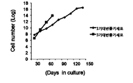
- Figure 2 is a graph showing a comparison of the proliferative power of the placental tissue-derived stem cells and the placenta tissue-derived stem cells of the term (term 3).
- Figure 3 is a result of flow cytometry (FACS analysis) showing the surface expression patterns of stage 1 placental tissue-derived stem cells.
- Figure 4 is the result of observing the increase in the number of cells according to the culture days of the first placental tissue-derived stem cells.
- Figure 5 is a karyotype analysis results by Kimja staining of stem cells derived from the first stage.
- FIG 6 shows the results of reverse transcription polymerase chain reaction (RT-PCR) showing the gene expression patterns of stage 1 placental tissue-derived stem cells (hES: human embryonic stem cell).
- RT-PCR reverse transcription polymerase chain reaction
- FIG. 7 is a PCR result showing the gene expression of stem cells derived from stage 1 placenta and stem cells derived from stage 3 (p5: 5 passages, p15: 15 passages).
- Figure 8 is an immunofluorescence staining (Immunocytochemisty) results showing the gene expression patterns of stage 1 placental tissue-derived stem cells.
- Figure 9 shows immunofluorescence staining (Immunocytochemisty) results showing the nestin gene expression according to ectoderm differentiation of stage 1 placental tissue-derived stem cells and stage 3 placental tissue-derived stem cells.
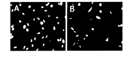
- Figure 10 is a microscopic picture confirming the differentiation-induced differentiation into ectoderm of the first placental tissue-derived stem cells using immunostaining (A: placental tissue-derived stem cells of undifferentiated, B: placental tissue-derived stem cells into neurons ).
- FIG. 12 is a micrograph showing the differentiation-induced appearance of mesenchymal cells of the first placental tissue-derived stem cells using immunostaining.
- FIG. 13 shows RT-PCR results for differentiated mesodermal cells (Undiff: undifferentiated, Diff: differentiated, Od: 0 days, 11d: 11 days, 13d: 13 days, 15d: 15 days).
- Figure 14 is a micrograph confirming the differentiation induced into endoderm of the first placental tissue-derived stem cells using immunostaining.
- the term 'placenta' consists of a decidual membrane derived from the uterine mucosa of the mother and a chorionic membrane, amnion, etc. derived from the fertilized egg, and is a place where material exchange between the mother and the fetus occurs.
- first trimester placenta' refers to a tissue connected to the fetus and mother of 14 weeks or less, which is the first quarter of pregnancy.
- the second trimester placenta refers to the tissues associated with the fetus and mother, 15-25 weeks, in the second trimester.
- the third trimester placenta refers to the tissues associated with the fetus and the mother, typically 26-40 weeks, the third trimester of pregnancy in which the fetus can be born and survive.
- the term 'stem cell' refers to the undifferentiated cells of the stage before they are differentiated into each cell constituting the tissue, and differentiates into specific cells by specific differentiation stimulation or environment. .
- the term 'embryonic stem cell' refers to any undifferentiated cell derived from an embryo that has the potential to be a variety of specialized cells.
- adult stem cells' used in the present invention refers to stem cells appearing in the stage of development or adult formation of each organ of the embryo.
- Adult stem cells are generally limited to cells that make up a specific tissue.
- progenitor cells' includes not only multipotent undifferentiated cells but also monopotnet undifferentiated cells.
- tissue' refers to a collection of cells having substantially the same function and / or shape in a multicellular organ.
- the origin may be a collection of cells of the same origin or a collection of cells of different origin as long as they have the same function and / or shape.
- the term 'differentiation' refers to a phenomenon in which a structure or a function is specialized while a cell divides and grows, that is, a cell or a tissue of an organism has a shape or function to perform a task given to each. It means to change.
- relatively simple systems are separated into two or more qualitatively different sub systems. For example, qualitatively between parts of a living organism that were almost homogeneous in the first place, such as head or torso distinctions between eggs that were initially homogenous in the development, or cells such as muscle cells or neurons.
- Phosphorus difference or as a result, is a state divided into subclasses or subclasses that can be distinguished qualitatively.
- the term 'cell therapeutic agent' is a medicine (US FDA regulation) used for the purpose of treatment, diagnosis, and prevention of cells and tissues prepared by isolation, culture, and special manufacture from humans, and the function of tissues of cells. It refers to a medicine used for the purpose of treatment, diagnosis, and prevention through a series of actions such as proliferating, selecting, or otherwise changing the biological characteristics of a living autologous, allogeneic or heterologous cell in vitro in order to restore it.
- Cell therapy agents are broadly classified into somatic cell therapy and stem cell therapy according to the degree of differentiation of cells, and the present invention relates in particular to stem cell therapy.
- the present invention relates to a method for separating stem cells from the first placenta (14 weeks or less pregnant), specifically, the treatment of fine placenta tissues less than 14 weeks pregnant with 0.01 ⁇ 0.5% collagenase
- the present invention relates to a method for isolating placental tissue-derived stem cells, which is then cultured in a fibroblast growth factor (FGF) -containing medium and then recovered.
- FGF fibroblast growth factor
- the present invention is the 1 st placental tissue-derived stem cells isolated by the method 2-30% fetal bovine serum (Fetal Bovine Serum, FBS), 2-20 ng / ml fibroblast growth factor and 0.1
- the present invention relates to a method of proliferation of placental tissue-derived stem cells suspended in Dulbecco's Modified Eagle's Media (DMEM) medium containing ⁇ 0.5 mM beta mercaptoethanol ( ⁇ - mercaptoethanol).
- DMEM Dulbecco's Modified Eagle's Media
- the content of the collagenase is 0.01 ⁇ 0.5% (W / V)
- the content of FGF may be characterized in that 2 ⁇ 20 ng / ml
- the placental tissue is 5 weeks or more 7 It may be a mammalian placental tissue of less than a week, the placental tissue may be human-derived placental tissue.
- placental stem cells have been produced with some tissues of term placenta that can be obtained at birth.
- the present invention provides stem cells isolated and cultured in the first placenta, which can be obtained due to the stop of the development of the fetus during pregnancy.
- stem cells unlike differentiated cells in which cell division has stopped, are capable of producing the same cells as themselves by cell division (proliferation; expansion), and when differentiation stimulation is applied It is characterized by having plasticity in differentiation because it can be differentiated into other cells by different environment or differentiation stimulus.
- These stem cells can be divided into embryonic and adult stem cells according to the origin of development, and in the present invention includes both embryonic stem cells and adult stem cells, but preferably means adult stem cells.
- Adult stem cells like embryonic stem cells, are capable of proliferation in a cultured state and have a differentiation ability to develop into cells having characteristic shapes and specialized functions according to changes in culture conditions and culture conditions.
- the adult stem cells can differentiate into tissue-specific progenitor cells inherent in the human body.
- Adult stem cells can be virtually any differentiated cell in the body and have the potential to generate replacement cells for a wide range of tissues and organs such as the heart, pancreas, nerve tissue, muscle, cartilage, and the like.
- Such adult stem cells are obtained from mammalian primates, preferably from most tissues such as human bone marrow, cord blood, blood, fat, liver, skin, gastrointestinal tract, placenta, uterus, brain, pancreas, liver, eyes and fetal tissue.
- the present invention relates to stem cells derived from stage 1 placental tissue, in particular, having differentiation ability into various tissues and having immunomodulatory ability.
- the obtained placental tissues are separated into single cell bodies by treatment with trypsin solution and / or collagenase, and then cultured in a suitable medium in which growth factors such as FGF and EGF are added in an appropriate amount, followed by FACS and the like.
- FGF growth factor
- EGF EGF
- adult stem cells can be isolated according to the growth rate.
- DMEM Dulbecco's Modified Eagle's Media
- ⁇ - mercaptoethanol beta - mercaptoethanol
- non-essential amino acids 1% non-essential amino acids
- antibiotics penicillin / streptomycin
- the finely cut placental tissue is left in a culture dish to induce cells to ooze out of the placental tissue, and after 3-7 days, the remaining placental tissue fragments are removed and attached to the culture dish. Induces them to form cell lines.
- the passage number reaches 30-40. Comparing the cell proliferation rate by separating and culturing cells in the same placenta and tertiary placental tissues, the average number of stem cells derived from tertiary placental tissues showed a sharp increase in the number of passages from 10 to 15 on average. -70 days before and after the life of the cell no longer grows. In comparison with the first placental tissue-derived stem cells according to the present invention shows a steady increase until the passage number 30 ⁇ 40 (120-150 days), and will maintain a steady increase even in more than 40 passages. These results demonstrate that the proliferative power of stage 1 placental tissue-derived stem cells is superior to stage 3 placental tissue-derived stem cells.
- the number and type of proliferated cells can be determined by flow cytometry, cell sorting, immunocytochemistry (eg staining with tissue specific or cell-label specific antibodies), fluorescence activated cell sorting (FACS), Measure changes in morphology and surface labeling of cells using standard cell detection techniques such as magnetic activated cell sorting (MACS), or morphology of cells using optical or confocal microscopy. Or can be easily monitored by measuring changes in gene expression using techniques well known in the art such as PCR and gene expression profiling.
- an antibody that specifically recognizes a surface antigen of a cell is labeled with fluorescence, and the fluorescence intensity is measured by converting the fluorescence intensity into an electrical signal by measuring the fluorescence of a conjugate of the labeled antibody and antigen to quantify the antigen expression level of the cell.
- fluorescent materials to be used it is possible to separate cells expressing a plurality of surface antigens.
- Fluorescent materials usable herein include FITC (fluorescein isothiocyanate), PE (phycoerythrin), APC (allo-phycocyanin), TR (TexasRed), Cy3, CyChrome, Red613, Red670, TRI-Color, QuantumRed and the like.
- the stem cell solution obtained above is collected, and the cells are separated by centrifugation or the like, and then directly stained with an antibody. After culturing and propagating in a medium, the method of staining an antibody can be used. To stain the cells, firstly, the primary antibody recognizing the surface antigen and the desired cell sample are mixed and incubated for 30 minutes to 1 hour on ice. If the primary antibody is labeled with fluorescence, it is separated by a flow cytometer after washing.
- the fluorescently labeled secondary antibody having a binding activity to the primary antibody and the cells reacted with the primary antibody are mixed and incubated in ice water for 30 minutes to 1 hour. After washing, the cells stained with the primary antibody and the secondary antibody are separated by flow cytometry.
- the present invention shows positive immunological properties for (a) CD9, CD13, CD29, CD44, CD90, CD105 and HLA-Class I, obtained by the separation method, and CD14, CD19, CD34, CD38 , Negative immunological properties for CD45, CD127 and HLA-DR; (b) expresses genes of Oct4, Nanog, Tbn, Klf4, Activin and Rex1; (c) has the ability to differentiate into ectoderm, mesoderm or endoderm derived cells; And (d) first placental tissue-derived stem cells of 14 weeks or less in pregnancy, which are characterized by being maintained over 30 passages in an undifferentiated state.
- Adult stem cells derived from the first placental tissue of the present invention are homogeneous, sterile and readily obtained in a form (pharmaceutical grade) suitable for administration to humans. After prolonged incubation, the cells can be characterized with CD series antigen markers such as CD29 (mononuclear cell marker), CD90 (mononuclear stem cell marker) and subjected to FACS analysis.
- CD series antigen markers such as CD29 (mononuclear cell marker), CD90 (mononuclear stem cell marker) and subjected to FACS analysis.
- Preferred placental tissue derived stem cells obtained by the method of the invention can be characterized by the presence of the following cell surface markers: positive immunological for CD9, CD13, CD29, CD44, CD90, CD105 and HLA-Class I Properties, negative immunological properties for CD14, CD19, CD34, CD38, CD45, CD127 and HLA-DR.
- cell surface labels are identified according to methods known in the art, eg, by batch measurements by flow cytometry, followed by washing and staining with anti-cell surface labeling antibodies.
- the placental stem cells of the present invention can also be identified using Oct4 markers, which can be said to be cell markers in undifferentiated state.
- Oct4 is well known as an undifferentiated state marker in stem cells, and in the art, Korean Patent Application No. 10-2004-0105716, "monoclonal antibody specific for human embryonic stem cells", Korean Patent Application No. 10-2004-0096780 "Double-stranded RNA for inhibiting Oct4 gene expression in maintenance of undifferentiated state of mammalian embryos and stem cells," Korean Patent Application No. 10-2006-0092128 As shown in “Cells and Manufacturing Methods", etc., it is common to perform an Oct4 expression test as an experiment for proving that they are stem cells in an undifferentiated state.
- Oct4 by confirming the expression of Nanog, Tbn, Klf4, Activin and Rex1 it can be confirmed that the first placental tissue-derived stem cells according to the present invention has characteristics as stem cells.
- RT-PCR Reverase Transcriptase-Polymerase Chain Reaction
- RT-PCR is a technique for synthesizing a corresponding cDNA using a template of RNA of a specific site, and then PCR amplification using the same, the experimental process (1) using reverse transcriptase It consists of preparing cDNA from RNA and (2) amplifying a specific region using cDNA. (2) process is the same as amplifying a specific gene region from genomic DNA.
- This method is not only simpler than the RNA analysis that was possible through methods such as Northern blot hybridization, but also helps in the study of mRNA sequencing and transcription, since the gene sequence can be determined. Technology is known throughout the industry.
- Stage 1 placental tissue-derived stem cells of the present invention show a positive response to the expression of Oct4, Nanog, Tbn, Klf4, Activin and Rex1 gene markers.
- Oct4 is well known as a marker of undifferentiated state in stem cells, and it is already known in the art that Nanog, Klf4, Activin or Rex1 is also a marker of human stem cells.
- the conventional tertiary placental tissue-derived stem cells were killed without being cultured for more than 20 generations, the first-placed placental tissue-derived stem cells according to the present invention have a proliferative capacity of 30 to 40 generations or more, preferably 40 or more generations.
- the first placental tissue-derived stem cells may be characterized in that the expression level of the Klf4 gene is increased 1.2 to 4 times compared to the third placental tissue-derived stem cells, and the expression level of the Activin gene is increased 1.2 to 4 times. Characterized in that, the expression level of the Rex1 gene may be characterized by an increase of 4 to 8 times. In one embodiment of the present invention, the gene expression capacity was compared in the fifth passage and the 15th passage, but the number of passages increased more than that, it has such excellent expression increase ability even in 30-40 passages or more.
- Early placental stem cells obtained by the method of the present invention have the ability to differentiate into mesoderm, endoderm and ectoderm derived cells. For example, it may be induced to differentiate along specific cell lineages, including beta-pancreatic cell differentiation, osteoblast differentiation, neuronal differentiation.
- the present invention relates to a method of differentiating stage 1 placental stem cells obtained by the isolation and / or proliferation method into ectoderm derived cells, mesodermal derived cells, and / or endoderm derived cells.
- placental stem cells according to the present invention include a method of differentiating neurons, bone cells and insulin secreting pancreatic beta cells.
- Measurement of differentiation of stem cells into specific cell types can be performed by methods well known in the art, for example, (1) placental stem cells are cultured in a medium containing 0.01% to 1% polyethyleneimine Forming a spherical body; And reattaching the globular body to a general culture dish to induce differentiation into neurons, and (2) placenta stem cells to dexamethasone and ascorbic acid-2-phosphate.
- beta-glycophosphate beta-glycophosphate incubated in bone formation induction medium to induce differentiation of placental stem cells into bone formation cells
- placental stem cells N2, B27 supplement and 1 ⁇ Differentiation into insulin-secreting beta-pancreatic cells through culturing in a medium containing 50 ng / ml of bFGF and exchanging the medium with a medium containing N2, B27 supplement and 1-20 mM nicotinamide Can be derived.
- the differentiation may be performed using techniques such as flow cytometry or immunocytochemistry to measure cell surface labels (eg staining cells with tissue-specific or cell-label specific antibodies) and changes in morphology, with optical microscopy. Or by examining the morphology of the cells using confocal microscopy, or by measuring changes in gene expression using techniques well known in the art such as PCR and gene-expression profiles.
- the present invention relates to a cell therapeutic agent containing first stage placental tissue-derived stem cells having the characteristics of differentiating into ectoderm-derived cells, mesoderm-derived cells and / or endoderm-derived cells.
- the present invention is not limited thereto, but as an example, a cell therapeutic agent for treating neurological diseases containing stem cells derived from first placental tissue having the characteristics of differentiating into neurons as an active ingredient, and having the characteristics of differentiating into bone forming cells
- Cell therapy agent for treating bone deletion disease containing stem cell derived from 1st placental tissue as an active ingredient Cell therapy agent for treating diabetes mellitus, containing stem cell derived from 1st placental tissue having the characteristics of differentiating into insulin secreting pancreatic beta cells as an active ingredient
- the stem cells according to the present invention may function as cell therapeutic agents for all diseases that can be treated due to the cells to be differentiated, as described below, due to the trioderm differentiation ability. have.
- Stage 1 placental tissue-derived stem cells according to the present invention can be used for various kinds of treatments, for example, by engraftment, transplantation or infusion of stem cells or derived cell populations.
- the placental stem cells of the present invention can replace or enhance existing tissues, become new or changed tissues, or combine with biological tissues or structures.
- the first placental tissue-derived stem cells of the present invention may be replaced.
- treatment of neurological diseases such as Alzheimer's and Parkinson's disease, muscular diseases such as progressive muscular dystrophy, Lou Gehrig's disease, bone deletion diseases such as osteoarthritis, osteoporosis, and diabetes containing placental stem cells or differentiated cells thereof of the present invention. It can be used for cell therapy.
- the placental stem cells of the present invention are transplanted, for example, of liver, pancreas, kidney, lung, nervous system, muscular system, bone, bone marrow, thymus, spleen, submucosal tissue, gonad or hair. It can be used to enhance or replace stem cells or derived cells.
- placental stem cells of the present invention typically substitute for a specific class of progenitor cells (eg, chondrocytes, stem cells, hematopoietic cells, pancreatic parenchymal cells, neural stem cells, muscle derived cells, etc.) in the therapy in which the derived cells are used. Can be used.
- Placental stem cells of the present invention can be used to strengthen, treat or replace cartilage or ligaments.
- an artificial prosthesis eg, buttock prosthesis
- a joint eg, knee
- Placental stem cells of the present invention can be used to treat tissue and organ damage caused by certain diseases.
- the placenta stem cells may be administered to the patient to regenerate or repair tissues or organs damaged by the disease (eg, to improve the immune system's ability after chemotherapy or radiation, and to repair heart tissue after myocardial infarction). do).
- the cell therapy agent of the present invention can be formulated by methods known to those skilled in the art. For example, it can be used parenterally in the form of an injection as a sterile solution or suspension with water or other pharmaceutically acceptable liquid, if necessary.
- pharmaceutically acceptable carriers or media specifically sterile water or physiological saline, vegetable oils, emulsifiers, suspending agents, surfactants, stabilizers, excipients, vehicles, preservatives, binders and the like, as appropriately combined
- those formulated by mixing in the form of unit doses required for pharmaceutical implementation are those formulated by mixing in the form of unit doses required for pharmaceutical implementation.
- the amount of the active ingredient should be determined so as to obtain an appropriate dose in a predetermined range, and the sterile composition for injection may be prescribed according to a conventional preparation using a vehicle such as distilled water for injection.
- the placental stem cells of the present invention can also be formulated as injectables (eg, WO 96/39101, incorporated herein by reference).
- aqueous solutions for injection for example, isotonic solutions containing physiological saline, glucose or other adjuvants, such as D-sorbitol, D-mannose, sodium chloride, and suitable dissolution aids such as alcohol, Ethanol, polyalcohols such as propylene glycol, polyethylene glycol, nonionic surfactants such as polysorbate 80 (TM) and HCO-50.
- suitable dissolution aids such as alcohol, Ethanol, polyalcohols such as propylene glycol, polyethylene glycol, nonionic surfactants such as polysorbate 80 (TM) and HCO-50.
- suitable dissolution aids such as alcohol, Ethanol, polyalcohols such as propylene glycol, polyethylene glycol, nonionic surfactants such as polysorbate 80 (TM) and HCO-50.
- buffers such as phosphate buffers, sodium acetate buffers, analgesics such as procaine hydrochloride, stabilizers such as benzyl alcohol, phenols, antioxidants.
- analgesics such as procaine hydrochloride
- stabilizers such as benzyl alcohol, phenols, antioxidants.
- Administration into the patient's body is preferably parenteral administration, specifically, once administration to the damaged area is basic, but may be administered several times.
- the administration time may be a short time or a long time continuous administration. More specifically, there may be mentioned injection, transdermal administration and the like.
- the first placenta was successfully implanted after embryo transplantation at the Maria Infertility Hospital, according to the guidelines of the Institutional Review Board of the Maria Infertility Hospital. However, since the heart was stopped due to ultrasonography, it was collected. It was used for research. Separated in aseptic condition and transferred to the laboratory.
- DPBS Dulbecco's Phosphate Buffered Saline
- Washed cells were treated with 10% Fetal Bovine Serum (FBS), 4 ng / ml fibroblast growth factor (FGF), 0.1 mM beta-mercaptoethanol ( ⁇ - mercaptoethanol), and 1% non-essential amino acids (non- Incubated in Dulbecco's Modified Eagle's Media (DMEM) medium containing essential amino acids) and antibiotics (penicillin / streptomycin), and cultured in a humidified chamber at 37 ° C. and 5% CO 2 .
- FBS Fetal Bovine Serum
- FGF fibroblast growth factor
- ⁇ - mercaptoethanol beta-mercaptoethanol
- 1% non-essential amino acids non- Incubated in Dulbecco's Modified Eagle's Media (DMEM) medium containing essential amino acids
- antibiotics penicillin / streptomycin
- the isolated cells removed the non-adherent tissue debris after 5 days of medium exchange. When the cells proliferated and became denser on the culture surface (75 cm 2 ), they were almost full (passfluent 1).
- the finely cut placental tissue is left in a culture dish to induce cells to seep out of the placental tissue, and after 3-7 days, the remaining placental tissue fragments are removed and the cells growing on the petri dish are allowed to grow. Induced to form cell lines (FIG. 1).
- the proliferation rate of the isolated human stage 1 placental stem cells was investigated. Human stage 1 placental stem cell lines were cultured up to 90-150 days after culturing 16 cell lines, and compared to the term (term 3) placenta-derived stem cell proliferation rate obtained at the time of delivery, which was also reported in cell proliferation rate. Higher results.
- Cells were harvested with PBS with 2 mM EDTA / 5% FBS and washed twice with PBS to analyze surface antigens of human stage 1 placental stem cells. Subsequently, fluorescein isothiocyanate (FITC) or PE (phycoerythrin) bound antibody was added to the cell suspension in an appropriate ratio in 2% FBS / PBS buffer and treated at 4 ° C. for 30 minutes, followed by FACS Vantage SE (Becton & Dickson). The fluorescence of the cells was measured. 5 ⁇ g / ml of propidium iodide was added to remove dead cells and debris prior to analysis. The results obtained were analyzed using the CellQuest (Becton & Dickinson) program.
- FITC fluorescein isothiocyanate
- PE phycoerythrin
- Example 6 karyotyping of stem cells derived from placental tissue
- the first placental stem cells with about 70% confluence in a 100 mm culture dish were treated with 0.1 ⁇ g / ml of colcemid for 2 hours, collected, washed and 75 mM potassium chloride at 37 ° C. The solution was treated for 20 minutes. After the treatment, the stock solution was washed by centrifugation, and then fixed with a fixed solution mixed with methanol and acetic acid (3: 1).
- the fixed cells were centrifuged again and resuspended in 0.5 ml of the same fixed solution.
- One drop of the suspension was dropped on a slide at a height of 30 cm. After the sample was completely dried, the slides were treated with 0.025% trypsin for 30 seconds and again washed with PBS. Samples were stained with 10% Giemsa solution and subjected to karyotyping with Cytovision of Applied Imaging.
- the results are shown in FIGS. 4 and 5.
- the established stage 1 placental stem cells proliferated continuously without any visible karyotype or morphological changes in vitro for 90-150 days.
- RT-PCR analysis was performed on the Klf4, Activin, and Rex1 genes of the cells by separating and culturing cells in the placental and tertiary placental tissues in the same manner.
- Target sequences from cDNA were amplified using the I-taq premix kit (Invitron Biotechnology) under the following conditions. The denatured cDNA was amplified for 35 minutes at 95 ° C, then denatured at 95 ° C for 30 seconds, then annealed at 60 ° C for 45 seconds, and amplified at 72 ° C for 45 seconds. It was. Amplified cDNA was identified by separation on 1.2% agarose gel and stained with ethidium bromide. When loading the amplified cDNA, the pixel intensity of GAPDH expression was normalized to cDNA amount of each experimental group.
- the first placental tissue-derived stem cells are expressed in Oct4, Nanog, Tbn, Klf4, Activin genes expressed in embryo-associated tissues or cells, the immunofluorescence staining results are shown in Figure 8 As shown.
- the expression levels of Klf4, Activin and Rex1 genes in expression genes are significantly higher in first placental tissue-derived stem cells than in third placental stem cells. This may be associated with differentiation and proliferation (FIG. 7).
- RT-PCR analysis was performed on the nestin gene of cells by separating and culturing cells in the first place and the third placenta tissue.
- stage 1 placental tissue had an initial nestin expression of 30.4%, which is significantly lower than that of cells derived from stage 3 placental. This suggests that cells derived from the stage 1 placenta are much more immature and can be said to agree with the above results.
- the starter group was more homogenous and more capable of differentiation than the 3rd cell differentiation rate of 91.7%. It can be confirmed (FIG. 9).
- Human placenta derived stem cells established in Example 3 were cultured in a culture dish coated with 0.01-1% polyetherenimine in an amount of 0.2 ⁇ 10 5 to 2.5 ⁇ 10 5 per cm 2. On day 2, stem cells in each culture dish aggregated with each other without adhering to the culture dish surface to form spheroids. This spherical body is reattached to a general culture dish to induce differentiation.
- Differentiation-induced cells were transferred onto a cover slip, incubated for 2 to 3 days, fixed with 4% paraformaldehyde solution at room temperature for 15 minutes, and washed with PBS.
- the fixed cells were diluted in PBS solution containing 5-10% Triton X-100 and normal goat serum at the rate indicated by the antibody producer, added to the sample, and then at room temperature. The reaction was carried out overnight at 1 hour or in a cold state.
- TRITC tetrarhodamine isothiocyanate
- FITC conjugate was reacted with primary antibody at room temperature or in a cold state. Treated in the same way.
- the cell nuclei were stained with Hoechst 33342 (10 ⁇ g / ml, Sigma) and observed by fluorescence microscopy.
- FIG. 10A shows undifferentiated stage 1 placental derived stem cells
- FIG. 10 B is a photograph showing stage 1 placental stem cells differentiated into neural progenitor cells showing Nestin expression.
- RT-PCR analysis was performed on Otx1 and NeuroD genes with the cells differentiated into ectoderm cells. The analysis was performed in the same manner as the reverse transcription polymerase chain reaction described in Example 7.
- stage 1 placental stem cells were induced to differentiate into ectoderm cells.
- Human stage 1 placenta-derived stem cells established in Example 3 were collected at a density of 0.1 ⁇ 10 4 to 1 ⁇ 10 4 per cm 2 (0.1 ⁇ mol / l dexamethasone, 0.05 mmol / l ascorbic acid-2-phosphate, 10 mmol / l beta-glycophosphate (20% human serum) for 10-21 days. After differentiation, Alizarin Red S staining and Alkaline phosphatase (ALP) staining confirmed the differentiation of placental stem cells into mesodermal cells related to bone formation.
- ALP Alkaline phosphatase
- alkaline phosphatase activity as a bone differentiation marker by staining
- cells differentiated from bone formation differentiation medium were cultured on a chamber slide, washed with sterile tertiary distilled water, and then ALP staining kit (Sigma-Aldrich, Steinheim). , Germany). Immersion was fixed in Citrate-Acetone-Formaldehyde fixative for 30 seconds and washed with deionized water. Slides were incubated for 15 minutes at room temperature in alkaline dye mixture, washed again, and stained with Neutral Red solution.
- the degree of ALP staining and Alizarin Red S staining can be confirmed that compared to the undifferentiated stem cells, it was confirmed that differentiation into mesodermal cells related to bone formation.
- RT-PCR analysis was performed on BGP, ALP, and ColiaI genes with the cells differentiated into mesodermal cells. RT-PCR was analyzed by the same process as the reverse transcription polymerization reaction described in Example 7.
- the cells began to differentiate and showed expression of genes related to bone formation such as BGP, ALP, Colia1 from 10 days to 21 days. These results show that human placental stem cells can differentiate into mesodermal cells related to bone formation.
- Human placenta-derived stem cells established in Example 3 were endothelial differentiation-inducing medium (ES-Cult basal containing N2 and B27 supplement and 10-25 ng / ml bFGF) at a concentration of 0.3 ⁇ 10 4 to 4 ⁇ 10 4 / cm 2. medium) and incubated for 6-7 days and exchanged for endoderm differentiation maturation medium (ES-Cult basal medium containing N2, B27 supplement and 10 mM nicotinamide) for 6-7 days.
- endothelial differentiation-inducing medium ES-Cult basal containing N2 and B27 supplement and 10-25 ng / ml bFGF
- endoderm differentiation maturation medium ES-Cult basal medium containing N2, B27 supplement and 10 mM nicotinamide
- Immunostaining assays were performed for Glut2, Insulin antibodies using the cells differentiated into endoderm cells. Immunostaining was analyzed in the same manner as the immunostaining assay mentioned in Example 6.
- RT-PCR analysis was performed on the Glut2 and Glucagon genes with the cells differentiated into endoderm cells. RT-PCR was analyzed in the same manner as the reverse transcription polymerase chain reaction described in Example 7.
- Glut2 and Glucagon were confirmed by differentiation of placental stem cells to confirm that they differentiated into true endoderm cells.
- the placental tissue-derived adult stem cells according to the present invention are very useful as cell therapeutic agents because they have excellent cell proliferation rate and more favorable differentiation ability than the existing placental tissue-derived stem cells. .
- it has the ability to differentiate into neural cells, bone forming cells or insulin secreting pancreatic beta cells, and is effective in the treatment of bone deletion diseases, neurological diseases, diabetes and the like.
Landscapes
- Health & Medical Sciences (AREA)
- Life Sciences & Earth Sciences (AREA)
- Engineering & Computer Science (AREA)
- Biomedical Technology (AREA)
- Chemical & Material Sciences (AREA)
- Zoology (AREA)
- Developmental Biology & Embryology (AREA)
- Biotechnology (AREA)
- Organic Chemistry (AREA)
- Bioinformatics & Cheminformatics (AREA)
- Genetics & Genomics (AREA)
- Wood Science & Technology (AREA)
- Cell Biology (AREA)
- General Health & Medical Sciences (AREA)
- Reproductive Health (AREA)
- Microbiology (AREA)
- Biochemistry (AREA)
- General Engineering & Computer Science (AREA)
- Pregnancy & Childbirth (AREA)
- Gynecology & Obstetrics (AREA)
- Pharmacology & Pharmacy (AREA)
- Public Health (AREA)
- Medicinal Chemistry (AREA)
- Veterinary Medicine (AREA)
- Animal Behavior & Ethology (AREA)
- Immunology (AREA)
- Virology (AREA)
- Epidemiology (AREA)
- General Chemical & Material Sciences (AREA)
- Chemical Kinetics & Catalysis (AREA)
- Neurosurgery (AREA)
- Neurology (AREA)
- Nuclear Medicine, Radiotherapy & Molecular Imaging (AREA)
- Medicines Containing Material From Animals Or Micro-Organisms (AREA)
- Micro-Organisms Or Cultivation Processes Thereof (AREA)
Abstract
L'invention concerne des cellules couches issues de tissu de placenta primaire à 14 semaines ou moins de gestation, un procédé de séparation de ces cellules, un procédé permettant la prolifération de celles-ci et un agent thérapeutique cellulaire contenant ces cellules souches. Plus particulièrement, l'invention concerne des cellules souches issues de tissu de placenta primaire à 14 semaines ou moins de gestation, un procédé de séparation de ces cellules, un procédé permettant la prolifération de celles-ci et un agent thérapeutique cellulaire contenant ces cellules souches, lesdites cellules souches (a) possédant des propriétés immunologiques positives par rapport à CD9, CD13, CD29, CD44, CD90, CD105 et le système HLA de classe I, et des propriétés immunologiques négatives contre CD14, CD19, CD34, CD38, CD45, CD127 et le système HLA-DR; (b) exprimant les gènes Oct4, Nanog, Tbn, Klf4, d'activine et Rex1; (c) étant capables de se différencier en cellules issues de l'ectoderme, du mésoderme ou de l'endoderme; et étant maintenues pendant 30 générations ou plus dans un état indifférencié. Bien qu'étant des cellules souches adultes, les cellules souches de l'invention issues de tissu de placenta primaire présentent une excellente capacité de prolifération cellulaire et une capacité de différenciation avantageuse par rapport à des cellules souches existantes issues de tissu de placenta primaire, et peuvent par conséquent être utilisées efficacement comme agent thérapeutique cellulaire. En particulier, ces cellules souches peuvent se différencier en cellules nerveuses, en cellules ostéogènes ou en cellules pancréatiques bêta sécrétant de l'insuline, et sont également efficaces pour traiter des maladies de déficit osseux, des maladies nerveuses, le diabète, etc.
Applications Claiming Priority (2)
| Application Number | Priority Date | Filing Date | Title |
|---|---|---|---|
| KR1020100067086A KR20120006386A (ko) | 2010-07-12 | 2010-07-12 | 1기 태반조직 유래 줄기세포 및 이를 함유하는 세포치료제 |
| KR10-2010-0067086 | 2010-07-12 |
Publications (2)
| Publication Number | Publication Date |
|---|---|
| WO2012008733A2 true WO2012008733A2 (fr) | 2012-01-19 |
| WO2012008733A3 WO2012008733A3 (fr) | 2012-04-19 |
Family
ID=45469910
Family Applications (1)
| Application Number | Title | Priority Date | Filing Date |
|---|---|---|---|
| PCT/KR2011/005103 WO2012008733A2 (fr) | 2010-07-12 | 2011-07-12 | Cellules souches issues de tissu de placenta primaire et agent thérapeutique contenant celles-ci |
Country Status (2)
| Country | Link |
|---|---|
| KR (1) | KR20120006386A (fr) |
| WO (1) | WO2012008733A2 (fr) |
Cited By (5)
| Publication number | Priority date | Publication date | Assignee | Title |
|---|---|---|---|---|
| US10232000B2 (en) | 2014-01-08 | 2019-03-19 | Samsung Life Public Welfare Foundation | Stem cells derived from basal portion of chorionic trophoblast layer and cell therapy comprising same |
| WO2019125073A1 (fr) * | 2017-12-22 | 2019-06-27 | 고려대학교 산학협력단 | Milieu conditionné par des cellules dérivées du placenta pour l'induction d'une dédifférenciation à partir de cellules somatiques donnant des cellules souches pluripotentes induites et procédé d'induction d'une dédifférenciation l'utilisant |
| WO2020036245A1 (fr) * | 2018-08-17 | 2020-02-20 | 고려대학교 산학협력단 | Milieu conditionné de cellules dérivées du placenta pour la production et l'amélioration de la fonction de cellules souches neurales humaines, et utilisation associée |
| US10617721B2 (en) | 2013-10-24 | 2020-04-14 | Ospedale San Raffaele S.R.L. | Methods for genetic modification of stem cells |
| US10669526B2 (en) | 2014-01-08 | 2020-06-02 | Samsung Life Public Welfare Foundation | Stem cells derived from pure chorionic trophoblast layer and cell therapy comprising same |
Families Citing this family (1)
| Publication number | Priority date | Publication date | Assignee | Title |
|---|---|---|---|---|
| KR102317052B1 (ko) * | 2020-05-04 | 2021-10-25 | 주식회사 티에스셀바이오 | 태반 유래의 세포외 소포의 항염증 항바이러스 효과 조성물 |
-
2010
- 2010-07-12 KR KR1020100067086A patent/KR20120006386A/ko not_active Application Discontinuation
-
2011
- 2011-07-12 WO PCT/KR2011/005103 patent/WO2012008733A2/fr active Application Filing
Non-Patent Citations (3)
| Title |
|---|
| BATTULA ET AL.: 'Human placenta and bone marrow derived MSC cultured in serum-free, b-FGF-containing medium express cell surface frizzled-9 and SSEA-4 and give rise to multilineage differentiation' DIFFERENTIATION vol. 75, 2007, pages 279 - 291 * |
| MIAO ET AL.: 'Isoration of mesenchymal stem cells from human placenta: Comparison with human bone marrow mesenchymal stem cells' CELL BIOLOGY INTERNATIONAL vol. 30, 2006, pages 681 - 687 * |
| PORTMANN-LANZ ET AL.: 'Placental mesenchymal stem cells as potential autologous graft for pre- and perinatal neuroregeneration' AMERICAN vol. 194, 2006, pages 664 - 673 * |
Cited By (5)
| Publication number | Priority date | Publication date | Assignee | Title |
|---|---|---|---|---|
| US10617721B2 (en) | 2013-10-24 | 2020-04-14 | Ospedale San Raffaele S.R.L. | Methods for genetic modification of stem cells |
| US10232000B2 (en) | 2014-01-08 | 2019-03-19 | Samsung Life Public Welfare Foundation | Stem cells derived from basal portion of chorionic trophoblast layer and cell therapy comprising same |
| US10669526B2 (en) | 2014-01-08 | 2020-06-02 | Samsung Life Public Welfare Foundation | Stem cells derived from pure chorionic trophoblast layer and cell therapy comprising same |
| WO2019125073A1 (fr) * | 2017-12-22 | 2019-06-27 | 고려대학교 산학협력단 | Milieu conditionné par des cellules dérivées du placenta pour l'induction d'une dédifférenciation à partir de cellules somatiques donnant des cellules souches pluripotentes induites et procédé d'induction d'une dédifférenciation l'utilisant |
| WO2020036245A1 (fr) * | 2018-08-17 | 2020-02-20 | 고려대학교 산학협력단 | Milieu conditionné de cellules dérivées du placenta pour la production et l'amélioration de la fonction de cellules souches neurales humaines, et utilisation associée |
Also Published As
| Publication number | Publication date |
|---|---|
| WO2012008733A3 (fr) | 2012-04-19 |
| KR20120006386A (ko) | 2012-01-18 |
Similar Documents
| Publication | Publication Date | Title |
|---|---|---|
| JP2022046511A (ja) | 胎盤幹細胞を使用する免疫調節 | |
| Fauza | Amniotic fluid and placental stem cells | |
| WO2015105356A1 (fr) | Cellules souches dérivées d'une couche trophoblastique chorionique pure, et thérapie cellulaire comprenant celles-ci | |
| WO2011049414A2 (fr) | Procédé pour l'induction d'une migration de cellules souches adultes dérivées de tissu adipeux | |
| WO2012008733A2 (fr) | Cellules souches issues de tissu de placenta primaire et agent thérapeutique contenant celles-ci | |
| WO2015105357A1 (fr) | Cellules souches dérivées de la partie basale de la couche trophoblastique chorionique, et thérapie cellulaire comprenant celles-ci | |
| WO2012173358A2 (fr) | Cellules souches pluripotentes issues de cellules somatiques du testicule, procédé de production de celles-ci, et composition pharmaceutique pour le traitement de l'impuissance comprenant celles-ci | |
| WO2013085303A1 (fr) | Cellules souches multipotentes dérivées de la membrane amniotique canine | |
| WO2019198962A1 (fr) | Procédé pour favoriser la prolifération et la différenciation de cellules souches pluripotentes induites préparées à partir de cellules souches issues d'urine | |
| WO2012033352A2 (fr) | Cellules souches multipotentes dérivées du liquide amniotique équin et leur procédé de production | |
| WO2019103528A2 (fr) | Composition de milieu de culture asérique | |
| WO2013165120A1 (fr) | Procédé pour cultiver des cellules souches de crête neurale, et utilisation de celles-ci | |
| WO2021118226A1 (fr) | Procédé de préparation d'une cellule souche mésenchymateuse à partir d'une cellule souche pluripotente humaine et cellules souches mésenchymateuses ainsi préparées | |
| WO2011126177A1 (fr) | Procédé d'augmentation de l'activité dans des cellules souches humaines | |
| WO2019050350A2 (fr) | Cellule de type de sertoli dérivée de cellule souche procédé de préparation associé et utilisation associée | |
| WO2022004938A1 (fr) | Procédé de préparation de cellules souches de type mésenchymateuses | |
| WO2011102680A2 (fr) | Antigène cd49f favorisant la prolifération, la pluripotence et la reprogrammation de cellules souches adultes par l'intermédiaire de la voie pi3k/akt/gsk3 | |
| WO2013077639A1 (fr) | Cellules souches mésenchymateuses issues d'une membrane amniotique équine | |
| WO2022108165A1 (fr) | Procédé de production d'exosomes isolés à partir de cellules souches mésenchymateuses dérivées de cellules souches pluripotentes induites, et utilisation associée | |
| WO2010008157A2 (fr) | Procédé permettant de différencier des cellules souches de cellules ectodermiques | |
| WO2017039251A1 (fr) | Cellule adhérente postnatale améliorée, et utilisation de celle-ci | |
| WO2021096218A1 (fr) | Procédé d'isolement de culture pure de cellules endothéliales vasculaires, milieu de maintien des caractéristiques de cellules endothéliales vasculaires, et procédé de culture le comprenant | |
| WO2021054692A1 (fr) | Cellules souches dérivées de villosités adjacentes à la plaque chorionique et agent thérapeutique de cellules de régénération tissulaire les comprenant | |
| WO2018101723A1 (fr) | Nouvelles cellules progénitrices mésenchymateuses fonctionnellement améliorées, composition d'agent thérapeutique cellulaire anti-inflammatoire les contenant, et procédé de préparation de cellules progénitrices mésenchymateuses | |
| WO2019190175A9 (fr) | Méthode pour la différenciation de neurones moteurs à partir de cellules souches mésenchymateuses dérivées de tonsil |
Legal Events
| Date | Code | Title | Description |
|---|---|---|---|
| 121 | Ep: the epo has been informed by wipo that ep was designated in this application |
Ref document number: 11807018 Country of ref document: EP Kind code of ref document: A2 |
|
| NENP | Non-entry into the national phase in: |
Ref country code: DE |
|
| 122 | Ep: pct application non-entry in european phase |
Ref document number: 11807018 Country of ref document: EP Kind code of ref document: A2 |