WO2012002452A1 - Gene introduction method - Google Patents
Gene introduction method Download PDFInfo
- Publication number
- WO2012002452A1 WO2012002452A1 PCT/JP2011/064945 JP2011064945W WO2012002452A1 WO 2012002452 A1 WO2012002452 A1 WO 2012002452A1 JP 2011064945 W JP2011064945 W JP 2011064945W WO 2012002452 A1 WO2012002452 A1 WO 2012002452A1
- Authority
- WO
- WIPO (PCT)
- Prior art keywords
- cells
- cell
- gene
- hours
- virus
- Prior art date
Links
Classifications
-
- C—CHEMISTRY; METALLURGY
- C12—BIOCHEMISTRY; BEER; SPIRITS; WINE; VINEGAR; MICROBIOLOGY; ENZYMOLOGY; MUTATION OR GENETIC ENGINEERING
- C12N—MICROORGANISMS OR ENZYMES; COMPOSITIONS THEREOF; PROPAGATING, PRESERVING, OR MAINTAINING MICROORGANISMS; MUTATION OR GENETIC ENGINEERING; CULTURE MEDIA
- C12N15/00—Mutation or genetic engineering; DNA or RNA concerning genetic engineering, vectors, e.g. plasmids, or their isolation, preparation or purification; Use of hosts therefor
- C12N15/09—Recombinant DNA-technology
- C12N15/63—Introduction of foreign genetic material using vectors; Vectors; Use of hosts therefor; Regulation of expression
- C12N15/79—Vectors or expression systems specially adapted for eukaryotic hosts
- C12N15/85—Vectors or expression systems specially adapted for eukaryotic hosts for animal cells
- C12N15/86—Viral vectors
- C12N15/867—Retroviral vectors
- C12N15/8676—Special methods for targeting systems
-
- C—CHEMISTRY; METALLURGY
- C12—BIOCHEMISTRY; BEER; SPIRITS; WINE; VINEGAR; MICROBIOLOGY; ENZYMOLOGY; MUTATION OR GENETIC ENGINEERING
- C12N—MICROORGANISMS OR ENZYMES; COMPOSITIONS THEREOF; PROPAGATING, PRESERVING, OR MAINTAINING MICROORGANISMS; MUTATION OR GENETIC ENGINEERING; CULTURE MEDIA
- C12N15/00—Mutation or genetic engineering; DNA or RNA concerning genetic engineering, vectors, e.g. plasmids, or their isolation, preparation or purification; Use of hosts therefor
- C12N15/09—Recombinant DNA-technology
- C12N15/63—Introduction of foreign genetic material using vectors; Vectors; Use of hosts therefor; Regulation of expression
- C12N15/79—Vectors or expression systems specially adapted for eukaryotic hosts
- C12N15/85—Vectors or expression systems specially adapted for eukaryotic hosts for animal cells
- C12N15/86—Viral vectors
-
- C—CHEMISTRY; METALLURGY
- C12—BIOCHEMISTRY; BEER; SPIRITS; WINE; VINEGAR; MICROBIOLOGY; ENZYMOLOGY; MUTATION OR GENETIC ENGINEERING
- C12N—MICROORGANISMS OR ENZYMES; COMPOSITIONS THEREOF; PROPAGATING, PRESERVING, OR MAINTAINING MICROORGANISMS; MUTATION OR GENETIC ENGINEERING; CULTURE MEDIA
- C12N15/00—Mutation or genetic engineering; DNA or RNA concerning genetic engineering, vectors, e.g. plasmids, or their isolation, preparation or purification; Use of hosts therefor
- C12N15/09—Recombinant DNA-technology
- C12N15/87—Introduction of foreign genetic material using processes not otherwise provided for, e.g. co-transformation
-
- C—CHEMISTRY; METALLURGY
- C12—BIOCHEMISTRY; BEER; SPIRITS; WINE; VINEGAR; MICROBIOLOGY; ENZYMOLOGY; MUTATION OR GENETIC ENGINEERING
- C12N—MICROORGANISMS OR ENZYMES; COMPOSITIONS THEREOF; PROPAGATING, PRESERVING, OR MAINTAINING MICROORGANISMS; MUTATION OR GENETIC ENGINEERING; CULTURE MEDIA
- C12N2740/00—Reverse transcribing RNA viruses
- C12N2740/00011—Details
- C12N2740/10011—Retroviridae
- C12N2740/13011—Gammaretrovirus, e.g. murine leukeamia virus
- C12N2740/13041—Use of virus, viral particle or viral elements as a vector
- C12N2740/13043—Use of virus, viral particle or viral elements as a vector viral genome or elements thereof as genetic vector
-
- C—CHEMISTRY; METALLURGY
- C12—BIOCHEMISTRY; BEER; SPIRITS; WINE; VINEGAR; MICROBIOLOGY; ENZYMOLOGY; MUTATION OR GENETIC ENGINEERING
- C12N—MICROORGANISMS OR ENZYMES; COMPOSITIONS THEREOF; PROPAGATING, PRESERVING, OR MAINTAINING MICROORGANISMS; MUTATION OR GENETIC ENGINEERING; CULTURE MEDIA
- C12N2740/00—Reverse transcribing RNA viruses
- C12N2740/00011—Details
- C12N2740/10011—Retroviridae
- C12N2740/16011—Human Immunodeficiency Virus, HIV
- C12N2740/16041—Use of virus, viral particle or viral elements as a vector
- C12N2740/16043—Use of virus, viral particle or viral elements as a vector viral genome or elements thereof as genetic vector
Definitions
- the present invention relates to a method for introducing a foreign gene into a target cell using a retroviral vector.
- Retroviral vectors can stably incorporate a target foreign gene into the chromosomal DNA of a target cell. Therefore, gene transfer using a retroviral vector is a preferable gene transfer means for gene therapy in which long-term gene expression is desired.
- the binding of a viral vector to RetroNectin (registered trademark) in the RBV method can be enhanced by using centrifugal force (centrifugal RBV method).
- centrifugal RBV method requires a container that can withstand centrifugal force and an expensive apparatus for performing centrifugal operation, and the operation is multi-step.
- centrifugal RBV method requires a container that can withstand centrifugal force and an expensive apparatus for performing centrifugal operation, and the operation is multi-step.
- centrifugal RBV method requires a container that can withstand centrifugal force and an expensive apparatus for performing centrifugal operation, and the operation is multi-step.
- it is difficult to scale up the treatment capacity when gene introduction into a large amount of cells is required.
- An object of the present invention is to provide a simple and effective method for introducing a gene into a target cell using a retroviral vector.
- the present invention [1] A method for introducing a foreign gene into a target cell using a retroviral vector, comprising the step (a): a retroviral vector loaded with a foreign gene in a culture vessel in which a retroviral binding substance is immobilized A step of incubating for 4 hours or more at a temperature of less than 25 ° C.
- step (b1) a step of washing the culture vessel obtained by step (a), and Step (b2): placing the target cells in the culture vessel washed in Step (b1) and incubating the method
- step (b2) a step of washing the culture vessel obtained by step (a)
- Step (b2) placing the target cells in the culture vessel washed in Step (b1) and incubating the method
- the retrovirus-binding substance is at least one selected from fibronectin, fibroblast growth factor, type V collagen, polylysine, DEAE-dextran, and fragments thereof.
- the retrovirus-binding substance is a substance having cell-binding properties.
- the culture vessel in which the retrovirus-binding substance is immobilized is a culture vessel in which the retrovirus-binding substance and the cell-binding substance are immobilized, and [8] cells.
- the binding substance is at least one selected from cell adhesion proteins, hormones, cytokines, antibodies, sugar chains, carbohydrates, and metabolites, About.
- the present invention provides a simple and effective gene transfer method.
- the method for introducing a gene of the present invention comprises a step (a) “after placing a liquid containing a retroviral vector loaded with a foreign gene in a culture vessel on which a retroviral binding substance is immobilized, Incubating at a temperature for 4 hours or more to obtain a culture vessel to which a retroviral vector is bound ", and step (b)” putting target cells in the culture vessel obtained by step (a) and incubating " .
- a retrovirus is a generic term for RNA viruses belonging to the Retroviridae family whose genome is composed of RNA and has a life cycle that converts the RNA into DNA in an infected cell.
- Oncoretrovirus and lentivirus including.
- An example of the oncoretrovirus is Moloney murine leukemia virus (MMLV).
- Examples of the lentivirus include human immunodeficiency virus type 1 (HIV-1) and simian immunodeficiency virus (SIV).
- the retroviral vector refers to a virus particle produced by genetic recombination technology based on an oncorretrovirus, a lentivirus, etc. belonging to a retrovirus, and an oncorretrovirus vector, a lentivirus vector, or a pseudotype thereof. Includes vectors.
- the oncoretrovirus vector include a vector based on MMLV.
- lentiviral vectors include HIV-1 based vectors and SIV based vectors.
- a pseudotype vector refers to a recombinant retroviral vector having an Env protein whose origin is different from that of a Gag protein or a Pol protein.
- Pseudotype vectors have Env proteins such as vesicular stomatitis virus (VSV), gibbon leukemia virus (GaLV), feline endogenous virus RD114, murine leukemia virus (Ecotropic-env, amphotropic-env, 10A1-env, etc.) Examples include oncoretrovirus vectors and lentivirus vectors.
- a replication-deficient recombinant retrovirus vector is preferably used.
- the vector is non-pathogenic, deficient in replication so that it cannot replicate in infected cells.
- These vectors can infect vertebrate cells, particularly host cells such as mammalian cells, and can stably incorporate a foreign gene carried in the vector into its chromosomal DNA.
- Step (a) “After putting a liquid containing a retroviral vector loaded with a foreign gene in a culture vessel on which a retroviral binding substance is immobilized, incubate at a temperature of less than 25 ° C. for 4 hours or more.
- a liquid containing a retroviral vector loaded with a foreign gene may be placed in a culture vessel on which a retroviral binding substance is immobilized, and then frozen and stored for a certain period of time. .
- the culture container containing the cryopreserved retroviral vector-containing liquid can be directly incubated at a temperature of less than 25 ° C. for 4 hours or more. That is, no additional process for thawing is particularly required. In this case, it is desirable that the incubation in the step (a) is incubation with shaking.
- a retrovirus packaging cell in which a gene encoding a retrovirus structural protein such as a gag-pol gene or an env gene has been incorporated into a chromosome in advance is packaged with a foreign gene.
- a method of producing by introducing a transfer vector carrying a signal, and a packaging plasmid having a gene encoding a retrovirus structural protein such as a gag-pol gene or an env gene in a cell having no retroviral structural protein there can be mentioned a method of producing the aforementioned transfer vector by transfection.
- a foreign gene to be introduced into a target cell can be used by being mounted in a recombinant retroviral vector under the control of an appropriate promoter, for example, an LTR promoter or a foreign promoter present in a retroviral vector.
- an appropriate promoter for example, an LTR promoter or a foreign promoter present in a retroviral vector.
- other regulatory elements cooperating with a promoter and transcription initiation site for example, an enhancer sequence, terminator sequence, and intron sequence may be present in the vector.
- the foreign gene introduced into the target cell may be natural or artificially generated, or a DNA molecule having a different origin is bound by a known means such as ligation. Also good.
- any gene desired to be introduced into the cell can be selected.
- exogenous genes include enzymes encoding proteins and proteins related to the disease to be treated, intracellular antibodies (see, for example, WO94 / 02610 pamphlet), T cell receptor genes, growth factors. , Antisense RNA, RNA causing RNA interference, ribozyme, false primer (see, for example, International Publication No. 90/13641 pamphlet) and the like can be used.
- a CD4 positive T cell exhibiting an anti-HIV effect can be obtained by introducing a gene expressing MazF, which is a sequence-specific RNase, into a CD4 positive T cell as a foreign gene (for example, International Publication No.
- a gene that confers sensitivity to a specific drug for example, a thymidine kinase gene, can be introduced into cells to confer sensitivity to the drug.
- the retroviral vector used in the present invention may contain an appropriate marker gene that enables selection of the transfected cell.
- marker genes include drug resistance genes that confer resistance to antibiotics on cells, reporter genes that can distinguish cells introduced by enzymatic activity and fluorescence, and cell surface marker genes that are localized on the cell surface. Is available.
- the neomycin phosphotransferase gene is used as a marker gene, the introduced cell can be isolated and purified after confirming resistance to G418 as an index.
- LNGFR Low Affinity Nerve Growth Factor Receptor
- the transfected cell can be isolated and purified by using an anti-LNGFR antibody.
- retroviral vectors examples include MFG vector (ATCC No. 68754), ⁇ -SGC vector (ATCC No. 68755), LXSN vector [BioTechniques, Vol. 7, 980-990]. Page (1989)], Takara Bio's DON-5, DON-AI-2, MEI-5 retroviral vectors and pLVSIN-CMV lentiviral vectors, Clontech's Retro-X Q vector series, Lenti-X There are vectors such as vector series.
- vectors are known packaging cell lines such as PG13 (ATCC CRL-10686), PA317 (ATCC CRL-9078), GP + E-86 (ATCC CRL-9642), GP + envAm12 (ATCC CRL-9964), [Proceedings of the National Academy of Sciences of the United States of America (Proc. Natl. Acad. Sci. USA), vol. 85, 6460-6464 (1988) It can be prepared by using a cell line such as ⁇ CRIP as described in “Year)”.
- 293T cells ATCC CRL-11268), G3T-hi cells (manufactured by Takara Bio Inc.), etc. are transiently transfected with a packaging plasmid that expresses the structural protein of the recombinant virus and a retrovirus vector plasmid. It can also be prepared by collecting the culture supernatant.
- Known culture media such as Dulbecco's modified Eagle's medium and Iscob's modified Dulbecco's medium can be used for culturing virus-producing cells and target cells produced by introducing transfer vectors into packaging cells.
- Gibco can be obtained as a commercial product.
- Various components can be added to these media depending on the type of cells targeted for gene transfer and other purposes. For example, serum, various cytokines, and a reducing agent can be added and used for the purpose of promoting or suppressing the growth and differentiation of target cells.
- calf serum for example, calf serum (CS), fetal calf serum (FCS), human serum and the like can be used, and in place of these, serum replacement or purified serum albumin (for example, human serum) Albumin) can also be used.
- Cytokines include interleukins (IL-2, IL-3, IL-4, IL-6, etc.), colony stimulating factors (G-CSF, GM-CSF, etc.), stem cell factor (SCF), erythropoietin.
- G-CSF colony stimulating factors
- GM-CSF GM-CSF
- SCF stem cell factor
- erythropoietin erythropoietin
- the culture medium suitable for culture cultivation of a target cell at the time of virus collection
- the target cells are human lymphocytes
- X-VIVO15 medium (Lonza) And AIM-V medium (Invitrogen) can be used.
- the liquid virus titer containing the retroviral vector used in the step (a) is not particularly limited, but 10 7 copies / mL or more is exemplified, and 10 8 to 10 12 copies / mL. Are more preferably exemplified, and 10 9 to 10 12 copies / mL are more preferably exemplified.
- the virus titer shown above is a virus titer calculated based on the RNA copy number measured using Retrovirus Titer Set (for Real Time PCR) manufactured by Takara Bio Inc. When this is indicated by the biological titer indicating the infectivity of the virus, the virus titer is 1/10 2 to 1/10 3 of the above value.
- the retrovirus-binding substance used in the present invention is not particularly limited as long as it has a binding affinity for retrovirus.
- fibronectin fibroblast growth factor
- type V collagen type V collagen
- polylysine polylysine
- DEAE-dextran DEAE-dextran
- at least one substance selected from these fragments it is also possible to enhance the binding property to the virus by chemically modifying these substances (for example, Patent Document 2).
- retrovirus-binding substance those having cell binding properties may be used, or a retrovirus-binding substance may be used in combination with a cell-binding substance.
- the cell-binding substance is not particularly limited as long as it is a substance showing binding affinity to cells.
- a substance selected from cell adhesion proteins, hormones, cytokines, antibodies, sugar chains, carbohydrates, metabolites, etc. Can be used.
- An antibody that specifically binds to a target cell is useful for specifically introducing a gene into a specific cell.
- the antibody that can be used in the present invention is not particularly limited, and an antibody against an antigen expressed in a target cell into which a gene is to be introduced can be appropriately selected and used.
- a gene can be introduced into the target cell with high specificity. For example, directing gene transfer to helper T cells when anti-CD4 antibodies are used, killer T cells when anti-CD8 antibodies are used, and hematopoietic stem cells when anti-CD34 antibodies are used. Can do.
- the antibody can be prepared by a known method. At present, many antibodies are commercially available, and these can also be used. These antibodies may be either polyclonal antibodies or monoclonal antibodies as long as they have desired properties such as cell specificity. In addition, antibodies modified by known techniques and antibody derivatives such as humanized antibodies, Fab fragments, single chain antibodies and the like may be used.
- proteins having cell adhesion activity fibronectin, laminin, thrombospondin, vitronectin, etc.
- fragments thereof containing cell binding domains various glycoproteins and sugar chains (for example, high mannose type N-) (Linked sugar chain)
- Linked sugar chain Linked sugar chain
- Such materials can be obtained from naturally occurring materials, and can be artificially produced (for example, by recombinant DNA technology or chemical synthesis technology). It can also be produced by a combination with a material produced automatically.
- a substance having a retrovirus binding site and another substance having a cell binding site are added.
- a substance having a retrovirus binding site and a cell binding site on the same molecule can be used. As these substances, those which do not substantially contain other proteins in which they coexist in nature are used.
- Fibronectin and fragments thereof are suitable as substances having both retrovirus binding properties and cell binding properties. Fibronectin and fragments thereof are described in, for example, Journal of Biochemistry, 256, 7277 (1981), Journal of Cell Biology, 102, 449. (1986), Journal of Cell Biology, 105, 489 (1987), can be produced in substantially pure form from naturally occurring materials. Moreover, it can also be produced using recombinant DNA technology by the method described in US Pat. No. 5,198,423.
- fibronectin fragments containing a heparin-II region that is a retrovirus binding site for example, recombinant polypeptides such as fibronectin fragments CH-296, H-271, H-296, and CH-271, and methods for obtaining them are described in detail in US Pat. No. 5,198,423.
- H-296 has a VLA-4 binding domain polypeptide
- CH-271 has a VLA-5 binding domain peptide
- CH-296 has both. [Nature Medicine, Vol. 2, pp. 876-882 (1996)].
- CH-296 is commercially available under the trade name of RetroNectin, RetroNectin.
- the culture vessel used in the present invention is not particularly limited, and examples thereof include a cell culture bag, a cell culture plate, a cell culture petri dish, a cell culture test tube, and a cell culture flask.
- the material of the culture vessel is not particularly limited, and for example, a plastic or glass culture vessel can be used in the present invention.
- a cell culture bag particularly a gas permeable cell culture bag, is suitable for the present invention.
- the retrovirus-binding substance is immobilized on the inner surface of the culture vessel, the present invention is not particularly limited.
- the culture vessel material include polystyrene, polyethylene, cycloolefin resin, and fluororesin.
- the method for immobilizing the retrovirus-binding substance to the culture vessel can be appropriately selected depending on the type of the substance and the type of culture vessel used.
- the retrovirus-binding substance is a polypeptide
- it can be immobilized by physical adsorption on the surface of the culture vessel.
- the retrovirus-binding substance may be immobilized on the culture vessel by covalent bonding using a crosslinking agent or the like.
- the temperature condition for incubation in step (a) is not particularly limited as long as it is less than 25 ° C, but is exemplified as less than 20 ° C, preferably exemplified as less than 18 ° C, and more preferably exemplified as 1 ° C to 18 ° C. 1 ° C. to 10 ° C. is even more preferred.
- the incubation time is not particularly limited as long as it is 4 hours or longer. However, from the viewpoint of introduction efficiency, it is more than 5 hours to 72 hours, preferably 5 hours to 48 hours, preferably 6 hours to 48 hours. Are more preferably exemplified, 8 hours to 48 hours are even more suitably exemplified, and 12 hours to 48 hours are even more suitably exemplified.
- the incubation time is, for example, 40 hours or less, 35 hours or less, 30 hours or less, 25 hours or less, 20 hours or less, or 18 hours or less.
- the incubation time is, for example, 4 hours or more, 6 hours or more, 8 hours or more, 12 hours or more, 16 hours or more, 18 hours or more, 20 hours or more, or 24 hours or more.
- the amount of the virus solution incubated in the step (a) is, for example, 0.5 mL to 1000 mL.
- Incubation may be performed in a stationary state, but by incubating with shaking, the binding efficiency of the retroviral vector to the culture container can be increased.
- the shaking can be performed by, for example, a method of rolling (reciprocating) or rotating the culture vessel on a plane, a seesaw-like swing that gives an inclination to the culture vessel, or a combination thereof.
- Various apparatuses for carrying out such shaking are commercially available.
- the shaking conditions are not particularly limited as long as the liquid containing the retroviral vector moves in the culture container according to the shape and size of the culture container used.
- the shaking speed is, for example, 30 rpm to 300 rpm, preferably 35 rpm to 280 rpm, more preferably 40 rpm to 260 rpm.
- the volume of the virus solution injected into the bag is determined by the mobility of the virus solution when the bag is shaken. It can be appropriately determined in consideration.
- the volume of the virus solution injected into the cell culture bag is, for example, 5 mL to 1000 mL, preferably 50 mL to 700 mL, more preferably 100 mL to 500 mL.
- the virus solution when the bag is shaken Sufficient mobility can be secured.
- the bag when using a cell culture bag with low mobility of the solution when shaken, as described in Example 6, the bag was shaken by injecting sterile air with the virus solution into the bag. It is possible to improve the mobility of the virus solution and improve the preloading efficiency.
- the volume ratio of virus solution to be injected and sterile air is, for example, 3: 1 to 2: 1.
- the gene transfer method of the present invention can realize high-efficiency gene transfer even in large-scale gene transfer using a cell culture bag, so that a large amount of gene transfer cells can be prepared. Is suitable.
- the present invention is not particularly limited, for example, a large scale bag having a culture area of about 200 cm 2 or more as described in Example 9 is used as a culture vessel, and the step (a) is performed by incubation with shaking. If the roll is a roll with a tilt angle of 0 degrees, the swing speed is 30 to 70 rpm, and if it is a seesaw-type shake with a tilt angle of 3 to 5 degrees (especially 4 degrees), the swing speed is 25 to Incubation is preferably performed at 45 rpm in the range of 1 to 10 ° C. The incubation time is preferably 8 to 20 hours.
- the retroviral vector is bound to the entire inner surface of the bag.
- the operation of bringing the inner surfaces of the bag into contact with each other may cause the associated retroviral vector to be detached and should be avoided.
- the retrovirus vector can be prevented from detaching by holding the bag between hard plates.
- the liquid containing the retroviral vector may be replaced with a liquid containing a fresh retroviral vector, and further incubated. Moreover, after the completion of the incubation, a liquid containing a fresh retroviral vector may be added to the liquid containing the retroviral vector, and further incubation may be performed. After performing the step (a), the step (a) may be repeated after performing the step (b1) described later, “the step of washing the culture vessel obtained by the step (a)”. By doing so, gene transfer efficiency can be further improved.
- Example 10 when the liquid in the culture vessel is recovered after the completion of the incubation in the step (a), a retrovirus vector that maintains infectivity remains.
- This recovered solution can be reused in the gene introduction method of the present invention. If the recovered liquid containing the retroviral vector is used as it is or mixed with a liquid containing an unused retroviral vector and used in the gene introduction method of the present invention, the amount of virus used can be reduced. Is.
- the efficiency of the retroviral vector binding to the culture container is, for example, the ratio of the cells finally introduced by the method of the present invention (gene transfer efficiency) and the number of provirus copies in the cells. This can be confirmed by measuring.
- the gene transfer efficiency can be measured by a known method. For example, when a marker gene encoding a fluorescent protein such as ZsGreen is used as a foreign gene to be introduced into cells, gene introduction efficiency can be measured by measuring the number of cells into which the gene has been introduced with a flow cytometer. In addition, when a gene encoding a gene product expressed on the cell surface is used as a foreign gene, if a labeled antibody that specifically binds to the gene product is used, a gene using a flow cytometer as described above is used. The introduction efficiency can be measured.
- the step (b) “the step of placing the target cell in the culture vessel obtained by the step (a) and incubating” can efficiently obtain the cell in which the target cell is infected with the retroviral vector and the foreign gene is introduced. it can.
- Incubation can be performed using conventional methods used to infect cells with retroviral vectors, for example, 35 ° C. to 40 ° C. (eg, 37 ° C.) in a medium suitable for the target cell, carbon dioxide gas concentration of 2 to 10% (eg, 5%).
- This condition and the incubation time can be appropriately changed according to the target cell and purpose. For example, the incubation time is 4 to 96 hours.
- the retroviral vector is bound to the upper and lower surfaces of the bag inner surface in step (a), the target cells are put in step (b) and incubated for a predetermined time, By inverting the top and bottom and further incubating for a certain period of time, infection of the retroviral vector to the target cells on both sides of the bag can be promoted, and the gene transfer efficiency can be further improved.
- target cells When using vectors based on oncoretroviruses as retroviral vectors, it is impossible to carry out the introduction of foreign genes into the chromosomal DNA for G 0 phase of the cell. Therefore, in such a case, it is preferable to induce target cells into the cell cycle by pre-stimulation with an appropriate target cell growth factor.
- an appropriate target cell growth factor For example, various cytokines such as interleukin-3, interleukin-6, stem cell factor and the like can be used for preliminary stimulation when gene transfer is performed on bone marrow cells or hematopoietic stem cells. If a lentiviral vector is used, preliminary stimulation is not always necessary.
- a receptor present on the cell surface is involved in retrovirus infection of cells.
- basic amino acid transporters and phosphate transporters are known to function as receptors for ecotropic and amphotropic viruses, respectively [Proceedings of the National Academy of Sciences. Of the United States of America, 93, 11407-11413 (1996)].
- pretreatment of the target cells in a medium with reduced basic amino acids, phosphates, or salts or precursors thereof It is possible to make it easy to be infected.
- stem cells stem cells: hematopoietic stem cells, mesenchymal stem cells, embryonic stem cells, etc.), hematopoietic cells, mononuclear cells (peripheral blood mononuclear cells, umbilical cord blood mononuclear cells, etc.), embryonic cells, primal germ -Cells (primary germ cell), oocytes, oocytes, ova, spermatocytes, sperm, erythroid progenitor cells, lymphocyte mother cells, mature blood cells, lymphocytes, B cells, T cells, NK cells, fibers Blast cells, neuroblast cells, nerve cells, endothelial cells, vascular endothelial cells, hepatocytes, myoblasts, skeletal muscle cells, smooth muscle cells, cancer cells, myeloma cells, leukemia cells and the like can be used.
- stem cells hematopoietic stem cells, mesenchymal stem cells, embryonic stem cells, etc.
- Hematopoietic cells obtained from blood or bone marrow are relatively easy to obtain, and since their culture and maintenance techniques have been established, they are suitable for utilizing the method of the present invention.
- pluripotent stem cells hematopoietic stem cells, mesenchymal stem cells, etc.
- progenitor cells are suitable as target cells.
- immune system cells such as CD4-positive T cells and their progenitor cells are suitable as target cells.
- gene therapy using CD4 positive T cells as target cells can be performed by the following procedure.
- a material containing CD4-positive T cells such as bone marrow tissue, peripheral blood, umbilical cord blood, etc. is collected from a donor. These materials can be used as they are for the gene transfer operation, but usually the mononuclear cell fraction is prepared by a method such as density gradient centrifugation.
- purification of cells using CD4 molecule as an index, removal of CD8 positive T cells and / or monocytes, and culture operation for expanding the number of CD4 positive T cells may be performed.
- the cell populations are subjected to appropriate pre-stimulation (for example, stimulation with CD3 ligand, CD28 ligand, or IL-2) as necessary, and then the recombinant retro loaded with the target gene by the method of the present invention. Infect with a viral vector.
- the gene-transferred cells thus obtained can be transplanted into a recipient by intravenous administration, for example.
- the recipient is preferably the donor itself, but allogeneic transplants can also be performed.
- hematopoietic stem cells that complement genes that are deficient or abnormal in patients, such as gene therapy for ADA deficiency and Gaucher disease.
- a drug resistance gene may be introduced into hematopoietic stem cells in order to alleviate the damage of hematopoietic cells caused by chemotherapeutic agents used for the treatment of cancer and leukemia.
- a gene therapy method for cancer there is a method in which a specific cytotoxic activity against cancer cells expressing the antigen is given to lymphocytes by introducing a gene encoding a T cell receptor that recognizes a tumor antigen.
- a nucleic acid molecule single-strand specific endoribonuclease, antisense nucleic acid, ribozyme that interferes with HIV replication or gene expression on T cells such as CD4 positive T cells infected with HIV that causes AIDS. Etc.
- International Publication No. 2007/020873, Human Gene Therapy, Vol. 22, pp. 35-43 (2011) for example, International Publication No. 2007/020873, Human Gene Therapy, Vol. 22, pp. 35-43 (2011).
- step (b) if the retroviral vector solution contains a substance that is not preferable for gene transfer or cell culture, before placing the cells, “washing the culture vessel to which the retroviral vector is bound” May be removed to remove substances undesirable for gene transfer and cell culture. That is, step (a) “After putting a liquid containing a retroviral vector loaded with a foreign gene in a culture vessel on which a retroviral binding substance is immobilized, incubate at a temperature of less than 25 ° C. for 4 hours or more.
- a gene introduction method including “a step of placing target cells in a culture vessel and incubating” is also an embodiment of the present invention.
- step (b1) “step of washing the culture vessel to which the retroviral vector is bound”, for example, a phosphate-buffered physiological saline or Hanks' physiological saline, a liquid medium used for culturing target cells, etc. Can be used. Furthermore, human serum albumin or the like can be appropriately added to these solutions.
- substances that are not preferable for gene transfer can be removed.
- a retrovirus infection inhibitor derived from packaging cells contained in the virus supernatant [Human Gene Therapy, Vol. 8, pp. 1459 to 1467 (1997) , Journal of Virology (J.
- Virol. 70, 6468-6473 (1996)] and substances added for the purpose of promoting the production of retroviruses during the culture of retrovirus-producing cells, such as In addition to phorbol 12-myristate 13-acetate (TPA) and dexamethasone [Gene Therapy, Vol. 2, pp. 547-551 (1995)], there are also cell waste products and sodium butyrate as described above.
- TPA phorbol 12-myristate 13-acetate
- dexamethasone Gene Therapy, Vol. 2, pp. 547-551 (1995)
- Example 1 Gene introduction into SupT1 cells by various virus preloading methods
- DON-ZsGreen retroviral vector pZsGreen Vector (Clontech) was cleaved with restriction enzymes BamHI and EcoRI (Takara Bio), agarose gel electrophoresis was performed, and the sequence encoding green fluorescent protein ZsGreen was obtained. An approximately 0.7 kbp fragment was recovered. The recovered fragment was blunt-ended using DNA Blunting Kit (Takara Bio) and then inserted into pDON-AI DNA (Takara Bio) to obtain a recombinant retrovirus vector plasmid pDON-ZsGreen. Next, an ecotropic DON-ZsGreen virus was prepared using the plasmid and Retrovirus Packaging Kit Eco (manufactured by Takara Bio Inc.).
- GaLV retrovirus packaging cells PG13 This was then infected with GaLV retrovirus packaging cells PG13.
- a high-titer virus-producing cell was cloned from the infected cell to establish a retrovirus vector-producing cell line PG13 / DON-ZsGreen.
- a GaLV / DON-ZsGreen virus solution was obtained by a conventional method in a medium containing 5 mM sodium butyrate (hereinafter referred to as DON-ZsGreen retrovirus vector).
- the RNA titer of the obtained virus solution was 1.88 ⁇ 10 10 copies / mL.
- the DON-ZsGreen retroviral vector prepared in Example 1- (1) was diluted 60-fold with RPMI medium, and 1 mL per well was added to the CH-296 coated plate.
- the conditions for shaking the plate during preloading are those in which the plate is not shaken during the preloading (standing), seesaw type with a tilt angle of 9 degrees at 35 rpm.
- a shaker Mil Mixer, SI-36 (manufactured by TAITEC)) and a shaker (Personal 10 INCUBATOR PERSONAL, TAITEC) with a roll of 0 degree tilt at 100 rpm and a shake width of 3 cm
- TAITEC Personal 10 INCUBATOR PERSONAL
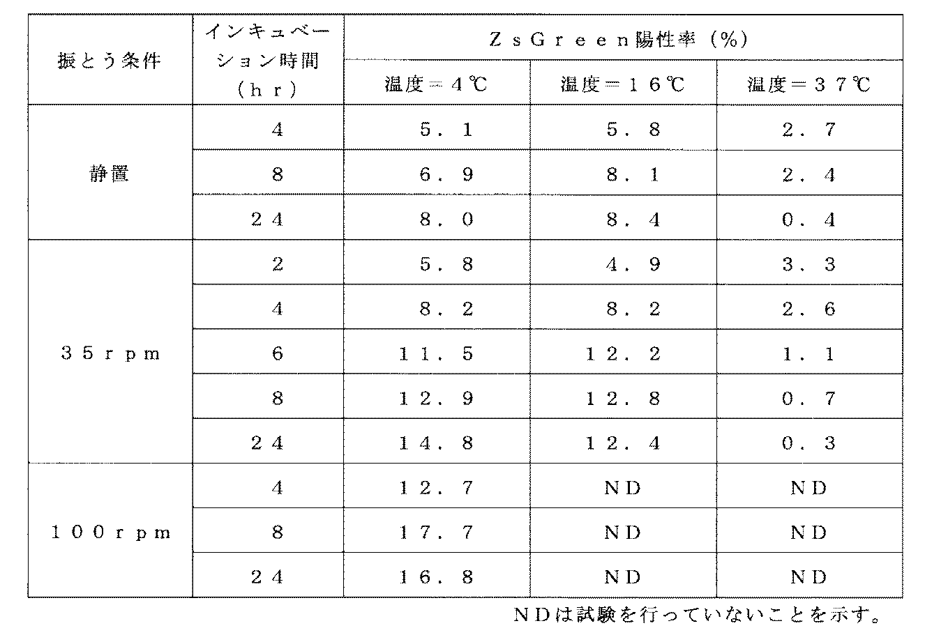
- Preloading was performed for each of these shaking conditions at an incubation temperature of 4 ° C. to 37 ° C. and an incubation time of 2 hours to 24 hours. After completion of the incubation, the virus solution was removed, and each well was washed with 1 mL of PBS containing 1.5% HSA per well. Next, using a medium containing 10% FBS and 1% Penicillin-Streptmycin, SupT1 cells (ATC CCRL-1942) were suspended at 5 ⁇ 10 5 cells / mL, and 1 mL each was added to the well preloaded with the aforementioned virus. After addition, the cells were cultured in a 37 ° C., 5% CO 2 incubator for retrovirus infection (gene transfer using a retrovirus vector).
- the cell suspension was transferred to a new untreated 24-well plate on the first day from the start of culture (culture day 1), and then 4 mL of medium containing 10% FBS and 1% Penicillin-Streptomycin was added per well. The cell suspension was diluted 5-fold. This cell suspension was continuously cultured until the third day of culture.
- Example 1- (2) Analysis of gene transfer efficiency The cells on the third day of culture obtained in Example 1- (2) were subjected to a flow cytometer FACS Canto II (manufactured by Becton Dickinson), and the ZsGreen positive rate was calculated as the gene transfer efficiency did. The results are shown in Table 1.
- high gene transfer efficiency was shown by preloading at 4 ° C. or 16 ° C. for 4 hours or more.
- a high loading efficiency was shown by performing preloading by shaking at 4 ° C. or 16 ° C. for 6 hours or more.
- the introduction efficiency was highest at a preloading time of 4 hours, and thereafter, the introduction efficiency was significantly lowered as the time was increased.
- high introduction efficiency was shown in the case of rolling at 100 rpm.
- Example 2 Gene Introduction into SupT1 Cells by Repeated 4 ° C. Shaking Preloading Method (1)
- DON-ZsGreen retroviral vector prepared in Example 1- (1) was diluted 30-fold in RPMI medium, 1 mL per well was added to the -296 coated plate. The plate during preloading was subjected to shaking by rolling with a tilt of 0 degree at 100 rpm.
- the incubation time was 24 hours (24 hours test group), and in order to examine the effect of repeated preloading, the fresh virus solution was replaced with 1 mL after 16 hours and further incubated for 8 hours (16 + 8 hours) The test was conducted on 2 test zones. The incubation temperature was 4 ° C. for all tests.
- Retrovirus infection was performed by culturing in a 5% CO 2 incubator.
- Example 2- (1) Analysis of gene transfer efficiency The cells on the third day of culture obtained in Example 2- (1) were subjected to a flow cytometer FACS Canto II (manufactured by Becton Dickinson), and the ZsGreen positive rate was calculated as the gene transfer efficiency did. The results are shown in Table 2.
- Example 3 Gene Introduction into SupT1 Cells by Various Closed Preloading Methods Using Cell Culture Bags
- a culture bag MultiLife (registered trademark) Spin manufactured by Takara Bio Inc.
- This bag was a CH-296 coated bag and used for the following experiments.
- the DON-ZsGreen retroviral vector prepared in Example 1- (1) was diluted 30-fold and 60-fold with RPMI medium, and 30 mL was injected into a CH-296 coated bag.
- the shaking conditions of the CH-296 coated bag during preloading are those for which the bag is not shaken (previously placed) during preloading, and for the case where the bag is shaken by rolling with a tilt of 0 degree at 100 rpm. did.
- Preloading was performed for each of these shaking conditions at an incubation temperature of 4 ° C. and an incubation time of 20 hours. After completion of the incubation, the virus solution was removed, and each bag was washed with 15 mL of PBS containing 1.5% HSA per bag.
- SupT1 cells were suspended at 5 ⁇ 10 5 cells / mL using a medium containing 10% FBS and 1% Penicillin-Streptmycin, and 30 mL each was injected into a bag preloaded with the aforementioned virus at 37 ° C., Retrovirus infection was performed by culturing in a 5% CO 2 incubator for 3 days.
- Example 3- (1) Analysis of gene transfer efficiency The cells on the third day of culture obtained in Example 3- (1) were subjected to a flow cytometer FACS Canto II (manufactured by Becton Dickinson), and the ZsGreen positive rate was calculated as the gene transfer efficiency did. The results are shown in Table 3.
- Example 4 Gene Introduction into CD4-Positive T Cell Populations by Various Virus Preloading Methods
- Preparation of LNGFR and MazF gene-loaded retrovirus vectors was performed in International Publication No. 2008/133137. The same method as in Examples 1 and 2 was performed. That is, the HIV LTR-MazF cassette is inserted in the opposite direction to the transcription of the retroviral vector genome, and the gene encoding the extracellular domain of human Low affinity Nerve Growth Factor is inserted downstream of the human PGK promoter in the forward direction.
- the recombinant retroviral vector plasmid pMT-MFR-PL2 was obtained, and the ecotropic MT-MFR-PL2 virus was prepared using the plasmid, which was then infected with GaLV retroviral packaging cell PG13 to obtain a high titer.
- the virus-producing cells were cloned to establish a retrovirus vector-producing cell line PG13 / MT-MFR-PL2.
- a GaLV / MT-MFR-PL2 virus solution was obtained by a conventional method in a medium containing 5 mM sodium butyrate.
- the RNA titer of the obtained virus solution was 6.6 ⁇ 10 9 copies / mL.
- PBMC peripheral blood mononuclear cells
- TK19 and TK29 Human peripheral blood mononuclear cells
- the CD8 positive selection beads (Dynabeads M-450 CD8: manufactured by Invitrogen) washed with Buffer 1 after suspending in PBS containing PBS (hereinafter referred to as Buffer 1) to 1 ⁇ 10 7 cells / mL It was added to a 10 7 cells 2 ⁇ 10 7 beads per.
- the cell suspension containing the beads is allowed to stand for 2 to 3 minutes on a magnetic separator MPC-15 (manufactured by Dynal) to recover the unbound cells.
- MPC-15 manufactured by Dynal
- the collected CD8-removed cell population is centrifuged at 500 ⁇ g for 5 minutes, and then 5 ⁇ 10 5 using a lymphocyte culture medium (hereinafter referred to as X-VIVO15CM) based on X-VIVO15 (manufactured by Lonza). It was suspended to 5 cells / mL.
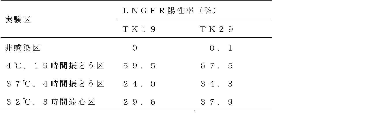
- Retrovirus vector preloading was performed at 4 ° C. for 19 hours in a shaking group, 37 It was carried out by three methods: a shaking group at 4 ° C. for 4 hours, and a centrifugation group at 32 ° C. for 3 hours. Specifically, 1 mL of the GaLV / MT-MFR-PL2 virus solution prepared in Example 2- (1) was added to each CH-296 coated plate, and shaken at 4 ° C. for 19 hours, 37 ° C. for 4 hours. The retroviral vector was preloaded by shaking or by centrifugation at 32 ° C. and 2000 ⁇ g for 3 hours.
- the shaking conditions were Mil Seer, SI-36, a seesaw type shaker, with an inclination angle of 9 degrees and a shaking speed of 35 rpm. Thereafter, the supernatant was removed, and each well was washed with 1 mL of PBS containing 1.5% HSA per well. The preloading plate thus prepared was stored at 4 ° C. until use.
- Example 4- (3) Analysis of gene transfer efficiency
- Cells on day 7 of culture obtained in Example 4- (3) were washed with 0.1% BSA / PBS. Next, cells were suspended in 0.1% BSA / PBS, and FITC-labeled mouse anti-human CD8 antibody (Becton Dickinson) and PerCP-labeled mouse anti-human CD3 antibody (Becton Dickinson) were used as antibody reaction solutions.
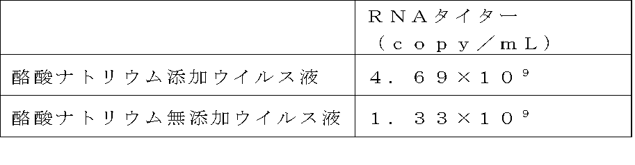
- Example 5 Gene transfer into CD4 positive T cell population by shaking virus preloading method (1) Preparation of retroviral vector Retroviral vector producing cell line PG13 / MT-MFR-PL2 described in Example 4- (1) was used to obtain a GaLV / MT-MFR-PL2 virus solution in a conventional manner in a medium containing or not containing 5 mM sodium butyrate. The RNA titer of the obtained virus solution is shown in Table 5 below.
- Retrovirus vector preloading Virus vector preloading was performed by shaking at 4 ° C. for 30 hours. It was. That is, to the CH-296 coated plate obtained by the same method as in Example 1- (2), the GaLV / MT-MFR-PL2 virus solution containing or not containing sodium butyrate prepared in Example 5- (1) was added. 0.7 mL was added per well, and the retroviral vector was preloaded by shaking at 4 ° C. for 30 hours. The shaking was performed using Mil Seer, SI-36, which is a seesaw type shaking machine, with an inclination angle of 9 degrees and a shaking speed of 35 rpm.
- the virus solution is removed and PBS containing 1.5% HSA is used at 1 mL per well to wash each well (virus wash zone), and the virus solution is not removed (virus non-virus). Washing zone) was set.
- the preloading plate thus prepared was stored at 4 ° C. until use.
- the cells were collected in a centrifuge tube on the 4th day of culture, centrifuged at 500 ⁇ g for 5 minutes to remove the supernatant, and then each cell was suspended in 0.7 mL of X-VIVO15CM. Retroviral infection was performed (second infection).
- Example (3) -4 Expansion culture X-VIVO15CM supplemented with Human AB Serum (manufactured by Lonza) at a final concentration of 5% was added on the fifth day of culture, and 5-fold dilution culture was performed until the seventh day of culture. . Furthermore, X-VIVO15CM supplemented with Human AB Serum (manufactured by Lonza) to a final concentration of 5% was added on the 7th day of culture, and 4-fold dilution culture was performed until the 10th day of culture. In the non-infected area, the same operation as in Example 5- (3) -2 to Example 5- (3) -4 was performed except that a CH-296 coated plate was used instead of the preloading plate. . Table 6 shows the expansion culture rate of each test group.
- Example 5- (3) Analysis of gene transfer efficiency
- the cells on day 10 of the culture obtained in Example 5- (3) were washed with 0.1% BSA / PBS. Next, cells were suspended in 0.1% BSA / PBS, and FITC-labeled mouse anti-human CD8 antibody (Becton Dickinson) and PerCP-labeled mouse anti-human CD3 antibody (Becton Dickinson) were used as antibody reaction solutions.
- Example 6 Gene Introduction into SupT1 Cells by Closed System Preloading Method Using Cell Culture Bag
- Preparation of MazF Gene-Loaded Retrovirus Vector is International Publication No. 2008/133137 Pamphlet The same method as in Examples 1 and 2 was performed.
- RNA titer of the obtained virus solution was 1.4 ⁇ 10 10 copies / mL.
- the MT-MFR3 retrovirus solution prepared in Example 6- (1) was diluted 2-fold with GT-T-RetroI medium.
- Preloading conditions were as follows: 25 mL of the diluted virus solution was injected into the CH-296 coated bag and then shaken for 16 hours or 24 hours. 25 mL of the viral vector and 10 mL of sterile air were added to the CH-296 coated bag. Three types were examined, which were shaken for 16 hours after the injection of. The shaking was performed at a shaking speed of 100 rpm using a rolling shaker (MMS-3010: manufactured by Tokyo Rika Kikai Co., Ltd.) having an inclination of 0 ° and a shaking width of 2.5 cm.
- MMS-3010 manufactured by Tokyo Rika Kikai Co., Ltd.
- the virus solution was removed, and the bag was washed with 15 mL of PBS containing 1.5% HSA per bag.
- 25 mL of SupT1 cell suspension prepared to 5 ⁇ 10 5 cells / mL using RPMI1640 medium containing 10% FBS and 1% Penicillin-Streptmycin is injected into a bag preloaded with the aforementioned virus.
- the cells were cultured at 37 ° C. in a 5% CO 2 incubator for 24 hours.
- the cells in each test group were suspended, diluted 5-fold using RPMI1640 medium containing 10% FBS and 1% Penicillin-Streptmycin, and further cultured for 2 days.
- Genomic DNA was extracted from FastPure (registered trademark) DNA Kit (manufactured by Takara Bio Inc.) from the equivalent of 1 ⁇ 10 6 cells obtained in Example 6- (2).
- the number of introduced copies was measured using Provider Copy Number Detection Primer Set, Human (for Real Time PCR) (manufactured by Takara Bio Inc.). The results are shown in Table 8 below.
- the material of PermaLife PL30 is harder than other bags.
- the movement of the contents liquid during shaking when only the virus liquid was injected into the bag and sealed was small, but the movement of the contents liquid increased when the virus liquid and air were injected into the bag. Further, as shown in Table 8, it was shown that the efficiency of preloading was improved by injecting air together with the virus solution into the sealed cell culture bag.
- Example 7 Examination of incubation time (plate infection) (1)
- Pre-loading conditions were examined in which 1 mL of diluted virus solution was injected into a CH-296 coated plate and then shaken for 12, 16, 24, 48, or 72 hours. The shaking was performed at a shaking speed of 100 rpm using a rolling shaker (MMS-3010: manufactured by Tokyo Rika Kikai Co., Ltd.) having an inclination of 0 ° and a shaking width of 2.5 cm.
- MMS-3010 manufactured by Tokyo Rika Kikai Co., Ltd.
- the virus solution was removed and washed with 0.5 mL of PBS containing 1.5% HSA per well.
- 1 mL of SupT1 cell suspension prepared to 5 ⁇ 10 5 cells / mL using RPMI1640 medium containing 10% FBS and 1% Penicillin-Streptmycin is injected into each plate preloaded with the virus.
- the cells were cultured at 37 ° C. in a 5% CO 2 incubator for 24 hours.
- the cells in each test group were suspended, diluted 5-fold using RPMI1640 medium containing 10% FBS and 1% Penicillin-Streptmycin, and further cultured for 2 days.
- Example 7- (1) Analysis of gene introduction efficiency From the equivalent of 1 ⁇ 10 6 cells obtained in Example 7- (1), the number of introduced copies was measured in the same manner as in Example 6- (3). The results are shown in Table 9 below.
- the gene transfer efficiency with the MT-MFR3 retroviral vector was highest when the incubation time was 16 hours, and an transfer efficiency of 1 copy / cell or more was realized until the incubation time was 12 to 48 hours.
- Example 8 Examination of incubation time (small scale bag infection) (1)
- MazF gene introduction into SupT1 cells MT-MFR3 retrovirus solution prepared in the same manner as in Example 6- (1) was diluted 4-fold with GT-T-RetroI medium, and the gas permeability of OriGen Incubation was performed by injecting 25 mL each into a CH-296 coated bag prepared in the same manner as in Example 6- (2) using the culture bag PeraLife PL30, and further injecting 10 mL of sterile air into the bag.
- 1 mL of the above-described retrovirus solution diluted 4 times was added to a CH-296 coated plate and incubated.
- Preloading conditions were examined for PL30 with shaking for 16, 16 or 24 hours and for plates only for 16 hours.
- the shaking was carried out at a shaking speed of 100 rpm using a rolling shaker having a tilt of 0 degree and a shaking width of 2.5 cm.
- the virus solution was removed and washed by injecting 15 mL of PBS containing 1.5% HSA per bag and 0.5 mL per well of the plate.
- 25 mL each of the SupT1 cell suspension prepared to 4 ⁇ 10 5 cells / mL using RPMI1640 medium containing 10% FBS and 1% Penicillin-Streptmycin is placed in a plate preloaded with the virus described above.
- Example 8- (1) Analysis of gene introduction efficiency From the equivalent of 1 ⁇ 10 6 cells obtained in Example 8- (1), the number of introduced copies was measured in the same manner as in Example 6- (3). The results are shown in Table 10 below.
- Example 9 Examination of Large Scale Bag Infection
- Solution Mobility Test with Large Scale Bag 180 mL of 10% FBS, 1% Penicillin ⁇ in OriGen's gas permeable culture bag PeraLife PL325 (culture area 362.6 cm 2 ) RPMI1640 medium containing Streptmycin was injected, and rolling was performed without tilting at a tilt of 0 ° and 50 rpm, and seesaw-type shaking without tilting at a tilt of 4 ° and 35 rpm.
- PL325 was used, it was confirmed that sufficient mobility was obtained up to the end of the bag without introducing air into the bag.
- EXAMPLE 6- MT-MFR3 retrovirus solution prepared in (1) was diluted 4-fold with GT-T-RetroI medium to prepare a virus diluted solution, and 180 ml each in CH-296 coated bag was coated with CH-296. 1 mL per well was added to the plate. Two preloading conditions were implemented for the PL325 bag: 0 ° tilt, roll shaking without air at 50 rpm, and 4 ° tilt, seesaw type shaking without air at 35 rpm. The plate was subjected to roll shaking at an inclination of 0 degree and 100 rpm. Incubation time was 16 hours.
- the virus solution was removed, and washed with PBS containing 1.5% HSA by adding 108 mL per bag and 0.5 mL per well of the plate.
- SupT1 cell suspension prepared to 4 ⁇ 10 5 cells / mL using RPMI 1640 medium containing 10% FBS and 1% Penicillin-Streptmycin was added to each of the plates pre-loaded with 180 mL of the above virus. 1 mL was injected into each cell, and cultured for 8 hours in a 37 ° C., 5% CO 2 incubator (total number of cells: 7.2 ⁇ 10 7 cells / bag).
- the cells were diluted 4-fold with RPMI1640 medium containing 10% FBS and 1% Penicillin-Streptmycin in a surface untreated 24-well plate and further cultured for 3 days.
- RPMI1640 medium containing 10% FBS and 1% Penicillin-Streptmycin
- For the PL325 bag test group 1.5 mL was sampled, diluted 4-fold in the same manner as the plate test group in a surface untreated 24-well plate, and further cultured for 3 days.
- the bag after sampling was inverted for 5 days in a 5% CO 2 incubator, and the bag surface was infected differently from the 8th hour after infection.
- Example 9- (1) Analysis of gene introduction efficiency From the equivalent of 1 ⁇ 10 6 cells obtained in Example 9- (1), the number of introduced copies was measured in the same manner as in Example 6- (3). The results are shown in Table 11 below.
- Example 10 Virus Solution Reuse Test (1) Virus Solution Preloading 1 (Recovery of Used Virus Solution) To the PL325 bag, 65 mL of 20 ⁇ g / mL CH-296 was added per bag, left at 4 ° C. overnight, and then washed twice with 100 mL of ACD-A. This bag was a CH-296 coat bag.
- Example 6- (1) 180 mL of the MT-MFR3 retrovirus stock solution prepared by the same method as in Example 6- (1) was added to the CH-296 coated bag. Pre-loading conditions were roll shaking without tilting at 0 degrees and 50 rpm air. The incubation temperature was 4 ° C. and the incubation time was 16 hours. After completion of the incubation, the virus solution was recovered and used as a used virus solution.
- virus solution 2 Preloading of virus solution 2
- 1 mL of virus solution was added per well to a CH296 coated plate obtained by the same method as in Example 1- (2).
- the virus solution the virus solution stock solution (New Virus), the used virus solution (Used Virus), and the New Virus of the same lot as the virus solution stock solution used in Example 10- (1) were diluted 2-fold with GT-T-RetroI. A total of four types were used (New Virus + GT-T-RetroI) and New Virus diluted with Used Virus (New Virus + Reuse Virus).
- the shake was performed at 4 ° C. for 16.5 hours at a shake speed of 100 rpm. Went. After completion of the incubation, the virus solution was removed and washed with 0.5 mL of physiological saline containing 1.5% HSA per well.
- Example 10- (1) (4) Analysis of gene introduction efficiency From the equivalent of 1 ⁇ 10 6 cells obtained in Example 10- (1), the number of introduced copies was measured in the same manner as in Example 6- (3). The results are shown in Table 10 below.
- the used virus solution after preloading for 16.5 hours under the condition of 4 ° C. has a titer equivalent to a 2-fold dilution of the unused virus solution.
- This example revealed that the virus solution used for virus preloading at a low temperature in the gene introduction method of the present invention is suitable for reuse.
- Example 11 Examination of incubation time 1 (Large scale bag infection) (1)
- MazF gene introduction into SupT1 cells MT-MFR3 retrovirus solution prepared by the same method as in Example 6- (1) was diluted 4-fold with GT-T-RetroI medium to prepare a virus dilution solution.
- Pre-loading conditions were as follows. For the PL325 bag, roll shaking without inclining air of 0 degree and 50 rpm was performed. The plate was subjected to roll shaking at an inclination of 0 degree and 100 rpm.
- Incubation times were 16, 24 or 48 hours for PL325 and 16 hours for plates.
- the virus solution was removed, and washed with PBS containing 1.5% HSA by adding 108 mL per bag and 0.5 mL per well of the plate.
- 180 mL of SupT1 cell suspension prepared to 5 ⁇ 10 5 cells / mL using RPMI1640 medium containing 10% FBS and 1% Penicillin-Streptmycin in plates pre-loaded with the aforementioned viruses 1 mL was injected into each cell, and cultured in a 37 ° C., 5% CO 2 incubator (total number of cells: 7.2 ⁇ 10 7 cells / bag). After 8 hours, for the bag test group, the bag was inverted and subsequently cultured in a 37 ° C., 5% CO 2 incubator.
- the cells were diluted 5-fold with RPMI1640 medium containing 10% FBS and 1% Penicillin-Streptomycin in a surface untreated 24-well plate and further cultured for 2 days.
- RPMI1640 medium containing 10% FBS and 1% Penicillin-Streptomycin
- 1.5 mL was sampled, diluted 5-fold in the same manner as the plate test group in a surface untreated 24-well plate, and further cultured for 2 days.
- Example 11- (1) Analysis of gene introduction efficiency From the equivalent of 1 ⁇ 10 6 cells obtained in Example 11- (1), the number of introduced copies was measured in the same manner as in Example 6- (3). The results are shown in Table 13 below.
- Example 12 Examination of incubation time 2 (Large scale bag infection) (1)
- Pre-loading conditions were as follows. For the PL325 bag, roll shaking without inclining air of 0 degree and 50 rpm was performed. The plate was subjected to roll shaking at an inclination of 0 degree and 100 rpm.
- Incubation times were 8, 12, 16 or 20 hours for PL325 and 16 hours for plates.
- the virus solution was removed, and washed with PBS containing 1.5% HSA by adding 108 mL per bag and 0.5 mL per well of the plate.
- 180 mL of SupT1 cell suspension prepared to 5 ⁇ 10 5 cells / mL using RPMI1640 medium containing 10% FBS and 1% Penicillin-Streptmycin in plates pre-loaded with the aforementioned viruses 1 mL was injected into each cell, and cultured in a 37 ° C., 5% CO 2 incubator (total number of cells: 7.2 ⁇ 10 7 cells / bag). Two hours later, for the bag test section, the bag was inverted and subsequently cultured in a 37 ° C., 5% CO 2 incubator.
- the cells were diluted 5-fold with RPMI1640 medium containing 10% FBS and 1% Penicillin-Streptomycin in a surface untreated 24-well plate and further cultured for 3 days.
- RPMI1640 medium containing 10% FBS and 1% Penicillin-Streptomycin
- 1.5 mL was sampled, diluted 5-fold in the same manner as the plate test group in a surface untreated 24-well plate, and further cultured for 3 days.
- Example 12- (1) Analysis of gene introduction efficiency From the equivalent of 1 ⁇ 10 6 cells obtained in Example 12- (1), the number of introduced copies was measured in the same manner as in Example 6- (3). The results are shown in Table 14 below.
- the present invention provides a simple and highly efficient gene transfer method.
- the present invention is particularly useful in fields such as medicine, cell engineering, genetic engineering, and developmental engineering.
Abstract
Description
[1]レトロウイルスベクターによる標的細胞への外来遺伝子の導入方法であって、工程(a):レトロウイルス結合性物質が固定化された培養容器に、外来遺伝子が搭載されたレトロウイルスベクターを含有する液体を入れた後、25℃未満の温度で4時間以上インキュベートし、レトロウイルスベクターが結合された培養容器を得る工程、及び工程(b):工程(a)により得られた培養容器に標的細胞を入れ、インキュベートする工程、を含む方法、
[2]工程(a)におけるインキュベーション時間が5時間超~48時間である、[1]に記載の方法、
[3]工程(a)におけるインキュベーションが、振とうを伴うインキュベーションである、[1]に記載の方法、
[4][1]に記載の遺伝子の導入方法であって、工程(a):レトロウイルス結合性物質が固定化された培養容器に、外来遺伝子が搭載されたレトロウイルスベクターを含有する液体を入れた後、25℃未満の温度で4時間以上インキュベートし、レトロウイルスベクターが結合された培養容器を得る工程、工程(b1):工程(a)により得られた培養容器を洗浄する工程、及び工程(b2):工程(b1)で洗浄された培養容器に標的細胞を入れ、インキュベートする工程、を含む方法、
[5]レトロウイルス結合性物質が、フィブロネクチン、繊維芽細胞増殖因子、V型コラーゲン、ポリリジン、DEAE-デキストラン、及びこれらのフラグメントから選択された少なくとも1種である、[1]に記載の方法、
[6]レトロウイルス結合性物質が、細胞結合性を併せ持つ物質である、[1]に記載の方法、
[7]レトロウイルス結合性物質が固定化された培養容器が、レトロウイルス結合性物質及び細胞結合性物質が固定化された培養容器である、[1]に記載の方法、並びに
[8]細胞結合性物質が、細胞接着性タンパク質、ホルモン、サイトカイン、抗体、糖鎖、炭水化物、及び代謝物から選択された少なくとも1種である、[7]に記載の方法、
に関する。 That is, the present invention
[1] A method for introducing a foreign gene into a target cell using a retroviral vector, comprising the step (a): a retroviral vector loaded with a foreign gene in a culture vessel in which a retroviral binding substance is immobilized A step of incubating for 4 hours or more at a temperature of less than 25 ° C. after the liquid to be added is obtained, and a step (b): targeting the culture vessel obtained by step (a) Placing and incubating the cells,
[2] The method according to [1], wherein the incubation time in step (a) is more than 5 hours to 48 hours,
[3] The method according to [1], wherein the incubation in step (a) is incubation with shaking,
[4] The gene introduction method according to [1], wherein step (a): a liquid containing a retroviral vector loaded with a foreign gene is placed in a culture vessel in which a retroviral binding substance is immobilized. And a step of incubating at a temperature of less than 25 ° C. for 4 hours or more to obtain a culture vessel bound with a retroviral vector, step (b1): a step of washing the culture vessel obtained by step (a), and Step (b2): placing the target cells in the culture vessel washed in Step (b1) and incubating the method,
[5] The method according to [1], wherein the retrovirus-binding substance is at least one selected from fibronectin, fibroblast growth factor, type V collagen, polylysine, DEAE-dextran, and fragments thereof.
[6] The method according to [1], wherein the retrovirus-binding substance is a substance having cell-binding properties.
[7] The method according to [1], wherein the culture vessel in which the retrovirus-binding substance is immobilized is a culture vessel in which the retrovirus-binding substance and the cell-binding substance are immobilized, and [8] cells. The method according to [7], wherein the binding substance is at least one selected from cell adhesion proteins, hormones, cytokines, antibodies, sugar chains, carbohydrates, and metabolites,
About.
pZsGreen Vector(クロンテック社製)を制限酵素BamHIとEcoRI(タカラバイオ社製)で切断し、アガロースゲル電気泳動を行い、緑色蛍光タンパク質ZsGreenをコードする配列を含む約0.7kbpのフラグメントを回収した。回収したフラグメントをDNA Blunting Kit(タカラバイオ社製)を用いて末端平滑化後、pDON-AI DNA(タカラバイオ社製)に挿入し、組換えレトロウイルスベクタープラスミドpDON-ZsGreenを取得した。次に、当該プラスミドとRetrovirus Packaging Kit Eco(タカラバイオ社製)を用いてエコトロピックDON-ZsGreenウイルスを作製した。その後、これをGaLVレトロウイルスパッケージング細胞PG13に感染させた。感染細胞から高力価のウイルス産生細胞をクローニングしてレトロウイルスベクター産生細胞株PG13/DON-ZsGreenを樹立した。さらに当該産生細胞を用いて、5mM 酪酸ナトリウムを含有する培地で常法によりGaLV/DON-ZsGreenウイルス液を取得した(以下、DON-ZsGreenレトロウイルスベクターと称す)。なお、取得したウイルス液のRNAタイターは1.88×1010コピー/mLであった。 (1) Preparation of DON-ZsGreen retroviral vector pZsGreen Vector (Clontech) was cleaved with restriction enzymes BamHI and EcoRI (Takara Bio), agarose gel electrophoresis was performed, and the sequence encoding green fluorescent protein ZsGreen was obtained. An approximately 0.7 kbp fragment was recovered. The recovered fragment was blunt-ended using DNA Blunting Kit (Takara Bio) and then inserted into pDON-AI DNA (Takara Bio) to obtain a recombinant retrovirus vector plasmid pDON-ZsGreen. Next, an ecotropic DON-ZsGreen virus was prepared using the plasmid and Retrovirus Packaging Kit Eco (manufactured by Takara Bio Inc.). This was then infected with GaLV retrovirus packaging cells PG13. A high-titer virus-producing cell was cloned from the infected cell to establish a retrovirus vector-producing cell line PG13 / DON-ZsGreen. Furthermore, using the producer cells, a GaLV / DON-ZsGreen virus solution was obtained by a conventional method in a medium containing 5 mM sodium butyrate (hereinafter referred to as DON-ZsGreen retrovirus vector). The RNA titer of the obtained virus solution was 1.88 × 10 10 copies / mL.
表面未処理24ウェルプレート(ベクトン・ディッキンソン社製)に1ウェルあたり500μLの20μg/mLのフィブロネクチンフラグメントであるCH-296(商品名レトロネクチン;タカラバイオ社製)を添加して4℃で一晩放置した後、500μLのPBSで2回洗浄した。本明細書の実施例では、このプレートをCH-296コートプレートとし、必要に応じて作製した。 (2) ZsGreen gene introduction into SupT1 cells CH-296 (trade name Retronectin; manufactured by Takara Bio Inc.), which is a surface untreated 24-well plate (Becton Dickinson), 500 μL per well of 20 μg / mL fibronectin fragment And left at 4 ° C. overnight, and then washed twice with 500 μL of PBS. In the examples of this specification, this plate was a CH-296 coated plate, and was produced as needed.
実施例1-(2)で得られた培養3日目の細胞をフローサイトメーターFACS CantoII(ベクトン・ディッキンソン社製)に供し、遺伝子導入効率としてZsGreen陽性率を算出した。その結果を表1に示す。 (3) Analysis of gene transfer efficiency The cells on the third day of culture obtained in Example 1- (2) were subjected to a flow cytometer FACS Canto II (manufactured by Becton Dickinson), and the ZsGreen positive rate was calculated as the gene transfer efficiency did. The results are shown in Table 1.
(1)実施例1-(1)で調製したDON-ZsGreenレトロウイルスベクターをRPMI培地にて30倍に希釈し、CH-296コートプレートに1ウェルあたり1mLずつ添加した。プレローディング中のプレートは、100rpmで傾斜0度の横揺れによる振とうに供した。インキュベーション時間は、24時間行ったもの(24時間試験区)、及びプレローディングの繰り返しによる効果を調べるため、16時間後に新鮮なウイルス液1mLに置換し、さらに8時間のインキュベーション行ったもの(16+8時間試験区)の2試験区について試験した。インキュベーション温度は、いずれの試験も4℃で実施した。インキュベーションの終了後ウイルス液を除去し、1.5%HSAを含むPBSを1ウェルあたり1mLずつ使用して各ウェルを洗浄した。次に10%FBS、1%Penicillin-Streptmycinを含む培地を用いて5×10⁵cells/mLとなるようにSupT1細胞を懸濁し、前述のウイルスをプレロードしたウェルに1mLずつ添加し、37℃、5%CO2インキュベータ内で培養してレトロウイルス感染を行った。培養開始から1日目(培養1日目)に0.4mLの細胞懸濁液を新たな表面未処理24ウェルプレートに移した後、10%FBS、1%Penicillin-Streptmycinを含む培地を1ウェルあたり1.6mLずつ添加し、細胞懸濁液を5倍希釈した。この細胞懸濁液について、培養3日目まで培養を継続した。 Example 2 Gene Introduction into SupT1 Cells by Repeated 4 ° C. Shaking Preloading Method (1) DON-ZsGreen retroviral vector prepared in Example 1- (1) was diluted 30-fold in RPMI medium, 1 mL per well was added to the -296 coated plate. The plate during preloading was subjected to shaking by rolling with a tilt of 0 degree at 100 rpm. The incubation time was 24 hours (24 hours test group), and in order to examine the effect of repeated preloading, the fresh virus solution was replaced with 1 mL after 16 hours and further incubated for 8 hours (16 + 8 hours) The test was conducted on 2 test zones. The incubation temperature was 4 ° C. for all tests. After completion of the incubation, the virus solution was removed, and each well was washed with 1 mL of PBS containing 1.5% HSA per well. Next, SupT1 cells were suspended at 5 × 10 5 cells / mL using a medium containing 10% FBS and 1% Penicillin-Streptmycin, and 1 mL each was added to the wells pre-loaded with the aforementioned virus at 37 ° C. Retrovirus infection was performed by culturing in a 5% CO 2 incubator. On the first day from the start of culture (1st day of culture), 0.4 mL of the cell suspension was transferred to a new surface-untreated 24-well plate, and then 1 well of a medium containing 10% FBS, 1% Penicillin-Streptmycin 1.6 mL per solution was added, and the cell suspension was diluted 5-fold. This cell suspension was continuously cultured until the third day of culture.
実施例2-(1)で得られた培養3日目の細胞をフローサイトメーターFACS CantoII(ベクトン・ディッキンソン社製)に供し、遺伝子導入効率としてZsGreen陽性率を算出した。その結果を表2に示す。 (2) Analysis of gene transfer efficiency The cells on the third day of culture obtained in Example 2- (1) were subjected to a flow cytometer FACS Canto II (manufactured by Becton Dickinson), and the ZsGreen positive rate was calculated as the gene transfer efficiency did. The results are shown in Table 2.

(1)培養面積60cm2の培養バッグCultiLife(登録商標) Spin(タカラバイオ社製)に20μg/mLのCH-296を10mL注入して4℃で一晩以上放置した後、15mLのPBSで2回洗浄した。このバッグをCH-296コートバッグとし、以下の実験に使用した。 Example 3 Gene Introduction into SupT1 Cells by Various Closed Preloading Methods Using Cell Culture Bags (1) 20 μg / mL in a culture bag MultiLife (registered trademark) Spin (manufactured by Takara Bio Inc.) having a culture area of 60 cm 2 After injecting 10 mL of CH-296 and leaving it to stand overnight at 4 ° C., it was washed twice with 15 mL of PBS. This bag was a CH-296 coated bag and used for the following experiments.
実施例3-(1)で得られた培養3日目の細胞をフローサイトメーターFACS CantoII(ベクトン・ディッキンソン社製)に供し、遺伝子導入効率としてZsGreen陽性率を算出した。その結果を表3に示す。 (2) Analysis of gene transfer efficiency The cells on the third day of culture obtained in Example 3- (1) were subjected to a flow cytometer FACS Canto II (manufactured by Becton Dickinson), and the ZsGreen positive rate was calculated as the gene transfer efficiency did. The results are shown in Table 3.

(1)LNGFR及びMazF遺伝子搭載レトロウイルスベクターの調製
LNGFR及びMazF遺伝子搭載レトロウイルスベクターの調製は国際公開第2008/133137号パンフレットの実施例1及び2と同様の方法で行った。すなわち、HIV LTR-MazFカセットがレトロウイルスベクターゲノムの転写とは逆方向に挿入され、かつヒトLow affinity Nerve Growth Factor Receptorの細胞外ドメインをコードする遺伝子がヒトPGKプロモーターの下流に順方向に挿入された組換えレトロウイルスベクタープラスミドpMT-MFR-PL2を取得し、当該プラスミドを用いてエコトロピックMT-MFR-PL2ウイルスを作製した後、これをGaLVレトロウイルスパッケージング細胞PG13に感染させ、高力価のウイルス産生細胞をクローニングしてレトロウイルスベクター産生細胞株PG13/MT-MFR-PL2を樹立した。さらに当該産生細胞を用いて、5mM 酪酸ナトリウムを含有する培地で常法によりGaLV/MT-MFR-PL2ウイルス液を取得した。なお、取得したウイルス液のRNAタイターは6.6×109コピー/mLであった。 Example 4 Gene Introduction into CD4-Positive T Cell Populations by Various Virus Preloading Methods (1) Preparation of LNGFR and MazF Gene-Loaded Retrovirus Vectors Preparation of LNGFR and MazF gene-loaded retrovirus vectors was performed in International Publication No. 2008/133137. The same method as in Examples 1 and 2 was performed. That is, the HIV LTR-MazF cassette is inserted in the opposite direction to the transcription of the retroviral vector genome, and the gene encoding the extracellular domain of human Low affinity Nerve Growth Factor is inserted downstream of the human PGK promoter in the forward direction. The recombinant retroviral vector plasmid pMT-MFR-PL2 was obtained, and the ecotropic MT-MFR-PL2 virus was prepared using the plasmid, which was then infected with GaLV retroviral packaging cell PG13 to obtain a high titer. The virus-producing cells were cloned to establish a retrovirus vector-producing cell line PG13 / MT-MFR-PL2. Furthermore, using the production cells, a GaLV / MT-MFR-PL2 virus solution was obtained by a conventional method in a medium containing 5 mM sodium butyrate. The RNA titer of the obtained virus solution was 6.6 × 10 9 copies / mL.
インフォームド・コンセントの得られた健常人ドナーTK19及びTK29から常法に従い調製したヒト末梢血単核細胞(PBMC)を、2mM EDTA、0.1% BSAを含むPBS(以下、Buffer1と称す)を用いて1×10⁷cells/mLになるように懸濁後、Buffer1で洗浄したCD8ポジティブセレクションビーズ(Dynabeads M-450 CD8:インビトロジェン社製)をPBMC1×107cellsあたり2×10⁷ビーズとなるように添加した。ローテーターを用いて4℃で30分間緩やかに攪拌したのち、ビーズを含む細胞懸濁液を磁気分離装置MPC-15(Dynal社製)上に2~3分静置してビーズ非結合細胞を回収した(以下、CD8除去細胞集団と記載)。回収したCD8除去細胞集団を500×gで5分間遠心したのち、X-VIVO15(ロンザ社製)を基礎とするリンパ球培養用培地(以下、X-VIVO15CMと称する)を用いて、5×10⁵cells/mLになるように懸濁した。 (2) Preparation of CD4-positive T cell population Human peripheral blood mononuclear cells (PBMC) prepared from normal donors TK19 and TK29 with informed consent according to a conventional method were prepared using 2 mM EDTA, 0.1% BSA. The CD8 positive selection beads (Dynabeads M-450 CD8: manufactured by Invitrogen) washed with Buffer 1 after suspending in PBS containing PBS (hereinafter referred to as Buffer 1) to 1 × 10 7 cells / mL It was added to a 10 7 cells 2 × 10 7 beads per. After gently stirring for 30 minutes at 4 ° C using a rotator, the cell suspension containing the beads is allowed to stand for 2 to 3 minutes on a magnetic separator MPC-15 (manufactured by Dynal) to recover the unbound cells. (Hereinafter referred to as a CD8-depleted cell population). The collected CD8-removed cell population is centrifuged at 500 × g for 5 minutes, and then 5 × 10 5 using a lymphocyte culture medium (hereinafter referred to as X-VIVO15CM) based on X-VIVO15 (manufactured by Lonza). It was suspended to 5 cells / mL.
(3)-1 レトロウイルスベクターのプレローディング
ウイルスベクターのプレローディングは4℃、19時間の振とう区、37℃、4時間の振とう区、及び32℃、3時間の遠心区の3種の方法で行った。すなわち、CH-296コートプレートに実施例2-(1)で調製したGaLV/MT-MFR-PL2ウイルス液を1ウェルあたり1mLずつ添加し、4℃、19時間の振とう、37℃、4時間の振とう、もしくは32℃、2000×g、3時間の遠心により、レトロウイルスベクターのプレローディングを行った。振とう条件は、シーソー型の振とう機であるMild Mixer, SI-36を用いて傾斜角度は9度、振とう速度は35rpmで行った。その後、上清を除去し、1.5%HSAを含むPBSを1ウェルあたり1mLずつ使用して各ウェルを洗浄した。こうして作製したプレローディングプレートは、使用するまで4℃で保存した。 (3) Gene introduction and expansion culture into the cell population prepared in Example 4- (2) (3) -1 Retrovirus vector preloading Virus vector preloading was performed at 4 ° C. for 19 hours in a shaking group, 37 It was carried out by three methods: a shaking group at 4 ° C. for 4 hours, and a centrifugation group at 32 ° C. for 3 hours. Specifically, 1 mL of the GaLV / MT-MFR-PL2 virus solution prepared in Example 2- (1) was added to each CH-296 coated plate, and shaken at 4 ° C. for 19 hours, 37 ° C. for 4 hours. The retroviral vector was preloaded by shaking or by centrifugation at 32 ° C. and 2000 × g for 3 hours. The shaking conditions were Mil Seer, SI-36, a seesaw type shaker, with an inclination angle of 9 degrees and a shaking speed of 35 rpm. Thereafter, the supernatant was removed, and each well was washed with 1 mL of PBS containing 1.5% HSA per well. The preloading plate thus prepared was stored at 4 ° C. until use.
実施例4-(2)で調製したCD8除去細胞集団を底面積25cm2細胞培養用フラスコ(コーニング社製)に10mL添加した。細胞数に対して3倍量のDynabeads Human T-Activator CD3/CD28(インビトロジェン社製)を遠心管に分注後、X-VIVO15で洗浄し、上記細胞を含むフラスコに添加し、フラスコを立てて37℃、5%CO2インキュベータ内での培養を開始した(培養0日目)。 (3) -2 Start of culture 10 mL of the CD8-removed cell population prepared in Example 4- (2) was added to a flask with a bottom area of 25 cm 2 for cell culture (manufactured by Corning). Three times the amount of Dynabeads Human T-Activator CD3 / CD28 (manufactured by Invitrogen) was dispensed into a centrifuge tube, washed with X-VIVO15, added to the flask containing the cells, and the flask was set up. Cultivation was started at 37 ° C. in a 5% CO 2 incubator (culture day 0).
培養3日目に細胞を遠心管に回収し、500×g、5分間遠心して上清を除去した後、5×10⁵cells/mLとなるようにX-VIVO15CMを加えて懸濁した。実施例3-(3)-1で作製したプレローディングプレートから上清を除去した後、上記細胞懸濁液を1ウェルあたり1mLずつ添加し、37℃、5%CO2インキュベータ内でインキュベートして細胞へのレトロウイルス感染を行った。なお、非感染区では、プレローディングプレートの代わりにCH-296コートプレートを用いる以外は上記と同様の操作を行った。 (3) -3 Gene transfer Cells were collected in a centrifuge tube on the third day of culture, centrifuged at 500 × g for 5 minutes to remove the supernatant, and then X-VIVO15CM was added to 5 × 10 5 cells / mL. In addition, it was suspended. After removing the supernatant from the preloading plate prepared in Example 3- (3) -1, add 1 mL of the cell suspension per well and incubate in a 37 ° C., 5% CO 2 incubator. Cells were retrovirally infected. In the non-infected area, the same operation as described above was performed except that a CH-296 coated plate was used instead of the preloading plate.
培養4日目に実施例4-(3)-3でレトロウイルス感染を行った細胞を遠心管に回収し、500×gで5分間遠心して上清を除去し、2mLのX-VIVO15CMに懸濁して2倍希釈培養した。さらに培養5日目にHuman AB Serum(ロンザ社製)を終濃度5%となるように添加したX-VIVO15CMを加えて、5倍希釈培養を培養7日目まで行った。 (3) -4 Expansion culture On day 4 of culture, the cells infected with the retrovirus in Example 4- (3) -3 were collected in a centrifuge tube, centrifuged at 500 × g for 5 minutes to remove the supernatant, The suspension was suspended in 2 mL of X-VIVO15CM and diluted twice. Furthermore, X-VIVO15CM supplemented with Human AB Serum (manufactured by Lonza) to a final concentration of 5% was added on the fifth day of culture, and 5-fold dilution culture was performed until the seventh day of culture.
実施例4-(3)で得られた培養7日目の細胞を0.1%BSA/PBSで洗浄した。次に、0.1%BSA/PBSに細胞を懸濁し、ここに抗体反応液として、FITC標識マウス抗ヒトCD8抗体(ベクトン・ディッキンソン社製)、PerCP標識マウス抗ヒトCD3抗体(ベクトン・ディッキンソン社製)、APC標識マウス抗ヒトLNGFR抗体(Miltenyi Biotec社製)、及びAPC‐Cy7標識マウス抗ヒトCD4抗体(ベクトン・ディッキンソン社製)を含む抗体液を添加し、抗体反応を行った。その後、0.1%BSA/PBSで細胞を2回洗浄し、再度0.1%BSA/PBSに懸濁した。この細胞をフローサイトメトリーに供し、遺伝子導入効率として、各々の細胞集団のCD3陽性CD4陽性細胞中のLNGFR陽性率を算出した。その結果を表4に示す。 (4) Analysis of gene transfer efficiency Cells on day 7 of culture obtained in Example 4- (3) were washed with 0.1% BSA / PBS. Next, cells were suspended in 0.1% BSA / PBS, and FITC-labeled mouse anti-human CD8 antibody (Becton Dickinson) and PerCP-labeled mouse anti-human CD3 antibody (Becton Dickinson) were used as antibody reaction solutions. Antibody solution containing APC-labeled mouse anti-human LNGFR antibody (manufactured by Miltenyi Biotec) and APC-Cy7-labeled mouse anti-human CD4 antibody (manufactured by Becton Dickinson), and an antibody reaction was carried out. Thereafter, the cells were washed twice with 0.1% BSA / PBS and suspended again in 0.1% BSA / PBS. The cells were subjected to flow cytometry, and the LNGFR positive rate in CD3 positive CD4 positive cells of each cell population was calculated as gene transfer efficiency. The results are shown in Table 4.
(1)レトロウイルスベクターの調製
実施例4-(1)に記載のレトロウイルスベクター産生細胞株PG13/MT-MFR-PL2を用いて、5mM 酪酸ナトリウムを含有する培地もしくは含有しない培地で常法によりGaLV/MT-MFR-PL2ウイルス液を取得した。なお、取得したウイルス液のRNAタイターを下記表5に示す。 Example 5 Gene transfer into CD4 positive T cell population by shaking virus preloading method (1) Preparation of retroviral vector Retroviral vector producing cell line PG13 / MT-MFR-PL2 described in Example 4- (1) Was used to obtain a GaLV / MT-MFR-PL2 virus solution in a conventional manner in a medium containing or not containing 5 mM sodium butyrate. The RNA titer of the obtained virus solution is shown in Table 5 below.
実施例4-(2)と同様の方法で、インフォームド・コンセントの得られた健常人ドナーTK19からCD8除去細胞集団を調製し、X-VIVO15CMを用いて、5×10⁵cells/mLになるように懸濁した。 (2) Preparation of CD8-depleted cell population In the same manner as in Example 4- (2), a CD8-depleted cell population was prepared from a healthy donor TK19 from which informed consent was obtained, and X-VIVO15CM was used. It suspended so that it might become 5 * 10 < 5 > cells / mL.
(3)-1 レトロウイルスベクターのプレローディング
ウイルスベクターのプレローディングは4℃、30時間振とうすることにより行った。すなわち、実施例1-(2)と同様の方法で得られたCH-296コートプレートに実施例5-(1)で調製した酪酸ナトリウム含有もしくは非含有のGaLV/MT-MFR-PL2ウイルス液を1ウェルあたり0.7mLずつ添加し、4℃、30時間の振とうによりレトロウイルスベクターのプレローディングを行った。振とうは、シーソー型の振とう機であるMild Mixer, SI-36を用いて傾斜角度は9度、振とう速度は35rpmで行った。プレローディング後は、上記ウイルス液を除去し1.5%HSAを含むPBSを1ウェルあたり1mLずつ使用して各ウェルを洗浄する区(ウイルス洗浄区)と、ウイルス液を除去しない区(ウイルス非洗浄区)を設定した。こうして作製したプレローディングプレートは、使用するまで4℃で保存した。 (3) Gene introduction into cell population prepared in Example 5- (2) and expansion culture (3) -1 Retrovirus vector preloading Virus vector preloading was performed by shaking at 4 ° C. for 30 hours. It was. That is, to the CH-296 coated plate obtained by the same method as in Example 1- (2), the GaLV / MT-MFR-PL2 virus solution containing or not containing sodium butyrate prepared in Example 5- (1) was added. 0.7 mL was added per well, and the retroviral vector was preloaded by shaking at 4 ° C. for 30 hours. The shaking was performed using Mil Seer, SI-36, which is a seesaw type shaking machine, with an inclination angle of 9 degrees and a shaking speed of 35 rpm. After preloading, the virus solution is removed and PBS containing 1.5% HSA is used at 1 mL per well to wash each well (virus wash zone), and the virus solution is not removed (virus non-virus). Washing zone) was set. The preloading plate thus prepared was stored at 4 ° C. until use.
実施例5-(2)で調製したCD8除去細胞集団を底面積25cm2細胞培養用フラスコ(コーニング社製)に12mL添加した。細胞数に対して3倍量のDynabeads Human T-Activator CD3/CD28(インビトロジェン社製)をX-VIVO15で洗浄した後、上記細胞を含むフラスコに添加し、フラスコを立てて37℃、5%CO2インキュベータ内での培養を開始した(培養0日目)。 (3) -2 Start of culture 12 mL of the CD8-removed cell population prepared in Example 5- (2) was added to a bottom area 25 cm 2 cell culture flask (manufactured by Corning). Three times the amount of Dynabeads Human T-Activator CD3 / CD28 (manufactured by Invitrogen) was washed with X-VIVO15 and added to the flask containing the cells. Cultivation in 2 incubators was started (culture day 0).
培養3日目に細胞を遠心管に回収し、500×g、5分間遠心して上清を除去した後、7.14×10⁵cells/mLとなるようにX-VIVO15CMを加えて懸濁した。実施例5-(3)-1で作製したプレローディングプレートから、ウイルス洗浄区については上清を除去した後、上記細胞懸濁液を1ウェルあたり0.7mLずつ添加後、さらに1ウェルあたり0.3mLのX-VIVO15CMを添加した(最終密度5×105/ウェル)。ウイルス非洗浄区については、各ウイルス液の入ったウェルに上記細胞懸濁液を1ウェルあたり0.7mLずつ添加した(最終密度5×105/ウェル)。これらのプレートを、37℃、5%CO2インキュベータ内でインキュベートして細胞へのレトロウイルス感染を行った(感染1回目)。 (3) -3 Gene transfer Cells were collected in a centrifuge tube on the third day of culture, centrifuged at 500 × g for 5 minutes to remove the supernatant, and then X− was adjusted to 7.14 × 10 5 cells / mL. VIVO15CM was added and suspended. From the preloading plate prepared in Example 5- (3) -1, after removing the supernatant in the virus washing section, 0.7 mL of the cell suspension was added per well, and then 0 per well was added. 3 mL of X-VIVO15CM was added (final density 5 × 10 5 / well). For the virus non-washed section, 0.7 mL of the cell suspension was added to each well containing virus solutions (final density 5 × 10 5 / well). These plates were incubated in a 37 ° C., 5% CO 2 incubator for retroviral infection of the cells (first infection).
培養5日目にHuman AB Serum(ロンザ社製)を終濃度5%となるように添加したX-VIVO15CMを加えて、5倍希釈培養を培養7日目まで行った。さらに培養7日目にHuman AB Serum(ロンザ社製)を終濃度5%となるように添加したX-VIVO15CMを加えて、4倍希釈培養を培養10日目まで行った。なお、非感染区では、プレローディングプレートの代わりにCH-296コートプレートを用いる以外は上記の実施例5-(3)-2~実施例5-(3)-4と同様の操作を行った。各試験区の拡大培養率を表6に示す。 (3) -4 Expansion culture X-VIVO15CM supplemented with Human AB Serum (manufactured by Lonza) at a final concentration of 5% was added on the fifth day of culture, and 5-fold dilution culture was performed until the seventh day of culture. . Furthermore, X-VIVO15CM supplemented with Human AB Serum (manufactured by Lonza) to a final concentration of 5% was added on the 7th day of culture, and 4-fold dilution culture was performed until the 10th day of culture. In the non-infected area, the same operation as in Example 5- (3) -2 to Example 5- (3) -4 was performed except that a CH-296 coated plate was used instead of the preloading plate. . Table 6 shows the expansion culture rate of each test group.
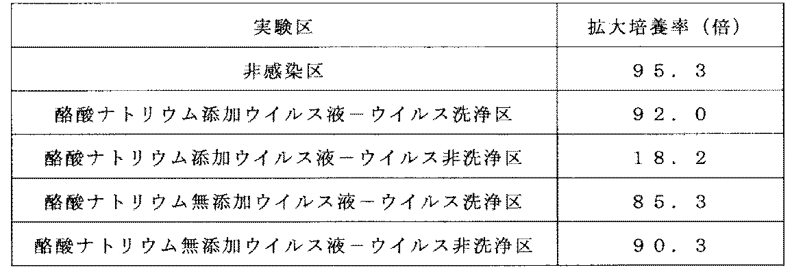
実施例5-(3)で得られた培養10日目の細胞を0.1%BSA/PBSで洗浄した。次に、0.1%BSA/PBSに細胞を懸濁し、ここに抗体反応液として、FITC標識マウス抗ヒトCD8抗体(ベクトン・ディッキンソン社製)、PerCP標識マウス抗ヒトCD3抗体(ベクトン・ディッキンソン社製)、APC標識マウス抗ヒトLNGFR抗体(Miltenyi Biotec社製)、及びAPC‐Cy7標識マウス抗ヒトCD4抗体(ベクトン・ディッキンソン社製)を含む抗体液を添加し、抗体反応を行った。その後、0.1%BSA/PBSで細胞を2回洗浄し、再度0.1%BSA/PBSに懸濁した。この細胞をフローサイトメトリーに供し、遺伝子導入効率として、各々の細胞集団のCD3陽性CD4陽性細胞中のLNGFR陽性率を算出した。その結果を表7に示す。 (4) Analysis of gene transfer efficiency The cells on day 10 of the culture obtained in Example 5- (3) were washed with 0.1% BSA / PBS. Next, cells were suspended in 0.1% BSA / PBS, and FITC-labeled mouse anti-human CD8 antibody (Becton Dickinson) and PerCP-labeled mouse anti-human CD3 antibody (Becton Dickinson) were used as antibody reaction solutions. Antibody solution containing APC-labeled mouse anti-human LNGFR antibody (manufactured by Miltenyi Biotec) and APC-Cy7-labeled mouse anti-human CD4 antibody (manufactured by Becton Dickinson), and an antibody reaction was carried out. Thereafter, the cells were washed twice with 0.1% BSA / PBS and suspended again in 0.1% BSA / PBS. The cells were subjected to flow cytometry, and the LNGFR positive rate in CD3 positive CD4 positive cells of each cell population was calculated as gene transfer efficiency. The results are shown in Table 7.
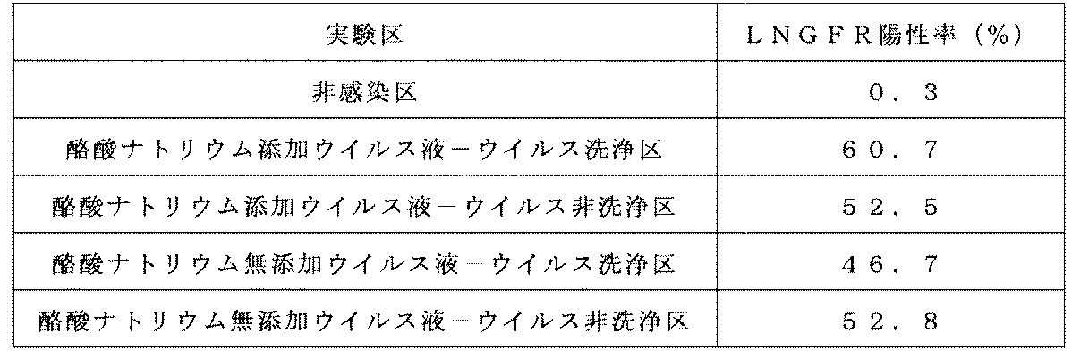
(1)MazF遺伝子搭載レトロウイルスベクターの調製
MazF遺伝子搭載レトロウイルスベクターの調製は国際公開第2008/133137号パンフレットの実施例1および2と同様の方法で行った。すなわち、HIV LTR-MazFカセットがレトロウイルスベクターゲノムの転写とは逆方向に挿入された組換えレトロウイルスベクタープラスミドpMT-MFR3を取得し、当該プラスミドを用いてエコトロピックMT-MFR3ウイルスを作製した後、これをGaLVレトロウイルスパッケージング細胞PG13に感染させ、高力価のウイルス産生細胞をクローニングしてレトロウイルスベクター産生細胞株PG13/MT-MFR3を樹立した。さらに、5mM 酪酸ナトリウムを含有するGT-T-RetroI培地(タカラバイオ株式会社製)にて上記のレトロウイルス産生細胞を32℃で24時間培養して得られた培養上清を採取し、GaLV/MT-MFR3ウイルス液を取得した。なお、取得したウイルス液のRNAタイターは1.4×1010コピー/mLであった。 Example 6 Gene Introduction into SupT1 Cells by Closed System Preloading Method Using Cell Culture Bag (1) Preparation of MazF Gene-Loaded Retrovirus Vector Preparation of MazF gene-loaded retrovirus vector is International Publication No. 2008/133137 Pamphlet The same method as in Examples 1 and 2 was performed. That is, after obtaining the recombinant retroviral vector plasmid pMT-MFR3 in which the HIV LTR-MazF cassette was inserted in the direction opposite to the transcription of the retroviral vector genome, and producing the ecotropic MT-MFR3 virus using the plasmid This was infected with GaLV retrovirus packaging cells PG13, and high-titer virus-producing cells were cloned to establish a retrovirus vector-producing cell line PG13 / MT-MFR3. Furthermore, the culture supernatant obtained by culturing the above retrovirus-producing cells for 24 hours at 32 ° C. in GT-T-RetroI medium (Takara Bio Inc.) containing 5 mM sodium butyrate was collected, and GaLV / MT-MFR3 virus solution was obtained. The RNA titer of the obtained virus solution was 1.4 × 10 10 copies / mL.
OriGen社のガス透過性培養バッグPermaLife PL30に、1バッグあたり9mLの20μg/mLのCH-296を添加して4℃で一晩放置した後、15mLのPBSで2回洗浄した。このバッグをCH-296コートバッグとし、以下の実験に使用した。 (2) MazF gene introduction into SupT1 cells 9 mL of 20 μg / mL CH-296 per bag was added to OriGen's gas permeable culture bag, PermaLife PL30, and left overnight at 4 ° C., then 15 mL of PBS And washed twice. This bag was a CH-296 coated bag and used for the following experiments.
実施例6-(2)で得られた細胞1×10⁶個相当より、FastPure(登録商標) DNA Kit(タカラバイオ社製)を用いてゲノムDNAを抽出し、Provirus Copy Number Detection Primer Set, Human (for Real Time PCR)(タカラバイオ社製)を用いて導入コピー数の測定を行った。結果を下記表8に示す。 (3) Analysis of gene transfer efficiency Genomic DNA was extracted from FastPure (registered trademark) DNA Kit (manufactured by Takara Bio Inc.) from the equivalent of 1 × 10 6 cells obtained in Example 6- (2). The number of introduced copies was measured using Provider Copy Number Detection Primer Set, Human (for Real Time PCR) (manufactured by Takara Bio Inc.). The results are shown in Table 8 below.

(1)SupT1細胞へのMazF遺伝子導入
実施例6-(1)と同様の方法で調製したMT-MFR3レトロウイルス液をGT-T-RetroI培地にて2倍希釈し、CH-296コートプレートに1ウェルあたり1mLずつ添加した。プレローディング条件は、CH-296コートプレートに希釈後のウイルス液を1mL注入した後に振とうを12、16、24、48または72時間行ったものについて検討した。振とうは、傾斜0度、振とう幅2.5cmの横揺れ振とう機(MMS-3010:東京理化器械社製)を用いて、100rpmの振とう速度で行った。インキュベーションの終了後ウイルス液を除去し、1.5%HSAを含むPBSを1ウェルあたり0.5mL添加して洗浄した。次に10%FBS、1%Penicillin-Streptmycinを含むRPMI1640培地を用いて5×10⁵cells/mLとなるように調製したSupT1細胞懸濁液を、前述のウイルスをプレロードしたプレートに1mLずつ注入し、37℃、5%CO2インキュベータ内で24時間培養した。次に、各試験区の細胞を懸濁し、10%FBS、1%Penicillin-Streptmycinを含むRPMI1640培地を用いて5倍希釈した後、さらに2日間培養した。 Example 7 Examination of incubation time (plate infection)
(1) MazF gene introduction into SupT1 cells MT-MFR3 retroviral solution prepared by the same method as in Example 6- (1) was diluted 2-fold with GT-T-RetroI medium and added to a CH-296 coated plate. 1 mL was added per well. Pre-loading conditions were examined in which 1 mL of diluted virus solution was injected into a CH-296 coated plate and then shaken for 12, 16, 24, 48, or 72 hours. The shaking was performed at a shaking speed of 100 rpm using a rolling shaker (MMS-3010: manufactured by Tokyo Rika Kikai Co., Ltd.) having an inclination of 0 ° and a shaking width of 2.5 cm. After completion of the incubation, the virus solution was removed and washed with 0.5 mL of PBS containing 1.5% HSA per well. Next, 1 mL of SupT1 cell suspension prepared to 5 × 10 5 cells / mL using RPMI1640 medium containing 10% FBS and 1% Penicillin-Streptmycin is injected into each plate preloaded with the virus. The cells were cultured at 37 ° C. in a 5% CO 2 incubator for 24 hours. Next, the cells in each test group were suspended, diluted 5-fold using RPMI1640 medium containing 10% FBS and 1% Penicillin-Streptmycin, and further cultured for 2 days.
実施例7-(1)で得られた細胞1×10⁶個相当より、実施例6-(3)と同様の方法で導入コピー数の測定を行った。結果を下記表9に示す。 (2) Analysis of gene introduction efficiency From the equivalent of 1 × 10 6 cells obtained in Example 7- (1), the number of introduced copies was measured in the same manner as in Example 6- (3). The results are shown in Table 9 below.
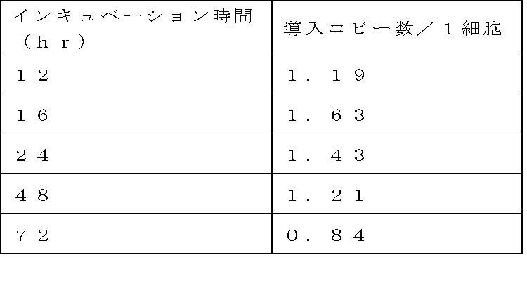
(1)SupT1細胞へのMazF遺伝子導入
実施例6-(1)と同様の方法で調製したMT-MFR3レトロウイルス液をGT-T-RetroI培地にて4倍希釈し、OriGen社のガス透過性培養バッグPermaLife PL30を用いて実施例6-(2)と同様に調製したCH-296コートバッグに25mLずつ注入し、さらにバッグ中に10mLの無菌の空気を注入してインキュベーションを行った。また、コントロールとしてCH-296コートプレートに上記の4倍希釈したレトロウイルス液を1ウェルあたり1mLずつ添加してインキュベーションを行った。プレローディング条件は、PL30については振とうを12、16もしくは24時間、プレートについては16時間のみ行ったものについて検討した。振とうは、傾斜0度、振とう幅2.5cmの横揺れ振とう機を用い、100rpmの振とう速度で行った。インキュベーションの終了後ウイルス液を除去し、1.5%HSAを含むPBSをバッグあたり15mL、プレートについては1ウェルあたり0.5mL注入して洗浄した。次に10%FBS、1%Penicillin-Streptmycinを含むRPMI1640培地を用いて4×10⁵cells/mLとなるように調製したSupT1細胞懸濁液を、前述のウイルスをプレロードしたバッグに25mLずつ、プレートについては1ウェルあたり1mLずつ注入し、37℃、5%CO2インキュベータ内で8時間培養した(総細胞数1×107細胞/PL30バッグ)。次に、各試験区の細胞を懸濁し、10%FBS、1%Penicillin-Streptmycinを含むRPMI1640培地を用いて4倍希釈した後、さらに3日間培養した。 Example 8 Examination of incubation time (small scale bag infection)
(1) MazF gene introduction into SupT1 cells MT-MFR3 retrovirus solution prepared in the same manner as in Example 6- (1) was diluted 4-fold with GT-T-RetroI medium, and the gas permeability of OriGen Incubation was performed by injecting 25 mL each into a CH-296 coated bag prepared in the same manner as in Example 6- (2) using the culture bag PeraLife PL30, and further injecting 10 mL of sterile air into the bag. In addition, as a control, 1 mL of the above-described retrovirus solution diluted 4 times was added to a CH-296 coated plate and incubated. Preloading conditions were examined for PL30 with shaking for 16, 16 or 24 hours and for plates only for 16 hours. The shaking was carried out at a shaking speed of 100 rpm using a rolling shaker having a tilt of 0 degree and a shaking width of 2.5 cm. After completion of the incubation, the virus solution was removed and washed by injecting 15 mL of PBS containing 1.5% HSA per bag and 0.5 mL per well of the plate. Next, 25 mL each of the SupT1 cell suspension prepared to 4 × 10 5 cells / mL using RPMI1640 medium containing 10% FBS and 1% Penicillin-Streptmycin is placed in a plate preloaded with the virus described above. Was injected at 1 mL per well and cultured in a 37 ° C., 5% CO 2 incubator for 8 hours (total number of cells: 1 × 10 7 cells / PL30 bag). Next, the cells in each test group were suspended, diluted 4-fold with RPMI1640 medium containing 10% FBS and 1% Penicillin-Streptmycin, and then cultured for another 3 days.
実施例8-(1)で得られた細胞1×10⁶個相当より、実施例6-(3)と同様の方法で導入コピー数の測定を行った。結果を下記表10に示す。 (2) Analysis of gene introduction efficiency From the equivalent of 1 × 10 6 cells obtained in Example 8- (1), the number of introduced copies was measured in the same manner as in Example 6- (3). The results are shown in Table 10 below.

(1)ラージスケールバッグでの溶液可動性試験
OriGen社のガス透過性培養バッグPermaLife PL325(培養面積362.6cm2)に180mLの10%FBS、1%Penicillin-Streptmycinを含むRPMI1640培地を注入し、傾斜0度、50rpmの空気を入れない横揺れ振とう、傾斜4度、35rpmの空気を入れないシーソー型振とうを行った。その結果、PL325を用いた場合は、バッグ中に空気を入れなくてもバッグ端部まで十分な可動性が得られていることを確認した。 Example 9 Examination of Large Scale Bag Infection (1) Solution Mobility Test with Large Scale Bag 180 mL of 10% FBS, 1% Penicillin− in OriGen's gas permeable culture bag PeraLife PL325 (culture area 362.6 cm 2 ) RPMI1640 medium containing Streptmycin was injected, and rolling was performed without tilting at a tilt of 0 ° and 50 rpm, and seesaw-type shaking without tilting at a tilt of 4 ° and 35 rpm. As a result, when PL325 was used, it was confirmed that sufficient mobility was obtained up to the end of the bag without introducing air into the bag.
PL325に、1バッグあたり65mLの20μg/mLのCH-296を添加して4℃で一晩放置した後、108mLのPBSで2回洗浄した。このバッグをCH-296コートバッグとし、以下の実験に使用した。 (2) MazF gene introduction into SupT1 cells To PL325, 65 mL of 20 μg / mL CH-296 per bag was added and left overnight at 4 ° C., and then washed twice with 108 mL of PBS. This bag was a CH-296 coated bag and used for the following experiments.
実施例9-(1)で得られた細胞1×10⁶個相当より、実施例6-(3)と同様の方法で導入コピー数の測定を行った。結果を下記表11に示す。 (2) Analysis of gene introduction efficiency From the equivalent of 1 × 10 6 cells obtained in Example 9- (1), the number of introduced copies was measured in the same manner as in Example 6- (3). The results are shown in Table 11 below.
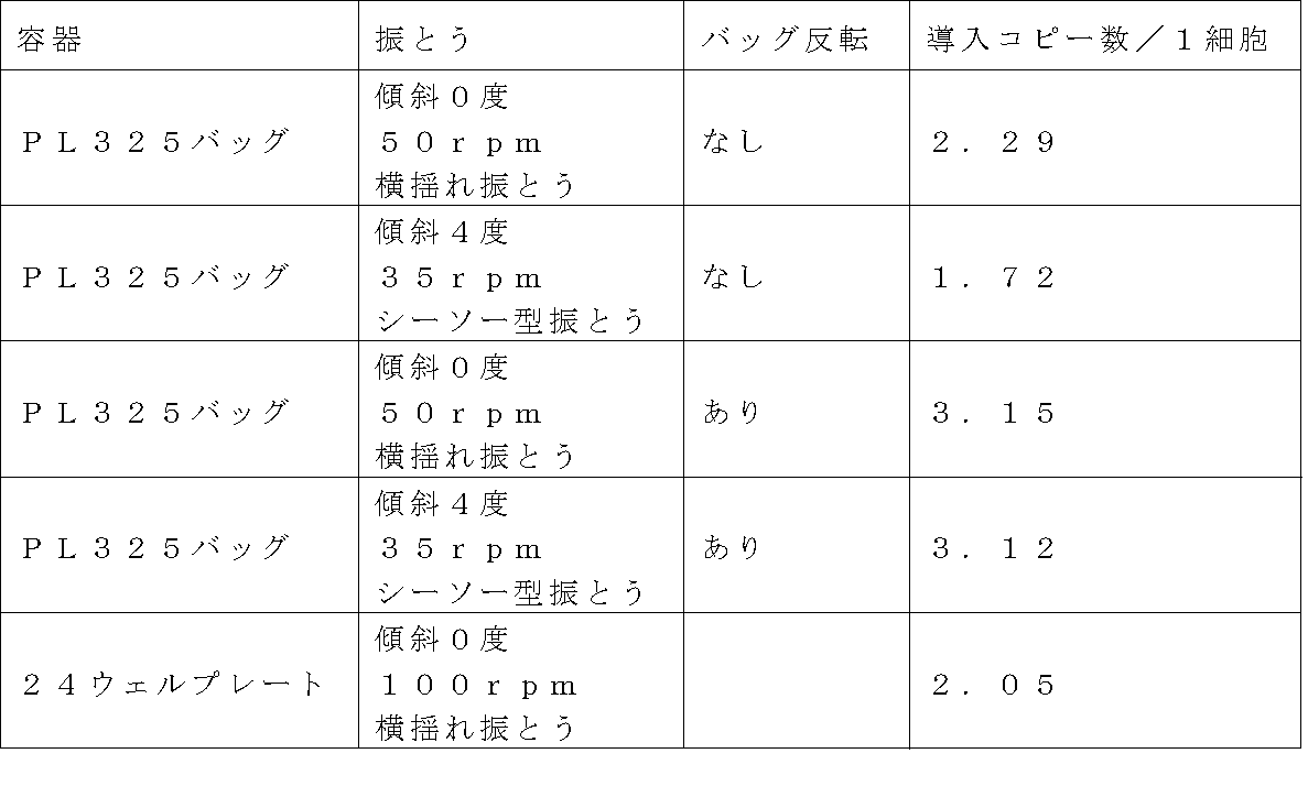
(1)ウイルス液のプレローディング1(使用済ウイルス液の回収)
PL325バッグに、1バッグあたり65mLの20μg/mLのCH-296を添加して4℃で一晩放置した後、100mLのACD-Aで2回洗浄した。このバッグをCH-296コートバッグとした。 Example 10 Virus Solution Reuse Test (1) Virus Solution Preloading 1 (Recovery of Used Virus Solution)
To the PL325 bag, 65 mL of 20 μg / mL CH-296 was added per bag, left at 4 ° C. overnight, and then washed twice with 100 mL of ACD-A. This bag was a CH-296 coat bag.
使用済ウイルス液を用いた場合の感染効率を調べるため、実施例1-(2)と同様の方法で得られたCH296コートプレートにウイルス液を1ウェルあたり1mLずつ添加した。ウイルス液は、実施例10-(1)で使用したウイルス液原液と同ロットのウイルス液原液(New Virus)、使用済ウイルス液(Used Virus)、New VirusをGT-T-RetroIで2倍希釈したもの(New Virus+GT-T-RetroI)およびNew VirusをUsed Virusで希釈したもの(New Virus+Reuse Virus)の計4種類を用いた。その後、傾斜0度、振とう幅2.5cmの横揺れ振とう機(MMS-3010:東京理化器械社製)を用いて、100rpmの振とう速度で4℃にて16.5時間の振とうを行った。インキュベーション終了後、ウイルス液を除去し、1.5%HSAを含む生理食塩水を1ウェルあたり0.5mL添加して洗浄した。 (2) Preloading of virus solution 2
In order to examine the infection efficiency when the used virus solution was used, 1 mL of virus solution was added per well to a CH296 coated plate obtained by the same method as in Example 1- (2). As for the virus solution, the virus solution stock solution (New Virus), the used virus solution (Used Virus), and the New Virus of the same lot as the virus solution stock solution used in Example 10- (1) were diluted 2-fold with GT-T-RetroI. A total of four types were used (New Virus + GT-T-RetroI) and New Virus diluted with Used Virus (New Virus + Reuse Virus). Then, using a roll shaker (MMS-3010: manufactured by Tokyo Rika Kikai Co., Ltd.) with a tilt of 0 degree and a shake width of 2.5 cm, the shake was performed at 4 ° C. for 16.5 hours at a shake speed of 100 rpm. Went. After completion of the incubation, the virus solution was removed and washed with 0.5 mL of physiological saline containing 1.5% HSA per well.
10%FBS、1%Penicillin-Streptmycinを含むRPMI1640培地を用いて5×10⁵cells/mLとなるように調製したSupT1細胞懸濁液を、前述のウイルスをプレロードしたプレートに1mLずつ注入し、37℃、5%CO2インキュベータ内で24時間培養した。次に、各試験区の細胞を懸濁し、10%FBS、1%Penicillin-Streptmycinを含むRPMI1640培地を用いて5倍希釈した後、さらに2日間培養した。 (3) Gene introduction into cells SupT1 cell suspension prepared to 5 × 10 5 cells / mL using RPMI1640 medium containing 10% FBS and 1% Penicillin-Streptmycin was preloaded with the aforementioned virus. 1 mL was poured into the plate and cultured in a 37 ° C., 5% CO 2 incubator for 24 hours. Next, the cells in each test group were suspended, diluted 5-fold using RPMI1640 medium containing 10% FBS and 1% Penicillin-Streptmycin, and further cultured for 2 days.
実施例10-(1)で得られた細胞1×10⁶個相当より、実施例6-(3)と同様の方法で導入コピー数の測定を行った。結果を下記表10に示す。 (4) Analysis of gene introduction efficiency From the equivalent of 1 × 10 6 cells obtained in Example 10- (1), the number of introduced copies was measured in the same manner as in Example 6- (3). The results are shown in Table 10 below.
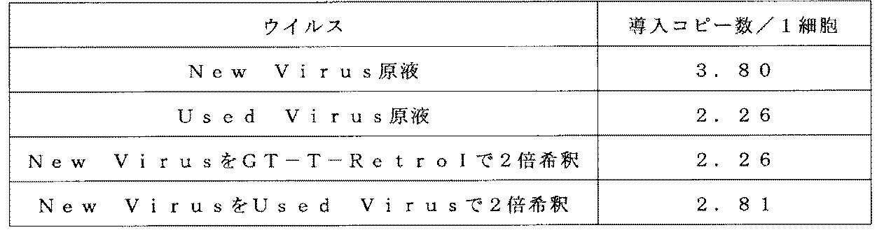
(1)SupT1細胞へのMazF遺伝子導入
実施例6-(1)と同様の方法で調製したMT-MFR3レトロウイルス液をGT-T-RetroI培地にて4倍希釈してウイルス希釈液を調製し、OriGen社のガス透過性培養バッグPermaLife PL325を用いて実施例9-(2)と同様の方法で調製したCH-296コートバッグに180mLずつ、CH-296コートプレートに1ウェルあたり0.5mLずつ添加した。プレローディング条件は、PL325バッグについては、傾斜0度、50rpmの空気を入れない横揺れ振とうを実施した。プレートについては、傾斜0度、100rpmの横揺れ振とうを行った。インキュベーション時間は、PL325については16、24もしくは48時間とし、プレートについては16時間とした。インキュベーションの終了後ウイルス液を除去し、1.5%HSAを含むPBSをバッグあたり108mL、プレート1ウェルあたり0.5mL添加して洗浄した。次に10%FBS、1%Penicillin-Streptmycinを含むRPMI1640培地を用いて5×10⁵cells/mLとなるように調製したSupT1細胞懸濁液を、前述のウイルスをプレロードしたバッグに180mLずつ、プレートに1mLずつ注入し、37℃、5%CO2インキュベータ内で培養した(総細胞数7.2×107細胞/バッグ)。8時間後、バッグ試験区についてはバッグを反転させて、引き続き37℃、5%CO2インキュベータ内で培養した。 Example 11 Examination of incubation time 1 (Large scale bag infection)
(1) MazF gene introduction into SupT1 cells MT-MFR3 retrovirus solution prepared by the same method as in Example 6- (1) was diluted 4-fold with GT-T-RetroI medium to prepare a virus dilution solution. 180 mL each in a CH-296 coated bag prepared in the same manner as in Example 9- (2) using an OriGen gas permeable culture bag PeraLife PL325, 0.5 mL per well in a CH-296 coated plate Added. Pre-loading conditions were as follows. For the PL325 bag, roll shaking without inclining air of 0 degree and 50 rpm was performed. The plate was subjected to roll shaking at an inclination of 0 degree and 100 rpm. Incubation times were 16, 24 or 48 hours for PL325 and 16 hours for plates. After completion of the incubation, the virus solution was removed, and washed with PBS containing 1.5% HSA by adding 108 mL per bag and 0.5 mL per well of the plate. Next, 180 mL of SupT1 cell suspension prepared to 5 × 10 5 cells / mL using RPMI1640 medium containing 10% FBS and 1% Penicillin-Streptmycin in plates pre-loaded with the aforementioned viruses 1 mL was injected into each cell, and cultured in a 37 ° C., 5% CO 2 incubator (total number of cells: 7.2 × 10 7 cells / bag). After 8 hours, for the bag test group, the bag was inverted and subsequently cultured in a 37 ° C., 5% CO 2 incubator.
実施例11-(1)で得られた細胞1×10⁶個相当より、実施例6-(3)と同様の方法で導入コピー数の測定を行った。結果を下記表13に示す。 (2) Analysis of gene introduction efficiency From the equivalent of 1 × 10 6 cells obtained in Example 11- (1), the number of introduced copies was measured in the same manner as in Example 6- (3). The results are shown in Table 13 below.
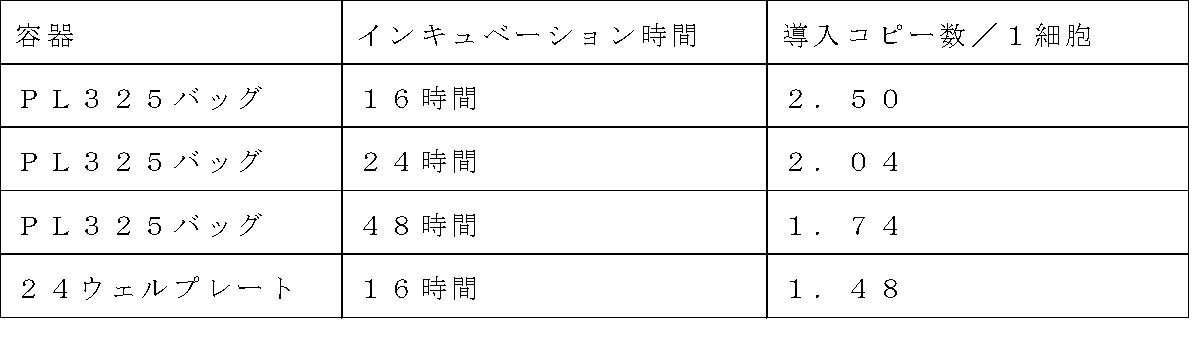
(1)SupT1細胞へのMazF遺伝子導入
実施例6-(1)と同様の方法で調製したMT-MFR3レトロウイルス液をGT-T-RetroI培地にて4倍希釈してウイルス希釈液を調製し、OriGen社のガス透過性培養バッグPermaLife PL325を用いて実施例9-(2)と同様の方法で調製したCH-296コートバッグに180mLずつ、CH-296コートプレートに1ウェルあたり0.5mLずつ添加した。プレローディング条件は、PL325バッグについては、傾斜0度、50rpmの空気を入れない横揺れ振とうを実施した。プレートについては、傾斜0度、100rpmの横揺れ振とうを行った。インキュベーション時間は、PL325については8、12、16もしくは20時間とし、プレートについては16時間とした。インキュベーションの終了後ウイルス液を除去し、1.5%HSAを含むPBSをバッグあたり108mL、プレート1ウェルあたり0.5mL添加して洗浄した。次に10%FBS、1%Penicillin-Streptmycinを含むRPMI1640培地を用いて5×10⁵cells/mLとなるように調製したSupT1細胞懸濁液を、前述のウイルスをプレロードしたバッグに180mLずつ、プレートに1mLずつ注入し、37℃、5%CO2インキュベータ内で培養した(総細胞数7.2×107細胞/バッグ)。2時間後、バッグ試験区についてはバッグを反転させて、引き続き37℃、5%CO2インキュベータ内で培養した。 Example 12 Examination of incubation time 2 (Large scale bag infection)
(1) MazF gene introduction into SupT1 cells MT-MFR3 retrovirus solution prepared by the same method as in Example 6- (1) was diluted 4-fold with GT-T-RetroI medium to prepare a virus dilution solution. 180 mL each in a CH-296 coated bag prepared in the same manner as in Example 9- (2) using an OriGen gas permeable culture bag PeraLife PL325, 0.5 mL per well in a CH-296 coated plate Added. Pre-loading conditions were as follows. For the PL325 bag, roll shaking without inclining air of 0 degree and 50 rpm was performed. The plate was subjected to roll shaking at an inclination of 0 degree and 100 rpm. Incubation times were 8, 12, 16 or 20 hours for PL325 and 16 hours for plates. After completion of the incubation, the virus solution was removed, and washed with PBS containing 1.5% HSA by adding 108 mL per bag and 0.5 mL per well of the plate. Next, 180 mL of SupT1 cell suspension prepared to 5 × 10 5 cells / mL using RPMI1640 medium containing 10% FBS and 1% Penicillin-Streptmycin in plates pre-loaded with the aforementioned viruses 1 mL was injected into each cell, and cultured in a 37 ° C., 5% CO 2 incubator (total number of cells: 7.2 × 10 7 cells / bag). Two hours later, for the bag test section, the bag was inverted and subsequently cultured in a 37 ° C., 5% CO 2 incubator.
実施例12-(1)で得られた細胞1×10⁶個相当より、実施例6-(3)と同様の方法で導入コピー数の測定を行った。結果を下記表14に示す。 (2) Analysis of gene introduction efficiency From the equivalent of 1 × 10 6 cells obtained in Example 12- (1), the number of introduced copies was measured in the same manner as in Example 6- (3). The results are shown in Table 14 below.
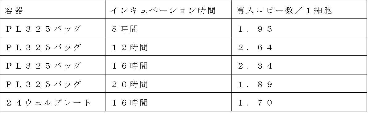
Claims (8)
- レトロウイルスベクターによる標的細胞への外来遺伝子の導入方法であって、下記工程(a)及び(b)を含む方法:
(a)レトロウイルス結合性物質が固定化された培養容器に、外来遺伝子が搭載されたレトロウイルスベクターを含有する液体を入れた後、25℃未満の温度で4時間以上インキュベートし、レトロウイルスベクターが結合された培養容器を得る工程;及び
(b)工程(a)により得られた培養容器に標的細胞を入れ、インキュベートする工程。 A method for introducing a foreign gene into a target cell using a retroviral vector, comprising the following steps (a) and (b):
(A) A liquid containing a retroviral vector loaded with a foreign gene is placed in a culture vessel on which a retroviral binding substance is immobilized, and then incubated at a temperature of less than 25 ° C. for 4 hours or longer. A step of obtaining a culture vessel to which is bound; and (b) a step of placing target cells in the culture vessel obtained by step (a) and incubating. - 工程(a)におけるインキュベーション時間が5時間超~48時間である、請求項1に記載の方法。 The method according to claim 1, wherein the incubation time in step (a) is more than 5 hours to 48 hours.
- 工程(a)におけるインキュベーションが、振とうを伴うインキュベーションである、請求項1に記載の方法。 The method according to claim 1, wherein the incubation in step (a) is incubation with shaking.
- 請求項1に記載の遺伝子の導入方法であって、下記工程(a)~(b2)を含む方法:
(a)レトロウイルス結合性物質が固定化された培養容器に、外来遺伝子が搭載されたレトロウイルスベクターを含有する液体を入れた後、25℃未満の温度で4時間以上インキュベートし、レトロウイルスベクターが結合された培養容器を得る工程;
(b1)工程(a)により得られた培養容器を洗浄する工程;及び
(b2)工程(b1)で洗浄された培養容器に標的細胞を入れ、インキュベートする工程。 The method for introducing a gene according to claim 1, comprising the following steps (a) to (b2):
(A) A liquid containing a retroviral vector loaded with a foreign gene is placed in a culture vessel on which a retroviral binding substance is immobilized, and then incubated at a temperature of less than 25 ° C. for 4 hours or longer. Obtaining a culture vessel to which is bound;
(B1) a step of washing the culture vessel obtained in step (a); and (b2) a step of placing the target cells in the culture vessel washed in step (b1) and incubating. - レトロウイルス結合性物質が、フィブロネクチン、繊維芽細胞増殖因子、V型コラーゲン、ポリリジン、DEAE-デキストラン、及びこれらのフラグメントから選択された少なくとも1種である、請求項1に記載の方法。 The method according to claim 1, wherein the retrovirus-binding substance is at least one selected from fibronectin, fibroblast growth factor, type V collagen, polylysine, DEAE-dextran, and fragments thereof.
- レトロウイルス結合性物質が、細胞結合性を併せ持つ物質である、請求項1に記載の方法。 The method according to claim 1, wherein the retrovirus-binding substance is a substance having cell binding properties.
- レトロウイルス結合性物質が固定化された培養容器が、レトロウイルス結合性物質及び細胞結合性物質が固定化された培養容器である、請求項1に記載の方法。 The method according to claim 1, wherein the culture vessel in which the retrovirus-binding substance is immobilized is a culture vessel in which the retrovirus-binding substance and the cell-binding substance are immobilized.
- 細胞結合性物質が、細胞接着性タンパク質、ホルモン、サイトカイン、抗体、糖鎖、炭水化物、及び代謝物から選択された少なくとも1種である、請求項7に記載の方法。 The method according to claim 7, wherein the cell-binding substance is at least one selected from cell adhesion proteins, hormones, cytokines, antibodies, sugar chains, carbohydrates, and metabolites.
Priority Applications (5)
| Application Number | Priority Date | Filing Date | Title |
|---|---|---|---|
| US13/805,373 US8841126B2 (en) | 2010-06-30 | 2011-06-29 | Method for gene transfer |
| EP11800914.1A EP2589656B8 (en) | 2010-06-30 | 2011-06-29 | Gene introduction method |
| JP2012522672A JP5778147B2 (en) | 2010-06-30 | 2011-06-29 | Gene transfer method |
| CN2011800325192A CN102959080A (en) | 2010-06-30 | 2011-06-29 | Gene introduction method |
| US14/457,453 US9228206B2 (en) | 2010-06-30 | 2014-08-12 | Method for gene transfer |
Applications Claiming Priority (4)
| Application Number | Priority Date | Filing Date | Title |
|---|---|---|---|
| JP2010-148860 | 2010-06-30 | ||
| JP2010148860 | 2010-06-30 | ||
| JP2010-245368 | 2010-11-01 | ||
| JP2010245368 | 2010-11-01 |
Related Child Applications (2)
| Application Number | Title | Priority Date | Filing Date |
|---|---|---|---|
| US13/805,373 A-371-Of-International US8841126B2 (en) | 2010-06-30 | 2011-06-29 | Method for gene transfer |
| US14/457,453 Continuation-In-Part US9228206B2 (en) | 2010-06-30 | 2014-08-12 | Method for gene transfer |
Publications (1)
| Publication Number | Publication Date |
|---|---|
| WO2012002452A1 true WO2012002452A1 (en) | 2012-01-05 |
Family
ID=45402156
Family Applications (1)
| Application Number | Title | Priority Date | Filing Date |
|---|---|---|---|
| PCT/JP2011/064945 WO2012002452A1 (en) | 2010-06-30 | 2011-06-29 | Gene introduction method |
Country Status (5)
| Country | Link |
|---|---|
| US (1) | US8841126B2 (en) |
| EP (1) | EP2589656B8 (en) |
| JP (1) | JP5778147B2 (en) |
| CN (1) | CN102959080A (en) |
| WO (1) | WO2012002452A1 (en) |
Cited By (1)
| Publication number | Priority date | Publication date | Assignee | Title |
|---|---|---|---|---|
| JP2019526288A (en) * | 2016-09-14 | 2019-09-19 | ブイビーシー ホールディングス エルエルシー | System, apparatus, and method for optimizing cell growth by controlling cell culture operations |
Families Citing this family (1)
| Publication number | Priority date | Publication date | Assignee | Title |
|---|---|---|---|---|
| US9228206B2 (en) * | 2010-06-30 | 2016-01-05 | Takara Bio Inc. | Method for gene transfer |
Citations (2)
| Publication number | Priority date | Publication date | Assignee | Title |
|---|---|---|---|---|
| WO1997018318A1 (en) * | 1995-11-13 | 1997-05-22 | Takara Shuzo Co., Ltd. | Method for gene introduction into target cells by retrovirus |
| WO2000001836A1 (en) * | 1998-07-01 | 2000-01-13 | Takara Shuzo Co., Ltd. | Gene transfer methods |
Family Cites Families (3)
| Publication number | Priority date | Publication date | Assignee | Title |
|---|---|---|---|---|
| US5686278A (en) | 1994-03-25 | 1997-11-11 | Indiana University Foundation | Methods for enhanced retrovirus-mediated gene transfer |
| TWI239352B (en) * | 1997-07-23 | 2005-09-11 | Takara Bio Inc | Gene transfer method with the use of serum-free medium |
| CN100379458C (en) * | 1998-07-01 | 2008-04-09 | 宝生物工程株式会社 | Gene transfer methods |
-
2011
- 2011-06-29 US US13/805,373 patent/US8841126B2/en active Active
- 2011-06-29 EP EP11800914.1A patent/EP2589656B8/en active Active
- 2011-06-29 CN CN2011800325192A patent/CN102959080A/en active Pending
- 2011-06-29 WO PCT/JP2011/064945 patent/WO2012002452A1/en active Application Filing
- 2011-06-29 JP JP2012522672A patent/JP5778147B2/en active Active
Patent Citations (2)
| Publication number | Priority date | Publication date | Assignee | Title |
|---|---|---|---|---|
| WO1997018318A1 (en) * | 1995-11-13 | 1997-05-22 | Takara Shuzo Co., Ltd. | Method for gene introduction into target cells by retrovirus |
| WO2000001836A1 (en) * | 1998-07-01 | 2000-01-13 | Takara Shuzo Co., Ltd. | Gene transfer methods |
Non-Patent Citations (2)
| Title |
|---|
| BAJAJ B. ET AL.: "High efficiencies of gene transfer with immobilized recombinant retrovirus: kinetics and optimization.", BIOTECHNOL. PROG., vol. 17, 2001, pages 587 - 596, XP055073781 * |
| See also references of EP2589656A4 * |
Cited By (1)
| Publication number | Priority date | Publication date | Assignee | Title |
|---|---|---|---|---|
| JP2019526288A (en) * | 2016-09-14 | 2019-09-19 | ブイビーシー ホールディングス エルエルシー | System, apparatus, and method for optimizing cell growth by controlling cell culture operations |
Also Published As
| Publication number | Publication date |
|---|---|
| EP2589656B8 (en) | 2016-07-13 |
| US8841126B2 (en) | 2014-09-23 |
| EP2589656A4 (en) | 2014-05-14 |
| US20130109096A1 (en) | 2013-05-02 |
| CN102959080A (en) | 2013-03-06 |
| JPWO2012002452A1 (en) | 2013-08-29 |
| EP2589656B1 (en) | 2016-04-20 |
| JP5778147B2 (en) | 2015-09-16 |
| EP2589656A1 (en) | 2013-05-08 |
Similar Documents
| Publication | Publication Date | Title |
|---|---|---|
| JP3807937B2 (en) | Gene transfer method | |
| JP3754090B2 (en) | Gene transfer method using serum-free medium | |
| US9670459B2 (en) | Production method for cell populations | |
| JP7469280B2 (en) | Bioproduction method for lentiviral vectors | |
| ES2956241T3 (en) | Method for NK cell transduction | |
| CN114901808A (en) | Method for producing CAR-T cells | |
| KR20230044420A (en) | Methods and compositions for producing viral fusosomes | |
| CN117642420A (en) | Lipid particles comprising truncated baboon endogenous retrovirus (BaEV) envelope glycoproteins and related methods and uses | |
| JP5778147B2 (en) | Gene transfer method | |
| JP4860612B2 (en) | Gene transfer method to adipocytes or preadipocytes | |
| Watanabe et al. | Generation of CAR T-cells using γ-retroviral vector | |
| Swainson et al. | Lentiviral transduction of immune cells | |
| Howe et al. | Vector systems for prenatal gene therapy: principles of retrovirus vector design and production | |
| WO2012086702A1 (en) | Method for gene introduction | |
| US9228206B2 (en) | Method for gene transfer | |
| WO2015053398A1 (en) | High-titer retrovirus vector | |
| JP4368865B2 (en) | Gene transfer method | |
| JP2013208107A (en) | Method for producing retrovirus vector | |
| WO2023140349A1 (en) | Cell sheet | |
| Park | Optimizing Growth and Productivity of Mammalian Cells for Immunotherapeutic Applications | |
| van Heuvel | Optimization of retroviral packaging cells for scale-up of vector production | |
| CN115803438A (en) | Promoter sequences for expressing gene therapy products in CD3+ cells in vitro and in vivo | |
| Holic et al. | Production of retrovirus-based vectors in mildly acidic pH conditions | |
| WO2013146480A1 (en) | Gene introduction method | |
| JP2012210218A (en) | Method of retrovirus storage |
Legal Events
| Date | Code | Title | Description |
|---|---|---|---|
| WWE | Wipo information: entry into national phase |
Ref document number: 201180032519.2 Country of ref document: CN |
|
| 121 | Ep: the epo has been informed by wipo that ep was designated in this application |
Ref document number: 11800914 Country of ref document: EP Kind code of ref document: A1 |
|
| WWE | Wipo information: entry into national phase |
Ref document number: 2012522672 Country of ref document: JP |
|
| WWE | Wipo information: entry into national phase |
Ref document number: 13805373 Country of ref document: US |
|
| NENP | Non-entry into the national phase |
Ref country code: DE |
|
| WWE | Wipo information: entry into national phase |
Ref document number: 2011800914 Country of ref document: EP |