WO2011146514A2 - Rapid isolation of monoclonal antibodies from animals - Google Patents
Rapid isolation of monoclonal antibodies from animals Download PDFInfo
- Publication number
- WO2011146514A2 WO2011146514A2 PCT/US2011/036852 US2011036852W WO2011146514A2 WO 2011146514 A2 WO2011146514 A2 WO 2011146514A2 US 2011036852 W US2011036852 W US 2011036852W WO 2011146514 A2 WO2011146514 A2 WO 2011146514A2
- Authority
- WO
- WIPO (PCT)
- Prior art keywords
- sequences
- acid sequences
- antibody
- antibodies
- cells
- Prior art date
Links
Classifications
-
- G—PHYSICS
- G01—MEASURING; TESTING
- G01N—INVESTIGATING OR ANALYSING MATERIALS BY DETERMINING THEIR CHEMICAL OR PHYSICAL PROPERTIES
- G01N33/00—Investigating or analysing materials by specific methods not covered by groups G01N1/00 - G01N31/00
- G01N33/48—Biological material, e.g. blood, urine; Haemocytometers
- G01N33/50—Chemical analysis of biological material, e.g. blood, urine; Testing involving biospecific ligand binding methods; Immunological testing
- G01N33/68—Chemical analysis of biological material, e.g. blood, urine; Testing involving biospecific ligand binding methods; Immunological testing involving proteins, peptides or amino acids
- G01N33/6854—Immunoglobulins
- G01N33/6857—Antibody fragments
-
- C—CHEMISTRY; METALLURGY
- C07—ORGANIC CHEMISTRY
- C07K—PEPTIDES
- C07K16/00—Immunoglobulins [IGs], e.g. monoclonal or polyclonal antibodies
- C07K16/06—Immunoglobulins [IGs], e.g. monoclonal or polyclonal antibodies from serum
- C07K16/065—Purification, fragmentation
-
- C—CHEMISTRY; METALLURGY
- C12—BIOCHEMISTRY; BEER; SPIRITS; WINE; VINEGAR; MICROBIOLOGY; ENZYMOLOGY; MUTATION OR GENETIC ENGINEERING
- C12Q—MEASURING OR TESTING PROCESSES INVOLVING ENZYMES, NUCLEIC ACIDS OR MICROORGANISMS; COMPOSITIONS OR TEST PAPERS THEREFOR; PROCESSES OF PREPARING SUCH COMPOSITIONS; CONDITION-RESPONSIVE CONTROL IN MICROBIOLOGICAL OR ENZYMOLOGICAL PROCESSES
- C12Q1/00—Measuring or testing processes involving enzymes, nucleic acids or microorganisms; Compositions therefor; Processes of preparing such compositions
- C12Q1/68—Measuring or testing processes involving enzymes, nucleic acids or microorganisms; Compositions therefor; Processes of preparing such compositions involving nucleic acids
- C12Q1/6869—Methods for sequencing
-
- C—CHEMISTRY; METALLURGY
- C12—BIOCHEMISTRY; BEER; SPIRITS; WINE; VINEGAR; MICROBIOLOGY; ENZYMOLOGY; MUTATION OR GENETIC ENGINEERING
- C12Q—MEASURING OR TESTING PROCESSES INVOLVING ENZYMES, NUCLEIC ACIDS OR MICROORGANISMS; COMPOSITIONS OR TEST PAPERS THEREFOR; PROCESSES OF PREPARING SUCH COMPOSITIONS; CONDITION-RESPONSIVE CONTROL IN MICROBIOLOGICAL OR ENZYMOLOGICAL PROCESSES
- C12Q1/00—Measuring or testing processes involving enzymes, nucleic acids or microorganisms; Compositions therefor; Processes of preparing such compositions
- C12Q1/68—Measuring or testing processes involving enzymes, nucleic acids or microorganisms; Compositions therefor; Processes of preparing such compositions involving nucleic acids
- C12Q1/6876—Nucleic acid products used in the analysis of nucleic acids, e.g. primers or probes
- C12Q1/6881—Nucleic acid products used in the analysis of nucleic acids, e.g. primers or probes for tissue or cell typing, e.g. human leukocyte antigen [HLA] probes
-
- G—PHYSICS
- G01—MEASURING; TESTING
- G01N—INVESTIGATING OR ANALYSING MATERIALS BY DETERMINING THEIR CHEMICAL OR PHYSICAL PROPERTIES
- G01N33/00—Investigating or analysing materials by specific methods not covered by groups G01N1/00 - G01N31/00
- G01N33/48—Biological material, e.g. blood, urine; Haemocytometers
-
- G—PHYSICS
- G16—INFORMATION AND COMMUNICATION TECHNOLOGY [ICT] SPECIALLY ADAPTED FOR SPECIFIC APPLICATION FIELDS
- G16B—BIOINFORMATICS, i.e. INFORMATION AND COMMUNICATION TECHNOLOGY [ICT] SPECIALLY ADAPTED FOR GENETIC OR PROTEIN-RELATED DATA PROCESSING IN COMPUTATIONAL MOLECULAR BIOLOGY
- G16B40/00—ICT specially adapted for biostatistics; ICT specially adapted for bioinformatics-related machine learning or data mining, e.g. knowledge discovery or pattern finding
- G16B40/30—Unsupervised data analysis
-
- C—CHEMISTRY; METALLURGY
- C07—ORGANIC CHEMISTRY
- C07K—PEPTIDES
- C07K2317/00—Immunoglobulins specific features
- C07K2317/50—Immunoglobulins specific features characterized by immunoglobulin fragments
- C07K2317/55—Fab or Fab'
-
- C—CHEMISTRY; METALLURGY
- C07—ORGANIC CHEMISTRY
- C07K—PEPTIDES
- C07K2317/00—Immunoglobulins specific features
- C07K2317/50—Immunoglobulins specific features characterized by immunoglobulin fragments
- C07K2317/56—Immunoglobulins specific features characterized by immunoglobulin fragments variable (Fv) region, i.e. VH and/or VL
- C07K2317/565—Complementarity determining region [CDR]
-
- C—CHEMISTRY; METALLURGY
- C07—ORGANIC CHEMISTRY
- C07K—PEPTIDES
- C07K2317/00—Immunoglobulins specific features
- C07K2317/60—Immunoglobulins specific features characterized by non-natural combinations of immunoglobulin fragments
- C07K2317/62—Immunoglobulins specific features characterized by non-natural combinations of immunoglobulin fragments comprising only variable region components
- C07K2317/622—Single chain antibody (scFv)
-
- C—CHEMISTRY; METALLURGY
- C07—ORGANIC CHEMISTRY
- C07K—PEPTIDES
- C07K2317/00—Immunoglobulins specific features
- C07K2317/90—Immunoglobulins specific features characterized by (pharmaco)kinetic aspects or by stability of the immunoglobulin
- C07K2317/92—Affinity (KD), association rate (Ka), dissociation rate (Kd) or EC50 value
-
- C—CHEMISTRY; METALLURGY
- C12—BIOCHEMISTRY; BEER; SPIRITS; WINE; VINEGAR; MICROBIOLOGY; ENZYMOLOGY; MUTATION OR GENETIC ENGINEERING
- C12Q—MEASURING OR TESTING PROCESSES INVOLVING ENZYMES, NUCLEIC ACIDS OR MICROORGANISMS; COMPOSITIONS OR TEST PAPERS THEREFOR; PROCESSES OF PREPARING SUCH COMPOSITIONS; CONDITION-RESPONSIVE CONTROL IN MICROBIOLOGICAL OR ENZYMOLOGICAL PROCESSES
- C12Q2600/00—Oligonucleotides characterized by their use
- C12Q2600/16—Primer sets for multiplex assays
-
- C—CHEMISTRY; METALLURGY
- C40—COMBINATORIAL TECHNOLOGY
- C40B—COMBINATORIAL CHEMISTRY; LIBRARIES, e.g. CHEMICAL LIBRARIES
- C40B30/00—Methods of screening libraries
- C40B30/04—Methods of screening libraries by measuring the ability to specifically bind a target molecule, e.g. antibody-antigen binding, receptor-ligand binding
Definitions
- the present invention relates generally to the field of antibody analysis and generation, such as antibody discovery from immunized animals. More particularly, it concerns novel methods and compositions for identification and/or production of desired antibodies or antigen-binding fragments. It also concerns identification of monoclonal antibodies from any mammal and more generally any animal that has an adaptive immune response that leads to the expression of soluble immunoglobulin and for which genomic information on its immunoglobulin locus is available.
- cancer therapeutic antibodies such as Herceptin (Trastuzumab, anti-Her2), Rituxan (Rituximab, anti-CD20), Eribitux/Vectibix (Cetuximab/Panitumumab, anti-EGFR), Avastin (anti-VEGF) and others have saved many tens of thousands of lives world-wide.
- Antibody therapeutics offer distinct advantages relative to small molecule drugs, namely: (i) better understood mechanisms of action; (ii) higher specificity and fewer-off target effects; (iii) predictable safety and toxicological profiles.
- Such methods include B cell immortalization by genetic reprogramming via Epstein-Barr Virus (Traggiai et al, 2004) or retrovirus-mediated gene transfer (Kwakkenbos et al, 2010), cloning of V genes by single cell PCR (Wrammert et al, 2008; Meijer et al, 2008), and methods for in vitro discovery via the display and screening of recombinant antibody libraries (Clackson et al, 1991; Feldhaus et al, 2003; Harvey et al, 2004; Schaffitzel et al, 1999; Hosse et al , 2006; Mazor et al, 2007; Zahnd et al, 2007; Kretzschmar and von Ruden , 2002).
- monoclonal antibodies can be isolated in vitro from large libraries of the variable (V) chains of the immunoglobulin repertoire from an immunized animal and then screening such libraries by a variety of display methods such as phage display, yeast display or bacterial display.
- display methods such as phage display, yeast display or bacterial display.
- aspects of the present invention overcome a major deficiency in the art by providing novel methods for determining serum antibody sequences or identifying abundant antibody sequences from serum, B cells, or directly from lymphoid tissues or from isolated B cells. Accordingly, in a first embodiment there is provided a method for identifying abundant antibody sequences in circulation, comprising: a) determining amino acid sequences of at least the complementarity determining region 3 (CDR3) of the VH and VL regions of antibodies in a serum- containing sample of a subject, to provide serum antibody sequences; and b) identifying the antibody sequences that exhibit a threshold level of abundancy relative to other serum antibody sequences.
- CDR3 complementarity determining region 3
- a "serum-containing sample” is intended to include any blood-related sample, such as a serum- containing sample, a plasma sample, or a blood sample with an additive.
- the amino acid sequences so determined comprises sequences of whole VH and VL regions.
- a method for generating one or more antibodies or antigen-binding fragments comprising: a) obtaining sequence and abundancy information of antibody amino acid sequences of at least the CDR3 of VH and VL regions of antibodies present in the serum of a subject; b) identifying those sequences that exhibit at least a threshold level of abundancy; and c) generating one or more antibodies or antigen-binding fragments that comprise one or more of the abundant amino acid sequences so determined.
- generation of such antibodies or antigen-binding fragments may comprise expression in a heterologous system or the use of in vitro protein synthesis.
- a method for determining antibody sequences in circulation comprising: a) obtaining nucleic acid sequences, and the corresponding amino acid, sequence information of one or more VH and VL genes in mature B cells of a subject and the corresponding amino acid sequences; b) obtaining mass spectra of peptides derived from serum antibodies of the subject; and c) using the sequence information and the mass spectra to determine the amino acid sequence of VH and VL regions of one or more antibodies in circulation.
- a method for generating antibodies comprising: a) obtaining sequence and abundancy information of amino acid sequences of VH and VL regions of antibodies in a serum-containing sample of a subject; and b) generating one or more antibodies that comprise VH and VL regions of the serum antibodies based on the sequence and abundancy information.
- a method for preparing CDR3- containing peptide fragments from serum antibodies of a subject comprising: a) obtaining nucleic acid, and corresponding amino acid, sequence information of at least the CDR3 of VH and VL genes in mature B cells of a subject; b) using the sequence information to select a protease; and c) preparing CDR3 -containing peptide fragments from serum antibodies of the subject with the protease.
- a protease may predominantly do not cleave CDR3 of the VH and VL peptides.
- the protease may cleave at sites adjacent to the CDR3 regions, leaving the CDR3 regions substantially intact.
- nucleic acid information in B cells there may also be provided methods related to nucleic acid information in B cells.
- a method for generating one or more antibodies or antigen-binding fragments comprising: a) obtaining sequence and abundancy information of nucleic acid sequences of at least the CDR3 of VH and VL genes in a plasma cell population of a subject; and b) identifying those sequences that exhibit at least a threshold level of abundancy; and c) generating one or more antibodies or antigen-binding fragments comprising one or more of the amino acid sequences corresponding to identified nucleic acid sequences.
- a method for identifying abundant antibody sequences in circulation comprising: a) determining sequence and abundancy information of nucleic acid sequences of at least the CDR3 of the VH and VL genes in plasma cells of a subject; and b) identifying those antibody amino acid sequences that correspond to antibody nucleic acid sequences that exhibit at least a threshold level of abundancy, thereby identifying abundant antibody sequences in circulation.
- the identified antibody amino acid sequences may be sequences of a selected class of antibodies, such as IgG, IgM, or IgA.
- determining or obtaining abundance information for vV gene sequences comprises obtaining the abundance information for a cluster of high homologous sequences derived from the same VDJ lineage. For example, multiple sequences can be aligned to identify clusters of highly homologous sequences (e.g., sequence that differ by the results of somatic hypermutation) and then the clusters can be ranked to determine the prevalence of the clusters. Accordingly, in aspects wherein VH and VL domains are paired to form a complete antibody V domain VH and VL domains belonging to similarly ranked clusters can be paired.
- the methods may overcome prior limitations by capitalizing on high throughput DNA sequencing of immunoglobulin DNA from a subject such as an immunized animal followed by bioinformatic analysis of the antibody nucleic acid sequence repertoire for the identification of monoclonal antibodies with specificity to the immunization antigen.
- the antibodies thus identified may be class-switched and somatic hypermutated or may be of a selected antibody class, such as IgG or IgA in humans and mice or their equivalents in other animals.
- a method for identifying candidate antigen-specific antibody variable region nucleic acid sequences may comprise obtaining a pool of antibody variable region nucleic acids from a population of nucleic acids of a lymphoid tissue of a subject, preferably without separation of B cells from the lymphoid tissue.
- the subject may have been exposed to an antigen, such as an infectious agent, an immunization agent, or a toxin.
- the variable region may at least contain a CDR3 region or a full length region containing CDRl-3, and may be a variable heavy chain or a variable light chain or both.
- the method may comprise immunizing the subject.
- the method may further comprise isolation of a lymphoid tissue.
- the lymphoid tissue isolation may at least or about 1, 2, 3, 4, 5, 6, 6, 8, 9, 10 days or any intermediate ranges after immunization.
- the method may further comprising obtaining a population of nucleic acids of lymphoid tissue, preferably without separation B cells from the lymphoid tissue.
- the lymphoid tissue may be a primary, secondary or tertiary lymphoid tissue, such as bone marrow, spleen, or lymph nodes.
- the subject may be any animal, such as mammal, fish, amphibian, or bird.
- the mammal may be human, mouse, primate, rabbit, sheep, or Pig-
- the nucleic acid pool of antibody variable regions may be a cDNA pool.
- Obtaining the nucleic acid pool may comprise the use of reverse transcriptase.
- the method for obtaining the nucleic acid pool may comprise rapid cDNA end amplification (RACE), PCR amplification, or nucleic acid hybridization.
- RACE rapid cDNA end amplification
- the nucleic acid population of the lymphoid tissue may contain other non-B cell nucleic acids as well as non-antibody nucleic acids.
- antibody-specific primer or probes may be used, such as primer or probes based on known antibody constant region cDNA sequences.
- the nucleic acid pool may be a genomic nucleic acid pool.
- the method may further comprise determining sequences and occurrence frequency of antibody variable region nucleic acids in the pool.
- the method may comprise identifying abundant variable region sequences.
- the method may further comprise identifying CDR3 sequences of the antibody variable region nucleic acid sequences, such as by homolog searching. Since CDR3 is the most variable region, variable region sequence frequency is preferably based on corresponding CDR3 frequency.
- the occurrence frequency of a selected variable region sequence may be further defined as the sum of the occurrence frequency of any variable region sequences having the same or similar CDR3 sequences as that of the selected variable region sequence.
- the similar CDR3 sequences may be at least about 90, 91, 92, 93, 94, 95, 96, 97, 98, 99% or any intermediate ranges.
- variable region sequences may be grouped based on the same or similar CDR3 sequences and each group has the same frequency as defined by the sum of the frequency of all the sequences in the same group.
- the frequency of variable region sequences may be the frequency of each different variable region sequence or based on similarity of full length variable regions, which contains CDR1 , CDR2, and CDR3.
- identification of abundant CDR3 sequences may be performed followed by identification of full length variable regions containing the identified abundant CDR3 sequences.
- primer or probes may be generated based on the abundant CDR3 sequences and used to enrich or amplify antibody variable region sequences encoding the abundant CDR3 sequences.
- such abundant sequences may occur in total at a frequency of at least 0.1, 0.2, 0.3, 0.4, 0.5, 0.6, 0.7, 0.8, 0.9, 1, 1.5, 2, 2.5, 3, 3.5, 4, 4.5, 5, 6, 7, 8, 9, 10% or any intermediate ranges in the sequences so determined.
- the abundant variable region sequences so identified may be candidate antigen-specific sequences.
- the method may further comprise selecting a pair comprising nucleic acid sequences of a V H and a V L at similar abundancy levels or a pair comprising nucleic acid sequences that belong to a cluster of nucleic acid sequences comprising similar abundancy.
- the VH nucleic acid sequence in the pair is the most abundant V H sequence and the V L nucleic acid sequence in the pair is the most abundant V L sequence.
- the V H and a V L at similar abundancy levels may be any V H and a V L have the same relative rank order in the VH or VL subpopulation, respectively, or similar concentration levels.
- the third most abundant VH may be paired with the third most abundant V L .
- a VH and/or V L may be aligned with other identified V H or V L sequences to identify clusters of highly homologous sequences (e.g., sequences differing by the results of hypermutation) the clusters are then ranked and the V H can be paired with a V L which belongs to a cluster of similar rank.
- the method may further comprise generating antibody or antibody fragments comprising amino acid sequences encoded by the paired nucleic acid sequences of VH and V L , At least one of the generated antibody or antibody fragments may bind the antigen that the subject has been exposed to, such as the immunization agent used to immunize the subject.
- the abundant variable region sequences may be directly chemical synthesized, such as an automatic synthesis method.
- the method may further comprise expressing the abundant variable region sequences (e.g. , synthesized) in an in vitro expression system or a heterologous cell expression system.
- the subject may be any animal, preferably a mammal or a human.
- the subject may have a disease or a condition including a tumor, an infectious disease, or an autoimmune disease, or have been immunized.
- the subject may recover or survive from a disease or a condition such as a tumor, an infectious disease, or an autoimmune disease.
- the subject may be under or after prevention and treatment for a disease or a condition, such as cancer therapy or infection disease therapy, or vaccination.
- the subject has or has been exposed to an antigen which is an infectious agent, a tumor antigen, a tumor cell, an allergen or a self-antigen.
- Such an infectious agent may be any pathogenic viruses, pathogenic bacteria, fungi, protozoa, multicellular parasites, and aberrant proteins such as prions, as wells as nucleic acids or antigens derived therefrom.
- An allergen could be any nonparasitic antigen capable of stimulating a type-I hypersensitivity reaction in individuals, such as many common environmental antigens. ⁇
- Tumor antigen could be any substance produced in tumor cells that triggers an immune response in the host. Any protein produced in a tumor cell that has an abnormal structure due to mutation can act as a tumor antigen. Such abnormal proteins are produced due to mutation of the concerned gene. Mutation of protooncogenes and tumor suppressors which lead to abnormal protein production are the cause of the tumor and thus such abnormal proteins are called tumor-specific antigens. Examples of tumor-specific antigens include the abnormal products of ras and p53 genes.
- the methods comprising obtaining sequence information of nucleic acid sequences of at least the CDR3 -coding sequence of VH and VL genes in B cells of the subject.
- the nucleic acid sequences so determined may comprise VH and VL genes.
- the B cells may be preferably mature B cells.
- the mature B cells may comprise memory B cells, plasma cells, or a combination thereof.
- the plasma cells may comprise bone marrow plasma cells, lymph node plasma cells or spleen plasma cells. In a particular example, bone marrow plasma cells may be used.
- the plasma cells may be selected or enriched based on differential expression of cell markers, particularly cell surface markers, such as Blimp-1 , CD138, CXCR4, and/or CD45.
- Obtaining the nucleic acid sequence information may comprise determining the nucleic acid sequences and optionally the corresponding amino acid sequences in the B cells or in lymphoid tissues, or in other aspects, obtaining such information from a service provider or a data storage device. In further aspects, such nucleic acid sequence information may be used for determining the amino acid sequences of the serum antibodies.
- any nucleic acid sequencing methods known in the art may be used, including high throughput DNA sequencing.
- high-throughput sequencing methods comprise sequencing- by-synthesis (e.g., 454 sequencing), sequencing-by-ligation, sequencing-by-hybridization, single molecule DNA sequencing, multiplex polony sequencing, nanopore sequencing, or a combination thereof.
- sequence information may comprise determining amino acid or nucleic acid sequences or obtaining such information from a service provider or a data storage device.
- Such amino acid sequence determination methods may comprise obtaining mass spectra of peptides derived from serum antibodies of the subject.
- any chromatography methods may be used, such as high performance liquid chromatography (HPLC).
- determining amino acid sequences there may be provided methods comprising isolating or enriching a selected class of serum antibodies such as IgG, IgM, IgA, IgE, or other major Ig classes, isolating or enriching serum antibodies that bind to a predetermined antigen, and/or isolating or enriching CDR3 -containing fragments of serum antibodies.
- a selected class of serum antibodies such as IgG, IgM, IgA, IgE, or other major Ig classes
- isolating or enriching serum antibodies that bind to a predetermined antigen and/or isolating or enriching CDR3 -containing fragments of serum antibodies.
- the methods may comprise preparing CDR3 -containing peptide fragments from serum antibodies using a protease that is identified based on the sequence information of nucleic acid sequences and corresponding amino acid sequences of at least the CDR3 of VH and VL regions in mature B cells of the subject.
- the protease cleaves VH and VL peptides at the site outside or adjacent to CDR3, thus leaving CDR3 regions substantially intact.
- method comprising enriching or purifying CDR3 -containing peptide fragments.
- methods may comprise conjugating CDR3- containing peptide fragments with a labeled thiol-specific conjugating agent for specific conjugation of the unique cysteine at the end of the CDR3 sequences.
- Methods of enriching or purifying conjugated CDR3 -containing peptide fragments may be based on the label on the conjugated CDR3- containing peptide fragments. Examples of the label include biotin.
- Certain aspects of the invention is based, in part, on the discovery that highly abundant antibody cDNAs in plasma cells or in a lymphoid tissue are correlated with antibody specificity toward an antigen related to a disease or a condition in the subject, such as a tumor.
- methods comprising determining the abundancy level of the amino acid sequences of the serum antibodies or of the nucleic acid sequences of VH and VL genes in the B cells or in a lymphoid tissue, for example, by an automated method.
- a quantitative method for mass spectrometry may be used for the determination of abundancy level of the amino acid sequences of serum antibodies.
- the threshold level of abundancy is a concentration of about, at least, or at most 5, 10, 20, 30, 40, 50, 100, 200, 300, 400, 500 ⁇ g/ml (or any range derivable therein) or a level of any one of the about 20, 30, 40, 50, 60, 70, 80, 90, 100, 200 (or any numerical range derivable therein) most abundant CDR3 -containing amino acid sequences of the serum antibodies.
- methods comprising identifying antibody nucleic acid sequences that exhibit at least a threshold level of abundancy.
- threshold level of abundancy may be at least 0.5, 1 , 2, 3, 4, 5, 6, 7, 8, 9, 10, 15% of frequency in an antibody gene pool of the subject, for example, antibody genes in a B cell population or a lymphoid tissue.
- a B cell population may be a specific mature B cell population, such as a population of mature B cells from a selected lymphoid tissue like bone marrow, spleen or lymph nodes.
- methods comprising reporting any of the determination or identification described above. For example, such report may be in a computer- accessible format.
- methods comprising generating one or more antibodies or antigen-binding fragments comprising one or more of the abundant amino acid sequences as described above.
- Generation of antibodies or antigen-binding fragments may comprise chemical synthesis of VH and VL coding regions corresponding to abundant VH and VL amino acid sequences of serum antibodies that exhibit at least a threshold level of abundancy, or comprise, in other aspects, chemical synthesis of abundant nucleic acid sequences of VH and VL genes in B cells or in a lymphoid tissue.
- the B cells may be mature B cells, particularly, plasma cells, more particularly, bone marrow plasma B cells.
- the generation methods may further comprise incorporating the abundant sequences into one or more expression vectors.
- the antibodies or antigen-binding fragments so generated may bind an antigen the subject has or has been exposed to.
- the antigen may be an infectious agent, a tumor antigen, a tumor cell or a self-antigen.
- Such binding may have a monovalent affinity of at least or about 100, 200, 10 3 , 10 4 , 10 5 pM, or 1, 2, 3, 4, 5 ⁇ or any range derivable therein.
- There may be further provided methods comprising evaluating the generated antibody or antigen-binding fragments for binding affinity or specificity to a predetermined antigen such as an infectious agent, a tumor antigen, a tumor cell or a self-antigen.
- each of the antibodies or antigen-binding fragments so generated comprises similarly abundant amino acid or nucleic acid sequences of V H and V L .
- a VH sequence may have a level of abundancy ranked as the 3 rd most abundant VH sequence in a serum- containing sample, which may be paired with a VL sequence that have a similar rank level of abundancy (for example, 3 rd , 4 TH , or 5 th ) in the same sample.
- the inventors determined that pairing VH genes with VL genes having a rank-order abundancy within +/- 3 (e.g., the 3 th most abundant VH with any of the 1 st -6 th most abundant V L ) results in antigen specific antibodies at a frequency greater than 50%.
- Embodiments discussed in the context of methods and/or compositions of the invention may be employed with respect to any other method or composition described herein. Thus, an embodiment pertaining to one method or composition may be applied to other methods and compositions of the invention as well.
- FIG. 1 Flow diagram of an exemplary embodiment of the experimental methodology for the quantitative analysis of serum Ig.
- FIG. 2 Schematic for isolation of monoclonal antibodies without screening by mining the antibody variable (V) gene repertoires of bone marrow plasma cells. Following immunization, mice are sacrificed and CD45R ⁇ CD138 + plasma cells are isolated. Following mRNA isolation and first-strand cDNA synthesis, variable light (V L ) and variable heavy (VH) gene DNA is generated. High-throughput 454 DNA sequencing and bioinformatic analysis is performed to determine the V L and VH repertoire. The most abundant V L and V H genes are identified and pairing is determined by a simple relative frequency rule. The respective antibody genes are synthesized using automated robotically assisted gene synthesis. Finally, antigen-specific antibody single chain variable fragments or full-length IgGs are expressed in bacteria or mammalian cells, respectively
- FIG. 3 Isolation of plasma cells from bone marrow.
- Left Panel Flow cytometry plot of the total mouse bone marrow cell population labeled with anti-CD45R-APC and anti-CD 138-PE antibodies.
- Middle panel Bone marrow cells remaining following depletion of CD45R + cells.
- Right Panel Cell population isolated following magnetic sorting with anti-CD 138 + conjugated magnetic beads.
- FIG. 4 Variable light ( L) and variable heavy (V H ) chain genes from bone marrow plasma cells. Agarose gel electrophoresis of V L and V H genes amplified by PCR from cDNA derived from bone marrow plasma cells of mice immunized with different antigens. From left: 1 st lane is DNA Ladder; 2 nd , 4 th , 6 th , 8 th lanes are V L ( ⁇ 370 bp) ; 3 rd , 5 th , 7 th , 9 th lanes are V H (-400 bp).
- FIG. 5 Flow chart of an exemplary embodiment of the bioinformatics pipeline for V gene analysis.
- CDR3s were identified by homology to conserved flanking amino acid sequences motifs. CDR3s found at the highest frequency (typically with frequency > 1%) were used to group the V gene sequences of interest. Homology analysis of V genes containing highly represented CDR3s was performed by multiple sequence alignment and calculation of pairwise identity. Finally, germline analysis of highly represented full-length V genes was performed to determine somatic mutations and V(D)J and V-J gene usage.
- FIG. 6 Example of graphical user interface (GUI) for V gene repertoire analysis.
- GUI application was developed for organization and graphical representation of high-throughput sequencing results of V genes.
- the program imports and organizes data sets from different samples, identifies CDRH3 and CDRL3 using the described CDR flanking motifs, and extracts CDR3 frequency distributions (SEQ ID NOS: 718-737 PRT and SEQ ID NOS: 738-747 DNA).
- FIG. 7 V H germline family representation in adjuvant and Cls immunized repertoires. Bar graph represents the frequency of V H gene families among the top 30 V H sequences in each repertoire (representing 24-47% of the total V H repertoire). A clear skewing towards IGHV1 is demonstrated in immunized mice.
- FIG. 8 Homology analysis of full-length V genes containing the same CDR3.
- FIG. 9 Comparison of high frequency CDRH3s reveals unique V H genes in each mouse.
- Heat map showing the distribution of highly represented CDRH3s in mice injected with Adjuvant (Adv), ovalbumin (OVA), Cls, and Bright (BR).
- the Y-axis represents the 10 highest frequency CDRH3 sequences identified in each mouse.
- the X-axis compares the frequency of these prevalent CDRH3 sequences across all other mice.
- White sequences found at frequencies that are not statistically significant (0.00-0.03%).
- Black sequences found at a frequency of >10%.
- FIGS. 10A-10B Principal component analysis (PCA) of CDRH3 sequences from bone marrow plasma cell repertoires of different mouse groups. Ovalbumin (OVA) and Adjuvant only mice were one immunization group (derived from the same cage and same litter) while Cls and Bright mice were another immunization group. First principal component analysis identified two main cluster groups, blue and red, which represent different experiments (i.e. immunizations carried out on different dates).
- FIG. 11 Percentage of CDRH3s distributed across subsets of four mouse populations. The percentage of common CDRH3 sequences between all combinations of four mice chosen from a set of eight.
- FIG. 12 Construction of synthetic antibody genes. Highly represented V H and V L genes from bone marrow plasma cell repertoires were synthesized as single chain variable fragments (scFvs) by joining V genes with a poly-gly-ser linker. Alignment. The poly-gly-ser linker serves as the anchor point for aligning the set of desired genes. Appropriate restriction sites are added to facilitate cloning and the sequences are padded to uniform length to enable a single overlapping oligonucleotide assembly scheme for building all genes. Primary assembly. Primary fragments are generated from overlapping sets of oligonucleotides using inside-out nucleation PCR (scheme right). Secondary assembly.
- scFvs single chain variable fragments
- the second step of the assembly is a conventional overlap-extension PCR joining the primary fragments together to form the final product (scheme right).
- the primary PCR products are diluted with water by the liquid-handling robot and a portion of each diluted primary reaction is added to the secondary reaction (example left).
- Gel image Agarose gel of typical scFv assembly products.
- First lane DNA ladder; second and third lanes; primary products at -400 bp; fourth lane: final product at -810 bp.
- FIGS. 13A-13B Kinetic binding analysis of purified anti-Cls IgGs by Surface Plasmon Resonance (Biacore).
- FIG. 13A Anti-Cls 2.1L-2.1HB was injected onto a chip with immobilized Cls at 25nM, 50nM, lOOnM or 200nM and
- FIG. 13B As above for anti-Cls 2.3L- 2.2H injected at 2.625nM, 5.25nM, 10.5nM or 21nM.
- FIG. 14 Detection of Cls by sandwich ELISA using antibodies derived by mining bone marrow plasma cell repertoires. Anti-Cls scFv 2.1L-2.1HB was coated on the plate and used as the capture antibody and anti-Cls IgG 2.3L-2.2H was used as a detection antibody.
- FIG. 15 Immunoprecipitation of Cls from human serum by using antibodies derived by mining bone marrow plasma cell repertoires.
- Anti-Cls IgG 2.3L-2.2H was used to capture Cls in human serum, following binding on Protein-A agarose beads.
- Western blot analysis was performed with anti-Cls scFv 2.1L-2.1HB as the primary antibody following by detection with anti-polyHis-HRP.
- Lane 1 100 kDa and 70 kDa M.W. markers; lane 2: no capture antibody; lanes 3 and 4: capture with 1.5 ⁇ g ml and 3 ⁇ g ml 2.3L-2.2H antibody, respectively.
- FIG. 16 Sequence alignment displays the 5' regions of all known sheep germline VH genes (IGHV). 5' degenerate primer mixes can be designed based on these sequences and used for PCR amplification from sheep first-strand cDNA (SEQ ID NOS:779-789).
- FIG. 17 Amino acid sequences of chicken germline IGHV region genes (SEQ ID NOS:761-778). DESCRIPTION OF ILLUSTRATIVE EMBODIMENTS
- serum antibody responses have only been characterized with respect to titers for specific antigens with little or no information on the relative amounts and affinities and specificities of the immunoglobulins that bind to the antigen.
- the ability of certain aspects of the present invention to deconvolute the serum immune response by characterizing the relative abundancy and amino acid sequences of its antibody components and then to individually evaluate them for therapeutic function can revolutionize protein therapeutics.
- B-cell maturation and homing are hallmarks of adaptive immunity and the production of protective immunoglobulin responses.
- Extensive studies in molecular immunology and genetics have led to a great appreciation of the temporal stages of B cell differentiation and many of the functions of various B cell subpopulations in mammals.
- the terminal -and irreversible- stage of B cell development is the formation of plasma cells that populate the bone marrow, spleen and other lymphoid tissues and serve as immunoglobulin (Ig) production factories.
- Fully differentiated plasma cells together with immature plasma cells (plasmablasts) that secrete lower amounts of antibody collectively represent less than 1 % of the lymphoid cells and yet are responsible for all the antibodies in circulation.
- circulating antibodies are comprised of three pools: a low affinity, nonspecific pool (produced primarily by ASC of B l and marginal zone B cell origin), a more abundant pool of polyclonal and progressively higher affinity/specificity antibodies generated in response to challenge, and a third pool of high affinity antibodies with diverse specificities towards antigens from earlier exposure.
- Antibodies comprising the third pool are produced throughout most of the organism's life time (as long as 50 yrs in man) without an apparent need for antigen re-stimulation. These long lasting responses play an important protective role to-reinfection and constitute the "humoral memory.”
- the non-specific antibody pool constitutes the "natural antibody” or "innate" component of the humoral immune response that confers early protection against pathogens.
- the second population of antibodies comprise the adaptive immune response and declines rapidly in intensity within weeks after challenge (Rajewsky, 1996; Manz et al, 2005; Shapiro-Shelef and Calame, 2005; Lanzavecchia and Sallusto, 2009).
- Plasma cells represent less than 1% of the lymphoid cells and yet they are responsible for all the antibodies in circulation (Rajewsky, 1996; Manz et al, 2005; Shapiro-Shelef and Calame, 2005; Lanzavecchia and Sallusto, 2009).
- antibodies are present in serum at concentrations between 10-20 mg/ml, of which 85% is IgG, about 7% is IgM and another 7-10% monomeric IgA (Manz et al, 2005).
- Analysis of the antibody secreting cell (ASC) populations in the mouse indicates that serum may contain as many as 500 different antibodies of which multiple may be of the same antigen specificity.
- low abundancy antibodies present at concentrations ⁇ 20 ⁇ g/ml (about 1 nM) are not likely to play a significant role, at least individually, in cancer surveillance and killing.
- Immunologists have relied on the isolation of antibodies with desired specificity by immortalization using techniques such as the hybridoma technology or B-cell immortalization by viral (EBV) infection or alternatively, by B-cell screening and cloning.
- BBV viral
- these approaches cannot capture the repertoire of antibodies in circulation.
- Plasma cells which produce antibodies cannot proliferate and cannot be fused or immortalized. It is not possible to know whether an antibody isolated from immortalizing/cloning memory B-cells is represented in the serum and at what level relative to other antibodies. For these reasons the art does not contain any information on how to isolate the antibodies that are present in circulation, especially those antibodies that are present in higher abundancy or are specific for binding to a disease causing antigen.
- the genes for the variable domains of these antibodies could then be synthesized, the respective IgGs or antibody fragments such as scFvs expressed and purified and then the antibodies or antibody fragments could be analyzed for binding to an antigen in the source of the subject, such as infectious agents or cancer cells of interest.
- a method for quantitative molecular deconvolution of antibody response from serum, there may be provided a method with exemplary embodiments illustrated in FIG. 1.
- one or more of the steps may be optional and variations of the steps may be used to carry out the same purpose.
- high-throughput sequencing like NextGen sequencing of V gene cDNAs from mature B-cells (memory and plasmablasts) in peripheral blood, or from plasma cells in the bone marrow and spleen (when available) is used to create a database of the amino acid sequences of the patient's antibodies.
- DNA sequencing could be used to build a sequence database of all the antibodies that are made by an individual.
- the immunoglobulin fraction from patient's serum is isolated, and antibodies are fractionated by various affinity methods for Ig class separation, and also by affinity binding on various antigens of interest.
- the Ig polypeptides in the various fractions are fragmented using proteases that preserve the integrity of the CDR3 regions.
- the CDR3 regions are unique or near unique identifiers of the different antibodies.
- the CDR3 peptides are also enriched from unrelated peptide by virtue of methods that capitalize on the presence of a Cys residue in the peptide. For example, reagents that react with thiols to introduce a biotin are used.
- the CDR3 peptides are then resolved and sequenced by shotgun proteomics LC-MS/MS methods that provide absolute quantitation of the various CDR3 sequences in the pool.
- the MS data is interpreted with reference to the amino acid sequence database generated in the first step above.
- the most abundant V H and V L genes identified in the serum are synthesized by total gene synthesis.
- VH and V L genes of the same abundancy are paired into IgG which is expressed and characterized for antigen binding affinity.
- antigen specific antibodies are isolated by affinity chromatography with immobilized antigen. Then, peptides that are unique or otherwise can identify sequences encoded by V gene families are identified as in the paragraph above. Subsequently VH and VL genes corresponding to peptides having the same abundance (frequency) or rank-order abundance are synthesized and paired into IgG which is expressed and characterized for antigen binding affinity. [0081] In further embodiments, the relative abundance of VH and VL cDNA in B cells from lymphoid tissues after immunization is used to identify antigen-specific antibodies. Following immunization, adaptive immune responses result in the production of antigen-specific antibodies by newly differentiated B cells.
- V gene cDNAs that encode antigen specific antibodies are expressed at very high level in lymphoid tissues.
- the inventors have employed high throughput DNA sequencing to determine the V genes expressed by B cells in a particular lymphoid compartment and then deduce the abundance or frequency of these V genes.
- the inventors synthesized highly abundant V genes and paired VH and VL genes according to their abundance or to their rank-order abundance in that tissue.
- the inventors have found that between 40% and >80% of the paired V genes give rise to antigen specific antibodies.
- the percentage of antigen specific antibodies thus produced correlates to the serum titer of antibodies as determined by dilution series of ELISA assays on plated coated with immobilized antigen.
- Pairing of V H and V L chains can also be guided by grouping the identified V H and V L sequences into clusters of related sequences (e.g., clusters representing sequences that differ only by somatic hypermutation). Such clustering can be accomplished by producing multiple sequence alignments of the identified VH and V L sequences and thereby clusters of related sequences. Clustering information can then be used to guide V H and V L chaining pairing.
- V H and VL chains identified by the instant methods can be screened by a combinatorial affinity assay (e.g., ELISA) to identified paired chains. These methods may be of particular use in situations where an antibody repertoire is not highly polarized, such as often occurs in samples from sheep, goats and rabbits.
- FIG. 2 An exemplary embodiment may be illustrated in FIG. 2.
- One or more of the steps may be optional and variations of the steps may be used to carry out the same purpose.
- First the process may begin with the sacrifice of an immunized animal and the collection of primary, secondary or tertiary lymphoid organs or tissue sample.
- Second, high-throughput sequencing like NextGen sequencing of V gene cDNAs may be carried out.
- Third, bioinformatic analysis may be employed to determine the frequency of occurrence of the various V genes.
- V genes expressed at high abundance or frequency could be identified.
- each of these high abundance (i.e., frequency) genes comprises at least 0.5% of the entire V gene population obtained or analyzed from the corresponding tissue.
- synthetic DNA encoding the abundant genes from above may be prepared.
- the genes encoding the VH and VL chains may be paired based on their ranked-ordered abundance.
- the respective VH-V l gene combinations may be expressed in a host cell to produce antibodies specific to the antigen used for animal immunization.
- antibody is used herein in the broadest sense and specifically encompasses at least monoclonal antibodies, polyclonal antibodies, multi-specific antibodies (e.g. bispecific antibodies), naturally polyspecific antibodies, chimeric antibodies, humanized antibodies, human antibodies, and antibody fragments.
- An antibody is a protein comprising one or more polypeptides substantially or partially encoded by immunoglobulin genes or fragments of immunoglobulin genes.
- the recognized immunoglobulin genes include the kappa, lambda, alpha, gamma, delta, epsilon and mu constant region genes, as well as myriad immunoglobulin variable region genes.
- Antibody fragments comprise a portion of an intact antibody, for example, one or more portions of the antigen-binding region thereof.
- antibody fragments include Fab, Fab', F(ab')2, and Fv fragments, diabodies, linear antibodies, single-chain antibodies, and multi-specific antibodies formed from intact antibodies and antibody fragments.
- an "intact antibody” is one comprising full-length heavy- and light-chains and an Fc region.
- An intact antibody is also referred to as a "full-length, heterodimeric” antibody or immunoglobulin.
- the term “variable” refers to the portions of the immunoglobulin domains that exhibit variability in their sequence and that are involved in determining the specificity and binding affinity of a particular antibody.
- antibody variable domain refers to a portion of the light and heavy chains of antibody molecules that include amino acid sequences of Complementary Determining Regions (CDRs; i.e., CDR1, CDR2, and CDR3), and Framework Regions (FRs; i.e., FR1, FR2, FR3, and FR4).
- CDRs Complementary Determining Regions
- FRs Framework Regions
- VH refers to the variable domain of the heavy chain.
- VL refers to the variable domain of the light chain.
- complementary nucleotide sequence refers to a sequence of nucleotides in a single-stranded molecule of DNA or RNA that is sufficiently complementary to that on another single strand to specifically hybridize to it with consequent hydrogen bonding.
- An "expression vector” is intended to be any nucleotide molecule used to transport genetic information.
- Certain aspects of the invention provide methods for identifying antibody variable domains or variable domain-coding sequences that are over-represented in serum or B cells. Such skewed representation of antibody variable domains is useful to identify novel antigen binding molecules having high affinity or specificity. Generating antibody or antibody fragments having variable domains with a high level of abundancy allows for the isolation of high affinity binders.
- the present invention is based, in part, on the discovery that abundancy levels of regions of an antibody variable domain that form the antigen binding pocket, for example CDR3 regions, could correlate with the desired affinity or specificity.
- certain aspects of the present invention provide methods of determining sequences and distribution of antibody complementarity determining regions (CDRs).
- the sequences of one to six of the complementary determining regions (CDRs) on VH and/or VL could be determined by protein sequencing or nucleic acid sequencing methods.
- the level of abundancy of variable domains or CDRs could be determined as an absolute level like a concentration or relative level like a rank-order.
- Antibodies are globular plasma proteins (-150 kDa) that are also known as immunoglobulins. They have sugar chains added to some of their amino acid residues. In other words, antibodies are glycoproteins.
- the basic functional unit of each antibody is an immunoglobulin (Ig) monomer (containing only one Ig unit); secreted antibodies can also be dimeric with two Ig units as with IgA, tetrameric with four Ig units like teleost fish IgM, or pentameric with five Ig units, like mammalian IgM.
- Ig immunoglobulin
- the Ig monomer is a "Y"-shaped molecule that consists of four polypeptide chains; two identical heavy chains and two identical light chains connected by disulfide bonds. Each chain is composed of structural domains called Ig domains. These domains contain about 70-1 10 amino acids and are classified into different categories (for example, variable or IgV, and constant or IgC) according to their size and function. They have a characteristic immunoglobulin fold in which two beta sheets create a "sandwich" shape, held together by interactions between conserved cysteines and other charged amino acids.
- Ig heavy chain There are five types of human Ig heavy chain denoted by the Greek letters: ⁇ , ⁇ , ⁇ , ⁇ , and ⁇ .
- the type of heavy chain present defines the class of antibody; these chains are found in IgA, IgD, IgE, IgG, and IgM antibodies, respectively. Distinct heavy chains differ in size and composition; Ig heavy chains a and ⁇ contain approximately 450 amino acids, while ⁇ and ⁇ have approximately 550 amino acids. Other animals encode analogous immunoglobulin heavy chain classes.
- Each heavy chain has two regions, the constant region and the variable region.
- the constant region is identical in all antibodies of the same isotype, but differs in antibodies of different isotypes.
- Heavy chains y, a and ⁇ have a constant region composed of three tandem (in a line) Ig domains, and a hinge region for added flexibility; heavy chains ⁇ and ⁇ have a constant region composed of four immunoglobulin domains.
- the variable region of the heavy chain differs in antibodies produced by different B cells, but is the same for all antibodies produced by a single B cell or B cell clone.
- the variable region of each heavy chain is approximately 1 10 amino acids long and is composed of a single Ig domain.
- a light chain has two successive domains: one constant domain and one variable domain.
- the approximate length of a light chain is 211 to 217 amino acids.
- Each antibody contains two light chains that are always identical; only one type of light chain, ⁇ or ⁇ , is present per antibody in these species.
- the fragment antigen-binding is a region on an antibody that binds to antigens. It is composed of one constant and one variable domain of each of the heavy and the light chain. These domains shape the paratope— the antigen-binding site— at the amino terminal end of the monomer.
- the two variable domains bind the epitope on their specific antigens.
- the variable domain is also referred to as the F v region and is the most important region for binding to antigens. More specifically variable loops, three each on the light (VL) and heavy (VH) chains are responsible for binding to the antigen. These loops are referred to as the Complementarity Determining Regions (CDRs).
- a complementarity determining region is a short amino acid sequence found in the variable domains of antigen receptor (e.g. immunoglobulin and T cell receptor) proteins that complements an antigen and therefore provides the receptor with its specificity for that particular antigen. CDRs are supported within the variable domains by conserved framework regions (FRs).
- antigen receptor e.g. immunoglobulin and T cell receptor
- Each polypeptide chain of an antigen receptor contains three CDRs (CDR1 , CDR2 and CDR3). Since the antigen receptors are typically composed of two polypeptide chains, there are six CDRs for each antigen receptor that can come into contact with the antigen (each heavy and light chain contains three CDRs), twelve CDRs on a single antibody molecule and sixty CDRs on a pentameric IgM molecule. Since most sequence variation associated with immunoglobulins and T cell receptors are found in the CDRs, these regions are sometimes referred to as hypervariable domains. Among these, CDR3 shows the greatest variability as it is encoded by a recombination of the VJ (VDJ in the case of heavy chain) regions.
- V gene sequences derived from cDNA may be analyzed. For example, information from such analysis may be used to generate a database of the V genes (V gene database) that give rise to circulating antibodies so that mass spectrometry (MS) spectra of peptides derived from serum antibodies can be assigned and in turn used to identify the respective full length V genes in the database encoding those peptides.
- the sequence information may be used to identify abundant variable gene nucleic acids such as mRNA transcripts and generate antibody or antibody fragments based on the abundant variable genes. The abundant variable genes so identified may correspond to antibodies or antibody fragments that have desired specificity or affinity.
- putative amino acid sequences for the VH and VL regions can be determined using standard algorithms and software packages (e.g. see the world wide web at mrc-lmb.cam.ac.uk/pubseq/, the Staden package and Gap4 programs). These can be further characterized to determine the CDR (Complementarity Determining Region) parts of the VH and VL sequences, particularly CDR1 , CDR2 and CDR3. Methods for determining the putative amino acid sequences and identifying CDR regions are well known in the art. In one particular embodiment, CDR3 sequences are identified by searching for highly conserved sequence motif at the N-terminal region preceding the CDR3.
- the putative amino acid sequence derived based on the nucleic acid sequencing of B cell cDNA could be used for the shot gun proteomic analysis of serum antibodies in some embodiments.
- a variety of methods have been developed for the immortalization or cloning of antibodies from individual B cells. These techniques include hybridoma technology, memory B cell immortalization by viral (EBV) infection, the engineering of memory B cells that express both surface and secreted antibodies, and the cloning of antigen-specific, antibody genes from transient ASC populations, from memory B cells or from splenic plasma cells. Recently microfluidic and nanopatterning devices have been used to increase the throughput of B cells interrogated for antigen binding and for the subsequent cloning of the VH and V L genes.
- the mRNA from B cells or directly from one or more lymphoid tissues could be isolated and converted to cDNA.
- the cDNA may be subject to VH and VL gene isolation.
- the genes encoding for the variable heavy and the variable light (VH and ⁇ , ⁇ ) genes could be amplified using specific primers that hybridize to the 5' and 3' ends of the cDNA. Depending on the primers used for cDNA construction, V genes of different Ig classes could be distinguished.
- V H and V L gene isolation may be based on Ig classes either by using known primer sets of variable gene amplification or, preferably by 3 ' RACE (rapid amplification of cDNA ends) using a class-specific 3' primer.
- the class-specific 3 ' primer may hybridize to the C H 2 domain.
- identifying antigen-specific variable region sequences by obtaining nucleic acid sequences directly from lymphoid tissues.
- B cells may not be separated from the lymphoid tissue where the B cells reside.
- the method may comprise isolation of primary, secondary, or tertiary lymphoid tissues. Any methods known for isolation of lymphoid tissues may be used.
- Lymphoid tissue associated with the lymphatic system is concerned with immune functions in defending the body against the infections and spread of tumors. It consists of connective tissue with various types of white blood cells enmeshed in it, most numerous being the lymphocytes.
- the lymphoid tissue may be primary, secondary, or tertiary depending upon the stage of lymphocyte development and maturation it is involved in.
- the tertiary lymphoid tissue typically contains far fewer lymphocytes, and assumes an immune role only when challenged with antigens that result in inflammation. It achieves this by importing the lymphocytes from blood and lymph.
- the central or primary lymphoid organs generate lymphocytes from immature progenitor cells.
- the thymus and the bone marrow constitute the primary lymphoid tissues involved in the production and early selection of lymphocytes.
- peripheral lymphoid organs maintain mature naive lymphocytes and initiate an adaptive immune response.
- the peripheral lymphoid organs are the sites of lymphocyte activation by antigen. Activation leads to clonal expansion and affinity maturation. Mature Lymphocytes recirculate between the blood and the peripheral lymphoid organs until they encounter their specific antigen.
- Secondary lymphoid tissue provides the environment for the foreign or altered native molecules (antigens) to interact with the lymphocytes. It is exemplified by the lymph nodes, and the lymphoid follicles in tonsils, Peyer's patches, spleen, adenoids, skin, etc. that are associated with the mucosa-associated lymphoid tissue (MALT).
- MALT mucosa-associated lymphoid tissue
- a lymph node is an organized collection of lymphoid tissue, through which the lymph passes on its way to returning to the blood. Lymph nodes are located at intervals along the lymphatic system. Several afferent lymph vessels bring in lymph, which percolates through the substance of the lymph node, and is drained out by an efferent lymph vessel.
- the substance of a lymph node consists of lymphoid follicles in the outer portion called the "cortex”, which contains the lymphoid follicles, and an inner portion called “medulla”, which is surrounded by the cortex on all sides except for a portion known as the "hilum”.
- the hilum presents as a depression on the surface of the lymph node, which makes the otherwise spherical or ovoid lymph node bean-shaped.
- the efferent lymph vessel directly emerges from the lymph node here.
- the arteries and veins supplying the lymph node with blood enter and exit through the hilum.
- Lymph follicles are a dense collection of lymphocytes, the number, size and configuration of which change in accordance with the functional state of the lymph node. For example, the follicles expand significantly upon encountering a foreign antigen. The selection of B cells occurs in the germinal center of the lymph nodes.
- Lymph nodes are particularly numerous in the mediastinum in the chest, neck, pelvis, axilla (armpit), inguinal (groin) region, and in association with the blood vessels of the intestines.
- B cells may be extracted for isolation of variable region nucleic acid sequences.
- B cells may not need to be separated from a lymphoid tissue, thus saving cost and time for B cell isolation.
- lymphoid tissues may be directed used to obtain a pool of antibody variable gene sequences, for example, by using antibody-specific primers or probes, such as primer or probes based on antibody constant region sequences.
- mature, circulating B-cells in peripheral blood (for example, about or at least or up to 3, 4, 5, 6, 7, 8, 9, 10, 15, 20 ml or any ranges derivable therefrom) may be used.
- the circulating B cells may be separated by magnetic sorting protocols (Jackson et al, 2008; Scheid et al, 2009; Smith et al, 2009; Kwakkenbos et al, 2010) as described in the Examples.
- plasma cells which are terminally differentiated B cells that reside in the bone marrow, spleen or in secondary lymphoid organs could be isolated and used for the determination of the B cell repertoire in an individual animal or human.
- plasma cells could be mobilized from the bone marrow into circulation, e.g., by administration of G-CSF (granulocyte colony-stimulating factor) and isolated.
- G-CSF granulocyte colony-stimulating factor
- ASC are terminally or near terminally differentiated B cells (including plasma cells and plasmablasts) that are demarcated by the surface markers (for example, syndecan-1). They lack surface IgM and IgD, other typical B cell surface markers (e.g., CD19) and importantly, they express the repressor Blimp- 1, the transcription factor Xbp-1 and down-regulate Pax-5.
- Antibody secreting cells can be generated from: (i) B l cells which produce low specificity "innate-like" IgM, (ii) from B cells that do not reside in the follicles of lymphoid organs (extrafollicular) and include marginal zone (MZ, IgM + , IgD + , CD27 +) cells which generally produce lower affinity antibodies (the latter mostly in the absence T-cell help), and finally, (iii) cells of the B2 lineage that have circulated through the lymphoid follicles. B2 cells progress to the plasma stage either directly from the germinal centers where they undergo selection for higher antigen affinity (following somatic hypermutation) or after they have first entered the memory compartment.
- B2 cells progress to the plasma stage either directly from the germinal centers where they undergo selection for higher antigen affinity (following somatic hypermutation) or after they have first entered the memory compartment.
- Plasma cells are typically unable to proliferate or de-differentiate back to earlier B cell lineages. Most plasma cells are short-lived and die within a few days. In contrast, a fraction of the plasma cells occupy "niches' (primarily in bone marrow) that provide an appropriate cytokine microenvironment for survival and continued antibody secretion that may last from months to years; i.e., these are the cells that produce antibodies primarily involved with protection to re-challenge and constitute the "humoral memory" immune response.
- a particularly preferred site for ASC isolation is the bone marrow where a large number of plasma cells that express antibodies specific for the antigen are found. It should be noted that B cells that mature to become plasma cells and to reside in the bone marrow predominantly express high affinity IgG antibodies. Mature plasma cells in the bone marrow are selected using based on cell surface markers well known in the field, e.g., CD138 CXCR4 + and CD45 ⁇ wea ⁇ Mature plasma cells can also be isolated based on the high expression level of the transcription factor Blimp- 1 ; methods for the isolation of Blimp-l h ' sh cells, especially from transgenic animals carrying reporter proteins linked to Blimp- 1 are known in the art.
- memory B cells are formed from activated B cells that are specific to the antigen encountered during the primary immune response. These cells are able to live for a long time, and can respond quickly following a second exposure to the same antigen.
- the responding naive (ones which have never been exposed to the antigen) cells proliferate to produce a colony of cells, most of which differentiate into the plasma cells, also called effector B cells (which produce the antibodies) and clear away with the resolution of infection, and the rest persist as the memory cells that can survive for years, or even a lifetime.
- any sequencing methods may be used to determine one or more of the VH and VL nucleotide sequences in the B cell repertoire.
- the nucleotide sequence of the VH and VL could be determined by 454 sequencing (Fox et al. , 2009) with a universal primer and without amplification to allow accurate quantitation of the respective mRNAs. Reads longer than 300 bp may be processed for further analysis (Weinstein et al , 2009).
- High-throughput sequencing technologies are described below.
- High-throughput sequencing technologies are intended to lower the cost of DNA sequencing beyond what is possible with standard dye-terminator methods.
- Emulsion PCR isolates individual DNA molecules along with primer-coated beads in aqueous droplets within an oil phase. Polymerase chain reaction (PCR) then coats each bead with clonal copies of the DNA molecule followed by immobilization for later sequencing.
- Emulsion PCR is used in the methods by Marguilis et al. (commercialized by 454 Life Sciences), Shendure and Porreca et al. (also known as "Polony sequencing") and SOLiD sequencing, (developed by Agencourt, now Applied Biosystems).
- Reversible terminator methods use reversible versions of dye-terminators, adding one nucleotide at a time, detect fluorescence at each position in real time, by repeated removal of the blocking group to allow polymerization of another nucleotide.
- Pyrosequencing (used by 454) also uses DNA polymerization, adding one nucleotide species at a time and detecting and quantifying the number of nucleotides added to a given location through the light emitted by the release of attached pyrophosphates.
- Sequencing by ligation uses a DNA ligase to determine the target sequence. Used in the polony method and in the SOLiD technology, it uses a pool of all possible oligonucleotides of a fixed length, labeled according to the sequenced position. Oligonucleotides are annealed and ligated; the preferential ligation by DNA ligase for matching sequences results in a signal informative of the nucleotide at that position.
- thermocycling amplification of DNA fragments as well as their separation by electrophoresis is done on a single glass wafer (approximately 10 cm in diameter) thus reducing the reagent usage as well as cost.
- Sequencing by hybridization is a non-enzymatic method that uses a DNA microarray.
- a single pool of DNA whose sequence is to be determined is fluorescently labeled and hybridized to an array containing known sequences. Strong hybridization signals from a given spot on the array identifies its sequence in the DNA being sequenced.
- Mass spectrometry may be used to determine mass differences between DNA fragments produced in chain-termination reactions.
- DNA sequencing methods currently under development include labeling the DNA polymerase (Scheid et al. , 2009), reading the sequence as a DNA strand transits through nanopores, and microscopy-based techniques, such as atomic force microscopy (AFM) or electron microscopy that are used to identify the positions of individual nucleotides within long DNA fragments (>5,000 bp) by nucleotide labeling with heavier elements ⁇ e.g., halogens) for visual detection and recording.
- AFM atomic force microscopy
- electron microscopy that are used to identify the positions of individual nucleotides within long DNA fragments (>5,000 bp) by nucleotide labeling with heavier elements ⁇ e.g., halogens) for visual detection and recording.
- antibody sequences particularly CDR3 sequences
- families could be grouped into families, with each family consisting of all the CDR3 sequences differing by one or two nucleotides or amino acids.
- the abundancy level of antibody variable region sequences may be based on the CDR3 sequences as identifiers.
- the sequences for determination of a level of abundancy may be a family including an identical CDR3 sequence (amino acid sequence or nucleic acid sequence) and a CDR3 sequence having at least 80% homology, for example 85, 90, 95, 96, 97, 98 or 99% homology therewith. Sequence homology is as determined using the BLAST2 program (Tatusova et al, 1999) at the National Center for Biotechnology Information, USA (world wide web at.ncbi.nlm.nih.gov) with default parameters.
- the sequences occurring in total at a relative level of abundancy represented by a frequency at least 1 percent in the set of sequences may be a combination of the CDR3 sequences or a sequence having 1 or 2 amino acid changes therefrom.
- a first sequence may occur at a frequency of 0.7 percent
- second, third and fourth sequences each having a single amino acid change therefrom each occur at a frequency of 0.1 %— the total occurrence in abundancy is therefore 1.1% and the dominant antibody sequence (occurring at a frequency of 0.7%) is therefore a candidate CDR3 sequence that could be used for antibody generation/characterization.
- the nucleic acid information through analysis of the variable region especially CDR sequence and abundancy could also be used to provide potential antigen-specific antibody or antibody fragments.
- the resulting V H and VK, ⁇ libraries based on the abundant variable region especially CDR information could be inserted into an appropriate expression vector suitable for the production of either full length IgG proteins or of antibody fragments (scFv or Fab or single domain antibodies comprising of only the V H or the VK, ⁇ chain). Libraries comprising of V H and VK, ⁇ could result in combinatorial pairing of the heavy and light chains.
- VH and Vk, ⁇ chains may be active while others will not give rise to functional antibodies.
- the inventors have found that because of the very high representation of antigen specific plasma cells in bone marrow, a very large fraction of the resulting clones express functional and high affinity recombinant antibodies.
- a scFv library constructed from V H and VK ⁇ genes isolated from bone marrow plasma cells >5% of the clones contained antigen specific antibodies.
- VH and V L genes for each antigen and from each mouse were synthesized, the heavy and light chains paired as discussed below, and the resulting antibody fragments were expressed in bacteria. Importantly, on average, >80% of the antibody fragments corresponding to the most highly expressed VH and VL genes in the immunized animals were found to be antigen specific by ELISA (enzyme-linked immunosorbent assay) and BIACore analysis.
- Any protein sequencing methods determining the amino acid sequences of its constituent peptides may be used.
- the two major direct methods of protein sequencing are mass spectrometry and the Edman degradation reaction. It is also possible to generate an amino acid sequence from the DNA or mRNA sequence encoding the protein, if this is known. However, there are also a number of other reactions which can be used to gain more limited information about protein sequences and can be used as preliminaries to the aforementioned methods of sequencing or to overcome specific inadequacies within them.
- shotgun proteomic strategy based on digesting proteins into peptides and sequencing them using tandem mass spectrometry and automated database searching could be the method of choice for identifying serum antibody sequences.
- Shotgun proteomics refers to the direct analysis of complex protein mixtures to rapidly generate a global profile of the protein complement within the mixture. This approach has been facilitated by the use of multidimensional protein identification technology (MudPIT), which incorporates multidimensional high-pressure liquid chromatography (LC/LC), tandem mass spectrometry (MS/MS) and database-searching algorithms.
- ModPIT multidimensional protein identification technology
- Ig proteins of a particular class could be isolated, for example, by affinity chromatography using protein A (or anti-IgA and anti-IgM antibodies for affinity purification of the other major Ig classes).
- antibodies or antibody fragments such as FAB fraction from digestion of purified Igs with papain and FAB purification, could be affinity enriched for binding to desired antigen or pathogen (e.g., a cancer cell, a tumor antigen, or an infection agent), or host tissue for the isolation of antibodies suspected to have a role in autoimmunity.
- desired antigen or pathogen e.g., a cancer cell, a tumor antigen, or an infection agent
- Antibodies may be eluted under denaturing conditions.
- several fractions or pools of serum-derived FABs could be generated, including those that are: (a) enriched for antigen, (b) enriched for host tissue and (c) antibodies with unrelated or unknown specificities.
- antibodies or antibody fragments such as FAB could be digested using proteases that cleave after amino acids/amino acid pairs that are under-represented in CDR3 but present in the adjacent framework regions.
- proteases for proteomic processing may be identified by bioinformatics analysis of the V gene sequence database.
- the FAB fractions are subjected to proteolysis with sequencing grade trypsin (Sigma) at 37°C for 4 hr.
- trypsin sequencing grade trypsin
- NAB proteases GluC
- LysC synthetic peptides
- CDR3 peptides could be enriched from unrelated peptides via specific conjugation of the unique Cys at the end of the CDR3 sequence with a thiol specific reagent that allows the purification of such peptides.
- the inventors have developed protocols that deploy a combination of appropriate proteases for peptide generation and Cys specific pull down of thiol containing CDR3 peptides which result in a peptide mixture comprising of at least 30% CDR3 peptide sequences.
- CDR3 peptides are enriched via reversible thiol specific biotinylation.
- CDR3 peptides are reacted with special chromophores that allow their specific excitation and detection during MS analysis.
- CDR3 peptides almost universally (>99%) contain cysteine, a biotinylated thiol-specific cross-linking agent is used to affinity isolate these peptides for mass spectral analysis thus greatly simplifying the complexity of the spectra.
- the peptides of antibody molecules could be resolved by reverse phase chromatography and in-line nanoelectrospray ionization/high-resolution tandem mass spectrometry, using well-established protocols (Ong and Mann, 2005; Pandey and Mann, 2000; Shevchenko et al, 1996; Hunt et al, 1986; Link et al, 1999; Washburn et al, 2001 ; Lu et al, 2007) and Fourier-transform LTQ-Orbitrap mass spectrometry (Hu et al, 2005) to collect hundreds of thousands of tandem mass spectra from CDR3 and other FAB-derived peptides.
- peptides are separated on a reverse phase Zorbax C-18 column (Agilent) running an elution gradient from 5% to 38% acetonitrile, 0.1 % formic acid.
- Peptides were eluted directly into an LTQ-Orbitrap mass spectrometer (Thermo Scientific) by nano-electrospray ionization.
- Data-dependant ion selection could be enabled, with parent ion mass spectra (MSI) collected at 100k resolution. Ions with known charge >+l may be selected for CID fragmentation spectral analysis (MS2) in order to decrease intensity, with a maximum of 12 parent ions selected per MSI cycle.
- MSI parent ion mass spectra
- Dynamic exclusion is activated, with ions selected for MS2 twice within 30 sec. Ions identified in an LC-MS/MS run as corresponding to peptides from the constant regions of the heavy and light chains may be excluded from data-dependent selection in subsequent experiments in order to increase selection of peptides from the variable region.
- variable gene sequencing data from B cells of the same subject are employed to supplement the protein sequence database for inteipreting peptide mass spectra in shotgun proteolysis (Marcotte, 2007).
- sample-specific sequence database we identify CDR3 peptides from the tandem mass spectra (controlling for false discovery rate using standard methods (Keller et al., 2002; Nesvizhskii et al., 2009).
- the APEX approach is based upon weighted counts of tandem mass spectra affiliated with a protein (the weighting incorporates machine learning estimates of peptide observability (Lu et al, 2007; Vogel, 2008), and the average ion intensity approach, based on mass spectrometry ion chromatogram peak volumes (Silva et al, 2006a).
- both methods could be employed to measure abundances of each of the identified antigen-specific IgGs in the serum-containing sample.
- Combinations (Malmstrom et al, 2009) and single peptide quantitation methods could also be used as alternatives.
- Algorithms for subtraction of non-CDR3 peptides could be used. On the basis of these measured abundances, at least the 50 or 100 most highly abundant VH and V L proteins in the sample could be rank-ordered.
- sample-specific protein sequence databases are created from high- throughput V region cDNA transcript data.
- VH and VL gene represented by >8 reads by 454 sequencing are compiled into a database which in turn is added to a concatenated forward/reversed- sequence protein-coding database.
- the amino acid sequence analysis coupled with the information various V gene pools of different B cell source could be employed to identify whether a particular serum antibody originated preferentially in the bone marrow, in secondary lymphoid tissues (as is likely to be the case early in the immune response), or in the case of persistent infection, possibly in tertiary lymphoid tissues.
- the possibility that a particular antibody is secreted by plasma cells that have migrated to different tissues could also be addressed.
- the inventors could employ this information to estimate the contribution of different compartments to humoral immunity in a quantitative fashion and could generate antibody or antibody fragments involved in different stage of immune response.
- Certain embodiments described above lead to the identification and quantitation of abundant serum antibodies of interest or the most abundant variable region sequences in B cells or in a selected lymphoid tissue. Such information may be used to develop antibody or antibody fragments that have desired binding affinity or antigen response. In certain aspects, their binding specificities or therapeutic utility could be evaluated. For example, antibody or antibody fragments which are cytotoxic towards cancer cells could be generated from the abundant serum polyclonal antibody pool. In further embodiments, antibody or antibody specific fragments that are specific for the antigen used to immunize any animal may be provided by analyzing sequence and abundance information of variable region nucleic acids in B cells or directly from lymphoid tissues. A. Gene synthesis for antibody generation
- V genes could be synthesized, assembled into FAB or IgG and expressed.
- VH and V L genes may be generated by high throughout gene synthesis based on the sequence information obtained by the methods described above.
- automated gene synthesis could be used. Briefly, gene fragments (lengths from 200 to 500 nucleotides) are generated using inside-out nucleation PCR reactions under carefully controlled conditions to ensure construction of the desired final fragment. Subsequently stitch- overlap extension PCR is used to synthesize the gene of interest. The design of these fragments and relevant overlaps is automated, with oligonucleotide synthesizer worklists and robot operation scripts for synthesis and assembly.
- V H has to be paired with cognate V L .
- the pairing problem could be addressed as follows: First the inventors have empirically found that the correct pairings of V H and V L s in a sample correlate well with the rank-ordered abundancy of the proteins in the sample. For example the fifth most abundant V H pairs with the fifth most abundant V L . So far with this approach, using V H and VL bioinformatic rank-ordering information for pairing, the inventors have achieved 75% success in pairing V H and VL genes to produce high affinity antigen specific antibodies from four different mice. Further, the inventors have found that even if the optimal VL for pairing is not the one having similar abundancy based on proteomic analysis and because antigen recognition is dominated by the V H sequence, antigen binding could be still observed, albeit with lower affinity.
- VH and VL chains can be identified by grouping together related VH and VL sequences.
- identified VH and/or VL sequence can be aligned and clustered base on the relatedness of the sequences.
- each group may comprise antibody sequences that differ from each other only by the result of somatic hypermutation.
- clusters of sequences can be ranked and the rank of the clusters used guide paring between VH and VL sequences.
- VH and VL chains can be paired based combinatorial affinity assays.
- VH and VL pairs for testing can be guided by abundance ranking and/or by clustering of related sequences as outlined above.
- ISH in situ hybridization
- VH and candidate V L probes for example, identified from the abundancy analysis.
- ISH can easily be applied in a high throughput manner using appropriate robotic automation.
- ESI-MS electrospray ionization mass spectrometry
- the synthesized VH and V L genes may be inserted into appropriate vectors for expression, for example, as FABs in E.coli or as full length IgGs by transient transfection of HEK293 cells.
- Binding between candidate antibody or antibody fragments and antigen could be then evaluated by any methods for binding detection and quantification, particularly ELISA.
- cancer specific antibodies or antibody fragments could be characterized by cancer and host cell binding by fluorescence-activated cell sorting (FACS) following fluorescent labeling of antibodies.
- FACS fluorescence-activated cell sorting
- Antibodies according to certain aspects of the invention may be labeled with a detectable label or may be conjugated with an effector molecule, for example a drug e.g., an antibacterial agent or a toxin or an enzyme, using conventional procedures and the invention extends to such labeled antibodies or antibody conjugates.
- an effector molecule for example a drug e.g., an antibacterial agent or a toxin or an enzyme
- Antibodies usable or produced in the present invention may be a whole antibody or an antigen binding fragment thereof and may in general belong to any immunoglobulin class. Thus, for example, it may be an IgM or an IgG antibody.
- the antibody or fragment may be of animal, for example, mammalian origin and may be for example of murine, rat, sheep or human origin.
- it may be a recombinant antibody fragment, i.e., an antibody or antibody fragment which has been produced using recombinant DNA techniques.
- Such recombination antibody fragment may comprise prevalent CD or variable domain sequences identified as above.
- Particular recombinant antibodies or antibody fragments include, (1) those having an antigen binding site at least part of which is derived from a different antibody, for example those in which the hypervariable or complementarity determining regions of one antibody have been grafted into the variable framework regions of a second, different antibody (as described in, for example, EP 239400); (2) recombinant antibodies or fragments wherein non-Fv sequences have been substituted by non-Fv sequences from other, different antibodies (as described in, for example, EP 171496, EP 173494 and EP 194276); or (3) recombinant antibodies or fragments possessing substantially the structure of a natural immunoglobulin but wherein the hinge region has a different number of cysteine residues from that found in the natural immunoglobulin but wherein one or more cysteine residues in a surface pocket of the recombinant antibody or fragment is in the place of another amino acid residue present in the natural immunoglobulin (as described in, for example, WO 89/0178
- the antibody or antibody fragment may be of polyclonal or monoclonal origin. It may be specific for at least one epitope.
- Antigen binding antibody fragments include, for example, fragments derived by proteolytic cleavage of a whole antibody, such as F(ab')2, Fab' or Fab fragments, or fragments obtained by recombinant DNA techniques, for example Fv fragments (as described, for example, in WO 89/02465).
- the present invention may involve methods that have a wide range of therapeutic applications, such as cancer therapy, enhancing immune response, vaccination, or treatment of infectious disease or autoimmune diseases.
- the present methods may be used for the quantitative molecular deconvolution of antibody response in cancer patients in remission to identify the sequence and abundancy of the highly represented antibodies in circulation that may contribute to the eradication of the tumor in the patient.
- Such antibodies could be very useful as therapeutic agents on their own or for the identification new antigens on cancer cells that can serve as therapeutic targets.
- the present methods can be used to identify antibodies that can protect patients from a particular infectious agent.
- Such antibodies may be identified either from patients that had been infected and then recovered from the infection or alternatively, from vaccinated patients. These antibodies or antibody fragments could be produced and their specificity and cytotoxicity toward cancer cells or neutralization pitency towards infectious agents could be evaluated. The ability to deconvolute the serum polyclonal response by characterizing the relative abundancy and amino acid sequences of its antibody components and then to individually evaluate cancer cell binding and cytotoxicity could provide an unprecedented wealth of information on the nature of adaptive immune responses to malignancies. Such identified antibodies could lead to discovery of potent cytotoxic cancer therapeutics and the identification of novel tumor antigens used for cancer detection and therapy.
- therapeutic antibodies for leukemia via the deconvolution of antibody responses in patients in remission, following allogeneic hematopoietic stem cell (HSC) transplantation could be identified by the methods described above. Promising antibodies could then be taken through pharmacological engineering and animal evaluation.
- Certain aspects of the present invention may involve the passive transfer of antibody or antibody fragments generated by certain aspects of the present invention to non-immune individuals (e.g. patients undergoing chemo/radio therapy, immunosuppression for organ transplantation, immunocompromised due to underlying conditions such as diabetes, trauma etc, also the very young or very old).
- non-immune individuals e.g. patients undergoing chemo/radio therapy, immunosuppression for organ transplantation, immunocompromised due to underlying conditions such as diabetes, trauma etc, also the very young or very old.
- the sequences of antibodies conferring immunity can be determined by looking for over-represented VH and VL sequences in patients who have overcome infection.
- These protective antibodies can be re-synthesised at the genetic level, over-expressed in E. coli (or other expression systems) and purified. The resultant purified recombinant antibody can then be administered to patients as a passive immunotherapy.
- Antibodies can also be ordered from commercial suppliers such as Operon Technologies Inc., USA
- Vaccination protects against infection by priming the immune system with pathogen- derived antigen(s). Vaccination is effected by a single or repeated exposures to the pathogen-derived antigen(s) and allows antibody maturation and B cell clonal expansion without the deleterious effects of the full-blown infectious process. T cell involvement is also of great importance in effecting vaccination of patients. Certain aspects of the present invention can also be used to monitor the immunisation process with experimental vaccines along with qualitative and quantitative assessment of antibody response. For example, one or more subjects are given the experimental vaccine and VH and VL sequences are amplified from the subjects and the antibody repertoire analyzed as described above.
- protective antibody candidate pool An increased abundancy of a given antibody variable domain or CDR sequence with vaccination could lead to the identification of a protective antibody candidate pool.
- Afeter validation, such protective antibodies could be used for treatment of patients in need of such protection, such as patients infected by a microorganism, such as a virus. XIII. Examples
- Protein antigens e.g., purified human complement protein Cls (CalBiochem), purified chicken egg ovalbumin (OVA, Sigma), or recombinant bacterially-expressed human B cell regulator of IgH transcription (BRIGHT) were resuspended in sterile-filtered phosphate buffered saline (PBS) at 1.0 mg/ml.
- PBS phosphate buffered saline
- 25 ⁇ of antigen solution was thoroughly mixed with 25 ⁇ of Complete Freund's Adjuvant (CFA, Pierce Biotechnology) and 50 ⁇ of sterile PBS and stored on ice.
- CFA Complete Freund's Adjuvant
- mice Female Balb/c mice (Charles Rivers Laboratories) 6-8 weeks old were housed in conventional barrier space and were maintained on a normal chow diet. Prior to injections, mice were bled from the tail vein and approximately 25 ⁇ of blood was collected and stored at -20°C for later analysis. Day 1 was designated as the day primary immunizations were performed. 100 ⁇ of the antigen-CFA mixture per mouse was injected with a 26-gauge needle subcutaneously into the backpad. Mice were monitored daily by animal housing staff and cages were changed twice per week.
- mice were given the secondary immunization intraperitoneally at 100 ⁇ of antigen- IFA mixture per mouse.
- mice were sacrificed by C0 2 asphyxiation and blood, lymph nodes, spleen, femurs and tibia were collected.
- Example 2 Isolation of the plasma cell (CD138 + CD45R (B220) " CD49b ⁇ )
- the muscle and fat tissue was removed from tibias and femurs harvested from immunized mice. The ends of both tibia and femurs were clipped with surgical scissors and bone marrow was flushed out with a 26-gauge insulin syringe (Becton Dickinson, BD). Bone marrow tissue was collected in sterile-filtered Buffer#l (PBS/0.1% bovine serum albumin (BSA)/2 mM ethylenediaminetetracetic acid (EDTA)).
- Buffer#l PBS/0.1% bovine serum albumin (BSA)/2 mM ethylenediaminetetracetic acid (EDTA)
- Bone marrow cells were collected by filtration through a 70-um cell strainer (BD) with mechanical disruption and washed with 20 ml of PBS and collected in a 50 ml tube (Falcon, BD). Bone marrow cells were then centrifuged at 1200 RPM for 10 min at 4°C. Supernatant was decanted and cell pellet was resuspended with 3.0 ml of RBC lysis buffer (eBioscience) and shaken gently at 25°C for 5 minutes. Cell suspension was then diluted with 20 ml of PBS and centrifuged at 1200 RPM for 10 minutes at 4°C. Supernatant was decanted and cell pellet was resuspended in 1.0 ml of Buffer#l .
- BD 70-um cell strainer
- each isolated bone marrow cell suspension was incubated with 7.5 ug and 10 ug of biotinylated rat anti-mouse CD45R (B220) and biotinylated rat anti-mouse CD49b (eBioscience), respectively.
- each isolated bone marrow cell suspension was incubated with 2.5 ug and 1.5 ug of biotinylated rat anti -mouse CD45R (B220) and biotinylated rat anti-mouse CD49b (eBioscience), respectively. Cell suspension was rotated at 4°C for 20 minutes.
- the negative fraction cells were then incubated with 2.5 ⁇ g of both biotinylated rat anti -mouse CD45R (B220) and biotinylated rat anti-mouse CD49b and rotated at 4°C for 20 min. Cells were then centrifuged at 2,000 RPM at 4°C for 6 min, supernatant was removed and cell pellet was resuspended in 1.0 ml of Buffer#l . 50 ⁇ of magnetic beads were added to each cell suspension and the mixture was rotated at 4°C for 20 min.
- Murine memory B cells Secondary lymphoid organs (spleen and lymph nodes) are harvested from immunized mice following euthanization. Tissue was collected in sterile-filtered Buffer#l (PBS/0.1% bovine serum albumin (BSA)/2 n M EDTA). Splenic and lymph node cells were collected by filtration through a 70-um cell strainer (BD) with mechanical disruption and washed with 20 ml of PBS and collected in a 50 ml tube (Falcon, BD). Splenic and lymph node cells were then centrifuged at 1200 RPM for 10 min at 4°C.
- Buffer#l PBS/0.1% bovine serum albumin (BSA)/2 n M EDTA
- Splenic and lymph node cells were collected by filtration through a 70-um cell strainer (BD) with mechanical disruption and washed with 20 ml of PBS and collected in a 50 ml tube (Falcon, BD). Splenic and lymph node cells were
- PBMCs from human volunteers are isolated and then stained with fluorescent antibodies.
- PBMC's are isolated using FACS by gating on CD19high/CD201ow/CD3neg and then a second sort on CD27high/CD38high to obtain a pure population of ASC and memory B cells (Wrammert et al, 2008).
- RNA isolation CD138 + CD45R ⁇ bone marrow plasma cells or peripheral ASC and B cells isolated as described in Examples 2 and 3 above were centrifuged at 2,000 RPM at 4°C for 5 min. Cells were then lysed with TRI reagent and total RNA was isolated according to the manufacturer's protocol in the Ribopure RNA isolation kit (Ambion). mRNA was isolated from total RNA through with oligodT resin and the Poly(A) purist kit (Ambion) according to the manufacturer's protocol. mRNA concentration was measured with an ND-1000 spectrophotometer (Nanodrop).
- PCR amplification The isolated mRNA was used for first strand cDNA synthesis by reverse transcription with the Maloney murine leukemia virus reverse transcriptase (MMLV-RT, Ambion). For cDNA synthesis, 50 ng of mRNA was used as a template and oligo(dT) primers were used RT-PCR was performed according to manufacturer protocol of Retroscript kit (Ambion). Following cDNA construction, PCR amplification was performed to amplify the VL and VH genes using 2 ul of unpurified cDNA product and established VL and VH degenerate primer mixes (Krebber et al, 1997; Mazor et ah, 2007). A complete list of primers can be found in Table 1.
- a 50 ul PCR reaction consisted of 0.2 mM of forward and reverse primer mixes, 5 ul of Thermopol buffer (NEB), 2 ul of unpurified cDNA, 1 ul of Taq DNA polymerase (NEB), and 39 ul of double distilled H20.
- the PCR thermocycle program was: 92°C for 3 min; 4 cycles (92°C for 1 min, 50°C for 1 min, 72°C for 1 min); 4 cycles (92°C for 1 min, 55°C for 1 min, 72°C for 1 min); 20 cycles (92°C for 1 min, 63°C for 1 min, 72°C for 1 min); 72°C for 7 min; 4°C storage.
- PCR gene products were gel purified and submitted to SeqWright (Houston, TX) and Genomic Sequencing and Analysis Center at the University of Texas Austin for Roche GS-FLX 454 DNA sequencing. [00189] PCR products of the VL and VH genes were gel purified and submitted to Genomic Sequencing and Analysis Center at the University of Texas Austin for 454 DNA sequencing.
- Rapid cDNA end (RACE) amplification Alternatively, a cDNA amplicon library specific for the variable light (VL) and variable heavy (VH) was constructed from the isolated mRNA. To start, first strand cDNA was synthesized from mRNA using the SMARTScribe Maloney murine leukemia virus reverse transcriptase (MMLV-RT, Clonetech). The cDNA synthesis utilized 25 ng mRNA, template switching specific 5' primers and 3' gene specific primers. Buffers and reaction conditions were used according to manufacturer's protocol.
- Primers were used that already incorporated 454 sequencing primers (Roche) on both 5' and 3' ends along with multiplex identifiers (MID) so that the cDNA synthesized and amplified could be directly used in the 454 emPCR step.
- the 5' forward primer utilized MMLV-RT template switching by the addition of three cytosine residues at the 3' end of first strand cDNA along with a portion of the 5' sequencing Primer B of 454 Titanium (SRpttl).
- primers were used to amplify the VL gene and a small portion of the 3 ' terminal of the light chain constant region CK along with the Primer A of 454 Titanium including 3 unique MIDs (SRp#2,3,4).
- VH genes were amplified along with a small portion of the 3 ' terminal of the heavy chain constant 1 (CHI) region along with the Primer A of 454 Titatnium including 3 unique MIDs (SRp#5,6, 7). All primers were synthesized and HPLC purified from Integrated DNA Technologies (IDT) and are listed in Table 2.
- IDTT Integrated DNA Technologies
- CAGCATCAGC (SEQ ID NO:2)
- CAGCATCAGC (SEQ ID NO: 3)
- PCR was performed to amplify cDNA with primers based on the 5' and 3 ' ends of the added 454 sequencing primers (SRp#8 and 9, respectively; note that 5 ' forward primer SRp#8 was biotinylated on the 5' end).
- Standard PCR conditions were used according to the Advantage 2 PCR kit (Clontech).
- the cDNA samples were then run on a 1% agarose gel and the bands corresponding to VL or VH at -450 and -500 bp, respectively were extracted and further purified (Zymogen).
- cDNA concentration was measured using a nanodrop spectrophotometer. 500 ng of cDNA per sample was then used for 454 sequencing.
- V gene repertoires isolated from BM-PC of eight mice were sequenced using high-throughput 454 GS-FLX sequencing (University of Texas, Austin, TX; SeqWright, Houston, TX). In total, 415,018 sequences were generated, and 454 data quality control filtered and grouped >97% of the sequences into datasets for each mouse according to their Multiplex Identifiers (MID) usages.
- MID Multiplex Identifiers
- CDR3s were identified by homology searching of highly conserved four amino acid motifs at N- and C-terminal of CDR3. Table 3 lists the motifs used to identify CDR-L3 and CDR-H3 and their occurrence frequency in KabatMan database (world wide web at bioinf.org.uk).
- control- 1 control-2 Cls-1 Cls-2 Bright-1 Bright-2
- Serum Ig proteins were isolated by affinity chromatography using protein A or G chromatography with elution using acetate buffer, pH 3.0 (or anti-IgA and anti-IgM antibodies for affinity purification of the other major Ig classes). Additionally, antigen-specific IgGs were further purified by affinity chromatography on an immobilized antigen column and elution with either 6 M urea or acetate buffer pH 3.0. Eluted samples were dialyzed in PBS for further processing.
- protease cleavage generates peptides that cleave N- and C-terminal of the CDR3, leaving the CDR3 sequence largely intact in most sequences on the data base.
- proteolytic cleavage was accomplished using sequencing grade trypsin (Sigma) at 37°C for 4 hr.
- GluC N- and C-terminal of the CDR3
- LysC Sigma
- proteolytic cleavage was accomplished using engineered variants of trypsin or of the bacterial outer membrane protease OmpT selected again so that cleavage preserves the integrity of CDR3. Digestions were quenched with 1% formic acid. Sample volume was reduced by speedvac centrifugation. To remove contaminants, peptides were bound and washed on C-18 Hypersep SpinTips (Thermo Scientific) and filtered through 10 kDa Microcon YM-10 centrifugal filters (Amicon) prior to LC-MS/MS analysis. [00198] In certain embodiments, CDR3 peptides were enriched by affinity purification of Cys containing immunoglobulin peptides.
- Cys residue is localized at the N-terminus of the CDR3 domain.
- VL-CK, ⁇ a d VH-CH1 polypeptides in FAB proteins each contain 4 Cys.
- isolation of Cys containing peptides from either the heavy or light chain results in significant enrichment of CDR3 peptides to approximately 25% of the pool. Enrichment of Cys containing peptides therefore significantly reduces the number of redundant peptides interrogated by MS and increases the depth of coverage.
- Cys-containing CDR3 peptides For purification of Cys-containing CDR3 peptides, the peptide fragments generated above are reacted with a 3 -fold molar excess of thiol specific biotinylation reagents such as isodoacetyl-LC-biotin (ProtecoChem) according to the manufacturers instructions. Following biotinylation, Cys-containing peptide fragments are separated by affinity chromatography on neutravidin and elution with a large excess (5 mM) free biotin.
- thiol specific biotinylation reagents such as isodoacetyl-LC-biotin (ProtecoChem) according to the manufacturers instructions.
- Cys-containing peptide fragments are separated by affinity chromatography on neutravidin and elution with a large excess (5 mM) free biotin.
- Peptides are then separated on a reverse phase Zorbax C-18 column (Agilent) running a 230 min elution gradient from 5% to 38% acetonitrile, 0.1% formic acid. Peptides are eluted directly into an LTQ-Orbitrap mass spectrometer (Thermo Scientific) by nano-electrospray ionization. Data-dependant ion selection was enabled, with parent ion mass spectra (MS I) collected at 100K resolution. Ions with known charge >+l were selected for CID fragmentation spectral analysis (MS2) in order of decreasing intensity, with a maximum of 12 parent ions selected per MSI cycle. Dynamic exclusion was activated, with ions selected for MS2 twice within 30 sec. excluded from MS2 selection for 30 sec.
- MS I parent ion mass spectra
- Ions identified in an LC-MS/MS run as corresponding to peptides from the constant regions of the heavy and light chains are excluded from data-dependant selection in subsequent experiments in order to increase selection of peptides from the variable region.
- predicted protease-specific peptides overlapping the hyper-variable regions of heavy and light chains are compiled into an inclusion list for which corresponding parent ions are preferentially selected for MS2 fragmentation analysis.
- Sample-specific protein sequence databases are created from the high-throughput V gene cDNA sequencing data. LC-MS/MS data are searched against this database using the Sequest search algorithm as part of the Bioworks software package (Thermo Scientific).
- other label-free Silva et al, 2006b; Gygi et al, 1999; Ross et al., 2004
- isotope label-based quantitative methods for mass spectrometry can be used to determine the abundancy of specific CDR3 families at the protein level.
- the sequencing data comprised all V genes irrespective of isotype where the proteomic data reflects only the IgG pool. Therefore, some CDR3 not represented or poorly represented may correspond to IgA and would not be detected by the present analysis.
- CDR3 sequences containing putative trypsin cleavage sites are highlighted in gray in Table 7).
- Table 7 Example of CDR3 VL gene mRNA sequence abundancy in plasma cells and the corresponding relative abundancy of the respective antibodies in serum as determined by counting the CDR3 peptide identified by MS.
- grouping of V genes based on CDR3 families substantially improves quantitation of the peptide dataset.
- CDR3 not represented or poorly represented may correspond to IgA and would not be detected by the present analysis.
- the proteolytic fragmentation used to generate this data led to some CDR3s that were partially cleaved and hence underrepresented as intact peptides (CDR3 sequences containing putative trypsin cleavage sites are highlighted in green). As discussed above this issue is successfully dealt with by judicious selection of fragmentation conditions.
- VL and VH protein sequences identified by the proteomic analysis and those from the gene sequencing analyses are then synthesized by automated gene construction adapting a previously developed process that exploits sequence data basing software and liquid handling robots for the rapid and efficient production of high-fidelity synthetic genes (Cox et al, 2007). Briefly, gene fragments are generated from synthetic oligonucleotides using inside-out nucleation PCR reactions, subsequent stitch-overlap extension PCR reactions are used and thus obviated the need for purification steps, making the process amenable for automation. Fragment design and relevant overlaps were automated through software, which further generated oligonucleotide synthesizer worklists and robot operation scripts for construction and assembly.
- VH and VL genes are paired based on the data from the proteomic and gene sequence frequency distributions. VH and VL genes occurring at the similar frequencies are paired. The inventors find that this strategy results in a high rate of success (>75%) of correctly paired antibodies exhibiting high antigen affinity (Tables 8).
- Table 8 VL and VH have CDR3 sequences paired based on abundancy rank order shown in Table 2 and affinities of the respective antibodies were measured by ELISA.
- Table 8 Examples of synthetic antibodies identified in serum via the technology disclosed herein and their antigen binding affinities.
- Synthetic VL and VH genes are assembled for full-length IgG expression. Synthetic gene products are purified by gel extraction following digestion with the restriction enzymes BssHII/BsiWI and BssHII Nhel for VL and VH, respectively. Digested VL and VH genes are ligated into the vectors pMAZ-IgL and pMAZ-IgHl, respectively. pMAZ-IgL carries the constant human kappa light chain antibody region and pMAZ-IgH carries the constant human heavy chain antibody region of IgGl .
- E.coli Jude 1 cells are transformed into E.coli Jude 1 cells and plated on Luria Broth (LB, Miller) agar plates supplemented with 100 ug/ml ampicillin. Single colonies are selected and verified for correct V gene sequence.
- E.coli cells carrying pMAZ-IgL and pMAZ-IgH vectors are then grown in 10 ml TB supplemented with 100 ug/ml ampicillin isolated and DNA is purified. 20 ⁇ g each of purified pMAZ-IgL and pMAZ-IgH are used for co-transfection and transient expression from HEK293-F cells following the Freestyle MAX expression system protocol (Invitrogen).
- HEK293-F cells are grown for 96h following transfection and media was harvested and IgG is purified by a protein-A agarose chromatography column.
- a standard ELISA is performed with a 96 well plate coated with either purified antigen at 2 ug/ml in PBS and blocked with 0.5% BSA.
- Purified IgGs were added to the plate at various concentrations for 1.5h and washed with PBS/0.05% Tween-20.
- a polyclonal antibody of goat anti-human Fc region conjugated HRP was used for detection and developed with TMB substrate and stopped with 2N H2SO4. The absorbance was measured at 450 nm with a 96 well spectrophotometer.
- a titration curve of purified IgGs was used to determine an approximate IC50 for binding (Table 8).
- a whole cell ELISA can be performed to estimate binding affinity.
- Tumor cells at 2xl0 5 cells/ml are added to a 96 well cell culture cluster plate in 100 ul of PBS. Cells were then exposed to purified IgGs at various concentrations for 1.5. The plate was then centrifuged at 2000 RPM for 1 min to pellet cells and resuspended and washed with PBS and repeated 3 times. For detection, a polyclonal antibody of goat anti-human Fc region conjugated HRP was and developed with TMB substrate and stopped with 2N H2SO4. The absorbance was measured at 450 nm with a 96 well spectrophotometer.
- V L and VH antibody gene repertoires derived from the mRNA transcripts of fully differentiated mature B cells - antibody secreting plasma cells - found within the bone marrow of immunized mice.
- V L and V H gene sequences were paired according to their relative frequencies within the repertoire.
- Antibody genes were rapidly synthesized by oligonucleotide and PCR assembly by utilizing automated liquid handling robots.
- Antibodies were recombinantly expressed in bacterial and mammalian systems as single-chain variable fragments (scFv) and full-length IgG, respectively (FIG. 2). Finally, it was confirmed that the resulting antibodies were overwhelmingly antigen-specific (21/27 or 78%), thus providing a method for rapid and direct isolation of MAbs without screening.
- scFv single-chain variable fragments
- IgG full-length IgG
- B cell maturation culminates in the terminal, non-proliferative stage of B cell development - the formation of plasma cells that serve as immunoglobulin production factories.
- Plasma cells represent less than 1% of all lymphoid cells and yet are responsible for the overwhelming majority of antibodies in circulation (Manz et al, 2005; Shapiro-Shelef and Calame, 2005).
- the bone marrow constitutes the major compartment where plasma cells take residency and produce antibodies for prolonged periods of time.
- mice a stable and highly-enriched antigen- specific BM-PC population of ⁇ 10 5 cells (10-20% of all BM-PCs) appears 6 days following secondary immunization and persists for prolonged periods (Manz et al, 1997).
- BM-PCs are responsible for the synthesis of the most abundant circulating antibodies, which in turn are likely to play a dominant role in pathogen neutralization and other protective humoral immune responses (Manz et al, 2005).
- mice were immunized with chicken egg ovalbumin (OVA), human complement serine protease (Cls), human B cell regulator of IgH transcription (Bright), or adjuvant only.
- OVA chicken egg ovalbumin
- Cls human complement serine protease
- Bright human B cell regulator of IgH transcription
- Antigen was co-injected with complete Freund's adjuvant followed by a secondary booster immunization in incomplete Freund's adjuvant.
- Mice were sacrificed 6 days post-secondary immunization and BM-PCs (CD45R " CD138 + ) were isolated to high purity (FIG. 3).
- Total RNA was extracted and reverse transcribed for synthesis of first strand cDNA.
- V gene primer mixes were utilized for second strand amplifications, resulting in V L and V H PCR products of high purity (FIG. 4), which were then submitted for high-throughput DNA sequencing of long reads using 454-GS FLX technology (Roche).
- SEQ ID 136 SEQ ID 152 a CDRH3 sequences present at high frequency in both immunized mice and adjuvant only mice were considered background and thus excluded from the list.
- V H sequences present at high frequency in both immunized mice and adjuvant only mice were considered background and thus excluded from the list.
- Table 13 Average somatic mutations in nucleotides (nt) and amino acids (AA) of the top 30 V H sequences in each BM-PC repertoire.
- V genes encoding a highly abundant CDR3 were dominated by one sequence with the second most abundant V gene sequence (somatic variant) being present at > 10- fold lower level and differing from the dominant sequence by 1-2 amino acids.
- Table 14 The most highly represented CDR3 groups and their full-length variable heavy (V H ) and variable light (V L ) gene frequencies and homologies.
- Table 15 Occurrence of the highest frequency CDRH3s from the bone marrow plasma cell repertoire of mice immunized with Cls, and their relative frequency in mice immunized with adjuvant or different antigens.
- SEQ ID NO: 161 1.11 0.00 0.00 0.02 0.01 0.01 0.00 0.01
- Table 16 Occurrence of the highest frequency CDRL3s from the bone marrow plasma cell repertoire of mice immunized with Cls, and their relative frequency in mice immunized with adjuvant or different antigens.
- sequence noise does not impact the methodology described herein, since the most abundant V genes in the BM-PC repertoire are represented at levels 20- to > 100-fold higher than the sequence noise level.
- V L and V H genes represented at approximately the same frequency likely arise from the same plasma cell and hence, are naturally paired.
- the top 4-5 most abundant full-length VL and V H genes from each mouse (excluding VH sequences that were cross-represented in adjuvant- only mice), which accounted for a minimum of 0.5% of the repertoire, were gene synthesized as pairs, recombinantly expressed, and tested for antigen binding.
- Synthetic genes were constructed by robotically assisted, high-throughput DNA synthesis as shown in detail above. Briefly, gene fragments (lengths from 200 to 500 nucleotides) were generated using inside-out nucleation PCR reactions. The design of these fragments and relevant overlaps was automated using customized software to facilitate robotic synthesis and assembly. Alignment and "padding" of the sequences at either end yielded genes of identical length and permitted the use of a generic overlapping assembly strategy that ensured the greatest oligonucleotide re-use (FIG. 12). In this manner, up to 48 V L and 48 V H genes could be synthesized and validated for correct ORF by one researcher within one week, at a reagent cost ⁇ $2,000.
- V L and V H pairing was determined by rank ordering of CDR3 frequency within the repertoire. In cases where two V L or V H genes were found at very similar frequencies, multiple VL-V h combinations were constructed. Paired V genes were then expressed as scFv fragments in E.coli.
- E.coli whole cell - lysates were prepared to express antibody single chain variable fragments (scFvs) that were constructed by pairing the most abundant V genes (as shown above).
- VL and VH gene pairing was determined by relative frequency (%) of the respective V genes in the bone marrow plasma cell repertoires.
- ELISA analysis was performed to determine antigen binding (see Online Methods). (+): >3-fold ELISA signal on antigen-coated wells relative to wells coated with unrelated antigen (bovine serum albumin and/or gelatin).
- Table 18 Antibody single chain variable fragments (scFvs) identified by by combinatorial pairing of top four VL and VH genes.
- a ELISA signal correlates to E.coli whole cell lysates expressing scFvs, measured as the OD450 signal of scFvs binding to antigen-coated wells relative to wells coated with unrelated antigen (bovine serum albumin).
- Mouse Cls-2 displayed the highest serum titers (Table 19) and therefore, antibodies from this mouse were selected for biophysical characterization of antigen binding affinity by surface plasmon resonance (Biacore). Antibodies were recombinantly expressed and purified as monomeric scFv fragments in E.coli and as full-length IgG antibodies in HEK 293F cells.
- Table 19 Serum IgG titers in mice immunized with different antigens.
- Antigen IgG Titer serum dilution
- *IgG titer was determined by maximum mouse serum dilution that gave an ELISA signal above background (binding with pre-immunized mouse serum).
- Table 20 Biophysical characterization of antibody single chain variable fragments (scFv) and IgGs derived by mining the bone marrow plasma cell repertoire of mouse Cls-2, which displayed the highest serum IgG titer.
- V L -V H pair 2.1L-2.1HB (scFv) 2.1L-2.1HB (IgG) 2.3L-2.2H (IgG)
- CDRL3 SEQ ID NO:60 SEQ ID NO:60 SEQ ID NO.-61 CDRH3 SDRFDGYFDY SDRFDGYFDY WLLLAY SEQ ID NO:20 SEQ ID NO:20 SEQ ID NO:21 kon CM "1 sec 1 ) 2.3 x 10 4 2.4 x 10 4 4.5 x 10 s
- VL variable light
- VH variable heavy
- CDR3 identification A search method was developed based on conserved flanking sequence motifs found upstream and downstream of CDR3. Searching motifs for CDRH3 and CDRL3 were determined based on amino acids that occur with an average frequency of 99% at specific positions in V genes from the Kabat database (Table 3). V H sequences were searched for the motif DXXX(Y/F)(Y/F)C (Kabat # 86-92) and WGXG(T/S) (Kabat # 103-107) at N- and C- termini of CDRH3, respectively.
- VL genes were found by searching for the motifs DXXXY[F/Y]C (Kabat # 82-88) and FGXGT (Kabat # 98-102). This approach correctly identifies over 94% of V H and 92% of VL full-length sequences in the Kabat database. Any sequences or reverse complements containing these motifs were extracted as either V H or V L genes, respectively. Only the sequences with in- frame CDR3 and without stop codons were further analyzed. For each sample, the most highly represented CDR3 sequences (typically represented at frequencies > 1%) were discovered, and their relative abundances in all other the 7 samples were calculated.
- FIG. 5 summarizes the bioinformatics analysis of the V gene sequences. Analysis was performed using Perl scripts in a Unix environment, which were converted into a graphical user interface using the Matlab 7.1 gui builder for enhanced visualization of results.
- full-length V genes were found for sequences containing identical CDR3s. First, sequences were placed in-frame by docking CDR3 motifs. Second, full-length V gene sequences were accepted if they did not contain stop codons and covered all three CDR regions. Non-identical, full-length V genes (containing at least one amino acid difference) were aligned to determine pairwise homology using the multiple sequence alignment tool in Geneious Software (Biomatters Ltd., FIG. 8 and Table 14).
- Synthetic antibodies can be constructed in accordance with with the methods described in the Example 8 above.
- Protein antigens e.g., purified hen egg lysozyme or keyhole limpet hemocyanin
- PBS phosphate buffered saline
- 200 ul of antigen solution was thoroughly mixed with 200 ul of Complete Freund's Adjuvant (CFA, Pierce Biotechnology) and 600 ul of sterile PBS and stored on ice.
- Animal species rabbits, sheep, goats, pigs mice were bled approximately 20 ml of blood was collected and stored at -20°C for later analysis. Day 1 was designated as the day primary immunizations were performed.
- Bone Marrow The muscle and fat tissue was removed from femurs harvested from immunized animals. Using an electric drill and sterilized drill bit, holes were drilled into the ends of both tibia and femurs to provide enough space for a 22-gauge needle to enter the bone. Bone marrow was flushed out using this procedure. Bone marrow tissue was collected in sterile-filtered Buffer#l (PBS/0.1% bovine serum albumin (BSA)/2 mM ethylenediaminetetracetic acid (EDTA)).
- Buffer#l PBS/0.1% bovine serum albumin (BSA)/2 mM ethylenediaminetetracetic acid (EDTA)
- Bone marrow cells were collected by filtration through a 70-um cell strainer (BD) with mechanical disruption, washed with 40 ml of PBS and collected in a 50 ml tube (Falcon, BD). Bone marrow cells were then centrifuged at 1200 RPM for 10 min at 4°C. Supernatant was decanted and cell pellet was resuspended with 5.0 ml of RBC lysis buffer (eBioscience) and shaken gently at 25°C for 5 minutes. Cell suspension was then diluted with 40 ml of PBS and centrifuged at 1200 RPM for 10 minutes at 4°C. Supernatant was decanted and cell pellet was resuspended in 2.0 ml of Buffer#l .
- BD 70-um cell strainer
- Lymph nodes were harvested from animals, placed in Buffer#l and stored on ice. Using two 22-guage needles, lymph node tissue was teased apart into small 1-2 mm fractions. Fractions were then placed in a petri dish, with 5 ml of Buffer#l . Next, 0.5 ml of 10X collagenase D (R&D Systems) was added to each lymph node tissue fraction and incubated at 37C for 30 minutes. Following incubation, 100 ul of 100 mM EDTA is added to the lymph node fractions (to quench collagenase). Lymph node fragments were then transferred to a 70-um cell strainer and with mechanical disruption were washed with 40 ml of PBS.
- 10X collagenase D R&D Systems
- Spleens were harvested from animals and placed in Buffer#l and stored on ice. Using two 22-guage needles, spleen tissue was teased apart into small 1-2 mm fractions. Fractions were then placed in a petri dish, with 5 ml of Buffer#l .
- cDNA libraries of the VL and VH genes from antibodies of a desired class were constructed from the isolated mRNA.
- To start first strand cDNA was synthesized from mRNA using the SMARTscribe Maloney Murine leukemia virus cDNA was prepared from mRNA using reverse transcriptase (MMLV-RT, Clonetech).
- the cDNA synthesis utilized 300 ng mRNA template switching 5' adaptor primers and oligo(dT) 3 ' primers.
- 5' RACE was performed by using RNA ligation according to the RLM-RACE First Choice kit (Ambion), according to the manufacturer's instructions.
- PCR products were purified with a 1% agarose gel, bands at -400-450 bp correlated to VL and VH and were isolated and submitted for 454 high-throughput DNA sequencing.
- V gene amplification by PCR using primer mixes V gene amplification by PCR using primer mixes.
- Isolated mRNA was used for first strand cDNA synthesis by reverse transcription with the Maloney murine leukemia virus reverse transcriptase (MMLV-RT, Ambion).
- MMLV-RT Maloney murine leukemia virus reverse transcriptase
- 50 ng of mRNA was used as a template and oligo(dT) primers were used; RT-PCR was performed according to manufacturer protocol of Retroscript kit (Ambion).
- VH and VL genes were amplified by PCR using 2 ul of unpurified cDNA product.
- 5' primer mixes for VL and VH were generated based on germline repertoire sequences available on the IMGT database. An example of 5' regions of VH genes in sheep is provided in FIG. 16. 3 ' primers were used based on constant regions, as described in Table 21.
- PCR products of the VL and VH genes were gel purified and submitted for 454 DNA sequencing.
- Isolated mRNA was used for first strand cDNA synthesis by reverse transcription with MMLV-RT (Ambion).
- MMLV-RT MMLV-RT
- 200 ng of mRNA was used as a template and 3' specific primers for V L and V H constant regions (Table 21) were used in the RT reaction; RT-PCR was performed at 55C for 90 min followed by 90C for 10 min.
- RNase was added to the reaction to deplete remaining mRNA.
- 10 ⁇ of biotinylated primers were added to the cDNA. Biotinylated primers were based on the reverse complement of primers described in Table 21 and contained an overhang on the 3 ' end of 454 sequencing Primer B.
- CDR3 is the most diversified region of antibody and dominates antigen recognition.
- the inventors realized that the sequences flanking CDR3 are highly conserved in all the species that have an immunoglobulin-encoding adaptive immune response system (fish, frog, birds, mammals, etc). These conserved motifs are believed to be essential to provide a rigid conformation context for properly presenting the ultra-variable CDR3 loop while keeping the overall structure of variable domain undisturbed.
- the inventors develop a fast algorithm for identifying the CDR3 based on probabilistic homology analysis. Three strategies can be used to design searching motifs for individual specie - using antibody database (rearranged genes), germline genes, or genomic sequences of immunoglobulins.
- This bioinformatics analysis method could be therefore applied for antibody sequences of nearly all the species carrying adapted immune systems, even without large antibody databases.
- the inventors developed a fast algorithm for identifying the CDR3 based on probabilistic homology analysis. Three strategies were used to design searching motifs for individual specie - using antibody database (rearranged genes), germline genes, or the genomic sequences of germline immunoglobulin segments from the genome sequence of the animal. This bioinformatics analysis method could be therefore applied for antibody sequences of nearly all the species carrying adapted immune systems, even without large antibody databases. [00247] Design of Rabbit CDR-H3 Homology Search Motifs.
- Table 22 5' searching motif for rabbit CDR-H3 and associated probability matrix
- Table 23 3' searching motif for rabbit CDR-H3 and associated probability matrix
- Table 24 5' searching motif for chicken CDR-H3 and associated possibility matrix
- Example 5 Analysis of antibody variable heavy and variable light chain sequences was performed essenetially as described in Example 5. The conserved motifs as examplified above were used to search for CDR3 regions. Construction of synthetic antibody genes based on identified abundant variable region sequences was essentially as described in Example 7. Antibody expression and binding analysis was esssentially as described in Example 8. Example 11: Rapid of V Gene Repertoires from Total Lymphoid Tissues Without B Cell Isolation Following Immunization of Different Animals
- Protein antigens (purified hen egg lysozyme or Concholepas concholepas hemocyanin)) were resuspended in sterile-filtered phosphate buffered saline (PBS) at 10 mg/ml.
- PBS phosphate buffered saline
- 100 ⁇ g of antigen diluted in 1 mL saline was mixed with 1 ml of Complete Freund's Adjuvant (CFA) and stored on ice. Animal species (rabbits, sheep, or goats) were bled on day 0 (pre-immunization bleed) into two heparin tubes to obtain approximately 15 mL blood for subsequent RNA isolation and serum titers. Day 1 was designated as the day primary immunizations were performed. 2.0 ml of the antigen-CFA mixture per animal was injected.
- CFA Complete Freund's Adjuvant
- Bone Marrow The muscle and fat tissue was removed from femurs and humeri harvested from immunized animals. Using a Dremel saw, bones were cross-sectioned directly adjacent to the joints. Bone marrow was flushed out with sterile PBS with the aid of a small spatula to break up cellular masses of the red marrow. Bone marrow cells were collected by filtration through a 70- ⁇ cell strainer (BD) with mechanical disruption, washed with 40 ml of PBS and collected in a 50 ml tube (Falcon, BD). Bone marrow cells were then centrifuged at 1200 RPM for 10 min at 4°C.
- BD 70- ⁇ cell strainer
- Spleen Spleens were harvested from animals and placed in sterile PBS and stored on ice. Using two razor blades, spleen tissue was diced and then teased apart into small 1-2 mm fractions.
- Blood was collected from animals into sealed heparin tubes and stored on ice. Blood was collected at day 0, six days after every booster injection, and at sacrifice of each animal. Blood at various time points (day 0, day 20, day 34 for all animals, as well as at sacrifice for each animal) was processed to isolate PBMCs for subsequent isolation of RNA. PBMC purification was accomplished by layering each bleed (16-20 mL for each animal) on top of histopaque solution (Sigma) at 1 : 1 volume, avoiding mixing of the contents. The blood-histopaque solution is centrifuged at 1,600 RPM for 30 minutes at 23 °C without centrifugation braking.
- the peripheral blood mononuclear cell (PBMC) layer is isolated following gradient centrifugation, and washed through centrifugation with wash buffer (PBS). Cells were then resuspended in 1.0-2.0 Buffer#l . The top layer containing 10- 12 mL serum was collected for analysis of serum titers against the respective antigen. Cardiac punctures at the time of sacrifice for each animal yielded approximately double the volume of blood. Serum volumes from these bleeds contained 30-50 mL.
- RNA concentration was measured with an ND-1000 spectrophotometer (Nanodrop).
- RNA-700 ng of total RNA in a volume of 2.75 ul was used for first strand cDNA synthesis with the SMARTer RACE cDNA amplification kit (Clontech). 1.0 ul of CDS Primer A was added to the RNA, followed by incubation in a thermal cycler at 72 °C for 3 minutes, then to 42 °C for 2 minutes.
- 1 ul of the SMARTer IIA oligo was added, followed by 5.25 ul of a master mix including 2.0 ul 5X First-Strand Buffer, 1.0 ul 20 mM DTT, 1.0 ul of 10 rnM dNTP mix, 0.25 ul R ase inhibitor (40 U/ul), and 1.0 ul SMARTScribe Reverse Transcriptase (100 U).
- the first strand synthesis reaction mixture was then incubated at 42 °C for 90 minutes, followed by 70 °C for 10 minutes.
- the first strand cDNA product was diluted with 100 ul of Tricine-EDTA Buffer.
- the 5' RACE amplification was setup with a master mix including 70.5 ul RNase-free water, 10 ul 10X Advantage 2 PCR Buffer (Clontech), 2.0 ul 10 mM dNTP mix, 10 ul 10X Universal Primer A Mix (Clontech), and 1.5 ul Advantage 2 Polymerase Mix (Clontech).
- 94 ul of master mix was added to a PCR tube containing 2.0 ul of first strand cDNA product and 1.0 ul of a 3 ' primer (10 ⁇ ).
- VH amplification 3 ' primers were designed based upon Ig mu or gamma CHI gene sequences of rabbit, sheep, or goat, which are publicly available from the IMGT database (rabbit and sheep) or Genbank (goat).
- VL amplification 3 ' primers were designed based upon Ig kappa or lambda gene sequences of rabbit, sheep, or goat, which are publicly available from the IMGT database (rabbit kappa) or Genbank (rabbit lambda, sheep and goat).
- 3 ' constant region specific primers for each species are shown in Table 26. PCR products were purified with a 1% agarose gel, bands at -500- 550 bp correlated to VL and VH and were isolated and submitted for 454 high-throughput DNA sequencing.
Landscapes
- Life Sciences & Earth Sciences (AREA)
- Health & Medical Sciences (AREA)
- Chemical & Material Sciences (AREA)
- Engineering & Computer Science (AREA)
- Immunology (AREA)
- Organic Chemistry (AREA)
- Proteomics, Peptides & Aminoacids (AREA)
- Physics & Mathematics (AREA)
- General Health & Medical Sciences (AREA)
- Molecular Biology (AREA)
- Biotechnology (AREA)
- Biophysics (AREA)
- Analytical Chemistry (AREA)
- Biochemistry (AREA)
- Medical Informatics (AREA)
- Bioinformatics & Cheminformatics (AREA)
- Wood Science & Technology (AREA)
- Zoology (AREA)
- Data Mining & Analysis (AREA)
- Hematology (AREA)
- Microbiology (AREA)
- Biomedical Technology (AREA)
- Genetics & Genomics (AREA)
- Urology & Nephrology (AREA)
- Medicinal Chemistry (AREA)
- Cell Biology (AREA)
- Bioinformatics & Computational Biology (AREA)
- Evolutionary Biology (AREA)
- Artificial Intelligence (AREA)
- Bioethics (AREA)
- Computer Vision & Pattern Recognition (AREA)
- Theoretical Computer Science (AREA)
- Databases & Information Systems (AREA)
- Epidemiology (AREA)
- Evolutionary Computation (AREA)
- Spectroscopy & Molecular Physics (AREA)
- Public Health (AREA)
- Software Systems (AREA)
- General Physics & Mathematics (AREA)
- General Engineering & Computer Science (AREA)
Abstract
Description
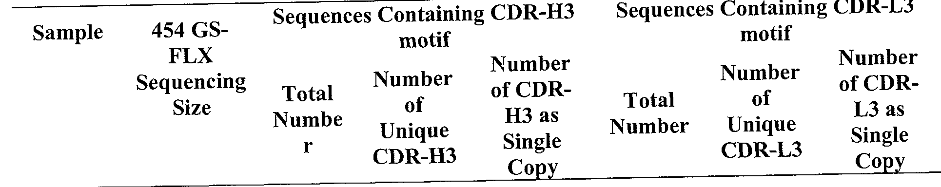


Claims
Priority Applications (5)
| Application Number | Priority Date | Filing Date | Title |
|---|---|---|---|
| KR20127032971A KR20130135028A (en) | 2010-05-17 | 2011-05-17 | Rapid isolation of monoclonal antibodies from animals |
| CA2799746A CA2799746C (en) | 2010-05-17 | 2011-05-17 | Rapid isolation of monoclonal antibodies from animals |
| EP11784105.6A EP2572203B1 (en) | 2010-05-17 | 2011-05-17 | Determination of antigen-specific antibody sequences in blood circulation |
| AU2011256290A AU2011256290B2 (en) | 2010-05-17 | 2011-05-17 | Rapid isolation of monoclonal antibodies from animals |
| CN2011800353065A CN103003697A (en) | 2010-05-17 | 2011-05-17 | Rapid isolation of monoclonal antibodies from animals |
Applications Claiming Priority (4)
| Application Number | Priority Date | Filing Date | Title |
|---|---|---|---|
| US34553810P | 2010-05-17 | 2010-05-17 | |
| US61/345,538 | 2010-05-17 | ||
| US37781610P | 2010-08-27 | 2010-08-27 | |
| US61/377,816 | 2010-08-27 |
Publications (2)
| Publication Number | Publication Date |
|---|---|
| WO2011146514A2 true WO2011146514A2 (en) | 2011-11-24 |
| WO2011146514A3 WO2011146514A3 (en) | 2012-11-22 |
Family
ID=44992293
Family Applications (1)
| Application Number | Title | Priority Date | Filing Date |
|---|---|---|---|
| PCT/US2011/036852 WO2011146514A2 (en) | 2010-05-17 | 2011-05-17 | Rapid isolation of monoclonal antibodies from animals |
Country Status (7)
| Country | Link |
|---|---|
| US (2) | US9090674B2 (en) |
| EP (1) | EP2572203B1 (en) |
| KR (1) | KR20130135028A (en) |
| CN (1) | CN103003697A (en) |
| AU (1) | AU2011256290B2 (en) |
| CA (1) | CA2799746C (en) |
| WO (1) | WO2011146514A2 (en) |
Cited By (15)
| Publication number | Priority date | Publication date | Assignee | Title |
|---|---|---|---|---|
| EP2604625A1 (en) * | 2011-09-26 | 2013-06-19 | Merus B.V. | Generation of binding molecules |
| WO2013078455A3 (en) * | 2011-11-23 | 2013-10-31 | The Board Of Regents Of The University Of Texas System | Proteomic identification of antibodies |
| CN103987853A (en) * | 2011-09-28 | 2014-08-13 | Cb生物技术公司 | Identification of antigen-specific adaptive immune responses using Arm-PCR and high-throughput sequencing |
| US9090674B2 (en) | 2010-05-17 | 2015-07-28 | The Board Of Regents Of The University Of Texas System | Rapid isolation of monoclonal antibodies from animals |
| EP3023435A1 (en) * | 2014-11-19 | 2016-05-25 | Fraunhofer-Gesellschaft zur Förderung der angewandten Forschung e.V. | Influenza virus binding peptides |
| US9708654B2 (en) | 2012-06-15 | 2017-07-18 | Board Of Regents, The University Of Texas System | High throughput sequencing of multiple transcripts |
| US9920110B2 (en) | 2011-03-09 | 2018-03-20 | Cell Signaling Technology, Inc. | Methods and reagents for creating monoclonal antibodies |
| US10513733B2 (en) | 2015-03-23 | 2019-12-24 | Board Of Regents, The University Of Texas System | High throughout sequencing of paired VH and VL transcripts from B cells secreting antigen-specific antibodies |
| WO2020210067A1 (en) * | 2019-04-08 | 2020-10-15 | Phanes Therapeutics, Inc. | Humanized anti-dll3 chimeric antigen receptors and uses thereof |
| EP3626727A4 (en) * | 2017-05-15 | 2021-02-24 | Remer Consultores Assessoria Empresarial Ltda. | Compound, synthetic intermediate, use, pharmaceutical composition, and neuromodulatory therapeutic method |
| US10966411B2 (en) | 2008-06-27 | 2021-04-06 | Merus N.V. | Antibody producing non-human mammals |
| US11773510B2 (en) | 2016-06-01 | 2023-10-03 | Genewiz Suzhou | Method of discovering specific functional antibodies |
| EP4021489A4 (en) * | 2019-08-30 | 2023-10-11 | F. Hoffmann-La Roche AG | Developing an efficient hybridoma platform for therapeutic antibody discovery |
| US11820979B2 (en) | 2016-12-23 | 2023-11-21 | Visterra, Inc. | Binding polypeptides and methods of making the same |
| US11866785B2 (en) | 2017-10-27 | 2024-01-09 | Board Of Regents, The University Of Texas System | Tumor specific antibodies and T-cell receptors and methods of identifying the same |
Families Citing this family (32)
| Publication number | Priority date | Publication date | Assignee | Title |
|---|---|---|---|---|
| ES2730951T3 (en) * | 2010-10-08 | 2019-11-13 | Harvard College | High performance immune sequencing |
| ES2615733T3 (en) | 2010-12-16 | 2017-06-08 | Gigagen, Inc. | Methods for parallel mass analysis of nucleic acids in individual cells |
| US9758591B2 (en) | 2012-08-24 | 2017-09-12 | The Regents Of The University Of California | Antibodies and vaccines for use in treating ROR1 cancers and inhibiting metastasis |
| WO2014066744A2 (en) * | 2012-10-25 | 2014-05-01 | True North Therapeutics, Inc. | Anti-complement c1s antibodies and uses thereof |
| US8945562B2 (en) | 2012-11-02 | 2015-02-03 | True North Therapeutics, Inc. | Anti-complement C1s antibodies |
| GB2584364A (en) | 2013-03-15 | 2020-12-02 | Abvitro Llc | Single cell bar-coding for antibody discovery |
| CA2914369C (en) | 2013-06-06 | 2023-02-14 | Igenica Biotherapeutics, Inc. | Anti-c10orf54 antibodies and uses thereof |
| JP6397938B2 (en) * | 2014-06-17 | 2018-09-26 | アカデミア シニカAcademia Sinica | Humanized anti-IgE antibody that crosslinks with CD23 of B lymphocytes but does not sensitize mast cells |
| US20170212130A1 (en) * | 2014-06-25 | 2017-07-27 | The Rockefeller University | Compositions and methods for rapid production of versatile nanobody repertoires |
| SG11201702060VA (en) | 2014-09-15 | 2017-04-27 | Abvitro Inc | High-throughput nucleotide library sequencing |
| CN107428827A (en) * | 2015-03-18 | 2017-12-01 | 宜康公司 | Monoclonal antibody is produced by B cell elutriation and propagation high flux |
| LT3280440T (en) | 2015-04-06 | 2023-02-27 | Bioverativ Usa Inc. | Humanized anti-c1s antibodies and methods of use thereof |
| CN107345241A (en) * | 2016-05-12 | 2017-11-14 | 眭维国 | B cell antigen receptor H chains CDR3 processing method |
| US11067583B2 (en) * | 2016-05-25 | 2021-07-20 | University Of Maryland, Baltimore | Methods of making active antibodies from biological fluids |
| MY196869A (en) | 2016-06-27 | 2023-05-05 | Univ California | Cancer treatment combinations |
| GB201612242D0 (en) * | 2016-07-14 | 2016-08-31 | Genome Res Ltd And Cambridge Entpr Ltd | Novel kit |
| CN108456682A (en) * | 2017-02-17 | 2018-08-28 | 苏州金唯智生物科技有限公司 | A kind of screening technique of monoclonal antibody and its application |
| WO2018237357A1 (en) | 2017-06-22 | 2018-12-27 | University Of Maryland, Baltimore | Broadly neutralizing antibodies against hiv |
| USD844653S1 (en) * | 2017-11-26 | 2019-04-02 | Jan Magnus Edman | Display screen with graphical user interface |
| CN112584860B (en) | 2018-05-08 | 2024-04-05 | 凡恩世制药(北京)有限公司 | anti-DLL 3 antibodies and uses thereof |
| EP3982369A4 (en) | 2019-06-07 | 2023-06-14 | Chugai Seiyaku Kabushiki Kaisha | Information processing system, information processing method, program, and method for producing antigen-binding molecule or protein |
| WO2021003082A1 (en) * | 2019-07-03 | 2021-01-07 | Phanes Therapeutics, Inc. | Anti-claudin 18.2/anti-cd47 bispecific antibodies and uses thereof |
| CN110452296A (en) * | 2019-07-16 | 2019-11-15 | 福州精锐生物技术有限公司 | A kind of rabbit monoclonal antibodies preparation method |
| EP3767628B1 (en) * | 2019-07-18 | 2024-03-27 | Bayer Aktiengesellschaft | Selection of antibodies / antibody fragments |
| CN110428870B (en) * | 2019-08-08 | 2023-03-21 | 苏州泓迅生物科技股份有限公司 | Method for predicting antibody heavy chain and light chain pairing probability and application thereof |
| CA3154556A1 (en) * | 2019-09-11 | 2021-03-18 | The Rockefeller University | Compositions and methods related to human neutralizing antibodies to hepatitis b |
| US20220362374A1 (en) * | 2021-04-29 | 2022-11-17 | The Government Of The United States, As Represented By The Secretary Of The Army | Antibodies Against Fentanyl and Fentanyl Analogs |
| CN113372447A (en) * | 2021-05-26 | 2021-09-10 | 重庆中元汇吉生物技术有限公司 | anti-PIVKA-II monoclonal antibody and application thereof |
| CN116693683B (en) * | 2022-02-28 | 2024-02-13 | 东莞市朋志生物科技有限公司 | Anti-testosterone antibody, reagent for detecting testosterone and kit |
| CN116836278B (en) * | 2022-03-23 | 2024-04-26 | 东莞市朋志生物科技有限公司 | Anti-progesterone antibody, kit for detecting progesterone |
| CN117487010A (en) * | 2022-08-02 | 2024-02-02 | 东莞市朋志生物科技有限公司 | Anti-tetraiodothyroxine antibody or functional fragment thereof, reagent for detecting tetraiodothyroxine and kit |
| CN115636879B (en) * | 2022-09-30 | 2024-01-05 | 南京集思慧远生物科技有限公司 | Nanometer antibody production and application method based on second-generation sequencing and proteomics |
Family Cites Families (14)
| Publication number | Priority date | Publication date | Assignee | Title |
|---|---|---|---|---|
| JP3649395B2 (en) * | 2000-04-27 | 2005-05-18 | 山之内製薬株式会社 | Fused heteroaryl derivatives |
| GB0130267D0 (en) | 2001-12-19 | 2002-02-06 | Neutec Pharma Plc | Focussed antibody technology |
| AU2002951346A0 (en) * | 2002-09-05 | 2002-09-26 | Garvan Institute Of Medical Research | Diagnosis of ovarian cancer |
| WO2004037852A2 (en) * | 2002-10-21 | 2004-05-06 | University Of Medicine And Dentistry Of New Jersey | Thymidylate synthase polymorphisms for use in screening for cancer susceptibility |
| US7329353B2 (en) * | 2004-01-23 | 2008-02-12 | Amgen Inc. | LC/MS method of analyzing high molecular weight proteins |
| AU2005211790B2 (en) | 2004-02-09 | 2010-03-11 | Northeastern University | Monoclonal antibody based biomarker discovery and development platform |
| WO2005084134A2 (en) | 2004-03-04 | 2005-09-15 | Dena Leshkowitz | Quantifying and profiling antibody and t cell receptor gene expression |
| ES2549077T3 (en) * | 2004-04-07 | 2015-10-22 | Genentech, Inc. | Mass spectrometry of antibody conjugates |
| ES2374403T3 (en) | 2006-12-21 | 2012-02-16 | Novartis Ag | QUANTIFICATION OF ANTIBODIES. |
| WO2009050261A1 (en) * | 2007-10-19 | 2009-04-23 | Pronota N.V. | Preparation of samples for proteome analysis |
| EP2088432A1 (en) | 2008-02-11 | 2009-08-12 | MorphoSys AG | Methods for identification of an antibody or a target |
| ES2726702T3 (en) | 2009-01-15 | 2019-10-08 | Adaptive Biotechnologies Corp | Adaptive immunity profiling and methods for the generation of monoclonal antibodies |
| EP2848704B1 (en) * | 2010-01-19 | 2018-08-29 | Verinata Health, Inc | Sequencing methods for prenatal diagnoses |
| CA2799746C (en) | 2010-05-17 | 2020-11-24 | Sai Reddy | Rapid isolation of monoclonal antibodies from animals |
-
2011
- 2011-05-17 CA CA2799746A patent/CA2799746C/en active Active
- 2011-05-17 KR KR20127032971A patent/KR20130135028A/en not_active Application Discontinuation
- 2011-05-17 CN CN2011800353065A patent/CN103003697A/en active Pending
- 2011-05-17 AU AU2011256290A patent/AU2011256290B2/en active Active
- 2011-05-17 EP EP11784105.6A patent/EP2572203B1/en active Active
- 2011-05-17 US US13/109,467 patent/US9090674B2/en active Active
- 2011-05-17 WO PCT/US2011/036852 patent/WO2011146514A2/en active Application Filing
-
2015
- 2015-07-27 US US14/809,651 patent/US20160034639A1/en not_active Abandoned
Non-Patent Citations (4)
| Title |
|---|
| DOMMAIR K ET AL., SEMINARS IN IMMUNOPATHOLOGY, vol. 21, no. 4, 2009, pages 467 - 477 |
| OBERMEIER ET AL., NATURE MEDICINE, vol. 14, no. 6, 2008, pages 688 - 693 |
| SCHULTER SF ET AL., MOLECULAR IMMUNOLOGY, vol. 27, no. 1, 1990, pages L7 - 23 |
| See also references of EP2572203A2 |
Cited By (35)
| Publication number | Priority date | Publication date | Assignee | Title |
|---|---|---|---|---|
| US11925174B2 (en) | 2008-06-27 | 2024-03-12 | Merus N.V. | Antibody producing non-human animals |
| US11785924B2 (en) | 2008-06-27 | 2023-10-17 | Merus N.V. | Antibody producing non-human animals |
| US11559049B2 (en) | 2008-06-27 | 2023-01-24 | Merus N.V. | Antibody producing non-human animals |
| US10966411B2 (en) | 2008-06-27 | 2021-04-06 | Merus N.V. | Antibody producing non-human mammals |
| US10175249B2 (en) | 2010-05-17 | 2019-01-08 | Board Of Regents, The University Of Texas System | Proteomic identification of antibodies |
| US9090674B2 (en) | 2010-05-17 | 2015-07-28 | The Board Of Regents Of The University Of Texas System | Rapid isolation of monoclonal antibodies from animals |
| US9920110B2 (en) | 2011-03-09 | 2018-03-20 | Cell Signaling Technology, Inc. | Methods and reagents for creating monoclonal antibodies |
| AU2018201328B2 (en) * | 2011-09-26 | 2020-07-02 | Merus N.V. | Generation of binding molecules |
| EP4086282A1 (en) * | 2011-09-26 | 2022-11-09 | Merus N.V. | Generation of binding molecules |
| US10647781B2 (en) | 2011-09-26 | 2020-05-12 | Merus N.V. | Generation of binding molecules |
| EP2604625B1 (en) | 2011-09-26 | 2017-06-28 | Merus N.V. | Generation of binding molecules |
| AU2012233037B2 (en) * | 2011-09-26 | 2015-10-29 | Merus N.V. | Generation of binding molecules |
| AU2016200316B2 (en) * | 2011-09-26 | 2017-11-23 | Merus N.V. | Generation of binding molecules |
| EP3263597A1 (en) * | 2011-09-26 | 2018-01-03 | Merus N.V. | Generation of binding molecules |
| US9908946B2 (en) | 2011-09-26 | 2018-03-06 | Merus N.V. | Generation of binding molecules |
| US11408095B2 (en) | 2011-09-26 | 2022-08-09 | Merus N.V. | Generation of binding molecules |
| EP2604625A1 (en) * | 2011-09-26 | 2013-06-19 | Merus B.V. | Generation of binding molecules |
| US9145588B2 (en) | 2011-09-26 | 2015-09-29 | Merus Biopharmaceuticals B.V. | Generation of binding molecules |
| CN103987853A (en) * | 2011-09-28 | 2014-08-13 | Cb生物技术公司 | Identification of antigen-specific adaptive immune responses using Arm-PCR and high-throughput sequencing |
| EP2761015A4 (en) * | 2011-09-28 | 2015-09-02 | Cb Biotechnologies Inc | Identification of antigen-specific adaptive immune responses using arm-pcr and high-throughput sequencing |
| CN109254143A (en) * | 2011-09-28 | 2019-01-22 | 艾瑞普特公司 | Use the adaptive immune response of Arm-PCR and high-flux sequence identification antigen-specific |
| US11782053B2 (en) | 2011-09-28 | 2023-10-10 | iRepertoire, Inc. | Identification of antigen-specific adaptive immune responses using ARM-PCR and high-throughput sequencing |
| WO2013078455A3 (en) * | 2011-11-23 | 2013-10-31 | The Board Of Regents Of The University Of Texas System | Proteomic identification of antibodies |
| US9146241B2 (en) | 2011-11-23 | 2015-09-29 | Board Of Regents, The University Of Texas System | Proteomic identification of antibodies |
| US9708654B2 (en) | 2012-06-15 | 2017-07-18 | Board Of Regents, The University Of Texas System | High throughput sequencing of multiple transcripts |
| US20180072770A1 (en) * | 2014-11-19 | 2018-03-15 | Fraunhofer-Gesellschaft Zur Foerderung Der Angewandten Forschung E.V. | Influenza Virus Binding Peptides |
| EP3023435A1 (en) * | 2014-11-19 | 2016-05-25 | Fraunhofer-Gesellschaft zur Förderung der angewandten Forschung e.V. | Influenza virus binding peptides |
| WO2016079250A1 (en) * | 2014-11-19 | 2016-05-26 | Fraunhofer-Gesellschaft zur Förderung der angewandten Forschung e. V. | Influenza virus binding peptides |
| US10513733B2 (en) | 2015-03-23 | 2019-12-24 | Board Of Regents, The University Of Texas System | High throughout sequencing of paired VH and VL transcripts from B cells secreting antigen-specific antibodies |
| US11773510B2 (en) | 2016-06-01 | 2023-10-03 | Genewiz Suzhou | Method of discovering specific functional antibodies |
| US11820979B2 (en) | 2016-12-23 | 2023-11-21 | Visterra, Inc. | Binding polypeptides and methods of making the same |
| EP3626727A4 (en) * | 2017-05-15 | 2021-02-24 | Remer Consultores Assessoria Empresarial Ltda. | Compound, synthetic intermediate, use, pharmaceutical composition, and neuromodulatory therapeutic method |
| US11866785B2 (en) | 2017-10-27 | 2024-01-09 | Board Of Regents, The University Of Texas System | Tumor specific antibodies and T-cell receptors and methods of identifying the same |
| WO2020210067A1 (en) * | 2019-04-08 | 2020-10-15 | Phanes Therapeutics, Inc. | Humanized anti-dll3 chimeric antigen receptors and uses thereof |
| EP4021489A4 (en) * | 2019-08-30 | 2023-10-11 | F. Hoffmann-La Roche AG | Developing an efficient hybridoma platform for therapeutic antibody discovery |
Also Published As
| Publication number | Publication date |
|---|---|
| WO2011146514A3 (en) | 2012-11-22 |
| US9090674B2 (en) | 2015-07-28 |
| CA2799746A1 (en) | 2011-11-24 |
| EP2572203B1 (en) | 2017-10-25 |
| AU2011256290A1 (en) | 2012-12-06 |
| EP2572203A4 (en) | 2013-10-09 |
| CA2799746C (en) | 2020-11-24 |
| AU2011256290B2 (en) | 2014-06-12 |
| KR20130135028A (en) | 2013-12-10 |
| CN103003697A (en) | 2013-03-27 |
| EP2572203A2 (en) | 2013-03-27 |
| US20110312505A1 (en) | 2011-12-22 |
| US20160034639A1 (en) | 2016-02-04 |
Similar Documents
| Publication | Publication Date | Title |
|---|---|---|
| US9090674B2 (en) | Rapid isolation of monoclonal antibodies from animals | |
| US10175249B2 (en) | Proteomic identification of antibodies | |
| US11866785B2 (en) | Tumor specific antibodies and T-cell receptors and methods of identifying the same | |
| AU2016235251B2 (en) | HLA-restricted epitopes encoded by somatically mutated genes | |
| US11447891B2 (en) | Compositions and methods for rapid production of versatile nanobody repertoires | |
| EP3421592A1 (en) | Identification of polynucleotides associated with a sample | |
| CN108220297A (en) | For generating the method for monoclonal antibody and reagent | |
| JP2023524719A (en) | Compositions and methods for identifying nanobodies and nanobody affinities | |
| JP6338235B2 (en) | Screening method and production method of low molecular weight antibody | |
| WO2016019888A1 (en) | Method and system for screening for nanobodies | |
| Georgiou et al. | Proteomic identification of antibodies | |
| Georgiou et al. | Rapid isolation of monoclonal antibodies from animals | |
| US9518131B2 (en) | Generating metal ion binding proteins | |
| McGrath et al. | Mutability and hypermutation antagonize immunoglobulin codon optimality | |
| Wolf et al. | Plasma cell immunoglobulin heavy chain repertoire dynamics before and after tetanus booster vaccination | |
| Le Bihan et al. | de Novo Sequencing of Antibodies for Identification of Neutralizing Antibodies in Human Plasma Post SARS-CoV-2 Vaccination | |
| WO2021258114A1 (en) | Process for direct readout of immunoglobulins | |
| CN117886929A (en) | Antibodies against novel coronaviruses, reagents and kits for detecting novel coronaviruses | |
| Gräßel | The surface atlas of human naive and activated CD4+ T cells |
Legal Events
| Date | Code | Title | Description |
|---|---|---|---|
| ENP | Entry into the national phase |
Ref document number: 2799746 Country of ref document: CA |
|
| NENP | Non-entry into the national phase |
Ref country code: DE |
|
| REEP | Request for entry into the european phase |
Ref document number: 2011784105 Country of ref document: EP |
|
| WWE | Wipo information: entry into national phase |
Ref document number: 2011784105 Country of ref document: EP |
|
| ENP | Entry into the national phase |
Ref document number: 2011256290 Country of ref document: AU Date of ref document: 20110517 Kind code of ref document: A |
|
| ENP | Entry into the national phase |
Ref document number: 20127032971 Country of ref document: KR Kind code of ref document: A |