WO2011126010A1 - Cancer cell-specific cell-penetrating peptide and use thereof - Google Patents
Cancer cell-specific cell-penetrating peptide and use thereof Download PDFInfo
- Publication number
- WO2011126010A1 WO2011126010A1 PCT/JP2011/058616 JP2011058616W WO2011126010A1 WO 2011126010 A1 WO2011126010 A1 WO 2011126010A1 JP 2011058616 W JP2011058616 W JP 2011058616W WO 2011126010 A1 WO2011126010 A1 WO 2011126010A1
- Authority
- WO
- WIPO (PCT)
- Prior art keywords
- peptide
- cells
- cell
- cancer cells
- nucleic acid
- Prior art date
Links
Images


Classifications
-
- A—HUMAN NECESSITIES
- A61—MEDICAL OR VETERINARY SCIENCE; HYGIENE
- A61K—PREPARATIONS FOR MEDICAL, DENTAL OR TOILETRY PURPOSES
- A61K38/00—Medicinal preparations containing peptides
- A61K38/04—Peptides having up to 20 amino acids in a fully defined sequence; Derivatives thereof
- A61K38/10—Peptides having 12 to 20 amino acids
-
- A—HUMAN NECESSITIES
- A61—MEDICAL OR VETERINARY SCIENCE; HYGIENE
- A61K—PREPARATIONS FOR MEDICAL, DENTAL OR TOILETRY PURPOSES
- A61K47/00—Medicinal preparations characterised by the non-active ingredients used, e.g. carriers or inert additives; Targeting or modifying agents chemically bound to the active ingredient
- A61K47/50—Medicinal preparations characterised by the non-active ingredients used, e.g. carriers or inert additives; Targeting or modifying agents chemically bound to the active ingredient the non-active ingredient being chemically bound to the active ingredient, e.g. polymer-drug conjugates
- A61K47/51—Medicinal preparations characterised by the non-active ingredients used, e.g. carriers or inert additives; Targeting or modifying agents chemically bound to the active ingredient the non-active ingredient being chemically bound to the active ingredient, e.g. polymer-drug conjugates the non-active ingredient being a modifying agent
- A61K47/62—Medicinal preparations characterised by the non-active ingredients used, e.g. carriers or inert additives; Targeting or modifying agents chemically bound to the active ingredient the non-active ingredient being chemically bound to the active ingredient, e.g. polymer-drug conjugates the non-active ingredient being a modifying agent the modifying agent being a protein, peptide or polyamino acid
- A61K47/64—Drug-peptide, drug-protein or drug-polyamino acid conjugates, i.e. the modifying agent being a peptide, protein or polyamino acid which is covalently bonded or complexed to a therapeutically active agent
-
- A—HUMAN NECESSITIES
- A61—MEDICAL OR VETERINARY SCIENCE; HYGIENE
- A61K—PREPARATIONS FOR MEDICAL, DENTAL OR TOILETRY PURPOSES
- A61K9/00—Medicinal preparations characterised by special physical form
- A61K9/0012—Galenical forms characterised by the site of application
- A61K9/0019—Injectable compositions; Intramuscular, intravenous, arterial, subcutaneous administration; Compositions to be administered through the skin in an invasive manner
-
- A—HUMAN NECESSITIES
- A61—MEDICAL OR VETERINARY SCIENCE; HYGIENE
- A61K—PREPARATIONS FOR MEDICAL, DENTAL OR TOILETRY PURPOSES
- A61K9/00—Medicinal preparations characterised by special physical form
- A61K9/08—Solutions
-
- A—HUMAN NECESSITIES
- A61—MEDICAL OR VETERINARY SCIENCE; HYGIENE
- A61P—SPECIFIC THERAPEUTIC ACTIVITY OF CHEMICAL COMPOUNDS OR MEDICINAL PREPARATIONS
- A61P35/00—Antineoplastic agents
-
- A—HUMAN NECESSITIES
- A61—MEDICAL OR VETERINARY SCIENCE; HYGIENE
- A61P—SPECIFIC THERAPEUTIC ACTIVITY OF CHEMICAL COMPOUNDS OR MEDICINAL PREPARATIONS
- A61P35/00—Antineoplastic agents
- A61P35/02—Antineoplastic agents specific for leukemia
Definitions
- the present invention relates to a cancer cell-selective membrane-permeable peptide having a specific amino acid sequence, and an antitumor containing an antitumor substance formed by binding a cancer cell-selective membrane-permeable peptide and an antitumor factor as an active ingredient
- the present invention relates to an imaging agent comprising an agent, a cancer cell-selective membrane-permeable peptide, and a labeling substance.
- Non-patent Document 1 Penetratin derived from Antennapedia (Non-patent Document 1) and Tat peptide (Non-patent Document 2) are known, and proteins, peptides, nucleic acids and the like are contained in cells. Used to transport to. It has also been reported that a peptide containing a polyarginine residue is used as a membrane-permeable carrier (Patent Document 1). However, when treating or detecting cancer, it is necessary to selectively deliver anticancer agents and imaging agents to specific cancer cells, and these peptides can be selectively permeated into specific cancer cells. It is not sufficient for this purpose because it does not show any properties. Furthermore, Patent Documents 2 to 4 disclose cell membrane-permeable peptides having various sequences, but it has not been investigated whether these peptides have cancer cell-selective membrane permeability.
- An object of the present invention is to provide a peptide having cancer cell-selective membrane permeability, an antitumor substance or an imaging agent using the peptide, and the like.
- cell-penetrating peptides cell-penetrating peptides; hereinafter referred to as “cell-penetrating peptides”
- cell-penetrating peptides selected / identified using genotype-phenotype mapping molecules.
- genotype-phenotype mapping molecules has been found to have a cancer cell-selective membrane permeation function.
- the present invention has been accomplished based on these findings.
- the gist of the present invention resides in the following [1] to [16].
- [1] A cancer cell-selective membrane-permeable peptide comprising any amino acid sequence shown in SEQ ID NOs: 1 to 10 and having a selective membrane permeability function for cancer cells.
- [2] A permeation method of a target substance into a specific cancer cell, wherein the target substance is bound to the peptide according to [1] and the target substance permeates into the specific cancer cell (except for a method for treating humans).
- An antitumor agent comprising an antitumor substance formed by binding a peptide having a selective membrane permeation function for cancer cells and an antitumor factor as an active ingredient.
- Cancer cells are uterine cancer cells, colon cancer cells, lung cancer cells, breast cancer cells, gastric cancer cells, liver cancer cells, prostate cancer cells, kidney cancer cells, pancreatic cancer cells, brain tumor cells, sarcoma cells, malignant mesothelioma cells.
- a peptide having a membrane permeation function is (1) a protein part including at least a candidate peptide consisting of 2 to 100 amino acid residues and a nucleic acid part including at least a base sequence encoding the candidate peptide, the C-terminus of the protein part and the 3 ′ end of the nucleic acid part
- Contacting a target cell with a group of genotype-phenotype mapping molecules that are covalently linked to a target cell (2) a step of amplifying a nucleic acid contained in the nucleic acid portion of the above-mentioned corresponding molecule introduced into the target cell; and (3) analyzing a base sequence of the amplified nucleic acid, and a peptide encoded by the base sequence, Identifying as a peptide having a membrane permeation function;
- the antitumor agent according to any one of [3] to [5], which is obtained by a method comprising [7]
- the mapping molecule in step (1) is the following (a): (A
- the method further comprises a step between steps (1) and (2), and (4) removing the molecule that has not been introduced into the cell from the surface of the target cell [6] or [7] The antitumor agent as described in.
- the cancer cells are uterine cancer cells, colon cancer cells, lung cancer cells, breast cancer cells, gastric cancer cells, liver cancer cells, prostate cancer cells, kidney cancer cells, pancreatic cancer cells, brain tumors, sarcoma cells, malignant mesothelioma cells,
- the antitumor agent according to [9] which is any cell selected from the group consisting of lymphoma cells and leukemia cells.
- any of [3] to [8], wherein the peptide having a membrane permeation function includes any amino acid sequence shown in SEQ ID NOs: 1 to 10 and has a selective membrane permeation function for cancer cells.
- An antitumor agent according to any one of the above.
- the antitumor factor is a protein having a function of compensating for the function of a tumor suppressor gene whose expression is lost in cancer cells or a functional domain peptide thereof The antitumor agent as described in.
- An imaging agent comprising an imaging substance formed by binding a peptide having a membrane permeation function selective to cancer cells and a labeling substance, and having an ability to selectively accumulate tumor sites.
- the labeling substance is a fluorescent substance or a substance having a positron emitting nuclide.
- the cancer cell-selective membrane-permeable peptide of the present invention exhibits selective membrane permeability for human cancer cells of different origins. And they exhibit higher permeability than non-selective permeability peptides.
- the antitumor substance of the present invention is obtained by binding an antitumor factor to a peptide having a selective membrane permeation function for cancer cells, and can reliably deliver the antitumor factor to cancer cells. Therefore, the antitumor substance of the present invention is expected to be an antitumor agent having excellent antitumor effects and few side effects as a tumor selective target drug.
- the imaging agent of the present invention has selective membrane permeability to cancer cells, cancer metastasis can be visualized with high sensitivity, and tracer development using PET (positron emission tomography) etc., intraoperative cancer Expected to be used for imaging.
- the numbers in the panel indicate CPP numbers.
- the numbers in the panel indicate CPP numbers.
- cancer cell-selective membrane-permeable peptide The cancer cell-specific cell-penetrating peptide of the present invention (hereinafter sometimes abbreviated as “CCS-CPP”) is SEQ ID NO: 1 to 10 comprising any amino acid sequence shown in FIG. 10 and having a selective membrane permeation function for cancer cells.
- the selective membrane permeation function for cancer cells means a selective membrane permeation function for cancer cells of a specific type (namely, pathological tissue type classification). It is preferable that the membrane permeation function for a specific type of cancer cell of interest is higher than the membrane permeation function for cancer cells other than the specific cancer cell and normal cells.
- the cancer cell within a specific kind may be one kind, and may be two or more kinds.
- the amino acid sequence at the N-terminus and / or C-terminus of the above amino acid sequence usually has a total of 1 to 10, preferably 1 to 5 amino acid sequences so that the sum of the constituent ratios of arginine and lysine does not exceed 35%.
- Those added and having the above functions are also included in the cancer cell-selective membrane-permeable peptide (CCS-CPP) of the present invention.
- CCS-CPP cancer cell-selective membrane-permeable peptide
- These peptides can be selected / identified by a method using an association molecule between a genotype (nucleic acid) and a phenotype (protein) described later.
- cancer cell membrane permeability is determined by co-culturing a peptide labeled with an appropriate labeling substance (for example, a fluorescent substance) and a cancer cell, and using the peptide permeated to the cancer cell as a marker using a fluorescence microscope or a flow site. It can be confirmed with a meter (Flowcytometer).
- an appropriate labeling substance for example, a fluorescent substance
- the antitumor substance used in the present invention is characterized in that a cancer cell-selective membrane-permeable peptide (CCS-CPP) and an antitumor factor are bound to each other.
- CCS-CPP cancer cell-selective membrane-permeable peptide
- the binding between CCS-CPP and the anti-tumor factor is any binding as long as the anti-tumor factor can maintain a binding state that can be incorporated into cancer cells by the membrane permeability function of CCS-CPP. It may be a style.
- the bonding mode include hydrogen bonding, van der Waals bonding, ionic bonding, and covalent bonding. Of these, covalent bonds are preferred.
- Coupling between CCS-CPP and an antitumor factor is preferably a covalent bond via a linker.
- the linker is not particularly limited as long as it retains the functions of CCS-CPP and an antitumor factor and can penetrate the cell membrane together with CCS-CPP.
- a peptide chain having a length of usually 1 to 5 residues, preferably about 1 to 3 residues, or a linker that can be used with a polyethylene glycol (PEG) chain having an equivalent length can be used. It is done.
- the amino acid residue constituting the peptide linker is preferably a small molecule, such as a glycine residue, without charge.
- a sequence for giving freedom of rotation to both domains CCS-CPP and antitumor factor
- a sequence for giving freedom of rotation to both domains CCS-CPP and antitumor factor
- a sequence containing glycine (G) and a linker containing proline (P) is preferable. More specifically, a sequence consisting of a glycine residue and a proline residue, for example, Particularly preferred is glycine (G) -proline (P) -glycine (G). With this configuration, the functions of both domains can be exhibited.
- the cancer cell-selective membrane-permeable peptide can be used as long as it has a selective membrane permeability function for a specific cancer cell and the ability to deliver an antitumor factor into the cancer cell.
- a peptide CCS-CPP
- a method including the following steps (1) to (3) can be mentioned as a preferable method for selecting / identifying a peptide having a membrane permeation function.
- (1) including a target cell, a protein part containing at least a candidate peptide consisting of 2 to 100 amino acid residues, and a nucleic acid part containing at least a base sequence encoding the candidate peptide, the C-terminal of the protein part and the nucleic acid part
- mapping molecule (IVV molecule) used in the above step (1) includes the following molecule (a) or molecule (b).
- A a protein part containing a fusion protein of a candidate peptide consisting of 2 to 100 amino acid residues and a target protein transported into the target cell by the peptide, a base sequence encoding the candidate peptide, and the target protein
- a nucleic acid part containing a base sequence to be bound, a nucleic acid derivative bound to the 3 ′ end of the nucleic acid part via a spacer, and the nucleic acid derivative and the C part of the protein part are covalently bonded.
- the nucleic acid derivative is not particularly limited as long as it is covalently bonded to the C-terminus of the protein portion. Specifically, a nucleic acid derivative having a chemical structure skeleton similar to aminoacyl-tRNA at the 3 ′ end can be selected.
- puromycin having an amide bond 3′-N-aminoacylpuromycin aminonucleoside (3′-N-aminoacylpuromycin aminoamide, PANS-amino acid), for example, PANS-Gly having an amino acid part of glycine PANS-Val whose amino acid part is valine, PANS-Ala whose amino acid part is alanine, and other PANS-amino acid compounds whose amino acid part corresponds to all amino acids.
- 3′-N-aminoacyl adenosine aminonucleoside (3′-Aminoacyladenosine aminonucleotide, AANS-amino acid) in which the amino group of 3′-aminoadenosine and the carboxyl group of amino acid are linked by an amide bond formed by dehydration condensation, for example, AANS-Gly having an amino acid part of glycine, AANS-Val having an amino acid part of alanine, AANS-Ala having an amino acid part of alanine, and an AANS-amino acid compound corresponding to each amino acid having an amino acid part of all amino acids can be used.
- nucleosides or those obtained by ester bonding of nucleosides and amino acids can be used.
- a nucleic acid or a compound obtained by chemically combining a substance having a chemical structure skeleton and base similar to nucleic acid and a substance having a chemical structure skeleton similar to amino acid, and peptide nucleic acid (PNA) are all used in this method.
- the nucleic acid derivative is more preferably a compound in which puromycin, PANS-amino acid or AANS-amino acid is bound to a nucleoside via a phosphate group.
- puromycin derivatives such as puromycin, ribocytidylpuromycin, deoxycytidylpuromycin, and deoxyuridylpuromycin are particularly preferred.
- the candidate peptide is usually composed of a random amino acid sequence of about 2 to 100 amino acid residues, preferably about 2 to 40 amino acid residues.
- the protein part includes a fusion protein of the candidate peptide and the target protein, and the nucleic acid part encodes the candidate peptide and the target protein. [Molecule (a)] or a non-protein target substance bound to the nucleic acid part [molecule (b)] is preferable.
- the target protein or non-protein target substance constituting the IVV molecule is not particularly limited as long as it can select / identify a membrane-permeable peptide that can be used in the present invention.
- the target protein is preferably one that can be a support protein for expressing a candidate peptide. If the candidate peptide is a short chain, it will be difficult to express it in a protein synthesis system, particularly a cell-free protein synthesis system. In this case, the candidate peptide can be fused with a support protein to be expressed in a protein synthesis system. Any of the fusion protein of the support protein and the candidate peptide may be N-terminal or C-terminal.
- the support protein is preferably (1) a globular protein that is easy to fold, (2) stable, and (3) does not contain a disulfide (SS) bond.
- SS disulfide
- Examples of the protein that satisfies these conditions include Oct-1 Pou-specific domain (73 amino acid residues) (Dekker, N. et al. (1993) Nature 362, 852-854).
- Non-protein target substances are generally high-molecular substances such as biologically active nucleic acids and sugars, low-molecular substances such as pharmaceutically active ingredients, and nano-materials such as quantum beads, photosensitizers and atoms. Examples include molecules. Examples of the position where such a target substance is added include a spacer portion.
- the IVV molecule preferably contains a labeling substance so that it can be easily detected when it is introduced (permeated) into the cell.
- the labeling substance may be any substance as long as it can detect that intracellular IVV molecules are permeated.
- fluorescent substances such as FITC (Fluoresceinisothiocyanate), Roadamine, Cy3, and Cy5; enzymes used for color assays such as horseradish peroxidase and ⁇ -galactosidase are preferably used.
- FITC Fluoresceinisothiocyanate
- Roadamine Cy3, and Cy5
- enzymes used for color assays such as horseradish peroxidase and ⁇ -galactosidase are preferably used.
- These labeling substances may be added to any position of the IVV molecule.
- a fluorescent protein such as GFP or DsRed can be added to the protein part of the IVV molecule as a fusion protein with a candidate peptide.
- the IVV molecule may include a substance having a property of specifically binding to a certain substance for the purpose of purifying the IVV molecule.
- specific examples include tag peptides such as FLAG, GST, HISx6, and affinity substances such as biotin.
- the tag peptide and affinity substance may be located anywhere in the protein part of the IVV molecule, but the tag peptide is preferably added to the C-terminal or N-terminal of the protein part of the IVV molecule. It is preferable to add to the spacer part mentioned later.
- a polymer substance such as polyethylene or polyethylene glycol or a derivative thereof described in WO98 / 16636, a biopolymer substance such as an oligonucleotide, a peptide or a derivative thereof, or the like is used. Of these, polyethylene glycol is preferred.
- the spacer includes the above-mentioned fluorescent substances such as FITC and derivatives thereof, affinity substances such as biotin and derivatives thereof, nucleic acids such as deoxyribonucleotides and derivatives thereof, or substances having bonds that are cleaved by biochemistry or chemical reaction, for example And a photodegradable substance such as a 5-substituted-2-nitroacetophenone derivative.
- the mapping molecule (IVV molecule) is prepared by first preparing mRNA encoding a protein portion containing a candidate peptide having puromycin bound to the 3 ′ end, and translating it using a cell-free translation system.
- Protein-RNA Chimeric Random Peptide Library (In Vtro ⁇ Virus Library; hereinafter referred to as the protein part reflecting the type) and the mRNA encoding it (the nucleic acid part reflecting the genotype) via puromycin May be referred to as “IVVL”).
- Preparation of IVVL suitable for selection / identification of a membrane-permeable peptide can be performed by, for example, a method described in JP-A-2005-13073.
- the target cell is contacted with IVVL (a group of IVV molecules) [the above step (1)], and the nucleic acid contained in the nucleic acid portion of the molecule that has penetrated into or into the target cell is amplified [the above step (2)]
- the peptide encoded by the base sequence can be identified as a peptide having a cell membrane permeation function [step (3) above]. This operation can be repeated as necessary.
- the obtained cell membrane permeable peptide is examined for membrane permeability using a specific cancer cell as a target cell, and a peptide having selective permeability to the cancer cell is selected / identified, whereby the antitumor substance of the present invention Cancer cell-selective membrane-permeable peptide (CCS-CPP) can be obtained.
- CCS-CPP Cancer cell-selective membrane-permeable peptide
- target cells used for selection / identification of CCS-CPP human or mammalian cells are preferable, and cancer cells (malignant tumor cells) are particularly preferable among them.
- Cancer cells that can be used as target cells are not particularly limited.
- a method for bringing the target cell into contact with the IVV molecule is not particularly limited, but a method of culturing the target cell by an appropriate method in the presence of the IVV molecule is preferable.
- the culture method, the amount of IVV molecule added, the culture temperature, the culture time, and the like can be appropriately selected depending on the type of target cell and IVV molecule.
- the cells are cultured in a 96-well plate so as to have 10 5 cells in a fetal calf serum-containing medium or the like, and the cells are used with an appropriate buffer solution such as PBS.
- the target cancer cell and the IVV molecule can be contacted, but the concentration of the IVV molecule to be contacted can be 1 to 1000 nM.
- the nucleic acid part of the IVV molecule in the cell into which the IVV molecule has been introduced is amplified.
- Cells into which IVV molecules have been introduced may be detected and separated, or may be used in the nucleic acid portion amplification step even if cells that have not been introduced are mixed.
- the detection method of the IVV molecule may be any method as long as it is clear that the IVV molecule is present outside or inside the cell.
- a method for detecting the labeling substance is preferably used.
- a fluorescent substance or a fluorescent protein is used as the labeling substance
- a flow cytometer or a fluorescence microscope can be used as the detection means.
- an enzyme for performing a color assay it can be detected by a color assay to which a necessary substrate is added.
- PCR Polymerase Chain Reaction
- the washing method can be appropriately selected from known methods for washing cells. Specifically, for example, treatment with nuclease such as acid or DNaseI is preferably used. In addition, when performing detection with a flow cytometer, it is preferable that the adherent cells after culture be treated with trypsin.
- IVV molecules that contain candidate peptides and IVV molecules that do not contain candidate peptides are introduced into the cell when the fluorescence intensity of the cell contacted with the IVV molecule containing the candidate peptide is stronger than that of the cell contacted with the IVV molecule not containing the candidate peptide. It may be judged that it is done. In addition, when a fluorescence microscope is used, fluorescence emitted from a fluorescent substance that is a labeling substance may be observed in the cell.
- a method for separating cells into which IVV molecules have been introduced into the cells a method in which cells with fluorescence detected by the flow cytometer are separated with a cell sorter, or cells with fluorescence detected by a fluorescence microscope are captured by laser. And the like.
- the nucleic acid part of the IVV molecules present in the cells is amplified by PCR, and the base sequence is analyzed to obtain a peptide having a function of penetrating into cells, that is, Cell membrane permeable peptides (CPP) can be identified.
- CPP Cell membrane permeable peptides
- the CPP obtained above can be selected to have a selective membrane permeation function for specific cancer cells.
- Examples of the cancer cell-selective membrane-permeable peptide (CCS-CPP) thus obtained include those containing any amino acid sequence shown in SEQ ID NOs: 1 to 10.
- an antitumor factor used for the antitumor substance of the present invention can be delivered to cancer cells by CCS-CPP, and has some function in the growth and metastasis of target cancer cells, Any factor may be used as long as it is suitable for the purpose of treating cancer.
- chemotherapeutic agents antitumor agents
- peptides continuous amino acid residue sequences up to about 30 residues
- proteins DNA, PNA (Peptide Nucleic Acid), LNA (Locked Nucleic Acid), siRNA (Small interfering RNA), microRNA and the like.
- chemotherapeutic agents include 5-FU, paclitaxel, cisplatin, etoposide (vinblastine), cyclophasphamide, Actinomycin D, irinotecan, steroid (predonisolone), and the like. These chemotherapeutic agents are linked to CCS-CPP by introducing a linker through its reactive group (hydroxyl group, amino group, etc.) and reacting the linker with the amino terminus, carboxy terminus or side chain of the peptide. be able to. Alternatively, these chemotherapeutic agents can be indirectly bound by encapsulating them in liposomes and binding CCS-CPP to the surface of the liposomes.
- a protein having a function of recovering the function of a tumor suppressor gene whose expression is lost in cancer cells in a compensatory manner or a peptide containing a functional domain thereof is preferable.
- a peptide containing a protein or its functional domain those capable of inducing apoptosis in cancer cells are preferable.
- a tumor suppressor gene whose expression is lost in cancer cells for example, p16 INK4a , p14 ARF , p15, p18, p21 CIP1 , p27 KIP1 , p53, p57 Kip2 , p73, RB, BRCA1, BRCA2, PTEN, APC, WT1, NF1, NF2, SMAD4, PTC, MSH2, Maspin, SDHD and the like.
- P16 is Fahraeus R, Lain S, Ball KL, Lane DP. Characterization of the cyclin-dependent kinase inhibitory domain of the INK4 family as a model for a synthetic tumour suppressor molecule.Oncogene 1998; 16: 587-96 etc., p53 is like Hupp TR, Sparks A, Lane DP (1995) Small peptides activate the latent sequence-specific DNA binding function of p53. Cell83: 237-245q, The details including the functional domain part are described.
- a protein having a function of restoring the function of a tumor suppressor gene at a compensatory level is an expression product (protein) of the above-described tumor suppressor gene, and its functional domain is an amino acid that controls the cancer suppressor function of the expression product (protein).
- a peptide comprising a sequence.
- p16 INK4a is, for example, lung cancer (adenocarcinoma, squamous cell carcinoma, small cell carcinoma, LCNEC), pharyngeal / laryngeal cancer (squamous cell carcinoma), digestive organ cancer (esophageal cancer, colon cancer, Gastric cancer, biliary tract cancer, hepatocellular carcinoma, pancreatic cancer), urinary cancer (renal cancer, bladder cancer, ureteral cancer), genital cancer [uterine cancer (cervical squamous cell carcinoma, intimal adenocarcinoma), ovarian cancer, prostate cancer, Testicular germ cell tumor], solid cancer such as skin cancer (malignant melanoma, squamous cell carcinoma); malignant bone and soft tissue tumor (osteosarcoma, Ewing sarcoma, rhabdomyosarcoma, liposarcoma, MFH, etc.), digestive organ mesenchyme Sarcomas such as
- Examples of such a functional amino acid sequence having a function of reversibly restoring the function of p16 INK4a include the amino acid sequence shown in SEQ ID NO: 12 (p16 minimal inhibitory sequence; p16 MIS). These peptides may be added with an appropriate amino acid sequence such as a linker or polyarginine. Examples of the linker include the peptide linkers described above. As polyarginine, those having 2 to 50 residues, preferably 5 to 20 residues are suitable. As long as the function is not affected, the amino acid may be D-type or L-type, and the sequence may be reversed.
- An antitumor substance can be obtained by expressing a peptide as an antitumor factor as a fusion protein with CCS-CPP in a suitable host or a cell-free translation system.
- the fusion protein may be chemically synthesized.
- a peptide as an antitumor factor and CCS-CPP may be chemically bound.
- PNA Peptide Nucleic Acid
- LNA Locked Nucleic Acid
- Examples of LNA include those described in the following documents. Nucleic Acids Res. 2004; 32 (19): 5757-5765. Nucleic Acids Res. 2010 January; 38 (1): e3 Curr Pharm Des. 2008; 14 (11): 1138-42. Review.
- siRNA small interfering RNA
- Examples of siRNA include the following (1) to (5).
- microRNA examples include those having the structure shown in FIG. 21 (see Hepatocellular calcinoma J Biol Chem. 2009 2009 Nov 13; 284 (46): 32015-27. Epub 2009 Sep 2).
- CCS-CPP cancer cell-selective membrane-permeable peptides
- the nucleic acid and CCS-CPP can be bound, for example, by binding a linker to the end of the nucleic acid and reacting the reactive group at the end of the linker with the terminal amino group or carboxyl group of CCS-CPP.
- Such an antitumor substance can be used alone for clinical application, but it is used as a pharmaceutical composition (antitumor agent) in combination with a pharmaceutically acceptable carrier. You can also. At this time, the ratio of the active ingredient (antitumor substance) to the carrier can be varied between 1 to 90% by weight. Moreover, as an administration form of such a pharmaceutical composition (antitumor agent), parenteral administration by injection, infusion or the like is preferable.
- a pharmaceutical composition (antitumor agent) for parenteral administration is usually prepared by dissolving the antitumor substance of the present invention in an appropriate carrier (medium), sterilizing and then filling it into an appropriate vial or ampoule. It can be prepared by sealing. In order to enhance the stability, the composition may be frozen and then filled into a vial, and the water may be removed under vacuum. Further, a surfactant, a wetting agent and the like may be added as necessary so that the active ingredient has a uniform distribution. The dosage is appropriately determined by a doctor according to the pharmacological properties of the antitumor factor, the patient's symptoms, age, weight, and the like.
- the imaging agent of the present invention is characterized in that it comprises a cancer cell-selective membrane-permeable peptide and a labeling substance, and contains an imaging substance having an ability to selectively accumulate tumor sites. is there.
- the same cancer cell-selective membrane-permeable peptide (CCS-CPP) as described above is used.
- preferable peptides include the amino acid sequences represented by SEQ ID NOs: 1 to 10 and have a selective membrane permeation function for specific cancer cells.
- the labeling substance is not particularly limited as long as it can trace the penetration of CCS-CPP into cancer cells and is pharmaceutically and physiologically acceptable.
- a fluorescent substance or a substance having a positron emitting nuclide is particularly preferable.
- the fluorescent substance include those capable of labeling the aforementioned IVV molecule, and among them, FITC is preferable.
- intravenous injection of FITC-labeled CCS-CPP enables intraoperative cancer imaging.
- a near-infrared probe such as ICG (Indocyanine Green) or DIPCY (dipicolylcyanine) as a labeling substance, in vitro imaging can be performed.
- the binding between these labeling substances and CCS-CPP can be carried out by a method known per se, if necessary, through an appropriate linker depending on the properties of each labeling substance.
- the coupling between CCS-CPP and FITC can be performed as follows using NHS-Fluorescein (5 / 6-carboxyfluorescein succinimidyl ester, 5 / 6-FAM SE; manufactured by Thermo Fisher Scientific, product number: 46410). Good. (1) Perform a Kaiser test on a portion of the synthesized peptide (CCS-CPP) resin to confirm that it is positive (dark blue to purple).
- positron emitting nuclides examples include 11 C, 13 N, 15 O, and 18 F. By labeling with a substance having these nuclides, cancer cells can be visualized by PET (positron emission tomography).
- Labeling CCS-CPP with these substances may be performed by a method known per se.
- natural amino acids such as methionine, phenylalanine, tyrosine, and tryptophan are known to be labeled with SPECT nuclide I-123, PET nuclide C-11, F-18, etc.
- CCS-CPP can be labeled by binding an amino acid to CCS-CPP or by using these labeled amino acids as amino acids constituting CCS-CPP.
- imaging substance can be used alone in clinical application, but can also be used as an imaging agent by blending with a pharmaceutically acceptable carrier.
- the ratio of the active ingredient (imaging substance) to the carrier at this time can be varied between 1 to 90% by weight.
- parenteral administration such as injection and infusion is suitable.
- the imaging agent can be prepared by dissolving or dispersing the active ingredient in an appropriate carrier (medium), sterilizing and filtering, and then filling and sealing an appropriate vial or ampoule.
- an appropriate carrier medium
- the composition may be frozen and then filled into a vial, and the water may be removed under vacuum.
- a surfactant, a wetting agent and the like may be added as necessary so that the active ingredient has a uniform distribution. Further, the dose is appropriately determined by a doctor.
- the IVVL-derived CPP used for the cell membrane permeability assay was synthesized with a FITC (Fluoresceinisothiocyanate) label and subjected to hydrochloride treatment.
- TAT HV-derived sequence
- r9 9-residue continuous D-arginine
- Table 2 shows the cell lines and origins of the cells used. These are what the inventor has maintained in sub-culture in the laboratory.
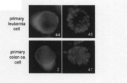
- patient-derived primary tumor cells all three acute myeloid leukemia cells were diagnosed bone marrow puncture specimens collected after obtaining informed consent of the patient at Okayama University School of Medicine. Surplus materials were donated with the approval of joint research with the same organization and used for this study. Similarly, patient-derived colorectal cancer cells were isolated from surgically removed cancer tissues with the informed consent of the patients at the Department of Gastroenterology and Oncology, Okayama University School of Medicine, and the cells maintained and managed in the same course Granted upon approval of joint research.
- HeLa ATCC number: CCL2
- A549 ATCC number: CCL185
- MCF-7 ATCC number: HTB22
- HepG2 ATCC number: HB-8065
- LNCap ATCC number: CRL-1740
- KPK Naito S , Kanamori T, Hisano S, Tanaka K, Momose S, Kamata N.
- Human renal cell carcinoma establishment and characterization of two new cell lines.
- lung cancer cancer stem cell phenotype cells Oct-4 +, Teromerase +, SSEA3 / 4 +, Alkalinephosphatase +
- colorectal cancer stem cell phenotype cells CD133 +, Oct-4 + , SSEA3 / 4 +, Teromerase +, Nestin +, AP +, CEA125 +
- myelocytic leukemia cancer stem cell phenotype cells CD44 +, Oct-4 +, Teromerase +, SSEA3 / 4 +, AP +
- cancer stem cells are tumor cells isolated from lung cancer tissue surgically removed from a 57-year-old Caucasian patient, colon cancer tissue surgically removed from a 37-year-old Caucasian patient, and peripheral blood from a 27-year-old Caucasian patient, respectively. is there.
- CELPROGEN's Human Leukemia Cancer stem cell complete growth media or StemPro-34 SFM medium (GIBCO / Invitrogen), Human Lung Cancer stem cell complete growth media, Human Colon cell complete growth media was used.
- Fluorescence measurement, peptide preparation, apoptosis measurement, etc. can be found in the literature Kondo, E., Seto, M., et al .: Highly efficient delivery of p16 anti-tumor peptide into aggressive leukemia / lymphoma cells using a novel transporter system. Mol. Cancer Ther. 3: 1623-1630, 2004. and Kondo, E., Tanaka, T., et al.: Potent synergy of dual antitumor peptides for growth suppression of human glioblastoma cell lines. Mol. CancerThe1.7: -1471, June 1, 2008.
- FIG. 1 schematically shows the outline of the method for examining permeability of various human malignant tumor cells .
- Each human malignant tumor cell, SV40largeT-transformed renal fibroblast, normal human skin fibroblast, 10,000 cells were seeded in a 96-well plate, and 24 hours later, each peptide numbered in the culture solution of these cells It added so that it might become 2 micromol, and the uptake
- the culture supernatant containing the fluorescent peptide was removed, washed 3 times with 1xPBS (-), treated with trypsin, detached from adherent cells, immediately transferred to a new 96-well plate, and resuspended in fresh culture medium. A speculum was performed later.
- CPP2 showed very high permeability to human colon cancer cell line Lovo, CPP7 to malignant mesothelioma H28, CPP28 to osteosarcoma cell line U2OS, CPP30 to breast cancer cell line MCF-7, CPP33 to In the lung cancer cell line A549, CPP44 showed selective high permeability to hepatocellular carcinoma HepG2, acute leukemia cell line ML-2, and normal human skin fibroblast NHDF.
- CPP10 and CPP45 showed a wide range of permeability to many types of cancer cell lines, sarcoma lines, and brain tumor (glioma) cell lines (FIG. 2).
- CPP47 and CPP48 did not show significant permeability in most solid malignant tumor lines, but lymphoma (T-cell, B-cell lymphoma, ATLL, pro-B-cell lymphoma) and chronic myelocytic leukemia cells High permeability to blood cell tumors such as strains (FIGS. 2 and 3). In contrast, CPP10 hardly penetrated blood cell tumors (data not shown).
- CPP44 was highly permeable to hepatocellular carcinoma HepG2, but almost no permeability to normal hepatocytes, and conversely, CPP48 did not penetrate HepG2, but was highly permeable to normal hepatocytes. A difference in permeability to tumor cells and non-tumor cells was observed even though they were the same development base (FIG. 4).
- cancerCstem cell phenotype cells for lung cancer Oct-4 +, omerTeromerase +, SSEA3 / 4 +, Alkalinephosphatase +
- cancer stem cell phenotype cells of colon cancer CD133 +, Oct-4 +, SSEA3 / 4 +, Teromerase +, Nestin +, AP +, CEA125 +
- CPP44, CPP7, CPP48, etc. lower left panel in Fig.
- CD34 + cells show high permeability in myelocytic leukemia cancer stem cell phenotype cells (CD44 +, Oct-4 +, Teromerase +, SSEA3 / 4 +, AP +). It was. All of these purified cells were Oct-3 / 4 + and 2 + Sox-2 + in RT-PCR (Reverse Transcription Polymerase Chain Reaction) method, and leukemia cells greatly exceeded the frequency in normal peripheral blood.
- the CD34 + cells were confirmed to be contained at a high frequency (about 20 to 25%) by visual analysis using a fluorescence microscope with phycoerythrine (PE) -labeled anti-CD34 antibody staining (lower panel in FIG. 5 lower panel).
- CPP44 has a broad and uniform distribution in the cytoplasm after being taken up by leukemia cells
- CPP48 has a dot-like endosomal pattern.
- CPP2 and CPP47 in colorectal cancer cells FIG. 6).
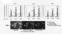
- uptake of fluorescently labeled peptides into primary cells showed high uptake of r9 and CPP44 in primary myelocytic leukemia cells under exposure conditions where TAT was weakly taken up into the cells.
- r9 and CPP2 showed clear uptake
- r9 and CPP44 showed clear uptake (FIG. 7).
- CPP44 showed higher fluorescence intensity than TAT and r9 (right panel in the middle of FIG. 8).
- these tumor cells reflected the difference in cell membrane permeability by tumor type, which almost coincided with the results of the assay using cell lines (only lung cancer cells differed somewhat from the primary cell line).
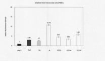
- CPP44 In normal skin fibroblasts (NHDF), CPP44 was as highly permeable as r9, whereas CPP2 was similar to TAT but was uptake but less permeable. In addition, in normal human peripheral blood mononuclear cells (PBMC), it was revealed that both CPP2 and CPP44 had a lower permeability than r9 and had the same level of uptake as TAT ( Figure 8 lower panel).
- PBMC peripheral blood mononuclear cells
- TBMC 4-residue continuous L-form polyarginine (R4), 9-residue continuous D-form polyarginine (r9), CPP2, CPP44, and CPP47 in PBMC as normal blood cells collected from 3 healthy subjects
- CPP47 and CPP44 which were highly permeable to lymphomas and leukemias, had low uptake into non-neoplastic lymphocytes, which was 1/2 to 1/3 that of r9, which showed the highest permeability. (FIG. 9).
- an anti-tumor peptide was prepared by fusing a functional amino acid sequence p16 minimal inhibitory sequence (p16 MIS) known to restore the function of p16 INK4a in a compensatory manner with CPP44.
- p16 MIS functional amino acid sequence p16 minimal inhibitory sequence
- CPP44-p16 MIS (SEQ ID NO: 13) is fused with p16 MIS (LDTLVVLHR; SEQ ID NO: 12) via the spacer sequence GPG after CPP44 (KRPTMRFRYTWNPMK; SEQ ID NO: 1) is inserted and the GP spacer sequence is inserted.
- CPP44-RI-p16 MIS is a CPP44-p16 MIS sequence in which the p16 MIS part, that is, LDTLVVLHR, is all replaced with D-amino acids to form retroinverso (mirror arrangement).
- the control is obtained by converting the retroinverso sequence portion of the CPP44-RI-p16 MIS into a random (random) sequence (CPP44-RI-p16 scramble; SEQ ID NO: 14).
- CPP44-RI-p16 scramble SEQ ID NO: 14
- the structures of these peptides are shown in the left panel of FIG.
- the peptides used in this verification were chemically synthesized by consigning to Sigma-Aldrich Japan (Genesis Division).
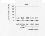
- a peptide (p16 MIS-4R; SEQ ID NO: 15) in which only four arginines added to the C-terminal side of each peptide for improving hydrophobicity are fused with p16 MIS-4R (SEQ ID NO: 15) is different from CPP44-p16 MIS in primary leukemia cells. It was confirmed that there was almost no permeability (right panel in FIG. 11). Note that p16-4MIS-4R has two PEGs added to the N-terminal in the same manner as CPP44-p16 MIS.
- CPP44-RI-p16 MIS exhibited the highest apoptosis-inducing ability in all three leukemia cells.
- Annexin V positivity was observed in cells of about 35% after introduction of CPP44-RI-p16 MIS at a final concentration of 5 ⁇ M, about 50% at 10 ⁇ M, and over 60% at 20 ⁇ M, and this was prominent depending on the concentration of the introduced peptide. Apoptosis-inducing effect was obtained (FIG. 13, upper panel).
- CPP44-RI-p16 MIS final concentration of 10 ⁇ M is about 50%
- CPP44-RI-p16 MIS final concentration of 10 ⁇ M is about 43%
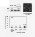
- the important point is whether or not the CPP44-RI-p16 MIS peptide after being taken up into tumor cells acts specifically in the p16 gene pathway.
- the inhibition state of RB phosphorylation as an output of CD16 inhibitor p16 action was analyzed. That is, lysates of leukemia cells (AML1 cells, 4 ⁇ 10 5 portions each) at a final concentration of 10 ⁇ M CPP44-RI-p16 MIS peptide introduction treatment time were prepared (0 hours, 3 hours, 6 hours, 12 hours, 24 hours after introduction). These were verified by immunoblotting using rabbit anti-serine 780 phosphorylated RB antibody (CST). As a result, it was confirmed that the expression of phosphorylated RB attenuated in a time-dependent manner from 3 hours after introduction (FIG. 14).
- a CPP2-RI-p16 MIS anti-tumor peptide (SEQ ID NO: 16) for colorectal cancer selective targeting designed by replacing the cell membrane permeable sequence with CPP2 (DSLKSYWYLQKFSWR; SEQ ID NO: 2), a colorectal cancer selective highly permeable sequence. It was prepared by the same method as above, and the antitumor effect was assayed on primary colon cancer cells. As a result, an apoptosis-inducing effect was observed in 26% at a final concentration of 10 ⁇ M and in less than 50% of the total cells introduced at 20 ⁇ M. This was about 4 times the antitumor effect compared to the case where CPP44-RI-p16 MIS was introduced at the same final concentration of 20 ⁇ M (FIG. 17).
- CPP44-RI-p16 MIS was introduced into K562 in the same manner as the above-mentioned acute leukemia cells.
- the induction rate of apoptosis was significantly lower than that of the AML cell group described above (Annexin V positive cells were only 2.2% after introduction of 10 ⁇ M peptide).
- this cause is thought to be due to the fact that this K562 cell line is located downstream of p16 INK4a and overexpresses the expression of the tumor suppressor gene RB, which is a key molecule involved in cell cycle regulation. It was.
- the introduction result in K562 cells lacking the expression of RB, which is the output molecule of p16 action shows that CPP44-RI-p16 MIS functions specifically in the p16-CDK4-RB gene pathway in the cell and causes cell death. It is considered that this phenomenon has been proved to be induced (FIG. 18).
- Microdisseminated lesions composed of human leukemia cells were distributed in large numbers around 0.5 to 3 mm in diameter on the mouse ovary, intestinal tract, and abdominal wall peritoneum (FIG. 19A; bilateral ovarian epithelial tumor nodules).
- a microfluorescence accumulation part was recognized on the ovary surface separately from the bladder having autofluorescence due to urine retention (FIG. 19B; UB is bladder).
- a large number of nodular lesions having a diameter of about 2 to 3 mm were also formed on the peritoneum (FIG. 19C), and in the fluorescence field image, nodular fluorescence accumulation portions corresponding to the same portion were observed (FIG. 19D).
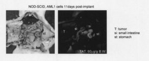
- AML1 cells were suspended in PBS (sodium phosphate interference solution), and then injected into the abdominal cavity of 7-week-old NOD-SCID mice ( ⁇ ) (Charles River Japan).
- FITC-labeled CPP44-RI-p16MIS peptide dissolved in PBS containing 270 mM ⁇ -lactose monohydrate was added at 12.8 mg / kg every 6 hours to 6 mice 10 days after the AML1 cell injection. Were administered by intraperitoneal injection a total of 4 times, and the survival time was measured.
- CPP44-RI-p16V95E obtained by binding p16V95E obtained by modifying 95th valine of p16 to glutamic acid instead of p16, and TAT instead of CPP44.
- TAT-RI-p16 the bound TAT-RI-p16
- PBS sodium phosphate interference solution
- the statistical analysis of the survival rate was calculated using the package software Statview (manufactured by SAS Institute). Survival curve was derived by Kaplan-Meier method (Yutomi Tominaga: Practical Statistics for Judgment of Treatment Effect, Tsubo Shobo (1980), etc.) and P ⁇ 0.05 was judged to be statistically superior.
- the animal test for this experiment has been approved by Okayama University graduate School of Biomedical Sciences and Aichi Cancer Center Research Institute. Handling and euthanasia of all mice, including cell transplantation and peptide administration, are performed painlessly or under anesthesia, and the animal experiment committees at Okayama University graduate School of Biomedical Sciences and Aichi Cancer Center Research Institute It was done according to the guidelines.
- mice died on 14th and 15th days after AML1 cell transplantation (average survival time: 14.3 days). These mice were killed by a malignant tumor in which bloody ascites accumulated in the abdomen.
- the average survival time was 20.8 days, the P value for vehicle was 0.0006, the p value for CPP44-RI-p16MISV95E was 0.0006, and the p value for TAT-RIp16MIS was 0.0008. A significant increase in survival time was observed.
- the CPP44-RI-p16MISV95E administration group linked with the inactivated peptide of the p16MIS peptide has a significantly lower survival rate than the CPP44-RI-p16MIS, and the survival-prolonging effect is lower than that of the solvent administration group. Not observed (mean survival: 15.8 days).
- the p-value was 0.0022 compared to the solvent administration group, and a significant increase in survival time was observed (average survival time: 17.0 days)
- the p-value for CPP44-RI-p16MISV95E was 0.0434, indicating that the effect was weaker than that of CPP44-RI-p16MIS.
- CPP44-RI-p16MIS peptide in human leukemia xenograft model showing extremely malignant properties induces a significant increase in survival time, and CPP44-RI-p16MIS can be administered. Anti-tumor effect was demonstrated.
- TAT-RIp16MIS using TAT instead of CPP44 peptide was also observed to have an extended survival period, but it was significantly weaker than CPP44-RI-p16MIS, and the superiority of CPP44 as a cancer cell-selective transmembrane peptide was observed. Indicated.
- IVVL-derived cancer cell-selective membrane permeation peptide which is CPP44 as a representative example, is It is considered that it has an antitumor effect based on a specific delivery function and has sufficient potential for application to an antitumor agent.
- the present invention can be widely used in basic fields and clinical fields as research in the field of biochemistry, development of medicines, antitumor agents and cancer selective imaging agents.
Abstract
Disclosed are: a cancer cell-specific cell-penetrating peptide which comprises one of the amino acid sequences represented by SEQ ID NOS: 1-10 and has a cancer cell-specific membrane penetrating function; an antitumor agent that contains, as an active ingredient, an antitumor substance in which a peptide that has a cancer cell-specific membrane penetrating function is bonded with an antitumor factor; and an imaging agent having an ability of selectively accumulating in a tumor site, in which a peptide that has a cancer cell-specific membrane penetrating function is bonded with a labeling substance.
Description
本発明は、特定のアミノ酸配列をもつ癌細胞選択的膜透過性ペプチド、癌細胞選択的膜透過性ペプチドと抗腫瘍性因子とが結合してなる抗腫瘍性物質を有効成分として含有する抗腫瘍剤、癌細胞選択的膜透過性ペプチドと標識物質が結合してなるイメージング剤に関する。
The present invention relates to a cancer cell-selective membrane-permeable peptide having a specific amino acid sequence, and an antitumor containing an antitumor substance formed by binding a cancer cell-selective membrane-permeable peptide and an antitumor factor as an active ingredient The present invention relates to an imaging agent comprising an agent, a cancer cell-selective membrane-permeable peptide, and a labeling substance.
細胞膜透過性ペプチドとしては、例えば、アンテナペディア由来のペネトラチン(penetratin)(非特許文献1)、及び、Tatペプチド(非特許文献2)などが知られており、タンパク質、ペプチド、核酸などを細胞中へ輸送するために使用されている。
また、ポリアルギニン残基を含むペプチドを膜透過性キャリアとして用いることも報告されている(特許文献1)。
しかしながら、癌を治療したり検出したりする場合には、抗癌剤やイメージング剤を特定の癌細胞に選択的に送達する必要があるところ、これらのペプチドは特定の癌細胞への選択的な膜透過性を示すものではないため、このような目的には不十分であった。
さらに、特許文献2~4では様々な配列を有する細胞膜透過性ペプチドが開示されているが、これらのペプチドが癌細胞選択的な膜透過性を有するかどうかについては調べられていない。 As cell membrane permeable peptides, for example, Penetratin derived from Antennapedia (Non-patent Document 1) and Tat peptide (Non-patent Document 2) are known, and proteins, peptides, nucleic acids and the like are contained in cells. Used to transport to.
It has also been reported that a peptide containing a polyarginine residue is used as a membrane-permeable carrier (Patent Document 1).
However, when treating or detecting cancer, it is necessary to selectively deliver anticancer agents and imaging agents to specific cancer cells, and these peptides can be selectively permeated into specific cancer cells. It is not sufficient for this purpose because it does not show any properties.
Furthermore,Patent Documents 2 to 4 disclose cell membrane-permeable peptides having various sequences, but it has not been investigated whether these peptides have cancer cell-selective membrane permeability.
また、ポリアルギニン残基を含むペプチドを膜透過性キャリアとして用いることも報告されている(特許文献1)。
しかしながら、癌を治療したり検出したりする場合には、抗癌剤やイメージング剤を特定の癌細胞に選択的に送達する必要があるところ、これらのペプチドは特定の癌細胞への選択的な膜透過性を示すものではないため、このような目的には不十分であった。
さらに、特許文献2~4では様々な配列を有する細胞膜透過性ペプチドが開示されているが、これらのペプチドが癌細胞選択的な膜透過性を有するかどうかについては調べられていない。 As cell membrane permeable peptides, for example, Penetratin derived from Antennapedia (Non-patent Document 1) and Tat peptide (Non-patent Document 2) are known, and proteins, peptides, nucleic acids and the like are contained in cells. Used to transport to.
It has also been reported that a peptide containing a polyarginine residue is used as a membrane-permeable carrier (Patent Document 1).
However, when treating or detecting cancer, it is necessary to selectively deliver anticancer agents and imaging agents to specific cancer cells, and these peptides can be selectively permeated into specific cancer cells. It is not sufficient for this purpose because it does not show any properties.
Furthermore,
本発明は、癌細胞選択的な膜透過性を有するペプチド、該ペプチドを用いた抗腫瘍性物質やイメージング剤などの提供を課題とする。
An object of the present invention is to provide a peptide having cancer cell-selective membrane permeability, an antitumor substance or an imaging agent using the peptide, and the like.
本発明者らは、上記課題を解決するために鋭意検討した結果、遺伝子型と表現型の対応付け分子を用いて選択/同定されたある種の細胞膜透過性ペプチド(cell-penetrating peptide;以下これを「CPP」と称することがある。)が、癌細胞選択的な膜透過機能を有することを見出した。本発明はこれらの知見に基づいて成し遂げられたものである。
As a result of intensive studies to solve the above problems, the present inventors have determined that certain cell membrane-penetrating peptides (cell-penetrating peptides; hereinafter referred to as “cell-penetrating peptides”) selected / identified using genotype-phenotype mapping molecules. Has been found to have a cancer cell-selective membrane permeation function. The present invention has been accomplished based on these findings.
すなわち、本発明の要旨は、次の[1]~[16]に存する。
[1]配列番号1ないし10に示す何れかのアミノ酸配列を含み、癌細胞に対する選択的な膜透過機能を有することを特徴とする癌細胞選択的膜透過性ペプチド。
[2][1]に記載のペプチドに目的物質を結合させ、該目的物質を特定癌細胞へ透過させる、目的物質の特定癌細胞への透過方法(ただし、ヒトを治療する方法は除く)。
[3]癌細胞に対する選択的な膜透過機能を有するペプチドと抗腫瘍性因子とが結合してなる抗腫瘍性物質を有効成分として含有することを特徴とする抗腫瘍剤。
[4]癌細胞が、子宮癌細胞、大腸癌細胞、肺癌細胞、乳癌細胞、胃癌細胞、肝癌細胞、前立腺癌細胞、腎癌細胞、膵臓癌細胞、脳腫瘍細胞、肉腫細胞、悪性中皮腫細胞、リンパ腫細胞および白血病細胞よりなる群から選ばれる何れかの細胞である、[3]に記載の抗腫瘍剤。
[5]結合が、リンカーを介する共有結合である、[3]または[4]に記載の抗腫瘍剤。
[6]膜透過機能を有するペプチドが、
(1)2~100アミノ酸残基からなる候補ペプチドを少なくとも含むタンパク質部と該候補ペプチドをコードする塩基配列を少なくとも含む核酸部とを含み、該タンパク質部のC末端と該核酸部の3’末端とが共有結合をしている遺伝子型と表現型の対応付け分子の群と、標的細胞とを接触させる工程;
(2)標的細胞内に導入された上記対応付け分子の核酸部分に含まれる核酸を増幅する工程;及び
(3)増幅された核酸の塩基配列を解析し、該塩基配列がコードするペプチドを、膜透過機能を有するペプチドとして同定する工程;
を含む方法より得られたものである、[3]ないし[5]の何れかに記載の抗腫瘍剤。
[7]工程(1)の対応付け分子が、下記の(a):
(a)2~100アミノ酸残基からなる候補ペプチドと該ペプチドにより標的細胞内へ輸送される目的タンパク質との融合タンパク質を含むタンパク質部と、該候補ペプチドをコードする塩基配列及び該目的タンパク質をコードする塩基配列を含む核酸部とを含み、該核酸部の3’末端にスペーサーを介して核酸誘導体が結合し、該核酸誘導体と該タンパク質部のC末端とが共有結合をしている分子、
または下記の(b):
(b)2~100アミノ酸残基からなる候補ペプチドを含むタンパク質部と、該候補ペプチドをコードする塩基配列を含む核酸部とを含み、該核酸部の3’末端にスペーサーを介して核酸誘導体が結合し、該核酸誘導体と該タンパク質部のC末端とが共有結合をしており、かつ、該スペーサーに該ペプチドにより標的細胞内へ輸送される非タンパク性目的物質が結合している分子、
である、[6]に記載の抗腫瘍剤。
[8]前記方法は、工程(1)と(2)の間に、さらに、(4)標的細胞表面から細胞内部に導入されていない該分子を除く工程を含む、[6]または[7]に記載の抗腫瘍剤。
[9]標的細胞が、癌細胞である、[6]~[8]のいずれかに記載の抗腫瘍剤。
[10]癌細胞が、子宮癌細胞、大腸癌細胞、肺癌細胞、乳癌細胞、胃癌細胞、肝癌細胞、前立腺癌細胞、腎癌細胞、膵臓癌細胞、脳腫瘍、肉腫細胞、悪性中皮腫細胞、リンパ腫細胞および白血病細胞よりなる群から選ばれる何れかの細胞である、[9]に記載の抗腫瘍剤。
[11]膜透過機能を有するペプチドが、配列番号1ないし10に示す何れかのアミノ酸配列を含み、癌細胞に対する選択的な膜透過機能を有するものである、[3]ないし[8]の何れかに記載の抗腫瘍剤。
[12]抗腫瘍性因子が、抗腫瘍薬剤である、[3]ないし[11]の何れかに記載の抗腫瘍剤。
[13]抗腫瘍性因子が、癌細胞において発現喪失している癌抑制遺伝子の機能を代償的に回復させる機能を有するタンパク質またはその機能ドメインペプチドである、[3]ないし[11]の何れかに記載の抗腫瘍剤。
[14]機能ドメインペプチドが、配列番号13に示すアミノ酸配列を含み、癌抑制遺伝子p16の機能を代償性に回復可能なペプチドである、[13]に記載の抗腫瘍剤。
[15]癌細胞に対して選択的な膜透過機能を有するペプチドと標識物質とが結合してなり、腫瘍部選択的な集積能を有するイメージング物質を含有することを特徴とするイメージング剤。
[16]標識物質が、蛍光物質または陽電子放射性核種を有する物質である、[15]に記載のイメージング剤。 That is, the gist of the present invention resides in the following [1] to [16].
[1] A cancer cell-selective membrane-permeable peptide comprising any amino acid sequence shown in SEQ ID NOs: 1 to 10 and having a selective membrane permeability function for cancer cells.
[2] A permeation method of a target substance into a specific cancer cell, wherein the target substance is bound to the peptide according to [1] and the target substance permeates into the specific cancer cell (except for a method for treating humans).
[3] An antitumor agent comprising an antitumor substance formed by binding a peptide having a selective membrane permeation function for cancer cells and an antitumor factor as an active ingredient.
[4] Cancer cells are uterine cancer cells, colon cancer cells, lung cancer cells, breast cancer cells, gastric cancer cells, liver cancer cells, prostate cancer cells, kidney cancer cells, pancreatic cancer cells, brain tumor cells, sarcoma cells, malignant mesothelioma cells. The antitumor agent according to [3], which is any cell selected from the group consisting of lymphoma cells and leukemia cells.
[5] The antitumor agent according to [3] or [4], wherein the bond is a covalent bond via a linker.
[6] A peptide having a membrane permeation function is
(1) a protein part including at least a candidate peptide consisting of 2 to 100 amino acid residues and a nucleic acid part including at least a base sequence encoding the candidate peptide, the C-terminus of the protein part and the 3 ′ end of the nucleic acid part Contacting a target cell with a group of genotype-phenotype mapping molecules that are covalently linked to a target cell;
(2) a step of amplifying a nucleic acid contained in the nucleic acid portion of the above-mentioned corresponding molecule introduced into the target cell; and (3) analyzing a base sequence of the amplified nucleic acid, and a peptide encoded by the base sequence, Identifying as a peptide having a membrane permeation function;
The antitumor agent according to any one of [3] to [5], which is obtained by a method comprising
[7] The mapping molecule in step (1) is the following (a):
(A) a protein part containing a fusion protein of a candidate peptide consisting of 2 to 100 amino acid residues and a target protein transported into the target cell by the peptide, a base sequence encoding the candidate peptide, and the target protein A nucleic acid part comprising a nucleic acid part comprising a base sequence, wherein a nucleic acid derivative is bonded to the 3 ′ end of the nucleic acid part via a spacer, and the nucleic acid derivative and the C part of the protein part are covalently bonded,
Or (b) below:
(B) a protein part comprising a candidate peptide consisting of 2 to 100 amino acid residues, and a nucleic acid part comprising a base sequence encoding the candidate peptide, wherein a nucleic acid derivative is present via a spacer at the 3 ′ end of the nucleic acid part A molecule in which the nucleic acid derivative and the C-terminus of the protein part are covalently bonded, and a non-protein target substance transported into the target cell by the peptide is bound to the spacer;
The antitumor agent according to [6].
[8] The method further comprises a step between steps (1) and (2), and (4) removing the molecule that has not been introduced into the cell from the surface of the target cell [6] or [7] The antitumor agent as described in.
[9] The antitumor agent according to any one of [6] to [8], wherein the target cell is a cancer cell.
[10] The cancer cells are uterine cancer cells, colon cancer cells, lung cancer cells, breast cancer cells, gastric cancer cells, liver cancer cells, prostate cancer cells, kidney cancer cells, pancreatic cancer cells, brain tumors, sarcoma cells, malignant mesothelioma cells, The antitumor agent according to [9], which is any cell selected from the group consisting of lymphoma cells and leukemia cells.
[11] Any of [3] to [8], wherein the peptide having a membrane permeation function includes any amino acid sequence shown in SEQ ID NOs: 1 to 10 and has a selective membrane permeation function for cancer cells. An antitumor agent according to any one of the above.
[12] The antitumor agent according to any one of [3] to [11], wherein the antitumor factor is an antitumor agent.
[13] Any one of [3] to [11], wherein the antitumor factor is a protein having a function of compensating for the function of a tumor suppressor gene whose expression is lost in cancer cells or a functional domain peptide thereof The antitumor agent as described in.
[14] The antitumor agent according to [13], wherein the functional domain peptide comprises the amino acid sequence shown in SEQ ID NO: 13, and is a peptide capable of recovering the function of the tumor suppressor gene p16 in a compensatory manner.
[15] An imaging agent comprising an imaging substance formed by binding a peptide having a membrane permeation function selective to cancer cells and a labeling substance, and having an ability to selectively accumulate tumor sites.
[16] The imaging agent according to [15], wherein the labeling substance is a fluorescent substance or a substance having a positron emitting nuclide.
本発明の癌細胞選択的膜透過性ペプチドは、従来の汎用細胞膜透過性ペプチド(TAT、ポリアルギニン)とは異なり、発生母地の異なるヒト癌細胞に対して、選択的な膜透過能を発揮する性質があり、またそれらは非選択的透過性ペプチドを上回る高透過能を発揮する。
Unlike the conventional general-purpose cell membrane-permeable peptide (TAT, polyarginine), the cancer cell-selective membrane-permeable peptide of the present invention exhibits selective membrane permeability for human cancer cells of different origins. And they exhibit higher permeability than non-selective permeability peptides.
本発明の抗腫瘍性物質は、癌細胞に対する選択的膜透過機能を有するペプチドに抗腫瘍性因子が結合してなるものであり、抗腫瘍性因子を確実に癌細胞へ送達させることができる。従って、本発明の抗腫瘍性物質は、腫瘍選択的標的薬剤として、抗腫瘍効果が優れ、副作用の少ない抗腫瘍剤となり得ることが期待される。
The antitumor substance of the present invention is obtained by binding an antitumor factor to a peptide having a selective membrane permeation function for cancer cells, and can reliably deliver the antitumor factor to cancer cells. Therefore, the antitumor substance of the present invention is expected to be an antitumor agent having excellent antitumor effects and few side effects as a tumor selective target drug.
本発明のイメージング剤は、癌細胞に対する選択的な膜透過性を有しているので、癌転移を高感度に描出することができ、PET(positron emission tomography)などを用いたトレーサー開発、術中癌イメージングなどへの利用が期待される。
Since the imaging agent of the present invention has selective membrane permeability to cancer cells, cancer metastasis can be visualized with high sensitivity, and tracer development using PET (positron emission tomography) etc., intraoperative cancer Expected to be used for imaging.
以下に、本発明を実施するための代表的な態様を具体的に説明するが、本発明はその要旨を超えない限り、以下の態様に限定されるものではない。
Hereinafter, typical embodiments for carrying out the present invention will be specifically described. However, the present invention is not limited to the following embodiments as long as the gist thereof is not exceeded.
1.癌細胞選択的膜透過性ペプチド
本発明の癌細胞選択的膜透過性ペプチド(cancer cell-specific cell-penetrating peptide;以下これを「CCS-CPP」と略称することがある)は、配列番号1ないし10に示す何れかのアミノ酸配列を含み、癌細胞に対する選択的な膜透過機能を有するものである。ここで、癌細胞に対する選択的な膜透過機能とは、特定の種類(すなわち病理学的組織型分類)の癌細胞に対する選択的な膜透過機能を意味する。目的とする特定の種類の癌細胞に対する膜透過機能が、該特定の癌細胞以外の癌細胞および正常細胞への膜透過機能に比べて高いことが好ましい。なお、特定の種類内での癌細胞は一種類でもよいし、二種類以上でもよい。 1. Cancer cell-selective membrane-permeable peptide The cancer cell-specific cell-penetrating peptide of the present invention (hereinafter sometimes abbreviated as “CCS-CPP”) is SEQ ID NO: 1 to 10 comprising any amino acid sequence shown in FIG. 10 and having a selective membrane permeation function for cancer cells. Here, the selective membrane permeation function for cancer cells means a selective membrane permeation function for cancer cells of a specific type (namely, pathological tissue type classification). It is preferable that the membrane permeation function for a specific type of cancer cell of interest is higher than the membrane permeation function for cancer cells other than the specific cancer cell and normal cells. In addition, the cancer cell within a specific kind may be one kind, and may be two or more kinds.
本発明の癌細胞選択的膜透過性ペプチド(cancer cell-specific cell-penetrating peptide;以下これを「CCS-CPP」と略称することがある)は、配列番号1ないし10に示す何れかのアミノ酸配列を含み、癌細胞に対する選択的な膜透過機能を有するものである。ここで、癌細胞に対する選択的な膜透過機能とは、特定の種類(すなわち病理学的組織型分類)の癌細胞に対する選択的な膜透過機能を意味する。目的とする特定の種類の癌細胞に対する膜透過機能が、該特定の癌細胞以外の癌細胞および正常細胞への膜透過機能に比べて高いことが好ましい。なお、特定の種類内での癌細胞は一種類でもよいし、二種類以上でもよい。 1. Cancer cell-selective membrane-permeable peptide The cancer cell-specific cell-penetrating peptide of the present invention (hereinafter sometimes abbreviated as “CCS-CPP”) is SEQ ID NO: 1 to 10 comprising any amino acid sequence shown in FIG. 10 and having a selective membrane permeation function for cancer cells. Here, the selective membrane permeation function for cancer cells means a selective membrane permeation function for cancer cells of a specific type (namely, pathological tissue type classification). It is preferable that the membrane permeation function for a specific type of cancer cell of interest is higher than the membrane permeation function for cancer cells other than the specific cancer cell and normal cells. In addition, the cancer cell within a specific kind may be one kind, and may be two or more kinds.
また、上記アミノ酸配列のN末および/またはC末に、通常1~10個、好ましくは1~5個のアミノ酸配列が、全体としてアルギニンおよびリジンの構成比率の和が35%を超えないように付加されたもので、上記機能を有するものも本発明の癌細胞選択的膜透過性ペプチド(CCS-CPP)に含まれる。
これらペプチドは、後述する遺伝子型(核酸)と表現型(タンパク質)の対応付け分子を用いる方法により選択/同定することができる。また、癌細胞膜透過性は、後述するとおり、適当な標識物質(例えば蛍光物質)でラベルしたペプチドと癌細胞を共培養し、癌細胞へ透過したペプチドを、蛍光を指標として蛍光顕微鏡やフローサイトメーター(Flowcytometer)などにより確認することができる。 In addition, the amino acid sequence at the N-terminus and / or C-terminus of the above amino acid sequence usually has a total of 1 to 10, preferably 1 to 5 amino acid sequences so that the sum of the constituent ratios of arginine and lysine does not exceed 35%. Those added and having the above functions are also included in the cancer cell-selective membrane-permeable peptide (CCS-CPP) of the present invention.
These peptides can be selected / identified by a method using an association molecule between a genotype (nucleic acid) and a phenotype (protein) described later. In addition, as described later, cancer cell membrane permeability is determined by co-culturing a peptide labeled with an appropriate labeling substance (for example, a fluorescent substance) and a cancer cell, and using the peptide permeated to the cancer cell as a marker using a fluorescence microscope or a flow site. It can be confirmed with a meter (Flowcytometer).
2.抗腫瘍性物質
本発明で使用される抗腫瘍性物質は、癌細胞選択的膜透過性ペプチド(CCS-CPP)と抗腫瘍性因子とが結合してなることに特徴をもつものである。ここで、CCS-CPPと抗腫瘍性因子との結合は、抗腫瘍性因子が、CCS-CPPの膜透過性機能により、癌細胞内へ取り込まれ得る結合状態を保持できるものであれば如何なる結合様式であってもよい。結合様式としては、例えば、水素結合、ファンデルワールス結合、イオン結合、共有結合などが挙げられる。これらの中で、共有結合が好ましい。 2. Antitumor substance The antitumor substance used in the present invention is characterized in that a cancer cell-selective membrane-permeable peptide (CCS-CPP) and an antitumor factor are bound to each other. Here, the binding between CCS-CPP and the anti-tumor factor is any binding as long as the anti-tumor factor can maintain a binding state that can be incorporated into cancer cells by the membrane permeability function of CCS-CPP. It may be a style. Examples of the bonding mode include hydrogen bonding, van der Waals bonding, ionic bonding, and covalent bonding. Of these, covalent bonds are preferred.
本発明で使用される抗腫瘍性物質は、癌細胞選択的膜透過性ペプチド(CCS-CPP)と抗腫瘍性因子とが結合してなることに特徴をもつものである。ここで、CCS-CPPと抗腫瘍性因子との結合は、抗腫瘍性因子が、CCS-CPPの膜透過性機能により、癌細胞内へ取り込まれ得る結合状態を保持できるものであれば如何なる結合様式であってもよい。結合様式としては、例えば、水素結合、ファンデルワールス結合、イオン結合、共有結合などが挙げられる。これらの中で、共有結合が好ましい。 2. Antitumor substance The antitumor substance used in the present invention is characterized in that a cancer cell-selective membrane-permeable peptide (CCS-CPP) and an antitumor factor are bound to each other. Here, the binding between CCS-CPP and the anti-tumor factor is any binding as long as the anti-tumor factor can maintain a binding state that can be incorporated into cancer cells by the membrane permeability function of CCS-CPP. It may be a style. Examples of the bonding mode include hydrogen bonding, van der Waals bonding, ionic bonding, and covalent bonding. Of these, covalent bonds are preferred.
CCS-CPPと抗腫瘍性因子との結合はリンカーを介する共有結合が好ましい。
リンカーとしては、CCS-CPPと抗腫瘍性因子の機能を保持し、CCS-CPPとともに細胞膜を透過し得るものであれば特に制限されない。具体的には、例えば、その長さが、通常1~5残基、好ましくは1~3残基程度のペプチド鎖や、同等の長さのポリエチレングリコール(PEG)鎖などが用い得るリンカーとして挙げられる。 Coupling between CCS-CPP and an antitumor factor is preferably a covalent bond via a linker.
The linker is not particularly limited as long as it retains the functions of CCS-CPP and an antitumor factor and can penetrate the cell membrane together with CCS-CPP. Specifically, for example, a peptide chain having a length of usually 1 to 5 residues, preferably about 1 to 3 residues, or a linker that can be used with a polyethylene glycol (PEG) chain having an equivalent length can be used. It is done.
ペプチドリンカーを構成するアミノ酸残基としては、電荷がなく、小分子のもの、例えばグリシン残基が好ましい。また、リンカー配列の端部、好ましくは両端部には、結合する両ドメイン(CCS-CPPと抗腫瘍性因子)に回転の自由度を与えるための配列を設けることが好ましい。具体的には、回転の自由度を与えるためにはグリシン(G)、リンカーとしてはプロリン(P)を含む配列が好ましく、さらに具体的には、グリシン残基とプロリン残基よりなるもの、例えば、グリシン(G)-プロリン(P)-グリシン(G)からなるものが特に好ましい。かかる構成とすることで、両ドメインの機能が発揮可能となる。
The amino acid residue constituting the peptide linker is preferably a small molecule, such as a glycine residue, without charge. In addition, it is preferable to provide a sequence for giving freedom of rotation to both domains (CCS-CPP and antitumor factor) to be bound at the end, preferably both ends of the linker sequence. Specifically, in order to give a degree of freedom of rotation, a sequence containing glycine (G) and a linker containing proline (P) is preferable. More specifically, a sequence consisting of a glycine residue and a proline residue, for example, Particularly preferred is glycine (G) -proline (P) -glycine (G). With this configuration, the functions of both domains can be exhibited.
癌細胞選択的膜透過性ペプチド(CCS-CPP)としては、特定の癌細胞に対する選択的な膜透過機能を有し、抗腫瘍因子を、癌細胞内に送達し得る能力を有するものであれば特に限定されない。かかるペプチド(CCS-CPP)は、例えばWO 98/16636、WO 2005/024018、特開2005-13073などに記載の方法に準じて、候補ペプチドの配列を含む遺伝子型(核酸)と表現型(タンパク質)の対応付け分子(以下これを、「In Vitro Virus分子」または「IVV分子」と称することがある。)の群を調製し、細胞膜透過性を指標として選択/同定することにより取得することができる。
The cancer cell-selective membrane-permeable peptide (CCS-CPP) can be used as long as it has a selective membrane permeability function for a specific cancer cell and the ability to deliver an antitumor factor into the cancer cell. There is no particular limitation. Such a peptide (CCS-CPP) can be obtained from a genotype (nucleic acid) containing a candidate peptide sequence and a phenotype (protein) in accordance with, for example, the methods described in WO 98/16636, WO 2005/024018, JP-A-2005-13073, etc. ) Of the corresponding molecule (hereinafter sometimes referred to as “In Vitro Virus molecule” or “IVV molecule”), and obtained by selecting / identifying using cell membrane permeability as an index. it can.
具体的には、次の工程(1)~(3)含む方法を、膜透過機能を有するペプチドを選択/同定する好ましい方法として挙げることができる。
(1)標的細胞と、2~100アミノ酸残基からなる候補ペプチドを少なくとも含むタンパク質部と該候補ペプチドをコードする塩基配列を少なくとも含む核酸部とを含み、該タンパク質部のC末端と該核酸部の3’末端とが共有結合をしている遺伝子型と表現型の対応付け分子の群とを接触させる工程;
(2)標的細胞内に導入された上記対応付け分子の核酸部分に含まれる核酸を増幅する工程;
(3)増幅された核酸の塩基配列を解析し、該塩基配列がコードするペプチドを、膜透過機能を有するペプチドとして同定する工程。 Specifically, a method including the following steps (1) to (3) can be mentioned as a preferable method for selecting / identifying a peptide having a membrane permeation function.
(1) including a target cell, a protein part containing at least a candidate peptide consisting of 2 to 100 amino acid residues, and a nucleic acid part containing at least a base sequence encoding the candidate peptide, the C-terminal of the protein part and the nucleic acid part Contacting a group of phenotypic mapping molecules with a genotype covalently bound to the 3 ′ end of
(2) Amplifying a nucleic acid contained in the nucleic acid part of the above-mentioned corresponding molecule introduced into the target cell;
(3) A step of analyzing the base sequence of the amplified nucleic acid and identifying the peptide encoded by the base sequence as a peptide having a membrane permeation function.
上記工程(1)で用いる対応付け分子(IVV分子)としては、さらに具体的には、下記の分子(a)または分子(b)が挙げられる。
More specifically, the mapping molecule (IVV molecule) used in the above step (1) includes the following molecule (a) or molecule (b).
(a)2~100アミノ酸残基からなる候補ペプチドと該ペプチドにより標的細胞内へ輸送される目的タンパク質との融合タンパク質を含むタンパク質部と、該候補ペプチドをコードする塩基配列および該目的タンパク質をコードする塩基配列を含む核酸部とを含み、該核酸部の3’末端にスペーサーを介して核酸誘導体が結合し、該核酸誘導体と該タンパク質部のC末端とが共有結合をしている分子。
(A) a protein part containing a fusion protein of a candidate peptide consisting of 2 to 100 amino acid residues and a target protein transported into the target cell by the peptide, a base sequence encoding the candidate peptide, and the target protein A nucleic acid part containing a base sequence to be bound, a nucleic acid derivative bound to the 3 ′ end of the nucleic acid part via a spacer, and the nucleic acid derivative and the C part of the protein part are covalently bonded.
(b)2~100アミノ酸残基からなる候補ペプチドを含むタンパク質部と、該候補ペプチドをコードする塩基配列を含む核酸部とを含み、該核酸部の3’末端にスペーサーを介して核酸誘導体が結合し、該核酸誘導体と該タンパク質部のC末端とが共有結合をしており、かつ、該スペーサーに該ペプチドにより標的細胞内へ輸送される非タンパク性目的物質が結合している分子。
(B) a protein part comprising a candidate peptide consisting of 2 to 100 amino acid residues, and a nucleic acid part comprising a base sequence encoding the candidate peptide, wherein a nucleic acid derivative is present via a spacer at the 3 ′ end of the nucleic acid part A molecule in which the nucleic acid derivative and the C-terminal of the protein part are covalently bonded, and a non-protein target substance transported into the target cell by the peptide is bound to the spacer.
核酸誘導体はタンパク質部のC末端に共有結合するものであればよいが、具体的には、その3'末端がアミノアシルtRNAに化学構造骨格が類似しているものを選択することができる。代表的な化合物として、アミド結合を有するピューロマイシン(Puromycin)、3'-N-アミノアシルピューロマイシンアミノヌクレオシド(3'-N-Aminoacylpuromycin aminonucleoside、PANS-アミノ酸)、例えば、アミノ酸部がグリシンのPANS-Gly、アミノ酸部がバリンのPANS-Val、アミノ酸部がアラニンのPANS-Ala、その他、アミノ酸部が全ての各アミノ酸に対応するPANS-アミノ酸化合物が挙げられる。
また、3'-アミノアデノシンのアミノ基とアミノ酸のカルボキシル基が脱水縮合して形成されるアミド結合で連結した3'-N-アミノアシルアデノシンアミノヌクレオシド(3'-Aminoacyladenosine aminonucleoside,AANS-アミノ酸)、たとえば、アミノ酸部がグリシンのAANS-Gly、アミノ酸部がバリンのAANS-Val、アミノ酸部がアラニンのAANS-Ala、その他、アミノ酸部が全アミノ酸の各アミノ酸に対応するAANS-アミノ酸化合物を使用できる。
また、ヌクレオシドあるいはヌクレオシドとアミノ酸のエステル結合したものなども使用できる。さらにまた、核酸あるいは核酸に類似した化学構造骨格及び塩基を有する物質と、アミノ酸に類似した化学構造骨格を有する物質とを化学的に結合した化合物、ペプチド核酸(PNA)は、すべて本方法において用いられる核酸誘導体に含まれる。
核酸誘導体としては、ピューロマイシン、PANS-アミノ酸もしくはAANS-アミノ酸がリン酸基を介してヌクレオシドと結合している化合物がより好ましい。これらの化合物の中でピューロマイシン、リボシチジルピューロマイシン、デオキシシチジルピューロマイシン、デオキシウリジルピューロマイシンなどのピューロマイシン誘導体が特に好ましい。 The nucleic acid derivative is not particularly limited as long as it is covalently bonded to the C-terminus of the protein portion. Specifically, a nucleic acid derivative having a chemical structure skeleton similar to aminoacyl-tRNA at the 3 ′ end can be selected. As a representative compound, puromycin having an amide bond, 3′-N-aminoacylpuromycin aminonucleoside (3′-N-aminoacylpuromycin aminoamide, PANS-amino acid), for example, PANS-Gly having an amino acid part of glycine PANS-Val whose amino acid part is valine, PANS-Ala whose amino acid part is alanine, and other PANS-amino acid compounds whose amino acid part corresponds to all amino acids.
In addition, 3′-N-aminoacyl adenosine aminonucleoside (3′-Aminoacyladenosine aminonucleotide, AANS-amino acid) in which the amino group of 3′-aminoadenosine and the carboxyl group of amino acid are linked by an amide bond formed by dehydration condensation, for example, AANS-Gly having an amino acid part of glycine, AANS-Val having an amino acid part of alanine, AANS-Ala having an amino acid part of alanine, and an AANS-amino acid compound corresponding to each amino acid having an amino acid part of all amino acids can be used.
Further, nucleosides or those obtained by ester bonding of nucleosides and amino acids can be used. Furthermore, a nucleic acid or a compound obtained by chemically combining a substance having a chemical structure skeleton and base similar to nucleic acid and a substance having a chemical structure skeleton similar to amino acid, and peptide nucleic acid (PNA) are all used in this method. Included in the nucleic acid derivative.
The nucleic acid derivative is more preferably a compound in which puromycin, PANS-amino acid or AANS-amino acid is bound to a nucleoside via a phosphate group. Of these compounds, puromycin derivatives such as puromycin, ribocytidylpuromycin, deoxycytidylpuromycin, and deoxyuridylpuromycin are particularly preferred.
候補ペプチドは、通常2~100アミノ酸残基程度、好ましくは2~40残基程度のランダムなアミノ酸配列からなるものである。また、IVV分子としては、上記分子(a)、分子(b)のとおり、タンパク質部が、候補ペプチドと目的タンパク質との融合タンパク質を含み、核酸部が、候補ペプチドおよび目的タンパク質をコードする塩基配列を含むもの[分子(a)]、または、核酸部に非タンパク性目的物質が結合したもの[分子(b)]が好ましい。
The candidate peptide is usually composed of a random amino acid sequence of about 2 to 100 amino acid residues, preferably about 2 to 40 amino acid residues. In addition, as the IVV molecule, as described in the above molecules (a) and (b), the protein part includes a fusion protein of the candidate peptide and the target protein, and the nucleic acid part encodes the candidate peptide and the target protein. [Molecule (a)] or a non-protein target substance bound to the nucleic acid part [molecule (b)] is preferable.
IVV分子を構成する目的タンパク質または非タンパク性目的物質としては、本発明で用い得る膜透過性ペプチドを選択/同定し得るものであれば特に制限はない。
目的タンパク質としては、候補ペプチドを発現させるための支持体タンパク質となり得るものが好ましい。候補ペプチドが短鎖であると、これをタンパク質合成系、特に無細胞タンパク質合成系において発現させることが困難となる。この場合、候補ペプチドを支持体タンパク質と融合させることにより、タンパク質合成系で発現させることが可能となる。支持体タンパク質と候補ペプチドとの融合タンパク質は、いずれがN末側またはC末側でもよい。
支持体タンパク質としては、一般的には、(1)球状タンパク質であってフォールディングしやすく、(2)安定性があり、(3)ジスルフィド(S-S)結合を含まないものが好ましい。これらの条件を満たすタンパク質としては、例えば、Oct-1のPou-specific domain(73アミノ酸残基)(Dekker, N. et al.(1993) Nature 362, 852-854)などが挙げられる。 The target protein or non-protein target substance constituting the IVV molecule is not particularly limited as long as it can select / identify a membrane-permeable peptide that can be used in the present invention.
The target protein is preferably one that can be a support protein for expressing a candidate peptide. If the candidate peptide is a short chain, it will be difficult to express it in a protein synthesis system, particularly a cell-free protein synthesis system. In this case, the candidate peptide can be fused with a support protein to be expressed in a protein synthesis system. Any of the fusion protein of the support protein and the candidate peptide may be N-terminal or C-terminal.
In general, the support protein is preferably (1) a globular protein that is easy to fold, (2) stable, and (3) does not contain a disulfide (SS) bond. Examples of the protein that satisfies these conditions include Oct-1 Pou-specific domain (73 amino acid residues) (Dekker, N. et al. (1993) Nature 362, 852-854).
非タンパク性目的物質としては、一般的に生物活性を有する核酸、糖などのような高分子物質や、医薬活性成分などの低分子物質、さらには量子ビーズ・光増感剤・原子などのナノ分子などが挙げられる。このような目的物質を付加する位置としては、スペーサー部分が挙げられる。
Non-protein target substances are generally high-molecular substances such as biologically active nucleic acids and sugars, low-molecular substances such as pharmaceutically active ingredients, and nano-materials such as quantum beads, photosensitizers and atoms. Examples include molecules. Examples of the position where such a target substance is added include a spacer portion.
また、上記IVV分子には、これが細胞内に導入された(透過した)場合に、簡便に検出できるよう、標識物質を含むことも好ましい。標識物質としては、細胞内IVV分子が透過していることが検出可能なものであれば如何なるものであってもよい。具体的には、例えば、FITC(Fluoresceinisothiocyanate)、Roadamine、Cy3、Cy5などの蛍光物質;西洋ワサビペルオキシダーゼやβ-ガラクトシダーゼなどの呈色アッセイに用いられる酵素などが好ましく用いられる。これら標識物質は、IVV分子のいずれの場所に付加されていてもよい。また、GFPやDsRedなどの蛍光を発するタンパク質をIVV分子のタンパク質部に候補ペプチドとの融合タンパク質として付加することもできる。上記呈色アッセイに用いられる酵素も、IVV分子のタンパク質部に候補ペプチドなどとの融合タンパク質として付加することができる。
Also, the IVV molecule preferably contains a labeling substance so that it can be easily detected when it is introduced (permeated) into the cell. The labeling substance may be any substance as long as it can detect that intracellular IVV molecules are permeated. Specifically, for example, fluorescent substances such as FITC (Fluoresceinisothiocyanate), Roadamine, Cy3, and Cy5; enzymes used for color assays such as horseradish peroxidase and β-galactosidase are preferably used. These labeling substances may be added to any position of the IVV molecule. Alternatively, a fluorescent protein such as GFP or DsRed can be added to the protein part of the IVV molecule as a fusion protein with a candidate peptide. The enzyme used in the color assay can also be added to the protein part of the IVV molecule as a fusion protein with a candidate peptide or the like.
また、上記IVV分子は、これを精製する目的で、ある物質と特異的に結合する性質を有する物質を含むこともできる。具体的には、例えば、FLAG、GST、HISx6などのタグペプチドやビオチンなどの親和性物質などが挙げられる。タグペプチド、親和性物質は、IVV分子中のタンパク質部のいずれの場所にあってもよいが、タグペプチドはIVV分子のタンパク質部のC末端あるいはN末端に付加することが好ましく、親和性物質は後述するスペーサー部分に付加することが好ましい。
In addition, the IVV molecule may include a substance having a property of specifically binding to a certain substance for the purpose of purifying the IVV molecule. Specific examples include tag peptides such as FLAG, GST, HISx6, and affinity substances such as biotin. The tag peptide and affinity substance may be located anywhere in the protein part of the IVV molecule, but the tag peptide is preferably added to the C-terminal or N-terminal of the protein part of the IVV molecule. It is preferable to add to the spacer part mentioned later.
スペーサーとしては、例えば、WO98/16636号公報に記載されているポリエチレンまたはポリエチレングリコールあるいはその誘導体などの高分子物質、オリゴヌクレオチドやペプチドあるいはその誘導体などの生体高分子物質などが用いられる。これらのうち、ポリエチレングリコールが好ましい。スペーサーには、上記したFITCなどの蛍光物質およびその誘導体、ビオチンなどの親和性物質およびその誘導体、デオキシリボヌクレオチドなどの核酸およびその誘導体、あるいは生化学または化学反応により切断される結合を持つ物質、例えば、5-置換-2-ニトロアセトフェノン誘導体などの光分解性物質などを含んでいてもよい。
As the spacer, for example, a polymer substance such as polyethylene or polyethylene glycol or a derivative thereof described in WO98 / 16636, a biopolymer substance such as an oligonucleotide, a peptide or a derivative thereof, or the like is used. Of these, polyethylene glycol is preferred. The spacer includes the above-mentioned fluorescent substances such as FITC and derivatives thereof, affinity substances such as biotin and derivatives thereof, nucleic acids such as deoxyribonucleotides and derivatives thereof, or substances having bonds that are cleaved by biochemistry or chemical reaction, for example And a photodegradable substance such as a 5-substituted-2-nitroacetophenone derivative.
上記対応付け分子(IVV分子)は、先ず、3’端にピューロマイシンを結合した候補ペプチドを含むタンパク質部をコードするmRNAを調製し、無細胞翻訳系を用いて翻訳することにより、タンパク質(表現型を反映するタンパク質部)とそれをコードするmRNA(遺伝子型を反映する核酸部)とがピューロマイシンを介して結合しているタンパク質-RNAキメラ型ランダムペプチドライブラリー(In Vtro Virus Library;以下これを「IVVL」と称することがある。)として調製することができる。膜透過性ペプチドの選択/同定に好適なIVVLの調製は、例えば、特開2005-13073号公報に記載されている方法により行うことができる。
The mapping molecule (IVV molecule) is prepared by first preparing mRNA encoding a protein portion containing a candidate peptide having puromycin bound to the 3 ′ end, and translating it using a cell-free translation system. Protein-RNA Chimeric Random Peptide Library (In Vtro 以下 Virus Library; hereinafter referred to as the protein part reflecting the type) and the mRNA encoding it (the nucleic acid part reflecting the genotype) via puromycin May be referred to as “IVVL”). Preparation of IVVL suitable for selection / identification of a membrane-permeable peptide can be performed by, for example, a method described in JP-A-2005-13073.
次に、標的細胞とIVVL(IVV分子の群)とを接触させ[上記工程(1)]、標的細胞内または標的細胞内へ透過した上記分子の核酸部分に含まれる核酸を増幅し[上記工程(2)]、増幅された核酸の塩基配列を解析し、該塩基配列がコードするペプチドが、細胞膜透過機能をもつペプチドであると同定できる[上記工程(3)]。また、この操作は、必要に応じて繰り返すことができる。
Next, the target cell is contacted with IVVL (a group of IVV molecules) [the above step (1)], and the nucleic acid contained in the nucleic acid portion of the molecule that has penetrated into or into the target cell is amplified [the above step (2)] By analyzing the base sequence of the amplified nucleic acid, the peptide encoded by the base sequence can be identified as a peptide having a cell membrane permeation function [step (3) above]. This operation can be repeated as necessary.
得られた細胞膜透過性ペプチドについて、特定の癌細胞を標的細胞として、膜透過性を検討し、癌細胞に対する選択的透過性をもつペプチドを選択/同定することにより、本発明の抗腫瘍性物質で用い得る癌細胞選択的膜透過性ペプチド(CCS-CPP)を取得することができる。
なお、IVVLと接触させる標的細胞に、特定の癌細胞を用いれば、上記したペプチドを取得後、特定の癌細胞を標的とする選択/同定を行うことなく、本発明で用い得るCCS-CPPを取得することができる。 The obtained cell membrane permeable peptide is examined for membrane permeability using a specific cancer cell as a target cell, and a peptide having selective permeability to the cancer cell is selected / identified, whereby the antitumor substance of the present invention Cancer cell-selective membrane-permeable peptide (CCS-CPP) can be obtained.
In addition, if a specific cancer cell is used as a target cell to be contacted with IVVL, CCS-CPP that can be used in the present invention is obtained without performing selection / identification targeting the specific cancer cell after obtaining the above-mentioned peptide. Can be acquired.
CCS-CPPの選択/同定に用いる標的細胞としては、ヒトまたは哺乳類動物細胞が好ましく、それらの中で、癌細胞(悪性腫瘍細胞)が特に好ましい。
標的細胞として用い得る癌細胞は特に限定されず、例えば、子宮癌細胞、大腸癌細胞、肺癌細胞、乳癌細胞、胃癌細胞、肝癌細胞、前立腺癌細胞、腎癌細胞、膵臓癌細胞、脳腫瘍、肉腫細胞、悪性中皮腫細胞、リンパ腫細胞および白血病細胞よりなる群から選ばれる何れかの細胞が挙げられる。 As target cells used for selection / identification of CCS-CPP, human or mammalian cells are preferable, and cancer cells (malignant tumor cells) are particularly preferable among them.
Cancer cells that can be used as target cells are not particularly limited. For example, uterine cancer cells, colon cancer cells, lung cancer cells, breast cancer cells, gastric cancer cells, liver cancer cells, prostate cancer cells, kidney cancer cells, pancreatic cancer cells, brain tumors, sarcomas Any cell selected from the group consisting of cells, malignant mesothelioma cells, lymphoma cells and leukemia cells can be mentioned.
上記標的細胞と、IVV分子とを接触させる方法としては、特に制限はないが、標的細胞をIVV分子の存在下で適当な方法により培養する方法などが好ましい。培養方法、IVV分子の添加量、培養温度、培養時間などは、標的細胞やIVV分子の種類などにより適宜選択することができる。具体的には、標的細胞として、例えばCHO細胞を用いた場合、96ウェルプレートに105個となるようにウシ胎仔血清含有培地などにより培養し、該細胞をPBSなどの適当な緩衝液を用いて洗浄した後にIVV分子1~100nMを含むPBSなどを添加し、好ましくは氷冷~37℃で置きながら、1~数時間培養する方法などが挙げられる。標的細胞として癌細胞を用いる場合も同様にして標的癌細胞とIVV分子とを接触させることができるが、接触させるIVV分子の濃度は1~1000nMとすることができる。
A method for bringing the target cell into contact with the IVV molecule is not particularly limited, but a method of culturing the target cell by an appropriate method in the presence of the IVV molecule is preferable. The culture method, the amount of IVV molecule added, the culture temperature, the culture time, and the like can be appropriately selected depending on the type of target cell and IVV molecule. Specifically, for example, when CHO cells are used as target cells, the cells are cultured in a 96-well plate so as to have 10 5 cells in a fetal calf serum-containing medium or the like, and the cells are used with an appropriate buffer solution such as PBS. After washing, PBS or the like containing 1 to 100 nM of IVV molecules is added, and the cells are preferably incubated at ice-cooled to 37 ° C. for 1 to several hours. Similarly, when a cancer cell is used as the target cell, the target cancer cell and the IVV molecule can be contacted, but the concentration of the IVV molecule to be contacted can be 1 to 1000 nM.
IVV分子と標的細胞を接触させた後、IVV分子が導入されている細胞内のIVV分子の核酸部分を増幅する。IVV分子が導入された細胞は、これを検出して分離してもよいし、導入されていない細胞とされている細胞が混合されたままでも核酸部分の増幅工程に用いることができる。
After contacting the IVV molecule with the target cell, the nucleic acid part of the IVV molecule in the cell into which the IVV molecule has been introduced is amplified. Cells into which IVV molecules have been introduced may be detected and separated, or may be used in the nucleic acid portion amplification step even if cells that have not been introduced are mixed.
IVV分子の検出方法は、IVV分子が細胞外または細胞内に存在することが明らかになる方法であれば如何なる方法でもよい。具体的には、上記標識物質を検出する方法が好ましく用いられる。標識物質として、蛍光物質または蛍光タンパク質を用いた場合、検出手段は、フローサイトメーター、あるいは蛍光顕微鏡などを用いることができる。また、呈色アッセイを行うための酵素を用いた場合には、必要な基質などを添加した呈色アッセイにより検出することができる。さらに、細胞から抽出したDNAを鋳型としてPCR (Polymerase Chain Reaction)を行い、IVV分子の核酸部分が増幅できることを指標として検出することもできる。
The detection method of the IVV molecule may be any method as long as it is clear that the IVV molecule is present outside or inside the cell. Specifically, a method for detecting the labeling substance is preferably used. When a fluorescent substance or a fluorescent protein is used as the labeling substance, a flow cytometer or a fluorescence microscope can be used as the detection means. In addition, when an enzyme for performing a color assay is used, it can be detected by a color assay to which a necessary substrate is added. Furthermore, PCR (Polymerase Chain Reaction) can be performed using DNA extracted from cells as a template, and the ability to amplify the nucleic acid part of the IVV molecule can be detected as an indicator.
いずれの方法においても、細胞内に導入されていないが、細胞表面に付着しているIVV分子を洗浄してから、上記検出を行うことが好ましい。洗浄の方法は、細胞を洗浄する公知の方法から適宜選択して行うことができる。具体的には、例えば、酸やDNaseIなどヌクレアーゼによる処理などが好ましく用いられる。また、フローサイトメーターによる検出を行う場合、培養後の接着系の細胞はトリプシン処理などを行っておくことも好ましい。
In any of the methods, it is preferable to perform the above detection after washing IVV molecules that are not introduced into the cells but are attached to the cell surface. The washing method can be appropriately selected from known methods for washing cells. Specifically, for example, treatment with nuclease such as acid or DNaseI is preferably used. In addition, when performing detection with a flow cytometer, it is preferable that the adherent cells after culture be treated with trypsin.
細胞内にIVV分子が導入されていることの確認は、例えば、検出手段としてフローサイトメーターを用いた場合には、候補ペプチドを含むIVV分子と、候補ペプチドを含まないIVV分子で同様の標的細胞への接触工程を行い、候補ペプチドを含むIVV分子と接触させた細胞の蛍光強度が、含まないIVV分子と接触させた細胞の蛍光強度に比べて強い場合に、該細胞内にIVV分子が導入されていると判断すればよい。また、蛍光顕微鏡を用いた場合には、細胞内に標識物質である蛍光物質から発せられる蛍光を観察すればよい。
Confirmation that IVV molecules have been introduced into the cells, for example, when a flow cytometer is used as a detection means, IVV molecules that contain candidate peptides and IVV molecules that do not contain candidate peptides. IVV molecule is introduced into the cell when the fluorescence intensity of the cell contacted with the IVV molecule containing the candidate peptide is stronger than that of the cell contacted with the IVV molecule not containing the candidate peptide. It may be judged that it is done. In addition, when a fluorescence microscope is used, fluorescence emitted from a fluorescent substance that is a labeling substance may be observed in the cell.
さらに、細胞内にIVV分子が導入された細胞の分離方法として、上記のフローサイトメーターにより蛍光が検出された細胞をセルソーターにより分離する方法や、蛍光顕微鏡により蛍光が検出された細胞を、レーザーキャプチャーを用いて分離する方法などが挙げられる。
Furthermore, as a method for separating cells into which IVV molecules have been introduced into the cells, a method in which cells with fluorescence detected by the flow cytometer are separated with a cell sorter, or cells with fluorescence detected by a fluorescence microscope are captured by laser. And the like.
かくして取得されたIVV分子が導入された細胞について、該細胞内に存在するIVV分子の核酸部をPCRによって増幅し、その塩基配列を解析することにより、細胞内への透過機能を有するペプチド、すなわち細胞膜透過性ペプチド(CPP)を同定することができる。
上記したIVVLの調製、ペプチドの選択/同定などは、特開2005-13073号公報に詳述されており、それに準じて実施すればよい。 For the cells into which the IVV molecules thus obtained are introduced, the nucleic acid part of the IVV molecules present in the cells is amplified by PCR, and the base sequence is analyzed to obtain a peptide having a function of penetrating into cells, that is, Cell membrane permeable peptides (CPP) can be identified.
Preparation of IVVL, selection / identification of peptides and the like described above are described in detail in JP-A-2005-13073, and may be carried out in accordance therewith.
上記した細胞内にIVV分子が導入されていることの確認方法と同様の方法により、上記で得られたCPPについて、特定の癌細胞に対する選択的な膜透過機能を有するものを選択することができる。
かくして得られた癌細胞選択的膜透過性ペプチド(CCS-CPP)としては、例えば、配列番号1ないし10に示す何れかのアミノ酸配列を含むものが挙げられる。 By the same method as that for confirming that IVV molecules have been introduced into the cells, the CPP obtained above can be selected to have a selective membrane permeation function for specific cancer cells. .
Examples of the cancer cell-selective membrane-permeable peptide (CCS-CPP) thus obtained include those containing any amino acid sequence shown in SEQ ID NOs: 1 to 10.
上記のようなCCS-CPPに、一つ以上の抗腫瘍性因子を結合させた複合体(抗腫瘍性物質)を調製して、標的癌細胞に接触させることによって、目的物質を癌細胞へ送達させることができる。
Prepare a complex (antitumor substance) in which one or more antitumor factors are bound to CCS-CPP as described above, and contact the target cancer cell to deliver the target substance to the cancer cell. Can be made.
ここで、本発明の抗腫瘍性物質に用いる抗腫瘍性因子としては、CCS-CPPにより癌細胞に送達可能なものであって、標的となる癌細胞の増殖や転移に何らかの機能を有し、癌の治療目的に適うものであれば如何なる因子であってもよい。具体的には、例えば、化学療法剤(抗腫瘍剤)、ペプチド(30残基程度までの連続アミノ酸残基配列)、タンパク質、DNA、PNA(Peptide Nucleic Acid)、LNA(Locked Nucleic Acid)、siRNA(small interfering RNA)、microRNAなどが挙げられる。
Here, as an antitumor factor used for the antitumor substance of the present invention, it can be delivered to cancer cells by CCS-CPP, and has some function in the growth and metastasis of target cancer cells, Any factor may be used as long as it is suitable for the purpose of treating cancer. Specifically, for example, chemotherapeutic agents (antitumor agents), peptides (continuous amino acid residue sequences up to about 30 residues), proteins, DNA, PNA (Peptide Nucleic Acid), LNA (Locked Nucleic Acid), siRNA (Small interfering RNA), microRNA and the like.
化学療法剤(抗腫瘍薬剤)としては、例えば、5-FU、paclitaxel、cisplatin、etoposide(vinblastine)、cyclophasphamide、Actinomycin D、イリノテカン、steroid(predonisolone)などが挙げられる。
これらの化学療法剤は、自身の反応性基(水酸基やアミノ基など)を通じてリンカーを導入し、該リンカーをペプチドのアミノ末端、カルボキシ末端または側鎖に反応させることにより、CCS-CPPに結合させることができる。
あるいは、これらの化学療法剤をリポソームに封入し、リポソームの表面にCCS-CPPを結合させることによって、間接的に結合させることもできる。 Examples of chemotherapeutic agents (antitumor agents) include 5-FU, paclitaxel, cisplatin, etoposide (vinblastine), cyclophasphamide, Actinomycin D, irinotecan, steroid (predonisolone), and the like.
These chemotherapeutic agents are linked to CCS-CPP by introducing a linker through its reactive group (hydroxyl group, amino group, etc.) and reacting the linker with the amino terminus, carboxy terminus or side chain of the peptide. be able to.
Alternatively, these chemotherapeutic agents can be indirectly bound by encapsulating them in liposomes and binding CCS-CPP to the surface of the liposomes.
ペプチドやタンパク質としては、例えば、癌細胞において発現喪失している癌抑制遺伝子の機能を代償的に回復させる機能を有するタンパク質またはその機能ドメインを含むペプチドが好ましい。かかるタンパク質やその機能ドメインを含むペプチドとしては、癌細胞に対してアポトーシスを誘導し得るものが好ましい。
As the peptide or protein, for example, a protein having a function of recovering the function of a tumor suppressor gene whose expression is lost in cancer cells in a compensatory manner or a peptide containing a functional domain thereof is preferable. As such a peptide containing a protein or its functional domain, those capable of inducing apoptosis in cancer cells are preferable.
ここで、癌細胞において発現喪失している癌抑制遺伝子としては、例えば、p16INK4a、p14ARF、p15、p18、p21CIP1、p27KIP1、p53、p57Kip2、p73、RB、BRCA1、BRCA2、PTEN、APC、WT1、NF1、NF2、SMAD4、PTC、MSH2、Maspin、SDHDなどが挙げられる。
Here, as a tumor suppressor gene whose expression is lost in cancer cells, for example, p16 INK4a , p14 ARF , p15, p18, p21 CIP1 , p27 KIP1 , p53, p57 Kip2 , p73, RB, BRCA1, BRCA2, PTEN, APC, WT1, NF1, NF2, SMAD4, PTC, MSH2, Maspin, SDHD and the like.
これら癌抑制遺伝子はそれ自体既知のものであり、例えば、P16は、Fahraeus R, Lain S, Ball KL, Lane DP. Characterization of the cyclin-dependent kinase inhibitory domain of the INK4 family as a model for a synthetic tumour suppressor molecule.Oncogene 1998;16:587-96などに、p53は、Hupp TR, Sparks A, Lane DP (1995) Small peptides activate the latent sequence-specific DNA binding function of p53. Cell83: 237-245qなどに、機能ドメイン部分も含め、その詳細が記載されている。
These tumor suppressor genes are known per se, for example, P16 is Fahraeus R, Lain S, Ball KL, Lane DP. Characterization of the cyclin-dependent kinase inhibitory domain of the INK4 family as a model for a synthetic tumour suppressor molecule.Oncogene 1998; 16: 587-96 etc., p53 is like Hupp TR, Sparks A, Lane DP (1995) Small peptides activate the latent sequence-specific DNA binding function of p53. Cell83: 237-245q, The details including the functional domain part are described.
癌抑制遺伝子の機能を代償的に回復させる機能を有するタンパク質とは、上記癌抑制遺伝子の発現産物(タンパク質)であり、その機能ドメインとは、該発現産物(タンパク質)の癌抑制機能をつかさどるアミノ酸配列を含むペプチドである。
A protein having a function of restoring the function of a tumor suppressor gene at a compensatory level is an expression product (protein) of the above-described tumor suppressor gene, and its functional domain is an amino acid that controls the cancer suppressor function of the expression product (protein). A peptide comprising a sequence.
これら癌抑制遺伝子の中で、p16INK4aは、例えば、肺癌(腺癌、扁平上皮癌、小細胞癌、LCNEC)、咽頭・喉頭癌(扁平上皮癌)、消化器癌(食道癌、大腸癌、胃癌、胆道癌、肝細胞癌、膵癌)、泌尿器癌(腎癌、膀胱癌、尿管癌)、生殖器癌[子宮癌(頸部扁平上皮癌、内膜腺癌)、卵巣癌、前立腺癌、精巣胚細胞腫瘍]、皮膚癌(悪性黒色腫、扁平上皮癌)などの固形癌;悪性骨軟部腫瘍(骨肉腫、ユーイング肉腫、横紋筋肉腫、脂肪肉腫、MFH、など)、消化器間葉系腫瘍(GIST、平滑筋肉腫、MPNSTなど)などの肉腫;急性・慢性骨髄球性白血病、リンパ球性白血病、悪性リンパ腫などの血液腫瘍;グリオーマ、グリオブラスト―マ、神経芽細胞種などの脳腫瘍;悪性中皮腫などの諸種の癌細胞で欠損していることが知られている。従って、p16INK4aの機能を代償的に回復させる機能を有する機能性アミノ酸配列を癌細胞に送達することができれば、諸種の癌細胞に対する効果が期待できる。
Among these tumor suppressor genes, p16 INK4a is, for example, lung cancer (adenocarcinoma, squamous cell carcinoma, small cell carcinoma, LCNEC), pharyngeal / laryngeal cancer (squamous cell carcinoma), digestive organ cancer (esophageal cancer, colon cancer, Gastric cancer, biliary tract cancer, hepatocellular carcinoma, pancreatic cancer), urinary cancer (renal cancer, bladder cancer, ureteral cancer), genital cancer [uterine cancer (cervical squamous cell carcinoma, intimal adenocarcinoma), ovarian cancer, prostate cancer, Testicular germ cell tumor], solid cancer such as skin cancer (malignant melanoma, squamous cell carcinoma); malignant bone and soft tissue tumor (osteosarcoma, Ewing sarcoma, rhabdomyosarcoma, liposarcoma, MFH, etc.), digestive organ mesenchyme Sarcomas such as tumors (GIST, leiomyosarcoma, MPNST, etc.); blood tumors such as acute and chronic myelocytic leukemia, lymphocytic leukemia, malignant lymphoma; brain tumors such as glioma, glioblastoma, neuroblastoma It is known to be deficient in various cancer cells such as malignant mesothelioma. Therefore, if a functional amino acid sequence having a function of reversibly restoring the function of p16 INK4a can be delivered to cancer cells, effects on various types of cancer cells can be expected.
かかるp16INK4aの機能を代償的に回復させる機能を有する機能性アミノ酸配列としては、例えば、配列番号12に示すアミノ酸配列(p16 minimal inhibitory sequence;p16 MIS)が挙げられる。
これらペプチドは、適当なアミノ酸配列、例えばリンカーやポリアルギニンなどを付加してもよい。リンカーとしては、上記したペプチドリンカーが挙げられる。ポリアルギニンとしては、通常2~50残基、好ましくは5~20残基程度のものが適当である。
また、機能に影響を及ぼさない限り、アミノ酸はD型でもL型でもよく、配列に順序は逆向きであってもよい。
抗腫瘍性因子としてのペプチドをCCS-CPPとの融合タンパク質として適当な宿主あるいは無細胞翻訳系で発現させることにより抗腫瘍物質を得ることができる。融合タンパク質は化学合成してもよい。あるいは、抗腫瘍性因子としてのペプチドとCCS-CPPとを化学的に結合させてもよい。 Examples of such a functional amino acid sequence having a function of reversibly restoring the function of p16 INK4a include the amino acid sequence shown in SEQ ID NO: 12 (p16 minimal inhibitory sequence; p16 MIS).
These peptides may be added with an appropriate amino acid sequence such as a linker or polyarginine. Examples of the linker include the peptide linkers described above. As polyarginine, those having 2 to 50 residues, preferably 5 to 20 residues are suitable.
As long as the function is not affected, the amino acid may be D-type or L-type, and the sequence may be reversed.
An antitumor substance can be obtained by expressing a peptide as an antitumor factor as a fusion protein with CCS-CPP in a suitable host or a cell-free translation system. The fusion protein may be chemically synthesized. Alternatively, a peptide as an antitumor factor and CCS-CPP may be chemically bound.
PNA(Peptide Nucleic Acid)としては、例えば、神経芽細胞腫(Neuroblastoma)を標的とする次の配列を有するもの(Mol Cancer Ther. 2005 May;4(5):779-86参照)が挙げられる。
PNAs-NLS;H-ATGCCGGGCATGATCT(配列番号18)-PKKKRKV(配列番号19)-NH2 Examples of PNA (Peptide Nucleic Acid) include those having the following sequence that targets neuroblastoma (see Mol Cancer Ther. 2005 May; 4 (5): 779-86).
PNAs-NLS; H-ATGCCGGGCATGATCT (SEQ ID NO: 18) -PKKKRKV (SEQ ID NO: 19) -NH 2
PNAs-NLS;H-ATGCCGGGCATGATCT(配列番号18)-PKKKRKV(配列番号19)-NH2 Examples of PNA (Peptide Nucleic Acid) include those having the following sequence that targets neuroblastoma (see Mol Cancer Ther. 2005 May; 4 (5): 779-86).
PNAs-NLS; H-ATGCCGGGCATGATCT (SEQ ID NO: 18) -PKKKRKV (SEQ ID NO: 19) -NH 2
LNA(Locked Nucleic Acid)としては、例えば、下記の文献に記載されたものが挙げられる。
Nucleic Acids Res. 2004; 32(19): 5757-5765.
Nucleic Acids Res. 2010 January; 38(1): e3
Curr Pharm Des. 2008;14(11):1138-42. Review. Examples of LNA (Locked Nucleic Acid) include those described in the following documents.
Nucleic Acids Res. 2004; 32 (19): 5757-5765.
Nucleic Acids Res. 2010 January; 38 (1): e3
Curr Pharm Des. 2008; 14 (11): 1138-42. Review.
siRNA(small interfering RNA)としては、例えば、下記(1)~(5)が挙げられる。
(1)慢性骨髄性白血病(CML)のBCR-ABLを標的とする次の配列を有するもの(Blood 2003;102(6)2236-2239参照)。
SENSE;5'-CAGAGUUCAA-AAGCCCUUCAG-3'(配列番号20)
ANTISENSE;3'-UCGUCUCAAGUU-UUCGGGAAGUC-5'(配列番号21) Examples of siRNA (small interfering RNA) include the following (1) to (5).
(1) Those having the following sequence targeting BCR-ABL of chronic myelogenous leukemia (CML) (see Blood 2003; 102 (6) 2236-2239).
SENSE; 5'-CAGAGUUCAA-AAGCCCUUCAG-3 '(SEQ ID NO: 20)
ANTISENSE; 3'-UCGUCUCAAGUU-UUCGGGAAGUC-5 '(SEQ ID NO: 21)
(2)乳癌(Brest Cancer)のHER2を標的とする次の配列を有するもの (Gene silencing by cell-penetrating, sequence-selective and nucleic-acid hydrolyzing antibodies. Lee WR, Jang JY, Kim JS, Kwon MH, Kim YS. Nucleic Acids Res. [Epub ahead of print]参照)。
SENSE;5'- TTAAU UCC AGU GGC CAU CAA A-3'(配列番号22)
ANTISENSE;3'-UUAAGGUCTCCGGUAGUUUTT-5'(配列番号23) (2) Those having the following sequence targeting HER2 of breast cancer (Gene silencing by cell-penetrating, sequence-selective and nucleic-acid hydrolyzing antibodies. Lee WR, Jang JY, Kim JS, Kwon MH, Kim YS. Nucleic Acids Res. [See Epub ahead of print].
SENSE; 5'- TTAAU UCC AGU GGC CAU CAA A-3 '(SEQ ID NO: 22)
ANTISENSE; 3'-UUAAGGUCTCCGGUAGUUUTT-5 '(SEQ ID NO: 23)
(3)前立腺癌(Prostate Cancer)のAndrogen Receptorを標的とする次の配列を有するもの(PLoS One. 2007 Oct 10;2(10):e1006参照)。
SENSE;5'-UCCCCAAGCCCAUCGUAGA-TT-3'(配列番号24)
ANTISENSE;3'-TTAGGGGUUCGGGUAGCAUCU-5'(配列番号25) (3) Those having the following sequence targeting the Androgen Receptor of prostate cancer (see PLoS One. 2007Oct 10; 2 (10): e1006).
SENSE; 5'-UCCCCAAGCCCAUCGUAGA-TT-3 '(SEQ ID NO: 24)
ANTISENSE; 3'-TTAGGGGUUCGGGUAGCAUCU-5 '(SEQ ID NO: 25)
SENSE;5'-UCCCCAAGCCCAUCGUAGA-TT-3'(配列番号24)
ANTISENSE;3'-TTAGGGGUUCGGGUAGCAUCU-5'(配列番号25) (3) Those having the following sequence targeting the Androgen Receptor of prostate cancer (see PLoS One. 2007
SENSE; 5'-UCCCCAAGCCCAUCGUAGA-TT-3 '(SEQ ID NO: 24)
ANTISENSE; 3'-TTAGGGGUUCGGGUAGCAUCU-5 '(SEQ ID NO: 25)
(4)大腸癌(Colon Cancer)のVEGFを標的とする次の配列を有するもの(Molecular Therapy (2006) 14, 343-350参照)。
SENSE;5'-AUGUGAAUGCAGACCAAAGAATT-3'(配列番号26)
ANTISENSE;3'-TTCUAUCGUUACUGCUUACGCAU-5'(配列番号27) (4) Those having the following sequence targeting VEGF of colon cancer (see Molecular Therapy (2006) 14, 343-350).
SENSE; 5'-AUGUGAAUGCAGACCAAAGAATT-3 '(SEQ ID NO: 26)
ANTISENSE; 3'-TTCUAUCGUUACUGCUUACGCAU-5 '(SEQ ID NO: 27)
(5)膵臓癌(Pancreatic Cancer)のRASを標的とする次の配列を有するもの(Cancer Sci. 2007 Jul;98(7):1128-36参照)。
krasGGT:
5'-GUUGGAGCUGGUGGCGUAGTT-3' (配列番号28)
5'-CUACGCCACCAGCUCCAACTT-3' (配列番号29)
krasGAT:
5'-GUUGGAGCUGAUGGCGUAGTT-3' (配列番号30)
5'-CUACGCCAUCAGCUCCAACTT-3' (配列番号31)
krsaGTT:
5'-GUUGGAGCUGUUGGCGUAGTT-3' (配列番号32)
5'-CUACGCCAACAGCUCCAACTT-3' (配列番号33) (5) Those having the following sequence targeting RAS of pancreatic cancer (see Cancer Sci. 2007 Jul; 98 (7): 1128-36).
krasGGT:
5'-GUUGGAGCUGGUGGCGUAGTT-3 '(SEQ ID NO: 28)
5'-CUACGCCACCAGCUCCAACTT-3 '(SEQ ID NO: 29)
krasGAT:
5'-GUUGGAGCUGAUGGCGUAGTT-3 '(SEQ ID NO: 30)
5'-CUACGCCAUCAGCUCCAACTT-3 '(SEQ ID NO: 31)
krsaGTT:
5'-GUUGGAGCUGUUGGCGUAGTT-3 '(SEQ ID NO: 32)
5'-CUACGCCAACAGCUCCAACTT-3 '(SEQ ID NO: 33)
krasGGT:
5'-GUUGGAGCUGGUGGCGUAGTT-3' (配列番号28)
5'-CUACGCCACCAGCUCCAACTT-3' (配列番号29)
krasGAT:
5'-GUUGGAGCUGAUGGCGUAGTT-3' (配列番号30)
5'-CUACGCCAUCAGCUCCAACTT-3' (配列番号31)
krsaGTT:
5'-GUUGGAGCUGUUGGCGUAGTT-3' (配列番号32)
5'-CUACGCCAACAGCUCCAACTT-3' (配列番号33) (5) Those having the following sequence targeting RAS of pancreatic cancer (see Cancer Sci. 2007 Jul; 98 (7): 1128-36).
krasGGT:
5'-GUUGGAGCUGGUGGCGUAGTT-3 '(SEQ ID NO: 28)
5'-CUACGCCACCAGCUCCAACTT-3 '(SEQ ID NO: 29)
krasGAT:
5'-GUUGGAGCUGAUGGCGUAGTT-3 '(SEQ ID NO: 30)
5'-CUACGCCAUCAGCUCCAACTT-3 '(SEQ ID NO: 31)
krsaGTT:
5'-GUUGGAGCUGUUGGCGUAGTT-3 '(SEQ ID NO: 32)
5'-CUACGCCAACAGCUCCAACTT-3 '(SEQ ID NO: 33)
microRNAとしては、例えば、図21に記載した構造をもつもの(Hepatocellular calcinoma J Biol Chem. 2009 Nov 13;284(46):32015-27. Epub 2009 Sep 2参照)が挙げられる。
Examples of microRNA include those having the structure shown in FIG. 21 (see Hepatocellular calcinoma J Biol Chem. 2009 2009 Nov 13; 284 (46): 32015-27. Epub 2009 Sep 2).
これら抗腫瘍性因子は、必要に応じて、各抗腫瘍性因子の性質に応じた適当なリンカーを介して、それ自体既知の方法で癌細胞選択的膜透過性ペプチド(CCS-CPP)に結合させればよい。かくして本発明の抗腫瘍性物質を得ることができる。
核酸とCCS-CPPは、例えば、核酸の末端にリンカーを結合させ、リンカーの末端の反応性基とCCS-CPPの末端アミノ基またはカルボキシル基を反応させることにより結合させることができる。 These antitumor factors bind to cancer cell-selective membrane-permeable peptides (CCS-CPP) in a manner known per se via an appropriate linker depending on the properties of each antitumor factor, if necessary. You can do it. Thus, the antitumor substance of the present invention can be obtained.
The nucleic acid and CCS-CPP can be bound, for example, by binding a linker to the end of the nucleic acid and reacting the reactive group at the end of the linker with the terminal amino group or carboxyl group of CCS-CPP.
かかる抗腫瘍性物質は、臨床へ応用するに際し、上記抗腫瘍性物質を単独で用いることも可能であるが、薬学的に許容され得る担体と配合して医薬品組成物(抗腫瘍剤)として用いることもできる。この時の有効成分(抗腫瘍性物質)の担体に対する割合は、1~90重量%の間で変動され得る。また、かかる医薬品組成物(抗腫瘍剤)の投与形態としては、注射剤、点滴剤などによる非経口投与が好ましい。
Such an antitumor substance can be used alone for clinical application, but it is used as a pharmaceutical composition (antitumor agent) in combination with a pharmaceutically acceptable carrier. You can also. At this time, the ratio of the active ingredient (antitumor substance) to the carrier can be varied between 1 to 90% by weight. Moreover, as an administration form of such a pharmaceutical composition (antitumor agent), parenteral administration by injection, infusion or the like is preferable.
非経口投与用の医薬品組成物(抗腫瘍剤)は、通常、本発明の抗腫瘍性物質を適当な担体(媒体)に溶解させて滅菌濾過し、次に適当なバイアルまたはアンプルに充填して密封することにより調製できる。また、安定性を高めるために組成物を凍結させた後にバイアル中に充填し、水を真空下で除去してもよい。また、有効成分が均一分布となるように必要に応じて界面活性剤、湿潤剤などを添加してもよい。
また、その投与量は、抗腫瘍性因子の薬理学的性状、患者の症状、年齢、体重などによって医師により適宜決定される。 A pharmaceutical composition (antitumor agent) for parenteral administration is usually prepared by dissolving the antitumor substance of the present invention in an appropriate carrier (medium), sterilizing and then filling it into an appropriate vial or ampoule. It can be prepared by sealing. In order to enhance the stability, the composition may be frozen and then filled into a vial, and the water may be removed under vacuum. Further, a surfactant, a wetting agent and the like may be added as necessary so that the active ingredient has a uniform distribution.
The dosage is appropriately determined by a doctor according to the pharmacological properties of the antitumor factor, the patient's symptoms, age, weight, and the like.
3.イメージング剤
本発明のイメージング剤は、癌細胞選択的膜透過性ペプチドと標識物質とが結合してなり、腫瘍部選択的な集積能を有するイメージング物質を含有してなることに特徴をもつものである。
本発明において、癌細胞選択的膜透過性ペプチド(CCS-CPP)としては、上記と同様のものが用いられる。好ましいペプチドも上記のとおり、配列番号1~10で示されるアミノ酸配列を含み、特定の癌細胞に対する選択的な膜透過機能を有するものが適当である。 3. Imaging Agent The imaging agent of the present invention is characterized in that it comprises a cancer cell-selective membrane-permeable peptide and a labeling substance, and contains an imaging substance having an ability to selectively accumulate tumor sites. is there.
In the present invention, the same cancer cell-selective membrane-permeable peptide (CCS-CPP) as described above is used. As described above, preferable peptides include the amino acid sequences represented by SEQ ID NOs: 1 to 10 and have a selective membrane permeation function for specific cancer cells.
標識物質としては、CCS-CPPの癌細胞への透過が追跡可能な物質であり、薬学的、生理学的に許容されるものであれば特に限定されない。それらの中で、蛍光物質または陽電子放射性核種を有する物質が特に好ましい。
蛍光物質としては、例えば上記したIVV分子に標識可能なものが挙げられるが、それらの中で、FITCが好ましい。例えばFITC標識CCS-CPPの静脈注射により術中癌イメージングが可能となる。また、標識物質として近赤外線プローブ、例えばICG(インドシアニングリーン)、DIPCY(dipicolylcyanine)などを用いることにより、生体外イメージングも可能となる。 The labeling substance is not particularly limited as long as it can trace the penetration of CCS-CPP into cancer cells and is pharmaceutically and physiologically acceptable. Among them, a fluorescent substance or a substance having a positron emitting nuclide is particularly preferable.
Examples of the fluorescent substance include those capable of labeling the aforementioned IVV molecule, and among them, FITC is preferable. For example, intravenous injection of FITC-labeled CCS-CPP enables intraoperative cancer imaging. Further, by using a near-infrared probe such as ICG (Indocyanine Green) or DIPCY (dipicolylcyanine) as a labeling substance, in vitro imaging can be performed.
これら標識物質とCCS-CPPとの結合は、各標識物質の性質に応じて、必要であれば適当なリンカーを介して、それ自体既知の方法で行うことができる。
例えば、CCS-CPPとFITCとの結合は、NHS-Fluorescein(5/6-carboxyfluorescein succinimidyl ester、5/6-FAM SE;Thermo Fisher Scientific社製、品番:46410)を用いて、次のとおり行えばよい。
(1)合成後のペプチド(CCS-CPP)レジンの一部でカイザーテストを行い、陽性(濃青色~紫色)であることを確認する。
(2)ペプチドレジンが入ったポリプレップカラムに、合成スケールの3倍量のNHS-Fluorescein、DMF(dimethylformamide)・DIPEA(N,N-diisopropylethylamine)(DMFの1/10の量)を加える。
(3)ローテーターで室温、3時間反応させる。
(4)カイザーテストを行い、陰性(黄色~うすい茶色)であることを確認する。
(5)2mLのDMFで5回洗浄する。
(6)2mLのMeOHで2回洗浄する。
(7)乾燥する。 The binding between these labeling substances and CCS-CPP can be carried out by a method known per se, if necessary, through an appropriate linker depending on the properties of each labeling substance.
For example, the coupling between CCS-CPP and FITC can be performed as follows using NHS-Fluorescein (5 / 6-carboxyfluorescein succinimidyl ester, 5 / 6-FAM SE; manufactured by Thermo Fisher Scientific, product number: 46410). Good.
(1) Perform a Kaiser test on a portion of the synthesized peptide (CCS-CPP) resin to confirm that it is positive (dark blue to purple).
(2) Add NHS-Fluorescein, DMF (dimethylformamide), DIPEA (N, N-diisopropylethylamine) (1/10 of DMF) in a polyprep column containing peptide resin in 3 times the synthetic scale.
(3) React with a rotator at room temperature for 3 hours.
(4) Perform a Kaiser test and confirm that it is negative (yellow to light brown).
(5)Wash 5 times with 2 mL DMF.
(6) Wash twice with 2 mL of MeOH.
(7) Dry.
例えば、CCS-CPPとFITCとの結合は、NHS-Fluorescein(5/6-carboxyfluorescein succinimidyl ester、5/6-FAM SE;Thermo Fisher Scientific社製、品番:46410)を用いて、次のとおり行えばよい。
(1)合成後のペプチド(CCS-CPP)レジンの一部でカイザーテストを行い、陽性(濃青色~紫色)であることを確認する。
(2)ペプチドレジンが入ったポリプレップカラムに、合成スケールの3倍量のNHS-Fluorescein、DMF(dimethylformamide)・DIPEA(N,N-diisopropylethylamine)(DMFの1/10の量)を加える。
(3)ローテーターで室温、3時間反応させる。
(4)カイザーテストを行い、陰性(黄色~うすい茶色)であることを確認する。
(5)2mLのDMFで5回洗浄する。
(6)2mLのMeOHで2回洗浄する。
(7)乾燥する。 The binding between these labeling substances and CCS-CPP can be carried out by a method known per se, if necessary, through an appropriate linker depending on the properties of each labeling substance.
For example, the coupling between CCS-CPP and FITC can be performed as follows using NHS-Fluorescein (5 / 6-carboxyfluorescein succinimidyl ester, 5 / 6-FAM SE; manufactured by Thermo Fisher Scientific, product number: 46410). Good.
(1) Perform a Kaiser test on a portion of the synthesized peptide (CCS-CPP) resin to confirm that it is positive (dark blue to purple).
(2) Add NHS-Fluorescein, DMF (dimethylformamide), DIPEA (N, N-diisopropylethylamine) (1/10 of DMF) in a polyprep column containing peptide resin in 3 times the synthetic scale.
(3) React with a rotator at room temperature for 3 hours.
(4) Perform a Kaiser test and confirm that it is negative (yellow to light brown).
(5)
(6) Wash twice with 2 mL of MeOH.
(7) Dry.
陽電子放射性核種としては、11C、13N、15O、18Fなど挙げられる。これらの核種を有する物質により標識することにより、PET(positron emission tomography)による癌細胞描出が可能となる。
Examples of positron emitting nuclides include 11 C, 13 N, 15 O, and 18 F. By labeling with a substance having these nuclides, cancer cells can be visualized by PET (positron emission tomography).
CCS-CPPへのこれら物質による標識は、それ自体既知の方法で行えばよい。
例えば、メチオニン、フェニルアラニン、チロシン、トリプトファンなどの天然アミノ酸は、SPECT核種であるI-123やPET核種であるC-11、F-18などで標識されることが知られているので、これらの標識アミノ酸をCCS-CPPに結合させるか、CCS-CPPを構成するアミノ酸にこれらの標識アミノ酸を用いることにより、CCS-CPPを標識することができる。 Labeling CCS-CPP with these substances may be performed by a method known per se.
For example, natural amino acids such as methionine, phenylalanine, tyrosine, and tryptophan are known to be labeled with SPECT nuclide I-123, PET nuclide C-11, F-18, etc. CCS-CPP can be labeled by binding an amino acid to CCS-CPP or by using these labeled amino acids as amino acids constituting CCS-CPP.
かかるイメージング物質は、臨床へ応用するに際し、単独で用いることも可能であるが、薬学的に許容され得る担体と配合してイメージング剤として用いることもできる。この時の有効成分(イメージング物質)の担体に対する割合は、1~90重量%の間で変動され得る。また、イメージング剤の投与形態としては、注射剤、点滴剤などによる非経口投与が適当である。
Such an imaging substance can be used alone in clinical application, but can also be used as an imaging agent by blending with a pharmaceutically acceptable carrier. The ratio of the active ingredient (imaging substance) to the carrier at this time can be varied between 1 to 90% by weight. In addition, as a dosage form of the imaging agent, parenteral administration such as injection and infusion is suitable.
また、上記イメージング剤は、有効成分を、適当な担体(媒体)に溶解又は分散させて滅菌濾過し、次に適当なバイアルまたはアンプルに充填して密封することにより調製することができる。また、安定性を高めるために組成物を凍結させた後にバイアル中に充填し、水を真空下で除去してもよい。また、有効成分が均一分布となるように必要に応じて界面活性剤、湿潤剤などを添加してもよい。また、その投与量は、医師により適宜決定される。
The imaging agent can be prepared by dissolving or dispersing the active ingredient in an appropriate carrier (medium), sterilizing and filtering, and then filling and sealing an appropriate vial or ampoule. In order to enhance the stability, the composition may be frozen and then filled into a vial, and the water may be removed under vacuum. Further, a surfactant, a wetting agent and the like may be added as necessary so that the active ingredient has a uniform distribution. Further, the dose is appropriately determined by a doctor.
以下、本発明を実施例によりさらに詳細に説明するが、下記の例は本発明の具体的な認識を得る一助と見なすべきものであり、本発明の範囲を何ら制限するものではない。
Hereinafter, the present invention will be described in more detail with reference to examples. However, the following examples should be considered as helping to obtain specific recognition of the present invention, and do not limit the scope of the present invention.
(1)実験材料および方法
用いた細胞膜透過性ペプチドは表1に示すとおりである。これらの中で、“CPP”が付された10種のポリペプチドは、特開2005-13073号公報に記載されている方法に準じて、ピューロマイシン(puromycin)を介在して表現型としての15アミノ酸残基ペプチドとそれに対応する遺伝子型としてのmRNAコード配列を有するprotein-RNAキメラ型ランダムペプチドライブラリー(in vitro virus library; IVVL)から分離/同定した細胞膜透過性ペプチド(cell-penetrating peptide; CPP)である(以下これを「IVVL由来CPP」と称することがある)。細胞膜透過性の検定に用いたIVVL由来CPPは、FITC(Fluoresceinisothiocyanate)ラベルで合成し、塩酸塩処理を施したものである。また、TAT(HIV由来の配列)およびr9(9残基連続D-アルギニン)は、現在汎用されている非選択的膜透過性ペプチドである。これらは、いずれもシグマアルドリッチジャパン(ジェノシス事業部)への委託合成により入手した。 (1) Experimental materials and methods The cell membrane permeable peptides used are as shown in Table 1. Of these, 10 polypeptides labeled with “CPP” have 15 phenotypes via puromycin according to the method described in JP-A-2005-13073. Cell-penetrating peptide (CPP) isolated / identified from a protein-RNA chimeric random peptide library (in vitro virus library; IVVL) having an amino acid residue peptide and its corresponding mRNA coding sequence (Hereinafter, this may be referred to as “IVVL-derived CPP”). The IVVL-derived CPP used for the cell membrane permeability assay was synthesized with a FITC (Fluoresceinisothiocyanate) label and subjected to hydrochloride treatment. TAT (HIV-derived sequence) and r9 (9-residue continuous D-arginine) are non-selective membrane-permeable peptides that are currently widely used. All of these were obtained by consignment synthesis to Sigma Aldrich Japan (Genosis Division).
用いた細胞膜透過性ペプチドは表1に示すとおりである。これらの中で、“CPP”が付された10種のポリペプチドは、特開2005-13073号公報に記載されている方法に準じて、ピューロマイシン(puromycin)を介在して表現型としての15アミノ酸残基ペプチドとそれに対応する遺伝子型としてのmRNAコード配列を有するprotein-RNAキメラ型ランダムペプチドライブラリー(in vitro virus library; IVVL)から分離/同定した細胞膜透過性ペプチド(cell-penetrating peptide; CPP)である(以下これを「IVVL由来CPP」と称することがある)。細胞膜透過性の検定に用いたIVVL由来CPPは、FITC(Fluoresceinisothiocyanate)ラベルで合成し、塩酸塩処理を施したものである。また、TAT(HIV由来の配列)およびr9(9残基連続D-アルギニン)は、現在汎用されている非選択的膜透過性ペプチドである。これらは、いずれもシグマアルドリッチジャパン(ジェノシス事業部)への委託合成により入手した。 (1) Experimental materials and methods The cell membrane permeable peptides used are as shown in Table 1. Of these, 10 polypeptides labeled with “CPP” have 15 phenotypes via puromycin according to the method described in JP-A-2005-13073. Cell-penetrating peptide (CPP) isolated / identified from a protein-RNA chimeric random peptide library (in vitro virus library; IVVL) having an amino acid residue peptide and its corresponding mRNA coding sequence (Hereinafter, this may be referred to as “IVVL-derived CPP”). The IVVL-derived CPP used for the cell membrane permeability assay was synthesized with a FITC (Fluoresceinisothiocyanate) label and subjected to hydrochloride treatment. TAT (HIV-derived sequence) and r9 (9-residue continuous D-arginine) are non-selective membrane-permeable peptides that are currently widely used. All of these were obtained by consignment synthesis to Sigma Aldrich Japan (Genosis Division).

用いた細胞の細胞株(cell line)と由来(origin)は表2に示すとおりである。これらは、発明者が研究室において継代培養して維持しているものである。
Table 2 shows the cell lines and origins of the cells used. These are what the inventor has maintained in sub-culture in the laboratory.

上記細胞株のうち、Widr、Lovo、MKN45、H28、ML-2、NHDF、ED-S、SALT-3、Ramos、SP-53、K562、HL-60、NALM-6は、(株)林原生物化学研究所基礎細胞研究部門より、正式の供与同意書を得て提供を受けたものある。また、FL-18は医療法人医仁会武田総合病院大野仁嗣博士より、U251は名古屋大学医学部附属病院遺伝子・再生医療センター夏目敦至博士から供与を受けたものである。
Among the above cell lines, Widr, Lovo, MKN45, H28, ML-2, NHDF, ED-S, SALT-3, Ramos, SP-53, K562, HL-60, and NALM-6 are Hayashibara Biological Co., Ltd. Some of them have been provided by the Fundamental Cell Research Division of the Chemical Research Laboratory with a formal grant agreement. In addition, FL-18 was provided by Dr. Hitoshi Ono, a medical corporation, Medical Association, and Takeda General Hospital, and U251 was provided by Dr. Yasutoshi Natsume, Nagoya University Hospital.
患者由来初代腫瘍細胞のうち、急性骨髄性白血病細胞3例はいずれも岡山大学医学部血液・腫瘍・呼吸器内科学講座において患者のインフォームドコンセントを得たうえで採取された診断用骨髄穿刺検体の余剰材料を同機関との共同研究の承認のもとに供与され、本研究に用いた。また患者由来大腸癌細胞も同様に岡山大学医学部消化器・腫瘍外科学講座にて患者のインフォームドコンセントを得て外科的摘出癌組織から分離され、同講座にて維持・管理されていた細胞を共同研究の承認のもとに供与されたものである。
Of the patient-derived primary tumor cells, all three acute myeloid leukemia cells were diagnosed bone marrow puncture specimens collected after obtaining informed consent of the patient at Okayama University School of Medicine. Surplus materials were donated with the approval of joint research with the same organization and used for this study. Similarly, patient-derived colorectal cancer cells were isolated from surgically removed cancer tissues with the informed consent of the patients at the Department of Gastroenterology and Oncology, Okayama University School of Medicine, and the cells maintained and managed in the same course Granted upon approval of joint research.
上記以外の細胞の受入番号(accession number)、入手先などは次のとおりである。
HeLa(ATCC number:CCL2)、A549(ATCC number:CCL185)、MCF-7(ATCC number:HTB22)、HepG2(ATCC number:HB-8065)、LNCap(ATCC number:CRL-1740)、KPK (Naito S, Kanamori T, Hisano S, Tanaka K, Momose S, Kamata N. Human renal cell carcinoma: establishment and characterization of two new cell lines. J Urol 1982; 128: 1117-21)、U2OS(ATCC number:HTB-96)、RC-13(ATCCから購入:CRL-2061TM)、RD-ES(ATCC number:HTB-166)、293T(ATCC number:CRL11268)、Jurkat(ATCC number:TIB-152)、Sw620(ATCC number:CCL227)、Colo320r(JCRB0225) Other cell accession numbers and sources are as follows.
HeLa (ATCC number: CCL2), A549 (ATCC number: CCL185), MCF-7 (ATCC number: HTB22), HepG2 (ATCC number: HB-8065), LNCap (ATCC number: CRL-1740), KPK (Naito S , Kanamori T, Hisano S, Tanaka K, Momose S, Kamata N. Human renal cell carcinoma: establishment and characterization of two new cell lines. J Urol 1982; 128: 1117-21), U2OS (ATCC number: HTB-96) , RC-13 (purchased from ATCC: CRL-2061 TM ), RD-ES (ATCC number: HTB-166), 293T (ATCC number: CRL11268), Jurkat (ATCC number: TIB-152), Sw620 (ATCC number: CCL227), Colo320r (JCRB0225)
また、実験に用いた3種類の癌幹細胞;肺癌のcancer stem cell phenotype細胞(Oct-4+, Teromerase+, SSEA3/4+, Alkalinephosphatase+)、大腸癌のcancer stem cell phenotype細胞(CD133+, Oct-4+, SSEA3/4+, Teromerase+, Nestin+, AP+, CEA125+)、骨髄球性白血病のcancer stem cell phenotype細胞(CD44+, Oct-4+, Teromerase+, SSEA3/4+, AP+)は、CELPROGEN社より購入したものである。これらの癌幹細胞は、それぞれ、57歳白人患者の外科的摘出を受けた肺癌組織、37歳白人患者の外科的摘出を受けた大腸癌組織、27歳白人患者の末梢血より分離した腫瘍細胞である。
Three types of cancer stem cells used in the experiment: lung cancer cancer stem cell phenotype cells (Oct-4 +, Teromerase +, SSEA3 / 4 +, Alkalinephosphatase +), colorectal cancer stem cell phenotype cells (CD133 +, Oct-4 + , SSEA3 / 4 +, Teromerase +, Nestin +, AP +, CEA125 +), myelocytic leukemia cancer stem cell phenotype cells (CD44 +, Oct-4 +, Teromerase +, SSEA3 / 4 +, AP +) were purchased from CELPROGEN It is. These cancer stem cells are tumor cells isolated from lung cancer tissue surgically removed from a 57-year-old Caucasian patient, colon cancer tissue surgically removed from a 37-year-old Caucasian patient, and peripheral blood from a 27-year-old Caucasian patient, respectively. is there.
これら3種類の癌幹細胞の培養維持には、いずれもCELPROGEN社のHuman Leukemia Cancer stem cell complete growth mediaあるいはStemPro-34 SFM medium (GIBCO/Invitrogen)、Human Lung Cancer stem cell complete growth media、Human Colon Cancer stem cell complete growth mediaを用いた。
In order to maintain the culture of these three types of cancer stem cells, CELPROGEN's Human Leukemia Cancer stem cell complete growth media or StemPro-34 SFM medium (GIBCO / Invitrogen), Human Lung Cancer stem cell complete growth media, Human Colon cell complete growth media was used.
蛍光の測定、ペプチドの調製、アポトーシスの測定などは、文献Kondo, E., Seto, M.,et al.: Highly efficient delivery of p16 anti-tumor peptide into aggressive leukemia/lymphoma cells using a novel transporter system. Mol. Cancer Ther. 3: 1623-1630, 2004.およびKondo, E., Tanaka, T., et al. : Potent synergy of dual antitumor peptides for growth suppression of human glioblastoma cell lines. Mol. CancerTher. 7: 1461-1471, June 1, 2008.に記載の方法に準じて実施した。
Fluorescence measurement, peptide preparation, apoptosis measurement, etc. can be found in the literature Kondo, E., Seto, M., et al .: Highly efficient delivery of p16 anti-tumor peptide into aggressive leukemia / lymphoma cells using a novel transporter system. Mol. Cancer Ther. 3: 1623-1630, 2004. and Kondo, E., Tanaka, T., et al.: Potent synergy of dual antitumor peptides for growth suppression of human glioblastoma cell lines. Mol. CancerThe1.7: -1471, June 1, 2008.
(2)各種のヒト悪性腫瘍細胞における透過性
細胞膜透過性の検討方法の概要を図1に模式的に示す。
各種のヒト悪性腫瘍細胞、SV40largeT-transformed 腎線維芽細胞、正常ヒト皮膚線維芽細胞の各1万個を96穴プレートに播いて24時間後、各番号のペプチドをこれら細胞の培養液に終濃度2μMになるように添加し、6時間後に倒立型蛍光顕微鏡で生細胞における各蛍光ペプチドの取り込みを視覚的に評価した。検鏡の前に蛍光ペプチド添加培養上清を除去し1xPBS(-)で3回洗浄後、トリプシン処理し接着細胞を剥離してただちに新しい96穴プレートに移入してfreshな培養液に再懸濁後に検鏡を行った。 (2) FIG. 1 schematically shows the outline of the method for examining permeability of various human malignant tumor cells .
Each human malignant tumor cell, SV40largeT-transformed renal fibroblast, normal human skin fibroblast, 10,000 cells were seeded in a 96-well plate, and 24 hours later, each peptide numbered in the culture solution of these cells It added so that it might become 2 micromol, and the uptake | capture of each fluorescent peptide in a living cell was visually evaluated with theinverted fluorescence microscope 6 hours afterward. Prior to microscopic examination, the culture supernatant containing the fluorescent peptide was removed, washed 3 times with 1xPBS (-), treated with trypsin, detached from adherent cells, immediately transferred to a new 96-well plate, and resuspended in fresh culture medium. A speculum was performed later.
細胞膜透過性の検討方法の概要を図1に模式的に示す。
各種のヒト悪性腫瘍細胞、SV40largeT-transformed 腎線維芽細胞、正常ヒト皮膚線維芽細胞の各1万個を96穴プレートに播いて24時間後、各番号のペプチドをこれら細胞の培養液に終濃度2μMになるように添加し、6時間後に倒立型蛍光顕微鏡で生細胞における各蛍光ペプチドの取り込みを視覚的に評価した。検鏡の前に蛍光ペプチド添加培養上清を除去し1xPBS(-)で3回洗浄後、トリプシン処理し接着細胞を剥離してただちに新しい96穴プレートに移入してfreshな培養液に再懸濁後に検鏡を行った。 (2) FIG. 1 schematically shows the outline of the method for examining permeability of various human malignant tumor cells .
Each human malignant tumor cell, SV40largeT-transformed renal fibroblast, normal human skin fibroblast, 10,000 cells were seeded in a 96-well plate, and 24 hours later, each peptide numbered in the culture solution of these cells It added so that it might become 2 micromol, and the uptake | capture of each fluorescent peptide in a living cell was visually evaluated with the
その結果、CPP2はヒト大腸癌細胞株Lovoに非常に高い透過性を示し、CPP7は悪性中皮腫H28に、CPP28は骨肉腫細胞株U2OSに、CPP30は乳癌細胞株MCF-7に、CPP33は肺癌細胞株A549に、CPP44は肝細胞癌HepG2と急性白血病株ML-2、さらに正常ヒト皮膚線維芽細胞NHDFに選択的高透過性を認めた。一方、CPP10とCPP45は多種類の癌細胞株と肉腫株、脳腫瘍(グリオーマ)細胞株などに広範囲の透過能を示した(図2)。
As a result, CPP2 showed very high permeability to human colon cancer cell line Lovo, CPP7 to malignant mesothelioma H28, CPP28 to osteosarcoma cell line U2OS, CPP30 to breast cancer cell line MCF-7, CPP33 to In the lung cancer cell line A549, CPP44 showed selective high permeability to hepatocellular carcinoma HepG2, acute leukemia cell line ML-2, and normal human skin fibroblast NHDF. On the other hand, CPP10 and CPP45 showed a wide range of permeability to many types of cancer cell lines, sarcoma lines, and brain tumor (glioma) cell lines (FIG. 2).
また、CPP47とCPP48はほとんどの固形悪性腫瘍株で有意な透過性を示さなかったが、リンパ腫(T-cell, B-cell lymphoma, ATLL, pro-B-cell lymphoma)や慢性骨髄球性白血病細胞株などの血球系腫瘍に高い透過性を示した(図2、図3)。CPP10は逆に血球系腫瘍にほとんど透過しなかった(data not shown)。
CPP47 and CPP48 did not show significant permeability in most solid malignant tumor lines, but lymphoma (T-cell, B-cell lymphoma, ATLL, pro-B-cell lymphoma) and chronic myelocytic leukemia cells High permeability to blood cell tumors such as strains (FIGS. 2 and 3). In contrast, CPP10 hardly penetrated blood cell tumors (data not shown).
さらに、CPP2がLovoに高透過性を示したことから、個別の細胞株でなく癌種としての大腸癌に高透過性であることを確認するため、他の3種のヒト大腸癌細胞株にて透過性を同様に検討した。その結果、Sw620, Colo320, Widrいずれの細胞においても同様に高い透過能が確認された(図4)。
Furthermore, since CPP2 was highly permeable to Lovo, in order to confirm that it was highly permeable to colorectal cancer as a cancer type rather than to individual cell lines, the other three human colorectal cancer cell lines were used. The permeability was similarly examined. As a result, high permeability was confirmed in all cells of Sw620, Colo320, and Widr (FIG. 4).
一方、CPP44は肝細胞癌HepG2に高透過性であったが、正常肝細胞に対しての透過性はほとんどなく、逆にCPP48はHepG2に透過しないが、正常肝細胞には高透過性であるなど、同じ発生母地でありながら腫瘍細胞と非腫瘍細胞に対する透過性の違いが見られた(図4)。
On the other hand, CPP44 was highly permeable to hepatocellular carcinoma HepG2, but almost no permeability to normal hepatocytes, and conversely, CPP48 did not penetrate HepG2, but was highly permeable to normal hepatocytes. A difference in permeability to tumor cells and non-tumor cells was observed even though they were the same development base (FIG. 4).
(3)癌幹細胞における透過性
近年癌先進研究分野では“癌幹細胞”の存在が大きな注目を集めている。そこで、患者由来プライマリー癌細胞を既報告にある複数の癌幹細胞マーカーの組み合わせで分離・純化した腫瘍細胞において、上記(1)と同様の手技でIVVL由来CPPを用いたスクリーニングを実施した。 (3) Permeability in cancer stem cells In recent years, the presence of “cancer stem cells” has attracted a great deal of attention in advanced cancer research. Accordingly, screening using IVVL-derived CPP was performed on tumor cells obtained by separating and purifying patient-derived primary cancer cells using a combination of a plurality of previously reported cancer stem cell markers by the same procedure as (1) above.
近年癌先進研究分野では“癌幹細胞”の存在が大きな注目を集めている。そこで、患者由来プライマリー癌細胞を既報告にある複数の癌幹細胞マーカーの組み合わせで分離・純化した腫瘍細胞において、上記(1)と同様の手技でIVVL由来CPPを用いたスクリーニングを実施した。 (3) Permeability in cancer stem cells In recent years, the presence of “cancer stem cells” has attracted a great deal of attention in advanced cancer research. Accordingly, screening using IVVL-derived CPP was performed on tumor cells obtained by separating and purifying patient-derived primary cancer cells using a combination of a plurality of previously reported cancer stem cell markers by the same procedure as (1) above.
その結果、肺癌のいわゆるcancer stem cell phenotype細胞(Oct-4+, Teromerase+, SSEA3/4+, Alkalinephosphatase+)にはCPP16とCPP2, CPP44(図5上段左パネル)が、大腸癌のcancer stem cell phenotype細胞(CD133+, Oct-4+, SSEA3/4+, Teromerase+, Nestin+, AP+, CEA125+)にはCPP2, CPP16, CPP47(図5上段右パネル)が良好な透過性を示した。また、骨髄球性白血病のcancer stem cell phenotype細胞(CD44+, Oct-4+, Teromerase+, SSEA3/4+, AP+)にはCPP44, CPP7, CPP48など(図5下段左パネル)が高い透過性を示した。これらの解析対象の純化細胞はいずれもRT-PCR(Reverse Transcription Polymerase Chain Reaction)法でもOct-3/4+, Sox-2+であり、また白血病細胞に関しては健常者末梢血における頻度を大きく上回り、CD34+細胞が高頻度(約20~25%)で含まれることをphycoerythrine(PE)標識抗CD34抗体染色にて蛍光顕微鏡による視覚的解析を用いて確認した(図5下段右パネル)。
As a result, so-called cancerCstem cell phenotype cells for lung cancer (Oct-4 +, omerTeromerase +, SSEA3 / 4 +, Alkalinephosphatase +) have CPP16, CPP2, CPP44 (upper left panel in Fig. 5), cancer stem cell phenotype cells of colon cancer (CD133 +, Oct-4 +, SSEA3 / 4 +, Teromerase +, Nestin +, AP +, CEA125 +) CPP2, CPP16, CPP47 (upper right panel in FIG. 5) showed good permeability. In addition, CPP44, CPP7, CPP48, etc. (lower left panel in Fig. 5) show high permeability in myelocytic leukemia cancer stem cell phenotype cells (CD44 +, Oct-4 +, Teromerase +, SSEA3 / 4 +, AP +). It was. All of these purified cells were Oct-3 / 4 + and 2 + Sox-2 + in RT-PCR (Reverse Transcription Polymerase Chain Reaction) method, and leukemia cells greatly exceeded the frequency in normal peripheral blood. The CD34 + cells were confirmed to be contained at a high frequency (about 20 to 25%) by visual analysis using a fluorescence microscope with phycoerythrine (PE) -labeled anti-CD34 antibody staining (lower panel in FIG. 5 lower panel).
この様に同じ種類の腫瘍細胞に透過するCPPは複数存在したが、膜透過後は細胞内での分布にそれぞれ違いが見られる。例えば、白血病細胞に取り込まれた後、CPP44は細胞質に広汎かつ均一な分布を示すが、CPP48はドット状のエンドソーマルなパターンである。大腸癌細胞におけるCPP2とCPP47の場合も同様である(図6)。
In this way, there were multiple CPPs that permeated the same type of tumor cells, but there were differences in the distribution within the cells after permeation through the membrane. For example, CPP44 has a broad and uniform distribution in the cytoplasm after being taken up by leukemia cells, whereas CPP48 has a dot-like endosomal pattern. The same applies to CPP2 and CPP47 in colorectal cancer cells (FIG. 6).
(4)患者由来プライマリー腫瘍細胞における透過性
各種悪性腫瘍細胞株においてIVVL由来CPPに選択的膜透過性が認められたため、次段階として実際の患者由来プライマリー腫瘍細胞においても選択的かつ高透過能を発揮するか否かを検討した。IVVL由来CPPの中からCPP2とCPP44を代表例として抽出し、これらと現在汎用されている非選択的透過性CPPであるTATおよびr9(9残基連続D体型ポリアルギニン)の透過能を比較した。 (4) Permeability in patient-derived primary tumor cells Because selective membrane permeability was observed in IVVL-derived CPP in various malignant tumor cell lines, selective and high permeability was also achieved in actual patient-derived primary tumor cells as the next step. We examined whether or not to demonstrate. CPP2 and CPP44 were extracted from IVVL-derived CPPs as representative examples, and the permeability of TAT and r9 (9-residue continuous D-form polyarginine), which are currently non-selective permeable CPPs, was compared. .
各種悪性腫瘍細胞株においてIVVL由来CPPに選択的膜透過性が認められたため、次段階として実際の患者由来プライマリー腫瘍細胞においても選択的かつ高透過能を発揮するか否かを検討した。IVVL由来CPPの中からCPP2とCPP44を代表例として抽出し、これらと現在汎用されている非選択的透過性CPPであるTATおよびr9(9残基連続D体型ポリアルギニン)の透過能を比較した。 (4) Permeability in patient-derived primary tumor cells Because selective membrane permeability was observed in IVVL-derived CPP in various malignant tumor cell lines, selective and high permeability was also achieved in actual patient-derived primary tumor cells as the next step. We examined whether or not to demonstrate. CPP2 and CPP44 were extracted from IVVL-derived CPPs as representative examples, and the permeability of TAT and r9 (9-residue continuous D-form polyarginine), which are currently non-selective permeable CPPs, was compared. .
その結果、蛍光標識したペプチドのプライマリー細胞への取り込みでは、TATが微弱に細胞内への取り込みの認められる露光条件で、プライマリー骨髄球性白血病細胞ではr9とCPP44が高い取り込みを示し、同様にプライマリー大腸癌細胞ではr9とCPP2が、プライマリー肺癌細胞ではr9とCPP44が明瞭な取り込みを示した(図7)。
As a result, uptake of fluorescently labeled peptides into primary cells showed high uptake of r9 and CPP44 in primary myelocytic leukemia cells under exposure conditions where TAT was weakly taken up into the cells. In colon cancer cells, r9 and CPP2 showed clear uptake, and in primary lung cancer cells, r9 and CPP44 showed clear uptake (FIG. 7).
(5)単一細胞における透過性
Flowcytometer(FACScan)による単一細胞レベルの蛍光強度を測定し、単一細胞における透過性を検討した。
その結果、3症例の患者由来プライマリー白血病細胞では、CPP44はそれぞれTATの178倍、134倍、86倍の蛍光強度を示し、r9に比較しても5倍、6.8倍、3.2倍の強度であった(図8上段パネル)。また、プライマリー大腸癌細胞においては、CPP2がTATの34倍の取り込み強度を示したが、CPP44の取り込みはTATと大きな差異は認められなかった(図8中段左パネル)。プライマリー肺癌細胞においてもCPP44がTAT, r9に比較して高い蛍光強度を示した(図8中段右パネル)。また、これら腫瘍細胞では、ほぼ細胞株を用いたアッセイの結果に一致する腫瘍種類別の細胞膜透過性の違いを反映していた(肺癌細胞のみ細胞株とプライマリーでやや異なる)。 (5) Permeability in single cells The fluorescence intensity at the single cell level was measured with a flow cytometer (FACScan), and the permeability in single cells was examined.
As a result, in three cases of primary leukemia cells derived from patients, CPP44 showed 178 times, 134 times, and 86 times the fluorescence intensity of TAT, and 5 times, 6.8 times, and 3.2 times that of r9. (The upper panel of FIG. 8). In primary colorectal cancer cells, CPP2 showed uptake strength 34 times that of TAT, but uptake of CPP44 was not significantly different from TAT (FIG. 8, middle panel, left panel). Also in primary lung cancer cells, CPP44 showed higher fluorescence intensity than TAT and r9 (right panel in the middle of FIG. 8). In addition, these tumor cells reflected the difference in cell membrane permeability by tumor type, which almost coincided with the results of the assay using cell lines (only lung cancer cells differed somewhat from the primary cell line).
正常皮膚線維芽細胞(NHDF)においては、CPP44はr9と同程度の高い透過性を示し、一方CPP2はTATと同様で取り込みはあるが低透過性であった。また、正常ヒト末梢血単核細胞(PBMC)ではCPP2, CPP44ともにr9より低くTATと同レベルの取り込みで、有意な透過能を持たないことが明らかとなった(図8下段パネル)。
In normal skin fibroblasts (NHDF), CPP44 was as highly permeable as r9, whereas CPP2 was similar to TAT but was uptake but less permeable. In addition, in normal human peripheral blood mononuclear cells (PBMC), it was revealed that both CPP2 and CPP44 had a lower permeability than r9 and had the same level of uptake as TAT (Figure 8 lower panel).
さらに、3例の健常者から採取した正常血球系細胞としてのPBMCにおいてTAT、4残基連続L体型ポリアルギニン(R4)、9残基連続D体型ポリアルギニン(r9)、CPP2、CPP44、CPP47を比較したところでは、リンパ腫や白血病に高透過性を示したCPP47やCPP44は非腫瘍性リンパ球への取り込みは低く、最も高透過性を示したr9の場合の1/2~1/3であった(図9)。
In addition, TBMC, 4-residue continuous L-form polyarginine (R4), 9-residue continuous D-form polyarginine (r9), CPP2, CPP44, and CPP47 in PBMC as normal blood cells collected from 3 healthy subjects In comparison, CPP47 and CPP44, which were highly permeable to lymphomas and leukemias, had low uptake into non-neoplastic lymphocytes, which was 1/2 to 1/3 that of r9, which showed the highest permeability. (FIG. 9).
(6)抗腫瘍性ペプチドの作製とその効果
以上の結果に基づいて、腫瘍種別選択的高透過能を発揮する癌細胞選択的膜透過性ペプチド(CCS-CPP)を利用した抗腫瘍性ペプチドの作製とその効果の検証を行った。
生物学的高悪性度群に属するヒト悪性腫瘍では、癌抑制遺伝子p16INK4aの発現喪失が高頻度に認められることが多数の既報告により知られている。p16INK4aはその分子作用機序として、細胞内でcyclin-dependent kinase 4(CDK4)あるいは6(CDK6)とcyclin D1との複合体に結合し、CDK4の活性化を阻害することによりCDK4の基質である癌抑制遺伝子RBのリン酸化を抑制し、最終的に細胞周期を停止させて細胞にアポトーシスを誘導する作用を持つ。 (6) Preparation of anti-tumor peptide and its effect Based on the above results, anti-tumor peptide using cancer cell-selective membrane-permeable peptide (CCS-CPP) that exhibits tumor type-selective high permeability Fabrication and verification of its effect were performed.
It is known from many reports that the loss of expression of the tumor suppressor gene p16 INK4a is frequently observed in human malignant tumors belonging to the biological high-grade group. p16 INK4a is a substrate of CDK4 by binding to a complex of cyclin-dependent kinase 4 (CDK4) or 6 (CDK6) and cyclin D1 in the cell and inhibiting CDK4 activation. It suppresses phosphorylation of a certain tumor suppressor gene RB, and finally stops cell cycle and induces apoptosis in cells.
上記(4)で解析した患者由来の1例のプライマリー大腸癌細胞および3例の急性骨髄球性白血病細胞においての発現をRT-PCR法で調べたところ、これら細胞のいずれにてもHeLaやNHDFでは確認されるp16INK4aの発現が喪失していた(図10)。また、p16INK4aの作用経路に位置する他の分子群CDK4(あるいはCDK6)、Cyclin D1、RBはいずれもすべてこれらp16INK4a陰性細胞でその発現が確認された(図10)。
When RT-PCR was used to examine expression in one primary colon cancer cell and three acute myeloid leukemia cells derived from the patient analyzed in (4) above, HeLa and NHDF were found in any of these cells. Then, the confirmed expression of p16 INK4a was lost (FIG. 10). In addition, expression of all other molecular groups CDK4 (or CDK6), Cyclin D1, and RB located in the p16 INK4a action pathway was confirmed in these p16 INK4a negative cells (FIG. 10).
そこで、p16INK4aの機能を代償性に回復させることで知られている機能性アミノ酸配列p16 minimal inhibitory sequence(p16 MIS)をCPP44と融合させた抗腫瘍性ペプチドを作製した。
Thus, an anti-tumor peptide was prepared by fusing a functional amino acid sequence p16 minimal inhibitory sequence (p16 MIS) known to restore the function of p16 INK4a in a compensatory manner with CPP44.
CPP44-p16 MIS(配列番号13)は、CPP44(KRPTMRFRYTWNPMK;配列番号1)をスぺーサー配列GPGを介してp16 MIS(LDTLVVLHR;配列番号12)と融合し、さらにGPスぺーサー配列を挿入後のC末端側にペプチド全体の疎水性を改善するため4個のアルギニンを付加したものである。CPP44-RI-p16 MISは、CPP44-p16 MISの配列のうち、p16 MISの部分、即ちLDTLVVLHRをすべてD-アミノ酸に置換し、さらにretroinverso(鏡面配列)としたものである。コントロールは、このCPP44-RI-p16 MISのretroinverso配列部分を無作為(ランダム)配列に変換したもの(CPP44-RI-p16 scramble;配列番号14)である。これらペプチドの構造を図11左パネルに示す。なお、本検証に用いたペプチドはシグマアルドリッチジャパン社(ジェノシス事業部)へ委託し化学合成したものである。
CPP44-p16 MIS (SEQ ID NO: 13) is fused with p16 MIS (LDTLVVLHR; SEQ ID NO: 12) via the spacer sequence GPG after CPP44 (KRPTMRFRYTWNPMK; SEQ ID NO: 1) is inserted and the GP spacer sequence is inserted. In order to improve the hydrophobicity of the whole peptide, 4 arginines were added to the C-terminal side. CPP44-RI-p16 MIS is a CPP44-p16 MIS sequence in which the p16 MIS part, that is, LDTLVVLHR, is all replaced with D-amino acids to form retroinverso (mirror arrangement). The control is obtained by converting the retroinverso sequence portion of the CPP44-RI-p16 MIS into a random (random) sequence (CPP44-RI-p16 scramble; SEQ ID NO: 14). The structures of these peptides are shown in the left panel of FIG. The peptides used in this verification were chemically synthesized by consigning to Sigma-Aldrich Japan (Genesis Division).
疎水性改善のため各ペプチドのC末端側に付加した4個のアルギニンのみにp16 MISを融合したペプチド(p16 MIS-4R;配列番号15)では、CPP44-p16 MISとは異なりプライマリー白血病細胞への透過能がほとんど無いことを確認した(図11右パネル)。なお、p16 MIS-4RにはN末端にCPP44-p16 MISと同様に2個のPEGが付加してある。
A peptide (p16 MIS-4R; SEQ ID NO: 15) in which only four arginines added to the C-terminal side of each peptide for improving hydrophobicity are fused with p16 MIS-4R (SEQ ID NO: 15) is different from CPP44-p16 MIS in primary leukemia cells. It was confirmed that there was almost no permeability (right panel in FIG. 11). Note that p16-4MIS-4R has two PEGs added to the N-terminal in the same manner as CPP44-p16 MIS.
また、CPP44のアミノ酸配列をretro-enantiomerに変換した場合(kmpnwtyrfrmtprk)は、白血病細胞へのCPPとしての本来の透過能自体が著しく損なわれることが確認された(図12)。
この原因としてはCPP44配列内にある2か所のプロリンの配置がD体に置換したため変化し、CPP44全体の構造がL体でできたCPP44と鏡面対照に成らなかったことが推察される。 Moreover, when the amino acid sequence of CPP44 was converted into retro-enantiomer (kmpnwtyrfrmtprk), it was confirmed that the original permeation ability itself as CPP to leukemia cells was significantly impaired (FIG. 12).
This is presumed that the arrangement of two prolines in the CPP44 sequence changed due to substitution with D-form, and the entire structure of CPP44 was not in specular contrast with CPP44 made of L-form.
次に、これらの結果に基づいてCPP44のみ、CPP44-p16 MIS、CPP44-RI-p16 MIS、CPP44-RI-p16 scrambleの計4種類のペプチドを用いて、前述の3例の患者由来プライマリー急性白血病細胞に導入し、28時間後での各ペプチド濃度でのアポトーシス誘導効果をAnnexin V(cy3ラベル)の陽性細胞数をFACSで測定することにより解析した。
Next, based on these results, using CPP44 only, CPP44-p16 MIS, CPP44-RI-p16 MIS, and CPP44-RI-p16 scramble, a total of 4 peptides, primary acute leukemia derived from the above 3 patients After introducing into the cells, the apoptosis-inducing effect at each peptide concentration after 28 hours was analyzed by measuring the number of positive cells of Annexin IV (cy3 label) by FACS.
その結果、CPP44-RI-p16 MIS が3例の白血病細胞いずれにても最も高いアポトーシス誘導能を発揮した。AML症例1においては終濃度5μMのCPP44-RI-p16 MIS導入で約35%、10μMで約50%、20μMで60%強の細胞にAnnexin V陽性が認められ、導入ペプチド濃度依存的に顕著なアポトーシス誘導効果が得られた(図13上段パネル)。
As a result, CPP44-RI-p16 MIS exhibited the highest apoptosis-inducing ability in all three leukemia cells. In AML case 1, Annexin V positivity was observed in cells of about 35% after introduction of CPP44-RI-p16 MIS at a final concentration of 5 μM, about 50% at 10 μM, and over 60% at 20 μM, and this was prominent depending on the concentration of the introduced peptide. Apoptosis-inducing effect was obtained (FIG. 13, upper panel).
同様にAML症例2ではCPP44-RI-p16 MIS終濃度10μMで約50%、AML症例3でもCPP44-RI-p16 MIS終濃度10μMで約43%、20μMでは60%弱の腫瘍細胞がアポトーシスに陥った(図13上段パネル)。このときのサンプル細胞へのペプチドの取り込みとAnnexin V陽性細胞は蛍光顕微鏡を用い視覚的にも再確認した(図13下段パネル)。
Similarly, in AML case 2, CPP44-RI-p16 MIS final concentration of 10 μM is about 50%, in AML case 3, CPP44-RI-p16 MIS final concentration of 10 μM is about 43%, and at 20 μM, less than 60% of tumor cells fall into apoptosis. (FIG. 13 upper panel). At this time, the uptake of peptides into the sample cells and Annexin V positive cells were visually confirmed again using a fluorescence microscope (lower panel in FIG. 13).
重要点として腫瘍細胞内に取り込まれた後のCPP44-RI-p16 MISペプチドがp16遺伝子経路特異的に作用しているか否かという問題がある。この点を確認するためCDK4 inhibitorであるp16作用の出力としてのRBリン酸化の阻害状態を解析した。すなわち、終濃度10μMのCPP44-RI-p16 MISペプチド導入処理時間別白血病細胞(AML1細胞, 4x10⁵個分ずつ)のライセートを調製し(導入後0時間、3時間、6時間、12時間、24時間)、これらをウサギ抗セリン780番リン酸化型RB抗体(CST社製)を用いたイムノブロットで検証した。その結果、導入後3時間から時間依存的にリン酸化型RBの発現が減弱していくことが確認された(図14)。
The important point is whether or not the CPP44-RI-p16 MIS peptide after being taken up into tumor cells acts specifically in the p16 gene pathway. In order to confirm this point, the inhibition state of RB phosphorylation as an output of CD16 inhibitor p16 action was analyzed. That is, lysates of leukemia cells (AML1 cells, 4 × 10 5 portions each) at a final concentration of 10 μM CPP44-RI-p16 MIS peptide introduction treatment time were prepared (0 hours, 3 hours, 6 hours, 12 hours, 24 hours after introduction). These were verified by immunoblotting using rabbit anti-serine 780 phosphorylated RB antibody (CST). As a result, it was confirmed that the expression of phosphorylated RB attenuated in a time-dependent manner from 3 hours after introduction (FIG. 14).
次に、プライマリー白血病細胞において著効を示したCPP44-RI-p16 MISが他の非腫瘍細胞系に及ぼす影響をNHDFにて調べた。その結果、終濃度5μM、10μM、20μMのCPP44-RI-p16 MISを導入したNHDFの28時間後のAnnexin V陽性率はいずれも細胞膜透過性配列部分のみのCPP44ペプチドを入れた場合とほぼ同様の3%前後であった。即ち97%の細胞はviability(生存)を保持しており、CPP44-RI-p16 MIS のNHDFに対する細胞傷害性はほとんど見られないことが判明した(図15)。
Next, the effect of CPP44-RI-p16 MIS, which was highly effective in primary leukemia cells, on other non-tumor cell lines was examined by NHDF. As a result, the Annexin V positive rate after 28 hours of NHDF introduced with CPP44-RI-p16 MIS at final concentrations of 5 μM, 10 μM, and 20 μM is almost the same as when CPP44 peptide containing only the cell membrane permeable sequence portion was added. It was around 3%. That is, 97% of the cells retained viability (survival), and it was found that the cytotoxicity of CPP44-RI-p16 MIS against NHDF was hardly observed (FIG. 15).
一方、RT-PCR法による正常PBMCの検索ではp16の発現は弱いながら認められ、さらにp16の機能を補填することが知られているp15遺伝子の発現が良好に認められる(図16右パネル)。そこで3例の健常者より得られたPBMCそれぞれに対する細胞傷害性の検定を実施した。その結果、CPP44-RI-p16 MISは白血病細胞で有意な抗腫瘍効果が認められた終濃度10μMの導入ではAnnexin V陽性細胞数は2.6%とアポトーシス誘導は極めて低率であり、20μMにおいては8%(92%の細胞が生存)、30μMにおいてもAnnexin V陽性率は27%であった(図16)。
On the other hand, in the search for normal PBMC by RT-PCR, the expression of p16 is observed although it is weak, and the expression of the p15 gene, which is known to compensate for the function of p16, is also observed well (FIG. 16 right panel). Therefore, a cytotoxicity test for each PBMC obtained from 3 healthy subjects was performed. As a result, CPP44-RI-p16 MIS showed a significant antitumor effect in leukemia cells. When introduced at a final concentration of 10μM, the number of Annexin V positive cells was 2.6% and apoptosis induction was very low. 8% (92% of cells survived), and Annexin V positive rate was 27% even at 30 μM (FIG. 16).
また、細胞膜透過性配列を大腸癌選択的高透過性配列であるCPP2(DSLKSYWYLQKFSWR;配列番号2)に置換したデザインの大腸癌選択標的用CPP2-RI-p16 MIS抗腫瘍ペプチド(配列番号16)を上記と同様の方法で作製し、プライマリー大腸癌細胞を対象に抗腫瘍効果を検定した。その結果、終濃度10μMでは26%、20μMでは導入した細胞全体の50%弱にアポトーシス誘導効果が認められた。これは同じ20μMの終濃度でCPP44-RI-p16 MISを導入した場合に比較し約4倍の抗腫瘍効果であった(図17)。
In addition, a CPP2-RI-p16 MIS anti-tumor peptide (SEQ ID NO: 16) for colorectal cancer selective targeting designed by replacing the cell membrane permeable sequence with CPP2 (DSLKSYWYLQKFSWR; SEQ ID NO: 2), a colorectal cancer selective highly permeable sequence. It was prepared by the same method as above, and the antitumor effect was assayed on primary colon cancer cells. As a result, an apoptosis-inducing effect was observed in 26% at a final concentration of 10 μM and in less than 50% of the total cells introduced at 20 μM. This was about 4 times the antitumor effect compared to the case where CPP44-RI-p16 MIS was introduced at the same final concentration of 20 μM (FIG. 17).
CPP44はp16INK4a遺伝子の発現を喪失している慢性骨髄球性白血病細胞株K562にもある程度の透過能を発揮するため、K562に前述の急性白血病細胞と同様にCPP44-RI-p16 MISを導入したところ、非常に興味深いことに、いずれもアポトーシス誘導率は先述のAML細胞群に比較して著しく低かった(10μMのペプチド導入でAnnexin V陽性細胞はわずか2.2%である)。
Since CPP44 also exhibits a certain level of permeability to the chronic myelocytic leukemia cell line K562, which has lost expression of the p16 INK4a gene, CPP44-RI-p16 MIS was introduced into K562 in the same manner as the above-mentioned acute leukemia cells. However, very interestingly, the induction rate of apoptosis was significantly lower than that of the AML cell group described above (Annexin V positive cells were only 2.2% after introduction of 10 μM peptide).
この原因はイムノブロットで示すとおり、当K562細胞株がp16INK4aの下流に位置し細胞周期調節に関わるkey分子である癌抑制遺伝子RBの発現を重ねて喪失していることに起因するものと考えられた。換言すれば、p16作用の出力分子であるRBの発現を欠損したK562細胞での導入結果は、CPP44-RI-p16 MISが細胞内でp16-CDK4-RB遺伝子経路特異的に機能し細胞死を誘導していることを証明した現象と考えられる(図18)。
As shown in the immunoblot, this cause is thought to be due to the fact that this K562 cell line is located downstream of p16 INK4a and overexpresses the expression of the tumor suppressor gene RB, which is a key molecule involved in cell cycle regulation. It was. In other words, the introduction result in K562 cells lacking the expression of RB, which is the output molecule of p16 action, shows that CPP44-RI-p16 MIS functions specifically in the p16-CDK4-RB gene pathway in the cell and causes cell death. It is considered that this phenomenon has been proved to be induced (FIG. 18).
(7)ヒト白血病異種移植モデルにおける抗腫瘍性ペプチドの腫瘍病変への送達
これらin vitroでの実験結果から、前出の患者由来ヒト急性白血病細胞AML1を腹腔内(i.p.)注射して腹膜播種を起こしたNOD-SCIDマウス(日本チャールズリバー社より購入した6週齢雌マウス)をヒト白血病異種移植モデル(human leukemia-xenograft model)として作製し、in vivoにおける抗腫瘍性ペプチドの腫瘍病変への送達を解析した。 (7) Delivery of anti-tumor peptide to tumor lesions in human leukemia xenograft model From these in vitro experimental results, intraperitoneal (ip) injection of patient-derived human acute leukemia cells AML1 was performed. Generated NOD-SCID mice (6-week-old female mice purchased from Charles River Japan) as a human leukemia-xenograft model and delivered antitumor peptides to tumor lesions in vivo Was analyzed.
これらin vitroでの実験結果から、前出の患者由来ヒト急性白血病細胞AML1を腹腔内(i.p.)注射して腹膜播種を起こしたNOD-SCIDマウス(日本チャールズリバー社より購入した6週齢雌マウス)をヒト白血病異種移植モデル(human leukemia-xenograft model)として作製し、in vivoにおける抗腫瘍性ペプチドの腫瘍病変への送達を解析した。 (7) Delivery of anti-tumor peptide to tumor lesions in human leukemia xenograft model From these in vitro experimental results, intraperitoneal (ip) injection of patient-derived human acute leukemia cells AML1 was performed. Generated NOD-SCID mice (6-week-old female mice purchased from Charles River Japan) as a human leukemia-xenograft model and delivered antitumor peptides to tumor lesions in vivo Was analyzed.
まず500万個のAML1細胞をRPMI培養液に懸濁したのち、7週齢NOD-SCIDマウス(♀)の腹腔内に注射した。注射後10日目に60 g/g(体重1gあたり60 g)のFITC標識CPP44-RI-p16MISペプチドをi.p.にて投与し、投与後12時間で開腹して腫瘍病変とペプチドの分布を蛍光実体顕微鏡下に観察した。
First, 5 million AML1 cells were suspended in RPMI culture solution and then injected into the abdominal cavity of 7-week-old NOD-SCID mice (♀). On the 10th day after injection, 60 g / g (60 体重 g per 1 g body weight) of FITC-labeled CPP44-RI-p16MIS peptide was administered by ip, and laparotomy was performed 12 hours after the administration. Observed under a microscope.
ヒト白血病細胞より成る微小播種性病巣は、マウス卵巣や腸管膜、腹壁腹膜上などに径0.5~3mm前後の大きさで多数分布していた(図19A;両側卵巣皮膜上腫瘍結節)。同部の蛍光視野像では尿の貯留により自家蛍光を有する膀胱とは別に卵巣表面に微小蛍光集積部が認められた(図19B;UBは膀胱)。一方、腹膜上にも多数の径2~3mm前後の結節状病変が形成されており(図19C)、蛍光視野像では同部に一致する結節状蛍光集積部が認められた(図19D)。これら以外の既存臓器や周囲組織に有意なペプチドの集積巣は目立たなかった。そこでこの蛍光ペプチド集積部の組織標本を作製し、これが腹膜表層から連続的に腹壁骨格筋浅部へ浸潤を示すヒト白血病細胞による播種性病変であることを病理組織学的に確認した(図19EおよびF)。
Microdisseminated lesions composed of human leukemia cells were distributed in large numbers around 0.5 to 3 mm in diameter on the mouse ovary, intestinal tract, and abdominal wall peritoneum (FIG. 19A; bilateral ovarian epithelial tumor nodules). In the fluorescence field image of the same part, a microfluorescence accumulation part was recognized on the ovary surface separately from the bladder having autofluorescence due to urine retention (FIG. 19B; UB is bladder). On the other hand, a large number of nodular lesions having a diameter of about 2 to 3 mm were also formed on the peritoneum (FIG. 19C), and in the fluorescence field image, nodular fluorescence accumulation portions corresponding to the same portion were observed (FIG. 19D). Other than these, significant peptide accumulation sites were not conspicuous in existing organs and surrounding tissues. Therefore, a tissue specimen of this fluorescent peptide accumulation part was prepared, and it was confirmed histopathologically that this was a disseminated lesion caused by human leukemia cells showing continuous infiltration from the peritoneal surface layer into the shallow part of the abdominal wall skeletal muscle (FIG. 19E). And F).
以上の結果より、CPP44-RI-p16MISペプチドは腹腔内環境下で微小ヒト白血病腫瘍結節に特異的に透過・取り込みが行われることが証明された。
一方、TATをCPP44の代わりに用いたペプチド(配列番号17)のヒト白血病異種移植モデル(human leukemia-xenograft model)マウス導入例では、同用量(60 g/g 体重)にて蛍光シグナルは非腫瘍部である胃および腸管に強く拡がり、有意なTATペプチドの播種性白血病腫瘍部分への取り込みは、CPP44-RI-p16MISの実施例との比較において極めて低率であった(図20)。 From the above results, it was proved that the CPP44-RI-p16MIS peptide is specifically permeated and taken up into micro human leukemia tumor nodules in the intraperitoneal environment.
On the other hand, in the case of the introduction of a human leukemia-xenograft model mouse peptide using TAT instead of CPP44 (SEQ ID NO: 17), the fluorescence signal is non-tumor at the same dose (60 g / g body weight). Uptake of the TAT peptide into the disseminated leukemia tumor portion was extremely low in comparison with the CPP44-RI-p16MIS example (FIG. 20).
(8)ヒト白血病異種移植モデルにおける癌細胞選択的膜透過性ペプチドに結合したp16投与の影響
(7)で腫瘍病変への送達能を確認した癌細胞選択的膜透過性ペプチドにp16RIMペプチドを結合したものを投与した前出の患者由来ヒト急性白血病細胞AML1を腹腔内注射して腹膜播種を起こしたNOD-SCIDマウスについて、その生存期間について検討した。詳細は以下のとおりである。 (8) Effect of p16 administration bound to cancer cell-selective membrane-permeable peptide in human leukemia xenograft model Bind p16RIM peptide to cancer-cell-selective membrane-permeable peptide whose delivery ability to tumor lesions was confirmed in (7) The survival period of NOD-SCID mice in which peritoneal dissemination was induced by intraperitoneal injection of human acute leukemia cells AML1 derived from the above patients who were administered the above was examined. Details are as follows.
(7)で腫瘍病変への送達能を確認した癌細胞選択的膜透過性ペプチドにp16RIMペプチドを結合したものを投与した前出の患者由来ヒト急性白血病細胞AML1を腹腔内注射して腹膜播種を起こしたNOD-SCIDマウスについて、その生存期間について検討した。詳細は以下のとおりである。 (8) Effect of p16 administration bound to cancer cell-selective membrane-permeable peptide in human leukemia xenograft model Bind p16RIM peptide to cancer-cell-selective membrane-permeable peptide whose delivery ability to tumor lesions was confirmed in (7) The survival period of NOD-SCID mice in which peritoneal dissemination was induced by intraperitoneal injection of human acute leukemia cells AML1 derived from the above patients who were administered the above was examined. Details are as follows.
まず3.0×10⁵個の上記AML1細胞をPBS(リン酸ナトリウム干渉液)に懸濁したのち、7週齢NOD-SCIDマウス(♀)(日本チャールズリバー社)の腹腔内に注射した。FITC標識CPP44-RI-p16MISペプチドは、270mMのα-ラクトース一水和物を含むPBSに溶解したものを、上記AML1細胞注射後10日目のマウス6匹に12.8mg/kgずつ、12時間おきに計4回、腹腔内注射により投与し、その生存期間について測定した。
First, 3.0 × 10 5 AML1 cells were suspended in PBS (sodium phosphate interference solution), and then injected into the abdominal cavity of 7-week-old NOD-SCID mice (♀) (Charles River Japan). FITC-labeled CPP44-RI-p16MIS peptide dissolved in PBS containing 270 mM α-lactose monohydrate was added at 12.8 mg / kg every 6 hours to 6 mice 10 days after the AML1 cell injection. Were administered by intraperitoneal injection a total of 4 times, and the survival time was measured.
また、コントロールとして、p16の95番目のバリンをグルタミン酸に改変したp16V95Eをp16の代わりに結合したCPP44-RI-p16V95E(以下、「CPP44-RI-p16V95E」と称する)、及びCPP44の代わりにTATを結合したTAT-RI-p16(以下、「TAT-RI-p16」と称する)、及びペプチド溶解に用いたPBS(リン酸ナトリウム干渉液)についても同様に、それぞれ上記AML1細胞注射後10日目のマウス6匹に12.8mg/kgずつ、12時間おきに計4回、腹腔内注射により投与し、その生存期間について観察及び測定を行った。
In addition, as a control, CPP44-RI-p16V95E (hereinafter referred to as “CPP44-RI-p16V95E”) obtained by binding p16V95E obtained by modifying 95th valine of p16 to glutamic acid instead of p16, and TAT instead of CPP44. Similarly, the bound TAT-RI-p16 (hereinafter referred to as “TAT-RI-p16”) and PBS (sodium phosphate interference solution) used for peptide dissolution were each 10 days after the AML1 cell injection. Six mice were administered 12.8 mg / kg by intraperitoneal injection every 12 hours for a total of 4 times, and the survival period was observed and measured.
生存率の統計的解析は、パッケージソフトStatview(SAS institute製)を用いて計算された。生存率カーブはKaplan-Meier法(富永祐民: 治療効果判定のための実用統計学、 蟹書房(1980)等)で導きP < 0.05を統計的に優位であると判定した。
なお、本実験の動物試験は岡山大学大学院医歯薬学総合研究科と愛知がんセンター研究所で承認されている。細胞の移植、ペプチド投与を含む全てのマウスの取り扱いと安楽死については、無痛もしくは麻酔下で行われ、岡山大学大学院医歯薬学総合研究科および愛知がんセンター研究所における動物実験委員会の厳しいガイドラインに従って行われた。 The statistical analysis of the survival rate was calculated using the package software Statview (manufactured by SAS Institute). Survival curve was derived by Kaplan-Meier method (Yutomi Tominaga: Practical Statistics for Judgment of Treatment Effect, Tsubo Shobo (1980), etc.) and P <0.05 was judged to be statistically superior.
The animal test for this experiment has been approved by Okayama University Graduate School of Biomedical Sciences and Aichi Cancer Center Research Institute. Handling and euthanasia of all mice, including cell transplantation and peptide administration, are performed painlessly or under anesthesia, and the animal experiment committees at Okayama University Graduate School of Biomedical Sciences and Aichi Cancer Center Research Institute It was done according to the guidelines.
上記結果を図22に示す。コントロールであるvehicleは、AML1細胞移植後14日目および15日目でマウス全例が死亡した(平均生存期間:14.3日)。これらのマウスは、腹部に血性腹水が貯留する悪性の腫瘍により死亡に至ったものである。一方、CPP44-RI-p16MISでは、平均生存期間:20.8日であり、vehicleに対するP値が0.0006、CPP44-RI-p16MISV95Eに対するp値が0.0006、さらにTAT-RIp16MISに対するp値は0.0008であり、各コントロールに対して有意な生存期間の延長が観察された。
p16MISペプチドの不活化ペプチドを連結したCPP44-RI-p16MISV95E投与群には、上記のとおり、CPP44-RI-p16MISに比べて明らかに劣る生存率であり、溶媒投与群に比較し生存期間延長効果は認められなかった(平均生存期間:15.8日)。また、CPP44ペプチドの代わりにTATペプチドを連結したTAT-RIp16MIS投与群では、溶媒投与群に比較してp値は0.0022であり、有意な生存期間の延長が認められた(平均生存期間:17.0日)が、CPP44-RI-p16MISV95Eに対するp値は0.0434であり、その効果はCPP44-RI-p16MISより弱いことが示された。 The results are shown in FIG. As a control vehicle, all mice died on 14th and 15th days after AML1 cell transplantation (average survival time: 14.3 days). These mice were killed by a malignant tumor in which bloody ascites accumulated in the abdomen. On the other hand, in CPP44-RI-p16MIS, the average survival time was 20.8 days, the P value for vehicle was 0.0006, the p value for CPP44-RI-p16MISV95E was 0.0006, and the p value for TAT-RIp16MIS was 0.0008. A significant increase in survival time was observed.
As mentioned above, the CPP44-RI-p16MISV95E administration group linked with the inactivated peptide of the p16MIS peptide has a significantly lower survival rate than the CPP44-RI-p16MIS, and the survival-prolonging effect is lower than that of the solvent administration group. Not observed (mean survival: 15.8 days). In addition, in the TAT-RIp16MIS administration group in which the TAT peptide was linked instead of the CPP44 peptide, the p-value was 0.0022 compared to the solvent administration group, and a significant increase in survival time was observed (average survival time: 17.0 days) However, the p-value for CPP44-RI-p16MISV95E was 0.0434, indicating that the effect was weaker than that of CPP44-RI-p16MIS.
以上の結果より、極めて悪性な性質を示すヒト白血病異種移植モデルに対し、CPP44-RI-p16MISペプチド投与を行うことにより有意な生存期間の延長が誘起され、CPP44-RI-p16MISを投与することの抗腫瘍効果が証明された。一方、CPP44ペプチドの代わりにTATを用いたTAT-RIp16MISにも生存期間の延長が観察されたが、CPP44-RI-p16MISより有意に弱く、癌細胞選択的膜透過ペプチドとしてのCPP44の優位性が示された。
Based on the above results, the administration of CPP44-RI-p16MIS peptide in human leukemia xenograft model showing extremely malignant properties induces a significant increase in survival time, and CPP44-RI-p16MIS can be administered. Anti-tumor effect was demonstrated. On the other hand, TAT-RIp16MIS using TAT instead of CPP44 peptide was also observed to have an extended survival period, but it was significantly weaker than CPP44-RI-p16MIS, and the superiority of CPP44 as a cancer cell-selective transmembrane peptide was observed. Indicated.
これらの結果より、in vitroのプライマリーヒト白血病細胞を用いたアポトーシス誘導結果と同様に、in vivoにおいても、CPP44を代表例としたIVVL由来癌細胞選択的膜透過ペプチド(CCS-CPP)は、腫瘍特異的デリバリー機能に基づく抗腫瘍効果を発揮し、抗腫瘍剤への応用の可能性を十分有すると考えられる。
From these results, similar to the results of inducing apoptosis using primary human leukemia cells in vitro, IVVL-derived cancer cell-selective membrane permeation peptide (CCS-CPP), which is CPP44 as a representative example, is It is considered that it has an antitumor effect based on a specific delivery function and has sufficient potential for application to an antitumor agent.
本発明は、生物化学分野の研究、医薬の開発、抗腫瘍剤や癌選択的イメージング剤として、基礎分野、臨床分野に広く利用できる。
The present invention can be widely used in basic fields and clinical fields as research in the field of biochemistry, development of medicines, antitumor agents and cancer selective imaging agents.
Claims (16)
- 配列番号1ないし10に示す何れかのアミノ酸配列を含み、癌細胞に対する選択的な膜透過機能を有することを特徴とする癌細胞選択的膜透過性ペプチド。 A cancer cell-selective membrane-permeable peptide comprising any amino acid sequence shown in SEQ ID NOs: 1 to 10 and having a selective membrane permeability function for cancer cells.
- 請求項1に記載のペプチドに目的物質を結合させ、該目的物質を特定癌細胞へ透過させる、目的物質の特定癌細胞への透過方法。 A method for permeating a target substance into a specific cancer cell, comprising binding the target substance to the peptide according to claim 1 and allowing the target substance to permeate into a specific cancer cell.
- 癌細胞に対する選択的な膜透過機能を有するペプチドと抗腫瘍性因子とが結合してなる抗腫瘍性物質を有効成分として含有することを特徴とする抗腫瘍剤。 An antitumor agent comprising an antitumor substance formed by binding a peptide having a selective membrane permeation function for cancer cells and an antitumor factor as an active ingredient.
- 癌細胞が、子宮癌細胞、大腸癌細胞、肺癌細胞、乳癌細胞、胃癌細胞、肝癌細胞、前立腺癌細胞、腎癌細胞、膵臓癌細胞、脳腫瘍細胞、肉腫細胞、悪性中皮腫細胞、リンパ腫細胞および白血病細胞よりなる群から選ばれる何れかの細胞である、請求項3に記載の抗腫瘍剤。 Cancer cells are uterine cancer cells, colon cancer cells, lung cancer cells, breast cancer cells, stomach cancer cells, liver cancer cells, prostate cancer cells, kidney cancer cells, pancreatic cancer cells, brain tumor cells, sarcoma cells, malignant mesothelioma cells, lymphoma cells 4. The antitumor agent according to claim 3, which is any cell selected from the group consisting of leukemia cells.
- 結合が、リンカーを介する共有結合である、請求項3または4に記載の抗腫瘍剤。 The antitumor agent according to claim 3 or 4, wherein the bond is a covalent bond via a linker.
- 膜透過機能を有するペプチドが、
(1)2~100アミノ酸残基からなる候補ペプチドを少なくとも含むタンパク質部と該候補ペプチドをコードする塩基配列を少なくとも含む核酸部とを含み、該タンパク質部のC末端と該核酸部の3’末端とが共有結合をしている遺伝子型と表現型の対応付け分子の群と、標的細胞とを接触させる工程;
(2)標的細胞内に導入された上記対応付け分子の核酸部分に含まれる核酸を増幅する工程;及び
(3)増幅された核酸の塩基配列を解析し、該塩基配列がコードするペプチドを、膜透過機能を有するペプチドとして同定する工程;
を含む方法より得られたものである、請求項3ないし5の何れか一項に記載の抗腫瘍剤。 A peptide having a membrane permeation function is
(1) a protein part including at least a candidate peptide consisting of 2 to 100 amino acid residues and a nucleic acid part including at least a base sequence encoding the candidate peptide, the C-terminus of the protein part and the 3 ′ end of the nucleic acid part Contacting a target cell with a group of genotype-phenotype mapping molecules that are covalently linked to a target cell;
(2) a step of amplifying a nucleic acid contained in the nucleic acid portion of the above-mentioned corresponding molecule introduced into the target cell; and (3) analyzing a base sequence of the amplified nucleic acid, and a peptide encoded by the base sequence, Identifying as a peptide having a membrane permeation function;
The antitumor agent according to any one of claims 3 to 5, which is obtained by a method comprising - 工程(1)の対応付け分子が、下記の(a):
(a)2~100アミノ酸残基からなる候補ペプチドと該ペプチドにより標的細胞内へ輸送される目的タンパク質との融合タンパク質を含むタンパク質部と、該候補ペプチドをコードする塩基配列及び該目的タンパク質をコードする塩基配列を含む核酸部とを含み、該核酸部の3’末端にスペーサーを介して核酸誘導体が結合し、該核酸誘導体と該タンパク質部のC末端とが共有結合をしている分子、
または下記の(b):
(b)2~100アミノ酸残基からなる候補ペプチドを含むタンパク質部と、該候補ペプチドをコードする塩基配列を含む核酸部とを含み、該核酸部の3’末端にスペーサーを介して核酸誘導体が結合し、該核酸誘導体と該タンパク質部のC末端とが共有結合をしており、かつ、該スペーサーに該ペプチドにより標的細胞内へ輸送される非タンパク性目的物質が結合している分子、
である、請求項6に記載の抗腫瘍剤。 The mapping molecule in step (1) is the following (a):
(A) a protein part containing a fusion protein of a candidate peptide consisting of 2 to 100 amino acid residues and a target protein transported into the target cell by the peptide, a base sequence encoding the candidate peptide, and the target protein A nucleic acid part comprising a nucleic acid part comprising a base sequence, wherein a nucleic acid derivative is bonded to the 3 ′ end of the nucleic acid part via a spacer, and the nucleic acid derivative and the C part of the protein part are covalently bonded,
Or (b) below:
(B) a protein part comprising a candidate peptide consisting of 2 to 100 amino acid residues, and a nucleic acid part comprising a base sequence encoding the candidate peptide, wherein a nucleic acid derivative is present via a spacer at the 3 ′ end of the nucleic acid part A molecule in which the nucleic acid derivative and the C-terminus of the protein part are covalently bonded, and a non-protein target substance transported into the target cell by the peptide is bound to the spacer;
The antitumor agent according to claim 6, wherein - 前記方法は、工程(1)と(2)の間に、さらに、(4)標的細胞表面から細胞内部に導入されていない該分子を除く工程を含む、請求項6または7に記載の抗腫瘍剤。 The antitumor according to claim 6 or 7, wherein the method further comprises a step between steps (1) and (2), and (4) removing the molecule not introduced into the cell from the surface of the target cell. Agent.
- 標的細胞が、癌細胞である、請求項6~8のいずれか一項に記載の抗腫瘍剤。 The antitumor agent according to any one of claims 6 to 8, wherein the target cell is a cancer cell.
- 癌細胞が、子宮癌細胞、大腸癌細胞、肺癌細胞、乳癌細胞、胃癌細胞、肝癌細胞、前立腺癌細胞、腎癌細胞、膵臓癌細胞、脳腫瘍、肉腫細胞、悪性中皮腫細胞、リンパ腫細胞および白血病細胞よりなる群から選ばれる何れかの細胞である、請求項9に記載の抗腫瘍剤。 Cancer cells are uterine cancer cells, colon cancer cells, lung cancer cells, breast cancer cells, gastric cancer cells, liver cancer cells, prostate cancer cells, kidney cancer cells, pancreatic cancer cells, brain tumors, sarcoma cells, malignant mesothelioma cells, lymphoma cells and The antitumor agent according to claim 9, which is any cell selected from the group consisting of leukemia cells.
- 膜透過機能を有するペプチドが、配列番号1ないし10に示す何れかのアミノ酸配列を含み、癌細胞に対する選択的な膜透過機能を有するものである、請求項3ないし8の何れか一項に記載の抗腫瘍剤。 The peptide having a membrane permeation function includes any one of the amino acid sequences shown in SEQ ID NOs: 1 to 10, and has a selective membrane permeation function for cancer cells. Antitumor agent.
- 抗腫瘍性因子が、抗腫瘍薬剤である、請求項3ないし11の何れか一項に記載の抗腫瘍剤。 The antitumor agent according to any one of claims 3 to 11, wherein the antitumor factor is an antitumor agent.
- 抗腫瘍性因子が、癌細胞において発現喪失している癌抑制遺伝子の機能を代償的に回復させる機能を有するタンパク質またはその機能ドメインペプチドである、請求項3ないし11の何れか一項に記載の抗腫瘍剤。 The antitumor factor is a protein having a function of restoring the function of a tumor suppressor gene whose expression is lost in cancer cells, or a functional domain peptide thereof, according to any one of claims 3 to 11. Antitumor agent.
- 機能ドメインペプチドが、配列番号13に示すアミノ酸配列を含み、癌抑制遺伝子p16の機能を代償性に回復可能なペプチドである、請求項13に記載の抗腫瘍剤。 The antitumor agent according to claim 13, wherein the functional domain peptide comprises the amino acid sequence shown in SEQ ID NO: 13, and is a peptide capable of recovering the function of the tumor suppressor gene p16 in a compensatory manner.
- 癌細胞に対して選択的な膜透過機能を有するペプチドと標識物質とが結合してなり、腫瘍部選択的な集積能を有するイメージング物質を含有することを特徴とするイメージング剤。 An imaging agent comprising a peptide having a membrane permeation function selective to cancer cells and a labeling substance, and an imaging substance having an ability to selectively accumulate tumor sites.
- 標識物質が、蛍光物質または陽電子放射性核種を有する物質である、請求項15に記載のイメージング剤。 The imaging agent according to claim 15, wherein the labeling substance is a fluorescent substance or a substance having a positron emitting nuclide.
Priority Applications (1)
| Application Number | Priority Date | Filing Date | Title |
|---|---|---|---|
| JP2012509666A JP5909611B2 (en) | 2010-04-06 | 2011-04-05 | Cancer cell-selective membrane-permeable peptide and use thereof |
Applications Claiming Priority (2)
| Application Number | Priority Date | Filing Date | Title |
|---|---|---|---|
| JP2010088186 | 2010-04-06 | ||
| JP2010-088186 | 2010-04-06 |
Publications (1)
| Publication Number | Publication Date |
|---|---|
| WO2011126010A1 true WO2011126010A1 (en) | 2011-10-13 |
Family
ID=44762946
Family Applications (1)
| Application Number | Title | Priority Date | Filing Date |
|---|---|---|---|
| PCT/JP2011/058616 WO2011126010A1 (en) | 2010-04-06 | 2011-04-05 | Cancer cell-specific cell-penetrating peptide and use thereof |
Country Status (2)
| Country | Link |
|---|---|
| JP (2) | JP5909611B2 (en) |
| WO (1) | WO2011126010A1 (en) |
Cited By (4)
| Publication number | Priority date | Publication date | Assignee | Title |
|---|---|---|---|---|
| JP2013215104A (en) * | 2012-04-04 | 2013-10-24 | Okayama Univ | Polypeptide which selectively introduces adenovirus vector into cancer cell and adenovirus vector having the same |
| JP2017519041A (en) * | 2014-05-21 | 2017-07-13 | シクロポーターズ,インコーポレイテッド | Cell membrane permeable peptide and method for producing and using the same |
| CN112843258A (en) * | 2021-01-28 | 2021-05-28 | 厦门大学附属翔安医院 | Breast cancer targeted molecular probe and preparation method and application thereof |
| CN113694075A (en) * | 2015-06-10 | 2021-11-26 | 得克萨斯州大学系统董事会 | Use of exosomes for treating diseases |
Citations (2)
| Publication number | Priority date | Publication date | Assignee | Title |
|---|---|---|---|---|
| WO2007049731A1 (en) * | 2005-10-28 | 2007-05-03 | Mitsubishi Tanabe Pharma Corporation | Novel cell membrane-permeable peptide |
| JP2010259371A (en) * | 2009-05-07 | 2010-11-18 | Okayama Univ | Vector for transducing nucleic acid |
Family Cites Families (2)
| Publication number | Priority date | Publication date | Assignee | Title |
|---|---|---|---|---|
| CA2658015A1 (en) * | 2006-03-30 | 2007-10-11 | Diatos S.A. | Camptothecin-peptide conjugates and pharmaceutical compositions containing the same |
| AU2008285364A1 (en) * | 2007-08-07 | 2009-02-12 | Transmolecular, Inc. | Chlorotoxins as drug carriers |
-
2011
- 2011-04-05 WO PCT/JP2011/058616 patent/WO2011126010A1/en active Application Filing
- 2011-04-05 JP JP2012509666A patent/JP5909611B2/en active Active
-
2015
- 2015-12-17 JP JP2015245842A patent/JP6152550B2/en not_active Expired - Fee Related
Patent Citations (2)
| Publication number | Priority date | Publication date | Assignee | Title |
|---|---|---|---|---|
| WO2007049731A1 (en) * | 2005-10-28 | 2007-05-03 | Mitsubishi Tanabe Pharma Corporation | Novel cell membrane-permeable peptide |
| JP2010259371A (en) * | 2009-05-07 | 2010-11-18 | Okayama Univ | Vector for transducing nucleic acid |
Non-Patent Citations (3)
| Title |
|---|
| KONDO, E. ET AL.: "Highly efficient delivery of p16 antitumor peptide into aggressive leukemia/lymphoma cells using a novel transporter system", MOL. CANCER THER., vol. 3, no. 12, 2004, pages 1623 - 1630 * |
| NAKASE, I. ET AL.: "Anticancer properties of artemisinin derivatives and their targeted delivery by transferrin conjugation", INT. J. PHARM., vol. 354, 2008, pages 28 - 33, XP022550487, DOI: doi:10.1016/j.ijpharm.2007.09.003 * |
| YUSUKE KONISHI ET AL.: "Saibo Sentakuteki na Yakubutsu Yuso o Mezashita Transferrin Juyotai o Hyoteki to suru Maku Toka Peptide no Kaihatsu, 27P-am268", ABSTRACTS OF 129TH ANNUAL MEETING OF PHARMACEUTICAL SOCIETY OF JAPAN, March 2009 (2009-03-01) * |
Cited By (4)
| Publication number | Priority date | Publication date | Assignee | Title |
|---|---|---|---|---|
| JP2013215104A (en) * | 2012-04-04 | 2013-10-24 | Okayama Univ | Polypeptide which selectively introduces adenovirus vector into cancer cell and adenovirus vector having the same |
| JP2017519041A (en) * | 2014-05-21 | 2017-07-13 | シクロポーターズ,インコーポレイテッド | Cell membrane permeable peptide and method for producing and using the same |
| CN113694075A (en) * | 2015-06-10 | 2021-11-26 | 得克萨斯州大学系统董事会 | Use of exosomes for treating diseases |
| CN112843258A (en) * | 2021-01-28 | 2021-05-28 | 厦门大学附属翔安医院 | Breast cancer targeted molecular probe and preparation method and application thereof |
Also Published As
| Publication number | Publication date |
|---|---|
| JPWO2011126010A1 (en) | 2013-07-11 |
| JP2016053082A (en) | 2016-04-14 |
| JP5909611B2 (en) | 2016-04-27 |
| JP6152550B2 (en) | 2017-06-28 |
Similar Documents
| Publication | Publication Date | Title |
|---|---|---|
| Lim et al. | A cancer specific cell-penetrating peptide, BR2, for the efficient delivery of an scFv into cancer cells | |
| Kelly et al. | Isolation of a colon tumor specific binding peptide using phage display selection | |
| Kondo et al. | Tumour lineage-homing cell-penetrating peptides as anticancer molecular delivery systems | |
| JP6152550B2 (en) | Colorectal cancer cell-selective membrane-permeable peptide and use thereof | |
| Bidwell III et al. | Targeting a c-Myc inhibitory polypeptide to specific intracellular compartments using cell penetrating peptides | |
| US20140051646A1 (en) | Cell penetrating peptides | |
| US20110256088A1 (en) | Targeted delivery of nucleic acids | |
| US9376468B2 (en) | Cell penetrating peptides for intracellular delivery of molecules | |
| US11904057B2 (en) | Nanoparticle-hydrogel composite for nucleic acid molecule delivery | |
| US9849190B2 (en) | Peptide having cancer selective translocation function and use thereof | |
| Zhou et al. | A novel near-infrared fluorescent probe TMTP1-PEG4-ICG for in vivo tumor imaging | |
| CN111093718A (en) | Therapeutic nanoconjugates and uses thereof | |
| KR102324242B1 (en) | Novel aptamer and its uses | |
| US11618901B2 (en) | Anti-miRNA carrier conjugated with a peptide binding to a cancer cell surface protein and use thereof | |
| KR101863283B1 (en) | Glycopeptide for contrast media targeting cancer cell and contrast media comprising it | |
| Jin et al. | A tumor-specific tissue-penetrating peptide enhances the efficacy of chemotherapy drugs in gastric cancer | |
| JP2012131743A (en) | Tumor accumulation type anticancer agent | |
| WO2017096036A1 (en) | Her2 peptide regents and methods | |
| Wang et al. | CPP2-p16MIS treatment–induced colon carcinoma cell death in vitro and prolonged lifespan of tumor-bearing mice | |
| WO2009089186A2 (en) | Peptide-conjugated oligonucleotide therapeutic and method of making and using same | |
| KR102294694B1 (en) | Novel dna aptamer and its uses | |
| US20230295229A1 (en) | Peptide selectively binding to cancer cell-derived exosome, and uses thereof | |
| Bell | Targeting Cell Surface GRP78 Receptor with Hybrid Peptides as Drug Carriers | |
| Stiltner et al. | Cell-Penetrating Peptides: Applications in Tumor Diagnosis and Therapeutics. Pharmaceutics 2021, 13, 890 | |
| JP2010259371A (en) | Vector for transducing nucleic acid |
Legal Events
| Date | Code | Title | Description |
|---|---|---|---|
| 121 | Ep: the epo has been informed by wipo that ep was designated in this application |
Ref document number: 11765910 Country of ref document: EP Kind code of ref document: A1 |
|
| WWE | Wipo information: entry into national phase |
Ref document number: 2012509666 Country of ref document: JP |
|
| NENP | Non-entry into the national phase |
Ref country code: DE |
|
| 122 | Ep: pct application non-entry in european phase |
Ref document number: 11765910 Country of ref document: EP Kind code of ref document: A1 |