WO2010143747A1 - Method for production of artificial intestinal tract - Google Patents
Method for production of artificial intestinal tract Download PDFInfo
- Publication number
- WO2010143747A1 WO2010143747A1 PCT/JP2010/060254 JP2010060254W WO2010143747A1 WO 2010143747 A1 WO2010143747 A1 WO 2010143747A1 JP 2010060254 W JP2010060254 W JP 2010060254W WO 2010143747 A1 WO2010143747 A1 WO 2010143747A1
- Authority
- WO
- WIPO (PCT)
- Prior art keywords
- cells
- intestinal tract
- ips
- cell
- dimensional
- Prior art date
Links
Images


Classifications
-
- A—HUMAN NECESSITIES
- A61—MEDICAL OR VETERINARY SCIENCE; HYGIENE
- A61L—METHODS OR APPARATUS FOR STERILISING MATERIALS OR OBJECTS IN GENERAL; DISINFECTION, STERILISATION OR DEODORISATION OF AIR; CHEMICAL ASPECTS OF BANDAGES, DRESSINGS, ABSORBENT PADS OR SURGICAL ARTICLES; MATERIALS FOR BANDAGES, DRESSINGS, ABSORBENT PADS OR SURGICAL ARTICLES
- A61L27/00—Materials for grafts or prostheses or for coating grafts or prostheses
- A61L27/36—Materials for grafts or prostheses or for coating grafts or prostheses containing ingredients of undetermined constitution or reaction products thereof, e.g. transplant tissue, natural bone, extracellular matrix
- A61L27/38—Materials for grafts or prostheses or for coating grafts or prostheses containing ingredients of undetermined constitution or reaction products thereof, e.g. transplant tissue, natural bone, extracellular matrix containing added animal cells
- A61L27/3804—Materials for grafts or prostheses or for coating grafts or prostheses containing ingredients of undetermined constitution or reaction products thereof, e.g. transplant tissue, natural bone, extracellular matrix containing added animal cells characterised by specific cells or progenitors thereof, e.g. fibroblasts, connective tissue cells, kidney cells
- A61L27/3834—Cells able to produce different cell types, e.g. hematopoietic stem cells, mesenchymal stem cells, marrow stromal cells, embryonic stem cells
-
- C—CHEMISTRY; METALLURGY
- C12—BIOCHEMISTRY; BEER; SPIRITS; WINE; VINEGAR; MICROBIOLOGY; ENZYMOLOGY; MUTATION OR GENETIC ENGINEERING
- C12N—MICROORGANISMS OR ENZYMES; COMPOSITIONS THEREOF; PROPAGATING, PRESERVING, OR MAINTAINING MICROORGANISMS; MUTATION OR GENETIC ENGINEERING; CULTURE MEDIA
- C12N5/00—Undifferentiated human, animal or plant cells, e.g. cell lines; Tissues; Cultivation or maintenance thereof; Culture media therefor
- C12N5/06—Animal cells or tissues; Human cells or tissues
- C12N5/0602—Vertebrate cells
- C12N5/0679—Cells of the gastro-intestinal tract
-
- A—HUMAN NECESSITIES
- A61—MEDICAL OR VETERINARY SCIENCE; HYGIENE
- A61L—METHODS OR APPARATUS FOR STERILISING MATERIALS OR OBJECTS IN GENERAL; DISINFECTION, STERILISATION OR DEODORISATION OF AIR; CHEMICAL ASPECTS OF BANDAGES, DRESSINGS, ABSORBENT PADS OR SURGICAL ARTICLES; MATERIALS FOR BANDAGES, DRESSINGS, ABSORBENT PADS OR SURGICAL ARTICLES
- A61L2430/00—Materials or treatment for tissue regeneration
- A61L2430/22—Materials or treatment for tissue regeneration for reconstruction of hollow organs, e.g. bladder, esophagus, urether, uterus
-
- C—CHEMISTRY; METALLURGY
- C12—BIOCHEMISTRY; BEER; SPIRITS; WINE; VINEGAR; MICROBIOLOGY; ENZYMOLOGY; MUTATION OR GENETIC ENGINEERING
- C12N—MICROORGANISMS OR ENZYMES; COMPOSITIONS THEREOF; PROPAGATING, PRESERVING, OR MAINTAINING MICROORGANISMS; MUTATION OR GENETIC ENGINEERING; CULTURE MEDIA
- C12N2506/00—Differentiation of animal cells from one lineage to another; Differentiation of pluripotent cells
- C12N2506/45—Differentiation of animal cells from one lineage to another; Differentiation of pluripotent cells from artificially induced pluripotent stem cells
Definitions
- the present invention relates to a method for producing an artificial intestinal tract from induced pluripotent stem (iPS) cells.
- the present invention also relates to an artificial intestinal tract produced by the above-described method and use of the artificial intestinal tract for regenerative medicine.
- iPS cells were discovered by Yamanaka et al. (Patent Document 1, Non-Patent Document 1). iPS cells have characteristics similar to embryonic stem (ES) cells derived from somatic cells by the action of specific transcription factors, and are capable of differentiating into various cells and tissues ( Non-patent documents 1 to 3). Since ES cells are derived from eggs or oocytes, iPS cells can be derived from individual somatic cells, so that when differentiated cells or tissues derived from iPS cells are transplanted into an individual, There is an advantage that there is almost no risk of rejection. Therefore, research for using iPS cells for regenerative medicine is being conducted.
- ES embryonic stem
- ES cells were first induced from mouse fertilized eggs (Non-patent Document 4), and human ES cells were also established thereafter (Non-patent Document 5). It has been reported that ES cells, particularly mouse ES cells, induce differentiation into cells such as vascular endothelial cells, smooth muscle cells, cardiomyocytes, nerve cells, insulin producing cells, dopamine producing cells (Non-patent Document 6). ⁇ 9). In contrast, iPS cell utilization research has just begun, and there are few reports on induction from iPS cells to differentiated cells.
- Non-Patent Document 2 construction of an intestinal tract-like cell mass from mouse ES cells (Patent Document 2), induction of intestinal differentiation from mouse ES cells (Non-Patent Documents 10 to 11), and the like have been reported.
- an intestinal tract that is peristaltically moved by subjecting an embryoid body formed from ES cells in a hanging drop culture system to adherent culture has been prepared. It has been pointed out that culture conditions are an important factor.
- epigenetic studies have reported that iPS cells are not identical to ES cells (Non-patent Document 12), and because iPS cells themselves are not sufficiently characterized, ES cell information remains as is in iPS. There is no guarantee that it can be applied to cells. Under such circumstances, if induction of differentiation of the intestinal tract peristalized from iPS cells is realized, the expectation of application to regenerative medicine will be further increased.
- iPS pluripotent stem
- an object of the present invention is to provide a method for producing an artificial intestinal tract from iPS cells.
- the present invention includes the following features.
- the present invention relates to a method for producing an artificial intestinal tract from artificial pluripotent stem (iPS) cells, which uses a three-dimensional three-dimensional culture system to form and culture embryoid bodies from purified iPS cells. And then culturing the embryoid body using a two-dimensional adhesion culture system, thereby inducing differentiation of the intestinal tract.
- the three-dimensional three-dimensional culture system is a suspension culture system.
- the iPS cells are derived from mammalian somatic cells.
- the embryoid body is formed and cultured for 5 to 7 days, preferably 6 days before the two-dimensional adhesion culture.
- the number of cells per drop (about 15-30 ⁇ l) is about 400-3,000, for example about 500-2,000, preferably one drop (about 15 ⁇ l).
- the number of cells per cell is 500 to 1,000.
- the intestinal tract has a neural network.
- the intestinal tract produced by the method of the present invention is composed of intestinal components derived from the three germ layers and is characterized by peristaltic movement. Therefore, in the second aspect, the present invention also provides an artificial intestinal tract produced by any of the methods described above.
- the present invention further provides the use of the artificial intestinal tract described above for the manufacture of a therapeutic agent for regenerative medicine resulting from a patient's bowel disease.
- the present invention provides an advantage that an artificial intestinal tract can be prepared using iPS cells derived from a patient's somatic cells, thereby avoiding the risk of rejection associated with transplantation. This also has the advantage that the range of application is greatly expanded compared to the production of an artificial intestine from embryonic stem (ES) cells.
- ES embryonic stem
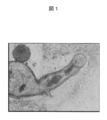
- FIG. 1 shows a tubular formation artificial tube derived from iPS cells.
- FIG. 2 shows a dome-like artificial intestinal tract induced to differentiate from iPS cells.
- FIG. 3 shows the delivery function (FIGS. 3A to 3C) of the intestinal contents (black) accompanying peristaltic movement (repetitive contraction and relaxation) in the artificial intestine of the present invention, and enlarged images (FIGS. 3D to 3F).
- FIG. 4 shows the presence of nerve cell bodies and nerve fibers of nerve cells differentiated from iPS cells in the artificial intestinal tract of the present invention (FIG. 4A) and the ganglion that is an aggregate of large nerve cells (FIG. 4). 4B).
- the scale bar represents 30 ⁇ m.
- the artificial intestinal tract refers to a peristaltic intestinal tract that is induced to differentiate from an induced pluripotent stem (iPS) cell.
- Components constituting the intestinal tract include those derived from the three germ layers (ie, ectoderm, endoderm and mesoderm), such as nerve cells, mucosal epithelial cells, microvilli, connective tissue, smooth muscle layer and the like.
- An induced pluripotent stem cell or iPS cell refers to an ES cell-like differentiated pluripotent stem cell initialized from a somatic cell by a reprogramming factor.
- the cells express ES cell specific markers such as Oct3 / 4, Nanog (or OCT3 / 4, NANOG).
- iPS cells also have the ability to differentiate into the various cells that make up the animal's body and to continue to proliferate semi-permanently while retaining their karyotype.
- Reprogramming refers to the process and means by which differentiated cells are induced and converted to undifferentiated cells, particularly differentiated pluripotent cells.
- a three-dimensional three-dimensional culture system is a three-dimensional culture system that is not a two-dimensional (planar) culture.
- suspension culture Keller, J., Physiol. (Lond) 168: 131-139 (1998), JP, 2008-178367 publication
- suspension culture (same as above), three-dimensional culture system under microgravity environment (T. Uemura et al., Space Utility Res 25: 170-173 (2009)) and the like.
- Suspension culture is also referred to as hanging drop culture, and is a culture method that utilizes gravity and surface tension, and is a method of culturing cells in a culture solution that hangs down in the form of water droplets.
- the suspension culture is a culture method in which cells are grown in a suspended state in a liquid medium without being attached to a culture vessel by coating with a cell non-adhesive polymer.
- a feeder cell is an auxiliary cell used to prepare a culture condition in which a cell to be proliferated is kept undifferentiated, and this cell itself is an antibiotic such as mitomycin C or radiation so as not to proliferate. Processed in advance by irradiation or the like.
- An embryoid body is a cell cluster formed by aggregation of iPS cells. Mammals include, but are not limited to, primates including humans, rodents including mice, and ungulates including cows.
- Somatic cells are all somatic cells except germ cells (oocytes, spermatogonia), germ stem cells (embryonic stem cells, sperm stem cells) or totipotent cells, mature or fetal cells (fibroblasts) Hair cells, muscle cells, hepatocytes, gastric mucosa cells, intestinal cells, spleen cells, pancreatic cells, pancreatic exocrine cells, brain cells, lung cells, kidney cells, skin cells, lymphocytes, epithelial cells, endothelial cells, etc.), It includes cells such as stem cells (hematopoietic stem cells, mesenchymal stem cells, neural stem cells, dental pulp stem cells, etc.) and precursor cells.
- Somatic cells also include cells such as primary cultured cells, passaged cells, and established cell lines.
- the tridermal system consists of endoderm, mesoderm, and ectoderm, and can differentiate into specific cells, tissues, and organs, respectively.
- the intestinal tract component refers to cells derived from the three germ layers that constitute the intestinal tract.
- Peristaltic movement refers to cooperative movement unique to the intestinal tract. 2.
- Production of iPS cells The iPS cells that can be used in the present invention can be artificially induced by reprogramming of somatic cells of any animal, preferably mammals, including humans and mice (for example, Takahashi, K. and Yamanaka, S., Cell 126: 663-676 (2006), Takahashi, K.
- Reprogramming is not particularly limited, and can be performed by any known method. Such methods include methods that use a combination of specific reprogramming factors, methods that combine reprogramming factors with microRNA (miRNA), and the like. Examples of combinations of reprogramming factors are not limited to the following, but combinations of Oct3 / 4, Sox2, Klf4 and c-Myc, or OCT3 / 4, SOX2, KLF4 and c-MYC, or homologs thereof.
- the amino acid sequence and base sequence of the reprogramming factor can be obtained, for example, by accessing the web site of NCBI, USA.
- the mouse sequences are registered as NM_013633, NM_011433, NM_010638, NM_010844, NM_028016, NM_145833, respectively.
- human sequences are registered as NM_203289 or NM_002701, NM_003106, NM_004235, NM_002467, NM_024865, NM_024647, respectively. ing.
- Examples of combinations of reprogramming factors and miRNAs are not limited to the following, but include Oct3 / 4, Sox2 and Klf4, or OCT3 / 4, SOX2 and KLF4, or homologs thereof, and miR-291-3p, miR -294 or miR-295, or combinations thereof with homologs of those miRNAs (Judson, RL et al., Nature Biotechnology 27: 459-461 (2009)). miR-291-3p, miR-294, or miR-295 are subsets of the mouse miR-290 cluster that increase the efficiency of somatic cell reprogramming by Oct3 / 4, Sox2 and Klf4 upon induction of mouse iPS cells. Has an enhancing effect.
- the term “homolog” is used when it belongs to the same family as the indicated reprogramming factor or miRNA, but the species from which it is derived is different.
- Reprogramming is performed by introducing a reprogramming factor or miRNA into the somatic cell.
- the reprogramming factor is a nucleic acid
- it can be introduced into a somatic cell via a vector such as a virus or a plasmid.
- Viral vectors include retrovirus vectors, lentivirus vectors, adenovirus vectors, adeno-associated virus vectors, Sendai virus vectors, and the like.
- any plasmid for mammalian cells can be used as the plasmid (Okita, K.
- the reprogramming factor DNA is not integrated into the cell genome (Stadfeld, M. et al., Science 322: 945-949 (2008)).
- Sendai virus vector it is said that the vector is destroyed after reprogramming.
- the vector may appropriately include a control element such as a promoter, enhancer, terminator, ribosome binding site, polyadenylation site, selection marker, reporter gene, and the like.
- Selectable markers include, for example, drug resistance genes such as positive markers such as kanamycin resistance gene, ampicillin resistance gene, puromycin resistance gene, and negative markers such as thymidine kinase gene and diphtheria toxin gene.
- the reporter gene includes gene sequences such as green fluorescent protein (GFP), GUS ( ⁇ -glucuronidase), and FLAG.
- GFP green fluorescent protein
- GUS ⁇ -glucuronidase
- FLAG FLAG.
- liposomes and membrane-permeable peptide vectors can be used for intracellular introduction of reprogramming factor proteins.
- General techniques for genetic recombination, including vector construction, are described in, for example, Sambrook, J. et al. Et al., Molecular Cloning: A Laboratory Manual, 2nd edition, Cold Spring Harbor Laboratory Press (1989), Ausubel, F. et al. M.M. Et al., Short protocols in Molecular Biology: A Comprehensive Methods from Current Protocols in Molecular Biology, John Wiley & Sons (1999). Induction of iPS cells is described in Japanese Patent No. 4183742, Takahashi, K. et al.
- iPS cell induction is by culturing somatic cells on feeder cells (mitomycin C-treated STO cells or SNL cells) in an appropriate induction medium in the presence of 37 ° C., 5% CO 2 , A somatic cell and a reprogramming factor or DNA (preferably a vector) encoding the factor are brought into contact and cultured, and an iPS-like colony formed after about 2 to 3 weeks is identified and selected.
- iPS cell induction method is to first contact a somatic cell with a reprogramming factor or DNA encoding the factor (preferably a vector) in an appropriate culture medium in the presence of 37 ° C. and 5% CO 2. For about 4-7 days, after which the cells are cultured on the same feeder cells in the presence of 37 ° C., 5% CO 2 in an appropriate culture medium for about 25 to about 30 days or more. IPS-like colonies generated after the above are identified and selected.
- the culture medium include DMEM, DMEM / F12, or DME medium containing 10 to 15% FCS (there are LIF (leukemia inhibitory factor), penicillin / streptomycin, puromycin, L-glutamine, non-essential).
- ES cell culture medium containing bFGF or SCF for example, mouse ES cell culture medium (eg, TX-WES medium, Thrombo X) or A culture medium for primate ES cell culture (for example, culture medium for primate (human and monkey) ES cells, reprocell), and the like can be used.
- mouse ES cell culture medium eg, TX-WES medium, Thrombo X
- a culture medium for primate ES cell culture for example, culture medium for primate (human and monkey) ES cells, reprocell
- the medium is replaced with a fresh medium once a day from the second day onward.
- the number of somatic cells used for reprogramming is not limited, but ranges from about 5 ⁇ 10 3 to about 5 ⁇ 10 6 cells per 100 cm 2 of culture dish.
- iPS cells By treating the iPS-like colonies on the dish with a solution containing trypsin and collagenase IV (CTK solution), the remaining colonies are spread on the feeder cells and cultured in the ES cell culture medium in the same manner. iPS cells can be passaged. At this time, the cells are cultured until they become 80-90% confluent, and the passage is repeated. Identification of iPS cells can be performed by detecting the expression of genes such as Oct3 / 4 or OCT3 / 4, Nanog or NANOG, which are ES cell marker genes, by RT-PCR. Furthermore, the formation of teratomas (teratomas) is confirmed by transplanting established cells to the dorsal ventral side of nude mice. 3.
- CTK solution trypsin and collagenase IV
- iPS cells Preparation of embryoid bodies from iPS cells
- the iPS cells produced and subcultured by the above-mentioned method contain a mixture of feeder cells such as fibroblasts (for example, mouse fetal fibroblasts (MEF)), and a few differentiated cells generated during repeated passages. Therefore, it is important to remove such cells.
- fibroblasts for example, mouse fetal fibroblasts (MEF)
- MEF mouse fetal fibroblasts
- LIF is contained in the iPS cell culture medium.
- iPS cells (about 5 ⁇ 10 3 to about 5 ⁇ 10 6 cells per 100 cm 2 of culture dish) in the above culture medium in the presence of 37 ° C. and 5% CO 2
- the iPS cells were seeded on gelatin, cultured in iPS cell culture medium for 2 days, and again, iPS cells were cultured on gelatin. Incubate for 1 day.
- High-purity iPS cells can be obtained in about 5 days from the start of iPS cell culture.
- the purity of iPS cells that can be used in the present invention is 95% or more, preferably 97% or more, more preferably 99% or more.
- purified iPS cells refers to cells containing 95% or more of iPS cells. Embryoid bodies are prepared using the purified iPS cells obtained as described above. When there is much MEF contamination and the purity of iPS cells is low, the differentiation induction efficiency of the artificial intestinal tract becomes extremely low. Therefore, purification of iPS cells, that is, removal of feeder cells such as MEF is very important for the production of artificial intestinal tract. It became clear that. At this time, a three-dimensional three-dimensional culture system or a two-dimensional planar culture system can be used as the culture system, but a three-dimensional three-dimensional culture system or a combination of three-dimensional three-dimensional culture and two-dimensional planar culture is preferable.
- Examples of the three-dimensional three-dimensional culture system include a suspension culture system, a suspension culture system, and a three-dimensional culture system in a microgravity environment.
- a preferred culture system is a suspension culture system (Keller et al., J. Physiol. (Lond) 168: 131-139 (1998), Japanese Patent Application Laid-Open No. 2006-239169).
- the suspension culture system is a culture system in which a culture solution is hung on the back of a petri dish lid, etc., and iPS cells aggregate under the culture drop under the influence of gravity, and an aggregate called an embryoid body becomes a short day. Formed with.
- a medium for producing an embryoid body in the above culture system is, for example, based on a DMEM, DMEM / F12 or DME medium containing about 10 to 15% FCS, and includes a non-essential amino acid, ⁇ -mercapto. Those containing ethanol, sodium pyruvate, penicillin / streptomycin, etc. can be used.
- a medium similar to this can be used in a suspension culture system or a three-dimensional culture system in a microgravity environment.
- suspension culture as described above, embryoid bodies obtained by culturing iPS cells for 5 to 7 days, preferably 6 days are obtained.
- the number of days of embryoid body culture is very important for the development of the artificial intestinal tract because the induction of differentiation of the artificial intestinal tract is higher in 6 days than in 5 days. became. 4).
- Induction of differentiation of artificial intestinal tract The embryoid body obtained by culturing iPS cells as described above for 5 to 7 days, preferably 6 days, is cultured for about 14 days using the same medium as that for embryoid body preparation.
- peristaltic intestinal tract having a luminal structure (see FIGS. 1 and 3).
- mucosal epithelial cells such as Goblet cells having microvilli, connective tissue, smooth muscle cells, serosa, Kahal stromal cells, nerve cells, etc. were detected.
- a group of cells from the germ layer In addition to regular automatic contraction, a large waving and undulating peristaltic movement is characteristic of the intestinal tract.
- the artificial intestinal tract (ES-Gut) from ES cells has many domestic-like formations (domestic shape), and the number of tubular formations (tubular) is small, whereas the iPS cells of the present invention
- iGut In the artificial intestinal tract (iGut) from, many of them took a tubular formation (tubular) (FIG. 1) rather than a dome-like formation (dome-shaped) (FIG. 2).
- the dome-shaped tissue is thought to be a tissue formed by cells proliferating and accumulating vertically, whereas the tubular tissue is thought to be formed by cells proliferating and accumulating horizontally.
- somatic cells of the patient are used as starting materials, it is considered that there are no ethical problems and problems of rejection as described above.
- iPS cells are similar to ES cells in terms of characteristics such as morphology, proliferation mode, differentiation pluripotency, and ability to form teratomas, but differentiation from iPS cells into somatic cells and tissues.
- iPS cells produced by reprogramming somatic cells have the ability to induce organ differentiation.
- the present invention further provides the use of the artificial intestinal tract described above for the manufacture of a therapeutic agent for regenerative medicine resulting from a patient's bowel disease.
- IPS cells are established from primary or subcultured somatic cells collected from patients in a strictly controlled clean room free from contamination by viruses, microorganisms, etc., and prepared from the iPS cells by the method of the present invention.
- the produced artificial intestinal tract is used as a therapeutic agent for regenerative medicine.
- the therapeutic agent includes at least an artificial intestinal tract, a medium for maintaining it (including serum if necessary), artificial blood, and the like.
- Intestinal diseases include refractory or congenital diseases such as inflammatory bowel diseases such as ulcerative colitis and Crohn's disease, and intestinal motility disorders such as Hirschsprung's disease.
- the artificial intestinal tract of the present invention is derived from the patient's own somatic cells, it has the potential to become a completely new therapeutic strategy that can be used for regenerative medicine and tailor-made medicine as an organ for transplantation without rejection.
- an artificial intestinal tract is made outside the body, which is replaced with or transplanted to the impaired intestinal tract of a patient. This is thought to cause the patient's intestine to function normally.
- Example 1 ⁇ Preparation of embryoid bodies from iPS cells> iPS cells
- the Kyoto University iPS Cell Research Center introduced 4 reprogramming factor (Oct3 / 4, Sox2, Klf4, c-Myc) DNA into mouse skin cells (somatic cells) via pMXs retroviral vectors.
- the iPS cells prepared in this way were provided by Kyoto University through RIKEN. This mouse iPS cell was used in the following experiments. Maintenance culture of iPS cells The iPS cells donated from Kyoto University were thawed and dispensed, and then stored frozen at -180 ° C.
- MEPS mouse fetal fibroblasts
- iPS cells were cultured thereon.
- the culture solution at that time is shown in Table 1 below.
- Production of Embryoid Body A three-dimensional three-dimensional culture system was used to induce cell-to-organization from iPS cells to complex organs composed of various cells in an orderly manner. Specifically, embryoid bodies were prepared from iPS cells using a hanging drop culture system in which a culture solution containing iPS cells was hung on the back of a petri dish lid.
- iPS cells aggregated under the culture drop under the influence of gravity, and aggregates called embryoid bodies were formed in one day.
- iPS cells are maintained and cultured, and frozen iPS cells are thawed and used for experiments, in which mouse fetal fibroblast (MEF) cells are contaminated.
- MEF mouse fetal fibroblast
- the culture solution is the same as in Table 1 above. This operation is performed the day before thawing iPS cells.
- iPS cells + MEF cells are seeded on gelatin and cultured for 2 days.
- the culture solution of (3) is spread on gelatin and cultured for 1 day, thereby further removing MEF and increasing the purity of iPS cells.
- Only high-purity iPS cells can be cultured in about 5 days from the start of iPS cell culture, and embryoid bodies (EBs) are produced using the purified iPS cells.
- EBs embryoid bodies
- a culture solution for EB production (Table 2) EBs were produced in a suspension culture system at a concentration of 500 cells / 15 ⁇ l.
- Table 2 a culture solution for EB production
- EBs were produced in a suspension culture system at a concentration of 500 cells / 15 ⁇ l.
- FIG. 2 An artificial intestinal tract is shown in FIG.
- the shape of an organ having a tubular structure is clear.
- Artificial intestinal tracts induced to differentiate from ES cells have many types of dome-like formation (dome shape), but artificial intestinal tracts induced to differentiate from iPS cells are more likely than domes-like formation (dome shape) (FIG. 2).
- dome shape domes-like formation
- FIG. 3 the produced artificial intestinal tract repeatedly contracted and relaxed, and as shown in FIG. 3, the contents of the intestinal tract (black; confirmed with an electron microscope were epithelial cells as a result of this peristaltic movement. Probably the epithelial cells that have fallen off due to regeneration).
- the cell group constituting the intestinal tract includes mucosal epithelial cells such as Goblet cells having microvilli, connective tissue, smooth muscle cells, serosa, Kahal stromal cells, nerve cells, etc. It has been shown.
- mucosal epithelial cells such as Goblet cells having microvilli, connective tissue, smooth muscle cells, serosa, Kahal stromal cells, nerve cells, etc. It has been shown.
- nerve staining using an anti-neurofilament antibody revealed that a network of nerve cell bodies, ganglia, and nerve fibers was constructed in the artificial intestine (FIG. 4).
- organs induced to differentiate from iPS cells have morphological characteristics similar to those of a mature mouse intestinal tract having a luminal structure and a neural network.
- the present invention provides a method for producing an artificial intestinal tract using iPS cells derived from a somatic cell of a patient.
- An artificial intestine without rejection produced by this method is used for regenerative medicine and tailor-made medicine. Useful for. All publications, patents and patent applications cited herein are incorporated herein by reference in their entirety.
Abstract
A method for producing an artificial intestinal tract from an artificial pluripotent stem (iPS) cell, which comprises forming an embryoid body from a purified iPS cell using a three-dimensional culture system, culturing the embryoid body, and culturing the embryoid body using a two-dimensional attached culture system, thereby inducing the differentiation into an intestinal tract; an artificial intestinal tract produced by the method; and use of the artificial intestinal tract in regenerative medicine.
Description
本発明は、人工多能性幹(iPS)細胞から人工腸管を作製する方法に関する。
本発明はまた、上記方法によって作製された人工腸管、並びに、該人工腸管の再生医療への利用に関する。 The present invention relates to a method for producing an artificial intestinal tract from induced pluripotent stem (iPS) cells.
The present invention also relates to an artificial intestinal tract produced by the above-described method and use of the artificial intestinal tract for regenerative medicine.
今日、再生医療が脚光を浴びている。これは、山中らによってiPS細胞が発見されたからである(特許文献1、非特許文献1)。iPS細胞は、特定の転写因子の作用によって体細胞から誘導される、胚性幹(ES)細胞に類似した特性を有しており、様々な細胞や組織に分化する能力をもつ細胞である(非特許文献1~3)。ES細胞が、卵子又は卵母細胞に由来するのに対して、iPS細胞は、個体の体細胞から誘導することができるため、iPS細胞から誘導された分化細胞又は組織を個体に移植したときの拒絶反応のリスクがほとんどないという利点がある。それゆえに、iPS細胞を再生医療に役立てるための研究が行われている。
ES細胞は、はじめマウス受精卵から誘導され(非特許文献4)、その後ヒトES細胞も樹立された(非特許文献5)。ES細胞、特にマウスES細胞からは、血管内皮細胞、平滑筋細胞、心筋細胞、神経細胞、インスリン産生細胞、ドーパミン産生細胞などの細胞に分化誘導することが多数報告されている(非特許文献6~9)。これに対して、iPS細胞の利用研究は始まったばかりであり、iPS細胞から分化細胞への誘導については報告が少ない。
本発明に関わる人工腸管に関しては、マウスES細胞からの腸管様細胞塊の構築(特許文献2)、マウスES細胞からの腸管の分化誘導(非特許文献10~11)などが報告されている。この報告のなかで、懸垂培養(hanging drop culture)系でES細胞から形成された胚様体を、付着培養に供することによって蠕動運動する腸管が作製されており、このとき、ES細胞の状態や培養条件が重要なファクターになると指摘されている。
しかしながら、エピジェネティックな研究により、iPS細胞はES細胞と同一でないという報告もされているため(非特許文献12)、またiPS細胞自体の特性化が十分でないために、ES細胞の情報がそのままiPS細胞に適用できるという保証もない。このような状況のなかで、iPS細胞から蠕動する腸管の分化誘導が実現されるならば、再生医療への応用の期待が一層高まることになるだろう。 Today, regenerative medicine is in the spotlight. This is because iPS cells were discovered by Yamanaka et al. (Patent Document 1, Non-Patent Document 1). iPS cells have characteristics similar to embryonic stem (ES) cells derived from somatic cells by the action of specific transcription factors, and are capable of differentiating into various cells and tissues ( Non-patent documents 1 to 3). Since ES cells are derived from eggs or oocytes, iPS cells can be derived from individual somatic cells, so that when differentiated cells or tissues derived from iPS cells are transplanted into an individual, There is an advantage that there is almost no risk of rejection. Therefore, research for using iPS cells for regenerative medicine is being conducted.
ES cells were first induced from mouse fertilized eggs (Non-patent Document 4), and human ES cells were also established thereafter (Non-patent Document 5). It has been reported that ES cells, particularly mouse ES cells, induce differentiation into cells such as vascular endothelial cells, smooth muscle cells, cardiomyocytes, nerve cells, insulin producing cells, dopamine producing cells (Non-patent Document 6). ~ 9). In contrast, iPS cell utilization research has just begun, and there are few reports on induction from iPS cells to differentiated cells.
Regarding the artificial intestinal tract related to the present invention, construction of an intestinal tract-like cell mass from mouse ES cells (Patent Document 2), induction of intestinal differentiation from mouse ES cells (Non-Patent Documents 10 to 11), and the like have been reported. In this report, an intestinal tract that is peristaltically moved by subjecting an embryoid body formed from ES cells in a hanging drop culture system to adherent culture has been prepared. It has been pointed out that culture conditions are an important factor.
However, since epigenetic studies have reported that iPS cells are not identical to ES cells (Non-patent Document 12), and because iPS cells themselves are not sufficiently characterized, ES cell information remains as is in iPS. There is no guarantee that it can be applied to cells. Under such circumstances, if induction of differentiation of the intestinal tract peristalized from iPS cells is realized, the expectation of application to regenerative medicine will be further increased.
人工多能性幹(iPS)細胞をヌードマウスに移植した場合、テラトーマ(teratoma)を形成することから、iPS細胞は多分化能を有するものと考えられ、理論上、さまざまな細胞に分化することが予想される。しかし、受精卵から樹立されたES細胞の多能性と違って、哺乳動物の皮膚細胞などの体細胞に特定の種類の再プログラム化因子を遺伝子導入して作製されたiPS細胞の分化多能性について、体を構成するすべての細胞、組織、臓器等に分化するかどうかについては、確実な情報が存在しない。
本発明者らは、先に、胚性幹(ES)細胞から人工腸管を分化誘導する方法を報告したが、今回、さらに適用範囲が広いと考えられるiPS細胞からの人工腸管の作製について研究を行った。
したがって、本発明の目的は、iPS細胞から人工腸管を作製する方法を提供することである。
本発明は、要約すると、以下の特徴を含む。
第1の態様において、本発明は、人工多能性幹(iPS)細胞から人工腸管を作製する方法であって、三次元立体培養系を用いて、純化iPS細胞から胚様体を形成及び培養し、その後、二次元付着培養系を用いて該胚様体を培養し、これによって腸管を分化誘導することを含む、上記方法を提供する。
その実施形態において、上記三次元立体培養系が懸垂培養系であることを特徴とする。
別の実施形態において、上記iPS細胞が哺乳動物体細胞由来であることを特徴とする。
別の実施形態において、上記二次元付着培養の前に、前記胚様体の形成及び培養を5~7日間、好ましくは6日間、行うことを特徴とする。
別の実施形態において、上記懸垂培養系において、1滴(約15~30μl)あたりの細胞数が約400~3,000、例えば約500~2,000である、好ましくは1滴(約15μl)あたりの細胞数が500~1,000であることを特徴とする。
別の実施形態において、上記腸管は、神経ネットワークを有することを特徴とする。
本発明の方法によって作製される上記腸管は、三胚葉系由来の腸管構成成分からなり、かつ、蠕動運動することを特徴とする。
したがって、第2の態様において、本発明はまた、上記のいずれかの方法で作製された人工腸管を提供する。
第3の態様において、本発明はさらに、患者の腸疾患に起因した再生医療のための治療剤の製造への、上記の人工腸管の使用を提供する。
本発明により、患者の体細胞から誘導されたiPS細胞を使用して人工腸管を作製することができるため、移植に伴う拒絶反応のリスクを回避できるという利点が提供される。またこれによって、胚性幹(ES)細胞からの人工腸管の作製と比べて適用範囲が格段に広がるという利点もある。 When an artificial pluripotent stem (iPS) cell is transplanted into a nude mouse, it forms a teratoma, and thus iPS cells are considered to have pluripotency and theoretically differentiate into various cells. Is expected. However, unlike the pluripotency of ES cells established from fertilized eggs, iPS cell differentiation pluripotency produced by gene transfer of a specific type of reprogramming factor into somatic cells such as mammalian skin cells Regarding sex, there is no reliable information on whether to differentiate into all cells, tissues, organs, etc. that make up the body.
The present inventors previously reported a method for inducing differentiation of an artificial intestinal tract from embryonic stem (ES) cells. This time, the present inventors have studied the production of an artificial intestinal tract from iPS cells, which is considered to have a wider application range. went.
Accordingly, an object of the present invention is to provide a method for producing an artificial intestinal tract from iPS cells.
In summary, the present invention includes the following features.
In a first aspect, the present invention relates to a method for producing an artificial intestinal tract from artificial pluripotent stem (iPS) cells, which uses a three-dimensional three-dimensional culture system to form and culture embryoid bodies from purified iPS cells. And then culturing the embryoid body using a two-dimensional adhesion culture system, thereby inducing differentiation of the intestinal tract.
In the embodiment, the three-dimensional three-dimensional culture system is a suspension culture system.
In another embodiment, the iPS cells are derived from mammalian somatic cells.
In another embodiment, the embryoid body is formed and cultured for 5 to 7 days, preferably 6 days before the two-dimensional adhesion culture.
In another embodiment, in the suspension culture system, the number of cells per drop (about 15-30 μl) is about 400-3,000, for example about 500-2,000, preferably one drop (about 15 μl). The number of cells per cell is 500 to 1,000.
In another embodiment, the intestinal tract has a neural network.
The intestinal tract produced by the method of the present invention is composed of intestinal components derived from the three germ layers and is characterized by peristaltic movement.
Therefore, in the second aspect, the present invention also provides an artificial intestinal tract produced by any of the methods described above.
In a third aspect, the present invention further provides the use of the artificial intestinal tract described above for the manufacture of a therapeutic agent for regenerative medicine resulting from a patient's bowel disease.
The present invention provides an advantage that an artificial intestinal tract can be prepared using iPS cells derived from a patient's somatic cells, thereby avoiding the risk of rejection associated with transplantation. This also has the advantage that the range of application is greatly expanded compared to the production of an artificial intestine from embryonic stem (ES) cells.
図1は、iPS細胞から分化誘導されたtubular formation(管状)の人工腸管を示す。
図2は、iPS細胞から分化誘導されたdome−like formation(ドーム状)の人工腸管を示す。ES細胞から分化誘導された人工腸管は、このタイプが多いが、iPS細胞から分化誘導された人工腸管は、図1のtubular formationのほうが多い傾向にある。
図3は、本発明の人工腸管における、蠕動運動(繰り返す収縮と弛緩)にともなう腸管内容物(黒)の運搬機能(図3A~3C)、およびその拡大像(図3D~3F)を示す。
図4は、本発明の人工腸管における、iPS細胞から分化誘導された神経細胞の神経細胞体と神経線維(図4A)、および大きな神経細胞の集合体である神経節(ganglion)の存在(図4B)を示す。図中、スケールバーは30μmを表す。 FIG. 1 shows a tubular formation artificial tube derived from iPS cells.
FIG. 2 shows a dome-like artificial intestinal tract induced to differentiate from iPS cells. There are many types of artificial intestinal tracts induced to differentiate from ES cells, but artificial intestinal tracts induced to differentiate from iPS cells tend to be more in the tubular formation of FIG.
FIG. 3 shows the delivery function (FIGS. 3A to 3C) of the intestinal contents (black) accompanying peristaltic movement (repetitive contraction and relaxation) in the artificial intestine of the present invention, and enlarged images (FIGS. 3D to 3F).
FIG. 4 shows the presence of nerve cell bodies and nerve fibers of nerve cells differentiated from iPS cells in the artificial intestinal tract of the present invention (FIG. 4A) and the ganglion that is an aggregate of large nerve cells (FIG. 4). 4B). In the figure, the scale bar represents 30 μm.
1.定義
以下に、本明細書で使用する用語の定義を記載する。しかし、これらの定義は、限定的に解釈されるべきでなく、業界で使用される最も広義に解釈されるべきである。
人工腸管は、人工多能性幹(iPS)細胞から分化誘導された、蠕動運動する腸管を指す。腸管を構成する成分は、三胚葉(すなわち、外胚葉、内胚葉及び中胚葉)由来のもの、例えば、神経細胞、粘膜上皮細胞、微絨毛、結合組織、平滑筋層などを含む。
人工多能性幹細胞又はiPS細胞は、再プログラム化因子により体細胞から初期化されたES細胞様の分化多能性幹細胞をいう。この細胞は、Oct3/4、Nanog(あるいは、OCT3/4、NANOG)などのES細胞特異的なマーカーを発現する。iPS細胞はまた、動物の体を構成する種々の細胞に分化する能力、及び核型を保持したまま半永久的に増殖し続ける能力を有する。
再プログラム化は、分化した細胞が、未分化細胞、特に分化多能性細胞に誘導、変換される過程及び手段を指す。
三次元立体培養系は、二次元(平面)培養ではない立体的に培養を行う系であり、例えば懸垂培養(Keller,J.,Physiol.(Lond)168:131−139(1998)、特開2008−178367号公報)、浮遊培養(同上)、微小重力環境下の三次元培養系(T.Uemura et al.,Space Utiliz Res 25:170−173(2009))などが含まれる。懸垂培養は、ハンギングドロップ培養(hanging drop culture)とも称せられ、重力と表面張力を利用する培養法であり、水滴状に垂れ下げた培養液の中で細胞を培養する方法である。浮遊培養は、細胞を、細胞非接着性ポリマーでコーティングするなどして培養器に付着させることなく、液体培地のなかに浮遊させた状態で増殖させる培養法である。浮遊培養では、予め形成させた胚様体又は凝集塊(もしくは集積体)を浮遊させて培養することができる。微小重力環境下の三次元培養は、RWV(rotating wall vessel)バイオリアクターを使用して行うことが可能であり、三次元的組織構築が可能なことが知られている。
フィーダー細胞は、増殖させようとする細胞を未分化のまま維持するという培養条件を整えるために使用される補助的細胞であり、この細胞自体は、増殖しないようにマイトマイシンCなどの抗生物質又は放射線照射などで予め処理されている。
胚様体は、iPS細胞が凝集して生じた細胞集塊である。
哺乳動物は、ヒトを含む霊長類、マウスを含むげっ歯類、ウシを含む有蹄類などを非制限的に含む。好ましい哺乳動物はヒトである。
体細胞は、生殖細胞(卵母細胞、精原細胞)、生殖幹細胞(胚性幹細胞、精子幹細胞)又は分化全能性細胞を除くすべての体性細胞であり、成熟又は胎児性細胞(線維芽細胞、毛細胞、筋肉細胞、肝細胞、胃粘膜細胞、腸細胞、脾細胞、膵細胞、膵外分泌細胞、脳細胞、肺細胞、腎細胞、皮膚細胞、リンパ球、上皮細胞、内皮細胞等)、幹細胞(造血幹細胞、間葉系幹細胞、神経幹細胞、歯髄幹細胞等)、前駆体細胞などの細胞を包含する。体細胞は、初代培養細胞、継代細胞、株化細胞などの細胞も包含する。
三胚葉系は、内胚葉、中胚葉及び外胚葉からなり、それぞれ特定の細胞、組織、臓器に分化することができる。
腸管構成成分は、腸管を構成する、三胚葉系由来の細胞をいう。
蠕動運動は、腸管に特有の協調運動をいう。
2.iPS細胞の作製
本発明で使用可能なiPS細胞は、ヒトやマウスを含むあらゆる動物、好ましくは哺乳動物の体細胞の再プログラム化によって人工的に誘導されうる(例えば、Takahashi,K.and Yamanaka,S.,Cell 126:663−676(2006)、Takahashi,K.et al.,Cell 131:861−872(2007)、Yu,J.et al.,Science 318:1917−1920(2007)参照)。再プログラム化は、特に限定されず、公知の任意の方法によって行うことができる。そのような方法には、特定の再プログラム化因子の組合せを使用する方法、再プログラム化因子とマイクロRNA(miRNA)を組合せる方法、などが含まれる。
再プログラム化因子の組合せの例は、以下のものに限定されないが、Oct3/4,Sox2,Klf4及びc−Myc、あるいはOCT3/4,SOX2,KLF4及びc−MYC、あるいはそれらのホモログ、の組合せ、Oct3/4,Sox2及びKlf4、あるいはOCT3/4,SOX2及びKLF4、あるいはそれらのホモログ、の組合せ、Oct3/4,Sox2,Nanog及びLin28、あるいはOCT3/4,SOX2,NANOG及びLIN28、あるいはそれらのホモログ、の組合せなどを含む(Takahashi,K.and Yamanaka,S.,Cell 126:663−676(2006)、Takahashi,K.et al.,Cell 131:861−872(2007)、Yu,J.et al.,Science 318:1917−1920(2007))。これらの再プログラム化因子は、核酸(すなわち、遺伝子、DNAもしくはRNA)又はタンパク質のいずれかである。
上記の再プログラム化因子のアミノ酸配列及び塩基配列は、例えば米国NCBIのwebサイトにアクセスすることによって入手可能である。例えば、Oct3/4,Sox2,Klf4,c−Myc,Nanog及びLin28について、マウスの各配列(アミノ酸配列及び塩基配列)はそれぞれ、NM_013633,NM_011443,NM_010637,NM_010849,NM_028016,NM_145833として登録されているし、また、OCT3/4,SOX2,KLF4,c−MYC,NANOG及びLIN28について、ヒトの各配列(アミノ酸配列及び塩基配列)はそれぞれ、NM_203289もしくはNM_002701,NM_003106,NM_004235,NM_002467,NM_024865,NM_024674として登録されている。
再プログラム化因子とmiRNAの組合せの例は、以下のものに限定されないが、Oct3/4,Sox2及びKlf4、あるいはOCT3/4,SOX2及びKLF4、あるいはそれらのホモログと、miR−291−3p,miR−294又はmiR−295、あるいはそれらmiRNAのホモログ、との組合せを含む(Judson,R.L.et al.,Nature Biotechnology 27:459−461(2009))。miR−291−3p,miR−294又はmiR−295は、マウスmiR—290クラスターのサブセットであり、マウスiPS細胞の誘導の際にOct3/4,Sox2及びKlf4による体細胞の再プログラム化の効率を高める作用をもつ。
本明細書で使用する「ホモログ」なる用語は、示された再プログラム化因子又はmiRNAと同じファミリーに属するが、それが由来する生物種が異なる場合に使用される。
再プログラム化は、再プログラム化因子又はmiRNAを体細胞内に導入することによって行われる。再プログラム化因子が核酸の場合には、それは、ウイルス、プラスミドなどのベクターを介して体細胞内に導入されうる。ウイルスベクターには、レトロウイルスベクター、レンチウイルスベクター、アデノウイルスベクター、アデノ随伴ウイルスベクター、センダイウイルスベクターなどが含まれる。また、プラスミドには、哺乳動物細胞用プラスミドのいずれも使用可能である(Okita,K.et al.,Science 322:949−953(2008))。このうち、アデノウイルスベクター、アデノ随伴ウイルスベクター又はプラスミドを使用する場合には、細胞ゲノムに再プログラム化因子DNAが組み込まれないし(Stadtfeld,M.et al.,Science 322:945−949(2008))、また、センダイウイルスベクターを使用する場合には、再プログラム化ののちに、該ベクターは破壊されると言われている。
ベクターには、再プログラム化因子DNAの他に、プロモーター、エンハンサー、ターミネーター、リボソーム結合サイト、ポリアデニル化サイトなどの制御要素、選択マーカー、レポータージーンなどが適宜含まれうる。選択マーカーには、例えば、薬剤耐性遺伝子、例えばカナマイシン耐性遺伝子、アンピシリン耐性遺伝子、ピューロマイシン耐性遺伝子などの陽性マーカー、並びに、チミジンキナーゼ遺伝子、ジフテリアトキシン遺伝子などの陰性マーカーなどが含まれる。また、レポータージーンには、例えば、緑色蛍光タンパク質(GFP)、GUS(β−グルクロニダーゼ)、FLAGなどの遺伝子配列が含まれる。
細胞への再プログラム化因子の導入は、ウイルス感染、エレクトロポレーション、カチオン性リポソーム、膜透過性ペプチドベクター(WO2007/132555)、膜透過性ペプチド表面修飾リポソームなどを利用する方法によって行うことができる。特に、リポソームや膜透過性ペプチドベクターは、再プログラム化因子タンパク質の細胞内導入のために利用できる。
ベクターの構築を含む遺伝子組換えのための一般的な技術は、例えばSambrook,J.ら,Molecular Cloning:A Laboratory Manual,2nd edition,Cold Spring Harbor Laboratory Press(1989)、Ausubel,F.M.ら,Short protocols in Molecular Biology:A Compendium Methods from Current Protocols in Molecular Biology,John Wiley & Sons(1999)などに記載されている。
iPS細胞の誘導は、日本国特許第4183742号、Takahashi,K.and Yamanaka,S.,Cell 126:663−676(2006)、Takahashi,K.et al.,Cell 131:861−872(2007)などに記載される方法によって行うことができる。
iPS細胞の誘導法の1つの例は、フィーダー細胞(マイトマイシンC処理したSTO細胞又はSNL細胞)上で体細胞を、37℃、5%CO2存在下で、適切な誘導培地中で培養し、体細胞と再プログラム化因子又は該因子をコードするDNA(好ましくは、ベクター)を接触させて培養し、約2~3週間後に生じたiPS様コロニーを同定・選択する。
iPS細胞の誘導法の別の例は、はじめに適切な培養培地中、37℃、5%CO2存在下で体細胞と再プログラム化因子又は該因子をコードするDNA(好ましくは、ベクター)を接触させて約4~7日間培養し、その後、該細胞を、適切な培養培地中、37℃、5% CO2存在下で、同様のフィーダー細胞上で培養し、約25~約30日又はそれ以上の後に生じたiPS様コロニーを同定・選択する。
培養培地としては、例えば、10~15%FCSを含有するDMEM、DMEM/F12又はDME培地(これらの培地にはさらに、LIF(leukemia inhibitory factor)、penicillin/streptomycin、puromycin、L−グルタミン、非必須アミノ酸類、β−メルカプトエタノールなどを適宜含むことができる。)、あるいは、bFGF又はSCFを含有するES細胞培養用培地、例えばマウスES細胞培養用培地(例えばTX−WES培地、トロンボX社)又は霊長類ES細胞培養用培地(例えば霊長類(ヒト&サル)ES細胞用培地、リプロセル)、などを使用しうる。
上記の培養の間には、培養開始2日目以降から毎日1回新鮮な培地と培地交換を行う。また、再プログラム化に使用する体細胞の細胞数は、限定されないが、培養ディッシュ100cm2あたり約5×10³~約5×10⁶細胞の範囲である。
ディッシュ上の上記iPS様コロニーを、トリプシン及びコラゲナーゼIVを含む溶液(CTK溶液)を用いて処理し、残ったコロニーを上記フィーダー細胞上に撒いてES細胞培養用培地で同様に培養することにより、iPS細胞を継代することができる。このとき、80~90%コンフルエントになるまで培養し、継代を繰り返す。
iPS細胞の同定は、ES細胞のマーカー遺伝子であるOct3/4もしくはOCT3/4,NanogもしくはNANOGなどの遺伝子の発現をRT−PCR法で検出することによって行うことができる。さらにまた、樹立した細胞をヌードマウスの背側の腹側部に移植することによってテラトーマ(奇形腫)の形成を確認する。
3.iPS細胞からの胚様体の作製
iPS細胞から胚様体を作製するに際して、iPS細胞を高純度に精製する必要がある。上記の方法で作製され継代されたiPS細胞には、線維芽細胞(例えばマウス胎仔線維芽細胞(MEF))などのフィーダー細胞、継代を繰り返す際に生じた僅かな分化細胞などが混在するため、このような細胞を除去することが重要である。混在する細胞を除くために、例えば、ゼラチン上にiPS細胞を撒いて培養することを繰り返し、これによって純化iPS細胞を得る。このときMEFを取り除きながら未分化状態を維持するために、iPS細胞培養培地にLIFを含有させる。
具体的には、線維芽細胞(フィーダー)上で、上記培養培地中、37℃、5% CO2存在下でiPS細胞(培養ディッシュ100cm2あたり約5×10³~約5×10⁶細胞)を2日間培養し、さらに、増殖したiPS細胞コロニーから線維芽細胞を除去するために、ゼラチン上にiPS細胞を撒いて、iPS細胞培養培地で2日間培養し、再度、ゼラチン上でiPS細胞を1日間培養する。iPS細胞の培養開始から約5日間で純度の高いiPS細胞を得ることができる。
本発明で使用可能なiPS細胞の純度は、95%以上、好ましくは97%以上、より好ましくは99%以上である。本明細書で「純化iPS細胞」とは、iPS細胞が95%以上含有する細胞をいう。
上記のようにして得られた純化iPS細胞を使用して胚様体を作製する。MEFの混入が多く、iPS細胞の純度が低い場合、人工腸管の分化誘導効率はきわめて低くなることから、iPS細胞の純化すなわちMEFなどのフィーダー細胞の除去が、人工腸管の作製には非常に重要であることが明らかとなった。またこのとき、培養系として、三次元立体培養系又は二次元平面培養系を使用できるが、三次元立体培養系、又は三次元立体培養と二次元平面培養を組み合わせた系、が好ましい。三次元立体培養系として、例えば懸垂培養系、浮遊培養系、微小重力環境下の三次元培養系などが挙げられる。好ましい培養系は、懸垂培養系である(Keller et al.,J.Physiol.(Lond)168:131−139(1998)、日本国特開2006−239169号公報)。
懸垂培養系は、培養液をシャーレの蓋の裏などにぶら下げて培養する培養系であり、iPS細胞は重力の影響で、培養滴の下側に凝集し、胚様体と呼ばれる凝集塊が短時日で形成される。この培養系では、培養滴の体積に応じてiPS細胞数を決定することが望ましく、例えば1滴(約15~30μl)あたり約400~3,000細胞、例えば約500~2,000細胞、好ましくは1滴(約15μl)あたり約500~1,000細胞が適する細胞濃度である。
上記の培養系で胚様体を作製するための培地は、例えば、約10~15%FCSを含有するDMEM、DMEM/F12又はDME培地を基礎として、これに、非必須アミノ酸類、β−メルカプトエタノール、ピルビン酸ナトリウム、penicillin/streptomycinなどを含むものを用いることができる。これと同様の培地は、浮遊培養系や微小重力環境下の三次元培養系でも使用可能である。
上記のような懸垂培養によって、iPS細胞を5~7日間、好ましくは6日間、培養した胚様体を得る。胚様体の培養日数は5日間よりも6日間のほうが、人工腸管の分化誘導効率が高くなることから、胚様体の培養日数が人工腸管の発生に非常に重要であることが今回明らかとなった。
4.人工腸管の分化誘導
上記のようにしてiPS細胞を5~7日間、好ましくは6日間培養して得られた胚様体を、胚様体作製用の培地と同じ培地を用いて約14日間、二次元付着培養することによって、管腔構造を有する蠕動運動する腸管を分化誘導することができる(図1、図3参照)。
誘導された腸管を構成する細胞群として、微絨毛を有するGoblet細胞などの粘膜上皮細胞、結合組織、平滑筋細胞、漿膜、カハール間質細胞、神経細胞などが検出されたが、これらは、三胚葉系からの細胞群である。規則的な自動収縮に加えて、大きく波打ち、うねるような蠕動運動は、腸管に特有な動きである。
形態学的に見た場合、ES細胞からの人工腸管(ES−Gut)はdome−like formation(ドーム状)が多く、tubular formation(管状)のものは少なかったのに対し、本発明のiPS細胞からの人工腸管(iGut)では、dome−like formation(ドーム状)(図2)よりもtubular formation(管状)(図1)を取るものが多く認められた。ここで、ドーム状組織は、細胞が縦向きに増殖・集積して組織が形成されるのに対して、管状組織は、細胞が横向きに増殖・集積して組織が形成されると考えられる。この相違の原因として、ES細胞とiPS細胞の細胞特性の違いによるものか、胚様体における内部微小環境の段階での違いによるものかは不明である。
iPS細胞からの人工腸管は、結果的には、胚性幹(ES)細胞から人工腸管を誘導したときと類似した培養条件で得ることができた。しかし、ヒトES細胞に関して、ヒトES細胞を調製する段階で受精卵を破壊するという倫理上の問題があるし、並びに、仮にそのような問題が解決されたとしても、移植に伴う拒絶反応のために受精卵の提供者のみへの使用に限定されるなどの問題がある。これに対して、本発明の方法では、患者の体細胞を出発材料とするため、上記のような倫理上の問題や拒絶反応の問題はないと考えられる。確かに、iPS細胞は、形態や増殖様式、分化多能性、テラトーマ(teratoma)形成能などの特性の点でES細胞と類似しているが、iPS細胞からの体細胞や体組織への分化誘導については十分な知見はないのが実状である。特に、体細胞を再プログラム化して作製したiPS細胞が、臓器分化誘導能を有するかどうかについては、全く未知のことである。こうしたなかで、ES細胞と同様の培養条件でiPS細胞から、腸管固有の細胞の複雑な集合体である人工腸管という臓器を分化誘導できたことは、予想できないことであり画期的である。
5.医療への応用
本発明はさらに、患者の腸疾患に起因した再生医療のための治療剤の製造への、上記の人工腸管の使用を提供する。
ウイルス、微生物などの汚染のない厳重に管理されたクリーンルーム内で、患者から採取した体細胞の初代培養細胞または継代培養細胞からiPS細胞を樹立し、このiPS細胞から、本発明の方法により作製された人工腸管を、再生医療のための治療剤として使用する。治療剤には、少なくとも、人工腸管の他に、それを維持するための培地(必要であれば血清を含む)、人工血液などが含まれる。
腸疾患には、例えば、潰瘍性大腸炎やクローン病のような炎症性腸疾患、ヒルシュスプルング病のような腸管運動異常疾患などの難治性あるいは先天性の疾患などが含まれる。
本発明の人工腸管は、患者自身の体細胞から誘導されるため、拒絶反応のない移植用の臓器として、再生医療やテーラーメイド医療のために使用できる全く新しい治療戦略になる可能性を有する。
上で説明した本発明方法によって、体外で人工腸管を作製し、これを、患者の障害のある腸管部分と置換するか又は該腸管部分に移植する。これによって、患者の腸が正常に機能するようになると考えられる。
さらにまた、患者由来あるいは疾患固有のiPS細胞から人工腸管を作製することにより、腸疾患の病態の機序解明、薬剤効果および毒性の評価、腸からの栄養の吸収試験、感染性腸炎のモデル作成などが可能となるだけでなく、創薬や細胞移植治療への臨床応用にも大いに役立つものと考えられる。
以下に実施例を挙げて本発明をさらに具体的に説明するが、本発明の範囲は、これらの実施例によって制限されないものとする。 1. Definitions The definitions of terms used in this specification are described below. However, these definitions should not be interpreted in a limited way, but in the broadest sense used in the industry.
The artificial intestinal tract refers to a peristaltic intestinal tract that is induced to differentiate from an induced pluripotent stem (iPS) cell. Components constituting the intestinal tract include those derived from the three germ layers (ie, ectoderm, endoderm and mesoderm), such as nerve cells, mucosal epithelial cells, microvilli, connective tissue, smooth muscle layer and the like.
An induced pluripotent stem cell or iPS cell refers to an ES cell-like differentiated pluripotent stem cell initialized from a somatic cell by a reprogramming factor. The cells express ES cell specific markers such as Oct3 / 4, Nanog (or OCT3 / 4, NANOG). iPS cells also have the ability to differentiate into the various cells that make up the animal's body and to continue to proliferate semi-permanently while retaining their karyotype.
Reprogramming refers to the process and means by which differentiated cells are induced and converted to undifferentiated cells, particularly differentiated pluripotent cells.
A three-dimensional three-dimensional culture system is a three-dimensional culture system that is not a two-dimensional (planar) culture. For example, suspension culture (Keller, J., Physiol. (Lond) 168: 131-139 (1998), JP, 2008-178367 publication), suspension culture (same as above), three-dimensional culture system under microgravity environment (T. Uemura et al., Space Utility Res 25: 170-173 (2009)) and the like. Suspension culture is also referred to as hanging drop culture, and is a culture method that utilizes gravity and surface tension, and is a method of culturing cells in a culture solution that hangs down in the form of water droplets. The suspension culture is a culture method in which cells are grown in a suspended state in a liquid medium without being attached to a culture vessel by coating with a cell non-adhesive polymer. In suspension culture, a previously formed embryoid body or aggregate (or aggregate) can be suspended and cultured. It is known that three-dimensional culture in a microgravity environment can be performed using a RWV (rotating wall vessel) bioreactor, and three-dimensional tissue construction is possible.
A feeder cell is an auxiliary cell used to prepare a culture condition in which a cell to be proliferated is kept undifferentiated, and this cell itself is an antibiotic such as mitomycin C or radiation so as not to proliferate. Processed in advance by irradiation or the like.
An embryoid body is a cell cluster formed by aggregation of iPS cells.
Mammals include, but are not limited to, primates including humans, rodents including mice, and ungulates including cows. A preferred mammal is a human.
Somatic cells are all somatic cells except germ cells (oocytes, spermatogonia), germ stem cells (embryonic stem cells, sperm stem cells) or totipotent cells, mature or fetal cells (fibroblasts) Hair cells, muscle cells, hepatocytes, gastric mucosa cells, intestinal cells, spleen cells, pancreatic cells, pancreatic exocrine cells, brain cells, lung cells, kidney cells, skin cells, lymphocytes, epithelial cells, endothelial cells, etc.), It includes cells such as stem cells (hematopoietic stem cells, mesenchymal stem cells, neural stem cells, dental pulp stem cells, etc.) and precursor cells. Somatic cells also include cells such as primary cultured cells, passaged cells, and established cell lines.
The tridermal system consists of endoderm, mesoderm, and ectoderm, and can differentiate into specific cells, tissues, and organs, respectively.
The intestinal tract component refers to cells derived from the three germ layers that constitute the intestinal tract.
Peristaltic movement refers to cooperative movement unique to the intestinal tract.
2. Production of iPS cells The iPS cells that can be used in the present invention can be artificially induced by reprogramming of somatic cells of any animal, preferably mammals, including humans and mice (for example, Takahashi, K. and Yamanaka, S., Cell 126: 663-676 (2006), Takahashi, K. et al., Cell 131: 861-872 (2007), Yu, J. et al., Science 318: 1917-1920 (2007)). . Reprogramming is not particularly limited, and can be performed by any known method. Such methods include methods that use a combination of specific reprogramming factors, methods that combine reprogramming factors with microRNA (miRNA), and the like.
Examples of combinations of reprogramming factors are not limited to the following, but combinations of Oct3 / 4, Sox2, Klf4 and c-Myc, or OCT3 / 4, SOX2, KLF4 and c-MYC, or homologs thereof. , Oct3 / 4, Sox2 and Klf4, or OCT3 / 4, SOX2 and KLF4, or a homologue thereof, Oct3 / 4, Sox2, Nanog and Lin28, or OCT3 / 4, SOX2, NANOG and LIN28, or a combination thereof Homologs, etc. (Takahashi, K. and Yamanaka, S., Cell 126: 663-676 (2006), Takahashi, K. et al., Cell 131: 861-872 (2007), Yu, J. et al. et al., Science 318: 1917-1920 (2007)). These reprogramming factors are either nucleic acids (ie, genes, DNA or RNA) or proteins.
The amino acid sequence and base sequence of the reprogramming factor can be obtained, for example, by accessing the web site of NCBI, USA. For example, for Oct3 / 4, Sox2, Klf4, c-Myc, Nanog, and Lin28, the mouse sequences (amino acid sequence and base sequence) are registered as NM_013633, NM_011433, NM_010638, NM_010844, NM_028016, NM_145833, respectively. In addition, for OCT3 / 4, SOX2, KLF4, c-MYC, NANOG and LIN28, human sequences (amino acid sequence and base sequence) are registered as NM_203289 or NM_002701, NM_003106, NM_004235, NM_002467, NM_024865, NM_024647, respectively. ing.
Examples of combinations of reprogramming factors and miRNAs are not limited to the following, but include Oct3 / 4, Sox2 and Klf4, or OCT3 / 4, SOX2 and KLF4, or homologs thereof, and miR-291-3p, miR -294 or miR-295, or combinations thereof with homologs of those miRNAs (Judson, RL et al., Nature Biotechnology 27: 459-461 (2009)). miR-291-3p, miR-294, or miR-295 are subsets of the mouse miR-290 cluster that increase the efficiency of somatic cell reprogramming by Oct3 / 4, Sox2 and Klf4 upon induction of mouse iPS cells. Has an enhancing effect.
As used herein, the term “homolog” is used when it belongs to the same family as the indicated reprogramming factor or miRNA, but the species from which it is derived is different.
Reprogramming is performed by introducing a reprogramming factor or miRNA into the somatic cell. When the reprogramming factor is a nucleic acid, it can be introduced into a somatic cell via a vector such as a virus or a plasmid. Viral vectors include retrovirus vectors, lentivirus vectors, adenovirus vectors, adeno-associated virus vectors, Sendai virus vectors, and the like. In addition, any plasmid for mammalian cells can be used as the plasmid (Okita, K. et al., Science 322: 949-953 (2008)). Among these, when an adenovirus vector, an adeno-associated virus vector or a plasmid is used, the reprogramming factor DNA is not integrated into the cell genome (Stadfeld, M. et al., Science 322: 945-949 (2008)). In addition, when Sendai virus vector is used, it is said that the vector is destroyed after reprogramming.
In addition to the reprogramming factor DNA, the vector may appropriately include a control element such as a promoter, enhancer, terminator, ribosome binding site, polyadenylation site, selection marker, reporter gene, and the like. Selectable markers include, for example, drug resistance genes such as positive markers such as kanamycin resistance gene, ampicillin resistance gene, puromycin resistance gene, and negative markers such as thymidine kinase gene and diphtheria toxin gene. The reporter gene includes gene sequences such as green fluorescent protein (GFP), GUS (β-glucuronidase), and FLAG.
Introduction of the reprogramming factor into the cell can be performed by a method utilizing viral infection, electroporation, cationic liposome, membrane-permeable peptide vector (WO2007 / 132555), membrane-permeable peptide surface-modified liposome, or the like. . In particular, liposomes and membrane-permeable peptide vectors can be used for intracellular introduction of reprogramming factor proteins.
General techniques for genetic recombination, including vector construction, are described in, for example, Sambrook, J. et al. Et al., Molecular Cloning: A Laboratory Manual, 2nd edition, Cold Spring Harbor Laboratory Press (1989), Ausubel, F. et al. M.M. Et al., Short protocols in Molecular Biology: A Comprehensive Methods from Current Protocols in Molecular Biology, John Wiley & Sons (1999).
Induction of iPS cells is described in Japanese Patent No. 4183742, Takahashi, K. et al. and Yamanaka, S .; , Cell 126: 663-676 (2006), Takahashi, K .; et al. , Cell 131: 861-872 (2007) and the like.
One example of iPS cell induction is by culturing somatic cells on feeder cells (mitomycin C-treated STO cells or SNL cells) in an appropriate induction medium in the presence of 37 ° C., 5% CO 2 , A somatic cell and a reprogramming factor or DNA (preferably a vector) encoding the factor are brought into contact and cultured, and an iPS-like colony formed after about 2 to 3 weeks is identified and selected.
Another example of the iPS cell induction method is to first contact a somatic cell with a reprogramming factor or DNA encoding the factor (preferably a vector) in an appropriate culture medium in the presence of 37 ° C. and 5% CO 2. For about 4-7 days, after which the cells are cultured on the same feeder cells in the presence of 37 ° C., 5% CO 2 in an appropriate culture medium for about 25 to about 30 days or more. IPS-like colonies generated after the above are identified and selected.
Examples of the culture medium include DMEM, DMEM / F12, or DME medium containing 10 to 15% FCS (there are LIF (leukemia inhibitory factor), penicillin / streptomycin, puromycin, L-glutamine, non-essential). Amino acids, β-mercaptoethanol, etc. can be included as appropriate.), Or ES cell culture medium containing bFGF or SCF, for example, mouse ES cell culture medium (eg, TX-WES medium, Thrombo X) or A culture medium for primate ES cell culture (for example, culture medium for primate (human and monkey) ES cells, reprocell), and the like can be used.
During the above culture, the medium is replaced with a fresh medium once a day from the second day onward. The number of somatic cells used for reprogramming is not limited, but ranges from about 5 × 10 3 to about 5 × 10 6 cells per 100 cm 2 of culture dish.
By treating the iPS-like colonies on the dish with a solution containing trypsin and collagenase IV (CTK solution), the remaining colonies are spread on the feeder cells and cultured in the ES cell culture medium in the same manner. iPS cells can be passaged. At this time, the cells are cultured until they become 80-90% confluent, and the passage is repeated.
Identification of iPS cells can be performed by detecting the expression of genes such as Oct3 / 4 or OCT3 / 4, Nanog or NANOG, which are ES cell marker genes, by RT-PCR. Furthermore, the formation of teratomas (teratomas) is confirmed by transplanting established cells to the dorsal ventral side of nude mice.
3. Preparation of embryoid bodies from iPS cells When preparing embryoid bodies from iPS cells, it is necessary to purify the iPS cells with high purity. The iPS cells produced and subcultured by the above-mentioned method contain a mixture of feeder cells such as fibroblasts (for example, mouse fetal fibroblasts (MEF)), and a few differentiated cells generated during repeated passages. Therefore, it is important to remove such cells. In order to remove the mixed cells, for example, iPS cells are seeded on gelatin and cultured repeatedly to obtain purified iPS cells. At this time, in order to maintain an undifferentiated state while removing MEF, LIF is contained in the iPS cell culture medium.
Specifically, on fibroblasts (feeders), iPS cells (about 5 × 10 3 to about 5 × 10 6 cells per 100 cm 2 of culture dish) in the above culture medium in the presence of 37 ° C. and 5% CO 2 In order to remove fibroblasts from the grown iPS cell colonies, the iPS cells were seeded on gelatin, cultured in iPS cell culture medium for 2 days, and again, iPS cells were cultured on gelatin. Incubate for 1 day. High-purity iPS cells can be obtained in about 5 days from the start of iPS cell culture.
The purity of iPS cells that can be used in the present invention is 95% or more, preferably 97% or more, more preferably 99% or more. As used herein, “purified iPS cells” refers to cells containing 95% or more of iPS cells.
Embryoid bodies are prepared using the purified iPS cells obtained as described above. When there is much MEF contamination and the purity of iPS cells is low, the differentiation induction efficiency of the artificial intestinal tract becomes extremely low. Therefore, purification of iPS cells, that is, removal of feeder cells such as MEF is very important for the production of artificial intestinal tract. It became clear that. At this time, a three-dimensional three-dimensional culture system or a two-dimensional planar culture system can be used as the culture system, but a three-dimensional three-dimensional culture system or a combination of three-dimensional three-dimensional culture and two-dimensional planar culture is preferable. Examples of the three-dimensional three-dimensional culture system include a suspension culture system, a suspension culture system, and a three-dimensional culture system in a microgravity environment. A preferred culture system is a suspension culture system (Keller et al., J. Physiol. (Lond) 168: 131-139 (1998), Japanese Patent Application Laid-Open No. 2006-239169).
The suspension culture system is a culture system in which a culture solution is hung on the back of a petri dish lid, etc., and iPS cells aggregate under the culture drop under the influence of gravity, and an aggregate called an embryoid body becomes a short day. Formed with. In this culture system, it is desirable to determine the number of iPS cells according to the volume of the culture drop, for example, about 400 to 3,000 cells, for example, about 500 to 2,000 cells per drop (about 15 to 30 μl), preferably Is a suitable cell concentration of about 500 to 1,000 cells per drop (about 15 μl).
A medium for producing an embryoid body in the above culture system is, for example, based on a DMEM, DMEM / F12 or DME medium containing about 10 to 15% FCS, and includes a non-essential amino acid, β-mercapto. Those containing ethanol, sodium pyruvate, penicillin / streptomycin, etc. can be used. A medium similar to this can be used in a suspension culture system or a three-dimensional culture system in a microgravity environment.
By suspension culture as described above, embryoid bodies obtained by culturing iPS cells for 5 to 7 days, preferably 6 days are obtained. It is now clear that the number of days of embryoid body culture is very important for the development of the artificial intestinal tract because the induction of differentiation of the artificial intestinal tract is higher in 6 days than in 5 days. became.
4). Induction of differentiation of artificial intestinal tract The embryoid body obtained by culturing iPS cells as described above for 5 to 7 days, preferably 6 days, is cultured for about 14 days using the same medium as that for embryoid body preparation. By performing two-dimensional adhesion culture, it is possible to induce differentiation of the peristaltic intestinal tract having a luminal structure (see FIGS. 1 and 3).
As a group of cells constituting the induced intestinal tract, mucosal epithelial cells such as Goblet cells having microvilli, connective tissue, smooth muscle cells, serosa, Kahal stromal cells, nerve cells, etc. were detected. A group of cells from the germ layer. In addition to regular automatic contraction, a large waving and undulating peristaltic movement is characteristic of the intestinal tract.
From the morphological point of view, the artificial intestinal tract (ES-Gut) from ES cells has many domestic-like formations (domestic shape), and the number of tubular formations (tubular) is small, whereas the iPS cells of the present invention In the artificial intestinal tract (iGut) from, many of them took a tubular formation (tubular) (FIG. 1) rather than a dome-like formation (dome-shaped) (FIG. 2). Here, the dome-shaped tissue is thought to be a tissue formed by cells proliferating and accumulating vertically, whereas the tubular tissue is thought to be formed by cells proliferating and accumulating horizontally. It is unclear whether this difference is due to differences in cell characteristics between ES cells and iPS cells or due to differences at the stage of the internal microenvironment in the embryoid body.
As a result, the artificial intestine from iPS cells could be obtained under the same culture conditions as when the artificial intestine was derived from embryonic stem (ES) cells. However, with regard to human ES cells, there is an ethical problem of destroying fertilized eggs at the stage of preparing human ES cells, and even if such problems are solved, because of rejection reactions associated with transplantation, However, there are problems such as being limited to use only for fertilized egg providers. On the other hand, in the method of the present invention, since somatic cells of the patient are used as starting materials, it is considered that there are no ethical problems and problems of rejection as described above. Certainly, iPS cells are similar to ES cells in terms of characteristics such as morphology, proliferation mode, differentiation pluripotency, and ability to form teratomas, but differentiation from iPS cells into somatic cells and tissues. Actually, there is not enough knowledge about induction. In particular, it is completely unknown whether iPS cells produced by reprogramming somatic cells have the ability to induce organ differentiation. Under such circumstances, it is unpredictable and epoch-making that an organ called an artificial intestine, which is a complex aggregate of cells unique to the intestine, can be induced to differentiate from iPS cells under the same culture conditions as ES cells.
5). Medical Application The present invention further provides the use of the artificial intestinal tract described above for the manufacture of a therapeutic agent for regenerative medicine resulting from a patient's bowel disease.
IPS cells are established from primary or subcultured somatic cells collected from patients in a strictly controlled clean room free from contamination by viruses, microorganisms, etc., and prepared from the iPS cells by the method of the present invention. The produced artificial intestinal tract is used as a therapeutic agent for regenerative medicine. The therapeutic agent includes at least an artificial intestinal tract, a medium for maintaining it (including serum if necessary), artificial blood, and the like.
Intestinal diseases include refractory or congenital diseases such as inflammatory bowel diseases such as ulcerative colitis and Crohn's disease, and intestinal motility disorders such as Hirschsprung's disease.
Since the artificial intestinal tract of the present invention is derived from the patient's own somatic cells, it has the potential to become a completely new therapeutic strategy that can be used for regenerative medicine and tailor-made medicine as an organ for transplantation without rejection.
According to the method of the present invention described above, an artificial intestinal tract is made outside the body, which is replaced with or transplanted to the impaired intestinal tract of a patient. This is thought to cause the patient's intestine to function normally.
Furthermore, by creating an artificial intestinal tract from patient-derived or disease-specific iPS cells, elucidation of the pathophysiology of intestinal diseases, evaluation of drug effects and toxicity, absorption of nutrients from the intestine, creation of infectious enteritis models In addition, it is considered to be useful for clinical application to drug discovery and cell transplantation therapy.
EXAMPLES Hereinafter, the present invention will be described more specifically with reference to examples. However, the scope of the present invention is not limited by these examples.
[実施例1]
<iPS細胞からの胚様体の作製>
iPS細胞
京都大学iPS細胞研究センターによって、マウスの皮膚細胞(体細胞)に、4つの初期化因子(Oct3/4,Sox2,Klf4,c−Myc)DNAをpMXsレトロウイルスベクターを介して遺伝子導入して作製されたiPS細胞を、理化学研究所を介して京都大学より供与された。以下の実験では、このマウスiPS細胞を使用した。
iPS細胞の維持培養
京都大学から供与されたiPS細胞は、一度、解凍して分注したのち、−180℃で凍結保存された。iPS細胞の未分化維持のために、未分化因子を放出するMEF(マウス胎仔線維芽細胞)をフィーダー細胞として、その上でiPS細胞を培養した。その際の培養液は、以下の表1に示した。

胚様体(embroid body)の作製
iPS細胞から、様々な細胞が秩序立って構成する複雑な臓器に分化誘導(cell−to−organ)するために、三次元立体培養系を使用した。具体的には、シャーレの蓋の裏にiPS細胞を含む培養液をぶら下げて培養する懸垂培養(hanging drop culture)系を用いて、iPS細胞から胚様体を作製した。この培養系では、iPS細胞は重力の影響で、培養滴の下に凝集し、1日で胚様体と呼ばれる凝集塊が形成された。
上記のようにiPS細胞を維持培養し、凍結したiPS細胞は、融解して実験に使用されるが、その中には、マウス胎仔線維芽(MEF)細胞が混入している。腸管分化誘導を行うときには、このMEF細胞がiPS細胞に混じることによって分化が阻害されることが今回明らかになった。そのため、可能な限りiPS細胞の純度を高くするためにMEF細胞を取り除く必要があった。そこで、MEFがゼラチンに付着しやすい性質を利用し、iPS+MEFをゼラチンコートしたシャーレのうえで何度か培養することにより、MEFを除去した。
実際、胚様体を作製する手順は、以下のとおりである。
(1)凍結されたMEFを解凍して培養する。培養液は上記表1のものと同じである。この操作は、iPS細胞を解凍する前日に行う。
(2)iPS細胞を解凍してMEFの上で2日間培養培養する。
(3)iPS細胞がコロニーを形成して未分化な状態を維持したまま増殖する。次に、MEFを除去するため、ゼラチン上にiPS細胞+MEF細胞を撒いて2日間培養する。
(4)再度、(3)の培養液をゼラチン上に撒いて1日間培養し、それによって、さらにMEFを除去し、iPS細胞の純度を上げていく。
(5)iPS細胞の培養開始から約5日間で純度の高いiPS細胞のみを培養することが可能となり、この純化iPS細胞を用いて胚様体(EB)の作製を行う。
腸管の分化誘導に必要な条件として、EB作製の段階で、iPS細胞の至適濃度が非常に重要であることが明らかになり、具体的には、EB作製のための培養液(表2)に500個/15μlの濃度にした状態で、懸垂培養系でEBを作製した。

[実施例2]
<腸管の分化誘導>
胚様体の状態で6日間培養したのち、二次元付着培養(outgrowth culture)を行うと、約14日目に蠕動運動する腸管が分化誘導されてきた。このときの培養液は表2と同じである。5日間培養された胚様体を使用したときには、腸管の分化誘導効率は低下した。このため、6日間の胚様体培養が望ましいことが分かった。
人工腸管を図1に示す。管腔構造(tubular formation(管状))を有する臓器の形状が明確である。ES細胞から分化誘導された人工腸管は、dome−like formation(ドーム状)タイプが多いが、iPS細胞から分化誘導された人工腸管は、dome−like formation(ドーム状)(図2)よりもこのtubular formation(管状)のほうが多い傾向にあった。また、作製された人工腸管では、収縮および弛緩を繰り返しており、図3に示すように、この蠕動運動に伴って腸管内容物(黒;電顕で確認したところ、上皮系細胞であったので、おそらく再生に伴い脱落した上皮細胞と考えられる。)が次第に移動する様子が観察された。また、組織・細胞学的検査によって、腸管を構成する細胞群が、微絨毛を有するGoblet細胞などの粘膜上皮細胞、結合組織、平滑筋細胞、漿膜、カハール間質細胞、神経細胞などを含むことが示された。神経細胞については、抗ニューロフィラメント抗体を使用した神経染色によって、人工腸管には、神経細胞体、神経節、神経線維のネットワークが構築されていることが判明した(図4)。
このように、蠕動運動、腸管構成細胞群などの特性から、iPS細胞から分化誘導された臓器は、管腔構造を有しかつ神経ネットワークを有する、成熟したマウス腸管と同様の形態学的特徴を有する人工腸管であり、その蠕動運動および運搬機能には、神経細胞の分化誘導、神経節、神経線維ネットワークの形成の関与が示された。
上記のような単純な培養条件で、iPS細胞から出発して、単なる体性細胞や体性組織ではない、蠕動する人工腸管なる臓器が作製されたことは意外であった。 [Example 1]
<Preparation of embryoid bodies from iPS cells>
iPS cells The Kyoto University iPS Cell Research Center introduced 4 reprogramming factor (Oct3 / 4, Sox2, Klf4, c-Myc) DNA into mouse skin cells (somatic cells) via pMXs retroviral vectors. The iPS cells prepared in this way were provided by Kyoto University through RIKEN. This mouse iPS cell was used in the following experiments.
Maintenance culture of iPS cells The iPS cells donated from Kyoto University were thawed and dispensed, and then stored frozen at -180 ° C. In order to maintain undifferentiated iPS cells, MEPS (mouse fetal fibroblasts) that release undifferentiated factors were used as feeder cells, and iPS cells were cultured thereon. The culture solution at that time is shown in Table 1 below.
Production of Embryoid Body A three-dimensional three-dimensional culture system was used to induce cell-to-organization from iPS cells to complex organs composed of various cells in an orderly manner. Specifically, embryoid bodies were prepared from iPS cells using a hanging drop culture system in which a culture solution containing iPS cells was hung on the back of a petri dish lid. In this culture system, iPS cells aggregated under the culture drop under the influence of gravity, and aggregates called embryoid bodies were formed in one day.
As described above, iPS cells are maintained and cultured, and frozen iPS cells are thawed and used for experiments, in which mouse fetal fibroblast (MEF) cells are contaminated. It has now been clarified that when intestinal differentiation is induced, differentiation is inhibited by mixing these MEF cells with iPS cells. Therefore, it was necessary to remove MEF cells to make iPS cells as pure as possible. Therefore, using the property that MEF easily adheres to gelatin, MEF was removed by culturing several times on a petri dish coated with iPS + MEF.
Actually, the procedure for producing an embryoid body is as follows.
(1) Thaw frozen MEF and culture. The culture solution is the same as in Table 1 above. This operation is performed the day before thawing iPS cells.
(2) Thaw iPS cells and culture on MEF for 2 days.
(3) iPS cells proliferate while forming colonies and maintaining an undifferentiated state. Next, in order to remove MEF, iPS cells + MEF cells are seeded on gelatin and cultured for 2 days.
(4) Again, the culture solution of (3) is spread on gelatin and cultured for 1 day, thereby further removing MEF and increasing the purity of iPS cells.
(5) Only high-purity iPS cells can be cultured in about 5 days from the start of iPS cell culture, and embryoid bodies (EBs) are produced using the purified iPS cells.
As a condition necessary for inducing differentiation of the intestinal tract, it became clear that the optimal concentration of iPS cells was very important at the stage of EB production. Specifically, a culture solution for EB production (Table 2) EBs were produced in a suspension culture system at a concentration of 500 cells / 15 μl.
[Example 2]
<Induction of intestinal differentiation>
After culturing for 6 days in the state of an embryoid body, after performing two-dimensional growth culture, the intestinal tract that peristally moves has been induced to induce differentiation on about the 14th day. The culture solution at this time is the same as in Table 2. When embryoid bodies cultured for 5 days were used, the differentiation induction efficiency of the intestinal tract decreased. For this reason, it was found that 6-day embryoid body culture is desirable.
An artificial intestinal tract is shown in FIG. The shape of an organ having a tubular structure (tubular formation) is clear. Artificial intestinal tracts induced to differentiate from ES cells have many types of dome-like formation (dome shape), but artificial intestinal tracts induced to differentiate from iPS cells are more likely than domes-like formation (dome shape) (FIG. 2). There was a tendency for more tube formation (tubular). In addition, the produced artificial intestinal tract repeatedly contracted and relaxed, and as shown in FIG. 3, the contents of the intestinal tract (black; confirmed with an electron microscope were epithelial cells as a result of this peristaltic movement. Probably the epithelial cells that have fallen off due to regeneration). In addition, by histological and cytological examinations, the cell group constituting the intestinal tract includes mucosal epithelial cells such as Goblet cells having microvilli, connective tissue, smooth muscle cells, serosa, Kahal stromal cells, nerve cells, etc. It has been shown. For nerve cells, nerve staining using an anti-neurofilament antibody revealed that a network of nerve cell bodies, ganglia, and nerve fibers was constructed in the artificial intestine (FIG. 4).
Thus, due to characteristics such as peristaltic movement and intestinal constitutive cell groups, organs induced to differentiate from iPS cells have morphological characteristics similar to those of a mature mouse intestinal tract having a luminal structure and a neural network. Involved in the induction of nerve cell differentiation, the formation of ganglia and nerve fiber networks in the peristaltic movement and transport function.
Surprisingly, starting from iPS cells under the simple culture conditions as described above, an organ that is a peristaltic artificial intestine that was not a mere somatic cell or tissue was produced.
<iPS細胞からの胚様体の作製>
iPS細胞
京都大学iPS細胞研究センターによって、マウスの皮膚細胞(体細胞)に、4つの初期化因子(Oct3/4,Sox2,Klf4,c−Myc)DNAをpMXsレトロウイルスベクターを介して遺伝子導入して作製されたiPS細胞を、理化学研究所を介して京都大学より供与された。以下の実験では、このマウスiPS細胞を使用した。
iPS細胞の維持培養
京都大学から供与されたiPS細胞は、一度、解凍して分注したのち、−180℃で凍結保存された。iPS細胞の未分化維持のために、未分化因子を放出するMEF(マウス胎仔線維芽細胞)をフィーダー細胞として、その上でiPS細胞を培養した。その際の培養液は、以下の表1に示した。
iPS細胞から、様々な細胞が秩序立って構成する複雑な臓器に分化誘導(cell−to−organ)するために、三次元立体培養系を使用した。具体的には、シャーレの蓋の裏にiPS細胞を含む培養液をぶら下げて培養する懸垂培養(hanging drop culture)系を用いて、iPS細胞から胚様体を作製した。この培養系では、iPS細胞は重力の影響で、培養滴の下に凝集し、1日で胚様体と呼ばれる凝集塊が形成された。
上記のようにiPS細胞を維持培養し、凍結したiPS細胞は、融解して実験に使用されるが、その中には、マウス胎仔線維芽(MEF)細胞が混入している。腸管分化誘導を行うときには、このMEF細胞がiPS細胞に混じることによって分化が阻害されることが今回明らかになった。そのため、可能な限りiPS細胞の純度を高くするためにMEF細胞を取り除く必要があった。そこで、MEFがゼラチンに付着しやすい性質を利用し、iPS+MEFをゼラチンコートしたシャーレのうえで何度か培養することにより、MEFを除去した。
実際、胚様体を作製する手順は、以下のとおりである。
(1)凍結されたMEFを解凍して培養する。培養液は上記表1のものと同じである。この操作は、iPS細胞を解凍する前日に行う。
(2)iPS細胞を解凍してMEFの上で2日間培養培養する。
(3)iPS細胞がコロニーを形成して未分化な状態を維持したまま増殖する。次に、MEFを除去するため、ゼラチン上にiPS細胞+MEF細胞を撒いて2日間培養する。
(4)再度、(3)の培養液をゼラチン上に撒いて1日間培養し、それによって、さらにMEFを除去し、iPS細胞の純度を上げていく。
(5)iPS細胞の培養開始から約5日間で純度の高いiPS細胞のみを培養することが可能となり、この純化iPS細胞を用いて胚様体(EB)の作製を行う。
腸管の分化誘導に必要な条件として、EB作製の段階で、iPS細胞の至適濃度が非常に重要であることが明らかになり、具体的には、EB作製のための培養液(表2)に500個/15μlの濃度にした状態で、懸垂培養系でEBを作製した。
<腸管の分化誘導>
胚様体の状態で6日間培養したのち、二次元付着培養(outgrowth culture)を行うと、約14日目に蠕動運動する腸管が分化誘導されてきた。このときの培養液は表2と同じである。5日間培養された胚様体を使用したときには、腸管の分化誘導効率は低下した。このため、6日間の胚様体培養が望ましいことが分かった。
人工腸管を図1に示す。管腔構造(tubular formation(管状))を有する臓器の形状が明確である。ES細胞から分化誘導された人工腸管は、dome−like formation(ドーム状)タイプが多いが、iPS細胞から分化誘導された人工腸管は、dome−like formation(ドーム状)(図2)よりもこのtubular formation(管状)のほうが多い傾向にあった。また、作製された人工腸管では、収縮および弛緩を繰り返しており、図3に示すように、この蠕動運動に伴って腸管内容物(黒;電顕で確認したところ、上皮系細胞であったので、おそらく再生に伴い脱落した上皮細胞と考えられる。)が次第に移動する様子が観察された。また、組織・細胞学的検査によって、腸管を構成する細胞群が、微絨毛を有するGoblet細胞などの粘膜上皮細胞、結合組織、平滑筋細胞、漿膜、カハール間質細胞、神経細胞などを含むことが示された。神経細胞については、抗ニューロフィラメント抗体を使用した神経染色によって、人工腸管には、神経細胞体、神経節、神経線維のネットワークが構築されていることが判明した(図4)。
このように、蠕動運動、腸管構成細胞群などの特性から、iPS細胞から分化誘導された臓器は、管腔構造を有しかつ神経ネットワークを有する、成熟したマウス腸管と同様の形態学的特徴を有する人工腸管であり、その蠕動運動および運搬機能には、神経細胞の分化誘導、神経節、神経線維ネットワークの形成の関与が示された。
上記のような単純な培養条件で、iPS細胞から出発して、単なる体性細胞や体性組織ではない、蠕動する人工腸管なる臓器が作製されたことは意外であった。 [Example 1]
<Preparation of embryoid bodies from iPS cells>
iPS cells The Kyoto University iPS Cell Research Center introduced 4 reprogramming factor (Oct3 / 4, Sox2, Klf4, c-Myc) DNA into mouse skin cells (somatic cells) via pMXs retroviral vectors. The iPS cells prepared in this way were provided by Kyoto University through RIKEN. This mouse iPS cell was used in the following experiments.
Maintenance culture of iPS cells The iPS cells donated from Kyoto University were thawed and dispensed, and then stored frozen at -180 ° C. In order to maintain undifferentiated iPS cells, MEPS (mouse fetal fibroblasts) that release undifferentiated factors were used as feeder cells, and iPS cells were cultured thereon. The culture solution at that time is shown in Table 1 below.
As described above, iPS cells are maintained and cultured, and frozen iPS cells are thawed and used for experiments, in which mouse fetal fibroblast (MEF) cells are contaminated. It has now been clarified that when intestinal differentiation is induced, differentiation is inhibited by mixing these MEF cells with iPS cells. Therefore, it was necessary to remove MEF cells to make iPS cells as pure as possible. Therefore, using the property that MEF easily adheres to gelatin, MEF was removed by culturing several times on a petri dish coated with iPS + MEF.
Actually, the procedure for producing an embryoid body is as follows.
(1) Thaw frozen MEF and culture. The culture solution is the same as in Table 1 above. This operation is performed the day before thawing iPS cells.
(2) Thaw iPS cells and culture on MEF for 2 days.
(3) iPS cells proliferate while forming colonies and maintaining an undifferentiated state. Next, in order to remove MEF, iPS cells + MEF cells are seeded on gelatin and cultured for 2 days.
(4) Again, the culture solution of (3) is spread on gelatin and cultured for 1 day, thereby further removing MEF and increasing the purity of iPS cells.
(5) Only high-purity iPS cells can be cultured in about 5 days from the start of iPS cell culture, and embryoid bodies (EBs) are produced using the purified iPS cells.
As a condition necessary for inducing differentiation of the intestinal tract, it became clear that the optimal concentration of iPS cells was very important at the stage of EB production. Specifically, a culture solution for EB production (Table 2) EBs were produced in a suspension culture system at a concentration of 500 cells / 15 μl.
<Induction of intestinal differentiation>
After culturing for 6 days in the state of an embryoid body, after performing two-dimensional growth culture, the intestinal tract that peristally moves has been induced to induce differentiation on about the 14th day. The culture solution at this time is the same as in Table 2. When embryoid bodies cultured for 5 days were used, the differentiation induction efficiency of the intestinal tract decreased. For this reason, it was found that 6-day embryoid body culture is desirable.
An artificial intestinal tract is shown in FIG. The shape of an organ having a tubular structure (tubular formation) is clear. Artificial intestinal tracts induced to differentiate from ES cells have many types of dome-like formation (dome shape), but artificial intestinal tracts induced to differentiate from iPS cells are more likely than domes-like formation (dome shape) (FIG. 2). There was a tendency for more tube formation (tubular). In addition, the produced artificial intestinal tract repeatedly contracted and relaxed, and as shown in FIG. 3, the contents of the intestinal tract (black; confirmed with an electron microscope were epithelial cells as a result of this peristaltic movement. Probably the epithelial cells that have fallen off due to regeneration). In addition, by histological and cytological examinations, the cell group constituting the intestinal tract includes mucosal epithelial cells such as Goblet cells having microvilli, connective tissue, smooth muscle cells, serosa, Kahal stromal cells, nerve cells, etc. It has been shown. For nerve cells, nerve staining using an anti-neurofilament antibody revealed that a network of nerve cell bodies, ganglia, and nerve fibers was constructed in the artificial intestine (FIG. 4).
Thus, due to characteristics such as peristaltic movement and intestinal constitutive cell groups, organs induced to differentiate from iPS cells have morphological characteristics similar to those of a mature mouse intestinal tract having a luminal structure and a neural network. Involved in the induction of nerve cell differentiation, the formation of ganglia and nerve fiber networks in the peristaltic movement and transport function.
Surprisingly, starting from iPS cells under the simple culture conditions as described above, an organ that is a peristaltic artificial intestine that was not a mere somatic cell or tissue was produced.
本発明は、患者の体細胞から誘導されたiPS細胞を用いて人工腸管を作製する方法を提供するものであり、この方法で作製された拒絶反応のない人工腸管は、再生医療やテーラーメイド医療のために有用である。
本明細書で引用した全ての刊行物、特許および特許出願をそのまま参考として本明細書にとり入れるものとする。 The present invention provides a method for producing an artificial intestinal tract using iPS cells derived from a somatic cell of a patient. An artificial intestine without rejection produced by this method is used for regenerative medicine and tailor-made medicine. Useful for.
All publications, patents and patent applications cited herein are incorporated herein by reference in their entirety.
Claims (8)
- 人工多能性幹(iPS)細胞から人工腸管を作製する方法であって、三次元立体培養系を用いて純化iPS細胞から胚様体を形成及び培養し、その後、二次元付着培養系を用いて該胚様体を培養し、これによって腸管を分化誘導することを含む、前記方法。 A method for producing an artificial intestinal tract from an induced pluripotent stem (iPS) cell, wherein an embryoid body is formed and cultured from a purified iPS cell using a three-dimensional three-dimensional culture system, and then a two-dimensional adhesion culture system is used. And culturing the embryoid body, thereby inducing differentiation of the intestinal tract.
- 前記三次元立体培養系が懸垂培養系である、請求項1に記載の方法。 The method according to claim 1, wherein the three-dimensional three-dimensional culture system is a suspension culture system.
- 前記iPS細胞が哺乳動物体細胞由来である、請求項1又は2に記載の方法。 The method according to claim 1 or 2, wherein the iPS cells are derived from mammalian somatic cells.
- 前記二次元付着培養の前に、前記胚様体の形成及び培養を5~7日間、好ましくは6日間行う、請求項1~3のいずれか1項に記載の方法。 The method according to any one of claims 1 to 3, wherein the embryoid body is formed and cultured for 5 to 7 days, preferably 6 days, before the two-dimensional adhesion culture.
- 前記懸垂培養系において、1滴(約15~30μl)あたりの細胞数が約400~3,000、好ましくは約500~2,000である、請求項2~4のいずれか1項に記載の方法。 5. The suspension culture system according to any one of claims 2 to 4, wherein the number of cells per drop (about 15 to 30 μl) is about 400 to 3,000, preferably about 500 to 2,000. Method.
- 前記腸管が神経ネットワークを有する、請求項1~5のいずれか1項に記載の方法。 The method according to any one of claims 1 to 5, wherein the intestinal tract has a neural network.
- 請求項1~6のいずれか1項に記載の方法で作製された人工腸管。 An artificial intestinal tract produced by the method according to any one of claims 1 to 6.
- 患者の腸疾患に起因した再生医療のための治療剤の製造への、請求項7に記載の人工腸管の使用。 Use of the artificial intestinal tract according to claim 7 for the manufacture of a therapeutic agent for regenerative medicine resulting from a patient's bowel disease.
Applications Claiming Priority (2)
| Application Number | Priority Date | Filing Date | Title |
|---|---|---|---|
| JP2009-139271 | 2009-06-10 | ||
| JP2009139271 | 2009-06-10 |
Publications (1)
| Publication Number | Publication Date |
|---|---|
| WO2010143747A1 true WO2010143747A1 (en) | 2010-12-16 |
Family
ID=43308996
Family Applications (1)
| Application Number | Title | Priority Date | Filing Date |
|---|---|---|---|
| PCT/JP2010/060254 WO2010143747A1 (en) | 2009-06-10 | 2010-06-10 | Method for production of artificial intestinal tract |
Country Status (1)
| Country | Link |
|---|---|
| WO (1) | WO2010143747A1 (en) |
Cited By (8)
| Publication number | Priority date | Publication date | Assignee | Title |
|---|---|---|---|---|
| WO2016061464A1 (en) * | 2014-10-17 | 2016-04-21 | Children's Hospital Center, D/B/A Cincinnati Children's Hospital Medical Center | In vivo model of human small intetine using pluripotent stem cells and methods of making and using same |
| JP2017515510A (en) * | 2014-05-12 | 2017-06-15 | ルースタービオ インコーポレイテッド | Immediately printable cells and integrated devices |
| US9719068B2 (en) | 2010-05-06 | 2017-08-01 | Children's Hospital Medical Center | Methods and systems for converting precursor cells into intestinal tissues through directed differentiation |
| WO2018207714A1 (en) * | 2017-05-09 | 2018-11-15 | 公立大学法人名古屋市立大学 | Method for producing intestinal organoid derived from pluripotent stem cells |
| US10174289B2 (en) | 2014-05-28 | 2019-01-08 | Children's Hospital Medical Center | Methods and systems for converting precursor cells into gastric tissues through directed differentiation |
| US11066650B2 (en) | 2016-05-05 | 2021-07-20 | Children's Hospital Medical Center | Methods for the in vitro manufacture of gastric fundus tissue and compositions related to same |
| JP2021158938A (en) * | 2020-03-31 | 2021-10-11 | 大日本印刷株式会社 | Cell structure including small-intestinal epithelial cell layer, application of the same, and method for manufacturing the same |
| US11767515B2 (en) | 2016-12-05 | 2023-09-26 | Children's Hospital Medical Center | Colonic organoids and methods of making and using same |
-
2010
- 2010-06-10 WO PCT/JP2010/060254 patent/WO2010143747A1/en active Application Filing
Non-Patent Citations (7)
| Title |
|---|
| FURTH M.E. ET AL.: "Stem cell sources to treat diabetes.", J CELL BIOCHEM., vol. 106, no. 4, 1 March 2009 (2009-03-01), pages 507 - 511, XP009147430, DOI: doi:10.1002/jcb.22000 * |
| KARUMBAYARAM S. ET AL.: "Directed differentiation of human-induced pluripotent stem cells generates active motor neurons.", STEM CELLS, vol. 27, no. 4, April 2009 (2009-04-01), pages 806 - 811, XP003030980, DOI: doi:10.1002/STEM.31 * |
| TAKASHI YAMADA ET AL.: "Saisei Igaku Kenkyu no Genjo to Shorai Tenbo Dai 9 Kai Zendo suru Chokan no Sakusei", FRONT. GASTROENTEROL., vol. 10, no. 3, 2005, pages 66 - 74 * |
| TATEISHI K. ET AL.: "Generation of insulin-secreting islet-like clusters from human skin fibroblasts.", J.BIOL.CHEM., vol. 283, no. 46, 2008, pages 31601 - 31607, XP009147429, DOI: doi:10.1074/jbc.M806597200 * |
| TAURA D. ET AL.: "Adipogenic differentiation of human induced pluripotent stem cells: comparison with that of human embryonic stem cells.", FEES LETT., vol. 583, no. 6, 18 March 2009 (2009-03-18), pages 1029 - 1033, XP025973312, DOI: doi:10.1016/j.febslet.2009.02.031 * |
| UEDA T. ET AL.: "Generation of functional gut-like organ from mouse induced pluripotent stem cells.", BIOCHEM.BIOPHYS.RES.COMMUN., vol. 391, no. 1, January 2010 (2010-01-01), pages 38 - 42, XP026907853, DOI: doi:10.1016/j.bbrc.2009.10.157 * |
| YAMADA T. ET AL.: "In vitro functional gut-like organ formation from mouse embryonic stem cells.", STEM CELLS, vol. 20, no. 1, 2002, pages 41 - 49 * |
Cited By (17)
| Publication number | Priority date | Publication date | Assignee | Title |
|---|---|---|---|---|
| US10781425B2 (en) | 2010-05-06 | 2020-09-22 | Children's Hospital Medical Center | Methods and systems for converting precursor cells into intestinal tissues through directed differentiation |
| US9719068B2 (en) | 2010-05-06 | 2017-08-01 | Children's Hospital Medical Center | Methods and systems for converting precursor cells into intestinal tissues through directed differentiation |
| JP2017515510A (en) * | 2014-05-12 | 2017-06-15 | ルースタービオ インコーポレイテッド | Immediately printable cells and integrated devices |
| US11058106B2 (en) | 2014-05-12 | 2021-07-13 | Roosterbio, Inc. | Ready-to-print cells and integrated devices |
| US11053477B2 (en) | 2014-05-28 | 2021-07-06 | Children's Hospital Medical Center | Methods and systems for converting precursor cells into gastric tissues through directed differentiation |
| US10174289B2 (en) | 2014-05-28 | 2019-01-08 | Children's Hospital Medical Center | Methods and systems for converting precursor cells into gastric tissues through directed differentiation |
| AU2015331848B2 (en) * | 2014-10-17 | 2022-03-03 | Children's Hospital Medical Center, D/B/A Cincinnati Children's Hospital Medical Center | In vivo model of human small intestine using pluripotent stem cells and methods of making and using same |
| JP2017532964A (en) * | 2014-10-17 | 2017-11-09 | チルドレンズ ホスピタル メディカル センター | In vivo model of human small intestine using pluripotent stem cells and methods for making and using the same |
| WO2016061464A1 (en) * | 2014-10-17 | 2016-04-21 | Children's Hospital Center, D/B/A Cincinnati Children's Hospital Medical Center | In vivo model of human small intetine using pluripotent stem cells and methods of making and using same |
| US11584916B2 (en) | 2014-10-17 | 2023-02-21 | Children's Hospital Medical Center | Method of making in vivo human small intestine organoids from pluripotent stem cells |
| US11066650B2 (en) | 2016-05-05 | 2021-07-20 | Children's Hospital Medical Center | Methods for the in vitro manufacture of gastric fundus tissue and compositions related to same |
| US11767515B2 (en) | 2016-12-05 | 2023-09-26 | Children's Hospital Medical Center | Colonic organoids and methods of making and using same |
| JPWO2018207714A1 (en) * | 2017-05-09 | 2020-03-12 | 公立大学法人名古屋市立大学 | Preparation of intestinal organoids derived from pluripotent stem cells |
| WO2018207714A1 (en) * | 2017-05-09 | 2018-11-15 | 公立大学法人名古屋市立大学 | Method for producing intestinal organoid derived from pluripotent stem cells |
| JP7174426B2 (en) | 2017-05-09 | 2022-11-17 | 公立大学法人名古屋市立大学 | Method for producing pluripotent stem cell-derived intestinal organoids |
| US11859212B2 (en) | 2017-05-09 | 2024-01-02 | Public University Corporation Nagoya City University | Method for producing intestinal organoid derived from pluripotent stem cells |
| JP2021158938A (en) * | 2020-03-31 | 2021-10-11 | 大日本印刷株式会社 | Cell structure including small-intestinal epithelial cell layer, application of the same, and method for manufacturing the same |
Similar Documents
| Publication | Publication Date | Title |
|---|---|---|
| JP6937821B2 (en) | Pluripotent stem cells that can be isolated from living tissues | |
| US10828335B2 (en) | Production of midbrain dopaminergic neurons and methods for the use thereof | |
| WO2010143747A1 (en) | Method for production of artificial intestinal tract | |
| Abou-Saleh et al. | The march of pluripotent stem cells in cardiovascular regenerative medicine | |
| JP6758625B2 (en) | Three-dimensional culture method using biodegradable polymer and culture base material that enables cell transplantation | |
| US20170107498A1 (en) | Novel and efficient method for reprogramming immortalized lymphoblastoid cell lines to induced pluripotent stem cells | |
| US9745549B2 (en) | Cell culture substrate, and cell culturing method using the substrate and method for inducing differentiation of pluripotent stem cells using the substrate | |
| US9650602B2 (en) | Reprogramming cells by three-dimensional cultivation | |
| KR102215413B1 (en) | Feeder-free derivation of human-induced pluripotent stem cells with synthetic messenger rna | |
| TW201629213A (en) | Production method for retinal tissue | |
| WO2010042800A1 (en) | Methods of reprogramming somatic cells and methods of use for such cells | |
| JP6452249B2 (en) | 3D cell culture method using fiber-on-fiber and substrate for the same | |
| Hu et al. | Derivation, expansion, and motor neuron differentiation of human-induced pluripotent stem cells with non-integrating episomal vectors and a defined xenogeneic-free culture system | |
| JP2022513355A (en) | Induced pluripotent cells containing controllable transgenes for conditional immortality | |
| Dierickx et al. | Embryonic template-based generation and purification of pluripotent stem cell-derived cardiomyocytes for heart repair | |
| JP5067765B2 (en) | Method for culturing pluripotent stem cells using extracellular matrix of egg membrane-derived cells | |
| Cheng et al. | Kinetic analysis of porcine fibroblast reprogramming toward pluripotency by defined factors | |
| Daneshvar et al. | Induction of pluripotency in human umbilical cord mesenchymal stem cells in feeder layer-free condition | |
| JP2017060498A (en) | Reprogramming cells by three-dimensional cultivation | |
| KR102361849B1 (en) | Feeder-free derivation of human-induced pluripotent stem cells with synthetic messenger rna | |
| Zhu et al. | Accelerated three-dimensional neuroepithelium formation from human embryonic stem cells and its use for quantitative differentiation to human retinal pigment epithelium | |
| JP2023090959A (en) | Method for producing stem cell, and method of reducing risk of transforming into cancer cell | |
| Marttila | Establishment and characterisation of new human induced pluripotent stem cell lines and cardiomyocyte differentiation: a comparative view | |
| GRACE | Generation of Porcine Induced Pluripotent Stem Cells and their Differentiation into Cardiac Lineages | |
| JP2019103393A (en) | Method for induction of skeletal muscle stem cell |
Legal Events
| Date | Code | Title | Description |
|---|---|---|---|
| 121 | Ep: the epo has been informed by wipo that ep was designated in this application |
Ref document number: 10786274 Country of ref document: EP Kind code of ref document: A1 |
|
| NENP | Non-entry into the national phase |
Ref country code: DE |
|
| 122 | Ep: pct application non-entry in european phase |
Ref document number: 10786274 Country of ref document: EP Kind code of ref document: A1 |
|
| NENP | Non-entry into the national phase |
Ref country code: JP |