WO2010087564A2 - Nurr1 변이체 발현을 이용한 개선된 도파민 신경세포의 유도방법 - Google Patents
Nurr1 변이체 발현을 이용한 개선된 도파민 신경세포의 유도방법 Download PDFInfo
- Publication number
- WO2010087564A2 WO2010087564A2 PCT/KR2009/006013 KR2009006013W WO2010087564A2 WO 2010087564 A2 WO2010087564 A2 WO 2010087564A2 KR 2009006013 W KR2009006013 W KR 2009006013W WO 2010087564 A2 WO2010087564 A2 WO 2010087564A2
- Authority
- WO
- WIPO (PCT)
- Prior art keywords
- nurr1
- akt
- protein
- phosphorylation
- cells
- Prior art date
- Legal status (The legal status is an assumption and is not a legal conclusion. Google has not performed a legal analysis and makes no representation as to the accuracy of the status listed.)
- Ceased
Links
Images

Classifications
-
- C—CHEMISTRY; METALLURGY
- C07—ORGANIC CHEMISTRY
- C07K—PEPTIDES
- C07K14/00—Peptides having more than 20 amino acids; Gastrins; Somatostatins; Melanotropins; Derivatives thereof
- C07K14/435—Peptides having more than 20 amino acids; Gastrins; Somatostatins; Melanotropins; Derivatives thereof from animals; from humans
- C07K14/705—Receptors; Cell surface antigens; Cell surface determinants
- C07K14/70567—Nuclear receptors, e.g. retinoic acid receptor [RAR], RXR, nuclear orphan receptors
-
- C—CHEMISTRY; METALLURGY
- C12—BIOCHEMISTRY; BEER; SPIRITS; WINE; VINEGAR; MICROBIOLOGY; ENZYMOLOGY; MUTATION OR GENETIC ENGINEERING
- C12N—MICROORGANISMS OR ENZYMES; COMPOSITIONS THEREOF; PROPAGATING, PRESERVING, OR MAINTAINING MICROORGANISMS; MUTATION OR GENETIC ENGINEERING; CULTURE MEDIA
- C12N15/00—Mutation or genetic engineering; DNA or RNA concerning genetic engineering, vectors, e.g. plasmids, or their isolation, preparation or purification; Use of hosts therefor
- C12N15/09—Recombinant DNA-technology
- C12N15/11—DNA or RNA fragments; Modified forms thereof; Non-coding nucleic acids having a biological activity
-
- C—CHEMISTRY; METALLURGY
- C12—BIOCHEMISTRY; BEER; SPIRITS; WINE; VINEGAR; MICROBIOLOGY; ENZYMOLOGY; MUTATION OR GENETIC ENGINEERING
- C12N—MICROORGANISMS OR ENZYMES; COMPOSITIONS THEREOF; PROPAGATING, PRESERVING, OR MAINTAINING MICROORGANISMS; MUTATION OR GENETIC ENGINEERING; CULTURE MEDIA
- C12N5/00—Undifferentiated human, animal or plant cells, e.g. cell lines; Tissues; Cultivation or maintenance thereof; Culture media therefor
- C12N5/06—Animal cells or tissues; Human cells or tissues
-
- C—CHEMISTRY; METALLURGY
- C12—BIOCHEMISTRY; BEER; SPIRITS; WINE; VINEGAR; MICROBIOLOGY; ENZYMOLOGY; MUTATION OR GENETIC ENGINEERING
- C12N—MICROORGANISMS OR ENZYMES; COMPOSITIONS THEREOF; PROPAGATING, PRESERVING, OR MAINTAINING MICROORGANISMS; MUTATION OR GENETIC ENGINEERING; CULTURE MEDIA
- C12N5/00—Undifferentiated human, animal or plant cells, e.g. cell lines; Tissues; Cultivation or maintenance thereof; Culture media therefor
- C12N5/06—Animal cells or tissues; Human cells or tissues
- C12N5/0602—Vertebrate cells
- C12N5/0618—Cells of the nervous system
- C12N5/0619—Neurons
-
- C—CHEMISTRY; METALLURGY
- C12—BIOCHEMISTRY; BEER; SPIRITS; WINE; VINEGAR; MICROBIOLOGY; ENZYMOLOGY; MUTATION OR GENETIC ENGINEERING
- C12N—MICROORGANISMS OR ENZYMES; COMPOSITIONS THEREOF; PROPAGATING, PRESERVING, OR MAINTAINING MICROORGANISMS; MUTATION OR GENETIC ENGINEERING; CULTURE MEDIA
- C12N2501/00—Active agents used in cell culture processes, e.g. differentation
- C12N2501/30—Hormones
- C12N2501/38—Hormones with nuclear receptors
Definitions
- the present invention relates to an improved method for inducing dopamine neurons, and more particularly, to a method for inducing dopamine neurons with improved function for use in cell transplantation from neural progenitor cells, and to Nurr1 variants produced by the method. It relates to expressed dopamine neurons.
- Midbrain dopamine (DA) neurons are neurons that play an important role in the control of voluntary movement and emotional regulation, and degeneration / disorder of this neuron subtype is associated with many neurological and mental abnormalities, such as, for example, Parkinson's disease. Cause of clinical condition.
- Nurr1 (NR4A2), a transcription factor belonging to the orphan nuclear receptor family, is expressed in the developing midbrain and is a critical factor in the development of midbrain dopamine neurons (Zetterstrom, R.H. et al., Science 276, 248-250, 1997); And Saucedo-Cardenas, O. et al. , Proc. Natl. Acad. Sci. USA 95, 4013-4018, 1998). Nurr1 is also expressed in the developing midbrain as well as in the midbrain dopamine neurons of the adult, and the continuous expression of this transcription factor is maintained in the dopaminergic phenotype (see Zetterstrom, R.H. et al. 1997; And Eells, J.
- NP neuroprogenitor
- the interest in Nurr1 has been increased due to the in vitro function of Nurr1 in inducing dopamine neurons from such cultured neural progenitor cells.
- naive non-dopaminergic neuronal progenitor cells are able to recover from dopamine reactive deficits in a rodent animal model of Parkinson's disease (PD).
- PD Parkinson's disease
- -synaptic is induced to differentiate into DA neurons exhibiting agonistic properties (Shim, JW et al ., Stem cells 25 , 1252-1262 (2007)).
- the present inventors began with the assumption that a potential therapeutic strategy could be established through manipulation of Nurr1 protein concentration or function in adult midbrain DA pathology, and the results of the study focused on signal regulation involved in Nurr1 proteolytic mechanism.
- the present invention has been completed by providing transplanted cells resistant to toxic environments.
- the present invention provides a method for inducing dopamine neurons comprising introducing in vitro genes capable of expressing Nurr1 variants with significantly reduced Akt-phosphorylation in neuronal progenitor cells by mutations in the Akt-phosphorylation-conserved site of Nurr1 protein.
- the present invention provides a neural progenitor cell in which the Nurr1 variant having significantly reduced Akt-phosphorylation was expressed by a mutation of the Akt-phosphorylated conserved region of the Nurr1 protein, and while expressing the Akt-phosphorylation of the Nurr1 protein, Nurr1 variants with significantly reduced Akt-phosphorylation by site mutations were expressed in neural progenitor cells to provide a dopamine neuronal cell population derived from the neural progenitor cells.
- the Nurr1 protein undergoes UPS-mediated degradation and this process is regulated by the opposite action of signals in ERK and Akt cells.
- Proteosome degradation systems are crucial regulators of intracellular protein activity and are targeted to proteasomes by covalent binding of ubiquitin, a variety of cellular protein 76-amino acid polypeptides to be degraded.
- the unstable phenotype of transplanted DA neurons (Paul, G. et al ., 2007) also contributes to the low yield of DA neurons after transplantation.
- a single mutation of the Akt-phosphorylation site of Nurr1 has a dramatic effect on Nurr1 protein stability and thus on the maintenance of TH + cells. It is clear that the effect of maintaining the DA phenotype is observed due to the continuous expression of Nurr1 in cells transduced with Nurr1 Akt (data not shown).
- Nurr1 is a transcription factor that activates the expression of genes related to the DA phenotype (see above Zetterstrom, RH et al. , 1997; Saucedo-Cardenas, O. et al. , 1998; and Jankovic, J., 2005).
- DA neurons produced by variant Nurr1 were more resistant to toxic stimuli (FIG.
- the induction of dopamine neurons including the introduction of in vitro genes capable of expressing Nurr1 variants in which Akt-phosphorylation is significantly reduced by neural precursor cells by mutations in the Akt-phosphorylation conserved region of Nurr1 protein.
- Mutations in the Akt-phosphorylated conservation region of the Nurr1 protein include both natural and artificially induced mutations. Natural mutations can be obtained from cDNA libraries, and artificially induced mutations can be readily produced by any conventional method known to those skilled in the art.
- Another embodiment of the present invention provides a method for inducing dopamine neurons, characterized in that intracellular degradation by ubiquitination of Nurr1 is significantly reduced by the decrease in Akt-phosphorylation.
- the Akt-phosphorylated conserved site for carrying out the induction method of the present invention is preferably located at the corresponding site in Ser 347 and homologous protein, but is not limited thereto. It is apparent to those skilled in the art that modifications to any form of Nurr1 protein that can reduce the Akt-mediated signal are within the scope of the present invention.
- the mutation of the Akt-phosphorus conserved region of the Nurr1 protein may include the insertion, deletion, substitution, etc.
- the mutation present in the Akt-phosphorylated conserved region may be a single substitution of Ser at position 347 with an amino acid residue having similar properties, and may be an amino acid residue having a volume range and hydrophobicity similar to Ser.
- Ser of 347 may be substituted with an amino acid residue selected from the group consisting of Ala, Gly, Thr and Met.
- the present invention provides a dopamine neuron inducing composition
- a Nurr1 variant in which Akt-phosphorylation is significantly reduced by mutations in the Akt-phosphorylation conserved region of Nurr1 protein.
- the Akt-phosphorylated conserved region can be located at the corresponding site in Ser 347 and homologous protein.
- the mutation of the Akt-phosphorylated conserved region is selected from the group consisting of Ala, Gly, Thr and Met at Ser. Is selected.
- the dopamine neuron induction composition according to this embodiment of the present invention Nurr1 significantly reduced Akt-phosphorylation in neural progenitor cells in accordance with gene transfer methods such as transfection of neural progenitor cells in accordance with the method of inducing dopamine neurons. It can be usefully used for gene therapy that expresses variants and induces dopamine neurons.
- Gene therapy of the dopamine neuron inducing composition provided in this embodiment can be used for any pathological condition requiring dopamine neuron. Such conditions that require dopamine neurons may include any neurodegenerative disorders, including Parkinson's disease, Alzheimer's disease (elderly dementia), demyelinating disease, amyotrophic lateral sclerosis (ALS), and spinal cord injury. Cerebral neurodegenerative diseases such as quadriplegia by, but not limited to.
- a neural progenitor cell that is capable of expressing a Nurr1 variant in which Akt-phosphorylation is significantly reduced by mutation of the Akt-phosphorylation region of Nurr1 protein.
- the Akt-phosphorylated conserved site used to construct the variant used in this embodiment is preferably located at the corresponding site in Ser 347 and homologous protein, but is not limited thereto. It is apparent to those skilled in the art that modifications to any form of Nurr1 protein that can reduce the Akt-mediated signal are within the scope of the present invention.
- the mutation of the Akt-phosphorylated conserved region of Nurr1 protein may include insertion, deletion, substitution, etc.
- the mutation present in the Akt-phosphorylated conserved region excludes phosphorylation function. This does not affect other structures or functions of the Nurr1 protein.
- the mutation present in the Akt-phosphorylated conserved region may be a single substitution of Ser at position 347 with an amino acid residue having similar properties, and may be an amino acid residue having a volume range and hydrophobicity similar to Ser.
- Ser of 347 may be substituted with an amino acid residue selected from the group consisting of Ala, Gly, Thr and Met.
- Another embodiment of the present invention provides a dopamine neuronal cell group derived from the neuronal progenitor cells by expressing Nurr1 variants in which Akt-phosphorylation is significantly reduced by mutations in the Akt-phosphorylated conserved region of Nurr1 protein.
- Neuronal cell populations according to this embodiment can be used for cell therapy via cell transplantation as provided in the following embodiments. Therefore, another aspect of the present invention provides a composition for treating neurodegenerative diseases comprising the dopamine neuronal population.
- Neurodegenerative diseases in this embodiment include but are not limited to Parkinson's disease, Alzheimer's disease (senile dementia), demyelinating disease, amyotrophic lateral sclerosis (ALS), and cerebral neurodegenerative diseases such as limb paralysis due to spinal injury It doesn't work.
- Parkinson's disease Alzheimer's disease (senile dementia), demyelinating disease, amyotrophic lateral sclerosis (ALS), and cerebral neurodegenerative diseases such as limb paralysis due to spinal injury It doesn't work.
- the present invention comprises the steps of gene-introduced Nurr1 variants in which Akt-phosphorylation is significantly reduced by mutation of the Akt-phosphorylation conserved region of Nurr1 protein into neuronal progenitor cells; And inducing dopamine neurons in a patient in need thereof, comprising inducing dopamine neurons in the patient by transplanting the transduced neuronal progenitor cells into the patient.
- This embodiment is broadly applicable to various pathologies that require the supply of neurons due to general or partial damage to dopamine neurons.
- the neural progenitor cells transfected with Nurr1 variants having significantly reduced Akt-phosphorylation by mutations in the Akt-phosphorylated conserved regions of the Nurr1 protein of the present invention can be transplanted into the affected area to maintain the differentiation induced. Transplantation can be performed by any known method used in cell transplantation (Shim, J. W., J. Neurosci. 24, 843-852, 2004). In summary, neuronal progenitor cells are harvested after introduction of the Nurr1 variant genes, isolated into single cells, and then injected into the affected area (eg, striatum) using injection.
- Various pathological conditions to which the method of inducing dopamine neuron production according to this embodiment can be applied include a number of neurological diseases including neurodegenerative diseases. Parkinson's disease, Alzheimer's disease (senile dementia), demyelinating disease, amyotrophic lateral sclerosis (ALS), and cerebral neurodegenerative diseases such as limb paralysis due to spinal injury.
- Nurr1 protein undergoes ubiquitin-proteosome-system (UPS) -mediated degradation in differentiated neuronal progenitor cells.
- Intracellular signals that cause proteolysis are known and, based on this molecular level understanding of the degradation process, the present invention provides ubiquitination-resistant Nurr1 variants.
- Inducing mutant protein expression in neural progenitor cells produces dopamine neurons that maintain stable Nurr1 protein concentration and maintain native Nurr1 function.
- DA neurons produced by the variant Nurr1 of the present invention are resistant to toxic stimuli and have been shown to improve cell viability in vitro and in vivo even after transplantation in rat brain.
- the present invention not only has brought substantial advances in the production of stem cell / progenitor cell-induced DA neurons for PD cell therapy, but also provides important implications for developing therapies to prevent Parkinson's disease progression.
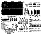
- Panel al shows Nurr1 + (ad) and Nurr1 + / TH + (eh) cells from Nurr1-transduced cultures and ⁇ -gal-stained cells from LacZ-transduced cells (il) over an in vitro differentiation period.
- Representative microscopic images for. Scale bar length is 20 ⁇ m.
- Inset of e-h is an enlarged view of the area indicated by the arrow.
- p-r is the experimental result of the UPS-mediated degradation process of Nurr1 protein.
- Nurr1 + cells (p) and Nurr1 protein concentrations (q) were treated with proteosome inhibitors MG132 or lactacystin (0, 1 and 10 ⁇ M) in the presence of protein synthesis inhibitor CHX (40 ⁇ g / ml).
- CHX protein synthesis inhibitor
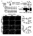
- Figure 2 is an experimental result suggesting that the Raf-MEK and PI3K-Akt intracellular pathways downstream of bFGF have an opposite effect on the regulation of Nurr1 proteolysis.
- bFGF is specific for maintenance of Nurr1 protein stability.
- transfection to Nurr1 in the absence (a, d) or presence of mitogen bFGF (b, e) or EGF (c, f) The induced cortical precursors were incubated for 2 days in the form of suspended cell aggregates (neurospheres), and then Nurr1 protein concentrations were measured (g). Scale bar length is 40 ⁇ m.
- neurospheres treated with various cytokines were plated on FN-coated surfaces and stained for Nurr1.
- Graph h depicts Nurr1 + cell reduction rate for 1 day of in vitro culture.
- Nurr1-transduced precursor cells were either treated with nothing (no tx) or treated with the indicated factors and inhibitors and the reduction ratio was calculated by changing the ratio of Nurr1 + cells before and after 1 day. * significantly different than untreated control at p ⁇ 0.01.
- i-m is data indicating the opposite role of EFK and Akt in regulating Nurr1 protein stability.
- i and j show the changes over time of activated pERK and pAkt proteins for 84 hours after bFGF discontinuation.
- Each point and bar on the graph represents the mean and SEM (relative to time zero) of the protein concentrations obtained from three independent Western blon analyzes.
- k and l are the result of comparing Nurr1 protein concentration in culture treated with FGFR1 blocker (SU5402), Raf-ERK inhibitor (PD98059), dn-raf, or PI3K-Akt inhibitor (LY294002, Waltmannin) to the untreated control. .
- HEK-293 cells transfected with Nurr1 were treated with CHX in the presence or absence of PD98059 or LY294002 and then harvested at the indicated time points and used for western blot assays.
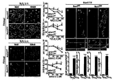
- 3 a shows immunoblotting results indicating direct protein binding of Akt to Nurr1.
- Cortical NP cells transduced with Flag-Nurr1 were immunoprecipitated by incubating with Flag antibodies and immunoblotting with Akt antibodies.
- b is the sequence alignment of the Nurr1 protein around the conservative Akt phosphorylation site.
- c is the result of immunoprecipitation and immunoblotting indicating that Akt phosphorylation is disrupted in the Nurr1 Akt variant protein with serine 347 replaced with alanine.
- Rat cortical NP cells transduced with Flag-Nurr1 WT or Flag-Nurr1 Akt were harvested and immunoprecipitated with Flag antibody, followed by immunoblotting with phospho- (Ser / Thr) Akt substrate antibody.
- d is the IP result indicating that destruction of the Akt phosphorylation site in Nurr1 increases protein stability.
- Nurr1 protein concentrations were compared in cultures transfected with Nurr1 WT and Nurr1 Akt for 6 hours of CHX (100 ⁇ g / ml) treatment.
- e shows a decrease in ubiquitination in the Nurr1 Akt phosphorylation variant.
- IP assays for Ub and Nurr1 protein binding were performed as described in FIG. 1.
- fs are results indicating maintenance of Nurr1 + / TH + DA neurons in cell culture transduced with Nurr1 Akt .
- Panel fq is a representative microscopic image of Nurr1 + (fk) and Nurr1 + / TH + (lq) cells from cultures transduced with Nurr1 WT and Nurr1 Akt through in vitro differentiation period. Scale bar length is 20 ⁇ m. Nurr1 + and TH + cells are shown in graphs r and s, respectively. * significantly different compared to corresponding Nurr1 WT values for the same differentiation day at p ⁇ 0.01.
- Figure 4 shows that NP cells transduced with Nurr1 Akt produce DA neurons that are resistant to toxic stimuli and have an increased in vivo survival after transplantation. al is the result of measuring cytotoxicity induced by H 2 O 2 (50-500 mM) or 6-OHDA (50-200 mM) treatment. After 6 days of in vitro differentiation, cells were treated with toxin for 8 hours and cell viability was measured by MTT assay (i, k), PI staining, and TH + cell direct counting (j, l). Scale bar length is 20 ⁇ m. Nurr1 + and TH + cells are shown in graphs r and s, respectively.
- HBSS Free Hank's Balanced Salt Solution
- the cells were cultured in serum-free N2 medium supplemented with bFGF (20 ng / ml; R & D system) for 4-5 days.
- FGF20 (20 ng / ml), NT3 (10 ng / ml) and BDNF (10 ng / ml; all purchased from Prospec-Tany TechnoGene), MG132 (1-10 mM) and lactacystin (1 -10 mM), SU5402 (20 mM), PD98059 (50 mM), U0126 (10 mM), LY294002 (20 mM), Waltmannin (1 mM; Calbiochem) and Leptomycin B (10 ng / ml; Sigma) Used. Cell cultures were maintained at 37 ° C. in a 5% CO 2 incubator.
- Virus particles were prepared by transfecting each vector with Lipofectamine TM (Invitrogen) in a retrovirus packaging cell line using 293 gpg, and the supernatant containing the virus particles was harvested after 72 hours of incubation.
- Proteins were extracted from the cultures, electrophoresed by SDS-PAGE and transferred to nitrocellulose membranes. The transferred protein was blocked in 5% skim milk in 0.001% Tween20 containing TBS. Primary antibody working concentrations were: Nurr1 (1: 1000, Chemicon), TH (1: 1000, Pel-Freez), ⁇ -galactosidase ( ⁇ -gal) (1: 1000, MP Biomedical), pERK (1: 1000, Cell Signaling), pAkt (Ser473) (1: 1000, Cell Signaling), HA (1: 1000, Covence), pAkt substrate (1: 1000, Sigma), and ⁇ -actin (1: 5000, Abcam).
- NP cells transduced with Flag-tagged Nurr1 WT or Nurr1 Akt were treated with RIPA buffer (50 mM HEPES, 150 mM NaCl, 1% NP40, 1 mM EDTA, 1 mM EGTA, 1 mM phenylmethylsulfonin fluorine) supplemented with protease inhibitors. Reed, 0.5% sodium deoxycholate, 1 mM Na 3 VO 4 ) was harvested.
- RIPA buffer 50 mM HEPES, 150 mM NaCl, 1% NP40, 1 mM EDTA, 1 mM EGTA, 1 mM phenylmethylsulfonin fluorine
- Reed 0.5% sodium deoxycholate, 1 mM Na 3 VO 4
- Cell lysates were incubated with anti-Flag antibody (Sigma: 5 ⁇ g / ml) for 2 hours at room temperature. After binding the antibody to protein G beads for 2 hours, the beads were washed three times with RIPA buffer and resuspended in the same buffer (Amresco). Samples were electrophoresed through SDS 10% polyacrylamide gels and transferred to nitrocellulose membranes and then probed with anti-HA, anti-Akt, or anti-pAkt substrate antibodies.
- anti-Flag antibody Sigma: 5 ⁇ g / ml
- HEK293 cells were transfected with Nurr1 WT (or Nurr1 Akt ) and harvested during treatment with CHX (100 ⁇ g / ml; Calbiochem) for 6 hours.
- Nurr1 protein concentration was measured using Western blot analysis.
- GAPDH (5′-GGCATTGCTCTCAATGACAA-3 ′ and 5′-AGGGCCTCTCTCTTGCTCTC-3 ′, 25 cycles, 60 ° C., 165 bp);
- NP cells were transduced with Nurr1 WT or Nurr1 Akt as described. After 6 days of in vitro differentiation, MTT (Sigma) assay for cell viability in culture treated with H 2 O 2 (50-500 ⁇ M; Sigma) or 6-OHDA (50-200 mM; MP biomedical) for 8 hours. , PI staining (Invitrogen), and direct counting of TH + cells.
- 6-OHDA-damaged rats were produced according to conventional methods (Shim, JW, J. Neurosci. 24, 843-852, 2004). Two days after Nurr1-wild-type or variant transduction, neural progenitor cells were harvested and isolated as single cells in HBSS. Using a 22-gauge needle, 3 ⁇ l of cell suspension was deposited in striatum (coordinates at AP, ML and V for bregma and dura mater: (1) -0.009, -0.42, -0.66) The incisor bar is set to 3.3 mm using a 22-gauge needle The needle was left in place for 10 minutes after each injection For the histological analysis, the animals were rompun (93.28 ⁇ g).
- Example 1 In vitro differentiation and exogenous Nurr1 protein degradation by exogenous Nurr1 protein expression
- Nurr1 expression was induced in cultured NP cells derived from the rat fetal cortex. As previously discovered (Kim, JY et al ., J. Neurochem . 85, 1443-1454 (2003); Park, CH et al ., FASEB J. 20, 2553-2555 (2006); and Park, CH et al ., J. Cell Sci . 119, 2310-2320 (2006)), exogenous Nurr1 expression results in cells positive for tyrosine hydroxylase (TH), an important marker of DA neurons, from primitive non-dopamine reactive NP cells. Produced. Nurr1 immunoreactivity was ubiquitous in the nuclei of virtually all TH + cells by day 4 of differentiation (FIG. 1F).
- TH tyrosine hydroxylase
- Leptomycin B an irreversible inhibitor of CRM-1-dependent export, had no effect on Nurr1 decline (data not shown), suggesting that Nurr1 degrades in the nucleus.
- Nurr1 protein was degraded during cell differentiation, but when cultured in a condition in which bFGF was continuously present, Nurr1 protein concentration was maintained in proliferating NP cells (FIG. 2D, e, g). As these results suggest, it was examined whether Nurr1 protein degradation could be prevented by other mitogens (mitosis promoter) acting on NP cells.
- NP cells Proliferation of NP cells was simply induced by epithelial growth factor (EGF) treatment or activation of Notch signaling via Notch intracellular domain (ICD) transduction (data not shown).
- EGF epithelial growth factor
- ICD Notch intracellular domain
- Nurr1 protein concentrations also did not change significantly with treatment of factors that regulate NP cell differentiation (BDNF, NT3, FGF20, Wnt-5a) and survival (Fas ligand, pan-caspase inhibitor) (FIG. 2H).
- BDNF, NT3, FGF20, Wnt-5a factors that regulate NP cell differentiation
- Fas ligand, pan-caspase inhibitor FIG. 2H
- the maintenance of Nurr1 protein concentration by bFGF was disrupted by treating the cells with SU5402, an FGF receptor blocker (FIG. 2K). Taken together, these results suggest that maintenance of Nurr1 protein is specifically mediated by bFGF.
- Akt phosphorylated Akt
- Nurr1 protein concentration in LY294002 treated medium was stable even after 6 hours of CHX treatment. As a result, it was confirmed that Raf-Erk and Akt signals play an anti-regulatory role in Nurr1 protein degradation.
- Nurr1 protein has an Akt phosphorylation conservation site at Ser 347 (FIG. 3B).
- pAkt substrate antibodies that recognize peptides and proteins phosphorylated at the Akt target motif (RXRXXS / T) readily bind to wild type Nurr1 (Nurr1 WT ), but the Nurr1 variant with serine 347 substituted with alanine ( Nurr1 Akt ) is not readily bound, suggesting that Akt mediates Nurr1 phosphorylation and that this Akt-mediated phosphorylation is missing in the variant.
- TH + cell numbers remained stable in culture transduced with Nurr1 Akt during the in vitro differentiation period.
- the percentage of TH + cells in culture transduced with Nurr1 Akt was Day 4: Differentiation: 66.50 ⁇ 2.68%, Day 8: Differentiation: 74.98 ⁇ 8.64%, Day 12: 74.93 ⁇ 5.92% of differentiation (FIG. 3L) -3q, s).
- DA phenotype genes such as TH and DA transporter DAT was increased in cultures transduced with Nurr1 Akt compared to control cultures (data not shown).
- Nurr1 Akt maintenance of phenotype DA is assumed to be achieved by the subsequent transcriptional activation is the result of keeping Nurr1 Akt protein concentration.
- Nurr1 may have a cell survival effect in TH + cells. Nurr1 has been reported to act as a cell viability factor (Saucedo-Cardenas, O. et al. , 1998; Wallen, A. et al ., 1999; and Sousa, KM et al ., 2007 ).
- Nurr1 protein stability in precursor cells transduced with Nurr1 Akt preserves the DA phenotype and improves cell viability. These events in turn contribute to the maintenance of TH + cells during differentiation.
- FIG. 4S Graft volume
- FIG. 4T total donor cells
- FIG. 4U Nurr1 + cells
- FIG. 4V TH + cells per graft
- the number of TH + cells per graft was 1757 ⁇ 155.4 (Nurr1 Akt ) versus 53.12 ⁇ 20 (Nurr1 WT ).
- Nurr1 Akt overexpression in donor cells improves cell viability and DA phenotype maintenance in transplanted cells.
Landscapes
- Health & Medical Sciences (AREA)
- Engineering & Computer Science (AREA)
- Life Sciences & Earth Sciences (AREA)
- Biomedical Technology (AREA)
- Chemical & Material Sciences (AREA)
- Genetics & Genomics (AREA)
- Organic Chemistry (AREA)
- Zoology (AREA)
- Bioinformatics & Cheminformatics (AREA)
- Wood Science & Technology (AREA)
- Biotechnology (AREA)
- General Health & Medical Sciences (AREA)
- Biochemistry (AREA)
- General Engineering & Computer Science (AREA)
- Molecular Biology (AREA)
- Microbiology (AREA)
- Cell Biology (AREA)
- Physics & Mathematics (AREA)
- Biophysics (AREA)
- Neurology (AREA)
- Gastroenterology & Hepatology (AREA)
- Immunology (AREA)
- Toxicology (AREA)
- High Energy & Nuclear Physics (AREA)
- Medicinal Chemistry (AREA)
- Proteomics, Peptides & Aminoacids (AREA)
- Plant Pathology (AREA)
- Neurosurgery (AREA)
- Micro-Organisms Or Cultivation Processes Thereof (AREA)
- Medicines That Contain Protein Lipid Enzymes And Other Medicines (AREA)
- Preparation Of Compounds By Using Micro-Organisms (AREA)
- Peptides Or Proteins (AREA)
- Medicines Containing Material From Animals Or Micro-Organisms (AREA)
Abstract
본 발명은 Nurr1 단백질의 Akt-인산화 보존부위의 돌연변이에 의하여 Akt-인산화가 유의하게 감소한 Nurr1 변이체를 신경전구세포에서 발현 가능하게 시험관 내 유전자 도입하는 것을 포함하는 도파민 신경세포의 유도방법을 제공한다. 또한, 본 발명은 Nurr1 단백질의 Akt-인산화 보존부위의 돌연변이에 의하여 Akt-인산화가 유의하게 감소한 Nurr1 변이체가 발현 가능하게 유전자도입된 신경전구세포, 및 이 신경전구세포에서 Nurr1 변이체를 발현하여 상기 신경전구세포로부터 유도되는 도파민 신경세포집단을 제공한다. 본 발명에 따르면 신경전구 세포에서 변이체 단백질 발현을 유도하면 Nurr1 단백질 농도가 안정적으로 유지되면서도 천연 Nurr1 기능이 유지되는 도파민 뉴런을 생산한다. 본 발명의 변이체 Nurr1에 의하여 생산되는 도파민 뉴런은 독성 자극에 내성이 있고 래트 뇌에서 이식 후에도 시험관 내 및 생체 내 세포 생존률이 향상된다.
Description
본 발명은 개선된 도파민 신경세포의 유도방법에 관한 것이며, 더 구체적으로는 세포이식에 사용하기 위하여 기능이 개선된 도파민 신경세포를 신경전구세포로부터 유도하는 방법 및 이 방법에 의하여 생산되는 Nurr1 변이체를 발현하여 유도된 도파민 신경세포에 관한 것이다.
중뇌 도파민 (DA) 뉴런은 수의 운동(voluntary movement)의 제어 및 감정 조절에서 중요한 역할을 하는 뉴런이며, 이 뉴런 서브타입의 퇴행/장애는, 예를 들어 파킨슨씨 병과 같은 많은 신경학적 및 정신적 이상의 임상 상태의 원인이 된다.
고아 핵 리셉터 패밀리(orphan nuclear receptor family)에 속하는 전사인자인 Nurr1 (NR4A2)은 발생하는 중뇌에서 발현되며 중뇌 도파민 뉴런 발생에 결정적인 인자이다(Zetterstrom, R.H. et al., Science 276, 248-250, 1997); 및 Saucedo-Cardenas, O. et al., Proc. Natl. Acad. Sci. USA 95, 4013-4018, 1998) 참조). Nurr1은 또한 발생하는 중뇌 뿐 아니라 성체의 중뇌 도파민 뉴런에서도 발현되는데, 이 전사인자의 지속적 발현은 도파민 반응성(dopaminergic) 표현형의 유지(상기 Zetterstrom, R.H. et al. 1997; 및 Eells, J.B., et al., Behav. Brain Res. 136, 267-275 (2002)) 및 생존(상기 Saucedo-Cardenas, O. et al.; Wallen, A. et al., Exp. Cell Res. 253, 737-746 (1999); 및 Sousa, K.M. et al.,
Stem cells 25, 511-519 (2007))에 결정적이라고 보고되었다. 성체 중뇌 도파민 병리 상태에서 Nurr1 의 농도 감소나 유전적 변형이 관찰되었다(Jankovic, J. et al., Prog. Neurobiol. 77, 128-138 (2005); 및 Chu, Y. et al., J. Comp. Neurol. 494, 495-514 (2006)).
신경퇴행성 장애를 치료하기 위하여 발생 중인 뇌나 성체 뇌로부터 신경전구(NP)세포를 분리하여 다수의 공여 세포를 생산할 목적으로 배양하려는 시도가 있어 왔다. 이와 같이 배양된 신경전구세포로부터 도파민 뉴런을 유도함에 있어서 Nurr1의 시험관 내 기능 때문에 Nurr1에 대한 관심이 증대되어 왔다. 즉, 외부적으로 Nurr1을 발현시키는 경우, 원시(naive) 비-도파민 반응성(dopaminergic) 신경전구세포가, 파킨슨씨 병(PD)의 설치 동물 모델에서 도파민 반응성 결손으로부터 회복할 수 있는 시냅스전(pre-synaptic) 작용특성을 나타내는 DA 뉴런으로 분화하도록 유도된다(Shim, J.W. et al., Stem cells 25, 1252-1262 (2007)).
전술한 바와 같이, 신경퇴행성 장애 치료를 위하여 신경전구세포를 분리하여 도파민 뉴런으로 유도하여 배양, 이식하는 치료적 접근방법 시도에서 드러난 가장 결정적인 문제는 이식 후 공여 세포의 세포 생존률이 낮고 DA 표현형이 상실된다는 점이다.
현재 사용하는 파킨슨씨병(PD) 치료를 위하여 세포 이식방법을 사용한 예에서 이식 후에 도파민 뉴런의 극히 일부만이 살아남는 것으로 나타났다(상기 Shim, J.W. et al. 2007, Arenas, E., Ann. N Y Acad. Sci.
1049, 51-66, 2005). 게다가, 최근 연구에 의하면 PD 질병상태 환경이 이식된 뉴런에 독성 신호를 전달한다는 것을 밝혔는데(Li, J.Y. et al., Nat. Med. 14, 501-503, 2008), 이 연구결과는 이식된 뉴런의 생존율을 증가시키기 위하여는 숙주 환경의 개선이 필요할 것이라는 점을 시사한다. 그러나, 질병을 치료하는 방법 외에 질병 상태에 의하여 발생되는 환경을 근본적으로 교정하는 것은 용이한 것이 아니다.
따라서, 신경퇴행성 질병의 도파민 세포이식치료를 위하여는 공여세포 생존율이 높고, 도파민 뉴런 표현형이 유지되며 세포 독성에도 내성을 가지는 도파민 뉴런 유도방법의 개발이 절실하다.
본 발명자들은 성체 중뇌 DA 병리상태에서 Nurr1 단백질 농도나 기능의 조작을 통하여 잠재적인 치료 전략이 수립될 수 있다는 가정에서 출발하여 Nurr1 단백질 분해 기작에 관여하는 신호 조절에 초점을 맞추어 진행된 연구 결과를 이용하여 독성 환경에 내성이 있는 이식된 세포를 제공하여 본 발명을 완성하였다.
본 발명은 Nurr1 단백질의 Akt-인산화 보존부위의 돌연변이에 의하여 Akt-인산화가 유의하게 감소한 Nurr1 변이체를 신경전구세포에서 발현 가능하게 시험관 내 유전자 도입하는 것을 포함하는 도파민 신경세포의 유도방법을 제공한다.
또한 본 발명에서는 Nurr1 단백질의 Akt-인산화 보존부위의 돌연변이에 의하여 Akt-인산화가 유의하게 감소한 Nurr1 변이체가 발현 가능하게 유전자 도입된 신경전구세포를 제공하는 한편, 이를 이용하여 Nurr1 단백질의 Akt-인산화 보존부위의 돌연변이에 의하여 Akt-인산화가 유의하게 감소한 Nurr1 변이체를 신경전구세포에서 발현하여 상기 신경전구세포로부터 유도되는 도파민 신경세포집단을 제공한다.
하기 실시예에서도 입증하는 바와 같이, 본 발명자들은 전구세포 분화과정 동안 Nurr1 단백질은 UPS-매개 분해를 겪으며 이 과정은 ERK 및 Akt 세포 내 신호의 반대 작용에 의하여 조절된다는 점을 밝혔다. 프로테오솜 분해 시스템은 세포 내 단백질 활성의 결정적 조절자이고, 분해될 다양한 세포 단백질 76-아미노산 폴리펩티드인 유비퀴틴의 공유결합에 의해 프로테오솜으로 표적화된다.
본 발명자들은 게다가 Nurr1 단백질의 직접 Akt 인산화가 이 단백질의 유비퀴틴화 초기 단계를 제어한다는 것을 밝혔다. 이 발견은 새로운 것이며, IκBα(Brockman, J.A. et al., Mol Cell Biol. 15, 2809-2818, 1995), p27Kip 1(Ganoth, D. et al., Nat. Cell biol. 3, 321-324, 2001), 및 시클린 E(Koepp, D.M. et al., Science 294, 173-177, 2001)의 유비퀴틴화 및 분해의 단백질 인산화-매개된 제어를 증명한 이전 연구 보고들에 의하여 뒷받침된다. 게다가, Nurr1이 속한 동일한 핵-호르몬 리셉터 패밀리 단백질의 맴버인 안드로겐 리셉터의 직접 Akt-매개된 인산화가, 유비퀴틴화의 속도결정 단계를 매개하는 활성인 E3 리가제 활성을 저해하는 것으로 밝혀졌다(Lin, H.K. 등, EMBO J. 21, 4037-4048, 2002). 현재까지의 지식은 핵이 핵 리셉터의 분해의 일차적 표적이 된다는 생각을 뒷받침한다(Alarid, E.T., Mol. Endocrinol. 20, 1972-1981, 2006). 이와 같은 이론과 일치되게, 핵 이출(export) 저해제인 렙토마이신 B로 처리하는 것으로는 Nurr1 분해에 영향이 없었는데, 이는 Nurr1-특이적 UPS- 매개된 분해가 핵에 편재된다는 것을 시사한다.
생존의 중요한 사항들 뿐 아니라, 이식된 DA 뉴런의 불안정한 표현형(상기 Paul, G. et al., 2007) 또한 이식 후 DA 뉴런의 낮은 수득률에 기여한다. 본 발명자는 이식 후 세포 생존율이 개선되고 시험관 내 및 생체 내 더 양호한 표현형 유지를 나타내는 공여체 DA 세포를 생산하는 유전적 조작의 예를 제시한다. 본 발명에서 Nurr1의 Akt-인산화 부위의 단일 돌연변이가 Nurr1 단백질 안정성에, 그에 따라 TH+ 세포의 유지에도 극적인 효과를 가져왔다. Nurr1Akt로 트랜스덕션된 세포에서 Nurr1의 계속적인 발현 때문에 DA 표현형이 유지되는 효과가 관찰된다는 것은 분명하다(데이터는 나타내지 않음). 왜냐하면, Nurr1은 DA 표현형에 관련된 유전자의 발현을 활성화하는 전사 인자이기 때문이다(상기 Zetterstrom, R.H. et al., 1997; 상기 Saucedo-Cardenas, O. et al., 1998; 및 상기 Jankovic, J., 2005). Nurr1Akt로 트랜스덕션된 전구세포로부터 유도된 TH+ 세포의 대다수는 분화의 오랜 기간 동안 Nurr1을 발현하였다. 이전의 연구 결과들(Jiang, C. et al., Exp. Neurol. 191, 154-162 (2005))과 일치하게, Nurr1-발현 DA 세포는 그들의 DA 표현형을 더 양호하게 유지하고 더 장기간 생존하였다(도 3). 게다가, 변이체 Nurr1에 의하여 생산된 DA 뉴런은 독성 자극에 대하여 더 내성을 가졌다(도 4). 이들 결과는, 이식 후 표현형을 유지할 수 있고 PD의 병적인 숙주 환경을 극복함으로써 생존할 수 있는 공여체 DA 세포를 생산하는 것을 통하여 PD에 대한 세포 치료적 접근방법에서 진전을 이룰 수 있음을 시사한다. Nurr1은 PD에서 민감한 인자이고 PD 환자에서 Nurr1 농도가 감소하는 것이 관찰되었는데, 이는 Nurr1이 PD의 치료 및 예방의 표적 분자임을 시사한다. Nurr1 단백질 분해 및 안정성에 대한 본 발명에 따르는 정보는 PD의 진행을 막기 위한 의학적 치료법 개발에 유용할 것이다.
따라서, 본 발명의 한 양태에서는 Nurr1 단백질의 Akt-인산화 보존부위의 돌연변이에 의하여 Akt-인산화가 유의하게 감소한 Nurr1 변이체를 신경전구세포에서 발현 가능하게 시험관 내 유전자 도입하는 것을 포함하는 도파민 신경세포의 유도방법을 제공한다. Nurr1 단백질의 Akt-인산화 보존부위의 돌연변이는 천연 돌연변이 또는 인공적으로 유발된 돌연변이 모두를 포함한다. 천연 돌연변이는 cDNA 라이브러리로부터 얻을 수 있으며, 인공적으로 유발된 돌연변이는 당업자에게 공지인 어떤 통상적인 방법에 의하여도 쉽게 제조될 수이다.
본 발명의 다른 한 양태에서는 상기 Akt-인산화 감소에 의하여 Nurr1의 유비퀴틴화에 의한 세포 내 분해가 유의하게 감소하는 것을 특징으로 하는 도파민 신경세포의 유도방법을 제공한다. 본 발명의 유도방법의 실시를 위한 Akt-인산화 보존부위는 Ser 347 및 상동 단백질에서 이에 대응하는 부위에 위치하는 것이 바람직하지만 이에 제한되지 않는다. Akt-매개 신호를 감소시킬 수 있는 어떤 형태의 Nurr1 단백질 상의 변형도 본 발명의 범위 안에 있다는 것이 당업자에게 명백하다. 이 양태에서 Nurr1 단백질의 Akt-인산화 보존부위의 돌연변이는 이 부위에서 아미노산 잔기의 삽입, 결실, 치환 등을 포함할 수 있으나, 바람직하게는 Akt-인산화 보존부위에 존재하는 돌연변이는 인산화 기능을 제외하면 Nurr1 단백질의 다른 구조나 기능에는 영향을 미치지 않는다. 따라서, Akt-인산화 보존부위에 존재하는 돌연변이는 위치 347의 Ser이 유사한 특성을 가지는 아미노산 잔기로 단일 치환된 것일 수 있으며, Ser과 유사한 부피 범위 및 소수성을 갖는 아미노산 잔기일 수 있다. 바람직하게는, 347의 Ser이 Ala, Gly, Thr 및 Met로 구성된 군으로부터 선택되는 아미노산 잔기로 치환된 것일 수 있다.
또 다른 양태에서, 본 발명은 Nurr1 단백질의 Akt-인산화 보존부위의 돌연변이에 의하여 Akt-인산화가 유의하게 감소한 Nurr1 변이체를 포함하는 도파민 신경세포 유도 조성물을 제공한다. 상기 Akt-인산화 보존부위는 Ser 347 및 상동 단백질에서 이에 대응하는 부위에 위치할 수 있으며, 바람직하게는 Akt-인산화 보존부위의 돌연변이는 위치 347의 Ser이 Ala, Gly, Thr 및 Met로 구성된 군으로부터 선택된다. 이와 같은 본 구체예에 따르는 도파민 신경세포 유도 조성물은 상기 도파민 신경세포의 유도방법에 따라 신경전구세포에 트랜스펙션시키는 방법 등의 유전자 전달 방법에 따라 신경전구세포에서 Akt-인산화가 유의하게 감소한 Nurr1 변이체를 발현하여 도파민 신경세포를 유도하는 유전자 치료에 유용하게 사용할 수 있다. 이 구체예에서 제공하는 도파민 신경세포 유도 조성물을 유전자 치료는 도파민 신경세포를 필요로 하는 어떠한 병리학적 상태에 대하여도 사용될 수 있다. 이와 같은 도파민 신경세포를 필요로 하는 상태는 어떠한 신경퇴행성 질환도 포함할 수 있으며, 파킨슨씨 병, 알쯔하이머병(노인성치매), 탈수초질환(demyelinating disease), ALS(amyotrophic lateral sclerosis) 및 척추손상에 의한 사지마비와 같은 뇌신경퇴행 질환을 포함하지만 이에 제한되지 않는다.
본 발명의 또 다른 양태에서는 Nurr1 단백질의 Akt-인산화 보존부위의 돌연변이에 의하여 Akt-인산화가 유의하게 감소한 Nurr1 변이체가 발현 가능하게 유전자도입된 신경전구세포를 제공한다. 본 구체예에서 사용하는 변이체를 구성하기 위하여 이용되는 Akt-인산화 보존부위는 Ser 347 및 상동 단백질에서 이에 대응하는 부위에 위치인 것이 바람직하지만 이에 제한되지 않는다. Akt-매개 신호를 감소시킬 수 있는 어떤 형태의 Nurr1 단백질 상의 변형도 본 발명의 범위 안에 있다는 것이 당업자에게 명백하다. 본 구체예에서 Nurr1 단백질의 Akt-인산화 보존부위의 돌연변이는 이 부위에서 아미노산 잔기의 삽입, 결실, 치환 등을 포함할 수 있으나, 바람직하게는 Akt-인산화 보존부위에 존재하는 돌연변이는 인산화 기능을 제외하면 Nurr1 단백질의 다른 구조나 기능에는 영향을 미치지 않는다. 따라서, Akt-인산화 보존부위에 존재하는 돌연변이는 위치 347의 Ser이 유사한 특성을 가지는 아미노산 잔기로 단일 치환된 것일 수 있으며, Ser과 유사한 부피 범위 및 소수성을 갖는 아미노산 잔기일 수 있다. 바람직하게는, 347의 Ser이 Ala, Gly, Thr 및 Met로 구성된 군으로부터 선택되는 아미노산 잔기로 치환된 것일 수 있다.
본 발명의 다른 양태에서는 Nurr1 단백질의 Akt-인산화 보존부위의 돌연변이에 의하여 Akt-인산화가 유의하게 감소한 Nurr1 변이체를 신경전구세포에서 발현하여 상기 신경전구세포로부터 유도되는 도파민 신경세포집단을 제공한다. 본 구체예에 따르는 신경세포집단은 하기 구체예들에서 제공하는 바와 같은 세포이식을 통하여 세포치료에 사용될 수 있다. 따라서, 본 발명의 또 다른 양태에서는 상기 도파민 신경세포집단을 포함하는 신경퇴행 질환 치료용 조성물을 제공한다. 이 구체예에서 신경퇴행 질환은 파킨슨씨 병, 알쯔하이머병(노인성치매), 탈수초질환(demyelinating disease), ALS(amyotrophic lateral sclerosis) 및 척추손상에 의한 사지마비와 같은 뇌신경퇴행 질환을 포함하지만 이에 제한되지 않는다.
또 다른 양태에서 본 발명은 Nurr1 단백질의 Akt-인산화 보존부위의 돌연변이에 의하여 Akt-인산화가 유의하게 감소한 Nurr1 변이체를 신경전구세포에 유전자도입하는 단계; 및 유전자도입된 신경전구세포를 환자에게 이식하여 환자에서 도파민 신경세포를 유도하는 단계를 포함하는 도파민 신경세포가 필요한 환자에서 도파민 신경세포 생성을 유도하는 방법을 제공한다. 이 구체예는 도파민 신경세포의 전반적 또는 일부 손상으로 인하여 신경세포의 공급이 필요한 다양한 병리 상태에 대하여 폭넓게 적용할 수 있다. 본 발명의 Nurr1 단백질의 Akt-인산화 보존부위의 돌연변이에 의하여 Akt-인산화가 유의하게 감소한 Nurr1 변이체를 유전자 도입한 신경전구세포를 환부에 이식하여 그 분화가 유도되도록 유지함에 의하여 실시할 수 있다. 이식은 세포 이식에서 사용하는 어떠한 공지의 방법에 의하여도 수행할 수 있다(Shim,J.W., J. Neurosci. 24, 843-852, 2004). 요약하면, Nurr1 변이체 유전자 도입 후 신경전구세포를 수확하여 단일세포로 분리한 후 주사를 사용하여 환부(예를 들면, 선조체)에 침적시켜 수행한다. 본 구체예에 따르는 도파민 신경세포 생성 유도 방법이 적용될 수 있는 다양한 병리 상태는 신경퇴행 질환을 포함하는 다수의 신경계 질환을 포함한다. 파킨슨씨 병, 알쯔하이머병(노인성치매), 탈수초질환(demyelinating disease), ALS(amyotrophic lateral sclerosis) 및 척추손상에 의한 사지마비와 같은 뇌신경퇴행 질환을 포함하지만 이에 제한되지 않는다.
본 발명자들은 분화하는 신경전구 세포에서 Nurr1 단백질은 유비퀴틴-프로테오솜-시스템(UPS)-매개 분해를 받는 것을 밝혔다. 단백질 분해를 유발하는 세포 내 신호는 밝혀져 있고, 분해 과정의 이와 같은 분자 수준의 이해를 토대로 본 발명은 유비퀴틴화-내성 Nurr1 변이체를 제공한다. 신경전구 세포에서 변이체 단백질 발현을 유도하면 Nurr1 단백질 농도가 안정적으로 유지되면서도 천연 Nurr1 기능이 유지되는 도파민 뉴런을 생산한다. 본 발명의 변이체 Nurr1에 의하여 생산되는 DA 뉴런은 독성 자극에 내성이 있고 래트 뇌에서 이식 후에도 시험관 내 및 생체 내 세포 생존률이 향상되는 것으로 나타났다. 본 발명은 PD 세포 치료 용도의 줄기세포/전구세포-유도된 DA 뉴런 생산에서의 실질적인 기술의 진보를 가져왔을 뿐 아니라, 파킨슨씨병 진행을 막는 치료법 개발에 중요한 시사점을 제공한다.
도 1은 신경전구세포로부터 도파민 뉴런 분화과정 동안 Nurr1 단백질의 농도 및 분해 과정에 대한 실험 결과이다.
패널 a-l은 시험관 내 분화 기간 동안에 걸쳐 Nurr1-트랜스덕션된 배양물로부터의 Nurr1+(a-d) 및 Nurr1+/TH+ (e-h) 세포, 및 LacZ-트랜스덕션된 세포로부터의 β-gal-염색된 세포(i-l)에 대한 대표적인 현미경적 영상을 도시한다. 눈금 막대 길이는 20 ㎛임. e-h의 삽입도는 화살로 표시한 부분에 대한 확대도.
Nurr1+, TH+, 및 β-gal+ 세포 비율의 변화를 그래프 m에 도시한다. p<0.01에서 분화 제 0 일 (Diff 0) 와 Diff 4의 값이 유의하게 차이가 남. 분화 제 0 일, 제 3 일 및 제 8일에서 Nurr1 단백질(n) 및 mRNA (o) 농도를 더 측정하였다.
p-r은 Nurr1 단백질의 UPS-매개된 분해 과정에 관한 실험 결과이다. 분화 제 6 일에 단백질 합성 저해제 CHX(40 ㎍/ml)의 존재 하에서 Nurr1+ 세포(p) 및 Nurr1 단백질 농도(q)를 프로테오솜 저해제 MG132 또는 락타시스틴(0, 1 및 10 μM)로 처리된 피질 전구세포에서 측정하였다. r은 Ub와 Nurr1 단백질 결합에 대한 IP 검정결과이다. * p<0.01에서 미처리 배양물과 유의하게 상이함.
도 2는 bFGF의 Raf-MEK 와 PI3K-Akt 세포 내 경로 하류는 Nurr1 단백질 분해의 조절에서 반대 작용을 한다는 점을 시사하는 실험 결과이다.
a-h 결과는 bFGF가 Nurr1 단백질 안정성의 유지에 특이적임을 나타낸다. Nurr1 단백질 안정성에 영향을 줄 수도 있는, 세포-세포 접촉 수준을 유사하게 유지하기 위하여, 미토겐 bFGF(b, e) 또는 EGF (c, f)의 부재(a, d)또는 존재 하에서 Nurr1으로 트랜스덕션된 피질 전구체를 부유 세포 응집물(뉴로스피어) 형태로 2일 동안 배양한 후, Nurr1 단백질 농도를 측정하였다(g). 눈금 막대 길이는 40 ㎛임. 또한, 여러 가지 시토킨을 처리한 뉴로스피어를 FN-코팅된 표면 상에 플레이팅하였고 Nurr1 에 대하여 염색하였다. 그래프 h는 시험관 내 배양물의 1일 동안 Nurr1+ 세포 감소 비율을 도시한다. Nurr1-트랜스덕션된 전구체 세포는 아무 것도 처리하지 않거나(no tx) 또는 표시한 인자와 저해제로 처리하고 처리 전 및 1일 경과 후 Nurr1+ 세포의 비율 변화로 감소비율을 계산하였다. * p<0.01에서 미처리 대조군에 비하여 유의하게 상이함.
i-m은 Nurr1 단백질 안정성 조절에 있어서 EFK 및 Akt의 상반된 역할을 나타내는 데이터이다. i 및 j는 bFGF 투여 중단 후 84 시간 동안 활성화된 pERK 및 pAkt 단백질의 시간 경과에 따르는 변화를 나타낸다. 그래프 상의 각 점 및 막대는 세 차례 독립적인 웨스턴 블론 분석으로부터 얻은 단백질 농도의 (시점 0과 비교한 상대적인) 평균 및 SEM을 나타낸다. k 및 l은 FGFR1 차단제(SU5402), Raf-ERK 저해제(PD98059), dn-raf, 또는 PI3K-Akt 저해제(LY294002, 월트마닌)을 처리한 배양물 내 Nurr1 단백질 농도를 미처리 대조군과 비교한 결과이다. m은 Nurr1 단백질 안정성에 대한 Raf-ERK 및 PI3K-Akt 신호의 효과를 나타낸다. Nurr1으로 트랜스펙션된 HEK-293 세포를 PD98059 또는 LY294002 존재 또는 부재 하에서 CHX로 처리한 후 표시된 시점에 수확하여 웨스턴 블롯 검정에 사용하였다.
도 3의 a는 Nurr1 에 대한 Akt의 직접 단백질 결합을 나타내는 면역블롯팅 결과이다. Flag-Nurr1으로 트랜스덕션된 피질 NP 세포를 Flag 항체와 함께 인큐베이션함으로써 면역침강 수행하고 Akt 항체로 면역블롯팅을 수행하였다. b는 보존적 Akt 인산화 부위 주위의 Nurr1 단백질의 서열 얼라인먼트이다. c는 세린 347이 알라닌으로 바뀐 Nurr1Akt 변이체 단백질에서 Akt 인산화가 파괴됨을 나타내는 면역침강 및 면역블롯팅 결과이다. Flag-Nurr1WT 또는 Flag-Nurr1Akt로 트랜스덕션 된 래트 피질 NP 세포를 수확하여 Flag 항체로 면역침강한 후, 포스포-(Ser/Thr)Akt 기질 항체에 의하여 면역블롯팅을 수행하였다. d는 Nurr1에서 Akt 인산화 부위의 파괴가 단백질 안정성을 증가시킴을 나타내는 IP 결과이다. Nurr1 단백질 농도를 CHX(100 ㎍/ml) 처리 6 시간 동안 Nurr1WT 및 Nurr1Akt로 트랜스펙션시킨 배양물에서 비교하였다. e는 Nurr1 Akt 인산화 변이체에서 유비퀴틴화의 감소를 나타낸다. 도 1에 기재된 바와 같이 Ub와 Nurr1 단백질 결합에 대한 IP 검정을 수행하였다. f-s는 Nurr1Akt로 트랜스덕션된 세포 배양물 내 Nurr1+/TH+ DA 뉴런의 유지를 나타내는 결과들이다. 패널 f-q는 시험관 내 분화 기간을 통하여 Nurr1WT 및 Nurr1Akt로 트랜스덕션된 배양물로부터의 Nurr1+(f-k) 및 Nurr1+/TH+(l-q) 세포에 대한 대표적인 현미경 영상이다. 눈금 막대 길이는 20 ㎛임. Nurr1+ 및 TH+ 세포를 그래프 r 및 s에 각각 도시한다. * p<0.01에서 동일한 분화 일에 대한 대응하는 Nurr1WT 값에 비하여 유의하게 상이함.
도 4는 Nurr1Akt로 트랜스덕션된 NP 세포는 독성 자극에 내성이 있고 이식 후 생체 내 생존율이 증가된 DA 뉴런을 생산한다는 것을 나타내는 결과이다. a-l은 H2O2 (50-500 mM) 또는 6-OHDA (50-200 mM) 처리로 유도된 세포독성을 측정한 결과들이다. 시험관 내 분화 6일 후, 세포를 8 시간 동안 독소로 처리하고 세포 생존율을 MTT 검정(i, k), PI 염색, 및 TH+ 세포 직접 계수에 의하여 측정하였다(j,l). 눈금 막대 길이는 20 ㎛임. Nurr1+ 및 TH+ 세포를 그래프 r 및 s에 각각 도시한다. * p<0.01에서 동일한 독소 농도에 대한 대응하는 Nurr1WT 값에 비하여 유의하게 상이함. 패널 a-d 및 e-h에 나타낸 것은 각각 TH+ 세포 및 PI-염색 세포의 대표적인 영상인데, 이는 Nurr1WT 및 Nurr1Akt로 트랜스덕션된 배양물에서 H2O2-유도된 세포 독성의 차이를 나타낸다. m-v, 이식 후 TH+/Nurr1+ 세포의 생체 내 생존율. Nurr1WT(m-o) 및 Nurr1Akt(p-r)로 트랜스덕션된 전구세포에 의하여 생산된 이식편에서 TH+(n, q), Nurr1+(m, p), 및 TH+/Nurr1+(o, r) 세포에 대한 대표적 영상. 이색편 내 이식편 부피(s), 전체 공여체 세포(t), Nurr1+(u), 및 TH+(v) 세포 수의 정량. 눈금 막대 길이는 20 ㎛임.
하기 실시예에서 사용된 재료와 실험 방법은 다음과 같다.
1. 신경전구세포의 일차 배양
배발생 제 13 일(E13)에 래트(Spraque Dawley) 배아 피질로부터 뇌 조직을 절개하였다. 절개된 피질을 Ca++/Mg++ 불포함 Hank's 균형 염 용액(HBSS; Gibco) 중에서 기계적으로 빻은 후, 19,000 세포/㎠ 밀도로 폴리오르니틴/피브로넥틴으로 사전코팅된 10 cm 배양 디쉬(Corning) 상에 접종한 후, 4 내지 5일 동안 bFGF(20 ng/ml; R&D system) 보충된 무혈청 N2 배지에서 배양하였다. 전구체 세포 증식에 의하여 생산된 세포 클러스터를 HBSS에서 분리시킨 후 신선하게 코팅된 24-웰 및 6-웰 플레이트 상에 50,000 세포/㎠ 밀도로 플레이팅하였다. N2+bFGF에서 60-80% 컨플루언시로 전구세포 증식을 추가적으로 유도한 후(전형적으로 플레이팅 1-2 일 후), 이 세포를 대상으로 후술하는 레트로바이러스 트랜스덕션을 수행하였다. 트랜스덕션 수행한 다음 날, bFGF를 4-12일 동안 투여 중지함으로써 세포 분화를 유도하였다. 배지는 매 2일마다 교환하였고, 투여 중지한 경우를 제외하고는 bFGF는 매일 보충하였다. 몇몇 실험에서는 bFGF 또는 EGF(20 ng/ml; R&D system)으로 보충된 배지 내 코팅되지 않은 표면 상에 접종함으로써 전구체 세포를 부유 세포 응집체 형태(뉴로스피어)로 배양하였다. 다음의 인자 또는 저해제: FGF20 (20 ng/ml), NT3 (10 ng/ml) 및 BDNF (10 ng/ml; 모두 Prospec-Tany TechnoGene로부터 구입), MG132 (1-10 mM) 및 락타시스틴 (1-10 mM), SU5402 (20 mM), PD98059 (50 mM), U0126 (10 mM), LY294002 (20 mM), 월트마닌 (1 mM; Calbiochem) 및 렙토마이신 B (10 ng/ml; Sigma)를 사용하였다. 세포 배양물은 5% CO2 배양기에서 37℃로 유지하였다.
2. 레트로바이러스 구성체 제작 및 감염 방법
각 cDNA 단편을 pCL(상기, Park, C.H. et al., 2006)의 멀티클로닝 부위 내로 삽입함으로써 Flag-태그 Nurr1WT, Nurr1Akt, dn-raf(대한민국, 서울, 연세대학교 최강열 박사가 제공함), Wnt5a, 및 Notch-ICD(대한민국, 서울, 연세대학교 김재상 박사가 제공함)를 발현하는 레트로바이러스 벡터를 제작하였다. 레트로바이러스 패키징 세포주 293gpg에 각 벡터를 LipofectamineTM(Invitrogen)을 사용하여 트랜스펙션시킴으로써 바이러스 입자를 제조하고, 72 시간 배양 후 바이러스 입자를 함유하는 상청액을 수확하였다. 바이러스 트랜스덕션을 위하여 세포를 폴리브렌(1 ㎍/ml; Sigma-Aldrich)을 함유하는 바이러스 수프와 함께 2시간 인큐베이션한 후 bFGF-보충된 N2로 배지를 교환하였다. 세포를 각 바이러스 구성체 혼합물로 감염시킴으로써 공동발현 연구를 수행하였다.
3. 면역형광 염색
배양된 세포 및 뇌 조직을 4% 파라포름알데히드(PFA)로 고정한 후, 0.1% BSA/10% 염소혈청/0.3% Triton X-100에서 차단하고 일차 항체와 함께 4 ℃에서 하룻밤 동안 인큐베이션하였다. 뇌 절편에 이식된 Nurr1을 발현하는 세포를 검출하기 위하여, 세포를 차단 과정 전에 소듐 도데실 설페이트(PBS 중 1%)로 실온에서 5 분 동안 처리함으로써 항원 검색방법을 사용하였다. 다음의 일차항체: Nurr1 (1:200, Chemicon, 배양된 세포 용으로, 또는 1:500, E-20, SantaCruz, 조직 내 이식된 세포 검출 용으로), 및 TH(1:250, Pel-Freez)를 사용하였다. Alexa 488-(1:200, Invitrogen) 및 Cy3-(1:200, Jackson Immunoresearch Laboratories)로 표지된 이차항체를 사용하였고 4,6-디아미디노-2-페닐인돌을 함유하는 VECTASHIELDR(DAPI, Vector Laboratories INC.)에 올려놓았다. 형광현미경(epifluorescence microscope (NiKon)) 또는 공초점 현미경(Leica) 아래에서 면역반응성 세포를 분석하였다.
4. 웨스턴 블롯 및 IP 검정
배양물로부터 단백질을 추출하고 SDS-PAGE에 의하여 전기영동한 후 니트로셀룰로스 멤브레인으로 옮겼다. 옮겨진 단백질을 TBS를 함유하는 0.001% Tween20 중의 5% 탈지 우유에서 차단하였다. 일차항체 작동 농도는 다음과 같았다: Nurr1 (1:1000, Chemicon), TH (1:1000, Pel-Freez), β-갈락토시다제 (β-gal) (1:1000, MP Biomedical), pERK (1:1000, Cell Signaling), pAkt (Ser473) (1:1000, Cell Signaling), HA (1:1000, Covence), pAkt 기질 (1:1000, Sigma), 및 β-액틴 (1:5000, Abcam). 퍼옥시다제와 접합된 이차 항-래빗 또는 항-마우스 IgG 항체(1:2000, Cell Signaling)를 사용하였다. 증강된 화학발광에 의하여(ECL 검출 키트; Welgene) 밴드를 가시화하였다. Nurr1WT 또는 Nurr1Akt의 후보 분자와의 물리적 단백질 결합 및 Nurr1 단백질의 pAkt-매개된 인산화를 IP 검정을 사용하여 측정하였다. Flag-태그된 Nurr1WT 또는 Nurr1Akt로 트랜스덕션된 NP 세포를, 프로테아제 저해제가 보충된 RIPA 완충액(50 mM HEPES, 150 mM NaCl, 1% NP40, 1mM EDTA, 1 mM EGTA, 1mM 페닐메틸술포닌 플루오리드, 0.5% 소듐 데옥시콜레이트, 1 mM Na3VO4)을 사용하여 수확하였다. Nurr1 단백질의 유비퀴틴화를 시험하기 위하여, HEK293 세포를 HA-태그된 유비퀴틴 및 Flag-Nurr1WT(또는 Nurr1Akt)로 공동-트랜스펙션시켰다. 세포 용해물을 항-Flag 항체(Sigma: 5 ㎍/ml)와 함께 2 시간 동안 실온에서 인큐베이션시켰다. 항체를 단백질 G 비드와 2 시간 동안 결합시킨 후, 비드를 RIPA 완충액을 사용하여 세 차례 세척하고 동일한 완충액(Amresco)에서 재현탁하였다. 샘플을 SDS 10% 폴리아크릴아미드 겔을 통하여 전기영동하고 니트로셀룰로스 멤브레인에 옮긴 후, 항-HA, 항-Akt, 또는 항-pAkt 기질 항체로 프로빙시켰다.
5. Nurr1 단백질 안정성 측정
HEK293 세포를 Nurr1WT(또는 Nurr1Akt)로 트랜스펙션시키고 6 시간 동안 CHX(100 ㎍/ml; Calbiochem)로 처리하는 동안 수확하였다. Nurr1 단백질 농도를 웨스턴블롯 분석을 사용하여 측정하였다.
6. RT-PCR
표준 RT-PCR 방법을 사용하였다. 각 프라이머 세트에 대한 최적 PCR 조건은 직선적 증폭 범위를 결정하기 위하여 MgCl2 농도, 어닐링 온도, 및 사이클 수를 변화시켜 측정하였다. (순방향 및 역방향) 프라이머 서열 및 PCR 조건은 다음과 같다:
GAPDH (5'-GGCATTGCTCTCAATGACAA-3' 및 5'-AGGGCCTCTCTCTTGCTCTC-3', 25 사이클, 60 ℃, 165 bp); 및
Nurr1 (5'-TGAAGAGAGCGGACAAGGAGATC-3' 및 5'-TCTGGAGTTAAGAAATCGGAGCTG-3', 35 사이클, 57 ℃, 255 bp)
7. 세포독성 검정
기재한 바와 같이 NP 세포를 Nurr1WT 또는 Nurr1Akt로 트랜스덕션시켰다. 시험관 내 분화 6 일 경과 후, H2O2 (50-500 μM; Sigma) 또는 6-OHDA (50-200 mM; MP biomedical)로 8 시간 동안 처리한 배양물 내 세포 생존율을 MTT(Sigma) 검정, PI 염색(Invitrogen), 및 TH+ 세포의 직접계수에 의하여 측정하였다.
8. 생체 내 이식
종래 방법에 따라(Shim, J.W., J. Neurosci. 24, 843-852, 2004) 6-OHDA-손상된 래트를 생산하였다. Nurr1-야생형 또는 변이체 트랜스덕션 2일 후 신경전구세포를 수확하고 HBSS에서 단일 세포로 분리하였다. 22-게이지 주사바늘을 사용하여, 3 ㎕의세포 현탁물을 선조체에서 침적시켰다(브레그마(bregma) 및 경질막에 대한 AP, ML 및 V에서 좌표: (1) -0.009, -0.42, -0.66 인사이서 바(incisor bar)는 22-게이지 니들을 사용하여 3.3 mm로 설정한다. 각 주사 후 10 분 동안 주사바늘을 제자리에 놓아 두었다. 조직학적 분석을 위하여, 동물을 롬푼(rompun)(93.28 ㎍/kg)과 혼합한 케타민(4.5 mg/kg)으로 마취시켰고 PBS 중의 4% PFA로 심장을 통하여 환류하였다. 뇌를 PBS 중의 30% 수크로스로 평형유지하고 동결 마이크로톰(CM 1850, Leica, Wetzlar, 독일) 상에서 얇게 절단하였다). (35 ㎛ 두께의) 자유롭게-부유하는 뇌 절편에 전술한 바와 같이 면역조직화학 방법을 수행하였다. 이식편에서 Nurr1, TH, 및 DAPI에 대하여 양성인 세포의 전체 수를 아베크롬비 보정 인자 (Abercrombie correction factor)를 사용하여 추산하였다(상기 Shim, J.W., 2004).
9. 세포 계수 및 통계학적 분석
최종 배율 400에서 접안 그리드를 사용하여 현미경 아래에서 세포 계수를 수행하였다. 각 웰 중 10 내지 25 개의 현미경 시야를 무작위적으로 선택하여 3 내지 5 배양 웰을 각 실험에서 분석하엿다. 데이터는 평균 ± SEM으로서 표현된다. 통계학적 비교는 Student's t-테스트에 의하여 이루어졌다.
이하의 실시예를 통하여 본 발명의 특정 구체예를 설명한다.
본 명세서에서 사용된 용어는 일반적, 사전적 의미로 한정하여 해석되어서는 안되며 본 발명을 가장 최선의 방법으로 설명하기 위하여 특정 용어의 개념을 적절한 범위 내에서 정의할 수 있다. 본 명세서 또는 당업계 종래 기술을 고려하면 여기에서 개시하는 본 발명의 범위 내에 있는 다양한 균등 또는 변형된 구체예들 또한 당업자에게 자명하다. 이하에 기재하는 구체예는 예시적인 목적으로 기술된 것일 뿐이며, 본 발명의 범위는 오직 특허청구범위에 의하여만 한정된다.
<실시예>
실시예 1: 외인성 Nurr1 단백질 발현에 의한 시험관 내 분화 및 Nurr1 단백질 분해
래트 태아 피질로부터 유래한 배양된 NP 세포에서 Nurr1 발현을 유도하였다. 이전에 밝혀진 바와 같이(Kim, J.Y. et al., J. Neurochem. 85, 1443-1454 (2003); Park, C.H. et al., FASEB J. 20, 2553-2555 (2006); 및 Park, C.H. et al., J. Cell Sci. 119, 2310-2320 (2006)), 외인성 Nurr1 발현은, 원시 비-도파민 반응성 NP 세포로부터 DA 뉴런의 중요 마커인 티로신 히드록실라제(TH)에 대하여 양성인 세포를 생산하였다. Nurr1 면역반응성은 분화 제 4 일까지 실질적으로 모든 TH+ 세포의 핵에서 편재되었다(도 1f).
Nurr1-면역반응성 세포의 수 및 Nurr1 단백질 농도는 분화기간이 더 길어지는 동안 점차 감소하였지만(도 1a-h, m, n), Nurr1 mRNA 농도는 유의하게 변화하지 않았는데(도 1o), 그 결과 Nurr1 면역반응성에 대하여는 음성인 TH+ 세포 집단의 증가를 가져왔다(도 1g, h의 삽입도 참조). 또한, 지속된 분화 후에는 Nurr1 발현의 감소 후 TH+ 세포 수가 실질적으로 감소하였다(도 1f-h, m). 대조군인, Nurr1의 것과 동일한 벡터 구성체에서 발현되는 외인성 Lac Z의 단백질 농도는 일정한 것으로 관찰되었고 세포 분화 기간 동안 변동이 없었다(도 1i-l, m, n).
세포를 프로테오솜 저해제 MG132 또는 락타시스틴으로 처리하면 Nurr1 단백질 분해를 유의하게 차단하였다(도 1p, q). 분화 제 6 일째에, Nurr1+ 세포는, 처리하지 않은 대조군에서는 전체 세포의 58.5±10.4 %를 차지하였고, MG132 (10 μM)로 처리한 세포에서는 90.0±10.6 %를 기록하였다(n=25, p<0.001). 면역침강(IP) 검정에서는, 유비퀴틴(Ub)에 직접 결합한 Nurr1 단백질과 폴리유비퀴틴화된 종류에 대응하는 지연 이동 형태(slower migratin forms)를 관찰할 수 있었다(도 1r).
CRM-1-의존적 이출(export)의 비가역적 저해제인 렙토마이신 B는 Nurr1 쇠퇴에 아무런 영향을 주지 않았는데(데이터 나타내지 않음), 이는 Nurr1이 핵 내에서 분해된다는 것을 시사한다.
실시예 2: 기본 섬유아세포성장인자 (bFGF)에 의한 Nurr1 단백질 분해 억제
세포분화 진행 중에 외인성 Nurr1 단백질이 분해되지만, bFGF가 지속적으로 존재하는 조건에서 배양하면, 증식하는 NP 세포에서 Nurr1 단백질 농도가 유지되었다(도 2d, e, g). 이들 결과가 시사하는 바에 따라, NP 세포에 작용하는 다른 미토겐(유사분열 촉진물질)에 의하여 Nurr1 단백질 분해를 막을 수 있는지 여부를 알아보았다.
NP 세포의 증식은 상피세포성장인자(EGF) 처리, 또는 Notch 세포내 도메인(ICD) 트랜스덕션을 통한 Notch 신호전달의 활성화에 의하여 간단하게 유도되었다(데이터는 나타내지 않음). 그러나, 이들 인자 중 어느 것도 bFGF에 의하여 유발되는 Nurr1 단백질 안정성을 유지할 수 없었다(도 2d-h). Nurr1 단백질 농도 또한 NP 세포 분화(BDNF, NT3, FGF20, Wnt-5a) 및 생존(Fas 리간드, pan-캐스파제 저해제)을 조절하는 인자들을 처리하여도 유의하게 변화하지 않았다(도 2h). bFGF에 의한 Nurr1 단백질 농도유지는 FGF 리셉터 차단제인 SU5402를 세포에 처리함으로써 파괴되었다(도 2k). 종합하여 보면, 이들 결과는 Nurr1 단백질의 유지는 bFGF에 의하여 특이적으로 매개된다는 점을 시사한다.
실시예 3: Raf- 및 Akt-매개신호 조절에 의한 Nurr1 단백질 안정성 유지
Nurr1 단백질 안정성을 유지하기 위한 bFGF의 하류에서 작용하는 세포 내 신호를 찾아내기 위한 실험을 수행하였다. 이를 위하여, bFGF 투여 중지 후의 기간 동안 시간 경과에 따른, 활성 형태(인산화 형태)의 후보 신호전달 분자 농도 변화를 조사하였다. bFGF 투여 중지 15 시간 이내에 인산화된 세포외 조절 키나제(phosphorylated extracellular regulated kinase: pERK) 농도가 즉각적으로 감소하는 것을 관찰하였고, 실험이 수행되는 나머지 분화기간 동안 지속적으로 pERK는 감소된 농도로 존재하였다(도 2i, j).
이와는 대조적으로, bFGF 하류의 다른 주요 신호전달 분자의 하나인 인산화된 Akt(pAkt) 농도는 bFGF 투여 중지 후 급격하게 증가하였다. Akt 신호전달의 활성화는 Raf/ERK 활성화 감소에 의하여 유발된 것으로 추측되는데, 그 이유는 pAkt와 Raf/ERK 신호는 서로 저해하는 방식으로 조절하는 것으로 알려져왔기 때문이다(Zimmermann, S. 및 Moelling, K., Science 286, 1741-1744, 1999; 및 Menges, C.W. 및 McCance, D.J., Oncogene 27, 2934-2940, 2007).
특이적 저해제인 PD98059 및 U0126로 Raf-Erk 신호전달을 저해하거나 Raf의 도미넌트 네거티브 형태(dn-raf)를 트랜스덕션한 결과, Nurr1 단백질 농도가 급격히 감소하였다(도 2k, 데이터 나타내지 않음). 반대로, PI3K-Akt 신호 저해제인 LY294002 및 월트마닌(wortmannin)을 처리하면 Nurr1 단백질 농도가 급격히 증가하였다(도 2l). Nurr1 단백질 농도 변화가 Erk- 또는 Akt-매개된 단백질 분해 조절에 의한 것인지 알아보기 위하여, 이들 신호전달 분자에 대한 저해제 존재 또는 부존재 하에서 Nurr1 단백질의 안정성을 비교하였다. Nurr1 단백질은 시클로헥시미딘(CHX) 처리 6 시간 내에 쉽게 분해되었다(도 2m). PD98059로 처리한 후에는 급속하게 Nurr1가 추가적으로 급격하게 감소하는 것이 관찰되었다. 반대로, LY294002로 처리한 배양액에서는 Nurr1 단백질 농도는 CHX 처리 6 시간 후에도 안정하였는데, 이 결과로 Nurr1 단백질 분해에서 Raf-Erk 및 Akt 신호가 반대-조절 역할을 한다는 것을 확인하였다.
실시예 4: 직접 Akt 인산화에 의한 Nurr1 유비퀴틴화 유발
Raf-Erk 및 Akt 분자가 Nurr1 단백질과 직접 상호작용을 통하여 작용하는지 여부를 관찰하였다. IP 검정에서는 Akt(도 3a) 및 Erk1/2/5(Sacchetti, P. et al., Nucleic Acids Res. 34, 5515-5527, 2006; Zhang, T. et al., Neurosci. Lett. 423, 118-122, 2007: 데이터 나타내지 않음)가 Nurr1과 직접 단백질 상호작용을 하는 것으로 관찰되었다. Erk 인산화 보존 부위 세 곳 모두가 결손된 Nurr1 변이 단백질의 분화-의존적 감소는 야생형 Nurr-1의 경우와 비슷하거나 유의하게 다르지 않았는데(데이터 나타내지 않음), 이로써 Nurr1의 직접 Erk 인산화화가 유지효과를 매개한다는 가능성은 배제된다.
Nurr1 단백질은 Ser 347에서 Akt 인산화 보존 부위를 갖는다(도 3b). 도 3c에 나타나는 바와 같이, Akt 표적 모티프(RXRXXS/T)에서 인산화된 펩티드 및 단백질을 인지하는 pAkt 기질 항체는 용이하게 야생형 Nurr1 (Nurr1WT)에 결합하지만, 세린 347이 알라닌으로 치환된 Nurr1 변이체(Nurr1Akt)에는 용이하게 결합하지 못하는데, 이는 Nurr1 인산화를 Akt가 매개하고 이 같은 Akt-매개된 인산화가 변이체에서는 결손됨을 시사한다.
돌연변이화의 효과는 매우 극적이다. CHX 처리 후 야생형 Nurr1 (Nurr1WT)에 비한 Nurr1Akt의 단백질 안정성의 명백한 차이가 나타난다(도 3d). Nurr1 유비퀴틴화는 Nurr1Akt로 트랜스펙션 된 세포에서 유의하게 감소하였는데, 이는 Akt 돌연변이의 효과가 UPS-매개 Nurr1 단백질 분해의 개시를 방해함에 의하여 유발됨을 시사한다(도 3e).
실시예 5: Nurr1
Akt
트랜스덕션에 의하여 생산된 TH+ DA 세포의 표현형 유지 및 세포 생존
Nurr1Akt의 단백질 안정성 유지와 일관 되게, Nurr1Akt로 트랜스덕션된 피질 전구세포(cortical precursor cell)에서 시험관 내 분화 12일 동안 Nurr1+ 세포 비율의 유의한 감소가 관찰되지 않은 반면(분화 제 0 일: 64.09 ± 1.29%, 분화 제 4 일: 75.02 ± 4.56%, 분화 제 8 일: 60.71 ± 4.85%, 분화 제 12 일: 63.93 ± 4.09%), 같은 기간 동안 Nurr1WT로 트랜스덕션된 배양물에서는 Nurr1+ 세포의 비율이 60% 넘게 손실되었다(도 3f-k). Nurr1Akt와 Nurr1WT의 mRNA의 농도는 별 차이가 없었기 때문에(데이터 나타내지 않음), Akt 돌연변이의 효과는 Nurr1 유전자의 전사가 증가에 의한 것이 아닐 것이라는 점을 더 확인하였다. Nurr1 단백질 농도 유지와 함께, 시험관 내 분화 기간 동안 Nurr1Akt로 트랜스덕션된 배양물에서 TH+ 세포 수가 안정적으로 유지되었다. 예를 들어, Nurr1Akt로 트랜스덕션된 배양물에서 TH+ 세포 비율은 분화 제 4 일: 66.50 ± 2.68%, 분화 제 8 일: 74.98 ± 8.64%, 분화 제 12 일: 74.93 ± 5.92%였다(도 3l-3q, s).
TH 및 DA 트랜스포터 DAT 같은 DA 표현형 유전자의 메신저 RNA 발현은 대조군 배양물과 비교하여 Nurr1Akt로 트랜스덕션된 배양물에서 증가하였다(데이터 나타내지 않음). Nurr1Akt로 트랜스덕션된 배양물에서 DA 표현형의 유지는 Nurr1Akt 단백질 농도 유지의 결과인 연속적인 전사 활성에 의하여 달성되는 것으로 추측된다. 또한, Nurr1은 TH+ 세포에서 세포 생존 효과를 가질 수 있다. 왜냐하면, Nurr1은 세포 생존 인자로 작용한다고 보고되어 왔기 때문이다(상기 Saucedo-Cardenas, O. et al., 1998; 상기 Wallen, A. et al., 1999; 및 상기 Sousa, K.M. et al., 2007).
세포 아폽토시스는 Nurr1WT로 트랜스덕션된 배양물에서보다 Nurr1Akt로 트랜스덕션된 배양물에서 두드러지게 감소하였다(분화 제 6 일에 아폽토시스 핵을 가진 세포비율이 Nurr1Akt로 트랜스덕션된 배양물에서 3.5 ± 0.76% 인데 비해 Nurr1WT로 트랜스덕션된 배양물에서는 10.69 ± 1.85%임). 게다가, Nurr1Akt로 트랜스덕션된 세포는, MTT 검정(도 4i, k), TH+ 세포 비율(도 4j, l), 및 PI 염색(도 4e-h 및 데이터 나타내지 않음)을 사용한 세포생존율 추산에 기초할 때, H2O2 및 6-히드록시도파민(6-OHDA) 처리에 의하여 유도된 세포 독성에 더 내성이 있었다. 그러므로, Nurr1Akt로 트랜스덕션된 전구 세포에서 Nurr1 단백질 안정성의 유지는 DA 표현형을 보존시키고 세포 생존율을 개선한다. 이들 이벤트는 이번에는 분화 동안 TH+ 세포의 유지에 공헌한다.
실시예 6: 생체 내 이식된 세포의 생존 효과 및 표현형 유지
마지막으로, 6-OHDA-손상 래트의 선조체(striatum) 내로 이식 후 공여 전구 세포에서 Nurr1Akt 발현의 생체 내 생존효과를 시험하였다. 이식 2 주 후, Nurr1WT로 트랜스덕션된 NP 세포로 이식된 선조체 내에서 생존한 공여 세포는 전혀 또는 거의 없었다(도 4m 및 n). 게다가, Nurr1WT로 트랜스덕션된 동물의 선조체 내에서 검출된 TH+ 세포의 대부분은 Nurr1에 대하여 음성이었다(도 4o). 반면, Nurr1Akt로 트랜스덕션된 전구세포는 생존하였고, 숙주 선조체로 통합되어 이식편 관상 질량체(tubular masses)를 생성하였고, TH+ 세포로 분화하였는데(도 4p 및 q), 그 대부분은 Nurr1 단백질을 발현하였다(도 4r). 이식편 부피(도 4s), 전체 공여체 세포 (도 4t), Nurr1+ 세포(도 4u), 및 이식편당 TH+ 세포 수(도 4v)가 Nurr1Akt로 트랜스덕션된 전구세포로 이식된 동물에서 훨씬 컸다(도 4). 예를 들어, 이식편당 TH+ 세포 수는 1757 ± 155.4 (Nurr1Akt) 대 53.12 ± 20 (Nurr1WT)이었다.
결론적으로, 공여체 세포에서 Nurr1Akt 과다발현은 이식된 세포에서 세포 생존율 및 DA 표현형 유지를 개선한다.
Claims (17)
- Nurr1 단백질의 Akt-인산화 보존부위의 돌연변이에 의하여 Akt-인산화가 유의하게 감소한 Nurr1 변이체를 신경전구세포에서 발현 가능하게 시험관 내 유전자 도입하는 것을 포함하는 도파민 신경세포의 유도방법.
- 제 1 항에 있어서, 상기 Akt-인산화 감소에 의하여 Nurr1의 유비퀴틴화에 의한 세포 내 분해가 유의하게 감소하는 것을 특징으로 하는 도파민 신경세포의 유도방법.
- 제 1 항 또는 제 2 항에 있어서, Akt-인산화 보존부위는 Ser 347 및 상동 단백질에서 이에 대응하는 부위에 위치하는 것을 특징으로 하는 도파민 신경세포의 유도방법.
- 제 3 항에 있어서, Akt-인산화 보존부위의 돌연변이는 위치 347의 Ser이 Ala, Gly, Thr 및 Met로 구성된 군으로부터 선택되는 아미노산 잔기로 치환된 것을 특징으로 하는 도파민 신경세포의 유도방법.
- Nurr1 단백질의 Akt-인산화 보존부위의 돌연변이에 의하여 Akt-인산화가 유의하게 감소한 Nurr1 변이체를 포함하는 도파민 신경세포 유도 조성물.
- 제 5 항에 있어서, Akt-인산화 보존부위는 Ser 347 및 상동 단백질에서 이에 대응하는 부위에 위치하는 것을 특징으로 하는 도파민 신경세포 유도 조성물.
- 제 3 항에 있어서, Akt-인산화 보존부위의 돌연변이는 위치 347의 Ser이 Ala, Gly, Thr 및 Met로 구성된 군으로부터 선택되는 아미노산 잔기로 치환된 것을 특징으로 하는 도파민 신경세포 유도 조성물.
- Nurr1 단백질의 Akt-인산화 보존부위의 돌연변이에 의하여 Akt-인산화가 유의하게 감소한 Nurr1 변이체가 발현 가능하게 유전자 도입된 신경전구세포.
- 제 5 항에 있어서, Akt-인산화 보존부위는 Ser 347 및 상동 단백질에서 이에 대응하는 부위에 위치하는 것을 특징으로 하는 신경전구세포.
- 제 6 항에 있어서, Akt-인산화 보존부위의 돌연변이는 위치 347의 Ser이 Ala, Gly, Thr 및 Met로 구성된 군으로부터 선택되는 아미노산 잔기로 치환된 것을 특징으로 하는 신경전구세포.
- 제 5 항에 따르는 신경전구세포에서 Nurr1 변이체 발현을 유도하여 상기 신경전구세포로부터 유도되는 도파민 신경세포집단.
- 제 11 항에 있어서, Akt-인산화 보존부위는 Ser 347 및 상동 단백질에서 이에 대응하는 부위에 위치하는 것을 특징으로 하는 도파민 신경세포집단.
- 제 12 항에 있어서, Akt-인산화 보존부위의 돌연변이는 위치 347의 Ser이 Ala, Gly, Thr 및 Met로 구성된 군으로부터 선택되는 아미노산 잔기로 치환된 것을 특징으로 하는 도파민 신경세포집단.
- 제 11 항에 따르는 도파민 신경세포집단을 포함하는 신경퇴행 질환 치료용 조성물.
- 제 14 항에 있어서, 신경퇴행 질환은 파킨슨씨 병, 알쯔하이머병(노인성치매), 탈수초질환(demyelinating disease), ALS(amyotrophic lateral sclerosis) 및 척추손상에 의한 사지마비로부터 선택되는 뇌신경퇴행 질환인 것을 특징으로 하는 신경퇴행 질환 치료용 조성물.
- Nurr1 단백질의 Akt-인산화 보존부위의 돌연변이에 의하여 Akt-인산화가 유의하게 감소한 Nurr1 변이체를 신경전구세포에 유전자도입하는 단계; 및 유전자도입된 신경전구세포를 환자에게 이식하여 환자에서 도파민 신경세포를 유도하는 단계를 포함하는 도파민 신경세포가 필요한 환자에서 도파민 신경세포 생성을 유도하는 방법.
- 제 15 항에 있어서, Akt-인산화 보존부위는 Ser 347 및 상동 단백질에서 이에 대응하는 부위에 위치하는 것을 특징으로 하는 도파민 신경세포가 필요한 환자에서 도파민 신경세포 생성을 유도하는 방법.
Applications Claiming Priority (2)
| Application Number | Priority Date | Filing Date | Title |
|---|---|---|---|
| KR10-2009-0007285 | 2009-01-30 | ||
| KR1020090007285A KR101181423B1 (ko) | 2009-01-30 | 2009-01-30 | Nurr1 변이체 발현을 이용한 개선된 도파민 신경세포의유도방법 |
Publications (2)
| Publication Number | Publication Date |
|---|---|
| WO2010087564A2 true WO2010087564A2 (ko) | 2010-08-05 |
| WO2010087564A3 WO2010087564A3 (ko) | 2010-09-23 |
Family
ID=42396143
Family Applications (1)
| Application Number | Title | Priority Date | Filing Date |
|---|---|---|---|
| PCT/KR2009/006013 Ceased WO2010087564A2 (ko) | 2009-01-30 | 2009-10-19 | Nurr1 변이체 발현을 이용한 개선된 도파민 신경세포의 유도방법 |
Country Status (2)
| Country | Link |
|---|---|
| KR (1) | KR101181423B1 (ko) |
| WO (1) | WO2010087564A2 (ko) |
Families Citing this family (1)
| Publication number | Priority date | Publication date | Assignee | Title |
|---|---|---|---|---|
| KR102452655B1 (ko) | 2017-03-31 | 2022-10-11 | 이창근 | 이동식 독립형 태양광 발전장치 |
Family Cites Families (3)
| Publication number | Priority date | Publication date | Assignee | Title |
|---|---|---|---|---|
| DE60035191T2 (de) * | 1999-05-03 | 2008-06-19 | Neuro Therapeutics Ab | Materialien und methoden zur entwicklung von dopaminergen neuronen |
| US20020114788A1 (en) * | 2000-07-27 | 2002-08-22 | Ole Isacson | Cell implantation therapy for neurological diseases or disorders |
| US20040214324A1 (en) * | 2002-12-09 | 2004-10-28 | Ole Isacson | Dopaminergic neurons differentiated from embryonic cells for treating neurodegenerative diseases |
-
2009
- 2009-01-30 KR KR1020090007285A patent/KR101181423B1/ko not_active Expired - Fee Related
- 2009-10-19 WO PCT/KR2009/006013 patent/WO2010087564A2/ko not_active Ceased
Also Published As
| Publication number | Publication date |
|---|---|
| KR101181423B1 (ko) | 2012-09-19 |
| WO2010087564A3 (ko) | 2010-09-23 |
| KR20100088223A (ko) | 2010-08-09 |
Similar Documents
| Publication | Publication Date | Title |
|---|---|---|
| Durand et al. | Accumulation of the cyclin‐dependent kinase inhibitor p27/Kip1 and the timing of oligodendrocyte differentiation | |
| Joyce et al. | Mechanisms of mitotic inhibition in corneal endothelium: contact inhibition and TGF-β2 | |
| Hearn et al. | GDNF and ET-3 differentially modulate the numbers of avian enteric neural crest cells and enteric neuronsin vitro | |
| Zhang et al. | Aquaporin-4 independent Kir4. 1 K+ channel function in brain glial cells | |
| Yoshizawa et al. | Involvement of a Rac activator, P-Rex1, in neurotrophin-derived signaling and neuronal migration | |
| JP6774333B2 (ja) | 年齢改変細胞および年齢改変細胞を作製するための方法 | |
| Zhao et al. | The effect of overexpression of PGC-1α on the mtDNA4834 common deletion in a rat cochlear marginal cell senescence model | |
| Shohayeb et al. | The role of WD40-repeat protein 62 (MCPH2) in brain growth: diverse molecular and cellular mechanisms required for cortical development | |
| Yan et al. | Neurotrophin-3 promotes proliferation and cholinergic neuronal differentiation of bone marrow-derived neural stem cells via notch signaling pathway | |
| JP2002511248A (ja) | Pns細胞株およびそのための使用方法 | |
| Schaberg et al. | The extracellular matrix molecule tenascin-C modulates cell cycle progression and motility of adult neural stem/progenitor cells from the subependymal zone | |
| Bren-Mattison et al. | Sonic hedgehog inhibits the terminal differentiation of limb myoblasts committed to the slow muscle lineage | |
| DK2733205T3 (en) | Corticospinal upper motor neurons, methods and compositions for differentiating neural stem cells by modulating CB1 cannabinoid receptor signaling and applications thereof | |
| Li et al. | P27KIP1 regulates neurogenesis in the rostral migratory stream and olfactory bulb of the postnatal mouse | |
| Corvaglia et al. | ProNGF is a cell-type-specific mitogen for adult hippocampal and for induced neural stem cells | |
| Wang et al. | Cooperation of phosphatidylcholine-specific phospholipase C and basic fibroblast growth factor in the neural differentiation of mesenchymal stem cells in vitro | |
| Boukhaddaoui et al. | An activity-dependent neurotrophin-3 autocrine loop regulates the phenotype of developing hippocampal pyramidal neurons before target contact | |
| Cheng et al. | Absence of Connexin 43 results in smaller retinas and arrested, depolarized retinal progenitor cells in human retinal organoids | |
| Jo et al. | Generation of dopamine neurons with improved cell survival and phenotype maintenance using a degradation-resistant nurr1 mutant | |
| Kanungo et al. | MEKK4 mediates differentiation in response to retinoic acid via activation of c-Jun N-terminal kinase in rat embryonal carcinoma P19 cells | |
| Fan et al. | CaMKIIαB mediates a survival response in retinal ganglion cells subjected to a glutamate stimulus | |
| Mani et al. | Targeted disruption of GAP-43 in P19 embryonal carcinoma cells inhibits neuronal differentiation: As well as acquisition of the morphological phenotype | |
| KR101181423B1 (ko) | Nurr1 변이체 발현을 이용한 개선된 도파민 신경세포의유도방법 | |
| CA2322747A1 (en) | Human spinal cord cell lines and methods of use therefor | |
| Stricker et al. | P-GAP-43 is enriched in horizontal cell divisions throughout rat cortical development |
Legal Events
| Date | Code | Title | Description |
|---|---|---|---|
| 121 | Ep: the epo has been informed by wipo that ep was designated in this application |
Ref document number: 09839324 Country of ref document: EP Kind code of ref document: A2 |
|
| NENP | Non-entry into the national phase |
Ref country code: DE |
|
| 122 | Ep: pct application non-entry in european phase |
Ref document number: 09839324 Country of ref document: EP Kind code of ref document: A2 |