WO2010061996A1 - Diagnostic kit of colon cancer using colon cancer related marker, and diagnostic method therof - Google Patents
Diagnostic kit of colon cancer using colon cancer related marker, and diagnostic method therof Download PDFInfo
- Publication number
- WO2010061996A1 WO2010061996A1 PCT/KR2008/007173 KR2008007173W WO2010061996A1 WO 2010061996 A1 WO2010061996 A1 WO 2010061996A1 KR 2008007173 W KR2008007173 W KR 2008007173W WO 2010061996 A1 WO2010061996 A1 WO 2010061996A1
- Authority
- WO
- WIPO (PCT)
- Prior art keywords
- colon cancer
- protein
- genes
- seq
- kit
- Prior art date
- Legal status (The legal status is an assumption and is not a legal conclusion. Google has not performed a legal analysis and makes no representation as to the accuracy of the status listed.)
- Ceased
Links
Classifications
-
- C—CHEMISTRY; METALLURGY
- C12—BIOCHEMISTRY; BEER; SPIRITS; WINE; VINEGAR; MICROBIOLOGY; ENZYMOLOGY; MUTATION OR GENETIC ENGINEERING
- C12Q—MEASURING OR TESTING PROCESSES INVOLVING ENZYMES, NUCLEIC ACIDS OR MICROORGANISMS; COMPOSITIONS OR TEST PAPERS THEREFOR; PROCESSES OF PREPARING SUCH COMPOSITIONS; CONDITION-RESPONSIVE CONTROL IN MICROBIOLOGICAL OR ENZYMOLOGICAL PROCESSES
- C12Q1/00—Measuring or testing processes involving enzymes, nucleic acids or microorganisms; Compositions therefor; Processes of preparing such compositions
- C12Q1/68—Measuring or testing processes involving enzymes, nucleic acids or microorganisms; Compositions therefor; Processes of preparing such compositions involving nucleic acids
- C12Q1/6876—Nucleic acid products used in the analysis of nucleic acids, e.g. primers or probes
- C12Q1/6883—Nucleic acid products used in the analysis of nucleic acids, e.g. primers or probes for diseases caused by alterations of genetic material
- C12Q1/6886—Nucleic acid products used in the analysis of nucleic acids, e.g. primers or probes for diseases caused by alterations of genetic material for cancer
-
- G—PHYSICS
- G01—MEASURING; TESTING
- G01N—INVESTIGATING OR ANALYSING MATERIALS BY DETERMINING THEIR CHEMICAL OR PHYSICAL PROPERTIES
- G01N33/00—Investigating or analysing materials by specific methods not covered by groups G01N1/00 - G01N31/00
- G01N33/48—Biological material, e.g. blood, urine; Haemocytometers
- G01N33/50—Chemical analysis of biological material, e.g. blood, urine; Testing involving biospecific ligand binding methods; Immunological testing
- G01N33/53—Immunoassay; Biospecific binding assay; Materials therefor
- G01N33/5302—Apparatus specially adapted for immunological test procedures
-
- G—PHYSICS
- G01—MEASURING; TESTING
- G01N—INVESTIGATING OR ANALYSING MATERIALS BY DETERMINING THEIR CHEMICAL OR PHYSICAL PROPERTIES
- G01N33/00—Investigating or analysing materials by specific methods not covered by groups G01N1/00 - G01N31/00
- G01N33/48—Biological material, e.g. blood, urine; Haemocytometers
- G01N33/50—Chemical analysis of biological material, e.g. blood, urine; Testing involving biospecific ligand binding methods; Immunological testing
- G01N33/53—Immunoassay; Biospecific binding assay; Materials therefor
- G01N33/575—Immunoassay; Biospecific binding assay; Materials therefor for cancer
- G01N33/5753—Immunoassay; Biospecific binding assay; Materials therefor for cancer of the stomach or small intestine
-
- C—CHEMISTRY; METALLURGY
- C12—BIOCHEMISTRY; BEER; SPIRITS; WINE; VINEGAR; MICROBIOLOGY; ENZYMOLOGY; MUTATION OR GENETIC ENGINEERING
- C12Q—MEASURING OR TESTING PROCESSES INVOLVING ENZYMES, NUCLEIC ACIDS OR MICROORGANISMS; COMPOSITIONS OR TEST PAPERS THEREFOR; PROCESSES OF PREPARING SUCH COMPOSITIONS; CONDITION-RESPONSIVE CONTROL IN MICROBIOLOGICAL OR ENZYMOLOGICAL PROCESSES
- C12Q2600/00—Oligonucleotides characterized by their use
- C12Q2600/158—Expression markers
Definitions
- the present invention relates to a diagnostic kit of colon cancer using a colon cancer-related marker and a method of yielding information necessary for the diagnosis of colon cancer. More particularly, the present invention relates to a colon cancer diagnosis composition, comprising at least one marker for measuring an mRNA or protein expression level of at least one gene specific for colon cancer, and a method of yielding information necessary for the diagnosis of colon cancer using the same.
- the large intestine is the last part of the digestive system in the body where the foods ingested through the mouth are digested and absorbed and excess foods stay. Its main function is to absorb water from the remaining indigestible food matter, and then to pass this useless waste material from the body.
- the large intestine houses various species of bacteria that perform a variety of functions.
- the large intestine is about 2 meters in length and consists of the colon, the rectum and the anus . Cancer is generated anywhere mucous membranes are. Particularly, carcinogenesis is likely to occur at the sigmoid colon and the rectum.
- colon cancer can be classified by the Dukes staging system or the UICC staging system.
- the systems for staging colon cancers do not depend on the size of tumor, but largely on the extent of local invasion, and whether there is distant metastasis.
- Standards of the Dukes classification and the UICC classification are given in Tables 1 and 2, respectively.
- colon cancer When detected at the early stage, colon cancer can be almost completely cured by endoscopic resection or surgical operation. Further, although metastasized to the liver or the lungs (distant metastasis), colon cancer may still be completely cured through surgical therapy unless found too late to be operated. In other words, surgical therapy is the most effective of the currently available therapies. However, when detected at too late a stage, that is, when metastasized into the organs difficult to excise, such as the lungs, the liver, the lymph nodes, the peritoneum, etc., surgical therapy cannot be applied for the treatment of the disease. Consequently, early detection is indispensable for the effective treatment of colon cancer.
- colon cancer may often recur. Accordingly, the patient is recommended to have a medical examination at regular intervals of 3 ⁇ 4 months after surgical operation. Cancer recurrence is likely to occur in the liver, the lungs and the peritoneum rather than in the other organs. Recurrence is also locally observed in the excised site. The recurrence period of colon cancer is shorter than that of other cancers. The site where the recurrence occurs must be resected for complete treatment. Since more than 80% of recurrence cases are found within three years after surgical operation, no recurrence within five years is a criterion for complete cure.
- a tumor marker characteristic of a specific cancer makes it possible to detect the cancer in an early stage through blood inspection.
- no tumor markers specific for colon cancer have been discovered yet.
- the marker CEA is positive only for about half of the patients as seen in Table 3.
- this marker is mainly employed to indicate the progression of colon cancer and the therapeutic effect of a therapy, but is not reliable as a diagnostic marker for the early detection of colon cancer.
- Transcobalamin I is a member of the R binder family of vitamin Bi 2 binding proteins and functions to transport cobalamin into cells. This protein is reported to be associated with various cancers including lung cancer, neuroblastoma, liver cancer, breast cancer and leukemia [Remmelink M et al., Identification by means of cDNA microarray analyses of gene expression modifications in squamous non-small cell lung cancers as compared to normal bronchial epithelial tissue, Int J Oncol. 2005 Jan;2 ⁇ (1) :247-58; Vu T et al., New assay for the rapid determination of plasma holotranscobalamin II levels: preliminary evaluation in cancer patients, Am J Hematol.
- Coll IAl (collagen, type XI, alpha 1) consists of 1806 amino acids with a molecular weight of 181121 Da. It is a trimer composed of three different chains: alpha l(XI), alpha 2(XI), and alpha 3(XI) .
- Alpha 3(XI) is a post-translational modification of alpha 1(11) .
- Alpha 1 (V) can also be found instead of alpha 3(XI)) .
- COLIlAl may play an important role in fibrillogenesis by controlling lateral growth of collagen II fibrils. The protein is reported to serve as a marker for cancers such as lung cancer and oral cavity carcinoma [Cortese R et al .
- ALDOB aldolase B, fructose-bisphosphate
- aldolase B fructose-bisphosphate
- aldolase A in muscle
- aldolase B in liver
- aldolase C in brain.
- CCL20 [Chemokine (C-C motif) ligand 20] is a small cytokine belonging to the CC chemokine family. It is strongly chemotactic for lymphocytes and attracts neutrophils. This chemokine is reported to be implicated in the generation of cancers such as stomach cancer and lung cancer [Yi-Ying Wu et al., Upregulation of CCL20 and Recruitment of CCR6 + Gastric Infiltrating Lymphocytes in Helicobacter pylori Gastritis, Infect Immun.
- MTHFDlL methylenetetrahydrofolate dehydrogenase (NADP+ dependent) 1- like
- PI3 protease inhibitor 3
- PI3 protease inhibitor 3
- PI3 protease inhibitor 3
- IL-Ib Interleukin-lb
- IL-Ib is a protein which binds to an interleukin-1 receptor to elicit an immune response through an inflammatory response. Its association with breast cancer was reported [Polymorphisms and Gastric Mucosal Levels of IL-I ⁇ Cytokine in Korean Patients with Gastric Cancer, Kim KJ et al., Korean Journal of Helicobacter and Upper Gastrointestinal Research, 4th, 2004] .
- SRPX2 (Sushi-repeat-containing protein, X-linked 2) is composed of 465 amino acid residues with a molecular weight of 53000 Da. Nowhere has SRPX2 yet been reported as being associated with colorectal cancer in the literature.
- SLCO4A1 (solute carrier organic anion transporter family, member 4Al) is composed of 722 amino acid residues with a molecular weight of 77193 Da and has the function of mediating the Na + - independent transport of organic anions such as thyroid hormone T3. SLCO4A1 has not yet been reported for its relationship with colorectal cancer.
- TESC (tescalcin) , composed of 267 amino acid residues with a molecular weight of 30289 Da, is essential for the coupling of ERK cascade activation with the expression of ETS family genes in megakaryocytic differentiation and has the functions of binding calcium and interacting with SLC9A1 (sodium/hydrogen exchanger 1) . TESC has not yet been reported for its relationship with colorectal cancer.
- IL-23a (interleukin 23) , composed of 196 amino acid residues with a molecular weight of 21986 Da, is associated with IL-12b to form IL-23.
- IL-23a involved in both the innate and acquired immune systems, rapidly responds to the infection of surrounding tissues to stimulate memory cells through Jak- Stat signaling pathways and to induce the production of proinflammatory cytokines. Nowhere has the relationship of IL- 23a with colorectal cancer yet been reported in the literature.
- a diagnostic marker for colon cancer which can induce a quantitatively analyzable reaction with at least one protein or gene selected from among proteins or genes of TCN, SULT2B1, ALDOB, COLIlAl, PI3, CCL20, MTHFDlL, IL-Ib, SRPX2, SLCO4A1, TESC, and IL-23a.
- FIGS. Ia and 1C are electrophoresis photographs showing expression levels of diagnosis markers in normal tissues and colon cancer tissues as identified by reverse transcription
- FIG. 2 is an electrophoresis photograph showing the expression levels of colon cancer diagnosis markers in 10 colon cancer cell lines as identified by RT-PCR.
- FIG. 3 is a view showing the expression in normal sera and colon cancer sera as identified by Western blotting.
- FIGS. 4a to 4d are microphotographs showing protein expression levels in normal mucous membrane and colon cancer tissues as identified by immunohistostaining.
- FIG. 5 is a diagram showing the principle of an immunodot assay and a photograph showing protein expression levels of TCN in normal serum and colon cancer serum as identified by an immunodot assay.
- FIG. 6 is a standard curve for SULT2B protein, established by an ELISA assay.
- FIG. 7 is a schematic diagram showing a structure of an immunochromatographic strip according to the present invention.
- the present invention provides a colon cancer diagnosis composition of colon cancer, comprising at least one marker for measuring an mRNA expression level of at least one selected from among genes having base sequences of SEQ ID NOS. 1 to 12.
- the present invention provides a colon cancer diagnosis composition, comprising at least one marker for measuring an expression level of a protein encoded by one gene selected from among genes having base sequences of SEQ ID NOS. 1 to 12.
- the genes serving as diagnostic markers useful in the present invention are TCN (transcobalamin I) of SEQ ID NO. 1, SULT2B1 (SuIfotransferase family, cytosolic, 2B, member 1) of SEQ ID NO. 2, ALDOB (aldolase B) of SEQ ID NO. 3, COLlIAl (collagen, type XI, alpha 1) of SEQ ID NO. 4, PI3 (peptidase inhibitor 3) of SEQ ID NO. 5, CCL20 [chemokine (C-C motif) ligand 20] of SEQ ID NO. 6, MTHFDlL (Monofunctional Cl- tetrahydrofolate synthase, mitochondrial) of SEQ ID NO.
- IL- lb(interleukin Ib) of SEQ ID NO. 8 SRPX2 (Sushi repeat- containing protein, SRPX2) of SEQ ID NO. 9, SLCO4A1 (Solute carrier organic anion transporter family member 4Al) of SEQ ID NO. 10, TESC(Tescalcin) of SEQ ID NO. 11, and IL- 23a(interleukin 23a) of SEQ ID NO. 12.
- the colon cancer diagnosis composition according to the present invention comprises a marker for measuring the mRNA or protein expression level of at least one selected from among the genes of SEQ ID NOS. 1 to 12.
- the composition comprises two or more markers in combination.
- the markers in combination may be composed of markers capable of measuring an mRNA expression level of one of the genes and a protein expression level of the same gene.
- the markers in combination are composed of markers capable of measuring mRNA expression levels or protein expression levels of two or more of the genes.
- the colon cancer diagnosis composition in accordance with the present invention can quantitatively analyze both the mRNA expression levels of the genes and the expression levels of the proteins encoded by the gene at the same time, thereby diagnosing colon caner of an early stage with a high level of reliability.
- the expression levels of the genes were found to be two to nine times higher in the biological samples taken from patients with colon cancer than in those taken from normal control.
- Sequence homology is used to describe the sequence relationships between two or more nucleic acids, polynucleotides, proteins or polypeptides and is understood in the context of the terms including (a) “reference sequence",
- a "reference sequence” is a defined sequence used as a basis for sequence comparison.
- a reference sequence may be a subset or the entirety of a specified sequence, for example, as a segment of a full-length cDNA or gene sequence, or the complete cDNA or gene sequence.
- a “comparison window” includes reference to a contiguous and specified segment of a polynucleotide sequence, wherein the polynucleotide sequence may be compared to a reference sequence and wherein the portion of the polynucleotide sequence in the comparison window may comprise additions, substitutions or deletions (i.e., gaps) compared to the reference sequence (which does not comprise additions, substitutions or deletions) for optimal alignment of the two sequences.
- the comparison window is at least 20 contiguous nucleotides in length, and optionally can be 30, 40, 50, 100, or longer. It is obvious to those skilled in the art that to avoid a high similarity to a reference sequence due to inclusion of gaps in the polynucleotide sequence, a gap penalty is typically introduced and is subtracted from the number of matches .
- Optimal alignment of sequences for comparison may be conducted by the local homology algorithm of Smith and Waterman, Adv. Appl. Math. 2: 482 (1981); by the homology alignment algorithm of Needleman and Wunsch, J. MoI. Biol. 48: 443 (1970); by the search for similarity method of Pearson and Lipman, Proc. Natl. Acad. Sci.
- the BLAST family of programs which can be used for database similarity searches includes: BLASTN for nucleotide query sequences against nucleotide database sequences; BLASTX for nucleotide query sequences against protein database sequences; BLASTP for protein query sequences against protein database sequences; TBLASTN for protein query sequences against nucleotide database sequences; and TBLASTX for nucleotide query sequences against nucleotide database sequences (See, Current Protocols in Molecular Biology, Chapter 19, Ausubel, et al., Eds., Greene Publishing and Wiley-Interscience, New York (1995) . New versions of these or new programs will be obviously available and can be used along with the present invention.
- sequence identity in the context of two nucleic acid or polypeptide sequences includes reference to the residues in the two sequences which are the same when aligned for maximum correspondence over a specified comparison window and which can be mutated typically by addition, deletion or substitution.
- percentage of sequence identity is used in reference to proteins, it is recognized that residue positions which are not identical often differ by conservative amino acid substitutions, where amino acid residues are substituted for other amino acid residues with similar chemical properties (e. g. charge or hydrophobic!ty) and therefore do not change the functional properties of the molecule. Where sequences differ in conservative substitutions, the percent sequence identity may be adjusted upwards to correct for the conservative nature of the substitution.
- Sequences which differ by such conservative substitutions are said to have "sequence similarity". Means for making this adjustment are well-known to those skilled in the art. Typically this involves scoring a conservative substitution as a partial rather than a full mismatch, thereby increasing the percentage sequence identity. Thus, for example, where an identical amino acid is given a score of 1 and a non-conservative substitution is given a score of zero, a conservative substitution is given a score between zero and 1. The scoring of conservative substitutions is calculated, e. g., according to the algorithm of Meyers and Miller, Computer Applic. Biol. Sci., 4: 11-17 (1988) e. g., as implemented in the program PC/GENE (Intelligenetics, Mountain View, California, USA) .
- Percentage of sequence identity means the value determined by comparing two optimally aligned sequences over a comparison window, wherein the portion of the polynucleotide sequence in the comparison window may comprise additions, substitutions or deletions (gaps) as compared to the reference sequence (which does not comprise additions, substitutions or deletions) for optimal alignment of the two sequences. The percentage is calculated by determining the number of positions at which the identical nucleic acid base or amino acid residue occurs in both sequences to yield the number of matched positions, dividing the number of matched positions by the total number of positions in the window of comparison and multiplying the result by 100 to yield the percentage of sequence identity.
- polynucleotide comprises a sequence that has at least 60% sequence identity, preferably at least 70%, more preferably at least 80%, far more preferably at least 90%, and most preferably 95%, 96%, 97%, 98%, 99% or 100%, compared to a reference sequence using one of the alignment programs described using standard parameters.
- sequence identity preferably at least 70%, more preferably at least 80%, far more preferably at least 90%, and most preferably 95%, 96%, 97%, 98%, 99% or 100%
- nucleotide sequences for these purposes normally means sequence identity of at least 60%, more preferably at least 70%, 80%, 90%, and most preferably at least 95%, 96%, 97%, 98%, 99% or 100%.
- nucleotide sequences are substantially identical is if two molecules hybridize to each other under stringent conditions. However, nucleic acids which do not hybridize to each other under stringent conditions are still substantially identical if the polypeptides which they encode are substantially identical. For example, this may occur when a copy of a nucleic acid is created using the maximum codon degeneracy permitted by the genetic code.
- One indication that two nucleic acid sequences are substantially identical is that the polypeptide which the first nucleic acid encodes is immunologically cross reactive with the polypeptide encoded by the second nucleic acid.
- substantially identical or “homologous” in the context of a peptide indicates that a peptide comprises a sequence with at least 60% sequence identity to a reference sequence, preferably 70%, more preferably 80%, far more preferably 85 %, most preferably at least 90% or 95 %, 96%, 97%, 98%, 99% or 100% sequence identity to the reference sequence over a specified comparison window.
- diagnosis means the process of identifying a medical condition or disease by its signs and symptoms. For the purpose of the present invention, “diagnosis” is used to mean determining the incidence of colon by examining whether the diagnostic marker of the present invention is expressed.
- colon cancer is intended to refer to cancerous growths on the innermost surface mucous membrane, including colon carcinoma, rectal cancer, and anal cancer.
- marker for diagnosis is intended to indicate a substance capable of diagnosing colon cancer by distinguishing colon cancer cells from normal cells, and includes organic biological molecules, quantities of which are increased or decreased in colon cancer cells relative to normal cells, such as polypeptides or nucleic acids (e. g., mRNA, etc.), lipids, glycolipids, glycoproteins and sugars (monosaccharides , disaccharides , oligosaccharides, etc.) .
- primers and antibodies fall within the scope of the markers according to the present invention as long as they can be used to quantitatively measure the change of these biomolecules in expression level in vivo.
- examples of the colon cancer diagnostic markers include TCN, SULT2B1, ALDOB, COLlIAl, PI3, CCL20, MTHFDlL, IL-Ib, SRPX2, SLCO4A1, TESC, and IL-23a of respective SEQ ID NOS. 1 to 12, which are genes whose expression is increased in colon cancer cells, related nucleic acids (e.g., mRNAs), organic biomolecules such as lipids, glycolipids, glycoproteins, sugars (monosaccharides, disaccharides, oligosaccharides) , primer sets or DNA chips capable of identifying the expression patterns of the mRNAs, and antibodies capable of identifying the expression patterns of the proteins .
- related nucleic acids e.g., mRNAs
- organic biomolecules such as lipids, glycolipids, glycoproteins, sugars (monosaccharides, disaccharides, oligosaccharides)
- primer sets or DNA chips capable of identifying the
- a significant diagnostic marker means a marker that has high validity, giving accurate diagnosis results, and high reliability, supplying constant results upon repeated measurement.
- the colon cancer diagnostic markers of the present invention which are genes whose expression always increases by direct or indirect factors when colon cancer occurs, display the same results upon repeated tests, and have high reliability due to a great difference in expression levels compared to a control, thus having a very low possibility of giving false results. Therefore, diagnosis based on the results obtained by measuring the expression levels of the significant diagnostic markers of the present invention is valid and reliable.
- any primer set may be used as a diagnostic marker.
- a primer set which binds specifically to one of SEQ ID NOS. 1 to 12.
- the primer set is selected from among base sequence sets of SEQ ID NOS. 13 to 36.
- the term "primer" refers to a short nucleic acid strand having a free 3' hydroxyl group, which forms a base pair with a complementary template so as to serve as a starting point for the production of a new template strand.
- DNA synthesis or replication requires a suitable buffer, proper temperatures, polymerizing enzyme (DNA polymerase, or reverse transcriptase) , and four kinds of nucleotide triphosphates, in addition to primers.
- primers useful in the present invention are sense and antisense nucleic acids ranging in length from 7 to 50 nucleotides. As long as its basic property of serving as a starting point is not altered, the primers may incorporate an additional characteristic thereinto.
- the primers useful in the present invention may be chemically synthesized using a phosphoamidite solid support method or other well-known techniques . Its nucleotide sequences may be modified using various means known in the art. Illustrative, non-limiting examples of the modification include methylation, "capping", substitution of natural nucleotides with one or more homologues, and alternation between nucleotides, such as uncharged linkers (e.g., methyl phosphonate, phosphotriester, phosphoroamidate, carbamate, etc.) or charged linkers (e.g., phosphorothioate, phosphorodithioate, etc.) .
- uncharged linkers e.g., methyl phosphonate, phosphotriester, phosphoroamidate, carbamate, etc.
- charged linkers e.g., phosphorothioate, phosphorodithioate, etc.
- Nucleic acids may contain one or more additionally covalent-bonded residues, which are exemplified by proteins (e. g., nucleases, toxins, antibodies, signal peptides, poly-L-lysine, etc.), intercalating agents (e. g., acridine, psoralene, etc.), chelating agents (e. g., metals, radioactive metals, iron, oxidative metals, etc.), and alkylating agents.
- proteins e. g., nucleases, toxins, antibodies, signal peptides, poly-L-lysine, etc.
- intercalating agents e. g., acridine, psoralene, etc.
- chelating agents e. g., metals, radioactive metals, iron, oxidative metals, etc.
- alkylating agents e. g., alkylating agents.
- the nucleic acid sequences of the present invention may also
- the composition for detecting a diagnostic marker of colon cancer includes a pair of primers specific to one or more genes selected from among TCN, SULT2B1, ALDOB, COLIlAl, PI3, CCL20, MTHFDlL, IL-Ib, SRPX2, SLCO4A1, TESC, and IL-23a (Table 4).
- the marker is an antibody specific for one of the proteins encoded by the gene of SEQ ID NOS. 1 to 12 (TCN, SULT2B1, ALDOB, COLIlAl, PI3, CCL20, MTHFDlL, IL-Ib, SRPX2, SLCO4A1, TESC, and IL-23a) .
- antibody refers to a specific protein molecule that indicates an antigenic region. With respect to the objects of the present invention, an antibody binds specifically to a marker protein, and includes all of polyclonal antibodies, monoclonal antibodies and recombinant antibodies. Since the colon cancer marker protein is identified as described above, it may be used to produce antibodies using techniques widely known in the art.
- Polyclonal antibodies may be produced by a method widely known in the art, which includes injecting the colon cancer marker protein antigen into an animal and collecting blood samples from the animal to obtain serum containing antibodies .
- Such polyclonal antibodies may be prepared from a certain animal host, such as goats, rabbits, sheep, monkeys, horses, pigs, cows and dogs.
- the antibodies produced can be isolated and purified using gel electrophoresis, dialysis, salting out, ion exchange chromatography, affinity chromatography, and other techniques .
- Monoclonal antibodies may be prepared by a method widely known in the art, such as a hybridoma method (Kohler and Milstein (1976) European Journal of Immunology 6:511-519), or a phage antibody library technique (Clackson et al., Nature, 352:624-628, 1991; Marks et al, J. MoI. Biol., 222:58, 1-597, 1991) .
- the antibodies of the present invention include complete forms having two full-length light chains and two full-length heavy chains, as well as functional fragments of antibody molecules.
- the functional fragments of antibody molecules refer to fragments retaining at least an antigen- binding function, and include Fab, F(ab'), F(ab')2, Fv and the like.
- the antibody is preferably a microparticle-conjugated antibody.
- the micro particle may be preferably colored latex or colloidal gold particle.
- any antibody may be used as long as it can be applied to the quantitative analysis of the expression level of the proteins encoded by the genes of SEQ ID NOS. 1 to 12.
- the immunochromatographic strip useful in the composition for the diagnosis of colon cancer comprises (a) a sample pad onto which a sample is absorbed; (b) a conjugate pad in which an antibody binds to proteins encoded by one or more genes selected from among base sequences of SEQ ID NOS. 1 to 12; (c) a test membrane with a test line and a control line, comprising a monoclonal antibody to the proteins encoded by one or more selected from among the genes of SEQ ID NOS. 1 to 12; (d) an absorbent pad into which remaining samples are absorbed; and (e) a support.
- the Luminex assay kit, the microarray kit, or the ELISA kit which may be useful in the composition for the diagnosis of colon cancer preferably comprises a secondary antibody the poly- or monoclonal antibody, whether conjugated with a label, to a protein encoded by the gene selected from among the genes of SEQ ID NOS. 1 to 12.
- the present invention provides a kit for diagnosing colon cancer, comprising the colon cancer diagnosis composition containing one or more markers capable of measuring the expression level of mRNA or protein of the gene selected from among genes of SEQ ID NOS. 1 to 12.
- the term "measurement of mRNA expression levels" or corresponding phrases, as used herein, are intended to refer to a process of assessing the presence and expression levels of mRNA of colon cancer marker genes in biological samples for diagnosing colon cancer, in which the amount of mRNA is measured. Analysis methods for measuring mRNA levels include, but are not limited to, RT-PCR, competitive RT-PCR, real-time RT-PCR, RNase protection assay (RPA) , Northern blotting and DNA chip assay.
- RPA RNase protection assay
- measure of protein expression levels are intended to refer to a process of assessing the presence and expression levels of proteins expressed from colon cancer marker genes in biological samples for diagnosing colon cancer, in which the amount of protein products of the marker genes is measured using antibodies specifically binding to the proteins.
- Analysis methods for measuring protein levels include, but are not limited to, Western blotting, enzyme linked immunosorbent assay
- ELISA radioimmunoassay
- RIA radioimmunodiffusion
- Ouchterlony immunodiffusion Ouchterlony immunodiffusion
- rocket Immunoelectrophoresis immunohistostaining
- immunoprecipitation assay complement fixation assay
- FACS fluorescence-activated cell sorting
- protein chip assay protein chip assay
- the diagnosis kit of the present invention is characterized by including essential elements required for performing RT- PCR.
- An RT-PCR kit includes a pair of primers specific for each marker gene.
- the primers are nucleotides having sequences specific to a nucleic acid sequence of each marker gene, and are about 7 bp to 50 bp in length, more preferably about 10 bp to 30 bp in length.
- the RT-PCR kit may include primers specific to a nucleic acid sequence of a control gene.
- the RT-PCR may further include test tubes or other suitable containers, reaction buffers
- dNTPs deoxynucleotides
- enzymes such as Taq-polymerase and reverse transcriptase, DNAse, RNAse inhibitor, DEPC-treated water, and sterile water.
- any type kit can be used in the present invention.
- a reverse transcription- polymerase chain reaction kit an immunodot kit, an ELISA kit, an immunochromatography kit, a Luminex assay kit, or a protein microarray kit thanks to their ability to rapidly and accurately measure mRNA or protein expression levels of biological samples.
- the colon cancer diagnosis kit may further comprise one or more components, solutions or devices suitable for the analysis of colon cancer.
- the luminex kit useful as a diagnosis kit of the present invention may comprise poly- and monoclonal antibodies to the proteins encoded by the genes of SEQ ID NOS. 1 to 12, and a secondary antibody to the poly- or monoclonal antibodies .
- the luminex assay according to the present invention is high- throughput quantification method which can analyze as many as 100 analytes at the same time even if the patient samples are present in a small amount (10 ⁇ 20 ⁇ l) and are not pretreated.
- the luminex assay is highly sensitive (pg level) and can perform quantitative analysis within a short time (3 ⁇ 4 hours), so that it is used as an alternative to ELISA or ELISPOT assay.
- An luminex assay is a multiplexed fluorescent microplate method by which 100 or more biological samples can be analyzed in each well of 96-well plates and employs two laser detectors to progress signal transmission in real time, so that polystyrene beads can be discriminated by 100 or more colors. 100 beads are designed in the following manner.
- red fluorescent beads and orange fluorescent beads are divided into 10 or more classes according to intensities on respective sides.
- the columns contain beads at different ratios of red and orange colors to form 100 color-coded bead set in total.
- each bead is coated with an antibody to a target protein and thus can be used for protein quantification through immune responses.
- a sample is analyzed using two laser rays. One laser is used to detect beads to identify the inherent bead number provided while the other laser functions to sense a sample protein reacted with the antibody conjugated to the bead. Therefore, 100 different proteins can be analyzed at the same time in one well. This assay also enjoys the advantage of sensing a sample even if it is present in an amount of as small as 15 ⁇ l .
- a luminex kit with which a luminex assay can be performed in accordance with the present invention includes an antibody specific to the marker protein.
- the antibody may be a monoclonal, polyclonal or recombinant antibody, which has high specificity and affinity to each marker protein and rarely has cross-reactivity to other proteins.
- the Luminex kit may comprise an antibody specific for a control protein.
- the Luminex kit may further include reagents capable of detecting bound antibodies, for example, a labeled secondary antibody, chromophores, enzymes (e. g., conjugated with an antibody) and their substrates or other substances capable of binding to the antibodies.
- the antibody may be an antibody conjugated to microparticles which may be selected from among colored latex particles and colloidal gold particles.
- the diagnosis kit may be characterized by including essential elements required for performing a DNA chip assay.
- a DNA chip kit may include a substrate plate onto which genes or fragments thereof, cDNA or oligonucleotides, are attached, and reagents, agents and enzymes for preparing fluorescent probes.
- the substrate plate may include a control gene or fragments thereof, such as cDNA or oligonucleotides.
- the diagnosis kit is characterized by including essential elements required for performing ELISA.
- An ELISA kit includes antibodies specific to marker proteins.
- the antibodies may be monoclonal, polyclonal or recombinant antibodies, which have high specificity and affinity to each marker protein and rarely have cross-reactivity to other proteins.
- the ELISA kit may include an antibody specific to a control protein.
- the ELISA kit may further include reagents capable of detecting bound antibodies, for example, a labeled secondary antibody, chromophores, enzymes (e. g., conjugated with an antibody) and their substrates or other substances capable of binding to the antibodies.
- the colon cancer diagnosis kit comprising an immunochromatographic strip for diagnosing colon cancer is characterized by including essential elements required for performing a rapid test which gives an analysis result within 5 min.
- a rapid test kit with an immunochromatographic strip includes antibodies specific to marker proteins.
- the antibodies may be monoclonal, polyclonal or recombinant antibodies, which have high specificity and affinity to each marker protein and rarely have cross-reactivity to other proteins.
- the rapid test kit may further include other substances necessary for the diagnosis, for example, a membrane on which specific antibodies and secondary antibodies are immobilized, a membrane with antibody-conjugated beads bound thereto, an absorbent pad, and a sample pad.
- the colon cancer diagnosis kit of the present invention may be characterized by including essential elements required for performing protein microarray for analyzing combined markers simultaneously.
- the protein microarray kit useful in the present invention includes antibodies specific to marker proteins bound to a solid support.
- the antibodies may be monoclonal, polyclonal or recombinant antibodies, which have high specificity and affinity to each marker protein and have little cross-reactivity to other proteins.
- the protein microarray kit may include an antibody specific to a control protein.
- the protein microarray kit may further include reagents capable of detecting bound antibodies, for example, a labeled secondary antibody, chromophores, enzymes (e. g., conjugated with an antibody) and their substrates or other substances capable of binding to the antibodies.
- the protein microarray of the present invention may include poly- and/or monoclonal antibodies to the protein bound to the slide and an enzyme-conjugated secondary antibody to the poly- or monoclonal antibodies .
- the present invention provides a method for yielding information necessary for the diagnosis of colon cancer, comprising measuring mRNA levels in a biological sample from a patient with suspected colon cancer using one or more primer sets, selected from among base sequences of SEQ ID NOS. 13 to 36, specific to one or more genes selected from among genes of SEQ ID NOS. 1 to 12 (TCN, SULT2B1, ALDOB, COLIlAl, PI3, CCL20, MTHFDlL, IL-Ib, SRPX2, SLCO4A1, TESC, and IL-23a) ; and comparing mRNA levels of the sample from the patient with those of a normal control sample to determine an increase in mRNA levels .
- the isolation of mRNA from a biological sample may be achieved using a known process, and mRNA levels may be measured by a variety of methods .
- Analysis methods for measuring mRNA levels include RT- PCR, competitive RT-PCR, real-time RT-PCR, RNase protection assay (RPA) , Northern blotting and DNA chip assay, but are not limited thereto.
- a patient with suspected colon cancer is compared with a normal control for mRNA expression levels of a colon cancer marker gene, and the patient's suspected colon cancer is diagnosed by determining whether expression levels of mRNA from the colon cancer marker gene have significantly increased.
- mRNA expression levels are preferably measured by RT- PCR or DNA chip using primers specific to a gene serving as a colon cancer marker.
- mRNA expression levels may be measured using a DNA chip in which the colon cancer marker genes or nucleic acid fragments thereof are anchored at high density to a glass-like base plate.
- a cDNA probe labeled with a fluorescent substance at its end or internal region is prepared using mRNA isolated from a sample, and is hybridized with the DNA chip. The DNA chip is then read to determine the presence or expression levels of the gene, thereby diagnosing the incidence of colon cancer.
- the present invention provides a method of diagnosing colon cancer, comprising measuring protein levels by contacting an antibody specific to one or more genes selected from among the genes of SEQ ID NOS. 1 to 12 (TCN, SULT2B1, ALDOB, COLIlAl, PI3, CCL20, MTHFDlL, IL-Ib, SRPX2, SLCO4A1, TESC, and IL-23a) with a biological sample from a patient with suspected colon cancer to form antigen-antibody complexes; and comparing protein levels of the sample from the patient with those of a normal control sample to determine an increase in protein level .
- SEQ ID NOS. 1 to 12 TCN, SULT2B1, ALDOB, COLIlAl, PI3, CCL20, MTHFDlL, IL-Ib, SRPX2, SLCO4A1, TESC, and IL-23a
- the isolation of proteins from a biological sample may be achieved using a known process, and protein levels may be measured by a variety of methods. Analysis methods for measuring mRNA levels include RT- PCR, competitive RT-PCR, real-time RT-PCR, RNase protection assay (RPA) , Northern blotting and DNA chip assay, but are not limited thereto.
- the term "biological sample”, as used herein particularly for the measurement of mRNA or protein levels, includes samples displaying a difference in expression levels of a colon cancer marker gene, such as tissues, cells, whole blood, serum, plasma, saliva, sputum, cerebrospinal fluid and urine, but is not limited thereto.
- Analysis methods for measuring protein levels in accordance with the present invention include, but are not limited to, an immunochromatography assay, an immunodot assay, a Luminex assay, an ELISA assay, a protein microarray assay, an immunostaining assay, a Western blotting assay, a radioimmunoassay (RIA) , a radioimmunodiffusion assay, an ouchterlony immunodiffusion assay, a rocket Immunoelectrophoresis assay, an immunohistostaining assay, an immunoprecipitation assay, a complement fixation assay, FACS, and a protein chip assay.
- the measurement of protein levels by immunodot assay may be carried out by (a) dotting a biological sample on a membrane; (b) reacting the sample with antibodies specific for the proteins encoded by one or more genes selected from among the genes of SEQ ID NOS. 1 to 12; and (c) adding a labeled secondary antibody to the membrane and developing a color.
- the ELISA assay is preferably a sandwich ELISA assay which can be implemented by (a) immobilizing Antibody 1 to the proteins of one or more genes selected from among the genes of SEQ ID NOS.
- the protein microarray assay preferably comprises (a) immobilizing onto a chip a polyclonal antibody specific for the proteins encoded by one or more genes selected from among the genes of SEQ ID NOS.
- antigen-antibody complex is intended to refer to binding products of a colon cancer marker protein to an antibody specific thereto.
- the antigen-antibody complex thus formed may be quantitatively determined by measuring the signal size of a detection label.
- Such a detection label may be selected from a group consisting of enzymes, fluorescent substances, ligands, luminescent substances, microparticles, redox molecules and radioactive isotopes, but the present invention is not limited to the examples.
- the enzymes available as detection labels include, but are not limited to, ⁇ -glucuronidase, ⁇ -D- glucosidase, ⁇ -D-galactosidase, urase, peroxidase, alkaline phosphatase, acetylcholinesterase, glucose oxidase, hexokinase and GDPase, RNase, glucose oxidase and luciferase, phosphofructokinase, phosphoenolpyruvate carboxylase, aspartate aminotransferase, phosphenolpyruvate decarboxylase, and ⁇ - latamase.
- fluorescent substances examples include, but are not limited to, fluorescin, isothiocyanate, rhodamine, phycoerythrin, phycocyanin, allophycocyanin, o-phthaldehyde, fluorescamin and DAP.
- bitine derivatives are useful, but are not given as a factor limiting the present invention.
- luminescent substances include acridinium esters, luciferin and luciferase, but are not limited thereto.
- microparticles its examples include, but are not limited to, colloidal gold and colored latex.
- redox molecules examples include, but are not limited to, ferrocene, ruthenium complexes, viologen, quinone, Ti ions, Cs ions, diimide, 1, 4-benzoquinone, hydroquinone, K 4 W(CN) 8 , [Os(bpy) 3 ] 2+ , [RU (bp ⁇ ) 3 ] 2+ and [MO(CN) 8 ] 4" .
- radioactive isotopes include, but are not limited to, 3 H, 14 C, 32 P, 35 S, 36 Cl, 51 Cr, 57 Co, 58 Co, 59 Fe, 90 Y, 125 I, 131 I and 186 Re.
- the protein expression levels are measured by ELISA.
- Examples of ELISA include direct ELISA using a labeled antibody recognizing an antigen immobilized on a solid support/ indirect ELISA using a labeled antibody recognizing a capture antibody forming complexes with an antigen immobilized on a solid support; direct sandwich ELISA using a labeled antibody recognizing an antigen bound to a antibody immobilized on a solid support; and indirect sandwich ELISA, in which a captured antigen bound to an antibody immobilized on a solid support is detected by first adding an antigen-specific antibody, and then a secondary labeled antibody which binds the antigen-specific antibody.
- the protein expression levels are detected by sandwich ELISA, where a sample reacts with an antibody immobilized on a solid support, and the resulting antigen-antibody complexes are detected by adding a labeled antibody specific for the antigen, followed by enzymatic development, or by first adding an antigen-specific antibody and then a secondary labeled antibody which binds to the antigen-specific antibody, followed by enzymatic development.
- Information necessary for the diagnosis of colon cancer can be provided by measuring the degree of complex formation of a colon cancer marker protein and an antibody thereto.
- the measurement of protein expression levels is preferably achieved using Western blotting using one or more antibodies to the colon cancer makers.
- Total proteins are isolated from a sample, separated according to size by electrophoresis, transferred onto a nitrocellulose membrane, and reacted with an antibody.
- the amount of proteins produced by gene expression is determined by measuring the amount of antigen-antibody complexes produced using a labeled antibody, thereby diagnosing the incidence of colon cancer.
- the detection method comprises assessing expression levels of maker genes in a control and cells in which colon cancer occurs.
- mRNA or protein levels may be expressed as an absolute (e. g., ⁇ g/ml) or relative (e. g., relative intensity of signals) difference in the amount of marker proteins .
- the measurement of protein expression levels is preferably performed with an immunochromatography diagnosis kit which is characterized by essential elements required for a rapid test which gives a result within 5 min.
- a rapid test kit using an immunochromatographic strip comprises an antibody specific for a marker protein.
- the antibody may be a monoclonal, polyclonal or recombinant antibody, which has high specificity and affinity to each marker protein and rarely have cross-reactivity to other proteins.
- the rapid test kit may further include other reagents capable of detecting bound antibodies, for example, a nitrocellulose membrane onto which specific antibodies and secondary antibodies are immobilized, a membrane with antibody-conjugated beads bound thereto, an absorbent pad, and a sample pad.
- Luminex kit which is characterized by including essential elements required for Luminex assay which is typically designed to analyze combined markers at the same time.
- a Luminex kit includes an antibody specific for a maker protein.
- the antibody may be a monoclonal, polyclonal or recombinant antibody, which has high specificity and affinity to each marker protein and rarely have cross-reactivity to other proteins.
- the Luminex kit may comprise an antibody specific for a control protein.
- the Luminex kit may further include reagents capable of detecting bound antibodies, for example, a labeled secondary antibody, chromophores , enzymes
- the diagnostic kit useful in measuring protein expression levels in accordance with the present invention is characterized by including essential elements required for performing protein microarray so as to analyze combined markers simultaneously.
- the microarray kit includes antibodies specific to marker proteins bound to a solid support.
- the antibodies may be monoclonal, polyclonal or recombinant antibodies, which have high specificity and affinity to each marker protein and have little cross-reactivity to other proteins.
- the protein microarray kit may include an antibody specific to a control protein.
- the protein microarray kit may further include reagents capable of detecting bound antibodies, for example, a labeled secondary antibody, chromophores, enzymes (e.
- proteins are isolated from the sample and hybridized with the protein chip to form antigen-antibody complexes .
- the protein chip is then read to determine the presence or expression levels of the proteins, thereby providing information necessary for the diagnosis of colon cancer.
- the protein expression levels may be measured through immunohistostaining using one or more antibodies to the colon cancer marker.
- Normal colonic epithelial tissues and colon cancer-suspected tissues are taken, immobilized, and embedded in a paraffin block which is then sectioned to slices of micrometers thickness on glass slides, followed by reaction with one of the antibodies. Thereafter, the antibodies which remain unreacted are washed off, and the bound antibodies are labeled with one of the above-mentioned detection labels. Under a microscope, the labeling of the antibodies is read.
- EXAMPLE 1 Excavation of Genes Overexpressed in Colon Cancer Using DNA Chip
- DNA chips 48K human microarray, commercially available from Illumina
- Total mRNA was isolated from normal colonic epithelial cells and colon cancer cells using an RNeasy Mini Kit (QIAGEN) and quantitatively analyzed on a chip (Experion RNA StdSens, Bio-Rad) .
- the total mRNA was biotinylated and amplified using Illumina TotalPrep RNA Amplification Kit (Ambion) .
- cDNA was synthesized with T7 oligo-dT primers and biotinylated by in vitro transcription with biotin-UDP.
- the biotin-labeled cDNA thus formed was quantified using NonoDrop.
- the cDNA prepared from normal colonic epithelial cells and colon cancer cells was hybridized on a chip (Human-6
- the fluorescence-labeled DNA chip was scanned using a confocal laser scanner (Illumina) to give fluorescence data of each spot.
- the fluorescence data were saved as TIFF images.
- the TIFF images were quantified with BeadStudio version 3 (Illumina) to quantify the fluorescence intensity at each spot.
- the quantitative results were normalized using the quantile function supplied by the program Avadis Prophetic version 3.3 (Strand Genomics) .
- 1,601 genes were analyzed for expression level in normal colonic epithelial cells and colon cancer cells, and the genes with overexpression of mRNA in colon cancer cells were finally selected (Table 5) .
- mRNA was isolated from total 40 tissues consisting of normal colonic epithelial cells and colon cancer cell tissues from 20 patients with colon cancer.
- RNA isolation manner using the guanidinium method.
- the RNA thus obtained was quantified with a spectrophotometer and stored at a -70 0 C freezer until use.
- Example 1 The colon cancer-specific, overexpressed genes selected in Example 1 were subjected to RT-PCR.
- primer sequences for the genes were designed using the Primer3 program. PCR was performed with these designed primers to examine expression levels of the genes. Base sequences of the primers are listed in Table 4, below.
- the mRNA isolated from the tissues and cell lines of Example 2 were converted into cDNA.
- the cDNA construction was accomplished using a cDNA synthesis kit (AccuScript High Fidelity 1 st Stand cDNA Synthesis Kit, STRATAGENE) .
- PCR amplification was carried out in the presence of the designed primers (1 st cycle: 94 °C ,5 mi; 2 nd to 35 th cycles: 94°C, 40 sec, 5 ⁇ °C 40 sec, 72°C, 30 sec; final extension: 72°C, 7 min) .
- Protein levels in serum of colon cancer patients and healthy persons were compared using a Western blotting method.
- Sera was isolated from the blood of colon cancer patients and healthy persons and diluted with the same volume of a sample buffer (125 mM Tris pH 6.8, 4% SDS, 10% glycerol, 0.006% bromophenol blue, 1.8% BME) before boiling.
- 12% SDS-PAGE separated serum proteins .
- the SDS-PAGE gel in which the serum proteins were separated according to sizes was brought into contact with a nitrocellulose membrane.
- the application of a current to the gel-membrane associate transferred the proteins onto the membrane which was then blocked for 1 hour in a TBST solution (1OmM Tris, 10OmM NaCl, 0.05% Tween 20) containing 3% FBS albumin, followed by reaction with an ALDOB antibody (R&D, 1:2000) at 4°C with shaking overnight. Afterwards, excess antibodies were washed off with PBST, and a horse radish peroxydase-conjugated secondary antibody (ABCAM, Rabbit polyclonal to Mouse IgG) was added and incubated at 4°C for 1 hour with shaking.
- ABCAM horse radish peroxydase-conjugated secondary antibody
- the nitrocellulose membrane was immersed in a mixture of 1:1 ECL Solution A (containing Luminol and enhancer) : Solution B (containing hydrogen peroxide) and incubated for 1 min with shaking. After being dried suitably, the membrane was attached to a film cassette and developed in a dark room. The same procedure was applied to COLIlAl, SULT2B1, MTHFDLL, TESC, and SRPX2. The results are shown in FIG. 3.
- Tissue slides were iininunostained so as to determine the presence and expression positions of the proteins in normal colonic epithelial tissues and colon cancer tissues.
- Sera from healthy persons and colon cancer patients were compared for secretion levels of the proteins TCN, SULT2B1, ALDOB, COLIlAl, PI3, CCL20, MTHFDlL, IL-I j ⁇ , SRPX2, SLC04A1, TESC, and IL-23 ⁇ using an immunodot assay with polyclonal antibodies.
- Each of the serum samples (10 samples per person) of a 5 to 20-fold dilution was dotted in an amount of 2 ⁇ l on a nitrocellulose membrane, dried at room temperature, and blocked in 1% BSAT (bovine serum albumin in Tris-buffered saline) solution. They were treated with a polyclonal antibody to TCN
- EXAMPLE 7 Establishment of ELISA System and Diagnosis of Colon Cancer thereby
- Monoclonal antibodies to TCN, SULT2B1, ALDOB, COLlIAl, PI3, CCL20, MTHFDlL, IL-l ⁇ , SRPX2, SLCO4A1, TESC, and IL-23 ⁇ proteins were diluted to a concentration of 1 ⁇ l/ml in 0.1M carbonate buffer (pH 9.6) and plated in an amount of 100 ⁇ l/well into 96-well microtiter plates. After incubating overnight at 4 0 C, the microtiter plates thus coated with the monoclonal antibodies were washed three times with 0.05% Tween- 20-containing PBS (PBS-T) .
- Blocking at room temperature for 2 hours with 1% BSA was followed by three rounds of washing with PBS-T.
- Each dilution of the proteins corresponding to SEQ ID NOS. 1 to 12 was added in an amount of 100 ⁇ l to the 96-well microtiter plates and incubated at room temperature for 2 hours, followed by washing three times with PBS-T.
- Polyclonal antibodies (1:2000 dilution) to TCN, SULT2B1, ALDOB, COLlIAl, PI3, CCL20, MTHFDlL, IL-1J3, SRPX2, SLCO4A1, TESC, and IL-23 ⁇ proteins were added in an amount of 100 ⁇ l to the 96-well microtiter plates, incubated for 2 hours and washed. 100 ⁇ l of a 200-fold diluted, horse radish peroxidase-conjugated secondary antibody was added, incubated at room temperature for 1 hour and washed three times, followed by color development with TMB. Absorbance at 450nm was read in an ELISA reader (Molecular Device, Sunnyvale, CA, USA) (FIG. 6) .
- Example 7-1 serum samples were measured for levels of TCN, SULT2B1, ALDOB, COLIlAl, PI3, CCL20, MTHFDlL, IL-I ⁇ , SRPX2, SLCO4A1, TESC, and IL-23 ⁇ proteins. After being diluted five folds, normal and colon cancer sera were calculated for concentrations of TCN, SULT2B1, ALDOB, COLIlAl, PI3, CCL20, MTHFDlL, IL-I ⁇ , SRPX2, SLCO4A1, TESC, and IL-23 ⁇ proteins.
- EXAMPLE 8 Kit Construction and Measurement of Protein Level in Serum
- ALDOB ALDOB, COLIlAl, PI3, CCL20, MTHFDlL, IL-l ⁇ , SRPX2, SLCO4A1,
- TESC TESC, and IL-23 ⁇ proteins was constructed using the following components :
- Solid phase antibody A microtiter plate with an antibody adsorbed thereto. It was constructed by plating polyclonal antibodies to TCN, SULT2B1, ALDOB, COLIlAl, PI3, CCL20, MTHFDlL, IL-l ⁇ , SRPX2, SLCO4A1, TESC, and IL-23 ⁇ in an amount of 100 ⁇ l per well into a microtiter plate, followed by incubating overnight at 4°C to adsorb albumin to the solid phase surface.
- Detection antibody Monoclonal antibodies to TCN, SULT2B1, ALDOB, COLIlAl, PI3, CCL20, MTHFDlL, IL-l ⁇ , SRPX2,
- Enzyme-conjugated antibody horse radish peroxidase (HRP) -conjugated secondary antibody
- Standard solution Standard solutions of TCN, SULT2B1, ALDOB, COLIlAl, PI3, CCL20, MTHFDlL, IL-l ⁇ , SRPX2, SLCO4A1, TESC, and IL-23 ⁇ proteins.
- kit dilutions of sera taken from patients were assayed for levels of TCN, SULT2B1, ALDOB, COLIlAl, PI3, CCL20, MTHFDlL, IL-l ⁇ , SRPX2, SLCO4A1, TESC, and IL-23 ⁇ proteins as follows .
- a suitable dilution of a serum sample in a diluent (D) was added in an amount of 100 ⁇ l per well to the solid phase antibody of the component A, and analyzed for concentrations of TCN, SULT2B1, ALDOB, COLIlAl, PI3, CCL20, MTHFDlL, IL-I ⁇ , SRPX2, SLCO4A1, TESC, and IL-23 ⁇ proteins using the sandwich ELISA kit established with the components B, C and E in Example 8-1.
- Immunochromatography kit 8-2-1 Construction of Immunochromatographic strip 1) Preparation of Ab-gold conjugate
- An antibody was added in a concentration of 15 ⁇ g/ml to a colloidal gold particle solution and then incubated at room temperature for 2 hours with agitation. To this solution was added 1/10 volume of 10% BSA, followed by the incubation of the resulting 1% BSA solution for 1 hour. Centrifugation at 12,000 rpm for 40 min precipitated Ab-gold conjugates. The supernatant was discarded and the precipitates were washed with 2 mM borate buffer. This washing process was repeated three times further. Thereafter, 2 mM borate buffer containing 1% BSA was added in an amount of about 1/10 volume of the gold solution to give a suspension. Absorbance at 530 nm was measured using a UV spectrophotometer and dilution was performed to form an O. D. of 3. 00.
- a sample pad Made of a cellulose material. As long as it absorbs samples, any can be used as a material for the sample pad.
- a nitrocellulose membrane (Millipore) was cut into a suitable size (0.7cm x 5cm).
- goat anti- sheep IgG was applied at a virtual control line about 3.4 m distant from the bottom of the plastic backing while monoclonal antibodies to TCN, SULT2B1, ALDOB, COLlIAl, PI3, CCL20, MTHFDlL, IL-lj ⁇ , SRPX2, SLCO4A1, TESC, and IL-23 ⁇ proteins were applied to a virtual detection line 2.7 cm distant from the bottom.
- the sample pad, the GF membrane, the NC membrane and the absorbent pad were laminated, as shown in FIG. 7, in such a manner as for samples to continuously migrate by capillary action, thus affording an immunochromatographic strip. 8-2-2. Result decision
- Luminex kit 8-3-1 Construction of Luminex kit
- Polyclonal antibodies to TCN, SULT2B1, ALDOB, COLIlAl, PI3, CCL20, MTHFDlL, IL-I ⁇ , SRPX2, SLCO4A1, TESC, and IL-23 ⁇ proteins were conjugated to beads.
- a sample dilution was added in an amount of 100 ⁇ l and incubated at room temperature for 2 hours, followed by washing three times with PBS-T. Then, they were incubated for 2 hours with 100 ⁇ l of each of monoclonal antibodies to the proteins corresponding to SEQ ID NOS. 1 to 12 and washed.
- a luminex kit for measuring concentrations of TCN, SULT2B1, ALDOB, COLIlAl, PI3, CCL20, MTHFDlL, IL-I ⁇ , SRPX2, SLCO4A1, TESC, and IL-23 ⁇ proteins were constructed using the following components .
- Solid phase antibody fluorescent beads with polyclonal antibodies to TCN, SULT2B1, ALDOB, COLIlAl, PI3,
- Standard solution Standard solutions of TCN, SULT2B1, ALDOB, COLIlAl, PI3, CCL20, MTHFDlL, IL-l ⁇ , SRPX2, SLCO4A1, TESC, and IL-23 ⁇ proteins.
- dilutions of sera taken from patients were assayed for proteins as follows.
- a suitable dilution of a serum sample in a diluent (D) was added in an amount of 100 ⁇ l per well to the solid phase antibody of the component A, and analyzed for concentrations of TCN, SULT2B1, ALDOB, COLIlAl, PI3, CCL20, MTHFDlL, IL-l ⁇ , SRPX2, SLCO4A1, TESC, and IL-23 ⁇ proteins using the components B, C and E.
- Well chips from Proteagen were coated with monoclonal antibodies to TCN, SULT2B1, ALDOB, COLIlAl, PI3, CCL20, MTHFDlL, IL-l ⁇ , SRPX2, SLCO4A1, TESC, and IL-23 ⁇ proteins.
- the chips were blocked with BSA buffer, incubated at room temperature for 1 hour with 100 ⁇ l of a serum dilution, and washed three times with PBS-T.
- the chips were incubated at 37 0 C for 1 hour with 100 ⁇ l of each of diluted monoclonal antibodies to TCN, SULT2B1, ALDOB, COLIlAl, PI3, CCL20, MTHFDlL, IL-l ⁇ , SRPX2, SLCO4A1, TESC, and IL-23 ⁇ proteins and washed.
- An additional one round of incubation was also conducted at room temperature for 0.5 hours with 100 ⁇ l of a 2000-fold diluted, Cy3-conjugated secondary antibody.
- the chips were washed three times before the measurement of fluorescent intensity at 532nm. The fluorescent intensities were plotted against concentrations to give a standard curve.
- the protein microarray system thus established was used to determine the serum levels of TCN, SULT2B1, ALDOB, COLlIAl, PI3, CCL20, MTHFDlL, IL-1J3, SRPX2, SLC04A1, TESC, and IL-23 ⁇ proteins.
- a sandwich protein microarray kit for measuring concentrations of TCN, SULT2B1, ALDOB, COLIlAl, PI3, CCL20, MTHFDlL, IL-I ⁇ , SRPX2, SLC04A1, TESC, and IL-23 ⁇ proteins were constructed using the following components.
- A. Solid phase antibody a slide coated with polyclonal antibodies to TCN, SULT2B1, ALDOB, COLIlAl, PI3, CCL20, MTHFDlL, IL-I ⁇ , SRPX2, SLC04A1, TESC, and IL-23 ⁇ proteins.
- Standard solution Standard solutions of TCN, SULT2B1, ALDOB, COLIlAl, PI3, CCL20, MTHFDlL, IL-l ⁇ , SRPX2, SLCO4A1, TESC, and IL-23 ⁇ proteins.
- dilutions of sera taken from colon cancer patients were assayed for proteins as follows.
- a suitable dilution of a serum sample in a diluent (D) was added in an amount of 100 ⁇ l per well to the solid phase antibody of the component A, and analyzed for concentrations of TCN, SULT2B1, ALDOB, COLIlAl, PI3, CCL20, MTHFDlL, IL-I ⁇ , SRPX2, SLC04A1, TESC, and IL-23 ⁇ proteins using the components B, C and E in the same manner as in the sandwich method of Example 8-4-1.
- the present invention provides diagnostic markers for accurately diagnosing colon cancer at an early stage and determining the metastasis and prognosis of colon cancer, thus affording data useful in the treatment and monitoring of colon cancer.
- the colon cancer diagnosis markers of the present invention can also be used in research for developing anticancer agents against colon cancer.
Landscapes
- Health & Medical Sciences (AREA)
- Life Sciences & Earth Sciences (AREA)
- Chemical & Material Sciences (AREA)
- Immunology (AREA)
- Engineering & Computer Science (AREA)
- Organic Chemistry (AREA)
- Molecular Biology (AREA)
- Pathology (AREA)
- Analytical Chemistry (AREA)
- Proteomics, Peptides & Aminoacids (AREA)
- Microbiology (AREA)
- General Health & Medical Sciences (AREA)
- Physics & Mathematics (AREA)
- Biotechnology (AREA)
- Genetics & Genomics (AREA)
- Urology & Nephrology (AREA)
- Hematology (AREA)
- Wood Science & Technology (AREA)
- Zoology (AREA)
- Biochemistry (AREA)
- Biomedical Technology (AREA)
- Hospice & Palliative Care (AREA)
- General Engineering & Computer Science (AREA)
- Bioinformatics & Cheminformatics (AREA)
- Cell Biology (AREA)
- Biophysics (AREA)
- Oncology (AREA)
- Food Science & Technology (AREA)
- Medicinal Chemistry (AREA)
- General Physics & Mathematics (AREA)
- Measuring Or Testing Involving Enzymes Or Micro-Organisms (AREA)
Abstract
Provided is a composition for diagnosing colon cancer. The composition comprises at least one marker for measuring an mRNA or protein expression level of at least one gene specific for colon cancer. It can screen the genes which are overexpressed specifically in colon cancer tissues or blood and can quantitatively analyze both the mRNA expression levels of the genes and the expression levels of the proteins encoded by the gene at the same time, thereby diagnosing colon caner of an early stage with a high level of reliability.
Description
[DESCRIPTION]
[invention Title]
DIAGNOSTIC KIT OF COLON CANCER USING COLON CANCER RELATED MARKER, AND DIAGNOSTIC METHOD THEROF
[Technical Field]
The present invention relates to a diagnostic kit of colon cancer using a colon cancer-related marker and a method of yielding information necessary for the diagnosis of colon cancer. More particularly, the present invention relates to a colon cancer diagnosis composition, comprising at least one marker for measuring an mRNA or protein expression level of at least one gene specific for colon cancer, and a method of yielding information necessary for the diagnosis of colon cancer using the same.
[Background Art]
The large intestine is the last part of the digestive system in the body where the foods ingested through the mouth are digested and absorbed and excess foods stay. Its main function is to absorb water from the remaining indigestible food matter, and then to pass this useless waste material from
the body. In addition, the large intestine houses various species of bacteria that perform a variety of functions. The large intestine is about 2 meters in length and consists of the colon, the rectum and the anus . Cancer is generated anywhere mucous membranes are. Particularly, carcinogenesis is likely to occur at the sigmoid colon and the rectum.
In Korea, the incidence of colon cancer has been dramatically increasing. It is the fourth leading cause of cancer-related death among men in Korea, after stomach cancer, lung cancer and liver cancer. Similar rates of cancer mortality are found for women. The frequency of colon cancer is reported to be higher in men than in women. Most cases occur among patients in their 50s, and secondly, in the 60s. The age of the greatest incidence of colon cancer in Korea is 10 years lower than that in the Western world such as U.S. and the Europe. In the 30s, the incidence frequency of colon cancer accounts for 5% - 10% of cases. Cases in the young are uncommon unless a family history of early colon cancer is present. Factors which have an influence on carcinogenesis are, for the most part, environmental, such as the westernization of the diet, particularly excess intake of animal oil and proteins, rather than heredity. Only 5% of colon cancer cases are attributed to hereditary predisposition. In consequence, persons with a high
risk of developing colon cancer are those who 1) have been affected by colon polyp, 2) have a family history of colon cancer, 3) suffer from ulcerative colitis for a long period of time, or 4) are attacked by incurable anal fistula.
Typically, colon cancer can be classified by the Dukes staging system or the UICC staging system. The systems for staging colon cancers do not depend on the size of tumor, but largely on the extent of local invasion, and whether there is distant metastasis. Standards of the Dukes classification and the UICC classification are given in Tables 1 and 2, respectively.
TABLE 1 Description of the Dukes Classification
Post- operation 5-
Stages Year Survival Pathological Conditions
Rate
Dukes A 90% Tumour confined to the intestinal wall
Dukes B 60 - 80% Tumour invading through the intestinal wall, but without lymph node involvement
Dukes C 20 - 50% With lymph node ( s ) involvement
Dukes D Less than 20% With distant metastasis to the peritoneum, the liver, the lungs, etc.
TABLE 2 Characteristics of UICC Stage Classification

Because there is only a slight difference between these two classifications, Dukes A is recognized to correspond to
UICC stage I, Dukes B to UICC stage II, Dukes C to UICC stage
III, and Dukes D to UICC stage IV. Particularly, the Dukes staging system is widely used internationally.
When detected at the early stage, colon cancer can be almost completely cured by endoscopic resection or surgical operation. Further, although metastasized to the liver or the lungs (distant metastasis), colon cancer may still be completely cured through surgical therapy unless found too late to be operated. In other words, surgical therapy is the most effective of the currently available therapies. However, when detected at too late a stage, that is, when metastasized into
the organs difficult to excise, such as the lungs, the liver, the lymph nodes, the peritoneum, etc., surgical therapy cannot be applied for the treatment of the disease. Consequently, early detection is indispensable for the effective treatment of colon cancer.
After surgical therapy, colon cancer may often recur. Accordingly, the patient is recommended to have a medical examination at regular intervals of 3 ~ 4 months after surgical operation. Cancer recurrence is likely to occur in the liver, the lungs and the peritoneum rather than in the other organs. Recurrence is also locally observed in the excised site. The recurrence period of colon cancer is shorter than that of other cancers. The site where the recurrence occurs must be resected for complete treatment. Since more than 80% of recurrence cases are found within three years after surgical operation, no recurrence within five years is a criterion for complete cure.
Detected at an early stage, nearly 100% of colon cancers can be completely cured. It is however very difficult to detect colon cancer in asymptomatic patients since the patients with colon cancer have no subjective symptoms in the early stage. Accordingly, a periodic examination must be made to detect colon cancer. An occult blood test is representative of colon cancer screening. Although showing a positive response in this
test, the subject cannot be determined to have colon cancer. Likewise, all negative responses do not guarantee the absence of colon cancer. That is, it is unreasonable to use the occult blood test as an accurate diagnostic method. The screening methods of colon cancer which are currently regarded as producing useful diagnostic results are summarized in Table 3, below.
TABLE 3
Colon Cancer Examination


A tumor marker characteristic of a specific cancer makes it possible to detect the cancer in an early stage through blood inspection. However, no tumor markers specific for colon cancer have been discovered yet. Although used for colon cancer, the marker CEA is positive only for about half of the patients as seen in Table 3. Thus, this marker is mainly employed to indicate the progression of colon cancer and the therapeutic effect of a therapy, but is not reliable as a diagnostic marker for the early detection of colon cancer.
Transcobalamin I (TCI) is a member of the R binder family of vitamin Bi2 binding proteins and functions to transport cobalamin into cells. This protein is reported to be associated with various cancers including lung cancer, neuroblastoma, liver cancer, breast cancer and leukemia [Remmelink M et al., Identification by means of cDNA microarray analyses of gene expression modifications in squamous non-small cell lung cancers as compared to normal bronchial epithelial tissue, Int J Oncol. 2005 Jan;2β (1) :247-58; Vu T et al., New assay for the
rapid determination of plasma holotranscobalamin II levels: preliminary evaluation in cancer patients, Am J Hematol. 1993 Feb;42(2) :202-ll; Areekul S et al., vitamin B12 and vitamin B12 binding proteins in patients with neuroblastoma, Southeast Asian J Trop Med Public Health, 1986 Jun; 17 (2) : 184-8; Wheeler K et al., Transcobalamin I as a "marker" for fibrolamellar hepatoma, Med Pediatr Oncol. 1986; 14 (4) :227-9; Gimsing P et al, Increased concentration of transcobalamin I in a patient with metastatic carcinoma of the breast, Scand J Haematol. 1978 Sep;21(3) :243-9; Coltman CA Jr et al., Serum vitamin B12- binding proteins in a case of eosinophilic leukemia, Med Pediatr Oncol. 1975; 1 (3) : 185-93] .
Coll IAl (collagen, type XI, alpha 1) consists of 1806 amino acids with a molecular weight of 181121 Da. It is a trimer composed of three different chains: alpha l(XI), alpha 2(XI), and alpha 3(XI) . Alpha 3(XI) is a post-translational modification of alpha 1(11) . Alpha 1 (V) can also be found instead of alpha 3(XI)) . COLIlAl may play an important role in fibrillogenesis by controlling lateral growth of collagen II fibrils. The protein is reported to serve as a marker for cancers such as lung cancer and oral cavity carcinoma [Cortese R et al . , Correlative gene expression and DNA methylation profiling in lung development nominate new biomarkers in lung
cancer, Int J Biochem Cell Biol, 2007 Dec 7; Chong IW et al., Great potential of a panel of multiple hMTHl, SPD, ITGAIl and COLIlAl markers for diagnosis of patients with non-small cell lung cancer, Oncol Rep. 2006 Nov; 16 (5) : 981-8; Schmalbach CE et al., Molecular profiling and the identification of genes associated with metastatic oral cavity/pharynx squamous cell carcinoma, Arch Otolaryngol Head Neck Surg. 2004 Mar; 130 (3) :295-302] .
ALDOB (aldolase B, fructose-bisphosphate) is a homotetramer composed of 364 amino acids with a molecular weight of 39473 Da. Three forms of this glycolytic enzyme are found, aldolase A in muscle, aldolase B in liver and aldolase C in brain. Studies have reported that the aldolase isozymes are associated with liver cancer, lung cancer and kidney cancer [Song H et al., Genes encoding Pir51, Beclin 1, RbAp48 and aldolase B are up- or down-regulated in human primary hepatocellular carcinoma, World J Gastroenterol. 2004 Feb 15; 10 (4) :509-13; Ojika T et al., An immunohistochemical study on three aldolase isozymes in human lung cancer, Nippon Kyobu Geka Gakkai Zasshi, 1992 Mar, 40(3), 382-6, Japanese; Zhu YY et al., An immunochemical and immunohistochemical study of aldolase isozymes in renal cell carcinoma, J Urol. 1991 Aug, 146(2) :469-72] .
SULT2B1 (Sulfotransferase family, cytosolic, 2B, member 1) is present in 3652 isoforms. These enzymes catalyze the sulfate conjugation of many hormones, neurotransmitters, drugs, and xenobiotic chemical compounds . Their relationship with cancer, particularly, breast cancer is reported [Tozlu S, Girault I et al., Identification of novel genes that co-cluster with estrogen receptor alpha in breast tumor biopsy specimens, using a large-scale real-time reverse transcription-PCR approach, Endocr Relat Cancer, 2006 Dec 13 (4) : 1109-20] .
CCL20 [Chemokine (C-C motif) ligand 20] is a small cytokine belonging to the CC chemokine family. It is strongly chemotactic for lymphocytes and attracts neutrophils. This chemokine is reported to be implicated in the generation of cancers such as stomach cancer and lung cancer [Yi-Ying Wu et al., Upregulation of CCL20 and Recruitment of CCR6+ Gastric Infiltrating Lymphocytes in Helicobacter pylori Gastritis, Infect Immun. 2007 September; 75(9) : 435774363; Ingel K Demedts et al, Accumulation of Dendritic Cells and Increased CCL20 Levels in the Airways of COPD Patients, AJRCCM Articles in Press. Published on March 1, 2007] .
With the molecular functions of ATP binding, catalytic
activity and formate-tetrahydrofolate ligase activity, MTHFDlL [methylenetetrahydrofolate dehydrogenase (NADP+ dependent) 1- like] is involved in the biosynthesis of folic acid and its derivatives . Nowhere has its relationship with colorectal cancer yet been reported.
PI3 (peptidase inhibitor 3) is a protease-inhibiting enzyme composed of 172 amino acid residues with a molecular weight of 18376 Da. This protein has not yet been reported as having any relationship with cancer. IL-Ib (Interleukin-lb) is a protein which binds to an interleukin-1 receptor to elicit an immune response through an inflammatory response. Its association with breast cancer was reported [Polymorphisms and Gastric Mucosal Levels of IL-I β Cytokine in Korean Patients with Gastric Cancer, Kim KJ et al., Korean Journal of Helicobacter and Upper Gastrointestinal Research, 4th, 2004] .
SRPX2 (Sushi-repeat-containing protein, X-linked 2) is composed of 465 amino acid residues with a molecular weight of 53000 Da. Nowhere has SRPX2 yet been reported as being associated with colorectal cancer in the literature.
SLCO4A1 (solute carrier organic anion transporter family, member 4Al) is composed of 722 amino acid residues with a molecular weight of 77193 Da and has the function of mediating
the Na+- independent transport of organic anions such as thyroid hormone T3. SLCO4A1 has not yet been reported for its relationship with colorectal cancer.
TESC (tescalcin) , composed of 267 amino acid residues with a molecular weight of 30289 Da, is essential for the coupling of ERK cascade activation with the expression of ETS family genes in megakaryocytic differentiation and has the functions of binding calcium and interacting with SLC9A1 (sodium/hydrogen exchanger 1) . TESC has not yet been reported for its relationship with colorectal cancer.
IL-23a (interleukin 23) , composed of 196 amino acid residues with a molecular weight of 21986 Da, is associated with IL-12b to form IL-23. IL-23a, involved in both the innate and acquired immune systems, rapidly responds to the infection of surrounding tissues to stimulate memory cells through Jak- Stat signaling pathways and to induce the production of proinflammatory cytokines. Nowhere has the relationship of IL- 23a with colorectal cancer yet been reported in the literature.
Leading to the present invention, intensive and thorough research through the examination of various genes expected to be involved in colon cancer for expression levels in cancer tissues, such as colon cancer, stomach cancer, breast cancer,
prostate cancer, liver cancer, etc. as well as in normal tissues using DNA chips, resulted in the finding that of the genes specifically expressed only in colon cancer tissues, highly putative colon cancer markers were confined to TCN, SULT2B1, ALDOB, COLIlAl, PI3, CCL20, MTHFDlL, IL-Ib, SRPX2, SLCO4A1, TESC, and IL-23a, and could be used alone or in combination as diagnostic markers for accurately detecting colon cancer in an early stage.
[Disclosure] [Technical Problem]
Accordingly, it is an object of the present invention to provide a diagnostic marker for colon cancer, which can induce a quantitatively analyzable reaction with at least one protein or gene selected from among proteins or genes of TCN, SULT2B1, ALDOB, COLIlAl, PI3, CCL20, MTHFDlL, IL-Ib, SRPX2, SLCO4A1, TESC, and IL-23a.
It is another object of the present invention to provide a colon cancer diagnosis composition, comprising a marker for measuring an mRNA or protein expression level of at least one selected from among TCN, SULT2B1, ALDOB, COLIlAl, PI3, CCL20,
MTHFDlL, IL-Ib, SRPX2, SLCO4A1, TESC, and IL-23a.
It is a further object of the present invention to
provide a colon cancer diagnosis kit, comprising the colon cancer diagnosis composition.
It is still a further object of the present invention to provide a method of yielding information necessary for the diagnosis of colon cancer, using the colon cancer diagnosis composition or kit.
[Description of Drawings]
FIGS. Ia and 1C are electrophoresis photographs showing expression levels of diagnosis markers in normal tissues and colon cancer tissues as identified by reverse transcription
PCR.
FIG. 2 is an electrophoresis photograph showing the expression levels of colon cancer diagnosis markers in 10 colon cancer cell lines as identified by RT-PCR. FIG. 3 is a view showing the expression in normal sera and colon cancer sera as identified by Western blotting.
FIGS. 4a to 4d are microphotographs showing protein expression levels in normal mucous membrane and colon cancer tissues as identified by immunohistostaining. FIG. 5 is a diagram showing the principle of an immunodot assay and a photograph showing protein expression levels of TCN in normal serum and colon cancer serum as identified by an
immunodot assay.
FIG. 6 is a standard curve for SULT2B protein, established by an ELISA assay.
FIG. 7 is a schematic diagram showing a structure of an immunochromatographic strip according to the present invention.
[Best Mode]
In accordance with an aspect thereof, the present invention provides a colon cancer diagnosis composition of colon cancer, comprising at least one marker for measuring an mRNA expression level of at least one selected from among genes having base sequences of SEQ ID NOS. 1 to 12.
In accordance with another aspect thereof, the present invention provides a colon cancer diagnosis composition, comprising at least one marker for measuring an expression level of a protein encoded by one gene selected from among genes having base sequences of SEQ ID NOS. 1 to 12.
The genes serving as diagnostic markers useful in the present invention are TCN (transcobalamin I) of SEQ ID NO. 1, SULT2B1 (SuIfotransferase family, cytosolic, 2B, member 1) of SEQ ID NO. 2, ALDOB (aldolase B) of SEQ ID NO. 3, COLlIAl (collagen, type XI, alpha 1) of SEQ ID NO. 4, PI3 (peptidase inhibitor 3) of SEQ ID NO. 5, CCL20 [chemokine (C-C motif)
ligand 20] of SEQ ID NO. 6, MTHFDlL (Monofunctional Cl- tetrahydrofolate synthase, mitochondrial) of SEQ ID NO. 7, IL- lb(interleukin Ib) of SEQ ID NO. 8, SRPX2 (Sushi repeat- containing protein, SRPX2) of SEQ ID NO. 9, SLCO4A1 (Solute carrier organic anion transporter family member 4Al) of SEQ ID NO. 10, TESC(Tescalcin) of SEQ ID NO. 11, and IL- 23a(interleukin 23a) of SEQ ID NO. 12.
The colon cancer diagnosis composition according to the present invention comprises a marker for measuring the mRNA or protein expression level of at least one selected from among the genes of SEQ ID NOS. 1 to 12. Preferably, the composition comprises two or more markers in combination. In this regard, the markers in combination may be composed of markers capable of measuring an mRNA expression level of one of the genes and a protein expression level of the same gene. Alternatively, the markers in combination are composed of markers capable of measuring mRNA expression levels or protein expression levels of two or more of the genes. When comprising the markers in combination, the colon cancer diagnosis composition in accordance with the present invention can quantitatively analyze both the mRNA expression levels of the genes and the expression levels of the proteins encoded by the gene at the same time, thereby diagnosing colon caner of an early stage
with a high level of reliability.
In an example of the present invention, the expression levels of the genes were found to be two to nine times higher in the biological samples taken from patients with colon cancer than in those taken from normal control.
It should be understood that base sequences showing sequence homology with those of the genes of SEQ ID NOS. 1 to 12 falls within the scope of the present invention. Likewise, the polypeptide sequences showing sequence homology with those encoded by the gene of SEQ ID NOS. 1 to 12 can be used in the present invention.
Sequence homology is used to describe the sequence relationships between two or more nucleic acids, polynucleotides, proteins or polypeptides and is understood in the context of the terms including (a) "reference sequence",
(b) "comparison window", (c) "sequence identity", (d)
"percentage of sequence identity" and (e) "substantial identity" or "homologous".
(a) A "reference sequence" is a defined sequence used as a basis for sequence comparison. A reference sequence may be a subset or the entirety of a specified sequence, for example, as a segment of a full-length cDNA or gene sequence, or the complete cDNA or gene sequence.
(b) A "comparison window" includes reference to a contiguous and specified segment of a polynucleotide sequence, wherein the polynucleotide sequence may be compared to a reference sequence and wherein the portion of the polynucleotide sequence in the comparison window may comprise additions, substitutions or deletions (i.e., gaps) compared to the reference sequence (which does not comprise additions, substitutions or deletions) for optimal alignment of the two sequences. Generally, the comparison window is at least 20 contiguous nucleotides in length, and optionally can be 30, 40, 50, 100, or longer. It is obvious to those skilled in the art that to avoid a high similarity to a reference sequence due to inclusion of gaps in the polynucleotide sequence, a gap penalty is typically introduced and is subtracted from the number of matches .
Methods of alignment of sequences for comparison are well-known in the art. Optimal alignment of sequences for comparison may be conducted by the local homology algorithm of Smith and Waterman, Adv. Appl. Math. 2: 482 (1981); by the homology alignment algorithm of Needleman and Wunsch, J. MoI. Biol. 48: 443 (1970); by the search for similarity method of Pearson and Lipman, Proc. Natl. Acad. Sci. 85: 2444 (1988); by computerized implementations of these algorithms, including,
but not limited to: CLUSTAL in the PC/Gene program by Intelligenetics, Mountain View, California, GAP, BESTFIT, BLAST, FASTA, and TFASTA in the Wisconsin Genetics Software Package, Genetics Computer Group (GCG), 575 Science Dr., Madison, Wisconsin, USA; (Higgins and Sharp, Gene, 73: 237-244, 1988) . The BLAST family of programs which can be used for database similarity searches includes: BLASTN for nucleotide query sequences against nucleotide database sequences; BLASTX for nucleotide query sequences against protein database sequences; BLASTP for protein query sequences against protein database sequences; TBLASTN for protein query sequences against nucleotide database sequences; and TBLASTX for nucleotide query sequences against nucleotide database sequences (See, Current Protocols in Molecular Biology, Chapter 19, Ausubel, et al., Eds., Greene Publishing and Wiley-Interscience, New York (1995) . New versions of these or new programs will be obviously available and can be used along with the present invention.
(c) "Sequence identity" or "identity" in the context of two nucleic acid or polypeptide sequences includes reference to the residues in the two sequences which are the same when aligned for maximum correspondence over a specified comparison window and which can be mutated typically by addition, deletion or substitution. When percentage of sequence identity is used
in reference to proteins, it is recognized that residue positions which are not identical often differ by conservative amino acid substitutions, where amino acid residues are substituted for other amino acid residues with similar chemical properties (e. g. charge or hydrophobic!ty) and therefore do not change the functional properties of the molecule. Where sequences differ in conservative substitutions, the percent sequence identity may be adjusted upwards to correct for the conservative nature of the substitution. Sequences which differ by such conservative substitutions are said to have "sequence similarity". Means for making this adjustment are well-known to those skilled in the art. Typically this involves scoring a conservative substitution as a partial rather than a full mismatch, thereby increasing the percentage sequence identity. Thus, for example, where an identical amino acid is given a score of 1 and a non-conservative substitution is given a score of zero, a conservative substitution is given a score between zero and 1. The scoring of conservative substitutions is calculated, e. g., according to the algorithm of Meyers and Miller, Computer Applic. Biol. Sci., 4: 11-17 (1988) e. g., as implemented in the program PC/GENE (Intelligenetics, Mountain View, California, USA) .
(d) "Percentage of sequence identity" means the value
determined by comparing two optimally aligned sequences over a comparison window, wherein the portion of the polynucleotide sequence in the comparison window may comprise additions, substitutions or deletions (gaps) as compared to the reference sequence (which does not comprise additions, substitutions or deletions) for optimal alignment of the two sequences. The percentage is calculated by determining the number of positions at which the identical nucleic acid base or amino acid residue occurs in both sequences to yield the number of matched positions, dividing the number of matched positions by the total number of positions in the window of comparison and multiplying the result by 100 to yield the percentage of sequence identity.
(e) (i) The term "substantial identity" or "homologous" means that a polynucleotide comprises a sequence that has at least 60% sequence identity, preferably at least 70%, more preferably at least 80%, far more preferably at least 90%, and most preferably 95%, 96%, 97%, 98%, 99% or 100%, compared to a reference sequence using one of the alignment programs described using standard parameters. One of skill will recognize that these values can be appropriately adjusted to determine corresponding identity of proteins encoded by two nucleotide sequences by taking into account codon degeneracy,
amino acid similarity, reading frame positioning and the like.
Substantial identity of amino acid sequences for these purposes normally means sequence identity of at least 60%, more preferably at least 70%, 80%, 90%, and most preferably at least 95%, 96%, 97%, 98%, 99% or 100%. Another indication that nucleotide sequences are substantially identical is if two molecules hybridize to each other under stringent conditions. However, nucleic acids which do not hybridize to each other under stringent conditions are still substantially identical if the polypeptides which they encode are substantially identical. For example, this may occur when a copy of a nucleic acid is created using the maximum codon degeneracy permitted by the genetic code. One indication that two nucleic acid sequences are substantially identical is that the polypeptide which the first nucleic acid encodes is immunologically cross reactive with the polypeptide encoded by the second nucleic acid.
(e) (ii) The terms "substantial identity" or "homologous" in the context of a peptide indicates that a peptide comprises a sequence with at least 60% sequence identity to a reference sequence, preferably 70%, more preferably 80%, far more preferably 85 %, most preferably at least 90% or 95 %, 96%, 97%, 98%, 99% or 100% sequence identity to the reference sequence over a specified comparison window.
The term "diagnosis", as used herein, means the process of identifying a medical condition or disease by its signs and symptoms. For the purpose of the present invention, "diagnosis" is used to mean determining the incidence of colon by examining whether the diagnostic marker of the present invention is expressed.
As used herein, the term "colon cancer" is intended to refer to cancerous growths on the innermost surface mucous membrane, including colon carcinoma, rectal cancer, and anal cancer.
The terms "marker for diagnosis", "diagnostic marker" or "diagnosis marker", as used herein, is intended to indicate a substance capable of diagnosing colon cancer by distinguishing colon cancer cells from normal cells, and includes organic biological molecules, quantities of which are increased or decreased in colon cancer cells relative to normal cells, such as polypeptides or nucleic acids (e. g., mRNA, etc.), lipids, glycolipids, glycoproteins and sugars (monosaccharides , disaccharides , oligosaccharides, etc.) . Also, primers and antibodies fall within the scope of the markers according to the present invention as long as they can be used to quantitatively measure the change of these biomolecules in expression level in vivo. With respect to the objects of the
present invention, examples of the colon cancer diagnostic markers include TCN, SULT2B1, ALDOB, COLlIAl, PI3, CCL20, MTHFDlL, IL-Ib, SRPX2, SLCO4A1, TESC, and IL-23a of respective SEQ ID NOS. 1 to 12, which are genes whose expression is increased in colon cancer cells, related nucleic acids (e.g., mRNAs), organic biomolecules such as lipids, glycolipids, glycoproteins, sugars (monosaccharides, disaccharides, oligosaccharides) , primer sets or DNA chips capable of identifying the expression patterns of the mRNAs, and antibodies capable of identifying the expression patterns of the proteins .
The selection and application of significant diagnostic markers determine the reliability of diagnosis results. A significant diagnostic marker means a marker that has high validity, giving accurate diagnosis results, and high reliability, supplying constant results upon repeated measurement. The colon cancer diagnostic markers of the present invention, which are genes whose expression always increases by direct or indirect factors when colon cancer occurs, display the same results upon repeated tests, and have high reliability due to a great difference in expression levels compared to a control, thus having a very low possibility of giving false results. Therefore, diagnosis based on the results obtained by
measuring the expression levels of the significant diagnostic markers of the present invention is valid and reliable.
At this time, the genes which are expressed on almost the same level between normal colonic epithelial cells and colon cancer cells were excluded. The genes which were expressed at two to nine or more times higher levels specifically in colon cancer cells compared to cells of normal tissues were selected as diagnostic markers of colon cancer.
As long as it is applied to the quantification of mRNA levels of at least one of the genes, any primer set may be used as a diagnostic marker. Preferable is a primer set which binds specifically to one of SEQ ID NOS. 1 to 12. In the present invention, the primer set is selected from among base sequence sets of SEQ ID NOS. 13 to 36. As used herein, the term "primer" refers to a short nucleic acid strand having a free 3' hydroxyl group, which forms a base pair with a complementary template so as to serve as a starting point for the production of a new template strand. DNA synthesis or replication requires a suitable buffer, proper temperatures, polymerizing enzyme (DNA polymerase, or reverse transcriptase) , and four kinds of nucleotide triphosphates, in addition to primers. Specific for the marker gene CTHRCl, CANP or KIAAOlOl, the primers useful in
the present invention are sense and antisense nucleic acids ranging in length from 7 to 50 nucleotides. As long as its basic property of serving as a starting point is not altered, the primers may incorporate an additional characteristic thereinto.
The primers useful in the present invention may be chemically synthesized using a phosphoamidite solid support method or other well-known techniques . Its nucleotide sequences may be modified using various means known in the art. Illustrative, non-limiting examples of the modification include methylation, "capping", substitution of natural nucleotides with one or more homologues, and alternation between nucleotides, such as uncharged linkers (e.g., methyl phosphonate, phosphotriester, phosphoroamidate, carbamate, etc.) or charged linkers (e.g., phosphorothioate, phosphorodithioate, etc.) . Nucleic acids may contain one or more additionally covalent-bonded residues, which are exemplified by proteins (e. g., nucleases, toxins, antibodies, signal peptides, poly-L-lysine, etc.), intercalating agents (e. g., acridine, psoralene, etc.), chelating agents (e. g., metals, radioactive metals, iron, oxidative metals, etc.), and alkylating agents. The nucleic acid sequences of the present invention may also be altered using a label capable of directly
or indirectly supplying a detectable signal. Examples of the label include radioisotopes, fluorescent molecules and biotin.
In accordance with an embodiment of the present invention, the composition for detecting a diagnostic marker of colon cancer includes a pair of primers specific to one or more genes selected from among TCN, SULT2B1, ALDOB, COLIlAl, PI3, CCL20, MTHFDlL, IL-Ib, SRPX2, SLCO4A1, TESC, and IL-23a (Table 4).
TABLE 4

The term "antibody", as used herein, refers to a specific protein molecule that indicates an antigenic region. With respect to the objects of the present invention, an antibody binds specifically to a marker protein, and includes all of polyclonal antibodies, monoclonal antibodies and recombinant antibodies. Since the colon cancer marker protein is identified as described above, it may be used to produce antibodies using techniques widely known in the art.
Polyclonal antibodies may be produced by a method widely known in the art, which includes injecting the colon cancer marker protein antigen into an animal and collecting blood samples from the animal to obtain serum containing antibodies . Such polyclonal antibodies may be prepared from a certain animal host, such as goats, rabbits, sheep, monkeys, horses,
pigs, cows and dogs. The antibodies produced can be isolated and purified using gel electrophoresis, dialysis, salting out, ion exchange chromatography, affinity chromatography, and other techniques . Monoclonal antibodies may be prepared by a method widely known in the art, such as a hybridoma method (Kohler and Milstein (1976) European Journal of Immunology 6:511-519), or a phage antibody library technique (Clackson et al., Nature, 352:624-628, 1991; Marks et al, J. MoI. Biol., 222:58, 1-597, 1991) .
In addition, the antibodies of the present invention include complete forms having two full-length light chains and two full-length heavy chains, as well as functional fragments of antibody molecules. The functional fragments of antibody molecules refer to fragments retaining at least an antigen- binding function, and include Fab, F(ab'), F(ab')2, Fv and the like.
In the composition for the diagnosis of colon cancer, the antibody is preferably a microparticle-conjugated antibody. The micro particle may be preferably colored latex or colloidal gold particle.
In the composition for the diagnosis of colon cancer, any antibody may be used as long as it can be applied to the
quantitative analysis of the expression level of the proteins encoded by the genes of SEQ ID NOS. 1 to 12. Preferable is an antibody used in an immunochromatographic strip kit, a Luminex assay kit, a protein microarray kit, an ELISA kit or an immunodot kit.
Preferably, the immunochromatographic strip useful in the composition for the diagnosis of colon cancer comprises (a) a sample pad onto which a sample is absorbed; (b) a conjugate pad in which an antibody binds to proteins encoded by one or more genes selected from among base sequences of SEQ ID NOS. 1 to 12; (c) a test membrane with a test line and a control line, comprising a monoclonal antibody to the proteins encoded by one or more selected from among the genes of SEQ ID NOS. 1 to 12; (d) an absorbent pad into which remaining samples are absorbed; and (e) a support.
The Luminex assay kit, the microarray kit, or the ELISA kit which may be useful in the composition for the diagnosis of colon cancer preferably comprises a secondary antibody the poly- or monoclonal antibody, whether conjugated with a label, to a protein encoded by the gene selected from among the genes of SEQ ID NOS. 1 to 12.
In accordance with another aspect thereof, the present invention provides a kit for diagnosing colon cancer,
comprising the colon cancer diagnosis composition containing one or more markers capable of measuring the expression level of mRNA or protein of the gene selected from among genes of SEQ ID NOS. 1 to 12. The term "measurement of mRNA expression levels" or corresponding phrases, as used herein, are intended to refer to a process of assessing the presence and expression levels of mRNA of colon cancer marker genes in biological samples for diagnosing colon cancer, in which the amount of mRNA is measured. Analysis methods for measuring mRNA levels include, but are not limited to, RT-PCR, competitive RT-PCR, real-time RT-PCR, RNase protection assay (RPA) , Northern blotting and DNA chip assay.
The term "measurement of protein expression levels" or corresponding phrases, as used herein, are intended to refer to a process of assessing the presence and expression levels of proteins expressed from colon cancer marker genes in biological samples for diagnosing colon cancer, in which the amount of protein products of the marker genes is measured using antibodies specifically binding to the proteins. Analysis methods for measuring protein levels include, but are not limited to, Western blotting, enzyme linked immunosorbent assay
(ELISA) , radioimmunoassay (RIA) , radioimmunodiffusion,
Ouchterlony immunodiffusion, rocket Immunoelectrophoresis, immunohistostaining, immunoprecipitation assay, complement fixation assay, FACS, and protein chip assay.
In a preferable embodiment, the diagnosis kit of the present invention is characterized by including essential elements required for performing RT- PCR. An RT-PCR kit includes a pair of primers specific for each marker gene. The primers are nucleotides having sequences specific to a nucleic acid sequence of each marker gene, and are about 7 bp to 50 bp in length, more preferably about 10 bp to 30 bp in length.
Also, the RT-PCR kit may include primers specific to a nucleic acid sequence of a control gene. The RT-PCR may further include test tubes or other suitable containers, reaction buffers
(varying in pH and magnesium concentrations), deoxynucleotides (dNTPs), enzymes such as Taq-polymerase and reverse transcriptase, DNAse, RNAse inhibitor, DEPC-treated water, and sterile water.
As long as it is applied to the diagnosis of colon cancer, any type kit can be used in the present invention. Preferable is a reverse transcription- polymerase chain reaction kit, an immunodot kit, an ELISA kit, an immunochromatography kit, a Luminex assay kit, or a protein microarray kit thanks to their ability to rapidly and
accurately measure mRNA or protein expression levels of biological samples. Preferably, the colon cancer diagnosis kit may further comprise one or more components, solutions or devices suitable for the analysis of colon cancer. The luminex kit useful as a diagnosis kit of the present invention may comprise poly- and monoclonal antibodies to the proteins encoded by the genes of SEQ ID NOS. 1 to 12, and a secondary antibody to the poly- or monoclonal antibodies . The luminex assay according to the present invention is high- throughput quantification method which can analyze as many as 100 analytes at the same time even if the patient samples are present in a small amount (10 ~ 20 μl) and are not pretreated. The luminex assay is highly sensitive (pg level) and can perform quantitative analysis within a short time (3 ~ 4 hours), so that it is used as an alternative to ELISA or ELISPOT assay. An luminex assay is a multiplexed fluorescent microplate method by which 100 or more biological samples can be analyzed in each well of 96-well plates and employs two laser detectors to progress signal transmission in real time, so that polystyrene beads can be discriminated by 100 or more colors. 100 beads are designed in the following manner. In a 10x10 bead matrix, red fluorescent beads and orange fluorescent beads are divided into 10 or more classes according to
intensities on respective sides. Within the matrix, the columns contain beads at different ratios of red and orange colors to form 100 color-coded bead set in total. Also, each bead is coated with an antibody to a target protein and thus can be used for protein quantification through immune responses. In this assay, a sample is analyzed using two laser rays. One laser is used to detect beads to identify the inherent bead number provided while the other laser functions to sense a sample protein reacted with the antibody conjugated to the bead. Therefore, 100 different proteins can be analyzed at the same time in one well. This assay also enjoys the advantage of sensing a sample even if it is present in an amount of as small as 15 μl .
A luminex kit with which a luminex assay can be performed in accordance with the present invention includes an antibody specific to the marker protein. The antibody may be a monoclonal, polyclonal or recombinant antibody, which has high specificity and affinity to each marker protein and rarely has cross-reactivity to other proteins. Also, the Luminex kit may comprise an antibody specific for a control protein. The Luminex kit may further include reagents capable of detecting bound antibodies, for example, a labeled secondary antibody, chromophores, enzymes (e. g., conjugated with an antibody) and
their substrates or other substances capable of binding to the antibodies. Also, the antibody may be an antibody conjugated to microparticles which may be selected from among colored latex particles and colloidal gold particles. In another embodiment of the present invention, the diagnosis kit may be characterized by including essential elements required for performing a DNA chip assay. A DNA chip kit may include a substrate plate onto which genes or fragments thereof, cDNA or oligonucleotides, are attached, and reagents, agents and enzymes for preparing fluorescent probes. Also, the substrate plate may include a control gene or fragments thereof, such as cDNA or oligonucleotides.
Further, preferably, the diagnosis kit is characterized by including essential elements required for performing ELISA. An ELISA kit includes antibodies specific to marker proteins. The antibodies may be monoclonal, polyclonal or recombinant antibodies, which have high specificity and affinity to each marker protein and rarely have cross-reactivity to other proteins. Also, the ELISA kit may include an antibody specific to a control protein. The ELISA kit may further include reagents capable of detecting bound antibodies, for example, a labeled secondary antibody, chromophores, enzymes (e. g., conjugated with an antibody) and their substrates or other
substances capable of binding to the antibodies.
The colon cancer diagnosis kit comprising an immunochromatographic strip for diagnosing colon cancer is characterized by including essential elements required for performing a rapid test which gives an analysis result within 5 min. A rapid test kit with an immunochromatographic strip includes antibodies specific to marker proteins. The antibodies may be monoclonal, polyclonal or recombinant antibodies, which have high specificity and affinity to each marker protein and rarely have cross-reactivity to other proteins. Also, the rapid test kit may further include other substances necessary for the diagnosis, for example, a membrane on which specific antibodies and secondary antibodies are immobilized, a membrane with antibody-conjugated beads bound thereto, an absorbent pad, and a sample pad.
Also, the colon cancer diagnosis kit of the present invention may be characterized by including essential elements required for performing protein microarray for analyzing combined markers simultaneously. The protein microarray kit useful in the present invention includes antibodies specific to marker proteins bound to a solid support. The antibodies may be monoclonal, polyclonal or recombinant antibodies, which have high specificity and affinity to each marker protein and have
little cross-reactivity to other proteins. Also, the protein microarray kit may include an antibody specific to a control protein. The protein microarray kit may further include reagents capable of detecting bound antibodies, for example, a labeled secondary antibody, chromophores, enzymes (e. g., conjugated with an antibody) and their substrates or other substances capable of binding to the antibodies. The protein microarray of the present invention may include poly- and/or monoclonal antibodies to the protein bound to the slide and an enzyme-conjugated secondary antibody to the poly- or monoclonal antibodies .
In accordance with another aspect thereof, the present invention provides a method for yielding information necessary for the diagnosis of colon cancer, comprising measuring mRNA levels in a biological sample from a patient with suspected colon cancer using one or more primer sets, selected from among base sequences of SEQ ID NOS. 13 to 36, specific to one or more genes selected from among genes of SEQ ID NOS. 1 to 12 (TCN, SULT2B1, ALDOB, COLIlAl, PI3, CCL20, MTHFDlL, IL-Ib, SRPX2, SLCO4A1, TESC, and IL-23a) ; and comparing mRNA levels of the sample from the patient with those of a normal control sample to determine an increase in mRNA levels .
The isolation of mRNA from a biological sample may be
achieved using a known process, and mRNA levels may be measured by a variety of methods .
Analysis methods for measuring mRNA levels include RT- PCR, competitive RT-PCR, real-time RT-PCR, RNase protection assay (RPA) , Northern blotting and DNA chip assay, but are not limited thereto.
With the detection methods, a patient with suspected colon cancer is compared with a normal control for mRNA expression levels of a colon cancer marker gene, and the patient's suspected colon cancer is diagnosed by determining whether expression levels of mRNA from the colon cancer marker gene have significantly increased. mRNA expression levels are preferably measured by RT- PCR or DNA chip using primers specific to a gene serving as a colon cancer marker.
After RT-PCT, the products are electrophoresed, and patterns and thicknesses of bands are analyzed to determine the expression and levels of mRNA from a gene used as a diagnostic marker of colon cancer while comparing the mRNA expression and levels with those of a control, thereby simply diagnosing the incidence of colon cancer. Alternatively, mRNA expression levels may be measured using a DNA chip in which the colon cancer marker genes or nucleic acid fragments thereof are
anchored at high density to a glass-like base plate. A cDNA probe labeled with a fluorescent substance at its end or internal region is prepared using mRNA isolated from a sample, and is hybridized with the DNA chip. The DNA chip is then read to determine the presence or expression levels of the gene, thereby diagnosing the incidence of colon cancer.
In accordance with another aspect thereof, the present invention provides a method of diagnosing colon cancer, comprising measuring protein levels by contacting an antibody specific to one or more genes selected from among the genes of SEQ ID NOS. 1 to 12 (TCN, SULT2B1, ALDOB, COLIlAl, PI3, CCL20, MTHFDlL, IL-Ib, SRPX2, SLCO4A1, TESC, and IL-23a) with a biological sample from a patient with suspected colon cancer to form antigen-antibody complexes; and comparing protein levels of the sample from the patient with those of a normal control sample to determine an increase in protein level .
The isolation of proteins from a biological sample may be achieved using a known process, and protein levels may be measured by a variety of methods. Analysis methods for measuring mRNA levels include RT- PCR, competitive RT-PCR, real-time RT-PCR, RNase protection assay (RPA) , Northern blotting and DNA chip assay, but are not limited thereto.
The term "biological sample", as used herein particularly for the measurement of mRNA or protein levels, includes samples displaying a difference in expression levels of a colon cancer marker gene, such as tissues, cells, whole blood, serum, plasma, saliva, sputum, cerebrospinal fluid and urine, but is not limited thereto.
Analysis methods for measuring protein levels in accordance with the present invention include, but are not limited to, an immunochromatography assay, an immunodot assay, a Luminex assay, an ELISA assay, a protein microarray assay, an immunostaining assay, a Western blotting assay, a radioimmunoassay (RIA) , a radioimmunodiffusion assay, an ouchterlony immunodiffusion assay, a rocket Immunoelectrophoresis assay, an immunohistostaining assay, an immunoprecipitation assay, a complement fixation assay, FACS, and a protein chip assay.
The measurement of protein levels by immunodot assay may be carried out by (a) dotting a biological sample on a membrane; (b) reacting the sample with antibodies specific for the proteins encoded by one or more genes selected from among the genes of SEQ ID NOS. 1 to 12; and (c) adding a labeled secondary antibody to the membrane and developing a color. The ELISA assay is preferably a sandwich ELISA assay which can be
implemented by (a) immobilizing Antibody 1 to the proteins of one or more genes selected from among the genes of SEQ ID NOS. 1 to 12; (b) reacting the immobilized Antibody 1 with a biological sample from a patient with suspected colon cancer to form an antigen-antibody complex; binding to the complex labeled Antibody 2 specific for the proteins encoded by one or more genes selected from among the genes of SEQ ID NOS. 1 to 12; and detecting the label to determine the protein level. The protein microarray assay preferably comprises (a) immobilizing onto a chip a polyclonal antibody specific for the proteins encoded by one or more genes selected from among the genes of SEQ ID NOS. 1 to 12; (b) reacting the immobilized Antibody 1 with a biological sample from a patient with suspected colon cancer to form an antigen-antibody complex; (c) binding to the complex a labeled monoclonal antibody specific for the proteins encoded by one or more genes selected from among the genes of SEQ ID NOS. 1 to 12; and (d) detecting the label to determine the protein level .
Through the analysis assays, a quantitative comparison can be made between the antigen-antibody complexes in a normal control and a patient with suspected colon cancer. Based on this comparison, a significant increase in the level of the colon cancer marker gene can be determined, thus giving
information necessary for the diagnosis of colon cancer.
As used herein, the term "antigen-antibody complex" is intended to refer to binding products of a colon cancer marker protein to an antibody specific thereto. The antigen-antibody complex thus formed may be quantitatively determined by measuring the signal size of a detection label.
Such a detection label may be selected from a group consisting of enzymes, fluorescent substances, ligands, luminescent substances, microparticles, redox molecules and radioactive isotopes, but the present invention is not limited to the examples. Examples of the enzymes available as detection labels include, but are not limited to, β-glucuronidase, β-D- glucosidase, β-D-galactosidase, urase, peroxidase, alkaline phosphatase, acetylcholinesterase, glucose oxidase, hexokinase and GDPase, RNase, glucose oxidase and luciferase, phosphofructokinase, phosphoenolpyruvate carboxylase, aspartate aminotransferase, phosphenolpyruvate decarboxylase, and β- latamase. Examples of the fluorescent substances include, but are not limited to, fluorescin, isothiocyanate, rhodamine, phycoerythrin, phycocyanin, allophycocyanin, o-phthaldehyde, fluorescamin and DAP. As the ligands, bitine derivatives are useful, but are not given as a factor limiting the present invention. Examples of the luminescent substances include
acridinium esters, luciferin and luciferase, but are not limited thereto. As for the microparticles, its examples include, but are not limited to, colloidal gold and colored latex. Examples of the redox molecules include, but are not limited to, ferrocene, ruthenium complexes, viologen, quinone, Ti ions, Cs ions, diimide, 1, 4-benzoquinone, hydroquinone, K4W(CN)8, [Os(bpy)3]2+, [RU (bpγ)3]2+ and [MO(CN)8]4" . Examples of the radioactive isotopes include, but are not limited to, 3H, 14C, 32P, 35S, 36Cl, 51Cr, 57Co, 58Co, 59Fe, 90Y, 125I, 131I and 186Re. Preferably, the protein expression levels are measured by ELISA. Examples of ELISA include direct ELISA using a labeled antibody recognizing an antigen immobilized on a solid support/ indirect ELISA using a labeled antibody recognizing a capture antibody forming complexes with an antigen immobilized on a solid support; direct sandwich ELISA using a labeled antibody recognizing an antigen bound to a antibody immobilized on a solid support; and indirect sandwich ELISA, in which a captured antigen bound to an antibody immobilized on a solid support is detected by first adding an antigen-specific antibody, and then a secondary labeled antibody which binds the antigen-specific antibody. More preferably, the protein expression levels are detected by sandwich ELISA, where a sample reacts with an antibody immobilized on a solid support, and the resulting
antigen-antibody complexes are detected by adding a labeled antibody specific for the antigen, followed by enzymatic development, or by first adding an antigen-specific antibody and then a secondary labeled antibody which binds to the antigen-specific antibody, followed by enzymatic development. Information necessary for the diagnosis of colon cancer can be provided by measuring the degree of complex formation of a colon cancer marker protein and an antibody thereto.
Further, the measurement of protein expression levels is preferably achieved using Western blotting using one or more antibodies to the colon cancer makers. Total proteins are isolated from a sample, separated according to size by electrophoresis, transferred onto a nitrocellulose membrane, and reacted with an antibody. The amount of proteins produced by gene expression is determined by measuring the amount of antigen-antibody complexes produced using a labeled antibody, thereby diagnosing the incidence of colon cancer. The detection method comprises assessing expression levels of maker genes in a control and cells in which colon cancer occurs. mRNA or protein levels may be expressed as an absolute (e. g., μg/ml) or relative (e. g., relative intensity of signals) difference in the amount of marker proteins .
Also, the measurement of protein expression levels is
preferably performed with an immunochromatography diagnosis kit which is characterized by essential elements required for a rapid test which gives a result within 5 min. A rapid test kit using an immunochromatographic strip comprises an antibody specific for a marker protein. The antibody may be a monoclonal, polyclonal or recombinant antibody, which has high specificity and affinity to each marker protein and rarely have cross-reactivity to other proteins. In addition, the rapid test kit may further include other reagents capable of detecting bound antibodies, for example, a nitrocellulose membrane onto which specific antibodies and secondary antibodies are immobilized, a membrane with antibody-conjugated beads bound thereto, an absorbent pad, and a sample pad.
In addition, the measurement of protein expression levels can be carried out with an assay kit which is characterized by including essential elements required for Luminex assay which is typically designed to analyze combined markers at the same time. A Luminex kit includes an antibody specific for a maker protein. The antibody may be a monoclonal, polyclonal or recombinant antibody, which has high specificity and affinity to each marker protein and rarely have cross-reactivity to other proteins. Also, the Luminex kit may comprise an antibody specific for a control protein. The Luminex kit may further
include reagents capable of detecting bound antibodies, for example, a labeled secondary antibody, chromophores , enzymes
(e. g., conjugated with an antibody) and their substrates or other substances capable of binding to the antibodies . The diagnostic kit useful in measuring protein expression levels in accordance with the present invention is characterized by including essential elements required for performing protein microarray so as to analyze combined markers simultaneously. The microarray kit includes antibodies specific to marker proteins bound to a solid support. The antibodies may be monoclonal, polyclonal or recombinant antibodies, which have high specificity and affinity to each marker protein and have little cross-reactivity to other proteins. Also, the protein microarray kit may include an antibody specific to a control protein. The protein microarray kit may further include reagents capable of detecting bound antibodies, for example, a labeled secondary antibody, chromophores, enzymes (e. g., conjugated with an antibody) and their substrates or other substances capable of binding to the antibodies . By a method of analyzing a sample using a protein microassay, proteins are isolated from the sample and hybridized with the protein chip to form antigen-antibody complexes . The protein chip is then read to determine the presence or expression levels of the
proteins, thereby providing information necessary for the diagnosis of colon cancer.
In a preferable embodiment, the protein expression levels may be measured through immunohistostaining using one or more antibodies to the colon cancer marker. Normal colonic epithelial tissues and colon cancer-suspected tissues are taken, immobilized, and embedded in a paraffin block which is then sectioned to slices of micrometers thickness on glass slides, followed by reaction with one of the antibodies. Thereafter, the antibodies which remain unreacted are washed off, and the bound antibodies are labeled with one of the above-mentioned detection labels. Under a microscope, the labeling of the antibodies is read.
[Mode for Invention] A better understanding of the present invention may be obtained through the following examples which are set forth to illustrate, but are not to be construed as limiting the present invention.
EXAMPLE 1: Excavation of Genes Overexpressed in Colon Cancer Using DNA Chip
In order to primarily extract genes which are overexpressed specifically in colon cancer cells compared to normal colonic epithelial cells, 2,230 genes were examined for expression level using DNA chips (48K human microarray, commercially available from Illumina) .
Total mRNA was isolated from normal colonic epithelial cells and colon cancer cells using an RNeasy Mini Kit (QIAGEN) and quantitatively analyzed on a chip (Experion RNA StdSens, Bio-Rad) . For use in hybridization, the total mRNA was biotinylated and amplified using Illumina TotalPrep RNA Amplification Kit (Ambion) . cDNA was synthesized with T7 oligo-dT primers and biotinylated by in vitro transcription with biotin-UDP.
The biotin-labeled cDNA thus formed was quantified using NonoDrop. The cDNA prepared from normal colonic epithelial cells and colon cancer cells was hybridized on a chip (Human-6
V2, Illumina) . After hybridization, the DNA chip was washed with buffer (Illumina Gene Expression System Wash Buffer,
Illumina) to remove non-specific hybridizations and labeled with fluorescent streptavidin-Cy3 conjugate (Amersham) .
The fluorescence-labeled DNA chip was scanned using a confocal laser scanner (Illumina) to give fluorescence data of each spot. The fluorescence data were saved as TIFF images.
The TIFF images were quantified with BeadStudio version 3 (Illumina) to quantify the fluorescence intensity at each spot. The quantitative results were normalized using the quantile function supplied by the program Avadis Prophetic version 3.3 (Strand Genomics) .
As a result, 1,601 genes were analyzed for expression level in normal colonic epithelial cells and colon cancer cells, and the genes with overexpression of mRNA in colon cancer cells were finally selected (Table 5) .
TABLE 5
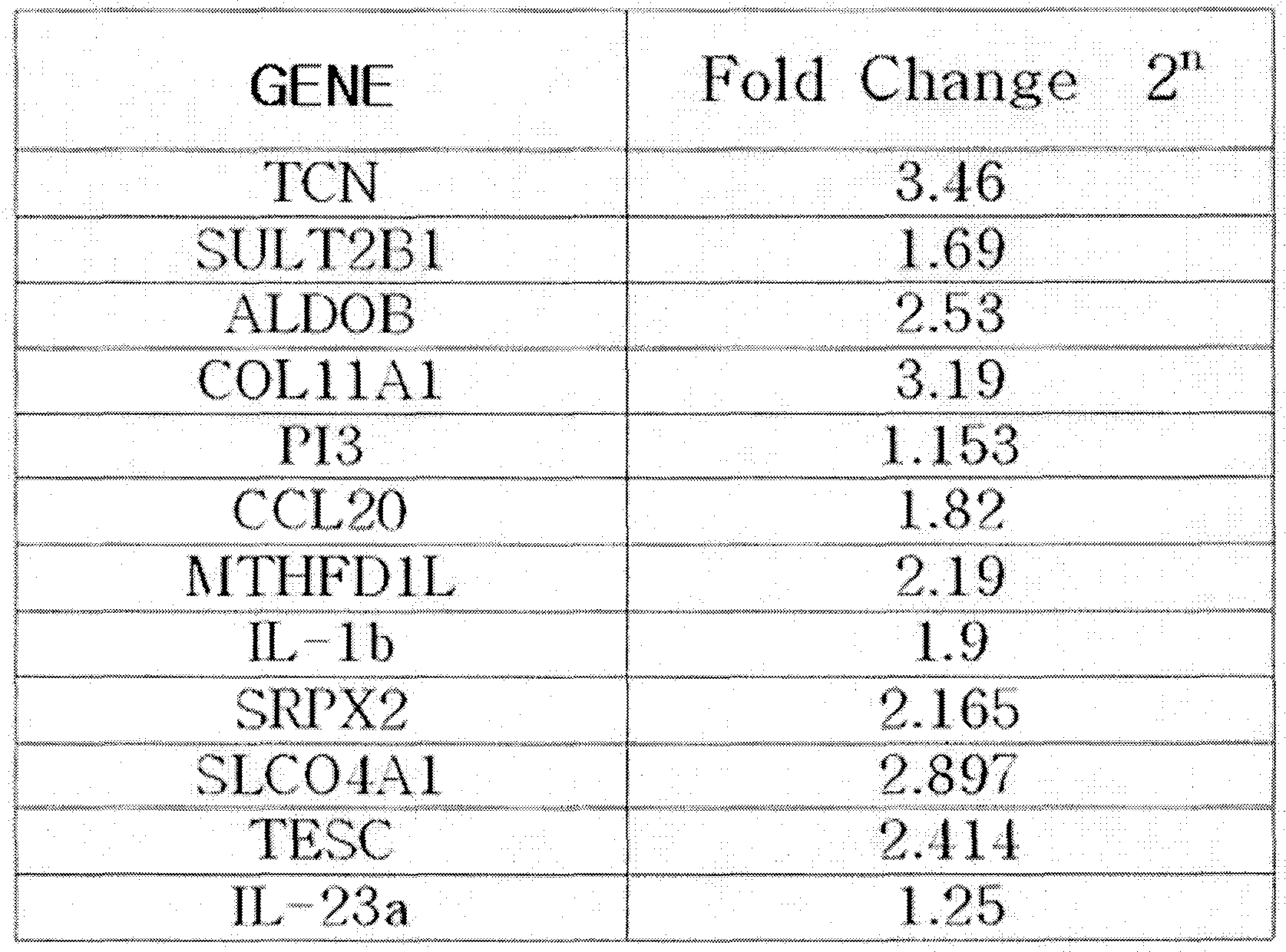
EXAMPLE 2 : mRMA Isolation from Tissues and Cells
For use in reverse transcription PCR, mRNA was isolated from total 40 tissues consisting of normal colonic epithelial cells and colon cancer cell tissues from 20 patients with colon cancer.
First, immediately after the surgical resection of tissues, blood was removed from the tissues in sterile phosphate buffered saline and frozen in liquid nitrogen.
Thereafter, total mRNA was isolated in a single-step RNA isolation manner using the guanidinium method. The total mRNA thus obtained was quantified with a spectrophotometer and stored in a -700C freezer until use. 10 colon cancer cell lines (DLD-I, HT29. HCTIl 6, colo205, SW480, SW620, SNU Cl, SNU C2A, KM 12C, KM 12SM) were obtained from KCLB (the Korean Cell Line Bank, located at 28, Yeonkun- dong, Jongno, Seoul, Korea) .
Each cell line was cultured for 5 - 6 days in DMEM (Invitrogen) or RPMI1640 (Invitrogen) , supplemented with 10% fetal bovine serum (FBS, Hyclon) and lmg/ml penicillin/streptomycin (Sigma) , after which total RNA was isolated in a single-step RNA isolation manner using the guanidinium method. The RNA thus obtained was quantified with a spectrophotometer and stored at a -700C freezer until use.
EXAMPLE 3 : Comparison of Gene Expression Levels by RT-PCR
The colon cancer-specific, overexpressed genes selected in Example 1 were subjected to RT-PCR.
An overall DNA sequence of each gene was obtained from the NCBI CoreNucleotide database
(http: //www. ncbi.nlm.nih.gov/) . Based on the DNA sequences,
primer sequences for the genes were designed using the Primer3 program. PCR was performed with these designed primers to examine expression levels of the genes. Base sequences of the primers are listed in Table 4, below.
TABLE 4

Through RT-PCR, the mRNA isolated from the tissues and cell lines of Example 2 were converted into cDNA. In this
regard, the cDNA construction was accomplished using a cDNA synthesis kit (AccuScript High Fidelity 1st Stand cDNA Synthesis Kit, STRATAGENE) .
From the cDNA, PCR amplification was carried out in the presence of the designed primers (1st cycle: 94 °C ,5 mi; 2nd to 35th cycles: 94°C, 40 sec, 5β°C 40 sec, 72°C, 30 sec; final extension: 72°C, 7 min) .
As a result, differences in gene expression level between normal colon cells and colon cancer cells were detected. Coincident with the results of Example 1, the genes of SEQ ID NOS. 1 to 12 was identified to increase their expression levels in the colon cancer cell lines as compared to the normal colon cells (FIGS. Ia, Ib, Ic, and 2) .
EXAMPLE 4: Comparison of Protein Expression Levels in Sera Using Western Blotting
Protein levels in serum of colon cancer patients and healthy persons were compared using a Western blotting method. Sera was isolated from the blood of colon cancer patients and healthy persons and diluted with the same volume of a sample buffer (125 mM Tris pH 6.8, 4% SDS, 10% glycerol, 0.006% bromophenol blue, 1.8% BME) before boiling. 12% SDS-PAGE
separated serum proteins . The SDS-PAGE gel in which the serum proteins were separated according to sizes was brought into contact with a nitrocellulose membrane. The application of a current to the gel-membrane associate transferred the proteins onto the membrane which was then blocked for 1 hour in a TBST solution (1OmM Tris, 10OmM NaCl, 0.05% Tween 20) containing 3% FBS albumin, followed by reaction with an ALDOB antibody (R&D, 1:2000) at 4°C with shaking overnight. Afterwards, excess antibodies were washed off with PBST, and a horse radish peroxydase-conjugated secondary antibody (ABCAM, Rabbit polyclonal to Mouse IgG) was added and incubated at 4°C for 1 hour with shaking. The nitrocellulose membrane was immersed in a mixture of 1:1 ECL Solution A (containing Luminol and enhancer) : Solution B (containing hydrogen peroxide) and incubated for 1 min with shaking. After being dried suitably, the membrane was attached to a film cassette and developed in a dark room. The same procedure was applied to COLIlAl, SULT2B1, MTHFDLL, TESC, and SRPX2. The results are shown in FIG. 3. ALDOB, COLIlAl, SULT2B1, MTHFDLL, TESC, and SRPX2 proteins were not or little detected in healthy persons (lanes 1 to 3) while being overexpressed in patients with colon cancer (lanes 4 to 7), demonstrating the usefulness thereof as colon cancer diagnosis markers
EXAMPLE 5: Comparison of Protein Expression Levels in Tissues Using IiranunoStaining
Tissue slides were iininunostained so as to determine the presence and expression positions of the proteins in normal colonic epithelial tissues and colon cancer tissues.
To this end, first, normal colonic epithelial cell tissues and colon cancer cell tissues were surgically excised from colon caner patients and embedded in paraffin blocks. Using a microtome, these blocks were cut into slices of 5 um thickness, followed by the attachment of the slices to glass slides. The tissue slides thus obtained were immunostained, and observed for the presence and positions of the proteins in tissues under a microscope. The antibodies used in this immunostaining were anti-TCN-antibody (Novus, 1:5000), anti- SULT2Bl-antibody (Abeam, 1:5000), and anti-ALDOB-antibody (Novus, 1:5000), and anti- COLllAl-antibody (Novus, 1:5000) .
The immunohistostaining results indicated that it was expressed at higher levels in the cytoplasm and cellular membranes of the tumor cell (FIGS. 4a to 4d) .
EXAMPLE 6: Measurement of Protein Levels in Sera by Immunodot
Analysis
Sera from healthy persons and colon cancer patients were compared for secretion levels of the proteins TCN, SULT2B1, ALDOB, COLIlAl, PI3, CCL20, MTHFDlL, IL-I jβ, SRPX2, SLC04A1, TESC, and IL-23 α using an immunodot assay with polyclonal antibodies. Each of the serum samples (10 samples per person) of a 5 to 20-fold dilution was dotted in an amount of 2 μl on a nitrocellulose membrane, dried at room temperature, and blocked in 1% BSAT (bovine serum albumin in Tris-buffered saline) solution. They were treated with a polyclonal antibody to TCN
(Novus, 1:1000), a polyclonal antibody to SULT2B1 (Abeam,
1:1000), a polyclonal antibody to COLIlAl (Novus, 1:1000), a polyclonal antibody to MTHFDlL (Abnova, 1:1000), a polyclonal antibody to SRPX2 (Proteintech, 1:500), and a polyclonal antibody to TESC (Proteintech, 1:1000) and then with a horse radish peroxidase-conjugated secondary antibody (1:10000), followed by developing in a DAB solution (0.5 mg/ml, diaminobenzidine in PBT) . Fluorescence data obtained by scanning were analyzed (FIG. 5) .
It was found to be expressed in larger amounts in colon cancer sera than in normal sera, demonstrating that these genes can be used as effective markers for the diagnosis and
prognosis of colon cancer.
EXAMPLE 7: Establishment of ELISA System and Diagnosis of Colon Cancer thereby
7-1. Establishment of ELI5A system
Monoclonal antibodies to TCN, SULT2B1, ALDOB, COLlIAl, PI3, CCL20, MTHFDlL, IL-lβ, SRPX2, SLCO4A1, TESC, and IL-23 α proteins were diluted to a concentration of 1 μl/ml in 0.1M carbonate buffer (pH 9.6) and plated in an amount of 100 μl/well into 96-well microtiter plates. After incubating overnight at 40C, the microtiter plates thus coated with the monoclonal antibodies were washed three times with 0.05% Tween- 20-containing PBS (PBS-T) . Blocking at room temperature for 2 hours with 1% BSA was followed by three rounds of washing with PBS-T. Each dilution of the proteins corresponding to SEQ ID NOS. 1 to 12 was added in an amount of 100 μl to the 96-well microtiter plates and incubated at room temperature for 2 hours, followed by washing three times with PBS-T. Polyclonal antibodies (1:2000 dilution) to TCN, SULT2B1, ALDOB, COLlIAl, PI3, CCL20, MTHFDlL, IL-1J3, SRPX2, SLCO4A1, TESC, and IL-23 α proteins were added in an amount of 100 μl to the 96-well microtiter plates, incubated for 2 hours and washed. 100 μl of
a 200-fold diluted, horse radish peroxidase-conjugated secondary antibody was added, incubated at room temperature for 1 hour and washed three times, followed by color development with TMB. Absorbance at 450nm was read in an ELISA reader (Molecular Device, Sunnyvale, CA, USA) (FIG. 6) .
7-2. Measurement of protein levels in sera using ELISA system.
Using the ELISA system established in Example 7-1, serum samples were measured for levels of TCN, SULT2B1, ALDOB, COLIlAl, PI3, CCL20, MTHFDlL, IL-I β, SRPX2, SLCO4A1, TESC, and IL-23 α proteins. After being diluted five folds, normal and colon cancer sera were calculated for concentrations of TCN, SULT2B1, ALDOB, COLIlAl, PI3, CCL20, MTHFDlL, IL-I β, SRPX2, SLCO4A1, TESC, and IL-23 α proteins.
EXAMPLE 8 : Kit Construction and Measurement of Protein Level in Serum
8-1. Sandwich ELISA kit
A kit for measuring concentrations of TCN, SULT2B1,
ALDOB, COLIlAl, PI3, CCL20, MTHFDlL, IL-lβ, SRPX2, SLCO4A1,
TESC, and IL-23 α proteins was constructed using the following
components :
A. Solid phase antibody: A microtiter plate with an antibody adsorbed thereto. It was constructed by plating polyclonal antibodies to TCN, SULT2B1, ALDOB, COLIlAl, PI3, CCL20, MTHFDlL, IL-lβ, SRPX2, SLCO4A1, TESC, and IL-23 α in an amount of 100 μl per well into a microtiter plate, followed by incubating overnight at 4°C to adsorb albumin to the solid phase surface.
B. Detection antibody: Monoclonal antibodies to TCN, SULT2B1, ALDOB, COLIlAl, PI3, CCL20, MTHFDlL, IL-lβ, SRPX2,
SLCO4A1, TESC, and IL-23 α
C. Enzyme-conjugated antibody: horse radish peroxidase (HRP) -conjugated secondary antibody
D. Serum dilution buffer E. Substrate (TMB)
F. Washing solution: 0.05% Tween-containing PBS (PBS-T)
G. Standard solution: Standard solutions of TCN, SULT2B1, ALDOB, COLIlAl, PI3, CCL20, MTHFDlL, IL-lβ, SRPX2, SLCO4A1, TESC, and IL-23 α proteins. Using the kit, dilutions of sera taken from patients were assayed for levels of TCN, SULT2B1, ALDOB, COLIlAl, PI3, CCL20, MTHFDlL, IL-lβ, SRPX2, SLCO4A1, TESC, and IL-23 α proteins as follows .
A suitable dilution of a serum sample in a diluent (D) was added in an amount of 100 μl per well to the solid phase antibody of the component A, and analyzed for concentrations of TCN, SULT2B1, ALDOB, COLIlAl, PI3, CCL20, MTHFDlL, IL-I β, SRPX2, SLCO4A1, TESC, and IL-23 α proteins using the sandwich ELISA kit established with the components B, C and E in Example 8-1.
8-2. Immunochromatography kit 8-2-1. Construction of Immunochromatographic strip 1) Preparation of Ab-gold conjugate
An antibody was added in a concentration of 15 μg/ml to a colloidal gold particle solution and then incubated at room temperature for 2 hours with agitation. To this solution was added 1/10 volume of 10% BSA, followed by the incubation of the resulting 1% BSA solution for 1 hour. Centrifugation at 12,000 rpm for 40 min precipitated Ab-gold conjugates. The supernatant was discarded and the precipitates were washed with 2 mM borate buffer. This washing process was repeated three times further. Thereafter, 2 mM borate buffer containing 1% BSA was added in an amount of about 1/10 volume of the gold solution to give a suspension. Absorbance at 530 nm was measured using a UV spectrophotometer and dilution was performed to form an O. D. of
3. 00.
2 ) Sample pad
Provided for absorbing a sample. Made of a cellulose material. As long as it absorbs samples, any can be used as a material for the sample pad.
3) Glass fiber (GF) membrane
Pretreated with 20 mM borate buffer containing sucrose.
4) Nitrocellulose (NC) membrane and line treatment
A nitrocellulose membrane (Millipore) was cut into a suitable size (0.7cm x 5cm). In the cut membranes, goat anti- sheep IgG was applied at a virtual control line about 3.4 m distant from the bottom of the plastic backing while monoclonal antibodies to TCN, SULT2B1, ALDOB, COLlIAl, PI3, CCL20, MTHFDlL, IL-ljβ, SRPX2, SLCO4A1, TESC, and IL-23 α proteins were applied to a virtual detection line 2.7 cm distant from the bottom.
5) Absorbent pad
Made of a cellulose membrane which can absorb materials remaining untreated after the immune response and thus allows the sample solution including analysates to migrate by capillary action.
6) Adhesive plastic backing
On an adhesive plastic backing, the sample pad, the GF
membrane, the NC membrane and the absorbent pad were laminated, as shown in FIG. 7, in such a manner as for samples to continuously migrate by capillary action, thus affording an immunochromatographic strip. 8-2-2. Result decision
3 ~ 5 min after 60-70 μl of a sample (e.g., a mixture of 1:5 (v/v) serum : elution buffer) was loaded on the sample pad, the strip was observed for color development at the control line and the result line and the concentration of the developed color. A positive sample developed red colors at both the control line and the result line. Only the control line was visualized as red for a negative sample.
8-3 Luminex kit 8-3-1. Construction of Luminex kit
Polyclonal antibodies to TCN, SULT2B1, ALDOB, COLIlAl, PI3, CCL20, MTHFDlL, IL-I β, SRPX2, SLCO4A1, TESC, and IL-23 α proteins were conjugated to beads. A sample dilution was added in an amount of 100 μl and incubated at room temperature for 2 hours, followed by washing three times with PBS-T. Then, they were incubated for 2 hours with 100 μl of each of monoclonal antibodies to the proteins corresponding to SEQ ID NOS. 1 to 12 and washed. An additional one round of incubation was conducted
at room temperature for 1 hour with 100 μl of a 2000-fold diluted, PE (phycoerythrin) -conjugated secondary antibody. They were washed three times before measurement in a luminex device. The fluorescence intensities were plotted against concentrations to give a standard curve.
8-3-2. Sandwich luminex kit
A luminex kit for measuring concentrations of TCN, SULT2B1, ALDOB, COLIlAl, PI3, CCL20, MTHFDlL, IL-I β, SRPX2, SLCO4A1, TESC, and IL-23 α proteins were constructed using the following components .
A. Solid phase antibody: fluorescent beads with polyclonal antibodies to TCN, SULT2B1, ALDOB, COLIlAl, PI3,
CCL20, MTHFDlL, IL-I β, SRPX2, SLCO4A1, TESC, and IL-23 α proteins adsorbed thereto. B. Detection antibody: monoclonal antibodies to TCN,
SULT2B1, ALDOB, COLIlAl, PI3, CCL20, MTHFDlL, IL-I β, SRPX2,
SLCO4A1, TESC, and IL-23 α
C. Enzyme-conjugated antibody: peroxidase-conjugated secondary antibody D. Serum dilution buffer
F. Washing solution: 0.05% Tween-containing PBS (PBS-T)
G. Standard solution: Standard solutions of TCN, SULT2B1, ALDOB, COLIlAl, PI3, CCL20, MTHFDlL, IL-lβ, SRPX2, SLCO4A1,
TESC, and IL-23 α proteins.
Using the kit, dilutions of sera taken from patients were assayed for proteins as follows. A suitable dilution of a serum sample in a diluent (D) was added in an amount of 100 μl per well to the solid phase antibody of the component A, and analyzed for concentrations of TCN, SULT2B1, ALDOB, COLIlAl, PI3, CCL20, MTHFDlL, IL-lβ, SRPX2, SLCO4A1, TESC, and IL-23 α proteins using the components B, C and E.
8-4. Protein microarray kit
8-4-1. Protein microarray system
Well chips from Proteagen were coated with monoclonal antibodies to TCN, SULT2B1, ALDOB, COLIlAl, PI3, CCL20, MTHFDlL, IL-lβ, SRPX2, SLCO4A1, TESC, and IL-23 α proteins. The chips were blocked with BSA buffer, incubated at room temperature for 1 hour with 100 μl of a serum dilution, and washed three times with PBS-T. Again, the chips were incubated at 370C for 1 hour with 100 μl of each of diluted monoclonal antibodies to TCN, SULT2B1, ALDOB, COLIlAl, PI3, CCL20, MTHFDlL, IL-lβ, SRPX2, SLCO4A1, TESC, and IL-23 α proteins and washed. An additional one round of incubation was also conducted at room temperature for 0.5 hours with 100 μl of a 2000-fold diluted, Cy3-conjugated secondary antibody. The chips
were washed three times before the measurement of fluorescent intensity at 532nm. The fluorescent intensities were plotted against concentrations to give a standard curve. The protein microarray system thus established was used to determine the serum levels of TCN, SULT2B1, ALDOB, COLlIAl, PI3, CCL20, MTHFDlL, IL-1J3, SRPX2, SLC04A1, TESC, and IL-23 α proteins.
8-4-2. Sandwich protein microarray kit
A sandwich protein microarray kit for measuring concentrations of TCN, SULT2B1, ALDOB, COLIlAl, PI3, CCL20, MTHFDlL, IL-I β, SRPX2, SLC04A1, TESC, and IL-23 α proteins were constructed using the following components.
A. Solid phase antibody: a slide coated with polyclonal antibodies to TCN, SULT2B1, ALDOB, COLIlAl, PI3, CCL20, MTHFDlL, IL-I β, SRPX2, SLC04A1, TESC, and IL-23 α proteins. B. Detection antibody: monoclonal antibodies to TCN, SULT2B1, ALDOB, COLIlAl, PI3, CCL20, MTHFDlL, IL-I β, SRPX2, SLC04A1, TESC, and IL-23 α
C. Enzyme-conjugated antibody: Cy3-conjugated secondary antibody D. Serum dilution buffer
F. Washing solution: 0.05% Tween-containing PBS (PBS-T)
G. Standard solution: Standard solutions of TCN, SULT2B1, ALDOB, COLIlAl, PI3, CCL20, MTHFDlL, IL-lβ, SRPX2, SLCO4A1,
TESC, and IL-23 α proteins.
Using the kit, dilutions of sera taken from colon cancer patients were assayed for proteins as follows. A suitable dilution of a serum sample in a diluent (D) was added in an amount of 100 μl per well to the solid phase antibody of the component A, and analyzed for concentrations of TCN, SULT2B1, ALDOB, COLIlAl, PI3, CCL20, MTHFDlL, IL-I β, SRPX2, SLC04A1, TESC, and IL-23 α proteins using the components B, C and E in the same manner as in the sandwich method of Example 8-4-1.
[industrial Applicability]
As described hitherto, the present invention provides diagnostic markers for accurately diagnosing colon cancer at an early stage and determining the metastasis and prognosis of colon cancer, thus affording data useful in the treatment and monitoring of colon cancer.
With ability to determine mRNA or protein expression levels of genes specific to colon cancer readily and rapidly, the colon cancer diagnosis markers of the present invention can also be used in research for developing anticancer agents against colon cancer.
Although the preferred embodiment ( s ) of the present invention have (has) been disclosed for illustrative purposes,
those skilled in the art will appreciate that various modifications, additions and substitutions are possible, without departing from the scope and spirit of the invention as disclosed in the accompanying claims.
[Sequence List Text]
Claims
[CLAIMS]
[Claim l]
A colon cancer diagnosis composition, comprising markers for determining mRNA expression levels of genes having base sequences of SEQ ID NOS. 1 to 12, alone or in combination.
[Claim 2]
The colon cancer diagnosis composition according to claim 1, wherein the markers are primer sets which specifically bind to the base sequences of SEQ ID NOS. 1 to 12.
[Claim 3]
The colon cancer diagnosis composition according to claim 2, wherein one of the primer sets is complimentary to one selected from the base sequence of SEQ ID NOS. 1 to 12 and is represented by one of SEQ ID NOS. 13 to 36.
[Claim 4] A colon cancer diagnosis composition, comprising markers for determining expression levels of proteins respectively encoded by genes having base sequences of SEQ ID NOS. 1 to 12, alone or in combination.
[Claim 5]
The colon cancer diagnosis composition according to claim 4, wherein the markers are antibodies specific to the proteins encoded by the genes having the base sequences of SEQ ID NOS. 1 to 12.
[Claim 6]
The colon cancer diagnosis composition according to claim 5, wherein the antibodies specific to the proteins are microparticle-conjugated antibodies .
[Claim 7]
The colon cancer diagnosis composition according to claim 6, wherein the microparticles are colored latex particles or colloidal gold particles.
[Claim 8]
A colon cancer diagnosis composition according to claim 5, wherein the antibodies specific to the proteins can be applied to an immunochromatographic strip kit, a luminex assay kit, a protein microarray kit, an ELISA kit, or an immunodot kit.
[Claim 9]
A colon cancer diagnosis kit, comprising the composition of one of claims 1 to 8.
[Claim lθ]
A method for measuring an mRNA expression level of at least one selected from a group consisting of genes having base sequences of SEQ ID NOS. 1 to 12, comprising: measuring an mRNA level in a biological sample from a patient with suspected colon cancer using one or more primer sets selected from among base sequences of SEQ ID NOS. 13 to 36, which are specific to one or more genes selected from among genes of SEQ ID NOS. 1 to 12; and comparing the mRNA level of the sample from the patient with that of a normal control sample to determine an increase in mRNA level.
[Claim 11] A method for measuring an expression level of at least one protein encoded by at least one selected from a group of genes of SEQ ID NOS. 1 to 12, comprising: measuring a protein level by contacting an antibody
specific to an protein encoded by one of the genes of SEQ ID NOS. 1 to 12 with a biological sample from a patient with suspected colon cancer to form an antigen-antibody complex; and comparing the protein level of the sample from the patient with that of a normal control sample to determine an increase in protein level .
[Claim 12]
A method according to claim 10 or 11, wherein the biological sample is selected from a group consisting of a tissue, a cell, whole blood, a serum, a plasma, saliva, sputum, cerebrospinal fluid and urine
[Claim 13] The method according to 11, wherein the expression level of the protein is measured using an assay selected from a group consisting of an immunochromatography assay, an immunodot assay, a luminex assay, an ELISA assay, a protein microarray assay, an immunostaining assay, and a Western blotting assay.
Applications Claiming Priority (2)
| Application Number | Priority Date | Filing Date | Title |
|---|---|---|---|
| KR10-2008-0120096 | 2008-11-28 | ||
| KR1020080120096A KR101073875B1 (en) | 2008-11-28 | 2008-11-28 | Diagnostic kit of colon cancer using colon cancer related marker, and Diagnostic method therof |
Publications (1)
| Publication Number | Publication Date |
|---|---|
| WO2010061996A1 true WO2010061996A1 (en) | 2010-06-03 |
Family
ID=42225850
Family Applications (1)
| Application Number | Title | Priority Date | Filing Date |
|---|---|---|---|
| PCT/KR2008/007173 Ceased WO2010061996A1 (en) | 2008-11-28 | 2008-12-04 | Diagnostic kit of colon cancer using colon cancer related marker, and diagnostic method therof |
Country Status (2)
| Country | Link |
|---|---|
| KR (1) | KR101073875B1 (en) |
| WO (1) | WO2010061996A1 (en) |
Cited By (12)
| Publication number | Priority date | Publication date | Assignee | Title |
|---|---|---|---|---|
| WO2013021088A3 (en) * | 2011-08-09 | 2013-08-01 | Oncomatrix, S.L. | Methods and products for in vitro diagnosis, in vitro prognosis and the development of drugs against invasive carcinomas |
| WO2013151736A3 (en) * | 2012-04-02 | 2013-12-19 | modeRNA Therapeutics | In vivo production of proteins |
| EP2680003A1 (en) | 2012-06-28 | 2014-01-01 | Fundació Institut d'Investigació Biomèdica de Bellvitge | Serum biomarker for diagnosing colorectal cancer |
| US9186372B2 (en) | 2011-12-16 | 2015-11-17 | Moderna Therapeutics, Inc. | Split dose administration |
| US9221891B2 (en) | 2012-04-02 | 2015-12-29 | Moderna Therapeutics, Inc. | In vivo production of proteins |
| US9587003B2 (en) | 2012-04-02 | 2017-03-07 | Modernatx, Inc. | Modified polynucleotides for the production of oncology-related proteins and peptides |
| US9950068B2 (en) | 2011-03-31 | 2018-04-24 | Modernatx, Inc. | Delivery and formulation of engineered nucleic acids |
| CN108653737A (en) * | 2018-06-25 | 2018-10-16 | 徐州市中心医院 | Purposes of the MTHFD1L inhibitor in preparing Dendritic cell medicine |
| EP3431609A1 (en) | 2017-07-17 | 2019-01-23 | Masarykova Univerzita | Method of diagnosis of colorectal cancer |
| US10258698B2 (en) | 2013-03-14 | 2019-04-16 | Modernatx, Inc. | Formulation and delivery of modified nucleoside, nucleotide, and nucleic acid compositions |
| WO2019144099A1 (en) * | 2018-01-22 | 2019-07-25 | Liquid Biopsy Research LLC | Methods for colon cancer detection and treatment monitoring |
| US10501513B2 (en) | 2012-04-02 | 2019-12-10 | Modernatx, Inc. | Modified polynucleotides for the production of oncology-related proteins and peptides |
Families Citing this family (6)
| Publication number | Priority date | Publication date | Assignee | Title |
|---|---|---|---|---|
| KR20020062899A (en) * | 2002-07-04 | 2002-07-31 | 서봉석 | Health food based on red ginseng and Cordyceps sinensis |
| KR101413689B1 (en) * | 2010-11-15 | 2014-07-01 | 한국생명공학연구원 | Monoclonal Antibody Specifically Binding to Human Tescalcin Protein and Its Use |
| KR101413690B1 (en) * | 2010-11-29 | 2014-07-23 | 한국생명공학연구원 | Methylation Biomarker for Detection of Cancers and Uses thereof |
| EP2607492A1 (en) * | 2011-12-22 | 2013-06-26 | Roche Diagniostics GmbH | Method for determining the concentration of an analyte |
| ES2893411T3 (en) * | 2016-09-15 | 2022-02-09 | Hoffmann La Roche | Procedures for performing multiplexed PCR |
| KR102649112B1 (en) * | 2021-11-18 | 2024-03-20 | 주식회사 스템바이오 | Apparatus and manufacturing method of a diagnostic kit with the sample of urine to diagnose the Colorectal cancer |
Citations (4)
| Publication number | Priority date | Publication date | Assignee | Title |
|---|---|---|---|---|
| US20060263786A1 (en) * | 2004-01-27 | 2006-11-23 | Rotem Sorek | Novel nucleotide and amino acid sequences, and assays and methods of use thereof for diagnosis of colon cancer |
| EP1798293A2 (en) * | 2005-12-14 | 2007-06-20 | Canon Kabushiki Kaisha | Gene set used for examination of colon cancer |
| WO2007112330A2 (en) * | 2006-03-24 | 2007-10-04 | Diadexus, Inc. | Compositions and methods for detection, prognosis and treatment of colon cancer |
| WO2008063414A2 (en) * | 2006-11-13 | 2008-05-29 | Source Precision Medicine, Inc. | Gene expression profiling for identification, monitoring, and treatment of colorectal cancer |
-
2008
- 2008-11-28 KR KR1020080120096A patent/KR101073875B1/en not_active Expired - Fee Related
- 2008-12-04 WO PCT/KR2008/007173 patent/WO2010061996A1/en not_active Ceased
Patent Citations (4)
| Publication number | Priority date | Publication date | Assignee | Title |
|---|---|---|---|---|
| US20060263786A1 (en) * | 2004-01-27 | 2006-11-23 | Rotem Sorek | Novel nucleotide and amino acid sequences, and assays and methods of use thereof for diagnosis of colon cancer |
| EP1798293A2 (en) * | 2005-12-14 | 2007-06-20 | Canon Kabushiki Kaisha | Gene set used for examination of colon cancer |
| WO2007112330A2 (en) * | 2006-03-24 | 2007-10-04 | Diadexus, Inc. | Compositions and methods for detection, prognosis and treatment of colon cancer |
| WO2008063414A2 (en) * | 2006-11-13 | 2008-05-29 | Source Precision Medicine, Inc. | Gene expression profiling for identification, monitoring, and treatment of colorectal cancer |
Cited By (34)
| Publication number | Priority date | Publication date | Assignee | Title |
|---|---|---|---|---|
| US9950068B2 (en) | 2011-03-31 | 2018-04-24 | Modernatx, Inc. | Delivery and formulation of engineered nucleic acids |
| US9702879B2 (en) | 2011-08-09 | 2017-07-11 | Oncomatryx Biopharma, S.L. | Methods and products for in vitro diagnosis, in vitro prognosis and the development of drugs against invasive carcinomas |
| WO2013021088A3 (en) * | 2011-08-09 | 2013-08-01 | Oncomatrix, S.L. | Methods and products for in vitro diagnosis, in vitro prognosis and the development of drugs against invasive carcinomas |
| US9271996B2 (en) | 2011-12-16 | 2016-03-01 | Moderna Therapeutics, Inc. | Formulation and delivery of PLGA microspheres |
| US9186372B2 (en) | 2011-12-16 | 2015-11-17 | Moderna Therapeutics, Inc. | Split dose administration |
| US9295689B2 (en) | 2011-12-16 | 2016-03-29 | Moderna Therapeutics, Inc. | Formulation and delivery of PLGA microspheres |
| US9572896B2 (en) | 2012-04-02 | 2017-02-21 | Modernatx, Inc. | In vivo production of proteins |
| US10501513B2 (en) | 2012-04-02 | 2019-12-10 | Modernatx, Inc. | Modified polynucleotides for the production of oncology-related proteins and peptides |
| US11564998B2 (en) | 2012-04-02 | 2023-01-31 | Modernatx, Inc. | Modified polynucleotides for the production of cytoplasmic and cytoskeletal proteins |
| US9587003B2 (en) | 2012-04-02 | 2017-03-07 | Modernatx, Inc. | Modified polynucleotides for the production of oncology-related proteins and peptides |
| US10772975B2 (en) | 2012-04-02 | 2020-09-15 | Modernatx, Inc. | Modified Polynucleotides for the production of biologics and proteins associated with human disease |
| US9782462B2 (en) | 2012-04-02 | 2017-10-10 | Modernatx, Inc. | Modified polynucleotides for the production of proteins associated with human disease |
| US9814760B2 (en) | 2012-04-02 | 2017-11-14 | Modernatx, Inc. | Modified polynucleotides for the production of biologics and proteins associated with human disease |
| US9827332B2 (en) | 2012-04-02 | 2017-11-28 | Modernatx, Inc. | Modified polynucleotides for the production of proteins |
| US9828416B2 (en) | 2012-04-02 | 2017-11-28 | Modernatx, Inc. | Modified polynucleotides for the production of secreted proteins |
| US9878056B2 (en) | 2012-04-02 | 2018-01-30 | Modernatx, Inc. | Modified polynucleotides for the production of cosmetic proteins and peptides |
| WO2013151736A3 (en) * | 2012-04-02 | 2013-12-19 | modeRNA Therapeutics | In vivo production of proteins |
| US10703789B2 (en) | 2012-04-02 | 2020-07-07 | Modernatx, Inc. | Modified polynucleotides for the production of secreted proteins |
| US10583203B2 (en) | 2012-04-02 | 2020-03-10 | Modernatx, Inc. | In vivo production of proteins |
| US10577403B2 (en) | 2012-04-02 | 2020-03-03 | Modernatx, Inc. | Modified polynucleotides for the production of secreted proteins |
| US9221891B2 (en) | 2012-04-02 | 2015-12-29 | Moderna Therapeutics, Inc. | In vivo production of proteins |
| US10501512B2 (en) | 2012-04-02 | 2019-12-10 | Modernatx, Inc. | Modified polynucleotides |
| US10463751B2 (en) | 2012-04-02 | 2019-11-05 | Modernatx, Inc. | Modified polynucleotides for the production of cytoplasmic and cytoskeletal proteins |
| US10493167B2 (en) | 2012-04-02 | 2019-12-03 | Modernatx, Inc. | In vivo production of proteins |
| EP2680003A1 (en) | 2012-06-28 | 2014-01-01 | Fundació Institut d'Investigació Biomèdica de Bellvitge | Serum biomarker for diagnosing colorectal cancer |
| ES2436667R1 (en) * | 2012-06-28 | 2014-05-21 | Fundació Institut D'investigació Biomèdica De Bellvitge (Idibell) | SERUM BIOMARCATOR TO DIAGNOSTIC COLORRECTAL CANCER |
| US10258698B2 (en) | 2013-03-14 | 2019-04-16 | Modernatx, Inc. | Formulation and delivery of modified nucleoside, nucleotide, and nucleic acid compositions |
| WO2019015702A1 (en) | 2017-07-17 | 2019-01-24 | Masarykova Univerzita | Method of diagnosis of colorectal cancer |
| EP3431609A1 (en) | 2017-07-17 | 2019-01-23 | Masarykova Univerzita | Method of diagnosis of colorectal cancer |
| WO2019144099A1 (en) * | 2018-01-22 | 2019-07-25 | Liquid Biopsy Research LLC | Methods for colon cancer detection and treatment monitoring |
| US11414707B2 (en) | 2018-01-22 | 2022-08-16 | Liquid Biopsy Research LLC | Methods for colon cancer detection and treatment |
| US12305240B2 (en) | 2018-01-22 | 2025-05-20 | Liquid Biopsy Research LLC | Methods for colon cancer detection and treatment |
| CN108653737B (en) * | 2018-06-25 | 2020-01-10 | 徐州市中心医院 | Application of MTHFD1L inhibitor in preparation of tongue squamous carcinoma treatment drug |
| CN108653737A (en) * | 2018-06-25 | 2018-10-16 | 徐州市中心医院 | Purposes of the MTHFD1L inhibitor in preparing Dendritic cell medicine |
Also Published As
| Publication number | Publication date |
|---|---|
| KR101073875B1 (en) | 2011-10-14 |
| KR20100061192A (en) | 2010-06-07 |
Similar Documents
| Publication | Publication Date | Title |
|---|---|---|
| WO2010061996A1 (en) | Diagnostic kit of colon cancer using colon cancer related marker, and diagnostic method therof | |
| US20110251097A1 (en) | Diagnostic kit of colon cancer using colon cancer related marker and diagnostic method thereof | |
| KR101146610B1 (en) | Novel Biomarkers Indicative of Liver Cancer and Their Uses | |
| KR101007567B1 (en) | Colorectal Cancer Diagnostic Markers Using Colorectal Cancer Overexpression Genes | |
| US9102706B2 (en) | Newly identified colon cancer marker and diagnostic kit using the same | |
| KR101029881B1 (en) | Colorectal Cancer Diagnostic Markers Using Colorectal Cancer Overexpression Genes | |
| KR101388711B1 (en) | Complement C9 as markers for the diagnosis of cancer | |
| KR101289454B1 (en) | Complement C9 as markers for the diagnosis of small cell lung cancer and non-small cell lung cancer | |
| KR100721507B1 (en) | MAC-2GP as a gastric cancer diagnostic marker | |
| KR100958086B1 (en) | Colorectal Cancer Diagnostic Markers Using Colorectal Cancer Overexpression Genes | |
| KR101007573B1 (en) | Colon cancer diagnostic markers using up-regulated genes | |
| KR100969692B1 (en) | Colorectal Cancer Diagnostic Markers Using Colorectal Cancer Overexpression Genes | |
| KR101060192B1 (en) | Colon cancer diagnostic markers using up-regulated genes | |
| KR101007574B1 (en) | Colorectal Cancer Diagnostic Markers Using Colorectal Cancer Overexpression Genes | |
| KR101901457B1 (en) | Novel Biomarkers for Liver Cancer Based on Liver Cancer Stem Cell Characteristics and Uses thereof | |
| KR20090097138A (en) | Colorectal Cancer Diagnostic Markers Using Colorectal Cancer Overexpression Genes | |
| KR101065027B1 (en) | Colon cancer diagnostic markers using up-regulated genes | |
| KR101007575B1 (en) | Colorectal Cancer Diagnostic Markers Using Colorectal Cancer Overexpression Genes | |
| KR101060193B1 (en) | Colon cancer diagnostic markers using up-regulated genes | |
| KR101007576B1 (en) | Colon cancer diagnostic markers using up-regulated genes | |
| KR101007572B1 (en) | ENPS as a gastric cancer diagnostic marker | |
| KR101007570B1 (en) | DC1 as a gastric cancer diagnostic marker | |
| KR100980004B1 (en) | ELK as a gastric cancer diagnostic marker | |
| KR20090081289A (en) | KLK-5 gene as a hepatocellular carcinoma tumor marker and method for diagnosing hepatocellular carcinoma using the same |
Legal Events
| Date | Code | Title | Description |
|---|---|---|---|
| 121 | Ep: the epo has been informed by wipo that ep was designated in this application |
Ref document number: 08878459 Country of ref document: EP Kind code of ref document: A1 |
|
| NENP | Non-entry into the national phase |
Ref country code: DE |
|
| 122 | Ep: pct application non-entry in european phase |
Ref document number: 08878459 Country of ref document: EP Kind code of ref document: A1 |