WO2010030840A2 - Novel receptor and uses thereof - Google Patents
Novel receptor and uses thereof Download PDFInfo
- Publication number
- WO2010030840A2 WO2010030840A2 PCT/US2009/056602 US2009056602W WO2010030840A2 WO 2010030840 A2 WO2010030840 A2 WO 2010030840A2 US 2009056602 W US2009056602 W US 2009056602W WO 2010030840 A2 WO2010030840 A2 WO 2010030840A2
- Authority
- WO
- WIPO (PCT)
- Prior art keywords
- naadp
- tpc
- agent
- cells
- polypeptide
- Prior art date
- Legal status (The legal status is an assumption and is not a legal conclusion. Google has not performed a legal analysis and makes no representation as to the accuracy of the status listed.)
- Ceased
Links
- FUHCEERDBRGPQZ-NAKRPHOHSA-N COc1c(CN(CC2)CCN2c(cccc2)c2F)cc([C@@H](c2c(C3)c(cccc4)c4[nH]2)N[C@H]3C(O)=O)cc1 Chemical compound COc1c(CN(CC2)CCN2c(cccc2)c2F)cc([C@@H](c2c(C3)c(cccc4)c4[nH]2)N[C@H]3C(O)=O)cc1 FUHCEERDBRGPQZ-NAKRPHOHSA-N 0.000 description 1
Classifications
-
- C—CHEMISTRY; METALLURGY
- C07—ORGANIC CHEMISTRY
- C07K—PEPTIDES
- C07K16/00—Immunoglobulins [IG], e.g. monoclonal or polyclonal antibodies
- C07K16/18—Immunoglobulins [IG], e.g. monoclonal or polyclonal antibodies against material from animals or humans
- C07K16/28—Immunoglobulins [IG], e.g. monoclonal or polyclonal antibodies against material from animals or humans against receptors, cell surface antigens or cell surface determinants
-
- A—HUMAN NECESSITIES
- A61—MEDICAL OR VETERINARY SCIENCE; HYGIENE
- A61K—PREPARATIONS FOR MEDICAL, DENTAL OR TOILETRY PURPOSES
- A61K38/00—Medicinal preparations containing peptides
- A61K38/16—Peptides having more than 20 amino acids; Gastrins; Somatostatins; Melanotropins; Derivatives thereof
- A61K38/17—Peptides having more than 20 amino acids; Gastrins; Somatostatins; Melanotropins; Derivatives thereof from animals; from humans
- A61K38/177—Receptors; Cell surface antigens; Cell surface determinants
-
- C—CHEMISTRY; METALLURGY
- C07—ORGANIC CHEMISTRY
- C07K—PEPTIDES
- C07K14/00—Peptides having more than 20 amino acids; Gastrins; Somatostatins; Melanotropins; Derivatives thereof
- C07K14/435—Peptides having more than 20 amino acids; Gastrins; Somatostatins; Melanotropins; Derivatives thereof from animals; from humans
- C07K14/705—Receptors; Cell surface antigens; Cell surface determinants
-
- C—CHEMISTRY; METALLURGY
- C12—BIOCHEMISTRY; BEER; SPIRITS; WINE; VINEGAR; MICROBIOLOGY; ENZYMOLOGY; MUTATION OR GENETIC ENGINEERING
- C12Q—MEASURING OR TESTING PROCESSES INVOLVING ENZYMES, NUCLEIC ACIDS OR MICROORGANISMS; COMPOSITIONS OR TEST PAPERS THEREFOR; PROCESSES OF PREPARING SUCH COMPOSITIONS; CONDITION-RESPONSIVE CONTROL IN MICROBIOLOGICAL OR ENZYMOLOGICAL PROCESSES
- C12Q1/00—Measuring or testing processes involving enzymes, nucleic acids or microorganisms; Compositions therefor; Processes of preparing such compositions
- C12Q1/68—Measuring or testing processes involving enzymes, nucleic acids or microorganisms; Compositions therefor; Processes of preparing such compositions involving nucleic acids
- C12Q1/6876—Nucleic acid products used in the analysis of nucleic acids, e.g. primers or probes
- C12Q1/6883—Nucleic acid products used in the analysis of nucleic acids, e.g. primers or probes for diseases caused by alterations of genetic material
-
- G—PHYSICS
- G01—MEASURING; TESTING
- G01N—INVESTIGATING OR ANALYSING MATERIALS BY DETERMINING THEIR CHEMICAL OR PHYSICAL PROPERTIES
- G01N33/00—Investigating or analysing materials by specific methods not covered by groups G01N1/00 - G01N31/00
- G01N33/48—Biological material, e.g. blood, urine; Haemocytometers
- G01N33/50—Chemical analysis of biological material, e.g. blood, urine; Testing involving biospecific ligand binding methods; Immunological testing
- G01N33/5005—Chemical analysis of biological material, e.g. blood, urine; Testing involving biospecific ligand binding methods; Immunological testing involving human or animal cells
- G01N33/5008—Chemical analysis of biological material, e.g. blood, urine; Testing involving biospecific ligand binding methods; Immunological testing involving human or animal cells for testing or evaluating the effect of chemical or biological compounds, e.g. drugs, cosmetics
- G01N33/5076—Chemical analysis of biological material, e.g. blood, urine; Testing involving biospecific ligand binding methods; Immunological testing involving human or animal cells for testing or evaluating the effect of chemical or biological compounds, e.g. drugs, cosmetics involving cell organelles, e.g. Golgi complex, endoplasmic reticulum
-
- G—PHYSICS
- G01—MEASURING; TESTING
- G01N—INVESTIGATING OR ANALYSING MATERIALS BY DETERMINING THEIR CHEMICAL OR PHYSICAL PROPERTIES
- G01N33/00—Investigating or analysing materials by specific methods not covered by groups G01N1/00 - G01N31/00
- G01N33/48—Biological material, e.g. blood, urine; Haemocytometers
- G01N33/50—Chemical analysis of biological material, e.g. blood, urine; Testing involving biospecific ligand binding methods; Immunological testing
- G01N33/68—Chemical analysis of biological material, e.g. blood, urine; Testing involving biospecific ligand binding methods; Immunological testing involving proteins, peptides or amino acids
- G01N33/6872—Intracellular protein regulatory factors and their receptors, e.g. including ion channels
-
- C—CHEMISTRY; METALLURGY
- C07—ORGANIC CHEMISTRY
- C07K—PEPTIDES
- C07K2317/00—Immunoglobulins specific features
- C07K2317/30—Immunoglobulins specific features characterized by aspects of specificity or valency
- C07K2317/34—Identification of a linear epitope shorter than 20 amino acid residues or of a conformational epitope defined by amino acid residues
-
- C—CHEMISTRY; METALLURGY
- C12—BIOCHEMISTRY; BEER; SPIRITS; WINE; VINEGAR; MICROBIOLOGY; ENZYMOLOGY; MUTATION OR GENETIC ENGINEERING
- C12Q—MEASURING OR TESTING PROCESSES INVOLVING ENZYMES, NUCLEIC ACIDS OR MICROORGANISMS; COMPOSITIONS OR TEST PAPERS THEREFOR; PROCESSES OF PREPARING SUCH COMPOSITIONS; CONDITION-RESPONSIVE CONTROL IN MICROBIOLOGICAL OR ENZYMOLOGICAL PROCESSES
- C12Q2600/00—Oligonucleotides characterized by their use
- C12Q2600/136—Screening for pharmacological compounds
-
- C—CHEMISTRY; METALLURGY
- C12—BIOCHEMISTRY; BEER; SPIRITS; WINE; VINEGAR; MICROBIOLOGY; ENZYMOLOGY; MUTATION OR GENETIC ENGINEERING
- C12Q—MEASURING OR TESTING PROCESSES INVOLVING ENZYMES, NUCLEIC ACIDS OR MICROORGANISMS; COMPOSITIONS OR TEST PAPERS THEREFOR; PROCESSES OF PREPARING SUCH COMPOSITIONS; CONDITION-RESPONSIVE CONTROL IN MICROBIOLOGICAL OR ENZYMOLOGICAL PROCESSES
- C12Q2600/00—Oligonucleotides characterized by their use
- C12Q2600/156—Polymorphic or mutational markers
-
- G—PHYSICS
- G01—MEASURING; TESTING
- G01N—INVESTIGATING OR ANALYSING MATERIALS BY DETERMINING THEIR CHEMICAL OR PHYSICAL PROPERTIES
- G01N2500/00—Screening for compounds of potential therapeutic value
-
- G—PHYSICS
- G01—MEASURING; TESTING
- G01N—INVESTIGATING OR ANALYSING MATERIALS BY DETERMINING THEIR CHEMICAL OR PHYSICAL PROPERTIES
- G01N2800/00—Detection or diagnosis of diseases
- G01N2800/50—Determining the risk of developing a disease
Definitions
- the present invention relates to the molecular identification of a novel receptor, more particularly the identification of a receptor involved in intracellular calcium release mediated by nicotinic acid adenine dinucleotide phosphate (NAADP).
- NAADP nicotinic acid adenine dinucleotide phosphate
- NAADP (nicotinic acid adenine dinucleotide phosphate) is a closely related molecule to NADP (nicotinamide adenine dinucleotide phosphate), differing only by the substitution of nicotinic acid for the corresponding primary amide.
- NAADP has been found to be a powerful calcium mobilising agent, with potencies often greater than those of other calcium-mobilising messengers such as IP 3 (inositol 1,4,5-trisphosphate) and cADPR (Galione, A. Biochem. Soc. Trans (2006) 34, 922-926).
- NAADP acts on a novel calcium release channel distinct from the two known classes of calcium release channel (IP 3 and ryanodine receptors), the NAADP mechanism being initially distinguished pharmacologically by its insensitivity to specific antagonists that selectively block the IP 3 and ryanodine receptors (Lee, H & Argus, R. (1995). / Biol Chem., 270, 2152-2157).
- the NAADP-mediated calcium release mechanism has since been demonstrated to have several key properties that distinguish it from IP 3 and ryanodine receptors (Galione, A et al., British Journal of Pharmacology (2004) 142, 1203-1207).
- NAADP has been shown to be regulated by extracellular agonists.
- NAADP has also been shown to mobilise calcium from intracellular stores in a number of models, ranging from echinoderm eggs and oocytes to a variety of mammalian cell types including T- lymphocytes, pancreatic beta and acinar cells, kidney cells and heart cells, neurones and smooth muscle platelets (Rutter, G. (2003) Biochem. J. 373, e3-e4).
- NAADP is therefore believed to have crucial roles in pharmacologically important processes such as insulin secretion, blood clotting, lymphocyte activation and smooth muscle and cardiac contractility.
- NAADP may be a better target for therapeutic agents since it may act to modulate the sensitivity of more common calcium-release pathways, and only then for a subset of receptor-mediated calcium signalling events.
- One aspect of the present invention relates to an agent that modulates the effect of a two pore calcium channel (TPC) polypeptide for use in the treatment or prophylaxis of a condition, disease or disorder associated with NAADP receptor-mediated biological activity.
- TPC two pore calcium channel
- the agent is an antibody or antibody fragment.
- the agent is capable of agonising or antagonising the effect of a TPC polypeptide.
- the agent is a nucleotide sequence that encodes or is complementary to a nucleotide sequence that encodes a TPC polypeptide or a functional fragment thereof.
- the nucleotide sequence is a single stranded oligonucleotide which comprises a sequence of no more than 50 nucleotides.
- the nucleotide sequence is a double stranded oligonucleotide which comprises a sequence of no more than 25 nucleotide base pairs.
- the agent is for use in the treatment or prophylaxis of a condition, disease or disorder selected from the group comprising diabetes, obesity, immune system disorders, premature labour, clotting disorders, lysosomal storage diseases, heart disease, cardiac disorders, asthma, hypertension, infertility, autism, prenatal depression, growth disorders, cancer, and hair and skin pigment defects.
- a condition, disease or disorder selected from the group comprising diabetes, obesity, immune system disorders, premature labour, clotting disorders, lysosomal storage diseases, heart disease, cardiac disorders, asthma, hypertension, infertility, autism, prenatal depression, growth disorders, cancer, and hair and skin pigment defects.
- Another aspect of the present invention relates to a vector comprising a nucleotide sequence that encodes or is complementary to a nucleotide sequence that encodes a TPC polypeptide or a functional fragment thereof.
- Another aspect of the present invention relates to a method of identifying an agent that modulates the effect of NAADP receptor mediated biological activity, comprising contacting the agent with:
- TPC polypeptide or functional fragment thereof a TPC polypeptide or functional fragment thereof
- the agent is for use in the treatment or prophylaxis of a condition, disease or disorder associated with NAADP receptor-mediated biological activity, such as diabetes, obesity, immune system disorders, premature labour, clotting disorders, lysosomal storage diseases, heart disease, cardiac disorders, asthma, hypertension, infertility, autism, prenatal depression, growth disorders, cancer, and hair and skin pigment defects.
- a condition, disease or disorder associated with NAADP receptor-mediated biological activity such as diabetes, obesity, immune system disorders, premature labour, clotting disorders, lysosomal storage diseases, heart disease, cardiac disorders, asthma, hypertension, infertility, autism, prenatal depression, growth disorders, cancer, and hair and skin pigment defects.
- kits comprising: a) a TPC polypeptide or functional fragment thereof; or
- kits are, for example, for use in identifying an agent that modulates the effect of NAADP receptor-mediated biological activity.
- a kit may thus include instructions for its use in identifying an agent that modulates the effect of NAADP receptor-mediated biological activity.
- kits are, for example, for use in allowing measurement of cell and tissue NAADP levels.
- a kit may thus include instructions for its use in measuring cell and tissue NAADP levels.
- Another aspect of the present invention relates to a method of screening a subject for the likelihood of developing a condition, disease or disorder associated with NAADP receptor-mediated biological activity, comprising a step of analysing a sample that has been obtained from the subject for a polymorphism in the gene which encodes a TPC polypeptide.
- Another aspect of the present invention relates to a method of identifying the origin of symptoms associated with a condition, disease or disorder associated with NAADP receptor-mediated biological activity, comprising a step of analysing a sample that has been obtained from a subject for a polymorphism in the gene which encodes a TPC polypeptide.
- kits may, for example, be for use in screening a subject or for identifying the origin of symptoms associated with a condition.
- a kit may thus include instructions for its use in screening a subject or for identifying the origin of symptoms associated with a condition.
- Another aspect of the present invention relates to a method for the treatment or prophylaxis of a condition, disease or disorder associated with NAADP receptor- mediated biological activity, comprising the step of administering an agent that modulates the effect of a two pore calcium channel (TPC) polypeptide to a subject.
- TPC two pore calcium channel
- a knock-out animal for example a knock-out mouse lacking the genes for one or more of TPCl, TPC2 and TPC3. For example it may be homozygous.
- the invention also provides the use of such an animal in an identification method of the invention.
- Figure 1 shows immunob lotting of membrane proteins from sea urchin (Strongylocentrotus purpuratus) eggs.
- Figure 2 shows (A) Schematic representation of protocol used for membrane preparations from S.purpuratus egg homogenates using a differential centrifugation protocol. H (homogenate), S (supernatant) and P (pellet); (B) [ 32 P]NAADP binding of protein samples from fractionated sea urchin ⁇ Strongylocentrotus purpuratus) eggs, and immunoblots indicating the presence or otherwise of TPCl, TPC2 and TPC3 in each of the protein samples; (C) and (D) [ 32 P] NAADP binding to immunoprecipitated sea urchin egg TCPs.
- Figure 3 shows bound [ 32 P]NAADP to SlOPlOO membranes, SpTPCl or SpTPC3 IPs after washes with a buffer with K+ (GIuIM) or without K+ (Hepes) in the absence or presence of lO ⁇ M NAADP.
- Figure 4 shows binding curves for binding of TPCl and TPC3 immunocomplexes derived from sea urchin egg protein sample, and of native membranes, to NAADP and NADP.
- Figure 5 shows (A) [ 32 P]NAADP binding activity of total membranes from HEK293 cells (wild type, human TPCl over-expressing, and human TPC2 over-expressing); (B) [ 32 P]NAADP binding activity of total membranes from HEK293 cells (wild type, human TPCl over-expressing, and human TPC2 over-expressing) at varying concentrations of NAADP.
- Figure 6 shows (A) displacement of [ 32 P]NAADP binding by NAADP and NADP from total membrane of mouse liver and (B) displacement of [ P]NAADP binding by NAADP and NADP from total membranes of human TPC2 over-expressing HEK293 cells.
- Figure 7 shows (A) Western blotting of solubilised membranes (input) from hTPC2 cells, and of immune complexes following incubation with anti-HA or control (IgG) antibody and immunoprecipitation; (B) [ 32 P]NAADP binding activity from input and supernatants and [ P]NAADP binding activity recovered from the beads after immunoprecipitation.
- Figure 10 shows calcium oscillations of pancreatic ⁇ -cells from wild type control mice in response to elevated glucose (10 mM).
- Figure 11 shows the response of pancreatic ⁇ -cells from TPC2 knockout mice in response to elevated glucose levels (10 mM).
- Figure 12 shows variation of mouse pancreatic ⁇ -cell [Ca + ] (as indicated by the Fura-2 fluorescence ratio (F340/F380)) (A) in response to 15 mM glucose and bafilomycin (3 ⁇ M); (B) following pretreatment of beta-cells with bafilomycin (3 ⁇ M) and addition of glucose or K + (45 mM); (C) in response to glucose (3 mM) and stimulation by NAADP- AM (60 nM); (D) in response to extracellular NAADP (60 nM) and K + (45 mM) in the presence of glucose (3 mM).
- A in response to 15 mM glucose and bafilomycin (3 ⁇ M)
- B following pretreatment of beta-cells with bafilomycin (3 ⁇ M) and addition of glucose or K + (45 mM)
- C in response to glucose (3 mM) and stimulation by NAADP- AM (60 nM)
- D in response to extracellular NAADP (60 nM
- Figure 13 shows (A) variation of mouse pancreatic ⁇ -cell [Ca 2+ ] (as indicated by the Fura- 2 fluorescence ratio (F340/F380)) following pretreatment with thapsigargin (TG) (l ⁇ M) for Ih, in the presence of glucose (3 mM), in the absence of extracellular Ca 2+ , and after addition of NAADP-AM (60 nM) ; (B) variation of mouse pancreatic ⁇ -cell [Ca 2+ ] (as indicated by the Fura-2 fluorescence ratio (F340/F380)) following pretreatment with Ned- 19 (100 ⁇ M), in the presence of glucose (3 mM), in the absence of extracellular Ca 2+ , and after addition of NAADP-AM (60 nM) and high K + (45 rnM); (C) variations in current due to [Ca 2+ ] !
- Figure 14 shows (A) variation of mouse pancreatic ⁇ -cell [Ca 2+ ] (as indicated by the Fura- 2 fluorescence ratio (F340/F380)) following challenge with glucose (15 mM) and NAADP-AM (60 nM); (B) variation of mouse pancreatic ⁇ -cell [Ca 2+ ] (as indicated by the Fura-2 fluorescence ratio (F340/F380)) following challenge with 15 mM glucose and extracellular NAADP (60 nM); (C) variation of mouse pancreatic ⁇ -cell [Ca 2+ ] (as indicated by the Fura-2 fluorescence ratio (F340/F380)) following pretreatmentof beta- cells with Ned-19 (100 uM) and then stimulation with 15 nM glucose and then high K + and diazoxide (100 uM); (D) variation of mouse pancreatic ⁇ -cell [Ca 2+ ] (as indicated by the Fura-2 fluorescence ratio (F340/F380)) following stimulation
- Figure 15 shows (A) single mouse pancreatic ⁇ -cell membrane potential oscillations elicited by 10 mM glucose before and after the addition of Ned-19 (100 uM); (B) variations in mouse pancreatic ⁇ -cell whole cell Ca 2+ current for cells bathed in 10 mM glucose with or without 100 uM Ned-19; (C) variations in mouse pancreatic ⁇ -cell whole cell K + -ATP current for cells bathed in 10 mM glucose and Ned-19 (100 uM), and then after addition of diazoxide (100 uM) and azide ( 2 mM); (D) changes in FAD fluorescence in response to glucose concentration (from 3 to 20 mM) and after addition of Ned-19 (100 uM) to mouse pancreatic ⁇ -cells.
- Figure 16 shows (A) insulin secretion from mouse islets of Langerhans expressed as % of total content triggered by glucose (15 mM), tolbutamide (250 uM) and K + (45 mM) in the absence of and presence of Ned-19; (B) variation of Fluo3 fluorescence following perifusion with 3 mM glucose and addition of tolbutamide (250 uM) and Ned-19 (100 uM); (C) variation of mouse ⁇ -cell [Ca + ] (as indicated by the Fura-2 fluorescence ratio (F340/F380)) following addition of tolbutamide (25 uM) wherein the ⁇ -cells were bathed in 0 niM glucose, 0 niM glucose and pretreated with NAADP-AM (60 nM), 3 niM glucose, or 3 rnM glucose and pretreated with Ned -19 (100 uM).
- FIG 17 shows (C) pancreatic ⁇ -cells were challenged with 10 rnM glucose and concanamycin (6 ⁇ M) was added acutely as indicated; (D) glucose-induced membrane potential oscillations are abolished by acute addition of bafilomycin.
- Figure 18 (C) shows the NAADP- AM-induced [Ca 2+ ] i response is prevented by bafilomycin (3 ⁇ M).
- Figure 19 (G) shows single cells were bathed in 3 mM glucose and membrane potential was recorded in current clamp mode. NAADP-AM (60 nM) was applied as indicated.
- Figure 20 (G) shows Ned- 19 does not affect the IP3 mediated pathway stimulated by acetycholine (100 ⁇ M).
- Figure 21 (D) shows simultaneous [Ca2+]i and whole cell current recording in response to the infusion of 100 nM NAADP through a patch pipette from a WT (a) or TPC2 "7" (b) single ⁇ -cells voltage clamped at -70 mV.
- Figure 22 (C) shows no electrical activity could be evoked by 10 mM glucose in single pancreatic ⁇ -cells from TPC2 "7" mice, although high K+ is still able to depolarize the cells.
- Figure 23 (F) shows insulin secretion from WT and TPC2 "7" intact islets of Langerhans was triggered by glucose (15 mM) or tolbutamide (250 ⁇ M). When Ned-19 was used the islets were pretreated for 5 minutes prior to stimulation with the secretagogues. Data are means + SE obtained from 3 different islet preparations. Insulin secretion is expressed as % of total content.
- FIG. 24 (G) shows TPC2 ablation improves glucose tolerance.
- Male WT and TPC2 "7" mice were fasted for approximately 16 hours and a solution of glucose was administered by intraperitoneal (IP) injection. The blood glucose was measured at different time-points as indicated.
- Data are means + SE obtained from 39 (WT) and 10 (TPC2 7 ) mice.
- Figure 25 shows NAADP-mediated Ca2+ release in mammalian cells expressing SpTPCs
- Figure 26 shows intracellular localization of SpTPCs;
- A Co-localization of mCherry.SpTPCs (mCherry signal) with acidic organelles (LysoTracker Green) in live HEK293 cells expressing mCherry.SpTPCs;
- B Anti-5pTPC3 immunofluorescence of S. purpuratus eggs with or without peptideblock for assessment of immuno staining specificity;
- C Localization of SpTPCs. mCherry expressed in oocytes of starfish A. miniata and detected by mCherry fluorescence or by immunofluorescence with corresponding anti-SpTPC antibodies.
- Figure 27 shows TPC overexpression causes changes in endo-lysosomal trafficking and morphology
- HA.HsTPC2-overexpressing ⁇ EK293 cells visualized by electron microscopy.
- Inset is a magnification of one region of an HA.HsTPC2-overexpressing cell.
- MLIB multiple lamellar inclusion body
- ZB zebra body
- IL large lysosome
- sL small lysosome
- NAADP is a potent intracellular messenger that activates calcium mobilisation from lysosome-like acidic stores through a novel receptor distinct from the relatively well characterised IP 3 and ryanodine receptors.
- the present invention is based on the demonstration that the two-pore channel proteins (TPCs or TPCNs) are components of the NAADP receptor, possessing the hallmark properties ascribed to NAADP receptors including nanomolar ligand affinity. It is further demonstrated that TPCs mediate intracellular calcium release from lysosomal stores in response to specific high affinity binding of NAADP that is abolished by knockout of the TPCs. Enhanced NAADP response in cells expressing recombinant human TPC2 is also demonstrated. The absence of a NAADP response and abnormal glucose calcium response in pancreatic beta-cells from TPC2 knockout mice is also demonstrated.
- the ability to modulate the selectivity of these calcium release pathways by modulating TPC polypeptides provides new therapeutic methods to treat conditions, diseases or disorders associated with NAADP receptor-mediated biological activity.
- NAADP is known to play important roles in a variety of physiological functions, and therefore the characterisation of the TPCs as components of the NAADP receptor has broad implications in human physiology and pathophysiology.
- one aspect of the present invention relates to an agent that modulates the effect of a two pore calcium channel (TPC) polypeptide for use in the treatment or prophylaxis of a condition, disease or disorder associated with NAADP receptor- mediated biological activity.
- TPC two pore calcium channel
- the agent is capable of agonising or antagonising the effect of a two pore calcium channel (TPC) polypeptide.
- the TPC polypeptide is preferably selected from one or more of a TPCl, TPC2 or TPC3 polypeptide, a functional fragment of a TPCl, TPC2 or TPC3 polypeptide, or a variant of such a polypeptide or a functional fragment thereof.
- TPCs are members of a superfamily of voltage-gated ion channels. Their predicted structures indicate 2-fold symmetry with a total of 12 putative transmembrane alpha- helices. Although only two TPC genes (TPCNl and TPCN2) are listed in sequence databases for all species, an additional gene (TPC3) also exists in sea urchin and in most vertebrates except primates and rodents.
- Ensembl http://www.ensembl.org/index.html
- TPCl sequences include human (GenBank accession number AY083666), dog (XM_534690), cow (XM_588037), rat (AB018253), mouse (AF217002), chicken
- TPC2 sequences include human (GenBank accession number AY029200), dog (XM_540804), cow (XM_594837), rat (XM_219555), mouse (AF052930), chicken (XM_421069), zebrafish (AAI25834), sea urchin (XM_791727), Ciona Savignyi (AACT01057851) and Ciona Intestinalis (AABS01000009).
- TPC3 sequences include rabbit (GenBank accession number EU344155), horse (BK006368), dog (BK006366), cow (AAFC03013630), chicken (EU344154), zebrafish (BK006367), sea urchin (XM_778695), Ciona Savignyi (AACTO 1065466) and Ciona Intestinalis (AABS01000219).
- one aspect of the present invention relates to an agent that modulates the effect of a two pore calcium channel (TPC) polypeptide for use in the treatment or prophylaxis of a condition, disease or disorder associated with NAADP receptor- mediated biological activity.
- the agent is capable of agonising or antagonising the effect of a TPC polypeptide.
- the agent is a TPC agonist.
- it may be NAADP, a functional derivative or analogue thereof, or an antibody or antibody fragment that specifically binds to a TPC polypeptide.
- agonists include agents that will bind to a TPC polypeptide or functional fragment thereof and mediate calcium release, or increase the sensitivity of receptors.
- Agonists may be small molecule compounds, for example analogues of NAADP. Modulators that increase the numbers of TPC receptors on a cell surface may also be used.
- the agent is a TPC antagonist.
- it may be a non-functional derivative or analogue of NAADP, or an antibody or antibody fragment that specifically binds to a TPC polypeptide.
- antagonists include agents that bind to a TPC polypeptide or functional fragment thereof and inhibit calcium release, or decrease the sensitivity of receptors.
- Ned-19 formerly known as Ned- 14.
- the identification and characterisation of Ned-19 as a NAADP antagonist is described in Naylor et al (2009) Nat Chem Biol. Apr;5(4):220-6, and in PCT International patent application number PCT/GB09/001582. Synthesis of Ned-19 is also described in Examble 6, below.
- Modulators that reduce the number of receptors on a cell surface may also be used.
- modulators include molecules that affect the bioavailability of endogenous ligands such as NAADP.
- Such molecules include purified or recombinant TPC polypeptides or fragments thereof that can bind or scavenge endogenous ligands that naturally bind to TPCs in vivo, thereby modulating the activity of the NAADP calcium release pathway.
- agents for use in the present invention should not substantially modulate other pathways involved in the release of Ca 2+ from intracellular stores (for example IP 3 mediated release) or involved in the influx of Ca 2+ (for example c ADPR- mediated influx).
- WO 2005/054198 also describes a series of pyridinium derivatives for use in modulating the release of intracellular calcium from a store controlled by NAADP.
- TPCs as NAADP binding proteins will allow the structure of these polypeptides and their functional domains to be elucidated.
- TPC polypeptides including functional fragments or derivatives thereof may be used as immunogens to generate monoclonal or polyclonal antibodies or antibody fragments for use as agonists or antagonists of NAADP-mediated calcium signalling.
- antibodies or antibody fragments to a particular functional TPC domain may be desired.
- the agonist or antagonist may comprise an antibody or antibody fragment that is capable of specifically binding to an antigenic determinant of a TPC polypeptide, i.e., the portion of a molecule (epitope) that makes contact with a particular antibody or other binding molecule.
- Suitable antibodies and antibody fragments include, for example, intact antibodies (polyclonal, monoclonal, or chimeric), antibody fragments, antibody heavy chains, antibody light chains, single chain antibodies, single-domain antibodies (a VHH for example), Fab antibody fragments, Fc antibody fragments, Fv antibody fragments, F(ab') 2 antibody fragments, Fab' antibody fragments, and single-chain Fv (scFv) antibody fragments.
- Antibody and antibody fragments for use in the present invention can be generated by a number of known artificial and natural processes as discussed below.
- the antibody fragment may be Fab, Fab', F(ab') 2 , Fv or single chain Fv (scFv), in which Fv fragments from H and L chains are ligated by an appropriate linker (Huston et al, Proc. Natl. Acad. Sci. USA, 85:5879-83, 1988). More specifically, an antibody fragment may be generated by treating an antibody with an enzyme, such as papain or pepsin. Alternatively, a gene encoding the antibody fragment may be constructed, inserted into an expression vector, and expressed in an appropriate host cell (see, for example, Co et al, J.
- An antibody may be modified by conjugation with a variety of molecules, such as polyethylene glycol (PEG).
- PEG polyethylene glycol
- the modified antibody can be obtained by chemically modifying an antibody. These modification methods are conventional in the field.
- an antibody may be obtained as a chimeric antibody, between a variable region derived from non-human antibody and the constant region derived from human antibody, or as a humanised antibody, comprising the complementarity determining region (CDR) derived from non-human antibody, the frame work region (FR) derived from human antibody, and the constant region.
- CDR complementarity determining region
- FR frame work region
- polyclonal antibodies can be raised in a mammal, for example, by one or more injections of an immunising agent and, if desired, an adjuvant.
- the immunising agent and/or adjuvant will be injected in the mammal by multiple subcutaneous or intraperitoneal injections.
- the immunising agent may include a TPC polypeptide, a functional fragment or derivative thereof, or a fusion protein of such a polypeptide, fragment or derivative. It may be useful to conjugate the immunising agent to a protein known to be immunogenic in the mammal being immunised.
- immunogenic proteins include but are not limited to keyhole limpet hemocyanin, serum albumin, bovine thyroglobulin, and soybean trypsin inhibitor.
- adjuvants which may be employed include Freund's complete adjuvant and MPL TDM adjuvant (moriophosphoryl Lipid A, synthetic trehalose dicorynomycolate).
- the immunisation protocol may be selected by one skilled in the art without undue experimentation.
- Intracellular antibodies are generally single chain antibodies which specifically bind a TPC polypeptide. They may be used in gene therapy by incorporating the sequence encoding the antibody into a recombinant vector and administered to cells expressing a TPC polypeptide to bind to and inhibit TPC function. Methods for producing these antibodies are known in the art. (see for example Tanaka et al, Nucleic Acids Research, 31 (5):e23 (2003))
- Monoclonal antibodies may be prepared using hybridoma methods, such as those described by Kohler and Milstein (Nature, 256:495, 1975).
- a hybridoma method a mouse, hamster, or other appropriate host animal, is typically immunised with an immunising agent to elicit lymphocytes that produce or are capable of producing antibodies that will specifically bind to the immunising agent.
- the lymphocytes may be immunised in vitro.
- the immunising agent will typically include the TPC polypeptide, a functional derivative or fragment thereof, or a fusion protein thereof.
- TPC polypeptide a functional derivative or fragment thereof, or a fusion protein thereof.
- PBLs peripheral blood lymphocytes
- spleen cells or lymph node cells are used if non-human mammalian sources are desired.
- the lymphocytes are then fused with an immortalised cell line using a suitable fusing agent, such as polyethylene glycol, to form a hybridoma cell (see, e.g., Goding, Monoclonal Antibodies: Principles and Practice, Academic Press, pp. 59-103, 1986).
- Immortalised cell lines are usually transformed mammalian cells, particularly myeloma cells of rodent, bovine and human origin. Usually, rat or mouse myeloma cell lines are employed.
- the hybridoma cells may be cultured in a suitable culture medium that preferably contains one or more substances that inhibit the growth or survival of the unfused, immortalised cells.
- a suitable culture medium that preferably contains one or more substances that inhibit the growth or survival of the unfused, immortalised cells.
- the parental cells lack the enzyme hypoxanthine guanine phosphoribosyl transferase (HGPRT or HPRT)
- HGPRT or HPRT the culture medium for the hybridomas typically will include hypoxanthine, aminopterin, and thymidine (“HAT medium”), that will prevent the growth of HGPRT-deficient cells.
- Preferred immortalised cell lines are those that fuse efficiently, support stable high level expression of antibody by the selected antibody-producing cells, and are sensitive to a medium such as HAT medium. More preferred immortalised cell lines are murine myeloma lines, which can be obtained, for instance, from the SaIk Institute Cell Distribution Center, San Diego, California and the American Type Culture Collection, Manas sas, Virginia. Human myeloma and mouse-human heteromyeloma cell lines also have been described for the production of human monoclonal antibodies [J. Immunol., 133:3001, 1984; Brodeur et al, Monoclonal Antibody Production Techniques and Applications, Marcel Dekker, Inc., New York, (1987) pp. 51-63].
- the culture medium in which the hybridoma cells are cultured can then be assayed for the presence of monoclonal antibodies directed against the TPC polypeptide.
- the binding specificity of monoclonal antibodies produced by the hybridoma cells is determined by immunoprecipitation or by an in vitro binding assay, such as radio linked immunoassay (RIA) or enzyme-linked immunoabsorbent assay (ELISA).
- RIA radio linked immunoassay
- ELISA enzyme-linked immunoabsorbent assay
- the binding affinity of the monoclonal antibody can, for example, be determined by the Scatchard analysis of Munson and Pollard (Anal. Biochem., 107:220, 1980).
- the clones may be subcloned by limiting dilution procedures and grown by standard methods [Goding, supra] .
- Suitable culture media for this purpose include, for example, Dulbecco's Modified Eagle's Medium and RPMI- 1640 medium.
- the hybridoma cells may be grown in vivo as ascites in a mammal.
- the monoclonal antibodies secreted by the subclones may be isolated or purified from the culture medium or ascites fluid by conventional immunoglobulin purification procedures such as, for example, protein A-Sepharose, hydroxyapatite chromatography, gel electrophoresis, dialysis, or affinity chromatography.
- the monoclonal antibodies may also be made by recombinant DNA methods, such as those described in U.S. Patent No. 4,816,567.
- DNA encoding the monoclonal antibodies for use in the invention can be readily isolated and sequenced using conventional procedures, e.g., by using oligonucleotide probes that are capable of binding specifically to genes encoding the heavy and light chains of murine antibodies.
- the hybridoma cells serve as a preferred source of such DNA.
- the DNA may be placed into expression vectors, which are then transfected into host cells such as simian COS cell Chinese hamster ovary (CHO) cells, or myeloma cells that do not otherwise produce immunoglobulin protein, to obtain the synthesis of monoclonal antibodies in the recombinant host cells.
- host cells such as simian COS cell Chinese hamster ovary (CHO) cells, or myeloma cells that do not otherwise produce immunoglobulin protein, to obtain the synthesis of monoclonal antibodies in the recombinant host cells.
- the DNA also may be modified, for example, by substituting the coding sequence for human heavy and light chain constant domains in place of the homologous murine sequences [U.S. Patent No. 4,816,567; Morrison et al., supra] or by co valently joining to the immunoglobulin coding sequence all or part of the coding sequence for a non-immunoglobulin polypeptide.
- Such a non-immunoglobulin polypeptide can be substituted for the constant domains of an antibody for use in the invention, or can be substituted for the variable domains of one antigen-combining site of an antibody to create a chimeric bivalent antibody.
- the antibodies may be monovalent antibodies. Methods for preparing monovalent antibodies are well known in the art. For example, one method involves recombinant expression of immunoglobulin light chain and modified heavy chain. The heavy chain is truncated generally at any point in the Fc region so as to prevent heavy chain crosslinking. Alternatively, the relevant cysteine residues are substituted with another amino acid residue or are deleted so as to prevent crosslinking.
- In vitro methods are also suitable for preparing monovalent antibodies. Digestion of antibodies to produce fragments thereof, particularly, Fab fragments, can be accomplished using routine techniques known in the art.
- the antibodies for use in the invention may further comprise humanised antibodies or human antibodies.
- Humanised forms of non-human (e.g., murine) antibodies are chimeric immunoglobulins, immunoglobulin chains or fragments thereof (such as Fv, Fab, Fab', F(ab') 2 or other antigen-binding subsequences of antibodies) which contain minimal sequence derived from non-human immunoglobulin.
- Humanised antibodies include human immunoglobulins (recipient antibody) in which residues from a complementary determining region (CDR) of the recipient are replaced by residues from a CDR of a non- human species (donor antibody) such as mouse, rat or rabbit having the desired specificity, affinity and capacity.
- CDR complementary determining region
- donor antibody such as mouse, rat or rabbit having the desired specificity, affinity and capacity.
- Fv framework residues of the human immunoglobulin are replaced by corresponding non-human residues.
- Humanised antibodies may also comprise residues which are found neither in the recipient antibody nor in the imported CDR or framework sequences.
- the humanised antibody will comprise substantially all of at least one, and typically two, variable domains, in which all or substantially all of the CDR regions correspond to those of a non- human immunoglobulin and all or substantially all of the FR regions are those of a human immunoglobulin consensus sequence.
- the humanised antibody optimally also will comprise at least a portion of an immunoglobulin constant region (Fc), typically that of a human immunoglobulin [Jones et al, Nature, 321:522-525 (1986); Riechmann et al, Nature, 332:323- 329, 1988; and Presta, Curr. Op. Struct. Biol, A:593-596, 1992].
- Fc immunoglobulin constant region
- a humanised antibody has one or more amino acid residues introduced into it from a source which is non human. These non-human amino acid residues are often referred to as "import" residues, which are typically taken from an "import” variable domain.
- Humanisation can be essentially performed following the method of Winter and co- workers [Jones et al, Nature, 321:522-525, 1986; Riechmann et al, Nature, 332:323-327, 1988; Verhoeyen et al, Science, 239:1534-1536, 1988], by substituting rodent CDRs or CDR sequences for the corresponding sequences of a human antibody.
- humanised antibodies are chimeric antibodies (U.S. Patent No. 4,816,567), wherein substantially less than an intact human variable domain has been substituted by the corresponding sequence from a non-human species.
- humanised antibodies are typically human antibodies in which some CDR residues and possibly some FR residues are substituted by residues from analogous sites in rodent antibodies.
- Human antibodies can also be produced using various techniques known in the art, including phage display libraries [Hoogenboom and Winter, J. MoI. Biol, 227:381 (1991); Marks et al, J. MoI Biol, 222:581 (1991)].
- phage display libraries Hoogenboom and Winter, J. MoI. Biol, 227:381 (1991); Marks et al, J. MoI Biol, 222:581 (1991)].
- the techniques of Cole et al and Boerner et al are also available for the preparation of human monoclonal antibodies (Cole et al,
- human antibodies can be made by introducing of human immunoglobulin loci into transgenic animals, e.g., mice in which the endogenous immunoglobulin genes have been partially or completely inactivated. Upon challenge, human antibody production is observed, which closely resembles that seen in humans in all respects, including gene rearrangement, assembly, and antibody repertoire. This approach is described, for example, in U.S. Patent Nos.
- the antibodies may also be affinity matured using known selection and/or mutagenesis methods as described above.
- Preferred affinity matured antibodies have an affinity which is five times, more preferably 10 times, even more preferably 20 or 30 times greater than the starting antibody (generally murine, humanised or human) from which the matured antibody is prepared.
- Bi-specific antibodies are monoclonal, preferably human or humanised, antibodies that have binding specificities for at least two different antigens.
- one of the binding specificities is for the TPC polypeptide, the other one is for any other antigen, and preferably for a cell- surface protein or receptor or receptor subunit, for example the cell surface cholecystokinin A receptor.
- bi-specific antibodies are known in the art. Traditionally, the recombinant production of bi-specific antibodies is based on the co-expression of two immunoglobulin heavy chain light-chain pairs, where the two heavy chains have different specificities [Milstein and Cuello, Nature, 305:537-539 (1983)]. Because of the random assortment of immunoglobulin heavy and light chains, these hybridomas (quadromas) produce a potential mixture of different antibody molecules, of which only one has the correct bi-specific structure. The purification of the correct molecule is usually accomplished by affinity chromatography steps. Similar procedures are disclosed in WO 93/08829, and in Traunecker et al, EMBO J., 10:3655-3659 (1991).
- Antibody variable domains with the desired binding specificities can be fused to immunoglobulin constant domain sequences.
- the fusion preferably is with an immunoglobulin heavy-chain constant domain, comprising at least part of the hinge, CH2, and CH3 regions. It is preferred to have the first heavy-chain constant region (CHl) containing the site necessary for light-chain binding present in at least one of the fusions.
- DNAs encoding the immunoglobulin heavy- chain fusions and, if desired, the immunoglobulin light chain are inserted into separate expression vectors, and are co-transfected into a suitable host organism.
- the interface between a pair of antibody molecules can be engineered to maximise the percentage of heterodimers which are recovered from recombinant cell culture.
- the preferred interface comprises at least a part of the CH3 region of an antibody constant domain.
- one or more small amino acid side chains from the interface of the first antibody molecule are replaced with larger side chains (e.g., tyrosine or tryptophan).
- Compensatory "cavities" of identical or similar size to the large side chain(s) are created on the interface of the second antibody molecule by replacing large amino acid side chains with smaller ones (e.g., alanine or threonine). This provides a mechanism for increasing the yield of the heterodimer over other unwanted end-products such as homodimers.
- Bi-specific antibodies can be prepared as full length antibodies or antibody fragments (e.g., F(ab') 2 bi-specific antibodies). Techniques for generating bi-specific antibodies from antibody fragments have been described in the literature. For example, bi-specific antibodies can be prepared using chemical linkage. Brennan et al, Science, 229:81 (1985) describe a procedure wherein intact antibodies are proteolytically cleaved to generate
- F(ab') 2 fragments These fragments are reduced in the presence of the dithiol complexing agent sodium arsenite to stabilise vicinal dithiols and prevent intermolecular disulfide formation.
- the Fab' fragments generated are then converted to thionitrobenzoate (TNB) derivatives.
- TAB thionitrobenzoate
- One of the Fab'-TNB derivatives is then reconverted to the Fab'-thiol by reduction with mercaptoethylamine and is mixed with an equimolar amount of the other Fab'-TNB derivative to form the bi-specific antibody.
- the bi- specific antibodies produced can be used as agents for the selective immobilisation of enzymes.
- Fab' fragments may be directly recovered from E. coli and chemically coupled to form bi- specific antibodies.
- Shalaby et al, J. Exp. Med., 175:217-225, 1992 describe the production of a fully humanised bi-specific antibody F(ab') molecule.
- bi-specific antibodies have been produced using leucine zippers.
- the leucine zipper peptides from the Fos and Jun proteins were linked to the Fab' portions of two different antibodies by gene fusion.
- the antibody homodimers were reduced at the hinge region to form monomers and then re-oxidised to form the antibody heterodimers. This method can also be utilised for the production of antibody homodimers.
- the fragments comprise a heavy-chain variable domain (V H ) connected to a light-chain variable domain (V L ) by a linker which is too short to allow pairing between the two domains on the same chain. Accordingly, the V H and V L domains of one fragment are forced to pair with the complementary V L and V H domains of another fragment, thereby forming two antigen -binding sites.
- V H and V L domains of one fragment are forced to pair with the complementary V L and V H domains of another fragment, thereby forming two antigen -binding sites.
- sFv single-chain Fv
- Antibodies with more than two valencies are contemplated.
- trispecific antibodies can be prepared (see, e.g., Tutt et al, J. Immunol, 147:60, 1991).
- bi-specific antibodies may bind to two different epitopes on a given TPC polypeptide herein.
- an anti-TPC arm may be combined with an arm which binds to a triggering molecule on a leukocyte such as a T-cell receptor molecule (e.g., CD2, CD3, CD28, or B7), or Fc receptors for IgG (Fc ⁇ R), such as Fc ⁇ RI (CD64), Fc ⁇ RII (CD32) and Fc ⁇ RII (CD 16) so as to focus cellular defence mechanisms to the cell expressing the particular TPC polypeptide.
- Bi-specific antibodies may also be used to localise cytotoxic agents to cells which express a particular TPC homologue.
- TPC -binding arm possess a TPC -binding arm and an arm which binds a cytotoxic agent or a radionuclide chelator, such as EOTUBE, DPTA, DOTA, or TETA.
- a cytotoxic agent or a radionuclide chelator such as EOTUBE, DPTA, DOTA, or TETA.
- Another bi-specific antibody of interest binds the TPC polypeptide and further binds tissue factor (TF).
- TF tissue factor
- Antiidiotypic antibodies can also be used in the therapies discussed herein, to induce an immune response to cells expressing a TPC polypeptide. Production of these antibodies is also well known (see for example Wagner et al, Hybridoma 16:33-40 (1997)).
- Heteroconjugate antibodies are also within the scope of the present invention.
- Heteroconjugate antibodies are composed of two covalently joined antibodies. Such antibodies have, for example, been proposed to target immune system cells to unwanted cells (see, e.g., U. S. Patent No. 4,676,980), and for treatment of HIV infection ( WO
- the antibodies may be prepared in vitro using known methods in synthetic protein chemistry, including those involving crosslinking agents.
- immunotoxins may be constructed using a disulfide exchange reaction or by forming a thioether bond.
- suitable reagents for this purpose include immunothiolate and methyl-4-mercaptobutyriniidate and those disclosed, for example, in U. S. Patent No. 4,676,980.
- an antibody may be desirable to modify an antibody with respect to effector function, so as to enhance, e.g., the effectiveness of the antibody.
- cysteine residue(s) may be introduced into the Fc region, thereby allowing interchain disulfide bond formation in this region.
- the homodimeric antibody thus generated may have improved internalisation capability and/or increased complement-mediated cell killing and antibody-dependent cellular cytotoxicity (ADCC) (See, e.g., Caron et al, J. Exp Med, 176:1191-1195, 1992 and Shopes, J. Immunol, 148:2918-2922, 1992).
- ADCC antibody-dependent cellular cytotoxicity
- Homodimeric antibodies may also be prepared using heterobifunctional cross-linkers as described in Wolff et al. Cancer
- an antibody can be engineered that has dual Fc regions and may thereby have enhanced complement lysis and ADCC capabilities. See, e.g., Stevenson et al, Anti-Cancer Drug Design, 3:219-230, 1989.
- the agent that modulates the effect of a two pore calcium channel (TPC) polypeptide for use in the treatment or prophylaxis of a condition, disease or disorder associated with NAADP receptor-mediated biological activity is a nucleotide sequence that encodes or is complementary to a nucleotide sequence that encodes a TPC polypeptide or a functional fragment thereof.
- the TPC nucleotide sequence is a nucleotide sequence that encodes or is complementary to a nucleotide sequence that encodes one or more of a TPCl, TPC2 or TPC3 polypeptide, a functional fragment of a TPCl, TPC2 or TPC3 polypeptide, or a variant of such a polypeptide or a functional fragment thereof.
- the nucleotide sequence is a single stranded oligonucleotide which comprises a sequence of no more than 50 nucleotides. In another embodiment, the nucleotide sequence is a double stranded oligonucleotide which comprises a sequence of no more than 25 nucleotide base pairs.
- Variants of candidate proteins and nucleic acids may also be used as possible modulators of TPC-mediated biological activity.
- the term "variant" refers to polynucleotide or polypeptide sequences different from the specifically identified sequences, wherein one or more nucleotides or amino acid residues is deleted, substituted, or added.
- Variants may be naturally occurring allelic variants, or non-naturally occurring variants. Variants may be from the same or from other species and may encompass homologues, paralogues and orthologues.
- the term "variant" with reference to polynucleotides and polypeptides encompasses all forms of polynucleotides and polypeptides as defined herein.
- Variant polynucleotide sequences preferably exhibit at least 50%, more preferably at least 51%, at least 52%, at least 53%, at least 54%, at least 55%, at least 56%, at least 57%, at least 58%, at least 59%, at least 60%, at least 61%, at least 62%, at least 63%, at least 64%, at least 65%, at least 66%, at least 67%, at least 68%, at least 69%, at least 70%, at least 71%, at least 72%, at least 73%, at least 74%, at least 75%, at least 76%, at least 77%, at least 78%, at least 79%, at least 80%, at least 81%, at least 82%, at least 83%, at least 84%, at least 85%, at least 86%, at least 87%, at least 88%, at least 89%, at least 90%, at least 91%, at least 92%, at least 93%, at least 94%, at least 95%, at least 96%, at least 97%, at
- Polynucleotide sequence identity can be determined in the following manner.
- the subject polynucleotide sequence is compared to a candidate polynucleotide sequence using BLASTN (from the BLAST suite of programs, version 2.2.18 [2 March 2008]) in bl2seq (Tatiana A. Tatusova, Thomas L. Madden (1999), "Blast 2 sequences - a new tool for comparing protein and nucleotide sequences", FEMS Microbiol Lett. 174:247-250), which is publicly available from NCBI (ftp://flp.ncbi.nih.gov/blast/).
- the default parameters of bl2seq are utilised except that filtering of low complexity parts should be turned off.
- the identity of polynucleotide sequences may be examined using the following unix command line parameters:
- the parameter -F F turns off filtering of low complexity sections.
- the parameter -p selects the appropriate algorithm for the pair of sequences.
- Polynucleotide sequence identity may also be calculated over the entire length of the overlap between a candidate and subject polynucleotide sequences using global sequence alignment programs (e.g. Needleman, S. B. and Wunsch, C. D. (1970) J. MoI. Biol. 48, 443-453).
- Needleman- Wunsch global alignment algorithm is found in the needle program in the EMBOSS package (Rice,P. LongdenJ. and Bleasby, A. EMBOSS: The European Molecular Biology Open Software Suite, Trends in Genetics June 2000, vol 16, No 6. pp.276-277) which can be obtained from http://www.hgmp.mrc.ac.uk/Software/EMBOSS/.
- the European Bioinformatics Institute server also provides the facility to perform EMBOS S-needle global alignments between two sequences on line at http:/www. ebi.ac.uk/emboss/align/.
- GAP Global Sequence Alignment. Computer Applications in the Biosciences 10, 227-235.
- BLASTN as described above is preferred for use in the determination of sequence identity for polynucleotide variants according to the present invention.
- Polynucleotide variants for use in the present invention also encompass those which exhibit a similarity to one or more of the specifically identified sequences that is likely to preserve the functional equivalence of those sequences and which could not reasonably be expected to have occurred by random chance.
- sequence similarity with respect to polynucleotides may be determined using the publicly available bl2seq program from the BLAST suite of programs referred to above. The similarity of polynucleotide sequences may be examined using the following unix command line parameters:
- the parameter -F F turns off filtering of low complexity sections.
- the parameter -p selects the appropriate algorithm for the pair of sequences. This program finds regions of similarity between the sequences and for each such region reports an "E value" which is the expected number of times one could expect to see such a match by chance in a database of a fixed reference size containing random sequences. The size of this database is set by default in the bl2seq program. For small E values, much less than one, the E value is approximately the probability of such a random match.
- Variant polynucleotide sequences preferably exhibit an E value of less than 1 x 10 ⁇ 10 , more preferably less than 1 x 10 ⁇ 20 , less than 1 x 10 ⁇ 30 , less than 1 x 10 ⁇ 40 , less than 1 x 10 ⁇ 50 , less than 1 x 10 ⁇ 60 , less than 1 x 10 ⁇ 70 , less than 1 x 10 ⁇ 80 , less than 1 x 10 ⁇ 90 , less than 1 x 10 ⁇ 100 , less than 1 x 10 ⁇ 110 , less than 1 x 10 ⁇ 120 , or less than 1 x 10 ⁇ 123 when compared with any one of the specifically identified sequences.
- variant polynucleotides for use in the present invention hybridise to a specified polynucleotide sequence, or complements thereof under stringent conditions.
- hybridise under stringent conditions refers to the ability of a polynucleotide molecule to hybridise to a target polynucleotide molecule (such as a target polynucleotide molecule immobilised on a DNA or RNA blot, such as a Southern blot or Northern blot) under defined conditions of temperature and salt concentration.
- a target polynucleotide molecule such as a target polynucleotide molecule immobilised on a DNA or RNA blot, such as a Southern blot or Northern blot
- the ability to hybridise under stringent hybridisation conditions can be determined by initially hybridising under less stringent conditions then increasing the stringency to the desired stringency.
- Tm melting temperature
- Typical stringent conditions for polynucleotide of greater than 100 bases in length would be hybridisation conditions such as prewashing in a solution of 6X SSC, 0.2% SDS; hybridising at 65 0 C, 6X SSC, 0.2% SDS overnight; followed by two washes of 30 minutes each in IX SSC, 0.1% SDS at 65 0 C and two washes of 30 minutes each in 0.2X SSC, 0.1% SDS at 65 0 C.
- exemplary stringent hybridisation conditions are 5 to 1O 0 C below Tm.
- Tm of a polynucleotide molecule of length less than 100 bp is reduced by approximately (500/oligonucleotide length)°C.
- PNAs peptide nucleic acids
- DNA or DNA-RNA hybrids can be calculated using the formula described in Giesen et al, Nucleic Acids Res. 1998 Nov l;26(21):5004-6.
- Exemplary stringent hybridisation conditions for a DNA- PNA hybrid having a length less than 100 bases are 5 to 1O 0 C below the Tm.
- Variant polynucleotides for use in the present invention also encompass polynucleotides that differ from the sequences disclosed herein but that, as a consequence of the degeneracy of the genetic code, encode a polypeptide having similar activity to a polypeptide encoded by a specified polynucleotide.
- a sequence alteration that does not change the amino acid sequence of the polypeptide is a "silent variation". Except for ATG (methionine) and TGG (tryptophan), other codons for the same amino acid may be changed by art recognised techniques, e.g., to optimise codon expression in a particular host organism.
- Polynucleotide sequence alterations resulting in conservative substitutions of one or several amino acids in the encoded polypeptide sequence without significantly altering its biological activity are also included in the invention.
- a skilled artisan will be aware of methods for making phenotypically silent amino acid substitutions (see, e.g., Bowie et al, 1990, Science 247, 1306).
- Variant polynucleotides due to silent variations and conservative substitutions in the encoded polypeptide sequence may be determined using the publicly available bl2seq program from the BLAST suite of programs (version 2.2.18 [2 March 2008] ) from NCBI (ftp://ftp.ncbi .nih. gov/blast/) via the tblastx algorithm as previously described.
- variant polypeptide sequences preferably exhibit at least 50%, more preferably at least 51%, at least 52%, at least 53%, at least 54%, at least 55%, at least 56%, at least 57%, at least 58%, at least 59%, at least 60%, at least 61%, at least 62%, at least 63%, at least 64%, at least 65%, at least 66%, at least 67%, at least 68%, at least 69%, at least 70%, at least 71%, at least 72%, at least 73%, at least 74%, at least 75%, at least 76%, at least %, at least 77%, at least 78%, at least 79%, at least 80%, at least 81%, at least 82%, at least 83%, at least 84%, at least
- Identity is preferably found over a comparison window of at least 20 amino acid positions, at least 50 amino acid positions, at least 100 amino acid positions, or over the entire length of a polypeptide.
- Polypeptide sequence identity can be determined in the following manner.
- the subject polypeptide sequence is compared to a candidate polypeptide sequence using BLASTP (from the BLAST suite of programs, version 2.2.18 [2 March 2008]) in bl2seq, which is publicly available from NCBI (ftp://ftp.ncbi.nih.gov/blast/).
- BLASTP from the BLAST suite of programs, version 2.2.18 [2 March 2008]
- bl2seq which is publicly available from NCBI (ftp://ftp.ncbi.nih.gov/blast/).
- NCBI ftp://ftp.ncbi.nih.gov/blast/
- Polypeptide sequence identity may also be calculated over the entire length of the overlap between a candidate and subject polynucleotide sequences using global sequence alignment programs.
- EMBOSS-needle available at http://www.ebi.acuk/emboss/align/
- GAP Human, X. (1994) On Global Sequence Alignment. Computer Applications in the Biosciences 10, 227-235) as discussed above are also suitable global sequence alignment programs for calculating polypeptide sequence identity.
- Polypeptide variants for use in the present invention also encompass those which exhibit a similarity to one or more of the specifically identified sequences that is likely to preserve the functional equivalence of those sequences.
- sequence similarity with respect to polypeptides may be determined using the publicly available bl2seq program from the BLAST suite of programs referred to above.
- the similarity of polypeptide sequences may be examined using the following unix command line parameters:
- Variant polypeptide sequences preferably exhibit an E value of less than 1 x 10 "10 , more preferably less than 1 x 10 "20 , less than 1 x 10 "30 , less than 1 x 10 "40 , less than 1 x 10 "50 , less than 1 x 10 "60 , less than 1 x 10 "70 , less than 1 x 10 "80 , less than 1 x 10 "90 , less than 1 x 10 "100 , less than 1 x 10 "110 , less than 1 x 10 "120 , or less than 1 x 10 "123 when compared with any one of the specifically identified sequences.
- the parameter -F F turns off filtering of low complexity sections.
- the parameter -p selects the appropriate algorithm for the pair of sequences. This program finds regions of similarity between the sequences and for each such region reports an "E value" which is the expected number of times one could expect to see such a match by chance in a database of a fixed reference size containing random sequences. For small E values, much less than one, this is approximately the probability of such a random match.
- a polypeptide variant for use in the present invention also encompasses that which is produced from the nucleic acid encoding a polypeptide, but differs from the wild type polypeptide in that it is processed differently such that it has an altered amino acid sequence.
- a variant may be produced by an alternative splicing pattern of the primary RNA transcript to that which produces a wild type polypeptide.
- Pattern recognition software applications are available for finding motifs or signature sequences.
- MEME Multiple Em for Motif Elicitation
- MAST Motif Alignment and Search Tool
- the MAST results are provided as a series of alignments with appropriate statistical data and a visual overview of the motifs found.
- MEME and MAST were developed at the University of California, San Diego.
- PROSITE (Bairoch and Bucher, Nucl. Acids Res., 22:3583, 1994; Hofmann et al., Nucl. Acids Res., 27:215, 1999) is a method of identifying the functions of uncharacterised proteins translated from genomic or cDNA sequences.
- the PROSITE database www.expasy.org/prosite
- Prosearch is a tool that enables a user to search a number of databases including SWISS-PROT, SWISS- 2DPAGE, SWISS-3DIMAGE, and ENZYME, as well as other cross-referenced databases such as EMBL, GenBank, OMEVI, Medline databases, etc., with a given sequence pattern or signature.
- polypeptide variants may be identified by physical methods, for example by screening expression libraries using antibodies raised against TPC polypeptides used in the invention (Sambrook et al, Molecular Cloning: A Laboratory Manual, 2nd Ed. Cold Spring Harbor Press, 1987) or by identifying polypeptides from natural sources with the aid of such antibodies.
- a "functional fragment" of a polypeptide molecule such as a TPC refers to a subsequence of the polypeptide that performs a function that is required for the biological activity and/or provides three dimensional structure of the polypeptide.
- the term may refer to a polypeptide, an aggregate of a polypeptide such as a dimer or other multimer, a fusion polypeptide, a polypeptide fragment, a polypeptide variant, or derivative thereof capable of performing the above enzymatic activity.
- a “functional derivative”, for example of NAADP, refers to a compound which possesses biological activity (either functional or structural) that is substantially similar to the biological activity of NAADP.
- the term “functional derivatives” is also intended to include “analogues” or “chemical derivatives” of a molecule, for example of NAADP.
- analogue refers to a compound substantially similar in function to either the entire molecule or to a functional fragment thereof.
- a molecule is said to be a "chemical derivative" of another molecule when it contains additional chemical moieties not normally a part of the molecule, or when it does not contain chemical moieties that are normally a part of the molecule.
- Such moieties may impart a biological function with improved characteristics over the native compound (e.g., such a derivative may have a longer half-life than the native compound).
- the moieties may alternatively decrease the toxicity of the molecule, eliminate or attenuate any undesirable side effect of the molecule, etc.
- Moieties capable of mediating such effects are disclosed in Remington's Pharmaceutical Sciences, 18th edition, A. R. Gennaro, Ed., Mack Publ., Easton, PA, 1990.
- Candidate agonists or antagonists may also come from any source of candidate molecules known in the art.
- these can be proteins, nucleic acids (including anti-sense nucleic acids), antibodies, peptides, organic molecules, and so forth. It is anticipated that compounds may be screened first in binding assays to determine their potential as possible modulators of NAADP-mediated calcium signalling, followed by screening for agonistic or antagonistic activity in a functional assay. Screening for TPC Agonists and Antagonists
- TPC nucleic acids, proteins, and derivatives may be used in screening assays to detect agents that specifically bind to TPC nucleic acids, proteins, or derivatives and thus which have potential use as agonists or antagonists of TPCs.
- assays are performed to screen for molecules with potential utility as lead compounds for drug development.
- the invention thus provides screening assays to identify molecules that specifically bind to TPC nucleic acids, proteins, or derivatives or bind to or interfere with the binding of NAADP to TPCs.
- Another aspect of the invention relates to a method of identifying an agent that modulates the effect of NAADP receptor mediated biological activity, comprising contacting the agent with:
- TPC polypeptide or functional fragment thereof a TPC polypeptide or functional fragment thereof
- the method may involve the use of a cell or lysosome- containing subcellular fraction thereof that i) expresses an endogenous TPC polypeptide.
- the cell or lysosome-containing subcellular fraction thereof may ii) contain an exogenous nucleic acid sequence encoding a TPC polypeptide or functional fragment thereof.
- the cell or lysosome-containing subcellular fraction thereof may iii) contain an endogenous nucleic acid sequence encoding a TPC polypeptide or functional fragment thereof that further contains at least one nucleic acid sequence (for example an exogenous sequence) that encodes a regulator which promotes expression of said peptide.
- a competitive ligand binding assay for example a radioligand or fluorescent ligand binding assay may be used.
- expression constructs comprising a nucleic acid sequence encoding a TPC polypeptide can be transfected into cells and membranes prepared from cells expressing TPCs. Membranes are then tested for binding of the labeled ligand.
- the ligand may be NAADP or a variant thereof.
- the label may, for example, be a fluorescent label or a radiolabel.
- a ligand labeled with 14 C, 13 C, 2 H, 3 H, or 32 P may be used.
- [ 32 P]NAADP may be used.
- [ 32 P]NAADP synthesis, membrane purification and radioligand binding studies have previously been described.
- membrane proteins either from mammalian cells expressing heterologous TPCs or from cells expressing endogenous TPCs can be solubilised and antibodies used to bind to TPCs.
- the TPC immunocomplexes can then be isolated, for example through the use of Protein A or G beads. Radioligand binding assays can then be performed on the immunocomplexes. [ 32 P]NAADP synthesis, membrane purification and radioligand binding studies have previously been described.
- antibodies or antibody fragments may be used which specifically bind to sea urchin TPC polypeptides that recognise the following sequences:
- TPCl EVSRLKWKSQREERL (SEQ ID NO: 1) and AYRGTRQRTKADLSK (SEQ ID NO: 1)
- TPC2 QKQPIHRKVYPIYG (SEQ ID NO: 3) and DEIYKHPHIQNLRF (SEQ ID NO: 4)
- TPC3 MEGPKDYVDSYMPKS (SEQ ID NO: 5) and TSLDKTTFSEPSSPV (SEQ ID NO: 6)
- receptor-ligand binding assays are known in the art, including numerous types of competitive binding assays, for example: solid phase direct or indirect radioimmunoassay (RIA), solid phase direct or indirect enzyme immunoassay (EIA), sandwich competition assay (see Stahli et al., Methods in Enzymology 9:242-253 (1983)); solid phase direct biotin-avidin EIA (see Kirkland et al., J. Immunol.
- RIA solid phase direct or indirect radioimmunoassay
- EIA enzyme immunoassay
- sandwich competition assay see Stahli et al., Methods in Enzymology 9:242-253 (1983)
- solid phase direct biotin-avidin EIA see Kirkland et al., J. Immunol.
- solid phase direct labeled assay solid phase direct labeled sandwich assay (see Harlow and Lane, "Antibodies, A Laboratory Manual", Cold Spring Harbor Press (1988)); solid phase direct label RIA using 1-125 label (see Morel et al., Molec. Immunol. 25(1):7- 15 (1988)); solid phase direct biotin-avidin EIA (Cheung et al., Virology 176:546-552 (1990)); and direct labeled RIA (Moldenhauer et al., Scand. J. Immunol. 32:77-82 (1990)).
- such an assay involves the use of purified TPC or functional fragment thereof bound to a solid surface, or to cells expressing either of these, an unlabeled test molecule and a labeled reference ligand, such as radiolabeled NAADP or immunoglobulin.
- a labeled reference ligand such as radiolabeled NAADP or immunoglobulin.
- Molecules identified by competition assay may include molecules binding to the same binding domain or epitope as the reference ligand, as well as molecules binding to an adjacent domain or epitope sufficiently proximal to the binding domain or epitope bound by the reference ligand for steric hindrance to occur.
- diversity libraries such as random or combinatorial peptide or nonpeptide libraries can be screened for molecules that specifically bind to TPCs.
- Many libraries are known in the art that can be used, e.g., chemically synthesised libraries, recombinant (e.g., phage display libraries), and in vitro translation-based libraries.
- libraries are commercially available from several sources (ArQuIe, Tripos/PanLabs, ChemDesign, Pharmacopoeia). Many diversity libraries suitable for use are known in the art and can be used to provide compounds to be tested according to the present invention. Alternatively, libraries can be constructed using standard methods. Chemical (synthetic) libraries (Houghten et al., 1991, Nature 354:84-86; Lam et al., 1991, Nature 354:82-84; Medynski, 1994, Bio/Technology 12:709-710; Gallop et al., 1994, J. Medicinal Chemistry 37(9):1233-1251), recombinant expression libraries, or polysome- based libraries are exemplary types of libraries that can be used.
- phage display libraries are described in Scot and Smith, 1990, Science 249:386-390; Devlin et al., 1990, Science, 249:404-406; Christian, R.B., et al., 1992, J. MoI. Biol. 227:711-718; Lenstra, 1992, J. Immunol. Meth. 152:149-157; Kay et al., 1993, Gene 128:59-65; and PCT Publication No. WO 94/18318 dated August 18, 1994.
- In vitro translation-based libraries include but are not limited to those described in PCT Publication No. WO 91/05058 dated April 18, 1991; and Mattheakis et al., 1994, Proc. Natl. Acad. Sci. USA 91:9022-9026.
- nonpeptide libraries By way of examples of nonpeptide libraries, a benzodiazepine library (see e.g., Bunin et al., 1994, Proc. Natl. Acad. Sci. USA 91:4708-4712) can be adapted for use.
- Peptoid libraries (Simon et al., 1992, Proc. Natl. Acad. Sci. USA 89:9367-9371) can also be used.
- Peptoids are polymers of non-natural amino acids that have naturally occurring side chains attached not to the alpha carbon but to the backbone amino nitrogen. Since peptoids are not easily degraded by human digestive enzymes, they are advantageously more easily adaptable to drug use.
- Screening the libraries can be accomplished by any of a variety of commonly known methods. See, e.g., the following references, which disclose screening of peptide libraries: Parmley and Smith, 1989, Adv. Exp. Med. Biol. 251:215-218; Scott and Smith, 1990,
- screening can be carried out by contacting the library members with a TPC polypeptide (or nucleic acid or derivative) immobilised on a solid phase and harvesting those library members that bind to the protein (or nucleic acid or derivative).
- TPC polypeptide or nucleic acid or derivative
- Examples of such screening methods termed “panning” techniques are described by way of example in Parmley and Smith, 1988, Gene 73:305-318; Fowlkes et al., 1992, BioTechniques 13:422-427; PCT Publication No. WO 94/18318; and in references cited herein above.
- the two-hybrid system for selecting interacting proteins in yeast can be used to identify molecules that specifically bind to a TPC polypeptide or derivative or that interfere with the formation of TPC receptor subunits.
- candidate molecules can be tested for agonistic or antagonistic activity in a functional assay.
- NAADP has been shown to mediate stimulus-specific calcium signalling from acidic stores in a number of models ranging from echinoderm eggs and oocytes to a variety of mammalian cell types including T-lymphocytes, pancreatic beta and acinar cells, kidney cells and heart cells, neurones and smooth muscle platelets. Such models can therefore be used to determine the ability of the candidate molecule to agonise or antagonise TPC- mediated calcium release from acidic stores.
- Functional assays can also be based on the effect of the calcium signalling on the cell.
- the NAADP pathway is known to modulate T cell activity.
- Functional assays may therefore comprise administering a candidate molecule to a T cell, and determining the effect on the cell.
- compounds for use in the present invention may be capable of inhibiting or enhancing the NAADP-mediated rise in calcium levels following stimulation of the cell via the TCR/CD3 complex.
- Such an effect may include inhibition or stimulation of cell proliferation in response to, for example, stimulation by an antigen presenting cell such as a dendritic cell.
- an antigen presenting cell such as a dendritic cell.
- one suitable assay comprises incubating a T cell with an antigen presenting cell in the presence or absence of a candidate agent and determining whether T cell proliferation is reduced or enhanced in the presence of the substance compared in the absence of the agent.
- Another suitable assay involves activating a T cell with a mitogen in the presence and absence of a candidate agent and determining whether T cell proliferation is reduced or enhanced in the presence of the substance compared in the absence of the agent.
- mitogens include monoclonal antibodies to CD3 or the TCR, phorbol 12- myristate 13-acetate, ionomycin, concanavalin A, phytohaemagglutinin, superantigens and antibodies to CD2, CD3 or the T cell receptor.
- T cell activation/proliferation may be measured using a variety of techniques, for example by measuring cell number, [ 3 H] thymidine incorporation levels of secreted cytokines such as IL-2 in the culture medium or by flow cytometric analysis of T cell surface markers indicative for activation (such as CD69, CD30, CD25 and HLA-DR).
- one aspect of the present invention relates to an agent that modulates the effect of a two pore calcium channel (TPC) polypeptide for use in the treatment or prophylaxis of a condition, disease or disorder associated with NAADP receptor- mediated biological activity.
- TPC two pore calcium channel
- Another aspect of the present invention relates to a method for the treatment or prophylaxis of a condition, disease or disorder associated with NAADP receptor- mediated biological activity, comprising the step of administering an agent that modulates the effect of a two pore calcium channel (TPC) polypeptide to a subject.
- TPC two pore calcium channel
- the agent is a TPC agonist.
- it may be NAADP, a functional derivative or analogue thereof, or an antibody or antibody fragment that specifically binds to a TPC polypeptide.
- agonists include agents that will bind to a TPC polypeptide or functional fragment thereof and mediate calcium release, or increase the sensitivity of receptors.
- Agonists may be small molecule compounds, for example analogues of NAADP. Modulators that increase the numbers of TPC receptors on a cell surface may also be used.
- the agonist mediates stimulus-specific calcium signalling, for example calcium mobilisation from intracellular stores as an additional pathway for triggering glucose-mediated calcium oscillations and insulin secretion in pancreatic beta-cells.
- NAADP mobilises calcium from acidic stores, but not the ER, and depolarises the plasma membrane by activating a cation current.
- NAADP-evoked calcium release was shown to abolish not only calcium signals and insulin secretion induced by glucose but also tolbutamide-induced insulin secretion, a K-ATP channel blocker used for the treatment of Type 2 diabetes.
- NAADP-mediated calcium mobilisation from acidic stores is a key step in triggering and sustaining stimulus-secretion coupling in pancreatic beta-cells in response to both glucose and sulphonylureas.
- CCK cholecystokinin
- CCK has previously been shown to elicit a rapid, dose-dependent and selective increase in NAADP in pancreatic acinar cells, an effect that is abolished by desensitising NAADP- sensitive calcium release by using high concentrations of NAADP or by nicotinic acid-derived pyrimidium analogues that act as selective inhibitors of NAADP-induced calcium release.
- CCK has been shown to activate NAADP production through the specific cell surface cholecystokinin A receptor. This receptor is present in the brain, strongly suggesting that NAADP plays a role in CCK- induced satiety, and therefore may offer novel targets for weight control and the treatment of obesity.
- NAADP evokes calcium release with a concentration-dependence similar to that evinced in both pancreatic acinar and beta cells in which nanomolar concentrations activated, but micromolar concentrations inhibited calcium release in T cells.
- An inactivating concentration of NAADP inhibits subsequent stimulation of Ca 2+ signalling via the T cell receptor/CD3.
- TPCs as components of the NAADP receptor therefore has important implications for the design of compounds capable of modulating T cell activity, since regulation of this NAADP/calcium signalling pathway provides an important means of not only developing agonists to stimulate T cells (and adaptive immune responses) but also developing antagonists for controlling T cell responses in a variety of T cell mediated immune disorders.
- agonists of the TPC polypeptide may be used in methods where stimulation of T cell responses, proliferation and/or differentiation is required, to treat a disorder that is susceptible to prevention or treatment by the induction of an adaptive immune response.
- these compounds may be used to treat immunodeficiency disorders mechanistically related to a defect in T cell activation.
- the agent is a TPC antagonist, for example a nonfunctional derivative or analogue of NAADP, or an antibody or antibody fragment that specifically binds to a TPC polypeptide.
- antagonists include agents that will bind to a TPC polypeptide or functional fragment thereof and inhibit calcium release, or decrease the sensitivity of receptors.
- An example of such an antagonist is Ned- 19 [Naylor et al (2009) Nat Chem Biol. Apr;5(4):220-6; PCT International patent application number PCT/GB09/001582]. Modulators that reduce the number of receptors on the surface of a cell may also be used.
- Antagonists of the TPC polypeptide may, for example, be used to treat or prevent conditions associated with an inappropriate T cell response, for example in treating autoimmune diseases, graft rejection or allergies.
- Adaptive immune responses are critical components of host defence during protection against foreign antigens, such as infectious organisms or toxins.
- specific immune responses are also sometimes elicited by antigens not associated with infectious agents, and this may cause serious disease.
- abnormalities in the induction or maintenance of self-tolerance can lead to immune responses against self antigens, and autoimmune disease.
- Other disorders include immune hyperreactivity, such as allergic reactions.
- Organ- specific autoimmune diseases include multiple sclerosis, insulin dependent diabetes mellitus, several forms of anaemia (aplastic, haemolytic), autoimmune hepatitis, thyroiditis, insulitis, iridocyclitis, skleritis, uveitis, orchitis, Addison's disease, myasthenia gravis, idiopathic thrombocytopenic purpura, and inflammatory bowel diseases (Crohn's disease, ulcerative colitis).
- anaemia aplastic, haemolytic
- autoimmune hepatitis thyroiditis
- insulitis iridocyclitis
- skleritis skleritis
- uveitis uveitis
- orchitis Addison's disease
- myasthenia gravis idiopathic thrombocytopenic purpura
- inflammatory bowel diseases Crohn's disease, ulcerative colitis
- Systemic autoimmune diseases include rheumatoid arthritis, juvenile arthritis, scleroderma and systemic sclerosis, Sjogren's syndrome, undifferentiated connective tissue syndrome, antiphospholipid syndrome, different forms of vasculitis (polyarteritis nodosa, allergic granulomatosis and angiitis, Wegner's granulomatosis, Kawasaki disease, hypersensitivity vasculitis, Henoch-Schoenlein purpura, Behcet's Syndrome, Takayasu arteritis, Giant cell arteritis, Thrombangiitis obliterans), lupus erythematosus, polymyalgia rheumatica, essential (mixed) cryoglobulinemia, Psoriasis vulgaris and psoriatic arthritis, diffus fasciitis with or without eosinophilia, polymyositis and other idiopathic inflammatory myopathies, relaps
- Unwanted immune reactions and inflammation include arthritis, including rheumatoid arthritis, inflammation associated with hypersensitivity, allergic reactions, asthma, systemic lupus erythematosus, collagen diseases and other autoimmune diseases, inflammation associated with atherosclerosis, arteriosclerosis, atherosclerotic heart disease, reperfusion injury, cardiac arrest, myocardial infarction, vascular inflammatory disorders, respiratory distress syndrome or other cardiopulmonary diseases, inflammation associated with peptic ulcer, ulcerative colitis and other diseases of the gastrointestinal tract, hepatic fibrosis, liver cirrhosis or other hepatic diseases, thyroiditis or other glandular diseases, glomerulonephritis or other renal and urologic diseases, otitis or other oto-rhino- laryngological diseases, dermatitis or other dermal diseases, periodontal diseases or other dental diseases, orchitis or epididimo-orchitis, infertility, orchidal trauma or other immune-related testicular diseases, placental dysfunction,
- retinitis or cystoid macular oedema g. retinitis or cystoid macular oedema, sympathetic ophthalmia, scleritis, retinitis pigmentosa, immune and inflammatory components of degenerative fondus disease, inflammatory components of ocular trauma, ocular inflammation caused by infection, proliferative vitreo-retinopathies, acute ischaemic optic neuropathy, excessive scarring, e. g.
- autoimmune diseases or conditions or disorders where, both in the central nervous system (CNS) or in any other organ, immune and/or inflammation suppression would be beneficial, Parkinson's disease, complication and/or side effects from treatment of Parkinson's disease, AIDS-related dementia complex HIV-related encephalopathy, Devic's disease, Sydenham chorea, Alzheimer's disease and other degenerative diseases, conditions or disorders of the CNS, inflammatory components of stokes, post-polio syndrome, immune and inflammatory components of psychiatric disorders, myelitis, encephalitis, subacute sclerosing pan- encephalitis, encephalomyelitis, acute neuropathy, subacute neuropathy, chronic neuropathy, Guillaim-Barre syndrome, Sydenham chora, myasthenia gravis, pseudo- tumour cerebri, Down's Syndrome, Huntington's disease, amy
- monocyte or leukocyte proliferative diseases e. g. leukaemia
- monocytes or lymphocytes for the prevention and/or treatment of graft rejection in cases of transplantation of natural or artificial cells, tissue and organs such as cornea, bone marrow, organs, lenses, pacemakers, natural or artificial skin tissue.
- NAADP is also known to play important roles in a number of other physiological functions including digestive enzyme secretion; fertilisation; smooth muscle contraction, including contraction of pulmonary, vascular and uterine smooth muscle; neuronal function, including neurite outgrowth; and platelet function.
- TPC genes have separately been linked to lysosomal storage diseases, autism and pre-natal personality disorders, growth disorders, cancers, and hair and skin pigment defects.
- Niemann-Pick disease Two human lysosomal storage diseases, Niemann-Pick disease and mucolipidosis IV, are known to be associated with dysregulated calcium homeostasis.
- Niemann-Pick disease Ca 2+ storage and NAADP-evoked Ca 2+ release are reduced, whilst in mucolipidosis IV Ca 2+ release is enhanced.
- the disease or disorder associated with NAADP receptor-mediated biological activity is selected from the group comprising diabetes, obesity, immune system disorders, premature labour, clotting disorders, lysosomal storage diseases, heart disease, cardiac disorders, asthma, hypertension, infertility, autism, prenatal depression, growth disorders, cancer, and hair and skin pigment defects.
- the lysosomal storage disease associated with NAADP receptor-mediated biological activity is selected from the group comprising Niemann-Pick disease and mucolipidosis IV.
- TPC antisense nucleic acids can be used to reduce the severity of, or to slow and/or halt the progression of a condition, disease or disorder associated with NAADP receptor-mediated biological activity.
- the present invention thus provides the therapeutic or prophylactic use of nucleic acids of at least six nucleotides and are preferably oligonucleotides (ranging from 6 to about 200 oligonucleotides), that are antisense to a gene or cDNA encoding a TPC polypeptide, or portions thereof.
- a TPC "antisense" nucleic acid as used herein refers to a nucleic acid capable of hybridising to a portion of a TPC nucleic acid (preferably mRNA) by virtue of some sequence complementarity.
- the antisense nucleic acid may be complementary to a coding and/or noncoding region of a TPC mRNA.
- the present invention relates to an isolated oligonucleotide that encodes or is complementary to a fragment of a TPC polypeptide, wherein:
- oligonucleotide comprises a single stranded oligonucleotide
- the oligonucleotide comprises a sequence of no more than 50 nucleotides
- the oligonucleotide comprises a double stranded oligonucleotide, the oligonucleotide comprises a sequence of no more than 25 nucleotide base pairs.
- the oligonucleotides can be DNA or RNA or chimeric mixtures or derivatives or modified versions thereof, single-stranded or double-stranded.
- the oligonucleotide can be modified at any position (examples of such modifications can be found in: Bailey, Ullmann's Encyclopedia of Industrial Chemistry (1998), 6 th ed. Wiley and Sons).
- Such antisense nucleic acids have utility as therapeutics that inhibit TPC function or activity.
- anti- sense-oligonucleotides may act by binding to the polypeptides coding for TPC or mRNAs corresponding thereto and thereby inhibiting the transcription or translation thereof, promoting the degradation of the mRNAs, and/or inhibiting the expression of the proteins encoded by the nucleotides, and finally inhibiting the function of the proteins.
- the nucleic acids that inhibit one or more gene products include small interfering RNAs (siRNA) comprising a combination of a sense strand nucleic acid and an antisense strand nucleic acid of a nucleotide sequence coding for TPC.
- siRNA refers to a double stranded RNA molecule which prevents translation of a target mRNA.
- the siRNA is constructed such that a single transcript has both the sense and complementary antisense sequences from the target gene, e.g., a hairpin.
- the nucleotide sequence of siRNAs may be designed using a siRNA design computer program available from the Ambion website
- nucleotide sequences for the siRNA are selected by the computer program based on the following protocol:
- the homology search can be performed using BLAST, which can be found on the NCBI server at: www.ncbi.nlm.nih.gov/BLAST/
- the TPC antisense nucleic acids may comprise small hairpin RNA (shRNA) structures that are cleaved by the cellular machinery into siRNA.
- the TPC antisense nucleic acids can be directly administered to a cell, or can be produced intracellularly by transcription of exogenous, introduced sequences. Alternatively, TPC antisense nucleic acids are produced intracellularly by transcription from an exogenous sequence.
- a vector can be introduced in vivo such that it is taken up by a cell, within which cell the vector or a portion thereof is transcribed, producing an antisense nucleic acid (RNA) of the invention.
- RNA antisense nucleic acid
- Such a vector can remain episomal or become chromosomally integrated, as long as it can be transcribed to produce the desired antisense RNA.
- Such vectors can be constructed by recombinant DNA technology methods standard in the art.
- the antisense nucleic acids of the invention comprise a sequence complementary to at least a portion of an RNA transcript of a TPC gene.
- absolute complementarity although preferred, is not required.
- compositions comprising an effective amount of a TPC antisense nucleic acid in a pharmaceutically acceptable carrier can be administered to a patient having a condition, disease or disorder associated with NAADP receptor-mediated biological activity.
- TPC antisense nucleic acid The amount of TPC antisense nucleic acid that will be effective in the treatment of a particular disorder or condition will depend on the nature of the disorder or condition, and can be determined by standard clinical techniques. Where possible, it is desirable to determine the antisense cytotoxicity in vitro, and then in useful animal model systems prior to testing and use in humans.
- NAADP receptor-mediated biological activity can also be mediated or enhanced by the expression of TPC nucleic acids.
- nucleotide sequences expressing a TPC polypeptide or functional fragment thereof can be used to increase the numbers of TPC receptors on a cell surface and thereby reduce the severity of, or to slow and/or halt the progression of a condition, disease or disorder associated with defective NAADP receptor- mediated biological activity.
- the agent is a nucleotide sequence that encodes a TPC polypeptide or functional fragment thereof.
- vector comprising said nucleotide sequences.
- the gene therapy methods relate to the introduction of nucleic acid sequences into an animal to achieve expression of a TPC polypeptide or functional fragment thereof.
- This method requires a polynucleotide which codes for a TPC polypeptide operatively linked to a promoter and any other genetic elements necessary for the expression of the polypeptide by the target tissue.
- Such gene therapy and delivery techniques are known in the art.
- cells from a patient may be engineered with a polynucleotide (DNA or RNA) comprising a promoter operably linked to a TPC polynucleotide ex vivo, with the engineered cells then being provided to a patient to be treated with the polypeptide.
- a polynucleotide DNA or RNA
- Such methods are well-known in the art. For example, see Beildegrun, A., et al., J. Natl. Cancer Inst. 85:207-216 (1993); Ferrantini, M. et al., Cancer Research 53:1107-1112 (1993), Ferrantini, M. et al., J. Immunology 153: 4604-4615 (1994); Kaido, T., et al., Int. J. Cancer 60: 221-229 (1995); Ogura, H., et al., Cancer Research 50: 5102-5106 (1990);
- the cells which are engineered are pancreatic acinar or beta cells.
- the pancreatic cells may be reintroduced into the patient through direct injection to the pancreas or to the tissues surrounding the pancreas.
- the TPC polynucleotide is delivered free of any delivery vehicle that acts to assist, promote or facilitate entry into the cell.
- the TPC polynucleotide is delivered free of viral sequences.
- the TPC polynucleotide is delivered free of viral particles.
- the TPC polynucleotide is delivered free of liposome formulations.
- the TPC polynucleotide is delivered free of lipofectin.
- the TPC polynucleotide is delivered free of precipitating agents.
- the TPC polynucleotides can also be delivered in liposome formulations and lipofectin formulations and the like can be prepared by methods well known to those skilled in the art.
- the TPC polynucleotide vector constructs used in the gene therapy method are preferably constructs that will not integrate into the host genome nor will they contain sequences that allow for replication.
- Appropriate vectors include pWLNEO, pSV2CAT, pOG44, pXTl and pSG available from Stratagene; pSVK3, pBPV, pMSG and pSVL available from Pharmacia; and pEFl/V5, pcDNA3.1, and pRc/CMV2 available from Invitrogen.
- Other suitable vectors will be readily apparent to the skilled artisan.
- Suitable promoters include adenoviral promoters, such as the adenoviral major late promoter; or heterologous promoters, such as the cytomegalovirus (CMV) promoter; the respiratory syncytial virus (RSV) promoter; inducible promoters, such as the MMT promoter, the metallothionein promoter; heat shock promoters; the albumin promoter; the ApoAI promoter; human globin promoters; viral thymidine kinase promoters, such as the Herpes Simplex thymidine kinase promoter; retroviral LTRs, the beta-actin promoter; and human growth hormone promoters.
- the promoter also may be a native promoter for TPCs.
- one major advantage of introducing naked nucleic acid sequences into target cells is the transitory nature of the polynucleotide synthesis in the cells. Studies have shown that non-replicating DNA sequences can be introduced into cells to provide production of the desired polypeptide for periods of up to six months.
- the naked polynucleotides are delivered by any method known in the art, including, but not limited to, direct needle injection at the delivery site, intravenous injection, topical administration, catheter infusion, and so-called "gene guns”. These delivery methods are known in the art.
- the vector constructs may also be delivered with delivery vehicles such as viral sequences, viral particles, liposome formulations, lipofectin, precipitating agents, microparticles, or microcapsules. Such methods of delivery are known in the art.
- the TPC polynucleotide constructs are complexed in a liposome preparation.
- Liposomal preparations for use in the present invention include cationic (positively charged), anionic (negatively charged) and neutral preparations.
- cationic liposomes are particularly preferred because a tight charge complex can be formed between the cationic liposome and the polyanionic nucleic acid.
- Cationic liposomes have been shown to mediate intracellular delivery of plasmid DNA (Feigner et al., Proc. Natl Acad. Sci. USA (1987) 84:7413-7416; mRNA (Malone et al., Proc. Natl. Acad. Sci. USA (1989) 86:6077-6081); and purified transcription factors (Debs et al., J. Biol. Chem. (1990) 265:10189-10192), in functional form.
- the liposomes can comprise multilamellar vesicles (MLVs), small unilamellar vesicles (SUVs), or large unilamellar vesicles (LUVs), with SUVs being preferred.
- MLVs multilamellar vesicles
- SUVs small unilamellar vesicles
- LUVs large unilamellar vesicles
- the various liposome-nucleic acid complexes are prepared using methods well known in the art. See, e.g., Straubinger et al., Methods of Immunology (1983), 101:512-527.
- LUVs find use with small nucleic acid fragments.
- LUVs are prepared by a number of methods, well known in the art. Commonly used methods include Ca 2+ -EDTA chelation (Papahadjopoulos et al., Biochim. Biophys. Acta (1975) 394:483; Wilson et al., Cell (1979) 17:77); ether injection (Deamer, D. and
- compositions may be useful to use such compositions to achieve sustained expression of the TPC nucleic acids.
- cells are engineered, ex vivo or in vivo, using a retroviral particle containing RNA which comprises a sequence encoding a TPC.
- Retroviruses from which the retroviral plasmid vectors may be derived include, but are not limited to, Moloney Murine Leukemia Virus, spleen necrosis virus, Rous sarcoma Virus, Harvey Sarcoma Virus, avian leukosis virus, gibbon ape leukemia virus, human immunodeficiency virus, Myeloproliferative Sarcoma Virus, and mammary tumour virus.
- the retroviral plasmid vector is employed to transduce packaging cell lines to form producer cell lines.
- packaging cells which may be transfected include, but are not limited to, the PE501, PA317, R-2, R-AM, PA 12, T19-14X, VT-19-17-H2, RCRE, RCRIP, GP-E-86, GP-envAml2, and DAN cell lines as described in Miller, Human Gene Therapy 1:5-14 (1990).
- the vector may transduce the packaging cells through any means known in the art. Such means include, but are not limited to, electroporation, the use of liposomes, and CaPO 4 precipitation.
- the retroviral plasmid vector may be encapsulated into a liposome, or coupled to a lipid, and then administered to a host.
- cells are engineered, ex vivo or in vivo, with a TPC polynucleotide contained in an adenovirus vector.
- Adenovirus can be manipulated such that it encodes and expresses TPC, and at the same time is inactivated in terms of its ability to replicate in a normal lytic viral life cycle. Adenovirus expression is achieved without integration of the viral DNA into the host cell chromosome, thereby alleviating concerns about insertional mutagenesis.
- adenoviruses have been used as live enteric vaccines for many years with an excellent safety profile (Schwartz, A. R. et al. (1974) Am. Rev. Respir. Dis. 109:233-238).
- adenovirus mediated gene transfer has been demonstrated in a number of instances including transfer of alpha- 1 -antitrypsin and CFTR to the lungs of cotton rats (Rosenfeld, M. A. et al. (1991) Science 252:431-434; Rosenfeld et al., (1992) Cell 68:143-155). Furthermore, extensive studies to attempt to establish adenovirus as a causative agent in human cancer were uniformly negative (Green, M. et al. (1979) Proc. Natl. Acad Sci. USA 76:6606).
- Suitable adenoviral vectors useful in the present invention are described, for example, in Kozarsky and Wilson, Curr. Opin. Genet. Devel. 3:499-503 (1993); Rosenfeld et al., Cell 68:143-155 (1992); Engelhardt et al., Human Genet. Ther. 4:759-769 (1993); Yang et al., Nature Genet. 7:362-369 (1994); Wilson et al., Nature 365:691-692 (1993); and U.S. Pat. No.5, 652,224.
- the adenovirus vector Ad2 is useful and can be grown in human 293 cells.
- These cells contain the El region of adenovirus and constitutively express EIa and EIb, which complement the defective adenoviruses by providing the products of the genes deleted from the vector.
- EIa and EIb which complement the defective adenoviruses by providing the products of the genes deleted from the vector.
- Ad2 other varieties of adenovirus (e.g., Ad3, Ad5, and Ad7) are also useful in the present invention.
- the adenoviruses used in the present invention are replication deficient.
- Replication deficient adenoviruses require the aid of a helper virus and/or packaging cell line to form infectious particles.
- the resulting virus is capable of infecting cells and can express a polynucleotide of interest which is operably linked to a promoter, but cannot replicate in most cells.
- Replication deficient adenoviruses may be deleted in one or more of all or a portion of the following genes: EIa, EIb, E3, E4, E2a, or Ll through L5.
- the cells are engineered, ex vivo or in vivo, using an adeno- associated virus (AAV).
- AAVs are naturally occurring defective viruses that require helper viruses to produce infectious particles (Muzyczka, N., Curr. Topics in Microbiol. Immunol. 158:97 (1992)). It is also one of the few viruses that may integrate its DNA into non-dividing cells. Vectors containing as little as 300 base pairs of AAV can be packaged and can integrate, but space for exogenous DNA is limited to about 4.5 kb. Methods for producing and using such AAVs are known in the art. See, for example, U.S. Pat. Nos. 5,139,941, 5,173,414, 5,354,678, 5,436,146, 5,474,935, 5,478,745, and 5,589,377.
- the present invention relates to an agent that modulates the effect of a two pore calcium channel (TPC) polypeptide for use in the treatment or prophylaxis of a condition, disease or disorder associated with NAADP receptor-mediated biological activity.
- the agent is capable of agonising or antagonising the effect of a TPC polypeptide.
- the agonist or antagonist is substantially purified.
- the subject is preferably an animal, including but not limited to animals such as cows, pigs, horses, chickens, cats, dogs, etc., and is preferably a mammal, and most preferably human.
- Various delivery systems are known and can be used to administer an agonist or antagonist for use in the present invention, e.g., encapsulation in liposomes (Langer, Science 249:1527-1533 (1990); Treat et al., in Liposomes in the Therapy of Infectious Disease and Cancer, Lopez-Berestein and Fidler (eds.), Liss, New York, pp. 353-365 (1989)), microparticles, microcapsules, recombinant cells capable of expressing the agonist or antagonist, receptor-mediated endocytosis (see, e.g., Wu and Wu, 1987, J. Biol. Chem.
- nucleic acid as part of a retroviral or other vector, an so forth.
- Methods of introduction include intradermal, intramuscular, intraperitoneal, intravenous, subcutaneous, intranasal, epidural, and oral routes.
- the agonist or antagonist may be administered by any convenient route, for example by infusion or bolus injection, by absorption through epithelial or mucocutaneous linings (e.g., oral mucosa, rectal and intestinal mucosa, etc.) and may be administered together with other biologically active agents.
- compositions comprising an agonist or antagonist for use in the present invention.
- Such compositions comprise a therapeutically effective amount of a candidate molecule, and a pharmaceutically acceptable carrier.
- pharmaceutically acceptable means approved by a regulatory agency of the Federal or a state government or listed in the U.S. Pharmacopeia or other generally recognised pharmacopeia for use in animals, and more particularly in humans.
- carrier refers to a diluent, adjuvant, excipient, or vehicle with which the candidate molecule is administered.
- the composition if desired, can also contain minor amounts of wetting or emulsifying agents, or pH buffering agents.
- compositions can take the form of solutions, suspensions, emulsion, tablets, pills, capsules, powders, sustained-release formulations and the like, as described in more detail below.
- suitable pharmaceutical carriers are described in "Remington's Pharmaceutical Sciences” by E.W. Martin.
- Such compositions will contain a therapeutically effective amount of the agent, preferably in purified form, together with a suitable amount of carrier so as to provide the form for proper administration to the patient.
- the formulation should suit the mode of administration.
- composition containing the agent may be in a form suitable for oral use, for example, as tablets, troches, lozenges, aqueous or oily suspensions, dispersible powders or granules, emulsions, hard or soft capsules, or syrups or elixirs.
- Compositions intended for oral use may be prepared according to any method known to the art for the manufacture of pharmaceutical compositions and such compositions may contain one or more agents selected from the group consisting of sweetening agents, flavouring agents, colouring agents and preserving agents in order to provide pharmaceutically elegant and palatable preparations. Tablets contain the agent in admixture with non-toxic pharmaceutically acceptable excipients which are suitable for the manufacture of tablets.
- excipients may be for example, inert diluents, such as calcium carbonate, sodium carbonate, lactose, calcium phosphate or sodium phosphate; granulating and disintegrating agents, for example corn starch, or alginic acid; binding agents, for example starch, gelatin or acacia, and lubricating agents, for example magnesium stearate, stearic acid or talc.
- inert diluents such as calcium carbonate, sodium carbonate, lactose, calcium phosphate or sodium phosphate
- granulating and disintegrating agents for example corn starch, or alginic acid
- binding agents for example starch, gelatin or acacia
- lubricating agents for example magnesium stearate, stearic acid or talc.
- the tablets may be uncoated or they may be coated by known techniques to delay disintegration and absorption in the gastrointestinal tract and thereby provide a sustained action over a longer period.
- a time delay material such as glyceryl monostearate or glyceryl distearate may be employed. They may also be coated by the techniques described in the US Patents 4,256,108; 4,166,452; and 4,265,874, to form osmotic therapeutic tablets for control release.
- Formulations for oral use may also be presented as hard gelatin capsules where in the agent is mixed with an inert solid diluent, for example calcium carbonate, calcium phosphate or kaolin, or as soft gelatin capsules wherein the agent is mixed with water or an oil medium, for example peanut oil, liquid paraffin or olive oil.
- an inert solid diluent for example calcium carbonate, calcium phosphate or kaolin
- water or an oil medium for example peanut oil, liquid paraffin or olive oil.
- Aqueous suspensions contain the active materials in admixture with excipients suitable for the manufacture of aqueous suspensions.
- excipients are suspending agents, for example sodium carboxymethylcellulose, methylcellulose, hydroxypropylmethylcellulose, sodium alginate polyvinyl-pyrrolidone, gum tragacanth and gum acacia; dispersing or wetting agents may be a naturally occurring phosphatide, for example lecithin, or condensation products of an alkylene oxide with fatty acids, for example polyoxyethylene stearate, or condensation products of ethylene oxide with long chain aliphatic alcohols, for example heptadecaethyleneoxycetanol, or condensation products of ethylene oxide with partial esters derived from fatty acids and a hexitol such a polyoxyethylene with partial esters derived from fatty acids and hexitol anhydrides, for example polyoxyethylene sorbitan monooleate.
- suspending agents for example sodium carboxymethylcellulose, methylcellulose,
- the aqueous suspensions may also contain one or more preservatives, for example ethyl, or n-propyl, p-hydroxybenzoate, one or more colouring agents, one or more flavouring agents, and one or more sweetening agents, such as sucrose or saccharin.
- preservatives for example ethyl, or n-propyl, p-hydroxybenzoate, one or more colouring agents, one or more flavouring agents, and one or more sweetening agents, such as sucrose or saccharin.
- Oily suspensions may be formulated by suspending the agent in a vegetable oil, for example arachis oil, olive oil, sesame oil or coconut oil, or in a mineral oil such as liquid paraffin.
- the oily suspensions may contain a thickening agent, for example beeswax, hard paraffin or cetyl a demulcent, a preservative and flavouring and colouring agents.
- the pharmaceutical compositions may be in the form of a sterile injectable aqueous or oleagenous suspension. This suspension may be formulated according to the known art using those suitable dispersing or wetting agents and suspending agents which have been mentioned above.
- the sterile injectable preparation may also be in a sterile injectable solution or suspension in a non-toxic parenterally-acceptable diluent or solvent, for example as a solution in 1,3-butane diol.
- a non-toxic parenterally-acceptable diluent or solvent for example as a solution in 1,3-butane diol.
- acceptable vehicles and solvents that may be employed are water, Ringer's solution and isotonic sodium chloride solution.
- sterile, fixed oils are conventionally employed as a solvent or suspending medium.
- any bland fixed oil may be employed including synthetic mono- or diglycerides.
- fatty acids such as oleic acid find use in the preparation of injectables.
- Agents for use in the invention may also be administered in the form of suppositories for rectal administration of the drug.
- These compositions can be prepared by mixing the drug with a suitable non-irritating excipient which is solid at ordinary temperatures but liquid at the rectal temperature and will therefore melt in the rectum to release the drug.
- suitable non-irritating excipient which is solid at ordinary temperatures but liquid at the rectal temperature and will therefore melt in the rectum to release the drug.
- Such materials are cocoa butter and polyethylene glycols.
- topical use creams, ointments, jellies, solutions or suspensions, etc containing the compounds of the invention are employed.
- topical application includes mouth washes and gargles.
- the amount of the agonist or antagonist which will be effective in the treatment of a particular disorder or condition will depend on the nature of the disorder or condition, and can be determined by standard clinical techniques.
- in vitro assays may optionally be employed to help identify optimal dosage ranges. Effective doses may be extrapolated from dose-response curves derived from in vitro or animal model test systems.
- Dosage levels of the order of from about 0.05 mg to about 140 mg per kilogram of body weight per day are useful in the treatment of the above- indicated conditions (about 2.5 mg to about 7 g per patient per day).
- inflammation may be effectively treated by the administration of from about 0.01 to 50 mg of the compound per kilogram of body weight per day (about 0.5 mg to about 3.5 g per patient per day).
- agent that may be combined with the carrier materials to produce a single dosage form will vary depending upon the host treated and the particular mode of administration.
- a formulation intended for the oral administration of humans may vary from about 5 to about 95% of the total composition.
- Dosage unit forms will generally contain between from about 1 mg to about 500 mg of agent.
- the composition is formulated in accordance with routine procedures as a pharmaceutical composition adapted for intravenous administration to human beings.
- compositions for intravenous administration are solutions in sterile isotonic aqueous buffer.
- the composition may also include a solubilising agent and a local anaesthetic such as lignocaine to ease pain at the site of the injection.
- the invention also provides a pharmaceutical pack or kit comprising one or more containers filled with one or more of the ingredients of the pharmaceutical compositions of the invention.
- mCherry-TPC fusion constructs were used to generate stable cell lines: TPCl, 2, and 3 from Strongylocentrotus purpuratus (sea urchin) were fused to the
- SpTPCl For: ATGGGAGACTACTACGAGTATGAG (SEQ ID NO: 7); Rev: TTAGAAGCGCAGGTTTTGTAT (SEQ ID NO: 8),
- ATGGAGGGGCCAAAGGATTAT SEQ ID NO: 9
- AAATCTCGCAAATTCTCGCC SEQ ID NO: 11
- CAAAGGTCGCTTTACCTGGA SEQ ID NO: 12
- AACTACAGGGAGGCGGCTAT SEQ ID NO: 13
- reaction parameters 50 0 C (30 min); 94 0 C (2 min); 40 cycles of 94°C (15 s), 51 0 C (30 s) and 68 0 C (3 min); final extension at 68 0 C (5 min).
- SpTPC cDNAs were subcloned into pcDNA5TO (Invitrogen) to allow tetracycline -regulated expression in mammalian cells expressing the tetracycline repressor.
- 2xHA, or mCherry tags were placed at the 5' end of the TPC cDNA (named throughout the text as HA.
- Constructs were transfected into HEK293 cells expressing the tetracycline repressor (TRExTM-293, Invitrogen) using the jetPEI reagent (Qbiogene) and clones of hygromycin-resistant cells screened for inducible expression of SpTPCs. Clones were propagated at 37 0 C, 5 % CO 2 in growth medium composed of DMEM, 10 % tetracycline-free foetal calf serum, 100 U/ml penicillin, 100 ⁇ g/ml streptomycin, 2 mM glutamine, 5 ⁇ g/ml blasticidin and 100 ⁇ g/ml hygromycin B.
- HEK293 cells expressing His.HsTPCl and ⁇ A.H?TPC2 constitutively [2] were maintained in 37 0 C, 5 % CO 2 in growth medium composed of DMEM, 10 % foetal calf serum, 100 U/ml penicillin, 100 ⁇ g/ml streptomycin, 2 mM glutamine and 500 ⁇ g/ml geneticin. Where indicated cells were grown for 24h in the presence of 1 ⁇ g/ml of tunicamycin or equivalent volume of carrier solvent (DMSO).
- DMSO carrier solvent
- RNA for microinjection was transcribed from linearized pCRII-TOPO constructs using the mMessage mMachine T7 or SP6 kit (Ambion) and purified using the RNeasy micro kit (QIAGEN). Immature oocytes of the starfish, A.
- oocyte miniata were washed in Ca2+-free artificial sea water for 30-45 min before use to remove the follicle cells.
- Immature oocytes were placed in a 200 ⁇ m opening Nitex mesh (SEFAR) and microinjected with 0.5 mg/ml RNA (pipette concentration) using an Eppendorf FemtoJet. The amount of injected material was estimated at 1-2 % of the oocyte volume.
- FIT C-labelled dextran (1 mM) was coinjected into oocytes as a marker. The oocytes were incubated in artificial sea water at 15 0 C for 48 h before analysis. Signal from mCherry was visualized on a Zeiss 510 confocal microscope using the excitation/emission parameters: (561/>575 nm).
- Immunofluorescence Cells were plated on poly-D-lysine-coated (Sigma) coverslips and expression was induced by addition of doxycycline (Sigma) 48 h before the fixation. Cells were fixed with 4% PFA for 15 min at 4 0 C and then permeablised in 0.2% Triton in PBS for 15 min at room temperature. Non-specific binding sites were blocked by incubation in 5% horse serum in 0.1% Triton-PBS (blocking buffer) for 1 h.
- the cells were then incubated in blocking buffer containing primary antibodies for 1 h at room temperature.
- Antibodies used were: ⁇ -LAMP-2 (Santa Cruz, sc-18822) for lysosomes, ⁇ -PDI (Abeam, ab2792) for ER, ⁇ -GM-130 (BD Transduction lab, #610822) for Golgi, and ⁇ -Cytochrome C (Zymed, #33-8200) for mitochondria.
- Antibody incubation was followed by extensive washing in 0.1% Triton-PBS.
- An Alexa Fluor488-conjugated secondary antibody (Molecular Probes) was then applied for 1 h, which again was followed by extensive washing with 0.1% Triton-PBS, followed by one wash in PBS.
- Coverslips were mounted using ProLong Gold Antifade Reagent (Invitrogen) and viewed through a 63x oil immersion objective on a Zeiss 510 META confocal microscope.
- TPCl & 2 Vesicular localisation, prominent staining in the vicinity of the nucleus, and in the cytosol. Co-localisation with acidic organelles (late endosomes / lysosomes, confirmed by live-cell staining with Lysotracker). No co-localization with the ER, golgi apparatus, or mitochondria (confirmed by live-cell Mitotracker staining).
- TPC3 Localized in longer-shapes organelles, prominent staining around the periphery of the cell. Co-localises to a certain extent with the ER. Does not colocalise with acidic organelles (late endosomes / lysosomes), the golgi apparatus, or mitochondria. The organelle on which the majority of TPC3 is localized has yet to be elucidated. It is suggested that it might be a less acidic organelle of the endosomal pathway.
- TPCl & 2 are NAADP-binding proteins, and putative NAADP-receptors, on acidic stores, but not on the ER.
- Example 2 Characterisation of TPCs as NAADP Binding Proteins in Sea Urchin Cloning of sea urchin TPCs Strongylocentrotus purpuratus (Sp) genomic databases were searched for sequences showing homology to previously cloned TPCs as described above in Example 1.
- Antibodies against sea urchin TPCs were generated by selecting amino acid sequences specific to sea urchin (S. Purpuratus) TPC isoforms TPCl, 2 and 3, and generating peptides corresponding to these (TPCl: EVSRLKWKSQREERL (SEQ ID NO: 1) and AYRGTRQRTKADLSK (SEQ ID NO: 2), TPC2:QKQPIHRKVYPIYG (SEQ ID NO: 3) and DEIYKHPHIQNLRF (SEQ ID NO: 4), TPC3: MEGPKD YVDSYMPKS (SEQ ID NO: 5) and TSLDKTTFSEPSSPV (SEQ ID NO: 6)), linking to a carrier protein, and injecting into rabbits.
- purpurarus egg homogenates in GIuIM 250 mM potassium gluconate, 250 mM Nmethylglucamine, 1 mM MgC12, 20 mM HEPES, pH 7.2
- GIuIM 250 mM potassium gluconate, 250 mM Nmethylglucamine, 1 mM MgC12, 20 mM HEPES, pH 7.2
- Homogenates were diluted 1/20 in GIuIM and centrifuged at 9000 g for 10 min. The resulting pellet was resuspended in GIuIM (PlO) and the supernatant was further centrifuged at 100k g for 1 h to produce a pellet (SlOPlOO) that was also resuspended in GIuEVI.
- Sample aliquots were stored at -80 0 C. Where indicated, native membranes were treated with PNGase F (Calbiochem) at room temperature for 24 h. Control reactions were performed in similar conditions with no enzyme
- Immunoblotting Protein samples were resolved in 7% acrylamide gels by SDS/PAGE and blotted onto nitrocellulose membranes. Membranes were blocked and incubated with affinity purified antibodies. HRP-conjugated anti-rabbit IgG (SIGMA) was used as a secondary antibody and specific bands visualized by chemiluminescence using ECL reagents (GE Healthcare).
- SIGMA HRP-conjugated anti-rabbit IgG
- Protein samples were solubilised in GIuIM buffer (250 mM K + gluconate, 250 mM N- methyl D-glucamine, 1 mM MgCl 2 and 20 mM HEPES, pH7.2) +1% CHAPS and unsoluble material spun at 100k x g for Ih. Cleared supernatant was incubated with immune sera in the presence or absence of competing peptides and protein A beads (GE healthcare) added to pull down immunocomplexes.
- GIuIM buffer 250 mM K + gluconate, 250 mM N- methyl D-glucamine, 1 mM MgCl 2 and 20 mM HEPES, pH7.2
- Binding was done in GIuIM with 0.2nM [ 32 P]NAADP in the presence or absence of competing nucleotides. Unbound [ 32 P]NAADP was removed by either rapid filtration (for native membranes) or PEG precipitation (for solubilized membranes) or by centrifugation through microspin filters (for immunocomplexes on protein A beads). Protein samples were washed with either binding buffer or 2OmM Hepes to test the irreversibility of binding. Bound [ 32 P]NAADP was determined by Cerenkov counting.
- K+-dependent irreversible binding of [ 32 P]NAADP is a characteristic of the native NAADP receptor in sea urchin egg homogenates. It was found that binding of [32P]NAADP to SpTPC 1 and SpTPC 3 immunocomplexes is essentially irreversible in K+-rich GIuIM buffer, while in the absence of K+, the bound [32P]NAADP is dissociated, and even more so if NAADP is included in the wash buffer (Figure 3). Measurement of binding of TPCl and TPC3 immunocomplexes (IP) to NAADP gave IC 50 values of 1.4 nM and 0.9 nM respectively.
- K + -dependent irreversible binding of [ 32 P]NAADP is a characteristic of the native NAADP receptor in sea urchin egg homogenates.
- binding of [ 32 P]NAADP to SpTPCl and SpTPC3 immunocomplexes is essentially irreversible in K + -rich GIuIM buffer, while in the absence of K + , the bound [ 32 P]NAADP is dissociated, and even more so if NAADP is included in the wash buffer (Figure 3).
- IP immunoprecipitation
- hTPC2 cells were solubilised for 60 min at 4 0 C in IP buffer containing (mM) 150 NaCl, 20 HEPES, 1 EDTA (pH 7.2) with 1% CHAPS and Ix proteinase inhibitor.
- the same amount of solubilised membrane was incubated with the indicated antibodies (Roche) at 4 0 C overnight.
- Protein A agarose beads (Amersham) for 120 min at 4 0 C, beads were centrifuged at 1,000 x g for 1 min and the supernatants were collected. Beads were washed 3 times with IP buffer and bound proteins were either eluted with 2x Laemmli sample buffer containing 2% SDS for western blotting analysis or used for [ 32 P]NAADP binding analysis.
- solubilised extracts for [ 32 P]NAADP binding to solubilised IP samples, solubilised extracts (same amount of total protein) were incubated in GIu-IM (in mM: 250 potassium gluconate, 250 N- methylglucamine, 20 HEPES, 1 MgCl, pH 7.2) supplemented with 0.2 nM [ 32 P]NAADP together with or without 10 ⁇ M of unlabelled NAADP. Incubations were performed at room temperature for 60 min. 500 ⁇ g ⁇ -globulin (Sigma) was added to the samples, and proteins were precipitated by incubating with 15% (w/v) polyethylene glycol (Sigma) for 30 min. Samples were then centrifuged at 13,000 x g for 5 min. The resulting pellets were washed with 15% (w/v) polyethylene glycol and dissolved in H 2 O for scintillation counting.
- GIu-IM in mM: 250 potassium gluconate, 250 N- methyl
- Displacement with NADP showed low affinity binding to microsomes from human TPC2-containing membranes with a K d value of 10.3 ⁇ M.
- Displacement with NADP showed low affinity binding to microsomes from mouse liver with a K d value of 4.5 ⁇ M.
- the binding affinities of recombinant TPC2 were very close to those of the endogenous NAADP receptor in mouse liver.
- HA-hTPC2 was depleted from the membranes. Solubilised membranes (input) from hTPC2 cells were incubated overnight with anti-HA or control (IgG) antibody and the immune complexes were pulled down with Protein A agarose beads. The resulting supernatant and pellet were tested for [ 32 P]NAADP binding separately. Western blotting showed that human TPC2 was mostly pulled down (Figure 7A).
- Example 4 Differences between pancreatic ⁇ -cells from wild type control mice and TPC2 knockout mice in their responses to NAADP and glucose
- TPCl and TPC2 knockout mice were generated by identifying mouse embryonic stem (ES) cells with gene trap vector inserts that had inserted into the TPCl and TPC2 gene sequences.
- ES cells were identified via the BayGenomics (California, USA) website and ES cells ordered from the same company. ES cells were injected into mouse blastocysts and chimeric mice obtained. Mice that were heterozygous and then homozygous for the gene trap knockout were obtained by breeding and their identity verified by genotyping with specific PCR primers. RT-PCR was used to verify that homozygous TPCl and TPC2 mice had no expression of the respective TPCl or TPC2 mRNA.
- Pancreatic ⁇ -cells from wild type control mice responded to intracellular application of NAADP (10OnM) by calcium-dependent oscillatory membrane potential oscillations as shown in Figure 8.
- Intracellular application of NAADP (10OnM) to pancreatic ⁇ -cells from TPC2 knockout mice did not show this response (Figure 9).
- TPC2 is a key component of the biochemical machinery underlying stimulus secretion coupling in beta cells.
- Example 5 NAADP-evoked Ca 2+ release is required for glucose-mediated depolarization, [Ca 2+ Ji spiking and insulin secretion in primary mouse pancreatic ⁇ - cells
- Pancreatic islets were isolated aseptically after collagenase digestion of the pancreas, and then dispersed into clusters and single cells by dispersing islets in a Ca 2+ -free medium and cultured in RPMI 1640 culture medium containing 10 mM glucose. The cells were cultured for 1-4 days.
- [Ca 2+ ] I imaging Cells were loaded with fura2-AM or fluo-3 for Ih at 37 0 C. All experiments were performed at 37 0 C.
- the [Ca ⁇ changes were acquired by dual-wavelength (340 and 380 nm) excitation spectrofluorimetry using a photometric-based system to capture the emitted fluorescence at 510 nm for fura2.
- the fluo3 loaded cells were excited at 490 nm and emitted fluorescence was captured at 530 nm.
- FAD fluorescence changes were detected by exciting the nucleotide at 488 nm. Emitted fluorescence was collected with a 505 long-pass filter.
- Voltage-clamp experiments were performed on single ⁇ -cells using the standard whole cell or the perforated patch-whole cell configurations.
- electrical contact with the cell interior was established by adding 0.3 mg/ml amphotericin B to the pipette solution.
- Whole cell K + -ATP current (IK + -ATP) was monitored by 100 ms-duration pulses of +20 mV from a holding potential of -70 mV.
- Whole cell Ca + current was recorded at 22-24 0 C by depolarizing the plasma membrane with a 100 ms pulse from -80 to 10 mV.
- the pipette solution was supplemented with 100 ⁇ M of fura2 pentapotassium.
- Insulin secretion was measured during 1 hr static incubations of whole islets in Krebs- Ringer Buffer. Insulin was measured using a Mouse Insulin ELISA kit. Total insulin content was extracted using 95:5 ethanol/acetic acid. Where Ned- 19 (a newly discovered permeant and irreversible selective NAADP antagonist) was used, islets were pre- incubated 5 minutes with the drug prior to addition of the secretagogues.
- Ned- 19 a newly discovered permeant and irreversible selective NAADP antagonist
- Glucose tolerance tests were carried out as previously described (Goldsworthy, M. et al., Diabetes 2008, 57, p2234-2244).
- Pancreatic ⁇ -cells are electrically excitable, and in response to stimulatory glucose concentrations oscillatory bursts of Ca 2+ action potentials are elicited. These drive cytosolic Ca 2+ ([Ca 2+ ]O oscillations which induce pulsatile insulin release.
- Pancreatic ⁇ -cells were challenged with 15 rnM glucose and the V-type-H+-ATPase inhibitor bafilomycin (3 ⁇ M) was applied as indicated in Figure 12A.
- the V-type-H+- ATPase inhibitor bafilomycin abolishes the glucose-induced [Ca 2+ ] ! oscillations.
- Pancreatic ⁇ -cells were pretreated with bafilomycin (3 ⁇ M) and glucose or K + (45 mM) were added as indicated in Figure 12B. Diazoxide (Dz) (100 ⁇ M) was used to prevent the effect of glucose on membrane potential. Pretreatment of beta-cells with bafilomycin prevents the rise of [Ca 2+ J 1 in response to glucose.
- Acidic stores are essential for glucose-induced [Ca 2+ ] i oscillations.
- Pancreatic ⁇ -cells were perifused with 3 mM glucose and stimulated by NAADP-AM (a membrane permeant NAADP analogue) (Parkesh et al, Cell Calcium, 2008, 43, 531) (60 nM) and glucose (15 nM) as indicated in Figure 12C.
- NAADP-AM a membrane permeant NAADP analogue
- glucose 15 nM
- ⁇ -cells were stimulated by extracellular NAADP (60 nM) and K + (45 mM) in the presence of 3 mM glucose as indicated in Figure 12D.
- the free NAADP (60 nM) does not affect the basal [Ca 2+ J 1 .
- ⁇ -cells were pretreated with l ⁇ M thapsigargin (TG) for Ih. In the absence of extracellular Ca 2+ and the presence of 3 mM glucose they were stimulated by NAADP- AM (60 nM) as indicated in Figure 13A.
- the NAADP-AM-evoked [Ca 2+ J 1 transient in ⁇ - cell was larger and faster after pre-treatment with thapsigargin (TG) to deplete ER Ca 2+ stores in Ca 2+ -free media and low glucose.
- the [Ca 2+ J 1 transient was not observed in the presence of bafilomycin (Fig 18C).
- ⁇ -cells were pretreated with Ned-19 (100 ⁇ M). In the absence of extracellular Ca + , and the presence of of 3 rnM glucose they were stimulated by NAADP-AM (60 nM) and high K + (45 rnM) as indicated in Figure 13B.
- the NAADP-AM-evoked [Ca 2+ J 1 transient in ⁇ - cell was prevented by the NAADP antagonist Ned-19.
- NAADP (0 nM, 100 nM or 100 ⁇ M) was infused through a patch pipette in the standard whole-cell configuration.
- the holding potential was -70 mV.
- Traces shown in Figure 13C represent different cells. Perfusion of 100 nM NAADP through a patch pipette evokes Ca + -dependent inward currents which are not seen in control cells , in the presence of desensitising NAADP concentration (100 uM) , in the presence of the Ca + chelator BAPTA, when positive ions are replaced by NMDG and in the presence of extracellular Ned-19 (lOO ⁇ M).
- NAADP-induced Ca 2+ release is required for glucose-induced [Ca 2+ J 1 oscillations in pancreatic ⁇ -cells.
- ⁇ -cells were challenged with 15 rnM glucose and NAADP-AM (60 nM) was applied as indicated in Figure 14A.
- Stimulation of mouse pancreatic ⁇ -cells by 15 mM glucose resulted in [Ca 2+ J 1 oscillations which rode upon a sustained plateau.
- Acute application of 60 nM NAADP-AM during the [Ca 2+ J 1 oscillations initially more than doubled the frequency of oscillations (5.8+0.7/min vs 11.6+1.3/min; n 5; p ⁇ 0.01) but then reversibly abolished them after about 20 to 25 min of application.
- the new [Ca 2+ J 1 level was, however, higher than the baseline [Ca 2+ J 1 observed with 3 mM glucose.
- NAADP-sensitive Ca 2+ stores play a key role in sustaining glucose-induced [Ca 2+ J 1 oscillations.
- High K + was used in the presence of diazoxide (Dz, 100 uM) to prevent the effect of glucose on the membrane potential.
- Dz diazoxide
- ⁇ -cells were stimulated with 15 mM glucose and Ned-19 (100 uM) was acutely applied as indicated in Figure 14D. When applied acutely, the glucose-induced [Ca 2+ ] ! rise was abolished by Ned-19.
- Ned-19 (100 uM) acutely was applied as indicated in Figure 15 A. Ned-19 inhibited glucose-induced membrane potential oscillations in ⁇ -cells.
- Ned-19 does not prevent the decrease of the flavine adenine dinuclueotide fluorescence suggesting the drug does not alter the glucose metabolism in ⁇ -cells.
- Ned- 19 was without effect on Ca2+ release induced by the stimulation of muscarinic receptors with acetylcholine (100 ⁇ M) (Fig. 20G), which leads to the opening of IP3RS and discharge of ER stores.
- Insulin secretion from control intact islets of Langerhans was triggered by glucose (15 mM), tolbutamide (250 uM) and K + (45 mM). Data are means + SE. Insulin secretion was expressed as % of total content as indicated in Figure 16A. Ned-19 inhibits both glucose- and tolbutamide- but not high K + -induced insulin secretion.
- ⁇ -cells from TPC2 "7" mice were perifused with 3 mM glucose throughout and tolbutamide (25 uM) and was applied as indicated in Figure 16B. Tolbutamide fails to elicit a Ca 2+ response in these cells, compared with wild type cells.
- ⁇ -cells were bathed in 0 or 3 mM glucose and tolbutamide (25 uM) was applied as indicated in Figure 16C.
- Therapeutic concentrations of tolbutamide required a minimal concentration of glucose (3 mM) to depolarise the plasma membrane of ⁇ - cell.
- Pretreatment of islets with low concentrations of NAADP-AM (60 nM) permitted a [Ca 2+ ] ! response to tolbutamide to be observed in the absence of glucose.
- the tolbutamide- induced [Ca2+] ! response in the presence of 3 mM glucose was prevented by pretreatment with Ned-19. This suggests that functional NAADP receptors are required for this effect of tolbutamide .
- NAADP-induced Ca 2+ release is essential for glucose- and tolbutamide-induced insulin secretion.
- Insulin secretion in islets derived from tpc2 "/" mice was also studied. At the resting glucose concentration (3 mM) after 10 min, little insulin secretion was seen in either wildtype or tpc2 "/” islets (data not shown). However, at 1 hour, compared to wild type controls, insulin secretion at resting glucose (3 mM) in the tpc2-/- islets was surprisingly increased slightly (Fig. 23F). Differences were found between wild type and tpc2 "A islets when insulin secretion was studied following induction with 15 mM glucose.
- TPC2 knockout mice are also interesting because while they confirm the importance of the NAADP/TPC2 signalling pathway, as triggers for membrane depolarization and Ca2+ signalling events during stimulus- secretion coupling in the pancreatic ⁇ -cell, they also indicate that in the TPC2 knockouts compensatory mechanisms may be operating.
- insulin secretion in response to 15 rnM glucose was abolished by Ned- 19, in the TPC2 knockouts a more complicated picture emerged, with the initial insulin secretion response to 15 mM glucose being depressed compared to wild type controls, but insulin secretion at a later time-point being substantially enhanced compared to controls.
- TPCl is playing a compensatory role in the TPC2 knockouts.
- benzyltriethylammonium chloride (2.27g, lOmmol) with stirring while maintaining the flask under inert atmosphere by purging with nitrogen.
- Dichloromethane 25 ml was added with gentle stirring.
- anhydrous aluminium chloride granules (1.33 g, lOmmol) were added while maintaining the reaction flask under nitrogen.
- reaction mixture was stirred under nitrogen for 30 min (5-10°C) and then allowed to stir at room temperature for 12 h .
- the resultant reaction mixture on careful evaporation furnished a white solid, which was further dried under high vacuum for 6 h.
- the solid was transferred to an airtight container and should be stored in desiccators.
- the L-tryptophan methyl ester (218 mg, 1.0 mmol; which may be prepared as described in D2) and [BnNEt 3 ]Cl-AlCl 3 , N 0.5, (36.1 mg, 10 mol%; which may be prepared as described in Dl) were placed in a microwave vial followed by the addition of dichloromethane (3 ml). The vial was carefully capped and flushed with nitrogen before addition of the aldehyde (222 mg, 1.2 mmol). Reactions with L-tryptophan methyl ester were heated at 100 0 C for 30 min in the Smith SynthesizerTM focused microwave instrument.
- HEK293 cells expressing mCherry.SpTPCs were assessed for their ability to respond to NAADP in two different ways. Using single-cell imaging to study the pattern of Ca 2+ release induced by cell-permeant NAADP-AM [Parkesh, R. et al (2008). Cell Calcium 43, 531-538] NAADP-evoked Ca 2+ transients were observed in a small number of HEK293 cells expressing mCherry alone, which probably reflects expression of low levels of endogenous HsTPCl and HsTPC2 in such cells.
- bafilomycin Al which abrogates Ca + storage in acidic organelles, low concentrations of NAADP itself, which desensitizes the NAADP response, and the selective NAADP receptor antagonist, Ned- 19, greatly reduced the responsiveness to NAADP of SpTPC-expressing cells (Figure 25C).
- NAADP was delivered directly into the cytosol via a patch pipette, since the configuration allows more detailed kinetics of the Ca 2+ response to be observed perhaps due to increased buffering by whole-cell dialysis in concert with use of the higher affinity Ca 2+ indicator, fura-2.
- cells expressing SpTPCl and SpTPCl gave greater Ca 2+ responses to NAADP, which were abolished by bafilomycin Al.
- the apparent looser coupling between SpTPCl and IP3Rs compared with the tighter coupling between SpTPCl and IP3Rs may be based on the differential localization of SpTPCl and SpTPC2 between endosomal and lysosomal Ca 2+ stores as outlined below. Surprisingly, it is shown that although TPC3 immunocomplexes bind to NAADP with very high affinity, TPC3 expression does not lead to enhanced NAADP-evoked Ca 2+ release and also suppresses activity of endogenous TPCs. It is concluded that there are differences in the functions of the different TPC isoforms in regulating Ca 2+ signalling mediated by NAADP.
- Example 8 - TPCs differentially localize to subcellular sites of NAADP-induced Ca 2+ release
- TPCs are associated with acidic organelles in human HEK293 cells.
- Fluorescence microscopy was used to study the locations of mCherry-tagged SpTPCl, SpTPCl and SpTPC3 compared to markers of other cellular components in a heterologous expression system. It was found that all three SpTPC isoforms are associated with acidic organelles in live HEK293 cells, as confirmed by LysoTracker Green co- localization as described below ( Figure 26A).
- SpTPC2 and SpTPC3 co-localize with LAMP-2 (a marker for late endosomes and lysosomes) in fixed cells.
- LAMP-2 a marker for late endosomes and lysosomes
- SpTPCl and SpTPC3 also show some co- localization with a marker for recycling endosomes (transferring receptor), further indicating an endo-lysosomal location.
- mCherry.SpTPCl showed some overlap with LAMP-2 staining. Endosomes and lysosomes are known to be part of a very dynamic system involving fusion events that lead to formation of hybrid organelles.
- Mammalian cells expressing mCherry constructs were grown on poly-D-lysine coated coverslips.
- Cells were loaded with 200 nM LysoTracker Green DND-26 (Invitrogen) in a buffer containing 121 mM NaCl, 5.4 mM KCl, 0.8 mM MgC12, 2 mM CaC12, 6 mM NaHCC ⁇ , 5.5 mM D-Glucose, 25 mM Hepes, pH 7.4 for 15 min at room temperature, followed by three washes.
- LysoTracker Green DND-26 Invitrogen
- Cells were viewed with a 63x oil immersion objective on a Zeiss 510 META confocal microscope, in multitrack mode, using the following excitation/emission parameters (nm): LysoTracker Green (488/505-530), mCherry (543/>560). Cells expressing HsTPCl or HsTPC2 were labelled as previously described [4]. Where indicated, cells were incubated with 10 ⁇ M Ned-19 [5] for 6 h or 12 h prior to staining with LysoTracker Green. Cells were visualized on a Zeiss Axioskop 2 microscope fitted with a Retiga CCD camera and Metamorph imaging suite.
- Example 9 Altering TPC expression has dramatic, differential effects upon endolysosomal transport and function
- Endo-lysosomal transport was assessed by following the fate of Alexa Fluor-594-conjugated cholera toxin B subunit that upon binding to endogenous plasma membrane ganglioside GMl is under normal conditions internalized and delivered to the Golgi via early endosomes.
- BODIPY-LacCer (Molecular Probes) was used at 7.5 ⁇ M as previously described [Lloyd- Evans, E. et al., (2008). Nat Med 14, 1247-1255].
- CtxB Cholera toxin B subunit
- SpTPC3- expressing cells fail to accumulate cholera toxin within the Golgi, however, and in contrast to the other SpTPC expressing cells, they show a tighter pattern of accumulation close to the nucleus, indicating a possible block in transport at a different point in the endocytic system.
- exogenous addition of either cholera toxin, or BODIPY-lactosylceramide which is, in a similar manner, normally transported from the plasma membrane to the Golgi via the endolysosomal system, resulted in accumulation of both markers within the endolysosomal compartments rather than in the Golgi.
- Ned-19 had any effect upon cells overexpressing TPCs. It was found that a 6 hr incubation with Ned-19 did not greatly affect control cells, whilst a 12 hr incubation at a low concentration (10 ⁇ M) had a small inhibitory effect upon cholera toxin trafficking ( Figures 27A and 27B) and induced a small elevation in Lysotracker Green accumulation ( Figures 27C and 27D).
- Electron microscopy analysis of HsTPC2- overexpressing cells confirms that these cells do resemble lysosomal storage disorder cells with conspicuous presence of heterogeneous multiple lamellar inclusion bodies and zebra bodies in lysosomes from HsTPC2-overexpressing cells (Figure 27E). This is indicative of generalized endocytic dysfunction and storage of multiple lipid species suggesting either a general lipid recycling defect or alternatively a lysosomal p ⁇ defect resulting in inactivation of acid hydrolases.
Landscapes
- Health & Medical Sciences (AREA)
- Life Sciences & Earth Sciences (AREA)
- Chemical & Material Sciences (AREA)
- Engineering & Computer Science (AREA)
- Immunology (AREA)
- Molecular Biology (AREA)
- Organic Chemistry (AREA)
- Proteomics, Peptides & Aminoacids (AREA)
- General Health & Medical Sciences (AREA)
- Medicinal Chemistry (AREA)
- Biochemistry (AREA)
- Biomedical Technology (AREA)
- Cell Biology (AREA)
- Genetics & Genomics (AREA)
- Urology & Nephrology (AREA)
- Zoology (AREA)
- Analytical Chemistry (AREA)
- Hematology (AREA)
- Biophysics (AREA)
- Physics & Mathematics (AREA)
- Biotechnology (AREA)
- Bioinformatics & Cheminformatics (AREA)
- Microbiology (AREA)
- Pathology (AREA)
- Wood Science & Technology (AREA)
- Food Science & Technology (AREA)
- Toxicology (AREA)
- General Physics & Mathematics (AREA)
- Gastroenterology & Hepatology (AREA)
- Peptides Or Proteins (AREA)
- Pharmacology & Pharmacy (AREA)
- General Engineering & Computer Science (AREA)
- Epidemiology (AREA)
- Animal Behavior & Ethology (AREA)
- Public Health (AREA)
- Veterinary Medicine (AREA)
- Tropical Medicine & Parasitology (AREA)
- Medicines That Contain Protein Lipid Enzymes And Other Medicines (AREA)
- Micro-Organisms Or Cultivation Processes Thereof (AREA)
Abstract
The invention provides agents that modulate the effect of a two pore calcium channel (TPC) polypeptide for use in the treatment or prophylaxis of a condition, disease or disorder associated with NAADP receptor-mediated biological activity. The invention also provides methods of identifying agents that modulate the effect of NAADP receptor mediated biological activity, and kits for use in such methods. Methods for the treatment or prophylaxis of a condition, disease or disorder associated with NAADP receptor- mediated biological activity are also provided.
Description
NOVEL RECEPTOR AND USES THEREOF
STATEMENT OF FEDERALLY SPONSORED RESEARCH
This work was supported in part by NIH grant P30-NS045758. Accordingly, the U.S. Government has certain rights in this invention.
TECHNICAL FIELD
The present invention relates to the molecular identification of a novel receptor, more particularly the identification of a receptor involved in intracellular calcium release mediated by nicotinic acid adenine dinucleotide phosphate (NAADP).
BACKGROUND OF THE INVENTION
NAADP (nicotinic acid adenine dinucleotide phosphate) is a closely related molecule to NADP (nicotinamide adenine dinucleotide phosphate), differing only by the substitution of nicotinic acid for the corresponding primary amide. NAADP, however, has been found to be a powerful calcium mobilising agent, with potencies often greater than those of other calcium-mobilising messengers such as IP3 (inositol 1,4,5-trisphosphate) and cADPR (Galione, A. Biochem. Soc. Trans (2006) 34, 922-926).
Studies have shown that NAADP acts on a novel calcium release channel distinct from the two known classes of calcium release channel (IP3 and ryanodine receptors), the NAADP mechanism being initially distinguished pharmacologically by its insensitivity to specific antagonists that selectively block the IP3 and ryanodine receptors (Lee, H & Argus, R. (1995). / Biol Chem., 270, 2152-2157). The NAADP-mediated calcium release mechanism has since been demonstrated to have several key properties that distinguish it from IP3 and ryanodine receptors (Galione, A et al., British Journal of Pharmacology (2004) 142, 1203-1207). However, it is thought that other calcium channels may be involved in the amplification of the initial NAADP-mediated calcium release in certain cell types, with cross-talk between NAADP signalling, IP3, intracellular Ca2+, and cADPR existing in many cell types (Churchill, G.C. & Galione, A. (2000) J Biol Chem., 275, 38687-38692). Importantly, NAADP-induced responses have been shown to potentiate those evoked by the application of IP3 or cADPR, demonstrating a calcium-induced
calcium release mechanism whereby calcium liberated by NAADP receptors goes on to provoke further calcium release via IP3 and cADPR (ryanodine) receptor channels (Rutter, G. (2003) Biochem. /.373, e3-e4). It is likely that the participation of multiple calcium release mechanisms produces distinct signalling patterns, and thus allows for a degree of specificity.
There are now direct measurements of NAADP levels in a variety of cells, and NAADP production has been shown to be regulated by extracellular agonists. NAADP has also been shown to mobilise calcium from intracellular stores in a number of models, ranging from echinoderm eggs and oocytes to a variety of mammalian cell types including T- lymphocytes, pancreatic beta and acinar cells, kidney cells and heart cells, neurones and smooth muscle platelets (Rutter, G. (2003) Biochem. J. 373, e3-e4).
NAADP is therefore believed to have crucial roles in pharmacologically important processes such as insulin secretion, blood clotting, lymphocyte activation and smooth muscle and cardiac contractility. In contrast to the ubiquitous IP3 system, NAADP may be a better target for therapeutic agents since it may act to modulate the sensitivity of more common calcium-release pathways, and only then for a subset of receptor-mediated calcium signalling events.
SUMMARY OF THE INVENTION
One aspect of the present invention relates to an agent that modulates the effect of a two pore calcium channel (TPC) polypeptide for use in the treatment or prophylaxis of a condition, disease or disorder associated with NAADP receptor-mediated biological activity.
In one embodiment the agent is an antibody or antibody fragment.
Preferably the agent is capable of agonising or antagonising the effect of a TPC polypeptide.
In one embodiment the agent is a nucleotide sequence that encodes or is complementary to a nucleotide sequence that encodes a TPC polypeptide or a functional fragment thereof.
In one embodiment the nucleotide sequence is a single stranded oligonucleotide which comprises a sequence of no more than 50 nucleotides.
In one embodiment the nucleotide sequence is a double stranded oligonucleotide which comprises a sequence of no more than 25 nucleotide base pairs.
In preferred embodiments the agent is for use in the treatment or prophylaxis of a condition, disease or disorder selected from the group comprising diabetes, obesity, immune system disorders, premature labour, clotting disorders, lysosomal storage diseases, heart disease, cardiac disorders, asthma, hypertension, infertility, autism, prenatal depression, growth disorders, cancer, and hair and skin pigment defects.
Another aspect of the present invention relates to a vector comprising a nucleotide sequence that encodes or is complementary to a nucleotide sequence that encodes a TPC polypeptide or a functional fragment thereof.
Another aspect of the present invention relates to a method of identifying an agent that modulates the effect of NAADP receptor mediated biological activity, comprising contacting the agent with:
a) a TPC polypeptide or functional fragment thereof; or
b) a cell expressing a TPC polypeptide or functional fragment thereof; or
c) a lysosome-containing subcellular fraction obtainable from a cell according to (b);
and detecting binding of the agent and/or detecting a downstream effect.
Preferably the agent is for use in the treatment or prophylaxis of a condition, disease or disorder associated with NAADP receptor-mediated biological activity, such as diabetes, obesity, immune system disorders, premature labour, clotting disorders, lysosomal storage diseases, heart disease, cardiac disorders, asthma, hypertension, infertility, autism, prenatal depression, growth disorders, cancer, and hair and skin pigment defects.
Another aspect of the present invention relates to a kit comprising:
a) a TPC polypeptide or functional fragment thereof; or
b) a cell expressing a TPC polypeptide or functional fragment thereof; or
c) a lysosome-containing subcellular fraction obtainable from a cell according to (b);
and instructions for its use in identifying an agent that modulates the effect of NAADP receptor-mediated biological activity.
Such kits are, for example, for use in identifying an agent that modulates the effect of NAADP receptor-mediated biological activity. A kit may thus include instructions for its use in identifying an agent that modulates the effect of NAADP receptor-mediated biological activity. Alternatively such kits are, for example, for use in allowing measurement of cell and tissue NAADP levels. A kit may thus include instructions for its use in measuring cell and tissue NAADP levels.
Another aspect of the present invention relates to a method of screening a subject for the likelihood of developing a condition, disease or disorder associated with NAADP receptor-mediated biological activity, comprising a step of analysing a sample that has been obtained from the subject for a polymorphism in the gene which encodes a TPC polypeptide.
Another aspect of the present invention relates to a method of identifying the origin of symptoms associated with a condition, disease or disorder associated with NAADP receptor-mediated biological activity, comprising a step of analysing a sample that has been obtained from a subject for a polymorphism in the gene which encodes a TPC polypeptide.
A kit may, for example, be for use in screening a subject or for identifying the origin of symptoms associated with a condition. A kit may thus include instructions for its use in screening a subject or for identifying the origin of symptoms associated with a condition.
Another aspect of the present invention relates to a method for the treatment or prophylaxis of a condition, disease or disorder associated with NAADP receptor- mediated
biological activity, comprising the step of administering an agent that modulates the effect of a two pore calcium channel (TPC) polypeptide to a subject.
In a further aspect of the invention, there is provided a knock-out animal, for example a knock-out mouse lacking the genes for one or more of TPCl, TPC2 and TPC3. For example it may be homozygous. The invention also provides the use of such an animal in an identification method of the invention.
DESCRIPTION OF THE DRAWINGS
Figure 1 shows immunob lotting of membrane proteins from sea urchin (Strongylocentrotus purpuratus) eggs.
Figure 2 shows (A) Schematic representation of protocol used for membrane preparations from S.purpuratus egg homogenates using a differential centrifugation protocol. H (homogenate), S (supernatant) and P (pellet); (B) [32P]NAADP binding of protein samples from fractionated sea urchin {Strongylocentrotus purpuratus) eggs, and immunoblots indicating the presence or otherwise of TPCl, TPC2 and TPC3 in each of the protein samples; (C) and (D) [32P] NAADP binding to immunoprecipitated sea urchin egg TCPs.
Figure 3 shows bound [32P]NAADP to SlOPlOO membranes, SpTPCl or SpTPC3 IPs after washes with a buffer with K+ (GIuIM) or without K+ (Hepes) in the absence or presence of lOμM NAADP.
Figure 4 shows binding curves for binding of TPCl and TPC3 immunocomplexes derived from sea urchin egg protein sample, and of native membranes, to NAADP and NADP.
Figure 5 shows (A) [32P]NAADP binding activity of total membranes from HEK293 cells (wild type, human TPCl over-expressing, and human TPC2 over-expressing); (B) [32P]NAADP binding activity of total membranes from HEK293 cells (wild type, human TPCl over-expressing, and human TPC2 over-expressing) at varying concentrations of NAADP.
Figure 6 shows (A) displacement of [32P]NAADP binding by NAADP and NADP from total membrane of mouse liver and (B) displacement of [ P]NAADP binding by NAADP and NADP from total membranes of human TPC2 over-expressing HEK293 cells.
Figure 7 shows (A) Western blotting of solubilised membranes (input) from hTPC2 cells, and of immune complexes following incubation with anti-HA or control (IgG) antibody and immunoprecipitation; (B) [32P]NAADP binding activity from input and supernatants and [ P]NAADP binding activity recovered from the beads after immunoprecipitation.
Figure 8 shows calcium-dependent oscillatory membrane potential oscillations of pancreatic β-cells from wild type control mice (n=2 preparations, 4 mice (2/preparation)) in response to intracellular application of NAADP (10OnM).
Figure 9 shows the response of pancreatic β-cells from TPC2 knockout mice (n=2 preparations, 4 mice (2/preparation)) to intracellular application of NAADP (10OnM).
Figure 10 shows calcium oscillations of pancreatic β-cells from wild type control mice in response to elevated glucose (10 mM).
Figure 11 shows the response of pancreatic β-cells from TPC2 knockout mice in response to elevated glucose levels (10 mM).
Figure 12 shows variation of mouse pancreatic β-cell [Ca +] (as indicated by the Fura-2 fluorescence ratio (F340/F380)) (A) in response to 15 mM glucose and bafilomycin (3 μM); (B) following pretreatment of beta-cells with bafilomycin (3 μM) and addition of glucose or K+ (45 mM); (C) in response to glucose (3 mM) and stimulation by NAADP- AM (60 nM); (D) in response to extracellular NAADP (60 nM) and K+ (45 mM) in the presence of glucose (3 mM).
Figure 13 shows (A) variation of mouse pancreatic β-cell [Ca2+] (as indicated by the Fura- 2 fluorescence ratio (F340/F380)) following pretreatment with thapsigargin (TG) (lμM) for Ih, in the presence of glucose (3 mM), in the absence of extracellular Ca2+, and after addition of NAADP-AM (60 nM) ; (B) variation of mouse pancreatic β-cell [Ca2+] (as indicated by the Fura-2 fluorescence ratio (F340/F380)) following pretreatment with Ned- 19 (100 μM), in the presence of glucose (3 mM), in the absence of extracellular Ca2+, and
after addition of NAADP-AM (60 nM) and high K+ (45 rnM); (C) variations in current due to [Ca2+]! oscillations following perfusion of NAADP (100 nM), in control mouse pancreatic β-cells, in the presence of NAADP (100 uM), in the presence of NAADP (100 uM) and BAPTA, in the presence of NAADP ( 100 uM) and NMDG, and in the presence of extracellular Ned- 19 ( 100 μM) .
Figure 14 shows (A) variation of mouse pancreatic β-cell [Ca2+] (as indicated by the Fura- 2 fluorescence ratio (F340/F380)) following challenge with glucose (15 mM) and NAADP-AM (60 nM); (B) variation of mouse pancreatic β-cell [Ca2+] (as indicated by the Fura-2 fluorescence ratio (F340/F380)) following challenge with 15 mM glucose and extracellular NAADP (60 nM); (C) variation of mouse pancreatic β-cell [Ca2+] (as indicated by the Fura-2 fluorescence ratio (F340/F380)) following pretreatmentof beta- cells with Ned-19 (100 uM) and then stimulation with 15 nM glucose and then high K+ and diazoxide (100 uM); (D) variation of mouse pancreatic β-cell [Ca2+] (as indicated by the Fura-2 fluorescence ratio (F340/F380)) following stimulation with 15 mM glucose and then acute application of Ned-19 (100 uM).
Figure 15 shows (A) single mouse pancreatic β-cell membrane potential oscillations elicited by 10 mM glucose before and after the addition of Ned-19 (100 uM); (B) variations in mouse pancreatic β-cell whole cell Ca2+ current for cells bathed in 10 mM glucose with or without 100 uM Ned-19; (C) variations in mouse pancreatic β-cell whole cell K+-ATP current for cells bathed in 10 mM glucose and Ned-19 (100 uM), and then after addition of diazoxide (100 uM) and azide ( 2 mM); (D) changes in FAD fluorescence in response to glucose concentration (from 3 to 20 mM) and after addition of Ned-19 (100 uM) to mouse pancreatic β-cells.
Figure 16 shows (A) insulin secretion from mouse islets of Langerhans expressed as % of total content triggered by glucose (15 mM), tolbutamide (250 uM) and K+ (45 mM) in the absence of and presence of Ned-19; (B) variation of Fluo3 fluorescence following perifusion with 3 mM glucose and addition of tolbutamide (250 uM) and Ned-19 (100 uM); (C) variation of mouse β-cell [Ca +] (as indicated by the Fura-2 fluorescence ratio (F340/F380)) following addition of tolbutamide (25 uM) wherein the β-cells were bathed
in 0 niM glucose, 0 niM glucose and pretreated with NAADP-AM (60 nM), 3 niM glucose, or 3 rnM glucose and pretreated with Ned -19 (100 uM).
Figure 17 shows (C) pancreatic β-cells were challenged with 10 rnM glucose and concanamycin (6 μM) was added acutely as indicated; (D) glucose-induced membrane potential oscillations are abolished by acute addition of bafilomycin.
Figure 18 (C) shows the NAADP- AM-induced [Ca2+] i response is prevented by bafilomycin (3μM).
Figure 19 (G) shows single cells were bathed in 3 mM glucose and membrane potential was recorded in current clamp mode. NAADP-AM (60 nM) was applied as indicated.
Figure 20 (G) shows Ned- 19 does not affect the IP3 mediated pathway stimulated by acetycholine (100 μM).
Figure 21 (D) shows simultaneous [Ca2+]i and whole cell current recording in response to the infusion of 100 nM NAADP through a patch pipette from a WT (a) or TPC2"7" (b) single β-cells voltage clamped at -70 mV.
Figure 22 (C) shows no electrical activity could be evoked by 10 mM glucose in single pancreatic β-cells from TPC2"7" mice, although high K+ is still able to depolarize the cells.
Figure 23 (F) shows insulin secretion from WT and TPC2"7" intact islets of Langerhans was triggered by glucose (15 mM) or tolbutamide (250 μM). When Ned-19 was used the islets were pretreated for 5 minutes prior to stimulation with the secretagogues. Data are means + SE obtained from 3 different islet preparations. Insulin secretion is expressed as % of total content.
Figure 24 (G) shows TPC2 ablation improves glucose tolerance. Male WT and TPC2"7" mice were fasted for approximately 16 hours and a solution of glucose was administered by intraperitoneal (IP) injection. The blood glucose was measured at different time-points as indicated. Data are means + SE obtained from 39 (WT) and 10 (TPC27 ) mice.
Figure 25 shows NAADP-mediated Ca2+ release in mammalian cells expressing SpTPCs;
(A) Ca2+ traces of TPC expressing cells, stimulated by NAADP-AM (1 μM); (B) summary
of results from experiments performed over 4 days with 2-4 coverslips each per cell line as outlined in (A). Responding cells are defined by a transient rise in F/Fo >1.5; (C) Effect of pre-treatment with NAADP-AM (1 nM, 30 min), bafilomycin Al (3 μM, 60 min) or trans Ned-19 (10 μM, 15 min) upon 1 μM NAADP-AM-induced Ca2+ release in cells expressing mCherry. SpTPC 1 or mCherry.5pTPC2; (D) Representative Ca2+ traces of cells dialyzed with NAADP (100 nM) via patch pipettes in whole-cell configuration, in the absence or presence of bafilomycin Al (1 μM) or heparin (200 μg/ml). Arrows indicate break-in; (E) Summary of results from (D), quantifying total Ca2+ increase as area under the curve; (F) Summary of results from (D), quantifying the time required to trigger secondary Ca2+ response after break-in.
Figure 26 shows intracellular localization of SpTPCs; (A) Co-localization of mCherry.SpTPCs (mCherry signal) with acidic organelles (LysoTracker Green) in live HEK293 cells expressing mCherry.SpTPCs; (B) Anti-5pTPC3 immunofluorescence of S. purpuratus eggs with or without peptideblock for assessment of immuno staining specificity; (C) Localization of SpTPCs. mCherry expressed in oocytes of starfish A. miniata and detected by mCherry fluorescence or by immunofluorescence with corresponding anti-SpTPC antibodies.
Figure 27 shows TPC overexpression causes changes in endo-lysosomal trafficking and morphology; (A) Lipid endocytosis and recycling in mCherrySpTPC-overexpressing HEK293 cells assessed by Alexa Fluor 568-Cholera toxin B subunit. Nuclei were labeled with Hoechst 33342. Endolysosomal localisation was confirmed by co-localisation with endocytosed high molecular weight rhodamine dextran (data not shown), Golgi localisation by a single perinuclear distribution not overlapping with the dextran; (B) Summary of results from experiments outlined in (A) corresponding to three separate experiments with a minimum of 50 cells analysed per experiment for the micrograph; (C) Pattern of lysosomal staining by Lysotracker green in SpTPC-overexpressing HEK293 cells, in the absence or presence of the NAADP receptor antagonist Ned-19 (10 μM, 12 h). Nuclei were labelled with Hoechst 33342; (D) Summary of results from experiments outlined in (C) corresponding to three separate experiments with a minimum of 50 cells analysed per experiment for the micrograph; (E) Lysosomal storage disease phenotype in
HA.HsTPC2-overexpressing ΗEK293 cells visualized by electron microscopy. Inset is a
magnification of one region of an HA.HsTPC2-overexpressing cell. MLIB (multiple lamellar inclusion body), ZB (zebra body), IL (large lysosome), sL (small lysosome).
DETAILED DESCRIPTION OF THE INVENTION
NAADP is a potent intracellular messenger that activates calcium mobilisation from lysosome-like acidic stores through a novel receptor distinct from the relatively well characterised IP3 and ryanodine receptors.
The present invention is based on the demonstration that the two-pore channel proteins (TPCs or TPCNs) are components of the NAADP receptor, possessing the hallmark properties ascribed to NAADP receptors including nanomolar ligand affinity. It is further demonstrated that TPCs mediate intracellular calcium release from lysosomal stores in response to specific high affinity binding of NAADP that is abolished by knockout of the TPCs. Enhanced NAADP response in cells expressing recombinant human TPC2 is also demonstrated. The absence of a NAADP response and abnormal glucose calcium response in pancreatic beta-cells from TPC2 knockout mice is also demonstrated.
Accordingly, the ability to modulate the selectivity of these calcium release pathways by modulating TPC polypeptides provides new therapeutic methods to treat conditions, diseases or disorders associated with NAADP receptor-mediated biological activity.
NAADP is known to play important roles in a variety of physiological functions, and therefore the characterisation of the TPCs as components of the NAADP receptor has broad implications in human physiology and pathophysiology.
Accordingly, one aspect of the present invention relates to an agent that modulates the effect of a two pore calcium channel (TPC) polypeptide for use in the treatment or prophylaxis of a condition, disease or disorder associated with NAADP receptor- mediated biological activity.
In preferred embodiments the agent is capable of agonising or antagonising the effect of a two pore calcium channel (TPC) polypeptide.
The TPC polypeptide is preferably selected from one or more of a TPCl, TPC2 or TPC3 polypeptide, a functional fragment of a TPCl, TPC2 or TPC3 polypeptide, or a variant of such a polypeptide or a functional fragment thereof.
TPC sequences
TPCs are members of a superfamily of voltage-gated ion channels. Their predicted structures indicate 2-fold symmetry with a total of 12 putative transmembrane alpha- helices. Although only two TPC genes (TPCNl and TPCN2) are listed in sequence databases for all species, an additional gene (TPC3) also exists in sea urchin and in most vertebrates except primates and rodents.
The sequences of a number of TPC genes from various species are known and are available on a number of publicly available databases such as Entrez Gene (http://www.ncbi.nlm.nih.gov/sites/entrez?db=gene) and Ensembl (http://www.ensembl.org/index.html) for use in the present invention.
Known TPCl sequences include human (GenBank accession number AY083666), dog (XM_534690), cow (XM_588037), rat (AB018253), mouse (AF217002), chicken
(XM_415322), zebrafish (XM_690831), sea urchin (XM_779608), beetle (XM_961911), silkworm (AADK01005632), louse (AAZOO 1002474), honey bee (XR_015086) and wasp (XM_001608014).
Known TPC2 sequences include human (GenBank accession number AY029200), dog (XM_540804), cow (XM_594837), rat (XM_219555), mouse (AF052930), chicken (XM_421069), zebrafish (AAI25834), sea urchin (XM_791727), Ciona Savignyi (AACT01057851) and Ciona Intestinalis (AABS01000009).
Known TPC3 sequences include rabbit (GenBank accession number EU344155), horse (BK006368), dog (BK006366), cow (AAFC03013630), chicken (EU344154), zebrafish (BK006367), sea urchin (XM_778695), Ciona Savignyi (AACTO 1065466) and Ciona Intestinalis (AABS01000219).
Modulators of TPC-mediated Biological Activity
As described above, one aspect of the present invention relates to an agent that modulates the effect of a two pore calcium channel (TPC) polypeptide for use in the treatment or prophylaxis of a condition, disease or disorder associated with NAADP receptor- mediated biological activity. Preferably the agent is capable of agonising or antagonising the effect of a TPC polypeptide.
In one preferred embodiment the agent is a TPC agonist. For example, it may be NAADP, a functional derivative or analogue thereof, or an antibody or antibody fragment that specifically binds to a TPC polypeptide. In this context agonists include agents that will bind to a TPC polypeptide or functional fragment thereof and mediate calcium release, or increase the sensitivity of receptors. Agonists may be small molecule compounds, for example analogues of NAADP. Modulators that increase the numbers of TPC receptors on a cell surface may also be used.
In another preferred embodiment the agent is a TPC antagonist. For example, it may be a non-functional derivative or analogue of NAADP, or an antibody or antibody fragment that specifically binds to a TPC polypeptide. In this context antagonists include agents that bind to a TPC polypeptide or functional fragment thereof and inhibit calcium release, or decrease the sensitivity of receptors.
An example of such an antagonist is Ned-19, formerly known as Ned- 14. The identification and characterisation of Ned-19 as a NAADP antagonist is described in Naylor et al (2009) Nat Chem Biol. Apr;5(4):220-6, and in PCT International patent application number PCT/GB09/001582. Synthesis of Ned-19 is also described in Examble 6, below.
Modulators that reduce the number of receptors on a cell surface may also be used.
Other modulators (for example agonists or antagonists) include molecules that affect the bioavailability of endogenous ligands such as NAADP. Such molecules include purified or recombinant TPC polypeptides or fragments thereof that can bind or scavenge endogenous ligands that naturally bind to TPCs in vivo, thereby modulating the activity of the NAADP calcium release pathway.
Preferably, agents for use in the present invention should not substantially modulate other pathways involved in the release of Ca2+ from intracellular stores (for example IP3 mediated release) or involved in the influx of Ca2+ (for example c ADPR- mediated influx).
Previous studies on the pharmacology of the NAADP receptors have addressed the structural determinants of the NAADP molecule required for receptor activation and inactivation (Lee and Aarhus, J. Biol. Chem., 1997, 272, 20378). In addition to the carboxyl group, the amino group at the adenine ring and the 2'-phosphate were shown to be critical to its biological activity. With NADP+ as substrate, the Aplysia ADP-ribosyl cyclase [E.C.3.2.2.5] is able to exchange nicotinamide for a range of pyridine analogues bearing substituents in the 3- and 4-position, but base exchange involving 2-position- modified pyridines was not supported. Lee & Aarhus (J. Biol. Chem., 1997, 272, 20378) reported the preparation and evaluation of a small series of analogues from commercially available compounds using this methodology, providing insights into the structural determinants that affect calcium release from the NAADP+ store in sea urchin egg homogenates.
Billington et al (British Journal of Pharmacology, 2004, 142, 1241) demonstrated that triazine dyes can competitively interact with NAADP receptors and induce calcium release, the first identification of molecules that are structurally unrelated to NAADP that are active at this site. Further, Dowden et al (Chem. Biol., 2006, 13, 659) have reported cell-permeant nicotinic acid-derived pyridinium compounds that inhibit Ca2+ release by the NAADP receptor.
WO 2005/054198 (University of Bath) also describes a series of pyridinium derivatives for use in modulating the release of intracellular calcium from a store controlled by NAADP.
The identification of TPCs as NAADP binding proteins will allow the structure of these polypeptides and their functional domains to be elucidated.
Generation of antibodies to TPC polypeptides
TPC polypeptides, including functional fragments or derivatives thereof may be used as immunogens to generate monoclonal or polyclonal antibodies or antibody fragments for use as agonists or antagonists of NAADP-mediated calcium signalling.
For example, antibodies or antibody fragments to a particular functional TPC domain may be desired. In other embodiments the agonist or antagonist may comprise an antibody or antibody fragment that is capable of specifically binding to an antigenic determinant of a TPC polypeptide, i.e., the portion of a molecule (epitope) that makes contact with a particular antibody or other binding molecule.
Suitable antibodies and antibody fragments include, for example, intact antibodies (polyclonal, monoclonal, or chimeric), antibody fragments, antibody heavy chains, antibody light chains, single chain antibodies, single-domain antibodies (a VHH for example), Fab antibody fragments, Fc antibody fragments, Fv antibody fragments, F(ab')2 antibody fragments, Fab' antibody fragments, and single-chain Fv (scFv) antibody fragments.
Antibody and antibody fragments for use in the present invention can be generated by a number of known artificial and natural processes as discussed below.
In one embodiment the antibody fragment may be Fab, Fab', F(ab')2, Fv or single chain Fv (scFv), in which Fv fragments from H and L chains are ligated by an appropriate linker (Huston et al, Proc. Natl. Acad. Sci. USA, 85:5879-83, 1988). More specifically, an antibody fragment may be generated by treating an antibody with an enzyme, such as papain or pepsin. Alternatively, a gene encoding the antibody fragment may be constructed, inserted into an expression vector, and expressed in an appropriate host cell (see, for example, Co et al, J. Immunol, 152:2968-76, 1994; Better and Horwitz, Methods Enzymol., 178:476-96, 1989; Pluckthun and Skerra, Methods Enzymol, 178:497-515, 1989; Lanioyi, Methods Enzymol, 121 :652-63, 1986; Rousseaux et al., Methods Enzymol., 121 :663-9, 1986; Bird and Walker, Trends Biotechnol, 9:132-7, 1991).
An antibody may be modified by conjugation with a variety of molecules, such as polyethylene glycol (PEG). The modified antibody can be obtained by chemically modifying an antibody. These modification methods are conventional in the field.
Alternatively, an antibody may be obtained as a chimeric antibody, between a variable region derived from non-human antibody and the constant region derived from human antibody, or as a humanised antibody, comprising the complementarity determining region (CDR) derived from non-human antibody, the frame work region (FR) derived from human antibody, and the constant region. Such antibodies can be prepared using known art methods.
In brief, methods of preparing polyclonal antibodies are known to the skilled artisan. Polyclonal antibodies can be raised in a mammal, for example, by one or more injections of an immunising agent and, if desired, an adjuvant. Typically, the immunising agent and/or adjuvant will be injected in the mammal by multiple subcutaneous or intraperitoneal injections. The immunising agent may include a TPC polypeptide, a functional fragment or derivative thereof, or a fusion protein of such a polypeptide, fragment or derivative. It may be useful to conjugate the immunising agent to a protein known to be immunogenic in the mammal being immunised. Examples of such immunogenic proteins include but are not limited to keyhole limpet hemocyanin, serum albumin, bovine thyroglobulin, and soybean trypsin inhibitor. Examples of adjuvants which may be employed include Freund's complete adjuvant and MPL TDM adjuvant (moriophosphoryl Lipid A, synthetic trehalose dicorynomycolate). The immunisation protocol may be selected by one skilled in the art without undue experimentation.
Intracellular antibodies are generally single chain antibodies which specifically bind a TPC polypeptide. They may be used in gene therapy by incorporating the sequence encoding the antibody into a recombinant vector and administered to cells expressing a TPC polypeptide to bind to and inhibit TPC function. Methods for producing these antibodies are known in the art. (see for example Tanaka et al, Nucleic Acids Research, 31 (5):e23 (2003))
Monoclonal antibodies may be prepared using hybridoma methods, such as those described by Kohler and Milstein (Nature, 256:495, 1975). In a hybridoma method, a mouse, hamster, or other appropriate host animal, is typically immunised with an immunising agent to elicit lymphocytes that produce or are capable of producing
antibodies that will specifically bind to the immunising agent. Alternatively, the lymphocytes may be immunised in vitro.
The immunising agent will typically include the TPC polypeptide, a functional derivative or fragment thereof, or a fusion protein thereof. Generally, either peripheral blood lymphocytes ("PBLs") are used if cells of human origin are desired, or spleen cells or lymph node cells are used if non-human mammalian sources are desired. The lymphocytes are then fused with an immortalised cell line using a suitable fusing agent, such as polyethylene glycol, to form a hybridoma cell (see, e.g., Goding, Monoclonal Antibodies: Principles and Practice, Academic Press, pp. 59-103, 1986). Immortalised cell lines are usually transformed mammalian cells, particularly myeloma cells of rodent, bovine and human origin. Usually, rat or mouse myeloma cell lines are employed. The hybridoma cells may be cultured in a suitable culture medium that preferably contains one or more substances that inhibit the growth or survival of the unfused, immortalised cells. For example, if the parental cells lack the enzyme hypoxanthine guanine phosphoribosyl transferase (HGPRT or HPRT), the culture medium for the hybridomas typically will include hypoxanthine, aminopterin, and thymidine ("HAT medium"), that will prevent the growth of HGPRT-deficient cells.
Preferred immortalised cell lines are those that fuse efficiently, support stable high level expression of antibody by the selected antibody-producing cells, and are sensitive to a medium such as HAT medium. More preferred immortalised cell lines are murine myeloma lines, which can be obtained, for instance, from the SaIk Institute Cell Distribution Center, San Diego, California and the American Type Culture Collection, Manas sas, Virginia. Human myeloma and mouse-human heteromyeloma cell lines also have been described for the production of human monoclonal antibodies [J. Immunol., 133:3001, 1984; Brodeur et al, Monoclonal Antibody Production Techniques and Applications, Marcel Dekker, Inc., New York, (1987) pp. 51-63].
The culture medium in which the hybridoma cells are cultured can then be assayed for the presence of monoclonal antibodies directed against the TPC polypeptide. Preferably, the binding specificity of monoclonal antibodies produced by the hybridoma cells is determined by immunoprecipitation or by an in vitro binding assay, such as radio linked
immunoassay (RIA) or enzyme-linked immunoabsorbent assay (ELISA). Such techniques and assays are known in the art. The binding affinity of the monoclonal antibody can, for example, be determined by the Scatchard analysis of Munson and Pollard (Anal. Biochem., 107:220, 1980).
After the desired hybridoma cells are identified, the clones may be subcloned by limiting dilution procedures and grown by standard methods [Goding, supra] . Suitable culture media for this purpose include, for example, Dulbecco's Modified Eagle's Medium and RPMI- 1640 medium. Alternatively, the hybridoma cells may be grown in vivo as ascites in a mammal.
The monoclonal antibodies secreted by the subclones may be isolated or purified from the culture medium or ascites fluid by conventional immunoglobulin purification procedures such as, for example, protein A-Sepharose, hydroxyapatite chromatography, gel electrophoresis, dialysis, or affinity chromatography.
The monoclonal antibodies may also be made by recombinant DNA methods, such as those described in U.S. Patent No. 4,816,567. DNA encoding the monoclonal antibodies for use in the invention can be readily isolated and sequenced using conventional procedures, e.g., by using oligonucleotide probes that are capable of binding specifically to genes encoding the heavy and light chains of murine antibodies. The hybridoma cells serve as a preferred source of such DNA. Once isolated, the DNA may be placed into expression vectors, which are then transfected into host cells such as simian COS cell Chinese hamster ovary (CHO) cells, or myeloma cells that do not otherwise produce immunoglobulin protein, to obtain the synthesis of monoclonal antibodies in the recombinant host cells. The DNA also may be modified, for example, by substituting the coding sequence for human heavy and light chain constant domains in place of the homologous murine sequences [U.S. Patent No. 4,816,567; Morrison et al., supra] or by co valently joining to the immunoglobulin coding sequence all or part of the coding sequence for a non-immunoglobulin polypeptide. Such a non-immunoglobulin polypeptide can be substituted for the constant domains of an antibody for use in the invention, or can be substituted for the variable domains of one antigen-combining site of an antibody to create a chimeric bivalent antibody.
The antibodies may be monovalent antibodies. Methods for preparing monovalent antibodies are well known in the art. For example, one method involves recombinant expression of immunoglobulin light chain and modified heavy chain. The heavy chain is truncated generally at any point in the Fc region so as to prevent heavy chain crosslinking. Alternatively, the relevant cysteine residues are substituted with another amino acid residue or are deleted so as to prevent crosslinking.
In vitro methods are also suitable for preparing monovalent antibodies. Digestion of antibodies to produce fragments thereof, particularly, Fab fragments, can be accomplished using routine techniques known in the art.
The antibodies for use in the invention may further comprise humanised antibodies or human antibodies. Humanised forms of non-human (e.g., murine) antibodies are chimeric immunoglobulins, immunoglobulin chains or fragments thereof (such as Fv, Fab, Fab', F(ab')2 or other antigen-binding subsequences of antibodies) which contain minimal sequence derived from non-human immunoglobulin. Humanised antibodies include human immunoglobulins (recipient antibody) in which residues from a complementary determining region (CDR) of the recipient are replaced by residues from a CDR of a non- human species (donor antibody) such as mouse, rat or rabbit having the desired specificity, affinity and capacity. In some instances, Fv framework residues of the human immunoglobulin are replaced by corresponding non-human residues.
Humanised antibodies may also comprise residues which are found neither in the recipient antibody nor in the imported CDR or framework sequences. In general, the humanised antibody will comprise substantially all of at least one, and typically two, variable domains, in which all or substantially all of the CDR regions correspond to those of a non- human immunoglobulin and all or substantially all of the FR regions are those of a human immunoglobulin consensus sequence. The humanised antibody optimally also will comprise at least a portion of an immunoglobulin constant region (Fc), typically that of a human immunoglobulin [Jones et al, Nature, 321:522-525 (1986); Riechmann et al, Nature, 332:323- 329, 1988; and Presta, Curr. Op. Struct. Biol, A:593-596, 1992].
Methods for humanising non-human antibodies are well known in the art. Generally, a humanised antibody has one or more amino acid residues introduced into it from a source
which is non human. These non-human amino acid residues are often referred to as "import" residues, which are typically taken from an "import" variable domain. Humanisation can be essentially performed following the method of Winter and co- workers [Jones et al, Nature, 321:522-525, 1986; Riechmann et al, Nature, 332:323-327, 1988; Verhoeyen et al, Science, 239:1534-1536, 1988], by substituting rodent CDRs or CDR sequences for the corresponding sequences of a human antibody. Accordingly, such "humanised" antibodies are chimeric antibodies (U.S. Patent No. 4,816,567), wherein substantially less than an intact human variable domain has been substituted by the corresponding sequence from a non-human species. In practice, humanised antibodies are typically human antibodies in which some CDR residues and possibly some FR residues are substituted by residues from analogous sites in rodent antibodies.
Human antibodies can also be produced using various techniques known in the art, including phage display libraries [Hoogenboom and Winter, J. MoI. Biol, 227:381 (1991); Marks et al, J. MoI Biol, 222:581 (1991)]. The techniques of Cole et al and Boerner et al are also available for the preparation of human monoclonal antibodies (Cole et al,
Monoclonal Antibodies and Cancer Therapy Alan R. Liss, p. 77 (1985) and Boerner et al, J. Immunol, 147(l):86-95 (1991)]. Similarly, human antibodies can be made by introducing of human immunoglobulin loci into transgenic animals, e.g., mice in which the endogenous immunoglobulin genes have been partially or completely inactivated. Upon challenge, human antibody production is observed, which closely resembles that seen in humans in all respects, including gene rearrangement, assembly, and antibody repertoire. This approach is described, for example, in U.S. Patent Nos. 5,545,807; 5,545,806; 5,569,825; 5,625,126; 5,633,425; 5,661,016, and in the following scientific publications: Marks et al, Bio/Technology, 10:779-783 (1992); Lonberg et al, Nature, 368 856-859 (1994); Morrison, Nature, 368:812-13 (1994); Fishwild et al, Nature
Biotechnology, 14:845-5 1 (1996); Neuberger, Nature Biotechnology, 14:826 (1996); Lonberg and Huszar, Intern. Rev. Immunol, 13:65-93 (1995).
The antibodies may also be affinity matured using known selection and/or mutagenesis methods as described above. Preferred affinity matured antibodies have an affinity which is five times, more preferably 10 times, even more preferably 20 or 30 times greater than
the starting antibody (generally murine, humanised or human) from which the matured antibody is prepared.
Bi-specific antibodies are monoclonal, preferably human or humanised, antibodies that have binding specificities for at least two different antigens. In the present case, one of the binding specificities is for the TPC polypeptide, the other one is for any other antigen, and preferably for a cell- surface protein or receptor or receptor subunit, for example the cell surface cholecystokinin A receptor.
Methods for making bi-specific antibodies are known in the art. Traditionally, the recombinant production of bi-specific antibodies is based on the co-expression of two immunoglobulin heavy chain light-chain pairs, where the two heavy chains have different specificities [Milstein and Cuello, Nature, 305:537-539 (1983)]. Because of the random assortment of immunoglobulin heavy and light chains, these hybridomas (quadromas) produce a potential mixture of different antibody molecules, of which only one has the correct bi-specific structure. The purification of the correct molecule is usually accomplished by affinity chromatography steps. Similar procedures are disclosed in WO 93/08829, and in Traunecker et al, EMBO J., 10:3655-3659 (1991).
Antibody variable domains with the desired binding specificities (antibody-antigen combining sites) can be fused to immunoglobulin constant domain sequences. The fusion preferably is with an immunoglobulin heavy-chain constant domain, comprising at least part of the hinge, CH2, and CH3 regions. It is preferred to have the first heavy-chain constant region (CHl) containing the site necessary for light-chain binding present in at least one of the fusions. DNAs encoding the immunoglobulin heavy- chain fusions and, if desired, the immunoglobulin light chain, are inserted into separate expression vectors, and are co-transfected into a suitable host organism. For further details of generating bi- specific antibodies see, for example, Suresh et al, Methods Enzymol, 121:210 (1986).
According to another approach described in WO 96/27011, the interface between a pair of antibody molecules can be engineered to maximise the percentage of heterodimers which are recovered from recombinant cell culture. The preferred interface comprises at least a part of the CH3 region of an antibody constant domain. In this method, one or more small amino acid side chains from the interface of the first antibody molecule are replaced with
larger side chains (e.g., tyrosine or tryptophan). Compensatory "cavities" of identical or similar size to the large side chain(s) are created on the interface of the second antibody molecule by replacing large amino acid side chains with smaller ones (e.g., alanine or threonine). This provides a mechanism for increasing the yield of the heterodimer over other unwanted end-products such as homodimers.
Bi-specific antibodies can be prepared as full length antibodies or antibody fragments (e.g., F(ab')2 bi-specific antibodies). Techniques for generating bi-specific antibodies from antibody fragments have been described in the literature. For example, bi-specific antibodies can be prepared using chemical linkage. Brennan et al, Science, 229:81 (1985) describe a procedure wherein intact antibodies are proteolytically cleaved to generate
F(ab')2 fragments. These fragments are reduced in the presence of the dithiol complexing agent sodium arsenite to stabilise vicinal dithiols and prevent intermolecular disulfide formation. The Fab' fragments generated are then converted to thionitrobenzoate (TNB) derivatives. One of the Fab'-TNB derivatives is then reconverted to the Fab'-thiol by reduction with mercaptoethylamine and is mixed with an equimolar amount of the other Fab'-TNB derivative to form the bi-specific antibody. The bi- specific antibodies produced can be used as agents for the selective immobilisation of enzymes.
Fab' fragments may be directly recovered from E. coli and chemically coupled to form bi- specific antibodies. Shalaby et al, (J. Exp. Med., 175:217-225, 1992) describe the production of a fully humanised bi-specific antibody F(ab') molecule.
Various techniques for making and isolating bi-specific antibody fragments directly from recombinant cell culture have also been described. For example, bi-specific antibodies have been produced using leucine zippers. Kostelny et al, J. Immunol., 148(5): 1547-1553 (1992). The leucine zipper peptides from the Fos and Jun proteins were linked to the Fab' portions of two different antibodies by gene fusion. The antibody homodimers were reduced at the hinge region to form monomers and then re-oxidised to form the antibody heterodimers. This method can also be utilised for the production of antibody homodimers. The "diabody" technology described by Hollinger et al, (Proc Natl Acad. Sci. USA, 90:6444-6448, 1993) has provided an alternative mechanism for making bi-specific antibody fragments. The fragments comprise a heavy-chain variable domain (VH)
connected to a light-chain variable domain (VL) by a linker which is too short to allow pairing between the two domains on the same chain. Accordingly, the VH and VL domains of one fragment are forced to pair with the complementary VL and VH domains of another fragment, thereby forming two antigen -binding sites. Another strategy for making bi- specific antibody fragments by the use of single-chain Fv (sFv) dimers has also been reported (see, e.g., Gruber et al, J. Immunol, 152:5368, 1994).
Antibodies with more than two valencies are contemplated. For example, trispecific antibodies can be prepared (see, e.g., Tutt et al, J. Immunol, 147:60, 1991).
Exemplary bi- specific antibodies may bind to two different epitopes on a given TPC polypeptide herein. Alternatively, an anti-TPC arm may be combined with an arm which binds to a triggering molecule on a leukocyte such as a T-cell receptor molecule (e.g., CD2, CD3, CD28, or B7), or Fc receptors for IgG (FcγR), such as FcγRI (CD64), FcγRII (CD32) and FcγRII (CD 16) so as to focus cellular defence mechanisms to the cell expressing the particular TPC polypeptide. Bi-specific antibodies may also be used to localise cytotoxic agents to cells which express a particular TPC homologue. These antibodies possess a TPC -binding arm and an arm which binds a cytotoxic agent or a radionuclide chelator, such as EOTUBE, DPTA, DOTA, or TETA. Another bi-specific antibody of interest binds the TPC polypeptide and further binds tissue factor (TF).
Antiidiotypic antibodies can also be used in the therapies discussed herein, to induce an immune response to cells expressing a TPC polypeptide. Production of these antibodies is also well known (see for example Wagner et al, Hybridoma 16:33-40 (1997)).
Heteroconjugate antibodies are also within the scope of the present invention. Heteroconjugate antibodies are composed of two covalently joined antibodies. Such antibodies have, for example, been proposed to target immune system cells to unwanted cells (see, e.g., U. S. Patent No. 4,676,980), and for treatment of HIV infection ( WO
91/00360; and WO 92/200373). It is contemplated that the antibodies may be prepared in vitro using known methods in synthetic protein chemistry, including those involving crosslinking agents. For example, immunotoxins may be constructed using a disulfide exchange reaction or by forming a thioether bond. Examples of suitable reagents for this
purpose include immunothiolate and methyl-4-mercaptobutyriniidate and those disclosed, for example, in U. S. Patent No. 4,676,980.
It may be desirable to modify an antibody with respect to effector function, so as to enhance, e.g., the effectiveness of the antibody. For example, cysteine residue(s) may be introduced into the Fc region, thereby allowing interchain disulfide bond formation in this region. The homodimeric antibody thus generated may have improved internalisation capability and/or increased complement-mediated cell killing and antibody-dependent cellular cytotoxicity (ADCC) (See, e.g., Caron et al, J. Exp Med, 176:1191-1195, 1992 and Shopes, J. Immunol, 148:2918-2922, 1992). Homodimeric antibodies may also be prepared using heterobifunctional cross-linkers as described in Wolff et al. Cancer
Research, 53:2560-2565, 1993. Alternatively, an antibody can be engineered that has dual Fc regions and may thereby have enhanced complement lysis and ADCC capabilities. See, e.g., Stevenson et al, Anti-Cancer Drug Design, 3:219-230, 1989.
Other Modulators
In one aspect, the agent that modulates the effect of a two pore calcium channel (TPC) polypeptide for use in the treatment or prophylaxis of a condition, disease or disorder associated with NAADP receptor-mediated biological activity is a nucleotide sequence that encodes or is complementary to a nucleotide sequence that encodes a TPC polypeptide or a functional fragment thereof.
In one embodiment the TPC nucleotide sequence is a nucleotide sequence that encodes or is complementary to a nucleotide sequence that encodes one or more of a TPCl, TPC2 or TPC3 polypeptide, a functional fragment of a TPCl, TPC2 or TPC3 polypeptide, or a variant of such a polypeptide or a functional fragment thereof.
In one embodiment, the nucleotide sequence is a single stranded oligonucleotide which comprises a sequence of no more than 50 nucleotides. In another embodiment, the nucleotide sequence is a double stranded oligonucleotide which comprises a sequence of no more than 25 nucleotide base pairs.
Variants of candidate proteins and nucleic acids may also be used as possible modulators of TPC-mediated biological activity. As used herein, the term "variant" refers to polynucleotide or polypeptide sequences different from the specifically identified sequences, wherein one or more nucleotides or amino acid residues is deleted, substituted, or added. Variants may be naturally occurring allelic variants, or non-naturally occurring variants. Variants may be from the same or from other species and may encompass homologues, paralogues and orthologues. The term "variant" with reference to polynucleotides and polypeptides encompasses all forms of polynucleotides and polypeptides as defined herein.
Polynucleotide variants
Variant polynucleotide sequences preferably exhibit at least 50%, more preferably at least 51%, at least 52%, at least 53%, at least 54%, at least 55%, at least 56%, at least 57%, at least 58%, at least 59%, at least 60%, at least 61%, at least 62%, at least 63%, at least 64%, at least 65%, at least 66%, at least 67%, at least 68%, at least 69%, at least 70%, at least 71%, at least 72%, at least 73%, at least 74%, at least 75%, at least 76%, at least 77%, at least 78%, at least 79%, at least 80%, at least 81%, at least 82%, at least 83%, at least 84%, at least 85%, at least 86%, at least 87%, at least 88%, at least 89%, at least 90%, at least 91%, at least 92%, at least 93%, at least 94%, at least 95%, at least 96%, at least 97%, at least 98%, or at least 99% identity to a specified polynucleotide sequence. Identity is preferably found over a comparison window of at least 20 nucleotide positions, at least 50 nucleotide positions, at least 100 nucleotide positions, or over the entire length of the specified polynucleotide sequence.
Polynucleotide sequence identity can be determined in the following manner. The subject polynucleotide sequence is compared to a candidate polynucleotide sequence using BLASTN (from the BLAST suite of programs, version 2.2.18 [2 March 2008]) in bl2seq (Tatiana A. Tatusova, Thomas L. Madden (1999), "Blast 2 sequences - a new tool for comparing protein and nucleotide sequences", FEMS Microbiol Lett. 174:247-250), which is publicly available from NCBI (ftp://flp.ncbi.nih.gov/blast/). The default parameters of bl2seq are utilised except that filtering of low complexity parts should be turned off.
The identity of polynucleotide sequences may be examined using the following unix command line parameters:
bl2seq-i nucleotideseql-j nucleotideseq2-F F-p blastn
The parameter -F F turns off filtering of low complexity sections. The parameter -p selects the appropriate algorithm for the pair of sequences. The bl2seq program reports sequence identity as both the number and percentage of identical nucleotides in a line "Identities = ".
Polynucleotide sequence identity may also be calculated over the entire length of the overlap between a candidate and subject polynucleotide sequences using global sequence alignment programs (e.g. Needleman, S. B. and Wunsch, C. D. (1970) J. MoI. Biol. 48, 443-453). A full implementation of the Needleman- Wunsch global alignment algorithm is found in the needle program in the EMBOSS package (Rice,P. LongdenJ. and Bleasby, A. EMBOSS: The European Molecular Biology Open Software Suite, Trends in Genetics June 2000, vol 16, No 6. pp.276-277) which can be obtained from http://www.hgmp.mrc.ac.uk/Software/EMBOSS/. The European Bioinformatics Institute server also provides the facility to perform EMBOS S-needle global alignments between two sequences on line at http:/www. ebi.ac.uk/emboss/align/.
Alternatively the GAP program may be used which computes an optimal global alignment of two sequences without penalising terminal gaps. GAP is described in the following paper: Huang, X. (1994) On Global Sequence Alignment. Computer Applications in the Biosciences 10, 227-235.
Use of BLASTN as described above is preferred for use in the determination of sequence identity for polynucleotide variants according to the present invention.
Polynucleotide variants for use in the present invention also encompass those which exhibit a similarity to one or more of the specifically identified sequences that is likely to preserve the functional equivalence of those sequences and which could not reasonably be expected to have occurred by random chance. Such sequence similarity with respect to polynucleotides may be determined using the publicly available bl2seq program from the BLAST suite of programs referred to above.
The similarity of polynucleotide sequences may be examined using the following unix command line parameters:
bl2seq-i nucleotideseql-j nucleotideseq2-F F-p tblastx
The parameter -F F turns off filtering of low complexity sections. The parameter -p selects the appropriate algorithm for the pair of sequences. This program finds regions of similarity between the sequences and for each such region reports an "E value" which is the expected number of times one could expect to see such a match by chance in a database of a fixed reference size containing random sequences. The size of this database is set by default in the bl2seq program. For small E values, much less than one, the E value is approximately the probability of such a random match.
Variant polynucleotide sequences preferably exhibit an E value of less than 1 x 10~10, more preferably less than 1 x 10~20, less than 1 x 10~30, less than 1 x 10~40, less than 1 x 10~50, less than 1 x 10~60, less than 1 x 10~70, less than 1 x 10~80, less than 1 x 10~90, less than 1 x 10~100, less than 1 x 10~110, less than 1 x 10~120, or less than 1 x 10~123 when compared with any one of the specifically identified sequences.
Alternatively, variant polynucleotides for use in the present invention hybridise to a specified polynucleotide sequence, or complements thereof under stringent conditions.
The term "hybridise under stringent conditions", and grammatical equivalents thereof, refers to the ability of a polynucleotide molecule to hybridise to a target polynucleotide molecule (such as a target polynucleotide molecule immobilised on a DNA or RNA blot, such as a Southern blot or Northern blot) under defined conditions of temperature and salt concentration. The ability to hybridise under stringent hybridisation conditions can be determined by initially hybridising under less stringent conditions then increasing the stringency to the desired stringency.
With respect to polynucleotide molecules greater than about 100 bases in length, typical stringent hybridisation conditions are no more than 25 to 3O0C (for example, 1O0C) below the melting temperature (Tm) of the native duplex (see generally, Sambrook et al, Eds, 1987, Molecular Cloning, A Laboratory Manual, 2nd Ed. Cold Spring Harbor Press;
Ausubel et al, 1987, Current Protocols in Molecular Biology, Greene Publishing,). Tm for polynucleotide molecules greater than about 100 bases can be calculated by the formula Tm = 81. 5 + 0. 41% (G + C-log (Na+) (Sambrook et al, Eds, 1987, Molecular Cloning, A Laboratory Manual, 2nd Ed. Cold Spring Harbor Press; Bolton and McCarthy, 1962, PNAS 84:1390). Typical stringent conditions for polynucleotide of greater than 100 bases in length would be hybridisation conditions such as prewashing in a solution of 6X SSC, 0.2% SDS; hybridising at 650C, 6X SSC, 0.2% SDS overnight; followed by two washes of 30 minutes each in IX SSC, 0.1% SDS at 650C and two washes of 30 minutes each in 0.2X SSC, 0.1% SDS at 650C.
With respect to polynucleotide molecules having a length less than 100 bases, exemplary stringent hybridisation conditions are 5 to 1O0C below Tm. On average, the Tm of a polynucleotide molecule of length less than 100 bp is reduced by approximately (500/oligonucleotide length)°C.
With respect to the DNA mimics known as peptide nucleic acids (PNAs) (Nielsen et al, Science. 1991 Dec 6;254(5037): 1497-500) Tm values are higher than those for DNA-
DNA or DNA-RNA hybrids, and can be calculated using the formula described in Giesen et al, Nucleic Acids Res. 1998 Nov l;26(21):5004-6. Exemplary stringent hybridisation conditions for a DNA- PNA hybrid having a length less than 100 bases are 5 to 1O0C below the Tm.
Variant polynucleotides for use in the present invention also encompass polynucleotides that differ from the sequences disclosed herein but that, as a consequence of the degeneracy of the genetic code, encode a polypeptide having similar activity to a polypeptide encoded by a specified polynucleotide. A sequence alteration that does not change the amino acid sequence of the polypeptide is a "silent variation". Except for ATG (methionine) and TGG (tryptophan), other codons for the same amino acid may be changed by art recognised techniques, e.g., to optimise codon expression in a particular host organism.
Polynucleotide sequence alterations resulting in conservative substitutions of one or several amino acids in the encoded polypeptide sequence without significantly altering its biological activity are also included in the invention. A skilled artisan will be aware of
methods for making phenotypically silent amino acid substitutions (see, e.g., Bowie et al, 1990, Science 247, 1306).
Variant polynucleotides due to silent variations and conservative substitutions in the encoded polypeptide sequence may be determined using the publicly available bl2seq program from the BLAST suite of programs (version 2.2.18 [2 March 2008] ) from NCBI (ftp://ftp.ncbi .nih. gov/blast/) via the tblastx algorithm as previously described.
Polypeptide Variants
The term "variant" with reference to polypeptides encompasses naturally occurring, recombinantly and synthetically produced polypeptides. Variant polypeptide sequences preferably exhibit at least 50%, more preferably at least 51%, at least 52%, at least 53%, at least 54%, at least 55%, at least 56%, at least 57%, at least 58%, at least 59%, at least 60%, at least 61%, at least 62%, at least 63%, at least 64%, at least 65%, at least 66%, at least 67%, at least 68%, at least 69%, at least 70%, at least 71%, at least 72%, at least 73%, at least 74%, at least 75%, at least 76%, at least %, at least 77%, at least 78%, at least 79%, at least 80%, at least 81%, at least 82%, at least 83%, at least 84%, at least
85%, at least 86%, at least 87%, at least 88%, at least 89%, at least 90%, at least 91%, at least 92%, at least 93%, at least 94%, at least 95%, at least 96%, at least 97%, at least 98%, or at least 99% identity to a specified sequence. Identity is preferably found over a comparison window of at least 20 amino acid positions, at least 50 amino acid positions, at least 100 amino acid positions, or over the entire length of a polypeptide.
Polypeptide sequence identity can be determined in the following manner. The subject polypeptide sequence is compared to a candidate polypeptide sequence using BLASTP (from the BLAST suite of programs, version 2.2.18 [2 March 2008]) in bl2seq, which is publicly available from NCBI (ftp://ftp.ncbi.nih.gov/blast/). The default parameters of bl2seq are utilised except that filtering of low complexity regions should be turned off.
Polypeptide sequence identity may also be calculated over the entire length of the overlap between a candidate and subject polynucleotide sequences using global sequence alignment programs. EMBOSS-needle (available at http://www.ebi.acuk/emboss/align/) and GAP (Huang, X. (1994) On Global Sequence Alignment. Computer Applications in
the Biosciences 10, 227-235) as discussed above are also suitable global sequence alignment programs for calculating polypeptide sequence identity.
Polypeptide variants for use in the present invention also encompass those which exhibit a similarity to one or more of the specifically identified sequences that is likely to preserve the functional equivalence of those sequences. Such sequence similarity with respect to polypeptides may be determined using the publicly available bl2seq program from the BLAST suite of programs referred to above. The similarity of polypeptide sequences may be examined using the following unix command line parameters:
bl2seq-i peptideseql-j peptideseq2-F F-p blastp
Variant polypeptide sequences preferably exhibit an E value of less than 1 x 10"10, more preferably less than 1 x 10"20, less than 1 x 10"30, less than 1 x 10"40, less than 1 x 10"50, less than 1 x 10"60, less than 1 x 10"70, less than 1 x 10"80, less than 1 x 10"90, less than 1 x 10"100, less than 1 x 10"110, less than 1 x 10"120, or less than 1 x 10"123 when compared with any one of the specifically identified sequences.
The parameter -F F turns off filtering of low complexity sections. The parameter -p selects the appropriate algorithm for the pair of sequences. This program finds regions of similarity between the sequences and for each such region reports an "E value" which is the expected number of times one could expect to see such a match by chance in a database of a fixed reference size containing random sequences. For small E values, much less than one, this is approximately the probability of such a random match.
Conservative substitutions of one or several amino acids of a described polypeptide sequence without significantly altering its biological activity are also included in the invention. A skilled artisan will be aware of methods for making phenotypically silent amino acid substitutions (see, e.g., Bowie et al, 1990, Science 247, 1306).
A polypeptide variant for use in the present invention also encompasses that which is produced from the nucleic acid encoding a polypeptide, but differs from the wild type polypeptide in that it is processed differently such that it has an altered amino acid
sequence. For example a variant may be produced by an alternative splicing pattern of the primary RNA transcript to that which produces a wild type polypeptide.
Multiple sequence alignments of a group of related sequences can be carried out with CLUSTALW (Thompson, J.D., Higgins, D.G. and Gibson, TJ. (1994) CLUSTALW: improving the sensitivity of progressive multiple sequence alignment through sequence weighting, positions-specific gap penalties and weight matrix choice. Nucleic Acids Research, 22:4673- 4680, http://www-igbmc.u-strasbg.fr/BioInfo/ClustalW/Top.html) or T-COFFEE (Notredame et al, J. MoI. Biol, 302:205-217, 2000) or PILEUP, which uses progressive, pairwise alignments. (Feng and Doolittle, J. MoI Evol, 25:351, 1987).
Pattern recognition software applications are available for finding motifs or signature sequences. For example, MEME (Multiple Em for Motif Elicitation) finds motifs and signature sequences in a set of sequences, and MAST (Motif Alignment and Search Tool) uses these motifs to identify similar or the same motifs in query sequences. The MAST results are provided as a series of alignments with appropriate statistical data and a visual overview of the motifs found. MEME and MAST were developed at the University of California, San Diego.
PROSITE (Bairoch and Bucher, Nucl. Acids Res., 22:3583, 1994; Hofmann et al., Nucl. Acids Res., 27:215, 1999) is a method of identifying the functions of uncharacterised proteins translated from genomic or cDNA sequences. The PROSITE database (www.expasy.org/prosite) contains biologically significant patterns and profiles and is designed so that it can be used with appropriate computational tools to assign a new sequence to a known family of proteins or to determine which known domain(s) are present in the sequence (Falquet et al., Nucl. Acids Res., 30:235, 2002). Prosearch is a tool that enables a user to search a number of databases including SWISS-PROT, SWISS- 2DPAGE, SWISS-3DIMAGE, and ENZYME, as well as other cross-referenced databases such as EMBL, GenBank, OMEVI, Medline databases, etc., with a given sequence pattern or signature.
In addition to the computer/database methods described above, polypeptide variants may be identified by physical methods, for example by screening expression libraries using antibodies raised against TPC polypeptides used in the invention (Sambrook et al,
Molecular Cloning: A Laboratory Manual, 2nd Ed. Cold Spring Harbor Press, 1987) or by identifying polypeptides from natural sources with the aid of such antibodies.
A "functional fragment" of a polypeptide molecule such as a TPC refers to a subsequence of the polypeptide that performs a function that is required for the biological activity and/or provides three dimensional structure of the polypeptide. The term may refer to a polypeptide, an aggregate of a polypeptide such as a dimer or other multimer, a fusion polypeptide, a polypeptide fragment, a polypeptide variant, or derivative thereof capable of performing the above enzymatic activity.
A "functional derivative", for example of NAADP, refers to a compound which possesses biological activity (either functional or structural) that is substantially similar to the biological activity of NAADP. The term "functional derivatives" is also intended to include "analogues" or "chemical derivatives" of a molecule, for example of NAADP.
The term "analogue" refers to a compound substantially similar in function to either the entire molecule or to a functional fragment thereof.
A molecule is said to be a "chemical derivative" of another molecule when it contains additional chemical moieties not normally a part of the molecule, or when it does not contain chemical moieties that are normally a part of the molecule. Such moieties may impart a biological function with improved characteristics over the native compound (e.g., such a derivative may have a longer half-life than the native compound). The moieties may alternatively decrease the toxicity of the molecule, eliminate or attenuate any undesirable side effect of the molecule, etc. Moieties capable of mediating such effects are disclosed in Remington's Pharmaceutical Sciences, 18th edition, A. R. Gennaro, Ed., Mack Publ., Easton, PA, 1990.
Candidate agonists or antagonists may also come from any source of candidate molecules known in the art. For example, these can be proteins, nucleic acids (including anti-sense nucleic acids), antibodies, peptides, organic molecules, and so forth. It is anticipated that compounds may be screened first in binding assays to determine their potential as possible modulators of NAADP-mediated calcium signalling, followed by screening for agonistic or antagonistic activity in a functional assay.
Screening for TPC Agonists and Antagonists
TPC nucleic acids, proteins, and derivatives may be used in screening assays to detect agents that specifically bind to TPC nucleic acids, proteins, or derivatives and thus which have potential use as agonists or antagonists of TPCs. In a preferred embodiment, such assays are performed to screen for molecules with potential utility as lead compounds for drug development.
The invention thus provides screening assays to identify molecules that specifically bind to TPC nucleic acids, proteins, or derivatives or bind to or interfere with the binding of NAADP to TPCs.
Accordingly, another aspect of the invention relates to a method of identifying an agent that modulates the effect of NAADP receptor mediated biological activity, comprising contacting the agent with:
a) a TPC polypeptide or functional fragment thereof; or
b) a cell expressing a TPC polypeptide or functional fragment thereof; or
c) a lysosome-containing subcellular fraction obtainable from a cell according to (b);
and detecting binding of the agent and/or detecting a downstream effect.
In preferred embodiments the method may involve the use of a cell or lysosome- containing subcellular fraction thereof that i) expresses an endogenous TPC polypeptide. The cell or lysosome-containing subcellular fraction thereof may ii) contain an exogenous nucleic acid sequence encoding a TPC polypeptide or functional fragment thereof. The cell or lysosome-containing subcellular fraction thereof may iii) contain an endogenous nucleic acid sequence encoding a TPC polypeptide or functional fragment thereof that further contains at least one nucleic acid sequence (for example an exogenous sequence) that encodes a regulator which promotes expression of said peptide.
In preferred embodiments, a competitive ligand binding assay, for example a radioligand or fluorescent ligand binding assay may be used. For example, expression constructs comprising a nucleic acid sequence encoding a TPC polypeptide can be transfected into
cells and membranes prepared from cells expressing TPCs. Membranes are then tested for binding of the labeled ligand. The ligand may be NAADP or a variant thereof. The label may, for example, be a fluorescent label or a radiolabel. For example, a ligand labeled with 14C, 13C, 2H, 3H, or 32P may be used. For example [32P]NAADP may be used. [32P]NAADP synthesis, membrane purification and radioligand binding studies have previously been described.
Alternatively, membrane proteins either from mammalian cells expressing heterologous TPCs or from cells expressing endogenous TPCs can be solubilised and antibodies used to bind to TPCs. The TPC immunocomplexes can then be isolated, for example through the use of Protein A or G beads. Radioligand binding assays can then be performed on the immunocomplexes. [32P]NAADP synthesis, membrane purification and radioligand binding studies have previously been described.
In preferred embodiments, antibodies or antibody fragments may be used which specifically bind to sea urchin TPC polypeptides that recognise the following sequences:
TPCl : EVSRLKWKSQREERL (SEQ ID NO: 1) and AYRGTRQRTKADLSK (SEQ ID
NO: 2)
TPC2: QKQPIHRKVYPIYG (SEQ ID NO: 3) and DEIYKHPHIQNLRF (SEQ ID NO: 4)
TPC3: MEGPKDYVDSYMPKS (SEQ ID NO: 5) and TSLDKTTFSEPSSPV (SEQ ID NO: 6)
A large number of receptor-ligand binding assays are known in the art, including numerous types of competitive binding assays, for example: solid phase direct or indirect radioimmunoassay (RIA), solid phase direct or indirect enzyme immunoassay (EIA), sandwich competition assay (see Stahli et al., Methods in Enzymology 9:242-253 (1983)); solid phase direct biotin-avidin EIA (see Kirkland et al., J. Immunol. 137:3614-3619 (1986)); solid phase direct labeled assay, solid phase direct labeled sandwich assay (see Harlow and Lane, "Antibodies, A Laboratory Manual", Cold Spring Harbor Press (1988)); solid phase direct label RIA using 1-125 label (see Morel et al., Molec. Immunol. 25(1):7-
15 (1988)); solid phase direct biotin-avidin EIA (Cheung et al., Virology 176:546-552 (1990)); and direct labeled RIA (Moldenhauer et al., Scand. J. Immunol. 32:77-82 (1990)).
Typically, such an assay involves the use of purified TPC or functional fragment thereof bound to a solid surface, or to cells expressing either of these, an unlabeled test molecule and a labeled reference ligand, such as radiolabeled NAADP or immunoglobulin.
Competitive inhibition is measured by determining the amount of label bound to the solid surface or cells in the presence of the test molecule.
Molecules identified by competition assay may include molecules binding to the same binding domain or epitope as the reference ligand, as well as molecules binding to an adjacent domain or epitope sufficiently proximal to the binding domain or epitope bound by the reference ligand for steric hindrance to occur.
By way of example, diversity libraries, such as random or combinatorial peptide or nonpeptide libraries can be screened for molecules that specifically bind to TPCs. Many libraries are known in the art that can be used, e.g., chemically synthesised libraries, recombinant (e.g., phage display libraries), and in vitro translation-based libraries.
Exemplary libraries are commercially available from several sources (ArQuIe, Tripos/PanLabs, ChemDesign, Pharmacopoeia). Many diversity libraries suitable for use are known in the art and can be used to provide compounds to be tested according to the present invention. Alternatively, libraries can be constructed using standard methods. Chemical (synthetic) libraries (Houghten et al., 1991, Nature 354:84-86; Lam et al., 1991, Nature 354:82-84; Medynski, 1994, Bio/Technology 12:709-710; Gallop et al., 1994, J. Medicinal Chemistry 37(9):1233-1251), recombinant expression libraries, or polysome- based libraries are exemplary types of libraries that can be used. Other examples include combinatorial libraries (Ohlmeyer et al., 1993, Proc. Natl. Acad. Sci. USA 90:10922- 10926; Erb et al., 1994, Proc. Natl.Acad.Sci. USA 91:11422-11426; Houghten et al., 1992, Biotechniques 13:412; Jayawickreme et al., 1994, Proc. Natl. Acad. Sci. USA 91:1614- 1618; Salmon et al., 1993, Proc. Natl. Acad. Sci. USA 90:11708-11712), organic diversity (e.g., nonpeptide) libraries (Bunin et al., 1994, Proc. Natl. Acad. Sci. USA 91:4708-4712) may be used.
Examples of phage display libraries are described in Scot and Smith, 1990, Science 249:386-390; Devlin et al., 1990, Science, 249:404-406; Christian, R.B., et al., 1992, J. MoI. Biol. 227:711-718; Lenstra, 1992, J. Immunol. Meth. 152:149-157; Kay et al., 1993, Gene 128:59-65; and PCT Publication No. WO 94/18318 dated August 18, 1994.
In vitro translation-based libraries include but are not limited to those described in PCT Publication No. WO 91/05058 dated April 18, 1991; and Mattheakis et al., 1994, Proc. Natl. Acad. Sci. USA 91:9022-9026.
By way of examples of nonpeptide libraries, a benzodiazepine library (see e.g., Bunin et al., 1994, Proc. Natl. Acad. Sci. USA 91:4708-4712) can be adapted for use. Peptoid libraries (Simon et al., 1992, Proc. Natl. Acad. Sci. USA 89:9367-9371) can also be used. Peptoids are polymers of non-natural amino acids that have naturally occurring side chains attached not to the alpha carbon but to the backbone amino nitrogen. Since peptoids are not easily degraded by human digestive enzymes, they are advantageously more easily adaptable to drug use.
Another example of a library that can be used, in which the amide functionalities in peptides have been permethylated to generate a chemically transformed combinatorial library, is described by Ostresh et al. (1994, Proc. Natl. Acad. Sci. USA 91:11138-11142).
Screening the libraries can be accomplished by any of a variety of commonly known methods. See, e.g., the following references, which disclose screening of peptide libraries: Parmley and Smith, 1989, Adv. Exp. Med. Biol. 251:215-218; Scott and Smith, 1990,
Science 249:386-390; Fowlkes et al., 1992; BioTechniques 13:422-427; Oldenburg et al., 1992, Proc. Natl. Acad. Sci. USA 89:5393-5397; Yu et al., 1994, Cell 76:933-945; Staudt et al., 1988, Science 241:577-580; Bock et al., 1992, Nature 355:564-566; Tuerk et al., 1992, Proc. Natl. Acad. Sci. USA 89:6988-6992; Ellington et al., 1992, Nature 355:850- 852; U.S. Patent No. 5,096,815, U.S. Patent No. 5,223,409, and U.S. Patent No.
5,198,346, all to Ladner et al., Rebar and Pabo, 1993, Science 263:671-673; and PCT Publication No. WO 94/18318.
In a specific embodiment, screening can be carried out by contacting the library members with a TPC polypeptide (or nucleic acid or derivative) immobilised on a solid phase and
harvesting those library members that bind to the protein (or nucleic acid or derivative). Examples of such screening methods, termed "panning" techniques are described by way of example in Parmley and Smith, 1988, Gene 73:305-318; Fowlkes et al., 1992, BioTechniques 13:422-427; PCT Publication No. WO 94/18318; and in references cited herein above.
In another embodiment, the two-hybrid system for selecting interacting proteins in yeast (Fields and Song, 1989, Nature 340:245-246; Chien et al., 1991, Proc. Natl. Acad. Sci. USA 88:9578-9582) can be used to identify molecules that specifically bind to a TPC polypeptide or derivative or that interfere with the formation of TPC receptor subunits.
The intention of identifying agents that specifically bind to TPC nucleic acids, proteins, or derivatives or bind to or interfere with the binding of NAADP to TPCs is to identify agents that modulate the effect of NAADP mediated biological activity. Accordingly, candidate molecules can be tested for agonistic or antagonistic activity in a functional assay.
NAADP has been shown to mediate stimulus-specific calcium signalling from acidic stores in a number of models ranging from echinoderm eggs and oocytes to a variety of mammalian cell types including T-lymphocytes, pancreatic beta and acinar cells, kidney cells and heart cells, neurones and smooth muscle platelets. Such models can therefore be used to determine the ability of the candidate molecule to agonise or antagonise TPC- mediated calcium release from acidic stores.
Functional assays can also be based on the effect of the calcium signalling on the cell. For example, the NAADP pathway is known to modulate T cell activity. Functional assays may therefore comprise administering a candidate molecule to a T cell, and determining the effect on the cell.
For example, compounds for use in the present invention may be capable of inhibiting or enhancing the NAADP-mediated rise in calcium levels following stimulation of the cell via the TCR/CD3 complex. Such an effect may include inhibition or stimulation of cell proliferation in response to, for example, stimulation by an antigen presenting cell such as a dendritic cell. Thus one suitable assay comprises incubating a T cell with an antigen
presenting cell in the presence or absence of a candidate agent and determining whether T cell proliferation is reduced or enhanced in the presence of the substance compared in the absence of the agent.
Another suitable assay involves activating a T cell with a mitogen in the presence and absence of a candidate agent and determining whether T cell proliferation is reduced or enhanced in the presence of the substance compared in the absence of the agent. Examples of suitable mitogens include monoclonal antibodies to CD3 or the TCR, phorbol 12- myristate 13-acetate, ionomycin, concanavalin A, phytohaemagglutinin, superantigens and antibodies to CD2, CD3 or the T cell receptor.
T cell activation/proliferation may be measured using a variety of techniques, for example by measuring cell number, [3H] thymidine incorporation levels of secreted cytokines such as IL-2 in the culture medium or by flow cytometric analysis of T cell surface markers indicative for activation (such as CD69, CD30, CD25 and HLA-DR).
Therapeutic Uses
As described above, one aspect of the present invention relates to an agent that modulates the effect of a two pore calcium channel (TPC) polypeptide for use in the treatment or prophylaxis of a condition, disease or disorder associated with NAADP receptor- mediated biological activity.
Another aspect of the present invention relates to a method for the treatment or prophylaxis of a condition, disease or disorder associated with NAADP receptor- mediated biological activity, comprising the step of administering an agent that modulates the effect of a two pore calcium channel (TPC) polypeptide to a subject.
In preferred embodiments the agent is a TPC agonist. For example, it may be NAADP, a functional derivative or analogue thereof, or an antibody or antibody fragment that specifically binds to a TPC polypeptide. In this context agonists include agents that will bind to a TPC polypeptide or functional fragment thereof and mediate calcium release, or increase the sensitivity of receptors. Agonists may be small molecule compounds, for
example analogues of NAADP. Modulators that increase the numbers of TPC receptors on a cell surface may also be used.
In one embodiment the agonist mediates stimulus- specific calcium signalling, for example calcium mobilisation from intracellular stores as an additional pathway for triggering glucose-mediated calcium oscillations and insulin secretion in pancreatic beta-cells.
For example, in mouse pancreatic beta-cells, glucose has previously been shown to increase NAADP synthesis, consistent with its role as an intracellular messenger. As demonstrated in Figures 12-14, the release of calcium from acidic stores by NAADP has now been shown to play a critical role in triggering and shaping glucose-evoked calcium signals. NAADP mobilises calcium from acidic stores, but not the ER, and depolarises the plasma membrane by activating a cation current. Inhibition of NAADP-evoked calcium release, either by a selective NAADP antagonist, Ned- 19, or self-desensitisation, was shown to abolish not only calcium signals and insulin secretion induced by glucose but also tolbutamide-induced insulin secretion, a K-ATP channel blocker used for the treatment of Type 2 diabetes.
It is therefore proposed that NAADP-mediated calcium mobilisation from acidic stores is a key step in triggering and sustaining stimulus-secretion coupling in pancreatic beta-cells in response to both glucose and sulphonylureas. These findings offer novel targets for the therapeutic management of insulin secretion in diabetes and other pancreatic beta-cell pathologies.
Similarly, the hormone cholecystokinin (CCK) has previously been shown to elicit a rapid, dose-dependent and selective increase in NAADP in pancreatic acinar cells, an effect that is abolished by desensitising NAADP- sensitive calcium release by using high concentrations of NAADP or by nicotinic acid-derived pyrimidium analogues that act as selective inhibitors of NAADP-induced calcium release. CCK has been shown to activate NAADP production through the specific cell surface cholecystokinin A receptor. This receptor is present in the brain, strongly suggesting that NAADP plays a role in CCK- induced satiety, and therefore may offer novel targets for weight control and the treatment of obesity.
Previous studies also have shown that NAADP evokes calcium release with a concentration-dependence similar to that evinced in both pancreatic acinar and beta cells in which nanomolar concentrations activated, but micromolar concentrations inhibited calcium release in T cells. An inactivating concentration of NAADP inhibits subsequent stimulation of Ca2+ signalling via the T cell receptor/CD3.
The identification of the TPCs as components of the NAADP receptor therefore has important implications for the design of compounds capable of modulating T cell activity, since regulation of this NAADP/calcium signalling pathway provides an important means of not only developing agonists to stimulate T cells (and adaptive immune responses) but also developing antagonists for controlling T cell responses in a variety of T cell mediated immune disorders.
For example, agonists of the TPC polypeptide may be used in methods where stimulation of T cell responses, proliferation and/or differentiation is required, to treat a disorder that is susceptible to prevention or treatment by the induction of an adaptive immune response. In particular, these compounds may be used to treat immunodeficiency disorders mechanistically related to a defect in T cell activation.
In other preferred embodiments the agent is a TPC antagonist, for example a nonfunctional derivative or analogue of NAADP, or an antibody or antibody fragment that specifically binds to a TPC polypeptide. In this context antagonists include agents that will bind to a TPC polypeptide or functional fragment thereof and inhibit calcium release, or decrease the sensitivity of receptors. An example of such an antagonist is Ned- 19 [Naylor et al (2009) Nat Chem Biol. Apr;5(4):220-6; PCT International patent application number PCT/GB09/001582]. Modulators that reduce the number of receptors on the surface of a cell may also be used.
Antagonists of the TPC polypeptide may, for example, be used to treat or prevent conditions associated with an inappropriate T cell response, for example in treating autoimmune diseases, graft rejection or allergies.
Adaptive immune responses are critical components of host defence during protection against foreign antigens, such as infectious organisms or toxins. However, specific
immune responses are also sometimes elicited by antigens not associated with infectious agents, and this may cause serious disease. For example, abnormalities in the induction or maintenance of self-tolerance can lead to immune responses against self antigens, and autoimmune disease.
It is thought that both autoimmune reactions and allograft rejections are initiated and perpetuated by a response involving T cells. Thus, in the absence of a specific therapy for any of the autoimmune diseases or for allograft rejection, many therapeutic strategies currently employed aim at down modulating the activity of the immune system, in particular by reducing or preventing the activation of T cells.
Examples of disorders that may be treated using antagonists of the TPC polypeptide include autoimmune disorders such as organ specific diseases and systemic illnesses. Other disorders include immune hyperreactivity, such as allergic reactions.
Organ- specific autoimmune diseases include multiple sclerosis, insulin dependent diabetes mellitus, several forms of anaemia (aplastic, haemolytic), autoimmune hepatitis, thyroiditis, insulitis, iridocyclitis, skleritis, uveitis, orchitis, Addison's disease, myasthenia gravis, idiopathic thrombocytopenic purpura, and inflammatory bowel diseases (Crohn's disease, ulcerative colitis).
Systemic autoimmune diseases include rheumatoid arthritis, juvenile arthritis, scleroderma and systemic sclerosis, Sjogren's syndrome, undifferentiated connective tissue syndrome, antiphospholipid syndrome, different forms of vasculitis (polyarteritis nodosa, allergic granulomatosis and angiitis, Wegner's granulomatosis, Kawasaki disease, hypersensitivity vasculitis, Henoch-Schoenlein purpura, Behcet's Syndrome, Takayasu arteritis, Giant cell arteritis, Thrombangiitis obliterans), lupus erythematosus, polymyalgia rheumatica, essential (mixed) cryoglobulinemia, Psoriasis vulgaris and psoriatic arthritis, diffus fasciitis with or without eosinophilia, polymyositis and other idiopathic inflammatory myopathies, relapsing panniculitis, relapsing polychondritis, lymphomatoid granulomatosis, erythema nodosum, ankylosing spondylitis, Reiter's syndrome, different forms of inflammatory dermatitis.
Unwanted immune reactions and inflammation include arthritis, including rheumatoid arthritis, inflammation associated with hypersensitivity, allergic reactions, asthma, systemic lupus erythematosus, collagen diseases and other autoimmune diseases, inflammation associated with atherosclerosis, arteriosclerosis, atherosclerotic heart disease, reperfusion injury, cardiac arrest, myocardial infarction, vascular inflammatory disorders, respiratory distress syndrome or other cardiopulmonary diseases, inflammation associated with peptic ulcer, ulcerative colitis and other diseases of the gastrointestinal tract, hepatic fibrosis, liver cirrhosis or other hepatic diseases, thyroiditis or other glandular diseases, glomerulonephritis or other renal and urologic diseases, otitis or other oto-rhino- laryngological diseases, dermatitis or other dermal diseases, periodontal diseases or other dental diseases, orchitis or epididimo-orchitis, infertility, orchidal trauma or other immune-related testicular diseases, placental dysfunction, placental insufficiency, habitual abortion, eclampsia, pre-eclampsia and other immune and/or inflammatory- related gynaecological diseases, posterior uveitis, intermediate uveitis, anterior uveitis, conjunctivitis, chorioretinitis, uveoretinitis, optic neuritis, intraocular inflammation, e. g. retinitis or cystoid macular oedema, sympathetic ophthalmia, scleritis, retinitis pigmentosa, immune and inflammatory components of degenerative fondus disease, inflammatory components of ocular trauma, ocular inflammation caused by infection, proliferative vitreo-retinopathies, acute ischaemic optic neuropathy, excessive scarring, e. g. following glaucoma filtration operation, immune and/or inflammation reaction against ocular implants and other immune and inflammatory-related ophthalmic diseases, inflammation associated with autoimmune diseases or conditions or disorders where, both in the central nervous system (CNS) or in any other organ, immune and/or inflammation suppression would be beneficial, Parkinson's disease, complication and/or side effects from treatment of Parkinson's disease, AIDS-related dementia complex HIV-related encephalopathy, Devic's disease, Sydenham chorea, Alzheimer's disease and other degenerative diseases, conditions or disorders of the CNS, inflammatory components of stokes, post-polio syndrome, immune and inflammatory components of psychiatric disorders, myelitis, encephalitis, subacute sclerosing pan- encephalitis, encephalomyelitis, acute neuropathy, subacute neuropathy, chronic neuropathy, Guillaim-Barre syndrome, Sydenham chora, myasthenia gravis, pseudo- tumour cerebri, Down's Syndrome, Huntington's disease, amyotrophic lateral sclerosis, inflammatory components of CNS
compression or CNS trauma or infections of the CNS, inflammatory components of muscular atrophies and dystrophies, and immune and inflammatory related diseases, conditions or disorders of the central and peripheral nervous systems, post-traumatic inflammation, septic shock, infectious diseases, inflammatory complications or side effects of surgery or organ, inflammatory and/or immune complications and side effects of gene therapy, e.g. due to infection with a viral carrier, or inflammation associated with AIDS, to suppress or inhibit a humoral and/or cellular immune response, to treat or ameliorate monocyte or leukocyte proliferative diseases, e. g. leukaemia, by reducing the amount of monocytes or lymphocytes, for the prevention and/or treatment of graft rejection in cases of transplantation of natural or artificial cells, tissue and organs such as cornea, bone marrow, organs, lenses, pacemakers, natural or artificial skin tissue.
NAADP is also known to play important roles in a number of other physiological functions including digestive enzyme secretion; fertilisation; smooth muscle contraction, including contraction of pulmonary, vascular and uterine smooth muscle; neuronal function, including neurite outgrowth; and platelet function.
Additionally, defects in or knockouts of the TPC genes have separately been linked to lysosomal storage diseases, autism and pre-natal personality disorders, growth disorders, cancers, and hair and skin pigment defects.
Two human lysosomal storage diseases, Niemann-Pick disease and mucolipidosis IV, are known to be associated with dysregulated calcium homeostasis. In Niemann-Pick disease, Ca2+ storage and NAADP-evoked Ca2+ release are reduced, whilst in mucolipidosis IV Ca2+ release is enhanced.
Accordingly, in preferred embodiments the disease or disorder associated with NAADP receptor-mediated biological activity is selected from the group comprising diabetes, obesity, immune system disorders, premature labour, clotting disorders, lysosomal storage diseases, heart disease, cardiac disorders, asthma, hypertension, infertility, autism, prenatal depression, growth disorders, cancer, and hair and skin pigment defects.
Preferably, the lysosomal storage disease associated with NAADP receptor-mediated biological activity is selected from the group comprising Niemann-Pick disease and mucolipidosis IV.
Gene Therapy
NAADP receptor-mediated biological activity can also be inhibited by use of TPC antisense nucleic acids. In preferred embodiments, TPC antisense nucleic acids can be used to reduce the severity of, or to slow and/or halt the progression of a condition, disease or disorder associated with NAADP receptor-mediated biological activity.
The present invention thus provides the therapeutic or prophylactic use of nucleic acids of at least six nucleotides and are preferably oligonucleotides (ranging from 6 to about 200 oligonucleotides), that are antisense to a gene or cDNA encoding a TPC polypeptide, or portions thereof. A TPC "antisense" nucleic acid as used herein refers to a nucleic acid capable of hybridising to a portion of a TPC nucleic acid (preferably mRNA) by virtue of some sequence complementarity. The antisense nucleic acid may be complementary to a coding and/or noncoding region of a TPC mRNA.
In specific aspects, the present invention relates to an isolated oligonucleotide that encodes or is complementary to a fragment of a TPC polypeptide, wherein:
a) when said oligonucleotide comprises a single stranded oligonucleotide, the oligonucleotide comprises a sequence of no more than 50 nucleotides; or
b) when said oligonucleotide comprises a double stranded oligonucleotide, the oligonucleotide comprises a sequence of no more than 25 nucleotide base pairs.
The oligonucleotides can be DNA or RNA or chimeric mixtures or derivatives or modified versions thereof, single-stranded or double-stranded. The oligonucleotide can be modified at any position (examples of such modifications can be found in: Bailey, Ullmann's Encyclopedia of Industrial Chemistry (1998), 6th ed. Wiley and Sons). Such antisense nucleic acids have utility as therapeutics that inhibit TPC function or activity. These anti- sense-oligonucleotides may act by binding to the polypeptides coding for TPC or mRNAs corresponding thereto and thereby inhibiting the transcription or translation thereof,
promoting the degradation of the mRNAs, and/or inhibiting the expression of the proteins encoded by the nucleotides, and finally inhibiting the function of the proteins.
The nucleic acids that inhibit one or more gene products include small interfering RNAs (siRNA) comprising a combination of a sense strand nucleic acid and an antisense strand nucleic acid of a nucleotide sequence coding for TPC. The term "siRNA" refers to a double stranded RNA molecule which prevents translation of a target mRNA. The siRNA is constructed such that a single transcript has both the sense and complementary antisense sequences from the target gene, e.g., a hairpin.
The nucleotide sequence of siRNAs may be designed using a siRNA design computer program available from the Ambion website
(http://www.ambion.com/techlib/misc/siRNA_finder.html). Nucleotide sequences for the siRNA are selected by the computer program based on the following protocol:
Selection of siRNA Target Sites:
1. Beginning with the AUG start codon of transcript, scan downstream for AA dinucleotide sequences. Record the occurrence of each AA and the 3' adjacent 19 nucleotides as potential siRNA target sites. Tusehi, et al. recommend not to design siRNA against the 5' and 3' untranslated regions (UTRs) and regions near the start codon (within 75 bases) as these may be richer in regulatory protein binding sites, and thus the complex of endonuclease and siRNAs that were designed against these regions may interfere with the binding of UTR-binding proteins and/or translation initiation complexes.
2. Compare the potential target sites to the human genome database and eliminate from consideration any target sequences with significant homology to other coding sequences. The homology search can be performed using BLAST, which can be found on the NCBI server at: www.ncbi.nlm.nih.gov/BLAST/
3. Select qualifying target sequences for synthesis. On the website of Ambion, several preferable target sequences can be selected along the length of the gene for evaluation.
Alternatively, the TPC antisense nucleic acids may comprise small hairpin RNA (shRNA) structures that are cleaved by the cellular machinery into siRNA.
The TPC antisense nucleic acids can be directly administered to a cell, or can be produced intracellularly by transcription of exogenous, introduced sequences. Alternatively, TPC antisense nucleic acids are produced intracellularly by transcription from an exogenous sequence. For example, a vector can be introduced in vivo such that it is taken up by a cell, within which cell the vector or a portion thereof is transcribed, producing an antisense nucleic acid (RNA) of the invention. Such a vector can remain episomal or become chromosomally integrated, as long as it can be transcribed to produce the desired antisense RNA. Such vectors can be constructed by recombinant DNA technology methods standard in the art.
The antisense nucleic acids of the invention comprise a sequence complementary to at least a portion of an RNA transcript of a TPC gene. However, absolute complementarity, although preferred, is not required.
Pharmaceutical compositions comprising an effective amount of a TPC antisense nucleic acid in a pharmaceutically acceptable carrier can be administered to a patient having a condition, disease or disorder associated with NAADP receptor-mediated biological activity.
The amount of TPC antisense nucleic acid that will be effective in the treatment of a particular disorder or condition will depend on the nature of the disorder or condition, and can be determined by standard clinical techniques. Where possible, it is desirable to determine the antisense cytotoxicity in vitro, and then in useful animal model systems prior to testing and use in humans.
NAADP receptor-mediated biological activity can also be mediated or enhanced by the expression of TPC nucleic acids. For example, nucleotide sequences expressing a TPC polypeptide or functional fragment thereof can be used to increase the numbers of TPC receptors on a cell surface and thereby reduce the severity of, or to slow and/or halt the progression of a condition, disease or disorder associated with defective NAADP receptor- mediated biological activity.
In preferred embodiment the agent is a nucleotide sequence that encodes a TPC polypeptide or functional fragment thereof.
Also provided are vector comprising said nucleotide sequences.
The gene therapy methods relate to the introduction of nucleic acid sequences into an animal to achieve expression of a TPC polypeptide or functional fragment thereof. This method requires a polynucleotide which codes for a TPC polypeptide operatively linked to a promoter and any other genetic elements necessary for the expression of the polypeptide by the target tissue. Such gene therapy and delivery techniques are known in the art.
Thus, for example, cells from a patient may be engineered with a polynucleotide (DNA or RNA) comprising a promoter operably linked to a TPC polynucleotide ex vivo, with the engineered cells then being provided to a patient to be treated with the polypeptide. Such methods are well-known in the art. For example, see Beildegrun, A., et al., J. Natl. Cancer Inst. 85:207-216 (1993); Ferrantini, M. et al., Cancer Research 53:1107-1112 (1993), Ferrantini, M. et al., J. Immunology 153: 4604-4615 (1994); Kaido, T., et al., Int. J. Cancer 60: 221-229 (1995); Ogura, H., et al., Cancer Research 50: 5102-5106 (1990);
Santodonato, L., et al., Human Gene Therapy 7:1-10 (1996); Santodonato, L., et al., Gene Therapy 4:1246-1255 (1997); and Zhang, J. -F. et al., Cancer Gene Therapy 3: 31-38 (1996)). In one embodiment, the cells which are engineered are pancreatic acinar or beta cells. The pancreatic cells may be reintroduced into the patient through direct injection to the pancreas or to the tissues surrounding the pancreas.
In one embodiment, the TPC polynucleotide is delivered free of any delivery vehicle that acts to assist, promote or facilitate entry into the cell. In another embodiment, the TPC polynucleotide is delivered free of viral sequences. In another embodiment, the TPC polynucleotide is delivered free of viral particles. In another embodiment, the TPC polynucleotide is delivered free of liposome formulations. In another embodiment, the TPC polynucleotide is delivered free of lipofectin. In another embodiment, the TPC polynucleotide is delivered free of precipitating agents. However, the TPC polynucleotides can also be delivered in liposome formulations and lipofectin formulations and the like can be prepared by methods well known to those skilled in the art.
The TPC polynucleotide vector constructs used in the gene therapy method are preferably constructs that will not integrate into the host genome nor will they contain sequences that allow for replication. Appropriate vectors include pWLNEO, pSV2CAT, pOG44, pXTl and pSG available from Stratagene; pSVK3, pBPV, pMSG and pSVL available from Pharmacia; and pEFl/V5, pcDNA3.1, and pRc/CMV2 available from Invitrogen. Other suitable vectors will be readily apparent to the skilled artisan.
Any strong promoter known to those skilled in the art can be used for driving the expression of a TPC polynucleotide. Suitable promoters include adenoviral promoters, such as the adenoviral major late promoter; or heterologous promoters, such as the cytomegalovirus (CMV) promoter; the respiratory syncytial virus (RSV) promoter; inducible promoters, such as the MMT promoter, the metallothionein promoter; heat shock promoters; the albumin promoter; the ApoAI promoter; human globin promoters; viral thymidine kinase promoters, such as the Herpes Simplex thymidine kinase promoter; retroviral LTRs, the beta-actin promoter; and human growth hormone promoters. The promoter also may be a native promoter for TPCs.
Unlike other gene therapy techniques, one major advantage of introducing naked nucleic acid sequences into target cells is the transitory nature of the polynucleotide synthesis in the cells. Studies have shown that non-replicating DNA sequences can be introduced into cells to provide production of the desired polypeptide for periods of up to six months.
The naked polynucleotides are delivered by any method known in the art, including, but not limited to, direct needle injection at the delivery site, intravenous injection, topical administration, catheter infusion, and so-called "gene guns". These delivery methods are known in the art.
The vector constructs may also be delivered with delivery vehicles such as viral sequences, viral particles, liposome formulations, lipofectin, precipitating agents, microparticles, or microcapsules. Such methods of delivery are known in the art.
In certain embodiments, the TPC polynucleotide constructs are complexed in a liposome preparation. Liposomal preparations for use in the present invention include cationic (positively charged), anionic (negatively charged) and neutral preparations. However,
cationic liposomes are particularly preferred because a tight charge complex can be formed between the cationic liposome and the polyanionic nucleic acid. Cationic liposomes have been shown to mediate intracellular delivery of plasmid DNA (Feigner et al., Proc. Natl Acad. Sci. USA (1987) 84:7413-7416; mRNA (Malone et al., Proc. Natl. Acad. Sci. USA (1989) 86:6077-6081); and purified transcription factors (Debs et al., J. Biol. Chem. (1990) 265:10189-10192), in functional form.
The liposomes can comprise multilamellar vesicles (MLVs), small unilamellar vesicles (SUVs), or large unilamellar vesicles (LUVs), with SUVs being preferred. The various liposome-nucleic acid complexes are prepared using methods well known in the art. See, e.g., Straubinger et al., Methods of Immunology (1983), 101:512-527.
The liposome and DNA form a very stable complex due to binding of the positively charged liposomes to the cationic DNA. SUVs find use with small nucleic acid fragments. LUVs are prepared by a number of methods, well known in the art. Commonly used methods include Ca2+-EDTA chelation (Papahadjopoulos et al., Biochim. Biophys. Acta (1975) 394:483; Wilson et al., Cell (1979) 17:77); ether injection (Deamer, D. and
Bangham, A., Biochim. Biophys. Acta (1976) 443:629, Ostro et al., Biochem. Biophys. Res. Commun. (1977) 76:836; Fraley et al., Proc. Natl. Acad. Sci. USA (1979) 76:3348); detergent dialysis (Enoch, H. and Strittmatter, P., Proc. Natl. Acad. Sci. USA (1979)76:145); and reverse-phase evaporation (REV) (Fraley et al., J. Biol. Chem. (1 980) 255:10431; Szoka, F. and Papahadjopoulos, D., Proc. Natl. Acad. Sci. USA (1978) 75:145; Schaefer-Ridder et al., Science (1982) 215:166).
In various embodiments of the invention, it may be useful to use such compositions to achieve sustained expression of the TPC nucleic acids. In a specific embodiment, it may be desirable to utilise liposomes targeted via antibodies to specific identifiable cell types (Leonetti et al., 1990, Proc. Natl. Acad. Sci. U.S.A. 87: 2448-2451; Renneisen et al., 1990, J. Biol. Chem. 265: 16337-16342).
In certain embodiments, cells are engineered, ex vivo or in vivo, using a retroviral particle containing RNA which comprises a sequence encoding a TPC. Retroviruses from which the retroviral plasmid vectors may be derived include, but are not limited to, Moloney Murine Leukemia Virus, spleen necrosis virus, Rous sarcoma Virus, Harvey Sarcoma
Virus, avian leukosis virus, gibbon ape leukemia virus, human immunodeficiency virus, Myeloproliferative Sarcoma Virus, and mammary tumour virus.
The retroviral plasmid vector is employed to transduce packaging cell lines to form producer cell lines. Examples of packaging cells which may be transfected include, but are not limited to, the PE501, PA317, R-2, R-AM, PA 12, T19-14X, VT-19-17-H2, RCRE, RCRIP, GP-E-86, GP-envAml2, and DAN cell lines as described in Miller, Human Gene Therapy 1:5-14 (1990). The vector may transduce the packaging cells through any means known in the art. Such means include, but are not limited to, electroporation, the use of liposomes, and CaPO4 precipitation. In one alternative, the retroviral plasmid vector may be encapsulated into a liposome, or coupled to a lipid, and then administered to a host.
In certain other embodiments, cells are engineered, ex vivo or in vivo, with a TPC polynucleotide contained in an adenovirus vector. Adenovirus can be manipulated such that it encodes and expresses TPC, and at the same time is inactivated in terms of its ability to replicate in a normal lytic viral life cycle. Adenovirus expression is achieved without integration of the viral DNA into the host cell chromosome, thereby alleviating concerns about insertional mutagenesis. Furthermore, adenoviruses have been used as live enteric vaccines for many years with an excellent safety profile (Schwartz, A. R. et al. (1974) Am. Rev. Respir. Dis. 109:233-238). Finally, adenovirus mediated gene transfer has been demonstrated in a number of instances including transfer of alpha- 1 -antitrypsin and CFTR to the lungs of cotton rats (Rosenfeld, M. A. et al. (1991) Science 252:431-434; Rosenfeld et al., (1992) Cell 68:143-155). Furthermore, extensive studies to attempt to establish adenovirus as a causative agent in human cancer were uniformly negative (Green, M. et al. (1979) Proc. Natl. Acad Sci. USA 76:6606).
Suitable adenoviral vectors useful in the present invention are described, for example, in Kozarsky and Wilson, Curr. Opin. Genet. Devel. 3:499-503 (1993); Rosenfeld et al., Cell 68:143-155 (1992); Engelhardt et al., Human Genet. Ther. 4:759-769 (1993); Yang et al., Nature Genet. 7:362-369 (1994); Wilson et al., Nature 365:691-692 (1993); and U.S. Pat. No.5, 652,224. For example, the adenovirus vector Ad2 is useful and can be grown in human 293 cells. These cells contain the El region of adenovirus and constitutively express EIa and EIb, which complement the defective adenoviruses by providing the
products of the genes deleted from the vector. In addition to Ad2, other varieties of adenovirus (e.g., Ad3, Ad5, and Ad7) are also useful in the present invention.
Preferably, the adenoviruses used in the present invention are replication deficient. Replication deficient adenoviruses require the aid of a helper virus and/or packaging cell line to form infectious particles. The resulting virus is capable of infecting cells and can express a polynucleotide of interest which is operably linked to a promoter, but cannot replicate in most cells. Replication deficient adenoviruses may be deleted in one or more of all or a portion of the following genes: EIa, EIb, E3, E4, E2a, or Ll through L5.
In certain other embodiments, the cells are engineered, ex vivo or in vivo, using an adeno- associated virus (AAV). AAVs are naturally occurring defective viruses that require helper viruses to produce infectious particles (Muzyczka, N., Curr. Topics in Microbiol. Immunol. 158:97 (1992)). It is also one of the few viruses that may integrate its DNA into non-dividing cells. Vectors containing as little as 300 base pairs of AAV can be packaged and can integrate, but space for exogenous DNA is limited to about 4.5 kb. Methods for producing and using such AAVs are known in the art. See, for example, U.S. Pat. Nos. 5,139,941, 5,173,414, 5,354,678, 5,436,146, 5,474,935, 5,478,745, and 5,589,377.
Therapeutic/Prophylactic Administration and Compositions
As described above, the present invention relates to an agent that modulates the effect of a two pore calcium channel (TPC) polypeptide for use in the treatment or prophylaxis of a condition, disease or disorder associated with NAADP receptor-mediated biological activity. Preferably the agent is capable of agonising or antagonising the effect of a TPC polypeptide.
In a preferred aspect, the agonist or antagonist is substantially purified. The subject is preferably an animal, including but not limited to animals such as cows, pigs, horses, chickens, cats, dogs, etc., and is preferably a mammal, and most preferably human.
Various delivery systems are known and can be used to administer an agonist or antagonist for use in the present invention, e.g., encapsulation in liposomes (Langer, Science 249:1527-1533 (1990); Treat et al., in Liposomes in the Therapy of Infectious
Disease and Cancer, Lopez-Berestein and Fidler (eds.), Liss, New York, pp. 353-365 (1989)), microparticles, microcapsules, recombinant cells capable of expressing the agonist or antagonist, receptor-mediated endocytosis (see, e.g., Wu and Wu, 1987, J. Biol. Chem. 262:4429-4432), construction of a nucleic acid as part of a retroviral or other vector, an so forth. Methods of introduction include intradermal, intramuscular, intraperitoneal, intravenous, subcutaneous, intranasal, epidural, and oral routes. The agonist or antagonist may be administered by any convenient route, for example by infusion or bolus injection, by absorption through epithelial or mucocutaneous linings (e.g., oral mucosa, rectal and intestinal mucosa, etc.) and may be administered together with other biologically active agents.
The present invention also provides pharmaceutical compositions comprising an agonist or antagonist for use in the present invention. Such compositions comprise a therapeutically effective amount of a candidate molecule, and a pharmaceutically acceptable carrier. In a specific embodiment, the term "pharmaceutically acceptable" means approved by a regulatory agency of the Federal or a state government or listed in the U.S. Pharmacopeia or other generally recognised pharmacopeia for use in animals, and more particularly in humans. The term "carrier" refers to a diluent, adjuvant, excipient, or vehicle with which the candidate molecule is administered. The composition, if desired, can also contain minor amounts of wetting or emulsifying agents, or pH buffering agents. These compositions can take the form of solutions, suspensions, emulsion, tablets, pills, capsules, powders, sustained-release formulations and the like, as described in more detail below. Examples of suitable pharmaceutical carriers are described in "Remington's Pharmaceutical Sciences" by E.W. Martin. Such compositions will contain a therapeutically effective amount of the agent, preferably in purified form, together with a suitable amount of carrier so as to provide the form for proper administration to the patient. The formulation should suit the mode of administration.
The pharmaceutical composition containing the agent may be in a form suitable for oral use, for example, as tablets, troches, lozenges, aqueous or oily suspensions, dispersible powders or granules, emulsions, hard or soft capsules, or syrups or elixirs. Compositions intended for oral use may be prepared according to any method known to the art for the manufacture of pharmaceutical compositions and such compositions may contain one or
more agents selected from the group consisting of sweetening agents, flavouring agents, colouring agents and preserving agents in order to provide pharmaceutically elegant and palatable preparations. Tablets contain the agent in admixture with non-toxic pharmaceutically acceptable excipients which are suitable for the manufacture of tablets. These excipients may be for example, inert diluents, such as calcium carbonate, sodium carbonate, lactose, calcium phosphate or sodium phosphate; granulating and disintegrating agents, for example corn starch, or alginic acid; binding agents, for example starch, gelatin or acacia, and lubricating agents, for example magnesium stearate, stearic acid or talc.
The tablets may be uncoated or they may be coated by known techniques to delay disintegration and absorption in the gastrointestinal tract and thereby provide a sustained action over a longer period. For example, a time delay material such as glyceryl monostearate or glyceryl distearate may be employed. They may also be coated by the techniques described in the US Patents 4,256,108; 4,166,452; and 4,265,874, to form osmotic therapeutic tablets for control release.
Formulations for oral use may also be presented as hard gelatin capsules where in the agent is mixed with an inert solid diluent, for example calcium carbonate, calcium phosphate or kaolin, or as soft gelatin capsules wherein the agent is mixed with water or an oil medium, for example peanut oil, liquid paraffin or olive oil.
Aqueous suspensions contain the active materials in admixture with excipients suitable for the manufacture of aqueous suspensions. Such excipients are suspending agents, for example sodium carboxymethylcellulose, methylcellulose, hydroxypropylmethylcellulose, sodium alginate polyvinyl-pyrrolidone, gum tragacanth and gum acacia; dispersing or wetting agents may be a naturally occurring phosphatide, for example lecithin, or condensation products of an alkylene oxide with fatty acids, for example polyoxyethylene stearate, or condensation products of ethylene oxide with long chain aliphatic alcohols, for example heptadecaethyleneoxycetanol, or condensation products of ethylene oxide with partial esters derived from fatty acids and a hexitol such a polyoxyethylene with partial esters derived from fatty acids and hexitol anhydrides, for example polyoxyethylene sorbitan monooleate. The aqueous suspensions may also contain one or more preservatives, for example ethyl, or n-propyl, p-hydroxybenzoate, one or more colouring
agents, one or more flavouring agents, and one or more sweetening agents, such as sucrose or saccharin.
Oily suspensions may be formulated by suspending the agent in a vegetable oil, for example arachis oil, olive oil, sesame oil or coconut oil, or in a mineral oil such as liquid paraffin. The oily suspensions may contain a thickening agent, for example beeswax, hard paraffin or cetyl a demulcent, a preservative and flavouring and colouring agents. The pharmaceutical compositions may be in the form of a sterile injectable aqueous or oleagenous suspension. This suspension may be formulated according to the known art using those suitable dispersing or wetting agents and suspending agents which have been mentioned above. The sterile injectable preparation may also be in a sterile injectable solution or suspension in a non-toxic parenterally-acceptable diluent or solvent, for example as a solution in 1,3-butane diol. Among the acceptable vehicles and solvents that may be employed are water, Ringer's solution and isotonic sodium chloride solution. In addition, sterile, fixed oils are conventionally employed as a solvent or suspending medium. For this purpose any bland fixed oil may be employed including synthetic mono- or diglycerides. In addition, fatty acids such as oleic acid find use in the preparation of injectables.
Agents for use in the invention may also be administered in the form of suppositories for rectal administration of the drug. These compositions can be prepared by mixing the drug with a suitable non-irritating excipient which is solid at ordinary temperatures but liquid at the rectal temperature and will therefore melt in the rectum to release the drug. Such materials are cocoa butter and polyethylene glycols.
For topical use, creams, ointments, jellies, solutions or suspensions, etc containing the compounds of the invention are employed. For the purposes of this specification, topical application includes mouth washes and gargles.
The amount of the agonist or antagonist which will be effective in the treatment of a particular disorder or condition will depend on the nature of the disorder or condition, and can be determined by standard clinical techniques. In addition, in vitro assays may optionally be employed to help identify optimal dosage ranges. Effective doses may be
extrapolated from dose-response curves derived from in vitro or animal model test systems.
Dosage levels of the order of from about 0.05 mg to about 140 mg per kilogram of body weight per day are useful in the treatment of the above- indicated conditions (about 2.5 mg to about 7 g per patient per day). For example, inflammation may be effectively treated by the administration of from about 0.01 to 50 mg of the compound per kilogram of body weight per day (about 0.5 mg to about 3.5 g per patient per day).
The amount of agent that may be combined with the carrier materials to produce a single dosage form will vary depending upon the host treated and the particular mode of administration. For example, a formulation intended for the oral administration of humans may vary from about 5 to about 95% of the total composition. Dosage unit forms will generally contain between from about 1 mg to about 500 mg of agent.
In a preferred embodiment, the composition is formulated in accordance with routine procedures as a pharmaceutical composition adapted for intravenous administration to human beings. Typically, compositions for intravenous administration are solutions in sterile isotonic aqueous buffer. Where necessary, the composition may also include a solubilising agent and a local anaesthetic such as lignocaine to ease pain at the site of the injection.
The invention also provides a pharmaceutical pack or kit comprising one or more containers filled with one or more of the ingredients of the pharmaceutical compositions of the invention.
EXAMPLES
Example 1 - Localisation of the possible NAADP-receptor in sea urchin
Constructs: mCherry-TPC fusion constructs were used to generate stable cell lines: TPCl, 2, and 3 from Strongylocentrotus purpuratus (sea urchin) were fused to the
COOH-terminus of mCherry in a pcDNA5-TOvector, and were transfected into HEK 293 Tet-On cells using JetPEI transfection reagent (Qbiogene). Cells expressing the TPC constructs were selected using media containing Blasticidin and Hygromycin.
TPC cDNAs were cloned from ovary or testis RNA as previously described (Davis LC et al., Curr Biol 18, 1612-1618). Full-length cDNAs were obtained using the following primers:
SpTPCl: For: ATGGGAGACTACTACGAGTATGAG (SEQ ID NO: 7); Rev: TTAGAAGCGCAGGTTTTGTAT (SEQ ID NO: 8),
SpTPC3
For: ATGGAGGGGCCAAAGGATTAT (SEQ ID NO: 9);
Rev: TGGATGAAACACACACATTCG (SEQ ID NO: 10)
Full-length SpTPC 1 was obtained by ligating 3 overlapping clones using internal restriction sites:
clone 1:
For: AAATCTCGCAAATTCTCGCC (SEQ ID NO: 11) Rev: CAAAGGTCGCTTTACCTGGA (SEQ ID NO: 12)
clone 2:
For: AACTACAGGGAGGCGGCTAT (SEQ ID NO: 13);
Rev: TCGGCTTCCTTGATCCATT (SEQ ID NO: 14),
clone 3:
For: AACATCTTTGAGGTGTCGAGG (SEQ ID NO: 15); Rev: AGCCTTTTGTACCCACATGA (SEQ ID NO: 16).
The following reaction parameters were used: 50 0C (30 min); 940C (2 min); 40 cycles of 94°C (15 s), 51 0C (30 s) and 68 0C (3 min); final extension at 68 0C (5 min).
Mammalian expression of TPCs.
Full-length SpTPC cDNAs were subcloned into pcDNA5TO (Invitrogen) to allow tetracycline -regulated expression in mammalian cells expressing the tetracycline repressor. 2xHA, or mCherry tags were placed at the 5' end of the TPC cDNA (named throughout
the text as HA. SpTPCs or mCherry. SpTPCs, respectively; tag name before protein name denotes tag at the N terminus of the protein and tag name after protein name denotes tag at the C terminus of the protein). Constructs were transfected into HEK293 cells expressing the tetracycline repressor (TREx™-293, Invitrogen) using the jetPEI reagent (Qbiogene) and clones of hygromycin-resistant cells screened for inducible expression of SpTPCs. Clones were propagated at 37 0C, 5 % CO2 in growth medium composed of DMEM, 10 % tetracycline-free foetal calf serum, 100 U/ml penicillin, 100 μg/ml streptomycin, 2 mM glutamine, 5 μg/ml blasticidin and 100 μg/ml hygromycin B.
HEK293 cells expressing His.HsTPCl and ΗA.H?TPC2 constitutively [2] were maintained in 37 0C, 5 % CO2 in growth medium composed of DMEM, 10 % foetal calf serum, 100 U/ml penicillin, 100 μg/ml streptomycin, 2 mM glutamine and 500 μg/ml geneticin. Where indicated cells were grown for 24h in the presence of 1 μg/ml of tunicamycin or equivalent volume of carrier solvent (DMSO).
Echinoderm expression.
Full-length SpTPC cDNAs were subcloned into pCRII-TOPO (Invitrogen). An mCherry tag was placed at either the 5' or 3' end of the SpTPC cDNA (named throughout the text as mCherry.SpTPCs or SpTPC s.mCherry, respectively). RNA for microinjection was transcribed from linearized pCRII-TOPO constructs using the mMessage mMachine T7 or SP6 kit (Ambion) and purified using the RNeasy micro kit (QIAGEN). Immature oocytes of the starfish, A. miniata, were washed in Ca2+-free artificial sea water for 30-45 min before use to remove the follicle cells. Immature oocytes were placed in a 200 μm opening Nitex mesh (SEFAR) and microinjected with 0.5 mg/ml RNA (pipette concentration) using an Eppendorf FemtoJet. The amount of injected material was estimated at 1-2 % of the oocyte volume. FIT C-labelled dextran (1 mM) was coinjected into oocytes as a marker. The oocytes were incubated in artificial sea water at 15 0C for 48 h before analysis. Signal from mCherry was visualized on a Zeiss 510 confocal microscope using the excitation/emission parameters: (561/>575 nm).
Immunofluorescence: Cells were plated on poly-D-lysine-coated (Sigma) coverslips and expression was induced by addition of doxycycline (Sigma) 48 h before the fixation. Cells
were fixed with 4% PFA for 15 min at 40C and then permeablised in 0.2% Triton in PBS for 15 min at room temperature. Non-specific binding sites were blocked by incubation in 5% horse serum in 0.1% Triton-PBS (blocking buffer) for 1 h.
The cells were then incubated in blocking buffer containing primary antibodies for 1 h at room temperature. Antibodies used were: α-LAMP-2 (Santa Cruz, sc-18822) for lysosomes, α-PDI (Abeam, ab2792) for ER, α-GM-130 (BD Transduction lab, #610822) for Golgi, and α-Cytochrome C (Zymed, #33-8200) for mitochondria. Antibody incubation was followed by extensive washing in 0.1% Triton-PBS. An Alexa Fluor488-conjugated secondary antibody (Molecular Probes) was then applied for 1 h, which again was followed by extensive washing with 0.1% Triton-PBS, followed by one wash in PBS.
Coverslips were mounted using ProLong Gold Antifade Reagent (Invitrogen) and viewed through a 63x oil immersion objective on a Zeiss 510 META confocal microscope.
Results:
TPCl & 2: Vesicular localisation, prominent staining in the vicinity of the nucleus, and in the cytosol. Co-localisation with acidic organelles (late endosomes / lysosomes, confirmed by live-cell staining with Lysotracker). No co-localization with the ER, golgi apparatus, or mitochondria (confirmed by live-cell Mitotracker staining).
TPC3: Localized in longer-shapes organelles, prominent staining around the periphery of the cell. Co-localises to a certain extent with the ER. Does not colocalise with acidic organelles (late endosomes / lysosomes), the golgi apparatus, or mitochondria. The organelle on which the majority of TPC3 is localized has yet to be elucidated. It is suggested that it might be a less acidic organelle of the endosomal pathway.
Further TPC localisation data is provided in Example 8 below.
The data presented indicate the localisation of TPCl & 2, which are NAADP-binding proteins, and putative NAADP-receptors, on acidic stores, but not on the ER.
Example 2 - Characterisation of TPCs as NAADP Binding Proteins in Sea Urchin Cloning of sea urchin TPCs
Strongylocentrotus purpuratus (Sp) genomic databases were searched for sequences showing homology to previously cloned TPCs as described above in Example 1.
Antibodies:
Antibodies against sea urchin TPCs were generated by selecting amino acid sequences specific to sea urchin (S. Purpuratus) TPC isoforms TPCl, 2 and 3, and generating peptides corresponding to these (TPCl: EVSRLKWKSQREERL (SEQ ID NO: 1) and AYRGTRQRTKADLSK (SEQ ID NO: 2), TPC2:QKQPIHRKVYPIYG (SEQ ID NO: 3) and DEIYKHPHIQNLRF (SEQ ID NO: 4), TPC3: MEGPKD YVDSYMPKS (SEQ ID NO: 5) and TSLDKTTFSEPSSPV (SEQ ID NO: 6)), linking to a carrier protein, and injecting into rabbits. Rabbit sera containing anti sea urchin TPC antibodies were affinity purified on resins containing the specific peptides against which the antibodies had been raised. Affinity purified antibodies were assessed by immunoblot analysis of recombinant sea urchin TPCs and sea urchin egg proteins and immunofluorescence studies with cells expressing recombinant sea urchin TPCs and also with sea urchin eggs.
Membrane Preparation.
All preparations were done in the presence of protease inhibitors (Roche). Mammalian cells were homogenized in hypotonic buffer (20 mM Hepes pH 7.2, ImM EDTA) and the nuclei and cell debris were removed by centrifugation at 1.5k g for 5 min at 4 0C. The resulting supernatant was centrifuged at 100k g for 60 min at 4 0C. The membrane pellet was resuspended in hypotonic buffer and aliquots stored at -800C. S. purpurarus egg homogenates in GIuIM (250 mM potassium gluconate, 250 mM Nmethylglucamine, 1 mM MgC12, 20 mM HEPES, pH 7.2) were used for sea urchin membrane preparations. Homogenates were diluted 1/20 in GIuIM and centrifuged at 9000 g for 10 min. The resulting pellet was resuspended in GIuIM (PlO) and the supernatant was further centrifuged at 100k g for 1 h to produce a pellet (SlOPlOO) that was also resuspended in GIuEVI. Sample aliquots were stored at -800C. Where indicated, native membranes were treated with PNGase F (Calbiochem) at room temperature for 24 h. Control reactions were performed in similar conditions with no enzyme.
Immunoblotting
Protein samples were resolved in 7% acrylamide gels by SDS/PAGE and blotted onto nitrocellulose membranes. Membranes were blocked and incubated with affinity purified antibodies. HRP-conjugated anti-rabbit IgG (SIGMA) was used as a secondary antibody and specific bands visualized by chemiluminescence using ECL reagents (GE Healthcare).
Immunopredpitation
Protein samples were solubilised in GIuIM buffer (250 mM K+ gluconate, 250 mM N- methyl D-glucamine, 1 mM MgCl2 and 20 mM HEPES, pH7.2) +1% CHAPS and unsoluble material spun at 100k x g for Ih. Cleared supernatant was incubated with immune sera in the presence or absence of competing peptides and protein A beads (GE healthcare) added to pull down immunocomplexes.
[32P]NAADP binding assays
Binding was done in GIuIM with 0.2nM [32P]NAADP in the presence or absence of competing nucleotides. Unbound [32P]NAADP was removed by either rapid filtration (for native membranes) or PEG precipitation (for solubilized membranes) or by centrifugation through microspin filters (for immunocomplexes on protein A beads). Protein samples were washed with either binding buffer or 2OmM Hepes to test the irreversibility of binding. Bound [32P]NAADP was determined by Cerenkov counting.
Results:
Immunoblotting of protein samples from sea urchin eggs indicated that all TPC isoforms (TPCl, TPC2 and TPC3) are expressed by sea urchin eggs (Figure 1). Measurement of [32P]NAADP binding of SlOPlOO protein samples indicated that high [ 32P] NAADP binding correlated with the presence of TPCl and TPC3 (Figure 2). TPCl and TPC3 immunocomplexes showed [32P]NAADP binding activity (Figure 2).
K+-dependent irreversible binding of [32P]NAADP is a characteristic of the native NAADP receptor in sea urchin egg homogenates. It was found that binding of [32P]NAADP to SpTPC 1 and SpTPC 3 immunocomplexes is essentially irreversible in K+-rich GIuIM buffer, while in the absence of K+, the bound [32P]NAADP is dissociated, and even more so if NAADP is included in the wash buffer (Figure 3).
Measurement of binding of TPCl and TPC3 immunocomplexes (IP) to NAADP gave IC50 values of 1.4 nM and 0.9 nM respectively. Binding to NADP gave IC50 values of 1.2 μM for TPCl IP and 1.8 μM for TPC3 IP. The comparable IC50 values for native membranes were 1.7 nM for binding to NAADP and 1.7 μM for binding to NADP (Figure 4). [32P] NAADP binding in TPC immunocomplexes is indistinguishable from binding to sea urchin membranes.
K+-dependent irreversible binding of [32P]NAADP is a characteristic of the native NAADP receptor in sea urchin egg homogenates. In line with this, it has now been found that binding of [32P]NAADP to SpTPCl and SpTPC3 immunocomplexes is essentially irreversible in K+-rich GIuIM buffer, while in the absence of K+, the bound [32P]NAADP is dissociated, and even more so if NAADP is included in the wash buffer (Figure 3). Altogether, these data indicate that endogenous TPC protein complexes possess the key unique properties previously identified for the native NAADP binding protein, thus providing the first direct evidence that TPCs are integral components of endogenous NAADP receptors.
Example 3 - Identification and Characterisation of the Mammalian NAADP Binding Protein
[32P]NAADP binding assays:
[32P]NAADP synthesis, membrane purification and radioligand binding studies were carried out as previously described (Patel et al, J. Biol. Chem., 2000, 275, 36495). Total membranes were incubated with 0.2 nM [32P]NAADP. lOOμM NAADP was added for determining non-specific binding) at room temperature for 60 min and the reaction was filtered with a Brandel Harvester. Radioactivity on the filters was determined by scintillation counting. For ligand competition assays, membranes were pre-incubated with desired concentrations of unlabeled NAADP or NADP at room temperature for 10 min before 0.2 nM [32P]NAADP was added and the incubation continued for another 60 min.
Immunoprecipitation
Solubilised total membrane from human TPC2 over-expressing HEK293 cells was used as the input. For immunoprecipitation (IP), total membranes of hTPC2 cells were solubilised for 60 min at 4 0C in IP buffer containing (mM) 150 NaCl, 20 HEPES, 1 EDTA (pH 7.2) with 1% CHAPS and Ix proteinase inhibitor. The same amount of solubilised membrane was incubated with the indicated antibodies (Roche) at 4 0C overnight. After incubating with Protein A agarose beads (Amersham) for 120 min at 4 0C, beads were centrifuged at 1,000 x g for 1 min and the supernatants were collected. Beads were washed 3 times with IP buffer and bound proteins were either eluted with 2x Laemmli sample buffer containing 2% SDS for western blotting analysis or used for [32P]NAADP binding analysis.
For [32P]NAADP binding to solubilised IP samples, solubilised extracts (same amount of total protein) were incubated in GIu-IM (in mM: 250 potassium gluconate, 250 N- methylglucamine, 20 HEPES, 1 MgCl, pH 7.2) supplemented with 0.2 nM [32P]NAADP together with or without 10 μM of unlabelled NAADP. Incubations were performed at room temperature for 60 min. 500 μg γ-globulin (Sigma) was added to the samples, and proteins were precipitated by incubating with 15% (w/v) polyethylene glycol (Sigma) for 30 min. Samples were then centrifuged at 13,000 x g for 5 min. The resulting pellets were washed with 15% (w/v) polyethylene glycol and dissolved in H2O for scintillation counting.
For [32P]NAADP binding to the protein coupled to agarose beads, the beads after IP (100 μl) were incubated with GIu-IM supplemented with 0.2 nM [32P]NAADP with or without 10 μM of unlabelled NAADP. After 60 min incubation at room temperature, beads were washed 3 times with GIu-IM, and 2% SDS was added to elute proteins from the beads. Eluted proteins were collected and spotted on GF/B filters for determining radioactivity.
Results:
Human TPC2 over-expressing HEK293 cells showed higher [32P]NAADP binding activity than wildtype cells, whereas hTPCl over-expressing cells showed similar binding levels to tthhee ccoonnttrrooll ((FFiigguurree 55AA)).. [[3322PP]]NNAAAADDPP bbiinding activity could be displaced by various concentrations of NAADP (Figure 5B).
Ligand competition assay showed that the human TPC2-containing membranes displayed two affinities to NAADP with Kd values of 5.0 nM and 7.2 μM for the high and low affinity sites respectively (Figure 6B). Displacement with NADP showed low affinity binding to microsomes from human TPC2-containing membranes with a Kd value of 10.3 μM. The binding affinities of NAADP to microsomal membranes prepared from mouse liver, a tissue where TPC2 is highly expressed, were also measured (Figure 6A). As shown in Figure 6, the competition curve for liver microsomes closely resembled that obtained from the hTPC2 cells with affinities to NAADP with Kd values of 6.6 nM and 4.6 μM (n = 4). Displacement with NADP showed low affinity binding to microsomes from mouse liver with a Kd value of 4.5 μM. In summary, the binding affinities of recombinant TPC2 were very close to those of the endogenous NAADP receptor in mouse liver.
To confirm that the [32P]NAADP binding is associated with the expressed TPC2 proteins, HA-hTPC2 was depleted from the membranes. Solubilised membranes (input) from hTPC2 cells were incubated overnight with anti-HA or control (IgG) antibody and the immune complexes were pulled down with Protein A agarose beads. The resulting supernatant and pellet were tested for [32P]NAADP binding separately. Western blotting showed that human TPC2 was mostly pulled down (Figure 7A). [32P]NAADP binding activity from input and supernatants showed that depletion of hTPC2 by immunoprecipitation greatly reduced [32P]NAADP binding whereas with the control antibody the binding remained in the supernatant (Figure 7B). [32P]NAADP binding activity recovered from the beads after immunoprecipitation showed that the majority of the binding was associated with the pellet precipitated by the anti-HA antibody (Figure 7B).
Example 4 - Differences between pancreatic β-cells from wild type control mice and TPC2 knockout mice in their responses to NAADP and glucose
Knockout mice:
TPCl and TPC2 knockout mice were generated by identifying mouse embryonic stem (ES) cells with gene trap vector inserts that had inserted into the TPCl and TPC2 gene sequences. ES cells were identified via the BayGenomics (California, USA) website and ES cells ordered from the same company. ES cells were injected into mouse blastocysts
and chimeric mice obtained. Mice that were heterozygous and then homozygous for the gene trap knockout were obtained by breeding and their identity verified by genotyping with specific PCR primers. RT-PCR was used to verify that homozygous TPCl and TPC2 mice had no expression of the respective TPCl or TPC2 mRNA.
Results:
Pancreatic β-cells from wild type control mice (n=2 preparations, 4 mice (2/preparation)) responded to intracellular application of NAADP (10OnM) by calcium-dependent oscillatory membrane potential oscillations as shown in Figure 8. Intracellular application of NAADP (10OnM) to pancreatic β-cells from TPC2 knockout mice (n=2 preparations, 4 mice (2/preparation)) did not show this response (Figure 9).
Addition of glucose evoked calcium oscillations in pancreatic β-cells from wild type control mice (Figure 10). In TPC2 knockout mice these oscillations were abolished or greatly delayed/reduced in amplitude (Figure 11).
In summary, TPC2 is a key component of the biochemical machinery underlying stimulus secretion coupling in beta cells.
Example 5 - NAADP-evoked Ca2+ release is required for glucose-mediated depolarization, [Ca2+Ji spiking and insulin secretion in primary mouse pancreatic β- cells
Preparation of pancreatic β-Cells
All experiments were performed with tissue from fed male CDl mice or TPC2"7" and their WT littermate mice. Pancreatic islets were isolated aseptically after collagenase digestion of the pancreas, and then dispersed into clusters and single cells by dispersing islets in a Ca2+-free medium and cultured in RPMI 1640 culture medium containing 10 mM glucose. The cells were cultured for 1-4 days.
[Ca2+]I imaging
Cells were loaded with fura2-AM or fluo-3 for Ih at 370C. All experiments were performed at 370C. The [Ca \ changes were acquired by dual-wavelength (340 and 380 nm) excitation spectrofluorimetry using a photometric-based system to capture the emitted fluorescence at 510 nm for fura2. The fluo3 loaded cells were excited at 490 nm and emitted fluorescence was captured at 530 nm.
Measurement of flavine adenine dinucleotide (FAD) fluorescence.
FAD fluorescence changes were detected by exciting the nucleotide at 488 nm. Emitted fluorescence was collected with a 505 long-pass filter.
Electrophysiology
Voltage-clamp experiments were performed on single β-cells using the standard whole cell or the perforated patch-whole cell configurations. In the perforated patch configuration, electrical contact with the cell interior was established by adding 0.3 mg/ml amphotericin B to the pipette solution. Whole cell K+-ATP current (IK+-ATP) was monitored by 100 ms-duration pulses of +20 mV from a holding potential of -70 mV. Whole cell Ca + current was recorded at 22-240C by depolarizing the plasma membrane with a 100 ms pulse from -80 to 10 mV. For NAADP infusion experiments the pipette solution was supplemented with 100 μM of fura2 pentapotassium.
Insulin secretion
Insulin secretion was measured during 1 hr static incubations of whole islets in Krebs- Ringer Buffer. Insulin was measured using a Mouse Insulin ELISA kit. Total insulin content was extracted using 95:5 ethanol/acetic acid. Where Ned- 19 (a newly discovered permeant and irreversible selective NAADP antagonist) was used, islets were pre- incubated 5 minutes with the drug prior to addition of the secretagogues.
Intraperitoneal glucose tolerance test.
Glucose tolerance tests were carried out as previously described (Goldsworthy, M. et al., Diabetes 2008, 57, p2234-2244).
Results:
Pancreatic β-cells are electrically excitable, and in response to stimulatory glucose concentrations oscillatory bursts of Ca2+ action potentials are elicited. These drive cytosolic Ca2+ ([Ca2+]O oscillations which induce pulsatile insulin release.
Pancreatic β-cells were challenged with 15 rnM glucose and the V-type-H+-ATPase inhibitor bafilomycin (3 μM) was applied as indicated in Figure 12A. The V-type-H+- ATPase inhibitor bafilomycin abolishes the glucose-induced [Ca2+]! oscillations.
Pancreatic β-cells were pretreated with bafilomycin (3 μM) and glucose or K+ (45 mM) were added as indicated in Figure 12B. Diazoxide (Dz) (100 μM) was used to prevent the effect of glucose on membrane potential. Pretreatment of beta-cells with bafilomycin prevents the rise of [Ca2+J1 in response to glucose.
A similar result was obtained with concanamycin, another V-type-H+- ATPase blocker (Fig. 17C. In addition, bafilomycin suppressed glucose-evoked production of action potentials (Fig. 17D).
Acidic stores are essential for glucose-induced [Ca2+] i oscillations.
Pancreatic β-cells were perifused with 3 mM glucose and stimulated by NAADP-AM (a membrane permeant NAADP analogue) (Parkesh et al, Cell Calcium, 2008, 43, 531) (60 nM) and glucose (15 nM) as indicated in Figure 12C. In the presence of non- stimulatory glucose concentration (3 mM) , 60 nM of the cell permeant analogue of NAADP (NAADP-AM) increases the [Ca2+J1.
β-cells were stimulated by extracellular NAADP (60 nM) and K+ (45 mM) in the presence of 3 mM glucose as indicated in Figure 12D. The free NAADP (60 nM) does not affect the basal [Ca2+J1. β-cells were pretreated with lμM thapsigargin (TG) for Ih. In the absence of extracellular Ca2+ and the presence of 3 mM glucose they were stimulated by NAADP- AM (60 nM) as indicated in Figure 13A. The NAADP-AM-evoked [Ca2+J1 transient in β- cell was larger and faster after pre-treatment with thapsigargin (TG) to deplete ER Ca2+ stores in Ca2+-free media and low glucose. The [Ca2+J1 transient was not observed in the presence of bafilomycin (Fig 18C).
β-cells were pretreated with Ned-19 (100 μM). In the absence of extracellular Ca +, and the presence of of 3 rnM glucose they were stimulated by NAADP-AM (60 nM) and high K+ (45 rnM) as indicated in Figure 13B. The NAADP-AM-evoked [Ca2+J1 transient in β- cell was prevented by the NAADP antagonist Ned-19.
NAADP (0 nM, 100 nM or 100 μM) was infused through a patch pipette in the standard whole-cell configuration. The holding potential was -70 mV. Traces shown in Figure 13C represent different cells. Perfusion of 100 nM NAADP through a patch pipette evokes Ca +-dependent inward currents which are not seen in control cells , in the presence of desensitising NAADP concentration (100 uM) , in the presence of the Ca + chelator BAPTA, when positive ions are replaced by NMDG and in the presence of extracellular Ned-19 (lOOμM).
NAADP-induced Ca2+ release is required for glucose-induced [Ca2+J1 oscillations in pancreatic β-cells.
β-cells were challenged with 15 rnM glucose and NAADP-AM (60 nM) was applied as indicated in Figure 14A. Stimulation of mouse pancreatic β-cells by 15 mM glucose resulted in [Ca2+J1 oscillations which rode upon a sustained plateau. Acute application of 60 nM NAADP-AM during the [Ca2+J1 oscillations initially more than doubled the frequency of oscillations (5.8+0.7/min vs 11.6+1.3/min; n=5; p<0.01) but then reversibly abolished them after about 20 to 25 min of application. The new [Ca2+J1 level was, however, higher than the baseline [Ca2+J1 observed with 3 mM glucose.
β-cells were challenged with 15 mM glucose and extracellular NAADP (60 nM) was applied as indicated in Figure 14B. No obvious effect was observed with extracellular NAADP itself. Since NAADP accumulates in cells due to NAADP-AM entry into cells followed by ester hydrolysis, it is likely that high levels are attained resulting in subsequent self-desensitization of the NAADP receptor. These observations suggest that NAADP-sensitive Ca2+ stores play a key role in sustaining glucose-induced [Ca2+J1 oscillations.
Since glucose still causes changes in electrical responses in pancreatic β-cells in the absence of KATP channels, the possible effects of NAADP on the pancreatic β-cell
membrane potential were examined. Non-stimulatory glucose (3 mM) alone (Fig. 19G) had no effect on resting membrane potential (around -70 mV). However, extracellular NAADP-AM (60 nM), in the presence of 3 mM glucose, induced, after -20 min delay, a burst of spikes ranging from -70 mV to around -50 mV (Fig 19G). β-cells were pre- treated with Ned-19 (100 uM) and were then stimulated by 15 nM glucose and high K+ as indicated in Figure 14C. High K+ was used in the presence of diazoxide (Dz, 100 uM) to prevent the effect of glucose on the membrane potential. Treatment of β-cells with the NAADP antagonist, Ned-19 (100 uM), prevented the glucose-induced [Ca2+]! rise, without affecting the initial [Ca "^]1 fall due to Ca + uptake by the ER.
β-cells were stimulated with 15 mM glucose and Ned-19 (100 uM) was acutely applied as indicated in Figure 14D. When applied acutely, the glucose-induced [Ca2+]! rise was abolished by Ned-19.
The membrane potential oscillations elicited by 10 mM glucose were recorded from a single cell in the perforated patch configuration. Ned-19 (100 uM) acutely was applied as indicated in Figure 15 A. Ned-19 inhibited glucose-induced membrane potential oscillations in β-cells.
Whole cell Ca + current was recorded in the perforated patch-clamp configuration. Cells were clamped at -80 mV. They were bathed in 10 mM glucose with or without 100 uM Ned-19 as indicated in Figure 15B. Ned-19 does not block voltage-dependent Ca2+ channels;
Whole cell K+-ATP Current was recorded in the perforated patch clamp configuration. Cells were bathed in 10 mM glucose and Ned-19 (100 uM), diazoxide (100 uM) and azide ( 2 mM) were added as indicated in Figure 15C. Ned-19 does not open the K+-ATP channels.
Changes in FAD fluorescence in response to glucose were used as a measure of mitochondrial metabolism. Glucose was raised from 3 to 20 mM to get a maximum response and Ned-19 (100 uM) was added as indicated in Figure 15D. Ned-19 does not prevent the decrease of the flavine adenine dinuclueotide fluorescence suggesting the drug does not alter the glucose metabolism in β-cells.
In addition, Ned- 19 was without effect on Ca2+ release induced by the stimulation of muscarinic receptors with acetylcholine (100 μM) (Fig. 20G), which leads to the opening of IP3RS and discharge of ER stores. These results therefore help validate the selectivity of Ned- 19 as a selective antagonist of NAADP and demonstrate that its effects are consistent with a major role for NAADP-induced Ca2+ release in glucose-induced Ca2+ homeostasis.
Insulin secretion from control intact islets of Langerhans was triggered by glucose (15 mM), tolbutamide (250 uM) and K+ (45 mM). Data are means + SE. Insulin secretion was expressed as % of total content as indicated in Figure 16A. Ned-19 inhibits both glucose- and tolbutamide- but not high K+-induced insulin secretion.
β-cells from TPC2"7" mice were perifused with 3 mM glucose throughout and tolbutamide (25 uM) and was applied as indicated in Figure 16B. Tolbutamide fails to elicit a Ca2+ response in these cells, compared with wild type cells.
β-cells were bathed in 0 or 3 mM glucose and tolbutamide (25 uM) was applied as indicated in Figure 16C. Therapeutic concentrations of tolbutamide required a minimal concentration of glucose (3 mM) to depolarise the plasma membrane of β- cell.
Pretreatment of islets with low concentrations of NAADP-AM (60 nM) permitted a [Ca2+]! response to tolbutamide to be observed in the absence of glucose. The tolbutamide- induced [Ca2+]! response in the presence of 3 mM glucose was prevented by pretreatment with Ned-19. This suggests that functional NAADP receptors are required for this effect of tolbutamide . The requirement for 3 mM glucose to potentiate the response to therapeutic concentrations of tolbutamide, and their block by Ned-19 and its substitution by NAADP- AM, indicates that glucose is stimulating NAADP production, and that this additional effect of glucose is required synergistically with KATP channel closure to initiate the signalling events in pancreatic β cells culminating in insulin secretion.NAADP-induced Ca2+ release is essential for glucose- and tolbutamide-induced insulin secretion.
It was found that the Ca2+- sensitive cation currents observed in Fig. 13C evoked by perfusing NAADP through the patch pipette, were preceded by small localized Ca2+ transients (Fig. 21Da), in contrast to the more global Ca2+ signals induced by loading cells with NAADPAM (Fig. 12C). Both the NAADP-evoked calcium transients and cation
currents were abolished in cells from tpc2~7~ mice (Fig. 21Db). Furthermore, just as glucose-evoked membrane potential oscillations were blocked by Ned-19 (Fig. 15A), so too were these responses absent in cells from tpc2"A mice (Fig. 22C).
Insulin secretion in islets derived from tpc2"/" mice was also studied. At the resting glucose concentration (3 mM) after 10 min, little insulin secretion was seen in either wildtype or tpc2"/" islets (data not shown). However, at 1 hour, compared to wild type controls, insulin secretion at resting glucose (3 mM) in the tpc2-/- islets was surprisingly increased slightly (Fig. 23F). Differences were found between wild type and tpc2"A islets when insulin secretion was studied following induction with 15 mM glucose. Thus while at 10 min after induction, insulin secretion in the tpc2"A islets were similar, at 60 min, there was a large, approximately three fold increase in secretion in tpc2"/" islets compared to wild type (Fig 23F). These findings indicate that while the initial ability of islets from tpc2"/" mice to secrete insulin may be impaired at the initial stages of glucose induction, in line with the abnormalities in the glucose-induced Ca2+ response identified in these mice, at later stages of glucose induction, compensatory mechanisms appear to be operating. However, Ned-19 abolished glucose (15 mM) enhanced insulin secretion at all time points in both wild type and tpc2"A islets. Since Ned-19 appears selective for antagonism of NAADP with no significant effects on IPsRs (Fig. 20G), KATp (Fig. 15C), VDCC (Fig. 15B), mitochondrial metabolism (Fig. 15D) or exocytosis itself (Fig. 16A), it is possible that the mechanism of compensation involves TPCl channels which are also expressed in β cells and may also be NAADPgated Ca2+ release channels.
To see if the abnormalities detected in insulin secretion in tpc2"/" mice were reflected in differences in glucose homeostasis in vivo in the TPC2 knockout animals, glucose tolerance tests were carried out on wild type and tpc2"A mice. This revealed that in male tpc2"/" mice ability to reduce blood glucose in response to intraperitoneal glucose injection was significanly enhanced compared to wild type males (Fig. 24G).
These findings in the TPC2 knockout mice are also intriguing because while they confirm the importance of the NAADP/TPC2 signalling pathway, as triggers for membrane depolarization and Ca2+ signalling events during stimulus- secretion coupling in the pancreatic β-cell, they also indicate that in the TPC2 knockouts compensatory mechanisms
may be operating. Thus, while insulin secretion in response to 15 rnM glucose was abolished by Ned- 19, in the TPC2 knockouts a more complicated picture emerged, with the initial insulin secretion response to 15 mM glucose being depressed compared to wild type controls, but insulin secretion at a later time-point being substantially enhanced compared to controls. One possibility is that TPCl is playing a compensatory role in the TPC2 knockouts.
Example 6 - Synthesis of Ned- 19
General Methods
All reagents were purchased from Aldrich and used without further purification. Dichloromethane was distilled over calcium hydride. 1H and 13C NMR spectra were recorded using a BRUKER DPX300 Spectrometer (300 MHz and 75 MHz respectively), and 19F NMR. 13C NMR were recorded as "J modulated spin echo experiment" (Jmod) where the quaternary and CH2 carbons give -ve responses and CH and CH3 give +ve responses. IR spectra were taken using a Biorad 135 Spectrophotometer. TLC was carried out with ALUGRAM® SIL G/UV254 TLC plates (0.20mm thickness). Besides UV visualisation, Dragendorff's spray reagent was used for Pictet-Spengler reaction products. Microwave assisted reactions were conducted in a Smith Synthesizer™.
Intermediate 1
Preparation of benzyltriethylammonium chloride.AlCl3, N=0.5 (Dl)
To a dry two-necked roundbottom flask with a stopper and septum was added benzyltriethylammonium chloride (2.27g, lOmmol) with stirring while maintaining the flask under inert atmosphere by purging with nitrogen. Dichloromethane (25 ml) was added with gentle stirring. When all the benzyltriethylammonium chloride was dissolved completely, the flask was cooled (5-10°C) and anhydrous aluminium chloride granules (1.33 g, lOmmol) were added while maintaining the reaction flask under nitrogen. The reaction mixture was stirred under nitrogen for 30 min (5-10°C) and then allowed to stir at room temperature for 12 h .The resultant reaction mixture on careful evaporation furnished
a white solid, which was further dried under high vacuum for 6 h. The solid was transferred to an airtight container and should be stored in desiccators.
Yield: 3.50 g (97%); NMR in CD2Cl2: 1H δ 7.45-7.60 (m, 5H, Ar), 4.35 (s, 2H, -CH9-Ph), 3.21-3.27 (q, 6H, 3-N-CH2-CH3, J=7.18Hz), 1.46-1.50 (t, 9H, 3-N-CH2-CH3, J=7.04Hz); 13C δ 132.87, 132.19, 132.19, 130.70, 126.52, 61.85, 53.67, 8.75; 27Al δ 103.11.
Intermediate 2
Preparation of L-tryptophan methyl ester HCl salt (D2)

To a solution of L-tryptophan methyl ester HCl salt (1.0 g, 3.9 mmol) in MeOH (10 ml) was added Ambersep 900 OH resin (5.0 g). The mixture stirred at room temperature for 15 min. The resin was then filtered and washed with MeOH, and filtrate upon evaporation provided the pure product. Yield: 0.86 g (quant).
Intermediate 3
Preparation of cis/trans-l-(4-methoxy-3-((4-phenylpiperazin-l-yl)methyl)phenyl)- 2,3,4,9-tetrahydro-lH-pyrido[3,4-b]indole-3-carboxylic acid (D3)

The L-tryptophan methyl ester (218 mg, 1.0 mmol; which may be prepared as described in D2) and [BnNEt3]Cl-AlCl3, N = 0.5, (36.1 mg, 10 mol%; which may be prepared as described in Dl) were placed in a microwave vial followed by the addition of dichloromethane (3 ml). The vial was carefully capped and flushed with nitrogen before addition of the aldehyde (222 mg, 1.2 mmol). Reactions with L-tryptophan methyl ester were heated at 100 0C for 30 min in the Smith Synthesizer™ focused microwave instrument. After cooling, the contents of the vial were diluted with water followed by the addition of 2% NaOH solution (5 ml). The resultant reaction mixture was extracted (3x5 ml ethyl acetate) and washed (water, brine). After drying, excess aldehyde was scavenged by stirring with 400 mg of aminomethylated polystyrene VHL resin (Novabiochem, 1.2- 1.6 mmol/g) for 10-12 h (TLC monitoring). The resin was then filtered and washed (3x2 ml dichloromethane), and the filtrate upon evaporation provided the pure product (263 mg, 69%). A mixture of diastereomers was obtained, 1:1 cis/trans ratio by NMR analysis.
Intermediate 3 A (cis/trans mixture)
cis/frαns-methyl-l-(3-(chloromethyl)-4-methoxyphenyl)-2,3,4,9-tetrahydro-lH- pyrido[3,4-b]indole-3-carboxylate (D3A)
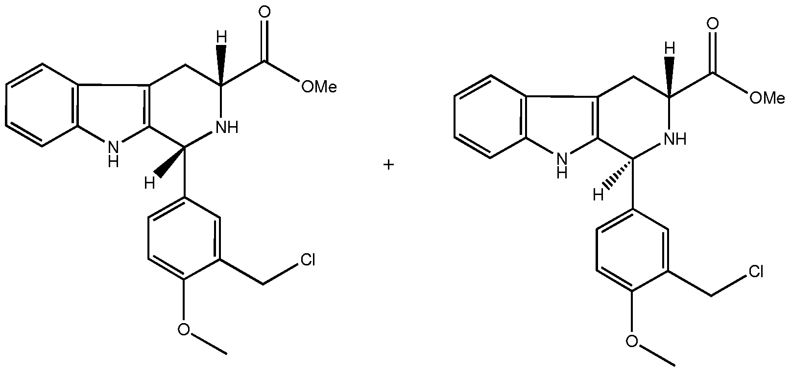
1H NMR (CDCl3) δ 9.88 (br s, IH, indole NH), 7.52-7.07 (m, IH), 6.84 (d, IH, /= 8.4 Hz, ArH), 5.33 (s, IH, d-H, trans), 5.21 (s, IH, d-H, cis), 4.60 (d, 2H, / = 2.6 Hz, ArCH2Cl, cis), 4.58 (d, 2H, / = 6.6 Hz, ArCH2Cl, trans), 3.96 (dd, IH, / = 11.0, 4.7 Hz, C3-H), 3.86 (s, 3H, OCH3, cis), 3.85 (s, 3H, OCH3, trans), 3.80 (s, 3H, -CO2CH3, cis), 3.11 (s, 3H, -CO2CH3, trans), 3.45 (br s, IH, N2-H), 3.22 (ddd, IH, /= 15.0, 4.7, 2.1 Hz, C4- Ha), 3.02 (ddd, IH, / = 15.0, 11.0, 2.4 Hz, C4-Hb) [3.85-2.90 (m, 2H, C4-H)]; 13C NMR (CDCl3) δ 172.9, 157.7, 136.2, 134.2, 130.9, 130.4, 127.0, 126.3, 122.0, 119.6, 118.2, 111.2, 111.0, 108.9, 58.0 (Ci), 56.9 (OCH3), 55.8 (C3), 55.3, 52.2 (OCH3, cis), 52.1 (OCH3, trans), 41.4 (ArCH2Cl, trans), 41.3 (ArCH2Cl, cis), 25.5 (C4).
Intermediate 3B (cis-isomer)
cis-methyl-l-(3-(chloromethyl)-4-methoxyphenyl)-2,3,4,9-tetrahydro-lH-pyrido[3,4- b]indole-3-carboxylate (D3B)

1H NMR (CDCl3) δ 7.54 (br s, IH, indole NH), 7.52-7.07 (m, IH), 6.84 (d, IH, /= 8.4 Hz, ArH), 5.21 (s, IH, Ci-H), 4.58 (d, 2H, / = 3.3 Hz, ArCH2Cl), 3.96 (dd, IH, /= 11.0, 4.7 Hz, C3-H), 3.86 (s, 3H, OMe), 3.77 (s, 3H, -CO2CH3), 3.45 (br s, IH, N2-H), 3.22 (ddd, IH, /= 15.0, 4.7, 2.1 Hz, C4-HJ, 3.02 (ddd, IH, / = 15.0, 11.0, 2.4 Hz, C4-Hb); 13C NMR (CDCl3) δ 172.9, 157.7, 136.2, 134.2, 130.9, 130.4, 127.0, 126.3, 122.0, 119.6, 118.2, 111.2, 111.0, 108.9, 57.9 (d), 56.8 (OCH3), 55.7 (C3), 55.3, 52.2 (OCH3), 41.3 (ArCH2Cl), 25.5 (C4); MS (ESI) mlz 385.5 ([M+H]+).
Intermediate 4
Preparation of cis-methyl-l-(3-((4-(2-fluorophenyl)piperazin-l-yl)methyl)-4- methoxyphenyl)-2,3,4,9-tetrahydro-lH-pyrido[3,4-b]indole-3-carboxylate (D4)

To a solution of ds-methyl l-(3-(chloromethyl)-4-methoxyphenyl)-2,3,4,9-tetrahydro-lH- pyrido[3,4-b]indole-3-carboxylate (55 mg 0.14 mmol; which may be prepared as described in D3B) in dichloromethane (5 ml) was added l-(2-fluorophenyl)-piperazine (31.1 mg, 0.17 mmol) and then stirred with 500 mg of diethylaminomethyl polystyrene resin (Fluka, 3.2 mmol/g). After 12h at room temperature, the resin was then filtered and washed (3x3 ml dichloromethane:methanol=l:l), and the filtrate upon evaporation provided the yellow mixture. The residue was submitted to chromatography (Silica-gel, eluent dichloromethane:Methanol=90:10, v/v), to give a yellow product.
1H NMR (CDCl3) δ 7.60 (br s, IH, indole NH), 7.52 (dd, IH, / = 5.8, 2.9 Hz), 7.37 (d, IH, / = 1.8 Hz), 7.22-6.83 (m, 9H), 6.95 (td, IH, / = 7.5, 0.7 Hz), 5.28 (s, IH, N2-H), 5.15 (S, IH, Ci-H), 3.96 (dd, IH, /= 11.1, 4.2 Hz, C3-H), 3.82 (s, 3H, OCH3), 3.80 (s, 3H, - CO2CH3), 3.63 (s, 2H, Ar-CH2-NR2), 3.24-3.18 (m, IH, C4-H), 3.06 (t, 4H, / = 4.2 Hz, (CH2)2N-Ar), 3.01-2.95 (m, IH, C4-H), 2.67 (br d, 4H / = 2.2 Hz, -N(CH2)2-); 13C NMR (CDCl3) δ 173.2, 158.1 (d), 136.1, 135.1, 132.5, 131.1, 128.3, 127.2, 124.3, 122.3 122.2, 121.8, 119.3, 118.9, 118.1, 116.1, 115.8, 110.9, 110.8, 108.8, 58.0 (Ci), 56.9 (OCH3),
55.8, 55.6 (C3), 53.0, 52.1 (OCH3), 50.4, 25.7 (C4); 19F NMR (CDCl3) δ -122.9; MS (ESI) ml z 529.5 ([M+H]+).
Preparation of cis-l-(3-((4-(2-fluorophenyl)piperazin-l-yl)methyl)-4- methoxyphenyl)-2,3,4,9-tetrahydro- lH-pyrido[3,4-b]indole-3-carboxylic acid (Ned- 19) (El)

To a solution of ds-methyl-l-(3-((4-(2-fluorophenyl)piperazin-l-yl)methyl)-4- methoxyphenyl)-2,3,4,9-tetrahydro-lH-pyrido[3,4-b]indole-3-carboxylate (200 mg, 0.92 mmol; which may be prepared as described in D4) in THF (3 ml) was added 3.0 g of Ambersep 900 OH (Fluka, 3.2 mmol/g), and stirred for 1 d at room temperature. The resin was then filtered and washed (3x3 ml dichloromethane:methanol=l:l), and the filtrate upon evaporation provided the pure product (1.6 mg).
1H NMR (CDCl3) δ 7.60 (br s, IH, indole NH), 7.52 (dd, IH, / = 5.8, 2.9 Hz), 7.37 (d, IH, / = 1.8 Hz), 7.22-6.83 (m, 9H), 6.95 (td, IH, / = 7.5, 0.7 Hz), 5.28 (s, IH, N2-H), 5.15 (S, IH, Ci-H), 3.96 (dd, IH, /= 11.1, 4.2 Hz, C3-H), 3.82 (s, 3H, OCH3), 3.63 (s, 2H, Ar-
CH2-NR2), 3.24-3.18 (m, IH, C4-H), 3.06 (t, 4H, / = 4.2 Hz, (CH2)2N-Ar), 3.01-2.95 (m, IH, C4-H), 2.67 (br d, 4H / = 2.2 Hz, -N(CH2)2-); 19F NMR (CDCl3) δ -122.9; MS (ESI) ml z 515.4 ([M+H]+).
Example 7 - Different TPCs isoforms show differential roles in NAADP-evoked Ca2+ release
It has been shown that human TPC2 and TPCl can mediate enhanced NAADP responses in transfected cells. Similar results were obtained in subsequent studies for mouse TPC2, but failed to show enhanced effects of NAADP-evoked Ca2+ release with mouse TPCl [Zong, X. et al., (2009). Pflugers Arch.], whilst overexpression of human TPCl potentiated responses to NAADP [Brailoiu, E. et al., (2009) J Cell Biol.l86(2):201-9]. In addition, no information regarding the role of TPC3 in the NAADP response was available. To clarify these discrepancies, a systematic comparison of the ability of all three SpTPC isoforms to mediate functional NAADP-evoked Ca2+ responses was carried out.
HEK293 cells expressing mCherry.SpTPCs were assessed for their ability to respond to NAADP in two different ways. Using single-cell imaging to study the pattern of Ca2+ release induced by cell-permeant NAADP-AM [Parkesh, R. et al (2008). Cell Calcium 43, 531-538] NAADP-evoked Ca2+ transients were observed in a small number of HEK293 cells expressing mCherry alone, which probably reflects expression of low levels of endogenous HsTPCl and HsTPC2 in such cells. However, from the population of cells expressing SpTPCl and SpTPCl isoforms, significantly more responded to NAADP-AM with typically a single Ca2+ transient (Fig. 25A). In contrast, almost no cells expressing SpTPC3 responded to NAADP-AM with, surprisingly, even the endogenous NAADP responses being suppressed by SpTPC3 expression (Figures 25A and 25B). Similar results were obtained with cells expressing HA.SpTPC proteins. Importantly, pharmacological agents known to antagonize the NAADP response greatly reduced the ability of SpTPC- expressing cells to respond to NAADP-AM. Thus, bafilomycin Al, which abrogates Ca + storage in acidic organelles, low concentrations of NAADP itself, which desensitizes the NAADP response, and the selective NAADP receptor antagonist, Ned- 19, greatly reduced the responsiveness to NAADP of SpTPC-expressing cells (Figure 25C).
In a different approach, NAADP was delivered directly into the cytosol via a patch pipette, since the configuration allows more detailed kinetics of the Ca2+ response to be observed perhaps due to increased buffering by whole-cell dialysis in concert with use of the higher affinity Ca2+ indicator, fura-2. Compared to controls, cells expressing SpTPCl and SpTPCl gave greater Ca2+ responses to NAADP, which were abolished by bafilomycin Al. Furthermore, the inhibitory effect of SpTPC3 on the NAADP response was also evident (Figure 25D), despite the lysosomal Ca + stores remaining demonstrably replete with Ca2+, as demonstrated by the release of Ca2+ mediated by bafilomycin Al in Ca2+- free buffer (data not shown). Importantly, whilst the NAADP response in control cells was small and monophasic, that observed in SpTPCl- and SpTPC2-expressing cells was biphasic - an initial 'pacemaker' rise followed by an explosive Ca2+-release phase (Figures 25D and 25E) with a longer lasting initial Ca2+ release for SpTPC 1 -expressing cells (Figures 25D and 25F). Application of the competitive IP3R antagonist heparin via the patch pipette, selectively blocked the large Ca2+-release phase, thus unmasking an initial NAADP trigger (Figures 25D to 25E). Importantly, responses to IP3 itself were identical between control cells and those expressing an isoform of SpTPC (data not shown) showing that the effects of NAADP in SpTPC expressing cells cannot be ascribed to enhanced IP3 sensitivity of IP3Rs.
These findings demonstrate that all three TPC isoforms play key roles in NAADP evoked Ca2+ release but in distinct ways that may reflect important functional differences between the NAADP-targeted Ca2+-release channels formed by each TPC isoform, as well as differences in coupling to other cellular Ca2+-release mechanisms. The results are consistent with the trigger hypothesis in which NAADP acting via TPCl and TPC2 provides an initial trigger Ca2+ that is subsequently amplified via Ca2+-induced Ca2+ release (CICR) by recruiting IP3Rs .
Since in the experiments employing NAADP-AM (Figures 25 A-C), the responses are generally all-or-nothing but vary in frequency depending on TPC expression, it is likely that only triggered Ca2+ responses amplified by IP3R recruitment are detected under this protocol. The data thus not only confirm TPCs as NAADP receptor components, but also as fundamental sources of local Ca2+ signals which then trigger regenerative Ca2+ responses via CICR mechanisms as previously predicted on the basis of pharmacological
evidence. In HEK293 cells, this is mediated predominantly via IP3Rs, whilst in SKBR3 this appears to be via RyRs. The apparent looser coupling between SpTPCl and IP3Rs compared with the tighter coupling between SpTPCl and IP3Rs may be based on the differential localization of SpTPCl and SpTPC2 between endosomal and lysosomal Ca2+ stores as outlined below. Surprisingly, it is shown that although TPC3 immunocomplexes bind to NAADP with very high affinity, TPC3 expression does not lead to enhanced NAADP-evoked Ca2+ release and also suppresses activity of endogenous TPCs. It is concluded that there are differences in the functions of the different TPC isoforms in regulating Ca2+ signalling mediated by NAADP.
Example 8 - TPCs differentially localize to subcellular sites of NAADP-induced Ca2+ release
Several lines of evidence have suggested that the NAADP receptor is present in acidic organelles and in line with this it was shown in Example 1 that TPCs are associated with acidic organelles in human HEK293 cells. To carry out a systematic examination of the pattern of localization of the three different TPC isoforms, a variety of approaches were used. Fluorescence microscopy was used to study the locations of mCherry-tagged SpTPCl, SpTPCl and SpTPC3 compared to markers of other cellular components in a heterologous expression system. It was found that all three SpTPC isoforms are associated with acidic organelles in live HEK293 cells, as confirmed by LysoTracker Green co- localization as described below (Figure 26A). In line with this, SpTPC2 and SpTPC3 (either mCherry- or HA-tagged versions) co-localize with LAMP-2 (a marker for late endosomes and lysosomes) in fixed cells. SpTPCl and SpTPC3 also show some co- localization with a marker for recycling endosomes (transferring receptor), further indicating an endo-lysosomal location. Similarly, mCherry.SpTPCl showed some overlap with LAMP-2 staining. Endosomes and lysosomes are known to be part of a very dynamic system involving fusion events that lead to formation of hybrid organelles. In line with this, it has been found that GFP-tagged versions of HsTPCl and HsTPC2 are indeed associated with highly motile vesicles. Although the general pattern of localization of TPCs is to acidic organelles, these findings also highlight important differences in the major distributions of different SpTPC isoforms that may reflect distinct roles for each isoform in mediating NAADP responses.
These localization studies were extended to a more physiological system by performing immuno staining of endogenous SpTPC3 in sea urchin eggs themselves, which revealed a punctate cortical staining pattern, in addition to some localization in deeper puncta (Figure 26B). Whilst SpTPC3 immunofluorescence was specific, that for SpTPCl and SpTPC2 was not blocked by immunogenic peptides (data not shown). Therefore, all three mCherry- tagged SpTPCs were expressed in oocytes from starfish, a related echinoderm gamete system that is especially suited to heterologous mRNA expression. Each SpTPC. mCherry isoform was localized to the cortex, as well as to intracellular puncta, which was more noticeable as visualized by immunostaining (Figure 26C). The prevalent cortical distribution of SpTPCs in echinoderms is in agreement with previous observations that the cortex exhibits the greatest sensitivity to NAADP in both sea urchin eggs and starfish oocytes, thus demonstrating for the first time that endogenous TPCs localize to subcellular loci of NAADP-induced Ca2+ release.
Live cell LysoTracker staining.
Mammalian cells expressing mCherry constructs were grown on poly-D-lysine coated coverslips. Cells were loaded with 200 nM LysoTracker Green DND-26 (Invitrogen) in a buffer containing 121 mM NaCl, 5.4 mM KCl, 0.8 mM MgC12, 2 mM CaC12, 6 mM NaHCCβ, 5.5 mM D-Glucose, 25 mM Hepes, pH 7.4 for 15 min at room temperature, followed by three washes. Cells were viewed with a 63x oil immersion objective on a Zeiss 510 META confocal microscope, in multitrack mode, using the following excitation/emission parameters (nm): LysoTracker Green (488/505-530), mCherry (543/>560). Cells expressing HsTPCl or HsTPC2 were labelled as previously described [4]. Where indicated, cells were incubated with 10 μM Ned-19 [5] for 6 h or 12 h prior to staining with LysoTracker Green. Cells were visualized on a Zeiss Axioskop 2 microscope fitted with a Retiga CCD camera and Metamorph imaging suite.
Example 9 - Altering TPC expression has dramatic, differential effects upon endolysosomal transport and function
Given the importance of Ca2+ signals for endo-lysosomal function, the effect of altering TPC expression upon cellular trafficking was investigated. Endo-lysosomal transport was assessed by following the fate of Alexa Fluor-594-conjugated cholera toxin B subunit that
upon binding to endogenous plasma membrane ganglioside GMl is under normal conditions internalized and delivered to the Golgi via early endosomes.
Trafficking.
BODIPY-LacCer (Molecular Probes) was used at 7.5 μM as previously described [Lloyd- Evans, E. et al., (2008). Nat Med 14, 1247-1255]. For cholera toxin uptake cells grown on coverslips were incubated on ice with 1 μg/ml Alexa Fluor 594-conjugated Cholera toxin B subunit (CtxB) for 30 min to label the plasma membrane. Cells were then washed twice with complete medium and left at 370C to internalize the probe for a further 2 h followed by 3 washes in ice cold complete medium supplemented with 1 % BSA to remove extracellular label. Coverslips were mounted in Vectashield (VectorLabs) prior to visualization. All images were taken on a Zeiss Axioskop 2 microscope fitted with a Retiga CCD camera and Metamorph imaging suite.
Results
It was found that control cells expressing mCherry alone show a normal endo-lysosomal function with cholera toxin accumulating in the Golgi (Figures 27 A and 27B). However, cells expressing mCherry.SpTPCs show an accumulation of cholera toxin within the endo- lysosomal system (Figures 27A and 27B) revealed by the punctate distribution and co- localisation with rhodamine-labelled high molecular weight dextran which endocytoses into lysosomes (data not shown). In this respect, cells expressing SpTPC 1 have the highest degree of such dysfunction, followed by 5pTPC2-expressing cells. Similarly, SpTPC3- expressing cells fail to accumulate cholera toxin within the Golgi, however, and in contrast to the other SpTPC expressing cells, they show a tighter pattern of accumulation close to the nucleus, indicating a possible block in transport at a different point in the endocytic system. Additionally, in HsTPCl and HsTPC2-overexpressing cells, exogenous addition of either cholera toxin, or BODIPY-lactosylceramide, which is, in a similar manner, normally transported from the plasma membrane to the Golgi via the endolysosomal system, resulted in accumulation of both markers within the endolysosomal compartments rather than in the Golgi. These phenotypes mimic those seen in a number of lysosomal storage disorders and are indicative of generalized and fundamental dysfunction within the endocytic pathway. In agreement with these findings, it was found that overexpression of
either SpTPCl or SpTPCl (Figures 27C and 27D) or the human proteins HsTPCl or HsTPC2 (led to dramatically enlarged lysosomes compared to the finer LysoTracker Green labelled punctate structures seen in control cells. In contrast, overexpression of SpTPC3 led to a small reduction in Lysotracker Green labelled punctate structures (Figures 27C and 27D), which may explain the difference observed in the abnormal cholera toxin localisation pattern (Figures 27A and 27B).
It was reasoned that if TPCs are components of the NAADP receptor, then an antagonist of NAADP-evoked Ca2+ release should be able to ameliorate the effects of TPC overexpression. Thus, it was investigated whether the selective NAADP receptor antagonist, Ned-19 had any effect upon cells overexpressing TPCs. It was found that a 6 hr incubation with Ned-19 did not greatly affect control cells, whilst a 12 hr incubation at a low concentration (10 μM) had a small inhibitory effect upon cholera toxin trafficking (Figures 27A and 27B) and induced a small elevation in Lysotracker Green accumulation (Figures 27C and 27D). Importantly, following Ned-19 treatment, cells overexpressing SpTPCl or SpTPCl showed a correction in both cholera toxin trafficking through the endocytic system to the Golgi (Figures 27A and 27B) and a reduction in LysoTracker Green staining consistent with normal lysosomal size and distribution (Figures 27C and 27D), reflecting the pattern seen in the control cells. In contrast, Ned-19 had no effect upon cells overexpressing SpTPC3 (Figures 27 A-D).
These findings demonstrate that perturbing TPC expression leads to defects in endolysosomal transport and function, similar to that found in lysosomal storage disorders, with the three SpTPC isoforms and two HsTPC isoforms displaying differential effects upon the endo-lysosomal system in a manner consistent with their pattern of localization. Thus, cells overexpressing TPCl display defects consistent with general endosomal dysfunction (widespread distribution of cholera toxin, Figure 27A), whilst TPC2 overexpression causes effects that emanate from lysosomal deregulation (more limited distribution of cholera toxin, Figure 27A). In contrast, SpTPC3 overexpression appears to affect the endocytic system differently, as evidenced by the focal punctate perinuclear accumulation of cholera toxin (Figure 27A), which mimics the pattern of localization for SpTPC3.
In addition, it has been demonstrated for the first time that a specific NAADP antagonist, Ned-19, can reverse the effects of TPCl or TPC2 overexpression, further confirming the importance of TPCs as mediators of NAADP responses. These data support a role for NAADP and TPCs in normal endo-lysosomal function, perhaps by providing a mechanism whereby local Ca2+ signals are evoked by TPC activation which in turn may regulate lysosomal biogenesis and other endo-lysosomal processes. It is also interesting to note that two human lysosomal diseases, Niemann-Pick disease and mucolipidosis IV are now known to be associated with dysregulated calcium homeostasis. In Niemann-Pick disease, Ca + storage and NAADP-evoked Ca + release are reduced, whilst in mucolipidosis IV Ca + release is enhanced (data not shown). The cellular pathologies induced by TPC overexpression in this study mirror the complex morphological endo-lysosomal characteristics seen in these diseases. Electron microscopy analysis of HsTPC2- overexpressing cells, which appears to have mainly lysosomal dysfunction, confirms that these cells do resemble lysosomal storage disorder cells with conspicuous presence of heterogeneous multiple lamellar inclusion bodies and zebra bodies in lysosomes from HsTPC2-overexpressing cells (Figure 27E). This is indicative of generalized endocytic dysfunction and storage of multiple lipid species suggesting either a general lipid recycling defect or alternatively a lysosomal pΗ defect resulting in inactivation of acid hydrolases.
Claims
1. An agent that modulates the effect of a two pore calcium channel (TPC) polypeptide for use in the treatment or prophylaxis of a condition, disease or disorder associated with NAADP receptor-mediated biological activity.
2. An agent as claimed in claim 1, in which the agent is an antibody or antibody fragment.
3. An agent as claimed in claim 1, in which the agent is an agonist or an antagonist.
4. An agent as claimed in claim 1, in which the agent is a nucleotide sequence that encodes a TPC polypeptide or a functional fragment thereof.
5. An agent as claimed in claim 1, in which the agent is a nucleotide sequence complementary to at least a portion of an RNA transcript of a TPC gene, wherein the nucleotide sequence is no more than 50 nucleotides.
6. An agent as claimed in claim 1, in which the agent is a double stranded oligonucleotide complementary to at least a portion of an RNA transcript of a TPC gene, wherein the double stranded oligonucleotide comprises a sequence of no more than 25 nucleotide base pairs.
7. An agent for use in the treatment or prophylaxis of a condition, disease or disorder as claimed in any one of claims 1-6, wherein the disease or disorder is selected from the group comprising diabetes, obesity, immune system disorders, premature labour, clotting disorders, lysosomal storage diseases, heart disease, cardiac disorders, asthma, hypertension, infertility, autism, pre-natal depression, growth disorders, cancer, and hair and skin pigment defects.
8. A vector comprising a nucleotide sequence as claimed in claim 4.
9. A method of identifying an agent that modulates the effect of NAADP receptor mediated biological activity, comprising contacting the agent with:
a) a TPC polypeptide or functional fragment thereof; or b) a cell expressing a TPC polypeptide or functional fragment thereof; or
c) a lysosome-containing subcellular fraction obtainable from a cell according to (b);
and detecting binding of the agent and/or detecting a downstream effect.
10. A method of identifying an agent as claimed in claim 9, wherein the agent is for use in the treatment or prophylaxis of a condition, disease or disorder associated with NAADP receptor-mediated biological activity.
11. A method of identifying an agent as claimed in claim 10, wherein the condition is associated with a disease or disorder selected from the group comprising diabetes, obesity, immune system disorders, premature labour, clotting disorders, lysosomal storage diseases, heart disease, cardiac disorders, asthma, hypertension, infertility, autism, prenatal depression, growth disorders, cancer, and hair and skin pigment defects.
12. A kit comprising:
a) a TPC polypeptide or functional fragment thereof; or
b) a cell expressing a TPC polypeptide or functional fragment thereof; or
c) a lysosome-containing subcellular fraction obtainable from a cell according to (b);
and instructions for its use in identifying an agent that modulates the effect of NAADP receptor-mediated biological activity.
13. A method of screening a subject for the likelihood of developing a condition, disease or disorder associated with NAADP receptor-mediated biological activity, comprising a step of analysing a sample that has been obtained from the subject for a polymorphism in the gene which encodes a TPC polypeptide.
14. A method of identifying the origin of symptoms associated with a condition, disease or disorder associated with NAADP receptor-mediated biological activity, comprising a step of analysing a sample that has been obtained from a subject for a polymorphism in the gene which encodes a TPC polypeptide.
15. A method for the treatment or prophylaxis of a condition, disease or disorder associated with NAADP receptor-mediated biological activity, comprising the step of administering an agent that modulates the effect of a two pore calcium channel (TPC) polypeptide to a subject.
Applications Claiming Priority (2)
| Application Number | Priority Date | Filing Date | Title |
|---|---|---|---|
| GB0816782A GB0816782D0 (en) | 2008-09-12 | 2008-09-12 | Novel receptor & uses thereof |
| GBGB0816782.7 | 2008-09-12 |
Publications (2)
| Publication Number | Publication Date |
|---|---|
| WO2010030840A2 true WO2010030840A2 (en) | 2010-03-18 |
| WO2010030840A3 WO2010030840A3 (en) | 2010-06-24 |
Family
ID=39930119
Family Applications (1)
| Application Number | Title | Priority Date | Filing Date |
|---|---|---|---|
| PCT/US2009/056602 Ceased WO2010030840A2 (en) | 2008-09-12 | 2009-09-11 | Novel receptor and uses thereof |
Country Status (2)
| Country | Link |
|---|---|
| GB (1) | GB0816782D0 (en) |
| WO (1) | WO2010030840A2 (en) |
Cited By (2)
| Publication number | Priority date | Publication date | Assignee | Title |
|---|---|---|---|---|
| WO2016024246A1 (en) * | 2014-08-14 | 2016-02-18 | Universita' Degli Studi Di Roma 'la Sapienza' | A therapeutic use of naadp and/or tcp2 antagonists |
| EP3527227A4 (en) * | 2016-10-11 | 2020-09-16 | Industrial Cooperation Foundation Chonbuk National University | USE OF CADPR OR NAADP ANTAGONISTS |
-
2008
- 2008-09-12 GB GB0816782A patent/GB0816782D0/en not_active Ceased
-
2009
- 2009-09-11 WO PCT/US2009/056602 patent/WO2010030840A2/en not_active Ceased
Non-Patent Citations (5)
Cited By (2)
| Publication number | Priority date | Publication date | Assignee | Title |
|---|---|---|---|---|
| WO2016024246A1 (en) * | 2014-08-14 | 2016-02-18 | Universita' Degli Studi Di Roma 'la Sapienza' | A therapeutic use of naadp and/or tcp2 antagonists |
| EP3527227A4 (en) * | 2016-10-11 | 2020-09-16 | Industrial Cooperation Foundation Chonbuk National University | USE OF CADPR OR NAADP ANTAGONISTS |
Also Published As
| Publication number | Publication date |
|---|---|
| GB0816782D0 (en) | 2008-10-22 |
| WO2010030840A3 (en) | 2010-06-24 |
Similar Documents
| Publication | Publication Date | Title |
|---|---|---|
| Bailly et al. | Shedding of kidney injury molecule-1, a putative adhesion protein involved in renal regeneration | |
| Zhao et al. | LILRB2-mediated TREM2 signaling inhibition suppresses microglia functions | |
| JP3837494B2 (en) | Soluble RAGE protein | |
| US7465549B2 (en) | Methods for identifying agents that bind a levetiracetam binding site (LBS) or compete with LEV binding to a LBS of a synaptic vesicle protein 2 (SV2) | |
| WO2002006483A1 (en) | Novel physiologically active peptide and use thereof | |
| UA124104C2 (en) | ANTIBODIES SPECIFIC TO HYPERPHOSPHORYLATED TAU PROTEIN, AND METHODS OF THEIR APPLICATION | |
| Laser et al. | The slow Wallerian degeneration protein, WldS, binds directly to VCP/p97 and partially redistributes it within the nucleus | |
| Tossidou et al. | Tyrosine phosphorylation of CD2AP affects stability of the slit diaphragm complex | |
| EP2268296A1 (en) | Compositions and method for the diagnosis, prevention and treament of alzeimer's disease | |
| JP2011191316A (en) | Antihuman baff antibody | |
| JP2018517402A (en) | Methods for selecting phosphatase selective and non-selective phosphatase inhibitors | |
| Wilkars et al. | Nedd4‐2 regulates surface expression and may affect N‐glycosylation of hyperpolarization‐activated cyclic nucleotide‐gated (HCN)‐1 channels | |
| Kern et al. | Nogo-A couples with Apg-1 through interaction and co-ordinate expression under hypoxic and oxidative stress | |
| WO2010030840A2 (en) | Novel receptor and uses thereof | |
| US20170008959A1 (en) | Ager-peptides and use thereof | |
| JPWO2002015925A1 (en) | Apoptosis control method and apoptosis control polypeptide | |
| CN101027047A (en) | Methods for the identification of agents for the treatment of seizures, neurological diseases, endocrinopathies and hormonal diseases | |
| EP1557670A1 (en) | REGULATION OF INTERACTION BETWEEN RAPL AND Rap1 | |
| JP2003128700A (en) | Soluble rage measurement method | |
| Reis et al. | Expression of cTnI-R145G affects shortening properties of adult rat cardiomyocytes | |
| JP2008526243A (en) | AGER peptides and uses thereof | |
| JP4037268B2 (en) | Human NMDA receptor splice variant | |
| Dagan et al. | Regulation of Store-Operated Ca2+ Entry by SARAF. Cells 2021, 10, 1887 | |
| US5700658A (en) | ICAM-4 materials and methods | |
| JP2004166695A (en) | CONTROL OF RAPL-Rap1 INTERACTION |
Legal Events
| Date | Code | Title | Description |
|---|---|---|---|
| 121 | Ep: the epo has been informed by wipo that ep was designated in this application |
Ref document number: 09813647 Country of ref document: EP Kind code of ref document: A2 |
|
| NENP | Non-entry into the national phase in: |
Ref country code: DE |
|
| 122 | Ep: pct application non-entry in european phase |
Ref document number: 09813647 Country of ref document: EP Kind code of ref document: A2 |