WO2010017198A2 - Fgfr extracellular domain acidic region muteins - Google Patents
Fgfr extracellular domain acidic region muteins Download PDFInfo
- Publication number
- WO2010017198A2 WO2010017198A2 PCT/US2009/052704 US2009052704W WO2010017198A2 WO 2010017198 A2 WO2010017198 A2 WO 2010017198A2 US 2009052704 W US2009052704 W US 2009052704W WO 2010017198 A2 WO2010017198 A2 WO 2010017198A2
- Authority
- WO
- WIPO (PCT)
- Prior art keywords
- ecd
- fgfr4
- seq
- polypeptide
- acid box
- Prior art date
Links
Classifications
-
- C—CHEMISTRY; METALLURGY
- C07—ORGANIC CHEMISTRY
- C07K—PEPTIDES
- C07K14/00—Peptides having more than 20 amino acids; Gastrins; Somatostatins; Melanotropins; Derivatives thereof
- C07K14/435—Peptides having more than 20 amino acids; Gastrins; Somatostatins; Melanotropins; Derivatives thereof from animals; from humans
- C07K14/705—Receptors; Cell surface antigens; Cell surface determinants
- C07K14/71—Receptors; Cell surface antigens; Cell surface determinants for growth factors; for growth regulators
-
- A—HUMAN NECESSITIES
- A61—MEDICAL OR VETERINARY SCIENCE; HYGIENE
- A61P—SPECIFIC THERAPEUTIC ACTIVITY OF CHEMICAL COMPOUNDS OR MEDICINAL PREPARATIONS
- A61P27/00—Drugs for disorders of the senses
- A61P27/02—Ophthalmic agents
-
- A—HUMAN NECESSITIES
- A61—MEDICAL OR VETERINARY SCIENCE; HYGIENE
- A61P—SPECIFIC THERAPEUTIC ACTIVITY OF CHEMICAL COMPOUNDS OR MEDICINAL PREPARATIONS
- A61P35/00—Antineoplastic agents
-
- A—HUMAN NECESSITIES
- A61—MEDICAL OR VETERINARY SCIENCE; HYGIENE
- A61P—SPECIFIC THERAPEUTIC ACTIVITY OF CHEMICAL COMPOUNDS OR MEDICINAL PREPARATIONS
- A61P43/00—Drugs for specific purposes, not provided for in groups A61P1/00-A61P41/00
-
- A—HUMAN NECESSITIES
- A61—MEDICAL OR VETERINARY SCIENCE; HYGIENE
- A61P—SPECIFIC THERAPEUTIC ACTIVITY OF CHEMICAL COMPOUNDS OR MEDICINAL PREPARATIONS
- A61P9/00—Drugs for disorders of the cardiovascular system
-
- A—HUMAN NECESSITIES
- A61—MEDICAL OR VETERINARY SCIENCE; HYGIENE
- A61K—PREPARATIONS FOR MEDICAL, DENTAL OR TOILETRY PURPOSES
- A61K38/00—Medicinal preparations containing peptides
- A61K38/16—Peptides having more than 20 amino acids; Gastrins; Somatostatins; Melanotropins; Derivatives thereof
- A61K38/17—Peptides having more than 20 amino acids; Gastrins; Somatostatins; Melanotropins; Derivatives thereof from animals; from humans
- A61K38/177—Receptors; Cell surface antigens; Cell surface determinants
- A61K38/179—Receptors; Cell surface antigens; Cell surface determinants for growth factors; for growth regulators
-
- C—CHEMISTRY; METALLURGY
- C07—ORGANIC CHEMISTRY
- C07K—PEPTIDES
- C07K14/00—Peptides having more than 20 amino acids; Gastrins; Somatostatins; Melanotropins; Derivatives thereof
- C07K14/435—Peptides having more than 20 amino acids; Gastrins; Somatostatins; Melanotropins; Derivatives thereof from animals; from humans
- C07K14/705—Receptors; Cell surface antigens; Cell surface determinants
-
- C—CHEMISTRY; METALLURGY
- C07—ORGANIC CHEMISTRY
- C07K—PEPTIDES
- C07K2319/00—Fusion polypeptide
- C07K2319/30—Non-immunoglobulin-derived peptide or protein having an immunoglobulin constant or Fc region, or a fragment thereof, attached thereto
-
- C—CHEMISTRY; METALLURGY
- C07—ORGANIC CHEMISTRY
- C07K—PEPTIDES
- C07K2319/00—Fusion polypeptide
- C07K2319/31—Fusion polypeptide fusions, other than Fc, for prolonged plasma life, e.g. albumin
Definitions
- the present invention relates to fibroblast growth factor receptor (FGFR) extracellular domains (ECDs) that have been engineered to exhibit decreased tissue binding by increasing the number of acidic amino acid residues within the D1-D2 linker region.
- FGFR fibroblast growth factor receptor
- ECDs extracellular domains
- the invention further relates to polypeptide and polynucleotide sequences, vectors, host cells, compositions, and kits comprising or encoding such molecules.
- the invention also relates to methods of making and using FGFR ECD acidic region muteins to treat proliferative disorders, including cancer, disorders of angiogenesis, and macular degeneration.
- Fibroblast growth factors and their receptors (FGFRs) are a highly conserved group of proteins with instrumental roles in angiogenesis, vasculogenesis, and wound healing, as well as in tissue patterning and limb formation in embryonic development. FGFs and FGFRs affect cell migration, proliferation, and survival, providing wide-ranging impacts on health and disease.
- the FGFR family comprises four major types of receptors, FGFRl , FGFR2, FGFR3, and FGFR4. These receptors are transmembrane proteins having an extracellular domain (ECD), a transmembrane domain, and an intracytoplasmic tyrosine kinase domain. Each of the extracellular domains contains either two or three immunoglobulin (Ig) domains. When there are three Ig domains, they are referred to as Dl, D2, and D3. Receptors having two Ig domains typically lack Dl. An acidic motif, called the acid box, is located in the linker region between Dl and D2 in the FGFR extracellular domain.
- ECD extracellular domain
- Ig immunoglobulin
- the D2 domain of FGFRs contains a heparin binding site.
- FGFR4 also contains a heparin binding site in Dl.
- the acid box is believed to interact with the heparin binding site in the D2 domain.
- the FGFRl and FGFR3 Dl domains are capable of interacting with the D2 and D3 domains. It has been hypothesized that the FGFRl acid box-mediated interactions with the D2 domain, and the FGFRl Dl domain-mediated interactions with the D2 and D3 domains play an autoinhibitory role that prevents receptor oligomerization in the absence of FGF ligand.
- extracellular FGFR activation by FGF ligand binding to an FGFR initiates a cascade of signaling events inside the cell, beginning with oligomerization of the receptor and activation of receptor tyrosine kinase activity.
- FGFs each with the capacity to bind one or more FGFRs. See, e.g., Zhang et al., J. Biol. Chem. 281:15, 694-15,700 (2006).
- FGFs can bind to and activate each of one or more FGFRs, often with large differences in their affinities for the different FGFRs.
- Heparin sulfate proteoglycan (“heparin") is required for the binding of FGFs to FGFRs under certain circumstances. See, e.g., Ornitz et al., MoI. Cell Biol. 12:240 (1992).
- FGF2 also known as basic FGF (bFGF)
- bFGF basic FGF
- the FGFR4 extracellular domain binds with high affinity to FGF2 and FGF19 ligands, among others.
- ECD extracellular domain
- FGFR4 ECD as a 'ligand trap' to bind, for example, free FGF2 and FGF 19, one may effectively treat proliferative disorders, including cancer, disorders of angiogenesis, and macular degeneration.
- Fc fusions of the wild-type FGFR4 ECD (“FGFR4 ECD Fc fusions”) showed that they exhibited poor bioavailability and a short serum half-life, which is at least partially due to excessive tissue binding, when administered to mice using intravenous (IV) methods. See FIG. 4 and Table 5.
- Fc fusions of the FGFRl ECD exhibited higher bioavailability and serum half-life. Furthermore, the FGFR4 ECD Fc fusions exhibited high levels of in vitro binding to extracellular matrix (ECM) components, whereas Fc fusions of the FGFRl, FGFR2, or FGFR3 ECD showed minimal or undetectable levels of ECM binding. See FIG. 5.
- the FGFRs typically contain an acidic motif, called an acid box, between the Dl and D2 domains.
- the Dl domain and the D1-D2 linker region, which contains the acid box, are more divergent than the D2 and D3 domains between FGFR4 and the other three FGFRs.
- the FGFRl, FGFR2, and FGFR3 acid boxes all contain a greater number of acidic amino acid residues than the FGFR4 acid box. See FIGs. HA, HB, and HC, respectively.
- FGFR4 ECD acidic region muteins engineered FGFR4 ECDs, called FGFR4 ECD acidic region muteins, that have an increase in the total number of acidic residues within the D1-D2 linker and thus a "stronger" acid box region to reduce ECM binding and potentially reduce tissue binding in vivo and to increase the bioavailability of FGFR4 ECD fusion proteins.
- FGFR4 ECD acidic region muteins that contain an increased number of acidic residues within the D1-D2 linker exhibit decreased ECM binding.
- certain FGFR4 ECD acidic region muteins exhibit decreased tissue binding and increased bioavailability.
- FGFR4 ECD acidic region chimeras include FGFR4 ECD D1-D2 linker chimeras, FGFR4 ECD exon 4 chimeras, FGFR4 ECD acid box chimeras, FGFR4 ECD long acid box chimeras, and FGFR4 ECD short acid box chimeras.
- an FGFR4 ECD acidic region mutein exhibited substantially improved bioavailability and serum half-life relative to a parental FGFR4 ECD Fc fusion. See FIG. 8.
- Mice injected with the FGFR4 ECD acidic region mutein showed a statistically significant reduction in tumor burden in certain tumor models when compared to a control group, showing that the FGFR4 ECD acidic region muteins possess a similar anti-tumor activity as FGFR4 ECDs administered in vivo.
- the FGFR4 ECD acidic region muteins may therefore be used, e.g., to treat proliferative disorders, including cancer, disorders of angiogenesis, and macular degeneration.
- both the FGFR2 ECD-Fc and FGFR3 ECD-Fc fusion proteins showed significantly lower levels of ECM binding in vitro than a parental FGFR4 ECD-Fc, however they showed slightly higher levels of ECM binding than an FGFRl ECD-Fc fusion protein at higher protein concentrations. See FIG. 5.
- the FGFR2 and FGFR3 acid boxes contain a greater number of acidic amino acid residues than the FGFR4 acid box, they both contain fewer acidic amino acid residues than the FGFRl acid box. See FIGs. HD and HE, respectively.
- FGFRl, FGFR2, and FGFR3 ECDs may be engineered to have a decrease in the total number of acidic residues within the D1-D2 linker, and thus a "weaker” acid box to increase tissue binding in vivo and to decrease the bioavailability of the FGFRl, FGFR2, and FGFR3 ECD fusion proteins.
- Such "weakened" FGFRl, FGFR2, and FGFR3 ECDs may be useful, for example, when delivered locally, to prevent toxicity and/or side effects that might occur with systemic administration.
- a polypeptide comprising an FGFR4 ECD acidic region mutein is provided.
- an isolated polypeptide comprising an FGFR4 ECD acidic region mutein is provided.
- an FGFR4 ECD acidic region mutein is an FGFR4 ECD D1-D2 linker chimera.
- the FGFR4 ECD D1-D2 linker chimera comprises a D1-D2 linker selected from an FGFRl D1-D2 linker, an FGFR2 D1-D2 linker, and an FGFR3 D1-D2 linker, in place of the FGFR4 D1-D2 linker.
- the FGFR4 ECD D1-D2 linker chimera comprises an amino acid sequence selected from SEQ ID NOs: 22, 26, 28, and
- the FGFR4 ECD D1-D2 linker chimera comprises an amino acid sequence selected from SEQ ID NOs: 35 to 38.
- an FGFR4 ECD acidic region mutein is an FGFR4 ECD exon 4 chimera.
- the FGFR4 ECD exon 4 chimera comprises an exon 4 selected from an FGFRl exon 4, an FGFR2 exon 4, and an FGFR3 exon 4, in place of the FGFR4 exon 4.
- the FGFR4 ECD exon 4 chimera comprises an amino acid sequence selected from SEQ ID NOs: 23, 92, 29, and
- the FGFR4 ECD exon 4 chimera comprises an amino acid sequence selected from SEQ ID NOs: 39 to 42.
- an FGFR4 ECD acidic region mutein is an FGFR4 ECD acid box chimera.
- the FGFR4 ECD acid box chimera comprises an acid box selected from the FGFRl acid box, the FGFR2 acid box, and the FGFR3 acid box, in place of the FGFR4 acid box.
- the FGFR4 ECD acid box chimera comprises an amino acid sequence selected from SEQ ID NOs: 24, 30, and 34 in place of the FGFR4 acid box having an amino acid sequence of SEQ ID NO: 20.
- an FGFR4 ECD acid box chimera comprises an acid box region selected from an FGFRl acid box region, an FGFR2 acid box region, and an FGFR3 acid box region, in place of the FGFR4 acid box.
- the FGFR4 ECD acid box chimera comprises an amino acid sequence selected from SEQ ID NOs: 56 to 65, in place of the FGFR4 acid box having an amino acid sequence of SEQ ID NO: 20.
- the FGFR4 ECD acid box chimera comprises an acid box region selected from an FGFRl acid box region, an FGFR2 acid box region, and an FGFR3 acid box region, in place of an FGFR4 acid box region, hi certain embodiments, the FGFR4 ECD acid box chimera comprises an amino acid sequence selected from SEQ ID NOs: 56 to 65, in place of an FGFR4 acid box region having an amino acid sequence selected from SEQ ID NOs: 46 to 55. In certain embodiments, the FGFR4 ECD acid box chimera comprises an FGFRl acid box region having the amino acid sequence of SEQ ID NO: 56 in place of an FGFR4 acid box region having the amino acid sequence of SEQ ID NO: 51.
- the FGFR4 ECD acid box chimera comprises an acid box selected from the FGFRl acid box, the FGFR2 acid box, and the FGFR3 acid box, in place of an FGFR4 acid box region.
- the FGFR4 ECD acid box chimera comprises an amino acid sequence selected from SEQ ID NOs: 24, 30, and 34 in place of an FGFR4 acid box region having an amino acid sequence selected from SEQ ID NOs: 46 to 55.
- the FGFR4 ECD acid box chimera comprises an amino acid sequence selected from SEQ ID NOs: 43 to 45 and 157.
- an FGFR4 ECD acidic region chimera is an FGFR4 ECD long acid box chimera
- the FGFR4 ECD long acid box chimera comprises a long acid box selected from an FGFRl long acid box, an FGFR2 long acid box, and an FGFR3 long acid box, in place of the FGFR4 long acid box.
- the FGFR4 long acid box chimera comprises an amino acid sequence selected from SEQ ID NOs: 98 to 100, in place of an FGFR4 long acid box selected from SEQ ID NOs: 96 and 97.
- the FGFR4 long acid box chimera comprises an amino acid sequence selected from SEQ ID NOs: 105 to 107.
- an FGFR4 ECD acidic region mutein is an FGFR4 ECD short acid box chimera.
- the FGFR4 ECD short acid box chimera comprises a short acid box selected from an FGFRl short acid box, an FGFR2 short acid box, and an FGFR3 short acid box, in place of the FGFR4 short acid box.
- the FGFR4 ECD short acid box chimera comprises an amino acid sequence selected from SEQ ID NOs: 102 to 104, in place of an FGFR4 short acid box having an amino acid sequence of SEQ ID NO: 101.
- the FGFR4 ECD short acid box chimera comprises an amino acid sequence selected from SEQ ED NOs: 108 to 110.
- the FGFR4 ECD acidic region mutein is an FGFR4 ECD long acid box variant
- the the FGFR4 ECD long acid box variant comprises a variant of the FGFR4 ECD that has an increased number of acidic amino acid residues in the long acid box relative to the FGFR4 wild-type long acid box.
- at least two, three, or four non-acidic residues within the long acid box of the FGFR4 ECD are each independently replaced with an acidic residue selected from GIu (E) and Asp (D).
- at least one acidic residue is inserted between amino acids 103 and 104 of SEQ ID NOs: 1 and 2.
- two acidic residues are inserted between amino acids 103 and 104 of SEQ ID NOs: 1 and 2.
- the number of acidic residues in the FGFR4 long acid box is at least seven.
- FGFR4 ECD residues 104 to 114 (SEQ ID NO: 145) are replaced with FGFRl ECD residues 106 to 117 (SEQ ID NO: 149);
- FGFR4 ECD residues 104 to 114 (SEQ ID NO: 145) are replaced with FGFRl ECD residues 107 to 117 (SEQ ID NO: 150);
- FGFR4 ECD residues 104 to 110 are replaced with FGFRl ECD residues 105-113 (SEQ ID NO: 151);
- FGFR4 ECD residues 113 to 116 (SEQ ID NO: 147) are replaced with FGFRl ECD residues 116-119 (SEQ ID NO: 152); or FGFR4 ECD residues 109
- FGFR4 ECD residues 104 to 114 are replaced with FGFRl ECD residues 106 to 117 (SEQ ID NO: 149); FGFR4 ECD residues 104 to 114 (SEQ ID NO: 145) are replaced with FGFRl ECD residues 107 to 117 (SEQ ID NO: 150); FGFR4 ECD residues 104 to 110 (SEQ ID NO: 146) are replaced with FGFRl ECD residues 105-113 (SEQ ID NO: 151); FGFR4 ECD residues 113 to 116 (SEQ ID NO: 147) are replaced with FGFRl ECD residues L 16-119 (SEQ ID NO: 152); or FGFR4 ECD residues 109 to 113 (SEQ ID NO: 148) are replaced with FGFRl ECD residues 112-116 (SEQ ID NO: 153).
- an FGFR4 ECD fusion molecule comprising an FGFR4 ECD acidic region mutein and a fusion partner is provided.
- an isolated FGFR4 ECD fusion molecule comprising an FGFR4 ECD acidic region mutein and a fusion partner is provided.
- an FGFR4 ECD fusion molecule comprising an amino acid sequence selected from SEQ ID NOs: 35 to 45, 105 to 121, and 157 is provided.
- an isolated FGFR4 ECD fusion molecule comprising an amino acid sequence selected from SEQ ID NOs: 35 to 45,
- an FGFR4 ECD fusion molecule comprising the amino acid sequence of SEQ ID NO: 35, is provided.
- an isolated FGFR4 ECD fusion molecule comprising the amino acid sequence of SEQ ID NO: 35 is provided.
- the fusion partner is selected from Fc, albumin, and polyethylene glycol.
- the fusion partner is Fc.
- an FGFR4 ECD fusion molecule comprising an amino acid sequence selected from SEQ ID NOs: 86 to 88, 124 to 140, 143, 144, and 158 is provided.
- an isolated FGFR4 ECD fusion molecule comprising an amino acid sequence selected from SEQ ID NOs: 86 to 88, 124 to 140, 143, 144, and 158 is provided. In certain embodiments, an FGFR4 ECD fusion molecule comprising the amino acid sequence of SEQ ID NO: 86 is provided. In certain embodiments, an isolated FGFR4 ECD fusion molecule comprising the amino acid sequence of SEQ ED NO: 86 is provided hi certain embodiments, an FGFR4 ECD fusion molecule consisting of the amino acid sequence of SEQ ID NO: 86 is provided, hi certain embodiments, an isolated FGFR4 ECD fusion molecule consisting of the amino acid sequence of SEQ ID NO: 86 is provided.
- a pharmaceutical composition comprising an FGFR4 ECD acidic region mutein and a pharmaceutically acceptable carrier is provided, hi certain embodiments, a polynucleotide comprising a nucleic acid sequence that encodes an FGFR4 ECD acidic region mutein is provided.
- a method of treating an angiogenic disorder in a patient comprising administering to the patient a pharmaceutical composition comprising an FGFR4 ECD acidic region mutein is provided.
- a method of treating cancer in a patient comprising administering to the patient a pharmaceutical composition comprising an FGFR4 ECD acidic region mutein is provided.
- the cancer is selected from colon, liver, lung, breast, and prostate cancers, hi certain embodiments, a method of treating macular degeneration in a patient comprising administering to the patient a pharmaceutical composition comprising an FGFR4 ECD acidic region mutein is provided.
- the FGFR4 acidic region mutein comprises at least one point mutation that inhibits glycosylation.
- the at least one point mutation that inhibits glycosylation is selected from N91 A, N156A, N237A, N269A, N290A, and N301A.
- the FGFR4 acidic region mutein comprises an amino acid sequence selected from SEQ ED NOs: 120, 121, and 168.
- an FGFR2 ECD acidic region mutein is provided.
- the FGFR2 ECD acidic region mutein is an FGFR2 ECD short acid box chimera
- the FGFR2 ECD short acid box chimera comprises at least the FGFRl short acid box in place of at least the FGFR2 short acid box.
- FGFR2 ECD residues 111 to 118 (SEQ BD NO: 155) are replaced with FGFRl ECD residues 105 to 112 (SEQ ID NO: 154).
- the FGFR2 ECD short acid box chimera comprises the amino acid sequence of SEQ ID NO: 122.
- an FGFR3 ECD acidic region mutein is provided.
- the FGFR3 ECD acidic region mutein is an FGFR3 ECD short acid box chimera.
- the FGFR3 ECD short acid box chimera comprises at least the FGFRl short acid box in place of at least the FGFR3 short acid box.
- FGFR3 ECD residues 110 to 117 are replaced with FGFRl ECD residues 105 to 112 (SEQ ID NO: 154).
- the FGFR3 ECD short acid box chimera comprises the amino acid sequence of SEQ ID NO: 123.
- FIG. 1 shows the extracellular domain (ECD) amino acid sequence of FGFR4 with a 17 amino acid C-terminal deletion, which was fused to an Fc domain in the parental FGFR4 ECD-Fc (also referred to herein as "R4Mut4").
- the amino acid sequence in Figure 1 includes the signal peptide, which is cleaved in the mature fusion protein.
- the numbers refer to the amino acid position, and certain domains within the ECD are illustrated in gray above the amino acid numbers.
- the amino acid positions within the signal peptide are given negative values because they are cleaved in the mature fusion protein.
- the first amino acid residue of the mature fusion protein is designated as amino acid position 1.
- the linker between the first and second Ig domains (referred to herein interchangeably as the "linker domain,” “linker region,” “D1-D2 linker,” and “D1-D2 linker region”) is illustrated in a darker gray.
- FIG.2 shows a sequence alignment of the linker domains from FGFRl and FGFR4 and the boundaries and sequence of the swapped regions in the three variants, called FGFR4ECD(ABMutl: deltal7)-Fc (ABMutl), FGFR4ECD(ABMut2: deltal7)-Fc (ABMut2), and FGFR4ECD(ABMut3: deltal7)-Fc (ABMut3). Acidic residues within the D1-D2 linker are indicated with underlining and bold font.
- FIG.3 shows the binding of RlMut4, three different preparations of R4Mut4, R4Mut4 plus heparin, and an IgG control to hepatocytes, detected by flow cytometry, as described in Example 5.
- the number of cells (counts) is shown on the Y- axis, and the X-axis shows the relative fluorescent signal, in log units.
- FIG.4 is a graphical representation of the plasma concentration (ng/ml) in mice of R4Mut4, administered with and without heparin, as described in Example 6.
- the plasma concentration is shown on the Y-axis, and was determined using an FGF2-binding ELISA.
- the X-axis shows the time following administration.
- the dashed line represents the lower limit of reproducible R4Mut4 detectability in the ELISA (approximately 156 ng/ml).
- Each data point represents the average from 5 animals, with the error bars representing the standard error of the mean.
- FIG.5 shows the binding of commercially-available FGFRl ECD-Fc, FGFR2 ECD-Fc, FGFR3 ECD-Fc, and FGFR4 ECD-Fc to Matrigel plates, as described in Example 7.
- the X-axis shows the concentration of the Fc fusion proteins and the Y-axis shows the absorbance at 450 run following incubation of the bound Fc fusion protein with OPD substrate. All binding reactions were carried out in triplicate and the data points represent the average values obtained.
- FIG. 6 shows the binding of RlMut4, R4Mut4, and FGFR4 ECD acidic region chimeras ABMutl, ABMut2, and ABMut3 to Matrigel plates, as described in Example 9.
- the X-axis shows the concentration of the Fc fusion proteins and the Y-axis shows the absorbance at 450 nm following incubation of the bound Fc fusion protein with OPD substrate. All binding reactions were carried out in triplicate and the data points represent the average value obtained.
- FIG. 7 shows the binding of RlMut4, R4Mut4, FGFR4 ECD acidic region chimeras ABMutl, ABMut2, and ABMut3, and an IgG control, to hepatocytes detected by flow cytometry, as described in Example 10.
- the number of cells (counts) is shown on the Y-axis, and the X-axis shows the relative fluorescent signal, in log units.
- FIG.8 is a graphical representation of the plasma concentrations (ng/ml) of R4Mut4 and ABMutl following their administration to mice, as described in Example 11.
- the plasma concentrations are shown on the Y-axis, and were determined using an FGF2- binding ELISA.
- the X-axis shows the time following administration.
- the dashed line represents the lower limit of reproducible R4Mut4 detectability in the ELISA (approximately 8 ng/ml). Data from five animals for each time point are included in the figure.
- FIG.9 shows the results of the xenograft experiment described in Example 12.
- mice were inoculated with tumor cells, and tumor growth was measured after administration of R4Mut4, ABMutl, or vehicle alone.
- the tumor size is shown on the Y- axis, and the number of days following tumor inoculation is shown on the X-axis.
- the dosing schedule for each treatment group is shown in Table 9, and the p-values of each treatment group at days 14 and 21 are shown in Table 10.
- FIG. 10 shows the amino acid sequence of the FGFR4 ECD acidic region, along with the locations of certain regions within the FGFR4 ECD acidic region, as defined herein.
- FIG. 11 shows amino acid sequence alignments between (A) the FGFR4 ECD acidic region and the FGFRl ECD acidic region, (B) the FGFR4 ECD acidic region and the FGFR2 ECD acidic region, (C) the FGFR4 ECD acidic region and the FGFR3 ECD acidic region, (D) the FGFRl ECD acidic region and the FGFR2 ECD acidic region, and (E) the FGFRl ECD acidic region and the FGFR3 ECD acidic region.
- FIG. 12 shows the binding of high concentrations of R4Mut4 and ABMutl fusion proteins expressed in CHO or 293-T cells to Matrigel plates, as described in Example 13.
- the X-axis shows the concentration of the Fc fusion proteins and the Y-axis shows the absorbance at 450 run following incubation of the bound Fc fusion protein with OPD substrate.
- FIG. 13 shows the binding of RlMut4, R4Mut4, ABMutl , R4Mut4(N104D), R4Mut4(P109D), R4Mut4(R113E), and R4Mut4(S116E) fusion proteins to Matrigel plates, as described in Example 14.
- the X-axis shows the concentration of the Fc fusion proteins and the Y-axis shows the absorbance at 450 ran following incubation of the bound Fc fusion protein with OPD substrate.
- FIG. 14 shows the binding of RlMut4, R4Mut4, ABMutl, R4(104- 114):R1(106-117), R4(104-114):R1(107-117), R4(104-110):R1(105-113), R4(113- 116):R1(116-119), and R4(109-113):Rl(112-116) fusion proteins to Matrigel plates, as described in Example 15.
- the X-axis shows the concentration of the Fc fusion proteins and the Y-axis shows the absorbance at 450 nm following incubation of the bound Fc fusion protein with OPD substrate.
- FIG. 15 shows the binding of R4Mut4, ABMutl , ABMutl(N91 A), and ABMutl (Nl 59A) fusion proteins to Matrigel plates, as described in Example 16.
- the X- axis shows the concentration of the Fc fusion proteins and the Y-axis shows the absorbance at 450 nm following incubation of the bound Fc fusion protein with OPD substrate.
- FIG. 16 shows the binding of the RlMut4, R4Mut4, ABMutl, R4(D1- D2):R2(D1-D2), and R4(D1-D2):R3(D1-D2) fusion proteins to Matrigel plates, as described in Example 17.
- the X-axis shows the concentration of the Fc fusion proteins and the Y-axis shows the absorbance at 450 nm following incubation of the bound Fc fusion protein with OPD substrate.
- FIG. 17 shows the binding of FGFR2 ECD-Fc, FGFR3 ECD-Fc, R2(l 11- 118):R1(1O5-112), and R3(l 10-117):R1(1O5-112) fusion proteins to Matrigel plates, as described in Example 18.
- the X-axis shows the concentration of the Fc fusion proteins and the Y-axis shows the absorbance at 450 nm following incubation of the bound Fc fusion protein with OPD substrate.
- nucleic acid molecule and “polynucleotide” may be used interchangeably, and refer to a polymer of nucleotides. Such polymers of nucleotides may contain natural and/or non-natural nucleotides, and include, but are not limited to, DNA, RNA, and PNA.
- polypeptide and protein are used interchangeably, and refer to a polymer of amino acid residues. Such polymers of amino acid residues may contain natural and/or non-natural amino acid residues, and include, but are not limited to, peptides, oligopeptides, dimers, trimers, and multimers of amino acid residues.
- polypeptide and protein include natural and non-natural amino acid sequences, and both full-length proteins and fragments thereof. Those terms also include post- translationally modified polypeptides and proteins, including, for example, glycosylated, sialylated, acetylated, and/or phosphorylated polypeptides and proteins.
- acidic amino acid refers to an amino acid residue that is negatively charged at physiological pH.
- Acidic amino acids include, but are not limited to, aspartic acid (Asp, D) and glutamic acid (GIu, E).
- non-acidic amino acid refers to an amino acid residue that is not negatively charged at physiological pH.
- FGFR extracellular domain and "FGFR ECD” include FGFRl ECDs, FGFR2 ECDs, FGFR3 ECDs, and FGFR4 ECDS, as defined herein.
- FGFRl extracellular domain and "FGFRl ECD” include native FGFRl ECDs, FGFRl ECD fragments, and FGFRl ECD variants.
- native FGFRl ECD refers to an FGFRl ECD having an amino acid sequence selected from SEQ ID NOs: 21 and 25.
- FGFRl ECD fragment refers a polypeptide having an amino acid sequence selected from SEQ ID NOs: 21 and 25, but wherein amino acid residues have been deleted from the amino- terminus and/or carboxy-terminus, wherein the fragments are capable of binding to FGF2.
- FGFRl ECD variants refers to variants of the portion of the FGFRl polypeptide that extends into the extracellular space and variants of fragments thereof that comprise Dl, D2, and D3, wherein the variants are capable of binding to FGF2.
- Such variants may contain amino acid additions, deletions, and substitutions, provided that the FGFRl ECD variants remain capable of ligand binding.
- an FGFRl ECD lacks a signal peptide, hi certain embodiments, an FGFRl ECD includes at least one signal peptide, which may be selected from a native FGFRl signal peptide and/or a heterologous signal peptide.
- FGFR2 extracellular domain and "FGFR2 ECD” include native FGFR2 ECDs, FGFR2 ECD fragments, and FGFR2 ECD variants.
- native FGFR2 ECD refers to an FGFR2 ECD having an amino acid sequence of SEQ ID NO: 27.
- FGFR2 ECD fragment refers a polypeptide having an amino acid sequence selected from SEQ ID NO: 27, but wherein amino acid residues have been deleted from the amino-terminus and/or carboxy-terminus, wherein the fragments are capable of binding to FGF2.
- a non-limiting exemplary FGFR2 ECD fragment has the amino acid sequence of SEQ ID NO: 160, which corresponds to the amino acid sequence of SEQ ID NO: 27, but with the last three carboxy-terminal amino acid residues, YLE, deleted.
- FGFR2 ECD variants refers to variants of the portion of the FGFR2 polypeptide that extends into the extracellular space and variants of fragments thereof that comprise Dl, D2, and D3, wherein the variants are capable of binding to FGF2.
- Such variants may contain amino acid additions, deletions, and substitutions, provided that the FGFR2 ECD variant remains capable of ligand binding.
- FGFR2 ECD variants may include amino acid substitutions within the FGFR2 ECD that inhibit N-glycosylation, referred to interchangeably herein as “FGFR2 ECD glycosylation mutants" and "FGFR2 ECD N-glycan mutants.”
- at least one amino acid within the FGFR2 ECD is mutated to prevent glycosylation at that site in the polypeptide.
- Non-limiting exemplary FGFR2 ECD amino acids that maybe glycosylated include N62, Nl 02, N207, N220, N244, N276, N297, and N310 in SEQ ID NO: 27.
- Non-limiting exemplary amino acid mutations in FGFR4 ECD glycosylation mutants include N62A, N102A, N207A, N220A, N244A, N276A, N297A, and N310A in SEQ ID NO: 27.
- an FGFR2 ECD lacks a signal peptide
- an FGFR2 ECD includes at least one signal peptide, which maybe selected from a native FGFR2 signal peptide and/or a heterologous signal peptide.
- the terms "FGFR3 extracellular domain" and "FGFR3 ECD” include native FGFR3 ECDs, FGFR3 ECD fragments, and FGFR3 ECD variants.
- the term "native FGFR3 ECD” refers to an FGFR3 ECD having an amino acid sequence of SEQ ID NO: 31.
- FGFR3 ECD fragment refers a polypeptide having an amino acid sequence selected from SEQ ED NO: 31, but wherein amino acid residues have been deleted from the amino-terminus and/or carboxy-terminus, wherein the fragments are capable of binding to FGF2.
- a non-limiting exemplary FGFR3 ECD fragment has the amino acid sequence of SEQ ID NO: 161, which corresponds to the amino acid sequence of SEQ ID NO: 31, but with the last three carboxy-terminal amino acid residues, YAG, deleted.
- FGFR3 ECD variants refers to variants of the portion of the FGFR3 polypeptide that extends into the extracellular space and variants of fragments thereof that comprise Dl, D2, and D3, wherein the variants are capable of binding to FGF2.
- Such variants may contain amino acid additions, deletions, and substitutions, provided that the FGFR3 ECD variant remains capable of ligand binding.
- FGFR3 ECD variants may include amino acid substitutions within the FGFR3 ECD that inhibit N-glycosylation, referred to interchangeably herein as "FGFR3 ECD glycosylation mutants" and "FGFR3 ECD N-glycan mutants.”
- at least one amino acid within the FGFR3 ECD is mutated to prevent glycosylation at that site in the polypeptide.
- Non-limiting exemplary FGFR3 ECD amino acids that may be glycosylated include N76, N203, N240, N272, N293, and N306 in SEQ ED NO: 31.
- Non-limiting exemplary amino acid mutations in FGFR3 ECD glycosylation mutants include N76A, N203A, N240A, N272A, N293A, and N306A in SEQ ID NO: 31.
- an FGFR3 ECD lacks a signal peptide.
- an FGFR3 ECD includes at least one signal peptide, which maybe selected from a native FGFR3 signal peptide and/or a heterologous signal peptide.
- FGFR4 extracellular domain and "FGFR4 ECD” include native FGFR4 ECDs, FGFR4 ECD fragments, and FGFR4 ECD variants.
- native FGFR4 ECD refers to an FGFR4 ECD having an amino acid sequence selected from SEQ ID NOs: 1, 2, 3, and 93.
- FGFR4 ECD fragment refers a polypeptide having an amino acid sequence selected from SEQ EO NOs: 1, 2, 3, and 93, but wherein all or a portion of the sequence LEASEEVE (SEQ ED NO: 70) has been deleted from the amino terminus and/or all or a portion of the sequence LPEEDPTWTAAAPEARYTD (SEQ ED NO: 71) has been deleted from the carboxy terminus of the polypeptide, wherein the fragments are capable of binding to FGF2 and/or FGFl 9.
- Non-limiting exemplary FGFR4 ECD fragments have the amino acid sequences shown in SEQ ID NOs: 6 to 10 and 76 to 81.
- FGFR4 ECD variants refers to variants of the portion of the FGFR4 polypeptide that extends into the extracellular space and variants of fragments thereof that comprise Dl, D2, and D3, wherein the variants are capable of binding to FGF2 and/or FGFl 9.
- Such variants may contain amino acid additions, deletions, and substitutions, provided that the FGFR4 ECD variant remains capable of binding to FGF2 and/or FGFl 9 (see below for a discussion of the structure/function relationship of FGFR extracellular domains).
- FGFR4 ECD variants may include amino acid substitutions within the FGFR4 ECD that inhibit N-glycosylation, referred to interchangeably herein as "FGFR4 ECD glycosylation mutants" and "FGFR4 ECD N-glycan mutants.”
- one or more amino acids within the FGFR4 ECD are mutated to prevent glycosylation at that site in the polypeptide.
- Non-limiting exemplary FGFR4 ECD amino acids that may be glycosylated include N91, N156, N237, N269, N290, and N301 in SEQ ID NOs: 1 and 2.
- Non-limiting exemplary amino acid mutations in FGFR4 ECD glycosylation mutants include N91A, N156A, N237A, N269A, N290A, and N301A in SEQ ID NOs: 1 and 2.
- an FGFR4 ECD lacks a signal peptide.
- an FGFR4 ECD includes at least one signal peptide, which may be selected from a native FGFR4 signal peptide and/or a heterologous signal peptide.
- FGFR42Ig extracellular domain and "FGFR4 2Ig ECD” include FGFR4 2Ig ECDs, FGFR42Ig ECD fragments, and FGFR4 2Ig ECD variants.
- FGFR4 2Ig ECD refers to a polypeptide comprising the acid box and domains D2 and D3 , wherein the polypeptide has an amino acid sequence selected from SEQ ID NOs: 1, 2, 3, and 93, but with at least a portion of Dl deleted.
- Exemplary FGFR4 2Ig ECDs have the amino acid sequence of SEQ ID NO: 94.
- FGFR4 2Ig ECD fragment refers to an FGFR42Ig ECD polypeptide wherein all or a portion of the sequence LPEEDPTWTAAAPEARYTD (SEQ ID NO: 71) has been deleted from the carboxy-terminus of the polypeptide.
- FGFR4 2Ig ECD variants refers to variants of the FGFR42Ig ECDs and FGFR4 2Ig ECD fragments discussed above, wherein the variants are capable of binding to FGF2 and/or FGF 19.
- Such variants may contain amino acid additions, deletions, and substitutions, provided that the FGFR4 2 Ig ECD variant remains capable of binding to FGF2 and/or FGF 19 (see below for a discussion of the structure/function relationship of FGFR extracellular domains).
- An "FGFR4 ECD D1-D2 linker chimera” refers to an FGFR4 ECD selected from native FGFR4 ECDs, FGFR4 ECD fragments, and FGFR4 ECD variants, in which the linker region between immunoglobulin-like domain I (Dl) and immunoglobulin-like domain II (D2) (referred to herein interchangeably as the "linker domain,'" “linker region,” “D1-D2 linker,” and “D1-D2 linker region”) has been replaced with the D1-D2 linker region from FGFRl, FGFR2, or FGFR3.
- the D1-D2 linker of the FGFR4 ECD has the sequence DSLTSSNDDEDPKSHRDPSNRHSYPQQ (SEQ ID NO: 16), which is amino acids 98 to 124, inclusive, of SEQ ID NO: 1; or has the sequence DSLTSSNDDEDPKSHRDLSNRHSYPQQ (SEQ ID NO: 17), which is amino acids 98 to 124, inclusive, of SEQ ID NO: 2.
- the D1-D2 linker of FGFRl has the sequence DALPSSEDDDDDDDSSSEEKETDNTKPNPV (SEQ ID NO: 22), which is amino acids 99 to 128, inclusive, of SEQ ED NO: 21; or has the sequence
- DALPSSEDDDDDDDSSSEEKETDNTKPNRMPV (SEQ ID NO: 26), which is amino acids 99 to 130, inclusive, of SEQ ID NO: 25.
- the D1-D2 linker of FGFR2 has the sequence DAISSGDDEDDTDGAEDFVSENSNNKR (SEQ ID NO: 28), which is amino acids 105 to 131, inclusive, of SEQ ID NO: 27.
- the D1-D2 linker of FGFR3 has the sequence DAPSSGDDEDGEDEAEDTGVDTG (SEQ ID NO: 32), which is amino acids 105 to 127, inclusive, of SEQ ID NO: 31.
- Certain exemplary FGFR4 ECD D1-D2 linker chimeras include, but are not limited to, FGFR4 ECD D1-D2 linker chimeras having the amino acid sequences of SEQ ID NOs: 35 to 38.
- FGFR42Ig ECD D1-D2 linker chimera refers to an FGFR42Ig ECD selected from FGFR4 2Ig ECDs, FGFR42Ig ECD fragments, and FGFR42Ig ECD variants, in which the linker region between immunoglobulin-like domain I (Dl) and immunoglobulin-like domain II (D2) (referred to herein interchangeably as the "linker domain,” “linker region,” “D1-D2 linker,” and “D1-D2 linker region”) has been replaced with the D1-D2 linker region from FGFRl, FGFR2, or FGFR3, as described above for FGFR4 ECD D1-D2 linker chimeras.
- Dl immunoglobulin-like domain I
- D2 immunoglobulin-like domain II
- corresponding amino acid residue and “corresponding residue” are used interchangeably herein to refer to an amino acid residue or a gap in the amino acid sequence (as indicated by "-") of a first FGFR ECD D1-D2 linker region that lines up with an amino acid residue or a gap in the amino acid sequence (as indicated by "- ") of a second FGFR ECD D1-D2 linker region shown in a sequence alignment.
- the corresponding amino acid residues between FGFR4 and FGFRl are shown in FIG. HA.
- the corresponding amino acid residues between FGFR4 and FGFR2 are shown in FIG. HB.
- the corresponding amino acid residues between FGFR4 and FGFR3 are shown in FIG. HC.
- the corresponding amino acid residues between FGFRl and FGFR2 are shown in FIG. HD.
- the corresponding amino acid residues between FGFRl and FGFR3 are shown in FIG. HE. hi certain embodiments, the amino acid residue of a first FGFR ECD is replaced by the corresponding amino acid residue of a second FGFR ECD.
- the amino acid residue of the first FGFR ECD when the corresponding amino acid of the second FGFR ECD is a gap, the amino acid residue of the first FGFR ECD is deleted, hi certain such embodiments, when the amino acid residue of the first FGFR ECD is a gap, the corresponding amino acid residue of the second FGFR ECD is inserted into the first FGFR ECD.
- corresponding amino acid sequence and “corresponding sequence” are used interchangeably herein to refer to the sequence of amino acid residues within a particular region of an FGFR ECD.
- FGFR4 ECD exon 4 chimera refers to an FGFR4 ECD selected from native FGFR4 ECDs, FGFR4 ECD fragments, and FGFR4 ECD variants, in which the amino acid sequence encoded by exon 4 (referred to herein interchangeably as “exon 4" or "exon 4 region”) has been replaced with the amino acid sequence encoded by exon 4 from FGFRl, FGFR2, or FGFR3.
- Exon 4 of the FGFR4 ECD encodes the sequence DSLTSSNDDEDPKSHRDPSNRHSYPQ (SEQ ID NO: 18), which is amino acids 98 to 123, inclusive, of SEQ ID NO: 1; or encodes the sequence
- DSLTSSNDDEDPKSHRDLSNRHSYPQ (SEQ ID NO: 19), which is amino acids 98 to 123, inclusive, of SEQ ID NO: 2.
- Exon 4 of FGFRl encodes the sequence DALPSSEDDDDDDDSSSEEKETDNTKPN (SEQ ID NO: 23), which is amino acids 99 to 126, inclusive, of SEQ ID NO: 21; or encodes the sequence
- DALPSSEDDDDDDDSSSEEKETDNTKPNRM (SEQ ID NO: 92), which is amino acids 99 to 128, inclusive, of SEQ ID NO: 25.
- Exon 4 of FGFR2 encodes the sequence DAISSGDDEDDTDGAEDFVSENSNNK (SEQ ID NO: 29), which is amino acids 105 to 130, inclusive, of SEQ ID NO: 27.
- Exon 4 of FGFR3 encodes the sequence DAPSSGDDEDGEDEAEDTGVDT (SEQ ID NO: 33), which is amino acids 105 to 126, inclusive, of SEQ ID NO: 31.
- Certain exemplary FGFR4 ECD exon 4 chimeras include, but are not limited to, FGFR4 ECD exon 4 chimeras having the amino acid sequences of SEQ ID NOs: 39 to 42.
- FGFR4 2Ig ECD exon 4 chimera refers to an FGFR4 2Ig ECD selected from FGFR4 2Ig ECDs, FGFR4 2Ig ECD fragments, and FGFR42Ig ECD variants, in which the amino acid sequence encoded by exon 4 (referred to herein interchangeably as “exon 4" or "exon 4 region") has been replaced with the amino acid sequence encoded by exon 4 from FGFRl, FGFR2, or FGFR3, as described above for FGFR4 ECD exon 4 chimeras.
- FGFR4 ECD acid box chimera refers to an FGFR4 ECD selected from native FGFR4 ECDs, FGFR4 ECD fragments, and FGFR4 ECD variants, in which at least the acid box has been replaced with at least the acid box from FGFRl, FGFR2, or FGFR3.
- the acid box of FGFR4 has the sequence DDEDPKSHR (SEQ ID NO: 20).
- the acid box of FGFRl has the sequence EDDDDDDDSS SE (SEQ ID NO: 24).
- the acid box of FGFR2 has the sequence DDEDDTD (SEQ ID NO: 30).
- the acid box of FGFR3 has the sequence DDEDGE (SEQ ID NO: 34).
- FGFR4 2Ig ECD acid box chimera refers to an FGFR4 ECD selected from FGFR42Ig ECDs, FGFR42Ig ECD fragments, and FGFR42Ig ECD variants, in which at least the acid box has been replaced with at least the acid box from FGFRl, FGFR2, or FGFR3, as described above for FGFR4 ECD acid box chimeras.
- the term "acid box region” means a region of an FGFR ECD that includes the acid box defined above, along with additional amino acids from the FGFR ECD sequence on either the ammo-terminus, the carboxy-terminus, or both the amino-terminus and the carboxy-terminus of the acid box, up to and including all of the additional amino acids found in the D1-D2 linker of the FGFR ECD, as defined above.
- the term FGFR4 ECD acid box chimera includes polypeptides in which the acid box of FGFR4 is replaced with an acid box region from FGFRl, FGFR2, or FGFR3.
- FGFR4 ECD acid box chimera also includes polypeptides in which an acid box region of FGFR4 is replaced with an acid box region from FGFRl, FGFR2, or FGFR3.
- the term FGFR4 ECD acid box chimera also includes polypeptides in which an acid box region of FGFR4 is replaced with the acid box from FGFRl, FGFR2, or FGFR3.
- Certain exemplary FGFR4 ECD acid box chimeras include, but are not limited to, FGFR4 ECD acid box chimeras having the amino acid sequences of SEQ ID NOs: 43 to 45 and 157.
- a "long acid box” refers to a region of an FGFR ECD that includes the acid box and certain additional amino acid residues on the amino-terrninus and/or carboxy-terminus of the acid box.
- the long acid box of the FGFR4 ECD has the sequence NDDEDPKSHRDPSNR (SEQ ID NO: 96), which is amino acids 104 to 118, inclusive, of SEQ ID NO: 1 ; or has the sequence NDDEDPKSHRDLSNR (SEQ ID NO: 97), which is amino acids 104 to 118, inclusive, of SEQ ID NO: 2.
- the long acid box of the FGFRl ECD has the sequence EDDDDDDDSSSEEKETD (SEQ ID NO: 98), which is amino acids 105 to 121, inclusive, of SEQ ID NOs: 21 and 25.
- the long acid box of the FGFR2 ECD has the sequence DDEDDTDGAEDFVSE (SEQ ID NO: 99), which is amino acids 111 to 125, inclusive, of SEQ ID NO: 27.
- the long acid box of the FGFR3 ECD has the sequence GDDEDGEDE AED (SEQ ID NO: 100), which is amino acids 110 to 121, inclusive, of SEQ ID NO: 31.
- FGFR4 ECD long acid box chimera refers to an FGFR4 ECD selected from native FGFR4 ECDs, FGFR4 ECD fragments, and FGFR4 ECD variants, in which at least the long acid box, but not more than the D1-D2 linker region, has been replaced with at least the long acid box, but not more than the Dl -D2 linker region, from FGFRl, FGFR2, or FGFR3.
- FGFR42Ig ECD long acid box chimera refers to an FGFR4 ECD selected from native FGFR42Ig ECDs, FGFR4 2Ig ECD fragments, and FGFR42 Ig ECD variants, in which at least the long acid box, but not more than the D1-D2 linker region, has been replaced with at least the long acid box, but not more than the D1-D2 linker region, from FGFRl, FGFR2, or FGFR3.
- short acid box refers to a region of an FGFR ECD having a stretch of consecutive acidic amino acid residues within the acid box.
- the short acid box of the FGFR4 ECD has the sequence DDED (SEQ ID NO: 101), which is amino acids 105 to 108, inclusive, of SEQ ID NOs: 1 and 2.
- the short acid box of the FGFRl ECD has the sequence EDDDDDDD (SEQ ID NO: 102), which is amino acids 105 to 112, inclusive, of SEQ ID NOs: 21 and 25.
- the short acid box of the FGFR2 ECD has the sequence DDEDD (SEQ ID NO: 103), which is amino acids 111 to 115, inclusive, of SEQ ID NO: 27.
- the short acid box of the FGFR3 ECD has the sequence DDED (SEQ ID NO: 104), which is amino acids 111 to 114, inclusive, of SEQ ID NO: 31.
- FGFR4 ECD short acid box chimera refers to an FGFR4 ECD selected from native FGFR4 ECDs, FGFR4 ECD fragments, and FGFR4 ECD variants, in which at least the short acid box, but not more than the D1-D2 linker region, from FGFR4 has been replaced with at least the short acid box, but not more than the Dl- D2 linker region, from FGFRl, FGFR2, or FGFR3.
- FGFR42Ig ECD short acid box chimera refers to an FGFR4 ECD selected from native FGFR42Ig ECDs, FGFR42Ig ECD fragments, and FGFR42Ig ECD variants, in which at least the short acid box, but not more than the D1-D2 linker region, from FGFR4 has been replaced with at least the short acid box, but not more than the D1-D2 linker region, from FGFRl, FGFR2, or FGFR3.
- FGFR2 ECD short acid box chimera refers to an FGFR2 ECD selected from native FGFR2 ECDs, FGFR2 ECD fragments, and FGFR2 ECD variants in which at least the short acid box, but not more than the D1-D2 linker region, from FGFR2 has been replaced with at least the short acid box, but not more than the Dl- D2 linker region, from FGFRl .
- the FGFR2 ECD short acid box chimera at least the FGFR2 acid box, but not more than the D1-D2 linker region, is replaced with the FGFRl short acid box.
- FGFR3 ECD short acid box chimera refers to an FGFR3 ECD selected from native FGFR3 ECDs, FGFR3 ECD fragments, and FGFR3 ECD variants in which at least the short acid box, but not more than the D1-D2 linker region, from FGFR3 has been replaced with at least the short acid box, but not more than the Dl- D2 linker region, from FGFRl.
- FGFR3 ECD short acid box chimera at least the FGFR3 acid box, but not more than the D1-D2 linker region, is replaced with the FGFRl short acid box.
- FGFR4 ECD acidic region chimera is used herein for convenience to refer to the following five types of molecules: FGFR4 ECD D1-D2 linker chimeras, FGFR4 ECD exon 4 chimeras, FGFR4 ECD acid box chimeras, FGFR4 ECD long acid box chimeras, and FGFR4 ECD short acid box chimeras.
- FGFR4 2Ig ECD acidic region chimera is used herein for convenience to refer to the following five types of molecules: FGFR4 2Ig ECD D1-D2 linker chimeras, FGFR42Ig ECD exon 4 chimeras, FGFR42Ig ECD acid box chimeras, FGFR4 2Ig ECD long acid box chimeras, and FGFR4 2Ig ECD short acid box chimeras.
- FGFR4 ECD long acid box variant refers to variants of the FGFR4 ECD selected from native FGFR4 ECDs, FGFR4 ECD fragments, and FGFR4 ECD variants, that have increased acidity in the long acid box relative to the FGFR4 wild- type long acid box.
- at least two non-acidic residues within the long acid box of the FGFR4 ECD are each independently replaced with an acidic residue.
- at least one residue within the long acid box of the FGFR4 ECD is replaced with the corresponding amino acid residue from FGFRl, FGFR2, or FGFR3.
- At least one acidic residue within the long acid box of the FGFR4 ECD is replaced with a different acidic residue.
- one or two acidic residues are inserted between amino acids 103 and 104 of SEQ ID NOs: 1 and 2.
- up to three non-acidic residues are deleted from the long acid box of the FGFR4 ECD.
- the total number of acidic residues within the long acid box of an FGFR4 ECD acidic region variant, including any acidic residues inserted between amino acids 103 and 104 of SEQ ID NOs: 1 and 2, is at least seven.
- FGFR42Ig ECD long acid box variant refers to variants of the FGFR4 ECD selected from native FGFR42Ig ECDs, FGFR4 2Ig ECD fragments, and FGFR42Ig ECD variants, that have increased acidity in the long acid box relative to the FGFR4 wild-type long acid box.
- at least two non-acidic residues within the long acid box of the FGFR42Ig ECD are replaced with an acidic residue.
- At least one residue within the long acid box of the FGFR4 2Ig ECD is replaced with the corresponding amino acid residue from FGFRl, FGFR2, or FGFR3.
- at least one acidic residue within the long acid box of the FGFR4 2Ig ECD is replaced with a different acidic residue.
- one or two acidic residues are inserted between amino acids 15 and 16 of SEQ ID NO: 94, which is an exemplary FGFR42Ig ECD.
- FGFR42Ig ECD long acid box variants up to three non-acidic residues are deleted from the long acid box of the FGFR4 ECD. In certain embodiments of FGFR42Ig ECD long acid box variants, the total number of acidic residues within the long acid box of an FGFR42Ig ECD acidic region variant, including any acidic residues inserted between amino acids 15 and 16 of SEQ ID NO: 94, is at least seven.
- An "FGFR4 ECD acidic region mutein" is an FGFR4 ECD selected from native FGFR4 ECDs, FGFR4 ECD fragments, and FGFR4 ECD variants having a greater number of acidic residues in the D1-D2 linker region than the wild-type FGFR4 ECD.
- FGFR4 ECD acidic region mutein is used herein to refer to the following types of molecules: FGFR4 ECD acidic region chimeras, including FGFR4 ECD D1-D2 linker chimeras, FGFR4 ECD exon 4 chimeras, FGFR4 ECD acid box chimeras, FGFR4 ECD long acid box chimeras, and FGFR4 ECD short acid box chimeras; and FGFR4 ECD long acid box variants.
- An "FGFR4 2Ig ECD acidic region mutein" is an FGFR4 2Ig ECD selected from native FGFR42Ig ECDs, FGFR42Ig ECD fragments, and FGFR4 2Ig ECD variants having a greater number of acidic residues in the D1-D2 linker region than the wild-type FGFR42Ig ECD.
- FGFR42Ig ECD acidic region mutein is used herein to refer to the following types of molecules: FGFR42Ig ECD acidic region chimeras, including FGFR42Ig ECD D1-D2 linker chimeras, FGFR4 2Ig ECD exon 4 chimeras, FGFR42Ig ECD acid box chimeras, FGFR42Ig ECD long acid box chimeras, and FGFR42Ig ECD short acid box chimeras; and FGFR42Ig ECD long acid box variants.
- An "FGFR2 ECD acidic region mutein" is an FGFR2 ECD selected from native FGFR2 ECDs, FGFR2 ECD fragments, and FGFR2 ECD variants having a greater number of acidic residues in the D1-D2 linker region than the wild-type FGFR2 ECD.
- An "FGFR3 ECD acidic region mutein" is an FGFR3 ECD selected from native FGFR3 ECDs, FGFR3 ECD fragments, and FGFR3 ECD variants having a greater number of acidic residues in the D1-D2 linker region than the wild-type FGFR3 ECD.
- FGFR4 ECD fusion molecule refers to a molecule comprising a polypeptide selected from an FGFR4 ECD and an FGFR4 ECD acidic region mutein, and a fusion partner.
- FGFR4 2Ig ECD fusion molecule refers to a molecule comprising a polypeptide selected from an FGFR4 2Ig ECD and an FGFR42Ig ECD acidic region mutein, and a fusion partner.
- FGFR2 ECD fusion molecule refers to a molecule comprising a polypeptide selected from an FGFR2 ECD and an FGFR2 ECD acidic region mutein, and a fusion partner.
- an FGFR2 ECD fusion molecule contains a "GS" linker between the FGFR2 ECD or the FGFR2 ECD acidic region mutein and the fusion partner.
- the term "FGFR3 ECD fusion molecule” refers to a molecule comprising a polypeptide selected from an FGFR3 ECD and an FGFR3 ECD acidic region mutein, and a fusion partner.
- an FGFR3 ECD fusion molecule contains a "GS" linker between the FGFR3 ECD or the FGFR3 ECD acidic region mutein and the fusion partner.
- the fusion partner may be linked to either the amino-terminus or the carboxy-terminus of the polypeptide.
- the polypeptide and the fusion partner are covalently linked.
- the fusion partner is also a polypeptide ("the fusion partner polypeptide")
- the polypeptide and the fusion partner polypeptide may be part of a continuous amino acid sequence, hi such cases, the polypeptide and the fusion partner polypeptide may be translated as a single polypeptide from a coding sequence that encodes both the polypeptide and the fusion partner polypeptide.
- the polypeptide and the fusion partner are covalently linked through other means, such as, for example, a chemical linkage other than a peptide bond.
- Many methods of covalently linking polypeptides to other molecules are known in the art. One skilled in the art can select a suitable method of covalent linkage based on the particular polypeptide and fusion partner to be covalently linked.
- the polypeptide and the fusion partner are noncovalently linked. In certain such embodiments, they may be linked, for example, using binding pairs.
- Exemplary binding pairs include, but are not limited to, biotin and avidin or streptavidin, an antibody and its antigen, etc.
- Certain exemplary fusion partners include, but are not limited to, an immunoglobulin Fc domain, albumin, and polyethylene glycol.
- the amino acid sequences of certain exemplary Fc domains are shown in SEQ ID NOs: 72 to 74.
- signal peptide refers to a sequence of amino acid residues that facilitates secretion of a polypeptide from a mammalian cell.
- a signal peptide is typically cleaved upon export of the polypeptide from the mammalian cell.
- Certain exemplary signal peptides include, but are not limited to, the signal peptides of FGFRl, FGFR2, FGFR3, and FGFR4, such as, for example, the amino acid sequences of SEQ ID NOs: 66 to 69, and 75.
- Certain exemplary signal peptides also include signal peptides from heterologous proteins.
- a "signal sequence” refers to a polynucleotide sequence that encodes a signal peptide.
- a "vector” refers to a polynucleotide that is used to express a polypeptide of interest in a host cell.
- a vector may include one or more of the following elements: an origin of replication, one or more regulatory sequences (such as, for example, promoters and/or enhancers) that regulate the expression of the polypeptide of interest, and/or one or more selectable marker genes (such as, for example, antibiotic resistance genes and genes that can be used in colorimetric assays, e.g., ⁇ -galactosidase).
- an origin of replication such as, for example, promoters and/or enhancers
- selectable marker genes such as, for example, antibiotic resistance genes and genes that can be used in colorimetric assays, e.g., ⁇ -galactosidase.
- a "host cell” refers to a cell that can be or has been a recipient of a vector or isolated polynucleotide.
- Host cells may be prokaryotic cells or eukaryotic cells.
- Exemplary eukaryotic cells include mammalian cells, such as primate or non-primate animal cells; fungal cells; plant cells; and insect cells.
- Certain exemplary mammalian cells include, but are not limited to, 293 and CHO cells.
- isolated refers to a molecule that has been separated from at least some of the components with which it is typically found in nature.
- a polypeptide is referred to as “isolated” when it is separated from at least some of the components of the cell in which it was produced.
- a polypeptide is secreted by a cell after expression, physically separating the supernatant containing the polypeptide from the cell that produced it is considered to be “isolating" the polypeptide.
- a polynucleotide is referred to as "isolated" when it is not part of the larger polynucleotide (such as, for example, genomic DNA or mitochondrial DNA, in the case of a DNA polynucleotide) in which it is typically found in nature, or is separated from at least some of the components of the cell in which it was produced, e.g., in the case of an RNA polynucleotide.
- a DNA polynucleotide that is contained in a vector inside a host cell may be referred to as “isolated” so long as that polynucleotide is not found in that vector in nature.
- subject and “patient” are used interchangeably herein to refer to mammals, including, but not limited to, rodents, simians, humans, felines, canines, equines, bovines, porcines, ovines, caprines, mammalian laboratory animals, mammalian farm animals, mammalian sport animals, and mammalian pets.
- angiogenesis refers to the development of new blood vessels, including capillary vessels. It can take place in healthy tissue or diseased tissue, such as, for example, cancer and macular degeneration.
- the term includes neovascularization, revascularization, angiopoiesis, and vasculogenesis.
- New blood vessel growth typically results from stimulation of endothelial cells by angiogenic factors which may be active in proliferative conditions, such as in cancer or macular degeneration.
- An "angiogenic factor” is one that promotes angiogenesis.
- angiogenic disorder refers to a condition in which there is inappropriate development of new blood vessels.
- Treatment covers any administration or application of a therapeutic for disease in a mammal, including a human, and includes inhibiting the disease, arresting its development, or relieving the disease, for example, by causing regression, or restoring or repairing a lost, missing, or defective function; or stimulating an inefficient process. Treatment may achieved with surgery, radiation, and/or administration of one or more molecules, including, but not limited to, small molecules and polymers, such as polypeptides.
- a "pharmaceutically acceptable carrier” refers to a non-toxic solid, semisolid, or liquid filler, diluent, encapsulating material, formulation auxiliary, or carrier conventional in the art for use with a therapeutic agent for administration to a subject.
- a pharmaceutically acceptable carrier is non-toxic to recipients at the dosages and concentrations employed and is compatible with other ingredients of the formulation.
- the pharmaceutically acceptable carrier is appropriate for the formulation employed.
- the carrier may be a gel capsule. If the therapeutic agent is to be administered subcutaneously, the carrier ideally is not irritable to the skin and does not cause injection site reaction.
- Certain exemplary FGFR4 ECDs include native FGFR4 ECDs, FGFR4 ECD fragments, and FGFR4 ECD variants.
- an FGFR4 ECD fragment may have all or a portion of the sequence LEASEEVE (SEQ ID NO: 70) deleted from the amino terminus and/or all or a portion of the sequence LPEEDPTWT AAAPEARYTD (SEQ ID NO: 71) deleted from the carboxy terminus of the polypeptide.
- Exemplary FGFR4 ECDs include, but are not limited to, FGFR4 ECDs having amino acid sequences selected from SEQ ID NOs: 1, 2, 3, 93, 6 to 10, and 76 to 81.
- One skilled in the art can create FGFR4 ECD variants that are capable of binding to FGF2 and/or FGFl 9 based on the extensive data available on the structure/function relationship for FGFRs.
- an FGFR4 ECD is isolated.
- An FGFR4 ECD acidic region chimera is an FGFR4 ECD selected from native FGFR4 ECDs, FGFR4 ECD fragments, and FGFR4 ECD variants having a greater number of acidic residues in the D1-D2 linker region than the wild-type FGFR4 ECD.
- FGFR4 ECD acidic region chimeras include FGFR4 ECD D1-D2 linker chimeras, FGFR4 ECD exon 4 chimeras, FGFR4 ECD acid box chimeras, FGFR4 ECD long acid box chimeras, and FGFR4 ECD short acid box chimeras, hi certain embodiments, an FGFR4 ECD acidic region chimera is isolated.
- Exemplary FGFR4 ECD D1-D2 linker chimeras include, but are not limited to, FGFR4 ECDs in which the D1-D2 linker of FGFR4, DSLTSSNDDEDPKSHRDPSNRHSYPQQ or
- DSLTSSNDDEDPKSHRDLSNRHSYPQQ (SEQ ID NO: 16 or 17, respectively) has been replaced with the D1-D2 linker of FGFRl, DALPSSEDDDDDDDSSSEEKETDNTKPNPV or
- DALPSSEDDDDDDDSSSEEKETDNTKPNRMPV (SEQ ID NO: 22 or 26, respectively (collectively referred to as "FGFR4 ECD Rl D1-D2 linker chimeras” and “FGFR4 ECD Rl RM D1-D2 linker chimeras,” respectively), the D1-D2 linker of FGFR2, DAISSGDDEDDTDGAEDFVSENSNNKR (SEQ ID NO: 28) (collectively referred to as "FGFR4 ECD R2 D1-D2 linker chimeras"), or the D1-D2 linker of FGFR3, DAPSSGDDEDGEDEAEDTGVDTG (SEQ ID NO: 32) (collectively referred to as "FGFR4 ECD R3 D1-D2 linker chimeras").
- FGFR4 ECD D1-D2 linker chimeras may include or lack a signal peptide.
- FGFR4 ECD D1-D2 linker chimeras may have all or a portion of the sequence LEASEEVE (SEQ ID NO: 70) deleted from the amino terminus and/or all or a portion of the sequence LPEEDPTWTAAAPEARYTD (SEQ ID NO: 71) deleted from the carboxy terminus of the polypeptide.
- Exemplary FGFR4 ECD D1-D2 linker chimeras include, but are not limited to, the FGFR4 ECD D1-D2 linker chimeras having amino acid sequences selected from SEQ ID NOs: 35 to 38. hi certain embodiments, FGFR4 ECD D1-D2 linker chimeras comprise at least one FGFR4 ECD glycosylation mutation.
- Exemplary FGFR4 ECD D1-D2 linker chimeras comprising glycosylation mutations include, but are not limited to, the FGFR4 ECD D1-D2 linker chimera of SEQ ID NO: 35 with the N91A glycosylation mutation, the N159A glycosylation mutation, or both the N91 A and N159A glycosylation mutations.
- an FGFR4 ECD D1-D2 linker chimera glycosylation mutants comprise an amino acid sequence selected from SEQ ID NOs: 120, 121, and 168.
- Exemplary FGFR4 ECD exon 4 chimeras include, but are not limited to, FGFR4 ECDs in which the FGFR4 exon 4 amino acid sequence, DSLTSSNDDEDPKSHRDPSNRHSYPQ or DSLTSSNDDEDPKSHRDLSNRHSYPQ (SEQ ID NOs: 18 and 19, respectively), has been replaced with the FGFRl exon 4 amino acid sequence, DALPSSEDDDDDDDSSSEEKETDNTKPN (SEQ ID NO: 23) or DALPSSEDDDDDDDSSSEEKETDNTKPNRM (SEQ ID NO: 92) (collectively referred to as "FGFR4 ECD Rl exon 4 chimeras”), the FGFR2 exon 4 amino acid sequence, DAISSGDDEDDTDGAEDFVSENSNNK (SEQ ID NO: 29) (collectively referred to as "FGFR4 ECD R2 exon 4 chimeras”), or the FGFR3 exon 4 amino acid sequence, DAPSSGDDEDGE
- FGFR4 ECD exon 4 chimeras may include or lack a signal peptide.
- FGFR4 ECD exon 4 chimeras may have all or a portion of the sequence LEASEEVE (SEQ ID NO: 70) deleted from the amino terminus and/or all or a portion of the sequence LPEEDPTWTAAAPEARYTD (SEQ ID NO: 71) deleted from the carboxy terminus of the polypeptide.
- Exemplary FGFR4 ECD exon 4 chimeras include, but are not limited to, the FGFR4 ECD exon 4 linker chimeras having amino acid sequences selected from SEQ ID NOs: 39-42.
- an FGFR4 ECD exon 4 chimera comprises the amino acid sequence of SEQ ID NO: 39. In certain embodiments, FGFR4 ECD exon 4 chimeras comprise at least one FGFR4 ECD glycosylation mutation.
- Exemplary FGFR4 ECD acid box chimeras include, but are not limited to, FGFR4 ECDs in which at least the FGFR4 acid box, defined herein as DDEDPKSHR (SEQ ID NO: 20) has been replaced with at least the FGFRl acid box, defined herein as EDDDDDDDSSSE (SEQ ID NO: 24) (collectively referred to as "FGFR4 ECD Rl acid box chimeras”), at least the FGFR2 acid box, defined herein as DDEDDTD (SEQ ID NO: 30) (collectively referred to as "FGFR4 ECD R2 acid box chimeras”), or at least the FGFR3 acid box, defined herein as DDEDGE (SEQ ID NO: 34) (collectively referred to as "FGFR4 ECD R3 acid box chimeras").
- DDEDPKSHR SEQ ID NO: 20
- EDDDDDDDSSSE SEQ ID NO: 24
- additional amino acids flanking the acid box sequences noted above are also replaced in the FGFR4 ECD and/or are also inserted from the FGFRl, FGFR2, or FGFR3 ECD.
- Such acid boxes including additional amino acids are called "acid box regions.”
- additional amino acids from the FGFRl, FGFR2, or FGFR3 acid box region are included up to, and including, the next acidic amino acid.
- an FGFR4 acid box or FGFR4 acid box region may be replaced with an FGFRl acid box or FGFRl acid box region having an amino acid sequence selected from EDDDDDDDSSSE (SEQ ID NO: 24), EDDDDDDDSSSEE (SEQ ID NO: 56), EDDDDDDDSSSEEKE (SEQ ID NO: 57), and EDDDDDDDSSSEEKETD (SEQ ID NO: 58).
- an FGFR4 acid box or FGFR4 acid box region may replaced with an FGFR2 acid box or FGFR2 acid box region having an amino acid sequence selected from DDEDDTD (SEQ ID NO: 30), DDEDDTDGAE (SEQ ID NO: 59), DDEDDTDGAED (SEQ ID NO: 60), and DDEDDTDGAEDFVSE (SEQ ED NO: 61).
- an FGFR4 acid box or FGFR4 acid box region may replaced with an FGFR3 acid box or FGFR3 acid box region having an amino acid sequence selected from DDEDGE (SEQ ID NO: 34), DDEDGED (SEQ ID NO: 62), DDEDGEDE (SEQ ID NO: 63), DDEDGEDEAE (SEQ ID NO: 64), and DDEDGEDEAED (SEQ ID NO: 65).
- additional amino acids may also be replaced, up to and including all of the amino acids in the D1-D2 linker, for example, in order to retain a particular spacing or structure in the acid box region.
- an FGFR4 acid box having the amino acid sequence of SEQ ID NO: 21 or an FGFR4 acid box region having an amino acid sequence selected from SEQ ID NOs: 46 to 55 may be replaced with (1) an FGFRl acid box having the amino acid sequence of SEQ ID NO: 24 or an FGFRl acid box region having an amino acid sequence selected from SEQ ID NOs: 56 to 58; (2) an FGFR2 acid box having the amino acid sequence of SEQ ID NO: 30 or an FGFR2 acid box region having an amino acid sequence selected from SEQ ID NOs: 59 to 61; or (3) an FGFR3 acid box having the amino acid sequence of SEQ ID NO: 34 or an FGFR3 acid box region having an amino acid sequence selected from SEQ ID NOs: 62 to 65.
- the FGFR4 acid box region of SEQ ID NO: 51 is replaced with the FGFRl acid box region of SEQ ID NO: 56.
- FGFR4 ECD acid box chimeras may include or lack a signal peptide.
- FGFR4 ECD acid box chimeras may have all or a portion of the sequence LEASEEVE (SEQ ID NO: 70) deleted from the amino terminus and/or all or a portion of the sequence LPEEDPTWTAAAPEARYTD (SEQ ID NO: 71) deleted from the carboxy terminus of the polypeptide.
- Exemplary FGFR4 ECD acid box chimeras include, but are not limited to, FGFR4 ECD acid box chimeras having amino acid sequences selected from SEQ ID NOs: 44 to 45 and 157.
- FGFR4 ECD acid box chimeras comprise at least one FGFR4 ECD glycosylation mutation.
- Exemplary FGFR4 ECD long acid box chimeras include, but are not limited to, FGFR4 ECDs in which at least the long acid box of FGFR4, NDDEDPKSHRDPSNR or NDDEDPKSHRDLSNR (SEQ ID NO: 96 or 97, respectively), but not more than the D1-D2 linker region, has been replaced with at least the long acid box of FGFRl, EDDDDDDDSSSEEKETD (SEQ ID NO: 98), but not more than the D1-D2 linker region (collectively referred to as "FGFR4 ECD Rl long acid box chimeras”); at least the long acid box of FGFR2, DDEDDTDGAEDFVSE (SEQ ID NO: 99), but not more than the D1-D2 linker region (collectively referred to as "FGFR4 ECD R2 long acid box chimeras”); or at least the long acid box of FGFR3, GDDEDGEDEAED (SEQ ID NO: 100), but
- FGFR4 ECD long acid box chimeras may include or lack a signal peptide.
- FGFR4 ECD long acid box chimeras may have all or a portion of the sequence LEASEEVE (SEQ ID NO: 70) deleted from the amino terminus and/or all or a portion of the sequence LPEEDPTWTAAAPEARYTD (SEQ ID NO: 71) deleted from the carboxy terminus of the polypeptide.
- Exemplary FGFR4 ECD long acid box chimeras include, but are not limited to, the FGFR4 ECD long acid box chimeras having amino acid sequences selected from SEQ ID NOs: 105-107.
- FGFR4 ECD long acid box chimeras comprise at least one FGFR4 ECD glycosylation mutation.
- Exemplary FGFR4 ECD short acid box chimeras include, but are not limited to, FGFR4 ECDs in which at least the short acid box of FGFR4, DDED (SEQ ID NO: 101), but not more than the D1-D2 linker region, has been replaced with at least the short acid box of FGFRl, EDDDDDDD (SEQ ID NO: 102), but not more than the D1-D2 linker region (collectively referred to as "FGFR4 ECD Rl short acid box chimeras”); at least the short acid box of FGFR2, DDEDD (SEQ ID NO: 103), but not more than the Dl- D2 linker region (collectively referred to as "FGFR4 ECD R2 short acid box chimeras"); or at least the short acid box of FGFR3, DDED (SEQ ID NO: 104), but not more than the D1-D2 linker region (collectively referred to as "FGFR4 ECD R3 short acid box
- FGFR4 ECD short acid box chimeras may include or lack a signal peptide.
- FGFR4 ECD short acid box chimeras may have all or a portion of the sequence LEASEEVE (SEQ ID NO: 70) deleted from the amino terminus and/or all or a portion of the sequence LPEEDPTWT AAAPEARYTD (SEQ ID NO: 71) deleted from the carboxy terminus of the polypeptide.
- Exemplary FGFR4 ECD short acid box chimeras include, but are not limited to, the FGFR4 ECD short acid box chimeras having amino acid sequences selected from SEQ ID NOs: 108- 110.
- FGFR4 ECD short acid box chimeras comprise at least one FGFR4 ECD glycosylation mutation.
- FGFR4 ECD Long Acid Box Variants SEQ ID NO: 70
- FGFR4 ECD long acid box variants include variants of the FGFR4 ECD that have an increased number of acidic amino acid residues in the long acid box relative to the FGFR4 wild-type long acid box.
- Exemplary FGFR4 ECD long acid variants include, but are not limited to, variants of the FGFR4 ECD in which at least two non- acidic residues within the long acid box of the FGFR4 ECD are each replaced with acidic residues; variants of the FGFR4 ECD in which at least one residue within the long acid box of the FGFR4 ECD is replaced with the corresponding amino acid residue from FGFRl, FGFR2, or FGFR3; variants of the FGFR4 ECD in which at least one acidic residue within the FGFR4 ECD long acid box is replaced with another acidic residue; variants of the FGFR4 ECD in which up to three non-acidic residues are deleted from the long acid box of the FGFR4 ECD; and variants of the FGFR
- FGFR4 ECD residue Nl 04 is replaced with the corresponding D 107 residue of FGFRl
- FGFR4 ECD residue P 109 is replaced with the corresponding Dl 12 residue of FGFRl
- FGFR4 ECD residue Rl 13 is replaced with the corresponding El 16 residue of FGFRl
- FGFR4 ECD residue Sl 16 is replaced with the corresponding El 19 residue of FGFRl.
- FGFR4 ECD residues 104 to 114, MDDEDPKSHRD are replaced with FGFRl ECD residues 106 to 117, DDDDDDDSSSEE (SEQ ID NO: 149);
- FGFR4 ECD residues 104 to 114, NDDEDPKSHRD are replaced with FGFRl ECD residues 107 to 117, DDDDDDSSSEE (SEQ ID NO: 150);
- FGFR4 ECD residues 104 to 110, NDDEDPK (SEQ ID NO: 146), are replaced with FGFRl ECD residues 105-113, EDDDDDDDS (SEQ ID NO: 151);
- FGFR4 ECD residues 113 to 116, RDPS are replaced with FGFRl ECD residues 116-119, EEKE (SEQ ID NO: 152); and
- FGFR4 ECD residues 104 to 114, MDDEDPKSHRD are replaced with FGFRl ECD residues 106 to 117, DDDD
- FGFR4 ECD long acid box variants may include or lack a signal peptide.
- FGFR4 ECD long acid box variants may have all or a portion of the sequence LEASEEVE (SEQ ID NO: 70) deleted from the amino terminus and/or all or a portion of the sequence LPEEDPTWT AAAPEARYTD (SEQ ID NO: 71) deleted from the carboxy terminus of the polypeptide.
- Exemplary FGFR4 ECD long acid box muteins include, but are not limited to, the FGFR4 ECD long acid box variants having amino acid sequences selected from SEQ ID NOs: 111 to 119.
- FGFR4 ECD long acid box variants comprise at least one FGFR4 ECD glycosylation mutation.
- Exemplary FGFR2 ECD short acid box chimeras include, but are not limited to, FGFR2 ECDs in which at least the FGFR2 acid box is replaced with the FGFRl short acid box.
- FGFR2 ECD residues 111 to 118, DDEDDTDG (SEQ ID NO: 155) are replaced with FGFRl ECD residues 105 to 112, EDDDDDDD (SEQ ID NO: 154).
- FGFR2 ECD short acid box chimeras may include or lack a signal peptide.
- FGFR2 ECD short acid box chimeras include FGFR2 ECD short acid box chimeras in which one or more amino acid residues have been deleted from the amino-terminus and/or the carboxy-terminus of the ECD, and wherein the FGFR2 ECD short acid box chimeras are capable of binding to FGF2.
- Non-limiting exemplary FGFR2 ECD short acid box chimeras include, but are not limited to, FGFR2 ECD short acid box chimeras having amino acid sequences selected from SEQ ID NOs: 122 and 164.
- Exemplary FGFR3 ECD short acid box chimeras include, but are not limited to, FGFR3 ECDs in which at least the FGFR3 acid box is replaced with the FGFRl short acid box.
- FGFR3 ECD residues 110 to 117, GDDEDGED (SEQ ID NO: 156) are replaced with FGFRl ECD residues 105 to 112, EDDDDDDD (SEQ TD NO: 154)
- FGFR3 ECD short acid box chimeras may include or lack a signal peptide.
- FGFR3 ECD short acid box chimeras include FGFR3 ECD short acid box chimeras in which one or more amino acid residues have been deleted from the amino-terminus and/or the carboxy-terminus of the ECD, and wherein the FGFR3 ECD short acid box chimeras are capable of binding to FGF2.
- Non-limiting exemplary FGFR3 ECD short acid box chimeras include, but are not limited to, the FGFR3 ECD short acid box chimera having the amino acid sequences selected from SEQ ID NOs: 123 and 165.
- FGFR ECD fusion molecules comprising an FGFR ECD and a fusion partner are provided.
- an FGFR ECD fusion molecule is isolated.
- a fusion partner is selected that imparts favorable pharmacokinetics and/or pharmacodynamics on the FGFR ECD fusion molecule.
- a fusion partner is selected that increases the half-life of the FGFR ECD fusion molecule relative to the corresponding FGFR ECD without the fusion partner.
- a lower dose and/or less- frequent dosing regimen may be required in therapeutic treatment.
- the resulting decreased fluctuation in FGFR ECD serum levels may improve the safety and tolerability of the FGFR ECD-based therapeutics.
- fusion partners Many different types are known in the art. One skilled in the art can select a suitable fusion partner according to the intended use.
- Non-limiting exemplary fusion partners include polymers, polypeptides, lipophilic moieties, and succinyl .groups.
- Exemplary polypeptide fusion partners include serum albumin and an antibody Fc domain.
- Exemplary polymer fusion partners include, but are not limited to, polyethylene glycol, including polyethylene glycols having branched and/or linear chains.
- oligomerization offers certain functional advantages to a fusion protein, including, but not limited to, multivalency, increased binding strength, and the combined function of different domains.
- a fusion partner comprises an oligomerization domain, for example, a dimerization domain.
- oligomerization domains include, but are not limited to, coiled-coil domains, including alpha-helical coiled-coil domains; collagen domains; collagen- like domains, and certain immunoglobulin domains.
- Certain exemplary coiled- coil polypeptide fusion partners include the tetranectin coiled-coil domain; the coiled-coil domain of cartilage oligomeric matrix protein; angiopoietin coiled-coil domains; and leucine zipper domains.
- Certain exemplary collagen or collagen-like oligomerization domains include, but are not limited to, those found in collagens, mannose binding lectin, lung surfactant proteins A and D, adiponectin, ficolin, conglutinin, macrophage scavenger receptor, and emilin.
- fusion partners are known in the art. One skilled in the art can select an appropriate Fc domain fusion partner according to the intended use.
- a fusion partner is an Fc immunoglobulin domain.
- An Fc fusion partner may be a wild-type Fc found in a naturally occurring antibody, a variant thereof, or a fragment thereof.
- Non-limiting exemplary Fc fusion partners include Fes comprising a hinge and the CH2 and CH3 constant domains of a human IgG, for example, human IgGl, IgG2, IgG3, or IgG4.
- Certain additional Fc fusion partners include, but are not limited to, human IgA and IgM.
- an Fc fusion partner comprises a C237S mutation. In certain embodiments, an Fc fusion partner comprises a hinge, CH2, and CH3 domains of human IgG2 with a P331S mutation, as described in U.S. Patent No. 6,900,292. Certain exemplary Fc domain fusion partners are shown in SEQ ID NOs: 72 to 74, 170, and 171.
- Certain exemplary FGFR4 ECD fusion molecules comprising an FGFR4 ECD include, but are not limited to, polypeptides having the amino acid sequences of SEQ ID NOs: 5 and 11 to 15.
- Certain exemplary FGFR4 ECD fusion molecules comprising an FGFR4 ECD acidic region chimera include, but are not limited to, polypeptides having the amino acid sequences of SEQ ID NOs: 86 to 88 and 158.
- an FGFR4 ECD fusion molecule comprises the amino acid sequence of SEQ ID NO: 86.
- an FGFR4 ECD fusion molecule consists of the amino acid sequence of SEQ ID NO: 86.
- a fusion partner is an albumin.
- Certain exemplary albumins include, but are not limited to, human serum album (HSA) and fragments of HSA thai, are capable of increasing the serum half- life and/or bioavailability of the polypeptide to which they are fused.
- a fusion partner is an albumin-binding molecule, such as, for example, a peptide that binds albumin or a molecule that conjugates with a lipid or other molecule that binds albumin.
- a fusion molecule comprising HSA is prepared as described, e.g., in U.S. Patent No. 6,686,179.
- a fusion partner is a polymer, for example, polyethylene glycol (PEG).
- PEG may comprise branched and/or linear chains.
- a fusion partner comprises a chemically-derivatized polypeptide having at least one PEG moiety attached.
- Pegylation of a polypeptide may be carried out by any method known in the art. One skilled in the art can select an appropriate method of pegylating a particular polypeptide, taking into consideration the intended use of the polypeptide.
- Certain exemplary PEG attachment methods include, for example, EP 0401 384; Malik et al., Exp.
- pegylation may be performed via an acylation reaction or an alkylation reaction, resulting in attachment of one or more PEG moieties via acyl or alkyl groups.
- PEG moieties are attached to a polypeptide through the ⁇ - or ⁇ -amino group of one or more amino acids, although any other points of attachment known in the art are also contemplated.
- Pegylation by acylation typically involves reacting an activated ester derivative of a PEG moiety with a polypeptide.
- a non-limiting exemplary activated PEG ester is PEG esterified to N-hydroxysuccinimide (NHS).
- NHS N-hydroxysuccinimide
- acylation is contemplated to include, without limitation, the following types of linkages between a polypeptide and PEG: amide, carbamate, and urethane. See, e.g., Chamow, Bioconjugate Chem., 5: 133-140 (1994).
- Pegylation by alkylation typically involves reacting a terminal aldehyde derivative of a PEG moiety with a polypeptide in the presence of a reducing agent.
- Non-limiting exemplary reactive PEG aldehydes include PEG propionaldehyde, which is water stable, and mono Cl-ClO alkoxy or aryloxy derivatives thereof. See, e.g., U.S. Patent No. 5,252,714.
- a pegylation reaction results in poly-pegylated polypeptides
- a pegylation reaction results in mono-, di-, and/or tri-pegylated polypeptides.
- pegylation chemistry and reaction conditions can select appropriate pegylation chemistry and reaction conditions to achieve the desired level of pegylation.
- desired pegylated species may be separated from a mixture containing other pegylated species and/or unreacted starting materials using various purification techniques known in the art, including among others, dialysis, salting-out, ultrafiltration, ion-exchange chromatography, gel filtration chromatography, and electrophoresis.
- the fusion partner may be attached, either covalently or non-covalently, to the amino-terminus or the carboxy- terminus of the FGFR ECD.
- the attachment may also occur at a location within the FGFR ECD other than the amino-terminus or the carboxy- terminus, for example, through an amino acid side chain (such as, for example, the side chain of cysteine, lysine, histidine, serine, or threonine).
- a linker may be included between the fusion partner and the FGFR ECD.
- Such linkers may be comprised of amino acids and/or chemical moieties.
- One skilled in the art can select a suitable linker depending on the attachment method used, the intended use of the FGFR ECD fusion molecule, and the desired spacing between the FGFR ECD and the fusion partner.
- Exemplary methods of covalently attaching a fusion partner to an FGFR ECD include, but are not limited to, translation of the fusion partner and the FGFR ECD as a single amino acid sequence and chemical attachment of the fusion partner to the FGFR ECD.
- additional amino acids may be included between the fusion partner and the FGFR ECD as a linker.
- the linker is glycine-serine ("GS").
- the linker is selected based on the polynucleotide sequence that encodes it, to facilitate cloning the fusion partner and/or FGFR ECD into a single expression construct (for example, a polynucleotide containing a particular restriction site may be placed between the polynucleotide encoding the fusion partner and the polynucleotide encoding the FGFR ECD, wherein the polynucleotide containing the restriction site encodes a short amino acid linker sequence).
- linkers of various sizes can typically be included during the coupling reaction.
- One skilled in the art can select a suitable method of covalently attaching a fusion partner to an FGFR ECD depending, for example, on the identity of the fusion partner and the particular use intended for the FGFR ECD fusion molecule.
- One skilled in the art can also select a suitable linker type and length, if one is desired.
- Exemplary methods of non-covalently attaching a fusion partner to an FGFR ECD include, but are not limited to, attachment through a binding pair.
- Exemplary binding pairs include, but are not limited to, biotin and avidin or streptavidin, an antibody and its antigen, etc..
- one skilled in the art can select a suitable method of non- covalently attaching a fusion partner to an FGFR ECD depending, for example, on the identity of the fusion partner and the particular use intended for the FGFR ECD fusion molecule.
- the selected non-covalent attachment method should be suitable for the conditions under which the FGFR ECD fusion molecule will be used, taking into account, for example, the pH, salt concentrations, and temperature.
- Nucleic acid molecules comprising polynucleotides that encode the polypeptides of the invention are provided.
- Nucleic acid molecules comprising polynucleotides that encode FGFR ECD fusion molecules in which the FGFR ECD and the fusion partner are translated as a single polypeptide are also provided.
- Such nucleic acid molecules can be constructed by one skilled in the art using recombinant DNA techniques conventional in the art.
- a polynucleotide encoding a polypeptide of the invention comprises a nucleotide sequence that encodes a signal peptide, which, when translated, will be fused to the amino-terminus of the FGFR polypeptide of the invention.
- the signal peptide may be the native signal peptide, the signal peptide of FGFRl, FGFR2, FGFR3, or FGFR4, or may be another heterologous signal peptide.
- the amino acid sequences for certain exemplary FGFR signal peptides are shown, e.g., in SEQ ID NOs: 66 to 69 and 75.
- Certain exemplary signal peptides are known in the art, and are described, e.g., in the online Signal Peptide Database maintained by the Department of Biochemistry, National University of Singapore, http://proline.bic.nus.edu.sg/spdb/index.html ⁇ see also Choo et al, BMC Bioinformatics, 6: 249 (2005)); and in PCT Publication No. WO 2006/081430.
- the nucleic acid molecule comprising the polynucleotide encoding the gene of interest is an expression vector that is suitable for expression in a selected host cell.
- Vectors comprising polynucleotides that encode the polypeptides of the invention are provided.
- Such vectors include, but are not limited to, DNA vectors, phage vectors, viral vectors, retroviral vectors, etc.
- One skilled in the art can select a suitable vector depending on the polypeptide to be expressed and the host cell chosen for expression.
- a vector is selected that is optimized for expression of polypeptides in CHO-S or CHO-S-derived cells. Exemplary such vectors are described, e.g., in Running Deer et al., Biotechnol. Prog. 20:880-889 (2004).
- a vector is chosen for in vivo expression of the polypeptides of the invention in animals, including humans.
- expression of the polypeptide is under the control of a promoter that functions in a tissue- specific manner.
- tissue-specific promoters are described, e.g., in PCT Publication No. WO 2006/076288.
- polypeptides of the invention can be expressed, in various embodiments, in prokaryotic cells, such as bacterial cells; or eukaryotic cells, such as fungal cells, plant cells, insect cells, and mammalian cells. Such expression may be carried out, for example, according to procedures known in the art.
- exemplary eukaryotic cells that can be used to express polypeptides include, but are not limited to, Cos cells, including Cos 7 cells; 293 cells, including 293-6E and 293-T cells; CHO cells, including CHO-S and DG44 cells; and NSO cells.
- a suitable host cell depending on the polypeptide to be expressed, the desired use of that polypeptide, and the scale of the production (e.g., a small amount for laboratory use, or a larger amount for pharmaceutical use).
- a particular eukaryotic host cell is selected based on its ability to make certain desired post-translational modifications of the polypeptide of the invention.
- CHO cells produce FGFR4 ECD acidic region muteins and/or FGFR4 ECD fusion molecules that have a higher level of glycosylation and/or sialylation than the same polypeptide produced in 293 cells.
- nucleic acid into a desired host cell can be accomplished by any method known in the art, including, but not limited to, calcium phosphate transfection, DEAE-dextran mediated transfection, cationic lipid-mediated transfection, electroporation, transduction, infection, etc. Certain exemplary methods are described, e.g., in Sambrook et al., Molecular Cloning, A Laboratory Manual, 3 rd ed. Cold Spring Harbor Laboratory Press (2001). Nucleic acids may be transiently or stably transfected in the desired host cells, according to methods known in the art.
- a polypeptide can be produced in vivo in an animal that has been engineered or transfected with a nucleic acid molecule encoding the polypeptide, according to methods known in the art.
- the polypeptides of the invention can be purified by various methods known in the art. Such methods include, but are not limited to, the use of affinity matrices, ion exchange chromatography, and/or hydrophobic interaction chromatography. Suitable affinity ligands include any ligands of the FGFR ECD or of the fusion partner, or antibodies thereto. For example, a Protein A, Protein G, Protein A/G, or an antibody affinity column may be used to bind to an Fc fusion partner to purify a polypeptide of the invention. Antibodies to the polypeptides of the invention may also be used to purify the polypeptides of the invention.
- Hydrophobic interactive chromatography for example, a butyl or phenyl column, may also suitable for purifying certain polypeptides.
- Many methods of purifying polypeptides are known in the art. One skilled in the art can select a suitable method depending on the identity of the polypeptide or molecule to be purified and on the scale of the purification (i.e., the quantity of polypeptide or molecule produced).
- Therapeutic Compositions are known in the art. One skilled in the art can select a suitable method depending on the identity of the polypeptide or molecule to be purified and on the scale of the purification (i.e., the quantity of polypeptide or molecule produced).
- the polypeptides of the invention can be administered in vivo by various routes known in the art, including, but not limited to, intravenous, intra-arterial, subcutaneous, parenteral, intranasal, intramuscular, intracardiac, intraventricular, intratracheal, buccal, rectal, intraperitoneal, intradermal, topical, transdermal, and intrathecal, or otherwise by implantation or inhalation.
- routes known in the art including, but not limited to, intravenous, intra-arterial, subcutaneous, parenteral, intranasal, intramuscular, intracardiac, intraventricular, intratracheal, buccal, rectal, intraperitoneal, intradermal, topical, transdermal, and intrathecal, or otherwise by implantation or inhalation.
- compositions can be formulated into preparations in solid, semi-solid, liquid, or gaseous forms; including, but not limited to, tablets, capsules, powders, granules, ointments, solutions, suppositories, enemas, injections, inhalants, and aerosols.
- Nucleic acid molecules encoding the polypeptides of the invention can be coated onto gold microparticles and delivered intradermally by a particle bombardment device, or "gene gun,” as described in the literature (see, e.g., Tang et al, Nature 356:152-154 (1992)).
- a particle bombardment device or "gene gun”
- compositions comprising the polypeptides of the invention are provided in formulation with pharmaceutically acceptable carriers, a wide variety of which are known in the art (see, e.g., Gennaro, Remington: The Science and Practice of Pharmacy with Facts and Comparisons: Drugfacts Plus, 20th ed. (2003); Ansel et al., Pharmaceutical Dosage Forms and Drug Delivery Systems, 7 th ed., Lippencott Williams and Wilkins (2004); Kibbe et al., Handbook of Pharmaceutical Excipients, 3 rd ed., Pharmaceutical Press (2000)).
- Various pharmaceutically acceptable carriers which include vehicles, adjuvants, carriers, and diluents, are available to the public.
- auxiliary substances such as pH adjusting and buffering agents, tonicity adjusting agents, stabilizers, wetting agents and the like, are also available to the public.
- Certain non-limiting exemplary carriers include saline, buffered saline, dextrose, water, glycerol, ethanol, and combinations thereof.
- a suitable carrier can select a suitable carrier according to the intended use.
- compositions comprising polypeptides of the invention can be formulated for injection by dissolving, suspending, or emulsifying them in an aqueous or nonaqueous solvent, such as vegetable or other oils, synthetic aliphatic acid glycerides, esters of higher aliphatic acids, or propylene glycol; and if desired, with conventional additives such as solubilizers, isotonic agents, suspending agents, emulsifying agents, stabilizers and preservatives.
- the compositions may be formulated for inhalation, for example, using pressurized acceptable propellents such as dichlorodifluoromethane, propane, nitrogen, and the like.
- compositions may also be formulated, in various embodiments, into sustained release microcapsules, such as with biodegradable or non-biodegradable polymers.
- a non- limiting exemplary biodegradable formulation includes poly lactic acid-glycolic acid polymer.
- a non-limiting exemplary non-biodegradable formulation includes a polyglycerin fatty acid ester. Certain methods of making such formulations are described, for example, in EP 1 125 584 Al. One skilled in the art can select a suitable formulation depending on the intended route of administration, using techniques and components known in the art.
- compositions comprising a polypeptide of the invention, with or without one or more additional agents.
- a unit dosage is supplied in single-use prefilled syringe for injection, hi various embodiments, the composition contained in the unit dosage may comprise saline, sucrose, or the like; a buffer, such as phosphate, or the like; and/or be formulated within a stable and effective pH range.
- the composition may be provided as a lyophilized powder that can be reconstituted upon addition of an appropriate liquid, for example, sterile water.
- the composition comprises one or more substances that inhibit protein aggregation, including, but not limited to, sucrose and arginine.
- a composition of the invention comprises heparin and/or a proteoglycan.
- compositions are administered in an amount effective for treatment and/or prophylaxis of the specific indication.
- the effective amount is typically dependent on the weight of the subject being treated, bis or her physical or health condition, the extensiveness of the condition to be treated, and/or the age of the subject being treated.
- the polypeptides of the invention are to be administered in an amount in the range of about 50 ug/kg body weight to about 30 mg/kg body weight per dose.
- the polypeptides of the invention can be administered in an amount in the range of about 100 ug/kg body weight to about 20 mg/kg body weight per dose.
- the polypeptides of the invention can be administered in an amount in the range of about 0.5 mg/kg body weight to about 20 mg/kg body weight per dose.
- compositions comprising the polypeptides of the invention can be administered as needed to subjects. Determination of the frequency of administration can be made by persons skilled in the art, such as an attending physician based on considerations of the condition being treated, age of the subject being treated, severity of the condition being treated, general state of health of the subject being treated and the like.
- an effective dose of the polypeptide of the invention is administered to a subject one or more times. In various embodiments, an effective dose of the polypeptide of the invention is administered to the subject at least twice a month, once a week, twice a week, or three times a week. In various embodiments, an effective dose of the polypeptide of the invention is administered to the subject for at least a week, at least a month, at least three months, at least six months, or at least a year.
- Polypeptides of the invention may be administered alone or with other modes of treatment. They may be provided before, substantially contemporaneous with, or after other modes of treatment, for example, surgery, chemotherapy, radiation therapy, or the administration of a biologic, such as a therapeutic antibody.
- polypeptides of the invention may be used as ligand traps in vivo to treat diseases associated with one or more ligands of the FGFR family, such as FGF2 and/or FGFl 9.
- the FGFR ECD polypeptide ligand traps may be used, for example, to treat a range of cancers and/or angiogenic disorders.
- the FGFR ECD polypeptide ligand traps comprise a fusion partner such as an Fc, albumin, or polyethylene glycol (discussed above).
- FGFR4 ECD acidic region muteins may be used to treat colon cancer.
- Expression of both FGFR4 and FGF 19 has been detected in primary colon tumors and several colon tumor cell lines (see, e.g., Desnoyers, Oncogene, 27:85-97 (2008); U.S. Patent Application No. 20070248604).
- Administration of a monoclonal antibody to FGF 19 significantly reduced tumor growth in two human colon cancer cell line xenograft models (HCTl 16 and Colo201 ; see Desnoyers).
- HCTl 16 and Colo201 see Desnoyers
- the FGFR4 ECD acidic region muteins of the invention may be administered, e.g., as described above, to patients who have colon cancer.
- the FGFR4 ECD acidic region muteins may be administered to colon cancer patients along with at least one other therapeutic regimen and/or agent.
- FGFR4 and FGFl 9 expression have also been detected in liver and lung tumors (see, e.g., Desnoyers, Oncogene, 27:85-97 (2008); U.S. Patent Publication No. 2007/0248604).
- a monoclonal antibody against FGF19 reduced tumor burden in an FGF19-transgenic mouse hepatocellular carcinoma model (see Desnoyers).
- FGFR4 expression has also been implicated in breast cancer (see, e.g., U.S. Patent No. 7,297,774).
- FGFR4 ECD acidic region muteins may also be used to treat liver cancers, breast cancers, including infiltrating ductal carcinoma and adenocarcinoma, and lung carcinomas, including small cell lung carcinomas and non-small cell lung carcinomas.
- FGFRl and FGFR4 overexpression have also been detected in prostate cancer, with a greater frequency of high levels of protein expression in grade 5 cancers than in grades 1-3 (Sahadevan et al., J. Pathol., 213(l):82-90 (2007)). Suppression of FGFR4 by RNA interference blocked prostate cancer cell proliferation in vitro (see id.). FGFR4 ECD acidic region muteins may therefore also be used to treat prostate cancers.
- FGFR ligands such as FGF2 are known stimulators of angiogenesis.
- FGF2 FGFR ECD polypeptides may be administered to patients with angiogenic disorders such as cancer and/or macular degeneration in order to inhibit angiogenesis.
- Additional cancers that may be treated with the FGFR ECD polypeptides of the invention include, for example, sarcomas and carcinomas including, but not limited to fibrosarcomas, myxosarcomas, liposarcomas, chondrosarcomas, osteogenic sarcomas, chordomas, angiosarcomas, endotheliosarcomas, lymphangiosarcomas, lymphangioendotheliosarcomas, synoviomas, mesotheliomas, Ewing's tumors, leiomyosarcomas, rhabdomyosarcomas, gastic cancers, pancreatic cancers, ovarian cancers, prostate cancers, squamous cell carcinomas, basal cell carcinomas, adenocarcinomas, sweat gland carcinomas, sebaceous gland carcinomas, papillary carcinomas, papillary adenocarcinomas, cystadenocarcinomas,
- cancers within the scope of the invention are hematologic malignancies; prostate cancer; bladder cancer; pancreatic cancer; ovarian cancer, salivary cancer; pituitary cancer; renal cell carcinoma; melanoma; glioblastoma; and retinoblastoma.
- Tumors comprising dysproliferative changes, such as hyperplasias, metaplasias, and dysplasias, may be treated, modulated, or prevented with FGFR ECD polypeptides as well, such as those found in epithelial tissues, including the cervix, esophagus, and lung, for example.
- Hyperplasia is a form of controlled cell proliferation involving an increase in cell number in a tissue or organ, without significant alteration in structure or function.
- endometrial hyperplasia often precedes endometrial cancer.
- Metaplasia is a form of controlled cell growth in which one type of adult or fully differentiated cell substitutes for another type of adult cell.
- Metaplasia can occur in epithelial or connective tissue cells.
- Atypical metaplasia involves a somewhat disorderly metaplastic epithelium.
- Dysplasia is frequently a forerunner of cancer, and is found mainly in the epithelia; it is a disorderly form of non-neoplastic cell growth, involving losses in individual cell uniformity and in the cell's architectural orientation.
- Dysplasia characteristically occurs where there exists chronic irritation or inflammation and is often found in the cervix, respiratory passages, oral cavity, and gall bladder.
- Other examples of benign tumors which can be treated, modulated, or prevented in accordance with the present invention include arteriovenous (AV) malformations, particularly in intracranial sites and myoleomas.
- AV arteriovenous
- FGFs contribute to normal bone formation and are expressed locally in the bone stromal environment, they may play a role in seeding, growth, and survival of bone metastases. FGFs have been implicated in bone formation, affecting osteoprogenitor cell replication, osteoblast differentiation, and apoptosis. Thus, agents that block FGF/FGFR interactions, including FGFR ECD polypeptides, can be used to treat bone metastases in cancers such as prostate cancer and breast cancer. Such agents will not only inhibit local osteoblastic conversion events, but also inhibit initial seeding, growth, and survival of the cancer bone metastases.
- R4Mut4 was cloned into and expressed from vector pTT5 (Biotechnology Research Institute, Montreal, Canada). Chimeras of the R4Mut4 fusion protein were constructed using PCR and conventional mutagenesis techniques.
- the R4Mut4 chimeras were originally cloned into the mammalian expression vector pcDNA3.1 (Invitrogen) for transient expression in 293-6E cells.
- FIG. 1 The primary sequence and domain structure of the FGFR4 moiety in the parental R4Mut4 construct is shown in FIG. 1.
- the stretch of amino acids between the first and second immunoglobulin (Ig) domains is denoted interchangeably herein as the "linker domain” “linker region,” “D1-D2 linker,” and “Dl- D2 linker region” (see FIG. 1).
- linker domain Within the linker domain is a smaller region called the “acid box” ("AB") found within the FGFR family.
- AB acid box
- Three chimeras of the parental R4Mut4 were constructed, in which regions within the R4Mut4 linker domain were replaced with the corresponding sequences from the FGFRl linker domain.
- CHO-S host cells can give higher yields and/or different glycosylation patterns for recombinant proteins when compared to 293-6E host cells.
- pTT5 and pDEF38 (ICOS Corporation, Bothell, WA) vectors.
- R4Mut4 and the FGFR4 ECD-Fc acidic region muteins were subcloned into the pTT5 and pDEF38 vectors using PCR and conventional subcloning techniques.
- DG44 (Invitrogen, Carlsbad, CA) is a derivative cell line of the CHO-S cell line that we have found can give higher yields of recombinant proteins.
- pDEF38 is a derivative cell line of the CHO-S cell line that we have found can give higher yields of recombinant proteins.
- Example 2 Transient Expression of Fusion Proteins in 293-6E and CHO-S Host Cells.
- fusion protein was transiently expressed in 293- 6E cells.
- the R4Mut4/pTT5 expression vector described in Example 1 was designed to provide transient expression in 293-6E host cells.
- the 293-6E host cells used for expression were previously adapted to serum-free suspension culture in Free-Style medium (Invitrogen). The cells were transfected with the expression vector while in logarithmic growth phase (log phase growth) at a cell density of between 9 x 10 5 /ml and 1.2 x lO 6 /ml.
- a transfection mixture was made by mixing 500 micrograms (ug) of the expression vector DNA in 25 ml of sterile phosphate buffered saline (PBS) with 1 mg of polyethylenimine (from a 1 mg/ml solution in sterile water) in 25 ml of sterile PBS. This transfection mixture was incubated for 15 min at room temperature. Following incubation, the transfection mixture was added to the 293 -6E cells in log phase growth for transfection. The cells and the transfection mixture were then incubated at 37 0 C in 5% CO 2 for 24 hours.
- PBS sterile phosphate buffered saline
- Trypton-Nl Organotechnie S.A., La Courneuve, France; 20% solution in sterile FreeStyle medium
- the mixture was maintained at 37 0 C and 5% CO 2 for about 6 - 8 days until the cells reached a density of about 3 - 4 x 10 6 cells/ml and showed a viability of >80%.
- To harvest the fusion protein from the cell culture medium cells were pelleted at 400 x g for 15 min at 4 0 C and the supernatant was decanted. The supernatant was cleared of cell debris by centrifugation at 3,315 x g for 15 min at 4 0 C. The cleared supernatant containing the fusion protein was then submitted for purification.
- transient production from suspension CHO-S host cells was carried out using the plasmid construct R4Mut4/pDEF38. Briefly, suspension CHO-S cells (Invitrogen) were cultured in Freestyle CHO expression medium supplemented with L- Glutamine (Invitrogen). The day before transfection, the CHO-S cells were seeded into a shaker flask at a density of about 5xlO 5 /ml, which then reached a density of about lxlO 6 /ml on the day of transfection.
- 156.25 ug of the expression vector DNA was mixed with 2.5 ml of OptiPro serum free medium.
- 156.25 ul of FreestyleMax transfection reagent (Invitrogen) was separately mixed with 2.5 ml of OptiPro serum free medium.
- the transfection mixture was made by combining the DNA/OptiPro medium mixture and the FreestyleMax/Optipro medium mixture for 10 min at room temperature. Following incubation, the transfection mixture was added to the CHO-S cells. The cells and the transfection mixture were then incubated at 37 0 C in 5% CO 2 for 6 days. Following incubation, the cell density was about 3.3-3.7 xlO 6 /ml with a viability of about 82-88%.
- the supernatant from the culture was separated from the cells by centrifugation and collected for purification. Using this method, 1 mg of R4Mut4 can be produced from 400 ml of transiently transfected cell culture in about 1 week.
- the R4Mut4 variants ABMutl, ABMut2, and ABMut3 were similarly produced by transient expression in CHO-S cells using the pDEF38 expression vectors described in Example 1.
- FGFR ECD-Fc fusion proteins expressed from recombinant host cells were purified from the cell culture supernatant using a first purification step of Protein- A affinity chromatography, followed by a second purification step of butyl hydrophobic interaction chromatography.
- Protein-A affinity chromatography step the components of the media were separated on a Mabselect Protein-A Sepharose column (GE Healthcare Bio-Sciences, Piscataway, NJ), which will bind to the Fc region of the fusion molecule.
- the column was equilibrated with ten column volumes of a sterile buffer of 10 mM Tris, 100 mM NaCl, pH 8.0; then the cell culture supernatant was applied to the column.
- the column was washed with eight column volumes of sterile 10 mM Tris, 100 mM NaCl buffer, pH 8.0.
- the bound material, including R4Mut4 was then eluted at a rate of 10 ml/min with a one step elution using seven column volumes of elution buffer (100 mM glycine, 100 mM NaCl, pH 2.7).
- Ten ml fractions were collected in tubes containing one ml 1 M Tris pH 8.0 (Ambion, Austin, TX) to neutralize the eluate. Fractions comprising R4Mut4 were identified by gel electrophoresis and pooled.
- pooled Protein-A column eluates were further purification on a butyl Sepharose column using a GE Healthcare Akta Purifier 100 (GE Healthcare Bio-Sciences, Piscataway, NJ).
- the column was first equilibrated with five column volumes of sterile 10 mM Tris, 1 M ammonium sulfate, pH 8.0. A half volume of 3 M ammonium sulfate was then added to the eluate, which was then applied to the equilibrated butyl Sepharose column.
- the column was washed with four column volumes of the equilibration buffer and the bound material was eluted at a rate of five ml/min with a linear gradient starting at 50% equilibration buffer / 50% elution buffer (10 mM Tris pH 8.0) and ending at 90% elution buffer / 10% equilibration buffer over a total volume of 20 column volumes. Finally, an additional two column volumes of 100% elution buffer was used. Fourteen ml fractions were collected. R4Mut4 was eluted with approximately 40-60% elution buffer. The fractions containing the bulk of the R4Mut4 were identified by gel electrophoresis and pooled.
- endotoxin levels were checked by the limulus amoebocyte lysate (LAL) assay (Cambrex, Walkersville, MD). Endotoxin levels were confirmed to be less than or equal to 1 endotoxin unit (EU) per mg of R4Mut4.
- LAL limulus amoebocyte lysate
- Example 4 Stable Production in DG44 Cells.
- the expression vector R4Mut4/pDEF38 described in Example 1, was used to transfect DG44 host cells for stable production of R4Mut4.
- the untransfected DHFR- negative CHO cell line, DG44 was cultured in CHO-CD serum free medium (Irvine Scientific, Irvine, CA) supplemented with 8 mM L-Glutamine, Ix Hypoxanthine/Thymidine (HT; Invitrogen), and 18 ml/L of Pluronic-68 (Invitrogen).
- R4Mut4/pDEF38 plasmid DNA was linearized by digestion with restriction enzyme Pvul, then precipitated by addition of ethanol, briefly air-dried, and then resuspended in 400 ul of sterile, distilled water.
- the DG44 cells were seeded into a shaker flask at a density of about 4xlO 5 /ml the day before transfection, and reached a density of about 0.8xl0 6 /ml on the day of transfection. The cells were harvested by centrifugation and about 1x10 7 cells were used per transfection.
- each cell pellet was resuspended in 0.1 ml of Nucleofector V solution and transferred to an Amaxa Nucleofector cuvette (Amaxa, Cologne, Germany). About 5 ug of the resuspended linearized plasmid DNA was added and mixed with the suspended DG44 cells in the cuvette. Cells were then electroporated with an Amaxa Nucleofector Device ⁇ using program U-024. Electroporated cells were cultured in CHO-CD medium for two days and then transferred into selective medium (CHO-CD serum free medium supplemented with 8 mM L-Glutamine and 18 ml/L Pluronic-68). The selective medium was changed once every week.
- R3 Long IGF I growth factor (Sigma, St. Louis, MO) was added to the medium and the culture was continued for another week until confluent.
- the supernatants from pools of stably transfected cell lines were assayed by a sandwich R4Mut4 ELISA to determine the product titer. This transfection method generated an expression level of about 30 ug/ml of R4Mut4 from the pools of stably transfected cells.
- R4Mut4 and RlMut4 were expressed and purified from CHO-S cells as described in Examples 2 and 3. For this experiment, three different batches of R4Mut4 were tested; each had been expressed and purified independently. Human IgGl control protein was obtained from Caltag (now part of Invitrogen).
- Hepatocytes were isolated from adult rats. Rats were anesthetized with isoflurane and the animals were kept as close to 37°C as possible with a heating element. A midline incision was made and the organs were removed from the cavity to access the portal vein. The portal vein was cannulated with a butterfly catheter secured with a bulldog clamp. The pump was started at 8 ml/min with Hanks Balanced Salt Solution without Ca 2+ or Mg 2+ , with 10 mM Hepes, 0.5 mM EGTA, 50 ug/ml gentamicin, pH 7.38. The inferior vena cava (IVC) was then cut about 2 cm below the liver and an exit point was created by cutting the heart.
- IVC inferior vena cava
- the flow was adjusted to 40 ml/min and once clear signs of perfusion were observed, the FVC was clamped between the liver and the posterior cut of the rVC.
- Liver Digest Media Invitrogen
- Both solutions were perfused together for 30 seconds, and then the Hanks Balanced Salt Solution was stopped.
- the flow rate of the Liver Digest Media was then decreased to 20 ml/min and the perfusion continued for approximately 10 minutes.
- the liver was excised and small slits in the capsule of each lobe were made.
- the liver was placed on sterile gauze affixed to the top of a beaker and gently rolled around while continuously rinsing the hepatocytes into the beaker using Liver Digest Media.
- the media containing the hepatocytes was poured into 50 ml conical tubes and centrifuged for approximately 2 minutes at 400 rpm. The media was aspirated off and 25 ml of Culture Media (Williams Medium E (Sigma), 100 IU/ml of penicillin (Cellgro), 100 ug/ml of streptomycin (Cellgro), IX ITS (Insulin, Transferrin and Sodium Selenite, from Sigma) and 10% FBS (Cellgro)) was added to half of the tubes and the cells were resuspended. The resuspended cells were then decanted into the remaining tubes and the volume of Culture Media was brought to 50 ml in each tube.
- Culture Media Wides Medium E (Sigma), 100 IU/ml of penicillin (Cellgro), 100 ug/ml of streptomycin (Cellgro), IX ITS (Insulin, Transferrin and Sodium Selenite, from Sigma) and 10% FBS (Cellgro)
- the tubes were centrifuged again as above and the decanting step was repeated.
- the cells were then resuspended in ice-cold staining buffer (Ca 2+ and Mg 2+ free PBS (Invitrogen) supplemented with 1% bovine serum albumin (BSA; w/v) and 0.1% NaN 3 (w/v), both from Sigma) at a concentration of 500,000 cells per ml.
- the hepatocytes were then incubated with 5 ug/ml of RlMut4, R4Mut4, or control human IgGl for 30 minutes.
- the RlMut4, R4Mut4, or control human IgGl was pre-incubated with a 10- fold molar excess of sodium heparin (from porcine intestinal mucosa, Catalog # 086K2231, Sigma) before mixing with the hepatocytes.
- the cells were washed in staining buffer and incubated with 5 ug/ml biotinylated goat anti- human Fc (Becton Dickinson) on ice for 30 min. The cells were then washed in staining buffer and incubated with 1 ug/ml streptavidin-APC conjugate (Becton Dickinson) on ice for 30 min. The cells were again washed with staining buffer and then stained with 0.5 ug/ml propidium iodide (Sigma).
- Non-viable cells that had absorbed the propidium iodide were gated out such that only viable cells were included in the flow cytometry analysis.
- the viable cells were quantitated for APC fluorescence.
- FIG. 3 R4Mut4 showed 10-fold more binding to rat hepatocytes than either control IgGl or RlMut4 in that experiment, hi addition, all three batches of R4Mut4 bound the rat hepatocytes with similar affinity, indicating that the high affinity of R4Mut4 for rat hepatocytes was not due to unusual expression or purification conditions. Finally, the addition of heparin prevented R4Mut4 binding to hepatocytes in that experiment.
- mice Thirty male Balb/C mice (Jackson Laboratories, Bar Harbor, ME) were weighed and sorted into two groups of 15 mice each based on a random distribution of body weight. Mice in the first group were given an intravenous injection (via tail vein) of 3 mg/kg R4Mut4 in a total volume of 0.5 ml PBS (Cellgro, Herndon, VA). Mice in the second group were given an intravenous injection (via tail vein) of 3 mg/kg R4Mut4 that had been premixed with 5 mg/kg sodium heparin (from porcine intestinal mucosa, Sigma) for 30 minutes-2 hours before IV dosing.
- PBS Cellgro, Herndon, VA
- mice in the second group were given an intravenous injection (via tail vein) of 3 mg/kg R4Mut4 that had been premixed with 5 mg/kg sodium heparin (from porcine intestinal mucosa, Sigma) for 30 minutes-2 hours before IV dosing.
- mice Blood from the mice was collected at 6 different time points after IV administration, at approximately 2 minutes, 2 hours, 8 hours, 1 day, 4 days, and 6 days. Blood was collected from each mouse only twice, the first by a retro-orbital bleed and the second terminally by cardiac puncture. The 15 mice from each IV group were further divided into three subgroups of 5 mice. The first subgroup was used to collect blood at 2 minutes and 2 hours. The second subgroup was used to collect blood at 24 hours and 4 days. The third subgroup was used to collect blood at 8 hours and 6 days.
- the retro-orbital bleeds were collected through heparin-coated capillary tubes (Fisher Scientific, Pittsburgh, PA) into K 2 -EDTA coated tubes (BD Biosciences; San Jose, CA), placed on wet ice for approximately 30 minutes and then spun at 10,621 x g (10,000 rpm) in a microfuge for 8 minutes.
- the second, terminal bleed was collected through uncoated syringes into K 2 -EDTA coated tubes (BD Biosciences) and then processed as above. Following centrifugation, the plasma was removed and frozen at - 80 0 C until analyzed by ELISA.
- test samples and R4Mut4 standards were made with a constant final concentration of 5% plasma in each sample.
- 100 ul of test sample was added to each well and then incubated for approximately 90 minutes at room temperature.
- the plates were washed 6 times with wash buffer, and then a peroxidase-conjugated AffIPure goat anti- human IgG-Fc antibody (Jackson ImmunoResearch Laboratories, West Grove, PA) diluted at 1:60,000 in assay diluent (1% BSA and 0.05% Tween-20 in PBS) was added to each well and incubated for approximately 1 hour at room temperature.
- the plates were washed 6 times with wash buffer.
- 96-well Matrigel plates (Becton-Dickinson) were blocked with a 1% (w/v) solution of BSA (Sigma).
- the FGFR ECD-Fc fusion proteins were serially diluted, transferred to the Matrigel plates, and allowed to bind for 1 hour at room temperature. Unbound fusion proteins were removed by washing three times with PBS (EMD Biosciences; La Jolla, CA) and 0.5% Tween (v/v; Sigma).
- Bound fusion proteins were detected using a peroxidase-conjugated anti-human Fc antibody (Bethel Laboratories, Montgomery, TX) and OPD (o-phenylenediamine dihydrochloride) substrate (Sigma), according to the manufacturer's instructions.
- FGFR4 ECD-Fc bound tightly to the Matrigel plates with an ECs 0 of approximately 300 ng/mL in that experiment. See FIG. 5.
- FGFRl ECD-Fc, FGFR2 ECD-Fc, and FGFR3 ECD-Fc all showed significantly weaker binding to the Matrigel plates than the FGFR4 ECD-Fc, with half-maximal binding not observed in that experiment even at concentrations of 10,000 ng/ml. See FIG. 5.
- the FGF2 and FGF19 ligand binding affinity and kinetics of the parental R4Mut4 and the five different FGFR4 ECD-Fc fusion proteins were determined using Biacore ® X surface plasmon resonance (SPR) technology (Uppsala, Sweden).
- FGF2 was selected because it is broadly expressed in adult tissue and has been implicated in cancer progression and angiogenesis.
- FGFl 9 was selected because, in the absence of other protein cofactors, it binds specifically to FGFR4. Briefly, Protein-A was covalently linked to a CM5 chip, according to manufacturer's instructions.
- the R4 proteins were produced in 293-6E host cells as described in Example 2, purified as described in Example 3, and then bound to the chip by interaction of the Fc domain with Protein-A.
- the R4 proteins were captured onto flow channels 2-4, while channel 1 served as a reference.
- FGF2 was purchased from Peprotech (Rocky Hill, NJ) and FGFl 9 was purchased from R&D Systems.
- Each FGF ligand was injected at 5 concentrations (100 nM, 25 nM, 6.25 nM, 1.56 nM, and 0 nM) for 2 minutes and dissociation was monitored for 4 minutes. 50 UM Heparin was included in the running buffer.
- the association constant, dissociation constant, affinity, and binding capacity of each of the R4 proteins for FGF2 and FGF19 was calculated using the Biacore TlOO Evaluation software package using the 1 :1 binding model.
- FGFR4 ECD-Fc fusion proteins in which Dl was deleted in either the presence (R4(2Ig+L)) or absence (R4(2Ig-L)) of the D1-D2 linker region, bound FGF2 with an affinity equal to or greater than the parental R4Mut4 in that experiment, as measured by the equilibrium dissociation constant (K D ). Deletion of Dl reduced binding to FGF19 by approximately ten-fold in the presence of the D1-D2 linker region (R4(2Ig+L)), and by approximately five-fold in the absence of the D1-D2 linker region (R4(2Ig-L)) in that experiment.
- FGFR4 ECD acidic region chimera-Fc fusion proteins ABMutl, ABMut2 and ABMut3 were tested for their ability to bind hepatocytes.
- the fusion proteins were expressed in 293-6E cells, as described in Example 2, and purified as described in Example 3.
- the RlMut4 and R4Mut4 proteins were expressed in CHO-S cells, as described in Example 2, and purified as described in Example 3. Human IgGl control protein was obtained from Caltag.
- ABMutl The pharmacokinetic properties of ABMutl were compared to the parental R4Mut4 in vivo. Both proteins were expressed in CHO-S cells as described in Example 2 and purified as described in Example 3.
- mice (Charles River Laboratories, Wilmington, MA) were weighed and sorted into two groups of 20 mice based on a random distribution of body weight. Mice in the first group received R4Mut4 and mice in the second group received ABMutl. Each mouse received 5 mg protein per kilogram body weight via intravenous injection through the tail vein in a total volume of 0.2 ml PBS (Cellgro).
- Blood plasma was collected at 9 different time points following IV administration, at approximately 2 minutes, 30 minutes, 2 hours, 7 hours, 1 day, 2 days, 3 days, 5 days, and 7 days. Blood was collected from each mouse two or three times, the first one or two collections by a retro-orbital bleed and the last terminally by cardiac puncture. The 20 mice from each treatment group were further divided into four subgroups of 5 mice. Each group was bled at the times shown in Table 5. Table 5. Sample Collection Times and Methods
- Plasma serum was collected and the R4Mut4 and ABMutl protein levels determined by a direct FGF2 binding ELISA as described in Example 7.
- the results are shown in FIG. 8 and certain pharmacokinetic parameters are given in Table 6.
- the results show that the replacement of the D1-D2 linker of the FGFR4 ECD with the FGFRl D1-D2 linker increased the half-life (ti /2 ) of the administered protein by 126%, increased the maximum observed plasma concentration (C ma x) by 183%, and increased the clearance time (CL) by 302%.
- the improved pharmacokinetic profile of ABMutl shows that it is present in the blood, at a therapeutic concentration, longer than the parental R4Mut4 molecule.
- Example 12 Activity in HCTl 16sc Cancer Xenograft Model.
- the anti-cancer activities of the parental R4Mut4 and ABMutl were tested in a xenograft colon cancer model using human colon carcinoma HCTl 16sc cells.
- the HCTl 16sc cell line is a sub-line of the HCTl 16 colon carcinoma cell line (ATCC, Manassas, VA) isolated from a subcutaneous HCTl 16 tumor and selected for more consistent in vivo growth using standard cell culture and xenograft techniques.
- the cells were cultured for five passages in RPMI 1640 media supplemented with 10% FBS (vol/vol), 2 mM L-Glutamine, 100 IU/ml of penicillin and 100 ug/ml of streptomycin (all from Cellgro) at 37°C in a humidified atmosphere with 5% CO 2 .
- Semi-confluent cells (-80%) were re-suspended in PBS without calcium and magnesium (Cellgro) at a concentration of IxIO 8 cells per ml.
- Matrigel basement membrane matrix (BD Biosciences) was added to 50% (vol/vol) to give a final concentration of 5x10 7 cells per ml and the mixture stored on ice until implantation into mice.
- mice For the xenograft experiments, sixty CB 17 SCID mice (Charles River Laboratories) were used. On day 1 , the body weight of each mouse was measured. The mice were randomly distributed into 6 groups of 10 mice based on their body weight. Once assigned to a treatment group, the mice were shaved on the right hind flank and then inoculated subcutaneously with 5 x 10 6 (100 ul) of the HCTl 16sc cells prepared as described above.
- FIG. 9 shows the results of that experiment. All groups of mice that received R4Mut4 or ABMutl showed a diminution of tumor growth compared to vehicle- treated animals.
- Table 8 shows the average percent inhibition of tumor growth for each treatment group at days 14 and 21 compared to the vehicle treated group, and the corresponding p-values. P-values were calculated using an ANOVA analysis followed by the Bonferonni t-test. See, e.g.. Mathematical Statistics and Data Analysis, 1988, Wadsworth & Brooks, Pacific Grove, CA. This analysis demonstrated that ABMutl reduced tumor growth to a similar or greater extent than the parental R4Mut4 in that experiment. Table 8. HCTl 16sc Xenograft Results
- Example 13 Increasing the Concentration of an FGFR4 ECD Acidic Region Chimera Leads to Detectable ECM Binding In Vitro.
- the RlMut4, R4Mut4, and ABMutl fusion proteins were used.
- the RlMut4 fusion protein served as a negative control for ECM binding (data not shown), and the R4Mut4 fusion protein served as a positive control for ECM binding.
- All three fusion proteins were expressed in CHO cells as described in Example 2 and purified as described in Example 3.
- the R4Mut4 and ABMutl fusion proteins were also transiently expressed in 293-T cells.
- 0.5-0.65 x 10 6 cells were plated in each well of a 6- well plate (with or without poly-lysine coating) in 2 ml DMEM supplemented with 10% FBS.
- a FugeneTM (Roche) stock was made by combining 93.5 ul Optimem with 6.5 ul FugeneTM, followed by a 5 min incubation.
- a DNA stock was made by combining 1.3 ug DNA with Optimem to a final volume of 100 ul.
- the FugeneTM stock (100 ul) was added to the DNA stock (100 ul), and the combined solution (200 ul) was added to one well of the 6-well plate. The solution was gently swirled and allowed to incubate with the cells for 30 min at room temperature. The cells were incubated in a humidified incubator with 5% CO 2 . After 40 hours, the medium was removed, the cells were washed, and 1.5 ml Optimem was added to each well.
- the fusion proteins were purified from the culture medium as described in Example 3, except that only the first purification step of Protein- A affinity chromatography was used. Protein levels were determined using AlphaScreen (hu IgG AlphaLISA; Perkin-Elmer #AL205C). ECM binding experiments were carried out as described in Example 7, except that up to 1 mg/ml of each FGFR ECD-Fc fusion protein was used in the in vitro ECM binding assay. A graphical representation of the results is shown in FIG. 12. As shown in FIG. 12, detectable levels of ECM binding were observed for the ABMutl fusion protein at higher concentrations, although ECM binding by the ABMutl fusion protein was still much lower than that of the R4Mut4 fusion protein.
- Example 14 Replacement of Certain Individual Non- Acidic Residues in the FGFR4 ECD Long Acid Box with the Corresponding Acidic Residues from FGFRl is Not Sufficient to Inhibit ECM Binding In Vitro.
- R4Mut4(N104D), R4Mut4(P109D), R4Mut4(Rl 13E), and R4Mut4(S 116E) fusion proteins were employed to generate clones in the pTT5 vector encoding the R4Mut4(N104D), R4Mut4(P109D), R4Mut4(Rl 13E), and R4Mut4(S 116E) fusion proteins.
- the R4Mut4(Nl 04D), R4Mut4(P109D), R4Mut4(Rl 13E), and R4Mut4(Sl 16E) long acid box variants correspond to SEQ ID NOs: 130, 131, 132, and 133, respectively.
- the R4Mut4(N104D), R4Mut4(P109D), R4Mut4(Rl 13E), and R4Mut4(Sl 16E) variants each contained a single amino acid change at amino acids 104, 109, 113, and 116, respectively, in SEQ ID NOs: 1 and 2.
- SEQ ID NOs: 1 and 2 In vitro ECM binding of the four FGFR4 ECD long acid box variants with single amino acid substitutions was compared to the RlMut4, R4Mut4, and ABMutl fusion proteins.
- All of the fusion proteins including Rl Mut4, R4Mut4, ABMutl, R4Mut4(N104D), R4Mut4(P109D), R4Mut4(R113E), and R4Mut4(S116E) were transiently expressed in 293-T cells and purified as described in Example 1. Protein levels were determined using AlphaScreen (hu IgG AlphaLISA; Perkin-Elmer #AL205C). The concentrations of purified R4Mut4(N104D) and R4Mut4(Sl 16E) were too low to reliably determine ECM binding.
- the concentrations of the purified R4Mut4(P109D) and R4Mut4(Rl 13E) fusion proteins were lower than the concentrations of the purified RlMut4, R4Mut4, and ABMutl fusion proteins, and did not permit an analysis of their ECM binding at the highest concentrations.
- ECM binding experiments were carried out as described in Example 7, except that higher protein levels were used for most of the fusion proteins tested in the in vitro ECM binding assay. A graphical representation of the results is shown in FIG. 13. As shown in FIG.
- the R4Mut4 variants with single amino acid substitutions that were expressed at sufficient levels to determine ECM binding i.e., the R4Mut4(P109D) and R4Mut4(Rl 13E) did not exhibit decreased ECM binding relative to R4Mut4.
- FGFR4 ECD long acid box variant fusion molecules called R4(104- 114):Rl(106-117), R4(104-114):Rl(107-117), R4(104-110):Rl(105-113), R4(113- 116):R1(116-119), and R4(1O9-113):R1(112-116), corresponding to SEQ. ID. NOs: 134, 135, 136, 137, and 138, respectively, were used in these experiments.
- amino acids 106 to 117 of the FGFRl ECD replace amino acids 104 to 114 of the FGFR4 ECD.
- FGFR4 ECD long acid box variant amino acids 107 to 117 of the FGFRl ECD replace amino acids 104 to 114 of the FGFR4 ECD.
- amino acids 105 to 113 of the FGFRl ECD replace amino acids 104 to 110 of the FGFR4 ECD.
- amino acids 116 to 119 of the FGFRl ECD replace amino acids 113 to 116 of the FGFR4 ECD.
- All of the fusion proteins including RlMut4, R4Mut4, ABMutl, R4(104- 114):Rl(106-117), R4(104-114):Rl(107-117), R4(104-110):Rl(105-113), R4(113- 116):R1(116-119), and R4(109-l 13):R1(112-116) were transiently expressed in 293-T cells and purified as described in Example 11. Protein levels were determined using AlphaScreen (hu IgG AlphaLISA; Perkin-Elmer #AL205C).
- FIG. 14 A graphical representation of the results is shown in FIG. 14. As shown in FIG. 14, at least the R4(104-l 14):R1(1O6-117) and R4(104-l 10):Rl(105-l 13) FGFR4 ECD long acid box variants exhibited ECM binding levels that were intermediate between the R4Mut4 and ABMutl fusion proteins.
- the FGFR4 ECD contains five N-glycan sites as determined by mass spectrometry. (Data not shown.)
- the FGFR4 ECD N-glycan sites at amino acids N91 and N156 of SEQ. ID. NOs: 1 and 2 are located adjacent to the amino-terminus of the FGFR4 D1-D2 linker and in the D2 heparin binding domain, respectively.
- those N-glycan sites are located at amino acids N91 and Nl 59.
- ABMutl(N91A) and ABMutl(N159A) fusion proteins correspond to SEQ ID NOs: 139 and 140, respectively.
- the R4Mut4, ABMutl, ABMutl(N91A), and ABMutl(N159A) fusion proteins were used in these experiments. All four fusion proteins were transiently expressed in CHO-S cells. Briefly, a 500 ml culture of CHO-S cells (Invitrogen) was established by inoculating 0.5 x 10 6 cells/ml in fresh 37 0 C Freestyle CHO medium containing 8 mM L-Glutamine (Invitrogen). The cells were grown in a 2 1 plastic flask and were derived from a seed strain that was continuously maintained up to passage 20.
- the cells were counted and diluted, if necessary, to 1 x 10 6 cells/ml in 37 0 C Freestyle CHO medium (Invitrogen) with a cell viability greater than 95%.
- the cells were transfected by transferring 10 ml of 37 0 C OptiPRO SFM medium containing 8 mM L-Glutamine (dilution media) into two 50 ml tubes.
- To the first tube (A), 625 ul of FreestyleMax transfection reagent (Invitrogen) were added.
- To the second tube (B) 625 ug of DNA were added. Both tubes were gently mixed by inverting, and the contents of tube A were immediately added to tube B, followed by gentle mixing by inversion.
- the mixture was incubated at room temperature for between 10 to 20 min, and was then delivered drop-wise into the 500 ml cell culture in the 2 1 culture flask while slowly swirling the flask.
- the culture was then transferred to an incubator at 37 0 C, 5% CO 2 , 125 rpm.
- the cell viability was greater than 80%, and the culture supernatant was collected into a centrifuge bottle.
- the supernatant was centrifuged at 1,000 xg for 10 min, transferred to a new centrifuge bottle, and centrifuged at 4,000 xg for 10 min.
- the supernatant was collected into a new bottle and filtered through a 0.2 um filter.
- the supernatant was stored at 37 0 C prior to the purification step.
- the fusion proteins were purified from the culture supernatant as described in Example 3, except that Q Sepharose anion exchange chromatography was used as the second purification step.
- Protein-A eluates were applied to a Q Sepharose HP column (GE Healthcare 17-1014-01) equilibrated with five column volumes of sterile buffer (10 mM Tris, 50 mM NaCl, pH 8.0). The column was washed with five column volumes of the same buffer and the bound material was eluted at a rate of five ml/min with a linear gradient of 15 column volumes of elution buffer (10 mM Tris, 2 M NaCl, pH 8.0), followed by five column volumes with 100% elution buffer.
- Protein levels were determined based on absorbance measurements at 280 nm. ECM binding experiments were carried out as described in Example 7, except that up to 1 mg/ml of the fusion proteins was used in the in vitro ECM binding assay. A graphical representation of the results is shown in FTG. 15. As shown in FIG. 15, the ABMutl fusion protein with either the N91 A or the N159A N-glycan mutation exhibited a further decrease in in vitro ECM binding, which would also predict a further increase in both C max and bioavailability.
- ABMutl(N91 A) and ABMutl(N159A) fusion proteins could inhibit the binding of FGF2 or FGF19 to surface-bound FGFR4 ECD-Fc (R4Mut4).
- ABMutl was the reference standard
- ABMutl(N91A) and ABMutl (Nl 59A) were the test samples.
- ABMutl, ABMutl(N91A), and ABMutl(N159A) were serially diluted in sample diluent (PBS containing 1% BSA (fraction V; Sigma #A3059), 0.05% Tween- 20, 200 ng/ml FGF2 (PreproTech #100-18B) or 50 ng/ml FGF19 (PeproTech #100-32), and 20 ug/ml heparin (Sigma #H3149)) to concentrations ranging from 1.5 ng/ml to 90,000 ng/ml. The protein mixtures were incubated for 60 min.
- a 96-well plate was incubated with 100 ul of 5 ug/ml R4Mut4 overnight at 4 0 C, washed three times, blocked in blocking buffer (PBS containing 1% BSA) for between one and two hours at room temperature, and washed three times. The protein mixtures (100 ul) were then transferred to the wells of the 96-well plate and incubated for one hour at room temperature with shaking.
- blocking buffer PBS containing 1% BSA
- FGF2 or FGF19 that was not bound to the test samples or the reference standard during the initial incubation step would be free to bind to the surface- bound R4Mut4.
- the wells were washed three times using a plate washer, followed by detection using biotinylated anti-FGF2 antibody (R&D Systems #BAM233) or biotinylated anti-FGF19 antibody (R&D Systems #BAF969) with the VECTASTAIN ABC Kit (Vector Laboratories #PK-4000).
- Biotinylated anti-FGF2 antibody or biotinylated anti-FGF19 was diluted to 1 ug/ml in assay diluent (PBS containing 1% BSA and 0.05% Tween-20), and 100 ul was added to each well, followed by a one hour incubation at room temperature with shaking.
- the ABC solution was reconstituted by mixing three drops of solution A with three drops of solution B in 15 ml PBS, and the solution was allowed to stand for 30 min at room temperature. The plates were washed six times using a plate washer and 100 ul of the freshly reconstituted ABC solution were added to each well, followed by a 45 min to one hour incubation at room temperature.
- TMB substrate 100 ul was added to each well, followed by incubation for 6 to 8 min at room temperature in the dark with gentle shaking. One hundred microliters of stop solution were added to each well, and the plates were mixed by tapping. The plate optical density (OD) was read at 450 nm with 570 nm subtraction.
- the OD values were then plotted versus the protein concentration on a log scale to generate standard curves.
- the OD value for each well was directly proportional to the amount of bound FGF2 or FGF19, and was inversely proportional to the amount of active FGFR4 ECD-Fc fusion protein in the test solution.
- the concentration profiles for the test samples and the reference standards were fit using a 4-parameter logistic.
- the relative binding activity (% bioactivity) of each test sample was calculated by dividing the IC 5O value for the standard reference by the IC 50 value for the test sample, which was then multiplied by 100%.
- the relative FGF2 binding activities of ABMutl(N91A) and ABMutl(Nl 59A) in this assay were 44% and 42%, respectively.
- the relative FGF19 binding activities of ABMutl(N91A) and ABMutl(N159A) in this assay were 51% and 56%, respectively.
- R4(D1-D2):R2(D1-D2) and R4(D1-D2):R3(D1-D2) fusion proteins correspond to SEQ ID NOs: 143 and 144, respectively.
- RlMut4, R4Mut4, ABMutl, R4(D1-D2):R2(D1-D2), and R4(D1- D2):R3(D1-D2) fusion proteins were used in these experiments. All of the fusion proteins were expressed in 293-T cells as described in Example 11. Protein levels were determined using AlphaScreen (hu IgG AlphaLISA; Perkin-Elmer #AL205C).
- the concentration of the purified R4(D1-D2):R3(D1-D2) fusion protein was lower than the concentrations of the purified RlMut4, R4Mut4, ABMutl, and R4(D1-D2):R2(D1-D2) fusion proteins, and did not permit an analysis of its ECM binding at higher concentrations.
- ECM binding experiments were carried out as described in Example 7, except that up to nearly 1 mg/ml of the FGFR ECD-Fc fusion proteins were used in the in vitro ECM binding assay. A graphical representation of the results is shown in FIG. 16. As shown in FIG. 16, both the R4(D1-D2):R2(D1-D2) and R4(D1-D2):R3(D1-D2) fusion proteins exhibited ECM binding levels similar to that of the ABMutl fusion protein.
- R2(l l l-118):Rl(105-112) and R3(110-117):Rl(105-112) fusion proteins Conventional cloning techniques were employed to generate clones in the pTT5 vector encoding the R2(l l l-118):Rl(105-112) and R3(110-117):Rl(105-112) fusion proteins.
- R2(ll 1-118):R1(1O5-112) and R3(l 10-117):R1(1O5-112) correspond to SEQ ID NOs: 166 and 167, respectively.
- the FGFR2 ECD-Fc, FGFR3 ECD-Fc, R2(111-118):R1(1O5-112), and R3(l 10-117):R1(1O5-112) fusion proteins were used in these experiments.
- the FGFR2 ECD-Fc, FGFR3 ECD-Fc fusion proteins, R2(111-118):R1(1O5-112), and R3(110- 117):R1(1O5-112) fusion proteins were transiently expressed in CHO-S cells and purified as described in Example 16. Protein levels were determined using absorbance measurements at 280 nm.
- FIGs. 17A-B Graphical representations of the results are shown in FIGs. 17A-B.
- the R2(l 11-118):R1(1O5-112) and R3(l 10- 117):Rl(105-l 12) fusion proteins exhibited slightly less ECM binding in vitro relative to the FGFR2 ECD-Fc and FGFR3 ECD-Fc fusion proteins, respectively.
- the FGFR ECD acidic region muteins and the FGFR ECD fusion molecules described herein are useful in treating proliferative diseases and diseases involving angiogenesis, including cancer and macular degeneration. They can be used to diagnose, prevent, and treat these diseases.
- Table 11 provides certain sequences discussed herein. All FGFR sequences are shown without the signal peptide unless otherwise indicated. Table 11: Sequences and Descriptions
Abstract
Description


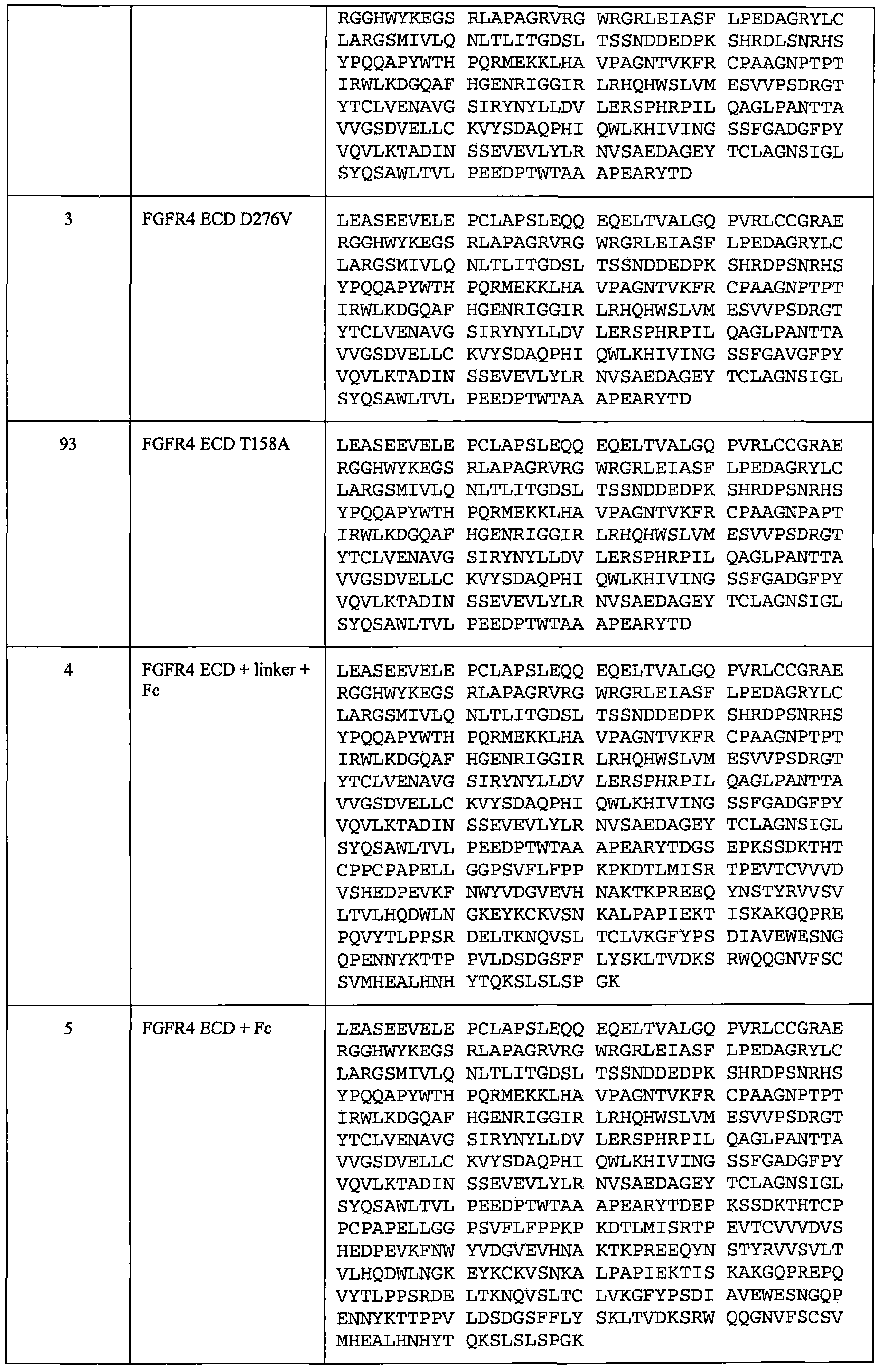


Claims
Priority Applications (8)
| Application Number | Priority Date | Filing Date | Title |
|---|---|---|---|
| BRPI0916904A BRPI0916904A2 (en) | 2008-08-04 | 2009-08-04 | polypeptides, ecd fgfr4 fusion molecules, pharmaceutical compositions, polynucleotide and methods of treating angiogenic disorder, patient cancer and patient macular degeneration and their uses |
| CA2732449A CA2732449A1 (en) | 2008-08-04 | 2009-08-04 | Fgfr extracellular domain acidic region muteins |
| JP2011522163A JP5787757B2 (en) | 2008-08-04 | 2009-08-04 | FGFR extracellular domain acidic region mutein |
| AU2009279806A AU2009279806B2 (en) | 2008-08-04 | 2009-08-04 | FGFR extracellular domain acidic region muteins |
| CN200980139415.4A CN102171343B (en) | 2008-08-04 | 2009-08-04 | FGFR extracellular domains acidic region mutain |
| EP09791148.1A EP2318529B1 (en) | 2008-08-04 | 2009-08-04 | Fgfr extracellular domain acidic region muteins |
| RU2011103972/10A RU2509774C2 (en) | 2008-08-04 | 2009-08-04 | Fibroblast growth factor receptor extracellular domain acidic region muteins |
| IL210914A IL210914A (en) | 2008-08-04 | 2011-01-27 | Fgfr extracellular domain acidic region muteins |
Applications Claiming Priority (2)
| Application Number | Priority Date | Filing Date | Title |
|---|---|---|---|
| US8612108P | 2008-08-04 | 2008-08-04 | |
| US61/086,121 | 2008-08-04 |
Publications (2)
| Publication Number | Publication Date |
|---|---|
| WO2010017198A2 true WO2010017198A2 (en) | 2010-02-11 |
| WO2010017198A3 WO2010017198A3 (en) | 2010-04-29 |
Family
ID=41110955
Family Applications (1)
| Application Number | Title | Priority Date | Filing Date |
|---|---|---|---|
| PCT/US2009/052704 WO2010017198A2 (en) | 2008-08-04 | 2009-08-04 | Fgfr extracellular domain acidic region muteins |
Country Status (10)
| Country | Link |
|---|---|
| US (2) | US8338569B2 (en) |
| EP (1) | EP2318529B1 (en) |
| JP (1) | JP5787757B2 (en) |
| CN (1) | CN102171343B (en) |
| AU (1) | AU2009279806B2 (en) |
| BR (1) | BRPI0916904A2 (en) |
| CA (1) | CA2732449A1 (en) |
| IL (1) | IL210914A (en) |
| RU (1) | RU2509774C2 (en) |
| WO (1) | WO2010017198A2 (en) |
Cited By (17)
| Publication number | Priority date | Publication date | Assignee | Title |
|---|---|---|---|---|
| EP2478003A1 (en) * | 2009-09-15 | 2012-07-25 | Five Prime Therapeutics, Inc. | Hair growth methods using fgfr4 extracellular domains |
| US8614183B2 (en) | 2009-11-13 | 2013-12-24 | Five Prime Therapeutics, Inc. | Use of FGFR1 extra cellular domain proteins to treat cancers characterized by ligand-dependent activating mutations in FGFR2 |
| US20140378665A1 (en) * | 2012-01-26 | 2014-12-25 | Amgen Inc. | Growth differentiation factor 15 (gdf-15) polypeptides |
| US8920800B2 (en) | 2010-11-15 | 2014-12-30 | Five Prime Therapeutics, Inc. | Treatment of cancer with elevated dosages of soluble FGFR1 fusion proteins |
| US9107862B2 (en) | 2007-09-04 | 2015-08-18 | Compugen Ltd. | Polypeptides and polynucleotides, and uses thereof as a drug target for producing drugs and biologics |
| US9175065B2 (en) | 2008-08-04 | 2015-11-03 | Five Prime Therapeutics, Inc. | FGFR extracellular domain acidic region muteins |
| US9173957B2 (en) | 2005-07-22 | 2015-11-03 | Five Prime Therapeutics, Inc. | FGFR2 fusion proteins comprising C-terminal deletion mutants of FGFR2-IIIC |
| US9428574B2 (en) | 2011-06-30 | 2016-08-30 | Compugen Ltd. | Polypeptides and uses thereof for treatment of autoimmune disorders and infection |
| US9617336B2 (en) | 2012-02-01 | 2017-04-11 | Compugen Ltd | C10RF32 antibodies, and uses thereof for treatment of cancer |
| EP3173102A1 (en) * | 2011-05-20 | 2017-05-31 | Yantai Rongchang Biotechnologies Co., Ltd. | Fgfr-fc fusion protein and use thereof |
| WO2017091577A1 (en) | 2015-11-23 | 2017-06-01 | Five Prime Therapeutics, Inc. | Fgfr2 inhibitors alone or in combination with immune stimulating agents in cancer treatment |
| WO2018007597A1 (en) * | 2016-07-07 | 2018-01-11 | Therachon | Soluble fibroblast growth factor receptor 3 (sfgfr3) polypeptides and uses thereof |
| US10016484B2 (en) | 2011-11-14 | 2018-07-10 | Five Prime Therapeutics, Inc. | Methods of treating lung cancer |
| US10724014B2 (en) | 2013-01-16 | 2020-07-28 | Institut National De La Sante Et De La Recherche Medicale | Soluble fibroblast growth factor receptor 3 (FGR3) polypeptide for use in the prevention or treatment of skeletal growth retardation disorders |
| US10752664B2 (en) | 2011-04-08 | 2020-08-25 | Amgen Inc. | Method of treating or ameliorating metabolic disorders using growth differentiation factor 15 (GDF-15) |
| US10894814B2 (en) | 2013-07-31 | 2021-01-19 | Amgen Inc. | Growth differentiation factor 15 (GDF-15) constructs |
| US11161889B2 (en) | 2018-04-09 | 2021-11-02 | Amgen Inc. | Growth differentiation factor 15 fusion proteins |
Families Citing this family (33)
| Publication number | Priority date | Publication date | Assignee | Title |
|---|---|---|---|---|
| JOP20190083A1 (en) | 2008-06-04 | 2017-06-16 | Amgen Inc | Fgf21 mutant fusion polypeptides and uses thereof |
| PE20120021A1 (en) | 2008-10-10 | 2012-02-10 | Amgen Inc | MUTANTS FGF21 |
| KR101860572B1 (en) | 2009-05-05 | 2018-05-24 | 암젠 인크 | Fgf21 mutants and uses thereof |
| CA2760674A1 (en) | 2009-05-05 | 2010-11-11 | Amgen Inc. | Fgf21 mutants and uses thereof |
| WO2010148142A1 (en) * | 2009-06-17 | 2010-12-23 | Amgen Inc. | Chimeric fgf19 polypeptides and uses thereof |
| WO2011068893A1 (en) * | 2009-12-02 | 2011-06-09 | Amgen Inc. | BINDING PROTEINS THAT BIND TO HUMAN FGFR1C, HUMAN β-KLOTHO AND BOTH HUMAN FGFR1C AND HUMANβ-KLOTHO |
| UA109888C2 (en) | 2009-12-07 | 2015-10-26 | ANTIBODY OR ANTIBODILITY ANTIBODY OR ITS BINDING TO THE β-CLOTE, FGF RECEPTORS AND THEIR COMPLEXES | |
| EP2512501A4 (en) | 2009-12-17 | 2014-01-01 | Five Prime Therapeutics Inc | Hair growth methods using fgfr3 extracellular domains |
| US8951972B2 (en) | 2010-12-09 | 2015-02-10 | Five Prime Therapeutics, Inc. | FGFR1 extracellular domain combination therapies for lung cancer |
| SG10201811256QA (en) | 2011-06-28 | 2019-01-30 | Inhibrx Lp | Serpin fusion polypeptides and methods of use thereof |
| US10400029B2 (en) | 2011-06-28 | 2019-09-03 | Inhibrx, Lp | Serpin fusion polypeptides and methods of use thereof |
| EP4306165A3 (en) | 2011-07-01 | 2024-04-17 | NGM Biopharmaceuticals, Inc. | Compositions, uses and methods for treatment of metabolic disorders and diseases |
| EP2925775B1 (en) | 2012-11-28 | 2020-09-16 | NGM Biopharmaceuticals, Inc. | Compositions and methods for treatment of metabolic disorders and diseases |
| US9290557B2 (en) | 2012-11-28 | 2016-03-22 | Ngm Biopharmaceuticals, Inc. | Compositions comprising variants and fusions of FGF19 polypeptides |
| US9273107B2 (en) | 2012-12-27 | 2016-03-01 | Ngm Biopharmaceuticals, Inc. | Uses and methods for modulating bile acid homeostasis and treatment of bile acid disorders and diseases |
| ES2915851T3 (en) | 2012-12-27 | 2022-06-27 | Ngm Biopharmaceuticals Inc | FGF19 chimeric peptides for use in the treatment of bile acid disorders |
| JP2016510751A (en) | 2013-03-06 | 2016-04-11 | ジェネンテック, インコーポレイテッド | Methods of treating and preventing anticancer drug resistance |
| CA2903587C (en) * | 2013-03-15 | 2021-09-28 | Genentech, Inc. | Il-22 polypeptides and il-22 fc fusion proteins and methods of use |
| US9957313B2 (en) | 2013-03-15 | 2018-05-01 | Remegen, Ltd. | FGFR-FC fusion proteins and the use thereof |
| NZ718962A (en) | 2013-10-28 | 2019-12-20 | Ngm Biopharmaceuticals Inc | Cancer models and associated methods |
| BR112016017248A8 (en) | 2014-01-24 | 2018-04-17 | Ngm Biopharmaceuticals Inc | antibody or fragment thereof, binding agent, transgenic animal, hybridoma, vector, pharmaceutical composition, fgf19 and / or fgf21-like signaling induction method, method for activating a klotho beta / fgf receptor complex, method for improving metabolism of glucose in an individual, method of detecting klotho beta, use of antibody or fragment thereof, use of pharmaceutical composition, treatment method and method for improving metabolic parameters |
| US10398758B2 (en) | 2014-05-28 | 2019-09-03 | Ngm Biopharmaceuticals, Inc. | Compositions comprising variants of FGF19 polypeptides and uses thereof for the treatment of hyperglycemic conditions |
| WO2015191986A1 (en) | 2014-06-13 | 2015-12-17 | Genentech, Inc. | Methods of treating and preventing cancer drug resistance |
| AU2015277438B2 (en) | 2014-06-16 | 2020-02-27 | Ngm Biopharmaceuticals, Inc. | Methods and uses for modulating bile acid homeostasis and treatment of bile acid disorders and diseases |
| JP6949711B2 (en) | 2014-10-23 | 2021-10-20 | エヌジーエム バイオファーマシューティカルス,インコーポレーテッド | Pharmaceutical composition containing peptide variant and method of use thereof |
| US10434144B2 (en) | 2014-11-07 | 2019-10-08 | Ngm Biopharmaceuticals, Inc. | Methods for treatment of bile acid-related disorders and prediction of clinical sensitivity to treatment of bile acid-related disorders |
| PL3242888T3 (en) * | 2015-01-07 | 2021-07-12 | Pfizer Inc. | Solube fgfr3 decoys for treating skeletal growth disorders |
| WO2016191765A1 (en) * | 2015-05-28 | 2016-12-01 | Remegen, Ltd. | Fgfr-fc fusion proteins and the use thereof |
| US10800843B2 (en) | 2015-07-29 | 2020-10-13 | Ngm Biopharmaceuticals, Inc. | Beta klotho-binding proteins |
| EP3888672A1 (en) | 2015-11-09 | 2021-10-06 | NGM Biopharmaceuticals, Inc. | Methods for treatment of bile acid-related disorders |
| US11370841B2 (en) | 2016-08-26 | 2022-06-28 | Ngm Biopharmaceuticals, Inc. | Methods of treating fibroblast growth factor 19-mediated cancers and tumors |
| EP3444275A1 (en) * | 2017-08-16 | 2019-02-20 | Exiris S.r.l. | Monoclonal antibody anti-fgfr4 |
| US11629180B2 (en) | 2018-12-07 | 2023-04-18 | Remegen Co., Ltd. | Bifunctional angiogenesis inhibitor and use thereof |
Citations (1)
| Publication number | Priority date | Publication date | Assignee | Title |
|---|---|---|---|---|
| WO2007014123A2 (en) * | 2005-07-22 | 2007-02-01 | Five Prime Therapeutics, Inc. | Compositions and methods of treating disease with fgfr fusion proteins |
Family Cites Families (42)
| Publication number | Priority date | Publication date | Assignee | Title |
|---|---|---|---|---|
| US5486462A (en) | 1984-11-23 | 1996-01-23 | The Regents Of The University Of California | Differentiative expression modules |
| GB9001466D0 (en) | 1990-01-23 | 1990-03-21 | Erba Carlo Spa | Extracellular form of the human fibroblast growth factor receptor |
| US5288855A (en) | 1989-01-23 | 1994-02-22 | Farmitalia Carlo Erba | Extracellular form of the human fibroblast growth factor receptor |
| HU215581B (en) | 1989-07-06 | 1999-01-28 | Regents Of The University Of California | Process for producing fibroblast growth factor receptors and their therapeutic use |
| US5750371A (en) | 1990-04-27 | 1998-05-12 | Takeda Chemical Industries Ltd | Water-soluble mutein of FGF receptor, DNA and production thereof |
| US5863888A (en) | 1990-07-06 | 1999-01-26 | Rhone-Poulenc Rorer Pharmaceuticals Inc. | Human Bek Fibroblast growth factor receptor |
| JP3615220B2 (en) | 1990-07-06 | 2005-02-02 | アヴェンティス ファーマシューティカルズ インコーポレイテッド | Fibroblast growth factor receptor |
| US5229501A (en) | 1991-01-11 | 1993-07-20 | Chiron Corporation | Expression and use of human fibroblast growth factor receptor |
| IL100219A0 (en) | 1991-12-02 | 1992-09-06 | Yeda Res & Dev | Variable region within fibroblast growth factor receptors that confers ligand specificity |
| US5474914A (en) | 1992-07-29 | 1995-12-12 | Chiron Corporation | Method of producing secreted CMV glycoprotein H |
| US6517872B1 (en) | 1995-06-12 | 2003-02-11 | Yeda Research And Development Co., Ltd. | FGFR3 as a marker for mesenchymal skeletal progenitor cells |
| AU3494397A (en) | 1996-06-18 | 1998-01-07 | United States Of America, Represented By The Secretary, Department Of Health And Human Services, The | Fibroblast growth factor receptor activating gene i and related compositions and methods |
| DE19802377A1 (en) * | 1998-01-22 | 1999-08-19 | Max Planck Gesellschaft | Use of inhibitors for the treatment of RTK-over-functional disorders, especially of cancer |
| AU3224700A (en) | 1999-02-08 | 2000-08-25 | Chiron Corporation | Fibroblast growth factor receptor-immunoglobulin fusion |
| US7135311B1 (en) | 1999-05-05 | 2006-11-14 | Institut Curie | Means for detecting and treating pathologies linked to FGFR3 |
| SI3351246T1 (en) * | 2001-02-19 | 2019-08-30 | Novartis Pharma Ag | Rapamycin derivative for the treatment of a solid tumor associated with deregulated angiogenesis |
| WO2003038054A2 (en) | 2001-10-31 | 2003-05-08 | New York University | Structure-based design and synthesis of fgf inhibitors and fgf modulator compounds |
| AU2003249055B2 (en) | 2002-07-15 | 2008-06-26 | Immunex Corporation | Methods and media for controlling sialylation of proteins produced by mammalian cells |
| IL156495A0 (en) | 2003-06-17 | 2004-01-04 | Prochon Biotech Ltd | Use of fgfr3 antagonists for treating t cell mediated diseases |
| CA2550245A1 (en) | 2003-12-19 | 2005-07-21 | Five Prime Therapeutics, Inc. | Fibroblast growth factor receptors 1, 2, 3, and 4 as targets for therapeutic intervention |
| EP1745073A2 (en) | 2004-05-14 | 2007-01-24 | Receptor Biologix, Inc. | Cell surface receptor isoforms and methods of identifying and using the same |
| WO2005115363A2 (en) | 2004-05-25 | 2005-12-08 | Yale University | Method for treating skeletal disorders resulting from fgfr malfunction |
| US20080286834A1 (en) | 2005-01-27 | 2008-11-20 | Robert Forgan Halenbeck | Leader Sequences For Directing Secretion of Polypeptides and Methods For Production Thereof |
| US20060234347A1 (en) | 2005-04-13 | 2006-10-19 | Harding Thomas C | Targeting multiple angiogenic pathways for cancer therapy using soluble tyrosine kinase receptors |
| WO2007059574A1 (en) | 2005-11-23 | 2007-05-31 | Apollo Life Sciences Limited | A molecule and chimeric molecules thereof |
| NZ569957A (en) | 2006-02-10 | 2012-03-30 | Genentech Inc | Anti-FGF19 antibodies and methods using same |
| US7794825B2 (en) * | 2006-04-25 | 2010-09-14 | Jeffrey M Kudrick | Prefabricated lightweight concrete structure including columns |
| CA2651755A1 (en) | 2006-05-12 | 2007-11-22 | Genentech, Inc. | Methods and compositions for the diagnosis and treatment of cancer |
| CL2007003411A1 (en) | 2006-11-28 | 2008-07-04 | Centelion | PROTEIN FUSION THAT CONSISTS IN AN FC REGION OF AN IMMUNOGLOBULIN WITH A FRAGMENT OR SOLUBLE DOMAIN OF A RECEIVER FOR FGF; POLINUCLEOTIDO THAT CODIFIES AND VECTOR AND CELL THAT UNDERSTAND IT; PHARMACEUTICAL COMPOSITION INCLUDING PROTEIN FU |
| CA2680046A1 (en) | 2007-03-23 | 2008-10-02 | The Translational Genomics Research Institute | Methods of diagnosing, classifying and treating endometrial cancer and precancer |
| WO2008133873A2 (en) | 2007-04-25 | 2008-11-06 | Aveo Pharmaceuticals, Inc. | Fgf-binding fusion proteins |
| CN101139387B (en) * | 2007-07-20 | 2011-04-06 | 暨南大学 | Recombinant soluble human FGFR2 extracellular fragment and production method and application thereof |
| RU2509774C2 (en) | 2008-08-04 | 2014-03-20 | Файв Прайм Терапьютикс, Инк. | Fibroblast growth factor receptor extracellular domain acidic region muteins |
| CA2767591A1 (en) | 2009-07-10 | 2011-01-13 | Mckechnie Vehicle Components | Use of resist coating to enhance adhesion of wheel claddings |
| US8445445B2 (en) | 2009-09-15 | 2013-05-21 | Five Prime Therapeutics, Inc. | Method of promoting hair growth using FGFR4 extracellular domains |
| AU2010319327B2 (en) | 2009-11-13 | 2015-08-13 | Five Prime Therapeutics, Inc. | Use of FGFR1 extra cellular domain proteins to treat cancers characterized by ligand-dependent activating mutations in FGFR2 |
| EP2512501A4 (en) | 2009-12-17 | 2014-01-01 | Five Prime Therapeutics Inc | Hair growth methods using fgfr3 extracellular domains |
| US8481038B2 (en) | 2010-11-15 | 2013-07-09 | Five Prime Therapeutics, Inc. | Treatment of cancer with elevated dosages of soluble FGFR1 fusion proteins |
| US8951972B2 (en) | 2010-12-09 | 2015-02-10 | Five Prime Therapeutics, Inc. | FGFR1 extracellular domain combination therapies for lung cancer |
| RU2013146242A (en) | 2011-03-17 | 2015-04-27 | Новартис Аг | FGFR AND ITS LIGANDS AS BREAST CANCER BIOMARKERS IN HR-POSITIVE INDIVIDUALS |
| EP2723391B1 (en) | 2011-06-24 | 2018-06-13 | University of Miami | Fibroblast growth factor receptor inhibition for the treatment of disease |
| US20130136740A1 (en) | 2011-11-14 | 2013-05-30 | Thomas Harding | Methods of treating cancer |
-
2009
- 2009-08-04 RU RU2011103972/10A patent/RU2509774C2/en not_active IP Right Cessation
- 2009-08-04 EP EP09791148.1A patent/EP2318529B1/en not_active Not-in-force
- 2009-08-04 WO PCT/US2009/052704 patent/WO2010017198A2/en active Application Filing
- 2009-08-04 JP JP2011522163A patent/JP5787757B2/en not_active Expired - Fee Related
- 2009-08-04 CA CA2732449A patent/CA2732449A1/en not_active Abandoned
- 2009-08-04 AU AU2009279806A patent/AU2009279806B2/en not_active Ceased
- 2009-08-04 CN CN200980139415.4A patent/CN102171343B/en not_active Expired - Fee Related
- 2009-08-04 US US12/535,479 patent/US8338569B2/en not_active Expired - Fee Related
- 2009-08-04 BR BRPI0916904A patent/BRPI0916904A2/en active Search and Examination
-
2011
- 2011-01-27 IL IL210914A patent/IL210914A/en not_active IP Right Cessation
-
2012
- 2012-09-12 US US13/612,044 patent/US9175065B2/en not_active Expired - Fee Related
Patent Citations (1)
| Publication number | Priority date | Publication date | Assignee | Title |
|---|---|---|---|---|
| WO2007014123A2 (en) * | 2005-07-22 | 2007-02-01 | Five Prime Therapeutics, Inc. | Compositions and methods of treating disease with fgfr fusion proteins |
Non-Patent Citations (3)
| Title |
|---|
| PLOTNIKOV ALEXANDER N ET AL: "Structural basis for FGF receptor dimerization and activation" CELL, vol. 98, no. 5, 3 September 1999 (1999-09-03), pages 641-650, XP002167376 ISSN: 0092-8674 * |
| POWERS C J ET AL: "FIBROBLAST GROWTH FACTORS, THEIR RECEPTORS AND SIGNALING" EXPERIMENTAL CELL RESEARCH, ACADEMIC PRESS, US, vol. 7, 1 January 2000 (2000-01-01), pages 165-197, XP002165147 ISSN: 0014-4827 * |
| SANCHEZ-HERAS ELENA ET AL: "The fibroblast growth factor receptor acid box is essential for interactions with N-cadherin and all of the major isoforms of neural cell adhesion molecule" JOURNAL OF BIOLOGICAL CHEMISTRY, vol. 281, no. 46, November 2006 (2006-11), pages 35208-35216, XP002549526 ISSN: 0021-9258 * |
Cited By (40)
| Publication number | Priority date | Publication date | Assignee | Title |
|---|---|---|---|---|
| US9173957B2 (en) | 2005-07-22 | 2015-11-03 | Five Prime Therapeutics, Inc. | FGFR2 fusion proteins comprising C-terminal deletion mutants of FGFR2-IIIC |
| US9192683B2 (en) | 2005-07-22 | 2015-11-24 | Five Prime Therapeutics, Inc. | Methods of treating disease with FGFR1 fusion proteins |
| US9555087B2 (en) | 2007-09-04 | 2017-01-31 | Compugen Ltd | Polypeptides and polynucleotides, and uses thereof as a drug target for producing drugs and biologics |
| US10098934B2 (en) | 2007-09-04 | 2018-10-16 | Compugen Ltd | Polypeptides and polynucleotides, and uses thereof as a drug target for producing drugs and biologics |
| US9107862B2 (en) | 2007-09-04 | 2015-08-18 | Compugen Ltd. | Polypeptides and polynucleotides, and uses thereof as a drug target for producing drugs and biologics |
| US9375466B2 (en) | 2007-09-04 | 2016-06-28 | Compugen Ltd | Polypeptides and polynucleotides, and uses thereof as a drug target for producing drugs and biologics |
| US9175065B2 (en) | 2008-08-04 | 2015-11-03 | Five Prime Therapeutics, Inc. | FGFR extracellular domain acidic region muteins |
| EP2478003A4 (en) * | 2009-09-15 | 2013-05-29 | Five Prime Therapeutics Inc | Hair growth methods using fgfr4 extracellular domains |
| EP2478003A1 (en) * | 2009-09-15 | 2012-07-25 | Five Prime Therapeutics, Inc. | Hair growth methods using fgfr4 extracellular domains |
| US8614183B2 (en) | 2009-11-13 | 2013-12-24 | Five Prime Therapeutics, Inc. | Use of FGFR1 extra cellular domain proteins to treat cancers characterized by ligand-dependent activating mutations in FGFR2 |
| US9169313B2 (en) | 2009-11-13 | 2015-10-27 | Five Prime Therapeutics, Inc. | Method of treatment of cancers characterized by ligand-dependent activating mutations in FGFR2 with FGFR1 ECD proteins |
| US8920800B2 (en) | 2010-11-15 | 2014-12-30 | Five Prime Therapeutics, Inc. | Treatment of cancer with elevated dosages of soluble FGFR1 fusion proteins |
| US9475871B2 (en) | 2010-11-15 | 2016-10-25 | Five Prime Therapeutics, Inc. | Treatment of cancer with elevated dosages of soluble FGFR1 fusion proteins |
| US10752664B2 (en) | 2011-04-08 | 2020-08-25 | Amgen Inc. | Method of treating or ameliorating metabolic disorders using growth differentiation factor 15 (GDF-15) |
| EP3173102A1 (en) * | 2011-05-20 | 2017-05-31 | Yantai Rongchang Biotechnologies Co., Ltd. | Fgfr-fc fusion protein and use thereof |
| US9428574B2 (en) | 2011-06-30 | 2016-08-30 | Compugen Ltd. | Polypeptides and uses thereof for treatment of autoimmune disorders and infection |
| US10016484B2 (en) | 2011-11-14 | 2018-07-10 | Five Prime Therapeutics, Inc. | Methods of treating lung cancer |
| US10537611B2 (en) | 2011-11-14 | 2020-01-21 | Five Prime Therapeutics, Inc. | Methods of treating lung cancer |
| US9714276B2 (en) * | 2012-01-26 | 2017-07-25 | Amgen Inc. | Growth differentiation factor 15 (GDF-15) polypeptides |
| US11739131B2 (en) | 2012-01-26 | 2023-08-29 | Amgen Inc. | Growth differentiation factor 15 (GDF-15) polypeptides |
| US10336798B2 (en) | 2012-01-26 | 2019-07-02 | Amgen Inc. | Growth differentiation factor 15 (GDF-15) polypeptides |
| US20140378665A1 (en) * | 2012-01-26 | 2014-12-25 | Amgen Inc. | Growth differentiation factor 15 (gdf-15) polypeptides |
| US9617336B2 (en) | 2012-02-01 | 2017-04-11 | Compugen Ltd | C10RF32 antibodies, and uses thereof for treatment of cancer |
| US11702642B2 (en) | 2013-01-16 | 2023-07-18 | INSERM (Institut National de la Santé et de la Recherche Médicale) | Soluble fibroblast growth factor receptor 3 (FGR3) polypeptide for use in the prevention or treatment of skeletal growth retardation disorders |
| US11814654B2 (en) | 2013-01-16 | 2023-11-14 | Institut National De La Sante Et De La Recherche Medicale | Soluble fibroblast growth factor receptor 3 (FGR3) polypeptide for use in the prevention or treatment of skeletal growth retardation disorders |
| US10724014B2 (en) | 2013-01-16 | 2020-07-28 | Institut National De La Sante Et De La Recherche Medicale | Soluble fibroblast growth factor receptor 3 (FGR3) polypeptide for use in the prevention or treatment of skeletal growth retardation disorders |
| US10894814B2 (en) | 2013-07-31 | 2021-01-19 | Amgen Inc. | Growth differentiation factor 15 (GDF-15) constructs |
| WO2017091577A1 (en) | 2015-11-23 | 2017-06-01 | Five Prime Therapeutics, Inc. | Fgfr2 inhibitors alone or in combination with immune stimulating agents in cancer treatment |
| WO2017091580A1 (en) | 2015-11-23 | 2017-06-01 | Five Prime Therapeutics, Inc. | Predicting response to cancer treatment with fgfr2 inhibitors |
| KR20190037240A (en) * | 2016-07-07 | 2019-04-05 | 테라숑 에스에이에스 | Soluble fibroblast growth factor receptor 3 (SFGFR3) polypeptides and uses thereof |
| RU2751483C2 (en) * | 2016-07-07 | 2021-07-14 | Пфайзер Инк. | POLYPEPTIDES THAT ARE SOLUBLE RECEPTORS OF FIBROBLAST GROWTH FACTOR 3 (sFGFR3), AND WAYS OF THEIR APPLICATION |
| AU2017294551B2 (en) * | 2016-07-07 | 2022-03-03 | INSERM (Institut National de la Santé et de la Recherche Médicale) | Soluble fibroblast growth factor receptor 3 (sFGFR3) polypeptides and uses thereof |
| KR102432192B1 (en) | 2016-07-07 | 2022-08-12 | 화이자 인코포레이티드 | Soluble fibroblast growth factor receptor 3 (SFGFR3) polypeptides and uses thereof |
| EP4122955A1 (en) * | 2016-07-07 | 2023-01-25 | Pfizer Inc. | Soluble fibroblast growth factor receptor 3 (sfgfr3) polypeptides and uses thereof |
| US11697678B2 (en) | 2016-07-07 | 2023-07-11 | Pfizer Inc. | Soluble fibroblast growth factor receptor 3 (SFGFR3) polypeptides and uses thereof |
| US11021528B2 (en) | 2016-07-07 | 2021-06-01 | INSERM (Institut National de la Santé et de la Recherche Médicale | Soluble fibroblast growth factor receptor 3 (SFGFR3) polypeptides and uses thereof |
| CN109715658A (en) * | 2016-07-07 | 2019-05-03 | 泰拉肖恩公司 | Soluble fibroblast growth factor receptor3 (SFGFR3) polypeptide and application thereof |
| CN109715658B (en) * | 2016-07-07 | 2023-09-12 | 辉瑞大药厂 | Soluble fibroblast growth factor receptor 3 (SFGFR 3) polypeptides and uses thereof |
| WO2018007597A1 (en) * | 2016-07-07 | 2018-01-11 | Therachon | Soluble fibroblast growth factor receptor 3 (sfgfr3) polypeptides and uses thereof |
| US11161889B2 (en) | 2018-04-09 | 2021-11-02 | Amgen Inc. | Growth differentiation factor 15 fusion proteins |
Also Published As
| Publication number | Publication date |
|---|---|
| AU2009279806B2 (en) | 2015-04-30 |
| US9175065B2 (en) | 2015-11-03 |
| CN102171343A (en) | 2011-08-31 |
| AU2009279806A1 (en) | 2010-02-11 |
| BRPI0916904A2 (en) | 2016-10-11 |
| JP5787757B2 (en) | 2015-09-30 |
| CN102171343B (en) | 2017-07-14 |
| US8338569B2 (en) | 2012-12-25 |
| CA2732449A1 (en) | 2010-02-11 |
| IL210914A0 (en) | 2011-04-28 |
| US20130004492A1 (en) | 2013-01-03 |
| RU2011103972A (en) | 2012-09-10 |
| EP2318529A2 (en) | 2011-05-11 |
| EP2318529B1 (en) | 2017-10-18 |
| US20100087627A1 (en) | 2010-04-08 |
| IL210914A (en) | 2016-09-29 |
| RU2509774C2 (en) | 2014-03-20 |
| WO2010017198A3 (en) | 2010-04-29 |
| JP2011529705A (en) | 2011-12-15 |
Similar Documents
| Publication | Publication Date | Title |
|---|---|---|
| EP2318529B1 (en) | Fgfr extracellular domain acidic region muteins | |
| EP2498799B1 (en) | Use of fgfr1 extra cellular domain proteins to treat cancers characterized by ligand-dependent activating mutations in fgfr2 | |
| US9173957B2 (en) | FGFR2 fusion proteins comprising C-terminal deletion mutants of FGFR2-IIIC | |
| EP2512501A2 (en) | Hair growth methods using fgfr3 extracellular domains | |
| EP2478003A1 (en) | Hair growth methods using fgfr4 extracellular domains |
Legal Events
| Date | Code | Title | Description |
|---|---|---|---|
| WWE | Wipo information: entry into national phase |
Ref document number: 200980139415.4 Country of ref document: CN |
|
| 121 | Ep: the epo has been informed by wipo that ep was designated in this application |
Ref document number: 09791148 Country of ref document: EP Kind code of ref document: A2 |
|
| ENP | Entry into the national phase |
Ref document number: 2732449 Country of ref document: CA Ref document number: 2011522163 Country of ref document: JP Kind code of ref document: A |
|
| WWE | Wipo information: entry into national phase |
Ref document number: 2009279806 Country of ref document: AU Ref document number: 498/KOLNP/2011 Country of ref document: IN |
|
| NENP | Non-entry into the national phase |
Ref country code: DE |
|
| REEP | Request for entry into the european phase |
Ref document number: 2009791148 Country of ref document: EP |
|
| WWE | Wipo information: entry into national phase |
Ref document number: 2009791148 Country of ref document: EP |
|
| ENP | Entry into the national phase |
Ref document number: 2009279806 Country of ref document: AU Date of ref document: 20090804 Kind code of ref document: A |
|
| WWE | Wipo information: entry into national phase |
Ref document number: 2011103972 Country of ref document: RU |
|
| REG | Reference to national code |
Ref country code: BR Ref legal event code: B01E Ref document number: PI0916904 Country of ref document: BR |
|
| ENP | Entry into the national phase |
Ref document number: PI0916904 Country of ref document: BR Kind code of ref document: A2 Effective date: 20110202 |