WO2009104913A2 - Composition comprising an extract of mixed herbs with lonicera japonica thunb and anemarrhena asphodeloides bunge for preventing and treating arthritic diseases - Google Patents
Composition comprising an extract of mixed herbs with lonicera japonica thunb and anemarrhena asphodeloides bunge for preventing and treating arthritic diseases Download PDFInfo
- Publication number
- WO2009104913A2 WO2009104913A2 PCT/KR2009/000797 KR2009000797W WO2009104913A2 WO 2009104913 A2 WO2009104913 A2 WO 2009104913A2 KR 2009000797 W KR2009000797 W KR 2009000797W WO 2009104913 A2 WO2009104913 A2 WO 2009104913A2
- Authority
- WO
- WIPO (PCT)
- Prior art keywords
- extract
- mixed
- cartilage
- health care
- lonicera japonica
- Prior art date
Links
- 239000000284 extract Substances 0.000 title claims abstract description 113
- 239000000203 mixture Substances 0.000 title claims abstract description 38
- 241000605445 Anemarrhena asphodeloides Species 0.000 title claims abstract description 36
- 244000167230 Lonicera japonica Species 0.000 title claims abstract description 35
- 235000017617 Lonicera japonica Nutrition 0.000 title claims abstract description 35
- 241000612118 Samolus valerandi Species 0.000 title claims abstract description 35
- 235000008216 herbs Nutrition 0.000 title claims abstract description 35
- 208000037265 diseases, disorders, signs and symptoms Diseases 0.000 title claims abstract description 34
- 201000010099 disease Diseases 0.000 title claims abstract description 33
- 230000002917 arthritic effect Effects 0.000 title claims abstract description 24
- 239000003814 drug Substances 0.000 claims abstract description 19
- AEDDIBAIWPIIBD-ZJKJAXBQSA-N mangiferin Chemical compound O[C@@H]1[C@@H](O)[C@H](O)[C@@H](CO)O[C@H]1C1=C(O)C=C(OC=2C(=CC(O)=C(O)C=2)C2=O)C2=C1O AEDDIBAIWPIIBD-ZJKJAXBQSA-N 0.000 claims description 56
- 238000000034 method Methods 0.000 claims description 54
- 201000008482 osteoarthritis Diseases 0.000 claims description 48
- 238000002360 preparation method Methods 0.000 claims description 36
- 235000013305 food Nutrition 0.000 claims description 33
- YWQSXCGKJDUYTL-UHFFFAOYSA-N Mangiferin Natural products CC(CCC=C(C)C)C1CC(C)C2C3CCC4C(C)(C)CCCC45CC35CCC12C YWQSXCGKJDUYTL-UHFFFAOYSA-N 0.000 claims description 29
- PZIRUHCJZBGLDY-UHFFFAOYSA-N Caffeoylquinic acid Natural products CC(CCC(=O)C(C)C1C(=O)CC2C3CC(O)C4CC(O)CCC4(C)C3CCC12C)C(=O)O PZIRUHCJZBGLDY-UHFFFAOYSA-N 0.000 claims description 28
- LRHPLDYGYMQRHN-UHFFFAOYSA-N N-Butanol Chemical compound CCCCO LRHPLDYGYMQRHN-UHFFFAOYSA-N 0.000 claims description 28
- 229940043357 mangiferin Drugs 0.000 claims description 28
- 238000011282 treatment Methods 0.000 claims description 28
- CWVRJTMFETXNAD-FWCWNIRPSA-N 3-O-Caffeoylquinic acid Natural products O[C@H]1[C@@H](O)C[C@@](O)(C(O)=O)C[C@H]1OC(=O)\C=C\C1=CC=C(O)C(O)=C1 CWVRJTMFETXNAD-FWCWNIRPSA-N 0.000 claims description 27
- CWVRJTMFETXNAD-KLZCAUPSSA-N Neochlorogenin-saeure Natural products O[C@H]1C[C@@](O)(C[C@@H](OC(=O)C=Cc2ccc(O)c(O)c2)[C@@H]1O)C(=O)O CWVRJTMFETXNAD-KLZCAUPSSA-N 0.000 claims description 27
- CWVRJTMFETXNAD-JUHZACGLSA-N chlorogenic acid Chemical compound O[C@@H]1[C@H](O)C[C@@](O)(C(O)=O)C[C@H]1OC(=O)\C=C\C1=CC=C(O)C(O)=C1 CWVRJTMFETXNAD-JUHZACGLSA-N 0.000 claims description 27
- 229940074393 chlorogenic acid Drugs 0.000 claims description 27
- FFQSDFBBSXGVKF-KHSQJDLVSA-N chlorogenic acid Natural products O[C@@H]1C[C@](O)(C[C@@H](CC(=O)C=Cc2ccc(O)c(O)c2)[C@@H]1O)C(=O)O FFQSDFBBSXGVKF-KHSQJDLVSA-N 0.000 claims description 27
- 235000001368 chlorogenic acid Nutrition 0.000 claims description 27
- BMRSEYFENKXDIS-KLZCAUPSSA-N cis-3-O-p-coumaroylquinic acid Natural products O[C@H]1C[C@@](O)(C[C@@H](OC(=O)C=Cc2ccc(O)cc2)[C@@H]1O)C(=O)O BMRSEYFENKXDIS-KLZCAUPSSA-N 0.000 claims description 27
- LFQSCWFLJHTTHZ-UHFFFAOYSA-N Ethanol Chemical compound CCO LFQSCWFLJHTTHZ-UHFFFAOYSA-N 0.000 claims description 26
- 230000036541 health Effects 0.000 claims description 22
- XLYOFNOQVPJJNP-UHFFFAOYSA-N water Substances O XLYOFNOQVPJJNP-UHFFFAOYSA-N 0.000 claims description 18
- 206010003246 arthritis Diseases 0.000 claims description 16
- 235000013361 beverage Nutrition 0.000 claims description 14
- 239000008194 pharmaceutical composition Substances 0.000 claims description 14
- 239000000287 crude extract Substances 0.000 claims description 11
- 230000002265 prevention Effects 0.000 claims description 10
- 241000124008 Mammalia Species 0.000 claims description 9
- 238000005194 fractionation Methods 0.000 claims description 8
- 201000003068 rheumatic fever Diseases 0.000 claims description 7
- 239000004480 active ingredient Substances 0.000 claims description 6
- 239000000843 powder Substances 0.000 claims description 6
- 239000000654 additive Substances 0.000 claims description 5
- 230000000996 additive effect Effects 0.000 claims description 5
- 239000002775 capsule Substances 0.000 claims description 5
- 239000003937 drug carrier Substances 0.000 claims description 5
- 239000003826 tablet Substances 0.000 claims description 4
- 239000008187 granular material Substances 0.000 claims description 3
- 239000004615 ingredient Substances 0.000 claims description 3
- 206010025135 lupus erythematosus Diseases 0.000 claims description 3
- 239000002904 solvent Substances 0.000 claims description 3
- 229940124597 therapeutic agent Drugs 0.000 claims description 3
- 230000003389 potentiating effect Effects 0.000 abstract description 26
- 230000003110 anti-inflammatory effect Effects 0.000 abstract description 17
- 235000013402 health food Nutrition 0.000 abstract description 10
- CPLXHLVBOLITMK-UHFFFAOYSA-N Magnesium oxide Chemical compound [Mg]=O CPLXHLVBOLITMK-UHFFFAOYSA-N 0.000 description 105
- 210000000845 cartilage Anatomy 0.000 description 84
- 238000012360 testing method Methods 0.000 description 71
- 230000005764 inhibitory process Effects 0.000 description 57
- 230000002401 inhibitory effect Effects 0.000 description 45
- 230000000694 effects Effects 0.000 description 42
- RZEKVGVHFLEQIL-UHFFFAOYSA-N celecoxib Chemical compound C1=CC(C)=CC=C1C1=CC(C(F)(F)F)=NN1C1=CC=C(S(N)(=O)=O)C=C1 RZEKVGVHFLEQIL-UHFFFAOYSA-N 0.000 description 38
- 229960000590 celecoxib Drugs 0.000 description 38
- 239000000243 solution Substances 0.000 description 38
- 239000013641 positive control Substances 0.000 description 36
- QTBSBXVTEAMEQO-UHFFFAOYSA-N acetic acid Substances CC(O)=O QTBSBXVTEAMEQO-UHFFFAOYSA-N 0.000 description 29
- 238000010171 animal model Methods 0.000 description 27
- 210000001519 tissue Anatomy 0.000 description 25
- MWUXSHHQAYIFBG-UHFFFAOYSA-N Nitric oxide Chemical compound O=[N] MWUXSHHQAYIFBG-UHFFFAOYSA-N 0.000 description 24
- 230000000052 comparative effect Effects 0.000 description 22
- 108010067787 Proteoglycans Proteins 0.000 description 21
- 102000016611 Proteoglycans Human genes 0.000 description 20
- 238000004458 analytical method Methods 0.000 description 20
- 210000004027 cell Anatomy 0.000 description 18
- 230000014509 gene expression Effects 0.000 description 18
- 102100027995 Collagenase 3 Human genes 0.000 description 16
- 108050005238 Collagenase 3 Proteins 0.000 description 16
- 241000283973 Oryctolagus cuniculus Species 0.000 description 16
- 208000002193 Pain Diseases 0.000 description 16
- 230000000202 analgesic effect Effects 0.000 description 16
- 230000000692 anti-sense effect Effects 0.000 description 16
- 230000036407 pain Effects 0.000 description 16
- 239000002609 medium Substances 0.000 description 15
- 210000005065 subchondral bone plate Anatomy 0.000 description 15
- 102000008186 Collagen Human genes 0.000 description 14
- 108010035532 Collagen Proteins 0.000 description 14
- 102000000380 Matrix Metalloproteinase 1 Human genes 0.000 description 14
- 108010016113 Matrix Metalloproteinase 1 Proteins 0.000 description 14
- 241000699666 Mus <mouse, genus> Species 0.000 description 14
- 229920001436 collagen Polymers 0.000 description 14
- 206010039073 rheumatoid arthritis Diseases 0.000 description 14
- 206010061218 Inflammation Diseases 0.000 description 13
- 206010030113 Oedema Diseases 0.000 description 13
- -1 e.g. Proteins 0.000 description 13
- 238000011084 recovery Methods 0.000 description 13
- 102000002274 Matrix Metalloproteinases Human genes 0.000 description 12
- 108010000684 Matrix Metalloproteinases Proteins 0.000 description 12
- 210000003321 cartilage cell Anatomy 0.000 description 12
- 230000015556 catabolic process Effects 0.000 description 12
- 238000006731 degradation reaction Methods 0.000 description 12
- 102000004127 Cytokines Human genes 0.000 description 11
- 108090000695 Cytokines Proteins 0.000 description 11
- 241000700159 Rattus Species 0.000 description 11
- 229940079593 drug Drugs 0.000 description 11
- 230000004054 inflammatory process Effects 0.000 description 11
- 238000002347 injection Methods 0.000 description 11
- 239000007924 injection Substances 0.000 description 11
- WSFSSNUMVMOOMR-UHFFFAOYSA-N Formaldehyde Chemical compound O=C WSFSSNUMVMOOMR-UHFFFAOYSA-N 0.000 description 10
- 210000001612 chondrocyte Anatomy 0.000 description 10
- 230000006378 damage Effects 0.000 description 10
- 229940021182 non-steroidal anti-inflammatory drug Drugs 0.000 description 10
- 239000000126 substance Substances 0.000 description 10
- 239000000041 non-steroidal anti-inflammatory agent Substances 0.000 description 9
- 239000006228 supernatant Substances 0.000 description 9
- 241001465754 Metazoa Species 0.000 description 8
- 108060008682 Tumor Necrosis Factor Proteins 0.000 description 8
- 102000000852 Tumor Necrosis Factor-alpha Human genes 0.000 description 8
- XEYBRNLFEZDVAW-ARSRFYASSA-N dinoprostone Chemical compound CCCCC[C@H](O)\C=C\[C@H]1[C@H](O)CC(=O)[C@@H]1C\C=C/CCCC(O)=O XEYBRNLFEZDVAW-ARSRFYASSA-N 0.000 description 8
- 238000002474 experimental method Methods 0.000 description 8
- 238000004519 manufacturing process Methods 0.000 description 8
- 230000002633 protecting effect Effects 0.000 description 8
- 238000003757 reverse transcription PCR Methods 0.000 description 8
- 238000010186 staining Methods 0.000 description 8
- 238000005303 weighing Methods 0.000 description 8
- VKUYLANQOAKALN-UHFFFAOYSA-N 2-[benzyl-(4-methoxyphenyl)sulfonylamino]-n-hydroxy-4-methylpentanamide Chemical compound C1=CC(OC)=CC=C1S(=O)(=O)N(C(CC(C)C)C(=O)NO)CC1=CC=CC=C1 VKUYLANQOAKALN-UHFFFAOYSA-N 0.000 description 7
- 229920002134 Carboxymethyl cellulose Polymers 0.000 description 7
- 102000043136 MAP kinase family Human genes 0.000 description 7
- 108091054455 MAP kinase family Proteins 0.000 description 7
- 102100030416 Stromelysin-1 Human genes 0.000 description 7
- 101710108790 Stromelysin-1 Proteins 0.000 description 7
- 210000001188 articular cartilage Anatomy 0.000 description 7
- 239000001768 carboxy methyl cellulose Substances 0.000 description 7
- 239000003795 chemical substances by application Substances 0.000 description 7
- 239000013642 negative control Substances 0.000 description 7
- MZOFCQQQCNRIBI-VMXHOPILSA-N (3s)-4-[[(2s)-1-[[(2s)-1-[[(1s)-1-carboxy-2-hydroxyethyl]amino]-4-methyl-1-oxopentan-2-yl]amino]-5-(diaminomethylideneamino)-1-oxopentan-2-yl]amino]-3-[[2-[[(2s)-2,6-diaminohexanoyl]amino]acetyl]amino]-4-oxobutanoic acid Chemical compound OC[C@@H](C(O)=O)NC(=O)[C@H](CC(C)C)NC(=O)[C@H](CCCN=C(N)N)NC(=O)[C@H](CC(O)=O)NC(=O)CNC(=O)[C@@H](N)CCCCN MZOFCQQQCNRIBI-VMXHOPILSA-N 0.000 description 6
- MSWZFWKMSRAUBD-IVMDWMLBSA-N 2-amino-2-deoxy-D-glucopyranose Chemical compound N[C@H]1C(O)O[C@H](CO)[C@@H](O)[C@@H]1O MSWZFWKMSRAUBD-IVMDWMLBSA-N 0.000 description 6
- 108091006146 Channels Proteins 0.000 description 6
- 108020004414 DNA Proteins 0.000 description 6
- IAZDPXIOMUYVGZ-UHFFFAOYSA-N Dimethylsulphoxide Chemical compound CS(C)=O IAZDPXIOMUYVGZ-UHFFFAOYSA-N 0.000 description 6
- WZUVPPKBWHMQCE-UHFFFAOYSA-N Haematoxylin Chemical compound C12=CC(O)=C(O)C=C2CC2(O)C1C1=CC=C(O)C(O)=C1OC2 WZUVPPKBWHMQCE-UHFFFAOYSA-N 0.000 description 6
- 102000008299 Nitric Oxide Synthase Human genes 0.000 description 6
- 108010021487 Nitric Oxide Synthase Proteins 0.000 description 6
- 206010042674 Swelling Diseases 0.000 description 6
- 230000036592 analgesia Effects 0.000 description 6
- MSWZFWKMSRAUBD-UHFFFAOYSA-N beta-D-galactosamine Natural products NC1C(O)OC(CO)C(O)C1O MSWZFWKMSRAUBD-UHFFFAOYSA-N 0.000 description 6
- 235000010948 carboxy methyl cellulose Nutrition 0.000 description 6
- 239000008112 carboxymethyl-cellulose Substances 0.000 description 6
- 229940105329 carboxymethylcellulose Drugs 0.000 description 6
- 239000012153 distilled water Substances 0.000 description 6
- 239000000975 dye Substances 0.000 description 6
- 239000012091 fetal bovine serum Substances 0.000 description 6
- 239000000706 filtrate Substances 0.000 description 6
- 229960002442 glucosamine Drugs 0.000 description 6
- 239000001963 growth medium Substances 0.000 description 6
- HQKMJHAJHXVSDF-UHFFFAOYSA-L magnesium stearate Chemical compound [Mg+2].CCCCCCCCCCCCCCCCCC([O-])=O.CCCCCCCCCCCCCCCCCC([O-])=O HQKMJHAJHXVSDF-UHFFFAOYSA-L 0.000 description 6
- 108090000623 proteins and genes Proteins 0.000 description 6
- 238000003756 stirring Methods 0.000 description 6
- 230000008961 swelling Effects 0.000 description 6
- 208000024891 symptom Diseases 0.000 description 6
- 231100000820 toxicity test Toxicity 0.000 description 6
- 208000031648 Body Weight Changes Diseases 0.000 description 5
- 102000000503 Collagen Type II Human genes 0.000 description 5
- 108010041390 Collagen Type II Proteins 0.000 description 5
- 102000029816 Collagenase Human genes 0.000 description 5
- 108060005980 Collagenase Proteins 0.000 description 5
- 102000003855 L-lactate dehydrogenase Human genes 0.000 description 5
- 108700023483 L-lactate dehydrogenases Proteins 0.000 description 5
- 241000699670 Mus sp. Species 0.000 description 5
- 239000002671 adjuvant Substances 0.000 description 5
- 230000015572 biosynthetic process Effects 0.000 description 5
- 230000004579 body weight change Effects 0.000 description 5
- 230000007541 cellular toxicity Effects 0.000 description 5
- 229960002424 collagenase Drugs 0.000 description 5
- 238000010494 dissociation reaction Methods 0.000 description 5
- 230000005593 dissociations Effects 0.000 description 5
- 231100000673 dose–response relationship Toxicity 0.000 description 5
- 238000000605 extraction Methods 0.000 description 5
- 238000011049 filling Methods 0.000 description 5
- 230000007674 genetic toxicity Effects 0.000 description 5
- 231100000025 genetic toxicology Toxicity 0.000 description 5
- 230000002757 inflammatory effect Effects 0.000 description 5
- 210000001503 joint Anatomy 0.000 description 5
- 210000002540 macrophage Anatomy 0.000 description 5
- 238000002156 mixing Methods 0.000 description 5
- 239000012188 paraffin wax Substances 0.000 description 5
- 238000003752 polymerase chain reaction Methods 0.000 description 5
- 230000001681 protective effect Effects 0.000 description 5
- 235000018102 proteins Nutrition 0.000 description 5
- 102000004169 proteins and genes Human genes 0.000 description 5
- OARRHUQTFTUEOS-UHFFFAOYSA-N safranin Chemical compound [Cl-].C=12C=C(N)C(C)=CC2=NC2=CC(C)=C(N)C=C2[N+]=1C1=CC=CC=C1 OARRHUQTFTUEOS-UHFFFAOYSA-N 0.000 description 5
- 238000001356 surgical procedure Methods 0.000 description 5
- 210000000689 upper leg Anatomy 0.000 description 5
- 208000031404 Chromosome Aberrations Diseases 0.000 description 4
- 238000002965 ELISA Methods 0.000 description 4
- 102000000589 Interleukin-1 Human genes 0.000 description 4
- 108010002352 Interleukin-1 Proteins 0.000 description 4
- PVNIIMVLHYAWGP-UHFFFAOYSA-N Niacin Chemical compound OC(=O)C1=CC=CN=C1 PVNIIMVLHYAWGP-UHFFFAOYSA-N 0.000 description 4
- 235000011054 acetic acid Nutrition 0.000 description 4
- 229960000583 acetic acid Drugs 0.000 description 4
- 230000004913 activation Effects 0.000 description 4
- 210000000988 bone and bone Anatomy 0.000 description 4
- 235000010418 carrageenan Nutrition 0.000 description 4
- 239000000679 carrageenan Substances 0.000 description 4
- 229920001525 carrageenan Polymers 0.000 description 4
- 229940113118 carrageenan Drugs 0.000 description 4
- 231100000005 chromosome aberration Toxicity 0.000 description 4
- 229940117173 croton oil Drugs 0.000 description 4
- 239000002781 deodorant agent Substances 0.000 description 4
- 230000003628 erosive effect Effects 0.000 description 4
- 210000003743 erythrocyte Anatomy 0.000 description 4
- 239000000796 flavoring agent Substances 0.000 description 4
- 210000002683 foot Anatomy 0.000 description 4
- 210000003127 knee Anatomy 0.000 description 4
- 239000007788 liquid Substances 0.000 description 4
- 239000012528 membrane Substances 0.000 description 4
- 108020004999 messenger RNA Proteins 0.000 description 4
- 230000002503 metabolic effect Effects 0.000 description 4
- 230000004048 modification Effects 0.000 description 4
- 238000012986 modification Methods 0.000 description 4
- 230000003287 optical effect Effects 0.000 description 4
- 150000003180 prostaglandins Chemical class 0.000 description 4
- 230000004044 response Effects 0.000 description 4
- LPXPTNMVRIOKMN-UHFFFAOYSA-M sodium nitrite Chemical compound [Na+].[O-]N=O LPXPTNMVRIOKMN-UHFFFAOYSA-M 0.000 description 4
- 239000000758 substrate Substances 0.000 description 4
- 238000003786 synthesis reaction Methods 0.000 description 4
- 229940088594 vitamin Drugs 0.000 description 4
- 229930003231 vitamin Natural products 0.000 description 4
- 235000013343 vitamin Nutrition 0.000 description 4
- 239000011782 vitamin Substances 0.000 description 4
- 150000003722 vitamin derivatives Chemical class 0.000 description 4
- UHVMMEOXYDMDKI-JKYCWFKZSA-L zinc;1-(5-cyanopyridin-2-yl)-3-[(1s,2s)-2-(6-fluoro-2-hydroxy-3-propanoylphenyl)cyclopropyl]urea;diacetate Chemical compound [Zn+2].CC([O-])=O.CC([O-])=O.CCC(=O)C1=CC=C(F)C([C@H]2[C@H](C2)NC(=O)NC=2N=CC(=CC=2)C#N)=C1O UHVMMEOXYDMDKI-JKYCWFKZSA-L 0.000 description 4
- CSCPPACGZOOCGX-UHFFFAOYSA-N Acetone Chemical compound CC(C)=O CSCPPACGZOOCGX-UHFFFAOYSA-N 0.000 description 3
- 108091003079 Bovine Serum Albumin Proteins 0.000 description 3
- 241000283707 Capra Species 0.000 description 3
- 101710176668 Cartilage oligomeric matrix protein Proteins 0.000 description 3
- 229920002261 Corn starch Polymers 0.000 description 3
- 101150086096 Eif2ak3 gene Proteins 0.000 description 3
- 102000004190 Enzymes Human genes 0.000 description 3
- 108090000790 Enzymes Proteins 0.000 description 3
- 102000004889 Interleukin-6 Human genes 0.000 description 3
- 108090001005 Interleukin-6 Proteins 0.000 description 3
- 102100022397 Nitric oxide synthase, brain Human genes 0.000 description 3
- 101710111444 Nitric oxide synthase, brain Proteins 0.000 description 3
- DNIAPMSPPWPWGF-UHFFFAOYSA-N Propylene glycol Chemical compound CC(O)CO DNIAPMSPPWPWGF-UHFFFAOYSA-N 0.000 description 3
- 208000027418 Wounds and injury Diseases 0.000 description 3
- 230000002159 abnormal effect Effects 0.000 description 3
- 239000002253 acid Substances 0.000 description 3
- 235000010443 alginic acid Nutrition 0.000 description 3
- 229920000615 alginic acid Polymers 0.000 description 3
- 239000000427 antigen Substances 0.000 description 3
- 102000036639 antigens Human genes 0.000 description 3
- 108091007433 antigens Proteins 0.000 description 3
- 230000006793 arrhythmia Effects 0.000 description 3
- 206010003119 arrhythmia Diseases 0.000 description 3
- 238000003556 assay Methods 0.000 description 3
- 210000004556 brain Anatomy 0.000 description 3
- 239000007853 buffer solution Substances 0.000 description 3
- 150000001720 carbohydrates Chemical class 0.000 description 3
- 235000014633 carbohydrates Nutrition 0.000 description 3
- 150000001875 compounds Chemical class 0.000 description 3
- 239000008120 corn starch Substances 0.000 description 3
- 230000000875 corresponding effect Effects 0.000 description 3
- 238000012258 culturing Methods 0.000 description 3
- 239000003085 diluting agent Substances 0.000 description 3
- 229940088598 enzyme Drugs 0.000 description 3
- 235000013355 food flavoring agent Nutrition 0.000 description 3
- 235000015203 fruit juice Nutrition 0.000 description 3
- 230000006698 induction Effects 0.000 description 3
- 208000014674 injury Diseases 0.000 description 3
- 229910052500 inorganic mineral Inorganic materials 0.000 description 3
- 210000004698 lymphocyte Anatomy 0.000 description 3
- 235000019359 magnesium stearate Nutrition 0.000 description 3
- LXCFILQKKLGQFO-UHFFFAOYSA-N methylparaben Chemical compound COC(=O)C1=CC=C(O)C=C1 LXCFILQKKLGQFO-UHFFFAOYSA-N 0.000 description 3
- 235000010755 mineral Nutrition 0.000 description 3
- 239000011707 mineral Substances 0.000 description 3
- 235000015097 nutrients Nutrition 0.000 description 3
- 238000012261 overproduction Methods 0.000 description 3
- 229940094443 oxytocics prostaglandins Drugs 0.000 description 3
- DHRLEVQXOMLTIM-UHFFFAOYSA-N phosphoric acid;trioxomolybdenum Chemical compound O=[Mo](=O)=O.O=[Mo](=O)=O.O=[Mo](=O)=O.O=[Mo](=O)=O.O=[Mo](=O)=O.O=[Mo](=O)=O.O=[Mo](=O)=O.O=[Mo](=O)=O.O=[Mo](=O)=O.O=[Mo](=O)=O.O=[Mo](=O)=O.O=[Mo](=O)=O.OP(O)(O)=O DHRLEVQXOMLTIM-UHFFFAOYSA-N 0.000 description 3
- 230000026731 phosphorylation Effects 0.000 description 3
- 238000006366 phosphorylation reaction Methods 0.000 description 3
- 239000003755 preservative agent Substances 0.000 description 3
- QELSKZZBTMNZEB-UHFFFAOYSA-N propylparaben Chemical compound CCCOC(=O)C1=CC=C(O)C=C1 QELSKZZBTMNZEB-UHFFFAOYSA-N 0.000 description 3
- 230000005855 radiation Effects 0.000 description 3
- 150000003839 salts Chemical class 0.000 description 3
- 230000019491 signal transduction Effects 0.000 description 3
- 239000011734 sodium Substances 0.000 description 3
- 230000001954 sterilising effect Effects 0.000 description 3
- 230000009885 systemic effect Effects 0.000 description 3
- 239000000454 talc Substances 0.000 description 3
- 229910052623 talc Inorganic materials 0.000 description 3
- 235000012222 talc Nutrition 0.000 description 3
- 238000002560 therapeutic procedure Methods 0.000 description 3
- 230000001988 toxicity Effects 0.000 description 3
- 231100000419 toxicity Toxicity 0.000 description 3
- 230000035899 viability Effects 0.000 description 3
- GHOKWGTUZJEAQD-ZETCQYMHSA-N (D)-(+)-Pantothenic acid Chemical compound OCC(C)(C)[C@@H](O)C(=O)NCCC(O)=O GHOKWGTUZJEAQD-ZETCQYMHSA-N 0.000 description 2
- 108091032973 (ribonucleotides)n+m Proteins 0.000 description 2
- GVJHHUAWPYXKBD-UHFFFAOYSA-N (±)-α-Tocopherol Chemical compound OC1=C(C)C(C)=C2OC(CCCC(C)CCCC(C)CCCC(C)C)(C)CCC2=C1C GVJHHUAWPYXKBD-UHFFFAOYSA-N 0.000 description 2
- JKMHFZQWWAIEOD-UHFFFAOYSA-N 2-[4-(2-hydroxyethyl)piperazin-1-yl]ethanesulfonic acid Chemical compound OCC[NH+]1CCN(CCS([O-])(=O)=O)CC1 JKMHFZQWWAIEOD-UHFFFAOYSA-N 0.000 description 2
- 102100022464 5'-nucleotidase Human genes 0.000 description 2
- SQDAZGGFXASXDW-UHFFFAOYSA-N 5-bromo-2-(trifluoromethoxy)pyridine Chemical compound FC(F)(F)OC1=CC=C(Br)C=N1 SQDAZGGFXASXDW-UHFFFAOYSA-N 0.000 description 2
- FHVDTGUDJYJELY-UHFFFAOYSA-N 6-{[2-carboxy-4,5-dihydroxy-6-(phosphanyloxy)oxan-3-yl]oxy}-4,5-dihydroxy-3-phosphanyloxane-2-carboxylic acid Chemical compound O1C(C(O)=O)C(P)C(O)C(O)C1OC1C(C(O)=O)OC(OP)C(O)C1O FHVDTGUDJYJELY-UHFFFAOYSA-N 0.000 description 2
- GUBGYTABKSRVRQ-XLOQQCSPSA-N Alpha-Lactose Chemical compound O[C@@H]1[C@@H](O)[C@@H](O)[C@@H](CO)O[C@H]1O[C@@H]1[C@@H](CO)O[C@H](O)[C@H](O)[C@H]1O GUBGYTABKSRVRQ-XLOQQCSPSA-N 0.000 description 2
- 102100021253 Antileukoproteinase Human genes 0.000 description 2
- CIWBSHSKHKDKBQ-JLAZNSOCSA-N Ascorbic acid Chemical compound OC[C@H](O)[C@H]1OC(=O)C(O)=C1O CIWBSHSKHKDKBQ-JLAZNSOCSA-N 0.000 description 2
- 208000023275 Autoimmune disease Diseases 0.000 description 2
- 241000894006 Bacteria Species 0.000 description 2
- 102100027473 Cartilage oligomeric matrix protein Human genes 0.000 description 2
- 229920001287 Chondroitin sulfate Polymers 0.000 description 2
- 241000699802 Cricetulus griseus Species 0.000 description 2
- CMSMOCZEIVJLDB-UHFFFAOYSA-N Cyclophosphamide Chemical compound ClCCN(CCCl)P1(=O)NCCCO1 CMSMOCZEIVJLDB-UHFFFAOYSA-N 0.000 description 2
- 238000011763 DBA/1J (JAX™ mouse strain) Methods 0.000 description 2
- RTZKZFJDLAIYFH-UHFFFAOYSA-N Diethyl ether Chemical compound CCOCC RTZKZFJDLAIYFH-UHFFFAOYSA-N 0.000 description 2
- 206010013911 Dysgeusia Diseases 0.000 description 2
- 239000004386 Erythritol Substances 0.000 description 2
- UNXHWFMMPAWVPI-UHFFFAOYSA-N Erythritol Natural products OCC(O)C(O)CO UNXHWFMMPAWVPI-UHFFFAOYSA-N 0.000 description 2
- 241000588724 Escherichia coli Species 0.000 description 2
- 108010010803 Gelatin Proteins 0.000 description 2
- WQZGKKKJIJFFOK-GASJEMHNSA-N Glucose Natural products OC[C@H]1OC(O)[C@H](O)[C@@H](O)[C@@H]1O WQZGKKKJIJFFOK-GASJEMHNSA-N 0.000 description 2
- 102100031181 Glyceraldehyde-3-phosphate dehydrogenase Human genes 0.000 description 2
- PEDCQBHIVMGVHV-UHFFFAOYSA-N Glycerine Chemical compound OCC(O)CO PEDCQBHIVMGVHV-UHFFFAOYSA-N 0.000 description 2
- 229920002683 Glycosaminoglycan Polymers 0.000 description 2
- 108010051696 Growth Hormone Proteins 0.000 description 2
- 239000007995 HEPES buffer Substances 0.000 description 2
- NTYJJOPFIAHURM-UHFFFAOYSA-N Histamine Chemical compound NCCC1=CN=CN1 NTYJJOPFIAHURM-UHFFFAOYSA-N 0.000 description 2
- 101000678236 Homo sapiens 5'-nucleotidase Proteins 0.000 description 2
- MHAJPDPJQMAIIY-UHFFFAOYSA-N Hydrogen peroxide Chemical compound OO MHAJPDPJQMAIIY-UHFFFAOYSA-N 0.000 description 2
- XEEYBQQBJWHFJM-UHFFFAOYSA-N Iron Chemical compound [Fe] XEEYBQQBJWHFJM-UHFFFAOYSA-N 0.000 description 2
- GUBGYTABKSRVRQ-QKKXKWKRSA-N Lactose Natural products OC[C@H]1O[C@@H](O[C@H]2[C@H](O)[C@@H](O)C(O)O[C@@H]2CO)[C@H](O)[C@@H](O)[C@H]1O GUBGYTABKSRVRQ-QKKXKWKRSA-N 0.000 description 2
- TWRXJAOTZQYOKJ-UHFFFAOYSA-L Magnesium chloride Chemical compound [Mg+2].[Cl-].[Cl-] TWRXJAOTZQYOKJ-UHFFFAOYSA-L 0.000 description 2
- 102100028452 Nitric oxide synthase, endothelial Human genes 0.000 description 2
- 101710090055 Nitric oxide synthase, endothelial Proteins 0.000 description 2
- 102100029438 Nitric oxide synthase, inducible Human genes 0.000 description 2
- 101710089543 Nitric oxide synthase, inducible Proteins 0.000 description 2
- 231100000107 OECD 471 Bacterial Reverse Mutation Test Toxicity 0.000 description 2
- 102000035195 Peptidases Human genes 0.000 description 2
- 108091005804 Peptidases Proteins 0.000 description 2
- 102000004005 Prostaglandin-endoperoxide synthases Human genes 0.000 description 2
- 108090000459 Prostaglandin-endoperoxide synthases Proteins 0.000 description 2
- 102100038803 Somatotropin Human genes 0.000 description 2
- CZMRCDWAGMRECN-UGDNZRGBSA-N Sucrose Chemical compound O[C@H]1[C@H](O)[C@@H](CO)O[C@@]1(CO)O[C@@H]1[C@H](O)[C@@H](O)[C@H](O)[C@@H](CO)O1 CZMRCDWAGMRECN-UGDNZRGBSA-N 0.000 description 2
- 229930006000 Sucrose Natural products 0.000 description 2
- 206010042434 Sudden death Diseases 0.000 description 2
- 210000001744 T-lymphocyte Anatomy 0.000 description 2
- 102000005789 Vascular Endothelial Growth Factors Human genes 0.000 description 2
- 108010019530 Vascular Endothelial Growth Factors Proteins 0.000 description 2
- TVXBFESIOXBWNM-UHFFFAOYSA-N Xylitol Natural products OCCC(O)C(O)C(O)CCO TVXBFESIOXBWNM-UHFFFAOYSA-N 0.000 description 2
- XLOMVQKBTHCTTD-UHFFFAOYSA-N Zinc monoxide Chemical compound [Zn]=O XLOMVQKBTHCTTD-UHFFFAOYSA-N 0.000 description 2
- JRMSLDWZFJZLAS-UHFFFAOYSA-M [7-(dimethylamino)-1,9-dimethylphenothiazin-3-ylidene]-dimethylazanium;chloride Chemical compound [Cl-].CC1=CC(N(C)C)=CC2=[S+]C3=CC(N(C)C)=CC(C)=C3N=C21 JRMSLDWZFJZLAS-UHFFFAOYSA-M 0.000 description 2
- 238000002835 absorbance Methods 0.000 description 2
- 230000002411 adverse Effects 0.000 description 2
- 229940072056 alginate Drugs 0.000 description 2
- 230000000844 anti-bacterial effect Effects 0.000 description 2
- 230000001741 anti-phlogistic effect Effects 0.000 description 2
- 239000002585 base Substances 0.000 description 2
- WQZGKKKJIJFFOK-VFUOTHLCSA-N beta-D-glucose Chemical compound OC[C@H]1O[C@@H](O)[C@H](O)[C@@H](O)[C@@H]1O WQZGKKKJIJFFOK-VFUOTHLCSA-N 0.000 description 2
- 210000001185 bone marrow Anatomy 0.000 description 2
- 210000002798 bone marrow cell Anatomy 0.000 description 2
- 239000001506 calcium phosphate Substances 0.000 description 2
- 238000006243 chemical reaction Methods 0.000 description 2
- 239000003153 chemical reaction reagent Substances 0.000 description 2
- 229940059329 chondroitin sulfate Drugs 0.000 description 2
- 238000003501 co-culture Methods 0.000 description 2
- 239000003086 colorant Substances 0.000 description 2
- 230000001276 controlling effect Effects 0.000 description 2
- 239000006071 cream Substances 0.000 description 2
- 229960004397 cyclophosphamide Drugs 0.000 description 2
- 231100000599 cytotoxic agent Toxicity 0.000 description 2
- 230000003013 cytotoxicity Effects 0.000 description 2
- 231100000135 cytotoxicity Toxicity 0.000 description 2
- 239000002619 cytotoxin Substances 0.000 description 2
- 230000007850 degeneration Effects 0.000 description 2
- 230000004069 differentiation Effects 0.000 description 2
- 238000009509 drug development Methods 0.000 description 2
- 238000001962 electrophoresis Methods 0.000 description 2
- 239000003995 emulsifying agent Substances 0.000 description 2
- UNXHWFMMPAWVPI-ZXZARUISSA-N erythritol Chemical compound OC[C@H](O)[C@H](O)CO UNXHWFMMPAWVPI-ZXZARUISSA-N 0.000 description 2
- 235000019414 erythritol Nutrition 0.000 description 2
- 229940009714 erythritol Drugs 0.000 description 2
- ZMMJGEGLRURXTF-UHFFFAOYSA-N ethidium bromide Chemical compound [Br-].C12=CC(N)=CC=C2C2=CC=C(N)C=C2[N+](CC)=C1C1=CC=CC=C1 ZMMJGEGLRURXTF-UHFFFAOYSA-N 0.000 description 2
- 229960005542 ethidium bromide Drugs 0.000 description 2
- 238000001914 filtration Methods 0.000 description 2
- OVBPIULPVIDEAO-LBPRGKRZSA-N folic acid Chemical compound C=1N=C2NC(N)=NC(=O)C2=NC=1CNC1=CC=C(C(=O)N[C@@H](CCC(O)=O)C(O)=O)C=C1 OVBPIULPVIDEAO-LBPRGKRZSA-N 0.000 description 2
- 238000009472 formulation Methods 0.000 description 2
- 239000000499 gel Substances 0.000 description 2
- 239000008273 gelatin Substances 0.000 description 2
- 229920000159 gelatin Polymers 0.000 description 2
- 235000019322 gelatine Nutrition 0.000 description 2
- 235000011852 gelatine desserts Nutrition 0.000 description 2
- 108020004445 glyceraldehyde-3-phosphate dehydrogenase Proteins 0.000 description 2
- 239000000122 growth hormone Substances 0.000 description 2
- LNEPOXFFQSENCJ-UHFFFAOYSA-N haloperidol Chemical compound C1CC(O)(C=2C=CC(Cl)=CC=2)CCN1CCCC(=O)C1=CC=C(F)C=C1 LNEPOXFFQSENCJ-UHFFFAOYSA-N 0.000 description 2
- 230000002489 hematologic effect Effects 0.000 description 2
- 230000001744 histochemical effect Effects 0.000 description 2
- 206010020718 hyperplasia Diseases 0.000 description 2
- 239000000367 immunologic factor Substances 0.000 description 2
- 238000000338 in vitro Methods 0.000 description 2
- CGIGDMFJXJATDK-UHFFFAOYSA-N indomethacin Chemical compound CC1=C(CC(O)=O)C2=CC(OC)=CC=C2N1C(=O)C1=CC=C(Cl)C=C1 CGIGDMFJXJATDK-UHFFFAOYSA-N 0.000 description 2
- 230000019189 interleukin-1 beta production Effects 0.000 description 2
- 210000005067 joint tissue Anatomy 0.000 description 2
- 239000008101 lactose Substances 0.000 description 2
- 229940041476 lactose 100 mg Drugs 0.000 description 2
- 230000007774 longterm Effects 0.000 description 2
- 210000003141 lower extremity Anatomy 0.000 description 2
- 239000011159 matrix material Substances 0.000 description 2
- 230000007246 mechanism Effects 0.000 description 2
- 210000002901 mesenchymal stem cell Anatomy 0.000 description 2
- HEBKCHPVOIAQTA-UHFFFAOYSA-N meso ribitol Natural products OCC(O)C(O)C(O)CO HEBKCHPVOIAQTA-UHFFFAOYSA-N 0.000 description 2
- 229920000609 methyl cellulose Polymers 0.000 description 2
- 239000001923 methylcellulose Substances 0.000 description 2
- 235000010981 methylcellulose Nutrition 0.000 description 2
- 231100000668 minimum lethal dose Toxicity 0.000 description 2
- 230000000877 morphologic effect Effects 0.000 description 2
- 230000004899 motility Effects 0.000 description 2
- 210000003205 muscle Anatomy 0.000 description 2
- 230000001537 neural effect Effects 0.000 description 2
- 229960003512 nicotinic acid Drugs 0.000 description 2
- 235000001968 nicotinic acid Nutrition 0.000 description 2
- 239000011664 nicotinic acid Substances 0.000 description 2
- 239000002674 ointment Substances 0.000 description 2
- 210000000056 organ Anatomy 0.000 description 2
- 230000000399 orthopedic effect Effects 0.000 description 2
- 239000000546 pharmaceutical excipient Substances 0.000 description 2
- 230000000144 pharmacologic effect Effects 0.000 description 2
- 239000006187 pill Substances 0.000 description 2
- 230000008569 process Effects 0.000 description 2
- 239000000047 product Substances 0.000 description 2
- 230000000770 proinflammatory effect Effects 0.000 description 2
- 229960003415 propylparaben Drugs 0.000 description 2
- 238000000159 protein binding assay Methods 0.000 description 2
- 230000001172 regenerating effect Effects 0.000 description 2
- 230000003252 repetitive effect Effects 0.000 description 2
- 229930182490 saponin Natural products 0.000 description 2
- 235000017709 saponins Nutrition 0.000 description 2
- 150000007949 saponins Chemical class 0.000 description 2
- 210000002966 serum Anatomy 0.000 description 2
- 238000013223 sprague-dawley female rat Methods 0.000 description 2
- 239000003381 stabilizer Substances 0.000 description 2
- 238000007447 staining method Methods 0.000 description 2
- 229960005322 streptomycin Drugs 0.000 description 2
- 239000005720 sucrose Substances 0.000 description 2
- 235000000346 sugar Nutrition 0.000 description 2
- 239000000725 suspension Substances 0.000 description 2
- 210000005222 synovial tissue Anatomy 0.000 description 2
- XOAAWQZATWQOTB-UHFFFAOYSA-N taurine Chemical compound NCCS(O)(=O)=O XOAAWQZATWQOTB-UHFFFAOYSA-N 0.000 description 2
- 210000002435 tendon Anatomy 0.000 description 2
- 229930193981 timosaponin Natural products 0.000 description 2
- 101150005573 uvrA gene Proteins 0.000 description 2
- 239000002023 wood Substances 0.000 description 2
- 239000000811 xylitol Substances 0.000 description 2
- 235000010447 xylitol Nutrition 0.000 description 2
- 229960002675 xylitol Drugs 0.000 description 2
- HEBKCHPVOIAQTA-SCDXWVJYSA-N xylitol Chemical compound OC[C@H](O)[C@@H](O)[C@H](O)CO HEBKCHPVOIAQTA-SCDXWVJYSA-N 0.000 description 2
- CWVRJTMFETXNAD-BMNNCGMMSA-N (1s,3r,4s,5r)-3-[(e)-3-(3,4-dihydroxyphenyl)prop-2-enoyl]oxy-1,4,5-trihydroxycyclohexane-1-carboxylic acid Chemical compound O[C@H]1[C@H](O)C[C@@](O)(C(O)=O)C[C@H]1OC(=O)\C=C\C1=CC=C(O)C(O)=C1 CWVRJTMFETXNAD-BMNNCGMMSA-N 0.000 description 1
- 101150084750 1 gene Proteins 0.000 description 1
- OWEGMIWEEQEYGQ-UHFFFAOYSA-N 100676-05-9 Natural products OC1C(O)C(O)C(CO)OC1OCC1C(O)C(O)C(O)C(OC2C(OC(O)C(O)C2O)CO)O1 OWEGMIWEEQEYGQ-UHFFFAOYSA-N 0.000 description 1
- WEEMDRWIKYCTQM-UHFFFAOYSA-N 2,6-dimethoxybenzenecarbothioamide Chemical compound COC1=CC=CC(OC)=C1C(N)=S WEEMDRWIKYCTQM-UHFFFAOYSA-N 0.000 description 1
- QKNYBSVHEMOAJP-UHFFFAOYSA-N 2-amino-2-(hydroxymethyl)propane-1,3-diol;hydron;chloride Chemical compound Cl.OCC(N)(CO)CO QKNYBSVHEMOAJP-UHFFFAOYSA-N 0.000 description 1
- 102000001707 3',5'-Cyclic-AMP Phosphodiesterases Human genes 0.000 description 1
- 108010054479 3',5'-Cyclic-AMP Phosphodiesterases Proteins 0.000 description 1
- XZKIHKMTEMTJQX-UHFFFAOYSA-N 4-Nitrophenyl Phosphate Chemical compound OP(O)(=O)OC1=CC=C([N+]([O-])=O)C=C1 XZKIHKMTEMTJQX-UHFFFAOYSA-N 0.000 description 1
- HIQIXEFWDLTDED-UHFFFAOYSA-N 4-hydroxy-1-piperidin-4-ylpyrrolidin-2-one Chemical compound O=C1CC(O)CN1C1CCNCC1 HIQIXEFWDLTDED-UHFFFAOYSA-N 0.000 description 1
- BTJIUGUIPKRLHP-UHFFFAOYSA-N 4-nitrophenol Chemical compound OC1=CC=C([N+]([O-])=O)C=C1 BTJIUGUIPKRLHP-UHFFFAOYSA-N 0.000 description 1
- YHQDZJICGQWFHK-UHFFFAOYSA-N 4-nitroquinoline N-oxide Chemical compound C1=CC=C2C([N+](=O)[O-])=CC=[N+]([O-])C2=C1 YHQDZJICGQWFHK-UHFFFAOYSA-N 0.000 description 1
- HRPVXLWXLXDGHG-UHFFFAOYSA-N Acrylamide Chemical compound NC(=O)C=C HRPVXLWXLXDGHG-UHFFFAOYSA-N 0.000 description 1
- 102100036601 Aggrecan core protein Human genes 0.000 description 1
- 108010067219 Aggrecans Proteins 0.000 description 1
- 102000002260 Alkaline Phosphatase Human genes 0.000 description 1
- 108020004774 Alkaline Phosphatase Proteins 0.000 description 1
- APKFDSVGJQXUKY-KKGHZKTASA-N Amphotericin-B Natural products O[C@H]1[C@@H](N)[C@H](O)[C@@H](C)O[C@H]1O[C@H]1C=CC=CC=CC=CC=CC=CC=C[C@H](C)[C@@H](O)[C@@H](C)[C@H](C)OC(=O)C[C@H](O)C[C@H](O)CC[C@@H](O)[C@H](O)C[C@H](O)C[C@](O)(C[C@H](O)[C@H]2C(O)=O)O[C@H]2C1 APKFDSVGJQXUKY-KKGHZKTASA-N 0.000 description 1
- 208000006820 Arthralgia Diseases 0.000 description 1
- BSYNRYMUTXBXSQ-UHFFFAOYSA-N Aspirin Chemical compound CC(=O)OC1=CC=CC=C1C(O)=O BSYNRYMUTXBXSQ-UHFFFAOYSA-N 0.000 description 1
- 206010051728 Bone erosion Diseases 0.000 description 1
- OYPRJOBELJOOCE-UHFFFAOYSA-N Calcium Chemical compound [Ca] OYPRJOBELJOOCE-UHFFFAOYSA-N 0.000 description 1
- 241000208828 Caprifoliaceae Species 0.000 description 1
- 101710132601 Capsid protein Proteins 0.000 description 1
- BVKZGUZCCUSVTD-UHFFFAOYSA-L Carbonate Chemical compound [O-]C([O-])=O BVKZGUZCCUSVTD-UHFFFAOYSA-L 0.000 description 1
- 102000055007 Cartilage Oligomeric Matrix Human genes 0.000 description 1
- 206010007710 Cartilage injury Diseases 0.000 description 1
- GHOKWGTUZJEAQD-UHFFFAOYSA-N Chick antidermatitis factor Natural products OCC(C)(C)C(O)C(=O)NCCC(O)=O GHOKWGTUZJEAQD-UHFFFAOYSA-N 0.000 description 1
- 229920002567 Chondroitin Polymers 0.000 description 1
- 235000005979 Citrus limon Nutrition 0.000 description 1
- 244000131522 Citrus pyriformis Species 0.000 description 1
- 206010011224 Cough Diseases 0.000 description 1
- 229920000858 Cyclodextrin Polymers 0.000 description 1
- FBPFZTCFMRRESA-FSIIMWSLSA-N D-Glucitol Natural products OC[C@H](O)[C@H](O)[C@@H](O)[C@H](O)CO FBPFZTCFMRRESA-FSIIMWSLSA-N 0.000 description 1
- FBPFZTCFMRRESA-KVTDHHQDSA-N D-Mannitol Chemical compound OC[C@@H](O)[C@@H](O)[C@H](O)[C@H](O)CO FBPFZTCFMRRESA-KVTDHHQDSA-N 0.000 description 1
- ZZZCUOFIHGPKAK-UHFFFAOYSA-N D-erythro-ascorbic acid Natural products OCC1OC(=O)C(O)=C1O ZZZCUOFIHGPKAK-UHFFFAOYSA-N 0.000 description 1
- FBPFZTCFMRRESA-JGWLITMVSA-N D-glucitol Chemical compound OC[C@H](O)[C@@H](O)[C@H](O)[C@H](O)CO FBPFZTCFMRRESA-JGWLITMVSA-N 0.000 description 1
- 201000004624 Dermatitis Diseases 0.000 description 1
- 229920001353 Dextrin Polymers 0.000 description 1
- 239000004375 Dextrin Substances 0.000 description 1
- 235000019739 Dicalciumphosphate Nutrition 0.000 description 1
- 239000006144 Dulbecco’s modified Eagle's medium Substances 0.000 description 1
- 238000008157 ELISA kit Methods 0.000 description 1
- 108010067770 Endopeptidase K Proteins 0.000 description 1
- 206010015150 Erythema Diseases 0.000 description 1
- 208000010201 Exanthema Diseases 0.000 description 1
- 208000009386 Experimental Arthritis Diseases 0.000 description 1
- 102000010834 Extracellular Matrix Proteins Human genes 0.000 description 1
- 108010037362 Extracellular Matrix Proteins Proteins 0.000 description 1
- RZSYLLSAWYUBPE-UHFFFAOYSA-L Fast green FCF Chemical compound [Na+].[Na+].C=1C=C(C(=C2C=CC(C=C2)=[N+](CC)CC=2C=C(C=CC=2)S([O-])(=O)=O)C=2C(=CC(O)=CC=2)S([O-])(=O)=O)C=CC=1N(CC)CC1=CC=CC(S([O-])(=O)=O)=C1 RZSYLLSAWYUBPE-UHFFFAOYSA-L 0.000 description 1
- 206010061159 Foot deformity Diseases 0.000 description 1
- 229940123457 Free radical scavenger Drugs 0.000 description 1
- 229930091371 Fructose Natural products 0.000 description 1
- 239000005715 Fructose Substances 0.000 description 1
- RFSUNEUAIZKAJO-ARQDHWQXSA-N Fructose Chemical compound OC[C@H]1O[C@](O)(CO)[C@@H](O)[C@@H]1O RFSUNEUAIZKAJO-ARQDHWQXSA-N 0.000 description 1
- 208000018522 Gastrointestinal disease Diseases 0.000 description 1
- 241001653121 Glenoides Species 0.000 description 1
- 238000006595 Griess deamination reaction Methods 0.000 description 1
- 241000534640 Haemodoraceae Species 0.000 description 1
- 208000006327 Hallux Rigidus Diseases 0.000 description 1
- SQUHHTBVTRBESD-UHFFFAOYSA-N Hexa-Ac-myo-Inositol Natural products CC(=O)OC1C(OC(C)=O)C(OC(C)=O)C(OC(C)=O)C(OC(C)=O)C1OC(C)=O SQUHHTBVTRBESD-UHFFFAOYSA-N 0.000 description 1
- 101001047090 Homo sapiens Potassium voltage-gated channel subfamily H member 2 Proteins 0.000 description 1
- 101001010792 Homo sapiens Transcriptional regulator ERG Proteins 0.000 description 1
- 206010020751 Hypersensitivity Diseases 0.000 description 1
- 101150062179 II gene Proteins 0.000 description 1
- HEFNNWSXXWATRW-UHFFFAOYSA-N Ibuprofen Chemical compound CC(C)CC1=CC=C(C(C)C(O)=O)C=C1 HEFNNWSXXWATRW-UHFFFAOYSA-N 0.000 description 1
- DGAQECJNVWCQMB-PUAWFVPOSA-M Ilexoside XXIX Chemical compound C[C@@H]1CC[C@@]2(CC[C@@]3(C(=CC[C@H]4[C@]3(CC[C@@H]5[C@@]4(CC[C@@H](C5(C)C)OS(=O)(=O)[O-])C)C)[C@@H]2[C@]1(C)O)C)C(=O)O[C@H]6[C@@H]([C@H]([C@@H]([C@H](O6)CO)O)O)O.[Na+] DGAQECJNVWCQMB-PUAWFVPOSA-M 0.000 description 1
- 102100034343 Integrase Human genes 0.000 description 1
- 102100037850 Interferon gamma Human genes 0.000 description 1
- 108010074328 Interferon-gamma Proteins 0.000 description 1
- 102000004310 Ion Channels Human genes 0.000 description 1
- 108090000862 Ion Channels Proteins 0.000 description 1
- 208000012659 Joint disease Diseases 0.000 description 1
- 102000002397 Kinins Human genes 0.000 description 1
- 108010093008 Kinins Proteins 0.000 description 1
- ODKSFYDXXFIFQN-BYPYZUCNSA-N L-arginine Chemical compound OC(=O)[C@@H](N)CCCN=C(N)N ODKSFYDXXFIFQN-BYPYZUCNSA-N 0.000 description 1
- 229930064664 L-arginine Natural products 0.000 description 1
- 235000014852 L-arginine Nutrition 0.000 description 1
- FBOZXECLQNJBKD-ZDUSSCGKSA-N L-methotrexate Chemical compound C=1N=C2N=C(N)N=C(N)C2=NC=1CN(C)C1=CC=C(C(=O)N[C@@H](CCC(O)=O)C(O)=O)C=C1 FBOZXECLQNJBKD-ZDUSSCGKSA-N 0.000 description 1
- QIVBCDIJIAJPQS-VIFPVBQESA-N L-tryptophane Chemical compound C1=CC=C2C(C[C@H](N)C(O)=O)=CNC2=C1 QIVBCDIJIAJPQS-VIFPVBQESA-N 0.000 description 1
- JVTAAEKCZFNVCJ-UHFFFAOYSA-M Lactate Chemical compound CC(O)C([O-])=O JVTAAEKCZFNVCJ-UHFFFAOYSA-M 0.000 description 1
- 235000010643 Leucaena leucocephala Nutrition 0.000 description 1
- 240000007472 Leucaena leucocephala Species 0.000 description 1
- HLFSDGLLUJUHTE-SNVBAGLBSA-N Levamisole Chemical compound C1([C@H]2CN3CCSC3=N2)=CC=CC=C1 HLFSDGLLUJUHTE-SNVBAGLBSA-N 0.000 description 1
- 229940122014 Lyase inhibitor Drugs 0.000 description 1
- 101150014058 MMP1 gene Proteins 0.000 description 1
- GUBGYTABKSRVRQ-PICCSMPSSA-N Maltose Natural products O[C@@H]1[C@@H](O)[C@H](O)[C@@H](CO)O[C@@H]1O[C@@H]1[C@@H](CO)OC(O)[C@H](O)[C@H]1O GUBGYTABKSRVRQ-PICCSMPSSA-N 0.000 description 1
- 229930195725 Mannitol Natural products 0.000 description 1
- 244000062730 Melissa officinalis Species 0.000 description 1
- 235000010654 Melissa officinalis Nutrition 0.000 description 1
- 102000005741 Metalloproteases Human genes 0.000 description 1
- 108010006035 Metalloproteases Proteins 0.000 description 1
- 206010052904 Musculoskeletal stiffness Diseases 0.000 description 1
- 240000001307 Myosotis scorpioides Species 0.000 description 1
- OVBPIULPVIDEAO-UHFFFAOYSA-N N-Pteroyl-L-glutaminsaeure Natural products C=1N=C2NC(N)=NC(=O)C2=NC=1CNC1=CC=C(C(=O)NC(CCC(O)=O)C(O)=O)C=C1 OVBPIULPVIDEAO-UHFFFAOYSA-N 0.000 description 1
- 230000004988 N-glycosylation Effects 0.000 description 1
- CMWTZPSULFXXJA-UHFFFAOYSA-N Naproxen Natural products C1=C(C(C)C(O)=O)C=CC2=CC(OC)=CC=C21 CMWTZPSULFXXJA-UHFFFAOYSA-N 0.000 description 1
- IOVCWXUNBOPUCH-UHFFFAOYSA-M Nitrite anion Chemical compound [O-]N=O IOVCWXUNBOPUCH-UHFFFAOYSA-M 0.000 description 1
- 239000000020 Nitrocellulose Substances 0.000 description 1
- 231100000061 OECD 425 Acute Oral Toxicity: Up-and-Down Procedure Toxicity 0.000 description 1
- 231100000694 OECD Guidelines for the Testing of Chemicals Toxicity 0.000 description 1
- 208000005141 Otitis Diseases 0.000 description 1
- 101710160107 Outer membrane protein A Proteins 0.000 description 1
- 229910019142 PO4 Inorganic materials 0.000 description 1
- 241001111421 Pannus Species 0.000 description 1
- 229920002230 Pectic acid Polymers 0.000 description 1
- 229930182555 Penicillin Natural products 0.000 description 1
- JGSARLDLIJGVTE-MBNYWOFBSA-N Penicillin G Chemical compound N([C@H]1[C@H]2SC([C@@H](N2C1=O)C(O)=O)(C)C)C(=O)CC1=CC=CC=C1 JGSARLDLIJGVTE-MBNYWOFBSA-N 0.000 description 1
- 102000003992 Peroxidases Human genes 0.000 description 1
- NBIIXXVUZAFLBC-UHFFFAOYSA-N Phosphoric acid Chemical compound OP(O)(O)=O NBIIXXVUZAFLBC-UHFFFAOYSA-N 0.000 description 1
- 239000002202 Polyethylene glycol Substances 0.000 description 1
- 239000004365 Protease Substances 0.000 description 1
- 244000018633 Prunus armeniaca Species 0.000 description 1
- 235000009827 Prunus armeniaca Nutrition 0.000 description 1
- 206010037660 Pyrexia Diseases 0.000 description 1
- 108010092799 RNA-directed DNA polymerase Proteins 0.000 description 1
- 239000012980 RPMI-1640 medium Substances 0.000 description 1
- 206010062237 Renal impairment Diseases 0.000 description 1
- 241000219061 Rheum Species 0.000 description 1
- 208000025747 Rheumatic disease Diseases 0.000 description 1
- AUNGANRZJHBGPY-SCRDCRAPSA-N Riboflavin Chemical compound OC[C@@H](O)[C@@H](O)[C@@H](O)CN1C=2C=C(C)C(C)=CC=2N=C2C1=NC(=O)NC2=O AUNGANRZJHBGPY-SCRDCRAPSA-N 0.000 description 1
- 239000006146 Roswell Park Memorial Institute medium Substances 0.000 description 1
- 241000607142 Salmonella Species 0.000 description 1
- 241000293869 Salmonella enterica subsp. enterica serovar Typhimurium Species 0.000 description 1
- KEAYESYHFKHZAL-UHFFFAOYSA-N Sodium Chemical compound [Na] KEAYESYHFKHZAL-UHFFFAOYSA-N 0.000 description 1
- FAPWRFPIFSIZLT-UHFFFAOYSA-M Sodium chloride Chemical compound [Na+].[Cl-] FAPWRFPIFSIZLT-UHFFFAOYSA-M 0.000 description 1
- 229920002472 Starch Polymers 0.000 description 1
- 244000228451 Stevia rebaudiana Species 0.000 description 1
- 206010042220 Stress ulcer Diseases 0.000 description 1
- 239000006180 TBST buffer Substances 0.000 description 1
- 108010006785 Taq Polymerase Proteins 0.000 description 1
- ZNEIIZNXGCIAAL-GEUMLUQESA-N Timosaponin A-I Chemical compound O([C@@H]1C[C@H]2CC[C@H]3[C@@H]4C[C@H]5[C@@H]([C@]4(CC[C@@H]3[C@@]2(C)CC1)C)[C@@H]([C@]1(OCC(C)CC1)O5)C)[C@@H]1O[C@H](CO)[C@H](O)[C@H](O)[C@H]1O ZNEIIZNXGCIAAL-GEUMLUQESA-N 0.000 description 1
- MMTWXUQMLQGAPC-YXOKLLKRSA-N Timosaponin A-III Chemical compound O([C@@H]1[C@@H](O)[C@@H](O)[C@@H](CO)O[C@H]1O[C@@H]1C[C@H]2CC[C@H]3[C@@H]4C[C@H]5[C@@H]([C@]4(CC[C@@H]3[C@@]2(C)CC1)C)[C@@H]([C@]1(OC[C@@H](C)CC1)O5)C)[C@@H]1O[C@H](CO)[C@@H](O)[C@H](O)[C@H]1O MMTWXUQMLQGAPC-YXOKLLKRSA-N 0.000 description 1
- SORUXVRKWOHYEO-FRUGGTEYSA-N Timosaponin b ii Chemical compound O([C@@H]1[C@@H](O)[C@@H](O)[C@@H](CO)O[C@H]1O[C@@H]1C[C@H]2CC[C@H]3[C@@H]4C[C@@H]5OC([C@H]([C@@H]5[C@@]4(C)CC[C@@H]3[C@@]2(C)CC1)C)(O)CC[C@H](C)CO[C@H]1[C@@H]([C@@H](O)[C@H](O)[C@@H](CO)O1)O)[C@@H]1O[C@H](CO)[C@@H](O)[C@H](O)[C@H]1O SORUXVRKWOHYEO-FRUGGTEYSA-N 0.000 description 1
- 102000046299 Transforming Growth Factor beta1 Human genes 0.000 description 1
- 101800002279 Transforming growth factor beta-1 Proteins 0.000 description 1
- 239000007983 Tris buffer Substances 0.000 description 1
- 229920004890 Triton X-100 Polymers 0.000 description 1
- 239000013504 Triton X-100 Substances 0.000 description 1
- QIVBCDIJIAJPQS-UHFFFAOYSA-N Tryptophan Natural products C1=CC=C2C(CC(N)C(O)=O)=CNC2=C1 QIVBCDIJIAJPQS-UHFFFAOYSA-N 0.000 description 1
- 229930003268 Vitamin C Natural products 0.000 description 1
- 229930003427 Vitamin E Natural products 0.000 description 1
- 230000003187 abdominal effect Effects 0.000 description 1
- 238000009825 accumulation Methods 0.000 description 1
- DPXJVFZANSGRMM-UHFFFAOYSA-N acetic acid;2,3,4,5,6-pentahydroxyhexanal;sodium Chemical compound [Na].CC(O)=O.OCC(O)C(O)C(O)C(O)C=O DPXJVFZANSGRMM-UHFFFAOYSA-N 0.000 description 1
- 229960001138 acetylsalicylic acid Drugs 0.000 description 1
- 230000036982 action potential Effects 0.000 description 1
- 230000007059 acute toxicity Effects 0.000 description 1
- 231100000403 acute toxicity Toxicity 0.000 description 1
- 239000000853 adhesive Substances 0.000 description 1
- 230000001070 adhesive effect Effects 0.000 description 1
- 239000000443 aerosol Substances 0.000 description 1
- 239000011543 agarose gel Substances 0.000 description 1
- 230000002776 aggregation Effects 0.000 description 1
- 238000004220 aggregation Methods 0.000 description 1
- 230000032683 aging Effects 0.000 description 1
- 150000001298 alcohols Chemical class 0.000 description 1
- 239000000783 alginic acid Substances 0.000 description 1
- 229960001126 alginic acid Drugs 0.000 description 1
- 150000004781 alginic acids Chemical class 0.000 description 1
- 208000026935 allergic disease Diseases 0.000 description 1
- 150000001408 amides Chemical class 0.000 description 1
- APKFDSVGJQXUKY-INPOYWNPSA-N amphotericin B Chemical compound O[C@H]1[C@@H](N)[C@H](O)[C@@H](C)O[C@H]1O[C@H]1/C=C/C=C/C=C/C=C/C=C/C=C/C=C/[C@H](C)[C@@H](O)[C@@H](C)[C@H](C)OC(=O)C[C@H](O)C[C@H](O)CC[C@@H](O)[C@H](O)C[C@H](O)C[C@](O)(C[C@H](O)[C@H]2C(O)=O)O[C@H]2C1 APKFDSVGJQXUKY-INPOYWNPSA-N 0.000 description 1
- 229960003942 amphotericin b Drugs 0.000 description 1
- FDASUPFDHLZNSK-FCDYBKDVSA-N anemarsaponin e Chemical compound C([C@@H](C)CCC1(OC)[C@H]([C@@H]2[C@@]3(C)CC[C@@H]4[C@@]5(C)CC[C@@H](C[C@H]5CC[C@H]4[C@@H]3C[C@@H]2O1)O[C@H]1[C@@H]([C@@H](O)[C@@H](O)[C@@H](CO)O1)O[C@H]1[C@@H]([C@@H](O)[C@H](O)[C@@H](CO)O1)O)C)O[C@@H]1O[C@H](CO)[C@@H](O)[C@H](O)[C@H]1O FDASUPFDHLZNSK-FCDYBKDVSA-N 0.000 description 1
- 239000005557 antagonist Substances 0.000 description 1
- YCSBALJAGZKWFF-UHFFFAOYSA-N anthracen-2-amine Chemical compound C1=CC=CC2=CC3=CC(N)=CC=C3C=C21 YCSBALJAGZKWFF-UHFFFAOYSA-N 0.000 description 1
- 230000001760 anti-analgesic effect Effects 0.000 description 1
- 230000003367 anti-collagen effect Effects 0.000 description 1
- 229940124599 anti-inflammatory drug Drugs 0.000 description 1
- 230000000702 anti-platelet effect Effects 0.000 description 1
- 230000001754 anti-pyretic effect Effects 0.000 description 1
- 230000002921 anti-spasmodic effect Effects 0.000 description 1
- 230000000259 anti-tumor effect Effects 0.000 description 1
- 230000000767 anti-ulcer Effects 0.000 description 1
- 230000000840 anti-viral effect Effects 0.000 description 1
- 239000003146 anticoagulant agent Substances 0.000 description 1
- 239000003963 antioxidant agent Substances 0.000 description 1
- 230000003078 antioxidant effect Effects 0.000 description 1
- 235000006708 antioxidants Nutrition 0.000 description 1
- 239000002221 antipyretic Substances 0.000 description 1
- 239000003435 antirheumatic agent Substances 0.000 description 1
- 238000003149 assay kit Methods 0.000 description 1
- GXDALQBWZGODGZ-UHFFFAOYSA-N astemizole Chemical compound C1=CC(OC)=CC=C1CCN1CCC(NC=2N(C3=CC=CC=C3N=2)CC=2C=CC(F)=CC=2)CC1 GXDALQBWZGODGZ-UHFFFAOYSA-N 0.000 description 1
- 238000011888 autopsy Methods 0.000 description 1
- 210000003719 b-lymphocyte Anatomy 0.000 description 1
- 239000011324 bead Substances 0.000 description 1
- 230000008901 benefit Effects 0.000 description 1
- FCPVYOBCFFNJFS-LQDWTQKMSA-M benzylpenicillin sodium Chemical compound [Na+].N([C@H]1[C@H]2SC([C@@H](N2C1=O)C([O-])=O)(C)C)C(=O)CC1=CC=CC=C1 FCPVYOBCFFNJFS-LQDWTQKMSA-M 0.000 description 1
- GUBGYTABKSRVRQ-QUYVBRFLSA-N beta-maltose Chemical compound OC[C@H]1O[C@H](O[C@H]2[C@H](O)[C@@H](O)[C@H](O)O[C@@H]2CO)[C@H](O)[C@@H](O)[C@@H]1O GUBGYTABKSRVRQ-QUYVBRFLSA-N 0.000 description 1
- 230000005540 biological transmission Effects 0.000 description 1
- 229940085298 biotin 10 mg Drugs 0.000 description 1
- 239000007844 bleaching agent Substances 0.000 description 1
- 210000004369 blood Anatomy 0.000 description 1
- 239000008280 blood Substances 0.000 description 1
- 238000009534 blood test Methods 0.000 description 1
- 230000037396 body weight Effects 0.000 description 1
- 230000014461 bone development Effects 0.000 description 1
- 235000001465 calcium Nutrition 0.000 description 1
- 239000011575 calcium Substances 0.000 description 1
- 229910052791 calcium Inorganic materials 0.000 description 1
- 229940037769 calcium carbonate 100 mg Drugs 0.000 description 1
- 229910000389 calcium phosphate Inorganic materials 0.000 description 1
- 235000011010 calcium phosphates Nutrition 0.000 description 1
- 239000000378 calcium silicate Substances 0.000 description 1
- 229910052918 calcium silicate Inorganic materials 0.000 description 1
- 235000012241 calcium silicate Nutrition 0.000 description 1
- OYACROKNLOSFPA-UHFFFAOYSA-N calcium;dioxido(oxo)silane Chemical compound [Ca+2].[O-][Si]([O-])=O OYACROKNLOSFPA-UHFFFAOYSA-N 0.000 description 1
- 238000010000 carbonizing Methods 0.000 description 1
- 239000000969 carrier Substances 0.000 description 1
- 230000022159 cartilage development Effects 0.000 description 1
- 238000004113 cell culture Methods 0.000 description 1
- 239000006143 cell culture medium Substances 0.000 description 1
- 230000030833 cell death Effects 0.000 description 1
- 229920002678 cellulose Polymers 0.000 description 1
- 239000001913 cellulose Substances 0.000 description 1
- 235000010980 cellulose Nutrition 0.000 description 1
- 235000013351 cheese Nutrition 0.000 description 1
- 230000001055 chewing effect Effects 0.000 description 1
- 210000004978 chinese hamster ovary cell Anatomy 0.000 description 1
- 239000000812 cholinergic antagonist Substances 0.000 description 1
- DLGJWSVWTWEWBJ-HGGSSLSASA-N chondroitin Chemical compound CC(O)=N[C@@H]1[C@H](O)O[C@H](CO)[C@H](O)[C@@H]1OC1[C@H](O)[C@H](O)C=C(C(O)=O)O1 DLGJWSVWTWEWBJ-HGGSSLSASA-N 0.000 description 1
- 230000006020 chronic inflammation Effects 0.000 description 1
- 208000037976 chronic inflammation Diseases 0.000 description 1
- 229940069647 citric acid 1000 mg Drugs 0.000 description 1
- 239000008395 clarifying agent Substances 0.000 description 1
- 238000003776 cleavage reaction Methods 0.000 description 1
- 108700004333 collagenase 1 Proteins 0.000 description 1
- 239000002299 complementary DNA Substances 0.000 description 1
- 239000000356 contaminant Substances 0.000 description 1
- 239000012050 conventional carrier Substances 0.000 description 1
- 230000002596 correlated effect Effects 0.000 description 1
- 239000003246 corticosteroid Substances 0.000 description 1
- 229960001334 corticosteroids Drugs 0.000 description 1
- 238000005520 cutting process Methods 0.000 description 1
- 230000002559 cytogenic effect Effects 0.000 description 1
- 230000009089 cytolysis Effects 0.000 description 1
- 230000001086 cytosolic effect Effects 0.000 description 1
- 230000003247 decreasing effect Effects 0.000 description 1
- 230000005786 degenerative changes Effects 0.000 description 1
- 230000003412 degenerative effect Effects 0.000 description 1
- 230000003111 delayed effect Effects 0.000 description 1
- 210000001787 dendrite Anatomy 0.000 description 1
- 235000019425 dextrin Nutrition 0.000 description 1
- 239000008121 dextrose Substances 0.000 description 1
- 206010012601 diabetes mellitus Diseases 0.000 description 1
- NEFBYIFKOOEVPA-UHFFFAOYSA-K dicalcium phosphate Chemical compound [Ca+2].[Ca+2].[O-]P([O-])([O-])=O NEFBYIFKOOEVPA-UHFFFAOYSA-K 0.000 description 1
- 229910000390 dicalcium phosphate Inorganic materials 0.000 description 1
- 229940038472 dicalcium phosphate Drugs 0.000 description 1
- 235000005911 diet Nutrition 0.000 description 1
- 230000000378 dietary effect Effects 0.000 description 1
- 230000001079 digestive effect Effects 0.000 description 1
- 208000010643 digestive system disease Diseases 0.000 description 1
- 238000010790 dilution Methods 0.000 description 1
- 239000012895 dilution Substances 0.000 description 1
- 235000013766 direct food additive Nutrition 0.000 description 1
- 150000002016 disaccharides Chemical class 0.000 description 1
- 208000035475 disorder Diseases 0.000 description 1
- 238000009826 distribution Methods 0.000 description 1
- VHJLVAABSRFDPM-QWWZWVQMSA-N dithiothreitol Chemical compound SC[C@@H](O)[C@H](O)CS VHJLVAABSRFDPM-QWWZWVQMSA-N 0.000 description 1
- 239000002934 diuretic Substances 0.000 description 1
- 230000001882 diuretic effect Effects 0.000 description 1
- 239000002552 dosage form Substances 0.000 description 1
- 208000000718 duodenal ulcer Diseases 0.000 description 1
- 208000001848 dysentery Diseases 0.000 description 1
- 230000002526 effect on cardiovascular system Effects 0.000 description 1
- 239000012636 effector Substances 0.000 description 1
- 229920001971 elastomer Polymers 0.000 description 1
- 239000003792 electrolyte Substances 0.000 description 1
- 238000003372 electrophysiological method Methods 0.000 description 1
- 239000000839 emulsion Substances 0.000 description 1
- 230000003511 endothelial effect Effects 0.000 description 1
- YQGOJNYOYNNSMM-UHFFFAOYSA-N eosin Chemical compound [Na+].OC(=O)C1=CC=CC=C1C1=C2C=C(Br)C(=O)C(Br)=C2OC2=C(Br)C(O)=C(Br)C=C21 YQGOJNYOYNNSMM-UHFFFAOYSA-N 0.000 description 1
- 231100000321 erythema Toxicity 0.000 description 1
- 238000011156 evaluation Methods 0.000 description 1
- 230000003203 everyday effect Effects 0.000 description 1
- 230000003090 exacerbative effect Effects 0.000 description 1
- 201000005884 exanthem Diseases 0.000 description 1
- 210000002744 extracellular matrix Anatomy 0.000 description 1
- 230000001815 facial effect Effects 0.000 description 1
- 239000011790 ferrous sulphate Substances 0.000 description 1
- 235000003891 ferrous sulphate Nutrition 0.000 description 1
- 239000000945 filler Substances 0.000 description 1
- 210000003811 finger Anatomy 0.000 description 1
- 235000019634 flavors Nutrition 0.000 description 1
- 239000004088 foaming agent Substances 0.000 description 1
- 229960000304 folic acid Drugs 0.000 description 1
- 235000019152 folic acid Nutrition 0.000 description 1
- 239000011724 folic acid Substances 0.000 description 1
- 239000012737 fresh medium Substances 0.000 description 1
- 230000006870 function Effects 0.000 description 1
- 235000013376 functional food Nutrition 0.000 description 1
- WIGCFUFOHFEKBI-UHFFFAOYSA-N gamma-tocopherol Natural products CC(C)CCCC(C)CCCC(C)CCCC1CCC2C(C)C(O)C(C)C(C)C2O1 WIGCFUFOHFEKBI-UHFFFAOYSA-N 0.000 description 1
- 208000018685 gastrointestinal system disease Diseases 0.000 description 1
- 239000007903 gelatin capsule Substances 0.000 description 1
- 239000011521 glass Substances 0.000 description 1
- 239000008103 glucose Substances 0.000 description 1
- 235000011187 glycerol Nutrition 0.000 description 1
- LPLVUJXQOOQHMX-UHFFFAOYSA-N glycyrrhetinic acid glycoside Natural products C1CC(C2C(C3(CCC4(C)CCC(C)(CC4C3=CC2=O)C(O)=O)C)(C)CC2)(C)C2C(C)(C)C1OC1OC(C(O)=O)C(O)C(O)C1OC1OC(C(O)=O)C(O)C(O)C1O LPLVUJXQOOQHMX-UHFFFAOYSA-N 0.000 description 1
- 229960004949 glycyrrhizic acid Drugs 0.000 description 1
- UYRUBYNTXSDKQT-UHFFFAOYSA-N glycyrrhizic acid Natural products CC1(C)C(CCC2(C)C1CCC3(C)C2C(=O)C=C4C5CC(C)(CCC5(C)CCC34C)C(=O)O)OC6OC(C(O)C(O)C6OC7OC(O)C(O)C(O)C7C(=O)O)C(=O)O UYRUBYNTXSDKQT-UHFFFAOYSA-N 0.000 description 1
- 230000012010 growth Effects 0.000 description 1
- 210000001255 hallux Anatomy 0.000 description 1
- 238000010438 heat treatment Methods 0.000 description 1
- 230000002949 hemolytic effect Effects 0.000 description 1
- 229960001340 histamine Drugs 0.000 description 1
- 230000013632 homeostatic process Effects 0.000 description 1
- 102000054350 human CHI3L1 Human genes 0.000 description 1
- 239000003906 humectant Substances 0.000 description 1
- 230000036571 hydration Effects 0.000 description 1
- 238000006703 hydration reaction Methods 0.000 description 1
- 230000009610 hypersensitivity Effects 0.000 description 1
- 230000002218 hypoglycaemic effect Effects 0.000 description 1
- 229960001680 ibuprofen Drugs 0.000 description 1
- 210000000987 immune system Anatomy 0.000 description 1
- 238000003364 immunohistochemistry Methods 0.000 description 1
- 230000006872 improvement Effects 0.000 description 1
- 238000001727 in vivo Methods 0.000 description 1
- 238000011534 incubation Methods 0.000 description 1
- 235000019531 indirect food additive Nutrition 0.000 description 1
- 229960000905 indomethacin Drugs 0.000 description 1
- 208000015181 infectious disease Diseases 0.000 description 1
- 230000008595 infiltration Effects 0.000 description 1
- 238000001764 infiltration Methods 0.000 description 1
- 239000003112 inhibitor Substances 0.000 description 1
- 229960000367 inositol Drugs 0.000 description 1
- CDAISMWEOUEBRE-GPIVLXJGSA-N inositol Chemical compound O[C@H]1[C@H](O)[C@@H](O)[C@H](O)[C@H](O)[C@@H]1O CDAISMWEOUEBRE-GPIVLXJGSA-N 0.000 description 1
- 230000017306 interleukin-6 production Effects 0.000 description 1
- 230000003834 intracellular effect Effects 0.000 description 1
- 238000000185 intracerebroventricular administration Methods 0.000 description 1
- 238000007918 intramuscular administration Methods 0.000 description 1
- 238000007913 intrathecal administration Methods 0.000 description 1
- 238000001990 intravenous administration Methods 0.000 description 1
- 229910052742 iron Inorganic materials 0.000 description 1
- BAUYGSIQEAFULO-UHFFFAOYSA-L iron(2+) sulfate (anhydrous) Chemical compound [Fe+2].[O-]S([O-])(=O)=O BAUYGSIQEAFULO-UHFFFAOYSA-L 0.000 description 1
- 229910000359 iron(II) sulfate Inorganic materials 0.000 description 1
- KRZBCHWVBQOTNZ-DLDRDHNVSA-N isochlorogenic acid Natural products O[C@@H]1[C@H](C[C@@](O)(C[C@H]1OC(=O)C=Cc2ccc(O)c(O)c2)C(=O)O)OC(=O)C=Cc3ccc(O)c(O)c3 KRZBCHWVBQOTNZ-DLDRDHNVSA-N 0.000 description 1
- CDYBOKJASDEORM-HBVDJMOISA-N isomangiferin Chemical compound O[C@@H]1[C@@H](O)[C@H](O)[C@@H](CO)O[C@H]1C1=C(O)C=C(O)C2=C1OC1=CC(O)=C(O)C=C1C2=O CDYBOKJASDEORM-HBVDJMOISA-N 0.000 description 1
- ALKWDTQJMCZSSY-UHFFFAOYSA-N isomangiferin Natural products OCC1OC(Oc2c(O)c(O)cc3C(=O)c4cc(O)c(O)cc4Oc23)C(O)C(O)C1O ALKWDTQJMCZSSY-UHFFFAOYSA-N 0.000 description 1
- MWDZOUNAPSSOEL-UHFFFAOYSA-N kaempferol Natural products OC1=C(C(=O)c2cc(O)cc(O)c2O1)c3ccc(O)cc3 MWDZOUNAPSSOEL-UHFFFAOYSA-N 0.000 description 1
- 229960001614 levamisole Drugs 0.000 description 1
- 210000003041 ligament Anatomy 0.000 description 1
- 230000000670 limiting effect Effects 0.000 description 1
- 239000000865 liniment Substances 0.000 description 1
- 238000011866 long-term treatment Methods 0.000 description 1
- 239000006210 lotion Substances 0.000 description 1
- 239000000314 lubricant Substances 0.000 description 1
- 238000005461 lubrication Methods 0.000 description 1
- 210000004072 lung Anatomy 0.000 description 1
- 210000005265 lung cell Anatomy 0.000 description 1
- IQPNAANSBPBGFQ-UHFFFAOYSA-N luteolin Chemical compound C=1C(O)=CC(O)=C(C(C=2)=O)C=1OC=2C1=CC=C(O)C(O)=C1 IQPNAANSBPBGFQ-UHFFFAOYSA-N 0.000 description 1
- LRDGATPGVJTWLJ-UHFFFAOYSA-N luteolin Natural products OC1=CC(O)=CC(C=2OC3=CC(O)=CC(O)=C3C(=O)C=2)=C1 LRDGATPGVJTWLJ-UHFFFAOYSA-N 0.000 description 1
- 235000009498 luteolin Nutrition 0.000 description 1
- 239000002697 lyase inhibitor Substances 0.000 description 1
- 239000012139 lysis buffer Substances 0.000 description 1
- 229920002521 macromolecule Polymers 0.000 description 1
- ZLNQQNXFFQJAID-UHFFFAOYSA-L magnesium carbonate Chemical compound [Mg+2].[O-]C([O-])=O ZLNQQNXFFQJAID-UHFFFAOYSA-L 0.000 description 1
- 239000001095 magnesium carbonate Substances 0.000 description 1
- 229910000021 magnesium carbonate Inorganic materials 0.000 description 1
- 229910001629 magnesium chloride Inorganic materials 0.000 description 1
- 239000000845 maltitol Substances 0.000 description 1
- 235000010449 maltitol Nutrition 0.000 description 1
- VQHSOMBJVWLPSR-WUJBLJFYSA-N maltitol Chemical compound OC[C@H](O)[C@@H](O)[C@@H]([C@H](O)CO)O[C@H]1O[C@H](CO)[C@@H](O)[C@H](O)[C@H]1O VQHSOMBJVWLPSR-WUJBLJFYSA-N 0.000 description 1
- 229940035436 maltitol Drugs 0.000 description 1
- 210000004962 mammalian cell Anatomy 0.000 description 1
- 239000000594 mannitol Substances 0.000 description 1
- 235000010355 mannitol Nutrition 0.000 description 1
- 239000003550 marker Substances 0.000 description 1
- 238000005259 measurement Methods 0.000 description 1
- 238000002483 medication Methods 0.000 description 1
- 229960000485 methotrexate Drugs 0.000 description 1
- 235000010270 methyl p-hydroxybenzoate Nutrition 0.000 description 1
- 239000004292 methyl p-hydroxybenzoate Substances 0.000 description 1
- 229960002216 methylparaben Drugs 0.000 description 1
- 231100001069 micronucleus induction Toxicity 0.000 description 1
- 238000000386 microscopy Methods 0.000 description 1
- 239000002480 mineral oil Substances 0.000 description 1
- 235000010446 mineral oil Nutrition 0.000 description 1
- 239000011812 mixed powder Substances 0.000 description 1
- 229910000402 monopotassium phosphate Inorganic materials 0.000 description 1
- 235000019796 monopotassium phosphate Nutrition 0.000 description 1
- 150000002772 monosaccharides Chemical class 0.000 description 1
- 230000035772 mutation Effects 0.000 description 1
- MAXCWSIJKVASQC-UHFFFAOYSA-N n-methyl-n-phenylnitrous amide Chemical compound O=NN(C)C1=CC=CC=C1 MAXCWSIJKVASQC-UHFFFAOYSA-N 0.000 description 1
- 229960002009 naproxen Drugs 0.000 description 1
- CMWTZPSULFXXJA-VIFPVBQESA-N naproxen Chemical compound C1=C([C@H](C)C(O)=O)C=CC2=CC(OC)=CC=C21 CMWTZPSULFXXJA-VIFPVBQESA-N 0.000 description 1
- 230000007935 neutral effect Effects 0.000 description 1
- 239000002547 new drug Substances 0.000 description 1
- 238000011587 new zealand white rabbit Methods 0.000 description 1
- 229920001220 nitrocellulos Polymers 0.000 description 1
- 231100000706 no observed effect level Toxicity 0.000 description 1
- 235000012149 noodles Nutrition 0.000 description 1
- 239000003921 oil Substances 0.000 description 1
- 235000019198 oils Nutrition 0.000 description 1
- 229920001542 oligosaccharide Polymers 0.000 description 1
- 150000002482 oligosaccharides Chemical class 0.000 description 1
- 239000006186 oral dosage form Substances 0.000 description 1
- 150000007524 organic acids Chemical class 0.000 description 1
- 238000004806 packaging method and process Methods 0.000 description 1
- 229940055726 pantothenic acid Drugs 0.000 description 1
- 235000019161 pantothenic acid Nutrition 0.000 description 1
- 239000011713 pantothenic acid Substances 0.000 description 1
- 238000012402 patch clamp technique Methods 0.000 description 1
- 230000007170 pathology Effects 0.000 description 1
- 230000007310 pathophysiology Effects 0.000 description 1
- LCLHHZYHLXDRQG-ZNKJPWOQSA-N pectic acid Chemical compound O[C@@H]1[C@@H](O)[C@@H](O)O[C@H](C(O)=O)[C@@H]1OC1[C@H](O)[C@@H](O)[C@@H](OC2[C@@H]([C@@H](O)[C@@H](O)[C@H](O2)C(O)=O)O)[C@@H](C(O)=O)O1 LCLHHZYHLXDRQG-ZNKJPWOQSA-N 0.000 description 1
- 229940049954 penicillin Drugs 0.000 description 1
- 210000003024 peritoneal macrophage Anatomy 0.000 description 1
- 108040007629 peroxidase activity proteins Proteins 0.000 description 1
- NBIIXXVUZAFLBC-UHFFFAOYSA-K phosphate Chemical compound [O-]P([O-])([O-])=O NBIIXXVUZAFLBC-UHFFFAOYSA-K 0.000 description 1
- 239000010452 phosphate Substances 0.000 description 1
- 239000008363 phosphate buffer Substances 0.000 description 1
- 239000002504 physiological saline solution Substances 0.000 description 1
- 235000017807 phytochemicals Nutrition 0.000 description 1
- 239000004069 plant analysis Substances 0.000 description 1
- 229930000223 plant secondary metabolite Natural products 0.000 description 1
- 239000013612 plasmid Substances 0.000 description 1
- 239000004033 plastic Substances 0.000 description 1
- 229920003023 plastic Polymers 0.000 description 1
- 229920000729 poly(L-lysine) polymer Polymers 0.000 description 1
- 229920001223 polyethylene glycol Polymers 0.000 description 1
- 239000010318 polygalacturonic acid Substances 0.000 description 1
- 229920001282 polysaccharide Polymers 0.000 description 1
- 239000005017 polysaccharide Substances 0.000 description 1
- 150000004804 polysaccharides Chemical class 0.000 description 1
- 235000013855 polyvinylpyrrolidone Nutrition 0.000 description 1
- 239000001267 polyvinylpyrrolidone Substances 0.000 description 1
- 229920000036 polyvinylpyrrolidone Polymers 0.000 description 1
- 239000001508 potassium citrate Substances 0.000 description 1
- 229960002635 potassium citrate Drugs 0.000 description 1
- QEEAPRPFLLJWCF-UHFFFAOYSA-K potassium citrate (anhydrous) Chemical compound [K+].[K+].[K+].[O-]C(=O)CC(O)(CC([O-])=O)C([O-])=O QEEAPRPFLLJWCF-UHFFFAOYSA-K 0.000 description 1
- 235000011082 potassium citrates Nutrition 0.000 description 1
- GNSKLFRGEWLPPA-UHFFFAOYSA-M potassium dihydrogen phosphate Chemical compound [K+].OP(O)([O-])=O GNSKLFRGEWLPPA-UHFFFAOYSA-M 0.000 description 1
- LWIHDJKSTIGBAC-UHFFFAOYSA-K potassium phosphate Substances [K+].[K+].[K+].[O-]P([O-])([O-])=O LWIHDJKSTIGBAC-UHFFFAOYSA-K 0.000 description 1
- 239000002244 precipitate Substances 0.000 description 1
- 231100001271 preclinical toxicology Toxicity 0.000 description 1
- 230000002335 preservative effect Effects 0.000 description 1
- 230000000750 progressive effect Effects 0.000 description 1
- 235000010232 propyl p-hydroxybenzoate Nutrition 0.000 description 1
- 239000004405 propyl p-hydroxybenzoate Substances 0.000 description 1
- 235000019419 proteases Nutrition 0.000 description 1
- 230000002797 proteolythic effect Effects 0.000 description 1
- 239000001397 quillaja saponaria molina bark Substances 0.000 description 1
- 239000002516 radical scavenger Substances 0.000 description 1
- 206010037844 rash Diseases 0.000 description 1
- 239000000376 reactant Substances 0.000 description 1
- HELXLJCILKEWJH-NCGAPWICSA-N rebaudioside A Chemical compound O([C@H]1[C@H](O)[C@@H](CO)O[C@H]([C@@H]1O[C@H]1[C@@H]([C@@H](O)[C@H](O)[C@@H](CO)O1)O)O[C@]12C(=C)C[C@@]3(C1)CC[C@@H]1[C@@](C)(CCC[C@]1([C@@H]3CC2)C)C(=O)O[C@H]1[C@@H]([C@@H](O)[C@H](O)[C@@H](CO)O1)O)[C@@H]1O[C@H](CO)[C@@H](O)[C@H](O)[C@H]1O HELXLJCILKEWJH-NCGAPWICSA-N 0.000 description 1
- 108020003175 receptors Proteins 0.000 description 1
- 230000002829 reductive effect Effects 0.000 description 1
- 238000010992 reflux Methods 0.000 description 1
- 238000011160 research Methods 0.000 description 1
- 229960000342 retinol acetate Drugs 0.000 description 1
- QGNJRVVDBSJHIZ-QHLGVNSISA-N retinyl acetate Chemical compound CC(=O)OC\C=C(/C)\C=C\C=C(/C)\C=C\C1=C(C)CCCC1(C)C QGNJRVVDBSJHIZ-QHLGVNSISA-N 0.000 description 1
- 235000019173 retinyl acetate Nutrition 0.000 description 1
- 239000011770 retinyl acetate Substances 0.000 description 1
- 238000010839 reverse transcription Methods 0.000 description 1
- 230000002441 reversible effect Effects 0.000 description 1
- 239000003161 ribonuclease inhibitor Substances 0.000 description 1
- 239000003419 rna directed dna polymerase inhibitor Substances 0.000 description 1
- CVHZOJJKTDOEJC-UHFFFAOYSA-N saccharin Chemical compound C1=CC=C2C(=O)NS(=O)(=O)C2=C1 CVHZOJJKTDOEJC-UHFFFAOYSA-N 0.000 description 1
- 229940081974 saccharin Drugs 0.000 description 1
- 235000019204 saccharin Nutrition 0.000 description 1
- 239000000901 saccharin and its Na,K and Ca salt Substances 0.000 description 1
- 239000001011 safranin dye Substances 0.000 description 1
- HFHDHCJBZVLPGP-UHFFFAOYSA-N schardinger α-dextrin Chemical compound O1C(C(C2O)O)C(CO)OC2OC(C(C2O)O)C(CO)OC2OC(C(C2O)O)C(CO)OC2OC(C(O)C2O)C(CO)OC2OC(C(C2O)O)C(CO)OC2OC2C(O)C(O)C1OC2CO HFHDHCJBZVLPGP-UHFFFAOYSA-N 0.000 description 1
- 230000007017 scission Effects 0.000 description 1
- CDAISMWEOUEBRE-UHFFFAOYSA-N scyllo-inosotol Natural products OC1C(O)C(O)C(O)C(O)C1O CDAISMWEOUEBRE-UHFFFAOYSA-N 0.000 description 1
- 239000000932 sedative agent Substances 0.000 description 1
- 230000001624 sedative effect Effects 0.000 description 1
- 238000013207 serial dilution Methods 0.000 description 1
- 235000020183 skimmed milk Nutrition 0.000 description 1
- 238000002791 soaking Methods 0.000 description 1
- 229910052708 sodium Inorganic materials 0.000 description 1
- 235000019812 sodium carboxymethyl cellulose Nutrition 0.000 description 1
- 229920001027 sodium carboxymethylcellulose Polymers 0.000 description 1
- 239000011780 sodium chloride Substances 0.000 description 1
- 235000010288 sodium nitrite Nutrition 0.000 description 1
- AGDSCTQQXMDDCV-UHFFFAOYSA-M sodium;2-iodoacetate Chemical compound [Na+].[O-]C(=O)CI AGDSCTQQXMDDCV-UHFFFAOYSA-M 0.000 description 1
- 239000007901 soft capsule Substances 0.000 description 1
- 239000000600 sorbitol Substances 0.000 description 1
- 235000010356 sorbitol Nutrition 0.000 description 1
- 229940043517 specific immunoglobulins Drugs 0.000 description 1
- 210000000952 spleen Anatomy 0.000 description 1
- 239000007921 spray Substances 0.000 description 1
- 235000019698 starch Nutrition 0.000 description 1
- 238000010972 statistical evaluation Methods 0.000 description 1
- 230000004936 stimulating effect Effects 0.000 description 1
- 230000000638 stimulation Effects 0.000 description 1
- 238000003860 storage Methods 0.000 description 1
- QTENRWWVYAAPBI-YCRXJPFRSA-N streptomycin sulfate Chemical compound OS(O)(=O)=O.OS(O)(=O)=O.OS(O)(=O)=O.CN[C@H]1[C@H](O)[C@@H](O)[C@H](CO)O[C@H]1O[C@@H]1[C@](C=O)(O)[C@H](C)O[C@H]1O[C@@H]1[C@@H](N=C(N)N)[C@H](O)[C@@H](N=C(N)N)[C@H](O)[C@H]1O.CN[C@H]1[C@H](O)[C@@H](O)[C@H](CO)O[C@H]1O[C@@H]1[C@](C=O)(O)[C@H](C)O[C@H]1O[C@@H]1[C@@H](N=C(N)N)[C@H](O)[C@@H](N=C(N)N)[C@H](O)[C@H]1O QTENRWWVYAAPBI-YCRXJPFRSA-N 0.000 description 1
- 229960002385 streptomycin sulfate Drugs 0.000 description 1
- 230000035882 stress Effects 0.000 description 1
- 238000007920 subcutaneous administration Methods 0.000 description 1
- 150000005846 sugar alcohols Chemical class 0.000 description 1
- FDDDEECHVMSUSB-UHFFFAOYSA-N sulfanilamide Chemical compound NC1=CC=C(S(N)(=O)=O)C=C1 FDDDEECHVMSUSB-UHFFFAOYSA-N 0.000 description 1
- 229940124530 sulfonamide Drugs 0.000 description 1
- 230000002459 sustained effect Effects 0.000 description 1
- 230000008409 synovial inflammation Effects 0.000 description 1
- 210000001258 synovial membrane Anatomy 0.000 description 1
- 201000004595 synovitis Diseases 0.000 description 1
- 239000006188 syrup Substances 0.000 description 1
- 235000020357 syrup Nutrition 0.000 description 1
- 235000018553 tannin Nutrition 0.000 description 1
- 229920001864 tannin Polymers 0.000 description 1
- 239000001648 tannin Substances 0.000 description 1
- 230000008685 targeting Effects 0.000 description 1
- 229960003080 taurine Drugs 0.000 description 1
- 238000010998 test method Methods 0.000 description 1
- 125000003831 tetrazolyl group Chemical group 0.000 description 1
- 230000001225 therapeutic effect Effects 0.000 description 1
- DPJRMOMPQZCRJU-UHFFFAOYSA-M thiamine hydrochloride Chemical compound Cl.[Cl-].CC1=C(CCO)SC=[N+]1CC1=CN=C(C)N=C1N DPJRMOMPQZCRJU-UHFFFAOYSA-M 0.000 description 1
- 239000002562 thickening agent Substances 0.000 description 1
- 230000035922 thirst Effects 0.000 description 1
- 210000000115 thoracic cavity Anatomy 0.000 description 1
- 210000003813 thumb Anatomy 0.000 description 1
- MMTWXUQMLQGAPC-XWIAVXRASA-N timosaponin A-III Natural products C[C@@H]1CC[C@@]2(OC1)O[C@@H]3C[C@H]4[C@H]5CC[C@@H]6C[C@H](CC[C@]6(C)[C@H]5CC[C@]4(C)[C@@H]3[C@@H]2C)O[C@@H]7O[C@H](CO)[C@H](O)[C@H](O)[C@H]7O[C@@H]8O[C@H](CO)[C@@H](O)[C@H](O)[C@H]8O MMTWXUQMLQGAPC-XWIAVXRASA-N 0.000 description 1
- SORUXVRKWOHYEO-UHFFFAOYSA-N timosaponin B-II Natural products O1C(CO)C(O)C(O)C(O)C1OCC(C)CCC(C(C1C2(C)CCC3C4(C)CC5)C)(O)OC1CC2C3CCC4CC5OC1OC(CO)C(O)C(O)C1OC1OC(CO)C(O)C(O)C1O SORUXVRKWOHYEO-UHFFFAOYSA-N 0.000 description 1
- 238000011200 topical administration Methods 0.000 description 1
- 230000000699 topical effect Effects 0.000 description 1
- 231100000027 toxicology Toxicity 0.000 description 1
- 230000009466 transformation Effects 0.000 description 1
- QORWJWZARLRLPR-UHFFFAOYSA-H tricalcium bis(phosphate) Chemical compound [Ca+2].[Ca+2].[Ca+2].[O-]P([O-])([O-])=O.[O-]P([O-])([O-])=O QORWJWZARLRLPR-UHFFFAOYSA-H 0.000 description 1
- LENZDBCJOHFCAS-UHFFFAOYSA-N tris Chemical compound OCC(N)(CO)CO LENZDBCJOHFCAS-UHFFFAOYSA-N 0.000 description 1
- 238000002525 ultrasonication Methods 0.000 description 1
- 230000003827 upregulation Effects 0.000 description 1
- 230000002227 vasoactive effect Effects 0.000 description 1
- 230000024883 vasodilation Effects 0.000 description 1
- 235000015112 vegetable and seed oil Nutrition 0.000 description 1
- 239000008158 vegetable oil Substances 0.000 description 1
- 235000013311 vegetables Nutrition 0.000 description 1
- 239000003981 vehicle Substances 0.000 description 1
- 239000011691 vitamin B1 Substances 0.000 description 1
- 239000011716 vitamin B2 Substances 0.000 description 1
- 235000019154 vitamin C Nutrition 0.000 description 1
- 239000011718 vitamin C Substances 0.000 description 1
- 235000019165 vitamin E Nutrition 0.000 description 1
- 239000011709 vitamin E Substances 0.000 description 1
- 229940046009 vitamin E Drugs 0.000 description 1
- 229940087380 vitamin b 12 0.2 mg Drugs 0.000 description 1
- 229940033203 vitamin b6 0.5 mg Drugs 0.000 description 1
- 239000000080 wetting agent Substances 0.000 description 1
- 239000011787 zinc oxide Substances 0.000 description 1
Images
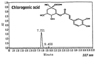

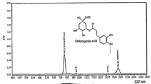

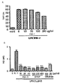


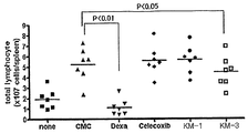


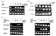

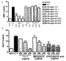


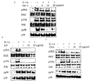

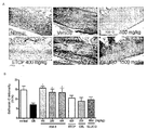

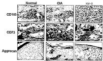

Classifications
-
- A—HUMAN NECESSITIES
- A61—MEDICAL OR VETERINARY SCIENCE; HYGIENE
- A61K—PREPARATIONS FOR MEDICAL, DENTAL OR TOILETRY PURPOSES
- A61K36/00—Medicinal preparations of undetermined constitution containing material from algae, lichens, fungi or plants, or derivatives thereof, e.g. traditional herbal medicines
- A61K36/18—Magnoliophyta (angiosperms)
- A61K36/185—Magnoliopsida (dicotyledons)
- A61K36/35—Caprifoliaceae (Honeysuckle family)
- A61K36/355—Lonicera (honeysuckle)
-
- A—HUMAN NECESSITIES
- A23—FOODS OR FOODSTUFFS; TREATMENT THEREOF, NOT COVERED BY OTHER CLASSES
- A23L—FOODS, FOODSTUFFS, OR NON-ALCOHOLIC BEVERAGES, NOT COVERED BY SUBCLASSES A21D OR A23B-A23J; THEIR PREPARATION OR TREATMENT, e.g. COOKING, MODIFICATION OF NUTRITIVE QUALITIES, PHYSICAL TREATMENT; PRESERVATION OF FOODS OR FOODSTUFFS, IN GENERAL
- A23L33/00—Modifying nutritive qualities of foods; Dietetic products; Preparation or treatment thereof
- A23L33/10—Modifying nutritive qualities of foods; Dietetic products; Preparation or treatment thereof using additives
- A23L33/105—Plant extracts, their artificial duplicates or their derivatives
-
- A—HUMAN NECESSITIES
- A61—MEDICAL OR VETERINARY SCIENCE; HYGIENE
- A61K—PREPARATIONS FOR MEDICAL, DENTAL OR TOILETRY PURPOSES
- A61K36/00—Medicinal preparations of undetermined constitution containing material from algae, lichens, fungi or plants, or derivatives thereof, e.g. traditional herbal medicines
- A61K36/18—Magnoliophyta (angiosperms)
- A61K36/88—Liliopsida (monocotyledons)
- A61K36/896—Liliaceae (Lily family), e.g. daylily, plantain lily, Hyacinth or narcissus
- A61K36/8964—Anemarrhena
-
- A—HUMAN NECESSITIES
- A61—MEDICAL OR VETERINARY SCIENCE; HYGIENE
- A61P—SPECIFIC THERAPEUTIC ACTIVITY OF CHEMICAL COMPOUNDS OR MEDICINAL PREPARATIONS
- A61P19/00—Drugs for skeletal disorders
- A61P19/02—Drugs for skeletal disorders for joint disorders, e.g. arthritis, arthrosis
Definitions
- the present invention relates to a composition
- a composition comprising an extract of mixed herbs with Lonicera japonica THUNB and Anemarrhena asphodeloides BUNGE for the prevention and treatment of arthritic disease.
- Arthritis is an autoimmune disease characterized by their symptoms such as pain, swelling and stiffness in the joints.
- the two major forms of arthritis in mammals are inflammatory arthritis such as rheumatoid arthritis (RA), and osteoarthritis (OA), a progressive, degenerative loss of cartilage often secondary to mechanical stress, aging, dysplastic conditions and/or injury.
- the symptoms of arthritis generally relates to arthrosis of spine, e.g., hallux rigidus, arthrosis psoriaticum, or rheumatic arthritis.
- Osteoarthritis manifests similar symptoms to a rheumatoid arthritis (RA).
- RA rheumatoid arthritis
- cartilage deteriorates, a reactive synovitis often develops later on.
- rheumatoid arthritis erodes cartilage, the secondary osteoarthritis changes the bone and cartilage development.
- the joints suffering with both diseases appear the same phenomena with each other.
- Osteoarthritis usually represents a pain which become worsen with exercise or a simple X-ray radiation showing clearly a thinned cartilage.
- Commonly affected joints are knees, hips, spine, finger, base of thumb or big toe etc.
- MMPs matrix metaloproteinases
- the disease is involved in the destruction of articular cartilage by MMPs (matrix metaloproteinases) which mainly works for cartilage depletion, which is characterized by degenerative changes in the articular cartilage and caused by the over-production of inflammatory cytokines, e.g., interleukin-1 (IL-1), tumor necrosis factor- ⁇ (TNF- ⁇ ) etc, causing to extremely severe pain in joints, tendons, muscles and ligament (Fernandes J.C., The role of cytokines in osteoarthritis pathophysiology , 39, pp237-246, 2002).
- IL-1 interleukin-1
- TNF- ⁇ tumor necrosis factor- ⁇
- Rheumatoid arthritis is a common autoimmune disease characterized by the swelling, deformation and destruction of joint, which culminates in severe physical disability.
- Rheumatic diseases include the diseases occurring at muscles, tendons, joints, bones or sinus, which are generally characterized by an inflammation and/or degeneration.
- the patients suffering with rheumatoid arthritis present the imbalance in immune system, which causes an overproduction of pro-inflammatory cytokines, e.g., TNF- ⁇ , IL-1 etc, and a lack of anti-inflammatory cytokines, e.g., IL-10, IL-1, etc.
- RA is characterized by synovial inflammation, which progresses to a cartilage destruction, bone erosion and subsequent joint deformity.
- polymorphonuclear cells, macrophages and lymphocytes are released from the joint.
- Activated T-lymphocytes produce cytotoxins and pro-inflammatory cytokines, while macrophages stimulate the release of the prostaglandins and cytotoxins.
- Vasoactive substances such as histamine, kinins and prostaglandins, are released at the site of inflammation and they cause to an edema, erythema and pain at the region of the inflamed joints.
- the main pathology of the affected synovial tissue is a hyperplasia and the sub-intimal infiltration of T and B lymphocytes.
- Synovial tissue hyperplasia forms in pannus tissue, which irreversibly destroys the cartilage and bone in the affected joint.
- RA progression is associated with elevated levels of TNF- ⁇ and IL-1 ⁇ produced by macrophages and dendrite cells, an imbalance of Th1/Th2 and over-production of antigen specific immunoglobulins.
- TNF- ⁇ and IL-1 ⁇ directly induce the synthesis of proteolytic enzyme such as matrix metalloproteinase (MMPs) which can break down the extracellular matrix macromolecules.
- MMPs matrix metalloproteinase
- TIMPs tissue inhibitors of metalloproteinases
- the first aid drugs for treating arthritis includes the drug for alleviating pain and inflammation, which is classified into non-steroidal anti-inflammatory drugs(NSAIDs), e.g., aspirin, ibuprofen, naproxen, methotrexate, etc.
- Secondary aid drugs include corticosteroids, slow acting antirheumatic drugs (SAARDs) or disease modifying drugs (DMs), e.g., penicilamine, cyclophosphamide, gold salts, azethioprine, levamisole, etc.
- SAARDs slow acting antirheumatic drugs
- DMs disease modifying drugs
- BRMs biological-response modifiers
- TNF- ⁇ antagonists which plays role in binding to its receptor or directly binding to the TNF- ⁇ protein.
- the use of DMARDs has been impeded by various disadvantages, for example, the potential of its long-term side effects and toxicity, high cost, hypersensitivity to the medications and infections due to TNF- ⁇ blockage, etc
- Degenerative arthritis one of representative osteo-joint diseases is chronic arthritis. It is difficult to treat the disease with conventionally available anti-inflammatory drugs in clinic. Moreover, the drugs give rise to systemic adverse response such as digestive disorder, gastro-intestinal disorder and renal function disorder and the adverse response of the drugs occurs more frequently as the age of patient increases, which causes lots of problems in case of long-term systemic treatment in older people. Therefore, the new drug development targeting anti-inflammatory effect, protecting and regenerating effect on cartilage has been urgently needed than previous systemic treatment therapy recently.
- NOS nitric oxide synthase
- PGs prostaglandin
- NOS enzyme generating NO from L-Arginine or cyclooxygenase (COX) involved in the synthesis of various prostaglandins have been the main target to block inflammation of arthritis.
- NOS enzymes for example, bNOS (brain NOS) existing in brain, nNOS (neuronal NOS) in neuronal system, eNOS (endothelial NOS) in endothelial system etc, which are expressed at regular levels in the human body.
- a small amount of NO reproduced thereby plays an important role in maintaining of homeostasis such as neuronal transmission or induction of vasodilation etc.
- excess amount of NO occurring abruptly by iNOS (induced NOS) induced by various cytokines or external stimulator gives rise to cell toxicity or inflammatory reaction.
- Chronic inflammation is correlated with the increased activity of iNOS (Chan P. S. et al., Osteoarthritis cartilage , 13(5) , pp387-394, 2005; Appleton I. et al., Adv. Pharmacol ., 35 , pp27-28, 1996).
- the cartilage is worsen and arthritis occurs caused by late production rate of proteoglycan or collagen in cartilage, which results in loss of cushion function.
- the articular cartilage consists of water (70 ⁇ 80%) necessary for lubrication and growth, collagen (10 ⁇ 15%), proteoglycan (5 ⁇ 10%) and chondrocyte, wherein proteoglycan has particular structure with core protein attached with several glycosaminoglycan(GAG) (Hardingham et al., J. Rheum(Suppl), 43(2) , pp86-90, 1995).
- Lonicerae Japonicae is a flower bud part of Lonicerae spp. belonged to Caprifoliaceae. They taste sweet and is good for detoxifying, which traditionally used for dysentery, pain and swelling. They have been known to have antiulcer, antibacterial, antiviral, antispasmodic, diuretic, anti-inflammatory and analgesic bioactivities.
- the main components are luteolin, inositol, saponin, tannin, isochlorogenic acid, chlorogenic acid etc (B.S. Chung et al., HyangYakDaeSaJeon, Youngrimsa, pp939-940, 1989).
- Anemarrhena asphodeloides BUNGE belonged to Haemodoraceae. They taste sour, which traditionally used for fever, dire thirst, cough and diabetes. They have been known to have hypoglycaemic, anti-pyretic, antiplatelet aggregation, inhibits stress ulcer, sedative, inhibits cAMP phosphodiesterase and Na/K-ATPase, haemolytic, antitumour bioactivities.
- the main components are various saponins such as timosaponin A-I, timosaponin A-II, timosaponin A-III, timosaponin A-IV, timosaponin B-I, and timosaponin B-II, nicotinic acid, magniferin, isomangiferin etc (B.S. Chung et al., HyangYakDaeSaJeon, Youngrimsa, pp203-204, 1989).
- saponins such as timosaponin A-I, timosaponin A-II, timosaponin A-III, timosaponin A-IV, timosaponin B-I, and timosaponin B-II, nicotinic acid, magniferin, isomangiferin etc (B.S. Chung et al., HyangYakDaeSaJe
- the present inventors have confirmed that the extract of mixed herbs with Lonicera japonica THUNB and Anemarrhena asphodeloides BUNGE shows potent anti-inflammatory effect through various experiments, i.e., the inhibitory effect on the dissociation of proteoglycan and type II collagen in cartilage tissue; protecting effect on cartilage due to the inhibition of MMP-1, MMP-3 and MMP-13 activity; and the restoring effect on cartilage tissue; the anti-inflammatory and antiphlogistic effect in edema animal model; anti-inflammatory effect confirmed by the inhibition test on PEG 2 activity, GAG degradation, and various genetic toxicity test, therefore, it can be used as the effective and safe therapeutics or health food for treating and preventing arthritic disease.
- the present invention provides a composition comprising an extract of mixed herbs with Lonicera japonica THUNB and Anemarrhena asphodeloides BUNGE for the prevention and treatment of arthritic disease and the use thereof.
- a pharmaceutical composition comprising an extract of mixed herbs with Lonicera japonica THUNB and Anemarrhena asphodeloides BUNGE as an active ingredient for the treatment and prevention of arthritic diseases, in particular, by way of stimulating the recovery of cartilage tissue, protecting cartilage damage due to the stimulation of cartilage component and inhibition of cartilage dissociation, and inhibiting inflammation and pain.
- the present invention provides a pharmaceutical composition
- a pharmaceutical composition comprising an extract of mixed herbs with Lonicera japonica THUNB and Anemarrhena asphodeloides BUNGE as an active ingredient for preventing and treating of arthritic disease.
- extract includes the crude extract soluble in water, C 1 to C 4 lower alcohol and the mixture thereof; and the butanol soluble extract obtained by fractionation with butanol solution therefrom.
- mixed herbs with Lonicera japonica THUNB and Anemarrhena asphodeloides BUNGE includes the mixed herbs with the mixed ratio ranging from 0.5 ⁇ 3: 1 by weight(w/w%), preferably, 1 ⁇ 2: 1 by weight(w/w%), most preferably 1.5 ⁇ 2:1 by weight(w/w%).
- extract contain chlorogenic acid as a standard component in the extract of Lonicera japonica THUNB and mangiferin as a standard component in the extract of Anemarrhena asphodeloides BUNGE, preferably, 0.5 ⁇ 6(w/w%) chlorogenic acid and 0.5 ⁇ 4(w/w%) mangiferin, more preferably, 1.5 ⁇ 5(w/w%) chlorogenic acid and 0.5 ⁇ 3.5(w/w%) mangiferin, most preferably 3 ⁇ 4.5(w/w%) chlorogenic acid and 0.5 ⁇ 2.5(w/w%) mangiferin as a standard component in total extract.
- arthritis disease includes degenerative arthritis, rheumatic arthritis or Lupus arthritis, preferably, rheumatic arthritis.
- the present invention also provided a use of an extract of mixed herbs with Lonicera japonica THUNB and Anemarrhena asphodeloides BUNGE for the preparation of therapeutic agent for the treatment and prevention of arthritic disease in mammal or human.
- An inventive extract of the present invention can be prepared in detail by following procedures.
- Lonicera japonica THUNB and Anemarrhena asphodeloides BUNGE are dried, cut, crushed, mixed together and added to 1 to 20-fold, preferably, approximately 5 to 10-fold volume of distilled water, C 1 to C 4 lower alcohols or the mixtures thereof, preferably the mixture of water and alcohol with approximately 1: 0.1 to 1: 10, more preferably, 1: 0.5 to 1: 5 with mixing ratio (v/v); the solution is treated with hot water at the temperature ranging from 10°C ⁇ 100°C, preferably, 60°C ⁇ 100°C for the period ranging from 1 to 6 hours, preferably, 2 to 4 hours with the extraction method by the extraction with hot water, cold water, reflux extraction, or ultra-sonication extraction, preferably, extraction with hot water; the extract is collected with filtration, concentrated under reduced pressure and dried to obtain an inventive crude extract of the present invention.
- butanol soluble solution is added to the above-mentioned crude extract, and then the suspension is performed to fractionation to obtain the inventive purified extract of the present invention.
- the inventive crude extract and purified extract prepared by the above-described procedures contains chlorogenic acid as a standard component in the extract of Lonicera japonica THUNB and mangiferin as a standard component in the extract of Anemarrhena asphodeloides BUNGE, preferably, 0.5 ⁇ 6(w/w%) chlorogenic acid and 0.5 ⁇ 4(w/w%) mangiferin, more preferably, 1.5 ⁇ 5(w/w%) chlorogenic acid and 0.5 ⁇ 3.5(w/w%) mangiferin, most preferably, 3.0 ⁇ 4.5(w/w%) chlorogenic acid and 0.5 ⁇ 2.5(w/w%) mangiferin as a standard component in total extract.
- the present invention also provided a use of an extract of mixed herbs with Lonicera japonica THUNB and Anemarrhena asphodeloides BUNGE prepared by the above-described preparation method for the preparation of therapeutic agent for the treatment and prevention of arthritic disease in mammal or human.
- the present invention also provided a pharmaceutical composition
- a pharmaceutical composition comprising an extract of mixed herbs with Lonicera japonica THUNB and Anemarrhena asphodeloides BUNGE prepared by the above-described preparation method as an active ingredient and a pharmaceutically acceptable carrier thereof for treating and preventing arthritic disease.
- the inventive composition for treating and preventing arthritic diseases may comprises the above-described extract as 0.1 ⁇ 50% by weight based on the total weight of the composition.
- the inventive composition may additionally comprise conventional carrier, adjuvants or diluents in accordance with a using method well known in the art. It is preferable that said carrier is used as appropriate substance according to the usage and application method, but it is not limited. Appropriate diluents are listed in the written text of Remington's Pharmaceutical Science (Mack Publishing co, Easton PA).
- composition according to the present invention can be provided as a pharmaceutical composition containing pharmaceutically acceptable carriers, adjuvants or diluents, e.g., lactose, dextrose, sucrose, sorbitol, mannitol, xylitol, erythritol, maltitol, starches, acacia rubber, alginate, gelatin, calcium phosphate, calcium silicate, cellulose, methyl cellulose, polyvinyl pyrrolidone, water, methylhydroxy benzoate, propylhydroxy benzoate, talc, magnesium stearate and mineral oil.
- pharmaceutically acceptable carriers e.g., lactose, dextrose, sucrose, sorbitol, mannitol, xylitol, erythritol, maltitol, starches, acacia rubber, alginate, gelatin, calcium phosphate, calcium silicate, cellulose, methyl cellulose, polyvinyl
- the formulations may additionally include fillers, anti-agglutinating agents, lubricating agents, wetting agents, flavoring agents, emulsifiers, preservatives and the like.
- the compositions of the invention may be formulated so as to provide quick, sustained or delayed release of the active ingredient after their administration to a patient by employing any of the procedures well known in the art.
- compositions of the present invention can be dissolved in oils, propylene glycol or other solvents that are commonly used to produce an injection.
- suitable examples of the carriers include physiological saline, polyethylene glycol, ethanol, vegetable oils, isopropyl myristate, etc., but are not limited to them.
- the extract of the present invention can be formulated in the form of ointments and creams.
- compositions containing present composition may be prepared in any form, such as oral dosage form (powder, tablet, capsule, soft capsule, aqueous medicine, syrup, elixirs pill, powder, sachet, granule), or topical preparation (cream, ointment, lotion, gel, balm, patch, paste, spray solution, aerosol and the like), or injectable preparation (solution, suspension, emulsion).
- oral dosage form prowder, tablet, capsule, soft capsule, aqueous medicine, syrup, elixirs pill, powder, sachet, granule
- topical preparation cream, ointment, lotion, gel, balm, patch, paste, spray solution, aerosol and the like
- injectable preparation solution, suspension, emulsion
- composition of the present invention in pharmaceutical dosage forms may be used in the form of their pharmaceutically acceptable salts, and also may be used alone or in appropriate association, as well as in combination with other pharmaceutically active compounds.
- the desirable dose of the inventive extract or composition varies depending on the condition and the weight of the subject, severity, drug form, route and period of administration, and may be chosen by those skilled in the art. However, in order to obtain desirable effects, it is generally recommended to administer at the amount ranging 0.1 to 1000 mg/kg, preferably, 1 to 100 mg/kg by weight/day of the inventive extract of the present invention.
- the dose may be administered in single or divided into several times per day.
- composition of present invention can be administered to a subject animal such as mammals (rat, mouse, domestic animals or human) via various routes. All modes of administration are contemplated, for example, administration can be made orally, rectally or by intravenous, intramuscular, subcutaneous, intra-cutaneous, intrathecal, epidural or intra-cerebroventricular injection.
- a functional health food comprising an extract of mixed herbs with Lonicera japonica THUNB and Anemarrhena asphodeloides BUNGE .
- functional health food defined herein is "the functional food having enhanced functionality such as physical functionality or physiological functionality by adding the extract of the present invention to conventional food to prevent or improve aimed disease in human or mammal”.
- health care food defined herein is "the food containing inventive extract of the present invention showing no specific intended effect but general intended effect in a small amount of quantity as a form of additive or in a whole amount of quantity as a form of capsule, pill, tablet etc".
- any substance the intended use which results or may reasonably be expected to result-directly or indirectly-in its becoming a component or otherwise affecting the characteristics of any food for example, thickening agent, maturing agent, bleaching agent, sequesterants, humectant, anticaking agent, clarifying agents, curing agent, emulsifier, stabilizer, thickner, bases and acid, foaming agents, nutrients, coloring agent, flavoring agent, sweetner, preservative agent, antioxidant, etc, which had been well-known in the art.
- direct additive a substance that becomes part of the food in trace amounts due to its packaging, storage or other handling.
- Health foods can be contained in food, health beverage, dietary therapy etc, and may be used as a form of powder, granule, tablet, chewing tablet, capsule, beverage etc for preventing or improving aimed disease.
- inventive extract can be added to food or beverage for prevention and improvement of aimed disease.
- the amount of inventive extract in food or beverage as a functional health food or health care food may generally range from about 0.01 to 15 w/w% of total weight of food for functional health food composition.
- the preferable amount of inventive extract of the present invention in the functional health food, health care food or special nutrient food may be varied in accordance to the intended purpose of each food, it is preferably used in general to use as a additive in the amount of inventive extract of the present invention ranging from about 0.01 to 5% in food such as noodles and the like, from 40 to 100% in health care food on the ratio of 100% of the food composition.
- the health beverage composition of present invention contains inventive extract as an essential component in the indicated ratio
- the other component can be various deodorant or natural carbohydrate etc such as conventional beverage.
- natural carbohydrate are monosaccharide such as glucose, fructose etc; disaccharide such as maltose, sucrose etc; conventional sugar such as dextrin, cyclodextrin; and sugar alcohol such as xylitol, and erythritol etc.
- natural deodorant such as taumatin, stevia extract such as levaudioside A, glycyrrhizin et al., and synthetic deodorant such as saccharin, aspartam et al.
- the amount of above described natural carbohydrate is generally ranges from about 1 to 20 g, preferably 5 to 12 g in the ratio of 100 ml of present beverage composition.
- the other components than aforementioned composition are various nutrients, a vitamin, a mineral or an electrolyte, synthetic flavoring agent, a coloring agent and improving agent in case of cheese, chocolate et al., pectic acid and the salt thereof, alginic acid and the salt thereof, organic acid, protective colloidal adhesive, pH controlling agent, stabilizer, a preservative, glycerin, alcohol, carbonizing agent used in carbonate beverage et al.
- the other component than aforementioned ones may be fruit juice for preparing natural fruit juice, fruit juice beverage and vegetable beverage, wherein the component can be used independently or in combination.
- the ratio of the components is not so important but is generally range from about 0 to 20 w/w% per 100 w/w% present composition.
- Examples of addable food comprising aforementioned extract therein are various food, beverage, gum, vitamin complex, health improving food and the like.
- the present invention comprising an extract of mixed herbs with Lonicera japonica THUNB and Anemarrhena asphodeloides BUNGE shows potent anti-inflammatory effect through various experiments, i.e., the inhibitory effect on the dissociation of proteoglycan and type II collagen in cartilage tissue; protecting effect on cartilage due to the inhibition of MMP-1, MMP-3 and MMP-13 activity; and the restoring effect on cartilage tissue; the anti-inflammatory and antiphlogistic effect in edema animal model; anti-inflammatory effect confirmed by the inhibition test on PEG 2 activity, GAG degradation and genetic toxicity tests, therefore, it can be used as the effective and safe therapeutics or health food for treating and preventing arthritic disease.
- Fig. 1 shows the result of chromatogram analysis on chlorogenic acid, a standard component contained in the extract of Lonicera japonica THUNB;
- Fig. 2 shows the result of chromatogram analysis on mangiferin, a standard component contained in the extract of Anemarrhena asphodeloides BUNGE;
- Fig. 3 shows the result of chromatogram analysis on chlorogenic acid, a standard component in KM-3;
- Fig. 4 shows the result of chromatogram analysis on mangiferin, a standard component in KM-3;
- Fig. 5-A presents inhibitory effect on NO production by treatment of KM-1
- Fig. 5-B presents inhibitory effect on NO production by treatment of KM-3
- Fig. 6-A presents inhibitory effect on PGE 2 production by treatment of KM-1;
- Fig. 6-B presents inhibitory effect on PGE 2 production by treatment of KM-3;
- Fig. 7-A presents inhibitory effect on IL-1 ⁇ production by treatment of KM-1;
- Fig. 7-B presents inhibitory effect on IL-1 ⁇ production by treatment of KM-3;
- Fig. 7-C presents inhibitory effect on IL-6 production by treatment of KM-3;
- Fig. 8 presents the inhibitory effects of KM-1 and KM-3 on swelling of Collagenase-Induced Osteoarthritis (CIA) animal model;
- Fig. 9 presents the inhibitory effects of KM-1 and KM-3 on the number of increased lymphocytes in the Collagenase-Induced Osteoarthritis (CIA) animal model ;
- Fig. 10 presents the inhibitory effect of KM-1 and KM-3 on the cartilage erosion in the Collagenase-Induced Osteoarthritis (CIA) animal model;
- Fig. 11 presents the inhibitory effect of KM-1 and KM-3 on the production of collagen-specific IgG antibody in the Collagenase-Induced Osteoarthritis (CIA) animal model test;
- Fig. 12-A depicts the inhibitory effect of KM-1 on the degradation of proteoglycan
- Fig. 12-B depicts the inhibitory effects of KM-3, mangiferin and chlorogenic acid on the degradation of proteoglycan
- Fig. 13-A depicts the increased mRNA gene expression of proteoglycan and type II collagen in cartilage tissue of the patient suffering from osteoarthritis, treated with KM-1 by RT-PCR method;
- Fig. 13-B depicts the increased mRNA gene expression of proteoglycan and type II collagen in cartilage tissue of the patient suffering from osteoarthritis, treated with KM-3 by RT-PCR method;
- Fig. 13-C depicts the increased mRNA gene expression of proteoglycan and type II collagen in cartilage tissue of the patient suffering from osteoarthritis, treated with mangiferin and chlorogenic acid by RT-PCR method;
- Fig. 14-A depicts the inhibitory effect of KM-1 on the level of MMP-13 activity in cartilage tissue of the patient suffering from osteoarthritis
- Fig. 14-B depicts the inhibitory effect of KM-3 on the level of MMP-13 activity in cartilage tissue of the patient suffering from osteoarthritis
- Fig. 15-A depicts the inhibitory effect of KM-1 on the level of MMP-1 activity in cartilage tissue of the patient suffering from osteoarthritis
- Fig. 15-B depicts the inhibitory effect of KM-3, mangiferin and chlorogenic acid on the level of MMP-1 activity in cartilage tissue of the patient suffering from osteoarthritis;
- Fig. 16 depicts the inhibitory effect on the level of mRNA gene expression of MMP-1, MMP-3 and MMP-13 of cartilage tissue of the patient from osteoarthritis treated with KM-1 (A) and KM-3 (B);
- Fig. 17 depicts the determination of the cytotoxicity of cartilage tissue of the patient suffering from osteoarthritis treated with KM-1 (A) and KM-3 (B);
- Fig. 18-A depicts the inhibitory effect on the phosphorylation of several mitogen activated protein kinase (MAPK) by KM-3 was involved in the cartilage protection of osteoarthritis cartilage;
- MAPK mitogen activated protein kinase
- Fig. 18-B depicts the inhibitory effect on the phosphorylation of several mitogen activated protein kinase (MAPK) by mangiferin was involved in the cartilage protection of osteoarthritis cartilage;
- MAPK mitogen activated protein kinase
- Fig. 18-C depicts the inhibitory effect on the phosphorylation of several mitogen activated protein kinase (MAPK) by chlorogenic acid was involved in the cartilage protection of osteoarthritis cartilage;
- MAPK mitogen activated protein kinase
- Fig. 19 represents the swelling and mobility recovery effect of KM-1 (A) and KM-3 (B) of joint in Collagenase-Induced Osteoarthritis (CIA) animal model;
- Fig. 20-A represents the cartilage protective effect of KM-1 by morphological analysis in joint of Collagenase-Induced Osteoarthritis (CIA) animal model;
- Fig. 20-B represents the cartilage protective effect of KM-3 by morphological analysis in joint of Collagenase-Induced Osteoarthritis (CIA) animal model;
- Fig. 21-A depicts the recovery effect of KM-1 on proteoglycan expression by Safranin O staining analysis in joint of Collagenase-Induced Osteoarthritis (CIA) animal model;
- Fig. 21-B depicts the recovery effect of KM-3 on proteoglycan expression by Safranin O staining analysis in joint of Collagenase-Induced Osteoarthritis (CIA) animal model;
- Fig. 22-A depicts the recovery effect of KM-1 on collagen expression by Masson Trichrome staining analysis in joint of Collagenase-Induced Osteoarthritis (CIA) animal model;
- Fig. 22-B depicts the recovery effect of KM-3 on collagen expression by Masson Trichrome staining analysis in joint of Collagenase-Induced Osteoarthritis (CIA) animal model;
- Fig. 23 represents the cartilage regenerating effect of KM-3 on the expression of differentiated mesenchymal stem cells (CD105, CD73) from the tissue of subchondral bone and aggrecan in joint of Collagenase-Induced Osteoarthritis (CIA) animal model;
- Fig. 24-A represents the inhibitory effect of KM-3 on the GAG degradation test performed by co-culturing the cartilage cell and the subchondral bone tissue at the normal region (NSC) isolated from the cartilage tissue and subchondral bone of patients suffering from osteoarthritis;
- Fig. 24-B represents the inhibitory effect of KM-3 on the GAG degradation test performed by co-culturing the cartilage cell and the subchondral bone tissue at the abnormal region (SC) isolated from the cartilage tissue and subchondral bone of patients suffering from osteoarthritis.
- SC abnormal region
- Anemarrhena asphodeloides BUNGE purchased from Kyunghee Medical Center in Korea, was air-dried, cut into small pieces and added to 0.7 L of 50% ethanol. The solution was refluxed for 4 hours with stirring at 85°C and the residue was filtered. The filtrate was concentrated and dried to obtain 50 g of the extract of Anemarrhena asphodeloides BUNGE to use as a comparative test sample (designated as 'AA extract' hereinafter).
- the KM-1 extract contains 3% chlorogenic acid(w/w%) and 3.5% mangiferin(w/w%) through chromatogram analysis.
- the KM-2 extract contains 1.5% chlorogenic acid (w/w%) and 1.8% mangiferin (w/w%) through chromatogram analysis.
- the KM-3 extract contains 4.5% chlorogenic acid(w/w%) and 2.1% mangiferin(w/w%) through chromatogram analysis( See , Fig. 3 & 4).
- the KM-4 extract contains 2.2% chlorogenic acid(w/w%) and 1.4% mangiferin (w/w%) through chromatogram analysis.
- Mouse macrophage cell line (Raw264.7, purchased from ATCC) was cultured in RPMI-1640 media (10% FBS, 2 mM 1-glutamine, 100 units/ml penicillin sodium, 100 units/ml streptomycin sulphate and 250 ng/ml amphotericin B).
- the culture cell was seeded on 24-well plate (10 6 cell/well) and treated with KM-1 (10, 50, 100, 200 ⁇ g/ml), KM-3 (10, 20, 40 ⁇ g/ml), positive control groups, i.e., Celecoxib (CEL, 100 ⁇ g/ml) and ETCP (SK Chemicals) (100, 200, 400 ⁇ g/ml) for 30 minutes.
- KM-1 10, 50, 100, 200 ⁇ g/ml
- KM-3 i.e., Celecoxib
- ETCP SK Chemicals
- DBA/1J mice were purchased from Chungang Experimental Animals ( www.labanimals.co.kr , Korea).
- Equal volume of CFA Complete Freund s Ajuvant
- IFA Incomplete Freund's Ajuvant
- mice were purchased from Chungang Experimental Animals ( www.labanimals.co.kr , Korea).
- Equal volume of CFA Complete Freund s Ajuvant
- 100 ⁇ l of the mixture was subcutaneously injected at 2.5 cm upper region from the tail fundus.
- 2ml of collagen solution mixed with the equal volume of IFA (Incomplete Freund's Ajuvant) was injected at 1 cm upper region from the tail fundus at the dose of 100 ⁇ l.
- KM-3(200 mg/kg) and Celecoxib 100 mg/kg
- CMC Carboxymethylcellulose, Sigma
- the joint cartilage of human was provided from the patient who had taken artificial joint surgery (Orthopedics Surgery Dep. of Kyunghee Medical Center). After revealing the surface of joint by surgery with a sterilized condition, about 200-220 mg of the articular surface tissue prepared from the articular cartilage of human and rabbit was dipped into DMEM medium (FBS, GIBCO BRL, USA) supplemented with 5% fetal bovine serum and 100 unit/ml of penicillin-streptomycin. The tissue was washed with the medium several times and then the articular tissue was cultured at 37°C in humidified 95% CO 2 incubator.
- DMEM medium FBS, GIBCO BRL, USA
- the medium was replaced with new basic medium containing inactivated 5% fetal bovine serum with heat treatment, 10mM HEPES, and 100 unit/ml of penicillin-streptomycin, and 30mg of the chondrocyte was transferred to 48-well plate.
- interlukin-1 ⁇ interlukin-1 ⁇ (IL-1 ⁇ , R&D system, USA) was added to the medium to induce inflammation and various concentrations of test sample (KM-1), ETCP (SK Chemnicals), Celecoxib (CEL, Pfizer Co., USA), and Glucosamine (GLUCO, Sigma Co., USA), i.e., 0.1, 0.2, and 0.4 mg/ml, were added thereto respectively.
- the medium was further cultured at 37°C for 7 days, and the supernatant was collected, which had been stored at -20°C to use as comparative test samples and test samples.
- RNA was treated with TRIzol reagent (Invitrogen Corporation, CA, USA) to isolate RNA and reverse transcription for 1 ⁇ g of total RNA was performed by adding buffer solution containing oligo(dT) 12 primer, Dntp (10 mM), 0.1 M dithiothreitol (DDT), reverse transcriptase and RNase inhibitor to the medium. The medium was incubated 42°C for 60 minutes. PCR (polymerase Chain Reaction) using by the primers disclosed in Table 1 and Sequence(SEQ) I.D.
- the solution was adjusted with distilled water to be total volume of 10 ⁇ l and then PCR was performed using by thermal cycler apparatus (Bio-Rad, USA) as follows: after denaturing at 94°C for 5 minutes, the PCR is performed in the order of the reaction for 60 sec at 94°C, 60 sec at 55°C, and 90 sec at 72°C. The cycles were repeated 30 times and the last extension was performed at 72°C for 5 minutes.
- the product produced by PCR was subjected to electrophoresis (5 V /cm) on 1.8% agarose gel and stained for 5 minutes with 2 ⁇ g/ml of ethidium bromide (EtBr). The stained product was washed for 10 minutes with distilled water and the result was determined at UV wavelength (260 nm).
- the subchondral bone tissue and joint cartilage sample of human were provided from the patient taken artificial joint surgery (Orthopedics Surgery Dep. of Kyunghee Medical Center).
- the subchondral bone was crushed to pieces, and treated with type II collagenase for 30 minutes. The pieces were subjected to explant culture and subcultured for 2 times to use.
- Cartilage cell was isolated with type II collagenase, and subcultured twice in culture media condition to use.
- the chondrocyte was made into beads with alginate and seeded on the upper chamber, while the subchondral bone tissue cell was seeded on the lower chamber and cultured for 24 hours. 50 ⁇ g/ml of KM-3 was treated thereto for 14 days and the media was collected at every 7 days.
- mice Male ICR mouse (Orient Bio, Japan) weighing from 20 to 25 g was acclimated for several days and each group consists of 8 mice. The test samples were administrated orally into the mice and 10% formalin solution (v/v, Sigma Co. USA) was subcutaneously administrated to the left posterior limb one hour after the administration. The licking frequency of the foot sole was observed at the 1 st phase (from initial time to 5 minutes after the administration) and 2 nd phase (from 15 minutes to 20 minutes after the administration) to record. The inhibition rate (%) was calculated by setting the inhibition rate in positive control group treated with Celecoxib, a conventionally used NSAID, to 100.
- Celecoxib a conventionally used NSAID
- MIA monosodium iodoactetate
- mice Male SD rat (Orientbio. Japan) weighing from 200 to 220 g, was acclimated for several days and MIA (Sigma, cat# I2512, USA) dissolved in PBS was injected into the glenoid cavity of left hind knee to induce arthritis. After a week of recovery, the subjects induced with arthritis were selected by using incapacitance tester apparatus (Linton, Stoelting Co., Wood Dale, IL) and the subjects were grouped to consist of 8 animals for each group. On the 8 th day after the induction, the test samples were orally administered at the routine time for every day and the measurement of resulting data was started at one week after the administration for 3 weeks, once a week.
- MIA Sigma, cat# I2512, USA
- the data was measured using by incapacitance tester (Linton, Stoelting Co., Wood Dale, IL) and calculated according to following Math formulae 1.
- the inhibition rate (%) was calculated by setting the inhibition rate in positive control group treated with Celecoxib, a conventionally used NSAID, to 100.
- rat paw pressure analgesia test was performed with the procedure described in the literature (Randall LO and Selitto JJ, Arch Int. Pharmacodyn ., 111 , pp409-419, 1957).
- Hot plate pain test was performed with the procedure described in the literature (Pharmacological report, 60(2008) pp409-414).
- mice Male ICR mouse (Orientbio. Japan) weighing from 15 to 20 g was acclimated for several days and grouped to 8-9 animals for each group. Test samples were orally administrated thereto. One and two hour after the treatment, the mouse was put into plastic cylinder, of which temperature was maintained at 55 ⁇ 1°C to determine the time when the mouse licked sole of the feet or jumped. The cut-off time was set to 15 seconds, and the inhibition rate(%) was calculated by setting the inhibition rate in positive control group treated with Celecoxib, a conventionally used NSAID, to 100.
- Celecoxib a conventionally used NSAID
- Acetic-acid induced writhing test was performed to determine the anti-inflammatory effect with the procedure described in the literature (H.O.J collier et al., Br. J. Pharmac. Chemother., 32 , pp295-310, 1968).
- Croton oil induces various skin inflammations such as rash, swelling, blister and so on.
- test using croton oil-induced ear edema was performed according to method disclosed in the literature literature (Gabor M, Mouse ear inflammation models and their pharmacological applications, Published by Akademiai Kiado, Budapest, pp24-28, 2000).
- mice Male ICR mouse (Orientbio. Japan)) weighing from 20 to 25g was used as an experimental animal and each group consists of 6 mice. The test samples were administrated orally and after 1 hour, 2.5% croton oil dissolved in acetone was spread on the inner and outer surface of right ear to induce ear-edema. After 4 hours, the increased rate of ear thickness was calculated by comparing with that of left ear of dead mouse with ether using by thickness gauge according to velocity transformation technique (Patrick et al., Toxicol. Appl. Pharmacol. , 81 , pp476-490, 1985).
- the inhibition rate (%) was calculated by setting the inhibition rate in positive control group treated with Celecoxib, a conventionally used NSAID, to 100.
- carrageenan-induced rat-paw edema test was performed as follows.
- mice Male Wister mouse (Orient Bio, Japan) weighing from 20 to 25 g was acclimated for several days and each group consists of 8 mice.
- the test samples were administrated orally into the mice in an amount of 100-400 mg/kg and carrageenan dissolved in a physiological solution was subcutaneously administrated to the left posterior limb to induce inflammation.
- the degree of the edema at the sole of left hind was compared with that of right hind with careful of contaminant using by a plethysmometer apparatus at regular intervals.
- Celecoxib was orally administrated as a positive control group in an amount of 100 mg/kg(body weight).
- the inhibition rate (%) was calculated by setting the inhibition rate in positive control group treated with Celecoxib, a conventionally used NSAID, to 100.
- the test group treated with KM-3 orally administrated in the amount of 400 mg/kg showed potent inhibitory effect on the edema, moreover, more effective than that treated with Celecoxib.
- Nitrite accumulation an indicative of NO synthesis, was measured by applying Griess reaction.
- Peritoneal macrophage was incubated in RPMI (GIBCO BRL, USA) medium containing inactivated fetal bovine serum (FBS, GIBCO BRL, USA) with heat, 100 unit/ml of penicillin and 100 unit/ml of streptomycin sulfate, and incubated at 37°C in 5% CO 2 incubator.
- the absorbance was measured at 550 nm within 10 mins by using micro plate reader (Power Wave 340, Bio-Tek, USA). Fresh medium in all experiments was used as non-treatment group. The amount of NO in the medium was calculated based on the generated sodium nitrite (NaNO 2 ) standard curve and result was showed in Fig. 5.
- test samples were performed according to the ELISA method disclosed in the literature (Dovedi SJ, et al., J. Urol ., 174(1) , pp332-337, 2005).
- the serum was diluted to 1: 500 with phosphate buffer and 50 ⁇ l of the dilution was added to supernatant.
- the pre-coated plates with goat anti-mouse prostaglandin E 2 monoclonal IgG were treated with various concentrations of KM-1 (10, 50, 100, 200 ⁇ g/ml), KM-3 (10, 20, 40, 80 ⁇ g/ml), Celecoxib (CEL, 80 ⁇ g/ml) and ETCP (80 ⁇ g/ml), reacted with together, and the amount of synthesized PGE 2 was measured against the serial dilution of standard PGE 2 to quantify.
- the group treated with KM-1 showed significantly inhibitory effect on the release of cytokine IL-1 ⁇ in a dose dependent manner.
- the group treated with KM-3 showed corresponding inhibitory effect on the release of IL-1 ⁇ and IL-6 expression to those with Celecoxib and ETCP.
- the anti-inflammation and immune inhibitory effect was determined using the animal model of Reference Example 2.
- the degree of edema was measured and the concentration of COMP (Cartilage Oligomeric matrix protein, Animal COMP ELISA, AnaMar Medical Co., Sweden), a representative indicator for determining the degree of cartilage destruction, the number of total lymphocyte in spleen, and collagen-specific antibody (Anti-collagen antibody assay kit, Chondrex Co., USA), which has been known as the main indicator among anti-inflammation effectors since the antibody reproduced by dint of collagen injection, attacks the cartilage site.As shown in Figs. 8 to 11, the test group treated with KM-1 and KM-3 showed potent anti-inflammatory activity corresponding to the positive control group treated with Celecoxib. Especially, the anti-inflammatory effect of KM-3 was superior to that of KM-1.
- COMP Cartilage Oligomeric matrix protein, Animal COMP ELISA, AnaMar Medical Co., Sweden
- Anti-collagen antibody assay kit Chondrex Co., USA
- DMB 1,9-dimethylmethylene blue
- the concentration of GAG in the culture medium of cartilage tissue incubated with the procedure disclosed in Reference Example 3 was measured by determining the amount of polyanionic substance produced by being reacted with blyscan dye solution and chondroitin sulfate was used as a standard.
- the reactant was centrifuged at 12,000 rpm for 10 minutes and the precipitate was dissolved in blyscan dye dissociation solution.
- the amount of spectroscopic GAG was determined at 540 nm and the inhibition rate was expressed based on the amount of degradated GAG induced by interlukin-1 ⁇ (IL-1 ⁇ ).
- proteoglycan and ColII gene collected from the cartilage tissue and chondrocytic cell of rabbit in above Reference Example 3 was determined and the test was performed by using reverse transcription polymerase chain reaction (RT-PCR) with the method disclosed in Reference Example 4.
- RT-PCR reverse transcription polymerase chain reaction
- KM-1 (10, 100, and 200 ⁇ g/ml)
- KM-3 (5, 10, 20 ⁇ g/ml)
- cholorogenic acid (0.01, 0.1, 1 ⁇ g/ml)
- the inventive extract of the present invention potently increased the gene expression of proteoglycan and Col II gene in a dose dependent manner, of which expressions were inhibited by the treatment of IL-1 ⁇ in cartilage tissue.
- Matrix metalloproteinase a protease cleaving the protein in cartilage tissue, destroys the cartilage tissue in rheumatic arthritis and osteoarthritis resulting in exacerbating arthritis. Accordingly, the inhibition of the enzyme reproduction is main target to protect articular cartilage (Nagase H and Woessner JF Jr., J . Biol . Chem ., 274(31 ), pp21491-21494, 1999).
- the inhibitory effect on MMP reproduction using human cartilage tissue medium prepared in Reference Example 3 was determined by using ELISA kit (MMP-1 kit, MMP-13 kit, Biomol Research Lab., Inc., PA, USA), according to the manual of manufacture and thiopeptolide (Ac-Prop Leu-Gly-[2-mercapto-4-methyl-pentanoyl]-Leu-Gly-OC 2 H 5 ) was used as a colorimetric substrate excised by MMP-1 (collagenase-1) and MMP-13 (collagenase-13).
- each 25 ⁇ l of medium was added to 96-well plate with 50 ⁇ l of the substrate to incubate at 37°C for 1 hr and the optical density was measured by ELISA reader (Molecular devices, USA) at 450 nm.
- the activity of each sample on MMP-1 and MMP-13 was determined by calculating the MMP (%) of medium in each well.
- the inventive extract of the present invention significantly inhibited the gene expression of MMP-1, MMP-3 and MMP-13 in cartilage cell in a dose dependent manner.
- LDH lactate dehydrogenase
- the negative control group and test groups treated with KM-1 (0.1, 0.2, and 0.4 mg/ml), KM-3 (0.1, 0.2, and 0.4 mg/ml) and positive control group (ETCP, CEL, GLUCO) was incubated to collect the culture medium.
- the substrate mixed powder diaphorase, lactate, NAD
- TBT solution Tris-buffered Tetrazolium, #G1781, Promega
- 50 ⁇ l of medium was mixed with 50 ⁇ l of substrate mixture to react together at room temperature for 30 minutes.
- the absorbance of culture medium was measured at 490 nm to determine the activity of LDH.
- the groups treated with KM-1 and KM-3 did not affect on the viability of cultured human cartilage tissue for 7 days. Accordingly, it has been confirmed that the inventive extract of the present invention did not show cell toxicity in cartilage tissue, which confirmed that it is safe.
- the protein was added to lysis buffer to mixed together, reacted for 1 hour at 4°C, and centrifuged at 15,000xg to obtain the supernatant. The supernatant was kept in refrigerator; a part of them was used to determine the amount of protein using by BCA solution. 20 ⁇ g of protein was performed to electrophoresis on 12% acrylamide gel, transferred to nitrocellulose paper and blocked for 1 hour with 5% skim milk. The antibodies against to pERK, pp38, pJNK were treated thereto for 2 hours, and washed with TBST solution. Each secondary antibody against those was reacted for 1 hour, washed and exposed by ECL solution kit to develop.
- CMC carboxymethyl cellulose
- KM-1 and KM-3 significantly inhibited the edema of osteoarthritis and further enhanced motility range of the rabbit.
- the cultured slices of the cartilage tissue of rabbit prepared in Reference Example 5 was fixed in 10% neutral formalin, subjected to decalcification, and embedded with paraffin.
- the paraffin block was sectionalized to the tickness of 5 ⁇ m and attached to poly-L-lysine-coated glass slide (Sigma, USA).
- the slices were subjected to de-paraffinization, hydration process and staining with hematoxylin and eosin.
- the pathologist who had not recognized the information on the sample was interpreted the stained slides and the slide was photographed with the lens (200X).
- the total score summed up with various factors, i.e., the injury of cartilage surface, the destruction and cleavage of cartilage, distribution of cartilage cell and etc., in the test group treated with KM-1 showed more decreased value, to about 2.2 lower level comparing with that in the control group treated with 0.5% CMC, which confirmed that inventive extract showed potent recovery effect on the cartilage tissue.
- Masson-Trichrome dye for staining collagen and the safranin dye for proteoglycan in the joint paraffin tissue of CIA animal model obtained from Reference Example 5 using CIA model rabbits was performed as follows.
- the paraffin was removed from the slide tissue, and the slide tissue went through saline soaking.
- the tissue was stained with Weigert s iron hematoxylin solution for 10 minutes, i.e., Safranin O staining method, and soaked in running water for 10 minutes. Thereafter, the tissue was further stained with fast green (FCF) solution for 5 minutes and rinsed for 10-15 seconds with 1% acetic acid.
- FCF fast green
- the slide was stained with 0.1% safranin O for 5 minutes again, dehydrated and sealed. Each stained slide was observed by microscopy and the intensity of the dye content was transformed into countable values using by I-solutionTM program (IMTechnology, England).
- the cartilage thickness of rabbit Femur chondyle of CIA animal model has been thinned, whereas the cartilage of femur chondyle of the group treated with KM-3 has been recovered to the similar level to that in normal group.
- KM-3 treated group showed more potent recovering effect on cartilage thickness than other treatment groups.
- the paraffin was removed from the tissue attached slide and the slide was washed.
- the tissue was reacted with 3% hydrogen peroxide for 5 minutes, washed with TBS, treated with proteinase K for 20 minutes, and washed again.
- the tissue was reacted with goat serum for 30 minutes, and then with antibodies such as CD105, CD73 antibody etc to wash with TBS.
- the tissue was reacted with streptavin-labeled antibody, stained with DAB, and mounted after contrast-staining with hematoxylin to observe by microscope.
- the cytokine and differentiation marker of bone were performed as follows.
- the culture media obtained from Reference Example 6 was used to determine the ALP activity.
- the cell was performed to lysis, collected to determine the intracellular alkaline phosphatase, which degrades p-nitrophenylphosphate (Sigma-Aldrich, USA) into p-nitrophenol and phosphate by measuring their optical density at 405 nm.
- the level of IL-1 ⁇ (#200-LA, R&D system, USA), VEGF (#DM900, R&D system, USA) and MMP-13 (#DM1300, R&D system, USA) in the media were determined by adding 100 ⁇ l of the supernatant of each group to the pre-coated plate with each antibody to react with together for 1 hour, and measuring the optical density at 540 nm.
- the concentration of GAG was determined by measuring the amount of the produced polyanionic substances resulting from the reaction with Blyscan dye solution, and chondroitin sulfate was used as a standard substance.
- the inventive extract KM-3 of the present invention did not affect on the level of cytokine, growth hormone and collagenase whereas in the normal subchondral bone tissue cell, there showed significantly inhibition from those of growth hormone and collagenase in the abnormal tissue cell.
- the GAG degradation was significantly inhibited where the subchondral bone tissue cell and cartilage cell were co-cultured.
- the inventive KM-3 extract of the present invention was orally administrated to each group consisting of 3 rats once at the dose of 5000 mg/20 ml/kg and the symptoms of rats were observed for 14 days. After administrating the extract, all the clinical changes i.e., mortality, clinical signs, and body weight changes was observed
- the inventive extract prepared in the present invention was potent and safe substance of which MLD (Minimum Lethal Dose) is presumed to over 5000 mg/kg.
- the inventive KM-3 extract of the present invention was orally administrated to three groups of SD rats, i.e., the test groups treated with 1000 mg/kg/day of KM-3 and that 2000 mg/kg/day of KM-3, and the control group treated with only adjuvant (0.5% CMC-Na; Sodium Carboxy methyl Cellulose) consisting of 5 rats and the symptoms of rats were observed for 14 days. After administrating the extract, all the clinical changes i.e., mortality, clinical signs, and body weight changes was observed
- the inventive extract prepared in the present invention was potent and safe substance showing NOEL (less than 1000 mg/kg) and NPAEL (2000 mg/kg) in oral administration.
- bacterial reverse mutation test using by histidine-required strains of Salmonella typhimurium , i.e., 5 strains TA100, TA1535, TA98, Escherichia coli WP2 uvrA and TA1537 (Molecular toxicology Inc. P.O. Box 1189 Boone, NC 28607, USA) and tryptophan required strain of Escherichia coli , i.e., WP2 uvrA were performed according to the method disclosed in the literature (Maron D. M. and Ames B. N. (1983): Revised methods for the Salmonella mutagenecity test. Mutat. 113: 173-215).
- DMSO dimethyl methoxysulfoxide
- bone marrow micronucleus test using by male ICR mouse was performed according to the method disclosed in the literature (Heddle, J. A., E. Staurt and M. F. Salamone (1984): The bone marrow micronucleus test, In : Handbook of mutagenecity test procedure, 2 nd Ed., B. J. Kilbey, M. Legator, W. Nichols and C. Ramel, Elsevier Science Publishers BV, pp441-457).
- the treated concentration of test samples and positive control drugs was determined through preliminarily test and the determined concentration as shown in Table 12 was set.
- the samples were treated in the presence (+S, 6 hrs) and absence (-S, 6 and 24 hrs) of metabolic activation system and the resulting chromosomal aberration was counted
- the group treated with KM-3 for 6 hrs and 24 hrs did not show statically significant increase in occurring frequency of chromosomal aberration in the presence and absence of metabolic activation system. Accordingly, it has been conformed that the group treated with KM-3 did not induce chromosomal aberration in CHL cell.
- hERG channel membrane fraction binding assay based on the founding that the increase of action potential duration among many factors to induce long QT prolongation (induce arrhythmia to cause sudden death is caused by the inhibition of Ikr of K+ channel, was performed by the method disclosed in the literature (Kevin Petrecca, Roxana Atansiu, Armin Akhavan and Alvin Shrier., N-linked glycosylation sites determine HERG channel surface membrane expression., J. Physiol ., 1999 , 515:41-48).
- hERG DNA which can encode Ikr, was inserted into the vector to form a plasmid, transfect into CHO cell line to express Ikr ion channel.
- the intensity of current was determined by patch clamp technique, an electro-physiological method, and the inhibition rate, IC 50 , was determined to predict the potential risk of long QT prolongation in accordance with the dosing amount of drug.
- the dose of KM-3 was determined through following test condition as shown in Table 13.
- the inhibition concentration of KM-3 showed > 100 g/ml, which indicates little acute toxicity of cardiovascular. Especially, it has been confirmed that the inventive extract is considered as safe due to very low risk for the potential of sudden death caused by arrhythmia.
- Injection preparation was prepared by dissolving active component, controlling pH to about 7.5 and then filling all the components in 2 ml ample and sterilizing by conventional injection preparation method.
- Powder preparation was prepared by mixing above components and filling sealed package.
- Tablet preparation was prepared by mixing above components and entabletting.
- Tablet preparation was prepared by mixing above components and filling gelatin capsule by conventional gelatin preparation method.
- Liquid preparation was prepared by dissolving active component, and then filling all the components in 1000ml ample and sterilizing by conventional liquid preparation method.
- Vitamin A acetate 70mg
- Vitamin E 1.0mg
- Vitamin B 1 0.13mg
- Vitamin B6 0.5mg
- Vitamin B12 0.2mg
- Health beverage preparation was prepared by dissolving active component, mixing, stirred at 85°C for 1 hour, filtered and then filling all the components in 1000ml ample and sterilizing by conventional health beverage preparation method.
- the inventive compositions comprising the extract of mixed herbs with Lonicera japonica THUNB and Anemarrhena asphodeloides BUNGE shows potent anti-inflammatory effect through various experiments, therefore, it can be used as the effective and safe therapeutics or health food for treating and preventing arthritic disease.
- Col II Sense primer AAC ACT GCC AAC GTC CAG AT (SEQ. I.D. 1), Anti-sense primer: CTG CAG CAC GGT ATA GGT GA (SEQ. I.D. 2); PG Sense primer: GAG GTC GTG GTG AAA GGT GT(SEQ. I.D. 3), Anti-sense primer: GTG TGG ATG GGG TAC CTG AC(SEQ. I.D. 4); MMP-1 Sense primer: AAA GGG AAT AAG TAC TGG G (SEQ. I.D. 5), Anti-sense primer: GTT TTT CCA GTG TTT TCC TCA G (SEQ. I.D.
- MMP-3 Sense primer TGC GTG GCA GTT TGC TCA GCC (SEQ. I.D. 7), Anti-sense primer: GAA TGT GAG TGG AGT CAC CTC (SEQ. I.D. 8); MMP-13 Sense primer: GAT AAA GAC TAT CCG AGA C (SEQ. I.D. 9), Anti-sense primer: CGA ACA ATA CGG TTA CTC (SEQ. I.D. 10); OCN Sense primer: CAT GAG AGC CCT CAC A (SEQ.I.D. 11), Anti-sense primer: AGA GCG ACA CCC TAG AC (SEQ. I.D.
- Col I Sense primer TGA CCT CAA GAT GTG CCA CT(SEQ. I.D. 13), Anti-sense primer: GGG AGT TTC CAT GAA GCC AC (SEQ. I.D. 14); GAPDH Sense primer: GCT CTC CAG AAC ATC ATC CCT GCC (SEQ. I.D. 15), Anti-sense primer: CGT TGT CAT ACC AGG AAA TGA GCT (SEQ. I.D. 16)
Landscapes
- Health & Medical Sciences (AREA)
- Natural Medicines & Medicinal Plants (AREA)
- Life Sciences & Earth Sciences (AREA)
- Chemical & Material Sciences (AREA)
- Engineering & Computer Science (AREA)
- Public Health (AREA)
- Animal Behavior & Ethology (AREA)
- Botany (AREA)
- Veterinary Medicine (AREA)
- Medicinal Chemistry (AREA)
- General Health & Medical Sciences (AREA)
- Mycology (AREA)
- Pharmacology & Pharmacy (AREA)
- Medical Informatics (AREA)
- Epidemiology (AREA)
- Microbiology (AREA)
- Biotechnology (AREA)
- Alternative & Traditional Medicine (AREA)
- Physical Education & Sports Medicine (AREA)
- Nuclear Medicine, Radiotherapy & Molecular Imaging (AREA)
- Rheumatology (AREA)
- Bioinformatics & Cheminformatics (AREA)
- Immunology (AREA)
- Chemical Kinetics & Catalysis (AREA)
- General Chemical & Material Sciences (AREA)
- Orthopedic Medicine & Surgery (AREA)
- Organic Chemistry (AREA)
- Nutrition Science (AREA)
- Food Science & Technology (AREA)
- Polymers & Plastics (AREA)
- Medicines Containing Plant Substances (AREA)
- Coloring Foods And Improving Nutritive Qualities (AREA)
Abstract
Description
| Gene | Primer Sequence | |
| Col II | Sense | AAC ACT GCC AAC GTC CAG AT (SEQ. I.D. 1) |
| Anti-sense | CTG CAG CAC GGT ATA GGT GA (SEQ. I.D. 2) | |
| PG | Sense | GAG GTC GTG GTG AAA GGT GT(SEQ. I.D. 3) |
| Anti-sense | GTG TGG ATG GGG TAC CTG AC(SEQ. I.D. 4) | |
| MMP-1 | Sense | AAA GGG AAT AAG TAC TGG G (SEQ. I.D. 5) |
| Anti-sense | GTT TTT CCA GTG TTT TCC TCA G (SEQ. I.D. 6) | |
| MMP-3 | Sense | TGC GTG GCA GTT TGC TCA GCC (SEQ. I.D. 7) |
| Anti-sense | GAA TGT GAG TGG AGT CAC CTC (SEQ. I.D. 8) | |
| MMP-13 | Sense | GAT AAA GAC TAT CCG AGA C (SEQ. I.D. 9) |
| Anti-sense | CGA ACA ATA CGG TTA CTC (SEQ. I.D. 10) | |
| OCN | Sense | CAT GAG AGC CCT CAC A (SEQ.I.D. 11) |
| Anti-sense | AGA GCG ACA CCC TAG AC (SEQ. I.D. 12) | |
| Col I | Sense | TGA CCT CAA GAT GTG CCA CT(SEQ. I.D. 13) |
| Anti-sense | GGG AGT TTC CAT GAA GCC AC (SEQ. I.D. 14) | |
| GAPDH | Sense | GCT CTC CAG AAC ATC ATC CCT GCC (SEQ. I.D. 15) |
| Anti-sense | CGT TGT CAT ACC AGG AAA TGA GCT (SEQ. I.D. 16) | |
| Group | Concentration (mg/kg) | Inhibition rate (%)* | |
| 1st | 2nd phase | ||
| LJ(Comparative Example 1) | 400 | 110 | 70.0 |
| AA(Comparative Example 2) | 400 | 88.2 | 73.9 |
| KM-1(Example 1) | 400 | 121.8 | 123.8 |
| KM-2 (Example 2) | 400 | 110.2 | 125.1 |
| KM-3 (Example 3) | 400 | 141.7* | 165.8** |
| KM-4 (Example 4) | 400 | 122.5 | 127.6 |
| * Relative inhibition rate by setting the inhibition rate in positive control group treated with Celecoxib to 100 | |||

| Group | Concentration (mg/kg) | Inhibition rate (%)* (at 3rd week) |
| LJ (Comparative Example 1) | 400 | 72.0 |
| AA(Comparative Example 2) | 400 | 89.1 |
| KM-1(Example 1) | 400 | 99.8 |
| KM-2(Example 2) | 400 | 94.5 |
| KM-3(Example 3) | 400 | 122.5 |
| KM-4(Example 4) | 400 | 112.8 |
| * Relative inhibition rate by setting the inhibition rate in positive control group treated with Celecoxib to 100 | ||
| Group | Concentration (mg/kg) | Inhibition rate (%)* |
| LJ(Comparative Example 1) | 400 | 95.9 |
| AA(Comparative Example 2) | 400 | 120.6 |
| KM-1(Example 1) | 400 | 145.7 |
| KM-2 (Example 2) | 400 | 128.5 |
| KM-3(Example 3) | 400 | 149.1 |
| KM-4(Example 4) | 400 | 131.8 |
| * Relative inhibition rate by setting the inhibition rate in positive control group treated with Celecoxib to 100 | ||
| Group | Concentration (mg/kg) | Inhibition rate (%)* |
| LJ(Comparative Example 1) | 400 | 82.5 |
| AA(Comparative Example 2) | 400 | 109.5 |
| KM-1(Example 1) | 400 | 130.4 |
| KM-2(Example 2) | 400 | 115.0 |
| KM-3(Example 3) | 400 | 147.8 |
| KM-4(Example 4) | 400 | 131.2 |
| * Relative inhibition rate by setting the inhibition rate in positive control group treated with Celecoxib to 100 | ||
| Group | Concentration (mg/kg) | Inhibition rate(%) |
| LJ(Comparative Example 1) | 400 | 82.5 |
| AA(Comparative Example 2) | 400 | 109.5 |
| KM-3 (Example 3) | 100 | 121.8 |
| 200 | 127.2 | |
| 400 | 167.6 | |
| * Relative inhibition rate by setting the inhibition rate in positive control group treated with Celecoxib to 100 | ||
| Group | Concentration (mg/kg) | Inhibition rate(%) |
| LJ(Comparative Example 1) | 400 | 32.5 |
| AA(Comparative Example 2) | 400 | 39.5 |
| KM-3 (Example 3) | 100 | 61.2 |
| 200 | 67.8 | |
| 400 | 77.9 | |
| * Relative inhibition rate by setting the inhibition rate in positive control group treated with Celecoxib to 100 | ||
| Group | Concentration (mg/kg) | Inhibition rate (%)* |
| LJ(Comparative Example 1) | 400 | 100.9 |
| AA(Comparative Example 1) | 400 | 88.7 |
| KM-1(Example 1) | 400 | 114.7 |
| KM-2(Example 2) | 400 | 105.8 |
| KM-3(Example 3) | 400 | 143.9 |
| KM-4(Example 4) | 400 | 121.7 |
| * Relative inhibition rate by setting the inhibition rate in positive control group treated with Celecoxib to 100 | ||
| Group | Concentration (mg/kg) | Inhibition rate (%)* |
| LJ(Comparative Example 1) | 400 | 90.9 |
| AA(Comparative Example 1) | 400 | 88.7 |
| KM-1(Example 1) | 400 | 94.7 |
| KM-2(Example 2) | 400 | 95.8 |
| KM-3(Example 3) | 400 | 126.9 |
| KM-4(Example 4) | 400 | 100.7 |
| * Relative inhibition rate by setting the inhibition rate in positive control group treated with Celecoxib to 100 | ||
| 0.5 % CMC treated group | KM-1 treated group | KM-3 treated group | |
| Femur condyle | |||
| Injury of cartilage surface | 3.7 ±1.2 | 1.9±0.5 | 1.2±0.3 |
| Cartilage destruction | 3.1±0.9 | 1.6±0.5 | 1.3±0.4 |
| Tearing/ Cutting | 3.3 ±0.7 | 1.3±0.3* | 1.3±0.2* |
| Disorganization of cartilage cell | 3.9±0.6 | 1.8±0.6 | 1.4±0.4* |
| Loss of cartilage cell | 2.8±0.8 | 1.1±0.2* | 1.1±0.2* |
| Lump formation | 2.6±0.8 | 1.0±0.3* | 0.9±0.1* |
| Total score | 19.4±5.0 | 8.7±2.4 | 7.2±1.6* |
| Each data represents the mean±S.D. (n=5)*P<0.01 compared to 0.5 % CMC treated group. | |||
| Control(7 d) | KM-3(7 d, 50 ㎍/ml) | |
| ALP (nmol/ ㎍ DNA) | ||
| NSC | 2.1±0.1 | 2.2±0.7 |
| SC | 3.8±0.3 | 3.1±0.4 |
| IL-1beta(pg/ ㎍ DNA) | ||
| NSC | 1.4±0.3 | 1.7±0.4 |
| SC | 1.5±0.2 | 1.2±0.5 |
| VEGF(pg/ ㎍ DNA) | ||
| NSC | 148.9±12.1 | 144.5±12.9 |
| SC | 752.9±23.7### | 139.1±17.6*** |
| TGF-beta1(pg/ ㎍ DNA) | ||
| NSC | 221.1±10.9 | 244.5±22.8 |
| SC | 421.1±21.1### | 219.8±17.9** |
| MMP-13(pg/ ㎍ DNA) | ||
| NSC | 137±20.7 | 123±11.8* |
| SC | 1776±44.5### | 326±21.3*** |
| *p<0.05, **p<0.01, ***p<0.001 compared with control group,###p<0.001 compared with NSC. | ||
| +S | - | ||
| 6 | 0 ㎍/ | 0 ㎍/ | |
| 500 ㎍/ml | 325 ㎍/ | ||
| 1000 ㎍/ml | 650 ㎍/ | ||
| 2000 ㎍/ | 1300 ㎍/ml | ||
| 24 | 0 ㎍/ | ||
| 250 ㎍/ | |||
| 500 ㎍/ml | |||
| 900 ㎍/ | |||
| 1000 ㎍/ml | |||
| Positive Control | CPA 12 ㎍/ | EMS | 800 ㎍/ml (6 hrs) |
| |
| Membrane (origin) | Human ERG K+ channel expressed in HEK-293 cell |
| RI | [3H] Astemizole (4nM) |
| | 50 mM HEPES (pH 7.4)60mM KCl0.1 |
| Culture | |
| 60 mins, RT | |
| filtration | Filtermat-A/0.3% PEI |
| Compound | hERG K+ channel | |
| %-Inhibition*(100 g/ml) | IC50 (g/ml) | |
| KM-3 | 27.6% | > 100 |
Claims (19)
- A pharmaceutical composition comprising an extract of mixed herbs with Lonicera japonica THUNB and Anemarrhena asphodeloides BUNGE as an active ingredient for the treatment and prevention of arthritic diseases
- The pharmaceutical composition of claim 1, wherein said extract is a crude extract or butanol soluble extract.
- The pharmaceutical composition of claim 2, wherein said crude extract is extracted with a solvent selected from the group comprising water, C1 to C4 lower alcohol or the mixture thereof.
- The pharmaceutical composition of claim 2, wherein said butanol soluble extract is obtained by the fractionation of the said crude extract of claim 3 with butanol.
- The pharmaceutical composition of claim 1, wherein said mixed herbs with Lonicera japonica THUNB and Anemarrhena asphodeloides BUNGE is mixed with the mixed ratio ranging from 0.5∼3: 1 by weight.
- The pharmaceutical composition of claim 1, wherein said extract contains chlorogenic acid and mangiferin as a standard component.
- The pharmaceutical composition of claim 6, wherein said extract contains 0.5~6(w/w%) chlorogenic acid and 0.5~4(w/w%) mangiferin as a standard component.
- The pharmaceutical composition of claim 1, wherein said arthritic disease is degenerative arthritis, rheumatic arthritis or Lupus arthritis.
- A use of an extract of mixed herbs with Lonicera japonica THUNB and Anemarrhena asphodeloides BUNGE for the preparation of therapeutic agent for the treatment and prevention of arthritic disease in mammal or human.
- A method of treating or preventing arthritic disease in human or mammal, wherein the method comprises administering a therapeutically effective amount of an extract of mixed herbs with Lonicera japonica THUNB and Anemarrhena asphodeloides BUNGE, as an effective ingredient, together with a pharmaceutically acceptable carrier thereof.
- A health care food comprising an extract of mixed herbs with Lonicera japonica THUNB and Anemarrhena asphodeloides BUNGE as an active ingredient, together with a sitologically acceptable additive for the prevention and alleviation of arthritic diseases.
- The health care food of claim 11, wherein said extract is a crude extract or butanol soluble extract.
- The health care food of claim 12, wherein said crude extract is extracted with a solvent selected from the group comprising water, C1 to C4 lower alcohol or the mixture thereof.
- The health care food of claim 12, wherein said butanol soluble extract is obtained by the fractionation of the crude extract of claim 13 with butanol.
- The health care food of claim 11, wherein said mixed herbs with Lonicera japonica THUNB and Anemarrhena asphodeloides BUNGE is mixed with the mixed ratio ranging from 0.5∼3: 1 by weight.
- The health care food of claim 11, wherein said extract contains chlorogenic acid and mangiferin as a standard component.
- The health care food of claim 16, wherein said extract contains 0.5~6(w/w%) chlorogenic acid and 0.5~4(w/w%) mangiferin as a standard component.
- The health care food of claim 11, wherein said arthritic disease is degenerative arthritis, rheumatic arthritis or Lupus arthritis.
- The health care food of claim 11, wherein said health care food is provided as powder, granule, tablet, capsule or beverage type.
Priority Applications (2)
| Application Number | Priority Date | Filing Date | Title |
|---|---|---|---|
| US12/865,995 US9833487B2 (en) | 2008-02-19 | 2009-02-19 | Composition for preventing and treating arthritic diseases |
| CN2009801046724A CN101939018B (en) | 2008-02-19 | 2009-02-19 | Composition comprising mixed herbal extract of lonicera japonica thunb and anemarrhena asphodeloides bunge preventing and treating arthritis |
Applications Claiming Priority (4)
| Application Number | Priority Date | Filing Date | Title |
|---|---|---|---|
| KR20080014935 | 2008-02-19 | ||
| KR10-2008-0014935 | 2008-02-19 | ||
| KR10-2009-0012642 | 2009-02-16 | ||
| KR1020090012642A KR101072264B1 (en) | 2008-02-19 | 2009-02-16 | Composition comprising mixed herbal extract of Lonicera japonica THUNB and Anemarrhena asphodeloides BUNGE preventing and treating arthritis |
Publications (2)
| Publication Number | Publication Date |
|---|---|
| WO2009104913A2 true WO2009104913A2 (en) | 2009-08-27 |
| WO2009104913A3 WO2009104913A3 (en) | 2009-11-26 |
Family
ID=40986055
Family Applications (1)
| Application Number | Title | Priority Date | Filing Date |
|---|---|---|---|
| PCT/KR2009/000797 WO2009104913A2 (en) | 2008-02-19 | 2009-02-19 | Composition comprising an extract of mixed herbs with lonicera japonica thunb and anemarrhena asphodeloides bunge for preventing and treating arthritic diseases |
Country Status (1)
| Country | Link |
|---|---|
| WO (1) | WO2009104913A2 (en) |
Cited By (4)
| Publication number | Priority date | Publication date | Assignee | Title |
|---|---|---|---|---|
| WO2011071296A3 (en) * | 2009-12-07 | 2011-10-13 | Pharmaking Co., Ltd. | Anti-inflammatory pharmaceutical composition comprising extracts from broussonetia papyrifera and lonicera japonica |
| KR101264737B1 (en) * | 2012-04-09 | 2013-05-15 | 강원대학교산학협력단 | Anti-inflammatory pharmaceutical composition comprising the extracts from Broussonetia papyrifera and Lonicera japonica |
| CN104640541A (en) * | 2012-12-13 | 2015-05-20 | 韩国韩医学研究院 | Skin-moisturising or wrinkle-improving external composition and cosmetic composition |
| CN113082017A (en) * | 2021-04-30 | 2021-07-09 | 山东大学齐鲁医院 | Application of mangiferin in preparing medicine and health food for treating intervertebral disc degenerative disease |
Citations (2)
| Publication number | Priority date | Publication date | Assignee | Title |
|---|---|---|---|---|
| KR20040026170A (en) * | 2002-09-23 | 2004-03-30 | 학교법인 경희대학교 | Lonicera japonica thunb |
| KR20050097857A (en) * | 2004-04-02 | 2005-10-10 | 박성배 | Composition for treating psoriasis-associated diseases |
-
2009
- 2009-02-19 WO PCT/KR2009/000797 patent/WO2009104913A2/en active Application Filing
Patent Citations (2)
| Publication number | Priority date | Publication date | Assignee | Title |
|---|---|---|---|---|
| KR20040026170A (en) * | 2002-09-23 | 2004-03-30 | 학교법인 경희대학교 | Lonicera japonica thunb |
| KR20050097857A (en) * | 2004-04-02 | 2005-10-10 | 박성배 | Composition for treating psoriasis-associated diseases |
Non-Patent Citations (2)
| Title |
|---|
| KIM, AE RA ET AL.: 'Active Components from Artemisia iwayomogi Displaying ONOO. Scavenging Activity' PHYTOTHERAPY RESEARCH vol. 18, 2004, pages 1 - 7 * |
| KIM, SUNG YOUNG ET AL.: 'Inhibitory Effects of Plant Extracts on Adjuvant-induced Arthritis' ARCH. PHARM. RES.. vol. 20, no. 4, 1997, pages 313 - 317 * |
Cited By (6)
| Publication number | Priority date | Publication date | Assignee | Title |
|---|---|---|---|---|
| WO2011071296A3 (en) * | 2009-12-07 | 2011-10-13 | Pharmaking Co., Ltd. | Anti-inflammatory pharmaceutical composition comprising extracts from broussonetia papyrifera and lonicera japonica |
| KR101158835B1 (en) * | 2009-12-07 | 2012-06-27 | 강원대학교산학협력단 | Anti-inflammatory pharmaceutical composition comprising the extracts from Broussonetia papyrifera and Lonicera japonica |
| KR101264737B1 (en) * | 2012-04-09 | 2013-05-15 | 강원대학교산학협력단 | Anti-inflammatory pharmaceutical composition comprising the extracts from Broussonetia papyrifera and Lonicera japonica |
| CN104640541A (en) * | 2012-12-13 | 2015-05-20 | 韩国韩医学研究院 | Skin-moisturising or wrinkle-improving external composition and cosmetic composition |
| CN104640541B (en) * | 2012-12-13 | 2016-10-19 | 韩国韩医学研究院 | For moisturizing skin or the preparation composition for external use that improves wrinkle and cosmetic composition |
| CN113082017A (en) * | 2021-04-30 | 2021-07-09 | 山东大学齐鲁医院 | Application of mangiferin in preparing medicine and health food for treating intervertebral disc degenerative disease |
Also Published As
| Publication number | Publication date |
|---|---|
| WO2009104913A3 (en) | 2009-11-26 |
Similar Documents
| Publication | Publication Date | Title |
|---|---|---|
| WO2014104672A1 (en) | A purified extract isolated from pseudolysimachion rotundum var. subintegrum containing abundant amount of active ingredient, the preparation thereof, and the composition comprising the same as an active ingredient for preventing or treating inflammation, allergy and asthma | |
| WO2018124508A1 (en) | Composition for prevention and treatment of muscular diseases or for improvement of muscle function containing 3,5-dicaffeoylquinic acid or chrysanthemum extract | |
| WO2009104902A2 (en) | Composition for improvement of exercise performance, fatigue recovery and antioxidation activity comprising panax species plant leaves extract or processed panax species plant leaves extract, or mixture of the both | |
| WO2017030410A1 (en) | Composition for prevention or treatment of metabolic syndrome or for antioxidation containing black bean leaf extracts and flavonol glycosides isolated therefrom as active ingredients | |
| WO2015160219A1 (en) | Pharmaceutical composition comprising pistacia weinmannifolia extract, fraction of same or compound separated from same for preventing or treating chronic obstructive pulmonary disease (copd) | |
| WO2012124888A2 (en) | Composition comprising the extract of herbal combination for preventing or treating diabetic peripheral neuropathy | |
| WO2009104913A2 (en) | Composition comprising an extract of mixed herbs with lonicera japonica thunb and anemarrhena asphodeloides bunge for preventing and treating arthritic diseases | |
| WO2014027832A1 (en) | Composition for preventing or treating colitis | |
| WO2014175543A1 (en) | Composition for preventing, relieving or treating colitis, containing complex extracts | |
| WO2020071667A1 (en) | Composition comprising molokhia extract as active ingredient for improving gut microbiome or for alleviating, preventing, or treating intestinal inflammation, leaky gut syndrome, obesity, or metabolic disease | |
| WO2012093787A2 (en) | Composition for activating gabaa benzodiazepine receptor and composition for anxiety alleviation, convulsion reduction, sedation and sleep induction and improvement containing phloroglucinol, phlorotannin or brown algae extract | |
| WO2017183924A1 (en) | Composition for preventing or treating dry eye syndrome comprising maple leaf extract | |
| WO2010143825A2 (en) | Anti-arthritic agent using cyathula officinalis | |
| WO2012081831A2 (en) | Composition comprising the extract of loranthus yadoriki sieb having monoamine oxidase-inhibiting activity | |
| WO2015002430A1 (en) | Pharmaceutical composition for preventing or treating asthma comprising pistacia weinmannifolia j. poiss. ex franch extract or fraction thereof | |
| WO2018221922A1 (en) | Composition for preventing and treating muscle-related diseases, containing coptidis rhizoma extract, and use thereof | |
| WO2012138146A9 (en) | Composition containing heat-treated powder or extract of glycine soja as active gradient for prevention and treatment of diabetes mellitus and diabetic complications | |
| WO2010041837A2 (en) | A use of the liquiritigenin abundant extract or liquiritigenin derived therefrom for increasing bile flow, choleretic effect, and for preventing and treating cholestatic liver diseases | |
| WO2017192013A1 (en) | Composition for improving muscular function or for enhancing exercise performance comprising vigna angularis var. angularis | |
| WO2010090423A2 (en) | Composition comprising combined crude drug extract for preventing and treating hyperlipidemia and diabetic hyperlipidemia | |
| WO2015046743A1 (en) | Pharmaceutical composition for preventing or treating thyroid diseases, containing lonicera caerulea l. var. edulis fruit extract as active ingredient | |
| WO2016048085A1 (en) | Bean-derived extract having increased contents of active ingredients | |
| WO2009104900A2 (en) | Composition comprising a flower extract of lonicera japonica thunb for preventing and treating arthritic diseases | |
| WO2019172566A1 (en) | Anti-stress agent, anti-depressant or anti-anxiety agent composition containing ionone as active ingredient | |
| WO2015199516A1 (en) | Composition for improving muscular function or enhancing exercise performance, containing kirenol or hui chum extract |
Legal Events
| Date | Code | Title | Description |
|---|---|---|---|
| WWE | Wipo information: entry into national phase |
Ref document number: 200980104672.4 Country of ref document: CN |
|
| 121 | Ep: the epo has been informed by wipo that ep was designated in this application |
Ref document number: 09712732 Country of ref document: EP Kind code of ref document: A2 |
|
| WWE | Wipo information: entry into national phase |
Ref document number: 12865995 Country of ref document: US |
|
| NENP | Non-entry into the national phase |
Ref country code: DE |
|
| 122 | Ep: pct application non-entry in european phase |
Ref document number: 09712732 Country of ref document: EP Kind code of ref document: A2 |