WO2009013543A2 - Targeted binging agents directed to kdr and uses thereof - 035 - Google Patents
Targeted binging agents directed to kdr and uses thereof - 035 Download PDFInfo
- Publication number
- WO2009013543A2 WO2009013543A2 PCT/GB2008/050615 GB2008050615W WO2009013543A2 WO 2009013543 A2 WO2009013543 A2 WO 2009013543A2 GB 2008050615 W GB2008050615 W GB 2008050615W WO 2009013543 A2 WO2009013543 A2 WO 2009013543A2
- Authority
- WO
- WIPO (PCT)
- Prior art keywords
- binding agent
- targeted binding
- antibody
- kdr
- antibodies
- Prior art date
Links
Classifications
-
- C—CHEMISTRY; METALLURGY
- C07—ORGANIC CHEMISTRY
- C07K—PEPTIDES
- C07K16/00—Immunoglobulins [IGs], e.g. monoclonal or polyclonal antibodies
- C07K16/18—Immunoglobulins [IGs], e.g. monoclonal or polyclonal antibodies against material from animals or humans
- C07K16/28—Immunoglobulins [IGs], e.g. monoclonal or polyclonal antibodies against material from animals or humans against receptors, cell surface antigens or cell surface determinants
- C07K16/2863—Immunoglobulins [IGs], e.g. monoclonal or polyclonal antibodies against material from animals or humans against receptors, cell surface antigens or cell surface determinants against receptors for growth factors, growth regulators
-
- A—HUMAN NECESSITIES
- A61—MEDICAL OR VETERINARY SCIENCE; HYGIENE
- A61K—PREPARATIONS FOR MEDICAL, DENTAL OR TOILETRY PURPOSES
- A61K38/00—Medicinal preparations containing peptides
- A61K38/16—Peptides having more than 20 amino acids; Gastrins; Somatostatins; Melanotropins; Derivatives thereof
-
- A—HUMAN NECESSITIES
- A61—MEDICAL OR VETERINARY SCIENCE; HYGIENE
- A61P—SPECIFIC THERAPEUTIC ACTIVITY OF CHEMICAL COMPOUNDS OR MEDICINAL PREPARATIONS
- A61P17/00—Drugs for dermatological disorders
- A61P17/06—Antipsoriatics
-
- A—HUMAN NECESSITIES
- A61—MEDICAL OR VETERINARY SCIENCE; HYGIENE
- A61P—SPECIFIC THERAPEUTIC ACTIVITY OF CHEMICAL COMPOUNDS OR MEDICINAL PREPARATIONS
- A61P19/00—Drugs for skeletal disorders
- A61P19/02—Drugs for skeletal disorders for joint disorders, e.g. arthritis, arthrosis
-
- A—HUMAN NECESSITIES
- A61—MEDICAL OR VETERINARY SCIENCE; HYGIENE
- A61P—SPECIFIC THERAPEUTIC ACTIVITY OF CHEMICAL COMPOUNDS OR MEDICINAL PREPARATIONS
- A61P27/00—Drugs for disorders of the senses
- A61P27/02—Ophthalmic agents
-
- A—HUMAN NECESSITIES
- A61—MEDICAL OR VETERINARY SCIENCE; HYGIENE
- A61P—SPECIFIC THERAPEUTIC ACTIVITY OF CHEMICAL COMPOUNDS OR MEDICINAL PREPARATIONS
- A61P29/00—Non-central analgesic, antipyretic or antiinflammatory agents, e.g. antirheumatic agents; Non-steroidal antiinflammatory drugs [NSAID]
-
- A—HUMAN NECESSITIES
- A61—MEDICAL OR VETERINARY SCIENCE; HYGIENE
- A61P—SPECIFIC THERAPEUTIC ACTIVITY OF CHEMICAL COMPOUNDS OR MEDICINAL PREPARATIONS
- A61P31/00—Antiinfectives, i.e. antibiotics, antiseptics, chemotherapeutics
-
- A—HUMAN NECESSITIES
- A61—MEDICAL OR VETERINARY SCIENCE; HYGIENE
- A61P—SPECIFIC THERAPEUTIC ACTIVITY OF CHEMICAL COMPOUNDS OR MEDICINAL PREPARATIONS
- A61P31/00—Antiinfectives, i.e. antibiotics, antiseptics, chemotherapeutics
- A61P31/04—Antibacterial agents
-
- A—HUMAN NECESSITIES
- A61—MEDICAL OR VETERINARY SCIENCE; HYGIENE
- A61P—SPECIFIC THERAPEUTIC ACTIVITY OF CHEMICAL COMPOUNDS OR MEDICINAL PREPARATIONS
- A61P35/00—Antineoplastic agents
-
- A—HUMAN NECESSITIES
- A61—MEDICAL OR VETERINARY SCIENCE; HYGIENE
- A61P—SPECIFIC THERAPEUTIC ACTIVITY OF CHEMICAL COMPOUNDS OR MEDICINAL PREPARATIONS
- A61P35/00—Antineoplastic agents
- A61P35/02—Antineoplastic agents specific for leukemia
-
- A—HUMAN NECESSITIES
- A61—MEDICAL OR VETERINARY SCIENCE; HYGIENE
- A61P—SPECIFIC THERAPEUTIC ACTIVITY OF CHEMICAL COMPOUNDS OR MEDICINAL PREPARATIONS
- A61P35/00—Antineoplastic agents
- A61P35/04—Antineoplastic agents specific for metastasis
-
- A—HUMAN NECESSITIES
- A61—MEDICAL OR VETERINARY SCIENCE; HYGIENE
- A61P—SPECIFIC THERAPEUTIC ACTIVITY OF CHEMICAL COMPOUNDS OR MEDICINAL PREPARATIONS
- A61P37/00—Drugs for immunological or allergic disorders
-
- A—HUMAN NECESSITIES
- A61—MEDICAL OR VETERINARY SCIENCE; HYGIENE
- A61P—SPECIFIC THERAPEUTIC ACTIVITY OF CHEMICAL COMPOUNDS OR MEDICINAL PREPARATIONS
- A61P43/00—Drugs for specific purposes, not provided for in groups A61P1/00-A61P41/00
-
- A—HUMAN NECESSITIES
- A61—MEDICAL OR VETERINARY SCIENCE; HYGIENE
- A61P—SPECIFIC THERAPEUTIC ACTIVITY OF CHEMICAL COMPOUNDS OR MEDICINAL PREPARATIONS
- A61P9/00—Drugs for disorders of the cardiovascular system
-
- A—HUMAN NECESSITIES
- A61—MEDICAL OR VETERINARY SCIENCE; HYGIENE
- A61K—PREPARATIONS FOR MEDICAL, DENTAL OR TOILETRY PURPOSES
- A61K39/00—Medicinal preparations containing antigens or antibodies
- A61K2039/505—Medicinal preparations containing antigens or antibodies comprising antibodies
-
- C—CHEMISTRY; METALLURGY
- C07—ORGANIC CHEMISTRY
- C07K—PEPTIDES
- C07K2317/00—Immunoglobulins specific features
- C07K2317/20—Immunoglobulins specific features characterized by taxonomic origin
- C07K2317/21—Immunoglobulins specific features characterized by taxonomic origin from primates, e.g. man
-
- C—CHEMISTRY; METALLURGY
- C07—ORGANIC CHEMISTRY
- C07K—PEPTIDES
- C07K2317/00—Immunoglobulins specific features
- C07K2317/50—Immunoglobulins specific features characterized by immunoglobulin fragments
- C07K2317/56—Immunoglobulins specific features characterized by immunoglobulin fragments variable (Fv) region, i.e. VH and/or VL
-
- C—CHEMISTRY; METALLURGY
- C07—ORGANIC CHEMISTRY
- C07K—PEPTIDES
- C07K2317/00—Immunoglobulins specific features
- C07K2317/70—Immunoglobulins specific features characterized by effect upon binding to a cell or to an antigen
- C07K2317/73—Inducing cell death, e.g. apoptosis, necrosis or inhibition of cell proliferation
-
- C—CHEMISTRY; METALLURGY
- C07—ORGANIC CHEMISTRY
- C07K—PEPTIDES
- C07K2317/00—Immunoglobulins specific features
- C07K2317/70—Immunoglobulins specific features characterized by effect upon binding to a cell or to an antigen
- C07K2317/75—Agonist effect on antigen
-
- C—CHEMISTRY; METALLURGY
- C07—ORGANIC CHEMISTRY
- C07K—PEPTIDES
- C07K2317/00—Immunoglobulins specific features
- C07K2317/70—Immunoglobulins specific features characterized by effect upon binding to a cell or to an antigen
- C07K2317/76—Antagonist effect on antigen, e.g. neutralization or inhibition of binding
-
- C—CHEMISTRY; METALLURGY
- C07—ORGANIC CHEMISTRY
- C07K—PEPTIDES
- C07K2317/00—Immunoglobulins specific features
- C07K2317/90—Immunoglobulins specific features characterized by (pharmaco)kinetic aspects or by stability of the immunoglobulin
- C07K2317/92—Affinity (KD), association rate (Ka), dissociation rate (Kd) or EC50 value
Definitions
- the invention relates to targeted binding agents against KDR and uses of such agents. More specifically, the invention relates to fully human monoclonal antibodies directed to KDR.
- the described targeted binding agents are useful in the treatment of diseases associated with the activity and/or overproduction of KDR and as diagnostics.
- VEGF-A vascular endothelial growth factor-A plays a critical role in inducing vascular growth and remodeling during development, and in a number of pathological conditions including the angiogenesis required to support solid tumour growth.
- VEGF-A signaling is predominantly mediated through activation of VEGF receptor 2 (VEGFR2; KDR/flk-1), which can stimulate endothelial cell proliferation, migration, vascular permeability, and neovascular survival (reviewed in Olsson et al, Nat. Rev. MoI. Cell Biol. 2006;7:359-71.).
- VEGFRl The broader VEGFR family of tyrosine kinase receptors consists of three members: VEGFRl, VEGFR2 and VEGFR3, also known as FIt-I, KDR/Flk-1 and Flt4, respectively.
- Known VEGFR ligands exhibit differential, well-defined selectivity for each VEGFR.
- VEGF-A binds both VEGFRl and VEGFR2
- VEGF-B and PlGF primarily bind VEGFRl
- VEGF-C and D are specific activators of VEGFR3.
- VEGF-E specifically activates VEGFR2 while VEGF-F will activate VEGFRl and VEGFR2.
- VEGFR signaling is of primary importance to endothelial cells, although the receptors have been implicated in regulating the function of other cells. For example FIt-I plays a role in mediating monocyte transmigration and, when expressed, can promote tumour cell migration.
- VEGFRl All three receptors form both homo- and heterodimers (with the exception of VEGFR1/3 heterodimers), allowing signals from the various VEGF ligands to be integrated.
- KDR appears to be the receptor that is central to many of these signaling events, as it is the common VEGFR expressed on blood and lymphatic vessels. KDR can also offset low VEGFR-I signaling activity.
- VEGFRl itself has low intrinsic kinase activity, and deletion of the VEGFRl kinase domain does not affect normal development. Experiments have shown that VEGFRl can synergize with KDR and facilitate full activation of KDR signaling (Carmeliet et al, Nat Med 2001, 7, 575; Auterio et al, 2003 Nat Med, 9, 936).
- VEGFR3 has also been shown to form a heterodimer with KDR (Alam et al, BBRC, 2004, 324, 909).
- KDR KDR dimerization dimerization dimerization dimerization dimerization
- VEGFRs consist of seven immunoglobin-like extracellular domains.
- Ligands e.g. VEGFA and PlGF
- domain 2 makes the primary contact and domain 3 determining the specificity of binding
- domains 4-6 are involved in dimerization of the receptor complexes.
- Ligand binding that stabilizes the receptor complexes can prolong dimerization, allowing productive signaling to proceed.
- receptor activation is a function of both binding and dimerization, receptor activation can be inhibited by inhibiting ligand-receptor binding or by blocking dimerization.
- Antibodies that block binding of ligand to the receptors have been described previously, for example, IMCl 121b and its murine equivalent, DClOl, both of which block binding of VEGF-A to KDR, and thereby block VEGF-A signaling through KDR, and are known to deliver an anti-tumour effect (Prewett et al Cancer Res 1999, 59, 5209; Lu et al, JBC 2003, 278, 43496). It has been reported both pre- clinically and clinically that as a direct consequence of inhibiting KDR signaling, there is a rebound increase in circulating VEGF-A levels.
- Antibodies such as IMCl 121b which block VEGF-A binding to KDR are expected to be less efficacious under conditions of increasing VEGF-A concentrations, where competition for binding to KDR may be won over by VEGF-A rather than the antibody.
- the present invention relates to targeted binding agents that specifically bind to KDR and inhibit the biological activity of KDR.
- Embodiments of the invention relate to targeted binding agents that specifically bind to KDR and inhibit receptor dimerisation.
- Embodiments of the invention also relate to targeted binding agents that specifically bind to KDR and inhibit binding of VEGF to KDR.
- Embodiments of the invention relate to fully human isolated targeted binding agents that specifically bind to KDR and inhibit binding of VEGF to KDR. Inhibition of KDR signaling by inhibition of receptor dimerisation is expected to have advantages over inhibition of VEGF-A binding to KDR.
- Targeted binding agents which inhibit receptor dimerisation are anticipated to be able to maximally inhibit the KDR signaling axis by blocking KDR:KDR homodimer and KDR:Flt-l heterodimer formation and hence block VEGF- A, VEGF-B and PlGF signaling through both KDR and FIt-I.
- increasing VEGF-A levels should have no direct impact on the efficacy of agents that inhibit receptor dimerization.
- Embodiments of the invention relate to targeted binding agents that specifically bind to
- the targeted binding agent that inhibits receptor dimerisation and binding of VEGF to KDR. In one embodiment of the invention the targeted binding agent specifically binds to KDR and inhibits KDR homodimer formation. In one embodiment of the invention the targeted binding agent specifically binds to KDR and inhibits KDR heterodimer formation.
- the targeted binding agent inhibits at least 5%, at least 10%, at least 15%, at least 20%, at least 25%, at least 30%, at least 35%, at least 40%, at least 45%, at least 50%, at least 55%, at least 60%, at least 65%, at least 70%, at least 75% , at least 80%, at least 85%, at least 90%, at least 95% of KDR receptor dimerisation that would occur in the absence of the targeted binding agent.
- the targeted binding agent specifically binds to KDR and inhibits binding of VEGF.
- the targeted binding agent specifically binds to KDR and inhibits binding of VEGF-A.
- the targeted binding agent specifically binds to KDR and inhibits binding of PLGF. In one embodiment of the invention the targeted binding agent specifically binds to KDR and inhibits binding of VEGF-C, VEGF-D and/or VEGF-E. In one embodiment the targeted binding agent inhibits at least 5%, at least 10%, at least 15%, at least 20%, at least 25%, at least 30%, at least 35%, at least 40%, at least 45%, at least 50%, at least 55%, at least 60%, at least 65%, at least 70%, at least 75% , at least 80%, at least 85%, at least 90%, at least 95% of VEGF or VEGF-C binding to KDR that would occur in the absence of the targeted binding agent.
- the targeted binding agent specifically binds to KDR and inhibits VEGF-mediated prostaglandin release.
- the targeted binding agent inhibits at least 5%, at least 10%, at least 15%, at least 20%, at least 25%, at least 30%, at least 35%, at least 40%, at least 45%, at least 50%, at least 55%, at least 60%, at least 65%, at least 70%, at least 75% , at least 80%, at least 85%, at least 90%, at least 95% of VEGF-mediated prostaglandin release that would occur in the absence of the targeted binding agent.
- the targeted binding agent at 133nM inhibits greater than 50% of VEGF- 165 -mediated tyrosine phosphorylation induced by 2nM VEGF-165 in human umbilical vein endothelial cells (HUVECs).
- the targeted binding agent at 133nM inhibits greater than 50% of VEGF- 165-mediated tyrosine phosphorylation induced by 2nM VEGF-165 in human umbilical vein endothelial cells (HUVECs) as measured in an assay wherein the HUVECs are firstly incubated in FCS supplemented media, which is then replaced with supplement-free media overnight, the targeted binding agent is then added and and after a pre-incubation period with the targeted binding agent, the cells are stimulated by addition of the VEGF-165.
- HUVECs human umbilical vein endothelial cells
- the targeted binding agent inhibits greater than 50% of VEGF- 165-mediated tyrosine phosphorylation in human umbilical vein endothelial cells (HUVECs) as measured in an assay as described in Example 11 herein.
- the targeted binding agent at a 1/20 dilution of hybridoma supernatant inhibits greater than 40% of VEGF-E mediated tyrosine phosphorylation induced by 2nM VEGF-E in HUVECs.
- the targeted binding agent at a 1/20 dilution of hybridoma supernatant inhibits greater than 40% of VEGF-E mediated tyrosine phosphorylation induced by 2nM VEGF-E in HUVECs as measured in an assay wherein the HUVECs are firstly incubated in FCS plus growth supplements media, which is then replaced by supplement- free media overnight, followed by addition of the targeted binding agent and finally by replacement with the VEGF-E.
- the targeted binding agent inhibits greater than 40% of VEGF-E mediated tyrosine phosphorylation in HUVECs as measured in an assay as described in Example 8 herein.
- the targeted binding agent at a 1/20 dilution of hybridoma supernatant inhibits greater than 55% of VEGF-E mediated cell survival as induced by InM VEGF-E in HUVECs. In some embodiments, the targeted binding agent at a 1/20 dilution of hybridoma supernatant inhibits greater than 55% of VEGF-E mediated cell survival as induced by InM VEGF-E in HUVECs as measured in an assay wherein the HUVECs are firstly incubated in FCS plus growth supplements media, followed by addition of the targeted binding agent and after a pre-incubation period, addition of VEGF-E. In some embodiments, the targeted binding agent inhibits greater than 55% of VEGF-E mediated cell survival in HUVECs as measured in an assay as described in Example 8 herein.
- the targeted binding agent at 20 ⁇ g/mL, 5 ⁇ g/mL, 1.25 ⁇ g/mL or 0.3125 ⁇ g/mL inhibits greater than 50% of endothelial cell tube formation in comparison with a control antibody.
- the targeted binding agent at 20 ⁇ g/mL, 5 ⁇ g/mL, 1.25 ⁇ g/mL or 0.3125 ⁇ g/mL inhibits greater than 50% of endothelial cell tube formation as measured in an assay wherein the targeted binding agent is introduced to co-cultures of HUVECs and human diploid fibroblasts maintained in either TCS Optimised Medium or MCDB131 medium supplemented with 2% foetal calf serum, 1% glutamine and 1% pencillin/streptomycin.
- the targeted binding agent at 20 ⁇ g/mL, 5 ⁇ g/mL, 1.25 ⁇ g/mL or 0.3125 ⁇ g/mL inhibits greater than 50% of endothelial cell tube formation as measured in an assay as described in Example 23. In some embodiments of the invention, the targeted binding agent at 20 ⁇ g/mL, 5 ⁇ g/mL, 1.25 ⁇ g/mL or 0.3125 ⁇ g/mL inhibits greater than 60%, 70%, 80% or 90% of endothelial cell tube formation. In some embodiments of the invention, the targeted binding agent dosed at 10 mg/kg or 1 mg/kg twice weekly inhibits greater than 50% of angiogenesis in vivo.
- the targeted binding agent dosed at 10 mg/kg or 1 mg/kg twice weekly inhibits greater than 50% of angiogenesis in vivo in a spheroid-based in vivo angiogenesis assay.
- the targeted binding agent inhibits greater than 50% of angiogenesis in vivo as measured in an assay wherein HUVEC spheroids are mixed in a Matrige I/fibrin solution with single HUVECs to reach a final number of 100,000 ECs as spheroids and 200,000 single ECs per injected plug; VEGF-A and FGF added at a final concentration of 1000ng/ml and the 500 ⁇ l of cell/matrix suspension injected into the study animal, with treatment with the targeted binding agent commenced the following day and ceased at day 21.
- the targeted binding agent inhibits greater than 50% of angiogenesis in vivo as measured in an assay as described in Example 24. In some 5 embodiments of the invention, the targeted binding agent inhibits greater than 60%, 70%, 80% or 90% of angiogenesis in vivo.
- the targeted binding agent binds KDR with a binding affinity (Kd) of less than 5 nanomolar (nM). In other embodiments, the targeted binding agent binds with a Kd of less than 4 nM, 3 nM, 2 nM or 1 nM. In some embodiments of the io invention, the targeted binding agent binds KDR with a Kd of less than 950 picomolar (pM). In some embodiments of the invention, the targeted binding agent binds KDR with a Kd of less than 900 pM. In other embodiments, the targeted binding agent binds with a Kd of less than 800 pM, 700 pM or 600 pM.
- Kd binding affinity
- the targeted binding agent binds KDR with a Kd of less than 500 pM. In other embodiments, the targeted binding agent binds is with a Kd of less than 400 pM. In still other embodiments, the targeted binding agent binds with a Kd of less than 300 pM. In some other embodiments, the targeted binding agent binds with a Kd of less than 200 pM.
- the Kd may be assessed using a method described herein or known to one of skill in the art (e.g., a BIAcore assay, ELISA) (Biacore International AB, Uppsala, Sweden).
- binding properties of the targeted binding agent or antibody of the invention may also be measured by reference to the dissociation or association rates (koff and Ic 0n respectively).
- a targeted binding agent or an antibody may have an k on rate (antibody (Ab) + antigen (Ag ) k °" ⁇ Ab-Ag) of at least 10 4 M 1 S 1 , at least 5 X 10 4 M 1 S 1 , at least 10 5 M 1 S 1 , at least 2 X 10 5 M 1 S 1 , at least 5 X 10 5 M 1 S 1 , at least 10 6 M 1 S 1 , at least 5 X 25 10 6 M 1 S "1 , at least 10 7 M 1 S 1 , at least 5 X 10 7 M 1 S 1 , or at least 10 8 M 1 S 1 .
- targeted binding agent or an antibody may have a k O ff rate ( (Ab- Ag ) ko// -» antibody (Ab) + antigen (Ag)) of less than 5XlO "1 s “1 , less than 10 "1 s “1 , less than 5xlO "2 s “1 , less than 10 "2 s “1 , less than 5xlO "3 s “1 , less than 10 "3 s “1 , less than 5xlO "4 s “1 , less than 10 "4 s “1 , less than 5x10 " 5 s “1 , less than 10 "5 s “1 , less than 5xlO "6 s “1 , less than 10 “6 s “1 , less than 5XlO 7 S “1 , less than 10 "7 S “1 , less than 5XlO 8 S 1 , less than 10 "8 S “1 , less than 5XlO 9 S 1 ,
- the targeted binding agent inhibits tumour growth and/or metastasis in a mammal. In other embodiments, the targeted binding agent ameliorates symptoms associated with inflammatory disorders in a mammal. In one embodiment, the targeted binding agent ameliorates symptoms associated with inflammatory disorders selected from rheumatoid arthritis or psoriasis in a mammal. Symptoms that may be ameliorated include, but are not limited to, angiogenesis and synovitis. In still other embodiments, the targeted binding agent ameliorates symptoms associated with cardiovascular disease in a mammal. In still other embodiments, the targeted binding agent ameliorates symptoms associated with a cardiovascular disease such as atherosclerosis in a mammal.
- Symptoms that may be ameliorated include, but are not limited to, inflammation and angiogenesis.
- the targeted binding agent ameliorates symptoms associated with sepsis in a mammal. Symptoms that may be ameliorated include, but are not limited to, uncontrolled vascular permeability, vascular leakage and angiogenesis.
- the targeted binding agent ameliorates symptoms associated with ocular disease.
- the targeted binding agent ameliorates symptoms associated with an ocular disease, such as ischaemic retinopathy or age- related macular degeneration. Symptoms that may be ameliorated include, but are not limited to, uncontrolled vascular permeability and vascular leakage.
- the targeted binding agent is an antibody.
- the targeted binding agent is a monoclonal antibody. In one embodiment of the invention, the targeted binding agent is a fully human monoclonal antibody. In another embodiment of the invention, the targeted binding agent is a fully human monoclonal antibody of the IgGl, IgG2, IgG3 or IgG4 isotype. In another embodiment of the invention, the targeted binding agent is a fully human monoclonal antibody of the IgG2 isotype. This isotype has reduced potential to elicit effector function in comparison with other isotypes, which may lead to reduced toxicity. In another embodiment of the invention, the targeted binding agent is a fully human monoclonal antibody of the IgGl isotype.
- the IgGl isotype has increased potential to elicit ADCC in comparison with other isotypes, which may lead to improved efficacy.
- the IgGl isotype has improved stability in comparison with other isotypes, e.g. IgG4, which may lead to improved bioavailability, or improved ease of manufacture or a longer half- life.
- the fully human monoclonal antibody of the IgGl isotype is of the z, za or f allotype.
- a further embodiment is a targeted binding agent or an antibody that specifically binds to KDR and comprises a sequence comprising one of the complementarity determining regions (CDR) sequences shown in Table 20.
- Embodiments of the invention include a targeted binding agent or antibody comprising a sequence comprising: any one of a CDRl, a CDR2 or a CDR3 sequence as shown in Table 20.
- a further embodiment is a targeted binding agent or an antibody that specifically binds to KDR and comprises a sequence comprising two of the CDR sequences shown in Table 20.
- the targeted binding agent or antibody comprises a sequence comprising a CDRl, a CDR2 and a CDR3 sequence as shown in Table 20.
- the targeted binding agent or antibody comprises a sequence comprising one of the CDR sequences shown in Table 21.
- Embodiments of the invention include a targeted binding agent or antibody comprising a sequence comprising: any one of a CDRl, a CDR2 or a CDR3 sequence as shown in Table 21.
- the targeted binding agent or antibody comprises a sequence comprising two of the CDR sequences shown in Table 21.
- the targeted binding agent or antibody comprises a sequence comprising a CDRl, a CDR2 and a CDR3 sequence as shown in Table 21.
- the targeted binding agent or antibody may comprise a sequence comprising a CDRl, a CDR2 and a CDR3 sequence as shown in Table 20 and a CDRl, a CDR2 and a CDR3 sequence as shown in Table 21.
- the targeted binding agent is an antibody.
- the targeted binding agent is a fully human monoclonal antibody.
- the targeted binding agent is a binding fragment of a fully human monoclonal antibody.
- Table 20 includes Table 20a and Table 20b.
- Table 21 includes Table 21a and
- the targeted binding agent or antibody comprises a sequence comprising any one of the heavy chain sequences shown in Table 20. In another embodiment, the targeted binding agent or antibody comprises a sequence comprising any one of the heavy chain sequences of antibodies 33D5, 29H3, 29F7, 33C3, 31E11, 21Al, 21H6, 24C9, 32G7, io 24B3, 33Bl, 29Al 1, 30H10, 32B2, 32Cl 1, 30E3, 1G6, 30F6, 30D7, 21H9, 29A3, 33El, 22B8, 27A3, 27D10, 30Al, 32F4 or 29D4.
- a targeted binding agent or antibody comprising a sequence comprising any one of the heavy chain sequences of antibodies 33D5, 29H3, 29F7, 33C3, 31E11, 21Al, 21H6, 24C9, 32G7, 24B3, 33Bl, 29Al 1, 30H10, 32B2, 32Cl 1, 30E3, 1G6, 30F6, 30D7, 21H9, 29A3, 33El, 22B8, is 21 A3, 27D10, 30Al, 32F4 or 29D4 or another antibody as disclosed herein, may further comprise any one of the light chain sequences shown in Table 21 or of antibodies 33D5, 29H3, 29F7, 33C3, 31E11, 21Al, 21H6, 24C9, 32G7, 24B3, 33Bl, 29A11, 30H10, 32B2, 32C11, 30E3, 1G6, 30F6, 30D7, 21H9, 29A3, 33El, 22B8, 27A3, 27D10, 30Al,
- the targeted binding agent or antibody comprises a sequence comprising any one of the light chain sequences shown in Table 21. In another embodiment, the targeted binding agent or antibody comprises a sequence comprising any one of the light chain sequences of antibodies 33D5, 29H3, 29F7, 33C3, 31E11, 21Al, 21H6, 24C9, 32G7, 24B3,
- the antibody is a fully human monoclonal antibody.
- the targeting binding agent is a monoclonal antibody selected from the group consisting of: 24B3, 27D10 and 33C3. In one embodiment, the targeted binding agent
- the targeting binding agent is monoclonal antibody 24B3. In certain other embodiments, the targeting binding agent is monoclonal antibody 27D10. In still other embodiments, the targeting binding agent is monoclonal antibody 33C3. In additional embodiments, the targeted binding agent is derivable from any of the foregoing monoclonal antibodies.
- a targeted binding agent or an antibody may comprise a sequence comprising a heavy chain CDRl, CDR2 and CDR3 selected from any one of the sequences shown in Table 20. In one embodiment a targeted binding agent or an antibody may comprise a sequence comprising a light chain CDRl, CDR2 and CDR3 selected from any one of the sequences shown in Table 21.
- a targeted binding agent or an antibody may io comprise a sequence comprising a heavy chain CDRl, CDR2 and CDR3 selected from any one of the CDRs of antibodies 33D5, 29H3, 29F7, 33C3, 31El 1, 21Al, 21H6, 24C9, 32G7, 24B3, 33Bl, 29Al 1, 30H10, 32B2, 32Cl 1, 30E3, 1G6, 30F6, 30D7, 21H9, 29A3, 33El, 22B8, 27A3, 27D10, 30Al, 32F4 or 29D4.
- a targeted binding agent or an antibody may comprise a sequence comprising a light chain CDRl, CDR2 and CDR3 selected from any one of is the CDRs of antibodies 33D5, 29H3, 29F7, 33C3, 31El 1, 21Al, 21H6, 24C9, 32G7, 24B3, 33Bl, 29Al 1, 30H10, 32B2, 32Cl 1, 30E3, 1G6, 30F6, 30D7, 21H9, 29A3, 33El, 22B8, 27A3, 27D10, 30Al, 32F4 or 29D4.
- the targeted binding agent or antibody may comprise a sequence comprising any one of a CDRl, a CDR2 or a CDR3 of any one of the fully human monoclonal
- the targeted binding agent or antibody may comprise a sequence comprising any one of a CDRl, a CDR2 or a CDR3 of any one of the fully human monoclonal antibodies 24B3, 27D10 or 33C3, as shown in Table 21.
- the targeted binding agent or antibody may comprise a sequence comprising a CDRl, a CDR2 and a CDR3 of fully human monoclonal antibody 24B3, 27D10 or
- the targeted binding agent or antibody may comprise a sequence comprising a CDRl, a CDR2 and a CDR3 of fully human monoclonal antibody 24B3, 27D10 or 33 C3, as shown in Table 21.
- the targeted binding agent or antibody may comprise a sequence comprising a CDRl, a CDR2 and a CDR3 of fully human monoclonal antibody 24B3, 27D10 or 33C3, as shown in Table 20, and a CDRl, a
- the antibody is a fully human monoclonal antibody.
- the targeted binding agent or antibody comprises a sequence comprising the CDRl, CDR2 and CDR3 sequence of fully human monoclonal antibody 24B3 as shown in Table 20 and the CDRl, CDR2 and CDR3 sequence of fully human monoclonal antibody 24B3 as shown in Table 21.
- the targeted binding agent or antibody comprises a sequence comprising the CDRl, CDR2 and CDR3 sequence of fully human monoclonal antibody 27D10 as shown in Table 20 and the CDRl, CDR2 and CDR3 sequence of fully human monoclonal antibody 27D10 as shown in Table 21.
- the targeted binding agent or antibody comprises a sequence comprising the CDRl, CDR2 and CDR3 sequence of fully human monoclonal antibody 33C3 as shown in Table 20 and the CDRl, CDR2 and CDR3 sequence of fully human monoclonal antibody 33C3 as shown in Table 21.
- the antibody is a fully human monoclonal antibody.
- a further embodiment of the invention is a targeted binding agent or antibody comprising a sequence comprising the contiguous sequence spanning the framework regions and CDRs, specifically from FRl through FR4 or CDRl through CDR3, of any one of the sequences as shown in Table 20 or Table 21.
- the targeted binding agent or antibody comprises a sequence comprising the contiguous sequences spanning the framework regions and CDRs, specifically from FRl through FR4 or CDRl through CDR3, of any one of the sequences of monoclonal antibodies 24B3, 27D10 or 33C3, as shown in Table 20 or Table 21.
- the antibody is a fully human monoclonal antibody.
- One embodiment provides a targeted binding agent or antibody, or antigen-binding portion thereof, wherein the agent or antibody, or antigen-binding portion thereof, comprises a heavy chain polypeptide comprising the sequence of SEQ ID NO.:42. In one embodiment, the agent or antibody, or antigen-binding portion thereof, further comprises a light chain polypeptide comprising the sequence of SEQ ID NO.:44. In some embodiments, the antibody is a fully human monoclonal antibody.
- the agent or antibody, or antigen-binding portion thereof comprises a heavy chain polypeptide comprising the sequence of SEQ ID NO.: 14. In one embodiment, the agent or antibody, or antigen-binding portion thereof, further comprises a light chain polypeptide comprising the sequence of SEQ ID NO.: 16. In some embodiments, the antibody is a fully human monoclonal antibody. In another embodiment the agent or antibody, or antigen-binding portion thereof, comprises a heavy chain polypeptide comprising the sequence of SEQ ID NO.:74. In another embodiment, the agent or antibody, or antigen-binding portion thereof, further comprises a light chain polypeptide comprising the sequence of SEQ ID NO.:76. In some embodiments, the antibody is a fully human monoclonal antibody.
- the targeted binding agent or antibody comprises as many as twenty, sixteen, ten, nine or fewer, e.g. one, two, three, four or five, amino acid additions, substitutions, deletions, and/or insertions within the disclosed CDRs or heavy or light chain sequences. Such modifications may potentially be made at any residue within the CDRs.
- the antibody is a fully human monoclonal antibody.
- the targeted binding agent or antibody comprises variants or derivatives of the CDRs disclosed herein, the contiguous sequences spanning the framework regions and CDRs (specifically from FRl through FR4 or CDRl through CDR3), the light or heavy chain sequences disclosed herein, or the antibodies disclosed herein.
- Variants include targeted binding agents or antibodies comprising sequences which have as many as twenty, sixteen, ten, nine or fewer, e.g.
- Variants include targeted binding agents or antibodies comprising sequences which have at least about 60, 70, 80, 85, 90, 95, 98 or about 99% amino acid sequence identity with any of the CDRl, CDR2 or CDR3s as shown in Table 20 or Table 21, the contiguous sequences spanning the framework regions and CDRs (specifically from FRl through FR4 or CDRl through CDR3) as shown in Table 20 or Table 21, the light or heavy chain sequences disclosed herein, or with the monoclonal antibodies disclosed herein.
- the percent identity of two amino acid sequences can be determined by any method known to one skilled in the art, including, but not limited to, pairwise protein alignment.
- variants comprise changes in the CDR sequences or light or heavy chain polypeptides disclosed herein that are naturally occurring or are introduced by in vitro engineering of native sequences using recombinant DNA techniques or mutagenesis techniques.
- Naturally occurring variants include those which are generated in vivo in the corresponding germline nucleotide sequences during the generation of an antibody to a foreign antigen.
- the derivative may be a heteroantibody, that is an antibody in which two or more antibodies are linked together.
- Derivatives include antibodies which have been chemically modified. Examples include covalent attachment of one or more polymers, such as water-soluble polymers, N-linked, or O-linked carbohydrates, sugars, phosphates, and/or other such molecules.
- the derivatives are modified in a manner that is different from the naturally occurring or starting antibody, either in the type or location of the molecules attached. Derivatives further include deletion of one or more chemical groups which are naturally present on the antibody.
- the targeted binding agent is a bispecif ⁇ c antibody.
- a bispecif ⁇ c antibody is an antibody that has binding specificity for at least two different epitopes. Methods for making bispecific antibodies are known in the art.
- the targeted binding agent or antibody comprises a sequence comprising SEQ ID NO.: 42.
- SEQ ID NO.:42 comprises any one of the combinations of germline and non-germline residues indicated by each row of Table 17.
- SEQ ID NO:42 comprises any one, any two, any three, any four, any five, any six, any seven, any eight or all nine of the germline residues as indicated in Table 17.
- SEQ ID NO.:42 comprises any one of the unique combinations of germline and non-germline residues indicated by each row of Table 17a.
- the targeted binding agent or antibody is derived from a germline sequence with VH3-21, D3-10 and JH4B domains, wherein one or more residues has been mutated to yield the corresponding germline residue at that position.
- the targeted binding agent or antibody comprises a sequence comprising SEQ ID NO.: 44.
- SEQ ID NO.:44 comprises any one of the unique combinations of germline and non-germline residues indicated by each row of Table 16.
- SEQ ID NO:44 comprises any one, any two or all three of the germline residues as indicated in Table 16.
- SEQ ID NO.:44 comprises any one of the unique combinations of germline and non-germline residues indicated by each row of Table 16a.
- the targeted binding agent or antibody is derived from a germline sequence with A30 and JKl domains, wherein one or more residues has been mutated to yield the corresponding germline residue at that position.
- the targeted binding agent or antibody comprises a sequence comprising SEQ ID NO.: 14.
- SEQ ID NO.: 14 comprises any one of the unique combinations of germline and non-germline residues indicated by each row of Table 15.
- SEQ ID NO: 14 comprises any one, any two, any three, any four, any five or all six of the germline residues as indicated in Table 15.
- SEQ ID NO.:14 comprises any one of the unique combinations of germline and non-germline residues indicated by each row of Table 15a.
- the targeted binding agent or antibody is derived from a germline sequence with VH4-39, D6-6 and JH4B domains, wherein one or more residues has been mutated to yield the corresponding germline residue at that position.
- the targeted binding agent or antibody comprises a sequence comprising SEQ ID NO.: 16.
- the targeted binding agent or antibody is derived from a germline sequence with A27 and JK4 domains, wherein one or more residues has been mutated to yield the corresponding germline residue at that position.
- the targeted binding agent or antibody comprises a sequence comprising SEQ ID NO.: 74.
- SEQ ID NO.:74 comprises any one of the unique combinations of germline and non-germline residues indicated by each row of Table 19.
- SEQ ID NO:74 comprises any one, any two, any three, any four, any five, any six, any seven or all eight of the germline residues as indicated in Table 19.
- SEQ ID NO.:74 comprises any one of the unique combinations of germline and non-germline residues indicated by each row of Table 19a.
- the targeted binding agent or antibody is derived from a germline sequence with VH3-11, D3-3 and JH5B domains, wherein one or more residues has been mutated to yield the corresponding germline residue at that position.
- the targeted binding agent or antibody comprises a sequence comprising SEQ ID NO.: 76.
- SEQ ID NO.:76 comprises any one of the unique combinations of germline and non-germline residues indicated by each row of Table 18.
- SEQ ID NO:76 comprises any one, any two, any three, any four, any five, any six, any seven, any eight or all nine of the germline residues as indicated in Table 18.
- SEQ ID NO.:76 comprises any one of the unique combinations of germline and non-germline residues indicated by each row of Table 18a.
- the targeted binding agent or antibody is derived from a germline sequence with 02 and JK4 domains, wherein one or more residues has been mutated to yield the corresponding germline residue at that position.
- a further embodiment of the invention is a targeted binding agent or antibody which competes for binding to KDR with the targeted binding agent or antibodies of the invention.
- the targeted binding agent or antibody competes for binding to KDR with any one of fully human monoclonal antibodies 24B3, 27D10 or 33C3.
- “Competes” indicates that the targeted binding agent or antibody competes for binding to KDR with any one of fully human monoclonal antibodies 24B3, 27D10 and 33C3, i.e. competition is unidirectional.
- Embodiments of the invention include a targeted binding agent or antibody which cross competes with any one of fully human monoclonal antibodies 24B3, 27D10 and 33C3 for binding to KDR.
- Cross competes indicates that the targeted binding agent or antibody competes for binding to KDR with any one of fully human monoclonal antibodies 24B3, 27D10 and 33C3, and vice versa, i.e. competition is bidirectional.
- a further embodiment of the invention is a targeted binding agent or antibody which competes for binding to the dimerisation domain of KDR.
- a targeted binding agent or antibody which cross-competes with the targeted binding agent or antibodies of the invention for binding to the dimerisation domain of KDR.
- a further embodiment of the invention is a targeted binding agent or antibody that binds to the same epitope on KDR as the targeted binding agent or antibodies of the invention.
- Embodiments of the invention also include a targeted binding agent or antibody that binds to the same epitope on KDR as any one of fully human monoclonal antibodies 24B3, 27D10 and 33C3.
- Other embodiments of the invention include isolated nucleic acid molecules encoding any of the targeted binding agents or antibodies described herein, vectors having isolated nucleic acid molecules encoding the targeted binding agents or antibodies described herein or a host cell transformed with any of such nucleic acid molecules.
- Embodiments of the invention include a nucleic acid molecule encoding a fully human isolated targeted binding agent that specifically bind to KDR and inhibit binding of VEGF to KDR.
- the invention also encompasses polynucleotides that hybridize under stringent or lower stringency hybridization conditions, as defined herein, to polynucleotides that encode any of the targeted binding agents or antibodies described herein.
- Embodiments of the invention also include a vector comprising the nucleic acid molecule encoding the binding agent. Additional embodiments include a host cell comprising the vector of comprising the nucleic acid molecule.
- antibodies can advantageously be, for example, polyclonal, oligoclonal, monoclonal, chimeric, humanised, and/or fully human antibodies.
- the targeted binding agent is a binding fragment of a fully human monoclonal antibody.
- the targeted binding agent can be a full-length antibody (e.g., having an intact human Fc region) or an antibody binding fragment (e.g., a Fab, Fab' or F(ab')2, FV or dAb).
- the antibodies can be single-domain antibodies such as camelid or human single VH or VL domains that bind to KDR, such as a dAb fragment.
- Embodiments of the invention described herein also provide cells for producing these antibodies.
- Examples of cells include hybridomas, or recombinantly created cells, such as Chinese hamster ovary (CHO) cells, variants of CHO cells (for example DG44) and NSO cells that produce antibodies against KDR. Additional information about variants of CHO cells can be found in Andersen and Reilly (2004) Current Opinion in Biotechnology 15, 456-462 which is incorporated herein in its entirety by reference.
- the antibody can be manufactured from a hybridoma that secretes the antibody, or from a recombinantly engineered cell that has been transformed or transfected with a gene or genes encoding the antibody.
- one embodiment of the invention is a method of producing an antibody of the invention by culturing host cells under conditions wherein a nucleic acid molecule is expressed to produce the antibody followed by recovering the antibody. It should be realised that embodiments of the invention also include any nucleic acid molecule which encodes an antibody or fragment of an antibody of the invention including nucleic acid sequences optimised for increasing yields of antibodies or fragments thereof when transfected into host cells for antibody production.
- a further embodiment herein includes a method of producing antibodies that specifically bind to KDR and inhibit the biological activity of KDR, by immunising a mammal with cells expressing human KDR, isolated cell membranes containing human KDR, purified human KDR, or a fragment thereof, and/or one or more orthologous sequences or fragments thereof.
- compositions including a targeted binding agent or antibody of the invention or binding fragment thereof, and a pharmaceutically acceptable carrier or diluent.
- Still further embodiments of the invention include methods of effectively treating an animal suffering from a proliferative, angiogenic, cell adhesion or invasion-related disease by administering to the animal a therapeutically effective dose of a targeted binding agent that specifically binds to KDR.
- the method further comprises selecting an animal in need of treatment for a proliferative, angiogenic, cell adhesion or invasion-related disease, and administering to the animal a therapeutically effective dose of a targeted binding agent that specifically binds to KDR.
- Still further embodiments of the invention include methods of effectively treating an animal suffering from a neoplastic disease by administering to the animal a therapeutically effective dose of a targeted binding agent that specifically binds to KDR.
- the method further comprises selecting an animal in need of treatment for a neoplastic disease, and administering to the animal a therapeutically effective dose of a targeted binding agent that specifically binds to KDR.
- Still further embodiments of the invention include methods of effectively treating an animal suffering from a non-neoplastic disease by administering to the animal a therapeutically effective dose of a targeted binding agent that specifically binds to KDR.
- the method further comprises selecting an animal in need of treatment for a non-neoplastic disease, and administering to the animal a therapeutically effective dose of a targeted binding agent that specifically binds to KDR.
- Still further embodiments of the invention include methods of effectively treating an animal suffering from a malignant tumour by administering to the animal a therapeutically effective dose of a targeted binding agent that specifically binds to KDR.
- the method further comprises selecting an animal in need of treatment for a malignant tumour, and administering to the animal a therapeutically effective dose of a targeted binding agent that specifically binds to KDR.
- Still further embodiments of the invention include methods of effectively treating an animal suffering from a disease or condition associated with KDR expression by administering to the animal a therapeutically effective dose of a targeted binding agent that specifically binds to KDR.
- the method further comprises selecting an animal in need of treatment for a disease or condition associated with KDR expression, and administering to the animal a therapeutically effective dose of a targeted binding agent that specifically binds to KDR.
- Still further embodiments of the invention include methods of effectively treating an animal suffering from KDR induced disease-related VEGF activation by administering to the animal a therapeutically effective dose of a targeted binding agent that specifically binds to KDR.
- the method further comprises selecting an animal in need of treatment for KDR induced disease-related VEGF activation, and administering to the animal a therapeutically effective dose of a targeted binding agent that specifically binds to KDR.
- a malignant tumour may be selected from the group consisting of: melanoma, small cell lung cancer, non-small cell lung cancer, glioma, hepatocellular (liver) carcinoma, thyroid tumour, gastric (stomach) cancer, prostate cancer, breast cancer, ovarian cancer, bladder cancer, lung cancer, glioblastoma, endometrial cancer, kidney cancer, colon cancer, pancreatic cancer, esophageal carcinoma, head and neck cancers, mesothelioma, sarcomas, biliary (cholangiocarcinoma), small bowel adenocarcinoma, pediatric malignancies and epidermoid carcinoma.
- melanoma small cell lung cancer, non-small cell lung cancer, glioma, hepatocellular (liver) carcinoma, thyroid tumour, gastric (stomach) cancer, prostate cancer, breast cancer, ovarian cancer, bladder cancer, lung cancer, glioblastoma, endometrial cancer, kidney cancer, colon cancer,
- Treatable proliferative, angiogenic, cell adhesion or invasion -related diseases include neoplastic diseases, such as, melanoma, small cell lung cancer, non-small cell lung cancer, glioma, hepatocellular (liver) carcinoma, thyroid tumour, gastric (stomach) cancer, gallbladder cancer, prostate cancer, breast cancer, ovarian cancer, bladder cancer, lung cancer, glioblastoma, endometrial cancer, kidney cancer, colon cancer, pancreatic cancer, esophageal carcinoma, head and neck cancers, mesothelioma, sarcomas, biliary (cholangiocarcinoma), small bowel adenocarcinoma, pediatric malignancies, epidermoid carcinoma and leukaemia, including chronic myelogenous leukaemia.
- neoplastic diseases such as, melanoma, small cell lung cancer, non-small cell lung cancer, glioma, hepatocellular (liver
- the neoplastic disease is melanoma, colon cancer or chronic myelogenous leukaemia.
- Non-neoplastic diseases include inflammatory disorders such as rheumatoid arthritis or psoriasis, cardiovascular disease such as atherosclerosis, sepsis, ocular disease such as ischaemic retinopathy or age-related macular degeneration.
- the present invention is suitable for use in inhibiting KDR, in patients with a tumour which is dependent alone, or in part, on KDR.
- Still further embodiments of the invention include use of a targeted binding agent or antibody of the invention in the preparation of a medicament for the treatment of an animal suffering from a proliferative, angiogenic, cell adhesion or invasion-related disease.
- the use further comprises selecting an animal in need of treatment for a proliferative, angiogenic, cell adhesion or invasion-related disease.
- Still further embodiments of the invention include use of a targeted binding agent or antibody of the invention in the preparation of a medicament for the treatment of an animal suffering from a neoplastic disease.
- the use further comprises selecting an animal in need of treatment for a neoplastic disease.
- Still further embodiments of the invention include use of a targeted binding agent or antibody of the invention in the preparation of a medicament for the treatment of an animal suffering from a non-neoplastic disease.
- the use further comprises selecting an animal in need of treatment for a non-neoplastic disease.
- Still further embodiments of the invention include use of a targeted binding agent or antibody of the invention in the preparation of a medicament for the treatment of an animal suffering from a malignant tumour.
- the use further comprises selecting an animal in need of treatment for a malignant tumour.
- Still further embodiments of the invention include use of a targeted binding agent or antibody of the invention in the preparation of a medicament for the treatment of an animal suffering from a disease or condition associated with KDR expression. In certain embodiments the use further comprises selecting an animal in need of treatment for a disease or condition associated with KDR expression. Still further embodiments of the invention include use of a targeted binding agent or antibody of the invention in the preparation of a medicament for the treatment of an animal suffering from KDR induced disease-related VEGF activation. In certain embodiments the use further comprises selecting an animal in need of treatment for KDR induced disease-related VEGF activation.
- Still further embodiments of the invention include a targeted binding agent or antibody of the invention for use as a medicament for the treatment of an animal suffering from a proliferative, angiogenic, cell adhesion or invasion-related disease.
- Still further embodiments of the invention include a targeted binding agent or antibody of the invention for use as a medicament for the treatment of an animal suffering from a neoplastic disease.
- Still further embodiments of the invention include a targeted binding agent or antibody of the invention for use as a medicament for the treatment of an animal suffering from a nonneoplastic disease. Still further embodiments of the invention include a targeted binding agent or antibody of the invention for use as a medicament for the treatment of an animal suffering from a malignant tumour.
- Still further embodiments of the invention include a targeted binding agent or antibody of the invention for use as a medicament for the treatment of an animal suffering from a disease or condition associated with KDR expression.
- Still further embodiments of the invention include a targeted binding agent or antibody of the invention for use as a medicament for the treatment of an animal suffering from KDR induced disease-related VEGF activation.
- treatment of a a proliferative, angiogenic, cell adhesion or invasion-related disease; a neoplastic disease; a non-neoplastic disease; a malignant tumour; a disease or condition associated with KDR expression; or KDR induced disease-related VEGF activation comprises managing, ameliorating, preventing, any of the aforementioned diseases or conditions.
- treatment of a neoplastic disease comprises inhibition of tumour growth, tumour growth delay, regression of tumour, shrinkage of tumour, increased time to regrowth of tumour on cessation of treatment, increased time to tumour recurrence, slowing of disease progression.
- the animal to be treated is a human.
- the targeted binding agent is a fully human monoclonal antibody. In some embodiments of the invention, the targeted binding agent is selected from the group consisting of fully human monoclonal antibodies 24B3, 27D10 and 33C3.
- Embodiments of the invention include a conjugate comprising the targeted binding agent as described herein, and a therapeutic agent.
- the therapeutic agent is a toxin.
- the therapeutic agent is a radioisotope.
- the therapeutic agent is a pharmaceutical composition.
- a method of selectively killing a cancerous cell in a patient comprises administering a fully human antibody conjugate to a patient.
- the fully human antibody conjugate comprises an antibody that can bind to KDR and an agent.
- the agent is either a toxin, a radioisotope, or another substance that will kill a cancer cell.
- the antibody conjugate thereby selectively kills the cancer cell.
- a conjugated fully human antibody that specifically binds to KDR is provided. Attached to the antibody is an agent, and the binding of the antibody to a cell results in the delivery of the agent to the cell.
- the above conjugated fully human antibody binds to an extracellular domain of KDR.
- the antibody and conjugated toxin are internalised by a cell that expresses KDR.
- the agent is a cytotoxic agent.
- the agent is, for example saporin, or auristatin, pseudomonas exotoxin, gelonin, ricin, calicheamicin or maytansine -based immunoconjugates, and the like.
- the agent is a radioisotope.
- the targeted binding agent or antibody of the invention can be administered alone, or can be administered in combination with additional antibodies or chemotherapeutic drugs or radiation therapy.
- a monoclonal, oligoclonal or polyclonal mixture of KDR antibodies that block cell adhesion, invasion, angiogenesis or proliferation can be administered in combination with a drug shown to inhibit tumour cell proliferation.
- Another embodiment of the invention includes a method of diagnosing diseases or conditions in which an antibody as disclosed herein is utilised to detect the level of KDR in a patient or patient sample.
- the patient sample is blood or blood serum or urine.
- methods for the identification of risk factors, diagnosis of disease, and staging of disease is presented which involves the identification of the expression and/or overexpression of KDR using anti-KDR antibodies.
- the methods comprise administering to a patient a fully human antibody conjugate that selectively binds to KDR on a cell.
- the antibody conjugate comprises an antibody that specifically binds to KDR and a label.
- the methods further comprise observing the presence of the label in the patient.
- the label is a green fluorescent protein.
- the invention further provides methods for assaying the level of KDR in a patient sample, comprising contacting an antibody as disclosed herein with a biological sample from a patient, and detecting the level of binding between said antibody and KDR in said sample.
- the biological sample is blood, plasma or serum.
- Another embodiment of the invention includes a method for diagnosing a condition associated with the expression of KDR in a cell by contacting the serum or a cell with an antibody as disclosed herein, and thereafter detecting the presence of KDR.
- the condition can be a proliferative, angiogenic, cell adhesion or invasion -related disease including, but not limited to, a neoplastic disease.
- the invention includes an assay kit for detecting KDR in mammalian tissues, cells, or body fluids to screen for KDR-related diseases.
- the kit includes an antibody as disclosed herein and a means for indicating the reaction of the antibody with KDR, if present.
- the antibody is a monoclonal antibody.
- the antibody that binds KDR is labelled.
- the antibody is an unlabelled primary antibody and the kit further includes a means for detecting the primary antibody.
- the means for detecting includes a labelled second antibody that is an anti- immunoglobulin.
- the antibody may be labelled with a marker selected from the group consisting of a fluorochrome, an enzyme, a radionuclide and a radiopaque material.
- the targeted binding agents or antibodies as disclosed herein can be modified to enhance their capability of fixing complement and participating in complement- dependent cytotoxicity (CDC). In other embodiments, the targeted binding agents or antibodies can be modified to enhance their capability of activating effector cells and participating in antibody-dependent cytotoxicity (ADCC). In yet other embodiments, the targeted binding agents or antibodies as disclosed herein can be modified both to enhance their capability of activating effector cells and participating in antibody-dependent cytotoxicity (ADCC) and to enhance their capability of fixing complement and participating in complement- dependent cytotoxicity (CDC).
- ADCC antibody-dependent cytotoxicity
- the targeted binding agents or antibodies as disclosed herein can be modified to reduce their capability of fixing complement and participating in complement- dependent cytotoxicity (CDC). In other embodiments, the targeted binding agents or antibodies can be modified to reduce their capability of activating effector cells and participating in antibody-dependent cytotoxicity (ADCC). In yet other embodiments, the targeted binding agents or antibodies as disclosed herein can be modified both to reduce their capability of activating effector cells and participating in antibody-dependent cytotoxicity (ADCC) and to reduce their capability of fixing complement and participating in complement- dependent cytotoxicity (CDC). In certain embodiments, the half-life of a targeted binding agent or antibody as disclosed herein and of compositions of the invention is at least about 4 to 7 days.
- the mean half- life of a targeted binding agent or antibody as disclosed herein and of compositions of the invention is at least about 2 to 5 days, 3 to 6 days, 4 to 7 days, 5 to 8 days, 6 to 9 days, 7 to 10 days, 8 to 11 days, 8 to 12, 9 to 13, 10 to 14, 11 to 15, 12 to 16, 13 to 17, 14 to 18, 15 to 19, or 16 to 20 days.
- the mean half-life of a targeted binding agent or antibody as disclosed herein and of compositions of the invention is at least about 17 to 21 days, 18 to 22 days, 19 to 23 days, 20 to 24 days, 21 to 25, days, 22 to 26 days, 23 to 27 days, 24 to 28 days, 25 to 29 days, or 26 to 30 days.
- the half-life of a targeted binding agent or antibody as disclosed herein and of compositions of the invention can be up to about 50 days.
- the half-lives of antibodies and of compositions of the invention can be prolonged by methods known in the art. Such prolongation can in turn reduce the amount and/or frequency of dosing of the antibody compositions.
- Antibodies with improved in vivo half-lives and methods for preparing them are disclosed in U.S. Patent No. 6,277,375; and International Publication Nos. WO 98/23289 and WO 97/3461.
- the invention provides an article of manufacture including a container.
- the container includes a composition containing a targeted binding agent or antibody as disclosed herein, and a package insert or label indicating that the composition can be used to treat cell adhesion, invasion, angiogenesis, and/or proliferation -related diseases, including, but not limited to, diseases characterised by the expression or overexpression of KDR.
- the invention provides a kit comprising a composition containing a targeted binding agent or antibody as disclosed herein, and instructions to administer the composition to a subject in need of treatment.
- the present invention provides formulation of proteins comprising a variant Fc region. That is, a non-naturally occurring Fc region, for example an Fc region comprising one or more non naturally occurring amino acid residues. Also encompassed by the variant Fc regions of present invention are Fc regions which comprise amino acid deletions, additions and/or modifications.
- the serum half- life of proteins comprising Fc regions may be increased by increasing the binding affinity of the Fc region for FcRn.
- the Fc variant protein has enhanced serum half life relative to comparable molecule.
- the present invention provides an Fc variant, wherein the Fc region comprises at least one non naturally occurring amino acid at one or more positions selected from the group consisting of 239, 330 and 332, as numbered by the EU index as set forth in Kabat.
- the present invention provides an Fc variant, wherein the Fc region comprises at least one non naturally occurring amino acid selected from the group consisting of 239D, 330L and 332E, as numbered by the EU index as set forth in Kabat.
- the Fc region may further comprise additional non naturally occurring amino acid at one or more positions selected from the group consisting of 252, 254, and 256, as numbered by the EU index as set forth in Kabat.
- the present invention provides an Fc variant, wherein the Fc region comprises at least one non naturally occurring amino acid selected from the group consisting of 239D, 330L and 332E, as numbered by the EU index as set forth in Kabat and at least one non naturally occurring amino acid at one or more positions selected from the group consisting of 252Y, 254T and 256E, as numbered by the EU index as set forth in Kabat.
- the present invention provides an Fc variant, wherein the Fc region comprises at least one non naturally occurring amino acid at one or more positions selected from the group consisting of 234, 235 and 331, as numbered by the EU index as set forth in Kabat.
- the present invention provides an Fc variant, wherein the Fc region comprises at least one non naturally occurring amino acid selected from the group consisting of 234F, 235F, 235Y, and 33 IS, as numbered by the EU index as set forth in Kabat.
- an Fc variant of the invention comprises the 234F, 235F, and 33 IS non naturally occurring amino acid residues, as numbered by the EU index as set forth in Kabat.
- an Fc variant of the invention comprises the 234F, 235 Y, and 33 IS non naturally occurring amino acid residues, as numbered by the EU index as set forth in Kabat.
- the Fc region may further comprise additional non naturally occurring amino acid at one or more positions selected from the group consisting of 252, 254, and 256, as numbered by the EU index as set forth in Kabat.
- the present invention provides an Fc variant, wherein the Fc region comprises at least one non naturally occurring amino acid selected from the group consisting of 234F, 235F, 235Y, and 33 IS, as numbered by the EU index as set forth in Kabat; and at least one non naturally occurring amino acid at one or more positions are selected from the group consisting of 252Y, 254T and 256E, as numbered by the EU index as set forth in Kabat.
- the present invention provides an Fc variant protein formulation, wherein the Fc region comprises at least a non naturally occurring amino acid at one or more positions selected from the group consisting of 239, 330 and 332, as numbered by the EU index as set forth in Kabat.
- the present invention provides an Fc variant protein formulation, wherein the Fc region comprises at least one non naturally occurring amino acid selected from the group consisting of 239D, 330L and 332E, as numbered by the EU index as set forth in Kabat.
- the Fc region may further comprise additional non naturally occurring amino acid at one or more positions selected from the group consisting of 252, 254, and 256, as numbered by the EU index as set forth in Kabat.
- the present invention provides an Fc variant protein formulation, wherein the Fc region comprises at least one non naturally occurring amino acid selected from the group consisting of 239D, 330L and 332E, as numbered by the EU index as set forth in Kabat and at least one non naturally occurring amino acid at one or more positions are selected from the group consisting of 252Y, 254T and 256E, as numbered by the EU index as set forth in Kabat.
- the present invention provides an Fc variant protein formulation, wherein the Fc region comprises at least one non naturally occurring amino acid at one or more positions selected from the group consisting of 234, 235 and 331, as numbered by the EU index as set forth in Kabat.
- the present invention provides an Fc variant protein formulation, wherein the Fc region comprises at least one non naturally occurring amino acid selected from the group consisting of 234F, 235F, 235Y, and 33 IS, as numbered by the EU index as set forth in Kabat.
- the Fc region may further comprise additional non naturally occurring amino acid at one or more positions selected from the group consisting of 252, 254, and 256, as numbered by the EU index as set forth in Kabat.
- the present invention provides an Fc variant protein formulation, wherein the Fc region comprises at least one non naturally occurring amino acid selected from the group consisting of 234F, 235F, 235Y, and 33 IS, as numbered by the EU index as set forth in Kabat; and at least one non naturally occurring amino acid at one or more positions are selected from the group consisting of 252Y, 254T and 256E, as numbered by the EU index as set forth in Kabat.
- amino acid substitutions and/or deletions can be generated by mutagenesis methods, including, but not limited to, site- directed mutagenesis (Kunkel, Proc. Natl. Acad. Sci. USA 82:488-492 (1985) ), PCR mutagenesis (Higuchi, in “PCR Protocols: A Guide to Methods and Applications", Academic Press, San Diego, pp. 177-183 (1990)), and cassette mutagenesis (Wells et al, Gene 34:315-323 (1985)).
- site-directed mutagenesis is performed by the overlap-extension PCR method (Higuchi, in "PCR Technology: Principles and Applications for DNA Amplification", Stockton Press, New York, pp. 61-70 (1989)).
- the technique of overlap- extension PCR can also be used to introduce any desired mutation(s) into a target sequence (the starting DNA).
- the first round of PCR in the overlap- extension method involves amplifying the target sequence with an outside primer (primer 1) and an internal mutagenesis primer (primer 3), and separately with a second outside primer (primer 4) and an internal primer (primer 2), yielding two PCR segments (segments A and B).
- the internal mutagenesis primer (primer 3) is designed to contain mismatches to the target sequence specifying the desired mutation(s).
- the products of the first round of PCR (segments A and B) are amplified by PCR using the two outside primers (primers 1 and 4).
- the resulting full-length PCR segment (segment C) is digested with restriction enzymes and the resulting restriction fragment is cloned into an appropriate vector.
- the starting DNA e.g., encoding an Fc fusion protein, an antibody or simply an Fc region
- the primers are designed to reflect the desired amino acid substitution.
- an Fc variant protein comprises one or more engineered glycoforms, i.e., a carbohydrate composition that is covalently attached to the molecule comprising an Fc region.
- Engineered glycoforms may be useful for a variety of purposes, including but not limited to enhancing or reducing effector function.
- Engineered glycoforms may be generated by any method known to one skilled in the art, for example by using engineered or variant expression strains, by co-expression with one or more enzymes, for example DI N-acetylglucosaminyltransferase III (GnTII l), by expressing a molecule comprising an Fc region in various organisms or cell lines from various organisms, or by modifying carbohydrate(s) after the molecule comprising Fc region has been expressed.
- Methods for generating engineered glycoforms are known in the art, and include but are not limited to those described in Umana et al, 1999, Nat.
- GlycoMAbTM glycosylation engineering technology GLYCART biotechnology AG, Zurich, Switzerland. See, e.g., WO 00061739; EA01229125; US 20030115614; Okazaki et al, 2004, JMB, 336: 1239-49.
- glycosylation of the Fc region can be modified to increase or decrease effector function (see for examples, Umana et al, 1999, Nat. Biotechnol 17:176-180; Davies et al., 2001, Biotechnol Bioeng 74:288-294; Shields et al, 2002, J Biol Chem 277:26733-26740; Shinkawa et al., 2003, J Biol Chem 278:3466-3473) U.S. Pat. No. 6,602,684; U.S. Ser. No. 10/277,370; U.S. Ser. No.
- the Fc regions of the antibodies of the invention comprise altered glycosylation of amino acid residues.
- the altered glycosylation of the amino acid residues results in lowered effector function.
- the altered glycosylation of the amino acid residues results in increased effector function.
- the Fc region has reduced fucosylation.
- the Fc region is afucosylated (see for examples, U.S. Patent Application Publication No.2005/0226867).
- Figure 1 is a bar chart showing the effect of inhibitory KDR antibodies on endothelial cell tube formation in a vessel length endothelial tube formation assay. Antibodies are indicated on the X axis and concentrations from left to right in each group of bars are 20 ⁇ g/mL, 5 ⁇ g/mL, 1.25 ⁇ g/mL and 0.3125 ⁇ g/mL.
- Figure 2 is a bar chart showing the effect of inhibitory KDR antibodies on angiogenesis in vivo.
- the Y axis shows vessel numbers.
- Embodiments of the invention relate to a novel set of VEGFR blocking molecules, such as, for example, antibodies, that inhibit VEGFR signaling without blocking binding of ligand to its receptor.
- VEGFR blocking molecules such as, for example, antibodies
- Such molecules can be used as single agents, or alternatively, in combination with VEGF-A binding antibodies/agents, antibodies that inhibit receptor-ligand binding and small molecule inhibitors of VEGFRs. They can also be used in combination with any standard or novel anti-cancer agents.
- Embodiments of the invention relate to targeted binding agents that bind to KDR.
- the targeted binding agents bind to KDR and inhibit the binding of the protein product of vascular endothelial growth factor (VEGF) to KDR.
- VEGF vascular endothelial growth factor
- the targeted binding agents bind to KDR and inhibit receptor dimerisation.
- the targeted binding agents bind to KDR and inhibit receptor dimerisation and binding of VEGF to KDR.
- the targeted binding agents are monoclonal antibodies, or binding fragments thereof. Such monoclonal antibodies may be refered to as anti- KDR antibodies herein.
- kits for treating diseases and conditions include fully human anti-KDR antibodies, and antibody preparations that are therapeutically useful.
- preparations of the anti-KDR antibody of the invention have desirable therapeutic properties, including strong binding affinity for KDR, the ability to inhibit KDR tyrosine phosphorylation in vitro, and the ability to inhibit KDR-induced cell activity in vitro and in vivo.
- embodiments of the invention include methods of using these antibodies for treating diseases.
- Anti-KDR antibodies of the invention are useful for preventing KDR-mediated tumourigenesis and tumour invasion of healthy tissue.
- KDR antibodies can be useful for treating diseases associated with angiogenesis such as ocular disease such as AMD, inflammatory disorders such as rheumatoid arthritis, and cardiovascular disease and sepsis as well as neoplastic diseases.
- diseases associated with angiogenesis such as ocular disease such as AMD, inflammatory disorders such as rheumatoid arthritis, and cardiovascular disease and sepsis as well as neoplastic diseases.
- the mechanism of action of this inhibition can include inhibition of VEGF from binding to KDR and/or by inhibiting dimerisation of the receptor, thereby preventing productive signaling and activation of proliferative signals.
- Diseases that are treatable through this inhibition mechanism include, but are not limited to a neoplastic disease.
- any disease that is characterized by any type of malignant tumour including metastatic cancers, lymphatic tumours, and blood cancers, can also be treated by this inhibition mechanism.
- Exemplary cancers in humans include a bladder tumour, breast tumour, prostate tumour, basal cell carcinoma, biliary tract cancer, bladder cancer, bone cancer, brain and CNS cancer (e.g., glioma tumour), cervical cancer, choriocarcinoma, colon and rectum cancer, connective tissue cancer, cancer of the digestive system; endometrial cancer, esophageal cancer; eye cancer; cancer of the head and neck; gastric cancer; intra-epithelial neoplasm; kidney cancer; larynx cancer; leukemia; liver cancer; lung cancer (e.g.
- lymphoma including Hodgkin's and Non-Hodgkin's lymphoma; melanoma; myeloma, neuroblastoma, oral cavity cancer (e.g., lip, tongue, mouth, and pharynx); ovarian cancer; pancreatic cancer, retinoblastoma; rhabdomyosarcoma; rectal cancer, renal cancer, cancer of the respiratory system; sarcoma, skin cancer; stomach cancer, testicular cancer, thyroid cancer; uterine cancer, cancer of the urinary system, as well as other carcinomas and sarcomas.
- Malignant disorders commonly diagnosed in dogs, cats, and other pets include, but are not limited to, lymphosarcoma, osteosarcoma, mammary tumours, mastocytoma, brain tumour, melanoma, adenosquamous carcinoma, carcinoid lung tumour, bronchial gland tumour, bronchiolar adenocarcinoma, fibroma, myxochondroma, pulmonary sarcoma, neurosarcoma, osteoma, papilloma, retinoblastoma, Ewing's sarcoma, Wilm's tumour, Burkitt's lymphoma, microglioma, neuroblastoma, osteoclastoma, oral neoplasia, fibrosarcoma, osteosarcoma and rhabdomyosarcoma, genital squamous cell carcinoma, transmissible venereal tumour, testicular tumour, seminoma, Sertoli cell tumour, hemangio
- exemplary cancers include insulinoma, lymphoma, sarcoma, neuroma, pancreatic islet cell tumour, gastric MALT lymphoma and gastric adenocarcinoma.
- Neoplasias affecting agricultural livestock include leukemia, hemangiopericytoma and bovine ocular neoplasia (in cattle); preputial fibrosarcoma, ulcerative squamous cell carcinoma, preputial carcinoma, connective tissue neoplasia and mastocytoma (in horses); hepatocellular carcinoma (in swine); lymphoma and pulmonary adenomatosis (in sheep); pulmonary sarcoma, lymphoma, Rous sarcoma, reticulo- endotheliosis, fibrosarcoma, nephroblastoma, B-cell lymphoma and lymphoid leukosis (in avian species); retinoblastoma, hepatic neoplasia, lymphosarcoma (lymphoblastic lymphoma), plasmacytoid leukemia and swimbladder sarcoma (in fish), caseous lumphadenitis (
- kits can include a targeted binding agent or antibody as disclosed herein along with the necessary labels for detecting such antibodies. These diagnostic assays are useful to screen for cell adhesion, invasion, angiogenesis or proliferation -related diseases including, but not limited to, neoplastic diseases.
- Another aspect of the invention is an antagonist of the biological activity of KDR wherein the antagonist binds to KDR.
- the antagonist is a targeted binding agent, such as an antibody.
- the antagonist may bind to: i) KDR; or ii) the KDR/VEGF complex, or a combination of these.
- the antagonist is able to antagonize the biological activity of KDR in vitro and in vivo.
- the antagonist may be selected from an antibody described herein, for example, antibody 27D10, 24B3 or 33C3.
- the antagonist of the biological activity of KDR may bind to KDR and thereby inhibit or suppress KDR receptor tyrosine kinase activity, thereby inhibiting cell adhesion and/or invasion and/or angiogenesis and/or proliferation.
- the mechanism of action of this inhibition may include binding of the antagonist to KDR and inhibiting the binding of a native KDR-specif ⁇ c ligand, such as, for example VEGF, to KDR.
- the mechanism of action of this inhibition may include binding of the antagonist to KDR and inhibiting dimerisation of KDR.
- mechanisms by which antagonism of the biological activity of KDR can be achieved include, but are not limited to, inhibition of binding of VEGF to KDR, and/or inhibition of receptor dimerisation or inhibition of KDR-VEGF mediated signaling activity.
- One embodiment is a targeted binding agent which binds to the same epitope or epitopes as fully human monoclonal antibody 27D10, 24B3 or 33C3.
- One embodiment is an antibody which binds to the same epitope or epitopes as fully human monoclonal antibody 27D10, 24B3 or 33C3.
- hybridoma that produces the targeted binding agent as described hereinabove.
- hybridoma that produces the light chain and/or the heavy chain of the antibodies as described hereinabove.
- the hybridoma produces the light chain and/or the heavy chain of a fully human monoclonal antibody.
- the hybridoma produces the light chain and/or the heavy chain of fully human monoclonal antibody 27D10, 24B3 or 33C3.
- the hybridoma may produce an antibody which binds to the same epitope or epitopes as fully human monoclonal antibody 27D10, 24B3 or 33C3.
- Another embodiment is a nucleic acid molecule encoding the targeted binding agent as described hereinabove.
- nucleic acid molecule encoding the light chain or the heavy chain of an antibody as described hereinabove. In one embodiment the nucleic acid molecule encodes the light chain or the heavy chain of a fully human monoclonal antibody. Still another embodiment is a nucleic acid molecule encoding the light chain or the heavy chain of a fully human monoclonal antibody selected from antibodies 27D10, 24B3 or 33C3.
- Another embodiment of the invention is a vector comprising a nucleic acid molecule or molecules as described hereinabove, wherein the vector encodes a targeted binding agent as defined hereinabove.
- Yet another embodiment of the invention is a host cell comprising a vector as described hereinabove.
- the host cell may comprise more than one vector.
- one embodiment of the invention is a method of producing a targeted binding agent of the invention by culturing host cells under conditions wherein a nucleic acid molecule is expressed to produce the targeted binding agent, followed by recovery of the targeted binding agent.
- a method of producing an antibody of the invention by culturing host cells under conditions wherein a nucleic acid molecule is expressed to produce the antibody, followed by recovery of the antibody.
- the invention includes a method of making an targeted binding agent by transfecting at least one host cell with at least one nucleic acid molecule encoding the targeted binding agent as described hereinabove, expressing the nucleic acid molecule in the host cell and isolating the targeted binding agent.
- the invention includes a method of making an antibody by transfecting at least one host cell with at least one nucleic acid molecule encoding the antibody as described hereinabove, expressing the nucleic acid molecule in the host cell and isolating the antibody.
- the invention includes a method of antagonising the biological activity of KDR by administering an antagonist as described herein. The method may include selecting an animal in need of treatment for disease-related cell adhesion and/or invasion and/or angiogenesis and/or proliferation, and administering to the animal a therapeutically effective dose of an antagonist of the biological activity of KDR.
- Another aspect of the invention includes a method of antagonising the biological activity of KDR by administering a targeted binding agent as described hereinabove.
- the method may include selecting an animal in need of treatment for disease-related cell adhesion and/or invasion and/or angiogenesis and/or proliferation, and administering to the animal a therapeutically effective dose of a targeted binding agent which antagonises the biological activity of KDR.
- Another aspect of the invention includes a method of antagonising the biological activity of KDR by administering an antibody as described hereinabove.
- the method may include selecting an animal in need of treatment for disease-related cell adhesion and/or invasion and/or angiogenesis and/or proliferation, and administering to the animal a therapeutically effective dose of an antibody which antagonises the biological activity of KDR.
- a method of treating disease-related cell adhesion and/or invasion and/or angiogenesis and/or proliferation in an animal by administering a therapeutically effective amount of an antagonist of the biological activity of KDR may include selecting an animal in need of treatment for disease-related cell adhesion and/or invasion and/or angiogenesis and/or proliferation, and administering to the animal a therapeutically effective dose of an antagonist of the biological activity of KDR.
- a method of treating disease-related cell adhesion and/or invasion and/or angiogenesis and/or proliferation in an animal by administering a therapeutically effective amount of a targeted binding agent which antagonizes the biological activity of KDR may include selecting an animal in need of treatment for disease- related cell adhesion and/or invasion and/or angiogenesis and/or proliferation, and administering to the animal a therapeutically effective dose of a targeted binding agent which antagonises the biological activity of KDR.
- the targeted binding agent can be administered alone, or can be administered in combination with additional antibodies or chemotherapeutic drugs or radiation therapy.
- a method of treating disease-related cell adhesion and/or invasion and/or angiogenesis and/or proliferation in an animal by administering a therapeutically effective amount of an antibody which antagonizes the biological activity of KDR may include selecting an animal in need of treatment for disease-related cell adhesion and/or invasion and/or angiogenesis and/or proliferation, and administering to the animal a therapeutically effective dose of an antibody which antagonises the biological activity of KDR.
- the antibody can be administered alone, or can be administered in combination with additional antibodies or chemotherapeutic drugs or radiation therapy.
- a method of treating cancer in an animal by administering a therapeutically effective amount of an antagonist of the biological activity of KDR may include selecting an animal in need of treatment for cancer, and administering to the animal a therapeutically effective dose of an antagonist which antagonises the biological activity of KDR.
- the antagonist can be administered alone, or can be administered in combination with additional antibodies or chemotherapeutic drugs or radiation therapy.
- the method may include selecting an animal in need of treatment for cancer, and administering to the animal a therapeutically effective dose of a targeted binding agent which antagonises the biological activity of KDR.
- the targeted binding agent can be administered alone, or can be administered in combination with additional antibodies or chemotherapeutic drugs or radiation therapy.
- a method of treating cancer in an animal by administering a therapeutically effective amount of an antibody which antagonizes the biological activity of KDR may include selecting an animal in need of treatment for cancer, and administering to the animal a therapeutically effective dose of an antibody which antagonises the biological activity of KDR.
- the antibody can be administered alone, or can be administered in combination with additional antibodies or chemotherapeutic drugs or radiation therapy.
- a method of reducing or inhibiting tumour cell proliferation, adhesion, invasion and/or angiogenesis, in an animal by administering a therapeutically effective amount of an antibody which antagonizes the biological activity of KDR may include selecting an animal in need of a reduction or inhibition of proliferation, cell adhesion, invasion and/or angiogenesis, and administering to the animal a therapeutically effective dose of an antibody which antagonises the biological activity of KDR.
- the antibody can be administered alone, or can be administered in combination with additional antibodies or chemotherapeutic drugs or radiation therapy.
- a method of reducing tumour growth and/or metastasis, in an animal by administering a therapeutically effective amount of an antibody which antagonizes the biological activity of KDR may include selecting an animal in need of a reduction of tumour growth and/or metastasis, and administering to the animal a therapeutically effective dose of an antibody which antagonises the biological activity of KJDR.
- the antibody can be administered alone, or can be administered in combination with additional antibodies or chemotherapeutic drugs or radiation therapy.
- an antagonist of the biological activity of KJDR for the manufacture of a medicament for the treatment of disease- related cell adhesion and/or invasion and/or angiogenesis and/or proliferation.
- the antagonist of the biological activity of KJDR is a targeted binding agent of the invention.
- the antagonist of the biological activity of KJDR is an antibody of the invention.
- an antagonist of the biological activity of KJDR for use as a medicament for the treatment of disease-related cell adhesion and/or invasion and/or angiogenesis and/or proliferation.
- the antagonist of the biological activity of KJDR is a targeted binding agent of the invention.
- the antagonist of the biological activity of KDR is an antibody of the invention.
- a targeted binding agent or an antibody which antagonizes the biological activity of KDR for the manufacture of a medicament for the treatment of disease-related cell adhesion and/or invasion and/or angiogenesis and/or proliferation.
- a targeted binding agent or an antibody which antagonizes the biological activity of KDR for use as a medicament for the treatment of disease-related cell adhesion and/or invasion and/or angiogenesis and/or proliferation According to another aspect of the invention there is provided the use of a targeted binding agent or an antibody which antagonizes the biological activity of KDR for the manufacture of a medicament for the treatment of disease-related cell adhesion and/or invasion and/or angiogenesis and/or proliferation. According to another aspect of the invention there is provided an antibody which antagonizes the biological activity of KDR for use as a medicament for the treatment of disease- related cell adhesion and/or invasion and/or angiogenesis and/or proliferation.
- an antagonist of the biological activity of KDR for the manufacture of a medicament for the treatment of cancer in a mammal.
- the antagonist of the biological activity of KDR is a targeted binding agent of the invention.
- the antagonist of the biological activity of KDR is an antibody of the invention.
- an antagonist of the biological activity of KDR for use as a medicament for the treatment of cancer in a mammal.
- the antagonist of the biological activity of KDR is a targeted binding agent of the invention.
- the antagonist of the biological activity of KDR is an antibody of the invention.
- a targeted binding agent which antagonizes the biological activity of KDR for the manufacture of a medicament for the treatment of cancer in a mammal.
- a targeted binding agent which antagonizes the biological activity of KDR for use as a medicament for the treatment of cancer in a mammal.
- an antibody which antagonizes the biological activity of KDR for the manufacture of a medicament for the treatment of cancer in a mammal.
- an antibody which antagonizes the biological activity of KDR for use as a medicament for the treatment of cancer in a mammal.
- a targeted binding agent or an antibody which antagonizes the biological activity of KDR for the manufacture of a medicament for the reduction or inhibition proliferation, cell adhesion, invasion and/or angiogenesis in an animal.
- a targeted binding agent or an antibody which antagonizes the biological activity of KDR for use as a medicament for the reduction or inhibition proliferation, cell adhesion, invasion and/or angiogenesis in an animal.
- a targeted binding agent or an antibody which antagonizes the biological activity of KDR for the manufacture of a medicament for reducing tumour growth and/or metastasis, in an animal.
- a targeted binding agent or an antibody which antagonizes the biological activity of KDR for use as a medicament for reducing tumour growth and/or metastasis, in an animal.
- the present invention is particularly suitable for use in antagonizing KDR, in patients with a tumour which is dependent alone, or in part, on KDR receptor tyrosine kinase.
- a pharmaceutical composition comprising an antagonist of the biological activity of KDR, and a pharmaceutically acceptable carrier.
- the antagonist comprises an antibody.
- a pharmaceutical composition comprising an antagonist of the biological activity of KDR, and a pharmaceutically acceptable carrier.
- the antagonist comprises an antibody.
- a clearing agent is administered, to remove excess circulating antibody from the blood.
- Anti-KDR antibodies are useful in the detection of KDR in patient samples and accordingly are useful as diagnostics for disease states as described herein. In addition, based on their ability to significantly inhibit KDR-mediated signaling activity (as demonstrated in the Examples below), anti-KDR antibodies have therapeutic effects in treating symptoms and conditions resulting from KDR expression. In specific embodiments, the antibodies and methods herein relate to the treatment of symptoms resulting from KDR induced cell adhesion, invasion, angiogenesis, proliferation and/or intracellular signaling.
- Further embodiments involve using the antibodies and methods described herein to treat cell adhesion, invasion, angiogenesis and/or proliferation -related diseases including neoplastic diseases, such as, melanoma, small cell lung cancer, non-small cell lung cancer, glioma, hepatocellular (liver) carcinoma, thyroid tumour, gastric (stomach) cancer, prostate cancer, breast cancer, ovarian cancer, bladder cancer, lung cancer, glioblastoma, endometrial cancer, kidney cancer, colon cancer, and pancreatic cancer.
- the antibodies may also be useful in treating cell adhesion and/or invasion in arthritis, atherosclerosis and diseases involving angiogenesis.
- kits for detecting KDR in mammalian tissues, cells, or body fluids to screen for cell adhesion-, invasion-, angiogenesis- or proliferation related diseases includes a targeted binding agent that binds to KDR and a means for indicating the reaction of the targeted binding agent with KDR, if present.
- the targeted binding agent that binds KDR is labeled.
- the targeted binding agent is an unlabeled and the kit further includes a means for detecting the targeted binding agent.
- the targeted binding agent is labeled with a marker selected from the group consisting of a fluorochrome, an enzyme, a radionuclide and a radio-opaque material.
- kits for detecting KDR in mammalian tissues, cells, or body fluids to screen for cell adhesion-, invasion-, angiogenesis or proliferation -related diseases includes an antibody that binds to KDR and a means for indicating the reaction of the antibody with KDR, if present.
- the antibody may be a monoclonal antibody.
- the antibody that binds KDR is labeled.
- the antibody is an unlabeled primary antibody and the kit further includes a means for detecting the primary antibody.
- the means includes a labeled second antibody that is an antiimmunoglobulin.
- the antibody is labeled with a marker selected from the group consisting of a fluorochrome, an enzyme, a radionuclide and a radio-opaque material.
- Embodiments of the invention include the specific antibodies listed below in Table 1. This table reports the identification number of each anti-KDR antibody, along with the SEQ ID number of the variable domain of the corresponding heavy chain and light chain genes and polypeptides, respectively. Each antibody has been given an identification number. TABLE 1.
- Standard techniques are used for recombinant DNA, oligonucleotide synthesis, and tissue culture and transformation (e.g., electroporation, lipofection). Enzymatic reactions and purification techniques are performed according to manufacturer's specifications or as commonly accomplished in the art or as described herein. The foregoing techniques and procedures are generally performed according to conventional methods well known in the art and as described in various general and more specific references that are cited and discussed throughout the present specification. See e.g., Sambrook et al. Molecular Cloning: A Laboratory Manual (3rd e ⁇ , Cold Spring Harbor Laboratory Press, Cold Spring Harbor, N.Y. (2001)), which is incorporated herein by reference.
- An antagonist or inhibitor may be a polypeptide, nucleic acid, carbohydrate, lipid, small molecular weight compound, an oligonucleotide, an oligopeptide, RNA interference (RNAi), antisense, a recombinant protein, an antibody, or fragments thereof or conjugates or fusion proteins thereof.
- RNAi RNA interference
- antisense see Opalinska JB, Gewirtz AM. (Sci STKE. 2003 Oct 28;2003 (206):pe47.)
- Disease-related cell adhesion and/or invasion and/or angiogenesis and/or proliferation may be any abnormal, undesirable or pathological cell adhesion and/or invasion and/or angiogenesis and/or proliferation, for example tumour-related cell adhesion and/or invasion and/or angiogenesis and/or proliferation.
- Cell adhesion- and/or invasion and/or angiogenesis- and/or proliferation- related diseases include, but are not limited to, non-solid tumours such as leukemia, multiple myeloma or lymphoma, and also solid tumours such as melanoma, small cell lung cancer, non-small cell lung cancer, glioma, hepatocellular (liver) carcinoma, glioblastoma, carcinoma of the thyroid, bile duct, bone, gastric, brain/CNS, head and neck, hepatic system, stomach, prostate, breast, renal, testicle, ovary, skin, cervix, lung, muscle, neuron, esophageal, bladder, lung, uterus, vulva, endometrium, kidney, colorectum, pancreas, pleural/peritoneal membranes, salivary gland, and epidermous.
- non-solid tumours such as leukemia, multiple myeloma or lymphoma
- solid tumours such as
- a compound refers to any small molecular weight compound with a molecular weight of less than about 2000 Daltons.
- KDR refers to the molecule that is KDR protein, also known as VEGFR2 and KDR receptor-like tyrosine kinase.
- neutralizing or “inhibits” when referring to a targeted binding agent, such as an antibody, relates to the ability of an antibody to eliminate, reduce, or significantly reduce, the activity of a target antigen. Accordingly, a “neutralizing" anti-KDR antibody of the invention is capable of eliminating or significantly reducing the activity of KDR.
- a neutralizing KDR antibody may, for example, act by blocking the binding of a native KDR-specif ⁇ c ligand, such as, for example, VEGF, to KDR. By blocking this binding, KDR signal-mediated activity is significantly, or completely, eliminated.
- a neutralizing antibody against KDR inhibits cell adhesion and/or invasion and/or angiogenesis and/or proliferation.
- An “antagonist of the biological activity of KDR” is capable of eliminating, reducing or significantly reducing the activity of KDR.
- An “antagonist of the biological activity of KDR” is capable of eliminating, reducing or significantly reducing KDR signaling.
- An “antagonist of the biological activity of KDR” may eliminate or significantly reduce cell adhesion and/or invasion and/or angiogenesis and/or proliferation.
- Reducing KDR signaling encompasses a reduction of KDR signaling by at least 5%, at least 10%, at least 15%, at least 20%, at least 25%, at least 30%, at least 35%, at least 40%, at least 45%, at least 50%, at least 55%, at least 60%, at least 65%, at least 70%, at least 75% , at least 80%, at least 85%, at least 90%, at least 95% in comparison with the level of signaling in the absence of a targeted binding agent, antibody or antagonist of the invention.
- polypeptide is used herein as a generic term to refer to native protein, fragments, or analogs of a polypeptide sequence.
- native protein, fragments, and analogs are species of the polypeptide genus.
- Preferred polypeptides in accordance with the invention comprise the human heavy chain immunoglobulin molecules and the human kappa light chain immunoglobulin molecules, as well as antibody molecules formed by combinations comprising the heavy chain immunoglobulin molecules with light chain immunoglobulin molecules, such as the kappa or lambda light chain immunoglobulin molecules, and vice versa, as well as fragments and analogs thereof.
- Preferred polypeptides in accordance with the invention may also comprise solely the human heavy chain immunoglobulin molecules or fragments thereof.
- non-occurring refers to the fact that an object can be found in nature.
- a polypeptide or polynucleotide sequence that is present in an organism (including viruses) that can be isolated from a source in nature and which has not been intentionally modified by man in the laboratory or otherwise is naturally-occurring.
- operably linked refers to positions of components so described that are in a relationship permitting them to function in their intended manner.
- a control sequence "operably linked" to a coding sequence is connected in such a way that expression of the coding sequence is achieved under conditions compatible with the control sequences.
- polynucleotide as referred to herein means a polymeric form of nucleotides of at least 10 bases in length, either ribonucleotides or deoxynucleotides or a modified form of either type of nucleotide, or RNA-DNA hetero-duplexes.
- the term includes single and double stranded forms of DNA.
- oligonucleotide referred to herein includes naturally occurring, and modified nucleotides linked together by naturally occurring, and non-naturally occurring linkages.
- Oligonucleotides are a polynucleotide subset generally comprising a length of 200 bases or fewer. Preferably, oligonucleotides are 10 to 60 bases in length and most preferably 12, 13, 14,
- Oligonucleotides are usually single stranded, e.g. for probes; although oligonucleotides may be double stranded, e.g. for use in the construction of a gene mutant. Oligonucleotides can be either sense or antisense oligonucleotides.
- nucleotides include deoxyribonucleotides and ribonucleotides.
- modified nucleotides includes nucleotides with modified or substituted sugar groups and the like.
- oligonucleotide linkages includes oligonucleotides linkages such as phosphorothioate, phosphorodithioate, phosphoroselenoate, phosphorodiselenoate, phosphoroanilothioate, phosphoraniladate, phosphoroamidate, and the like. See e.g., LaPlanche et al. Nucl. Acids Res. 14:9081 (1986); Stec et al. J. Am. Chem. Soc. 106:6077 (1984); Stein et al.
- An oligonucleotide can include a label for detection, if desired.
- the term "selectively hybridise” referred to herein means to detectably and specifically bind.
- Polynucleotides, oligonucleotides and fragments thereof selectively hybridise to nucleic acid strands under hybridisation and wash conditions that minimise appreciable amounts of detectable binding to nonspecific nucleic acids. High stringency conditions can be used to achieve selective hybridisation conditions as known in the art and discussed herein.
- nucleic acid sequence homology between the polynucleotides, oligonucleotides, or antibody fragments and a nucleic acid sequence of interest will be at least 80%, and more typically with preferably increasing homologies of at least 85%, 90%, 95%, 99%, and 100%.
- Stringent hybridization conditions include, but are not limited to, hybridization to filter- bound DNA in 6X sodium chloride/sodium citrate (SSC) (0.9 M NaCl/90 mM NaCitrate, pH 7.0) at about 45°C followed by one or more washes in 0.2X SSC/0.1% SDS at about 50-65 0 C, highly stringent conditions such as hybridization to filter-bound DNA in 6X SSC at about 45°C followed by one or more washes in 0.1X SSC/0.2% SDS at about 60 0 C, or any other stringent hybridization conditions known to those skilled in the art (see, for example, Ausubel, F.M. et al., eds. 1989 Current Protocols in Molecular Biology, vol.
- SSC sodium chloride/sodium citrate
- Two amino acid sequences are "homologous" if there is a partial or complete identity between their sequences. For example, 85% homology means that 85% of the amino acids are identical when the two sequences are aligned for maximum matching. Gaps (in either of the two sequences being matched) are allowed in maximizing matching; gap lengths of 5 or less are preferred with 2 or less being more preferred.
- two protein sequences are homologous, as this term is used herein, if they have an alignment score of more than 5 (in standard deviation units) using the program ALIGN with the mutation data matrix and a gap penalty of 6 or greater. See Dayhoff, M.O., in Atlas of Protein Sequence and Structure, pp. 101-110 (Volume 5, National Biomedical Research Foundation (1972)) and Supplement 2 to this volume, pp. 1-10.
- the two sequences or parts thereof are more preferably homologous if their amino acids are greater than or equal to 50% identical when optimally aligned using the ALIGN program.
- the functional sites of mouse and human orthologues may have a higher degree of homology than non- functional regions.
- the term "corresponds to” is used herein to mean that a polynucleotide sequence is homologous (i.e., is identical, not strictly evolutionarily related) to all or a portion of a reference polynucleotide sequence, or that a polypeptide sequence is identical to a reference polypeptide sequence.
- the term “complementary to” is used herein to mean that the complementary sequence is homologous to all or a portion of a reference polynucleotide sequence.
- the nucleotide sequence "TATAC” corresponds to a reference sequence "TATAC” and is complementary to a reference sequence "GTATA".
- sequence identity means that two polynucleotide or amino acid sequences are identical (i.e., on a nucleotide -by-nucleotide or residue -by-residue basis) over the comparison window.
- percentage of sequence identity is calculated by comparing two optimally aligned sequences over the window of comparison, determining the number of positions at which the identical nucleic acid base (e.g., A, T, C, G, U, or I) or amino acid residue occurs in both sequences to yield the number of matched positions, dividing the number of matched positions by the total number of positions in the comparison window (i.e., the window size), and multiplying the result by 100 to yield the percentage of sequence identity.
- substantially identical denotes a characteristic of a polynucleotide or amino acid sequence, wherein the polynucleotide or amino acid comprises a sequence that has at least 85 percent sequence identity, preferably at least 90 to 95 percent sequence identity, more preferably at least 99 percent sequence identity, as compared to a reference sequence over a comparison window of at least 18 nucleotide (6 amino acid) positions, frequently over a window of at least 24-48 nucleotide (8-16 amino acid) positions, wherein the percentage of sequence identity is calculated by comparing the reference sequence to the sequence which may include deletions or additions which total 20 percent or less of the reference sequence over the comparison window.
- the reference sequence may be a subset of a larger sequence.
- Examples of unconventional amino acids include: 4-hydroxyproline, ⁇ - carboxyglutamate, ⁇ -N,N,N-trimethyllysine, ⁇ -N-acetyllysine, O-phosphoserine, N-acetylserine, N-formylmethionine, 3-methylhistidine, 5-hydroxylysine, ⁇ -N-methylarginine, and other similar amino acids and imino acids (e.g., 4-hydroxyproline).
- the left-hand direction is the amino terminal direction and the right-hand direction is the carboxy- terminal direction, in accordance with standard usage and convention.
- the left-hand end of single-stranded polynucleotide sequences is the 5 ' end; the left-hand direction of double-stranded polynucleotide sequences is referred to as the 5' direction.
- the direction of 5' to 3' addition of nascent RNA transcripts is referred to as the transcription direction; sequence regions on the DNA strand having the same sequence as the RNA and which are 5' to the 5' end of the RNA transcript are referred to as "upstream sequences"; sequence regions on the DNA strand having the same sequence as the RNA and which are 3' to the 3' end of the RNA transcript are referred to as "downstream sequences".
- the term "substantial identity” means that two peptide sequences, when optimally aligned, such as by the programs GAP or BESTFIT using default gap weights, share at least 80 percent sequence identity, preferably at least 90 percent sequence identity, more preferably at least 95 percent sequence identity, and most preferably at least 99 percent sequence identity.
- residue positions that are not identical differ by conservative amino acid substitutions.
- Conservative amino acid substitutions refer to the interchangeability of residues having similar side chains.
- a group of amino acids having aliphatic side chains is glycine, alanine, valine, leucine, and isoleucine; a group of amino acids having aliphatic-hydroxyl side chains is serine and threonine; a group of amino acids having amide-containing side chains is asparagine and glutamine; a group of amino acids having aromatic side chains is phenylalanine, tyrosine, and tryptophan; a group of amino acids having basic side chains is lysine, arginine, and histidine; and a group of amino acids having sulfur- containing side chains is cysteine and methionine.
- Preferred conservative amino acids substitution groups are: valine-leucine-isoleucine, phenylalanine-tyrosine, lysine-arginine, alanine-valine, glutamic-aspartic, and asparagine-glutamine.
- minor variations in the amino acid sequences of antibodies or immunoglobulin molecules are contemplated as being encompassed by the present invention, providing that the variations in the amino acid sequence maintain at least 75%, more preferably at least 80%, 90%, 95%, and most preferably 99% sequence identity to the antibodies or immunoglobulin molecules described herein.
- conservative amino acid replacements are contemplated. Conservative replacements are those that take place within a family of amino acids that have related side chains.
- More preferred families are: serine and threonine are an aliphatic-hydroxy family; asparagine and glutamine are an amide-containing family; alanine, valine, leucine and isoleucine are an aliphatic family; and phenylalanine, tryptophan, and tyrosine are an aromatic family.
- Structural and functional domains can be identified by comparison of the nucleotide and/or amino acid sequence data to public or proprietary sequence databases.
- computerized comparison methods are used to identify sequence motifs or predicted protein conformation domains that occur in other proteins of known structure and/or function. Methods to identify protein sequences that fold into a known three-dimensional structure are known. Bowie et al. Science 253:164 (1991).
- sequence motifs and structural conformations that may be used to define structural and functional domains in accordance with the antibodies described herein.
- cysteine residues in proteins are either engaged in cysteine-cysteine disulfide bonds or sterically protected from the disulfide bond formation when they are a part of folded protein region. Disulfide bond formation in proteins is a complex process, which is determined by the redox potential of the environment and specialized thiol-disulfide exchanging enzymes (Creighton, Methods Enzymol.
- cysteine residue does not have a pair in protein structure and is not sterically protected by folding, it can form a disulfide bond with a free cysteine from solution in a process known as disulfide shuffling.
- disulfide shuffling a process known as disulfide shuffling
- free cysteines may also interfere with naturally occurring disulfide bonds (such as those present in antibody structures) and lead to low binding, low biological activity and/or low stability.
- Preferred amino acid substitutions are those which: (1) reduce susceptibility to proteolysis, (2) reduce susceptibility to oxidation, (3) alter binding affinity for forming protein complexes, (4) alter binding affinities, and (4) confer or modify other physicochemical or functional properties of such analogs.
- Analogs can include various mutations of a sequence other than the naturally-occurring peptide sequence. For example, single or multiple amino acid substitutions (preferably conservative amino acid substitutions) may be made in the naturally- occurring sequence (preferably in the portion of the polypeptide outside the domain(s) forming intermolecular contacts.
- a conservative amino acid substitution should not substantially change the structural characteristics of the parent sequence (e.g., a replacement amino acid should not tend to break a helix that occurs in the parent sequence, or disrupt other types of secondary structure that characterizes the parent sequence).
- Examples of art-recognized polypeptide secondary and tertiary structures are described in Proteins, Structures and Molecular Principles (Creighton, Ed., W. H. Freeman and Company, New York (1984)); Introduction to Protein Structure (C. Branden and J. Tooze, eds., Garland Publishing, New York, N.Y. (1991)); and Thornton et at. Nature 354: 105 (1991), which are each incorporated herein by reference.
- such methods may be used to make amino acid substitutions or deletions of one or more variable region cysteine residues participating in an intrachain disulfide bond to generate antibody molecules lacking one or more intrachain disulfide bonds.
- CDR region or “CDR” is intended to indicate the hypervariable regions of the heavy and light chains of an antibody which confer antigen-binding specificity to the antibody.
- CDRs may be defined according to the Kabat system (Kabat, E.A. et al. (1991) Sequences of Proteins of Immunological Interest, 5th Edition. US Department of Health and Human Services, Public Service, NIH, Washington), and later editions.
- An antibody typically contains 3 heavy chain CDRs and 3 light chain CDRs.
- CDR or CDRs is used here in order to indicate, according to the case, one of these regions or several, or even the whole, of these regions which contain the majority of the amino acid residues responsible for the binding by affinity of the antibody for the antigen or the epitope which it recognises.
- the third CDR of the heavy chain (HCDR3) has a greater size variability (greater diversity essentially due to the mechanisms of arrangement of the genes which give rise to it). It may be as short as 2 amino acids although the longest size known is 26. CDR length may also vary according to the length that can be accommodated by the particular underlying framework. Functionally, HCDR3 plays a role in part in the determination of the specificity of the antibody (Segal et al, PNAS, 71:4298-4302, 1974, Amit et al, Science, 233:747-753, 1986, Chothia et al, J. MoI.
- a "set of CDRs" referred to herein comprises CDRl, CDR2 and CDR3.
- a set of HCDRs refers to HCDRl, HCDR2 and HCDR3
- a set of LCDRs refers to LCDRl, LCDR2 and LCDR3.
- Variants of the VH and VL domains and CDRs of the present invention including those for which amino acid sequences are set out herein, and which can be employed in targeting agents and antibodies for KDR can be obtained by means of methods of sequence alteration or mutation and screening for antigen targeting with desired characteristics.
- desired characteristics include but are not limited to: increased binding affinity for antigen relative to known antibodies which are specific for the antigen; increased neutralisation of an antigen activity relative to known antibodies which are specific for the antigen if the activity is known; specified competitive ability with a known antibody or ligand to the antigen at a specific molar ratio; ability to immunoprecipitate ligand-receptor complex; ability to bind to a specified epitope; linear epitope, e.g.
- peptide sequence identified using peptide -binding scan e.g. using peptides screened in linear and/or constrained conformation; conformational epitope, formed by non- continuous residues; ability to modulate a new biological activity of KDR, or downstream molecule; ability to bind and/or neutralise KDR and/or for any other desired property.
- the techniques required to make substitutions within amino acid sequences of CDRs, antibody VH or VL domains and antigen binding sites are available in the art. Variants of antibody molecules disclosed herein may be produced and used in the present invention. Following the lead of computational chemistry in applying multivariate data analysis techniques to the structure/property-activity relationships (Wold, et al. Multivariate data analysis in chemistry.
- the properties of antibodies can be derived from empirical and theoretical models (for example, analysis of likely contact residues or calculated physicochemical property) of antibody sequence, functional and three-dimensional structures and these properties can be considered singly and in combination.
- An antibody antigen-binding site composed of a VH domain and a VL domain is typically formed by six loops of polypeptide: three from the light chain variable domain (VL) and three from the heavy chain variable domain (VH).
- VL light chain variable domain
- VH heavy chain variable domain
- Analysis of antibodies of known atomic structure has elucidated relationships between the sequence and three-dimensional structure of antibody combining sites. These relationships imply that, except for the third region (loop) in VH domains, binding site loops have one of a small number of main-chain conformations: canonical structures.
- the canonical structure formed in a particular loop has been shown to be determined by its size and the presence of certain residues at key sites in both the loop and in framework regions.
- sequence-structure relationship can be used for prediction of those residues in an antibody of known sequence, but of an unknown three-dimensional structure, which are 5 important in maintaining the three-dimensional structure of its CDR loops and hence maintain binding specificity. These predictions can be backed up by comparison of the predictions to the output from lead optimisation experiments.
- a model can be created of the antibody molecule using any freely available or commercial package, such as WAM.
- a protein visualisation and analysis software package such as Insight II (Accelrys, Inc.) or Deep io View may then be used to evaluate possible substitutions at each position in the CDR. This information may then be used to make substitutions likely to have a minimal or beneficial effect on activity or confer other desirable properties.
- polypeptide fragment refers to a polypeptide that has an amino-terminal and/or carboxy-terminal deletion, but where the remaining amino acid sequence is is identical to the corresponding positions in the naturally-occurring sequence deduced, for example, from a full-length cDNA sequence. Fragments typically are at least 5, 6, 8 or 10 amino acids long, preferably at least 14 amino acids long, more preferably at least 20 amino acids long, usually at least 50 amino acids long, and even more preferably at least 70 amino acids long.
- analog refers to polypeptides which are comprised of a segment of at least
- polypeptide analogs comprise a conservative amino acid substitution (or addition or deletion) with respect to the naturally-occurring sequence.
- Analogs typically are at least 20 amino acids long, preferably at least 50 amino acids long or longer, and can often be as long as a full-length naturally- occurring polypeptide.
- Peptide analogs are commonly used in the pharmaceutical industry as non-peptide drugs with properties analogous to those of the template peptide. These types of non-peptide compound are termed “peptide mimetics” or “peptidomimetics” (Fauchere, J Adv. Drug Res. so 15:29 (1986); Veber and Freidinger TINS p.392 (1985); and Evans et al. J. Med. Chem. 30:1229 (1987), which are incorporated herein by reference). Such compounds are often developed with the aid of computerized molecular modeling. Peptide mimetics that are structurally similar to therapeutically useful peptides may be used to produce an equivalent therapeutic or prophylactic effect.
- a paradigm polypeptide i.e., a polypeptide that has a biochemical property or pharmacological activity
- Systematic substitution of one or more amino acids of a consensus sequence with a D-amino acid of the same type may be used to generate more stable peptides.
- constrained peptides comprising a consensus sequence or a substantially identical consensus sequence variation may be generated by methods known in the art (Rizo and Gierasch Ann. Rev. Biochem. 61:387 (1992), incorporated herein by reference); for example, by adding internal cysteine residues capable of forming intramolecular disulfide bridges which cyclize the peptide.
- An antibody may be oligoclonal, a polyclonal antibody, a monoclonal antibody, a chimeric antibody, a CDR-grafted antibody, a multi-specific antibody, a bi-specific antibody, a catalytic antibody, a chimeric antibody, a humanized antibody, a fully human antibody, an anti- idiotypic antibody and antibodies that can be labeled in soluble or bound form as well as fragments, variants or derivatives thereof, either alone or in combination with other amino acid sequences provided by known techniques.
- An antibody may be from any species.
- antibody and antibodies encompass monoclonal antibodies (including full-length monoclonal antibodies), polyclonal antibodies, camelised antibodies and chimeric antibodies.
- antibody or antibodies refers to a polypeptide or group of polypeptides that are comprised of at least one binding domain that is formed from the folding of polypeptide chains having three-dimensional binding spaces with internal surface shapes and charge distributions complementary to the features of an antigenic determinant of an antigen. chain.
- Native antibodies are usually heterotetrameric glycoproteins of about 150,000 daltons, composed of two identical light (L) chains and two identical heavy (H) chains.
- Each light chain is linked to a heavy chain by one covalent disulfide bond, while the number of disulfide linkages varies between the heavy chains of different immunoglobulin isotypes.
- Each heavy and light chain also has regularly spaced intrachain disulfide bridges.
- Each heavy chain has at one end a variable domain (VH) followed by a number of constant domains.
- Each light chain has a variable domain at one end (VL) and a constant domain at its other end; the constant domain of the light chain is aligned with the first constant domain of the heavy chain, and the light chain variable domain is aligned with the variable domain of the heavy chain.
- Light chains are classified as either lambda chains or kappa chains based on the amino acid sequence of the light chain constant region.
- variable domain of a kappa light chain may also be denoted herein as VK.
- variable region may also be used to describe the variable domain of a heavy chain or light chain. Particular amino acid residues are believed to form an interface between the light and heavy chain variable domains.
- the variable regions of each light/heavy chain pair form an antibody binding site.
- Such antibodies may be derived from any mammal, including, but not limited to, humans, monkeys, pigs, horses, rabbits, dogs, cats, mice, etc.
- antibody or “antibodies” includes binding fragments of the antibodies of the invention, exemplary fragments include single-chain Fvs (scFv), single-chain antibodies, single domain antibodies, domain antibodies, Fv fragments, Fab fragments, F(ab') fragments, F(ab')2 fragments, antibody fragments that exhibit the desired biological activity, disulfide-stabilised variable region (dsFv), dimeric variable region (Diabody), anti-idiotypic (anti-Id) antibodies (including, e.g., anti-Id antibodies to antibodies of the invention), intrabodies, linear antibodies, single-chain antibody molecules and multispecific antibodies formed from antibody fragments and epitope-binding fragments of any of the above.
- scFv single-chain Fvs
- dsFv disulfide-stabilised variable region
- Diabody dimeric variable region
- anti-Id antibodies including, e.g., anti-Id antibodies to antibodies of the invention
- intrabodies linear antibodies, single-chain antibody molecules and multi
- antibodies include immunoglobulin molecules and immunologically active fragments of immunoglobulin molecules, i.e., molecules that contain an antigen-binding site.
- Immunoglobulin molecules can be of any type (e.g., IgG, IgE, IgM, IgD, IgA and IgY), class (e.g., IgGl, IgG2, IgG3, IgG4, IgAl and IgA2) or subclass. Digestion of antibodies with the enzyme, papain, results in two identical antigen-binding fragments, known also as "Fab" fragments, and a "Fc" fragment, having no antigen-binding activity but having the ability to crystallize.
- Fab fragment antigen-binding fragments
- Fv when used herein refers to the minimum fragment of an antibody that retains both antigen-recognition and antigen-binding sites. This region consists of a dimer of one heavy and one light chain variable domain in tight, non-covalent or covalent association. It is in this configuration that the three CDRs of each variable domain interact to define an antigen-binding site on the surface of the VH-VL dimer. Collectively, the six CDRs confer antigen-binding specificity to the antibody. However, even a single variable domain (or half of an Fv comprising only three CDRs specific for an antigen) has the ability to recognize and bind antigen, although at a lower affinity than the entire binding site.
- Fab when used herein refers to a fragment of an antibody that comprises the constant domain of the light chain and the CHl domain of the heavy chain.
- dAb when used herein refers to a fragment of an antibody that is the smallest functional binding unit of a human antibodies.
- a “dAb” is a single domain antibody and comprises either the variable domain of an antibody heavy chain (VH domain) or the variable domain of an antibody light chain (VL domain).
- VH domain variable domain of an antibody heavy chain
- VL domain variable domain of an antibody light chain
- Each dAb contains three of the six naturally occurring CDRs (Ward et al., Binding activities of a repertoire of single immunoglobulin variable domains secreted from Escherichia coli. Nature 341, 544-546 (1989); Holt, et al, Domain antibodies: protein for therapy, Trends Biotechnol. 21, 484 ⁇ -9 (2003)). With molecular weights ranging from 11 to 15 kDa, they are four times smaller than a fragment antigen binding (Fab)2 and half the size of a single chain Fv (scFv) molecule.
- Fab fragment antigen binding
- “Came lid” when used herein refers to antibody molecules are composed of heavy-chain dimers which are devoid of light chains, but nevertheless have an extensive antigen-binding repertoire (Hamers-Casterman C, Atarhouch T, Muyldermans S, Robinson G, Hamers C, Songa EB, Bendahman N, Hamers R (1993) Naturally occurring antibodies devoid of light chains. Nature 363:446 ⁇ 148).
- diabodies refers to small antibody fragments with two antigen-binding sites, which fragments comprise a heavy chain variable domain (V H ) connected to a light chain variable domain (V L ) in the same polypeptide chain (V H -V L ).
- V H heavy chain variable domain
- V L light chain variable domain
- the domains are forced to pair with the complementary domains of another chain and create two antigen-binding sites.
- Diabodies are described more fully in, for example, EP 404,097; WO 93/11161; and Hollinger et al, Proc. Natl. Acad. Sci. USA, 90:6444-6448 (1993).
- binding fragments are (Ward, E.S. et al., (1989) Nature 341, 544-546) the Fab fragment consisting of VL, VH, CL and CHl domains; (McCafferty et al (1990) Nature, 348, 552-554) the Fd fragment consisting of the VH and CHl domains; (Holt et al (2003) Trends in Biotechnology 21, 484-490) the Fv fragment consisting of the VL and VH domains of a single antibody; (iv) the dAb fragment (Ward, E.S.
- Fv, scFv or diabody molecules may be stabilised by the incorporation of disulphide bridges linking the VH and VL domains (Reiter, Y. et al, Nature Biotech, 14, 1239-1245, 1996).
- Minibodies comprising a scFv joined to a CH3 domain may also be made (Hu, S. et al, (1996) Cancer Res., 56, 3055- 3061).
- binding fragments are Fab', which differs from Fab fragments by the addition of a few residues at the carboxyl terminus of the heavy chain CHl domain, including one or more cysteines from the antibody hinge region, and Fab'-SH, which is a Fab' fragment in which the cysteine residue(s) of the constant domains bear a free thiol group.
- variable refers to the fact that certain portions of the variable domains differ extensively in sequence among antibodies and are responsible for the binding specificity of each particular antibody for its particular antigen. However, the variability is not evenly distributed through the variable domains of antibodies. It is concentrated in segments called Complementarity Determining Regions (CDRs) both in the light chain and the heavy chain variable domains.
- CDRs Complementarity Determining Regions
- variable domains The more highly conserved portions of the variable domains are called the framework regions (FR).
- the variable domains of native heavy and light chains each comprise four FR regions, largely adopting a ⁇ -sheet configuration, connected by three CDRs, which form loops connecting, and in some cases forming part of, the ⁇ -sheet structure.
- the CDRs in each chain are held together in close proximity by the FR regions and, with the CDRs from the other chain, contribute to the formation of the antigen-binding site of antibodies (see, Kabat et al., Sequences of Proteins of Immunological Interest, 5th Ed. Public Health Service, National Institutes of Health, Bethesda, MD (1991)).
- the constant domains are generally not involved directly in antigen binding, but may influence antigen binding affinity and may exhibit various effector functions, such as participation of the antibody in ADCC, CDC, and/or apoptosis.
- hypervariable region when used herein refers to the amino acid residues of an antibody which are associated with its binding to antigen.
- the hypervariable regions encompass the amino acid residues of the "complementarity determining regions" or "CDRs” (e.g., residues 24-34 (Ll), 50-56 (L2) and 89-97 (L3) of the light chain variable domain and residues 31-35 (Hl), 50-65 (H2) and 95-102 (H3) of the heavy chain variable domain; Kabat et al., Sequences of Proteins of Immunological Interest, 5th Ed.
- CDRs complementarity determining regions
- targeted binding agent refers to an antibody, or binding fragment thereof that preferentially binds to a target site.
- the targeted binding agent is specific for only one target site. In other embodiments, the targeted binding agent is specific for more than one target site.
- the targeted binding agent may be a monoclonal antibody and the target site may be an epitope.
- Binding fragments of an antibody are produced by recombinant DNA techniques, or by enzymatic or chemical cleavage of intact antibodies. Binding fragments include Fab, Fab', F(ab')2, Fv, dAb and single-chain antibodies. An antibody other than a "bispecif ⁇ c" or "bifunctional” antibody is understood to have each of its binding sites identical. An antibody substantially inhibits adhesion of a receptor to a counter-receptor when an excess of antibody reduces the quantity of receptor bound to counter-receptor by at least about 20%, 40%, 60% or 80%, and more usually greater than about 85% (as measured in an in vitro competitive binding assay).
- epitopic determinants includes any protein determinant capable of specific binding to an immunoglobulin or T-cell receptor. Epitopic determinants usually consist of chemically active surface groupings of molecules such as amino acids or sugar side chains and may, but not always, have specific three-dimensional structural characteristics, as well as specific charge characteristics. An antibody is said to specifically bind an antigen when the dissociation constant is ⁇ 1 ⁇ M, preferably ⁇ 100 nM and most preferably ⁇ 10 nM.
- agent is used herein to denote a chemical compound, a mixture of chemical compounds, a biological macromolecule, or an extract made from biological materials.
- KDR polypeptide refers to a portion of an KDR polypeptide that has a biological or an immunological activity of a native KDR polypeptide.
- Biological when used herein refers to a biological function that results from the activity of the native KDR polypeptide.
- a preferred KDR biological activity includes, for example, KDR induced cell adhesion and invasion and/or angiogenesis and/or proliferation.
- mammal when used herein refers to any animal that is considered a mammal. Preferably, the mammal is human.
- Animal when used herein encompasses animals considered a mammal. Preferably the animal is human.
- mAb refers to monoclonal antibody.
- Label when used herein refers to a small vesicle that may be useful for delivery of drugs that may include the KDR polypeptide of the invention or antibodies to such an KDR polypeptide to a mammal.
- Label or “labeled” as used herein refers to the addition of a detectable moiety to a polypeptide, for example, a radiolabel, fluorescent label, enzymatic label chemiluminescent labeled or a biotinyl group.
- Radioisotopes or radionuclides may include 3 H, 14 C, 15 N, 35 S, 90 Y, 99 Tc, 111 In, 125 I, 131 I, fluorescent labels may include rhodamine, lanthanide phosphors or FITC and enzymatic labels may include horseradish peroxidase, ⁇ -galactosidase, luciferase, alkaline phosphatase.
- Additional labels include, by way of illustration and not limitation: enzymes, such as glucose-6-phosphate dehydrogenase ("G6PDH”), alpha-D-galactosidase, glucose oxydase, glucose amylase, carbonic anhydrase, acetylcholinesterase, lysozyme, malate dehydrogenase and peroxidase; dyes; additional fluorescent labels or fluorescers include, such as fluorescein and its derivatives, fluorochrome, GFP (GFP for "Green Fluorescent Protein”), dansyl, umbelliferone, phycoerythrin, phycocyanin, allophycocyanin, o-phthaldehyde, and fluorescamine; fluorophores such as lanthanide cryptates and chelates e.g.
- enzymes such as glucose-6-phosphate dehydrogenase (“G6PDH”), alpha-D-galactosidase, glucose oxydase, glucose
- chemoluminescent labels or chemiluminescers such as isoluminol, luminol and the dioxetanes; sensitisers; coenzymes; enzyme substrates; particles, such as latex or carbon particles; metal sol; crystallite; liposomes; cells, etc., which may be further labelled with a dye, catalyst or other detectable group; molecules such as biotin, digoxygenin or 5- bromodeoxyuridine; toxin moieties, such as for example a toxin moiety selected from a group of Pseudomonas exotoxin (PE or a cytotoxic fragment or mutant thereof), Diptheria toxin or a cytotoxic fragment or mutant thereof, a botulinum toxin A, B, C, D, E or F, ricin or a cytotoxic fragment thereof e.g. ricin A, abrin or a cytotoxic fragment thereof, saporin
- PE Pseudomonas exotoxi
- pharmaceutical agent or drug refers to a chemical compound or composition capable of inducing a desired therapeutic effect when properly administered to a patient.
- Other chemistry terms herein are used according to conventional usage in the art, as exemplified by The McGraw-Hill Dictionary of Chemical Terms (Parker, S., Ed., McGraw-Hill, San Francisco (1985)), (incorporated herein by reference).
- substantially pure means an object species is the predominant species present (i.e., on a molar basis it is more abundant than any other individual species in the composition), and preferably a substantially purified fraction is a composition wherein the object species comprises at least about 50 percent (on a molar basis) of all macromolecular species present. Generally, a substantially pure composition will comprise more than about 80 percent of all macromolecular species present in the composition, more preferably more than about 85%, 90%, 95%, and 99%. Most preferably, the object species is purified to essential homogeneity (contaminant species cannot be detected in the composition by conventional detection methods) wherein the composition consists essentially of a single macromolecular species.
- patient includes human and veterinary subjects.
- Antibody-dependent cell-mediated cytotoxicity and “ADCC” refer to a cell-mediated reaction in which non-specific cytotoxic cells that express Ig Fc receptors (FcRs) ⁇ e.g. Natural Killer (NK) cells, monocytes, neutrophils, and macrophages) recognise bound antibody on a target cell and subsequently cause lysis of the target cell.
- FcRs Ig Fc receptors
- NK cells Natural Killer
- monocytes express Fc ⁇ RI, Fc ⁇ RII and Fc ⁇ RIII.
- FcRs expression on hematopoietic cells is summarised in Table 3 on page 464 of Ravetch and Kinet, Annu. Rev. Immunol 9:457-92 (1991).
- ADCC activity of a molecule of interest can be assessed in vitro, such as that described in U.S. Patent No. 5,500,362, or 5,821,337.
- Useful effector cells for such assays include peripheral blood mononuclear cells (PBMC) and Natural Killer (NK) cells.
- PBMC peripheral blood mononuclear cells
- NK Natural Killer
- ADCC activity of the molecule of interest can be assessed in vivo, e.g., in an animal model such as that disclosed in Clynes et al. PNAS (USA) 95:652-656 (1988).
- “Complement dependent cytotoxicity” and “CDC” refer to the mechanism by which antibodies carry out their cell-killing function.
- CIq a constituent of the first component of complement
- Igs IgG or IgM
- antigen Hughs-Jones, N.C., and B. Gardner. 1979. MoI. Immunol. 16:697
- CIq is a large, structurally complex glycoprotein of -410 kDa present in human serum at a concentration of 70 ⁇ g/ml (Cooper, N.R. 1985. Adv. Immunol. 37:151).
- CIr and CIs forms the complex Cl, the first component of complement.
- At least two of the N- terminal globular heads of CIq must be bound to the Fc of Igs for Cl activation, hence for initiation of the complement cascade (Cooper, N.R. 1985. Adv. Immunol. 37:151).
- antibody half-life means a pharmacokinetic property of an antibody that is a measure of the mean survival time of antibody molecules following their administration.
- Antibody half-life can be expressed as the time required to eliminate 50 percent of a known quantity of immunoglobulin from the patient's body or a specific compartment thereof, for example, as measured in serum or plasma, i.e., circulating half-life, or in other tissues.
- Half- life may vary from one immunoglobulin or class of immunoglobulin to another. In general, an increase in antibody half-life results in an increase in mean residence time (MRT) in circulation for the antibody administered.
- MRT mean residence time
- isotype refers to the classification of an antibody's heavy or light chain constant region.
- the constant domains of antibodies are not involved in binding to antigen, but exhibit various effector functions.
- a given human antibody or immunoglobulin can be assigned to one of five major classes of immunoglobulins: IgA, IgD, IgE, IgG, and IgM.
- IgA, IgD, IgE, IgG, and IgM Several of these classes may be further divided into subclasses (isotypes), e.g., IgGl (gamma 1), IgG2 (gamma 2), IgG3 (gamma 3), and IgG4 (gamma 4), and IgAl and IgA2.
- the heavy chain constant regions that correspond to the different classes of immunoglobulins are called ⁇ , ⁇ , ⁇ , ⁇ , and ⁇ , respectively.
- the structures and three-dimensional configurations of different classes of immunoglobulins are well-known.
- human immunoglobulin classes only human IgGl, IgG2, IgG3, IgG4, and IgM are known to activate complement.
- Human IgGl and IgG3 are known to mediate in humans.
- Human light chain constant regions may be classified into two major classes, kappa and lambda.
- the isotype of an antibody that specifically binds KDR can be switched, for example to take advantage of a biological property of a different isotype.
- the antibodies in some circumstances it can be desirable in connection with the generation of antibodies as therapeutic antibodies against KDR that the antibodies be capable of fixing complement and participating in complement-dependent cytotoxicity (CDC).
- CDC complement-dependent cytotoxicity
- isotypes of antibodies that are capable of the same, including, without limitation, the following: murine IgM, murine IgG2a, murine IgG2b, murine IgG3, human IgM, human IgA, human IgGl, and human IgG3.
- antibodies can be desirable in connection with the generation of antibodies as therapeutic antibodies against KDR that the antibodies be capable of binding Fc receptors on effector cells and participating in antibody-dependent cytotoxicity (ADCC).
- ADCC antibody-dependent cytotoxicity
- isotypes of antibodies that are capable of the same, including, without limitation, the following: murine IgG2a, murine IgG2b, murine IgG3, human IgGl, and human IgG3.
- antibodies that are generated need not initially possess such an isotype but, rather, the antibody as generated can possess any isotype and the antibody can be isotype switched thereafter using conventional techniques that are well known in the art. Such techniques include the use of direct recombinant techniques ⁇ see e.g., U.S. Patent No. 4,816,397), cell-cell fusion techniques ⁇ see e.g., U.S. Patent Nos. 5,916,771 and 6,207,418), among others.
- the anti- KDR antibodies discussed herein are fully human antibodies. If an antibody possessed desired binding to KDR, it could be readily isotype switched to generate a human IgM, human IgGl, or human IgG3 isotype, while still possessing the same variable region (which defines the antibody's specificity and some of its affinity). Such molecule would then be capable of fixing complement and participating in CDC and/or be capable of binding to Fc receptors on effector cells and participating in ADCC. "Whole blood assays" use unfractionated blood as a source of natural effectors. Blood contains complement in the plasma, together with FcR-expressing cellular effectors, such as polymorphonuclear cells (PMNs) and mononuclear cells (MNCs). Thus, whole blood assays allow simultaneous evaluation of the synergy of both ADCC and CDC effector mechanisms in vitro.
- PMNs polymorphonuclear cells
- MNCs mononuclear cells
- a “therapeutically effective” amount as used herein is an amount that provides some improvement or benefit to the subject. Stated in another way, a “therapeutically effective” amount is an amount that provides some alleviation, mitigation, and/or decrease in at least one clinical symptom. Clinical symptoms associated with the disorders that can be treated by the methods of the invention are well-known to those skilled in the art. Further, those skilled in the art will appreciate that the therapeutic effects need not be complete or curative, as long as some benefit is provided to the subject.
- the basic antibody structural unit is known to comprise a tetramer.
- Each tetramer is composed of two identical pairs of polypeptide chains, each pair having one "light” (about 25 kDa) and one "heavy” chain (about 50-70 kDa).
- the amino-terminal portion of each chain includes a variable region of about 100 to 110 or more amino acids primarily responsible for antigen recognition.
- the carboxy-terminal portion of each chain defines a constant region primarily responsible for effector function.
- Human light chains are classified as kappa and lambda light chains.
- Heavy chains are classified as mu, delta, gamma, alpha, or epsilon, and define the antibody's isotype as IgM, IgD, IgA, and IgE, respectively.
- variable and constant regions are joined by a "J" region of about 12 or more amino acids, with the heavy chain also including a "D” region of about 10 more amino acids.
- the variable regions of each light/heavy chain pair form the antibody binding site.
- an intact antibody has two binding sites.
- the two binding sites are the same.
- the chains all exhibit the same general structure of relatively conserved framework regions (FR) joined by three hyper variable regions, also called CDRs.
- both light and heavy chains comprise the domains FRl, CDRl, FR2, CDR2, FR3, CDR3 and FR4.
- the assignment of amino acids to each domain is in accordance with the definitions of Kabat Sequences of Proteins of Immunological Interest (National Institutes of Health, Bethesda, Md. (1987 and 1991)), or Chothia & LeskJ MoI. Biol. 196:901-917 (1987); Chothia et al. Nature 342:878-883 (1989).
- a bispecif ⁇ c or bifunctional antibody is an artificial hybrid antibody having two different heavy/light chain pairs and two different binding sites.
- Bispecif ⁇ c antibodies can be produced by a variety of methods including fusion of hybridomas or linking of Fab' fragments. See, e.g., Songsivilai & Lachmann Clin. Exp. Immunol. 79: 315-321 (1990), Kostelny et al. J. Immunol. 148:1547-1553 (1992).
- Bispecif ⁇ c antibodies do not exist in the form of fragments having a single binding site (e.g., Fab, Fab', and Fv).
- a VH domain is paired with a VL domain to provide an antibody antigen- binding site, although a VH or VL domain alone may be used to bind antigen.
- the VH domain (see Table 12) may be paired with the VL domain (see Table 13), so that an antibody antigen- binding site is formed comprising both the VH and VL domains.
- Human Antibodies and Humanization of Antibodies Human antibodies avoid some of the problems associated with antibodies that possess murine or rat variable and/or constant regions. The presence of such murine or rat derived proteins can lead to the rapid clearance of the antibodies or can lead to the generation of an immune response against the antibody by a patient. In order to avoid the utilization of murine or rat derived antibodies, fully human antibodies can be generated through the introduction of functional human antibody loci into a rodent, other mammal or animal so that the rodent, other mammal or animal produces fully human antibodies.
- XenoMouse strains of mice that have been engineered to contain up to but less than 1000 kb-sized germline configured fragments of the human heavy chain locus and kappa light chain locus. See Mendez et al. Nature Genetics 15:146-156 (1997) and Green and Jakobovits J. Exp, Med. 188:483-495 (1998).
- the XenoMouse ® strains are available from Amgen, Inc. (Fremont, California, U.S.A).
- mice are capable of producing human immunoglobulin molecules and antibodies and are deficient in the production of murine immunoglobulin molecules and 5 antibodies. Technologies utilised for achieving the same are disclosed in U.S. Patent Application Serial No. 08/759,620, filed December 3, 1996 and International Patent Application Nos. WO 98/24893, published June 11, 1998 and WO 00/76310, published December 21, 2000, the disclosures of which are hereby incorporated by reference. See also Mendez et al. Nature Genetics 15:146-156 (1997), the disclosure of which is hereby incorporated by reference. io The production of the XenoMouse R strains of mice is further discussed and delineated in
- minilocus an exogenous Ig locus is mimicked through the inclusion of pieces (individual genes) from the Ig locus.
- V H genes one or more D H genes
- J H genes one or more J H genes
- mu constant region usually a second constant region
- Kirin has also demonstrated the generation of human antibodies from mice in which, through microcell fusion, large pieces of chromosomes, or entire chromosomes, have been introduced. See European Patent Application Nos. 773 288 and 843 961, the disclosures of which are hereby incorporated by reference. Additionally, KMTM- mice, which are the result of cross-breeding of Kirin's Tc mice with Medarex's minilocus (Humab) mice have been generated. These mice possess the human IgH transchromosome of the Kirin mice and the kappa chain transgene of the Genpharm mice (Ishida et al, Cloning Stem Cells, (2002) 4:91-102). Human antibodies can also be derived by in vitro methods.
- Suitable examples include but are not limited to phage display (Medimmune, Morphosys, Dyax, Biosite/Medarex, Xoma, Symphogen, Alexion (formerly Proliferon), Affimed) ribosome display (Medimmune), yeast display, and the like.
- mice were prepared through the utilization of the XenoMouse ® technology, as described below. Such mice are capable of producing human immunoglobulin molecules and antibodies and are deficient in the production of murine immunoglobulin molecules and antibodies. Technologies utilised for achieving the same are disclosed in the patents, applications, and references disclosed in the background section herein. In particular, however, a preferred embodiment of transgenic production of mice and antibodies therefrom is disclosed in U.S. Patent Application Serial No. 08/759,620, filed December 3, 1996 and International Patent Application Nos. WO 98/24893, published June 11, 1998 and WO 00/76310, published December 21, 2000, the disclosures of which are hereby incorporated by reference. See also Mendez et al. Nature Genetics 15:146-156 (1997), the disclosure of which is hereby incorporated by reference.
- XenoMouse ® lines of mice are immunised with an antigen of interest (e.g. KX)R), lymphatic cells (such as B-cells) are recovered from the hyper- immunised mice, and the recovered lymphocytes are fused with a myeloid-type cell line to prepare immortal hybridoma cell lines.
- KX antigen of interest
- lymphatic cells such as B-cells
- myeloid-type cell line to prepare immortal hybridoma cell lines.
- These hybridoma cell lines are screened and selected to identify hybridoma cell lines that produced antibodies specific to the antigen of interest.
- methods for the production of multiple hybridoma cell lines that produce antibodies specific to KX)R are characterisation of the antibodies produced by such cell lines, including nucleotide and amino acid sequence analyses of the heavy and light chains of such antibodies.
- B cells can be directly assayed.
- CD 19+ B cells can be isolated from hyperimmune XenoMouse® mice and allowed to proliferate and differentiate into antibody-secreting plasma cells.
- Antibodies from the cell supernatants are then screened by ELISA for reactivity against the KX)R immunogen.
- the supernatants might also be screened for immunoreactivity against fragments of KX)R to further map the different antibodies for binding to domains of functional interest on KX)R.
- the antibodies may also be screened other related human endoglycosidases and against the rat, the mouse, and non-human primate, such as Cynomolgus monkey, orthologues of KX)R, the last to determine species cross-reactivity.
- B cells from wells containing antibodies of interest may be immortalised by various methods including fusion to make hybridomas either from individual or from pooled wells, or by infection with EBV or transfection by known immortalising genes and then plating in suitable medium.
- single plasma cells secreting antibodies with the desired specificities are then isolated using an KDR-specific hemolytic plaque assay (see for example Babcook et al, Proc. Natl. Acad. Sci. USA 93:7843-48 (1996)).
- Cells targeted for lysis are preferably sheep red blood cells (SRBCs) coated with the KDR antigen.
- SRBCs sheep red blood cells
- a plaque In the presence of a B-cell culture containing plasma cells secreting the immunoglobulin of interest and complement, the formation of a plaque indicates specific KDR-mediated lysis of the sheep red blood cells surrounding the plasma cell of interest.
- the single antigen-specific plasma cell in the center of the plaque can be isolated and the genetic information that encodes the specificity of the antibody is isolated from the single plasma cell.
- RT-PCR reverse-transcription followed by PCR
- Such cloned DNA can then be further inserted into a suitable expression vector, preferably a vector cassette such as a pcDNA, more preferably such a pcDNA vector containing the constant domains of immunglobulin heavy and light chain.
- the generated vector can then be transfected into host cells, e.g., HEK293 cells, CHO cells, and cultured in conventional nutrient media modified as appropriate for inducing transcription, selecting transformants, or amplifying the genes encoding the desired sequences.
- host cells e.g., HEK293 cells, CHO cells, and cultured in conventional nutrient media modified as appropriate for inducing transcription, selecting transformants, or amplifying the genes encoding the desired sequences.
- antibodies that specifically bind KDR can be expressed in cell lines other than hybridoma cell lines. Sequences encoding particular antibodies can be used to transform a suitable mammalian host cell.
- Transformation can be by any known method for introducing polynucleotides into a host cell, including, for example packaging the polynucleotide in a virus (or into a viral vector) and transducing a host cell with the virus (or vector) or by transfection procedures known in the art, as exemplified by U.S. Patent Nos. 4,399,216,
- Mammalian cell lines available as hosts for expression are well known in the art and include many immortalized cell lines available from the American Type Culture Collection (ATCC), including but not limited to Chinese hamster ovary (CHO) cells, HeLa cells, baby hamster kidney (BHK) cells, monkey kidney cells (COS), human hepatocellular carcinoma cells (e.g., Hep G2), human epithelial kidney 293 cells, and a number of other cell lines. Cell lines of particular preference are selected through determining which cell lines have high expression levels and produce antibodies with constitutive KDR binding properties.
- ATCC American Type Culture Collection
- a myeloma, CHO cell or other cell line is prepared that possesses a heavy chain with any desired isotype and another myeloma, CHO cell or other cell line is prepared that possesses the light chain.
- Such cells can, thereafter, be fused and a cell line expressing an intact antibody can be isolated.
- antibody candidates are generated that meet desired "structural" attributes as discussed above, they can generally be provided with at least certain of the desired "functional” attributes through isotype switching.
- Embodiments of the invention include sterile pharmaceutical formulations of anti-KDR antibodies that are useful as treatments for diseases. Such formulations would inhibit the binding of a native KDR-specif ⁇ c ligand such as, for example, VEGF, to KX)R, thereby effectively treating pathological conditions where, for example, serum or tissue KDR expression is abnormally elevated.
- Anti-KDR antibodies preferably possess adequate affinity to potently inhibit native KDR-specif ⁇ c ligands such as, for example, VEGF, and preferably have an adequate duration of action to allow for infrequent dosing in humans. A prolonged duration of action will allow for less frequent and more convenient dosing schedules by alternate parenteral routes such as subcutaneous or intramuscular injection.
- Sterile formulations can be created, for example, by filtration through sterile filtration membranes, prior to or following lyophilization and reconstitution of the antibody.
- the antibody ordinarily will be stored in lyophilized form or in solution.
- Therapeutic antibody compositions generally are placed into a container having a sterile access port, for example, an intravenous solution bag or vial having an adapter that allows retrieval of the formulation, such as a stopper pierceable by a hypodermic injection needle.
- the route of antibody administration is in accord with known methods, e.g., injection or infusion by intravenous, intraperitoneal, intracerebral, intramuscular, intraocular, intraarterial, intrathecal, inhalation or intralesional routes, direct injection to a tumour site, or by sustained release systems as noted below.
- the antibody is preferably administered continuously by infusion or by bolus injection.
- an effective amount of antibody to be employed therapeutically will depend, for example, upon the therapeutic objectives, the route of administration, and the condition of the patient. Accordingly, it is preferred that the therapist titer the dosage and modify the route of administration as required to obtain the optimal therapeutic effect. Typically, the clinician will administer antibody until a dosage is reached that achieves the desired effect. The progress of this therapy is easily monitored by conventional assays or by the assays described herein.
- Antibodies as described herein, can be prepared in a mixture with a pharmaceutically acceptable carrier.
- This therapeutic composition can be administered intravenously or through the nose or lung, preferably as a liquid or powder aerosol (lyophilized).
- the composition may also be administered parenterally or subcutaneously as desired.
- the therapeutic composition should be sterile, pyrogen- free and in a parenterally acceptable solution having due regard for pH, isotonicity, and stability. These conditions are known to those skilled in the art.
- dosage formulations of the compounds described herein are prepared for storage or administration by mixing the compound having the desired degree of purity with pharmaceutically acceptable carriers, excipients, or stabilizers.
- Such materials are non-toxic to the recipients at the dosages and concentrations employed, and include buffers such as TRIS HCl, phosphate, citrate, acetate and other organic acid salts; antioxidants such as ascorbic acid; low molecular weight (less than about ten residues) peptides such as polyarginine, proteins, such as serum albumin, gelatin, or immunoglobulins; hydrophilic polymers such as polyvinylpyrrolidinone; amino acids such as glycine, glutamic acid, aspartic acid, or arginine; monosaccharides, disaccharides, and other carbohydrates including cellulose or its derivatives, glucose, mannose, or dextrins; chelating agents such as EDTA; sugar alcohols such as mannitol or sorbitol; counterions such as sodium and/or nonionic surfactants such as TWEEN, PLURONICS or polyethyleneglycol.
- buffers such as TRIS HCl, phosphate, citrate, a
- Sterile compositions for injection can be formulated according to conventional pharmaceutical practice as described in Remington: The Science and Practice of Pharmacy (20 th ed, Lippincott Williams & Wilkens Publishers (2003)). For example, dissolution or suspension of the active compound in a pharmaceutically acceptable carrier such as water or naturally occurring vegetable oil like sesame, peanut, or cottonseed oil or a synthetic fatty vehicle like ethyl oleate or the like may be desired. Buffers, preservatives, antioxidants and the like can be incorporated according to accepted pharmaceutical practice.
- sustained-release preparations include semipermeable matrices of solid hydrophobic polymers containing the polypeptide, which matrices are in the form of shaped articles, films or microcapsules.
- sustained-release matrices include polyesters, hydrogels (e.g., poly(2-hydroxyethyl-methacrylate) as described by Langer et al., J. Biomed Mater. Res., (1981) 15:167-277 and Langer, Chem. Tech., (1982) 12:98-105, or poly(vinylalcohol)), polylactides (U.S. Pat. No.
- Sustained-released compositions also include preparations of crystals of the antibody suspended in suitable formulations capable of maintaining crystals in suspension. These preparations when injected subcutaneously or intraperitonealy can produce a sustained release effect.
- Other compositions also include liposomally entrapped antibodies. Liposomes containing such antibodies are prepared by methods known per se: U.S. Pat. No. DE 3,218,121; Epstein et al, Proc. Natl. Acad. Sci. USA, (1985) 82:3688-3692; Hwang et al, Proc. Natl. Acad. Sci.
- the dosage of the antibody formulation for a given patient will be determined by the 5 attending physician taking into consideration various factors known to modify the action of drugs including severity and type of disease, body weight, sex, diet, time and route of administration, other medications and other relevant clinical factors.
- Therapeutically effective dosages may be determined by either in vitro or in vivo methods.
- An effective amount of the antibodies, described herein, to be employed therapeutically io will depend, for example, upon the therapeutic objectives, the route of administration, and the condition of the patient. Accordingly, it is preferred for the therapist to titer the dosage and modify the route of administration as required to obtain the optimal therapeutic effect.
- a typical daily dosage might range from about 0.0001mg/kg, 0.001mg/kg, 0.01mg/kg, 0.1mg/kg, lmg/kg, 10mg/kg to up to 100mg/kg, 1000mg/kg, 10000mg/kg or more, of the patient's body weight is depending on the factors mentioned above.
- the dosage may be between 0.0001 mg/kg and 20 mg/kg, 0.0001 mg/kg and 10 mg/kg, 0.0001 mg/kg and 5 mg/kg, 0.0001 and 2 mg/kg, 0.0001 and 1 mg/kg, 0.0001 mg/kg and 0.75 mg/kg, 0.0001 mg/kg and 0.5 mg/kg, 0.0001 mg/kg to 0.25 mg/kg, 0.0001 to 0.15 mg/kg, 0.0001 to 0.10 mg/kg, 0.001 to 0.5 mg/kg, 0.01 to 0.25 mg/kg or 0.01 to 0.10 mg/kg of the patient's body weight depending on the factors mentioned above.
- the clinician will administer the therapeutic antibody until a dosage is reached that achieves the desired effect.
- the progress of this therapy is easily monitored by conventional assays or as described herein.
- Doses of antibodies of the invention may be repeated and the administrations may be separated by at least 1 day, 2 days, 3 days, 5 days, 10 days, 15 days, 30 days, 45 days, 2 months,
- compositions and methods herein will be administered with suitable carriers, excipients, and other agents that are incorporated into formulations to provide improved transfer, delivery, tolerance, and the like.
- suitable carriers, excipients, and other agents that are incorporated into formulations to provide improved transfer, delivery, tolerance, and the like.
- formulations include, for example, powders, pastes, ointments,
- Such modalities include, without limitation, advanced antibody therapeutics, such as bispecific antibodies, immunotoxins, and radiolabeled therapeutics, single domain antibodies, antibody fragments, such as a Fab, Fab', F(ab')2, Fv or dAb, generation of peptide therapeutics, KDR binding domains in novel scaffolds, gene therapies, particularly intrabodies, antisense therapeutics, and small molecules.
- advanced antibody therapeutics such as bispecific antibodies, immunotoxins, and radiolabeled therapeutics
- single domain antibodies such as a Fab, Fab', F(ab')2, Fv or dAb
- generation of peptide therapeutics KDR binding domains in novel scaffolds
- gene therapies particularly intrabodies, antisense therapeutics, and small molecules.
- An antigen binding site may be provided by means of arrangement of CDRs on non- antibody protein scaffolds, such as f ⁇ bronectin or cytochrome B etc. (Haan & Maggos (2004) BioCentury, 12(5): A1-A6; Koide et al. (1998) Journal of Molecular Biology, 284: 1141-1151; Nygren et al. (1997) Current Opinion in Structural Biology, 7: 463-469) or by randomising or mutating amino acid residues of a loop within a protein scaffold to confer binding specificity for a desired target. Scaffolds for engineering novel binding sites in proteins have been reviewed in detail by Nygren et al. (Nygren et al.
- Protein scaffolds for antibody mimics are disclosed in WO/0034784, which is herein incorporated by reference in its entirety, in which the inventors describe proteins (antibody mimics) that include a fibronectin type III domain having at least one randomised loop.
- a suitable scaffold into which to graft one or more CDRs, e.g. a set of HCDRs, may be provided by any domain member of the immunoglobulin gene superfamily.
- the scaffold may be a human or non-human protein.
- An advantage of a non-antibody protein scaffold is that it may provide an antigen- binding site in a scaffold molecule that is smaller and/or easier to manufacture than at least some antibody molecules.
- Small size of a binding member may confer useful physiological properties, such as an ability to enter cells, penetrate deep into tissues or reach targets within other structures, or to bind within protein cavities of the target antigen.
- Use of antigen binding sites in non-antibody protein scaffolds is reviewed in Wess, 2004 (Wess, L. In: BioCentury, The Bernstein Report on BioBusiness, 12(42), A1-A7, 2004).
- Typical are proteins having a stable backbone and one or more variable loops, in which the amino acid sequence of the loop or loops is specifically or randomly mutated to create an antigen-binding site that binds the target antigen.
- Such proteins include the IgG-binding domains of protein A from S.
- aureus transferrin, albumin, tetranectin, f ⁇ bronectin (e.g. 10th f ⁇ bronectin type III domain), lipocalins as well as gamma- crystalline and other Aff ⁇ linTM scaffolds (Scil Proteins).
- Aff ⁇ linTM scaffolds Scil Proteins
- Other approaches include synthetic "Microbodies” based on cyclotides - small proteins having intra-molecular disulphide bonds, Microproteins (VersabodiesTM, Amunix) and ankyrin repeat proteins (DARPins, Molecular Partners).
- a targeted binding agent may comprise other amino acids, e.g. forming a peptide or polypeptide, such as a folded domain, or to impart to the molecule another functional characteristic in addition to ability to bind antigen.
- Targeted binding agents of the invention may carry a detectable label, or may be conjugated to a toxin or a targeting moiety or enzyme (e.g. via a peptidyl bond or linker).
- a targeted binding agent may comprise a catalytic site (e.g. in an enzyme domain) as well as an antigen binding site, wherein the antigen binding site binds to the antigen and thus targets the catalytic site to the antigen.
- the catalytic site may inhibit biological function of the antigen, e.g. by cleavage.
- bispecif ⁇ c antibodies can be generated that comprise (i) two antibodies one with a specificity to KDR and another to a second molecule that are conjugated together, (ii) a single antibody that has one chain specific to KDR and a second chain specific to a second molecule, or (iii) a single chain antibody that has specificity to KDR and the other molecule.
- bispecific antibodies can be generated using techniques that are well known; for example, in connection with (i) and (ii) see e.g., Fanger et al. Immunol Methods 4:72-81 (1994) and Wright and Harris, supra, and in connection with (iii) see e.g., Traunecker et al. Int. J. Cancer (Suppl.) 7:51-52 (1992).
- the second specificity can be made to the heavy chain activation receptors, including, without limitation, CD16 or CD64 ⁇ see e.g., Deo et al. Immunol. Today 18:127 (1997)) or CD89 (see e.g., Valerius et al. Bloo d 90:4485-4492 (1997)).
- Antibodies can also be modified to act as immunotoxins, utilizing techniques that are well known in the art. See e.g., Vitetta Immunol Today 14:252 (1993). See also U.S. Patent No. 5,194,594. In connection with the preparation of radiolabeled antibodies, such modified antibodies can also be readily prepared utilizing techniques that are well known in the art. See e.g., Junghans et al. in Cancer Chemotherapy and Biotherapy 655-686 (2d edition, Chafner and Longo, eds., Lippincott Raven (1996)). See also U.S. Patent Nos. 4,681,581, 4,735,210, 5,101,827, 5,102,990 (RE 35,500), 5,648,471, and 5,697,902. Each immunotoxin or radiolabeled molecule would be likely to kill cells expressing the desired multimeric enzyme subunit oligomerisation domain.
- an antibody When an antibody is linked to an agent ⁇ e.g., radioisotope, pharmaceutical composition, or a toxin), it is contemplated that the agent possess a pharmaceutical property selected from the group of antimitotic, alkylating, antimetabolite, antiangiogenic, apoptotic, alkaloid, COX-2, and antibiotic agents and combinations thereof.
- agent e.g., radioisotope, pharmaceutical composition, or a toxin
- the agent possess a pharmaceutical property selected from the group of antimitotic, alkylating, antimetabolite, antiangiogenic, apoptotic, alkaloid, COX-2, and antibiotic agents and combinations thereof.
- the drug can be selected from the group of nitrogen mustards, ethylenimine derivatives, alkyl sulfonates, nitrosoureas, triazenes, folic acid analogs, anthracyclines, taxanes, COX-2 inhibitors, pyrimidine analogs, purine analogs, antimetabolites, antibiotics, enzymes, epipodophyllotoxins, platinum coordination complexes, vinca alkaloids, substituted ureas, methyl hydrazine derivatives, adrenocortical suppressants, antagonists, endostatin, taxols, camptothecins, oxaliplatin, doxorubicins and their analogs, and a combination thereof.
- toxins further include gelonin, Pseudomonas exotoxin (PE), PE40, PE38, diphtheria toxin, ricin, abrin, alpha toxin, saporin, ribonuclease (RNase), DNase I, Staphylococcal enterotoxin-A, pokeweed antiviral protein, gelonin, Pseudomonas endotoxin, members of the enediyne family of molecules, such as calicheamicin and esperamicin, as well as derivatives, combinations and modifications thereof.
- Chemical toxins can also be taken from the group consisting of duocarmycin (see, e.g., U.S. Patent No.
- chemotherapeutic agents also include Adriamycin, Doxorubicin, 5-Fluorouracil, Cytosine arabinoside (Ara-C), Cyclophosphamide, Thiotepa, Taxotere (docetaxel), Busulfan, Cytoxin, Taxol, Methotrexate, Cisplatin, Melphalan, Vinblastine, Bleomycin, Etoposide, Ifosfamide, Mitomycin C, Mitoxantrone, Vincreistine, Vinorelbine, Carboplatin, Teniposide, Daunomycin, Carminomycin, Aminopterin, Dactinomycin, Mitomycins, Esperamicins (see, U.S.
- Patent No. 4,675,187 Melphalan, and other related nitrogen mustards.
- Suitable toxins and chemotherapeutic agents are described in Remington's Pharmaceutical Sciences, 19th Ed. (Mack Publishing Co. 1995), and in Goodman And Gilman's The Pharmacological Basis of Therapeutics, 7th Ed. (MacMillan Publishing Co. 1985).
- Other suitable toxins and/or chemotherapeutic agents are known to those of skill in the art.
- radioisotopes include gamma-emitters, positron-emitters, and x-ray emitters that can be used for localisation and/or therapy, and beta-emitters and alpha-emitters that can be used for therapy.
- the radioisotopes described previously as useful for diagnostics, prognostics and staging are also useful for therapeutics.
- Non-limiting examples of anti-cancer or anti-leukemia agents include anthracyclines such as doxorubicin (adriamycin), daunorubicin (daunomycin), idarubicin, detorubicin, carminomycin, epirubicin, esorubicin, and morpholino and substituted derivatives, combinations and modifications thereof.
- anthracyclines such as doxorubicin (adriamycin), daunorubicin (daunomycin), idarubicin, detorubicin, carminomycin, epirubicin, esorubicin, and morpholino and substituted derivatives, combinations and modifications thereof.
- Exemplary pharmaceutical agents include cis-platinum, taxol, calicheamicin, vincristine, cytarabine (Ara-C), cyclophosphamide, prednisone, daunorubicin, idarubicin, fludarabine, chlorambucil, interferon alpha, hydroxyurea, temozolomide, thalidomide, and bleomycin, and derivatives, combinations and modifications thereof.
- the anticancer or anti-leukemia is doxorubicin, morpholinodoxorubicin, or morpholinodaunorubicin.
- the antibodies of the invention also encompass antibodies that have half-lives (e.g., serum half-lives) in a mammal, preferably a human, of greater than that of an unmodified antibody.
- Said antibody half life may be greater than about 15 days, greater than about 20 days, greater than about 25 days, greater than about 30 days, greater than about 35 days, greater than about 40 days, greater than about 45 days, greater than about 2 months, greater than about 3 months, greater than about 4 months, or greater than about 5 months.
- the increased half-lives of the antibodies of the present invention or fragments thereof in a mammal, preferably a human, result in a higher serum titer of said antibodies or antibody fragments in the mammal, and thus, reduce the frequency of the administration of said antibodies or antibody fragments and/or reduces the concentration of said antibodies or antibody fragments to be administered.
- Antibodies or fragments thereof having increased in vivo half-lives can be generated by techniques known to those of skill in the art.
- antibodies or fragments thereof with increased in vivo half-lives can be generated by modifying (e.g., substituting, deleting or adding) amino acid residues identified as involved in the interaction between the Fc domain and the FcRn receptor (see, e.g., International Publication Nos. WO 97/34631 and WO 02/060919, which are incorporated herein by reference in their entireties).
- Antibodies or fragments thereof with increased in vivo half-lives can be generated by attaching to said antibodies or antibody fragments polymer molecules such as high molecular weight polyethyleneglycol (PEG).
- PEG high molecular weight polyethyleneglycol
- PEG can be attached to said antibodies or antibody fragments with or without a multifunctional linker either through site-specific conjugation of the PEG to the N- or C-terminus of said antibodies or antibody fragments or via epsilon-amino groups present on lysine residues.
- Linear or branched polymer derivatisation that results in minimal loss of biological activity will be used.
- the degree of conjugation will be closely monitored by SDS-PAGE and mass spectrometry to ensure proper conjugation of PEG molecules to the antibodies.
- Unreacted PEG can be separated from antibody- PEG conjugates by, e.g., size exclusion or ion-exchange chromatography.
- affinity values can be important, other factors can be as important or more so, depending upon the particular function of the antibody.
- an immunotoxin toxin associated with an antibody
- the act of binding of the antibody to the target can be useful; however, in some embodiments, it is the internalisation of the toxin into the cell that is the desired end result.
- antibodies with a high percent internalisation can be desirable in these situations.
- antibodies with a high efficiency in internalisation are contemplated.
- a high efficiency of internalisation can be measured as a percent internalised antibody, and can be from a low value to 100%.
- 0.1-5, 5-10, 10-20, 20-30, 30-40, 40-45, 45-50, 50-60, 60-70, 70-80, 80-90, 90-99, and 99-100% can be a high efficiency.
- the desirable efficiency can be different in different embodiments, depending upon, for example, the associated agent, the amount of antibody that can be administered to an area, the side effects of the antibody-agent complex, the type (e.g., cancer type) and severity of the problem to be treated.
- the antibodies disclosed herein provide an assay kit for the detection of KDR expression in mammalian tissues or cells in order to screen for a disease or disorder associated with changes in expression of KDR.
- the kit comprises an antibody that binds KDR and means for indicating the reaction of the antibody with the antigen, if present.
- the targeted binding agent or antibody defined herein may be applied as a sole therapy or may involve, in addition to the compounds of the invention, conventional surgery or radiotherapy or chemotherapy.
- Such chemotherapy may include one or more of the following categories of anti tumour agents:
- antiproliferative/antineoplastic drugs and combinations thereof as used in medical oncology, such as alkylating agents (for example cis-p latin, oxalip latin, carboplatin, cyclophosphamide, nitrogen mustard, melphalan, chlorambucil, busulphan, temozolamide and nitrosoureas); antimetabolites (for example gemcitabine and antifolates such as fluoropyrimidines like 5-fluorouracil and tegafur, raltitrexed, methotrexate, cytosine arabinoside, and hydroxyurea); antitumor antibiotics (for example anthracyclines like adriamycin, bleomycin, doxorubicin, daunomycin, epirubicin, idarubicin, mitomycin-C, dactinomycin and mithramycin); antimitotic agents (for example vinca alkaloids like vincristine,
- metalloproteinase inhibitors like marimastat, inhibitors of urokinase plasminogen activator receptor function or, inhibitors of cathepsins, inhibitors of serine proteases for example matriptase, hepsin, urokinase, inhibitors of heparanase);
- cytotoxic agents such as fludarabine, 2-chlorodeoxyadenosine, chlorambucil or doxorubicin and combination thereoff such as Fludarabine + cyclophosphamide, CVP: cyclophosphamide + vincristine + prednisone, ACVBP: doxorubicin + cyclophosphamide + vindesine + bleomycin + prednisone, CHOP: cyclophosphamide + doxorubicin + vincristine + prednisone, CNOP: cyclophosphamide + mitoxantrone + vincristine + prednisone, m-BACOD: methotrexate + bleomycin + doxorubicin + cyclophosphamide + vincristine + dexamethasone + leucovorin., MACOP-B: methotrexate + doxorubicin + cyclophosphamide, vin
- inhibitors of growth factor function include growth factor antibodies and growth factor receptor antibodies (for example the anti-erbB2 antibody trastuzumab [HerceptinTM], the anti-EGFR antibody panitumumab, the anti-erbBl antibody cetuximab [Erbitux, C225] and any growth factor or growth factor receptor antibodies disclosed by Stern et al. Critical reviews in oncology/haematology, 2005, Vol.
- inhibitors also include tyrosine kinase inhibitors, for example inhibitors of the epidermal growth factor family (for example EGFR family tyrosine kinase inhibitors such as N-(3-chloro-4- fluorophenyl)-7-methoxy-6-(3-morpholinopropoxy)quinazolin-4-amine (gef ⁇ tinib, ZD1839), N- (3-ethynylphenyl)-6,7-bis(2-methoxyethoxy)quinazolin-4-amine (erlotinib, OSI-774) and 6- acrylamido-N-(3-chloro-4-fluorophenyl)-7-(3-morpholinopropoxy)-quinazolin-4-amine (CI 1033), erbB2 tyrosine kinase inhibitors such as lapatinib, inhibitors of the hepatocyte growth factor family, inhibitors of the platelet
- antiangiogenic agents such as those which inhibit the effects of vascular endothelial growth factor, [for example the anti-vascular endothelial cell growth factor antibody bevacizumab (AvastinTM) and VEGF receptor tyrosine kinase inhibitors such as 4-(4-bromo-2- fluoroanilino)-6-methoxy-7-(l-methylpiperidin-4-ylmethoxy)quinazoline (ZD6474; Example 2 within WO 01/32651), 4-(4-fluoro-2-methylindol-5-yloxy)-6-methoxy-7-(3-pyrrolidin-l- ylpropoxy)quinazoline (AZD2171; Example 240 within WO 00/47212), vatalanib (PTK787; WO 98/35985) and SUl 1248 (sunitinib; WO 01/60814), compounds such as those disclosed in International Patent Applications WO97/22596, WO
- vascular damaging agents such as Combretastatin A4 and compounds disclosed in International Patent Applications WO 99/02166, WO 00/40529, WO 00/41669, WO 01/92224, WO 02/04434 and WO 02/08213;
- antisense therapies for example those which are directed to the targets listed above, such as G-3139 (Genasense), an anti bcl2 antisense;
- gene therapy approaches including for example approaches to replace aberrant genes such as aberrant p53 or aberrant BRCAl or BRCA2, GDEPT (gene directed enzyme pro drug therapy) approaches such as those using cytosine deaminase, thymidine kinase or a bacterial nitroreductase enzyme and approaches to increase patient tolerance to chemotherapy or radiotherapy such as multi drug resistance gene therapy; and
- (x) immunotherapy approaches including for example treatment with Alemtuzumab (campath-lHTM), a monoclonal antibody directed at CD52, or treatment with antibodies directed at CD22, ex vivo and in vivo approaches to increase the immunogenicity of patient tumour cells, transfection with cytokines such as interleukin 2, interleukin 4 or granulocyte macrophage colony stimulating factor, approaches to decrease T cell anergy such as treatment with monoclonal antibodies inhibiting CTLA-4 function, approaches using transfected immune cells such as cytokine transfected dendritic cells, approaches using cytokine transfected tumour cell lines and approaches using anti idiotypic antibodies.
- inhibitors of protein degradation such as proteasome inhibitor such as Velcade
- biotherapeutic therapeutic approaches for example those which use peptides or proteins (such as antibodies or soluble external receptor domain constructions) which either sequester receptor ligands, block ligand binding to receptor or decrease receptor signalling (e.g. due to enhanced receptor degradation or lowered expression levels).
- peptides or proteins such as antibodies or soluble external receptor domain constructions
- the anti-tumour treatment defined herein may involve, in addition to the compounds of the invention, treatment with other antiproliferative/antineoplastic drugs and combinations thereof, as used in medical oncology, such as alkylating agents (for example cis-platin, oxaliplatin, carboplatin, cyclophosphamide, nitrogen mustard, melphalan, chlorambucil, busulphan, temozolamide and nitrosoureas); antimetabolites (for example gemcitabine and antifolates such as fluoropyrimidines like 5-fluorouracil and tegafur, raltitrexed, methotrexate, cytosine arabinoside, and hydroxyurea); antitumour antibiotics (for example anthracyclines like adriamycin, bleomycin, doxorubicin, daunomycin, epirubicin, idarubicin, mitomycin-C, dactinomycin and mith
- the anti-tumour treatment defined herein may involve, in addition to the compounds of the invention, treatment with gemcitabine.
- Such conjoint treatment may be achieved by way of the simultaneous, sequential or separate dosing of the individual components of the treatment.
- Such combination products employ the compounds of this invention, or pharmaceutically acceptable salts thereof, within the dosage range described hereinbefore and the other pharmaceutically active agent within its approved dosage range.
- Immunizations were conducted using soluble KDR (VEGF Receptor 2 D i-7, Cat. #676490, Calbiochem). For the campaign, 10 ⁇ g/mouse of soluble protein was provided in the initial boost, followed by 5 ⁇ g/mouse in subsequent boosts, for immunization in XenoMouseTM.
- the immunization was carried out according to the methods disclosed in U.S. Patent Application Serial No. 08/759,620, filed December 3, 1996 and International Patent Application Nos. WO 98/24893, published June 11, 1998 and WO 00/76310, published December 21, 2000, the disclosures of which are hereby incorporated by reference.
- the immunization programs are summarized in Table 2.
- IP refers to "intraperitoneal”
- mice were sacrificed by cervical dislocation, and the draining lymph nodes harvested and pooled from each cohort. There were two harvests performed for this program.
- Harvest 1 used six mice with ID numbers 157166, 157168, 157597, 157599, 157600, 157602.
- Harvest 2 used five mice with ID numbers 157662, 157663, 157665, 157672, 157694.
- the lymphoid cells were dissociated by grinding in DMEM to release the cells from the tissues and the cells were suspended in DMEM. The cells were counted, and 0.9 ml DMEM per 100 million lymphocytes added to the cell pellet to resuspend the cells gently but completely. Using 100 ⁇ l of CD90+ magnetic beads per 100 million cells, the cells were labeled by incubating the cells with the magnetic beads at 4°C for 15 minutes. The magnetically labeled cell suspension containing up to 10 8 positive cells (or up to 2x10 9 total cells) was loaded onto a LS+ column and the column washed with DMEM. The total effluent was collected as the CD90- negative fraction (most of these cells were expected to be B cells).
- the fusion was performed by mixing washed enriched Day 6 B cells with nonsecretory myeloma P3X63Ag8.653 cells purchased from ATCC, cat.# CRL 1580 (Kearney et al, J. Immunol. 123, 1979, 1548-1550) at a ratio of 1:4.
- the cell mixture was gently pelleted by centrifugation at 400 x g for 4 minutes. After decanting of the supernatant, the cells were gently mixed using a 1 ml pipette.
- Preheated PEG (1 ml per 10 6 B-cells) was slowly added with gentle agitation over 1 minute followed by 1 minute of mixing.
- Preheated IDMEM (2 ml per 10 6 B- cells) was then added over 2 minutes with gentle agitation. Finally preheated IDMEM (8 ml per 10 6 B-cells) was added over 3 minutes.
- the fused cells were spun down at 400 x g for 6 minutes and resuspended in 20 ml of Selection media (DMEM (Invitrogen), 15 % FBS (Hyclone), supplemented with L-glutamine, pen/strep, MEM Non-essential amino acids, Sodium Pyruvate, 2-Mercaptoethanol (all from Invitrogen), HA-Azaserine Hypoxanthine and OPI (oxaloacetate, pyruvate, bovine insulin) (both from Sigma) and IL-6 (Boehringer Mannheim)) per 10 6 B-cells. Cells were incubated for 20-30 minutes at 37°C and then resuspended in 200 ml Selection media and cultured for 3-4 days in a T 175 flask.
- Selection media DMEM (Invitrogen), 15 % FBS (Hyclone), supplemented with L-glutamine, pen/strep, MEM Non-essential amino acids,
- Hybridomas were grown as routine in the selective medium. Exhaustive supernatants collected from the hybridomas that potentially produce anti-human KDR antibodies were subjected to subsequent screening assays.
- HUMAN UMBILICAL VEIN ENDOTHELIAL CELLS FACS analysis was performed on human umbilical vein endothelial cells (HUVECs) to measure the titers of antibody against human KDR produced as described in Examples 1 and 2.
- HUVECs were seeded at 60,000 cells/well and incubated with 2 ⁇ L of sample (at 1 :50 dilution) for one hour at 4°C. The wells were then washed and incubated with Cy5- conjugated goat anti- human antibody at 5 ⁇ g/mL and 7-Amino-Actinomycin (7 AAD) at 5 ⁇ g/mL for 15 minutes at 4°C. Bound KDR was detected using FACS analysis.
- the positive control was goat anti-KDR antibody (R&D Systems, Inc.), and negative controls included negative controls included naive XMG2 sera and naive XMG4 (XM3C-1) sera. Animals with the greatest FACS Geometric Mean Fluorescence were selected for subsequent hybridoma generation. Table 3 lists the FACS data obtained from analysis of the HUVECs.
- HYBRIDOMA SUPERNATANT SCREENING BY BINDING ASSAY Hybridoma supernatants containing antibody were screened by assays that measure binding to both immobilized soluble human KDR and to the Ig4-7 domain of KDR.
- a KDR antibody containing the Ig4-7 domain (b-KDRIg4-7-myc-his) was obtained by cloning the extracellular domains 4-7 of the KDR antibody into pSecTag DNA expression constructs.
- the constructs were transiently transfected and expressed in 293 T cells, and the expressed his-tagged protein was purified by nickel (Ni) column chromatography.
- Plates were coated with human VEGFRl (Flt-1)/Fc (FIt-I, Cat.#321-FL/CF, R&D Systems, Inc.) or mouse KDR (FIk- 1)/Fc (FIk-I, Cat.#443-KD/CF, R&D Systems, Inc.) at 500 ng/mL and incubated overnight at 4°C. After washing and blocking the plates, the antibody- containing supernatants were added (at 1:5 dilution) into the coated wells and incubated for one hour at room temperature. The plates were then incubated with mouse anti-human IgG 2, 3 and 4 and subsequently with goat anti-mouse IgG Fc POD, to detect and measure bound antibodies. Table 7 provides the results of the ELISA cross-reactivity assay for each substrate.
- NATIVE BINDING TO HUMAN UMBILICAL VEIN ENDOTHELIAL CELLS FACS analysis was performed on human umbilical vein endothelial cells (HUVECs) to confirm native binding of antibody in the hybrodima supernatant produced as described in Examples 1 and 2.
- HUVECs were seeded at 15,000 cells/well and incubated with 100 ⁇ L of sample supernatant (at 1:50 dilution) for one hour at 4°C.
- the positive control was goat anti- KDR antibody (R&D Systems, Inc.), and negative controls included irrelevant hybridoma supernatants at the same dilution.
- SUPERNATANTS The relative potency of the various antibody-containing supernatants was compared by measuring how well the antibodies blocked KDR phosphorylation in an endogenously KDR- expressing normal cell line (HUVEC). The assays were conducted using a supernatant dilution of 1:20 and included examination of the blockade of both VEGF165-mediated and VEGF-E mediated KDR tyrosine phosphorylation. VEGF 165 binds to both KDR:KDR homodimers and KDR:VEGF-Receptor-1 hetrodimers.
- VEGF-E is an Orf virus homologue of VEGF165; however, unlike VEGF 165, VEGF-E is specifically binds to KDR, therefore providing selective analysis of KDR dimerization ⁇ Endocrine Reviews, August 2004, 25(4): 581-611).
- the relative potencies of the different antibody containing supernatants were also examined for their ability to block survival of serum-deprived HUVEC cells as mediated by both VEGF165 and VEGF-E. These assays were also performed at a supernatant dilution of 1:20. All incubations with HUVEC cells took place at 37°C and 5% CO 2 .
- HUVEC cells were seeded at 25,000 cells/well and incubated overnight in Endothelial Cell Basal Medium-2 (EBM-2, Clonetics EGM-2 BulletKit, catalog #CC-3162) + 2%FCS + the following growth supplements: hydrocortisone, hFGF-B, R3-IGF-1, ascorbic acid, heparin, FBS, hEGF, and GA- 1000. The cells were then washed and incubated in supplement-free media overnight. On the third day, the various antibody-containing supernatants were added to the HUVEC cells and incubated for 2 hours. The supernatant volumes were then removed and replaced by 50 ⁇ L of VEGF165 at 2 nM final concentration.
- EBM-2 Endothelial Cell Basal Medium-2
- 2%FCS the following growth supplements: hydrocortisone, hFGF-B, R3-IGF-1, ascorbic acid, heparin, FBS, hEGF, and GA- 1000.
- HUVEC cells were seeded and incubated with antibody-containing supernatants as described above. The supernatant volumes were then removed and replaced by 50 ⁇ L of VEGF-E at 2 nM final concentration. After incubation with VEGF-E for 7 minutes, the cells were lysed, and the cell lysates were measured for inhibition of VEGF-E-mediated KDR activity.
- Table 10 provides a listing of ELISA readings indicating the level of inhibition of VEGF-E-mediated KDR tyrosine phosphorylation.
- HUVEC cells were seeded at 10,000 cells/well and incubated overnight in EBM-2 + 2% FCS + all supplements except VEGF (see above). The cells were then washed and the various antibody-containing supernatants (at either 1:10 or 1:20 dilution in 50 ⁇ L of basal EBM-2 media) were added to the HUVEC cells and incubated for 2 hours. Fifty (50) ⁇ L of VEGF 165 (1 nM final concentration) with chloroquine (25 nM final concentration, Sigma- Aldrich) and 0.01% FCS (final concentration) was added to the cells, and the cells were incubated for 4 days at 37°C and 5% CO 2 .
- HUVEC cells were seeded and incubated with antibody-containing supernatants as described above. The cells were then washed and the various antibody-containing supernatants (at 1 : 10 dilution in basal EBM-2 media) were added to the HUVEC cells and incubated for 2 hours.
- VEGF-E (Cederlane) was added to the cells to a final concentration of 1 nM, and the cells were incubated for 4 days. Cell survival was then measured as above by addition of luminescent substrate and luminometer assay.
- Table 12 provides a listing of the degree of inhibition observed as a percentage of VEGF-E maximal activity. Values approaching 100% are equivalent to complete inhibition of VEGF-E activity. Table 12. Survival of HUVEC Cells by VEGF-E-mediated KDR Activity
- ELISA plates were coated with a greater amount of KDR (at 500 ng/mL) in comparison with the Limited Antigen Quantitation assay described below.
- Sample containing antibody (Ab) was titrated on the KDR-coated ELISA plates and was incubated overnight at 4°C to allow Ab binding to approach equilibrium. Titration of Ab in sample covered a dilution range of 1 :25 to 1:18,225.
- a standard curve of KDR-specif ⁇ c antibody of known concentration was used to define the linear range of the assay. Data within the linear range were used to derive the relative concentration of KDR-specif ⁇ c Ab in each titrated sample. The high KDR concentration and the overnight incubation limited the effect of Ab affinity, allowing quantitation of the relative amount of KDR-specif ⁇ c Ab present in each sample.
- LA Limited Antigen
- ELISA Quantitation
- ELISA plates were coated with a lower amount of KDR (3.125, 6.25, 12.5, 25 and 50 ng/mL) in comparison with the High Antigen Quantitation assay described above.
- Samples containing one concentration of antibody (Ab) (1:25 dilution) were incubated overnight to allow Ab binding to approach equilibrium.
- the low antigen concentration limited the effect of antibody concentration, allowing ranking of the antibodies based on their relative affinity.
- Tables 13 and 14 summarize the results for the hybridoma lines that had the desired neutralizing activity and preferred binding kinetics.
- variable heavy chains and the variable light chains of the antibodies were sequenced to determine their DNA sequences.
- the complete sequence information for the anti-KDR antibodies is provided in the sequence listing with nucleotide and amino acid sequences for each gamma and kappa chain combination.
- the variable heavy sequences were analyzed to determine the VH family, the D-region sequence and the J-region sequence.
- the sequences were then translated to determine the primary amino acid sequence and compared to the germline VH, D and J-region sequences to assess somatic hypermutations.
- Table 20 is a table comparing the antibody heavy chain regions to their cognate germ line heavy chain region.
- Table 21 is a table comparing the antibody kappa light chain regions to their cognate germ line light chain region. It should be appreciated that amino acid sequences among the sister clones collected from each hybridoma are identical. As an example, the heavy chain and light chain sequences for 30E3.3 would be identical to the sequences for 30E3.1 and 30E3.2. For ease of viewing Tables 20 and 21, sister clones are grouped together and identified by their common chain name. For example, the sequences for 30E3.1, 30E3.2 and 30E3.3 are listed as a common sequence under the chain name "30E3".
- variable (V) regions of immunoglobulin chains are encoded by multiple germ line DNA segments, which are joined into functional variable regions (V H DJ H or V K J K ) during B-cell ontogeny.
- V H DJ H or V K J K functional variable regions
- the antibody sequence can be mutated back to the germline sequence.
- Such corrective mutations can occur at one, two, three or more positions, or a combination of any of the mutated positions, using standard molecular biological techniques.
- Table 16 shows that the light chain sequence of mAb 24B3 (SEQ ID NO.: 44) differs from the corresponding germline sequence (SEQ ID NO.: 124) through a Ser to Asn mutation (mutation 1) in the CDRl region, a Thr to Ala mutation (mutation 2) in the CDR2 region and an Arg to Ser mutation (mutation 3) in the CDR3 region.
- the amino acid or nucleotide sequence encoding the light chain of mAb 24B3 can be modified to change mutation 1 to yield the germline sequence at the site of mutation 1. Further, the amino acid or nucleotide sequence encoding the light chain of mAb 24B3 can be modified to change mutation 2 or mutation 3 to yield the germline sequence at the site of mutation 2 or mutation 3. Still further, the amino acid or nucleotide sequence encoding the light chain of mAb 24B3 can be modified to change both mutation 1 and mutation 2, or any other combination of two or more mutations to yield the germline sequence at those particular sites.
- Tables 15-19 below illustrate the positions of such variations from the germline for mAb 27D10, 24B3 and 33C3. Tables 15a-19a illustrate the positions of such variations from the germline for mAb 27D10, 24B3 and 33C3. Each row represents a unique combination of germline and non-germline residues at the position indicated by bold type.
- the potency of the candidate KDR antibodies was determined to discriminate candidate antibodies based on their ability to prevent the VEGF165-induced tyrosine phosphorylation of KDR in HUVEC cells. This assay was performed with two different conditions, one with a one hour pre-incubation of antibody with HUVEC cells at 4°C and the second with a 24-hour preincubation of antibody with HUVEC cells at 37°C.
- HUVEC cells were seeded at 25,000 cells/well in supplemented media (EMB-2 + 2% FCS) and incubated overnight at 37°C. The cells were then incubated overnight in media without supplement at 37°C. On day 3, 50 ⁇ L/well of candidate antibody titration or serum-free media (for control) was added to the HUVEC cells. The cells were pre-incubated with candidate antibody for either one hour at 4°C or for 24 hours at 37°C. After the pre-incubation period, the cells were stimulated with 50 ⁇ L of 2 nM VEGF 165 (Calbiochem) and subsequently lysed. Cell lysates were then assayed by ELISA. Table 22 shows assay results for the candidate antibodies after a one hour pre-incubation period. Table 23 shows the results for the candidate antibodies after a 24 hour pre-incubation period.
- the next assay was conducted in order to characterize the ability of anti-KDR antibodies ability to deliver an agonist signal.
- HUVEC cells were seeded at 25,000 cells/well in supplemented media (EMB-2 + 2% FCS) and incubated overnight at 37°C. The media was then replaced with supplement- free media (EMB-2), and the cells were incubated overnight at 37°C. On day 3, the supplement-free media was replaced with 50 ⁇ L/well of candidate antibody titration or serum- free media (for control), and the cells were incubated with candidate antibody for one hour at 4°C. After the preincubation period, the cells were stimulated with 2 nM VEGF- 165 (Calbiochem) and subsequently lysed. Cell lysates were then assayed by ELISA.
- Table 24 shows assay results for the candidate antibodies as a percentage of phosphorylation activity compared to that induced by VEGF- 165.
- VEGF165-MEDIATED SURVIVAL The relative potencies of the purified candidate antibodies were examined for their ability to block survival of serum-deprived HUVEC cells as mediated by VEGF 165. These assays were also performed at a antibody concentrations of 133 nM. All incubations with HUVEC cells took place at 37°C and 5% CO 2 .
- the relative potencies of the purified candidate antibodies were also examined for their ability to block release of 6-keto Prostaglandin F l ⁇ from HUVEC cells as mediated by VEGF 165. These assays were also performed at antibody concentrations of 133 nM. All incubations with HUVEC cells took place at 37°C and 5% CO 2 .
- HUVEC cells were seeded at 20,000 cells/well and incubated for three days in supplemented media (EBM-2 + 2% FCS + all supplements except VEGF, see Example 8). The cells were then washed, and the candidate antibodies were added to the HUVEC cells and incubated for 2 hours at 4°C. VEGF 165 was added to the cells to a final concentration of 1 nM, and the cells were incubated overnight at 37°C and 5% CO 2 . Supernatant samples from each well were then measured for release of VEGFl 65-mediated 6-keto Prostaglandin F l ⁇ . Table 26 shows the results as a percent inhibition of prostaglandin release.
- the cross-reactivity of purified antibodies was tested against KDR mouse ortholog KDR mouse ortholog (mouse VEGFR2, or FIk-I) and human VEGFRl (FIt-I). The ability of the antibodies to bind soluble KDR Ig4-7 domain and to the entire KDR extracellular domain (ECD) was also measured.
- HUVEC cells were seeded at 25,000 cells/well and incubated overnight in supplemented media (EBM-2 + 2%FCS + all supplements except VEGF). The cells were then washed and incubated in supplement-free media overnight.
- the various purified antibodies at 100 nM starting concentration and serially diluted 1:5 to 6 pM final concentration) were added to the HUVEC cells and incubated for one hour at 4°C. The media was then replaced by 50 ⁇ L of VEGF 165 at 2 nM final concentration. After stimulation with VEGF 165 for 10 minutes, the cells were lysed, and the cell lysates were measured for inhibition of VEGFl 65-mediated KDR activity.
- Table 29 indicates the level of inhibition of VEGF 165 -mediated KDR tyrosine phosphorylation as EC50 values and as percentage inhibition values.
- HUVEC cells were seeded at 10,000 cells/well and incubated overnight in supplemented media (EBM-2 + 2%FCS + all supplements except VEGF). The cells were then washed and the various purified antibodies were added to the HUVEC cells (at 200 nM starting concentration and serially diluted 1:4 to 13 pM final concentration) and incubated for 2 hours at 4°C. VEGF 165 was added to the cells to a final concentration of 1 nM, and the cells were incubated for three days. Cell survival was then measured by addition of luminescent substrate and luminometer assay. Table 30 indicates HUVEC survival by VEGF 165 -mediated KDR activity as EC50 values and as percent inhibition of survival. Table 30. Survival of HUVEC Cells by VEGF165-mediated KDR Activity
- HUVEC cells were seeded at 20,000 cells/well and incubated for three days in supplemented media (EBM-2 + 2% FCS + all supplements except VEGF). The cells were then washed, and the candidate antibodies were added to the HUVEC cells (at 333 nM starting concentration and serially diluted 1 :5 to 4 pM final concentration) and incubated for 2 hours at 4°C. VEGF 165 was added to the cells to a final concentration of 1 nM, and the cells were incubated overnight. Supernatant samples from each well were then measured for release of VEGF165-mediated 6-keto Prostaglandin Fi ⁇ . Table 31 shows the results as a EC50 values and as percent inhibition of prostaglandin release. Table 31. Inhibition of VEGF165-mediated 6-keto Prostaglandin F i ⁇ release in HUVEC Cells by Purified Antibody
- HUVEC cells were seeded at 150,000 cells/well and incubated with titrations of purified antibody for 4 hours at 4°C. The cells were then washed and incubated with goat anti-human IgG-Fc-Cy5 + 5 ⁇ g/mL 7-Amino-Actinomycin (7 AAD) for 30 minutes at 4°C. Bound KDR was detected using FACS analysis. Table 32 lists the FACS data obtained from analysis of the HUVEC cells with purified antibodies. Table 32. Binding Affinity/Avidity of Purified Antigens to KDR in HUVEC cells
- KDR derived from cynomolgus monkey was cloned and expressed on the surface of HEK 293T cells. The binding of purified antibodies to cell-bound cynomolgus KDR (with parental cells as negative control) was tested by FACS analysis in this assay.
- Cynomolgus KDR was cloned in 3 pieces. One piece was PCR amplified from cynomolgus lung cDNA, and the remaining two pieces were PCR amplified from cynomolgus kidney cDNA. The three PCR products were inserted into pCR3.1 Bid vector. HEK 293T cells were then transfected with cynomologus KDR expression vector or with empty pCR3.1 Bid vector. Transfected cells were seeded at 50,000 cells/well and incubated with 5 ⁇ g/mL of candidate antibody for one hour at 4°C.
- the cells were then washed and incubated with secondary antibody (Cy5 -conjugated goat anti-human antibody, or Cy5 -conjugated rabbit anti- goat antibody, plus 7-Amino-Actinomycin (7AAD)) for 15 minutes at 4°C. Binding between cynomolgus KDR and purified antibody was detected by FACS analysis. Table 34 provides a summary of the results for the assay.
- HEK 293 T cells were transfected with human KDR (courtesy of Amgen Fremont, Fremont CA) or with empty pCR3.1 Bid vector. Transfected cells were then seeded at 50,000 cells/well and incubated with 5 ⁇ g/mL of candidate antibody for one hour at 4°C. The antibodies were subsequently removed, and the cells were and incubated VEGF 165 (Cat.# 293-VE, R&D Systems, Inc.) at a concentration of 50 ng/mL for one hour at 4°C. The cells were then washed and incubated with goat anti-VEGF for one hour at 4°C.
- VEGF 165 Cat.# 293-VE, R&D Systems, Inc.
- the anti-KDR candidate antibodies were assayed for their ability to inhibit VEGF-C- mediated KDR tyrosine phosphorylation in HUVEC cells.
- HUVEC cells were seeded at 25,000 cells/well and incubated overnight in supplemented media (EBM-2 + 2%FCS + all supplements except VEGF). The cells were then washed and incubated in supplement- free media overnight.
- EBM-2 + 2%FCS + all supplements except VEGF supplemented media
- Biacore 2000 using standard amine coupling. Immobilization levels were kept between 250 and 350 RU.
- concentration of KDR was determined by UV-VIS spectroscopy using a molar absorptivity at 280 nm of 110, 440 M 1 Cm "1 , which was calculated from the sequence of the protein using a method developed by Pace et al. (G.R. Grimsley and C. N. Pace (2003) in Current Protocols in Protein Science (John Wiley & Sons, Inc.), 3.1.1-3.1.9).
- the antigen KDR was
- the binding constants are provided in the table below. The number shown in parentheses is the standard io error in the last significant figure.
- Each purified anti-KDR antibody was tested for its ability to block binding of other anti- KDR antibodies to human KDR using a cell adhesion assay.
- Wells of 96 well plates were coated over night at 4°C with 33C3, 24B3 or 27D10 at a concentration of lO ⁇ g/ml PBS. The wells were then blocked with PBS/3% BSA for an hour at 25 37°C and washed with PBS. Titrations of purified anti-KDR antibody were tested against each coating antibody for cross competition to KDR, using a maximum concentration of 25 ⁇ g/ml. Isotype controls IgGl and IgG2 (Sigma- Aldrich, Cat #15154 and 15404 respectively) were included. Antibody titrations were prepared in serum free Hams F 12 media at 10 times the final assay concentration and lO ⁇ l was added to triplicate test wells.
- Porcine Aortic Endothelial cells transfected with human KDR were added at a density of 100,000 cells per well in 90 ⁇ l of serum free Hams F12 media .
- the plates were incubated at 37°C, 5% CO 2 for 1 hour. Non-adhered cells were flicked from the plates and the wells washed twice with PBS.
- the adhered cells were fixed with 100% ethanol for 30 minutes at room temperature and then stained with 0.1% crystal violet 5 in 1.5% methanol, for 15 minutes at room temperature. Excess stain was washed off with water and the stain within the cells solubilised with lOOul per well of 0.1% triton X-100 in double distilled water for 2 hours on an orbital platform. The OD at 570nm was measured and the percent inhibition of binding by each antibody calculated.
- the table indicates the cross competition between each of the antibodies for human KDR as percent inhibition of cell binding to coating antibody by 25 ⁇ g/ml of inhibiting antibody.
- KDR inhibitory antibodies were tested for the ability to reduce endothelial cell tube formation in an in vitro co-culture assay (TCS Cell Works Cat no. ZHA-1000).
- TCS Cell Works Cat no. ZHA-1000 Human Umbilical Vein Endothelial Cells (HUVECs) and human diploid fibroblasts were obtained as co-cultures in 24 well plates.
- KDR blocking antibodies were introduced to the cultures on day 1 and at regular intervals over an 11 -day period at the following concentrations:
- the co-culture model was maintained in either TCS Optimised medium (supplied with the co- culture assay) or in MCDB131 medium supplemented with 2% foetal calf serum (FCS), 1% glutamine and 1% penicillin/streptomycin (hereafter referred to as 2% FS MCDB131 medium).
- the co-culture model was maintained at 37°C in a humidified 5% C ⁇ 2/95% air atmosphere.
- CD31 using a tubule staining kit according to the manufacturors instructions (TCS Cell Works Cat no. ZHA-1225). Briefly, cells were fixed with ice-cold 70% ethanol for 30 minutes at room temperature (RT). Cells were blocked after which they were treated with anti-human CD31 for 60 minutes at RT. Plates were washed and treated with goat anti-mouse IgG conjugated with alkaline phosphatase (AP) for 60 minutes at RT. After incubation with the AP-conjugated secondary antibody, the plates were washed and 5-bromo-4-chloro-3-indolyl phosphate/nitro blue tetrazolium (BCIP/NBT) substrate was added for approximately 10 minutes. The development of a dark purple colour within 10 minutes reflected tubule formation. Plates were subsequently washed and left to air dry.
- TCS Cell Works Cat no. ZHA-1225 room temperature
- Quantification of tubule growth was conducted by whole-well image analysis methodology using a Zeiss KS400 3.0 Image Analyser.
- the morphological parameter measured in the quantification methodology was total tubule length. All tubule formations within each of the 24 wells were measured excluding a rim of lOO ⁇ m depth to avoid edge retraction artifact.
- ANGIOGENESIS ASSAY Human umbilical vein endothelial cell (HUVEC) spheroids were prepared as described earlier (Korff and Augustin: J Cell Biol 143: 1341-52, 1998) by pipetting 100 endothelial cells (EC) in a hanging drop on plastic dishes to allow overnight spheroid formation. The following day, using the method previously described (Alajati et al: Nature Methods 5:439-445, 2008), EC spheroids were harvested and mixed in a Matrigel/f ⁇ brin solution with single HUVECs to reach a final number of 100,000 ECs as spheroids and 200,000 single ECs per injected plug.
- VEGF-A and FGF-2 were added at a final concentration of 1000 ng/ml.
- Male SCID mice (5-8 weeks old) were subcutaneously injected with 500 ⁇ l of the cell/matrix suspension. The following day (day 1) treatment commenced. At day 21 the study was terminated.
- the matrix plugs were removed and fixed in 4% PFA. All matrix plugs were paraffin embedded and cut to a thickness of 8 - 10 ⁇ m for histological examination. Blood vessels were visualized by staining for human CD34 and smooth muscle actin (SMA) and the vessel density and pericyte coverage was determined.
- SMA smooth muscle actin
- a group of human cancer patients diagnosed with pancreatic cancer is randomized into treatment groups. Each patient group is treated with weekly intravenous injections of fully human monoclonal antibodies against KDR as described herein. Each patient is dosed with an effective amount of the antibody ranging from 5 mg/kg/week to 15 mg/kg/week for 4-8 months.
- a control group is given only the standard chemotherapeutic regimen.
- tumour burden is assessed by magnetic resonance imaging (MRI). It can be expected that the patients who have received weekly antibody treatments will show significant reductions in tumour size, time delay to progression or prolonged survival compared to patients that do not receive antibody treatment. In some treated patients, it can be expected that the tumours are no longer detectable. In contrast, it can be expected that tumour size increases or remains substantially the same in the control group.
- MRI magnetic resonance imaging
- a group of human cancer patients diagnosed with colon cancer is randomized into treatment groups. Each patient group is treated 3 -weekly with intravenous injections of fully human monoclonal antibodies against KDR as described herein. Each patient is dosed with an effective amount of the antibody ranging from 5 mg/kg/week to 15 mg/kg/week for 4-8 months.
- a control group is given only the standard chemotherapeutic regimen.
- tumour burden is assessed by magnetic resonance imaging (MRI). It it can be expected that the patients who have received 3 -weekly antibody treatments show significant reductions in tumour size, time delay to progression or prolonged survival compared to patients that do not receive the antibody treatment. In some treated patients, it can be expected that the tumours are no longer detectable. In contrast, it can be expected that tumour size increases or remains substantially the same in the control group.
- MRI magnetic resonance imaging
- a group of human cancer patients diagnosed with melanoma is randomized into treatment groups. Each patient group is treated 3 -weekly with intravenous injections of fully human monoclonal antibodies against KDR as described herein. Each patient is dosed with an effective amount of the antibody ranging from 5 mg/kg/week to 15 mg/kg/week for 4-8 months.
- a control group is given only the standard chemotherapeutic regimen. At periodic times during and after the treatment regimen, tumour burden is assessed by magnetic resonance imaging (MRI).
- MRI magnetic resonance imaging
- a group of human cancer patients diagnosed with CML is randomized into treatment groups. Each patient group is treated 3 -weekly with intravenous injections of fully human monoclonal antibodies against KDR as described herein. Each patient is dosed with an effective amount of the antibody ranging from 5 mg/kg/week to 15 mg/kg/week for 4-8 months.
- a control group is given only the standard chemotherapeutic regimen. At periodic times during and after the treatment regimen, tumour burden is assessed by magnetic resonance imaging (MRI). It it can be expected that the patients who have received 3 -weekly antibody treatments show significant reductions in CML, time delay to progression or prolonged survival compared to patients that do not receive the antibody treatment. In some treated patients, it can be expected that the CML is no longer detectable. In contrast, it can be expected that CML increases or remains substantially the same in the control group.
- EXAMPLE 29 EXAMPLE 29
- a human patient is diagnosed with a malignant tumour.
- the patient is treated with weekly intravenous injections of fully human monoclonal antibodies against KDR as described herein for 8 weeks.
- tumour burden is assessed by magnetic resonance imaging (MRI). It can be expected that significant reductions in tumour size are found.
- MRI magnetic resonance imaging
Abstract
Description


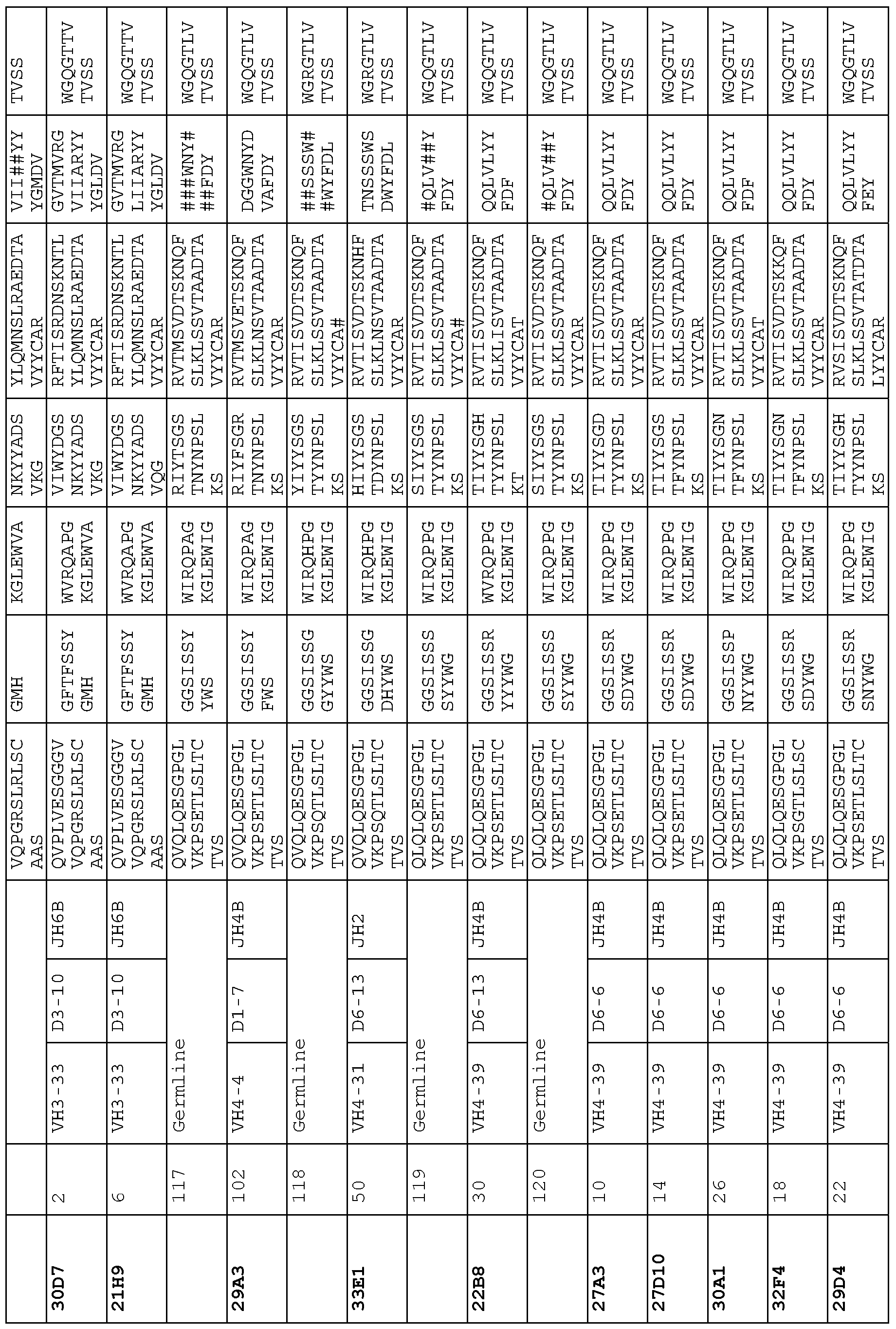


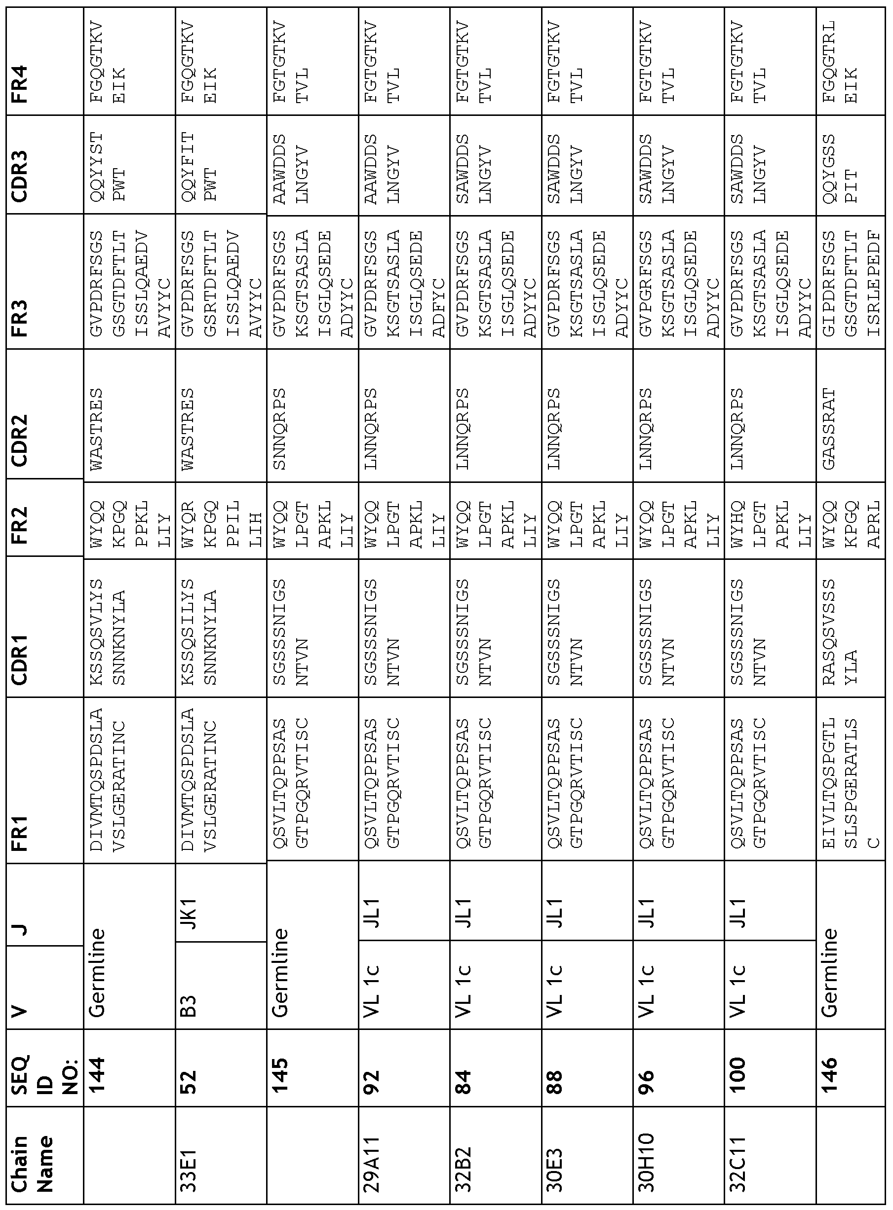


Claims
Priority Applications (5)
| Application Number | Priority Date | Filing Date | Title |
|---|---|---|---|
| EP08788590A EP2183028A2 (en) | 2007-07-25 | 2008-07-23 | Targeted binging agents directed to kdr and uses thereof - 035 |
| CA2694396A CA2694396A1 (en) | 2007-07-25 | 2008-07-23 | Targeted binding agents directed to kdr and uses thereof - 035 |
| AU2008278803A AU2008278803A1 (en) | 2007-07-25 | 2008-07-23 | Targeted binging agents directed to KDR and uses thereof - 035 |
| JP2010517493A JP2010534469A (en) | 2007-07-25 | 2008-07-23 | KDR-directed targeted binding substances and uses thereof |
| US12/669,724 US8119130B2 (en) | 2007-07-25 | 2008-07-23 | Targeted binding agents directed to KDR and uses thereof—035 |
Applications Claiming Priority (2)
| Application Number | Priority Date | Filing Date | Title |
|---|---|---|---|
| US95180507P | 2007-07-25 | 2007-07-25 | |
| US60/951,805 | 2007-07-25 |
Publications (2)
| Publication Number | Publication Date |
|---|---|
| WO2009013543A2 true WO2009013543A2 (en) | 2009-01-29 |
| WO2009013543A3 WO2009013543A3 (en) | 2010-05-06 |
Family
ID=40281893
Family Applications (1)
| Application Number | Title | Priority Date | Filing Date |
|---|---|---|---|
| PCT/GB2008/050615 WO2009013543A2 (en) | 2007-07-25 | 2008-07-23 | Targeted binging agents directed to kdr and uses thereof - 035 |
Country Status (6)
| Country | Link |
|---|---|
| US (1) | US8119130B2 (en) |
| EP (1) | EP2183028A2 (en) |
| JP (1) | JP2010534469A (en) |
| AU (1) | AU2008278803A1 (en) |
| CA (1) | CA2694396A1 (en) |
| WO (1) | WO2009013543A2 (en) |
Cited By (2)
| Publication number | Priority date | Publication date | Assignee | Title |
|---|---|---|---|---|
| US8119130B2 (en) | 2007-07-25 | 2012-02-21 | Medimmune Limited | Targeted binding agents directed to KDR and uses thereof—035 |
| WO2023010122A3 (en) * | 2021-07-29 | 2023-03-02 | Sonoma Biotherapeutics, Inc. | Synovial extracellular matrix-specific chimeric antigen receptor for targeting regulatory t cells to treat autoimmune diseases |
Families Citing this family (19)
| Publication number | Priority date | Publication date | Assignee | Title |
|---|---|---|---|---|
| JO3375B1 (en) * | 2010-11-08 | 2019-03-13 | Regeneron Pharma | Human antibodies to human tnf-like ligand 1a (tl1a) |
| EP3971206A1 (en) | 2011-02-10 | 2022-03-23 | Roche Glycart AG | Mutant interleukin-2 polypeptides |
| EA201892619A1 (en) | 2011-04-29 | 2019-04-30 | Роше Гликарт Аг | IMMUNOCONJUGATES CONTAINING INTERLEUKIN-2 MUTANT POLYPETIPS |
| CA2854153A1 (en) | 2011-11-02 | 2013-05-10 | Apexigen, Inc. | Anti-kdr antibodies and methods of use |
| CN104650229A (en) * | 2011-11-02 | 2015-05-27 | 埃派斯进有限公司 | Anti-KDR antibodies and methods of use |
| US20140044675A1 (en) | 2012-08-10 | 2014-02-13 | Roche Glycart Ag | Interleukin-2 fusion proteins and uses thereof |
| JO3462B1 (en) | 2012-08-22 | 2020-07-05 | Regeneron Pharma | Human Antibodies to GFR?3 and methods of use thereof |
| DK2888283T3 (en) | 2012-08-24 | 2018-11-19 | Univ California | ANTIBODIES AND VACCINES FOR TREATING ROR1 CANCER AND INHIBITIVE METASTASE |
| PL2895512T3 (en) * | 2012-09-12 | 2018-11-30 | Neurimmune Holding Ag | Human islet amyloid polypeptide (hiapp) specific antibodies and uses thereof |
| CA2931114A1 (en) | 2014-02-06 | 2015-08-13 | F. Hoffmann-La Roche Ag | Interleukin-2 fusion proteins and uses thereof |
| JP2018528759A (en) * | 2015-06-30 | 2018-10-04 | ヘンリアス バイオテック カンパニー リミテッド | Anti-vascular endothelial growth factor receptor 2 (VEGFR2) antibody |
| US10688181B2 (en) | 2016-06-27 | 2020-06-23 | The Regents Of The University Of California | Cancer treatment combinations |
| SG11201907299XA (en) | 2017-02-08 | 2019-09-27 | Dragonfly Therapeutics Inc | Multi-specific binding proteins for activation of natural killer cells and therapeutic uses thereof to treat cancer |
| EP3579866A4 (en) * | 2017-02-08 | 2020-12-09 | Dragonfly Therapeutics, Inc. | Antibody heavy chain variable domains targeting the nkg2d receptor |
| AU2018220736A1 (en) | 2017-02-20 | 2019-09-05 | Dragonfly Therapeutics, Inc. | Proteins binding HER2, NKG2D and CD16 |
| SG11202003280SA (en) | 2017-10-24 | 2020-05-28 | Magenta Therapeutics Inc | Compositions and methods for the depletion of cd117+ cells |
| EP3700540A4 (en) | 2017-10-24 | 2021-11-10 | Magenta Therapeutics, Inc. | Compositions and methods for the depletion of cd117+ cells |
| WO2019084053A1 (en) * | 2017-10-24 | 2019-05-02 | Magenta Therapeutics, Inc. | Compositions and methods for the depletion of cd117+ cells |
| EP3749346A4 (en) | 2018-02-08 | 2021-12-15 | Dragonfly Therapeutics, Inc. | Antibody variable domains targeting the nkg2d receptor |
Citations (1)
| Publication number | Priority date | Publication date | Assignee | Title |
|---|---|---|---|---|
| WO2002102973A2 (en) * | 2001-06-20 | 2002-12-27 | Prochon Biotech Ltd. | Antibodies that block receptor protein tyrosine kinase activation, methods of screening for and uses thereof |
Family Cites Families (3)
| Publication number | Priority date | Publication date | Assignee | Title |
|---|---|---|---|---|
| US20040242851A1 (en) * | 2001-06-26 | 2004-12-02 | Zhenping Zhu | Bispecific antibodies that bind to vegf receptors |
| DK1487856T3 (en) * | 2002-03-04 | 2010-10-18 | Imclone Llc | KDR-specific human antibodies and their use |
| CA2694396A1 (en) | 2007-07-25 | 2009-01-29 | Astrazeneca Ab | Targeted binding agents directed to kdr and uses thereof - 035 |
-
2008
- 2008-07-23 CA CA2694396A patent/CA2694396A1/en not_active Abandoned
- 2008-07-23 JP JP2010517493A patent/JP2010534469A/en active Pending
- 2008-07-23 AU AU2008278803A patent/AU2008278803A1/en not_active Abandoned
- 2008-07-23 US US12/669,724 patent/US8119130B2/en not_active Expired - Fee Related
- 2008-07-23 EP EP08788590A patent/EP2183028A2/en not_active Withdrawn
- 2008-07-23 WO PCT/GB2008/050615 patent/WO2009013543A2/en active Application Filing
Patent Citations (1)
| Publication number | Priority date | Publication date | Assignee | Title |
|---|---|---|---|---|
| WO2002102973A2 (en) * | 2001-06-20 | 2002-12-27 | Prochon Biotech Ltd. | Antibodies that block receptor protein tyrosine kinase activation, methods of screening for and uses thereof |
Non-Patent Citations (9)
| Title |
|---|
| BAKA S ET AL: "A review of the latest clinical compounds to inhibit VEGF in pathological angiogenesis" EXPERT OPINION ON THERAPEUTIC TARGETS, vol. 10, no. 6, 1 January 2006 (2006-01-01), pages 867-876, XP008080403 ASHLEY PUBLICATIONS, LONDON, GB ISSN: 1472-8222 * |
| CASTELLONE M D ET AL: "Receptor tyrosine kinase inhibitors in thyroid cancer" BAILLIERE'S BEST PRACTICE AND RESEARCH. CLINICAL ENDOCRINOLOGYAND METABOLISM, vol. 22, no. 6, 1 December 2008 (2008-12-01), pages 1023-1038, XP025710138 BAILLIERE TINDALL, LONDON, GB ISSN: 1521-690X [retrieved on 2008-11-29] * |
| ESKENS F A L M ET AL: "The clinical toxicity profile of vascular endothelial growth factor (VEGF) and vascular endothelial growth factor receptor (VEGFR) targeting angiogenesis inhibitors; A review" EUROPEAN JOURNAL OF CANCER, PERGAMON PRESS, OXFORD, GB, vol. 42, no. 18, 1 December 2006 (2006-12-01), pages 3127-3139, XP025104562 ISSN: 0959-8049 [retrieved on 2006-12-01] * |
| HELDIN C-H: "DIMERIZATION OF CELL SURFACE RECEPTORS IN SIGNAL TRANSDUCTION" CELL, vol. 80, 27 January 1995 (1995-01-27), pages 213-223, XP002021034 CELL PRESS, CAMBRIDGE, NA, US ISSN: 0092-8674 * |
| HUANG T T ET AL: "Targeted Therapy for Malignant Glioma Patients: Lessons Learned and the Road Ahead" NEUROTHERAPEUTICS,, vol. 6, no. 3, 1 July 2009 (2009-07-01), pages 500-512, XP026254707 ISSN: 1933-7213 [retrieved on 2009-06-26] * |
| IMCLONE SYSTEMS INCORPORATED: "Phase II Study of IMC-1121B for Advanced Renal Cell Cancer Opens for Patient Enrollment" INTERNET CITATION (PRESS RELEASE), [Online] 24 August 2007 (2007-08-24), XP002553489 Retrieved from the Internet: URL:http://phx.corporate-ir.net/phoenix.zhtml?c=97689&p=irol-newsArticle_print&ID=1043534&highlight=> [retrieved on 2009-11-02] * |
| OLSSON A-K ET AL: "VEGF RECEPTOR SIGNALLING - IN CONTROL OF VASCULAR FUNCTION" NATURE REVIEWS MOLECULAR CELL BIOLOGY, vol. 7, no. 5, 1 May 2006 (2006-05-01), pages 359-377, XP009069866 MACMILLAN MAGAZINES, LONDON, GB cited in the application * |
| ROSKOSKI ET AL: "Vascular endothelial growth factor (VEGF) signaling in tumor progression" CRITICAL REVIEWS IN ONCOLOGY / HEMATOLOGY, vol. 62, no. 3, 6 May 2007 (2007-05-06), pages 179-213, XP022063754 ELSEVIER SCIENCE IRELAND LTD., LIMERICK, IE ISSN: 1040-8428 * |
| WALTENBERGER J ET AL: "DIFFERENT SIGNAL TRANSDUCTION PROPERTIES OF KDR AND FLT1, TWO RECEPTORS FOR VASCULAR ENDOTHELIAL GROWTH FACTOR" JOURNAL OF BIOLOGICAL CHEMISTRY, vol. 269, no. 43, 28 October 1994 (1994-10-28), pages 26988-26995, XP002052458 AMERICAN SOCIETY OF BIOLOCHEMICAL BIOLOGISTS, BIRMINGHAM, US ISSN: 0021-9258 * |
Cited By (3)
| Publication number | Priority date | Publication date | Assignee | Title |
|---|---|---|---|---|
| US8119130B2 (en) | 2007-07-25 | 2012-02-21 | Medimmune Limited | Targeted binding agents directed to KDR and uses thereof—035 |
| WO2023010122A3 (en) * | 2021-07-29 | 2023-03-02 | Sonoma Biotherapeutics, Inc. | Synovial extracellular matrix-specific chimeric antigen receptor for targeting regulatory t cells to treat autoimmune diseases |
| US11753457B2 (en) | 2021-07-29 | 2023-09-12 | Sonoma Biotherapeutics, Inc. | Synovial extracellular matrix-specific chimeric antigen receptor for targeting regulatory T cells to treat autoimmune diseases |
Also Published As
| Publication number | Publication date |
|---|---|
| US20100260765A1 (en) | 2010-10-14 |
| US8119130B2 (en) | 2012-02-21 |
| EP2183028A2 (en) | 2010-05-12 |
| JP2010534469A (en) | 2010-11-11 |
| CA2694396A1 (en) | 2009-01-29 |
| WO2009013543A3 (en) | 2010-05-06 |
| AU2008278803A1 (en) | 2009-01-29 |
Similar Documents
| Publication | Publication Date | Title |
|---|---|---|
| US8119130B2 (en) | Targeted binding agents directed to KDR and uses thereof—035 | |
| US9206255B2 (en) | Nucleic acid molecule encoding target antibodies directed to DLL4 | |
| US8569459B2 (en) | Targeted binding agents directed to sonic hedgehog homolog and uses thereof | |
| US20120107324A1 (en) | TARGETED BINDING AGENTS DIRECTED TO a5ß1 AND USES THEREOF | |
| EP2420514A1 (en) | Targeted binding agents directed to PDGFR-alpha and uses thereof | |
| US20120114667A1 (en) | TARGETED BINDING AGENTS DIRECTED TO a5BETA1 AND USES THEREOF | |
| US20100233154A1 (en) | Targeted binding agents directed to heparanase and uses thereof 463 | |
| CA2748161A1 (en) | Targeted binding agents directed to .alpha.5.beta.1 and uses thereof | |
| AU2015255300A1 (en) | Antibodies directed to dll4 and uses thereof |
Legal Events
| Date | Code | Title | Description |
|---|---|---|---|
| 121 | Ep: the epo has been informed by wipo that ep was designated in this application |
Ref document number: 08788590 Country of ref document: EP Kind code of ref document: A2 |
|
| WWE | Wipo information: entry into national phase |
Ref document number: 2694396 Country of ref document: CA |
|
| WWE | Wipo information: entry into national phase |
Ref document number: 2010517493 Country of ref document: JP |
|
| NENP | Non-entry into the national phase |
Ref country code: DE |
|
| WWE | Wipo information: entry into national phase |
Ref document number: 2008278803 Country of ref document: AU |
|
| WWE | Wipo information: entry into national phase |
Ref document number: 2008788590 Country of ref document: EP |
|
| ENP | Entry into the national phase |
Ref document number: 2008278803 Country of ref document: AU Date of ref document: 20080723 Kind code of ref document: A |
|
| WWE | Wipo information: entry into national phase |
Ref document number: 12669724 Country of ref document: US |