WO2008054600A2 - Compositions and methods for biodetection by nucleic acid-templated chemistry - Google Patents
Compositions and methods for biodetection by nucleic acid-templated chemistry Download PDFInfo
- Publication number
- WO2008054600A2 WO2008054600A2 PCT/US2007/021094 US2007021094W WO2008054600A2 WO 2008054600 A2 WO2008054600 A2 WO 2008054600A2 US 2007021094 W US2007021094 W US 2007021094W WO 2008054600 A2 WO2008054600 A2 WO 2008054600A2
- Authority
- WO
- WIPO (PCT)
- Prior art keywords
- alkyl
- dna
- reaction
- nucleic acid
- oligonucleotide
- Prior art date
Links
- 0 CC1(C)c(c(cccc2)c2cc2)c2*(O)=C1C Chemical compound CC1(C)c(c(cccc2)c2cc2)c2*(O)=C1C 0.000 description 1
- KRKGTQRUYNFELU-UHFFFAOYSA-N CC1(C)c2ccccc2[N](CCCCC(O)=O)(=C)=C1C Chemical compound CC1(C)c2ccccc2[N](CCCCC(O)=O)(=C)=C1C KRKGTQRUYNFELU-UHFFFAOYSA-N 0.000 description 1
- MHNBHSBXTJIZFG-UHFFFAOYSA-N CN(CCC(O)=O)c1ccc(C=O)cc1 Chemical compound CN(CCC(O)=O)c1ccc(C=O)cc1 MHNBHSBXTJIZFG-UHFFFAOYSA-N 0.000 description 1
- WBGWUCXEMSSZJL-UHFFFAOYSA-N OC(CC1)NC1=O Chemical compound OC(CC1)NC1=O WBGWUCXEMSSZJL-UHFFFAOYSA-N 0.000 description 1
- HTTJXGVLHBBCEB-UHFFFAOYSA-N ON(C1OC1CC1)C1=O Chemical compound ON(C1OC1CC1)C1=O HTTJXGVLHBBCEB-UHFFFAOYSA-N 0.000 description 1
Classifications
-
- C—CHEMISTRY; METALLURGY
- C07—ORGANIC CHEMISTRY
- C07H—SUGARS; DERIVATIVES THEREOF; NUCLEOSIDES; NUCLEOTIDES; NUCLEIC ACIDS
- C07H21/00—Compounds containing two or more mononucleotide units having separate phosphate or polyphosphate groups linked by saccharide radicals of nucleoside groups, e.g. nucleic acids
- C07H21/04—Compounds containing two or more mononucleotide units having separate phosphate or polyphosphate groups linked by saccharide radicals of nucleoside groups, e.g. nucleic acids with deoxyribosyl as saccharide radical
-
- C—CHEMISTRY; METALLURGY
- C07—ORGANIC CHEMISTRY
- C07D—HETEROCYCLIC COMPOUNDS
- C07D209/00—Heterocyclic compounds containing five-membered rings, condensed with other rings, with one nitrogen atom as the only ring hetero atom
- C07D209/02—Heterocyclic compounds containing five-membered rings, condensed with other rings, with one nitrogen atom as the only ring hetero atom condensed with one carbocyclic ring
- C07D209/04—Indoles; Hydrogenated indoles
- C07D209/08—Indoles; Hydrogenated indoles with only hydrogen atoms or radicals containing only hydrogen and carbon atoms, directly attached to carbon atoms of the hetero ring
-
- C—CHEMISTRY; METALLURGY
- C07—ORGANIC CHEMISTRY
- C07D—HETEROCYCLIC COMPOUNDS
- C07D209/00—Heterocyclic compounds containing five-membered rings, condensed with other rings, with one nitrogen atom as the only ring hetero atom
- C07D209/02—Heterocyclic compounds containing five-membered rings, condensed with other rings, with one nitrogen atom as the only ring hetero atom condensed with one carbocyclic ring
- C07D209/04—Indoles; Hydrogenated indoles
- C07D209/10—Indoles; Hydrogenated indoles with substituted hydrocarbon radicals attached to carbon atoms of the hetero ring
- C07D209/14—Radicals substituted by nitrogen atoms, not forming part of a nitro radical
-
- C—CHEMISTRY; METALLURGY
- C07—ORGANIC CHEMISTRY
- C07D—HETEROCYCLIC COMPOUNDS
- C07D209/00—Heterocyclic compounds containing five-membered rings, condensed with other rings, with one nitrogen atom as the only ring hetero atom
- C07D209/56—Ring systems containing three or more rings
-
- C—CHEMISTRY; METALLURGY
- C07—ORGANIC CHEMISTRY
- C07D—HETEROCYCLIC COMPOUNDS
- C07D209/00—Heterocyclic compounds containing five-membered rings, condensed with other rings, with one nitrogen atom as the only ring hetero atom
- C07D209/56—Ring systems containing three or more rings
- C07D209/58—[b]- or [c]-condensed
-
- C—CHEMISTRY; METALLURGY
- C09—DYES; PAINTS; POLISHES; NATURAL RESINS; ADHESIVES; COMPOSITIONS NOT OTHERWISE PROVIDED FOR; APPLICATIONS OF MATERIALS NOT OTHERWISE PROVIDED FOR
- C09B—ORGANIC DYES OR CLOSELY-RELATED COMPOUNDS FOR PRODUCING DYES, e.g. PIGMENTS; MORDANTS; LAKES
- C09B23/00—Methine or polymethine dyes, e.g. cyanine dyes
- C09B23/0091—Methine or polymethine dyes, e.g. cyanine dyes having only one heterocyclic ring at one end of the methine chain, e.g. hemicyamines, hemioxonol
-
- C—CHEMISTRY; METALLURGY
- C09—DYES; PAINTS; POLISHES; NATURAL RESINS; ADHESIVES; COMPOSITIONS NOT OTHERWISE PROVIDED FOR; APPLICATIONS OF MATERIALS NOT OTHERWISE PROVIDED FOR
- C09B—ORGANIC DYES OR CLOSELY-RELATED COMPOUNDS FOR PRODUCING DYES, e.g. PIGMENTS; MORDANTS; LAKES
- C09B23/00—Methine or polymethine dyes, e.g. cyanine dyes
- C09B23/10—The polymethine chain containing an even number of >CH- groups
- C09B23/105—The polymethine chain containing an even number of >CH- groups two >CH- groups
-
- G—PHYSICS
- G01—MEASURING; TESTING
- G01N—INVESTIGATING OR ANALYSING MATERIALS BY DETERMINING THEIR CHEMICAL OR PHYSICAL PROPERTIES
- G01N33/00—Investigating or analysing materials by specific methods not covered by groups G01N1/00 - G01N31/00
- G01N33/48—Biological material, e.g. blood, urine; Haemocytometers
- G01N33/50—Chemical analysis of biological material, e.g. blood, urine; Testing involving biospecific ligand binding methods; Immunological testing
- G01N33/53—Immunoassay; Biospecific binding assay; Materials therefor
- G01N33/5308—Immunoassay; Biospecific binding assay; Materials therefor for analytes not provided for elsewhere, e.g. nucleic acids, uric acid, worms, mites
-
- G—PHYSICS
- G01—MEASURING; TESTING
- G01N—INVESTIGATING OR ANALYSING MATERIALS BY DETERMINING THEIR CHEMICAL OR PHYSICAL PROPERTIES
- G01N33/00—Investigating or analysing materials by specific methods not covered by groups G01N1/00 - G01N31/00
- G01N33/48—Biological material, e.g. blood, urine; Haemocytometers
- G01N33/50—Chemical analysis of biological material, e.g. blood, urine; Testing involving biospecific ligand binding methods; Immunological testing
- G01N33/53—Immunoassay; Biospecific binding assay; Materials therefor
- G01N33/531—Production of immunochemical test materials
- G01N33/532—Production of labelled immunochemicals
- G01N33/533—Production of labelled immunochemicals with fluorescent label
-
- Y—GENERAL TAGGING OF NEW TECHNOLOGICAL DEVELOPMENTS; GENERAL TAGGING OF CROSS-SECTIONAL TECHNOLOGIES SPANNING OVER SEVERAL SECTIONS OF THE IPC; TECHNICAL SUBJECTS COVERED BY FORMER USPC CROSS-REFERENCE ART COLLECTIONS [XRACs] AND DIGESTS
- Y10—TECHNICAL SUBJECTS COVERED BY FORMER USPC
- Y10T—TECHNICAL SUBJECTS COVERED BY FORMER US CLASSIFICATION
- Y10T436/00—Chemistry: analytical and immunological testing
- Y10T436/14—Heterocyclic carbon compound [i.e., O, S, N, Se, Te, as only ring hetero atom]
- Y10T436/142222—Hetero-O [e.g., ascorbic acid, etc.]
- Y10T436/143333—Saccharide [e.g., DNA, etc.]
-
- Y—GENERAL TAGGING OF NEW TECHNOLOGICAL DEVELOPMENTS; GENERAL TAGGING OF CROSS-SECTIONAL TECHNOLOGIES SPANNING OVER SEVERAL SECTIONS OF THE IPC; TECHNICAL SUBJECTS COVERED BY FORMER USPC CROSS-REFERENCE ART COLLECTIONS [XRACs] AND DIGESTS
- Y10—TECHNICAL SUBJECTS COVERED BY FORMER USPC
- Y10T—TECHNICAL SUBJECTS COVERED BY FORMER US CLASSIFICATION
- Y10T436/00—Chemistry: analytical and immunological testing
- Y10T436/14—Heterocyclic carbon compound [i.e., O, S, N, Se, Te, as only ring hetero atom]
- Y10T436/145555—Hetero-N
Definitions
- the present invention relates generally to probes and their use in biodetection and diagnostics. More particularly, the invention relates to compositions and methods for biodetection using nucleic acid-templated chemistry (e.g., synthesis of compounds having desired fluorescent, chemiluminescent or chromophoric properties in a multiplex detection of nucleic acids or proteins).
- nucleic acid-templated chemistry e.g., synthesis of compounds having desired fluorescent, chemiluminescent or chromophoric properties in a multiplex detection of nucleic acids or proteins.
- Fluorescent and colored compounds have been used in the fields of biological research and medicine to detect the presence, absence, state, quantity, and composition of biomolecules. Assays using fluorescent and colored compounds may be performed in vitro, in situ, or in vivo. Examples of commonly used in vitro assays for detection of DNA and RNA are real-time and end-point polymerase chain reaction (PCR), DNA sequencing, and DNA microarray technologies.
- PCR polymerase chain reaction
- the multiplex detection was achieved by using intercalating dyes as labels in DNA restriction fragment analysis and capillary electrophoresis with frequency-domain fluorescence lifetime detection method (Mclntosh, et al., Electrophoresis, 2002, 23, 1473-1479). Since these methods use pre- labeled fluorescence dyes, the detection sensitivity relies largely on the separation of target bound and unbound fluorescence labeled probes. Though solid phase immobilization of the target gene (fluorescence in situ hybridization, for example) can improve the separation efficiency by simply washing away the unbound fluorescence labeled probes, this introduces an extra process. However, the potential background still can be high, and the procedure can be laborious.
- a non-fluorescence label moiety can be attached to the probes so that the fluorescence signal only occurs after the hybridization event.
- DNA-programmed chemistry has provided a novel approach for generation of fluorescence dye in situ. See, e.g., Li, X.; Liu, D. R. Angew. Chem. Int. Ed. 2004, 43, 4848- 4870; U.S. Patent No. 7,070,928.
- Polymethine dye has been widely used as laser dyes, photographic sensitizers and fluorescence probes due to its superior fluorescence and photochemical properties.
- polymethine dyes are generally synthesized by acid/base catalyzed condensation under anhydrous conditions which is not comparable to the nucleic acid-templated chemistry (Jedrzejewska, et al. Dyes and Pigments 2003, 58, 47-58).
- the literature has reported an improved aldol condensation in water using Lewis-acid (Kobayashi, et al., J. Am. Chem. Soc. 1998, 120, 8287-8288) and enamine-based organocatalyst (Mase, et al. J. Am.
- the quaternary salt of polymethine precursor (active hydrogen component) used for condensing with aldehyde is different substantially from the precursor (alpha carbon of aldehyde) in a conventional aldol condensation.
- the present invention is based, in part, upon the discovery that nucleic acid- templated chemistry can be applied in detection of multiple biological targets simultaneously.
- the present invention is based, in part, upon the discovery that polymethine dyes can be synthesized by nucleic acid-templated chemistry.
- Assays of this invention may be performed in vitro, in situ, or in vivo.
- the present invention relates to a method for making a polymethine dye comprising conducting an aldol condensation between an aldehyde and an active hydrogen component in an aqueous condition in the presence of an organocatalyst.
- the condensation reaction is:
- R H, alkyl
- R" H, alkyl, alkyl carboxylic acid
- R 1 Ph or ⁇ /-heterocycle, H, alkyl, SO 3 H, OH, CN, Cl, Br, NO 2 , NH 2 , N(R) 2 , OR where R is alkyl grou
- the organocatalyst is a secondary amine, a primary amine, a bifunctional amine-acid catalyst or a diamine.
- the secondary amine may be a pyrrolidine, a piperidine, a nornicotine, or an analog thereof, for example.
- the primary amine may be a valine or a peptide having fewer than 3 amino acid units, for example.
- the bifunctional amine-acid catalyst may be pyrrolidine/ AcOH, for example.
- the diamine catalyst may be Nl,Nl-dimethylethane-l,2- diamine, propane- 1,2-diamine, l-(2-aminoethyl)-piperidine, or an analog thereof, for example.
- the invention generally relates to a hemicyanine dye having the chemical structure of (I), (II) or (III), for example, prepared by the methods disclosed herein.
- R H, alkyl
- R" H, alkyl, alkyl carboxylic acid
- R" Ph or ⁇ /-heterocycle, H, alkyl, SO 3 H, OH, CN, Cl, Br, NO 2 , NH 2 , N(R) 2 , OR where R is alkyl group
- R1 alkyl
- R1 alkyl
- R2 Ph, H, alkyl, SO 3 H, OH, CN, Cl, Br 1 NO 2
- R2 Ph, H, alkyl, SO 3 H, OH, CN, Cl, Br, NO 2 ,
- R 3 Ph, H, alkyl, SO 3 H, OH, CN, Cl, Br, NO 2
- R 3 Ph, H, alkyl, SO 3 H, OH, CN, Cl, Br, NO 2 ,
- the invention generally relates to an aldehyde having the chemical structure of IV or V:
- the invention generally relates to a quaternary salt having the chemical structure of VI or VII:
- the invention generally relates to an quaternary salt-nucleic acid conjugate having the chemical structure of:
- n 0 to 16
- Z1 O, S, Se, P, NH 2 , NR 1 , C(CH 3 ) 2 where R 1 is alkyl group
- R any substituted benzyl or higher fused benzyl rings, H, alkyl, SO 3 H, OH, CN, Cl, Br, NO 2 , NH 2 , N(R 1 );,, OR 1 while R 1 is alkyl group
- Z 2 benzene or any N-heterocycles
- the invention generally relates to an aldehyde-nucleic acid conjugate having the chemical structure of:
- R1 H, alkyl
- R 2 Ph or N-heterocycle, H, alkyl, SO 3 H,
- the invention generally relates to a hemicyanine dye-nucleic acid conjugate having the chemical structure of:
- the invention generally relates to making a hemicyanine- nucleic acid conjugate comprising conducting a nucleic acid-templated reaction between an aldehyde and quaternary salt disclosed herein to make a hemicyanine disclosed herein.
- the nucleic acid-templated reaction is in an end of helix architect. In some other embodiments, the nucleic acid-templated reaction is in a middle of helix architect.
- the invention generally relates to a method for selecting a dye having a desired fluorescent property.
- the method includes (a) preparing a library of oligonucleotide-encoded dyes through nucleic acid-templated synthesis; (b) hybridizing the oligonucleotide-encoded dyes with spatially arrayed complementary oligonucleotide probes immobilized on a solid support; (c) measuring the absorption and fluorescence properties of the oligonucleotide-encoded dye directly on the solid support; (d) identifying the oligonucleotides that encode the dyes having the desired fluorescence properties based on the position of the immobilized complementary oligonucleotide probes, and (e) identifying and characterizing the chemical structure of the dyes having the desired fluorescence property.
- the invention generally relates to a method for detecting multiple target nucleotide sequences.
- the method includes: (a) providing a number of probe pairs, the number equal to the number of target nucleotide sequences, wherein each probe pair comprises (1) a first probe comprising (i) a first oligonucleotide sequence and (ii) a first reactive group linked to the first oligonucleotide sequence, and (2) a corresponding second probe comprising (i) a second oligonucleotide sequence and (ii) a second reactive group linked to the second oligonucleotide sequence, wherein the first oligonucleotide sequence and the second oligonucleotide sequence are complementary to two separate regions of a corresponding target nucleotide sequence; (b) combining the probe pairs with a sample to be tested for the presence of the target nucleotide sequences under conditions where the first probes and the second probes hybridize to their respective complementary regions of
- the number of target nucleotide sequences may be between about 2 to about 20, for example, 2 to 6.
- the target nucleotide sequences may be in solution phase.
- the target nucleotide sequences may be attached to a solid support.
- the one or more reactions between the first reactive groups and the corresponding second reactive groups generate fluorescent compounds that may be detected.
- the one or more reactions between the first reactive groups and the corresponding second reactive groups generate chemiluminescent compounds that may be detected.
- the one or more reactions between the first reactive groups and the corresponding second reactive groups may comprise an aldol condensation reaction, for example.
- the one or more reactions between the first reactive groups and the corresponding second reactive groups may comprise a Wittig reaction.
- the invention encompasses a kit that provides one, two or more of the probes described herein. More particularly, the invention encompasses a kit that provides one, two or more of the probes that utilize nucleic acid-templated chemistry for the generation of detectable signals as a way for detecting the presence of biological targets.
- DNA programmed chemistry refers to nucleic acid-templated chemistry, for example, sequence specific control of chemical reactants to yield specific products accomplished by (1) providing one or more templates, which have associated reactive group(s); (2) contacting one or more transfer groups (reagents) having an anti-codon (e.g., complementary sequence with one or more templates) and reactive group(s) under conditions to allow for hybridization to the templates and (3) reaction of the reactive groups to yield products.
- transfer groups e.g., complementary sequence with one or more templates
- Structures of the reactants and products need not be related to those of the nucleic acids comprising the template and transfer group oligonucleotides. See, e.g., U.S. Patent No. 7,070,928 and U.S. Application Publication No. 2004/0180412 Al, by Liu et al; Gartner, et al., 2004, Science, vol. 305, pp. 1601-1605; Doyon, et al., 2003, JACS, vol. 125, pp. 12372-12373, all of which are expressly incorporated herein by reference in their entireties.
- nucleic acid refers to a polymer of nucleotides.
- the polymer may include, without limitation, natural nucleosides (i.e., adenosine, thymidine, guanosine, cytidine, uridine, deoxyadenosine, deoxythymidine, deoxyguanosine, and deoxycytidine), nucleoside analogs (e.g., 2-aminoadenosine, 2-thiothymidine, inosine, pyrrolo-pyrimidine, 3- methyl adenosine, 5-methylcytidine, C5-bromouridine, C5-fluorouridine, C5-iodouridine, C5- propynyl-uridine, C5-propynyl-cytidine,
- Nucleic acids and oligonucleotides may also include other polymers of bases having a modified backbone, such as a locked nucleic acid (LNA), a peptide nucleic acid (PNA), a threose nucleic acid (TNA).
- LNA locked nucleic acid
- PNA peptide nucleic acid
- TAA threose nucleic acid
- compositions are described as having, including, or comprising specific components, or where processes are described as having, including, or comprising specific process steps, it is contemplated that compositions of the present invention also consist essentially of, or consist of, the recited components, and that the processes of the present invention also consist essentially of, or consist of, the recited processing steps. Further, it should be understood that the order of steps or order for performing certain actions are immaterial so long as the invention remains operable. Moreover, two or more steps or actions may be conducted simultaneously.
- FIG. 1 illustrates the general chemical structures of polymethine, cyanine and hemicyanine dyes.
- FIG. 2 illustrates the general chemical structures of hemicyanine dyes useful for multiplex and their aldehyde and quaternary salt precursors.
- FIG. 3 illustrates the chemical structures of a four-plex hemicyanine-DNA dye system and their spectroscopic properties.
- FIG. 4 is a schematic representation of solution phase-based DPC fluorescence assay for multiple analytes.
- FIG. 5 is a schematic representation of solid phase-based DPC fluorescence assay for multiple analytes.
- FIG. 6 is a schematic representation of immunohistochemistry test for multiple family receptor dimers in non-zip-coded architecture.
- FIG. 7 is a schematic representation of immunohistochemistry test for multiple family receptor dimers in zip-coded architecture.
- FIG. 8 is a schematic representation of a method for the detection of a biological target under one embodiment of the present invention.
- FIG. 9 is a schematic representation of a method for the detection of a biological target under one embodiment of the present invention.
- FIG. 10 shows examples of hybridization as affected by concentration, temperature, and the presence or absence of a single base pair mismatch.
- FIG. 11 shows exemplary oligonucleotides used in certain melting curve experiments.
- FIG. 12 is a schematic representation of a method for the detection of a biological target under one embodiment of the present invention.
- FIG. 13 is a schematic representation of a method for the detection of platelet derived growth factor (PDGF) under one embodiment of the present invention.
- PDGF platelet derived growth factor
- FIG. 14 shows exemplary embodiment of a splinted, zip-coded detection system with aptamers as target binding moieties.
- FIG. 15 shows exemplary embodiment of a splinted, zip-coded detection system with antibodies as target binding moieties.
- FIG. 16 shows exemplary embodiment of a splinted, zip-coded detection system with antibodies as target binding moieties.
- FIG. 17 shows absorption and fluorescence emission spectra of DPC reaction mixtures (end of helix).
- FIG. 18 shows absorption and fluorescence emission spectra of DPC reaction mixtures (end of helix).
- FIG. 19 shows LC-MS data of a crude DPC reaction mixture.
- FIG. 20 shows LC-MS data of a DPC reaction product.
- FIG. 21 shows absorption and fluorescence emission spectra of DPC reaction mixtures.
- FIG. 22 shows LC-MS data of a DPC reaction product.
- FIG. 23 shows certain fluorescence intensity data of a DPC reaction.
- FIG. 24 shows certain fluorescence intensity data of a DPC reaction.
- FIG. 25 shows fluorescence excitation and emission spectra of four hemicyanine dyes.
- FIG. 26 shows exemplary DNA sequences useful for four-plex hemicyanine dye generation.
- FIG. 27 shows normalized fluorescence emission spectra of four DNA conjugated hemicyanine dyes from middle of helix DPC reactions.
- FIG. 28 shows fluorescence kinetic analysis of two DPC reactions in end of helix architecture.
- FIG. 29 shows an example of fluorescence signal generation and biological target detection via triphenylphosphine (TPP) and azidocoumarin (AzC) reporter chemistry.
- TPP triphenylphosphine
- AzC azidocoumarin
- FIG. 30 shows an example of fluorescence signal generation and biological target detection via TPP and AzC reporter chemistry.
- FIG. 31 shows certain examples of melt curves illustrating the effect of oligonucleotide concentration on T m .
- FIG. 32 shows certain examples with DNA hybridization melting curves of biotinylated oligonucleotides with and without avidin.
- FIG. 33 shows certain examples of T n , changes of complementary biotinylated oligos upon binding to avidin.
- FIG. 34 shows certain examples of the effect of salt and magnesium concentrations upon T m of oligonucleotides +/- biotin.
- FIG. 35 shows certain examples of the melting temperature behavior of biotinylated oligonucleotides at different ratios of oligonucleotides to avidin.
- FIG. 36 shows certain examples of melting curves of 5' and 3' (-) biotin-strand oligos duplexed with biotin-5' (+) strand oligo in the absence and presence of avidin.
- FIG. 37 shows certain examples of melting curves of AT-rich biotinylated oligo dimers with and without avidin.
- FIG. 38 is a schematic representation of a method for the detection of a biological target under one embodiment of the present invention.
- FIG. 39 shows examples of experimental results on detection of a biological target under one embodiment of the present invention.
- FIG. 4OA and FIG. 4OB show examples of experimental results (the effect of formamide in the reaction mixture) on detection of a biological target under one embodiment of the present invention.
- FIG. 41 A and FIG. 41B show examples of experimental results (the effect of formamide in the reaction mixture) on detection of a biological target under one embodiment of the present invention.
- FIG. 42 shows examples of experimental results (the effect of formamide in the reaction mixture) on detection of a biological target under one embodiment of the present invention.
- FIG. 43 shows examples of experimental results (time course of reaction mixtures) on detection of a biological target under one embodiment of the present invention.
- FIG. 44 shows examples of experimental results (time course of reaction mixtures) on detection of a biological target under one embodiment of the present invention.
- FIG. 45 shows examples of experimental results (probe ratios) on detection of a biological target under one embodiment of the present invention.
- FIG. 46 shows an example of detection of PDGF by a zip-coded detection system.
- FIG. 47 shows experiments on ratios of aptamers and reporters.
- FIG. 48 illustrates an embodiment of a "one-piece" detection system for the detection of PDGF.
- the invention is to detect the presence of target analytes via nucleic acid-templated chemistry, for example, through measurement of fluorescence of polymethine dyes generated by nucleic acid-templated reactions templated by target nucleic acids or proteins.
- the present invention provides methods for analysis of multiple analytes in a convenient, accurate and sensitive way.
- the method uses nucleic acid probes conjugated with non-fluorescence precursor (e.g., aldehydes and methyl quaternary salts) and polymethine multiplex dyes are generated through the chemical reaction of the probes upon hybridization with target nucleic acids.
- non-fluorescence precursor e.g., aldehydes and methyl quaternary salts
- polymethine multiplex dyes are generated through the chemical reaction of the probes upon hybridization with target nucleic acids.
- the invention provides novel chemical compositions of polymethine dyes and methods of synthesizing polymethine dyes in conventional reactions under aqueous conditions as well
- Typical A and D terminals for polymethine dyes (as shown in FIG.
- the compound includes thiazoles, pyrroles, pyrrolines, indoles, 1, 3, 3-trimethylindolines, tetrazoles, pyrimidine, pyridines, quinolines, and higher fused N-heterocycles or any substituted benzyl rings. If the terminals are both iV-atom containing heterocycles, the compound is named cyanine. If only one iV-atom is part of the ring system, the compound is named hemicyanine.
- the fluorescence emission wavelength of the polymethine dye can be tuned from near-UV to near-IR.
- the terminal group may also provide mean for finer tuning.
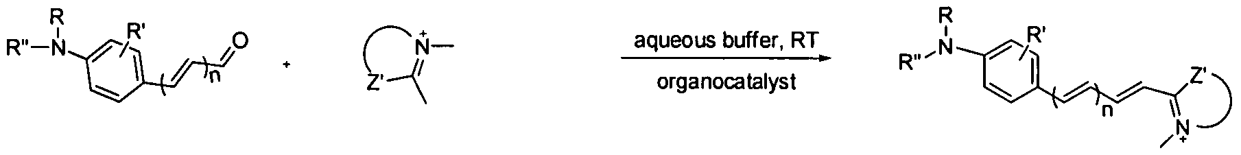
- Scheme 1 depicts hemicyanine formation through organocatalytic aldol condensation in aqueous buffer.
- organocatalysts such as pyrrolidine analogues have been listed here.
- catalyst By using catalyst, the reaction condition for the hemicyanine formation can switch from anhydrous to aqueous condition. The percentage of the water content used depends only on the solubility of the starting materials.
- Scheme 2 is a schematic illustration of hemicyanine dye generation through DPC in the presence of organocatalyst.
- component A aldehyde_DNA
- component B quaternary salt bearing active hydrogen component
- hemicyaine_DNA conjugate The general chemical structures of component A (aldehyde_DNA), component B (quaternary salt bearing active hydrogen component) and hemicyaine_DNA conjugate have also been described.
- the fluorescence emission wavelength of the hemicyaine dye can be tuned by changing the number of the vinyl group in the polyene chain (n) or by using different substitute groups (R') and terminal groups in component B.
- FIG. 2 shows the general chemical structures of hemicyanine dyes useful for multiplex and their aldehyde and quaternary salt precursors.
- An example of four-plex hemicyanineJDNA dyes derived from the general structure and their maximum UV absorption and fluorescence's emission wavelength has been described in FIG. 3.
- R H, alkyl
- R" H, alkyl, alkyl carboxylic acid
- R' Ph or /V-heterocycle, H, alkyl, SO 3 H, OH, CN, Cl, Br, NO 2 , NH 2 , N(R) 2 , OR where R is alkyl group
- Ammo acid such as valine or small peptide
- Bifunctional amine-acid catalysts such as pyrrolidine/AcOH
- R H, alkyl
- FIG. 4 is a schematic illustration of solution phase based DPC fluorescence assay for mutiplex detection involving multiple analytes. Each pair is labeled with non-fluorescence precursors complementary to a sequence that is indicative of a specific polymorphic site or genotype in the diagnostic determination of infection with a virus which can be PCR-amplified products. After hybridization, DPC fluorescence product will be formed. By changing the hybridization conditions such as salt and temperature, only matched pairs will generate the fluorescence signal.
- washing process is needed to remove any non-fluorescence precursors.
- This method is not only useful for analyzing or detecting polynucleotide sequences, it also can be used, for example, in an antibody based assay utilizing a nucleic acid conjugated antibody.
- FIG. 5 is a schematic illustration of a solid phase based DPC fluorescence assay for multiple analytes.
- Different solid supports can be used to immobilize genes of interest, such as a glass plate, a polymer or a gold plate. After hybridization and fluorescence compound formation by catalyzed DPC, the solid support can be visualized directly by fluorescence microscopy or detected by a fluorescence reader without the need for washing.
- FIG. 6 illustrates a multiplexed immunohistochemistry (IHC) test for multiple family receptor dimers.
- Multiple pairs of probes each pair being directed at a particular homo- or hetero dimer and with a distinct DPC product (e.g., a distinct fluorescent signal from each pair as shown), can provide simultaneous detection and profiling of multiple receptor dimers.
- DPC product e.g., a distinct fluorescent signal from each pair as shown
- Both zip-coded, as illustrated in FIG. 7, and non-zip-coded probe pairs can be employed in a multiplex test. See, e.g., PCT International Application No. PCT/US07/ , titled "Receptor Family Profiling," by Landsman et al, filed September 18, 2007 in the U.S. Receiving Office.
- a larger number of analytes can be detected as compared to conventional methods.
- the fluorescence emission wavelength of polymethine dye can be easily tuned from far UV to near IR, so multi- wavelength dyes can be generated in one-pot by utilizing multi-codon DNAs.
- the numbers of analyte that can be detected are not limited by the DPC and dye chemistry.
- FIG. 8 and FIG. 9 illustrate one embodiment of the invention for the detection of a protein target.
- FIG. 8 shows an embodiment of detection of a protein target by DPC-based probes.
- Two probes contain target binding moieties, complementary oligonucleotides, and chemically reactive species X and Y, respectively.
- X and Y react to create a signal generating (e.g., fluorescent) compound, which may or may not covalently link both probes.
- the reaction product of X and Y may also be released as an unbound, soluble compound into the solution.
- the protein target may be attached to a solid-phase such as the surface of a bead, glass slide (microarray), etc., or be in solution.
- the target binding moieties may be aptamers, antibodies, antibody fragments (i.e., Fab), receptor proteins, or small molecules, for example.
- FIG. 9 More particularly illustrated in FIG. 9 is an example of the dual -probe approach with two probes, each carrying a "prefluorophore" precursor (Rl and R2) and containing a binding moiety for a target and an oligonucleotide sequence that is designed to anneal to each other.
- the detection is performed under conditions such that the prefluorophore oligos will not anneal to each other in the absence of a target. These conditions are generally selected such that the ambient temperature is higher than the T m of the oligonucleotide pairs in the absence of the target (so that the oligo pairs will not anneal in the absence of the intended target analyte).
- the localized high concentration of the oligos shifts the T m of their double stranded complex upwards so that hybridization occurs, which is followed by a signal-generating nucleic acid-templated reaction (a reaction between Rl and R2).
- the signal-generating nucleic acid-templated reaction is accelerated both due to the localized higher concentration of the prefluorophores, but may also be facilitated by the proximity and orientation of the prefluorophore groups towards one another.
- This configuration of signal generation has the potential to enable creation of kits for the detection of various biomolecules, cells, surfaces and for the design of in situ assays.
- the signal generation does not require enzymes and the homogeneous format requires no sample manipulation.
- two oligonucleotides are shown, each of which is linked through an optional spacer arm to a separate binder, as shown in this case is an antibody but may be other binders such as aptamers or small molecules.
- a separate binder as shown in this case is an antibody but may be other binders such as aptamers or small molecules.
- Each antibody recognizes a separate epitope on a common target analyte such as a protein.
- Spacer arms can be added to one or both oligonucleotides between the oligo and the binder. In certain cases, this spacer arm may be required to meet proximity requirements to achieve a desired reactivity.
- Spacer arms in principle can be any suitable groups, for example, linear or branched aliphatic carbon chains C3 to C5, ClO, C15, C20, C25, C30, C35, C40, or ClOO groups, a DNA sequence of 1 to 10, 15, 20, 30, 50 or 100 bases long, or polyethylene glycol oligomers of the appropriate length.
- the prefluorophores may reside in an "end of helix" configuration (FIG. 9 top), one attached to the 5' end of an oligo and other to the 3' end.
- one oligonucleotide is attached to a 5' to a spacer arm and a target binder, and the other 3' is attached to a spacer arm and separate target binder.
- Spacer arms which can consist of non- complementary DNA sequences, or synthetic spacer arms such as oligomers of ethylene glycol, can be added to meet proximity requirements. Such spacer arms can be very flexible, which has the advantage of overcoming any steric hindrance to binding that might occur with a rigid spacer.
- a suitably long spacer arm design can permit both oligonucleotides to be linked 5' to their binders (FIG. 9 bottom), or both linked 3', as long as the oligonucleotides can anneal in the antiparallel configuration and allow the reactive groups to react with each other.
- An optimal spacer arm length may be designed for each target. Spacer arms which are excessively long should be avoided as they may reduce specificity in the system or a reduced increased T m effect.
- the proximity effect afforded by tethering the pair of oligonucleotides may affect the kinetics of annealing of two complementary oligonucleotide sequences compared to the two oligonucleotides free in solution. More importantly, a localized high concentration shifts the melting curve upwards compared to the free complex, i.e. increase the T m of the complex. In a bulk solution, it is known that T m has dependence upon total oligonucleotide concentration as illustrated in the equation below. Wetmur, Criti. Rev. in Biochem. And MoI. Biol., 1991, 26, 227-259.
- T m (1000* ⁇ H) / (A + ⁇ S + R In(Q /4)-273.15 + 16.6 log Na + )
- ⁇ H and ⁇ S are the enthalpy and entropy for helix formation
- R is the molar gas constant
- C t is the total concentration of oligomers
- Na + is the molar concentration of sodium ion in the solution.
- FIG. 10 shows that the slope of T m vs. concentration within the range of short oligonucleotides in 0.1 M salt has a dependence of about +7° C per 10-fold increase in concentration of oligonucleotides (sequences in FIG. 11) based on the above equation. So, for example, a 1000-fold increase in local concentration would be expected to raise T m by about +21 0 C.
- Reaction products of Rl and R2 may be released from the hybridization complex as a result of the chemical transformation.
- the fluorophore or chromophore may be separated from the hybridization complex and analyzed independently, or the fluorophore or chromophore and the annealed oligonucleotides may be removed once detected so that additional rounds of interrogation of the sample can be conducted.
- the reaction between Rl and R2 may or may not be covalently linked to the two probes once the product(s) is formed.
- FIG. 12 illustrates another embodiment of the invention, which employs a "zip- coded" splint architecture for nucleic acid template-based biodetection.
- the target binding moieties instead of the target binding moieties being directly linked (optionally via spacer groups) to the complementary oligonucleotides that hybridize and set up nucleic acid templated reactions, the target binding moieties is linked to a "zip code” oligonucleotide sequence.
- Each of the corresponding reporter oligonucleotides has a complementary, "anti-zip code” sequence (in addition to a "reporter” sequence that sets up nucleic acid-templated reaction).
- the nucleic acid-templated chemical reactions are set up by the hybridization of the reporter oligos, which are linked to reactive groups that react and generate detectable signals. It is important that each oligonucleotide sequence of the probes is complementary only to its intended hybridization partner and not complementary to other oligonucleotides in the detection system.
- This zip-coded architecture supports creating a single reporter-oligonucleotide conjugate which would assemble with different downstream reporter oligonucleotides through an anti-zip code sequence. Libraries of different reporters linked to a unique anti-zip code may be tested simply by mixing each one with stoicheometric amounts of the binder-zip code oligonucleotide conjugate with its complementary zip code.
- FIG. 13 is an illustration of a zip-coded splinted architecture approach where the target binding moieties are two aptamers.
- PDGF platelet derived growth factor
- the TPP reporter oligonucleotide self- assembles to the PDGF aptamer oligonucleotide through hybridization of zip code sequence (NNN ) to the complementary anti zip code sequence (N'N'N' ) on the TPP reporter oligonucleotide.
- the reporter oligonucleotide terminates with an exemplary 10-base reporter sequence and a 5'-TPP group.
- a separate pair of oligonucleotides with different zip codes and anti-zip codes (complementary to each other pairwise), also self-assembles to provide the AzC reporter sequence and a 3'-AzC group.
- the AzC oligonucleotides are complementary and antiparallel to the TPP oligonucleotides so the TPP and AzC groups terminate end-to-end when the TPP and AzC oligonucleotides anneal to each other.
- FIG. 14 illustrates in more detail the zip-coded splinted architecture approach for detection of PDGF with illustrative oligo sequences and reporter chemistry (TPP and AzC).
- the TPP pair includes, first, a PDGF-aptamer on the 5 '-end, a Cl 8 polyethylene-glycol based spacer, and an 18-mer zip code sequence.
- the TPP reporter sequence includes a complementary anti-zip code sequence on its 3' terminus, a Cl 8 PEG spacer, and a ten base pair reporter sequence terminating in a 5' TPP group.
- the AzC pair of oligonucleotides includes a 3'-aptamer linked through a Cl 8 PEG spacer to a separate zip code, and a detection oligonucleotide linked to a 5' anti-zip code, a Cl 8 PEG spacer, and a reporter oligonucleotide (complementary to the TPP oligonucleotide) terminating in a 3' AzC group.
- FIG. 15 illustrates an example of the corresponding architect where antibodies are used instead of aptamers as target binding moieties.
- the zip-coded system is based upon two pairs of oligonucleotides, with each pair being held together by the base-pairing of a unique zip code and an anti-zip code pair.
- "Zip codes” are oligonucleotide sequences which bind specifically to their complementary sequences, and preferably are designed such they are not complementary to known genomic sequences (relevant if the sample may contain genomic DNA), have similar T m values, lack significant secondary structure, and do not anneal to other zip code or anti-zip code sequences in the detection system.
- Factors that may be considered in optimizing a design of a zip-coded architecture include, for example, (1) spacer groups (e.g., oligonucleotides and/or non-base groups) between the aptamer/antibody and zip codes (spacer 1), e.g., to allow hybridization partners to reach each other, to prevent any steric hindrance; (2) Length of a zip code sequence in order to form a sufficiently stable annealing to the anti-zip code sequence to form the complex; and (3) Spacer groups (spacer 2) between the anti-zip code and the reporter sequence, e.g., to prevent any steric hindrance.
- spacer groups e.g., oligonucleotides and/or non-base groups
- the binders (target binding moieties) attached to the oligonucleotides may be any chemical moieties that specifically bind to a target molecule and allow the design of the invention to work. Examples include a wide range of functionalities, such as (1) antibodies: e.g., IgG, IgM, IgA, IgE, Fab's, Fab', F(ab) 2 , Dab, Fv or ScFv fragments; (2) small molecule binders, such as inhibitors, drugs, cofactors; (3) receptors for protein detection, and vice versa; (4) DNA, RNA, PNA aptamers; (5) DNA sequences for DNA-binding and regulatory proteins; (6) peptides representing protein binding motifs; (7) peptides discovered through phage display, random synthesis, mutagenesis; (8) naturally binding protein pairs and complexes; (9) antigens (for antibody detection); and (10) a single polyclonal antibody separately attached to two oligonucleot
- the target binding moieties attached to the oligonucleotides may be of heterogeneous types directed against different sites within the same target.
- the two binders may be two different antibodies, an antibody and a receptor, an antibody and a small molecule binder, a receptor and a peptide, an aptamer and a cofactor, or any other combination.
- the target analytes can be of any type, provided the target supports two (or more) binding sites. Molecules which exist in equilibrium with a monomeric form and a homodimeric or higher polymerization phase may be detected by a pair of probes containing the same binder but different complementary DNA sequences. Suitable targets include proteins, cell surfaces, antibodies, antigens, viruses, bacteria, organic surfaces, membranes, organelles, in situ analysis of fixed cells, protein complexes. The invention may be particularly suited for the detection of fusion proteins (e.g., BCR-ABL in the presence of BCR and ABL). [0107] In the design of the probes, one consideration is the T m of the two reporter sequences carrying the reactive groups.
- the T m of the duplex should be below room temperature in the absence of a target, this sequence normally should be short, for example 6-15 bases and/or A-T rich.
- a typical reporter length of 10 base pairs might have a T m of around 3O 0 C at a low salt concentration. Therefore, it is often necessary even with a short sequence to add 10% to 40% volume/volume formamide to further lower the temperature below assay temperature, or to elevate the assay temperature.
- Very short reporter oligonucleotides may suffer from a lack of specificity and exhibit some binding to zip code sequences (when these are employed) which is undesirable.
- oligonucleotide in between the binding moiety and the reporter sequence including any zip code sequences. These must be long enough for the reporter oligonucleotides to reach each other and anneal.
- the sequences may be interspersed with polyethylene glycol (PEG) linkers that are flexible and may afford additional protection against any steric hindrance.
- PEG polyethylene glycol
- total lengths of oligonucleotides may be around 35 bases long.
- Oligonucleotides containing 0, 1, or 2 Cl 8 PEG spacers, or homopolymer tracts may also be utilized (i.e. Ci 0 ).
- a third consideration is the length of zip and anti-zip sequences when these are employed (i.e. FIG. 13 and FIG. 16).
- T m the length of the duplex between the zip codes and anti-zip codes.
- the T m should be substantially higher than the highest temperature that will be used in the assay in order that the reporter oligonucleotides remain firmly attached to the binding moiety.
- zip codes of about twice the length of the reporter sequences i.e. total length of 15-30 bases
- nucleic acid-templated chemistry may be used to create or destroy a label that effects an optical signal, e.g., creating or destroying a fluorescent, chemiluminescent, or colorimetric molecule.
- a detection reaction may be designed to create or destroy a product that directly or indirectly creates a detectable label, for example, a product that catalyzes a reaction that creates an optical label; inhibits a reaction that creates an optical label; is a fluorescence quencher; is a fluorescent energy transfer molecule; creates a Ramen label; creates an electrochemiluminescent label (i.e. ruthernium bipyridyl); produces an electron spin label molecule.
- Examples 1 to 4 are related to DNA probe preparation. Both the aldehyde and heterocyclic precursors bearing an active hydrogen component can be conjugated to DNA through amide bond formation. First, an acid heterocyclic or aromatic precursor is synthesized. The acid is then converted to the active N-hydroxysucciimide ester (NHS ester) that readily reacts with DNA bearing amine functionality.
- NHS ester active N-hydroxysucciimide ester
- Oligonucleotides were prepared using standard phosphoramidite chemistry and purified by reversed-phase Cl 8 column (Glen Research, Sterling VA, USA). Oligonucleotides bearing 5 '-amino groups were prepared using 5'-Amino-Modifier 5 and oligonucleotides bearing 3'-aminogroups were prepared using 3'-Amino-Modifier C7 CPG (Glen Research, Sterling VA, USA). Concentration of the DNA and heterocyclic conjugated DNA was determined by UV absorbance at 260 nm. The contribution of the UV absorbance at 260 nm from the heterocyclic moiety in the heterocyclic conjugated DNA was negligible and was not considered.
- Antizip2 reporter 1 GGACTCGAGCACCAAT AC-X-TAT AAATTCG-NH2 (SEQ ID NO: 69)
- Antizip3 reporter NH2-CGAATTTATA-X-CTGACCATCGATGGCAGC (SEQ ID NO: 70)
- Antizip5 reporter mismatch NH2-CCAATTAATA-X-CTGACTATGGATGGCACG (SEQ ID NO: 71)
- Antizip5 reporter NH2-CGAATTTATA-X-CTGACTATGGATGGCACG (SEQ ID NO: 71)
- Antizip2 report2 GGACTCGAGCACCAATACXTATAAATTCGCCC (SEQ ID NO:
- EDC3 H2N -AGATCCCACTAGCAC (SEQ ID NO: 76)
- EDC4 GTGGTAGTT GGAGCT-NH2 (SEQ ID NO: 77)
- EDC5 TCTTGCCTACGCCAC -NH2 (SEQ ID NO: 78)
- Scheme 3 gives one example of synthesizing DNA conjugated quaternary salt bearing active hydrogen component (indolinium DNA). 2,3,3-trimethylindolenine is commercially available. The acid functionality is introduced to the indoline ring through iV-quaternization.
- Indolinium_antizip5 (DNA: SEQ ID NO: 72): 10% yield.
- Indolinium_ antizip5m (DNA: SEQ ID NO 71): 10% yield.
- Indolinium_ antizip3 (DNA: SEQ ID NO: 70): 5% yield.
- Scheme 4 provides another example of synthesizing DNA conjugated quaternary salt bearing active hydrogen component (benzoindolinium_DNA) following the similar route as indolinium_DNA.
- Scheme 5 provides one example of synthesizing DNA conjugated aldehyde.
- the acid functionality in aldehyde precursor is introduced through hydrolysis of a cyano group by hydrogen peroxide (Brady, J. D.; Robins, S. P. J. Bio. Chem. 2001, 276, 18812-18818.).
- Scheme 5 Example of synthesizing DNA conjugated aldehyde (A0_DNA).
- Scheme 6 provides an example of synthesizing DNA labeled ⁇ , ⁇ -unsaturated aldehyde 1.
- Wittig reagent was used for the two-carbon homologation of aldehydes into the corresponding ⁇ , ⁇ -enals (Eitel, M.; Pindur, U. Synthesis 1989, 364-367.
- the acid functionality in aldehyde precursor is introduced through hydrolysis of a cyano group by concentrated HCl (Bratenko, M. K.; Chornous, V. A.; Vovk, M. V. Chemistry of Heterocyclic Compounds 2004, 40, 1279-1282).
- Example 5 to 8 are related to the preparation of indole and indolinium analogues for DNA conjugation.
- Indole analogues can be synthesized following the general Fischer-indole synthesis by converting aryl hydrazones to indoles under acidic conditions (Scheme 7). First, a primary aromatic amine and nitrous acid reacts to give a diazonium salt. The diazonium salt is then reduced to a hydrazine (Hunsberger et. al. J. Org. Chem. 1956, 21, 394-399). Finally hydrazine reacts with 3-methylbutan-2-one to form the aryl hydrazone which upon isomerization and elimination OfNH 3 forms indole (Lindsey et. al. Tetrahedron 1989, 45, 4845 ⁇ 1866).
- Example 8 Synthesis of 3-(4-carboxybutyl)-l,l > 2-trimethyl-lH-benzo[e]indolium-7- sulfonate.
- Example 9 Organocatalyzed hemicyanine synthesis in aqueous buffer.
- Scheme 8 provides an example of synthesizing hemicyaine 8 in aqueous buffer in the presence of various catalysts.
- the extent of hemicyanine formation was easily monitored by analytical reversed-phase HPLC (UV at 545 ran). Hemicyanine 8 has fluorescence excitation wavelength maximum at 535 nm and emission maximum at 580 nm.
- MALDI-MS analysis of the product confirms the structure (M+: 449.1992).
- the experimental data indicates (S)- pyrrolidinemethylpyrrolidine ((S)-PMP) has better catalytical ability than other catalysts.
- Scheme 8 Hemicyanine formation in aqueous buffer in the presence of catalysts
- Examples 10-13 are related to DPC of hemicyanine formation.
- Scheme 9 gives an example of DPC hemicyanine formation through end of helix architecture. Upon annealing, the two hemicyanine precursors were placed in reactive proximity at the end of helix and a hemicyanine linked to both DNA was formed after condensation.
- indolinium EDC2 50 mM phosphate buffer, pH 8 5
- FIG. 17 shows the fluorescence emission of DPC reaction mixture of indolinium and aldehyde DNA (I_EDC2 and A_EDC4) (DNA: SEQ ID NO: 75; SEQ ID NO: 77)at various conditions.
- I_EDC2 and A_EDC4 DNA: SEQ ID NO: 75; SEQ ID NO: 77
- the DPC reaction is catalyst dependent and not pH dependent. Without the addition of (S)-PMP, there is no fluorescence signal.
- FIG. 20 gives the electrospray mass data of purified DPC product (EDC4 H EDC2) (DNA: SEQ ID NO: 77; SEQ ID NO: 75) which confirms the structure.
- EDC2_H_EDC4 DNA: SEQ ID NO: 75 ; SEQ ID NO: 77
- the extinction coefficiency of EDC2 H EDC4 (DNA: SEQ ID NO: 75; SEQ ID NO: 77) at 550 nm is around 87000 in water.
- DPC reaction Reactions were performed with 200 nM each of reagent in 10 mM (S)- PMP, 50 mM sodium phosphate buffer, pH 8.4, 1 M NaCl at RT unless otherwise specified. Catalyst (S)-PMP was added after mixing both reagents together in reaction buffer.
- Scheme 10 provides another example of DPC hemicyanine formation through middle of helix architecture where the reactants were labeled to two probes which can complementary with a single template. Upon annealing, the two hemicyanine precursors were placed in reactive proximity at the middle of helix and a hemicyanine linked to both DNA was formed. The experimental data indicate only in the presence of the template, fluorescence signal is generated (FIG. 21). EDC2_H_EDC5 (DNA: SEQ ID NO: 75; SEQ ID NO: 78) was purified and its structure was confirmed by mass data (FIG. 22).
- EDC2_H_EDC5 DNA: SEQ ID NO: 75; SEQ ID NO: 78
- EDC2_H_EDC4 DNA: SEQ ID NO: 75; SEQ ID NO: 77
- the extinction coefficiency of EDC2 H EDC5 (DNA: SEQ ID NO: 75; SEQ ID NO: 78) at 550 nm is around 75000 in water.
- DPC reaction Reactions were performed with 200 nM each of indolinium_EDC5 (DNA: SEQ ID NO: 78), aldehyde_EDC5 (DNA: SEQ ID NO: 78), EDCl (DNA: SEQ ID NO: 74) in 10 mM (R)-PMP, 50 mM sodium phosphate buffer, pH 8.4, 1 M NaCl at RT. (S)-PMP was added after mixing both reagents and template together in the reaction buffer.
- FIG. 3 gives an example of four-plex hemicyanine_DNA dyes that can be generated through DPC.
- total two quaternary salt precursors indolinium I and benzoindolinium BI
- two aldehydes AO, Al
- benzoindolinium compound generally gives 20 run of red shift while one extra double bond conjugation shifts the fluorescence emission wavelength towards to visible range ( ⁇ 80 nm for hemicyanine dye).
- FIG. 3 lists the spectroscopic properties of these four hemicyaine_DNA dyes.
- FIG. 27 shows normalized fluorescence emission spectra of four individual DPC reactions between indolinium/aldehyde (a) and benzoindolinium /aldehyde (b), indolinium/ ⁇ , ⁇ -unsaturated aldehyde 1 (c) and benzoindolinium/ ⁇ , ⁇ -unsaturated aldehyde 1 (d) using above mentioned DNA codon (FIG. 26).
- indolinium/aldehyde a
- benzoindolinium /aldehyde b
- indolinium/ ⁇ , ⁇ -unsaturated aldehyde 1 c
- benzoindolinium/ ⁇ , ⁇ -unsaturated aldehyde 1 d
- DPC reaction Reactions were performed with 200 nM each of strands and template in 10 mM N,N-dimethyl ethylenediamine (DMEDA), 50 mM sodium phosphate buffer, pH 8.4, 150 mM NaCl at RT. Catalyst was added after mixing both reagents and template together in the reaction buffer.
- DMEDA N,N-dimethyl ethylenediamine
- Two hemicyanine products were formed by mixing antizip3_indolinium with antizip2 reporterl AO and antizip2 reporterl Al respectively (DNA: SEQ ID NO: 69).
- the product (17) formed between antizip3_indolinium (DNA: SEQ ID NO: 70) and antizip2 reporterl_A0 (DNA: SEQ ID NO: 69) has excitation maximum at 540 nm and emission maximum at 600 nm, while the product (18) formed between antizip3_indolinium (DNA: SEQ ID NO: 70) and antizip2 reporterl Al (DNA: SEQ ID NO: 69) had excitation maximum at 600 nm and emission maximum at 670 nm (FIG. 28).
- DPC reaction Reactions were performed with 200 nM each of reagent in 15 mM DMEDA, 50 mM sodium phosphate buffer, pH 8.0, 2.5 mM MgC12 at 30 °C. Total reaction volume was 50 ⁇ L. Catalyst DMEDA was added after mixing both reagents together in reaction buffer. Fluorescence was recorded immediately after the addition of catalyst DMEDA.
- oligonucleotides were prepared using standard phosphoramidite chemistry (Glen Research, Sterling VA, USA). Oligonucleotides bearing 5 '-amino groups (Oligo2 and Oligo ⁇ ) were prepared using 5'-Amino-Modifier 5 and Oligonucleotides bearing 3'-aminogroups (Oligo4 and Oligo5) were prepared using 3'-Amino-Modifier C7 CPG (Glen Research, Sterling VA, USA)
- Oligo2 5 '-H2N- AGCTCC AACTACC AC-3' (SEQ. ID. NO. 20)
- Oligo5 5' -TCTTGCCTACGCC AC-NH2-3' (SEQ. ID. NO. 22)
- Oligo ⁇ 5 '-H2N- AGATCCC ACTAGC AC-3' (SEQ. ID. NO. 23)
- Oligol, Oligo4 and Oligo5 were removed from the synthesis support and purified by reversed-phase HPLC.
- the amino groups of Oligo2 and Oligo ⁇ were converted while resin- bound to their triphenyl phosphine derivatives and these were purified and isolated (Sakurai et ai, J. Amer. Chem. Soc, 2005, 127, ppl ⁇ O-1667) to give Oligo2-TPP and Oligo-6TPP, respectively.
- the reaction was performed by adding 1 ⁇ L of triflouroacetic acid to 5 ⁇ L of N-methylmorpholine to prepare a buffer to which was added 10 ⁇ L of water containing 6.6 nmol of Oligo 4 or Oligo 5, followed by addition of 30 ⁇ L of a 0.16 M solution of the coumarin NHS-ester in dimethylformamide. Each reaction was allowed to proceed for 2 hours at room temperature, whereupon 50 ⁇ L of 0.1 M aqueous triethylammonium acetate was added.
- FIG. 29 shows that when Oligo4-AzC and Oligo2-TPP are combined to final concentrations of 200 nM and 400 nM respectively, a rapid increase in fluorescence is observed.
- 004 denotes Oligo4-AzC
- 002 denote Oligo2-TPP
- 006 denotes Oligo6-TPP.
- the fluorescence does not occur when Oligo6-TPP is substituted for Oligo2-TPP.
- Oligo2-TPP is perfectly complementary in its base-pairing ability to Oligo4-AzC
- Oligo6-TPP is not, as it contains three mismatched nucleotides.
- 001 denotes Oligol
- 002 denotes Oligo2-TPP
- 005 denotes Oligo5- AzC
- 006 denotes Oligo6-TPP.
- the results show that fluorescence is generated only when the combination of fully complementary oligonucleotides is present (Oligol, Oligo5-AzC and Oligo2-TPP).
- a model system was prepared which included two twenty-mer oligonucleotides with a ten-base complementary region and ten-base single stranded spacer arms, further linked to a six carbon spacer arm. These oligos were synthesized both with and without a 5'-biotin (with a 6-carbon spacer arm). As shown below, the complementary region is underlined. A third oligo was identical to the (-) strand oligo but with 4 base mismatches (italicized) to the (+) strand.
- Oligo 26 (+) strand 5' CTTCGGCCCAGATATCGT (SEQ. ID. NO. 24)
- Oligo 27 (-) strand 3' GTCTATAGCATCGACATC (SEQ. ID. NO. 25)
- Oligo 28 (-) mismatch 3' 7 ⁇ CTATAG7 ⁇ TCGACATC (SEQ. ID. NO. 26)
- FIG. 32 Such an experiment was conducted as shown in FIG. 32.
- the oligonucleotides were added to a solution in the presence or absence of avidin held at 60° C, a so-called hot start.
- a hot start the oligonucleotides bind to the biotin binding sites at a temperature well above their T m in solution, assuring that they are single stranded.
- the solution was then ramped down to 10° C and a melting curve analysis performed ascending to 70° C.
- the melting curves of non-biotinylated oligo pair in the presence or absence of avidin showed a T m of 30-32° C (where RFU indicates relative fluorescence units).
- T n In the presence of avidin, however, two well separated T n , peaks were generated with T n , values of 33° C and 52° C.
- the difference in T n , +/- biotin tended to be greatest at lower salt concentrations (FIG. 33) and slightly higher in the presence of 10 mM magnesium chloride (FIG. 34) (where RFU indicates relative fluorescence units).
- the substitution of a 3' biotinylated (-) strand oligo for a 5' biotinylated strand oligonucleotide showed little difference in T n , values (FIG. 36) (RFU indicates relative fluorescence units) with previous results in which both oligonucleotides were 5' biotinylated.
- Results were essentially identical if the experiment was conducted by adding equimolar amounts of both the oligonucleotides at room temperature, ramping to 60° C, and then obtaining the melting curves.
- suitable melting curves can be generated by adding an excess molar of each oligo relative to avidin if desired. (Large excesses of pairs of oligos increases the size of the low T m peak, however, as predicted.) This was not detrimental in forming high T m hybrid DNA since the pairs of oligos competed equally for biotin binding sites as long as they were added together in equal molar amounts.
- oligos were added one at a time, it was important to add about a 2:1 molar ratio of the first oligo to avidin followed by a 2:1 ratio of the second oligo. With sequential addition, adding an excess molar amount of either oligo relative to avidin occupies all the binding sites of the avidin with the first oligo and prevents occupying adjacent sites with the second, complementary oligo and exhibiting the elevated T n , effect.
- Anti-biotin antibody contains two biotin binding sites located near the ends of the Fab portion of the antibody, but the binding sites are much further apart than the biotin binding sites on avidin.
- Example 18 Detection of Protein Targets - Aptamers as Target Binders
- an exemplary system was designed to utilize nucleic acid-templated azidocoumarin (AzC) -triphenylphosphine (TPP) chemistry to detect a protein target upon aptamer binding and annealing of the two complementary DNA probes.
- AzC azidocoumarin
- TPP triphenylphosphine
- Human PDGF-BB and PDGF-AA was obtained from R&D Systems (220-BB and 220-AA, respectively).
- Anti-human PDGF-B Subunit monoclonal antibody was obtained from R&D Systems (MAB2201).
- Buffers included Tris/Mg buffer, at 50 mM Tris/HCl, pH 8.0 - 10 mM MgCl 2 .
- Oligonucleotides used were as follows:
- CAGGCTACGGCACGTAGAGCATCACCATG DPC-aptamer (28) ATCCTGCCCCCCCCATATTTAAGC TPP none probe 202 GCTTAAATATCCCCCCCCCAGGCTACGG DPC-aptamer (29) CACGTAGAGCATCACCATGATCCTG none AZC probe 203 GTGGGAATGGTGCCCCCCCCCCCAGGCTAC DPC-aptamer (30) GGCACGTAGAGCATCACCATGATCCTG none AZC probe-mismatch 204 GTGGTAGTTGGAGTCGTGGCGTAGGCAAG (31) A none none target 205 GTGGTAGTTGGAGTCACACGTGGCGTAGG (32) CAAGA none none target 206 GTGGTAGTTGGAGCTCACACCACACGTGGC (33) GTAGGCAAGA none none target 207 GTGGTAGTTGGAGTCACACACACCACACA (34) CAGTGGCGTAGGCAAGA none none target
- each 100 microliter reaction contained, in a total volume of 100 ⁇ l, 1 xTris/Mg buffer, 40 picomoles of TPP and AzC reaction probes, 40 picomoles of target oligonucleotide or of target protein, and typically 25-30% v/v of formamide. Samples were incubated at 25° C in a Wallac Victor 1420 spectrophotometer and the increase in fluorescence monitored with excitation at 355 nm and emission at 460 nm.
- PDGF platelet-derived growth factor
- Each sequence contained a 5'-TPP or 3'-AZC group with the aptamer linked 3' or 5', respectively.
- a second AzC probe, oligo #203, was the same as oligo #202 except that its annealing sequence was entirely mismatched to the TPP oligo (#201).
- the oligonucleotides can form at least a partial duplex even in the absence of PDGF-BB (T n , slightly higher than T ⁇ a y).
- T n slightly higher than T ⁇ a y.
- the DNA target-dependence of the reactions in 20% and 30% formamide is explained by the assay being conducted at a temperature greater than the T m in the absence of protein target. No reaction occurs unless the T 01 of the complex is increased by the binding of the two probes to the PDGF-BB target. At 40% formamide, the reaction doesn't occur with any set of reactions.
- T m had been reduced so low that binding to PDGF-BB could not raise it above T assa y, or that formamide had inhibited PDGF-BB binding to the aptamers.
- a more complex situation is the observed inhibition of reaction rate upon addition of PDGF-BB in the absence of formamide. Since half of the duplexes formed by PDGF-BB are non-productive (50% will be homoduplexes) the reduction in rate is likely due to PDGF-BB binding preventing these homoduplexes from disassociating and then reassociating in solution with complementary pairs to form heteroduplexes. This situation should not occur using pairs of probes specifically directed against different binding sites in a heterodimeric target.
- the sensitivity of the assay was calculated by measuring reaction rates generated from a dilution series of PDGF-BB concentrations.
- the minimum detection level on the Wallac instrument was estimated at 0.8 picomoles in a 100 microliter assay volume, based upon the calculated value of three times the standard deviation of the background noise of the assay.
- the assay sensitivity was also determined using PDGF-AA as a target.
- the aptamer monomer is expected to have an affinity for PDGF-AA about ten times weaker than for PDGF- BB.
- the avidity of binding of the dimer is expected to be tighter than the affinity of the monomer, and its affinity should be substantially tighter (lower K 1 ) than the concentrations tested of the target PDGFs (down to about 1 nanomolar). As shown in FIG.
- the reaction rates of the aptamer DPC probes to PDGF-AA at low or high concentrations (0, 1.25, 2.5, 5, 10, 20, and 40 pmole of PDGF-AA) were not substantially different than the reaction rates with PDGF-BB. This is consistent with the model of an aptamer pair binding as a dimer and exhibiting increased avidity.
- FIG. 45 was an experiment in which the total amount of the two probes was kept constant, at 800 nMoles probes/reaction, while the ratio of the two probes was varied. The ratio producing the highest reaction rate was approximately 1 :1, consistent with the expected mechanism.
- FIG. 14 [15x] illustrates in more detail an exemplary zip-code architect.
- the TPP pair contained, first, a PDGF-aptamer on the 5 '-end, a Cl 8 polyethylene-glycol based spacer, and an 18-mer zip code sequence.
- the TPP reporter sequence contained a complementary anti- zip code sequence on its 3' terminus, a C18 PEG spacer, and a ten base pair reporter sequence terminating in a 5' TPP group.
- the pair of oligonucleotides comprising the AzC detection probe contained a 3 '-aptamer linked through a Cl 8 PEG spacer to a separate zip code, and a detection oligonucleotide linked to a 5' anti-zip code, a Cl 8 PEG spacer, and a reporter oligonucleotide (complementary to the TPP oligonucleotide) terminating in a 3' AzC group.
- the PDGF-independent signal increased (background) but the PDGF-dependent signal remained about constant. Both of these observations are consistent with the model that the complex is assembled in the ratio of 1 : 1 : 1 for each of the aptamer oligos, each of the reporter oligos, and PDGF.
- a set of one-piece TPP and AzC probes was compared which contained only the zip code sequences and no zip code-anti zip code sequences (FIG. 48).
- the reaction rates of this one-piece system were similar to that of the two-piece system, except that the rate enhancement due to the addition of PDGF was typically slightly better than that of the two- piece system.
- the sequence of the aptamer-containing TPP and AzC probes was also systematically varied to determine any constraints on the design.
- the aptamer-containing TPP and AzC oligos were synthesized, both having the same sequences as described in FIG. 14 but with the following changes: (1) omission of the C18-PEG spacer.
- the results of these experiments indicate that the aptamer-based PDGF detection system can be assembled separating the binding and DPC functions into two separate oligonucleotides.
- the detection format described in FIG. 13 self-assembled into pairs of annealed oligonucleotides which will function similarly to oligonucleotides synthesized in a single piece.
- the reporter and aptamer oligonucleotides may be separately assembled prior to introduction of target, or all species may be added together in almost any order. This process may be extended to the solution-phase assembly of more than one pair of annealed detection oligos, for example, to detect multiple targets. Detection of multiple targets may require using different reporter oligonucleotides which generate separately discernable signals (for example, different wavelengths of emitted light).
- the results with the aptamer system indicate that a stable complex between binding and reporter sequences can be formed simply by annealing the zip code and anti-zip code regions
- the oligonucleotides may be incubated in pairs (a binder oligonucleotide and a reactive oligonucleotide for nucleic acid-template chemistry) at a temperature at which the zip codes and anti-zip codes are mostly double stranded, but the rest of the sequences are single-stranded.
- Adding an intercalating, photoactivatable cross-linker such as Trioxalen, followed by UV irradiation may irreversibly crosslink the two strands.
- UV irradiation may introduce thymidine dimers between separate strands of annealed sequences.
- a sequence may be introduced complementary to a short target (splice) DNA, abutting 3' and 5', which may then be ligated with DNA ligase.
- the splice oligonucleotide may alternately be composed of RNA, and removed after ligation with RNase H, which hydrolyzes RNA annealed to DNA. This can result in converting the two oligonucleotides into a single piece of single-stranded DNA.
- Example 20 Zip-Coded Architecture for DPC-based Biodetection - Antibody Binders
- the aptamer sequences are replaced with non-DNA binders such as antibodies.
- the aptamer sequences are replaced with chemically active groups, such as aldehydes, and reacted with non-DNA binder sequences such as antibodies or receptors to the protein targets (FIG. 16).
- the optimal design for the binder and reporter oligonucleotides may be achieved with considerations on the size and geometry of the binder and size and geometry of the binding sites of the target. A longer, or shorter spacer arms, for example, may be used to optimally span the distance between binding sites on the target and avoid steric hindrance due to the binders themselves.
- the zip-coded oligonucleotide designed to hybridize to the TPP reporter molecule was synthesized containing a 5 '-amino group.
- the zip-coded oligonucleotide designed to hybridize to the AzC reporter molecule contained a 3 '-amino group. Synthesis of the conjugates between the oligonucleotides and anti-PDGF-BB antibody were performed by SoluLink Biosciences (San Diego, CA).
- the SoluLink technology for conjugation of the antibody and oligonucleotides first requires modification of the primary amino groups of the antibody with succinimidyl 2- hydrazinonicotinate acetone hydrazone) to incorporate an acetone hydrazone onto the antibody.
- the primary amines of the oligonucleotides are separately activated with succinimimdyl 4- formylbenzoate.
- the two activated molecules are mixed in the desired ratio (typically 6:1) and reacted at a mildly acidic pH to form a stable hydrazone linkage.
- the details of this chemistry are available at www.SoluLink.com.
- Two conjugates were prepared: one containing the zip code to anneal to the AzC -containing reporter oligonucleotide, and the other containing the zip code to anneal to the TPP-containing reporter oligonucleotide.
- the antibody-oligonucleotide conjugates received from SoluLink were further purified by gel chromatography on a 1.6 x 60 cm column of Superdex S-200 (Amersham Biosciences) in PBS buffer (0.01 M potassium phosphate, pH 7.4 - 0.138 M sodium chloride). The main antibody peak, eluting at about 0.6 times the column volume, was collected and a later eluting peak of contaminating non-conjugated oligonucleotide was discarded.
- the antibody conjugate was concentrated by reversed dialysis with a Pierce (Rockford, IL) 30 K molecular weight cut-off Slide-A-Lyzer using Pierce Concentrating Solution.
- the protein content was determined using the Bio-Rad Micro BCA Reagent Kit and the oligonucleotide content determined using SYBR Gold DNA binding dye (Molecular Probes (Eugene, OR).
- the conjugates were both determined to contain an average of approximately 3 oligonucleotides per protein molecule.
- Recombinant human PDGF-BB (220-BB) and mouse monoclonal anti-PDGF-BB (MAB220) were obtained from R&D Systems (Minneapolis MN).
- TPP reporter TPP-(amino modifier C6)-CGAATTTATA-C18PEG-TCAGCATCGTACCTCAGC (SEQ ID NO.: 9) (SEQ ID NO.: 58)
- TPP zip code (amino modifier CO)-CCCCCCCCCCCCCCCCGCTGAGGTACGATGCTGA
- the two antibody-oligo conjugates with their reporter were first assembled separately in a volume of 10 ⁇ l. Each assembly contained 0.5 ⁇ M (5 picomoles) of antibody- oligonucleotide conjugate and 0.15 ⁇ M of (15 pmoles) of complementary reporter oligonucleotide in 0.05 M Tris/HCl pH 8 - 0.01 M magnesium chloride. Each was incubated for at least 15 minutes at 4° C before use in the detection reaction mixture.
- each reaction may contain in a volume of 50 ⁇ l: 10 ⁇ l of each conjugate assembly, prepared as described above, and variable amounts of PDGF-BB, in a buffer of 0.05 M Tris/HCl pH 8 - 0.01 M magnesium chloride-40% volume/volume formamide.
- the conjugates are present in this reaction mixture at 0.2 ⁇ M.
- Samples are incubated in the wells of a black 96-well microplate in a Wallac Victor Luminometer at 25° C. Fluorescence can be followed vs. time with excitation at 355 nm and emission at 460 nm.
- Reactions typically may be carried out at 25° C, monitoring fluorescence generation at the wavelength optimums of the reaction product, 7-amino coumarin.
Abstract
Description


Claims


Priority Applications (5)
| Application Number | Priority Date | Filing Date | Title |
|---|---|---|---|
| EP07867185A EP2066813A2 (en) | 2006-09-28 | 2007-09-28 | Compositions and methods for biodetection by nucleic acid-templated chemistry |
| JP2009530463A JP2010504983A (en) | 2006-09-28 | 2007-09-28 | Compositions and methods for biological detection by chemistry using nucleic acid templates |
| AU2007314516A AU2007314516A1 (en) | 2006-09-28 | 2007-09-28 | Compositions and methods for biodetection by nucleic acid-templated chemistry |
| CA002664649A CA2664649A1 (en) | 2006-09-28 | 2007-09-28 | Compositions and methods for biodetection by nucleic acid-templated chemistry |
| US12/391,898 US8071388B2 (en) | 2006-09-28 | 2009-02-24 | Compositions and methods for biodetection by nucleic acid-templated chemistry |
Applications Claiming Priority (8)
| Application Number | Priority Date | Filing Date | Title |
|---|---|---|---|
| US84785906P | 2006-09-28 | 2006-09-28 | |
| US60/847,859 | 2006-09-28 | ||
| US90536407P | 2007-03-07 | 2007-03-07 | |
| US60/905,364 | 2007-03-07 | ||
| US91802307P | 2007-03-14 | 2007-03-14 | |
| US60/918,023 | 2007-03-14 | ||
| USPCT/US2007/020223 | 2007-09-18 | ||
| PCT/US2007/020223 WO2008036273A2 (en) | 2006-09-18 | 2007-09-18 | Receptor family profiling |
Related Parent Applications (1)
| Application Number | Title | Priority Date | Filing Date |
|---|---|---|---|
| PCT/US2007/020223 Continuation-In-Part WO2008036273A2 (en) | 2006-09-18 | 2007-09-18 | Receptor family profiling |
Related Child Applications (1)
| Application Number | Title | Priority Date | Filing Date |
|---|---|---|---|
| US12/391,898 Continuation-In-Part US8071388B2 (en) | 2006-09-28 | 2009-02-24 | Compositions and methods for biodetection by nucleic acid-templated chemistry |
Publications (2)
| Publication Number | Publication Date |
|---|---|
| WO2008054600A2 true WO2008054600A2 (en) | 2008-05-08 |
| WO2008054600A3 WO2008054600A3 (en) | 2009-04-16 |
Family
ID=40590086
Family Applications (1)
| Application Number | Title | Priority Date | Filing Date |
|---|---|---|---|
| PCT/US2007/021094 WO2008054600A2 (en) | 2006-09-28 | 2007-09-28 | Compositions and methods for biodetection by nucleic acid-templated chemistry |
Country Status (6)
| Country | Link |
|---|---|
| US (1) | US8071388B2 (en) |
| EP (1) | EP2066813A2 (en) |
| JP (1) | JP2010504983A (en) |
| AU (1) | AU2007314516A1 (en) |
| CA (1) | CA2664649A1 (en) |
| WO (1) | WO2008054600A2 (en) |
Cited By (18)
| Publication number | Priority date | Publication date | Assignee | Title |
|---|---|---|---|---|
| WO2009018003A2 (en) * | 2007-07-27 | 2009-02-05 | Ensemble Discovery Corporation | Detection assays and use thereof |
| WO2009105657A1 (en) * | 2008-02-22 | 2009-08-27 | Ensemble Discovery Corporation | Compositions and methods for catalyzing dna-programmed chemistry |
| WO2014108836A1 (en) | 2013-01-09 | 2014-07-17 | Actelion Pharmaceuticals Ltd | Antibacterial oxadiazolone derivatives |
| US8932992B2 (en) | 2001-06-20 | 2015-01-13 | Nuevolution A/S | Templated molecules and methods for using such molecules |
| US9096951B2 (en) | 2003-02-21 | 2015-08-04 | Nuevolution A/S | Method for producing second-generation library |
| US9109248B2 (en) | 2002-10-30 | 2015-08-18 | Nuevolution A/S | Method for the synthesis of a bifunctional complex |
| CN104849253A (en) * | 2014-06-11 | 2015-08-19 | 南方医科大学 | Application of benzoindole hemicyanine dye in detection of glucose |
| US9121110B2 (en) | 2002-12-19 | 2015-09-01 | Nuevolution A/S | Quasirandom structure and function guided synthesis methods |
| WO2016074683A1 (en) | 2014-11-11 | 2016-05-19 | Lundorf Pedersen Materials Aps | Method for identification of molecules with desired characteristics |
| US9574189B2 (en) | 2005-12-01 | 2017-02-21 | Nuevolution A/S | Enzymatic encoding methods for efficient synthesis of large libraries |
| WO2019213543A1 (en) * | 2018-05-04 | 2019-11-07 | Li-Cor, Inc. | 2-[2-[4-[bis(2-sulfoethyl)amino]phenyl]ethenyl]-1-butyl-3,3-dimethyl-3h-indolium hemicyanine dyes for the detection of antibodies and other biomolecules |
| CN110862340A (en) * | 2019-11-19 | 2020-03-06 | 西北大学 | Synthesis method of probe molecule |
| US10731151B2 (en) | 2002-03-15 | 2020-08-04 | Nuevolution A/S | Method for synthesising templated molecules |
| US10730906B2 (en) | 2002-08-01 | 2020-08-04 | Nuevolutions A/S | Multi-step synthesis of templated molecules |
| US11118215B2 (en) | 2003-09-18 | 2021-09-14 | Nuevolution A/S | Method for obtaining structural information concerning an encoded molecule and method for selecting compounds |
| US11186719B2 (en) | 2018-05-04 | 2021-11-30 | Li-Cor, Inc. | Hemicyanine dyes |
| US11225655B2 (en) | 2010-04-16 | 2022-01-18 | Nuevolution A/S | Bi-functional complexes and methods for making and using such complexes |
| US11965209B2 (en) | 2003-09-18 | 2024-04-23 | Nuevolution A/S | Method for obtaining structural information concerning an encoded molecule and method for selecting compounds |
Families Citing this family (6)
| Publication number | Priority date | Publication date | Assignee | Title |
|---|---|---|---|---|
| US7968289B2 (en) * | 2005-05-03 | 2011-06-28 | Ensemble Therapeutics Corporation | Turn over probes and use thereof for nucleic acid detection |
| DK2064348T3 (en) * | 2006-09-18 | 2012-05-29 | Ensemble Therapeutics Corp | Receptor family profiling |
| US20090162845A1 (en) * | 2007-12-20 | 2009-06-25 | Elazar Rabbani | Affinity tag nucleic acid and protein compositions, and processes for using same |
| US8956859B1 (en) | 2010-08-13 | 2015-02-17 | Aviex Technologies Llc | Compositions and methods for determining successful immunization by one or more vaccines |
| CN103113283A (en) * | 2013-01-22 | 2013-05-22 | 天津理工大学 | Method for preparing hemicyanine plastocyanin temperature-sensitive molecular optical probe containing electron-donating group |
| CN103113282A (en) * | 2013-01-22 | 2013-05-22 | 天津理工大学 | Preparation method for electrondrawing group containing semi-cyanine temperature-sensitive molecular optical probe |
Citations (7)
| Publication number | Priority date | Publication date | Assignee | Title |
|---|---|---|---|---|
| US2398999A (en) * | 1942-09-01 | 1946-04-23 | Eastman Kodak Co | Polymethine dyes |
| GB1229429A (en) * | 1967-04-26 | 1971-04-21 | ||
| US3987037A (en) * | 1971-09-03 | 1976-10-19 | Minnesota Mining And Manufacturing Company | Chromophore-substituted vinyl-halomethyl-s-triazines |
| GB1480133A (en) * | 1973-06-18 | 1977-07-20 | Minnesota Mining & Mfg | Cyanine dyes and their use as spectral sensitizers in direct positive photographic materials |
| EP0383092A2 (en) * | 1989-02-13 | 1990-08-22 | Bayer Corporation | Chromogenic merocyanine enzyme substrates |
| EP0632102A1 (en) * | 1993-06-28 | 1995-01-04 | Bayer Ag | Mass dyeing of synthetic material |
| WO2006128138A2 (en) * | 2005-05-26 | 2006-11-30 | Ensemble Discovery Corportion | Biodetection by nucleic acid-templated chemistry |
Family Cites Families (18)
| Publication number | Priority date | Publication date | Assignee | Title |
|---|---|---|---|---|
| US2734901A (en) * | 1956-02-14 | Process of preparing evdotlum | ||
| DE1281818B (en) * | 1965-07-17 | 1968-10-31 | Rheinische Kalksteinwerke | Process for the simultaneous grinding of several raw materials with different grindability |
| US3890307A (en) * | 1973-04-02 | 1975-06-17 | Eastman Kodak Co | Process for the production of methine dyes |
| JP3388907B2 (en) * | 1994-09-30 | 2003-03-24 | 松下電器産業株式会社 | Merocyanine-based dye, precursor compound thereof, method of synthesizing merocyanine-based dye, and merocyanine-labeled protein |
| KR20010089419A (en) * | 1999-09-14 | 2001-10-06 | 하야시바라 켄 | Styryl Dyes |
| DE60136166D1 (en) * | 2000-02-07 | 2008-11-27 | Illumina Inc | NUCLEIC ACID PROOF METHOD WITH UNIVERSAL PRIMING |
| NZ527128A (en) | 2000-12-27 | 2005-11-25 | Invitrogen Corp | Primers and methods for the detection and discrimination of nucleic acids |
| ATE335754T1 (en) * | 2001-03-19 | 2006-09-15 | Harvard College | DEVELOPMENT OF NEW MOLECULAR FUNCTIONS |
| JP2003234133A (en) * | 2002-02-06 | 2003-08-22 | National Institute Of Advanced Industrial & Technology | Semiconductor electrode, photoelectric conversion element and photoelectrochemical solar battery having styryl series dye as photo-sensitizer |
| JP4657919B2 (en) * | 2002-08-19 | 2011-03-23 | プレジデント アンド フェローズ オブ ハーバード カレッジ | Evolving new molecular functions |
| JP2004184282A (en) * | 2002-12-04 | 2004-07-02 | Matsushita Electric Ind Co Ltd | Dye labelled compound and method for measuring saccharifying hemoglobin using the same |
| US8017323B2 (en) * | 2003-03-26 | 2011-09-13 | President And Fellows Of Harvard College | Free reactant use in nucleic acid-templated synthesis |
| JP2006294360A (en) * | 2005-04-08 | 2006-10-26 | Nippon Kayaku Co Ltd | Dye-sensitized photoelectric transducer |
| US7968289B2 (en) * | 2005-05-03 | 2011-06-28 | Ensemble Therapeutics Corporation | Turn over probes and use thereof for nucleic acid detection |
| ES2402574T3 (en) * | 2005-10-11 | 2013-05-06 | Basf Se | Sulphide dyes mixture |
| DK2064348T3 (en) * | 2006-09-18 | 2012-05-29 | Ensemble Therapeutics Corp | Receptor family profiling |
| US20100159446A1 (en) * | 2007-07-27 | 2010-06-24 | Haff Lawrence A | Detection Assays and Use Thereof |
| WO2009105657A1 (en) * | 2008-02-22 | 2009-08-27 | Ensemble Discovery Corporation | Compositions and methods for catalyzing dna-programmed chemistry |
-
2007
- 2007-09-28 WO PCT/US2007/021094 patent/WO2008054600A2/en active Application Filing
- 2007-09-28 AU AU2007314516A patent/AU2007314516A1/en not_active Abandoned
- 2007-09-28 JP JP2009530463A patent/JP2010504983A/en active Pending
- 2007-09-28 CA CA002664649A patent/CA2664649A1/en not_active Abandoned
- 2007-09-28 EP EP07867185A patent/EP2066813A2/en not_active Withdrawn
-
2009
- 2009-02-24 US US12/391,898 patent/US8071388B2/en not_active Expired - Fee Related
Patent Citations (7)
| Publication number | Priority date | Publication date | Assignee | Title |
|---|---|---|---|---|
| US2398999A (en) * | 1942-09-01 | 1946-04-23 | Eastman Kodak Co | Polymethine dyes |
| GB1229429A (en) * | 1967-04-26 | 1971-04-21 | ||
| US3987037A (en) * | 1971-09-03 | 1976-10-19 | Minnesota Mining And Manufacturing Company | Chromophore-substituted vinyl-halomethyl-s-triazines |
| GB1480133A (en) * | 1973-06-18 | 1977-07-20 | Minnesota Mining & Mfg | Cyanine dyes and their use as spectral sensitizers in direct positive photographic materials |
| EP0383092A2 (en) * | 1989-02-13 | 1990-08-22 | Bayer Corporation | Chromogenic merocyanine enzyme substrates |
| EP0632102A1 (en) * | 1993-06-28 | 1995-01-04 | Bayer Ag | Mass dyeing of synthetic material |
| WO2006128138A2 (en) * | 2005-05-26 | 2006-11-30 | Ensemble Discovery Corportion | Biodetection by nucleic acid-templated chemistry |
Cited By (25)
| Publication number | Priority date | Publication date | Assignee | Title |
|---|---|---|---|---|
| US10669538B2 (en) | 2001-06-20 | 2020-06-02 | Nuevolution A/S | Templated molecules and methods for using such molecules |
| US8932992B2 (en) | 2001-06-20 | 2015-01-13 | Nuevolution A/S | Templated molecules and methods for using such molecules |
| US10731151B2 (en) | 2002-03-15 | 2020-08-04 | Nuevolution A/S | Method for synthesising templated molecules |
| US10730906B2 (en) | 2002-08-01 | 2020-08-04 | Nuevolutions A/S | Multi-step synthesis of templated molecules |
| US11001835B2 (en) | 2002-10-30 | 2021-05-11 | Nuevolution A/S | Method for the synthesis of a bifunctional complex |
| US10077440B2 (en) | 2002-10-30 | 2018-09-18 | Nuevolution A/S | Method for the synthesis of a bifunctional complex |
| US9109248B2 (en) | 2002-10-30 | 2015-08-18 | Nuevolution A/S | Method for the synthesis of a bifunctional complex |
| US9284600B2 (en) | 2002-10-30 | 2016-03-15 | Neuvolution A/S | Method for the synthesis of a bifunctional complex |
| US9121110B2 (en) | 2002-12-19 | 2015-09-01 | Nuevolution A/S | Quasirandom structure and function guided synthesis methods |
| US9096951B2 (en) | 2003-02-21 | 2015-08-04 | Nuevolution A/S | Method for producing second-generation library |
| US11118215B2 (en) | 2003-09-18 | 2021-09-14 | Nuevolution A/S | Method for obtaining structural information concerning an encoded molecule and method for selecting compounds |
| US11965209B2 (en) | 2003-09-18 | 2024-04-23 | Nuevolution A/S | Method for obtaining structural information concerning an encoded molecule and method for selecting compounds |
| US9574189B2 (en) | 2005-12-01 | 2017-02-21 | Nuevolution A/S | Enzymatic encoding methods for efficient synthesis of large libraries |
| US11702652B2 (en) | 2005-12-01 | 2023-07-18 | Nuevolution A/S | Enzymatic encoding methods for efficient synthesis of large libraries |
| WO2009018003A3 (en) * | 2007-07-27 | 2009-07-16 | Ensemble Discovery Corp | Detection assays and use thereof |
| CN101952719A (en) * | 2007-07-27 | 2011-01-19 | 通信改革公司 | Detection assay method and uses thereof |
| WO2009018003A2 (en) * | 2007-07-27 | 2009-02-05 | Ensemble Discovery Corporation | Detection assays and use thereof |
| WO2009105657A1 (en) * | 2008-02-22 | 2009-08-27 | Ensemble Discovery Corporation | Compositions and methods for catalyzing dna-programmed chemistry |
| US11225655B2 (en) | 2010-04-16 | 2022-01-18 | Nuevolution A/S | Bi-functional complexes and methods for making and using such complexes |
| WO2014108836A1 (en) | 2013-01-09 | 2014-07-17 | Actelion Pharmaceuticals Ltd | Antibacterial oxadiazolone derivatives |
| CN104849253A (en) * | 2014-06-11 | 2015-08-19 | 南方医科大学 | Application of benzoindole hemicyanine dye in detection of glucose |
| WO2016074683A1 (en) | 2014-11-11 | 2016-05-19 | Lundorf Pedersen Materials Aps | Method for identification of molecules with desired characteristics |
| WO2019213543A1 (en) * | 2018-05-04 | 2019-11-07 | Li-Cor, Inc. | 2-[2-[4-[bis(2-sulfoethyl)amino]phenyl]ethenyl]-1-butyl-3,3-dimethyl-3h-indolium hemicyanine dyes for the detection of antibodies and other biomolecules |
| US11186719B2 (en) | 2018-05-04 | 2021-11-30 | Li-Cor, Inc. | Hemicyanine dyes |
| CN110862340A (en) * | 2019-11-19 | 2020-03-06 | 西北大学 | Synthesis method of probe molecule |
Also Published As
| Publication number | Publication date |
|---|---|
| WO2008054600A3 (en) | 2009-04-16 |
| AU2007314516A1 (en) | 2008-05-08 |
| CA2664649A1 (en) | 2008-05-08 |
| EP2066813A2 (en) | 2009-06-10 |
| US20090275142A1 (en) | 2009-11-05 |
| US8071388B2 (en) | 2011-12-06 |
| JP2010504983A (en) | 2010-02-18 |
Similar Documents
| Publication | Publication Date | Title |
|---|---|---|
| US8071388B2 (en) | Compositions and methods for biodetection by nucleic acid-templated chemistry | |
| EP2064348B1 (en) | Receptor family profiling | |
| AU2006249340A1 (en) | Biodetection by nucleic acid-templated chemistry | |
| JP5518428B2 (en) | Real-time nucleic acid detection method and composition | |
| CA2182516C (en) | Probes labelled with energy transfer coupled dyes | |
| EP0684239A1 (en) | A method for detecting a target substance in a sample, utilizing pyrylium compound | |
| EP1110088B1 (en) | Energy transfer dyes | |
| CN1328374C (en) | Novel method of highly sensitive nucleic acid analysis | |
| JP3992079B2 (en) | Nucleic acid analysis probe and detection method | |
| US6743588B2 (en) | Fluorescent dye and method of measuring nucleic acid | |
| CA2355365A1 (en) | Nucleic acid sequencing method | |
| JP2002281978A (en) | Method for non-label detection of target dna using dna probe array | |
| JPH11290098A (en) | Stain of nucleic acid, detection of double stranded nucleic acid by using the same, and reagent for detecting target nucleic acid |
Legal Events
| Date | Code | Title | Description |
|---|---|---|---|
| WWE | Wipo information: entry into national phase |
Ref document number: 2007314516 Country of ref document: AU |
|
| WWE | Wipo information: entry into national phase |
Ref document number: 2664649 Country of ref document: CA |
|
| ENP | Entry into the national phase |
Ref document number: 2009530463 Country of ref document: JP Kind code of ref document: A |
|
| NENP | Non-entry into the national phase |
Ref country code: DE |
|
| WWE | Wipo information: entry into national phase |
Ref document number: 2007867185 Country of ref document: EP |
|
| ENP | Entry into the national phase |
Ref document number: 2007314516 Country of ref document: AU Date of ref document: 20070928 Kind code of ref document: A |
|
| 121 | Ep: the epo has been informed by wipo that ep was designated in this application |
Ref document number: 07867185 Country of ref document: EP Kind code of ref document: A2 |