WO2007139373A1 - Identification of tumor suppressor genes in an acute myeloid leukaemia model - Google Patents

Identification of tumor suppressor genes in an acute myeloid leukaemia model Download PDF

Info

Publication number
WO2007139373A1
WO2007139373A1 PCT/NL2006/000273 NL2006000273W WO2007139373A1 WO 2007139373 A1 WO2007139373 A1 WO 2007139373A1 NL 2006000273 W NL2006000273 W NL 2006000273W WO 2007139373 A1 WO2007139373 A1 WO 2007139373A1
Authority
WO
WIPO (PCT)
Prior art keywords
protein
genes
gene
homolog
dna
Prior art date
Application number
PCT/NL2006/000273
Other languages
French (fr)
Inventor
Ivo Paul Touw
Stefanus Joseph Erkeland
Renee Beekman
Original Assignee
Erasmus University Medical Center Rotterdam
Priority date (The priority date is an assumption and is not a legal conclusion. Google has not performed a legal analysis and makes no representation as to the accuracy of the date listed.)
Filing date
Publication date
Application filed by Erasmus University Medical Center Rotterdam filed Critical Erasmus University Medical Center Rotterdam
Priority to EP06747563A priority Critical patent/EP2021498A1/en
Priority to US12/303,067 priority patent/US20100003677A1/en
Priority to PCT/NL2006/000273 priority patent/WO2007139373A1/en
Priority to CA002658028A priority patent/CA2658028A1/en
Publication of WO2007139373A1 publication Critical patent/WO2007139373A1/en

Links

Classifications

    • CCHEMISTRY; METALLURGY
    • C12BIOCHEMISTRY; BEER; SPIRITS; WINE; VINEGAR; MICROBIOLOGY; ENZYMOLOGY; MUTATION OR GENETIC ENGINEERING
    • C12QMEASURING OR TESTING PROCESSES INVOLVING ENZYMES, NUCLEIC ACIDS OR MICROORGANISMS; COMPOSITIONS OR TEST PAPERS THEREFOR; PROCESSES OF PREPARING SUCH COMPOSITIONS; CONDITION-RESPONSIVE CONTROL IN MICROBIOLOGICAL OR ENZYMOLOGICAL PROCESSES
    • C12Q1/00Measuring or testing processes involving enzymes, nucleic acids or microorganisms; Compositions therefor; Processes of preparing such compositions
    • C12Q1/68Measuring or testing processes involving enzymes, nucleic acids or microorganisms; Compositions therefor; Processes of preparing such compositions involving nucleic acids
    • C12Q1/6876Nucleic acid products used in the analysis of nucleic acids, e.g. primers or probes
    • C12Q1/6883Nucleic acid products used in the analysis of nucleic acids, e.g. primers or probes for diseases caused by alterations of genetic material
    • C12Q1/6886Nucleic acid products used in the analysis of nucleic acids, e.g. primers or probes for diseases caused by alterations of genetic material for cancer
    • CCHEMISTRY; METALLURGY
    • C12BIOCHEMISTRY; BEER; SPIRITS; WINE; VINEGAR; MICROBIOLOGY; ENZYMOLOGY; MUTATION OR GENETIC ENGINEERING
    • C12QMEASURING OR TESTING PROCESSES INVOLVING ENZYMES, NUCLEIC ACIDS OR MICROORGANISMS; COMPOSITIONS OR TEST PAPERS THEREFOR; PROCESSES OF PREPARING SUCH COMPOSITIONS; CONDITION-RESPONSIVE CONTROL IN MICROBIOLOGICAL OR ENZYMOLOGICAL PROCESSES
    • C12Q2600/00Oligonucleotides characterized by their use
    • C12Q2600/154Methylation markers

Definitions

  • the invention is related to the field of cancer, more specifically to the field of leukaemia and to the detection of genes playing a role in the development of said cancer.
  • Retroviral integration mutagenesis is considered a powerful tool to identify cancer genes in mice (Suzuki, T., et al, 2002, Nat. Genet. 32:166-174; Erkeland, S.J. e al, 2004, J. Virol. 78:1971-1980; Joosten, M. et al, 2002, Oncogene 21:7247-7255; Mikkers, H. et al, 200, Nat. Genet. 32:153-159; Neil, J.C. and Cameron, E.R., 2002, Cancer Cell 2:253-255; Akagi, K. et al, 2004, Nucleic Acids Res. 32:D523-527).
  • VIS-flanking genes common to independent tumors i.e. common VIS genes
  • VIS genes not yet found common often also belong to gene classes associated with cancer and may qualify as disease genes.
  • genes located more distantly from the VIS may also be involved in disease, but the likelihood of this happening and the influence of the distance between the gene and the VIS is unknown.
  • the genes, detected in this mouse model have clinical relevance for human cancers (Erkeland, S.J. et al, 2006, Cancer Res. 66:622-626).
  • VIS flanking genes are most frequently increased due to the transcription enhancing activities of the viral LTR. Thus, in that case it would only be possible to find genes that play an active role in the forming or maintenance of the tumor. It would be desirable to search for (common) VIS-flanking genes, that are effective in the above indicated mouse retroviral integration mutagenesis models, of which the expression is decreased by the viral insertion, since these genes would likely act normally as tumor suppressor genes. With the current models, it is very difficult to discriminate between genes that are overexpressed and genes of which the expression is inhibited.
  • One embodiment of the present invention is a method to identify tumor suppressor genes by detecting genes in a mouse retroviral insertion mutagenesis model which expression is inhibited by methylation of the viral insertion or the VIS-flanking gene. This is preferably accomplished by first randomly cutting the mouse genomic DNA, immunoprecipitating the methylated DNA and amplifying the VIS-flanking DNA by inverse PCR, optionally followed by cloning and sequencing of the amplicons.
  • Fig. 1 Taqman strategy for detection of methylated CpG in integrated LTR's of MuLV. These LTR's are known to possess 516 CpG's. Analysis is focused on CpG's 161-337,which are core CpG's known to be target for methylation.
  • Two rounds of PCR are performed on bisulphite-treated genomic DNA. The first regular PCR is done with methylation insensitive primers to amplify the region containing CpG's 161-337. The second (Taqman PCR) round is performed with nested primers within this region in which the reverse (RV) primer is either methylation sensitive (Ml) or methylation insensitive (MIu). Signals are quantified by Taqman light cycler. Probe and primer compositions are given in text. Delta Ct values calculated by substracting Ct values obtained with RV primer MIu from Ct values obtained with RV primer Ml provide a quantitative measure of the methylation status of LTRs in a given tumor sample.
  • RV
  • Fig. 2 Results of methylation detection experiments (Taqman) in leukaemia samples from mice infected with the Graffi 1.4 murine leukaemia virus.
  • To generate a reference line for the Taqman assay mixing experiments with methylated LTR-containing plasmid (341) and nonmethylated LTR- containing plasmid (340) were performed and delta Ct values calculated as described with Fig. 1 (upper Table).
  • These cloned LTR sequences are derived from bisulphite-treated genomic DNA from a Graffi- 1.4-induced tumor. PCR- amplified LTR sequences from this tumor were cloned into TA vector and sequenced to detect methylation status. This showed that the assay is linear between delta Ct values 0 and 8.00 (Graph). Based on these values, 5 categories of methylation, ( ⁇ 5; 5-12.5; 12.5-25; 25-50; and 50-100) were defined (lower Table).
  • Fig. 3 Results of the agarose gel with the amplicons from the inverse PCR after MeDIP enrichment for methylated DNA.
  • Tumor cell samples from different leukemic mice 99-12, 99-49, etc), derived from liver (Li) spleen (SpI) or bone marrow (BM) were analyzed. Bands with sizes greater than the viral LTR sequence only (marked by line) represent fragments that consist in part of LTR sequence and in part of flanking genomic sequences.
  • Proviral tagging (Berns. 1988. Arch Virol.102: 1-18; Kim et al. 2003. J Virol. 77:2056-62) is a method that uses a retrovirus to infect normal vertebrate cells. After infection, the virus integrates into the genome thereby disrupting the local organization of the genome. This integration affects the expression or function of genes, depending on the integration site of the virus, which may for instance be in a coding region, a regulatory region or a region nearby a gene.
  • the cell will acquire a selective advantage to develop into a tumor as compared to cells in which no genes involved in tumor development are affected.
  • all cells within the tumor originating from the cell affected in a gene involved in tumor development will carry the same proviral integration.
  • the affected gene can be identified.
  • Mouse retroviral insertion mutagenesis models are known for several types of cancer.
  • AML acute myeloid leukaemia
  • Gr- 1.4 the Graffi 1.4 (Gr- 1.4)
  • BXH2 the Graffi 1.4
  • AKxD murine leukaemia virus (MuLV) models have been proven useful for finding genes involved in the development, maintenance and spread of leukaemia.
  • AML Acute myeloid leukemia
  • AML subtype determined by various clinical parameters, including age, and laboratory findings, for instance cytogenetic features, allogeneic stem cell transplantation might follow the remission induction by chemotherapy.
  • the 5 years overall and disease free survival rate of adult AML is currently in the order of 35-40%.
  • There is a strong need for a more precise diagnosis of AML which allows for better distinction between the prognostic subtypes and for new therapeutic strategies for the large contingent of patients that can not be cured to date.
  • the currently available laboratory techniques allow for a prognostic classification, but this is still far from optimal. Still, most patients cannot satisfactorily be risk-stratified and still a majority of patients are not cured by currently available treatment modalities.
  • leukemia The pathogenesis of leukemia is complex. Before becoming clinically overt, leukemic cells have acquired multiple defects in regulatory genes that control normal blood cell production. In human leukemia, until now only few of these genes have been identified, mainly by virtue of the fact that these genes were located in critical chromosomal regions involved in specific chromosome translocations found in human AML. Studies in mice, particularly those involving retroviral tagging, have yielded only relatively small numbers of retroviral insertions and target genes per study, but have nonetheless made clear that there are at least a few hundred genes that can be involved in the pathogenesis of murine leukemia.
  • mice infected with murine mammalian tumor virus MMTV
  • mice infected with e.g., Moloney virus or Cas-Br-M virus for B and T cell lymphoma's e.g., Moloney virus or Cas-Br-M virus for B and T cell lymphoma's.
  • MuLV preferentially, albeit not exclusively, integrate into the 5' promoter region of genes, it is generally assumed that expression of VTS- fianking genes is most frequently increased due to the transcription enhancing activities of the viral LTR.
  • CpG islands in the viral LTR are a potential target for de novo methylation, which could form the initiating event to silencing the (expression of the) viral insert and the VIS-flanking genes.
  • Unmethylated CpG islands are associated with housekeeping genes, while the islands of many tissue-specific genes are methylated, except in the tissue where they are expressed (Yevin and Razin, 1993, in DNA Methylation: Molecular Biology and Biological Significance. Birkhauer Verlag, Basel, p. 523-568).
  • This methylation of DNA has been proposed to play an important role in the control of expression of different genes in eukaryotic cells during embryonic development. Consistent with this hypothesis, inhibition of DNA methylation has been found to induce differentiation in mammalian cells (Jones and Taylor, 1980, Cell 20:85-93). Methylation of DNA in the regulatory region of a gene can inhibit transcription of the gene. This is probably caused by intrusion of the 5- methylcytosine into the major groove of the DNA helix, which interferes with the binding of transcription factors.
  • these genes which are subject to transcription and translation in a normal, wild-type cell, would then act as tumor suppressors.
  • this is accomplished by digesting the genomic DNA with a restriction enzyme, enrichment of methylated DNA fragments by immunoprecipitation and applying an inverse PCR on these fragments. The amplified fragments are then subjected to gel electrophoresis, which yields several bands, which can be sequenced and from which the identity of the genes can be retrieved.
  • the invention is not limited to the above-applied method. Any method known in the art which enables isolation of VIS-flanking genes surrounding a methylated viral insert would be feasible to detect potential tumor suppressor genes.
  • G-CSF granulocyte colony-stimulating factor
  • a murine interleukin3-dependent cell-line engineered to express the human G-CSF receptor is particularly suitable for these studies (De Koning et al, Blood 91: 1924, 1998).
  • Genes of interest can be knocked-down in these cells using siRNA or shRNA approaches and changes in cell proliferation, survival and differentiation and expression of genes and activation of signaling pathways involved herein can be taken as functional endpoints.
  • This analysis can be extended to primary bone marrow stem cells and progenitor cells using in vitro and in vivo approaches in mice.
  • hematopoietic stem cells transduced with siRNA or shRNA can be transplanted into irradiated recipient mice, which can be monitored for defects in blood cell production and possible development of leukemia.
  • These experiments may also be performed in (genetically modified) mouse strains that are already predisposed to tumor development due to other genetic abnormalities.
  • genetic approaches may be taken to knock out genes in mouse embryonic stem cells to generate gene deficient mouse strains and to cross these mice with relevant tumor-prone strains to study cooperativity of gene defects in tumor development.
  • an embodiment of the present invention are the tumor suppressor genes, that were found in the VIS-flanking genes of the methylated samples. These genes are listed in Table 3. The person skilled in the art will recognise that some of the genes found are already known as tumor suppressor genes (Smadl and Madl-like), but the largest part of the listed genes are unknown to play a role in suppression of tumors. Ideally, a tumor suppressor gene is found in more than one sample, which confirms its importance in tumor suppression. Expression of the genes of interest will be analyzed in clinical AML, by employing gene array-based expression profiling (VaIk et al, N Engl.
  • genes from Table 3, and optionally further identified by the above described expression profiling may be used to develop diagnostic tools to further risk-stratify cancer, in particular AML.
  • genetic expression information alongside with clinical parameters, can be used to classify AML, and, on basis of said classification, predictions can be made about responsiveness to a particular therapy. It is envisaged that the genes of the present invention will be a further aid for such a classification and determination of susceptibility to therapy.
  • the genes from Table 3 may potentially also form the starting point for the design of therapeutic strategies.
  • One such a strategy can be to increase expression of the gene in vivo, e.g. by enhancing the activity of the promoter and/or by genetic therapies using (viral) vectors coding for the gene.
  • Another strategy aimed at restoring activities of critical downstream substrates of these genes is envisaged.
  • the tumor suppressor genes of the invention are known, a person skilled in the art can easily detect downstream gene products and/or substrates. Depending on the nature of such products and/or substrates therapy will consist of administration of these products and/or substrates to restore natural levels, or closing down pathways that would deplete the produced amounts by e.g. siRNA treatment.
  • bsLTRrvl CCCAAAATAAACAATCAATCAATC bsLTRfw2: GAGAATAGGGAAGTTTAGATTAA
  • dNTFs 0.25 ⁇ l dNTFs (10 mM) 0.25 ⁇ l probe: bsLTR Ml (5'-AAACGCGCGAACAAAAACGAAAAACGAACTA-
  • IgG control controls for the amount of aspecific binding
  • A-methylcytidine sample (positive if DNA was methylated)
  • H19 positive control, H19 ICRl fw (ACATTCACAC GAG CATC CAGG) x H19
  • LTR L2N (Mspl) (ATCTGTGGTGAGCAGTTTCGG) x L3N
  • LTR long terminal repeat
  • the genomic DNA was digested with Mbol.
  • the fragmented DNA was enriched for methylated DNA by irnmunoprecipitation with MeDIP (incubation with antibodies directed against 5-methyl-cytosine, ⁇ -5MC).
  • Primers L2N and L3N were generated to detect methylated LTR after MeDIP.
  • Primers were also generated for the methylation imprinted gene H 19, serving as positive control on the MeDIP procedure.
  • Enrichment of LTRs after MeDIP with ⁇ 5-mC was found in 25/34 samples tested thus far. Positive signals were found in all methylation categories, with generally the highest signal in the high to medium high methylation categories and lower signals in the low to very low categories.
  • MeDIP on normal hematopoietic tissues was negative for LTR, but positive for the methylation imprinted gene H19.

Abstract

The present invention comprises a method method to identify tumor suppressor genes by detecting genes in a mouse retroviral insertion mutagenesis model which expression is inhibited by methylation of the viral insertion or the VIS-flanking gene. This is preferably accomplished by first randomly cutting the mouse genomic DNA, immunoprecipitating the methylated DNA and amplifying the VIS-flanking DNA by inverse PCR, optionally followed by cloning and sequencing of the amplicons. Next to the already known tumor suppressor genes Smad1 and Mad1-like, several putative tumor suppressor genes have been found. The tumor suppressing properties of these genes, as indicated in Table 3 also form part of the present invention. Further use of these genes and/or its substrates or downstream products, for diagnosis and therapy of cancer, preferably AML is envisaged.

Description

Title: Identification of tumor suppressor genes in an acute myeloid leukaemia model
The invention is related to the field of cancer, more specifically to the field of leukaemia and to the detection of genes playing a role in the development of said cancer.
Retroviral integration mutagenesis is considered a powerful tool to identify cancer genes in mice (Suzuki, T., et al, 2002, Nat. Genet. 32:166-174; Erkeland, S.J. e al, 2004, J. Virol. 78:1971-1980; Joosten, M. et al, 2002, Oncogene 21:7247-7255; Mikkers, H. et al, 200, Nat. Genet. 32:153-159; Neil, J.C. and Cameron, E.R., 2002, Cancer Cell 2:253-255; Akagi, K. et al, 2004, Nucleic Acids Res. 32:D523-527). Identification of genes generally takes place by amplification of the genomic sequences flanking the virus integration site (VIS), whereby VIS-flanking genes common to independent tumors (i.e. common VIS genes) are considered bona fide disease genes. However, VIS genes not yet found common often also belong to gene classes associated with cancer and may qualify as disease genes. Further, genes located more distantly from the VIS may also be involved in disease, but the likelihood of this happening and the influence of the distance between the gene and the VIS is unknown. Recently, it has been established that the genes, detected in this mouse model, have clinical relevance for human cancers (Erkeland, S.J. et al, 2006, Cancer Res. 66:622-626).
It is generally assumed that expression of VIS flanking genes is most frequently increased due to the transcription enhancing activities of the viral LTR. Thus, in that case it would only be possible to find genes that play an active role in the forming or maintenance of the tumor. It would be desirable to search for (common) VIS-flanking genes, that are effective in the above indicated mouse retroviral integration mutagenesis models, of which the expression is decreased by the viral insertion, since these genes would likely act normally as tumor suppressor genes. With the current models, it is very difficult to discriminate between genes that are overexpressed and genes of which the expression is inhibited.
Thus, there is need for a method using retroviral integration mutagenesis, which allows for the detection of genes inhibited because of the viral insertion.
The inventors now have discovered that such genes can be identified by investigating the methylation pattern which in some instances occurs during retroviral integration. As is well known, one of the defence mechanisms of cells against viral attack is methylation of the viral DNA, thereby marking said DNA as 'foreign', whereafter the methylated DNA is silenced by endogenous silencing mechanisms. The methylation takes place at the so- called CpG islands in the LTR of the virus, through mechanisms which are well known in the art. In this way expression of the viral DNA and the DNA of the VIS-fianking genes is prohibited. It has further appeared that this methylation is able to spread over the VIS-flanking genes, which thus results in further inactivation (inhibition of expression) of the VIS-flanking genes. One embodiment of the present invention is a method to identify tumor suppressor genes by detecting genes in a mouse retroviral insertion mutagenesis model which expression is inhibited by methylation of the viral insertion or the VIS-flanking gene. This is preferably accomplished by first randomly cutting the mouse genomic DNA, immunoprecipitating the methylated DNA and amplifying the VIS-flanking DNA by inverse PCR, optionally followed by cloning and sequencing of the amplicons.
Next to the already known tumor suppressor genes Smadl and Madl-like, several putative tumor suppressor genes have been found. The tumor suppressing properties of these genes, as indicated in Table 3 also form part of the present invention. LEGENDS TO THE FIGURES
Fig. 1. Taqman strategy for detection of methylated CpG in integrated LTR's of MuLV. These LTR's are known to possess 516 CpG's. Analysis is focused on CpG's 161-337,which are core CpG's known to be target for methylation. Two rounds of PCR are performed on bisulphite-treated genomic DNA. The first regular PCR is done with methylation insensitive primers to amplify the region containing CpG's 161-337. The second (Taqman PCR) round is performed with nested primers within this region in which the reverse (RV) primer is either methylation sensitive (Ml) or methylation insensitive (MIu). Signals are quantified by Taqman light cycler. Probe and primer compositions are given in text. Delta Ct values calculated by substracting Ct values obtained with RV primer MIu from Ct values obtained with RV primer Ml provide a quantitative measure of the methylation status of LTRs in a given tumor sample.
Fig. 2. Results of methylation detection experiments (Taqman) in leukaemia samples from mice infected with the Graffi 1.4 murine leukaemia virus. To generate a reference line for the Taqman assay, mixing experiments with methylated LTR-containing plasmid (341) and nonmethylated LTR- containing plasmid (340) were performed and delta Ct values calculated as described with Fig. 1 (upper Table). These cloned LTR sequences are derived from bisulphite-treated genomic DNA from a Graffi- 1.4-induced tumor. PCR- amplified LTR sequences from this tumor were cloned into TA vector and sequenced to detect methylation status. This showed that the assay is linear between delta Ct values 0 and 8.00 (Graph). Based on these values, 5 categories of methylation, (<5; 5-12.5; 12.5-25; 25-50; and 50-100) were defined (lower Table).
Fig. 3. Results of the agarose gel with the amplicons from the inverse PCR after MeDIP enrichment for methylated DNA. Tumor cell samples from different leukemic mice (99-12, 99-49, etc), derived from liver (Li) spleen (SpI) or bone marrow (BM) were analyzed. Bands with sizes greater than the viral LTR sequence only (marked by line) represent fragments that consist in part of LTR sequence and in part of flanking genomic sequences.
DETAILED DESCRIPTION OF THE INVENTION
In the research that led to the present invention, a number of genomic regions were identified to be involved in tumor development by proviral tagging. Proviral tagging (Berns. 1988. Arch Virol.102: 1-18; Kim et al. 2003. J Virol. 77:2056-62) is a method that uses a retrovirus to infect normal vertebrate cells. After infection, the virus integrates into the genome thereby disrupting the local organization of the genome. This integration affects the expression or function of genes, depending on the integration site of the virus, which may for instance be in a coding region, a regulatory region or a region nearby a gene. If a cellular gene involved in tumor development is affected, the cell will acquire a selective advantage to develop into a tumor as compared to cells in which no genes involved in tumor development are affected. As a result, all cells within the tumor originating from the cell affected in a gene involved in tumor development will carry the same proviral integration. Through analysis of the region nearby the retroviral integration site, the affected gene can be identified.
Mouse retroviral insertion mutagenesis models are known for several types of cancer. For acute myeloid leukaemia (AML) the Graffi 1.4 (Gr- 1.4), BXH2 and AKxD murine leukaemia virus (MuLV) models have been proven useful for finding genes involved in the development, maintenance and spread of leukaemia.
Acute myeloid leukemia (AML) is the most frequent form of acute leukemia in adults and is one of the most aggressive forms of leukemia, which is acutely life threatening unless treated with different kinds of chemotherapy. Depending on the AML subtype determined by various clinical parameters, including age, and laboratory findings, for instance cytogenetic features, allogeneic stem cell transplantation might follow the remission induction by chemotherapy. The 5 years overall and disease free survival rate of adult AML is currently in the order of 35-40%. There is a strong need for a more precise diagnosis of AML, which allows for better distinction between the prognostic subtypes and for new therapeutic strategies for the large contingent of patients that can not be cured to date. The currently available laboratory techniques allow for a prognostic classification, but this is still far from optimal. Still, most patients cannot satisfactorily be risk-stratified and still a majority of patients are not cured by currently available treatment modalities.
The pathogenesis of leukemia is complex. Before becoming clinically overt, leukemic cells have acquired multiple defects in regulatory genes that control normal blood cell production. In human leukemia, until now only few of these genes have been identified, mainly by virtue of the fact that these genes were located in critical chromosomal regions involved in specific chromosome translocations found in human AML. Studies in mice, particularly those involving retroviral tagging, have yielded only relatively small numbers of retroviral insertions and target genes per study, but have nonetheless made clear that there are at least a few hundred genes that can be involved in the pathogenesis of murine leukemia. There is a strong conservation between the mouse and human hematopoietic systems, as is for instance evident from the fact that the biological properties of the hematopoietic progenitor cells and the regulators (hematopoietic growth factors) are largely similar. Therefore, it is not surprising, that is recently has been established (Erkeland, S.J. et ah, 2006) that these genes have human, clinical relevance.
Also for other cancers such models exist, e.g. mice infected with murine mammalian tumor virus (MMTV) as a model for breast cancer and mice infected with e.g., Moloney virus or Cas-Br-M virus for B and T cell lymphoma's. Because MuLV preferentially, albeit not exclusively, integrate into the 5' promoter region of genes, it is generally assumed that expression of VTS- fianking genes is most frequently increased due to the transcription enhancing activities of the viral LTR. However, CpG islands in the viral LTR, are a potential target for de novo methylation, which could form the initiating event to silencing the (expression of the) viral insert and the VIS-flanking genes.
In mammalian cells, approximately 3.5 to 5% of the cytosine residues in genomic DNA are present as 5-methylcytosine (Ehrlich et al., 1982, Nucl. Acids Res. 10:2709-2721). This modification of cytosine takes place after DNA replication and is catalyzed by DNA methyltransferase using S-adenosyl- methionine as the methyl donor. Approximately 70% to 80% of 5- methylcytosine residues are found in the CpG sequence (Bird, 1986, Nature 321:209-213). This sequence, when found at high frequency in the genome, is referred to as CpG islands. Unmethylated CpG islands are associated with housekeeping genes, while the islands of many tissue-specific genes are methylated, except in the tissue where they are expressed (Yevin and Razin, 1993, in DNA Methylation: Molecular Biology and Biological Significance. Birkhauer Verlag, Basel, p. 523-568). This methylation of DNA has been proposed to play an important role in the control of expression of different genes in eukaryotic cells during embryonic development. Consistent with this hypothesis, inhibition of DNA methylation has been found to induce differentiation in mammalian cells (Jones and Taylor, 1980, Cell 20:85-93). Methylation of DNA in the regulatory region of a gene can inhibit transcription of the gene. This is probably caused by intrusion of the 5- methylcytosine into the major groove of the DNA helix, which interferes with the binding of transcription factors.
Existence of methylation has been shown in the present mouse model by a methylation sensitive Q-PGR (Fig. 1). However, other strategies for demonstrating methylation, such as MeDIP and methylation sensitive restriction enzyme digestion, may be employed. By Q-PCR, it was found that LTR methylation in the applied model occurs with variable frequencies, ranging from <5% to 50-100% (See Table 2). However, these estimations are currently provisional and need to be verified by other methods. Since tumors developed in these cases, where the proviral insertion
(and possibly a part of the flanking genes) were methylated and thus the expression of these genes was inhibited, this means that knock-out of these genes apparently is a trigger for the development or maintenance of the tumor. Thus, it is envisaged, that these genes, which are subject to transcription and translation in a normal, wild-type cell, would then act as tumor suppressors. As is exemplified in the Experimental part, it is possible to retrieve the identity of the VIS-flanking genes from samples of the tumors. In the present invention, this is accomplished by digesting the genomic DNA with a restriction enzyme, enrichment of methylated DNA fragments by immunoprecipitation and applying an inverse PCR on these fragments. The amplified fragments are then subjected to gel electrophoresis, which yields several bands, which can be sequenced and from which the identity of the genes can be retrieved.
However, the invention is not limited to the above-applied method. Any method known in the art which enables isolation of VIS-flanking genes surrounding a methylated viral insert would be feasible to detect potential tumor suppressor genes.
There are several ways whereby the identified genes can be assayed for their tumor suppressor function. Firstly, growth factor dependent cell lines are available that faithfully recapitulate normal myeloid cell proliferation, survival and differentiation in response to exogenous stimuli, such as granulocyte colony-stimulating factor (G-CSF). Based on the cellular features of AML cells, it is a reasonable assumption that reduced expression of tumor suppressor genes in this model will have negative effects on the induction of myeloid differentiation and stress-induced (e.g., by growth factor deprivation) apoptosis induction, or positive effects on pro-survival and proliferation signaling pathways. A murine interleukin3-dependent cell-line engineered to express the human G-CSF receptor is particularly suitable for these studies (De Koning et al, Blood 91: 1924, 1998). Genes of interest (single or multiple) can be knocked-down in these cells using siRNA or shRNA approaches and changes in cell proliferation, survival and differentiation and expression of genes and activation of signaling pathways involved herein can be taken as functional endpoints. This analysis can be extended to primary bone marrow stem cells and progenitor cells using in vitro and in vivo approaches in mice. For the latter, hematopoietic stem cells transduced with siRNA or shRNA can be transplanted into irradiated recipient mice, which can be monitored for defects in blood cell production and possible development of leukemia. These experiments may also be performed in (genetically modified) mouse strains that are already predisposed to tumor development due to other genetic abnormalities. In addition, genetic approaches may be taken to knock out genes in mouse embryonic stem cells to generate gene deficient mouse strains and to cross these mice with relevant tumor-prone strains to study cooperativity of gene defects in tumor development.
Thus, an embodiment of the present invention are the tumor suppressor genes, that were found in the VIS-flanking genes of the methylated samples. These genes are listed in Table 3. The person skilled in the art will recognise that some of the genes found are already known as tumor suppressor genes (Smadl and Madl-like), but the largest part of the listed genes are unknown to play a role in suppression of tumors. Ideally, a tumor suppressor gene is found in more than one sample, which confirms its importance in tumor suppression. Expression of the genes of interest will be analyzed in clinical AML, by employing gene array-based expression profiling (VaIk et al, N Engl. J Med 2004 Apr 15;350(16): 1617-28), to determine their relevance for human disease and to establish their potential prognostic value, along the lines similar to those described in the study by Erkeland et al (Erkeland, S.J. et al., 2006, Cancer Res. 66:622-626).
The genes from Table 3, and optionally further identified by the above described expression profiling may be used to develop diagnostic tools to further risk-stratify cancer, in particular AML. As is shown in WO 2005/080601 genetic expression information, alongside with clinical parameters, can be used to classify AML, and, on basis of said classification, predictions can be made about responsiveness to a particular therapy. It is envisaged that the genes of the present invention will be a further aid for such a classification and determination of susceptibility to therapy.
The genes from Table 3 may potentially also form the starting point for the design of therapeutic strategies. One such a strategy can be to increase expression of the gene in vivo, e.g. by enhancing the activity of the promoter and/or by genetic therapies using (viral) vectors coding for the gene. Another strategy aimed at restoring activities of critical downstream substrates of these genes is envisaged. Now the tumor suppressor genes of the invention are known, a person skilled in the art can easily detect downstream gene products and/or substrates. Depending on the nature of such products and/or substrates therapy will consist of administration of these products and/or substrates to restore natural levels, or closing down pathways that would deplete the produced amounts by e.g. siRNA treatment.
EXPERIMENTAL PART 1. Protocols
I. PCR to amplify LTR sequences after bisulphite treatment
Take 2 ul of DNA from tumor samples and treat with bisulphite as described in protocol of DNA EZ methylation kit D5002(ZymoResearch/BaseClear) Use 1 ul for PCR:
1 ul template 5' 940C
1 ul bsLTRrvl 10 cycles lul bsLTRfw2 30" 94 C
6 ul NTP's (1.25mM/NTP) 30" 50 C
5 ul buffer 1' 72 C
0.25 ul Taq T 72 C storage at 4 C
bsLTRrvl: CCCAAAATAAACAATCAATCAATC bsLTRfw2: GAGAATAGGGAAGTTTAGATTAA
II. Quantification of methylated LTR by quantitative PCR (Taqman)
2 μl DNA (from PCR I)
0.25 μl dNTFs (10 mM) 0.25 μl probe: bsLTR Ml (5'-AAACGCGCGAACAAAAACGAAAAACGAACTA-
3' ) or UM2 (5 pmol/μl) (AAACCATATCTAAAAACCATCTATTCTTACCCCC )
2.5 μl buffer A
0.125 μl Ampli Taq Gold
1 μl bsLTRtqm Fw2 (10 pmol/μl) (GGTTAAATAGGATATTTGTGGTGAGTAG) 1 μl bsLTRtqm RvI (10 pmol/μl) (AATTCTTAAACCTCTTTTATAAAACTC)
5 μl MgCl2 (25 mM)
12.875 μl MQ (end volume 25 μl)
Cycling Protocol: Ix 10 min 950C
45x 15 sec 950C 30 sec 580C 30 sec 600C III. MeDIP on methylated CpGs
Reagents
Proteinase K (10 mg/ml) Mbol enzyme (GATC) & Neb 3 buffer; Mbol R0147L, Biolabs α-Methylcytidine antibody (lμg/μl) BI-MECY-0500, Eurogentech, Maastricht
Pre-immune serum IgG (12 μg/μl, diluted to lμg/μl), Mouse IgG technical grade from serum, Sigma, Zwijndrecht
Ip buffer (should be cold!): PBS solution 0,05% Triton-X-100
100 % ethanol
3M NaAc, ph 5,5
Phenol/chloroform
Glycogen 20 μg/μl Roche 901393 (optional) Protein G-Sepharose beads Protocol
Day l
Digestion of genomic DNA
Take 10 microgram genomic DNA and digest o/n with 50 units of Mbol (10 μl) in total of 100 μl (Neb buffer 3) Day 2
Antibody incubation
Take 2x 40 μl of digestion product and denaturise DNA for 10' at 95° (also for enzyme inactivation) Keep 4 μl as 10% input control, add 200 μl IP -buffer and put on at 4° on a roller until prot K will be added
Put denaturised samples directly on ice
Add 20 μg antibody (20 μl) and add total volume up to 500 μl with IP-buffer (1 sample with α-methylcytidine and 1 with mouse pre-immune serum IgG) Incubate samples for 2 hr at 4° on a roller
Incubation with Dynabeads
Wash 60 μl of Dynabeads (M-280 Sheep anti mouse IgG 112.01, Dynal Biotech) per tumor sample; 3 x Add 1000 μl IP -buffer to pooled beads and place in magnet for 2 minutes, remove supernatant, at the last step: resuspend beads thoroughly in 110 μl IP- buffer per tumor sample
Add 50 μl of beads to the + and -sample of each tumor and incubate for 2 hr at 4° on a roller Washing beads
Wash samples 3x with 700 μl IP-buffer, finally resuspend beads in 200 μl IP- buffer Add 200 μl IP-buffer to the 10% input sample Elution ofDNA
Add 2 μl proteinase K (= 20 μg) to the input, + and - samples and incubate or for 3 hrs at 50°
Discard beads and keep the supernatant DNA recovery
Add 200 μl phenol/chloroform and spin down (spin 5' at 13k rpm)
Collect supernatant
Add 500 μl 100% EtOH
Add 20 μl 3M NaAc pH 5,5 and optional 0,5 μl (10 μg) glycogen Incubate o/n at -200C to precipitate DNA (or at -80°C until sample is frozen) Day 3
Spin 30' 13k, 4°C Decant supernatant
Add 500 μl ice cold 70% EtOH Spin 10' 13k, 4°C Dilute DNA in 20 μl MQ PCR after MeDIP
Samples
Use 1 μl of the MeDIPped DNA for PCR
PCR on 3 different samples:
Input control (should always be positive)
IgG control (controls for the amount of aspecific binding)
A-methylcytidine sample (positive if DNA was methylated)
Sequences
Amplification of 3 different sequences
H19: positive control, H19 ICRl fw (ACATTCACAC GAG CATC CAGG) x H19
ICRl rv (GCTCTTTAGGTTTGGCGCAAT )125 bp
LTR: L2N (Mspl) (ATCTGTGGTGAGCAGTTTCGG) x L3N
(AGAGGCTTTATTAGGAACGGG) 287 bp
Tm: 58 ° Elongation: 30"
Expected result:
Figure imgf000013_0001
III. Inverse PCR after MeDIP
Take 8 μl MeDIPped DNA
Add 2 μl dilution buffer, and add up to 10 μl with MQ-H2O
Add 10 μ T4 DNA ligation buffer
Add 1 μl T4 DNA ligase
Leave at RT for 15'
Heat inactivate T4 DNA ligase at 65° for 15' • Take 2 μl for PCR in total of 50 μl (L5 x L6)
• Take 2 μl of this dilution and perform nested PCE (L5N x L6N)
PCR program: INVPCRl (60°) and INVPCR2 (56°) 10' 94° 30 cycles 30" 94°
30" 60° (L5 x L6) or 56° (L5NxL6N) 3' 72° End cycles 5' 72° 4° storage
Primers: L5: CAACCTGGAAACATCTGATGG L6: CCCAAGAACCCTTACTCGGC L5N: CTTGAAACTGCTGAGGGTTA L6N: AGTCCTCCGATAGACTGTGTC
I. Quantification of methylated LTR by quantitative PCR (Taqman)
To establish whether integrated proviral sequences, specifically CpG islands in
the long terminal repeat (LTR) sequences of Graffi 1.4 murine leukaemia virus
(Gr-1.4 MuLV) were methylated in Gr-1.4 MuLV-induced tumors, and to what
extent, a quantitative method involving methylation specific PCR, based on
Taqman technology, was developed. (Fig. 1). Methylation specific PCR (MSP)
is a well established technique in genome research (Derks et al, Cell Oncol.
2004;26(5-6):291-9).To establish linearity of this assay, an experiment was
performed with plasmid DNA's containing sequences derived either from the
unmethylated LTR (plasmid 340) or the methylated LTR (plasmid 341). Based
on this, a reference line was generated and methylation status categories defined (Fig. 2). Next it was established that genomic DNA samples from normal somatic tissues (bone marrow, liver, spleen) do not give a specific signal in this assay, in line with the fact that these normal tissues are not expected not contain (methylated) Graffi 1.4 LTR sequences (Table 1). We then screened all Graffi 1.4-induced tumors (n=81). Distinct methylation categories were defined: high (n=7), medium-high (n=15), medium (n=12), low (n=20) and very low to none (n=27) (high and medium high samples shown in Table 2).
Table 1. Ct values in normal tissue samples. Ct value < 30 for MIu and <34 for Ml indicate that no methylated LTRs are present.
(Tissue || I Ct MIu I I Ct Ml I normal bone marrow 31,8 35,4
Normal liver 31,4 37,9
Normal spleen 31,8 40,8
MQ (nested PCR) 30,3 33,9
MQ (Taqman) Not determined Not determined
Table 2. Methylation status of high and medium high methylated samples.
Figure imgf000015_0001
Figure imgf000016_0001
II. MeDIP on methylated CpGs
The genomic DNA was digested with Mbol. The fragmented DNA was enriched for methylated DNA by irnmunoprecipitation with MeDIP (incubation with antibodies directed against 5-methyl-cytosine, α-5MC). Primers L2N and L3N were generated to detect methylated LTR after MeDIP. Primers were also generated for the methylation imprinted gene H 19, serving as positive control on the MeDIP procedure. Enrichment of LTRs after MeDIP with α5-mC was found in 25/34 samples tested thus far. Positive signals were found in all methylation categories, with generally the highest signal in the high to medium high methylation categories and lower signals in the low to very low categories. As expected, MeDIP on normal hematopoietic tissues was negative for LTR, but positive for the methylation imprinted gene H19.
III. Inverse PCR after MeDIP and identification of flanking genomic regions MeDIP/iPCR was performed on the positively responding samples (all high and
medium high methylation samples, except 99-10, 99-33, 99-34 and 00-17, and
samples 00-18 (spleen), 99-3 (liver), 99-47 (liver), 00-19 (bone marrow), 99-56 (spleen), 99-7 (liver) and 99-58 (spleen) from the middle methylation samples and samples 00-5 (spleen) and 99-45 (bone marrow from the low methylation samples). This resulted in 1 to 7 bands per tumor sample (Fig. 3, results of medium and low methylation samples not shown). Bands were isolated and subjected to nucleotide sequencing to identify flanking sequences. Genes located within a distance of 500 Kb were identified (Table 3). These gene products include known suppressor genes such as Smadl and Madl-like, as
well as a number of genes with as yet poorly characterized roles in cancer.
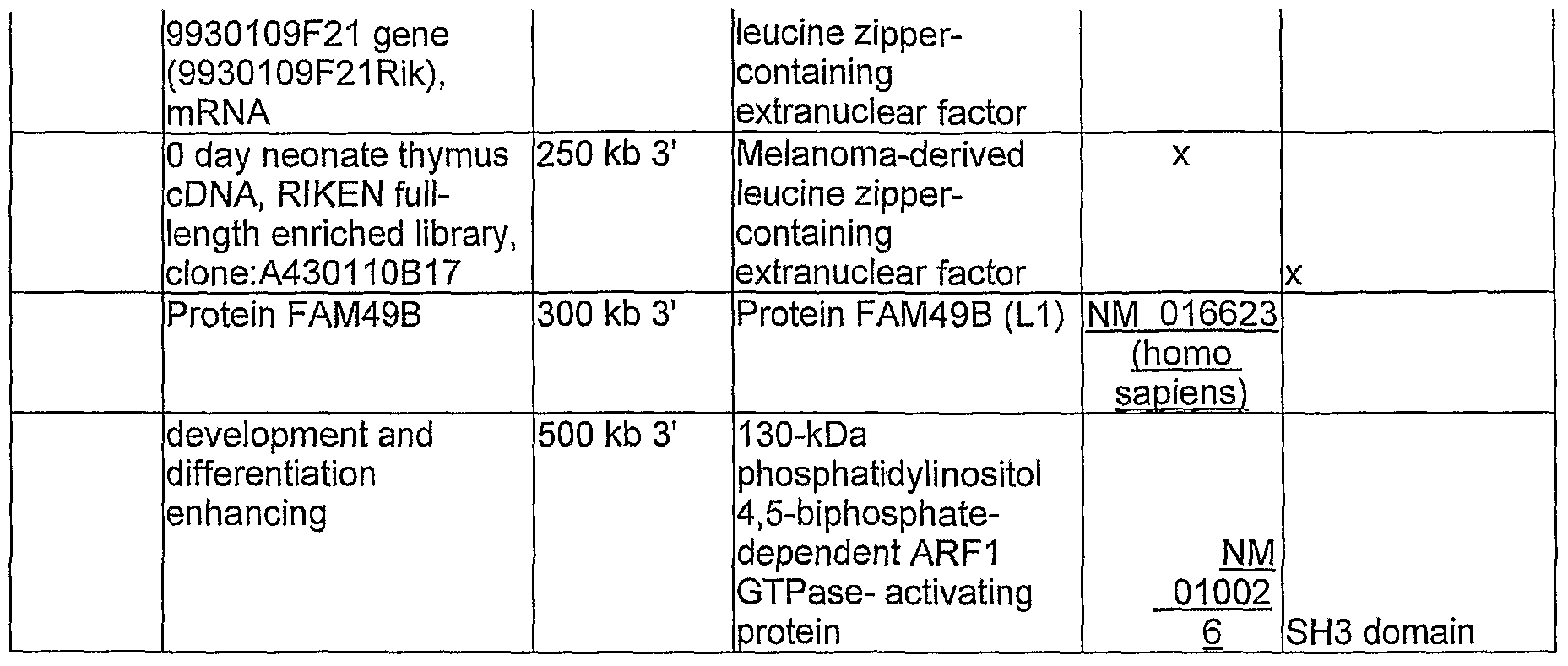
Table 3. Genes located within a distance of 500 Kb of a methylated VIS
Figure imgf000017_0001
Figure imgf000017_0002
Figure imgf000018_0001
Figure imgf000019_0001
99-36 lunatic fringe gene intron 1 Beta-1,3-N- band 4 homolog acetylglucosaminyltra nsferase lunatic embryonic fringe NM 008494 development
12 days embryo 5,5 kb 5' eyeball cDNA, RIKEN full-length enriched library, clone:D230015O06 tweety homologue 3 10,5 kb 3' tweety 3 chloride channel
NM 175274 activity
Figure imgf000020_0001
Figure imgf000021_0001
Figure imgf000022_0001
Figure imgf000022_0002
Figure imgf000023_0001
Figure imgf000023_0002
Figure imgf000024_0001
Figure imgf000024_0002
Figure imgf000025_0001
Figure imgf000025_0002
Figure imgf000026_0001
Figure imgf000027_0001
Figure imgf000028_0001
NM is involved in the
Figure imgf000028_0002
02419 polvadenvlation
Figure imgf000029_0001
Figure imgf000030_0001
Figure imgf000030_0002
Figure imgf000031_0001
Figure imgf000032_0001
Figure imgf000033_0001
Figure imgf000033_0002
Figure imgf000034_0001

Claims

Claims
1. Method for the identification of tumor suppressor genes comprising a) infecting mice with a cancer causing retrovirus; b) checking for the presence of methylated viral inserts; and c) identifying the genes flanking the viral insertion site.
2. Method according to claim 1, wherein the genomic DNA is randomly cut to provide fragments containing the viral inserts.
3. Method according to claim 1 or 2, further comprising a step of enrichment of methylated DNA fragments, preferably by immunoprecipitating said methylated DNA fragments.
4. Method according to claim 3, wherein the immunoprecipation is performed with an antibody directed against 5-methyl-cytosine (α-5mC).
5. Method according to any of claim 1-4, wherein the methylated fragments are amplified, preferably by inverse PCR.
6. Tumor suppressor gene selected from the group consisting of A kinase anchor protein 7, arginase lfrom liver, cofactor required for SpI trancriptional activation subunit 3, erytrocyte protein 4.1-like, ectonucleotide pyrophosphatase/phosphodiesterase 3, ectonucleotide pyrophosphatase/phosphodiesterase 1, cyclin D3, taube nuss, Riken cDNa 1700001C19, bystin, guanylate cyclase activator Ia (retina), Trf (TATA binding protein-related factor) -proximal protein homolog, ubiquitin specific peptidase 49, guanylate cyclase activator IB, mitochondrial ribosomal protein SlO, transcriptional regulating factor 1, fibroblast growth factor receptor substrate 3, progastricsin (pepsinogen C), transcription factor EB, forkhead box P4, DNA primase, p58 subunit, RIKEN 1700001G17 gene, Rab23, Bcl2-associated athanogene 2, zinc finger protein 451, dystonin, lunatic fringe gene homolog, 12 days embryo eyeball cDNA, EIKEN full-length, enriched library, clone:D230015O06, tweety homologue 3, galectin-related inter-fiber protein, carbohydrate sulfotransferase 12, IQ motif containing E, guanine nucleotide binding protein αl2, caspase recruitment domain family member 11, eukaryotic translation initiation factor 3, subunit 9, sorting nexin 8, FtsJ homolog 2, nudix (nucleoside diphosphate linked moiety X)-type motif 1, Stearoyl-CoenzymeA desaturase 1, Stearoyl-CoenzymeA desaturase 2,
Stearoyl- Coenzyme A desaturase 3, Stearoyl-CoenzymeA desaturase 4, cDNA sequence BC046386, biogenesis of lysosome-related organelles complex-1 subunit 2, CWF19-like 1 cell cycle control, wingless related MMTV integration site 8b, gene model 341, NADH dehydrogenase (ubiquinone) 1 beta subcomplex 8, hypoxia-inducible factor 1 α subunit inhibitor, paired box gene 2, conserved helix-loop -helix ubiquitous kinase, SPFH domain family member 1, cytochrome P450 family 2 subfamily c polypeptide 44, carboxypeptidase N polypeptide 1, dynamin binding protein, ATP -binding cassette sub-family C (CFTR/MRP) member 2, methylmalonic aciduria (cobalamin deficiency) type A, hypothetical protein LOC67687, OTU domain containing 4, ATP-binding cassette sub-family E (OABP) member 1, anaphase promoting complex subunit 10, G-protein coupled receptor 171, purinergic G-protein coupled receptor P2Y 14, purinergic G-protein coupled receptor P2Y 13, purinergic G-protein coupled receptor P2Y 12, mediator of RNA polymerase II transcription subunit 12 homolog (yeast)-like, G protein-coupled receptor 87, Usher syndrome 3A homolog, 15 days embryo head cDNA RIKEN full-length enriched library clone:4022435C0, seven in absentia 2, WAS protein family member 2, D164 sialomucin-like 2, mitogen-activated protein kinase kinase kinase 6, AT hook DNA binding motif containing 1, solute carrier family 9 (sodium/hydrogen exchanger) member 1, Gardner-Rasheed feline sarcoma viral (Fgr) oncogene homolog, G-protein coupled receptor 3, synaptotagmin-like 1, WD and tetratricopeptide repeats 1, nuclear distribution gene C homolog, nuclear receptor subfamily 0 group B member 2, G patch domain containing 3, ATP binding domain 1 family member B, stratifin, zinc finger DHHC domain containing 18, phosphatidylinositol glycan class V, syntaxin 12, protein phosphatase 1 regulatory (inhibitor) subunit 8, replication protein A2, acid-like sphingomyelin phosphodiesterase 3B, X KeIl blood group precursor related family member 8 homolog, eyes absent 3 homolog (Drosophila), cleavage stimulation factor 3' pre-RNA, subunit 1, RIKEN cDNA F730031O20 gene, aurora kinase A, RIKEN cDNA 2410001C21 gene (2410001C21Rik:) mRNA, RIKEN cDNA 2010011120 gene (2010011I20Rik) mRNA, Adult male spinal cord cDNA RIKEN full-length enriched library clone:A330041C17, hypothetical protein LOC76426, melanocortin 3 receptor, transcription factor AP-2 gamma, cerebellin 4 precursor protein, bone morphogenetic protein 7, myosin IH, forkhead box N4, potassium channel tetramerisation domain containing 10, acetyl- Coenzyme A carboxylase beta, ubiquitin protein ligase E3B, mevalonate kinase, methylmalonic aciduria (cobalamin deficiency) type B homolog (human), uracil DNA glycosylase, ubiquitin specific peptidase 30, transient receptor potential cation channel subfamily V member 4, glycolipid transfer protein, G protein-coupled receptor kinase -inter actor 2, ankyrin repeat domain 13a, D-amino acid oxidase 1, slingshot homolog 1 (Drosophila), coronin actin binding protein 1C, selectin platelet (p-selectin) ligand, hypothetical protein LOC74236, expressed sequence AI987692, RIKEN cDNA 9930109F21 gene (9930109F21Rik) mRNA, 0 day neonate thymus cDNA RIKEN full-length enriched library clone :A430 HOB 17, Protein FAM49B development and differentiation enhancing.
7. Use of a tumor suppressor gene from Table 3 for diagnosis of AML, more preferably, wherein said diagnosis comprises classification of AML subtypes and/or determination of susceptibility to therapy.
8. Use of a tumor suppressor gene from Table 3 for therapy of AML.
9. Method for therapy of AML by increasing the expression and/or availability of a tumor suppression gene of table 3.
PCT/NL2006/000273 2006-06-01 2006-06-01 Identification of tumor suppressor genes in an acute myeloid leukaemia model WO2007139373A1 (en)

Priority Applications (4)

Application Number Priority Date Filing Date Title
EP06747563A EP2021498A1 (en) 2006-06-01 2006-06-01 Identification of tumor suppressor genes in an acute myeloid leukaemia model
US12/303,067 US20100003677A1 (en) 2006-06-01 2006-06-01 Identification of tumor suppressor genes in an acute myeloid leukaemia model
PCT/NL2006/000273 WO2007139373A1 (en) 2006-06-01 2006-06-01 Identification of tumor suppressor genes in an acute myeloid leukaemia model
CA002658028A CA2658028A1 (en) 2006-06-01 2006-06-01 Identification of tumor suppressor genes in an acute myeloid leukaemia model

Applications Claiming Priority (1)

Application Number Priority Date Filing Date Title
PCT/NL2006/000273 WO2007139373A1 (en) 2006-06-01 2006-06-01 Identification of tumor suppressor genes in an acute myeloid leukaemia model

Publications (1)

Publication Number Publication Date
WO2007139373A1 true WO2007139373A1 (en) 2007-12-06

Family

ID=37712473

Family Applications (1)

Application Number Title Priority Date Filing Date
PCT/NL2006/000273 WO2007139373A1 (en) 2006-06-01 2006-06-01 Identification of tumor suppressor genes in an acute myeloid leukaemia model

Country Status (4)

Country Link
US (1) US20100003677A1 (en)
EP (1) EP2021498A1 (en)
CA (1) CA2658028A1 (en)
WO (1) WO2007139373A1 (en)

Cited By (1)

* Cited by examiner, † Cited by third party
Publication number Priority date Publication date Assignee Title
CN102574927A (en) * 2009-09-28 2012-07-11 希森美康株式会社 Hybridoma producing anti-methylated DNA antibody and utilization of same

Families Citing this family (2)

* Cited by examiner, † Cited by third party
Publication number Priority date Publication date Assignee Title
CN104671198A (en) * 2015-02-03 2015-06-03 天津大学 Method for preparing stripe-shaped micro-nanometer wrinkle structure by using electron beam induction method
CN110938695A (en) * 2019-12-16 2020-03-31 山东大学齐鲁医院 Novel application of ankyrin repeat structural domain 13A gene and/or protein coded by same

Non-Patent Citations (7)

* Cited by examiner, † Cited by third party
Title
BROWN REBECCA L ET AL: "AKAP7gamma is a nuclear RI-binding AKAP.", BIOCHEMICAL AND BIOPHYSICAL RESEARCH COMMUNICATIONS, vol. 306, no. 2, 27 June 2003 (2003-06-27), pages 394 - 401, XP002420267, ISSN: 0006-291X *
EKMEKCI CUMHUR G ET AL: "Aberrant methylation of multiple tumor suppressor genes in acute myeloid leukemia.", AMERICAN JOURNAL OF HEMATOLOGY NOV 2004, vol. 77, no. 3, November 2004 (2004-11-01), pages 233 - 240, XP002420271, ISSN: 0361-8609 *
ERKELAND S J ET AL: "Large-Scale Identification of Disease Genes Involved in Acute Myeloid Leukemia", JOURNAL OF VIROLOGY 2004 UNITED STATES, vol. 78, no. 4, 2004, pages 1971 - 1980, XP002420268, ISSN: 0022-538X *
ESTELLER MANEL: "Profiling aberrant DNA methylation in hematologic neoplasms: A view from the tip of the iceberg.", CLINICAL IMMUNOLOGY (ORLANDO), vol. 109, no. 1, October 2003 (2003-10-01), pages 80 - 88, XP002420270, ISSN: 1521-6616 *
SUZUKI T ET AL: "NEW GENES INVOLVED IN CANCER IDENTIFIED BY RETROVIRAL TAGGING", NATURE GENETICS, NEW YORK, NY, US, vol. 32, September 2002 (2002-09-01), pages 166 - 174,331, XP001133900, ISSN: 1061-4036 *
TOUW I P ET AL: "Retroviral insertion mutagenesis in mice as a comparative oncogenomics tool to identify disease genes in human leukemia", MOLECULAR THERAPY 2007 UNITED KINGDOM, vol. 15, no. 1, 2007, pages 13 - 19, XP002420272, ISSN: 1525-0016 1525-0024 *
UREN A G ET AL: "Retroviral insertional mutagenesis: past, present and future", ONCOGENE, vol. 24, no. 52, November 2005 (2005-11-01), pages 7656 - 7672, XP002420269, ISSN: 0950-9232 *

Cited By (3)

* Cited by examiner, † Cited by third party
Publication number Priority date Publication date Assignee Title
CN102574927A (en) * 2009-09-28 2012-07-11 希森美康株式会社 Hybridoma producing anti-methylated DNA antibody and utilization of same
CN102574927B (en) * 2009-09-28 2015-01-07 希森美康株式会社 Hybridoma producing anti-methylated DNA antibody and utilization of same
US9169330B2 (en) 2009-09-28 2015-10-27 Sysmex Corporation Hybridoma producing anti-methylated DNA antibody and utilization of same

Also Published As

Publication number Publication date
US20100003677A1 (en) 2010-01-07
EP2021498A1 (en) 2009-02-11
CA2658028A1 (en) 2007-12-06

Similar Documents

Publication Publication Date Title
Margalef et al. Stabilization of reversed replication forks by telomerase drives telomere catastrophe
US7858317B2 (en) Aberrantly methylated genes as markers of breast malignancy
Mansouri et al. Epigenetic deregulation in chronic lymphocytic leukemia: Clinical and biological impact
US11788145B2 (en) Epigeneiic silencing of NMT2
Yehezkel et al. Characterization and rescue of telomeric abnormalities in ICF syndrome type I fibroblasts
JP2010517582A (en) Methods for early detection and prognosis of colon cancer
Tiedemann et al. Acute depletion redefines the division of labor among DNA methyltransferases in methylating the human genome
Xiao et al. PTPRG inhibition by DNA methylation and cooperation with RAS gene activation in childhood acute lymphoblastic leukemia
US10597730B2 (en) Method of diagnosing cancer
Zhao et al. Critically short telomeres derepress retrotransposons to promote genome instability in embryonic stem cells
WO2007139373A1 (en) Identification of tumor suppressor genes in an acute myeloid leukaemia model
Kong et al. Methylation‐associated silencing of death‐associated protein kinase gene in laryngeal squamous cell cancer
WO2013173912A1 (en) Method of diagnosing cancer comprising detection of the methylation signature in the htert promoter
Diaz-Meyer et al. Alternative mechanisms associated with silencing of CDKN1C in Beckwith–Wiedemann syndrome
WO2010020787A1 (en) Diagnosis and treatment of tumours
US20090275632A1 (en) Methods of diagnosis and treatment
Smail Qualitative and Quantitative Identification of DNA Methylation Changes in Blood of the Breast Cancer Patients
JP5116935B2 (en) Method for evaluating the degree of canceration of a mammal-derived specimen
Xu et al. DNMT3A Cooperates with YAP/TAZ to Drive Gallbladder Cancer Metastasis
Naipauer et al. RNA-sequencing analysis of a multistep and hit-and-run cell and animal model of KSHV tumorigenesis reveal the roles of mutations, CpG methylation, and viral-infection footprints in oncogenesis
Campbell Gene expression analysis of telomerase related genes in myeloid malignancy
Kwok The role and molecular characteristics of constitutional MLH1 epimutation in young-onset cancer.
Choi Studies on the alteration of DNA methylation in cancer
Zhang et al. Conceptual Evolution in Cancer Biology
Kardooni Epigenetic Silencing of Gene Expression in Paediatric Malignant Astrocytoma

Legal Events

Date Code Title Description
121 Ep: the epo has been informed by wipo that ep was designated in this application

Ref document number: 06747563

Country of ref document: EP

Kind code of ref document: A1

ENP Entry into the national phase

Ref document number: 2658028

Country of ref document: CA

NENP Non-entry into the national phase

Ref country code: DE

WWE Wipo information: entry into national phase

Ref document number: 2006747563

Country of ref document: EP

NENP Non-entry into the national phase

Ref country code: RU

WWE Wipo information: entry into national phase

Ref document number: 12303067

Country of ref document: US