WO2007100566A2 - Use of dha and ara in the preparation of a composition for regulating gene expression - Google Patents
Use of dha and ara in the preparation of a composition for regulating gene expression Download PDFInfo
- Publication number
- WO2007100566A2 WO2007100566A2 PCT/US2007/004451 US2007004451W WO2007100566A2 WO 2007100566 A2 WO2007100566 A2 WO 2007100566A2 US 2007004451 W US2007004451 W US 2007004451W WO 2007100566 A2 WO2007100566 A2 WO 2007100566A2
- Authority
- WO
- WIPO (PCT)
- Prior art keywords
- dha
- ara
- gene
- subject
- expression
- Prior art date
Links
- 230000014509 gene expression Effects 0.000 title claims abstract description 189
- 239000000203 mixture Substances 0.000 title claims description 36
- 238000002360 preparation method Methods 0.000 title claims description 23
- 230000001105 regulatory effect Effects 0.000 title description 27
- 108090000623 proteins and genes Proteins 0.000 claims abstract description 292
- 238000000034 method Methods 0.000 claims abstract description 60
- 206010028980 Neoplasm Diseases 0.000 claims description 39
- -1 FXD3 Proteins 0.000 claims description 38
- 208000037265 diseases, disorders, signs and symptoms Diseases 0.000 claims description 36
- 235000013350 formula milk Nutrition 0.000 claims description 33
- 108091007505 ADAM17 Proteins 0.000 claims description 32
- 208000035475 disorder Diseases 0.000 claims description 20
- 108700020463 BRCA1 Proteins 0.000 claims description 19
- 101150072950 BRCA1 gene Proteins 0.000 claims description 19
- 102000036365 BRCA1 Human genes 0.000 claims description 18
- 230000003405 preventing effect Effects 0.000 claims description 18
- 230000003827 upregulation Effects 0.000 claims description 17
- 102100038824 Peroxisome proliferator-activated receptor delta Human genes 0.000 claims description 16
- 101000967216 Homo sapiens Eosinophil cationic protein Proteins 0.000 claims description 15
- 101000832767 Homo sapiens Disintegrin and metalloproteinase domain-containing protein 8 Proteins 0.000 claims description 14
- 101000763951 Homo sapiens Mitochondrial import inner membrane translocase subunit Tim8 A Proteins 0.000 claims description 14
- 102000052116 epidermal growth factor receptor activity proteins Human genes 0.000 claims description 14
- 108700015053 epidermal growth factor receptor activity proteins Proteins 0.000 claims description 14
- YOHYSYJDKVYCJI-UHFFFAOYSA-N n-[3-[[6-[3-(trifluoromethyl)anilino]pyrimidin-4-yl]amino]phenyl]cyclopropanecarboxamide Chemical compound FC(F)(F)C1=CC=CC(NC=2N=CN=C(NC=3C=C(NC(=O)C4CC4)C=CC=3)C=2)=C1 YOHYSYJDKVYCJI-UHFFFAOYSA-N 0.000 claims description 14
- 102100040618 Eosinophil cationic protein Human genes 0.000 claims description 13
- 102100026808 Mitochondrial import inner membrane translocase subunit Tim8 A Human genes 0.000 claims description 13
- 102100025991 Betaine-homocysteine S-methyltransferase 1 Human genes 0.000 claims description 12
- 101000628647 Homo sapiens Serine/threonine-protein kinase 24 Proteins 0.000 claims description 12
- 101000880439 Homo sapiens Serine/threonine-protein kinase 3 Proteins 0.000 claims description 12
- 101001098818 Homo sapiens cGMP-inhibited 3',5'-cyclic phosphodiesterase A Proteins 0.000 claims description 12
- 102100026764 Serine/threonine-protein kinase 24 Human genes 0.000 claims description 12
- 102100037093 cGMP-inhibited 3',5'-cyclic phosphodiesterase A Human genes 0.000 claims description 12
- 230000004770 neurodegeneration Effects 0.000 claims description 12
- 102100037991 85/88 kDa calcium-independent phospholipase A2 Human genes 0.000 claims description 11
- 102100025979 Disintegrin and metalloproteinase domain-containing protein 33 Human genes 0.000 claims description 11
- 101000720049 Homo sapiens Disintegrin and metalloproteinase domain-containing protein 33 Proteins 0.000 claims description 11
- 101000588298 Homo sapiens Endoplasmic reticulum membrane sensor NFE2L1 Proteins 0.000 claims description 11
- 101000577547 Homo sapiens Nuclear respiratory factor 1 Proteins 0.000 claims description 11
- 101000764357 Homo sapiens Protein Tob1 Proteins 0.000 claims description 11
- 101710125553 PLA2G6 Proteins 0.000 claims description 11
- 101000984044 Homo sapiens LIM homeobox transcription factor 1-beta Proteins 0.000 claims description 9
- 101000645277 Homo sapiens Mitochondrial import inner membrane translocase subunit Tim23 Proteins 0.000 claims description 9
- 102100025457 LIM homeobox transcription factor 1-beta Human genes 0.000 claims description 9
- 102100026255 Mitochondrial import inner membrane translocase subunit Tim23 Human genes 0.000 claims description 9
- 208000008589 Obesity Diseases 0.000 claims description 9
- 102000019347 Tob1 Human genes 0.000 claims description 9
- 208000006673 asthma Diseases 0.000 claims description 9
- 102100033773 Collagen alpha-6(IV) chain Human genes 0.000 claims description 8
- 102100028115 Forkhead box protein P2 Human genes 0.000 claims description 8
- 101000710885 Homo sapiens Collagen alpha-6(IV) chain Proteins 0.000 claims description 8
- 101001059881 Homo sapiens Forkhead box protein P2 Proteins 0.000 claims description 8
- 235000020824 obesity Nutrition 0.000 claims description 8
- 102100020760 Ferritin heavy chain Human genes 0.000 claims description 7
- 101001002987 Homo sapiens Ferritin heavy chain Proteins 0.000 claims description 7
- 101000819458 Homo sapiens Frizzled-3 Proteins 0.000 claims description 7
- 101000711744 Homo sapiens Non-secretory ribonuclease Proteins 0.000 claims description 7
- 101000801260 Homo sapiens Troponin C, slow skeletal and cardiac muscles Proteins 0.000 claims description 7
- 101000851334 Homo sapiens Troponin I, cardiac muscle Proteins 0.000 claims description 7
- 102100034217 Non-secretory ribonuclease Human genes 0.000 claims description 7
- 102100036859 Troponin I, cardiac muscle Human genes 0.000 claims description 7
- 201000008696 deafness-dystonia-optic neuronopathy syndrome Diseases 0.000 claims description 7
- 230000028993 immune response Effects 0.000 claims description 7
- 208000024827 Alzheimer disease Diseases 0.000 claims description 6
- 101000868273 Homo sapiens CD44 antigen Proteins 0.000 claims description 6
- 230000003828 downregulation Effects 0.000 claims description 6
- 108010043412 neuropeptide Y-Y1 receptor Proteins 0.000 claims description 6
- 102100032912 CD44 antigen Human genes 0.000 claims description 5
- 102100021262 Frizzled-3 Human genes 0.000 claims description 5
- 101000937172 Homo sapiens Protein FAN Proteins 0.000 claims description 5
- 208000000175 Nail-Patella Syndrome Diseases 0.000 claims description 5
- 108010085793 Neurofibromin 1 Proteins 0.000 claims description 5
- 102000007530 Neurofibromin 1 Human genes 0.000 claims description 5
- 102100038878 Neuropeptide Y receptor type 1 Human genes 0.000 claims description 5
- 102100027633 Protein FAN Human genes 0.000 claims description 5
- 230000004199 lung function Effects 0.000 claims description 5
- 201000010046 Dilated cardiomyopathy Diseases 0.000 claims description 4
- 102100024364 Disintegrin and metalloproteinase domain-containing protein 8 Human genes 0.000 claims description 4
- 101000992378 Homo sapiens Oxysterol-binding protein 2 Proteins 0.000 claims description 4
- 101001086862 Homo sapiens Pulmonary surfactant-associated protein B Proteins 0.000 claims description 4
- 101150007959 Lum gene Proteins 0.000 claims description 4
- 102100032164 Oxysterol-binding protein 2 Human genes 0.000 claims description 4
- 102100032617 Pulmonary surfactant-associated protein B Human genes 0.000 claims description 4
- 101150061875 ADAM33 gene Proteins 0.000 claims description 3
- 102100034051 Heat shock protein HSP 90-alpha Human genes 0.000 claims description 3
- 101100108123 Homo sapiens ADAM33 gene Proteins 0.000 claims description 3
- 208000000475 Mohr-Tranebjaerg syndrome Diseases 0.000 claims description 3
- 101150027439 NPY1 gene Proteins 0.000 claims description 3
- 206010012601 diabetes mellitus Diseases 0.000 claims description 3
- 201000001421 hyperglycemia Diseases 0.000 claims description 3
- 208000002780 macular degeneration Diseases 0.000 claims description 3
- 230000002265 prevention Effects 0.000 claims description 3
- 238000011282 treatment Methods 0.000 claims description 3
- 206010010356 Congenital anomaly Diseases 0.000 claims description 2
- 102100035298 Cytokine SCM-1 beta Human genes 0.000 claims description 2
- 102100022132 High affinity immunoglobulin epsilon receptor subunit gamma Human genes 0.000 claims description 2
- 101000804771 Homo sapiens Cytokine SCM-1 beta Proteins 0.000 claims description 2
- 101001016865 Homo sapiens Heat shock protein HSP 90-alpha Proteins 0.000 claims description 2
- 101001078626 Homo sapiens Heat shock protein HSP 90-alpha A2 Proteins 0.000 claims description 2
- 101000824104 Homo sapiens High affinity immunoglobulin epsilon receptor subunit gamma Proteins 0.000 claims description 2
- 101000945340 Homo sapiens Killer cell immunoglobulin-like receptor 2DS1 Proteins 0.000 claims description 2
- 101001038339 Homo sapiens LIM homeobox transcription factor 1-alpha Proteins 0.000 claims description 2
- 101000992387 Homo sapiens Oxysterol-binding protein-related protein 9 Proteins 0.000 claims description 2
- 101000884271 Homo sapiens Signal transducer CD24 Proteins 0.000 claims description 2
- 102100033631 Killer cell immunoglobulin-like receptor 2DS1 Human genes 0.000 claims description 2
- 102100040290 LIM homeobox transcription factor 1-alpha Human genes 0.000 claims description 2
- 101100366137 Mesembryanthemum crystallinum SODCC.1 gene Proteins 0.000 claims description 2
- 108010059196 N-hydroxy-2-acetylaminofluorene sulfotransferase Proteins 0.000 claims description 2
- 208000002537 Neuronal Ceroid-Lipofuscinoses Diseases 0.000 claims description 2
- 102100032162 Oxysterol-binding protein-related protein 9 Human genes 0.000 claims description 2
- 101100096142 Panax ginseng SODCC gene Proteins 0.000 claims description 2
- 102100038081 Signal transducer CD24 Human genes 0.000 claims description 2
- 102100023985 Sulfotransferase 1C2 Human genes 0.000 claims description 2
- 230000010083 bronchial hyperresponsiveness Effects 0.000 claims description 2
- 201000011304 dilated cardiomyopathy 1A Diseases 0.000 claims description 2
- 230000002222 downregulating effect Effects 0.000 claims description 2
- 230000004217 heart function Effects 0.000 claims description 2
- 230000004377 improving vision Effects 0.000 claims description 2
- 230000000626 neurodegenerative effect Effects 0.000 claims description 2
- 201000008051 neuronal ceroid lipofuscinosis Diseases 0.000 claims description 2
- 101150017120 sod gene Proteins 0.000 claims description 2
- 230000004936 stimulating effect Effects 0.000 claims description 2
- 102100031111 Disintegrin and metalloproteinase domain-containing protein 17 Human genes 0.000 claims 4
- 102100028744 Nuclear respiratory factor 1 Human genes 0.000 claims 4
- 102100021633 Cathepsin B Human genes 0.000 claims 3
- 102100032219 Cathepsin D Human genes 0.000 claims 3
- 101000933413 Homo sapiens Betaine-homocysteine S-methyltransferase 1 Proteins 0.000 claims 3
- 101000898449 Homo sapiens Cathepsin B Proteins 0.000 claims 3
- 101000869010 Homo sapiens Cathepsin D Proteins 0.000 claims 3
- 101001061405 Homo sapiens Frizzled-9 Proteins 0.000 claims 3
- 101001065609 Homo sapiens Lumican Proteins 0.000 claims 3
- 101001013832 Homo sapiens Mitochondrial peptide methionine sulfoxide reductase Proteins 0.000 claims 3
- 101001127470 Homo sapiens p53 apoptosis effector related to PMP-22 Proteins 0.000 claims 3
- 102100032114 Lumican Human genes 0.000 claims 3
- 102100031767 Mitochondrial peptide methionine sulfoxide reductase Human genes 0.000 claims 3
- 108010015181 PPAR delta Proteins 0.000 claims 3
- 102100030898 p53 apoptosis effector related to PMP-22 Human genes 0.000 claims 3
- 101001063991 Homo sapiens Leptin Proteins 0.000 claims 1
- 101000804764 Homo sapiens Lymphotactin Proteins 0.000 claims 1
- 101000823949 Homo sapiens Serine palmitoyltransferase 2 Proteins 0.000 claims 1
- 102100030874 Leptin Human genes 0.000 claims 1
- 102100035304 Lymphotactin Human genes 0.000 claims 1
- 208000018737 Parkinson disease Diseases 0.000 claims 1
- 102100022059 Serine palmitoyltransferase 2 Human genes 0.000 claims 1
- MBMBGCFOFBJSGT-KUBAVDMBSA-N all-cis-docosa-4,7,10,13,16,19-hexaenoic acid Chemical compound CC\C=C/C\C=C/C\C=C/C\C=C/C\C=C/C\C=C/CCC(O)=O MBMBGCFOFBJSGT-KUBAVDMBSA-N 0.000 description 363
- YZXBAPSDXZZRGB-DOFZRALJSA-N arachidonic acid Chemical compound CCCCC\C=C/C\C=C/C\C=C/C\C=C/CCCC(O)=O YZXBAPSDXZZRGB-DOFZRALJSA-N 0.000 description 291
- 235000020669 docosahexaenoic acid Nutrition 0.000 description 184
- 229940090949 docosahexaenoic acid Drugs 0.000 description 180
- 235000021342 arachidonic acid Nutrition 0.000 description 145
- 229940114079 arachidonic acid Drugs 0.000 description 145
- 230000009469 supplementation Effects 0.000 description 69
- 230000006870 function Effects 0.000 description 62
- 230000001965 increasing effect Effects 0.000 description 56
- 210000004027 cell Anatomy 0.000 description 48
- 238000011161 development Methods 0.000 description 47
- 230000018109 developmental process Effects 0.000 description 47
- 235000018102 proteins Nutrition 0.000 description 47
- 102000004169 proteins and genes Human genes 0.000 description 47
- 102000043279 ADAM17 Human genes 0.000 description 28
- 201000011510 cancer Diseases 0.000 description 28
- 210000004556 brain Anatomy 0.000 description 27
- 241000699670 Mus sp. Species 0.000 description 26
- 230000003247 decreasing effect Effects 0.000 description 26
- 230000000694 effects Effects 0.000 description 26
- 235000005911 diet Nutrition 0.000 description 25
- 235000020978 long-chain polyunsaturated fatty acids Nutrition 0.000 description 25
- 241001465754 Metazoa Species 0.000 description 24
- 230000033228 biological regulation Effects 0.000 description 23
- 230000001413 cellular effect Effects 0.000 description 23
- 230000022131 cell cycle Effects 0.000 description 22
- 230000035772 mutation Effects 0.000 description 20
- 208000003019 Neurofibromatosis 1 Diseases 0.000 description 19
- 208000024834 Neurofibromatosis type 1 Diseases 0.000 description 19
- 108010076371 Lumican Proteins 0.000 description 17
- 102000011681 Lumican Human genes 0.000 description 17
- 230000015572 biosynthetic process Effects 0.000 description 17
- XEEYBQQBJWHFJM-UHFFFAOYSA-N Iron Chemical compound [Fe] XEEYBQQBJWHFJM-UHFFFAOYSA-N 0.000 description 16
- 230000037396 body weight Effects 0.000 description 16
- 230000003993 interaction Effects 0.000 description 16
- 210000004379 membrane Anatomy 0.000 description 16
- 239000012528 membrane Substances 0.000 description 16
- 239000000523 sample Substances 0.000 description 16
- 102000040945 Transcription factor Human genes 0.000 description 15
- 108091023040 Transcription factor Proteins 0.000 description 15
- 230000010261 cell growth Effects 0.000 description 15
- 201000010099 disease Diseases 0.000 description 15
- 230000008520 organization Effects 0.000 description 15
- 229920002477 rna polymer Polymers 0.000 description 15
- 230000006907 apoptotic process Effects 0.000 description 14
- 230000023715 cellular developmental process Effects 0.000 description 14
- 230000000378 dietary effect Effects 0.000 description 14
- 210000004185 liver Anatomy 0.000 description 14
- 208000015122 neurodegenerative disease Diseases 0.000 description 14
- 108091008765 peroxisome proliferator-activated receptors β/δ Proteins 0.000 description 14
- 102000004225 Cathepsin B Human genes 0.000 description 13
- 108090000712 Cathepsin B Proteins 0.000 description 13
- 210000003169 central nervous system Anatomy 0.000 description 13
- 210000003710 cerebral cortex Anatomy 0.000 description 13
- 150000002632 lipids Chemical class 0.000 description 13
- 238000004458 analytical method Methods 0.000 description 12
- 230000037213 diet Effects 0.000 description 12
- 230000002438 mitochondrial effect Effects 0.000 description 12
- 102000005962 receptors Human genes 0.000 description 12
- 108020003175 receptors Proteins 0.000 description 12
- 230000011664 signaling Effects 0.000 description 12
- 102000003908 Cathepsin D Human genes 0.000 description 11
- 108090000258 Cathepsin D Proteins 0.000 description 11
- 108010021111 Uncoupling Protein 2 Proteins 0.000 description 11
- 102000008219 Uncoupling Protein 2 Human genes 0.000 description 11
- 230000037356 lipid metabolism Effects 0.000 description 11
- 239000000047 product Substances 0.000 description 11
- 230000033772 system development Effects 0.000 description 11
- 238000013518 transcription Methods 0.000 description 11
- 230000035897 transcription Effects 0.000 description 11
- 230000032258 transport Effects 0.000 description 11
- 101000701363 Homo sapiens Phospholipid-transporting ATPase IC Proteins 0.000 description 10
- 241000700159 Rattus Species 0.000 description 10
- 230000030833 cell death Effects 0.000 description 10
- 230000004663 cell proliferation Effects 0.000 description 10
- 235000014113 dietary fatty acids Nutrition 0.000 description 10
- 229930195729 fatty acid Natural products 0.000 description 10
- 239000000194 fatty acid Substances 0.000 description 10
- 150000004665 fatty acids Chemical class 0.000 description 10
- 210000000987 immune system Anatomy 0.000 description 10
- 210000000653 nervous system Anatomy 0.000 description 10
- 230000002829 reductive effect Effects 0.000 description 10
- 108010088623 Betaine-Homocysteine S-Methyltransferase Proteins 0.000 description 9
- 102100026261 Metalloproteinase inhibitor 3 Human genes 0.000 description 9
- 102000015785 Serine C-Palmitoyltransferase Human genes 0.000 description 9
- 108010024814 Serine C-palmitoyltransferase Proteins 0.000 description 9
- 108010031429 Tissue Inhibitor of Metalloproteinase-3 Proteins 0.000 description 9
- 230000004913 activation Effects 0.000 description 9
- 230000021164 cell adhesion Effects 0.000 description 9
- HVYWMOMLDIMFJA-DPAQBDIFSA-N cholesterol Chemical compound C1C=C2C[C@@H](O)CC[C@]2(C)[C@@H]2[C@@H]1[C@@H]1CC[C@H]([C@H](C)CCCC(C)C)[C@@]1(C)CC2 HVYWMOMLDIMFJA-DPAQBDIFSA-N 0.000 description 9
- 230000007812 deficiency Effects 0.000 description 9
- 235000020673 eicosapentaenoic acid Nutrition 0.000 description 9
- 210000001508 eye Anatomy 0.000 description 9
- 108020004999 messenger RNA Proteins 0.000 description 9
- 230000007781 signaling event Effects 0.000 description 9
- 230000014616 translation Effects 0.000 description 9
- 102100036683 Growth arrest-specific protein 1 Human genes 0.000 description 8
- 101001072723 Homo sapiens Growth arrest-specific protein 1 Proteins 0.000 description 8
- 101001038006 Homo sapiens Lysophosphatidic acid receptor 3 Proteins 0.000 description 8
- 102100040388 Lysophosphatidic acid receptor 3 Human genes 0.000 description 8
- 241001504519 Papio ursinus Species 0.000 description 8
- 102100030448 Phospholipid-transporting ATPase IC Human genes 0.000 description 8
- JAZBEHYOTPTENJ-JLNKQSITSA-N all-cis-5,8,11,14,17-icosapentaenoic acid Chemical compound CC\C=C/C\C=C/C\C=C/C\C=C/C\C=C/CCCC(O)=O JAZBEHYOTPTENJ-JLNKQSITSA-N 0.000 description 8
- 210000000481 breast Anatomy 0.000 description 8
- 229960005135 eicosapentaenoic acid Drugs 0.000 description 8
- JAZBEHYOTPTENJ-UHFFFAOYSA-N eicosapentaenoic acid Natural products CCC=CCC=CCC=CCC=CCC=CCCCC(O)=O JAZBEHYOTPTENJ-UHFFFAOYSA-N 0.000 description 8
- 235000021323 fish oil Nutrition 0.000 description 8
- 230000012010 growth Effects 0.000 description 8
- 229910052742 iron Inorganic materials 0.000 description 8
- 230000013016 learning Effects 0.000 description 8
- 230000015654 memory Effects 0.000 description 8
- 238000002493 microarray Methods 0.000 description 8
- 239000003921 oil Substances 0.000 description 8
- 235000019198 oils Nutrition 0.000 description 8
- 230000035755 proliferation Effects 0.000 description 8
- 201000000980 schizophrenia Diseases 0.000 description 8
- 230000019491 signal transduction Effects 0.000 description 8
- 150000003384 small molecules Chemical class 0.000 description 8
- 230000005760 tumorsuppression Effects 0.000 description 8
- 230000016776 visual perception Effects 0.000 description 8
- 101710137189 Amyloid-beta A4 protein Proteins 0.000 description 7
- 102100022704 Amyloid-beta precursor protein Human genes 0.000 description 7
- 101710151993 Amyloid-beta precursor protein Proteins 0.000 description 7
- 206010006187 Breast cancer Diseases 0.000 description 7
- 208000026310 Breast neoplasm Diseases 0.000 description 7
- 108091006027 G proteins Proteins 0.000 description 7
- 102000030782 GTP binding Human genes 0.000 description 7
- 108091000058 GTP-Binding Proteins 0.000 description 7
- 101000597932 Homo sapiens Protein numb homolog Proteins 0.000 description 7
- 102100026632 Mimecan Human genes 0.000 description 7
- 102100028642 Prostaglandin E synthase 3 Human genes 0.000 description 7
- DZHSAHHDTRWUTF-SIQRNXPUSA-N amyloid-beta polypeptide 42 Chemical compound C([C@@H](C(=O)N[C@@H](C)C(=O)N[C@@H](CCC(O)=O)C(=O)N[C@@H](CC(O)=O)C(=O)N[C@H](C(=O)NCC(=O)N[C@@H](CO)C(=O)N[C@@H](CC(N)=O)C(=O)N[C@@H](CCCCN)C(=O)NCC(=O)N[C@@H](C)C(=O)N[C@H](C(=O)N[C@@H]([C@@H](C)CC)C(=O)NCC(=O)N[C@@H](CC(C)C)C(=O)N[C@@H](CCSC)C(=O)N[C@@H](C(C)C)C(=O)NCC(=O)NCC(=O)N[C@@H](C(C)C)C(=O)N[C@@H](C(C)C)C(=O)N[C@@H]([C@@H](C)CC)C(=O)N[C@@H](C)C(O)=O)[C@@H](C)CC)C(C)C)NC(=O)[C@H](CC=1C=CC=CC=1)NC(=O)[C@@H](NC(=O)[C@H](CC(C)C)NC(=O)[C@H](CCCCN)NC(=O)[C@H](CCC(N)=O)NC(=O)[C@H](CC=1N=CNC=1)NC(=O)[C@H](CC=1N=CNC=1)NC(=O)[C@@H](NC(=O)[C@H](CCC(O)=O)NC(=O)[C@H](CC=1C=CC(O)=CC=1)NC(=O)CNC(=O)[C@H](CO)NC(=O)[C@H](CC(O)=O)NC(=O)[C@H](CC=1N=CNC=1)NC(=O)[C@H](CCCNC(N)=N)NC(=O)[C@H](CC=1C=CC=CC=1)NC(=O)[C@H](CCC(O)=O)NC(=O)[C@H](C)NC(=O)[C@@H](N)CC(O)=O)C(C)C)C(C)C)C1=CC=CC=C1 DZHSAHHDTRWUTF-SIQRNXPUSA-N 0.000 description 7
- 230000027455 binding Effects 0.000 description 7
- 230000008827 biological function Effects 0.000 description 7
- 230000031018 biological processes and functions Effects 0.000 description 7
- 230000008859 change Effects 0.000 description 7
- 108020005567 glycine N-acyltransferase Proteins 0.000 description 7
- 230000002440 hepatic effect Effects 0.000 description 7
- 235000020256 human milk Nutrition 0.000 description 7
- 238000004519 manufacturing process Methods 0.000 description 7
- 230000004060 metabolic process Effects 0.000 description 7
- 230000037361 pathway Effects 0.000 description 7
- 238000003753 real-time PCR Methods 0.000 description 7
- 230000004083 survival effect Effects 0.000 description 7
- 108010085238 Actins Proteins 0.000 description 6
- 241000283690 Bos taurus Species 0.000 description 6
- 102100024457 Cyclin-dependent kinase 9 Human genes 0.000 description 6
- 102100031702 Endoplasmic reticulum membrane sensor NFE2L1 Human genes 0.000 description 6
- 101000980930 Homo sapiens Cyclin-dependent kinase 9 Proteins 0.000 description 6
- 101001002170 Homo sapiens Glutamine amidotransferase-like class 1 domain-containing protein 3, mitochondrial Proteins 0.000 description 6
- 101000978210 Homo sapiens Leukotriene C4 synthase Proteins 0.000 description 6
- 101000596772 Homo sapiens Transcription factor 7-like 1 Proteins 0.000 description 6
- 101000666382 Homo sapiens Transcription factor E2-alpha Proteins 0.000 description 6
- 101000843556 Homo sapiens Transcription factor HES-1 Proteins 0.000 description 6
- 108090000862 Ion Channels Proteins 0.000 description 6
- 102000004310 Ion Channels Human genes 0.000 description 6
- 102000016267 Leptin Human genes 0.000 description 6
- 108010092277 Leptin Proteins 0.000 description 6
- 102100023758 Leukotriene C4 synthase Human genes 0.000 description 6
- 102100023031 Neural Wiskott-Aldrich syndrome protein Human genes 0.000 description 6
- 102000048238 Neuregulin-1 Human genes 0.000 description 6
- 108090000556 Neuregulin-1 Proteins 0.000 description 6
- 206010061902 Pancreatic neoplasm Diseases 0.000 description 6
- 102100036985 Protein numb homolog Human genes 0.000 description 6
- 102100020949 Putative glutamine amidotransferase-like class 1 domain-containing protein 3B, mitochondrial Human genes 0.000 description 6
- 102100023606 Retinoic acid receptor alpha Human genes 0.000 description 6
- 102000001494 Sterol O-Acyltransferase Human genes 0.000 description 6
- 108010054082 Sterol O-acyltransferase Proteins 0.000 description 6
- 102100038313 Transcription factor E2-alpha Human genes 0.000 description 6
- 101001003006 Xenopus laevis Ferritin heavy chain B Proteins 0.000 description 6
- 230000005856 abnormality Effects 0.000 description 6
- 238000003782 apoptosis assay Methods 0.000 description 6
- 230000009286 beneficial effect Effects 0.000 description 6
- 230000003915 cell function Effects 0.000 description 6
- 239000002299 complementary DNA Substances 0.000 description 6
- 210000004292 cytoskeleton Anatomy 0.000 description 6
- 230000002068 genetic effect Effects 0.000 description 6
- 210000004251 human milk Anatomy 0.000 description 6
- 229940039781 leptin Drugs 0.000 description 6
- NRYBAZVQPHGZNS-ZSOCWYAHSA-N leptin Chemical compound O=C([C@H](CO)NC(=O)[C@H](CC(C)C)NC(=O)[C@H](CC(O)=O)NC(=O)[C@H](CC(C)C)NC(=O)[C@H](CCC(N)=O)NC(=O)[C@H](CC=1C2=CC=CC=C2NC=1)NC(=O)[C@H](CC(C)C)NC(=O)[C@@H](NC(=O)[C@H](CC(O)=O)NC(=O)[C@H](CCC(N)=O)NC(=O)[C@H](CC(C)C)NC(=O)[C@H](CO)NC(=O)CNC(=O)[C@H](CCC(N)=O)NC(=O)[C@@H](N)CC(C)C)CCSC)N1CCC[C@H]1C(=O)NCC(=O)N[C@@H](CS)C(O)=O NRYBAZVQPHGZNS-ZSOCWYAHSA-N 0.000 description 6
- 238000012423 maintenance Methods 0.000 description 6
- 230000004048 modification Effects 0.000 description 6
- 238000012986 modification Methods 0.000 description 6
- 238000003012 network analysis Methods 0.000 description 6
- 230000001537 neural effect Effects 0.000 description 6
- 230000036542 oxidative stress Effects 0.000 description 6
- 230000008569 process Effects 0.000 description 6
- 238000012545 processing Methods 0.000 description 6
- 230000005522 programmed cell death Effects 0.000 description 6
- 230000004044 response Effects 0.000 description 6
- OKTWQKXBJUBAKS-WQADZSDSSA-N 2-[[(e,2r,3s)-2-amino-3-hydroxyoctadec-4-enoxy]-hydroxyphosphoryl]oxyethyl-trimethylazanium;chloride Chemical compound [Cl-].CCCCCCCCCCCCC\C=C\[C@H](O)[C@H](N)COP(O)(=O)OCC[N+](C)(C)C OKTWQKXBJUBAKS-WQADZSDSSA-N 0.000 description 5
- 102100022528 5'-AMP-activated protein kinase catalytic subunit alpha-1 Human genes 0.000 description 5
- 102000002659 Amyloid Precursor Protein Secretases Human genes 0.000 description 5
- 108010043324 Amyloid Precursor Protein Secretases Proteins 0.000 description 5
- BHPQYMZQTOCNFJ-UHFFFAOYSA-N Calcium cation Chemical compound [Ca+2] BHPQYMZQTOCNFJ-UHFFFAOYSA-N 0.000 description 5
- 108010019961 Cysteine-Rich Protein 61 Proteins 0.000 description 5
- 102000005889 Cysteine-Rich Protein 61 Human genes 0.000 description 5
- 102100032620 Cytotoxic granule associated RNA binding protein TIA1 Human genes 0.000 description 5
- 102100040104 DNA-directed RNA polymerase III subunit RPC9 Human genes 0.000 description 5
- 101001071611 Dictyostelium discoideum Glutathione reductase Proteins 0.000 description 5
- 102100033305 Glutathione S-transferase A3 Human genes 0.000 description 5
- 101000677993 Homo sapiens 5'-AMP-activated protein kinase catalytic subunit alpha-1 Proteins 0.000 description 5
- 101000654853 Homo sapiens Cytotoxic granule associated RNA binding protein TIA1 Proteins 0.000 description 5
- 101000870590 Homo sapiens Glutathione S-transferase A3 Proteins 0.000 description 5
- 101001071608 Homo sapiens Glutathione reductase, mitochondrial Proteins 0.000 description 5
- 101000628954 Homo sapiens Mitogen-activated protein kinase 12 Proteins 0.000 description 5
- 101000606548 Homo sapiens Receptor-type tyrosine-protein phosphatase gamma Proteins 0.000 description 5
- 101000818717 Homo sapiens Zinc finger protein 611 Proteins 0.000 description 5
- 102100029408 Interferon-inducible double-stranded RNA-dependent protein kinase activator A Human genes 0.000 description 5
- 101710154084 Interferon-inducible double-stranded RNA-dependent protein kinase activator A Proteins 0.000 description 5
- 102100026932 Mitogen-activated protein kinase 12 Human genes 0.000 description 5
- 208000012902 Nervous system disease Diseases 0.000 description 5
- 208000025966 Neurological disease Diseases 0.000 description 5
- 206010061535 Ovarian neoplasm Diseases 0.000 description 5
- 102100039661 Receptor-type tyrosine-protein phosphatase gamma Human genes 0.000 description 5
- 102100020981 Regulator of G-protein signaling 16 Human genes 0.000 description 5
- 101710148341 Regulator of G-protein signaling 16 Proteins 0.000 description 5
- 102100021105 Zinc finger protein 611 Human genes 0.000 description 5
- 230000035508 accumulation Effects 0.000 description 5
- 238000009825 accumulation Methods 0.000 description 5
- 239000011575 calcium Substances 0.000 description 5
- 230000005754 cellular signaling Effects 0.000 description 5
- 235000012000 cholesterol Nutrition 0.000 description 5
- 230000007423 decrease Effects 0.000 description 5
- 239000003814 drug Substances 0.000 description 5
- 210000002889 endothelial cell Anatomy 0.000 description 5
- 210000002216 heart Anatomy 0.000 description 5
- 210000004072 lung Anatomy 0.000 description 5
- 210000004324 lymphatic system Anatomy 0.000 description 5
- 230000008271 nervous system development Effects 0.000 description 5
- 210000002569 neuron Anatomy 0.000 description 5
- 235000020660 omega-3 fatty acid Nutrition 0.000 description 5
- 238000007254 oxidation reaction Methods 0.000 description 5
- 108091008695 photoreceptors Proteins 0.000 description 5
- 238000001243 protein synthesis Methods 0.000 description 5
- 239000003642 reactive oxygen metabolite Substances 0.000 description 5
- 210000002027 skeletal muscle Anatomy 0.000 description 5
- 210000003491 skin Anatomy 0.000 description 5
- 230000000153 supplemental effect Effects 0.000 description 5
- 210000001519 tissue Anatomy 0.000 description 5
- 239000011782 vitamin Substances 0.000 description 5
- 229930003231 vitamin Natural products 0.000 description 5
- 235000013343 vitamin Nutrition 0.000 description 5
- 229940088594 vitamin Drugs 0.000 description 5
- 102100036009 5'-AMP-activated protein kinase catalytic subunit alpha-2 Human genes 0.000 description 4
- 102100024049 A-kinase anchor protein 13 Human genes 0.000 description 4
- 108010001058 Acyl-CoA Dehydrogenase Proteins 0.000 description 4
- 102100035765 Angiotensin-converting enzyme 2 Human genes 0.000 description 4
- 108090000975 Angiotensin-converting enzyme 2 Proteins 0.000 description 4
- 102000004120 Annexin A3 Human genes 0.000 description 4
- 108090000670 Annexin A3 Proteins 0.000 description 4
- YDNKGFDKKRUKPY-JHOUSYSJSA-N C16 ceramide Natural products CCCCCCCCCCCCCCCC(=O)N[C@@H](CO)[C@H](O)C=CCCCCCCCCCCCCC YDNKGFDKKRUKPY-JHOUSYSJSA-N 0.000 description 4
- 108700020472 CDC20 Proteins 0.000 description 4
- OYPRJOBELJOOCE-UHFFFAOYSA-N Calcium Chemical compound [Ca] OYPRJOBELJOOCE-UHFFFAOYSA-N 0.000 description 4
- 102100026550 Caspase-9 Human genes 0.000 description 4
- 101150023302 Cdc20 gene Proteins 0.000 description 4
- 102100038099 Cell division cycle protein 20 homolog Human genes 0.000 description 4
- 208000001333 Colorectal Neoplasms Diseases 0.000 description 4
- 108010037462 Cyclooxygenase 2 Proteins 0.000 description 4
- 102000053602 DNA Human genes 0.000 description 4
- 108020004414 DNA Proteins 0.000 description 4
- 230000004543 DNA replication Effects 0.000 description 4
- 102100035890 Delta(24)-sterol reductase Human genes 0.000 description 4
- 208000012239 Developmental disease Diseases 0.000 description 4
- 208000014094 Dystonic disease Diseases 0.000 description 4
- 108050002772 E3 ubiquitin-protein ligase Mdm2 Proteins 0.000 description 4
- 102000012199 E3 ubiquitin-protein ligase Mdm2 Human genes 0.000 description 4
- 102000004190 Enzymes Human genes 0.000 description 4
- 108090000790 Enzymes Proteins 0.000 description 4
- 102100028071 Fibroblast growth factor 7 Human genes 0.000 description 4
- WQZGKKKJIJFFOK-GASJEMHNSA-N Glucose Natural products OC[C@H]1OC(O)[C@H](O)[C@@H](O)[C@@H]1O WQZGKKKJIJFFOK-GASJEMHNSA-N 0.000 description 4
- 102100036442 Glutathione reductase, mitochondrial Human genes 0.000 description 4
- DHMQDGOQFOQNFH-UHFFFAOYSA-N Glycine Chemical compound NCC(O)=O DHMQDGOQFOQNFH-UHFFFAOYSA-N 0.000 description 4
- 102000015779 HDL Lipoproteins Human genes 0.000 description 4
- 108010010234 HDL Lipoproteins Proteins 0.000 description 4
- 102100032742 Histone-lysine N-methyltransferase SETD2 Human genes 0.000 description 4
- 101000783681 Homo sapiens 5'-AMP-activated protein kinase catalytic subunit alpha-2 Proteins 0.000 description 4
- 101000833679 Homo sapiens A-kinase anchor protein 13 Proteins 0.000 description 4
- 101001104144 Homo sapiens DNA-directed RNA polymerase III subunit RPC9 Proteins 0.000 description 4
- 101001060261 Homo sapiens Fibroblast growth factor 7 Proteins 0.000 description 4
- 101001015037 Homo sapiens Integrin beta-7 Proteins 0.000 description 4
- 101000917824 Homo sapiens Low affinity immunoglobulin gamma Fc region receptor II-b Proteins 0.000 description 4
- 101001059991 Homo sapiens Mitogen-activated protein kinase kinase kinase kinase 1 Proteins 0.000 description 4
- 101000946889 Homo sapiens Monocyte differentiation antigen CD14 Proteins 0.000 description 4
- 101000621420 Homo sapiens Neural Wiskott-Aldrich syndrome protein Proteins 0.000 description 4
- 101001113465 Homo sapiens Partitioning defective 6 homolog beta Proteins 0.000 description 4
- 101000650811 Homo sapiens Semaphorin-3D Proteins 0.000 description 4
- 101000714243 Homo sapiens Transcription factor IIIB 90 kDa subunit Proteins 0.000 description 4
- 101000851627 Homo sapiens Transmembrane channel-like protein 6 Proteins 0.000 description 4
- 101000982055 Homo sapiens Unconventional myosin-Ia Proteins 0.000 description 4
- 101000850658 Homo sapiens Voltage-dependent anion-selective channel protein 3 Proteins 0.000 description 4
- 101000802094 Homo sapiens mRNA decay activator protein ZFP36L1 Proteins 0.000 description 4
- 238000009015 Human TaqMan MicroRNA Assay kit Methods 0.000 description 4
- 108050003304 Huntingtin-interacting protein 1 Proteins 0.000 description 4
- 208000026350 Inborn Genetic disease Diseases 0.000 description 4
- 206010022489 Insulin Resistance Diseases 0.000 description 4
- 102100033016 Integrin beta-7 Human genes 0.000 description 4
- 102100029205 Low affinity immunoglobulin gamma Fc region receptor II-b Human genes 0.000 description 4
- 101710151321 Melanostatin Proteins 0.000 description 4
- 108010052285 Membrane Proteins Proteins 0.000 description 4
- 102100028199 Mitogen-activated protein kinase kinase kinase kinase 1 Human genes 0.000 description 4
- 102100035877 Monocyte differentiation antigen CD14 Human genes 0.000 description 4
- CRJGESKKUOMBCT-VQTJNVASSA-N N-acetylsphinganine Chemical compound CCCCCCCCCCCCCCC[C@@H](O)[C@H](CO)NC(C)=O CRJGESKKUOMBCT-VQTJNVASSA-N 0.000 description 4
- 102400000064 Neuropeptide Y Human genes 0.000 description 4
- MWUXSHHQAYIFBG-UHFFFAOYSA-N Nitric oxide Chemical compound O=[N] MWUXSHHQAYIFBG-UHFFFAOYSA-N 0.000 description 4
- 241000282520 Papio Species 0.000 description 4
- 102100030869 Parathyroid hormone 2 receptor Human genes 0.000 description 4
- 101710149426 Parathyroid hormone 2 receptor Proteins 0.000 description 4
- 102100023651 Partitioning defective 6 homolog beta Human genes 0.000 description 4
- 102000035195 Peptidases Human genes 0.000 description 4
- 108091005804 Peptidases Proteins 0.000 description 4
- 101710103638 Prostaglandin E synthase 3 Proteins 0.000 description 4
- 241000315672 SARS coronavirus Species 0.000 description 4
- 101100010298 Schizosaccharomyces pombe (strain 972 / ATCC 24843) pol2 gene Proteins 0.000 description 4
- 102100027746 Semaphorin-3D Human genes 0.000 description 4
- 102100037575 Sestrin-3 Human genes 0.000 description 4
- 102000004357 Transferases Human genes 0.000 description 4
- 108090000992 Transferases Proteins 0.000 description 4
- 102100036810 Transmembrane channel-like protein 6 Human genes 0.000 description 4
- 102100027881 Tumor protein 63 Human genes 0.000 description 4
- 102100026773 Unconventional myosin-Ia Human genes 0.000 description 4
- 102100033099 Voltage-dependent anion-selective channel protein 3 Human genes 0.000 description 4
- 230000009102 absorption Effects 0.000 description 4
- 238000010521 absorption reaction Methods 0.000 description 4
- 239000002253 acid Substances 0.000 description 4
- 229940024606 amino acid Drugs 0.000 description 4
- 235000001014 amino acid Nutrition 0.000 description 4
- 150000001413 amino acids Chemical class 0.000 description 4
- 210000005013 brain tissue Anatomy 0.000 description 4
- 229910052791 calcium Inorganic materials 0.000 description 4
- 229910001424 calcium ion Inorganic materials 0.000 description 4
- 230000033476 cardiovascular system development Effects 0.000 description 4
- 239000000969 carrier Substances 0.000 description 4
- 229940106189 ceramide Drugs 0.000 description 4
- ZVEQCJWYRWKARO-UHFFFAOYSA-N ceramide Natural products CCCCCCCCCCCCCCC(O)C(=O)NC(CO)C(O)C=CCCC=C(C)CCCCCCCCC ZVEQCJWYRWKARO-UHFFFAOYSA-N 0.000 description 4
- 238000012512 characterization method Methods 0.000 description 4
- 238000006243 chemical reaction Methods 0.000 description 4
- 210000000349 chromosome Anatomy 0.000 description 4
- 230000007547 defect Effects 0.000 description 4
- 230000002950 deficient Effects 0.000 description 4
- 238000004925 denaturation Methods 0.000 description 4
- 230000036425 denaturation Effects 0.000 description 4
- 208000010118 dystonia Diseases 0.000 description 4
- 230000013020 embryo development Effects 0.000 description 4
- 230000037149 energy metabolism Effects 0.000 description 4
- 230000004373 eye development Effects 0.000 description 4
- 230000009368 gene silencing by RNA Effects 0.000 description 4
- 208000016361 genetic disease Diseases 0.000 description 4
- 239000008103 glucose Substances 0.000 description 4
- 102000007434 glycine N-acyltransferase Human genes 0.000 description 4
- 230000009067 heart development Effects 0.000 description 4
- 102000044550 human FZD3 Human genes 0.000 description 4
- 238000009396 hybridization Methods 0.000 description 4
- 229910052500 inorganic mineral Inorganic materials 0.000 description 4
- 208000017169 kidney disease Diseases 0.000 description 4
- 102100034702 mRNA decay activator protein ZFP36L1 Human genes 0.000 description 4
- 239000011159 matrix material Substances 0.000 description 4
- 230000007246 mechanism Effects 0.000 description 4
- 230000001404 mediated effect Effects 0.000 description 4
- 239000011707 mineral Substances 0.000 description 4
- 235000010755 mineral Nutrition 0.000 description 4
- 230000003387 muscular Effects 0.000 description 4
- 230000003988 neural development Effects 0.000 description 4
- VVGIYYKRAMHVLU-UHFFFAOYSA-N newbouldiamide Natural products CCCCCCCCCCCCCCCCCCCC(O)C(O)C(O)C(CO)NC(=O)CCCCCCCCCCCCCCCCC VVGIYYKRAMHVLU-UHFFFAOYSA-N 0.000 description 4
- URPYMXQQVHTUDU-OFGSCBOVSA-N nucleopeptide y Chemical compound C([C@@H](C(=O)N[C@@H]([C@@H](C)CC)C(=O)N[C@@H](CC(N)=O)C(=O)N[C@@H](CC(C)C)C(=O)N[C@@H]([C@@H](C)CC)C(=O)N[C@@H]([C@@H](C)O)C(=O)N[C@@H](CCCNC(N)=N)C(=O)N[C@@H](CCC(N)=O)C(=O)N[C@@H](CCCNC(N)=N)C(=O)N[C@@H](CC=1C=CC(O)=CC=1)C(N)=O)NC(=O)[C@H](CC=1NC=NC=1)NC(=O)[C@H](CCCNC(N)=N)NC(=O)[C@H](CC(C)C)NC(=O)[C@H](C)NC(=O)[C@H](CO)NC(=O)[C@H](CC=1C=CC(O)=CC=1)NC(=O)[C@H](CC=1C=CC(O)=CC=1)NC(=O)[C@H](CCCNC(N)=N)NC(=O)[C@H](C)NC(=O)[C@H](CC(C)C)NC(=O)[C@H](CC(O)=O)NC(=O)[C@H](CCC(O)=O)NC(=O)[C@H](C)NC(=O)[C@H]1N(CCC1)C(=O)[C@H](C)NC(=O)[C@H](CC(O)=O)NC(=O)[C@H](CCC(O)=O)NC(=O)CNC(=O)[C@H]1N(CCC1)C(=O)[C@H](CC(N)=O)NC(=O)[C@H](CC(O)=O)NC(=O)[C@H]1N(CCC1)C(=O)[C@H](CCCCN)NC(=O)[C@H](CO)NC(=O)[C@H]1N(CCC1)C(=O)[C@@H](N)CC=1C=CC(O)=CC=1)C1=CC=C(O)C=C1 URPYMXQQVHTUDU-OFGSCBOVSA-N 0.000 description 4
- 235000016709 nutrition Nutrition 0.000 description 4
- 210000000056 organ Anatomy 0.000 description 4
- 230000002018 overexpression Effects 0.000 description 4
- 230000003647 oxidation Effects 0.000 description 4
- 230000002797 proteolythic effect Effects 0.000 description 4
- 230000009467 reduction Effects 0.000 description 4
- 210000002460 smooth muscle Anatomy 0.000 description 4
- DUYSYHSSBDVJSM-KRWOKUGFSA-N sphingosine 1-phosphate Chemical compound CCCCCCCCCCCCC\C=C\[C@@H](O)[C@@H](N)COP(O)(O)=O DUYSYHSSBDVJSM-KRWOKUGFSA-N 0.000 description 4
- 210000001541 thymus gland Anatomy 0.000 description 4
- 238000013519 translation Methods 0.000 description 4
- 208000001072 type 2 diabetes mellitus Diseases 0.000 description 4
- 230000034512 ubiquitination Effects 0.000 description 4
- DVSZKTAMJJTWFG-SKCDLICFSA-N (2e,4e,6e,8e,10e,12e)-docosa-2,4,6,8,10,12-hexaenoic acid Chemical compound CCCCCCCCC\C=C\C=C\C=C\C=C\C=C\C=C\C(O)=O DVSZKTAMJJTWFG-SKCDLICFSA-N 0.000 description 3
- 101150029062 15 gene Proteins 0.000 description 3
- GZJLLYHBALOKEX-UHFFFAOYSA-N 6-Ketone, O18-Me-Ussuriedine Natural products CC=CCC=CCC=CCC=CCC=CCC=CCCCC(O)=O GZJLLYHBALOKEX-UHFFFAOYSA-N 0.000 description 3
- 102000029791 ADAM Human genes 0.000 description 3
- 108091022885 ADAM Proteins 0.000 description 3
- 102100022900 Actin, cytoplasmic 1 Human genes 0.000 description 3
- 102000007469 Actins Human genes 0.000 description 3
- 102000002296 Acyl-CoA Dehydrogenases Human genes 0.000 description 3
- 102100039675 Adenylate cyclase type 2 Human genes 0.000 description 3
- 102100022712 Alpha-1-antitrypsin Human genes 0.000 description 3
- 102100024401 Alpha-1D adrenergic receptor Human genes 0.000 description 3
- 102100033658 Alpha-globin transcription factor CP2 Human genes 0.000 description 3
- 108010090849 Amyloid beta-Peptides Proteins 0.000 description 3
- 102000013455 Amyloid beta-Peptides Human genes 0.000 description 3
- 102000052567 Anaphase-Promoting Complex-Cyclosome Apc1 Subunit Human genes 0.000 description 3
- 108700004581 Anaphase-Promoting Complex-Cyclosome Apc1 Subunit Proteins 0.000 description 3
- 102000052583 Anaphase-Promoting Complex-Cyclosome Apc8 Subunit Human genes 0.000 description 3
- 102100040432 Ankyrin repeat and BTB/POZ domain-containing protein 1 Human genes 0.000 description 3
- 206010003571 Astrocytoma Diseases 0.000 description 3
- 201000001320 Atherosclerosis Diseases 0.000 description 3
- 108091005625 BRD4 Proteins 0.000 description 3
- 102100039958 BUB3-interacting and GLEBS motif-containing protein ZNF207 Human genes 0.000 description 3
- 208000010392 Bone Fractures Diseases 0.000 description 3
- 208000024806 Brain atrophy Diseases 0.000 description 3
- 102100029895 Bromodomain-containing protein 4 Human genes 0.000 description 3
- 102100031658 C-X-C chemokine receptor type 5 Human genes 0.000 description 3
- 102100025351 C-type mannose receptor 2 Human genes 0.000 description 3
- 101150013553 CD40 gene Proteins 0.000 description 3
- 241000282472 Canis lupus familiaris Species 0.000 description 3
- 208000024172 Cardiovascular disease Diseases 0.000 description 3
- 108010078791 Carrier Proteins Proteins 0.000 description 3
- 102100036650 Chemokine-like protein TAFA-2 Human genes 0.000 description 3
- 102100032766 Chordin-like protein 2 Human genes 0.000 description 3
- 102100036045 Colipase Human genes 0.000 description 3
- 102100033781 Collagen alpha-2(IV) chain Human genes 0.000 description 3
- 102100030976 Collagen alpha-2(IX) chain Human genes 0.000 description 3
- 206010009944 Colon cancer Diseases 0.000 description 3
- 102100032768 Complement receptor type 2 Human genes 0.000 description 3
- 102100034528 Core histone macro-H2A.1 Human genes 0.000 description 3
- 102100022786 Creatine kinase M-type Human genes 0.000 description 3
- 102000009503 Cyclin-Dependent Kinase Inhibitor p18 Human genes 0.000 description 3
- 108010009367 Cyclin-Dependent Kinase Inhibitor p18 Proteins 0.000 description 3
- 102100038497 Cytokine receptor-like factor 2 Human genes 0.000 description 3
- 206010011878 Deafness Diseases 0.000 description 3
- 102100029921 Dipeptidyl peptidase 1 Human genes 0.000 description 3
- 102000057955 Eosinophil Cationic Human genes 0.000 description 3
- 101710191360 Eosinophil cationic protein Proteins 0.000 description 3
- 241000283086 Equidae Species 0.000 description 3
- 108700024394 Exon Proteins 0.000 description 3
- 102100028137 F-box/WD repeat-containing protein 8 Human genes 0.000 description 3
- 102100027279 FAS-associated factor 1 Human genes 0.000 description 3
- 241000282326 Felis catus Species 0.000 description 3
- 102100028073 Fibroblast growth factor 5 Human genes 0.000 description 3
- 206010017076 Fracture Diseases 0.000 description 3
- 102000003688 G-Protein-Coupled Receptors Human genes 0.000 description 3
- 108090000045 G-Protein-Coupled Receptors Proteins 0.000 description 3
- 102100033962 GTP-binding protein RAD Human genes 0.000 description 3
- 108050007570 GTP-binding protein Rad Proteins 0.000 description 3
- 108090001053 Gastrin releasing peptide Proteins 0.000 description 3
- 102100040890 Glucagon receptor Human genes 0.000 description 3
- 102100030652 Glutamate receptor 1 Human genes 0.000 description 3
- 102100023608 Glycine N-acyltransferase Human genes 0.000 description 3
- 102100033944 Glycine receptor subunit alpha-2 Human genes 0.000 description 3
- 102000003886 Glycoproteins Human genes 0.000 description 3
- 108090000288 Glycoproteins Proteins 0.000 description 3
- 102100040898 Growth/differentiation factor 11 Human genes 0.000 description 3
- 102100032191 Guanine nucleotide exchange factor VAV3 Human genes 0.000 description 3
- 102100036703 Guanine nucleotide-binding protein subunit alpha-13 Human genes 0.000 description 3
- 102100022662 Guanylyl cyclase C Human genes 0.000 description 3
- 101150017737 HSPB3 gene Proteins 0.000 description 3
- 102100039168 Heat shock protein beta-3 Human genes 0.000 description 3
- 102100035961 Hematopoietically-expressed homeobox protein HHEX Human genes 0.000 description 3
- 102100035082 Homeobox protein TGIF2 Human genes 0.000 description 3
- 101000959347 Homo sapiens Adenylate cyclase type 2 Proteins 0.000 description 3
- 101000823116 Homo sapiens Alpha-1-antitrypsin Proteins 0.000 description 3
- 101000689696 Homo sapiens Alpha-1D adrenergic receptor Proteins 0.000 description 3
- 101000800875 Homo sapiens Alpha-globin transcription factor CP2 Proteins 0.000 description 3
- 101000964352 Homo sapiens Ankyrin repeat and BTB/POZ domain-containing protein 1 Proteins 0.000 description 3
- 101000744928 Homo sapiens BUB3-interacting and GLEBS motif-containing protein ZNF207 Proteins 0.000 description 3
- 101000922405 Homo sapiens C-X-C chemokine receptor type 5 Proteins 0.000 description 3
- 101000576898 Homo sapiens C-type mannose receptor 2 Proteins 0.000 description 3
- 101100275686 Homo sapiens CR2 gene Proteins 0.000 description 3
- 101000983523 Homo sapiens Caspase-9 Proteins 0.000 description 3
- 101000912124 Homo sapiens Cell division cycle protein 23 homolog Proteins 0.000 description 3
- 101000715173 Homo sapiens Chemokine-like protein TAFA-2 Proteins 0.000 description 3
- 101000941976 Homo sapiens Chordin-like protein 2 Proteins 0.000 description 3
- 101000876022 Homo sapiens Colipase Proteins 0.000 description 3
- 101000710876 Homo sapiens Collagen alpha-2(IV) chain Proteins 0.000 description 3
- 101000919645 Homo sapiens Collagen alpha-2(IX) chain Proteins 0.000 description 3
- 101001067929 Homo sapiens Core histone macro-H2A.1 Proteins 0.000 description 3
- 101001047110 Homo sapiens Creatine kinase M-type Proteins 0.000 description 3
- 101000956427 Homo sapiens Cytokine receptor-like factor 2 Proteins 0.000 description 3
- 101000929877 Homo sapiens Delta(24)-sterol reductase Proteins 0.000 description 3
- 101000793922 Homo sapiens Dipeptidyl peptidase 1 Proteins 0.000 description 3
- 101001060235 Homo sapiens F-box/WD repeat-containing protein 8 Proteins 0.000 description 3
- 101000914654 Homo sapiens FAS-associated factor 1 Proteins 0.000 description 3
- 101001060267 Homo sapiens Fibroblast growth factor 5 Proteins 0.000 description 3
- 101001040075 Homo sapiens Glucagon receptor Proteins 0.000 description 3
- 101001010445 Homo sapiens Glutamate receptor 1 Proteins 0.000 description 3
- 101000996294 Homo sapiens Glycine receptor subunit alpha-2 Proteins 0.000 description 3
- 101000893545 Homo sapiens Growth/differentiation factor 11 Proteins 0.000 description 3
- 101000775742 Homo sapiens Guanine nucleotide exchange factor VAV3 Proteins 0.000 description 3
- 101001072481 Homo sapiens Guanine nucleotide-binding protein subunit alpha-13 Proteins 0.000 description 3
- 101000899808 Homo sapiens Guanylyl cyclase C Proteins 0.000 description 3
- 101001021503 Homo sapiens Hematopoietically-expressed homeobox protein HHEX Proteins 0.000 description 3
- 101000596938 Homo sapiens Homeobox protein TGIF2 Proteins 0.000 description 3
- 101001054832 Homo sapiens Inhibin beta C chain Proteins 0.000 description 3
- 101001076418 Homo sapiens Interleukin-1 receptor type 1 Proteins 0.000 description 3
- 101001044893 Homo sapiens Interleukin-20 receptor subunit alpha Proteins 0.000 description 3
- 101001033642 Homo sapiens Interphotoreceptor matrix proteoglycan 1 Proteins 0.000 description 3
- 101001056699 Homo sapiens Intersectin-2 Proteins 0.000 description 3
- 101001063392 Homo sapiens Lymphocyte function-associated antigen 3 Proteins 0.000 description 3
- 101000582813 Homo sapiens Mediator of RNA polymerase II transcription subunit 16 Proteins 0.000 description 3
- 101001019367 Homo sapiens Mitofusin-1 Proteins 0.000 description 3
- 101001018141 Homo sapiens Mitogen-activated protein kinase kinase kinase 2 Proteins 0.000 description 3
- 101001018145 Homo sapiens Mitogen-activated protein kinase kinase kinase 3 Proteins 0.000 description 3
- 101000966872 Homo sapiens Myotubularin-related protein 2 Proteins 0.000 description 3
- 101000873851 Homo sapiens N(G),N(G)-dimethylarginine dimethylaminohydrolase 1 Proteins 0.000 description 3
- 101001128148 Homo sapiens N-acetylated-alpha-linked acidic dipeptidase 2 Proteins 0.000 description 3
- 101000583053 Homo sapiens NGFI-A-binding protein 1 Proteins 0.000 description 3
- 101000589305 Homo sapiens Natural cytotoxicity triggering receptor 2 Proteins 0.000 description 3
- 101000663003 Homo sapiens Non-receptor tyrosine-protein kinase TNK1 Proteins 0.000 description 3
- 101000603068 Homo sapiens Nucleolar protein 56 Proteins 0.000 description 3
- 101000614335 Homo sapiens P2X purinoceptor 2 Proteins 0.000 description 3
- 101000692944 Homo sapiens PHD finger-like domain-containing protein 5A Proteins 0.000 description 3
- 101001000773 Homo sapiens POU domain, class 2, transcription factor 2 Proteins 0.000 description 3
- 101000613343 Homo sapiens Polycomb group RING finger protein 2 Proteins 0.000 description 3
- 101000584499 Homo sapiens Polycomb protein SUZ12 Proteins 0.000 description 3
- 101001049835 Homo sapiens Potassium channel subfamily K member 3 Proteins 0.000 description 3
- 101000685718 Homo sapiens Protein S100-Z Proteins 0.000 description 3
- 101001000069 Homo sapiens Protein phosphatase 1 regulatory subunit 12B Proteins 0.000 description 3
- 101000741910 Homo sapiens Protein phosphatase 1 regulatory subunit 7 Proteins 0.000 description 3
- 101000615238 Homo sapiens Proto-oncogene DBL Proteins 0.000 description 3
- 101000738765 Homo sapiens Receptor-type tyrosine-protein phosphatase N2 Proteins 0.000 description 3
- 101001112293 Homo sapiens Retinoic acid receptor alpha Proteins 0.000 description 3
- 101001106403 Homo sapiens Rho GTPase-activating protein 4 Proteins 0.000 description 3
- 101001051706 Homo sapiens Ribosomal protein S6 kinase beta-1 Proteins 0.000 description 3
- 101000658057 Homo sapiens S-adenosyl-L-methionine-dependent tRNA 4-demethylwyosine synthase TYW1 Proteins 0.000 description 3
- 101000687735 Homo sapiens SWI/SNF-related matrix-associated actin-dependent regulator of chromatin subfamily D member 2 Proteins 0.000 description 3
- 101000864786 Homo sapiens Secreted frizzled-related protein 2 Proteins 0.000 description 3
- 101000587438 Homo sapiens Serine/arginine-rich splicing factor 5 Proteins 0.000 description 3
- 101001123812 Homo sapiens Serine/threonine-protein kinase Nek11 Proteins 0.000 description 3
- 101000637839 Homo sapiens Serine/threonine-protein kinase tousled-like 1 Proteins 0.000 description 3
- 101000614400 Homo sapiens Serine/threonine-protein phosphatase 2A regulatory subunit B'' subunit alpha Proteins 0.000 description 3
- 101000597662 Homo sapiens Serine/threonine-protein phosphatase 2B catalytic subunit alpha isoform Proteins 0.000 description 3
- 101000739911 Homo sapiens Sestrin-3 Proteins 0.000 description 3
- 101000631711 Homo sapiens Signal peptide, CUB and EGF-like domain-containing protein 3 Proteins 0.000 description 3
- 101000878981 Homo sapiens Squalene synthase Proteins 0.000 description 3
- 101000664934 Homo sapiens Synaptogyrin-2 Proteins 0.000 description 3
- 101000820476 Homo sapiens Syntaxin-binding protein 4 Proteins 0.000 description 3
- 101000946863 Homo sapiens T-cell surface glycoprotein CD3 delta chain Proteins 0.000 description 3
- 101000946833 Homo sapiens T-cell surface glycoprotein CD8 beta chain Proteins 0.000 description 3
- 101000642523 Homo sapiens Transcription factor SOX-7 Proteins 0.000 description 3
- 101000987003 Homo sapiens Tumor protein 63 Proteins 0.000 description 3
- 101000777245 Homo sapiens Undifferentiated embryonic cell transcription factor 1 Proteins 0.000 description 3
- 101000873111 Homo sapiens Vesicle transport protein SEC20 Proteins 0.000 description 3
- 101000804811 Homo sapiens WD repeat and SOCS box-containing protein 1 Proteins 0.000 description 3
- 101000788669 Homo sapiens Zinc finger MYM-type protein 2 Proteins 0.000 description 3
- 101000759185 Homo sapiens Zinc finger X-chromosomal protein Proteins 0.000 description 3
- 101000964417 Homo sapiens Zinc finger and BTB domain-containing protein 11 Proteins 0.000 description 3
- 101000988419 Homo sapiens cAMP-specific 3',5'-cyclic phosphodiesterase 4D Proteins 0.000 description 3
- 108010044240 IFIH1 Interferon-Induced Helicase Proteins 0.000 description 3
- 102100026812 Inhibin beta C chain Human genes 0.000 description 3
- 102100027353 Interferon-induced helicase C domain-containing protein 1 Human genes 0.000 description 3
- 102100026016 Interleukin-1 receptor type 1 Human genes 0.000 description 3
- 102100022706 Interleukin-20 receptor subunit alpha Human genes 0.000 description 3
- 102100039096 Interphotoreceptor matrix proteoglycan 1 Human genes 0.000 description 3
- 102100025505 Intersectin-2 Human genes 0.000 description 3
- 102100035878 Krev interaction trapped protein 1 Human genes 0.000 description 3
- FFEARJCKVFRZRR-BYPYZUCNSA-N L-methionine Chemical compound CSCC[C@H](N)C(O)=O FFEARJCKVFRZRR-BYPYZUCNSA-N 0.000 description 3
- 102100030984 Lymphocyte function-associated antigen 3 Human genes 0.000 description 3
- 102100030253 Mediator of RNA polymerase II transcription subunit 16 Human genes 0.000 description 3
- 108091013859 Mimecan Proteins 0.000 description 3
- 102100034715 Mitofusin-1 Human genes 0.000 description 3
- 102100033058 Mitogen-activated protein kinase kinase kinase 2 Human genes 0.000 description 3
- 102100033059 Mitogen-activated protein kinase kinase kinase 3 Human genes 0.000 description 3
- 241001529936 Murinae Species 0.000 description 3
- 241000699666 Mus <mouse, genus> Species 0.000 description 3
- 101100275687 Mus musculus Cr2 gene Proteins 0.000 description 3
- 102100040602 Myotubularin-related protein 2 Human genes 0.000 description 3
- 102100035854 N(G),N(G)-dimethylarginine dimethylaminohydrolase 1 Human genes 0.000 description 3
- 102100031895 N-acetylated-alpha-linked acidic dipeptidase 2 Human genes 0.000 description 3
- 108010071382 NF-E2-Related Factor 2 Proteins 0.000 description 3
- 102100030407 NGFI-A-binding protein 1 Human genes 0.000 description 3
- 102100032851 Natural cytotoxicity triggering receptor 2 Human genes 0.000 description 3
- 101150083321 Nf1 gene Proteins 0.000 description 3
- 102100037669 Non-receptor tyrosine-protein kinase TNK1 Human genes 0.000 description 3
- 108010029782 Nuclear Cap-Binding Protein Complex Proteins 0.000 description 3
- 102100032342 Nuclear cap-binding protein subunit 2 Human genes 0.000 description 3
- 102100031701 Nuclear factor erythroid 2-related factor 2 Human genes 0.000 description 3
- 102100037052 Nucleolar protein 56 Human genes 0.000 description 3
- 102100040479 P2X purinoceptor 2 Human genes 0.000 description 3
- 102100026389 PHD finger-like domain-containing protein 5A Human genes 0.000 description 3
- 102100035591 POU domain, class 2, transcription factor 2 Human genes 0.000 description 3
- 102100035278 Pendrin Human genes 0.000 description 3
- 102000003728 Peroxisome Proliferator-Activated Receptors Human genes 0.000 description 3
- 108090000029 Peroxisome Proliferator-Activated Receptors Proteins 0.000 description 3
- 102100040919 Polycomb group RING finger protein 2 Human genes 0.000 description 3
- 102100030702 Polycomb protein SUZ12 Human genes 0.000 description 3
- 102100023207 Potassium channel subfamily K member 3 Human genes 0.000 description 3
- 108010071690 Prealbumin Proteins 0.000 description 3
- 102100038280 Prostaglandin G/H synthase 2 Human genes 0.000 description 3
- 108090000748 Prostaglandin-E Synthases Proteins 0.000 description 3
- 102100023107 Protein S100-Z Human genes 0.000 description 3
- 102100036490 Protein diaphanous homolog 1 Human genes 0.000 description 3
- 101710158942 Protein diaphanous homolog 1 Proteins 0.000 description 3
- 102100036545 Protein phosphatase 1 regulatory subunit 12B Human genes 0.000 description 3
- 102100038755 Protein phosphatase 1 regulatory subunit 7 Human genes 0.000 description 3
- 102100021384 Proto-oncogene DBL Human genes 0.000 description 3
- 238000011529 RT qPCR Methods 0.000 description 3
- 102100024683 Ras-related protein R-Ras Human genes 0.000 description 3
- 101710094326 Ras-related protein R-Ras Proteins 0.000 description 3
- 102100037404 Receptor-type tyrosine-protein phosphatase N2 Human genes 0.000 description 3
- 102100021431 Rho GTPase-activating protein 4 Human genes 0.000 description 3
- 102100024908 Ribosomal protein S6 kinase beta-1 Human genes 0.000 description 3
- 102100035039 S-adenosyl-L-methionine-dependent tRNA 4-demethylwyosine synthase TYW1 Human genes 0.000 description 3
- 108091006507 SLC26A4 Proteins 0.000 description 3
- 108091006296 SLC2A1 Proteins 0.000 description 3
- 102100024795 SWI/SNF-related matrix-associated actin-dependent regulator of chromatin subfamily D member 2 Human genes 0.000 description 3
- 102100030054 Secreted frizzled-related protein 2 Human genes 0.000 description 3
- 102100029703 Serine/arginine-rich splicing factor 5 Human genes 0.000 description 3
- 102100028775 Serine/threonine-protein kinase Nek11 Human genes 0.000 description 3
- 102100032015 Serine/threonine-protein kinase tousled-like 1 Human genes 0.000 description 3
- 102100040446 Serine/threonine-protein phosphatase 2A regulatory subunit B'' subunit alpha Human genes 0.000 description 3
- 102100035348 Serine/threonine-protein phosphatase 2B catalytic subunit alpha isoform Human genes 0.000 description 3
- 102000043912 Sestrin Human genes 0.000 description 3
- 102100028925 Signal peptide, CUB and EGF-like domain-containing protein 3 Human genes 0.000 description 3
- 108010074687 Signaling Lymphocytic Activation Molecule Family Member 1 Proteins 0.000 description 3
- 102100029215 Signaling lymphocytic activation molecule Human genes 0.000 description 3
- 108010040068 Small Leucine-Rich Proteoglycans Proteins 0.000 description 3
- 102100034803 Small nuclear ribonucleoprotein-associated protein N Human genes 0.000 description 3
- 102100023536 Solute carrier family 2, facilitated glucose transporter member 1 Human genes 0.000 description 3
- 102100037997 Squalene synthase Human genes 0.000 description 3
- 229930182558 Sterol Natural products 0.000 description 3
- 102100032891 Superoxide dismutase [Mn], mitochondrial Human genes 0.000 description 3
- 102100038649 Synaptogyrin-2 Human genes 0.000 description 3
- 102100021678 Syntaxin-binding protein 4 Human genes 0.000 description 3
- 102100035891 T-cell surface glycoprotein CD3 delta chain Human genes 0.000 description 3
- 102100034928 T-cell surface glycoprotein CD8 beta chain Human genes 0.000 description 3
- 102000003565 TRPV2 Human genes 0.000 description 3
- 102100036730 Transcription factor SOX-7 Human genes 0.000 description 3
- 102000009190 Transthyretin Human genes 0.000 description 3
- 102100040411 Tripeptidyl-peptidase 2 Human genes 0.000 description 3
- 102000004903 Troponin Human genes 0.000 description 3
- 108090001027 Troponin Proteins 0.000 description 3
- 101150077905 Trpv2 gene Proteins 0.000 description 3
- 108700025716 Tumor Suppressor Genes Proteins 0.000 description 3
- 102000044209 Tumor Suppressor Genes Human genes 0.000 description 3
- 102100040245 Tumor necrosis factor receptor superfamily member 5 Human genes 0.000 description 3
- 101710117112 UDP-N-acetylglucosamine-peptide N-acetylglucosaminyltransferase 110 kDa subunit Proteins 0.000 description 3
- 102100029633 UDP-glucuronosyltransferase 2B15 Human genes 0.000 description 3
- 101710200683 UDP-glucuronosyltransferase 2B15 Proteins 0.000 description 3
- 102100031278 Undifferentiated embryonic cell transcription factor 1 Human genes 0.000 description 3
- 208000012931 Urologic disease Diseases 0.000 description 3
- 102100035030 Vesicle transport protein SEC20 Human genes 0.000 description 3
- 102100035334 WD repeat and SOCS box-containing protein 1 Human genes 0.000 description 3
- 102100025085 Zinc finger MYM-type protein 2 Human genes 0.000 description 3
- 102100023405 Zinc finger X-chromosomal protein Human genes 0.000 description 3
- 102100040330 Zinc finger and BTB domain-containing protein 11 Human genes 0.000 description 3
- 101710185494 Zinc finger protein Proteins 0.000 description 3
- 102100023597 Zinc finger protein 816 Human genes 0.000 description 3
- 210000000577 adipose tissue Anatomy 0.000 description 3
- 210000000593 adipose tissue white Anatomy 0.000 description 3
- 230000004075 alteration Effects 0.000 description 3
- 230000037354 amino acid metabolism Effects 0.000 description 3
- 238000000540 analysis of variance Methods 0.000 description 3
- 101150072346 anapc1 gene Proteins 0.000 description 3
- 230000001640 apoptogenic effect Effects 0.000 description 3
- 238000003491 array Methods 0.000 description 3
- 238000003556 assay Methods 0.000 description 3
- 102100029170 cAMP-specific 3',5'-cyclic phosphodiesterase 4D Human genes 0.000 description 3
- 150000001720 carbohydrates Chemical class 0.000 description 3
- 235000014633 carbohydrates Nutrition 0.000 description 3
- 230000009084 cardiovascular function Effects 0.000 description 3
- 230000015556 catabolic process Effects 0.000 description 3
- 230000009087 cell motility Effects 0.000 description 3
- 210000002808 connective tissue Anatomy 0.000 description 3
- 230000003436 cytoskeletal effect Effects 0.000 description 3
- 231100000895 deafness Toxicity 0.000 description 3
- 230000034994 death Effects 0.000 description 3
- 230000006735 deficit Effects 0.000 description 3
- 238000012217 deletion Methods 0.000 description 3
- 230000037430 deletion Effects 0.000 description 3
- 230000001419 dependent effect Effects 0.000 description 3
- 230000004069 differentiation Effects 0.000 description 3
- KAUVQQXNCKESLC-UHFFFAOYSA-N docosahexaenoic acid (DHA) Natural products COC(=O)C(C)NOCC1=CC=CC=C1 KAUVQQXNCKESLC-UHFFFAOYSA-N 0.000 description 3
- 230000002526 effect on cardiovascular system Effects 0.000 description 3
- 230000002357 endometrial effect Effects 0.000 description 3
- 210000003979 eosinophil Anatomy 0.000 description 3
- 235000013305 food Nutrition 0.000 description 3
- 230000035876 healing Effects 0.000 description 3
- 230000036541 health Effects 0.000 description 3
- 208000016354 hearing loss disease Diseases 0.000 description 3
- 230000002489 hematologic effect Effects 0.000 description 3
- 230000008191 hematological system development Effects 0.000 description 3
- 208000003215 hereditary nephritis Diseases 0.000 description 3
- 210000001320 hippocampus Anatomy 0.000 description 3
- 208000026278 immune system disease Diseases 0.000 description 3
- 230000006698 induction Effects 0.000 description 3
- 208000014674 injury Diseases 0.000 description 3
- 210000000936 intestine Anatomy 0.000 description 3
- 230000003834 intracellular effect Effects 0.000 description 3
- 210000003734 kidney Anatomy 0.000 description 3
- 238000011813 knockout mouse model Methods 0.000 description 3
- 210000000265 leukocyte Anatomy 0.000 description 3
- 239000003446 ligand Substances 0.000 description 3
- 230000007257 malfunction Effects 0.000 description 3
- 229930182817 methionine Natural products 0.000 description 3
- 238000010369 molecular cloning Methods 0.000 description 3
- 210000003205 muscle Anatomy 0.000 description 3
- 210000005036 nerve Anatomy 0.000 description 3
- 235000015097 nutrients Nutrition 0.000 description 3
- ZQPPMHVWECSIRJ-KTKRTIGZSA-N oleic acid group Chemical group C(CCCCCCC\C=C/CCCCCCCC)(=O)O ZQPPMHVWECSIRJ-KTKRTIGZSA-N 0.000 description 3
- 201000008482 osteoarthritis Diseases 0.000 description 3
- 238000003068 pathway analysis Methods 0.000 description 3
- 235000020777 polyunsaturated fatty acids Nutrition 0.000 description 3
- 239000002243 precursor Substances 0.000 description 3
- 230000002028 premature Effects 0.000 description 3
- 125000002924 primary amino group Chemical group [H]N([H])* 0.000 description 3
- 150000003180 prostaglandins Chemical class 0.000 description 3
- 235000019833 protease Nutrition 0.000 description 3
- 230000006798 recombination Effects 0.000 description 3
- 238000005215 recombination Methods 0.000 description 3
- 230000008439 repair process Effects 0.000 description 3
- 208000023504 respiratory system disease Diseases 0.000 description 3
- 210000001525 retina Anatomy 0.000 description 3
- 230000002207 retinal effect Effects 0.000 description 3
- 210000003583 retinal pigment epithelium Anatomy 0.000 description 3
- 230000028327 secretion Effects 0.000 description 3
- 108700020508 sestrin Proteins 0.000 description 3
- 108010039827 snRNP Core Proteins Proteins 0.000 description 3
- 239000007787 solid Substances 0.000 description 3
- 150000003408 sphingolipids Chemical class 0.000 description 3
- 210000000952 spleen Anatomy 0.000 description 3
- 150000003432 sterols Chemical class 0.000 description 3
- 235000003702 sterols Nutrition 0.000 description 3
- 238000003860 storage Methods 0.000 description 3
- 108010045815 superoxide dismutase 2 Proteins 0.000 description 3
- 230000001629 suppression Effects 0.000 description 3
- 208000011580 syndromic disease Diseases 0.000 description 3
- 108010039189 tripeptidyl-peptidase 2 Proteins 0.000 description 3
- 238000010798 ubiquitination Methods 0.000 description 3
- 208000014001 urinary system disease Diseases 0.000 description 3
- 150000003722 vitamin derivatives Chemical class 0.000 description 3
- 230000004584 weight gain Effects 0.000 description 3
- 235000019786 weight gain Nutrition 0.000 description 3
- 101150084750 1 gene Proteins 0.000 description 2
- 102100030492 1-phosphatidylinositol 4,5-bisphosphate phosphodiesterase epsilon-1 Human genes 0.000 description 2
- 101150025032 13 gene Proteins 0.000 description 2
- 101150098072 20 gene Proteins 0.000 description 2
- 102100036734 26S proteasome non-ATPase regulatory subunit 10 Human genes 0.000 description 2
- 102100033097 26S proteasome non-ATPase regulatory subunit 6 Human genes 0.000 description 2
- 102100022276 60S ribosomal protein L35a Human genes 0.000 description 2
- 102100040086 A-kinase anchor protein 8 Human genes 0.000 description 2
- 108010011376 AMP-Activated Protein Kinases Proteins 0.000 description 2
- 102000014156 AMP-Activated Protein Kinases Human genes 0.000 description 2
- 108010088547 ARNTL Transcription Factors Proteins 0.000 description 2
- 102100030089 ATP-dependent RNA helicase DHX8 Human genes 0.000 description 2
- 102100034064 Actin-like protein 6A Human genes 0.000 description 2
- 102100030923 Acyl-CoA dehydrogenase family member 10 Human genes 0.000 description 2
- 208000036065 Airway Remodeling Diseases 0.000 description 2
- 208000024985 Alport syndrome Diseases 0.000 description 2
- 102100028661 Amine oxidase [flavin-containing] A Human genes 0.000 description 2
- 102100021697 Anamorsin Human genes 0.000 description 2
- 102000005446 Anaphase-Promoting Complex-Cyclosome Human genes 0.000 description 2
- 108010031677 Anaphase-Promoting Complex-Cyclosome Proteins 0.000 description 2
- 102100021618 Ankyrin repeat and SOCS box protein 6 Human genes 0.000 description 2
- 102100031366 Ankyrin-1 Human genes 0.000 description 2
- 102100029459 Apelin Human genes 0.000 description 2
- 101000693933 Arabidopsis thaliana Fructose-bisphosphate aldolase 8, cytosolic Proteins 0.000 description 2
- 101100480809 Arabidopsis thaliana TCP10 gene Proteins 0.000 description 2
- 102100036783 Arf-GAP with GTPase, ANK repeat and PH domain-containing protein 1 Human genes 0.000 description 2
- 102100021661 Aryl hydrocarbon receptor nuclear translocator-like protein 2 Human genes 0.000 description 2
- BSYNRYMUTXBXSQ-UHFFFAOYSA-N Aspirin Chemical compound CC(=O)OC1=CC=CC=C1C(O)=O BSYNRYMUTXBXSQ-UHFFFAOYSA-N 0.000 description 2
- 102100027765 Atlastin-2 Human genes 0.000 description 2
- 206010003694 Atrophy Diseases 0.000 description 2
- 102100032311 Aurora kinase A Human genes 0.000 description 2
- 102100032423 Bcl-2-associated transcription factor 1 Human genes 0.000 description 2
- 102100022794 Bestrophin-1 Human genes 0.000 description 2
- 102100031184 C-Maf-inducing protein Human genes 0.000 description 2
- OBMZMSLWNNWEJA-XNCRXQDQSA-N C1=CC=2C(C[C@@H]3NC(=O)[C@@H](NC(=O)[C@H](NC(=O)N(CC#CCN(CCCC[C@H](NC(=O)[C@@H](CC4=CC=CC=C4)NC3=O)C(=O)N)CC=C)NC(=O)[C@@H](N)C)CC3=CNC4=C3C=CC=C4)C)=CNC=2C=C1 Chemical compound C1=CC=2C(C[C@@H]3NC(=O)[C@@H](NC(=O)[C@H](NC(=O)N(CC#CCN(CCCC[C@H](NC(=O)[C@@H](CC4=CC=CC=C4)NC3=O)C(=O)N)CC=C)NC(=O)[C@@H](N)C)CC3=CNC4=C3C=CC=C4)C)=CNC=2C=C1 OBMZMSLWNNWEJA-XNCRXQDQSA-N 0.000 description 2
- 102000014832 CACNA1S Human genes 0.000 description 2
- 101150052962 CACNA1S gene Proteins 0.000 description 2
- 102100028737 CAP-Gly domain-containing linker protein 1 Human genes 0.000 description 2
- 101150052299 COL4A6 gene Proteins 0.000 description 2
- 102100040775 CREB-regulated transcription coactivator 1 Human genes 0.000 description 2
- 102100025227 Calcium/calmodulin-dependent protein kinase type II subunit gamma Human genes 0.000 description 2
- 102100024436 Caldesmon Human genes 0.000 description 2
- 102000021350 Caspase recruitment domains Human genes 0.000 description 2
- 108091011189 Caspase recruitment domains Proteins 0.000 description 2
- 108090000267 Cathepsin C Proteins 0.000 description 2
- 102100031441 Cell cycle checkpoint protein RAD17 Human genes 0.000 description 2
- 206010008342 Cervix carcinoma Diseases 0.000 description 2
- 108010012236 Chemokines Proteins 0.000 description 2
- 102000019034 Chemokines Human genes 0.000 description 2
- 206010008635 Cholestasis Diseases 0.000 description 2
- 102100031633 Chorionic somatomammotropin hormone-like 1 Human genes 0.000 description 2
- 102100031265 Chromodomain-helicase-DNA-binding protein 2 Human genes 0.000 description 2
- 108010035532 Collagen Proteins 0.000 description 2
- 102000008186 Collagen Human genes 0.000 description 2
- 102100030977 Collagen alpha-3(IX) chain Human genes 0.000 description 2
- 102100025849 Complement C1q subcomponent subunit C Human genes 0.000 description 2
- 102100040492 Complement component C8 gamma chain Human genes 0.000 description 2
- 206010056370 Congestive cardiomyopathy Diseases 0.000 description 2
- 102100024300 Cryptic protein Human genes 0.000 description 2
- 102100039193 Cullin-2 Human genes 0.000 description 2
- 102100039441 Cytochrome b-c1 complex subunit 2, mitochondrial Human genes 0.000 description 2
- 102100031655 Cytochrome b5 Human genes 0.000 description 2
- 108010052832 Cytochromes Proteins 0.000 description 2
- 102000018832 Cytochromes Human genes 0.000 description 2
- 102100032218 Cytokine-inducible SH2-containing protein Human genes 0.000 description 2
- 101710132484 Cytokine-inducible SH2-containing protein Proteins 0.000 description 2
- 102100033215 DNA nucleotidylexotransferase Human genes 0.000 description 2
- 102100037373 DNA-(apurinic or apyrimidinic site) endonuclease Human genes 0.000 description 2
- 102100032883 DNA-binding protein SATB2 Human genes 0.000 description 2
- 102100032273 DNA-directed RNA polymerase III subunit RPC6 Human genes 0.000 description 2
- 102100030438 Derlin-1 Human genes 0.000 description 2
- 102100022334 Dihydropyrimidine dehydrogenase [NADP(+)] Human genes 0.000 description 2
- 206010061818 Disease progression Diseases 0.000 description 2
- 101800001224 Disintegrin Proteins 0.000 description 2
- 102100040858 Dual specificity protein kinase CLK4 Human genes 0.000 description 2
- 102100021076 Dynactin subunit 2 Human genes 0.000 description 2
- 102100032244 Dynein axonemal heavy chain 1 Human genes 0.000 description 2
- 208000032928 Dyslipidaemia Diseases 0.000 description 2
- 102100032449 EGF-like repeat and discoidin I-like domain-containing protein 3 Human genes 0.000 description 2
- 102100023078 Early endosome antigen 1 Human genes 0.000 description 2
- 102100027126 Echinoderm microtubule-associated protein-like 2 Human genes 0.000 description 2
- 102100035090 Elongator complex protein 4 Human genes 0.000 description 2
- 101710167759 Elongator complex protein 4 Proteins 0.000 description 2
- 208000005189 Embolism Diseases 0.000 description 2
- 206010014759 Endometrial neoplasm Diseases 0.000 description 2
- 102100031780 Endonuclease Human genes 0.000 description 2
- 102100028778 Endonuclease 8-like 1 Human genes 0.000 description 2
- 102100035218 Epidermal growth factor receptor kinase substrate 8-like protein 2 Human genes 0.000 description 2
- 102100029055 Exostosin-1 Human genes 0.000 description 2
- 102000018700 F-Box Proteins Human genes 0.000 description 2
- 102100026339 F-box-like/WD repeat-containing protein TBL1X Human genes 0.000 description 2
- 102100036118 Far upstream element-binding protein 1 Human genes 0.000 description 2
- 102100034334 Fatty acid CoA ligase Acsl3 Human genes 0.000 description 2
- 101710088570 Flagellar hook-associated protein 1 Proteins 0.000 description 2
- 101001067614 Flaveria pringlei Serine hydroxymethyltransferase 2, mitochondrial Proteins 0.000 description 2
- 101150075185 Foxp2 gene Proteins 0.000 description 2
- 101710140944 Frizzled-3 Proteins 0.000 description 2
- 102100022277 Fructose-bisphosphate aldolase A Human genes 0.000 description 2
- 102100022272 Fructose-bisphosphate aldolase B Human genes 0.000 description 2
- 102100035233 Furin Human genes 0.000 description 2
- 101150111025 Furin gene Proteins 0.000 description 2
- 102100037759 GRB2-associated-binding protein 2 Human genes 0.000 description 2
- 102100030708 GTPase KRas Human genes 0.000 description 2
- 102100025614 Galectin-related protein Human genes 0.000 description 2
- 241000287828 Gallus gallus Species 0.000 description 2
- 102100028605 Gamma-tubulin complex component 2 Human genes 0.000 description 2
- 102100028592 Gamma-tubulin complex component 3 Human genes 0.000 description 2
- 206010018429 Glucose tolerance impaired Diseases 0.000 description 2
- 239000004471 Glycine Substances 0.000 description 2
- 102100033851 Gonadotropin-releasing hormone receptor Human genes 0.000 description 2
- 102100030386 Granzyme A Human genes 0.000 description 2
- 102100023952 Guanine nucleotide-binding protein subunit alpha-14 Human genes 0.000 description 2
- 101150092640 HES1 gene Proteins 0.000 description 2
- 206010019280 Heart failures Diseases 0.000 description 2
- 102100030378 Hemoglobin subunit theta-1 Human genes 0.000 description 2
- 102100022123 Hepatocyte nuclear factor 1-beta Human genes 0.000 description 2
- 101710121996 Hexon protein p72 Proteins 0.000 description 2
- 102100038715 Histone deacetylase 8 Human genes 0.000 description 2
- 102100038970 Histone-lysine N-methyltransferase EZH2 Human genes 0.000 description 2
- 102100030641 Homeobox protein orthopedia Human genes 0.000 description 2
- 102100023603 Homer protein homolog 3 Human genes 0.000 description 2
- 101001126442 Homo sapiens 1-phosphatidylinositol 4,5-bisphosphate phosphodiesterase epsilon-1 Proteins 0.000 description 2
- 101001136581 Homo sapiens 26S proteasome non-ATPase regulatory subunit 10 Proteins 0.000 description 2
- 101001135306 Homo sapiens 26S proteasome non-ATPase regulatory subunit 6 Proteins 0.000 description 2
- 101001110988 Homo sapiens 60S ribosomal protein L35a Proteins 0.000 description 2
- 101000890594 Homo sapiens A-kinase anchor protein 8 Proteins 0.000 description 2
- 101000864666 Homo sapiens ATP-dependent RNA helicase DHX8 Proteins 0.000 description 2
- 101000798882 Homo sapiens Actin-like protein 6A Proteins 0.000 description 2
- 101000773897 Homo sapiens Acyl-CoA dehydrogenase family member 10 Proteins 0.000 description 2
- 101000689685 Homo sapiens Alpha-1A adrenergic receptor Proteins 0.000 description 2
- 101000694718 Homo sapiens Amine oxidase [flavin-containing] A Proteins 0.000 description 2
- 101000896743 Homo sapiens Anamorsin Proteins 0.000 description 2
- 101000754305 Homo sapiens Ankyrin repeat and SOCS box protein 6 Proteins 0.000 description 2
- 101000796140 Homo sapiens Ankyrin-1 Proteins 0.000 description 2
- 101000771523 Homo sapiens Apelin Proteins 0.000 description 2
- 101000928218 Homo sapiens Arf-GAP with GTPase, ANK repeat and PH domain-containing protein 1 Proteins 0.000 description 2
- 101000936988 Homo sapiens Atlastin-2 Proteins 0.000 description 2
- 101000798300 Homo sapiens Aurora kinase A Proteins 0.000 description 2
- 101000798490 Homo sapiens Bcl-2-associated transcription factor 1 Proteins 0.000 description 2
- 101000903449 Homo sapiens Bestrophin-1 Proteins 0.000 description 2
- 101000993081 Homo sapiens C-Maf-inducing protein Proteins 0.000 description 2
- 101000891939 Homo sapiens CREB-regulated transcription coactivator 1 Proteins 0.000 description 2
- 101001077334 Homo sapiens Calcium/calmodulin-dependent protein kinase type II subunit gamma Proteins 0.000 description 2
- 101000910297 Homo sapiens Caldesmon Proteins 0.000 description 2
- 101001130422 Homo sapiens Cell cycle checkpoint protein RAD17 Proteins 0.000 description 2
- 101000940558 Homo sapiens Chorionic somatomammotropin hormone-like 1 Proteins 0.000 description 2
- 101000777079 Homo sapiens Chromodomain-helicase-DNA-binding protein 2 Proteins 0.000 description 2
- 101000919644 Homo sapiens Collagen alpha-3(IX) chain Proteins 0.000 description 2
- 101000933636 Homo sapiens Complement C1q subcomponent subunit C Proteins 0.000 description 2
- 101000749897 Homo sapiens Complement component C8 gamma chain Proteins 0.000 description 2
- 101000980044 Homo sapiens Cryptic protein Proteins 0.000 description 2
- 101000746072 Homo sapiens Cullin-2 Proteins 0.000 description 2
- 101000746756 Homo sapiens Cytochrome b-c1 complex subunit 2, mitochondrial Proteins 0.000 description 2
- 101000922386 Homo sapiens Cytochrome b5 Proteins 0.000 description 2
- 101000800646 Homo sapiens DNA nucleotidylexotransferase Proteins 0.000 description 2
- 101000655236 Homo sapiens DNA-binding protein SATB2 Proteins 0.000 description 2
- 101001088194 Homo sapiens DNA-directed RNA polymerase III subunit RPC6 Proteins 0.000 description 2
- 101000842611 Homo sapiens Derlin-1 Proteins 0.000 description 2
- 101000902632 Homo sapiens Dihydropyrimidine dehydrogenase [NADP(+)] Proteins 0.000 description 2
- 101000880945 Homo sapiens Down syndrome cell adhesion molecule Proteins 0.000 description 2
- 101000749298 Homo sapiens Dual specificity protein kinase CLK4 Proteins 0.000 description 2
- 101001041190 Homo sapiens Dynactin subunit 2 Proteins 0.000 description 2
- 101001016198 Homo sapiens Dynein axonemal heavy chain 1 Proteins 0.000 description 2
- 101001016381 Homo sapiens EGF-like repeat and discoidin I-like domain-containing protein 3 Proteins 0.000 description 2
- 101001050162 Homo sapiens Early endosome antigen 1 Proteins 0.000 description 2
- 101001057942 Homo sapiens Echinoderm microtubule-associated protein-like 2 Proteins 0.000 description 2
- 101001123824 Homo sapiens Endonuclease 8-like 1 Proteins 0.000 description 2
- 101000876686 Homo sapiens Epidermal growth factor receptor kinase substrate 8-like protein 2 Proteins 0.000 description 2
- 101000918311 Homo sapiens Exostosin-1 Proteins 0.000 description 2
- 101000835691 Homo sapiens F-box-like/WD repeat-containing protein TBL1X Proteins 0.000 description 2
- 101000930770 Homo sapiens Far upstream element-binding protein 1 Proteins 0.000 description 2
- 101000780194 Homo sapiens Fatty acid CoA ligase Acsl3 Proteins 0.000 description 2
- 101000755879 Homo sapiens Fructose-bisphosphate aldolase A Proteins 0.000 description 2
- 101000755933 Homo sapiens Fructose-bisphosphate aldolase B Proteins 0.000 description 2
- 101001024902 Homo sapiens GRB2-associated-binding protein 2 Proteins 0.000 description 2
- 101000584612 Homo sapiens GTPase KRas Proteins 0.000 description 2
- 101001058904 Homo sapiens Gamma-tubulin complex component 2 Proteins 0.000 description 2
- 101001058968 Homo sapiens Gamma-tubulin complex component 3 Proteins 0.000 description 2
- 101000996727 Homo sapiens Gonadotropin-releasing hormone receptor Proteins 0.000 description 2
- 101001009599 Homo sapiens Granzyme A Proteins 0.000 description 2
- 101000904077 Homo sapiens Guanine nucleotide-binding protein subunit alpha-14 Proteins 0.000 description 2
- 101000843063 Homo sapiens Hemoglobin subunit theta-1 Proteins 0.000 description 2
- 101001045758 Homo sapiens Hepatocyte nuclear factor 1-beta Proteins 0.000 description 2
- 101001032118 Homo sapiens Histone deacetylase 8 Proteins 0.000 description 2
- 101000882127 Homo sapiens Histone-lysine N-methyltransferase EZH2 Proteins 0.000 description 2
- 101000596925 Homo sapiens Homeobox protein TGIF1 Proteins 0.000 description 2
- 101000584427 Homo sapiens Homeobox protein orthopedia Proteins 0.000 description 2
- 101001048461 Homo sapiens Homer protein homolog 3 Proteins 0.000 description 2
- 101001078133 Homo sapiens Integrin alpha-2 Proteins 0.000 description 2
- 101001034842 Homo sapiens Interferon-induced transmembrane protein 2 Proteins 0.000 description 2
- 101001033233 Homo sapiens Interleukin-10 Proteins 0.000 description 2
- 101001043817 Homo sapiens Interleukin-31 receptor subunit alpha Proteins 0.000 description 2
- 101001053444 Homo sapiens Iroquois-class homeodomain protein IRX-1 Proteins 0.000 description 2
- 101000956778 Homo sapiens LETM1 domain-containing protein 1 Proteins 0.000 description 2
- 101001038321 Homo sapiens Leucine-rich repeat protein 1 Proteins 0.000 description 2
- 101000917826 Homo sapiens Low affinity immunoglobulin gamma Fc region receptor II-a Proteins 0.000 description 2
- 101000577058 Homo sapiens M-phase phosphoprotein 6 Proteins 0.000 description 2
- 101000634835 Homo sapiens M1-specific T cell receptor alpha chain Proteins 0.000 description 2
- 101000574992 Homo sapiens Mediator of RNA polymerase II transcription subunit 26 Proteins 0.000 description 2
- 101000967192 Homo sapiens Metastasis-associated protein MTA3 Proteins 0.000 description 2
- 101000896484 Homo sapiens Mitotic checkpoint protein BUB3 Proteins 0.000 description 2
- 101001128431 Homo sapiens Myeloid-derived growth factor Proteins 0.000 description 2
- 101000650160 Homo sapiens NEDD4-like E3 ubiquitin-protein ligase WWP2 Proteins 0.000 description 2
- 101000721717 Homo sapiens NTF2-related export protein 2 Proteins 0.000 description 2
- 101000597425 Homo sapiens Nuclear RNA export factor 2 Proteins 0.000 description 2
- 101001109269 Homo sapiens NudC domain-containing protein 3 Proteins 0.000 description 2
- 101000988395 Homo sapiens PDZ and LIM domain protein 4 Proteins 0.000 description 2
- 101001129621 Homo sapiens PH domain leucine-rich repeat-containing protein phosphatase 1 Proteins 0.000 description 2
- 101001129803 Homo sapiens Paired mesoderm homeobox protein 2A Proteins 0.000 description 2
- 101000735213 Homo sapiens Palladin Proteins 0.000 description 2
- 101000755630 Homo sapiens Peripheral-type benzodiazepine receptor-associated protein 1 Proteins 0.000 description 2
- 101000583692 Homo sapiens Pleckstrin homology-like domain family A member 1 Proteins 0.000 description 2
- 101000597239 Homo sapiens Pleckstrin homology-like domain family B member 2 Proteins 0.000 description 2
- 101001126471 Homo sapiens Plectin Proteins 0.000 description 2
- 101000610110 Homo sapiens Pre-B-cell leukemia transcription factor 2 Proteins 0.000 description 2
- 101000983583 Homo sapiens Procathepsin L Proteins 0.000 description 2
- 101000718497 Homo sapiens Protein AF-10 Proteins 0.000 description 2
- 101000797623 Homo sapiens Protein AMBP Proteins 0.000 description 2
- 101001133650 Homo sapiens Protein PALS2 Proteins 0.000 description 2
- 101000642815 Homo sapiens Protein SSXT Proteins 0.000 description 2
- 101000873612 Homo sapiens Protein bicaudal D homolog 1 Proteins 0.000 description 2
- 101000878687 Homo sapiens Protein cramped-like Proteins 0.000 description 2
- 101001072191 Homo sapiens Protein disulfide-isomerase A2 Proteins 0.000 description 2
- 101001098769 Homo sapiens Protein disulfide-isomerase A6 Proteins 0.000 description 2
- 101000861587 Homo sapiens Protein farnesyltransferase subunit beta Proteins 0.000 description 2
- 101000893493 Homo sapiens Protein flightless-1 homolog Proteins 0.000 description 2
- 101000824299 Homo sapiens Protocadherin Fat 2 Proteins 0.000 description 2
- 101000610022 Homo sapiens Protocadherin beta-13 Proteins 0.000 description 2
- 101001048695 Homo sapiens RNA polymerase II elongation factor ELL Proteins 0.000 description 2
- 101001062098 Homo sapiens RNA-binding protein 14 Proteins 0.000 description 2
- 101000668140 Homo sapiens RNA-binding protein 8A Proteins 0.000 description 2
- 101000685886 Homo sapiens RNA-binding protein RO60 Proteins 0.000 description 2
- 101001104100 Homo sapiens Rab effector Noc2 Proteins 0.000 description 2
- 101001078484 Homo sapiens Ribonuclease H1 Proteins 0.000 description 2
- 101001132646 Homo sapiens Ribonucleoprotein PTB-binding 1 Proteins 0.000 description 2
- 101000637795 Homo sapiens SH3 domain and tetratricopeptide repeat-containing protein 2 Proteins 0.000 description 2
- 101000616556 Homo sapiens SH3 domain-containing protein 19 Proteins 0.000 description 2
- 101000633778 Homo sapiens SLAM family member 5 Proteins 0.000 description 2
- 101000702542 Homo sapiens SWI/SNF-related matrix-associated actin-dependent regulator of chromatin subfamily E member 1 Proteins 0.000 description 2
- 101000825071 Homo sapiens Sclerostin domain-containing protein 1 Proteins 0.000 description 2
- 101001067604 Homo sapiens Serine hydroxymethyltransferase, mitochondrial Proteins 0.000 description 2
- 101001123846 Homo sapiens Serine/threonine-protein kinase Nek1 Proteins 0.000 description 2
- 101000836066 Homo sapiens Serpin B6 Proteins 0.000 description 2
- 101000836877 Homo sapiens Sialic acid-binding Ig-like lectin 11 Proteins 0.000 description 2
- 101000703681 Homo sapiens Single-minded homolog 1 Proteins 0.000 description 2
- 101000702072 Homo sapiens Small proline-rich protein 2B Proteins 0.000 description 2
- 101000822448 Homo sapiens Sodium channel and clathrin linker 1 Proteins 0.000 description 2
- 101000701440 Homo sapiens Stanniocalcin-1 Proteins 0.000 description 2
- 101000697781 Homo sapiens Syntaxin-6 Proteins 0.000 description 2
- 101000634836 Homo sapiens T cell receptor alpha chain MC.7.G5 Proteins 0.000 description 2
- 101000891625 Homo sapiens TBC1 domain family member 4 Proteins 0.000 description 2
- 101000848999 Homo sapiens Tastin Proteins 0.000 description 2
- 101000655352 Homo sapiens Telomerase reverse transcriptase Proteins 0.000 description 2
- 101000612990 Homo sapiens Tetraspanin-3 Proteins 0.000 description 2
- 101000845196 Homo sapiens Tetratricopeptide repeat protein 8 Proteins 0.000 description 2
- 101000669402 Homo sapiens Toll-like receptor 7 Proteins 0.000 description 2
- 101000702545 Homo sapiens Transcription activator BRG1 Proteins 0.000 description 2
- 101000800546 Homo sapiens Transcription factor 21 Proteins 0.000 description 2
- 101000819074 Homo sapiens Transcription factor GATA-4 Proteins 0.000 description 2
- 101000802105 Homo sapiens Transducin-like enhancer protein 2 Proteins 0.000 description 2
- 101000844510 Homo sapiens Transient receptor potential cation channel subfamily M member 1 Proteins 0.000 description 2
- 101000895030 Homo sapiens Transmembrane ascorbate-dependent reductase CYB561 Proteins 0.000 description 2
- 101000610980 Homo sapiens Tumor protein D52 Proteins 0.000 description 2
- 101000753253 Homo sapiens Tyrosine-protein kinase receptor Tie-1 Proteins 0.000 description 2
- 101001135619 Homo sapiens Tyrosine-protein phosphatase non-receptor type 5 Proteins 0.000 description 2
- 101000772901 Homo sapiens Ubiquitin-conjugating enzyme E2 D2 Proteins 0.000 description 2
- 101000644655 Homo sapiens Ubiquitin-conjugating enzyme E2 E1 Proteins 0.000 description 2
- 101000761646 Homo sapiens Ubiquitin-conjugating enzyme E2 G2 Proteins 0.000 description 2
- 101000583031 Homo sapiens Unconventional myosin-Va Proteins 0.000 description 2
- 101000626697 Homo sapiens YEATS domain-containing protein 4 Proteins 0.000 description 2
- 101000730643 Homo sapiens Zinc finger protein PLAGL1 Proteins 0.000 description 2
- 101000734338 Homo sapiens [Pyruvate dehydrogenase (acetyl-transferring)] kinase isozyme 3, mitochondrial Proteins 0.000 description 2
- 206010061218 Inflammation Diseases 0.000 description 2
- 102100025305 Integrin alpha-2 Human genes 0.000 description 2
- 102100040020 Interferon-induced transmembrane protein 2 Human genes 0.000 description 2
- 102100039068 Interleukin-10 Human genes 0.000 description 2
- 102100021594 Interleukin-31 receptor subunit alpha Human genes 0.000 description 2
- 102100024435 Iroquois-class homeodomain protein IRX-1 Human genes 0.000 description 2
- 108700042464 KRIT1 Proteins 0.000 description 2
- 101150090242 KRIT1 gene Proteins 0.000 description 2
- FFFHZYDWPBMWHY-VKHMYHEASA-N L-homocysteine Chemical compound OC(=O)[C@@H](N)CCS FFFHZYDWPBMWHY-VKHMYHEASA-N 0.000 description 2
- AGPKZVBTJJNPAG-WHFBIAKZSA-N L-isoleucine Chemical compound CC[C@H](C)[C@H](N)C(O)=O AGPKZVBTJJNPAG-WHFBIAKZSA-N 0.000 description 2
- QEFRNWWLZKMPFJ-ZXPFJRLXSA-N L-methionine (R)-S-oxide Chemical compound C[S@@](=O)CC[C@H]([NH3+])C([O-])=O QEFRNWWLZKMPFJ-ZXPFJRLXSA-N 0.000 description 2
- QEFRNWWLZKMPFJ-UHFFFAOYSA-N L-methionine sulphoxide Natural products CS(=O)CCC(N)C(O)=O QEFRNWWLZKMPFJ-UHFFFAOYSA-N 0.000 description 2
- 102100038448 LETM1 domain-containing protein 1 Human genes 0.000 description 2
- 102100040249 Leucine-rich repeat protein 1 Human genes 0.000 description 2
- 102000004895 Lipoproteins Human genes 0.000 description 2
- 108090001030 Lipoproteins Proteins 0.000 description 2
- 102100029204 Low affinity immunoglobulin gamma Fc region receptor II-a Human genes 0.000 description 2
- 102100025307 M-phase phosphoprotein 6 Human genes 0.000 description 2
- 102100024573 Macrophage-capping protein Human genes 0.000 description 2
- 101710125418 Major capsid protein Proteins 0.000 description 2
- 241000124008 Mammalia Species 0.000 description 2
- 108091027974 Mature messenger RNA Proteins 0.000 description 2
- 102100025546 Mediator of RNA polymerase II transcription subunit 26 Human genes 0.000 description 2
- 102000018697 Membrane Proteins Human genes 0.000 description 2
- 102000005741 Metalloproteases Human genes 0.000 description 2
- 108010006035 Metalloproteases Proteins 0.000 description 2
- 102100040617 Metastasis-associated protein MTA3 Human genes 0.000 description 2
- 108010050345 Microphthalmia-Associated Transcription Factor Proteins 0.000 description 2
- 102100030157 Microphthalmia-associated transcription factor Human genes 0.000 description 2
- 108020005196 Mitochondrial DNA Proteins 0.000 description 2
- 102000006404 Mitochondrial Proteins Human genes 0.000 description 2
- 108010058682 Mitochondrial Proteins Proteins 0.000 description 2
- 102100021718 Mitotic checkpoint protein BUB3 Human genes 0.000 description 2
- 102100031789 Myeloid-derived growth factor Human genes 0.000 description 2
- 102000003505 Myosin Human genes 0.000 description 2
- 108060008487 Myosin Proteins 0.000 description 2
- HOACIBQKYRHBOW-UHFFFAOYSA-N N-(2-methylbutanoyl)glycine Chemical compound CCC(C)C(=O)NCC(O)=O HOACIBQKYRHBOW-UHFFFAOYSA-N 0.000 description 2
- 102000004019 NADPH Oxidase 1 Human genes 0.000 description 2
- 108090000424 NADPH Oxidase 1 Proteins 0.000 description 2
- 101150065403 NECTIN2 gene Proteins 0.000 description 2
- 102100027549 NEDD4-like E3 ubiquitin-protein ligase WWP2 Human genes 0.000 description 2
- 102100025112 NTF2-related export protein 2 Human genes 0.000 description 2
- 102100035488 Nectin-2 Human genes 0.000 description 2
- 108010009519 Neuronal Wiskott-Aldrich Syndrome Protein Proteins 0.000 description 2
- 230000005913 Notch signaling pathway Effects 0.000 description 2
- 102100035403 Nuclear RNA export factor 2 Human genes 0.000 description 2
- 102100022471 NudC domain-containing protein 3 Human genes 0.000 description 2
- 101800002327 Osteoinductive factor Proteins 0.000 description 2
- 206010033128 Ovarian cancer Diseases 0.000 description 2
- 102000004316 Oxidoreductases Human genes 0.000 description 2
- 108090000854 Oxidoreductases Proteins 0.000 description 2
- 102100029178 PDZ and LIM domain protein 4 Human genes 0.000 description 2
- 102100031152 PH domain leucine-rich repeat-containing protein phosphatase 1 Human genes 0.000 description 2
- 102100031686 Paired mesoderm homeobox protein 2A Human genes 0.000 description 2
- 102100035031 Palladin Human genes 0.000 description 2
- 102000003982 Parathyroid hormone Human genes 0.000 description 2
- 108090000445 Parathyroid hormone Proteins 0.000 description 2
- 101710176384 Peptide 1 Proteins 0.000 description 2
- 102100022369 Peripheral-type benzodiazepine receptor-associated protein 1 Human genes 0.000 description 2
- 102000007456 Peroxiredoxin Human genes 0.000 description 2
- 108091000080 Phosphotransferase Proteins 0.000 description 2
- 102100030887 Pleckstrin homology-like domain family A member 1 Human genes 0.000 description 2
- 102100035156 Pleckstrin homology-like domain family B member 2 Human genes 0.000 description 2
- 102100030477 Plectin Human genes 0.000 description 2
- 102100040168 Pre-B-cell leukemia transcription factor 2 Human genes 0.000 description 2
- 241000288906 Primates Species 0.000 description 2
- 102100026534 Procathepsin L Human genes 0.000 description 2
- 208000000236 Prostatic Neoplasms Diseases 0.000 description 2
- 102100026286 Protein AF-10 Human genes 0.000 description 2
- 102100032859 Protein AMBP Human genes 0.000 description 2
- 102100032446 Protein S100-A7 Human genes 0.000 description 2
- 102100035586 Protein SSXT Human genes 0.000 description 2
- 102100035898 Protein bicaudal D homolog 1 Human genes 0.000 description 2
- 102100038012 Protein cramped-like Human genes 0.000 description 2
- 102100036351 Protein disulfide-isomerase A2 Human genes 0.000 description 2
- 102100037061 Protein disulfide-isomerase A6 Human genes 0.000 description 2
- 102100027569 Protein farnesyltransferase subunit beta Human genes 0.000 description 2
- 102100040923 Protein flightless-1 homolog Human genes 0.000 description 2
- 102000016611 Proteoglycans Human genes 0.000 description 2
- 108010067787 Proteoglycans Proteins 0.000 description 2
- 102100022093 Protocadherin Fat 2 Human genes 0.000 description 2
- 102100040143 Protocadherin beta-13 Human genes 0.000 description 2
- 208000002607 Pseudarthrosis Diseases 0.000 description 2
- 102100023449 RNA polymerase II elongation factor ELL Human genes 0.000 description 2
- 102100029250 RNA-binding protein 14 Human genes 0.000 description 2
- 102100039691 RNA-binding protein 8A Human genes 0.000 description 2
- 102100023433 RNA-binding protein RO60 Human genes 0.000 description 2
- 108010092799 RNA-directed DNA polymerase Proteins 0.000 description 2
- 102100040095 Rab effector Noc2 Human genes 0.000 description 2
- 102100025290 Ribonuclease H1 Human genes 0.000 description 2
- 102000006382 Ribonucleases Human genes 0.000 description 2
- 108010083644 Ribonucleases Proteins 0.000 description 2
- 102100033913 Ribonucleoprotein PTB-binding 1 Human genes 0.000 description 2
- 108010005256 S100 Calcium Binding Protein A7 Proteins 0.000 description 2
- 102100021782 SH3 domain-containing protein 19 Human genes 0.000 description 2
- 102100029216 SLAM family member 5 Human genes 0.000 description 2
- 102100031029 SWI/SNF-related matrix-associated actin-dependent regulator of chromatin subfamily E member 1 Human genes 0.000 description 2
- 102100023152 Scinderin Human genes 0.000 description 2
- 102100022432 Sclerostin domain-containing protein 1 Human genes 0.000 description 2
- MTCFGRXMJLQNBG-UHFFFAOYSA-N Serine Natural products OCC(N)C(O)=O MTCFGRXMJLQNBG-UHFFFAOYSA-N 0.000 description 2
- 102100034606 Serine hydroxymethyltransferase, mitochondrial Human genes 0.000 description 2
- 102100028751 Serine/threonine-protein kinase Nek1 Human genes 0.000 description 2
- 102100025512 Serpin B6 Human genes 0.000 description 2
- 102100027125 Sialic acid-binding Ig-like lectin 11 Human genes 0.000 description 2
- 102100031980 Single-minded homolog 1 Human genes 0.000 description 2
- 102000001732 Small Leucine-Rich Proteoglycans Human genes 0.000 description 2
- 102100030315 Small proline-rich protein 2B Human genes 0.000 description 2
- 102100022483 Sodium channel and clathrin linker 1 Human genes 0.000 description 2
- 208000022758 Sorsby fundus dystrophy Diseases 0.000 description 2
- 102100030511 Stanniocalcin-1 Human genes 0.000 description 2
- 101710190410 Staphylococcal complement inhibitor Proteins 0.000 description 2
- 235000019486 Sunflower oil Nutrition 0.000 description 2
- 102100027866 Syntaxin-6 Human genes 0.000 description 2
- 102100029454 T cell receptor alpha chain MC.7.G5 Human genes 0.000 description 2
- 102100040257 TBC1 domain family member 4 Human genes 0.000 description 2
- 101150064104 TIMM8A gene Proteins 0.000 description 2
- 102000003617 TRPM1 Human genes 0.000 description 2
- 102100034475 Tastin Human genes 0.000 description 2
- 108010017842 Telomerase Proteins 0.000 description 2
- 108010033710 Telomeric Repeat Binding Protein 2 Proteins 0.000 description 2
- 102100030784 Telomeric repeat-binding factor 2 Human genes 0.000 description 2
- 102100040874 Tetraspanin-3 Human genes 0.000 description 2
- 102100039390 Toll-like receptor 7 Human genes 0.000 description 2
- 102100031027 Transcription activator BRG1 Human genes 0.000 description 2
- 102100033121 Transcription factor 21 Human genes 0.000 description 2
- 102100021380 Transcription factor GATA-4 Human genes 0.000 description 2
- 102100034697 Transducin-like enhancer protein 2 Human genes 0.000 description 2
- 108020004566 Transfer RNA Proteins 0.000 description 2
- 102000004887 Transforming Growth Factor beta Human genes 0.000 description 2
- 108090001012 Transforming Growth Factor beta Proteins 0.000 description 2
- 102100021338 Transmembrane ascorbate-dependent reductase CYB561 Human genes 0.000 description 2
- 102100040418 Tumor protein D52 Human genes 0.000 description 2
- 102100022007 Tyrosine-protein kinase receptor Tie-1 Human genes 0.000 description 2
- 102100033259 Tyrosine-protein phosphatase non-receptor type 5 Human genes 0.000 description 2
- 102100031929 UDP-N-acetylglucosamine-peptide N-acetylglucosaminyltransferase 110 kDa subunit Human genes 0.000 description 2
- 102100030439 Ubiquitin-conjugating enzyme E2 D2 Human genes 0.000 description 2
- 102100020711 Ubiquitin-conjugating enzyme E2 E1 Human genes 0.000 description 2
- 102100024870 Ubiquitin-conjugating enzyme E2 G2 Human genes 0.000 description 2
- 102100030409 Unconventional myosin-Va Human genes 0.000 description 2
- 102100037111 Uracil-DNA glycosylase Human genes 0.000 description 2
- 208000006105 Uterine Cervical Neoplasms Diseases 0.000 description 2
- 241000700605 Viruses Species 0.000 description 2
- 208000013521 Visual disease Diseases 0.000 description 2
- 108010026102 Vitamin D3 24-Hydroxylase Proteins 0.000 description 2
- 102000013387 Vitamin D3 24-Hydroxylase Human genes 0.000 description 2
- 108090001129 Voltage-dependent anion channels Proteins 0.000 description 2
- 102000004962 Voltage-dependent anion channels Human genes 0.000 description 2
- 102000013814 Wnt Human genes 0.000 description 2
- 108050003627 Wnt Proteins 0.000 description 2
- 208000027418 Wounds and injury Diseases 0.000 description 2
- 102100024780 YEATS domain-containing protein 4 Human genes 0.000 description 2
- 102100032570 Zinc finger protein PLAGL1 Human genes 0.000 description 2
- 102100034824 [Pyruvate dehydrogenase (acetyl-transferring)] kinase isozyme 3, mitochondrial Human genes 0.000 description 2
- 230000002159 abnormal effect Effects 0.000 description 2
- 229960001138 acetylsalicylic acid Drugs 0.000 description 2
- 210000001789 adipocyte Anatomy 0.000 description 2
- 230000032683 aging Effects 0.000 description 2
- 208000026935 allergic disease Diseases 0.000 description 2
- 238000000137 annealing Methods 0.000 description 2
- 230000003110 anti-inflammatory effect Effects 0.000 description 2
- 235000019789 appetite Nutrition 0.000 description 2
- 230000006793 arrhythmia Effects 0.000 description 2
- 206010003119 arrhythmia Diseases 0.000 description 2
- 230000037444 atrophy Effects 0.000 description 2
- 208000027115 auditory system disease Diseases 0.000 description 2
- 210000002469 basement membrane Anatomy 0.000 description 2
- 230000008901 benefit Effects 0.000 description 2
- 239000003613 bile acid Substances 0.000 description 2
- 239000003833 bile salt Substances 0.000 description 2
- 229940003871 calcium ion Drugs 0.000 description 2
- 230000033077 cellular process Effects 0.000 description 2
- 201000010881 cervical cancer Diseases 0.000 description 2
- 235000013330 chicken meat Nutrition 0.000 description 2
- 238000010367 cloning Methods 0.000 description 2
- 239000005516 coenzyme A Substances 0.000 description 2
- 229940093530 coenzyme a Drugs 0.000 description 2
- 229920001436 collagen Polymers 0.000 description 2
- 210000004087 cornea Anatomy 0.000 description 2
- 210000004351 coronary vessel Anatomy 0.000 description 2
- 230000006378 damage Effects 0.000 description 2
- 230000030609 dephosphorylation Effects 0.000 description 2
- 238000006209 dephosphorylation reaction Methods 0.000 description 2
- 230000008021 deposition Effects 0.000 description 2
- 238000013461 design Methods 0.000 description 2
- 238000001784 detoxification Methods 0.000 description 2
- 230000025507 digestive system development Effects 0.000 description 2
- CZWHMRTTWFJMBC-UHFFFAOYSA-N dinaphtho[2,3-b:2',3'-f]thieno[3,2-b]thiophene Chemical compound C1=CC=C2C=C(SC=3C4=CC5=CC=CC=C5C=C4SC=33)C3=CC2=C1 CZWHMRTTWFJMBC-UHFFFAOYSA-N 0.000 description 2
- 230000005750 disease progression Effects 0.000 description 2
- 208000037765 diseases and disorders Diseases 0.000 description 2
- 230000005782 double-strand break Effects 0.000 description 2
- 229940079593 drug Drugs 0.000 description 2
- 230000008030 elimination Effects 0.000 description 2
- 238000003379 elimination reaction Methods 0.000 description 2
- 238000011156 evaluation Methods 0.000 description 2
- GXHVDDBBWDCOTF-UHFFFAOYSA-N ever-1 Natural products CCC(C)C(=O)OC1C(CC(C)C23OC(C)(C)C(CC(OC(=O)c4cccnc4)C12COC(=O)C)C3OC(=O)C)OC(=O)C GXHVDDBBWDCOTF-UHFFFAOYSA-N 0.000 description 2
- 230000029142 excretion Effects 0.000 description 2
- 210000003414 extremity Anatomy 0.000 description 2
- 210000002950 fibroblast Anatomy 0.000 description 2
- 230000037406 food intake Effects 0.000 description 2
- 102000034356 gene-regulatory proteins Human genes 0.000 description 2
- 108091006104 gene-regulatory proteins Proteins 0.000 description 2
- KWIUHFFTVRNATP-UHFFFAOYSA-N glycine betaine Chemical compound C[N+](C)(C)CC([O-])=O KWIUHFFTVRNATP-UHFFFAOYSA-N 0.000 description 2
- 239000008187 granular material Substances 0.000 description 2
- 239000003102 growth factor Substances 0.000 description 2
- 210000003128 head Anatomy 0.000 description 2
- 231100000413 helminthotoxic Toxicity 0.000 description 2
- 230000001403 helminthotoxic effect Effects 0.000 description 2
- 230000000971 hippocampal effect Effects 0.000 description 2
- 230000013632 homeostatic process Effects 0.000 description 2
- 230000001771 impaired effect Effects 0.000 description 2
- 230000001976 improved effect Effects 0.000 description 2
- 230000004054 inflammatory process Effects 0.000 description 2
- 239000003112 inhibitor Substances 0.000 description 2
- 230000002401 inhibitory effect Effects 0.000 description 2
- NOESYZHRGYRDHS-UHFFFAOYSA-N insulin Chemical compound N1C(=O)C(NC(=O)C(CCC(N)=O)NC(=O)C(CCC(O)=O)NC(=O)C(C(C)C)NC(=O)C(NC(=O)CN)C(C)CC)CSSCC(C(NC(CO)C(=O)NC(CC(C)C)C(=O)NC(CC=2C=CC(O)=CC=2)C(=O)NC(CCC(N)=O)C(=O)NC(CC(C)C)C(=O)NC(CCC(O)=O)C(=O)NC(CC(N)=O)C(=O)NC(CC=2C=CC(O)=CC=2)C(=O)NC(CSSCC(NC(=O)C(C(C)C)NC(=O)C(CC(C)C)NC(=O)C(CC=2C=CC(O)=CC=2)NC(=O)C(CC(C)C)NC(=O)C(C)NC(=O)C(CCC(O)=O)NC(=O)C(C(C)C)NC(=O)C(CC(C)C)NC(=O)C(CC=2NC=NC=2)NC(=O)C(CO)NC(=O)CNC2=O)C(=O)NCC(=O)NC(CCC(O)=O)C(=O)NC(CCCNC(N)=N)C(=O)NCC(=O)NC(CC=3C=CC=CC=3)C(=O)NC(CC=3C=CC=CC=3)C(=O)NC(CC=3C=CC(O)=CC=3)C(=O)NC(C(C)O)C(=O)N3C(CCC3)C(=O)NC(CCCCN)C(=O)NC(C)C(O)=O)C(=O)NC(CC(N)=O)C(O)=O)=O)NC(=O)C(C(C)CC)NC(=O)C(CO)NC(=O)C(C(C)O)NC(=O)C1CSSCC2NC(=O)C(CC(C)C)NC(=O)C(NC(=O)C(CCC(N)=O)NC(=O)C(CC(N)=O)NC(=O)C(NC(=O)C(N)CC=1C=CC=CC=1)C(C)C)CC1=CN=CN1 NOESYZHRGYRDHS-UHFFFAOYSA-N 0.000 description 2
- 230000008611 intercellular interaction Effects 0.000 description 2
- 208000001024 intrahepatic cholestasis Diseases 0.000 description 2
- 230000007872 intrahepatic cholestasis Effects 0.000 description 2
- 229960000310 isoleucine Drugs 0.000 description 2
- 108010053156 lipid transfer protein Proteins 0.000 description 2
- 230000004807 localization Effects 0.000 description 2
- 230000004777 loss-of-function mutation Effects 0.000 description 2
- 230000007040 lung development Effects 0.000 description 2
- 210000004698 lymphocyte Anatomy 0.000 description 2
- 230000002132 lysosomal effect Effects 0.000 description 2
- 210000002540 macrophage Anatomy 0.000 description 2
- 208000015486 malignant pancreatic neoplasm Diseases 0.000 description 2
- 230000035800 maturation Effects 0.000 description 2
- 230000023881 membrane protein ectodomain proteolysis Effects 0.000 description 2
- 239000002207 metabolite Substances 0.000 description 2
- 206010061289 metastatic neoplasm Diseases 0.000 description 2
- 108010020410 methionine sulfoxide reductase Proteins 0.000 description 2
- 238000010208 microarray analysis Methods 0.000 description 2
- 230000005012 migration Effects 0.000 description 2
- 238000013508 migration Methods 0.000 description 2
- 235000013336 milk Nutrition 0.000 description 2
- 210000004080 milk Anatomy 0.000 description 2
- 239000008267 milk Substances 0.000 description 2
- 210000001616 monocyte Anatomy 0.000 description 2
- 210000004165 myocardium Anatomy 0.000 description 2
- 230000007472 neurodevelopment Effects 0.000 description 2
- 230000004766 neurogenesis Effects 0.000 description 2
- 230000000926 neurological effect Effects 0.000 description 2
- 230000016273 neuron death Effects 0.000 description 2
- 231100000189 neurotoxic Toxicity 0.000 description 2
- 230000002887 neurotoxic effect Effects 0.000 description 2
- 238000010606 normalization Methods 0.000 description 2
- 239000002773 nucleotide Substances 0.000 description 2
- 125000003729 nucleotide group Chemical group 0.000 description 2
- 229940012843 omega-3 fatty acid Drugs 0.000 description 2
- 239000006014 omega-3 oil Substances 0.000 description 2
- 229940094443 oxytocics prostaglandins Drugs 0.000 description 2
- 210000000496 pancreas Anatomy 0.000 description 2
- 208000008443 pancreatic carcinoma Diseases 0.000 description 2
- 239000000199 parathyroid hormone Substances 0.000 description 2
- 229960001319 parathyroid hormone Drugs 0.000 description 2
- 230000035699 permeability Effects 0.000 description 2
- 108030002458 peroxiredoxin Proteins 0.000 description 2
- 150000003904 phospholipids Chemical class 0.000 description 2
- 230000026731 phosphorylation Effects 0.000 description 2
- 238000006366 phosphorylation reaction Methods 0.000 description 2
- 102000020233 phosphotransferase Human genes 0.000 description 2
- 230000003169 placental effect Effects 0.000 description 2
- 210000000557 podocyte Anatomy 0.000 description 2
- 230000004481 post-translational protein modification Effects 0.000 description 2
- 230000035935 pregnancy Effects 0.000 description 2
- 108090000765 processed proteins & peptides Proteins 0.000 description 2
- 230000000750 progressive effect Effects 0.000 description 2
- 230000001737 promoting effect Effects 0.000 description 2
- 230000012846 protein folding Effects 0.000 description 2
- 230000004850 protein–protein interaction Effects 0.000 description 2
- 230000017854 proteolysis Effects 0.000 description 2
- 238000011002 quantification Methods 0.000 description 2
- 230000008929 regeneration Effects 0.000 description 2
- 238000011069 regeneration method Methods 0.000 description 2
- 230000022983 regulation of cell cycle Effects 0.000 description 2
- 230000014493 regulation of gene expression Effects 0.000 description 2
- 230000037425 regulation of transcription Effects 0.000 description 2
- 230000010076 replication Effects 0.000 description 2
- 210000004994 reproductive system Anatomy 0.000 description 2
- 230000003938 response to stress Effects 0.000 description 2
- 210000001164 retinal progenitor cell Anatomy 0.000 description 2
- 230000002245 ribonucleolytic effect Effects 0.000 description 2
- 108020004418 ribosomal RNA Proteins 0.000 description 2
- 210000001044 sensory neuron Anatomy 0.000 description 2
- JLVSPVFPBBFMBE-HXSWCURESA-O sphingosylphosphocholine acid Chemical compound CCCCCCCCCCCCC\C=C\[C@@H](O)[C@@H]([NH3+])COP([O-])(=O)OCC[N+](C)(C)C JLVSPVFPBBFMBE-HXSWCURESA-O 0.000 description 2
- 210000000278 spinal cord Anatomy 0.000 description 2
- 238000007619 statistical method Methods 0.000 description 2
- 230000004960 subcellular localization Effects 0.000 description 2
- 238000006467 substitution reaction Methods 0.000 description 2
- 239000002600 sunflower oil Substances 0.000 description 2
- 230000001502 supplementing effect Effects 0.000 description 2
- 239000006188 syrup Substances 0.000 description 2
- 235000020357 syrup Nutrition 0.000 description 2
- 239000011269 tar Substances 0.000 description 2
- 210000002435 tendon Anatomy 0.000 description 2
- ZRKFYGHZFMAOKI-QMGMOQQFSA-N tgfbeta Chemical compound C([C@H](NC(=O)[C@H](C(C)C)NC(=O)CNC(=O)[C@H](CCC(O)=O)NC(=O)[C@H](CCCNC(N)=N)NC(=O)[C@H](CC(N)=O)NC(=O)[C@H](CC(C)C)NC(=O)[C@H]([C@@H](C)O)NC(=O)[C@H](CCC(O)=O)NC(=O)[C@H]([C@@H](C)O)NC(=O)[C@H](CC(C)C)NC(=O)CNC(=O)[C@H](C)NC(=O)[C@H](CO)NC(=O)[C@H](CCC(N)=O)NC(=O)[C@@H](NC(=O)[C@H](C)NC(=O)[C@H](C)NC(=O)[C@@H](NC(=O)[C@H](CC(C)C)NC(=O)[C@@H](N)CCSC)C(C)C)[C@@H](C)CC)C(=O)N[C@@H]([C@@H](C)O)C(=O)N[C@@H](C(C)C)C(=O)N[C@@H](CC=1C=CC=CC=1)C(=O)N[C@@H](C)C(=O)N1[C@@H](CCC1)C(=O)N[C@@H]([C@@H](C)O)C(=O)N[C@@H](CC(N)=O)C(=O)N[C@@H](CCC(O)=O)C(=O)N[C@@H](C)C(=O)N[C@@H](CC=1C=CC=CC=1)C(=O)N[C@@H](CCCNC(N)=N)C(=O)N[C@@H](C)C(=O)N[C@@H](CC(C)C)C(=O)N1[C@@H](CCC1)C(=O)N1[C@@H](CCC1)C(=O)N[C@@H](CCCNC(N)=N)C(=O)N[C@@H](CCC(O)=O)C(=O)N[C@@H](CCCNC(N)=N)C(=O)N[C@@H](CO)C(=O)N[C@@H](CCCNC(N)=N)C(=O)N[C@@H](CC(C)C)C(=O)N[C@@H](CC(C)C)C(O)=O)C1=CC=C(O)C=C1 ZRKFYGHZFMAOKI-QMGMOQQFSA-N 0.000 description 2
- 231100000331 toxic Toxicity 0.000 description 2
- 230000002588 toxic effect Effects 0.000 description 2
- UFTFJSFQGQCHQW-UHFFFAOYSA-N triformin Chemical compound O=COCC(OC=O)COC=O UFTFJSFQGQCHQW-UHFFFAOYSA-N 0.000 description 2
- 230000000007 visual effect Effects 0.000 description 2
- 230000003442 weekly effect Effects 0.000 description 2
- 230000004580 weight loss Effects 0.000 description 2
- RYCNUMLMNKHWPZ-SNVBAGLBSA-N 1-acetyl-sn-glycero-3-phosphocholine Chemical compound CC(=O)OC[C@@H](O)COP([O-])(=O)OCC[N+](C)(C)C RYCNUMLMNKHWPZ-SNVBAGLBSA-N 0.000 description 1
- WRGQSWVCFNIUNZ-GDCKJWNLSA-N 1-oleoyl-sn-glycerol 3-phosphate Chemical compound CCCCCCCC\C=C/CCCCCCCC(=O)OC[C@@H](O)COP(O)(O)=O WRGQSWVCFNIUNZ-GDCKJWNLSA-N 0.000 description 1
- NCYCYZXNIZJOKI-IOUUIBBYSA-N 11-cis-retinal Chemical compound O=C/C=C(\C)/C=C\C=C(/C)\C=C\C1=C(C)CCCC1(C)C NCYCYZXNIZJOKI-IOUUIBBYSA-N 0.000 description 1
- 101150066838 12 gene Proteins 0.000 description 1
- 101150016096 17 gene Proteins 0.000 description 1
- 101150078635 18 gene Proteins 0.000 description 1
- 102100027621 2'-5'-oligoadenylate synthase 2 Human genes 0.000 description 1
- ZIIUUSVHCHPIQD-UHFFFAOYSA-N 2,4,6-trimethyl-N-[3-(trifluoromethyl)phenyl]benzenesulfonamide Chemical compound CC1=CC(C)=CC(C)=C1S(=O)(=O)NC1=CC=CC(C(F)(F)F)=C1 ZIIUUSVHCHPIQD-UHFFFAOYSA-N 0.000 description 1
- 208000006044 2-methylbutyryl-CoA dehydrogenase deficiency Diseases 0.000 description 1
- 102100034510 2-phosphoxylose phosphatase 1 Human genes 0.000 description 1
- 102100029444 28S ribosomal protein S10, mitochondrial Human genes 0.000 description 1
- 210000002348 5-ht neuron Anatomy 0.000 description 1
- 102100040385 5-hydroxytryptamine receptor 4 Human genes 0.000 description 1
- HFDKKNHCYWNNNQ-YOGANYHLSA-N 75976-10-2 Chemical compound C([C@@H](C(=O)N[C@@H]([C@@H](C)CC)C(=O)N[C@@H](CC(N)=O)C(=O)N[C@@H](CCSC)C(=O)N[C@@H](CC(C)C)C(=O)N[C@@H]([C@@H](C)O)C(=O)N[C@@H](CCCNC(N)=N)C(=O)N1[C@@H](CCC1)C(=O)N[C@@H](CCCNC(N)=N)C(=O)N[C@@H](CC=1C=CC(O)=CC=1)C(N)=O)NC(=O)[C@H](CCCNC(N)=N)NC(=O)[C@H](CCCNC(N)=N)NC(=O)[C@H](CC(C)C)NC(=O)[C@H](CC(O)=O)NC(=O)[C@H](C)NC(=O)[C@H](C)NC(=O)[C@H](CC=1C=CC(O)=CC=1)NC(=O)[C@H](CCC(N)=O)NC(=O)[C@H](C)NC(=O)[C@H](CCSC)NC(=O)[C@H](CCC(N)=O)NC(=O)[C@H](CCC(O)=O)NC(=O)[C@H]1N(CCC1)C(=O)[C@@H](NC(=O)[C@H](C)NC(=O)[C@H](CC(N)=O)NC(=O)[C@H](CC(O)=O)NC(=O)CNC(=O)[C@H]1N(CCC1)C(=O)[C@H](CC=1C=CC(O)=CC=1)NC(=O)[C@@H](NC(=O)[C@H]1N(CCC1)C(=O)[C@H](CCC(O)=O)NC(=O)[C@H](CC(C)C)NC(=O)[C@H]1N(CCC1)C(=O)[C@H](C)N)C(C)C)[C@@H](C)O)C1=CC=C(O)C=C1 HFDKKNHCYWNNNQ-YOGANYHLSA-N 0.000 description 1
- 102100032309 A disintegrin and metalloproteinase with thrombospondin motifs 15 Human genes 0.000 description 1
- 102100032291 A disintegrin and metalloproteinase with thrombospondin motifs 16 Human genes 0.000 description 1
- 108091005672 ADAMTS15 Proteins 0.000 description 1
- 108091005675 ADAMTS16 Proteins 0.000 description 1
- 102100028783 AP-3 complex subunit sigma-2 Human genes 0.000 description 1
- 102100032792 ATPase family AAA domain-containing protein 2B Human genes 0.000 description 1
- 101710159080 Aconitate hydratase A Proteins 0.000 description 1
- 101710159078 Aconitate hydratase B Proteins 0.000 description 1
- 102000004373 Actin-related protein 2 Human genes 0.000 description 1
- 108090000963 Actin-related protein 2 Proteins 0.000 description 1
- ORILYTVJVMAKLC-UHFFFAOYSA-N Adamantane Natural products C1C(C2)CC3CC1CC2C3 ORILYTVJVMAKLC-UHFFFAOYSA-N 0.000 description 1
- 102100025291 Adenosine 5'-monophosphoramidase HINT3 Human genes 0.000 description 1
- 102100036793 Adhesion G protein-coupled receptor L3 Human genes 0.000 description 1
- 102100022279 Aldehyde dehydrogenase family 3 member B2 Human genes 0.000 description 1
- 108700028369 Alleles Proteins 0.000 description 1
- 102100021836 Alpha-2,8-sialyltransferase 8E Human genes 0.000 description 1
- 102100033310 Alpha-2-macroglobulin-like protein 1 Human genes 0.000 description 1
- GUBGYTABKSRVRQ-XLOQQCSPSA-N Alpha-Lactose Chemical compound O[C@@H]1[C@@H](O)[C@@H](O)[C@@H](CO)O[C@H]1O[C@@H]1[C@@H](CO)O[C@H](O)[C@H](O)[C@H]1O GUBGYTABKSRVRQ-XLOQQCSPSA-N 0.000 description 1
- 102100026882 Alpha-synuclein Human genes 0.000 description 1
- 102100040038 Amyloid beta precursor like protein 2 Human genes 0.000 description 1
- 108010079054 Amyloid beta-Protein Precursor Proteins 0.000 description 1
- 102000014303 Amyloid beta-Protein Precursor Human genes 0.000 description 1
- 108010049777 Ankyrins Proteins 0.000 description 1
- 102000008102 Ankyrins Human genes 0.000 description 1
- 102000000412 Annexin Human genes 0.000 description 1
- 108050008874 Annexin Proteins 0.000 description 1
- 102100037435 Antiviral innate immune response receptor RIG-I Human genes 0.000 description 1
- YZXBAPSDXZZRGB-DOFZRALJSA-M Arachidonate Chemical compound CCCCC\C=C/C\C=C/C\C=C/C\C=C/CCCC([O-])=O YZXBAPSDXZZRGB-DOFZRALJSA-M 0.000 description 1
- 102100035921 Arginine/serine-rich protein PNISR Human genes 0.000 description 1
- 102000004580 Aspartic Acid Proteases Human genes 0.000 description 1
- 108010017640 Aspartic Acid Proteases Proteins 0.000 description 1
- 208000037260 Atherosclerotic Plaque Diseases 0.000 description 1
- 102000040350 B family Human genes 0.000 description 1
- 108091072128 B family Proteins 0.000 description 1
- 102100032481 B-cell CLL/lymphoma 9 protein Human genes 0.000 description 1
- 102100022983 B-cell lymphoma/leukemia 11B Human genes 0.000 description 1
- 102100021523 BPI fold-containing family A member 1 Human genes 0.000 description 1
- 108700040618 BRCA1 Genes Proteins 0.000 description 1
- 108700020462 BRCA2 Proteins 0.000 description 1
- 102000052609 BRCA2 Human genes 0.000 description 1
- 102100021746 BTB/POZ domain-containing protein 9 Human genes 0.000 description 1
- 241000894006 Bacteria Species 0.000 description 1
- 108091071247 Beta family Proteins 0.000 description 1
- 201000004569 Blindness Diseases 0.000 description 1
- 101150008921 Brca2 gene Proteins 0.000 description 1
- 108050005711 C Chemokine Proteins 0.000 description 1
- 102000017483 C chemokine Human genes 0.000 description 1
- 101150071146 COX2 gene Proteins 0.000 description 1
- 101150085973 CTSD gene Proteins 0.000 description 1
- 101100497948 Caenorhabditis elegans cyn-1 gene Proteins 0.000 description 1
- 108010078311 Calcitonin Gene-Related Peptide Receptors Proteins 0.000 description 1
- 102000014468 Calcitonin Gene-Related Peptide Receptors Human genes 0.000 description 1
- 102100033868 Cannabinoid receptor 1 Human genes 0.000 description 1
- 102100021953 Carboxypeptidase Z Human genes 0.000 description 1
- 102000002666 Carnitine O-palmitoyltransferase Human genes 0.000 description 1
- 108010018424 Carnitine O-palmitoyltransferase Proteins 0.000 description 1
- 108010076119 Caseins Proteins 0.000 description 1
- 108090000397 Caspase 3 Proteins 0.000 description 1
- 102100029855 Caspase-3 Human genes 0.000 description 1
- 108090000566 Caspase-9 Proteins 0.000 description 1
- 102000003902 Cathepsin C Human genes 0.000 description 1
- 102100031203 Centrosomal protein 43 Human genes 0.000 description 1
- 102100036178 Centrosomal protein of 192 kDa Human genes 0.000 description 1
- 241000282693 Cercopithecidae Species 0.000 description 1
- VEXZGXHMUGYJMC-UHFFFAOYSA-M Chloride anion Chemical compound [Cl-] VEXZGXHMUGYJMC-UHFFFAOYSA-M 0.000 description 1
- 108010059480 Chondroitin Sulfate Proteoglycans Proteins 0.000 description 1
- 102000005598 Chondroitin Sulfate Proteoglycans Human genes 0.000 description 1
- 108010077544 Chromatin Proteins 0.000 description 1
- 102100038135 Cilia- and flagella-associated protein 251 Human genes 0.000 description 1
- 108010005939 Ciliary Neurotrophic Factor Proteins 0.000 description 1
- 102100031614 Ciliary neurotrophic factor Human genes 0.000 description 1
- 108010042086 Collagen Type IV Proteins 0.000 description 1
- 102000004266 Collagen Type IV Human genes 0.000 description 1
- 208000002330 Congenital Heart Defects Diseases 0.000 description 1
- 101150092569 Ctsc gene Proteins 0.000 description 1
- 102100028181 Cytokine-like protein 1 Human genes 0.000 description 1
- 102000004127 Cytokines Human genes 0.000 description 1
- 108090000695 Cytokines Proteins 0.000 description 1
- 101710173353 Cytotoxicity-associated immunodominant antigen Proteins 0.000 description 1
- 102100025405 DENN domain-containing protein 5B Human genes 0.000 description 1
- 230000005778 DNA damage Effects 0.000 description 1
- 231100000277 DNA damage Toxicity 0.000 description 1
- 230000033616 DNA repair Effects 0.000 description 1
- 230000004568 DNA-binding Effects 0.000 description 1
- 102100039301 DNA-directed RNA polymerase II subunit RPB3 Human genes 0.000 description 1
- 108050000833 DNA-directed RNA polymerase III subunit RPC9 Proteins 0.000 description 1
- 241000252212 Danio rerio Species 0.000 description 1
- 102100035784 Decorin Human genes 0.000 description 1
- 108090000738 Decorin Proteins 0.000 description 1
- 102100031602 Dedicator of cytokinesis protein 10 Human genes 0.000 description 1
- 102100024347 Dedicator of cytokinesis protein 5 Human genes 0.000 description 1
- 102100024354 Dedicator of cytokinesis protein 6 Human genes 0.000 description 1
- 101710154532 Delta(24)-sterol reductase Proteins 0.000 description 1
- 206010012289 Dementia Diseases 0.000 description 1
- 108010008532 Deoxyribonuclease I Proteins 0.000 description 1
- 102100030012 Deoxyribonuclease-1 Human genes 0.000 description 1
- 102100022733 Diacylglycerol kinase epsilon Human genes 0.000 description 1
- 108010038218 Dietary Fish Proteins Proteins 0.000 description 1
- 206010067671 Disease complication Diseases 0.000 description 1
- 102100031116 Disintegrin and metalloproteinase domain-containing protein 19 Human genes 0.000 description 1
- 101710121160 Disintegrin and metalloproteinase domain-containing protein 19 Proteins 0.000 description 1
- 102100037888 DnaJ homolog subfamily B member 12 Human genes 0.000 description 1
- 102100037829 Docking protein 3 Human genes 0.000 description 1
- 241000255581 Drosophila <fruit fly, genus> Species 0.000 description 1
- 102100037730 Dynein regulatory complex protein 8 Human genes 0.000 description 1
- 102100021183 E3 ubiquitin-protein ligase RNF130 Human genes 0.000 description 1
- 102000036530 EDG receptors Human genes 0.000 description 1
- 108091007263 EDG receptors Proteins 0.000 description 1
- 102100033907 EF-hand calcium-binding domain-containing protein 3 Human genes 0.000 description 1
- 102000001301 EGF receptor Human genes 0.000 description 1
- 108060006698 EGF receptor Proteins 0.000 description 1
- 102000002322 Egg Proteins Human genes 0.000 description 1
- 108010000912 Egg Proteins Proteins 0.000 description 1
- 102100028410 Endophilin-A1 Human genes 0.000 description 1
- 102100024240 Endophilin-A3 Human genes 0.000 description 1
- 102100033176 Epithelial membrane protein 2 Human genes 0.000 description 1
- 108700039887 Essential Genes Proteins 0.000 description 1
- 241000206602 Eukaryota Species 0.000 description 1
- 102100038984 Exosome complex component RRP4 Human genes 0.000 description 1
- 102000010834 Extracellular Matrix Proteins Human genes 0.000 description 1
- 108010037362 Extracellular Matrix Proteins Proteins 0.000 description 1
- 208000002111 Eye Abnormalities Diseases 0.000 description 1
- 102100020903 Ezrin Human genes 0.000 description 1
- 108010066805 F-Box Proteins Proteins 0.000 description 1
- 108091072033 F-box protein family Proteins 0.000 description 1
- 102100037316 F-box/LRR-repeat protein 4 Human genes 0.000 description 1
- 102100028147 F-box/WD repeat-containing protein 4 Human genes 0.000 description 1
- 102000013339 FBXL17 Human genes 0.000 description 1
- 101150000662 FBXL17 gene Proteins 0.000 description 1
- 102000013340 FBXL7 Human genes 0.000 description 1
- 102100023639 FYVE, RhoGEF and PH domain-containing protein 5 Human genes 0.000 description 1
- 101150049384 Fbxl7 gene Proteins 0.000 description 1
- 102100035292 Fibroblast growth factor 14 Human genes 0.000 description 1
- 102000003983 Flavoproteins Human genes 0.000 description 1
- 108010057573 Flavoproteins Proteins 0.000 description 1
- 108010067715 Focal Adhesion Protein-Tyrosine Kinases Proteins 0.000 description 1
- 102000016621 Focal Adhesion Protein-Tyrosine Kinases Human genes 0.000 description 1
- 102100036939 G-protein coupled receptor 20 Human genes 0.000 description 1
- 102000019448 GART Human genes 0.000 description 1
- 101100042525 Gallus gallus SH3GL2 gene Proteins 0.000 description 1
- 102100030875 Gastricsin Human genes 0.000 description 1
- 208000034826 Genetic Predisposition to Disease Diseases 0.000 description 1
- 102100028689 Glucocorticoid-induced transcript 1 protein Human genes 0.000 description 1
- 208000002705 Glucose Intolerance Diseases 0.000 description 1
- 102100029865 Glutaminyl-peptide cyclotransferase-like protein Human genes 0.000 description 1
- 102100040870 Glycine amidinotransferase, mitochondrial Human genes 0.000 description 1
- 102100033365 Growth hormone-releasing hormone receptor Human genes 0.000 description 1
- 102100031618 HLA class II histocompatibility antigen, DP beta 1 chain Human genes 0.000 description 1
- 108010045483 HLA-DPB1 antigen Proteins 0.000 description 1
- 102100039330 HMG box-containing protein 1 Human genes 0.000 description 1
- 102100034684 Haloacid dehalogenase-like hydrolase domain-containing protein 3 Human genes 0.000 description 1
- 101710113864 Heat shock protein 90 Proteins 0.000 description 1
- 101710147599 Heat shock protein HSP 90-alpha Proteins 0.000 description 1
- 108010004889 Heat-Shock Proteins Proteins 0.000 description 1
- 102000002812 Heat-Shock Proteins Human genes 0.000 description 1
- 102000048988 Hemochromatosis Human genes 0.000 description 1
- 108700022944 Hemochromatosis Proteins 0.000 description 1
- 102400001369 Heparin-binding EGF-like growth factor Human genes 0.000 description 1
- 101800001649 Heparin-binding EGF-like growth factor Proteins 0.000 description 1
- 102100037848 Heterochromatin protein 1-binding protein 3 Human genes 0.000 description 1
- 101150065637 Hfe gene Proteins 0.000 description 1
- 102100027369 Histone H1.4 Human genes 0.000 description 1
- 102100030689 Histone H2B type 1-D Human genes 0.000 description 1
- 102100029129 Histone-lysine N-methyltransferase PRDM7 Human genes 0.000 description 1
- 108700005087 Homeobox Genes Proteins 0.000 description 1
- 102100028091 Homeobox protein Nkx-3.2 Human genes 0.000 description 1
- 102000009331 Homeodomain Proteins Human genes 0.000 description 1
- 108010048671 Homeodomain Proteins Proteins 0.000 description 1
- 241000282412 Homo Species 0.000 description 1
- 101001008910 Homo sapiens 2'-5'-oligoadenylate synthase 2 Proteins 0.000 description 1
- 101001132150 Homo sapiens 2-phosphoxylose phosphatase 1 Proteins 0.000 description 1
- 101000699882 Homo sapiens 28S ribosomal protein S10, mitochondrial Proteins 0.000 description 1
- 101000964065 Homo sapiens 5-hydroxytryptamine receptor 4 Proteins 0.000 description 1
- 101000768007 Homo sapiens AP-3 complex subunit sigma-2 Proteins 0.000 description 1
- 101000923353 Homo sapiens ATPase family AAA domain-containing protein 2B Proteins 0.000 description 1
- 101001006021 Homo sapiens Adenosine 5'-monophosphoramidase HINT3 Proteins 0.000 description 1
- 101000928176 Homo sapiens Adhesion G protein-coupled receptor L3 Proteins 0.000 description 1
- 101000755890 Homo sapiens Aldehyde dehydrogenase family 3 member B2 Proteins 0.000 description 1
- 101000616703 Homo sapiens Alpha-2,8-sialyltransferase 8E Proteins 0.000 description 1
- 101000799921 Homo sapiens Alpha-2-macroglobulin-like protein 1 Proteins 0.000 description 1
- 101000834898 Homo sapiens Alpha-synuclein Proteins 0.000 description 1
- 101000890401 Homo sapiens Amyloid beta precursor like protein 2 Proteins 0.000 description 1
- 101000952099 Homo sapiens Antiviral innate immune response receptor RIG-I Proteins 0.000 description 1
- 101001000549 Homo sapiens Arginine/serine-rich protein PNISR Proteins 0.000 description 1
- 101000798495 Homo sapiens B-cell CLL/lymphoma 9 protein Proteins 0.000 description 1
- 101000899089 Homo sapiens BPI fold-containing family A member 1 Proteins 0.000 description 1
- 101000896814 Homo sapiens BTB/POZ domain-containing protein 9 Proteins 0.000 description 1
- 101000767052 Homo sapiens CAP-Gly domain-containing linker protein 1 Proteins 0.000 description 1
- 101000710899 Homo sapiens Cannabinoid receptor 1 Proteins 0.000 description 1
- 101000776477 Homo sapiens Centrosomal protein 43 Proteins 0.000 description 1
- 101000884598 Homo sapiens Cilia- and flagella-associated protein 251 Proteins 0.000 description 1
- 101000916671 Homo sapiens Cytokine-like protein 1 Proteins 0.000 description 1
- 101000722005 Homo sapiens DENN domain-containing protein 5B Proteins 0.000 description 1
- 101000669859 Homo sapiens DNA-directed RNA polymerase II subunit RPB3 Proteins 0.000 description 1
- 101000866268 Homo sapiens Dedicator of cytokinesis protein 10 Proteins 0.000 description 1
- 101001052956 Homo sapiens Dedicator of cytokinesis protein 5 Proteins 0.000 description 1
- 101001052950 Homo sapiens Dedicator of cytokinesis protein 6 Proteins 0.000 description 1
- 101001044812 Homo sapiens Diacylglycerol kinase epsilon Proteins 0.000 description 1
- 101000805849 Homo sapiens DnaJ homolog subfamily B member 12 Proteins 0.000 description 1
- 101000805167 Homo sapiens Docking protein 3 Proteins 0.000 description 1
- 101000880830 Homo sapiens Dynein regulatory complex protein 8 Proteins 0.000 description 1
- 101000925434 Homo sapiens EF-hand calcium-binding domain-containing protein 3 Proteins 0.000 description 1
- 101000688572 Homo sapiens Endophilin-A3 Proteins 0.000 description 1
- 101000851002 Homo sapiens Epithelial membrane protein 2 Proteins 0.000 description 1
- 101000882168 Homo sapiens Exosome complex component RRP4 Proteins 0.000 description 1
- 101000854648 Homo sapiens Ezrin Proteins 0.000 description 1
- 101001026867 Homo sapiens F-box/LRR-repeat protein 4 Proteins 0.000 description 1
- 101001060244 Homo sapiens F-box/WD repeat-containing protein 4 Proteins 0.000 description 1
- 101000827825 Homo sapiens FYVE, RhoGEF and PH domain-containing protein 5 Proteins 0.000 description 1
- 101000878181 Homo sapiens Fibroblast growth factor 14 Proteins 0.000 description 1
- 101001071355 Homo sapiens G-protein coupled receptor 20 Proteins 0.000 description 1
- 101000574654 Homo sapiens GTP-binding protein Rit1 Proteins 0.000 description 1
- 101001058426 Homo sapiens Glucocorticoid-induced transcript 1 protein Proteins 0.000 description 1
- 101000585397 Homo sapiens Glutaminyl-peptide cyclotransferase-like protein Proteins 0.000 description 1
- 101000893303 Homo sapiens Glycine amidinotransferase, mitochondrial Proteins 0.000 description 1
- 101000997535 Homo sapiens Growth hormone-releasing hormone receptor Proteins 0.000 description 1
- 101001035846 Homo sapiens HMG box-containing protein 1 Proteins 0.000 description 1
- 101000872853 Homo sapiens Haloacid dehalogenase-like hydrolase domain-containing protein 3 Proteins 0.000 description 1
- 101001025546 Homo sapiens Heterochromatin protein 1-binding protein 3 Proteins 0.000 description 1
- 101001009443 Homo sapiens Histone H1.4 Proteins 0.000 description 1
- 101001084684 Homo sapiens Histone H2B type 1-D Proteins 0.000 description 1
- 101001124898 Homo sapiens Histone-lysine N-methyltransferase PRDM7 Proteins 0.000 description 1
- 101000578251 Homo sapiens Homeobox protein Nkx-3.2 Proteins 0.000 description 1
- 101001037204 Homo sapiens Hydrocephalus-inducing protein homolog Proteins 0.000 description 1
- 101000961156 Homo sapiens Immunoglobulin heavy constant gamma 1 Proteins 0.000 description 1
- 101001044118 Homo sapiens Inosine-5'-monophosphate dehydrogenase 1 Proteins 0.000 description 1
- 101001033699 Homo sapiens Insulinoma-associated protein 2 Proteins 0.000 description 1
- 101001010842 Homo sapiens Intraflagellar transport protein 57 homolog Proteins 0.000 description 1
- 101000960245 Homo sapiens Isocitrate dehydrogenase [NAD] subunit gamma, mitochondrial Proteins 0.000 description 1
- 101000944148 Homo sapiens Kazal-type serine protease inhibitor domain-containing protein 1 Proteins 0.000 description 1
- 101001091389 Homo sapiens Kelch-like protein 14 Proteins 0.000 description 1
- 101000945211 Homo sapiens Kelch-like protein 28 Proteins 0.000 description 1
- 101000945490 Homo sapiens Killer cell immunoglobulin-like receptor 3DL2 Proteins 0.000 description 1
- 101001006794 Homo sapiens Kinesin-like protein KIF6 Proteins 0.000 description 1
- 101001091610 Homo sapiens Krev interaction trapped protein 1 Proteins 0.000 description 1
- 101000619472 Homo sapiens Lateral signaling target protein 2 homolog Proteins 0.000 description 1
- 101001039189 Homo sapiens Leucine-rich repeat-containing protein 17 Proteins 0.000 description 1
- 101001065663 Homo sapiens Lipolysis-stimulated lipoprotein receptor Proteins 0.000 description 1
- 101000866855 Homo sapiens Major histocompatibility complex class I-related gene protein Proteins 0.000 description 1
- 101001114654 Homo sapiens Methylmalonic aciduria type A protein, mitochondrial Proteins 0.000 description 1
- 101001055794 Homo sapiens Microfibrillar-associated protein 3-like Proteins 0.000 description 1
- 101000637710 Homo sapiens Mitochondrial import inner membrane translocase subunit Tim13 Proteins 0.000 description 1
- 101001098460 Homo sapiens Mitochondrial inner membrane protein OXA1L Proteins 0.000 description 1
- 101000585775 Homo sapiens Myoneurin Proteins 0.000 description 1
- 101001028827 Homo sapiens Myosin phosphatase Rho-interacting protein Proteins 0.000 description 1
- 101000603172 Homo sapiens Neuroligin-3 Proteins 0.000 description 1
- 101000637249 Homo sapiens Nexilin Proteins 0.000 description 1
- 101000586232 Homo sapiens ORM1-like protein 3 Proteins 0.000 description 1
- 101001121109 Homo sapiens Olfactory receptor 2F1 Proteins 0.000 description 1
- 101001121108 Homo sapiens Olfactory receptor 2F2 Proteins 0.000 description 1
- 101001137510 Homo sapiens Outer dynein arm-docking complex subunit 2 Proteins 0.000 description 1
- 101001099557 Homo sapiens PDZ domain-containing protein 9 Proteins 0.000 description 1
- 101001129090 Homo sapiens PI-PLC X domain-containing protein 3 Proteins 0.000 description 1
- 101100464838 Homo sapiens PPARD gene Proteins 0.000 description 1
- 101000871508 Homo sapiens PTB domain-containing engulfment adapter protein 1 Proteins 0.000 description 1
- 101001091425 Homo sapiens Papilin Proteins 0.000 description 1
- 101000610206 Homo sapiens Pappalysin-1 Proteins 0.000 description 1
- 101001133605 Homo sapiens Parkin coregulated gene protein Proteins 0.000 description 1
- 101000891028 Homo sapiens Peptidyl-prolyl cis-trans isomerase FKBP11 Proteins 0.000 description 1
- 101001000631 Homo sapiens Peripheral myelin protein 22 Proteins 0.000 description 1
- 101001082860 Homo sapiens Peroxisomal membrane protein 2 Proteins 0.000 description 1
- 101000923328 Homo sapiens Phospholipid-transporting ATPase IG Proteins 0.000 description 1
- 101001126806 Homo sapiens Phosphorylated adapter RNA export protein Proteins 0.000 description 1
- 101000730580 Homo sapiens Polycystic kidney disease protein 1-like 1 Proteins 0.000 description 1
- 101000687782 Homo sapiens Polyserase-2 Proteins 0.000 description 1
- 101001077420 Homo sapiens Potassium voltage-gated channel subfamily H member 7 Proteins 0.000 description 1
- 101001105692 Homo sapiens Pre-mRNA-processing factor 6 Proteins 0.000 description 1
- 101000914035 Homo sapiens Pre-mRNA-splicing regulator WTAP Proteins 0.000 description 1
- 101001070479 Homo sapiens Probable G-protein coupled receptor 101 Proteins 0.000 description 1
- 101001062087 Homo sapiens Probable RNA-binding protein 19 Proteins 0.000 description 1
- 101001001272 Homo sapiens Prostatic acid phosphatase Proteins 0.000 description 1
- 101001048456 Homo sapiens Protein Hook homolog 2 Proteins 0.000 description 1
- 101000994307 Homo sapiens Protein ITPRID2 Proteins 0.000 description 1
- 101001028689 Homo sapiens Protein JTB Proteins 0.000 description 1
- 101001121714 Homo sapiens Protein NYNRIN Proteins 0.000 description 1
- 101001129746 Homo sapiens Protein PHTF1 Proteins 0.000 description 1
- 101000716750 Homo sapiens Protein SCAF11 Proteins 0.000 description 1
- 101000814371 Homo sapiens Protein Wnt-10a Proteins 0.000 description 1
- 101000814350 Homo sapiens Protein Wnt-8a Proteins 0.000 description 1
- 101000623801 Homo sapiens Protein misato homolog 1 Proteins 0.000 description 1
- 101000735465 Homo sapiens Protein mono-ADP-ribosyltransferase PARP6 Proteins 0.000 description 1
- 101000781955 Homo sapiens Proto-oncogene Wnt-1 Proteins 0.000 description 1
- 101001116937 Homo sapiens Protocadherin alpha-4 Proteins 0.000 description 1
- 101000612671 Homo sapiens Pulmonary surfactant-associated protein C Proteins 0.000 description 1
- 101000966772 Homo sapiens Putative apolipoprotein(a)-like protein 2 Proteins 0.000 description 1
- 101001121746 Homo sapiens Putative olfactory receptor 8G2 Proteins 0.000 description 1
- 101000580722 Homo sapiens RNA-binding protein 26 Proteins 0.000 description 1
- 101001109419 Homo sapiens RNA-binding protein NOB1 Proteins 0.000 description 1
- 101000848745 Homo sapiens Rap guanine nucleotide exchange factor 6 Proteins 0.000 description 1
- 101000994788 Homo sapiens Ras GTPase-activating-like protein IQGAP3 Proteins 0.000 description 1
- 101000584765 Homo sapiens Ras-related protein Rab-6B Proteins 0.000 description 1
- 101001012157 Homo sapiens Receptor tyrosine-protein kinase erbB-2 Proteins 0.000 description 1
- 101000738772 Homo sapiens Receptor-type tyrosine-protein phosphatase beta Proteins 0.000 description 1
- 101001094537 Homo sapiens Retrotransposon Gag-like protein 3 Proteins 0.000 description 1
- 101000856728 Homo sapiens Rho GDP-dissociation inhibitor 1 Proteins 0.000 description 1
- 101000740178 Homo sapiens Sal-like protein 4 Proteins 0.000 description 1
- 101000740382 Homo sapiens Sciellin Proteins 0.000 description 1
- 101000685296 Homo sapiens Seizure 6-like protein Proteins 0.000 description 1
- 101000880461 Homo sapiens Serine/threonine-protein kinase 40 Proteins 0.000 description 1
- 101001038335 Homo sapiens Serine/threonine-protein kinase LMTK2 Proteins 0.000 description 1
- 101000637771 Homo sapiens Solute carrier family 35 member G1 Proteins 0.000 description 1
- 101000708620 Homo sapiens Spermine oxidase Proteins 0.000 description 1
- 101000652220 Homo sapiens Suppressor of cytokine signaling 4 Proteins 0.000 description 1
- 101000652226 Homo sapiens Suppressor of cytokine signaling 6 Proteins 0.000 description 1
- 101000891620 Homo sapiens TBC1 domain family member 1 Proteins 0.000 description 1
- 101000625768 Homo sapiens TBC1 domain family member 22A Proteins 0.000 description 1
- 101001063514 Homo sapiens Telomerase-binding protein EST1A Proteins 0.000 description 1
- 101000642188 Homo sapiens Terminal uridylyltransferase 7 Proteins 0.000 description 1
- 101000847159 Homo sapiens Testis-specific gene 13 protein Proteins 0.000 description 1
- 101000674009 Homo sapiens Threonine-tRNA ligase 1, cytoplasmic Proteins 0.000 description 1
- 101000795185 Homo sapiens Thyroid hormone receptor-associated protein 3 Proteins 0.000 description 1
- 101000813738 Homo sapiens Transcription factor ETV6 Proteins 0.000 description 1
- 101000825082 Homo sapiens Transcription factor SOX-12 Proteins 0.000 description 1
- 101000657386 Homo sapiens Transcription initiation factor TFIID subunit 8 Proteins 0.000 description 1
- 101000648534 Homo sapiens Transmembrane 6 superfamily member 1 Proteins 0.000 description 1
- 101000648659 Homo sapiens Transmembrane gamma-carboxyglutamic acid protein 3 Proteins 0.000 description 1
- 101000638154 Homo sapiens Transmembrane protease serine 2 Proteins 0.000 description 1
- 101000831807 Homo sapiens Transmembrane protein 234 Proteins 0.000 description 1
- 101000611197 Homo sapiens Trinucleotide repeat-containing gene 6C protein Proteins 0.000 description 1
- 101000772169 Homo sapiens Tubby-related protein 2 Proteins 0.000 description 1
- 101000788548 Homo sapiens Tubulin alpha-4A chain Proteins 0.000 description 1
- 101000888372 Homo sapiens UPF0686 protein C11orf1 Proteins 0.000 description 1
- 101000710301 Homo sapiens Uncharacterized protein C10orf67, mitochondrial Proteins 0.000 description 1
- 101001000122 Homo sapiens Unconventional myosin-Ie Proteins 0.000 description 1
- 101000749631 Homo sapiens Uromodulin-like 1 Proteins 0.000 description 1
- 101000864776 Homo sapiens Vesicle transport protein SFT2C Proteins 0.000 description 1
- 101000965724 Homo sapiens Volume-regulated anion channel subunit LRRC8B Proteins 0.000 description 1
- 101000964562 Homo sapiens Zinc finger FYVE domain-containing protein 9 Proteins 0.000 description 1
- 101000964566 Homo sapiens Zinc finger Y-chromosomal protein Proteins 0.000 description 1
- 101000818579 Homo sapiens Zinc finger and BTB domain-containing protein 22 Proteins 0.000 description 1
- 101000788842 Homo sapiens Zinc finger and BTB domain-containing protein 9 Proteins 0.000 description 1
- 101000785703 Homo sapiens Zinc finger protein 273 Proteins 0.000 description 1
- 101000964388 Homo sapiens Zinc finger protein 286A Proteins 0.000 description 1
- 101000788750 Homo sapiens Zinc finger protein 347 Proteins 0.000 description 1
- 101000976598 Homo sapiens Zinc finger protein 419 Proteins 0.000 description 1
- 101000760183 Homo sapiens Zinc finger protein 44 Proteins 0.000 description 1
- 101000915634 Homo sapiens Zinc finger protein 479 Proteins 0.000 description 1
- 101000781876 Homo sapiens Zinc finger protein 518A Proteins 0.000 description 1
- 101000802321 Homo sapiens Zinc finger protein 547 Proteins 0.000 description 1
- 101000964696 Homo sapiens Zinc finger protein 567 Proteins 0.000 description 1
- 101000760271 Homo sapiens Zinc finger protein 582 Proteins 0.000 description 1
- 101000976602 Homo sapiens Zinc finger protein 584 Proteins 0.000 description 1
- 101000976451 Homo sapiens Zinc finger protein 589 Proteins 0.000 description 1
- 101000964790 Homo sapiens Zinc finger protein 81 Proteins 0.000 description 1
- 101000857270 Homo sapiens Zinc finger protein GLIS1 Proteins 0.000 description 1
- 101000721407 Homo sapiens Zinc finger protein OZF Proteins 0.000 description 1
- 101000691578 Homo sapiens Zinc finger protein PLAG1 Proteins 0.000 description 1
- 102000016252 Huntingtin Human genes 0.000 description 1
- 108050004784 Huntingtin Proteins 0.000 description 1
- 208000023105 Huntington disease Diseases 0.000 description 1
- 102100040204 Hydrocephalus-inducing protein homolog Human genes 0.000 description 1
- 208000033892 Hyperhomocysteinemia Diseases 0.000 description 1
- 206010020649 Hyperkeratosis Diseases 0.000 description 1
- 206010058490 Hyperoxia Diseases 0.000 description 1
- 208000013038 Hypocalcemia Diseases 0.000 description 1
- DGAQECJNVWCQMB-PUAWFVPOSA-M Ilexoside XXIX Chemical compound C[C@@H]1CC[C@@]2(CC[C@@]3(C(=CC[C@H]4[C@]3(CC[C@@H]5[C@@]4(CC[C@@H](C5(C)C)OS(=O)(=O)[O-])C)C)[C@@H]2[C@]1(C)O)C)C(=O)O[C@H]6[C@@H]([C@H]([C@@H]([C@H](O6)CO)O)O)O.[Na+] DGAQECJNVWCQMB-PUAWFVPOSA-M 0.000 description 1
- 102100039345 Immunoglobulin heavy constant gamma 1 Human genes 0.000 description 1
- 208000017463 Infantile neuroaxonal dystrophy Diseases 0.000 description 1
- 102100021602 Inosine-5'-monophosphate dehydrogenase 1 Human genes 0.000 description 1
- 102000004877 Insulin Human genes 0.000 description 1
- 108090001061 Insulin Proteins 0.000 description 1
- 102100039093 Insulinoma-associated protein 2 Human genes 0.000 description 1
- 201000006347 Intellectual Disability Diseases 0.000 description 1
- 102100029996 Intraflagellar transport protein 57 homolog Human genes 0.000 description 1
- 108091092195 Intron Proteins 0.000 description 1
- 101150069749 Ipo9 gene Proteins 0.000 description 1
- 102100039906 Isocitrate dehydrogenase [NAD] subunit gamma, mitochondrial Human genes 0.000 description 1
- 102100033083 Kazal-type serine protease inhibitor domain-containing protein 1 Human genes 0.000 description 1
- 102100034926 Kelch-like protein 14 Human genes 0.000 description 1
- 102100033556 Kelch-like protein 28 Human genes 0.000 description 1
- 208000001126 Keratosis Diseases 0.000 description 1
- 102100034840 Killer cell immunoglobulin-like receptor 3DL2 Human genes 0.000 description 1
- 102100027927 Kinesin-like protein KIF6 Human genes 0.000 description 1
- 229930182844 L-isoleucine Natural products 0.000 description 1
- GUBGYTABKSRVRQ-QKKXKWKRSA-N Lactose Natural products OC[C@H]1O[C@@H](O[C@H]2[C@H](O)[C@@H](O)C(O)O[C@@H]2CO)[C@H](O)[C@@H](O)[C@H]1O GUBGYTABKSRVRQ-QKKXKWKRSA-N 0.000 description 1
- 102100022150 Lateral signaling target protein 2 homolog Human genes 0.000 description 1
- 208000001791 Leiomyomatosis Diseases 0.000 description 1
- 102100040690 Leucine-rich repeat-containing protein 17 Human genes 0.000 description 1
- 240000006240 Linum usitatissimum Species 0.000 description 1
- 102100032010 Lipolysis-stimulated lipoprotein receptor Human genes 0.000 description 1
- 102100030817 Liver carboxylesterase 1 Human genes 0.000 description 1
- 101710181187 Liver carboxylesterase 1 Proteins 0.000 description 1
- 206010067125 Liver injury Diseases 0.000 description 1
- 102100037611 Lysophospholipase Human genes 0.000 description 1
- 108020002496 Lysophospholipase Proteins 0.000 description 1
- 108091054455 MAP kinase family Proteins 0.000 description 1
- 102000043136 MAP kinase family Human genes 0.000 description 1
- 102100026639 MICOS complex subunit MIC60 Human genes 0.000 description 1
- 101710128942 MICOS complex subunit MIC60 Proteins 0.000 description 1
- 208000008167 Magnesium Deficiency Diseases 0.000 description 1
- 102100031328 Major histocompatibility complex class I-related gene protein Human genes 0.000 description 1
- 206010025476 Malabsorption Diseases 0.000 description 1
- 208000004155 Malabsorption Syndromes Diseases 0.000 description 1
- 229920002774 Maltodextrin Polymers 0.000 description 1
- 208000036626 Mental retardation Diseases 0.000 description 1
- 208000001145 Metabolic Syndrome Diseases 0.000 description 1
- 102100023377 Methylmalonic aciduria type A protein, mitochondrial Human genes 0.000 description 1
- 102100026108 Microfibrillar-associated protein 3-like Human genes 0.000 description 1
- 102000004668 Mitochondrial Form Creatine Kinase Human genes 0.000 description 1
- 108010003865 Mitochondrial Form Creatine Kinase Proteins 0.000 description 1
- 102000015494 Mitochondrial Uncoupling Proteins Human genes 0.000 description 1
- 108010050258 Mitochondrial Uncoupling Proteins Proteins 0.000 description 1
- 102100032125 Mitochondrial import inner membrane translocase subunit Tim13 Human genes 0.000 description 1
- 102100037148 Mitochondrial inner membrane protein OXA1L Human genes 0.000 description 1
- 102100040216 Mitochondrial uncoupling protein 3 Human genes 0.000 description 1
- 101710112412 Mitochondrial uncoupling protein 3 Proteins 0.000 description 1
- 241000713869 Moloney murine leukemia virus Species 0.000 description 1
- 241000699660 Mus musculus Species 0.000 description 1
- 101100108124 Mus musculus Adam33 gene Proteins 0.000 description 1
- 101100284799 Mus musculus Hesx1 gene Proteins 0.000 description 1
- 101100464839 Mus musculus Ppard gene Proteins 0.000 description 1
- 102000008934 Muscle Proteins Human genes 0.000 description 1
- 108010074084 Muscle Proteins Proteins 0.000 description 1
- 208000021642 Muscular disease Diseases 0.000 description 1
- 206010028594 Myocardial fibrosis Diseases 0.000 description 1
- 102100030166 Myoneurin Human genes 0.000 description 1
- 102100037183 Myosin phosphatase Rho-interacting protein Human genes 0.000 description 1
- 102100032975 Myosin-1 Human genes 0.000 description 1
- 101710204036 Myosin-1 Proteins 0.000 description 1
- FFDGPVCHZBVARC-UHFFFAOYSA-N N,N-dimethylglycine Chemical compound CN(C)CC(O)=O FFDGPVCHZBVARC-UHFFFAOYSA-N 0.000 description 1
- 101150079373 NPY1R gene Proteins 0.000 description 1
- 101150006407 NRF1 gene Proteins 0.000 description 1
- 238000011887 Necropsy Methods 0.000 description 1
- 206010028851 Necrosis Diseases 0.000 description 1
- 102000014413 Neuregulin Human genes 0.000 description 1
- 108050003475 Neuregulin Proteins 0.000 description 1
- 206010029260 Neuroblastoma Diseases 0.000 description 1
- 102100038940 Neuroligin-3 Human genes 0.000 description 1
- 102100031801 Nexilin Human genes 0.000 description 1
- 108091028043 Nucleic acid sequence Proteins 0.000 description 1
- 102100030120 ORM1-like protein 3 Human genes 0.000 description 1
- 102100026616 Olfactory receptor 2F1 Human genes 0.000 description 1
- 102100026618 Olfactory receptor 2F2 Human genes 0.000 description 1
- 108020005187 Oligonucleotide Probes Proteins 0.000 description 1
- 206010073338 Optic glioma Diseases 0.000 description 1
- 208000001132 Osteoporosis Diseases 0.000 description 1
- 102100035706 Outer dynein arm-docking complex subunit 2 Human genes 0.000 description 1
- 206010033307 Overweight Diseases 0.000 description 1
- 102000003697 P-type ATPases Human genes 0.000 description 1
- 108090000069 P-type ATPases Proteins 0.000 description 1
- 102000007354 PAX6 Transcription Factor Human genes 0.000 description 1
- 101150081664 PAX6 gene Proteins 0.000 description 1
- 102100038548 PDZ domain-containing protein 9 Human genes 0.000 description 1
- 102100031211 PI-PLC X domain-containing protein 3 Human genes 0.000 description 1
- 108010016731 PPAR gamma Proteins 0.000 description 1
- 102100033719 PTB domain-containing engulfment adapter protein 1 Human genes 0.000 description 1
- 108010047613 PTB-Associated Splicing Factor Proteins 0.000 description 1
- 235000019482 Palm oil Nutrition 0.000 description 1
- 102000018886 Pancreatic Polypeptide Human genes 0.000 description 1
- 108020002230 Pancreatic Ribonuclease Proteins 0.000 description 1
- 102000005891 Pancreatic ribonuclease Human genes 0.000 description 1
- 102100034934 Papilin Human genes 0.000 description 1
- 102100040156 Pappalysin-1 Human genes 0.000 description 1
- 108010058828 Parathyroid Hormone Receptors Proteins 0.000 description 1
- 102000006461 Parathyroid Hormone Receptors Human genes 0.000 description 1
- 102100034314 Parkin coregulated gene protein Human genes 0.000 description 1
- 101500013104 Pelophylax ridibundus Secretoneurin Proteins 0.000 description 1
- 102100040348 Peptidyl-prolyl cis-trans isomerase FKBP11 Human genes 0.000 description 1
- 102100030564 Peroxisomal membrane protein 2 Human genes 0.000 description 1
- 102100038825 Peroxisome proliferator-activated receptor gamma Human genes 0.000 description 1
- 241000286209 Phasianidae Species 0.000 description 1
- 102000011420 Phospholipase D Human genes 0.000 description 1
- 108090000553 Phospholipase D Proteins 0.000 description 1
- 108010064785 Phospholipases Proteins 0.000 description 1
- 102000015439 Phospholipases Human genes 0.000 description 1
- 102100032660 Phospholipid-transporting ATPase IG Human genes 0.000 description 1
- 108700019535 Phosphoprotein Phosphatases Proteins 0.000 description 1
- 102000045595 Phosphoprotein Phosphatases Human genes 0.000 description 1
- 108010064209 Phosphoribosylglycinamide formyltransferase Proteins 0.000 description 1
- 102000004160 Phosphoric Monoester Hydrolases Human genes 0.000 description 1
- 108090000608 Phosphoric Monoester Hydrolases Proteins 0.000 description 1
- 102100030276 Phosphorylated adapter RNA export protein Human genes 0.000 description 1
- 102100032580 Polycystic kidney disease protein 1-like 1 Human genes 0.000 description 1
- 229920012196 Polyoxymethylene Copolymer Polymers 0.000 description 1
- 102100024778 Polyserase-2 Human genes 0.000 description 1
- 101710163354 Potassium voltage-gated channel subfamily H member 2 Proteins 0.000 description 1
- 102100025133 Potassium voltage-gated channel subfamily H member 7 Human genes 0.000 description 1
- 102100021232 Pre-mRNA-processing factor 6 Human genes 0.000 description 1
- 102100026431 Pre-mRNA-splicing regulator WTAP Human genes 0.000 description 1
- 108010069820 Pro-Opiomelanocortin Proteins 0.000 description 1
- 102100027467 Pro-opiomelanocortin Human genes 0.000 description 1
- 102100034137 Probable G-protein coupled receptor 101 Human genes 0.000 description 1
- 102100029246 Probable RNA-binding protein 19 Human genes 0.000 description 1
- 101710191567 Probable endopolygalacturonase C Proteins 0.000 description 1
- 101710093543 Probable non-specific lipid-transfer protein Proteins 0.000 description 1
- 102100025803 Progesterone receptor Human genes 0.000 description 1
- 102220584652 Proline-, glutamic acid- and leucine-rich protein 1_D22A_mutation Human genes 0.000 description 1
- 102100035703 Prostatic acid phosphatase Human genes 0.000 description 1
- 239000004365 Protease Substances 0.000 description 1
- 102100023601 Protein Hook homolog 2 Human genes 0.000 description 1
- 102100032831 Protein ITPRID2 Human genes 0.000 description 1
- 108010029485 Protein Isoforms Proteins 0.000 description 1
- 102000001708 Protein Isoforms Human genes 0.000 description 1
- 102000001253 Protein Kinase Human genes 0.000 description 1
- 102100025467 Protein NYNRIN Human genes 0.000 description 1
- 102100031569 Protein PHTF1 Human genes 0.000 description 1
- 102100020876 Protein SCAF11 Human genes 0.000 description 1
- 102100030244 Protein SOX-15 Human genes 0.000 description 1
- 102100039461 Protein Wnt-10a Human genes 0.000 description 1
- 102100039453 Protein Wnt-8a Human genes 0.000 description 1
- 102100021538 Protein kinase C zeta type Human genes 0.000 description 1
- 102100023096 Protein misato homolog 1 Human genes 0.000 description 1
- 102100034932 Protein mono-ADP-ribosyltransferase PARP6 Human genes 0.000 description 1
- 206010037407 Pulmonary hypoplasia Diseases 0.000 description 1
- 102100040971 Pulmonary surfactant-associated protein C Human genes 0.000 description 1
- 102100040609 Putative apolipoprotein(a)-like protein 2 Human genes 0.000 description 1
- 102100025540 Putative olfactory receptor 8G2 Human genes 0.000 description 1
- 238000002123 RNA extraction Methods 0.000 description 1
- 102000044126 RNA-Binding Proteins Human genes 0.000 description 1
- 101710105008 RNA-binding protein Proteins 0.000 description 1
- 102100027477 RNA-binding protein 26 Human genes 0.000 description 1
- 102100022491 RNA-binding protein NOB1 Human genes 0.000 description 1
- 108091007333 RNF130 Proteins 0.000 description 1
- 238000011530 RNeasy Mini Kit Methods 0.000 description 1
- 102100033982 Ran-binding protein 9 Human genes 0.000 description 1
- 102100034587 Rap guanine nucleotide exchange factor 6 Human genes 0.000 description 1
- 102100034417 Ras GTPase-activating-like protein IQGAP3 Human genes 0.000 description 1
- 102100028429 Ras-related and estrogen-regulated growth inhibitor Human genes 0.000 description 1
- 102100030014 Ras-related protein Rab-6B Human genes 0.000 description 1
- 101000933603 Rattus norvegicus Protein BTG1 Proteins 0.000 description 1
- 102100030086 Receptor tyrosine-protein kinase erbB-2 Human genes 0.000 description 1
- 102100037424 Receptor-type tyrosine-protein phosphatase beta Human genes 0.000 description 1
- 108091007187 Reductases Proteins 0.000 description 1
- 208000001647 Renal Insufficiency Diseases 0.000 description 1
- 208000006265 Renal cell carcinoma Diseases 0.000 description 1
- 201000007737 Retinal degeneration Diseases 0.000 description 1
- 102100035122 Retrotransposon Gag-like protein 3 Human genes 0.000 description 1
- 102100025642 Rho GDP-dissociation inhibitor 1 Human genes 0.000 description 1
- 102100040756 Rhodopsin Human genes 0.000 description 1
- 108090000820 Rhodopsin Proteins 0.000 description 1
- 102000003661 Ribonuclease III Human genes 0.000 description 1
- 108010057163 Ribonuclease III Proteins 0.000 description 1
- 102100032022 SH3 domain and tetratricopeptide repeat-containing protein 2 Human genes 0.000 description 1
- 108091006162 SLC17A6 Proteins 0.000 description 1
- 108091006271 SLC5A4 Proteins 0.000 description 1
- 102000000369 SOCS box domains Human genes 0.000 description 1
- 108050008939 SOCS box domains Proteins 0.000 description 1
- 101150022955 SPTLC2 gene Proteins 0.000 description 1
- 238000010818 SYBR green PCR Master Mix Methods 0.000 description 1
- 240000004808 Saccharomyces cerevisiae Species 0.000 description 1
- 235000019485 Safflower oil Nutrition 0.000 description 1
- 102100037192 Sal-like protein 4 Human genes 0.000 description 1
- 101100351804 Schizosaccharomyces pombe (strain 972 / ATCC 24843) pfl8 gene Proteins 0.000 description 1
- 102100037235 Sciellin Human genes 0.000 description 1
- 102100023160 Seizure 6-like protein Human genes 0.000 description 1
- 102100037627 Serine/threonine-protein kinase 40 Human genes 0.000 description 1
- 102100040292 Serine/threonine-protein kinase LMTK2 Human genes 0.000 description 1
- 101710186850 Sestrin-3 Proteins 0.000 description 1
- 102100030404 Signal peptide peptidase-like 2B Human genes 0.000 description 1
- 208000006981 Skin Abnormalities Diseases 0.000 description 1
- 102100032211 Solute carrier family 35 member G1 Human genes 0.000 description 1
- 102100020883 Solute carrier family 5 member 4 Human genes 0.000 description 1
- 108050001286 Somatostatin Receptor Proteins 0.000 description 1
- 102000011096 Somatostatin receptor Human genes 0.000 description 1
- 108010073771 Soybean Proteins Proteins 0.000 description 1
- 102100032800 Spermine oxidase Human genes 0.000 description 1
- 102100027780 Splicing factor, proline- and glutamine-rich Human genes 0.000 description 1
- 101150100832 Sppl2b gene Proteins 0.000 description 1
- 208000006011 Stroke Diseases 0.000 description 1
- 229930006000 Sucrose Natural products 0.000 description 1
- CZMRCDWAGMRECN-UGDNZRGBSA-N Sucrose Chemical compound O[C@H]1[C@H](O)[C@@H](CO)O[C@@]1(CO)O[C@@H]1[C@H](O)[C@@H](O)[C@H](O)[C@@H](CO)O1 CZMRCDWAGMRECN-UGDNZRGBSA-N 0.000 description 1
- 206010042434 Sudden death Diseases 0.000 description 1
- 241000282887 Suidae Species 0.000 description 1
- 102100030524 Suppressor of cytokine signaling 4 Human genes 0.000 description 1
- 101000983124 Sus scrofa Pancreatic prohormone precursor Proteins 0.000 description 1
- 108050006783 Synuclein Proteins 0.000 description 1
- 102000019355 Synuclein Human genes 0.000 description 1
- 102100040238 TBC1 domain family member 1 Human genes 0.000 description 1
- 102100024691 TBC1 domain family member 22A Human genes 0.000 description 1
- 101150106448 TNNC1 gene Proteins 0.000 description 1
- 108010006785 Taq Polymerase Proteins 0.000 description 1
- 102100031022 Telomerase-binding protein EST1A Human genes 0.000 description 1
- 102100033224 Terminal uridylyltransferase 7 Human genes 0.000 description 1
- 102100032808 Testis-specific gene 13 protein Human genes 0.000 description 1
- 102100031271 Tetratricopeptide repeat protein 8 Human genes 0.000 description 1
- 102100040537 Threonine-tRNA ligase 1, cytoplasmic Human genes 0.000 description 1
- 102100029689 Thyroid hormone receptor-associated protein 3 Human genes 0.000 description 1
- 108700038377 Tob1 Proteins 0.000 description 1
- 101150041680 Tob1 gene Proteins 0.000 description 1
- 208000005611 Tooth Abnormalities Diseases 0.000 description 1
- 102100039580 Transcription factor ETV6 Human genes 0.000 description 1
- 102100034749 Transcription initiation factor TFIID subunit 8 Human genes 0.000 description 1
- 102100028864 Transmembrane 6 superfamily member 1 Human genes 0.000 description 1
- 102100028870 Transmembrane gamma-carboxyglutamic acid protein 3 Human genes 0.000 description 1
- 102100023935 Transmembrane glycoprotein NMB Human genes 0.000 description 1
- 102100031989 Transmembrane protease serine 2 Human genes 0.000 description 1
- 102100024194 Transmembrane protein 234 Human genes 0.000 description 1
- 102100040242 Trinucleotide repeat-containing gene 6C protein Human genes 0.000 description 1
- 102100029294 Tubby-related protein 2 Human genes 0.000 description 1
- 102100036964 Tuberoinfundibular peptide of 39 residues Human genes 0.000 description 1
- 101710122291 Tuberoinfundibular peptide of 39 residues Proteins 0.000 description 1
- 102100025239 Tubulin alpha-4A chain Human genes 0.000 description 1
- 102100040255 Tubulin-specific chaperone C Human genes 0.000 description 1
- 102000001742 Tumor Suppressor Proteins Human genes 0.000 description 1
- 108010040002 Tumor Suppressor Proteins Proteins 0.000 description 1
- 101710140697 Tumor protein 63 Proteins 0.000 description 1
- 102100029151 UDP-glucuronosyltransferase 1A10 Human genes 0.000 description 1
- 102100040210 UDP-glucuronosyltransferase 1A8 Human genes 0.000 description 1
- 108010074998 UGT1A8 UDP-glucuronosyltransferase Proteins 0.000 description 1
- 102100039284 UPF0686 protein C11orf1 Human genes 0.000 description 1
- 108090000848 Ubiquitin Proteins 0.000 description 1
- 102000044159 Ubiquitin Human genes 0.000 description 1
- 102100034444 Uncharacterized protein C10orf67, mitochondrial Human genes 0.000 description 1
- 102100035820 Unconventional myosin-Ie Human genes 0.000 description 1
- 102100040616 Uromodulin-like 1 Human genes 0.000 description 1
- GXBMIBRIOWHPDT-UHFFFAOYSA-N Vasopressin Natural products N1C(=O)C(CC=2C=C(O)C=CC=2)NC(=O)C(N)CSSCC(C(=O)N2C(CCC2)C(=O)NC(CCCN=C(N)N)C(=O)NCC(N)=O)NC(=O)C(CC(N)=O)NC(=O)C(CCC(N)=O)NC(=O)C1CC1=CC=CC=C1 GXBMIBRIOWHPDT-UHFFFAOYSA-N 0.000 description 1
- 108010004977 Vasopressins Proteins 0.000 description 1
- 102000002852 Vasopressins Human genes 0.000 description 1
- 241000251539 Vertebrata <Metazoa> Species 0.000 description 1
- 102100030061 Vesicle transport protein SFT2C Human genes 0.000 description 1
- 102100038036 Vesicular glutamate transporter 2 Human genes 0.000 description 1
- 206010047626 Vitamin D Deficiency Diseases 0.000 description 1
- 102100040982 Volume-regulated anion channel subunit LRRC8B Human genes 0.000 description 1
- 208000021017 Weight Gain Diseases 0.000 description 1
- 108010046377 Whey Proteins Proteins 0.000 description 1
- 102000007544 Whey Proteins Human genes 0.000 description 1
- 102000052547 Wnt-1 Human genes 0.000 description 1
- 240000008042 Zea mays Species 0.000 description 1
- 235000005824 Zea mays ssp. parviglumis Nutrition 0.000 description 1
- 235000002017 Zea mays subsp mays Nutrition 0.000 description 1
- HCHKCACWOHOZIP-UHFFFAOYSA-N Zinc Chemical compound [Zn] HCHKCACWOHOZIP-UHFFFAOYSA-N 0.000 description 1
- PTFCDOFLOPIGGS-UHFFFAOYSA-N Zinc dication Chemical compound [Zn+2] PTFCDOFLOPIGGS-UHFFFAOYSA-N 0.000 description 1
- 102100040801 Zinc finger FYVE domain-containing protein 9 Human genes 0.000 description 1
- 102100040802 Zinc finger Y-chromosomal protein Human genes 0.000 description 1
- 102100021131 Zinc finger and BTB domain-containing protein 22 Human genes 0.000 description 1
- 102100025397 Zinc finger and BTB domain-containing protein 9 Human genes 0.000 description 1
- 102100024687 Zinc finger protein 2 Human genes 0.000 description 1
- 101710180929 Zinc finger protein 2 Proteins 0.000 description 1
- 102100026333 Zinc finger protein 273 Human genes 0.000 description 1
- 102100040318 Zinc finger protein 286A Human genes 0.000 description 1
- 102100025433 Zinc finger protein 347 Human genes 0.000 description 1
- 102100023560 Zinc finger protein 419 Human genes 0.000 description 1
- 102100024660 Zinc finger protein 44 Human genes 0.000 description 1
- 102100029034 Zinc finger protein 479 Human genes 0.000 description 1
- 102100036690 Zinc finger protein 518A Human genes 0.000 description 1
- 102100034646 Zinc finger protein 547 Human genes 0.000 description 1
- 102100040789 Zinc finger protein 567 Human genes 0.000 description 1
- 102100024716 Zinc finger protein 582 Human genes 0.000 description 1
- 102100023562 Zinc finger protein 584 Human genes 0.000 description 1
- 102100023640 Zinc finger protein 589 Human genes 0.000 description 1
- 102100040640 Zinc finger protein 81 Human genes 0.000 description 1
- 102100025883 Zinc finger protein GLIS1 Human genes 0.000 description 1
- 102100025229 Zinc finger protein OZF Human genes 0.000 description 1
- 102100026200 Zinc finger protein PLAG1 Human genes 0.000 description 1
- 201000000690 abdominal obesity-metabolic syndrome Diseases 0.000 description 1
- 230000001594 aberrant effect Effects 0.000 description 1
- 230000009471 action Effects 0.000 description 1
- 230000003213 activating effect Effects 0.000 description 1
- 108010013985 adhesion receptor Proteins 0.000 description 1
- 239000000853 adhesive Substances 0.000 description 1
- 230000001070 adhesive effect Effects 0.000 description 1
- 239000011543 agarose gel Substances 0.000 description 1
- 238000000246 agarose gel electrophoresis Methods 0.000 description 1
- AWUCVROLDVIAJX-UHFFFAOYSA-N alpha-glycerophosphate Natural products OCC(O)COP(O)(O)=O AWUCVROLDVIAJX-UHFFFAOYSA-N 0.000 description 1
- DTOSIQBPPRVQHS-PDBXOOCHSA-N alpha-linolenic acid Chemical compound CC\C=C/C\C=C/C\C=C/CCCCCCCC(O)=O DTOSIQBPPRVQHS-PDBXOOCHSA-N 0.000 description 1
- 235000020661 alpha-linolenic acid Nutrition 0.000 description 1
- VREFGVBLTWBCJP-UHFFFAOYSA-N alprazolam Chemical compound C12=CC(Cl)=CC=C2N2C(C)=NN=C2CN=C1C1=CC=CC=C1 VREFGVBLTWBCJP-UHFFFAOYSA-N 0.000 description 1
- 230000002052 anaphylactic effect Effects 0.000 description 1
- 230000019552 anatomical structure morphogenesis Effects 0.000 description 1
- 230000033115 angiogenesis Effects 0.000 description 1
- 230000003288 anthiarrhythmic effect Effects 0.000 description 1
- 230000000879 anti-atherosclerotic effect Effects 0.000 description 1
- 230000000922 anti-bactericidal effect Effects 0.000 description 1
- 230000001093 anti-cancer Effects 0.000 description 1
- 230000001028 anti-proliverative effect Effects 0.000 description 1
- 230000000840 anti-viral effect Effects 0.000 description 1
- 230000006851 antioxidant defense Effects 0.000 description 1
- 230000003078 antioxidant effect Effects 0.000 description 1
- 230000036528 appetite Effects 0.000 description 1
- 235000021407 appetite control Nutrition 0.000 description 1
- 230000004596 appetite loss Effects 0.000 description 1
- 238000013459 approach Methods 0.000 description 1
- 229940114078 arachidonate Drugs 0.000 description 1
- KBZOIRJILGZLEJ-LGYYRGKSSA-N argipressin Chemical compound C([C@H]1C(=O)N[C@@H](CCC(N)=O)C(=O)N[C@@H](CC(N)=O)C(=O)N[C@@H](CSSC[C@@H](C(N[C@@H](CC=2C=CC(O)=CC=2)C(=O)N1)=O)N)C(=O)N1[C@@H](CCC1)C(=O)N[C@@H](CCCN=C(N)N)C(=O)NCC(N)=O)C1=CC=CC=C1 KBZOIRJILGZLEJ-LGYYRGKSSA-N 0.000 description 1
- 206010003246 arthritis Diseases 0.000 description 1
- 101150020534 asb-1 gene Proteins 0.000 description 1
- 210000001130 astrocyte Anatomy 0.000 description 1
- QVGXLLKOCUKJST-UHFFFAOYSA-N atomic oxygen Chemical compound [O] QVGXLLKOCUKJST-UHFFFAOYSA-N 0.000 description 1
- 230000002238 attenuated effect Effects 0.000 description 1
- 201000002430 autosomal recessive Alport syndrome Diseases 0.000 description 1
- 230000028600 axonogenesis Effects 0.000 description 1
- 230000001580 bacterial effect Effects 0.000 description 1
- WQZGKKKJIJFFOK-VFUOTHLCSA-N beta-D-glucose Chemical compound OC[C@H]1O[C@@H](O)[C@H](O)[C@@H](O)[C@@H]1O WQZGKKKJIJFFOK-VFUOTHLCSA-N 0.000 description 1
- 229960003237 betaine Drugs 0.000 description 1
- 210000000941 bile Anatomy 0.000 description 1
- 108010063091 bilirubin uridine-diphosphoglucuronosyl transferase 1A10 Proteins 0.000 description 1
- 238000005842 biochemical reaction Methods 0.000 description 1
- 238000003766 bioinformatics method Methods 0.000 description 1
- 230000001851 biosynthetic effect Effects 0.000 description 1
- 210000004369 blood Anatomy 0.000 description 1
- 239000008280 blood Substances 0.000 description 1
- 230000004641 brain development Effects 0.000 description 1
- 230000003925 brain function Effects 0.000 description 1
- 150000005693 branched-chain amino acids Chemical class 0.000 description 1
- 210000000621 bronchi Anatomy 0.000 description 1
- 244000309464 bull Species 0.000 description 1
- 238000010805 cDNA synthesis kit Methods 0.000 description 1
- 230000028956 calcium-mediated signaling Effects 0.000 description 1
- 230000009400 cancer invasion Effects 0.000 description 1
- 239000000828 canola oil Substances 0.000 description 1
- 235000019519 canola oil Nutrition 0.000 description 1
- 239000007894 caplet Substances 0.000 description 1
- 239000002775 capsule Substances 0.000 description 1
- 108010053786 carboxypeptidase Z Proteins 0.000 description 1
- 230000010343 cardiac dilation Effects 0.000 description 1
- 230000000747 cardiac effect Effects 0.000 description 1
- 210000001196 cardiac muscle myoblast Anatomy 0.000 description 1
- 210000004413 cardiac myocyte Anatomy 0.000 description 1
- 210000000748 cardiovascular system Anatomy 0.000 description 1
- 239000005018 casein Substances 0.000 description 1
- BECPQYXYKAMYBN-UHFFFAOYSA-N casein, tech. Chemical compound NCCCCC(C(O)=O)N=C(O)C(CC(O)=O)N=C(O)C(CCC(O)=N)N=C(O)C(CC(C)C)N=C(O)C(CCC(O)=O)N=C(O)C(CC(O)=O)N=C(O)C(CCC(O)=O)N=C(O)C(C(C)O)N=C(O)C(CCC(O)=N)N=C(O)C(CCC(O)=N)N=C(O)C(CCC(O)=N)N=C(O)C(CCC(O)=O)N=C(O)C(CCC(O)=O)N=C(O)C(COP(O)(O)=O)N=C(O)C(CCC(O)=N)N=C(O)C(N)CC1=CC=CC=C1 BECPQYXYKAMYBN-UHFFFAOYSA-N 0.000 description 1
- 235000021240 caseins Nutrition 0.000 description 1
- 230000000453 cell autonomous effect Effects 0.000 description 1
- 230000025084 cell cycle arrest Effects 0.000 description 1
- 230000012820 cell cycle checkpoint Effects 0.000 description 1
- 230000011712 cell development Effects 0.000 description 1
- 230000024245 cell differentiation Effects 0.000 description 1
- 230000008619 cell matrix interaction Effects 0.000 description 1
- 210000000170 cell membrane Anatomy 0.000 description 1
- 230000003833 cell viability Effects 0.000 description 1
- 230000007969 cellular immunity Effects 0.000 description 1
- 230000030570 cellular localization Effects 0.000 description 1
- 230000021617 central nervous system development Effects 0.000 description 1
- 229940116592 central nervous system diagnostic radiopharmaceuticals Drugs 0.000 description 1
- 210000001638 cerebellum Anatomy 0.000 description 1
- 230000017712 cerebellum development Effects 0.000 description 1
- 230000002490 cerebral effect Effects 0.000 description 1
- 230000007870 cholestasis Effects 0.000 description 1
- 231100000359 cholestasis Toxicity 0.000 description 1
- 150000001840 cholesterol esters Chemical class 0.000 description 1
- 210000003483 chromatin Anatomy 0.000 description 1
- 208000019425 cirrhosis of liver Diseases 0.000 description 1
- 238000003776 cleavage reaction Methods 0.000 description 1
- 239000003240 coconut oil Substances 0.000 description 1
- 235000019864 coconut oil Nutrition 0.000 description 1
- 230000019771 cognition Effects 0.000 description 1
- 230000017804 collagen fibril organization Effects 0.000 description 1
- 208000029742 colonic neoplasm Diseases 0.000 description 1
- 230000000052 comparative effect Effects 0.000 description 1
- 150000001875 compounds Chemical class 0.000 description 1
- 230000001010 compromised effect Effects 0.000 description 1
- 238000002247 constant time method Methods 0.000 description 1
- 230000008602 contraction Effects 0.000 description 1
- 235000005822 corn Nutrition 0.000 description 1
- 235000005687 corn oil Nutrition 0.000 description 1
- 239000002285 corn oil Substances 0.000 description 1
- 210000003683 corneal stroma Anatomy 0.000 description 1
- 230000004453 corneal transparency Effects 0.000 description 1
- 238000012937 correction Methods 0.000 description 1
- 239000013078 crystal Substances 0.000 description 1
- 230000001351 cycling effect Effects 0.000 description 1
- XUJNEKJLAYXESH-UHFFFAOYSA-N cysteine Natural products SCC(N)C(O)=O XUJNEKJLAYXESH-UHFFFAOYSA-N 0.000 description 1
- 235000018417 cysteine Nutrition 0.000 description 1
- 230000001086 cytosolic effect Effects 0.000 description 1
- 231100000135 cytotoxicity Toxicity 0.000 description 1
- 230000003013 cytotoxicity Effects 0.000 description 1
- 208000024515 deafness dystonia syndrome Diseases 0.000 description 1
- 230000004665 defense response Effects 0.000 description 1
- 238000006731 degradation reaction Methods 0.000 description 1
- 230000003413 degradative effect Effects 0.000 description 1
- 238000006356 dehydrogenation reaction Methods 0.000 description 1
- 230000003111 delayed effect Effects 0.000 description 1
- 230000002939 deleterious effect Effects 0.000 description 1
- 238000001514 detection method Methods 0.000 description 1
- 230000009547 development abnormality Effects 0.000 description 1
- 235000013367 dietary fats Nutrition 0.000 description 1
- 235000001434 dietary modification Nutrition 0.000 description 1
- 235000015872 dietary supplement Nutrition 0.000 description 1
- 230000009274 differential gene expression Effects 0.000 description 1
- 230000029087 digestion Effects 0.000 description 1
- 230000010339 dilation Effects 0.000 description 1
- 108700003601 dimethylglycine Proteins 0.000 description 1
- 230000003292 diminished effect Effects 0.000 description 1
- 230000012361 double-strand break repair Effects 0.000 description 1
- 239000003596 drug target Substances 0.000 description 1
- 230000009977 dual effect Effects 0.000 description 1
- 208000031068 ectodermal dysplasia syndrome Diseases 0.000 description 1
- 230000002900 effect on cell Effects 0.000 description 1
- 239000012636 effector Substances 0.000 description 1
- 235000013345 egg yolk Nutrition 0.000 description 1
- 210000002969 egg yolk Anatomy 0.000 description 1
- 235000013601 eggs Nutrition 0.000 description 1
- 150000002066 eicosanoids Chemical class 0.000 description 1
- 238000001962 electrophoresis Methods 0.000 description 1
- 230000003028 elevating effect Effects 0.000 description 1
- 230000008011 embryonic death Effects 0.000 description 1
- 238000005538 encapsulation Methods 0.000 description 1
- 230000012202 endocytosis Effects 0.000 description 1
- 210000002472 endoplasmic reticulum Anatomy 0.000 description 1
- 238000005516 engineering process Methods 0.000 description 1
- 230000007613 environmental effect Effects 0.000 description 1
- 201000004306 epidermodysplasia verruciformis Diseases 0.000 description 1
- 230000029544 epithelial tube branching involved in lung morphogenesis Effects 0.000 description 1
- 210000003238 esophagus Anatomy 0.000 description 1
- ZMMJGEGLRURXTF-UHFFFAOYSA-N ethidium bromide Chemical compound [Br-].C12=CC(N)=CC=C2C2=CC=C(N)C=C2[N+](CC)=C1C1=CC=CC=C1 ZMMJGEGLRURXTF-UHFFFAOYSA-N 0.000 description 1
- 229960005542 ethidium bromide Drugs 0.000 description 1
- 210000002744 extracellular matrix Anatomy 0.000 description 1
- 239000003925 fat Substances 0.000 description 1
- 235000013861 fat-free Nutrition 0.000 description 1
- 235000019197 fats Nutrition 0.000 description 1
- 230000004634 feeding behavior Effects 0.000 description 1
- 230000035557 fibrillogenesis Effects 0.000 description 1
- 238000011049 filling Methods 0.000 description 1
- 210000000497 foam cell Anatomy 0.000 description 1
- 235000020218 follow-on milk formula Nutrition 0.000 description 1
- 235000012631 food intake Nutrition 0.000 description 1
- 238000010230 functional analysis Methods 0.000 description 1
- 230000002538 fungal effect Effects 0.000 description 1
- 210000001035 gastrointestinal tract Anatomy 0.000 description 1
- 239000007897 gelcap Substances 0.000 description 1
- 238000003500 gene array Methods 0.000 description 1
- 102000054766 genetic haplotypes Human genes 0.000 description 1
- 210000004602 germ cell Anatomy 0.000 description 1
- 230000002518 glial effect Effects 0.000 description 1
- 210000001905 globus pallidus Anatomy 0.000 description 1
- 230000009036 growth inhibition Effects 0.000 description 1
- 239000003630 growth substance Substances 0.000 description 1
- 230000005831 heart abnormality Effects 0.000 description 1
- 230000037183 heart physiology Effects 0.000 description 1
- 210000005096 hematological system Anatomy 0.000 description 1
- 230000011132 hemopoiesis Effects 0.000 description 1
- 231100000234 hepatic damage Toxicity 0.000 description 1
- 230000009716 hepatic expression Effects 0.000 description 1
- 239000000833 heterodimer Substances 0.000 description 1
- 230000002962 histologic effect Effects 0.000 description 1
- 229940088597 hormone Drugs 0.000 description 1
- 239000005556 hormone Substances 0.000 description 1
- 102000057567 human JTB Human genes 0.000 description 1
- 102000043345 human RNASE3 Human genes 0.000 description 1
- 230000007062 hydrolysis Effects 0.000 description 1
- 238000006460 hydrolysis reaction Methods 0.000 description 1
- 230000002209 hydrophobic effect Effects 0.000 description 1
- 230000000055 hyoplipidemic effect Effects 0.000 description 1
- 230000003225 hyperhomocysteinemia Effects 0.000 description 1
- 230000000222 hyperoxic effect Effects 0.000 description 1
- 206010020871 hypertrophic cardiomyopathy Diseases 0.000 description 1
- 230000000705 hypocalcaemia Effects 0.000 description 1
- 230000002267 hypothalamic effect Effects 0.000 description 1
- 230000036737 immune function Effects 0.000 description 1
- 231100000837 impairment of bile flow Toxicity 0.000 description 1
- 238000002513 implantation Methods 0.000 description 1
- 230000008676 import Effects 0.000 description 1
- 238000001727 in vivo Methods 0.000 description 1
- 238000011065 in-situ storage Methods 0.000 description 1
- 230000001939 inductive effect Effects 0.000 description 1
- 230000036512 infertility Effects 0.000 description 1
- 208000000509 infertility Diseases 0.000 description 1
- 231100000535 infertility Toxicity 0.000 description 1
- 230000008595 infiltration Effects 0.000 description 1
- 238000001764 infiltration Methods 0.000 description 1
- 230000002757 inflammatory effect Effects 0.000 description 1
- 239000004615 ingredient Substances 0.000 description 1
- 229940125396 insulin Drugs 0.000 description 1
- 230000003914 insulin secretion Effects 0.000 description 1
- 239000000543 intermediate Substances 0.000 description 1
- 230000000968 intestinal effect Effects 0.000 description 1
- 210000001596 intra-abdominal fat Anatomy 0.000 description 1
- 201000002161 intrahepatic cholestasis of pregnancy Diseases 0.000 description 1
- 208000030776 invasive breast carcinoma Diseases 0.000 description 1
- 102000040037 iron/manganese superoxide dismutase family Human genes 0.000 description 1
- 108091069400 iron/manganese superoxide dismutase family Proteins 0.000 description 1
- 238000002955 isolation Methods 0.000 description 1
- AGPKZVBTJJNPAG-UHFFFAOYSA-N isoleucine Natural products CCC(C)C(N)C(O)=O AGPKZVBTJJNPAG-UHFFFAOYSA-N 0.000 description 1
- 210000002510 keratinocyte Anatomy 0.000 description 1
- 201000006370 kidney failure Diseases 0.000 description 1
- 239000008101 lactose Substances 0.000 description 1
- 230000001418 larval effect Effects 0.000 description 1
- 210000005240 left ventricle Anatomy 0.000 description 1
- 108010087711 leukotriene-C4 synthase Proteins 0.000 description 1
- 150000002617 leukotrienes Chemical class 0.000 description 1
- 230000002197 limbic effect Effects 0.000 description 1
- 230000000670 limiting effect Effects 0.000 description 1
- 229960004488 linolenic acid Drugs 0.000 description 1
- KQQKGWQCNNTQJW-UHFFFAOYSA-N linolenic acid Natural products CC=CCCC=CCC=CCCCCCCCC(O)=O KQQKGWQCNNTQJW-UHFFFAOYSA-N 0.000 description 1
- 230000004576 lipid-binding Effects 0.000 description 1
- 230000003520 lipogenic effect Effects 0.000 description 1
- 230000002366 lipolytic effect Effects 0.000 description 1
- 230000008604 lipoprotein metabolism Effects 0.000 description 1
- 239000007788 liquid Substances 0.000 description 1
- 102000004311 liver X receptors Human genes 0.000 description 1
- 108090000865 liver X receptors Proteins 0.000 description 1
- 201000007270 liver cancer Diseases 0.000 description 1
- 230000008818 liver damage Effects 0.000 description 1
- 208000019423 liver disease Diseases 0.000 description 1
- 230000003908 liver function Effects 0.000 description 1
- 208000014018 liver neoplasm Diseases 0.000 description 1
- 210000005228 liver tissue Anatomy 0.000 description 1
- 230000006742 locomotor activity Effects 0.000 description 1
- 235000020667 long-chain omega-3 fatty acid Nutrition 0.000 description 1
- 208000018773 low birth weight Diseases 0.000 description 1
- 230000005823 lung abnormality Effects 0.000 description 1
- 230000003050 macronutrient Effects 0.000 description 1
- 235000021073 macronutrients Nutrition 0.000 description 1
- 235000004764 magnesium deficiency Nutrition 0.000 description 1
- 230000010311 mammalian development Effects 0.000 description 1
- 238000005259 measurement Methods 0.000 description 1
- 125000002496 methyl group Chemical group [H]C([H])([H])* 0.000 description 1
- 230000011987 methylation Effects 0.000 description 1
- 238000007069 methylation reaction Methods 0.000 description 1
- 230000001617 migratory effect Effects 0.000 description 1
- 210000003470 mitochondria Anatomy 0.000 description 1
- 230000008437 mitochondrial biogenesis Effects 0.000 description 1
- 210000001700 mitochondrial membrane Anatomy 0.000 description 1
- 230000004879 molecular function Effects 0.000 description 1
- 210000002161 motor neuron Anatomy 0.000 description 1
- 238000010172 mouse model Methods 0.000 description 1
- 101150021123 msrA gene Proteins 0.000 description 1
- 210000000663 muscle cell Anatomy 0.000 description 1
- 230000004118 muscle contraction Effects 0.000 description 1
- 230000002107 myocardial effect Effects 0.000 description 1
- 235000021288 n-6 DPA Nutrition 0.000 description 1
- 230000017074 necrotic cell death Effects 0.000 description 1
- 201000010193 neural tube defect Diseases 0.000 description 1
- 208000033510 neuroaxonal dystrophy Diseases 0.000 description 1
- 201000007599 neurodegeneration with brain iron accumulation 2a Diseases 0.000 description 1
- 230000007658 neurological function Effects 0.000 description 1
- 230000017511 neuron migration Effects 0.000 description 1
- 230000014511 neuron projection development Effects 0.000 description 1
- 201000001119 neuropathy Diseases 0.000 description 1
- 230000007823 neuropathy Effects 0.000 description 1
- 230000000324 neuroprotective effect Effects 0.000 description 1
- 210000000440 neutrophil Anatomy 0.000 description 1
- 230000007959 normoxia Effects 0.000 description 1
- 210000003458 notochord Anatomy 0.000 description 1
- 102000039446 nucleic acids Human genes 0.000 description 1
- 108020004707 nucleic acids Proteins 0.000 description 1
- 150000007523 nucleic acids Chemical class 0.000 description 1
- 235000021048 nutrient requirements Nutrition 0.000 description 1
- 230000035764 nutrition Effects 0.000 description 1
- 235000003170 nutritional factors Nutrition 0.000 description 1
- 238000013116 obese mouse model Methods 0.000 description 1
- 238000002966 oligonucleotide array Methods 0.000 description 1
- 239000002751 oligonucleotide probe Substances 0.000 description 1
- 230000005868 ontogenesis Effects 0.000 description 1
- 208000008511 optic nerve glioma Diseases 0.000 description 1
- 230000001956 orexigenic effect Effects 0.000 description 1
- 230000008212 organismal development Effects 0.000 description 1
- 230000011164 ossification Effects 0.000 description 1
- 230000003349 osteoarthritic effect Effects 0.000 description 1
- 230000002611 ovarian Effects 0.000 description 1
- 230000004792 oxidative damage Effects 0.000 description 1
- 229910052760 oxygen Inorganic materials 0.000 description 1
- 239000001301 oxygen Substances 0.000 description 1
- 238000006213 oxygenation reaction Methods 0.000 description 1
- 239000002540 palm oil Substances 0.000 description 1
- 201000002528 pancreatic cancer Diseases 0.000 description 1
- 230000008807 pathological lesion Effects 0.000 description 1
- 230000007170 pathology Effects 0.000 description 1
- 208000028169 periodontal disease Diseases 0.000 description 1
- 230000002093 peripheral effect Effects 0.000 description 1
- 210000001428 peripheral nervous system Anatomy 0.000 description 1
- 208000033808 peripheral neuropathy Diseases 0.000 description 1
- 108010054300 peroxisomal acyl-CoA oxidase Proteins 0.000 description 1
- 230000002085 persistent effect Effects 0.000 description 1
- 230000000144 pharmacologic effect Effects 0.000 description 1
- 208000024335 physical disease Diseases 0.000 description 1
- 230000035790 physiological processes and functions Effects 0.000 description 1
- 210000001127 pigmented epithelial cell Anatomy 0.000 description 1
- 239000006187 pill Substances 0.000 description 1
- 238000003752 polymerase chain reaction Methods 0.000 description 1
- 230000007859 posttranscriptional regulation of gene expression Effects 0.000 description 1
- 230000003389 potentiating effect Effects 0.000 description 1
- 239000000843 powder Substances 0.000 description 1
- 108010021380 pregestimil Proteins 0.000 description 1
- 230000000861 pro-apoptotic effect Effects 0.000 description 1
- 108090000468 progesterone receptors Proteins 0.000 description 1
- 230000000770 proinflammatory effect Effects 0.000 description 1
- 210000004129 prosencephalon Anatomy 0.000 description 1
- 235000019419 proteases Nutrition 0.000 description 1
- 108060006633 protein kinase Proteins 0.000 description 1
- 108010050991 protein kinase C zeta Proteins 0.000 description 1
- 238000000575 proteomic method Methods 0.000 description 1
- 238000000746 purification Methods 0.000 description 1
- 101150010582 ranbp9 gene Proteins 0.000 description 1
- 238000000090 raster image correlation spectroscopy Methods 0.000 description 1
- 238000009790 rate-determining step (RDS) Methods 0.000 description 1
- 239000011541 reaction mixture Substances 0.000 description 1
- 230000007342 reactive astrogliosis Effects 0.000 description 1
- 230000025915 regulation of apoptotic process Effects 0.000 description 1
- 230000009703 regulation of cell differentiation Effects 0.000 description 1
- 230000021014 regulation of cell growth Effects 0.000 description 1
- 230000013683 regulation of chromosome segregation Effects 0.000 description 1
- 230000027425 release of sequestered calcium ion into cytosol Effects 0.000 description 1
- 208000015347 renal cell adenocarcinoma Diseases 0.000 description 1
- 238000011160 research Methods 0.000 description 1
- 230000000241 respiratory effect Effects 0.000 description 1
- 230000004258 retinal degeneration Effects 0.000 description 1
- 108091008726 retinoic acid receptors α Proteins 0.000 description 1
- 108091000053 retinol binding Proteins 0.000 description 1
- 102000029752 retinol binding Human genes 0.000 description 1
- 238000003757 reverse transcription PCR Methods 0.000 description 1
- 238000012552 review Methods 0.000 description 1
- 229940100486 rice starch Drugs 0.000 description 1
- 210000004358 rod cell outer segment Anatomy 0.000 description 1
- 102200116727 rs80356560 Human genes 0.000 description 1
- 239000003813 safflower oil Substances 0.000 description 1
- 235000005713 safflower oil Nutrition 0.000 description 1
- 238000005070 sampling Methods 0.000 description 1
- 230000007017 scission Effects 0.000 description 1
- 210000004739 secretory vesicle Anatomy 0.000 description 1
- 210000002863 seminiferous tubule Anatomy 0.000 description 1
- 230000035945 sensitivity Effects 0.000 description 1
- 238000004904 shortening Methods 0.000 description 1
- 102000034285 signal transducing proteins Human genes 0.000 description 1
- 108091006024 signal transducing proteins Proteins 0.000 description 1
- 230000007727 signaling mechanism Effects 0.000 description 1
- 230000003584 silencer Effects 0.000 description 1
- 210000004927 skin cell Anatomy 0.000 description 1
- 208000017520 skin disease Diseases 0.000 description 1
- 210000000813 small intestine Anatomy 0.000 description 1
- 102000042034 small leucine-rich proteoglycan (SLRP) family Human genes 0.000 description 1
- 108091079913 small leucine-rich proteoglycan (SLRP) family Proteins 0.000 description 1
- 239000011734 sodium Substances 0.000 description 1
- 229910052708 sodium Inorganic materials 0.000 description 1
- 229940001941 soy protein Drugs 0.000 description 1
- 239000003549 soybean oil Substances 0.000 description 1
- 235000012424 soybean oil Nutrition 0.000 description 1
- 230000021595 spermatogenesis Effects 0.000 description 1
- 230000004137 sphingolipid metabolism Effects 0.000 description 1
- 230000002269 spontaneous effect Effects 0.000 description 1
- 229910001220 stainless steel Inorganic materials 0.000 description 1
- 239000010935 stainless steel Substances 0.000 description 1
- 238000010561 standard procedure Methods 0.000 description 1
- 230000035882 stress Effects 0.000 description 1
- 210000003699 striated muscle Anatomy 0.000 description 1
- 238000007920 subcutaneous administration Methods 0.000 description 1
- 239000005720 sucrose Substances 0.000 description 1
- 150000003462 sulfoxides Chemical class 0.000 description 1
- 239000013589 supplement Substances 0.000 description 1
- 208000035581 susceptibility to neural tube defects Diseases 0.000 description 1
- 210000000225 synapse Anatomy 0.000 description 1
- 230000000946 synaptic effect Effects 0.000 description 1
- 230000003956 synaptic plasticity Effects 0.000 description 1
- 230000005062 synaptic transmission Effects 0.000 description 1
- 239000003826 tablet Substances 0.000 description 1
- 238000012360 testing method Methods 0.000 description 1
- 210000001550 testis Anatomy 0.000 description 1
- 230000001225 therapeutic effect Effects 0.000 description 1
- 238000002560 therapeutic procedure Methods 0.000 description 1
- 238000005382 thermal cycling Methods 0.000 description 1
- 150000003595 thromboxanes Chemical class 0.000 description 1
- 235000020209 toddler milk formula Nutrition 0.000 description 1
- 231100000167 toxic agent Toxicity 0.000 description 1
- 239000003440 toxic substance Substances 0.000 description 1
- 108091006105 transcriptional corepressors Proteins 0.000 description 1
- 230000002103 transcriptional effect Effects 0.000 description 1
- 238000011222 transcriptome analysis Methods 0.000 description 1
- 238000012546 transfer Methods 0.000 description 1
- 230000009466 transformation Effects 0.000 description 1
- 238000011830 transgenic mouse model Methods 0.000 description 1
- 230000007704 transition Effects 0.000 description 1
- 108091007466 transmembrane glycoproteins Proteins 0.000 description 1
- 102000027257 transmembrane receptors Human genes 0.000 description 1
- 108091008578 transmembrane receptors Proteins 0.000 description 1
- 238000002054 transplantation Methods 0.000 description 1
- 102000040811 transporter activity Human genes 0.000 description 1
- 108091092194 transporter activity Proteins 0.000 description 1
- 230000008733 trauma Effects 0.000 description 1
- 150000003626 triacylglycerols Chemical class 0.000 description 1
- 230000029534 trypsinogen activation Effects 0.000 description 1
- 108010093459 tubulin-specific chaperone C Proteins 0.000 description 1
- 230000005740 tumor formation Effects 0.000 description 1
- 230000004614 tumor growth Effects 0.000 description 1
- 235000021122 unsaturated fatty acids Nutrition 0.000 description 1
- 150000004670 unsaturated fatty acids Chemical class 0.000 description 1
- 210000004291 uterus Anatomy 0.000 description 1
- 238000010200 validation analysis Methods 0.000 description 1
- 229960003726 vasopressin Drugs 0.000 description 1
- 230000001196 vasorelaxation Effects 0.000 description 1
- 235000015112 vegetable and seed oil Nutrition 0.000 description 1
- 239000008158 vegetable oil Substances 0.000 description 1
- 230000003612 virological effect Effects 0.000 description 1
- 208000029257 vision disease Diseases 0.000 description 1
- 210000000857 visual cortex Anatomy 0.000 description 1
- 230000031836 visual learning Effects 0.000 description 1
- 238000012800 visualization Methods 0.000 description 1
- 235000021119 whey protein Nutrition 0.000 description 1
- 239000002676 xenobiotic agent Substances 0.000 description 1
- 239000011701 zinc Substances 0.000 description 1
- 229910052725 zinc Inorganic materials 0.000 description 1
Classifications
-
- A—HUMAN NECESSITIES
- A61—MEDICAL OR VETERINARY SCIENCE; HYGIENE
- A61K—PREPARATIONS FOR MEDICAL, DENTAL OR TOILETRY PURPOSES
- A61K31/00—Medicinal preparations containing organic active ingredients
- A61K31/185—Acids; Anhydrides, halides or salts thereof, e.g. sulfur acids, imidic, hydrazonic or hydroximic acids
- A61K31/19—Carboxylic acids, e.g. valproic acid
- A61K31/20—Carboxylic acids, e.g. valproic acid having a carboxyl group bound to a chain of seven or more carbon atoms, e.g. stearic, palmitic, arachidic acids
- A61K31/202—Carboxylic acids, e.g. valproic acid having a carboxyl group bound to a chain of seven or more carbon atoms, e.g. stearic, palmitic, arachidic acids having three or more double bonds, e.g. linolenic
-
- A—HUMAN NECESSITIES
- A61—MEDICAL OR VETERINARY SCIENCE; HYGIENE
- A61P—SPECIFIC THERAPEUTIC ACTIVITY OF CHEMICAL COMPOUNDS OR MEDICINAL PREPARATIONS
- A61P11/00—Drugs for disorders of the respiratory system
-
- A—HUMAN NECESSITIES
- A61—MEDICAL OR VETERINARY SCIENCE; HYGIENE
- A61P—SPECIFIC THERAPEUTIC ACTIVITY OF CHEMICAL COMPOUNDS OR MEDICINAL PREPARATIONS
- A61P25/00—Drugs for disorders of the nervous system
-
- A—HUMAN NECESSITIES
- A61—MEDICAL OR VETERINARY SCIENCE; HYGIENE
- A61P—SPECIFIC THERAPEUTIC ACTIVITY OF CHEMICAL COMPOUNDS OR MEDICINAL PREPARATIONS
- A61P25/00—Drugs for disorders of the nervous system
- A61P25/28—Drugs for disorders of the nervous system for treating neurodegenerative disorders of the central nervous system, e.g. nootropic agents, cognition enhancers, drugs for treating Alzheimer's disease or other forms of dementia
-
- A—HUMAN NECESSITIES
- A61—MEDICAL OR VETERINARY SCIENCE; HYGIENE
- A61P—SPECIFIC THERAPEUTIC ACTIVITY OF CHEMICAL COMPOUNDS OR MEDICINAL PREPARATIONS
- A61P27/00—Drugs for disorders of the senses
- A61P27/02—Ophthalmic agents
-
- A—HUMAN NECESSITIES
- A61—MEDICAL OR VETERINARY SCIENCE; HYGIENE
- A61P—SPECIFIC THERAPEUTIC ACTIVITY OF CHEMICAL COMPOUNDS OR MEDICINAL PREPARATIONS
- A61P3/00—Drugs for disorders of the metabolism
-
- A—HUMAN NECESSITIES
- A61—MEDICAL OR VETERINARY SCIENCE; HYGIENE
- A61P—SPECIFIC THERAPEUTIC ACTIVITY OF CHEMICAL COMPOUNDS OR MEDICINAL PREPARATIONS
- A61P3/00—Drugs for disorders of the metabolism
- A61P3/06—Antihyperlipidemics
-
- A—HUMAN NECESSITIES
- A61—MEDICAL OR VETERINARY SCIENCE; HYGIENE
- A61P—SPECIFIC THERAPEUTIC ACTIVITY OF CHEMICAL COMPOUNDS OR MEDICINAL PREPARATIONS
- A61P3/00—Drugs for disorders of the metabolism
- A61P3/08—Drugs for disorders of the metabolism for glucose homeostasis
- A61P3/10—Drugs for disorders of the metabolism for glucose homeostasis for hyperglycaemia, e.g. antidiabetics
-
- A—HUMAN NECESSITIES
- A61—MEDICAL OR VETERINARY SCIENCE; HYGIENE
- A61P—SPECIFIC THERAPEUTIC ACTIVITY OF CHEMICAL COMPOUNDS OR MEDICINAL PREPARATIONS
- A61P35/00—Antineoplastic agents
-
- A—HUMAN NECESSITIES
- A61—MEDICAL OR VETERINARY SCIENCE; HYGIENE
- A61P—SPECIFIC THERAPEUTIC ACTIVITY OF CHEMICAL COMPOUNDS OR MEDICINAL PREPARATIONS
- A61P37/00—Drugs for immunological or allergic disorders
-
- A—HUMAN NECESSITIES
- A61—MEDICAL OR VETERINARY SCIENCE; HYGIENE
- A61P—SPECIFIC THERAPEUTIC ACTIVITY OF CHEMICAL COMPOUNDS OR MEDICINAL PREPARATIONS
- A61P37/00—Drugs for immunological or allergic disorders
- A61P37/02—Immunomodulators
-
- A—HUMAN NECESSITIES
- A61—MEDICAL OR VETERINARY SCIENCE; HYGIENE
- A61P—SPECIFIC THERAPEUTIC ACTIVITY OF CHEMICAL COMPOUNDS OR MEDICINAL PREPARATIONS
- A61P9/00—Drugs for disorders of the cardiovascular system
Definitions
- the present invention relates generally to a method for modulating gene expression in subjects.
- Every gene contains the information required to make a protein or a non-coding ribonucleic acid (RNA).
- RNA ribonucleic acid
- RNA * molecule a primary transcript
- Most human genes are divided into exons and introns, and only the exons carry information required for protein synthesis.
- Most primary transcripts are therefore processed by splicing to remove intron sequences and generate a mature transcript or messenger RNA (mRNA) that only contains exons.
- mRNA messenger RNA
- the second stage of gene expression, protein synthesis is also known as translation. During this stage there is no direct correspondence between the nucleotide sequence in deoxyribonucleic acid (DNA) and RNA and the sequence of amino acids in the protein. In fact, three nucleotides are required to specify one amino acid.
- genes in the human genome are not expressed in the same manner. Some genes are expressed in all cells all of the time. These so-called housekeeping genes are essential for very basic cellular functions. Other genes are expressed in particular cell types or at particular stages of development. For example, the genes that encode muscle proteins such as actin and myosin are expressed only in muscle cells, not in the cells of the brain. Still other genes can be activated or inhibited by signals circulating in the body, such as hormones.
- transcription factors are proteins themselves, they must also be produced by genes, and these genes must be regulated by other transcription factors. In this way, all genes and proteins can be linked into a regulatory hierarchy starting with the transcription factors present in the egg at the beginning of development. A number of human diseases are known to result from the absence or malfunction of transcription factors and the disruption of gene expression thus caused.
- the present invention is directed to a novel method for modulating the expression of one or more genes in a subject, wherein the gene is selected from the group consisting of those genes listed in Tables 4 - 9 herein under the "Gene Symbol" column, the method comprising administering to the subject DHA and ARA, alone or in combination with one another.
- the subject can be an infant or a child.
- the subject can be one that is in need of such modulation.
- ARA and . DHA can be administered in a ratio of ARA.DHA of between about 1:10 to about 10:1 by weight.
- the present invention is also directed to a novel method for upregulating the expression of one or more genes in a subject, wherein the gene is selected from the group consisting of those genes listed in Tables 4 and 6 herein under the "Gene Symbol" column, the method comprising administering to the subject DHA or ARA, alone or in combination with one another.
- the present invention is additionally directed to a novel method for downregulating the expression of one or more genes in a subject, wherein the gene is selected from the group consisting of those genes listed in Tables 5 and 7 under the "Gene Symbol" column, the method comprising administering to the subject DHA or ARA, alone or in combination with one another.
- the present invention is also directed to a novel method for upregulating the expression of one or more genes in a subject, wherein the gene is selected from the group consisting of TIMM8A, TIMM23, NF1 ,
- the invention is further directed, in an embodiment, to a method for modulating the expression of one or more genes in a subject, wherein the gene is selected from the group consisting of TIMM8A, TIMM23, NF1 , LUM 1 BRCA1 , ADAM17, TOB1 , RNASE2, RNASE3, NRF1 ,
- the present invention is also, in an embodiment, directed to a method for treating or preventing tumors in a subject, the method comprising modulating a gene selected from the group consisting of TOB1 , NF1 , FZD3, STK3, BRCA1 , NRF1 , PERP, and COL4A6 in the subject by administering to the subject an effective amount of DHA or ARA, alone or in combination with one another.
- the invention is directed to a method for treating or preventing neurodegeneration in a subject, the method comprising modulating a gene selected from the group consisting of PLA2G6, TIMM8A, ADAM17, TIMM23, MSRA, CTSD, CTSB, LMX1B, and BHMT in the subject by administering to the subject an effective amount of DHA or ARA, alone or in combination with one another.
- the invention is also directed to a method for improving vision in a subject, the method comprising modulating the LUM gene in the subject by administering to the subject an effective amount of DHA or ARA, alone or in combination with one another.
- the invention is further directed to a method for treating or preventing macular degeneration In a subject, the method comprising modulating the LUM gene in the subject by administering to the subject an effective amount of DHA or ARA, alone or in combination with one another.
- the invention is directed to a method for stimulating an immune response in a subject, the method comprising modulating a gene selected from the group consisting of RNASE2, RNASE3, and ADAM8 in the subject by administering to the subject an effective amount of DHA or ARA, alone or in combination with one another.
- the invention is directed to a method for improving lung function in a subject, the method comprising modulating the ADAM33 gene in the subject by administering to the subject an effective amount of DHA or ARA, alone or in combination with one another.
- the invention is also directed to a method for improving cardiac function in a subject, the method comprising modulating a gene selected from the group consisting of TNNC1 and PDE3A in the subject by administering to the subject an effective amount of DHA or ARA, alone or in combination with one another.
- the invention is directed to a method for treating or preventing obesity in a subject, the method comprising modulating a gene selected from the group consisting of PPARD, NPY1 R, and LEP in the subject by administering to the subject an effective amount of DHA or ARA, alone or in combination with one another.
- a gene selected from the group consisting of PPARD, NPY1 R, and LEP in the subject by administering to the subject an effective amount of DHA or ARA, alone or in combination with one another.
- Figure 1 illustrates the ingenuity network analysis generated from L3/C comparisons.
- the network is graphically represented as nodes (genes) and edges (the biological relationship between genes).
- modulation means a positive or negative regulatory effect on the expression of a gene.
- upregulate means a positive regulatory effect on the expression of a gene.
- the term “downregulate” means a negative regulatory effect on the expression of a gene.
- expression means the conversion of genetic information encoded in a gene into mRNA, transfer RNA (tRNA) or ribosomal RNA (rRNA) through transcription.
- infant means a postnatal human that is less than about 1 year of age.
- child means a human that is between about 1 year and 12 years of age. In some embodiments, a child is between the ages of about 1 and 6 years. In other embodiments, a child is between the ages of about 7 and 12 years.
- subject means any animal.
- exemplary subjects can be domestic animals, farm or zoo animals, wild animals, non-human animals, or humans.
- Non-humans subjects can include dogs, cats, horses, pigs, cattle, chickens, turkeys, and the like.
- Human subjects can be infants, children, and/or adults.
- infant formula means a composition that satisfies the nutrient requirements of an infant by being a substitute for human milk. In the United States, the contents of an infant formula are dictated by the federal regulations set forth at 21 C.F.R. Sections 100, 106, and 107. These regulations define macronutrient, vitamin, mineral, and other ingredient levels in an effort to stimulate the nutritional and other properties of human breast milk.
- the inventors have discovered a novel method for modulating the expression of one or more genes in a subject by administering docosahexaenoic acid (DHA) and arachidonic acid (ARA) to the subject.
- DHA docosahexaenoic acid
- ARA arachidonic acid
- certain genes are upregulated and in other embodiments certain genes are downregulated via the method of the present invention.
- the method comprises administering docosahexaenoic acid (DHA) and arachidonic acid (ARA) to the subject in a ratio of ARA:DHA of between about 1:10 to about 10:1 by weight.
- a ratio of about 1:5 to about 5:1 can be used, and in other embodiments a ratio of about 1 :2 to about 2:1 can be used.
- the present inventors have shown that the administration of DHA or ARA, alone or in combination with one another, can modulate the expression of genes across diverse biological processes. They have also shown that DHA or ARA, alone or in combination with one another, modulate the expression of genes involved in learning, memory, speech development, lung function, iron storage and transport, oxygenation, immune function, anti-cancer effects, tumor suppression, adiposity, weight gain, obesity, atherosclerosis and many other biological functions and disorders.
- DHA and ARA are long chain polyunsaturated fatty acids (LCPUFA) which have previously been shown to contribute to the health and growth of infants. Specifically, DHA and ARA have been shown to support the development and maintenance of the brain, eyes and nerves of infants. Birch, E., etal., A Randomized Controlled Trial of Long-Chain
- DHA and ARA are typically obtained through breast milk in infants that are breast-fed. In infants that are formula-fed, however, DHA and ARA must be supplemented into the diet. [00034] While it is known that DHA and ARA are beneficial to the development of brain, eyes and nerves in infants, DHA and ARA previously have not been shown to have any effect on the modulation of genetic expression in a subject — in particular in an infant. The effects of DHA or ARA, alone or in combination with one another, on the modulation of genetic expression in the present invention were surprising and unexpected.
- the subject can be an infant. Furthermore, the subject can be in need of the modulation of the expression of one or more genes. Such modulation could be upregulation or downregulation of one or more genes.
- the subject can be at risk for developing a disease or disorder related to the increased or reduced expression of a particular gene. The subject can be at risk due to genetic predisposition, lifestyle, diet, or inherited syndromes, diseases, or disorders.
- the form of administration of DHA and ARA is not critical, as long as a therapeutically effective amount is administered to the subject.
- the DHA arid ARA are administered to a subject via tablets, pills, encapsulations, caplets, gelcaps, capsules, oil drops, or sachets.
- the DHA and ARA are added to a food or drink product and consumed.
- the food 5 or drink product may be a children's nutritional product such as a follow-on formula, growing up milk, or a milk powder or the product may be an infant's nutritional product, such as an infant formula.
- the subject is an infant, it is convenient to provide DHA and ARA as supplements into an infant formula which can then be fed to 0 the infant.
- the DHA and the ARA can be administered to the subject separately or in combination.
- the infant formula for use in the present invention is nutritionally complete and contains suitable types and amounts of lipid, carbohydrate, protein, vitamins and minerals.
- the 5 amount of lipid or fat typically can vary from about 3 to about 7 g/100 kcal.
- the amount of protein typically can vary from about 1 to about 5 g/100 kcal.
- the amount of carbohydrate typically can vary from about 8 to about 12 g/100 kcal.
- Protein sources can be any used in the art, e.g., nonfat milk, whey protein, casein, soy protein, hydrolyzed protein, amino acids, 0 and the like.
- Carbohydrate sources can be any used in the art, e.g., lactose, glucose, corn syrup solids, maltodextrins, sucrose, starch, rice syrup solids, and the like.
- Lipid sources can be any used in the art, e.g., vegetable oils such as palm oil, canola oil, corn oil, soybean oil, palmolein, coconut oil, medium chain triglyceride oil, high oleic sunflower oil, high
- infant formula can be used.
- Enfalac, Enfamil®, Enfamil® Premature Formula Enfamil® with Iron, Lactofree®, Nutramigen®, Pregestimil®, and ProSobee® (available from Mead Johnson & Company, Evansville, IN,
- U.S.A. may be supplemented with suitable levels of DHA or ARA, alone or in combination with one another, and used in practice of the method of the invention. Additionally, Enfamil® LIPIL®, which contains effective levels of DHA and ARA 1 is commercially available and may be utilized in the present invention.
- the method of the invention requires the administration of a DHA or ARA, alone or in combination with one another.
- the weight ratio of ARA:DHA is typically from about 1 :3 to about 9:1. In one embodiment of the present invention, this ratio is from about 1 :2 to about 4:1. In yet another embodiment, the ratio is from about 2:3 to about 2:1. In one particular embodiment the ratio is about 2:1. In another particular embodiment of the invention, the ratio is about 1:1.5. In other embodiments, the ratio is about 1 :1.3. In still other embodiments, the ratio is about 1 :1.9. In a particular embodiment, the ratio is about 1.5:1. In a further embodiment, the ratio is about 1.47:1.
- the level of DHA is between about 0.0% and 1.00% of fatty acids, by weight.
- ARA alone may treat or reduce obesity.
- the level of DHA may be about 0.32% by weight. In some embodiments, the level of DHA may be about 0.33% by weight. In another embodiment, the level of DHA may be about 0.64% by weight. In another embodiment, the level of DHA may be about 0.67% by weight. In yet another embodiment, the level of DHA may be about 0.96% by weight. In a further embodiment, the level of DHA may be about 1.00% by weight. [00043] In embodiments of the invention, the level of ARA is between 0.0% and 0.67% of fatty acids, by weight. Thus, in certain embodiments of the invention, DHA alone can moderate gene expression in a subject.
- the level of ARA may be about 0.67% by weight. In another embodiment, the level of ARA may be about 0.5% by weight. In yet another embodiment, the level of DHA may be between about 0.47% and 0.48% by weight. [00044]
- the amount of DHA in an embodiment of the present invention is typically from about 3 mg per kg of body weight per day to about 150 mg per kg of body weight per day. In one embodiment of the invention, the amount is from about 6 mg per kg of body weight per day to about 100 mg per kg of body weight per day. In another embodiment the amount is from about 15 mg per kg of body weight per day to about 60 mg per kg of body weight per day.
- the amount of ARA in an embodiment of the present invention is typically from about 5 mg per kg of body weight per day to about 150 mg per kg of body weight per day. In one embodiment of this invention, the amount varies from about 10 mg per kg of body weight per day to about 120 mg per kg of body weight per day. In another embodiment, the amount varies from about 15 mg per kg of body weight per day to about 90 mg per kg of body weight per day. In yet another embodiment, the amount varies from about 20 mg per kg of body weight per day to about
- the amount of DHA in infant formulas for use in the present invention typically varies from about 2 mg/100 kilocalories (kcal) to about 100 mg/100 kcal. In another embodiment, the amount of DHA varies from about 5 mg/100 kcal to about.75 mg/100 kcal. In yet another embodiment, the amount of DHA varies from about 15 mg/100 kcal to about 60 mg/100 kcal.
- the amount of ARA in infant formulas for use in the present invention typically varies from about 4 mg/100 kcal to about 100 mg/100 kcal. In another embodiment, the amount of ARA varies from about 10 mg/100 kcal to about 67 mg/100 kcal. In yet another embodiment, the amount of ARA varies from about 20 mg/100 kcal to about 50 mg/100 kcal. In a particular embodiment, the amount of ARA varies from about 25 mg/100 kcal to about 40 mg/100 kcal. In a particular embodiment, the amount of ARA is about 30 mg/100kcal.
- infant formula supplemented with oils containing DHA or ARA, alone or in combination with one another, for use in the present invention can be made using standard techniques known in the art.
- an equivalent amount of an oil which is normally present in infant formula such as high oleic sunflower oil, may be replaced with DHA or
- the source of the ARA and DHA can be any source known in the art such as marine oil, fish oil, single cell oil, egg yolk lipid, brain lipid, and the like.
- the DHA and ARA can be in natural form, provided that the remainder of the LCPUFA source does not result in any substantial deleterious effect on the infant.
- the DHA and ARA can be used in refined form.
- the LCPUFA source may or may not contain eicosapentaenoic acid (EPA). In some embodiments, the LCPUFA used in the invention contains little or no EPA.
- the infant formulas used herein contain less than about 20 mg/100 kcal EPA; in some embodiments less than about 10 mg/100 kcal EPA; in other embodiments less than about 5 mg/100 kcal EPA; and in still other embodiments substantially no EPA.
- Sources of DHA and ARA may be single cell oils as taught in U.S. Pat. Nos. 5,374,657, 5,550,156, and 5,397,591 , the disclosures of which are incorporated herein by reference in their entirety.
- DHA or ARA alone or in combination with one another, may be supplemented into the diet of an infant from birth until the infant reaches about one year of age.
- the infant may be a preterm infant.
- DHA or ARA alone or in combination with one another, may be supplemented into the diet of a subject from birth until the subject reaches about two years of age.
- DHA or ARA alone or in combination with one another, may be supplemented into the diet of a subject for the lifetime of the subject.
- the subject may be a child, adolescent, or adult.
- the subject of the invention is a child between the ages of one and six years old. In another embodiment the subject of the invention is a child between the ages of seven and twelve years old.
- the administration of DHA to children between the ages of one and twelve years of age is effective in modulating the expression of various genes, such as those listed in Tables 4-9. In other embodiments, the administration of DHA and ARA to children between the ages of one and twelve years of age is effective in modulating the expression of various genes, such as those listed in Tables
- DHA or ARA are effective in modulating the expression of certain genes in an animal subject.
- the animal subject can be one that is in need of such regulation.
- the animal subject is typically a mammal, which can be domestic, farm, zoo, sports, or pet animals, such as dogs, horses, cats, cattle, and the like.
- the present invention is also directed to the use of DHA or ARA, alone or in combination with one another, for the preparation of a medicament for modulating the expression of one or more genes in. a subject, wherein the gene is selected from the group consisting of those genes listed in Tables 4-7 under the "Gene Symbol" column.
- the DHA or ARA may be used to prepare a medicament for the regulation of gene expression in any human or animal neonate.
- the medicament could be used to regulate gene expression in domestic, farm, zoo, sports, or pet animals, such as dogs, horses, cats, cattle, and the like.
- the animal is in need of the regulation of gene expression.
- Example 1 [00056] This example describes the results of DHA and ARA supplementation in modulating gene expression. Methods Animals All animal work took place at the Southwest Foundation for Biomedical Research (SFBR) located in San Antonio, TX. Animal protocols were approved by the SFBR and Cornell University Institutional Animal Care and Use Committee (IACUC). Animal characteristics are summarized in Table 1. Table 1. Baboon Neonate Characteristics
- Neonates were transferred to the nursery within 24 hours of birth and randomized to one of three diet groups. Animals were housed in enclosed incubators until 2 weeks of age and then moved to individual stainless steel cages in a controlled access nursery. Room temperatures were maintained at temperatures between 76 0 F to 82 0 F 7 with a 12 hour light/dark cycle.. They were fed experimental formulas until 12 weeks of life.
- Control (C) and L, moderate DHA formula are the commercially available human infant formulas Enfamil® and Enfamil LIPIL®, respectively.
- Formula L3 had an equivalent concentration of ARA' and was targeted at three-fold the concentration of DHA.
- Formulas were provided by Mead Johnson & Company (Evansville, IN) in ready-to-feed form. Each diet was sealed in cans assigned two different color-codes to mask investigators. Animals were offered 1 ounce of formula four times daily at 07:00, 10:00, 13:00 and 16:00 with an additional feed during the first 2 nights. On day 3 and beyond, neonates were offered 4 ounces total; when they consumed the entire amount, the amount offered was increased in daily 2 ounce increments. Neonates were hand fed for the first 7-10 days until independent feeding was established. Growth
- Neonatal growth was assessed using body weight measurements, recorded two or three times weekly. Head circumference and crown-rump length data were obtained weekly for each animal.
- RNA from the precentral gyrus of the cerebral cortex was placed in RNALater according to vendor instructions and was used for the microarray analysis and validation of microarray results.
- HG-U133 Plus 2.0 arrays Affymetrix GenechipTM HG-U133 Plus 2.0 arrays. See http://www.affymetrix.com/products/arrays/specific/hgu133plus.affx.
- the HG-U133 Plus 2.0 has >54,000 probe sets representing 47,000 transcripts and variants, including 38,500 well-characterized human genes.
- One hybridization was performed for each animal (12 chips total). RNA preparations and array hybridizations were processed at Genome
- Raw data (.CEL files) were uploaded into lobion's Gene Traffic MULTI 3.2 (lobion Informatics, La JoIIa, CA, USA) and analyzed by using the robust multi-array analysis (RMA) method.
- RMA performs three operations specific to Affymetrix GeneChip arrays: global background normalization, normalization across all of the selected hybridizations, and Iog2 transformation of "perfect match" oligonucleotide probe values.
- Statistical analysis using the significance analysis tool set in Gene Traffic was utilized to perform Multiclass ANOVA on all probe level normalized data. Pairwise comparisons were made between C versus L and C versus L3 and all probe set comparisons reaching P ⁇ 0.05 were included in the analysis. Gene lists of differentially expressed probe sets were generated from this output for functional analysis.
- Bioinformatics Analysis [00066] Expression data was annotated using NIH DAVID
- RT PCR Real-Time Polymerase Chain Reaction
- RNA from 30 mg samples of baboon cerebral cortex brain tissue homogenates was extracted using the RNeasy Mini kit (Qiagen, Valencia, CA). Each RNA preparation was treated with DNase I according to the manufacturer's instructions. The yield of total RNA was assessed by 260 nm UV absorption. The quality of RNA was analyzed by 260/280 nm ratios of the samples and by agarose gel electrophoresis to verify RNA integrity.
- RNA from each group was reverse-transcribed into first strand cDNA using the iScript cDNA synthesis kit (Bio-Rad, Hercules, CA).
- the iScript reverse transcriptase was a modified MMLV-derived reverse transcriptase and the iScript reaction mix contains both oligo(dT) and random primers.
- the generated first strand cDNA was stored at -20 0 C until used.
- Quantitative real-time PCR using SYBR green and TaqMan assay methods was used to verify the differential expression of selected genes that were upregulated in the L3/C comparison. All the primers were gene-specific and generated from human sequences ⁇ www.ensembl.org>.
- PCR primers were designed with PrimerQuest software (IDT, Coralville, IA) and ordered from Integrated DNA Technologies (IDT, Coralville, IA). Initially primers were tested by polymerase chain reactions with baboon cerebral cortex brain cDNA as template in a 30 ⁇ l reaction volume using Eppendorf gradient thermal cycler (Eppendorf), with 1 ⁇ m of each primer, 0.25 mm each of dNTPs, 3 ⁇ l of 10 ⁇ PCR buffer (Perkin-Elmer Life Sciences, Foster City, CA, USA), 1.5 mM MgCI 2 and 1.5 U Taq polymerase (Ampli Taq II; Perkin-Elmer Life Sciences).
- Thermal cycling conditions were: initial denaturation at 95°C for 5 minutes followed by 25- 35 cycles of denaturation at 95°C for 30 seconds, annealing at 60 0 C for 1 minute and extension at 72°C for 1 minute, with a final extension at 72"C for 2 minutes.
- PCR products were separated by electrophoresis on 2% agarose gel stained with ethidium bromide and bands of appropriate sizes were obtained.
- the PCR products of LUM, TIMM8A, UCP2, ⁇ - ACTIN, ADAM17 and ATP8B1 were sequenced and deposited with GenBank (Ace Numbers: DQ779570, DQ779571, DQ779572, DQ779573, DQ779574 and DQ779575, respectively).
- IPA Ingenuity pathway analysis
- Each Affymetrix probe set ID was mapped to its corresponding gene identifier in r the IPA knowledge database.
- Probe sets representing genes having direct interactions with genes in the IPA knowledge database are called "focus" genes, which were then used as a starting point for generating functional networks.
- Each generated network was assigned a score according to the number of differentially regulated focus genes in the dataset. These scores are derived from negative logarithm of the P indicative of the likelihood that focus genes found together in a network due to random chance. Scores of 4 or higher have 99.9% confidence level of significance.
- the expression change is provided as a "Iog2 value", or a log base 2 value. For purposes of discussion herein, some of these values were converted to linear percentages.
- the sixth, seventh, eighth and ninth columns, entitled “biological function”, “molecular function”, “cellular component” and “pathway”, provide any known information about that gene related to those functions.
- Tables 5 through 7 contain the same categories as those discussed in Table 4.
- Table 5 illustrates genes that were shown to be downregulated by DHA and ARA supplementation at either 0.33% DHA or 1.00% DHA that have a known biological function.
- Table 6 illustrates genes that were shown to be upregulated by DHA and ARA supplementation at either 0.33% DHA or 1.00% DHA that have no known biological function.
- Table 7 illustrates genes that were shown to be downregulated by DHA and ARA supplementation at either 0.33% DHA or 1.00% DHA that have no known biological function.
- Table 8 illustrates spleen genes that were either upregulated or downregulated as a result of 1.00% DHA and 0.67% ARA supplementation.
- the first column shows the Affymetrix Probe ID No.
- the second column describes the commonly recognized name of the genes
- the third column shows the expression change of the gene.
- the fourth, fifth, and sixth columns provide any known information about those genes.
- Table 9 illustrates spleen genes that were either upregulated or down regulated as a result of 0.33% DHA and 0.67% ARA supplementation. The columns are organized in the same manner as those in Table 8.
- Table 12 presents results from genes related to lipid metabolism that are regulated by dietary LCPUFA.
- PLA2G6 Genes related to phospholipids biosynthesis (PLA2G6 and DGKE) were differentially expressed. PLA2G6 was downregulated in both groups. This gene codes for the Ca-independent cytosolic phospholipase A2 Group Vl. Alterations in this gene have very recently been implicated as a common feature of neurodegenerative disorders involving iron accumulation, Morgan, N.V., etal., PLA2G6, Encoding a Phospholipase
- PLA2 are a superfamily of enzymes that liberate fatty acids from the sn-2 position of phospholipids; in the globus pallidus DHA and ARA are the most abundant acyl groups at this site.
- the present invention has shown to be useful in downregulating PLA2G6, thereby preventing or treating neurodegerative disorders.
- This enzyme catalyzes the two carbon elongation of polyunsaturated 18 and 20 carbon fatty acids.
- Leonard, A.E., etal. Cloning of a Human cDNA Encoding a Novel Enzyme Involved in the Elongation of Long-Chain Polyunsaturated Fatty Acids, Biochem. J. 350 Pt. 3: 765-70 (2000); Leonard, A.E., et a/., Identification and Expression of Mammalian Long-Chain PUFA Elongation Enzymes, Lipids 37(8): 733-40 (2002).
- DGKE was upregulated in the L3/C comparison.
- SPT2 serine palmitoyltransferase
- SPTLC2 long-chain base subunit 2
- SPT Serine palmitoyl-CoA transferase
- Sphingolipids play a very important role in cell membrane formation, signal transduction, and plasma lipoprotein metabolism.
- SPT is considered to be a heterodimer of two subunits of Sptld and Sptlc2.
- a SPTLC2 deficiency causes a significant decrease in plasma Ceramide levels.
- Ceramide is a well known second messenger and plays an important role in apoptosis. Strategies elevating cellular Ceramide are employed for therapies aimed at arresting growth or promoting apoptosis. M. R.
- a SPTLC2 deficiency causes a significant decrease of plasma S1P (sphingosine-1 -phosphate) levels.
- S1P sphingosine-1 -phosphate
- 65% of S1P is associated with lipoproteins, where HDL is the major carrier.
- HDL has been shown to bind to S1P/Edg receptors on human endothelial cells, and for this reason is believed to mediate many of the antiinflammatory actions of HDL on endothelial cells.
- F. Okajima Plasma Lipoproteins Behave as Carriers of Extracellular Sphingosine 1- Phosphate: Is this an Atherogenic Mediator or an Anti-Atherogenic
- LysoSM lysosphingomyelin
- SPTLC2 was upregulated in both the L group and the L3 group in the present study. It is believed that supplementation with DHA and ARA can increase plasma LysoSM levels and plasma S1 P levels.
- ARA a precursor for eicosanoids including prostaglandins, leukotrienes, and thromboxanes.
- One of the genes derived from membrane-bound ARA, which catalyze the first step in the biosynthesis of cysteinyl leukotrienes, Leukotriene C4 synthase ⁇ LTC4S) was downregulated in both DHA/ARA groups.
- LTC4S is a potent proinflammatory and anaphylactic mediator.
- DHA and ARA supplementation may have anti-inflammatory effects due to its downregulation of LTC4S.
- PGES3 prostaglandin E synthase 3
- TEBP telomerase-binding protein p23
- 23-KD inactive progesterone receptor
- ⁇ SP90, p23 A ubiquitous highly conserved protein which functions as a co-chaperone for the heat shock protein, ⁇ SP90, p23 participates in the folding of a number of cell regulatory proteins. Buchner, J., Hs ⁇ 90 & Co. - A Holding for Folding, Trends Biochem. Sci. 24(4): 136- 41 (1999); Weaver, A.J., et al., Crystal Structure and Activity of Human
- Annexin A3 also known as Lipocortin HI was observed with increasing DHA.
- ACADs acyl-CoA dehydrogenases
- ACADs are a family of mitochondrial matrix flavoproteins that catalyze the dehydrogenation of acyl-CoA derivatives and are involved in the ⁇ -oxidation and branched chain amino-acid metabolism. Rozen, R.
- ACADSB acyl-CoA Dehydrogenase Gene Family
- Genomics 24(2):280-87 (1994) acyl-CoA Dehydrogenase Gene Family
- Ye, X., et al. Cloning and Characterization of a Human cDNA ACAD10 Mapped to Chromosome 12q24:1, MoI. Bio. Rep. 31(3): 191-95 (2004).
- ACADSB deficiency causes isolated 2-methylbutyrylglycinuria, a defect in isoleucine catabolism.
- PRKAG2, PRKAA1, SOAT1, and FDFTI showed significant associations with LCPUFA levels.
- DHCR24 24- dehydrocholesterol reductase
- SELADIN1 selective AD indicator 1
- SELADiNI may activate estrogen receptors in the brain and protect from beta-amyloid- mediated toxicity.
- Decreased expression of SELADIN1 was observed in brain regions of patients with Alzheimer's disease. Benvenuti, S., et a/., Estrogen and Selective Estrogen Receptor
- PRKAG2 protein kinase, AMP-activated, gamma 2
- PRKAG2 protein kinase, AMP-activated, gamma 2
- AMPK AMP-activated Protein Kinase
- Ca2+ Signalling in O2- Sensing Cells, J. Physiol. (2006); Watt, MJ. , et a/., CNTF Reverses Obesity-Induced Insulin Resistance by Activating Skeletal Muscle AMPK
- SOAT1 sterol O-acyl transferase
- ACAT cholesterol acyl transferase
- HNF4A Hepatic nuclear factor-4 ⁇
- CLPS Cellular protein phosphatidylcholine
- ALDH3B2 Low-density lipoprotein 4A
- Intrahepatic cholestasis, or impairment of bile flow, is an important manifestation of inherited and acquired liver disease resulting in hepatic accumulation of the toxic bile acids and progressive liver damage. Bile acids enhance efficient digestion and absorption of dietary fats and fat-soluble vitamins, and are the main route for excretion of sterols.
- Expression of ATP8B1 is high in the small intestine, and mutations in the ATP8B1 gene have been linked to intrahepatic cholestasis.
- ATP8B1 may function as a bile salt transporter.
- the knockout mouse phenotype of ATP8B1 revealed a
- PDE3A phosphodiesterase 3A, cGMP-inhibited
- PDE3A and PDE3B are 120 kDa protein found in myocardium and platelets. Liu, H., Expression of Cyclic GMP-lnhibited Phosphodiesterases 3A and 3B (PDE3A and PDE3B) in Rat Tissues: Differential Subcellular Localization and Regulated Expression by Cyclic AMP, Br. J. Pharm. 125(7): 1501-10 (1998). Ding, ef al. showed significantly decreased expression of PDE3A in the left ventricles of failing human hearts.
- Leptin which has a role in energy metabolism, was overexpressed in the brain tissue of the L3/C group. Leptin is a secreted adipocyte hormone that plays a pivotal role in the regulation of food intake and energy homeostasis.
- Leptin suppresses feeding and decreases adiposity in part by inhibiting hypothalamic Neuropeptide Y synthesis and secretion. Stephens, T.W., ef al., The Role of Neuropeptide Yin the Antiobesity Action of the Obese Gene Product, Nature 377(6549) 530-32 (1995); Schwartz, M.W., ef al.,
- the present invention is directed to a method for improving body composition in a subject by administering a therapeutically effective amount of DHA and ARA to that subject.
- LOC131873 hyperpeptide
- ATP11C which have ion channel activity
- Other transcripts with ion channel activity including VDAC3, FTH1, KCNK3, KCNH7, and TRPMt were overexpressed in L3/C group and underexpressed in L/C.
- GLRA2, TRPV2 and HFE are overexpressed in
- UCP2 uncoupling protein 2
- LCPUFA a mitochondrial proton carrier
- the data shows an increased expression of UCP2 in neonatal cerebral cortex associated with dietary LCPUFA; increased expression was observed in both the groups but more so in L3/C.
- QRT-PCR confirmed the array results.
- Nutritional regulation and induction of mitochondrial uncoupling proteins resulting from dietary n3-PUFA in skeletal muscle and white adipose tissue have been observed. Baillie,
- VDAC3 voltage-dependent anion channel 3
- VDAC Voltage-Dependent Anion Channel
- VDAC3 557 lacking VDAC3 exhibit infertility.
- All the transcripts (VDAC3, KCNK3 and KCNH7) having voltage-gated anion channel porin activity were overexpressed with increasing DHA.
- FTH1 (ferritin heavy chain 1) is upregulated by DHA and ARA supplementation in infancy.
- FTH1 is the primary iron storage factor and is required for iron homeostasis. It has been previously shown to be expressed in the human brain.
- FTH 1 Chain cDNA from Adult Brain with an Elongated Untranslated Region: New Findings and Insights, Analyst 123(1 ): 41-50 (1998). It has been identified as an essential mediator of the antioxidant and protective activities of NF- ⁇ B.
- a reduced expression of FTH 1 may be responsible for abnormal accumulation of ferritin and may be responsible for human cases of hyperferritenemia. Abnormal accumulation of ferritin was found to be associated with an autosomal dominant slowly progressing neurodegenerative disease clinically characterized by tremor, cerebellar ataxia, Parkinsonism, pyramidal signs, behavioral disturbances, and cognitive decline.
- FTH1 was downregulated in the L group by 8%, but was upregulated in the L3 group by 37%, as compared to the control group.
- DHA and ARA can positively influence the transport and exchange of important nutrients and metabolites in the body. This may be important in biological processes ranging from nervous system function to muscle contraction to insulin release.
- G-Proteins and Signaling Numerous genes encoding G-protein activity were differentially regulated. The majority of those were induced by high levels of DHA. For example, GNA13, GNA14, PTHR2, RCP9 and FZD3 showed increased expression in both DHA groups. EDG7, SH3TC2, GNRHR, ADRA1A, BLR1, GPR101, GPR20 and OR8G2 were downregulated in L/C and upregulated in L3/C. [000110] DHA regulates G-protein signaling in the brain and retina.
- G-proteins are membrane- associated proteins which promote exchange of GTP for GDP and regulate signal transduction and membrane traffic.
- GNAi 3 deficiency impairs angiogenesis in mice while GNA14 activates the NF- ⁇ B signaling cascade.
- RCP9 also known as calcitonin gene-related peptide- receptor component protein, may have a role during hematopoiesis.
- Another gene modulated by DHA and ARA supplementation includes FZD3 (frizzled, drosophilia, homolog of, 3). The FZD3 array results were confirmed by SYBR green real time PCR assay. G-Proteins are involved in the signaling mechanism, which uses the exchange of GDP for GTP as a molecule "switch" to allow or inhibit biochemical reactions inside the cell. Members of the FZD family are receptors for secreted WNT glycoproteins, which are involved in developmental control. RT-PCR and quantitative TaqMan analysis detected wide expression of
- FZD3 with highest levels in the limbic areas of the CNS and significant levels in testis, kidney, and uterus, as well as in a neuroblastoma cell line.
- Tissir and Goffinet showed expression of FZD3 during postnatal CNS development in mice.
- Tissir, F. & Goffinet, A.M. Expression of Planar Cell Polarity genes During Development of the Mouse CNS, Eur. J. Neurosci. 23(3): 597-607 (2006).
- the frizzled 3 (FZD3) gene is located on chromosome 8p21 , a region that has been implicated in schizophrenia in genetic linkage studies. A strong association has been shown between the FZD3 locus and schizophrenia in Chinese population. Y. Zhang, et al., Positive Association of the Human Frizzled 3 (FZD3) Gene Haplotype with Schizophrenia in Chinese Han Population. Am. J. Med. Genet. B. Neuropsychiatr. Genet. 129(1 ):16-9 (2004); J. Yang, et a!., Association
- Frizzled 3 can be a candidate tumor suppressor gene as loss of heterozygosity at chromosome 8p21 is detected in human breast and ovarian cancers.
- FZD3 has also been proposed as an important gene implicated in the neurogenesis of the CNS during embryogenesis. H. Kirikoshi, et a/., Molecular Cloning and Genomic Structure of Human Frizzled-3 at Chromosome 8p21 Biochem. Biophys. Res. Commun. 271(1):8-14 (2000).
- Table 4 FZD3 has been upregulated in baboon infants in the L and L3 groups via DHA and ARA supplementation.
- Neuropeptide Y is a 36-amino acid peptide with strong orexigenic effects in vivo. Tatemoto, K., Neuropeptide Y: Complete Amino
- NPY1R Pancreatic Polypeptide (PP-fold) Family: Physiological Implications, Proc. Soc. Exp.
- EDG7 endothelial differentiation, lysophosphatidic acid G- protein-coupled receptor, 7 mediates calcium mobilization. Bandoh, K., et al., Molecular Cloning and Characterization of a Novel Human G- Protein-Coupled Receptor, EDG7, for Lysophosphatidic Acid, J. Biol.
- RAPGEF6, OR2F1/OR2F2, CCM1 and SFRP2 were upregulated in L3/C and downregulated in L/C.
- genes WNT10A, ADCY2, OGT, DDAH1 and BCL9 were upregulated in L/C and downregulated in L3/C.
- IQGAP3, GCGR, APLN, CYTL1, GRP, LPHN3, CNR1, VAV3 and MCF2 were downregulated in both the groups.
- NF1 neurofibromatosis type 1
- NF1 is a disorder characterized particularly by "cafe-au-lait” spots and fibromatous tumors of the skin with an incidence of approximately 1 in 3000 people worldwide.
- NF1 gene expression and function are required for normal fracture healing. Id. Individuals with germline mutations in NF1 are predisposed to the development of benign and malignant tumors of the peripheral and central nervous system. Y. Zhu, etal., Jnactivation of NF1 in CNS Causes Increased Glial Progenitor Proliferation and Optic Glioma Formation. Development. 132(24):5577-88 (2005). Loss of neurofibromin
- N F1 -associated tumors including astrocytomas.
- D. H. Gutmann, et ai Loss of Neurofibromatosis 1 (NF1) Gene Expression in NF1 -Associated Pilocytfc Astrocytomas, Neuropathol. Appl. Neurobiol. 26:361 -367 (2002); L. Kluwe, et a/., Loss of NF1 Alleles Distinguish Sporadic from NF1 -Associated Pilocytic
- WSB 1 is a SOCS-box-containing WD-40 protein expressed during embryonic development in chicken.
- Vasiliauskas, D. S., et a/., SwiP-1 Novel SOCS Box Containing WD-Protein Regulated by Signalling Centres and by Shh During Development, Mech. Dev. 82(1 -2):79-94
- Table 13 shows differential expression of 24 genes related to development.
- TIMM8A, NRG1, SEMA3D and NUMB genes were upregulated in both L/C and L3/C groups.
- HES1 and S(M1 were downregulated in both the groups.
- GDF11, SMA3/SMA5, SH3GL3 were downregulated in L/C and upregulated in L3/C.
- the mRNA levels of growth factors FGF5 and FGF14 displayed increased abundance in L/C and decreased abundance in L3/C.
- TIMM8A also known as Deafness/Dystonia Peptide 1 (DDP1), is a well conserved protein organized in mitochondrial intermembrane space. It belongs to a family of evolutionary conserved proteins that are organized in the mitochondrial intermembrane space. These proteins mediate the import and intersection of hydrophobic membrane proteins into the mitochondrial inner membrane. It is a homolog of yeast translocase of the inner mitochondrial membrane 8.
- Loss of function in the TIMM8A gene causes Mohr-Tranebjaerg syndrome, a progressive neurodegenerative disorder resulting in deafness, blindness, dystonia and mental deficiency.
- Loss of function in the TIMM8A gene can also cause Jensen syndrome, a disorder which results in optocoacoustic nerve atrophy with dementia.
- L. Tranebjaerg, et a/., A De Novo Missense Mutation in a Critical Domain of the X-linked DDP Gene Causes the Typical Deafness-Dystonia-Optic Atrophy Syndrome. Eur J Hum Genet. 8(6):464-67 (2000); S. Hofmann, etal., The
- DDP1 Deafness Dystonia Peptide 1
- TIMM8A was upregulated in the cerebral cortex. Specifically, it was upregulated by 4% in the L group and 57% in the L3 group as compared to the control group. TaqMan assay confirmed the array results. Thus, it is believed that upregulation of the T1MM8A gene through DHA and ARA supplementation in infancy can prevent the later onset of Mohr-Tranebjaerg syndrome, Jensen syndrome and other neurodegenerative disorders.
- TIMM23 which is also known as TIM23, is a mitochondrial inner membrane protein and is essential for cell viability.
- Lohret TA et al., Tim23, a Protein Import Component of the Mitochondrial Inner Membrane, is Required for Normal Activity of the Multiple Conductance Channel, MCC, J. Cell. Biol. 21;137(2):377-86 (1997).
- TIM23 mRNA content per cell clearly increases during the late stage of pregnancy and the mammary gland function is activated at this stage and may trigger lactogenesis.
- Sun Y et al., Hormonal Regulation of Mitochondrial Tim23 Gene Expression in the Mouse Mammary Gland, MoI. Cell. Endocrinol.
- NRG1 is essential for the development and function of the CNS facilitating the neuronal migration and axon guidance. Bernstein, H.G., et al., Localization of Neuregulin-1 Alpha (Heregulin-Alpha) and One of its Receptors, ErbB-4 Tyrosine Kinase, in Developing and Adult Human Brain, Brain Res. Bull. 69(5): 546-59 (2006).
- NUMB negatively regulates notch signaling and plays a role in retinal neurogenesis, influencing the proliferation and differentiation of retinal progenitors and maturation of postmitotic neurons.
- the invention is directed to a method for regulating the development of a subject comprising administering to that subject a therapeutically effective amount of DHA and ARA.
- LCPUFAs may be effective in preventing various neurodegenerative disorders via their ability to modulate development- related genes.
- Lumican (LUM), a member of the small-leucine-rich- proteoglycan (SLRP) family, is an extracellular matrix glycoprotein widely distributed in mammalian connective tissues.
- SLRP small-leucine-rich- proteoglycan
- Lumican helps in the establishment of corneal stromal matrix organization during neonatal development in mice. Those lacking lumican exhibit several corneal related defects.
- Beecher, N., et al. NeoNatal Development of the Corneal Stroma in Wild-Type and Lumican-Null Mice, Invet. Opthalmol. Vis. Sci. 42(8): 1750-1756 (2006). It is important for corneal transparency in mice. Mutations in TIMP3 gene result in autosomal dominant disorder, Sorsby's fundus dystrophy, an age- related macular degeneration of retina. Li, Z., et al., TIMP3 Mutation in
- Sorsby's Fundus Dystrophy Molecular Insights, Expert Rev. MoI. Med. 7(24) 1-15 (2005). It has been suggested that a possible mechanism for retinal degeneration in Sorsby's fundus dystrophy was traceable to nutrition. Clarke, M., et al., Clinical Features of a Novel TlMP-3 Mutation Causing Sorsby's Fundus Dystrophy: Implications for Disease Mechanism,
- Lumican-null mice exhibit altered collagen fibril organization and loss of corneal transparency. Carlson, et al., J. Biol. Chem. 280(27):25541-47. Lumican also significantly suppressed subcutaneous tumor formation in syngenic mice and induced and/or enhanced the
- LUM was upregulated in both the L and L3 group in brain tissue.
- DHA and ARA supplementation has a beneficial effect in upregulating LUM expression and it is believed that such upregulation can slow disease progression and provide a higher survival rate among individuals with breast, pancreatic, or colorectal cancers. It is believed that DHA and ARA supplementation also aids in tumor suppression.
- IMPG1 is a proteoglycan which participates in retinal adhesion and photoreceptor survival.
- Kuehn, M.H. & Hageman, G.S. Expression and Characterization of the IPM 150 Gene (IMPG 1) Product, A Novel Human Photoreceptor Cell-Associated ChondroitinSulfate Proteoglycan, Matrix Bio. 18(5): 509-518 (1999).
- Higher amounts of DHA in the infant formula increased the expression of IMPG1.
- Expression of RAX transcript was decreased in both the supplemental groups. Increased RAX expression is seen in the retinal progenitor cells during the vertebrate eye development and is downregulated in the differentiated neurons.
- Mathers, P. H. & Jamrich, M. Regulation of Eye Formation by the Rx and Pax6 Homeohox Genes, Cell. MoI. Life Sci. 57(2): 186-194 (2000); Furukawa,
- DHA is well known to promote neurite growth in the brain; this could be the possible reason for RAXdownregulation in the present study.
- DHA and ARA supplementation modulate genes which aid in preserving or developing visual heath. Supplementation may prevent or treat the development of visual diseases or disorders or may improve the development of visual components.
- Integral to Membrane/Membrane Fraction [000139] Transcripts that are integral parts of biological membranes or within the membrane fractions were differentially expressed in the present invention.
- EVER1, PERP, Cep192, SSFA2, LPAL2, TMEM20, TM6SF1 were upregulated in both the groups.
- ORMDL3, SEZ6L, HYDIN, TA-LRRP, PKD1L1 were upregulated in L3/C and downregulated in L/C.
- MFAP3L was upregulated in L/C and downregulated in L3/C.
- Transcripts of GP2 and SYNGR2 were downregulated in both the groups.
- TMC6 epidermodysplasia verruciformis
- HYDIN is a novel gene and nearly-complete loss of its function due to mutations causes congenital hydrocephalus in mice. Davy, B.E. &
- PERP p53 Effector Related to PMP22
- PERP was expressed in the brain via DHA and ARA supplementation.
- PERP is a putative transmembrane receptor and a tumor suppressor gene.
- PERP knockout mice die after birth due to compromised adhesion and dramatic blistering in stratified epithelia. Loss of PERP might be associated with ectodermal dysplasia syndromes or an enhanced spontaneous risk of cancer by impairing the tumor suppression activity of both the p53 and p63 pathways.
- PERP is required for the survival of notochord and skin cells.
- DHA and ARA supplementation may affect membrane/membrane functions by influencing (1) membrane composition and permeability, (2) interactions with membrane proteins, (3) membrane- bound receptor functions, (4) photoreceptor signal transduction, and/or (5) transport.
- PCD Programmed cell death
- CARD6 caspase recruitment domain protein 6
- TIAI was upregulated in L3/C and downregulated in L/C in the present invetion.
- TIA1 is a member of RNA-binding protein family with pro-apoptotic activity, and it silences the translation of cyclooxygenase-2 (COX2). Narayanan, et al. suggested that DHA indirectly increases the expression of genes which downregulate COX2 expression. Narayanan, B.A., et al., Docosahexaenoic Acid Regulated Genes and Transcription
- MYO1A and MVOSA were upregulated with increasing amounts of DHA whereas MYO1E showed decreased expression.
- Myosin-1 isoforms are membrane associated molecular motors which play essential roles in membrane dynamics, cytoskeletal structure and signal transduction. Sokac, era/.., Regulation and Expression ofMetazoan Unconventional Myosins, in International Review of Cytology — A Survey of Cell Biology, Vo. 200: 197-304 (2000). [000146] Expression of Collagen types IV and IX were altered by dietary
- COL4A6 and COL9A3 showed increased expression whereas COL4A2 and COL9A2 showed decreased expression with increasing DHA.
- Type IV collagen is the major component of the basement membrane. Mild forms of Alport nephropathy are associated with deletion in COL4A6 gene and eye abnormalities are common in people afflicted
- WASL also known as neural WASP (WASP)
- WASP neural WASP
- HIP1 huntingtin interacting protein 1
- HOOK2 hook homolog 2
- Glycoprotein CD44 is a cell-surface adhesion molecule that is involved in cell-cell and cell-matrix interactions while
- PCDHB13 is a member of protocadherin beta family of transmembrane glycoproteins. Wu, ef al., A Striking Organization of a Large Family of Human Neural Cadherin-like Cell Adhesion Genes, Cell 97(5) 779-790 (1999). NLGN3 and CYR61 were downregulated in both groups.
- cytoskeletal and cell adhesion proteins hold together the components of solid tissues. They are also important for the function of migratory cells like white blood cells. Certain cancers involve mutations in genes for adhesion proteins that result in abnormal cell-to-cell interactions and tumor growth. Cell adhesion proteins also hold synapses together, which may affect learning and memory. In Alzheimer's disease there is abnormal regulation of synaptic cell adhesion. The results have shown that DHA and ARA can modulate genes involved with proper cytoskeletal and cell adhesion.
- a method of the present invention involves supplementing a subject with DHA and ARA in order to treat or prevent cancer or Alzheimer's disease, improve memory, or allow the migration of white blood cells.
- Peptidases [000150] Several transcripts having peptidase activity were differentially expressed. SERPJNB6 was significantly upregulated in L3/C and downregulated in L/C. Of note, the ADAM families of proteins (ADAM17, ADAM33, ADAM8, and ADAMTS16) were upregulated and ADAMTS15 was downregulated in both the supplemental groups. ADAM proteins are membrane-anchored glycoproteins named for two of the motifs they carry: an adhesive domain (disintegrin) and a degradative domain (metalloprotease).
- ADAM17 is required for proteolytic processing of other proteins and has been reported to participate in the cleaving of the amyloid precursor protein. Loss of ADAM17 is reported in abnormalities associated with heart, skin, lung and intestines. Real time PCR confirmed the array results of ADAM17. [000151] ADAM17 is also known as Tumor Necrosis Factor-Alpha Converting Enzyme (TACE). ADAM17 plays a neuroprotective role by TACE.
- Amyloid Protein Precursor J. Biol. Chem. 273:27765-27767 (1998); Endres K, et al., Shedding of the Amyloid Precursor Protein-Like Protein APLP2 by Disintegrin-Metalloproteinases, FEBS J. 272 (22):5808-5820 (2005). Additionally, aspirin induces platelet receptor shedding via ADAM17. Aktas B, et al., Aspirin Induces Platelet Receptor Shedding via
- ADAM 17 (TACE), J. Biol. Chem. 280(48):39716-22 (2005).
- a lack of ADAM17 leads to developmental abnormalities in mice, including defects in epithelial structures such as skin and intestines, as well as in morphogenesis of the lung. Peschon JJ, et al., An Essential Role for Ectodomain Shedding in Mammalian Development, Science
- ADAM17 mediates regulated ectodomain shedding of the severe-acute respiratory syndrome-coronavirus (SARS-CoV) Receptor, Angiotensin-converting enzyme-2 (ACE2). Lambert, D.W., et al., Tumor
- ADAM17 Necrosis Factor-Alpha Convertase
- SARS-CoV Severe-Acute Respiratory Syndrome- Coronavirus
- ACE2 Angiotensin-Converting Enzyme-2
- ADAM33 is a member of the 'disintegrin and metalloprotease domain' family of proteins and has been recently implicated in asthma and bronchial hyperresponsiveness_by positional cloning. Van Eerdewegh, P., et al., Association of the ADAM33 Gene with Asthma and Bronchial Hyperresponsiveness, Nature 418:426-30 (2002). [000155] ADAM33 occurs in smooth muscle bundles and around embryonic bronchi, strongly suggesting that it might play an important role in smooth muscle development and function. Haitchi HM, et al., ADAM33 Expression in Asthmatic Airways and Human Embryonic Lungs, Am. J. Respir. Crit. Care Med.
- ADAM33 protein in both differentiated and undifferentiated embryonic mesenchymal cells suggests that it may be involved in airway wall "modeling" and may additionally be involved in determining lung function throughout life. Id.; Holgate, ST, e.f al., ADAM33: a Newly Identified Protease Involved in Airway Remodeling, PuIm. Pharmacol. Ther. 19(1):3-11 (2006).
- ADAM33 mRNA expression increases during embryonic lung development and remains into adulthood.
- High-level expression in smooth muscles and fibroblasts suggest that ADAM33 plays a role in airway remodeling in asthmatics.
- Lee, JY, et al., A Disintegrin and Metalloproteinase 33 Protein in Asthmatics Relevance to Airflow Limitation, Am. J. Respir. Crit. Care Med. (Dec 30, 2005).
- ADAM8 (a disintegrin and metalloproteinase domain 8) was expressed in the liver via DHA and ARA supplementation.
- ADAM8 also known as CD156, is highly expressed in monocytes, neutrophils, and eosinophils. It plays an important role in asthma disease. Recently, it was discovered that ADAM8 significantly inhibited experimentally induced asthma in mice. Thus, ADAM8 may also play a role in allergic diseases.
- ADAM8 plays a role in regulating monocyte adhesion and migration. Peroxisome proliferator-activated receptor- ⁇ activation could also lead to increased expression of ADAM8.
- CTSB Cathepsin B
- amyloid precursor protein secretase (APPS)
- Felbor, etal. reported deficiency of CTSB results in brain atrophy and loss of nerve cells in mice. Felbor, et ah. Neuronal Loss and Brain Atrophy in Mice Lacking Cathepsis V and L, Proc. Natl. Acad. Sci. 99(12) 7883-7888 (2002).
- CTSC corontal disease senor
- Cathepsin C was downregulated in the L/C group and upregulated in the L3/C group. Loss of function mutations in CTSC gene are associated with tooth and skin abnormalities. Toomes, et al., Loss-of-Function Mutations in the Cathepsin C Gene Result in Periodontal Disease and Palmoplanar Keratosis, Nat. Genet. 23(4): 421-424 (1999).
- CTSB Cathepsin B
- Cathepsin B was shown to be expressed in the brain due to DHA and ARA supplementation.
- Cathepsin B is also known as amyloid precursor protein secretase (APPS) and is involved in the proteolytic processing of amyloid precursor protein (APP). Incomplete proteolytic processing of APP has been suggested to be a causative factor in Alzheimer's disease.
- CTSB deficient mice show a reduction in premature intrapancreatic trypsinogen activation. It has been reported that combined deficiency of CTSB and CTSL results in neuronal loss and brain atrophy, suggesting that CTSB and CTSL are essential for maturation and integrity of the CNS.
- NAALAD2 was upregulated while PAPLN, RNF130, TMPRSS2, PGC, CPZ, FURIN were downregulated.
- CPZ interacts with WNT proteins and may regulate embryonic development; however, its expression in adult tissues is less abundant.
- TPP2 and SPPL2B showed increased expression in L/C and decreased expression In L3/C.
- PAPPA, GZMA, SERPINA1, QPCTL transcripts were downregulated in L/C and upregulated in L3/C.
- Several hypothetical proteins (FLJ10504, FLJ30679, FLJ90661, FLJ25179, DKFZp686L1818) were differentially expressed. [000161] Based upon the above results, the inventors have shown that
- DHA and ARA supplementation are effective in modulating peptidase - genes. Accordingly, DHA and ARA are useful in prevention or treating abnormalities in the skin, heart, lung and/or intestines. As part of the method of the present invention, DHA and ARA may be especially useful in aiding the maturation and integrity of the lungs and/or CNS. DHA and
- ARA may also be useful in preventing or treating asthma or allergic disease.
- SESN3, RAD1, GAS1 and PARD6B involved in cell cycle regulation were upregulated in both the groups.
- SESN3 (sestrin 3) was expressed in the brain by DHA and ARA supplementation. Sestrins are cysteine sulfinyl reductases whose expression is modulated by p53. Budanov, et al. showed that sestrins are
- GAS1 Growth arrest specific gene 1
- Mice lacking GAS1 had significantly reduced cerebellar size compared to wild type mice. Liu, et al. proposed that GAS1 perform dual roles in cell cycle arrest and in proliferation in a cell autonomous manner. Liu, etal., Growth Arrest Specific Gene 1 is a Positive Growth Regulator for the Cerebellum, Dev.
- INHBC transforming growth factor-beta
- GAA/ transforming growth factor-beta
- OIF Osteoinductive factor
- Mimecan is a member of small-leucine rich proteoglycan gene family and is a major component of cornea and other connective tissues. It has a role in bone formation, cornea development and regulation of collagen fibrillogenesis in corneal stroma. CDC20 regulates anaphase-promoting complex.
- a method of the present invention comprises
- MSRA peptide methionine sufoxide reductase
- SOD2 was downregulated in L/C and upregulated in L3/C
- GSR was upregulated in the L/C and downregulated in the L3/C.
- GSTA3 was downregulated in both the groups.
- MSRA Oxidative damage to proteins by reactive oxygen species is associated with oxidative stress, aging, and age-related diseases.
- MSRA is expressed in the retinal pigmented epithelial cells, neurons, and throughout the nervous system. Knock-outs of the MSRA gene in mice result in shortened life-spans both under normoxia and hyperoxia (100% oxygen) conditions. MSRA also participates in the regulation of proteins. MSRA plays an important role in neurodegenerative diseases like Alzheimer's and Parkinson's by reducing the effects of reactive oxygen species. Overexpression of MSRA protects human fibroblasts against H 2 O 2 -mediated oxidative stress.
- Reactive oxygen species can oxidize methoionine (Met) to methionine sulfoxide (MetO).
- the oxidized product, methinine sulfoxide can be enzymatically reduced back to methionine by peptide methionine sulfoxide reductase.
- MSRA methoionine
- Methionine sulfoxide reductase is a regulator of antioxidant defense and life span in mammals.
- SOD2 belongs to the iron/manganese superoxide dismutase family. It encodes a mitochondrial protein and helps in the elimination of reactive oxygen species generated within mitochondria. In the present
- DHA and ARA supplementation are effective in modulating genes associated with stress response. Based upon these results, DHA and ARA supplementation are useful in preventing or treating oxidative stress, age- related disorders, and neurodegenerative diseases. In addition, DHA and ARA supplementation may aid in proper development and integrity of the retina, neurons, and nervous system. Supplementation of a therapeutically effective amount of DHA and ARA may also lengthen the life span of a subject.
- LMTK2 NEK11, TNK1, BRD4 and MGC4796.
- Transcripts having dephosphorylation activity including ACPL2, KIAA1240, PPP2R3A, PPP1R12B,. PTPRG, PPP3CA and ACPP were upregulated in L3/C group.
- MTMR2, PPP1R7, PTPRN2 and HDHD3 were significantly downregulated with increasing DHA.
- Zinc finger proteins Several transcription factors are differentially expressed by dietary LCPUFA. Zinc finger proteins, Homeo box proteins and RNA Pol Il transcription factors were among them. Several of the Zinc finger proteins were overexpressed in L3/C, which include ZNF611, ZNF584, ZNF81,
- Zinc finger proteins exhibit varied biological functions in eukaryotes including activation of transcription, protein folding, regulation of apoptosis, and lipid binding.
- RNA Pol Il transcription factors (BRCA1 , TFCP2, CHD2, THRAP3, SMARCD2 and NFE2L2) showed increased expression in L3/C.
- BRCA 1 is a tumor suppressor gene. BRCA 1 was the first identified and cloned breast and ovarian cancer susceptibility gene. Miki Y., et al., A Strong Candidate for the Breast and Ovarian Cancer Susceptibility Gene BRCA1, Science 266(5182):66-71 (1994). Both hereditary and sporadic breast and ovarian tumors frequently have v. decreased BRCA1 expression. Wilcox CB, et al., High-Resolution
- BRCA1 may contribute to its tumor suppressor activity, including roles in cell cycle checkpoints, transcription, protein ubiquitination, apoptosis, DNA repair and regulation of chromosome segregation. Venkitaraman AR. Cancer Susceptibility and the Functions ofBRCAI and BRCA2, Cell 108:171-182 (2002); Rosen EM, et al, BRCA1 Gene in Breast Cancer, J. Cell. Physiol. 196:19-41 (2003); Lou Z, et al., BRCA1 Participates in DNA Decatenation, Nat. Struct. MoI. Biol. 12:589-93 (2005); Zhang, J. & Powell, S.N., The Role of
- BRCA 1 plays an important role in maintaining genomic integrity by protecting cells from double-strand breaks (DSB) that arise during DNA replication or after DNA damage.
- BRCA1 was upregulated in both the L group and the L3 group, and, thus, it is believed that DHA and ARA supplementation lowers the risk of pancreatic, endometrial, cervical, and prostatic cancers and can suppress tumors.
- Transcripts performing receptor activities were differentially expressed. While increasing levels of DHA were associated with decreased expression of CD40, ITGB7, IL20RA, CD14, DOK3, MR1, BZRAP1, RARA, CD3D, IL1R1, MCP, and HOMER3 transcripts, increased expression was detected for FCGR2B, IL31RA, MRC2,
- retinoic acid receptor ⁇ (RARA) activity was decreased in both the groups. EGFR expression levels were confirmed by QRT-PCR.
- RARA retinoic acid receptor ⁇
- Twenty-five probe sets having a role in. the ubiquitination process were differentially expressed.
- five members of F- box protein family (FBXL7, FBXL4, FBXL17, FBXW4 and FBXW8) showed increased expression in L3/C group.
- F-Box proteins participate in varied cellular processes such as signal transduction, development, regulation of transcription, and transition of cell cycle. They contain
- LOC124245) DNA binding (KIAA1305, HP1-BP74, H2AFY, C17orf31, HIST1H2BD and HIST1H1E) 7) protein binding (ABTB1, MGC50721, RANBP9, STXBP4, BTBD5 and KLHL14) and 8) protein folding (HSPB3, DNAJB12, FKBP11 and TBCC) were all differentially expressed. Also, several transcripts which play a role in RNA processing events were differentially expressed.
- SFRS2IP, LOC81691 , EXOSC2, SFPQ, SNRPN and SFRS5 showed increased expression with increasing DHA whereas NOL5A, RBM19, NCBP2 and PHF5A showed decreased expression with increasing DHA.
- Transcripts related to immune response were also differentially expressed.
- IGHG1 were overexpressed and PLUNC was underexpressed with increasing DHA.
- FOXP2 Formhead box P2
- DHA and ARA DHA and ARA
- FOXP2 is a putative transcription factor that plays an important role in neurological development.
- a mutation in FOXP2 can cause severe speech and language deficits.
- Recent studies in songbirds show that during times of song plasticity FOXP2 is upregulated in a striatal region essential for song
- XLC1 genes that were upregulated by DHA and ARA supplementation include XLC1 and 2. They are chemokines, C motif, ligands 1 & 2. Chemokines are a group of small (approximately 8 to 14 kD), mostly basic structurally related molecules that regulate cell trafficking of various types of leukocytes through interactions with a subset of 7- transmembrane G protein-coupled receptors.
- Chemokines also play fundamental roles in the development, homeostasis, and function of the immune system, and they have effects on cells of the central nervous system as well as on endothelial cells involved in angiogenesis or angiostasis. They are considered to be mediators of the immune response. Therefore, the inventors believe that upregulation of XLC1 or 2 via DHA and ARA supplementation improves function of the immune system.
- RNASE3 also known as Eosinophil Cationic protein, is a ribonuclease of f the "A" family. It is localized to the granule matrix of the eosinophil and possess neurotoxic, helminthotoxic, and defense responses to bacteria and ribonucleolytic activities. It has been implicated in connection with cellular immunity. It is believed, therefore, that the upregulation of RNASE3 via DHA and ARA supplementation improves the function of the immune system.
- NRF1 is a transcription factor that acts on nuclear genes encoding respiratory subunits and components of the mitochondrial transcription and replication machinery.
- NRF1 is well known to regulate mitochondrial DNA transcription and replication in various tissues. Knocking out the NRF1 gene leads to embryonic death around the time of the implantation in a mouse. May-Panloup P., et a/., Increase of
- NRF1 plays an important role in neuronal survival after acute brain injury.
- NRF1 increases the intracellular glutathione, level.
- Gamr ⁇ a-glutamylcysteinylglycine or glutathione (GSH) performs important protective functions in the cell through maintenance of the intracellular redox balance and elimination of xenobiotics and free radicals.
- Myhrstad MC, et a/., TCF11/NRF1 Overexpression Increases the Intracellular Glutathione Level and Can Transactivate the Gamma- Glutamylcysteine Synthetase (GCS) Heavy Subunit Promoter, Biochim. Biophys. Acta. 1517(2):212-9 (2001).
- GCS Gamma- Glutamylcysteine Synthetase
- STK3 is a gene which is also known as Mammalian Sterile 20- Like 2 (MST2) or Kinase Responsive to Stress 1 (KRS1). It is a member of the germinal center kinase group Il (GCK II) family of mitogen-activated protein kinases. Dan I., et ai, The Ste20 Group Kinases as Regulators of MAP Kinase Cascades, Trends Cell. Biol. 11 :220-30 (2001 ). Emerging evidence suggests that the proapoptotic kinase MST2 acts in a novel tumor suppression pathway. O'Neill EE, etaf., Mammalian Sterile 20-Like
- RNASE3 is also known as Eosinophil cationic protein (ECP). It is a highly basic protein of the ribonuclease-A family that is released from matrix of eosinophil granules. RNASE3 possesses antiviral, antibactericidal, neurotoxic, helminthotoxic, and ribonucleolytic activities. Rosenberg, H. F., Recombinant Human Eosinophil Cationic Protein: Ribonuclease Activity is not Essential for Cytotoxicity, J. Biol. Chem.
- RNA silencing is a eukaryotic cellular surveillance mechanism that defends against viruses, controls transposable elements, and participates in the formation of silent chromatin. RNA silencing is also involved in post- transcriptional regulation of gene expression during developmental processes. RNASE3 enhances the suppression of RNA silencing.
- RNASE 3 is an Extraordinarily Stable Protein Among Human Pancreatic-Type RNases, J. Biochem. 132(5):737-42
- RNASE2 is also known as Eosinophil-derived neurotoxin (EDA/). It has been demonstrated that remarkable similarities exist between Eosinophil-derived neurotoxin and Eosinophil cationic protein. Hamann KJ, ef al., Structure and Chromosome Localization of the Human
- Eosinophil-Derived Neurotoxin and Eosinophil Cationic Protein Genes Evidence for tntronless Coding Sequences in the Ribonuclease Gene Ssuperfamily, Genomics 7(4):535-46 (1990). EDN inactivates retroviruses in vitro. Rosenberg, H. F., Domachowske, J.B., Eosinophils, Eosinophil Ribonucleases, and their Role in Host Defense against Respiratory Virus
- EDN Eosinophil-Derived Neurotoxin
- EDN has also been shown to be responsible in part for the HIV-1 inhibitory activities in the supernatant of allogeneic mixed lymphocyte reaction.
- Rugeles MT, et al. Ribonuclease is Partly responsible for the HIV-1 Inhibitory Effect Activated by HLA Alloantigen Recognition, AIDS 17:481 - 486 (2003).
- RNASE2 and RNASE3 were upregulated in the baboon thymus in the presence of either 1.00% DHA or 0.33% DHA and 0.67% ARA supplementation.
- DHA and ARA supplementation can be effective in providing antiviral, antibactericidal, neurotoxic, helminthotoxic, and ribonucleolytic properties
- RNASE2 589 cytotoxic, and dendritic cell chemotactic activities via the upregulation of RNASE2 and RNASE3.
- TNNC1 also known as Troponin C, Cardiac (TNC)
- Troponin C Cardiac
- the first mutation of the TNNC1 gene was identified in a patient with hypertrophic cardiomyopathy. This mutation is associated with a reduction in calcium sensitivity.
- the amino acid substitution TNNC1 (G159D) is localized in a domain of the protein contitutively occupied by Ca 2+ . This may change the affinity for Ca 2+ and, thereby, alter the ability of the troponin complex to regulate myocardial contractility.
- Idiopathic dilated cardiomyopathy is the most common cause of heart failure and cardiac transplantation in the young. The condition is characterized by unexplained left ventricle dilation, impaired systolic function, and nonspecific histologic abnormalities dominated by myocardial fibrosis. Patients may experience severe disease complications including arrhythmia, thromboembolic events, and sudden death. It has been proposed that DCM mutations in the troponin complex may induce a profound reduction in force generation leading to impaired systolic function and cardiac dilation. In addition, it is possible that the myocardium of mutation carriers may be more susceptible to environmental influences such as viruses and toxic agents. [000195] Thus, it is believed that an increased expression of TNNC1 via DHA and ARA supplementation may prevent or treat malfunctions, diseases, or disorders of the heart, such as arrhythmia, thromboembolic events, and even heart failure.
- DCM Idiopathic dilated cardiomyopathy
- ASB1 (ankyrin repeat- and socs box-containing protein) has been shown to be expressed in the liver due to DHA and ARA supplementation.
- ASB1 belongs to the suppressor of cytokine signaling (SOCS) box protein superfamily. The ankyrin-repeats are compatible with a role in protein-protein interactions. It has been shown that mice lacking
- ASB1 gene display a dimunition of spermatogenesis with less complete filling of seminiferous tubules.
- ASB1 overexpression of ASB1 had no apparent effects. It is believed, then, that DHA and ARA supplementation according to the method of the present invention may modulate the expression of ASB1 and aid in the proper development and activity of the reproductive system.
- CTSD Cathepsin D
- CONCL congenital ovine neuronal ceroid lipofuscinosis
- CTSD Huntingtin protein
- RPE retinal pigment epithelium
- POS photoreceptor outer segment
- the method of the present invention is useful in modulating CTSD expression and preventing or treating neurodegenerative and/or metastatic diseases through DHA and ARA supplementation.
- LMX1B LiM Homeobox Transcription Factor 1 , beta
- NPS nail patella syndrome
- LMX1B regulates the expression of multiple podocyte genes critical for podocyte differentiation and function.
- Supplementation with DHA and ARA according to the method of the invention has been shown to modulate LMX1B expression and
- BHMT betaine-Homocysteine methyltransferase
- BHMT 20 important zinc metalloenzyme in the liver.
- the expression of BHMT is confined mainly to the liver and its expression is reduced in cases of liver cirrhosis and liver cancer.
- BHMT is expressed abundantly in the nuclear region of the monkey eye lens and is developmentally regulated. As BHMTIs abundantly present in the eye lens, it can be considered as an
- Hyperhomocysteinemia is considered to be a risk factor for a number of important diseases like kidney failure, cardiovascular disorders, stroke, neurodegenerative diseases (including Alzheimer's) and neural tube defects.
- BHMT catalyzes the transfer of methyl groups from betaine to homocysteine to form dimethylglycine and
- the present invention is useful in modulating the expression of BHMT in the liver and thereby promoting healthy liver function.
- PPARD peroxisome proliferator-activated receptor- ⁇
- C18 unsaturated fatty acids are known to activate human and mouse PPARD.
- PPARs are transcription factors and are involved in the regulation of genes in response to fatty acids.
- PPARD knockout mice were observed to be metabolically less active and glucose intolerant, whereas receptor activation improved insulin sensitivity. This suggests that PPARD ameliorates hyperglycemia and could suggest a therapeutic approach to treat type Il diabetes.
- PPARD plays beneficial roles in cardiovascular disorders by inhibiting the onset of oxidative stress-induced apoptosis in cardiomyoblasts.
- Ligand activation of PPARD can induce terminal differentiation of keratinocytes. Burdick, et at.
- PPARD can be used as target for treating obesity, dyslipidemias and type-2 diabetes. Increased expression of PPARD is observed during first and third trimester of pregnancy, indicating an important role in placental function.
- DHA and ARA supplementation according to the method of the present invention can modulate PPARD expression, improving insulin sensitivity, improving glucose intolerance, improving hyperglycemia, and treating obesity, dyslipidemias and type-2 diabetes.
- MGC5391, RNF126P1, FAM19A2 and NOB1P were repressed considerably.
- H LETMD1 LUM 1 MAP4K1 , NF1 , NRG1 , SMARCA4, SMARCD2. SOAT1 , Growth and Proliferation U NUMB, PDE3A, PERP 1 PPP3CA, UBE2D2, VAV3
- VMD2 WASL NSMAF, RARA 1 RPS6SB1, SERPINA1
- CD58 9 CD58.
- GZMA 10 14 Cellular Development, Hematological IL1R1 , RAD1, RAD17, SPRR2B, SSA2, System Development and Function,
- TUBGCP2 O TUBGCP2.
- TUBGCP3, WSB1 TROAP Cellular Function and Maintenance, Cell O Death
- CD8B1 CD8B1 , DNAH1 , EX0SC2, MR1 (H2ls), P2RX2, PTPN5 8 12 Cellular Development, Hematological
- ARL6IP2 1 Developmental Disorder, Genetic Disorder, Neurological Disease
- VIL2 TERF2 THRAP5 4 GLCCI1.
Landscapes
- Health & Medical Sciences (AREA)
- Animal Behavior & Ethology (AREA)
- Public Health (AREA)
- Veterinary Medicine (AREA)
- Life Sciences & Earth Sciences (AREA)
- Pharmacology & Pharmacy (AREA)
- Medicinal Chemistry (AREA)
- General Health & Medical Sciences (AREA)
- Chemical & Material Sciences (AREA)
- Engineering & Computer Science (AREA)
- General Chemical & Material Sciences (AREA)
- Nuclear Medicine, Radiotherapy & Molecular Imaging (AREA)
- Organic Chemistry (AREA)
- Bioinformatics & Cheminformatics (AREA)
- Chemical Kinetics & Catalysis (AREA)
- Diabetes (AREA)
- Neurosurgery (AREA)
- Biomedical Technology (AREA)
- Hematology (AREA)
- Immunology (AREA)
- Obesity (AREA)
- Epidemiology (AREA)
- Neurology (AREA)
- Hospice & Palliative Care (AREA)
- Ophthalmology & Optometry (AREA)
- Emergency Medicine (AREA)
- Cardiology (AREA)
- Endocrinology (AREA)
- Psychiatry (AREA)
- Heart & Thoracic Surgery (AREA)
- Pulmonology (AREA)
- Acyclic And Carbocyclic Compounds In Medicinal Compositions (AREA)
- Pharmaceuticals Containing Other Organic And Inorganic Compounds (AREA)
Abstract
Description


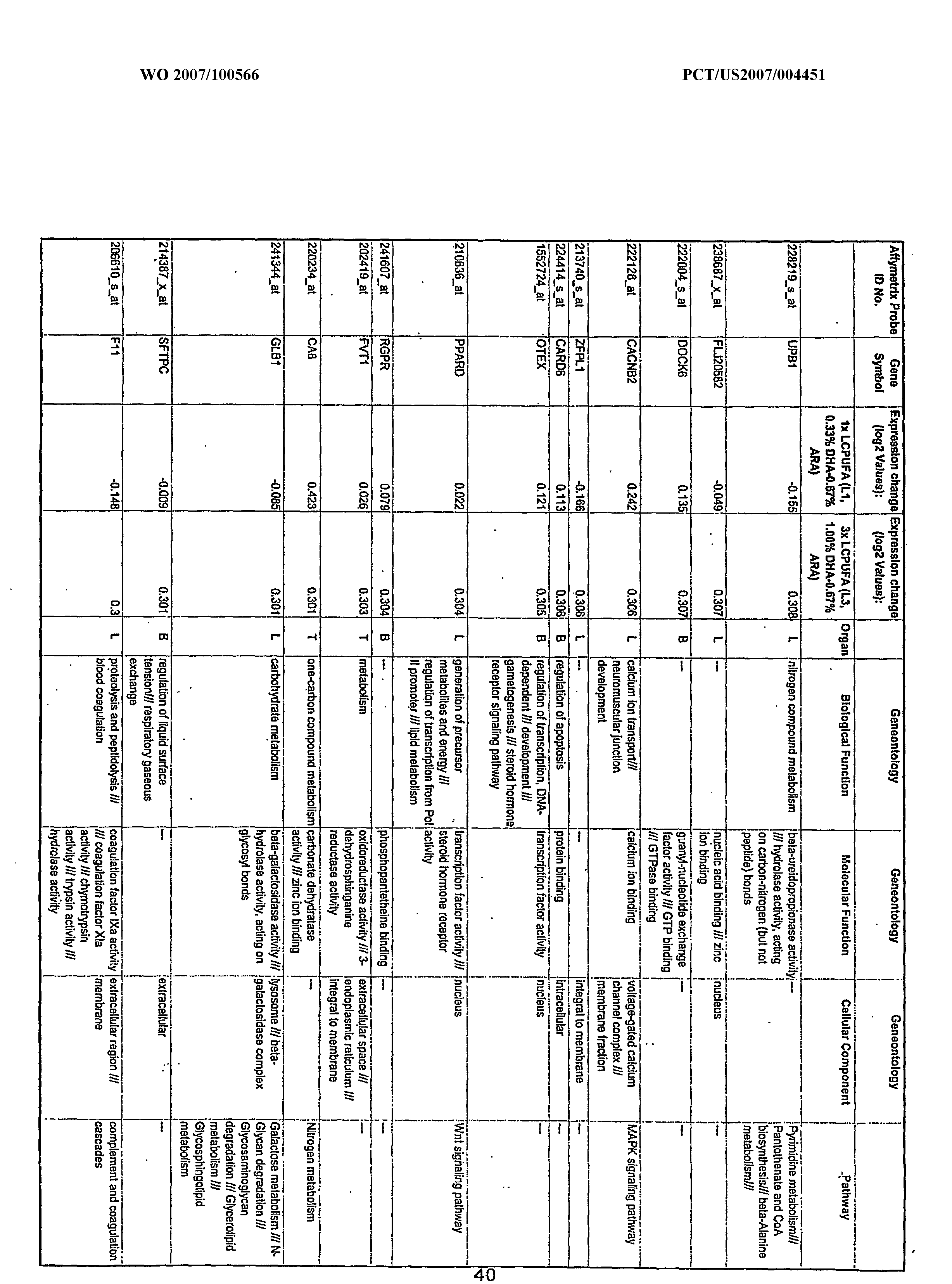


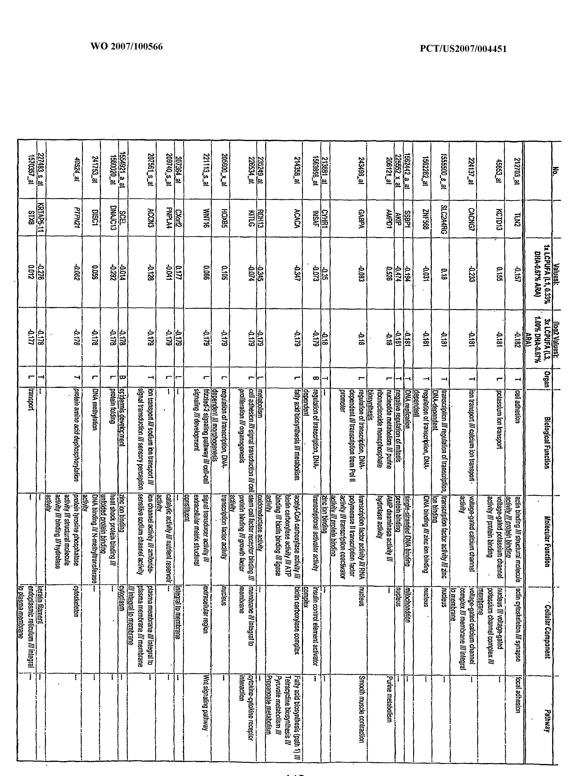


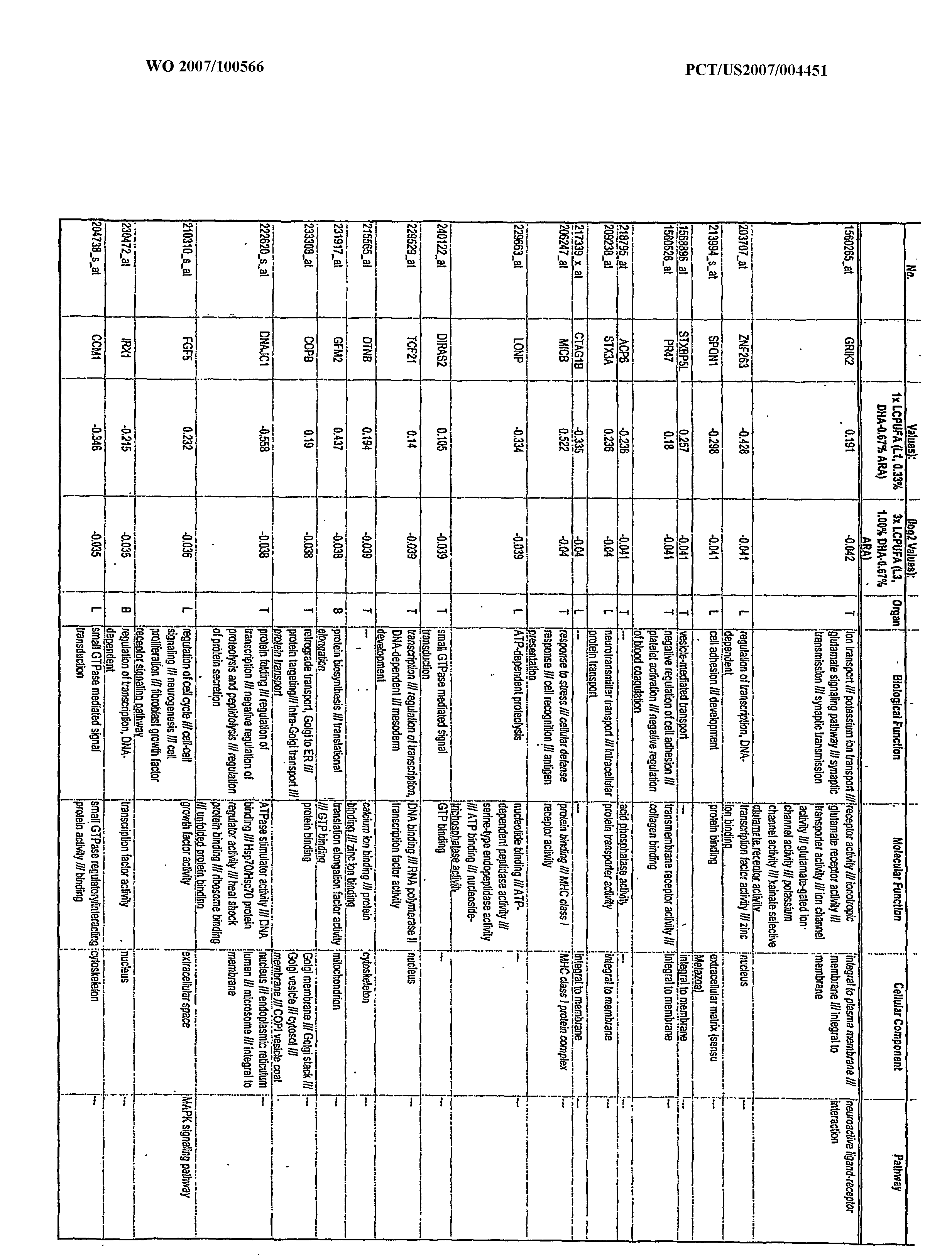


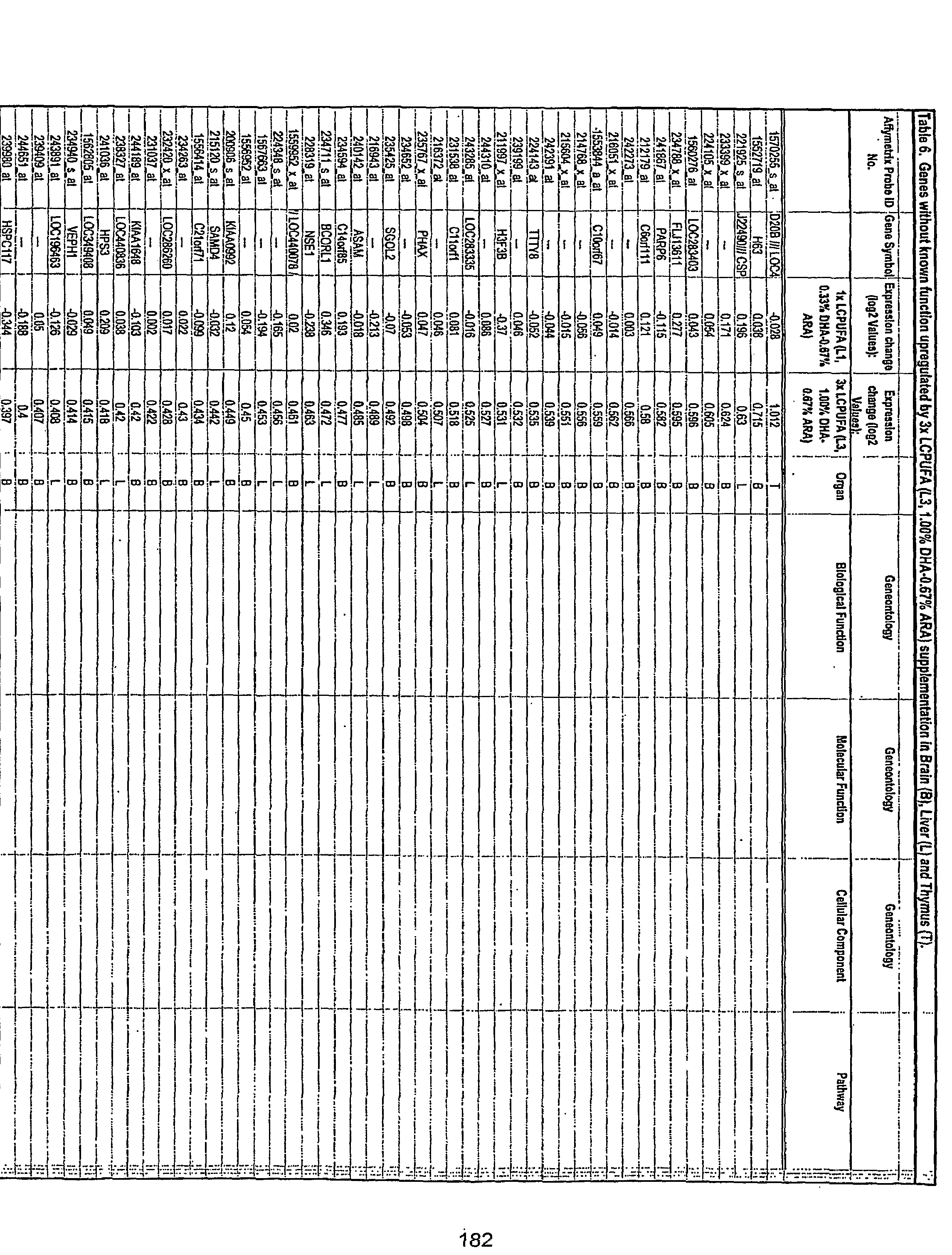


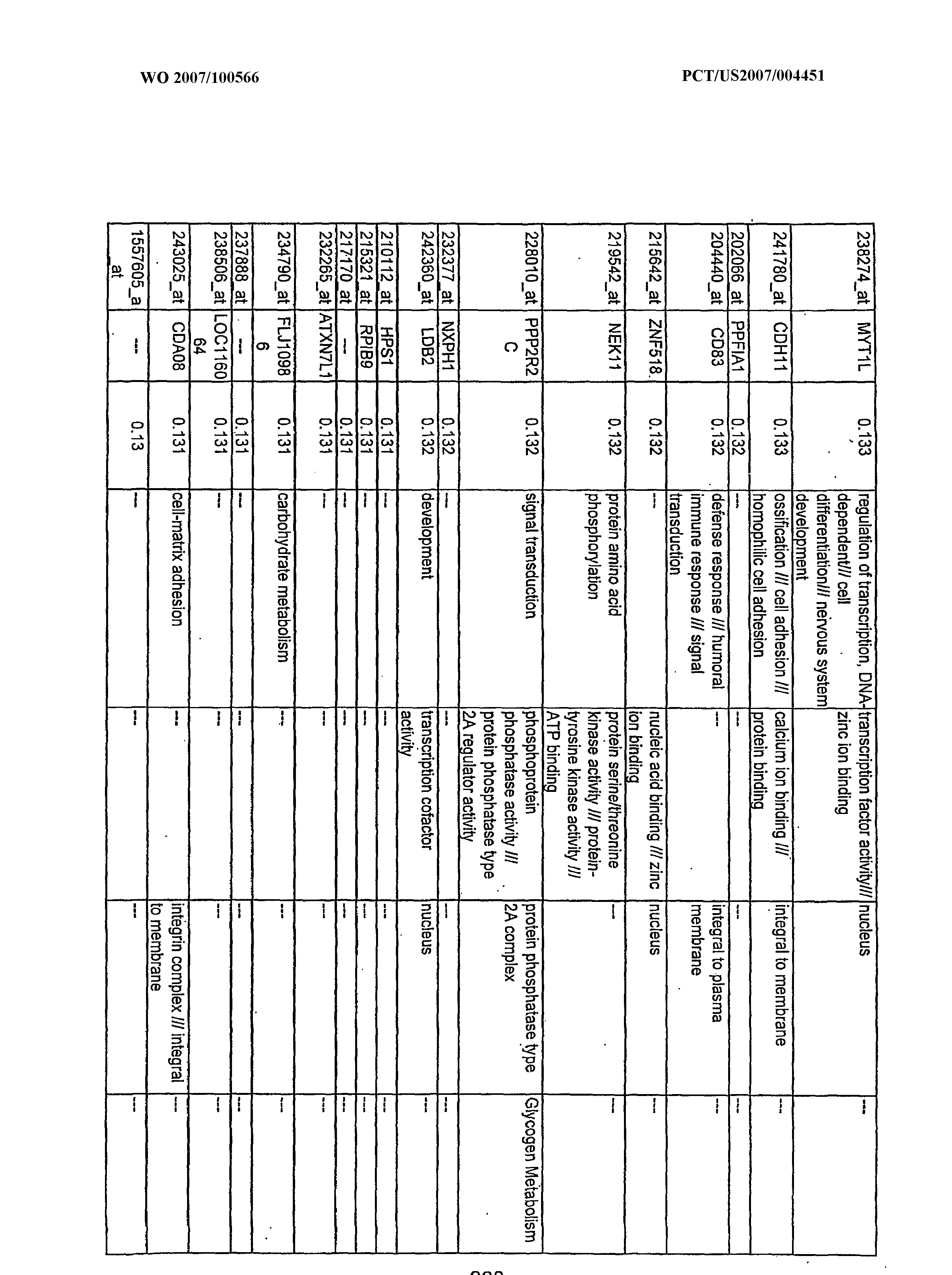

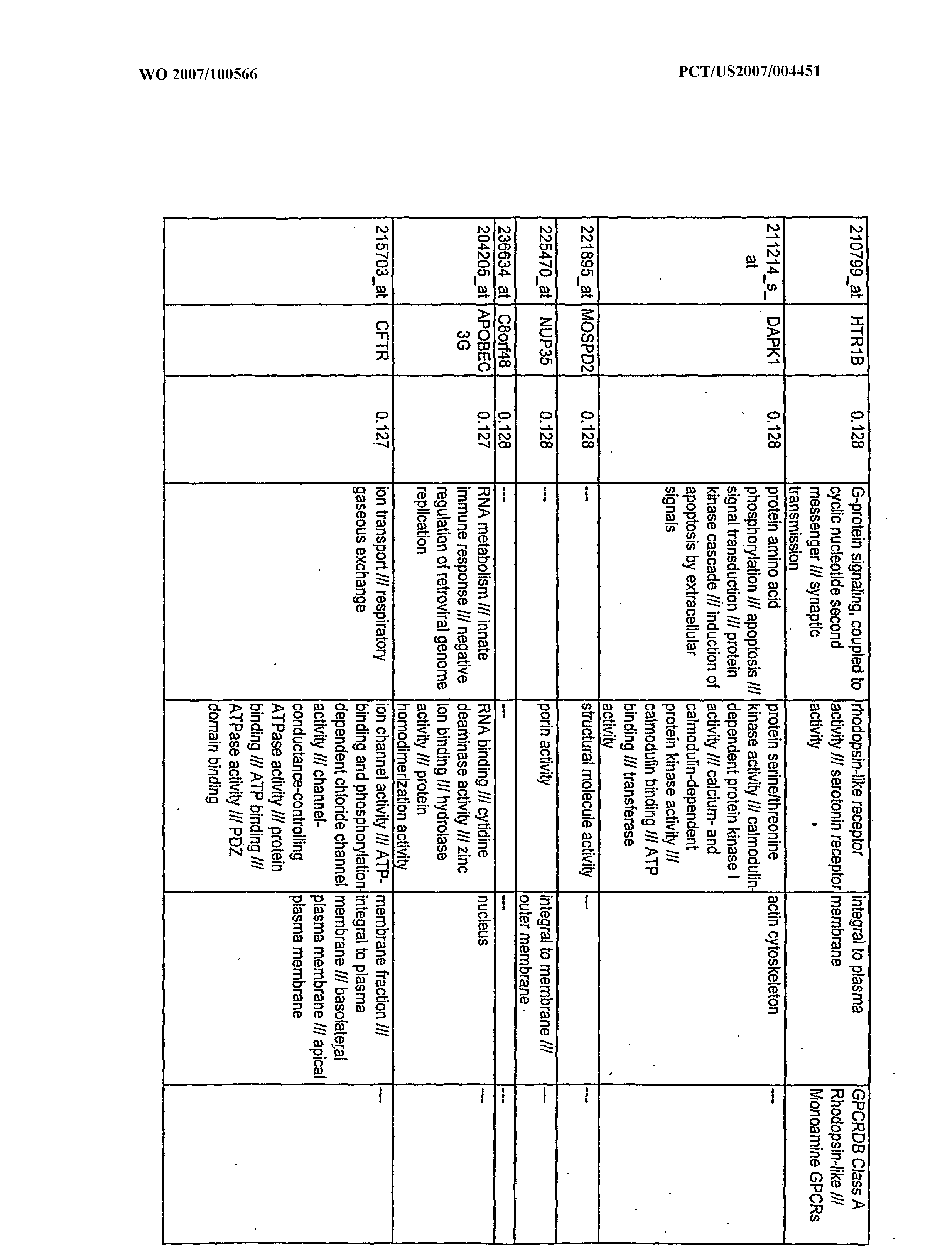


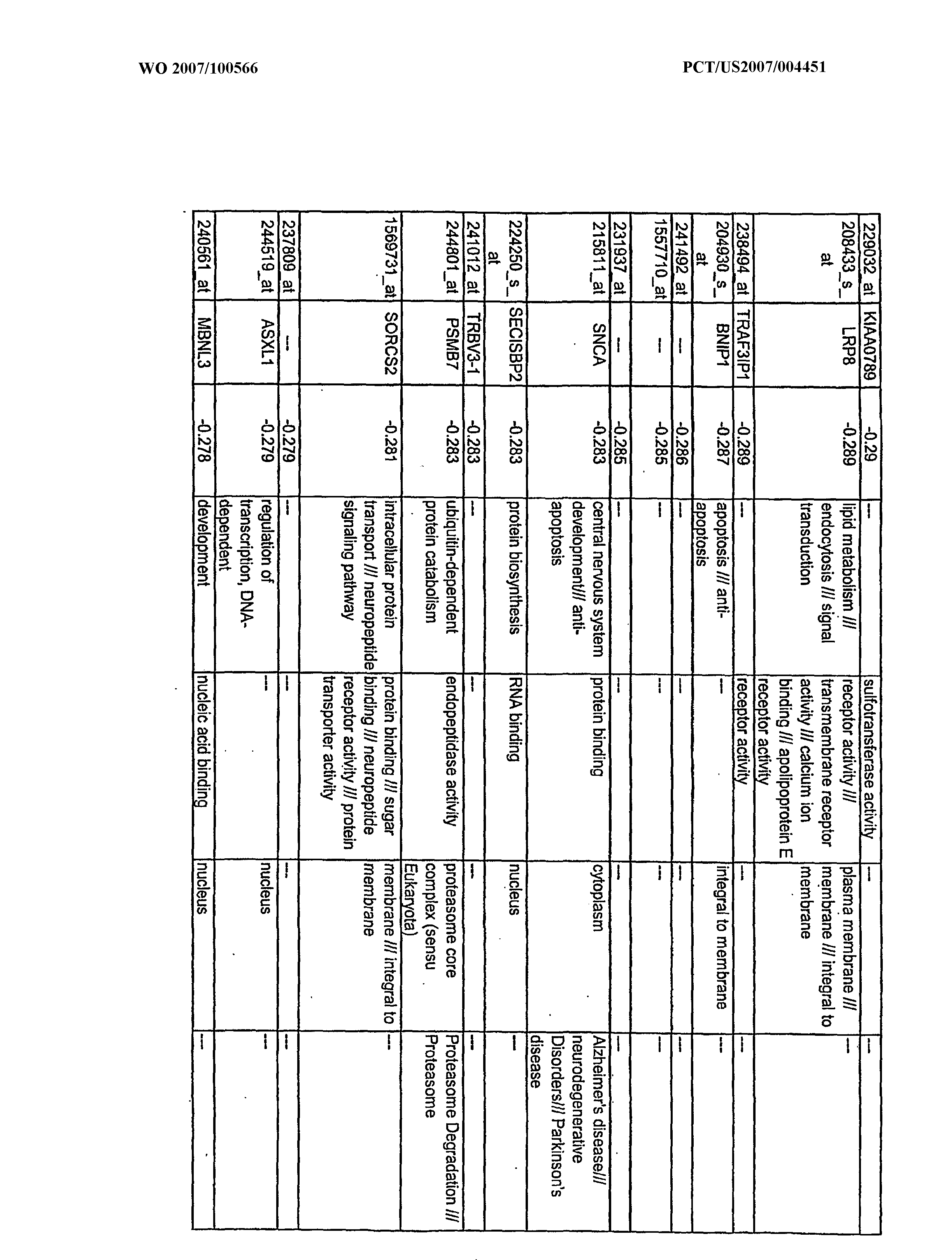


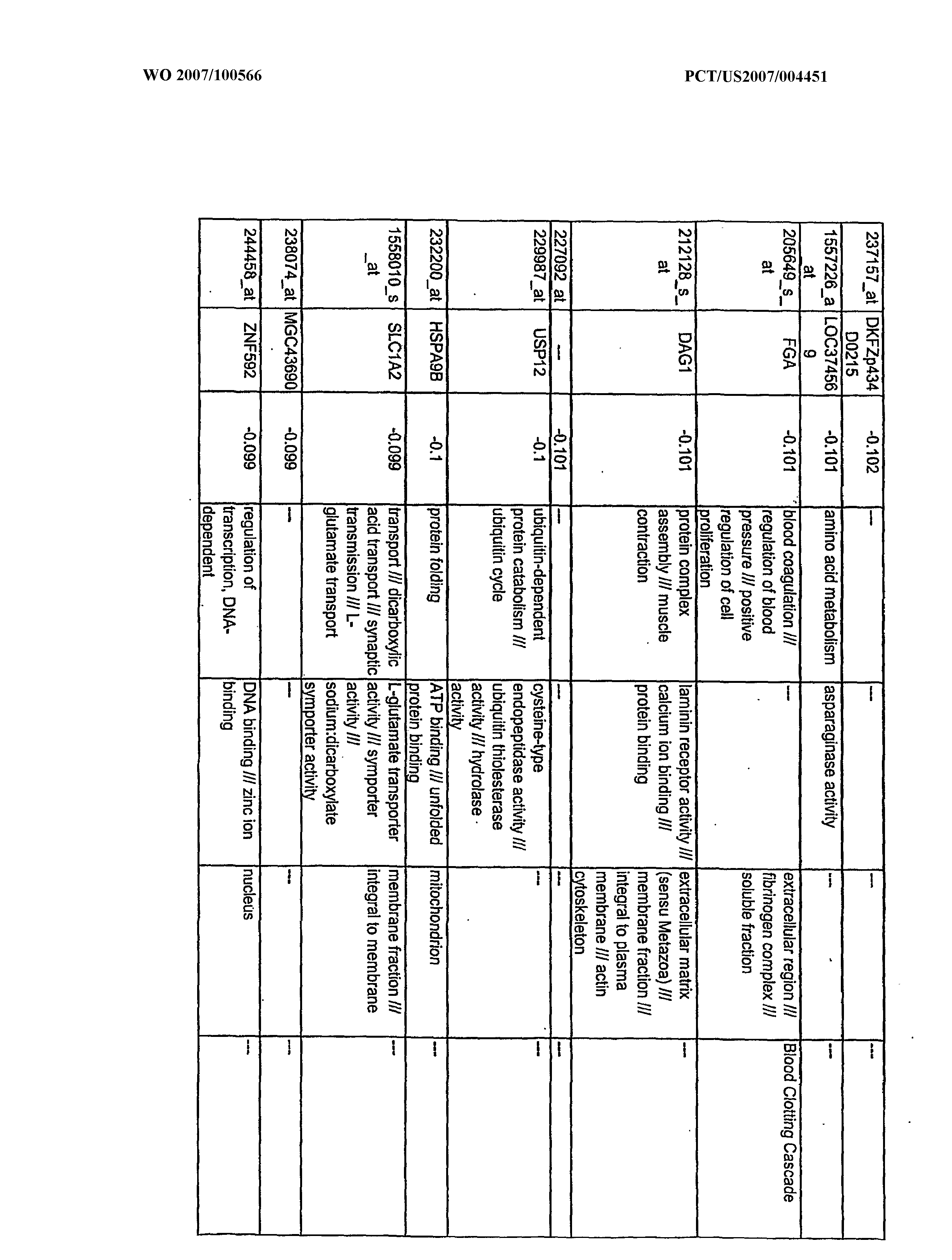


Claims
Priority Applications (5)
| Application Number | Priority Date | Filing Date | Title |
|---|---|---|---|
| EP07751225A EP1993530A2 (en) | 2006-02-28 | 2007-02-21 | Use of dha and ara in the preparation of a composition for regulating gene expression |
| MX2008010881A MX2008010881A (en) | 2006-02-28 | 2007-02-21 | Use of dha and ara in the preparation of a composition for regulating gene expression. |
| CA002645123A CA2645123A1 (en) | 2006-02-28 | 2007-02-21 | Use of dha and ara in the preparation of a composition for regulating gene expression |
| BRPI0708365-3A BRPI0708365A2 (en) | 2006-02-28 | 2007-02-21 | use of dha and ara in the preparation of a composition for regulating gene expression |
| NO20083439A NO20083439L (en) | 2006-02-28 | 2008-08-05 | Modulation of gene expression by docosahexaenoic acid |
Applications Claiming Priority (2)
| Application Number | Priority Date | Filing Date | Title |
|---|---|---|---|
| US77772406P | 2006-02-28 | 2006-02-28 | |
| US60/777,724 | 2006-02-28 |
Publications (2)
| Publication Number | Publication Date |
|---|---|
| WO2007100566A2 true WO2007100566A2 (en) | 2007-09-07 |
| WO2007100566A3 WO2007100566A3 (en) | 2008-10-09 |
Family
ID=38328537
Family Applications (1)
| Application Number | Title | Priority Date | Filing Date |
|---|---|---|---|
| PCT/US2007/004451 WO2007100566A2 (en) | 2006-02-28 | 2007-02-21 | Use of dha and ara in the preparation of a composition for regulating gene expression |
Country Status (11)
| Country | Link |
|---|---|
| US (1) | US20090099259A1 (en) |
| EP (1) | EP1993530A2 (en) |
| KR (1) | KR20080106415A (en) |
| CN (1) | CN101431993A (en) |
| BR (1) | BRPI0708365A2 (en) |
| CA (1) | CA2645123A1 (en) |
| MX (1) | MX2008010881A (en) |
| NO (1) | NO20083439L (en) |
| RU (1) | RU2008138380A (en) |
| TW (1) | TW200810747A (en) |
| WO (1) | WO2007100566A2 (en) |
Cited By (3)
| Publication number | Priority date | Publication date | Assignee | Title |
|---|---|---|---|---|
| EP2318023A4 (en) * | 2008-07-01 | 2012-03-07 | Mead Johnson Nutrition Co | Nutritional compositions containing punicalagins |
| US8343753B2 (en) | 2007-11-01 | 2013-01-01 | Wake Forest University School Of Medicine | Compositions, methods, and kits for polyunsaturated fatty acids from microalgae |
| US10251928B2 (en) | 2014-11-06 | 2019-04-09 | Mead Johnson Nutrition Company | Nutritional supplements containing a peptide component and uses thereof |
Families Citing this family (1)
| Publication number | Priority date | Publication date | Assignee | Title |
|---|---|---|---|---|
| WO2017053718A2 (en) * | 2015-09-23 | 2017-03-30 | Boston Medical Center Corporation | Biomarkers for the early detection of parkinson's disease |
Family Cites Families (4)
| Publication number | Priority date | Publication date | Assignee | Title |
|---|---|---|---|---|
| WO1998044917A1 (en) * | 1997-03-27 | 1998-10-15 | Bristol-Myers Squibb Company | Use of docosahexanoic acid and arachidonic acid enhancing the growth of preterm infants |
| CN1160119C (en) * | 2001-02-27 | 2004-08-04 | 中国科学院上海生物化学研究所 | Function and Application of Tob Gene in Mammalian Central Nervous System |
| US8633246B2 (en) * | 2003-08-11 | 2014-01-21 | Hill's Pet Nutrition, Inc. | Omega-3 fatty acids for osteoarthritis |
| IL158552A0 (en) * | 2003-10-22 | 2004-05-12 | Enzymotec Ltd | Lipids containing omega-3 fatty acids |
-
2007
- 2007-02-21 WO PCT/US2007/004451 patent/WO2007100566A2/en active Application Filing
- 2007-02-21 EP EP07751225A patent/EP1993530A2/en not_active Withdrawn
- 2007-02-21 MX MX2008010881A patent/MX2008010881A/en not_active Application Discontinuation
- 2007-02-21 RU RU2008138380/14A patent/RU2008138380A/en not_active Application Discontinuation
- 2007-02-21 KR KR1020087020978A patent/KR20080106415A/en not_active Withdrawn
- 2007-02-21 CA CA002645123A patent/CA2645123A1/en not_active Abandoned
- 2007-02-21 BR BRPI0708365-3A patent/BRPI0708365A2/en not_active IP Right Cessation
- 2007-02-21 CN CNA200780015037XA patent/CN101431993A/en active Pending
- 2007-02-27 TW TW096106842A patent/TW200810747A/en unknown
- 2007-02-28 US US11/712,102 patent/US20090099259A1/en not_active Abandoned
-
2008
- 2008-08-05 NO NO20083439A patent/NO20083439L/en not_active Application Discontinuation
Cited By (5)
| Publication number | Priority date | Publication date | Assignee | Title |
|---|---|---|---|---|
| US8343753B2 (en) | 2007-11-01 | 2013-01-01 | Wake Forest University School Of Medicine | Compositions, methods, and kits for polyunsaturated fatty acids from microalgae |
| EP2318023A4 (en) * | 2008-07-01 | 2012-03-07 | Mead Johnson Nutrition Co | Nutritional compositions containing punicalagins |
| US11077166B2 (en) | 2013-03-15 | 2021-08-03 | Mead Johnson Nutrition Company | Nutritional supplements containing a peptide component and uses thereof |
| US10251928B2 (en) | 2014-11-06 | 2019-04-09 | Mead Johnson Nutrition Company | Nutritional supplements containing a peptide component and uses thereof |
| US10933114B2 (en) | 2014-11-06 | 2021-03-02 | Mead Johnson Nutrition Company | Nutritional supplements containing a peptide component and uses thereof |
Also Published As
| Publication number | Publication date |
|---|---|
| CA2645123A1 (en) | 2007-09-07 |
| TW200810747A (en) | 2008-03-01 |
| MX2008010881A (en) | 2008-09-03 |
| NO20083439L (en) | 2008-11-28 |
| US20090099259A1 (en) | 2009-04-16 |
| WO2007100566A3 (en) | 2008-10-09 |
| EP1993530A2 (en) | 2008-11-26 |
| CN101431993A (en) | 2009-05-13 |
| KR20080106415A (en) | 2008-12-05 |
| RU2008138380A (en) | 2010-04-10 |
| BRPI0708365A2 (en) | 2011-04-26 |
Similar Documents
| Publication | Publication Date | Title |
|---|---|---|
| Khavinson et al. | Towards realization of longer life | |
| Fortress et al. | 17β-Estradiol regulates histone alterations associated with memory consolidation and increases Bdnf promoter acetylation in middle-aged female mice | |
| Jin et al. | Icariin, a phoshphodiesterase-5 inhibitor, improves learning and memory in APP/PS1 transgenic mice by stimulation of NO/cGMP signalling | |
| Morris | A forkhead in the road to longevity: the molecular basis of lifespan becomes clearer | |
| Carty et al. | SARM: From immune regulator to cell executioner | |
| Kothapalli et al. | Differential cerebral cortex transcriptomes of baboon neonates consuming moderate and high docosahexaenoic acid formulas | |
| Kong et al. | Vitamin D deficiency promotes nonalcoholic steatohepatitis through impaired enterohepatic circulation in animal model | |
| Yao et al. | Genetic imaging of neuroinflammation in Parkinson’s disease: Recent advancements | |
| Li et al. | Biliverdin administration ameliorates cerebral ischemia reperfusion injury in rats and is associated with proinflammatory factor downregulation | |
| JP2007106732A (en) | Methods and compositions for altering cell function | |
| Georgel et al. | Where epigenetics meets food intake: their interaction in the development/severity of gout and therapeutic perspectives | |
| Mørkholt et al. | CPT1A plays a key role in the development and treatment of multiple sclerosis and experimental autoimmune encephalomyelitis | |
| Liu et al. | Lidocaine improves cerebral ischemia-reperfusion injury in rats through cAMP/PKA signaling pathway | |
| Cao et al. | The role of the miR-99b-5p/mTOR signaling pathway in neuroregeneration in mice following spinal cord injury | |
| US20090099259A1 (en) | Method for regulating gene expression | |
| Wu et al. | High glucose promotes il-17a-induced gene expression through histone acetylation in retinal pigment epithelium cells | |
| Galali et al. | The impact of ketogenic diet on some metabolic and non‐metabolic diseases: Evidence from human and animal model experiments | |
| Ghosh et al. | The complex clinical and genetic landscape of hereditary peripheral neuropathy | |
| Si et al. | Neuroprotective effect of miR-212-5p on isoflurane-induced cognitive dysfunction by inhibiting neuroinflammation | |
| Yuan et al. | Leucine deprivation results in antidepressant effects via GCN2 in AgRP neurons | |
| Shakhtshneider et al. | Analysis of the Ldlr, Apob, Pcsk9 and Ldlrap1 genes variability in patients with familial hypercholesterolemia in West Siberia using targeted high throughput resequencing | |
| Ashiqueali et al. | Early life interventions metformin and trodusquemine metabolically reprogram the developing mouse liver through transcriptomic alterations | |
| C Brett et al. | Current therapeutic advances in patients and experimental models of Huntington's disease | |
| Lim et al. | CREB-regulated transcription during glycogen synthesis in astrocytes | |
| Zhang et al. | Role and mechanism of the zinc finger protein ZNF580 in foam-cell formation |
Legal Events
| Date | Code | Title | Description |
|---|---|---|---|
| 121 | Ep: the epo has been informed by wipo that ep was designated in this application | ||
| WWE | Wipo information: entry into national phase |
Ref document number: 12008501676 Country of ref document: PH |
|
| WWE | Wipo information: entry into national phase |
Ref document number: 6794/DELNP/2008 Country of ref document: IN |
|
| WWE | Wipo information: entry into national phase |
Ref document number: MX/a/2008/010881 Country of ref document: MX |
|
| WWE | Wipo information: entry into national phase |
Ref document number: 1020087020978 Country of ref document: KR |
|
| WWE | Wipo information: entry into national phase |
Ref document number: 2645123 Country of ref document: CA |
|
| NENP | Non-entry into the national phase |
Ref country code: DE |
|
| WWE | Wipo information: entry into national phase |
Ref document number: 2007751225 Country of ref document: EP |
|
| ENP | Entry into the national phase |
Ref document number: 2008138380 Country of ref document: RU Kind code of ref document: A |
|
| WWE | Wipo information: entry into national phase |
Ref document number: 200780015037.X Country of ref document: CN |
|
| ENP | Entry into the national phase |
Ref document number: PI0708365 Country of ref document: BR Kind code of ref document: A2 Effective date: 20080828 |