WO2006126864A1 - Animal models carrying tumors expressing human prostate cancer-specific antigen and method for analyzing prevention and treatment efficacy of dendritic cells-derived immunotherapeutics using the above - Google Patents
Animal models carrying tumors expressing human prostate cancer-specific antigen and method for analyzing prevention and treatment efficacy of dendritic cells-derived immunotherapeutics using the above Download PDFInfo
- Publication number
- WO2006126864A1 WO2006126864A1 PCT/KR2006/002028 KR2006002028W WO2006126864A1 WO 2006126864 A1 WO2006126864 A1 WO 2006126864A1 KR 2006002028 W KR2006002028 W KR 2006002028W WO 2006126864 A1 WO2006126864 A1 WO 2006126864A1
- Authority
- WO
- WIPO (PCT)
- Prior art keywords
- cancer
- prostate cancer
- cell line
- cancer cell
- mouse
- Prior art date
Links
- 208000000236 Prostatic Neoplasms Diseases 0.000 title claims abstract description 175
- 206010060862 Prostate cancer Diseases 0.000 title claims abstract description 174
- 239000000427 antigen Substances 0.000 title claims abstract description 129
- 108091007433 antigens Proteins 0.000 title claims abstract description 129
- 102000036639 antigens Human genes 0.000 title claims abstract description 129
- 206010028980 Neoplasm Diseases 0.000 title claims abstract description 115
- 210000004443 dendritic cell Anatomy 0.000 title claims abstract description 87
- 230000002265 prevention Effects 0.000 title claims abstract description 34
- 238000000034 method Methods 0.000 title claims abstract description 32
- 238000011282 treatment Methods 0.000 title claims abstract description 29
- 230000001024 immunotherapeutic effect Effects 0.000 title claims abstract description 25
- 238000010171 animal model Methods 0.000 title claims abstract description 18
- 210000004027 cell Anatomy 0.000 claims abstract description 220
- 201000011510 cancer Diseases 0.000 claims abstract description 101
- 241001465754 Metazoa Species 0.000 claims abstract description 53
- 230000015572 biosynthetic process Effects 0.000 claims abstract description 18
- 230000005907 cancer growth Effects 0.000 claims abstract description 13
- 231100000405 induce cancer Toxicity 0.000 claims abstract description 8
- 241000699666 Mus <mouse, genus> Species 0.000 claims description 46
- 102000007066 Prostate-Specific Antigen Human genes 0.000 claims description 46
- 108010072866 Prostate-Specific Antigen Proteins 0.000 claims description 46
- 102100041003 Glutamate carboxypeptidase 2 Human genes 0.000 claims description 30
- 101000892862 Homo sapiens Glutamate carboxypeptidase 2 Proteins 0.000 claims description 30
- 150000001413 amino acids Chemical class 0.000 claims description 30
- 239000002773 nucleotide Substances 0.000 claims description 29
- 125000003729 nucleotide group Chemical group 0.000 claims description 29
- 101710120463 Prostate stem cell antigen Proteins 0.000 claims description 26
- 102100036735 Prostate stem cell antigen Human genes 0.000 claims description 26
- 239000013598 vector Substances 0.000 claims description 26
- 208000008839 Kidney Neoplasms Diseases 0.000 claims description 22
- 206010038389 Renal cancer Diseases 0.000 claims description 22
- 201000010982 kidney cancer Diseases 0.000 claims description 22
- 125000003275 alpha amino acid group Chemical group 0.000 claims description 21
- 210000002307 prostate Anatomy 0.000 claims description 13
- 206010027476 Metastases Diseases 0.000 claims description 10
- 230000009401 metastasis Effects 0.000 claims description 10
- 238000010172 mouse model Methods 0.000 claims description 10
- 238000010254 subcutaneous injection Methods 0.000 claims description 6
- 239000007929 subcutaneous injection Substances 0.000 claims description 6
- GZCWLCBFPRFLKL-UHFFFAOYSA-N 1-prop-2-ynoxypropan-2-ol Chemical compound CC(O)COCC#C GZCWLCBFPRFLKL-UHFFFAOYSA-N 0.000 claims description 5
- 108010051457 Acid Phosphatase Proteins 0.000 claims description 5
- 102000013563 Acid Phosphatase Human genes 0.000 claims description 5
- 241000699660 Mus musculus Species 0.000 claims description 4
- 241000283984 Rodentia Species 0.000 claims description 3
- 241000699670 Mus sp. Species 0.000 description 42
- 229940029030 dendritic cell vaccine Drugs 0.000 description 20
- 230000014509 gene expression Effects 0.000 description 16
- 108091028043 Nucleic acid sequence Proteins 0.000 description 15
- 238000004458 analytical method Methods 0.000 description 11
- 108020004414 DNA Proteins 0.000 description 10
- 239000002299 complementary DNA Substances 0.000 description 10
- 230000000694 effects Effects 0.000 description 10
- 108090000623 proteins and genes Proteins 0.000 description 10
- 239000013604 expression vector Substances 0.000 description 9
- 239000012634 fragment Substances 0.000 description 9
- 102000004169 proteins and genes Human genes 0.000 description 8
- 238000001262 western blot Methods 0.000 description 8
- 210000000612 antigen-presenting cell Anatomy 0.000 description 6
- 210000002798 bone marrow cell Anatomy 0.000 description 6
- 231100000504 carcinogenesis Toxicity 0.000 description 6
- 238000011156 evaluation Methods 0.000 description 6
- 229920001481 poly(stearyl methacrylate) Polymers 0.000 description 6
- 230000002685 pulmonary effect Effects 0.000 description 6
- 208000005623 Carcinogenesis Diseases 0.000 description 5
- 108090000695 Cytokines Proteins 0.000 description 5
- 102000004127 Cytokines Human genes 0.000 description 5
- 206010062904 Hormone-refractory prostate cancer Diseases 0.000 description 5
- 230000001093 anti-cancer Effects 0.000 description 5
- 230000036952 cancer formation Effects 0.000 description 5
- 238000000746 purification Methods 0.000 description 5
- 239000007787 solid Substances 0.000 description 5
- 210000004988 splenocyte Anatomy 0.000 description 5
- NLXLAEXVIDQMFP-UHFFFAOYSA-N Ammonia chloride Chemical compound [NH4+].[Cl-] NLXLAEXVIDQMFP-UHFFFAOYSA-N 0.000 description 4
- WSFSSNUMVMOOMR-UHFFFAOYSA-N Formaldehyde Chemical compound O=C WSFSSNUMVMOOMR-UHFFFAOYSA-N 0.000 description 4
- 238000012408 PCR amplification Methods 0.000 description 4
- 101150056441 Pctp gene Proteins 0.000 description 4
- 102000007056 Recombinant Fusion Proteins Human genes 0.000 description 4
- 108010008281 Recombinant Fusion Proteins Proteins 0.000 description 4
- 210000001744 T-lymphocyte Anatomy 0.000 description 4
- 210000004899 c-terminal region Anatomy 0.000 description 4
- 238000006243 chemical reaction Methods 0.000 description 4
- 210000001151 cytotoxic T lymphocyte Anatomy 0.000 description 4
- 239000012636 effector Substances 0.000 description 4
- 238000002474 experimental method Methods 0.000 description 4
- 238000001794 hormone therapy Methods 0.000 description 4
- 230000008105 immune reaction Effects 0.000 description 4
- 230000028993 immune response Effects 0.000 description 4
- 238000009169 immunotherapy Methods 0.000 description 4
- 239000000203 mixture Substances 0.000 description 4
- 230000036961 partial effect Effects 0.000 description 4
- 238000002415 sodium dodecyl sulfate polyacrylamide gel electrophoresis Methods 0.000 description 4
- 238000002560 therapeutic procedure Methods 0.000 description 4
- 210000001519 tissue Anatomy 0.000 description 4
- 230000026683 transduction Effects 0.000 description 4
- 238000010361 transduction Methods 0.000 description 4
- 210000000689 upper leg Anatomy 0.000 description 4
- 102000002260 Alkaline Phosphatase Human genes 0.000 description 3
- 108020004774 Alkaline Phosphatase Proteins 0.000 description 3
- 241000588724 Escherichia coli Species 0.000 description 3
- OKKJLVBELUTLKV-UHFFFAOYSA-N Methanol Chemical compound OC OKKJLVBELUTLKV-UHFFFAOYSA-N 0.000 description 3
- -1 PAP Proteins 0.000 description 3
- 108010076504 Protein Sorting Signals Proteins 0.000 description 3
- 239000003242 anti bacterial agent Substances 0.000 description 3
- 229940088710 antibiotic agent Drugs 0.000 description 3
- 238000010367 cloning Methods 0.000 description 3
- 238000010276 construction Methods 0.000 description 3
- 230000009089 cytolysis Effects 0.000 description 3
- 210000003162 effector t lymphocyte Anatomy 0.000 description 3
- 210000003527 eukaryotic cell Anatomy 0.000 description 3
- 239000003163 gonadal steroid hormone Substances 0.000 description 3
- 238000002649 immunization Methods 0.000 description 3
- 230000003053 immunization Effects 0.000 description 3
- 230000005764 inhibitory process Effects 0.000 description 3
- 238000011081 inoculation Methods 0.000 description 3
- 239000002609 medium Substances 0.000 description 3
- 238000002360 preparation method Methods 0.000 description 3
- 230000009465 prokaryotic expression Effects 0.000 description 3
- 230000035755 proliferation Effects 0.000 description 3
- 239000000243 solution Substances 0.000 description 3
- 210000000952 spleen Anatomy 0.000 description 3
- 210000002303 tibia Anatomy 0.000 description 3
- 210000004881 tumor cell Anatomy 0.000 description 3
- 229960005486 vaccine Drugs 0.000 description 3
- IHCCLXNEEPMSIO-UHFFFAOYSA-N 2-[4-[2-(2,3-dihydro-1H-inden-2-ylamino)pyrimidin-5-yl]piperidin-1-yl]-1-(2,4,6,7-tetrahydrotriazolo[4,5-c]pyridin-5-yl)ethanone Chemical compound C1C(CC2=CC=CC=C12)NC1=NC=C(C=N1)C1CCN(CC1)CC(=O)N1CC2=C(CC1)NN=N2 IHCCLXNEEPMSIO-UHFFFAOYSA-N 0.000 description 2
- 238000011725 BALB/c mouse Methods 0.000 description 2
- LFQSCWFLJHTTHZ-UHFFFAOYSA-N Ethanol Chemical compound CCO LFQSCWFLJHTTHZ-UHFFFAOYSA-N 0.000 description 2
- 102000004457 Granulocyte-Macrophage Colony-Stimulating Factor Human genes 0.000 description 2
- 108010017213 Granulocyte-Macrophage Colony-Stimulating Factor Proteins 0.000 description 2
- 102100034343 Integrase Human genes 0.000 description 2
- 108090000978 Interleukin-4 Proteins 0.000 description 2
- KFZMGEQAYNKOFK-UHFFFAOYSA-N Isopropanol Chemical compound CC(C)O KFZMGEQAYNKOFK-UHFFFAOYSA-N 0.000 description 2
- 206010058467 Lung neoplasm malignant Diseases 0.000 description 2
- 108700018351 Major Histocompatibility Complex Proteins 0.000 description 2
- NWIBSHFKIJFRCO-WUDYKRTCSA-N Mytomycin Chemical compound C1N2C(C(C(C)=C(N)C3=O)=O)=C3[C@@H](COC(N)=O)[C@@]2(OC)[C@@H]2[C@H]1N2 NWIBSHFKIJFRCO-WUDYKRTCSA-N 0.000 description 2
- 108010092799 RNA-directed DNA polymerase Proteins 0.000 description 2
- 239000012980 RPMI-1640 medium Substances 0.000 description 2
- 108020005091 Replication Origin Proteins 0.000 description 2
- 230000001464 adherent effect Effects 0.000 description 2
- 235000019270 ammonium chloride Nutrition 0.000 description 2
- 238000002512 chemotherapy Methods 0.000 description 2
- 210000003743 erythrocyte Anatomy 0.000 description 2
- 230000002068 genetic effect Effects 0.000 description 2
- 238000003306 harvesting Methods 0.000 description 2
- 230000006698 induction Effects 0.000 description 2
- 238000002347 injection Methods 0.000 description 2
- 239000007924 injection Substances 0.000 description 2
- 238000010253 intravenous injection Methods 0.000 description 2
- 238000002955 isolation Methods 0.000 description 2
- 201000005202 lung cancer Diseases 0.000 description 2
- 208000020816 lung neoplasm Diseases 0.000 description 2
- 239000003550 marker Substances 0.000 description 2
- 210000004897 n-terminal region Anatomy 0.000 description 2
- 239000013642 negative control Substances 0.000 description 2
- 235000001968 nicotinic acid Nutrition 0.000 description 2
- 230000008569 process Effects 0.000 description 2
- 108090000765 processed proteins & peptides Proteins 0.000 description 2
- 239000000047 product Substances 0.000 description 2
- 210000001236 prokaryotic cell Anatomy 0.000 description 2
- 238000001959 radiotherapy Methods 0.000 description 2
- 238000011160 research Methods 0.000 description 2
- 230000004044 response Effects 0.000 description 2
- 238000012163 sequencing technique Methods 0.000 description 2
- 239000006228 supernatant Substances 0.000 description 2
- 230000020382 suppression by virus of host antigen processing and presentation of peptide antigen via MHC class I Effects 0.000 description 2
- 210000003462 vein Anatomy 0.000 description 2
- MZOFCQQQCNRIBI-VMXHOPILSA-N (3s)-4-[[(2s)-1-[[(2s)-1-[[(1s)-1-carboxy-2-hydroxyethyl]amino]-4-methyl-1-oxopentan-2-yl]amino]-5-(diaminomethylideneamino)-1-oxopentan-2-yl]amino]-3-[[2-[[(2s)-2,6-diaminohexanoyl]amino]acetyl]amino]-4-oxobutanoic acid Chemical compound OC[C@@H](C(O)=O)NC(=O)[C@H](CC(C)C)NC(=O)[C@H](CCCN=C(N)N)NC(=O)[C@H](CC(O)=O)NC(=O)CNC(=O)[C@@H](N)CCCCN MZOFCQQQCNRIBI-VMXHOPILSA-N 0.000 description 1
- OHVLMTFVQDZYHP-UHFFFAOYSA-N 1-(2,4,6,7-tetrahydrotriazolo[4,5-c]pyridin-5-yl)-2-[4-[2-[[3-(trifluoromethoxy)phenyl]methylamino]pyrimidin-5-yl]piperazin-1-yl]ethanone Chemical compound N1N=NC=2CN(CCC=21)C(CN1CCN(CC1)C=1C=NC(=NC=1)NCC1=CC(=CC=C1)OC(F)(F)F)=O OHVLMTFVQDZYHP-UHFFFAOYSA-N 0.000 description 1
- HMUNWXXNJPVALC-UHFFFAOYSA-N 1-[4-[2-(2,3-dihydro-1H-inden-2-ylamino)pyrimidin-5-yl]piperazin-1-yl]-2-(2,4,6,7-tetrahydrotriazolo[4,5-c]pyridin-5-yl)ethanone Chemical compound C1C(CC2=CC=CC=C12)NC1=NC=C(C=N1)N1CCN(CC1)C(CN1CC2=C(CC1)NN=N2)=O HMUNWXXNJPVALC-UHFFFAOYSA-N 0.000 description 1
- VZSRBBMJRBPUNF-UHFFFAOYSA-N 2-(2,3-dihydro-1H-inden-2-ylamino)-N-[3-oxo-3-(2,4,6,7-tetrahydrotriazolo[4,5-c]pyridin-5-yl)propyl]pyrimidine-5-carboxamide Chemical compound C1C(CC2=CC=CC=C12)NC1=NC=C(C=N1)C(=O)NCCC(N1CC2=C(CC1)NN=N2)=O VZSRBBMJRBPUNF-UHFFFAOYSA-N 0.000 description 1
- WZFUQSJFWNHZHM-UHFFFAOYSA-N 2-[4-[2-(2,3-dihydro-1H-inden-2-ylamino)pyrimidin-5-yl]piperazin-1-yl]-1-(2,4,6,7-tetrahydrotriazolo[4,5-c]pyridin-5-yl)ethanone Chemical compound C1C(CC2=CC=CC=C12)NC1=NC=C(C=N1)N1CCN(CC1)CC(=O)N1CC2=C(CC1)NN=N2 WZFUQSJFWNHZHM-UHFFFAOYSA-N 0.000 description 1
- SNBCLPGEMZEWLU-QXFUBDJGSA-N 2-chloro-n-[[(2r,3s,5r)-3-hydroxy-5-(5-methyl-2,4-dioxopyrimidin-1-yl)oxolan-2-yl]methyl]acetamide Chemical compound O=C1NC(=O)C(C)=CN1[C@@H]1O[C@H](CNC(=O)CCl)[C@@H](O)C1 SNBCLPGEMZEWLU-QXFUBDJGSA-N 0.000 description 1
- YLZOPXRUQYQQID-UHFFFAOYSA-N 3-(2,4,6,7-tetrahydrotriazolo[4,5-c]pyridin-5-yl)-1-[4-[2-[[3-(trifluoromethoxy)phenyl]methylamino]pyrimidin-5-yl]piperazin-1-yl]propan-1-one Chemical compound N1N=NC=2CN(CCC=21)CCC(=O)N1CCN(CC1)C=1C=NC(=NC=1)NCC1=CC(=CC=C1)OC(F)(F)F YLZOPXRUQYQQID-UHFFFAOYSA-N 0.000 description 1
- FWMNVWWHGCHHJJ-SKKKGAJSSA-N 4-amino-1-[(2r)-6-amino-2-[[(2r)-2-[[(2r)-2-[[(2r)-2-amino-3-phenylpropanoyl]amino]-3-phenylpropanoyl]amino]-4-methylpentanoyl]amino]hexanoyl]piperidine-4-carboxylic acid Chemical compound C([C@H](C(=O)N[C@H](CC(C)C)C(=O)N[C@H](CCCCN)C(=O)N1CCC(N)(CC1)C(O)=O)NC(=O)[C@H](N)CC=1C=CC=CC=1)C1=CC=CC=C1 FWMNVWWHGCHHJJ-SKKKGAJSSA-N 0.000 description 1
- QRXMUCSWCMTJGU-UHFFFAOYSA-N 5-bromo-4-chloro-3-indolyl phosphate Chemical compound C1=C(Br)C(Cl)=C2C(OP(O)(=O)O)=CNC2=C1 QRXMUCSWCMTJGU-UHFFFAOYSA-N 0.000 description 1
- 108091093088 Amplicon Proteins 0.000 description 1
- 206010005003 Bladder cancer Diseases 0.000 description 1
- 208000003174 Brain Neoplasms Diseases 0.000 description 1
- 206010006187 Breast cancer Diseases 0.000 description 1
- 208000026310 Breast neoplasm Diseases 0.000 description 1
- 238000011740 C57BL/6 mouse Methods 0.000 description 1
- 206010008342 Cervix carcinoma Diseases 0.000 description 1
- 206010009944 Colon cancer Diseases 0.000 description 1
- 108010062580 Concanavalin A Proteins 0.000 description 1
- 241000701022 Cytomegalovirus Species 0.000 description 1
- 102000012410 DNA Ligases Human genes 0.000 description 1
- 108010061982 DNA Ligases Proteins 0.000 description 1
- 238000001712 DNA sequencing Methods 0.000 description 1
- 102000004190 Enzymes Human genes 0.000 description 1
- 108090000790 Enzymes Proteins 0.000 description 1
- HVLSXIKZNLPZJJ-TXZCQADKSA-N HA peptide Chemical compound C([C@@H](C(=O)N[C@@H](CC(O)=O)C(=O)N[C@@H](C(C)C)C(=O)N1[C@@H](CCC1)C(=O)N[C@@H](CC(O)=O)C(=O)N[C@@H](CC=1C=CC(O)=CC=1)C(=O)N[C@@H](C)C(O)=O)NC(=O)[C@H]1N(CCC1)C(=O)[C@@H](N)CC=1C=CC(O)=CC=1)C1=CC=C(O)C=C1 HVLSXIKZNLPZJJ-TXZCQADKSA-N 0.000 description 1
- 101710154606 Hemagglutinin Proteins 0.000 description 1
- 102100037850 Interferon gamma Human genes 0.000 description 1
- 108010074328 Interferon-gamma Proteins 0.000 description 1
- 206010023825 Laryngeal cancer Diseases 0.000 description 1
- 102000043129 MHC class I family Human genes 0.000 description 1
- 108091054437 MHC class I family Proteins 0.000 description 1
- 241000124008 Mammalia Species 0.000 description 1
- 241000714177 Murine leukemia virus Species 0.000 description 1
- AFCARXCZXQIEQB-UHFFFAOYSA-N N-[3-oxo-3-(2,4,6,7-tetrahydrotriazolo[4,5-c]pyridin-5-yl)propyl]-2-[[3-(trifluoromethoxy)phenyl]methylamino]pyrimidine-5-carboxamide Chemical compound O=C(CCNC(=O)C=1C=NC(=NC=1)NCC1=CC(=CC=C1)OC(F)(F)F)N1CC2=C(CC1)NN=N2 AFCARXCZXQIEQB-UHFFFAOYSA-N 0.000 description 1
- 208000001894 Nasopharyngeal Neoplasms Diseases 0.000 description 1
- 206010061306 Nasopharyngeal cancer Diseases 0.000 description 1
- 239000000020 Nitrocellulose Substances 0.000 description 1
- 239000004677 Nylon Substances 0.000 description 1
- 101710093908 Outer capsid protein VP4 Proteins 0.000 description 1
- 101710135467 Outer capsid protein sigma-1 Proteins 0.000 description 1
- 206010033128 Ovarian cancer Diseases 0.000 description 1
- 206010061535 Ovarian neoplasm Diseases 0.000 description 1
- 206010061902 Pancreatic neoplasm Diseases 0.000 description 1
- 102100035703 Prostatic acid phosphatase Human genes 0.000 description 1
- 101710176177 Protein A56 Proteins 0.000 description 1
- 239000012979 RPMI medium Substances 0.000 description 1
- 239000006146 Roswell Park Memorial Institute medium Substances 0.000 description 1
- 208000005718 Stomach Neoplasms Diseases 0.000 description 1
- 230000006052 T cell proliferation Effects 0.000 description 1
- 108060008682 Tumor Necrosis Factor Proteins 0.000 description 1
- 102000000852 Tumor Necrosis Factor-alpha Human genes 0.000 description 1
- 208000007097 Urinary Bladder Neoplasms Diseases 0.000 description 1
- 208000006105 Uterine Cervical Neoplasms Diseases 0.000 description 1
- 230000009471 action Effects 0.000 description 1
- 230000000735 allogeneic effect Effects 0.000 description 1
- 239000003098 androgen Substances 0.000 description 1
- 210000004102 animal cell Anatomy 0.000 description 1
- 230000003466 anti-cipated effect Effects 0.000 description 1
- 239000002246 antineoplastic agent Substances 0.000 description 1
- 229940041181 antineoplastic drug Drugs 0.000 description 1
- 230000001363 autoimmune Effects 0.000 description 1
- 230000003115 biocidal effect Effects 0.000 description 1
- 210000000988 bone and bone Anatomy 0.000 description 1
- 210000001185 bone marrow Anatomy 0.000 description 1
- 108010006025 bovine growth hormone Proteins 0.000 description 1
- 238000002619 cancer immunotherapy Methods 0.000 description 1
- 201000010881 cervical cancer Diseases 0.000 description 1
- 208000029742 colonic neoplasm Diseases 0.000 description 1
- 230000021615 conjugation Effects 0.000 description 1
- 239000013078 crystal Substances 0.000 description 1
- 210000004748 cultured cell Anatomy 0.000 description 1
- 238000012258 culturing Methods 0.000 description 1
- 210000000805 cytoplasm Anatomy 0.000 description 1
- 230000001086 cytosolic effect Effects 0.000 description 1
- 230000007423 decrease Effects 0.000 description 1
- 230000007812 deficiency Effects 0.000 description 1
- 238000003745 diagnosis Methods 0.000 description 1
- 230000004069 differentiation Effects 0.000 description 1
- 201000010099 disease Diseases 0.000 description 1
- 208000037265 diseases, disorders, signs and symptoms Diseases 0.000 description 1
- 239000003814 drug Substances 0.000 description 1
- 238000001035 drying Methods 0.000 description 1
- 238000005516 engineering process Methods 0.000 description 1
- 238000000605 extraction Methods 0.000 description 1
- 230000002349 favourable effect Effects 0.000 description 1
- 239000012737 fresh medium Substances 0.000 description 1
- 206010017758 gastric cancer Diseases 0.000 description 1
- 238000001415 gene therapy Methods 0.000 description 1
- 210000004907 gland Anatomy 0.000 description 1
- 239000010931 gold Substances 0.000 description 1
- 229910052737 gold Inorganic materials 0.000 description 1
- 239000000185 hemagglutinin Substances 0.000 description 1
- 210000003958 hematopoietic stem cell Anatomy 0.000 description 1
- 125000001165 hydrophobic group Chemical group 0.000 description 1
- 230000036039 immunity Effects 0.000 description 1
- 238000011534 incubation Methods 0.000 description 1
- 230000002401 inhibitory effect Effects 0.000 description 1
- 206010023841 laryngeal neoplasm Diseases 0.000 description 1
- 230000003902 lesion Effects 0.000 description 1
- 230000000670 limiting effect Effects 0.000 description 1
- 201000007270 liver cancer Diseases 0.000 description 1
- 208000014018 liver neoplasm Diseases 0.000 description 1
- 210000004072 lung Anatomy 0.000 description 1
- 210000001165 lymph node Anatomy 0.000 description 1
- 208000015486 malignant pancreatic neoplasm Diseases 0.000 description 1
- 238000004519 manufacturing process Methods 0.000 description 1
- 230000035800 maturation Effects 0.000 description 1
- 238000005259 measurement Methods 0.000 description 1
- 239000012528 membrane Substances 0.000 description 1
- 229960004857 mitomycin Drugs 0.000 description 1
- 238000002156 mixing Methods 0.000 description 1
- 230000004048 modification Effects 0.000 description 1
- 238000012986 modification Methods 0.000 description 1
- 210000001616 monocyte Anatomy 0.000 description 1
- 229920001220 nitrocellulos Polymers 0.000 description 1
- 229920001778 nylon Polymers 0.000 description 1
- 210000000056 organ Anatomy 0.000 description 1
- 201000002528 pancreatic cancer Diseases 0.000 description 1
- 208000008443 pancreatic carcinoma Diseases 0.000 description 1
- 239000013612 plasmid Substances 0.000 description 1
- 238000002264 polyacrylamide gel electrophoresis Methods 0.000 description 1
- 230000008488 polyadenylation Effects 0.000 description 1
- 238000001556 precipitation Methods 0.000 description 1
- 102000004196 processed proteins & peptides Human genes 0.000 description 1
- 239000013587 production medium Substances 0.000 description 1
- 108010043671 prostatic acid phosphatase Proteins 0.000 description 1
- 201000007094 prostatitis Diseases 0.000 description 1
- 238000011472 radical prostatectomy Methods 0.000 description 1
- 239000000376 reactant Substances 0.000 description 1
- 210000000664 rectum Anatomy 0.000 description 1
- 230000009467 reduction Effects 0.000 description 1
- 239000011347 resin Substances 0.000 description 1
- 229920005989 resin Polymers 0.000 description 1
- 238000010839 reverse transcription Methods 0.000 description 1
- 230000002441 reversible effect Effects 0.000 description 1
- 239000000523 sample Substances 0.000 description 1
- 239000012723 sample buffer Substances 0.000 description 1
- 230000028327 secretion Effects 0.000 description 1
- 210000000582 semen Anatomy 0.000 description 1
- 230000010473 stable expression Effects 0.000 description 1
- 238000010186 staining Methods 0.000 description 1
- 201000011549 stomach cancer Diseases 0.000 description 1
- 239000000725 suspension Substances 0.000 description 1
- 230000002194 synthesizing effect Effects 0.000 description 1
- 239000008399 tap water Substances 0.000 description 1
- 235000020679 tap water Nutrition 0.000 description 1
- 238000012360 testing method Methods 0.000 description 1
- 238000012546 transfer Methods 0.000 description 1
- 230000009466 transformation Effects 0.000 description 1
- 230000001131 transforming effect Effects 0.000 description 1
- 201000005112 urinary bladder cancer Diseases 0.000 description 1
- 238000005406 washing Methods 0.000 description 1
Classifications
-
- A—HUMAN NECESSITIES
- A01—AGRICULTURE; FORESTRY; ANIMAL HUSBANDRY; HUNTING; TRAPPING; FISHING
- A01K—ANIMAL HUSBANDRY; AVICULTURE; APICULTURE; PISCICULTURE; FISHING; REARING OR BREEDING ANIMALS, NOT OTHERWISE PROVIDED FOR; NEW BREEDS OF ANIMALS
- A01K67/00—Rearing or breeding animals, not otherwise provided for; New or modified breeds of animals
- A01K67/027—New or modified breeds of vertebrates
- A01K67/0271—Chimeric vertebrates, e.g. comprising exogenous cells
-
- A—HUMAN NECESSITIES
- A61—MEDICAL OR VETERINARY SCIENCE; HYGIENE
- A61P—SPECIFIC THERAPEUTIC ACTIVITY OF CHEMICAL COMPOUNDS OR MEDICINAL PREPARATIONS
- A61P35/00—Antineoplastic agents
-
- G—PHYSICS
- G01—MEASURING; TESTING
- G01N—INVESTIGATING OR ANALYSING MATERIALS BY DETERMINING THEIR CHEMICAL OR PHYSICAL PROPERTIES
- G01N33/00—Investigating or analysing materials by specific methods not covered by groups G01N1/00 - G01N31/00
- G01N33/48—Biological material, e.g. blood, urine; Haemocytometers
- G01N33/50—Chemical analysis of biological material, e.g. blood, urine; Testing involving biospecific ligand binding methods; Immunological testing
- G01N33/53—Immunoassay; Biospecific binding assay; Materials therefor
-
- A—HUMAN NECESSITIES
- A01—AGRICULTURE; FORESTRY; ANIMAL HUSBANDRY; HUNTING; TRAPPING; FISHING
- A01K—ANIMAL HUSBANDRY; AVICULTURE; APICULTURE; PISCICULTURE; FISHING; REARING OR BREEDING ANIMALS, NOT OTHERWISE PROVIDED FOR; NEW BREEDS OF ANIMALS
- A01K2227/00—Animals characterised by species
- A01K2227/10—Mammal
- A01K2227/105—Murine
-
- A—HUMAN NECESSITIES
- A01—AGRICULTURE; FORESTRY; ANIMAL HUSBANDRY; HUNTING; TRAPPING; FISHING
- A01K—ANIMAL HUSBANDRY; AVICULTURE; APICULTURE; PISCICULTURE; FISHING; REARING OR BREEDING ANIMALS, NOT OTHERWISE PROVIDED FOR; NEW BREEDS OF ANIMALS
- A01K2267/00—Animals characterised by purpose
- A01K2267/03—Animal model, e.g. for test or diseases
- A01K2267/0331—Animal model for proliferative diseases
Definitions
- the present invention relates to a method for analyzing the prevention and treatment efficacy of a dendritic cell- derived immunotherapeutic for prostate cancer, more particularly, to a method for a method for analyzing the prevention and treatment efficacy of a dendritic cell-derived immunotherapeutic for prostate cancer using an animal model bearing human prostate cancer.
- Prostate gland is a male organ to produce a portion of seminal fluid, which is placed under bladder and adjacent to rectum.
- Prostate cancer is mostly caused by the canceration of gland cells in prostate gland and is very likely to be metastasized to lymph nodes and bone. 90% of prostate cancers are proliferated with the help of male sex hormones generated in body. Therefore, it is a prevalent therapy for prostate cancer to prevent the proliferation of cancer and to lyse cancer cells by inhibiting the action of male sex hormone (Greenlee, R. T et al . , Cancer statistics. CA Cancer J. Clin. 50:7 (2000)).
- prostate cancer In recent, the incidence of prostate cancer is increased with the highest rate in Korea and found mostly in more than 50 year-old man. Prostate cancer, occupying about 20% of male cancers, is the most prevalent cancer in USA and Europe. In USA, prostate cancer shows the highest incidence among male cancers and the second highest mortality behind lung cancer. The number of people in Korea suffering from prostate cancer has been increased due to the increase in life span and senescent man and westernization of food. Such increasing pattern becomes more prominent in the last decade years. According to records of cancer patients in Korea, the proportion of prostate cancer patient in male cancers is 2.7% and 3.0% in the year of 2001 and 2002, respectively. It could be anticipated that the incidence of prostate cancer will be very fast increased.
- the therapy for prostate cancer includes (a) radical prostatectomy, (b) radiotherapy, (c) chemotherapy and (d) hormone therapy.
- the most prevailing therapy is a hormone therapy in which the secretion of a male sex hormone is inhibited or its production is blocked by androgen removal.
- the hormone therapy exhibits temporary treatment effects in more than 80% of patients such as the inhibition of cancer growth and reduction of lesion.
- hormone- refractory prostate cancer HRPC
- Patients having HRPC are died within one year from diagnosis. HRPC are hardly treated with conventional anticancer drugs, chemotherapy and radiotherapy, so that there remains a need for a novel therapy for prostate cancer (Fong, L. et al.,
- dendritic cell-based therapeutics have been suggested as a novel immunotherapy for HRPC ( (Fong, L. et al . , Dendritic cells in cancer immunotherapy. Annu. Rev. Immunol. 18:245.(2000); Xue BH. et al., Induction of human cytotoxic T lymphocytes specific for prostate-specific antigen. Prostate. 30:73.78.(1997)).
- HRPC dendritic cell-based therapeutics
- the present inventors have established xenogenic cancer cell lines expressing human prostate cancer-specific antigens and animal models using them.
- dendritic cells as immunotherapeutics for prostate cancer could been accurately analyzed using the animal models.
- a method for analyzing the prevention and treatment efficacy of a dendritic cell-derived immunotherapeutic for prostate cancer using an animal model carrying tumors expressing a human prostate cancer-specific antigen which comprises the steps of: (a) (a' ) administering to a normal animal other than human dendritic cells to be analyzed, or (a") administering to a normal animal other than human a cancer cell line expressing the human prostate cancer-specific antigen to induce cancer in the normal animal; (b) (b' ) administering to the animal the cancer cell line expressing the human prostate cancer-specific antigen to induce cancer in the animal when the step (a' ) is performed in the step (a) , or (b") administering to the animal with cancer dendritic cells to be analyzed when the step (a") is performed in the step (a) ; and (c) determining the prevention and treatment efficacy of the dendritic cells as immunotherapeutics for prostate cancer by measuring the formation or growth of cancer cells originated from
- the present method for analyzing the treatment efficacy of a dendritic cell-derived immunotherapeutic for prostate cancer comprises the steps of (a") administering to a normal animal other than human a cancer cell line expressing the human prostate cancer-specific antigen to induce cancer in the normal animal; (b") administering to the animal with cancer dendritic cells to be analyzed; and (c) determining the treatment efficacy of the dendritic cells as immunotherapeutics for prostate cancer by measuring the formation or growth of cancer cells originated from the cancer cell line in the animal.
- the present method for analyzing the prevention efficacy of a dendritic cell-derived immunotherapeutic for prostate cancer comprises the steps of (a' ) administering to a normal animal other than human dendritic cells to be analyzed; (b' ) administering to the animal the cancer cell line expressing the human prostate cancer-specific antigen to induce cancer in the animal; and (c) determining the prevention of the dendritic cells as immunotherapeutics for prostate cancer by measuring the formation or growth of cancer cells originated from the cancer cell line in the animal.
- the present invention provides firstly a successful protocol for analyzing the efficacy of a human dendritic cell- derived iramunotherapeutic for prostate cancer using animal models. According to conventional technologies, animal models have not yet been provided for such analysis.
- animals used include any animal species except for human, preferably mammals, more preferably rodents, still more preferably a mouse (Mus musculus) , and most preferably Balb/c mouse.
- mammals preferably mammals, more preferably rodents, still more preferably a mouse (Mus musculus) , and most preferably Balb/c mouse.
- mouse Mus musculus
- Balb/c mouse preferably Balb/c mouse.
- an antigen used to establish a cancer cell line expressing a human prostate cancer-specific antigen includes any antigen expressed in human prostate cancer cells (Schmid, H. P. et al., Observations on the doubling time of prostate cancer: the use of serial prostate-specific antigen in patients with untreated disease as a measure of increasing cancer volume. Cancer 71:2031(1993); Tjoa, B. A.et al., Evaluation of phase I/II clinical trials in prostate cancer with dendritic cells and PSMA peptides. Prostate 36:39(1998)).
- the human prostate cancer-specific antigen is PCA (prostate cancer antigen) , PSCA (prostate stem cell antigen) , PSA (prostate-specific antigen) , PAP (prostate acid phosphatase) or PSMA (prostate-specific membrane antigen) , more preferably PAP, PSA or PCA, still more preferably PAP or PSA, most preferably PAP.
- the human prostate cancer-specific antigens may comprise natural-occurring full length amino acid sequences as well as their partial sequences.
- the antigen useful in this invention comprises an amino acid sequence spanning amino acids 30-204 of SEQ ID N0:l for PCA, an amino acid sequence spanning amino acids 23-93 of SEQ ID NO: 2 for PSCA, an amino acid sequence spanning amino acids 1-386 of SEQ ID NO: 3 for PSA or an amino acid sequence spanning amino acids 1-261 of SEQ ID NO: 4 for PSA or an amino acid sequence spanning amino acids 1-707 of SEQ ID NO: 5 for PSMA.
- the cancer cell lines used in cancer induction in normal animals may be derived from various animals.
- the cancer cell line is allogeneic or syngeneic to the recipient animal, more preferably syngeneic to the recipient animal.
- a mouse is used as normal animals and a mouse-derived cancer cell line is used as cancer cell lines.
- Balb/c mouse is used as normal animals and a Balb/c-derived cancer cell line is used as cancer cell lines.
- the cancer cell line expressing the human prostate cancer-specific antigen is not originated from prostate cancer.
- a mouse derived prostate cancer cell line (C57BL/6 mouse derived RM cell line) has been suggested; however, it has not been elucidated to express prostate cancer-specific antigens described above. Therefore, dendritic cell-derived immunotherapeutics for prostate cancer could not be evaluated using the cancer cell line, which is unsuitable in the present invention.
- the cancer cell lines useful in this invention include renal cancer cell lines (e.g., RENCA), gastric cancer cell lines, brain cancer cell lines, lung cancer cell lines, breast cancer cell lines, ovary cancer cell lines, liver cancer cell lines, bronchial cancer cell lines, nasopharyngeal cancer cell lines, laryngeal cancer cell lines, pancreatic cancer cell lines, bladder cancer cell lines, colon cancer cell lines and cervical cancer cell lines.
- renal cancer cell lines e.g., RENCA
- gastric cancer cell lines e.g., gastric cancer cell lines
- brain cancer cell lines e.g., lung cancer cell lines, breast cancer cell lines, ovary cancer cell lines
- liver cancer cell lines e.g., bronchial cancer cell lines, nasopharyngeal cancer cell lines, laryngeal cancer cell lines, pancreatic cancer cell lines, bladder cancer cell lines, colon cancer cell lines and cervical cancer cell lines.
- RENCA syngeneic renal cancer cell line
- Cancer cells other than prostate cancer cells are transformed with nucleotide sequences encoding human prostate-specific antigens and then used in the present invention.
- Human prostate cancer-specific antigen-encoding nucleotide sequences may comprise natural-occurring full length nucleotide sequences as well as their partial sequences.
- the nucleotide sequence encoding human prostate cancer-specific antigens useful in this invention comprises a nucleotide sequence encoding an amino acid sequence spanning amino acids 30-204 of PCA, an amino acid sequence spanning amino acids 23-93 of PSCA, an amino acid sequence spanning amino acids 1-386 of PAP, an amino acid sequence spanning amino acids 1-261 of PSA, or an amino acid sequence spanning amino acids 1-707 of PSMA.
- the nucleotide sequence encoding human prostate cancer-specific antigens comprises a nucleotide sequence of nucleotides 88-612 of SEQ ID NO: 6 for PCA, a nucleotide sequence of nucleotides 67-279 of SEQ ID NO: 7 for PSCA, a nucleotide sequence of nucleotides 1-1158 of SEQ ID NO: 8 for PAP, a nucleotide sequence of nucleotides 1-783 of SEQ ID NO: 9 for PSA, or a nucleotide sequence of nucleotides 1-2121 of SEQ ID NO: 10 for PSMA.
- the nucleotide sequences coding for human prostate cancer- specific antigens may be prepared by a variety of methods. For instance, total RNA is isolated from human-derived prostate cancer cell line (e.g., LNCaP. FGC), from which cDNA molecules are synthesized using primers designed by referring to known nucleotide sequences encoding human prostate cancer-specific antigens.
- the cDNA molecules synthesized thus are cloned into suitable expression vectors for animal cells (e.g., pcDNA3.1(+)) and introduced into cancer cells (e.g., renal cancer cell, RENCA) other than prostate cancer cells.
- transformed cancer cells expressing human prostate cancer-specific antigens are selected and used to establish cancer cell line expressing human prostate cancer-specific antigens .
- human prostate cancer-specific antigens-expressing cancer cell lines are established for the first time by the present inventors.
- the cancer cell line expressing a human prostate cancer-specific antigen processes the human prostate cancer-specific antigen expressed and displays the processed antigen molecule on its surface through Major Histocompatibility Complex I.
- the cancer cell line permits to be recognized by T cells specific to the human prostate cancer antigen.
- Dendritic cells to be analyzed in this invention may be prepared by various protocols known to one of skill in the art.
- dendritic cells are obtained from monocytes, hematopoietic progenitor cells or bone marrow cells.
- the preparation process for dendritic cells using bone marrow cells are exemplified as follows: Bone marrow cells are isolated from a femur and tibia of mice and the cultured in media containing suitable cytokines (e.g., IL-4 and GM-CSF) for the differentiation to dendritic cells.
- suitable cytokines e.g., IL-4 and GM-CSF
- the immature dendritic cells obtained thus are pulsed with a human prostate cancer- specific antigen and then cultured in media containing suitable cytokines for maturating dendritic cells, which serve as samples to be analyzed.
- the pulsing becomes very effective when CTP (cytoplasmic transduction peptide) -conjugated antigens are used.
- the CTP molecule delivers antigens into cytoplasm not nucleus, which permits dendritic cells to present more effectively antigens on their surface through Major Histocompatibility Complex Class I (MHC I) molecules.
- MHC I Major Histocompatibility Complex Class I
- the dendritic cells to be analyzed may be administered into animals via various routes, preferably intravenous injection or subcutaneous injection, most preferably subcutaneous injection.
- the cancer cell lines expressing human prostate cancer-specific antigens may be administered into normal animals via various routes, preferably intravenous injection or subcutaneous injection, most preferably subcutaneous injection (Fong, L.et al., Dendritic cells injected via different routes induce immunity in cancer patients. J. Immunol. 166:4254.(2001)).
- the dendritic cells in the step (a) are administered into animals, e.g. mice in a dose of 1 x 10 4 -l x 10 8 cells, preferably 1 x 10 5 -l x 10 7 cells and more preferably about 1 x 10 6 cells. It is preferred that the administration of dendritic cells is performed twice in a suitable time interval (e.g., one week).
- the cancer cell line in the step (a) are administered into animals, e.g. mice in a dose of 1 x 10 4 -l x 10 8 cells, preferably I x 10 5 -l x 10 7 cells and more preferably about 1 x 10 6 cells.
- human prostate cancer antigen-expressing cancer cell lines administered to animal except for human induce the formation of cancerous tissues in animals, which enables the present method to be successfully performed.
- the administration route and dose of cancer cell lines in the step (a) described above can be also applied to the step (b) .
- the human prostate cancer-specific antigen used to pulse dendritic cells in the step (a' ) and (ii) the human prostate cancer-specific antigen expressed in the cancer cell line of the step (b' ) are originated from the same one antigen.
- the human prostate cancer-specific antigen used to pulse dendritic cells in the step (a' ) is PAP
- the human prostate cancer-specific antigen expressed in the cancer cell line of the step (b' ) expresses is also PAP. Therefore, cytotoxic T lymphocytes induced by dendritic cells presenting PAP recognize cancer cell lines expressing PAP, resulting in the lysis of cancer cell lines .
- the formation or growth of cancer cells in animals are measured to determine the prevention or treatment efficacy of the dendritic cells as immunotherapeutics for prostate cancer.
- the formation or growth of cancer cells in animals can be evaluated with naked eye or using devices such as calipers. Where the further formation or growth of cancer cells are observed, it can be determined that dendritic cells of interest as immunotherapeutics possess the prevention or treatment efficacy for prostate cancer.
- the present invention allows for animal model-based evaluation of dendritic cells as immunotherapeutics. Dendritic cells selected by the present invention become promising candidates as immunotherapeutics for prostate cancer.
- a mouse-derived renal cancer cell line expressing a human prostate cancer-specific antigen, characterized in that the human prostate cancer-specific antigen is PCA (prostate cancer antigen) , PSCA (prostate stem cell antigen) , PSA (prostate- specific antigen) , PAP (prostate acid phosphatase) or PSMA (prostate-specific membrane antigen) and the renal cancer cell line is not a prostate cancer cell-derived one.
- PCA prostate cancer antigen
- PSCA prostate stem cell antigen
- PSA prostate- specific antigen
- PAP prostate acid phosphatase
- PSMA prostate-specific membrane antigen
- the renal cancer cell line (RENCA) expressing a human prostate cancer-specific antigen of this invention has been firstly developed by the present inventors for establishing prostate cancer animal models.
- the renal cancer cell line of this invention is prepared by transforming with a nucleotide sequence encoding PCA, PSA or PAP.
- Human prostate cancer-specific antigen-encoding nucleotide sequences may comprise natural-occurring full length nucleotide sequences as well as their partial sequences.
- the renal cancer line is transformed with a vector containing a nucleotide sequence encoding an amino acid sequence spanning amino acids 30-204 of PCA, an amino acid sequence spanning amino acids 1-386 of PAP or an amino acid sequence spanning amino acids 1-261 of PSA.
- the PCA-encoding sequence is set forth in nucleotides 88-612 of SEQ ID NO: 6 for PCA
- the PAP-encoding sequence is set forth in nucleotides 1- 1158 of SEQ ID NO: 8
- the PSA-encoding sequence is set forth in nucleotides 1-783 of SEQ ID NO: 9.
- the cancer cell line expressing human prostate cancer-specific antigens is transformed with pcDNA3.1 (+) -Tag/PAP, pcDNA3.1(+)- Tag/PSA or pcDNA3.1 (+) -Tag/PCA as depicted in Fig. 2.
- PCA nucleotides 88-612 of SEQ ID NO: 6
- PAP nucleotides 1-1158 of SEQ ID NO: 8
- PSA nucleotides 1-783 of SEQ ID NO: 9, respectively.
- the cancer cell line expressing human prostate cancer- specific antigens processes the human prostate cancer-specific antigen expressed and presents the processed antigen molecule on its surface through Major Histocompatibility Complex I. As results, the cancer cell line permits to be recognized by T lymphocytes specific to the human prostate cancer antigen.
- a mouse prostate cancer model characterized in that the mouse model has a cancer formed by inoculating the renal cancer cell line of this invention expressing the human prostate cancer-specific antigen, and the metastasis or growth of the cancer formed in the mouse model is inhibited by the treatment of dendritic cells pulsed with the human prostate cancer-specific antigen.
- the mouse prostate cancer model bears a cancer formed by inoculating the mouse renal cancer cell line expressing human prostate cancer-specific antigen and allows for the evaluation of dendritic cells as immunotherpeutics for prostate cancer.
- Mouse models have not been yet suggested to evaluate the prevention and treatment efficacy for prostate cancer.
- the renal cancer cell line injected into the mouse is syngeneic to the mouse.
- the mouse prostate cancer model is used for performing the present method to analyze the prevention and treatment efficacy of dendritic cells for prostate cancer described hereinabove.
- the mouse model of this invention is Balb/c mouse syngeneic to the injected cancer cell line.
- Fig. 1 is a gel photograph showing PCR products of nucleotide sequences encoding prostate-specific antigens PCA, PSCA, PSA, PAP and PSMA.
- cDNA molecules were synthesized from LNCaP.
- FGC human prostate cancer cell line
- Each PCR amplicon represents a full length sequence lack of signal sequence and transmembrane domain.
- Lanes M, 1, 2, 3, 4 and 5 denote 1 kb ladder marker, PAP (amino acids 33-386; 1.062 kb) , PSA (amino acids 19-261; 0.729 kb) , PSMA (amino acids 394-707; 0.94 kb) , PCA (amino acids 30-204; 0.52 kb) and PSCA (amino acids 23-93; 0.21 kb) , respectively.
- Fig. 2 represents genetic maps and their partial nucleotide sequences of recombinant vectors for expressing prostate cancer-specific antigens.
- cDNA molecules synthesized from LNCaP. FGC (human prostate cancer cell line) as templates, nucleotide sequences encoding prostate-specific antigens PCA, PSCA, PSA, PAP and PSMA were amplified by PCR.
- the amplified sequences were cloned into either a eukaryotic vector (panel A) or prokaryotic vector (panel B) .
- HA, 36A, P CMV , BGH pA, fl ori and SV40 ori represent hemagglutinin-encoding sequence, 36A Tag-encoding sequence, promoter of cytomegalovirus, polyadenylation sequence of bovine growth hormone gene, fl replication origin and SV40 replication origin, respectively.
- the antibiotics represent antibiotic-resistant genes.
- Fig. 3 shows the results of Western blotting for prostate cancer antigens expressed in transformed cells.
- RENCA cells were transformed with the recombinant pcDNA3.1-HA-36A/PAP, pcDNA3.1-HA-36A/PSA or pcDNA3.1-HA-36A/PCA and selected in the presence of antibiotics G418, followed by Western blotting.
- As an antibody for analysis 36A Tag-specific monoclonal antibody was used.
- Lanes M, 1, 2, 3 and 4 represent the results of a molecular weight marker, RENCA, RENCA/PAP, RENCA/PSA and RENCA/PCA, respectively.
- Fig. 4 represents the results of Western blotting showing the expression stability of prostate cancer antigens (PAP, PSA and PCA) introduced into RENCA cell.
- PAP prostate cancer antigens
- PCA prostate cancer antigens
- Fig. 5 shows the results of SDS-PAGE analysis and Western blotting analysis for prostate cancer antigens (PCA, PSCA, PAP, PSA and PSMA) .
- the nucleotide sequences encoding prostate cancer antigens were cloned into pCTP vector and expressed in BL21-gold(DE3) .
- the recombinant CTP-conjugated proteins expressed were confirmed by 12% SDS-PAGE and Western blotting.
- Lanes M, 1, 2, 3, 4 and 5 correspond to molecular weight marker, CTP-PCA, CTP-PSCA, CTP-PAP, CTP-PSA and CTP-PSMA, respectively.
- Fig. 6 shows the results of Western blotting for splenocytes harboring CTP-conjugated prostate cancer antigens.
- Mouse splenocytes were treated with 50 ⁇ g of the CTP-conjugated PCA, PCA, PSCA, PAP, PSA or PSMA. 24-hr later, Western blotting were carried out to verify that the CTP-antigen was introduced into cells.
- Lanes M and 1-6 correspond to molecular weight marker, a negative control mouse splenocyte, CTP-PCA, CTP-PSCA, CTP-PAP, CTP-PSA and CTP-PSMA.
- 7a and 7b represent the relative growth rate of solid cancer in BALB/c mice induced by prostate cancer antigen- expressing recombinant RENCA or control RENCA cells.
- 1 x 10 6 cells of recombinant RENCA or control RENCA were subcutaneously inoculated into BALB/c mice and the formation and rate of cancer were observed. Following the inoculation of recombinant tumor cell lines, the size of tumor was measured in a time interval of 2 days.
- Fig. 8 represents the prevention effects of DC (dendritic cell) -based vaccines to inhibit tumorigenesis induced by recombinant RENCA cell lines.
- DC dendritic cell
- Fig. 8 represents the prevention effects of DC (dendritic cell) -based vaccines to inhibit tumorigenesis induced by recombinant RENCA cell lines.
- 1 x 10 6 cells/mouse of DC were subcutaneously injected twice into mice in a time interval of one week.
- 1 x 10 6 cells/mouse of recombinant cancer cell lines were subcutaneously injected into mice. Thereafter, the size of tumor was measured in a time interval of 2 days.
- Fig. 9 represents the prevention effects of DC-based vaccines to inhibit tumorigenesis. Mice were immunized with DC vaccines and challenged with recombinant cancer cell lines. The formation of cancer was examined in a time interval of 2 days.
- Fig. 10 shows the prevention efficacy of DC vaccines to inhibit pulmonary metastasis.
- Mice were administered twice with DC pulsed with CTP-PAP or CTP-PSA in a time interval of one week. Then, a recombinant prostate cancer cell line (RENCA/PAP or RENCA/PSA) was inoculated into mice via tail vein. After 30 days of inoculation, lung was extracted and photographed, and the number of cancer nodules formed was counted.
- Figs. 11a and lib represent the treatment efficacy of DC vaccine for cancer in mice harboring tumor. 1 x 10 6 cells/mouse of recombinant cancer cell lines expressing human prostate cancer antigens were subcutaneously injected into mice.
- bone marrow-derived dendtiric cells Bm-DC
- a recombinant prostate cancer antigen CTP-PAP, CTP-PSA or CTP- PCA
- CTP-PAP a recombinant prostate cancer antigen
- CTP-PSA a recombinant prostate cancer antigen
- CTP-PCA a recombinant prostate cancer antigen
- Fig. lla the formation and size of tumor were examined.
- the tumor in mice was photographed (Fig. lib) .
- Fig. 12 represents the activities of cancer antigen- specific cytotoxic T lymphocytes in mice treated with DC vaccines.
- T lymphocytes were isolated from spleen of mice treated with DC vaccines and mixed with antigen presenting cells (APC) pulsed with each CTP-antigen at a ratio of 5:1 (T:APC) . Following 5 days of incubation, the activities of cytotoxic T lymphocytes were measured.
- APC antigen presenting cells
- EXAMPLE 1 Preparation of Mouse Cell Lines Expressing Human- Derived Prostate Cancer Antigens
- EXAMPLE 1-1 Construction of Expression Vectors for Human- Derived Prostate Cancer Antigens (a) Culture of Human-Derived Prostate Cancer Cell Line LNCaP. FGC
- the LNCaP. FGC used in this experiment is human-derived prostate cancer cell line expressing human prostate cancer-specific antigens such as PCA (prostate cancer antigen) , PSCA
- PSA prostate-specific antigen
- PAP prostate acid phosphatase
- PSMA prostate-specific membrane antigen
- the prostate cancer cell line was cultured and maintained in RPMI-1640 medium (Gibco BRL) containing 10%
- Trizol Trizol
- a mixture of 10 ⁇ g of total RNA and 1 ⁇ g of oligo (dT) 12 -i 8 primer were denatured for 5 min at 65 ° C and transferred on ice, to which reverse transcriptase buffer, 10 mM DTT, 1 mM dNTP mixture and 20 units RNAsin were added.
- the reactant mixture was prereacted for 2 min at 42 ° C and then underwent reverse transcription using 200 units MMLV (Molony Murine Leukemia Virus) reverse transcriptase (Invitrogen, Inc.) for 60 min at 42°C. After the completion of reactions, the reactions were kept to stand for 15 min at 70°C to inactivate the enzyme.
- PCR reactions were carried out using cDNA molecules synthesized as templates for amplifying cDNA molecules of PCA, PSCA, PAP, PSA and PSMA. The primer sequences used are summarized in Tables Ia and Ib.
- PCR amplifications were conducted for obtaining DNA fragments, PAP (1062 bp) , PSA (729 bp) , PSMA (942 bp) , PCA (525 bp) and PSCA (213 bp) , for expressing in prokaryotic cells under the following thermal conditions: 25 cycles of 30 sec at 94°C, 30 sec at 52°C, and 50 sec at 72°C.
- Primers for introducing Tag sequence are Tag-Xhol /s ( 5 ' -ACCCTCGAGGTCCATGACCGGAGGTCAGC AGATGGGTCGCGACCTGTACGACGA-3 ' ) and Tag-Xbal/as (5 ' -ACCTCTAGATT
- Tag DNA fragments were prepared by PCR amplification under the thermal conditions: 1 cycle of 30 sec at 94°C, 30 sec at 52°C, and 5 min at 72°C.
- the amino acid sequence of 36A Tag is SMTGGQQMGRDLYDDDDKDRWGS and its nucleotide sequence is TCC ATG ACC GGA GGT CAG CAG ATG GGT CGC GAC CTG TAC GAC GAT GAC GAC AAG GAC AGA TGG GGA AGC.
- Tag DNA fragments prepared were digested using Xhol/Xbal and ligated to pcDNA3.1(+), producing pcDNA3.1 (+) -Tag vector.
- Each of DNA fragments for human prostate cancer antigens was digested using Hindlll/Xhol and cloned into pcDNA3.1 (+) -Tag vector, followed by confirming cloned sequences by sequencing (see Fig. 2).
- pCTP-Td vector was used.
- pCTP-Td vector was constructed by genetically manipulating pTAT-HA vector (kindly provided by Dr. S.
- pTAT-HA vector was digested for 2 hr at 37°C using BamHI and Ncol to eliminate Tat domain and HA-tag domain, and then purified using Gel extraction kit (Nucleogen) .
- CTP DNA fragments (nucleotide sequence, TAC GGA CGC CGC GCA CGC CGC CGC CGC CGC CGC CGC CGC; amino acid sequence, YGRRARRRRRR), a forward primer (5'-GAT CCA TGT ACG GAC GCC GCG CAC GCC GCC GCC GCC GCC GCT C-3' ) and a reverse primer (5'-CAT GGA GCG GCG GCG GCG GCG GCG GCG GCG TGC GCG GCG TCC GTA CAT G-3' ) were synthesized (Bionics, Inc.), heated for 5 min at 95°C and cooled to room temperature at a rate of 1 °C/min to prepare double stand.
- Primers were designed to permit CTP DNA fragments to have cohesive end for BamHI and Ncol. After purification of annealed CTP DNA fragments using PCR purification kit (Necleogen) , they were ligated to vectors using T4 DNA ligase (Roche) and then transformed into E. coli JM109 (Stratagene) , yielding recombinant plasmid, pCTP-Td vector. The construction of pCTP-Td vector was confirmed by both restriction analysis using BamHI and Ncol and sequencing (Solgent, Korea) .
- Each of DNA fragments for human prostate cancer antigens was digested using Hindlll/Xhol and cloned into pCTP vector (see Fig. 2) .
- DNA sequencing was undertaken for the nucleotides sequences cloned. It was verified that the amino acid sequences encoded by the cloned sequences had 100% identity to known amino acid sequences of PCA, PSCA, PAP, PSA and PSMA (Blast 2 sequence search) .
- the sequences introduced into prokaryotic expression vectors are lack of sequences corresponding to N- terminal signal peptide and transmembrane domain adjacent to C- terminal.
- the nucleotide sequences into prokaryotic expression vectors encode amino acids 30-204 of PCA (175 aa) , amino acids 23-93 of PSCA (71 aa) , amino acids 33-386 of PAP (354 aa) , amino acids 19-261 of PSA (243 aa) and amino acids 394-707 of PSMA (314 aa) .
- nucleotide sequences into eukaryotic expression vectors encode amino acids 30-204 of PCA (175 aa) , amino acids 23-93 of PSCA (71 aa) , amino acids 1-386 of PAP (386 aa) , amino acids 1-261 of PSA (261 aa) and amino acids 1-707 of PSMA (707 aa) .
- EXAMPLE 1-2 Establishment of Human-Derived Prostate Cancer Antigen-Expressing Mouse Cell Lines
- PAP-expressing cell line was denoted as "RENCA/PAP", deposited on May 23, 2005 in International Depository Authority, the Korean Collection for Type Cultures and was given accession number KCTC 10808BP.
- EXAMPLE 2 Purification of Recombinant CTP-Conjugated Proteins for Pulsing Dendritic Cells and Measurement of Transduction Potential
- EXAMPLE 2-1 Expression and Purification of Recombinant CTP- Conjugated Prostate Cancer Antigens
- E. coli BL21Gold (DE3) competent cells (Stratagene) were transformed with recombinant pCTP-Td vectors carrying cDNA for each prostate cancer antigen to prepare transformants according to Hanahan method and cultured in ampicillin-LB medium. The transformants cultured were centrifuged, washed with PBS and harvested, followed by analyzing prostate cancer antigen expression on 12% SDS-PAGE. Following the expression, recombinant proteins CTP-PCA, CTP-PSCA, CTP-PSA, CTP-PAP and CTP-PSMA were purified through a column of Ni + -NTA resin
- CTP-PCA shows a molecular weight of about 24 kDa
- CTP-PSCA of about 14 kDa
- CTP-PAP of about 45 kDa
- CTP-PSA of about 33 kDa
- CTP-PSMA of about 41 kDa.
- the CTP-PSMA expressed corresponds to the C-terminal of PSMA. Since the N-terminal signal sequence of PSMA was not revealed in a motif prediction analysis, the expression of the region corresponding amino acids 2-707 (706 aa) was primarily attempted. Unlikely to eukaryotic cells, the expression of PSMA was not observed in E. coli. Thereafter, either the N-terminal region (2-393) or C-terminal region (394-707) was cloned into pCTP vector and expressed. As results it was revealed that only the C-terminal region was shown to be expressed. These results urge us to reason that hydrophobic residues concentrated at the N-terminal region would inhibit its expression. Accordingly, we used the C-terminal region of PSMA for preparing vaccines as immunotherpeutics .
- CTP-PCA CTP-PCA
- CTP-PSCA CTP-PSCA
- CTP-PAP CTP-PSA
- CTP-PSMA CTP-PSMA
- 1 x 10 5 mouse splenocytes were cultured in a 6-we11 plate and incubated with 50 ⁇ g/well of the recombinant protein. 24-hr later, cells were collected and trypsinized to remove proteins attached on their surface. Then, cells were lyzed using a sonicator and resolved by PAGE, followed by Western blotting using anti-His ⁇ -tag monoclonal antibody (Qiagene) .
- Qiagene anti-His ⁇ -tag monoclonal antibody
- CTP-PCA, CTP-PSCA, CTP-PAP, CTP- PSA and CTP-PSMA show the bands of about 24 kDa, about 14 kDa, about 33 kDa, about 40 kDa and about 41 kDa, respectively.
- PSCA shows multi-band pattern since it forms dimmer or multimer. Therefore, it could be understood that recombinant CTP- conjugated prostate cancer antigens are successfully introduced into cells.
- EXAMPLE 3 Establishment of Animal Prostate Caner Models
- EXAMPLE 3-1 Evaluation of Formation and Growth of Cancer Caused by Prostate Cancer Antigen-Expressing Cell Lines in Mice
- Cell lines prepared hereinabove were injected into femurs of 6- week-old Balb/c mouse (Daehan Biolink, Inc., Korea).
- the recombinant cell lines expressing human prostate cancer antigens were cultured and maintained in RPMI medium containing 10% FBS and 500 ⁇ g/ml G418.
- RENCA/PAP cell line shows a growth rate of cancer lower than other cell lines and the RENCA/PCA cell shows a growth rate of cancer similar to RENCA. It would be understood that the expression of PAP is made in the highest level and in turn elicits the strongest immune responses.
- prostate cancer mouse models are successfully established by the present invention and allows for evaluating the prevention and treatment efficacy of dendritic cell-based vaccines.
- EXAMPLE 4 Analysis of Anti-Cancer Efficacy of Dendritic Cells
- EXAMPLE 4-1 Prevention Efficacy of Dendritic Cell-Based Vaccine (Prevention Model)
- mice were immunized twice with dendritic cells pulsed with recombinant CTP-conjugated prostate cancer antigens and challenged with cancer cell lines expressing prostate cancer-specific antigen, followed by examining the formation of solid cancer and pulmonary metastasis.
- mice dendritic cells were prepared by differentiating bone marrow cells of a femur and tibia into dendritic cells. The both ends of a femur and tibia were dissected, from which bone marrow cells were extracted and collected into a 50 ml tube. Bone marrow cells collected were suspended in 0.83% ammonium chloride solution to remove red blood cells, and cultured in a dendritic cell production medium (RPMI-1640 medium containing 10% FBS, 10 ng/ml mouse recombinant IL-4 and 10 ng/ml mouse GM- CSF) for 2 days. Non-adherent cells were removed to obtain only adherent cells on the bottom of tubes.
- RPMI-1640 medium containing 10% FBS, 10 ng/ml mouse recombinant IL-4 and 10 ng/ml mouse GM- CSF
- mice immunized by denritic cells pulsed with CTP-PAP mice immunized by denritic cells pulsed with CTP-PSA
- mice immunized by denritic cells pulsed with CTP-PCA were subcutaneously challenged with 1 x 10 6 cells/mouse of RENCA/PAP, RENCA/PSA and RENCA/PCA, respectively.
- the size of cancer was measured every two days.
- mice immunized using dendritic cells pulsed with CTP-PAP or CTP-PSA antigen were shown to bear no tumor mass.
- mice immunized using dendritic cells pulsed with CTP-PCA had cancerous tissues.
- Fig. 9 graphically shows the cancer incidence in a cancer prevention model using dendritic cells.
- mice immunized with antigen-pulsed dendritic cells mice challenged with RENCA/PAP or RENCA/PSA cell lines were found to exhibit 100% prevention efficacy for cancer.
- Mice challenged with RENCA/PCA cell line were shown to exhibit the prevention efficacy much lower than mice challenged with RENCA/PAP or RENCA/PSA, although tumorigenesis was retarded.
- DC vaccines for prostate cancer are not prepared using PCA as a sole antigen.
- EXAMPLE 4-2 Inhibition of Prostate Cancer Metastasis by Dendritic Cell-Based Vaccine (Prevention Model)
- mice were immunized twice with dendritic cell-based vaccines as described hereinabove and injected via tail vein with 1 x 10 6 cells/mouse of cell lines expressing prostate cancer antigens. 30 days later, mice were euthanized and pulmonary metastasis was evaluated.
- mice immunized using dendritic cells pulsed with human prostate cancer antigens were shown to exhibit no pulmonary metastasis.
- mice treated with either dendrtic cells not pulsed or PBS were shown to elicit strong pulmonary metastasis. It could be understood that dendritic cell-based vaccines elicit strong immune reactions specific to cancer antigens and in turn inhibit the formation and metastasis of cancer.
- EXAMPLE 4-3 Treatment Efficacy of Dendritic Cell-Based Vaccine (Regression Model)
- mice were subcutaneously injected with 1 x 10 6 cells/mouse of recombinant cell lines expressing prostate cancer antigen (PAP, PSA and PCA) . After 3 days, mice were injected twice in a time interval of one week with 1 x 10 6 cells/mouse of dendritic cells pulsed with CTP-conjugated antigens (CTP-PAP, CTP-PSA and CTP-PCA) . Following the second administration of DC, the formation and growth of cancer were observed for one month in a time interval of 2 days.
- PAP prostate cancer antigen
- PCA prostate cancer antigen
- CTP-PAP CTP-conjugated antigens
- Figs. 11a and lib the growth of cancer was inhibited in all of prostate cancer mouse models established using RENCA/PAP, RENCA/PSA and RENCA/PCA cell lines.
- the RENCA/PAP mouse model was shown to exhibit the strongest inhibition to tumorigenesis .
- EXAMPLE 4-4 Analysis of CTL Response Induced by Dendritic Cell-Based Vaccine
- T cell proliferation and CTL activity were analyzed using splenocytes isolated from mice of Example 4-3. Each mouse was euthanized according to cervical dislocation method and spleen was isolated and stored in RPMI. Each spleen was passed through a 70 ⁇ m sieve and suspended tissues were then removed. The resultant was centrifuged to collect cells, after which were suspended in 0.83% ammonium chloride solution to remove red blood cells. The splenocytes prepared were passed through a nylon wool column to isolate T lymphocytes as effector cells, and mixed with APC (antigen presenting cells) at a ration of 5:1, followed by culturing for 5 days. APC was prepared 2 days prior to experiment.
- APC antigen presenting cells
- splenocytes were isolated from normal mice and treated for 3 hr with 3 ⁇ g/ml of Con-A.
- the stimulated cells were incubated with 50 ⁇ g/ml of each antigen protein (CTP-PAP, CTP-PSA and CTP-PCA) for 24 hr.
- the cell concentration was maintained at 1 x 10 ⁇ cells during culture.
- cells were treated with mitomycin C for 40 min and washed three times to prepare APC.
- effector T cells were isolated by removing dead cells with Histopaque (Sigma) .
- the effector T cell and target cell were mixed at ET ratios of 0:1, 5:1, 10:1, 20:1 and 40:1 and incubated for 24 hr.
- target cells prostate cancer antigen-expressing cells were used.
- the target cells were cultured at a density of 1 x 10 4 cells/well in a 96-well plate and then used.
- a control not containing effector cells underwent fixation and staining and was used for calculating specific lysis activities.
- the supernatant of cells was removed and treated for 1 hr with 100 ⁇ l of 10% formalin for fixation.
- dendritic cells pulsed with the CTP-PCA antigen induce effectively the immune reactions against PCA-expressing cell lines.
- Tumor cells formed by PCA-expressing cancer cell lines show the increase in the proliferation due to the expression of PCA, so that anti-cancer efficacy of dendritic cells pulsed with the CTP-PCA antigen was measured to be relatively low.
- the anti-cancer efficacy of dendritic cells may reflect both the anti-cancer immune responses and the proliferation of tumor cells .
- the present invention provides methods for analyzing the prevention and treatment efficacy of dendritic cells as immunotherapeutics for prostate cancer by use of animal models. For executing the prevention or treatment of prostate cancer using dendritic cells in a clinical scale, it is prerequisite to verity the efficacy and safety of dendritic cells in animal models.
- the present invention allows for animal model-based evaluation of dendritic cells as immunotherapeutics. Dendritic cell-based vaccines (DC vaccines) selected by the present invention become promising candidates as immunotherapeutics for prostate cancer.
Landscapes
- Life Sciences & Earth Sciences (AREA)
- Health & Medical Sciences (AREA)
- Environmental Sciences (AREA)
- Engineering & Computer Science (AREA)
- Immunology (AREA)
- Chemical & Material Sciences (AREA)
- Molecular Biology (AREA)
- Urology & Nephrology (AREA)
- Cell Biology (AREA)
- Hematology (AREA)
- Biomedical Technology (AREA)
- Animal Behavior & Ethology (AREA)
- Zoology (AREA)
- General Health & Medical Sciences (AREA)
- Biodiversity & Conservation Biology (AREA)
- Medicinal Chemistry (AREA)
- Animal Husbandry (AREA)
- Pathology (AREA)
- General Physics & Mathematics (AREA)
- Biochemistry (AREA)
- Analytical Chemistry (AREA)
- Physics & Mathematics (AREA)
- Biotechnology (AREA)
- Food Science & Technology (AREA)
- Microbiology (AREA)
- Veterinary Medicine (AREA)
- Chemical Kinetics & Catalysis (AREA)
- General Chemical & Material Sciences (AREA)
- Nuclear Medicine, Radiotherapy & Molecular Imaging (AREA)
- Public Health (AREA)
- Pharmacology & Pharmacy (AREA)
- Organic Chemistry (AREA)
- Micro-Organisms Or Cultivation Processes Thereof (AREA)
Abstract
The present invention relates to a method for analyzing the prevention and treatment efficacy of a dendritic cell- derived immunotherapeutic for prostate cancer using an animal model carrying tumors expressing a human prostate cancer- specific antigen, which comprises the steps of : (a) (a') administering to a normal animal other than human dendritic cells to be analyzed, or (a') administering to a normal animal other than human a cancer cell line expressing the human prostate cancer-specific antigen to induce cancer in the normal animal; (b) (b' ) administering to the animal the cancer cell line expressing the human prostate cancer-specific antigen to induce cancer in the animal when the step (a' ) is performed in the step (a) , or (b') administering to the animal with cancer dendritic cells to be analyzed when the step (a') is performed in the step (a) ; and (c) determining the prevention and treatment efficacy of the dendritic cells as immunotherapeutics for prostate cancer by measuring the formation or growth of cancer cells originated from the cancer cell line in the animal.
Description
ANIMAL MODELS CARRYING TUMORS EXPRESSING HUMAN PROSTATE CANCER- SPECIFIC ANTIGEN AND METHOD FOR ANALYZING PREVENTION AND TREATMENT EFFICACY OF DENDRITIC CELLS-DERIVED IMMUNOTHERAPEUTICS USING THE ABOVE
FIELD OF THE INVENTION
The present invention relates to a method for analyzing the prevention and treatment efficacy of a dendritic cell- derived immunotherapeutic for prostate cancer, more particularly, to a method for a method for analyzing the prevention and treatment efficacy of a dendritic cell-derived immunotherapeutic for prostate cancer using an animal model bearing human prostate cancer.
DESCRIPTION OF THE RELATED ART
Prostate gland is a male organ to produce a portion of seminal fluid, which is placed under bladder and adjacent to rectum. Prostate cancer is mostly caused by the canceration of gland cells in prostate gland and is very likely to be metastasized to lymph nodes and bone. 90% of prostate cancers are proliferated with the help of male sex hormones generated in body. Therefore, it is a prevalent therapy for prostate cancer to prevent the proliferation of cancer and to lyse cancer cells by inhibiting the action of male sex hormone (Greenlee, R. T et al . , Cancer statistics. CA Cancer J. Clin. 50:7 (2000)).
In recent, the incidence of prostate cancer is increased with the highest rate in Korea and found mostly in more than 50 year-old man. Prostate cancer, occupying about 20% of male
cancers, is the most prevalent cancer in USA and Europe. In USA, prostate cancer shows the highest incidence among male cancers and the second highest mortality behind lung cancer. The number of people in Korea suffering from prostate cancer has been increased due to the increase in life span and senescent man and westernization of food. Such increasing pattern becomes more prominent in the last decade years. According to records of cancer patients in Korea, the proportion of prostate cancer patient in male cancers is 2.7% and 3.0% in the year of 2001 and 2002, respectively. It could be anticipated that the incidence of prostate cancer will be very fast increased.
The therapy for prostate cancer includes (a) radical prostatectomy, (b) radiotherapy, (c) chemotherapy and (d) hormone therapy. The most prevailing therapy is a hormone therapy in which the secretion of a male sex hormone is inhibited or its production is blocked by androgen removal. The hormone therapy exhibits temporary treatment effects in more than 80% of patients such as the inhibition of cancer growth and reduction of lesion. However, in most of patients showing favorable responses to the temporary hormone therapy, hormone- refractory prostate cancer (HRPC) is very likely to be developed. Patients having HRPC are died within one year from diagnosis. HRPC are hardly treated with conventional anticancer drugs, chemotherapy and radiotherapy, so that there remains a need for a novel therapy for prostate cancer (Fong, L. et al.,
Induction of tissue-specific autoimmune prostatitis with prostatic acid phosphatase immunization: implications for immunotherapy of prostate cancer. J. Immunol. 159:3113.(1997)).
Recently, as the immunotherapy using cytokines and
dendritic cells becomes promising, dendritic cell-based therapeutics (DC vaccines) have been suggested as a novel immunotherapy for HRPC ( (Fong, L. et al . , Dendritic cells in cancer immunotherapy. Annu. Rev. Immunol. 18:245.(2000); Xue BH. et al., Induction of human cytotoxic T lymphocytes specific for prostate-specific antigen. Prostate. 30:73.78.(1997)). For clinical tests of immunotherapy using dendritic cells, it is prerequisite to examine their efficacy and safety in animal models. However, there has not been yet proposed prostate cancer animal models for evaluating dendritic cell-based vaccines against human prostate cancer.
Throughout this application, several patents and publications are referenced and citations are provided in parentheses. The disclosure of these patents and publications is incorporated into this application in order to more fully describe this invention and the state of the art to which this invention pertains.
DETAILED DESCRIPTION OF THIS INVENTION
Endeavoring to meet the need in the art described above, the present inventors have established xenogenic cancer cell lines expressing human prostate cancer-specific antigens and animal models using them. In addition, we have found that the prevention and treatment efficacy of dendritic cells as immunotherapeutics for prostate cancer could been accurately analyzed using the animal models.
Accordingly, it is an object of this invention to provide a method for analyzing the prevention and treatment efficacy of
a dendritic cell-derived immunotherapeutic for prostate cancer.
It is another object of this invention to provide a mouse- derived renal cancer cell line expressing a human prostate cancer-specific antigen. It is still another object to this invention to provide a mouse (Mus musculus) prostate cancer model.
Other objects and advantages of the present invention will become apparent from the detailed description to follow taken in conjugation with the appended claims and drawings.
In one aspect of this invention, there is provided a method for analyzing the prevention and treatment efficacy of a dendritic cell-derived immunotherapeutic for prostate cancer using an animal model carrying tumors expressing a human prostate cancer-specific antigen, which comprises the steps of: (a) (a' ) administering to a normal animal other than human dendritic cells to be analyzed, or (a") administering to a normal animal other than human a cancer cell line expressing the human prostate cancer-specific antigen to induce cancer in the normal animal; (b) (b' ) administering to the animal the cancer cell line expressing the human prostate cancer-specific antigen to induce cancer in the animal when the step (a' ) is performed in the step (a) , or (b") administering to the animal with cancer dendritic cells to be analyzed when the step (a") is performed in the step (a) ; and (c) determining the prevention and treatment efficacy of the dendritic cells as immunotherapeutics for prostate cancer by measuring the formation or growth of cancer cells originated from the cancer cell line in the animal.
The present invention is directed to (i) methods for analyzing the prevention efficacy of dendritic cell-derived immunotherapeutic for prostate cancer and (ii) methods for analyzing the treatment efficacy of a dendritic cell-derived immunotherapeutic for prostate cancer.
In this regard, the present method for analyzing the treatment efficacy of a dendritic cell-derived immunotherapeutic for prostate cancer comprises the steps of (a") administering to a normal animal other than human a cancer cell line expressing the human prostate cancer-specific antigen to induce cancer in the normal animal; (b") administering to the animal with cancer dendritic cells to be analyzed; and (c) determining the treatment efficacy of the dendritic cells as immunotherapeutics for prostate cancer by measuring the formation or growth of cancer cells originated from the cancer cell line in the animal.
The present method for analyzing the prevention efficacy of a dendritic cell-derived immunotherapeutic for prostate cancer comprises the steps of (a' ) administering to a normal animal other than human dendritic cells to be analyzed; (b' ) administering to the animal the cancer cell line expressing the human prostate cancer-specific antigen to induce cancer in the animal; and (c) determining the prevention of the dendritic cells as immunotherapeutics for prostate cancer by measuring the formation or growth of cancer cells originated from the cancer cell line in the animal.
The present invention provides firstly a successful protocol for analyzing the efficacy of a human dendritic cell-
derived iramunotherapeutic for prostate cancer using animal models. According to conventional technologies, animal models have not yet been provided for such analysis.
In the present invention, animals used include any animal species except for human, preferably mammals, more preferably rodents, still more preferably a mouse (Mus musculus) , and most preferably Balb/c mouse. The term used herein "normal animal" refers to animals having not cancer.
According to the present method, an antigen used to establish a cancer cell line expressing a human prostate cancer-specific antigen includes any antigen expressed in human prostate cancer cells (Schmid, H. P. et al., Observations on the doubling time of prostate cancer: the use of serial prostate-specific antigen in patients with untreated disease as a measure of increasing cancer volume. Cancer 71:2031(1993); Tjoa, B. A.et al., Evaluation of phase I/II clinical trials in prostate cancer with dendritic cells and PSMA peptides. Prostate 36:39(1998)). Preferably, the human prostate cancer- specific antigen is PCA (prostate cancer antigen) , PSCA (prostate stem cell antigen) , PSA (prostate-specific antigen) , PAP (prostate acid phosphatase) or PSMA (prostate-specific membrane antigen) , more preferably PAP, PSA or PCA, still more preferably PAP or PSA, most preferably PAP. The human prostate cancer-specific antigens may comprise natural-occurring full length amino acid sequences as well as their partial sequences. Preferably, the antigen useful in this invention comprises an amino acid sequence spanning amino acids 30-204 of SEQ ID N0:l for PCA, an amino acid sequence spanning amino acids 23-93 of
SEQ ID NO: 2 for PSCA, an amino acid sequence spanning amino acids 1-386 of SEQ ID NO: 3 for PSA or an amino acid sequence spanning amino acids 1-261 of SEQ ID NO: 4 for PSA or an amino acid sequence spanning amino acids 1-707 of SEQ ID NO: 5 for PSMA.
The cancer cell lines used in cancer induction in normal animals may be derived from various animals. Preferably, the cancer cell line is allogeneic or syngeneic to the recipient animal, more preferably syngeneic to the recipient animal. According to a preferred embodiment, a mouse is used as normal animals and a mouse-derived cancer cell line is used as cancer cell lines. More preferably, Balb/c mouse is used as normal animals and a Balb/c-derived cancer cell line is used as cancer cell lines. According to a preferred embodiment, the cancer cell line expressing the human prostate cancer-specific antigen is not originated from prostate cancer. In the meantime, a mouse derived prostate cancer cell line (C57BL/6 mouse derived RM cell line) has been suggested; however, it has not been elucidated to express prostate cancer-specific antigens described above. Therefore, dendritic cell-derived immunotherapeutics for prostate cancer could not be evaluated using the cancer cell line, which is unsuitable in the present invention. The cancer cell lines useful in this invention include renal cancer cell lines (e.g., RENCA), gastric cancer cell lines, brain cancer cell lines, lung cancer cell lines, breast cancer cell lines, ovary cancer cell lines, liver cancer cell
lines, bronchial cancer cell lines, nasopharyngeal cancer cell lines, laryngeal cancer cell lines, pancreatic cancer cell lines, bladder cancer cell lines, colon cancer cell lines and cervical cancer cell lines. Where renal cancer cell lines are used as cancer cell lines and Balb/c mice are used as recipient animals, a syngeneic renal cancer cell line, RENCA, is the most suitable in this invention.
Cancer cells other than prostate cancer cells are transformed with nucleotide sequences encoding human prostate- specific antigens and then used in the present invention. Human prostate cancer-specific antigen-encoding nucleotide sequences may comprise natural-occurring full length nucleotide sequences as well as their partial sequences. Preferably, the nucleotide sequence encoding human prostate cancer-specific antigens useful in this invention comprises a nucleotide sequence encoding an amino acid sequence spanning amino acids 30-204 of PCA, an amino acid sequence spanning amino acids 23-93 of PSCA, an amino acid sequence spanning amino acids 1-386 of PAP, an amino acid sequence spanning amino acids 1-261 of PSA, or an amino acid sequence spanning amino acids 1-707 of PSMA. More preferably, the nucleotide sequence encoding human prostate cancer-specific antigens comprises a nucleotide sequence of nucleotides 88-612 of SEQ ID NO: 6 for PCA, a nucleotide sequence of nucleotides 67-279 of SEQ ID NO: 7 for PSCA, a nucleotide sequence of nucleotides 1-1158 of SEQ ID NO: 8 for PAP, a nucleotide sequence of nucleotides 1-783 of SEQ ID NO: 9 for PSA, or a nucleotide sequence of nucleotides 1-2121 of SEQ ID NO: 10 for PSMA.
The nucleotide sequences coding for human prostate cancer- specific antigens may be prepared by a variety of methods. For instance, total RNA is isolated from human-derived prostate cancer cell line (e.g., LNCaP. FGC), from which cDNA molecules are synthesized using primers designed by referring to known nucleotide sequences encoding human prostate cancer-specific antigens. The cDNA molecules synthesized thus are cloned into suitable expression vectors for animal cells (e.g., pcDNA3.1(+)) and introduced into cancer cells (e.g., renal cancer cell, RENCA) other than prostate cancer cells. Among cells, transformed cancer cells expressing human prostate cancer-specific antigens are selected and used to establish cancer cell line expressing human prostate cancer-specific antigens . As described above, human prostate cancer-specific antigens-expressing cancer cell lines are established for the first time by the present inventors. The cancer cell line expressing a human prostate cancer-specific antigen processes the human prostate cancer-specific antigen expressed and displays the processed antigen molecule on its surface through Major Histocompatibility Complex I. As results, the cancer cell line permits to be recognized by T cells specific to the human prostate cancer antigen.
Dendritic cells to be analyzed in this invention may be prepared by various protocols known to one of skill in the art. For example, dendritic cells are obtained from monocytes, hematopoietic progenitor cells or bone marrow cells.
The preparation process for dendritic cells using bone
marrow cells are exemplified as follows: Bone marrow cells are isolated from a femur and tibia of mice and the cultured in media containing suitable cytokines (e.g., IL-4 and GM-CSF) for the differentiation to dendritic cells. The immature dendritic cells obtained thus are pulsed with a human prostate cancer- specific antigen and then cultured in media containing suitable cytokines for maturating dendritic cells, which serve as samples to be analyzed. The pulsing becomes very effective when CTP (cytoplasmic transduction peptide) -conjugated antigens are used. The CTP molecule delivers antigens into cytoplasm not nucleus, which permits dendritic cells to present more effectively antigens on their surface through Major Histocompatibility Complex Class I (MHC I) molecules. The descriptions of CTP molecules are also found in WO 03/097671, the teachings of which are incorporated herein by reference.
The dendritic cells to be analyzed may be administered into animals via various routes, preferably intravenous injection or subcutaneous injection, most preferably subcutaneous injection. The cancer cell lines expressing human prostate cancer-specific antigens may be administered into normal animals via various routes, preferably intravenous injection or subcutaneous injection, most preferably subcutaneous injection (Fong, L.et al., Dendritic cells injected via different routes induce immunity in cancer patients. J. Immunol. 166:4254.(2001)).
The dendritic cells in the step (a) are administered into animals, e.g. mice in a dose of 1 x 10⁴-l x 108 cells, preferably 1 x 10⁵-l x 107 cells and more preferably about 1 x 10⁶ cells. It
is preferred that the administration of dendritic cells is performed twice in a suitable time interval (e.g., one week).
The cancer cell line in the step (a) are administered into animals, e.g. mice in a dose of 1 x 10⁴-l x 108 cells, preferably I x 105-l x 107 cells and more preferably about 1 x 10⁶ cells.
Based on knowledge available to one of skill in the art, it could be generally believed that when cancer cell lines expressing human prostate cancer-specific antigens are administered to animal except for human, they are very likely to be eliminated by immune reactions in animals. Surprisingly, according to the present invention, human prostate cancer antigen-expressing cancer cell lines administered to animal except for human induce the formation of cancerous tissues in animals, which enables the present method to be successfully performed.
The administration route and dose of cancer cell lines in the step (a) described above can be also applied to the step (b) .
According to the present invention, (i) the human prostate cancer-specific antigen used to pulse dendritic cells in the step (a' ) and (ii) the human prostate cancer-specific antigen expressed in the cancer cell line of the step (b' ) are originated from the same one antigen. For instance, where the human prostate cancer-specific antigen used to pulse dendritic cells in the step (a' ) is PAP, the human prostate cancer- specific antigen expressed in the cancer cell line of the step (b' ) expresses is also PAP. Therefore, cytotoxic T lymphocytes induced by dendritic cells presenting PAP recognize cancer cell
lines expressing PAP, resulting in the lysis of cancer cell lines .
In the final step of the present invention, the formation or growth of cancer cells in animals are measured to determine the prevention or treatment efficacy of the dendritic cells as immunotherapeutics for prostate cancer. The formation or growth of cancer cells in animals can be evaluated with naked eye or using devices such as calipers. Where the further formation or growth of cancer cells are observed, it can be determined that dendritic cells of interest as immunotherapeutics possess the prevention or treatment efficacy for prostate cancer.
For executing the prevention or treatment of prostate cancer using dendritic cells in a clinical scale, it is prerequisite to verity the efficacy and safety of dendritic cells in animal models. The present invention allows for animal model-based evaluation of dendritic cells as immunotherapeutics. Dendritic cells selected by the present invention become promising candidates as immunotherapeutics for prostate cancer.
In another aspect of this invention, there is provided a mouse-derived renal cancer cell line (RENCA) expressing a human prostate cancer-specific antigen, characterized in that the human prostate cancer-specific antigen is PCA (prostate cancer antigen) , PSCA (prostate stem cell antigen) , PSA (prostate- specific antigen) , PAP (prostate acid phosphatase) or PSMA (prostate-specific membrane antigen) and the renal cancer cell line is not a prostate cancer cell-derived one.
The renal cancer cell line (RENCA) expressing a human
prostate cancer-specific antigen of this invention has been firstly developed by the present inventors for establishing prostate cancer animal models.
The renal cancer cell line of this invention is prepared by transforming with a nucleotide sequence encoding PCA, PSA or PAP. Human prostate cancer-specific antigen-encoding nucleotide sequences may comprise natural-occurring full length nucleotide sequences as well as their partial sequences. Preferably, the renal cancer line is transformed with a vector containing a nucleotide sequence encoding an amino acid sequence spanning amino acids 30-204 of PCA, an amino acid sequence spanning amino acids 1-386 of PAP or an amino acid sequence spanning amino acids 1-261 of PSA. More preferably, the PCA-encoding sequence is set forth in nucleotides 88-612 of SEQ ID NO: 6 for PCA, the PAP-encoding sequence is set forth in nucleotides 1- 1158 of SEQ ID NO: 8, and the PSA-encoding sequence is set forth in nucleotides 1-783 of SEQ ID NO: 9. Most preferably, the cancer cell line expressing human prostate cancer-specific antigens is transformed with pcDNA3.1 (+) -Tag/PAP, pcDNA3.1(+)- Tag/PSA or pcDNA3.1 (+) -Tag/PCA as depicted in Fig. 2. In Fig. 2, "PCA", "PAP" and "PSA" represent nucleotides 88-612 of SEQ ID NO: 6, nucleotides 1-1158 of SEQ ID NO: 8 and nucleotides 1-783 of SEQ ID NO: 9, respectively.
The cancer cell line expressing human prostate cancer- specific antigens processes the human prostate cancer-specific antigen expressed and presents the processed antigen molecule on its surface through Major Histocompatibility Complex I. As results, the cancer cell line permits to be recognized by T
lymphocytes specific to the human prostate cancer antigen.
In still another aspect of this invention, there is provided a mouse prostate cancer model, characterized in that the mouse model has a cancer formed by inoculating the renal cancer cell line of this invention expressing the human prostate cancer-specific antigen, and the metastasis or growth of the cancer formed in the mouse model is inhibited by the treatment of dendritic cells pulsed with the human prostate cancer-specific antigen.
The mouse prostate cancer model bears a cancer formed by inoculating the mouse renal cancer cell line expressing human prostate cancer-specific antigen and allows for the evaluation of dendritic cells as immunotherpeutics for prostate cancer. Mouse models have not been yet suggested to evaluate the prevention and treatment efficacy for prostate cancer.
According to a preferred embodiment, the renal cancer cell line injected into the mouse is syngeneic to the mouse.
According to a preferred embodiment, the mouse prostate cancer model is used for performing the present method to analyze the prevention and treatment efficacy of dendritic cells for prostate cancer described hereinabove.
According to a preferred embodiment, the mouse model of this invention is Balb/c mouse syngeneic to the injected cancer cell line.
BRIEF DESCRIPTION OF THE DRAWINGS
Fig. 1 is a gel photograph showing PCR products of
nucleotide sequences encoding prostate-specific antigens PCA, PSCA, PSA, PAP and PSMA. For preparing recombinant antigens, cDNA molecules were synthesized from LNCaP. FGC (human prostate cancer cell line) and used for PCR amplification of nucleotide sequences encoding prostate-specific antigens PCA, PSCA, PSA, PAP and PSMA. Each PCR amplicon represents a full length sequence lack of signal sequence and transmembrane domain. Lanes M, 1, 2, 3, 4 and 5 denote 1 kb ladder marker, PAP (amino acids 33-386; 1.062 kb) , PSA (amino acids 19-261; 0.729 kb) , PSMA (amino acids 394-707; 0.94 kb) , PCA (amino acids 30-204; 0.52 kb) and PSCA (amino acids 23-93; 0.21 kb) , respectively.
Fig. 2 represents genetic maps and their partial nucleotide sequences of recombinant vectors for expressing prostate cancer-specific antigens. Using cDNA molecules synthesized from LNCaP. FGC (human prostate cancer cell line) as templates, nucleotide sequences encoding prostate-specific antigens PCA, PSCA, PSA, PAP and PSMA were amplified by PCR. For expressions, the amplified sequences were cloned into either a eukaryotic vector (panel A) or prokaryotic vector (panel B) . In the genetic map of vectors, HA, 36A, PCMV, BGH pA, fl ori and SV40 ori represent hemagglutinin-encoding sequence, 36A Tag-encoding sequence, promoter of cytomegalovirus, polyadenylation sequence of bovine growth hormone gene, fl replication origin and SV40 replication origin, respectively. The antibiotics represent antibiotic-resistant genes.
Fig. 3 shows the results of Western blotting for prostate cancer antigens expressed in transformed cells. RENCA cells were transformed with the recombinant pcDNA3.1-HA-36A/PAP,
pcDNA3.1-HA-36A/PSA or pcDNA3.1-HA-36A/PCA and selected in the presence of antibiotics G418, followed by Western blotting. As an antibody for analysis, 36A Tag-specific monoclonal antibody was used. Lanes M, 1, 2, 3 and 4 represent the results of a molecular weight marker, RENCA, RENCA/PAP, RENCA/PSA and RENCA/PCA, respectively.
Fig. 4 represents the results of Western blotting showing the expression stability of prostate cancer antigens (PAP, PSA and PCA) introduced into RENCA cell. Cell lines established were cultured in the absence of G418 and 1 x 10⁶ cells were subjected to Western blotting. Nc denotes a negative control, non-transformed RENCA cells.
Fig. 5 shows the results of SDS-PAGE analysis and Western blotting analysis for prostate cancer antigens (PCA, PSCA, PAP, PSA and PSMA) . The nucleotide sequences encoding prostate cancer antigens were cloned into pCTP vector and expressed in BL21-gold(DE3) . The recombinant CTP-conjugated proteins expressed were confirmed by 12% SDS-PAGE and Western blotting. Lanes M, 1, 2, 3, 4 and 5 correspond to molecular weight marker, CTP-PCA, CTP-PSCA, CTP-PAP, CTP-PSA and CTP-PSMA, respectively.
Fig. 6 shows the results of Western blotting for splenocytes harboring CTP-conjugated prostate cancer antigens. Mouse splenocytes were treated with 50 μg of the CTP-conjugated PCA, PCA, PSCA, PAP, PSA or PSMA. 24-hr later, Western blotting were carried out to verify that the CTP-antigen was introduced into cells. Lanes M and 1-6 correspond to molecular weight marker, a negative control mouse splenocyte, CTP-PCA, CTP-PSCA, CTP-PAP, CTP-PSA and CTP-PSMA.
Figs. 7a and 7b represent the relative growth rate of solid cancer in BALB/c mice induced by prostate cancer antigen- expressing recombinant RENCA or control RENCA cells. 1 x 10⁶ cells of recombinant RENCA or control RENCA were subcutaneously inoculated into BALB/c mice and the formation and rate of cancer were observed. Following the inoculation of recombinant tumor cell lines, the size of tumor was measured in a time interval of 2 days.
Fig. 8 represents the prevention effects of DC (dendritic cell) -based vaccines to inhibit tumorigenesis induced by recombinant RENCA cell lines. For investigating the prevention efficacy of DC pulsed with prostate cancer antigens, 1 x 10⁶ cells/mouse of DC were subcutaneously injected twice into mice in a time interval of one week. 1-week later, 1 x 10⁶ cells/mouse of recombinant cancer cell lines were subcutaneously injected into mice. Thereafter, the size of tumor was measured in a time interval of 2 days.
Fig. 9 represents the prevention effects of DC-based vaccines to inhibit tumorigenesis. Mice were immunized with DC vaccines and challenged with recombinant cancer cell lines. The formation of cancer was examined in a time interval of 2 days.
Fig. 10 shows the prevention efficacy of DC vaccines to inhibit pulmonary metastasis. Mice were administered twice with DC pulsed with CTP-PAP or CTP-PSA in a time interval of one week. Then, a recombinant prostate cancer cell line (RENCA/PAP or RENCA/PSA) was inoculated into mice via tail vein. After 30 days of inoculation, lung was extracted and photographed, and the number of cancer nodules formed was counted.
Figs. 11a and lib represent the treatment efficacy of DC vaccine for cancer in mice harboring tumor. 1 x 10⁶ cells/mouse of recombinant cancer cell lines expressing human prostate cancer antigens were subcutaneously injected into mice. 3-day later, bone marrow-derived dendtiric cells (Bm-DC) pulsed with a recombinant prostate cancer antigen, CTP-PAP, CTP-PSA or CTP- PCA were subcutaneously injected twice into mice in a time interval of one week. After 2 days of the injection, the formation and size of tumor were examined (Fig. lla) . On 28 day of injection, the tumor in mice was photographed (Fig. lib) .
Fig. 12 represents the activities of cancer antigen- specific cytotoxic T lymphocytes in mice treated with DC vaccines. T lymphocytes were isolated from spleen of mice treated with DC vaccines and mixed with antigen presenting cells (APC) pulsed with each CTP-antigen at a ratio of 5:1 (T:APC) . Following 5 days of incubation, the activities of cytotoxic T lymphocytes were measured.
The following specific examples are intended to be illustrative of the invention and should not be construed as limiting the scope of the invention as defined by appended claims .
EXAMPLES EXAMPLE 1 : Preparation of Mouse Cell Lines Expressing Human- Derived Prostate Cancer Antigens
EXAMPLE 1-1 : Construction of Expression Vectors for Human- Derived Prostate Cancer Antigens
(a) Culture of Human-Derived Prostate Cancer Cell Line LNCaP. FGC
The LNCaP. FGC used in this experiment is human-derived prostate cancer cell line expressing human prostate cancer- specific antigens such as PCA (prostate cancer antigen) , PSCA
(prostate stem cell antigen) , PSA (prostate-specific antigen) ,
PAP (prostate acid phosphatase) or PSMA (prostate-specific membrane antigen) , and was obtained from the Korean Cell Line
Research Foundation. The prostate cancer cell line was cultured and maintained in RPMI-1640 medium (Gibco BRL) containing 10%
FBS. Cultured cells were treated with trypsin-EDTA for 1 min to obtain non-adherent single cells and then subcultured to 80% confluency. The subculturing was carried out 2-3 times a week.
(b) Preparation of cDNA PCR Products of PCA, PSCA, PSA, PAP and PSMA in LNCaP
Prior to harvesting LNCaP, cells were subcultured 2-3 times to 60% confluency and trypsinized, followed by harvesting cells. Total RNA was extracted using Trizol (Gibco BRL) from cells harvested and subjected to isopropanol precipitation and 70% ethanol washing for purification. For synthesizing cDNA, a mixture of 10 μg of total RNA and 1 μg of oligo (dT)12-i8 primer were denatured for 5 min at 65°C and transferred on ice, to which reverse transcriptase buffer, 10 mM DTT, 1 mM dNTP mixture and 20 units RNAsin were added. The reactant mixture was prereacted for 2 min at 42 °C and then underwent reverse transcription using 200 units MMLV (Molony Murine Leukemia Virus) reverse transcriptase (Invitrogen, Inc.) for 60 min at 42°C. After the completion of reactions, the reactions were kept to stand for 15 min at 70°C to inactivate the enzyme. PCR reactions were carried out using cDNA molecules synthesized as
templates for amplifying cDNA molecules of PCA, PSCA, PAP, PSA and PSMA. The primer sequences used are summarized in Tables Ia and Ib.
TABLE Ia
Primers for Cloning into Prokaryotic Expression Vectors

TABLE Ib
Primers for Cloning into Eukaryotic Expression Vectors

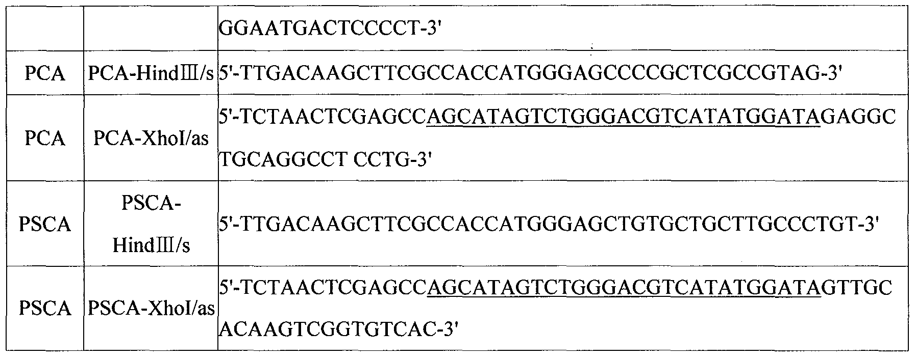
In Table Ib, the underlines denote HA (hemagglutinin) -Tag sequence .
Using primer sets (Bionics, Inc.) in Table Ia and PCR polymerase (Intron Biotech, Inc.), PCR amplifications were conducted for obtaining DNA fragments, PAP (1062 bp) , PSA (729 bp) , PSMA (942 bp) , PCA (525 bp) and PSCA (213 bp) , for expressing in prokaryotic cells under the following thermal conditions: 25 cycles of 30 sec at 94°C, 30 sec at 52°C, and 50 sec at 72°C. Likely, for obtaining DNA fragments, PAP (1185 bp) , PSA (810 bp) , PSMA (2148 bp) , PCA (552 bp) and PSCA (240 bp) , for expressing in eukaryotic cells, PCR amplifications were conducted using primer sets in Table Ib. For making it feasible to detect proteins expressed in eukaryotic cells, 36A Tag sequence developed by CreaGen, Inc. (Korea) was introduced to the amplified nucleotide sequences. Primers for introducing Tag sequence are Tag-Xhol /s ( 5 ' -ACCCTCGAGGTCCATGACCGGAGGTCAGC AGATGGGTCGCGACCTGTACGACGA-3 ' ) and Tag-Xbal/as (5 ' -ACCTCTAGATT
AGCTTCCCCATCTGTCCTTGTCGTCATCGTCGTACAGGTCGCG-S'). Tag DNA fragments were prepared by PCR amplification under the thermal conditions: 1 cycle of 30 sec at 94°C, 30 sec at 52°C, and 5 min at 72°C.
The amino acid sequence of 36A Tag is SMTGGQQMGRDLYDDDDKDRWGS and its nucleotide sequence is TCC ATG ACC GGA GGT CAG CAG ATG GGT CGC GAC CTG TAC GAC GAT GAC GAC AAG GAC AGA TGG GGA AGC.
(c) Cloning Prostate Cancer Antigen cDNA into Expression Vectors (pcDNA3.1(+) Vector and pCTP Vector)
To introduce into vector (pcDNA3.1 (+) ; Invitrogen) 36A Tag system for evaluating expression, Tag DNA fragments prepared were digested using Xhol/Xbal and ligated to pcDNA3.1(+), producing pcDNA3.1 (+) -Tag vector. Each of DNA fragments for human prostate cancer antigens was digested using Hindlll/Xhol and cloned into pcDNA3.1 (+) -Tag vector, followed by confirming cloned sequences by sequencing (see Fig. 2). To obtain recombinant prostate cancer antigens in prokaryotic cells, pCTP-Td vector was used. pCTP-Td vector was constructed by genetically manipulating pTAT-HA vector (kindly provided by Dr. S. Dowdy at the Washington University, H. Nagahara et al., Nature Med. 4:1449(1998)). pTAT-HA vector was digested for 2 hr at 37°C using BamHI and Ncol to eliminate Tat domain and HA-tag domain, and then purified using Gel extraction kit (Nucleogen) . For preparing CTP DNA fragments (nucleotide sequence, TAC GGA CGC CGC GCA CGC CGC CGC CGC CGC CGC; amino acid sequence, YGRRARRRRRR), a forward primer (5'-GAT CCA TGT ACG GAC GCC GCG CAC GCC GCC GCC GCC GCC GCT C-3' ) and a reverse primer (5'-CAT GGA GCG GCG GCG GCG GCG GCG TGC GCG GCG TCC GTA CAT G-3' ) were synthesized (Bionics, Inc.), heated for 5 min at 95°C and cooled to room temperature at a rate of 1 °C/min to prepare double stand. Primers were designed to permit CTP DNA fragments to have cohesive end for BamHI and Ncol. After purification of
annealed CTP DNA fragments using PCR purification kit (Necleogen) , they were ligated to vectors using T4 DNA ligase (Roche) and then transformed into E. coli JM109 (Stratagene) , yielding recombinant plasmid, pCTP-Td vector. The construction of pCTP-Td vector was confirmed by both restriction analysis using BamHI and Ncol and sequencing (Solgent, Korea) .
Each of DNA fragments for human prostate cancer antigens was digested using Hindlll/Xhol and cloned into pCTP vector (see Fig. 2) . DNA sequencing was undertaken for the nucleotides sequences cloned. It was verified that the amino acid sequences encoded by the cloned sequences had 100% identity to known amino acid sequences of PCA, PSCA, PAP, PSA and PSMA (Blast 2 sequence search) . The sequences introduced into prokaryotic expression vectors are lack of sequences corresponding to N- terminal signal peptide and transmembrane domain adjacent to C- terminal. The nucleotide sequences into prokaryotic expression vectors encode amino acids 30-204 of PCA (175 aa) , amino acids 23-93 of PSCA (71 aa) , amino acids 33-386 of PAP (354 aa) , amino acids 19-261 of PSA (243 aa) and amino acids 394-707 of PSMA (314 aa) . In the meantime, the nucleotide sequences into eukaryotic expression vectors encode amino acids 30-204 of PCA (175 aa) , amino acids 23-93 of PSCA (71 aa) , amino acids 1-386 of PAP (386 aa) , amino acids 1-261 of PSA (261 aa) and amino acids 1-707 of PSMA (707 aa) .
EXAMPLE 1-2: Establishment of Human-Derived Prostate Cancer Antigen-Expressing Mouse Cell Lines
(a) Analysis of Antigen Expression in Prostate Cancer Antigen- Expressing Mouse Cell Lines
To prepare prostate cancer antigen-expressing cell lines, pcDNA3.1 (+) -Tag/prostate cancer antigen (PAP, PSA or PCA) vectors were transformed into mouse renal cancer cell line RENCA cells (the Korean Cell Line Research Foundation) . On the day before transformation, 5 x 10⁵ cells/well were plated into 6-well plate and then transformed with 2 μg of the recombinant vector using GenePorter (Gene therapy system) . 48 hours later, cells were treated with 800 μg/ml of G418 (DUCHEFA, Haarlem, Netherland) to select viable cells. Cells selected were proliferated and harvested and in turn the expression pattern of antigens was verified by Western blotting analysis. Harvested cells were washed twice with PBS, heated in protein sample buffer and centrifuged to remove genomic DNA molecules, followed by SDS-PAGE for supernatant isolation. Protein bands resolved were transferred to a nitrocellulose membrane using semi-dry transfer blotter (Bio-Rad) and incubated with a primary antibody, Tag antigen-specific monoclonal antibody (CreaGen, Inc.) and a secondary antibody AP (alkaline phosphatase) -conjugated anti-mouse IgG (Sigma). The bands were visualized using AP reaction solution (Promega) containing NBT/BCIP. As shown in Fig. 3, proteins of interest were successfully expressed in three cell lines harboring expression vectors for antigens.
Of prostate cancer antigen-expressing cell lines established, PAP-expressing cell line was denoted as "RENCA/PAP", deposited on May 23, 2005 in International Depository Authority, the Korean Collection for Type Cultures and was given accession number KCTC 10808BP.
(b) Evaluation of Cell Line Stability in View of Antigen Expression
To verify whether recombinant cell lines established maintain the antigen expression potential in the absence of antibiotics (G418) when injected into mice, cell lines were subcultured in a medium with no G418 and examined, so that the stable expression of introduced foreign sequences were checked. Each of cell lines (RENCA/PAP, RENCA/PSA and RENCA/PCA) was cultured in the absence of G418, 1 x 10⁶ cells were harvested every three days and then their antigen expression potential was examined by Western blotting analysis using anti-Tag monoclonal antibody. As shown in Fig. 4, the antigen expression potential is maintained for 15 days in all of three cell lines maintain although it decreases slightly with the lapse of time. Consequently, it could be appreciated that human prostate cancer antigen-expressing cell lines prepared in Examples are very useful in the construction of mouse prostate cancer models.
EXAMPLE 2 : Purification of Recombinant CTP-Conjugated Proteins for Pulsing Dendritic Cells and Measurement of Transduction Potential
EXAMPLE 2-1: Expression and Purification of Recombinant CTP- Conjugated Prostate Cancer Antigens E. coli BL21Gold (DE3) competent cells (Stratagene) were transformed with recombinant pCTP-Td vectors carrying cDNA for each prostate cancer antigen to prepare transformants according to Hanahan method and cultured in ampicillin-LB medium. The
transformants cultured were centrifuged, washed with PBS and harvested, followed by analyzing prostate cancer antigen expression on 12% SDS-PAGE. Following the expression, recombinant proteins CTP-PCA, CTP-PSCA, CTP-PSA, CTP-PAP and CTP-PSMA were purified through a column of Ni+-NTA resin
(Qiagen) . The proteins analyzed were shown to have a molecular weight higher by about 6 kDa, which is ascribed to non-genome sequence originated from vectors. In other words, CTP-PCA shows a molecular weight of about 24 kDa, CTP-PSCA of about 14 kDa, CTP-PAP of about 45 kDa, CTP-PSA of about 33 kDa and CTP-PSMA of about 41 kDa.
The CTP-PSMA expressed corresponds to the C-terminal of PSMA. Since the N-terminal signal sequence of PSMA was not revealed in a motif prediction analysis, the expression of the region corresponding amino acids 2-707 (706 aa) was primarily attempted. Unlikely to eukaryotic cells, the expression of PSMA was not observed in E. coli. Thereafter, either the N-terminal region (2-393) or C-terminal region (394-707) was cloned into pCTP vector and expressed. As results it was revealed that only the C-terminal region was shown to be expressed. These results urge us to reason that hydrophobic residues concentrated at the N-terminal region would inhibit its expression. Accordingly, we used the C-terminal region of PSMA for preparing vaccines as immunotherpeutics .
EXAMPLE 2-2: Transduction Potential of Recombinant CTP-Prostate Cancer Antigens
To elucidate the transduction potential of recombinant
proteins, cells were incubated with each of five recombinant proteins (CTP-PCA, CTP-PSCA, CTP-PAP, CTP-PSA and CTP-PSMA) . 1 x 10⁵ mouse splenocytes were cultured in a 6-we11 plate and incubated with 50 μg/well of the recombinant protein. 24-hr later, cells were collected and trypsinized to remove proteins attached on their surface. Then, cells were lyzed using a sonicator and resolved by PAGE, followed by Western blotting using anti-Hisβ-tag monoclonal antibody (Qiagene) .
As represented in Fig. 6, CTP-PCA, CTP-PSCA, CTP-PAP, CTP- PSA and CTP-PSMA show the bands of about 24 kDa, about 14 kDa, about 33 kDa, about 40 kDa and about 41 kDa, respectively. PSCA shows multi-band pattern since it forms dimmer or multimer. Therefore, it could be understood that recombinant CTP- conjugated prostate cancer antigens are successfully introduced into cells.
EXAMPLE 3: Establishment of Animal Prostate Caner Models EXAMPLE 3-1 : Evaluation of Formation and Growth of Cancer Caused by Prostate Cancer Antigen-Expressing Cell Lines in Mice We examined the formation and growth of cancer caused by mouse cell lines expressing human prostate cancer antigens. Cell lines prepared hereinabove were injected into femurs of 6- week-old Balb/c mouse (Daehan Biolink, Inc., Korea). The recombinant cell lines expressing human prostate cancer antigens were cultured and maintained in RPMI medium containing 10% FBS and 500 μg/ml G418. Cells at optimal growth state were washed 2-3 times with PBS, treated with trypsin-EDTA for single cell isolation, and suspended at 1 x 10⁶ cells/100 μl in PBS.
100 μl of the suspension were subcutaneously inoculated into mice. Following the inoculation of cell lines, the formation of solid cancer was observed in a time interval of 2 days. The size of solid cancer was measured using calipers. As represented in Fig. 7a, all mice inoculated with prostate cancer cell lines were found to bear solid cancer. Interestingly, a lower dose of cells, e.g. I x 105 cells could cause tumorigeneisis and cancer formed in mice were not extinguished by immune response to heterologous antigens. Fig. 7b shows the growth rate of cancer. The recombinant RENCA/PAP cell line shows a growth rate of cancer lower than other cell lines and the RENCA/PCA cell shows a growth rate of cancer similar to RENCA. It would be understood that the expression of PAP is made in the highest level and in turn elicits the strongest immune responses.
These results demonstrate that the tumorigenesis in mice caused by cells expressing human prostate cancer antigens could not be prevented by immune responses to heterologous antigens. Accordingly, it could be determined that prostate cancer mouse models are successfully established by the present invention and allows for evaluating the prevention and treatment efficacy of dendritic cell-based vaccines.
EXAMPLE 4 : Analysis of Anti-Cancer Efficacy of Dendritic Cells EXAMPLE 4-1: Prevention Efficacy of Dendritic Cell-Based Vaccine (Prevention Model)
To investigate whether dendritic cell-based vaccines can prevent prostate cancer, mice were immunized twice with
dendritic cells pulsed with recombinant CTP-conjugated prostate cancer antigens and challenged with cancer cell lines expressing prostate cancer-specific antigen, followed by examining the formation of solid cancer and pulmonary metastasis.
Mice dendritic cells were prepared by differentiating bone marrow cells of a femur and tibia into dendritic cells. The both ends of a femur and tibia were dissected, from which bone marrow cells were extracted and collected into a 50 ml tube. Bone marrow cells collected were suspended in 0.83% ammonium chloride solution to remove red blood cells, and cultured in a dendritic cell production medium (RPMI-1640 medium containing 10% FBS, 10 ng/ml mouse recombinant IL-4 and 10 ng/ml mouse GM- CSF) for 2 days. Non-adherent cells were removed to obtain only adherent cells on the bottom of tubes. Medium was changed with a fresh medium in a time interval of 2-3 days to prevent the deficiency of cytokines. On a 6 day of culture, immature dendritic cells were harvested and incubated with CTP-PSA, CTP- PCA or a 1:1 mixture of CTP-PSA and CTP-PCA. Immature dendritic cells were pulsed with 50 μg/ml of each antigen protein for 20 hr, to which 100 μg/ml of IFN-γ and 100 μg/ml of TNF-α as cytokines for DC maturation were added. 1 x 10⁶ cells of dendritic cells pulsed with antigens were subcutaneously injected into mice to elicit immune reactions to cancer. Immunization with dendritic cells was conducted twice in a time interval of 2 weeks. After one week of the second immunization, mice immunized by denritic cells pulsed with CTP-PAP, mice immunized by denritic cells pulsed with CTP-PSA, and mice
immunized by denritic cells pulsed with CTP-PCA were subcutaneously challenged with 1 x 10⁶ cells/mouse of RENCA/PAP, RENCA/PSA and RENCA/PCA, respectively. The size of cancer (length x breadth) was measured every two days. As shown in Fig. 8, mice immunized using dendritic cells pulsed with CTP-PAP or CTP-PSA antigen were shown to bear no tumor mass. In contrast, mice immunized using dendritic cells pulsed with CTP-PCA had cancerous tissues.
Fig. 9 graphically shows the cancer incidence in a cancer prevention model using dendritic cells. Among mice immunized with antigen-pulsed dendritic cells, mice challenged with RENCA/PAP or RENCA/PSA cell lines were found to exhibit 100% prevention efficacy for cancer. Mice challenged with RENCA/PCA cell line were shown to exhibit the prevention efficacy much lower than mice challenged with RENCA/PAP or RENCA/PSA, although tumorigenesis was retarded. These results are ascribed to a low antigenecity of PCA. Therefore, it is preferable that DC vaccines for prostate cancer are not prepared using PCA as a sole antigen.
EXAMPLE 4-2: Inhibition of Prostate Cancer Metastasis by Dendritic Cell-Based Vaccine (Prevention Model)
Mice were immunized twice with dendritic cell-based vaccines as described hereinabove and injected via tail vein with 1 x 10⁶ cells/mouse of cell lines expressing prostate cancer antigens. 30 days later, mice were euthanized and pulmonary metastasis was evaluated.
As shown in Fig. 10, mice immunized using dendritic cells
pulsed with human prostate cancer antigens were shown to exhibit no pulmonary metastasis. In contrast, mice treated with either dendrtic cells not pulsed or PBS were shown to elicit strong pulmonary metastasis. It could be understood that dendritic cell-based vaccines elicit strong immune reactions specific to cancer antigens and in turn inhibit the formation and metastasis of cancer.
EXAMPLE 4-3: Treatment Efficacy of Dendritic Cell-Based Vaccine (Regression Model)
Mice were subcutaneously injected with 1 x 10⁶ cells/mouse of recombinant cell lines expressing prostate cancer antigen (PAP, PSA and PCA) . After 3 days, mice were injected twice in a time interval of one week with 1 x 10⁶ cells/mouse of dendritic cells pulsed with CTP-conjugated antigens (CTP-PAP, CTP-PSA and CTP-PCA) . Following the second administration of DC, the formation and growth of cancer were observed for one month in a time interval of 2 days.
As represented in Figs. 11a and lib, the growth of cancer was inhibited in all of prostate cancer mouse models established using RENCA/PAP, RENCA/PSA and RENCA/PCA cell lines. In particular, the RENCA/PAP mouse model was shown to exhibit the strongest inhibition to tumorigenesis .
EXAMPLE 4-4: Analysis of CTL Response Induced by Dendritic Cell-Based Vaccine
T cell proliferation and CTL activity were analyzed using splenocytes isolated from mice of Example 4-3. Each mouse was
euthanized according to cervical dislocation method and spleen was isolated and stored in RPMI. Each spleen was passed through a 70 μm sieve and suspended tissues were then removed. The resultant was centrifuged to collect cells, after which were suspended in 0.83% ammonium chloride solution to remove red blood cells. The splenocytes prepared were passed through a nylon wool column to isolate T lymphocytes as effector cells, and mixed with APC (antigen presenting cells) at a ration of 5:1, followed by culturing for 5 days. APC was prepared 2 days prior to experiment. Separately, splenocytes were isolated from normal mice and treated for 3 hr with 3 μg/ml of Con-A. The stimulated cells were incubated with 50 μg/ml of each antigen protein (CTP-PAP, CTP-PSA and CTP-PCA) for 24 hr. The cell concentration was maintained at 1 x 10δ cells during culture. After 24-hr culture, cells were treated with mitomycin C for 40 min and washed three times to prepare APC. After 5-days of culture of effector T cells, effector T cells were isolated by removing dead cells with Histopaque (Sigma) . The effector T cell and target cell were mixed at ET ratios of 0:1, 5:1, 10:1, 20:1 and 40:1 and incubated for 24 hr. As target cells, prostate cancer antigen-expressing cells were used. One day before mixed with effector cells, the target cells were cultured at a density of 1 x 10⁴ cells/well in a 96-well plate and then used. A control not containing effector cells underwent fixation and staining and was used for calculating specific lysis activities. At 24-hr after mixing effector and target cells, the supernatant of cells was removed and treated for 1 hr with 100 μl of 10% formalin for fixation. Following
the removal of formalin, cells were stained using 80 μl of 4% crystal violet for 30 min and washed three times with tap water, followed by drying on air. Cells were treated with 80% methanol for 10 min. Finally, the OD values were measured at 570 nm. The specific lysis activity was calculated according to (sample OD/control OD) x 100.
As represented in Fig. 12, CTLs were effectively induced in all three mouse models. These results demonstrate that the administration of dendritic cell-based vaccines pulsed with CTP-PAP, CTP-PSA or CTP-PCA induces effectively CTLs specific to human prostate cancer antigens, giving rise to the prevention and treatment efficacy to cancer. For experiments using cells expressing the PAP antigen, since mice themselves might have CTL clones, pulmonary metastasis was less than other cell lines and CTL activity was observed in control. For experiments using cells expressing the PSA or PCA antigen, only Ag-pulsed dendritic cells exhibited strong CTL activities. These results represent that CTLs having anti-cancer activity are effectively induced by dendritic cell-based vaccines. Interestingly, dendritic cells pulsed with the CTP-PCA antigen induce effectively the immune reactions against PCA-expressing cell lines. Tumor cells formed by PCA-expressing cancer cell lines show the increase in the proliferation due to the expression of PCA, so that anti-cancer efficacy of dendritic cells pulsed with the CTP-PCA antigen was measured to be relatively low. In other words, it would be recognized that the anti-cancer efficacy of dendritic cells may reflect both the anti-cancer immune responses and the proliferation of tumor
cells .
As described previously, the present invention provides methods for analyzing the prevention and treatment efficacy of dendritic cells as immunotherapeutics for prostate cancer by use of animal models. For executing the prevention or treatment of prostate cancer using dendritic cells in a clinical scale, it is prerequisite to verity the efficacy and safety of dendritic cells in animal models. The present invention allows for animal model-based evaluation of dendritic cells as immunotherapeutics. Dendritic cell-based vaccines (DC vaccines) selected by the present invention become promising candidates as immunotherapeutics for prostate cancer.
Having described a preferred embodiment of the present invention, it is to be understood that variants and modifications thereof falling within the spirit of the invention may become apparent to those skilled in this art, and the scope of this invention is to be determined by appended claims and their equivalents.

Claims
1. A method for analyzing the prevention and treatment efficacy of a dendritic cell-derived immunotherapeutic for prostate cancer using an animal model carrying tumors expressing a human prostate cancer-specific antigen, which comprises the steps of:
(a) (a' ) administering to a normal animal other than human dendritic cells to be analyzed, or (a") administering to a normal animal other than human a cancer cell line expressing the human prostate cancer-specific antigen to induce cancer in the normal animal;
(b) (b' ) administering to the animal the cancer cell line expressing the human prostate cancer-specific antigen to induce cancer in the animal when the step (a' ) is performed in the step (a) , or (b") administering to the animal with cancer dendritic cells to be analyzed when the step (a") is performed in the step (a) ; and
(c) determining the prevention and treatment efficacy of the dendritic cells as immunotherapeutics for prostate cancer by measuring the formation or growth of cancer cells originated from the cancer cell line in the animal.
2. The method according to claim 1, wherein the animal is rodents .
3. The method according to claim 3, wherein the rodent is a mouse (Mus musculus) .
4. The method according to 1, wherein the human prostate cancer-specific antigen is PCA (prostate cancer antigen) , PSCA (prostate stem cell antigen) , PSA (prostate-specific antigen) , PAP (prostate acid phosphatase) or PSMA (prostate-specific membrane antigen) .
5. The method according to claim 4, wherein the human prostate cancer-specific antigen is PAP.
6. The method according to claim 1, wherein the cancer cell line is derived from a mouse (Mus musculus) .
7. The method according to claim 6, wherein the cancer cell line is syngeneic to the animal.
8. The method according to claim 1, wherein the administration of dendritic cells or the cancer cell line in the step (a) is carried out by subcutaneous injection.
9. The method according to claim 1, wherein the administration of dendritic cells or the cancer cell line in the step (b) is carried out by subcutaneous injection.
10. The method according to claim 1, wherein the cancer cell line expressing the human prostate cancer-specific antigen is not a prostate cancer cell-derived one.
11. A mouse-derived renal cancer cell line (RENCA) expressing a human prostate cancer-specific antigen, characterized in that the human prostate cancer-specific antigen is PCA (prostate cancer antigen) , PSCA (prostate stem cell antigen) , PSA (prostate-specific antigen) , PAP (prostate acid phosphatase) or PSMA (prostate-specific membrane antigen) and the renal cancer cell line is not a prostate cancer cell-derived one.
12. The mouse-derived renal cancer cell line (RENCA) according to claim 11, wherein the cancer cell line is transformed with a vector containing a nucleotide sequence encoding an amino acid sequence spanning amino acids 30-204 of SEQ ID NO:1, an amino acid sequence of SEQ ID NO: 3, or an amino acid sequence of SEQ ID NO:4.
13. The mouse-derived renal cancer cell line (RENCA) according to claim 11, wherein the cancer cell line is transformed with a vector containing a nucleotide sequence of nucleotides 88-612 of SEQ ID NO: 6, a nucleotide sequence of SEQ ID NO: 8, or a nucleotide sequence of SEQ ID NO: 9.
14. The mouse-derived renal cancer cell line (RENCA) according to claim 13, wherein the cancer cell line is transformed with pcDNA3.1 (+) -Tag/PAP, pcDNA3.1 (+) -Tag/PSA or pcDNA3.1 (+) -Tag/PCA.
15. The mouse-derived renal cancer cell line (RENCA) according to claim 11, wherein the cancer cell line is RENCA/PAP (KCTC 10808BP) expressing the PAP antigen.
16. A mouse prostate cancer model, characterized in that the mouse model has a cancer formed by inoculating the renal cancer cell line of any one of claims 11-15 expressing the human prostate cancer-specific antigen, and the metastasis or growth of the cancer formed in the mouse model is inhibited by the treatment of dendritic cells pulsed with the human prostate cancer-specific antigen.
17. The mouse prostate cancer model according to claim 16, wherein the renal cancer cell line is syngeneic to the mouse.
18. The mouse prostate cancer model according to claim 16, wherein the mouse prostate cancer model is used for performing the method of any one of claims 1-10.
Priority Applications (1)
| Application Number | Priority Date | Filing Date | Title |
|---|---|---|---|
| US11/915,737 US20090293138A1 (en) | 2005-05-27 | 2006-05-27 | Animal models carrying tumors expressing human prostate cancer-specific antigen and method for analyzing prevention and treatment efficacy of dendritic cells-derived immunotherapeutics |
Applications Claiming Priority (2)
| Application Number | Priority Date | Filing Date | Title |
|---|---|---|---|
| KR10-2005-0045236 | 2005-05-27 | ||
| KR1020050045236A KR100647847B1 (en) | 2005-05-27 | 2005-05-27 | - Animal Models Carrying Tumors Expressing Human Prostate Cancer-Specific Antigen and Method for Analyzing Prevention and Treatment Efficacy of Dendritic Cells-Derived Immunotherapeutics Using the Above |
Publications (1)
| Publication Number | Publication Date |
|---|---|
| WO2006126864A1 true WO2006126864A1 (en) | 2006-11-30 |
Family
ID=37452233
Family Applications (1)
| Application Number | Title | Priority Date | Filing Date |
|---|---|---|---|
| PCT/KR2006/002028 WO2006126864A1 (en) | 2005-05-27 | 2006-05-27 | Animal models carrying tumors expressing human prostate cancer-specific antigen and method for analyzing prevention and treatment efficacy of dendritic cells-derived immunotherapeutics using the above |
Country Status (3)
| Country | Link |
|---|---|
| US (1) | US20090293138A1 (en) |
| KR (1) | KR100647847B1 (en) |
| WO (1) | WO2006126864A1 (en) |
Cited By (1)
| Publication number | Priority date | Publication date | Assignee | Title |
|---|---|---|---|---|
| WO2008143400A1 (en) * | 2007-05-17 | 2008-11-27 | Creagene Inc. | Animal models carrying tumors expressing human liver cancer-specific antigen and method for analyzing prevention and treatment efficacy of dendritic cells-derived immunotherapeutics using the above |
Families Citing this family (4)
| Publication number | Priority date | Publication date | Assignee | Title |
|---|---|---|---|---|
| EP3909973A3 (en) * | 2010-11-12 | 2022-01-26 | The Trustees Of The University Of Pennsylvania | Consensus prostate antigens, nucleic acid molecules encoding the same and uses thereof |
| EP2543386A1 (en) * | 2011-07-05 | 2013-01-09 | Sotio a.s. | Means and methods for active cellular immunotherapy of cancer by using tumor cells killed by high hydrostatic pressure |
| EP2844282B1 (en) * | 2012-05-04 | 2019-06-12 | Pfizer Inc | Prostate-associated antigens and vaccine-based immunotherapy regimens |
| CN112674028B (en) * | 2020-12-30 | 2022-06-28 | 汉姆德(宁波)智能医疗科技有限公司 | Method for establishing cancer model of animal induced by cancer inducer |
Citations (1)
| Publication number | Priority date | Publication date | Assignee | Title |
|---|---|---|---|---|
| WO2004044197A1 (en) * | 2002-11-13 | 2004-05-27 | Procyon Biopharma Inc. | Knock-in mouse prostate cancer model |
Family Cites Families (3)
| Publication number | Priority date | Publication date | Assignee | Title |
|---|---|---|---|---|
| US6080409A (en) | 1995-12-28 | 2000-06-27 | Dendreon Corporation | Immunostimulatory method |
| KR100490308B1 (en) * | 2002-02-25 | 2005-05-17 | 크레아젠 주식회사 | Method of Preparing Mature Dendritic Cell-Vaccine for Immunotherapy |
| KR100496062B1 (en) * | 2002-12-10 | 2005-06-16 | 크레아젠 주식회사 | Novel Recombinant Sabin Type 1 Poliovirus Vector and Improved Recombinant Polio Vaccine Composition Using the Same |
-
2005
- 2005-05-27 KR KR1020050045236A patent/KR100647847B1/en active IP Right Grant
-
2006
- 2006-05-27 WO PCT/KR2006/002028 patent/WO2006126864A1/en active Application Filing
- 2006-05-27 US US11/915,737 patent/US20090293138A1/en not_active Abandoned
Patent Citations (1)
| Publication number | Priority date | Publication date | Assignee | Title |
|---|---|---|---|---|
| WO2004044197A1 (en) * | 2002-11-13 | 2004-05-27 | Procyon Biopharma Inc. | Knock-in mouse prostate cancer model |
Non-Patent Citations (5)
| Title |
|---|
| CHAKRABORTY N.G. ET AL.: "Recognition of PSA-derived peptide antigens by T cells from prostate cancer patients without any prior stimulation", CANCER IMMUNOL. IMMUNOTHER., vol. 52, no. 8, August 2003 (2003-08-01), pages 497 - 505, XP003004143 * |
| FIZAZI K. AND NAVONE N.M.: "Preclinical models of prostate cancer", BULL. CANCER, vol. 92, no. 2, February 2005 (2005-02-01), pages 129 - 141, XP008073098 * |
| PESHWA M.V. ET AL.: "Induction of prostate tumor-specific CD8+ cytotoxic T-lymphocytes in vitro using antigen-presenting cells pulsed with prostatic acid phosphatase peptide", PROSTATE, vol. 36, no. 2, 1 July 1998 (1998-07-01), pages 129 - 138, XP000886216 * |
| ROOS A.K. ET AL.: "Induction of PSA-specific CTLs and anti-tumor immunity by a genetic prostate cancer vaccine", PROSTATE, vol. 62, no. 3, 15 February 2005 (2005-02-15), pages 217 - 223, XP003004142 * |
| TJOA B. ET AL.: "Evaluation of phase I/II clinical trials in prostate cancer with dendritic cells and PSMA peptides", PROSTATE, vol. 36, no. 1, 15 June 1998 (1998-06-15), pages 39 - 44, XP003004144 * |
Cited By (3)
| Publication number | Priority date | Publication date | Assignee | Title |
|---|---|---|---|---|
| WO2008143400A1 (en) * | 2007-05-17 | 2008-11-27 | Creagene Inc. | Animal models carrying tumors expressing human liver cancer-specific antigen and method for analyzing prevention and treatment efficacy of dendritic cells-derived immunotherapeutics using the above |
| KR100900742B1 (en) | 2007-05-17 | 2009-06-08 | 크레아젠 주식회사 | Animal Models Carrying Tumors Expressing Human Liver Cancer-Specific Antigen and Method for Analyzing Prevention and Treatment Efficacy of Dendritic Cells-Derived Immunotherapeutics Using the Above |
| CN101680016B (en) * | 2007-05-17 | 2012-11-14 | 医疗基因公司 | Animal models carrying tumors expressing human liver cancer-specific antigen and method for analyzing prevention and treatment efficacy of dendritic cells-derived immunotherapeutics using the above |
Also Published As
| Publication number | Publication date |
|---|---|
| US20090293138A1 (en) | 2009-11-26 |
| KR100647847B1 (en) | 2006-11-23 |
Similar Documents
| Publication | Publication Date | Title |
|---|---|---|
| US20220017593A1 (en) | Compositions and methods for use of recombinant t cell receptors for direct recognition of tumor antigen | |
| JP7256024B2 (en) | Cancer vaccine and therapeutic method using the same | |
| KR102049928B1 (en) | Yeast-muc1 immunotherapeutic compositions and uses thereof | |
| US20060107339A1 (en) | Transgenic animal expressing HLA-A24 and utilization thereof | |
| US20180002720A1 (en) | Cell for use in immunotherapy which contains modified nucleic acid construct encoding wilms tumor gene product or fragment thereof, method for producing said cell, and said nucleic acid construct | |
| Matsuyoshi et al. | Enhanced priming of antigen-specific CTLs in vivo by embryonic stem cell-derived dendritic cells expressing chemokine along with antigenic protein: application to antitumor vaccination | |
| JP2002509716A (en) | Methods and compositions for raising an immune response to a telomerase antigen | |
| KR20010024585A (en) | Tumor-specific antigens, methods for their production and their use for immunization and diagnosis | |
| US20110027310A1 (en) | Compositions and Methods for Cancer Treatment | |
| US20100235932A1 (en) | Animal models carrying tumors expressing human liver cancer-specific antigen and method for analyzing prevention and treatment efficacy of dendritic cells-derived immunotherapeutics using the above | |
| US20090293138A1 (en) | Animal models carrying tumors expressing human prostate cancer-specific antigen and method for analyzing prevention and treatment efficacy of dendritic cells-derived immunotherapeutics | |
| CN110573173A (en) | TERT immunogenic compositions and methods of treatment using same | |
| JP5110674B2 (en) | Compositions and methods for dendritic cell-based immunotherapy | |
| US10576145B2 (en) | CD200 inhibitors and methods of use thereof | |
| KR100647844B1 (en) | Animal Models Carrying Tumors Expressing Human Prostate Cancer-Specific Antigen and Method for Analyzing Prevention Efficacy of Dendritic Cells-Derived Immunotherapeutics Using the Above | |
| JP2006525787A (en) | Rhesus monkey HER2 / neu, nucleotide encoding the same and use thereof | |
| JP2007511534A (en) | Compositions and methods for synergistic induction of anti-tumor immunity | |
| CN106350578A (en) | Application of NY-ESO-1 in diagnosis and treatment of microsatellite instability intestinal cancer | |
| Fukui et al. | Anti-tumor activity of dendritic cells transfected with mRNA for receptor for hyaluronan-mediated motility is mediated by CD4+ T cells | |
| Fukui et al. | Therapeutic effect of dendritic cells loaded with a fusion mRNA encoding tyrosinase-related protein 2 and enhanced green fluorescence protein on B16 melanoma | |
| JP4705697B2 (en) | MN / CA9-derived HLA-A24 restricted tumor antigen peptide | |
| JP2009035534A (en) | Hla-a24-binding antigen peptide and use thereof | |
| Alexeev et al. | Recombinant DNA technology in emerging modalities for melanoma immunotherapy | |
| WO2004018680A1 (en) | Method of screening tumor antigen | |
| CN106350579A (en) | Application of NY-ESO-2 in diagnosis and treatment of microsatellite instability intestinal cancer |
Legal Events
| Date | Code | Title | Description |
|---|---|---|---|
| 121 | Ep: the epo has been informed by wipo that ep was designated in this application | ||
| NENP | Non-entry into the national phase |
Ref country code: DE |
|
| NENP | Non-entry into the national phase |
Ref country code: RU |
|
| WWE | Wipo information: entry into national phase |
Ref document number: 11915737 Country of ref document: US |
|
| 122 | Ep: pct application non-entry in european phase |
Ref document number: 06747469 Country of ref document: EP Kind code of ref document: A1 |