DESCRIPTION
METHOD FOR DIAGNOSING OR PREDICTING SUSCEPTIBILITY TO OPTIC NEUROPATHY
TECHNICAL FIELD The present invention relates to a set of genetic polymorphisms linked to optic neuropathy. BACKGROUND ART Glaucoma is a major cause of blindness worldwide, and estimated approximately 67 million peopl e suffered from some form of glaucoma. The majority of cases occur as late adult onset (typically over age 40 years) o ϋ primary open- angle glaucoma (POAG) , which is the most common form of glaucoma and affects approximately 2% in wfc.ite population and 7% of black population over 40 years old. POAG results in a characteristic visual field changes corresponding to the excavation of the optic disc that is usually associated with an elevation of intraocular pressure (IOP) . Normal- tension glaucoma (NTG) is a form of open-angle glaucoma in which typical glaucomatous cupping of the o;ptic nerve head and visual field loss are present but in which there is no evidence of increased IOP over 21 mm Hg at all times. In Japan, prevalence of glaucoma is approximately 3.5 % over 40 years old: POAG 0.58 % and NTG 2.04 % . Prevalence of NTG
in Japanese population is high compared with that in other populations. Glaucoma is a multifactorial disorder characterized by a progressive optic neuropathy associated with a specific visual field loss, and results from the interaction of multiple genes and environmental influences, although intraocular pressure (IOP) is a major risk factor for glaucoma. Risk factors to develop glaucoma include high IOP, age, race, positive family history, myopia, the presence of diabetes or hypertension, and genetic factors. Although the exact pathogenesis of glaucomatous optic neuropathy is remains unclear, it is generally accepted that an increased IOP is a major risk factor. Current treatment for glaucoma consists of interventions which lower IOP. However, in some patients with glaucoma, NTG or advanced stage of POAG, reduction of IOP- does not prevent the progression of the disease, indicating that factors other than an increased IOP may be involved in the development or progress of glaucoma. POAG and NTG are a heterogeneous group of conditions probably with different multi-factorial etiologies resulting in the observed patterns of neuronal loss in the optic disk. The association between glaucoma and the presence of many systemic vascular diseases including low systemic blood pressure, nocturnal dips in blood pressure,
hypertension, migraine, vasospasm, and diabetes has been reported. The presence of optic disc hemorrhages in NTG patients suggests that vascular insufficiencies are deeply involved in the development and progression of NTG. A high percentage of patients with POAG receive a wide variety of medications for coexisting disorder. Especially, systemic hypertension was the most common disorder, occurring in 48% of the total population. Glaucoma-like morphological changes have been reported in patients with Leber's hereditary optic neuropathy (LHON) at the atrophic stage and dominant optic atrophy (DAO) . Recently, the inventor has reported optic disc excavation by a quantitative analysis using Heidelberg retinal tomography (HRT) in the atrophic stage of Japanese 15 patients with LHON harboring the 11778 mutation (Mashima Y et. al., Arch Clin Exp Ophthalmol 2003; 241:75-80, the contents of the cited reference are herein incorporated by reference) . LHON is a maternally-transmitted eye disease that mainly affects young adult men. Approximately 70% of patients .were male. This disease usually causes severe and permanent loss of vision resulting in a visual acuity of less than 0.1. Visual field defects are present as central or cecocentral scotomas. So far more than 20 point mutations of mitochondrial DNA ( tDNA) have been reported in LHON patients worldwide (Brown MD et. al . , Clin Neurosci
1994; 2:138-145, the contents of the cited reference are herein incorporated by reference) , and more than 80% of LHON patients carry one of three mtDNA mutations at nucleotide position 3460, 11778, or 14484 (Mackey DA et. al., Am J Hum Genet 1996; 59:481-485, the contents of the cited references are herein incorporated by reference) . Although NTG patients were tested for the three LHON mutations of mtDNA nucleotide positions 3460, 11778 and 14484, no mutations and no defects in respiratory chain activity in skeletal muscle samples were detected (Brierley EJ et. al., Arch Ophthalmol 114:142-146 and Opial D et . al . , Graefes Arch Clin Exp Ophthalmol 239:437-440, the contents of the cited references are herein incorporated by reference) . The major difference among LHON patients with one of these mtDNA mutations is in the clinical- course. The 3460 and 14484 mutations are associated with better visual prognosis than the 11778 mutation which shows visual recovery rates of only 4% to 7% (OostraRJ et. al., J med Genet 1994;31:280-286, Riordan-Eva P et . al., Brain 1995; 118:319-337, Mashima Y et. al., Curr Eye Res 1998;17:403- 408, the contents .of the cited reference are herein incorporated by reference) . However, visual recovery has been documented in some patients with the 11778 mutation and an age of onset in the low teens (Stone EM et. al., J
clin Meuro-Ophthalmol 1992; 12:10-14, Zhu D et. al., Am J Med Genet 1992; 42:173-179, Salmaggi A et. al., Intern J Neuroscience 1994; 77:261-266, Oostra RJ et. al., Clin Genet 1997; 51:388-393, Mashima Y et. al., Jpn J Ophthalmol 2002; 46:660-667, the contents of the cited references are herein incorporated by reference) . Recovery of vision appears to be more likely when visual deterioration begins at an early age, even in patients with the 11778 mutation. The clinical variability of LHON patients, which includes age at onset, male predilection, incomplete penetrance, and visual recovery, suggests that the disease most likely results from polygenic or multifactorial mechanisms, possibly involving environmental stressors, X- chromosomal loci, and other mtDNA mutations (Man PYW et. al., J Med Genet 2002; 39:162-169, the contents of the cited reference are herein incorporated by reference) . - However, attempts to identify a relevant locus on the X- chromosome have not been successful (Chalmers RM et. al., Am J Hum Genet 1996;59:103-108 and Pegoraro E et. al . , Am J Med Genet 2003; 119A: 37-40, the contents of the cited reference are herein incorporated by reference) . So-called secondary LHON mutations" are more frequently found in European LHON patients than in unaffected Europeans and are polymorphisms linked to the European haplotype J. These polymorphisms are not strong autonomous risk factors (Brown
MD et. al., Am J Hum Genet 1997;60:381-387 and Torroni A et. al., Am J Hum Genet 1997;60:1107-1121, the contents of the cited reference are herein incorporated by reference) . Thus, the primary mutations are the major risk factors in LHON, but additional etiologic factors that augment or modulate the pathogenic phenotypes appear to be necessary. Considerable evidence indicates that heavy alcohol and/or tobacco use increases the risk of optic neuropathy in LHON families (Smith PR et. al . , Q J Med 1993;86:657-660, Chalmers RM et. al . , Brain 1996; 119: 1481- 1486 and Tsao K et. al . , Br J Ophthalmol 1999;83:577-581, the contents of the cited reference are herein incorporated by reference) , although one study did not find this association. Possible secondary genetic interactions are complex and not firmly established (Kerrison JB et . al., Am J Ophthalmol 2000;130:803-812, the contents of the cited reference are herein incorporated by reference) . Oxidative stress has been implicated in many disorders associated with mutations of mtDNA. A recent investigation in an animal model identified reactive oxygen species (ROS) as a likely factor in the pathogenesis of LHON (Qi X et. al . , Invest Ophthalmol Vis Sci 2003; 44 : 1088- 1096, the contents of the cited reference are herein incorporated by reference) . Additionally, the mtDNA 'LHON pathogenic mutations were found to predispose cells to Fas-
dependent apoptotic death in vi tro (Danielson SR et. al . , J Biol Chem 2002;277:5810-5815, the contents of the cited reference are herein incorporated by reference) . These findings implied that there must be some nuclear modifier genes involved for developing LHON. SUMMARY OF THE INVENTION The inventor has revealed that some known and unknown SNPs are linked to onset of optic neuropathy including glaucoma and Leber's disease and completed the instant invention. Accordingly, the present invention provides a set of genetic polymorphisms being associated with optic neuropathy, which comprises at least one polymorphism selected from the group consisting of: (1) AAG to AAT substitution at codon 198 of the Endothelin- 1 gene (Lysl98Asn) ;
(2) -1370T>G polymorphism of the Endothelin-1 gene promoter region; (3) A138 insertion/deletion (A138I/D) polymorphism in exon 1 of the Endothelin-1 gene;
(4) +70OG polymorphism in 3' non-coding region of the Endot elin receptor A gene;
(5) +12220T polymorphism of the Endothelin Receptor A gene; (6) CAC to CAT substitution at codon 323 in exon 6 of the
Endothelin Receptor A gene (His323His) ; (7) -231A>G polymorphism of the Endothelin Receptor A gene promoter region; (8) CTG to CTA substitution at codon 277 in exon 4 of the Endothelin receptor B gene;
(9) 9099OA polymorphism of the Mitochondrial gene;
(10) 9101T>G polymorphism of the Mitochondrial gene;
(11) 9101T>C polymorphism of the Mitochondrial gene;
(12) 9804G>A polymorphism of the Mitochondrial gene; (13) 11778G>A polymorphism of the Mitochondrial gene;
(14) -713T>G polymorphism of the Angiotensin II type 1 receptor gene promoter region;
(16) 31230A polymorphism of the Angiotensin II type 2 receptor gene; (25) CAA to CGA substitution at codon 192 of the
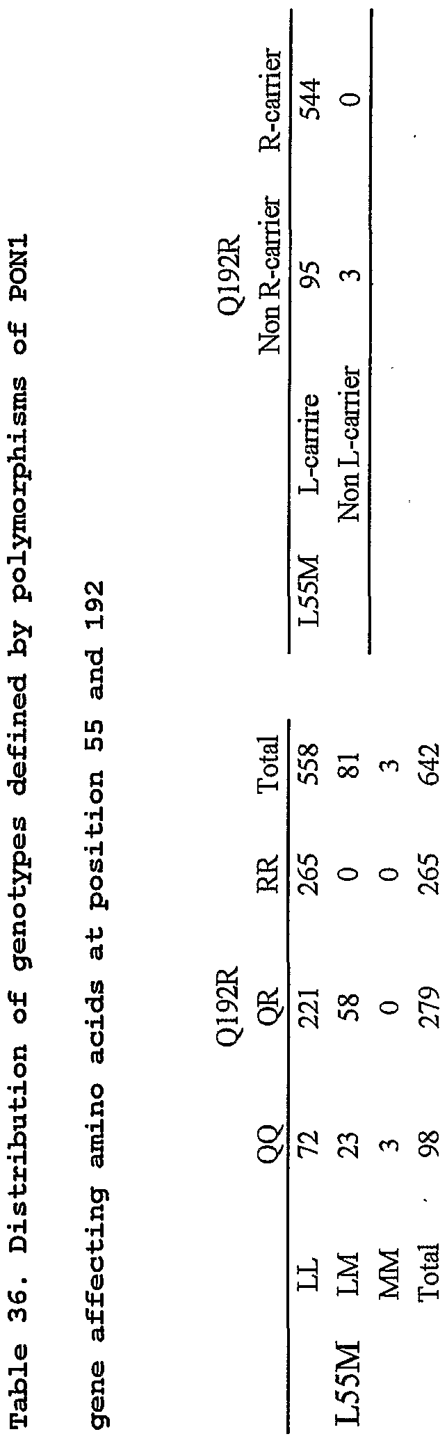
Paraoxonase 1 gene (Glnl92Arg) ; •■ • •
(26) TTG to ATG substitution at codon 55 of the Paraoxonase
1 gene (Leu55Met) ;
(27) CGG to CAG substitution at codon 144 of the Noelin 2 gene (Argl44Gln) ;
(32) GGA to CGA substitution at codon 389 of the βl adrenergic receptor gene (Gly389Arg) ;
(35) 1105T>C polymorphism of the Myocilin gene (Phe369Leu) ;
(36) 412G>A polymorphism of the Optineurin gene; (37) 1402OT polymorphism of the E-Selectin gene;
(38) The combination of polymorphisms of -8570T of the Tumor necrosis factor c. gene promoter region and 412G>A of the Optineurin gene; (39) The combination of polymorphisms of -8630A of the Tumor necrosis factor α gene promoter region and 603T>A of the Optineurin gene (40) CGC to CCC substitution at codon 72 of the TP53 gene (Arg72Pro) ; (41) TAG to CAC substitution at codon 113 of the Microsomal epoxide hydrasel gene (Tyrll3His) ; (42) -110A>C polymorphism of the Heatshock protein 70-1 gene promoter region; (43) -3380A polymorphism of the Endothelin converting enzyme gene promoter region; (44) -670A>G polymorphism of the CD95 gene promoter region; (45) AAG to AAA substitution at codon 119 of the Microsomal epoxide hydrase 1 gene (Lysll9Lys) ; (47) GGA to AGA substitution at codon 16 of the β2 adrenergic receptor gene (GlylδArg) ; and (48) CAA to GAA substitution at codon 27 of the β2 adrenergic receptor gene (Gln27Glu) . In addition, . the present invention also provides a method for diagnosing or predicting susceptibility to optic neuropathy in a human subject, which comprising the • steps of:
i) obtaining a biological sample from the subject, ii) determining genotype of the sample in respect of the set of the polymorphisms defined as above, and iii) diagnosing or predicting susceptibility to optic neuropathy in the subject based on the genotype. According to the present invention, the optic neuropathy may preferably be glaucoma or Laber's disease. The polymorphism (l)-(39) and (42) -(48) may be used especially for glaucoma. Among them, those (1), (2), (5)- (7), (16), (26), (32), (43) and (45) may be used especially for normal tension glaucoma and those (4), (14), (25), (35), (36), (38), (42), (44), (47)- (48) may be used especially for primary open angle glaucoma. The polymorphisms (40) and (41) may be used especially for Laber's disease. According to the present invention, the set of polymorphisms may - further comprise at least one other polymorphism which has been known to be associated with optic neuropathy. In another aspect of the present invention, a kit for diagnosing or predicting susceptibility to optic neuropathy in a human subject which comprises primer set and/or probe suitable for determining genotype in respect of the set of genetic polymorphisms defined as above. In further aspect of the present invention, • neuly identified SNPs are provided in Mitocondorial gene,
Myocilin gene and Noelin 2 gene. Accordingly, the present invention encompass nucleotide fragment covering those SNPs. In general, in order to deter in genotype in respect of said SNP, 90 or more contignous nucleotide sequence containing the SNP may be required. Namely, an isolated polynucleotide consisting of a segment of the sequence:
8881 tctaagatta aaaatgccct agcccacttc ttaccacaag gcacacctac accccttatc 8941 cccatactag ttattatcga aaccatcagc ctactcattc aaccaatagc cctggccgta 9001 cgcctaaccg ctaacattac tgcaggccac ctactcatgc acctaattgg aagcgccacc 9061 ctagcaatat caaccattaa ccttccctct acacttatca tcttcacaat tctaattcta
9121 ctgactatcc tagaaatcgc tgtcgcctta atccaagcct acgttttcac acttctagta 9181 agcctctacc tgcacgacaa cacataatga cccaccaatc acatgcctat catatagtaa wherein the segment comprises at least 90 contignuous nucleotide, and the at least 90 contignuous nucleotide includes position 9099 of the sequence, and wherein position 9099 of the sequence is A or an isolated polynucleotide which is entirely complementary to the above segment ; or wherein the segment comprises at least 90 contignuous nucleotide, and the at least 90 contignuous nucleotide includes position 9101 of the sequence, and wherein position - 9101 of the sequence is G; or an isolated polynucleotide which is entirely complementary to either of the above segment . The present invention further provides an isolated
polynucleotide consisting of a segment of the sequence : 301 actggaaagc acgggtgctg tggtgtactc ggggagcctc tatttccagg gcgctgagtc 361 cagaactgtc ataagatatg agctgaatac cgagacagtg aaggctgaga aggaaatccc 421 tggagctggc taccacggac ag cccgta ttcttggggt ggctacacgg acattgactt 481 ggctgtggat gaagca'ggcc tctgggtcat ttacagcacc gatgaggcca aaggtgccat
541 tgtcctctcc aaactgaacc cagagaatct ggaactcgaa caaacctggg agacaaacat wherein the segment comprises at least 90 contignuous nucleotide, and the at least 90 contignuous nucleotide includes codon 369, which is corresponding to the underlined nucleotides of the sequence, and wherein codon
369 is substituted such that it codes for Leu, or an isolated polynucleotide which is entirely complementary to the above segment . The present invention further provides an isolated polynucleotide consisting of a segment of the sequence :
79741 ttagttccta caat.ggagtc atgtctggga agaatctagg gtccaatatg agccacatgt
79801 caagggccag gtgtgcatca aagacaaagg gtgaagttat gagtcagagg ttggagtcat
79861 gtctgggtca aaggccaggg gtcaggcttg gccatggttc catcttgatg cacaggagct 79921 gaaggacagg atgacggaac tgttgcccct gagctcggtc ctggagcagt acaaggcaga
79981 cacqcqσacc attgtacgct tgcgggagga ggtgaggaat ctctccggca gtctggcggc
80041 cattcaggag gagatgggtg cctacgggta tgaggacctg cagcaacggg tgatggccct
80101 ggaggcccgg ctccacgcct gcgcccagaa gctgggtatg ccttggccct tgaccctgac
80161 ccctgatctc tgactgccac acccaactcc agtatcacct gtttgtgcct agaagctgga 80221 cacagttttg acctctaact tttaaacctc aacccttgac cttcctacct aaggctacac
wherein the segment comprises at least 90 contignuous nucleotide, and the at least 90 contignuous nucleotide includes codon 144, which is corresponding to the underlined nucleotides of the sequence, and wherein codon 144 is substituted such that it codes for Gin, or an isolated polynucleotide which is entirely complementary to the above segment . BRIEF DESCRIPTION OF THE DRAWINGS Fig. 1 represents correlation of' clinical Characteristics of NTG Patients with AT2R 31230A Polymorphism and ACE I/D Polymorphism Fig. 2 represents DHPLC tracing patterns in the Exon3C of the MYOC gene. Fig. 3 represents novel missense mutation, Phe369Leu detected in exon 3 of the MYOC gene. Fig. 4 represents a DHPLC tracing of MYOC gene from a patient with POAG. Fig. 5A represents the IOP after oral candesartan cilexetil or placebo. Fig.. 5B represents the ocular perfusion pressure after oral candesartan cilexetil or placebo Fig. 5C represents the IOP after oral candesartan cilexetil in each of the 15 subjects. PREFERRED EMBODIMENT OF THE INVENTION In the present specification and claims, "genetic
polymorphism" means genomic diversity between individuals at a locus. Genetic polymorphism may be single nucleotide substitution called as "Single nucleotide polymorphisms" or "SNPs" as well as those consisting of plural nucleotides. The genetic polymorphism may or may not be those affect on the phenotype of the individual. In addition, a nucleotide sequence of an individual is different from the corresponding wild type sequence, i.e., having insertion, deletion or substitution on the wild type sequence, said nucleotidesequence is called as "genetic mutant" and the genetic mutant is also included in "polymorphic variant" according to the present invention. In the present specification and claims, expression like "9099OA" or "C9099A" means that the gene has a polymorphsm at position 9099, that is, there are two alleles of the gene and the one has cytosine or C- and -the • other has adenine or A at 9099 (bi-allelic) . It does not necessarily mean the frequent allele has C whereas the rare allele has A at said position. The . expression like "Glnl92Arg" represents an amino acid substitution due to the base substitution in the gene coding for the amino acid sequence. For example, Glnl92Arg represents Glycine at codon 192, i.e. amino acid number 192, is replaced with Arginine or Arg. This also means that there are polymorphic variants of the protein wherein the
amino acid at codon 192 is Gin or Arg. According to the present invention, determining genotype in respect of the genetic polymorphisms may be carried out by every single polymorphism, or plurality or 5 all polymorphisms may be determined at the same time. In the present invention, the method for diagnosing or predicting susceptibility to optic neuropathy in a human subject which comprises determining genotype in respect of the set of genetic polymorphism of which relationship with0 optic neuropathy is newly reported in this application. In addition to the genetic polymorphism identified as being linked to optic neuropathy by the instant invention, any other polymorphism which had been revealed as being linked to optic neuropathy may be detected together. By employing5 plural genetic polymorphisms linked to optic neuropathy, • - • the diagnostic probability can be improved. - - ■ According to- the present invention, the method used for determining genotype in respect of the genetic polymorphisms is not limited and may be any of those known0 to the art. Representative method for determining genotype in respect of the genetic polymorphisms include polymerase chain reaction restriction fragment length polymorphism (PCR-RFLP) analysis, polymerase chain reaction followed by single strand conformation polymorphism (PCR-SSCP) analysis,5 ASO hybridization analysis, direct sequencing analysys,
ARMS analysis, DGGE analysis, RNaseA cleaving analysis, chemical restriction analysis, DPL analysis, TaqMctn® PCR analysis, . Invader® assay, MALDI-TOF/MS analysis, TDI analysis, single nucleotide extension assay, WAVE assay, one molecular fluorescent detection assay. According to the present invention, the detection method may be one of those or combination of two or more. According to the present invention, biological sample to be used for detecting the genetic polymorphism is not specifically limited and may be hair, blood, saliva- lymph fluid, respiratory tract ucosa, cultured cells and urine. In the specification and claims, "diagnosing or predicting susceptibility to optic neuropathy" includes not only diagnosing onset of optic neuropathy but also determining risk factors which hasten onset of the disease as well as accelerate the disease progresses. • According to the present invention, kits for detecting the genetic polymorphism as well as protein polymorphism identified as above are also provided. Said ki"ts may comprise . primers and/or probes which are specifically designed for detecting the above-identified genetic polymorphisms; antibodies for detecting the above- identified protein polymorphism. According to the present invention, said kit may be used for diagnosing or predicting susceptibility to optic neuropathy.
In the present specification and claims, the term "primer" denotes a specific oligonucleotide sequence which is complementary to a part of the target nucleotide sequence and used to hybridize to the target nucleotide sequence. A primer serves as an initiation point for nucleotide polymerization catalyzed by either DNA polymerase, RNA polymerase or reverse transcriptase. The term "probe" denotes a defined nucleic acid segment which can be used to identify a specific polynucleotide sequence present in samples or confirming target DNA or RNA in a gene modifying process, said nucleic acid segment comprising a nucleotide sequence complementary to the specific polynucleotide sequence to be identified. According to the present invention, primers and probes may be designed based on the targeted sequence so that they are specific to the position at which the targeted polymorphism is expected and/or surrounding sequence of the position so long as they are not identical to some other genes, i.e. it is necessary not to be repeating sequence nor palindrome sequence. According to the present invention, genetic polymorphisms which are linked to optic neuropatriy, especially glaucoma and Leber's disease are identified. Based on the findings, the genotype in respect of the genetic polymorphisms of a biological sample obtained from
an individual is determined and based on thus obtained genotype, onset of the disease or predicted risk for onset of the disease can be determined. In addition to the polymorphisms identified (l)-(48) as above, genotypes in respect of some other genetic polymorphisms which had been known to the art being highly associated with optic neuropathy may be determined for improved relaiability of the diagnosis or prediction. For example, two types of genetic polymorphisms in myocilin as well as optineurin genes have been revealed by the inventor to be associated with onset of primary open- angle glaucoma. In addition to the two genes, 4 other genetic polymorphisms including mutations had been identified to be associated with- primary open-angle glaucoma. Almost 100% of the subjects having both the risk genotype in respect of the genetic polymorphisms- of- the present invention and of those already known to the art may develop glaucoma. That is, the set of the genetic polymorphisms will be useful for precrinical test. In .regard of some SNPS, the inventor confirmed correlation with optic neuropathy in a specific group, such as race or sex. Accordingly, said SNPs may prefereably be used for diagnosing or predicting the risk for optic neuropathy in the specified group. Further, statical analysis of the genotyp in respect
of the set of polymorphisms may provide useful information such as predictive age of onset, predictive association with lifestyle-related diseases, predictive association with symptom factors. In addition, effect of some medical treatments may also be predictable based on the information. According to the present invention, predicting susceptibility to optic neuropathy can be carried out before onset of the disease based- on the genotype, and the subject can receive advice on how to remove the risk factor, for example, to improve life style or alter the environment. In addition, it may possible to receive an early treatment such as reduction of the risk gene. an appropriate treatment can be started earlier. Consequently, those "order made treatment" can reduce the risk for vision loss. For example, in case a subject has the genotype linked to high risk for onset of optic neuropathy, inhibition of onset, reduction of the risk of onset or relief of symptoms can be expected by introducing to the subject the genotype linked to low risk for onset and expressing the same. Further, anti sence to the mRNA of the allele of high risk for onset of optic neuropathy or RNAi method may be used for inhibiting expression of the high risk allele. In another aspect, based on the genotype determination in respect of the set of polymolptiisms shown in the present
invention, genetic etiology of optic neuropathy may be revealed and thus obtained etiology may be useful for development of novel medical agents. Further, by combining genotype information which is associated with optic neuropathy obtained by the present invention and the other genotype information which is associated with life style diseases and the like, comprehensive risk for age-related, life-style related diseases can be predicted and used for high quality of life. The present invention will be further illustrated by means of the examples shown below. It is to be expressly understood, however, that the examples are for purpose of illustration only and is not intended to limit of the scope of the invention. EXAMPLE 1 Genetic Variants of TP53 and EPHXl in Leber's Hereditary Optic Neuropathy and their Relationship to Age at Onset
Purpose: To determine whether genetic polymorphisms of the genes for oxidative stress and apoptosis cause the clinical variability in patients with Leber's hereditary optic neuropathy (LHON) .
MATERIALS AND METHODS
Patients We studied 86 unrelated Japanese patients with LHON
carrying the 11778 mutation with homoplasmy- Their mtDNA mutation was confirmed by polymerase chain rreaction followed by a restriction-enzyme assay whichi revealed concordant gain of the Maelll site (Mashima Y et. al . , Curr Eye Res 1998;17:403-408, the contents of the cited reference are herein incorporated by reference) . The mean age at the onset of visual loss in 86 LHON patients was 25.1 ± 13.0 years with a range 3 to 65 years. Genomic DNA Extraction and Genotyping DNA was extracted from peripheral blood leukocytes by the SDS-proteinase K and phenol/chloroform extraction method. Polymorphisms were examined in the oxidative stress-related gene, microsomal epoxide hydrolase (EPHXl) (Ki ura K et. al . , Am J Ophthalmol 2000; 130: 769-773, the contents of the cited reference are herein incorporated by reference).), -and the apoptosis-related gene, Arg72Pro in- TP53 (Ara S et. al . , Nucleic Acids Res 1990; 18:4961, the contents of the cited reference are herein incorporated by reference) . Each polymorphism was identified using polymerase chain reaction-restriction fragment length polymorphism (PCR-RFLP) techniques (Table 1) .
to o (SI O cπ
Table 1. Primer sequences, product size, and annealing temperatures
Product Annealing Gene Primer sequences Size . Temperature Restriction Enzyme (bp) (°C) F TTG CCG TCC CAA GCA ATG GAT GA TP53 199 60.0 Ace II R TCT GGG AAG GGA CAG AAG ATG AC F EPHXl GAT CGA TAA GTT CCG TTT CAC C 165 56.0 EcoR V R TCA ATC TTA GTC TTG AAG TGA GGA T
RESULTS The associations between age at onset and the polymorphisms were presented in Table 2-1 and Table 2-2.
Table 2-1. Association between age at onset and TP53 (Arg72Pro) and EPHXl (Tyrll3His) gene polymorphism in Leber' s hereditary optic neuropathy
P Value for t-test Table 2-2 . Association between age at onset and TP53 (Arg/Arg) and EPHXl (His/His) gene polymorphism in Leber' s hereditary optic neuropathy
P value for Kruskal- allis Group 1 : Patients who have Arg/Arg at codon 72 in TP53 and His/His at codon 113 in EPHXl Group 2 : Patients who have Arg/Arg at codon 72 in TP53 but not His/His at codon 113 in EPHXl, or His/His at codon 113 in EPHXl but not Arg/Arg at codon 72 in TP53 Group 3 : Patients other than Groups 1 and 2
As shown in Table 2-1, the codon 72 genotype in TP53 and the codon 113 genotype in EPHXl were significantly
associated with younger age at onset of Leber's hereditary optic neuropathy. As shown in Table 2-2, the co-existence of the Codon 72 genotype in TP53 and the codon 113 genotype in EPHXl were significantly associated with younger age at onset of Leber's hereditary optic neuropathy. These results indicated that detection of the Arg/Arg homozygote in TP53 and His/His homozygote in EPHXl make possible the early diagnosis and early treatment of Leber's hereditary optic neuropathy. These results also indicated that the Codon 72 polymorphism may interact with mitochondrial dysfunction to influence disease expression. Individual variations may exist in the apoptotic response that is correlated with the polymorphism at codon 72 of p53. Bonafe et al (Biochem Biophys Res Commun 2002;299:539-541.). reported that cultured cells from healthy subjects carrying the Arg/Arg genotype underwent more extensive apoptosis than cells from Arg/Pro subjects in response to the cytotoxic drug cytosine arabinoside. Thus, naturally occurring genetic variability at the p53 gene could partly explain individual differences in in vivo susceptibility of cells to a chemotherapeutic drug. Dumount et al (Nat Genet 2003;33:357-365). reported that the Arg72 variant was more efficient than the Pro72 variant at inducing apoptosis, with at least one mechanism
underlying this greater efficiency being enhanced localization of Arg72 variant to mitochondria in tumor cells. The synthetic p53 inhibitors might be highly effective in treating LHON in which neurons died by apoptosis triggered by mitochondrial impairment and oxidative stress. Partial nucleotide sequences for EPHXl and TP53 genes containing the targeted polymorphism are as follows:
EPHXl Tyrll3His Codon 113 (underlined) (TAC to CAC change) 181 tgctgggctt tgccatctac tggttcatct cccgggacaa agaggaaact ttgccacttg 241 aagatgggtg gtgggggcca ggcacgaggt ccgcagccag ggaggacgac agcatccgcc 301 ctttcaaggt ggaaacgtca gatgaggaga tccacgactt acaccagagg atcgataagt 361 tccgtttcac cccacctttg gaggacagct gcttccacta tggcttcaac tccaactacc 421 tgaagaaagt catctcctac tggcggaatg aatttgactg gaagaagcag gtggagattc 481 tcaacagata ccctcacttc aagactaaaa ttgaagggct ggacatccac ttcatccacg 541 tgaagccccc ccagctgccc gcaggccata ccccgaagcc cttgctgatg gtgaacggct 601 ggcccggctc tttctacgag ttttataaga tcatcccact cctgactgac cccaagaacc 661 atggcctgag cgatgagcac gtttttgaag tcatctgccc ttccatccct ggctatggct 721 tctcagaggc atcctccaag aaggggttca actcggtggc caccgccagg atcttttaca
TP 53 Codon 72 (underlined) : CGC(Arg) to CCC(Pro),
13081 gcaggcccac caccccgacc ccaaccccag ccccctagca gagacctgtg ggaagcgaaa 13141 attccatggg actgactttc tgctcttgtc tttcagactt cctgaaaaca acgttctggt 13201 aaggacaagg gttgggctgg ggacctggag ggctggggac ctggagggct ggggggctgg 13261 ggggctgagg acctggtcct ctgactgctc ttttcaccca tctacagtcc cccttgccgt
13321 cccaagcaat gga.tga.tttq atgctgtccc cggacgatat tgaacaatgg ttcactgaag 13381 acccaggtcc agatgaagct cccagaatgc cagaggctgc tccccgcgtg gcccctgcac 13441 cagcagctcc tacaccggcg gcccctgcac cagccccctc ctggcccctsr tcatcttctg 13501 tcccttccca gaaaacctac cagggcagct acggtttccg tctgggcttc ttgcattctg 13561 ggacagccaa gtctgtgact tgcacggtca gttgccctga ggggctggct tccatgagac
13621 ttcaatgcct ggccgtatcc ccctgcattt cttttgtttg gaactttggg attcctcttc 13681 accctttggc ttcctgtcag tgttttttta tagtttaccc acttaatgtg tgatctctga 13741 ctcctgtccc aaagttgaat attcccccct tgaatttggg cttttatcca tcccatcaca 13801 ccctcagcat ctctcctggg gatgcagaac ttttcttttt cttcatccac gtgtattcct
Example 2 Mitochondrial DNA mutations related with Leber's hereditary optic neuropathy in primary open-angle glaucoma and normal-tension glaucoma
Materials and Methods Patients A total of 651 blood samples were collected at seven institutions in Japan. There were 201 POAG patients, 232 NTG patients, and 218 normal controls, and none of the subjects .was related to others in this study. The mean age at the time of examination was 61.2 ±
16.0 years in POAG, 58.8 ± 13.6 years in NTG, and 70.6 +
10.9 years in the control subjects. The mean age of the control subjects was significantly older than that of POAG patients (P <0.001) and the NTG patients (_? <0.001). We
purposely selected older control subjects to reduce the probability that a subset of them would eventually develop glaucoma. There were 112 (55.7%) men in the POAG group, 108 (46.6%) in the NTG group, and 89 (40.8%) in the control group . Patients were considered to have POAG if they had a normal open-angle, a cup-disc ratio greater than 0.7 with typical glaucomatous visual field loss on either Goldmann or Humphrey perimetry, and the absence of ocular, rhinologic, neurological, or systemic disorders which might be responsible for the optic nerve damage. Patients with NTG had an IOP of 21 mmHg or lower. Patients with exfoliative glaucoma, pigmentary glaucoma, and corticosteroid-induced glaucoma were excluded. Two-hundred-eighteen control samples were obtained from Japanese subjects who had no known eye abnormalities- except for cataracts. These subjects were older than 40 years, had IOPs below 21 mm Hg, had normal optic discs, and no family history of glaucoma. Detection of mtDNA Mutations by Invader® Assay Genomic DNA was isolated from peripheral blood lymphocytes by .standard methods of phenol-chloroform extraction. The primary probes (wild and mutant probes) and Invader® oligonucleotides (Invader® probe) used to detect
o cπ cπ
Table 3. The oligonucleotide sequence of wild type, mutant, and Invader probes with Invader assay to detect mutation of mtD
Nucleotide Target Probe Sequence Tm Dye Wild Flap sβqueaoe-gccataaaacfccrttcacca 63. 2 RED G346QA Anti -sense Mutant Flap soqueaσe-acca aaaactcttcaccaaa 63. 3 FAM Invader- ccctacgggcfcactacaacc ttcgctgact 77. 7 Wild Flap sθquenαe-atga aagtgtagagggaagg 64. 1 FAM rSlOlc sense Mutant Flap sequence-gtgataagtgfcagagggaag 62. 2 RED Invader ggcgacagcgatttctaggatagtcagtagaattagaattgtgaagT 7-.. S Wild Flap sequsiαe-gccacaggcttcca 63. 7 FAM G9804A anti-sense Mutant Flap sequenαe-accacaggd-tecac 63. 7 RED Invader catttccgacggcatc aeggctcaacattttttgtaT 76. 7 Wild Flap sequenoe-gcatcataatcctctctcaag 63. 5 RED G1177 BA Anti-sense Mutant Flap s
βqu-inoe-acatcataatcctctctcaag 62. 2 FAM Invader gcctagcaaactcaaactacgaacgcactcacagtct 77. 7 Wild Flap sθq ence-atggttgtctttggatatactac 63. 4 FAM
T14484C Sense Mutant Flap sequeαoe-gtggttgtctttggatatacta 62. 8 RED o J dS- Invade r ttttgggggaggttatatgggtttaatagtttttttaatttatttagggggaatgt 76. 0 CD -*• Wild Flap scquemoe-atttagggggaatgatggt 64. 0 FAM T1449 BG sense Mutant Flap sequenαe-gtttagggggaatgatgg 62. 7 RED Q Ϊ3
* Invader tgttattattctgaattttgggggaggttatatggg ttaatagtttttttaatttT 74. 1 o z H-
1 -J P -J 03 H- 0 * t
Invader® assay FRET-detection 256-well plates (Third Wave Technologies, Inc, Madison, WI) contains the generic components of an Invader® assay (Cleavase® enzyme VIII, FRET probes, MOPS buffer, and polyethylene glycol) dried in each of the individual wells. The biplex format of the Invader® assay enabled simultaneous detection of two DNA sequences in a single well. The detail method was described previously. In brief,
8 μl of the primary probe/Invader®/mixture and total DNA (10 ng) samples were added to each well of a 96-well plate, and were denatured by incubation at 95° C for 10 min. After 15 μl of mineral oil (Sigma, St. Louis, MO) was overlaid on all reaction wells, the plate was incubated isothermally at
63° C for 2 hours in a PTC-100 thermal cycler (MJ Research, Waltham, MA) and then kept at 4° C until fluorescence measurements. The fluorescence intensities were measured on a CytoFlour 4000 fluorescence plate reader (Applied
Biosystems, Foster City, CA) with excitation at 485 nm/20 nm (wavelength/ bandwidth) and emission at 530 nm/25 nm for FAM dye;, excitation at 560 nm/20 nm and emission at 620 nm/40 nm for Redmond RED (RED) dye. Each samples was tested in duplicate in . the same plate and two fluorescence measurements were performed in each plate. Thus, four measurements were obtained for each sample and they were averaged.
Direct DNA Sequencing To detect mutations by direct sequencing, the PCR products were first purified with the QIAquick PCR
Purification Kit (QIAGEN, Valenica, CA, USA) to remove unrreacted primers and precursors. The sequencing reactions were then performed using the ABI PRISM BigDye Terminator (v.3.1) Cycle Sequencing Kit, according to the manufacturer's protocol (Applied Biosystems). The data were collected by the ABI PRISM 310 Genetic Analyzer and analyzed by the ABI PRISM sequencing analysis program (v.3.7) .
Table 4. Primer sequences mutation Primer Sequences (5' to 3') 3460 F CAG TCA GAG GTT CAA TTC CTC R TGG GGA GGG GGG TTC ATA GTA 1 1778 F GGC GCA GTC ATT CTC ATA AT R AAG TAG GAG AGT GAT ATT TG 14484 F none R GCT TTGTTT CTGTTGAGT GT 9101 F AAAATGCCCTAGCCCACTTC R GTC ATT ATGTGT TGT CGT GC 9804 F CAC ATC CGT ATT ACT CGC AT - R CGGATGAAGCAGATAGTGAG
RESULTS A total of .651 Japanese subjects were studied. When a nucleotide substitution is located within a primary probe or an invader probe, the examined cases showed no reaction to both probes by Invader assay. In such cases, direct
sequence analysis showed single nucleotide polymorphisms (SNPs) at the nucleotide position of 9099, 9101, 9102, 9797, and 9815. As shown in Table 5, 7 patients including 5 females and 2 males harbored 5 mutations of mtDNA, and have not developed LHON. Two patients (Cases 1 and 2) harbored novel amino acid changes which have not been to associated with LHON, and 5 patients (Cases 3 to 7) harbored LHON mutations. These mtDNA mutations were not detected in normal controls. Table 5.
As described above, we found 5 mtDNA mutations including 2 novel mtDNA mutations in glaucoma patients . These results indicated that mtDNA mutations is one of the risk factor to develop or progress the glaucoma, and detection of the mtDNA mutations makes possible the early diagnosis and early treatment of glaucoma. Partial nucleotide sequences of mitochondrial' gene containing the targeted mutations/polymorphism are as
follows :
C9099A, T9101G (underlined)
8881 tctaagatta aaaatgccct agcccacttc ttaccacaag gcacacctac accccttatc
8941 cccatactag ttattatcga aaccatcagc ctactcattc aaccaatagc cctggccgta
9001 cgcctaaccg ctaacattac tgcaggccac ctactcatgc acctaattgg' aagcgccacc
9061 ctagcaatat caaccattaa ccttccctct acacttatca tcttcacaat tctaattcta
9121 ctgactatcc tagaaatcgc tgtcgcctta atccaagcct acgttttcac acttctagta
9181 agcctctacc tgcacgacaa cacataatga cccaccaatc acatgcctat catatagtaa
G9804A (underlined)
9541 taggagggca ctggccccca acaggcatca ccccgctaaa tcccctagaa gtcccactcc
9601 taaacacatc cgtattactc gcatcaggag tatcaatcac ctgagctcac catagtctaa
9661 tagaaaacaa ccgaaaccaa ataattcaag cactgcttat tacaatttta ctgggtctct 9721 attttaccct cctacaagcc tcagagtact tcgagtctcc cttcaccatt tccgacggca
9781 tctacggctc aacatttttt gtagccacag gcttccacgg acttcacgtc attattggct
9841 caactttcct cactatctgc ttcatccgcc aactaatatt tcactttaca tccaaacatc
9901 actttggctt cgaagccgcc gcctgatact ggcattttgt agatgtggtt tgactatttc
G11778A (underlined)
11641 agccctcgta gtaacagcca ttctcatcca aaccccctga agcttcaccg gcgcagtcat 11701 tctcataatc gcccacgggc ttacatcctc attactattc tgcctagcaa actcaaacta 11761 cgaacgcact cacagtcgca tcataatcct ctctcaagga cttcaaactc tactcccact 11821 aatagctttt tgatgacttc tagcaagcct cgctaacctc gccttacccc ccactattaa 11881 cctactggga gaactctctg tgctagtaac cacgttctcc tgatcaaata tcactctcct
11941 acttacagga ctcaacatac tagtcacagc cctatactcc ctctacatat ttaccacaac 12001 acaatggggc tcactcaccc accacattaa caacataaaa ccctcattca cacgagaaaa
Example 3 Gene polymorphisms of the renin-angiotensin aldosterone system associate with risk for developing primary open-angle glaucoma and normal-tension glaucoma
Purpose: Multiple environmental and genetic factors may be involved in pathogenesis of glaucoma. To predict genetic risk of glaucoma, an association study in gene polymorphisms of the renin-angiotensin-aldosterone (R-A-A) system was performed. MATERIALS and METHODS Patients and Control study subjects A total of 551 blood samples were collected at seven institutes in Japan. They were 162 POAG patients, 1-93 NTG patients, and 196 normal subjects, and none of the subjects was related to others in this study. The average age at examination was 58.8 ± 13.7 years in NTG, 62.0 ± 15.4 years in POAG, and 71.2 ± 10.4 years in normal subjects. The average age of the normal control subjects is significantly higher than NTG patients (p <0.001) or POAG patients (p <0.001), respectively. This could reduce the possibility that a subset will eventually develop glaucoma. The familial history was recorded in 66
(34.2%) out of 127 NTG patients and 49 (30.2%) out of 113 POAG patients. Male patients were 89 (46.1%) in NTG and 87 (53.7%) in POAG, and 77 (39.3%) in normal subjects. One hundred ninety-six Japanese control samples were obtained from individuals who had no known eye abnormalities except cataract. These subjects were older than 40 years with IOP below 21 mmHg, no glaucomatous disc change, and no family history of glaucoma. Genotyping Seven genes and 10 polymorphisms in the R-A-A system were determined for each subject with glaucoma or normal Japanese control with renin (REN) I8-83G>A (Frossard PM et. al., Hypertens Res 1998;21:221-225, the contents of the cited reference are herein incorporated by reference) , angiotensin II type 1 receptor (AT1R) 1166A>C, -5210T, - 713T>G (Nalogowska-Glosnicka K et . al . , - Med Sci Monit 2000;6:523-529 and Erdmann J et. al., Ann Hum Genet 1999;63:369-374, the contents of the cited reference are herein incorporated by reference) , angiotensin II type 2 receptor. (AT2R) 31230A (Katsuya T et . al . , Mol Cell Endocrinol 1997;127:221-228, the contents of the cited reference are herein incorporated by reference) , cytochrome P45011B1 (CYP11B1) -344T>C (Tsujita Y et . al . , Hypertens Res 2001;24:105-109, the contents of the cited reference are herein incorporated by reference) , and chymase (CYM)
31230A, were identified using by polymerase chain reaction-restriction fragment length polymorphism (PCR- RFLP) . The angiotensin-converting enzyme (ACE) insertion/deletion (I/D) was determined only by PCR and agarose gel electrophoresis. To avoid the false determination of ACE/ID polymorphism, I allele specific amplification was carried out following the protocol of Lindpaintner et al (N Engl J Med 1995; 332: 706-711, the contents of the cited reference are herein incorporated by reference) . Genomic DNA was isolated from peripheral blood lymphocytes by phenol-chloroform extraction. The primer sets and restriction enzymes used were listed in Table 6.
DO P> cπ O Cπ O Cπ
RESULTS
Genotype distribution of R-A-A system in Japanese population Of 10 polymorphisms in R-A-A system, two showed a significantly difference in frequencies of genotypes: AT1R/-713T>G for POAG, and AT2/31230A for NTG (Table 7) . A 31230A polymorphism was associated with only female patients with NTG. A frequency of homozygous G genotype (GG) in AT1R/- 713T>G polymorphism was significantly higher (p=0.04 for
TT+TG v GG) in POAG patients (4.2%) than in controls (0.5%). A frequency of CA+AA genotypes in AT2R/31230A polymorphism was significantly higher (p=0.011 for CC v CA+AA) in female patients with NTG (70.8%) than in female controls (55.0%). Table 7. Association between glaucoma (POAG and NTG) and gene polymorphism of the renin-angiotensin aldosterone system.
Association between two promoter polymorphisms in ATIR in POAG patients A frequency of POAG carriers with combined homozygous -521T and homozygous -713G (4.2%) was significantly higher (p=0.011) than that of normals (0%) (Table 8-1) . Only POAG patients, neither NTG nor normal subjects, had this genotype.
Table 8-1. Distribution of genotypes of ATIR -521T allele and -713G allele
A: Subjects with two -521 alleles and two -713G alleles B: Subjects not satisfying the criteria for Group A.
These results indicated that gene polymorphism of the renin-angiotensin aldosterone system is one of important genetic risk factors for development of glaucoma. Detection of AT1R/-731T>G polymorphisms makes possible the early diagnosis and early treatment of POAG. Especially, specific genotype of combined homozygous -521T and homozygous -713G in the ATIR gene is useful for the' early diagnosis of POAG. Detection of the AT2R/31230A
polymorphisms' make possible the early diagnosis and early treatment of female patient with NTG.
Clinical Characteristics of NTG Patients with AT2R 31230A Polymorphism and ACE I/D Polymorphism The clinical features recorded in the glaucoma patients were age at diagnosis, untreated maximum IOP (defined as IOP at diagnosis) , and visual field defects at the initial examination (defined as visual field defects at diagnosis. The severity of the visual field defects was scored from 1 to 5. Data obtained with different perimeters were combined using a five-point scale defined as follows: 1 = no alteration; 2 = early defect; 3 = moderate defect; 4 = severe defect; and 5 = light perception only or no vision. Field defects were judged to be early, moderate, or severe according to Kozaki's classification based on the results of Goldmann perimetry or the classification used four the Humphrey field analyzer. The former classification is most widely used in Japan. Significant association of the clinical characteristics of visual field score was detected between male glaucoma patients with AT2R genotype. Visual field score in male POAG. patients with C genotype had worse than those with A genotype (P=0.04, Table 8-2) . No significant association of the clinical characteristics (age, IOP/ and visual field score) was detected between female glaucoma
patients with C/C and those with C/A+A/A genotypes. The visual field score had a tendency to be worse in NTG patients with C/C genotype than those with C/A+A/A genotypes (P = 0.165) . However, when combined with ACE insertion/deletion polymorphism, female patients with NTG who carried C/C in the AT2R gene as well as ID+DD in the ACE gene had significantly worse visual field scores than the other three combined genotypes (P = 0.012; Table 8-3, Figure 1) .
Table 8-2 Comparison of Clinical characteristics of male glaucoma patients according to AT2R genotypes
AT2 3123G>A Male Phenotvpe Phenotvpe Variable C A P value* POAG Age at diagnosis (ys) 57.0+10.9 (n=62) 56.9±14.0 (n=46) 0.808 IOP at diagnosis (mm Hg) 26.8±6.7 (n=55)) 27.5+6.7 (n=43) 0.522 Visual field score at diagnosis 3.27+0.96 (n=62) 2.89+0.74 (n=46) 0.015
* P value for logistic regression analysis
t Cπ
Cπ
Ta Le 8-3 Comparison of clinical characteristcs of female patients with NTG according to ACE genotypes (Insertion/deletion) and AT2R genotypes (31230A)
ACE I/I I/D+D/D Clinical characteristcs AT2R C/C C/A + A/A C/C C/A + A/A Age at diagnosis (ys) 63.6+10.9 (n=15) 57.0±1-1.2 (n=47) 56.2+14.1 (n-23) 58.5+12.0 (n=51) 0.313 IOP at diagnosis (mm Hg) 16.0±2.2 (n=16) 16.5+2.6 (n=43) 16.1+2.7 (n=20) 16.5+2.2 (n=49) 0.75 Visual field score at diagnosis 2.47+0.51 (n=17) 2.64+0.53 (n=47) 3.13±0.76 (n=23) 2.65+0.59 (n=52) 0.012t P value tested by Krus al- allis test P<0.05
Partial nucleotide sequences of ATIR and AT2R genes containing the targeted polymorphi-s are as follows: ATIR -713 (the underlined "t") T>G
1861 attactgtaa actacagtca ccctactcac ctatctaaca ttaattgatt tttggtaaac 1921 taatctaatc ttgctttctg gcatcaacct cacttgacca tggtgtatag tccctttcat 1981 atgttattgg atTcaatttg cctacatttt gtfcgagaatt tttatctata ctcttaagaa 2041 atattgatct gtagtctcgt gatgtcttta tcfcggttttg ttatcagggt gatactggcc 2101 tcatagcatg agttgggaga tcatccttac tcfctctattt tttggaagag tttgtgaaga 2161 attgatatta tttcttcttt aaatatttat tgcg-gttttta aaatacattt ttaaaatgca AT2R 3123 (the underlined "c") OA, the underlined oligonucleotide sequences were used for primers ggattcagatttctctttgaaacatgcttgtgtttcttagt__:ggggttttatatccatttttatcaggatt tcctcttgaaccagaaccagtctttcaactcattgcatcatttacaagacaacattgtaagagagatgag cacttctaagttgagtatattataatagattagtactggafc.tattcaggctttaggcatatgcttcttta aaaacgctataaattatattcctcttgcatttcacttgagfc-ggaggtttatagttaatctataactacat attgaatagggctaqgaatatagattaaatcatactcctat-igc
(Based on GenBank accession No. AY536522, the AT2R 3123 corresponds nucleotide number 4926) 4741 gtgtttctta gtggggtttt atatccattt ttatcaggat ttcctcttga accagaacca 4801 gtctttcaac tcattgcatc atttacaaga caacattgta agagagatga gcacttctaa 4861 gttgagtata ttataataga ttagtactgg attattcagg ctttaggcat atgcttcttt 4921 aaaaacgcta taaattatat tcctcttgca ttfccacttga gtggaggttt atagttaatc 4981 tataactaca tattgaatag ggctaggaat atagattaaa tcatactcct atgctttagc 5041 ttatttttac agttatagaa agcaagatgt actataacat agaattgcaa tctataatat 5101 ttgtgtgttc actaaactct gaataagcac ttfc-ttaaaaa actttctact cattttaatg
Example 4 Gene polymorphisms of the Endothelin gene associate with risk for developing normal-tension glaucoma
Methods Patients A total of 605 blood samples were collected. There were 178 POAG patients, 214 NTG patients, and 213 normal controls, and none of the subjects was related to others in this study. Patients were considered to have POAG if they had a normal open-angle, a cup-disc ratio greater than 0.7 with typical glaucomatous visual field loss on either Goldmann or Humphrey perimetry, and the absence of ocular, rhinologic, neurological or systemic disorders which might be responsible for the optic nerve damage. Patients with NTG had an IOP of 21 mmHg or lower. Patients with exfoliative glaucoma, pigmentary glaucoma, and corticosteroid-induced glaucoma were excluded. Control samples were obtained from Japanese subjects who had no known eye abnormalities except for cataracts. These subjects .had IOPs below 21 mm Hg, had normal optic discs, and no family history of glaucoma.
Detection of G/T polymorphism of endothelin (ET) gene by Invader assay DNA was isolated from peripheral blood lymphocytes by standard methods of phenol-chloroform extraction, and
G/T polymorphism (Lys/lys, Lys/Asn and Asn/Asn) at codon 198 in exon 5 of ET gene was determined by the Invader® assay. The primary probes (wild and mutant probes) and Invader® oligonucleotides (Invader® probe) used to detect the G/T polymorphism of ET gene are shown in Table 9.
®
Invader® assay FRET-detection 96-well plates (Third Wave Technologies, Inc, Madison, WI) contains the generic components of an Invader® assay (Cleavase® enzymes VIII, FRET probes, MOPS buffer, and polyethylene glycol) dxied in each of the individual wells. In brief, 8 μl of the primary probe/Invader
®/mixture and total DNA (HO ng) samples were added to each well of a 96-well plate, and were denatured by incubation at 95° C for 10 min. A-fter 15 μl of mineral oil (Sigma, St. Louis, MO) was overZLaid on all reaction wells, the plate was incubated isothermally at 63° C for 2 hours in a PTC-100 thermal cycler (MJ Re search, Waltham, MA) and then kept at 4° C until fluorescence measurements. The fluorescence intensities were measured on a CytoFlour 4000 fluorescence plate reader (-Applied Biosystems, Foster City, CA) with excitation at 48.5 nm/20 nm (wavelength/ bandwidth) and emission at-530-nm/25 nm for FAM dye; excitation at 560 nm/20 nm and emission at 620 nm/40 nm for Redmond RED (RED) dye. Each sample was tested in duplicate in the same plate and two fluor escence measurements were performed in each plate. Thus, four measurements were obtained for each sample and they were averaged. Results The genotype frequencies of G/T polymorphism (I_.ys/lys, Lys/Asn and Asn/Asn) at codon 198 in exon 5 of ET g-ene are
presented in Table 10.
Table 10. The genotype frequency at codon 198 in exon 5 of
ET gene
These results indicated that Lys/Lys homozygote of ET-1 gene at codon 198 in exon 5 is one of the risk factor to develop or progress the NTG, and detection of the Lys/Lys homozygote makes possible the early diagnosis and early treatment of NTG.
Partial sequence of EDN1 comprising codon 198 is as follows :
EDNl Codon 198 (underlined) : aag (Lys) to aat (Asn)
9061 ttgaggtttt atcaaagagt tgcggcgggt ggtgaaagtt cacaaccaga ttcaggtttt
9121 gtttgtgcca gattctaatt ttacatgttt cttttgccaa agggtgattt ttttaaaata
9181 acatttgttt tctcttatct tgctttatta ggtcggagac catgagaaac agcgtcaaat
9241 catct.tttca tgatcccaag ctgaaaggca agccctccag agagcgttat gtgacccaca
9301 accgagcaca ttgg|tga|cag accttcgggg cctgtctgaa gccatagcct ccacggagag
9361 ccctgtggcc gactctgcac tctccaccct ggctgggatc agagcaggag catcctctgc (|tga| is the translation termination codon)
Example 5 Novel MYOC Gene Mutation, Phe369Leu, in Japanese Patients with Primary Open-angle Glaucoma Detected by
Denaturing High-performance Liquid Chromatography
Purpose: To screen for mutations in the MYOC gene in Japanese patients with primary open-angle glaucoma (POAG) using denaturing high-performance liquid chromatography (DHPLC) . Materials and Methods Patients Blood samples were collected from 171 POAG patients and 100 normal subjects at seven Japanese medical institutions. The subjects were unrelated, and their mean age at the time of examination was 55.1 + 16.0 (+ standard deviation) years for the patients with POAG and 70.5 ± 10.6 years for the normal subjects. We purposely selected older control subjects to reduce the probability that a subset of - • them would develop glaucoma. A detailed family history was obtained by interviews in 55 POAG patients (32.2%). There were 91 men (53.2%) in the POAG patients, and 41 men (41.0%) in the normal subjects _. DNA Extraction and PCR Conditions Genomic DNA was isolated from peripheral blood lymphocytes by standard methods. The seven exonic regions of the MYOC gene were amplified by polymerase chain • reaction (PCR) using the primer sets listed in Table 11.
For high-throughput analysis of the patients, samples from three patients were pooled. The PCR reaction was performed with a thermal cycler (iCycler; Bio Rad, Hercules, CA) in a total volume of 25 μl. The PCR conditions were: denaturation at 95° C for 9 min; followed by 35 cycles at 95° C for 1 min; 58° C for 30 sec (Table 1); and 72° C for 1.5 min; a final extension step was then carried out at 72° C for 7 min. For heteroduplex formation, each PCR product (25 μl) was denatured at 95° C for 5 min and gradually cooled to 25° C. For analyses of a few samples, each of seven exonic regions was amplified simultaneously by PCR in a 96-well plate (96-well Multiplate, MLP-9601; MJ Research, Waltha , MA) . Seven wells were used for each patient. Primer sets were designed to be effective using a single annealing temperature of 58° C (Table 11) .
Table 11. Primer sequences, product size, and PCR annealing and DHPLC analysis temperatures
Prim er sequences Product PCR Tm DHP LC Tm Exon (5' to 3') size (bp) (°C > (°C ) 1A F AGC ACA GCA GAG CTT TCC AGA GGA R CTC CAG GTC TAA GCG TTG G 302 58.0 61.9 1B F CAG GCC ATG TCA GTC ATC CA R TCT CAT TTT CTT GCC TTA GTC 298 58.0 61.2, 64.5 1C F GAA ACC CAA ACC AGA GAG 255 R ATA TCA CCT GCT GAA CTC AGA GTC 58.0 61.0, 63.5 2A F CCT CAA CAT AGT CAA TCC TTG GGC R ACA TGA ATA AAG ACC ATG TGG GCA 245 58.0 56.3, 59.3 3A F GAT TAT GGA TTA AGT GGT GCT TCG R TGT CTC GGT ATT CAG CTC AT 375 58.0 59.3, 61.3, 62.3 3B F CAT ACT GCC TAG GCC ACT GGA R ATT GGC GAC TGA CTG CTT AC 337 58.0 60.9, 61.4 3C F GAA TCT GGA ACT CGA ACA AA R CTG AGC ATC TCC TTC TGC CAT 333 58.0 59.7, 61.7
Denaturing HPLC Analysis For high-throughput analysis, a 25 μl volume of PCR products from the three patients was automatically injected into the chromatograph for analysis using the WAVE® System for DHPLC analysis (Transgenomic, Omaha, NE) . The DHPLC melting temperatures are listed in Table 1. For analysis of a small number of samples, following 96-well-plate PCR, the plate was next placed in a WAVE® System programmed to automatically analyze each well at two to three melting temperatures. Approximately 3 hrs was sufficient time to analyze one individual's sample. When abnormal chromatographic patterns were detected in the pooled samples by the high-throughput protocol, the sample was reanalyzed individually m the WAVE System. The PCR product that showed the abnormal chromatographic
pattern was then sequenced. Direct DNA Sequencing For direct sequencing, PCR products were purified with a QIA Quick PCR purification kit (Qiagen, Valencia, CA) to remove unused primers and precursors. The PCR products were directly sequenced with the same forward and reverse PCR amplification primers on an ABI310 automated sequencer using BigDye chemistry according to the manufacturer's recommended protocol (Applied Biosystems, Foster City, CA) . Results Screening of Pools of DNA in 171 Patients Four DHPLC tracing patterns in the Exon3C region were shown in Figure 2. The upper most pattern (A) has a normal appearance, while the middle pattern (B) showed a broad shoulder, and the lower patterns (C and D) had a characteristic double peak pattern indicative of sequence variations in this region. Sequencing analysis of samples B, C, and D revealed Thr448Pro, Pro481Ser, and Ala488Ala mutations (Table 12) . Four glaucoma-causing mutations were identified in 5 (2.9%) of 171 patients with POAG. In addition, eight polymorphisms and five synonymous codon changes were identified (Table 12) . One novel missense mutation, • Phe369Leu detected in exon 3 (Figure 3) was not present in
100 normal Japanese subjects. The three other missense mutations, Ile360Asn, Ala363Thr, and Thr448Pro have been reported in Japanese patients with POAG.
Table 12. MYOC mutations and polymorphisms in patients with POAG and controls
P Sequence Amino acid Frequency change change patients controls Mutations 3 c.1079T>A Ile360Asn 1/171 0/100 3 c.1087G>A Ala363Thr 2/171 0/100 3 c.1105T>C Phe369Leu* 1/171 0/100 3 c.1342A>C Thr448Pro 1/171 0/100 Polymorphisms c.34G>C Gly12Arg 1/171 2/100 c.57G>T Gln19His 1/171 1/100 c.136C>T Arg46Stop 1/171 1/100 C.210OT VatfOVal1" 2/171 0/100 c.227G>A Arg76l_ys 14/171 9/100 C.3690T Thr123Thr 1/171 0/100 c.473G>A Arg158Gln 1/171 1/100 2 C.6110T Thr204Met 0/171 1/100 2 C.6240G Asp208Glu 5/171 2/100 3 C.8640T Ile288Ile 1/171 0/100 3 c.H 10G>A Pro370Pro 0/171 1/100 3 0.1441 OT Pro481 Ser 1/171 0/100 3 C.14640T Ala488Ala 3/171 1/100 Novel myocilin mutation; novel myocilin polymorphism.
Screening of Individual Patients by Plate PCR followed by DHPLC A DHPLC tracing from a patient with POAG is shown in Figure 4.. In the exon3B region, an abnormal tracing indicative of sequence variation can be seen, which proved to represent a Phe369Leu mutation on direct sequencing. Partial nucleotide sequences for MYOC exon 3 gene containing the targeted polymorphism is as follows: • MYOC Exon 3, codon 369 (underlined) TTC(Phe) to CTC (Leu)
301 actggaaagc acgggtgctg tggtgtactc ggggagcctc tatttccagg gcgctgagtc 361 cagaactgtc ataagatatg agctgaatac cgagacagtg aaggctgaga aggaaatccc 421 tggagctggc taccacggac agttcccgta ttcttggggt ggctacacgg acattgactt 481 ggctgtggat gaagcaggcc tctgggtcat ttacagcacc gatgaggcca aaggtgccat 541 tgtcctctcc aaactgaacc cagagaatct ggaactcgaa caaacctggg agacaaacat The nucleotide sequences of MYOC exon 1-3 are available from GenBank, Accession Nos . AB006686-AB006688
Example 6 Variants in Optineurin Gene and their Association with Tumor Necrosis Factor-α Polymorphisms in Japanese
Patients with Glaucoma
Purpose : To investigate sequence variations in the optineurin (OPTN) gene and their association with TNF-α polymorphism in Japanese patients with glaucoma .
SUBJECTS AND METHODS Patients and Control Subjects A total of 629 blood samples were collected at seven institutions in Japan . There were 194 POAG patients, 217 NTG patients, and 218 normal controls , and none of the subj ects was related to others in this study. The patients whose age at diagnosis was less than 35 years and patients with over -5.5 D of myopia were excluded . POAG patients with MYOC mutations were also excluded .
DNA Extraction and PCR Conditions Genomic DNA was isolated from peripheral blood lymphocytes by phenol-chloroform extraction. The 13 exonic coding regions of the OPTN gene were' amplified by polymerase chain reaction (PCR) using the primer sets listed in Table 13. A 20-base GC-clamp was attached to some of the forward primers to detect mutations in the higher melting temperature domain by DHPLC analysis (Narayanaswami G et. al . , Genet Test. 2001;5:9-16). In high-throughput analysis, samples from three patients were pooled. PCR was performed with a thermal cycler (iCycler, Bio-Rad; Hercules, CA) in a total volume of 20 μl containing; 45 ng of genomic DNA, 2 μl GeneAmp lOx PCR buffer II, 2 μl of GeneAmp dNTP mix with a 2.0 itiM concentration of each dNTP, 2.4 μl of a 25 mM MgCl2 solution; 4 pmol of each primer, and 0.1 U of AmpliTaq Gold DNA polymerase (Applied Biosystems, Foster City, CA) . PCR conditions were; denaturation at 95° C for 9 min, followed by 35 cycles at 95° C for 1 min, 55° to 60° C for 30 sec (Table 13) , and 72° C for 1 min and 30 sec, and a final extension step at 72° C for 7 min.
Table 13 . Primer sequences , PCR product sizes , and PCR annealing and DHPLC analysis temperatures
Exon Primer Sequences PCR product PCR DHPLC (5' to 3') size (bp) Tm (°C) Tm (°C) 4 F CCAGTGGGTTTGTGGGACTCC 317 60 61.7 R AAAGGGATGGCATTTCTTGCA 5 F GTCCACTTTCCTGGTGTGTGACT 277 55 58.7 R CAACATCACAATGGATCG 6 F AGCCTTAGTTTGATCTGTTCATTCA 293 60 57.0, 62.5 R GTTTCATCTTTCCAGGGGAGGCT 7 F GC-clamp AATCCCTTGCATTTCTGTTTTT 188 55 59.4,61.4,62.4 R GTGACAAGCACCCAGTGACGA 8 F GC-clamp GGTTACTCTCTTCTTAGTCTTTGGA 320 57 54.6, 58.5 R GGGTGAACTGTATGGTATCTTAATT 9 F GC-clamp GCTATTTCTCTTAAAGCCAAAGAGA 242' 55 . 57.4, 59.4 R CAGTGGCTGGACTACTCTCGT 10 F GC-clamp GTCAGATGATAATTGTACAGATAT 227 55 57.8, 59.8 R AATGTATATTTCAAAGGAGGATAAA 11 F CCACTGCGACGTAAAGGAGCA 286 60 • 57.5, 59.5 R CAAATCCGAATTCCAATCTGTATAA 12 F GC-clamp GGTTGGGAGGCAAGACTATAAGTT 233 60 55.5, 56.5 R TTCTGTTCATTACTAGGCTATGGAA 13 F CAGGCAGAATTATTTCAAAACCAT 264 60 58.9, 61.9 R CGAGAATACAGTCAGGGCTGG 14 F GCACTACCTCCTCATCGCATAAACA 260 60 56.7, 59.7 R GGCCATGCTGATGTGAGCTCT 15 F GC-clamp GGACTGTCTGCTCAGTGTTGTCA 282 60 56.0, 59.0, 61.0 R GGTGCCTTGATTTGGAATCCA 16 F GC-clamp CACAACTGCCTGCAAAATGGAACT 294 60 61.7 R GAGGCAAAATATTTGAGTGAAAACA GC-clamp : CGCCCGCCGCCGCCCGCCGC
Denaturing HPLC Analysis DHPLC analysis was performed using the WAVE® SYSTEMS (Transgenomic, Omaha, NE) . For heteroduplex formation, products of each PCR (20 μl ) were denatured at 95° C for 5 min and gradually cooled to 25° C . The annealed PCR products from the three mixed samples were automatically inj ected into a DNASep® cartridge (Transgenomic, Omaha, NE) Buffer A ( Transgenomic, Omaha, NE) was made up of
0.1 M triethylammonium acetate (TEAA) , and Buffer B of 0.1 M TEAA and 25% acetonitrile. Analysis was carried out at a flow rate of 0.9 ml/min and the Buffer B gradient increased by 2%/min for 4.5 min. Elution of DNA fragments from the cartridge was detected by absorbance at 260 nm. The temperatures used for the analysis were selected according to the sequences of the DNA fragments. The WAVEMAKER software (v.4.1, Transgenomic, Omaha, NE) predicted the melting behavior of the DNA fragments at various temperatures. The predicted melting domains within the DNA fragment determined the temperatures for the DHPLC analysis (Table 13) . When abnormal chromatographic patterns were detected in a pool of three samples, each of the three samples was re-analyzed individually in the WAVE® SYSTEM. Then, the PCR product that showed an abnormal chromatographic pattern was sequenced. Once a correlation between abnormal chromatographic patterns and base changes was confirmed by direct sequencing analysis, additional sequencing analyses were not performed when any of the known abnormal chromatographic patterns were observed in the DHPLC analysis. Direct DNA Sequencing To detect mutations by direct sequencing, the PCR products were first purified with the QIAquick PCR Purification Kit (QIAGEN, Valenica, CA, USA) to remove
unreacted primers and precursors. The sequencing reactions were then performed using the ABI PRISM BigDye Terminator (v.3.1) Cycle Sequencing Kit, according to the manufacturer's protocol (Applied Biosystems). The data were collected by the ABI PRISM 310 Genetic Analyzer and analyzed by the ABI PRISM sequencing analysis program (v.3.7) . Genotyping OPTN c.412G>A (Thr3 Thr) Polymorphism The G to A substitution at position c.412 in exon 4 of the OPTN gene was detected by using restriction enzyme, ffpyCH^IV (New England BioLabs, Beverly, MA) , with the same primers listed in Table 13 for the DHPLC analysis. The G allele sequence was cut into two fragments (188 bp + 129 bp) by iϊpyCH^IV, while the A allele sequence remained intact (317 bp) . The polymorphism was confirmed by • restriction-enzyme assay and the chromatographic pattern of DHPLC . Genotyping OPTN c.603T>A (Met98Lys) Polymorphism The T to A substitution at position c.603 in exon 5 of the OP N gene was detected by restriction enzyme, Stu I (TaKaRa, Shiga, Japan) , using the same primers as for the DHPLC analysis (Table 13) . The A allele sequence was cut into two fragments (175 bp + 102 bp) by Stu I, while the T allele sequence remained intact (277 bp) . The polymorphism was confirmed by restriction-enzyme assay and the
chromatographic pattern of DHPLC.
Genotyping OPTN c.l944G>A (Arg545Gln) Polymorphism The G to A substitution at position c.1944 in exon
16 of the OPTN gene was analyzed by the Invader assay provided by the Research Department of R&D Center, BML (Saitama, Japan) . The polymorphism was confirmed by
Invader® assay and by the chromatographic pattern of DHPLC.
Genotyping TNF-α -308G>A Polymorphism Genotyping the -308G>A polymorphism in the TNF- promoter region was performed by using restriction enzyme Wcol (New England BioLabs, Beverly, MA) , with the forward primer, 5' -AGGCAATAGGTTTTGAGGGCCAT-3' , and the reverse primer, 5' -GTAGTGGGCCCTGCACCTTCT -3'. The forward primer contained one nucleotide mismatch (bold and underlined) , which allowed the use of the restriction enzyme. The G allele sequence was cut into two fragments (192 bp +20 bp) by Ncol while the A allele sequence remained intact (212 bp) . Genotyping TΝF-α -8570T Polymorphism Genotyping the -8570T polymorphism in the TΝF-α; promoter region was performed by using restriction enzyme Hindi (TaKaRa, Shiga, Japan), with the forward primer, 5'- AAGTCGAGTATGGGGACCCCCCGTTAA-3' , and the reverse primer, 5'- CCCCAGTGTGTGGCCATATCTTCTT-3' . The forward primer contained one nucleotide mismatch (bold and underlined) , which
allowed the use of the restriction enzyme. The C allele sequence was cut into two fragments (106 bp +25 bp) by ffincll, while the T allele sequence remained intact (131 bp) . Transcriptional activity of the -857T allele was significantly greater than that of -857C allele. Genotyping TNF-α -8630A Polymorphism Genotyping the -8630A polymorphism in the TNF-α. promoter region was done by using restriction enzyme EcoNI (New England BioLabs r Beverly, MA) with the forward primer, 5'-GCTGAGAAGATGAAGGAAAAGTC-3f , and the reverse primer, 5'- CCTCTACATGGCCCTGTCCT-3' . The reverse primer contained one nucleotide mismatch (bold and underlined) , which allowed the use of the restriction enzyme. The C allele sequence was cut into two fragments (183 bp +23 bp) by EcoNI, while the A allele sequence remained intact (206 bp) . •Transcriptional activity of the -863A allele was significantly greater than that of -863C allele. Statistical Analyses The frequencies of the genotypes and alleles in patients . and controls were compared with the chi-square test and Fisher's exact test. The odds ratio and 95% confidence intervals (CI) also were calculated. The Hardy- Weinberg equilibrium for the observed frequencies was also calculated. Comparisons of the clinical characteristics between the two groups were performed using Mann-Whitney U
test or Student's unpaired t-test when appropriate. Logarithmic transformation was performed on skewed distribution clinical data which were the IOP at diagnosis of POAG, visual field score at diagnosis of NTG, and POAG to obtain a normal distribution for performing analysis of variance (ANOVA) . One-way ANOVA was used to compare three clinical characteristics among patients with 4 different combinations of the TNF-α/-857C>T and optineurin/412G>A genotypes, or the TNF-oc/-863C>A and optineurin/603T>A genotypes (see Table 17) . Statistical analysis was performed with SPSS program (SPSS Inc., Chicago, USA ). A P value of <0.05 was considered to be significant. RESULTS OPTN Variants in Japanese Subjects A total of 629 Japanese subjects were studied, and the results are presented in Table 14.
Table 14. OPTN variants observed in glaucoma patients and control subjects
Sequence Codon Frequency in Subjects (%) Location Changes Changes POAG NTG Control Exon 4 0.386OG His26Asp ■ 1/201(0.5) 0/232(0) 0/218(0) Exon 4 c.449-451delCTC '■ Leu47del 0/201(0) 0/232(0) 1/218(0.5) Exon 5 c.603T>A Met98Lys 33/201(16.4) 50 /232 (21.6) 36/218(16.5) Exon 16 c.l944G>A Arg545Gln 14 / 192 (7.3) 15/222(6.8) 11/214(5.1) Exon 4 c.412G>A Thr34Thr 69/201(34.3) 74/232(31.9) 52/218(23.9) Exon 4 c.42lG>A. Pro37Pro 0 / 201 (0) 1/232(0.4) 0/218(0) Exon 4 C.4570T Thr49Thr 2/201(1) 0/232(0) 0/218(0) Exon 16 C.2023OT His571His 0/162(0) 0 / 193 (0) 2 / 196 (1.0) Intron 4 C.476+150A 0/201(0) 0/232(0) 1/218(0.5) Intron 6 c.863-10G>A * N/Cf N/C N/C Intron 6 C.863-50T * N/C N/C N/C Intron 8 c.l089+20G>A 4 / 133 (3.0) 11/172(6.4) 4 / 126 (3.2) Intron 9 C.1192+190T 0/133(0) 4/172 (2.3) 3 / 130 (2.3) Intron 11 c.l458+28G>C 1 / 133 (0.8) 4/172 (2.3) 0 / 157 (0) Intron 15 c.l922+10G>A 2 / 133 (1.5) 4/172(2.3) 1 / 157 (0.6) Intron 15 c.l922+12G>C 0/133(0) 1 / 172 (0.6) 0 / 157 (0) Intron 15 C.1923-480A* N/C N/C N/C
* Sequence variation was found by direct sequencing analysis. f Not checked -- -Seventeen sequence changes were identified in the glaucoma patients and control subjects. Among these, three were missense changes, one was a deletion of one amino acid residue, four were synonymous codon changes, and nine were changes .in noncoding sequences. One possible disease causing-mutation, His26Asp, was identified in one POAG proband and was not present in the 218 normal Japanese controls. Her brother aged 55 harbored the mutation and was diagnosed as NTG. Her brother's daughter aged 23 also had the mutation and showed cupping of the optic nerve head
with a cup/disk ratio of 0.7 with no sign of visual field defect by Humphrey perimetry . A deletion of Leu47 (3-bp deletion, CTC) was found in 1 control. A Met98Lys was identified in 33 POAG patients, 48 NTG patients, and 36 controls, and an Arg545Gln was identified in 11 POAG patients, 15 NTG patients, and 11 controls. Four synonymous nucleotide substitutions, c.412G>A (Thr34Thr) , c.421G>A (Pro37Pro) , c.4570T (Thr49Thr) , and C.2023OT (His571His) were found. The Thr34Thr substitution was present in 69 (35.6%) POAG patients, 69 (31.8%) NTG patients, and 52 (23.9%) controls, and the Pro37Pro was found in 1 NTG patient. The Thr49T_hr was identified in 1 POAG patient, and the His571His was present in 2 controls. Distribution of OPTN Variants in Japanese Subjects •The •■ Thr34Thr (c.412G>A) polymorphism • was significantly associated with POAG and NTG (Table 15) . A significant association was found in patients with POAG (P =0.009 in genotype frequency: G/G vs G/A+A/A, and P = 0.003 in allele frequency) . No significant difference was detected between glaucoma patients and controls in either genotype or allele frequency for the Met98Lys (c.603T>A) or the Arg545Gln (c.l944G>A) polymorphisms. However, the Met98Lys polymorphism had a higher tendency to be associated with NTG than with POAG. The observed genotype
frequencies were in agreement with those predicted by the Hardy-Weinberg equilibrium.
Three clinical characteristics of the glaucoma patients, viz., age at diagnosis, IOP at diagnosis, and
visual field score at diagnosis, were examined for association with c.412G>A (Thr34Thr) or c.603T>A (Met98Lys) polymorphisms (Table 16) . The glaucoma patients did not show an association with the clinical characteristics with the c.412G>A polymorphism. POAG patients with trie G/A+A/A genotype (or 412A carriers) tended to have more advanced visual field scores than those with the G/G genotype (or non-412A carriers; P = 0.093). POAG patients with the 603T>A polymorphism showed a weak association with age at diagnosis (P = 0.046) .
Table16 Comparison of clinical characteristcs of glaucoma patients according to OPTN genotypes c.412G>A (Thr34Thr) Phenotvpe Variable G/G G/A+A/A P value* POAG Age at diagnosis (ys) 58.1 + 11.8 (n = 123) 58.8 + 12.6 (n = 69) 0.663 IOP at diagnosis (mm Hg) 27.0 + 6.5 (n = 112) 26.1 + 5.0 (n = 60) 0.360 Visual field score at diagnosis 3.0 + 0.9 (n = 125) 3.2 + 0.9 (n = 69) 0.093 NTG Age at diagnosis (ys) 58.7 + 11.7 (n = 148) 56.6 ± 11.2 (n = 69) 0.206 IOP at diagnosis (mm Hg) 16.4 + 2.6 (n = 139) 16.6 + 2.2 (n = 67) 0.848 Visual field score at diagnosis 2.8 ± 0.7 (n = 148) 2.7 + 0.7 (n = 69) 0.135 c.603T>A ( et98Lys) Phenotvpe Variable T/T T/A+A/A P value* POAG Age at diagnosis (ys) 57.6 + 11.9 (n = 159) 62.2 ± 12.4 (n = 33) 0.046+ IOP at diagnosis (mm Hg) 26.8 + 5.8 (n = 143) 26.5 ± 7.1 (n = 29) 0.931 Visual field score at diagnosis 3.1 + 0.9 (n = 161) 3.2 ± 0.9 (n = 33) 0.280 NTG Age at diagnosis (ys) 58.4 ± 11.6 (n = 169) 56.6 ± 11.6 (n = 48) 0.304 IOP at diagnosis (mm Hg) 16.4 ± 2.4 (n = 160) 16.8 ± 2.6 (n = 46) 0.270 Visual field score at diagnosis 2.8 ± 0.7 (n = 169) 2.8 ± 0.6 (n = 48) 0.318
* P values for Mann-Whitney U test, t PO.05
Association between OPTN Polymorphism and TNF-α
Polymorphism in Glaucoma Patients
No significant difference in genotype or allele frequency was noted between patients and controls for the three polymorphisms of the -308G>A, -8570T or -8630A. In addition, the glaucoma patients did not show an association with the clinical characteristics for the three polymorphisms (data not shown) . The observed genotype frequencies were in agreement with those predicted by the Hardy-Weinberg equilibrium. However, among individuals with the C/T+T/T genotype (or -857T carriers) in the TNF-α gene, 44.1 % of POAG patients were G/A+A/A genotypes (or 412A carriers) in the OPTN gene compared to 21.6 % of controls (Table 17). This difference in frequency was significant (P = 0.006). Among individuals with the C/A+A/A genotype (or -863A carriers) in the TNF-α gene, 603A carriers (or Lys98 carriers) in the OPTN gene were significantly associated with POAG as well as NTG (P = 0.008 and 0.027, respectively).
Table 17 Distribution of optineu irin genotypes (c.412 !G>A and c.603T>A) according to TNF-or genotypes (-8570T and -8630A) c.412G>A (Thr34Thr) Phenotype -8570T C/C (%) Odds ratio C/T4T/T (%) Odds ratio c.412G>A G/G G/A + A/A P value* 95 % CI G/G G/A + A/AP value* 95 % CI POAG 92 (68.1) 43 (31.9) 0.204 1.40 33 (55.9) 26 (44.1) 0.006φ 2.86 (0.83-2.37) (1.34-6.08) NTG 97 (65.5) 51 (34.5) 0.077 1.58 51 (73.9) 18 (26.1) 0.531 1.28 (0.95-2.62) (0.59-2.77) Control 108 (75.0) 36 (25.0) 58 (78.4) 16 (21.6) Phenotype -8630/S i C/C (%) Odds ratio C/A4A A(%) Odds ratio c.412G>A G/G G/A A/A P value* 95 % CI G/G G/A 4 A/AP value* 95 % CI POAG 91 (64.5) 50 (35.5) 0.017 1.84 34 (64.2) 19 (35.8) 0.280 1.56 (1.11-3.05) (0.69-3.53) NTG 110 (69.2) 49 (30.8) 0.114 1.49 38 (65.5) 20 (34.5) 0.341 1.47 (0.91-2.46) (0.66-3.28) Control 124 (77.0) 37 (23.0) 42 (73.7) 15 (26.3) c.603T>A (Met98Lys) Phenotype -857C>7 C/C (%) Odds ratio C/T4T/T (%) Odds ratio c.603T>A 111 T/A A/A P value* 95 % CI T/T T/A 4 A AP value* 95 % CI POAG 112 (83.0) 23 (17.0) 0.811 1.08 49 (83.1) 10 (16.9) 0.925 0.96 (0.57-2.03) (0.39-2.37) NTG 111 (75.0) 37 (25.0) 0.056 1.75 58 (84.1) 11 (15.9) 0.795 0.89 (0.98-3.13) (0.37-2.14) Control 121 (84.0) 23 (16.0) 61 (82.4) 13 (17.6) Phenotype -8630A C/C (%) Odds ratio C/A4A A (%) Odds ratio c.603T>A T/T T/A 4 A/A P value* 95 % CI T/T T/A 4 A/AP value* 95 % CI POAG 123 (87.2) 18 (12.8) 0.127 0.61 38 (71.7) 15 (28.3) O.OOδφ 4.11 (0.33-1.15) (1.37-12.27) NTG 125 (78.6) 34 (21.4) 0.636 1.14 44 (75.9) 14 (24.1) 0.027+ 3.31 (0.66-1.97) (1.10-9.91) Control 130 (80.7) 31 (19.3) 52 (91.2) 5 (8.8)
* P values for χ 2 test, t P<0.05 φ P<0.01
The clinical characteristics of these combined genotypes, such as age at diagnosis, IOP at diagnosis, and visual field score at diagnosis are shown in Table 18. The
POAG patients who were TNF-α/-857T and optineurin/412A carriers had significantly worse (P = 0.020) visual field scores than those who were TNF-α/-857T and non- optineurin/412A carriers. However, there was no significant difference in the three clinical features of POAG patients among the four genotypes of combined -857T>A and c.412G>A
polymorphisms (Table 6) by one-way ANTOVA: P = 0.823 for age at diagnosis; P = 0.692 for IOP at diagnosis; and P = 0.152 for visual field score at diagnosis . POAG patients who were TNF-α/-863A and optineurin/603A carriers had signrficantly worse (P = 0.026) visual field scores than those who were TNF-α/-863A and non- optineurin/603A carriers. However, there was no significant difference in the visualL field score of POAG patients among the four genotypes of combined -863 C>A and -603 T>A polymorphisms (Table 6, one-way ANOVA: P =0.200).
Table 18 Comparison of clinical characteristics of glaucoma patients according to TNF-αr genotypes (-857T and -863A) and optineurin genotypes (412A and 603A ) c.412G>A (Thr34Thr) (TNF-α genotypes) C/T+T/T (-85TT carrier) (OPTN genotypes) G/G G/A+A/A P value* POAG Age at diagnosis (ys) 57.1 ± 10.7 (n= 32) 57.6+ 13.1 (rv = 26) 0.802 IOP at diagnosis (mm Hg) 26.4 ±6.1 (n=30) 26.4+5.5 (rv = 20) 0.786 Visual field score 2.9+0.9 (n=33) 3.3 ±0.8 (rv = 26) 0.020t
NTG Age at diagnosis (ys) 58.4 ±11.1 (n=51) 59:3 ± 10.5 (rv = 18) 0.790 IOP at diagnosis (mm Hg) 16.4 ±2.6 (n=46) 16.1+2.3 (r = 17) 0.520 Visual field score 2.8 ±0.8 (n=51) 2.6 + 0.5 (n- = 18) 0.335 c.603T>A ( et98Lys) (TNF-α genotypes) C/A+A/A (-853A carrier) (OPTN genotypes) T/T T/A+A/A P value* POAG Age at diagnosis (ys) 56.3 ± 10.5 (n= 38) 62.0 ± 13.8 (rv = 15) 0.074 IOP at diagnosis (mm Hg) 27.9 ±6.5 (n=36) 26.9 ±8.7 (n< = 14) 0.488 Visual field score 3.0 ±0.8 (n=38) 3.5 ±0.9 (n> = 15) 0.026+
NTG Age at diagnosis (ys) 57.9±11.4(n=44) 56.9 ± 11.9 (rv = 14) 0.579 IOP at diagnosis (mm Hg) 16.2 ±2.4 (n=40) 16.9 + 2.4 (rv = 14) 0.364 Visual field score 2.9 ±0.5 (n=44) 2.7 ±0.6 (rv = 14) 0.296 P values for Mann-Whitney U test. PO.05
Partial nucleotide sequence of OPTN exon 4, comprising the targeted polymorphism, 412G>A (underlined)
caacagtgac ttttccacag gaacttctgc aatgtcccat caacctctca gctgcctcac tgaaaaggag gacagcccca gtgaaagcac aggaaatgga cccccccacc tggcccaccc aaacctggac acgtttaccc cggaggagct gctgcagcag atgaaagagc tcctgaccga gaaccaccag ctgaaaggtg agcagggctg gcccctgtgt ' gccccattca tcctgggcct Sequence of OPTN gene, GeneBan Accession No.
AF423071 1 atcccggtcg ggagttctct ccaggcggca cgatgccgag gaaacagtga ccctgagcga 61 agccaagccg ggcggcaggt gtggctttga tagctggtgg tgccacttcc tggccttgga 121 tgagccgtac gcctctgtaa acccaacttc ctcacctttg aaacagctgc ctggttcagc 181 attaatgaag attagtcagt gacaggcctg gtgtgctgag tccgcacata gaagaatcaa 241 aaatgtccaa aatgtaactg gagagaaagt gggcaacttt tggagtgact tttccacagg 301 aacttctgca atgtcccatc aacctctcag ctgcctcact gaaaaggagg acagccccag 361 tgaaagcaca ggaaatggac ccccccacct ggcccaccca aacctggaca cgtttacccc 421 ggaggagctg ctgcagcaga tgaaagagct cctgaccgag aaccaccagc tgaaagaagc 481 catgaagcta aataatcaag ccatgaaagg gagatttgag gagctttcgg cctggacaga 541 gaaacagaag gaagaacgcc agttttttga gatacagagc aaagaagcaa aagagcgtct 601 aatggccttg agtcatgaga atgagaaatt gaaggaagag cttggaaaac taaaagggaa 661 atcagaaagg tcatctgagg accccactga tgactccagg cttcccaggg ccgaagcgga 721 gcaggaaaag gaccagctca ggacccaggt ggtgaggcta caagcagaga aggcagacct 781 gttgggcatc gtgtctgaac tgcagctcaa gctgaactcc agcggctcct cagaagattc 841 ctttgttgaa attaggatgg ctgaaggaga agcagaaggg tcagtaaaag aaatcaagca 901 tagtcctggg cccacgagaa cagtctccac tggcacggca ttgtctaaat ataggagcag 961 atctgcagat ggggccaaga attacttcga acatgaggag ttaactgtga gccagctcct
1021 gctgtgccta agggaaggga atcagaaggt ggagagactt gaagttgcac tcaaggaggc 1081 caaagaaaga gtttcagatt ttgaaaagaa aacaagtaat cgttctgaga ttgaaaccca
1141 gacagagggg agcacagaga aagagaatga tgaagagaaa ggcccgrgaga ctgttggaag
1201 cgaagtggaa gcactgaacc tccaggtgac atctctgttt aaggag-cttc aagaggctca
1261 tacaaaactc agcgaagctg agctaatgaa gaagagactt caagaaaagt gtcaggccct
1321 tgaaaggaaa aattctgcaa ttccatcaga gttgaatgaa aagcaa gage ttgtttatac 1381 taacaaaaag ttagagctac aagtggaaag catgctatca gaaatc aaaa tggaacaggc
1441 taaaacagag gatgaaaagt ccaaattaac tgtgctacag atgaca caca acaagcttct
1501 tcaagaacat aataatgcat tgaaaacaat tgaggaacta acaagaaaag agtcagaaaa
1561 agtggacagg gcagtgctga aggaactgag tgaaaaactg gaactg«gcag agaaggctct
1621 ggcttccaaa cagctgcaaa tggatgaaat gaagcaaacc attgcc-aagc aggaagagga 1681 cctggaaacc atgaccatcc tcagggctca gatggaagtt tactgt-fcctg attttcatgc
1741 tgaaagagca gcgagagaga aaattcatga ggaaaaggag caactg<cjcat tgcagctggc
1801 agttctgctg aaagagaatg atgctttcga agacggaggc aggcag-fccct tgatggagat
1861 gcagagtcgt catggggcga gaacaagtga ctctgaccag caggct-fcacc ttgttcaaag
1921 aggagctgag gacagggact ggcggcaaca gcggaatatt ccgattc__att cctgccccaa 1981 gtgtggagag gttctgcctg acatagacac gttacagatt cacgtgstgg attgcatcat 2041 ttaagtgttg atgtatcacc tccccaaaac tgttggt Partial nucleotide sequence for TNF-α ge e comprising the targeted polymorphic position is as follows: TNF-α -8630A; -8570T (underlined) 3121 ccacatgtag cggctctgag gaatgggtta caggagacct ctgggg_≡agat gtgaccacag 3181 caatgggtag gagaatgtcc agggctatga aagtcgagta tggggacnccc cccttaacga -8630A -8570T
3241 agacagggcc atgtagaggg ccccagggag tgaaagagcc tccagg-≡acct ccaggtatgg 3301 aatacagggg acgtttaaga agatatggcc acacactggg gccctgaagaa gtga'gagctt
Example 7. Effect of Oral Angiotensin II Receptor Blocker on IOP in Normal Subjects and Its Association with SNPs in ATIR and AT2R Genes Example 7-1. Methods Relationship between polymorphism at nucleotide number 3123 (C or A) of the angiotensin II receptor 2 gene (AT2R) on chromosome-X and the effect of candesartan cilexetil, an angiotensin II receptor blocker was examined. This study was performed on 20 healthy volunteers (13 men and 8 women) without systemic and eye diseases. Among them, 9 men had C, 4 men had A, 4 women had CC and 4 women had CA genotype at the polymorphic point. The each subject was given candesartan cilexetil orally and the IOP was recorded from 1 to 24 hours after the administration. RESULTS Change in Intraocular pressure 1-24 hours after the drug administration is shown in Table 19.
Table 19 ,

In male, oral administration of candesartan cilexetil hardly lowered the IOP of 75% of those with A genotype at nucleotide 3123 of AT2R gene, whereas the IOP of 100% of those with C genotype was effectively lowered. In female, oral administration of candesartan cilexetil was effectively lower the IOP of 100% of those with CC genotype. This result suggest that nucleotide 3123 of AT2(AGTR2) gene polymorphism associate with the effect of candesartan cilexetil. Example 7-2. Methods This study was performed on 20 healthy volunteers (13 men and 7 woman, age 23 to 28 years) without systemic and
eye diseases. In the morning (10:00 hr) , each subject was given either 12 mg oral candesartan cilexetil (Blopress®, Takeda, Japan) or the placebo in a randomized crossover double-blind fashion. The baseline heart rate, systolic/diastolic arterial pressures (SBP/DBP) , and IOP were recorded. The subjects then received oral candesartan cilexetil or placebo, and measurements were repeated hourly for 6 hr and after 24 hr. One month later, each subject received the alternative treatment. Only the right eye was measured and analyzed. The ocular perfusion pressure (OPP) is defined as the difference between the pressure in the arteries entering the tissue and the veins leaving it. The OPP can be approximated by the following formula using the mean blood pressure (BPm) and the IOP. • OPP = 2/3 x BPm - IOP, where BPm = DBP + 1/3 x (SBP - DBP) . A search for polymorphisms in ATR1 and ATR2 was performed in the 20 subjects and correlated with the changes in the IOP. This research followed the tenets of the Declaration of Helsinki. Written informed consent was obtained after the nature and possible consequences of the study were explained. Where applicable, the research was approved by the institutional human experimentation committee for analysis of DNA.
Statistical Analysis Statistical analysis of the results following ARB was performed with StatView (SAS Institute, USA) using repeated measure ANOVA test. ANOVA test with Bonferroni correction was used for statistical analysis of each IOP values: a P value <0.0004 was considered to be statistically significant. RESULTS The changes in the IOP after oral
' candesartan cilexetil or placebo are shown in Figure 5A. The IOP in the subjects who received the placebo was not altered significantly. On the other hand, as early as 1 hr after oral candesartan cilexetil, the IOP had fallen significantly and remained low for 5 hr (P <0.0001) compared with placebo. Candesartan cilexetil did not significantly affect perfusion pressures (Fig. 5B) .-- No significant change in SBP, DBP, and heart rate was detected after a single oral dose of candesartan cilexetil or placebo (data not shown) . The. changes in the IOP after oral candesartan cilexetil in each of the 20 subjects are shown in Figure 5C. There was no significant association between the effects of candesartan cilexetil and the three SNPs in the ATR1 gene in the 20 control subjects (Table 19-2) . For the ATR2 genotype, however, 4 men with the A genotype showed a
reduction of the IOP by 2.3 ± 0.5 mmHg, which was the same value as that of subjects who received placebo, and a significantly less decrease in the IOP than in the 9 men with the C genotype (5.0 ± 1.1 mmHg, P = 0.014). No woman had the AA genotype in this study.
Table 19-2. Effects of angiotensin II receptor blocker on intraocular pressure in association with genotypes of the angiotensin II receptor genes Number Maximum reduction Polymorphisms Genotype (eyes) of IOP (mmHg) P* AGTR1 -713T>G TT 18 4.9 ± 1.8 TG 2 5.0 ± 4.2 P=0.898 GG 0 0 AGTR1 -521 OT CC 18 4.9 ± 1.8 CT 1 2 P =0.117 TT 1 8 AGTR1 1166A>C AA 18 5.1 ± 2.0 AC 2 5.2 ± 1.6 P=0.405 CC 0 0 AGTR2 31230A C (male) 9 5.0 ± 1.1 P=0.014 J A (male) 4 2.3 ± 0.5 CC (female) 3 7.0 ± 1.0 CA (female) 4 6.0 ± 1.6 P=0.354 AA (female) 0 0 * P value for Mann-Whitney U est t P value for Kruskal-Wallis test $ P <0.05
Example 8. Associations between glaucoma and gene polymorphisms of endothelin-1 and endothelin type A receptor
Purpose: Endothelin 1 (ET-1), a potent vasoconstrictor, may affect regulation of intraocular pressure and ocular vessel
tone. Thus, ET-1 and its receptors may contribute to development of glaucoma. We investigated whether gene polymorphisms of ET-1 {EDNl ) and its receptors ETA (EDNRA) and ETB (EDNRB) were associated with glaucoma phenotypes and clinical features. Methods Study population: A total of 650 Japanese subjects (224 normal controls, 176 POAG patients, and 250 NTG patients), recruited from seven Japanese medical institutions, were examined in this study. All subjects were unrelated. Mean age (± standard deviation) at diagnosis of OAG was 57.2112.8 years. OAG subjects were divided into POAG patients and NTG patients, aged 58.8+12.2 and 56.1+13.2 years at diagnosis, respectively (Table 1) . Mean age at the time of examination was 70.0+11.2 years in controls. We- purposely selected older control subjects to reduce the likelihood that a subset of controls would later develop glaucoma. Ophthalmic examinations included slit-lamp biomicroscopy, optic disc examination, IOP measurement by Goldmann applanation tonometry, and gonioscopy. Visual fields were assessed with Humphrey automated perimetry (program 30-2) or Goldmann perimetry. Severity of visual field defects was scored from 1 to 5. Data obtained by two
types of perimetry were combined using a five-point scale: 1, no alterations; 2, early defects; 3, moderate defects; 4, severe defects; and 5, light perception only or no light perception. This severity scale followed Kozaki's classification, which has been used most widely in Japan so far, based on Goldmann perimetry, or by the classification established for the Humphrey Field Analyzer. POAG was diagnosed on fulfillment of all of the following criteria: maximum IOP was above 21 mm Hg; open angles on gonioscopy; typical glaucomatous disc cupping associated with visual field changes; and absence of other ocular, rhinologic, neurological, or systemic disorders potentially causing optic nerve damage. We excluded patients with elevated IOP secondary to defined causes (e.g., trauma, uveitis, steroid administrtion, or exfoliative, pigmentary, or neovascular glaucoma) . POAG patients with MYOC mutations and JOAG patients were also excluded. NTG was diagnosed by the same criteria as POAG except that IOP did not exceed 21 mm Hg at all times during the follow-up period. Normal control subjects had IOP less than 20 mm Hg, no glaucomatous disc changes, and no family history of glaucoma. DNA extraction and genotyping of the polymorphisms Genomic DNA was isolated from peripheral blood ■ lymphocytes by standard methods. Nine single nucleotide
polymorphisms (SNPs) were detected among all participants: four for EDNl (T-1370G, +138/exl del/ins, G8002A, K198N) ; four for EDNRA (G-231A, H323H, C+70G, C+1222T) ; and one for EDNRB (L277L) . These polymorphisms are listed at http: //genecanvas . idf. inserm. fr/ . We genotyped these SNPs using the Invader® assay (Third Wave Technologies, Inc, Madison, WI) , which was recently developed for high- throughput genotyping of SNPs (Lyamichev V et. al., Nat Biotechnol 1999;17:292-296, the contents of the cited reference are herein incorporated by reference) . The oligonucleotide sequences of primary probes and Invader® probes used in this study are listed in Table 20.
ro ro h-1
Cn o Cπ
controls with those in OAG patients, POAG patients, and NTG patients were performed by χ2 analysis. Associations of clinical characteristics (age at diagnosis, untreated maximum of IOP, and visual field score at diagnosis) with genotypes were assessed by the Mann-Whitney U test.
Statistical analyses were carried out with SPSS for Windows (version 12.0; SPSS Inc, Chicago, IL) . A value of p<0.05 was considered to be significant. Results Table 21 shows genotype and allele frequencies obtained in this study. Distributions were consistent with Hardy- Weinberg equilibrium. For the EDNl/+138/exl del/ins polymorphism, frequencies of the del/del and del/ins + ins/ins genotypes respectively were 74.2% and 25.8% in OAG patients overall (p=0.016), 74.4% and 25.6% in POAG patients (p=0.047), and 74.0% and 26.0% in NTG patients ■ (p=0.037), compared with 65.2% and 34.8% in control subjects. For the SDN2/K198N polymorphism, 53.2% of OAG patients were found to have the KK genotype, which was significantly higher than the 43.8% prevalence in control sub ects (p=0.022) . When OAG patients were divided into those with POAG and those with NTG, frequency of the KK genotype in NTG patients was much higher than in controls (p=0.008), while genotype and allele frequency distributions in POAG patients did not differ statistically
from those in controls. A gender difference was noted; specifically, the KK genotype was significantly more prevalent in female NTG patients (p=0.010 vs. female controls) than in male NTG patients (p=0.251 vs. male controls; Table 22) . Polymorphism of EDN1/G8002A in the intron 4 region was highly coincident with EDJW1/K198N, except in one sample (data not shown) . Frequencies of EDNRA/ C+1222 genotypes (CC vs. CT+TT) differed slightly between OAG patients and controls (p=0.036). Distribution of genotypes for other polymorphisms showed no significant differences between any patient group and controls. Characteristics of patients are examined in dominant model and recessive model of each polymorphism, and data with significant differences are shown in Table 23. In OAG patients overall and in POAG patients, no characteristic showed a significant difference between genotype groups. In NTG patients, however, the AA group of EDNRA/ G-231A had poorer visual field scores at diagnosis than the GG+GA group (3-0+0.8 vs. 2.710.6, p=0.043). We also found significantly poorer visual field scores at diagnosis in the GG group for EDNRA/C+70G than the CC+CG group among NTG patients (3.010.7 vs. 2.710.7, p=0.014). Untreated maximum of IOP in the TT group for EDNRA/Ε323E was statistically higher than in the CC+CT group in NTG patients (17.2+2.2 vs.
16.6+2.3, p=O.040). Other polymorphisms in NTG patients showed no significant differences in characteristics between genotype groups .
Table 21. Genotype and allele frequencies of EDNl, EDNRA, and EDNRB polymorphisms in control subjects and glaucoma patients
Polymorphism Genotype frequency B value Allele frequency D value TT TG+GG T G
££W//T-1370G Control (n=224) 133 (59.4) 91 (40.6) 350 (78.1) 98 (21.9) OAG (n=426) 273 (64.1) 153 (35.9) 0.239 675 (79.2) 177 (20.8) 0.644 POAG (n=176) 108 (61.4) 68 (38.6) 0.687 275 (78.1) 77 (21.9) 1.000 NTG (n=250) 165 (66.0) 85 (34.0) 0.136 400 (80.0) 100 (20.0) 0.478 del del del ins + ins ins del ins
£EW//+138/ex1 del/ins Control (n=224) 146 (65.2) 78 (34.8) 364 (81.3) 84 (18.8) OAG (n=426) 316 (74.2) 110 (25.8) 0.016* 734 (86.2) 118 (13.8) 0.020* POAG (n=176) 131 (74.4) 45 (25.6) 0.047* 303 (86.1) 49 (13.9) 0.069 NTG (n=250) 185 (74.0) 65 (26.0) 0.037* 431 (86.2) . 69 (13.8) 0.039* KK KN+NN K N
50/V//K198N Control (n=224) 98 (43.8) 126 (56.3) 295 (65.8) 153 (34.2) OAG (n=425) 226 (53.2) 199 (46.8) 0.022* 609 (71.6) 241 (28.4) 0.031* POAG (n=175) 86 (49.1) 89 (50.9) 0.284 245 (70.0) 105 (30.0) 0.213 NTG (n=250) 140 (56.0) 110 (44.0) 0.008* 364 (72.8) 136 (27.2) 0.020* GG GA+AA G A
H7/W/./G-231A Control (n=224) 62 (27.7) 162 (72.3) 244 (54.5) 204 (45.5) OAG (n=425) 118 (27.8) 307 (72.2) 0.981 455 (53.5) 395 (46.5) 0.748 POAG (n=176) 52 (29.5) 124 (70.5) 0.681 195 (55.4) 157 (44.6) 0.792 NTG (n=249) 66 (26.5) 183 (73.5) 0.774 260 (52.2) 238 (47.8) 0.488 TT TC+CC T C
ECWΛ4/H323H Control (n=224) 122 (54.5) 102 (45.5) 327 (73.0) 121 (27.0) OAG (n=426) 228 (53.5) 198 (46.5) 0.819 626 (73.5) 226 (26.5) 0.852 POAG (n=176) 95 (54.0) 81 (46.0) 0.923 259 (73.6) 93 (26.4) 0.852 NTG (n=250) 133 (53.2) 117 (46.8) 0.783 367 (73.4) 133 (26.6) 0.887 CC CG+GG C G
£ZWΛ4/C+70G Control (n=224) 61 (27.2) 163 (72.8) 229 (51.1) 219 (48.9) OAG (π=426) 128 (30.0) 298 (70.0) 0.453 462 (54.2) 390 (45.8) 0.286 POAG (n=176) 57 (32.4) 119 (67.6) 0.262 196 (55.7) 156 (44.3) 0.199 NTG (n=250) 71 (28.4) 179 (71.6) 0.777 266 (53.2) 234 (46.8) 0.521 CC CT+TT C T
£Z7VΛ4/C+1222T Control (n=224) 137 (61.2) 87 (38.8) 347 (77.5) 101 (22.5) OAG (n=426) 224 (52.6) 202 (47.4) 0.036* 620 (72.8) 232 (27.2) 0.066 POAG (n=176) 92 (52.3) 84 (47.4) 0.074 254 (72.2) 98 (27.8) 0.085 NTG (n=250) 132 (52.8) 118 (47.2) 0.067 366 (73.2) 134 (26.8) 0.130 AA AG+GG A G
EDNRB/lZm. Control (n=224) 77 (34.4) 147 (65.6) 254 (56.7) 194 (43.3) OAG (n=425) 118 (27.8) 307 (72.2) 0.081 443 (52.1) 407 (47.9) 0.116 POAG (n=176) 48 (27.3) 128 (72.7) 0.128 184 (52.3) 168 (47.7) 0.212 NTG (n=249) 70 (28.1) 179 (71.9) 0.142 259 (52.0) 239 (48.0) 0.148
Data are n ( ).
*P <0.05 (% 2test).
Genotype distributions showed significant differences for DNΪM 38/ex1 del/ins (p=0.016) and £CW7/K198N (p=0.022) polymorphisms, and a slight difference for ECWΛ4/C+1222T polymorphism (p=0.036) between OAG patients and controls. After dividing the OAG group into POAG and NTG, frequency of the KK genotype for the EDN1/K198N polymorphism in NTG patients was much higher than in controls (p=0.008).
Cπ cπ
Table 22. Genotype frequency of EDN1/K198N polymorphism in male and female subjects
Male Female
Polymorphism Genotype frequency p value Genotype frequency p value KK KN+NN KK KN+NN
££W//K198N Control (n=100) 46 (46.0) 54 (54.0) Control (n=124) 52 (41.9) 72 (58.1) OAG (n=218) 112(51.4) 106(48.6) 0.373 OAG (n=207) 114 (55.1) 93 (44.9) 0.021 POAG (n=99) 48 (48.5) 51 (51.5) 0.726 POAG (n=76) 38 (50.0) 38 (50.0) 0.266 NTG (n=119) 64 (53.8) 55 (46.2) 0.251 NTG (n=131) 76 (58.0) 55 (42.0) 0.010*
*R < 0.05 (χ2 test).
In the EDN1 /YΛ 98N polymorphism, genotype distributions diversed according to gender. The KK genotype for this polymorphism was significantly more prevalent in female NTG patients (p=0.010 vs. female controls) than in male NTG patients (p=0.251 vs. male controls).
In male subjects, the following correlations were confirmed: l)The A138insertion/deletion(A138I/D) polymorphism in exon 1 of the Endothelin-1 gene is associated with both o-f POAG and NTG (Table 24) .
2) The -231A>G polymorphism of promoter region of the Endothelin receptor A gene is associated with NTG, especially with patients with intraocular pressure at less than 15mmHg (Table 25) . 3) The CAC to CAT substitution at codon No. 233 in exon 6 of the Endothelin receptor A gene (His323His) is associated with NTG, especially with patients with intraocular pressure at less than 15mrαHg (Table 26) .
4) The CTG to CTA substitution at codon No. 277 in exon 4 of the Endothelin receptor B gene is associated with both of
POAG and NTG (Table 27) . In female patients, following correlations were confirmed: l ) The AAG to AAT substitution at codon No . 198 of the endothelin-1 gene (Lysl98Asn) is associated with NTG ( Table
28 ) .
2) The -1370T>G polymorphism of the Endothelin-1 gene promoter region is associated with NTG (Table- 29) .
3) The +70C>G(70 bases from the stop codon) polymorphism in 3' non-coding region of the Endothelin receptor A is associated with POAG (Table 30) .
4) The +12220T (1222 bases from the stop codon) polymorphism in 3' non-coding region of the Endothelin receptor A is associated NTG (wherein the intraocular pressure is 16mmHg- 21mmHg) (Table 31) .
Table 24. Endothelin A138I/D (Male)
Table 25. Endothelin Receptor A -231A>G (Male)
H-NTG : NTG patients with intraocular pressure at 16 mmHg- 21mmHg . L-NTG: NTG patients with maximal intraocular pressure at 15mmHg or less. Table 26. Endothelin Receptor A H323H OT His323His (Male)
H-NTG : NTG patients with intraocular pressure at 16 itiHg- 21mmHg . L-NTG: MTG patients with maximal intraocular pressure at
15mmHg or less
Table 27. Endothelin Receptor B L277L G>A Leu277Leu (Male)
Table 28. Endothelin Lysl98Asn G>T or K198N (Female)
Table 29. Endothelin -1370T>G (Female)
H-NTG: NTG patients with -intraocular pressure at 16 mmHg- 21ιrιmHg. L-NTG: MTG patients with maximal intraocular pressure at 15mmHg or less. Partial nucleotide secquences of endothelin-1 (EDNl) and endothelin receptor A (EDNRA) and endothelin receptor B (EDNRB) comprising the targeted polymorphisms are shown below EDNl -1370 (underlined) T>G 2101 ttgaattcca ccctccatcc ccagsaaaac tggagtaaaa caaaaagagg agat ggacaa 2161 agtgtgtatt tgatggcatc ccctc ggaag agactctaaa tttatcccat aggt cttact 2221 gggccactgt gagcgctttg gtggagaaca aacaaaaatt ctgggtgctc agttgtctaa 2281 cctgaaaaat gggactagcg gaaaaagcca atgtgttcca tgcacctttt gctttcttta
2341 ttaaggcatg atgtcacctg tacagtaact gccctgtgtg tacttcaggg
END1 +138 (underlined) ins /del (each one of the a at 3743-3745)
3661 ccagctctcc accgccgcgt gcgcctgcag acgctccgct cgctgccttc tctcctggca 3721 ggcgctgcct tttctccccg ttaaagggca cttgggctga aggatcgctt tgagatctga. 3781 ggaacccgca gcgctttgag ggacctgaag ctgtttttct tcgttttcct ttgggttcag- 3841 tttgaacggg aggtttttga tccctttttt tcaga|atg|ga ttatttgctc atgattttct
(|atg| is the initiation codon)
EDNRA +70 (underlined) OG
63601 atccagtgga agaaccacga tcaaaacaac cacaacacag accggagcag ccataaggac 63661 agcatgaac|t ga|ccaccctt agaagcactc ctcggtactc ccataatcct ctcggagaaa 63721 aaaatcacaa ggcaactgtg actccgggaa tctcttctct gatccttctt ccttaattca 63781 ctcccacacc caagaagaaa tgctttccaa aaccgcaagg gtagactggt ttatccaccc 63841 acaacatcta cgaatcgtac ttctttaatt gatctaattt acatattctg cgtgttgtat
(|tga| is the translation termination codon)
EDNRA +1222 (underlined) Ol
64741 ttaatttttc ttaaaatgtt aactggcagt aagtcttttt tgatcattcc cttttccata 64801 taggaaacat aattttgaag tggccagatg agtttatcat gtcagtgaaa aataattacc
64861 cacaaatgcc accagaactt aacgattctt cacttottgg ggttttcagt atgaacctaa
64921 ctccccaccc caacatctcc ctcccacatt gtcaccattt caaagggccc acagtgactt
64981 ttgctgggca ttttcccaga tgtttacaga ctgtgagtac agcagaaaat cttttactag
EDNRA codon No . 323 (underlined) (T>C) His323His
60721 gaggtagagg cagtgtaagc caggctgttc tcctggctct tctttgaatt attctttctc 60781 tggtgtctgc tacttcttgg tactgtagtt cttgcatcta gtataaaaac actaaatttg
60841 ttgtcctatt tttttctcac tttcctttag cgtcgagaag tggcaaaaac agttttctgc
60901 ttggttgtaa tttttgctct ttgctggttc cctcttcafct taagccgtat attgaagaaa 60961 actgtgtata acgagatgga caagaaccga tgtgaattac ttaggtatga tcctgtgtac 61021 tcgctagaaa attggagttt ctcagatttt catatttata atacttttac aaaaccagct
EDNRA -231 (underlined) A>G
2041 ggaggagacg gggaggacag actggaggcg tgttcctccg gagttttc'tt tttcgtgcga 2101 gccctcgcgc gcgcgtacag tcatcccgct ggtctgacga ttgtggagag gcggtggaga 2161 ggcttcatcc atcccacccg gtcgtcgccg gggattgggg tcccagcgag acctccccgg 2221 gagaagcagt gcccaggagg ttttctgaag ccggggaagc tgtgcagccg aagccgccgc 2281 cgcgccggag cccgggacac cggccaccct ccgcgccacc caccctcgcc ggctccggct 2341 tcctctggcc caggcgccgc gcggacccgg cagctgtctg cgcacgccga gctccacggt
EDNRB codon No. 277 (underlined) eu277 eu (CTG to CTA) 75361 taatcattcc ctgatgaatt tttttaagtt taacatttgt tatataagat tttcttacag 75421 aggagtatta atcgtaaaaa ttctctcatc cctatagttt tacaagacag caaaagattg 75481 gtggcttjrttc agtttctatt tctgcttgcc attggccatc actgcatttt tttatacact 75541 aatgacctgt gaaatgttga gaaagaaaag tggcatgcag attgctttaa atgatcacct 75601 aaagcaggta agaaaataca aatatttgat aactcgtggt tgaatttata attatgaata
Example 9. Association between Gene Polymorphism of βl adrenergic receptor (ADRB1) and Glaucoma Methods
Association between gene polymorphism of ADRBl and glaucoma was examined among POAG, NTG patients and normal (control) subjects using PCR-RFLP techniques (Table 32-1) .
Table 32-1. Primer sequences n Primer sequences Restriction Gene Enzyme ADRB 1 F CCG CCT CTT CGT CTT CTT CAA CTG BsmF1 Gly389Arg R GAT AGC AGG TGA ACT CGA AGC CCA
Results As shown in Table 32-2 , the polymorphism of Gly389Arg in ADRBl is associated with NTG (Table 32-2) .
Partial nucleotide sequence of βl-Adrenalin Receptor comprising the targeted polymorphism. B1AR codon 389 (underlined GGA (Gly) to CGA(Arg) Gly389Arg 1021 ttcctggcca acgtggtgaa ggccttccac cgcgagctgg tgcccgaccg cctcttcgtc 1081 ttcttcaact ggctgggcta cgccaactcg gccttcaacc ccatcatcta ctgccgcagc 1141 cccgacttcc gcaaggcctt ccagggactg ctctgctgcg cgcgcagggc tgcccgccgg 1201 cgccacgcga cccacggaga ccggccgcgc gcctcgggct gtctggcccg gcccggaccc 1261 ccgccatcgc ccggggccgc ctcggacgac gacgacgacg atgtcgtcgg ggccacgccg
Example 10. Correlation between Gene Polymorphism of E-
Selectin and glaucoma
Methods Relationship between a E-selectin gene polymorphism and glaucoma among subject with POAG, NTG and normal subject was examined by means of Invader® method. Invader® oligonucleotides (Invader® probe) used to detect the C/T polymorphism of SELE gene are shown in Table 33-1.
M ) H1 H1
On o cπ o Cπ
>-g H3 !*> f-J Ω ty ( tr o ro w 0 fD l-h H4 H- +
GO h. H1 01
CO 3 1 ro o fO t & n CD
° V H- H P LQ f" ^ O o ω o o H- 1 o !D H rt O ro i-^ 0. Table 33-1 s: 3 H- r+ ^ O Mutation nucleotide change Target Probe Sequence Length Tm Dye cr Hi o (bp) CC) r+ Wild Flap-CATGGATCAAGTGAAGXTGA 32 63.8 RED t SELE 1402 CT C to T Anti-sense Mutant Hap-TATGGATGAACTCAAGTTGAG 31 63.4 FAM M o Invader TCTTGTGCCTTCAGGTGTGAGGAGGGATTTGAATTAA 37 77.2 1 Hi ro p ro δ o o r+ !-■• P) !=S 2 p. g i--! ι-3 -7. • 0 ro s: ω
Table 33-2. E-selectin 1402OT
Partial nucleotide sequence of E-selectin comprising the targeted polymorohism is as follows: SELE No. 1402 (underlined) OT 7561 tgtttttatt ttattttaag ataaaaagaa ctattgaaga gcttgggaac ttggttacct 7621 tgggaaacgt attgctggag atgcaaacaa acttctaaag tgctctctcg tgtgttccag 7681 ctgtgagatg cgatgctgtc caccagcccc cgaagggttt ggtgaggtgt gctcattccc 7741 ctattggaga attcacctac aagtcctctt gtgccttcag ctgtgaggag ggatttgaat 7801 tacatggatc aactcaactt gagtgcacat ctcagggaca atggacagaa gaggttcctt 7861 cctgccaagg tagaattgag tgcagacttt tttagggtac aggtcaaata cttcataaag 7921 tttctgaacc tagattgccc caaaggggtt tggtcctaat ttcctacatg ctgaaaacta 7981 agtagcgctt acactttaca ttcattgttg acttttaagc aagttttgga agttttccag 8041 tagatttttc tgaaactctg cctgtgtacc taacatttgc agtggtaaaa tgttcaagcc 8101 tggcagttcc gggaaagatc aacatgagct gcagtgggga gcccgtgttt ggcactgtgt
Example 11. Paraoxonase 1 gene polymorphisms are associated with clinical features of open-angle glaucoma Purpose: Oxidative derivatives of low-density lipoprotein (LDL) are injurious to endothelium. Endothelial dysfunction
is known to be involved in the pathogenesis of open-angle glaucoma (OAG) . High-density lipoprotein (HDL) prevents the oxidative modification of LDL. We examined whether polymorphisms in the paraoxonase 1 (PON1), PON2, and platelet-activating factor acetylhydrolase (PAF-AH) genes, HDL-associated antioxidant enzymes, were associated with OAG in a Japanese population. MATERIALS and METHODS Patients and control study subjects Six hundred and ninety-eight blood samples were collected at seven Japanese institutions. Subjects included 190 POAG patients, 268 NTG patients, and 240 normal controls. None subject was related to any other. Age at the blood sampling (mean ± SD) was 65.3 ± 11.9 years in POAG patients, 58.8 ± 13.4 years in NTG patients, and 69.7 ± 11.2 years in normal subjects, normal control subjects were significantly older than POAG patients (p <0.001) or NTG patients (p <0.001), which would reduce the likelihood of control subjects eventually developing glaucoma. Clinical features recorded in glaucoma patients were age at diagnosis, IOP at diagnosis, and visual field defects at diagnosis. Severity of visual field defects was scored from 1 to 5. Data obtained with different perimeters were combined using a five-point scale defined as follows:
1 = no alternation; 2 = early defect; 3 = moderate defect; 4 = severe defect; 5 = light perception only or no vision. Field defects were judged to be early, moderate, or severe according to Kozaki's classification based on Goldmann perimetry or by the classification used for the Humphrey field analyzer. The former classification has been most widely used in Japan so far. All patients received serial ophthalmic examinations including IOP measurements by Goldmann applanation tonometry, Humphrey perimetry (30-2) or Goldmann perimetry, gonioscopy, and optic disc examination including fundus photograph. All of glaucoma patients were diagnosed according to the following criteria: the presence of typical optic disc damage with glaucomatous cupping (cup/disc ratio >0.7) and loss of neuroretinal rim; reproducible visual field defects compatible with the glaucomatous cupping; and open angles on gonioscopy. Among the OAG patients, POAG was diagnosed if they had an IOP >21 mm Hg at any time during the follow-up period. Patients with exfoliative glaucoma, pigmentary glaucoma, and corticosteroid-induced glaucoma were excluded. Among the OAG patients, NTG was diagnosed when: the untreated peak IOP was consistently equal to or less than 21 mm Hg at all times including the 3 baseline measurements and that -during the diurnal testing values (every 3 hours from 6 AM to 24
PM) ; the peak IOP with or without medication after diagnosis was consistently <22 mm Hg throughout the follow- up period; and the absence of a secondary cause for glaucomatous optic neuropathy, such as a previously elevated IOP following trauma, a period of steroid administration, or uveitis. Control subjects were recruited from among Japanese individuals who had no known eye abnormalities except for cataracts. These subjects numbered 196 and were older than 40 years, with IOP below 20 mm Hg, no glaucomatous disc change, and no family history of glaucoma. Genotyping Genomic DNA was isolated from peripheral blood lymphocytes by standard methods . Four SNPs were then detected in all participants: two for PON1 (L55M, Q192R) ; one for P0N2 (Cys311Ser, C311S) ; and one for PAF-AH (V279F) . These SNPs were genotyped by means of the Invader
® assay (Third Wave Technologies, Inc, Madison, WI, USA) which was recently developed for high-throughput genotyping of SNPs. - The oligonucleotide sequences of primary probes and Invader
® probes used in this study were listed in Table 34.
Statistical analysis Hardy-Weinberg equilibrium was assessed by chi- squared analysis. Frequencies of the genotypes and alleles were compared between cases and controls by chi-squared analysis. Multivariate analyses were performed with a logistic regression model to confirm the association between the three clinical variables and the genotype.
Comparison of IOPs between genotype groups of Q192R in he PON 1 gene was performed by Kruskal-Wallis test. Statistical analyses were carried out with SPSS (version 12.0; SPSS, Chicago, IL) . A value of p < 0.05 was considered to indicate significance. RESULTS Distributions of genotypes for the four SNPs in glaucoma patients and controls are shown in Table 35. The L55M polymorphism of the PON1 gene had a significantly different genotype frequency in patients with NTG. Distribution of genotypes for polymorphisms in the PON2 gene and PAF-AH gene showed no significant differences between any patient group and controls (Table 35) . And there was no significant difference in allele frequency of the 4 SNPs.
The distributions of the combined two polymorphisms of the P0N1 gene in OAG population are shown in Table 36. As clearly shown, . methionine (M) at position 55 (M allele) was rarely associated with arginine (R) at position 192 (R allele) . Analysis confirmed a linkage disequilibrium- between the polymorphisms giving rise to leuicine (L) at
position 55 and arginene (R) at position 192 (P<0.001)
Characteristics ' of patients were examined in dominant and recessive models for each polymorphism. -In the recessive model, no significant difference was seen in
three characteristics in patients with OAG for any polymorphisms. Significant differences with the dominant model of PON1 polymorphisms are shown in Tables 37 and 38. For L55M polymorphism in the PON1 gene in OAG patients, the LL group (non-55M carriers) was significantly younger at diagnosis than the LM+MM group (55M carriers) (56.8 ± 12.8 years vs. 60.1 ± 11.4, p=0.028) (Table 37). This association was not observed in POAG patients, but in NTG patients (55.6 + 13.1 years vs. 63.7 ± 9.6, p=0'.001). For Q192R polymorphism, untreated maximum IOPs at diagnosis were significantly higher in OAG patients with QR+RR group (192R carriers) (21.5 + 7.4 mm Hg) than those with QQ group (non-192Rcarriers) (18.7 ± 5.3 mm Hg, P=0.006, Table 38) . Untreated maximum IOPs were higher in 192R carriers than in non-carriers among POAG patients (27.5 ± 7.0 mm Hg vs. 24.0 + 4.9 for POAG, p=0.049) as well as among NTG patients (15.8 ± 2.8 mm Hg vs. 16.7 ± 2.4 for NTG, p=0.03O) .
Table 37 Clinical characteristics of NTG patients according to genotype of L55M in the PONl gene
Genotype Phenotype Clinical characteristics LL LM+MM P value* Age at diagnosis (ys) 56.8 ± 12.8 (n = 473) 60.1 ± 11.4 (n = 62) 0.028 OAG IOP at diagnosis (mm Hg) 21.1 ± 7.2 (n = = 409) 21.5 ± 6.1 (n = 58) 0.681 Visual field score at diagnosis 2.9 ± 0.8 (n = = 476) 3.0 ± 0.7 (n = 63) 0.899 POAG Age at diagnosis (ys) 58.6 ± 12.2 (n = 199) 58.2 ± 12.3 (n = 34) 0.836 IOP at diagnosis (mm Hg) 27.3 ± 7.1 (n = = 170) 25.9 ± 4.8 (n = 31) 0.352 Visual field score at diagnosis 3.9 ± 0.9 (n = = 200) 3.0 ± 0.7 (n = 35) 0.475 NTG Age at diagnosis (ys) 55.6 ± 13.1 (n = 274) 63.7 ± 9.6 (n = 28) 0.001 IOP at diagnosis (mm Hg) 16.6 ± 2.5 (n = = 239) 16.6 ± 2.7 (n = 27) 0.984 Visual field score at diagnosis 2.8 ± 0.7 (n = = 276) . 2.9 ± 0.7 (n = 28) 0.343
P value* with Logistic regression analyses
Table 38 Clinical characteristics of glaucoma patients according to genotype of Q192R in the PONl gene
Genotype Phenotype Clinical characteristics 00 OR+RR P value* Age at diagnosis (ys) 56.2 ± 13.9 (n = 77) 57.5 ± 12.4 (n = 468) 0.974 OAG IOP at diagnosis (mm Hg) 18.7 ± 5.3 (n = 66) 21.5 ± 7.4 (n = 409) 0.006 Visual field score at diagnosis 2.7 ± 0.7 (n = 77) 2.9 ± 0.8 (n = 472) 0.100 Age at diagnosis (ys) 55.2 ± 12.8 (n = 29) 58.9 ± 12.0 (n = 210) 0.259 POAG Untreated IOP at diagnosis (mm Hg) 24.0 ± 4.9 (n = 23) 27.5 ± 7.0 (n = 183) 0.049 Visual field score at diagnosis 2.8 ± 0.7 (n = 29) 3.1 ± 0.9 (n = 212) 0.415 Age at diagnosis (ys) 56.8 ± 14.6 (n = 48) 56.4 ± 12.7 (n = 258) 0.395 NTG Untreated IOP at diagnosis (mm Hg) 15.8 ± 2.8 (n = 43) 16.7 ± 2.4 (n = 226) 0.030 Visual field score at diagnosis 2.7 ± 0.7 (n = 48) 2.8 ± 0.7 (n = 260) 0.155
P value* with Logistic regression analyses
The Glyl92Arg (Q192R) polymorphism in PONlgene was associated with POAG (Table 39) . The Leu55Met polymorphism was associated with NTG, especially with less than 15mmHg ( Table 40 )
Table 40 PONl Leu55Met (L55M)
H-NTG: NTG patients with intraocular pressure at 16 mmHg- 2ImmHg. L-NTG: MTG patients with maximal intraocular pressure at 15mmHg or less.
Conclusion: PONl gene polymorphisms may influence features of Japanese patients with OAG, especially those with NTG.
Partial nucleotide sequence of Paraoxonase 1 gene containing the targeted polymorphisms is as follows: • PONl Codon 55 (underlined) TTG(Leu) to ATG(Met) (Leu55Met)
and
PONl Codon 192 (underlined) CAA(Gln) to CGA(Arg) (Glnl92Arg)
1 agagcctcct agcccgtcgg tgtctgcgcc catcgatccc tttgtctatc cccgaccatg 61 gcgaagctga ttgcgctcac cctcttgggg atgggac.tgg cactcttcag gaaccaccag 121 tcttcttacc aaacacgact taatgctctc cgagaggtac aacccgtaga acttcctaac 181 tgtaatttag ttaaaggaat cgaaactggc tctgaagact tggagatact gcctaatgga 241 ctggctttca ttagctctgg attaaagtat cctggaataa agagcttcaa ccccaacagt 301 cctggaaaaa tacttctgat ggacctgaat gaagaagatc caacagtgtt ggaattgggg 361 atcactggaa gtaaatttga tgtatcttca tttaaccctc atgggattag cacattcaca 421 gatgaagata atgccatgta cctcctggtg gtgaaccatc cagatgccaa gtccacagtg 481 gagttgttta aatttcaaga agaagaaaaa tcgcttttgc atctaaaaac catcagacat 541 aaacttctgc ctaatttgaa tgatattgtt gctgtgggac ctgagcactt ttatggcaca 601 aatgatcact attttcttga cccctactta caatcctggg agatgtattt gggtttagcg 661 tggtcgtatg ttgtctacta tagtccaagt gaagttcgag tggtggcaga aggatttgat 721 tttgctaatg gaatcaacat ttcacccgat ggcaagtatg tctatatagc tgagttgctg 781 gctcataaga ttcatgtgta tgaaaagcat gctaattgga ctttaactcc attgaagtcc 841 cttgacttta ataccctcgt ggataacata tctgtggatc ctgagacagg agacctttgg 901 gttggatgcc atcccaatgg catgaaaatc ttcttctatg actcagagaa tcctcctgca 961 tcagaggtgc ttcgaatcca gaacattcta acagaagaac ctaaagtgac acaggtttat
Example 12. Evaluation of the Noelin 2 gene in the ethiology of open— angle glaucoma
Purpose : To screen for mutations in the Noelin 2 gene in Japanese patients with open-angle glaucoma using denaturing
high-performance liquid chromatography (DHPLC) .
Methods
Subjects A total of 616 blood samples were collected at eight institutions in Japan. There were 276 POAG patients, 340
NTG patients, and 300 normal controls, and none of the subjects was related to others in this study.
DNA Extraction and PCR Conditions All of the blood samples were analyzed at Keio University. Genomic DNA was isolated from peripheral blood lymphocytes by phenol-chloroform extraction. The 6 exonic coding regions of the Noelin 2 gene were amplified by polymerase chain reaction (PCR) using the primer sets listed in Table 41.
Table 41 . Primer sequences , PCR product sizes , and PCR annealing and DHPLC analysis temperatures
Exon Primer Sequences PCR product PCR DHPLC (5' to 3') size (bp) Tm (°C) Tm (°C) 1 F not determined R not determined 2 F GCGAGACCCTCACTGGGATT 344 67 62.0, 63.0, 64.0 R GCCTGGAGAGGAGCTGGATT 3 F GGTTGGGATTTGGGGAAGGA 284 67 60.3, 62.3, 64.3 R CCAGACATGACTCCATTGTAGGAA 4 F GAGTCAGAGGTTGGAGTCATGT 249 65 62.7, 63.2, 63.7 A R CCGTTGCTGCAGGTCCTCATA 4 F CAGACACGCGGACCATTGTA 208 65 63.1,64.1,65.1 B R GGGTGTGGCAG'TCAGAGATCA 5 F CCCAACTTGATCACAGCACTT 269 65 61.7,63.7,64.7 R CTAGGCACCTATGGGCAGTCAA 6 F CTAATGGCTGTAGCTGGTGCT 336 65 62.5, 63.5, 64.5 A R GTAGGGGAAGGTGTTGTTGTAA 6 F CCAGAGCAACGTGGTGGTCA 248 67 B R GGTAGCCGGTGTCCCAGGA 6 F GGCTGTGTACACCACCAACCA 214 67 C R CTCGTAACTGGACGTGTTGGT 6 F CATGATCTGCGGTGTGCTCTA 267 67 61.5,62.0 D R GCAGCCCGAGCCACAGCATT
In high-throughput analysis, samples from three patients were pooled. PCR was performed with a thermal cycler (iCycler, Bio-Rad; Hercules, CA) in a .total volume of 20 μl containing; 45 ng of genomic DNA, 2 μl GeneAmp lOx PCR buffer II, 2 μl of GeneAmp dNTP mix with a 2.0 iciM concentration of each dNTP, 2.4 μl of a 25 mM MgCl2 solution; 4 pmol of each primer-, and 0.1 U of AmpliTaq Gold DNA polymerase (Applied Biosystems, Foster City, CA) . The PCR conditions were; denaturatio at 95° C for 9 min, followed by 35 cycles at 95° C for 1 min, 65° C or 67° C for 30 sec (Table 1), and 72° C for 1 min and 30 sec, and a final extension step at 72° C for 7 min.
Denaturing HPLC Analysis For high-throughput analysis, a 25 μl volume of PCR products from the three patients was automatically injected into the chromatograph for analysis using the WAVE® System for DHPLC analysis (Transgenomic, Omaha, NE) . The DHPLC melting temperatures are listed in Table 41. When abnormal chromatographic patterns were detected in the pooled samples by the high-throughput protocol, the sample was reanalyzed individually in the WAVE®' System. The PCR product that showed the abnormal chromatographic pattern was then sequenced.
Direct DNA Sequencing For direct sequencing, PCR products were purified with a QIA Quick PCR purification kit (Qiagen, Valencia, CA) to remove unused primers and precursors. The PCR products were directly sequenced with the same forward and reverse PCR amplification primers on an ABI310 automated sequencer using BigDye chemistry according to the manufacturer's recommended protocol (Applied Biosystems, Foster City, CA) .
Screening Myocilin Gene Two patients with glaucoma who harbored the mutation in the Noelin 2 gene were screened in the myocilin gene by
DHPLC .
Genotyping Noelin 2 c.462G>A (Argl44Gln) Polymorphism The G to A substitution at position c.462 in exon 4 of the Noelin 2 gene was detected by using restriction enzyme, BstUl. The G allele sequence was cut into two fragments (140 bp + 200 bp) by BstUl, while the A allele sequence remained intact (344 bp) . The polymorphism was confirmed by restriction-enzyme assay and by the chromatographic pattern of DHPLC. Statistical Analyses The frequencies of the genotypes and alleles in patients and controls were compared with the chi-square test or Fisher's exact test. The Hardy-Weinbexg equilibrium for the observed frequencies was also calculated. Statistical analysis was performed with SPSS program (SPSS Inc., Chicago, USA ) . A P value of <0.05 was considered to be significant. Results Noelin 2 Variants in Japanese Subjects A total of 616 Japanese subjects were studied, and the results are presented in Table 42. Ten sequence changes were identified in the glaucoma patients and control subjects. Among these, two were missense changes, seven were synonymous codon changes, and one was a change in intron sequences. One possible disease caαsing-mutation, Argl44Gln, was identified in one POAG proband and one POAG
proband, and was not present in the 300 normal Japanese controls. No significant difference was detected between glaucoma patients and controls for the Argl06Gln (P=0.30), Ala226Ala (P=0.30), and Arg427Arg (P=0.30). The NTG patient with Argl44Gln harbored the Arg76Lys change in the myocilin gene. A possible glaucoma-causing mutation in exon 4, Argl44Gln, was identified in 2(0.3%) of the 616 Japanese glaucoma patients.
Table 42. OLEM2 Variants oberved in glaucoma patients and control subjects Sequence Codon Frequency in Subjects (%) Location Chanqes Chanαes POAG NTG Control Exon 4 c.462G>A Arg144Gln 1/276(0.4) 1 / 340 (0.3) 0 / 300 (0) Exon 3 c.348G>A Arg106Gln 111 / 211 (52.6) 135 / 276 (48.9) 115 /241 (47.7) Exon 3 c.289G>A T r86Thr 1/211 (0.5) 0/276(0) 0 / 241 (0) Exon 3 c.346G>A Ala105Ala 1/211 (0.5) 0/276(0) 0 / 241 (0) Exon 4 c.451G>A Lys140Lys 1/276(0.4) 0 / 340 (0) 0 / 300 (0) Exon 4 c.487G>A Glu152Glu 2/276(0.7) 0 / 340 (0) 0 / 300 (0) Exon 5 C.6280T Thr199Thr 0/211 (0) 1/274(0.4) 0 / 241 (0) Exon 5 c.709G>A Ala226Ala 15/211 (7.1) 27/274(9.9) 28 /241 (11.6) Exon 6 C.13120T Arg427Arg 34/211 (16.1) 45/270(16.7) 30/240(12.5) Intron 6 c.1393+42T>C 117/210(55.7) N/C N/C
* Sequence variation was found by direct sequencing analysis. Partial nucleotide sequence of Noelin 2 comprising the targeted polymorphisms is as follows:
Noelin 2 codon 144 (underlined) CGG(Arg) to CAG(Gln) : (GG: 200 bp+144 bp, GA: 344 bp+200 bp+144 bp, AA: 344 bp) • (BstUl)
codon 140 (underlined) Lysl40Lys (AAOAAA) codon 152 (underlined) Glul52Glu (GAOCAA)
79741 ttagttccta caatggagtc atgtctggga agaatctagg gtccaatatg agccacatgt
79801 caagggccag gtgtgcatca aagacaaagg gtgaagttat gagtcagagg ttggagtcat 79861 gtctgggtca aaggccaggg gtcaggcttg gccatggttc catcttgatg cacaggagct 79921 gaaggacagg atgacggaac tgttgcccct gagctcggtc ctggagcagt acaaggcaga 79981 cacgcgracc attgtacgct tσcσ σaσσa σσtcraααaat ctctccggca gtctggcggc 80041 cattcaggag gagatgggtg cctacgggta tgaggacctg cagcaacggg tgatggccct 80101 ggaggcccgg ctccacgcct gcgcccagaa gctgggtatg ccttggccct tgaccctgac 80161 ccctgatctc tgactgccac acccaactcc agtatcacct gtttgtgcct agaagctgga 80221 cacagttttg acctctaact tttaaacctc aacccttgac cttcctacct aaggctacac
79841-79862, 80164-80184; primers for detecting polymorphism at codon 144 79916-80131, coding reagion
Example 13. Evaluation of the Heat shock protein 70-
1(HSP70-1) gene in the etiology of glaucoma Association between glaucoma and gene polymorphism of HSP70-1 (Biogerontology 4: 215-220, 2003 and Hum Genet 114: 236-241, 2004) was examined among POAG, NTG patients and control subject using Invader assay. The primary probes (wild and mutant probes) and
Invader® oligonucleotides (Invader® probe) used to detect the polymorphism of HSP70-1 gene are shown in Table 43.
Table 43. The oligonucleotide sequence of HSP70-1 Gene Polymorphism nucleotide change format Probe Sequence A Flap sequence-TTTTCGCCTCCCGT HSP70-1 -110AX) A o C PCR C Flap sequence-GTTTCGCCTCCCGT Invader GCTGCCAGGTCGGGAATATTCCAGGGC PCR F CGCCATGGAGACCAACACCC R GCCGGTTCCCTGCTCTCTGTC
Results As shown in Table 44, the polymorphism of -110A>C in HSP70-1 is associated with glaucoma, especially POAG.
r H1 H1 Cπ O O cn
61 cctccagtga atcccagaag actctggaga gttctgagca gggggcgg- ca ctctggcctc
121 tgattggtcc aaggaaggct ggggggcagg acgggaggcg aaaccccfcgg aatattcccg
181 acctggcagc ctcatcgagc tcggtgattg gctcagaagg gaaaaggcigg gtctccgtga
241 cgacttataa aacgccaggg gcaagcggtc cggataacgg ctagcctgjag gagctgctgc 301 gacagtccac tacctttttc gagagtgact cccgttgtcc caaggcttcc cagagcgaac
Example 14 . Evaluation of the Endothelin converting enzyme 1 (ECE1) gene in the etiology of glaucoma Association between glaucoma and gene polymorphism of ECEl was examined in POAG and NTG patients using Invader assay. The primary probes (wild and mutant probes ) and Invader® oligonucleotides ( Invader® probe) us ed to detect the polymorphism of ECEl gene are shown in TabXe 45.
t H1π o Cπ O Cπ
Table 45. The oligonucleotide sequence of ECEl
Gene Polymorphismiucleotide change Target format arm Probe Sequence Length Tm Dye (bp) CO C Flap sequence-GTGGCCCAGAGCA 23 63.0 FAM ECEl C-338A C to A Sense PCR 1-3 A Flap-sequence-TTGGCCCAGAGCAA 26 63.2 RED Invader GGCAGATAACAAAAGTATCAGGAAGGTGCCCTCGATC 37 77.5 PCR F TAAGTCCCCCTTCAACAACC R AAGCTGAAAAGTACGCATAAATG
to o Cπ O Cπ
M !Λ C _ CD K CΛ H1 r-1 H- rt Λ ω ω > SD CΛ O n o H" s; J P rt Table 46. Genotype distribution of ECE-1 gene CD H- P- H polymorphisms in glaucoma patients and controls
ECE-1/-3380A polymorphism three genotypes Clinical chracteristics CC n CA n AA n P
POAG Age at diagnosis (ys) 56.8 ± 12.2 68 57.8 ± 12.4 106 61.9 ± 10.5 34 0.089 H rt IOP at diagnosis (mm Hg) 26.2 ± 5.8 60 26.8 ± 6.5 94 26.6 ± 4.8 32 0.301 o t-f >τ) CD Visual field score at diagnosis 3.1 ± 1.0 68 3.1 ± 0.9 105 3.0 ± 0.8 35 0.917 NTG Age at diagnosis (ys) 59.1 ± 13.0 97 54.2 ± 12.2 136 54.1 ± 14.2 53 0.015 H- TJ IOP at diagnosis (mm Hg) 16.7 ± 2.4 91 16.8 ± 2.4 123 15.6 ± 2.6 46 0.024 P O Visual field score at diagnosis 2.8 ± 0.7 99 2.8 ± 0.7 136 2.8 ± 0.7 53 0.704 ι-3 Q 1 o H
ECE-1 /-3380A polymorphism two genotypes two genotypes t-f H- Clinical chracteristics CC n CA+AA n P CC+CA n AA n P O POAG Age at diagnosis (ys) 56.8 ± 12.2 68 58.8 ± 12.1 140 0.262 57.4 ± 12.3 174 61.9 ± 10.5 34 0.032 IOP at diagnosis (mm Hg) 26.2 ± 5.8 60 26.7 ± 6.1 126 0.161 26.5 ± 6.2 154 26.6 ± 4.8 32 0.285 o Visual field score at diagnosis 3.1 ± 1.0 68 3.0 ± 0.9 140 0.715 3.1 ± 0.9 ■ 173 3.0 ± 0.8 35 0.761 Hi NTG Age at diagnosis (ys) 59.1 ± 13.0 97 54.1 ± 12.8 189 0.004 56.2 ± 12.7 233 54.1 ± 14.2 53 0.350 1 IOP at diagnosis (mm Hg) 16.7 ± 2.4 91 16.5 ± 2.5 169 0.507 16.7 ± 2.4 214 15.6 ± 2.6 46 0.007 ω Visual field score at diagnosis 2.8 ±" 0.7 99 2.8 ± 0.7 189 0.755 2.8 ± 0.7 235 2.8 ± 0.7 53 0.534 CO 00 O ^ H-
Partial nucleotide sequence of ECE-1 comprising the targeted polymorphism is shown as follows:
ECEl -3380A (underlined) 1 ttttgtctgg tctttctagc attaaccccc tagacacacc taaggctgat gccggg-ggga 61 acctgtcttg attgctctgg gccacatcga gggcaccttc ctgatacttt tgttafcctgc
121 cactggggac ccggttgttg aagggggact taagattttc tcgaaggagg ggtcac_-tgtg
181 agggcctttc ctgcctgcta ggggcttcag tttgggggcc cccactcccg actcccjggca
241 agggaggggt ccccatctcc cccgggcctc tcgggtcttg gggtctcccc gggagc ccgg
Example 15. Evaluation of the CD50 gene in the etiology of ope -angle glaucoma Polymorphism of CD50 gene was identified using polymerase chain reaction-restriction fragment length polymorphism (PCR-RFLP) techniques ( Table 47 ) .
to ro H
Cπ o Cπ cπ n JO o (D o 01 cπ H- rt tn ω > PJ cn CΛ cn cn O EJ' O O H- rυ ø rt CD H- P- P s: H H- ED (-1- LT ^ CD "-. J-> 0) 03 Table 47. Primer sequences, product size, and annealing C- ^« O O rt temperatures S-) CD CD cn O W l-1 „ Primer sequences . Product size Annealing Restriction CD Gene ,.., . „, primer name ,, > , . ,<>,__,. ._ (5 to 3 ) (bp) temperature ( C) Enzyme Ω H- o CD95 F CTA CCT AAG AGC TAT CTA CCG TTC CD95F 232 65.0 Mva I (A-670G) R GGC TGT CCA TGT TGT GGC TGC CD95R I-1 ^ cn UJ 3 O ^ o n Hi 1 σ. -J o H- 0
to o cπ o cπ
Table 49. The oligonucleotide sequence of
Mutation nucleotide change Target Probe Sequence Length Tm Dye Wild Flap sequence-CTTAGTCTTGAAGTGAGGG 29 62.7 FAI¬ EPHX1 K119 G to A Sense Mutant Flap sequence-TTTAGTCTTGAAGTGAGGG 31 62.3 RED Invader TCTCTGGCTGGCGTTTTGCAMCATACCTTCAATA 35
t to Cπ o Cπ o Cπ
Ω O cn Ci Q- H O ft ω I-1 cn <£> cn tr1 t O n s: P H- ω H- Co cn cn ι-3 Table 50. Genotype distribution and allele frequency of PJ O Ω tr H- EPHXl gene polymorphisms in glaucoma patients and fo CD rt CD Cπ P-. O controls s: H- rt rt l-T tr lEPHXI G>A (Lys119Lys) Genotype Frequency Allele frequency I-1 CD o G/G G/A A/A P G/G G/A+A/A G/G+G/A A/A p G A CONTROL 107 87 30 107 117 194 30 301 Ω 224 47.8 38.8 13.4 47.8 52.2 86.6 13.4 67.2 32.8 O o NTG 121 110 19 0.100 121 129 0.891 231 19 0.039 352 148 0.286 PJ H 250 48.4 44.0 7.6 48.4 51.6 92.4 7.6 70.4 29.6 T! t-T POAG 83 64 29 0.669 83 93 0.904 147 29 0.388 230 122 0.583 176 47.2 36.4 16.5 47.2 52.8 83.5 16.5 65.3 34.7 CD H- cn n •Ό1 CD Ω O H- Hi PJ H ι-3 O
Partial nucleotide sequence of EPHXl comprising the targeted polymoprhisms is as follows:
Example 17. Evaluation of the β2 adrenergic receptor (ADRB2) gene in the etiology of glaucoma Association between glaucoma and gene polymorphism of ADRB2 was examined in open angle glaucoma patients (POAG and NTG patients) using Invader assay. The primary probes (wild and mutant probes) and Invader® oligonucleotides (Invader® probe) used to detect the polymorphism of ADRB2 gene are shown in Table 51.
10 to H
Cπ o Cπ o Cπ
* ID πj Q 01 y β J H1 rt cn n cn iQ Q cn σ. ! o s: i- o Hi H- 3 !Λ t [ Hi C H- tr Table 51. The oligonucleotide sequence of ADRB2 n CD Co cn cn Cπ O ro Ω H- co Gene Mutation nucleotide change Target Probe Sequence Length Tm Dye rt (bp) (°C) CD r+ A Flap sequence-TATTGGGTGCCAGCA 27 63.8 RED 0. c CD ADRB2 Gln16Arg (G46A) G to A Sense G Flap sequence-CATTGGGTGCCAGC 24 63.2 FAM s: Invader TCGTGGTCCGGCGCATGGCT TCA 23 77.5 F- rt G Flap sequencβ-CAAAGGGACGAGGTGT 26 63.8 RED •d o ADRB2 Gln27Glu(C79G) C to G Anti-Sense G Flap sequence-GAAAGGGACGAGGTGT 30 63.4 FAM 1— Invader GCCGGACCACGACGTCACGCAGT 23 77.0 CD H o • H O !-r 0 cn cn CD 0 rt O o Hi Hi
Table 52. Clinical characteristics of glaucoma patients according to genotype of GlnlδArg in the ADRB2 gene ADRB2 Gly16Alg Genotype Phenotype Clinical characteristics RR RG+GG P value* Age at diagnosis (ys) 57.9 + 12.7 (n =100 ) 56.3 ± 12.7 (n =371 ) 0.085 OAG IOP at diagnosis (mm Hg) 20.3 ± 5.8 (n = 90) 20.8 + 6.5 (n = 335) 0.469 Visual field score at diagnosis 2.8 ± 0.7 (n = 99) 2.9 + 0.8 (n = 375) 0.508 POAG Age at diagnosis (ys) 62.9 + 12.7 (n = 39) 56.7 + 11.7 (n= 162) IOP at diagnosis (mm Hg) 26.3 ± 4.9 (n = 33) 26.3 ± 6.0 (n = 147) 0.973 "Visual field score at diagnosis 3.0 + 0.9 (n = 38) 3.1 ± 0.9 (n = 164) 0.898 NTG Age at diagnosis (ys) 54.7 + 11.7 (n= 61) 56.0 ± 13.5 (n = 209) 0.531 IOP at diagnosis (mm Hg) 16.8 + 2.5 (n = 57) 16.6 ± 2.4 (n= 188) 0.581 Visual field score at diagnosis 2.7 + 0.5 (n = 61) 2.8 ± 0.7 (n = 211) 0.266 P value* with Logistic regression analyses
As shown in Table 53, the polymorphism of Gln27Glu (C79G) is associated with high intraocular pressure (IOP) in OAG, especially POAG.
Table 53. Clinical characteristics of glaucoma patients according to genotype of Gln27Glu in the ADRB2 gene
ADRB2 GIn27Glu(Q27E) Phenotype Variable QQ QE+EE P value* POAG Age at diagnosis (ys) 58.4 + 12.3 (n = 162) 56.3 + 12.2 (n = 30) 0.272 IOP at diagnosis (mm Hg) 26.0 ± 5.1 (n = = 144) 28.6 + 9.1 (n = 28) ||f 0|8 f|i! Visual field score at diagnosis 3.1 ± 0.9 (n = 163) 3.1 + 0.9 (n = 30) 0.837 NTG Age at diagnosis (ys) 55.6 ± 12.8 (n = 250) 58.2 + 12.6 (n = 23) 0.986 IOP at diagnosis (mm Hg) 16.6 + 2.5 (n = 230) 17.1 + 2.0 (n = 17) 0.447 Visual field score at diagnosis 2.8 ± 0.7 n - 251) 2.8 + 0.6 (n = 24) 0.692 OAG Age at diagnosis (ys) 56.7 + 12.7 (n =412 ) 57.1 + 12.3 (n = 53) 0.448 IOP at diagnosis (mm Hg) 20.2 + 5.9 (n = 374) 24.2 ± 9.2 (n = 45) βf« Visual field score at diagnosis 2.9 + 0.8 (n = 414) 2.9 + 0.8 (n= 54) 1.000
*P value with Logistic regression analyses
Partial nucleotide sequence for ADRB2 gene containing the targeted polymorphisms is as follows :
ADRB2 codon Nos . Glyl6Arg (GGA>AGA) : Gln27Glu ( CAA>GAA ) (underlined) 1 gcgcttacct gccagactgc gcgccatggg gcaacccggg aacggcagcg ccttcttgct 61 ggcacccaat ggaagccatg cgccggacca cgacgtcacg cagcaaaggg acgaggtgtg 121 ggtggtgggc atgggcatcg tcatgtctct catcgtcctg gccatcgtgt ttggcaatgt 181 gctggtcatc acagccattg ccaagttcga gcgtctgcag acggtcacca actacttcat 241 cacttcactg gcctgtgctg atctggtcat gggcctagca gtggtgccct ttggggccgc 301 ccatattctt atgaaaatgt ggacttttgg caacttctgg tgcgagtttt ggacttccat