WO2004043237A2 - Uncoupled collagen synthesis and degradation assays - Google Patents

Uncoupled collagen synthesis and degradation assays Download PDF

Info

Publication number
WO2004043237A2
WO2004043237A2 PCT/US2003/036047 US0336047W WO2004043237A2 WO 2004043237 A2 WO2004043237 A2 WO 2004043237A2 US 0336047 W US0336047 W US 0336047W WO 2004043237 A2 WO2004043237 A2 WO 2004043237A2
Authority
WO
WIPO (PCT)
Prior art keywords
antibody
marker
collagen
cartilage
degradation
Prior art date
Application number
PCT/US2003/036047
Other languages
French (fr)
Other versions
WO2004043237A3 (en
Inventor
Linda J. Sandell
Patrick Garnero
Original Assignee
Barnes-Jewish Hospital
Priority date (The priority date is an assumption and is not a legal conclusion. Google has not performed a legal analysis and makes no representation as to the accuracy of the date listed.)
Filing date
Publication date
Application filed by Barnes-Jewish Hospital filed Critical Barnes-Jewish Hospital
Priority to EP03781902A priority Critical patent/EP1558932A2/en
Priority to CA002502926A priority patent/CA2502926A1/en
Priority to AU2003287697A priority patent/AU2003287697A1/en
Priority to JP2004552120A priority patent/JP2006510005A/en
Publication of WO2004043237A2 publication Critical patent/WO2004043237A2/en
Publication of WO2004043237A3 publication Critical patent/WO2004043237A3/en

Links

Classifications

    • GPHYSICS
    • G01MEASURING; TESTING
    • G01NINVESTIGATING OR ANALYSING MATERIALS BY DETERMINING THEIR CHEMICAL OR PHYSICAL PROPERTIES
    • G01N33/00Investigating or analysing materials by specific methods not covered by groups G01N1/00 - G01N31/00
    • G01N33/48Biological material, e.g. blood, urine; Haemocytometers
    • G01N33/50Chemical analysis of biological material, e.g. blood, urine; Testing involving biospecific ligand binding methods; Immunological testing
    • G01N33/53Immunoassay; Biospecific binding assay; Materials therefor
    • G01N33/564Immunoassay; Biospecific binding assay; Materials therefor for pre-existing immune complex or autoimmune disease, i.e. systemic lupus erythematosus, rheumatoid arthritis, multiple sclerosis, rheumatoid factors or complement components C1-C9
    • GPHYSICS
    • G01MEASURING; TESTING
    • G01NINVESTIGATING OR ANALYSING MATERIALS BY DETERMINING THEIR CHEMICAL OR PHYSICAL PROPERTIES
    • G01N33/00Investigating or analysing materials by specific methods not covered by groups G01N1/00 - G01N31/00
    • G01N33/48Biological material, e.g. blood, urine; Haemocytometers
    • G01N33/50Chemical analysis of biological material, e.g. blood, urine; Testing involving biospecific ligand binding methods; Immunological testing
    • G01N33/68Chemical analysis of biological material, e.g. blood, urine; Testing involving biospecific ligand binding methods; Immunological testing involving proteins, peptides or amino acids
    • G01N33/6887Chemical analysis of biological material, e.g. blood, urine; Testing involving biospecific ligand binding methods; Immunological testing involving proteins, peptides or amino acids from muscle, cartilage or connective tissue
    • GPHYSICS
    • G01MEASURING; TESTING
    • G01NINVESTIGATING OR ANALYSING MATERIALS BY DETERMINING THEIR CHEMICAL OR PHYSICAL PROPERTIES
    • G01N2800/00Detection or diagnosis of diseases
    • G01N2800/10Musculoskeletal or connective tissue disorders
    • GPHYSICS
    • G01MEASURING; TESTING
    • G01NINVESTIGATING OR ANALYSING MATERIALS BY DETERMINING THEIR CHEMICAL OR PHYSICAL PROPERTIES
    • G01N2800/00Detection or diagnosis of diseases
    • G01N2800/10Musculoskeletal or connective tissue disorders
    • G01N2800/101Diffuse connective tissue disease, e.g. Sjögren, Wegener's granulomatosis
    • G01N2800/102Arthritis; Rheumatoid arthritis, i.e. inflammation of peripheral joints
    • GPHYSICS
    • G01MEASURING; TESTING
    • G01NINVESTIGATING OR ANALYSING MATERIALS BY DETERMINING THEIR CHEMICAL OR PHYSICAL PROPERTIES
    • G01N2800/00Detection or diagnosis of diseases
    • G01N2800/10Musculoskeletal or connective tissue disorders
    • G01N2800/105Osteoarthritis, e.g. cartilage alteration, hypertrophy of bone
    • YGENERAL TAGGING OF NEW TECHNOLOGICAL DEVELOPMENTS; GENERAL TAGGING OF CROSS-SECTIONAL TECHNOLOGIES SPANNING OVER SEVERAL SECTIONS OF THE IPC; TECHNICAL SUBJECTS COVERED BY FORMER USPC CROSS-REFERENCE ART COLLECTIONS [XRACs] AND DIGESTS
    • Y10TECHNICAL SUBJECTS COVERED BY FORMER USPC
    • Y10STECHNICAL SUBJECTS COVERED BY FORMER USPC CROSS-REFERENCE ART COLLECTIONS [XRACs] AND DIGESTS
    • Y10S436/00Chemistry: analytical and immunological testing
    • Y10S436/811Test for named disease, body condition or organ function

Definitions

  • the present invention relates in general to the field of medical diagnostics, and more particularly to assays for determining cartilage degeneration status, including cartilage degeneration status in osteoarthritis ("OA"), rheumatoid arthritis (“RA”) status, and status in other arthritic conditions.
  • OA osteoarthritis
  • RA rheumatoid arthritis
  • Knee OA one of the most common forms of OA, is associated with significant morbidity (Felson, D.T., Epidemiology of Osteoarthritis. In: Brandt K.F., et al., eds., OSTEOARTHRITIS. Oxford University Press, pp. 13-22 (1998)).
  • JSW joint space width
  • Arthroscopy provides a direct and magnified view of the cartilage surface that has prompted some to consider arthroscopy as the gold standard for the assessment of cartilage lesions (Fife, R.S., et al., Relationships between arthroscopic evidence of cartilage damage and radiographic evidence of joint space narrowing in early osteoarthritis of the knee, Arthritis Rheum. 34:377-382 (1991); Ayral, X., et al., Chondroscopy: a new method for scoring chondropathy, Semin. Arthritis Rheum. 22:289-297 (1993)). Arthroscopic scoring systems of chondropathy have been established and validated (Aryal, X., Semin.
  • Molecular markers are molecules or fragments thereof of tissue matrices which are released into biological fluids during the process of tissue biosynthesis and turnover and which can be measured by immunoassays.
  • Molecular markers of bone, cartilage and synovium have been described and their changes have been investigated in patients with OA, mainly in cross-sectional studies (Garnero, P., et al., Molecular basis and clinical use of biochemical markers of bone, cartilage and synovium in joint diseases, Arthritis Rheum. 43:953-961 (2000)).
  • detection of molecular markers for collagen synthesis or degradation has not been used to provide information on the progression of OA and other forms of cartilage degeneration.
  • Type II collagen is synthesized as a procollagen molecule including the N- (PIINP)and C- (PIICP) propeptides at each end.
  • Type II procollagen is produced in two forms as the result of alternative RNA splicing (Ryan, M.S., et al., Differential expression of a cysteine-rich domain in the amino-terminal propeptide of type II (cartilage) procollagen by alternative splicing of messenger RNA, J. Biol. Chem. 265:10336-10339 (1990); Nah, H.D., et al., Type II collagen mRNA containing an alternatively spliced exon predominates in the chick limb prior to chondrogenesis, J.
  • One form (IIA) includes and the other form (IIB) excludes a 69 amino acid cysteine-rich globular domain encoded by exon 2 in the PIINP.
  • Type IIB procollagen is expressed at high levels in well- differentiated chondrocytes, forming the framework of normal adult cartilage.
  • type IIA procollagen is temporally expressed in prechondrogenic condensing limb mesenchyme, sclerotome and early cartilage (Sandell, L.
  • spliced type II procollagen mRNAs define distinct populations of cells during vertebral development: differential expression of the amino-propeptide, J. Cell. Biol. 114:1307-1319 (1991); Sandell, L.J., et al., Alternative splice form of type II procollagen mRNA (IIA) is predominant in skeletal precursors and non-cartilaginous tissues during early mouse development, Dev. Dyn. 199:129-140 (1994); Lui, V.C, etal., Tissue-specific and differential expression of alternatively spliced alpha 1 (II) collagen mRNAs in early embryos, Dev. Dyn.
  • IIA Alternative splice form of type II procollagen mRNA
  • Type IIA procollagen amino propeptide is localized in human embryonic tissues, J. Histochem. Cytochem. 45:1469-1480 (1997)) and can be re-expressed later in the development at the onset of cartilage hyperthrophy (Nah, H.D., et al., Type IIA * procollagen: Expression in developing chicken limb cartilage and human osteoarthritic articular cartilage, Dev. Dyn. 220:307-322 (2001)).
  • type IIA procollagen is re-expressed by adult articular chondrocytes of affected human osteoarthritic cartilage (Nah, Dev.
  • PIICP is a valid index of the rate of type II collagen synthesis in healthy and OA cartilage and that serum PIICP levels were decreased in patients with OA.
  • An ELISA was developed for measuring specifically the N-propeptide of type IIA procollagen (PIIANP) with no significant cross- reactivity with type I collagen N-propeptide and reported decreased serum levels of PIIANP in patients with knee OA and RA compared to age-matched healthy controls suggesting a deficit of type II collagen synthesis in joint diseases (Rousseau, J-C, etal., Abstract, Serum levels of type IIA procollagen amino terminal propeptide (PIIANP) are decreased in patients with knee osteoarthritis and rheumatoid arthritis, Arthritis Rheum.
  • CTX-II C-terminal cross-linking telopeptide of type II collagen
  • U.S. Patent No. 5,780,240 to Sandell describes assays to detect cartilage synthesis in OA patients.
  • the assays are useful in providing methods for detecting type IIA mRNA and/or type IIA procollagen/propeptide in samples from non-embryonic individuals.
  • the methods allow determination of whether a patient has OA, they do not provide a method for determining the progress of OA in a patient, how to determine the rate of progression of the disease, how to determine the likelihood of increased or decreased OA progress, nor how to determine the efficacy of drugs on the progress of OA.
  • cartilage markers that can provide information on collagen metabolism and the progression of the arthritic disease state. Such markers would be useful in estimating the progression of cartilage degeneration diseases such as OA and RA. In addition, such markers would allow accurate determination of the therapeutic effects certain cartilage degeneration drug treatments, including osteoarthritis and rheumatoid arthritis drug treatments, so would be useful for pharmaceutical efficacy studies in mammals.
  • the present invention is based in part on the surprising discovery that decreased serum PIIANP levels, when associated with high cartilage degradative markers, are associated with a fast rate of cartilage loss and alterations of type II collagen synthesis in the signal joint.
  • the invention is based in part on the surprising discovery that the combination of a marker of cartilage synthesis (PIIANP) with that of catabolism (CTX-II) in an uncoupling index improves the ability to discriminate between patients with knee OA and controls as compared with using one of these two markers alone.
  • PIIANP marker of cartilage synthesis
  • CTX-II catabolism
  • a method for detecting or predicting cartilage destruction in a subject comprising detecting an uncoupling of type II collagen synthesis from type II collagen degradation in the subject.
  • a method for determining the progress of osteoarthritis or cartilage destruction in a subject comprising quantifying an uncoupling of type II collagen synthesis from type II collagen degradation in the subject
  • a method for detecting or predicting osteoarthritis in a subject comprising the steps of (a) providing a first and a second body fluid sample, wherein the first sample is taken from a subject from which status of osteoarthritis is to be determined and the second sample is taken from the same subject at a later time, (b) providing a first antibody, second antibody, third labeled antibody, and fourth labeled antibody, wherein the first antibody is capable of specifically binding to a human collagen synthesis marker, the second antibody is capable of specifically binding to a human collagen degradation marker, the third labeled antibody is capable of binding to the human collagen synthesis marker, and the fourth labeled antibody- is capable of binding to the human collagen degradation marker, and a detecting reagent capable of detecting the label, (c) contacting the first antibody, second antibody, third labeled antibody, fourth labeled antibody, and the detecting reagent with the first body fluid sample, (d) contacting the first antibody, second antibody, third labeled antibody, fourth labeled antibody, and the detecting
  • a method for detecting or predicting osteoarthritis in a subject comprising the steps of (a) providing a body fluid sample, wherein the sample is taken from a subject from which status of osteoarthritis is to be determined, (b) providing a first antibody, second antibody and a third labeled antibody, wherein the first antibody is capable of specifically binding to a human collagen synthesis marker, the second antibody is capable of specifically binding to a human collagen degradation marker, and the third labeled antibody is capable of binding to both the human collagen synthesis marker and human collagen degradation marker, and a detecting reagent capable of detecting the label, (c) contacting the first antibody, second antibody, and the third labeled antibody, and the detecting reagent with the body fluid sample, (d) contacting the first antibody, second antibody and the third labeled antibody, and the detecting reagent with the body fluid sample, and (e) detecting the amount of and determining the concentration of human collagen synthesis marker and collagen degradation marker in the sample, wherein
  • a solid support in contact with a combination of a first antibody and second antibody, wherein the first antibody is capable of specifically binding to a human collagen synthesis marker and the second antibody is capable of specifically binding to a human collagen degradation marker.
  • a kit for detecting the progression of osteoarthritis comprising instructions setting forth a method comprising the following: (a) providing a first and a second body fluid sample, wherein the first sample is taken from a subject from which status of osteoarthritis is to be determined and the second sample is taken from the same subject at a later time, and (b) detecting the amount of and determining the concentration of human collagen synthesis marker and collagen degradation marker in the first sample to provide a reference value, and detecting the amount of and determining the concentration of human collagen synthesis marker and collagen degradation marker in the second sample, wherein an increased concentration of human collagen degradation marker coupled with a decreased concentration of collagen synthesis marker in the second sample compared to the reference value indicates that the test subject has a high probability of having had or being at risk of progressive osteoarthritis.
  • this invention provides a method for quantitating the progress of osteoarthritis in a subject comprising the steps of: (a) detecting both a synthesis marker and degradation marker in a biological sample of the subject; (b) comparing the amounts of the synthesis marker and degradation marker; and (c) correlating the relative amounts of the synthesis marker and degradation marker with predetermined standards to quantitate the progress of osteoarthritis.
  • a method for determining status of osteoarthritis in a patient comprising (a) assaying a sample taken from said patient for collagen synthesis markers and collagen degradation markers; and (b) comparing a value obtained in step (a) to a prior value obtained following assay of a prior sample taken from said patient, any difference there between being indicative of a change in the status of osteoarthritis in the patient.
  • Figure 1 is a graphical illustration of individual values of molecular markers of type II collagen metabolism in 75 patients with knee osteoarthritis;
  • Figure 2 shows graphical illustrations of progression of joint damage over one year in patients with low and high levels of molecular markers of type II collagen synthesis and degradation at baseline;
  • Figure 3 shows graphical illustrations of the correlation between baseline levels of molecular markers of type II collagen synthesis and degradation and the one year change of knee joint space width in patients with knee OA;
  • Figure 4 shows graphical illustrations of the combination of measurements of molecular markers of type II synthesis and of degradation in an uncoupling index to identify patients with knee osteoarthritis at the highest risk of progression of joint damage;
  • Figure 5 is a graphical illustration of individual values of the one year changes in the visual analogue scale (VAS) score of chondropathy according to baseline levels of serum N- propeptide of type IIA procollagen (PIIANP) and urinary C-terminal cross-linking telopeptide of type II collagen (CTX-II).
  • VAS visual analogue scale
  • therapeutic treatment refers to receipt by a subject of a therapeutically effective amount of a pharmaceutical.
  • therapeutically effective amount refers to those amounts that, when administered to a particular subject in view of the nature and severity of that subject's disease or condition, will have the desired therapeutic effect, e.g. , an amount which will cure, or at least partially arrest or prevent the disease or condition.
  • Cartilage degeneration occurs in many diseases. Two of the most important , cartilage degeneration conditions are OA and RA.
  • the hallmark of OA is the loss of articular cartilage. This loss arises from an imbalance between cartilage synthesis and cartilage degradation over a variable period of time.
  • Applicants have discovered that patients with knee OA are characterized by an uncoupling of type II collagen synthesis and degradation which can be detected by assays for serum PIIANP and urinary CTX-II. Combination of these two markers is useful in identifying patients with knee OA at high risk for rapid progression of joint damage, detecting changes in the progress of the disease and detecting OA inhibition over time using therapeutic agents, among other uses.
  • the present methods and articles of manufacture are therefore useful as aids for diagnosing and monitoring cartilage degeneration in arthritic or pre-arthritic conditions such as osteoarthritis and rheumatoid arthritis, and also are useful as aids in determining the effectiveness of cartilage degeneration therapies, such as arthritis drugs and other therapies.
  • the present invention is supported by a seventy-five patient study of medial knee OA patients (51 women, 24 men; mean age: 63 ⁇ 8 years, mean disease duration: 4.3 ⁇ 1.5 years).
  • serum levels of N-propeptide of type IIA procollagen (“PIIANP”) and urinary excretion of C-terminal cross-linking telopeptide of type II collagen (“CTX-II”) as markers of type II collagen synthesis and degradation were measured.
  • Joint Space Width (“JSW”) on X-ray and medial chondropathy by arthroscopy Visual Analogue Scale (“VAS”) score, 100 mm) were measured in all patients at baseline and in 52 of them after one year. Progression of joint destruction was defined by a decrease of JSW ⁇ 0.5 mm on X-ray and by an increase of chondropathy in VAS score > + 8.0 units between the baseline and one year evaluation.
  • the rate of type II collagen synthesis and degradation assessed by PIIANP and CTX-II was studied in patients with knee OA and to investigate whether the combined use of these two new molecular markers could predict the progression of joint damage evaluated by both X-ray and arthroscopy of the joints over one year.
  • the two newly developed molecular markers of type II collagen metabolism provide evidence that patients with knee OA are characterized by an uncoupling of type II collagen synthesis and degradation.
  • the combination of markers of synthesis and catabolism of type II collagen in an uncoupling index is highly predictive of the progression of joint damage suggesting that this index could be useful to identify patients at high risk for cartilage destruction.
  • the methods and apparatus of the invention also allow accurate determination of the therapeutic effects of certain OA drug treatments, so are also useful for pharmaceutical efficacy studies in mammals. It will be understood by those skilled in the art that the methods and apparatus of the present invention can be used in a wide variety of mammalian subjects which include, but are not limited to, humans, rats, mice, rabbits, goats, and sheep. Other farm animals and companion animals such as horse, bovine, dogs and cats are also intended to be subjects of the present invention. Cartilage degeneration can occur as a result of a variety of diseases, OA and RA being the primary diseases of interest. Those skilled in the art will understand that the methods and apparatus of the present invention may be used to predict and detect the progression of all arthritic conditions which result in cartilage degeneration. While the following study focuses on OA progression, the progression of other cartilage degeneration conditions such as RA and other arthritic conditions are not meant to be excluded from the scope of the present invention.
  • PIIANP is decreased compared to controls (Rousseau, supra) suggesting a deficit of type II collagen synthesis and thus of cartilage repair.
  • decreased PIIANP levels measured alone in patients with knee OA cannot predict the progress of the disease.
  • the present invention shows that decreased serum
  • PIIANP levels are associated when uncoupled with degradative markers with a faster rate of cartilage loss and alterations of type II collagen synthesis in the signal joint.
  • CTX-II catabolism
  • molecular markers reflect dynamic changes in cartilage metabolism, they may be more predictive of the rate of cartilage loss in the following years. Longitudinal studies investigating the values of molecular markers to predict progression of joint damage are scarce. A predictive value of serum C-reactive protein (Spector, T.D., et al., Low-levels increases in serum C-reactive protein are present in early osteoarthritis of the knee and predict progressive disease, Arthritis Rheum. 40:723-727 (1997)), COMP (Sharif, M., et al., Relationship between serum cartilage oligomeric matrix protein levels and disease progression in osteoarthritis of the knee joint, Brit. J. Rheumatol.
  • the Example shows that lower baseline levels of PIIANP and higher levels of CTX-II are associated with increased rate of progression of joint damage over one year evaluated either by X-rays or arthroscopy, in agreement with the concept that a decreased reparative process and increased degradation of cartilage matrix will lead to an accelerated rate of joint degradation.
  • the association between baseline levels of these two markers and progression was, however, modest and inconsistent across all analyses.
  • PIIANP and CTX-II were combined in an uncoupling index of type II collagen synthesis and degradation, a highly significant correlation was found with progression of joint destruction assessed either by X-ray or arthroscopy.
  • PIIANP and CTX-II in the sample being tested can be detected by any means known to the art.
  • type IIA procollagen and/or propeptide can be identified in fluid samples using immunological techniques, or in tissue samples using immunohistochemical techniques.
  • the peptides can be isolated, and sequenced.
  • the biological sample is a fluid (e.g., serum, synovial fluid, or urine) from an adult human individual being tested, and the identifying agents are antibodies (polyclonal or monoclonal) which react with PIIANP and CTX-II proteins.
  • Samples of cartilage for the assays can be obtained by arthroscopy of the joint or upon surgery.
  • degradation markers which can be used in the invention include COMP, keratin sulfate, link protein, aggrecan, aggrecan fragments, Type II collagen, and Type VI collagen, among other markers. These markers when measured individually have been attempted to be used to diagnose OA, but not the progression of the disease. The measurement of these markers can be analyzed by uncoupling methods with measurement of cartilage degradation markers, such as CTX-II, however, such that the progression of OA over the course of time may be predicted.
  • Antibodies of this invention can be used as probes in detecting certain antigens in human joints.
  • the expression or lack of expression of these antigens can provide clinically exploitable information which is not apparent after standard histopathological evaluations. It is then possible to correlate the phenotypes of individual joint conditions, OA in particular, with various aspects of joint condition and responsiveness to certain types of therapies, thus establishing important classifications of prognosis.
  • Standard methods using antibodies can be used to detect and quantitate collagen synthesis and degradation markers including, but not limited to, radioimmunoassays ("RIA”), receptor assays, enzyme immunoassays ("EIA”), cytochemical bioassays, ligand assays, immunoradiometric assays, fluoroimmunoassays, and enzyme-linked immunosorbent assays ("ELISA”).
  • RIA radioimmunoassays
  • EIA enzyme immunoassays
  • cytochemical bioassays cytochemical bioassays
  • ligand assays cytochemical bioassays
  • immunoradiometric assays immunoradiometric assays
  • fluoroimmunoassays fluoroimmunoassays
  • enzyme-linked immunosorbent assays enzyme-linked immunosorbent assays
  • antibodies described herein can be used to screen human biological fluids for the presence of a specific antigen, preferably collagen synthesis and degradation markers.
  • a specific antigen preferably collagen synthesis and degradation markers.
  • human fluids such as blood serum or urine, can be taken from a patient and assayed for a specific epitope, either as released antigen or membrane-bound on cells in the sample fluid, using the anti-marker antibodies in standard RIAs or ELISAs known in the art.
  • the antibodies used in such methods are preferably monoclonal antibodies.
  • sandwich or double antibody assay of which a number of variations exist, all of which are intended to be encompassed by the present invention.
  • a typical forward sandwich assay unlabeled antibody is immobilized on a solid substrate, e.g., microtiter plate wells, and the sample to be tested is brought into contact with the bound molecule. After incubation for a period of time sufficient to allow formation of an antibody-antigen binary complex, a second antibody labeled with a reporter molecule capable of inducing a detectable signal is then added.
  • sandwich assay is intended to encompass all variations on the basic two-site technique.
  • One limiting factor of the sandwich assays techniques of the present invention requires that both antibodies have different binding specificities for the collagen synthesis and degradation markers.
  • a number of possible combinations are possible.
  • a primary antibody is either covalently or passively bound to a solid support.
  • the solid surface is usually glass or a polymer, the most commonly used polymers being cellulose, polyacrylamide, nylon, polystyrene, polyvinylchloride or polypropylene.
  • the solid supports may be in the form of tubes, beads, discs or microplates, or any other surfaces suitable for conducting an immunoassay.
  • the binding processes are well known in the art. Following binding, the solid phase-antibody complex is washed in preparation for the test sample. An aliquot of the body fluid containing the collagen synthesis and degradation markers to be tested is then added to the solid phase complex and incubated at 25 °C for a period of time sufficient to allow binding of any collagen synthesis or degradation marker present to the antibody specific for collagen synthesis or degradation marker. The second antibody is then added to the solid phase complex and incubated at 25 °C. for an additional period of time sufficient to allow the second antibody to bind to the primary antibody-antigen solid phase complex. The second antibody is linked to a reporter molecule, the visible signal of which is used to indicate the binding of the second antibody to any antigen in the sample.
  • reporter molecule as used in the present invention is meant a molecule which by its chemical nature provides an analytically detectable signal which allows the detection of antigen-bound antibody. Detection must be at least relatively quantifiable to allow determination of the amount of antigen in the sample. The signal may be calculated in absolute terms or may be calculated in comparison with a standard (or series of standards) containing a known normal level of antigen.
  • the most commonly used reporter molecules of this type of assay are either enzymes or fluorophores.
  • an enzyme is conjugated to the second antibody, often by means of glutaraldehyde or periodate. As will be apparent to those skilled in the art, a wide variety of different conjugation techniques exist.
  • Commonly used enzymes include horseradish peroxidase, glucose oxidase, ⁇ -galactosidase and alkaline phosphatase, among others.
  • the substrates to be used with the specific enzymes are generally chosen for the production, upon hydrolysis by the corresponding enzyme, of a detectable color change.
  • p-nitrophenyl phosphate is suitable for use with alkaline phosphatase conjugates; for peroxidase conjugates, 1 ,2-phenylenediamine or toluidine are commonly used.
  • fluorogenic substrates which yield a fluorescent product rather than the chromogenic substrates noted above.
  • the enzyme-labeled antibody is added to the first antibody-synthesis marker or antibody- degradation marker complex and allowed to bind to the complex, then the excess reagent is washed away.
  • a solution containing the appropriate substrate is then added to the tertiary complex of antibody-antigen-labeled antibody.
  • the substrate reacts with the enzyme linked to the second antibody, giving a qualitative visual signal, which may be further quantitated, usually spectrophotometrically, to give an evaluation of the amount of antigen which is present in the serum sample.
  • fluorescent compounds such as fluorescein or rhodamine may be chemically coupled to antibodies without altering their binding capacity.
  • the fluorochrome-labeled antibody When activated by illumination with light of a particular wavelength, the fluorochrome-labeled antibody absorbs the light energy inducing a state of excitability in the molecule followed by emission of the light at a longer wavelength. The emission appears as a characteristic color visually detectable with a light microscope.
  • the fluorescent-labeled antibody is allowed to bind to the first antibody-synthesis marker or antibody-degradation marker complex. After washing the unbound reagent, the remaining ternary complex is then exposed to light of the appropriate wavelength, and the fluorescence observed indicates the presence of the antigen.
  • Immunofluorescence and EIA techniques are both very well established in the art and are particularly preferred for the present method.
  • other reporter molecules such as radioisotopes, chemiluminescent or bioluminescent molecules may also be employed. It will be readily apparent to those skilled in the art how to vary the procedure to suit the required use.
  • the human sample to be tested which contains the collagen synthesis or degradation markers may be used in a single site immunoassay wherein it is adhered to a solid substrate either covalently or noncovalently. An unlabeled anti-synthesis marker or anti-degradation marker antibody is brought into contact with the sample bound on the solid substrate.
  • the second antibody may be a general antibody, i.e., zenogeneic antibody to immunoglobulin, particularly anti-(lgM and IgG) linked to a reporter molecule, that is capable of binding an antibody that is specific for the synthesis marker or degradation marker of interest.
  • a competitive ELISA a patient serum and an antigen-specific conjugate are co- incubated with a captured antigen. The amount of color developed is inversely proportional to the amount of antigen-specific patient immunoglobulin present. Careful standardization is required to interpret the results.
  • Antibodies against collagen synthesis and degradation markers can also be used to detect collagen synthesis and degradation markers in histological and cytological specimens, and in particular, to determine the progression of cartilage degeneration based on staining patterns and intensities.
  • staining patterns can be observed by using an immunostaining technique and monoclonal antibodies against chondrocyte or other joint cells or tissues with a small degree of heterogeneity.
  • morphologically osteoarthritic cells can generally be seen to exhibit an increased degree of staining when compared to non-osteoarthritic cells.
  • Non-specific staining will preferably be absent on non-collagen or other cartilage components not affected by cartilage degeneration of the joint specimen.
  • Immunofluorescent histological techniques can also be used to examine human specimens with monoclonal antibodies.
  • slides containing cryostat sections of frozen, unfixed tissue biopsy samples or cytological smears are air dried, formalin fixed and incubated with the monoclonal antibody preparation in a humidified chamber at room temperature.
  • the slides are then layered with a preparation of antibody directed against the monoclonal antibody, usually some type of anti-mouse immunoglobulin if the monoclonal antibodies used are derived from the fusion of a mouse spleen lymphocyte and a mouse myeloma cell line.
  • This antimouse immunoglobulin is tagged with a compound that fluoresces at a particular wavelength, e.g., rhodamine or fluorescein isothiocyanate.
  • the staining pattern and intensities within the sample are then determined by fluorescent light microscopy and optionally photographically recorded. See Aigner, et al., supra for a discussion of histological methods which may be used within the scope of the present invention.
  • Monoclonal antibodies which can be used in the invention can be produced by a hybridoma using methods well known in the art.
  • Various additional procedures known in the art may be used for the production of antibodies to epitopes of the collagen synthesis and degradation markers.
  • Such antibodies include but are not limited to polyclonal, monclonal, chimeric, single chain, Fab fragments and a Fab expression library.
  • various host animals may be immunized by injection with a particular collagen synthesis or degradation marker, or a synthetic collagen synthesis or degradation marker, including but not limited to rabbits, mice, rats, goats, horses, pigs, among other animals.
  • adjuvants may be used to increase the immunological response, depending on the host species, including but not limited to Freund's (complete and incomplete), mineral gels such as aluminum hydroxide, surface active substances such as lysolecithin, pluronic polyols, polyanions, peptides, oil emulsions, keyhole limpet hemocyanin, dinitrophenol, and potentially useful human adjuvants such as BCG (bacille Calmette-Guerin) and corynebacterium parvum.
  • Monoclonal antibodies to peptides of collagen synthesis and degradation markers may be prepared by using any technique which provides for the production of antibody molecules by continuous cell lines in culture.
  • Monoclonal antibodies specific to collagen synthesis and degradation markers may be produced in germ-free animals utilizing recent technology (PCT/US90/02545).
  • Human antibodies may be used and can be obtained by using human hybridomas (Cote, et al., Proc. Natl. Acad. Sci., 80:2026-2030 (1983)) or by transforming human B cells with EBV virus in vitro (Cole, et al., In: MONOCLONAL ANTIBODIES AND CANCER THERAPY, Alan R. Liss, pp. 77- 96 (1985)).
  • Monoclonal or polyclonal antibodies specific for the substrate of interest which may advantageously be used as anchoring antibodies should be prepared against unique epitopes of the substrate to minimize cross reactions with other substrates. Similarly, the detection antibody should only react with a collagen synthesis or degradation marker. Monoclonal antibodies or polyclonal antibodies with high binding affinities for such unique epitopes may be used, preferably those that will not interfere with ligand binding if the substrate is also a receptor. For example, where the substrate is a receptor involved in signal transduction, the extracellular domain, which bears unique epitopes that may be involved in ligand recognition and binding, may advantageously be used as the immunogen. For non-receptor substrates where the amino acid sequence is known, non-conserved, variable regions of the protein may be used as immunogens. Solid Phase Systems
  • the solid phase used in the assays of this invention may be any surface commonly used in immunoassays.
  • the solid phase may be particulate; it may be the surface of beads, for example, glass or polystyrene beads; or it may be the solid wall surface of any of a variety of containers, for example, centrifuge tubes, columns, microtiter plate wells, filters, membranes and tubing, among other containers.
  • the invention also includes solid supports which may be attached to the surface of a surgical device or other instrument which directly contacts fluid to be studied while the instrument is within a patient. The fluid in direct contact with the instrument may be in the joint, blood stream or other tissue which contains collagen synthesis or degradation markers.
  • particles When particles are used as the solid phase, they will preferably be of a size in the range of from about 0.4 to 200 microns, more usually from about 0.8 to 4.0 microns.
  • Magnetic or magnetizable particles such as, paramagnetic particles (PMP), are a preferred particulate solid phase, and microtiter plate wells are a preferred solid wall surface.
  • PMP paramagnetic particles
  • Magnetic or magnetizable particles may be particularly preferred when the steps of the methods of this invention are performed in an automated immunoassay system.
  • Preferred detection/quantitation systems of this invention may be luminescent, and a luminescent detection/quantitation system in conjunction with a signal amplification system could be used, if necessary.
  • Exemplary luminescent labels, preferably chemiluminescent labels, are detailed below, as are signal amplification systems.
  • the complexes formed by the assays of this invention can be detected, or detected and quantitated, by any known detection/quantitation systems used in immunoassays.
  • the antibodies of this invention used as tracers may be labeled in any manner directly or indirectly that results in a signal that is visible or can be rendered visible.
  • Detectable marker substances include radionuclides, such as 3 H, 125 l, and 13 l; fluorescers, such as, fluorescein isothiocyanate and other fluorochromes, phycobiliproteins, phycoerythin, rare earth chelates, Texas red, dansyl and rhodamine; colorimetric reagents (chromogens); electron-opaque materials, such as colloidal gold; bioluminescers; chemiluminescers; dyes; enzymes, such as, horseradish peroxidase, alkaline phosphatase, glucose oxidase, glucose-6-phosphate dehydrogenase, acetylcholinesterase, o , ⁇ - galactosidase, among others; coenzymes; enzyme substrates; enzyme cofactors; enzyme inhibitors; enzyme subunits; metal ions; free radicals; or any other immunologically active or inert substance which provides a means of detecting
  • Preferred detection, or detection and quantitation systems produce luminescent signals, bioluminescent (BL) or chemiluminescent (CL).
  • BL or CL assays the intensity or the total light emission is measured and related to the concentration of the analyte.
  • Light can be measured quantitatively using a luminometer (photomultiplier tube as the detector) or charge-coupled device, or qualitatively by means of photographic or X-ray film.
  • the main advantage of using such assays is their simplicity and analytical sensitivity, enabling the detection and/or quantitation of very small amounts of analyte.
  • Exemplary luminescent labels are acridinium esters, acridinium sulfonyl carboxamides, luminol, umbelliferone, isoluminol derivatives, photoproteins, such as aequorin, and luciferases from fireflies, marine bacteria, Vargulla and Renilla.
  • Luminol can be used optionally with an enhancer molecule, preferably selected from the group consisting of 4-iodophenol or 4-hydroxycinnamic acid.
  • Acridinium esters are one of the preferred types of CL labels according to this invention. A signal is generated by treatment with an oxidant under basic conditions.
  • AP and HRP are two preferred enzyme labels which can be quantitated by a range of BL and CL reactions.
  • AP can be used with a substrate, such as an adamantyl 1 ,2-dioxetane aryl phosphate substrate (e.g., AMPPD or CSPD; (Kricka, L.
  • HRP is preferably used with substrates, such as, 2', 3', 6'-trifluorophenyl 3-methoxy-10- methylacridan-9-carboxylate.
  • BL and CL reactions can also be adapted for analysis of not only enzymes, but other substrates, cofactors, inhibitors, metal ions and the like.
  • luminol, firefly luciferase, and marine bacterial luciferase reactions are indicator reactions for the production or consumption of peroxide, ATP, and NADPH, respectively. They can be coupled to other reactions involving oxidases, kinases, and dehydrogenases, and can be used to measure any component of the coupled reaction (enzyme, substrate, cofactor).
  • the detectable marker may be directly or indirectly linked to an antibody used in an assay of this invention.
  • Exemplary of an indirect linkage of the detectable label is the use of a binding pair between the antibody and the marker, or the use of well known signal amplification signals, such as, using a biotinylated antibody complexed to UGP and then adding streptavidin conjugated to HRP and then TMB.
  • binding pairs that can be used to link antibodies of assays of this invention to detectable markers are biotin/avidin, streptavidin, or anti-biotin; avidin/anti- avidin; thyroxine/thyroxine-binding globulin; antigen/antibody; antibody/anti-antibody; carbohydrate/lectins; hapten/anti-hapten antibody; dyes and hydrophobic molecules/hydrophobic protein binding sites; enzyme inhibitor, coenzyme or cofactor/enzyme; polynucleic acid/homologous polynucleic acid sequence; fluorescein/anti- fluorescein; dinitrophenol/anti-dinitrophenol; vitamin B12/intrinsic factor; cortisone, cortisol/cortisol binding protein; and ligands for specific receptor protein/membrane associated specific receptor proteins.
  • Preferred binding pairs according to this invention are biotin/avidin or streptavidin, more preferably biotin/streptavidin.
  • labels nay be bound either covalently or non-covalently.
  • Exemplary antibody conjugation methods are described in: Avarmeas, etal., Scan. J. Immunol., 8 (Suppl. 7):7 (1978); Bayer, et al., Meth. Enzymol., 62:308 (1979); Chandler, etal., J. Immunol. Meth., 53:187 (1982); Ekeke and Abuknesha, J. Steroid Biochem., 11 :1579
  • a fluorescent, chemiluminescent, or colored product can be determined or measured fluoromethcally, luminometrically, spectrophotometrically or visually.
  • anti-UGP antibodies may be coupled to magnetizable particles.
  • a preferred automated/immunoassay system is the Ciba Corning ACS: 180TM Automated Chemiluminescence System (CCD; Medfield, Mass. (USA)).
  • CCD Ciba Corning ACS: 180TM Automated Chemiluminescence System (CCD; Medfield, Mass. (USA)).
  • the system uses chemiluminescent labels as tracers and paramagnetic particles as solid-phase reagents.
  • the ACS: 180 system accommodates both competitive binding and sandwich-type assays, wherein each of the steps are automated.
  • the ACS:180 uses micron-sized paramagnetic particles that maximize the available surface area, and provide a means of rapid magnetic separation of bound tracer from unbound tracer without centrifugation. Reagents can be added simultaneously or sequentially.
  • Other tags such as an enzymatic tag, can be used in place of a chemiluminescent label, such as, acridinium ester.
  • the assays of the present invention may also be adapted to a "dip stick" format according to U.S. Patent Nos. 6,352,862 and 5,602,040, each of which is incorporated herein by reference in its entirety, or similar apparatus.
  • the assays of the present invention may also be adapted to a "dip stick" format according the U.S. Reissue Patent No. RE035306, incorporated herein by reference in its entirety, or similar apparatus.
  • kits Further, the present invention provides a kit for detecting cartilage degeneration status, such as the progression of cartilage degeneration, which is applicable for the practice of the method of the present invention.
  • the kit comprises an antibody specific to collagen synthesis and degradation markers, whereby the detection of cartilage degeneration and other cartilage degeneration conditions can be carried out using the antibody in an immunological assay.
  • the kit may comprise first and second antibodies specific to collagen synthesis and degradation markers.
  • the second antibody is preferably capable of binding to a conjugate of the collagen synthesis and degradation markers and the first antibody.
  • an antibody that recognizes an epitope different from that recognized by the first antibody may be used as the second antibody. It is preferable that the first and second antibodies be monoclonal antibodies.
  • the kit of the present invention may further comprise a substance and/or a device suitable for the detection of antibodies, the immobilization of antibodies, and the like.
  • the kit may further comprise a carrier (e.g., a microtiter plate), a solution for the immobilization (e.g., carbonate buffer) and a blocking solution (e.g., gelatin- containing PBS).
  • a carrier e.g., a microtiter plate
  • a solution for the immobilization e.g., carbonate buffer
  • a blocking solution e.g., gelatin- containing PBS
  • the kit may further comprise a detecting reagent for detecting the label.
  • the detecting reagent may comprise a conjugate of streptavidin with horseradish peroxidase (HRP) as well as a color-developing solution that is capable of developing a color by the action of HRP.
  • HRP horseradish peroxidase
  • the kit will include instructions setting forth at least that (a) a first and a second body fluid sample are to be provided, wherein the first sample is taken from a subject from which status of osteoarthritis is to be determined, and the second sample is taken from the same subject at a later time, and (b) the amount of, and the concentration of the human collagen synthesis marker and of the collagen degradation marker in the first sample are detected to provide a reference value, and the amount of and concentration of the human collagen synthesis marker and of the collagen degradation marker in the second sample are detected, wherein an increased concentration of human collagen degradation marker coupled with a decreased concentration of collagen synthesis marker in the second sample compared to the reference value indicates that the test subject has a high probability of having had or being at risk of progressive osteoarthritis.
  • Healthy subjects included 38 postmenopausal women (mean age: 63.2 ⁇ 8.1 years from 50 to 80 years) and 20 men aged from 53 to 79 years (mean; 62 ⁇ 8.2 years). Menopausal status was defined as the absence of menses for at least 12 months. Healthy women and men were randomly selected from two large population based-cohorts involved in prospective studies on the determinants of bone loss in women (OFELY study) and men (MINOS study) (Garnero, E., etal., Increased bone turnover in late postmenopausal women is a major determinant of osteoporosis, J. Bone Miner. Res.
  • VAS visual analogue scale
  • Radiological evaluation consisted of bilateral anteroposterior weight-bearing knee radiographs with the knee fully extended. Radiographs were taken in a single radiography unit by the same staff of 3 technicians using a standardized technique: 1) patient positioning: patients stand with knees fully extended, weight equally distributed on both legs, back of knees in contact with the cassette, feet rotated so that tibial spines are centralized in femoral notch, 2) radiographic procedure: both knees are X-rayed together, the X-ray beam is directed at center of the medial compartment of the signal knee and with the aid of fluoroscopy the beam is placed parallel to the medial tibial plateau with a tube to film distance of 1.10 m. the same procedure was followed for the second X-ray after 12 months.
  • the severity of OA of the medial femorotibial compartment was evaluated by measuring the joint space width, i.e., the inter-bone distance between the medial femoral condyle and the medial tibial plateau at the narrowest point in millimeters (Ravaud, supra).
  • the paired radiographs of each patient at entry and after one year were analyzed by a single investigator using a blind procedure in which the investigator was unaware of patient identity and of the chronology of the radiographs.
  • Arthroscopy of the knees Arthroscopy of the knee was performed under local anaesthesia, without tourniquet hemostasis, with a 2.7 mm Storz Arthroscope (Storz, Paris, France) having a 30° fore oblique lens, using inferolateral approach.
  • Arthroscopic exploration was combined with joint lavage, consisting of 1 liter of normal saline.
  • the arthroscopic evaluation focused on the symptomatic compartment, i.e., the medial femorotibial compartment.
  • Each arthroscopy was recorded on a VHS videotape (Sony, Tokyo, Japan).
  • Arthroscopies were performed by 3 trained arthroscopists and recorded on the same planned, systematic, standardized manner.
  • Synthesis of type II collagen was evaluated by measuring serum PIIANP using a recently developed ELISA (Rousseau, supra).
  • This ELISA is based on a rabbit polyclonal antiserum raised against the recombinant human GST-exon 2 fusion protein (Oganesian, supra) and recombinant human GST-exon 2 as a standard.
  • the specificity of the antibody against PIIANP was previously demonstrated by Western blot analyses against the recombinant exon 2 protein (prior and after cleavage with thrombin) and against type IIA procollagen isolated from the culture medium of human fetal ribs (Oganesian, supra).
  • microtiter plates are coated overnight at4°C with 100 ⁇ l (10 ng ⁇ nl) of GST-exon 2 protein and then saturated for 2 hours at room temperature with phosphate buffer saline, 1% bovine serum albumin (Sigma, St Louis, MO). After washing, 100 ⁇ l of standards or serum samples are incubated for 4 hours at room temperature together with 100 ⁇ l of anti-GST- exon 2 antiserum at the adequate dilution (1/1650). After washing, 100 ⁇ l of a peroxydase conjugated anti-rabbit antibody (Sigma, St Louis, MO) is added into each well and incubated for 1 hour at room temperature.
  • H 2 0 2 /tetramethylbenzidine substrate indicator solution (Sigma, St Louis, MO) are added into each well. After 30 minutes of incubation at room temperature, 100 ⁇ l of stopping solution (2M H 2 S0 4 ) are pipetted into each well. The absorbency at 450 nm is measured in an ELISA plate reader, and the concentration of the unknown samples is determined by constructing a standard curve from measurement of the standards with known concentrations of human recombinant exon-2 protein. Intra and interassay CVs are lower than 11% (Rousseau, supra).
  • Type II collagen degradation was assessed by measuring urinary C-terminal cross- linking telopeptide of type II collagen (U-CTX-II) using an ELISA based on a monoclonal antibody raised against a linear six amino acid epitope of the type II collagen C-telopeptide (Christgau, supra). Intra and inter assay CVs are lower than 8 % and 10%, respectively (Christgau, supra).
  • Z-score i.e., number of SDs from the mean of age-matched healthy controls using log-transformed data.
  • Radiological progression of joint destruction was defined as an increase in the joint space narrowing (JSN) of 0.5 mm and more between baseline and 12 months. This cut-off is based on the intra-observer reproducibility of radiographic measurement over 2 weeks (Ravaud, supra) and calculated according to the method proposed by Bland and Altman (Bland, J.M., et al., Statistical methods for assessing agreement between two methods of clinical measurement, Lancet i:307-310 (1986)).
  • Arthroscopic progression of chondropathy was defined as an increase in the VAS score of more than 8.0 units between baseline and 12 months. This cut-off is based on the intra-observer reproducibility of the arthroscopic quantification of chondropathy, as previously reported (Ayral, J.
  • CTX-II we also defined patients at risk as those with values above the mean + 2 SDs of the controls in order to identify a proportion of patients at risk similar to those identified by serum PIIANP.
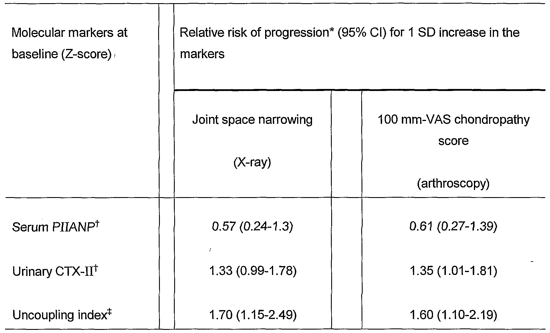
  • the risk of radiological and arthroscopic progression of joint destruction according to baseline levels of molecular markers was estimated by relative risks obtained by logistic regression analyses.
  • Figure 1 is a is graphical illustration of individual values of molecular markers of type II collagen metabolism in 75 patients with knee osteoarthritis. Each value is expressed as a Z-score, i.e. in number of standard deviations from the mean of 58 healthy age-matched controls. The plain lines and associated number represent the mean Z-score. The asterisks (*) represent the statistical significance (p ⁇ 0.0001) of the mean Z-score compared to the value of 0 as assessed by one group t-test.
  • PIIANP refers to N-propeptide of type IIA procollagen
  • CTX-II refers to C-terminal cross-linking telopeptide of type II collagen. Uncoupling index was calculated as the Z-score of urinary CTX-II minus Z-score of serum PIIANP.
  • Figure 2 shows two bar graphs representing analysis of progression of joint damage over one year in patients with low and high levels of molecular markers of type II collagen synthesis and degradation at baseline.
  • Low levels of serum N-propeptide of type IIA procollagen (PIIANP) were those below the mean-1 SD of healthy controls.
  • High levels of urinary C-terminal cross-linking telopeptide of type II collagen (CTX-II) were those which exceeded the mean + 1 SD of healthy controls.
  • the uncoupling Index was calculated as the Z-score of urinary CTX-II minus the Z-score of serum PIIANP.
  • P values refer to the difference between the two groups of baseline levels of molecular markers.
  • Patients with knee OA were classified as low or high baseline serum PIIANP using as a cut-off the mean-1 SD of the controls and low or high baseline urinary CTX-II using as a cut-off the mean + 1 SD of the controls. Using these cut-offs, 46% and 60% of patients were identified as having low serum PIIANP and high urinary CTX-II, respectively. As shown in Figure 2, patients with low baseline serum PIIANP had a higher progression of joint destruction over one year, as assessed either by X-ray or arthroscopy, as compared to the other patients.
  • the regression lines, coefficients of correlation (R) and significance levels (p) were obtained from linear regression analyses.
  • Figure 4 shows two bar graphs analyzing the combination of molecular markers of type II synthesis and degradation to identify patients with knee osteoarthritis at the highest risk of progression of joint damage.
  • Low levels of serum N-propeptide of type IIA procollagen (PIIANP) were those below the mean - 1 SD of healthy controls (Z-score -1).
  • High levels of urinary C-terminal cross-linking telopeptide of type II collagen (CTX-II) were those which exceeded the mean + 1 SD (Z-score +1) or the mean + 2 SDs (Z-score +2) of healthy controls.
  • Uncoupling Index was calculated as the Z-score of urinary CTX-II minus Z- score of serum PIIANP. The numbers over each bar indicate the relative risks (95% confidence intervals).
  • high urinary CTX-II was defined as levels > mean + 2 SDs, which identified a proportion of patients at risk similar to that of serum PIIANP (43%), the relative risk of progression were of 2.7 and 3.3, for X-ray and arthroscopy, respectively.
  • Figure 5 shows individual values of the one year changes in the visual analogue scale (VAS) score of chondropathy according to baseline levels of serum N-propeptide of type IIA procollagen (PIIANP) and urinary C-terminal cross-linking telopeptide of type II collagen (CTX-II).
  • VAS visual analogue scale
  • PIIANP serum N-propeptide of type IIA procollagen
  • CTX-II urinary C-terminal cross-linking telopeptide of type II collagen
  • the cut-off used to separate patients with low and high levels of serum PIIANP was the mean - 1 SD of healthy controls.
  • the cut-off used to separate patients with high and low levels of CTX-II was the mean + 1 SD of healthy controls.
  • the plain horizontal lines represent the median of the one year change in VAS score in the different groups of patients.
  • the horizontal dotted line represents the increase in VAS score over one year (8 units) used to define significant progression (see statistical analyses).
  • One of the main uses of the molecular markers of the invention is to identify patients at high risk for rapid progression of joint destruction who would benefit from chondroprotective therapy, rather than for the diagnosis of OA.
  • clinical indices such as pain and physical function score are poorly related to the destruction of joint structure as was confirmed in this study that there was no association between pain, Lesquesne's functional index, knee effusion and progression.
  • a weak and non significant association between increased urinary CTX-II levels and a lower joint space width in agreement with a previous cross-sectional study in patients with knee OA (Garnero, Ann. Rheum. Dis. 60, supra) was found, whereas serum PIIANP was not predictive.
  • the benefits of the present invention include, in an exemplary embodiment, providing a method for detecting or predicting cartilage destruction in a subject, the method comprising detecting an uncoupling of type II collagen synthesis from type II collagen degradation in the subject.
  • Detecting the uncoupling of type II collagen synthesis from type II collagen degradation in the subject comprises, for example, (a) detecting both a synthesis marker and degradation marker in a biological sample of the subject, (b) comparing the amounts of the synthesis marker and degradation marker, and (c) correlating the relative amounts of the synthesis marker and degradation marker with predetermined standards to detect cartilage destruction in the subject.
  • step (b) is performed at least twice.
  • step (a) is a single step.
  • the synthesis marker is PIIANP and the degradation marker is selected from the group consisting of CTX-II, Type II collagen, Type VI collagen, COMP, keratin sulfate, link protein, aggrecan, and aggrecan fragments.
  • the detection step (a) may also be performed by an assay selected from the group consisting of radioimmunoassays, enzyme immunoassays, ligand assays, immunoradiometric assays, fluoroimmunoassays, and enzyme-linked immunosorbent assays.
  • the assay is an enzyme-linked immunosorbent assay, it is preferably one of a competitive ELISA or sandwich ELISA.
  • the biological samples are preferably selected from the group consisting of blood, serum, urine, sputum, interstitial fluid, joint debris, cartilage fragments and synovial cells.
  • the biological sample is cartilage, more preferably knee cartilage.
  • Another embodiment includes performing the above method on a subject which is a mammal.
  • the mammal is a human.
  • the subject is receiving therapeutic treatment for said cartilage degeneration condition while the above method is being performed.
  • the cartilage degeneration condition may be rheumatoid arthritis and osteoarthritis, but may include other cartilage degeneration conditions having synthesis and degradation markers which may be examined by uncoupling analysis.
  • a method for determining the progress of osteoarthritis or cartilage destruction in a subject comprising quantifying the uncoupling of type II collagen synthesis from type II collagen degradation in the subject.
  • the quantification comprises, for example, (a) measuring an amount of a synthesis marker in a biological sample of the subject, (b) measuring an amount of a degradation marker in the biological sample of the subject, (c) calculating a value of an uncoupling index using the amount of synthesis marker and the amount of degradation marker, and (d) comparing the value of the uncoupling index with predetermined standards to quantify the status of osteoarthritis or cartilage destruction in the subject.
  • Steps (a) and (b) can be performed by an assay selected from the group consisting of radioimmunoassays, enzyme immunoassays, ligand assays, immunoradiometric assays, fluoroimmunoassays, and enzyme-linked immunosorbent assays.
  • Suitable enzyme-linked immunosorbent assays include competitive ELISAs and sandwich ELISAs.
  • the sample is, for example, blood, serum, urine, sputum, interstitial fluid, joint debris, cartilage fragments and synovial cells.
  • the osteoarthritis cartilage synthesis marker is PIIANP
  • the method the osteoarthritis cartilage degradation marker is selected from the group consisting of CTX-II, Type II collagen, Type VI collagen, COMP, keratin sulfate, link protein, aggrecan, and aggrecan fragments.
  • a method for detecting or predicting cartilage degeneration in a subject comprising the steps of (a) providing a first and a second body fluid sample, wherein the first sample is taken from a subject from which status of cartilage degeneration is to be determined and the second sample is taken from the same subject at a later time; (b) providing a first antibody, second antibody, third labeled antibody, and fourth labeled antibody, wherein the first antibody is capable of specifically binding to a human collagen synthesis marker, the second antibody is capable of specifically binding to a human collagen degradation marker, the third labeled antibody is capable of binding to the human collagen synthesis marker, and the fourth labeled antibody is capable of binding to the human collagen degradation marker, and a detecting reagent capable of detecting the label; (c) contacting the first antibody, second antibody, third labeled antibody, fourth labeled antibody, and the detecting reagent with the first body fluid sample; (d) contacting the first antibody, second antibody, third labeled antibody, fourth labele
  • the concentration of human collagen degradation marker and synthesis marker in both the first and second body fluid samples from the subject may be determined by an immunological assay.
  • the immunological assay is selected from the group consisting of radioimmunoassays, enzyme immunoassays, ligand assays, immunoradiometric assays, fluoroimmunoassays, and enzyme-linked immunosorbent assays. More preferably, an enzyme-linked immunosorbent assay is used which is either a competitive ELISA or sandwich ELISA.
  • the body fluid sample of step (a) is preferably selected from the group consisting of blood, serum, urine, sputum, interstitial fluid, joint debris, cartilage fragments and synovial cells.
  • the amount of collagen degradation marker in the second sample is greater than a reference value defined as a mean value plus one standard deviation of the collagen degradation marker concentration in the first sample.
  • the amount of collagen synthesis marker in the second sample is less than a reference value defined as a mean value plus one standard deviation of the collagen synthesis marker concentration in the first sample.
  • both indications are combined to indicate whether a test subject has a high probability of having or being at risk of having progressive cartilage degeneration whereby (a) to indicate that the test subject has a high probability of having or being at risk of having progressive cartilage degeneration, the amount of collagen degradation marker in the second sample is greater than a reference value defined as a mean value plus one standard deviation of the collagen degradation marker concentration in the first sample; and (b) to indicate that the test subject has a high probability of having or being at risk of having progressive cartilage degeneration, the amount of collagen synthesis marker in the second sample is less than a reference value defined as a mean value plus one standard deviation of the collagen synthesis marker concentration in the first sample.
  • the first antibody and second antibody of the above method may be immobilized on a solid surface.
  • the solid surface is a microtiter plate or a dip stick.
  • the solid surface is an instrument in contact with a human joint or a human bloodstream.
  • the osteoarthritis cartilage synthesis marker is PIIANP
  • the osteoarthritis cartilage degradation marker is selected from the group consisting of CTX-II, Type II collagen, Type VI collagen, COMP, keratin sulfate, link protein, aggrecan, and aggrecan fragments.
  • contacting the first antibody, second antibody, third labeled antibody, fourth labeled antibody, and the detecting reagent with the first body fluid sample may be performed simultaneously.
  • contacting the first antibody, second antibody, third labeled antibody, fourth labeled antibody, and the detecting reagent with the first body fluid sample may be performed sequentially.
  • contacting the first antibody, second antibody, third labeled antibody, fourth labeled antibody, and the detecting reagent with the second body fluid sample may be performed simultaneously.
  • contacting the first antibody, second antibody, third labeled antibody, fourth labeled antibody, and the detecting reagent with the second body fluid sample may be performed sequentially.
  • the label of the third and fourth labeled antibodies may comprise biotin, and the third and fourth antibodies may be detected by the method further providing a composition conjugated to streptavidin and adding the composition to the contacted first antibody or second antibody, third labeled antibody, and fourth labeled antibody, wherein the composition is directly detectable or the composition generates a second directly detectable composition.
  • the detectable composition is an enzyme, the enzyme preferably being a peroxidase, more preferably horseradish peroxidase. In another aspect, the above enzyme generates a detectable colored composition.
  • the first antibody or the second antibody is a monoclonal antibody or a polyclonal antibody.
  • the first antibody is a monoclonal antibody and the second antibody is a polyclonal antibody.
  • the cartilage synthesis marker is PIIANP and the cartilage degradation marker is selected from the group consisting of CTX-II, Type II collagen, Type VI collagen, COMP, keratin sulfate, link protein, aggrecan, and aggrecan fragments.
  • the above method steps (a) through (e) may be automated.
  • the patient of the above method may be receiving therapeutic treatment for said cartilage degeneration condition at the same time the method is being performed.
  • the status to be detected by the above method may be progression, decrease or stability of said cartilage degeneration condition.
  • the cartilage degeneration condition may be rheumatoid arthritis, osteoarthritis or other cartilage degeneration conditions detectable by uncoupling synthesis and degradation biological markers.
  • the method for detecting or predicting cartilage degeneration in a subject may comprise the steps of (a) providing a body fluid sample, wherein the sample is taken from a subject from which status of cartilage degeneration is to be determined; (b) providing a first antibody, second antibody and a third labeled antibody, wherein the first antibody is capable of specifically binding to a human collagen synthesis marker, the second antibody is capable of specifically binding to a human collagen degradation marker, and the third labeled antibody is capable of binding to both the human collagen synthesis marker and human collagen degradation marker, and a detecting reagent capable of detecting the label; (c) contacting the first antibody, second antibody, and the third labeled antibody, and the detecting reagent with the body fluid sample; (d) contacting the first antibody, second antibody and the third labeled antibody, and the detecting reagent with the body fluid sample; and, (e) detecting the amount of and determining the concentration of human collagen synthesis marker and collagen degradation marker in the sample, wherein a concentration of human
  • a solid support in contact with a combination of a first antibody and second antibody, wherein the first antibody is capable of specifically binding to a human collagen synthesis marker and the second antibody is capable of specifically binding to a human collagen degradation marker.
  • the first antibody and second antibody of the above method may be immobilized on a solid surface such as a microtiter plate or a dip stick.
  • the solid surface is an instrument in contact with a human joint or a human bloodstream.
  • the osteoarthritis cartilage synthesis marker is, for example, PIIANP
  • the osteoarthritis cartilage degradation marker is, for example, selected from the group consisting of CTX-II, Type II collagen, Type VI collagen, COMP, keratin sulfate, link protein, aggrecan, and aggrecan fragments.
  • a kit for detecting the progression of osteoarthritis comprising instructions setting forth a method comprising the following: (a) providing a first and a second body fluid sample, wherein the first sample is taken from a subject from which status of osteoarthritis is to be determined and the second sample is taken from the same subject at a later time, and (b) detecting the amount of and determining the concentration of human collagen synthesis marker and collagen degradation marker in the first sample to provide a reference value, and detecting the amount of and determining the concentration of human collagen synthesis marker and collagen degradation marker in the second sample, wherein an increased concentration of human collagen degradation marker coupled with a decreased concentration of collagen synthesis marker in the second sample compared to the reference value indicates that the test subject has a high probability of having had or being at risk of progressive osteoarthritis.
  • the kit includes a first antibody, second antibody and a third labeled antibody, wherein the first antibody is capable of specifically binding to a human collagen synthesis marker, the second antibody is capable of specifically binding to a human collagen degradation marker, and the third labeled antibody is capable of binding to both the human collagen synthesis marker and human collagen degradation marker, and a detecting reagent capable of detecting the label.
  • the instructions setting forth the method further include the following, (a) providing a first antibody, second antibody and a third labeled antibody, wherein the first antibody is capable of specifically binding to a human collagen synthesis marker, the second antibody is capable of specifically binding to a human collagen degradation marker, and the third antibody is capable of binding to both the human collagen synthesis marker and human collagen degradation marker, and a detecting reagent capable of detecting the label, (b) contacting the first antibody, second antibody, and the third labeled antibody, and the detecting reagent with the first body fluid sample, and (c) contacting the first antibody, second antibody and the third labeled antibody, and the detecting reagent with the second body fluid sample to detect the amount of and determine the concentration of human collagen synthesis marker and collagen degradation marker in the first sample to provide a reference value, and to detect the amount of and determine the concentration of human collagen synthesis marker and collagen degradation marker in the second sample.
  • the kit in another embodiment, includes a first antibody, second antibody third labeled antibody, and fourth labeled antibody, wherein the first antibody is capable of specifically binding to a human collagen synthesis marker, the second antibody is capable of specifically binding to a human collagen degradation marker, the third labeled antibody is capable of binding to a human collagen synthesis marker and the fourth labeled antibody is capable of binding to a human collagen degradation marker, and a detecting reagent capable of detecting the label.
  • the instructions for detecting the amount of and determining the concentration of human collagen synthesis marker and collagen degradation marker in the first sample to provide a reference value, and for detecting the amount of and determining the concentration of human collagen synthesis marker and collagen degradation marker in the second sample include the following steps: (a) providing a first and a second body fluid sample, wherein the first sample is taken from a subject from which status of cartilage degeneration is to be determined and the second sample is taken from the same subject at a later time; (b) providing a first antibody, second antibody, third labeled antibody, and fourth labeled antibody, wherein the first antibody is capable of specifically binding to a human collagen synthesis marker, the second antibody is capable of specifically binding to a human collagen degradation marker, the third labeled antibody is capable of binding to the human collagen synthesis marker, and the fourth labeled antibody is capable of binding to the human collagen degradation marker, and a detecting reagent capable of detecting the label; (c) contacting the first antibody, second antibody, third labeled antibody, fourth labeled antibody,
  • the invention generally relates to methods, kits and articles of manufacture for detecting and determining the progression of cartilage degeneration diseases, such as osteoarthritis and rheumatoid arthritis, by quantitating collagen synthesis and degradation markers in patient samples.
  • cartilage degeneration diseases such as osteoarthritis and rheumatoid arthritis
  • a joint affected by cartilage degeneration in question expresses collagen synthesis and degradation markers, a change in this value is indicative of a change in the progression of the cartilage degeneration condition.
  • the methods and apparatus of the invention allow accurate determination of the therapeutic effects certain cartilage degeneration drug treatments, including osteoarthritis and rheumatoid arthritis drug treatments, so are also useful for pharmaceutical efficacy studies in mammals.
  • Serum PIIANP 1 (ng/ml) 17.8 ⁇ 20.1 ⁇ 5.4 0.20 18.2 ⁇ 20.2 ⁇ 5.2 0.25 5.7 6.1
  • PIIANP N-propeptide of type IIA procollagen
  • CTX-II C-terminal cross-linking telopeptide of type II collagen
  • PIIANP N-propeptide of type IIA procollagen
  • CTX-II C-terminal cross-linking telopeptide of type II collagen.
  • PIIANP N-propeptide of type IIA procollagen
  • CTX-II C-terminal cross-linking telopeptide of type II collagen

Abstract

The invention relates to methods for determining the progression of cartilage degeneration diseases, such as osteoarthritis and rheumatoid arthritis, by quantitating collagen synthesis and degradation markers in patient samples. One can determine whether a cartilage degeneration condition is progressing, regressing, or remaining stable by quantitating collagen synthesis and degradation markers in patient samples and comparing the value obtained to a reference value. When a joint affected by cartilage degeneration in question expresses collagen synthesis and degradation markers, a change in this value is indicative of a change in the progression of the cartilage degeneration condition. The methods and apparatus of the invention allow accurate determination of the therapeutic effects certain cartilage degeneration drug treatments, including osteoarthritis and rheumatoid arthritis drug treatments, so are also useful for pharmaceutical efficacy studies in mammals. See Figure 1.

Description

UNCOUPLED COLLAGEN SYNTHESIS AND DEGRADATION ASSAYS
CROSS-REFERENCE TO RELATED APPLICATIONS This application claims priority to United States provisional patent application number 60/424,941 , filed November 8, 2002, the specification of which is herein incorporated by reference in its entirety.
BACKGROUND OF THE INVENTION The present invention relates in general to the field of medical diagnostics, and more particularly to assays for determining cartilage degeneration status, including cartilage degeneration status in osteoarthritis ("OA"), rheumatoid arthritis ("RA") status, and status in other arthritic conditions.
The hallmark of OA, the most common cartilage degeneration joint disease, and RA is cartilage loss leading to joint destruction. Knee OA, one of the most common forms of OA, is associated with significant morbidity (Felson, D.T., Epidemiology of Osteoarthritis. In: Brandt K.F., et al., eds., OSTEOARTHRITIS. Oxford University Press, pp. 13-22 (1998)).
To assess the progression of cartilage destruction the most established methods are the measurement of joint space width (JSW) using plain X-rays and the assessment of chondropathy by arthroscopic evaluation of the knee. These two techniques have however some limitations. When there is radiological evidence of OA, there is often already significant joint damage. Because changes of JSW are relatively small compared to the precision error of X-ray measurements, at least one year and preferably 2 years are usually necessary to accurately assess the progression of joint damage or its reduction by treatment (Ravaud, P., et al., Variability in knee radiographing: implication for definition of radiological progression in medial knee osteoarthritis, Ann. Rheum. Dis. 57:624-629 (1998)). Magnetic resonance imaging is more sensitive than plain X-ray, although its reproducibility is not yet fully validated and is currently being optimized for monitoring patients with OA.
Arthroscopy provides a direct and magnified view of the cartilage surface that has prompted some to consider arthroscopy as the gold standard for the assessment of cartilage lesions (Fife, R.S., et al., Relationships between arthroscopic evidence of cartilage damage and radiographic evidence of joint space narrowing in early osteoarthritis of the knee, Arthritis Rheum. 34:377-382 (1991); Ayral, X., et al., Chondroscopy: a new method for scoring chondropathy, Semin. Arthritis Rheum. 22:289-297 (1993)). Arthroscopic scoring systems of chondropathy have been established and validated (Aryal, X., Semin. Arthritis Rheum. 22, supra; Ayral, X., et al., Arthroscopic evaluation of chondropathy in osteoarthritis of the knee, J. Rheumatol. 23:698-706 (1996)). This is however an invasive technique which can not be routinely applied to all patients and which requires trained investigators. Clearly, for identifying patients at high risk for destructive OA and for monitoring drug efficacy there is a need for non-invasive methods that can be repeated and have improved sensitivity compared to plain X-rays.
Molecular markers are molecules or fragments thereof of tissue matrices which are released into biological fluids during the process of tissue biosynthesis and turnover and which can be measured by immunoassays. Molecular markers of bone, cartilage and synovium have been described and their changes have been investigated in patients with OA, mainly in cross-sectional studies (Garnero, P., et al., Molecular basis and clinical use of biochemical markers of bone, cartilage and synovium in joint diseases, Arthritis Rheum. 43:953-961 (2000)). However, detection of molecular markers for collagen synthesis or degradation has not been used to provide information on the progression of OA and other forms of cartilage degeneration.
Because the loss of cartilage is believed to result from the combination of a decreased reparative process coupled with an increased degradative phenomenon (Dean, D . D . , et al. , Evidence for metalloproteinase and metalloproteinase inhibitor imbalance in human osteoarthritic cartilage, J. Clin. Invest. 84:678-685 (1989); Poole, A.R., Cartilage in health and disease. In: Koopman W.J., ed., ARTHRITIS AND ALLIED CONDITIONS: A TEXTBOOK OF RHEUMATOLOGY, Baltimore: Williams & Wilkins, 13 ed., pp. 255-308 (1997)), thereby limiting the capacity of cartilage repair (Campbell, C.J., The healing of cartilage defects, Clin. Orthop. 64:45-63 (1969); Kim, H.K., etal., The potential for regeneration of articular cartilage in defects created by chondral shaving and subchondral abrasion: an experimental investigation in rabbits, J. Bone Joint Surgery Am. 73:1304-1315 (1991)) and because type II collagen is the most abundant protein of cartilage matrix, the assessment of type II collagen synthesis and degradation is an attractive approach for the investigation of OA and other cartilage degeneration conditions.
In vitro studies performed on cartilage tissue from patients with OA and controls have provided evidence of altered synthesis (Nelson, F., et al., Evidence of altered synthesis of type II collagen in patients with osteoarthritis, J. Clin. Invest. 102:2115-2125 (1998)). Certain immunoassays have indicated increased degradation of type II collagen in OA (Hollander, A.P., et al., Increased damage of type II collagen in osteoarthritic articular cartilage detected by a new immunoassay, J. Clin. Invest. 93:1722-1732 (1994); Billinghurst, R.C., et al., Enhanced cleavage of type II collagen by collagenase in osteoarthritic articular cartilage, J. Clin. Invest. 99:1534-1545 (1997); Hollander, A.P., etal., Damage to type II collagen in aging and osteoarthritis starts at the articular surface, originates around chondrocytes and extends into the cartilage with progressive degeneration, J. Clin. Invest. 96:2859-2869 (1995)). However, molecular markers that indicate both collagen synthesis and degradation have remained unavailable. Therefore, accurate and precise assessment of the level and/or progression of OA and other cartilage degenerative conditions in vitro and/or in vivo has remained a significant problem.
Type II collagen is synthesized as a procollagen molecule including the N- (PIINP)and C- (PIICP) propeptides at each end. Type II procollagen is produced in two forms as the result of alternative RNA splicing (Ryan, M.S., et al., Differential expression of a cysteine-rich domain in the amino-terminal propeptide of type II (cartilage) procollagen by alternative splicing of messenger RNA, J. Biol. Chem. 265:10336-10339 (1990); Nah, H.D., et al., Type II collagen mRNA containing an alternatively spliced exon predominates in the chick limb prior to chondrogenesis, J. Biol. Chem. 266:23446-23452 (1991)). One form (IIA) includes and the other form (IIB) excludes a 69 amino acid cysteine-rich globular domain encoded by exon 2 in the PIINP. Type IIB procollagen is expressed at high levels in well- differentiated chondrocytes, forming the framework of normal adult cartilage. On the other hand, type IIA procollagen is temporally expressed in prechondrogenic condensing limb mesenchyme, sclerotome and early cartilage (Sandell, L. J., et al., Alternatively spliced type II procollagen mRNAs define distinct populations of cells during vertebral development: differential expression of the amino-propeptide, J. Cell. Biol. 114:1307-1319 (1991); Sandell, L.J., et al., Alternative splice form of type II procollagen mRNA (IIA) is predominant in skeletal precursors and non-cartilaginous tissues during early mouse development, Dev. Dyn. 199:129-140 (1994); Lui, V.C, etal., Tissue-specific and differential expression of alternatively spliced alpha 1 (II) collagen mRNAs in early embryos, Dev. Dyn. 203:198-211 (1995); Oganesian, A., et al., Type IIA procollagen amino propeptide is localized in human embryonic tissues, J. Histochem. Cytochem. 45:1469-1480 (1997)) and can be re-expressed later in the development at the onset of cartilage hyperthrophy (Nah, H.D., et al., Type IIA * procollagen: Expression in developing chicken limb cartilage and human osteoarthritic articular cartilage, Dev. Dyn. 220:307-322 (2001)). In addition it has recently been shown that type IIA procollagen is re-expressed by adult articular chondrocytes of affected human osteoarthritic cartilage (Nah, Dev. Dyn. 220, supra; Aigner, T., etal., Re-expression of type IIA procollagen by adult articular chondrocytes in osteoarthritic cartilage, Arthritis Rheum. 42:1443-1450 (1999)). During secretion and before incorporation of type II collagen molecules into cartilage matrix, the N and C propeptides are removed by specific enzymes and released in part into the synovial fluid and cleared into the blood. The serum level of these propeptides is thus believed to represent an adequate index of the rate of type II collagen synthesis. The first assays developed to investigate type II collagen synthesis were for PIICP. Nelson, et al. (J. Clin. Invest. 102, supra) showed that PIICP is a valid index of the rate of type II collagen synthesis in healthy and OA cartilage and that serum PIICP levels were decreased in patients with OA. An ELISA was developed for measuring specifically the N-propeptide of type IIA procollagen (PIIANP) with no significant cross- reactivity with type I collagen N-propeptide and reported decreased serum levels of PIIANP in patients with knee OA and RA compared to age-matched healthy controls suggesting a deficit of type II collagen synthesis in joint diseases (Rousseau, J-C, etal., Abstract, Serum levels of type IIA procollagen amino terminal propeptide (PIIANP) are decreased in patients with knee osteoarthritis and rheumatoid arthritis, Arthritis Rheum. 43 (supp.):S351 (2000)). To assess type II collagen degradation, immunoassays using antibodies recognizing either neo-epitopes generated by denaturation of the triple helix domain of type II collagen (Hollander, J. Clin. Invest. 93, supra; Downs, J.T., etal., Analyis of collagenase-cleavage of type II collagen using a neoepitope ELISA, J. Immunol. Methods 247:25-34 (2001)) or cross- linked fragments of the telopeptides (Moskowitz, R.W., et al., Abstract, Type IT C-telopeptide 2B4 epitope is a marker for cartilage degradation in familial osteoarthritis, Arthritis Rheum. 41 (supp.):S352 (1998); Christgau, S., et al., Collagen type II C-telopeptide fragments as an index of cartilage degradation, Bone 29:209-215 (2001)) have been recently developed. Using an assay recognizing C-terminal cross-linking telopeptide of type II collagen (CTX-II) in urine (Christgau, supra), subjects of a cross sectional study showed increased levels of urinary CTX-II in patients with knee OA (Garnero, P., et al., Cross sectional evaluation of biochemical markers of bone, cartilage, and synovial tissue metabolism in patients with knee osteoarthritis: relations with disease activity and joint damage, Ann. Rheum. Dis. 60:619-626 (2001)).
Some methods of detecting OA are presently known in the art. For example, U.S. Patent No. 5,780,240 to Sandell, incorporated herein by reference in its entirety, describes assays to detect cartilage synthesis in OA patients. The assays are useful in providing methods for detecting type IIA mRNA and/or type IIA procollagen/propeptide in samples from non-embryonic individuals. However, while the methods allow determination of whether a patient has OA, they do not provide a method for determining the progress of OA in a patient, how to determine the rate of progression of the disease, how to determine the likelihood of increased or decreased OA progress, nor how to determine the efficacy of drugs on the progress of OA. Likewise, U.S. Patent No. 5,541 ,066 to Sandell, incorporated herein by reference in its entirety, describes assays for determining cartilage synthesis associated with osteoarthritis, but also fails to overcome the limitations in the art with respect to evaluating the progress of disease in a patient with OA.
- A - A cross-sectional study of multiple (fourteen) molecular markers for monitoring osteoarthritis has been described. The markers of inflammation, bone, cartilage and synovium metabolism were segregated into clusters and used in distinguishing osteoarthritis at baseline. Otterness et al., An analysis of 14 molecular markers for monitoring osteoarthritis: segregation of the markers into clusters and distinguishing osteoarthritis at baseline, Osteoarthritis and Cartilage 8:180-185 (2000). Using a principal component analysis, as opposed to an uncoupling analysis, the study reported that molecular markers reflecting cartilage synthesis (serum aggregan epitope 846 and PIICP) and those of cartilage catabolism (serum cartilage oligomeric matrix protein (COMP) and keratan sulfate) segregated into two separate and independent factors and that the combination of these two groups together with tumor necrosis factor receptor type II (a marker of inflammation) provided the best discrimination between patient with knee OA and healthy controls. However, only three markers (tumor necrosis factor receptor II, cartilage oligomeric matrix protein and epitope 846) from independent clusters minimally discriminated osteoarthritis patients from controls. A conclusion of the study was that better markers are needed to accurately and precisely determine the status of osteoarthritis in subjects.
The use of molecular markers of bone formation and bone resorption to determine type I osteoporosis has been described. Eastell, R. et al., Evaluation of bone turnover in type I osteoporosis using biochemical markers specific for both bone formation and bone resorption, Osteoporos Int. 3:255-260 (1993). However, bone synthesis and degradation markers are not associated with collagen synthesis and degradation markers and are not predictive whether such collagen markers could be used in a predictive way with osteoarthritis, rheumatoid arthritis and other cartilage degeneration conditions.
Accordingly, there is a need for cartilage markers that can provide information on collagen metabolism and the progression of the arthritic disease state. Such markers would be useful in estimating the progression of cartilage degeneration diseases such as OA and RA. In addition, such markers would allow accurate determination of the therapeutic effects certain cartilage degeneration drug treatments, including osteoarthritis and rheumatoid arthritis drug treatments, so would be useful for pharmaceutical efficacy studies in mammals.
BRIEF SUMMARY OF THE INVENTION The present invention is based in part on the surprising discovery that decreased serum PIIANP levels, when associated with high cartilage degradative markers, are associated with a fast rate of cartilage loss and alterations of type II collagen synthesis in the signal joint. No correlation between serum PIIANP and urinary CTX-II in patients with knee OA exists, suggesting that the rate of synthesis of type II collagen molecules is independent of the rate of degradation of resident molecules within cartilage matrix in favor of an uncoupling of these two activities. In addition, the invention is based in part on the surprising discovery that the combination of a marker of cartilage synthesis (PIIANP) with that of catabolism (CTX-II) in an uncoupling index improves the ability to discriminate between patients with knee OA and controls as compared with using one of these two markers alone.
Accordingly, in an exemplary embodiment there is provided a method for detecting or predicting cartilage destruction in a subject, the method comprising detecting an uncoupling of type II collagen synthesis from type II collagen degradation in the subject.
In another embodiment, there is provided a method for determining the progress of osteoarthritis or cartilage destruction in a subject comprising quantifying an uncoupling of type II collagen synthesis from type II collagen degradation in the subject
In another embodiment, there is provided a method for detecting or predicting osteoarthritis in a subject, the method comprising the steps of (a) providing a first and a second body fluid sample, wherein the first sample is taken from a subject from which status of osteoarthritis is to be determined and the second sample is taken from the same subject at a later time, (b) providing a first antibody, second antibody, third labeled antibody, and fourth labeled antibody, wherein the first antibody is capable of specifically binding to a human collagen synthesis marker, the second antibody is capable of specifically binding to a human collagen degradation marker, the third labeled antibody is capable of binding to the human collagen synthesis marker, and the fourth labeled antibody- is capable of binding to the human collagen degradation marker, and a detecting reagent capable of detecting the label, (c) contacting the first antibody, second antibody, third labeled antibody, fourth labeled antibody, and the detecting reagent with the first body fluid sample, (d) contacting the first antibody, second antibody, third labeled antibody, fourth labeled antibody, and the detecting reagent with the second body fluid sample, and (e) detecting the amount of and determining the concentration of human collagen synthesis marker and collagen degradation marker in the first sample to provide a reference value and detecting the amount of and determining the concentration of human collagen synthesis marker and collagen degradation marker in the second sample, wherein an increased concentration of human collagen degradation marker coupled with a decreased concentration of collagen synthesis marker in the second sample compared to the reference value indicates that the test subject has a high probability of having had or being at risk of progressive osteoarthritis.
In another embodiment, there is provided a method for detecting or predicting osteoarthritis in a subject, the method comprising the steps of (a) providing a body fluid sample, wherein the sample is taken from a subject from which status of osteoarthritis is to be determined, (b) providing a first antibody, second antibody and a third labeled antibody, wherein the first antibody is capable of specifically binding to a human collagen synthesis marker, the second antibody is capable of specifically binding to a human collagen degradation marker, and the third labeled antibody is capable of binding to both the human collagen synthesis marker and human collagen degradation marker, and a detecting reagent capable of detecting the label, (c) contacting the first antibody, second antibody, and the third labeled antibody, and the detecting reagent with the body fluid sample, (d) contacting the first antibody, second antibody and the third labeled antibody, and the detecting reagent with the body fluid sample, and (e) detecting the amount of and determining the concentration of human collagen synthesis marker and collagen degradation marker in the sample, wherein a concentration of human collagen degradation marker greater than one standard deviation above a predetermined reference value uncoupled with a decreased concentration of collagen synthesis marker less than one standard deviation below a predetermined reference value indicates that the test subject has a high probability of having had or being at risk of progressive osteoarthritis.
In another embodiment, there is provided a solid support in contact with a combination of a first antibody and second antibody, wherein the first antibody is capable of specifically binding to a human collagen synthesis marker and the second antibody is capable of specifically binding to a human collagen degradation marker.
In another embodiment, there is provided a kit for detecting the progression of osteoarthritis comprising instructions setting forth a method comprising the following: (a) providing a first and a second body fluid sample, wherein the first sample is taken from a subject from which status of osteoarthritis is to be determined and the second sample is taken from the same subject at a later time, and (b) detecting the amount of and determining the concentration of human collagen synthesis marker and collagen degradation marker in the first sample to provide a reference value, and detecting the amount of and determining the concentration of human collagen synthesis marker and collagen degradation marker in the second sample, wherein an increased concentration of human collagen degradation marker coupled with a decreased concentration of collagen synthesis marker in the second sample compared to the reference value indicates that the test subject has a high probability of having had or being at risk of progressive osteoarthritis.
Thus, based on the foregoing principles, this invention provides a method for quantitating the progress of osteoarthritis in a subject comprising the steps of: (a) detecting both a synthesis marker and degradation marker in a biological sample of the subject; (b) comparing the amounts of the synthesis marker and degradation marker; and (c) correlating the relative amounts of the synthesis marker and degradation marker with predetermined standards to quantitate the progress of osteoarthritis. Further provided is a method for determining status of osteoarthritis in a patient comprising (a) assaying a sample taken from said patient for collagen synthesis markers and collagen degradation markers; and (b) comparing a value obtained in step (a) to a prior value obtained following assay of a prior sample taken from said patient, any difference there between being indicative of a change in the status of osteoarthritis in the patient.
These and other features, aspects and advantages of the present invention will become better understood with reference to the following description, examples and appended claims.
BRIEF DESCRIPTION OF THE SEVERAL VIEWS OF THE DRAWINGS
Figure 1 is a graphical illustration of individual values of molecular markers of type II collagen metabolism in 75 patients with knee osteoarthritis;
Figure 2 shows graphical illustrations of progression of joint damage over one year in patients with low and high levels of molecular markers of type II collagen synthesis and degradation at baseline;
Figure 3 shows graphical illustrations of the correlation between baseline levels of molecular markers of type II collagen synthesis and degradation and the one year change of knee joint space width in patients with knee OA;
Figure 4 shows graphical illustrations of the combination of measurements of molecular markers of type II synthesis and of degradation in an uncoupling index to identify patients with knee osteoarthritis at the highest risk of progression of joint damage; and
Figure 5 is a graphical illustration of individual values of the one year changes in the visual analogue scale (VAS) score of chondropathy according to baseline levels of serum N- propeptide of type IIA procollagen (PIIANP) and urinary C-terminal cross-linking telopeptide of type II collagen (CTX-II).
DETAILED DESCRIPTION OF THE INVENTION
Abbreviations and Definitions
Unless indicated otherwise, the terms defined below have the following meanings: Therapeutic Treatment: As used herein, the term "therapeutic treatment" refers to receipt by a subject of a therapeutically effective amount of a pharmaceutical.
Therapeutically Effective Amount: As used herein, the term "therapeutically effective amount" refers to those amounts that, when administered to a particular subject in view of the nature and severity of that subject's disease or condition, will have the desired therapeutic effect, e.g. , an amount which will cure, or at least partially arrest or prevent the disease or condition.
The Role of PIIANP and CTX-II in Cartilage Degeneration Cartilage degeneration occurs in many diseases. Two of the most important , cartilage degeneration conditions are OA and RA. The hallmark of OA is the loss of articular cartilage. This loss arises from an imbalance between cartilage synthesis and cartilage degradation over a variable period of time. Applicants have discovered that patients with knee OA are characterized by an uncoupling of type II collagen synthesis and degradation which can be detected by assays for serum PIIANP and urinary CTX-II. Combination of these two markers is useful in identifying patients with knee OA at high risk for rapid progression of joint damage, detecting changes in the progress of the disease and detecting OA inhibition over time using therapeutic agents, among other uses. The present methods and articles of manufacture are therefore useful as aids for diagnosing and monitoring cartilage degeneration in arthritic or pre-arthritic conditions such as osteoarthritis and rheumatoid arthritis, and also are useful as aids in determining the effectiveness of cartilage degeneration therapies, such as arthritis drugs and other therapies.
The present invention is supported by a seventy-five patient study of medial knee OA patients (51 women, 24 men; mean age: 63 ± 8 years, mean disease duration: 4.3 ± 1.5 years). At baseline, serum levels of N-propeptide of type IIA procollagen ("PIIANP") and urinary excretion of C-terminal cross-linking telopeptide of type II collagen ("CTX-II") as markers of type II collagen synthesis and degradation, were measured. Joint Space Width ("JSW") on X-ray and medial chondropathy by arthroscopy (Visual Analogue Scale ("VAS") score, 100 mm) were measured in all patients at baseline and in 52 of them after one year. Progression of joint destruction was defined by a decrease of JSW ± 0.5 mm on X-ray and by an increase of chondropathy in VAS score > + 8.0 units between the baseline and one year evaluation.
At baseline, patients with knee OA, compared to 58 healthy age and sex-matched controls, had decreased serum PIIANP (20 versus 29 ng/ml, p < 0.001) and increased urinary CTX-II (618 versus 367 ng/mmol Cr, p < 0.001). Highest discrimination between patients with OA and controls was obtained by combining PIIANP and CTX-II in an uncoupling index (Z-score CTX-II minus Z-score PIIANP) with a mean Z-score of + 2.9 (p < 0.0001). Increased baseline levels of uncoupling index was associated with greater progression of joint damage evaluated by either changes in JSW (r = -0.46, p = 0.0016) or VAS score (r = 0.36, p =0.014). Patients with both low PIIANP (< mean-1 Standard
Deviation ("SD") of controls) and high CTX-II (> mean + 1 SD of controls) progressed 8 fold more rapidly than the others (p = 0.012 and <0.0001 when progression was assessed by X- ray and arthroscopy, respectively) and had a relative risk of progression of 2.9 (0.80 - 11.1) and 9.3 (2.9 - 39) for X-ray and arthroscopy, respectively.
The rate of type II collagen synthesis and degradation assessed by PIIANP and CTX-II was studied in patients with knee OA and to investigate whether the combined use of these two new molecular markers could predict the progression of joint damage evaluated by both X-ray and arthroscopy of the joints over one year. In this study, the two newly developed molecular markers of type II collagen metabolism provide evidence that patients with knee OA are characterized by an uncoupling of type II collagen synthesis and degradation. The combination of markers of synthesis and catabolism of type II collagen in an uncoupling index is highly predictive of the progression of joint damage suggesting that this index could be useful to identify patients at high risk for cartilage destruction. The methods and apparatus of the invention also allow accurate determination of the therapeutic effects of certain OA drug treatments, so are also useful for pharmaceutical efficacy studies in mammals. It will be understood by those skilled in the art that the methods and apparatus of the present invention can be used in a wide variety of mammalian subjects which include, but are not limited to, humans, rats, mice, rabbits, goats, and sheep. Other farm animals and companion animals such as horse, bovine, dogs and cats are also intended to be subjects of the present invention. Cartilage degeneration can occur as a result of a variety of diseases, OA and RA being the primary diseases of interest. Those skilled in the art will understand that the methods and apparatus of the present invention may be used to predict and detect the progression of all arthritic conditions which result in cartilage degeneration. While the following study focuses on OA progression, the progression of other cartilage degeneration conditions such as RA and other arthritic conditions are not meant to be excluded from the scope of the present invention.
It was discovered that PIIANP is decreased compared to controls (Rousseau, supra) suggesting a deficit of type II collagen synthesis and thus of cartilage repair. However, decreased PIIANP levels measured alone in patients with knee OA cannot predict the progress of the disease. In contrast, the present invention shows that decreased serum
PIIANP levels are associated when uncoupled with degradative markers with a faster rate of cartilage loss and alterations of type II collagen synthesis in the signal joint. No correlation between serum PIIANP and urinary CTX-II in patients with knee OA exists, suggesting that the rate of synthesis of type II collagen molecules is independent of the rate of degradation of resident molecules within cartilage matrix in favor of an uncoupling of these two activities. In addition, the combination of a marker of cartilage synthesis (PIIANP) with that of catabolism (CTX-II) in an uncoupling index allowed improvement of the discrimination between patients with knee OA and controls as compared with using one of these two markers alone. These molecular markers may be used to identify patients at high risk for rapid progression of joint destruction who would benefit from chondroprotective therapy, as opposed to diagnosing OA. Indeed, clinical indices such as pain and physical function score are poorly related to the destruction of joint structure as is confirmed in Example 1 below that there was no association between pain, Lesquesne's functional index, knee effusion and progression. At baseline, it was found that a weak and non significant association between increased CTX-II levels and a lower joint space width in agreement with a previous cross- sectional study in patients with knee OA (Garnero, Ann. Rheum. Dis. 60, supra), whereas PIIANP was not predictive. These data suggest that levels of molecular markers are individually poorly predictive of the current extent of joint damage. However, because molecular markers reflect dynamic changes in cartilage metabolism, they may be more predictive of the rate of cartilage loss in the following years. Longitudinal studies investigating the values of molecular markers to predict progression of joint damage are scarce. A predictive value of serum C-reactive protein (Spector, T.D., et al., Low-levels increases in serum C-reactive protein are present in early osteoarthritis of the knee and predict progressive disease, Arthritis Rheum. 40:723-727 (1997)), COMP (Sharif, M., et al., Relationship between serum cartilage oligomeric matrix protein levels and disease progression in osteoarthritis of the knee joint, Brit. J. Rheumatol. 34:306-310 (1995); Conrozier, T., et al., Serum concentrations of cartilage oligomeric matrix protein and bone sialoprotein in hip osteoarthritis: A one year prospective study, Ann. Rheum. Dis. 9:527-532 (1998)), and hyaluronic acid (Sharif, M., et al., Serum hyaluronic acid level as a predictor of disease progression in osteoarthritis of the knee, Arthritis Rheum. 38:760-767 (1995)), has been found in some but not all studies (Georges, C, et al., Serum biologic markers as predictors of disease progression in osteoarthritis of the knee, Arthritis Rheum. 40:590-591 (1997)). However molecular markers investigated in these previous studies were not specific of joint tissue (Garnero, Arthritis Rheum. 43, supra) and none of them evaluated the metabolism of type II collagen, the main abundant protein of cartilage matrix.
The Example shows that lower baseline levels of PIIANP and higher levels of CTX-II are associated with increased rate of progression of joint damage over one year evaluated either by X-rays or arthroscopy, in agreement with the concept that a decreased reparative process and increased degradation of cartilage matrix will lead to an accelerated rate of joint degradation. When used separately, the association between baseline levels of these two markers and progression was, however, modest and inconsistent across all analyses. In contrast, when PIIANP and CTX-II were combined in an uncoupling index of type II collagen synthesis and degradation, a highly significant correlation was found with progression of joint destruction assessed either by X-ray or arthroscopy. This index accounted for up to 21 % of the inter-individual variability of the progression rate and each unit increase of this index was associated with 60 to 70% increase in the risk of progression. In addition, patients with both low levels of PIIANP and high CTX-II who accounted for about 29% of the population had a 3 to 9 fold increased risk of progression suggesting that this new index may indeed be an important new risk factor for progression of joint damage. Therefore, using the two newly developed specific molecular markers can determine whether patients with knee OA are characterized by depressed type II collagen synthesis and increased type II collagen degradation. Combining these two molecular markers in an uncoupling index allows the identification of patients with a high risk of subsequent progression of joint damage. The presence of PIIANP and CTX-II in the sample being tested can be detected by any means known to the art. For example, type IIA procollagen and/or propeptide can be identified in fluid samples using immunological techniques, or in tissue samples using immunohistochemical techniques. Alternatively, the peptides can be isolated, and sequenced. According to the invention, in preferred form, the biological sample is a fluid (e.g., serum, synovial fluid, or urine) from an adult human individual being tested, and the identifying agents are antibodies (polyclonal or monoclonal) which react with PIIANP and CTX-II proteins. Samples of cartilage for the assays can be obtained by arthroscopy of the joint or upon surgery.
Examples of degradation markers which can be used in the invention include COMP, keratin sulfate, link protein, aggrecan, aggrecan fragments, Type II collagen, and Type VI collagen, among other markers. These markers when measured individually have been attempted to be used to diagnose OA, but not the progression of the disease. The measurement of these markers can be analyzed by uncoupling methods with measurement of cartilage degradation markers, such as CTX-II, however, such that the progression of OA over the course of time may be predicted.
Immunoassays
Antibodies of this invention can be used as probes in detecting certain antigens in human joints. The expression or lack of expression of these antigens can provide clinically exploitable information which is not apparent after standard histopathological evaluations. It is then possible to correlate the phenotypes of individual joint conditions, OA in particular, with various aspects of joint condition and responsiveness to certain types of therapies, thus establishing important classifications of prognosis.
Standard methods using antibodies can be used to detect and quantitate collagen synthesis and degradation markers including, but not limited to, radioimmunoassays ("RIA"), receptor assays, enzyme immunoassays ("EIA"), cytochemical bioassays, ligand assays, immunoradiometric assays, fluoroimmunoassays, and enzyme-linked immunosorbent assays ("ELISA"). Particularly preferred, for ease of detection, and its quantitative nature, is the sandwich or double antibody assay, of which a number of variations exist, all of which are intended to be encompassed by the present invention. These methods are well known and will be understood by those skilled in the art to require a reasonable amount of experimentation to optimize the interaction between antibodies and antigens and the detection of the antigens by the antibodies. These and other immunoassay techniques may be found in PRINCIPLES AND PRACTICE OF IMMUNOASSAY, 2ND EDITION, Price and Newman, eds., MacMillan (1997) and ANTIBODIES, A LABORATORY MANUAL, Harlow and Lane, eds., Cold Spring Harbor Laboratory, Ch. 9 (1988), each of which is incorporated herein by reference in its entirety.
The use of antibodies described herein can be used to screen human biological fluids for the presence of a specific antigen, preferably collagen synthesis and degradation markers. In vitro immunoserological evaluation of sera withdrawn from patients thereby permits non-invasive determination of the progression of cartilage degeneration. By way of illustration, human fluids, such as blood serum or urine, can be taken from a patient and assayed for a specific epitope, either as released antigen or membrane-bound on cells in the sample fluid, using the anti-marker antibodies in standard RIAs or ELISAs known in the art. The antibodies used in such methods are preferably monoclonal antibodies. Particularly preferred, for ease of detection and because of its quantitative nature, is the sandwich or double antibody assay of which a number of variations exist, all of which are intended to be encompassed by the present invention. For example, in a typical forward sandwich assay, unlabeled antibody is immobilized on a solid substrate, e.g., microtiter plate wells, and the sample to be tested is brought into contact with the bound molecule. After incubation for a period of time sufficient to allow formation of an antibody-antigen binary complex, a second antibody labeled with a reporter molecule capable of inducing a detectable signal is then added. Incubation is continued allowing sufficient time for binding with the antigen at a different site and the formation of a ternary complex of antibody- antigen-labeled antibody. Any unreacted material is washed away, and the presence of the antigen is determined by observation of a signal which may be quantitated by comparison with a control sample containing known amounts of antigen. Variations on the forward sandwich assay include the simultaneous assay in which both sample and antibody are added simultaneously to the bound antibody, or a reverse sandwich assay in which the labeled antibody and sample to be tested are first combined, incubated and added to the unlabelled surface bound antibody. These techniques are well known to those skilled in the art, and the possibility of minor variations will be readily apparent. As used herein, the term "sandwich assay" is intended to encompass all variations on the basic two-site technique. One limiting factor of the sandwich assays techniques of the present invention requires that both antibodies have different binding specificities for the collagen synthesis and degradation markers. Thus, a number of possible combinations are possible. As a more specific example, in a typical forward sandwich assay, a primary antibody is either covalently or passively bound to a solid support. The solid surface is usually glass or a polymer, the most commonly used polymers being cellulose, polyacrylamide, nylon, polystyrene, polyvinylchloride or polypropylene. The solid supports may be in the form of tubes, beads, discs or microplates, or any other surfaces suitable for conducting an immunoassay. The binding processes are well known in the art. Following binding, the solid phase-antibody complex is washed in preparation for the test sample. An aliquot of the body fluid containing the collagen synthesis and degradation markers to be tested is then added to the solid phase complex and incubated at 25 °C for a period of time sufficient to allow binding of any collagen synthesis or degradation marker present to the antibody specific for collagen synthesis or degradation marker. The second antibody is then added to the solid phase complex and incubated at 25 °C. for an additional period of time sufficient to allow the second antibody to bind to the primary antibody-antigen solid phase complex. The second antibody is linked to a reporter molecule, the visible signal of which is used to indicate the binding of the second antibody to any antigen in the sample. The term "reporter molecule" as used in the present invention is meant a molecule which by its chemical nature provides an analytically detectable signal which allows the detection of antigen-bound antibody. Detection must be at least relatively quantifiable to allow determination of the amount of antigen in the sample. The signal may be calculated in absolute terms or may be calculated in comparison with a standard (or series of standards) containing a known normal level of antigen. The most commonly used reporter molecules of this type of assay are either enzymes or fluorophores. In the case of an EIA, an enzyme is conjugated to the second antibody, often by means of glutaraldehyde or periodate. As will be apparent to those skilled in the art, a wide variety of different conjugation techniques exist. Commonly used enzymes include horseradish peroxidase, glucose oxidase, β-galactosidase and alkaline phosphatase, among others. The substrates to be used with the specific enzymes are generally chosen for the production, upon hydrolysis by the corresponding enzyme, of a detectable color change. For example, p-nitrophenyl phosphate is suitable for use with alkaline phosphatase conjugates; for peroxidase conjugates, 1 ,2-phenylenediamine or toluidine are commonly used. It is also possible to employ fluorogenic substrates which yield a fluorescent product rather than the chromogenic substrates noted above. In all cases, the enzyme-labeled antibody is added to the first antibody-synthesis marker or antibody- degradation marker complex and allowed to bind to the complex, then the excess reagent is washed away. A solution containing the appropriate substrate is then added to the tertiary complex of antibody-antigen-labeled antibody. The substrate reacts with the enzyme linked to the second antibody, giving a qualitative visual signal, which may be further quantitated, usually spectrophotometrically, to give an evaluation of the amount of antigen which is present in the serum sample.
Alternatively, fluorescent compounds such as fluorescein or rhodamine may be chemically coupled to antibodies without altering their binding capacity. When activated by illumination with light of a particular wavelength, the fluorochrome-labeled antibody absorbs the light energy inducing a state of excitability in the molecule followed by emission of the light at a longer wavelength. The emission appears as a characteristic color visually detectable with a light microscope. As in the EIA, the fluorescent-labeled antibody is allowed to bind to the first antibody-synthesis marker or antibody-degradation marker complex. After washing the unbound reagent, the remaining ternary complex is then exposed to light of the appropriate wavelength, and the fluorescence observed indicates the presence of the antigen. Immunofluorescence and EIA techniques are both very well established in the art and are particularly preferred for the present method. However, other reporter molecules, such as radioisotopes, chemiluminescent or bioluminescent molecules may also be employed. It will be readily apparent to those skilled in the art how to vary the procedure to suit the required use. In yet another alterative embodiment, the human sample to be tested which contains the collagen synthesis or degradation markers may be used in a single site immunoassay wherein it is adhered to a solid substrate either covalently or noncovalently. An unlabeled anti-synthesis marker or anti-degradation marker antibody is brought into contact with the sample bound on the solid substrate. After a suitable period of incubation sufficient to allow formation of an antibody-antigen binary complex, a second antibody, labeled with a reporter molecule capable of inducing a detectable signal is added and incubation is continued allowing sufficient time for the formation of a ternary complex of antigen-antibody-labeled antibody. For the single site immunoassay, the second antibody may be a general antibody, i.e., zenogeneic antibody to immunoglobulin, particularly anti-(lgM and IgG) linked to a reporter molecule, that is capable of binding an antibody that is specific for the synthesis marker or degradation marker of interest. In a competitive ELISA, a patient serum and an antigen-specific conjugate are co- incubated with a captured antigen. The amount of color developed is inversely proportional to the amount of antigen-specific patient immunoglobulin present. Careful standardization is required to interpret the results. These and other immunoassays are within the scope of the presentation.
Histological Techniques
Antibodies against collagen synthesis and degradation markers can also be used to detect collagen synthesis and degradation markers in histological and cytological specimens, and in particular, to determine the progression of cartilage degeneration based on staining patterns and intensities. For example, staining patterns can be observed by using an immunostaining technique and monoclonal antibodies against chondrocyte or other joint cells or tissues with a small degree of heterogeneity. Using this method, morphologically osteoarthritic cells can generally be seen to exhibit an increased degree of staining when compared to non-osteoarthritic cells. Non-specific staining will preferably be absent on non-collagen or other cartilage components not affected by cartilage degeneration of the joint specimen.
Immunofluorescent histological techniques can also be used to examine human specimens with monoclonal antibodies. In a typical protocol, slides containing cryostat sections of frozen, unfixed tissue biopsy samples or cytological smears are air dried, formalin fixed and incubated with the monoclonal antibody preparation in a humidified chamber at room temperature. The slides are then layered with a preparation of antibody directed against the monoclonal antibody, usually some type of anti-mouse immunoglobulin if the monoclonal antibodies used are derived from the fusion of a mouse spleen lymphocyte and a mouse myeloma cell line. This antimouse immunoglobulin is tagged with a compound that fluoresces at a particular wavelength, e.g., rhodamine or fluorescein isothiocyanate. The staining pattern and intensities within the sample are then determined by fluorescent light microscopy and optionally photographically recorded. See Aigner, et al., supra for a discussion of histological methods which may be used within the scope of the present invention.
Monoclonal Antibodies
Monoclonal antibodies which can be used in the invention can be produced by a hybridoma using methods well known in the art. Various additional procedures known in the art may be used for the production of antibodies to epitopes of the collagen synthesis and degradation markers. Such antibodies include but are not limited to polyclonal, monclonal, chimeric, single chain, Fab fragments and a Fab expression library. For the production of antibodies, various host animals may be immunized by injection with a particular collagen synthesis or degradation marker, or a synthetic collagen synthesis or degradation marker, including but not limited to rabbits, mice, rats, goats, horses, pigs, among other animals. Various adjuvants may be used to increase the immunological response, depending on the host species, including but not limited to Freund's (complete and incomplete), mineral gels such as aluminum hydroxide, surface active substances such as lysolecithin, pluronic polyols, polyanions, peptides, oil emulsions, keyhole limpet hemocyanin, dinitrophenol, and potentially useful human adjuvants such as BCG (bacille Calmette-Guerin) and corynebacterium parvum. Monoclonal antibodies to peptides of collagen synthesis and degradation markers may be prepared by using any technique which provides for the production of antibody molecules by continuous cell lines in culture. These include, but are not limited to the hybridoma technique originally described by Kohler and Milstein, (Nature, 256:495-497 (1975)). Monoclonal antibodies specific to collagen synthesis and degradation markers may be produced in germ-free animals utilizing recent technology (PCT/US90/02545). Human antibodies may be used and can be obtained by using human hybridomas (Cote, et al., Proc. Natl. Acad. Sci., 80:2026-2030 (1983)) or by transforming human B cells with EBV virus in vitro (Cole, et al., In: MONOCLONAL ANTIBODIES AND CANCER THERAPY, Alan R. Liss, pp. 77- 96 (1985)). In addition, techniques developed for the production of chimeric antibodies by splicing the genes from a mouse antibody molecule of appropriate antigen specificity together with genes from a human antibody molecule of appropriate biological activity can be used (Morrison, etal., Proc. Natl. Acad. Sci., 8(1 ):6851 -6855 (1984); Neuberger, et al., Nature, 312:604-608 (1984); Takeda, etal., Nature, 314:452-454 (1985)).
Monoclonal or polyclonal antibodies specific for the substrate of interest which may advantageously be used as anchoring antibodies should be prepared against unique epitopes of the substrate to minimize cross reactions with other substrates. Similarly, the detection antibody should only react with a collagen synthesis or degradation marker. Monoclonal antibodies or polyclonal antibodies with high binding affinities for such unique epitopes may be used, preferably those that will not interfere with ligand binding if the substrate is also a receptor. For example, where the substrate is a receptor involved in signal transduction, the extracellular domain, which bears unique epitopes that may be involved in ligand recognition and binding, may advantageously be used as the immunogen. For non-receptor substrates where the amino acid sequence is known, non-conserved, variable regions of the protein may be used as immunogens. Solid Phase Systems
The solid phase used in the assays of this invention may be any surface commonly used in immunoassays. For example, the solid phase may be particulate; it may be the surface of beads, for example, glass or polystyrene beads; or it may be the solid wall surface of any of a variety of containers, for example, centrifuge tubes, columns, microtiter plate wells, filters, membranes and tubing, among other containers. The invention also includes solid supports which may be attached to the surface of a surgical device or other instrument which directly contacts fluid to be studied while the instrument is within a patient. The fluid in direct contact with the instrument may be in the joint, blood stream or other tissue which contains collagen synthesis or degradation markers.
When particles are used as the solid phase, they will preferably be of a size in the range of from about 0.4 to 200 microns, more usually from about 0.8 to 4.0 microns.
Magnetic or magnetizable particles such as, paramagnetic particles (PMP), are a preferred particulate solid phase, and microtiter plate wells are a preferred solid wall surface.
Magnetic or magnetizable particles may be particularly preferred when the steps of the methods of this invention are performed in an automated immunoassay system.
Preferred detection/quantitation systems of this invention may be luminescent, and a luminescent detection/quantitation system in conjunction with a signal amplification system could be used, if necessary. Exemplary luminescent labels, preferably chemiluminescent labels, are detailed below, as are signal amplification systems.
Signal Detection/Quantitation Systems
The complexes formed by the assays of this invention can be detected, or detected and quantitated, by any known detection/quantitation systems used in immunoassays. As appropriate, the antibodies of this invention used as tracers may be labeled in any manner directly or indirectly that results in a signal that is visible or can be rendered visible.
Detectable marker substances include radionuclides, such as 3H, 125l, and 13 l; fluorescers, such as, fluorescein isothiocyanate and other fluorochromes, phycobiliproteins, phycoerythin, rare earth chelates, Texas red, dansyl and rhodamine; colorimetric reagents (chromogens); electron-opaque materials, such as colloidal gold; bioluminescers; chemiluminescers; dyes; enzymes, such as, horseradish peroxidase, alkaline phosphatase, glucose oxidase, glucose-6-phosphate dehydrogenase, acetylcholinesterase, o , β- galactosidase, among others; coenzymes; enzyme substrates; enzyme cofactors; enzyme inhibitors; enzyme subunits; metal ions; free radicals; or any other immunologically active or inert substance which provides a means of detecting or measuring the presence or amount of immunocomplex formed. Exemplary of enzyme substrate combinations are horseradish peroxidase and tetramethyl benzidine (TMB), and alkaline phosphatase and paranitrophenyl phosphate (pNPP).
Preferred detection, or detection and quantitation systems according to this invention produce luminescent signals, bioluminescent (BL) or chemiluminescent (CL). In BL or CL assays, the intensity or the total light emission is measured and related to the concentration of the analyte. Light can be measured quantitatively using a luminometer (photomultiplier tube as the detector) or charge-coupled device, or qualitatively by means of photographic or X-ray film. The main advantage of using such assays is their simplicity and analytical sensitivity, enabling the detection and/or quantitation of very small amounts of analyte. Exemplary luminescent labels are acridinium esters, acridinium sulfonyl carboxamides, luminol, umbelliferone, isoluminol derivatives, photoproteins, such as aequorin, and luciferases from fireflies, marine bacteria, Vargulla and Renilla. Luminol can be used optionally with an enhancer molecule, preferably selected from the group consisting of 4-iodophenol or 4-hydroxycinnamic acid. Acridinium esters are one of the preferred types of CL labels according to this invention. A signal is generated by treatment with an oxidant under basic conditions.
Also preferred luminescent detection systems are those wherein the signal is produced by an enzymatic reaction upon a substrate. BL and CL detection schemes have been developed for assaying alkaline phosphatase (AP), glucose oxidase, glucose 6- phosphate dehydrogenase, horseradish peroxidase (HRP), and xanthine-oxidase labels, among others. AP and HRP are two preferred enzyme labels which can be quantitated by a range of BL and CL reactions. For example, AP can be used with a substrate, such as an adamantyl 1 ,2-dioxetane aryl phosphate substrate (e.g., AMPPD or CSPD; (Kricka, L. J., "Chemiluminescence and Bioluminescence, Analysis by," at p. 167, Molecular Biology and Biotechnology: A Comprehensive Desk Reference, ed. R. A. Meyers, VCH Publishers; N.Y., N.Y.; 1995)); preferably a disodium salt of 4-methoxy-4-(3-phosphatephenyl)spiro [1 ,2- dioxetane-3,2'-adamantane], with or without an enhancer molecule, preferably, 1- (trioctylphosphonium methyl)-4-(tributylphosphonium methyl) benzene diochloride. HRP is preferably used with substrates, such as, 2', 3', 6'-trifluorophenyl 3-methoxy-10- methylacridan-9-carboxylate.
BL and CL reactions can also be adapted for analysis of not only enzymes, but other substrates, cofactors, inhibitors, metal ions and the like. For example, luminol, firefly luciferase, and marine bacterial luciferase reactions are indicator reactions for the production or consumption of peroxide, ATP, and NADPH, respectively. They can be coupled to other reactions involving oxidases, kinases, and dehydrogenases, and can be used to measure any component of the coupled reaction (enzyme, substrate, cofactor). The detectable marker may be directly or indirectly linked to an antibody used in an assay of this invention. Exemplary of an indirect linkage of the detectable label is the use of a binding pair between the antibody and the marker, or the use of well known signal amplification signals, such as, using a biotinylated antibody complexed to UGP and then adding streptavidin conjugated to HRP and then TMB.
Exemplary of binding pairs that can be used to link antibodies of assays of this invention to detectable markers are biotin/avidin, streptavidin, or anti-biotin; avidin/anti- avidin; thyroxine/thyroxine-binding globulin; antigen/antibody; antibody/anti-antibody; carbohydrate/lectins; hapten/anti-hapten antibody; dyes and hydrophobic molecules/hydrophobic protein binding sites; enzyme inhibitor, coenzyme or cofactor/enzyme; polynucleic acid/homologous polynucleic acid sequence; fluorescein/anti- fluorescein; dinitrophenol/anti-dinitrophenol; vitamin B12/intrinsic factor; cortisone, cortisol/cortisol binding protein; and ligands for specific receptor protein/membrane associated specific receptor proteins. Preferred binding pairs according to this invention are biotin/avidin or streptavidin, more preferably biotin/streptavidin.
Various means for linking labels directly or indirectly to antibodies are known in the art. For example, labels nay be bound either covalently or non-covalently. Exemplary antibody conjugation methods are described in: Avarmeas, etal., Scan. J. Immunol., 8 (Suppl. 7):7 (1978); Bayer, et al., Meth. Enzymol., 62:308 (1979); Chandler, etal., J. Immunol. Meth., 53:187 (1982); Ekeke and Abuknesha, J. Steroid Biochem., 11 :1579
(1979); Engvall and Perlmann, J. Immunol., 109:129 (1972); Geoghegan, etal., Immunol. Comm., 7:1 (1978); and Wilson and Nakane, Immunofluorescence and Related Techniques, p. 215 (Elsevier/North Holland Biomedical Press; Amsterdam (1978)).
Depending upon the nature of the label, various techniques can be employed for detecting, or detecting and quantitating the label. For fluorescers, a large number of fluorometers are available. For chemiluminescers, luminometers or films are available. With enzymes, a fluorescent, chemiluminescent, or colored product can be determined or measured fluoromethcally, luminometrically, spectrophotometrically or visually.
Automated Immunoassay System
The methods of this invention can be readily adapted to automated immunochemistry analyzers. To facilitate automation of the methods of this invention and to reduce the turnaround time, anti-UGP antibodies may be coupled to magnetizable particles.
A preferred automated/immunoassay system is the Ciba Corning ACS: 180™ Automated Chemiluminescence System (CCD; Medfield, Mass. (USA)). The Ciba Corning
ACS:180™ Automated Immunoassay System is described in Dudley, B.S., J. Clin.
Immunoassay. 14(2):77 (Summer 1991). The system uses chemiluminescent labels as tracers and paramagnetic particles as solid-phase reagents. The ACS: 180 system accommodates both competitive binding and sandwich-type assays, wherein each of the steps are automated. The ACS:180 uses micron-sized paramagnetic particles that maximize the available surface area, and provide a means of rapid magnetic separation of bound tracer from unbound tracer without centrifugation. Reagents can be added simultaneously or sequentially. Other tags, such as an enzymatic tag, can be used in place of a chemiluminescent label, such as, acridinium ester.
Assays of Urine The assays of the present invention may also be adapted to a "dip stick" format according to U.S. Patent Nos. 6,352,862 and 5,602,040, each of which is incorporated herein by reference in its entirety, or similar apparatus.
Assays of Whole Blood The assays of the present invention may also be adapted to a "dip stick" format according the U.S. Reissue Patent No. RE035306, incorporated herein by reference in its entirety, or similar apparatus.
Kits Further, the present invention provides a kit for detecting cartilage degeneration status, such as the progression of cartilage degeneration, which is applicable for the practice of the method of the present invention.
The kit comprises an antibody specific to collagen synthesis and degradation markers, whereby the detection of cartilage degeneration and other cartilage degeneration conditions can be carried out using the antibody in an immunological assay. When a two antibody sandwich ELISA is employed as the immunological assay, the kit may comprise first and second antibodies specific to collagen synthesis and degradation markers. The second antibody is preferably capable of binding to a conjugate of the collagen synthesis and degradation markers and the first antibody. For this purpose, for example, an antibody that recognizes an epitope different from that recognized by the first antibody may be used as the second antibody. It is preferable that the first and second antibodies be monoclonal antibodies.
The kit of the present invention may further comprise a substance and/or a device suitable for the detection of antibodies, the immobilization of antibodies, and the like. To immobilize the antibodies, the kit may further comprise a carrier (e.g., a microtiter plate), a solution for the immobilization (e.g., carbonate buffer) and a blocking solution (e.g., gelatin- containing PBS). For the detection of the antibodies, it is preferable that the antibodies be labeled previously. In this case, the kit may further comprise a detecting reagent for detecting the label. For example, when biotin is used as the labeling substance, the detecting reagent may comprise a conjugate of streptavidin with horseradish peroxidase (HRP) as well as a color-developing solution that is capable of developing a color by the action of HRP.
In one embodiment, the kit will include instructions setting forth at least that (a) a first and a second body fluid sample are to be provided, wherein the first sample is taken from a subject from which status of osteoarthritis is to be determined, and the second sample is taken from the same subject at a later time, and (b) the amount of, and the concentration of the human collagen synthesis marker and of the collagen degradation marker in the first sample are detected to provide a reference value, and the amount of and concentration of the human collagen synthesis marker and of the collagen degradation marker in the second sample are detected, wherein an increased concentration of human collagen degradation marker coupled with a decreased concentration of collagen synthesis marker in the second sample compared to the reference value indicates that the test subject has a high probability of having had or being at risk of progressive osteoarthritis.
These and other features, aspects and advantages of the present invention will become better understood with reference to the following description, examples and appended claims.
EXAMPLE
The following experimental example describing evaluation of subjects exhibiting OA symptoms is offered by way of illustration and not by way of limitation.
Patients with knee osteoarthritis and healthy controls 75 patients (51 women, 24 men; mean age: 63.0 ± 8.0 years) were investigated who met the American College of Rheumatology criteria for primary knee OA (Altman, R., et al., Development of criteria for the classification and reporting of osteoarthritis. Classification of osteoarthritis of the knee. Diagnostic and Therapeutic Criteria Committee of the American Rheumatism Association, Arthritis Rheum. 29:1039-49 (1986)). These patients were part of two larger randomized, multicenter, double blind studies comparing the efficacy of diacerhein and Tenidap and including respectively 61 and 665 patients (Ayral, X., et al., Abstract, Progression of knee osteoarthritis assessed by arthroscopy: a multicenter, randomized, double blind comparison of tenidap sodium versus piroxicam, Ann. Rheum. Dis. 59 (supp.):S54 (2000)). Other inclusion criteria were: 1) clinical involvement (pain) of the medial femorotibial compartment, 2) active disease defined by pain of any duration in the signal knee on at least 30 days in the last 2 months and by inadequate pain control certified by prior failure of intra-articular glucocorticoid injection, physical exercises, analgesics and non steroidal anti-inflammatory drugs, and justifying joint lavage, 3) absence of advanced disease defined by a radiological joint space width of 2 mm or greater at the narrowest point of the medial femorotibial compartment of the signal joint on weight-bearing radiographs with knees fully extended, 4) presence of chondropathy of the medial compartment at entry which was approved by the central reader of video made during the arthroscopy screening. For approval, at least 10 % of one articular surface of the medial compartment (femoral condyle or tibial plateau) had to be affected by superficial fibrillation or worse and if 90 % or greater of both surfaces showed absence of cartilage, the patient was not eligible, 5) no intra- articular lesion requiring surgery (e.g. of meniscus, cartilage or ligament) at the arthroscopy screening or likely to require surgery during study period, 6) no intra-articular injection within the 3 months prior to the arthroscopy screening and 7) absence of contraindication of arthroscopy (anticoagulant therapy, cutaneous lesion of the knee, allergy to lidocaine).
All women were postmenopausal and all patients were without treatments that could interfere with bone metabolism including estrogen replacement. Fifty two of these 75 patients were evaluated after one year for progression of joint destruction assessed by both X-ray and arthroscopy.
Healthy subjects included 38 postmenopausal women (mean age: 63.2 ± 8.1 years from 50 to 80 years) and 20 men aged from 53 to 79 years (mean; 62 ± 8.2 years). Menopausal status was defined as the absence of menses for at least 12 months. Healthy women and men were randomly selected from two large population based-cohorts involved in prospective studies on the determinants of bone loss in women (OFELY study) and men (MINOS study) (Garnero, E., etal., Increased bone turnover in late postmenopausal women is a major determinant of osteoporosis, J. Bone Miner. Res. 11 :337-349 (1996); Szulc, P., et al., Cross-sectional assessment of age-related bone loss in men: The MINOS Study, Bone 26:123-129 (2000)) he cohort of the OFELY study comprises 1039 healthy female volunteers, 31-89 years of age randomly selected from affiliates of the section of a health insurance company (Mutuelle Generate de I'Education Nationale) from the Rhone district in France. The cohort of the MINOS study comprises 842 healthy male volunteers, 50-85 years of age, randomly selected from the affiliated of the section of a health insurance company (Societe de Secours Miniere de Bourgogne) in Montceau les Mines, a town located in the same region as that of the healthy women. All healthy women and men did not have evidence of symptomatic OA as assessed by clinical examination of the hands performed by an experienced rheumatologist and by using the following question: "Has a doctor ever told you that you had osteoarthritis". Furthermore, X-ray films of the thoracic and lumbar spine were obtained in all subjects. Spine films were graded with a standard atlas to document the severity of disc degeneration and osteophyte formation using the method of Lane, et al. (Reliability of new indices of radiographic osteoarthritis of the hand and hip and lumbar spine degeneration, J. Rheumatol. 20:1911-1918 (1993))with a grade of 0 as normal, 1 for mild narrowing and or mild osteophytes, 2 moderate-severe (2-3) narrowing and or moderate- severe (2-3) osteophytes. Subjects with a grade of 2 (moderate-severe) were excluded from the control group. All subjects were healthy without any disease or treatment that could interfere with bone or joint metabolism including hormone replacement therapy in postmenopausal women.
This prospective, longitudinal study of one-year duration was approved by the local ethical committee and informed consent was obtained from all patients.
Clinical variables
At baseline, pain was evaluated in all patients using a 100 mm visual analogue scale (VAS), functional disability was assessed by the Lequesne's functional index (Lequesne, M., et al., Indexes of severity for osteoarthritis of the hip and knee. Validation-value in comparison with other assessment tests, Scand. J. Rheumatol. 85 (supp. 65):85-89 (2000)), disease duration and presence or absence of knee effusion were collected. Additionally the presence of concomitant osteoarthritis affected joints was evaluated using the articular index whose numerical value is the sum of the total cartilage surface area (as cm2) of each symptomatic joint as previously reported (Goldberg, R.L., et al., Elevated plasma levels of hyaluronate in patients with osteoarthritis and rheumatoid arthritis, Arthritis Rheum. 34:799- 806 (1991)).
Radiological variables
Radiological evaluation consisted of bilateral anteroposterior weight-bearing knee radiographs with the knee fully extended. Radiographs were taken in a single radiography unit by the same staff of 3 technicians using a standardized technique: 1) patient positioning: patients stand with knees fully extended, weight equally distributed on both legs, back of knees in contact with the cassette, feet rotated so that tibial spines are centralized in femoral notch, 2) radiographic procedure: both knees are X-rayed together, the X-ray beam is directed at center of the medial compartment of the signal knee and with the aid of fluoroscopy the beam is placed parallel to the medial tibial plateau with a tube to film distance of 1.10 m. the same procedure was followed for the second X-ray after 12 months.
The severity of OA of the medial femorotibial compartment was evaluated by measuring the joint space width, i.e., the inter-bone distance between the medial femoral condyle and the medial tibial plateau at the narrowest point in millimeters (Ravaud, supra). The paired radiographs of each patient at entry and after one year were analyzed by a single investigator using a blind procedure in which the investigator was unaware of patient identity and of the chronology of the radiographs.
Arthroscopy of the knees Arthroscopy of the knee was performed under local anaesthesia, without tourniquet hemostasis, with a 2.7 mm Storz Arthroscope (Storz, Paris, France) having a 30° fore oblique lens, using inferolateral approach. Arthroscopic exploration was combined with joint lavage, consisting of 1 liter of normal saline. The arthroscopic evaluation focused on the symptomatic compartment, i.e., the medial femorotibial compartment. Each arthroscopy was recorded on a VHS videotape (Sony, Tokyo, Japan). Arthroscopies were performed by 3 trained arthroscopists and recorded on the same planned, systematic, standardized manner. All videotapes were analyzed. Arthroscopies were scored for chondropathy using the overall assessment of the investigator by a 100 mm VAS in which 0 indicates the absence of chondropathy and 100 the most severe chondropathy as previously reported (Ayral, Semin. Arthritis Rheum. 22, supra). One VAS is used for each articular surface of the medial compartment: medial femoral condyle and medial tibial plateau. A VAS score is obtained by averaging the VAS scores from the two corresponding articular surfaces of the compartment. The paired arthroscopy videotapes of each patient at entry and after one year were analyzed, using a blind procedure in which the investigator was unaware of patient identity and of the chronology of the videotapes.
Biochemical measurements
Fasting blood samples were collected before 10:00 a.m. in plain tubes containing separation gel. They were allowed to stand for 20 minutes and then centrifuged for 10 minutes at 2500 rpm. The serum was then mixed and split in different aliquots. The interval between blood drawing and freezing at -70°C was less than 2 hours. Fasting second morning void urine samples were also collected in plastic containers. After mixing the whole collection, aliquots of urine were transferred into plastic tubes and frozen at -70°C without any acidification. Both serum and urine samples were obtained in all OA patients on the day pain and function were assessed, the radiographs were taken and arthroscopy performed. Samples were obtained before arthroscopic exploration. Samples were obtained in controls according to the same procedure. All biological samples were kept frozen at-70°C until assayed. Molecular marker of type II collagen synthesis
Synthesis of type II collagen was evaluated by measuring serum PIIANP using a recently developed ELISA (Rousseau, supra). This ELISA is based on a rabbit polyclonal antiserum raised against the recombinant human GST-exon 2 fusion protein (Oganesian, supra) and recombinant human GST-exon 2 as a standard. The specificity of the antibody against PIIANP was previously demonstrated by Western blot analyses against the recombinant exon 2 protein (prior and after cleavage with thrombin) and against type IIA procollagen isolated from the culture medium of human fetal ribs (Oganesian, supra). Briefly, microtiter plates are coated overnight at4°C with 100 μl (10 ngΛnl) of GST-exon 2 protein and then saturated for 2 hours at room temperature with phosphate buffer saline, 1% bovine serum albumin (Sigma, St Louis, MO). After washing, 100 μl of standards or serum samples are incubated for 4 hours at room temperature together with 100 μl of anti-GST- exon 2 antiserum at the adequate dilution (1/1650). After washing, 100 μl of a peroxydase conjugated anti-rabbit antibody (Sigma, St Louis, MO) is added into each well and incubated for 1 hour at room temperature. After washing, 100 μl of H202/tetramethylbenzidine substrate indicator solution (Sigma, St Louis, MO) are added into each well. After 30 minutes of incubation at room temperature, 100 μl of stopping solution (2M H2S04) are pipetted into each well. The absorbency at 450 nm is measured in an ELISA plate reader, and the concentration of the unknown samples is determined by constructing a standard curve from measurement of the standards with known concentrations of human recombinant exon-2 protein. Intra and interassay CVs are lower than 11% (Rousseau, supra).
Molecular marker of type II collagen degradation Type II collagen degradation was assessed by measuring urinary C-terminal cross- linking telopeptide of type II collagen (U-CTX-II) using an ELISA based on a monoclonal antibody raised against a linear six amino acid epitope of the type II collagen C-telopeptide (Christgau, supra). Intra and inter assay CVs are lower than 8 % and 10%, respectively (Christgau, supra).
Statistical analyses
All data are expressed in mean ± SD unless otherwise specified. Distribution of serum PIIANP and urinary CTX-II data in controls and patients with knee OA was assessed by the Shapiro-Wilk test. Serum PIIANP levels were normally distributed in both controls and OA patients (p = 0.89 and 0.19 in controls and knee OA patients, respectively). Urinary
CTX-II levels were normally distributed in controls (p=0.65), but not in knee OA patients (p<0.0001). Consequently, urinary CTX-II levels were log transformed before analyses which resulted in normalization of the data (p = 0.51 and p = 0.24, in controls and knee OA patients respectively). To compare the performance of the molecular markers to differentiate patients with knee OA from controls, data was expressed as a Z-score, i.e., number of SDs from the mean of age-matched healthy controls using log-transformed data. An uncoupling index of type II collagen synthesis and degradation (Z score CTX-II minus Z-score PIIANP) was calculated as suggested by Eastell and colleagues (Eastell, R., et al., Evaluation of bone turnover in type I osteoporosis using biochemical markers specific for both bone formation and bone resorption, Osteoporos Int. 3:255-260 (1993)) for biochemical markers of bone turnover in osteoporosis. Such biochemical markers of bone turnover in osteoporosis are unassociated with the biochemical markers of the present invention related to collagen synthesis and degradation in osteoarthritis and other cartilage degeneration conditions.
Radiological progression of joint destruction was defined as an increase in the joint space narrowing (JSN) of 0.5 mm and more between baseline and 12 months. This cut-off is based on the intra-observer reproducibility of radiographic measurement over 2 weeks (Ravaud, supra) and calculated according to the method proposed by Bland and Altman (Bland, J.M., et al., Statistical methods for assessing agreement between two methods of clinical measurement, Lancet i:307-310 (1986)). Arthroscopic progression of chondropathy was defined as an increase in the VAS score of more than 8.0 units between baseline and 12 months. This cut-off is based on the intra-observer reproducibility of the arthroscopic quantification of chondropathy, as previously reported (Ayral, J. Rheumatol. 23, supra). Differences in serum PIIANP, urinary CTX-II and uncoupling index between progressors and non-progressors were assessed by unpaired student t-test. Correlation between Z- score of PIIANP, Z-score of urinary CTX-II, the uncoupling index and radiological or arthroscopic progression was assessed by linear regression analyses. Patients were also separated according to baseline levels of serum PIIANP and urinary CTX-II using as a cutoff the mean-1 SD (serum PIIANP) and the mean + 1 SD (urinary CTX-II) of healthy controls. For CTX-II we also defined patients at risk as those with values above the mean + 2 SDs of the controls in order to identify a proportion of patients at risk similar to those identified by serum PIIANP. The risk of radiological and arthroscopic progression of joint destruction according to baseline levels of molecular markers was estimated by relative risks obtained by logistic regression analyses.
All statistical analyses were carried out using SAS (SAS institute Inc., Gary, NC) (SAS STAT User's Guide, Version 6, Axh Ed, Vols. 1 and 2). RESULTS Type II collagen synthesis and degradation in patients with knee OA and in healthy controls As shown on Table 1 , patients with knee OA and controls did not differ for gender, age and height. As expected patients with knee OA were heavier and had a higher body mass index (BMI) than controls. In the whole population, however, there was no significant association between serum PIIANP and urinary CTX-II with either body weight (p = 0.69 and 0.21, for serum PIIANP and urinary CTX-II, respectively) or BMI (p = 0.62 and 0.77). Patients with knee OA had decreased serum PIIANP levels (20.2 ± 5.8 versus 28.5 ± 5.1 ng/ml, p < 0.001 , in knee OA patients and controls, respectively) and increased urinary excretion of CTX-II (618 ± 389 versus 367 ± 88 ng/mmol Cr, p < 0.001) compared to age- matched controls.
Figure 1 is a is graphical illustration of individual values of molecular markers of type II collagen metabolism in 75 patients with knee osteoarthritis. Each value is expressed as a Z-score, i.e. in number of standard deviations from the mean of 58 healthy age-matched controls. The plain lines and associated number represent the mean Z-score. The asterisks (*) represent the statistical significance (p<0.0001) of the mean Z-score compared to the value of 0 as assessed by one group t-test. PIIANP refers to N-propeptide of type IIA procollagen, and CTX-II refers to C-terminal cross-linking telopeptide of type II collagen. Uncoupling index was calculated as the Z-score of urinary CTX-II minus Z-score of serum PIIANP.
When expressed as a Z-score, urinary CTX-II was more sensitive than serum PIIANP (p < 0.001) to discriminate knee OA patients from healthy controls (Figure 1). No significant correlation (r = - 0.068, p = 0.60) was found between serum PIIANP and urinary CTX-II and the highest discrimination between OA patients and controls was obtained when CTX-II and PIIANP were combined in an uncoupling index of type II collagen synthesis and degradation (Z-score CTX-II minus Z-score PIIANP) with a mean Z-score of + 2.9 (p < 0.001 versus Z-score of CTX-II and PIIANP) (Figure 1).
At baseline, it was found that a weak association between the Z-score of urinary CTX-II and joint space width (r = - 0.16, p = 0.17) and between the uncoupling index and Lesquesne's functional index (r = 0.25, p = 0.05). None of the other associations between the Z-score of PIIANP or CTX-II and indices of pain, function or joint damage were significant. Prediction of joint destruction by baseline levels of serum PIIANP and urinary CTX-II
Figure 2 shows two bar graphs representing analysis of progression of joint damage over one year in patients with low and high levels of molecular markers of type II collagen synthesis and degradation at baseline. Low levels of serum N-propeptide of type IIA procollagen (PIIANP) were those below the mean-1 SD of healthy controls. High levels of urinary C-terminal cross-linking telopeptide of type II collagen (CTX-II) were those which exceeded the mean + 1 SD of healthy controls. The uncoupling Index was calculated as the Z-score of urinary CTX-II minus the Z-score of serum PIIANP. P values refer to the difference between the two groups of baseline levels of molecular markers.
Patients with knee OA were classified as low or high baseline serum PIIANP using as a cut-off the mean-1 SD of the controls and low or high baseline urinary CTX-II using as a cut-off the mean + 1 SD of the controls. Using these cut-offs, 46% and 60% of patients were identified as having low serum PIIANP and high urinary CTX-II, respectively. As shown in Figure 2, patients with low baseline serum PIIANP had a higher progression of joint destruction over one year, as assessed either by X-ray or arthroscopy, as compared to the other patients. Conversely, patients with high baseline levels of urinary CTX-II had a faster progression of joint destruction than patient with low levels of urinary CTX-II , although the difference between the two groups was significant only for the chondropathy score as assessed by arthroscopy (Figure 2, right panel). Significantly, patients with both low baseline serum PIIANP and high urinary CTX-II, who accounted for 29 % of the population, had an eight (8)- fold higher rate of progression of joint destruction over one year than the other patients (p = 0.012 and p < 0.0001 , for X-ray and arthroscopy, respectively) (Figure 2) Patients with knee OA were characterized as progressors and non-progressors based on the changes over one year in JSW or VAS chondropathy score. There was no significant difference between progressors and non progressors for age, body mass index, disease duration, pain (VAS), Lesquesnes's functional index, proportion of patients with knee effusion, articular index, radiographic JSW and arthroscopy chondropathy VAS at baseline (data not shown). Patients who showed a significant progression of joint destruction assessed either by X-ray or arthroscopy had lower baseline values of serum PIIANP and increased urinary CTX-II than patients who did not progress, although the difference was significant only for urinary CTX-II (Table 2). The uncoupling index of type II collagen synthesis and degradation at baseline was again more sensitive than each marker alone to differentiate progressors and non-progressors with a 90 to 98% (p: 0.006 - 0.0024) higher baseline values of this index in patients who progressed during the study (Table 2). Figure 3 shows three scatter plots demonstrating the correlation between baseline levels of molecular markers of type II collagen synthesis and degradation, and the one year change of knee joint space width in patients with knee OA. Baseline levels of (a) serum N- propeptide of type IIA procollagen (PIIANP), and (b) urinary C-terminal cross-linking telopeptide of type II collagen (CTX-II), were expressed as a Z-score, i.e., in number of standard deviation from the mean values of healthy controls. The uncoupling index (c) was calculated as the Z-score of urinary CTX-II minus the Z-score of serum PIIANP. The regression lines, coefficients of correlation (R) and significance levels (p) were obtained from linear regression analyses. When both baseline levels of molecular markers (Z-score) and changes in radiological JSW and VAS arthroscopic score of chondropathy over 12 months were considered as continuous variables, low levels of serum PIIANP and high levels of urinary CTX-II at baseline were associated with a higher progression of joint destruction (Table 3 and Figure 3). The association was however only significant between urinary CTX-II and changes in VAS chondropathy score. The uncoupling index (Z-score CTX-II minus Z-score PIIANP) was a better predictor of joint destruction over one year than either serum PIIANP or urinary CTX-II alone, and highly significantly correlated with both changes in JSW (p = 0.0016) and VAS score of chondropathy (p = 0.014) (Table 3 and Figure 3).
When baseline Z-scores of molecular markers were analyzed as continuous variables, increased serum PIIANP and urinary CTX-II were associated respectively with decreased and increased risk of progression, although the relative risk was significant only for CTX-II (p = 0.042) when progression was assessed by arthroscopy (Table 4). Interestingly when the Z-scores of serum PIIANP and urinary CTX-II were combined in the uncoupling index, each unit increase of the index was significantly associated with increased risk of progression with relative risks of 1.7 (p = 0.008) and 1.6 (p = 0.013) for X-ray and arthroscopic progression, respectively (Table 4). It was then estimated that the risk of progression for levels of serum PIIANP and urinary CTX-II respectively below and above the upper limit of the reference range of healthy controls.
Figure 4 shows two bar graphs analyzing the combination of molecular markers of type II synthesis and degradation to identify patients with knee osteoarthritis at the highest risk of progression of joint damage. Low levels of serum N-propeptide of type IIA procollagen (PIIANP) were those below the mean - 1 SD of healthy controls (Z-score -1). High levels of urinary C-terminal cross-linking telopeptide of type II collagen (CTX-II) were those which exceeded the mean + 1 SD (Z-score +1) or the mean + 2 SDs (Z-score +2) of healthy controls. Uncoupling Index was calculated as the Z-score of urinary CTX-II minus Z- score of serum PIIANP. The numbers over each bar indicate the relative risks (95% confidence intervals).
Patients with baseline levels of serum PIIANP ± mean-1 SD of healthy controls had a relative risk of progression of 2.9 and 2.8 (by X-ray and arthroscopy, respectively), although relative risks did not reach significance (Figure A). Patients with urinary CTX-II levels > mean + 1 SD of controls had a relative risk of progression of 1.4 (0.4-5.0) and 4.3 (1.1-17.9) when progression was assessed by X-ray and arthroscopy, respectively. When high urinary CTX-II was defined as levels > mean + 2 SDs, which identified a proportion of patients at risk similar to that of serum PIIANP (43%), the relative risk of progression were of 2.7 and 3.3, for X-ray and arthroscopy, respectively. Patients with both decreased serum PIIANP and increased urinary CTX-II had a higher risk of progression than patients with either low serum PIIANP or high urinary CTX-II (Figure 4).
Figure 5 shows individual values of the one year changes in the visual analogue scale (VAS) score of chondropathy according to baseline levels of serum N-propeptide of type IIA procollagen (PIIANP) and urinary C-terminal cross-linking telopeptide of type II collagen (CTX-II). The cut-off used to separate patients with low and high levels of serum PIIANP was the mean - 1 SD of healthy controls. The cut-off used to separate patients with high and low levels of CTX-II was the mean + 1 SD of healthy controls. The plain horizontal lines represent the median of the one year change in VAS score in the different groups of patients. The horizontal dotted line represents the increase in VAS score over one year (8 units) used to define significant progression (see statistical analyses). The figures at the top represent the percentage of patients showing a significant progression of joint destruction that is with an increase of VAS score over one year > 8 units.
Among patients with low PIIANP (irrespective of urinary CTX-II levels) or high CTX- II (irrespective of serum PIIANP levels), 46 and 42% of patients, respectively were identified as progressors. Among patients who had only one risk factor for progression, i.e., those having low PIIANP but low CTX-II or high CTX-II but high PIIANP, the proportion of progressors was small (0 and 19%, respectively). The proportion of progressors was highest (67%) among patients with both low PIIANP and high CTX-II. In this example, using two newly developed molecular markers of type II collagen metabolism, patients with knee OA were characterized for an uncoupling of type II collagen synthesis and degradation. More importantly, it was discovered that the combination of markers of synthesis and catabolism of type II collagen in an uncoupling index is highly predictive of the progression of joint damage showing that this index is useful to identify patients at high risk for cartilage degradation, including osteoarthritis and rheumatoid arthritis patients.
This results set forth in this example confirm that serum PIIANP is decreased compared to controls (Rousseau, supra) suggesting a deficit of type II collagen synthesis and thus of cartilage repair. It was also confirmed that the rate of cartilage breakdown, and more specifically of type II collagen degradation, is increased in OA in agreement with two recent previous smaller studies using this marker and with histological experiments showing increased type II collagen damage in OA cartilage. No correlation was found between serum PIIANP and urinary CTX-II in patients with knee OA suggesting that the rate of synthesis of type II collagen molecules is independent of the rate of degradation of resident molecules within cartilage matrix in favor of an uncoupling of these two activities. Accordingly, the combination of a marker of cartilage synthesis (serum PIIANP) with that of catabolism (urinary CTX-II) in an uncoupling index allows accurate and precise discrimination between patients with knee OA and controls as compared with using one of these two markers alone.
One of the main uses of the molecular markers of the invention is to identify patients at high risk for rapid progression of joint destruction who would benefit from chondroprotective therapy, rather than for the diagnosis of OA. Indeed, clinical indices such as pain and physical function score are poorly related to the destruction of joint structure as was confirmed in this study that there was no association between pain, Lesquesne's functional index, knee effusion and progression. At baseline, a weak and non significant association between increased urinary CTX-II levels and a lower joint space width in agreement with a previous cross-sectional study in patients with knee OA (Garnero, Ann. Rheum. Dis. 60, supra) was found, whereas serum PIIANP was not predictive. These data suggest that levels of molecular markers alone are poorly predictive of the current extent of joint damage.
Longitudinal studies investigating the values of molecular markers to predict progression of joint damage are scarce. A predictive value of serum C-reactive protein (Spector, T.D., etal., Low-levels increases in serum C-reactive protein are present in early osteoarthritis of the knee and predict progressive disease, Arthritis Rheum. 40:723-727 (1997)), COMP (Sharif, M., et al., Relationship between serum cartilage oligomeric matrix protein levels and disease progression in osteoarthritis of the knee joint, Brit. J. Rheumatol. 34:306-310 (1995); Conrozier, T., etal., Serum concentrations of cartilage oligomeric matrix protein and bone sialoprotein in hip osteoarthritis: A one year prospective study, Ann. Rheum. Dis. 9:527-532 (1998)) and hyaluronic acid (Sharif, M., et al., Serum hyaluronic acid level as a predictor of disease progression in osteoarthritis of the knee, Arthritis Rheum. 38:760-767 (1995)) has been found in some but not all studies (Georges, C, et al., Serum biologic markers as predictors of disease progression in osteoarthritis of the knee, Arthritis Rheum. 40:590-591 (1997)). However, molecular markers investigated in these previous studies were not specific of joint tissue (Garnero, Arthritis Rheum. 43, supra) and none of them evaluated the metabolism of type II collagen, the main abundant protein of cartilage matrix.
In a longitudinal evaluation, lower baseline levels of serum PIIANP and higher urinary levels of CTX-II were found to be associated with increased rate of progression of joint damage over one year evaluated either by X-rays or arthroscopy, in agreement with the concept that a decreased reparative process and increased degradation of cartilage matrix will lead to an accelerated rate of joint degradation. When used separately, the association between baseline levels of these two markers and progression was however modest and non consistent across all analyses. In contrast, when serum PIIANP and urinary CTX-II were combined in an uncoupling index of type II collagen synthesis and degradation, a highly significant correlation was found with progression of joint destruction assessed either by X-ray or arthroscopy. This index accounted for up to 21% of the inter-individual variability of the progression rate and each unit increase of this index was associated with 60 to 70% increase in the risk of progression. In addition, patients with both low levels of serum PIIANP and high urinary CTX-II who accounted for about 29% of the population had a 3 to 9 fold increased risk of progression suggesting that this new index may indeed be an important new risk factor for progression of joint damage.
The benefits of the present invention include, in an exemplary embodiment, providing a method for detecting or predicting cartilage destruction in a subject, the method comprising detecting an uncoupling of type II collagen synthesis from type II collagen degradation in the subject. Detecting the uncoupling of type II collagen synthesis from type II collagen degradation in the subject comprises, for example, (a) detecting both a synthesis marker and degradation marker in a biological sample of the subject, (b) comparing the amounts of the synthesis marker and degradation marker, and (c) correlating the relative amounts of the synthesis marker and degradation marker with predetermined standards to detect cartilage destruction in the subject. In one embodiment, step (b) is performed at least twice. In another embodiment, step (a) is a single step.
Preferably, the synthesis marker is PIIANP and the degradation marker is selected from the group consisting of CTX-II, Type II collagen, Type VI collagen, COMP, keratin sulfate, link protein, aggrecan, and aggrecan fragments. The detection step (a) may also be performed by an assay selected from the group consisting of radioimmunoassays, enzyme immunoassays, ligand assays, immunoradiometric assays, fluoroimmunoassays, and enzyme-linked immunosorbent assays. When the assay is an enzyme-linked immunosorbent assay, it is preferably one of a competitive ELISA or sandwich ELISA.
The biological samples are preferably selected from the group consisting of blood, serum, urine, sputum, interstitial fluid, joint debris, cartilage fragments and synovial cells. Preferably, the biological sample is cartilage, more preferably knee cartilage. Another embodiment includes performing the above method on a subject which is a mammal. Preferably, the mammal is a human. In another aspect of the invention, the subject is receiving therapeutic treatment for said cartilage degeneration condition while the above method is being performed. The cartilage degeneration condition may be rheumatoid arthritis and osteoarthritis, but may include other cartilage degeneration conditions having synthesis and degradation markers which may be examined by uncoupling analysis.
In another embodiment, there is provided a method for determining the progress of osteoarthritis or cartilage destruction in a subject comprising quantifying the uncoupling of type II collagen synthesis from type II collagen degradation in the subject. The quantification comprises, for example, (a) measuring an amount of a synthesis marker in a biological sample of the subject, (b) measuring an amount of a degradation marker in the biological sample of the subject, (c) calculating a value of an uncoupling index using the amount of synthesis marker and the amount of degradation marker, and (d) comparing the value of the uncoupling index with predetermined standards to quantify the status of osteoarthritis or cartilage destruction in the subject. Steps (a) and (b) can be performed by an assay selected from the group consisting of radioimmunoassays, enzyme immunoassays, ligand assays, immunoradiometric assays, fluoroimmunoassays, and enzyme-linked immunosorbent assays. Suitable enzyme-linked immunosorbent assays include competitive ELISAs and sandwich ELISAs. The sample is, for example, blood, serum, urine, sputum, interstitial fluid, joint debris, cartilage fragments and synovial cells. In an exemplary embodiment, the osteoarthritis cartilage synthesis marker is PIIANP, and the method the osteoarthritis cartilage degradation marker is selected from the group consisting of CTX-II, Type II collagen, Type VI collagen, COMP, keratin sulfate, link protein, aggrecan, and aggrecan fragments.
In another embodiment, a method is provided for detecting or predicting cartilage degeneration in a subject, the method comprising the steps of (a) providing a first and a second body fluid sample, wherein the first sample is taken from a subject from which status of cartilage degeneration is to be determined and the second sample is taken from the same subject at a later time; (b) providing a first antibody, second antibody, third labeled antibody, and fourth labeled antibody, wherein the first antibody is capable of specifically binding to a human collagen synthesis marker, the second antibody is capable of specifically binding to a human collagen degradation marker, the third labeled antibody is capable of binding to the human collagen synthesis marker, and the fourth labeled antibody is capable of binding to the human collagen degradation marker, and a detecting reagent capable of detecting the label; (c) contacting the first antibody, second antibody, third labeled antibody, fourth labeled antibody, and the detecting reagent with the first body fluid sample; (d) contacting the first antibody, second antibody, third labeled antibody, fourth labeled antibody, and the detecting reagent with the second body fluid sample; and, (e) detecting the amount of and determining the concentration of human collagen synthesis marker and collagen degradation marker in the first sample to provide a reference value and detecting the amount of and determining the concentration of human collagen synthesis marker and collagen degradation marker in the second sample, wherein an increased concentration of human collagen degradation marker coupled with a decreased concentration of collagen synthesis marker in the second sample compared to the reference value indicates that the test subject has a high probability of having had or being at risk of progressive cartilage degeneration. The concentration of human collagen degradation marker and synthesis marker in both the first and second body fluid samples from the subject may be determined by an immunological assay. Preferably, the immunological assay is selected from the group consisting of radioimmunoassays, enzyme immunoassays, ligand assays, immunoradiometric assays, fluoroimmunoassays, and enzyme-linked immunosorbent assays. More preferably, an enzyme-linked immunosorbent assay is used which is either a competitive ELISA or sandwich ELISA. The body fluid sample of step (a) is preferably selected from the group consisting of blood, serum, urine, sputum, interstitial fluid, joint debris, cartilage fragments and synovial cells.
In another embodiment, to indicate that the test subject has a high probability of having or being at risk of having progressive cartilage degeneration, the amount of collagen degradation marker in the second sample is greater than a reference value defined as a mean value plus one standard deviation of the collagen degradation marker concentration in the first sample. In a second aspect of this embodiment, to indicate that the test subject has a high probability of having or being at risk of having progressive cartilage degeneration, the amount of collagen synthesis marker in the second sample is less than a reference value defined as a mean value plus one standard deviation of the collagen synthesis marker concentration in the first sample. Preferably, both indications are combined to indicate whether a test subject has a high probability of having or being at risk of having progressive cartilage degeneration whereby (a) to indicate that the test subject has a high probability of having or being at risk of having progressive cartilage degeneration, the amount of collagen degradation marker in the second sample is greater than a reference value defined as a mean value plus one standard deviation of the collagen degradation marker concentration in the first sample; and (b) to indicate that the test subject has a high probability of having or being at risk of having progressive cartilage degeneration, the amount of collagen synthesis marker in the second sample is less than a reference value defined as a mean value plus one standard deviation of the collagen synthesis marker concentration in the first sample.
The first antibody and second antibody of the above method may be immobilized on a solid surface. Preferably, the solid surface is a microtiter plate or a dip stick. In another embodiment of the above method, the solid surface is an instrument in contact with a human joint or a human bloodstream. In an exemplary embodiment of the solid support, the osteoarthritis cartilage synthesis marker is PIIANP, and the osteoarthritis cartilage degradation marker is selected from the group consisting of CTX-II, Type II collagen, Type VI collagen, COMP, keratin sulfate, link protein, aggrecan, and aggrecan fragments. In the methods, contacting the first antibody, second antibody, third labeled antibody, fourth labeled antibody, and the detecting reagent with the first body fluid sample may be performed simultaneously. Alternatively, contacting the first antibody, second antibody, third labeled antibody, fourth labeled antibody, and the detecting reagent with the first body fluid sample may be performed sequentially. In another aspect, contacting the first antibody, second antibody, third labeled antibody, fourth labeled antibody, and the detecting reagent with the second body fluid sample may be performed simultaneously. Alternatively, contacting the first antibody, second antibody, third labeled antibody, fourth labeled antibody, and the detecting reagent with the second body fluid sample may be performed sequentially. In another embodiment of the method, the label of the third and fourth labeled antibodies may comprise biotin, and the third and fourth antibodies may be detected by the method further providing a composition conjugated to streptavidin and adding the composition to the contacted first antibody or second antibody, third labeled antibody, and fourth labeled antibody, wherein the composition is directly detectable or the composition generates a second directly detectable composition. Preferably, the detectable composition is an enzyme, the enzyme preferably being a peroxidase, more preferably horseradish peroxidase. In another aspect, the above enzyme generates a detectable colored composition.
In another embodiment of the above method, the first antibody or the second antibody is a monoclonal antibody or a polyclonal antibody. Preferably, the first antibody is a monoclonal antibody and the second antibody is a polyclonal antibody. In another aspect, the cartilage synthesis marker is PIIANP and the cartilage degradation marker is selected from the group consisting of CTX-II, Type II collagen, Type VI collagen, COMP, keratin sulfate, link protein, aggrecan, and aggrecan fragments. In addition, the above method steps (a) through (e) may be automated. The patient of the above method may be receiving therapeutic treatment for said cartilage degeneration condition at the same time the method is being performed. The status to be detected by the above method may be progression, decrease or stability of said cartilage degeneration condition. The cartilage degeneration condition may be rheumatoid arthritis, osteoarthritis or other cartilage degeneration conditions detectable by uncoupling synthesis and degradation biological markers.
In yet another embodiment, the method for detecting or predicting cartilage degeneration in a subject may comprise the steps of (a) providing a body fluid sample, wherein the sample is taken from a subject from which status of cartilage degeneration is to be determined; (b) providing a first antibody, second antibody and a third labeled antibody, wherein the first antibody is capable of specifically binding to a human collagen synthesis marker, the second antibody is capable of specifically binding to a human collagen degradation marker, and the third labeled antibody is capable of binding to both the human collagen synthesis marker and human collagen degradation marker, and a detecting reagent capable of detecting the label; (c) contacting the first antibody, second antibody, and the third labeled antibody, and the detecting reagent with the body fluid sample; (d) contacting the first antibody, second antibody and the third labeled antibody, and the detecting reagent with the body fluid sample; and, (e) detecting the amount of and determining the concentration of human collagen synthesis marker and collagen degradation marker in the sample, wherein a concentration of human collagen degradation marker greater than one standard deviation above a predetermined reference value uncoupled with a decreased concentration of collagen synthesis marker less than one standard deviation below a predetermined reference value indicates that the test subject has a high probability of having had or being at risk of progressive cartilage degeneration. The cartilage degeneration condition of the above method may be rheumatoid arthritis, osteoarthritis or other cartilage degeneration conditions detectable by uncoupling biological markers associated with the condition.
In another embodiment, there is provided a solid support in contact with a combination of a first antibody and second antibody, wherein the first antibody is capable of specifically binding to a human collagen synthesis marker and the second antibody is capable of specifically binding to a human collagen degradation marker. The first antibody and second antibody of the above method may be immobilized on a solid surface such as a microtiter plate or a dip stick. In another embodiment, the solid surface is an instrument in contact with a human joint or a human bloodstream. The osteoarthritis cartilage synthesis marker is, for example, PIIANP, and the osteoarthritis cartilage degradation marker is, for example, selected from the group consisting of CTX-II, Type II collagen, Type VI collagen, COMP, keratin sulfate, link protein, aggrecan, and aggrecan fragments.
In another embodiment, there is provided a kit for detecting the progression of osteoarthritis comprising instructions setting forth a method comprising the following: (a) providing a first and a second body fluid sample, wherein the first sample is taken from a subject from which status of osteoarthritis is to be determined and the second sample is taken from the same subject at a later time, and (b) detecting the amount of and determining the concentration of human collagen synthesis marker and collagen degradation marker in the first sample to provide a reference value, and detecting the amount of and determining the concentration of human collagen synthesis marker and collagen degradation marker in the second sample, wherein an increased concentration of human collagen degradation marker coupled with a decreased concentration of collagen synthesis marker in the second sample compared to the reference value indicates that the test subject has a high probability of having had or being at risk of progressive osteoarthritis.
In one embodiment of the kit, the kit includes a first antibody, second antibody and a third labeled antibody, wherein the first antibody is capable of specifically binding to a human collagen synthesis marker, the second antibody is capable of specifically binding to a human collagen degradation marker, and the third labeled antibody is capable of binding to both the human collagen synthesis marker and human collagen degradation marker, and a detecting reagent capable of detecting the label. The instructions setting forth the method further include the following, (a) providing a first antibody, second antibody and a third labeled antibody, wherein the first antibody is capable of specifically binding to a human collagen synthesis marker, the second antibody is capable of specifically binding to a human collagen degradation marker, and the third antibody is capable of binding to both the human collagen synthesis marker and human collagen degradation marker, and a detecting reagent capable of detecting the label, (b) contacting the first antibody, second antibody, and the third labeled antibody, and the detecting reagent with the first body fluid sample, and (c) contacting the first antibody, second antibody and the third labeled antibody, and the detecting reagent with the second body fluid sample to detect the amount of and determine the concentration of human collagen synthesis marker and collagen degradation marker in the first sample to provide a reference value, and to detect the amount of and determine the concentration of human collagen synthesis marker and collagen degradation marker in the second sample.
In another embodiment of the kit, the kit includes a first antibody, second antibody third labeled antibody, and fourth labeled antibody, wherein the first antibody is capable of specifically binding to a human collagen synthesis marker, the second antibody is capable of specifically binding to a human collagen degradation marker, the third labeled antibody is capable of binding to a human collagen synthesis marker and the fourth labeled antibody is capable of binding to a human collagen degradation marker, and a detecting reagent capable of detecting the label. The instructions for detecting the amount of and determining the concentration of human collagen synthesis marker and collagen degradation marker in the first sample to provide a reference value, and for detecting the amount of and determining the concentration of human collagen synthesis marker and collagen degradation marker in the second sample, include the following steps: (a) providing a first and a second body fluid sample, wherein the first sample is taken from a subject from which status of cartilage degeneration is to be determined and the second sample is taken from the same subject at a later time; (b) providing a first antibody, second antibody, third labeled antibody, and fourth labeled antibody, wherein the first antibody is capable of specifically binding to a human collagen synthesis marker, the second antibody is capable of specifically binding to a human collagen degradation marker, the third labeled antibody is capable of binding to the human collagen synthesis marker, and the fourth labeled antibody is capable of binding to the human collagen degradation marker, and a detecting reagent capable of detecting the label; (c) contacting the first antibody, second antibody, third labeled antibody, fourth labeled antibody, and the detecting reagent with the first body fluid sample; (d) contacting the first antibody, second antibody, third labeled antibody, fourth labeled antibody, and the detecting reagent with the second body fluid sample; and, (e) detecting the amount of and determining the concentration of human collagen synthesis marker and collagen degradation marker in the first sample to provide a reference value and detecting the amount of and determining the concentration of human collagen synthesis marker and collagen degradation marker in the second sample, wherein an increased concentration of human collagen degradation marker coupled with a decreased concentration of collagen synthesis marker in the second sample compared to the reference value indicates that the test subject has a high probability of having had or being at risk of progressive cartilage degeneration.
Thus, the invention generally relates to methods, kits and articles of manufacture for detecting and determining the progression of cartilage degeneration diseases, such as osteoarthritis and rheumatoid arthritis, by quantitating collagen synthesis and degradation markers in patient samples. Using the invention, one can determine whether a cartilage degeneration condition is progressing, regressing, or remaining stable by quantitating collagen synthesis and degradation markers in patient samples and comparing the value obtained to a reference value. When a joint affected by cartilage degeneration in question expresses collagen synthesis and degradation markers, a change in this value is indicative of a change in the progression of the cartilage degeneration condition. The methods and apparatus of the invention allow accurate determination of the therapeutic effects certain cartilage degeneration drug treatments, including osteoarthritis and rheumatoid arthritis drug treatments, so are also useful for pharmaceutical efficacy studies in mammals.
TABLE 1 Baseline characteristics of patients with knee osteoarthritis and healthy controls
Knee OA (n=75) Controls (n=58) P value
Demographic Gender (F/M) 51 / 24 38/20 0.91 Age (years) 63 ± 8 63 ± 8 0.98 Weight (kg) 78.6 ± 14 68.4 ± 14.3 <0.001 Height (cm) 163 ± 8 161 ± 9.0 0.38 Body Mass Index 29.5 ± 4.5 26.2 ± 3.7 <0.001
Clinical activity
Disease duration (month 58 ± 62
Pain (IOO-mmVAS) 51.8 ± 17.5
Lequesne's functional index 8.8 ± 2.96
Knee effusion (yes/no) 28/46
Articular score 283 ± 125
Assessment of femorotibial compartment
Joint space width (X-ray, mm) 3.98 ± 1.46
Arthroscopic evaluation of 40.1 ± 19.4 chondropathy (100-mmVAS)
Results are shown as mean ± SD TABLE 2
Baseline levels of molecular markers of type II collagen metabolism of 52 patients with knee
OA with or without progression of joint damage
Molecular Markers one year progression in at baseline
Joint space narrowing 100 mm-VAS chondropathy
(X-ray) (Arthroscopy
Yes* No P Yes* No p
(n=16) (n=36) (n=16) (n=36)
Serum PIIANP1" (ng/ml) 17.8 ± 20.1 ±5.4 0.20 18.2 ± 20.2 ± 5.2 0.25 5.7 6.1
Urinary CTX-IIt 819 ± 539 ± 0.04 764 ± 549 ± 361 0.032
(ng/mmol Cr) 566 259 429
Uncoupling Index* 4.84 ± 2.45 ± 0.0024 4.49 ± 2.36 ± 2.26 0.006
2.50 2.07 2.57
* progressors were patients with a decreases in joint space width of 0.5 mm or more or an increase in VAS chondropathy score > + 8 units at one year.
+ PIIANP: N-propeptide of type IIA procollagen; CTX-II: C-terminal cross-linking telopeptide of type II collagen
* Calculated as the Z-score of urinary CTX-II minus Z-score of PIIANP. Results are shown as mean ± SD
TABLE 3
Association between baseline levels of molecular markers of type II collagen metabolism and one year changes in X-ray and arthroscopic evaluation of the femorotibial compartment of 52 patients with knee osteoarthritis
Molecular Markers one year change in at baseline (Z-score)
Joint Space Width (X-Ray) 100-mm VAS chondropathy (Arthroscopy)
Serum PIIANP+ 0.29 0.059 -0.26 0.088
Urinary CTX-II1" -0.27 0.056 0.29 0.037
Uncoupling index* -0.46 0.0016 0.36 0.014
* r and p values were obtained from linear regression analyses between the Z-score of each marker at baseline and the one year changes of joint space width and VAS chondropathy score. + PIIANP: N-propeptide of type IIA procollagen; CTX-II: C-terminal cross-linking telopeptide of type II collagen.
* Calculated as the Z-score of urinary CTX-II minus Z-score of serum PIIANP.
TABLE 4
Association between baseline levels of molecular markers of type II collagen metabolism and the risk of progression of joint damage over one year
Figure imgf000045_0001
* progression was defined by a decrease of joint space width of 0.5 mm or more or an increase in VAS chondropathy score > + 8 units at one year.
+ PIIANP: N-propeptide of type IIA procollagen; CTX-II: C-terminal cross-linking telopeptide of type II collagen
* Calculated as the Z-score of urinary CTX-II minus Z-score of PIIANP.
Other Embodiments
The invention described and claimed herein is not to be limited in scope by the specific embodiments herein disclosed because these embodiments are intended as illustration of several aspects of the invention. Any equivalent embodiments are intended to be within the scope of this invention. Indeed, various modifications of the invention in addition to those shown and described herein will become apparent to those skilled in the art from the foregoing description. Such modifications are also intended to fall within the scope of the appended claims.
References Cited
All references cited above are incorporated herein by reference in their entirety and for all purposes to the same extent as if each individual publication, patent or patent application was specifically and individually indicated to be incorporated by reference in its entirety for all purposes. Citation of a reference herein shall not be construed as an admission that such is prior art to the present invention.

Claims

CLAIMS What is claimed is:
1. A method for detecting or predicting cartilage destruction in a subject, said method comprising detecting an uncoupling of type II collagen synthesis from type II collagen degradation in the subject.
2. A method in accordance with claim 1 wherein detecting an uncoupling of type II collagen synthesis from type II collagen degradation in the subject comprises:
(a) detecting both a synthesis marker and degradation marker in a biological sample of the subject;
(b) comparing the amounts of the synthesis marker and degradation marker; and
(c) correlating the relative amounts of the synthesis marker and degradation marker with predetermined standards to detect cartilage destruction in the subject.
3. The method of claim 2, wherein step (b) is performed at least twice.
4. The method of claim 2, wherein step (a) is a single step.
5. The method of claim 2, wherein the osteoarthritis cartilage synthesis marker is PIIANP.
6. The method of claim 2, wherein the osteoarthritis cartilage degradation marker is selected from the group consisting of CTX-II, Type II collagen, Type VI collagen, COMP, keratin sulfate, link protein, aggrecan, and aggrecan fragments.
7. The method of claim 2, wherein the detection of step (a) is performed by an assay selected from the group consisting of radioimmunoassays, enzyme immunoassays, ligand assays, immunoradiometric assays, fluoroimmunoassays, and enzyme-linked immunosorbent assays.
8. The method of claim 7, wherein the enzyme-linked immunosorbent assay is selected from the group consisting of competitive ELISAs and sandwich ELISAs.
9. The method of claim 2, wherein the biological sample is selected from the group consisting of blood, serum, urine, sputum, interstitial fluid, joint debris, cartilage fragments and synovial cells.
10. The method of claim 2, wherein the subject is a mammal.
11. The method of claim 10, wherein the mammal is a human.
12. The method of claim 2, wherein the biological sample is cartilage.
13. The method of claim 12, wherein the cartilage is knee cartilage.
14. A method for determining the progress of osteoarthritis or cartilage destruction in a subject, said method comprising quantifying an uncoupling of type II collagen synthesis from type II collagen degradation in the subject.
15. A method in accordance with claim 14 wherein quantifying an uncoupling of type II collagen synthesis from type II collagen degradation in the subject comprises:
(a) measuring an amount of a synthesis marker in a biological sample of the subject;
(b) measuring an amount of a degradation marker in the biological sample of the subject;
(c) calculating a value of an uncoupling index using the amount of synthesis marker and the amount of degradation marker;
(d) comparing the value of the uncoupling index with predetermined standards to quantify the status of osteoarthritis or cartilage destruction in the subject.
16. A method in accordance with claim 15, wherein step (a) and step (b) are performed by an assay selected from the group consisting of radioimmunoassays, enzyme immunoassays, ligand assays, immunoradiometric assays, fluoroimmunoassays, and enzyme-linked immunosorbent assays.
17. A method in accordance with claim 16, wherein the enzyme-linked immunosorbent assay is selected from the group consisting of competitive ELISAs and sandwich ELISAs.
18. The method of claim 15, wherein said sample is selected from the group consisting of blood, serum, urine, sputum, interstitial fluid, joint debris, cartilage fragments and synovial cells.
19. The method of claim 15, wherein the osteoarthritis cartilage synthesis marker is PIIANP.
20. The method of claim 15, wherein the osteoarthritis cartilage degradation marker is selected from the group consisting of CTX-II, Type II collagen, Type VI collagen, COMP, keratin sulfate, link protein, aggrecan, and aggrecan fragments.
21. A method for detecting or predicting osteoarthritis in a subject, the method comprising the steps of:
(a) providing a first and a second body fluid sample, wherein the first sample is taken from a subject from which status of osteoarthritis is to be determined and the second sample is taken from the same subject at a later time;
(b) providing a first antibody, second antibody, third labeled antibody, and fourth labeled antibody, wherein the first antibody is capable of specifically binding to a human collagen synthesis marker, the second antibody is capable of specifically binding to a human collagen degradation marker, the third labeled antibody is capable of binding to the human collagen synthesis marker, and the fourth labeled antibody is capable of binding to the human collagen degradation marker, and a detecting reagent capable of detecting the label;
(c) contacting the first antibody, second antibody, third labeled antibody, fourth labeled antibody, and the detecting reagent with the first body fluid sample;
(d) contacting the first antibody, second antibody, third labeled antibody, fourth labeled antibody, and the detecting reagent with the second body fluid sample; and,
(e) detecting the amount of and determining the concentration of human collagen synthesis marker and collagen degradation marker in the first sample to provide a reference value and detecting the amount of and determining the concentration of human collagen synthesis marker and collagen degradation marker in the second sample, wherein an increased concentration of human collagen degradation marker coupled with a decreased concentration of collagen synthesis marker in the second sample compared to the reference value indicates that the test subject has a high probability of having had or being at risk of progressive osteoarthritis.
22. The method of claim 21 , wherein the concentration of human collagen degradation marker and synthesis marker in both the first and second body fluid samples from the subject is determined by an immunological assay.
23. The method of claim 22, wherein the immunological assay is selected from the group consisting of radioimmunoassays, enzyme immunoassays, ligand assays, immunoradiometric assays, fluoroimmunoassays, and enzyme-linked immunosorbent assays.
24. The method of claim 23, wherein the enzyme-linked immunosorbent assay is selected from the group consisting of competitive ELISAs and sandwich ELISAs.
25. The method of claim 21 , wherein the body fluid sample of step (a) is selected from the group consisting of blood, serum, urine, sputum, interstitial fluid, joint debris, cartilage fragments and synovial cells.
26. The method of claim 21 , wherein the first antibody and second antibody is immobilized on a solid surface.
27. The method of claim 21 , wherein in step (b), the label of the third and fourth labeled antibodies comprise biotin, and the third and fourth antibodies are detected by the method further providing a composition conjugated to streptavidin and adding the composition to the contacted first antibody or second antibody, third labeled antibody, and fourth labeled antibody of step (d), wherein the composition is directly detectable or the composition generates a second directly detectable composition.
28. The method of claim 27, wherein the detectable composition is an enzyme.
29. The method of claim 28, wherein the enzyme is a horseradish peroxidase.
30. The method of claim 28, wherein the enzyme generates a detectable colored composition.
31. The method of claim 21 , wherein the first antibody or the second antibody is a monoclonal antibody.
32. The method of claim 21 , wherein the first antibody or the second antibody is a polyclonal antibody.
33. The method of claim 21 , wherein the first antibody is a monoclonal antibody and the second antibody is a polyclonal antibody.
34. The method of claim 21 , wherein the osteoarthritis cartilage synthesis marker is PIIANP.
35. The method of claim 21 , wherein the osteoarthritis cartilage degradation marker is selected from the group consisting of CTX-II, Type II collagen, Type VI collagen, COMP, keratin sulfate, link protein, aggrecan, and aggrecan fragments.
36. A method for detecting or predicting osteoarthritis in a subject, the method comprising the steps of:
(a) providing a body fluid sample, wherein the sample is taken from a subject from which status of osteoarthritis is to be determined;
(b) providing a first antibody, second antibody and a third labeled antibody, wherein the first antibody is capable of specifically binding to a human collagen synthesis marker, the second antibody is capable of specifically binding to a human collagen degradation marker, and the third labeled antibody is capable of binding to both the human collagen synthesis marker and human collagen degradation marker, and a detecting reagent capable of detecting the label;
(c) contacting the first antibody, second antibody, and the third labeled antibody, and the detecting reagent with the body fluid sample; (d) contacting the first antibody, second antibody and the third labeled antibody, and the detecting reagent with the body fluid sample; and,
(e) detecting the amount of and determining the concentration of human collagen synthesis marker and collagen degradation marker in the sample, wherein a concentration of human collagen degradation marker greater than one standard deviation above a predetermined reference value uncoupled with a decreased concentration of collagen synthesis marker less than one standard deviation below a predetermined reference value indicates that the test subject has a high probability of having had or being at risk of progressive osteoarthritis.
37. The method of claim 36, wherein the concentration of human collagen degradation marker and synthesis marker in the body fluid sample from the subject is determined by an immunological assay.
38. The method of claim 37, wherein the immunological assay is selected from the group consisting of radioimmunoassays, enzyme immunoassays, ligand assays, immunoradiometric assays, fluoroimmunoassays, and enzyme-linked immunosorbent assays.
39. The method of claim 38, wherein the enzyme-linked immunosorbent assay is selected from the group consisting of competitive ELISAs and sandwich ELISAs.
40. The method of claim 36, wherein the body fluid sample of step (a) is selected from the group consisting of blood, serum, urine, sputum, interstitial fluid, joint debris, cartilage fragments and synovial cells.
41. The method of claim 36, wherein the osteoarthritis cartilage synthesis marker is PIIANP.
42. The method of claim 36, wherein the osteoarthritis cartilage degradation marker is selected from the group consisting of CTX-II, Type II collagen, Type VI collagen, COMP, keratin sulfate, link protein, aggrecan, and aggrecan fragments.
43. A solid support in contact with a combination of a first antibody and second antibody, wherein the first antibody is capable of specifically binding to a human collagen synthesis marker and the second antibody is capable of specifically binding to a human collagen degradation marker.
44. The solid support of claim 43, wherein the osteoarthritis cartilage synthesis marker is PIIANP.
45. The solid support of claim 43, wherein the osteoarthritis cartilage degradation marker is selected from the group consisting of CTX-II, Type II collagen, Type VI collagen, COMP, keratin sulfate, link protein, aggrecan, and aggrecan fragments.
46. A kit for detecting the progression of osteoarthritis comprising instructions setting forth a method comprising the following:
(a) providing a first and a second body fluid sample, wherein the first sample is taken from a subject from which status of osteoarthritis is to be determined and the second sample is taken from the same subject at a later time; and
(b) detecting the amount of and determining the concentration of human collagen synthesis marker and collagen degradation marker in the first sample to provide a reference value, and detecting the amount of and determining the concentration of human collagen synthesis marker and collagen degradation marker in the second sample, wherein an increased concentration of human collagen degradation marker coupled with a decreased concentration of collagen synthesis marker in the second sample compared to the reference value indicates that the test subject has a high probability of having had or being at risk of progressive osteoarthritis.
PCT/US2003/036047 2002-11-08 2003-11-10 Uncoupled collagen synthesis and degradation assays WO2004043237A2 (en)

Priority Applications (4)

Application Number Priority Date Filing Date Title
EP03781902A EP1558932A2 (en) 2002-11-08 2003-11-10 Uncoupled collagen synthesis and degradation assays
CA002502926A CA2502926A1 (en) 2002-11-08 2003-11-10 Uncoupled collagen synthesis and degradation assays
AU2003287697A AU2003287697A1 (en) 2002-11-08 2003-11-10 Uncoupled collagen synthesis and degradation assays
JP2004552120A JP2006510005A (en) 2002-11-08 2003-11-10 Uncoupled collagen synthesis and degradation assay

Applications Claiming Priority (2)

Application Number Priority Date Filing Date Title
US42494102P 2002-11-08 2002-11-08
US60/424,941 2002-11-08

Publications (2)

Publication Number Publication Date
WO2004043237A2 true WO2004043237A2 (en) 2004-05-27
WO2004043237A3 WO2004043237A3 (en) 2004-08-26

Family

ID=32312899

Family Applications (1)

Application Number Title Priority Date Filing Date
PCT/US2003/036047 WO2004043237A2 (en) 2002-11-08 2003-11-10 Uncoupled collagen synthesis and degradation assays

Country Status (6)

Country Link
US (2) US20040203072A1 (en)
EP (1) EP1558932A2 (en)
JP (1) JP2006510005A (en)
AU (1) AU2003287697A1 (en)
CA (1) CA2502926A1 (en)
WO (1) WO2004043237A2 (en)

Cited By (5)

* Cited by examiner, † Cited by third party
Publication number Priority date Publication date Assignee Title
JP2008537595A (en) * 2005-04-04 2008-09-18 バイオジェン アイデック エムエイ インコーポレイテッド Methods and products for assessing immune responses to therapeutic proteins
JP2009523226A (en) * 2005-11-01 2009-06-18 アボツト・バイオテクノロジー・リミテツド Methods and compositions for diagnosing ankylosing spondylitis using biomarkers
CN102650638A (en) * 2011-02-25 2012-08-29 广州固康生物科技有限公司 Detection kit for type-II collagen degradation products in urine and preparation method for detection kit
CN102650637A (en) * 2011-02-25 2012-08-29 广州固康生物科技有限公司 Detection kit for urine beta-collagen degradation products and preparation method thereof
US11280793B2 (en) 2009-10-11 2022-03-22 Biogen Ma Inc. Anti-VLA-4 related assays

Families Citing this family (8)

* Cited by examiner, † Cited by third party
Publication number Priority date Publication date Assignee Title
US20050074744A1 (en) * 2003-10-02 2005-04-07 Dibenedetto Peter Assays for evaluating anti-osteoarthritic activity
JP5351773B2 (en) * 2007-03-07 2013-11-27 モザイクヴェス ディアグノシュティクス アンド テラポイティクス アクチェン ゲゼルシャフト Method for normalizing the concentration of an analyte in a urine sample
WO2008156865A2 (en) * 2007-06-20 2008-12-24 Schering Corporation Joint destruction biomarkers for anti-il-17a therapy of inflammatory joint disease
WO2009137666A2 (en) * 2008-05-08 2009-11-12 The Trustees Of The University Of Pennsylvania Chemiluminescence enhanced detection
WO2010045024A1 (en) 2008-10-16 2010-04-22 Cytonics Corporation Biomarkers and methods for detecting and treating spinal and joint pain
AU2011233753A1 (en) * 2010-03-31 2012-09-13 Anamar Ab Method to detect tissue degradation leading to inflammation
GB201016050D0 (en) * 2010-09-24 2010-11-10 Nordic Bioscience As Assay for a type II collagen biomarker in serum
CA3076029A1 (en) * 2017-09-29 2019-04-04 Merck Patent Gmbh Metabolic biomarkers for predicting responsiveness to fgf-18 compound

Citations (3)

* Cited by examiner, † Cited by third party
Publication number Priority date Publication date Assignee Title
WO1990013678A1 (en) 1989-05-08 1990-11-15 Farber Mark I Process and apparatus for the recovery of precious metals from slag, tailings and other materials
US5541066A (en) 1994-02-18 1996-07-30 Sandell; Linda J. Assays for cartilage synthesis in osteoarthritis based on detection of type IIA mRNA
USRE35306E (en) 1988-07-25 1996-07-30 Pacific Biotech, Inc. Whole blood assays using porous membrane support devices

Family Cites Families (7)

* Cited by examiner, † Cited by third party
Publication number Priority date Publication date Assignee Title
EP1248112A3 (en) * 1987-04-27 2004-08-25 Inverness Medical Switzerland GmbH Immunochromatographic specific binding assay device
US6352862B1 (en) * 1989-02-17 2002-03-05 Unilever Patent Holdings B.V. Analytical test device for imuno assays and methods of using same
US6132976A (en) * 1992-12-04 2000-10-17 Shriners Hospitals For Children Immunoassays for the measurement of collagen denaturation and cleavage in cartilage
US5782240A (en) * 1994-12-22 1998-07-21 Snap Laboratories, L.L.C. Method of classifying respiratory sounds
US5935796A (en) * 1995-06-30 1999-08-10 The University Of Melbourne Diagnostic methods and compositions relating to the proteoglycan proteins of cartilage breakdown
ATE230950T1 (en) * 1997-10-24 2003-02-15 Hill Rom Services Inc MATTRESS WITH AIR FLUID CHAMBERS
US6642007B1 (en) * 1998-11-02 2003-11-04 Pfizer Inc. Assays for measurement of type II collagen fragments in urine

Patent Citations (4)

* Cited by examiner, † Cited by third party
Publication number Priority date Publication date Assignee Title
USRE35306E (en) 1988-07-25 1996-07-30 Pacific Biotech, Inc. Whole blood assays using porous membrane support devices
WO1990013678A1 (en) 1989-05-08 1990-11-15 Farber Mark I Process and apparatus for the recovery of precious metals from slag, tailings and other materials
US5541066A (en) 1994-02-18 1996-07-30 Sandell; Linda J. Assays for cartilage synthesis in osteoarthritis based on detection of type IIA mRNA
US5780240A (en) 1994-02-18 1998-07-14 University Of Washington Assays for cartilage synthesis in osteoarthritis based on detection of type IIA procollagen/propeptide

Cited By (11)

* Cited by examiner, † Cited by third party
Publication number Priority date Publication date Assignee Title
JP2008537595A (en) * 2005-04-04 2008-09-18 バイオジェン アイデック エムエイ インコーポレイテッド Methods and products for assessing immune responses to therapeutic proteins
US8871449B2 (en) 2005-04-04 2014-10-28 Biogen Idec Ma Inc. Methods and products for evaluating an immune response to a therapeutic protein
US9709575B2 (en) 2005-04-04 2017-07-18 Biogen Ma Inc. Methods and products for evaluating an immune response to a therapeutic protein
US10705095B2 (en) 2005-04-04 2020-07-07 Biogen Ma Inc. Methods and products for evaluating an immune response to a therapeutic protein
US11686734B2 (en) 2005-04-04 2023-06-27 Biogen Ma Inc. Methods and products for evaluating an immune response to a therapeutic protein
JP2009523226A (en) * 2005-11-01 2009-06-18 アボツト・バイオテクノロジー・リミテツド Methods and compositions for diagnosing ankylosing spondylitis using biomarkers
JP2012093361A (en) * 2005-11-01 2012-05-17 Abbott Biotechnology Ltd Method and composition for diagnosing ankylosing spondylitis by using biomarker
JP2014013241A (en) * 2005-11-01 2014-01-23 Abbvie Biotechnology Ltd Method and composition for diagnosing ankylosing spondylitis using biomarker
US11280793B2 (en) 2009-10-11 2022-03-22 Biogen Ma Inc. Anti-VLA-4 related assays
CN102650638A (en) * 2011-02-25 2012-08-29 广州固康生物科技有限公司 Detection kit for type-II collagen degradation products in urine and preparation method for detection kit
CN102650637A (en) * 2011-02-25 2012-08-29 广州固康生物科技有限公司 Detection kit for urine beta-collagen degradation products and preparation method thereof

Also Published As

Publication number Publication date
US7964360B2 (en) 2011-06-21
CA2502926A1 (en) 2004-05-27
US20070292892A1 (en) 2007-12-20
WO2004043237A3 (en) 2004-08-26
JP2006510005A (en) 2006-03-23
EP1558932A2 (en) 2005-08-03
US20040203072A1 (en) 2004-10-14
AU2003287697A1 (en) 2004-06-03

Similar Documents

Publication Publication Date Title
US7964360B2 (en) Uncoupled collagen synthesis and degradation assays
Thiele et al. Malondialdehyde‐acetaldehyde adducts and anti–malondialdehyde‐acetaldehyde antibodies in rheumatoid arthritis
US10060930B2 (en) Systems and methods for characterizing kidney diseases
Rosenquist et al. Serum CrossLaps One Step ELISA. First application of monoclonal antibodies for measurement in serum of bone-related degradation products from C-terminal telopeptides of type I collagen
Poole et al. The assessment of cartilage degradation in vivo: development of an immunoassay for the measurement in body fluids of type II collagen cleaved by collagenases
US7531301B2 (en) Systems and methods for identifying organ transplant risk
US7138229B2 (en) Systems and methods for characterizing kidney diseases
US20080206794A1 (en) Systems And Methods For Characterizing Contrast Induced-Nephropathy
EP1104887A2 (en) A method and test kit for assaying collagen fragments in body fluids
US20070111327A1 (en) Methods of detecting lubricin
US20070134728A1 (en) Systems and methods for characterizing conditions and diseases associated with organ transplantation and organ health
AU2017232081A1 (en) Acute kidney injury
WO2005012907A1 (en) Systems and methods for characterizing kidney diseases
KR101495225B1 (en) Kit and method for diagnosis, prognosis or monitoring liver disease by determing the amount of AST present in biological samples
CN114573692A (en) Immunoassay and antibodies for detecting chromogranin A
US20120231477A1 (en) Blood Biomarkers for Bone Fracture and Cartilage Injury
US20110256639A1 (en) Assessment of protein degradation by measurement of isomerised neo-epitope containing fragments
US20110244483A1 (en) Assessment of protein degradation by measurement of collagen fragments
EP1218749B1 (en) Detection of prostate cancer measuring psa/igf-1 ratio
Thomas et al. Analytical and clinical performance of improved Abbott IMx CA 125 assay: comparison with Abbott CA 125 RIA
Mee Diagnostic Techniques in Autoimmune Blistering Diseases
JP6023496B2 (en) Diagnosis method of inflammatory aneurysm
Messan et al. Malondialdehyde-acetaldehyde adducts (MAA) and anti-MAA antibody in rheumatoid arthritis

Legal Events

Date Code Title Description
AK Designated states

Kind code of ref document: A2

Designated state(s): AE AG AL AM AT AU AZ BA BB BG BR BY BZ CA CH CN CO CR CU CZ DE DK DM DZ EC EE EG ES FI GB GD GE GH GM HR HU ID IL IN IS JP KE KG KP KR KZ LC LK LR LS LT LU LV MA MD MG MK MN MW MX MZ NI NO NZ OM PG PH PL PT RO RU SC SD SE SG SK SL SY TJ TM TN TR TT TZ UA UG US UZ VC VN YU ZA ZM ZW

AL Designated countries for regional patents

Kind code of ref document: A2

Designated state(s): GH GM KE LS MW MZ SD SL SZ TZ UG ZM ZW AM AZ BY KG KZ MD RU TJ TM AT BE BG CH CY CZ DE DK EE ES FI FR GB GR HU IE IT LU MC NL PT RO SE SI SK TR BF BJ CF CG CI CM GA GN GQ GW ML MR NE SN TD TG

121 Ep: the epo has been informed by wipo that ep was designated in this application
DFPE Request for preliminary examination filed prior to expiration of 19th month from priority date (pct application filed before 20040101)
WWE Wipo information: entry into national phase

Ref document number: 2003287697

Country of ref document: AU

WWE Wipo information: entry into national phase

Ref document number: 2003781902

Country of ref document: EP

WWE Wipo information: entry into national phase

Ref document number: 2502926

Country of ref document: CA

WWE Wipo information: entry into national phase

Ref document number: 2004552120

Country of ref document: JP

WWP Wipo information: published in national office

Ref document number: 2003781902

Country of ref document: EP