WO1997036617A2 - Method of t1-weighted magnetic resonance imaging of res organs - Google Patents
Method of t1-weighted magnetic resonance imaging of res organs Download PDFInfo
- Publication number
- WO1997036617A2 WO1997036617A2 PCT/GB1997/000886 GB9700886W WO9736617A2 WO 1997036617 A2 WO1997036617 A2 WO 1997036617A2 GB 9700886 W GB9700886 W GB 9700886W WO 9736617 A2 WO9736617 A2 WO 9736617A2
- Authority
- WO
- WIPO (PCT)
- Prior art keywords
- magnetic particles
- particles
- magnetic
- composition
- ratio
- Prior art date
Links
Classifications
-
- A—HUMAN NECESSITIES
- A61—MEDICAL OR VETERINARY SCIENCE; HYGIENE
- A61K—PREPARATIONS FOR MEDICAL, DENTAL OR TOILETRY PURPOSES
- A61K49/00—Preparations for testing in vivo
-
- A—HUMAN NECESSITIES
- A61—MEDICAL OR VETERINARY SCIENCE; HYGIENE
- A61K—PREPARATIONS FOR MEDICAL, DENTAL OR TOILETRY PURPOSES
- A61K49/00—Preparations for testing in vivo
- A61K49/06—Nuclear magnetic resonance [NMR] contrast preparations; Magnetic resonance imaging [MRI] contrast preparations
- A61K49/18—Nuclear magnetic resonance [NMR] contrast preparations; Magnetic resonance imaging [MRI] contrast preparations characterised by a special physical form, e.g. emulsions, microcapsules, liposomes
- A61K49/1818—Nuclear magnetic resonance [NMR] contrast preparations; Magnetic resonance imaging [MRI] contrast preparations characterised by a special physical form, e.g. emulsions, microcapsules, liposomes particles, e.g. uncoated or non-functionalised microparticles or nanoparticles
- A61K49/1821—Nuclear magnetic resonance [NMR] contrast preparations; Magnetic resonance imaging [MRI] contrast preparations characterised by a special physical form, e.g. emulsions, microcapsules, liposomes particles, e.g. uncoated or non-functionalised microparticles or nanoparticles coated or functionalised microparticles or nanoparticles
- A61K49/1824—Nuclear magnetic resonance [NMR] contrast preparations; Magnetic resonance imaging [MRI] contrast preparations characterised by a special physical form, e.g. emulsions, microcapsules, liposomes particles, e.g. uncoated or non-functionalised microparticles or nanoparticles coated or functionalised microparticles or nanoparticles coated or functionalised nanoparticles
- A61K49/1827—Nuclear magnetic resonance [NMR] contrast preparations; Magnetic resonance imaging [MRI] contrast preparations characterised by a special physical form, e.g. emulsions, microcapsules, liposomes particles, e.g. uncoated or non-functionalised microparticles or nanoparticles coated or functionalised microparticles or nanoparticles coated or functionalised nanoparticles having a (super)(para)magnetic core, being a solid MRI-active material, e.g. magnetite, or composed of a plurality of MRI-active, organic agents, e.g. Gd-chelates, or nuclei, e.g. Eu3+, encapsulated or entrapped in the core of the coated or functionalised nanoparticle
- A61K49/1851—Nuclear magnetic resonance [NMR] contrast preparations; Magnetic resonance imaging [MRI] contrast preparations characterised by a special physical form, e.g. emulsions, microcapsules, liposomes particles, e.g. uncoated or non-functionalised microparticles or nanoparticles coated or functionalised microparticles or nanoparticles coated or functionalised nanoparticles having a (super)(para)magnetic core, being a solid MRI-active material, e.g. magnetite, or composed of a plurality of MRI-active, organic agents, e.g. Gd-chelates, or nuclei, e.g. Eu3+, encapsulated or entrapped in the core of the coated or functionalised nanoparticle having a (super)(para)magnetic core coated or functionalised with an organic macromolecular compound, i.e. oligomeric, polymeric, dendrimeric organic molecule
- A61K49/1857—Nuclear magnetic resonance [NMR] contrast preparations; Magnetic resonance imaging [MRI] contrast preparations characterised by a special physical form, e.g. emulsions, microcapsules, liposomes particles, e.g. uncoated or non-functionalised microparticles or nanoparticles coated or functionalised microparticles or nanoparticles coated or functionalised nanoparticles having a (super)(para)magnetic core, being a solid MRI-active material, e.g. magnetite, or composed of a plurality of MRI-active, organic agents, e.g. Gd-chelates, or nuclei, e.g. Eu3+, encapsulated or entrapped in the core of the coated or functionalised nanoparticle having a (super)(para)magnetic core coated or functionalised with an organic macromolecular compound, i.e. oligomeric, polymeric, dendrimeric organic molecule the organic macromolecular compound being obtained otherwise than by reactions only involving carbon-to-carbon unsaturated bonds, e.g. PLGA
- A61K49/186—Nuclear magnetic resonance [NMR] contrast preparations; Magnetic resonance imaging [MRI] contrast preparations characterised by a special physical form, e.g. emulsions, microcapsules, liposomes particles, e.g. uncoated or non-functionalised microparticles or nanoparticles coated or functionalised microparticles or nanoparticles coated or functionalised nanoparticles having a (super)(para)magnetic core, being a solid MRI-active material, e.g. magnetite, or composed of a plurality of MRI-active, organic agents, e.g. Gd-chelates, or nuclei, e.g. Eu3+, encapsulated or entrapped in the core of the coated or functionalised nanoparticle having a (super)(para)magnetic core coated or functionalised with an organic macromolecular compound, i.e. oligomeric, polymeric, dendrimeric organic molecule the organic macromolecular compound being obtained otherwise than by reactions only involving carbon-to-carbon unsaturated bonds, e.g. PLGA the organic macromolecular compound being polyethyleneglycol [PEG]
-
- A—HUMAN NECESSITIES
- A61—MEDICAL OR VETERINARY SCIENCE; HYGIENE
- A61K—PREPARATIONS FOR MEDICAL, DENTAL OR TOILETRY PURPOSES
- A61K49/00—Preparations for testing in vivo
- A61K49/06—Nuclear magnetic resonance [NMR] contrast preparations; Magnetic resonance imaging [MRI] contrast preparations
- A61K49/18—Nuclear magnetic resonance [NMR] contrast preparations; Magnetic resonance imaging [MRI] contrast preparations characterised by a special physical form, e.g. emulsions, microcapsules, liposomes
- A61K49/1818—Nuclear magnetic resonance [NMR] contrast preparations; Magnetic resonance imaging [MRI] contrast preparations characterised by a special physical form, e.g. emulsions, microcapsules, liposomes particles, e.g. uncoated or non-functionalised microparticles or nanoparticles
- A61K49/1821—Nuclear magnetic resonance [NMR] contrast preparations; Magnetic resonance imaging [MRI] contrast preparations characterised by a special physical form, e.g. emulsions, microcapsules, liposomes particles, e.g. uncoated or non-functionalised microparticles or nanoparticles coated or functionalised microparticles or nanoparticles
- A61K49/1824—Nuclear magnetic resonance [NMR] contrast preparations; Magnetic resonance imaging [MRI] contrast preparations characterised by a special physical form, e.g. emulsions, microcapsules, liposomes particles, e.g. uncoated or non-functionalised microparticles or nanoparticles coated or functionalised microparticles or nanoparticles coated or functionalised nanoparticles
- A61K49/1827—Nuclear magnetic resonance [NMR] contrast preparations; Magnetic resonance imaging [MRI] contrast preparations characterised by a special physical form, e.g. emulsions, microcapsules, liposomes particles, e.g. uncoated or non-functionalised microparticles or nanoparticles coated or functionalised microparticles or nanoparticles coated or functionalised nanoparticles having a (super)(para)magnetic core, being a solid MRI-active material, e.g. magnetite, or composed of a plurality of MRI-active, organic agents, e.g. Gd-chelates, or nuclei, e.g. Eu3+, encapsulated or entrapped in the core of the coated or functionalised nanoparticle
- A61K49/1851—Nuclear magnetic resonance [NMR] contrast preparations; Magnetic resonance imaging [MRI] contrast preparations characterised by a special physical form, e.g. emulsions, microcapsules, liposomes particles, e.g. uncoated or non-functionalised microparticles or nanoparticles coated or functionalised microparticles or nanoparticles coated or functionalised nanoparticles having a (super)(para)magnetic core, being a solid MRI-active material, e.g. magnetite, or composed of a plurality of MRI-active, organic agents, e.g. Gd-chelates, or nuclei, e.g. Eu3+, encapsulated or entrapped in the core of the coated or functionalised nanoparticle having a (super)(para)magnetic core coated or functionalised with an organic macromolecular compound, i.e. oligomeric, polymeric, dendrimeric organic molecule
- A61K49/1863—Nuclear magnetic resonance [NMR] contrast preparations; Magnetic resonance imaging [MRI] contrast preparations characterised by a special physical form, e.g. emulsions, microcapsules, liposomes particles, e.g. uncoated or non-functionalised microparticles or nanoparticles coated or functionalised microparticles or nanoparticles coated or functionalised nanoparticles having a (super)(para)magnetic core, being a solid MRI-active material, e.g. magnetite, or composed of a plurality of MRI-active, organic agents, e.g. Gd-chelates, or nuclei, e.g. Eu3+, encapsulated or entrapped in the core of the coated or functionalised nanoparticle having a (super)(para)magnetic core coated or functionalised with an organic macromolecular compound, i.e. oligomeric, polymeric, dendrimeric organic molecule the organic macromolecular compound being a polysaccharide or derivative thereof, e.g. chitosan, chitin, cellulose, pectin, starch
Definitions
- This invention relates to a method of generating enhanced images in magnetic resonance (MR) imaging procedures, in particular MR angiography, and to the use of magnetic particles as contrast agents in contrast media for such procedures.
- MR magnetic resonance
- contrast agents to enhance image contrast, e.g. between different organs or tissues or between healthy and unhealthy tissue, is a well established technique.
- the contrast agents used generally have a contrast generating effect due to their effects on the characteristic relaxation times T ⁇ (spin-lattice) or T 2 (spin-spin) of the imaging nuclei which are responsible for the MR signals which are detected and manipulated to yield images.
- the function for signal intensity is dependent on T x in such a way that, absent other effects, a decrease in T x would lead to an increase in signal intensity.
- the signal intensity function is dependent on T 2 in such a way that, again absent other effects, a decrease in T 2 (or T 2 *) would lead to a decrease in signal intensity.
- T 2 and T 2 dependency shall where appropriate cover T 2 * and T 2 * dependency as well.
- MR contrast agents may reduce both T x and T 2 , for a given imaging sequence one effect may dominate, and where the 1 1 decrease dominates the agent may be referred to as a T x or positive contrast agent while where the T 2 decrease dominates the agent may be called a T 2 or negative contrast agent.
- the first parenteral MR contrast agents available commercially were low molecular weight lanthanide chelates, such as GdDTPA and GdDTPA-BMA, which distribute extracellularly and generally have an MR signal intensity increasing effect in the zones into which they distribute. Such contrast agents are thus commonly used in T x -weighted imaging sequences in which their image brightening effect is optimal.
- T 2 -weighted imaging of the liver and spleen was proposed in US-A-4859210 ( idder) . Widder taught that the particle should be administered and allowed to accumulate at the liver before a T 2 - weighted imaging sequence is used to generate images in which the particles' localized image darkening effect serves to enhance contrast, for example between healthy liver parenchyma and liver tumour tissue.
- MR contrast agents which assist visualization of the vasculature, i.e. so-called blood pool or angiographic contrast agents, by enhancing contrast between blood vessels and surrounding tissue or organs.
- the utility of an MR angiographic contrast agent depends largely upon its relaxivity profile and pharmacokinetic behaviour.
- the agent should remain in the intravascular space for a period of time sufficient to allow image generation, i.e. the blood half life should be sufficient to provide an adequate imaging window.
- the low molecular weight chelates are inadequate because of their rapid distribution to the whole extracellular volume.
- Macromolecular paramagnetic agents e.g. lanthanide chelates of polychelants
- Macromolecular paramagnetic agents have been found to be operable as MR angiographic contrast agents but only have the contrast enhancing effect of brightening blood vessels.
- certain magnetic particles having low r 2 /r 1 ratios may be used particularly effectively in liver angiography, the study of the vasculature within the liver in RES angiography (or the study of other phagocytosing organs which have appropriate vasculature) , by virtue of their opposed contrast generating effects in the two environments of blood vessel (before RES uptake) and liver tissue (after RES uptake) .
- a positive signal enhancement of the vessels containing the magnetic particles is achieved whereas in the organs of the reticuloendothelial system, once the particles accumulate, there is a negative contrast effect.
- the vessel to liver contrast is increased significantly.
- the magnetic particles used as the contrast agent should comprise particles which, unlike those of Widder (supra) or of other commercially available magnetic particle MR contrast media, have a low r 2 /r x ratio and a prolonged blood residence half-life and that the images generated should comprise (i.e. be or include) T x -weighted images.
- the invention provides a method of contrast enhanced MR angiography of the human or non-human (preferably mammalian, avian or reptilian) animal body, said method comprising administering into the vasculature thereof a contrast effective amount of a contrast agent composition comprising magnetic particles having a 2 l ratio of no more than 5, and, at a time (preferably within 50 minutes of administration of the 2 l ⁇ 5 particles, and especially preferably within 40 minutes of administration) when sufficient of said magnetic particles remain in the vasculature to provide positive contrast enhancement thereof in a T x -weighted image while sufficient magnetic particles have been taken up into an organ of the reticuloendothelial system to provide negative contrast enhancement thereof in said T x -weighted image, generating a Ti-weighted image magnetic resonance image of at least said organ.
- a contrast agent composition comprising magnetic particles having a 2 l ratio of no more than 5, and, at a time (preferably within 50 minutes of administration of the 2 l ⁇ 5 particles, and
- a magnetic resonance imaging contrast medium for use in a method of diagnosis which includes administering said medium into the vasculature of a human or non-human animal body, and, at a time when sufficient of said magnetic particles remain in the vasculature to provide positive contrast enhancement thereof in a Ti-weighted image while sufficient magnetic particles have been taken up into an organ of the reticuloendothelial system to provide negative contrast enhancement thereof in said T x -weighted image, generating a ⁇ -weighted image magnetic resonance image of at least said organ.
- the r 2 / threshold required by the method of the invention is important as conventional high r 2 /rj_ ratio magnetic particles do not appear to function adequately as positive contrast agents in T x -weighted angiographic imaging.
- the rate of magnetic particle uptake by the RES is highly dependent on the size and surface characteristics of the particles and may also be dosage dependant.
- particles with a high first pass liver clearance administered at a dosage below the RES saturation limit, may largely be cleared from the vasculature in a single passage through the liver.
- Such particles administered at higher dosages may show prolonged blood residence times (due for example to RES saturation) with successive drops in blood concentration as the clearance function returns.
- Particles coupled to opsonization inhibitors or blood lifetime prolonging agents may instead show a more constant blood clearance rate.
- the required length of the imaging window will of course depend upon the image generation technique (e.g. the selected pulse sequence) and the MR imaging apparatus used.
- magnetic particle is meant a particle, either a composite of magnetic and non-magnetic materials or being of magnetic material alone.
- magnetic material is meant a material which exhibits ferromagne ic, ferrimagnetic or, more preferably, superparamagnetic behaviour.
- a magnetic particle may thus contain a single magnetic crystal, either free or coupled to or coated with a non-magnetic (i.e. non ferro, ferri or superparamagnetic) material, or a plurality of magnetic crystals, either freely aggregated or coupled to, coated with, embedded in or otherwise associated with a non-magnetic material.
- the magnetic particles are preferably composite particles majoritatively or predominantly (e.g. substantially exclusively) comprising a single magnetic crystal coupled to a non-magnetic material.
- the size and size distribution of the magnetic crystals and particles and the chemical nature of the surface of the overall particle are of great importance in determining the contrast generation efficacy, the blood half-life, and the biodistribution and biodegrada ion of the contrast agent.
- the magnetic crystal size is within the single domain size range (such that the particles are superparamagnetic and thus have no hysteresis and a reduced tendency to aggregate) and the overall particle size distribution is narrow so that the particles have uniform biodistribution and bioelimination and uniform contrast effects in like environments.
- the magnetic particles should be coupled to or provided with a surface coating of a material which modifies particle biodistribution, e.g. by prolonging blood half-life, or by increasing stability, or which acts as a targeting vector causing active or passive distribution to a target site, such as for example the RES.
- the particles should be coupled to or provided with a coating of a blood half- life prolonging material.
- the magnetic particles which meet both the r 2 /r x ratio and sufficient blood residence time criteria set out above one may also administer into the vasculature of the subject under study further magnetic particles (the second particles) having different r 2 /r 1 ratios and/or blood half-lifes from the first particles.
- the second particles one may utilize particles having a shorter blood half- life than the first particles and optionally having a larger r 2 /ri ratio.
- the first and second particles may be administered together or separately, but preferably together.
- the second set of particles is then more rapidly extracted from the blood by the RES and as a result a greater proportion of the imaging window of the first particles for blood pool visualization can be used for liver vessel imaging angiography.
- This combination of sets of magnetic particles having different blood half-lifes is new and forms further aspects of the invention.
- the invention provides a diagnostic composition comprising magnetic particles and a physiologically tolerable carrier or excipient, characterised in that said composition contains a first plurality of magnetic particles having an r 2 /r x ratio of no more than 5 and a blood half-life of up to 24 hours, e.g. up to 250 minutes, and a second plurality of magnetic particles having a blood half-life lower, preferably at least 50% lower, than that of said first plurality.
- the second particles should be particles having a high first pass effect and especially preferably the first particles should incorporate an opsonization inhibitor or other blood lifetime prolonging material.
- the invention also provides a diagnostic composition pack comprising a first composition comprising a first plurality of magnetic particles having an ⁇ c 2 /r x ratio of no more than 5 and a blood half-life of up to 24 hours, e.g. up to 250 minutes, together with a physiologically tolerable carrier or excipient, and separately a second composition comprising a second plurality of magnetic particle together with a physiologically tolerable carrier, said second plurality of magnetic particles having a blood half-life lower than that of said first plurality for simultaneous, separate or sequential use in a method of MR imaging.
- second particles may but need not comply with the ⁇ 2 l ⁇ - ⁇ ratio requirement placed on the first particles.
- SPIOs conventional iron oxide agents
- Such SPIOs have been widely described in the literature (see for example Hagspiel et al . Radiology 196 : 471-478 (1995) and Laniado et al . Radiologe, Suppl . 2:35, S266- S270 (1995) ) and are available for example as Endorem from Guerbet SA.
- USPIOs ultra small iron oxide agents
- USPIOs have also been widely described in the literature (see Weissleder et al. Adv. Drug Rev. 1£: 321-334 (1995) and Benderbous et al. Radiologe, Suppl 2:35, S248-S252 (1995)) and are under trial for example as AMI227 (Sinerem) by Guerbet SA.
- the magnetic crystals in the magnetic particles used according to the invention may be particles of any material capable of showing ferri, ferro or superparamagnetic behaviour.
- the magnetic crystals will preferably be of any precipitable magnetic metal oxide or oxide hydroxide, including mixed metal compounds, for example compounds as discussed in US-A-4827945 (Groman) , EP-A-525189 (Meito Sangyo) , EP-A-580878 (BASF) and PCT/GB94/02097 (Nycomed) or by US-A-5160725 (Pilgrimm) or WO94/21240 (Pilgrimm) . Particular mention in this regard may be made of magnetic iron oxide compounds of formula
- M 11 and M 111 are transition or lanthanide metals in the II or III valence state, at least one of which is Fe, and n is zero or a positive number, or more particularly of formula
- M 11 is a divalent metal such as Fe, Mg, Be, Mn, Zn, Co, Ba, Sr, and Cu
- M 111 is a trivalent metal such as Al, Yb, Y, Mn, Cr or a lanthanide
- n and m are each zero or a positive number.
- the magnetic crystals are iron oxides of formula (FeO) n Fe 2 0 3 where n is in the range 0 to 1, typified by maghemite ( ⁇ -Fe 2 0 3 ) and magnetite (Fe 3 0 4 ) or are mixtures of such magnetic iron oxides.
- Mean crystal sizes i.e. of the magnetic core material, should generally be in the range 1 to 50 nm, preferably 1 to 20 nm and especially preferably 2 to 15 nm and, for use as blood pool agents, the mean overall particle size including any coating material should preferably be below 250 nm, especially preferably below 100 nm, more especially below 30 nm.
- the magnetic crystals may be produced by liquid phase precipitation, generally in a solution of a polymeric coating agent (e.g. using a co-precipitation technique such as that described by Molday in US-A-4452773) .
- the co-precipitation technique described in PCT/GB97/00067 will be used, especially for the preparation of particles containing a blood lifetime prolonging material.
- This technique involves precipitation in a branched polymer containing aqueous medium and subsequently cleaving the polymer to release composite particles comprising magnetic crystals and a cleaved polymer coating.
- the composite particles may simultaneously or subsequently be coupled to a blood lifetime prolonging material, such as polyethylene glycol (PEG) .
- PEG polyethylene glycol
- the non-magnetic material in composite magnetic particles used according to the invention may be any physiologically tolerable material or combination of materials with which the composite particles have the required biodistribution and pharmacokinetic profile.
- such non-magnetic materials will comprise natural, semi-synthetic or synthetic polymers, e.g. carbohydrates, carbohydrate derivatives, proteins, polyalkylene oxides and derivatives thereof polyaminoacids, and block copolymers.
- the base is preferably added to an aqueous medium which contains the metal ions and the polymer.
- the base and polymer may be combined and the metal ions subsequently added.
- the composite particles produced comprise single supermagnetic crystals coated with a cleaved polymer, e.g. starch, coating.
- polymer-coated particles can be used as the first particles in the method of the invention.
- particles coupled to a blood life time prolonging polymer either as a single "coating” material as suggested by Pilgrimm (supra) and by Ilium in US-A- 4904479, or as a second "coating” material as suggested in PCT/GB97/00067.
- Examples of materials which may be used in this way include carbohydrates such as oligo- and polysaccharides, as well as polyamino acids, oligo- and polynucleotides and polyalkylene oxides (including poloxamers and poloxamines) and other materials proposed by Pilgrimm in US-A-5160725 and WO-94/21240, by Nycomed in PCT/GB94/02097, by Bracco in US-A-5464696 and by Ilium in US-A-4904479.
- carbohydrates such as oligo- and polysaccharides, as well as polyamino acids, oligo- and polynucleotides and polyalkylene oxides (including poloxamers and poloxamines) and other materials proposed by Pilgrimm in US-A-5160725 and WO-94/21240, by Nycomed in PCT/GB94/02097, by Bracco in US-A-5464696 and by Ilium in US-A-4904479.
- the second coating material is a natural or synthetic structural-type polysaccharide, a synthetic polyaminoacid or a physiologically tolerable synthetic polymer as described in PCT/GB94/02097 or a stabilizer substance as described by Pilgrimm or Ilium (supra) .
- the second coating material is a polyalkyleneoxide (e.g. a poloxamer, poloxamine, a polyethyleneglycol, etc.) or a heparinoid, and especially preferably such a material carrying a functional group, e.g. an oxyacid (e.g.
- sulphur, carbon or phosphorus oxyacid function, which permits the coating material to bind chemically or adsorb to the composite particles and especially to the core magnetic crystals.
- MPP methoxy-PEG-phosphate
- other polyalkyleneoxide materials described by Pilgrimm in US-A-5160725 and WO-94/21240.
- the molecular weight of the second coating material has been found not to be particularly critical and may conveniently be in the range 0.1 to 1000 kD, but materials having molecular weights of 0.3 to 20 kD, especially 0.5 to 10 kD and most especially 1 to 5 kD, are preferred, for example polyalkylene oxide materials having at least 60 alkylene oxide repeat units.
- the weight ratio of the second coating material to the magnetic crystals is preferably in the range 0.02 to 25 g/g, especially 0.4 to 10 g/g and particularly 0.5 to
- the first particles used according to the invention should have an r 2 /ri ratio of no more than 5 (measured at 0.5T and 37°C) , preferably no more than 4, especially preferably no more than 3 and most preferably no more than 2.5.
- the relaxivity of magnetic crystal containing particles varies with the size and composition of the magnetic crystals, the number of crystals per magnetic particle and the size and composition of any non ⁇ magnetic component of the particle (e.g. any polymer coating) as well as with temperature and applied magnetic field.
- r 2 /ri may be in the range 1 to 100 at 0.5T and 37°C.
- Ti-weighted image generation will be effected about 1 minute to 24 hours, preferably 2 minutes to 4 hours, especially preferably 3 to 60 minutes and particularly preferably up to 50 minutes after administration of the magnetic particles.
- T x -weighted image generation will preferably be at a time when the positive contrast enhancement corresponds to a signal (or image) intensity increase of at least 80%, more preferably at least 100%, especially preferably at least 150%, while the negative contrast enhancement corresponds to a signal (or image) intensity decrease of at least 20%, more preferably at least 30%, especially preferably at least 35%.
- a delay of 1/6 T M to T preferably 6 to % T M , where T is the blood half-life of the particles, will be adequate.
- the first particles used according to the invention should preferably have a blood half-life up to 24 hours, e.g. up to 250 minutes. As the model for this measurement the rabbit may be used. However it is of course preferred that the particles' blood half-life in the species under investigation should similarly lie within this range.
- the first particles need not have a particularly extensive blood half-life. In this regard half-lifes of as low as 1 minute may be adequate.
- first particles having half-lifes of at least 5 or more especially at least 10 minutes may be convenient to use.
- Very long half life particles e.g. particles having half lifes of at least 30 minutes, e.g. at least 50 minutes or even as long as 24 hours, may be used. Particularly conveniently however where first particles alone are used and are administered in a single injection, first particles having blood half- lifes of 5 to 180 or more particularly 10 to 120 minutes may be used.
- first particles having a high first pass effect may conveniently be used at dosages above the RES saturation limit.
- first and second particles are administered separately, no delay from administration of the first particles to image generation may be necessary.
- the second particles in this case may be administered considerably in advance, e.g. up to 24 hours beforehand. If they have very short blood half-lifes the second particles may be administered simultaneously or even after the first particles.
- second particles not meeting the r /r x ratio criterion of the first particles they will be administered at least twice their half-life, preferably at least five times their half-life before the Ti-dependent image is generated in order that " they are by then substantially cleared from the blood.
- the second particles do meet the r 2 /r 1 criterion, such substantial clearage is desirable but not necessary.
- the second particles if they do not satisfy the r 2 /x x criterion then if administered simultaneously or subsequent to the first particles they should have a shorter half-life than the first particles and preferably should have a high first pass effect. If administered in advance, the shorter half-life may not be necessary. In any event the dosage size and timing should be such that high r 2 /ri particles are substantially cleared from the vasculature before the Ti- weighted image is generated.
- the blood lifetime criterion for the first particles is that they must have a sufficient, but not too long, blood circulation time in order to give an acceptable imaging window, the period during which sufficient magnetic particles (first or second) have been taken up by the RES organ to provide negative contrast in the RES organ tissue whilst sufficient first particles are still circulating in the blood to provide positive contrast in the blood in a Ti-weighted, i.e. T x and T 2 dependent, image.
- the magnetic particles will generally be administered in compositions in a conventional pharmaceutical form, e.g. suspension, emulsion, powder etc. which may contain aqueous vehicles (such as water for injections) and/or ingredients to adjust osmolality, pH, viscosity, and stability.
- aqueous vehicles such as water for injections
- ingredients to adjust osmolality, pH, viscosity, and stability.
- the composition is in suspension form with the suspension being isotonic and isohydric with blood.
- an isotonic suspension can be prepared by the addition of salts like sodium chloride, low-molecular weight sugars like glucose (dextrose) , lactose, maltose, or mannitol or a soluble fraction of the coating agent or a mixture of these.
- Isohydricity can be achieved by the addition of acids like hydrochloric acid or bases like sodium hydroxide if only a minor adjustment of pH is required. Buffers such as citrate, acetate, borate, tartrate, and gluconate may also be used.
- the chemical stability of the particle suspension can be modified by the addition of antioxidants like ascorbic acid or sodium pyrosulphite. Excipients may also be added to improve the physical stability of the preparation.
- surfactants like polysorbates, lecithin or sorbitan esters, viscosity modifiers like glycerol, propyleneglycol and polyethylene glycols (macrogols) , or cloud point modifiers, preferably non-ionic surfactants.
- compositions will advantageously contain the magnetic crystals at a diagnostically effective metal concentration, generally 0.1 to 250 mg Fe/ml, preferably 0.5 to 100 mg Fe/ml, and especially preferably 1 to 75 mg Fe/ml.
- the dosage used will be a contrast effective dosage. Generally this will lie in the region 0.05 to 30 mg Fe/kg bodyweight, preferably 0.1 to 15 mg Fe/kg and especially preferably 0.25 to 8 mg Fe/kg.
- first and second particles may conveniently be used in a weight ratio (of magnetic crystals) of from 1 to 10 to 10 to 1, preferably 10 to 1 to 1 to 1.
- the particles may be injected or infused into the vasculature by conventional means. However if imaging of RES organs such as lymph nodes is required, some localised injection of particles to promote RES uptake by the organs of interest may be desirable. Thus besides simply the liver and spleen, other RES organs such as lymph nodes and bone marrow may be imaged by the method of the invention.
- T ⁇ and T 2 dependant images should be generated.
- T x and T 2 dependant images may be weighted to emphasise the dependence of the signal intensity on T x or T 2 .
- the resulting images are generally referred to as Ti-weighted and T 2 -weighted images.
- an image will be adequately Ti- weighted when the first particles in circulation provide positive contrast in the image.
- T 2 -weighted images i.e. images in which RES organs will have negative contrast and the blood vessels in and adjacent these organs will have positive contrast
- pre-contrast (or no contrast) images will desirably be generated.
- temporally spaced post contrast images will be generated in order to allow the time dependence of particle uptake by the RES to be followed.
- Figure 1A and B are pre- and post contrast T x - weighted images of the pig abdomen.
- Figures 2A and B are pre- and post contrast T 2 - weighted images of the pig abdomen.
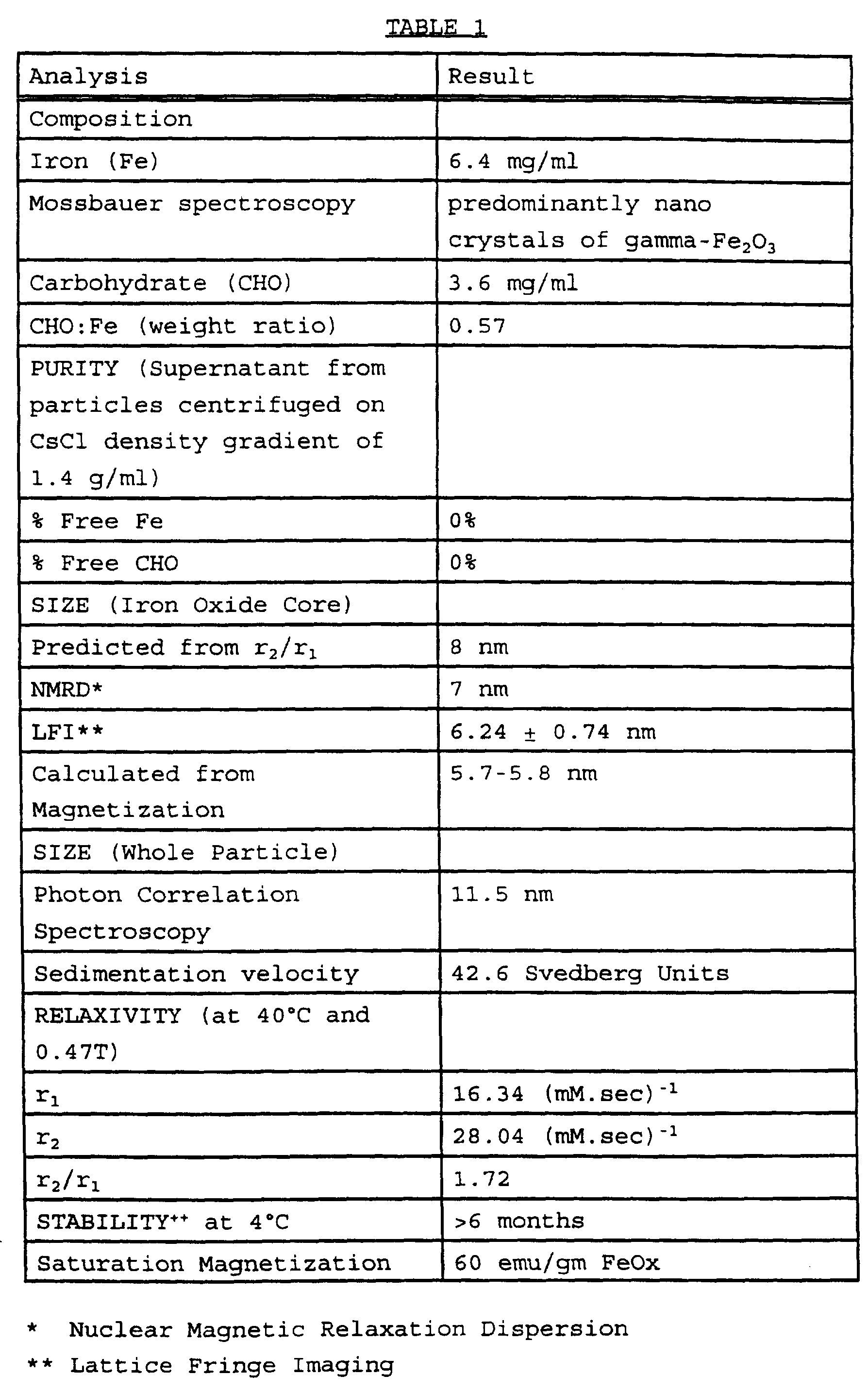
- Gel preparation steps starch solution preparation and heating to 55°C, addition of iron chloride to starch solution, addition of ammonium hydroxide to iron/starch solution, heating of reaction mixture to 87-90°C and product cooling/gel neutralization.
- Wash set gel by pumping cold deionized water through settled gel suspension until pH is less than 8.5.
- E. Gel Oxidative Cleavage with Sodium Hvoochlorite A dose titration of the amount of sodium hypochlorite (hypo) per gram of gel can be done on a new lot to optimize production. Magnetic particle production " is assessed by photon correlation spectroscopy (PCS) for size and dispersity, and by determination of water proton relaxation rates.
- PCS photon correlation spectroscopy
- b. Weigh out gel, add hypochlorite and heat in water bath at 70°C for 45 minutes.
- c. Add 8M urea (0.8 ml/5gms of gel) after heating. Urea inactivates excess hypochlorite.
- the longitudinal relaxation rate (1/Ti) is measured as a function of magnetic field strength in the range 2.35 Gauss to 1.2 Tesla. See for example Koenig et al. NMR Spectroscopy of Cells and Organisms, Vol. II, page 75, R.K. Gupta (Ed), CRC Press, 1987 and Koenig et al. Progress in NMR Spectroscopy 2.: 487-567 (1990) .
- Methoxy PEG phosphate (MPP) (mol. wt. 5 kD) was added to an aqueous suspension of particles produced according to Example 1 at the desired ratio of MPP to iron oxide (FeOx) (2 gms MPP/gm FeOx) , incubated for 15 hrs at 37°C with constant rotation and then stored at 4°C until used.
- MPP Methoxy PEG phosphate
- the particles can be autoclave sterilised at 121°C for 15 minutes.
- mice were injected via tail vein with 100 ⁇ L samples at 1 mg Fe/mL of the preparations of Examples 1 and 2. At timed intervals, animals were euthanized, blood samples were collected and pooled from two mice and 1/Ti was measured. From 1/T X values the half lives (T ) were determined. The results are set out in Table 2 which includes for comparison the results for conventional MSM particles:
- MSM Conventional co-precipitated magnetic starch particles.
- MPP coated particles produced according to Example 2 diluted to an iron concentration of 10 mg FeOx/mL with 5% dextrose solution and sterile filtered before injection.
- a contrast agent composition produced according to Example 4 was administered intravenously into the pig at a dosage of 4 mg Fe/kg bodyweight.
- Pre and 35 minutes post contrast T x -weighted MR images were recorded at 1.5T, Turbo-FLASH, TR/TE/TI/flip 15 ms/4.1 ms/846 ms/25° and appear as Figures 1A and IB hereto.
- the liver appears bright in the left of the pre-contrast image. In the post contrast image, the liver parenchyma is dark while the blood vessels running through it appear very bright.
- a contrast agent composition produced according to Example 4 was administered intravenously into the pig at a dosage of 5 mg Fe/kg bodyweigh .
- Pre and 20 minutes post contrast T 2 -weighted MR images were recorded at 1.5T fast spin echo, TR/TE 1800 ms/100 ms and appear as Figures 2A and 2B hereto.
- Pre contrast the liver parenchyma appears lighter than the blood vessels; post contrast both liver parenchyma and blood vessels appear dark. Comparison between Figures IB and 2B allows differentiation between blood vessels in the liver and other non-parenchymal items.
- MPP coated particles produced according to Example 2 are combined with particles produced according to Example 1 in a weight ratio (of FeOx) of 5:1, diluted to an iron concentration of 10 mg FeOx/mL with 5% dextrose solution and sterile filtered.
- Example 1 In place of the particles of Example 1 one may use conventional MSM or commercially available magnetic particles such as those available from Guerbet SA under the trade name Endorem.
Abstract
Description

Claims
Priority Applications (8)
| Application Number | Priority Date | Filing Date | Title |
|---|---|---|---|
| EA199800881A EA000796B1 (en) | 1996-04-01 | 1997-03-27 | Method of t1-weighted magnetic resonance imaging of res organs |
| AT97914482T ATE228017T1 (en) | 1996-04-01 | 1997-03-27 | T1 TIME-WEIGHTED NUCLEAR SPIN TOMOGRAPH OF THE RES ORGANS |
| EP97914482A EP0896546B1 (en) | 1996-04-01 | 1997-03-27 | Method of t1-weighted magnetic resonance imaging of res organs |
| IL12641297A IL126412A0 (en) | 1996-04-01 | 1997-03-27 | Method of t-weighted magnetic resonance imaging of res organs |
| DE69717269T DE69717269T2 (en) | 1996-04-01 | 1997-03-27 | T1 TIME-WEIGHTED CORE SPIN TOMOGRAM OF RES ORGANS |
| AU21721/97A AU704926B2 (en) | 1996-04-01 | 1997-03-27 | Method of T1-weighted magnetic resonance imaging of RES organs |
| JP53503697A JP4361970B2 (en) | 1996-04-01 | 1997-03-27 | Method for T1-weighted nuclear magnetic resonance imaging of reticuloendothelial (RES) organs |
| NO984555A NO984555D0 (en) | 1996-04-01 | 1998-09-29 | T1 - Liver |
Applications Claiming Priority (2)
| Application Number | Priority Date | Filing Date | Title |
|---|---|---|---|
| US08/625,223 | 1996-04-01 | ||
| US08/625,223 US5855868A (en) | 1996-04-01 | 1996-04-01 | Method of T1 -weighted resonance imaging of RES organs |
Publications (2)
| Publication Number | Publication Date |
|---|---|
| WO1997036617A2 true WO1997036617A2 (en) | 1997-10-09 |
| WO1997036617A3 WO1997036617A3 (en) | 1998-02-26 |
Family
ID=24505090
Family Applications (1)
| Application Number | Title | Priority Date | Filing Date |
|---|---|---|---|
| PCT/GB1997/000886 WO1997036617A2 (en) | 1996-04-01 | 1997-03-27 | Method of t1-weighted magnetic resonance imaging of res organs |
Country Status (14)
| Country | Link |
|---|---|
| US (1) | US5855868A (en) |
| EP (1) | EP0896546B1 (en) |
| JP (1) | JP4361970B2 (en) |
| KR (1) | KR20000005194A (en) |
| CN (1) | CN1219135A (en) |
| AT (1) | ATE228017T1 (en) |
| AU (1) | AU704926B2 (en) |
| CA (1) | CA2250818A1 (en) |
| DE (1) | DE69717269T2 (en) |
| EA (1) | EA000796B1 (en) |
| ES (1) | ES2188924T3 (en) |
| IL (1) | IL126412A0 (en) |
| NO (1) | NO984555D0 (en) |
| WO (1) | WO1997036617A2 (en) |
Cited By (1)
| Publication number | Priority date | Publication date | Assignee | Title |
|---|---|---|---|---|
| EP2205613A2 (en) * | 2007-10-15 | 2010-07-14 | Seoul National University Industry Foundation | Biocompatible suspension stabilizer for dispersing inorganic nanoparticles into aqueous solution |
Families Citing this family (9)
| Publication number | Priority date | Publication date | Assignee | Title |
|---|---|---|---|---|
| PT81498B (en) * | 1984-11-23 | 1987-12-30 | Schering Ag | METHOD FOR PREPARING COMPOSITIONS FOR DIAGNOSTICS CONTAINING MAGNETIC PARTICLES |
| PT915738E (en) | 1996-08-05 | 2002-07-31 | Schering Ag | METHOD FOR PREPARING MEDIA OF CONTRAST FOR TOMOGRAPHY OF MAGNETIC RESONANCE |
| WO2001074245A1 (en) * | 2000-03-31 | 2001-10-11 | Amersham Health As | Method of magnetic resonance imaging |
| US7082326B2 (en) * | 2000-03-31 | 2006-07-25 | Amersham Health As | Method of magnetic resonance imaging |
| KR20020075513A (en) * | 2001-03-24 | 2002-10-05 | 조용덕 | The waste water treatment method using electrolysis |
| DE102004022061A1 (en) * | 2004-05-05 | 2005-12-08 | Siemens Ag | Method for improved interventional imaging in magnetic resonance tomography |
| US20100259259A1 (en) * | 2005-09-21 | 2010-10-14 | Markus Zahn | Systems and methods for tuning properties of nanoparticles |
| US10814019B2 (en) * | 2014-06-30 | 2020-10-27 | University Of Washington | MRI signal suppression agents, compositions, and methods |
| US10466326B2 (en) * | 2015-05-15 | 2019-11-05 | Stc. Unm | Quantitative [Fe]-MRI (femri) of anti-PSMA-conjugated SPIONs based on PSMA expression levels |
Citations (4)
| Publication number | Priority date | Publication date | Assignee | Title |
|---|---|---|---|---|
| US3935187A (en) * | 1973-10-19 | 1976-01-27 | Standard Brands Incorporated | Process for depolymerizing amylaceous polymers |
| US4501726A (en) * | 1981-11-12 | 1985-02-26 | Schroeder Ulf | Intravascularly administrable, magnetically responsive nanosphere or nanoparticle, a process for the production thereof, and the use thereof |
| US5160725A (en) * | 1987-03-24 | 1992-11-03 | Silica Gel Gesellschaft Mbh Adsorptions-Technik, Apparatebau | Magnetic liquid compositions |
| WO1997025073A2 (en) * | 1996-01-10 | 1997-07-17 | Nycomed Imaging A/S | Contrast media |
Family Cites Families (17)
| Publication number | Priority date | Publication date | Assignee | Title |
|---|---|---|---|---|
| US4452773A (en) * | 1982-04-05 | 1984-06-05 | Canadian Patents And Development Limited | Magnetic iron-dextran microspheres |
| GB8408127D0 (en) * | 1984-03-29 | 1984-05-10 | Nyegaard & Co As | Contrast agents |
| US4767611A (en) * | 1984-07-03 | 1988-08-30 | Gordon Robert T | Method for affecting intracellular and extracellular electric and magnetic dipoles |
| US4849210A (en) * | 1985-05-08 | 1989-07-18 | Molecular Biosystems, Inc. | Magnetic resonance imaging of liver and spleen with superparamagnetic contrast agents |
| GB8601100D0 (en) * | 1986-01-17 | 1986-02-19 | Cosmas Damian Ltd | Drug delivery system |
| US5314679A (en) * | 1986-07-03 | 1994-05-24 | Advanced Magnetics Inc. | Vascular magnetic resonance imaging agent comprising nanoparticles |
| US4827945A (en) * | 1986-07-03 | 1989-05-09 | Advanced Magnetics, Incorporated | Biologically degradable superparamagnetic materials for use in clinical applications |
| US5069216A (en) * | 1986-07-03 | 1991-12-03 | Advanced Magnetics Inc. | Silanized biodegradable super paramagnetic metal oxides as contrast agents for imaging the gastrointestinal tract |
| US4925678A (en) * | 1987-04-01 | 1990-05-15 | Ranney David F | Endothelial envelopment drug carriers |
| US5358702A (en) * | 1990-04-10 | 1994-10-25 | Unger Evan C | Methoxylated gel particle contrast media for improved diagnostic imaging |
| US5328681A (en) * | 1991-01-19 | 1994-07-12 | Meito Sangyo Kabushiki Kaisha | Composition comprising magnetic metal oxide ultrafine particles and derivatized polysaccharides |
| US5225282A (en) * | 1991-12-13 | 1993-07-06 | Molecular Bioquest, Inc. | Biodegradable magnetic microcluster comprising non-magnetic metal or metal oxide particles coated with a functionalized polymer |
| EP0580878B1 (en) * | 1992-06-01 | 1996-01-10 | BASF Aktiengesellschaft | The use of dispersions of magneto-ionic particles as MRI contrast media |
| US5349957A (en) * | 1992-12-02 | 1994-09-27 | Sterling Winthrop Inc. | Preparation and magnetic properties of very small magnetite-dextran particles |
| WO1994021240A2 (en) * | 1993-03-17 | 1994-09-29 | Silica Gel Ges.M.B.H | Superparamagnetic particles, process for producing the same and their use |
| DE4428851C2 (en) * | 1994-08-04 | 2000-05-04 | Diagnostikforschung Inst | Nanoparticles containing iron, their production and application in diagnostics and therapy |
| AU687093B2 (en) * | 1994-09-27 | 1998-02-19 | Nycomed Imaging As | Contrast agent |
-
1996
- 1996-04-01 US US08/625,223 patent/US5855868A/en not_active Expired - Lifetime
-
1997
- 1997-03-27 ES ES97914482T patent/ES2188924T3/en not_active Expired - Lifetime
- 1997-03-27 JP JP53503697A patent/JP4361970B2/en not_active Expired - Lifetime
- 1997-03-27 WO PCT/GB1997/000886 patent/WO1997036617A2/en not_active Application Discontinuation
- 1997-03-27 AU AU21721/97A patent/AU704926B2/en not_active Ceased
- 1997-03-27 AT AT97914482T patent/ATE228017T1/en not_active IP Right Cessation
- 1997-03-27 DE DE69717269T patent/DE69717269T2/en not_active Expired - Lifetime
- 1997-03-27 CA CA002250818A patent/CA2250818A1/en not_active Abandoned
- 1997-03-27 EP EP97914482A patent/EP0896546B1/en not_active Expired - Lifetime
- 1997-03-27 EA EA199800881A patent/EA000796B1/en not_active IP Right Cessation
- 1997-03-27 IL IL12641297A patent/IL126412A0/en unknown
- 1997-03-27 KR KR1019980707869A patent/KR20000005194A/en not_active Application Discontinuation
- 1997-03-27 CN CN97194760A patent/CN1219135A/en active Pending
-
1998
- 1998-09-29 NO NO984555A patent/NO984555D0/en not_active Application Discontinuation
Patent Citations (4)
| Publication number | Priority date | Publication date | Assignee | Title |
|---|---|---|---|---|
| US3935187A (en) * | 1973-10-19 | 1976-01-27 | Standard Brands Incorporated | Process for depolymerizing amylaceous polymers |
| US4501726A (en) * | 1981-11-12 | 1985-02-26 | Schroeder Ulf | Intravascularly administrable, magnetically responsive nanosphere or nanoparticle, a process for the production thereof, and the use thereof |
| US5160725A (en) * | 1987-03-24 | 1992-11-03 | Silica Gel Gesellschaft Mbh Adsorptions-Technik, Apparatebau | Magnetic liquid compositions |
| WO1997025073A2 (en) * | 1996-01-10 | 1997-07-17 | Nycomed Imaging A/S | Contrast media |
Non-Patent Citations (5)
| Title |
|---|
| AUTIO, K. ET AL: "Heat-induced structural changes of acid-hydrolysed and hypochlorite-oxidized barley starches" CARBOHYDRATE POLYMERS, vol. 29, no. 2, February 1996, BARKING, GB, pages 155-161, XP002037910 * |
| CHEMICAL ABSTRACTS, vol. 115, no. 10, 1991 Columbus, Ohio, US; abstract no. 093953, WANG, HAICHENG: "Development of cold-setting starch adhesive" XP002037908 & HUAXUE YU ZHANHE, 1990, NO. 3, PAGES 168-9, * |
| CHEMICAL ABSTRACTS, vol. 70, no. 12, 24 March 1969 Columbus, Ohio, US; abstract no. 050898, HASEGAWA, TOSHIKATSU ET AL: "Adsorption behavior of oxidized starch onto iron or aluminum" XP002037909 & KOGYO KAGAKU ZASSHI, 1968, VOL. 71, NO. 12, PAGES 2086-7, * |
| KRESSE M ET AL: "MAGNETOPHARMAKA" DEUTSCHE APOTHEKER ZEITUNG, vol. 134, no. 33, 18 August 1994, pages 13/14, 17-20, 23 - 25, XP000457935 * |
| PAROVUORI P ET AL: "Oxidation of potato starch by hydrogen peroxide" STARCH, 1995, VOL. 47, NO. 1, PAGE(S) 19-23., XP002037907 * |
Cited By (2)
| Publication number | Priority date | Publication date | Assignee | Title |
|---|---|---|---|---|
| EP2205613A2 (en) * | 2007-10-15 | 2010-07-14 | Seoul National University Industry Foundation | Biocompatible suspension stabilizer for dispersing inorganic nanoparticles into aqueous solution |
| EP2205613A4 (en) * | 2007-10-15 | 2012-09-12 | Seoul Nat Univ Ind Foundation | Biocompatible suspension stabilizer for dispersing inorganic nanoparticles into aqueous solution |
Also Published As
| Publication number | Publication date |
|---|---|
| DE69717269D1 (en) | 2003-01-02 |
| CA2250818A1 (en) | 1997-10-09 |
| JP2000507567A (en) | 2000-06-20 |
| EA000796B1 (en) | 2000-04-24 |
| JP4361970B2 (en) | 2009-11-11 |
| EA199800881A1 (en) | 1999-04-29 |
| KR20000005194A (en) | 2000-01-25 |
| AU2172197A (en) | 1997-10-22 |
| CN1219135A (en) | 1999-06-09 |
| NO984555L (en) | 1998-09-29 |
| DE69717269T2 (en) | 2003-08-28 |
| AU704926B2 (en) | 1999-05-06 |
| NO984555D0 (en) | 1998-09-29 |
| US5855868A (en) | 1999-01-05 |
| ATE228017T1 (en) | 2002-12-15 |
| EP0896546B1 (en) | 2002-11-20 |
| EP0896546A2 (en) | 1999-02-17 |
| WO1997036617A3 (en) | 1998-02-26 |
| ES2188924T3 (en) | 2003-07-01 |
| IL126412A0 (en) | 1999-05-09 |
Similar Documents
| Publication | Publication Date | Title |
|---|---|---|
| Ferrucci et al. | Iron oxide-enhanced MR imaging of the liver and spleen: review of the first 5 years. | |
| EP0670695B1 (en) | Polymers as contrast media for magnetic resonance imaging | |
| Runge et al. | Paramagnetic agents for contrast-enhanced NMR imaging: a review | |
| AU655175B2 (en) | Diagnostic agents | |
| US5310539A (en) | Melanin-based agents for image enhancement | |
| EP0693288B1 (en) | Polymers as contrast media for magnetic resonance | |
| EP0414700A1 (en) | Contrast agents for magnetic resonance imaging. | |
| JP4965020B2 (en) | Contrast-enhanced magnetic resonance imaging of tissue perfusion | |
| US5855868A (en) | Method of T1 -weighted resonance imaging of RES organs | |
| JPH07505638A (en) | Magnetic resonance imaging methods and compositions | |
| US20020151787A1 (en) | Method of tumor imaging | |
| WO1996010359A1 (en) | Chelate complex with high conspicuity for magnetic resonance imaging | |
| US7082326B2 (en) | Method of magnetic resonance imaging | |
| Woodward et al. | Magnetic Contrast Imaging: Magnetic nanoparticles as probes in living systems | |
| Woodward et al. | 10 Magnetic Contrast Imaging | |
| Ferrucci | Iron oxide enhanced MR imaging of the liver and spleen: review of the first five years | |
| EP1267716B1 (en) | Method of magnetic resonance imaging | |
| Froehlich | MR contrast agents | |
| WO2001074245A1 (en) | Method of magnetic resonance imaging | |
| Laurent et al. | Contrast agents for MRI: recent advances | |
| GB2311138A (en) | ESR-enhanced MRI using magnetic particles as contrast agents | |
| Weishaupt et al. | MR Contrast Agents | |
| Reimer | Encyclopedia of Diagnostic Imaging Springer-Verlag Berlin Heidelberg New York 2008 |
Legal Events
| Date | Code | Title | Description |
|---|---|---|---|
| WWE | Wipo information: entry into national phase |
Ref document number: 97194760.0 Country of ref document: CN |
|
| AK | Designated states |
Kind code of ref document: A2 Designated state(s): AL AM AT AU AZ BA BB BG BR BY CA CH CN CU CZ DE DK EE ES FI GB GE GH HU IL IS JP KE KG KP KR KZ LC LK LR LS LT LU LV MD MG MK MN MW MX NO NZ PL PT RO RU SD SE SG SI SK TJ TM TR TT UA UG US UZ VN YU AM AZ BY KG KZ MD RU TJ TM |
|
| AL | Designated countries for regional patents |
Kind code of ref document: A2 Designated state(s): GH KE LS MW SD SZ UG AT BE CH DE DK ES FI FR GB GR IE IT LU MC NL PT SE BF |
|
| DFPE | Request for preliminary examination filed prior to expiration of 19th month from priority date (pct application filed before 20040101) | ||
| 121 | Ep: the epo has been informed by wipo that ep was designated in this application | ||
| AK | Designated states |
Kind code of ref document: A3 Designated state(s): AL AM AT AU AZ BA BB BG BR BY CA CH CN CU CZ DE DK EE ES FI GB GE GH HU IL IS JP KE KG KP KR KZ LC LK LR LS LT LU LV MD MG MK MN MW MX NO NZ PL PT RO RU SD SE SG SI SK TJ TM TR TT UA UG US UZ VN YU AM AZ BY KG KZ MD RU TJ TM |
|
| AL | Designated countries for regional patents |
Kind code of ref document: A3 Designated state(s): GH KE LS MW SD SZ UG AT BE CH DE DK ES FI FR GB GR IE IT LU MC NL PT SE BF |
|
| ENP | Entry into the national phase |
Ref document number: 2250818 Country of ref document: CA Ref document number: 2250818 Country of ref document: CA Kind code of ref document: A |
|
| WWE | Wipo information: entry into national phase |
Ref document number: 1019980707869 Country of ref document: KR |
|
| WWE | Wipo information: entry into national phase |
Ref document number: 1997914482 Country of ref document: EP |
|
| WWE | Wipo information: entry into national phase |
Ref document number: 332543 Country of ref document: NZ |
|
| WWE | Wipo information: entry into national phase |
Ref document number: 199800881 Country of ref document: EA |
|
| REG | Reference to national code |
Ref country code: DE Ref legal event code: 8642 |
|
| WWP | Wipo information: published in national office |
Ref document number: 1997914482 Country of ref document: EP |
|
| WWP | Wipo information: published in national office |
Ref document number: 1019980707869 Country of ref document: KR |
|
| WWG | Wipo information: grant in national office |
Ref document number: 1997914482 Country of ref document: EP |
|
| WWW | Wipo information: withdrawn in national office |
Ref document number: 1019980707869 Country of ref document: KR |