US9861600B2 - Methods and compositions for treating and identifying compounds to treat age-related macular degeneration treatment - Google Patents
Methods and compositions for treating and identifying compounds to treat age-related macular degeneration treatment Download PDFInfo
- Publication number
- US9861600B2 US9861600B2 US14/858,602 US201514858602A US9861600B2 US 9861600 B2 US9861600 B2 US 9861600B2 US 201514858602 A US201514858602 A US 201514858602A US 9861600 B2 US9861600 B2 US 9861600B2
- Authority
- US
- United States
- Prior art keywords
- dopa
- cells
- cell population
- tyrosine
- pedf
- Prior art date
- Legal status (The legal status is an assumption and is not a legal conclusion. Google has not performed a legal analysis and makes no representation as to the accuracy of the status listed.)
- Active
Links
- 0 *OC(=O)C(N)CC1=CC=C(O)C(O)=C1 Chemical compound *OC(=O)C(N)CC1=CC=C(O)C(O)=C1 0.000 description 8
- POEYAUMBFXDQIS-UHFFFAOYSA-N C1=CC=NC=C1.CCC(=O)C(C)(C)C Chemical compound C1=CC=NC=C1.CCC(=O)C(C)(C)C POEYAUMBFXDQIS-UHFFFAOYSA-N 0.000 description 2
- SRWIKUAHSWZJNS-UHFFFAOYSA-N CC(C)(C)C(=O)CN.CCCCC Chemical compound CC(C)(C)C(=O)CN.CCCCC SRWIKUAHSWZJNS-UHFFFAOYSA-N 0.000 description 2
- WTDRDQBEARUVNC-LURJTMIESA-N N[C@@H](CC1=CC=C(O)C(O)=C1)C(=O)O Chemical compound N[C@@H](CC1=CC=C(O)C(O)=C1)C(=O)O WTDRDQBEARUVNC-LURJTMIESA-N 0.000 description 2
Images


Classifications
-
- A—HUMAN NECESSITIES
- A61—MEDICAL OR VETERINARY SCIENCE; HYGIENE
- A61K—PREPARATIONS FOR MEDICAL, DENTAL OR TOILETRY PURPOSES
- A61K31/00—Medicinal preparations containing organic active ingredients
- A61K31/185—Acids; Anhydrides, halides or salts thereof, e.g. sulfur acids, imidic, hydrazonic or hydroximic acids
- A61K31/19—Carboxylic acids, e.g. valproic acid
- A61K31/195—Carboxylic acids, e.g. valproic acid having an amino group
- A61K31/197—Carboxylic acids, e.g. valproic acid having an amino group the amino and the carboxyl groups being attached to the same acyclic carbon chain, e.g. gamma-aminobutyric acid [GABA], beta-alanine, epsilon-aminocaproic acid, pantothenic acid
- A61K31/198—Alpha-aminoacids, e.g. alanine, edetic acids [EDTA]
-
- A—HUMAN NECESSITIES
- A61—MEDICAL OR VETERINARY SCIENCE; HYGIENE
- A61K—PREPARATIONS FOR MEDICAL, DENTAL OR TOILETRY PURPOSES
- A61K31/00—Medicinal preparations containing organic active ingredients
-
- A—HUMAN NECESSITIES
- A61—MEDICAL OR VETERINARY SCIENCE; HYGIENE
- A61K—PREPARATIONS FOR MEDICAL, DENTAL OR TOILETRY PURPOSES
- A61K31/00—Medicinal preparations containing organic active ingredients
- A61K31/045—Hydroxy compounds, e.g. alcohols; Salts thereof, e.g. alcoholates
- A61K31/07—Retinol compounds, e.g. vitamin A
-
- A—HUMAN NECESSITIES
- A61—MEDICAL OR VETERINARY SCIENCE; HYGIENE
- A61K—PREPARATIONS FOR MEDICAL, DENTAL OR TOILETRY PURPOSES
- A61K31/00—Medicinal preparations containing organic active ingredients
- A61K31/185—Acids; Anhydrides, halides or salts thereof, e.g. sulfur acids, imidic, hydrazonic or hydroximic acids
- A61K31/19—Carboxylic acids, e.g. valproic acid
- A61K31/195—Carboxylic acids, e.g. valproic acid having an amino group
-
- A—HUMAN NECESSITIES
- A61—MEDICAL OR VETERINARY SCIENCE; HYGIENE
- A61K—PREPARATIONS FOR MEDICAL, DENTAL OR TOILETRY PURPOSES
- A61K31/00—Medicinal preparations containing organic active ingredients
- A61K31/33—Heterocyclic compounds
- A61K31/335—Heterocyclic compounds having oxygen as the only ring hetero atom, e.g. fungichromin
- A61K31/35—Heterocyclic compounds having oxygen as the only ring hetero atom, e.g. fungichromin having six-membered rings with one oxygen as the only ring hetero atom
- A61K31/352—Heterocyclic compounds having oxygen as the only ring hetero atom, e.g. fungichromin having six-membered rings with one oxygen as the only ring hetero atom condensed with carbocyclic rings, e.g. methantheline
- A61K31/353—3,4-Dihydrobenzopyrans, e.g. chroman, catechin
- A61K31/355—Tocopherols, e.g. vitamin E
-
- A—HUMAN NECESSITIES
- A61—MEDICAL OR VETERINARY SCIENCE; HYGIENE
- A61K—PREPARATIONS FOR MEDICAL, DENTAL OR TOILETRY PURPOSES
- A61K31/00—Medicinal preparations containing organic active ingredients
- A61K31/33—Heterocyclic compounds
- A61K31/335—Heterocyclic compounds having oxygen as the only ring hetero atom, e.g. fungichromin
- A61K31/365—Lactones
- A61K31/375—Ascorbic acid, i.e. vitamin C; Salts thereof
-
- A—HUMAN NECESSITIES
- A61—MEDICAL OR VETERINARY SCIENCE; HYGIENE
- A61K—PREPARATIONS FOR MEDICAL, DENTAL OR TOILETRY PURPOSES
- A61K33/00—Medicinal preparations containing inorganic active ingredients
- A61K33/24—Heavy metals; Compounds thereof
- A61K33/30—Zinc; Compounds thereof
-
- A—HUMAN NECESSITIES
- A61—MEDICAL OR VETERINARY SCIENCE; HYGIENE
- A61K—PREPARATIONS FOR MEDICAL, DENTAL OR TOILETRY PURPOSES
- A61K33/00—Medicinal preparations containing inorganic active ingredients
- A61K33/24—Heavy metals; Compounds thereof
- A61K33/34—Copper; Compounds thereof
-
- A—HUMAN NECESSITIES
- A61—MEDICAL OR VETERINARY SCIENCE; HYGIENE
- A61K—PREPARATIONS FOR MEDICAL, DENTAL OR TOILETRY PURPOSES
- A61K45/00—Medicinal preparations containing active ingredients not provided for in groups A61K31/00 - A61K41/00
- A61K45/06—Mixtures of active ingredients without chemical characterisation, e.g. antiphlogistics and cardiaca
-
- A—HUMAN NECESSITIES
- A61—MEDICAL OR VETERINARY SCIENCE; HYGIENE
- A61K—PREPARATIONS FOR MEDICAL, DENTAL OR TOILETRY PURPOSES
- A61K49/00—Preparations for testing in vivo
- A61K49/0004—Screening or testing of compounds for diagnosis of disorders, assessment of conditions, e.g. renal clearance, gastric emptying, testing for diabetes, allergy, rheuma, pancreas functions
- A61K49/0008—Screening agents using (non-human) animal models or transgenic animal models or chimeric hosts, e.g. Alzheimer disease animal model, transgenic model for heart failure
-
- A—HUMAN NECESSITIES
- A61—MEDICAL OR VETERINARY SCIENCE; HYGIENE
- A61P—SPECIFIC THERAPEUTIC ACTIVITY OF CHEMICAL COMPOUNDS OR MEDICINAL PREPARATIONS
- A61P27/00—Drugs for disorders of the senses
- A61P27/02—Ophthalmic agents
-
- A—HUMAN NECESSITIES
- A61—MEDICAL OR VETERINARY SCIENCE; HYGIENE
- A61P—SPECIFIC THERAPEUTIC ACTIVITY OF CHEMICAL COMPOUNDS OR MEDICINAL PREPARATIONS
- A61P43/00—Drugs for specific purposes, not provided for in groups A61P1/00-A61P41/00
-
- G—PHYSICS
- G01—MEASURING; TESTING
- G01N—INVESTIGATING OR ANALYSING MATERIALS BY DETERMINING THEIR CHEMICAL OR PHYSICAL PROPERTIES
- G01N33/00—Investigating or analysing materials by specific methods not covered by groups G01N1/00 - G01N31/00
- G01N33/48—Biological material, e.g. blood, urine; Haemocytometers
- G01N33/50—Chemical analysis of biological material, e.g. blood, urine; Testing involving biospecific ligand binding methods; Immunological testing
- G01N33/5005—Chemical analysis of biological material, e.g. blood, urine; Testing involving biospecific ligand binding methods; Immunological testing involving human or animal cells
- G01N33/5008—Chemical analysis of biological material, e.g. blood, urine; Testing involving biospecific ligand binding methods; Immunological testing involving human or animal cells for testing or evaluating the effect of chemical or biological compounds, e.g. drugs, cosmetics
- G01N33/502—Chemical analysis of biological material, e.g. blood, urine; Testing involving biospecific ligand binding methods; Immunological testing involving human or animal cells for testing or evaluating the effect of chemical or biological compounds, e.g. drugs, cosmetics for testing non-proliferative effects
- G01N33/5023—Chemical analysis of biological material, e.g. blood, urine; Testing involving biospecific ligand binding methods; Immunological testing involving human or animal cells for testing or evaluating the effect of chemical or biological compounds, e.g. drugs, cosmetics for testing non-proliferative effects on expression patterns
-
- G—PHYSICS
- G01—MEASURING; TESTING
- G01N—INVESTIGATING OR ANALYSING MATERIALS BY DETERMINING THEIR CHEMICAL OR PHYSICAL PROPERTIES
- G01N33/00—Investigating or analysing materials by specific methods not covered by groups G01N1/00 - G01N31/00
- G01N33/48—Biological material, e.g. blood, urine; Haemocytometers
- G01N33/50—Chemical analysis of biological material, e.g. blood, urine; Testing involving biospecific ligand binding methods; Immunological testing
- G01N33/5005—Chemical analysis of biological material, e.g. blood, urine; Testing involving biospecific ligand binding methods; Immunological testing involving human or animal cells
- G01N33/5008—Chemical analysis of biological material, e.g. blood, urine; Testing involving biospecific ligand binding methods; Immunological testing involving human or animal cells for testing or evaluating the effect of chemical or biological compounds, e.g. drugs, cosmetics
- G01N33/502—Chemical analysis of biological material, e.g. blood, urine; Testing involving biospecific ligand binding methods; Immunological testing involving human or animal cells for testing or evaluating the effect of chemical or biological compounds, e.g. drugs, cosmetics for testing non-proliferative effects
- G01N33/5041—Chemical analysis of biological material, e.g. blood, urine; Testing involving biospecific ligand binding methods; Immunological testing involving human or animal cells for testing or evaluating the effect of chemical or biological compounds, e.g. drugs, cosmetics for testing non-proliferative effects involving analysis of members of signalling pathways
-
- A—HUMAN NECESSITIES
- A61—MEDICAL OR VETERINARY SCIENCE; HYGIENE
- A61K—PREPARATIONS FOR MEDICAL, DENTAL OR TOILETRY PURPOSES
- A61K2300/00—Mixtures or combinations of active ingredients, wherein at least one active ingredient is fully defined in groups A61K31/00 - A61K41/00
-
- G—PHYSICS
- G01—MEASURING; TESTING
- G01N—INVESTIGATING OR ANALYSING MATERIALS BY DETERMINING THEIR CHEMICAL OR PHYSICAL PROPERTIES
- G01N2333/00—Assays involving biological materials from specific organisms or of a specific nature
- G01N2333/435—Assays involving biological materials from specific organisms or of a specific nature from animals; from humans
- G01N2333/46—Assays involving biological materials from specific organisms or of a specific nature from animals; from humans from vertebrates
- G01N2333/47—Assays involving proteins of known structure or function as defined in the subgroups
- G01N2333/4701—Details
- G01N2333/4703—Regulators; Modulating activity
- G01N2333/4704—Inhibitors; Supressors
-
- G—PHYSICS
- G01—MEASURING; TESTING
- G01N—INVESTIGATING OR ANALYSING MATERIALS BY DETERMINING THEIR CHEMICAL OR PHYSICAL PROPERTIES
- G01N2333/00—Assays involving biological materials from specific organisms or of a specific nature
- G01N2333/435—Assays involving biological materials from specific organisms or of a specific nature from animals; from humans
- G01N2333/705—Assays involving receptors, cell surface antigens or cell surface determinants
- G01N2333/72—Assays involving receptors, cell surface antigens or cell surface determinants for hormones
- G01N2333/726—G protein coupled receptor, e.g. TSHR-thyrotropin-receptor, LH/hCG receptor, FSH
-
- G—PHYSICS
- G01—MEASURING; TESTING
- G01N—INVESTIGATING OR ANALYSING MATERIALS BY DETERMINING THEIR CHEMICAL OR PHYSICAL PROPERTIES
- G01N2500/00—Screening for compounds of potential therapeutic value
- G01N2500/10—Screening for compounds of potential therapeutic value involving cells
Definitions
- Age-related macular degeneration is an aging-associated disease resulting in the loss of vision in the macula (the center of the visual field) because of damage to the retina.
- AMD is a prevalent disorder of the aged, with approximately 10% of patients 66 to 74 years and 30% of patients 75 to 85 years of age having some level of macular degeneration.
- the present invention provides methods for treating age-related macular degeneration (AMD), comprising administering to a subject with AMD an amount effective for treating AMD of an agonist of the OA1 receptor.
- AMD age-related macular degeneration
- the present invention provides methods for limiting development of AMD, comprising administering to a subject at risk of developing AMD an amount effective for limiting development of AMD of an agonist of the OA1 receptor.
- the agonist of the OA1 receptor is selected from the group consisting of L-DOPA and L-DOPA analogues.
- the present invention provides methods for identifying compounds to treat AMD, comprising contacting cells with a test compound, wherein the cells comprise:
- the present invention provides methods for identifying compounds to treat AMD, comprising
- test compound administered during embryonic photoreceptor and/or retinal ganglion development
- test compound (b) comparing an effect of the test compound on photoreceptor and/or retinal ganglion development in the embryo or post-natal non-human mammal, to photoreceptor and/or retinal ganglion development in an embryo or post-natal non-human mammal not administered the test compound, wherein those test compounds that increase photoreceptor and/or retinal ganglion development are candidate compounds for treating and/or limiting development of AMD.
- compositions comprising:
- composition comprising a source of vitamin C, a source of vitamin E, a source of vitamin A, a source of zinc, and a source of copper.
- FIG. 1 Western blot analysis of proteins bound (B) or unbound (U) to strepavidin-conjugated beads after biotinylation of RPE in situ, cultured RPE (b), or COS cells transfected to express OA1-GFP (c). Blots were probed to visualize OA1 and actin after cell surface biotinylation and fractionation using streptavidin-conjugated beads. For cultured cells (b, c) cells were either maintained in 500 ⁇ M (normal DMEM) or 1 ⁇ M tyrosine for 3 days prior to analysis. (D) Quantification of western blot analysis by densitometry.
- FIG. 2(A) Representative traces of [Ca 2+ ]i during the time course of the standard experimental protocol in transfected and untransfected CHO cells. After establishment of a stable baseline for 3 minutes, the test agent was added at 1 ⁇ M. At 5 minutes, KCl was added to serve as a control that the cells were Fura-2 loaded and patent. Identical protocols were performed for both transfected cells and paired untransfected cells.
- B Summary data for [Ca 2+ ]i in response to tyrosine, dopamine, and L-DOPA in transfected and untransfected CHO cells. Untransfected cells are shown with L-DOPA treatment. The experimental control of membrane depolarization with KCl is also shown.
- Each bar represents data collected from at least 10 experiments and is presented as the mean change from baseline [Ca 2+ ]i after test agent addition. Error bars represent S.D., and t-test analyses were used to test for significant differences, * denotes p ⁇ 0.01. Analysis of pertussis toxin sensitivity of [Ca 2+ ]i increase in cells transfected to express OA1 or RPE that express the natural protein. Data represent mean of at least 6 experiments. (C) Analysis of pertussis toxin sensitivity of [Ca 2+ ]i increase in cells transfected to express OA1 or RPE that express the natural protein.
- Data represent mean of at least 6 experiments for each group of transfected cells and 20 individual experiments for each the treated and untreated RPE with endogenous OA1 expression. T-tests analyses were used to test for significant differences, and * denotes p ⁇ 0.01.
- the control group represents transfected but untreated CHO cells and the basal level of cAMP in those cells. Cells were treated with 1.0 ⁇ M L-DOPA, 0.1 ⁇ M forskolin, L-DOPA+0.1 ⁇ M forskolin, and as a positive control 1 ⁇ M forskolin.
- Results represent the mean cAMP levels observed in at least 6 experiments in which all experimental groups were analyzed in a paired fashion using replicate monolayers in the same culture plate. Error bars represent the S.D. of each group, and the only significant difference observed was the increase in cAMP levels after forskolin treatment.
- FIG. 3(A) Binding kinetics between OA1 and L-DOPA were determined using radiolabeled ligand binding assays. Results represent data collected from 5 such experiments and are presented as mean specific binding +/ ⁇ SEM. The hyperbolic curve fit exhibited an R 2 value of 0.994, Kd was determined to be 9.34 ⁇ 10 ⁇ 6 M+/1.14 ⁇ 10 ⁇ 6 M.
- FIG. 3(E) Scatchard plot illustrating the kinetics of a single site binding relationship based on FIG. 3( a ) .
- FIG. 4 All images represent 2 ⁇ m thick confocal sections of CHO cells transfected to express OA1-GFP. ⁇ -arrestin was visualized using immuno fluorescence methods. Prior to addition of L-DOPA (a-c) and after treatment with 1 ⁇ M L-DOPA (d-f), and the merged images (c, f) illustrate regions where the two proteins co-localize, at the resolution of white light imaging. (g,h) are low magnification of field of transfected CHO cells, with two transfected cells visible (arrows) (g). The remainder of the cell population is visualized using antibodies to ⁇ -arrestin (h) to illustrate that ⁇ -arrestin recruitment to the membrane only occurred in the OA1 expressing cells (arrows).
- FIG. 5 PEDF concentrations were determined by ELISA of cell conditioned medium.
- RPE cells were control cells, without L-DOPA treatment, or OA1 stimulated cells that were treated with 1 ⁇ M L-DOPA prior to being maintained for 3 days in normal DMEM. Data are presented as the mean of 3 experiments conducted in triplicate, error bars represent S.D, and * denotes P ⁇ 0.01 using a paired t-test.
- B PEDF concentrations in conditioned medium from pigmenting RPE determined by ELISA. Cells were either control pigmenting RPE cultures or paired cultures treated with phenylthiourea (PTU) at 200 ⁇ M.
- PTU phenylthiourea
- FIG. 6(A) Data represents mean+/ ⁇ SEM bound [3H]-L-DOPA in all fractions, total, specific and non-specific. Non-specific binding was determined by measuring radiolabeled-L-DOPA bound in the presence of excess unlabeled L-DOPA (1 mM). Specific binding at each given concentration is determined by subtracting the measured non-specific binding from the measured total binding.
- (B) The figure illustrates competitive interaction between tyrosine and L-DOPA, measured using increasing concentrations of tyrosine and 5 ⁇ M [H 3 ] L-DOPA. Each data point represents the mean data from 5 replicate wells, and the error bars are S.D. Data illustrate that tyrosine competes for binding with L-DOPA, but with a low affinity. The results suggest tyrosine has a Ki of 52.9 ⁇ M, and fits the single site binding model with an r 2 value of 0.85. Saturation could not be achieved because of the limited solubility of tyrosine.
- FIG. 7 Western blot and graphical representation of PEDF secretion in wild-type vs OA deficient mice.
- FIG. 8(A) is a graphical representation of data demonstrating that L-DOPA supplementation increases retinal ganglion cell numbers compared to what is expected in a normal wild-type mouse.
- B is a graphical representation of data demonstrating that L-DOPA supplementation increases photoreceptor numbers compared to what is expected in a normal wild-type mouse.
- C is a Western blot showing PEDF detection in 2 wild-type and 2 OA1 ⁇ /y mice.
- the present invention provides methods for treating age-related macular degeneration (AMD), comprising administering to a subject with AMD an amount effective for treating AMD of an agonist of the OA1 receptor.
- AMD age-related macular degeneration
- the present invention provides methods for limiting development of AMD, comprising administering to a subject at risk of developing AMD an amount effective for limiting development of AMD of an agonist of the OA1 receptor.
- the human Oa1 gene is found on the X chromosome, and has been shown to encode a 404 amino acid protein OA1 (SEQ ID NO:2), likely to be a G-protein coupled receptor (GPCR) [12,13] based upon sequence analysis [14].
- OA1 SEQ ID NO:2
- GPCR G-protein coupled receptor
- the inventors have identified the OA1 signaling pathway as a critical determinant of neurosensory retina survival, such that stimulation of this pathway will provide treatment for AMD as well as a means to limit AMD development for those at potential risk.
- OA1 and tyrosinase participate in an autocrine loop through L-DOPA that regulates the secretion of at least one potent neurotrophic factor, PEDF.
- L-DOPA can be used to stimulate OA1 activity and thus upregulate PEDF expression, making it a valuable therapeutic to treat and limit development of AMD.
- OA1 agonists can be identified, for example, using the drug discovery methods of the third and fourth aspects of the invention. Exemplary OA1 agonists are discussed in detail below.
- the subject preferably is a human.
- AMD means an aging-associated disease resulting in the loss of vision in the macula (the center of the visual field) because of damage to the retina know as Age-related Macular Degeneration.
- AMD encompasses both wet and dry AMD, described in more detail below.
- AMD begins with characteristic drusen (yellow deposits) in the macula between the retinal pigment epithelium and the underlying choroid. Most people with these early changes (referred to as age-related maculopathy) have good vision. People with drusen can go on to develop advanced AMD. The risk is considerably higher when the drusen are large and numerous and associated with disturbance in the pigmented cell layer under the macula.
- Subjects with age-related maculopathy may progress to either of the two main forms of advanced AMD, each of which can be treated or be limited in its development using the methods of the invention.
- “Wet” AMD causes vision loss due to abnormal blood vessel growth in the choriocapillaries, through Bruch's membrane, ultimately leading to blood and protein leakage below the macula. Bleeding, leaking, and scarring from these blood vessels eventually causes irreversible damage to the photoreceptors and rapid vision loss if left untreated.
- “Dry” AMD occurs when light-sensitive cells in the macula slowly break down, gradually causing vision loss in the affected eye. Blurring in AMD is probably due to the accumulation of drusen under the retinal pigment epithelium (RPE) which alters to focal properties of the photoreceptors by moving them out of the plane of focus.
- RPE retinal pigment epithelium
- Dry AMD may occur in one or both eyes, and can advance from age-related maculopathy into intermediate or advanced stages of dry AMD.
- Intermediate Dry AMD Either many medium-sized drusen or one or more large drusen. Some people see a blurred spot in the center of their vision. More light may be needed for reading and other tasks.
- Advanced Dry AMD In addition to drusen, a breakdown of light-sensitive cells and supporting tissue in the central retinal area. This breakdown can cause a blurred spot in the center of vision. Over time, the blurred spot may get bigger and darker, taking more of the central vision; may have difficulty reading or recognizing faces until they are very close to you.
- AMD symptoms include, but are not limited to blurred/reduced central vision, central scotomas (shadows or missing areas of vision), trouble discerning one dark color from another dark color and/or one light color from another light color; slow recovery of visual function after exposure to bright light, a loss in contrast sensitivity, so that contours, shadows and color vision are less vivid, retinal pigment epithelial (RPE) disturbance (including pigment clumping and/or dropout), RPE detachment, geographic atrophy, subretinal neovascularization, and disciform scar, and distorted vision (metamorphopsia), such that a grid of straight lines appears wavy and parts of the grid may appear blank Symptoms of dry AMD and wet AMD are generally similar early during disease progression, and thus it may not be possible to determine which early-stage patients will develop dry vs. wet forms of AMD. Dry AMD develops as ‘geographic atrophy’, and early AMD become ‘wet’ AMD when new blood vessels sprout.
- RPE retinal pigment epithelial
- treat or “treating” AMD means accomplishing one or more of the following: (a) reducing the severity of AMD; (b) limiting or preventing development of one or more symptoms characteristic of AMD, as described above; (c) inhibiting worsening of one or more symptoms characteristic of AMD, as described above; (d) limiting or preventing recurrence of AMD in patients that have previously had the disorder(s); and (e) limiting or preventing recurrence of one or more symptoms in patients that were previously symptomatic for AMD.
- Such treating includes treating of wet AMD and dry AMD.
- the term “limiting development of” AMD means to prevent or to minimize development of AMD in individuals at risk of developing AMD, as well as limiting progression of age-related maculopathy to AMD (wet or dry), or intermediate dry AMD to advanced dry or ‘wet’ AMD.
- the methods comprise treating a subject with drusen accumulation (ie: age-related maculopathy), to limit development of AMD.
- the methods comprise treating a subject with an amount effective of the OA1 agonist to decrease the rate of lines of loss of vision relative to a non-treated AMD subject, or subject at risk of AMD.
- the methods comprise treating a subject with wet AMD, or at risk of developing wet AMD, an amount effective of the OA1 agonist to decrease the rate and number of new blood vessel formation.
- OA1 stimulation causes the RPE to increase PEDF secretion, and PEDF is a potent anti-angiogenic factor.
- OA1 stimulation strategies may stop new blood vessel development in ‘wet’ AMD, in addition to its effects on retinal development discussed herein.
- the methods comprise treating a subject that has blurred or reduced central vision with an amount of OA1 agonist effective to increase the lines of visual acuity in one or both eyes.
- the lines of visual acuity are as measured by the standard Snellen test, where the increase or decrease in ‘lines’ of visual acuity are based on which smallest ‘line’ on a Snellen chart a patient can read clearly.
- Subjects at risk of developing AMD mean anyone with any risk factor for development of AMD, including but not limited to being over 50 years old (in various preferred embodiments, over 60 years old, over 65 years old, over 70 years old, or over 75 years old), presence of drusen deposits, Caucasian race, having a blood relative that has or had AMD, a mutation in the complement factor H gene (CFH) of (Tyr402His), Arg80Gly variant of the complement protein C3 gene, hypertension, high cholesterol levels, obesity, smoking, a high fat intake, and mutations in the fibulin 5 gene.
- the subject to be treated has one or more of these risk factors, particularly in methods for limiting development of AMD.
- terapéuticaally effective amount refers to an amount that is sufficient or effective to limit development of or treat (prevent the progression of or reverse) AMD.
- the appropriate dosage range depends on the choice of the compound, the route of administration, the nature of the formulation, the nature of the subject's condition, and the judgment of the attending practitioner. For example, oral administration would be expected to require higher dosages than administration by intravenous injection. Variations in these dosage levels can be adjusted using standard empirical routines for optimization, as is well understood in the art.
- the OA1 receptor agonist comprises a compound selected from the group consisting of L-DOPA and L-DOPA analogues.
- L-DOPA is [2-amino-3-(3,4-dihydroxyphenyl)propanoic acid] known for use in treating Parkinson's, and has the following structure.
- L-DOPA is commercially available and methods for its synthesis are known to those of skill in the art.
- L-DOPA analogues are those L-DOPA variants that retain OA1-stimulatory activity, including L-DOPA prodrugs, of which many are known in the art; exemplary such analogues are disclosed below. While not being bound by a specific mechanism of action, the inventor believes that L-DOPA binding to OA1 involves two sites of binding, one involving one or both hydroxyl groups, and one involving the carboxylic acid group.
- the L-DOPA analogues are L-DOPA prodrugs that are metabolized to L-DOPA after administration (and generally prior to binding to OA1 on the cell surface), and thus are expected to retain OA1-stimulatory activity.
- one or both hydroxyl group and/or the carboxyl group can be substituted to produce various analogues (prodrug or otherwise) for use in the methods of the invention.
- the L-DOPA analogues comprise L-DOPA esters
- the L-DOPA ester is selected from the group consisting of L-DOPA methyl ester, L-DOPA butyl ester, L-DOPA pentyl ester, L-DOPA cyclohexyl ester, L-DOPA benzyl ester, and L-DOPA ethyl ester.
- the L-DOPA esters are selected from the alkyl, aryl and substituted and unsubstituted aralkyl esters of L-DOPA.
- the L-DOPA esters are represented by the following formula:
- R is a straight or branched chain alkyl (C 1 -C 20 ) such as methyl, ethyl, propyl, butyl, myristyl, palmityl, pentyl, tetradecyl, hexadecyl and the like; aryl(C 6 -C 9 ) such as phenyl, tolyl and the like; substituted and unsubstituted mono, di or polyhydroxyalkyl(C 1 -C 20 ) such as benzyl, alkoxybenzyl, 4-hydroxybutyl, 2-hydroxypropyl, 2,3-dihydroxypropyl, 1,3-dihydroxypropyl, 6-hydroxyhexyl and 5-hydroxypentyl and the like optionally having a substituent such as alkoxy(C 1-5 ) [methoxy, ethoxy, butoxy and the like]; carbalkoxy (C 1-5 ) [methoxycarbonyl, ethoxycarbonyl, propy
- the L-DOPA analogues comprise bile acid conjugates as are known in the art.
- Exemplary L-DOPA bile acid conjugates, and methods for preparing them, are disclosed in WO/2002/028882 and US20020151526.
- these prodrugs are cleaved within the enterohepatic system to release the parent drug and/or an active metabolite from the bile acid into the systemic circulation.
- significantly, only a fraction (typically ⁇ 50%) ⁇ 50%) of the prodrug is cleaved during each pass through the enterohepatic cycle.
- the enterohepatic circulation serves as a reservoir of the drug enabling sustained systemic drug levels to be achieved.
- Naturally occurring bile acids such as cholic acid, chenodeoxycholic acid, ursodeoxycholic acid, deoxycholic acid, ursocholic acid and lithocholic acid are particularly preferred.
- the site of conjugation of these bile acids to L-DOPA or other L-DOPA analogue is preferably via the 3-hydroxy group or the C-24 carboxyl moiety.
- cleavable linker functionality may be introduced between the drug and the bile acid and this linker may be selected.
- L-DOPA bile acid conjugates are represented by the following formula
- the L-DOPA analogues comprise di or tri-peptide derivatives.
- Exemplary L-DOPA di- or tri-peptide analogues, and methods for preparing them, are disclosed in U.S. Pat. No. 3,803,120 and 5,686,423.
- Oral absorption of the di- and tri-peptide L-DOPA prodrugs show high oral bioavailability with some compounds having the plasma concentration 60-100 fold higher than that of L-dopa.
- such L-DOPA prodrugs are represented by the following formula
- R1 and R2 of the di- or tri-peptide derivative of L-DOPA (2-amino-3-(3,4-dihydroxyphenyl-)propanoic acid) of the formula (I) together is trimethylene.
- di-peptide derivatives of L-DOPA [2-amino-3-(3,4-dihydroxyphenyl)propanoic acid] are represented by the following formula
- R3 is hydrogen; and R4 is phenyl or hydroxyphenyl; or R3 and R4 together is trimethylene.
- the L-DOPA analogues comprise amine prodrugs as are known in the art.
- Exemplary L-DOPA amine analogues, and methods for preparing them, are disclosed in US20060025385 and WO/2004/069146.
- such L-DOPA amine analogues are represented by
- Preferred L-DOPA amine analogues include: compounds wherein R5 and R6 are each hydrogen; compounds wherein R1 and R2 are each hydrogen; compounds wherein R3 and R4 are each hydrogen; compounds wherein at least one of R1, R2, R3 and R4, preferably R3 and/or R4 is carbonyl, e.g., acetyl.
- Additional preferred compounds according to the present embodiments include compounds wherein at least one of R1, R2, R3 and R4 is an alkyl, alkenyl or alkynyl having 1-30 carbon atoms, or, alternatively, at least one of R1, R2, R3 and R4 is a fatty acid acyl, derived from, for example, myristic acid, lauric acid, palmitic acid, stearic acid, oleic acid, arachidonic acid, linoleic acid or linolenic acid.
- L-DOPA amine analogues include ⁇ -amino-3,4-dihydroxy-benzenepropanamide, ⁇ -N-acetyl-3,4-dihydroxy-benzenepropanamide and pharmaceutically acceptable salts thereof
- L-DOPA prodrugs for use in the present invention are disclosed in U.S. Pat. Nos. 4,065,566 and 4,035,507 and are represented by the formula
- each R is independently selected from the group consisting of a hydrogen atom, an acyl group, a
- R3 represents the residue of any N,N—C1-C2 dialkylamino acid or a C4-C6 cycloalkylamino acid
- R1 represents a member selected from the group consisting of a hydroxyl group and a —OM group, wherein M is an alkali metal (Na, K, etc.) or an ammonium ion; and wherein R2 represents a member selected from the group consisting of a
- R3 represents the residue of any N,N—(C1-C2)-dialkylamino acid or a C4-C6-cycloalkylamino acid
- R represents an acyl group; wherein R2 represents a hydrogen atom; and wherein R1 represents a —NHCH(R4)COOR5 group, wherein R4 represents the residue of any naturally occurring amino acid, and wherein R5 represents a member selected from the group consisting of a hydrogen atom, a C1-C5 alkyl group (e.g., methyl, ethyl, propyl, butyl, pentyl), and a C1-C5 alkylaryl group (e.g., —CH 2 —C 6 H 5 , —CH 2 —CH 2 —C 6 H 5 , etc.), and the HX salts thereof, wherein X is a conventional pharmaceutically acceptable acid addition salt anion (e.g., chloride, bromide, perchlorate, methanesulfonate, succinate, etc.);
- X is a conventional pharmaceutically acceptable acid addition salt anion (e.g., chloride, bromide, perchlorate
- Preferred exemplary L-DOPA prodrugs disclosed in U.S. Pat. Nos. 4,065,566 and 4,035,507 include the following:
- alkyl refers to a saturated aliphatic hydrocarbon including straight chain and branched chain groups.
- the alkyl group preferably has between 1 and 30 carbon atoms, more preferably between 1 and 20 carbon atoms. While lower alkyls, e.g., of between 1 and 6 carbon atoms may facilitate the formulation of the compounds, higher alkyls provides for enhanced permeability thereof through the BBB.
- the alkyl group may be substituted or non-substituted.
- the substituent group can be, for example, cycloalkyl, alkenyl, aryl, heteroaryl, heteroalicyclic, hydroxy, alkoxy, aryloxy, thiohydroxy, thioalkoxy, thioaryloxy, halo, carboxy, alkoxycarbonyl, thiocarboxy, carbamyl, and amino, as these terms are defined herein.
- cycloalkyl refers to an all-carbon monocyclic or fused ring (i.e., rings which share an adjacent pair of carbon atoms) group wherein one of more of the rings does not have a completely conjugated pi-electron system.
- examples, without limitation, of cycloalkyl groups are cyclopropane, cyclobutane, cyclopentane, cyclopentene, cyclohexane, cyclohexadiene, cycloheptane, cycloheptatriene and adamantane.
- the cycloalkyl group, according to the present invention may be substituted or non-substituted.
- the substituent group can be, for example, alkyl, cycloalkyl, alkenyl, aryl, heteroaryl, heteroalicyclic, hydroxy, alkoxy, aryloxy, thiohydroxy, thioalkoxy, thioaryloxy, halo, carboxy, alkoxycarbonyl, thiocarboxy, carbamyl, and amino, as these terms are defined herein.
- alkenyl refers to an alkyl group which consists of at least two carbon atoms and at least one carbon-carbon double bond.
- alkynyl refers to an alkyl group which consists of at least two carbon atoms and at least one carbon-carbon triple bond.
- both the alkenyl and the alkynyl groups preferably have between 1 and 30 carbon atoms.
- aryl group refers to an all-carbon monocyclic or fused-ring polycyclic (i.e., rings which share adjacent pairs of carbon atoms) group having a completely conjugated pi-electron system. Examples, without limitation, of aryl groups are phenyl, naphthalenyl and anthracenyl. The aryl group, according to the present invention, may be substituted or non-substituted.
- the substituent group can be, for example, alkyl, cycloalkyl, alkenyl, aryl, heteroaryl, heteroalicyclic, hydroxy, alkoxy, aryloxy, thiohydroxy, thioalkoxy, thioaryloxy, halo, carboxy, alkoxycarbonyl, thiocarboxy, carbamyl, and amino, as these terms are defined herein.
- C-carboxy refers to a+C( ⁇ O)—OR′ group, where R′ is hydrogen, alkyl, cycloalkyl, alkenyl, aryl, heteroaryl (bonded through a ring carbon) or heteroalicyclic (bonded through a ring carbon) as defined herein.
- O-carboxy refers to a R′—C( ⁇ O)—O— group, where R′ is hydrogen, alkyl, cycloalkyl, alkenyl, aryl, heteroaryl (bonded through a ring carbon) or heteroalicyclic (bonded through a ring carbon) as defined herein.
- carbonyl refers to a —C( ⁇ O)—R′ group, where R′ is as defined hereinabove.
- thiocarbonyl refers to a —C( ⁇ S)—R′ group, where R′ is as defined hereinabove.
- An “O-carbamyl” group refers to an —OC( ⁇ O)—NR′R′′ group, where R′ is as defined hereinabove and R′′ is as defined for R′.
- An “O-thiocarbamyl” group refers to an —OC( ⁇ S)—NR′R′′ group, where R′ is and R′′ are as defined hereinabove.
- a “fatty acid acyl” refers to a R′′′C( ⁇ O)—O— group, where R′′′ is a saturated or unsaturated hydrocarbon chain having at least 10 carbon atoms.
- alkoxy refers to both an —O-alkyl and an —O-cycloalkyl group, as defined hereinabove.
- Representative examples of alkoxy groups include methoxy, ethoxy, propoxy and tert-butoxy.
- the —O-alkyl and the O-cycloalkyl groups may be substituted or non-substituted.
- the substituent group can be, for example, cycloalkyl, alkenyl, aryl, heteroaryl, heteroalicyclic, hydroxy, alkoxy, aryloxy, thiohydroxy, thioalkoxy, thioaryloxy, halo, carboxy, alkoxycarbonyl, thiocarboxy, carbamyl, and amino, as these terms are defined herein.
- thioalkoxy refers to both an —S-alkyl group, and an —S-cycloalkyl group, as defined herein.
- hydroxy refers to an —OH group.
- thiohydroxy refers to an —SH group.
- aryloxy refers to both an —O-aryl and an —O-heteroaryl group, as defined herein.
- a “thioaryloxy” group refers to both an —S-aryl and an —S-heteroaryl group, as defined herein.
- amino refers to a —NR′R′′ group, with R′ and R′′ as defined hereinabove.
- alkoxycarbonyl which is also referred to herein interchangeably as “carbalkoxy”, refers to a carboxy group, as defined hereinabove, where R′ is not hydrogen.
- heteroaryl group includes a monocyclic or fused ring (i.e., rings which share an adjacent pair of atoms) group having in the ring(s) one or more atoms, such as, for example, nitrogen, oxygen and sulfur and, in addition, having a completely conjugated pi-electron system.
- heteroaryl groups include pyrrole, furane, thiophene, imidazole, oxazole, thiazole, pyrazole, pyridine, pyrimidine, quinoline, isoquinoline and purine.
- a “heteroalicyclic” group refers to a monocyclic or fused ring group having in the ring(s) one or more atoms such as nitrogen, oxygen and sulfur.
- the rings may also have one or more double bonds. However, the rings do not have a completely conjugated pi-electron system.
- halo refers to a fluorine, chlorine, bromine or iodine atom.
- phosphonyl describes an —P( ⁇ O)(OR′) 2 group, with R′ as defined hereinabove.
- the methods may comprise administering two or more compounds selected from the group consisting of L-DOPA and L-DOPA analogues.
- the methods may further comprise administering a further therapeutic compound to the subject, including but not limited to an L-amino acid decarboxylase inhibitor, such as carbidopa or benserazide.
- L-amino acid decarboxylase inhibitors can be used, for example, to increase plasma half-life of L-DOPA and reduce conversion of L-DOPA to dopamine peripherally, which reduces side effects of L-DOPA treatment.
- the methods may further comprise administering one or more other compounds useful for treating or limiting development of AMD, including but not limited to anti-angiogenic therapeutics, such as anti-vascular endothelial growth factor (VEGF) agents, including but not limited to VEGF antibodies (or fragments thereof) such as ranibizumab or bevacizumab, or VEGF aptamers, such as pegaptanib.
- anti-angiogenic therapeutics such as anti-vascular endothelial growth factor (VEGF) agents, including but not limited to VEGF antibodies (or fragments thereof) such as ranibizumab or bevacizumab, or VEGF aptamers, such as pegaptanib.
- VEGF vascular endothelial growth factor
- the L-DOPA or L-DOPA analogues may be present in a more complex mixture, such as in a nutritional supplement containing L-DOPA or L-DOPA analogues.
- any one or more of the L-DOPA and/or L-DOPA analogues described herein may be used in the form of a dietary supplement.
- a dietary supplement may combine any one or more further components that might be beneficial in treating or limiting development of AMD.
- L-DOPA and/or an L-DOPA analogue are combined with a combination of vitamin C source, vitamin E source, Vitamin A source, zinc source, and, and copper source, disclosed in U.S. Pat. No. 6,660,297 as useful in treating AMD; U.S. Pat. No. 6,660,297 is incorporated by reference herein in its entirety.
- any suitable amount of each of these additional components can be used in combination with L-DOPA and/or L-DOPA analogues in carrying out the methods of the invention.
- this combination may further comprise lutein and/or zeaxanthin in an amount suitable to provide further protective retinal effects, preferably between 1 mg and 100 mg; between 1 mg and 50 mg, between 2 mg and 25 mg, or between 2 mg and 10 mg per day.
- this combination may further comprise docosahexaenoic acid (DHA) and/or eicosapentaenoic acid (EPA) in an amount suitable to provide further protective retinal effects, preferably between 250 mg and 1000 mg; between 300 mg and 750 mg, between 350 mg and 750 mg, or between 350 mg and 650 mg per day.
- DHA docosahexaenoic acid
- EPA eicosapentaenoic acid
- Ascorbic acid is the preferred source of vitamin C, although other sources such as for example sodium ascorbate could alternatively be used.
- Dl-alpha tocopheryl acetate is the preferred source of vitamin E, although other sources of vitamin E, such as for example trimethyl tocopheryl acetate and/or vitamin E succinate, may be used in the alternative.
- Beta-carotene is preferred in the subject composition due to its ready commercial availability although alternative carotenoid proforms of vitamin A could likewise be used.
- Zinc is preferred in the form of zinc oxide in subject tablets due to the fact zinc oxide provides the most concentrated form for elemental zinc and is well tolerated in the digestive system.
- other forms of zinc such as for example zinc gluconate may alternatively be used or be used in combination with zinc oxide in the subject composition.
- Copper in the form of cupric oxide is preferred in the subject tablets to help prevent zinc induced copper deficiency anemia, although other forms of copper such as for example copper gluconate may alternatively be used or used in combination with cupric oxide in the subject composition.
- the amounts of each of these other components is as follows:
- beta carotene between 17.2 mg and 28 mg beta carotene (approximately 6-10 times the RDA of vitamin A; beta carotene is a prodrug of vitamin A);
- the amounts of each of these other components is as follows:
- L-DOPA and/or L-DOPA analogues including but not limited to lutein and/or zeaxanthin in an amount suitable to provide further protective retinal effects, preferably between 1 mg and 100 mg; between 1 mg and 50 mg, between 2 mg and 25 mg, or between 2 mg and 10 mg per day; and/or docosahexaenoic acid (DHA) and/or eicosapentaenoic acid (EPA) in an amount suitable to provide further protective retinal effects, preferably between 250 mg and 1000 mg; between 300 mg and 750 mg, between 350 mg and 750 mg, or between 350 mg and 650 mg per day.
- additional compounds that may optionally be used include but are not limited to alpha-lipoic acid and, phenolic compounds such as for example but not limited to oligomeric proanthocyanidins, anthocyanosides
- L-DOPA and/or L-DOPA analogues can be administered individually or in combination, usually in the form of a pharmaceutical composition. Such compositions are prepared in a manner well known in the pharmaceutical art. L-DOPA and/or L-DOPA analogues can be administered as the sole active pharmaceutical agent, or they can be used in combination with one or more other compounds useful for carrying out the methods of the invention, including but not limited to an anti-angiogenic therapeutics such as VEG-F, and L-amino acid decarboxylase inhibitors, such as carbidopa and benserazide. When administered as a combination, combination can be formulated as separate compositions that are given at the same time or different times, or can be given as a single composition.
- the L-DOPA and/or L-DOPA analogues may be made up in a solid form (including granules, powders or suppositories) or in a liquid form (e.g., solutions, suspensions, or emulsions).
- the L-DOPA and/or L-DOPA analogues may be applied in a variety of solutions and may be subjected to conventional pharmaceutical operations such as sterilization and/or may contain conventional adjuvants, such as preservatives, stabilizers, wetting agents, emulsifiers, buffers etc.
- the L-DOPA and/or L-DOPA analogues may be administered by any suitable route, including but not limited to oral, topical (including but not limited to eye drops and ophthalmic ointments), parenteral, intranasal, pulmonary, or rectal in dosage unit formulations containing conventional non-toxic pharmaceutically acceptable carriers, adjuvants and vehicles.
- parenteral as used herein includes percutaneous, subcutaneous, intravascular (e.g., intravenous), intramuscular, or intrathecal injection or infusion techniques and the like.
- a pharmaceutical formulation comprising a compound of the invention and a pharmaceutically acceptable carrier.
- L-DOPA and/or L-DOPA analogues may be present in association with one or more non-toxic pharmaceutically acceptable carriers and/or diluents and/or adjuvants, and if desired other active ingredients.
- the pharmaceutical compositions containing L-DOPA and/or L-DOPA analogues may be in a form suitable for oral use, for example, as tablets, troches, lozenges, aqueous or oily suspensions, dispersible powders or granules, emulsion, hard or soft capsules, or syrups or elixirs.
- Eye drops can be prepared using any technique in the art, including but not limited to using a tonicity agent such as sodium chloride or concentrated glycerin, a buffer such as sodium phosphate or sodium acetate, a surfactant such as polyoxyethylene sorbitan monooleate, polyoxyl 40 stearate or polyoxyethylene hydrogenated castor oil, a stabilizer such as sodium citrate or sodium edetate, a preservative such as benzalkonium chloride or paraben as needed.
- the pH of the eye drops is preferably in the range of from 4 to 8.
- Ophthalmic ointments can be prepared with a generally used base such as white soft paraffin or liquid paraffin.
- L-DOPA and/or L-DOPA analogues intended for oral use may be prepared according to any method known to the art for the manufacture of pharmaceutical compositions and such compositions may contain one or more agents selected from the group consisting of sweetening agents, flavoring agents, coloring agents and preservative agents in order to provide palatable preparations.
- Tablets contain the L-DOPA and/or L-DOPA analogues in admixture with non-toxic pharmaceutically acceptable excipients that are suitable for the manufacture of tablets.
- excipients may be for example, inert diluents, such as calcium carbonate, sodium carbonate, lactose, calcium phosphate or sodium phosphate; granulating and disintegrating agents, for example, corn starch, or alginic acid; binding agents, for example starch, gelatin or acacia, and lubricating agents, for example magnesium stearate, stearic acid or talc.
- the tablets may be uncoated or they may be coated by known techniques. In some cases such coatings may be prepared by known techniques to delay disintegration and absorption in the gastrointestinal tract and thereby provide a sustained action over a longer period.
- a time delay material such as glyceryl monosterate or glyceryl distearate may be employed.
- Formulations for oral use may also be presented as hard gelatin capsules wherein the L-DOPA and/or L-DOPA analogue is mixed with an inert solid diluent, for example, calcium carbonate, calcium phosphate or kaolin, or as soft gelatin capsules wherein the active ingredient is mixed with water or an oil medium, for example peanut oil, liquid paraffin or olive oil.
- an inert solid diluent for example, calcium carbonate, calcium phosphate or kaolin
- an oil medium for example peanut oil, liquid paraffin or olive oil.
- Aqueous suspensions contain the L-DOPA and/or L-DOPA analogues in admixture with excipients suitable for the manufacture of aqueous suspensions.
- excipients are suspending agents, for example sodium carboxymethylcellulose, methylcellulose, hydropropyl-methylcellulose, sodium alginate, polyvinylpyrrolidone, gum tragacanth and gum acacia; dispersing or wetting agents may be a naturally-occurring phosphatide, for example, lecithin, or condensation products of an alkylene oxide with fatty acids, for example polyoxyethylene stearate, or condensation products of ethylene oxide with long chain aliphatic alcohols, for example heptadecaethyleneoxycetanol, or condensation products of ethylene oxide with partial esters derived from fatty acids and a hexitol such as polyoxyethylene sorbitol monooleate, or condensation products of ethylene oxide with partial esters derived from fatty acids and hexitol anhydr
- the aqueous suspensions may also contain one or more preservatives, for example ethyl, or n-propyl p-hydroxybenzoate, one or more coloring agents, one or more flavoring agents, and one or more sweetening agents, such as sucrose or saccharin.
- preservatives for example ethyl, or n-propyl p-hydroxybenzoate
- coloring agents for example ethyl, or n-propyl p-hydroxybenzoate
- flavoring agents for example ethyl, or n-propyl p-hydroxybenzoate
- sweetening agents such as sucrose or saccharin.
- Oily suspensions may be formulated by suspending the L-DOPA and/or L-DOPA analogues in a vegetable oil, for example arachis oil, olive oil, sesame oil or coconut oil, or in a mineral oil such as liquid paraffin.
- the oily suspensions may contain a thickening agent, for example beeswax, hard paraffin or cetyl alcohol. Sweetening agents and flavoring agents may be added to provide palatable oral preparations. These compositions may be preserved by the addition of an anti-oxidant such as ascorbic acid.
- Dispersible powders and granules suitable for preparation of an aqueous suspension by the addition of water provide the active ingredient in admixture with a dispersing or wetting agent, suspending agent and one or more preservatives.
- a dispersing or wetting agent e.g., glycerol, glycerol, glycerol, glycerol, glycerol, glycerol, glycerin, glycerin, glycerin, glycerin, glycerin, sorbitol, sorbitol, sorbitol, sorbitol, sorbitol, sorbitol, sorbitol, sorbitol, sorbitol, sorbitol, glycerol, glycerol, glycerol, glycerol, glycerol, glycerol, glycerol, glycerol, glycerol
- compositions for use in the methods of the invention may also be in the form of oil-in-water emulsions.
- the oily phase may be a vegetable oil or a mineral oil or mixtures of these.
- Suitable emulsifying agents may be naturally-occurring gums, for example gum acacia or gum tragacanth, naturally-occurring phosphatides, for example soy bean, lecithin, and esters or partial esters derived from fatty acids and hexitol, anhydrides, for example sorbitan monooleate, and condensation products of the said partial esters with ethylene oxide, for example polyoxyethylene sorbitan monooleate.
- the emulsions may also contain sweetening and flavoring agents.
- Syrups and elixirs may be formulated with sweetening agents, for example glycerol, propylene glycol, sorbitol, glucose or sucrose. Such formulations may also contain a demulcent, a preservative and flavoring and coloring agents.
- the pharmaceutical compositions may be in the form of a sterile injectable aqueous or oleaginous suspension. This suspension may be formulated according to the known art using those suitable dispersing or wetting agents and suspending agents that have been mentioned above.
- the sterile injectable preparation may also be a sterile injectable solution or suspension in a non-toxic parentally acceptable diluent or solvent, for example as a solution in 1,3-butanediol.
- Suitable vehicles and solvents that may be employed are water, Ringer's solution and isotonic sodium chloride solution.
- sterile, fixed oils are conventionally employed as a solvent or suspending medium.
- any bland fixed oil may be employed including synthetic mono- or diglycerides.
- fatty acids such as oleic acid find use in the preparation of injectables.
- L-DOPA and L-DOPA analogues are known in the art; see, for example, Kao et al., Pharmaceutical Research 17(8):978-984 (2000).
- L-DOPA and/or L-DOPAS analogues can be administered at dosages of between 10 mg/day and 1500 mg/day; in various preferred embodiments administration can be between 20 mg and 1200 mg/day, 50 mg and 1000 mg/day, 100 mg and 500 mg/day, and 200 mg and 400 mg/day.
- compositions containing the compounds described herein are administered to an individual in need thereof.
- the subject is a mammal; in a more preferred embodiment, the subject is a human.
- compositions are administered in an amount sufficient to carry out the methods of the invention. Amounts effective for these uses depend on factors including, but not limited to, the nature of the compound (specific activity, etc.), the route of administration, the stage and severity of the disorder, the weight and general state of health of the subject, and the judgment of the prescribing physician.
- the active compounds are effective over a wide dosage range. However, it will be understood that the amount of the compound actually administered will be determined by a physician, in the light of the above relevant circumstances. Therefore, the above dosage ranges are not intended to limit the scope of the invention in any way.
- compositions comprising:
- composition comprising a source of vitamin C, a source of vitamin E, a source of vitamin A, a source of zinc, and a source of copper.
- the amount of L-DOPA and/or L-DOPAS analogues in the compositions is suitable to provide for administration at dosages of between 10 mg/day and 1500 mg/day; in various preferred embodiments administration can be between 20 mg and 1200 mg/day, 50 mg and 1000 mg/day, 100 mg and 500 mg/day, and 200 mg and 400 mg/day.
- Ascorbic acid is the preferred source of vitamin C in the subject tablets, although other sources such as for example sodium ascorbate could alternatively be used.
- Dl-alpha tocopheryl acetate is the preferred source of vitamin E in the subject tablets although other sources of vitamin E, such as for example trimethyl tocopheryl acetate and/or vitamin E succinate, may be used in the alternative.
- Beta-carotene is preferred in the subject composition due to its ready commercial availability although alternative carotenoid proforms of vitamin A could likewise be used.
- Zinc is preferred in the form of zinc oxide in subject tablets due to the fact zinc oxide provides the most concentrated form for elemental zinc and is well tolerated in the digestive system.
- zinc gluconate may alternatively be used or be used in combination with zinc oxide in the subject composition.
- Copper in the form of cupric oxide is preferred in the subject tablets to help prevent zinc induced copper deficiency anemia, although other forms of copper such as for example copper gluconate may alternatively be used or used in combination with cupric oxide in the subject composition.
- composition “b” provides a formulation suitable to permit ingestion of the following amounts of each component:
- Ascorbic acid at least 450 mg
- beta carotene 17.2 mg
- cupric oxide 1.6 mg.
- composition “b” provides a formulation suitable to permit ingestion of the following amounts of each component:
- the preferred daily dosage of the subject composition as specified above may be administered in the form of 1, 2, 3, 4, or more dosage forms according to any suitable route of administration as disclosed above.
- the dosage form is an oral or topical dosage form, according to any embodiment of such dosage forms described herein.
- the daily dosage of the subject composition is provided in the form of one dosage form taken twice daily, for a total of two dosage forms a day, or in the form of two dosage forms taken twice daily, for a total of four dosage forms a day. Compared to taking the total daily dose once a day, twice daily dosing of half the total daily dose in one or more dosage forms per dose provides improved absorption and better maintenance of blood levels of the essential ingredients.
- each dosage form is formulated to preferably provide not less than approximately 225 mg ascorbic acid, approximately 200 IU dl-alpha tocopheryl acetate, approximately 8.6 mg beta-carotene, approximately 34 mg zinc oxide and approximately 0.8 mg cupric oxide upon oral administration.
- each tablet is formulated to preferably provide not less than approximately 112.5 mg ascorbic acid, approximately 100 IU dl-alpha tocopheryl acetate, approximately 4.3 mg beta-carotene, approximately 17 mg zinc oxide, approximately 0.4 mg cupric oxide, and between 5 mg and 750 mg or L-DOPA and/or L-DOPA analogues.
- compositions comprise
- beta carotene (d) between 17.2 mg and 28 mg beta carotene (approximately 6-10 times the RDA of vitamin A; beta carotene is a prodrug of vitamin A);
- the composition may comprise between 10 mg and 1200 mg; between 25 mg and 1000 mg; between 50 mg and 500 mg, or between 100 mg and 400 mg L-DOPA or L-DOPA analogue.
- L-DOPA and/or L-DOPA analogues including but not limited to lutein and/or zeaxanthin in an amount suitable to provide further protective retinal effects, preferably between 1 mg and 100 mg; between 1 mg and 50 mg, between 2 mg and 25 mg, or between 2 mg and 10 mg per day; and/or docosahexaenoic acid (DHA) and/or eicosapentaenoic acid (EPA) in an amount suitable to provide further protective retinal effects, preferably between 250 mg and 1000 mg; between 300 mg and 750 mg, between 350 mg and 750 mg, or between 350 mg and 650 mg per day.
- DHA docosahexaenoic acid
- EPA eicosapentaenoic acid
- the amounts necessary in any particular dosage form to provide the recited amounts can be determined by one of skill in the art based on the teachings herein and the number of dosage forms to be administered per day.
- the present invention provides in vitro methods for identifying compounds to treat AMD, comprising contacting cells with a test compound, wherein the cells comprise:
- human OA1 (SEQ ID NO:1-2 NP 000264.1) is a G-protein coupled receptor and the inventors have herein identified L-DOPA as an OA1 ligand. As disclosed in more detail below, the inventor has discovered the existence of an autocrine loop between OA1 and tyrosinase linked through L-DOPA, and this loop includes the secretion of at least one very potent retinal neurotrophic factor (PEDF) as well as an increase in intracellular calcium concentration.
- PEDF retinal neurotrophic factor
- OA1 is a selective L-DOPA receptor whose downstream effects govern spatial patterning of the developing retina.
- test compounds that selectively up-regulate PEDF expression and/or intracellular calcium concentration via stimulation of the OA1 pathway are candidate compounds for treating and/or limiting development of AMD.
- the methods of this aspect of the invention can be carried out with any OA1 homologue of, including but not limited to:
- Xenopus tropicalis SEQ ID NOS:5-6 (NM_001011018);
- Rat SEQ ID NOS: 9-10 (NM_001106958);
- Platypus SEQ ID NOS: 11-12 (XM_001506318);
- Xenopus laevis SEQ ID NOS: 13-14 (NM_001096842)
- Zebrafish SEQ ID NOS: 17-18 (NM_200822);
- Rhesus monkey SEQ ID NOS:21-22 (XM_001090139;
- Macaque SEQ ID NO: 23 (BV209253).
- PEDF pigment epithelium-derived factor (Exp Eye Res 53: 411-414), and is a known neurotrophic factor with the potential to alter neurosensory retina development, and to inhibit blood vessel growth.
- the methods of this aspect of the invention can be carried out with any PEDF homologue of, including but not limited to:
- Rat SEQ ID NOS:27-28 (NM_031356);
- Zebra finch SEQ ID NOS: 29-30 (XM_002197419);
- Xenpous tropicalis SEQ ID NOS:33-34 (NM_203755);
- Guinea pig SEQ ID NOS:41-42 (EF679792);
- Platypus SEQ ID NOS:47-48 (XM_001507128);
- Macaque SEQ ID NOS: 51-52 (AB174277);
- Rhesus monkey SEQ ID NOS: 55-56 (XM_001117361);
- the first and second population of cells can be any suitable eukaryotic cell types, where the first population of cells is capable of expressing OA1 as a cell surface receptor protein.
- the first and second populations of cells are of mammalian origin, such as mouse, rat, hamster, or human cells. All eukaryotic cells tested to date have been found suitable for carrying out the methods of the invention, particularly when used with embodiments involving analysis of intracellular calcium concentration.
- MCF7 breast cancer epithelial cells
- COS cells kidney fibroblasts
- MDCK cells kidney epithelial
- CHO Choinese hamster ovary
- Mouse RPE Mouse RPE
- 3T3 mouse fibroblast
- a first portion of the first population of cells expressing OA1 as a cell surface receptor protein are contacted with the test compound, and a second portion of the first population of cells are not contacted with the test compound, and those compounds that increase expression of PEDF and/or increased intracellular calcium concentration in the first portion relative to the second are candidate compounds for treating and/or limiting development of AMD.
- the method may comprise use of a second population of cells not expressing OA1 as a cell surface receptor protein, and those compounds that increase expression of PEDF and/or increased intracellular calcium concentration in the first cell population relative to the second cell population are candidate compounds for treating and/or limiting development of AMD.
- the first and second populations of cells are the same cell type, with the first being engineered to recombinantly express OA1, while the second population of cells is not.
- the second population of cells may be transfected with a similar expression vector as the first population of cells; such transfection may comprise transfection with an empty expression vector (ie: no expressed protein driven from the vector in the transfected cells), or an expression vector capable of expressing a truncated or mutated OA1 that does not insert appropriately into the cell membrane.
- cells can be transfected with an expression vector encoding an OA1 mutant known to be inactive for OA1 signaling, or an engineered form of OA1 that can signal through a different GPCR pathway (eg:cAMP).
- an “increase in PEDF expression” or “increase in intracellular calcium concentration” is any increases in PEDF expression or intracellular calcium concentration in the first population of cells during the course of the assay above that seen in the second population of cells (or the first portion of the first population relative to the second portion).
- the method does not require a specific amount of increase in PEDF expression or intracellular calcium concentration over control, so long as the compound(s) promotes an increase in PEDF expression or intracellular calcium concentration above that seen in the control.
- the increase is a statistically significant increase as measured by standard statistical measurements.
- intracellular calcium concentrations are well known in the art and exemplary methods using Fura-2 cell loading and ratiometric imaging are described in the examples below.
- intracellular calcium concentration can be measured using any method known to those of skill in the art, including but not limited to FuraTM I (see below), or high throughput methods using FLIPerTM.
- Determining expression levels of PEDF in the cell populations can be performed using any technique in the art such as those described below, including but not limited to, mRNA hybridization (Northern blot, slot blot, etc.), reverse transcription-polymerase chain reaction techniques using any suitable primer sets, fluorescence-in situ hybridization, and antibody detection in conditioned cell medium expressing/secreting PEDF (Western blot, immunocytochemistry, ELISA).
- PEDF antibodies are commercially available (for example, from Abcam, Cambridge, Mass.). Protein analysis can be on conditioned cell medium (since PEDF is an expressed protein); all assays can also be conducted at intracellular PEDF protein/mRNA production.
- recombinant cells can be generated that include an expression vector driving expression of a detectable signal (GFP, luciferase, etc.) from the PEDF promoter; such cells can be used as the first cell population where “PEDF expression” is measured via measuring the detectable fluorescent intensity or other signal driven by the PEDF promoter.
- a detectable signal GFP, luciferase, etc.
- the term “contacting” means in vitro under suitable conditions to promote binding of OA1 ligands to OA1 expressed on the cell surface of the first population of cells.
- the “contacting” can occur at the time of initiating the culturing, or any time subsequent to initiating the culturing of the cell populations.
- PEDF expression and/or intracellular calcium concentration can be measured at any time after contacting with the test compound as determined appropriate for a given assay.
- a time course is carried out, measuring levels pre-contacting and at various times post-contact.
- such measurements of calcium signaling after contacting are made between 5 seconds and 60 minutes; more preferably 10 second and 30 minutes, 10 seconds and 10 minutes, and 10 seconds and 5 minutes.
- measurement of PEDF expression can range between 1 minute and 72 hours, with analysis of PEDF secretion requiring later measurements than analysis of PEDF mRNA expression, PEDF intracellular protein expression, or expression of detectable signals driven by the PEDF promoter.
- the contacting occurs in cell culture medium that has either a very low concentration of tyrosine (for example, between 0.1 um and 10 um tyrosine) or no tyrosine, to reduce its production of endogenous L-DOPA in the cells, and to maintain the amount of OA1 present at the cell surface (since OA1 internalizes to the endosomes upon ligand binding).
- cells are cultured prior to test compound contacting in low tyrosine medium to maximize OA1 expression and localization at the cell surface, followed by plating into tyrosine-free media for contacting with the test compounds.
- contacting occurs in low tyrosine medium.
- the culture media includes a tyrosinase inhibitor, including but not limited to phenylthiourea, to limit cell production of L-DOPA from tyrosine. This embodiment is particularly preferred when using pigmented cells.
- the method may further comprise use of one or more of L-DOPA, tyrosine, and dopamine as competitors for binding to OA1.
- This embodiment may be carried out after identifying a test compound as an OA1 ligand, or it may be carried out in an initial screen of test compounds for binding to OA1.
- concentrations of 1 mM and above tyrosine and dopamine can compete with L-DOPA for binding to OA1.
- competitive assays using tyrosine and/or dopamine at concentrations between 1 mM and 100 mM, preferably between 1 mM and 50 mM or between 1 mM and 25 mM can be used to further verify that the test compounds are operating via the OA1 pathway, and to measure the ability of tyrosine and dopamine to displace positive test compound binding to OA1 as compared to displacement of L-DOPA.
- competitive binding compared to L-DOPA (at similar molarity to the test compounds being tested) can help identify those compounds with increased avidity for OA1 compared to L-DOPA.
- test compounds can be assessed using the methods of the fourth and fifth aspects (see below) of the invention, including small molecules, polypeptides, and nucleic acids.
- test compounds comprise polypeptide sequences
- polypeptides may be chemically synthesized or recombinantly expressed.
- Recombinant expression can be accomplished using standard methods in the art, as disclosed above.
- expression vectors can comprise bacterial or viral expression vectors, and such host cells can be prokaryotic or eukaryotic.
- Synthetic polypeptides prepared using the well-known techniques of solid phase, liquid phase, or peptide condensation techniques, or any combination thereof, can include natural and unnatural amino acids.
- Amino acids used for peptide synthesis may be standard Boc (N ⁇ -amino protected N ⁇ -t-butyloxycarbonyl) amino acid resin with standard deprotecting, neutralization, coupling and wash protocols, or standard base-labile N ⁇ -amino protected 9-fluorenylmethoxycarbonyl (Fmoc) amino acids. Both Fmoc and Boc N ⁇ -amino protected amino acids can be obtained from Sigma, Cambridge Research Biochemical, or other chemical companies familiar to those skilled in the art.
- the polypeptides can be synthesized with other N ⁇ -protecting groups that are familiar to those skilled in this art. Solid phase peptide synthesis may be accomplished by techniques familiar to those in the art and provided, such as by using automated synthesizers.
- test compounds comprise antibodies
- such antibodies can be polyclonal or monoclonal.
- the antibodies can be humanized, fully human, or murine forms of the antibodies.
- Such antibodies can be made by well-known methods, such as described in Harlow and Lane, Antibodies; A Laboratory Manual, Cold Spring Harbor Laboratory, Cold Spring Harbor, N.Y., (1988).
- nucleic acids may be chemically synthesized or recombinantly expressed as well. Recombinant expression techniques are well known to those in the art (See, for example, Sambrook, et al., 1989, supra).
- the nucleic acids may be DNA or RNA, and may be single stranded or double.
- such nucleic acids can be chemically or enzymatically synthesized by manual or automated reactions, using standard techniques in the art. If synthesized chemically or by in vitro enzymatic synthesis, the nucleic acid may be purified prior to introduction into the cell.
- the nucleic acids can be purified from a mixture by extraction with a solvent or resin, precipitation, electrophoresis, chromatography, or a combination thereof.
- the nucleic acids may be used with no or a minimum of purification to avoid losses due to sample processing.
- test compounds comprise compounds other then polypeptides, antibodies, or nucleic acids
- test compounds can be made by any of the variety of methods in the art for conducting organic chemical synthesis.
- Test compounds identified as increasing the expression of PEDF and/or intracellular calcium concentration in the first cell population relative to the second cell population can be further assessed for use as a candidate compound for treating or limiting development of AMD using any further technique, including but not limited to the in vivo methods of the fourth aspect of the invention, described below.
- the method may further comprise re-testing the positive test compounds in the assay in the presence of competitive amounts of tyrosine and/or dopamine, as described above.
- the present invention provides methods for identifying compounds to treat AMD, comprising
- test compound administered during embryonic photoreceptor and/or retinal ganglion development
- test compound (b) comparing an effect of the test compound on photoreceptor and/or retinal ganglion development in the embryo or post-natal non-human mammal, to photoreceptor and/or retinal ganglion development in an embryo or post-natal non-human mammal not administered the test compound, wherein those test compounds that increase photoreceptor and/or retinal ganglion development are candidate compounds for treating and/or limiting development of AMD.
- OA1 signaling can be used to rescue photoreceptor and ganglion cell development in tyrosinase-deficient animals, and in the process establish the neurotrophic effect of OA1 signaling.
- compounds that rescue neurosensory retinal development through OA1 signaling are good candidates for AMD treatment.
- the present invention provides the first establishment of such an animal model for AMD drug screening.
- tyrosinase acts on tyrosine to create L-DOPA.
- a tyrosinase deficient mammal does not produce L-DOPA, permitting the use of such mammals to identify activators of OA1 (via rescue of retinal development and/or increased PEDF expression) in the absence of endogenous L-DOPA.
- a “tyrosinase deficient” means that the pregnant female non-human mammal does not produce adequate amounts of tyrosinase to create L-DOPA in amounts adequate for normal pigment formation.
- the pregnant non-human mammal is a knockout animal (deleted for portion or all of the tyrosinase gene, or have naturally occurring mutations in the tyrosinse gene or accessory genes that control, activate, or traffic tyrosinase to the melanosome) with no ability to express or traffic functional tyrosinase.
- tyrosinase knockouts are known in the art and are commercially available (Lexicon Pharmaceuticals, Jackson Laboratories, Taconic Farms.
- the tyrosinase deficiency may be transiently induced by methods known in the art including, but not limited to, administering siRNAs targeting tyrosinase, tyrosinase antibody/aptamer treatment, etc.
- the non-human mammal can be any in which tyrosinase-deficient (retinal albino) females can be obtained, which includes all mammals.
- the non-human mammal is mouse, pig, apes, and rat.
- test compound is continued during the post-natal period of photoreceptor and/or retinal ganglion development.
- embryonic and post-natal photoreceptor and/or retinal ganglion development pathways in various non-human mammals is well understood by those of skill in the art.
- mouse embryonic photoreceptor and retinal ganglion development begins on embryonic day 10 (E10) and retinal development is complete by postnatal day 14 (P14) when the pups eyes are open.
- test compounds are first administered at about day E7, E8, E9, or E10 (to facilitate its presence at the earliest stage of ocular development) and administration can continue as desired for a given assay between day P1, P2, P3, P4, P5, P6, P7, P8, P9, P10, P11, P12, P13, and day P14 or later as desired (up to one year post-natal).
- administration will be to the pregnant female mother during the embryonic phase and to the pup postnatally.
- pigmented cell development begins in earnest at approximately day E10.5 (when OA1 and tyrosinase appear), and thus in one embodiment, administration of test compound may begin on about day E10, E10.5, or E11 and continue as desired up to about day P1, P2, P3, P4, P5, P6, P7, P8, P9, P10, P11, P12, P13, P14 or later as desired. In another embodiment, test compound administration may be limited to between day E7 and E10 or E11.
- retinal ganglion development begins in earnest at about day E12, and thus in one embodiment, administration of test compound may begin on about day E12 or E13 and continue as desired up to about day P1, P2, P3, P4, P5, P6, P7, P8, P9, P10, P11, P12, P13, P14 or later as desired. In another embodiment, test compound administration may be limited to between day E7 and E12 or E13. In a most preferred embodiment test compounds are first administered daily from day E7 until day P14. As will be understood by those of skill in the art, the exact timing of test compound administration will depend on the goals of the particular assay and can be determined by one of skill in the art based on the teachings herein.
- test compounds may be administered by any route suitable for use with experimental animals, including those routes of administration disclosed above for therapeutic administration of L-DOPA or L-DOPA analogues.
- the test compounds are administered in the animal's drinking water, parenterally (as discussed above) or topically (for example, in eye drops or ophthalmic ointments).
- Frequency of test compound administration can be as often as appropriate for a given assay; in a preferred embodiment, test compound is administered daily throughout the desired course of treatment; in other embodiments, administration is every second, third, fourth, or fifth day during the course of treatment; the frequency of administration can be determined by one of skill in the art based on the teachings herein and the specific goals of a given assay.
- an “increase in photoreceptor and/or retinal ganglion development” is any increase in photoreceptor and/or retinal ganglion development in test-compound treated vs. non-treated embryos/animals.
- the method does not require a specific amount of increase in photoreceptor and/or retinal ganglion development over control, so long as the compound(s) promotes an increase in photoreceptor and/or retinal ganglion development above that seen in the control.
- the increase is a statistically significant increase as measured by standard statistical measurements.
- animals are euthanized at the appropriate time point, and retinal ganglion cells and/or photoreceptors are counted using standard methods in the art, including but not limited to those disclosed in the examples below.
- Test compounds identified as increasing photoreceptor and/or retinal ganglion development can be further assessed for use as a candidate compound for treating or limiting development of AMD using any further technique, including but not limited to re-testing the positive test compounds using the in vitro methods disclosed in the third aspect of the invention in the presence of competitive amounts of tyrosine and/or dopamine. As shown in the examples below, at concentrations of 1 mM and above, tyrosine and dopamine can compete with L-DOPA for binding to OA1.
- competitive assays using tyrosine and/or dopamine at concentrations between 1 mM and 100 mM, preferably between 1 mM and 50 mM or between 1 mM and 25 mM can be used to further verify that the test compounds are operating via the OA1 pathway, and to measure the ability of tyrosine and dopamine to displace positive test compound binding to OA1 as compared to displacement of L-DOPA.
- L-DOPA is an Endogenous Ligand for OA1
- Albinism is a genetic defect characterized by a loss of pigmentation.
- the neurosensory retina which is not pigmented, exhibits pathologic changes secondary to the loss of pigmentation in the retina pigment epithelium (RPE).
- RPE retina pigment epithelium
- How the loss of pigmentation in the RPE causes developmental defects in the adjacent neurosensory retina has not been determined, but offers a unique opportunity to investigate the interactions between these two important tissues.
- One of the genes which causes albinism encodes for an orphan GPCR (OA1) expressed only in pigmented cells, including the RPE.
- OA1 orphan GPCR
- OA1 is a selective L-DOPA receptor, with no measurable second messenger activity from two closely related compounds, tyrosine and dopamine Radiolabeled ligand binding confirmed that OA1 exhibited a single, saturable binding site for L-DOPA. Dopamine competed with L-DOPA for the single OA1 binding site suggesting it could function as an OA1 antagonist.
- OA1 response to L-DOPA was defined by several common measures of GPCR activation including influx of intracellular calcium and recruitment of ⁇ -arrestin. Further, inhibition of tyrosinase, the enzyme that makes L-DOPA, resulted in decreased PEDF secretion by RPE. Further, stimulation of OA1 in RPE with L-DOPA resulted in increased PEDF secretion.
- Albinism is a group of inherited genetic diseases in which there is a variable loss of pigmentation in the eye, hair or skin. When the eye is affected, there are significant alterations in neurosensory retina development that lead to low vision [1-8].
- OCA ocular-cutaneous albinism
- OA ocular albinism
- OCA occurs when all pigmented tissues exhibit hypopigmentation and involves genetic mutations that result in defects in the melanin synthetic machinery [3,7-9].
- OA occurs when cutaneous tissues pigment normally, but the ocular tissues are hypopigmented [10,11]. Since the same proteins produce pigment in all tissues, OA most likely results from lack of expression of the melanogenic enzymes in ocular tissue rather than an inability to synthesize melanin because the other tissues pigment normally.
- OA can be linked to at least one gene, Oa1, which is found on the X chromosome.
- Oa1 encodes a 404 amino acid protein likely to be an orphan G-protein coupled receptor (GPCR), OA1 (Genbank GPR143) [12,13] based upon sequence analysis [14].
- GPCR G-protein coupled receptor
- OA1 Genebank GPR143
- Schiaffino et al. has demonstrated that OA1 associates with several G ⁇ , subunits as well as G ⁇ adding further evidence that OA1 is a GPCR [14,15].
- Innamorati et. al. used a combinatorial expression strategy to illustrate GPCR-like activity from OA1, as well as ⁇ -arrestin association, even in the absence of a ligand [16].
- OA1 could signal through a G ⁇ q subunit through phospholipase C and inositol triphosphate second messengers.
- Staleva and Orlow have demonstrated GPCR signaling from OA1 that appeared to be activated by a component in the melanosomal compartment [17].
- OA1 is a GPCR.
- the localization of OA1 as a fully intracellular protein is not typical of GPCRs and suggests that it would be a unique member of the family [14].
- OA1 is primarily localized to the endolysomal compartment [14,15,18-21] and melanosomes [11,14,22] rather than the cell surface.
- OA1 as a potential GPCR was investigated, based on the hypothesis that the endosomal localization of OA1 in cultured cells was due to internalization of OA1 in response to an agent in the culture medium. Further, a ligand for OA1 was sought based on the observation that all forms OCA and OA appear to have the same retinal phenotype, indicating that tyrosinase activity and OA1 signaling are coupled upstream of retinal development. Thus, tests on whether tyrosinase activity produces the ligand for OA1 were carried out.
- L-DOPA melanin synthesis
- OA1 is a highly selective L-DOPA receptor
- L-DOPA causes OA1 signaling with the downstream effect of neurotrophic factor secretion by RPE.
- the first evidence is presented of a ligand for OA1, and provide a mechanism through which either tyrosinase or OA1 deficiency results in changes to retinal development.
- OA1 has previously been localized in pigment granules in situ [22], however, using transfected cells of various types, OA1 also has been localized to both the plasma membrane [16,17] and the endosomal fraction of cultured cells [14,16-18,20,21].
- the investigation began by determining where OA1 resides in the human tissue using cell surface biotinylation/western blot strategies. In the human eye, OA1 was present on the apical cell surface of the RPE in situ ( FIG. 1 A). Quantification of cell surface, biotinylated OA1 in five human eyes indicated that at least 3.5+/ ⁇ 0.7% of the total OA1 resided on the apical cell surface of RPE in situ.
- OA1 was detected on the plasma membrane of cultured RPE cells both in the absence (not shown), and in medium containing low concentrations of tyrosine (1 ⁇ M, FIG. 1 B). Averaged over five experiments, 4.5+1-1% of total OA1 protein was observed on the surface of cultured RPE maintained in 1 ⁇ M tyrosine, similar to what was observed for RPE in situ. In all experiments actin was observed in the unbound protein fraction, demonstrating the absence of any cytoplasmic protein in the cell surface assay. Similarly, OA1-GFP expressed in COS illustrated a cell surface expression that was tyrosine sensitive ( FIG. 1 C).
- OA1 has previously been characterized as an endosomal protein in cultured RPE cells as shown in ( FIG. 1 E).
- the distribution of OA1 in low tyrosine medium was diffuse on the plasma membrane of cultured RPE cells, with little endosomal accumulation ( FIG. 1 F), an observation consistent with the results obtained using biochemical methods.
- Tyrosinase function in melanogenesis begins with its activity on tyrosine to create L-DOPA, followed by a second reaction to create dopaquinone that leads to pigment formation [25].
- L-DOPA has the greatest half-life, and L-DOPA is released into the subretinal space apical to the RPE when melanin synthesis occurs [23,24].
- L-DOPA is also the precursor to dopamine, a neurotransmitter produced by dopaneurgic neurons from tyrosine. The release of calcium from intracellular stores is a common downstream effect of GPCR activation by a ligand.
- OA1 Since the expression of OA1 on the cell surface appears to be sensitive to tyrosine, it was examined whether tyrosine, or its metabolites L-DOPA and dopamine, could stimulate influx of Ca 2+ into the cytoplasm in an OA1-dependent manner.
- CHO cells were transfected with an OA1 expression vector then maintained in DMEM containing 1 ⁇ M tyrosine for 48 hours followed by tyrosine-free DMEM for 24 hours to facilitate cell surface expression of OA1.
- Intracellular Ca 2+ was evaluated using Fura-2, and [Ca 2+ ]i was determined by ratiometric imaging [26]. In the absence of any ligand, [Ca 2+ ]i was not significantly different between transfected and untransfected cells ( FIG.
- pertussis toxin was used to distinguish between G q coupled [Ca 2+ ]i signaling and G i linked signaling ( FIG. 2 C). In all cells studied, pertussis toxin lowered the basal level of [Ca 2+ ]i, indicating its activity on inhibition of the background signaling through G i subunit activity. Pertussis toxin was used in experiments conducted in cells transfected to express OA1 including both CHO and RPE, as well as RPE expressing the endogenous OA1 protein at natural levels. In all transfected cells tested the measured [Ca 2+ ]i response to L-DOPA was greater than in the absence of the toxin ( FIG.
- FIG. 3 A Standard methods of radiolabeled ligand binding were used to characterize the interaction between OA1 and L-DOPA ( FIG. 3 A).
- CHO cells were transfected to express OA1, then binding of L-DOPA was quantified in a concentration-dependent manner, and the results were further characterized by Scatchard Plot analysis ( FIG. 3E ).
- Results illustrate saturable binding of L-DOPA to OA1 expressing cells with a Kd of 9.35 ⁇ 10 ⁇ 6 M. No specific binding was observed in untransfected CHO cells, indicating that the cells do not have an endogenous L-DOPA receptor (not shown). All binding parameters, total, specific, and nonspecific are shown as supplemental data ( FIG. 6A ).
- Tyrosine exhibited the potential to interact with OA1, but neither tyrosine nor dopamine stimulated OA1 signaling (see FIG. 2 ).
- Competitive ligand binding was used to determine whether either tyrosine or dopamine competed with L-DOPA for OA1 binding. At high concentrations (1 mM), both tyrosine and dopamine competed with L-DOPA for OA1 binding ( FIG. 3 B). To further characterize this the kinetics of the competition between L-DOPA and either dopamine ( FIG. 3 C) or tyrosine ( FIG. 6B ) was examined Dopamine exhibited competitive binding to a single site with L-DOPA with a Ki of 2.33 ⁇ 10 ⁇ 6 +1-0.2 ⁇ 10 ⁇ 6 M. Similar experiments with tyrosine demonstrated inhibition of L-DOPA binding only at high concentrations ( FIG. 6B ). Saturation kinetics were not possible with tyrosine because of its low affinity and insolubility at the high concentrations.
- FIG. 4 The effect of L-DOPA on ⁇ -arrestin localization was then tested ( FIG. 4 ).
- Cells were transfected to express OA1 then cultured in 1 ⁇ M tyrosine DMEM for 48 hours prior to analysis to allow cell surface expression of the protein.
- Cells were then treated with 1 ⁇ M L-DOPA followed by rapid fixation on ice in cold methanol. Initially, under resting conditions in the absence of an agonist, OA1-GFP was found at the cell surface and ⁇ -arrestin was diffuse in the cytoplasm ( FIG.
- pigmented RPE were used to determine whether L-DOPA stimulation increases PEDF expression/secretion.
- RPE cells were placed in tyrosine-free medium for 24 hours then treated with 1 ⁇ M L-DOPA for one hour. After treatment, the cells were returned to standard medium without exogenous L-DOPA for three days. Control cells were not treated with L-DOPA, but the medium was changed at the same time the experimental cells were returned to normal medium. Conditioned medium was collected after three days and PEDF was measured. Results illustrate a significant increase in the secretion of PEDF in pigmented cells treated with L-DOPA when compared to paired, control monolayers of pigmented RPE ( FIG. 5 A). Importantly, this significant increase occurred in cells which were pigmenting and therefore expressed OA1 and had a basal level of PEDF expression.
- pigmented RPE cells secrete PEDF through an autocrine loop involving tyrosinase activity and OA1 signaling
- PTU specific tyrosinase inhibitor phenylthiourea
- OA1 is a cell surface GPCR, but is a target for endocytosis that may be stimulated by tyrosine or tyrosine metabolites.
- the results differ from past reports of OA1 localization that have classified OA1 as a unique type of intracellular GPCR.
- Most GPCRs are cell surface proteins that are internalized by a variety of signals, and the data suggest OA1 is similar to most other GPCRs.
- OA1 signaling activity was stimulated by L-DOPA, but not by either its precursor, tyrosine, or its neuronal metabolite dopamine. This result suggests an extraordinarly sensitive receptor activity able to distinguish between closely related molecules, after all L-DOPA and tyrosine differ by a sole hydroxyl group.
- OA1 is sensitive to tyrosine, as tyrosine causes an intracellular localization of OA1 in cultured cells. However, no signaling response to tyrosine was noted, and competition binding studies suggest that tyrosine has a low affinity for OA1.
- the response of OA1 to L-DOPA was measured in three ways, increased [Ca 2+ ]i, recruitment of ⁇ -arrestin to plasma membrane OA1, and the increased secretion of PEDF.
- inhibiting the activity of tyrosinase in pigmented RPE inhibits L-DOPA production, and results in a decreased secretion of PEDF.
- these studies present a strong argument for a productive ligand:receptor relationship between L-DOPA and OA1.
- the data suggest selectivity among tyrosine and its metabolites, with only L-DOPA being a productive ligand for OA1.
- OA1-GFP expressing CHO cells plated on glass cover slips were rinsed in Ca 2+ containing HEPES buffered Hanks Balanced Salt Solution (HBSS) (pH 7.45), then incubated with 2.5 ⁇ M Fura-2 (solubilized in anhydrous dimethylsulfoxide and 0.002% pluronic acid) for 20 minutes at 37° C., 5% CO 2 .
- the Fura-2 loaded cells were rinsed with HBSS for 15 minutes at 37° C., 5% CO 2 to allow for full cleavage of the dye to its active form.
- Each cover slip was incubated in 1 ml of HBSS in a chamber held at 37° C. on the stage of an inverted Olympus IX70 microscope equipped with a 40 ⁇ 1.35 NA UV-fluor objective.
- excitation light from a 200 W Xe bulb was passed alternately through 340 and 380 nm filters.
- image pairs were taken every minute for the first three minutes, which established a stable baseline.
- L-DOPA (1 ⁇ M final concentration) was added and image sets were taken every 30 seconds for the next three minutes.
- KCl (20 mM final concentration) was added one minute before completion of each experiment as a positive control to establish that the cells were loaded with Fura-2.
- R max represents the ratio of fluorescence intensity of ion-sensitive wavelengths under fully deprotonated conditions, whereas R min is the ratio for the dye when it is fully protonated.
- R min represents Fura-2 in the absence of Ca 2+ (Ca 2+ ⁇ 1 nM) whereas R max represents the Ca 2+ -Fura-2 chelate as previously described [26].
- R min , R max and Kd were determined in independent experiments in Fura-2 loaded cells, and subsequently utilized for calculation of free Ca 2+ for the experimental procedures.
- CHO cells were transfected to express OA1-GFP were plated into 24-well plates. Cells were chilled to ⁇ 2 C, then rinsed in cold binding buffer, 25 mM Tris, 150 mM NaCl, 5 mM EDTA, 5 ⁇ M digitonin (pH 7.45). Cells were incubated for two hours in binding buffer containing [ 3 H]-L-DOPA (Moravek Biochemicals, Brea, Calif.) at concentrations between 10 ⁇ 4 M to 10 ⁇ 9 M. The temperature was not allowed to exceed ⁇ 2° C. at any step of the assay. Controls included assays conducted on nontransfected CHO and specific binding was determined by competition with excess unlabelled L-DOPA at 10 ⁇ 3 M. Bound L-DOPA was quantified by scintillation spectroscopy.
- [ 3 H]-L-DOPA Mooravek Biochemicals, Brea, Calif.
- the OA1 Loop Functions in vivo
- PEDF secretion in OA deficient mice was compared to wild type mice, and showed that wild-type mice secreted significantly more PEDF than OA1 ⁇ /y mice.
- the culture medium (C.M.) used contains PEDF, and it is likely that PEDF in the CM from OA1 ⁇ /y is from the medium used, not the RPE.
- Results ( FIG. 7 ) are quantified and summarized in the graph. The difference, even with the background PEDF in the CM for both groups is significant. T-test analysis results are presented
- Tyrosinase deficient pregnant mice were maintained under normal conditions (No L-DOPA), or supplemented with 1.0 mg/ml L-DOPA in there drinking water, beginning on embryonic day 7 for their pups. Animals were maintained on supplemental until post-natal day 14, when ocular development is over and the eyes are open.
- FIG. 8A demonstrates that L-DOPA supplementation increases retinal ganglion cell numbers compared to what is expected in a normal wild-type mouse.
- FIG. 8B shows the same result for photoreceptors. Photoreceptors are not counted directly as they are too dense. Rather, the area occupied by photoreceptor nuclei is measured as a measure of photoreceptor numbers. L-DOPA supplementation increased the photoreceptor nuclear area, so the number of photoreceptors were increased. Again, this appeared to restore the albino animal to normal levels.
- FIG. 8C As shown in FIG. 8C , Four paired littermate animals, 2 wild-type and 2 OA1 ⁇ /y (female OA1 deficient) were euthanized and the retinas from each animal were loaded independently in a lane, then proteins were western blotted to detect PEDF, which was readily observed in the retina from wild-type mice. In contrast, PEDF is not readily detected in the retinas from the OA1 ⁇ /y mice.
- PEDF is the most potent neurotrophic factor made by RPE.
- the identification of L-DOPA as the ligand for OA1, which controls PEDF expression ties together L-DOPA and neurotrophic activity in the RPE.
- L-DOPA is produced as a by-product of pigment production, this established for the first time a linkage between RPE pigmentation and neurotrophic activity. This system is defined as the OA1 autocrine loop. Tyrosinase makes pigment and releases L-DOPA. Released L-DOPA binds to and initiates signaling through OA1.
- OA1 signaling controls the expression of both tyrosinase and PEDF.
- those with lower RPE pigmentation will have lower tyrosinase, lower L-DOPA, lower OA1 signaling, and lower PEDF production.
- the final determinant of the health of the neurosensory retina is PEDF, but we can use OA1 signaling to increase the OA1 loop activity, and increase the neurotrophic activity of the RPE.
- the effect of OA1 signaling will be to foster neuron survival.
Abstract
Description
-
- (i) pigment epithelium-derived factor (PEDF) expression in the first cell population relative to one or both (A) PEDF expression in the first population of cells not contacted with the test compound, and (B) the second cell population, and
- (ii) intracellular calcium concentration in the first cell population relative to one or both (A) intracellular calcium concentration in the first population of cells not contacted with the test compound, and (B) the second cell population;
wherein the positive test compounds are candidate compounds for treating and/or limiting development of AMD.


wherein R is a straight or branched chain alkyl (C1-C20) such as methyl, ethyl, propyl, butyl, myristyl, palmityl, pentyl, tetradecyl, hexadecyl and the like; aryl(C6-C9) such as phenyl, tolyl and the like; substituted and unsubstituted mono, di or polyhydroxyalkyl(C1-C20) such as benzyl, alkoxybenzyl, 4-hydroxybutyl, 2-hydroxypropyl, 2,3-dihydroxypropyl, 1,3-dihydroxypropyl, 6-hydroxyhexyl and 5-hydroxypentyl and the like optionally having a substituent such as alkoxy(C1-5) [methoxy, ethoxy, butoxy and the like]; carbalkoxy (C1-5) [methoxycarbonyl, ethoxycarbonyl, propoxycarbonyl butoxycarbonyl and the like]; amino; mono or dialkylamino(C1-10) [methylamino, methylethylamino, diethylamino and the like]; acylamino(C1-5) [acetamido, butyramido and the like]; ketoalkyl (C1-5) [methylketo, ethylketo, butylketo and the like]; halo [chloro, bromo and the like] or carboxamide; substituted and unsubstituted aralkyl(C7-20) such as benzyl, alkoxybenzyl C8-14) [methoxy, ethoxy, isobutoxy and the like]; phenylethyl; phenylpropyl; phenylbutyl; phenylhexyl; phenyloctyl and the like; and pharmaceutically acceptable organic or inorganic counterion salts.

- wherein R1 is selected from the group consisting of hydrogen and OH;
- R2 is selected from the group consisting of hydrogen and OH;
- X is selected from the group consisting of OH and D-Y—, where Y is selected from the group consisting of a covalent bond and a cleavable linker group covalently connecting D to the steroid;
- D is a member selected from the group consisting of L-DOPA and other L-DOPA analogues;
- W is selected from the group consisting of (a) a substituted alkyl group containing a moiety which is negatively charged at physiological pH, which moiety is selected from the group consisting of —COOH, —SO3H, —SO2H, —P(O)(OR6)(OH), —OP(O)(OR6)(OH), —OSO3H and the like and pharmaceutically acceptable salts thereof,
- where R6 is selected from the group consisting of alkyl, substituted alkyl, aryl and substituted aryl; and (b) a group of the formula -M-Y′-D′
- where M is selected from the group consisting of —CH2OC(O)— and —CH2CH2C(O)—;
- Y′ is a covalent bond or a cleavable linker group covalently connecting D′ to M;
- D′ is a member selected from the group consisting of L-DOPA and other L-DOPA analogues;
- with the proviso that either X is —Y-D and/or W is -M-Y′-D′
- wherein the compound of formula (I) above is a substrate for an intestinal bile acid transporter;
or a pharmaceutically acceptable salt thereof.

- wherein n is 0 or 1; R is hydrogen or hydroxyl, preferably R is hydroxyl;
- R1 is hydrogen; and
- R2 is hydrogen, alkyl of from one to four carbon atoms, alkyl of from one to four carbon atoms substituted with one —OH, —SH, —SCH3, —NH2, —NHC(═NH)NH2, —COOH, phenyl, hydroxyphenyl, indolyl or imidazolyl group, alkyl from one to four carbon atoms substituted with one carboalkoxyl group of from one to six carbon atoms, preferably R2 is hydrogen, methyl or hydroxymethyl; or
- R1 and R2 together are trimethylene.
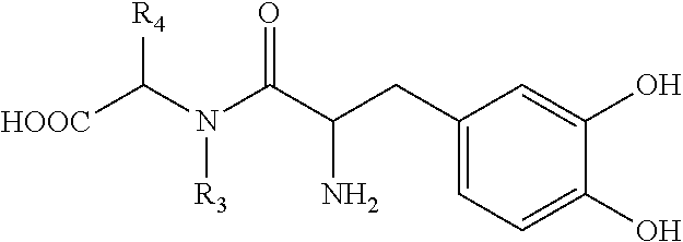

- wherein *C denotes a chiral carbon;
- R1, R2, R3 and R4 are each independently selected from the group consisting of hydrogen, alkyl having 1-30 carbon atoms, alkenyl having 1-30 carbon atoms, alkynyl having 1-30 carbon atoms, cycloalkyl, aryl, O-carboxy, C-carboxy, carbonyl, thiocarbonyl, 0-carbamyl, 0-thiocarbamyl and a fatty acid acyl, or, alternatively, R1 and R2 and/or R3 and R4 form a five- or six-membered ring; and
- R5 and R6 are each independently selected from the group consisting of hydrogen, alkyl, cycloalkyl, aryl and phosphonyl,
or a pharmaceutically acceptable salt thereof.

wherein each R is independently selected from the group consisting of a hydrogen atom, an acyl group, a

group, a —CO-pyridyl group, and a —CO—R3 group, wherein R3 represents the residue of any N,N—C1-C2 dialkylamino acid or a C4-C6 cycloalkylamino acid

wherein R1 represents a member selected from the group consisting of a hydroxyl group and a —OM group, wherein M is an alkali metal (Na, K, etc.) or an ammonium ion; and wherein R2 represents a member selected from the group consisting of a

group,
a —CO-pyridyl group, and a —CO—R3 group, wherein R3 represents the residue of any N,N—(C1-C2)-dialkylamino acid or a C4-C6-cycloalkylamino acid


wherein R represents an acyl group; wherein R2 represents a hydrogen atom; and wherein R1 represents a —NHCH(R4)COOR5 group, wherein R4 represents the residue of any naturally occurring amino acid, and wherein R5 represents a member selected from the group consisting of a hydrogen atom, a C1-C5 alkyl group (e.g., methyl, ethyl, propyl, butyl, pentyl), and a C1-C5 alkylaryl group (e.g., —CH2—C6H5, —CH2—CH2—C6H5, etc.), and the HX salts thereof, wherein X is a conventional pharmaceutically acceptable acid addition salt anion (e.g., chloride, bromide, perchlorate, methanesulfonate, succinate, etc.);
-
- 1. Glycyl-3,4-diacetyloxy-L-phenylalanine and its HX salt, wherein X represents a pharmaceutically acceptable anion.
- 2. Glycyl-3,4-diacetyloxy-L-phenylalanine-methyl ester and its HX salt, wherein X represents a pharmaceutically acceptable anion.
- 3. 3,4-diacetyloxy-L-phenylalanyl-glycine and its HX salt, wherein X represents a pharmaceutically acceptable anion.
- 4. N-nicotinoyl-3,4-dihydroxy-L-phenylalanine and its M salt, wherein M represents an alkali metal.
- 5. N-nicotinoyl-3,4-diacetyloxy-L-phenylalanine and its M salt, wherein M represents an alkali metal.
- 6. N-nicotinoyl-3,4-dipivalyloxy-L-phenylalanine and its M salt, wherein M represents an alkali metal.
- 7. 3,4-diacetyloxy-L-phenylalanyl-glycine and its HX salt, wherein X represents a pharmaceutically acceptable anion.
- 8. 3,4-diacetyloxy-L-phenylalanyl-glycine-methyl ester and its HX salt, wherein X represents a pharmaceutically acceptable anion.
- 9. 3,4-diacetyloxy-L-phenylalanyl-glycine-ethyl ester and its HX salt, wherein X represents a pharmaceutically acceptable anion.
- 10. 3,4-diacetyloxy-L-phenylalanyl-glycine-benzyl ester and its HX salt, wherein X represents a pharmaceutically acceptable anion.
- 11. 3,4-diacetyloxy-L-phenylalanyl-L-leucine and its HX salt, wherein X represents a pharmaceutically acceptable anion.
- 12. 3,4-diacetyloxy-L-phenylalanyl-L-leucine-methyl ester and its HX salt, wherein X represents a pharmaceutically acceptable anion.
- 13. 3,4-diacetyloxy-L-phenylalanyl-L-leucine-ethyl ester and its HX salt, wherein X represents a pharmaceutically acceptable anion.
- 14. 3,4-diacetyloxy-L-phenylalanyl-L-leucine-benzyl ester and its HX salt, wherein X represents a pharmaceutically acceptable anion.
- 15. 3,4-diacetyloxy-L-phenylalanyl-L-isoleucine and its HX salt, wherein X represents a pharmaceutically acceptable anion.
- 16. 3,4-diacetyloxy-L-phenylalanyl-L-isoleucine-methyl ester and its HX salt, wherein X represents a pharmaceutically acceptable anion.
- 17. 3,4-diacetyloxy-L-phenylalanyl-L-isoleucine-ethyl ester and its HX salt, wherein X represents a pharmaceutically acceptable anion.
- 18. 3,4-diacetyloxy-L-phenylalanyl-L-isoleucine-benzyl ester and its HX salt, wherein X represents a pharmaceutically acceptable anion.
- 19. 3,4-diacetyloxy-L-phenylalanyl-phenylalanine and its HX salt, wherein X represents a pharmaceutically acceptable anion.
- 20. 3,4-diacetyloxy-L-phenylalanyl-phenylalanine-methyl ester and its HX salt, wherein X represents a pharmaceutically acceptable anion.
- 21. 3,4-diacetyloxy-L-phenylalanyl-phenylalanine-ethyl ester and its HX salt, wherein X represents a pharmaceutically acceptable anion.
- 22. 3,4-diacetyloxy-L-phenylalanyl-phenylalanine-benzyl ester and its HX salt, wherein X represents a pharmaceutically acceptable anion.
- 23. Glycyl-3,4-diacetyloxy-L-phenylalanine and its HX salt, wherein X represents a pharmaceutically acceptable anion.
- 24. Glycyl-3,4-dipivalyloxy-L-phenylalanine and its HX salt, wherein X represents a pharmaceutically acceptable anion.
- 25. Glycyl-3,4-diacetyloxy-L-phenylalanine-methyl ester and its HX salt, wherein X represents a pharmaceutically acceptable anion.
- 26. Glycyl-3,4-diacetyloxy-L-phenylalanine-ethyl ester and its HX salt, wherein X represents a pharmaceutically acceptable anion.
- 27. Glycyl-3,4-diacetyloxy-L-phenylalanine-benzyl ester and its HX salt, wherein X represents a pharmaceutically acceptable anion.
- 28. L-leucyl-3,4-diacetyloxy-L-phenylalanine and its HX salt, wherein X represents a pharmaceutically acceptable anion.
- 29. L-leucyl-3,4-diacetyloxy-L-phenylalanine-methyl ester and its HX salt, wherein X represents a pharmaceutically acceptable anion.
- 30. L-leucyl-3,4-diacetyloxy-L-phenylalanine-ethyl ester and its HX salt, wherein X represents a pharmaceutically acceptable anion.
- 31. L-leucyl-3,4-diacetyloxy-L-phenylalanine-benzyl ester and its HX salt, wherein X represents a pharmaceutically acceptable anion.
- 32. L-isoleucyl-3,4-diacetyloxy-L-phenylalanine and its HX salt, wherein X represents a pharmaceutically acceptable anion.
- 33. L-isoleucyl-3,4-diacetyloxy-L-phenylalanine-methyl ester and its HX salt, wherein X represents a pharmaceutically acceptable anion.
- 34. L-isoleucyl-3,4-diacetyloxy-L-phenylalanine-ethyl ester and its HX salt, wherein X represents a pharmaceutically acceptable anion.
- 35. L-isoleucyl-3,4-diacetyloxy-L-phenylalanine-benzyl ester and its HX salt, wherein X represents a pharmaceutically acceptable anion.
- 36. Phenylalanyl-3,4-diacetyloxy-L-phenylalanine and its HX salt, wherein X represents a pharmaceutically acceptable anion.
- 37. Phenylalanyl-3,4-diacetyloxy-L-phenylalanine-methyl ester and its HX salt, wherein X represents a pharmaceutically acceptable anion.
- 38. Phenylalanyl-3,4-diacetyloxy-L-phenylalanine-ethyl ester and its HX salt, wherein X represents a pharmaceutically acceptable anion.
- 39. Phenylalanyl-3,4-diacetyloxy-L-phenylalanine-benzyl ester and its HX salt, wherein X represents a pharmaceutically acceptable anion.
- 40. 3,4-diacetyloxy-L-phenylalanyl-3,4-diacetyloxy-L-phenylalanine and its HX salt, wherein X represents a pharmaceutically acceptable anion.
- 41. 3,4-diacetyloxy-L-phenylalanyl-3,4-diacetyloxy-L-phenylalanine-methyl ester and its HX salt, wherein X represents a pharmaceutically acceptable anion.
- 42. 3,4-diacetyloxy-L-phenylalanyl-3,4-diacetyloxy-L-phenylalanine-ethyl ester and its HX salt, wherein X represents a pharmaceutically acceptable anion.
- 43. 3,4-diacetyloxy-L-phenylalanyl-3,4-diacetyloxy-L-phenylalanine-benzyl ester and its HX salt, wherein X represents a pharmaceutically acceptable anion.
- 44. N-[N,N-dimethylamino]-glycyl-3,4-diacetyloxy-L-phenylalanine and its M salt, wherein M represents an alkali metal.
- 45. N-nicotinoyl-3,4-dinicotinoyloxy-L-phenylalanine and its M salt, wherein M represents an alkali metal.
- 46. N-3-pyridylacetyl-3,4-dihydroxy-L-phenylalanine and its M salt, wherein M represents an alkali metal.
- 47. N-3-pyridylacetyl-3,4-diacetyloxy-L-phenylalanine and its M salt, wherein M represents an alkali metal.
- 48. 3,4-N,N-dimethylaminoglycyl-L-phenylalanine methylester and its HX salt, wherein X represents a pharmaceutically acceptable anion.
- 49. N[N,N-dimethylamino]glycyl-3,4-[N,N-dimethylaminoglycyl]-L-phenylalanine and its M salt, wherein M represents an alkali metal.
- 50. N[N,N-diethylaminoglycyl]-3,4-diacetyloxy-L-phenylalanine and its M salt, wherein M represents an alkali metal.
-
- (i) pigment epithelium-derived factor (PEDF) expression in the first cell population relative to one or both (A) PEDF expression in the first population of cells not contacted with the test compound, and (B) the second cell population, and
- (ii) intracellular calcium concentration in the first cell population relative to one or both (A) intracellular calcium concentration in the first population of cells not contacted with the test compound, and (B) the second cell population
- wherein the positive test compounds are candidate compounds for treating and/or limiting development of AMD.
- RPE—Cells were isolated as described [46] and maintained in Dulbecco's modified essential medium (DMEM) supplemented with 5% fetal bovine serum (FBS). For experiments in which tyrosine concentrations were lowered, custom manufactured DMEM produced without tyrosine by JRH Biosciences (Lenexa, Kans.) was used. Dialyzed FBS was purchased from Invitrogen, (San Diego, Calif.).
- COS-7 and CHO—Cells were obtained from ATCC and cultured in DMEM supplemented with 5% FBS. For analysis of OA1 distribution, cells were cultured in tyrosine-free DMEM supplemented with 1 μM tyrosine, 5% dialyzed FBS for 2-4 days, then tyrosine-free media as described for the experiment.
Cell Surface Biotinylation - Human RPE In Situ—Human eyecups were produced by dissection ˜2 mm anterior to the equator and removals of the anterior segment. The vitreous and retina were removed without impairing the underlying RPE monolayer, and the retina was cut at the optic nerve head. The resulting eyecups with RPE exposed were rinsed three times with reaction buffer (100 mM NaCl, 50 mM NaHCO3, pH 8.0) then filled with Sulfo-NHS-LC-Biotin (1 mg/ml) two times for thirty minutes. The reaction was stopped with TG buffer (25 mM Tris, 192 mM Glycine, pH 8.3) then the cells were harvested in lysis buffer (2 mM EDTA, 1% Triton X and 1
% Tween 20 in Tris Base Saline Buffer) containing Halt Protease Inhibitor Cocktail. Intact cells and pigment granules were removed by centrifugation at 14,000 rpm for 20 minutes. Biotinylated proteins were captured overnight with immobilized streptavidin beads and then mixed with 4× reducing buffer (250 mM Tris, pH 6.8, 8% SDS, 40% Glycerol, 20% Beta-mercaptoethanol, 0.08% bromophenol blue). The OA1 protein was separated on a 10% SDS-PAGE gel and identified by a using a polyclonal rabbit OA1 antibody for western blot analysis. Paired western blots were probed with a monoclonal antibody directed against actin. - Cultured Cells—RPE and transfected cells were maintained in DMEM containing tyrosine concentrations described for the experiment. Cultures were rinsed three times in reaction buffer, then biotinylated as described above for the in situ preparation.
Cloning of Oa1
[Cai]#=Kd#*(R−R min#)/R max #−R)
in which R, Rmin, and Rmax are the measured, minimum, and maximum ratios, respectively. Rmax represents the ratio of fluorescence intensity of ion-sensitive wavelengths under fully deprotonated conditions, whereas Rmin is the ratio for the dye when it is fully protonated. In the case of Fura-2, R increases with increasing Ca2+; hence Rmin represents Fura-2 in the absence of Ca2+ (Ca2+<1 nM) whereas Rmax represents the Ca2+-Fura-2 chelate as previously described [26]. Rmin, Rmax and Kd were determined in independent experiments in Fura-2 loaded cells, and subsequently utilized for calculation of free Ca2+ for the experimental procedures.
Radiolabeled Ligand Binding
- 1. Akeo K, Shirai S, Okisaka S, Shimizu H, Miyata H, et al. (1996) Histology of fetal eyes with oculocutaneous albinism. Arch Ophthalmol 114: 613-616.
- 2. Gregor Z (1978) The perifoveal vasculature in albinism. Br J Ophthalmol 62: 554-557.
- 3. Schraermeyer U, Heimann K (1999) Current understanding on the role of retinal pigment epithelium and its pigmentation. Pigment Cell Res 12: 219-236.
- 4. Rachel R A, Mason C A, Beermann F (2002) Influence of tyrosinase levels on pigment accumulation in the retinal pigment epithelium and on the uncrossed retinal projection. Pigment Cell Res 15: 273-281.
- 5. Okulicz J F, Shah R S, Schwartz R A, Janniger C K (2003) Oculocutaneous albinism. J Eur Acad Dermatol Venereol 17: 251-256.
- 6. Donatien P, Jeffery G (2002) Correlation between rod photoreceptor numbers and levels of ocular pigmentation. Invest Ophthalmol Vis Sci 43: 1198-1203.
- 7. Russell-Eggitt I (2001) Albinism. Ophthalmol Clin North Am 14: 533-546.
- 8. Oetting W S (1999) Albinism. Curr Opin Pediatr 11: 565-571.
- 9. Oetting W S, King R A (1999) Molecular basis of albinism: mutations and polymorphisms of pigmentation genes associated with albinism. Hum Mutat 13: 99-115.
- 10. Shen B, Samaraweera P, Rosenberg B, Orlow S J (2001) Ocular albinism type 1: more than meets the eye. Pigment Cell Res 14: 243-248.
- 11. Incerti B, Cortese K, Pizzigoni A, Surace E M, Varani S, et al. (2000) Oa1 knock-out: new insights on the pathogenesis of ocular albinism type 1. Hum Mol Genet 9: 2781-2788.
- 12. Bassi M T, Schiaffino M V, Renieri A, De Nigris F, Galli L, et al. (1995) Cloning of the gene for ocular albinism type 1 from the distal short arm of the X chromosome. Nat Genet 10: 13-19.
- 13. Schiaffino M V, Bassi M T, Galli L, Renieri A, Bruttini M, et al. (1995) Analysis of the OA1 gene reveals mutations in only one-third of patients with X-linked ocular albinism. Hum Mol Genet 4: 2319-2325.
- 14. Schiaffino M V, d'Addio M, Alloni A, Baschirotto C, Valetti C, et al. (1999) Ocular albinism: evidence for a defect in an intracellular signal transduction system. Nat Genet 23: 108-112.
- 15. Schiaffino M V, Tacchetti C (2005) The ocular albinism type 1 (OA1) protein and the evidence for an intracellular signal transduction system involved in melanosome biogenesis. Pigment Cell Res 18: 227-233.
- 16. Innamorati G, Piccirillo R, Bagnato P, Palmisano I, Schiaffino M V (2006) The melanosomal/lysosomal protein OA1 has properties of a G protein-coupled receptor. Pigment Cell Research 19: 125-135.
- 17. Staleva L, Orlow S J (2006) Ocular albinism 1 protein: trafficking and function when expressed in Saccharomyces cerevisiae. Exp Eye Res 82: 311-318.
- 18. Shen B, Orlow S J (2001) The ocular albinism type 1 gene product is an N-glycoprotein but glycosylation is not required for its subcellular distribution. Pigment Cell Res 14: 485-490.
- 19. d'Addio M, Pizzigoni A, Bassi M T, Baschirotto C, Valetti C, et al. (2000) Defective intracellular transport and processing of OA1 is a major cause of ocular albinism type 1. Hum Mol Genet 9: 3011-3018.
- 20. Shen B, Rosenberg B, Orlow S J (2001) Intracellular distribution and late endosomal effects of the ocular albinism type 1 gene product: consequences of disease-causing mutations and implications for melanosome biogenesis. Traffic 2: 202-211.
- 21. Samaraweera P, Shen B, Newton J M, Barsh G S, Orlow S J (2001) The mouse ocular albinism 1 gene product is an endolysosomal protein. Exp Eye Res 72: 319-329.
- 22. Schiaffino M V, Baschirotto C, Pellegrini G, Montalti S, Tacchetti C, et al. (1996) The ocular albinism type 1 gene product is a membrane glycoprotein localized to melanosomes. Proc Natl Acad Sci USA 93: 9055-9060.
- 23. Ilia M, Jeffery G (2000) Retinal cell addition and rod production depend on early stages of ocular melanin synthesis. J Comp Neurol 420: 437-444.
- 24. Ilia M, Jeffery G (1999) Retinal mitosis is regulated by dopa, a melanin precursor that may influence the time at which cells exit the cell cycle: analysis of patterns of cell production in pigmented and albino retinae. J Comp Neurol 405: 394-405.
- 25. Ito S (2003) The IFPCS presidential lecture: a chemist's view of melanogenesis. Pigment Cell Res 16: 230-236.
- 26. Martinez-Zaguilan R, Tompkins L S, Gillies R J, Lynch R M (2006) Simultaneous analysis of intracellular pH and Ca2+ from cell populations. Methods Mol Biol 312: 269-287.
- 27. Ferguson S S, Caron M G (2004) Green fluorescent protein-tagged beta-arrestin translocation as a measure of G protein-coupled receptor activation. Methods in Molecular Biology 237: 121-126.
- 28. Barak L S, Warabi K, Feng X, Caron M G, Kwatra M M (1999) Real-time visualization of the cellular redistribution of G protein-coupled
receptor kinase 2 and beta-arrestin 2 during homologous desensitization of the substance P receptor. J Biol Chem 274: 7565-7569. - 29. Zhang J, Barak L S, Anborgh P H, Laporte S A, Caron M G, et al. (1999) Cellular trafficking of G protein-coupled receptor/beta-arrestin endocytic complexes. J Biol Chem 274: 10999-11006.
- 30. Tohgo A, Choy E W, Gesty-Palmer D, Pierce K L, Laporte S, et al. (2003) The stability of the G protein-coupled receptor-beta-arrestin interaction determines the mechanism and functional consequence of ERK activation. J Biol Chem 278: 6258-6267.
- 31. Ferguson S S, Zhang J, Barak L S, Caron M G (1998) Molecular mechanisms of G protein-coupled receptor desensitization and resensitization. Life Sci 62: 1561-1565.
- 32. Barak L S, Ferguson S S, Zhang J, Caron M G (1997) A beta-arrestin/green fluorescent protein biosensor for detecting G protein-coupled receptor activation. J Biol Chem 272: 27497-27500.
- 33. Barak L S, Ferguson S S, Zhang J, Martenson C, Meyer T, et al. (1997) Internal trafficking and surface mobility of a functionally intact beta2-adrenergic receptor-green fluorescent protein conjugate. Mol Pharmacol 51: 177-184.
- 34. McKay B S, Goodman B, Falk T, Sherman S J (2006) Retinal pigment epithelial cell transplantation could provide trophic support in Parkinson's disease: Results from an in vitro model system. Exp Neurol 201: 234-243.
- 35. Tombran-Tink J, Shivaram S M, Chader G J, Johnson L V, Bok D (1995) Expression, secretion, and age-related downregulation of pigment epithelium-derived factor, a serpin with neurotrophic activity. J Neurosci 15: 4992-5003.
- 36. Malchiodi-Albedi F, Feher J, Caiazza S, Formisano G, Perilli R, et al. (1998) PEDF (pigment epithelium-derived factor) promotes increase and maturation of pigment granules in pigment epithelial cells in neonatal albino rat retinal cultures. Int J Dev Neurosci 16: 423-432.
- 37. Behling K C, Surace E M, Bennett J (2002) Pigment epithelium-derived factor expression in the developing mouse eye. Mol Vis 8: 449-454.
- 38. Aymerich M S, Alberdi E M, Martinez A, Becerra S P (2001) Evidence for pigment epithelium-derived factor receptors in the neural retina. Invest Ophthalmol Vis Sci 42: 3287-3293.
- 39. Tombran-Tink J, Chader G G, Johnson L V (1991) PEDF: a pigment epithelium-derived factor with potent neuronal differentiative activity. Exp Eye Res 53: 411-414.
- 40. Jablonski M M, Tombran-Tink J, Mrazek D A, Iannaccone A (2001) Pigment epithelium-derived factor supports normal Muller cell development and glutamine synthetase expression after removal of the retinal pigment epithelium. Glia 35: 14-25.
- 41. Jablonski M M, Tombran-Tink J, Mrazek D A, Iannaccone A (2000) Pigment epithelium-derived factor supports normal development of photoreceptor neurons and opsin expression after retinal pigment epithelium removal. J Neurosci 20: 7149-7157.
- 42. Jeffery G (1998) The retinal pigment epithelium as a developmental regulator of the neural retina. Eye 12 (Pt 3b): 499-503.
- 43. Piccirillo R, Palmisano I, Innamorati G, Bagnato P, Altimare D, et al. (2003) An unconventional dileucine-based motif and a novel cytosolic motif are required for the lysosomal and melanosomal targeting of OA1. Journal of Cell Science 119: 2003-2014.
- 44. Van Raamsdonk C D, Fitch K R, Fuchs H, de Angelis M H, Barsh G S (2004) Effects of G-protein mutations on skin color. Nat Genet 36: 961-968.
- 45. Young A, Powelson E B, Whitney I E, Raven M A, Nusinowitz S, et al. (2008) Involvement of OA1, an intracellular GPCR, and G alpha i3, its binding protein, in melanosomal biogenesis and optic pathway formation. Invest Ophthalmol Vis Sci 49: 3245-3252.
- 46. Hu J, Bok D (2001) A cell culture medium that supports the differentiation of human retinal pigment epithelium into functionally polarized monolayers. Mol Vis 7: 14-19.
- 47. Stamer W D, Golightly S F, Hosohata Y, Ryan E P, Porter A C, et al. (2001) Cannabinoid C B(1) receptor expression, activation and detection of endogenous ligand in trabecular meshwork and ciliary process tissues. Eur J Pharmacol 431: 277-286.
Claims (20)
Priority Applications (1)
| Application Number | Priority Date | Filing Date | Title |
|---|---|---|---|
| US14/858,602 US9861600B2 (en) | 2008-04-18 | 2015-09-18 | Methods and compositions for treating and identifying compounds to treat age-related macular degeneration treatment |
Applications Claiming Priority (4)
| Application Number | Priority Date | Filing Date | Title |
|---|---|---|---|
| US12462408P | 2008-04-18 | 2008-04-18 | |
| PCT/US2009/041021 WO2009129497A2 (en) | 2008-04-18 | 2009-04-17 | Methods and compositions for treating and identifying compounds to treat age-related macular degeneration |
| US93766910A | 2010-11-05 | 2010-11-05 | |
| US14/858,602 US9861600B2 (en) | 2008-04-18 | 2015-09-18 | Methods and compositions for treating and identifying compounds to treat age-related macular degeneration treatment |
Related Parent Applications (2)
| Application Number | Title | Priority Date | Filing Date |
|---|---|---|---|
| PCT/US2009/041021 Division WO2009129497A2 (en) | 2008-04-18 | 2009-04-17 | Methods and compositions for treating and identifying compounds to treat age-related macular degeneration |
| US12/937,669 Division US9173862B2 (en) | 2008-04-18 | 2009-04-17 | Methods and compositions for treating and identifying compounds to treat age-related macular degeneration |
Publications (2)
| Publication Number | Publication Date |
|---|---|
| US20160015665A1 US20160015665A1 (en) | 2016-01-21 |
| US9861600B2 true US9861600B2 (en) | 2018-01-09 |
Family
ID=41110686
Family Applications (2)
| Application Number | Title | Priority Date | Filing Date |
|---|---|---|---|
| US12/937,669 Active 2030-10-16 US9173862B2 (en) | 2008-04-18 | 2009-04-17 | Methods and compositions for treating and identifying compounds to treat age-related macular degeneration |
| US14/858,602 Active US9861600B2 (en) | 2008-04-18 | 2015-09-18 | Methods and compositions for treating and identifying compounds to treat age-related macular degeneration treatment |
Family Applications Before (1)
| Application Number | Title | Priority Date | Filing Date |
|---|---|---|---|
| US12/937,669 Active 2030-10-16 US9173862B2 (en) | 2008-04-18 | 2009-04-17 | Methods and compositions for treating and identifying compounds to treat age-related macular degeneration |
Country Status (7)
| Country | Link |
|---|---|
| US (2) | US9173862B2 (en) |
| EP (1) | EP2273988B1 (en) |
| JP (1) | JP2011520788A (en) |
| AU (1) | AU2009236078B2 (en) |
| BR (1) | BRPI0910575A2 (en) |
| CA (1) | CA2721360A1 (en) |
| WO (1) | WO2009129497A2 (en) |
Families Citing this family (8)
| Publication number | Priority date | Publication date | Assignee | Title |
|---|---|---|---|---|
| US20120237473A1 (en) * | 2011-03-14 | 2012-09-20 | University Of Medicine And Dentistry Of New Jersey | Compositions And Methods For Cell Based Retinal Therapies |
| FR3000105B1 (en) * | 2012-12-24 | 2015-05-15 | Lvmh Rech | APTAMERS INHIBITING THE ENZYMATIC ACTIVITY OF TYROSINASE |
| WO2015029948A1 (en) * | 2013-08-26 | 2015-03-05 | リンク・ジェノミクス株式会社 | Prophylactic or therapeutic agent for retinal disease caused by retinal pigment epithelium disorder |
| EP3277660B1 (en) | 2015-03-30 | 2019-10-16 | Berlirem GmbH | Water-soluble l-dopa esters |
| US10314473B2 (en) * | 2015-09-09 | 2019-06-11 | New York University | System and method for in vivo detection of fluorescence from an eye |
| WO2018036498A1 (en) * | 2016-08-24 | 2018-03-01 | National Institute Of Biological Sciences, Beijing | Entacapone-related compounds to treat macular degeneration |
| US20220096410A1 (en) | 2019-01-09 | 2022-03-31 | Fuso Pharmaceutical Industries, Ltd. | Pharmaceutical composition for intraocular or oral administration for treatment of retinal diseases |
| WO2020160256A1 (en) * | 2019-01-31 | 2020-08-06 | Arizona Board Of Regents On Behalf Of The University Of Arizona | Compositions and methods for treating or limiting development of age-related macular degeneration |
Citations (16)
| Publication number | Priority date | Publication date | Assignee | Title |
|---|---|---|---|---|
| US3891696A (en) | 1973-11-02 | 1975-06-24 | Interx Research Corp | Novel, transient pro-drug forms of l-dopa |
| US4035507A (en) | 1975-04-17 | 1977-07-12 | Interx Research Corporation | Novel, transient pro-drug forms of L-DOPA to treat Parkinson's disease |
| US4663349A (en) | 1985-12-30 | 1987-05-05 | Merck & Co., Inc. | Rectally absorbable form of L-dopa |
| US4771073A (en) | 1985-12-30 | 1988-09-13 | Merck & Co., Inc. | Rectally absorbable form of L-dopa |
| US4873263A (en) | 1985-12-30 | 1989-10-10 | Merck & Co., Inc. | Rectally absorbable form of L-dopa |
| US5345885A (en) | 1992-09-09 | 1994-09-13 | Nara Company, Ltd. | Looper of tufting machine |
| WO1997016181A1 (en) | 1995-11-03 | 1997-05-09 | University Of Kentucky | Method for the intranasal administration of l-dopa prodrugs |
| US5686423A (en) | 1996-02-16 | 1997-11-11 | Department Of Health, The Executive Yuan, Republic Of China | Di-and tri-peptide mimetic compounds for Parkinson's disease |
| WO1999043286A2 (en) | 1998-02-24 | 1999-09-02 | Mcp Hahnemann University | Isolated stromal cells for use in the treatment of diseases of the central nervous system |
| WO2000000197A1 (en) | 1998-06-26 | 2000-01-06 | Warner-Lambert Company | 4-benzyl piperidine alkylsulfoxide heterocycles and their use as subtype-selective nmda receptor antagonists |
| WO2002028882A1 (en) | 2000-10-06 | 2002-04-11 | Xenoport, Inc. | Bile acid prodrugs of l-dopa and their use in the sustained treatment of parkinsonism |
| US20020102581A1 (en) | 1999-02-19 | 2002-08-01 | Hageman Gregory S. | Diagnostics and therapeutics for ocular disorders |
| WO2003070269A1 (en) | 2002-02-18 | 2003-08-28 | Cevec Pharmaceuticals Gmbh | Treatment for diseases of the eye, inner ear and central nervous system |
| US6660297B2 (en) | 2001-03-23 | 2003-12-09 | Bausch & Lomb Incorporated | Nutritional supplement to treat macular degeneration |
| WO2004069146A2 (en) | 2003-02-07 | 2004-08-19 | Yissum Research Development Company Of The Hebrew University Of Jerusalem | L-dopa amide derivatives and uses thereof |
| US20040220270A1 (en) | 2003-03-07 | 2004-11-04 | The Jackson Laboratory | Methods and composition of treating glaucoma by modulating tyrosinase/L-DOPA pathway |
-
2009
- 2009-04-17 EP EP09733334.8A patent/EP2273988B1/en active Active
- 2009-04-17 JP JP2011505240A patent/JP2011520788A/en active Pending
- 2009-04-17 BR BRPI0910575A patent/BRPI0910575A2/en not_active Application Discontinuation
- 2009-04-17 AU AU2009236078A patent/AU2009236078B2/en active Active
- 2009-04-17 US US12/937,669 patent/US9173862B2/en active Active
- 2009-04-17 WO PCT/US2009/041021 patent/WO2009129497A2/en active Application Filing
- 2009-04-17 CA CA2721360A patent/CA2721360A1/en not_active Abandoned
-
2015
- 2015-09-18 US US14/858,602 patent/US9861600B2/en active Active
Patent Citations (19)
| Publication number | Priority date | Publication date | Assignee | Title |
|---|---|---|---|---|
| US3891696A (en) | 1973-11-02 | 1975-06-24 | Interx Research Corp | Novel, transient pro-drug forms of l-dopa |
| US4035507A (en) | 1975-04-17 | 1977-07-12 | Interx Research Corporation | Novel, transient pro-drug forms of L-DOPA to treat Parkinson's disease |
| US4663349A (en) | 1985-12-30 | 1987-05-05 | Merck & Co., Inc. | Rectally absorbable form of L-dopa |
| US4771073A (en) | 1985-12-30 | 1988-09-13 | Merck & Co., Inc. | Rectally absorbable form of L-dopa |
| US4873263A (en) | 1985-12-30 | 1989-10-10 | Merck & Co., Inc. | Rectally absorbable form of L-dopa |
| US5345885A (en) | 1992-09-09 | 1994-09-13 | Nara Company, Ltd. | Looper of tufting machine |
| WO1997016181A1 (en) | 1995-11-03 | 1997-05-09 | University Of Kentucky | Method for the intranasal administration of l-dopa prodrugs |
| US5686423A (en) | 1996-02-16 | 1997-11-11 | Department Of Health, The Executive Yuan, Republic Of China | Di-and tri-peptide mimetic compounds for Parkinson's disease |
| WO1999043286A2 (en) | 1998-02-24 | 1999-09-02 | Mcp Hahnemann University | Isolated stromal cells for use in the treatment of diseases of the central nervous system |
| WO2000000197A1 (en) | 1998-06-26 | 2000-01-06 | Warner-Lambert Company | 4-benzyl piperidine alkylsulfoxide heterocycles and their use as subtype-selective nmda receptor antagonists |
| US20020102581A1 (en) | 1999-02-19 | 2002-08-01 | Hageman Gregory S. | Diagnostics and therapeutics for ocular disorders |
| WO2002028882A1 (en) | 2000-10-06 | 2002-04-11 | Xenoport, Inc. | Bile acid prodrugs of l-dopa and their use in the sustained treatment of parkinsonism |
| US20020151526A1 (en) | 2000-10-06 | 2002-10-17 | Gallop Mark A. | Bile-acid prodrugs of L-dopa and their use in the sustained treatment of parkinsonism |
| US6660297B2 (en) | 2001-03-23 | 2003-12-09 | Bausch & Lomb Incorporated | Nutritional supplement to treat macular degeneration |
| WO2003070269A1 (en) | 2002-02-18 | 2003-08-28 | Cevec Pharmaceuticals Gmbh | Treatment for diseases of the eye, inner ear and central nervous system |
| US20050142128A1 (en) | 2002-02-18 | 2005-06-30 | Ulrich Schraermeyer | Treatment for diseases of the eye inner ear and central nervous system |
| WO2004069146A2 (en) | 2003-02-07 | 2004-08-19 | Yissum Research Development Company Of The Hebrew University Of Jerusalem | L-dopa amide derivatives and uses thereof |
| US20060025385A1 (en) | 2003-02-07 | 2006-02-02 | Daphne Atlas | L-DOPA amide derivatives and uses thereof |
| US20040220270A1 (en) | 2003-03-07 | 2004-11-04 | The Jackson Laboratory | Methods and composition of treating glaucoma by modulating tyrosinase/L-DOPA pathway |
Non-Patent Citations (60)
| Title |
|---|
| Akeo K, Shirai S, Okisaka S, Shimizu H, Miyata H, et al. (1996) Arch Ophthalmol 114: 613-616. |
| Aymerich MS, et al. (2001) Invest Ophthalmol Vis Sci 42: 3287-3293. |
| Barak LS, et al. (1997) J Biol Chem 272: 27497-27500. |
| Barak LS, et al. (1997) Mol Pharmacol 51: 177-184. |
| Barak LS, et al. (1999) J Biol Chem 274: 7565-7569. |
| Bassi MT, et al. (1995) Nat Genet 10: 13-19. |
| Behling KC, et al. (2002) Mol Vis 8: 449-454. |
| Berendschot, et al., (2002) IOVS, "Macular Pigment and Melanin in Age-Related Maculopathy in a General Population", vol. 43(6) pp. 1929-1932. |
| d'Addio M, et al. (2000) Hum Mol Genet 9: 3011-3018. |
| Decatur, et al, (2009) ARVO Meeting Abstracts, 50: 1866. |
| Decatur, et al., (2008) ARVO Meeting Abstracts, 49: 5554. |
| Donatien P, et al. (2002) Invest Ophthalmol Vis Sci 43: 1198-1203. |
| Entry for "Caucasian". Dictionary.com Unabridged. Dictionary.com website: dictionary.reference.com/browse/caucasian.Accessed Jan. 6, 2015. |
| Ferguson SS, Caron MG (2004) Methods in Molecular Biology 237: 121-126. |
| Ferguson SS, et al. (1998) Life Sci 62: 1561-1565. |
| Gregor Z (1978) Br J Ophthalmol 62: 554-557. |
| Hu J, Bok D (2001) Mol Vis 7: 14-19. |
| Ilia M, Jeffery G (1999) J Comp Neurol 405: 394-405. |
| Ilia M, Jeffery G (2000) J Comp Neurol 420: 437-444. |
| Incerti B, et al. (2000) Hum Mol Genet 9: 2781-2788. |
| Innamorati G, et al. (2006) Pigment Cell Research 19: 125-135. |
| ISR for WO 2009/129497, dated Oct. 13, 2009. |
| Ito S (2003) Pigment Cell Res 16: 230-236. |
| Jablonski MM, et al. (2000) J Neurosci 20: 7149-7157. |
| Jablonski MM, et al. (2001) Glia 35: 14-25. |
| Jeffery G (1998) Eye 12(Pt 3b): 499-503. |
| Kanis, et al. (2007) Graefe's Arch Clin Exp Ophthalmol, "Influence of Macular Pigment and Melanin on INcident Early AMD in a White Population", vol. 245 pp. 767-773. |
| Kao, et al., (2000) Pharmaceutical Research, 17)8):978-984. |
| Lai, et al. (1973) Journal of Pharmaceutical Sciences, 62:510. |
| Lopez, et al., (2008) PLOS Biology 6(9): 1861-1869. |
| Malchiodi-Albedi F, et al. (1998) Int J Dev Neurosci 16: 423-432. |
| Martinez-Zaguilan R, et al. (2006) Methods Mol Biol 312: 269-287. |
| McKay BS, et al. (2006) Exp Neurol 201: 234-243. |
| Nowak JZ. Age-related macular degeneration (AMD): pathogenesis and therapy. 2006 Pharmacol. Rep. 58: 353-363. |
| Oetting WS (1999) Curr Opin Pediatr 11 : 565-571. |
| Oetting WS, et al. (1999) Hum Mutat 13: 99-115. |
| Okulicz JF, et al. (2003) J Eur Acad Dermatol Venereol 17: 251-256. |
| Piccirillo R, et al. (2003) Journal of Cell Science 119: 2003-2014. |
| Rachel, et al. (2002) Pigment Cell Res 15: 273-281. |
| Russell-Eggitt 1(2001) Ophthalmol Olin North Am 14: 533-546. |
| Samaraweera P, et al. (2001) Exp Eye Res 72: 3 19-329. |
| Schiaffino MV and Tacchetti C (2005) Pigment Cell Res 18: 227-233. |
| Schiaffino MV, et al. (1995) Hum Mol Genet 4: 2319-2325. |
| Schiaffino MV, et al. (1996) Proc Natl Acad Sci USA 93: 9055-9060. |
| Schiaffino MV, et al. (1999) Nat Genet 23: 108-112. |
| Schraermeyer U, Heimann K (1999) Pigment Cell Res 12: 219-236. |
| Shen B, and Orlow SJ (2001) Pigment Cell Res 14: 485-490. |
| Shen B, et al. (2001) Pigment Cell Res 14: 243-248. |
| Shen B, et al. (2001) Traffic 2: 202-211. |
| Staleva L, and Orlow SJ (2006) Exp Eye Res 82: 311-318. |
| Stamer WD, et al. (2001) Eur J Pharmacol 431: 277-286. |
| Teeple, et al., (2007) ARVO Meeting Abstracts, 48: 2537. |
| Tohgo A, et al. (2003) J Biol Chem 278: 6258-6267. |
| Tombran-Tink J, et al. (1991) Exp Eye Res 53: 411-414. |
| Tombran-Tink J, et al. (1995) J Neurosci 15: 4992-5003. |
| Van Raamsdonk CD, et al. (2004) Nat Genet 36: 961-968. |
| Vaziri, et al., Investigative Ophthalmology & Visual Science 56: Abstract No. 2818-C0046, publication date May 5, 2015). |
| Whiting, et al., (2005) Annual Meeting of the Association for Research in vision and Ophthalmology, Ft Lauderdale, FL 46(Supps) 2297. |
| Young A, et al. (2008) Invest Ophthalmol Vis Sci 49: 3245-3252. |
| Zhang J, et al. (1999) J Biol Chem 274: 10999-11006. |
Also Published As
| Publication number | Publication date |
|---|---|
| AU2009236078B2 (en) | 2014-03-06 |
| WO2009129497A3 (en) | 2009-12-03 |
| JP2011520788A (en) | 2011-07-21 |
| AU2009236078A1 (en) | 2009-10-22 |
| US20160015665A1 (en) | 2016-01-21 |
| BRPI0910575A2 (en) | 2017-06-06 |
| US9173862B2 (en) | 2015-11-03 |
| US20110044908A1 (en) | 2011-02-24 |
| CA2721360A1 (en) | 2009-10-22 |
| EP2273988B1 (en) | 2015-09-09 |
| WO2009129497A2 (en) | 2009-10-22 |
| EP2273988A2 (en) | 2011-01-19 |
Similar Documents
| Publication | Publication Date | Title |
|---|---|---|
| US9861600B2 (en) | Methods and compositions for treating and identifying compounds to treat age-related macular degeneration treatment | |
| Lopez et al. | L-DOPA is an endogenous ligand for OA1 | |
| Leithe et al. | Endocytosis and post-endocytic sorting of connexins | |
| Uriarte et al. | Circulating ghrelin crosses the blood-cerebrospinal fluid barrier via growth hormone secretagogue receptor dependent and independent mechanisms | |
| DK2865421T3 (en) | Bittersweet receptor agonists for topical use | |
| Chen et al. | 16 Structure and Function of Rod and Cone Photoreceptors | |
| AU2011279703B2 (en) | Materials and method for suppressing inflamatory and neuropathic pain | |
| Miller et al. | Pregnancy-associated plasma protein-aa regulates photoreceptor synaptic development to mediate visually guided behavior | |
| Chothe et al. | Transport of hepcidin, an iron-regulatory peptide hormone, into retinal pigment epithelial cells via oligopeptide transporters and its relevance to iron homeostasis | |
| US20090175848A1 (en) | Modulation of the Cooperativity Between the Ion Channels TRPM5 and TRPA1 | |
| Zhang et al. | Cone arrestin expression and induction in retinoblastoma cells | |
| Kothandapani et al. | Distinctive functioning of STARD1 in the fetal Leydig cells compared to adult Leydig and adrenal cells. Impact of Hedgehog signaling via the primary cilium | |
| Rejdak et al. | Ontogenic changes of kynurenine aminotransferase I activity and its expression in the chicken retina | |
| AU2018221147B2 (en) | Brain osteocalcin receptor and cognitive disorders | |
| Vähätupa | Regulation of Angiogenesis: Role of R-Ras, furin and syndecan 4 in retinal angiogenesis | |
| Leung et al. | The Logistical Backbone of Visual Function: Complementary Function of Dietary Vitamin A Receptors and Rhodopsin Transporters | |
| Caputo | Molecular mechanism of synaptic vesicles recycling: role of the Phospholipid Scramblase-1 (PLSCR1) | |
| Stajano | Role of tetraspanin 15 in metabotropic glutamate receptor 5 signaling and extracellular vesicles docking at neurons from mus musculus domesticus | |
| Feng | Study of Short Forms of P/Q-Type Voltage-Gated Calcium Channels | |
| Tokhunts | Identifying the biologically active forms of HEDGEHOG proteins | |
| Cragg et al. | Immunolocalization of the zinc transporter hZTL1 at the apical membrane of human jejunum and Caco-2 cells | |
| Shen et al. | in photoreceptor cells, creatine supply to photoreceptor cells from local Müller glia might take place in patients with CRT deficiency. We studied creatine biosynthesis using albino Wistar rats. Although the retina in albino rats is likely stressed | |
| Nourollahi | CADM1 function in regards to insulin secretion in the beta cell | |
| CA2436259A1 (en) | Compounds effecting neuron remodeling and assays for same | |
| Entwistle | Pancreatic secretions and their effect on circulatory and immune cells |
Legal Events
| Date | Code | Title | Description |
|---|---|---|---|
| AS | Assignment |
Owner name: ARIZONA BOARD OF REGENTS, A BODY CORPORATE OF THE Free format text: ASSIGNMENT OF ASSIGNORS INTEREST;ASSIGNOR:MCKAY, BRIAN;REEL/FRAME:036624/0577 Effective date: 20101102 |
|
| AS | Assignment |
Owner name: NATIONAL INSTITUTES OF HEALTH (NIH), U.S. DEPT. OF Free format text: CONFIRMATORY LICENSE;ASSIGNOR:UNIVERSITY OF ARIZONA;REEL/FRAME:037032/0948 Effective date: 20151030 |
|
| STCF | Information on status: patent grant |
Free format text: PATENTED CASE |
|
| MAFP | Maintenance fee payment |
Free format text: PAYMENT OF MAINTENANCE FEE, 4TH YR, SMALL ENTITY (ORIGINAL EVENT CODE: M2551); ENTITY STATUS OF PATENT OWNER: SMALL ENTITY Year of fee payment: 4 |