US9855155B2 - Endoprosthesis anchoring and sealing - Google Patents
Endoprosthesis anchoring and sealing Download PDFInfo
- Publication number
- US9855155B2 US9855155B2 US14/316,151 US201414316151A US9855155B2 US 9855155 B2 US9855155 B2 US 9855155B2 US 201414316151 A US201414316151 A US 201414316151A US 9855155 B2 US9855155 B2 US 9855155B2
- Authority
- US
- United States
- Prior art keywords
- suture
- endoprosthesis
- stent
- main body
- barbs
- Prior art date
- Legal status (The legal status is an assumption and is not a legal conclusion. Google has not performed a legal analysis and makes no representation as to the accuracy of the status listed.)
- Expired - Fee Related
Links
Images


Classifications
-
- A—HUMAN NECESSITIES
- A61—MEDICAL OR VETERINARY SCIENCE; HYGIENE
- A61F—FILTERS IMPLANTABLE INTO BLOOD VESSELS; PROSTHESES; DEVICES PROVIDING PATENCY TO, OR PREVENTING COLLAPSING OF, TUBULAR STRUCTURES OF THE BODY, e.g. STENTS; ORTHOPAEDIC, NURSING OR CONTRACEPTIVE DEVICES; FOMENTATION; TREATMENT OR PROTECTION OF EYES OR EARS; BANDAGES, DRESSINGS OR ABSORBENT PADS; FIRST-AID KITS
- A61F2/00—Filters implantable into blood vessels; Prostheses, i.e. artificial substitutes or replacements for parts of the body; Appliances for connecting them with the body; Devices providing patency to, or preventing collapsing of, tubular structures of the body, e.g. stents
- A61F2/82—Devices providing patency to, or preventing collapsing of, tubular structures of the body, e.g. stents
- A61F2/852—Two or more distinct overlapping stents
-
- A—HUMAN NECESSITIES
- A61—MEDICAL OR VETERINARY SCIENCE; HYGIENE
- A61F—FILTERS IMPLANTABLE INTO BLOOD VESSELS; PROSTHESES; DEVICES PROVIDING PATENCY TO, OR PREVENTING COLLAPSING OF, TUBULAR STRUCTURES OF THE BODY, e.g. STENTS; ORTHOPAEDIC, NURSING OR CONTRACEPTIVE DEVICES; FOMENTATION; TREATMENT OR PROTECTION OF EYES OR EARS; BANDAGES, DRESSINGS OR ABSORBENT PADS; FIRST-AID KITS
- A61F2/00—Filters implantable into blood vessels; Prostheses, i.e. artificial substitutes or replacements for parts of the body; Appliances for connecting them with the body; Devices providing patency to, or preventing collapsing of, tubular structures of the body, e.g. stents
- A61F2/02—Prostheses implantable into the body
- A61F2/04—Hollow or tubular parts of organs, e.g. bladders, tracheae, bronchi or bile ducts
- A61F2/06—Blood vessels
- A61F2/064—Blood vessels with special features to facilitate anastomotic coupling
-
- A—HUMAN NECESSITIES
- A61—MEDICAL OR VETERINARY SCIENCE; HYGIENE
- A61F—FILTERS IMPLANTABLE INTO BLOOD VESSELS; PROSTHESES; DEVICES PROVIDING PATENCY TO, OR PREVENTING COLLAPSING OF, TUBULAR STRUCTURES OF THE BODY, e.g. STENTS; ORTHOPAEDIC, NURSING OR CONTRACEPTIVE DEVICES; FOMENTATION; TREATMENT OR PROTECTION OF EYES OR EARS; BANDAGES, DRESSINGS OR ABSORBENT PADS; FIRST-AID KITS
- A61F2/00—Filters implantable into blood vessels; Prostheses, i.e. artificial substitutes or replacements for parts of the body; Appliances for connecting them with the body; Devices providing patency to, or preventing collapsing of, tubular structures of the body, e.g. stents
- A61F2/02—Prostheses implantable into the body
- A61F2/04—Hollow or tubular parts of organs, e.g. bladders, tracheae, bronchi or bile ducts
- A61F2/06—Blood vessels
- A61F2/07—Stent-grafts
-
- A—HUMAN NECESSITIES
- A61—MEDICAL OR VETERINARY SCIENCE; HYGIENE
- A61F—FILTERS IMPLANTABLE INTO BLOOD VESSELS; PROSTHESES; DEVICES PROVIDING PATENCY TO, OR PREVENTING COLLAPSING OF, TUBULAR STRUCTURES OF THE BODY, e.g. STENTS; ORTHOPAEDIC, NURSING OR CONTRACEPTIVE DEVICES; FOMENTATION; TREATMENT OR PROTECTION OF EYES OR EARS; BANDAGES, DRESSINGS OR ABSORBENT PADS; FIRST-AID KITS
- A61F2/00—Filters implantable into blood vessels; Prostheses, i.e. artificial substitutes or replacements for parts of the body; Appliances for connecting them with the body; Devices providing patency to, or preventing collapsing of, tubular structures of the body, e.g. stents
- A61F2/82—Devices providing patency to, or preventing collapsing of, tubular structures of the body, e.g. stents
- A61F2/86—Stents in a form characterised by the wire-like elements; Stents in the form characterised by a net-like or mesh-like structure
- A61F2/90—Stents in a form characterised by the wire-like elements; Stents in the form characterised by a net-like or mesh-like structure characterised by a net-like or mesh-like structure
-
- A—HUMAN NECESSITIES
- A61—MEDICAL OR VETERINARY SCIENCE; HYGIENE
- A61B—DIAGNOSIS; SURGERY; IDENTIFICATION
- A61B17/00—Surgical instruments, devices or methods
- A61B17/04—Surgical instruments, devices or methods for suturing wounds; Holders or packages for needles or suture materials
- A61B17/06—Needles ; Sutures; Needle-suture combinations; Holders or packages for needles or suture materials
- A61B17/06166—Sutures
- A61B2017/06176—Sutures with protrusions, e.g. barbs
-
- A—HUMAN NECESSITIES
- A61—MEDICAL OR VETERINARY SCIENCE; HYGIENE
- A61F—FILTERS IMPLANTABLE INTO BLOOD VESSELS; PROSTHESES; DEVICES PROVIDING PATENCY TO, OR PREVENTING COLLAPSING OF, TUBULAR STRUCTURES OF THE BODY, e.g. STENTS; ORTHOPAEDIC, NURSING OR CONTRACEPTIVE DEVICES; FOMENTATION; TREATMENT OR PROTECTION OF EYES OR EARS; BANDAGES, DRESSINGS OR ABSORBENT PADS; FIRST-AID KITS
- A61F2/00—Filters implantable into blood vessels; Prostheses, i.e. artificial substitutes or replacements for parts of the body; Appliances for connecting them with the body; Devices providing patency to, or preventing collapsing of, tubular structures of the body, e.g. stents
- A61F2/02—Prostheses implantable into the body
- A61F2/04—Hollow or tubular parts of organs, e.g. bladders, tracheae, bronchi or bile ducts
- A61F2/06—Blood vessels
- A61F2002/061—Blood vessels provided with means for allowing access to secondary lumens
-
- A—HUMAN NECESSITIES
- A61—MEDICAL OR VETERINARY SCIENCE; HYGIENE
- A61F—FILTERS IMPLANTABLE INTO BLOOD VESSELS; PROSTHESES; DEVICES PROVIDING PATENCY TO, OR PREVENTING COLLAPSING OF, TUBULAR STRUCTURES OF THE BODY, e.g. STENTS; ORTHOPAEDIC, NURSING OR CONTRACEPTIVE DEVICES; FOMENTATION; TREATMENT OR PROTECTION OF EYES OR EARS; BANDAGES, DRESSINGS OR ABSORBENT PADS; FIRST-AID KITS
- A61F2/00—Filters implantable into blood vessels; Prostheses, i.e. artificial substitutes or replacements for parts of the body; Appliances for connecting them with the body; Devices providing patency to, or preventing collapsing of, tubular structures of the body, e.g. stents
- A61F2/02—Prostheses implantable into the body
- A61F2/04—Hollow or tubular parts of organs, e.g. bladders, tracheae, bronchi or bile ducts
- A61F2/06—Blood vessels
- A61F2002/065—Y-shaped blood vessels
- A61F2002/067—Y-shaped blood vessels modular
-
- A—HUMAN NECESSITIES
- A61—MEDICAL OR VETERINARY SCIENCE; HYGIENE
- A61F—FILTERS IMPLANTABLE INTO BLOOD VESSELS; PROSTHESES; DEVICES PROVIDING PATENCY TO, OR PREVENTING COLLAPSING OF, TUBULAR STRUCTURES OF THE BODY, e.g. STENTS; ORTHOPAEDIC, NURSING OR CONTRACEPTIVE DEVICES; FOMENTATION; TREATMENT OR PROTECTION OF EYES OR EARS; BANDAGES, DRESSINGS OR ABSORBENT PADS; FIRST-AID KITS
- A61F2/00—Filters implantable into blood vessels; Prostheses, i.e. artificial substitutes or replacements for parts of the body; Appliances for connecting them with the body; Devices providing patency to, or preventing collapsing of, tubular structures of the body, e.g. stents
- A61F2/02—Prostheses implantable into the body
- A61F2/04—Hollow or tubular parts of organs, e.g. bladders, tracheae, bronchi or bile ducts
- A61F2/06—Blood vessels
- A61F2/07—Stent-grafts
- A61F2002/075—Stent-grafts the stent being loosely attached to the graft material, e.g. by stitching
-
- A—HUMAN NECESSITIES
- A61—MEDICAL OR VETERINARY SCIENCE; HYGIENE
- A61F—FILTERS IMPLANTABLE INTO BLOOD VESSELS; PROSTHESES; DEVICES PROVIDING PATENCY TO, OR PREVENTING COLLAPSING OF, TUBULAR STRUCTURES OF THE BODY, e.g. STENTS; ORTHOPAEDIC, NURSING OR CONTRACEPTIVE DEVICES; FOMENTATION; TREATMENT OR PROTECTION OF EYES OR EARS; BANDAGES, DRESSINGS OR ABSORBENT PADS; FIRST-AID KITS
- A61F2/00—Filters implantable into blood vessels; Prostheses, i.e. artificial substitutes or replacements for parts of the body; Appliances for connecting them with the body; Devices providing patency to, or preventing collapsing of, tubular structures of the body, e.g. stents
- A61F2/82—Devices providing patency to, or preventing collapsing of, tubular structures of the body, e.g. stents
- A61F2/848—Devices providing patency to, or preventing collapsing of, tubular structures of the body, e.g. stents having means for fixation to the vessel wall, e.g. barbs
- A61F2002/8483—Barbs
-
- A—HUMAN NECESSITIES
- A61—MEDICAL OR VETERINARY SCIENCE; HYGIENE
- A61F—FILTERS IMPLANTABLE INTO BLOOD VESSELS; PROSTHESES; DEVICES PROVIDING PATENCY TO, OR PREVENTING COLLAPSING OF, TUBULAR STRUCTURES OF THE BODY, e.g. STENTS; ORTHOPAEDIC, NURSING OR CONTRACEPTIVE DEVICES; FOMENTATION; TREATMENT OR PROTECTION OF EYES OR EARS; BANDAGES, DRESSINGS OR ABSORBENT PADS; FIRST-AID KITS
- A61F2210/00—Particular material properties of prostheses classified in groups A61F2/00 - A61F2/26 or A61F2/82 or A61F9/00 or A61F11/00 or subgroups thereof
- A61F2210/0004—Particular material properties of prostheses classified in groups A61F2/00 - A61F2/26 or A61F2/82 or A61F9/00 or A61F11/00 or subgroups thereof bioabsorbable
-
- A—HUMAN NECESSITIES
- A61—MEDICAL OR VETERINARY SCIENCE; HYGIENE
- A61F—FILTERS IMPLANTABLE INTO BLOOD VESSELS; PROSTHESES; DEVICES PROVIDING PATENCY TO, OR PREVENTING COLLAPSING OF, TUBULAR STRUCTURES OF THE BODY, e.g. STENTS; ORTHOPAEDIC, NURSING OR CONTRACEPTIVE DEVICES; FOMENTATION; TREATMENT OR PROTECTION OF EYES OR EARS; BANDAGES, DRESSINGS OR ABSORBENT PADS; FIRST-AID KITS
- A61F2220/00—Fixations or connections for prostheses classified in groups A61F2/00 - A61F2/26 or A61F2/82 or A61F9/00 or A61F11/00 or subgroups thereof
- A61F2220/0008—Fixation appliances for connecting prostheses to the body
- A61F2220/0016—Fixation appliances for connecting prostheses to the body with sharp anchoring protrusions, e.g. barbs, pins, spikes
-
- A—HUMAN NECESSITIES
- A61—MEDICAL OR VETERINARY SCIENCE; HYGIENE
- A61F—FILTERS IMPLANTABLE INTO BLOOD VESSELS; PROSTHESES; DEVICES PROVIDING PATENCY TO, OR PREVENTING COLLAPSING OF, TUBULAR STRUCTURES OF THE BODY, e.g. STENTS; ORTHOPAEDIC, NURSING OR CONTRACEPTIVE DEVICES; FOMENTATION; TREATMENT OR PROTECTION OF EYES OR EARS; BANDAGES, DRESSINGS OR ABSORBENT PADS; FIRST-AID KITS
- A61F2220/00—Fixations or connections for prostheses classified in groups A61F2/00 - A61F2/26 or A61F2/82 or A61F9/00 or A61F11/00 or subgroups thereof
- A61F2220/0025—Connections or couplings between prosthetic parts, e.g. between modular parts; Connecting elements
- A61F2220/0033—Connections or couplings between prosthetic parts, e.g. between modular parts; Connecting elements made by longitudinally pushing a protrusion into a complementary-shaped recess, e.g. held by friction fit
-
- A—HUMAN NECESSITIES
- A61—MEDICAL OR VETERINARY SCIENCE; HYGIENE
- A61F—FILTERS IMPLANTABLE INTO BLOOD VESSELS; PROSTHESES; DEVICES PROVIDING PATENCY TO, OR PREVENTING COLLAPSING OF, TUBULAR STRUCTURES OF THE BODY, e.g. STENTS; ORTHOPAEDIC, NURSING OR CONTRACEPTIVE DEVICES; FOMENTATION; TREATMENT OR PROTECTION OF EYES OR EARS; BANDAGES, DRESSINGS OR ABSORBENT PADS; FIRST-AID KITS
- A61F2220/00—Fixations or connections for prostheses classified in groups A61F2/00 - A61F2/26 or A61F2/82 or A61F9/00 or A61F11/00 or subgroups thereof
- A61F2220/0025—Connections or couplings between prosthetic parts, e.g. between modular parts; Connecting elements
- A61F2220/0075—Connections or couplings between prosthetic parts, e.g. between modular parts; Connecting elements sutured, ligatured or stitched, retained or tied with a rope, string, thread, wire or cable
-
- A—HUMAN NECESSITIES
- A61—MEDICAL OR VETERINARY SCIENCE; HYGIENE
- A61F—FILTERS IMPLANTABLE INTO BLOOD VESSELS; PROSTHESES; DEVICES PROVIDING PATENCY TO, OR PREVENTING COLLAPSING OF, TUBULAR STRUCTURES OF THE BODY, e.g. STENTS; ORTHOPAEDIC, NURSING OR CONTRACEPTIVE DEVICES; FOMENTATION; TREATMENT OR PROTECTION OF EYES OR EARS; BANDAGES, DRESSINGS OR ABSORBENT PADS; FIRST-AID KITS
- A61F2230/00—Geometry of prostheses classified in groups A61F2/00 - A61F2/26 or A61F2/82 or A61F9/00 or A61F11/00 or subgroups thereof
- A61F2230/0063—Three-dimensional shapes
- A61F2230/0069—Three-dimensional shapes cylindrical
-
- A—HUMAN NECESSITIES
- A61—MEDICAL OR VETERINARY SCIENCE; HYGIENE
- A61F—FILTERS IMPLANTABLE INTO BLOOD VESSELS; PROSTHESES; DEVICES PROVIDING PATENCY TO, OR PREVENTING COLLAPSING OF, TUBULAR STRUCTURES OF THE BODY, e.g. STENTS; ORTHOPAEDIC, NURSING OR CONTRACEPTIVE DEVICES; FOMENTATION; TREATMENT OR PROTECTION OF EYES OR EARS; BANDAGES, DRESSINGS OR ABSORBENT PADS; FIRST-AID KITS
- A61F2250/00—Special features of prostheses classified in groups A61F2/00 - A61F2/26 or A61F2/82 or A61F9/00 or A61F11/00 or subgroups thereof
- A61F2250/0014—Special features of prostheses classified in groups A61F2/00 - A61F2/26 or A61F2/82 or A61F9/00 or A61F11/00 or subgroups thereof having different values of a given property or geometrical feature, e.g. mechanical property or material property, at different locations within the same prosthesis
- A61F2250/0039—Special features of prostheses classified in groups A61F2/00 - A61F2/26 or A61F2/82 or A61F9/00 or A61F11/00 or subgroups thereof having different values of a given property or geometrical feature, e.g. mechanical property or material property, at different locations within the same prosthesis differing in diameter
-
- A—HUMAN NECESSITIES
- A61—MEDICAL OR VETERINARY SCIENCE; HYGIENE
- A61F—FILTERS IMPLANTABLE INTO BLOOD VESSELS; PROSTHESES; DEVICES PROVIDING PATENCY TO, OR PREVENTING COLLAPSING OF, TUBULAR STRUCTURES OF THE BODY, e.g. STENTS; ORTHOPAEDIC, NURSING OR CONTRACEPTIVE DEVICES; FOMENTATION; TREATMENT OR PROTECTION OF EYES OR EARS; BANDAGES, DRESSINGS OR ABSORBENT PADS; FIRST-AID KITS
- A61F2250/00—Special features of prostheses classified in groups A61F2/00 - A61F2/26 or A61F2/82 or A61F9/00 or A61F11/00 or subgroups thereof
- A61F2250/0058—Additional features; Implant or prostheses properties not otherwise provided for
- A61F2250/0067—Means for introducing or releasing pharmaceutical products into the body
Definitions
- An aneurysm is an abnormal dilation of a layer or layers of an arterial wall, usually caused by a systemic collagen synthetic or structural defect.
- An abdominal aortic aneurysm is an aneurysm in the abdominal portion of the aorta, usually located in or near one or both of the two iliac arteries or near the renal arteries. The aneurysm often arises in the infrarenal portion of the diseased aorta, for example, below the kidneys.
- a thoracic aortic aneurysm is an aneurysm in the thoracic portion of the aorta. When left untreated, the aneurysm may rupture, usually causing rapid fatal hemorrhaging.
- Aneurysms may be classified or typed by their position as well as by the number of aneurysms in a cluster. Typically, abdominal aortic aneurysms may be classified into five types.
- a Type I aneurysm is a single dilation located between the renal arteries and the iliac arteries. Typically, in a Type I aneurysm, the aorta is healthy between the renal arteries and the aneurysm and between the aneurysm and the iliac arteries.
- a Type II A aneurysm is a single dilation located between the renal arteries and the iliac arteries.
- the aorta is healthy between the renal arteries and the aneurysm, but not healthy between the aneurysm and the iliac arteries.
- the dilation extends to the aortic bifurcation.
- a Type II B aneurysm may include three dilations. One dilation is located between the renal arteries and the iliac arteries.
- the aorta is healthy between the aneurysm and the renal arteries, but not healthy between the aneurysm and the iliac arteries.
- the other two dilations are located in the iliac arteries between the aortic bifurcation and the bifurcations between the external iliacs and the internal iliacs.
- the iliac arteries are healthy between the iliac bifurcation and the aneurysms.
- a Type II C aneurysm also may include three dilations. However, in a Type II C aneurysm, the dilations in the iliac arteries extend to the iliac bifurcation.
- a Type III aneurysm is a single dilation located between the renal arteries and the iliac arteries.
- the aorta is not healthy between the renal arteries and the aneurysm. In other words, the dilation extends to the renal arteries.
- a ruptured abdominal aortic aneurysm is presently the thirteenth leading cause of death in the United States.
- the routine management of abdominal aortic aneurysms has been surgical bypass, with the placement of a graft in the involved or dilated segment.
- resection with a synthetic graft via a transperitoneal or retroperitoneal procedure has been the standard treatment, it is associated with significant risk.
- complications include perioperative myocardial ischemia, renal failure, erectile impotence, intestinal ischemia, infection, lower limb ischemia, spinal cord injury with paralysis, aorta-enteric fistula, and death.
- Surgical treatment of abdominal aortic aneurysms is associated with an overall mortality rate of five percent in asymptomatic patients, sixteen to nineteen percent in symptomatic patients, and is as high as fifty percent in patients with ruptured abdominal aortic aneurysms.
- Disadvantages associated with conventional surgery include an extended recovery period associated with the large surgical incision and the opening of the abdominal cavity, difficulties in suturing the graft to the aorta, the loss of the existing thrombosis to support and reinforce the graft, the unsuitability of the surgery for many patients having abdominal aortic aneurysms, and the problems associated with performing the surgery on an emergency basis after the aneurysm has ruptured.
- the typical recovery period is from one to two weeks in the hospital and a convalescence period, at home, ranging from two to three months or more, if complications ensue. Since many patients having abdominal aortic aneurysms have other chronic illnesses, such as heart, lung, liver and/or kidney disease, coupled with the fact that many of these patients are older, they are less than ideal candidates for surgery.
- aneurysms The occurrence of aneurysms is not confined to the abdominal region. While abdominal aortic aneurysms are generally the most common, aneurysms in other regions of the aorta or one of its branches are possible. For example, aneurysms may occur in the thoracic aorta. As is the case with abdominal aortic aneurysms, the widely accepted approach to treating an aneurysm in the thoracic aorta is surgical repair, involving replacing the aneurysmal segment with a prosthetic device. This surgery, as described above, is a major undertaking, with associated high risks and with significant mortality and morbidity.
- Stent-grafts or endoprostheses are now Food and Drug Administration (FDA) approved and commercially available.
- FDA Food and Drug Administration
- Their delivery procedure typically involves advanced angiographic techniques performed through vascular accesses gained via surgical cut down of a remote artery, which may include the common femoral or brachial arteries.
- a remote artery which may include the common femoral or brachial arteries.
- the appropriate size introducer Over a guidewire, the appropriate size introducer will be placed. The catheter and guidewire are passed through the aneurysm. Through the introducer, the stent-graft will be advanced to the appropriate position.
- Typical deployment of the stent-graft device requires withdrawal of an outer sheath while maintaining the position of the stent-graft with an inner-stabilizing device.
- stent-grafts are self-expanding; however, an additional angioplasty procedure, e.g., balloon angioplasty, may be required to secure the position of the stent-graft. Following the placement of the stent-graft, standard angiographic views may be obtained.
- additional angioplasty procedure e.g., balloon angioplasty
- arteriotomy closure typically requires open surgical repair.
- Some procedures may require additional surgical techniques, such as hypogastric artery embolization, vessel ligation, or surgical bypass in order to adequately treat the aneurysm or to maintain blood flow to both lower extremities.
- additional surgical techniques such as hypogastric artery embolization, vessel ligation, or surgical bypass in order to adequately treat the aneurysm or to maintain blood flow to both lower extremities.
- additional advanced catheter directed techniques such as angioplasty, stent placement and embolization, in order to successfully exclude the aneurysm and efficiently manage leaks.
- endoprostheses represent a significant improvement over conventional surgical techniques
- One concern with the use of endoprostheses is the prevention of endo-leaks and the disruption of the normal fluid dynamics of the vasculature.
- Devices using any technology should preferably be simple to position and reposition as necessary, should preferably provide an acute, fluid tight seal, and should preferably be anchored to prevent migration without interfering with normal blood flow in both the aneurysmal vessel as well as branching vessels.
- devices using the technology should preferably be able to be anchored, sealed, and maintained in bifurcated vessels, tortuous vessels, highly angulated vessels, partially diseased vessels, calcified vessels, odd shaped vessels, short vessels, and long vessels.
- the endoprostheses should preferably be highly durable, extendable and re-configurable while maintaining acute and long-term fluid tight seals and anchoring positions.
- the endoprostheses should also preferably be able to be delivered percutaneously utilizing catheters, guidewires and other devices which substantially eliminate the need for open surgical intervention. Accordingly, the diameter of the endoprostheses in the catheter is an important factor. This is especially true for aneurysms in the larger vessels, such as the thoracic aorta.
- the endoprostheses should preferably be percutaneously delivered and deployed such that surgical cut down is unnecessary.
- the repair device should also be able to maintain fluid tight seals, especially in devices forming a number of independent interlocking or overlapping components.
- the endoprosthesis can also be used in an artery that has an arterial bifurcation for blood to flow downstream from the artery to the bifurcation.
- the endoprosthesis may include a main body along with first and second tubular extensions.
- the main body of the endoprosthesis is configured to be placed in an artery upstream of the arterial bifurcation.
- the main body has a distal end with an anchor portion connected to a fabric portion of the main body.
- the fabric has inner and outer surfaces and is connected to a first plurality of spaced apart stent hoops.
- the main body extends from the distal end along a longitudinal axis into two tubular flow passages or legs. Each of the legs including respective second and third plurality of spaced apart stent hoops connected to the fabric portion of each leg.
- the first tubular extension is configured for insertion into one of the two legs.
- the second tubular extension is configured for insertion into the other of the two legs, each of the first and second tubular extensions having an outer surface and an inner surface.
- the first suture connects at least two of the spaced apart stent hoops on the main body together.
- the first suture may include unidirectional barbs configured to reduce movement of the main body with respect to the artery in the direction of blood flow when the endoprosthesis is implanted into the artery.
- I have also devised another endoprosthesis that includes a generally cylindrical graft portion that extends along a longitudinal axis to define a flow passage and a plurality of stent hoops.
- the plurality of stent hoops are connected to the graft portion and disposed in a spaced apart relationship along the longitudinal axis.
- the first suture connects at least two of the spaced apart stent hoops together.
- the first suture also includes unidirectional barbs configured to reduce movement of the main body with respect to a direction of blood flow in an artery.
- I have further devised another endoprosthesis that includes a generally cylindrical graft portion, a plurality of stent hoops and at least a suture.
- the generally cylindrical graft portion extends along a longitudinal axis to define a flow passage from a first end to a second end.
- the plurality of stent hoops are connected to the graft portion and disposed in a spaced apart relationship along the longitudinal axis.
- Each stent hoop circumscribes about the longitudinal axis in a sinusoidal path about the longitudinal axis that presents apices spaced apart about the longitudinal axis.
- the at least one suture circumscribes the longitudinal axis and connects to the apices of the stent hoop.
- Such suture is configured to include barbs in opposing configuration between at least two apices.
- the first suture comprises a plurality of sutures connected to the anchor portion and at least two spaced apart stent hoops; the first suture comprise a plurality of sutures connected to an apex of one stent hoop to an apex of another stent hoop; the first suture comprises a bioresorbable material; the first suture comprises a non-bioresorbable material; the first suture comprises a shape memory material.
- the endoprosthesis may also include suture knots extending through the graft portion to secure the at least a first suture to the graft portion and the stent hoop; the knots are located proximate respective apices that are defined by a sinusoidal structure of the stent hoop; the spaced apart stent hoops are disposed on the inner surface of the graft portion; the endoprosthesis may include at least a second suture disposed along the outer surface of a distal end of at least one of the tubular extensions so that unidirectional barbs disposed on the at least second suture prevents movement of the at least one tubular extension relative to the respective legs of the main body; the endoprosthesis may include at least a third suture disposed along the outer surface of a proximal end of at least one of the tubular extensions so that unidirectional barbs disposed on the at least second suture; the endoprosthesis may include at least a fourth suture disposed about the circumference of the outer surface of
- FIG. 1A illustrates the modular endoprosthesis in schematic form having a main body that bifurcates into two legs as well as respective extensions of different lengths for each of the two legs of the main body.
- FIG. 1B is a diagrammatic representation of the exemplary anchoring and sealing prosthesis in accordance with one embodiment of the present invention.
- FIG. 1C is a close-up illustration of the main body of FIG. 1A but without the anchor section to illustrate the exemplary sutures that may be utilized.
- FIG. 2A is a diagrammatic representation of an extension that can be used with a leg of the main body.
- FIG. 2B is a close up view of a caudal end of the extension that can be used for insertion into one of the legs of the main body.
- FIG. 2C is a close up view of a configuration of the suture utilized in the cranial portion of the extension of FIG. 2B .
- FIG. 2D is a close-up view of the cranial end of the extension in FIG. 2A that is configured to remain in place in the arterial vessel after deployment.
- FIG. 2E is a close-up view of another configuration of the suture utilized in the caudal portion of the extension in FIG. 2D .
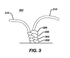
- FIG. 3 is a diagrammatic representation of a first exemplary four-point suture knot in accordance with the present invention.
- FIGS. 4A-4I illustrate the various configurations of the directional sutures that can be utilized as part of various embodiments of the invention.
- the terms “about” or “approximately” for any numerical values or ranges indicate a suitable dimensional tolerance that allows the part or collection of components to function for its intended purpose as described herein. More specifically, “about” or “approximately” may refer to the range of values ⁇ 10% of the recited value, e.g. “about 90%” may refer to the range of values from 81% to 99%.
- the terms “patient,” “host,” “user,” and “subject” refer to any human or animal subject and are not intended to limit the systems or methods to human use, although use of the subject invention in a human patient represents a preferred embodiment.
- cranial or “caudal” are in this application are used to indicate a relative position or direction with respect to the person receiving the implant. As applied to “cranial,” the term indicates a position or direction closer to the heart, while the term “caudal” indicates a position or direction further away from the heart of such a subject.
- Aneurysm repair devices make take on a wide variety of configurations.
- Aneurysm repair devices may include one element configurations or multiple element or modular element configurations.
- the secondary sealing mechanisms of the present invention may be utilized with many types of aneurysm repair devices that rely on a primary seal of fabric mesh against the vessel wall. While Abdominal Aortic Aneurysm (AAA) repair devices are used as specific examples, it is contemplated that this unique sealing mechanism can be used with many other types of devices at various other locations.
- AAAA Abdominal Aortic Aneurysm
- the anchoring and sealing system may include a main portion 100 with two modular extensions 200 .
- the main portion 100 includes trunk section 102 that extends from a cranial portion 102 D toward a caudal portion 102 P proximate a bifurcated section, which includes two legs, each defining respective longitudinal axes L 2 and L 3 .
- the combination of the graft material and the underlying scaffold structures creates a blood carrying conduit for insertion into a vessel.
- the graft material may be attached to the underlying scaffold structures via any suitable means.
- the graft material may be attached to portions of the underlying scaffold structures by sutures.
- sutures may include any suitable biocompatible material that is preferably highly durable and wear resistant.
- Graft material 110 described in detail below in FIG. 1B , and may be affixed to at least a portion of the trunk section 102 and to both of the legs 106 and 108 .
- the underlying scaffold structures of the trunk section 102 may include a number of substantially tubular stent structures, which may be formed from any number of suitable materials.
- the upper or cranial end of the trunk section 102 may include a first stent hoop 112 having a diamond shaped configuration formed from a plurality of struts 114 .
- Marker bands 116 formed from a highly radiopaque material such as tantalum may be positioned at various locations on the struts 114 for imaging purposes during device implantation. In other words, the markers 116 may help the physician to visualize the device under radio fluoroscopy.
- At the upper apex 118 of each diamond cell is an eyelet and barb structure 120 .
- the eyelet portion is utilized in conjunction with a delivery system while the barb portion is utilized to affix the anchoring and sealing component 100 in the vessel into which it is placed.
- the upper portion of the first stent hoop 112 is not covered with graft material 110 .
- This portion is not covered so that it does not interfere with or otherwise impede blood flow to or from cross or branch arteries, for example, the renal arteries.
- the lower portion of the first stent hoop 112 is covered by graft material 110 .
- Sutures 121 are utilized to secure the graft material to the lower or caudal apexes 123 of the first stent hoop 112 .
- suture 150 can be provided with barbs. Details of this securement mechanism can be seen in greater details in FIG. 1C .
- the suture 150 can be configured to circumscribe the outer surface of the graft material 100 with knots 156 to secure the suture 150 to the apices of the anchoring stent 112 (not shown for clarity).
- the suture 150 is preferably configured to include barbs that are in opposing directions about the longitudinal axis L 1 such that between the apices (AX 1 , AX 2 , AX 3 , AX 4 . . . AXn) the barbs on the suture 150 a or 150 b are situated in an opposing configuration (shown by opposing directional arrows F 1 and F 2 which represent the resistance to any force applied in opposition to these directional arrows).
- the opposing barbs on suture 150 a prevent the apices AX 2 and AX 3 from approaching each other.
- the endoprosthesis is maintained at its optimum diameter while in the blood vessel.
- suture 150 a and 150 b The benefits of such configuration of the barbs in suture 150 a and 150 b are that the endoprosthesis is virtually prevented from rotating with respect to the longitudinal axis L 1 and leaks of blood flow past the endoprosthesis proximate the sutures 150 a or 150 a are virtually prevented if not eliminated. While sutures 150 a and 150 b are shown at the cranial portion of the endoprosthesis, such sutures can be utilized at any portion including the extensions 200 where these benefits are desired.
- the lower or caudal portion of the trunk section 102 may include three individual stent hoops 122 , 124 and 126 .
- Stent hoops 122 and 124 are identical in design, with each forming a single row of struts 128 arranged in a substantially zigzag configuration.
- Sutures 130 are utilized to attach the graft material 110 to each of the stent hoops 122 and 124 .
- each stent hoop 122 and 124 may include a suture locking mechanism 131 on at least one upper and lower apex 132 . These suture locking mechanisms 131 allow for special suture knots to secure the graft material 110 to the stent hoops 122 and 124 .
- Stent hoop 126 is identical to stent hoops 122 and 124 with one exception. Specifically, the struts 134 forming this third stent hoop 126 are tapered inward in the circumferential direction thereby causing the diameter of the lower portion of the trunk section 102 to decrease where it connects to the bifurcated section 104 . As with the other two stent hoops 122 and 124 , stent hoop 126 also may include suture locking mechanisms 131 on at least one upper and lower apex 132 .
- the bifurcated section 104 includes two legs 106 and 108 . As may be readily seen from FIG. 1B , leg 106 is longer than leg 108 . This configuration eases deliverability.
- Each leg 106 and 108 is otherwise identical, and may include a plurality of individual, substantially tubular stent hoops 136 .
- Each stent hoop 136 may include a single row of struts 138 arranged in a substantially zigzag configuration. Sutures 140 are utilized to secure the graft material 110 to the stent hoops 136 .
- sutures 140 unlike sutures 130 and 121 are only utilized to secure the graft material 110 proximate the apexes 142 of the stent hoops 136 rather than along the entire length of a strut.
- Each leg 106 and 108 is free to move independently of each other; however, proximate the junction with the trunk section 102 , the graft material 110 of each leg 106 and 108 is stitched together with sutures 144 . This is done to prevent tearing of the graft material 110 if and when the legs 106 and 108 move.
- the graft material 110 covering the anchoring and sealing component 100 may include crimped sections 146 between the various underlying scaffold elements. These crimped sections increase the flexibility of the entire device.
- the anchoring and sealing component 100 is percutaneously positioned in a blood vessel with one or more aneurysms. It is anchored in healthy tissue above the aneurysm and serves as the first conduit to bypass the diseased section of the artery. Additional stent-graft components or endovascular grafts attach to the legs 106 and 108 to extend the bypass to healthy tissue beyond the aneurysm.
- the system is designed as a modular system so that as many extensions as necessary may be utilized. Essentially, the additional or modular components overlap and form an interference fit.
- This particular exemplary embodiment having two legs is specifically designed for branching into two vessels, for example, from the abdominal aortic artery to the iliac arteries. However, other similar modular components may be utilized in any other suitable artery.
- additional markers 148 are affixed to the device in various locations.
- the additional markers 148 may be formed out of any suitable, highly radiopaque material such as tantalum.
- the additional markers 148 may be attached to either or both of the underlying stent structures and the graft material by any suitable means, including stitches and glue. In the exemplary embodiment, the additional markers 148 are attached to the graft material.
- FIG. 1B is a close-up view of the main body 102 of the endoprosthesis without the anchor section 116 .
- main body 102 is configured to be placed in an artery (e.g., abdominal artery) upstream of the arterial bifurcation.
- the main body 102 has a caudal end 102 d with an anchor portion 116 connected to a fabric portion or graft material 210 of the main body.
- the fabric portion 210 has inner and outer surfaces.
- the fabric portion is connected to a first plurality of spaced apart stent hoops 122 , 124 , 126 and so on.
- the main body extends from the caudal end 102 d along a longitudinal axis L 1 into two tubular flow passages or legs 106 and 108 .
- each of the legs includes respective second and third plurality of spaced apart stent hoops 136 ( FIG. 1B ) connected to the fabric portion of each leg.
- a first suture 152 that connects the anchor portion and at least two spaced apart stent hoops.
- the first suture 152 includes from four to eight of the first suture 152 depending on the number of apices 125 for the anchor portion and the stent hoops 122 , 124 , or 126 .
- the first suture may include a plurality of sutures 154 connected to an apex 125 of one stent hoop 122 to an apex of another stent hoop 124 .
- first suture 152 or 154 disposed along on the outer surface of the main body with a physical configuration to allow the main body to remain in place once deployed in the blood vessel despite the hydrodynamic forces of blood flow impinging upon the endoprosthesis.
- the first suture 152 or 154 connects at least two of the spaced apart stent hoops 122 and 124 on the main body together with unidirectional barbs provided on the suture 152 or 154 to reduce movement of the main body with respect to the artery in the direction of blood flow (i.e., movement along longitudinal axis from the caudal end to the cranial bifurcated portion).
- the first suture 152 or 154 may be a non-bioresorbable material.
- the first suture 150 , second suture 152 , or third suture 154 may be formed from a bioresorbable material.
- Suitable biodegradable materials may include polymers such as polylactic acid (i.e., PLA), polyglycolic acid (i.e., PGA), polydioxanone (i.e., PDS), polyhydroxybutyrate (i.e., PHB), polyhydroxyvalerate (i.e., PHV), and copolymers or a combination of PHB and PHV (available commercially as Biopol®), polycaprolactone (available as Capronor®), polyanhydrides (aliphatic polyanhydrides in the back bone or side chains or aromatic polyanhydrides with benzene in the side chain), polyorthoesters, polyaminoacids (e.g., poly-L-lysine, polyglutamic acid), pseudo-polyaminoacids (e.g., with back bone of polyaminoacids altered), polycyanocrylates, or polyphosphazenes.
- PLA polylactic acid
- PDS polyglycolic acid
- bio-resorbable includes a suitable biocompatible material, mixture of materials or partial components of materials being degraded into other generally non-toxic materials by an agent present in biological tissue (i.e., being bio-degradable via a suitable mechanism, such as, for example, hydrolysis) or being removed by cellular activity (i.e., bioresorption, bioabsorption, or bio-resorbable), by bulk or surface degradation (i.e., bioerosion such as, for example, by utilizing a water insoluble polymer that is soluble in water upon contact with biological tissue or fluid), or a combination of one or more of the bio-degradable, bio-erodable, or bio-resorbable material noted above.
- the suture 152 or 154 may be a shape memory material such as shape memory metal or polymers.
- suture knots 156 are provided so as to extend through the graft portion or graft material to secure at least the first suture 152 or 154 to the graft material and the stent hoop ( 122 , 124 or 126 ).
- the knots 156 are located proximate respective apices 125 that are defined by a sinusoidal (i.e., zig-zag like waveform that circumscribes the longitudinal axis to present spaced apart apices about the longitudinal axis L-L) structure of the stent hoop 122 , 124 , or 126 .
- the spaced apart stent hoops 122 , 124 , and 126 are disposed on the inner (i.e., inside) surface of the graft material so that the stent hoops are not in direct contact with the blood vessel tissue.
- the exemplary endovascular graft extension 200 may include one or more first stent hoops 202 , a second stent hoop 204 , a third stent hoop 206 and a fourth stent hoop 208 .
- Graft material 210 is attached to the stent hoops 202 , 204 , 206 and 208 to form a substantially tubular conduit.
- crimped sections 212 are formed in the graft material 210 between the stent hoops to increase flexibility.
- the fourth stent hoop 208 would be anchored in healthy tissue below the aneurysm and a number of the uppermost first stent hoops 202 would overlap with one of the legs 106 and 108 of the anchoring and sealing component 100 thereby establishing a fluid channel through the diseased section of the artery.
- the degree of overlap may vary. Obviously the greater the degree of overlap, the less likely the chance of separation.
- the endoprosthesis suture locks of the present invention have more chances to engage with a higher degree of overlap.
- a second endovascular graft would be connected to the second leg. As stated above, additional endovascular grafts may be connected together if longer conduits are required to bypass the diseased tissue.
- the one or more first stent hoops 202 each may include a single row of struts 214 arranged in a substantially zigzag configuration.
- Sutures 216 are utilized to secure the graft material 210 to the stent hoops 202 . These sutures 216 are only utilized to secure the graft material 210 proximate the apexes 218 of the first stent hoops 202 rather than along the entire length of a strut forming the segment 202 .
- the diameter of the one or more first stent hoops 202 with the graft material 210 attached thereto is substantially equal to that of either of the legs 106 and 108 such that a tight interference fit may be achieved when the components are attached.
- the endovascular graft extension 200 fits inside of the legs 106 and 108 ; however, in alternate exemplary embodiments wherein the endovascular graft extension 200 fits over or outside of the legs 106 and 108 .
- knots on the sutures of the cranial or cranial end of the endovascular graft extension 200 help anchor the endovascular graft within the legs 106 and 108 .
- the upper or caudal most first stent hoop 202 may include a marker band 203 for positioning the device.
- the marker band 203 may include any suitable, highly radiopaque material such as tantalum. It is worthwhile to note that while a number of different markers are illustrated, additional markers that are not shown are positioned at various locations around each of the components so that the physician may easily visualize the device under radio fluoroscopy.
- the second segment 204 may include a single row of struts 220 arranged in a substantially zigzag configuration.
- the diameter of the stent hoop 204 is slightly larger than the diameter of the stent hoop 202 .
- the increase in diameter may be achieved through the use of longer struts.
- Sutures 222 are utilized to secure the graft material 210 to the stent hoop 204 . These sutures 222 are only utilized to secure the graft material 210 proximate the apexes 224 of the second stent hoop 204 rather than along the entire length of a strut forming the segment 204 .
- each stent hoop 204 may include a suture locking mechanism 226 on at least one upper and lower apex 224 .
- These suture locking mechanisms 226 allow for special suture knots to secure the graft material 210 to the stent hoop 204 . It has been determined that these locations are subject to wear due to high biological forces and thus additional securing mechanism are utilized to prevent separation of the graft material 210 .
- Third stent hoop 206 is identical to stent hoop 204 with one exception. Specifically, the struts 228 forming this third stent hoop 206 are tapered outward in the circumferential direction thereby causing the diameter of the lower portion of the endovascular graft extension 200 to increase where it anchors in the vessel. Sutures 230 are utilized to secure the graft material 210 to the third sent segment 206 . As with the second stent hoop 204 , third stent hoop 206 also may include suture locking mechanisms 226 on at least one upper and lower apex 224 . The diameter of the third stent hoop 206 is substantially equal to the diameter of the second stent hoop 204 on one end and substantially equal to the diameter of the fourth stent hoop 208 on the other end.
- the fourth stent hoop 208 has a diamond shaped configuration formed from a plurality of struts 232 .
- Sutures 234 are utilized to secure the graft material 210 to the fourth stent hoop 208 .
- Suture locking mechanisms 236 are utilized on one or more apexes 238 only on the end of the fourth stent hoop 208 proximate the third stent hoop 206 .
- the fourth stent hoop 208 also may include at least one marker band 240 attached to a strut for imaging the device. As described above, the marker band 240 may include any suitable, highly radiopaque material such as tantalum.
- a first tubular extension 200 is configured for insertion into one of the two legs 106 and 108 of the main body 102 and a second tubular extension 200 is configured for its insertion into the other of the two legs.
- each of the first and second tubular extensions 200 has an outer surface and an inner surface.
- the inner surface of graft 200 preferably has the stent hoops mounted on such surface while the outside surface of the graft 200 preferably has a retention mechanism provided thereon. Details of the graft retention mechanism to improve the retainability of the caudal or cranial end of the graft to the leg 106 or 108 are shown further in the enlarged FIG. 2B . Details of the mechanism to improve the retainability of the cranial or caudal end to the blood vessel are shown in the enlarged FIG. 2D .
- suture knots 156 can be used at various suitable locations to retain the barbed sutures 152 (shown in greater detail in FIG. 2C ) to the graft 200 .
- the barbed suture 152 may be aligned with the apices of the stent hoops and secured proximate the apices as the suture 152 extends along the length of the graft.
- certain apices may be skipped as shown on the far right side of the graft.
- the barbs are oriented to resist against the force of the blood flow (BF) impinging on the graft 200 . Due to the pulsing nature of blood flow, the barbs can be configured to resist both the blood flow and any resulting back flow by having barbs oriented in opposed directions.
- BF blood flow
- sutures 152 can also be utilized to secure the caudal portion of graft 200 to respective blood vessels after bifurcation of the abdominal artery.
- Sutures 152 are preferably arranged, in FIG. 2E , such that the caudal portion can resist the force due to backflow of blood due to the pulsatile nature of blood flowing from the main artery. Nevertheless, the barbed sutures can be configured to resist both the blood flow and any resulting back flow by having barbs oriented in opposed directions.
- the sutures 152 or 154 can be infused or loaded with bioactive agents to aid in the healing response or to achieve a desired physiological response.
- bio-active agents such as blood de-clotting agent (e.g., heparin, warfarin, etc.,) anti-proliferative/antimitotic agents including natural products such as vinca alkaloids (i.e. vinblastine, vincristine, and vinorelbine), paclitaxel, epidipodophyllotoxins (i.e.
- antibiotics dactinomycin (actinomycin D) daunorubicin, doxorubicin and idarubicin
- anthracyclines mitoxantrone, bleomycins, plicamycin (mithramycin) and mitomycin
- enzymes L-asparaginase which systemically metabolizes L-asparagine and deprives cells which do not have the capacity to synthesize their own asparagine
- antiplatelet agents such as G(GP) IIb/IIIa inhibitors and vitronectin receptor antagonists
- anti-proliferative/antimitotic alkylating agents such as nitrogen mustards (mechlorethamine, cyclophosphamide and analogs, melphalan, chlorambucil), ethylenimines and methylmelamines (hexamethylmelamine and thiotepa), alkyl sulfonates-busulfan, ni
- anti-coagulants heparin, synthetic heparin salts and other inhibitors of thrombin
- fibrinolytic agents such as tissue plasminogen activator, streptokinase and urokinase), aspirin, dipyridamole, ticlopidine, clopidogrel, abciximab
- antimigratory antisecretory (breveldin)
- anti-inflammatory such as adrenocortical steroids (cortisol, cortisone, fludrocortisone, prednisone, prednisolone, 6 ⁇ -methylprednisolone, triamcinolone, betamethasone, and dexamethasone), non-steroidal agents (salicylic acid derivatives i.e.
- All of the stent hoops described herein are substantially tubular elements that may be formed utilizing any number of techniques and any number of materials.
- all of the stent hoops are formed from a nickel-titanium alloy (Nitinol), shape set laser cut tubing.
- the graft material utilized to cover all of the stent hoops may be made from any number of suitable biocompatible materials, including woven, knitted, sutured, extruded, or cast materials forming polyester, polytetrafluoroethylene, silicones, urethanes, and ultra-light weight polyethylene, such as that commercially available under the trade designation SPECTRATM.
- the materials may be porous or nonporous.
- Exemplary materials include a woven polyester fabric made from DACRONTM or other suitable PET-type polymers.
- the fabric for the graft material is a forty denier (denier is defined in grams of nine thousand meters of a filament or yarn), twenty-seven filament polyester yarn, having about seventy to one-hundred end yarns per cm per face and thirty-two to forty-six pick yarns per cm face. At this weave density, the graft material is relatively impermeable to blood flow through the wall, but is relatively thin, ranging between 0.08 and 0.12 mm in wall thickness.
- crimps Prior to attachment of the graft component to the stent hoops, crimps are formed between the stent positions by placing the graft material on a shaped mandrel and thermally forming indentations in the surface.
- the crimps 146 and 212 respectively are about two mm long and 0.5 mm deep. With these dimensions, the endovascular graft can bend and flex while maintaining an open lumen. Also, prior to attachment of the graft material to the stent hoops, the graft material is cut in a shape to conform to the shapes of the stent hoops.
- the graft material is attached to each of the stent hoops.
- the graft material may be attached to the stent hoops in any number of suitable ways.
- the graft material is attached to the stent hoops by sutures.
- the graft material 110 in the lower portion of the first stent hoop 112 , the graft material 110 with sutures 121 using a blanket type stitch.
- the graft material is attached with sutures 130 using a blanket type stitch.
- the graft material is attached with sutures 140 using point type stitches.
- the graft material 210 is attached to first stent hoops by sutures 216 using point type stitches.
- the graft material 210 is attached using sutures 222 using blanket type stitches.
- the graft material 210 is attached using sutures 230 using blanket type stitches.
- the graft material 210 is attached using sutures 234 using blanket type stitches, shown here in FIG. 3 .
- the suture knots utilized to fasten the graft material to the underlying stent structures may be modified to enhance the overall performance of the aneurysm repair device. Essentially, the modified suture knots may be utilized to create tailored profiles that increase the ability of one component to adhere the other components and/or vessels to prevent component separation or migration. Details of various embodiments of the suture knots can be found in US Patent Application Publication No. US20110071614 filed on Sep. 24, 2009, which is hereby incorporated by reference as if set forth herein.
- barbed suture 150 , 152 , or 154 it is within the scope of this disclosure to also utilize variations of the barbs such as those shown, for example, in FIGS. 4A-4I , which can be in the form of undulations or ripples molded or formed in the suture.
- the suture may have a nominal diameter of about 0.1 millimeters to about 0.4 millimeters in short-term absorbable, long-term absorbable and nonabsorbable polymer configurations. It should also be noted that while the barbs are shown in the plan view in two-dimension, in actual use, the barbs are configured such that the actual barbs would also extend to contact the vessel (e.g., FIG.
Landscapes
- Health & Medical Sciences (AREA)
- Engineering & Computer Science (AREA)
- Biomedical Technology (AREA)
- Heart & Thoracic Surgery (AREA)
- Public Health (AREA)
- Transplantation (AREA)
- Cardiology (AREA)
- Veterinary Medicine (AREA)
- Oral & Maxillofacial Surgery (AREA)
- Vascular Medicine (AREA)
- Life Sciences & Earth Sciences (AREA)
- Animal Behavior & Ethology (AREA)
- General Health & Medical Sciences (AREA)
- Gastroenterology & Hepatology (AREA)
- Pulmonology (AREA)
- Prostheses (AREA)
- Media Introduction/Drainage Providing Device (AREA)
Abstract
Description
Claims (18)
Priority Applications (9)
| Application Number | Priority Date | Filing Date | Title |
|---|---|---|---|
| US14/316,151 US9855155B2 (en) | 2014-06-26 | 2014-06-26 | Endoprosthesis anchoring and sealing |
| CA2952602A CA2952602C (en) | 2014-06-26 | 2015-06-05 | Improved endoprosthesis anchoring and sealing |
| JP2016573802A JP6770895B2 (en) | 2014-06-26 | 2015-06-05 | Improved internal prosthesis fixation and sealing |
| MX2016017024A MX385835B (en) | 2014-06-26 | 2015-06-05 | IMPROVED ANCHORING AND SEALING OF ENDOPROSTHESIS. |
| PCT/US2015/034314 WO2015199943A1 (en) | 2014-06-26 | 2015-06-05 | Improved endoprosthesis anchoring and sealing |
| EP15730608.5A EP3160390B1 (en) | 2014-06-26 | 2015-06-05 | Improved endoprosthesis anchoring and sealing |
| CN201580033002.3A CN106456311B (en) | 2014-06-26 | 2015-06-05 | Improved Endoprosthesis Anchorage and Sealing |
| AU2015280490A AU2015280490B2 (en) | 2014-06-26 | 2015-06-05 | Improved endoprosthesis anchoring and sealing |
| TW104120232A TWI710368B (en) | 2014-06-26 | 2015-06-24 | Improved endoprosthesis anchoring and sealing |
Applications Claiming Priority (1)
| Application Number | Priority Date | Filing Date | Title |
|---|---|---|---|
| US14/316,151 US9855155B2 (en) | 2014-06-26 | 2014-06-26 | Endoprosthesis anchoring and sealing |
Publications (2)
| Publication Number | Publication Date |
|---|---|
| US20150374517A1 US20150374517A1 (en) | 2015-12-31 |
| US9855155B2 true US9855155B2 (en) | 2018-01-02 |
Family
ID=53442996
Family Applications (1)
| Application Number | Title | Priority Date | Filing Date |
|---|---|---|---|
| US14/316,151 Expired - Fee Related US9855155B2 (en) | 2014-06-26 | 2014-06-26 | Endoprosthesis anchoring and sealing |
Country Status (9)
| Country | Link |
|---|---|
| US (1) | US9855155B2 (en) |
| EP (1) | EP3160390B1 (en) |
| JP (1) | JP6770895B2 (en) |
| CN (1) | CN106456311B (en) |
| AU (1) | AU2015280490B2 (en) |
| CA (1) | CA2952602C (en) |
| MX (1) | MX385835B (en) |
| TW (1) | TWI710368B (en) |
| WO (1) | WO2015199943A1 (en) |
Cited By (4)
| Publication number | Priority date | Publication date | Assignee | Title |
|---|---|---|---|---|
| US20160175122A1 (en) * | 2014-12-19 | 2016-06-23 | Boston Scientific Scimed, Inc. | Stent with anti-migration features |
| US10874772B2 (en) | 2018-07-12 | 2020-12-29 | Cook Medical Technologies Llc | Coated medical device and method of coating such a device |
| US10925713B2 (en) | 2018-08-07 | 2021-02-23 | Cook Medical Technologies Llc | Stitch wire routing and delivery system |
| US11331178B2 (en) | 2018-08-09 | 2022-05-17 | Cook Medical Technologies Llc | Stent-graft |
Families Citing this family (10)
| Publication number | Priority date | Publication date | Assignee | Title |
|---|---|---|---|---|
| CN106344209B (en) * | 2016-10-11 | 2018-11-20 | 有研医疗器械(北京)有限公司 | A kind of endovascular stent for abdominal aorta and its conveying device and application method |
| DE102017120819A1 (en) * | 2017-09-08 | 2019-03-14 | Jotec Gmbh | Intraluminal vascular prosthesis system |
| WO2019209731A1 (en) * | 2018-04-23 | 2019-10-31 | Endologix, Inc. | Modulation of inflammatory response following endovascular treatment |
| CN110833469B (en) * | 2018-08-17 | 2023-06-20 | 先健科技(深圳)有限公司 | Tectorial membrane support |
| CN109498075A (en) * | 2019-01-04 | 2019-03-22 | 上海形状记忆合金材料有限公司 | A kind of small-sized implantation instrument with hangnail |
| CN112137756B (en) * | 2019-06-28 | 2025-07-01 | 浙江脉通智造科技(集团)有限公司 | Tubular coated structure and preparation method thereof and coated stent |
| RU2742451C1 (en) * | 2020-03-04 | 2021-02-05 | Заза Александрович Кавтеладзе | Bifurcation stent-graft system for treating aneurism of abdominal aorta and method of treating aneurism of abdominal aorta using thereof |
| CA3179536A1 (en) * | 2020-04-06 | 2021-10-14 | Tepha, Inc. | Medical implants for marking surgical sites |
| CN113069256B (en) * | 2021-03-26 | 2022-12-09 | 珠海通桥医疗科技有限公司 | A kind of intracranial flexible closed-loop stent |
| EP4497412A1 (en) * | 2023-07-28 | 2025-01-29 | Medtronic Trading NL B.V. | Aortic implant with proximal anchoring layer |
Citations (18)
| Publication number | Priority date | Publication date | Assignee | Title |
|---|---|---|---|---|
| US3123077A (en) | 1964-03-03 | Surgical suture | ||
| US5776180A (en) * | 1994-02-09 | 1998-07-07 | Boston Scientific Technology | Bifurcated endoluminal prosthesis |
| US5800515A (en) * | 1995-08-03 | 1998-09-01 | B. Braun Celsa (Societe Anonyme) | Prosthesis implantable in a human or animal duct such as a stent or a prosthesis for aneurism |
| WO2001064137A1 (en) | 2000-02-28 | 2001-09-07 | Fraunhofer-Gesellschaft zur Förderung der angewandten Forschung e.V. | Anchoring system for implantable heart valve prostheses |
| US6355056B1 (en) * | 1995-06-01 | 2002-03-12 | Meadox Medicals, Inc. | Implantable intraluminal prosthesis |
| WO2004014236A1 (en) | 2002-08-09 | 2004-02-19 | Quill Medical, Inc. | Suture anchor and method |
| US20040093027A1 (en) | 2002-03-04 | 2004-05-13 | Walter Fabisiak | Barbed tissue connector for sealing vascular puncture wounds |
| WO2006028925A1 (en) | 2004-09-02 | 2006-03-16 | Med Institute, Inc. | Modular prosthesis and method for branch vessels |
| US20060178733A1 (en) * | 2005-01-21 | 2006-08-10 | Leonard Pinchuk | Modular stent graft employing bifurcated graft and leg locking stent elements |
| WO2007016166A2 (en) | 2005-07-27 | 2007-02-08 | Cook Critical Care Incorporated | Stent/graft device and method for open surgical placement |
| US7226474B2 (en) * | 2000-05-01 | 2007-06-05 | Endovascular Technologies, Inc. | Modular graft component junctions |
| US20070224237A1 (en) | 2006-03-24 | 2007-09-27 | Julia Hwang | Barbed sutures having a therapeutic agent thereon |
| WO2009052188A1 (en) | 2007-10-15 | 2009-04-23 | Edwards Lifesciences Corporation | Transcatheter heart valve with micro-anchors |
| GB2472602A (en) | 2009-08-11 | 2011-02-16 | Cook William Europ | Stent graft having barb sutured to stent and graft. |
| US20110071614A1 (en) | 2009-09-24 | 2011-03-24 | David Christopher Majercak | Stent - graft suture locks |
| US8163007B2 (en) * | 2008-02-08 | 2012-04-24 | Cook Medical Technologies Llc | Stent designs for use with one or more trigger wires |
| US8246652B2 (en) | 1993-05-03 | 2012-08-21 | Ethicon, Inc. | Suture with a pointed end and an anchor end and with equally spaced yieldable tissue grasping barbs located at successive axial locations |
| US8715334B2 (en) * | 2011-07-14 | 2014-05-06 | Boston Scientific Scimed, Inc. | Anti-migration stent with quill filaments |
Family Cites Families (13)
| Publication number | Priority date | Publication date | Assignee | Title |
|---|---|---|---|---|
| US8795332B2 (en) * | 2002-09-30 | 2014-08-05 | Ethicon, Inc. | Barbed sutures |
| AUPO700897A0 (en) * | 1997-05-26 | 1997-06-19 | William A Cook Australia Pty Ltd | A method and means of deploying a graft |
| US6368345B1 (en) * | 1998-09-30 | 2002-04-09 | Edwards Lifesciences Corporation | Methods and apparatus for intraluminal placement of a bifurcated intraluminal garafat |
| US6325820B1 (en) * | 1998-11-16 | 2001-12-04 | Endotex Interventional Systems, Inc. | Coiled-sheet stent-graft with exo-skeleton |
| FR2797389B1 (en) * | 1999-08-09 | 2001-11-30 | Novatech Inc | BIFURCED AORTIC PROSTHESIS |
| EP1549248A4 (en) * | 2002-09-26 | 2015-11-25 | Advanced Bio Prosthetic Surfac | NITINOL VACUUM-DEPOSITED ALLOY FILMS HAVING HIGH RESISTANCE, MEDICAL MATERIALS FOR THIN FILM CANDLES, AND METHOD OF MANUFACTURING THE SAME |
| WO2004049978A1 (en) * | 2002-12-04 | 2004-06-17 | Cook Incorporated | Method and device for treating aortic dissection |
| CN2675083Y (en) * | 2004-01-06 | 2005-02-02 | 微创医疗器械(上海)有限公司 | Y-shaped aortic stent graft |
| US20060276883A1 (en) * | 2005-06-01 | 2006-12-07 | Cook Incorporated | Tapered and distally stented elephant trunk stent graft |
| CN2912562Y (en) * | 2006-05-10 | 2007-06-20 | 上海赢生医疗科技有限公司 | Branch type tectorial blood vessel support bracket designed with modular structure |
| CN101259045A (en) * | 2008-04-01 | 2008-09-10 | 中国人民解放军第二军医大学 | An abdominal aorta-internal and external iliac artery endoluminal isolation graft |
| CN202397646U (en) * | 2011-12-29 | 2012-08-29 | 北京华脉泰科医疗器械有限公司 | Aorta covered stent |
| US20130274861A1 (en) * | 2012-04-12 | 2013-10-17 | Sanford Health | Debranching Stent Graft Limb and Methods for Use |
-
2014
- 2014-06-26 US US14/316,151 patent/US9855155B2/en not_active Expired - Fee Related
-
2015
- 2015-06-05 CA CA2952602A patent/CA2952602C/en active Active
- 2015-06-05 EP EP15730608.5A patent/EP3160390B1/en active Active
- 2015-06-05 WO PCT/US2015/034314 patent/WO2015199943A1/en not_active Ceased
- 2015-06-05 MX MX2016017024A patent/MX385835B/en unknown
- 2015-06-05 CN CN201580033002.3A patent/CN106456311B/en active Active
- 2015-06-05 JP JP2016573802A patent/JP6770895B2/en active Active
- 2015-06-05 AU AU2015280490A patent/AU2015280490B2/en not_active Ceased
- 2015-06-24 TW TW104120232A patent/TWI710368B/en active
Patent Citations (18)
| Publication number | Priority date | Publication date | Assignee | Title |
|---|---|---|---|---|
| US3123077A (en) | 1964-03-03 | Surgical suture | ||
| US8246652B2 (en) | 1993-05-03 | 2012-08-21 | Ethicon, Inc. | Suture with a pointed end and an anchor end and with equally spaced yieldable tissue grasping barbs located at successive axial locations |
| US5776180A (en) * | 1994-02-09 | 1998-07-07 | Boston Scientific Technology | Bifurcated endoluminal prosthesis |
| US6355056B1 (en) * | 1995-06-01 | 2002-03-12 | Meadox Medicals, Inc. | Implantable intraluminal prosthesis |
| US5800515A (en) * | 1995-08-03 | 1998-09-01 | B. Braun Celsa (Societe Anonyme) | Prosthesis implantable in a human or animal duct such as a stent or a prosthesis for aneurism |
| WO2001064137A1 (en) | 2000-02-28 | 2001-09-07 | Fraunhofer-Gesellschaft zur Förderung der angewandten Forschung e.V. | Anchoring system for implantable heart valve prostheses |
| US7226474B2 (en) * | 2000-05-01 | 2007-06-05 | Endovascular Technologies, Inc. | Modular graft component junctions |
| US20040093027A1 (en) | 2002-03-04 | 2004-05-13 | Walter Fabisiak | Barbed tissue connector for sealing vascular puncture wounds |
| WO2004014236A1 (en) | 2002-08-09 | 2004-02-19 | Quill Medical, Inc. | Suture anchor and method |
| WO2006028925A1 (en) | 2004-09-02 | 2006-03-16 | Med Institute, Inc. | Modular prosthesis and method for branch vessels |
| US20060178733A1 (en) * | 2005-01-21 | 2006-08-10 | Leonard Pinchuk | Modular stent graft employing bifurcated graft and leg locking stent elements |
| WO2007016166A2 (en) | 2005-07-27 | 2007-02-08 | Cook Critical Care Incorporated | Stent/graft device and method for open surgical placement |
| US20070224237A1 (en) | 2006-03-24 | 2007-09-27 | Julia Hwang | Barbed sutures having a therapeutic agent thereon |
| WO2009052188A1 (en) | 2007-10-15 | 2009-04-23 | Edwards Lifesciences Corporation | Transcatheter heart valve with micro-anchors |
| US8163007B2 (en) * | 2008-02-08 | 2012-04-24 | Cook Medical Technologies Llc | Stent designs for use with one or more trigger wires |
| GB2472602A (en) | 2009-08-11 | 2011-02-16 | Cook William Europ | Stent graft having barb sutured to stent and graft. |
| US20110071614A1 (en) | 2009-09-24 | 2011-03-24 | David Christopher Majercak | Stent - graft suture locks |
| US8715334B2 (en) * | 2011-07-14 | 2014-05-06 | Boston Scientific Scimed, Inc. | Anti-migration stent with quill filaments |
Non-Patent Citations (2)
| Title |
|---|
| International Preliminary Report on Patentability for Application No. PCT/US2015/034314, dated Dec. 27, 2016, 7 pages. |
| International Search Report arid Written Opinion for Application No. PCT/US2015/034314, dated Aug. 19, 2015, 11 pages. |
Cited By (6)
| Publication number | Priority date | Publication date | Assignee | Title |
|---|---|---|---|---|
| US20160175122A1 (en) * | 2014-12-19 | 2016-06-23 | Boston Scientific Scimed, Inc. | Stent with anti-migration features |
| US10117761B2 (en) * | 2014-12-19 | 2018-11-06 | Boston Scientific Scimed, Inc. | Stent with anti-migration features |
| US10874772B2 (en) | 2018-07-12 | 2020-12-29 | Cook Medical Technologies Llc | Coated medical device and method of coating such a device |
| US10925713B2 (en) | 2018-08-07 | 2021-02-23 | Cook Medical Technologies Llc | Stitch wire routing and delivery system |
| US11331178B2 (en) | 2018-08-09 | 2022-05-17 | Cook Medical Technologies Llc | Stent-graft |
| US12004941B2 (en) | 2018-08-09 | 2024-06-11 | Cook Medical Technologies Llc | Stent-graft |
Also Published As
| Publication number | Publication date |
|---|---|
| JP2017518823A (en) | 2017-07-13 |
| WO2015199943A1 (en) | 2015-12-30 |
| CN106456311A (en) | 2017-02-22 |
| US20150374517A1 (en) | 2015-12-31 |
| AU2015280490B2 (en) | 2019-11-07 |
| MX385835B (en) | 2025-03-18 |
| CA2952602A1 (en) | 2015-12-30 |
| JP6770895B2 (en) | 2020-10-21 |
| CN106456311B (en) | 2020-08-21 |
| TW201615163A (en) | 2016-05-01 |
| CA2952602C (en) | 2022-07-05 |
| AU2015280490A1 (en) | 2017-01-12 |
| MX2016017024A (en) | 2018-01-12 |
| TWI710368B (en) | 2020-11-21 |
| EP3160390B1 (en) | 2020-04-01 |
| EP3160390A1 (en) | 2017-05-03 |
Similar Documents
| Publication | Publication Date | Title |
|---|---|---|
| US9855155B2 (en) | Endoprosthesis anchoring and sealing | |
| EP2301476B1 (en) | Stent-graft suture locks | |
| US20190192320A1 (en) | Prosthesis having an everting pivoting fenestration | |
| EP3583915B1 (en) | Endoprosthesis with predetermined curvature formed by tethers | |
| US20140005764A1 (en) | Sealing mechanism for expandable vascular device | |
| US8226703B2 (en) | Method and device for attaching a stent structure to AAA graft material | |
| US7344562B2 (en) | AAA low profile support structure | |
| US9687366B2 (en) | Endoleak mitigator for aneurysm stent-graft |
Legal Events
| Date | Code | Title | Description |
|---|---|---|---|
| AS | Assignment |
Owner name: CORDIS CORPORATION, CALIFORNIA Free format text: ASSIGNMENT OF ASSIGNORS INTEREST;ASSIGNOR:MAJERCAK, DAVID;REEL/FRAME:033189/0833 Effective date: 20140624 |
|
| AS | Assignment |
Owner name: CARDINAL HEALTH SWITZERLAND 515 GMBH, SWITZERLAND Free format text: ASSIGNMENT OF ASSIGNORS INTEREST;ASSIGNOR:CORDIS CORPORATION;REEL/FRAME:042126/0259 Effective date: 20170329 |
|
| STCF | Information on status: patent grant |
Free format text: PATENTED CASE |
|
| MAFP | Maintenance fee payment |
Free format text: PAYMENT OF MAINTENANCE FEE, 4TH YEAR, LARGE ENTITY (ORIGINAL EVENT CODE: M1551); ENTITY STATUS OF PATENT OWNER: LARGE ENTITY Year of fee payment: 4 |
|
| FEPP | Fee payment procedure |
Free format text: MAINTENANCE FEE REMINDER MAILED (ORIGINAL EVENT CODE: REM.); ENTITY STATUS OF PATENT OWNER: LARGE ENTITY |
|
| AS | Assignment |
Owner name: HPS INVESTMENT PARTNERS, LLC, NEW YORK Free format text: SECURITY INTEREST;ASSIGNORS:CORDIS US CORP.;ACCESS CLOSURE, LLC;REEL/FRAME:072973/0157 Effective date: 20251001 Owner name: CORDIS US CORP, FLORIDA Free format text: ASSIGNMENT OF ASSIGNORS INTEREST;ASSIGNORS:CARDINAL HEALTH SWITZERLAND 515 GMBH;CARDINAL HEALTH 529, LLC;CORDIS CORPORATION;AND OTHERS;REEL/FRAME:072987/0675 Effective date: 20210802 |
|
| LAPS | Lapse for failure to pay maintenance fees |
Free format text: PATENT EXPIRED FOR FAILURE TO PAY MAINTENANCE FEES (ORIGINAL EVENT CODE: EXP.); ENTITY STATUS OF PATENT OWNER: LARGE ENTITY |
|
| STCH | Information on status: patent discontinuation |
Free format text: PATENT EXPIRED DUE TO NONPAYMENT OF MAINTENANCE FEES UNDER 37 CFR 1.362 |
|
| FP | Lapsed due to failure to pay maintenance fee |
Effective date: 20260102 |