US9820652B2 - Multi-photon microscope having an excitation-beam array - Google Patents
Multi-photon microscope having an excitation-beam array Download PDFInfo
- Publication number
- US9820652B2 US9820652B2 US14/562,885 US201414562885A US9820652B2 US 9820652 B2 US9820652 B2 US 9820652B2 US 201414562885 A US201414562885 A US 201414562885A US 9820652 B2 US9820652 B2 US 9820652B2
- Authority
- US
- United States
- Prior art keywords
- image
- imaging system
- sub
- light signals
- region
- Prior art date
- Legal status (The legal status is an assumption and is not a legal conclusion. Google has not performed a legal analysis and makes no representation as to the accuracy of the status listed.)
- Expired - Fee Related, expires
Links
Images


Classifications
-
- A—HUMAN NECESSITIES
- A61—MEDICAL OR VETERINARY SCIENCE; HYGIENE
- A61B—DIAGNOSIS; SURGERY; IDENTIFICATION
- A61B5/00—Measuring for diagnostic purposes; Identification of persons
- A61B5/0059—Measuring for diagnostic purposes; Identification of persons using light, e.g. diagnosis by transillumination, diascopy, fluorescence
- A61B5/0071—Measuring for diagnostic purposes; Identification of persons using light, e.g. diagnosis by transillumination, diascopy, fluorescence by measuring fluorescence emission
-
- A—HUMAN NECESSITIES
- A61—MEDICAL OR VETERINARY SCIENCE; HYGIENE
- A61B—DIAGNOSIS; SURGERY; IDENTIFICATION
- A61B5/00—Measuring for diagnostic purposes; Identification of persons
- A61B5/40—Detecting, measuring or recording for evaluating the nervous system
- A61B5/4058—Detecting, measuring or recording for evaluating the nervous system for evaluating the central nervous system
- A61B5/4064—Evaluating the brain
-
- G—PHYSICS
- G01—MEASURING; TESTING
- G01N—INVESTIGATING OR ANALYSING MATERIALS BY DETERMINING THEIR CHEMICAL OR PHYSICAL PROPERTIES
- G01N21/00—Investigating or analysing materials by the use of optical means, i.e. using sub-millimetre waves, infrared, visible or ultraviolet light
- G01N21/62—Systems in which the material investigated is excited whereby it emits light or causes a change in wavelength of the incident light
- G01N21/63—Systems in which the material investigated is excited whereby it emits light or causes a change in wavelength of the incident light optically excited
- G01N21/64—Fluorescence; Phosphorescence
- G01N21/645—Specially adapted constructive features of fluorimeters
- G01N21/6456—Spatial resolved fluorescence measurements; Imaging
- G01N21/6458—Fluorescence microscopy
-
- G—PHYSICS
- G02—OPTICS
- G02B—OPTICAL ELEMENTS, SYSTEMS OR APPARATUS
- G02B21/00—Microscopes
- G02B21/0004—Microscopes specially adapted for specific applications
- G02B21/002—Scanning microscopes
- G02B21/0024—Confocal scanning microscopes (CSOMs) or confocal "macroscopes"; Accessories which are not restricted to use with CSOMs, e.g. sample holders
- G02B21/0032—Optical details of illumination, e.g. light-sources, pinholes, beam splitters, slits, fibers
-
- G—PHYSICS
- G02—OPTICS
- G02B—OPTICAL ELEMENTS, SYSTEMS OR APPARATUS
- G02B21/00—Microscopes
- G02B21/0004—Microscopes specially adapted for specific applications
- G02B21/002—Scanning microscopes
- G02B21/0024—Confocal scanning microscopes (CSOMs) or confocal "macroscopes"; Accessories which are not restricted to use with CSOMs, e.g. sample holders
- G02B21/0052—Optical details of the image generation
- G02B21/0076—Optical details of the image generation arrangements using fluorescence or luminescence
-
- A—HUMAN NECESSITIES
- A61—MEDICAL OR VETERINARY SCIENCE; HYGIENE
- A61B—DIAGNOSIS; SURGERY; IDENTIFICATION
- A61B5/00—Measuring for diagnostic purposes; Identification of persons
- A61B5/0033—Features or image-related aspects of imaging apparatus, e.g. for MRI, optical tomography or impedance tomography apparatus; Arrangements of imaging apparatus in a room
- A61B5/004—Features or image-related aspects of imaging apparatus, e.g. for MRI, optical tomography or impedance tomography apparatus; Arrangements of imaging apparatus in a room adapted for image acquisition of a particular organ or body part
- A61B5/0042—Features or image-related aspects of imaging apparatus, e.g. for MRI, optical tomography or impedance tomography apparatus; Arrangements of imaging apparatus in a room adapted for image acquisition of a particular organ or body part for the brain
Definitions
- the present invention relates to microscopy in general, and, more particularly, to multi-photon microscopy.
- Multi-photon microscopy is an imaging technique in which an excitation laser signal is scanned over a region of interest (i.e., image field) and fluorophores in the image field are excited only when they simultaneously absorb multiple photons of the excitation light.
- a region of interest i.e., image field
- fluorophores in the image field are excited only when they simultaneously absorb multiple photons of the excitation light.
- simultaneous absorption of two photons is required to excite a fluorophore.
- Multi-photon microscopy is often used to generate fluorescent images of living cells and other microscopic objects and has become an important tool in medical imaging.
- Multi-photon microscopy enables imaging of living tissue at depths to about one millimeter (mm). Because longer wavelengths tend to scatter in tissue to a lesser degree than shorter wavelengths, the excitation laser typically provides a signal characterized by an infrared wavelength. To excite the dye to emit a fluorescence photon, two photons of infrared light must be absorbed simultaneously. Infrared excitation light is attractive because it minimizes scattering in the tissue being imaged. In order to create a two-dimensional image of the image field, the laser beam is scanned over the image field while fluorescence light from each point in the region is detected at a camera or photomultiplier tube.
- Fluorescent emission from the fluorophores increases quadratically with the intensity of the excitation light.
- fluorescence can be confined within a narrow focal depth. This gives a depth-of-field resolution comparable to that produced by conventional confocal laser scanning microscopes.
- a Ti-Sapphire laser is a commonly used excitation source.
- commercially available Ti-Sapphire lasers have an average power of only a few Watts and a repetition rate of around only 80 MHz. This enables a photon collection rate of approximately 100-10,000 photons per pixel, per image frame, and at a frame rate of no more than 10-20 Hz—resulting in a signal-to-noise ratio (SNR) of only 10-100.
- SNR signal-to-noise ratio
- a higher frame rate could potentially be achieved by simply increasing the scanning speed of the excitation signal (e.g., by 100-fold).
- operation at a higher frame rate results in reduced photon collection (to only ⁇ 1-10 photons per pixel per frame).
- the deleterious effects of faster scanning on image quality and SNR generally outweigh any potential benefit.
- scanning speed is often limited by mechanical and/or optical constraints.
- a two-photon imaging system that can provide an image of a region of interest in real time and with improved clarity would be a significant advance in the state-of-the-art.
- the present invention enables imaging of a large image region in real time by linearly scanning an array of interrogation beams across the image region, where the array is rotated in the plane of the image region so that, within a single scan, each beamlet interrogates a different one of a two-dimensional array of linear sub-regions and the entire image region is interrogated.
- Embodiments of the present invention are particularly well suited for use in in-vivo brain imaging, simultaneous multi-area imaging of disparate brain subsystems, and simultaneous surface- and deep-imaging.
- the present invention is applicable to multi-photon microscopy and single-photon microscopy.
- An illustrative embodiment comprises a two-photon microscopy imaging system that employs an optical system that provides a plurality of laser beamlets arranged such that give rise to a two-dimensional array of optical spots (i.e., foci) at an image region that defines a first plane, where the array includes a plurality of rows of foci.
- the beamlets simultaneously excite fluorescence at a two-dimensional array of locations that are distributed throughout the image region.
- the imaging system linearly scans the beamlet array across the image region in a first direction, where the beamlet array is oriented with respect to the first direction such that each of its rows forms an angle to the first direction.
- the angle, the number of beamlets, and the beamlet spacing within each row are selected so that adjacent beamlets in each row are staggered along the spacing between the rows in the direction orthogonal to the scan direction.
- the angle, the number of beamlets, and the beamlet spacing within each row are selected so that adjacent beamlets in each row are substantially evenly distributed along the spacing between the rows in the direction orthogonal to the scan direction.
- the spacing of the beamlets and the angle of rotation of the array are selected such that the spacing between adjacent scan lines is sufficiently small to achieve micron-level image resolution, while also keeping the spacing between the spots large enough to mitigate crosstalk.
- the excitation source is an ultrashort-pulsed regenerative fiber laser amplifier, which enables each laser pulse to have an energy higher than that provided by a Ti-Sapphire laser, which is typically used in the prior art.
- the higher energy of the laser pulses enables an enhanced two-photon excitation effect for each of the plurality of laser foci, yet the ultrashort-pulsed operation keeps the average optical power delivered to brain tissue within a tolerable level.
- a deconvolution algorithm is used to reconstruct a complete image frame from multiple sub-frames.
- the deconvolution algorithm includes additional correction for optical cross-talk between neighboring laser foci.
- An embodiment of the present invention is an imaging system comprising: a source operative for providing a first plurality of light signals the first plurality of light signals being arranged such that they form a two-dimensional array of foci at an image region that defines a first plane; a single-axis scanner operative for linearly scanning the first plurality of light signals along a first direction in the first plane such that each light signal of the first plurality thereof interrogates a different one of a plurality of sub-image regions, wherein the plurality of sub-image regions are arranged in a two-dimensional arrangement within the image region; and a detector operative for detecting a second plurality of light signals, wherein each of the second plurality of light signals is generated in response to absorption of optical energy from at least one of the first plurality of light signals.
- FIG. 1 depicts a schematic drawing of a portion of an imaging system in accordance with an illustrative embodiment of the present invention.
- FIG. 2 depicts operations of a method suitable for imaging an image region in accordance with the illustrative embodiment of the present invention.
- FIG. 3A depicts a portion of foci array 300 at focal plane 114 .
- FIG. 3B depicts a portion of foci array 300 at focal plane 114 where the array is rotated by angle, ⁇ , relative to the scanning direction 310 of scanner 106 .
- FIGS. 4A-B depict schematic drawings of top and front views, respectively, of imaging system 100 , as well as the excitation paths through it.
- FIG. 4C depicts a top view of the optomechanics of system 100 , as well as excitation and emission paths through it.
- FIG. 5 depicts sub-operations suitable for use in operation 205 .
- FIGS. 6A-C depict images of a test specimen before deconvolution, after computation assembly of raw sub-images, and after application of the complete deconvolution routine, respectively.
- FIG. 1 depicts a schematic drawing of a portion of an imaging system in accordance with an illustrative embodiment of the present invention.
- Imaging system 100 is a two-photon laser-scanning microscopy system that comprises source 102 , lenslet array 104 , scanner 106 , optics system 108 , objective 110 , imager 112 , and processor 128 .
- imaging system 100 is a single-photon microscopy system.
- imaging system 100 is a multi-photon microscopy system that requires more than two photons to excite a fluorophore.
- FIG. 2 depicts operations of a method suitable for imaging an image region in accordance with the illustrative embodiment of the present invention.
- Method 200 begins with operation 201 , wherein light beam 116 is provided to lenslet array 104 .
- Source 102 includes an ultrashort-pulsed regenerative fiber laser amplifier (hereinafter referred to as a “fiber laser”) that emits light at approximately 1030 nm (e.g., a regenerative ultrafast Yb 3+ laser amplifier, etc.).
- the fiber laser has an average power of 20 Watts and a tunable repetition rate that is within the range of approximately 200 kHz to approximately 2 MHz.
- the fiber laser provides pulses of optical energy that have higher energy than the typical output power of a conventional Ti-Sapphire laser. This enables an enhancement of the two-photon excitation effect for multiple laser foci in each of excitation signals 118 ; however, it keeps the average optical power delivered to image region 122 within a tolerable range.
- lenslet array 104 distributes the optical energy in light beam 116 into excitation signals 118 (i.e., beamlets 118 ) and provides them to optics system 108 .
- Lenslet array 104 is an array of microlenses operative for receiving light beam 116 and distributing it into a two-dimensional array of equal-intensity beamlets (i.e., excitation signals 118 ).
- excitation signals 118 In order to convert the output of the fiber laser into a plurality of substantially equal-intensity beamlets, prior to being received by lenslet array 104 , the output of the fiber laser is first expanded and then shaped at a beam shaper, which corrects the beam profile from Gaussian to flat. Once corrected, the now homogeneous-intensity laser beam is reduced again and provided to lenslet array 104 .
- the lenslet array splits the laser beam into a plurality of beamlets.
- excitation signals 118 includes 25 beamlets; however, the number of beamlets can have any practical value. Excitation signals 118 typically includes hundreds of beamlets.
- Optics system 108 is an arrangement of optical components for providing excitation signals 118 as a two-dimensional array of foci at focal plane 118 .
- Optics system 108 includes numerous optical components, including aspheric lenses, meniscus compound lenses for mitigating field curvature at focal plane 118 , dichroic mirror 124 for removing light at the excitation wavelength from the light received at imager 112 , a tube lens, and scanner 106 .
- aspheric lenses including aspheric lenses, meniscus compound lenses for mitigating field curvature at focal plane 118 , dichroic mirror 124 for removing light at the excitation wavelength from the light received at imager 112 , a tube lens, and scanner 106 .
- FIG. 3A depicts a portion of foci array 300 at focal plane 114 .
- Foci array 300 is a two-dimensional array of foci 302 , which are arranged in equally spaced columns 304 and rows 306 .
- the x- and y-spacing, d, between adjacent foci 302 at focal plane 114 is equal and has a value of approximately 25 microns; however, one skilled in the art will recognize that the spacing can have any suitable value.
- the x- and y-spacing between foci 302 is different.
- Each of rows 306 is parallel with array axis 308 .
- Scanner 106 is a single-axis laser scanning mirror.
- scanner 106 is a different scanning element, such as a rotatable prism, dual-axis scanning mirror configured to scan in only one dimension, and the like.
- the positions of scanner 106 and the aspheric lenses within optics system 108 are selected such that each of excitation signals 118 is incident on the center of scanner 106 as well as the back aperture of objective 110 .
- a single-axis scanning element affords advantages over multi-photon imaging systems of the prior art, which include dual-axis scanners that raster scan a light beam over an image region.
- a single-axis scanning element can operate at a modest rate (e.g., the same as the frame rate of the system) and requires a relatively simple controller.
- a conventional raster-scanning mechanism requires that the fast-scanning axis operates at a much higher rate than the imaging frame rate.
- This need for high-speed scanning makes it extremely difficult, if not impossible, for such a scanner to work properly.
- a typical prior-art high-speed mechanical scanner capable of kHz (or higher) operation operates in resonance mode.
- such prior-art scanners are normally characterized by relatively poor angular position control, which gives rise to poor image resolution for their corresponding microscope systems.
- scanner 106 scans excitation signals 118 along scan direction 310 (i.e., along the x-direction as shown in FIGS. 3A-B ). In some embodiments, the scanner scans the excitation signals through the desired range of motion in approximately 1 millisecond.
- Scanner 106 and optics system 108 are arranged such that foci array 300 is rotated relative to the array of beamlets in excitation signals 118 so that the scanner scans the beamlets along a direction that is at a non-zero angle with respect to the direction defined by the rows of beamlets.
- FIG. 3B depicts a portion of foci array 300 at focal plane 114 where the array is rotated by angle, ⁇ , relative to the scanning direction 310 of scanner 106 .
- Foci array 300 is tilted relative to the scan direction of scanner 106 to enable foci 306 to collectively scan the entirety of image region 122 .
- the value of ⁇ is based on the spacing between foci 302 as well as the number of foci in each row 306 , N. In some embodiments, ⁇ is equal to arctan (1/N).
- the values of angle, ⁇ , and spacing, d are selected so that the spacing between adjacent scanning lines is small enough to enable a desired spatial resolution (e.g., micron-level resolution). In some embodiments, these values are selected to mitigate cross-talk as well.
- the values of angle, ⁇ , and spacing, d are selected so that, along the direction orthogonal to scanning direction 310 (i.e., along the y-direction as shown in FIGS. 3A-B ), the separation, s 1 , between adjacent foci within each row 306 is an even fraction of the separation, s 2 , between adjacent rows 306 .
- fluorescence signals 120 are detected at imager 112 , which generates output signal 128 based on the fluorescence signals. Fluorescence signals 120 are generated at fluorophores located in image region 122 . As fluorescence signals 120 are emitted from image region 122 , they are incident on dichroic mirror 124 , which passes reflected light at the excitation wavelength but reflects light at fluorescence wavelengths toward imager 112 .
- Imager 112 is a multi-pixel photon collecting device characterized by noise that is nearly shot-noise-limited. Imager 112 enables simultaneous capture of fluorescence signals from substantially all excited fluorophores in image region 122 .
- exemplary imager 112 comprises an image intensifier and high-speed camera operative for directly forming multi-pixel images of image region 122 .
- the high-speed camera includes a camera system having a frame rate of 25 kHz and resolution of 768 ⁇ 768 pixels. Such a camera is sufficiently fast to acquire 25 rounds of data acquisition in 1 millisecond.
- suitable imagers include, without limitation, ultra-low-read-noise cameras (e.g., a single scientific CMOS camera, etc.), and the like.
- the camera of imager 112 include a frame trigger input such that accurate foci travel info can be developed for a plurality of sub-frames by synchronizing scanner 106 and the frame trigger.
- FIGS. 4A-B depict schematic drawings of top and front views, respectively, of imaging system 100 , as well as the excitation paths through it.
- FIG. 4C depicts a top view of the optomechanics of system 100 , as well as excitation and emission paths through it.
- imaging system 100 enables one-photon imaging capability, which affords more versatile operation using a single imaging system.
- tissue scattering can lead to photons being emitted from different positions and overlapping into the same pixels at the camera of imager 112 . This can lead to a blurred image. This phenomenon is particularly problematic when imaging in tissue to depths greater than a few hundred microns.
- imager 112 passes output signal 126 to processor 128 .
- processor 128 reconstructs a fluorescence image of image region 122 from output signal 126 using a deconvolution algorithm.
- a complete image frame can be reconstructed from multiple sub-frames while correcting for optical crosstalk between nearby foci.
- the algorithm also provides spatial registration.
- the post-processing routine included in operation 206 enables extraction of latent image information from a blurred image by utilizing both the photon excitation/emission position information and optical system information (i.e., the point-spread function of system 100 ).
- FIG. 5 depicts sub-operations suitable for use in operation 206 .
- Operation 206 begins with sub-operation 501 , wherein a plurality of sub-frame images are taken for each full frame image, where each sub-frame image captures a fraction of the foci travels.
- the number of sub-frame images taken can be any practical number, based on the capability of imager 112 ; however, the number of sub-frame images taken with present technology is typically within the range of approximately 10 to approximately 20. It should be noted that, as the number of sub-frame images taken increases, so does the amount of accurate foci position information that can be utilized for further deconvolution steps. Unfortunately, increasing the number of sub-frame images also decreases the photon signal obtained from each sub-frame image. It should be further noted that a practical limit on the number of sub-frame images normally arises from the upper limit on camera throughput/frame rate.
- a point-spread function is estimated for image degradation induced by system 100 .
- Deconvolution algorithms suitable for estimating the PSF are described by Biggs, et al., in “Acceleration of Iterative Image Restoration Algorithms,” Applied Optics, Vol. 36, pp. 1766-1776 (1997), which is incorporated herein by reference.
- Estimation of the PSF is performed with the assistance of some prior knowledge on foci-position information and the processes by which the image of image region 122 is degraded.
- Exemplary degradation mechanisms include movement of image region 122 during imaging, misalignment within optics system 108 (e.g., out-of-focus lenses, optical element translation, etc.), signal-dependent noise, electronic noise, quantization noise, and the like.
- f is the original undistorted image
- g is the distorted noisy image
- h is the PSF of system 100
- n is the corrupting noise.
- the PSF developed in sub-operation 502 is used in an iterative reconstruction algorithm that is applied to the rest of the sub-images.
- Reconstruction algorithms suitable for use with the present invention include, without limitation, Richardson-Lucy deconvolution, maximum-entropy deconvolution, Gerchberg-Saxton magnitude and phase retrieval algorithms, and the like.
- a specialized Richardson-Lucy deconvolution algorithm is applied to the rest of the sub-images in sub-operation 503 .
- an iterative reconstruction algorithm is expressed as:
- processor 128 sums the sub-images that have been through sub-operation 503 to form a complete fluorescence image of image region 122 .
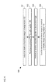
- FIGS. 6A-C depict images of a test specimen before deconvolution, after computation assembly of raw sub-images, and after application of the complete deconvolution routine, respectively.
- Images 600 - 602 are obtained after light propagation through 250 microns of brain tissue. Careful examination of images 600 - 602 reveals that many of the finer features of test image 600 are restored in image 602 .
Landscapes
- Health & Medical Sciences (AREA)
- Life Sciences & Earth Sciences (AREA)
- Physics & Mathematics (AREA)
- Neurology (AREA)
- General Health & Medical Sciences (AREA)
- Pathology (AREA)
- General Physics & Mathematics (AREA)
- Analytical Chemistry (AREA)
- Chemical & Material Sciences (AREA)
- Biophysics (AREA)
- Veterinary Medicine (AREA)
- Heart & Thoracic Surgery (AREA)
- Medical Informatics (AREA)
- Molecular Biology (AREA)
- Surgery (AREA)
- Animal Behavior & Ethology (AREA)
- Engineering & Computer Science (AREA)
- Public Health (AREA)
- Biomedical Technology (AREA)
- Optics & Photonics (AREA)
- Physiology (AREA)
- Neurosurgery (AREA)
- Psychology (AREA)
- Nuclear Medicine, Radiotherapy & Molecular Imaging (AREA)
- Biochemistry (AREA)
- Immunology (AREA)
- Investigating, Analyzing Materials By Fluorescence Or Luminescence (AREA)
- Microscoopes, Condenser (AREA)
Abstract
Description
g=h
- where {circumflex over (ƒ)}k is the estimate of f after k iterations, * is the correlation operator, and Mi is a foci-position mask for sub-frame i. It should be noted that accurate foci travel info can be developed for each sub-frame i synchronizing
scanner 106 and a frame trigger applied to the camera ofimager 112. As a result, each iteration step of the estimation excludes any non-zero results for any pixel outside the foci position mask. Typically, the foci-position mask, Mi, for each sub-frame i is obtained during an offline calibration routine. A non-limiting example of a suitable calibration routine includes imaging the surface of a known, uniform fluorescence source, such as Uranium compound glass, usingsystem 100 where the system has a synchronized triggering signal, and acquiring the foci-position mask for each sub-frame directly atimager 112.
Claims (17)
Priority Applications (1)
| Application Number | Priority Date | Filing Date | Title |
|---|---|---|---|
| US14/562,885 US9820652B2 (en) | 2013-12-09 | 2014-12-08 | Multi-photon microscope having an excitation-beam array |
Applications Claiming Priority (2)
| Application Number | Priority Date | Filing Date | Title |
|---|---|---|---|
| US201361913695P | 2013-12-09 | 2013-12-09 | |
| US14/562,885 US9820652B2 (en) | 2013-12-09 | 2014-12-08 | Multi-photon microscope having an excitation-beam array |
Publications (2)
| Publication Number | Publication Date |
|---|---|
| US20150157210A1 US20150157210A1 (en) | 2015-06-11 |
| US9820652B2 true US9820652B2 (en) | 2017-11-21 |
Family
ID=53269911
Family Applications (1)
| Application Number | Title | Priority Date | Filing Date |
|---|---|---|---|
| US14/562,885 Expired - Fee Related US9820652B2 (en) | 2013-12-09 | 2014-12-08 | Multi-photon microscope having an excitation-beam array |
Country Status (1)
| Country | Link |
|---|---|
| US (1) | US9820652B2 (en) |
Families Citing this family (9)
| Publication number | Priority date | Publication date | Assignee | Title |
|---|---|---|---|---|
| US10830701B2 (en) * | 2017-05-06 | 2020-11-10 | Howard Hughes Medical Institute | Scanned line angular projection microscopy |
| GB201710743D0 (en) * | 2017-07-04 | 2017-08-16 | King S College London | Luminescence imaging apparatus and methods |
| DE102018128590A1 (en) * | 2018-11-14 | 2020-05-14 | Carl Zeiss Microscopy Gmbh | Fluctuation-based fluorescence microscopy |
| RU2712756C1 (en) * | 2019-06-06 | 2020-01-31 | Федеральное государственное автономное образовательное учреждение высшего образования "Национальный исследовательский Томский политехнический университет" | Device for investigation of combustion process of powders of metals or their mixtures |
| US12468136B2 (en) | 2019-11-11 | 2025-11-11 | Howard Hughes Medical Institute | Random access projection microscopy |
| CN113049098B (en) * | 2021-03-10 | 2024-02-20 | 哈尔滨工业大学 | High-resolution imaging method through scattering media based on Richardson–Lucy deconvolution |
| KR102719078B1 (en) * | 2021-10-26 | 2024-10-24 | 포항공과대학교 산학협력단 | Device and method for imaging and examining cells on the surface of living tissue using moxifloxacin |
| DE102023104144A1 (en) * | 2023-02-20 | 2024-08-22 | Leica Microsystems Cms Gmbh | Data processing device for a digital imaging device, microscope and microscopy method |
| WO2025193918A1 (en) * | 2024-03-13 | 2025-09-18 | Memorial Sloan-Kettering Cancer Center | Handheld shortwave infrared imaging system |
Citations (3)
| Publication number | Priority date | Publication date | Assignee | Title |
|---|---|---|---|---|
| US20070057211A1 (en) | 2005-05-25 | 2007-03-15 | Karsten Bahlman | Multifocal imaging systems and method |
| US8284483B2 (en) | 2009-02-04 | 2012-10-09 | Ecole Polytechnique | Method and device for acquiring signals in laser scanning microscopy |
| US20120257196A1 (en) | 2011-04-07 | 2012-10-11 | Valerica Raicu | High speed microscope with spectral resolution |
-
2014
- 2014-12-08 US US14/562,885 patent/US9820652B2/en not_active Expired - Fee Related
Patent Citations (3)
| Publication number | Priority date | Publication date | Assignee | Title |
|---|---|---|---|---|
| US20070057211A1 (en) | 2005-05-25 | 2007-03-15 | Karsten Bahlman | Multifocal imaging systems and method |
| US8284483B2 (en) | 2009-02-04 | 2012-10-09 | Ecole Polytechnique | Method and device for acquiring signals in laser scanning microscopy |
| US20120257196A1 (en) | 2011-04-07 | 2012-10-11 | Valerica Raicu | High speed microscope with spectral resolution |
Non-Patent Citations (1)
| Title |
|---|
| David S. C. Biggs et al., "Acceleration of Iterative Image Restoration Algorithms", Mar. 10, 1997, pp. 1766-1775, vol. 36, No. 8, Publisher: Applied Optics. |
Also Published As
| Publication number | Publication date |
|---|---|
| US20150157210A1 (en) | 2015-06-11 |
Similar Documents
| Publication | Publication Date | Title |
|---|---|---|
| US9820652B2 (en) | Multi-photon microscope having an excitation-beam array | |
| US11946854B2 (en) | Systems and methods for two-dimensional fluorescence wave propagation onto surfaces using deep learning | |
| US20230085827A1 (en) | Single-shot autofocusing of microscopy images using deep learning | |
| US9581798B2 (en) | Light sheet-based imaging device with extended depth of field | |
| JP7181537B2 (en) | Time-resolved imaging method with high spatial resolution | |
| US11449964B2 (en) | Image reconstruction method, device and microscopic imaging device | |
| US10830701B2 (en) | Scanned line angular projection microscopy | |
| US10642017B2 (en) | Imaging system and imaging method | |
| US11106027B2 (en) | Resolution enhancement for line scanning excitation microscopy systems and methods | |
| EP3037861A1 (en) | Imaging method, and system, for obtaining an super-resolution image of an object | |
| TW202129267A (en) | System for wafer inspection | |
| US11215804B2 (en) | Microscope and method for imaging a sample | |
| US20150103181A1 (en) | Auto-flat field for image acquisition | |
| JP6928757B2 (en) | Systems and methods for image processing in light microscopy | |
| US20150168705A1 (en) | Autofocus system and autofocus method for focusing on a surface | |
| KR101064672B1 (en) | Confocal Laser Scanning Microscope with Two-way Line Scanning Method and Image Processing Method Using the Same | |
| WO2017169597A1 (en) | Image acquisition device and image acquisition method | |
| CN117631249A (en) | Line scan confocal scanning light field microscopy imaging device and method | |
| JP4905356B2 (en) | Line scanning confocal microscope | |
| US20250384990A1 (en) | Systems and methods for increasing three-dimensional image quality using morphology-based recomposition | |
| US20250104388A1 (en) | System and methods for increasing image quality using a morphological-based composition | |
| Ting et al. | Light-sheet light-field macrophotography for imaging neuronal structure | |
| KR20250046859A (en) | Open-top Two-photon light sheet microscope and its operated method |
Legal Events
| Date | Code | Title | Description |
|---|---|---|---|
| AS | Assignment |
Owner name: THE BOARD OF TRUSTEES OF THE LELAND STANFORD JUNIO Free format text: ASSIGNMENT OF ASSIGNORS INTEREST;ASSIGNORS:ZHANG, TONG;SCHNITZER, MARK;LECOQ, JEROME ANTHONY-JEAN;AND OTHERS;REEL/FRAME:035253/0966 Effective date: 20141205 |
|
| AS | Assignment |
Owner name: THE BOARD OF TRUSTEES OF THE LELAND STANFORD JUNIO Free format text: CORRECTIVE ASSIGNMENT TO CORRECT THE LANGUAGE IN ORIGINAL ASSIGNMENT PREVIOUSLY RECORDED AT REEL: 035253 FRAME: 0966. ASSIGNOR(S) HEREBY CONFIRMS THE ASSIGNMENT;ASSIGNORS:ZHANG, TONG;SCHNITZER, MARK;LECOQ, JEROME ANTHONY-JEAN;AND OTHERS;SIGNING DATES FROM 20160526 TO 20170415;REEL/FRAME:042366/0868 |
|
| STCF | Information on status: patent grant |
Free format text: PATENTED CASE |
|
| MAFP | Maintenance fee payment |
Free format text: PAYMENT OF MAINTENANCE FEE, 4TH YR, SMALL ENTITY (ORIGINAL EVENT CODE: M2551); ENTITY STATUS OF PATENT OWNER: SMALL ENTITY Year of fee payment: 4 |
|
| FEPP | Fee payment procedure |
Free format text: MAINTENANCE FEE REMINDER MAILED (ORIGINAL EVENT CODE: REM.); ENTITY STATUS OF PATENT OWNER: SMALL ENTITY |
|
| LAPS | Lapse for failure to pay maintenance fees |
Free format text: PATENT EXPIRED FOR FAILURE TO PAY MAINTENANCE FEES (ORIGINAL EVENT CODE: EXP.); ENTITY STATUS OF PATENT OWNER: SMALL ENTITY |
|
| STCH | Information on status: patent discontinuation |
Free format text: PATENT EXPIRED DUE TO NONPAYMENT OF MAINTENANCE FEES UNDER 37 CFR 1.362 |
|
| FP | Lapsed due to failure to pay maintenance fee |
Effective date: 20251121 |