CROSS REFERENCE TO RELATED APPLICATIONS
The present application claims the benefit of U.S. Provisional Application Ser. No. 61/824,734 filed on May 17, 2013, which is incorporated herein by reference in its entirety.
FIELD OF THE INVENTION
The present invention generally relates to cancer, and more particularly to the management of acute myeloid leukemia (AML).
REFERENCE TO SEQUENCE LISTING
Pursuant to 37 C.F.R. 1.821(c), a sequence listing is submitted herewith as an ASCII compliant text file named Sequence_Listing_ST25.txt, that was created on Apr. 4, 2014 and having a size of ˜27 kilobytes. The content of the aforementioned file is hereby incorporated by reference in its entirety.
BACKGROUND OF THE INVENTION
AML is a clonal disorder clinically presenting as increased proliferation of heterogeneous and undifferentiated myeloid blasts. Understanding the biology of human AML stem/progenitor cells is an important prerequisite for the development of more efficacious therapeutic strategies in the treatment of AML as current therapies fail to permanently eradicate the disease in a significant proportion of patients (Patel, J., et al. The New England Journal of Medicine 366, 1079-1089 (2012)). Studies by Lapidot and Dick (Lapidot, T., et al. Nature 367, 645-648 (1994)) are at the basis of the current understanding of AML as a heterogeneous disease consisting of cells which differ from each other with regards to proliferative potential, cell cycle kinetics, resistance to chemotherapy and self-renewal capacity thus caricaturing the hierarchy of the normal hematopoietic system (Bonnet, D. & Dick, J. Nature medicine 3, 730-737 (1997); Hope, K., Jin, L. & Dick, J. Nature immunology 5, 738-743 (2004); Ishikawa, F., et al. Nature biotechnology 25, 1315-1321 (2007); Pearce, D., et al. Blood 107, 1166-1173 (2006)). Leukemia initiating cells (LIC) which are defined by their potential to engraft immunocompromised mice are at the apex of this hierarchy and share some important features with normal hematopoietic stem cells (HSC) as the ability to self-renew and to recapitulate the diversity of the hierarchy. Engraftment potential of human leukemic cells in immunocompromised mice has been correlated with clinical outcome in adult (Pearce D. et al., supra) and more recently pediatric leukemia patients (Woiterski, J., et al. International journal of cancer March 23. doi: 10.1002/ijc.28170. [Epub ahead of print] (2013)). Furthermore, leukemic stem cell associated gene expression signatures (Eppert, K., et al. Nature medicine 17, 1086-1093 (2011); Gentles, A., Plevritis, S., Majeti, R. & Alizadeh, A. JAMA: the journal of the American Medical Association 304, 2706-2715 (2010)) identified by comparative microarray studies have been associated with worse survival providing evidence for clinical relevance of these experimentally defined cells. However, LICs rapidly differentiate or/and undergo apoptosis when deprived of their in vivo environment and exposed to currently available in vitro conditions, which is a major obstacle in the development of LIC targeted therapies and casts doubt on the interpretation of results emanating from ex vivo treatment of these cells. Cell lines which have been used in the past for drug screenings have overcome these constraints but do not reflect the hierarchical organization of the primary disease anymore which makes them an inappropriate tool for the development of LIC targeted therapies.
There is thus a need for strategies to modulate AML stem/progenitor cell expansion and/or differentiation.
The present description refers to a number of documents, the content of which is herein incorporated by reference in their entirety.
SUMMARY OF THE INVENTION
More specifically, in accordance with the present invention, there is provided In an aspect, the present invention provides a method for (i) inhibiting or preventing the differentiation of acute myeloid leukemia (AML) initiating cells ex vivo, and/or (ii) promoting the expansion or maintenance of undifferentiated primary acute myeloid leukemia (AML) blasts ex vivo, and/or (iii) partially rescuing AML initiating activity ex vivo, said method comprising contacting said cells with (a) a suppressor of the Aryl hydrocarbon Receptor (AhR) and/or (b) a compound of general formula I or II:
or a salt or a prodrug thereof,
wherein:
- Z is
- 1) —P(O)(OR1)(OR1),
- 2) —C(O)OR1,
- 3) —C(O)NHR1,
- 4) —C(O)N(R1)R1,
- 5) —C(O)R1,
- 6) —CN,
- 7) —SR1,
- 8) —S(O)2NH2,
- 9) —S(O)2NHR1,
- 10) —S(O)2N(R1)R1,
- 11) —S(O)R1,
- 12) —S(O)2R1,
- 13) -L,
- 14) -benzyl optionally substituted with 1, 2 or 3 RA or R1 substituents,
- 15) -L-heteroaryl optionally substituted with one or more RA or R1 substituents attached on either or both the L and the heteroaryl groups,
- 16) -L-heterocyclyl optionally substituted with one or more RA or R1 substituents attached on either one or both the L and the heterocyclyl groups,
- 17) -L-aryl optionally substituted with one or more RA or R1 substituents attached on either or both the L and the heteroaryl groups,
- 18) -heteroaryl optionally substituted with one or more RA or R1 substituents, or
- 19) -aryl optionally substituted with one or more RA or R1 substituents,
- and wherein each substituent is optionally attached to the L group if it is not already present,
- and wherein, when (R1) and R1 are attached to a nitrogen atom, optionally they join together with the nitrogen atom to form a 3 to 7-membered ring which optionally includes one or more other heteroatom selected from N, O and S, optionally the is substituted with one or more R1 or RA;
- W is
- 1) —H,
- 2) -halogen,
- 3) —OR1,
- 4) -L-OH,
- 5) -L-OR1,
- 6) —SR1,
- 7) —CN,
- 8) —P(O)(OR1)(OR1),
- 9) —NHR1,
- 10) —N(R1)R1,
- 11) -L-NH2,
- 12) -L-NHR1,
- 13) -L-N(R1)R1,
- 14) -L-SR1,
- 15) -L-S(O)R1,
- 16) -L-S(O)2R1,
- 17) -L-P(O)(OR1)(OR1),
- 18) —C(O)OR1,
- 19) —C(O)NH2,
- 20) —C(O)NHR1,
- 21) —C(O)N(R1)R1,
- 22) —NHC(O)R1,
- 23) —NR1C(O)R1,
- 24) —NHC(O)OR1,
- 25) —NR1C(O)OR1,
- 26) —OC(O)NH2,
- 27) —OC(O)NHR1,
- 28) —OC(O)N(R1)R1,
- 29) —OC(O)R1,
- 30) —C(O)R1,
- 31) —NHC(O)NH2,
- 32) —NHC(O)NHR1,
- 33) —NHC(O)N(R1)R1,
- 34) —NR1C(O)NH2,
- 35) —NR1C(O)NHR1,
- 36) —NR1C(O)N(R1)R1,
- 37) —NHS(O)2R1,
- 38) —NR1S(O)2R1,
- 39) —S(O)2NH2,
- 40) —S(O)2NHR1,
- 41) —S(O)2N(R1)R1,
- 42) —S(O)R1,
- 43) —S(O)2R1,
- 44) —OS(O)2R1,
- 45) —S(O)2OR1,
- 46) -benzyl optionally substituted with 1, 2 or 3 RA or R1 substituents,
- 47) -L-heteroaryl optionally substituted with one or more RA or R1 substituents attached on either or both the L and the heteroaryl groups,
- 48) -L-heterocyclyl optionally substituted with one or more RA or R1 substituents attached on either or both the L and the heterocyclyl groups,
- 49) -L-aryl optionally substituted with one or more RA or R1 substituents attached on either or both the L and aryl groups,
- 50) -L-NR1(R1),
- 51) -L-)2NR1,
- 52) -L-(N(R1)-L)n-N(R1)R1,
- 53) -L-(N(R1)-L)n-heteroaryl optionally substituted with one or more RA or R1 substituents attached on either or both the L and heteroaryl groups,
- 54) -L-(N(R1)-L)n-heterocyclyl optionally substituted with one or more RA or R1 substituents attached on either or both the L and heterocyclyl groups,
- 55) -L-(N(R1)-L)n-aryl optionally substituted with one or more RA or R1 substituents attached on either or both the L and aryl groups,
- 56) —O-L-N(R1)R1,
- 57) —O-L-heteroaryl optionally substituted with one or more RA or R1 substituents attached on either or both the L and heteroaryl groups,
- 58) —O-L-heterocyclyl optionally substituted with one or more RA or R1 substituents attached on either or both the L and heterocyclyl groups,
- 59) —O-L-aryl optionally substituted with one or more RA or R1 substituents attached on either or both the L and aryl groups,
- 60) —O-L)2-NR1,
- 61) —O-L-(N(R1)-L)n-N(R1)R1,
- 62) —O-L-(N(R1)-L)n-heteroaryl optionally substituted with one or more RA or R1 substituents attached on either or both the L and heteroaryl groups,
- 63) —O-L-(N(R1)-L)n-heterocyclyl optionally substituted with one or more RA or R1 substituents attached on either or both the L and heterocyclyl groups,
- 64) —O-L-(N(R1)-L)n-aryl optionally substituted with one or more RA or R1 substituents,
- 65) —S-L-heteroaryl optionally substituted with one or more RA or R1 substituents,
- 66) —S-L-heterocyclyl optionally substituted with one or more RA or R1 substituents,
- 67) —S-L-aryl optionally substituted with one or more RA or R1 substituents attached on either or both the L and aryl groups,
- 68) —S-L)2NR1,
- 69) —S-L-(N(R1)-L)n-N(R1)R1,
- 70) —S-L-(N(R1)-L)n-heteroaryl optionally substituted with one or more RA substituents,
- 71) —S-L-(N(R1)-L)n-heterocyclyl optionally substituted with one or more RA substituents,
- 72) —S-L-(N(R1)-L)n-aryl optionally substituted with one or more RA substituents,
- 73) —NR1(R1),
- 74) —(N(R1)-L)n-N(R1)R1,
- 75) —N(R1)L)2-NR1,
- 76) —(N(R1)-L)n-N(R1)RA,
- 77) —(N(R1)-L)n-heteroaryl optionally substituted with one or more RA or R1 substituents,
- 78) —(N(R1)-L)n-heterocyclyl optionally substituted with one or more RA or R1 substituents,
- 79) —(N(R1)-L)n-aryl optionally substituted with one or more RA or R1 substituents,
- 80) -heteroaryl optionally substituted with one or more RA substituents, or
- 81) -aryl optionally substituted with one or more RA substituents,
- and wherein each substituent is optionally attached to the L group if it is not already present,
- and wherein when two R1 substituents are present on the same nitrogen atom, then each R1 substituent is independently selected from the list of R1 values described thereafter,
- and wherein n is an integer equal to either 0, 1, 2, 3, 4, or 5,
- and wherein, when (R1) and R1 are attached to a nitrogen atom, optionally they join together with the nitrogen atom to form a 3 to 7-membered ring which optionally includes one or more other heteroatom selected from N, O and S, optionally the ring is substituted with one or more R1 or RA;
- L is
- 1) —C1-6 alkyl,
- 2) —C2-6 alkenyl,
- 3) —C2-6 alkynyl,
- 4) —C3-7 cycloalkyl,
- 5) —C3-7 cycloalkenyl,
- 6) heterocyclyl,
- 7) —C1-6 alkyl-C3-7 cycloalkyl,
- 8) —C1-6 alkyl-heterocyclyl,
- 9) aryl, or
- 10) heteroaryl,
- and wherein the alkyl, the alkenyl, the alkynyl, the cycloalkyl, the cycloalkenyl, the heterocyclyl, the aryl and the heteroaryl groups are each independently optionally substituted with one or two RA substituent;
- R1 is
- 1) —H,
- 2) —C1-6 alkyl,
- 3) —C2-6 alkenyl,
- 4) —C2-6 alkynyl,
- 5) —C3-7 cycloalkyl,
- 6) —C3-7 cycloalkenyl,
- 7) —C1-6 perfluorinated,
- 8) -heterocyclyl,
- 9) -aryl,
- 10) -heteroaryl,
- 11) -benzyl, or
- 12) 5-[(3aS,4S,6aR)-2-oxohexahydro-1H-thieno[3,4-d]imidazol-4-yl]pentanoyl,
- and wherein the alkyl, the alkenyl, the alkynyl, the cycloalkenyl, the perfluorinated alkyl, the heterocyclyl, the aryl, the heteroaryl and the benzyl groups are each independently optionally substituted with 1, 2 or 3 RA or R1 substituents;
- R2 is
- 1) —H,
- 2) —C1-6 alkyl,
- 3) —SR1,
- 4) —C(O)R1,
- 5) —S(O)R1,
- 6) —S(O)2R1,
- 7) -benzyl optionally substituted with 1, 2 or 3 RA or R1 substituents,
- 8) -L-heteroaryl optionally substituted with one or more RA or R1 substituents attached on either one or both the L and the heteroaryl groups,
- 9) -L-heterocyclyl optionally substituted with one or more RA or R1 substituents attached on either one or both the L and the heterocyclyl groups,
- 10) -L-aryl optionally substituted with one or more RA or R1 substituents attached on either one or both the L and the aryl groups,
- 11) -heteroaryl optionally substituted with one or more RA or R1 substituents, or
- 12) -aryl optionally substituted with one or more RA or R1 substituents,
- and wherein each substituent is optionally attached to the L group if it is not already present;
- RA is
- 1) -halogen,
- 2) —CF3,
- 3) —OH,
- 4) —OR1,
- 5) -L-OH,
- 6) -L-OR1,
- 7) —OCF3,
- 8) —SH,
- 9) —SR1,
- 10) —CN,
- 11) —NO2,
- 12) —NH2,
- 13) —NHR1,
- 14) —NR1R1,
- 15) -L-NH2,
- 16) -L-NHR1,
- 17) -L-NR4R1,
- 18) -L-SR1,
- 19) -L-S(O)R1,
- 20) -L-S(O)2R1,
- 21) —C(O)OH,
- 22) —C(O)OR1,
- 23) —C(O)NH2,
- 24) —C(O)NHR1,
- 25) —C(O)N(R1)R1,
- 26) —NHC(O)R1,
- 27) —NR1C(O)R1,
- 28) —NHC(O)OR1,
- 29) —NR1C(O)OR1,
- 30) —OC(O)NH2,
- 31) —OC(O)NHR1,
- 32) —OC(O)N(R1)R1,
- 33) —OC(O)R1,
- 34) —C(O)R1,
- 35) —NHC(O)NH2,
- 36) —NHC(O)NHR1,
- 37) —NHC(O)N(R1)R1,
- 38) —NR1C(O)NH2,
- 39) —NR1C(O)NHR1,
- 40) —NR1C(O)N(R1)R1,
- 41) —NHS(O)2R1,
- 42) —NR1S(O)2R1,
- 43) —S(O)2NH2,
- 44) —S(O)2NHR1,
- 45) —S(O)2N(R1)R1,
- 46) —S(O)R1,
- 47) —S(O)2R1,
- 48) —OS(O)2R1,
- 49) —S(O)2OR1,
- 50) -benzyl,
- 51) —N3, or
- 52) —C(—N═N—)(CF3),
and wherein the benzyl group is optionally substituted with 1, 2 or 3 RA or R1 substituents.
In an embodiment, the compound of item (b) above is a compound of general formula III or IV:
or a salt or a prodrug thereof, wherein Z and R
2 are each as defined above, and m is an integer from 1 to 6, and wherein when m is 2 or more, X
i are the same or different and are each independently NR
1, CH
2, O or S, wherein R
1 is as defined above, and L
i are the same or different and are each independently L as defined above, and wherein R
3 and R
4 are the same or different and are each independently H, R
1 as defined in
claim 1, or they join together with N to form a 3 to 7-membered ring which optionally includes one or more other heteroatom selected from N, O and S, optionally the ring is substituted with one or more R
1 or R
A.
In an embodiment, the compound of item (b) above is a compound of general formula V or VI:
or a salt or a prodrug thereof, wherein Z, L, R
1 and R
2 are each as defined above.
In an embodiment, the compound of item (b) above is a compound of general formula IIA:
or a salt or a prodrug thereof, wherein R1, W and R2 are each as defined above.
In an embodiment, the compound of item (b) above is a compound of general formula IIB:
or a salt or a prodrug thereof, wherein W and R2 are each as defined above and Het is a 3 to 7-membered heterocycle, optionally substituted with one or more R1 or RA as defined above.
In an embodiment, the compound of item (b) above is a compound of general formula IIC:
or a salt or a prodrug thereof, wherein W and R2 are each as defined above and wherein R5 and R6 are the same or different and are each independently L as defined above, or they join together with C to form a 5 to 7-membered ring which optionally includes one or more heteroatom selected from N, O and S, optionally the ring is substituted with one or more R1 or RA. In an embodiment, the ring is a 5-membered ring, and the heteroatom is N. In a further embodiment, the ring includes four N. In an embodiment, R2 is benzyl.
In an embodiment, the compound of item (b) above is a compound of general formula IVA
or a salt or a prodrug thereof, wherein W, L, R1, R2, m, L1, R3 and R4 are each as defined above.
In a further embodiment, Z is CO2Me or 2-methyl-2H-tetrazol-5-yl; R2 is benzyl, 3-thienylmethyl or 3-pyridinyl methyl; and W is NH-L-N(R1)R1 wherein L is C2-4 alkyl and R1 is C1-4 alkyl or (R1) and R1 join together with the nitrogen atom to which they are attached to form a 3 to 7-membered ring, which optionally includes one or more other heteroatom selected from N, O and S, optionally the ring is substituted with one or more R1 or RA.
In an embodiment, the compound of item (b) above is any of compounds 1 to 55 depicted below, or a salt or a prodrug thereof.
| |
| Compound | |
| number |
Structure |
| |
| |
| |
| 1 |
|
| |
| 2 |
|
| |
| 3 |
|
| |
| 4 |
|
| |
| 5 |
|
| |
| 6 |
|
| |
| 7 |
|
| |
| 8 |
|
| |
| 9 |
|
| |
| 10 |
|
| |
| 11 |
|
| |
| 12 |
|
| |
| 13 |
|
| |
| 14 |
|
| |
| 15 |
|
| |
| 16 |
|
| |
| 17 |
|
| |
| 18 |
|
| |
| 19 |
|
| |
| 20 |
|
| |
| 21 |
|
| |
| 22 |
|
| |
| 23 |
|
| |
| 24 |
|
| |
| 25 |
|
| |
| 26 |
|
| |
| 27 |
|
| |
| 28 |
|
| |
| 29 |
|
| |
| 30 |
|
| |
| 31 |
|
| |
| 32 |
|
| |
| 33 |
|
| |
| 34 |
|
| |
| 35 |
|
| |
| 36 |
|
| |
| 37 |
|
| |
| 38 |
|
| |
| 39 |
|
| |
| 40 |
|
| |
| 41 |
|
| |
| 42 |
|
| |
| 43 |
|
| |
| 44 |
|
| |
| 45 |
|
| |
| 46 |
|
| |
| 47 |
|
| |
| 48 |
|
| |
| 49 |
|
| |
| 50 |
|
| |
| 51 |
|
| |
| 52 |
|
| |
| 53 |
|
| |
| 54 |
|
| |
| 55 |
|
| |
In an embodiment, the compound of item (b) above is compound 1:
or a salt or a prodrug thereof.
In an embodiment, the method comprises contacting said cells with a compound of Table 1 below.
In another aspect, the present invention provides a method for (i) inhibiting or preventing the differentiation, and/or (ii) promoting the expansion or maintenance, of acute myeloid leukemia (AML) initiating cells ex vivo, said method comprising contacting said cells with a suppressor of the Aryl hydrocarbon Receptor (AhR) and/or with a compound of general formula I-VI, IIA-IIC, IVA, VIA, or any of compounds 1 to 55 as defined above. In an embodiment, the method comprises contacting said cells with a compound of Table 1 below.
In a specific embodiment, the methods comprise contacting said cells with (a) a suppressor of AhR and (b) a compound of general formula I-VI, IIA-IIC, IVA, VIA, or any of compounds 1 to 55 as defined above. In a further embodiment, the compound (b) is compound 1 or a salt or a prodrug thereof. In another specific embodiment, said suppressor of AhR is StemRegenin 1 (SR1), retusin-7-methylether (C01), UM0125464 (C02), chrysin (C04), kaempferide (C06), xanthone, 3-chloro-N-(2,3-dihydro-1,4-benzodioxin-6-yl)-1-benzithiophene-2-carboxamide (C03), 5-methoxyflavone (C08), or N-methyl-β-carboline-3-carboxamide (C05). In another specific embodiment, said suppressor of AhR is StemRegenin 1 (SR1), retusin-7-methylether (C01), UM0125464 (C02), 3-chloro-N-(2,3-dihydro-1,4-benzodioxin-6-yl)-1-benzithiophene-2-carboxamide (C03), chrysin (C04), kaempferide (C06), 5-methoxyflavone (C08), or N-methyl-β-carboline-3-carboxamide (C05). In another specific embodiment, said suppressor of AhR is N-methyl-β-carboline-3-carboxamide.
In another specific embodiment, the method comprises (a) providing a cell population comprising said AML initiating cells and (b) culturing said cell population ex vivo under suitable conditions for expanding or maintaining undifferentiated primary AML blasts.
In another aspect, the present invention provides an enriched AML initiating cell population. In another aspect, the present invention provides an ex vivo culture comprising an AML initiating cell population. In a specific embodiment, the cell population is obtained by the method of the present invention as described herein. In an embodiment, the enriched AML initiating cell population has been maintained in culture for at least 1, 2, 4, 8, 12, 18 or 24 h. In other embodiments, the enriched AML initiating cell population has been maintained in culture for at least 2, 3, 4, 5, 6 or 7 days. In an embodiment, the number of AML initiating cells in said population is at least 5-fold higher relative to the number of AML initiating cells in a corresponding population cultured in the absence of the compounds of items (a) and/or (b) defined above. In further embodiments, the number of AML initiating cells in said population is at least 10-, 15, 20-, 25-, 30-, 40- or 50-fold higher relative to the number of AML initiating cells in a corresponding population cultured in the absence of the compounds of items (a) and/or (b) defined above
In another aspect, the present invention provides method for determining whether a test agent may be useful for inhibiting and/or eliminating AML initiating cells, said method comprising (a) culturing a cell population comprising AML initiating cells in the presence of a suppressor of the Aryl hydrocarbon Receptor (AhR) and/or general formula I-VI, IIA-IIC, IVA, VIA, or any of compounds 1 to 55 as defined above and (b) contacting said cell population with said test agent; and (c) determining whether undifferentiated primary AML blasts are inhibited and/or eliminated in the presence of the test agent. In an embodiment, the method comprises culturing said cells in the presence of a compound of Table 1 below.
In a specific embodiment, step (c) comprises comparing the number of undifferentiated primary AML blasts in the culture in the presence and absence of said test agent, wherein a lower number of undifferentiated primary AML blasts in the presence of said test agent is indicative that said test agent may be useful for inhibiting and/or eliminating AML initiating cells.
In another aspect, the present invention provides a method for (i) stimulating the differentiation, and/or (ii) inhibiting the expansion or maintenance, of acute myeloid leukemia (AML) initiating cells ex vivo, said method comprising culturing said cells in the presence of an agonist of the Aryl hydrocarbon Receptor (AhR).
In another aspect, the present invention provides a method for inhibiting or eliminating AML initiating cells in a subject, said method comprising administering to said subject an effective amount of a pharmaceutically acceptable agonist of the Aryl hydrocarbon Receptor (AhR).
In another aspect, the present invention provides a method for preventing or inhibiting minimal residual disease (MRD) in an AML patient, said method comprising administering to said patient an effective amount of a pharmaceutically acceptable suppressor of the Aryl hydrocarbon Receptor (AhR).
In a specific embodiment, the method further comprises administering a chemotherapeutic agent to the subject.
Other objects, advantages and features of the present invention will become more apparent upon reading of the following non-restrictive description of specific embodiments thereof, given by way of example only with reference to the accompanying drawings.
BRIEF DESCRIPTION OF THE DRAWINGS
In the appended drawings:
FIG. 1A is an overview of the strategy used for the screening of modulators of the differentiation of primary human AML cells. 6,168 compounds were tested on a cytogenetically normal AML for their ability to prevent differentiation of primary human AML cells in vitro. Loss of CD34 and acquisition of CD15 surface expression measured by HTS-flow cytometry were used as indication of differentiation. 78 compounds complying with indicated viability criteria yielded >50% increase of CD34+CD15− (%) compared to control (DMSO). Of these, 23 compounds were retested in secondary screenings, and 8 compounds were selected for validation. After excluding compounds with common target and negative impact on cell proliferation, 2 compounds were selected for validation;
FIG. 1B depicts plots showing typical phenotypic changes (loss of CD34, acquisition of CD15) occurring upon in vitro culture of AML 05H163;
FIG. 1C shows a waterfall plot of 5,969 screen compounds complying with viability criteria. The names of compounds matching with secondary screen criteria are depicted above the dotted line. The compounds depicted at the right bottom corner (below the dotted line) are AhR-agonists and they induce further differentiation compared to the DMSO only culture condition.
FIG. 1D shows the impact of the indicated compounds on CD34 and CD15 expression compared to DMSO after 5 days in culture (secondary screens). Dashed line in upper panels indicates CD34 profile of AML cells at t0;
FIG. 1E shows the changes in expression levels of the indicated markers after 24 h incubation with selected compounds compared to DMSO, measured by q-RT-PCR with GAPDH as endogenous control. Note that C07 (UM729) has no effect on AhR target genes, suggesting that it acts on a different target;
FIG. 1F shows the chemical structures and dose response experiments for the three compounds selected for validation. Black bars indicating CD34+CD15− absolute cell counts refer to left y-axis, grey bars indicating CD34+CD15− percentages refer to right y-axis (means±SD, n=3 (C05, SR1), n=2 (UM729) wells/dose, 384-well plate);
FIG. 2A shows the expression levels (RPKM) of the indicated genes in human AML specimens with normal karyotype, determined by RNA-Seq (Table 1). Bars indicate means of log 2-transformed values;
FIG. 2B Fold changes in gene expression levels (mean±SEM, n=13 samples), measured by q-RT-PCR with GAPDH as endogenous control, following 24 h exposure to the indicated compounds or combinations. TCDD, 2,3,7,8-Tetrachlorodibenzodioxin. _ no compound, SR1 was added at 500 nM, TCDD was added at 0.1 nM, 1 nM and 10 nM;
FIG. 3A shows the fractions of CD34+CD15− cells (mean±SD, 3-8 replicates per sample) after 7-day culture with SR1 (0.75-1 μM), or vehicle (DMSO) normalized to fresh cells (td7/t0). Wilcoxon matched-pairs signed rank test (left panel). Representative FACS profiles of AML 08H012 on day 0 and following 7-day culture with DMSO or SR1 are depicted in the right panel;
FIG. 3B shows the fold changes of CD34+CD15− cell numbers (geometric mean±SD, 3-8 replicates per sample) after 7-day culture with SR1 (0.75-1 μM) or DMSO compared to input numbers (td7/t0, log 2-transformed, P<0.0001, paired t-test) (right panel). The right panel shows fold changes of total cells (mean±SEM, n=16, P=0.2, paired t-test);
FIG. 3C shows CellTrace™ Violet profiles of CD33+ and CD34+CD33+ AML cells (04H112 after 4-day culture with SR1 or DMSO. Dashed line indicates CellTrace™ Violet peak of fresh cells (t0). Each individual peak represents one generation of cells;
FIG. 3D shows fold changes (geometric mean±SEM, n=9) in total cells (left) and CD34+CD15− AML cells (right) after 7-day culture in cytokine-supplemented or cytokine-free medium containing SR1 (0.75-1 μM), C05 (2 μM), or DMSO, compared to input numbers (td7/t0), ** P<0.005, paired t-test;
FIG. 4A shows an overview of the in vivo experiments. AML cells from 6 primary human AML samples were injected untreated into the tail vein of sublethally irradiated NSG mice at 4 different doses. On the same day, cell cultures were initiated in T25 culture flasks at a density of 3×10⁶ cells in 6 ml serum-free medium per flask supplemented with DMSO (0.1%) or SR1 (500 nM). After 4 days in culture the equivalents of freshly injected cell doses were transplanted into NSG mice. Engraftment of human leukemic cells in mouse bone marrow was determined by flow cytometry 10-16 weeks after transplantation;
FIG. 4B shows the engraftment levels of AML cells cultured for 4 days in presence of SR1 (0.5-1 μM) or DMSO. Bars indicate means, grey-shaded area indicates <1% human cells in recipient bone marrow, or the absence of engraftment. Mice with <0.1% engraftment were positioned at 0.1%. Indicated transplantation doses represent input cell numbers;
FIG. 4C shows representative FACS plots of patient cells recovered from recipient NSG mice 16 weeks after transplantation of 2×10⁶ uncultured cells (left), or equivalent cell numbers harvested from 4-day cultures containing DMSO or SR1 (500 nM). Non transplanted patient cells (05H163) are also shown (lower right). Numbers indicate percentages within total mouse bone marrow, numbers in brackets represent fractions of human CD45+ cells;
FIG. 4D shows the estimated LSC frequencies and 95% confidence intervals (CI) for 6 AML specimens in FIG. 4B. When all or none of the recipients were engrafted, one-sided CI were calculated and are indicated by arrows. Differences between conditions were analyzed by Chi-square test (Hu, Y, and Smyth, G K (2009). ELDA: Extreme limiting dilution analysis for comparing depleted and enriched populations in stem cell and other assays. Journal of Immunological Methods 347, 70-78; http://bioinf.wehi.edu.au/software/elda/), *P<0.05 **P<0.005, differences between DMSO and fresh cells are significant (P<0.0005) for all samples;
FIG. 5A shows the impact of UM729 on CD34 and CD15 expression of sample 05H163 following 5 days in culture as indicated. Dashed line indicates CD34 peak on day 0;
FIG. 5B shows the relative and absolute CD34+CD15− cell numbers (means±SD, n=3) of AML 05H163 following 5 day culture in the indicated conditions;
FIG. 5C shows the percentages of CD34+CD15− cells (mean±SD, 3-8 replicates per sample) normalized to fresh cells (td7/t0) after 7-day culture with UM729 (1 μM), or vehicle (DMSO). SR1 and DMSO values (means) are shown again for direct comparison (see FIG. 3A for error bars). Wilcoxon matched-pairs test. ***P<0.001, ****P<0.0001;
FIG. 5D depicts plots showing the impact of the indicated compounds on CD34 expression of sample 08H118;
FIG. 5E shows cytospins of fresh AML cells and after 4-day culture as indicated. SR1 is at 500 nM, UM729 at 1 μM. Scale bar indicates 20 μM;
FIG. 5F shows the engraftment levels of AML samples after 4-day cultures in UM729 and UM729+SR1, compared to SR1 alone. DMSO and SR1 values (means) are shown for direct comparison (see FIG. 4B for single recipients). * Mice injected with sample 08H012 were analyzed 14 weeks after transplantation in this experiment;
FIG. 5G shows leukemic engraftment (05H163) following injection of 2×10⁶ input cells exposed for 4 days to the indicated compounds (left panel). LSC frequency analysis of AML 05H163 cultured in different compounds (right panel). One-sided 95% CI shown for DMSO as no engraftment was detected at highest dose;
FIG. 6 shows the chemical structure of compounds C01-004, C06 and C08 confirmed in secondary screenings. The structure of compounds C05 and C07 is depicted in FIG. 1F;
FIG. 7A shows the fold changes of CYP1B1 expression compared to fresh cells after 24 h (grey bars) or 6 days (black bars) in control culture conditions (DMSO);
FIG. 7B shows AhR target gene induction after 24 h incubation in serum-free culture medium in absence and presence of vehicle DMSO (0.1%) compared to t0 (Left panel). The right panel shows AhR target gene induction after 24 h incubation in cultures containing DMSO or SR1 (500 nM), in phenol red free medium and in phenol red free medium without antibiotics (AB) compared to t0;
FIG. 7C shows the fold difference in AhR target gene expression after 24 h exposure to SR1 or DMSO in glass dishes, or SR1 in standard polystyrene culture dishes (plastic-SR1) compared to control DMSO and polystyrene dish (plastic-DMSO);
FIG. 7D shows the fold-change (mean±SEM) in AhR target gene expression after 24 h incubation under normoxic versus hypoxic (1% O2) conditions, and in presence or absence of SR1 (500 nM) compared to expression levels in normoxic control conditions (Normoxia DMSO was used as reference);
FIG. 7E shows the fold difference in AhR target gene expression after 24 h exposure to SR1 or DMSO in serum-free medium (SFM) or co-cultured on a confluent feeder layer of NIH-3T3 fibroblasts compared to t0. Sample 05H163 expanded in NSG mice (05H163*) was used for experiments in FIGS. 7B, 7D and 7E, and sample 04H112 expanded in NSG mice (04H112*) for the experiment in FIG. 7D. GAPDH served as endogenous control in all experiments depicted in FIGS. 7A-7E.
FIG. 8A shows (left panel) the proportions of CD34+CD15− cells (mean±SD) after 7-day culture with N-methyl-β-carboline-3-carboxamide (C05, 2 μM, n=16 AML samples, 3-8 technical replicates per sample), or vehicle (DMSO). Results were normalized to fresh cells (td7/t0). The right panel shows FACS profiles of AML 12H030 (MO, 46,XY) after 7-day culture with DMSO, or C05 (2 μM), or SR1 (0.75 μM). On day 0, 94% of 12H030 cells were CD34+CD15−. P<0.0001, Wilcoxon matched-pairs signed rank test;
FIG. 8B shows (left panel) the fold changes of CD34+CD15− AML cell numbers (geometric means±SD) following 7-day culture with C05 (2 μM, n=15 AML samples, 3-8 technical replicates per sample), or control DMSO compared to input numbers (td7/t0, log 2-transformed). P<0.0001, paired t-test. The right panel shows the fold changes in total gated (viable) cell counts (geometric means±SEM, n=15) following 7 day culture with C05 or DMSO compared to input numbers (td7/t0, log 2-transformed). P=0.2, paired t-test;
FIG. 9 shows the detection of human myeloid cells (CD33+), B-cells (CD19+CD33−) and T-cells (CD3+CD33−) 10 weeks after injection of 2×10⁶ unsorted patient cells (sample 05H050) (left panel). The right panel shows the detection of human CD3+CD45+ T-cells in NSG bone marrow after injection of 2×10⁶ unsorted patient cells (sample 06H135). Numbers in upper panels represent percentages within mouse bone marrow. Percentages in lower panels are fractions of total human CD45+ cells. In both cases the unsorted patient samples contained non-leukemic cells which successfully engrafted NSG mice which has to be distinguished from leukemic engraftment by using a comprehensive antibody cocktail;
FIG. 10 shows the percentage of CD34+CD15− cells (mean±SD) after 7 days in culture with C05 (2 μM), UM729 (1 μM) or both compounds relative to the percentage at t0. Wilcoxon matched pairs signed rank test, ***P<0.001, ****P<0.0001;
FIG. 11 shows the distribution of CD34+CD38−, CD34+CD38+, CD34−CD38+, and CD34−CD38− compartments after 7 days in culture with either DMSO or SR1+UM729 (upper left panel); fold difference in the percentages of CD34+CD38− and CD34+CD38+ cells in presence of SR1+UM729 compared to DMSO (lower left panel). The right panel is a representative FACS plots of samples 05H149 and 04H001 comparing DMSO, SR1 alone and SR1+UM729. Note the shift towards the more primitive cell compartments;
FIG. 12 shows a CellTrace™ Violet profiles of CD33+ and CD34+CD33+ AML cells (04H112, M1,46,XY) following 4-day culture with UM729 (1 μM) or UM729 (1 μM)+SR1 (0.5 μM), or vehicle DMSO. Dashed line indicates CellTrace™ Violet peak for fresh cells (t0), and each individual peak represents one generation of cells;
FIG. 13 shows engraftment of AML sample 05H163 16 weeks after injection of 2×10⁴, 2×10⁵, or 2×10⁶ uncultured cells or cultured input cell equivalents. Cultures were supplemented with UM729 (1 μM), UM729 and C05 (2 μM), or DMSO. For DMSO only the mean engraftment level is shown for direct comparison (see FIG. 4B for engraftment levels of individual recipients);
FIG. 14A depicts the amino acid sequence of a human AhR polypeptide precursor (NCBI Reference Sequence: NP_001612.1, SEQ ID NO:2). The mature form comprises residues 11-848 (residues 1-10 correspond to a propeptide);
FIGS. 14B and 14C depict the nucleotide sequence of a human AhR mRNA (NCBI Reference Sequence: NM_001621.4, coding sequence 614-3160, SEQ ID NO:1);
FIGS. 15A, 15B and 15C show the patient and specimen information for the studies described herein;
FIGS. 16A and 16B show LSC frequencies and engraftment levels measured in the experiments described in Example 6.
DETAILED DESCRIPTION OF INVENTION
In the studies described herein, the present inventors have shown that contacting a cell population comprising primary human AML cells with a suppressor of the Aryl hydrocarbon Receptor (AhR) and/or with a compound of general formula I-VI, IIA-IIC, IVA, VIA, or any of compounds 1 to 55 as defined herein allows expansion of phenotypically and morphologically undifferentiated primary human AML blasts and partially rescues AML initiating activity in vitro (e.g., in vitro).
Accordingly, in a first aspect, the present invention provides a method for (i) inhibiting or preventing the differentiation of acute myeloid leukemia (AML) initiating cells ex vivo; and/or (ii) promoting the expansion or maintenance of undifferentiated primary AML blasts ex vivo; and/or (iii) partially rescuing (i.e., near maintaining) AML initiating activity ex vivo, said method comprising contacting said cells with a suppressor of the Aryl hydrocarbon Receptor (AhR) and/or with a compound of general formula I-VI, IIA-IIC, IVA, VIA, or any of compounds 1 to 55 as defined above.
In another aspect, the present invention provides a method for (i) inhibiting or preventing the differentiation, and/or (ii) promoting the expansion or maintenance, of acute myeloid leukemia (AML) initiating cells ex vivo, said method comprising contacting said cells with a compound set forth in Table 1 below.
In another aspect, the present invention provides a method for (i) inhibiting or preventing the differentiation, and/or (ii) promoting the expansion or maintenance, of acute myeloid leukemia (AML) initiating cells ex vivo, said method comprising contacting said cells with a suppressor of the Aryl hydrocarbon Receptor (AhR) and/or with a compound of general formula I-VI, IIA-IIC, IVA, VIA, or any of compounds 1 to 55 as defined above.
In another aspect, the present invention provides a method for (i) inhibiting or preventing the differentiation, and/or (ii) promoting the expansion or maintenance, of acute myeloid leukemia (AML) initiating cells ex vivo, said method comprising contacting said cells with a compound set forth in Table 1 below.
The term “AML initiating cells” (or “AML stem/progenitor cells”) refers to cells having the potential to self-renew and to engraft immunocompromised mice (e.g., to reconstitute a phenotypic and functional leukemic cell hierarchy), and are enriched in the CD34+ compartment. LIC-activity also exists however in the CD34− compartment. Ongoing differentiation in general including ongoing LIC differentiation is characterized by loss of CD34 expression and increased CD15 expression.
AhR (Aryl Hydrocarbon Receptor) is a member of the bHLH (basic Helix-Loop-Helix)-PAS (Per-ARNT-Sim) family of transcriptional regulators that control a variety of developmental and physiological events, including Neurogenesis, Tracheal and Salivary duct formation, Toxin metabolism, Circadian rhythms, response to Hypoxia and Hormone Receptor function. The unique feature of all bHLH-PAS proteins is the PAS domain, named after the first three proteins identified with this motif, the Drosophila Per, Human ARNT and Drosophila Sim. The PAS domain consists of 260-310 amino acids and incorporates two well-conserved hydrophobic repeats, termed PAS-A (or PAS-1) and PAS-B (or PAS-2), separated by a poorly conserved spacer. Overall, the PAS domain is not well conserved and can mediate a number of diverse biochemical functions. In human Ahr, the bHLH domain spans residues 27-80, the PAS-1 domain spans residues 111-181, the PAS-2 domain spans residues 275-342 and the PAC domain spans residues 348-386. The amino acid sequence of a human AhR polypeptide precursor (NCBI Reference Sequence: NP_001612.1) is depicted in FIG. 14A (SEQ ID NO:2), and the corresponding cDNA sequence (NM_001621.4) is depicted in FIGS. 14B and 14C (SEQ ID NO:1).
AHR, also known as the Dioxin receptor, is recognized as the culprit for most toxic responses observed after exposure to PAH (Polycyclic Aromatic Hydrocarbons), Dioxins (e.g., TCDD (2,3,7,8-tetrachlorodibenzo-p-dioxin)), and Polychlorinated Biphenyls. Ligands for AHR are diverse which include dietary compounds, natural and synthetic flavonoids, natural products, and pharmaceuticals.
AhR suppressors (e.g., inhibitors/antagonists) are well known in the art. The term AhR suppressor includes any compound able to negatively affect the activity of AhR by reducing for example its expression (i.e., at the transcriptional and/or translational level), the level of AhR mRNA and/or protein, or an activity associated with AhR. It includes intracellular as well as extracellular suppressors. Without being so limited, such suppressors include RNA interference agents (e.g., siRNA, shRNA, miRNA and the like), antisense molecules, ribozymes, proteins (e.g., dominant negative, inactive variants), peptides, small molecules, antibodies, antibody fragments, etc.
AhR Antibodies
In an embodiment, the AhR suppressor (e.g., inhibitor/antagonist) is a neutralizing antibody directed against (or specifically binding to) a human AhR polypeptide. The term “antibody” or “immunoglobulin” is used in the broadest sense, and covers monoclonal antibodies (including full-length monoclonal antibodies), polyclonal antibodies, humanized antibodies, CDR-grafted antibodies, chimeric antibodies, multispecific antibodies, and antibody fragments so long as they exhibit the desired biological activity (e.g., neutralizing an activity of the AhR polypeptide). Antibody fragments comprise a portion of a full length antibody, generally an antigen binding or variable region thereof. Examples of antibody fragments include Fab, Fab′, F(ab′)2, and Fv fragments, diabodies, linear antibodies, single-chain antibody molecules, single domain antibodies (e.g., from camelids), shark NAR single domain antibodies, and multispecific antibodies formed from antibody fragments. Antibody fragments can also refer to binding moieties comprising CDRs or antigen binding domains including, but not limited to, VH regions (VH, VH-VH), anticalins, PepBodies, antibody-T-cell epitope fusions (Troybodies) or Peptibodies. In an embodiment, the antibody is a monoclonal antibody. In another embodiment, the antibody is a humanized or CDR-grafted antibody.
In general, techniques for preparing antibodies (including monoclonal antibodies and hybridomas) and for detecting antigens using antibodies are well known in the art (Campbell, 1984, In “Monoclonal Antibody Technology Laboratory Techniques in Biochemistry and Molecular Biology”, Elsevier Science Publisher, Amsterdam, The Netherlands) and in Harlow et al., 1988 (in: Antibody A Laboratory Manual, CSH Laboratories).
Polyclonal antibodies are preferably raised in animals by multiple subcutaneous (s.c.), intravenous (i.v.) or intraperitoneal (i.p.) injections of the relevant antigen (e.g., an AhR polypeptide, or a fragment thereof) with or without an adjuvant. It may be useful to conjugate the relevant antigen to a protein that is immunogenic in the species to be immunized, e.g., keyhole limpet hemocyanin, serum albumin, bovine thyroglobulin, or soybean trypsin inhibitor using a bifunctional or derivatizing agent, for example, maleimidobenzoyl sulfosuccinimide ester (conjugation through cysteine residues), N-hydroxysuccinimide (through lysine residues), glutaraldehyde, succinic anhydride, SOCl2, or R1N═C═NR, where R and R1 are different alkyl groups.
Animals may be immunized against the antigen (AhR polypeptide or a fragment thereof), immunogenic conjugates, or derivatives by combining the antigen or conjugate (e.g., 100 μg for rabbits or 5 μg for mice) with 3 volumes of Freund's complete adjuvant and injecting the solution intradermally at multiple sites. One month later the animals are boosted with the antigen or conjugate (e.g., with ⅕ to 1/10 of the original amount used to immunize) in Freund's complete adjuvant by subcutaneous injection at multiple sites. Seven to 14 days later the animals are bled and the serum is assayed for antibody titer. Animals are boosted until the titer plateaus. Preferably, for conjugate immunizations, the animal is boosted with the conjugate of the same antigen, but conjugated to a different protein and/or through a different cross-linking reagent. Conjugates also can be made in recombinant cell culture as protein fusions. Also, aggregating agents such as alum are suitably used to enhance the immune response.
Monoclonal antibodies may be made using the hybridoma method first described by Kohler et al., Nature, 256: 495 (1975), or may be made by recombinant DNA methods (e.g., U.S. Pat. No. 6,204,023). Monoclonal antibodies may also be made using the techniques described in U.S. Pat. Nos. 6,025,155 and 6,077,677 as well as U.S. Patent Application Publication Nos. 2002/0160970 and 2003/0083293.
In the hybridoma method, a mouse or other appropriate host animal, such as a rat, hamster or monkey, is immunized (e.g., as hereinabove described) to elicit lymphocytes that produce or are capable of producing antibodies that will specifically bind to the antigen used for immunization. Alternatively, lymphocytes may be immunized in vitro. Lymphocytes then are fused with myeloma cells using a suitable fusing agent, such as polyethylene glycol, to form a hybridoma cell. The hybridoma cells thus prepared are seeded and grown in a suitable culture medium that preferably contains one or more substances that inhibit the growth or survival of the unfused, parental myeloma cells. For example, if the parental myeloma cells lack the enzyme hypoxanthine guanine phosphoribosyl transferase (HGPRT or HPRT), the culture medium for the hybridomas typically will include hypoxanthine, aminopterin, and thymidine (HAT medium), which substances prevent the growth of HGPRT-deficient cells.
A human chimeric antibody can be produced in the following manner. cDNA encoding heavy chain variable region (VH) and light chain variable region (VL) obtained from a hybridoma derived from non-human animal cells producing monoclonal antibodies, the cDNA is inserted to each of expression vectors for animal cells having DNA encoding a heavy chain constant region (CH) and light chain constant region (CL) of a human antibody so as to construct a human chimeric antibody expression vector, and this vector is introduced to animal cells to express the human chimeric antibody.
A humanized antibody refers to an antibody which is obtained by grafting the amino acid sequence of the complementary determining region (CDR) of VH and VL of a non-human animal antibody to CDR corresponding to VH and VL of a human antibody. The region other than CDR of VH and VL is called a framework region (hereinbelow, described as “FR”). A humanized antibody can be produced in the following manner. cDNA encoding an amino acid sequence of VH which consists of an amino acid sequence of CDR of VH of a non-human antibody and an amino acid sequence of FR of VH of any human antibody, and cDNA encoding an amino acid sequence of VL which consists of an amino acid sequence of CDR of VL of a non-human animal antibody and an amino acid sequence of FR of VL of any human antibody are constructed, these cDNAs are inserted respectively into expression vectors for animal cells having DNA encoding CH and CL of a human antibody so as to construct a humanized antibody expression vector, and this vector is inserted into animal cells to express the humanized antibody.
Based on the sequences of the AhR polypeptide (see FIG. 14A), the skilled person would be able to generate antibodies directed against this polypeptide, which in turn may be used to neutralize its activity.
RNA Interference Agents Targeting AhR
In another embodiment, the AhR suppressor (e.g., inhibitor/antagonist) is an RNA interference agent targeting an mRNA encoding AhR. The term “RNA interference agent” as used herein refers to molecules that specifically binds to a target mRNA and induces its degradation (usually through the RNA-induced silencing complex (RISC) or interferes with its translation, and includes for example microRNA (miRNA) molecules, antisense molecules, small interfering RNA (siRNA) molecules and small/short hairpin RNA (shRNA). Chemically modified nucleosides, such as 2′-substituted arabinonucleosides (e.g., 2′F-ANA) and 2′-substituted RNA (e.g., 2′F-RNA), may be used for incorporation into RNA interference agents to enhance one or more properties, such as nuclease resistance, pharmacokinetics or affinity for a target RNA.
The RNA interference agent may be expressed from recombinant viral vectors, such as vectors derived from adenoviruses, adeno-associated viruses, lentiviruses, retroviruses, herpesviruses, and the like. Such vectors typically comprise a sequence encoding an RNA interference agent of interest and a suitable promoter operatively linked to the RNA interference agent for expressing the RNA interference agent. The vector may also comprise other sequences, such as regulatory sequences, to allow, for example, expression in a specific cell/tissue/organ, or in a particular intracellular environment/compartment. Methods for generating, selecting and using viral vectors are well known in the art.
An siRNA targeting AhR is disclosed in Abdelrahim et al., Molecular Pharmacology June 2003 vol. 63 no. 6: 1373-1381: 5′-UACUUCCACCUCAGUUGGCTT-3′ (sense, SEQ ID NO:3), 3′-TTAUGAAGGUGGAGUCAACCG-5′ (antisense, SEQ ID NO:4). Two siRNA targeting AhR are also disclosed in Ishida et al., Carcinogenesis vol. 31 no. 2 pp. 287-295, 2010: 5-GCCGAGUCCCAUAUCCGAAUG-3 (sense, SEQ ID NO:5), 5-GACGUAUGUCCAAGAUUCUUU-3 (antisense, SEQ ID NO:6). RNA interference agents directed against AhR are also commercially available. For example, AhR shRNA are available from Origene (Catalog # TG320259). AhR siRNA are available from Origene (Catalog # SR300136), Qiagen (Catalog # SI00293587, SI00293594, SI02780148, SI03043971 and SI03050747), Santa Cruz Biotechnology (Catalog # sc-29654), Life Technologies (Catalog # s1198, s1199, s1200, s199481) and Dharmacon/Thermo Scientific (ON-TARGET plus SMARTpool® siRNA reagent), for example. Reagents and kits for performing RNA interference are available commercially from for example Ambion Inc. (Austin, Tex., USA), New England Biolabs Inc. (Beverly, Mass., USA), Sigma-Aldrich and Invitrogen (Carlsbad, Calif., USA).
Small-Molecule AhR Suppressors
WO 2007/128723 discloses small-molecule AhR suppressors of the formula:
in which R1 and R2 independently of one another are hydrogen or C1-C12-alkyl, R3 to R11 independently of one another are hydrogen, C1-C12-alkyl, hydroxyl or C1-C12-alkoxy, and the broken line represents either a double bond or two hydrogens. This includes the following compounds:
Other examples of AhR suppressors include the dietary flavonoids such as flavone, apigenin and naringenin (US 2004/0077080), as well as flavonoid compounds of the formula:
in which the 5′ position is hydrogen or iodo, the 4′ position is selected from hydrogen, iodo, azido, nitro, a group —NCS, cyano, amino or a group —NHCOCH3; and the 3′ position is hydroxy or lower alkoxy having from 1 to 3 carbon atoms, which may be saturated or unsaturated. Preferred flavone compounds of this class include those with a 3′-methoxy group and a 4′-substituent having one or more terminal atoms of high electron density (—N3, —NO2, or —NCS). Particular compounds include 3′-methoxy-4′-nitroflavone (WO 2009/115807, Henry et al., Mol. Pharmacol 55: 716-725, 1999).
Other AhR suppressors are the flavonoids 7,8-Benzoflavone and 2′,4′,6-Trimethoxyflavone:
Another AhR suppressor is the indole derivative 3,3′-diindolymethane (DIM) (Hestermann et al., Mol. Cell. Biol. 23: 7920-7925, 2003), of the formula:
AhR suppressors are also disclosed in WO 2012/015904, for example CB7993113, CMLD-2166 and CB7950998:
WO 2012/015904 also discloses AhR suppressors of the following formula:
wherein: Y is C or N; X is OR1, NHR1, SR1, CH2(n)R1, halo, or H; n is 0-6; Z is O, S, or NH; R1; and R2 are independently H, alkyl, alkenyl, alkynyl, amino, aminosulfonyl, alkoxy, acyl, aryl, heteroaryl, arylalkyl, cycloalkyl, heteroarylalkyl, heterocyclyl, or haloalkyl, each of which may be optionally substituted; R3, R4, R5 and R6 are independently absent, H, halo, alkyl, alkenyl, alkynyl, alkoxy, acyl, aryl, heteroaryl, arylalkyl, cycloalkyl, heteroarylalkyl, heterocyclyl, or haloalkyl, each of which may be optionally substituted; pharmaceutically acceptable salts thereof.
WO 2012/015904 also discloses AhR suppressors of the following formula:
wherein: X′ is H, alkyl, aminosulfonyl, alkoxy, amino, acyl, aryl, or heteroaryl (preferably alkyl, alkoxy, amino, or aminosulfonyl), each of which may be optionally substituted; n is 0-6 (preferably 0 or 1); R2 is H, alkyl, acyl, aryl, heteroaryl, arylalkyl, cycloalkyl, heteroarylalkyl, heterocyclyl, or haloalkyl (preferably aryl, substituted aryl, heteroaryl, or substituted aryl), each of which may be optionally substituted; R3, R4, R5 and R6 are independently H, alkyl, acyl, halo, aryl, or heteroaryl (preferably H, alkoxy, alkyl, or halo), each of which may be optionally substituted; and pharmaceutically acceptable salts thereof.
WO 2012/015904 also discloses AhR suppressors of the following formula:
wherein: Y is C or N; X is OR1, NHR1, SR1, CH2(n)R1, halo, or H; n is 0-6; Z is O, S, or NH; R1; and R2 are independently H, alkyl, alkenyl, alkynyl, amino, aminosulfonyl, alkoxy, acyl, aryl, heteroaryl, arylalkyl, cycloalkyl, heteroarylalkyl, heterocyclyl, or haloalkyl, each of which may be optionally substituted; R3, R4, R5 and R6 are independently absent, H, halo, alkyl, alkenyl, alkynyl, alkoxy, acyl, aryl, heteroaryl, arylalkyl, cycloalkyl, heteroarylalkyl, heterocyclyl, or haloalkyl, each of which may be optionally substituted; and stereoisomers thereof. In some embodiments of these aspects, the C at position 2 is in the R configuration and the C at position 3 is in the S configuration. In some embodiments of these aspects, the C at position 2 is in the S configuration and the C at position 3 is in the R configuration. In some embodiments of these aspects, the C at position 2 is in the R configuration and the C at position 3 is in the R configuration. In some embodiments of these aspects, the C at position 2 is in the S configuration and the C at position 3 is in the S configuration.
Another example of AhR suppressor is the compound CH-223191, 2-methyl-2H-pyrazole-3-carboxylic acid-(2-methyl-4-o-tolyazophenyl)-amide, of the formula:
WO 2004/041758 discloses AhR suppressors (stilbene derivatives) of the formula:
wherein R3, R4 and R5 and R3′, R4′ and R5′ are identical or different and represent H, OH, O-alkoxy or hal, said alkoxy group being a C1-C6 alkoxy and “hal” being F, Cl or CF3, with the proviso that one of R4′, R3 and R5 or R4, R3′ and R5′ does not represent OH, OCH3, or OCH2CH3 when the two other substituents are both OH, OCH3, or OCH2CH3, respectively.
Another AhR suppressor is N-(2-(1H-indol-3-yl)ethyl)-9-isopropyl-2-(5-methylpyridin-3-yl)-9H-purin-6-amine (GNF351), disclosed in Smith et al., JPET July 2011 vol. 338 no. 1 318-327.
Another AhR suppressor is 1,3-dichloro-5-[(1E)-2-(4-chlorophenyl)ethenyl]-benzene (PDM 2), which has the following structure:
Another AhR suppressor is StemRegenin 1 (SR1), which has the following structure:
Additional AhR suppressors are listed in Table 1, FIG. 1C, FIG. 1F and FIG. 6, and include retusin-7-methylether, UM0125464, chrysin, kaempferide, xanthone, 3-chloro-N-(2,3-dihydro-1,4-benzodioxin-6-yl)-1-benzithiophene-2-carboxamide, 5-methoxyflavone, N-methyl-β-carboline-3-carboxamide.
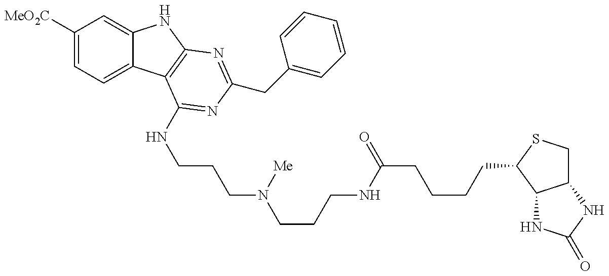
Another compound that was shown to (i) inhibit or prevent the differentiation of acute myeloid leukemia (AML) initiating cells ex vivo, and/or (ii) promote the expansion or maintenance of undifferentiated primary AML blasts ex vivo; and/or (iii) partially rescue (i.e., near maintain) AML initiating activity ex vivo is methyl 4-((3-(piperidin-1-yl)propyl)amino)-9H-pyrimido[4,5-b]indole-7-carboxylate (referred to as UM729 in the Examples below), which has the following structure:
Compounds structurally related to UM729 are disclosed in US2015/011543 and include the compound of general formula I-VI, IIA-IIC, IVA, VIA, or any of compounds 2 to 55 defined above. Methods to synthesize such compounds are described in US2015/011543 which is incorporated by reference.
In an embodiment, the above-mentioned method comprises (a) providing a cell population comprising said AML initiating cells and (b) culturing said cell population ex vivo under suitable conditions for expanding undifferentiated primary AML blasts The cell population (e.g., AML specimen/cell sample) may first be subjected to enrichment or purification steps, including negative and/or positive selection of cells based on specific cellular markers (e.g., CD34+, CD38−, CD123, TIM3, CD96, etc.) in order to provide a starting cell population. Methods for isolating said starting cell population based on specific cellular markers may use fluorescent activated cell sorting (FACS) technology or solid or insoluble substrate to which is bound antibodies or ligands that interact with specific cell surface markers. For example, cells may be contacted with a solid substrate (e.g., column of beads, flasks, magnetic particles) containing the antibodies and any unbound cells are removed. When a solid substrate comprising magnetic or paramagnetic beads is used, cells bound to the beads can be readily isolated by a magnetic separator.
The cell culture may be carried out in natural medium, a semi-synthetic medium or a synthetic medium in terms of composition, and may be a solid medium, a semisolid medium or a liquid medium in terms of shape, and any nutrient medium used for cell culture, which may be supplemented with a mixture of cell expanding factors. Such medium typically comprises sodium, potassium, calcium, magnesium, phosphorus, chlorine, amino acids, vitamins, cytokines, hormones, antibiotics, serum, fatty acids, saccharides or the like. In the culture, other chemical components or biological components may be incorporated singly or in combination, as the case requires. Such components to be incorporated in the medium may be fetal calf serum, human serum, horse serum, insulin, transferrin, lactoferrin, cholesterol, ethanolamine, sodium selenite, monothioglycerol, 2-mercaptoethanol, bovine serum albumin, sodium pyruvate, polyethylene glycol, various vitamins, various amino acids, agar, agarose, collagen, methylcellulose, various cytokines, various growth factors or the like. For example, the medium may be supplemented with a combination of bovine serum albumin, insulin, transferrin (BIT). Examples of such basal medium appropriate for a method of culturing cells without limitation, Dulbecco's Modified Eagles's Medium (DMEM), Ham's Nutrient Mixture H12 Mixture F12, McCoy's 5A medium, Eagles's Minimum Essential Medium (EMEM), αMEM medium (alpha Modified Eagles's Minimum Essential Medium), RPMI1640® medium, Isocove's Modified Dulbecco's Medium (IMDM), StemPro34 (Invitrogen®), X-VIVO 10 (Cambrex), X-VIVO 15 (Cambrex®) and Stemline® II (Sigma-Aldrich), StemSpan® Serum-Free Expansion Medium (SFEM) (StemCell Technologies®, Vancouver, Canada), StemSpan® H3000-Defined Medium (StemCell Technologies®, Vancouver, Canada), CellGro®, SCGM (CellGenix®, Freiburg Germany), and StemPro®-34 SFM (Invitrogen®).
In another aspect, the present invention provides a method for determining whether a test agent may be useful for inhibiting and/or eliminating AML initiating cells, said method comprising (a) culturing a cell population comprising AML initiating cells in the presence of an suppressor of the Aryl hydrocarbon Receptor (AhR) and/or a compound of general formula I-VI, IIA-IIC, IVA, VIA, or any of compounds 1 to 55 defined above; (b) contacting said cell population with said test agent; (c) determining whether AML initiating cells are inhibited and/or eliminated in the presence of the test agent.
In another aspect, the present invention provides a method for determining whether a test agent may be useful for inhibiting and/or eliminating AML initiating cells, said method comprising (a) culturing a cell population comprising AML initiating cells in the presence of a compound set forth in Table 1 below; (b) contacting said cell population with said test agent; (c) determining whether AML initiating cells are inhibited and/or eliminated in the presence of the test agent.
The above-noted screening method or assay may be applied to a single test compound or to a plurality or “library” of such compounds (e.g., a combinatorial library). Any such compounds may be utilized as lead compounds and further modified to improve their therapeutic, prophylactic and/or pharmacological properties for inhibiting and/or eliminating AML initiating cells.
Test compounds (drug candidates) may be obtained from any number of sources including libraries of synthetic or natural compounds, including peptide/polypeptide libraries, small molecule libraries, RNAi libraries. For example, numerous means are available for random and directed synthesis of a wide variety of organic compounds and biomolecules, including expression of randomized oligonucleotides. Alternatively, libraries of natural compounds in the form of bacterial, fungal, plant and animal extracts are available or readily produced. Additionally, natural or synthetically produced libraries and compounds are readily modified through conventional chemical, physical and biochemical means.
Screening assay systems may comprise a variety of means to enable and optimize useful assay conditions. Such means may include but are not limited to: suitable buffer solutions, temperature control means and detection means.
Elimination or Inhibition of AML Initiating Cells
The present inventors have shown that activation of the AhR pathway is associated with the differentiation and/or elimination of AML initiating cells.
Accordingly, in another aspect, the present provides a method for (i) stimulating the differentiation, and/or (ii) inhibiting the expansion or maintenance, of acute myeloid leukemia (AML) initiating cells ex vivo, said method comprising culturing said cells in the presence of an agonist of the Aryl hydrocarbon Receptor (AhR).
In another aspect, the present invention provides a method for inhibiting or eliminating AML initiating cells in a subject, said method comprising administering to said subject an effective amount of a pharmaceutically acceptable agonist of the Aryl hydrocarbon Receptor (AhR).
AhR agonist refers to an agent capable of activating the AhR pathway, which may be assessed by detecting the expression of one or more AhR target genes, such as the AhR repressor AHRR, and isozymes of the cytochrome P450 family 1 such as CYP1B1, CYP1A1 and CYP1A2.
“Pharmaceutically acceptable” as used herein refers to an agent that is not toxic to the subject when used at a biologically effective dose.
AhR agonists/ligands include synthetic and naturally occurring compounds. Synthetic AhR agonists/ligands include halogenated aromatic hydrocarbons (polychlorinated dibenzodioxins, dibenzofurans and biphenyls) and polycyclic aromatic hydrocarbons (3-methylcholanthrene, benzo-α-pyrene, benzanthracenes and benzoflavones). Naturally occurring compounds that have been identified as ligands of Ahr include derivatives of tryptophan such as indigo dye and indirubin, tetrapyrroles such as bilirubin, the arachidonic acid metabolites lipoxin-A4 and prostaglandin G, modified low-density lipoprotein and several dietary carotinoids (Denison et al., Chem. Biol. Interact. 141 (1-2): 3-24; Annu. Rev. Pharmacol. Toxicol. 43: 309-34; Adachi J et al., J. Biol. Chem. 276 (34): 31475-8; Sinal C J and Bend J R (1997). Mol. Pharmacol. 52 (4): 590-9; Seidel S D, et al. (2001). J. Biochem. Mol. Toxicol. 15 (4): 187-96; McMillan B J and Bradfield C A (2007) Proc. Natl. Acad. Sci. U.S.A. 104 (4): 1412-7; Stevens et al., Immunology. 2009 July; 127(3): 299-311). Examples of AhR agonists/ligands include: 6-formylindolo(3,2-b)carbazole (FICZ), indolo(3,2-b)carbazole (ICZ),2-(1′H-indole-3′-carbonyl)-thiazole-4-carboxylic acid methyl ester (ITE) and its precursor 2-(1′H-indole-3′-carbonyl)-thiazole-4-carboxylate (ITC) (and analogs thereof disclosed in U.S. Pat. No. 7,419,992), polycyclic aromatic hydrocarbon (PAH), polychlorinated biphenyl (PCB), 2,3,7,8 tetrachlorodibenzo-p-dioxin (TCDD), β-nephthoflavone (BNF), 3-indoxyl-sulfate (I3S), 1-(4-Methylphenyl)-2-(4,5,6,7-tetrahydro-2-imino-3(2H)-benzothiazolyl)ethanone hydrobromide (Pifithrin-α hydrobromide), (2′Z,3′E)-6-Bromo-1-methylindirubin-3′-oxime (MeBIO).
AhR agonists/ligands are disclosed in Bisson et al., J. Med. Chem. 2009, 52: 5635-5641, for example, 5-hydroxy-7-methoxyflavone, 7-methoxyisoflavone, 6-methylflavone, 3-hydroxy-6-methylflavone, pinocembrin (5,7-dihydroxyflavanone) and 7,8,2′-trihydroxyflavone.
Another example of AhR agonist is compound VAF347 [4-(3-chlorophenyl)-N-[4-(trifluoromethyl)phenyl]pyrimidin-2-amine], and its pro-drug version VAG539 [4-(3-chloro-phenyl)-pyrimidin-2-yl]-(4-trifluoromethyl-phenyl)-carbamic acid 2-[(2-hydroxy-ethyl)-methyl-amino]-ethyl ester] (Lawrence B P, Blood 112(4):1158-65, 2008). VAF347 has the following structure:
Another example of AhR agonist is Semaxanib (SU5416) [3-(3,5-dimethyl-1H-pyrrol-2-ylmethylene)-1,3-dihydro-indole-2-one]
SU5416 was initially characterized as a potent and selective synthetic inhibitor of VEGF receptor/pathway, but was shown to be an aryl hydrocarbon receptor (AhR) agonist that activates the human AHR with a potency approaching TCDD (Mezrich J D, et al. (2012) PLoS ONE 7(9): e44547. doi:10.1371/journal.pone.0044547.
Relapse of AML is caused by the persistence of leukemic blasts and leukemic stem cells (AML initiating cells) after therapy. The small proportion of morphologically undetectable residual leukemic cells that persist after chemotherapy is called minimal residual disease (MRD). The elimination or inhibition of AML initiating cells in a subject using a pharmaceutically acceptable AhR agonist may thus be a strategy to prevent or inhibit MRD, and in turn to prevent or decrease the likelihood of AML relapse.
In the method for inhibiting or eliminating AML initiating cells, and/or for preventing or inhibiting MRD, in a subject of the present invention, the pharmaceutically acceptable AhR agonist may be formulated into a pharmaceutical composition.
Such compositions may be prepared in a manner well known in the pharmaceutical art. Supplementary active compounds can also be incorporated into the compositions. As used herein “pharmaceutically acceptable carrier” or “excipient” or “diluent” includes any and all solvents, buffers, dispersion media, coatings, antibacterial and antifungal agents, isotonic and absorption delaying agents, and the like that are physiologically compatible. The carrier can be suitable, for example, for intravenous, parenteral, subcutaneous, intramuscular, intracranial, intraorbital, ophthalmic, intraventricular, intracapsular, intraspinal, intrathecal, epidural, intracisternal, intraperitoneal, intranasal or pulmonary (e.g., aerosol) administration (see Remington: The Science and Practice of Pharmacy by Alfonso R. Gennaro, 2003, 21th edition, Mack Publishing Company).
Formulations suitable for oral administration can consist of (a) liquid solutions, such as an effective amount of active agent(s)/composition(s) suspended in diluents, such as water, saline or PEG 400; (b) capsules, sachets or tablets, each containing a predetermined amount of the active ingredient, as liquids, solids, granules or gelatin; (c) suspensions in an appropriate liquid; and (d) suitable emulsions. Tablet forms can include one or more of lactose, sucrose, mannitol, sorbitol, calcium phosphates, corn starch, potato starch, microcrystalline cellulose, gelatin, colloidal silicon dioxide, talc, magnesium stearate, stearic acid, and other excipients, colorants, fillers, binders, diluents, buffering agents, moistening agents, preservatives, flavoring agents, dyes, disintegrating agents, and pharmaceutically compatible carriers. Lozenge forms can comprise the active ingredient in a flavor, e.g., sucrose, as well as pastilles comprising the active ingredient in an inert base, such as gelatin and glycerin or sucrose and acacia emulsions, gels, and the like containing, in addition to the active ingredient, carriers known in the art.
Formulations for parenteral administration may, for example, contain excipients, sterile water, or saline, polyalkylene glycols such as polyethylene glycol, oils of vegetable origin, or hydrogenated napthalenes. Biocompatible, biodegradable lactide polymer, lactide/glycolide copolymer, or polyoxyethylene-polyoxypropylene copolymers may be used to control the release of the compounds. Other potentially useful parenteral delivery systems for compounds/compositions of the invention include ethylenevinyl acetate copolymer particles, osmotic pumps, implantable infusion systems, and liposomes. Formulations for inhalation may contain excipients, (e.g., lactose) or may be aqueous solutions containing, for example, polyoxyethylene-9-lauryl ether, glycocholate and deoxycholate, or may be oily solutions for administration in the form of nasal drops, or as a gel.
For preparing pharmaceutical compositions from the compound(s)/composition(s) of the present invention, pharmaceutically acceptable carriers are either solid or liquid. Solid form preparations include powders, tablets, pills, capsules, cachets, suppositories, and dispersible granules. A solid carrier can be one or more substance, which may also act as diluents, flavoring agents, binders, preservatives, tablet disintegrating agents, or an encapsulating material.
In powders, the carrier is a finely divided solid, which is in a mixture with the finely divided active component. In tablets, the active component (pharmaceutically acceptable AhR agonist) is mixed with the carrier having the necessary binding properties in suitable proportions and compacted in the shape and size desired. The powders and tablets may typically contain from 5% or 10% to 70% of the active compound/composition. Suitable carriers are magnesium carbonate, magnesium stearate, talc, sugar, lactose, pectin, dextrin, starch, gelatin, tragacanth, methylcellulose, sodium carboxymethylcellulose, a low melting wax, cocoa butter, and the like. The term “preparation” is intended to include the formulation of the active compound with encapsulating material as a carrier providing a capsule in which the active component with or without other carriers, is surrounded by a carrier, which is thus in association with it. Similarly, cachets and lozenges are included. Tablets, powders, capsules, pills, cachets, and lozenges can be used as solid dosage forms suitable for oral administration.
Liquid form preparations include solutions, suspensions, and emulsions, for example, water or water/propylene glycol solutions. For parenteral injection, liquid preparations can be formulated in solution in aqueous polyethylene glycol solution.
Aqueous solutions suitable for oral use are prepared by dissolving the pharmaceutically acceptable AhR agonist in water and adding suitable colorants, flavors, stabilizers, and thickening agents as desired. Aqueous suspensions suitable for oral use can be made by dispersing the finely divided active component in water with viscous material, such as natural or synthetic gums, resins, methylcellulose, sodium carboxymethylcellulose, and other well-known suspending agents.
In embodiments, the pharmaceutical compositions are formulated to target delivery of the active agent (e.g., pharmaceutically acceptable AhR agonist) to a particular cell, tissue and/or organ, such as the bone marrow or the peripheral blood. For example, it is known that formulation of an agent in liposomes results in a more targeted delivery to the bone marrow while reducing side effects (Hassan et al., Bone Marrow Transplant. 1998; 22(9):913-8). Myeloid-specific antigens can also be used to target the bone marrow (Orchard and Cooper, Q. J. Nucl. Med. Mol. Imaging. 2004; 48(4):267-78). In embodiments, the pharmaceutical compositions are formulated to increase the entry of the agent into a cell and/or into the nucleus of a cell.
An “effective amount” is an amount sufficient to effect a significant biological effect, such as (i) decreasing the number of AML initiating cells (ii) stimulating the differentiation of AML initiating cells, and/or (iii) inhibiting the expansion or maintenance of AML initiating cells in a biological system; In an embodiment, the above-mentioned agent or composition is used in an effective amount so as to (i) decreasing the number of AML initiating cells (ii) stimulating the differentiation of AML initiating cells, and/or (iii) inhibiting the expansion or maintenance of AML initiating cells in a subject by at least 10%, 20%, 30%, 40%, 50%, 60%, 70%, 80%, 90%, 95% or 100%. An effective amount can be administered in one or more administrations, applications or dosages. The compositions can be administered one from one or more times per day to one or more times per week; including once every other day. The skilled artisan will appreciate that certain factors may influence the dosage and timing required to effectively treat a subject, including but not limited to previous treatments, the general health and/or age of the subject, the target site of action, the patient's weight, special diets being followed by the patient, concurrent medications being used, the administration route, other diseases present and other factors. Moreover, treatment of a subject with a therapeutically effective amount of the compositions described herein can include a single treatment or a series of treatments. The dosage will be adapted by the clinician in accordance with conventional factors such as the extent of the disease and different parameters from the patient. Typically, 0.001 to 1000 mg/kg of body weight/day will be administered to the subject. In an embodiment, a daily dose range of about 0.01 mg/kg to about 500 mg/kg, in a further embodiment of about 0.1 mg/kg to about 200 mg/kg, in a further embodiment of about 1 mg/kg to about 100 mg/kg, in a further embodiment of about 10 mg/kg to about 50 mg/kg, may be used. The dose administered to a patient, in the context of the present invention should be sufficient to effect/induce a beneficial biological effect in the patient over time. The size of the dose also will be determined by the existence, nature, and extent of any adverse side-effects that accompany the administration. Effective doses may be extrapolated from dose response curves derived from in vitro or animal model test systems. For example, in order to obtain an effective mg/kg dose for humans based on data generated from rat studies, the effective mg/kg dosage in rat may be divided by six.
In the method for inhibiting or eliminating AML initiating cells in a subject of the present invention, administration to the patient of a chemotherapeutic agent or other anti-leukemia therapies may be combined with the administration of the AhR agonist, with the chemotherapeutic agent being administered either prior to, simultaneously with, or subsequent to, administration of the AhR agonist. In an embodiment, the chemotherapeutic agent is an anti-leukemia (anti-AML) agent. Agents typically used for AML treatment include cytarabine (ara-C), anthracycline drugs such as daunorubicin (daunomycin) and idarubicin, cladribine (Leustatin, 2-CdA), fludarabine (Fludara) and/or topotecan. In an embodiment, the chemotherapeutic agent is used in the induction phase and/or consolidation phase of the treatment. In a further embodiment, the chemotherapeutic agent is used in the induction phase of the treatment. In an embodiment, the AhR agonist is used in the induction phase and/or consolidation phase of the treatment. In a further embodiment, the AhR agonist is used in the consolidation phase of the treatment.
The chemotherapeutic agent may be a cytotoxic agent, for example (a) Mustard gas derivatives: Mechlorethamine, Cyclophosphamide, Chlorambucil, Melphalan, and Ifosfamide (b) Ethylenimines: Thiotepa and Hexamethylmelamine (c) Alkylsulfonates: Busulfan (d) Hydrazines and triazines: Althretamine, Procarbazine, Dacarbazine and Temozolomide (e) Nitrosureas: Carmustine, Lomustine and Streptozocin (f) Metal salts: Carboplatin, Cisplatin, and Oxaliplatin (g) Vinca alkaloids: Vincristine, Vinblastine and Vinorelbine (h) Taxanes: Paclitaxel and Docetaxel (i) Podophyllotoxins: Etoposide and Tenisopide. (j) Camptothecan analogs: Irinotecan and Topotecan (k) Anthracyclines: Doxorubicin, Daunorubicin, Epirubicin, Mitoxantrone and Idarubicin (l) Chromomycins: Dactinomycin and Plicamycin (m) Miscellaneous antitumor antibiotics: Mitomycin and Bleomycin (n) Folic acid antagonists: Methotrexate (o) Pyrimidine antagonists: 5-Fluorouracil, Foxuridine, Cytarabine, Capecitabine, and Gemcitabine (p) Purine antagonists: 6-Mercaptopurine and 6-Thioguanine (q) Adenosine deaminase inhibitors: Cladribine, Fludarabine, Nelarabine and Pentostatin (r) Topoisomerase I inhibitors: Ironotecan and Topotecan (s) Topoisomerase II inhibitors: Amsacrine, Etoposide, Etoposide phosphate and Teniposide (t) Ribonucleotide reductase inhibitors: Hydroxyurea (u) Adrenocortical steroid inhibitors: Mitotane (v) Enzymes: Asparaginase and Pegaspargase (w) Antimicrotubule agents: Estramustine (x) Retinoids: Bexarotene, Isotretinoin and Tretinoin (ATRA).
Other examples of chemotherapeutic agents include, but are not limited to: acivicin; aclarubicin; acodazole hydrochloride; acronine; adozelesin; aldesleukin; altretamine; ambomycin; ametantrone acetate; aminoglutethimide; anastrozole; anthracyclin; anthramycin; asperlin; azacitidine (Vidaza); azetepa; azotomycin; batimastat; benzodepa; bicalutamide; bisantrene hydrochloride; bisnafide dimesylate; bisphosphonates (e.g., pamidronate (Aredria), sodium clondronate (Bonefos), zoledronic acid (Zometa), alendronate (Fosamax), etidronate, ibandornate, cimadronate, risedromate, and tiludromate); bizelesin; brequinar sodium; bropirimine; cactinomycin; calusterone; caracemide; carbetimer; carmustine; carubicin hydrochloride; carzelesin; cedefingol; cirolemycin; crisnatol mesylate; decitabine (Dacogen); demethylation agents; dexormaplatin; dezaguanine; dezaguanine mesylate; diaziquone; droloxifene; droloxifene citrate; dromostanolone propionate; duazomycin; edatrexate; eflornithine hydrochloride; EphA2 inhibitors; elsamitrucin; enloplatin; enpromate; epipropidine; erbulozole; esorubicin hydrochloride; etanidazole; etoprine; fadrozole hydrochloride; fazarabine; fenretinide; floxuridine; fluorocitabine; fosquidone; fostriecin sodium; histone deacetylase inhibitors (HDAC-Is); ilmofosine; imatinib mesylate (Gleevec, Glivec); iproplatin; lanreotide acetate; lenalidomide (Revlimid); letrozole; leuprolide acetate; liarozole hydrochloride; lometrexol sodium; lomustine; losoxantrone hydrochloride; masoprocol; maytansine; megestrol acetate; melengestrol acetate; menogaril; metoprine; meturedepa; mitindomide; mitocarcin; mitocromin; mitogillin; mitomalcin; mitosper; mycophenolic acid; nocodazole; nogalamycin; ormaplatin; oxisuran; peliomycin; pentamustine; peplomycin sulfate; perfosfamide; pipobroman; piposulfan; piroxantrone hydrochloride; plomestane; porfimer sodium; porfiromycin; prednimustine; puromycin; puromycin hydrochloride; pyrazofurin; riboprine; rogletimide; safingol; saflngol hydrochloride; semustine; simtrazene; sparfosate sodium; sparsomycin; spirogermanium hydrochloride; spiromustine; spiroplatin; streptonigrin; streptozocin; sulofenur; talisomycin; tecogalan sodium; tegafur; teloxantrone hydrochloride; temoporfin; teroxirone; testolactone; thiamiprine; tiazofurin; tirapazamine; toremifene citrate; trestolone acetate; triciribine phosphate; trimetrexate; trimetrexate glucuronate; triptorelin; tubulozole hydrochloride; uracil mustard; uredepa; vapreotide; verteporfin; vindesine; vindesine sulfate; vinepidine sulfate; vinglycinate sulfate; vinleurosine sulfate; vinrosidine sulfate; vinzolidine sulfate; vorozole; zeniplatin; zinostatin; zorubicin hydrochloride; 20-epi-1,25 dihydroxyvitamin D3; 5-ethynyluracil; abiraterone; aclarubicin; acylfulvene; adecypenol; adozelesin; aldesleukin; ALL-TK antagonists; altretamine; ambamustine; amidox; amifostine; aminolevulinic acid; amrubicin; anagrelide; anastrozole; andrographolide; angiogenesis inhibitors; antagonist D; antagonist G; antarelix; antiandrogen, prostatic carcinoma; antiestrogen; antineoplaston; antisense oligonucleotides; aphidicolin glycinate; apoptosis gene modulators; apoptosis regulators; apurinic acid; ara-CDP-D L-PTBA; asulacrine; atamestane; atrimustine; axinastatin 1; axinastatin 2; axinastatin 3; azasetron; azatoxin; azatyrosine; baccatin III derivatives; balanol; batimastat; BCR/ABL antagonists; benzochlorins; benzoylstaurosporine; beta lactam derivatives; beta-alethine; betaclamycin B; betulinic acid; bFGF inhibitor; bicalutamide; bisantrene; bisaziridinylspermine; bisnafide; bistratene A; bizelesin; breflate; bropirimine; budotitane; buthionine sulfoximine; calcipotriol; calphostin C; camptothecin derivatives; canarypox IL-2; carboxamide-amino-triazole; carboxyamidotriazole; CaRest M3; CARN 700; cartilage derived inhibitor; carzelesin; casein kinase inhibitors (ICOS); castanospermine; cecropin B; cetrorelix; chlorins; chloroquinoxaline sulfonamide; cicaprost; cis-porphyrin; clomifene analogues; clotrimazole; collismycin A; collismycin B; combretastatin A4; combretastatin analogue; conagenin; crambescidin 816; crisnatol; cryptophycin 8; cryptophycin A derivatives; curacin A; cyclopentanthraquinones; cycloplatam; cypemycin; cytolytic factor; cytostatin; dacliximab; decitabine; dehydrodidemnin B; deslorelin; dexamethasone; dexifosfamide; dexrazoxane; dexverapamil; diaziquone; didemnin B; didox; diethylnorspermine; dihydro-5-azacytidine; dihydrotaxol, dioxamycin; diphenyl spiromustine; docosanol; dolasetron; doxifluridine; droloxifene; dronabinol; duocarmycin SA; ebselen; ecomustine; edelfosine; edrecolomab; eflornithine; elemene; emitefur; epristeride; estramustine analogue; estrogen agonists; estrogen antagonists; etanidazole; exemestane; fadrozole; fazarabine; fenretinide; filgrastim; finasteride; flavopiridol; flezelastine; fluasterone; fluorodaunorunicin hydrochloride; forfenimex; formestane; fostriecin; fotemustine; gadolinium texaphyrin; gallium nitrate; galocitabine; ganirelix; gelatinase inhibitors; glutathione inhibitors; HMG CoA reductase inhibitors (e.g., atorvastatin, cerivastatin, fluvastatin, lescol, lupitor, lovastatin, rosuvastatin, and simvastatin); hepsulfam; heregulin; hexamethylene bisacetamide; hypericin; ibandronic acid; idoxifene; idramantone; ilmofosine; ilomastat; imidazoacridones; imiquimod; insulin-like growth factor-receptor inhibitor; interferon agonists; interferons; interleukins; iobenguane; iododoxorubicin; ipomeanol, 4-iroplact; irsogladine; isobengazole; isohomohalicondrin B; itasetron; jasplakinolide; kahalalide F; lamellarin-N triacetate; lanreotide; leinamycin; lenograstim; lentinan sulfate; leptolstatin; letrozole; leuprolide and, estrogen, and progesterone; leuprorelin; levamisole; LFA-3TIP (Biogen, Cambridge, Mass.; International Publication No. WO 93/0686 and U.S. Pat. No. 6,162,432); liarozole; linear polyamine analogue; lipophilic disaccharide peptide; lipophilic platinum compounds; lissoclinamide 7; lobaplatin; lombricine; lometrexol; lonidamine; losoxantrone; lovastatin; loxoribine; lurtotecan; lutetium texaphyrin; lysofylline; lytic peptides; maitansine; mannostatin A; marimastat; masoprocol; matrilysin inhibitors; matrix metal loproteinase inhibitors; menogaril; merbarone; meterelin; metoclopramide; MIF inhibitor; mifepristone; miltefosine; mirimostim; mismatched double stranded RNA; mitoguazone; mitolactol; mitonafide; mitotoxin fibroblast growth factor-saporin; mofarotene; molgramostim; monophosphoryl lipid A+myobacterium cell wall sk; mopidamol; multiple drug resistance gene inhibitor; multiple tumor suppressor 1-based therapy; mustard anticancer agent; mycaperoxide B; mycobacterial cell wall extract; myriaporone; N-acetyldinaline; N-substituted benzamides; nafarelin; nagrestip; naloxone+pentazocine; napavin; naphterpin; nartograstim; nedaplatin; nemorubicin; neridronic acid; nilutamide; nisamycin; nitric oxide modulators; nitroxide antioxidant; nitrullyn; 06-benzylguanine; octreotide; okicenone; oligonucleotides; onapristone; oracin; oral cytokine inducer; ormaplatin; osaterone; oxaunomycin; paclitaxel; paclitaxel analogues; paclitaxel derivatives; palauamine; palmitoylrhizoxin; pamidronic acid; panaxytriol; panomifene; parabactin; pazelliptine; peldesine; pentosan polysulfate sodium; pentrozole; perflubron; perfosfamide; perillyl alcohol; phenazinomycin; phenylacetate; phosphatase inhibitors; picibanil; pilocalne hydrochloride; pirarubicin; piritrexim; placetin A; placetin B; platinum complex; platinum compounds; platinum-triamine complex; porfimer sodium; porfiromycin; prednisone; propyl bis-acridone; prostaglandin J2; proteasome inhibitors; protein A-based immune modulator; protein kinase C inhibitors, microalgal; protein tyrosine phosphatase inhibitors; purine nucleoside phosphorylase inhibitors; purpurins; pyrazoloacridine; pyridoxylated hemoglobin polyoxyethylene conjugate; raf antagonists; raltitrexed; ramosetron; ras farnesyl protein transferase inhibitors; ras inhibitors; ras-GAP inhibitor; retelliptine demethylated; rhenium Re 186 etidronate; rhizoxin; RII retinamide; rogletimide; rohitukine; romurtide; roquinimex; rubiginone BI; ruboxyl; safingol; saintopin; SarCNU; sarcophytol A; sargramostim; Sdi 1 mimetics; semustine; senescence derived inhibitor 1; sense oligonucleotides; signal transduction inhibitors; signal transduction modulators; gamma secretase inhibitors, sizofuran; sobuzoxane; sodium borocaptate; sodium phenylacetate; solverol; sonermin; sparfosic acid; spicamycin D; spiromustine; splenopentin; spongistatin 1; squalamine; stem cell inhibitor; stem-cell division inhibitors; stipiamide; stromelysin inhibitors; sulfinosine; superactive vasoactive intestinal peptide antagonist; suradista; suramin; swainsonine; synthetic glycosaminoglycans; tallimustine; leucovorin; tamoxifen methiodide; tauromustine; tazarotene; tecogalan sodium; tegafur; tellurapyrylium; telomerase inhibitors; temoporfin; tetrachlorodecaoxide; tetrazomine; thaliblastine; thiocoraline; thrombopoietin; thrombopoietin mimetic; thymalfasin; thymopoietin receptor agonist; thymotrinan; tin ethyl etiopurpurin; tirapazamine; titanocene bichloride; topsentin; toremifene; totipotent stem cell factor; translation inhibitors; triacetyluridine; triciribine; trimetrexate; triptorelin; tropisetron; turosteride; tyrosine kinase inhibitors; tyrphostins; UBC inhibitors; ubenimex; urokinase receptor antagonists; vapreotide; variolin B; vector system, erythrocyte gene therapy; thalidomide; velaresol; veramine; verdins; verteporfin; vinxaltine; vorozole; zanoterone; zeniplatin; zilascorb; and zinostatin stimalamer.
Description of Illustrative Embodiments
The present invention is illustrated in further details by the following non-limiting examples.
EXAMPLE 1
Material and Methods
AML Specimens
All AML specimens used in this study are from adult AML patients and were analyzed and cryopreserved at Leukemia Cell Bank of Quebec at Maisonneuve-Rosement Hospital, Montreal. Detailed patient and specimen characteristics are provided in FIGS. 15A-15C.
Cell Culture
AML cells were thawed in 37° C. water bath and 1:10 diluted in prewarmed Iscove's modified Dulbecco's medium (IMDM) containing 20% FBS and DNase 100 μg/ml. Cells were cultured in IMDM supplemented with 15% BIT (bovine serum albumin, insulin, transferrin, Stem Cell Technologies #09500), SCF 100 ng/ml (Shenandoah #100-04), FLT3L 50 ng/ml (Shenandoah #100-21), II-3 20 ng/ml (Shenandoah #100-80), G-CSF 20 ng/ml (Shenandoah #100-72), β-mercaptoethanol (10−4M), gentamicin (50 μg/ml) and ciprofloxacin (10 μg/ml). For high-throughput screening (HTS) the culture medium was prepared with 15% FBS instead of BIT. When compounds were added to the culture medium, final DMSO concentrations were 0.1% in all in vitro experiments and 0.01% when cultured cells were transplanted into NSG-mice. For co-culture experiments NIH-3T3 cells were grown in tissue culture treated 6-wellplates to ˜70% confluency. 106 AML cells expanded in NSG mice (05H163*) were seeded on top of the feeder layer or plated in serum-free medium without feeders in presence and absence of SR1. Cells were harvested after 24 h and RNA was isolated for q-PCR experiments as described below. To establish hypoxic culture conditions cells were cultured in a hypoxia chamber (Stem Cell Technologies, 27310) which was flushed at t0 and t1h with a sterile gas mixture containing 1% O2, 5% CO2 and 94% N2 (4 min at 20 l/min).
Primary and Secondary Screens and Validation Experiments
In the primary screen cells were plated in transparent 384-wellplates (Greiner, 781182) at a density of 5,000 cells in 50 μl final volume per well. Compounds were tested at 2 μM (commercial libraries) or 1 μg/ml (Medicinal Chemistry Facility, IRIC). In secondary screenings selected compounds were tested in five serial dilutions ranging from 3× higher to 1:9 diluted concentrations compared to the concentration used in the primary screen. Information on hit compounds and selection criteria for primary and secondary screens is provided in Table 1. For validation experiments AML cells were grown in 384-well plates with 3-8 replicates per condition.
Flow Cytometry
Flow cytometry was performed on an LSR™ II cytometer equipped with an HTS-device (BD Bioscience, Primary and Secondary screens and in vitro validation experiments) or on a BD Canto™ II cytometer (BD Bioscience, Xenotransplantation and CellTrace Violet™ experiments). Cells were stained for 30 minutes at 4° C. protected from light if not otherwise indicated (CellTrace Violet™ staining). The following flow cytometry-antibodies were used: CD45 Pacific Blue (BioLegend 304029), CD33 PE (BD Bioscience 555450), CD34 APC (BD Bioscience 555824), CD3 FITC (BD Bioscience 555332), CD19 PE-Cy7 (BD Bioscience 557835), CD15 PE (BD Bioscience 555401), and anti-mouse CD45.1 APC-efluor 730 (eBioscience 47-0453-82). CD34 intensities in flow cytometry plots were set at high levels on day 0 to allow tracing of non-predictable decreases of intensities during 7 days in culture.
Morphology Analysis
2-4×10⁵ fresh and cultured cells were centrifuged onto cytospin slides, stained with Wright stain solution for 10 minutes and washed with PBS and water prior to analysis. Pictures of cytospins were taken with a Canon® EOS 5D camera connected to a Zeiss® Axio Imager microscope (40× objective).
Cell Proliferation Assay
CellTrace™ Violet (Invitrogen/Life Technologies C34557) was added at a final concentration of 3 μM to cell suspensions (107 cells/ml) containing SR1 (500 nM), UM729 (1 μM), both compounds, or vehicle DMSO (0.1%). CellTrace™ Violet labeled cells were stained with surface antibodies against human CD3, CD19, CD34, and CD33 on day 0, day 2, and day 4 prior to analysis on a BD Canto™ II flow cytometer. Data were analyzed using FlowJo™ version 7.6.5.
Xenotransplantation
NOD.Cg-Prkdcscid II2rgtm1Wjl/SzJ (NSG) mice were purchased from Jackson Laboratory® (Bar Harbor, Me.) and bred in a pathogen-free animal facility. All AML samples were transplanted via the tail vein into 8-12 week old sublethally irradiated (250 cGy, 137Cs-gamma source) NSG mice. AML cells were transplanted at four different cell doses in groups of four recipient mice directly after thawing, or resuspended at 5×10⁵ cells/ml in media supplemented with SR1 (0.5-1 μM), UM729 (1 μM), both compounds, or vehicle DMSO. On day 4, equivalents of the three highest to cell doses from each of the 3-4 flasks per condition were transplanted. The technician injecting fresh and cultured cells in NSG mice was not informed about the experimental conditions. Human leukemic engraftment in mouse bone marrow was determined by flow cytometry at 10 weeks (09H043, 09H083, 08H012), or at 14 or 16 weeks for specimens 04H112 and 05H163, respectively. On average 150,000 gated events were acquired. Mice were considered positive if human cells represented >1% of the bone marrow cell population. Mice were excluded only in case of obvious non-leukemia related death (e.g. first two weeks after irradiation). To discriminate between engraftment of leukemic and normal cells present in unsorted patient samples only recipients with proportions of CD45+CD33+ or CD45+CD34+ cells higher than proportions of CD19+CD33− or CD3+ were considered to harbor cells of leukemic origin.
Compounds
Commercially available compounds and chemical libraries used in the screen were from Sigma (Lopac, 887), Aldrich (5), Biomol (Natural Products, 362), EMD (24), Maybridge (80), Microsource Discovery Spectrum (1129), and Prestwick Chemical Library (1126). IRIC's library comprised 2555 compounds. The following compounds were purchased as fresh powders: Stem Regenin 1 (Alichem, 41864), Myriocin (Sigma, M1177), Xanthone (Microsource, 00200523), Retusin-7-methylether (Microsource, 00240645), Chrysin (Santa Cruz, S.C.-204686), N-methyl-beta-carboline-3-carboxamide (Tocris, 0554/100), UM0045609 (3-chloro-N-(2,3-dihydro-1,4-benzodoxin-6-yl)-1-benzithiophene-2-carboxamide, Chembridge, 7295866). TCDD was purchased from Sigma (48599) dissolved in toluene. UM0125729 and UM0125464 were synthesized at the medicinal chemistry department of the institute. All powders were resuspended in DMSO and diluted in culture medium right before use. Final DMSO concentration in all conditions was 0.1% in in vitro experiments and 0.01% when cultured cells were injected into NSG mice.
RNA Isolation and q-RT-PCR
RNA was isolated from primary AML samples using Trizol® reagent according to the manufacturer's instructions (Invitrogen/Life Technologies) and reverse transcribed into cDNA using MMLV reverse transcriptase and random primers. 2× Fast Master Mix® containing primers described below and probes from the Universal Probe Library® (Roche Diagnostics) were used for q-RT-PCR reactions which were amplified in 2-3 technical replicates on an ABI 7900HT Fast Real-Time® PCR System (Applied Biosystems/Life Technologies). Analysis was done with SDS 2.2.2 software (Applied Biosystems/Life Technologies) using the comparative delta CT method with GAPDH as reference gene. The following primers and probes were used: GAPDH: 5′-AGCCACATCGCTCAGACAC-3′ (forward, SEQ ID NO:7), 5′-GCCCAATACGACCAAATCC-3′ (reverse, SEQ ID NO:8), probe 60, CYP1A1: 5′-AAAGGCTTTTACATCCCCAAG-3′ (forward, SEQ ID NO:9), 5′-GGGTTGACCCATAGCTTCTG-3′ (reverse, SEQ ID NO:10), probe 59, CYP1B1: 5′-CGGCCACTATCACTGACATC-3′ (forward, SEQ ID NO:11), 5′-CTCGAGTCTGCACATCAGGA-3′ (reverse, SEQ ID NO:12), probe 20, AHRR: 5′-TGCTTCATCTGCCGTGTG-3′ (forward, SEQ ID NO:13), 5′-AGCTGCCAAGCCTGTGAC-3′ (reverse, SEQ ID NO:14), probe 72, AHR: 5′-AGCCGGTGCAGAAAACAG-3′ (forward, SEQ ID NO:15), 5′-CTATGCCGCTTGGAAGGAT-3′ (reverse, SEQ ID NO:16), probe 33.
RNA-Sequencing
RNA-Sequencing (RNA-Seq) was performed on 50 NK-AML samples as part of the Leucégène Project at IRIC. Specimens with high proportion of blast cells were prioritized to minimize the impact of contaminating non-AML cells on transcriptome data (Supplementary Table 2). Transcriptome sequencing was done as described for our previously reported T-ALL collection (Simon, C., et al. Genes & development 26, 651-656 (2012)). Transcript levels are given as Reads Per Kilobase per Million mapped reads (RPKM).
Statistical Analysis
Statistical analyses of all in vitro experiments were done using Graphpad Prism v 6.01. Paired t-test was used after confirming normal distribution to compare log 2-transformed-fold changes (end value/input value) of total and CD34+CD15− cells in different AML samples. Normalized CD34+CD15− percentages (td7/t0) were analyzed by Wilcoxon matched pairs signed rank test. Bars and error bars represent means and standard deviations (SD), or standard errors of the mean (SEM), as specified. Extreme limiting dilution analysis software (Hu, Y. & Smyth, G. K. ELDA: extreme limiting dilution analysis for comparing depleted and enriched populations in stem cell and other assays. Journal of immunological methods 347, 70-78 (2009); http://bioinf.wehi.edu.au/software/elda/) was used to estimate LSC frequencies with 95% confidence intervals. In cases where all mice were positive or negative, one-sided confidence intervals were calculated. Differences in LSC frequencies between culture conditions were analyzed by Chi-square test. P-values <0.05 were considered significant.
EXAMPLE 2
Small Molecules Inhibit AML Cell Differentiation Ex Vivo
To identify small molecules that expand primary human AML cells in vitro while maintaining their phenotypic, morphologic, and functional characteristics, ˜6,000 compounds were tested in a chemical screen comprised of commercially available compounds and small molecules proprietary to IRIC (FIG. 1A). Given the lack of definitive markers that prospectively identify leukemic stem and progenitor cells, the continuous loss of CD34 expression and the acquisition of lineage markers was monitored by flow cytometry as an indication of ongoing differentiation during in vitro culture (FIG. 1B). CD15 was chosen as a lineage marker; this was an important secondary marker to avoid bias for compounds that directly affect CD34 gene/protein expression. Surprisingly, primary hit compounds were highly enriched (34%) for Iso-/Flavonoids (Table 1). A subset of primary compounds was therefore selected for dose-response experiments, and eight compounds that complied with secondary screen criteria were identified (FIG. 1C, Table 1 in FIGS. 15A and 15B, and FIG. 6). Representative FACS profiles illustrating the effect of four different chemotypes on CD34 and CD15 expression are shown (FIG. 1D). As flavonoids are known modulators of the AhR pathway18-20, AhR target gene expression was determined by q-RT-PCR and seven of eight compounds were found to suppress canonical AhR target genes CYP1A1, CYP1B1 and AHRR in a similar manner to the known AhR antagonist SR110. In line with the enrichment of AhR suppressors within primary hit compounds, known AhR agonists (Indirubins21, Tryptophan and the Tryptophan metabolite Kynurenine22) were also found among compounds that further reduced CD34+CD15− cell counts compared to DMSO (FIG. 1C). Two chemically distinct AhR suppressors (FIG. 1F) were selected to further elucidate the role of the AhR pathway on genetically and morphologically diverse primary human AML specimens: N-methyl-β-carboline-3-carboxamide, which yielded the highest CD34+CD15− cell counts in secondary screenings, and the known AhR antagonist SR1. Compound UM729, which had no effect on AhR target genes (FIG. 1E, FIG. 1F), was also included in validation experiments.
| TABLE 1 |
| |
| Hit compounds identified in primary screen |
| |
| |
| Compound |
|
|
Chemical |
percent gated |
| ID |
Supplier |
Compound Name |
class/Chemotype |
cells (viability) |
| |
| UM0121179 |
MICROSOURCE |
Retusin 7-Methylether |
Isoflavone |
82.10 |
| UM0125464 |
UdeM |
UM0125464 |
Aminothiazole |
84.80 |
| UM0045609 |
MAYBRIDGE |
3-chloro-N-(2,3-dihydro- |
Benzothiazole |
82.90 |
| |
|
1,4-benzodioxin-6-yl)-1- |
|
|
| |
|
benzithiophene-2- |
|
|
| |
|
carboxamide |
|
|
| UM0118950 |
PRESTWICK |
Chrysin |
Flavone |
86.90 |
| UM0119840 |
SIGMA |
N-Methyl-beta-carboline- |
β-Carboline |
86.20 |
| |
|
3-carboxamide |
|
|
| UM0119298 |
BIOMOL |
Kaempferide |
Isoflavone |
82.40 |
| UM0125729 |
UdeM |
UM0125729 |
Pyrimido indole |
78.40 |
| UM0113898 |
BIOMOL |
5-Methoxyflavone |
Flavone |
82.20 |
| UM0120986 |
MICROSOURCE |
Xanthone |
Xanthone |
81.40 |
| UM0124057 |
UdeM |
UM0124057 |
Cyclohexylidene |
82.00 |
| UM0119319 |
BIOMOL |
Isorhamnetine |
Flavone |
83.50 |
| UM0118952 |
PRESTWICK |
Kaempferol |
Flavone |
78.90 |
| UM0119305 |
BIOMOL |
6-Methoxyluteolin |
Flavone |
83.60 |
| UM0119328 |
BIOMOL |
Ochratoxin A |
Dihydroisocoumarin |
82.10 |
| UM0125636 |
UdeM |
UM0125636 |
Phenol |
65.80 |
| UM0119400 |
BIOMOL |
Diosmetine |
Flavone |
80.70 |
| UM0119199 |
BIOMOL |
Myriocin |
Atypical amino acid |
79.90 |
| UM0118428 |
MICROSOURCE |
Tranylcypromine |
Aminocyclopropan |
77.80 |
| |
|
hydrochloride |
|
|
| UM0119223 |
BIOMOL |
Swainsonine |
Alkaloid |
81.90 |
| UM0124988 |
UdeM |
Aline |
Alkaloid |
81.50 |
| UM0119342 |
BIOMOL |
Gitoxigenin |
Steroid |
77.10 |
| UM0119219 |
BIOMOL |
Rapamycin |
Macrolide |
72.30 |
| UM0120664 |
MICROSOURCE |
Benzalkonium chloride |
Alkaloid |
55.50 |
| UM0120835 |
MICROSOURCE |
4-Methylesculetin |
Coumarin |
84.90 |
| UM0120589 |
MICROSOURCE |
Methoxyvone |
Flavone |
82.40 |
| UM0125540 |
UdeM |
UM0125540 |
Aminoisoxazole |
83.30 |
| UM0121217 |
MICROSOURCE |
Peucenin |
Chromone |
83.20 |
| UM0126742 |
UdeM |
UM0126742 |
Aminothiazole |
82.40 |
| UM0120975 |
MICROSOURCE |
Isotectorigenin 7- |
Isoflavone |
82.60 |
| |
|
Methylether |
|
|
| UM0119289 |
BIOMOL |
Kaempferol-7- |
Flavone |
84.80 |
| |
|
Neohesperidoside |
|
|
| UM0125539 |
UdeM |
UM0125539 |
Aminoisoxazole |
83.80 |
| UM0125453 |
UdeM |
UM0125453 |
Aminothiadiazole |
82.60 |
| UM0118473 |
PRESTWICK |
Quercetine dihydrate |
Flavone |
82.20 |
| UM0126675 |
UdeM |
UM0126675 |
Thiourea |
80.90 |
| UM0126682 |
UdeM |
UM0126682 |
Thiodiazole |
83.10 |
| UM0118614 |
PRESTWICK |
Apigenin |
Flavone |
86.90 |
| UM0121186 |
MICROSOURCE |
2-Hydroxyxanthone |
Xanthone |
82.40 |
| UM0126741 |
UdeM |
UM0126741 |
Aminothiazole |
79.40 |
| UM0120160 |
SIGMA |
UM0120160 |
Flavone |
81.20 |
| UM0121826 |
UdeM |
UM0121826 |
Aminothiadiazole |
85.00 |
| UM0119613 |
SIGMA |
8-Bromo-cAMP sodium |
Adenosine |
82.20 |
| UM0120851 |
MICROSOURCE |
Liquiritigenin dimethylether |
Flavanone |
81.20 |
| UM0121218 |
MICROSOURCE |
Derrustone |
Isoflavone |
83.90 |
| UM0070201 |
MICROSOURCE |
4′-Methoxyflavone |
Flavone |
78.10 |
| UM0119121 |
PRESTWICK |
Verteporfin |
Benzoporphyrin |
86.80 |
| UM0045562 |
MAYBRIDGE |
UM0045562 |
Aminooxy pyridine |
81.80 |
| UM0120947 |
MICROSOURCE |
Prenyletin |
Coumarin |
81.10 |
| UM0120143 |
SIGMA |
Phenamil |
Phenamil |
84.10 |
| |
|
methanesulfonate |
methanesulfonate |
|
| UM0120559 |
MICROSOURCE |
Ipriflavone |
Flavone |
81.00 |
| UM0126533 |
UdeM |
UM0126533 |
Cyanopyridine |
82.40 |
| UM0121168 |
MICROSOURCE |
3,8-Dimethoxyflavone |
Flavone |
80.80 |
| UM0120987 |
MICROSOURCE |
Acacetin diacetate |
Flavone |
81.60 |
| UM0121173 |
MICROSOURCE |
5,7-Dimethoxyflavone |
Flavone |
82.00 |
| UM0120789 |
MICROSOURCE |
Methylorsellinic acid |
Phenol |
84.00 |
| |
|
ethyl ester |
|
|
| UM0121829 |
UdeM |
UM0121829 |
Cyclopentadiene |
85.40 |
| UM0119416 |
BIOMOL |
Lupinine |
Alkaloid |
85.50 |
| UM0118103 |
PRESTWICK |
Boldine |
Alkaloid |
87.00 |
| UM0123031 |
UdeM |
UM0123031 |
Imino pyrazole |
85.50 |
| UM0120923 |
MICROSOURCE |
Dictamine |
Alkaloid |
82.70 |
| UM0121233 |
MICROSOURCE |
2-Ethoxycarbonyl-5,7- |
Flavone |
86.30 |
| |
|
dihydroxy-8,3′,4′,5′- |
|
|
| |
|
tetramethoxyisoflavone |
|
|
| UM0117304 |
BIOMOL |
Pratol |
Flavone |
82.40 |
| UM0118703 |
PRESTWICK |
Chicago sky blue 6B |
Diazo dye, |
86.30 |
| |
|
|
autofluorescence |
|
| |
|
|
confirmed |
|
| UM0120960 |
MICROSOURCE |
2′-beta-Dihydrochalcone |
Chalcone |
72.40 |
| UM0118758 |
PRESTWICK |
Acetopromazine |
Phenothiazine |
79.10 |
| |
|
maleate salt |
|
|
| UM0120964 |
MICROSOURCE |
Pinosylvin |
Phenol |
69.90 |
| UM0118303 |
PRESTWICK |
Harmine hydrochloride |
β-Carboline |
78.60 |
| UM0118699 |
PRESTWICK |
Lovastatin |
Statin |
81.00 |
| UM0126684 |
UdeM |
UM0126684 |
Thiazole |
78.80 |
| UM0121171 |
MICROSOURCE |
Apigenin triacetate |
Flavone |
80.10 |
| UM0118175 |
PRESTWICK |
Luteolin |
Flavone |
79.80 |
| UM0119448 |
BIOMOL |
Galangine |
Flavone |
82.60 |
| UM0119559 |
SIGMA |
4-Androstene-3,17-dione |
Steroid |
82.30 |
| UM0121497 |
MICROSOURCE |
Tranylcypromine sulfate |
Aminocyclopropan |
76.50 |
| UM0118532 |
PRESTWICK |
Resveratrol |
Phenol |
78.00 |
| UM0126692 |
UdeM |
UM0126692 |
Benzoazepine |
74.40 |
| UM0119468 |
BIOMOL |
Geraldol |
Flavone |
78.50 |
| UM0121512 |
BIOMOL |
Fumagillin |
Sesquiterpene |
78.60 |
| UM0120889 |
MICROSOURCE |
Dimethyl gambogate |
Xanthonoid |
65.10 |
| |
| |
|
|
|
|
|
|
2ndary |
| |
|
|
|
|
|
|
screen |
| |
|
|
|
|
|
|
criteria |
| |
|
|
|
|
|
|
fulfilled |
| |
|
|
|
|
|
|
(0 = no, |
| |
|
|
|
% |
% |
|
1 = yes, |
| |
gated |
% increase |
% increase |
increase |
increase |
retested in |
NA if |
| Compound |
event |
CD34+CD15− |
CD34+CD15− |
CD34+ |
CD34+ |
2ndary screen |
not |
| ID |
counts |
(%) |
(cell counts) |
(%) |
(cell counts) |
(0 = no, 1 = yes) |
tested) |
| |
| UM0121179 |
5675.00 |
124.10 |
125.38 |
58.87 |
59.80 |
1 |
1 |
| UM0125464 |
8302.00 |
113.41 |
123.91 |
53.20 |
60.79 |
1 |
1 |
| UM0045609 |
5523.00 |
94.85 |
109.14 |
46.63 |
56.89 |
1 |
1 |
| UM0118950 |
7916.00 |
105.68 |
108.30 |
47.05 |
48.57 |
1 |
1 |
| UM0119840 |
8304.00 |
76.94 |
85.11 |
31.60 |
37.57 |
1 |
1 |
| UM0119298 |
5522.00 |
98.71 |
75.15 |
42.31 |
25.55 |
1 |
1 |
| UM0125729 |
6919.00 |
47.24 |
63.00 |
14.29 |
26.44 |
1 |
1 |
| UM0113898 |
5551.00 |
82.42 |
61.47 |
36.28 |
20.92 |
1 |
1 |
| UM0120986 |
4858.00 |
94.85 |
86.26 |
34.82 |
28.81 |
1 |
0 |
| UM0124057 |
4542.00 |
80.95 |
74.92 |
41.14 |
36.44 |
1 |
0 |
| UM0119319 |
5861.00 |
74.28 |
62.94 |
37.41 |
28.67 |
1 |
0 |
| UM0118952 |
3947.00 |
87.18 |
44.57 |
26.34 |
−2.29 |
1 |
0 |
| UM0119305 |
4469.00 |
88.83 |
40.01 |
31.39 |
−2.77 |
1 |
0 |
| UM0119328 |
5523.00 |
52.56 |
34.37 |
14.82 |
1.28 |
1 |
0 |
| UM0125636 |
4781.00 |
58.53 |
21.27 |
27.99 |
−2.19 |
1 |
0 |
| UM0119400 |
4869.00 |
97.56 |
20.75 |
31.48 |
−19.72 |
1 |
0 |
| UM0119199 |
3816.00 |
72.63 |
9.44 |
1.19 |
−36.05 |
1 |
0 |
| UM0118428 |
3324.00 |
52.70 |
−10.11 |
49.50 |
−11.95 |
1 |
0 |
| UM0119223 |
2609.00 |
79.57 |
−22.20 |
30.00 |
−43.81 |
1 |
0 |
| UM0124988 |
2135.00 |
82.53 |
−23.25 |
63.66 |
−31.16 |
1 |
0 |
| UM0119342 |
2321.00 |
62.33 |
−40.00 |
48.71 |
−44.85 |
1 |
0 |
| UM0119219 |
1997.00 |
68.93 |
−44.01 |
27.40 |
−57.86 |
1 |
0 |
| UM0120664 |
801.00 |
133.07 |
−57.89 |
45.92 |
−73.74 |
1 |
0 |
| UM0120835 |
4282.00 |
111.64 |
104.34 |
22.85 |
18.23 |
0 |
NA |
| UM0120589 |
6355.00 |
76.68 |
98.62 |
34.02 |
50.99 |
0 |
NA |
| UM0125540 |
6697.00 |
72.52 |
95.13 |
36.21 |
54.32 |
0 |
NA |
| UM0121217 |
7412.00 |
74.92 |
94.61 |
23.46 |
37.27 |
0 |
NA |
| UM0119289 |
7188.00 |
50.39 |
72.47 |
12.75 |
29.49 |
0 |
NA |
| UM0125539 |
6688.00 |
51.54 |
71.03 |
36.38 |
54.29 |
0 |
NA |
| UM0125453 |
7650.00 |
76.22 |
70.20 |
34.39 |
29.99 |
0 |
NA |
| UM0118473 |
6880.00 |
83.29 |
69.64 |
15.71 |
6.69 |
0 |
NA |
| UM0126675 |
4300.00 |
61.62 |
69.64 |
39.55 |
47.07 |
0 |
NA |
| UM0126682 |
4668.00 |
46.78 |
67.22 |
37.65 |
57.48 |
0 |
NA |
| UM0118614 |
6981.00 |
63.93 |
66.30 |
20.12 |
22.02 |
0 |
NA |
| UM0121186 |
5612.00 |
66.25 |
65.21 |
48.93 |
48.11 |
0 |
NA |
| UM0126741 |
4361.00 |
57.86 |
65.20 |
16.87 |
21.99 |
0 |
NA |
| UM0120160 |
8056.00 |
42.50 |
62.34 |
26.62 |
44.34 |
0 |
NA |
| UM0121826 |
8944.00 |
38.23 |
61.69 |
25.59 |
46.90 |
0 |
NA |
| UM0119613 |
5800.00 |
83.42 |
61.33 |
30.06 |
14.28 |
0 |
NA |
| UM0120851 |
4377.00 |
63.25 |
59.21 |
34.97 |
31.66 |
0 |
NA |
| UM0121218 |
6864.00 |
53.68 |
58.63 |
21.91 |
25.53 |
0 |
NA |
| UM0070201 |
4754.00 |
69.03 |
58.22 |
28.71 |
20.27 |
0 |
NA |
| UM0119121 |
5887.00 |
37.29 |
58.17 |
15.38 |
33.14 |
0 |
NA |
| UM0045562 |
6188.00 |
31.41 |
57.76 |
16.11 |
39.35 |
0 |
NA |
| UM0120947 |
4658.00 |
60.33 |
56.71 |
18.10 |
15.58 |
0 |
NA |
| UM0120143 |
6917.00 |
58.21 |
54.83 |
30.88 |
28.04 |
0 |
NA |
| UM0120559 |
5670.00 |
40.47 |
54.50 |
27.34 |
39.89 |
0 |
NA |
| UM0126533 |
4672.00 |
35.24 |
54.31 |
8.68 |
24.44 |
0 |
NA |
| UM0121168 |
5465.00 |
58.96 |
53.85 |
29.05 |
24.97 |
0 |
NA |
| UM0120987 |
4869.00 |
59.88 |
53.47 |
27.58 |
22.16 |
0 |
NA |
| UM0121173 |
5883.00 |
46.97 |
53.11 |
29.43 |
34.90 |
0 |
NA |
| UM0120789 |
4445.00 |
52.70 |
52.88 |
22.49 |
22.38 |
0 |
NA |
| UM0121829 |
7706.00 |
51.03 |
52.26 |
16.30 |
17.23 |
0 |
NA |
| UM0119416 |
7430.00 |
28.13 |
51.77 |
10.12 |
30.63 |
0 |
NA |
| UM0118103 |
8550.00 |
31.10 |
50.97 |
−3.69 |
10.21 |
0 |
NA |
| UM0120923 |
5710.00 |
33.65 |
50.43 |
13.58 |
27.61 |
0 |
NA |
| UM0121233 |
6855.00 |
45.77 |
50.24 |
15.01 |
18.21 |
0 |
NA |
| UM0117304 |
5475.00 |
68.85 |
47.36 |
50.40 |
31.59 |
0 |
NA |
| UM0118703 |
6352.00 |
54.52 |
42.76 |
42.39 |
31.67 |
0 |
NA |
| UM0120960 |
4411.00 |
63.62 |
41.99 |
24.69 |
8.21 |
0 |
NA |
| UM0118758 |
6649.00 |
57.85 |
41.15 |
−0.82 |
−11.68 |
0 |
NA |
| UM0120964 |
4446.00 |
59.46 |
39.69 |
24.20 |
8.62 |
0 |
NA |
| UM0118303 |
5689.00 |
81.33 |
38.52 |
8.79 |
−17.08 |
0 |
NA |
| UM0118699 |
6285.00 |
62.41 |
36.94 |
1.88 |
−14.30 |
0 |
NA |
| UM0126684 |
3488.00 |
59.98 |
36.16 |
42.30 |
21.49 |
0 |
NA |
| UM0121171 |
4467.00 |
66.25 |
31.61 |
48.36 |
17.48 |
0 |
NA |
| UM0118175 |
5849.00 |
70.76 |
27.50 |
40.48 |
4.97 |
0 |
NA |
| UM0119448 |
6098.00 |
60.64 |
23.06 |
30.10 |
−0.57 |
0 |
NA |
| UM0119559 |
6224.00 |
53.81 |
20.14 |
1.57 |
−20.76 |
0 |
NA |
| UM0121497 |
3425.00 |
70.66 |
19.65 |
20.22 |
−15.83 |
0 |
NA |
| UM0118532 |
5228.00 |
60.46 |
12.57 |
22.62 |
−14.19 |
0 |
NA |
| UM0126692 |
2369.00 |
56.68 |
−9.43 |
53.09 |
−11.13 |
0 |
NA |
| UM0119468 |
3728.00 |
64.06 |
−23.14 |
25.29 |
−41.47 |
0 |
NA |
| UM0121512 |
2089.00 |
55.51 |
−46.09 |
31.22 |
−54.59 |
0 |
NA |
| UM0120889 |
1507.00 |
52.62 |
−48.79 |
47.72 |
−50.44 |
0 |
NA |
| |
| |
|
|
% increase |
| |
optimal |
% increase |
CD34+CD15− |
| Compound |
concentration in |
CD34+CD15− (%) |
(cell counts) |
| ID |
2ndary screen |
2ndary screen |
2ndary screen |
| |
| UM0121179 |
3× |
218.84 |
199.22 |
| UM0125464 |
2× |
158.94 |
210.42 |
| UM0045609 |
3× |
204.83 |
198.60 |
| UM0118950 |
2× |
194.69 |
172.16 |
| UM0119840 |
2× |
172.95 |
252.10 |
| UM0119298 |
1× |
105.80 |
114.93 |
| UM0125729 |
1× |
205.07 |
206.53 |
| UM0113898 |
2× |
105.80 |
119.91 |
| |
| UdeM: University of Montreal |
| % increase compared to DMSO: ((compound − DMSO)/DMSO*100) |
| Primary Screen Criteria: |
| 1. ≧50% increase of CD34+CD15− cells (%) compared to DMSO AND no loss (≧0%) in absolute CD34+CD15− cell counts OR |
| 2. ≧50% increase in absolute CD34+CD15− cell counts compared to DMSO AND |
| 3. ≧50% gated cells (viable cells) |
| Secondary Screen Criteria: |
| 1. ≧50% increase of CD34+CD15− cells (%) compared to DMSO AND |
| 2. ≧50% increase in absolute CD34+CD15- cell counts compared to DMSO AND |
| 3. ≧50% gated cells (viable cells) in at least one of the tested doses (5 serial dilutions ranging from 3× to 1/9 of dose tested in primary screen) |
EXAMPLE 3
The AhR Pathway is Rapidly Activated in AML Cells Ex Vivo
Given the enrichment for AhR suppressors among hit compounds, it was assessed whether AhR suppression reflects the physiology of human AML cells in vivo and whether AhR activation was common to all AML specimens when exposed to in vitro conditions. RNA-Seq data of 50 AML specimens with normal karyotype (FIGS. 16A-16C) was examined and it was found that canonical AhR targets are not expressed, although the receptor itself is well expressed in nearly all samples analyzed (FIG. 2A). Upon 24 h in vitro culture, a marked upregulation of AhR target genes AHRR and CYP1A1 (53- and 180-fold, respectively) was observed in control conditions (DMSO). This upregulation was effectively antagonized by SR1 and could only be marginally further induced by the known AhR agonist 2,3,7,8-Tetrachlorodibenzodioxin (TCDD) indicating that the pathway is near maximally activated by in vitro culture conditions (FIG. 2B). The upregulation of AhR target genes was not transient, as their expression remained elevated during 6-day incubation (FIG. 7A). Different media lacking constituents such as the vehicle DMSO, phenol red, or antibiotics, and cultured AML cells in glass dishes and hypoxic conditions (1% O2) were tested, but the same magnitude of target gene induction was observed in all conditions tested (FIGS. 7B-7D). Interestingly, when cells were cultured on a feeder layer of NIH-3T3-fibroblasts, induction of target genes was lower compared to cultures without feeders, but this suppression was not at the levels achieved by SR1 treatment (FIG. 7E).
EXAMPLE 4
AhR Suppressors Expand Genetically Diverse CD34+ AML Cells
17 genetically and morphologically diverse AML samples (Table 2) were selected and exposed to N-methyl-β-carboline-3-carboxamide (C05) and SR1 in optimized serum-free conditions. All AML specimens treated with SR1 showed higher percentages of CD34+CD15− cells following a 7-day culture period compared to DMSO controls with a median CD34+CD15− percentage relative to uncultured cells of 72% (SR1) versus 19% in control cultures (FIG. 3A). A similar but weaker effect was seen when AML cells were exposed to C05 (56% (C05) versus 22% (DMSO), FIG. 8A. In the absence of AhR suppression, absolute CD34+CD15− cell numbers were reduced below input values in nine of the 16 samples with an up to 66-fold loss (FIG. 3B). In contrast, AhR suppression, whether through addition of SR1 (FIG. 3B) or C05 (FIG. 8B), had a significant impact on CD34+CD15− cell numbers (P<0.0001, paired t-test) resulting in net expansion in almost all samples (15/16) in the presence of SR1 with up to 42-fold increase in CD34+CD15− cell numbers (AML 08H112, 46,XY). The monocytic AML 11H192 could not be maintained in the tested culture conditions. Importantly, total cell counts in these short-term cultures were not significantly different (P=0.2) compared to control DMSO (FIGS. 3B and 8B), suggesting that AhR suppressors expand CD34+CD15− cells by inhibiting differentiation rather than by promoting proliferation.
| |
TABLE 2 |
| |
|
| |
RNA-Seq statistics |
| |
RNASeq |
% blasts in |
|
|
Exon |
| |
Leucegene |
sequenced |
|
Mapped |
coverage |
| Sample ID |
FAB |
Karyotype |
Project |
tissue |
Total reads |
reads |
(X) |
| |
| 02H053 |
M1 |
46,XY[20] |
X |
96% |
254,354,904 |
165,800,182 |
221.213 |
| 02H066 |
M1 |
46,XX[22] |
X |
95% |
202,166,862 |
138,400,653 |
176.864 |
| 03H041 |
M5 |
46,XX[22] |
X |
83% |
139,456,674 |
98,879,311 |
132.944 |
| 03H116 |
M1 |
46,XX[21] |
X |
97% |
210,354,746 |
162,117,898 |
185.162 |
| 03H119 |
M1 |
46,XY[20] |
X |
92% |
240,466,732 |
170,488,400 |
216.359 |
| 04H024 |
M1 |
46,XX[21] |
X |
76% |
235,971,514 |
168,716,547 |
221.64 |
| 04H112 |
M1 |
46,XX[21] |
X |
91% |
314,407,390 |
211,062,439 |
279.763 |
| 04H133 |
M1 |
46,XX[20] |
X |
91% |
254,348,770 |
184,912,350 |
236.982 |
| 05H050 |
M4 |
46,XY[20] |
X |
94% |
244,252,476 |
162,065,400 |
209.772 |
| 05H094 |
M5B |
46,XY[23] |
X |
94% |
24,051,756 |
16,136,335 |
19.2821 |
| 05H149-R |
M1 |
46,XY[20] |
X |
80% |
134,708,214 |
89,494,406 |
101.48 |
| 05H163 |
M1 |
46,XY[22] |
X |
86% |
130,822,284 |
102,587,123 |
119.194 |
| 05H181 |
M5B |
46,XX[11] |
X |
80% |
157,482,558 |
117,851,283 |
150.301 |
| 06H028 |
M1 |
46,XX[20] |
X |
95% |
239,658,580 |
192,280,705 |
203.931 |
| 06H144 |
M1 |
46,XX[20] |
X |
90% |
275,126,550 |
209,487,397 |
214.754 |
| 07H062 |
M1 |
46,XY[20] |
X |
90% |
152,645,692 |
122,405,514 |
140.41 |
| 07H135 |
M1 |
46,XY[20] |
X |
97% |
238,032,296 |
179,457,947 |
210.412 |
| 08H112 |
N.A. |
46,XY[20] |
X |
85% |
246,299,096 |
165,427,777 |
199.009 |
| 09H043 |
M1 |
46,XY[21] |
X |
80% |
200,324,858 |
148,737,817 |
185.227 |
| 09H083 |
M1 |
46,XX[20] |
X |
94% |
272,928,142 |
210,486,994 |
193.356 |
| 09H111 |
M5B |
46,XX[21] |
X |
80% |
198,444,036 |
153,875,438 |
194.471 |
| 09H113 |
M1 |
46,XY[22] |
X |
95% |
202,205,718 |
154,075,327 |
150.744 |
| 09H115 |
M1 |
46,XY[24] |
X |
90% |
177,782,298 |
140,448,905 |
137.61 |
| 10H031 |
M5B |
46,XX[27] |
X |
73% |
294,445,232 |
227,741,140 |
258.608 |
| 10H038 |
M0 |
46,XX[20] |
X |
91% |
278,264,752 |
203,811,372 |
206.272 |
| 10H052 |
N.A. |
46,XX[20] |
X |
66% |
245,700,060 |
156,177,584 |
165.45 |
| 10H056 |
M1 |
46,XX[18] |
X |
83% |
149,407,924 |
109,576,242 |
133.201 |
| 10H072 |
M5B |
46,XY[20] |
X |
77% |
199,904,146 |
160,643,454 |
171.614 |
| 10H089 |
N.A. |
46,XX[26] |
X |
80% |
345,269,918 |
252,820,926 |
259.518 |
| 10H092 |
M1 |
46,XX[21] |
X |
90% |
132,441,898 |
86,464,545 |
101.569 |
| 10H095 |
M1 |
46,XX[24] |
X |
91% |
107,501,728 |
80,897,078 |
87.7174 |
| 10H101 |
M1(Blood)/ |
46,XX[22] |
X |
70% |
186,830,108 |
141,544,598 |
135.704 |
| |
M2(Bone |
| |
Marrow) |
| 10H115 |
M1 |
46,XY[23] |
X |
88% |
232,634,008 |
175,901,037 |
168.647 |
| 10H166 |
M4 |
46,XY[20] |
X |
89% |
47,256,206 |
36,063,413 |
41.9361 |
| 11H006 |
M5a |
46,XX[23] |
X |
94% |
197,121,192 |
135,994,122 |
173.416 |
| 11H009 |
M2 |
46,XY[20] |
X |
70% |
125,574,140 |
97,540,825 |
92.1638 |
| 11H021 |
M2 |
46,XX[20] |
X |
70% |
98,971,350 |
72,044,458 |
80.2961 |
| 11H058 |
M1 |
46,XY[20] |
X |
90% |
213,247,132 |
158,880,141 |
195.422 |
| 11H072 |
M2 |
46,XX[20] |
X |
80% |
153,767,048 |
116,293,065 |
124.031 |
| 11H083 |
M5A |
46,XY[20] |
X |
80% |
147,602,940 |
109,415,102 |
126.551 |
| 11H095 |
M5A |
46,XY[20] |
X |
87% |
84,723,668 |
63,993,606 |
81.0176 |
| 11H126 |
M5B |
46,XY[21] |
X |
68% |
115,843,254 |
90,815,288 |
113.408 |
| 11H142 |
M1 |
46,XX[21] |
X |
96% |
181,720,350 |
141,979,309 |
137.491 |
| 11H160 |
M4 |
46,XX[22] |
X |
65% |
315,611,422 |
248,270,460 |
307.426 |
| 06H045 |
M2 |
46,XX[22] |
X |
70% |
95,841,108 |
68,878,706 |
81.7275 |
| 07H042 |
N.A. |
46,XY[20] |
X |
83% |
140,483,762 |
106,332,188 |
126.59 |
| 08H048 |
M1 |
46,XY[21] |
X |
96% |
219,693,590 |
158,546,611 |
212.155 |
| 09H031 |
M1 |
46,XX[20] |
X |
85% |
238,696,800 |
165,191,997 |
211.304 |
| 11H151 |
M1 |
46,XY[21] |
X |
78% |
239,643,126 |
176,576,254 |
208.475 |
| 12H030 |
M0 |
46,XY[20] |
X |
93% |
236,172,776 |
176,340,449 |
207.64 |
| |
|
|
mean |
85% |
195,413,236 |
143,678,602 |
166.20 |
| |
|
|
stdev |
9% |
69,959,423.2 |
51,385,157.2 |
60.5 |
| |
| R: relapse |
| N.A.: not applicable as not classifiable according to FAB classification |
To further test this hypothesis, population doublings were tracked using CellTrace™ Violet (Invitrogen®/Life Technologies®) labeled AML cells, in the presence and absence of SR1, and no difference in the distribution of cell generations was observed (FIG. 3C). Furthermore, cytokine withdrawal resulted in a net loss of total and CD34+CD15− cells in all conditions (FIG. 3D), indicating that both tested compounds were not mitogenic. Although total cell counts were similar compared to control also in the absence of cytokines (FIG. 3D), a significantly greater number of CD34+CD15− cells was maintained in cytokine-free conditions in the presence of AhR suppressors compared to controls (FIG. 3D). These results collectively suggest that the onset of differentiation observed following AhR activation might be independent from proliferation.
EXAMPLE 5
AhR Suppressors Support Maintenance of Leukemia Stem Cells
To determine whether functionally engrafting LSCs were supported under the culture conditions, fresh and cultured AML cells were transplanted into immunocompromised NSG mice (FIG. 4A). Unsorted AML specimens were used to avoid bias for certain LSC compartments (e.g., CD34+ versus CD34−). Human leukemic engraftment in mouse bone marrow was analyzed by flow cytometry and anti-human CD3 and CD19 antibodies were included to distinguish human cells of leukemic origin from engraftment of contaminating normal HSCs or lymphocytes potentially contained in unsorted AML samples (FIG. 9). Six samples with proven leukemic engraftment capacity were selected to quantify the impact of SR1 on LSC activity (FIGS. 16A-16C). SR1 was chosen over C05 for in vivo studies, as it had shown better results in vitro. All six samples yielded higher engraftment levels when cultured in the presence of SR1 compared to control DMSO (FIG. 4B). Of note, the numbers of total cells injected into NSG mice on day 4 were similar in the presence and absence of SR1 (total viable cells per flask (median±SD) following 4-day culture 6.2±3×10⁶ (DMSO) versus 4.8±2×10⁶ (SR1)) indicating that the control cultures contained living, but more differentiated cells. The impact of SR1 treatment was very strong for sample 05H163, where transplantation of cells recovered from SR1-supplemented cultures, representing the progeny of 2×10⁶ input cells, yielded on average 50% engraftment, whereas the same dose of DMSO-exposed cells did not reach the threshold for positive engraftment, which was 1% in these studies (FIG. 4C). The CD34+ phenotype (>80% of human CD45+) was maintained independently of the overall engraftment level and irrespective of whether fresh cells, or cells derived from 4-day cultures were transplanted (FIG. 4C). In the absence of SR1 treatment, LSC frequencies were markedly reduced by 10- to 146-fold (average 59-fold) within 4 days in culture (FIG. 4D). LSC frequencies were significantly higher (˜10-fold, with a range of 3.7-15.4, Chi-Square test) in SR1-compared to DMSO-supplemented cultures in five of six samples (FIG. 4D). This rescue was partial since LSC numbers in SR1-supplemented cultures remained below that in uncultured cells (FIGS. 4D and 17A-B). Together, these results suggest that AhR activation predominantly impacts the leukemia stem and progenitor cell containing compartment.
EXAMPLE 6
Compound UM729 Collaborates with AhR Suppressors
The Pyrimido indole UM729 was recently identified to expand normal HSPCs in an AhR-independent manner (see WO2013/110198). It was next assessed whether UM729 would have an additive effect with AhR suppressors on the ex vivo culture of primary human AML cells. The addition of UM729 to the screen compounds C01 (Flavonoid), C03 (Benzothiophene), and C05 (β-Carboline) resulted in maintenance of the CD34+CD15− phenotype in ≧85% of cultured 05H163 cells (86% on day 0, see FIG. 1B), a noticeable improvement over maintenance achieved with AhR suppression alone (FIGS. 5A, 5B). Absolute numbers of CD34+CD15− cells were also higher when UM729 was added compared to AhR suppressors alone (FIG. 5B). Next, 17 AML specimens were exposed to UM729 alone and in combination with SR1. Although most samples responded to UM729 (FIG. 5C), SR1 was shown to be superior to UM729, when either compound was added alone, in preventing AML cell differentiation in vitro (FIG. 5C). However, the addition of UM729 to SR1-supplemented cultures enhanced the effect of SR1 in all specimens (FIG. 5C). This was particularly significant in samples that did not respond to UM729 alone (FIG. 5D), indicating that the AhR pathway might be dominant over the pathway targeted by UM729 in these samples. A similar trend was seen when UM729 was combined with C05 (FIGS. 5D and 10). It has been shown that LSCs reside mostly in the CD34+CD38− compartment, at a lower frequency in the CD34+CD38+ fraction, and occasionally in the CD34−CD38+ and CD34−CD38− compartments7. CD38 expression was thus monitored as an additional surface marker, as it was noticed that not all samples upregulated CD15 in serum-free conditions. It was found that both CD34+ compartments (CD38− and CD38+) benefitted from the presence of SR1 and UM729 compared to DMSO (FIG. 11), with the impact more consistent on CD34+CD38− compared to CD34+CD38+ cells.
As demonstrated with SR1, UM729 did not affect the number of early cell divisions (FIG. 12). Whereas typical morphologic signs of differentiation (decreased nucleus-cytoplasm-ratio, basophilic granulation) were observed on cytospins from 4-day DMSO and SR1 cultures, these were rarely seen in cells cultured with UM729 alone or in combination with SR1 further demonstrating the additional benefit of UM729 in combination with SR1 (FIG. 5E).
The impact of UM729 alone and in combination with SR1 on LSC activity was next evaluated by assessing engraftment levels in NSG mice. When administered alone, SR1 was superior to UM729 in supporting LSC functional activity in four of six samples, and was equally efficacious in two samples (FIGS. 5F and 16A-B). Importantly, the addition of UM729 to SR1- (or C05-) supplemented cultures increased engraftment levels, but not LSC frequency, in most samples (FIGS. 5F, 5G, 16A, 16B and 13). Taking into consideration the inter-specimen variation, it may be concluded from the in vitro and in vivo studies that there is an additive effect of UM729 and SR1 on the maintenance of AML stem and progenitor cells in vitro.
Thus, the experiments described herein show improved culture conditions for primary human AML cells, in which serum-free medium supplemented with the small molecules SR1 (an AhR suppressor) and UM729 was used. These conditions yielded improved relative and absolute numbers of phenotypically undifferentiated CD34+ AML progenitors from many specimens and supported the ex vivo maintenance of functionally engrafting human LSCa that are otherwise rapidly lost in culture.
Although the present invention has been described hereinabove by way of specific embodiments thereof, it can be modified, without departing from the spirit and nature of the subject invention as defined in the appended claims. The scope of the claims should not be limited by the preferred embodiments set forth in the examples, but should be given the broadest interpretation consistent with the description as a whole. In the claims, the word “comprising” is used as an open-ended term, substantially equivalent to the phrase “including, but not limited to”. The singular forms “a”, “an” and “the” include corresponding plural references unless the context clearly dictates otherwise.
REFERENCES
1. Burnett, A., Wetzler, M. & Lowenberg, B. Therapeutic advances in acute myeloid leukemia. Journal of clinical oncology: official journal of the American Society of Clinical Oncology 29, 487-494 (2011).
2. Dick, J. & Lapidot, T. Biology of normal and acute myeloid leukemia stem cells. International journal of hematology 82, 389-396 (2005).
3. Hope, K., Jin, L. & Dick, J. Acute myeloid leukemia originates from a hierarchy of leukemic stem cell classes that differ in self-renewal capacity. Nature immunology 5, 738-743 (2004).
4. Lapidot, T. et al. A cell initiating human acute myeloid leukaemia after transplantation into SCID mice. Nature 367, 645-648 (1994).
5. Pearce, D. et al. AML engraftment in the NOD/SCID assay reflects the outcome of AML: implications for our understanding of the heterogeneity of AML. Blood 107, 1166-1173 (2006).
6. Woiterski, J. et al. Engraftment of low numbers of pediatric acute lymphoid and myeloid leukemias into NOD/SCID/IL2Rcγnull mice reflects individual leukemogenecity and highly correlates with clinical outcome. International Journal of Cancer 133, 1547-1556 (2013).
7. Eppert, K. et al. Stem cell gene expression programs influence clinical outcome in human leukemia. Nature medicine 17, 1086-1093 (2011).
8. Gentles, A., Plevritis, S., Majeti, R. & Alizadeh, A. Association of a leukemic stem cell gene expression signature with clinical outcomes in acute myeloid leukemia. JAMA: the journal of the American Medical Association 304, 2706-2715 (2010).
9. Csaszar, E. et al. Rapid expansion of human hematopoietic stem cells by automated control of inhibitory feedback signaling. Cell stem cell 10, 218-229 (2012).
10. Boitano, A. E. et al. Aryl hydrocarbon receptor antagonists promote the expansion of human hematopoietic stem cells. Science 329, 1345-1348 (2010).
11. Mayani, H., Flores-Figueroa, E. & Chavez-Gonzalez, A. In vitro biology of human myeloid leukemia. Leukemia research 33, 624-637 (2009).
12. Barabe, F., Kennedy, J. A., Hope, K. J. & Dick, J. E. Modeling the initiation and progression of human acute leukemia in mice. Science 316, 600-604 (2007).
13. Heuser, M. et al. MN1 overexpression induces acute myeloid leukemia in mice and predicts ATRA resistance in patients with AML. Blood 110, 1639-1647 (2007).
14. Choi, J.-S., Braymer, J., Nanga, R., Ramamoorthy, A. & Lim, M. Design of small molecules that target metal-A{beta} species and regulate metal-induced A{beta} aggregation and neurotoxicity. Proceedings of the National Academy of Sciences of the United States of America 107, 21990-21995 (2010).
15. Borowiak, M. et al. Small molecules efficiently direct endodermal differentiation of mouse and human embryonic stem cells. Cell stem cell 4, 348-358 (2009).
16. Feng, B., Ng, J.-H., Heng, J.-C.D. & Ng, H.-H. Molecules that promote or enhance reprogramming of somatic cells to induced pluripotent stem cells. Cell stem cell 4, 301-312 (2009).
17. Bone, H., Nelson, A., Goldring, C., Tosh, D. & Welham, M. A novel chemically directed route for the generation of definitive endoderm from human embryonic stem cells based on inhibition of GSK-3. Journal of cell science 124, 1992-2000 (2011).
18. Sauvageau, G. Pyrimido[4,5-b]indole derivatives and use thereof in the expansion of hematopoietic stem cells. PCT application No. PCT/CA2013/050052, published under PCT publication No. WO/2013/110198 (2013).
19. Denison, M. & Nagy, S. Activation of the aryl hydrocarbon receptor by structurally diverse exogenous and endogenous chemicals. Annual review of pharmacology and toxicology 43, 309-334 (2003).
20. Henry, E. et al. Flavone antagonists bind competitively with 2,3,7,8-tetrachlorodibenzo-p-dioxin (TCDD) to the aryl hydrocarbon receptor but inhibit nuclear uptake and transformation. Molecular pharmacology 55, 716-725 (1999).
21. Bouchez, L. C. et al. Small-molecule regulators of human stem cell self-renewal. Chembiochem: a European journal of chemical biology 12, 854-857 (2011).
22. Knockaert, M. et al. Independent actions on cyclin-dependent kinases and aryl hydrocarbon receptor mediate the antiproliferative effects of indirubins. Oncogene 23, 4400-4412 (2004).
23. Opitz, C. A. et al. An endogenous tumour-promoting ligand of the human aryl hydrocarbon receptor. Nature 478, 197-203 (2011).
24. Swanson, H. DNA binding and protein interactions of the AHR/ARNT heterodimer that facilitate gene activation. Chemico-biological interactions 141, 63-76 (2002).
25. Denison, M., Soshilov, A., He, G., DeGroot, D. & Zhao, B. Exactly the same but different: promiscuity and diversity in the molecular mechanisms of action of the aryl hydrocarbon (dioxin) receptor. Toxicological sciences: an official journal of the Society of Toxicology 124, 1-22 (2011).
26. Krüger, T., Long, M. & Bonefeld-Jørgensen, E. Plastic components affect the activation of the aryl hydrocarbon and the androgen receptor. Toxicology 246, 112-123 (2008).
27. Bhakta, K. et al. Regulation of cytochrome P4501A1 expression by hyperoxia in human lung cell lines: Implications for hyperoxic lung injury. Toxicology and applied pharmacology 233, 169-178 (2008).
28. Magnusson, M. et al. Expansion on stromal cells preserves the undifferentiated state of human hematopoietic stem cells despite compromised reconstitution ability. PLoS One 8, e53912 (2013).
29. Taussig, D. et al. Leukemia-initiating cells from some acute myeloid leukemia patients with mutated nucleophosmin reside in the CD34(−) fraction. Blood 115, 1976-1984 (2010).
30. NTP. Report on Carcinogens, Twelfth Edition. Research Triangle Park, N.C.: U.S. Department of Health and Human Services, Public Health Service, National Toxicology Program. 499 pp. (2011).
31. Prud'homme, G. J. et al. Breast cancer stem-like cells are inhibited by a non-toxic aryl hydrocarbon receptor agonist. PLoS One 5, e13831 (2010).
32. Xiao, Z., Hao, Y., Liu, B. & Qian, L. Indirubin and meisoindigo in the treatment of chronic myelogenous leukemia in China. Leukemia & lymphoma 43, 1763-1768 (2002).