US9499805B2 - Methods and compositions for synthetic RNA endonucleases - Google Patents
Methods and compositions for synthetic RNA endonucleases Download PDFInfo
- Publication number
- US9499805B2 US9499805B2 US13/805,240 US201113805240A US9499805B2 US 9499805 B2 US9499805 B2 US 9499805B2 US 201113805240 A US201113805240 A US 201113805240A US 9499805 B2 US9499805 B2 US 9499805B2
- Authority
- US
- United States
- Prior art keywords
- rna
- puf
- seq
- asre
- domain
- Prior art date
- Legal status (The legal status is an assumption and is not a legal conclusion. Google has not performed a legal analysis and makes no representation as to the accuracy of the status listed.)
- Active, expires
Links
Images


Classifications
-
- C—CHEMISTRY; METALLURGY
- C12—BIOCHEMISTRY; BEER; SPIRITS; WINE; VINEGAR; MICROBIOLOGY; ENZYMOLOGY; MUTATION OR GENETIC ENGINEERING
- C12N—MICROORGANISMS OR ENZYMES; COMPOSITIONS THEREOF; PROPAGATING, PRESERVING, OR MAINTAINING MICROORGANISMS; MUTATION OR GENETIC ENGINEERING; CULTURE MEDIA
- C12N15/00—Mutation or genetic engineering; DNA or RNA concerning genetic engineering, vectors, e.g. plasmids, or their isolation, preparation or purification; Use of hosts therefor
- C12N15/09—Recombinant DNA-technology
- C12N15/10—Processes for the isolation, preparation or purification of DNA or RNA
- C12N15/102—Mutagenizing nucleic acids
-
- C—CHEMISTRY; METALLURGY
- C12—BIOCHEMISTRY; BEER; SPIRITS; WINE; VINEGAR; MICROBIOLOGY; ENZYMOLOGY; MUTATION OR GENETIC ENGINEERING
- C12N—MICROORGANISMS OR ENZYMES; COMPOSITIONS THEREOF; PROPAGATING, PRESERVING, OR MAINTAINING MICROORGANISMS; MUTATION OR GENETIC ENGINEERING; CULTURE MEDIA
- C12N9/00—Enzymes; Proenzymes; Compositions thereof; Processes for preparing, activating, inhibiting, separating or purifying enzymes
- C12N9/14—Hydrolases (3)
- C12N9/16—Hydrolases (3) acting on ester bonds (3.1)
- C12N9/22—Ribonucleases [RNase]; Deoxyribonucleases [DNase]
-
- A—HUMAN NECESSITIES
- A61—MEDICAL OR VETERINARY SCIENCE; HYGIENE
- A61K—PREPARATIONS FOR MEDICAL, DENTAL OR TOILETRY PURPOSES
- A61K38/00—Medicinal preparations containing peptides
- A61K38/16—Peptides having more than 20 amino acids; Gastrins; Somatostatins; Melanotropins; Derivatives thereof
- A61K38/43—Enzymes; Proenzymes; Derivatives thereof
- A61K38/46—Hydrolases (3)
- A61K38/465—Hydrolases (3) acting on ester bonds (3.1), e.g. lipases, ribonucleases
-
- C—CHEMISTRY; METALLURGY
- C12—BIOCHEMISTRY; BEER; SPIRITS; WINE; VINEGAR; MICROBIOLOGY; ENZYMOLOGY; MUTATION OR GENETIC ENGINEERING
- C12N—MICROORGANISMS OR ENZYMES; COMPOSITIONS THEREOF; PROPAGATING, PRESERVING, OR MAINTAINING MICROORGANISMS; MUTATION OR GENETIC ENGINEERING; CULTURE MEDIA
- C12N15/00—Mutation or genetic engineering; DNA or RNA concerning genetic engineering, vectors, e.g. plasmids, or their isolation, preparation or purification; Use of hosts therefor
- C12N15/09—Recombinant DNA-technology
- C12N15/63—Introduction of foreign genetic material using vectors; Vectors; Use of hosts therefor; Regulation of expression
- C12N15/79—Vectors or expression systems specially adapted for eukaryotic hosts
- C12N15/85—Vectors or expression systems specially adapted for eukaryotic hosts for animal cells
- C12N15/86—Viral vectors
-
- C—CHEMISTRY; METALLURGY
- C12—BIOCHEMISTRY; BEER; SPIRITS; WINE; VINEGAR; MICROBIOLOGY; ENZYMOLOGY; MUTATION OR GENETIC ENGINEERING
- C12N—MICROORGANISMS OR ENZYMES; COMPOSITIONS THEREOF; PROPAGATING, PRESERVING, OR MAINTAINING MICROORGANISMS; MUTATION OR GENETIC ENGINEERING; CULTURE MEDIA
- C12N7/00—Viruses; Bacteriophages; Compositions thereof; Preparation or purification thereof
-
- C—CHEMISTRY; METALLURGY
- C12—BIOCHEMISTRY; BEER; SPIRITS; WINE; VINEGAR; MICROBIOLOGY; ENZYMOLOGY; MUTATION OR GENETIC ENGINEERING
- C12Q—MEASURING OR TESTING PROCESSES INVOLVING ENZYMES, NUCLEIC ACIDS OR MICROORGANISMS; COMPOSITIONS OR TEST PAPERS THEREFOR; PROCESSES OF PREPARING SUCH COMPOSITIONS; CONDITION-RESPONSIVE CONTROL IN MICROBIOLOGICAL OR ENZYMOLOGICAL PROCESSES
- C12Q1/00—Measuring or testing processes involving enzymes, nucleic acids or microorganisms; Compositions therefor; Processes of preparing such compositions
- C12Q1/34—Measuring or testing processes involving enzymes, nucleic acids or microorganisms; Compositions therefor; Processes of preparing such compositions involving hydrolase
- C12Q1/44—Measuring or testing processes involving enzymes, nucleic acids or microorganisms; Compositions therefor; Processes of preparing such compositions involving hydrolase involving esterase
-
- C—CHEMISTRY; METALLURGY
- C07—ORGANIC CHEMISTRY
- C07K—PEPTIDES
- C07K2319/00—Fusion polypeptide
- C07K2319/85—Fusion polypeptide containing an RNA binding domain
-
- C—CHEMISTRY; METALLURGY
- C12—BIOCHEMISTRY; BEER; SPIRITS; WINE; VINEGAR; MICROBIOLOGY; ENZYMOLOGY; MUTATION OR GENETIC ENGINEERING
- C12N—MICROORGANISMS OR ENZYMES; COMPOSITIONS THEREOF; PROPAGATING, PRESERVING, OR MAINTAINING MICROORGANISMS; MUTATION OR GENETIC ENGINEERING; CULTURE MEDIA
- C12N2750/00—MICROORGANISMS OR ENZYMES; COMPOSITIONS THEREOF; PROPAGATING, PRESERVING, OR MAINTAINING MICROORGANISMS; MUTATION OR GENETIC ENGINEERING; CULTURE MEDIA ssDNA viruses
- C12N2750/00011—Details
- C12N2750/14011—Parvoviridae
- C12N2750/14111—Dependovirus, e.g. adenoassociated viruses
- C12N2750/14141—Use of virus, viral particle or viral elements as a vector
- C12N2750/14143—Use of virus, viral particle or viral elements as a vector viral genome or elements thereof as genetic vector
-
- G—PHYSICS
- G01—MEASURING; TESTING
- G01N—INVESTIGATING OR ANALYSING MATERIALS BY DETERMINING THEIR CHEMICAL OR PHYSICAL PROPERTIES
- G01N2333/00—Assays involving biological materials from specific organisms or of a specific nature
- G01N2333/005—Assays involving biological materials from specific organisms or of a specific nature from viruses
- G01N2333/08—RNA viruses
-
- G—PHYSICS
- G01—MEASURING; TESTING
- G01N—INVESTIGATING OR ANALYSING MATERIALS BY DETERMINING THEIR CHEMICAL OR PHYSICAL PROPERTIES
- G01N2333/00—Assays involving biological materials from specific organisms or of a specific nature
- G01N2333/90—Enzymes; Proenzymes
- G01N2333/914—Hydrolases (3)
- G01N2333/916—Hydrolases (3) acting on ester bonds (3.1), e.g. phosphatases (3.1.3), phospholipases C or phospholipases D (3.1.4)
- G01N2333/922—Ribonucleases (RNAses); Deoxyribonucleases (DNAses)
-
- Y—GENERAL TAGGING OF NEW TECHNOLOGICAL DEVELOPMENTS; GENERAL TAGGING OF CROSS-SECTIONAL TECHNOLOGIES SPANNING OVER SEVERAL SECTIONS OF THE IPC; TECHNICAL SUBJECTS COVERED BY FORMER USPC CROSS-REFERENCE ART COLLECTIONS [XRACs] AND DIGESTS
- Y02—TECHNOLOGIES OR APPLICATIONS FOR MITIGATION OR ADAPTATION AGAINST CLIMATE CHANGE
- Y02A—TECHNOLOGIES FOR ADAPTATION TO CLIMATE CHANGE
- Y02A50/00—TECHNOLOGIES FOR ADAPTATION TO CLIMATE CHANGE in human health protection, e.g. against extreme weather
- Y02A50/30—Against vector-borne diseases, e.g. mosquito-borne, fly-borne, tick-borne or waterborne diseases whose impact is exacerbated by climate change
Definitions
- the present invention is directed to sequence specific restriction enzymes for site-specific cleavage of RNA, as well as methods of their use.
- Ribonucleases play important roles in various pathways of nucleic acid metabolism, including control of gene expression, mRNA surveillance and degradation and host defense mechanism against RNA viruses (1-3). Since the first ribonuclease was discovered as a heat stable enzyme from pancreas capable of digesting yeast RNA, a diverse panel of RNases has been characterized. However, unlike DNA restriction enzymes, a protein enzyme that cleaves RNA in a sequence-specific manner has not been found in nature.
- RNA endonucleases either specifically cleave their target through recognition of certain structures (e.g., RNase III family, RNase H or most ribozymes) (4-6), or have essentially no sequence specificity (e.g., RNase A cleaves after pyrimidine residues and RNase T1 cleaves after G residues) (7).
- the sequence specific cleavage of RNA can be achieved by a large multi-component complex such as the spliceosome or the RISC complex in RNAi pathway, each of which require guide RNA to recognize their targets (8, 9) and involve large protein/RNA assemblies, limiting their application in probing structured RNA or manipulating recombinant RNA in vitro.
- RNA-cleaving DNAzymes Sequence specific cleavage of RNA has been achieved using engineered hammerhead ribozymes or RNA-cleaving DNAzymes (10). Both types of enzyme recognize their substrates through Watson-Crick binding arms of 6-12 nt, and therefore can achieve high target selectivity. However, these nucleic acid enzymes generally have low turnover rate (with k cat around 1 min ⁇ 1 ) compared to protein enzymes, possibly due to tight binding to their substrates. In addition, the in vitro application of such nucleic acid enzymes is compromised by the high production cost and low stability of RNA, as well as the difficulty in controlling the folding of single stranded RNA or DNA.
- the present invention overcomes previous shortcoming in the art by providing site specific RNA endonucleases and methods of their use.
- the present invention provides a synthetic RNA endonuclease comprising the formula: A-B-C, wherein: A is an RNA binding domain, B is a linker peptide, and C is a cleavage domain.
- the present invention provides a method of detecting a target RNA in a sample, comprising: a) contacting the sample with the RNA endonuclease of this invention under conditions whereby cleavage of RNA occurs if the target RNA is present in the sample and wherein the RNA binding domain of the RNA endonuclease is modified to bind the target RNA; and b) detecting a cleavage product of the target RNA, thereby detecting the target RNA in the sample.
- the present invention provides a method of cleaving a target mRNA in a sample, comprising contacting the sample with the RNA endonuclease of this invention under conditions whereby cleavage of the target mRNA occurs and wherein the RNA binding domain of the RNA endonuclease is modified to bind the target mRNA, thereby cleaving the target mRNA in the sample.
- a method is provided of cleaving a target mRNA in a cell, comprising introducing into the cell the RNA endonuclease of this invention, wherein the RNA binding domain of the RNA endonuclease is modified to bind the target mRNA, under conditions whereby cleavage of the mRNA occurs, thereby cleaving the target mRNA in the cell.
- Also provided herein is a method of inhibiting expression of a target gene in a cell, comprising introducing into the cell the RNA endonuclease this invention, wherein the RNA binding domain of the RNA endonuclease is modified to bind mRNA encoding a gene product of the target gene, under conditions whereby cleavage of the mRNA occurs, thereby inhibiting expression of the target gene in the cell.
- the present invention additionally provides a method of cleaving a target mRNA in a mitochondrion in a cell, comprising introducing into the cell the RNA endonuclease of this invention, wherein the RNA binding domain of the RNA endonuclease is modified to bind the target mRNA in the mitochondrion and wherein the RNA endonuclease comprises a mitochondrial targeting signal sequence, under conditions whereby cleavage of the target mRNA in the mitochondrion occurs, thereby cleaving the target mRNA in the mitochondrion in the cell.
- RNA endonuclease of this invention comprising introducing into the cell the RNA endonuclease of this invention, wherein the RNA binding domain of the RNA endonuclease is modified to bind mRNA encoding a gene product of the target mitochondrial gene and wherein the RNA endonuclease comprises a mitochondrial targeting signal sequence, under conditions whereby cleavage of the target mRNA in the mitochondrion occurs, thereby inhibiting expression of the target mitochondrial gene in the cell.
- Additional aspects of this invention include a method of treating dystrophia myotonica (DM) in a subject, comprising administering to the subject an effective amount of the RNA endonuclease of this invention, wherein the RNA binding domain of the RNA endonuclease is modified to bind mRNA encoding (CUG)n repeats in the 3′ UTR of DMPK to treat DM1 and/or mRNA encoding (CCUG)n repeats in intron 1 of ZNF9 to treat DM2 and wherein the RNA endonuclease comprises a mitochondrial targeting signal sequence, thereby treating DM in the subject.
- DM dystrophia myotonica
- the present invention also provides a method of detecting an RNA virus in a sample, comprising: a) contacting the sample with the RNA endonuclease of this invention under conditions whereby cleavage of RNA occurs if RNA of the RNA virus is present in the sample and wherein the RNA binding domain of the RNA endonuclease is modified to bind a target RNA of the RNA virus; and b) detecting a cleavage product of the target RNA, thereby detecting the RNA virus in the sample.
- a method is also provided herein of diagnosing a viral infection in a subject, comprising: a) contacting the sample from the subject with the RNA endonuclease of this invention under conditions whereby cleavage of RNA occurs if viral RNA is present in the sample and wherein the RNA binding domain of the RNA endonuclease is modified to bind viral RNA; and b) detecting a cleavage product of the target RNA, thereby detecting viral RNA in the sample and thereby diagnosing a viral infection in the subject.
- the present invention provides a method of identifying a strain of an RNA virus in a sample, comprising: a) contacting the sample with the RNA endonuclease of this invention under conditions whereby cleavage of RNA occurs if the RNA of the strain of the RNA virus is present in the sample and wherein the RNA binding domain of the RNA endonuclease is modified to bind a target RNA specific to the strain of the RNA virus; and b) detecting a cleavage product of the target RNA, thereby identifying the strain of the RNA virus in the sample.
- FIG. 1 Design and activity validation of artificial sequence specific RNA endonucleases (ASREs).
- ASREs artificial sequence specific RNA endonucleases
- FIG. 2 Biochemical characterization of ASREs.
- the linker length affects the activity of ASRE.
- ASRE with a tripeptide linker showed limited activity whereas efficient cleavage activity was achieved with medium (heptapeptide) and long linkers (dodecapeptide).
- medium heptapeptide
- long linkers dodecapeptide
- non-specific digestion products indicated by asterisks in lane 5
- ASRE containing a dodecapeptide linker Digestion time course of 7u6g RNA by ASRE(6-2/7-2). Digestion was followed in the standard reaction condition substituted with 3 mM Mn 2+ .
- RNA substrates containing either an NRE site (UGUAUAUA) or 7u6g site (UugAUAUA) were incubated with ASRE (Wt) or ASRE (6-2/7-2). Lanes 1 and 4 are controls without enzyme.
- ASRE shows divalent metal ion selectivity, with Mn 2+ yielding highest activity and Mg 2+ and Co 2+ giving lower activity. The concentrations of divalent metal ions were 3 mM in all lanes.
- FIG. 3 Kinetic parameters and cleavage sites of ASREs.
- (Panel A) Plot of initial reaction rates vs. substrate concentration for ASRE(wt). The enzyme was incubated with various concentration of cognate substrate for 5 min and the initial reaction rates were measured as the amount of RNA cleaved per minute. Kinetics constants were determined by fitting the curve to a Michaelis-Menton kinetic model.
- (Panel C) The cleavage site was mapped by 5′ or 3′ DSS-RACE from gel purified RNA products.
- FIG. 4 Using ASRE to silence gene expression in bacterial cells.
- Control ASREs include the non-specific ASRE(87621) that targets a different sequence and the mutated ASRE(LacZ) containing a D1353A mutation in the PIN domain active site.
- Panel B The levels of lacZ mRNA were measured by real time RT-PCR. The cell samples were the same as in panel A. LacZ mRNA levels were normalized to ftsZ mRNA and the relative RNA abundances compared to uninduced controls are plotted. In all figures, error bars represent the standard error across all clones. The lacZ mRNA was partially degraded in D1353A mutated ASRE(LacZ), consistent with previous finding that the PUF with a single mutation at D1353 still had partial activity in vivo 17 .
- FIG. 5 Sequence alignment of human, mouse, rat Pumilio 1 (hpum1, Mpum1, Ratpum1, SEQ ID NOS:55-57) human and mouse Pumilio2 (hpum2, Mpum2, SEQ ID NOS:58 and 59) and Drosophila Pumilio (Dspum, SEQ ID NO:60).
- FIG. 6 Sequence alignment and comparison of the Drosophila Pumilio (PumDr, SEQ ID NO:61) and human PUM1 (SEQ ID NO:62) and PUM2 (SEQ ID NO:63) proteins.
- FIG. 7 Unique RNA binding mode of PUF domain.
- PUF A Crystal structure of a PUF domain that binds to RNA.
- Panel B Schematic diagram of modular recognition code of each PUF repeat and the RNA base. The 8 PUF repeats bind RNA in an anti-parallel fashion, with each repeat recognizing a single base. Each base was stacked between two PUF repeats by the Tyr, His or Arg residue, and the side chains of two amino acid residues in each repeat determine which RNA base to recognize.
- the U will be recognized by NxxxQ (SEQ ID NO:64) residues in a PUF repeat, the G will be recognized by SxxxE (SEQ ID NO:65), and the A will be recognized by (C/S)xxxQ (SEQ ID NO:66).
- NxxxQ SEQ ID NO:64
- SxxxE SEQ ID NO:65
- C/SxxxQ SEQ ID NO:66
- FIG. 8 Sequence alignment of the PIN domains of EST1A family proteins from Homo sapiens (hu_pin, SEQ ID NO:67), Mus musculus (Ms_pin, SEQ ID NO:68), Drosophila melanogaster (Dm_pin, SEQ ID NO:69), and Caenorhabditis elegans (Ce_pin, SEQ ID NO:70) (upper alignment).
- FIG. 9 Structure of the potential functional residues of the smg6 PIN domain and archael PIN-domain proteins.
- FIG. 10 Structurally similar proteins.
- FIG. 11 Structure of RNAse A-like fold.
- FIG. 12 Structure of RNAse H-like fold.
- FIG. 13 (Panel A) Expression and purification of ASRE proteins. The proteins were purified from soluble bacterial fraction using Ni-NTA column chromatography. The purification yields milligram levels of homogeneous preparation as shown in the figures. The approximate molecular weight of the protein is 63 KDa. (Panel B) ASRE activity with varying pH.
- FIG. 14 Hi-fidelity cleavage of ASRE.
- ASRE(6-2/7-2) is used to test the activity in cleaving different substrates including NRE (UGUAUAUA), 3g (UGUAUgUA), 5g3g1g (UGUgUgUg), 87621 (gugAUAag, also named as 8g7u6g2a1g) and its cognate substrate, 7u6g in standard digestion assay. No digestion is obtained with non-specific substrates in the reaction conditions tested.
- FIG. 15 Measurement of initial rate of ASRE catalyzed cleavage.
- Panel A A representative example of urea-PAGE gel used for determination of kinetic parameters of ASRE(6-2/7-2). Different amounts of RNA substrates were incubated with ASRE or buffer for only 5 mins, equal volumes of digested products and undigested controls in adjacent lanes of PAGE.
- Panel B The plot of RNA band intensity vs. the input RNA amount of undigested control samples, suggesting the linear range of quantification.
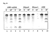
- FIG. 16 Single stranded DNA does not interfere with ASRE activity. Addition of single stranded 60 nt DNA in the reaction mixture does not inhibit ASRE activity.
- RNA substrate (7u6g) is digested with ASRE in a 10 min reaction in the presence of ss DNA (the molar ratio of RNA:DNA is 1:1 in lanes ⁇ 2 and 1:10 in lanes 3-4). Experimental controls (RNaseA and RNaseH) are also shown.
- FIG. 17 Schematic diagram of DSS-RACE. Top panel shows digestion of RNA substrate by ASRE—the 5′ portion of the product is shown in grey and the 3′ end in black. The cut site is either mapped by 3′RACE (Panel A) or 5′RACE (Panel B). A number of clones ( ⁇ 40) were sequenced for determination of cleavage sites.
- FIG. 18 Sequence at the cleavage sites as mapped by DSS-RACE.
- (Panel A) Part of the sequence near the recognition site of GU/UG RNA substrate SEQ ID NO:86) is shown with the PUF recognition site marked in bold.
- (Panel B) The 3′ end sequence of 5′ cleavage product (SEQ ID NOS:87 and 88). The major cut site is marked with an asterisk.
- Panel C The 5′ end sequence of the 3′ cleavage product (SEQ ID NOS:89 and 90). The major cut site is marked with an asterisk.
- An extra “G” residue (boxes) was usually added due to the terminal deoxynucleotide transferase activity of reverse transcriptase.
- FIG. 19 Decrease in ⁇ -galactosidase protein level in ASRE(LacZ) treated cells.
- the cell lysates from independent clones of bacteria expressing control ASRE (lanes 1-3), D1353A mutated ASRE(LacZ) (lanes 4-6, and ASRE(LacZ) were separated with SDS-PAGE by western blots with antibody against ⁇ -galactosidase (top panel) or GroEL (middle panel).
- the expression level of ASREs was determined by Comassie staining in bottom panel.
- FIG. 20 Proposed models for the non-specific cleavage of the PIN-PUF fusion protein.
- Panel A The ASRE with PUF-PIN orientation from N- to C-terminus. The PIN domain is probably curved back with the active site facing the PIN domain and RNA substrate.
- Panel B ProPanel posed model of fusion protein in PIN-PUF orientation. The active site of the PIN domain may face away from PUF, therefore it can non-specifically cleave RNA.
- Panel C An alternative proposed model of fusion protein in PIN-PUF orientation. The C-terminal residue/linker of the PUF domain is very flexible, therefore allowing RNA to access the PIN active site and be cleaved non-specifically.
- FIG. 21 (Panel A) Diagram of human mitochondrial genome. The 13 protein coding genes are shown and the tRNA genes are represented with a black or grey bar. (Panel B) Diagram of three ASREs used in experiments. (Panel C) Subcellular localization of mitoASRE and control ASREs. HeLa cells were transiently transfected with expression vectors and the ASREs were detected with anti-flag antibody. Mitotrackers were used to mark the mitochondria. (Panel D) The decrease of ND5 mRNA as judged by real time RT-PCR. (Panel E) The change of ND5 protein level measured by western blots. Samples are assayed 24 hours after tetracycline (tet) induction.
- tet tetracycline
- FIG. 22 RNA pathogenesis model of DM1.
- the RNA repeats in the 3′UTR of DMPK (SEQ ID NO:91) form a hairpin structure that sequesters MBNL1 and increases CUGBP1 level.
- FIG. 23 Engineering ASRE for specific RNA cleavage.
- the ASRE was constructed by fusing N-terminal PUF and C-terminal PIN domains with a 7 aa linker. RNA substrates containing a PUF binding site (grey box) were incubated with the purified ASRE at 37° C.
- Left panel time course of ASRE catalyzed RNA cleavage.
- Right panel two ASREs, each recognizing Wt or 7u6g targets that differ by 2-nt. ASREs specifically cleave their cognate substrate.
- FIG. 24 Identification of PUF binding code for C residue.
- PUF-Eco is a control PUF domain with an EcoRI site (equivalent of two amino acids) inserted between S and R residues. All RNA targets containing U, A, C and G in position 3 were tested for the specific binding of PUF:RNA, which was determined as the LacZ activity in the Y3H system. Higher LacZ activity reflects strong binding between RNA and protein.
- FIG. 25 Engineering PUF domains to specifically recognize (CUG) n repeats.
- the (CUG) n repeat (SEQ ID NO:144) can be recognized as 8-nt through three different frames, thus 3 possible PUF domains can be engineered.
- Panel B Step-wise mutating each PUF repeat to generate PUF domains that recognize different targets. The top row represents the starting sequences recognized by two modified PUF domains. Using site directed mutagenesis on each repeat, the PUF binding specificity was changed to recognize different intermediates, and the bottom sequences are (CUG) n frames 1 and 2 that were recognized by the PUF-D and PUF-E.
- Panel C Measurement of RNA:Protein interaction using Y3H system. The binding between wildtype PUF and its cognate RNA was used as positive control.
- FIG. 26 Effects of linker sequence and structure on ASRE activity.
- the ASRE(6-2/7-2) containing different linker sequences (SEQ ID NOS: 145-148) was incubated with its cognate substrate and the cleavage of the RNA was detected with urea-PAGE gel.
- the different linkers were chosen from the protein inter-domain linker database. Both the sequence and the PDB code were shown.
- the secondary structures of the linkers are: 1al04_1 (SEQ ID NO:145): HHHHTTCCCCCCCHHHH; 1sesA_1 SEQ ID NO:146): HHHHHHHHHHH; 1qaxA_3 SEQ ID NO:147): HHHHTTTHHH; 1pfkA_2 SEQ ID NO:148): CGGGGGCSCC.
- the structure of inter-domain linkers was determined in the natural context of respective proteins.
- the notation of secondary structure follows the standard DSSP code (H, ⁇ helix; T, hydrogen bonded turn; C, coil; G, 3 10 helix; S, bend). See Table 2 for details.
- FIG. 27 Identification of a cytosine-recognition code by yeast three-hybrid screen.
- Panel A Schematic representation of the interaction between wild-type PUF and its RNA target sequence (5′-UGUAUAUA). Protein repeats are indicated by squares and RNA bases by ovals (dashed lines, hydrogen bonds; parentheses, van der Waals contacts). For library screening, the third RNA base was mutated to cytosine (C3) and served as a new target. Nucleotides encoding positions 1043 and 1047 of the PUF were randomized in the screened library.
- the interaction between Gal4-PUF and target RNA fused with MS2 binding sequence can trigger the expression of both reporter genes, LacZ and HIS3.
- the ⁇ -galactosidase activity relative to that of the wild-type PUF-U3 RNA pair was plotted to reflect the strength of protein-RNA interaction (right).
- the fold increase in binding of the mutant protein to the cognate base vs. the non-cognate base in the wild-type RNA is indicated above the bars.
- FIG. 28 The cytosine-recognition code can be transferred to other PUM repeats.
- PUM repeat 2 Mutation of PUM repeat 2 to convert its binding specificity to recognize C7 RNA. Indicated mutations were introduced in repeat 2 (left). Protein-RNA binding measured with the yeast three-hybrid system using ⁇ -galactosidase activity is shown (right). Wild-type PUF and its cognate target RNA were included in all experiments as controls and its relative activity set to 1.
- Panel B Mutation of PUM repeat 5 to convert its binding specificity to recognize C4 RNA. Indicated mutations were introduced in repeat 5.
- PUM repeat 6 Mutation of PUM repeat 6 to convert its binding specificity to recognize C3 RNA.
- Indicated mutations including two different stacking residues, were introduced.
- FIG. 29 Designed PUF domains that recognize targets with multiple cytosines.
- PUM repeats 2 and 6 Diagram showing the mutations in PUM repeats 2 and 6 to recognize C3C7 RNA. Relative protein-RNA binding is shown as in FIG. 28 .
- Panel B Stepwise generation of a PUF mutant (PUF-D) that can bind to (CUG) n repeat RNA. Diagrams showing the mutations in PUM repeats 1, 3, 4, 5 and 6 (PUF-D) or in PUM repeats 1, 2, 3, 5, 7 and 8 (PUF-E) to recognize (CUG) n repeat RNA.
- PUM repeats 1, 3, 4, 5 and 6 PUM repeats 1, 2, 3, 5, 7 and 8
- PEF-E Relative protein-RNA binding to WT or (CUG) 5 RNA is shown as in FIG. 28 .
- FIG. 30 Crystal structure of PUF-R6(SYxxR) in complex with C3 RNA.
- Panel A Interaction of PUF-R6(SYxxR) with C3 RNA. Ribbon diagram of interaction of repeat 6 with C3 base (complex 1 with chain A and C shown).
- Panel B Interaction of wild-type PUF(NYxxQ) with U3 RNA. Ribbon diagram of interaction of repeat 6 with U3 base. RNA and base-interacting side chains are shown as stick models. Hydrogen bonds are indicated with dashed lines. This figure was created with PyMol.
- FIG. 31 Using the cytosine-recognition code to direct engineered splicing factors.
- Panel A Modulating alternative splicing of a cassette exon in a reporter RNA. Diagram of how the two types of ESFs can affect splicing of a cassette exon (left). Gly-PUF ESF directed to the exonic target can increase exon inclusion whereas the RS-PUF ESF can decrease exon inclusion. RT-PCR products of splicing reactions (top right) and quantification of splicing (bottom right). The splicing reporter gene and expression vectors for different ESFs were co-transfected at 1:2 ratio into 293T cells.
- the gene (SEQ ID NOS:93 and 94) and protein sequences (SEQ ID NOS:95 and 96) of VEGF-A in the region near the alternative splice sites are shown with two PUFs recognizing different cytosine-containing sequences.
- the cultured MDA-MB-231 cells were transfected with 1 ⁇ g expression vectors of Gly-PUF#1 or RS-PUF#2.
- Total RNA was purified 24 hours after transfection to detect VEGF-A splicing by RT-PCR. The percentages of b isoforms were quantified and are plotted below the gel.
- FIG. 32 Natural PUF proteins with putative cytosine-recognition code.
- Panel A Alignment and phylogenetic tree of the putative C-recognition PUM repeat in Nop9p homologs from yeast, plants, filamentous fungi and protists (SEQ ID NOS:97-126). The query sequences were selected to maximize the divergence of the species, but are otherwise arbitrary. The Giardia protein EES98274 was chosen as the outgroup in the phylogenetic tree.
- Panel B Alignment of the putative C-recognition PUM repeat in Nop9p homologs from the HomoloGene database (SEQ ID NOS:127-142). The homologous Volvox carteri protein (Accession No. XP_002952190) was included in the alignment as the outgroup in the phylogenetic tree. The conserved positions for cytosine recognition are highlighted.
- the transitional phrase “consisting essentially of” (and grammatical variants) is to be interpreted as encompassing the recited materials or steps “and those that do not materially affect the basic and novel characteristic(s)” of the claimed invention. See, In re Herz, 537 F.2d 549, 551-52, 190 U.S.P.Q. 461, 463 (CCPA 1976) (emphasis in the original); see also MPEP ⁇ 2111.03. Thus, the term “consisting essentially of” as used herein should not be interpreted as equivalent to “comprising.”
- the present invention provides a new class of synthetic RNA endonucleases that are analogous to DNA restriction enzymes.
- These artificial site-specific RNA endonucleases were created by combining an RNA endonuclease (e.g., the PIN domain of human SMG6, with a series of modified PUF domains specifically recognizing an 8-nt region of RNA as an RNA substrate.
- the ASRE binds this RNA substrate with high affinity and efficiently makes a single cleavage near the binding site.
- ASREs containing different PUF domains can recognize and cleave distinct substrates without detectable activity between non-cognate ASRE/RNA pairs.
- RNAi interfering RNA
- the present invention provides a synthetic RNA endonuclease comprising the formula: A-B-C, wherein: A is an RNA binding domain, B is a linker peptide, and C is a cleavage domain.
- the RNA binding domain can be a Pumilio homology domain (PU-HUD) and in particular embodiments, the PU-HUD can be a human Pumilio 1 (PUF) domain.
- the RNA binding domain can be any protein or peptide that binds RNA in s sequence specific manner.
- the RNA binding domain can be any protein or peptide that contains modular armadillo repeats (ARM repeats) and in particular can be any such modular protein that binds RNA in a sequence specific manner wherein the RNA specificity can be changed by modifying the amino acid side chain(s) of the protein.
- the RNA binding domain can be, for example any PUF protein family member with a Pum-HD domain.
- Nonlimiting examples of a PUF family member include FBF in C. elegans , Ds pum in Drosophila and PUF proteins in plants such as Arabidopsis and rice (the genes are yet to be named.
- a phylogenetic tree of the PUM-HDs of Arabidopsis , rice and other plant and non-plant species is provided in Tam et al. (“The Puf family of RNA-binding proteins in plants: phylogeny, structural modeling, activity and subcellular localization” BMC Plant Biol. 10:44, 2010, the entire contents of which are incorporated by reference herein).
- PUF family members are highly conserved from yeast to human and all members of the family bind to RNA in a sequence specific manner with a predictable code.
- accession number for the domain is PS50302 in the Prosite database (Swiss Institute of Bioinformatics) and a sequence alignment of some of the members of this family is shown in FIG. 5 (ClustalW multiple sequence alignment of human, mouse, rat Pumilio 1 (hpum1, Mpum1, Ratpum1) and human and mouse Pumilio2 (hpum2, Mpum2), respectively, is shown.
- the Drosophila pum1 is very different in length from other mammalian Pumilio1 homologues.
- the C-terminal PUF HUD domain is only shown in the sequence alignment.
- FIG. 6 shows the ClustalW amino acid sequence alignment and comparison of the Drosophila Pumilio (PumDr) and human PUM1 and PUM2 proteins.
- the N-terminal part of human and fly Pum proteins shows weak homology (40% similarity) and differs significantly in size and protein sequence.
- the C-terminal part shows a very high degree of homology and evolutionary conservation (78% identity, 86% similarity for PUM1 and 79% identity, 88% similarity for PUM2), with highly conserved protein sequence and structure of the Pum RNA-binding domain.
- PUM-HD is composed of the N-terminal conserved part of 20 amino acids, eight Pum repeats of 36 amino acids each, and the C-terminal conserved region.
- the PUF domain of this invention can be made up of eight 36 mers, in which 33 of the amino acids are conserved and the 34 th , 35 th and 36 th amino acids can vary, imparting specificity for a particular base in an RNA sequence.
- the RNA binding domain is about 300 (e.g., 310, 309, 308, 307, 306, 305, 304, 303, 302, 301, 300, 299, 298, 297, 296, 295, 294, 293, 292, 291, 290, etc.) amino acids in length.
- the RNA cleavage domain is about 120 amino acids in length.
- the RNA endonuclease of this invention is designed to bind to a specific RNA sequence of about 8 nucleotides (e.g., 8-16 contiguous RNA bases) to position the cleavage domain to cut the RNA at a specific site.
- the RNA can be cut between any of the 8 contiguous RNA bases as well as at any site upstream and/or downstream of the 8-nt sequence bound by the RNA binding domain (e.g., 1, 2, 3, 4, 5, 6, 7, 8, 9, 10 or more nucleotides upstream or downstream of the 8-nt RNA sequence).
- the fifth nucleotide of the 8-nt sequence is a U or C, while the other 7 nucleotides can vary.
- the RNA endonuclease of this invention comprises an RNA binding domain (e.g., Puf-HUD) that is modified to bind an RNA sequence that is different than the RNA sequence bound by an unmodified (e.g., wild type) RNA binding domain.
- the RNA sequence can be about an 8mer (e.g., an 8mer, 9 mer, 10mer, 11mer, 12mer, 13mer, 14mer, 15mer, 16mer, etc.).
- FIG. 7 shows the RNA recognition code of the PUF domain.
- the recognition code can be generally written as:
- the recognition code for A can be SerArgXXXGln (SEQ ID NO:5).
- RNA binding domains that can be employed in the RNA endonuclease of this invention include RNA binding domains (RBDs), such as in most splicing proteins, including heteronuclear ribonuclear proteins (HNRNP) and the K homology group of proteins (KH loop proteins); any of which can be used as the RNA binding module of the ASREs of this invention.
- RBDs RNA binding domains
- HNRNP heteronuclear ribonuclear proteins
- KH loop proteins K homology group of proteins
- the ASREs of this invention comprise a linker peptide, which can be from about three amino acids to about 20 amino acids in length (e.g., 2, 3, 4, 5, 6, 7, 8, 9, 10, 11, 12, 13, 14, 15, 16, 17, 18, 19, 20, 21, 22, 23, 24, 25, 26, 27, 28, 29, 30, etc.) amino acids).
- a particular linker peptide of this invention is VDTGNGS (SEQ ID NO:18).
- any suitable linker peptide can be used in the ASRE of this invention.
- Basic structural guidelines for selection of a linker peptide sequence are that the linker sequence is ideally rich in neutral to polar amino acids that have a slight helical propensity.
- the linker peptide forms an alpha helical structure.
- a linker peptide of this invention does not comprise a proline, a phenylalanine, a tyrosine and/or a tryptophan.
- Table 2 and FIG. 26 show various nonlimiting examples of linker peptides of this invention.
- the cleavage domain can be the PilT N-terminus (PIN) domain of SMG6.
- PIN PilT N-terminus
- any suitable cleavage domain can be used in the ASREs of this invention.
- a cleavage domain of this invention typically would not exceed 30 KDa and it would have independent activity in trans.
- the cleavage domain may also have a RNAse H/A-like fold at the active site lined by acidic residues (Asp/Glu) or His, which acts via a metal ion (divalent or tetravalent) and can cleave the phosphodiester bond in the nucleic acid backbone.
- the PIN domain of hsMG6 (EST1A; GenBank® Database Accession No. NM_017575, incorporated by reference herein; synonyms include C17orf31, KIAA0732 and SMG-6)) can be used in the ASREs of this invention.
- FIG. 1A for PIN domain structure.
- FIG. 8 shows a sequence alignment of the PIN domains of EST1A family proteins from Homo sapiens (hu_pin), Mus musculus (Ms_pin), Drosophila melanogaster (Dm_pin), and Caenorhabditis elegans (Ce_pin) (upper alignment). conserveed residues are shown by asterisk.
- the PIN domain has an RnaseH like active site fold and is also very similar in active site architecture to an Archaebacterial PIN domain.
- FIG. 9 shows the structure of the potential functional residues of the smg6 PIN domain and archaeal PIN-domain proteins. A stereoview of the residues at the putative active sites of the smg6 PIN domain is shown. Molecules A and B in the asymmetric unit are superimposed. Residues D1251, E1282, D1353, T1390, and D1392 are shown as stick models, and the four water molecules are shown as spheres. The hydrogen bonds are shown as dashed lines.
- FIG. 10 shows structurally similar proteins.
- Panel A Topology diagrams of the PIN domain of hEST1A, archaeal PIN-domain protein PAE2754, and the 50 nuclease domain of Taq polymerase. Strands are represented by arrows and helices by rectangles. The core structures of these proteins consist of a parallel ⁇ -sheet with strand order 32145. The structurally similar elements are shown in different grey shades. Asterisks indicate the residue positions at the active site of Taq polymerase and at the putative active sites of the hEST1A PIN domain and PAE2754.
- the RNA cleavage domain can include an RNAse A-like fold ( FIG. 11 ) and/or an RNAse H-like fold ( FIG. 12 ).
- RNAse A is a relatively small protein (124 residues, ⁇ 13.7 kDa). It can be characterized as a two-layer ⁇ + ⁇ protein that is folded in half, with a deep cleft for binding the RNA substrate.
- the first layer is composed of three alpha helices (residues 3-13, 24-34 and 50-60) from the N-terminal half of the protein.
- the second layer consists of three ⁇ -hairpins (residues 61-74, 79-104 and 105-124 from the C-terminal half) arranged in two ⁇ -sheets.
- the hairpins 61-74 and 105-124 form a four-stranded, antiparallel ⁇ -sheet that lays on helix 3 (residues 50-60).
- the longest ⁇ -hairpin 79-104 mates with a short ⁇ -strand (residues 42-45) to form a three-stranded, antiparallel ⁇ -sheet that lays on helix 2 (residues 24-34).
- RNase A has four disulfide bonds in its native state: Cys26-Cys84, Cys58-110, Cys40-95 and Cys65-72.
- the first two (26-84 and 58-110) are essential for conformational folding; each joins an alpha helix of the first layer to a beta sheet of the second layer, forming a small hydrophobic core in its vicinity.
- the latter two disulfide bonds (40-95 and 65-72) are less essential for folding; either one can be reduced (but not both) without affecting the native structure under physiological conditions. These disulfide bonds connect loop segments and are relatively exposed to solvent.
- the 65-72 disulfide bond has an extraordinarily high propensity to form, significantly more than would be expected from its loop entropy, both as a peptide and in the full-length protein. This suggests that the 61-74 ⁇ -hairpin has a high propensity to fold conformationally.
- the active site is lined by His 119, Lys 66, Lys 41, His 12 and His 12 (PDB ID 2aas).
- the RNase H domain is responsible for hydrolysis of the RNA portion of RNA-DNA hybrids, and this activity requires the presence of divalent cations (Mg 2+ or Mn 2+ ) that bind its active site.
- This domain is a part of a large family of homologous RNase H enzymes of which the RNase HI protein from Escherichia coli is the best characterized Secondary structure predictions for the enzymes from E.
- coli coli , yeast, human liver and diverse retroviruses (such as Rous sarcoma virus and the Foamy viruses) supported, in every case, the five beta-strands (1 to 5) and four or five alpha-helices (A, B/C, D, E) that have been identified by crystallography in the RNase H domain of human immunodeficiency virus 1 (HIV-1) reverse transcriptase and in E. coli RNase H.
- retroviruses such as Rous sarcoma virus and the Foamy viruses
- the RNA binding domain can be at the amino terminus of the endonuclease and the cleavage domain can be at the carboxy terminus of the endonuclease. In this orientation, sequence specific cleavage can be achieved. In other embodiments, the cleavage domain can be at the amino terminus of the endonuclease and the RNA binding domain can be at the carboxy terminus of the endonuclease. In this orientation, nonspecific cleavage of RNA can be achieved (see FIG. 1 , Panels B and C and Examples section below).
- the RNA endonuclease can comprise, consist of, or consist essentially the following amino acid sequences, designated ASRE(WT), ASRE(6-2/7-2), ASRE(6-2/7-2/1-1), ASRE(531), ASRE(87621), ASRE(LacZ), ASRE(3-2) and mitoASRE(ND5), which are provided as nonlimiting examples of the ESRAs of this invention, as it is readily apparent to the skilled artisan that numerous other ESRAs can be designed according to the teachings of this invention to target any RNA sequence for cleavage.
- the nomenclature of the exemplary ASREs and their respective targets are provided in Table 5.
- the present invention provides a method of detecting a target RNA in a sample, comprising: a) contacting the sample with an RNA endonuclease of this invention under conditions whereby cleavage of RNA occurs if the target RNA is present in the sample and wherein the RNA binding domain of the RNA endonuclease is modified to bind the target RNA; and b) detecting a cleavage product of the target RNA, thereby detecting the target RNA in the sample.
- the RNA endonuclease of this invention can function at about pH 7.5 (e.g., in a range of about pH 7.0 to about pH 8.0) in the presence of divalent metal cation. Activity is observed in the presence of manganese, magnesium and cobalt, with enzyme activity in the order of Mn 2+ >Mg 2+ , >Co 2+ .
- the cleavage product can be visualized on urea-polyacrylamide or denatured formaldehyde agarose gels by staining with ethidium bromide or by SYBR green dyes.
- the RNA digestion product can also be detected by radioactive methods as well as non-radioactive methods, including, e.g., DIG and cyanine dye labeled probes.
- the present invention provides a method of cleaving a target mRNA in a sample, comprising contacting the sample with the RNA endonuclease of this invention under conditions whereby cleavage of the target mRNA occurs and wherein the RNA binding domain of the RNA endonuclease is modified to bind the target mRNA, thereby cleaving the target mRNA in the sample.
- the present invention provides a method of cleaving a target mRNA in a cell, comprising introducing into the cell the RNA endonuclease of this invention, wherein the RNA binding domain of the RNA endonuclease is modified to bind the target mRNA, under conditions whereby cleavage of the target mRNA occur, thereby cleaving the target mRNA in the cell.
- the amount of cleavage of the target mRNA can be, for example, 1%, 2%, 3%, 5%, 10%, 15%, 20%, 25%, 30%, 40%, 50%, 60%, 70%, 80%, 90% 95%, 98% or 100% as compared with a suitable control.
- the RNA endonuclease of this invention can be directly introduced in various cell types using membrane penetrating peptide (aka, cell penetrating peptide). This can involve fusing the RNA endonuclease with the membrane penetrating peptide.
- membrane penetrating peptide aka, cell penetrating peptide
- the nucleotide sequence encoding the RNA endonuclease of this invention can be present in a cell transiently and/or can be stably integrated into the genome of the cell and/or the genome of the cell.
- the nucleotide sequence can also be stably expressed in the cell even without being integrated into the genome, via a plasmid or other nucleic acid construct as would be well known in the art.
- RNA endonuclease expression vectors into animals can be achieved by nanoparticles or viral vectors such as those commonly used in gene therapy and as are well known in the art.
- the present invention provides a nucleic acid molecule comprising a nucleotide sequence encoding an RNA endonuclease of this invention, as well as a nucleic acid construct (e.g., vector, plasmid, etc. comprising such a nucleic acid molecule and a cell comprising such a nucleic acid molecule and/or nucleic acid construct.
- a nucleic acid construct e.g., vector, plasmid, etc. comprising such a nucleic acid molecule and a cell comprising such a nucleic acid molecule and/or nucleic acid construct.
- a cell comprising a nucleic acid molecule, nucleic acid construct, vector and/or polypeptide of this invention is also provided herein.
- a composition comprising such a cell, nucleic acid molecule, nucleic acid construct, vector and/or polypeptide of this invention in a carrier, such as a pharmaceutically acceptable carrier is further provided herein.
- Also provided herein is a method of inhibiting (e.g., silencing) expression of a target gene in a cell comprising introducing into the cell the RNA endonuclease of this invention, wherein the RNA binding domain of the RNA endonuclease is modified to bind mRNA encoding a gene product of the target gene, under conditions whereby cleavage of the mRNA occurs, thereby inhibiting (either partially or totally) expression of the target gene in the cell.
- Expression of the target gene can be inhibited, for example, by 1%, 2%, 3%, 5%, 10%, 15%, 20%, 25%, 30%, 40%, 50%, 60%, 70%, 80%, 90% 95%, 98% or 100% as compared with a suitable control.
- the cell can be a bacterium (e.g., a pathogenic strain of a bacterium) or the cell can be in an organism, such as a parasite.
- a bacterium e.g., a pathogenic strain of a bacterium
- the cell can be in an organism, such as a parasite.
- Nonlimiting examples of a bacterium of this invention include Escherichia coli, Bacillus anthracis, Bordatella pertussis, Borrelia burgdorferi, Brucella canis, Brucella melitensis, Brucella suis, Chlamydia pneumoniae, Chlamydia trachomatis, Clostridium botulinum, Clostridium difficile, Pseudomembranous colitis, Clostridium perfringens, Enterococcus faecalis, Enterococcus faecium, Legionella pneumophila, Neisseria gonorrhoea and Yersinia pestis.
- Nonlimiting examples of a parasite of this invention include Plasmodium falciparum, Toxoplasma gondii, Leishmania donovan and Trypanosoma cruzi.
- the present invention further provides a method of cleaving a target mRNA in a mitochondrion in a cell, comprising introducing into the cell the RNA endonuclease of this invention, wherein the RNA binding domain of the RNA endonuclease is modified to bind the target mRNA in the mitochondrion and wherein the RNA endonuclease comprises a mitochondrial targeting signal sequence, under conditions whereby cleavage of the target mRNA in the mitochondrion occurs, thereby cleaving the target mRNA in the mitochondrion in the cell.
- This method can also be employed to target an RNA in the nucleus of a cell, as well as an RNA in a chloroplast of a plant, according to the methods described herein.
- RNA endonuclease of this invention comprising introducing into the cell the RNA endonuclease of this invention, wherein the RNA binding domain of the RNA endonuclease is modified to bind mRNA encoding a gene product of the target mitochondrial gene and wherein the RNA endonuclease comprises a mitochondrial targeting signal sequence, under conditions whereby cleavage of the target mRNA in the mitochondrion occurs, thereby inhibiting expression of the target mitochondrial gene in the cell.
- Nonlimiting examples of mitochondrial genes that can be targeted according to methods of this invention include NADH dehydrogenase (complex 1) genes (e.g., MN-ND1, MT-ND2, MT-ND3, MT-ND4, MT-ND4L, MT-ND5, MY-ND6), Coenzyme Q-cytochrome c reductase/Cytochrome b (complex III) (e.g., MT-CYB), cytochrome c oxidase (complex IV) (e.g., MT-CO1, MT-CO2, MT-CO3) and ATP synthase (e.g., MT-ATP6, MT-ATP8).
- NADH dehydrogenase complex 1
- MN-ND1, MT-ND2, MT-ND3, MT-ND4, MT-ND4L, MT-ND5, MY-ND6 Coenzyme Q-cytochrome c reductase/Cytochrome b
- the RNA endonuclease can be introduced into the cell via a viral vector comprising a nucleotide sequence encoding the RNA endonuclease and in some embodiments, the viral vector can be an adeno associated viral vector.
- the cell of these methods can be in an organism, which can be a mammal and in some embodiments, is a human subject.
- Additional embodiments of this invention include a method of treating dystrophia myotonica (DM) in a subject, comprising administering to the subject an effective amount of the RNA endonuclease of this invention, wherein the RNA binding domain of the RNA endonuclease is modified to bind mRNA encoding (CUG)n repeats in the 3′ UTR of DMPK to treat DM1 and/or mRNA encoding (CCUG)n repeats in intron 1 of the ZNF9 gene to treat DM2 and wherein the RNA endonuclease comprises a nuclear targeting signal sequence, thereby treating DM in the subject.
- a method can be expanded to encompass treating various trinucleotide repeat disorders (e.g., Alzheimer's disease) as are known in the art, by employing the teachings of this invention.
- the methods of the present invention can also be employed for treating infection by an RNA virus in a subject according to the teachings set forth herein.
- the terms “treat,” “treating” or “treatment” refer to any type of action that imparts a modulating effect, which, for example, can be a beneficial and/or therapeutic effect, to a subject afflicted with a condition, disorder, disease or illness, including, for example, improvement in the condition of the subject (e.g., in one or more symptoms), delay in the progression of the disorder, disease or illness, prevention or delay of the onset of the disease, disorder, or illness, and/or change in clinical parameters of the condition, disorder, disease or illness, etc., as would be well known in the art.
- an effective response or “responding effectively” means a positive or beneficial response to a particular treatment in contrast to a “lack of an effective response” which can be an ineffectual, negative or detrimental response as well as the lack of a positive or beneficial response.
- An effective response or lack of effective response i.e., ineffective response
- Effective amount refers to an amount of a compound or composition of this invention that is sufficient to produce a desired effect, which can be a therapeutic and/or beneficial effect.
- the effective amount will vary with the age, general condition of the subject, the severity of the condition being treated, the particular agent administered, the duration of the treatment, the nature of any concurrent treatment, the pharmaceutically acceptable carrier used, and like factors within the knowledge and expertise of those skilled in the art.
- an “effective amount” in any individual case can be determined by one of ordinary skill in the art by reference to the pertinent texts and literature and/or by using routine experimentation. (See, for example, Remington, The Science And Practice of Pharmacy (20th ed. 2000)).
- An exemplary dosage range for the administration to a subject of a nucleic acid molecule comprising a nucleotide sequence of this invention in the form of a viral vector can be, for example, from about 5 ⁇ 10 12 viral genomes per kg to about 5 ⁇ 10 15 viral genomes per kg.
- One of skill in the art would be able to determine the optimal dose for a given subject and a given condition.
- a method is also provided herein of detecting an RNA virus in a sample, comprising: a) contacting the sample with the RNA endonuclease this invention under conditions whereby cleavage of RNA occurs if RNA of the RNA virus is present in the sample and wherein the RNA binding domain of the RNA endonuclease is modified to bind a target RNA of the RNA virus; and b) detecting a cleavage product of the target RNA, thereby detecting the RNA virus in the sample.
- RNA virus e.g., a virus
- RNA endonuclease of this invention under conditions whereby cleavage of RNA occurs if viral RNA (e.g., of the RNA virus) is present in the sample and wherein the RNA binding domain of the RNA endonuclease is modified to bind target viral RNA; and b) detecting a cleavage product of the target viral RNA, thereby detecting viral RNA in the sample and thereby diagnosing infection by an RNA virus in the subject.
- viral RNA e.g., of the RNA virus
- RNA binding domain of the RNA endonuclease is modified to bind target viral RNA
- detecting a cleavage product of the target viral RNA thereby detecting viral RNA in the sample and thereby diagnosing infection by an RNA virus in the subject.
- an RNA virus include retroviruses, alphaviruses, flaviruses, etc., as are well known in the art.
- the present invention provides a method of identifying a strain of an RNA virus (e.g., a particular strain of an RNA virus) in a sample, comprising: a) contacting the sample with the RNA endonuclease of this invention under conditions whereby cleavage of RNA occurs if RNA of the strain of the RNA virus is present in the sample and wherein the RNA binding domain of the RNA endonuclease is modified to bind a target RNA specific to the strain of the RNA virus; and b) detecting a cleavage product of the target RNA, thereby identifying the strain of the RNA virus in the sample.
- a RNA virus e.g., a particular strain of an RNA virus
- RNA structure e.g., characterize an unknown or partially identified RNA structure
- ASREs RNA cleavage pattern
- the design of such methods, employing the ASREs of this invention, would be well known to those of skill in the art.
- the cleavage will provide a unique tool to fractionate single stranded RNA into small pieces prior to deep sequencing of RNA.
- the ASRE-mediated cleavage of this invention can also be used, for example, to distinguish single stranded RNA from double stranded RNA, and/or be used to probe the structure or RNA.
- the present invention provides a protein “restriction enzyme” of RNAs, which specifically recognizes an 8-nt RNA sequence and makes a single cleavage in the target.
- these enzymes efficiently and specifically cleave diverse RNA targets not only in vitro, but also in bacterial cells in mitochondria—any cells these data-reporter protein with target sequence. Mitochondrial target is endogenous gene.
- this invention provides new methods for in vitro detection or manipulation of RNA (e.g. generating an RNA digestion map) and for modulating (e.g., inhibiting) gene expression in organisms where interfering RNA (RNAi) machinery is not available.
- RNAi interfering RNA
- the PUF domain of human Pum1 (residue 828-1176) (SwissProt Accession No. Q14671 Pum1-human; incorporated by reference herein) and the PIN domain of human Smg6 (residue 1238-1421) (SwissProt Accession No. Q86US8; incorporated by reference herein) were amplified with PCR and joined to sequences encoding different peptide linkers designed according to amino acid propensity of known protein linkers.
- the resulting fusion proteins were cloned in expression vector pT7HTb or pET43.1b, both of which encode an N-terminal His ⁇ 6 tag; the latter also included a soluble Nus tag that can be removed by enterokinase digestion.
- expression constructs were introduced into BL21(DE3) E. coli cells and expression was induced with 0.3 mM IPTG. The bacterial cells were disrupted by sonication in lysis buffer and the recombinant proteins were purified with a Ni-NTA column (His-Gravy Trap, GE Health Care).
- Protein purity was assessed with SDS-PAGE and the proteins were further concentrated in 20 mM HEPES pH 7.0, 150 mM NaCl, 1 mM DTT, and stored at ⁇ 20° C. in aliquots supplemented with 50% glycerol. Preparations were stable for 2-3 months under these conditions.
- the ASREs were named according to the identity of their PUF domains, since most ASREs all had the same PIN domain. Each PUF domain recognizes its 8-nt target sequence in an anti-parallel fashion, with the first repeat recognizing the 8 th base and so on.
- WT PUF repeat C terminus-8 7 6 5 4 3 2 1-N terminus RNA target (5′ to 3′ ends): 5′-U G U A U A U A-3′
- RNA target 7u6g represents UugAUAUA sequence, with the mutated base shown in lower case.
- the PUF domains were named by the mutated repeats and the number of the mutated amino acid in each repeat.
- ASRE(6-2/7-2) has two mutated amino acids in each of the 6 th and 7 th repeat, causing it to recognize the 7u6g RNA. See Table 5 for more examples.
- DNA fragments containing single PUF domain binding sites were amplified by PCR and inserted between the HindIII and XbaI sites of pcDNA3.
- the resulting plasmids were linearized by overnight XbaI digestion, purified, and used as templates in an in vitro transcription reaction with T7 RNA polymerase (NEB).
- the reaction mixtures were treated further with RNase-free DNAse I (Promega) to remove template and the RNA products were purified. All ASRE digestion reactions were performed in buffers containing 20 mM HEPES (pH 8.0), 150 mM NaCl, and 10% glycerol supplemented with respective divalent metal ions. Reactions were carried out at 37° C.
- the 5′ and 3′ digestion products were extracted from denaturing urea-PAGE gels, and RACE was used to map both ends of the cleavage products.
- polyA polymerase was used to add a 10-30 nt poly-A tail at the end of the 5′ fragment, and the resulting RNA was reverse transcribed with a poly-T containing primer.
- the resulting cDNA was amplified by nested PCR, cloned in pcDNA3, and sequenced ( FIG. 17 , Panel A). To map the 5′ end of the 3′ cleavage fragment ( FIG.
- the gel extracted RNA was first reverse transcribed with a specific primer.
- the resulting cDNA was purified and elongated with terminal transferase in the presence of dATP.
- Second strand synthesis was carried out using a poly-T-containing primer and Klenow fragment (NEB). The resulting product was PCR amplified, cloned and sequenced.
- E. coli BL21(DE3) was transformed with either empty vector pET43.1b, or the vector encoding ASRE(lacZ) or control ASREs not targeting lacZ mRNA. Multiple colonies were selected in each sample to circumvent clonal variation. Protein expression of each clone was induced with 0.3 mM IPTG for 6 h at 37° C. To measure ⁇ -galactosidase activity, cells were lysed with two rounds of sonication in cold buffer and the standard ⁇ -galactosidase activity assay for all lysates was carried out in a 96 well plate format as described previously 27 . Total protein concentrations of each sample were measured using the Bradford method (Pierce) to ensure that equal amounts of total protein were used in the activity assays.
- RNA total cellular RNA was purified with a Qiagen RNeasy kit and treated with DNase I. Equal amounts of total RNA (300-500 ng) from each sample were used to make first strand cDNA by random priming (High capacity cDNA reverse transcription kit from Applied Biosystem), and the lacZ mRNA was measured with q-PCR using a SYBR green kit of Applied Biosystem. Immunoblotting with a monoclonal ⁇ -galactosidase antibody (Santa Cruz cat # sc-56394) was used to measure ⁇ -galactosidase protein levels.
- the PUF domain of human Pum1 (residue 828-1176) (SwissProt Accession No. Q14671 Pum1-human; incorporated by reference herein) and the PIN domain of human Smg6 (residue 1238-1421) (SwissProt Accession No. Q86US8; incorporated by reference herein) were amplified with PCR primer pairs 1,2 and 3,4 (Table 4) using Roche Hi-fidelity Taq DNA polymerase. DNA fragments encoding the two domains were joined with sequences encoding different peptide linkers designed according to amino acid propensity of known protein linkers.
- the cells were then harvested and disrupted by sonication in lysis buffer (20 mM sodium phosphate pH 7.4, 500 mM NaCl, 20 mM imidazole, 1 mM 2-mercaptoethanol, 1 mM PMSF). Lysates were loaded onto a Ni-NTA column (His-Gravy Trap, GE Health Care) and washed with the same buffer. Elution was obtained with a linear step gradient of imidazole from 50-300 mM and protein purity was assessed by SDS-PAGE. Imidazole was removed using a Milipore Amicon ultra centrifugal concentrator.
- the proteins were further treated with TEV (Promega) or enterokinase (NEB) to remove the His-tag or Nus-tag as per manufacturer's instructions and the fragments containing His-tag were removed by Ni-NTA. Proteins were further concentrated in 20 mM HEPES pH 7.0, 150 mM NaCl, 1 mM DTT and stored at ⁇ 20° C. in aliquots supplemented with 50% glycerol. Preparations were stable over 2-3 months at ⁇ 20° C.
- ASRE peptide linkers were designed according to amino acid propensity of known protein inter-domain linkers distinct from intra-domain loops. Residues Pro, Gly, Asp, Asn, His, Ser and Thr are preferred in intra-domain loop regions and, thus, were avoided. Thus, in some embodiments, a peptide linker of this invention can exclude Pro, Gly, Asp, Asn, His, Ser and/or Thr in any combination.
- a proline residue is highly likely in both loops and linkers; within loops, proline is usually involved in tight turns, whereas few proline turns are found in linkers. Most residues in known linkers adopt an ⁇ -helical structure, although a significant fraction of non-helical residues have a coil structure.
- linkers with slight helical propensity were chosen.
- the first two amino acids of the linkers came from the restriction site SalI used for construction of fusion proteins.
- a natural linker with known 3D structure was chosen from the linker database (fumarate reductase flavoprotein subunit; PDB 1qlaB_1, VDTGNWF, SEQ ID NO:17).
- the last two aromatic amino acids in the 12 ⁇ linker were changed to glycine and serine (VDTGNGS, SEQ ID NO:18), thus changing the helical propensity to EECHHCC (see Table 2).
- DNA fragments from plasmid pGZ3 containing various single PUF domain binding sites (25) were amplified with SmSubHF1/GUXbR1 primer pairs and cloned between HindIII and XbaI site of pcDNA3.
- the resulting plasmid DNA was linearized with overnight XbaI digestion and purified.
- the linearized DNA (0.2 ⁇ g) was used as template in a 50 ⁇ l in vitro transcription reaction containing T7 RNA polymerase (NEB) supplemented with 1 unit of Murine RNase inhibitor (NEB).
- the reactions were further treated with RNase free DNAse I (Promega) for 1 h at 37° C. and RNA products were isolated by ethanol precipitation.
- the 5′ and 3′ digestion products were extracted from denaturing PAGE gels, and RACE (rapid amplification of cDNA ends) was used to map both ends of the cleavage products.
- RACE rapid amplification of cDNA ends
- a poly-A tail was first added to purified RNA by incubating 100-300 ng RNA with polyA polymerase (NEB) and 2 mM ATP for 30 min following manufacturer's instructions ( FIG. 17 , Panel A). An average of 10-30 nt was added at the end of the RNA.
- the gel extracted RNA ( ⁇ 75 nt) was reverse transcribed using a known reverse primer GuXbR1 Table 4).
- the complementary DNA product was further purified with ethanol precipitation, and elongated with terminal transferase (3 units) in a 50 ⁇ l reaction supplemented with dATP for 30 min at 37° C. according to the manufacturer's instructions. The reaction was stopped by heating at 80° C. for 20 min and further diluted in EB buffer (Qiagen) to 0.5 ml. Second strand synthesis was carried out using a poly-T containing primer (GUHIF1) and Klenow fragment (NEB).
- GUIF1 poly-T containing primer
- NEB Klenow fragment
- the resulting product was further PCR amplified using a 3′ nested primer (GUnestedXbR1) and 5′ anchor primer (GUHIF2) harboring XbaI and HindIII site.
- the resulting product was cloned in pcDNA3 and sequenced ( FIG. 17 , Panel B).
- E. coli BL21(DE3) was transformed with either empty vector pET43.1b or vector encoding ASRE(lacZ).
- E. coli was also transformed with ASRE(87621) to serve as non-specific control and with ASRE(lacZ) having a D1353A mutation as an inactive control.
- Multiple colonies were selected in each sample to circumvent clonal variation. Each clone was grown in liquid LB medium overnight, and the saturated culture was freshly inoculated (diluted 1:1000) in LB with an appropriate antibiotic and grown up to OD 600 of 0.25-0.3 for induction. Protein expression was induced with 0.3 mM IPTG, and 5 mM Mn 2+ was also added at this point.
- Bacterial cells were harvested 6 h after induction at 37° C. for the measurement of ⁇ -galactosidase activity. Two aliquots of each sample were snap frozen in an alcohol-dry ice mixture and stored at ⁇ 80° C. for further analyses of RNA and protein levels.
- the cells were lysed with two rounds of sonication in cold buffer (100 mM KPO 4 buffer pH 7.4, 2 mM 2-mercaptoethanol and 1 mM PMSF).
- Total protein concentrations of each sample were measured using the Bradford method (Pierce) to ensure that equal amounts of total protein were used in the activity assay.
- Standard ⁇ -galactosidase activity assays were performed for all samples in a 96 well plate format as described previously (27). Briefly, a total of 1-2 ⁇ g total protein was added in 300 ul assay reaction mixtures and incubated at 37° C. for 30 min, whereupon reactions were quenched by addition of Na 2 CO 3 to 1 M.
- RNA levels frozen cells were thawed on ice and resuspended in 500 ⁇ l TE buffer (pH 7.9) with 1 mg/ml of lysozyme, and incubated at room temperature for 15 min. Two rounds of sonication were then used to lyse the cell membrane efficiently. Cell debris was pelleted by 5 min centrifugation at 15000 rpm in a microcentrifuge, and the supernatants were used for RNA purification with a Qiagen RNeasy mini kit. RNA eluted from the columns was treated with 2U of DNase I at 37° C. for 30 min (Promega), followed by heat inactivation of DNase.
- RNA 300-500 ng
- Q-PCR was performed using lacZ specific primers (Table 4) and a SYBR green kit (Applied Biosystems), and the results were calibrated to the expression level of ftsZ using gene specific primers (FtsZF1, FtsZR1 in Table 4).
- the data analysis was performed using ABI-prism q-RT PCR software.
- ⁇ -galactosidase protein level was extracted by sonication in 100 mM KPO 4 buffer pH 7.4, 2 mM 2-mercaptoethanol and 1 mM PMSF. Protein concentrations were measured by Bradford assay (Pierce) and equal amounts of total protein were loaded in each well of SDS-PAGE gels. The blots were probed using a monoclonal ⁇ -galactosidase antibody (1:1000; Santa Cruz cat. no. sc-56394) and detected using ECL-Western Blot reagent (GE Heath Care). Blots were stripped using Restore Plus western blot stripping buffer (Pierce) and re-probed with anti-GroEL antibody (1:2000; Sigma cat. no. G6532) as loading control.
- ASREs Artificial Sequence-Specific RNA Endonucleases
- RNA recognition domain a modular design was adopted by combining a target recognition domain and a catalytic domain.
- target recognition domain the unique RNA recognition domain of PUF proteins (named for Drosophila Pumilio and C. elegans fem-3 binding factor) 11 was chosen.
- PUF proteins named for Drosophila Pumilio and C. elegans fem-3 binding factor
- KH K homology
- the RNA-binding domain of human Pumilio1 contains eight repeats that recognize eight consecutive RNA bases, with each repeat recognizing a single base 13 ( FIG. 1 , Panel A, left). Moreover, two amino acid side chains in each PUF repeat recognize the Watson-Crick edge of the corresponding base and determine the specificity of that repeat 13,14 . Using this recognition code, a PUF domain can be designed to specifically bind most 8-mer RNA sequences 13,14 .
- RNA cleavage module For the RNA cleavage module, a small endonuclease domain with limited exonuclease activity was used.
- One good candidate is the PIN domain (PilT N-terminus) of SMG6, a key factor involved in nonsense-mediated decay (NMD). This domain has well-defined molecular architecture and requires only a divalent metal cation for sequence-independent RNA cleavage ( FIG. 1 , Panel A, right).
- the architecture of this design was first tested by placing the PUF domain at the N-terminus and the PIN domain at the C-terminus and vice versa.
- An ASRE was constructed by fusing an N-terminal modified PUF(6-2/7-2) containing mutations N1043S/Q1047E in repeat 6 and S1079N/E1083Q in repeat 7 to specifically bind UugAUAUA (7u6g) 14 to a C-terminal wild type PIN or a mutated PIN (D1353A mutation in PIN active site).
- the recombinant ASREs (PUF-PIN fusion proteins) were expressed in E. coli and purified to homogeneity ( FIG.
- a suitable linker is important for the high catalytic activity of ASRE.
- a glycine or Gly/Ser rich linker may be too flexible and unstable and, thus, could act as an energetic, structural, or activity-interfering nuisance, especially when it is longer than necessary to connect two domains 16 .
- a short linker may generate a structural barrier that prevents simultaneous contact of the two domains with an RNA.
- a database of known linkers revealed that both short (less than 6 amino acids) and long linkers (more than 14 amino acids) are very rare in natural linkers and average linker length varies from 8-10 amino acids 15 .
- VDT tri peptide
- VDTGNGS hepta peptide
- VDRRMARDGLVH do-deca peptide
- the purified ASRE with the tri-peptide linker had very low activity compared to the other two enzymes (lane 2).
- the enzymes had considerably higher activities with linker lengths of 7 aa or 12 aa (lanes 3 and 4).
- linker lengths 7 aa or 12 aa (lanes 3 and 4).
- non-specific cleavage products were observed at longer incubation time with the 12-aa linker ( FIG. 2 , Panel A, lane 5).
- the ASRE with hepta peptide linker was used in further studies described herein.
- the ASRE containing the heptapeptide linker completed RNA cleavage within two hours ( FIG. 2 , Panel B), and displayed strict pH selectivity with reduced reaction rate below pH 7.5 and no detectable cleavage at pH 6.0.
- the nanos response element (NRE: UGUAUAUA)
- the NRE differs from the PUF(6-2/7-2) target by two nucleotides 14 .
- this ASRE(wt) cleaved only the substrate containing its cognate target NRE (lane 3) but not the closely related RNA 7u6g (lane 2).
- the ASRE(6-2/7-2) specifically cleaved its cognate target (7u6g) but not the substrate containing NRE ( FIG. 2 , Panel C, lanes 6 and 5, respectively).
- the ASRE(6-2/7-2) failed to cleave other closely related RNAs, including UGUAUgUA (3g), UGUgUgUg (5g3g1g) and gugAUAag (8g7u6g2a1g, or 87621 in short) that vary by 3 or 6 nucleotides from the ASRE(6-2/7-2) 7u6g ( FIG. 14 ).
- UGUAUgUA 3g
- UGUgUgUg 5g3g1g
- gugAUAag 8g7u6g2a1g, or 87621 in short
- the active site of the PIN domain is lined by three conserved aspartate residues (D1251, D1353, D1392) that coordinate one divalent metal cation to activate a water molecule for nucleophilic attack of the 3′-5′ phosphodiester bonds 17,18 .
- D1251, D1353, D1392 conserved aspartate residues
- ASRE the reaction was carried out in the presence of different divalent metal ions including Mn 2+ , Co 2+ , Ca 2+ and Mg 2+ .
- optimal activity of ASRE was detected in the presence of Mn 2+ and suboptimal activity was detected in the presence of Mg 2+17 .
- ASRE also had limited activity in presence of Co 2+ ( FIG.
- ASREs showed very little to no activity with non-cognate RNA substrates, with the exception of ASRE(671) toward non-cognate 7u6g RNA (UugAUAUA).
- ASRE(671) toward non-cognate 7u6g RNA
- This non-cognate activity displayed more than a five-fold decrease in V max compared to ASRE(671)'s cognate substrate (UugAUAUg), which has a single base difference at the end of the 8-nt target.
- This non-cognate activity may be explained by the fact that the evolutionary plasticity of PUF domain allows recognition of suboptimal target sequences.
- RNA cleavage sites were identified from both 5′ and 3′ RACE which are labeled as sites 1 and 2 according to their positions in substrate.
- the two cleavage products can be matched to the same site, suggesting that an ASRE makes only a single, distinct cleavage rather than several random cleavages.
- the digested RNA products can be cloned with the DSS-RACE protocol ( FIG. 17 ), indicating that RNA cleavage catalyzed by ASRE generates a 5′ fragment with 3′ hydroxyl group and a 3′ fragment with 5′ phosphate.
- the major digestion site lies 4 bases downstream from the PUF binding site and accounts for ⁇ 80% of digestion products, whereas the minor cleavage site lies in the third position of the PUF binding 8-nt. Since PUF binds RNA in an anti-parallel fashion with the first repeat at N-terminus recognizing the 8 th position of RNA and the PIN domain is at the C-terminus of the ASRE, such a cleavage pattern is somewhat surprising. This result indicated that ASREs might form a foldback structure with the RNA substrate being bound by the one arm (PUF domain) and the PIN catalytic domain folded back to cleave the phosphodiester backbone at downstream sites ( FIG. 3 , Panel D).
- RNA can also form secondary and tertiary structure in solution adds complexity to this model. Minor cleavage sites were also determined near the PUF binding sequence that were mapped by single clones; such sites could be accounted for by experimental artifact of RNA amplification with RACE, by incomplete ASRE digestion, or by flexibility of the ASRE-RNA complex.
- RNAi interfering RNA
- Bacteria were selected because: (1) bacteria do not express endogenous Pumilio-like protein homologues, and thus are cleaner systems to start with, (2) lacZ gene expression and regulation is very well understood in E. coli , and (3) ASREs are expressed as active forms in bacterial cells.
- a modified ASRE ASRE(LacZ) (or ASRE(6g3g2a) in the nomenclature used herein) was engineered with a mutant PUF domain (repeats 2,3,6) that specifically recognizes the target sequence UGGAUGAA, which occurs twice (position 1232-1239 and 1520-1527) in the lacZ mRNA.
- BL21(DE3) cells in which expression of the LacZ is under the control of IPTG, were transformed with the expression vectors for either ASRE(LacZ) or control ASREs. As shown in FIG.
- RNA “restriction enzyme” was created. The generation of this novel enzyme and other sequence-specific derivatives enables efficient and specific cleavage of diverse RNA targets both in vitro and in vivo.
- the keys for successful generation of ASREs include the choice of a sequence-specific RNA binding domain, a suitable endonuclease domain, an optimal linker, and a correct orientation of these elements.
- These data indicate that the unique RNA recognition mode of the PUF domain renders ASREs the ability to specifically recognize a diverse panel of RNA targets without detectable cross activity between non-cognate ASRE/RNA pairs.
- These data demonstrate that the PIN domain of human SMG6 cleaves RNA at specific sites only when fused to the C-terminus of the PUF domain, not vice versa ( FIG. 1 , Panel C).
- the PIN in PUF-PIN orientation can “fold back” so that the active site faces PUF domain to specifically cleave RNA ( FIG.
- linkers with helical or helix-coil-helix structures produced the most active ASREs, whereas linkers with helix-turn-helix or 3-10 helix structures reduced the activity of ASREs.
- linker length also affects the specificity of the enzyme: non-specific cleavage became apparent with linkers approaching ⁇ 20 ⁇ in length, probably due to excess flexibility that allowed the PIN endonuclease domain to recognize and cleave any RNA.
- RNA cleavage reaction of ASREs is indeed catalyzed by the PIN domain of SMG6: (1) ASRE has the same cation preference as that of PIN, both requiring Mn 2+ for maximum activity 17 ; (2) a mutation in the active site of the PIN domain abolished ASRE activity; and (3) non-specific RNA cleavage was observed when the PIN domain had too much flexibility to recognize any RNA (either with a long linker or a reverse PIN-PUF orientation). The reaction catalyzed by ASREs was fairly efficient as judged by the k cat /K m value (>10 7 min ⁇ 1 M ⁇ 1 ).
- the Michaelis constants (K m ) of the ASREs were significantly higher than the dissociation constant of a typical PUF with its cognate sequence 14 , suggesting that the ASRE K m is determined mainly by the interaction of PIN with RNA and the catalytic rate of PIN. In fact, a tight RNA-protein interaction may lead to slow turnover of the enzyme and affect its reaction rate. Therefore, the activity of ASREs may be improved by using a catalytic domain more efficient than PIN domain.
- ASRE(lacZ) could specifically silence lacZ mRNA indicates that the ASRE can recognize its target in vivo with sufficiently high efficiency and affinity to overcome the protective effect of ribosomes.
- the co-transcriptional binding of mRNA by ribosomes in bacteria may limit low affinity off: target effects of ASREs, as only the specific ASRE with high binding affinity will compete efficiently with ribosomes.
- the silencing effect was most likely due to mRNA degradation rather than a translational block, as an ASRE with a mutation in the PIN domain active site did not affect the ⁇ -galactosidase activity.
- mRNA degradation was also confirmed by q-RT-PCR analysis. Using a similar strategy, other gene product(s) may also be targeted in organisms where RNAi does not exist.
- the present invention also includes various ways to optimize and expand ASRE usage.
- the PUF domain can specifically recognize an 8-nt sequence
- ASRE in vivo use of ASRE to silence gene expression would presumably benefit from a longer ASRE binding site that can minimize off-target effects
- ASRE to probe RNA structure or sequence may require a shorter targeting site to produce multiple cleavages in a single substrate.
- It should be possible to increase the length of recognition site by adding more PUF repeats, or by using multiple PUFs in a single fusion protein.
- the length of the recognition site may be relaxed by relaxing the specificity of some PUF repeats (i.e., making some repeats to bind all four bases equally well).
- the catalytic activity of ASRE may also be improved by using riboendonuclease domains other than PIN, or by optimizing the PIN domain active site to increase activity.
- the relative positions of the cleavage site may be affected by the conformation of the linker region; thus, testing peptide linkers with different sequences and structures will likely reduce the “star activity” of ASREs so that cleavage occurs at only a single, predictable site.
- mitochondria dysfunction is linked to various diseases such as cancer, autism and age-associated neurodegenerative diseases.
- Most mitochondrial proteins are coded by nuclear DNA and imported into mitochondria after translation.
- Mitochondria have a distinct genome, and the human mitochondrial genome contains 13 protein-coding, 2 rRNA, and 22 tRNA genes ( FIG. 21 , Panel A).
- mitochondrial gene mutations are closely linked to various diseases, the functions of these genes are hard to study because there are limited research tools to manipulate mitochondrial gene expression.
- a new ASRE with a mitochondrial targeting signal has been generated that can be used to specifically silence mitochondrial gene expression by cleaving mitochondrial RNA, making it possible to determine the function of each gene in the mitochondrial genome.
- TTTATOTG 8-nt target sequence in subunit 5 of the respiratory complex I (mtND5) gene was selected for these studies ( FIG. 21 , Panel A).
- the sequence is a unique hit in the 16.5 kb mitochondrial genome thus ensuring minimal off target effect.
- OTC ornithine transcarbamylase
- mitochondrial ASRE mitochondrial ASRE
- mitochondrial ASRE mitochondrial ASRE
- ASRE(ND5) was engineered, which lacks mitochondrial targeting signals, as well as a mutated ASRE(ND5) that contains a mutation in the active site of the PIN domain.
- the initial study was to transiently transfect HELA and HEK293 cells with N-terminal flag tagged ASREs. It was found that the mitoASRE and mutated ASRE(ND5) could be successfully translocated to the mitochondrial matrix, and the control ASRE(ND5) that lacks mitochondrial targeting sequence is mainly located in the cytoplasm.
- Real time PCR showed 30% decrease of mtND5 transcript compared to control or cells expressing inactive DA control. A concomitant decrease in protein level was also noticed as depicted by western blot analysis. Cells also showed a slow growth phenotype on expression of mtND5 ASRE.
- cells expressing DA mutant mitoASRE(ND5) showed slightly increased amounts of mtND5 mRNA, likely because the inactive mitoASRE(ND5) can protect its target from natural mRNA turnover when binding to the target.
- Examination of ND5 protein level by western blot also showed that the mtND5 protein was decreased in cells expressing mitoASRE(ND5) but not mutated mitoASRE(ND5) ( FIG. 21 , Panel E). Induction of mitoASRE(ND5) reduced growth rate of cells.
- RNA interference RNA interference
- a 32 aa mitochondrial targeting signal from ornithine transcarbamylase leader peptide (MLFNLRILLNNAAFRNGHNFMVRNFRCGQPLQ, SEQ ID NO:71) was used as a mitochondrial targeting signal for ASRE ( FIG. 21 , Panel B).
- Tetracycline inducible stable lines were produced that expressed mitoASRE(ND5) or a catalytically inactive version cloned between HindIII and NotI sites in a pCDNA5/FRT/TO construct. This construct was co-transfected with pOG44 plasmid to facilitate integration into an FRT locus of Flp-In T-Rex 293 cells (Cat no. K6500-01, Invitrogen).
- Stably integrated cells were selected using DMEM medium with 10% FBS and 100 ug/ml of hygromycin B. Cells were then passed for 8 generations on 60 mm dishes for stability and creation of isogenic lines. To induce mitoASRE expression, cells were re-plated in fresh DMEM medium with 10% FBS without any drug and induced with tetracycline (6-10 ug/ml). Twenty-four hour post induction expression of ASRE was checked on western blot using anti-flag antibody (M2 F1804, Sigma). Growth curves and viability assays were performed at different time points post induction. Briefly, cells were dislodged with trypsin after different growth times, mixed with 0.4% trypan blue (1:1) and counted. The proliferation assays were performed using a WST-1 assay kit (Roche, Cat. no. 11 644 807 001).
- RNA levels were measured by real time quantitative PCR using QPCR SYBR Green Low Rox mix (Thermo Scientific, Cat. no. AB-4322/A) using gene specific primers and normalized against GAPDH gene expression.
- QPCR SYBR Green Low Rox mix Thermo Scientific, Cat. no. AB-4322/A
- Protein was measured using Bradford reagent (Pierce).
- Myotonic dystrophy (dystrophia myotonica, DM) is the most common form of muscular dystrophy in adults that affects 1 in 8500 individuals worldwide.
- the genetic mutations responsible for DM were identified as the expanding (CUG) n repeats in the 3′ UTR of DMPK mRNA (for DM1) or the (CCUG) n expansion in the intron of ZNF9 (for DM2).
- CUG expanding RNA repeats
- CCUG CCUG n expansion in the intron of ZNF9
- the present study provides a novel approach to target and cleave the toxic RNA repeat with the artificial site-specific RNA nucleases (ASREs) as described herein.
- ASREs RNA binding module
- PAF domain RNA binding module
- PIN domain of SMG6 endoribonuclease domain
- ASREs that can specifically bind and cleave expanding RNA repeats in the cell nucleus where the toxic RNAs are accumulated are described herein.
- the present study focuses on DM1, which is the most common and severe form of DM, while it is understood that similar strategies can be developed to treat DM2.
- the native PUF domain contains 8 PUF repeats, each specifically recognizing one RNA nucleotide through hydrogen bonding with the base A, U or G.
- the modular “binding code” of the PUF repeat to C nucleotide has been identified, using a yeast-3-hybrid screen, making it possible to engineer a PUF domain that specifically recognizes any 8-nt RNA.
- PUF domains will be designed and engineered that specifically recognize all three possible 8-nt sequences in a (CUG) n repeat. The affinity and specificity of RNA-protein interaction will be determined with various assays to select for a modified PUF domain that binds a (CUG) n repeat with high affinity.
- Such PUF domains will be used either as the RNA binding modules of ASREs, or as competitors of the toxic RNA repeats that sequester endogenous RNA binding proteins in DM1 patients.
- PUF domains that specifically recognize (CUG) n will be used to generate novel ASREs.
- a nuclear localization sequence will also be included in the ASRE to direct the enzyme into the nucleus where the RNA repeats accumulate.
- the specific cleavage of (CUG) n repeats will be analyzed in vitro and in cultured DM1 cells. Experiments will be done to determine if expression of ASRE can reduce the number of ribonuclear aggregates caused by (CUG) n , and to further determine if the normal expression level and localization of muscleblind-like 1 (MBNL1) and CUG-binding protein 1 (CUGBP1) can be restored by ASRE.
- MBNL1 muscleblind-like 1
- CUGBP1 CUG-binding protein 1
- ASREs will also be tested for the ability to reverse the mis-splicing of genes affected in patients, using cultured DM1 cells (such as CIC-1, cTnT and SERCA1).
- cultured DM1 cells such as CIC-1, cTnT and SERCA1.
- mRNA-seq will be used to determine (i) if normal gene expression and alternative splicing pattern can be restored at a genomic scale by the designer ASRE in DM1 cells, and (ii) the off-target effect of ASRE treatment.
- Adeno-associated virus (AAV) vectors will be used as a gene delivery tool to express ASRE in a DM1 mouse model (HSA LR mice), and to further test if ASRE can relief the DM1 phenotype in muscle and heart.
- the expression of ASRE will be analyzed in different muscle tissues and studies will be carried out to determine if the nuclear MBNL1 sequestration can be released.
- the splicing and the function of key DM1 marker genes (such as CIC-1, cTnT and SERCA1) will be examined to determine if they can be restored in muscles, and if the myopathy phenotype of DM1 mice can be reduced.
- DM Myotonic dystrophy
- CCG dystrophia myotonica-protein kinase gene
- ZNF9 Zinc finger protein 9
- (CUG) n repeats specifically bind to splicing regulatory proteins, forming RNA-protein complexes that accumulate within the nucleus.
- Two classes of splicing factors are known to be affected by (CUG) n ( FIG. 22 ; Ref. 1): (i) The members of muscleblind-like family (e.g., MBNL1) are sequestered in the nuclear foci, resulting in nuclear depletion and loss of function (8-10); (ii) The CUG binding proteins (e.g., CUGBP1) are up-regulated in DM1 cells through a PKC-mediated phosphorylation event that stabilizes the protein (11,12).
- AAV vectors can effectively deliver genes in muscle and heart (21) and the loss of MBNL1 is a primary pathogenic event in DM1 mice (20).
- AAV vectors can effectively deliver genes in muscle and heart (21) and the loss of MBNL1 is a primary pathogenic event in DM1
- this method produced encouraging results in a mice model (20).
- the strength of such protein-based approaches is to use gene therapy tools (like AAV) to achieve efficient delivery.
- gene therapy tools like AAV
- AON antisense oligonucleotide
- CCG cleave n repeats using RNAi (22), short antisense oligos (23) or ribozymes (24).
- AON antisense oligonucleotide
- RNAi 22
- short antisense oligos 2
- ribozymes 24
- AON based method is to use a morpholino oligonucleotide that blocks the (CUG) n from binding to proteins.
- the present study employees a new therapeutic strategy using an artificial site-specific RNA endonuclease (ASRE) to specifically cleave (CUG) n repeats.
- ASRE site-specific RNA endonuclease
- CUG cleave
- Engineering of such a novel enzyme takes advantage of the unique RNA recognition mode of PUP proteins, whose functions involve mediating mRNA stability and translation (29).
- the PUF domain of human pumilio 1 contains 8 repeats that bind consecutive bases in an anti-parallel fashion, with each repeat recognizing a single base (30) ( FIG. 7 ).
- Each PUF repeat uses two amino acids to recognize the edge of the corresponding base and a third amino acid (Tyr, His or Arg) to stack between adjacent bases, causing a very specific binding between a PUF domain and an RNA.
- a PUF domain can be modified to bind most 8-nt RNA (30,31).
- This unique binding mode makes PUF a programmable RNA-binding domain that can be used in various artificial factors for specific splicing modulation or RNA detection (32,33).
- ASREs have been designed and engineered to specifically cleave RNA with an 8-nt PUF target, and in the present study, ASREs will be generated that recognize (CUG) n with high affinity and efficiently cleave the RNA repeats.
- Novel RNA restriction enzymes will be engineered to specifically cleave RNAs.
- the simple protein enzyme that cleaves RNA in a sequence-specific manner has not been found in nature, and the known RNases either cleave their targets through recognition of certain structures (e.g., RNase III family, RNase H or most ribozymes) (34-36) or have essentially no cleavage specificity (e.g., RNase A or RNase T1) (37).
- RNase III family, RNase H or most ribozymes 34-36
- RNase A or RNase T1 essentially no cleavage specificity
- Development of ASREs will have broad applications for in vitro RNA manipulations, and will make gene silencing possible in organisms lacking RNAi and/or in cellular compartments where RNAi machinery does not function (such as in mitochondria).
- ASREs combine the strength of current DM1 therapies and overcome some of their limitations.
- the approaches under investigation are gene therapy and antisense oligonucleotide (AON) based methods. While gene delivery with AAV is quite efficient in muscle and heart, gene therapy does not allow for the targeting of toxic RNA but rather restores some but not all genes affected in DM patients.
- AON methods can target toxic RNA directly but have delivery problems.
- the present study provides a new approach using ASREs that target RNA directly and can be delivered into muscle and heart, for example, with AAV vectors.
- the designer ASREs can directly recognize and cleave (CUG) n repeats like AON does, and can be delivered to the muscle and heart of DM1 patient with vectors such as AAV vectors.
- CAG cleave
- ASREs will be directed into the nucleus with NLS to specifically cleave (CUG) n repeats.
- the (CUG) n repeats accumulated in the nucleus are known to be the pathogenic molecules of DM1.
- a small amount of cytoplasmic DMPK with (CUG) n is needed for normal function of DMPK, targeting only the (CUG) n in nuclear foci may be more specific and beneficial.
- AAV vectors are proven to be efficient in delivering genes to muscle and heart, and are currently tested in human trails. Therefore this approach can be readily transferred to an animal DM1 model and human patients.
- RNA restriction enzyme that cleaves RNA in a sequence-specific manner has not been found in nature.
- a modular design was used by combining a target recognition domain and a catalytic domain.
- the unique RNA recognition domain of human PUM1 was used as it is possible to “reprogram” the binding specificity of the PUF domain to recognize most 8-nt RNA.
- a short peptide linker was used to link a designer PUF with a small endonuclease domain, the PIN domain of human SMG6 that has a well-defined molecular architecture and requires only a divalent metal cation for RNA cleavage (39,40).
- the resulting ASREs were expressed and purified from an E. coli expression system and incubated with RNA substrate containing the recognition sequences. ASREs were found to efficiently recognize and cleave RNA substrates with cognate binding sites ( FIG. 23 ), and can distinguish between substrates differing by 2-nt at recognition site ( FIG. 23 , two ASREs with different recognition sites were used).
- the ASRE can be used to specifically silence gene expression by directly targeting the corresponding RNA.
- An ASRE was designed to target the LacZ gene in bacterial cells, and it was shown that ASRE(LacZ) can indeed induce specific mRNA degradation and gene silencing, whereas control ASRE or ASRE with a mutated PIN domain did not affect gene expression ( FIG. 4 ).
- This method is particularly useful to specifically cleave RNA in cell compartments where RNAi is not functional (e.g., in the mitochondrion and/or nucleus), and it was found that a specifically designed ASRE can be used to selectively degrade the mRNA of a mitochondrial-encoded gene ND1.
- the challenge of making an ASRE to cleave a (CUG) n repeat is to identify the C binding code of the PUF repeat to engineer specific PUF domains for (CUG) n sequence.
- the specificity of each PUF repeat is determined by the two residues at positions 16 and 20 in that repeat (31).
- a yeast three-hybrid (Y3H) screen was used to determine the specific amino acid combination that enables the PUF domain to recognize C (41).
- the design of the Y3H screen is similar to the Y2H screen except that an RNA molecule is used as an adaptor of molecular interaction ( FIG. 24 , Panel A).
- the specific binding of the PUF domain to the RNA adaptor can recruit the transcriptional activation domain and turn on the expression of reporter genes.
- the wild-type PUF specifically recognizes Wt RNA sequence (UGUAUAUA), and the third position of this target was changed to C (UGCAUAUA; U3C RNA) and amino acids responsible for base recognition in the PUF repeat 6 were randomized.
- the wild-type PUF can recognize WtRNA to activate LacZ gene ( FIG. 24 , Panel B), whereas the single mutation U3C completely abolished the binding of WT PUF and U3C RNA, giving a very low background for the screen.
- the corresponding sequences coding for the repeat 6 of PUF domain ( FIG. 24 , Panel C) were randomized and cloned into the Y3H system to screen for yeast colonies that grew on His depleted plates.
- the positive clones were reconfirmed by the expression of the LacZ gene, and the plasmid DNAs were purified from the double positive clones and were further sequenced to identify the amino acid combination in PUF repeat 6.
- a total of 19 positive clones were sequenced and it was found that all have the same amino acid combinations (Ser in position 16 and Arg in position 20).
- all the triplet codes for Ser and Arg were found in the sequencing results, suggesting that the screen has good coverage for the random sequence space.
- the modified PUF domain binds most strongly to RNA with a C in the third position (C3) as judged by the LacZ activity of yeast strain ( FIG. 24 , Panel D), whereas the binding between other bases was either not detectable (for A or G) or very weak (for U).
- the wild type PUF only binds to U3 RNA but not the other sequences, and a modified PUF with two amino acid inserted between Ser and Mg of repeat 6 (PUF-Eco) does not bind to any of the target RNAs ( FIG. 24 , Panel D).
- PUF-Eco a modified PUF with two amino acid inserted between Ser and Mg of repeat 6
- the modular structure of the PUF domain enabled the programming of the specificity of each PUF repeat independently.
- the binding code is indeed “modular,” which will enable the design of a PUF domain that specifically recognizes any 8-nt RNA in (CUG) n repeat.
- the (CUG) n repeat can generate three different RNA octamers (CUGCUGCU, UGCUGCUG and GCUGCUGC) according to different frames ( FIG. 25 , Panel A).
- each PUF domain can be engineered, each recognizing one of the possible 8-nt in (CUG) n .
- the binding affinity of PUF:(CUG) n will be assayed either with the Y3H assay (by measuring the ⁇ -gal activity (42)), or with a standard electrophoretic mobility shift assay (EMSA) using purified PUFs.
- FIG. 25 Panel B shows two examples of PUF domains (PUF-D and PUF-E) that were step-wise mutated in each PUF repeat to recognize frame 1 and 2 of (CUG) n .
- the starting PUF domains were generated for other reasons to recognize different sequence, and 1-3 PUF repeats were mutated in each step to change the binding specificity of that repeat (indicated with arrows in FIG. 25 , Panel B). This process will generate several intermediate PUF domains (PUF-A to C) that recognize different 8-nt targets.
- the intermediate PUFs and final PUFs were co-transfected with (CUG) 5 RNA into the Y3H system, and assays were carried out to detect binding between different PUFs to the (CUG) 5 RNA which contains only a single copy of the 8-nt target. It was found that only the PUF-D and PUF-E, not the intermediate PUFs (PUF-A to C) or the wt-PUF, can bind (CUG) 5 to mediate LacZ expression ( FIG. 25 , Panel C). The binding between designer PUFs to the (CUG) 5 repeat is very specific, with much higher affinity than the binding between WT-PUF to its target as judged by LacZ expression ( FIG. 25 , Panel C). This experiment demonstrated that the newly identified “C code” could indeed be used to engineer PUFs.
- PUF domains will be generated that bind to the third frame of (CUG) n .
- a determination will also be made of the PUF:(CUG) n binding stoichiometry, which may depend on RNA length. Studies will be conducted to examine if the designer PUF domains can bind to control sequences such as (GUC) n and (CAG) n that form similar structures but have different 8-nt sub-sequence.
- PUF domains will be generated that can be used either as the RNA binding modules of ASREs, or as competitors of the endogenous MBNL1 sequestered by the toxic RNA repeats.
- ASREs will be constructed with an N-terminal PUF domain and C-terminal PIN domain linked by a short linker ( FIG. 23 , Panel A).
- a nuclear localization signal (NLS) that directs the ASRE to the nucleus and a His-tag for detection/purification will be included.
- the NLS from SV40 large T-antigen (PKKKRKV, SEQ ID NO:72) will be used, which does not interfere with functions of PUF fusion proteins (33).
- At least three designer ASREs will be engineered with PUF domains recognizing all 8-nt sequences in (CUG) n repeats ( FIG. 25 , Panel A).
- the RNA products will be separated with denatured PAGE gel to access the cleavage results.
- the catalytic efficiencies of new ASREs will be compared to the previous ones that have k cat /K M in the range of ⁇ 10 7 M ⁇ 1 min ⁇ 1 . Since previous ASREs made a single cleavage in RNA substrate with one PUF binding site, studies will be conducted to examine how the new ASREs will cut (CUG) n containing multiple target sites.
- the amino acid combination that can make the PUF repeat recognize C has been identified. Since the native PUF domains were never found to be able to recognize C, the Y3H screen makes it possible to engineer an RNA recognition domain for any 8-nt target. However, the binding selectivity against other bases may not be optimal, and it was found that the newly modified PUF can recognize a U residue in the third position with a low affinity ( FIG. 24 , Panel D).
- the binding specificity will be optimized by molecular modeling and mutagenesis.
- the PUF-RNA interface near the C nucleotide will be modeled using the PUF structure and Resetta software platform (43). The goal is to identify additional PUF mutations that decrease affinity to A, U and G while maintaining the affinity to C nucleotide.
- Mammalian expression vectors of designer ASREs will be generated to test if they can cleave (CUG) n inside cultured cells.
- Cultured muscle cells from transgenic DM1 mouse (HSA LR mice; University of Rochester) and human DM1 fibroblasts (Coriell Cell Repositories) will be used.
- the cultured cells will be transfected with either transient or stable expression vectors containing designer ASRE genes, and assayed to determine (i) the expression and nuclear localization of ASREs, (ii) if the ASREs can disperse the nuclear foci of (CUG) n , (iii) if the (CUG) n RNA is degraded, and (iv) if the MBNL1 are released from the nuclear foci by ASREs.
- Western blot will be used to examine if the level of CUGBP1 will be decreased in the ASRE transfected cells.
- RT-PCR will be used to examine the alternative splicing events in genes that are affected by (CUG) n in DM1 patients. Focus will be on splicing of CIC-1, insulin receptor, cTnT and SERCA1, all of which are known to undergo splicing shift in DM1 cells to produce embryonic isoforms. The reversal of the splicing by designer ASREs in cultured DM1 cells is the expected outcome.
- HTP sequencing of total mRNA will be carried out using mRNAs from DM1 cells treated with designer ASREs, control ASRE and empty vector. Changes in both total gene expression and ratios of splicing isoforms will be determined.
- a reliable protocol was used to generate a directional cDNA library for HTP sequencing. It was possible to obtain >20 million reads of 75 nt in a single illumina sequencing lane, and the Bowtie-TopHat pipeline was used to analyze the alternative splicing events.
- the changes of each alternative splicing event between different samples can be scored using the fisher's exact test (44).
- the mRNA-seq data will be compared with the previous splicing microarray result in DM1 mice (13), and a determination will be made regarding whether the same set of alternative splicing events can be reversed by designer ASREs.
- the (CUG) n repeat will be the best substrate of the designer ASRE as it contains many copies of ASRE targets.
- Other nuclear RNA may be cleaved to a lesser extent, and mRNA in the cytoplasm will largely evade cleavage. Therefore a large off-target effect is not expected.
- a computer will be used to scan the mRNA for the ASRE target, and a combination of multiple ASREs that target different 8-nt in (CUG) n will be used to minimize the off-target effect.
- Using multiple ASREs at a lower level will decrease the off-target effects as only the (CUG) n can be targeted by all designer ASREs.
- ASRE in vivo efficacy of ASRE will be determined in a animal model of DM1.
- viral vectors will be used to deliver ASREs into DM1 mouse model (HSA LR mice) and tests will be done to determine if the ASREs can correct the alternative splicing of key genes and relieve the DM1 phenotype in muscle and heart.
- ASRE gene Because of the relatively small size of the ASRE gene ( ⁇ 1.5 kb), it can be packaged with an AAV vector that has been shown to efficiently deliver genes into skeletal muscles and heart (21,45,46).
- ASREs that can effectively cleave (CUG) n repeat in vitro will be cloned into different AAV vectors.
- ASREs that recognize different RNA sequences and the ASRE with mutated active site in the PIN domain will be used.
- Two types of promoters will be tested to drive the expression of ASRE in AAV: the first is the CMV promoter that is commonly used in AAV systems for high level expression in all tissues, the second type are muscle-specific promoters such as myogenin promoter and/or synthetic muscle promoters (47).
- the expression efficiency of the two types of promoters will be analyzed in cultured skeletal muscle cells, and the muscle-specific promoters will be selected if the expression level is comparable to CMV.
- the goal is to make ASRE expression more specific in muscles, therefore limiting the possible off-target effects.
- AAV particles Upon confirming expression in muscle cells, AAV particles will be generated that contain ASREs.
- a DM1 mouse model HSA LR mouse
- CCG non-coding
- AAV serotype 9 AAV9
- AAV9 which can mediate high level gene expression in muscle and heart by either systematic injection or local injection (48, 49) will be used. It is expected that the the muscle tissues near the injection region will stably express ASREs 1-2 weeks after injection, leading to the cleavage of (CUG)n repeats.
- the muscle fibers will be dissected and assayed for the accumulation of (CUG) n repeat with fluorescence in situ hybridization (FISH). Assays will also be done to determine the distribution pattern of MBNL1 that is accumulated as nuclear foci in HSA LR mouse but has a dispersed nuclear localization pattern in normal mouse. The level of other proteins (such as CUGBP1) whose expressions have changed in DM1 patients by (CUG) n repeats will also be measured, and it will be determined if the ASRE can restore normal expression levels of these proteins.
- CUA fluorescence in situ hybridization
- RNA will be purified from the muscle tissues in the injected region and RT-PCR will be used to assay for the alternative splicing pattern of key genes that were affected in HSA LR mouse (including ClC-1, cTnT and SERCA1). It is expected that the splicing pattern of these genes will be shifted towards the normal adult isoforms by ASREs compared to the controls.
- AAV treatment can rescue the physiological deficits of DM1
- the expression and function of muscle-specific chloride channel (ClC-1) will be examined.
- EMG electromyography
- ASRE expressing virus AAV9
- HSA LR mice HSA LR mice
- ASREs ASRE expressing virus
- (i) assays for the accumulation of (CUG) n repeats will be carried out with FISH to determine if ASRE can disperse the nuclear foci; (2) assays for the distribution pattern of MBNL1 and the expression levels of CELF proteins will be carried out; (3) assays for alternative splicing patterns for key genes that were affected in HSA LR mouse (including ClC-1, cTnT and SERCA1) will be carried out; and (4) assays to determine whether AAV treatment can rescue the physiological deficits of DM1, including the electrophysiological properties of muscle-specific chloride channel (ClC-1) and the myotonia phenotype of muscles will be carried out.
- PUF proteins possess a recognition code for bases A, U and G, allowing designed RNA sequence specificity of their modular Pumilio (PUM) repeats.
- PUM Pumilio
- recognition side chains in a PUM repeat for cytosine are unknown. Described herein is the identification of a cytosine-recognition code by screening random amino acid combinations at conserved RNA recognition positions using a yeast 3-hybrid system. This C-recognition code is specific and modular, as specificity can be transferred to different positions in the RNA recognition sequence.
- a crystal structure of a modified PUF domain reveals specific contacts between an arginine side chain and the cytosine base.
- the C-recognition code was applied to design PUF domains that recognize targets with multiple cytosines and to generate engineered splicing factors that modulate alternative splicing.
- a divergent yeast PUF protein, Nop9p was also identified, that may recognize natural target RNAs with cytosine. This work furthers understanding of natural PUF protein target recognition and expands the ability to engineer PUF domains to recognize any RNA sequence.
- RNA regulation including splicing, localization, translation and degradation. Such recognition may be directed toward unstructured RNA requiring discrimination of RNA sequences, folded RNA motifs, or some combination of sequence and structural specificity (1).
- PUF protein family named after Drosophila Pumilio and Caenorhabditis elegans fem-3 mRNA binding factor [FBF]
- BFBF mRNA binding factor
- PUF proteins generally recognize the 3′-UTR of their target mRNAs to control the mRNA stability and translation (2-7).
- RNA-binding domain of PUF proteins known as the Pumilio homology domain (PUM-HD) or PUF domain
- PUM-HD Pumilio homology domain
- the PUF domain of human Pumilio1 contains eight PUM repeats, each containing three ⁇ -helices packed together in a curved structure (8-10). RNA is bound as an extended strand to the concave surface of the PUF domain with the bases contacted by protein side chains.
- each PUM repeat recognizes a single RNA base through the second helix ( ⁇ 2) in an antiparallel arrangement, i.e., nucleotides 1-8 are recognized by PUF repeats 8-1, respectively.
- the ⁇ 2 helices of PUM repeats contain a five-residue sequence, designated here as 12xx5, where the side chain at position 2 stacks with the recognized base and the side chains at positions 1 and 5 recognize the edge of the base (8,11) ( FIG. 27 , Panel A). Specific residues at these positions direct the base recognition properties of the repeat.
- This PUF-RNA recognition code makes it possible to modify a PUM repeat to bind a particular RNA base, producing a designed PUF domain that specifically recognizes a given 8-nt RNA target.
- Such de novo designed RNA binders have been used to track RNA localization in cells (12,13), study PUF protein function (14,15), and modulate alternative splicing (16) and continue to provide a useful tool for biomedical research with possible therapeutic applications.
- RNA-interaction sequence SYxxR SEQ ID NO:4
- PUF human Pumilio 1
- the arginine side chain interacts directly with the cytosine and the serine side chain helps to position the arginine residue.
- This recognition code is applied as described herein to design new PUF domains to recognize RNA targets with multiple cytosine residues such as CUG repeats that are responsible for the pathogenesis of myotonic dystrophy.
- the code is also used to engineer splicing factors that modulate alternative splicing of both a splicing reporter and an endogenous gene.
- a naturally occurring yeast PUF protein, Nop9p appears to contain a repeat with a code for cytosine and is conserved in homologs from yeast to human, suggesting that the natural target sequences of these PUF proteins may contain cytosine ( FIG. 32 ).
- a PUF mutant library was generated through three PCR amplifications using primers with randomized regions.
- reaction 1 the 5′ portion of the Pumilio 1 PUF domain was amplified from wild-type PUF with primers Bam-Puf-1F (5′-GGA TCC GAG GCC GCA GCC GCC ITT TGG AA, SEQ ID NO:74) and Puf-R6N-2R (5′-GAT TAC ATA NNN TCC ATA TTG ATC CTG TAC CAG, SEQ ID NO:75).
- the 3′ portion of the PUF domain was amplified with primers Puf-R6N-1F (5′-TAT GTA ATC NNN CAT GTA CTG GAG CAC GGT CG, SEQ ID NO:76) and Puf-Xho-2R (5′-CTC GAG CCC CTA AGT CAA CAC CGT TCT TC, SEQ ID NO:77).
- the Puf-R6N-2R contains 3 random nucleotides encoding the amino acid at position 1043 while Puf-R6N-1F contains random nucleotides encoding the residue at position 1047.
- the purified PCR products of reactions 1 and 2 were mixed as the template for reaction 3 with primers Bam-Puf1-1F and Puf-Xho-2R.
- the final PCR products encode the entire PUF domain and have the two randomized codons at positions 1043 and 1047.
- Yeast expression plasmid encoding wild-type PUF fused at the N terminus to the Gal-4 AD was created by amplification of the coding sequence of the PUF domain from pTYB3-HsPUM1-HD (9) and subcloned into the pACT2 plasmid using BamHI and XhoI sites. Plasmids expressing target RNAs were made by annealing DNA oligonucleotides encoding the desired RNAs and subcloning into the pIIIA-MS2-2 plasmid using SmaI and SphI restriction sites.
- Y3H assays were performed in yeast strain YBZ-1 as described previously (18,19).
- a yeast library screening system was generated directly through gap-repair.
- the pIIIA-MS2-2 plasmid carrying UGCAUAUA RNA was transformed and expressed in yeast strain YBZ-1.
- an EcoRI site was introduced by site-directed mutagenesis into wild-type pACT2-PUF between the nucleotides encoding positions 1043 and 1047.
- the pACT2-PUF-Eco DNA was linearized by EcoRI and co-transformed with the random PUF PCR library at a molar ratio of 6:1 into the yeast.
- yeast transformants were screened on plates lacking histidine and containing 10 mM 3-AT. The transformants that survived HIS growth selection were confirmed with LacZ expression. Selected yeast plasmid DNAs were sequenced and reintroduced into the mother strain to confirm the interaction and specificity.
- Additional PUF site-directed mutants carried by pACT2 were generated using the QuikChange site-directed mutagenesis kit (Agilent).
- the pTYB3-PUF mutants for in vitro protein expression were created by PCR amplification from yeast expression plasmids and subcloning into the pTYB3 plasmid using NcoI and SapI restriction sites.
- plasmids encoding the RS-PUF or Gly-PUF fusion proteins (16) were mutated.
- the activity of ⁇ -galactosidase was measured using 96-well plates using 12 clones from each sample (20). The yeast colonies were randomly picked and inoculated into 12 different wells with 100 ⁇ l culture media in a 96-well plate. After overnight growth in 24° C. with shaking, the culture density of each well was determined by reading OD 650 with a plate-type spectrophotometer (spectroMAX PLUS from Molecular Devices).
- RNAs were generated by in vitro transcription and purified on denaturing gels. 50 pmol of RNAs were labeled at the 3′ end with biotinylated cytidine bisphosphate using T4 RNA ligase following manufacturer's directions (Thermo Scientific Pierce RNA 3′ End Biotinylation Kit).
- RNA labeled RNA
- binding buffer 10 mM HEPES, pH 7.3, 20 mM KCl, 1 mM MgCl 2 , 1 mM DTT, and 0.1 g/L tRNA
- the binding reactions were separated by electrophoresis on 6% nondenaturing PAGE run with 1 ⁇ TBE at 4° C., transferred to nylon membranes, and crosslinked to the membrane by UV.
- Biotin-labeled RNA was detected by chemiluminescence using the Thermo Scientific LightShift Chemiluminescent RNA EMSA Kit following manufacturer's directions.
- RNA oligonucleotide was obtained from Dharmacon (Lafayette, Colo.).
- the protein-RNA complex was prepared by mixing a 1:1.1 molar ratio of purified protein (3.5 mg/ml) and RNA in a buffer containing 20 mM Tris-HCl, pH 7.5; 100 mM NaCl; and 1 mM DTT.
- Human embryonic kidney 293T cells or breast cancer MDA-MB-231 cells were grown in Dulbecco's modified Eagle's medium supplemented with 10% fetal bovine serum. Cells were seeded onto 24-well plates and transfected with Lipofectamine 2000 following manufacturer's directions. The purification of total RNA and semi-quantitative RT-PCR were carried out as described previously (16).
- the Saccharomyces Genome Database was also searched using the BLASTP with the following queries: (1) the two natural yeast PUF repeats containing a possible C-recognition code, and (2) all the yeast PUF repeats in which the native RNA recognition motifs were replaced with SxxxR (SEQ ID NO:143). Only the two PUF repeats from Puf2p and Nop9p were identified.
- a yeast 3-hybrid (Y3H) system was used that utilizes co-expression of the PUF domain fused with the Gal-4 activation domain (AD), an RNA target with an MS2 binding site, and an MS2-LexA fusion protein ( FIG. 27 , Panel B).
- This system can be used to reliably measure the relative binding affinity between RNA and protein (18,19).
- a uridine-to-cytosine mutation was introduced at the 3 rd position of a wild-type PUF target sequence ( FIG. 27 , Panel A).
- a PUF domain library was generated with random sequences at the 1 st and 5 th positions of the RNA-interaction motif in repeat 6, which recognizes the third position of the RNA target sequence ( FIG. 27 , Panels A and C).
- U3 wild-type PUF and its cognate target sequence
- wild-type PUF cannot recognize the target RNA with a cytosine at the third position (C3), suggesting that the screen has a low false positive background ( FIG. 27 , Panel D).
- Yeast transformants were screened first for HIS3 expression, and 200 of the resulting positive clones were reconfirmed with a LacZ activity assay. Plasmids encoding functional PUFs were recovered from the doubly-positive yeast clones (178 clones), and a subset were sequenced to identify amino acid combinations directing cytosine recognition.
- RNA-protein interaction between PUF domains and RNA targets containing each of the four bases at the 3 rd position was measured. It was found using a Y3H assay that wild-type PUF bound only to the natural target sequence with a U at the 3 rd position (U3), and a mutant protein, PUF-Eco, with an EcoRI site inserted between positions 1043 and 1047 did not recognize any of the target RNAs ( FIG. 27 , Panel E).
- PUF-R6(SYxxR) specifically bound to the C3-containing target with similar affinity as the PUF-WT protein and U3 RNA (18) and did not recognize targets with an A3 or G3. Residual binding of PUF-R6(SYxxR) to the wild-type U3 sequence was measured, likely due to the lack of a stacking side chain (asparagine) in repeat 7.
- the Cytosine-Recognition Code can be Transferred to Other PUM Repeats.
- mutations in repeat 5 are sufficient to change the binding specificity from A4 to C4, while wild-type PUF does not recognize a C4 target RNA.
- Repeat 5 of wild-type PUF has an arginine (R1008) in position to stack with the RNA base, and it was found that the two mutations in the edge-interacting side chains were sufficient for cytosine recognition. Therefore, arginine can serve as the stacking amino acid residue in the C-binding code.
- introduction of a third mutation in repeat 5 (SYxxR (SEQ ID NO:4) in positions 1007-1011) maintained C-binding specificity and may better prevent binding to A4-containing RNA ( FIG. 28 , Panel B).
- PUF-D was designed to recognize UGCUGCUG with five mutated repeats [R1(SRxxE, SEQ ID NO:83), R3(SYxxR, SEQ ID NO:4), R4(SYxxE, SEQ ID NO:84), R5(NRxxQ, SEQ ID NO:85), and R6(SYxxR, SEQ ID NO:4)] and PUF-E was designed to recognize GCUGCUGC with mutations in six repeats [R1(SYxxR, SEQ ID NO:4), R2(SYxxE, SEQ ID NO:84), R3(NRxxQ, SEQ ID NO:85), R5(SRxxE, SEQ ID NO:83), R7(SYxxR, SEQ ID NO:4), and R8(SYxxE, SEQ ID NO:84)] ( FIG.
- Engineered splicing factors have been developed by combining a designed PUF domain with different splicing modulation domains to specifically regulate different types of alternative splicing events (16).
- ESFs were created that can target C-containing elements by fusing either the Gly-rich domain of hnRNP A1 or the RS domain of ASF/SF2 with the PUF-R6(SYxxR) domain that specifically recognizes UGCAUAUA.
- This ESF was tested by co-transfecting 293T cells with plasmids expressing the ESF and a splicing reporter containing the cognate 8-nt target sequence in an alternatively spliced cassette exon. Changes in alternative splicing were analyzed using body-labeled RT-PCR ( FIG. 31 , Panel A) (16).
- ESFs were designed to control the splicing of an endogenous gene using recognition of a C-containing target sequence.
- the alternative splicing of VEGF-A an important mediator of angiogenesis and a key anti-tumor target, was chosen for manipulation.
- the VEGF-A gene contains 8 exons that undergo extensive alternative splicing to produce multiple isoforms.
- One newly discovered class of isoforms (b isoforms) has anti-angiogenic activity that is opposite to canonical VEGF-A isoforms (29,30).
- Most solid cancers are associated with a switch from the VEGF-A b isoforms to the pro-angiogenic a isoforms to promote angiogenesis.
- restoring the normal splicing balance to the b isoforms may have potential as a new anti-VEGF cancer therapy.
- VEGF-A isoforms are generated by the alternative use of a 3′ splice site (ss) in exon 8 ( FIG. 31 , Panel B).
- Pro-angiogenic isoforms are spliced with a proximal 3′ ss
- the anti-angiogenic b isoforms are spliced with a distal 3′ ss.
- the choice of alternative 3′ ss is generally controlled by regulatory cis-elements between the proximal and distal splice sites and/or inside the “core” exonic region. Therefore, new PUF domains were designed to specifically recognize sequences in these regions.
- ESFs Two ESFs were designed to modulate VEGF-A alternative splicing: PUF#1 recognized the sequence GCGGUGAG between the proximal and distal 3′ ss and PUF#2 recognized the sequence CUGAUACA downstream of the distal 3′ ss ( FIG. 31 , Panel B).
- the Gly-PUF#1 ESF should inhibit splicing of pro-angiogenic isoforms (VEGF-A xxx ), whereas the RS-PUF#2 ESF should promote anti-angiogenic VEGF-A xxx b isoforms, thus both should shift VEGF-A splicing toward the b isoforms.
- VEGF-A xxx pro-angiogenic isoforms
- RS-PUF#2 ESF should promote anti-angiogenic VEGF-A xxx b isoforms, thus both should shift VEGF-A splicing toward the b isoforms.
- VEGF Vascular endothelial growth factor
- ONPG O-nitrophenol- ⁇ -D-galactopyranoside
- SMART Simple modular architecture research tool
- EMSA electrophoretic mobility shift assay
- ss splice site.
- I 5-turn helix ( ⁇ helix). Min length 5 residues.
- T hydrogen bonded turn (3, 4 or 5 turn)
- E extended strand in parallel and/or anti-parallel ⁇ -sheet conformation, Minimal length 2 residues.
- B residue in isolated ⁇ -bridge (single pair ⁇ -sheet hydrogen bond formation)
- S bend (the only non-hydrogen-bond based assignment *Deletions of one aa (one pitch 3 amino acid) from C-terminus each time to change the relative angle between PIN and PUF # From linker data base (www.ibi.vu.nl/programs/linkerdbwww)
- the underlined sequences are changed from original protein sequence to accommodate a SalI restriction site.
- I qlaB_1 the last two aromatic amino acids (W, F) are changed to G and S
Landscapes
- Health & Medical Sciences (AREA)
- Life Sciences & Earth Sciences (AREA)
- Chemical & Material Sciences (AREA)
- Engineering & Computer Science (AREA)
- Organic Chemistry (AREA)
- Genetics & Genomics (AREA)
- Bioinformatics & Cheminformatics (AREA)
- Zoology (AREA)
- Wood Science & Technology (AREA)
- Biotechnology (AREA)
- General Engineering & Computer Science (AREA)
- General Health & Medical Sciences (AREA)
- Molecular Biology (AREA)
- Biomedical Technology (AREA)
- Biochemistry (AREA)
- Microbiology (AREA)
- Medicinal Chemistry (AREA)
- Proteomics, Peptides & Aminoacids (AREA)
- Immunology (AREA)
- Physics & Mathematics (AREA)
- Biophysics (AREA)
- Virology (AREA)
- Plant Pathology (AREA)
- Gastroenterology & Hepatology (AREA)
- Pharmacology & Pharmacy (AREA)
- Epidemiology (AREA)
- Animal Behavior & Ethology (AREA)
- Public Health (AREA)
- Veterinary Medicine (AREA)
- Analytical Chemistry (AREA)
- Crystallography & Structural Chemistry (AREA)
- Enzymes And Modification Thereof (AREA)
- Micro-Organisms Or Cultivation Processes Thereof (AREA)
- Measuring Or Testing Involving Enzymes Or Micro-Organisms (AREA)
Priority Applications (1)
| Application Number | Priority Date | Filing Date | Title |
|---|---|---|---|
| US13/805,240 US9499805B2 (en) | 2010-06-18 | 2011-06-17 | Methods and compositions for synthetic RNA endonucleases |
Applications Claiming Priority (3)
| Application Number | Priority Date | Filing Date | Title |
|---|---|---|---|
| US35634010P | 2010-06-18 | 2010-06-18 | |
| US13/805,240 US9499805B2 (en) | 2010-06-18 | 2011-06-17 | Methods and compositions for synthetic RNA endonucleases |
| PCT/US2011/040933 WO2011160052A2 (en) | 2010-06-18 | 2011-06-17 | Methods and compositions for sequence specific rna endonucleases |
Related Parent Applications (1)
| Application Number | Title | Priority Date | Filing Date |
|---|---|---|---|
| PCT/US2011/040933 A-371-Of-International WO2011160052A2 (en) | 2010-06-18 | 2011-06-17 | Methods and compositions for sequence specific rna endonucleases |
Related Child Applications (1)
| Application Number | Title | Priority Date | Filing Date |
|---|---|---|---|
| US15/353,485 Division US20170137803A1 (en) | 2010-06-18 | 2016-11-16 | Methods and compositions for synthetic rna endonucleases |
Publications (2)
| Publication Number | Publication Date |
|---|---|
| US20130129701A1 US20130129701A1 (en) | 2013-05-23 |
| US9499805B2 true US9499805B2 (en) | 2016-11-22 |
Family
ID=45348907
Family Applications (5)
| Application Number | Title | Priority Date | Filing Date |
|---|---|---|---|
| US13/805,240 Active 2032-04-12 US9499805B2 (en) | 2010-06-18 | 2011-06-17 | Methods and compositions for synthetic RNA endonucleases |
| US15/353,485 Abandoned US20170137803A1 (en) | 2010-06-18 | 2016-11-16 | Methods and compositions for synthetic rna endonucleases |
| US15/602,546 Abandoned US20180080018A1 (en) | 2010-06-18 | 2017-05-23 | Methods and Compositions for Synthetic RNA Endonucleases |
| US16/021,892 Active US10815476B2 (en) | 2010-06-18 | 2018-06-28 | Methods and compositions for synthetic RNA endonucleases |
| US17/029,666 Active US12037579B2 (en) | 2010-06-18 | 2020-09-23 | Synthetic RNA binding polypeptides |
Family Applications After (4)
| Application Number | Title | Priority Date | Filing Date |
|---|---|---|---|
| US15/353,485 Abandoned US20170137803A1 (en) | 2010-06-18 | 2016-11-16 | Methods and compositions for synthetic rna endonucleases |
| US15/602,546 Abandoned US20180080018A1 (en) | 2010-06-18 | 2017-05-23 | Methods and Compositions for Synthetic RNA Endonucleases |
| US16/021,892 Active US10815476B2 (en) | 2010-06-18 | 2018-06-28 | Methods and compositions for synthetic RNA endonucleases |
| US17/029,666 Active US12037579B2 (en) | 2010-06-18 | 2020-09-23 | Synthetic RNA binding polypeptides |
Country Status (2)
| Country | Link |
|---|---|
| US (5) | US9499805B2 (ru) |
| WO (1) | WO2011160052A2 (ru) |
Cited By (2)
| Publication number | Priority date | Publication date | Assignee | Title |
|---|---|---|---|---|
| US10330674B2 (en) | 2015-01-13 | 2019-06-25 | Massachusetts Institute Of Technology | Pumilio domain-based modular protein architecture for RNA binding |
| GB202117314D0 (en) | 2021-11-30 | 2022-01-12 | Clarke David John | Cyclic nucleic acid fragmentation |
Families Citing this family (10)
| Publication number | Priority date | Publication date | Assignee | Title |
|---|---|---|---|---|
| WO2011160052A2 (en) | 2010-06-18 | 2011-12-22 | The University Of North Carolina At Chapel Hill | Methods and compositions for sequence specific rna endonucleases |
| WO2012068627A1 (en) | 2010-11-24 | 2012-05-31 | The University Of Western Australia | Peptides for the specific binding of rna targets |
| JP6588995B2 (ja) * | 2015-03-13 | 2019-10-09 | ザ ジャクソン ラボラトリーThe Jackson Laboratory | 三構成要素CRISPR/Cas複合体系およびその使用 |
| CA3036695A1 (en) | 2016-09-13 | 2018-03-22 | The Jackson Laboratory | Targeted enhanced dna demethylation |
| US11026990B2 (en) | 2017-04-12 | 2021-06-08 | Shenzhen International Institute For Biomedical Research | PUM 1 protein as target for virus inhibition |
| WO2019226990A1 (en) * | 2018-05-25 | 2019-11-28 | New York Institute Of Technology | Direct nucleic acid sequencing method |
| WO2019236982A1 (en) * | 2018-06-08 | 2019-12-12 | Locana, Inc. | Rna-targeting fusion protein compositions and methods for use |
| CN111793627A (zh) * | 2019-04-08 | 2020-10-20 | 中国科学院上海生命科学研究院 | 利用人工构建rna编辑酶进行rna定点编辑及相关应用 |
| US11663389B2 (en) * | 2021-04-16 | 2023-05-30 | Taiwan Semiconductor Manufacturing Company, Ltd. | Circuit layout |
| WO2024077283A2 (en) * | 2022-10-07 | 2024-04-11 | Sens Research Foundation | Allotopic expression of mtdna genes |
Citations (4)
| Publication number | Priority date | Publication date | Assignee | Title |
|---|---|---|---|---|
| US20030219382A1 (en) | 2002-03-06 | 2003-11-27 | Young Paul E. | Cancer-linked gene as target for chemotherapy |
| US20090081756A1 (en) | 2004-05-11 | 2009-03-26 | Masato Katahira | Nucleic acid-enzyme complex |
| US20130129701A1 (en) | 2010-06-18 | 2013-05-23 | Zefeng Wang | Methods and Compositions for Sequence Specific RNA Endonucleases |
| US20130323813A1 (en) | 2010-11-24 | 2013-12-05 | The University Of Western Australia | Peptides for the specific binding of rna targets |
Family Cites Families (7)
| Publication number | Priority date | Publication date | Assignee | Title |
|---|---|---|---|---|
| WO2010075303A1 (en) | 2008-12-23 | 2010-07-01 | The United States Of America, As Represented By The Secretary, Department Of Health And Human Services | Splicing factors with a puf protein rna-binding domain and a splicing effector domain and uses of same |
| JP2021526860A (ja) | 2018-06-08 | 2021-10-11 | ロックアネイビオ, インコーポレイテッド | 適応免疫をモジュレートするための組成物および方法 |
| WO2019236982A1 (en) | 2018-06-08 | 2019-12-12 | Locana, Inc. | Rna-targeting fusion protein compositions and methods for use |
| US20220175960A1 (en) | 2018-08-24 | 2022-06-09 | Locanabio, Inc. | Fasl immunomodulatory gene therapy compositions and methods for use |
| CN111793627A (zh) | 2019-04-08 | 2020-10-20 | 中国科学院上海生命科学研究院 | 利用人工构建rna编辑酶进行rna定点编辑及相关应用 |
| EP3997227A1 (en) | 2019-07-10 | 2022-05-18 | Locanabio, Inc. | Rna-targeting knockdown and replacement compositions and methods for use |
| WO2022140459A1 (en) | 2020-12-22 | 2022-06-30 | Enzerna Biosciences, Inc. | Synthetic site-specific rna editing entities |
-
2011
- 2011-06-17 WO PCT/US2011/040933 patent/WO2011160052A2/en active Application Filing
- 2011-06-17 US US13/805,240 patent/US9499805B2/en active Active
-
2016
- 2016-11-16 US US15/353,485 patent/US20170137803A1/en not_active Abandoned
-
2017
- 2017-05-23 US US15/602,546 patent/US20180080018A1/en not_active Abandoned
-
2018
- 2018-06-28 US US16/021,892 patent/US10815476B2/en active Active
-
2020
- 2020-09-23 US US17/029,666 patent/US12037579B2/en active Active
Patent Citations (4)
| Publication number | Priority date | Publication date | Assignee | Title |
|---|---|---|---|---|
| US20030219382A1 (en) | 2002-03-06 | 2003-11-27 | Young Paul E. | Cancer-linked gene as target for chemotherapy |
| US20090081756A1 (en) | 2004-05-11 | 2009-03-26 | Masato Katahira | Nucleic acid-enzyme complex |
| US20130129701A1 (en) | 2010-06-18 | 2013-05-23 | Zefeng Wang | Methods and Compositions for Sequence Specific RNA Endonucleases |
| US20130323813A1 (en) | 2010-11-24 | 2013-12-05 | The University Of Western Australia | Peptides for the specific binding of rna targets |
Non-Patent Citations (146)
Cited By (3)
| Publication number | Priority date | Publication date | Assignee | Title |
|---|---|---|---|---|
| US10330674B2 (en) | 2015-01-13 | 2019-06-25 | Massachusetts Institute Of Technology | Pumilio domain-based modular protein architecture for RNA binding |
| US11275081B2 (en) | 2015-01-13 | 2022-03-15 | Massachusetts Institute Of Technology | Pumilio domain-based modular protein architecture for RNA binding |
| GB202117314D0 (en) | 2021-11-30 | 2022-01-12 | Clarke David John | Cyclic nucleic acid fragmentation |
Also Published As
| Publication number | Publication date |
|---|---|
| US20170137803A1 (en) | 2017-05-18 |
| US10815476B2 (en) | 2020-10-27 |
| US12037579B2 (en) | 2024-07-16 |
| US20210254042A1 (en) | 2021-08-19 |
| US20130129701A1 (en) | 2013-05-23 |
| WO2011160052A3 (en) | 2012-04-05 |
| US20190153429A1 (en) | 2019-05-23 |
| WO2011160052A2 (en) | 2011-12-22 |
| US20180080018A1 (en) | 2018-03-22 |
Similar Documents
| Publication | Publication Date | Title |
|---|---|---|
| US12037579B2 (en) | Synthetic RNA binding polypeptides | |
| US20220127603A1 (en) | Novel crispr rna targeting enzymes and systems and uses thereof | |
| AU2021203747B2 (en) | Novel Type VI CRISPR orthologs and systems | |
| EP3765616B1 (en) | Novel crispr dna and rna targeting enzymes and systems | |
| AU2016342038B2 (en) | Type VI-B CRISPR enzymes and systems | |
| Roy et al. | Interaction of individual structural domains of hnRNP LL with the BCL2 promoter i-motif DNA | |
| JP7418210B2 (ja) | 遺伝学的にコード化可能なバイオセンサーのための頑強な小分子結合アプタマーを生成するためのインビトロ選択を用いた生物学的rna足場の使用 | |
| JP7705382B2 (ja) | 新規crispr dnaターゲティング酵素及びシステム | |
| US20230383288A1 (en) | Systems, methods, and compositions for rna-guided rna-targeting crispr effectors | |
| US20070244031A1 (en) | Methods and Compositions for Homozygous Gene Inactivation Using Collections of Pre-Defined Nucleotide Sequences Complementary Chromosomal Transcripts | |
| Al-Furoukh et al. | Binding to G-quadruplex RNA activates the mitochondrial GTPase NOA1 | |
| US20210139890A1 (en) | Novel crispr rna targeting enzymes and systems and uses thereof | |
| Bressendorff et al. | Importance of an N-terminal structural switch in the distinction between small RNA-bound and free ARGONAUTE | |
| WO2020165570A1 (en) | Methods relating to disrupting the binding of af9 partner proteins to af9 and/or enl | |
| US20090233985A1 (en) | Compositions and methods for inhibiting hiv infections by inhibiting lerepo4 and glipr | |
| Newman et al. | Crystal Structure of the Werner’s Syndrome Helicase | |
| Kim | Transcriptional Inhibition of (GGGGCC) c9orf72 Hexanucleotide Repeat Expansion in Frontotemporal Dementia and Amyotrophic Lateral Sclerosis | |
| Dunn | Investigation of free U6 small nuclear ribonucleoprotein structure and function | |
| Deogharia | Pseudouridine Modifications in Human tRNAs and Archaeal rRNAs | |
| Barkan | Structure-Stabilizing RNA Modifications Prevent MBNL Binding to Toxic CUG and CCUG Repeat RNA in Myotonic Dystrophy This dissertation has been accepted and approved in partial fulfillment of the requirements for the Doctor of Philosophy degree in the Department of Chemistry and Biochemistry by | |
| Delorimier | Structure-stabilizing rna modifications prevent mbnl binding to toxic CUG and CCUG repeat RNA in myotonic dystrophy | |
| Zhao | Searching for lymphoid-specific phosphatase (LYP) inhibitor |
Legal Events
| Date | Code | Title | Description |
|---|---|---|---|
| AS | Assignment |
Owner name: THE UNIVERSITY OF NORTH CAROLINA AT CHAPEL HILL, N Free format text: ASSIGNMENT OF ASSIGNORS INTEREST;ASSIGNORS:WANG, ZEFENG;CHOUDHURY, RAJARSHI;REEL/FRAME:029564/0733 Effective date: 20121221 |
|
| STCF | Information on status: patent grant |
Free format text: PATENTED CASE |
|
| CC | Certificate of correction | ||
| MAFP | Maintenance fee payment |
Free format text: PAYMENT OF MAINTENANCE FEE, 4TH YR, SMALL ENTITY (ORIGINAL EVENT CODE: M2551); ENTITY STATUS OF PATENT OWNER: SMALL ENTITY Year of fee payment: 4 |
|
| MAFP | Maintenance fee payment |
Free format text: PAYMENT OF MAINTENANCE FEE, 8TH YR, SMALL ENTITY (ORIGINAL EVENT CODE: M2552); ENTITY STATUS OF PATENT OWNER: SMALL ENTITY Year of fee payment: 8 |