US9457063B2 - Method for treatment of diabetic vascular leakage-induced disease using C-peptide - Google Patents
Method for treatment of diabetic vascular leakage-induced disease using C-peptide Download PDFInfo
- Publication number
- US9457063B2 US9457063B2 US13/987,543 US201313987543A US9457063B2 US 9457063 B2 US9457063 B2 US 9457063B2 US 201313987543 A US201313987543 A US 201313987543A US 9457063 B2 US9457063 B2 US 9457063B2
- Authority
- US
- United States
- Prior art keywords
- peptide
- diabetic
- vegf
- induced
- injection
- Prior art date
- Legal status (The legal status is an assumption and is not a legal conclusion. Google has not performed a legal analysis and makes no representation as to the accuracy of the status listed.)
- Active, expires
Links
- VOUAQYXWVJDEQY-QENPJCQMSA-N 33017-11-7 Chemical compound OC(=O)CC[C@H](N)C(=O)N[C@@H](C)C(=O)N[C@@H](CCC(O)=O)C(=O)N[C@@H](CC(O)=O)C(=O)N[C@@H](CC(C)C)C(=O)N[C@@H](CCC(N)=O)C(=O)N[C@@H](C(C)C)C(=O)NCC(=O)N[C@@H](CCC(N)=O)C(=O)N[C@@H](C(C)C)C(=O)N[C@@H](CCC(O)=O)C(=O)N[C@@H](CC(C)C)C(=O)NCC(=O)NCC(=O)NCC(=O)N1CCC[C@H]1C(=O)NCC(=O)N[C@@H](C)C(=O)NCC(=O)N[C@@H](CO)C(=O)N[C@@H](CC(C)C)C(=O)N[C@@H](CCC(N)=O)C(=O)N1[C@H](C(=O)N[C@@H](CC(C)C)C(=O)N[C@@H](C)C(=O)N[C@@H](CC(C)C)C(=O)N[C@@H](CCC(O)=O)C(=O)NCC(=O)N[C@@H](CO)C(=O)N[C@@H](CC(C)C)C(=O)N[C@@H](CCC(N)=O)C(O)=O)CCC1 VOUAQYXWVJDEQY-QENPJCQMSA-N 0.000 title claims abstract description 154
- 108010075254 C-Peptide Proteins 0.000 title claims abstract description 153
- 230000002792 vascular Effects 0.000 title claims abstract description 50
- 238000000034 method Methods 0.000 title claims abstract description 31
- 201000010099 disease Diseases 0.000 title claims abstract description 18
- 208000037265 diseases, disorders, signs and symptoms Diseases 0.000 title claims abstract description 18
- 206010012601 diabetes mellitus Diseases 0.000 title claims description 100
- 230000002401 inhibitory effect Effects 0.000 claims abstract description 25
- 206010012689 Diabetic retinopathy Diseases 0.000 claims abstract description 19
- 230000003834 intracellular effect Effects 0.000 claims description 47
- 239000007924 injection Substances 0.000 claims description 29
- 238000002347 injection Methods 0.000 claims description 29
- BHPQYMZQTOCNFJ-UHFFFAOYSA-N Calcium cation Chemical compound [Ca+2] BHPQYMZQTOCNFJ-UHFFFAOYSA-N 0.000 claims description 25
- 229910001424 calcium ion Inorganic materials 0.000 claims description 25
- 210000002867 adherens junction Anatomy 0.000 claims description 22
- 230000015572 biosynthetic process Effects 0.000 claims description 22
- 210000003518 stress fiber Anatomy 0.000 claims description 21
- 241001465754 Metazoa Species 0.000 claims description 19
- 230000002207 retinal effect Effects 0.000 claims description 17
- 230000003204 osmotic effect Effects 0.000 claims description 7
- 241000282414 Homo sapiens Species 0.000 claims description 6
- 150000001413 amino acids Chemical group 0.000 claims description 5
- 230000003028 elevating effect Effects 0.000 claims description 5
- 238000010254 subcutaneous injection Methods 0.000 claims description 5
- 239000007929 subcutaneous injection Substances 0.000 claims description 5
- 239000007928 intraperitoneal injection Substances 0.000 claims description 4
- 208000007342 Diabetic Nephropathies Diseases 0.000 claims description 3
- 208000032131 Diabetic Neuropathies Diseases 0.000 claims description 3
- 206010061218 Inflammation Diseases 0.000 claims description 3
- 208000033679 diabetic kidney disease Diseases 0.000 claims description 3
- 230000004054 inflammatory process Effects 0.000 claims description 3
- 238000010253 intravenous injection Methods 0.000 claims description 3
- 230000006492 vascular dysfunction Effects 0.000 claims description 3
- 102000005789 Vascular Endothelial Growth Factors Human genes 0.000 abstract description 88
- 108010019530 Vascular Endothelial Growth Factors Proteins 0.000 abstract description 88
- 230000002265 prevention Effects 0.000 abstract description 32
- 102000008790 VE-cadherin Human genes 0.000 abstract description 25
- 108010018828 cadherin 5 Proteins 0.000 abstract description 25
- 208000002249 Diabetes Complications Diseases 0.000 abstract description 4
- 206010012655 Diabetic complications Diseases 0.000 abstract description 4
- 108010073929 Vascular Endothelial Growth Factor A Proteins 0.000 description 87
- 241000699670 Mus sp. Species 0.000 description 46
- 239000003642 reactive oxygen metabolite Substances 0.000 description 46
- 210000001525 retina Anatomy 0.000 description 43
- 239000000203 mixture Substances 0.000 description 26
- 238000004624 confocal microscopy Methods 0.000 description 20
- 230000000694 effects Effects 0.000 description 20
- 210000004027 cell Anatomy 0.000 description 18
- PWKSKIMOESPYIA-BYPYZUCNSA-N L-N-acetyl-Cysteine Chemical compound CC(=O)N[C@@H](CS)C(O)=O PWKSKIMOESPYIA-BYPYZUCNSA-N 0.000 description 15
- GLEVLJDDWXEYCO-UHFFFAOYSA-N Trolox Chemical compound O1C(C)(C(O)=O)CCC2=C1C(C)=C(C)C(O)=C2C GLEVLJDDWXEYCO-UHFFFAOYSA-N 0.000 description 14
- 238000002474 experimental method Methods 0.000 description 10
- 210000001519 tissue Anatomy 0.000 description 10
- 229920002307 Dextran Polymers 0.000 description 8
- ZSJLQEPLLKMAKR-UHFFFAOYSA-N Streptozotocin Natural products O=NN(C)C(=O)NC1C(O)OC(CO)C(O)C1O ZSJLQEPLLKMAKR-UHFFFAOYSA-N 0.000 description 7
- 210000002889 endothelial cell Anatomy 0.000 description 7
- 230000035699 permeability Effects 0.000 description 7
- 239000008194 pharmaceutical composition Substances 0.000 description 7
- ZSJLQEPLLKMAKR-GKHCUFPYSA-N streptozocin Chemical compound O=NN(C)C(=O)N[C@H]1[C@@H](O)O[C@H](CO)[C@@H](O)[C@@H]1O ZSJLQEPLLKMAKR-GKHCUFPYSA-N 0.000 description 7
- 229960001052 streptozocin Drugs 0.000 description 7
- WSFSSNUMVMOOMR-UHFFFAOYSA-N Formaldehyde Chemical compound O=C WSFSSNUMVMOOMR-UHFFFAOYSA-N 0.000 description 6
- 239000004480 active ingredient Substances 0.000 description 6
- NOESYZHRGYRDHS-UHFFFAOYSA-N insulin Chemical compound N1C(=O)C(NC(=O)C(CCC(N)=O)NC(=O)C(CCC(O)=O)NC(=O)C(C(C)C)NC(=O)C(NC(=O)CN)C(C)CC)CSSCC(C(NC(CO)C(=O)NC(CC(C)C)C(=O)NC(CC=2C=CC(O)=CC=2)C(=O)NC(CCC(N)=O)C(=O)NC(CC(C)C)C(=O)NC(CCC(O)=O)C(=O)NC(CC(N)=O)C(=O)NC(CC=2C=CC(O)=CC=2)C(=O)NC(CSSCC(NC(=O)C(C(C)C)NC(=O)C(CC(C)C)NC(=O)C(CC=2C=CC(O)=CC=2)NC(=O)C(CC(C)C)NC(=O)C(C)NC(=O)C(CCC(O)=O)NC(=O)C(C(C)C)NC(=O)C(CC(C)C)NC(=O)C(CC=2NC=NC=2)NC(=O)C(CO)NC(=O)CNC2=O)C(=O)NCC(=O)NC(CCC(O)=O)C(=O)NC(CCCNC(N)=N)C(=O)NCC(=O)NC(CC=3C=CC=CC=3)C(=O)NC(CC=3C=CC=CC=3)C(=O)NC(CC=3C=CC(O)=CC=3)C(=O)NC(C(C)O)C(=O)N3C(CCC3)C(=O)NC(CCCCN)C(=O)NC(C)C(O)=O)C(=O)NC(CC(N)=O)C(O)=O)=O)NC(=O)C(C(C)CC)NC(=O)C(CO)NC(=O)C(C(C)O)NC(=O)C1CSSCC2NC(=O)C(CC(C)C)NC(=O)C(NC(=O)C(CCC(N)=O)NC(=O)C(CC(N)=O)NC(=O)C(NC(=O)C(N)CC=1C=CC=CC=1)C(C)C)CC1=CN=CN1 NOESYZHRGYRDHS-UHFFFAOYSA-N 0.000 description 6
- 230000001225 therapeutic effect Effects 0.000 description 6
- WQZGKKKJIJFFOK-GASJEMHNSA-N Glucose Natural products OC[C@H]1OC(O)[C@H](O)[C@@H](O)[C@@H]1O WQZGKKKJIJFFOK-GASJEMHNSA-N 0.000 description 5
- 241000282412 Homo Species 0.000 description 5
- 239000003085 diluting agent Substances 0.000 description 5
- 239000003814 drug Substances 0.000 description 5
- 239000000975 dye Substances 0.000 description 5
- 230000000069 prophylactic effect Effects 0.000 description 5
- 210000002966 serum Anatomy 0.000 description 5
- 230000008728 vascular permeability Effects 0.000 description 5
- YJIYWYAMZFVECX-UHFFFAOYSA-N 2-[N-[2-(acetyloxymethoxy)-2-oxoethyl]-2-[2-[2-[bis[2-(acetyloxymethoxy)-2-oxoethyl]amino]phenoxy]ethoxy]anilino]acetic acid acetyloxymethyl ester Chemical compound CC(=O)OCOC(=O)CN(CC(=O)OCOC(C)=O)C1=CC=CC=C1OCCOC1=CC=CC=C1N(CC(=O)OCOC(C)=O)CC(=O)OCOC(C)=O YJIYWYAMZFVECX-UHFFFAOYSA-N 0.000 description 4
- COXVTLYNGOIATD-HVMBLDELSA-N CC1=C(C=CC(=C1)C1=CC(C)=C(C=C1)\N=N\C1=C(O)C2=C(N)C(=CC(=C2C=C1)S(O)(=O)=O)S(O)(=O)=O)\N=N\C1=CC=C2C(=CC(=C(N)C2=C1O)S(O)(=O)=O)S(O)(=O)=O Chemical compound CC1=C(C=CC(=C1)C1=CC(C)=C(C=C1)\N=N\C1=C(O)C2=C(N)C(=CC(=C2C=C1)S(O)(=O)=O)S(O)(=O)=O)\N=N\C1=CC=C2C(=CC(=C(N)C2=C1O)S(O)(=O)=O)S(O)(=O)=O COXVTLYNGOIATD-HVMBLDELSA-N 0.000 description 4
- 108010010803 Gelatin Proteins 0.000 description 4
- 210000004369 blood Anatomy 0.000 description 4
- 239000008280 blood Substances 0.000 description 4
- 229960003699 evans blue Drugs 0.000 description 4
- 238000009472 formulation Methods 0.000 description 4
- 239000008273 gelatin Substances 0.000 description 4
- 229920000159 gelatin Polymers 0.000 description 4
- 235000019322 gelatine Nutrition 0.000 description 4
- 235000011852 gelatine desserts Nutrition 0.000 description 4
- 239000008103 glucose Substances 0.000 description 4
- 210000003632 microfilament Anatomy 0.000 description 4
- 230000002093 peripheral effect Effects 0.000 description 4
- 108090000765 processed proteins & peptides Proteins 0.000 description 4
- 239000000243 solution Substances 0.000 description 4
- CSCPPACGZOOCGX-UHFFFAOYSA-N Acetone Chemical compound CC(C)=O CSCPPACGZOOCGX-UHFFFAOYSA-N 0.000 description 3
- 241000283690 Bos taurus Species 0.000 description 3
- LFQSCWFLJHTTHZ-UHFFFAOYSA-N Ethanol Chemical compound CCO LFQSCWFLJHTTHZ-UHFFFAOYSA-N 0.000 description 3
- 102000004877 Insulin Human genes 0.000 description 3
- 108090001061 Insulin Proteins 0.000 description 3
- DNIAPMSPPWPWGF-UHFFFAOYSA-N Propylene glycol Chemical compound CC(O)CO DNIAPMSPPWPWGF-UHFFFAOYSA-N 0.000 description 3
- CZMRCDWAGMRECN-UGDNZRGBSA-N Sucrose Chemical compound O[C@H]1[C@H](O)[C@@H](CO)O[C@@]1(CO)O[C@@H]1[C@H](O)[C@@H](O)[C@H](O)[C@@H](CO)O1 CZMRCDWAGMRECN-UGDNZRGBSA-N 0.000 description 3
- 229930006000 Sucrose Natural products 0.000 description 3
- 239000013504 Triton X-100 Substances 0.000 description 3
- 229920004890 Triton X-100 Polymers 0.000 description 3
- QOMNQGZXFYNBNG-UHFFFAOYSA-N acetyloxymethyl 2-[2-[2-[5-[3-(acetyloxymethoxy)-2,7-difluoro-6-oxoxanthen-9-yl]-2-[bis[2-(acetyloxymethoxy)-2-oxoethyl]amino]phenoxy]ethoxy]-n-[2-(acetyloxymethoxy)-2-oxoethyl]-4-methylanilino]acetate Chemical compound CC(=O)OCOC(=O)CN(CC(=O)OCOC(C)=O)C1=CC=C(C)C=C1OCCOC1=CC(C2=C3C=C(F)C(=O)C=C3OC3=CC(OCOC(C)=O)=C(F)C=C32)=CC=C1N(CC(=O)OCOC(C)=O)CC(=O)OCOC(C)=O QOMNQGZXFYNBNG-UHFFFAOYSA-N 0.000 description 3
- 239000001045 blue dye Substances 0.000 description 3
- -1 dispersing media Substances 0.000 description 3
- 231100000673 dose–response relationship Toxicity 0.000 description 3
- 239000000839 emulsion Substances 0.000 description 3
- MHMNJMPURVTYEJ-UHFFFAOYSA-N fluorescein-5-isothiocyanate Chemical compound O1C(=O)C2=CC(N=C=S)=CC=C2C21C1=CC=C(O)C=C1OC1=CC(O)=CC=C21 MHMNJMPURVTYEJ-UHFFFAOYSA-N 0.000 description 3
- 239000011521 glass Substances 0.000 description 3
- 201000001421 hyperglycemia Diseases 0.000 description 3
- 229940125396 insulin Drugs 0.000 description 3
- 230000010412 perfusion Effects 0.000 description 3
- 239000000546 pharmaceutical excipient Substances 0.000 description 3
- 230000003449 preventive effect Effects 0.000 description 3
- 108700038288 rhodamine-phalloidin Proteins 0.000 description 3
- 239000005720 sucrose Substances 0.000 description 3
- 239000000725 suspension Substances 0.000 description 3
- 239000003981 vehicle Substances 0.000 description 3
- GUBGYTABKSRVRQ-XLOQQCSPSA-N Alpha-Lactose Chemical compound O[C@@H]1[C@@H](O)[C@@H](O)[C@@H](CO)O[C@H]1O[C@@H]1[C@@H](CO)O[C@H](O)[C@H](O)[C@H]1O GUBGYTABKSRVRQ-XLOQQCSPSA-N 0.000 description 2
- 241000271566 Aves Species 0.000 description 2
- 210000002237 B-cell of pancreatic islet Anatomy 0.000 description 2
- VTYYLEPIZMXCLO-UHFFFAOYSA-L Calcium carbonate Chemical compound [Ca+2].[O-]C([O-])=O VTYYLEPIZMXCLO-UHFFFAOYSA-L 0.000 description 2
- 206010007134 Candida infections Diseases 0.000 description 2
- 241000283707 Capra Species 0.000 description 2
- 206010015866 Extravasation Diseases 0.000 description 2
- ZHNUHDYFZUAESO-UHFFFAOYSA-N Formamide Chemical compound NC=O ZHNUHDYFZUAESO-UHFFFAOYSA-N 0.000 description 2
- PEDCQBHIVMGVHV-UHFFFAOYSA-N Glycerine Chemical compound OCC(O)CO PEDCQBHIVMGVHV-UHFFFAOYSA-N 0.000 description 2
- GUBGYTABKSRVRQ-QKKXKWKRSA-N Lactose Natural products OC[C@H]1O[C@@H](O[C@H]2[C@H](O)[C@@H](O)C(O)O[C@@H]2CO)[C@H](O)[C@@H](O)[C@H]1O GUBGYTABKSRVRQ-QKKXKWKRSA-N 0.000 description 2
- 241000124008 Mammalia Species 0.000 description 2
- 241000699666 Mus <mouse, genus> Species 0.000 description 2
- 208000007027 Oral Candidiasis Diseases 0.000 description 2
- 241000282579 Pan Species 0.000 description 2
- 229930040373 Paraformaldehyde Natural products 0.000 description 2
- ISWSIDIOOBJBQZ-UHFFFAOYSA-N Phenol Chemical compound OC1=CC=CC=C1 ISWSIDIOOBJBQZ-UHFFFAOYSA-N 0.000 description 2
- 108010076181 Proinsulin Proteins 0.000 description 2
- 241000700159 Rattus Species 0.000 description 2
- 241000580836 Sula sula Species 0.000 description 2
- 241000287411 Turdidae Species 0.000 description 2
- 229960004308 acetylcysteine Drugs 0.000 description 2
- 238000003556 assay Methods 0.000 description 2
- 230000008901 benefit Effects 0.000 description 2
- WQZGKKKJIJFFOK-VFUOTHLCSA-N beta-D-glucose Chemical compound OC[C@H]1O[C@@H](O)[C@H](O)[C@@H](O)[C@@H]1O WQZGKKKJIJFFOK-VFUOTHLCSA-N 0.000 description 2
- 210000004204 blood vessel Anatomy 0.000 description 2
- 201000003984 candidiasis Diseases 0.000 description 2
- 239000002775 capsule Substances 0.000 description 2
- 239000003795 chemical substances by application Substances 0.000 description 2
- 230000001684 chronic effect Effects 0.000 description 2
- 230000007812 deficiency Effects 0.000 description 2
- 238000013118 diabetic mouse model Methods 0.000 description 2
- 235000005911 diet Nutrition 0.000 description 2
- 230000037213 diet Effects 0.000 description 2
- 229940079593 drug Drugs 0.000 description 2
- 230000036251 extravasation Effects 0.000 description 2
- 238000013534 fluorescein angiography Methods 0.000 description 2
- 239000008187 granular material Substances 0.000 description 2
- 230000036541 health Effects 0.000 description 2
- 238000000338 in vitro Methods 0.000 description 2
- 210000004692 intercellular junction Anatomy 0.000 description 2
- 239000008101 lactose Substances 0.000 description 2
- 210000005240 left ventricle Anatomy 0.000 description 2
- 239000012669 liquid formulation Substances 0.000 description 2
- HQKMJHAJHXVSDF-UHFFFAOYSA-L magnesium stearate Chemical compound [Mg+2].CCCCCCCCCCCCCCCCCC([O-])=O.CCCCCCCCCCCCCCCCCC([O-])=O HQKMJHAJHXVSDF-UHFFFAOYSA-L 0.000 description 2
- 230000007246 mechanism Effects 0.000 description 2
- 208000030159 metabolic disease Diseases 0.000 description 2
- 210000004088 microvessel Anatomy 0.000 description 2
- 239000003068 molecular probe Substances 0.000 description 2
- 239000012457 nonaqueous media Substances 0.000 description 2
- 229920002866 paraformaldehyde Polymers 0.000 description 2
- 239000000843 powder Substances 0.000 description 2
- 239000003755 preservative agent Substances 0.000 description 2
- 230000008569 process Effects 0.000 description 2
- 230000001681 protective effect Effects 0.000 description 2
- 239000002516 radical scavenger Substances 0.000 description 2
- 238000011160 research Methods 0.000 description 2
- 239000007787 solid Substances 0.000 description 2
- UCSJYZPVAKXKNQ-HZYVHMACSA-N streptomycin Chemical compound CN[C@H]1[C@H](O)[C@@H](O)[C@H](CO)O[C@H]1O[C@@H]1[C@](C=O)(O)[C@H](C)O[C@H]1O[C@@H]1[C@@H](NC(N)=N)[C@H](O)[C@@H](NC(N)=N)[C@H](O)[C@H]1O UCSJYZPVAKXKNQ-HZYVHMACSA-N 0.000 description 2
- 239000006188 syrup Substances 0.000 description 2
- 235000020357 syrup Nutrition 0.000 description 2
- 239000003826 tablet Substances 0.000 description 2
- 239000000454 talc Substances 0.000 description 2
- 229910052623 talc Inorganic materials 0.000 description 2
- 235000012222 talc Nutrition 0.000 description 2
- XLYOFNOQVPJJNP-UHFFFAOYSA-N water Substances O XLYOFNOQVPJJNP-UHFFFAOYSA-N 0.000 description 2
- 239000000080 wetting agent Substances 0.000 description 2
- VZSRBBMJRBPUNF-UHFFFAOYSA-N 2-(2,3-dihydro-1H-inden-2-ylamino)-N-[3-oxo-3-(2,4,6,7-tetrahydrotriazolo[4,5-c]pyridin-5-yl)propyl]pyrimidine-5-carboxamide Chemical compound C1C(CC2=CC=CC=C12)NC1=NC=C(C=N1)C(=O)NCCC(N1CC2=C(CC1)NN=N2)=O VZSRBBMJRBPUNF-UHFFFAOYSA-N 0.000 description 1
- PXEZTIWVRVSYOK-UHFFFAOYSA-N 2-(3,6-diacetyloxy-2,7-dichloro-9h-xanthen-9-yl)benzoic acid Chemical compound C1=2C=C(Cl)C(OC(=O)C)=CC=2OC2=CC(OC(C)=O)=C(Cl)C=C2C1C1=CC=CC=C1C(O)=O PXEZTIWVRVSYOK-UHFFFAOYSA-N 0.000 description 1
- FWMNVWWHGCHHJJ-SKKKGAJSSA-N 4-amino-1-[(2r)-6-amino-2-[[(2r)-2-[[(2r)-2-[[(2r)-2-amino-3-phenylpropanoyl]amino]-3-phenylpropanoyl]amino]-4-methylpentanoyl]amino]hexanoyl]piperidine-4-carboxylic acid Chemical compound C([C@H](C(=O)N[C@H](CC(C)C)C(=O)N[C@H](CCCCN)C(=O)N1CCC(N)(CC1)C(O)=O)NC(=O)[C@H](N)CC=1C=CC=CC=1)C1=CC=CC=C1 FWMNVWWHGCHHJJ-SKKKGAJSSA-N 0.000 description 1
- FTEDXVNDVHYDQW-UHFFFAOYSA-N BAPTA Chemical compound OC(=O)CN(CC(O)=O)C1=CC=CC=C1OCCOC1=CC=CC=C1N(CC(O)=O)CC(O)=O FTEDXVNDVHYDQW-UHFFFAOYSA-N 0.000 description 1
- 201000004569 Blindness Diseases 0.000 description 1
- 238000011740 C57BL/6 mouse Methods 0.000 description 1
- 241000282472 Canis lupus familiaris Species 0.000 description 1
- PTHCMJGKKRQCBF-UHFFFAOYSA-N Cellulose, microcrystalline Chemical compound OC1C(O)C(OC)OC(CO)C1OC1C(O)C(O)C(OC)C(CO)O1 PTHCMJGKKRQCBF-UHFFFAOYSA-N 0.000 description 1
- FBPFZTCFMRRESA-FSIIMWSLSA-N D-Glucitol Natural products OC[C@H](O)[C@H](O)[C@@H](O)[C@H](O)CO FBPFZTCFMRRESA-FSIIMWSLSA-N 0.000 description 1
- FBPFZTCFMRRESA-KVTDHHQDSA-N D-Mannitol Chemical compound OC[C@@H](O)[C@@H](O)[C@H](O)[C@H](O)CO FBPFZTCFMRRESA-KVTDHHQDSA-N 0.000 description 1
- FBPFZTCFMRRESA-JGWLITMVSA-N D-glucitol Chemical compound OC[C@H](O)[C@@H](O)[C@H](O)[C@H](O)CO FBPFZTCFMRRESA-JGWLITMVSA-N 0.000 description 1
- 238000008157 ELISA kit Methods 0.000 description 1
- LVGKNOAMLMIIKO-UHFFFAOYSA-N Elaidinsaeure-aethylester Natural products CCCCCCCCC=CCCCCCCCC(=O)OCC LVGKNOAMLMIIKO-UHFFFAOYSA-N 0.000 description 1
- 102000004190 Enzymes Human genes 0.000 description 1
- 108090000790 Enzymes Proteins 0.000 description 1
- 241000282326 Felis catus Species 0.000 description 1
- 102100024785 Fibroblast growth factor 2 Human genes 0.000 description 1
- 108090000379 Fibroblast growth factor 2 Proteins 0.000 description 1
- 206010018473 Glycosuria Diseases 0.000 description 1
- 229920000084 Gum arabic Polymers 0.000 description 1
- HTTJABKRGRZYRN-UHFFFAOYSA-N Heparin Chemical compound OC1C(NC(=O)C)C(O)OC(COS(O)(=O)=O)C1OC1C(OS(O)(=O)=O)C(O)C(OC2C(C(OS(O)(=O)=O)C(OC3C(C(O)C(O)C(O3)C(O)=O)OS(O)(=O)=O)C(CO)O2)NS(O)(=O)=O)C(C(O)=O)O1 HTTJABKRGRZYRN-UHFFFAOYSA-N 0.000 description 1
- FYYHWMGAXLPEAU-UHFFFAOYSA-N Magnesium Chemical compound [Mg] FYYHWMGAXLPEAU-UHFFFAOYSA-N 0.000 description 1
- 229930195725 Mannitol Natural products 0.000 description 1
- 229920000168 Microcrystalline cellulose Polymers 0.000 description 1
- 241000699660 Mus musculus Species 0.000 description 1
- NIPNSKYNPDTRPC-UHFFFAOYSA-N N-[2-oxo-2-(2,4,6,7-tetrahydrotriazolo[4,5-c]pyridin-5-yl)ethyl]-2-[[3-(trifluoromethoxy)phenyl]methylamino]pyrimidine-5-carboxamide Chemical compound O=C(CNC(=O)C=1C=NC(=NC=1)NCC1=CC(=CC=C1)OC(F)(F)F)N1CC2=C(CC1)NN=N2 NIPNSKYNPDTRPC-UHFFFAOYSA-N 0.000 description 1
- AFCARXCZXQIEQB-UHFFFAOYSA-N N-[3-oxo-3-(2,4,6,7-tetrahydrotriazolo[4,5-c]pyridin-5-yl)propyl]-2-[[3-(trifluoromethoxy)phenyl]methylamino]pyrimidine-5-carboxamide Chemical compound O=C(CCNC(=O)C=1C=NC(=NC=1)NCC1=CC(=CC=C1)OC(F)(F)F)N1CC2=C(CC1)NN=N2 AFCARXCZXQIEQB-UHFFFAOYSA-N 0.000 description 1
- 206010030113 Oedema Diseases 0.000 description 1
- 241000282577 Pan troglodytes Species 0.000 description 1
- 229930182555 Penicillin Natural products 0.000 description 1
- JGSARLDLIJGVTE-MBNYWOFBSA-N Penicillin G Chemical compound N([C@H]1[C@H]2SC([C@@H](N2C1=O)C(O)=O)(C)C)C(=O)CC1=CC=CC=C1 JGSARLDLIJGVTE-MBNYWOFBSA-N 0.000 description 1
- 239000002202 Polyethylene glycol Substances 0.000 description 1
- 208000004880 Polyuria Diseases 0.000 description 1
- 241000700157 Rattus norvegicus Species 0.000 description 1
- 208000017442 Retinal disease Diseases 0.000 description 1
- 206010038923 Retinopathy Diseases 0.000 description 1
- 229920002472 Starch Polymers 0.000 description 1
- 238000000692 Student's t-test Methods 0.000 description 1
- 241000287436 Turdus merula Species 0.000 description 1
- 206010067584 Type 1 diabetes mellitus Diseases 0.000 description 1
- TVXBFESIOXBWNM-UHFFFAOYSA-N Xylitol Natural products OCCC(O)C(O)C(O)CCO TVXBFESIOXBWNM-UHFFFAOYSA-N 0.000 description 1
- 230000002159 abnormal effect Effects 0.000 description 1
- 239000003070 absorption delaying agent Substances 0.000 description 1
- 239000000205 acacia gum Substances 0.000 description 1
- 235000010489 acacia gum Nutrition 0.000 description 1
- 239000002253 acid Substances 0.000 description 1
- 238000007792 addition Methods 0.000 description 1
- 239000002671 adjuvant Substances 0.000 description 1
- 239000000443 aerosol Substances 0.000 description 1
- 238000000540 analysis of variance Methods 0.000 description 1
- 230000033115 angiogenesis Effects 0.000 description 1
- 238000010171 animal model Methods 0.000 description 1
- 230000000844 anti-bacterial effect Effects 0.000 description 1
- 230000000843 anti-fungal effect Effects 0.000 description 1
- 239000007864 aqueous solution Substances 0.000 description 1
- 239000007900 aqueous suspension Substances 0.000 description 1
- 239000011230 binding agent Substances 0.000 description 1
- 238000001574 biopsy Methods 0.000 description 1
- 230000037396 body weight Effects 0.000 description 1
- 229910000019 calcium carbonate Inorganic materials 0.000 description 1
- 239000001506 calcium phosphate Substances 0.000 description 1
- 229910000389 calcium phosphate Inorganic materials 0.000 description 1
- 235000011010 calcium phosphates Nutrition 0.000 description 1
- 239000000378 calcium silicate Substances 0.000 description 1
- 229910052918 calcium silicate Inorganic materials 0.000 description 1
- 235000012241 calcium silicate Nutrition 0.000 description 1
- OYACROKNLOSFPA-UHFFFAOYSA-N calcium;dioxido(oxo)silane Chemical compound [Ca+2].[O-][Si]([O-])=O OYACROKNLOSFPA-UHFFFAOYSA-N 0.000 description 1
- 238000004113 cell culture Methods 0.000 description 1
- 230000017455 cell-cell adhesion Effects 0.000 description 1
- 239000001913 cellulose Substances 0.000 description 1
- 235000010980 cellulose Nutrition 0.000 description 1
- 229920002678 cellulose Polymers 0.000 description 1
- 230000008859 change Effects 0.000 description 1
- 239000002738 chelating agent Substances 0.000 description 1
- 239000007979 citrate buffer Substances 0.000 description 1
- 238000011260 co-administration Methods 0.000 description 1
- 239000011248 coating agent Substances 0.000 description 1
- 239000000470 constituent Substances 0.000 description 1
- 238000007796 conventional method Methods 0.000 description 1
- 239000008121 dextrose Substances 0.000 description 1
- 238000003745 diagnosis Methods 0.000 description 1
- 238000010586 diagram Methods 0.000 description 1
- 239000007884 disintegrant Substances 0.000 description 1
- 239000003995 emulsifying agent Substances 0.000 description 1
- 210000003038 endothelium Anatomy 0.000 description 1
- 150000002148 esters Chemical class 0.000 description 1
- LVGKNOAMLMIIKO-QXMHVHEDSA-N ethyl oleate Chemical compound CCCCCCCC\C=C/CCCCCCCC(=O)OCC LVGKNOAMLMIIKO-QXMHVHEDSA-N 0.000 description 1
- 229940093471 ethyl oleate Drugs 0.000 description 1
- 230000029142 excretion Effects 0.000 description 1
- 238000000605 extraction Methods 0.000 description 1
- 239000000945 filler Substances 0.000 description 1
- 239000012530 fluid Substances 0.000 description 1
- 235000003599 food sweetener Nutrition 0.000 description 1
- 230000014509 gene expression Effects 0.000 description 1
- 235000001727 glucose Nutrition 0.000 description 1
- 230000035780 glucosuria Effects 0.000 description 1
- 235000011187 glycerol Nutrition 0.000 description 1
- 239000001963 growth medium Substances 0.000 description 1
- 229960002897 heparin Drugs 0.000 description 1
- 229920000669 heparin Polymers 0.000 description 1
- 230000028993 immune response Effects 0.000 description 1
- 238000003018 immunoassay Methods 0.000 description 1
- 238000002513 implantation Methods 0.000 description 1
- 238000001727 in vivo Methods 0.000 description 1
- 238000011534 incubation Methods 0.000 description 1
- 230000001939 inductive effect Effects 0.000 description 1
- 230000004941 influx Effects 0.000 description 1
- 230000005764 inhibitory process Effects 0.000 description 1
- 210000004153 islets of langerhan Anatomy 0.000 description 1
- 239000007951 isotonicity adjuster Substances 0.000 description 1
- 230000000670 limiting effect Effects 0.000 description 1
- 229940057995 liquid paraffin Drugs 0.000 description 1
- 239000000314 lubricant Substances 0.000 description 1
- 229920002521 macromolecule Polymers 0.000 description 1
- 239000011777 magnesium Substances 0.000 description 1
- 229910052749 magnesium Inorganic materials 0.000 description 1
- 235000019359 magnesium stearate Nutrition 0.000 description 1
- 239000000845 maltitol Substances 0.000 description 1
- 235000010449 maltitol Nutrition 0.000 description 1
- VQHSOMBJVWLPSR-WUJBLJFYSA-N maltitol Chemical compound OC[C@H](O)[C@@H](O)[C@@H]([C@H](O)CO)O[C@H]1O[C@H](CO)[C@@H](O)[C@H](O)[C@H]1O VQHSOMBJVWLPSR-WUJBLJFYSA-N 0.000 description 1
- 229940035436 maltitol Drugs 0.000 description 1
- 239000000594 mannitol Substances 0.000 description 1
- 235000010355 mannitol Nutrition 0.000 description 1
- 238000005259 measurement Methods 0.000 description 1
- 230000001404 mediated effect Effects 0.000 description 1
- 239000002609 medium Substances 0.000 description 1
- HEBKCHPVOIAQTA-UHFFFAOYSA-N meso ribitol Natural products OCC(O)C(O)C(O)CO HEBKCHPVOIAQTA-UHFFFAOYSA-N 0.000 description 1
- 229920000609 methyl cellulose Polymers 0.000 description 1
- 239000001923 methylcellulose Substances 0.000 description 1
- 235000010981 methylcellulose Nutrition 0.000 description 1
- LXCFILQKKLGQFO-UHFFFAOYSA-N methylparaben Chemical compound COC(=O)C1=CC=C(O)C=C1 LXCFILQKKLGQFO-UHFFFAOYSA-N 0.000 description 1
- 239000008108 microcrystalline cellulose Substances 0.000 description 1
- 235000019813 microcrystalline cellulose Nutrition 0.000 description 1
- 229940016286 microcrystalline cellulose Drugs 0.000 description 1
- 239000002480 mineral oil Substances 0.000 description 1
- 235000010446 mineral oil Nutrition 0.000 description 1
- 238000012986 modification Methods 0.000 description 1
- 230000004048 modification Effects 0.000 description 1
- 235000020925 non fasting Nutrition 0.000 description 1
- QIQXTHQIDYTFRH-UHFFFAOYSA-N octadecanoic acid Chemical compound CCCCCCCCCCCCCCCCCC(O)=O QIQXTHQIDYTFRH-UHFFFAOYSA-N 0.000 description 1
- 239000004006 olive oil Substances 0.000 description 1
- 235000008390 olive oil Nutrition 0.000 description 1
- 239000006186 oral dosage form Substances 0.000 description 1
- 210000000056 organ Anatomy 0.000 description 1
- 230000002018 overexpression Effects 0.000 description 1
- NFHFRUOZVGFOOS-UHFFFAOYSA-N palladium;triphenylphosphane Chemical compound [Pd].C1=CC=CC=C1P(C=1C=CC=CC=1)C1=CC=CC=C1.C1=CC=CC=C1P(C=1C=CC=CC=1)C1=CC=CC=C1.C1=CC=CC=C1P(C=1C=CC=CC=1)C1=CC=CC=C1.C1=CC=CC=C1P(C=1C=CC=CC=1)C1=CC=CC=C1 NFHFRUOZVGFOOS-UHFFFAOYSA-N 0.000 description 1
- 229940049954 penicillin Drugs 0.000 description 1
- 230000000144 pharmacologic effect Effects 0.000 description 1
- 239000006187 pill Substances 0.000 description 1
- 229920001223 polyethylene glycol Polymers 0.000 description 1
- 239000001267 polyvinylpyrrolidone Substances 0.000 description 1
- 229920000036 polyvinylpyrrolidone Polymers 0.000 description 1
- 235000013855 polyvinylpyrrolidone Nutrition 0.000 description 1
- 238000002360 preparation method Methods 0.000 description 1
- QELSKZZBTMNZEB-UHFFFAOYSA-N propylparaben Chemical compound CCCOC(=O)C1=CC=C(O)C=C1 QELSKZZBTMNZEB-UHFFFAOYSA-N 0.000 description 1
- 229960003415 propylparaben Drugs 0.000 description 1
- 102000004169 proteins and genes Human genes 0.000 description 1
- 108090000623 proteins and genes Proteins 0.000 description 1
- 230000004044 response Effects 0.000 description 1
- 230000019491 signal transduction Effects 0.000 description 1
- 239000002904 solvent Substances 0.000 description 1
- 239000000600 sorbitol Substances 0.000 description 1
- 235000010356 sorbitol Nutrition 0.000 description 1
- 238000002943 spectrophotometric absorbance Methods 0.000 description 1
- 238000002798 spectrophotometry method Methods 0.000 description 1
- 239000003381 stabilizer Substances 0.000 description 1
- 238000010186 staining Methods 0.000 description 1
- 239000008107 starch Substances 0.000 description 1
- 235000019698 starch Nutrition 0.000 description 1
- 238000007619 statistical method Methods 0.000 description 1
- 229960005322 streptomycin Drugs 0.000 description 1
- 238000007920 subcutaneous administration Methods 0.000 description 1
- 238000006467 substitution reaction Methods 0.000 description 1
- 239000000829 suppository Substances 0.000 description 1
- 239000004094 surface-active agent Substances 0.000 description 1
- 239000003765 sweetening agent Substances 0.000 description 1
- 238000012353 t test Methods 0.000 description 1
- 239000002562 thickening agent Substances 0.000 description 1
- QORWJWZARLRLPR-UHFFFAOYSA-H tricalcium bis(phosphate) Chemical compound [Ca+2].[Ca+2].[Ca+2].[O-]P([O-])([O-])=O.[O-]P([O-])([O-])=O QORWJWZARLRLPR-UHFFFAOYSA-H 0.000 description 1
- 208000001072 type 2 diabetes mellitus Diseases 0.000 description 1
- 210000003954 umbilical cord Anatomy 0.000 description 1
- 235000015112 vegetable and seed oil Nutrition 0.000 description 1
- 239000008158 vegetable oil Substances 0.000 description 1
- 210000003462 vein Anatomy 0.000 description 1
- 239000000811 xylitol Substances 0.000 description 1
- 235000010447 xylitol Nutrition 0.000 description 1
- HEBKCHPVOIAQTA-SCDXWVJYSA-N xylitol Chemical compound OC[C@H](O)[C@@H](O)[C@H](O)CO HEBKCHPVOIAQTA-SCDXWVJYSA-N 0.000 description 1
- 229960002675 xylitol Drugs 0.000 description 1
Images


Classifications
-
- A—HUMAN NECESSITIES
- A61—MEDICAL OR VETERINARY SCIENCE; HYGIENE
- A61K—PREPARATIONS FOR MEDICAL, DENTAL OR TOILETRY PURPOSES
- A61K38/00—Medicinal preparations containing peptides
- A61K38/16—Peptides having more than 20 amino acids; Gastrins; Somatostatins; Melanotropins; Derivatives thereof
-
- A—HUMAN NECESSITIES
- A61—MEDICAL OR VETERINARY SCIENCE; HYGIENE
- A61K—PREPARATIONS FOR MEDICAL, DENTAL OR TOILETRY PURPOSES
- A61K38/00—Medicinal preparations containing peptides
- A61K38/16—Peptides having more than 20 amino acids; Gastrins; Somatostatins; Melanotropins; Derivatives thereof
- A61K38/17—Peptides having more than 20 amino acids; Gastrins; Somatostatins; Melanotropins; Derivatives thereof from animals; from humans
- A61K38/1703—Peptides having more than 20 amino acids; Gastrins; Somatostatins; Melanotropins; Derivatives thereof from animals; from humans from vertebrates
- A61K38/1709—Peptides having more than 20 amino acids; Gastrins; Somatostatins; Melanotropins; Derivatives thereof from animals; from humans from vertebrates from mammals
-
- A—HUMAN NECESSITIES
- A61—MEDICAL OR VETERINARY SCIENCE; HYGIENE
- A61K—PREPARATIONS FOR MEDICAL, DENTAL OR TOILETRY PURPOSES
- A61K9/00—Medicinal preparations characterised by special physical form
- A61K9/0012—Galenical forms characterised by the site of application
- A61K9/0019—Injectable compositions; Intramuscular, intravenous, arterial, subcutaneous administration; Compositions to be administered through the skin in an invasive manner
-
- A—HUMAN NECESSITIES
- A61—MEDICAL OR VETERINARY SCIENCE; HYGIENE
- A61P—SPECIFIC THERAPEUTIC ACTIVITY OF CHEMICAL COMPOUNDS OR MEDICINAL PREPARATIONS
- A61P13/00—Drugs for disorders of the urinary system
- A61P13/12—Drugs for disorders of the urinary system of the kidneys
-
- A—HUMAN NECESSITIES
- A61—MEDICAL OR VETERINARY SCIENCE; HYGIENE
- A61P—SPECIFIC THERAPEUTIC ACTIVITY OF CHEMICAL COMPOUNDS OR MEDICINAL PREPARATIONS
- A61P17/00—Drugs for dermatological disorders
- A61P17/02—Drugs for dermatological disorders for treating wounds, ulcers, burns, scars, keloids, or the like
-
- A—HUMAN NECESSITIES
- A61—MEDICAL OR VETERINARY SCIENCE; HYGIENE
- A61P—SPECIFIC THERAPEUTIC ACTIVITY OF CHEMICAL COMPOUNDS OR MEDICINAL PREPARATIONS
- A61P27/00—Drugs for disorders of the senses
- A61P27/02—Ophthalmic agents
-
- A—HUMAN NECESSITIES
- A61—MEDICAL OR VETERINARY SCIENCE; HYGIENE
- A61P—SPECIFIC THERAPEUTIC ACTIVITY OF CHEMICAL COMPOUNDS OR MEDICINAL PREPARATIONS
- A61P3/00—Drugs for disorders of the metabolism
- A61P3/08—Drugs for disorders of the metabolism for glucose homeostasis
- A61P3/10—Drugs for disorders of the metabolism for glucose homeostasis for hyperglycaemia, e.g. antidiabetics
-
- A—HUMAN NECESSITIES
- A61—MEDICAL OR VETERINARY SCIENCE; HYGIENE
- A61P—SPECIFIC THERAPEUTIC ACTIVITY OF CHEMICAL COMPOUNDS OR MEDICINAL PREPARATIONS
- A61P9/00—Drugs for disorders of the cardiovascular system
-
- A—HUMAN NECESSITIES
- A61—MEDICAL OR VETERINARY SCIENCE; HYGIENE
- A61P—SPECIFIC THERAPEUTIC ACTIVITY OF CHEMICAL COMPOUNDS OR MEDICINAL PREPARATIONS
- A61P9/00—Drugs for disorders of the cardiovascular system
- A61P9/10—Drugs for disorders of the cardiovascular system for treating ischaemic or atherosclerotic diseases, e.g. antianginal drugs, coronary vasodilators, drugs for myocardial infarction, retinopathy, cerebrovascula insufficiency, renal arteriosclerosis
Definitions
- the present invention relates to the prevention or treatment of diabetes-related diseases using C-peptide. More particularly, the present invention relates to a method for prevention or treatment of diabetic vascular leakage-induced diseases using C-peptide, a method for prevention or treatment of diabetic retinopathy, using C-peptide, and a composition for use in the prevention or treatment of the diseases, comprising C-peptide.
- Diabetes is a group of metabolic diseases with multiple etiologies, characterized by chronic hyperglycemia resulting from the absolute or functional deficiency of insulin activity.
- a high blood glucose level maintained for a long period of time causes a chronic metabolic disorder and causes damage to blood vessels, with the subsequent onset of various complications.
- These typically develop after 10 years of onset of diabetes because almost all organs of the body have been damaged.
- abnormal vascular leakage is observed in diabetes patients, and diabetic vascular leakage induces various complications including diabetic retinopathy, diabetic neuropathy, diabetic nephropathy, diabetic vascular dysfunction, diabetic inflammation, etc.
- VEGF vascular endothelial growth factor
- VEGF vascular endothelial growth factor
- ROS generation and stress fiber formation interrupt VE-cadherin-based cell-cell adhesion at adherens junctions.
- VE-cadherin is known as a requisite component to prevent the disassembly of blood vessel walls and to coordinate the passage of macromolecules through the endothelium.
- Human C-peptide is a short peptide cleaved from proinsulin and is secreted in equimolar concentrations with insulin by pancreatic ⁇ -cells into the circulation. Deficiency of C-peptide, along with insulin, is a typical feature of type 1 diabetes mellitus as well as of the later stages of type 2 diabetes mellitus. Retinopathy is one of the major complications induced by diabetes and is the leading cause of blindness in adults. C-peptide is used for the diagnosis of diabetes, but there have been no reports on the application of C-peptide in the treatment of diabetic vascular leakage or secondhand diseases induced by diabetic vascular leakages.
- C-peptide can be used for protecting VEGF-induced vascular leakage in diabetic retinopathy, leading to the present invention.
- the present invention provides a method for the prevention or treatment of diabetic vascular leakage-induced diseases, comprising administering an effective amount of C-peptide to an animal in need thereof.
- the present invention provides a pharmaceutical composition for the prevention or treatment of diabetic vascular leakage-induced diseases, comprising C-peptide as an active ingredient.
- the present invention provides a method for the prevention or treatment of diabetic retinopathy, comprising administering an effective amount of C-peptide to an animal in need thereof.
- the present invention provides a pharmaceutical composition for the prevention or treatment of diabetic retinopathy, comprising C-peptide as an active ingredient.
- the pharmaceutical composition comprising C-peptide in accordance with the present invention has prophylactic or therapeutic applications to a broad spectrum of diabetic complications accompanied by vascular leakage.
- FIG. 1A shows fluorescent images of the retina in which the square areas of upper panels are given as magnified images in lower panels of respective pictures.
- FIGS. 2A and 2B show that C-peptide inhibits VEGF-induced ROS generation but has no effect on intracellular calcium ions.
- FIG. 2A is a graph of the levels of intracellular ROS determined using confocal microscopy after HUVECs were pre-incubated with 1 mM NAC, 0.5 mM Trolox, or 5 ⁇ M BAPTA-AM for 30 min, and stimulated with 10 ng/ml VEGF for 10 min, wherein data are expressed as mean ⁇ S.D. from three independent experiments.
- FIG. 2B is a graph in which ROS levels are plotted against C-peptide concentrations after HUVECs are pre-treated with various concentrations of C-peptide for 30 min and then stimulated with 10 ng/ml VEGF, showing that C-peptide inhibited the VEGF-induced generation of intracellular ROS in a dose-dependent manner.
- FIG. 3 shows fluorescent photographs of HUVECs, demonstrating that C-peptide inhibits VEGF-induced stress fiber formation and VEGF-induced disassembly of adherens junction.
- the inhibitory activity of C-peptide against VEGF-induced stress fiber formation was observed after HUVECs were incubated for 1 hr with 10 ng/ml VEGF or 0.5 nM C-peptide alone, or with 10 ng/ml VEGF in the presence of 0.5 nM C-peptide, 1 mM NAC, or 0.5 mM Trolox.
- FIG. 4B shows histograms of VE-cadherin in which adherens junctions are represented as indicated by dotted lines;
- FIG. 4C shows that C-peptide recovers hyperglycemia-induced disruption of adherens junctions in the retina of diabetic mice.
- FIGS. 5A and 5B show that anti-VEGF and ROS scavengers prevent against vascular leakage in retinas of diabetic mice.
- FIG. 5A shows fluorescent images of the retina (bar, 100 ⁇ m).
- FIG. 6A shows serum C-peptide levels left after injection into mice by an osmotic pump.
- FIGS. 6B and 6C show that when administered by intradermal injection, C-peptide prevents VEGF-induced vascular leakage in the peripheral vessels of diabetic mice.
- FIG. 6D is a graph in which serum C-peptide levels are plotted against time after intravitreal injection.
- FIG. 7 is a schematic diagram of a possible signaling pathway involved in the C-peptide-mediated prevention of VEGF-induced vascular permeability, showing that C-peptide prevents VEGF-induced vascular leakage by inhibiting VEGF-induced ROS generation.
- C-peptide inhibits VEGF-induced intracellular ROS generation with no effects on VEGF-induced intracellular calcium ion elevation, as opposed to VEGF that generates intracellular ROS through an increased influx of calcium ions into cells; C-peptide inhibits VEGF-induced stress fiber formation in endothelial cells as well as VEGF-induced disruption of VE-cadherin; and C-peptide prevents microvascular leakage in the retinas of diabetic mice by inhibiting VEGF-induced intracellular ROS generation, which stimulates stress fiber formation and disassembly of the adherens junction, resulting in micro-vascular permeability.
- the mechanism by which C-peptide inhibits diabetes-induced vascular leakage is illustrated in FIG. 7 .
- the present invention pertains to a method and a composition for the prevention or treatment of a diabetic vascular leakage-induced disease using C-peptide, and a method and a composition for the prevention or treatment of diabetic retinopathy.
- the present invention addresses a method and a composition for the prevention or treatment of diabetic vascular leakage using C-peptide.
- the present invention provides a method for prevention or treatment of a vascular leakage-induced disease, comprising administering an effective amount of C-peptide to an animal in need thereof. Also, the present invention provides a pharmaceutical composition for the prevention or treatment of a diabetic vascular leakage-induced disease, comprising C-peptide as an active ingredient.
- C-peptide refers to a short protein constituent of proinsulin, found in mammals and birds, which is produced by pancreatic ⁇ -cells in the islets of Langerhans. Its length varies from 21 to 31 amino acids depending on its source. Mammalian C-peptides from various mammals including dogs, cats, rats, chimpanzees, mice, cows, etc. as well as human C-peptide, and avian C-peptide from, for example, thrushes, fall within the scope of the present invention.
- C-peptides originating from humans are composed of 31 amino acids while C-peptide is found as a 29-mer peptide in mice ( Mus musculus , SEQ ID NO: 4), as a 26-mer peptide in cow ( Bos taurus , SEQ ID NO: 5), and as a 21-mer peptide in thrushes ( Turdus merula , SEQ ID NO: 6) and red-footed boobies ( Sula sula , SEQ ID NO: 7).
- the C-peptide of the present invention may have an amino acid sequence selected from the group consisting of SEQ ID NOS: 1 to 7.
- the C-peptide is employed as an active ingredient in the method or the composition provided by the present invention.
- the method or the composition may be applied to any animal that may be affected by a diabetic vascular leakage-induced disease as well as humans.
- C-peptide is preferably applied to animals of its origin.
- the method or the composition of the present invention preferably comprises the C-peptide of human origin (SEQ ID NO: 1).
- C-peptide is found to suppress extravascular leak in diabetic model mice, so that C-peptide or a composition comprising C-peptide is applicable to the prevention or treatment of diabetic vascular leakage.
- the prophylactic or therapeutic effect of the method or composition of the present invention on diabetic vascular leakage-induced diseases may be attributed to C-peptide's inhibitory activity against intracellular ROS generation, without elevating intracellular calcium ion (Ca 2+ ) levels, against stress-fiber formation, and/or against disassembly of the adherens junction.
- the prophylactic or therapeutic effect of C-peptide on diabetic vascular leakage can be applied to humans as well as any animal that could be affected by diabetes.
- the effect can be applied not only to the retina, as will be demonstrated in the following Example section, but also to peripheral vessels of other tissues. That is, the diabetic vascular leakage may be retinal leakage or microvascular leakage in peripheral vessels of other tissues.
- the method or composition of the present invention may be applied to the prevention or treatment of the vascular leakage following the onset of various diabetic complications, which occur in any diabetes patient, whether animal or human, and include diabetic retinopathy, diabetic neuropathy, diabetic nephropathy, diabetic vascular dysfunction, diabetic inflammation, etc.
- composition of the present invention is a pharmaceutical composition which may further comprise a pharmaceutically acceptable vehicle, excipient or diluent.
- a pharmaceutically acceptable vehicle, excipient or diluent is intended to include any and all solvents, dispersing media, coating agents, adjuvants, stabilizers, preservatives, anti-bacterial and fungal agents, isotonic agents, and absorption delaying agents.
- Examples of the vehicle, excipient or diluent useful in the present invention include lactose, dextrose, sucrose, sorbitol, mannitol, xylitol, maltitol, glucose, glycerin, acacia gum, alginage, gelatin, calcium phosphate, calcium silicate, cellulose, methyl cellulose, microcrystalline cellulose, polyvinyl pyrrolidone, water, methylhydroxybenzoate, propylhydroxybenzoate, talc, magnesium stearate, mineral oil, etc.
- the pharmaceutical composition of the present invention may be formulated into oral dosage forms such as powders, granules, tablets, capsules, suspensions, emulsions, syrups and aerosols, or into sterile injections.
- oral dosage forms such as powders, granules, tablets, capsules, suspensions, emulsions, syrups and aerosols, or into sterile injections.
- diluents or expedients such as fillers, thickeners, binders, wetting agents, disintegrants, and surfactants.
- Solid formulations for oral dosage include tablets, pills, powders, granules, and capsules. These solid formulations are prepared with a lecithin-like emulsifier in combination with at least one expedient such as starch, calcium carbonate, sucrose, lactose, or gelatin.
- a lubricant such as magnesium, stearate, talc, etc.
- Liquid formulations for oral administration include suspensions, internal solutions, emulsions, and syrups.
- various expedients such as wetting agents, sweeteners, and preservatives, as well as simple diluents such as water and liquid paraffin may be contained.
- Formulations for non-oral dosage may be typified by sterile aqueous solution, non-aqueous solutions, suspensions, emulsions, lyophilized agents, and suppositories.
- vegetable oils such as propylene glycol, polyethylene glycol and olive oil, or injectable ester such as ethyloleate may be used.
- any administration route may be adopted for the method or composition using C-peptide in accordance with the present invention.
- Preferable is subcutaneous injection using an osmotic pump, intradermal injection, intravenous injection, intraperitoneal injection or intravitreal injection.
- C-peptide may be administered in an “effective amount,” or a “pharmaceutically effective amount.”
- the term “effective amount,” or “pharmaceutically effective amount,” as used herein, means an amount sufficient to afford a prophylactic or therapeutic effect without inducing a significant or undue immune response. It is determined depending on various factors known in the pharmaceutical or medical field, including the type and severity of disease, drug activity, administration route, discharge ratio, administration period of time, co-administered drugs, and others, patient's age, weight and sex, diet habit, health state, etc.
- composition of the present invention may be administered alone or in combination with other therapeutics.
- co-administration of the composition of the present invention and other therapeutics may be carried out simultaneously or sequentially. Single- or multi-dosages are possible. It is important to use the composition in as minimal an amount as possible, but in an amount that is sufficient enough to obtain the greatest therapeutic effect without causing side effects.
- administration of the composition may be conducted using a device which helps the active ingredient direct toward target cells.
- the present invention addresses a method and a composition for the prevention or treatment of diabetic retinopathy, using C-peptide. More particularly, the present invention provides a method for prevention or treatment of diabetic retinopathy, comprising administering an effective amount of C-peptide to an animal in need thereof. Also, the present invention provides a pharmaceutical composition for the prevention or treatment of diabetic retinopathy, comprising C-peptide as an active ingredient.
- C-peptide was found to suppress extravascular leak in diabetic model mice, so C-peptide or a composition comprising C-peptide is applicable to the prevention or treatment of diabetic retinopathy.
- the prophylactic or therapeutic effect of C-peptide on diabetic retinopathy can be applied to humans as well as any animal that could be affected by diabetes.
- C-peptide “dosage”, “administration”, and “effective amount (pharmaceutically effective amount)” used in the context of the prevention or treatment of diabetic retinopathy are as described in the previous aspect of the present invention.
- the composition of the present invention, or C-peptide may be administered at a single dose of from 2.4 ⁇ g to 60 ⁇ g and preferably at a single dose of 18 ⁇ g via an intrevitreal or interadermal route, or at a rate of from 1.45 pmol/kg/min to 36.5 pmol/kg/min using a mini-osmotic pump.
- the amount is not limited to those ranges, and may vary within the total daily dose, depending on a patient's age, weight, sex, health state and diet, the time of administration, the route of administration, the rate of excretion, and the severity of disease.
- mice Male C57BL/6 mice, six weeks old, were purchased from Nara Biotech (Seoul, Korea). The mice were maintained in temperature-controlled clean racks with a 12-h light/dark cycle. All experiments were performed in accordance with the guidelines of the Institutional Animal Care and Use Ethics Committee of Kangwon National University.
- HUVECs were isolated from the human umbilical cord vein according to a well known process, and cells from passages 3 to 6 were used in the following experiments.
- Cells were inoculated into M199 culture media (supplemented with 20% FBS, 3 ng/ml bFGF, 5 U/ml heparin, 100 U/ml penicillin, and 100 ⁇ g/ml streptomycin) in 2% gelatin-coated coverslips, dishes, or plates, and grown at 37° C. in a humidified 5% CO 2 incubator.
- cells were incubated for 6 h in low-serum medium (M199 supplemented as above, but with only 1% FBS), and treated for a suitable time with 10 ng/ml VEGF in the presence or absence of various concentrations of C-peptide.
- low-serum medium M199 supplemented as above, but with only 1% FBS
- VEGF levels increase in the retinas of diabetic animals and diabetic patients. Effects of C-peptide on retinal vascular leakage in streptozotocin diabetic mice with diabetic retinopathy were investigated.
- Diabetic mice were generated by a single intraperitoneal injection of streptozotocin (150 mg/kg body weight) freshly prepared in 100 mM citrate buffer (pH 4.5). All mice were supplied with 10% sucrose overnight. Sufficient hyperglycemia was observed 2 days post-injection, as measured by the ACCU-CHEK® Active blood glucose monitor (Roche Diagnostics, Germany). One week after the streptozotocin injection, mice with non-fasting blood glucose levels greater than 16 mM, polyuria, and glucosuria were defined as diabetic and used for the experiments.
- C-peptide concentrations in the vitreous chamber were maintained within the physiological range (0.9 to 2.0 nM) for 12 hrs after intravitreal injection.
- retina leakage was quantified using fluorescein angiography.
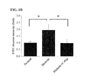
- FIGS. 1A and 1B demonstrate the inhibitory activity of C-peptide against diabetes-induced vascular leakage in photographs (a) and a graph (b).
- FIG. 1A shows representative fluorescent images of the retina.
- the square areas in upper panels are given as magnified images in lower panels of respective pictures.
- VEGF expression in retinal endothelial cells is involved in retinal vascular leakage through the generation of intracellular ROS.
- an examination was made of the protective effect of C-peptide against VEGF-induced ROS generation and thereby the prevention of vascular leakage, using HUVECs.
- HUVECs inoculated into coverslips were treated with VEGF and C-peptide, and incubated with 10 ⁇ M 2′,7′-dichlorodihydrofluorescein diacetate (Molecular Probes, Eugene, Oreg.) in phenol red-free media for the last 10 min.
- the coverslips were mounted on a perfusion chamber, and labeled cells were rapidly scanned by confocal microscopy.
- the level of intracellular ROS was determined by comparing the fluorescence intensities of treated cells with those of control cells, and expressing this as a fold difference.
- FIGS. 2A and 2B show that C-peptide inhibits VEGF-induced ROS generation but has no effect on intracellular calcium ions.
- FIG. 2A is a graph of the levels of intracellular ROS determined using confocal microscopy after HUVECs were pre-incubated with 1 mM NAC, 0.5 mM Trolox, or 5 ⁇ M BAPTA-AM (1,2-bis(2-aminophenoxy)ethane-N,N,N-′,N′-tetraacetic acid tetrakis(acetoxymethyl ester)) for 30 min, and stimulated with 10 ng/ml VEGF for 10 min. Data are expressed as mean ⁇ S.D.
- VEGF generated intracellular ROS (p ⁇ 0.01), and this ROS generation was abolished by treatment with the ROS scavengers Trolox and NAC (N-acetyl-L-cysteine) ( FIG. 2A ). Meanwhile, the Ca 2+ chelator BAPTA-AM also blocked VEGF-induced intracellular ROS generation, indicating that VEGF produces intracellular ROS by elevating intracellular Ca 2+ .
- FIG. 2B is a graph in which ROS levels are plotted against C-peptide concentrations after HUVECs are pre-treated with various concentrations of C-peptide for 30 min and then stimulated with 10 ng/ml VEGF.
- C-peptide inhibited the VEGF-induced generation of intracellular ROS in a dose-dependent manner, with complete prevention observed at 0.5 nM ROS.
- coverslips To monitor intracellular Ca 2+ levels, cells inoculated into coverslips were incubated with 1 ⁇ M Fluo-4 AM for 30 min. The coverslips were mounted on perfusion chambers and scanned every 10 sec by confocal microscopy (FV-300). Serial images from the scan were processed to analyze changes in intracellular Ca 2+ levels at the single-cell level. Data were expressed as the relative fluorescence intensity (RFI).
- RFI relative fluorescence intensity
- VEGF induced a rapid increase in intracellular Ca 2+ and the elevated intracellular Ca 2+ level was maintained until 600 sec.
- BAPTA-AM blocked changes in intracellular Ca 2+ levels in response to VEGF.
- C-peptide did not increase intracellular Ca 2+ ( FIG. 2C ) and had no effect on VEGF-induced changes in intracellular Ca 2+ .
- VEGF generates intracellular ROS by elevating intracellular Ca 2+ levels whereas C-peptide inhibits the VEGF-induced elevation of intracellular ROS without affecting intracellular Ca 2+ levels.
- VEGF activates stress fiber formation, and the disruption of VE-cadherin based adherens junction integrity, resulting in vascular leakage.
- an examination was made of the effect of C-peptide on VEGF-induced stress formation and VEGF-induced disassembly of the adherens junction.
- Elevation of VEGF levels in retinal endothelial cells can induce stress fiber formation through increased ROS generation.
- actin filaments were visualized in HUVECs by staining with rhodamine-phalloidin.
- HUVECs were grown on gelatin-coated round coverslips in 12-well culture plates, and incubated with 10 ng/ml C-peptide or 0.5 mM VEGF, alone or in combination, or with 10 ng/ml VEGF in the presence of an ROS scavenger, that is, 1 mM NAC or 0.5 mM Trolox for 1 hr at 37° C.
- the cells were rapidly rinsed with PBS and fixed with 3.7% formaldehyde in PBS for 30 min. Then, the cells were permeabilized with 0.2% Triton X-100 in PBS for 30 min, and stained with rhodamine-palloidin (Molecular Probes) in PBS for 1 h.
- the resulting stained cells were mounted on a glass slide using a mounting solution, followed by visualizing actin filaments by confocal microscopy.
- FIG. 3 shows fluorescent photographs of HUVECs, demonstrating that C-peptide inhibits VEGF-induced stress fiber formation and VEGF-induced disassembly of adherens junction.
- FIG. 3 shows the inhibitory activity of C-peptide against VEGF-induced stress fiber formation.
- VEGF activated the formation of stress fibers, which was sufficiently suppressed by treatment with C-peptide. VEGF-activated stress fiber formation was also inhibited upon treatment with the ROS scavengers NAC and Trolox, indicating that intracellular ROS is essential for the VEGF-induced formation of stress fibers.
- Confluent cell layers in 6-well plates were incubated with C-peptide or an ROS scavenger for 30 min, and then treated with VEGF for 90 min at 37° C.
- the treated cells were rinsed with PBS, and fixed with 3.7% formaldehyde in PBS for 30 min.
- the cells were permeabilized with 0.2% Triton X-100 in PBS for 30 min, and stained overnight at 4° C. with a monoclonal VE-cadherin antibody (Santa Cruz Biotechnology, Santa Cruz, Calif.) in PBS. Thereafter, the cells were probed with goat anti-mouse FITC (fluorescein isothiocyanate; Sigma, St. Louis, Mo.), followed by visualizing VE-cadherin by confocal microscopy.
- goat anti-mouse FITC fluorescein isothiocyanate
- FIGS. 4A and 4B show the inhibitory activity of C-peptide against the VEGF-induced disruption of the adherens junctions.
- the scale bar represents 20 ⁇ m.
- VEGF stimulated the disassembly of VE-cadherin, and this disassembly was inhibited by C-peptide.
- the VEGF-induced disassembly of VE-cadherin was also prevented by NAC and Trolox, indicating that intracellular ROS mediates the VEGF-induced disruption of VE-cadherin.
- FIG. 4B there are histograms in which changes in permeability resulting from the disassembly of VE-cadherin are represented by relative fluorescence intensities (RFI).
- RFI relative fluorescence intensities
- VE-cadherin was stained in the retina and visualized using confocal microscopy as described below.
- Microvessels were stained for VE-cadherin in retinal whole mounts.
- isolated retinas were fixed in 100% ethanol for 30 min, delipidated in 100% ice-cold acetone for 10 min, and permeabilized with 0.3% Triton X-100 for 1 hr.
- the retinas were incubated with an anti-VE-cadherin antibody (Enzo Life Sciences, Farmingdale, N.Y., USA) at 4° C. for 24 hrs, and probed with goat anti-mouse FITC (Sigma) at 4° C. for 24 hrs, followed by visualizing VE-cadherin by confocal microscopy (FV-300, Olympus).
- Results are shown in FIG. 4C . Consistent with the preventive effect of C-peptide against VEGF-induced disassembly of VE-cadherin in HUVECs, as seen in FIG. 4C , the intravitreal injection of C-peptide inhibited the diabetes-induced disassembly of adherens junctions in the microvessels of the diabetic mouse retina.
- C-peptide protects against VEGF-induced disassembly of VE-cadherin at cell junctions by inhibiting intracellular ROS generation in endothelial cells.
- a monoclonal VEGF antibody 100 ⁇ g/ml
- N-acetyl-cysteine 81.5 mg/ml
- Trolox 125 ⁇ g/ml
- mice were injected with 1.25 mg of 500-kDa FITC-
- FIGS. 5A and 5B Results are shown in FIGS. 5A and 5B .
- intravitreal injection of a monoclonal anti-VEGF antibody significantly inhibited microvascular leakage in the retinas of diabetic mice, demonstrating that VEGF is involved in retinal vascular leakage in diabetic mice.
- Intravitreal injection of the ROS scavengers NAC and Trolox also prevented retinal vascular leakage in diabetic mice.
- extravasation of FITC-dextran in the retinas of diabetic mice was also blocked by C-peptide injection.
- C-peptide protects against VEGF-induced retinal vascular leakage by inhibiting intracellular ROS generation in retinal endothelial cells.
- mice After implantation of a mini-osmotic pump for delivering C-peptide, blood c-peptide levels were measured.
- serum C-peptide levels were measured using a C-peptide Enzyme Immunoassay Kit (RayBiotech, Norcross, Ga.).
- FIG. 6A shows serum C-peptide levels left after injection into mice by an osmotic pump. As is understood from the data of FIG. 6A , the serum C-peptide level was elevated to a normal value. Hence, a mini-osmotic pump makes it possible to conduct subcutaneous injection at a delivery rate of from 1.45 pmol/kg/min to 36.5 pmol/kg/min.
- FIGS. 6B and 6C Results are shown in FIGS. 6B and 6C .
- FIG. 6B shows representative images of mouse groups treated with or without VEGF alone or in combination with various amounts of C-peptide.
- intradermal injection of VEGF significantly induced vascular permeability in diabetic mice, which was suppressed by C-peptide in a dose-dependent manner.
- C-peptide alone did not cause a significant change in vascular permeability.
- C-peptide levels in the vitreous fluid were monitored with time.
- Results are given in FIG. 6D .
- Intravitreal injection of C-peptide was previously found to prevent retinal leakage 24 hrs post-injection.
- the vitreous chamber maintained a normal level of C-peptide until about 12 hrs after intravitreal injection. That is, it is understood from the data of FIG. 6D that a normal intravitreal C-peptide level was maintained upon intravitreal injection, thereby suppressing VEGF-induced retinal leakage.
- C-peptide prevents microvascular leakage in the retinas of diabetic mice by inhibiting VEGF-induced intracellular ROS generation, which stimulates stress fiber formation and disassembly of the adherens junction, resulting in micro-vascular permeability
Landscapes
- Health & Medical Sciences (AREA)
- Life Sciences & Earth Sciences (AREA)
- Public Health (AREA)
- General Health & Medical Sciences (AREA)
- Animal Behavior & Ethology (AREA)
- Engineering & Computer Science (AREA)
- Chemical & Material Sciences (AREA)
- Bioinformatics & Cheminformatics (AREA)
- Pharmacology & Pharmacy (AREA)
- Medicinal Chemistry (AREA)
- Veterinary Medicine (AREA)
- Epidemiology (AREA)
- Immunology (AREA)
- Gastroenterology & Hepatology (AREA)
- Proteomics, Peptides & Aminoacids (AREA)
- Zoology (AREA)
- Chemical Kinetics & Catalysis (AREA)
- General Chemical & Material Sciences (AREA)
- Nuclear Medicine, Radiotherapy & Molecular Imaging (AREA)
- Organic Chemistry (AREA)
- Marine Sciences & Fisheries (AREA)
- Diabetes (AREA)
- Urology & Nephrology (AREA)
- Cardiology (AREA)
- Heart & Thoracic Surgery (AREA)
- Dermatology (AREA)
- Endocrinology (AREA)
- Ophthalmology & Optometry (AREA)
- Obesity (AREA)
- Vascular Medicine (AREA)
- Hematology (AREA)
- Emergency Medicine (AREA)
- Medicines That Contain Protein Lipid Enzymes And Other Medicines (AREA)
- Peptides Or Proteins (AREA)
Abstract
Description
Claims (9)
Priority Applications (1)
| Application Number | Priority Date | Filing Date | Title |
|---|---|---|---|
| US13/987,543 US9457063B2 (en) | 2012-08-06 | 2013-08-06 | Method for treatment of diabetic vascular leakage-induced disease using C-peptide |
Applications Claiming Priority (2)
| Application Number | Priority Date | Filing Date | Title |
|---|---|---|---|
| US201261680184P | 2012-08-06 | 2012-08-06 | |
| US13/987,543 US9457063B2 (en) | 2012-08-06 | 2013-08-06 | Method for treatment of diabetic vascular leakage-induced disease using C-peptide |
Publications (2)
| Publication Number | Publication Date |
|---|---|
| US20140148386A1 US20140148386A1 (en) | 2014-05-29 |
| US9457063B2 true US9457063B2 (en) | 2016-10-04 |
Family
ID=50068318
Family Applications (2)
| Application Number | Title | Priority Date | Filing Date |
|---|---|---|---|
| US13/987,543 Active 2034-01-21 US9457063B2 (en) | 2012-08-06 | 2013-08-06 | Method for treatment of diabetic vascular leakage-induced disease using C-peptide |
| US13/987,544 Active US9119816B2 (en) | 2012-08-06 | 2013-08-06 | Method for prevention or treatment of diabetic angiogenesis impairment using C-peptide |
Family Applications After (1)
| Application Number | Title | Priority Date | Filing Date |
|---|---|---|---|
| US13/987,544 Active US9119816B2 (en) | 2012-08-06 | 2013-08-06 | Method for prevention or treatment of diabetic angiogenesis impairment using C-peptide |
Country Status (5)
| Country | Link |
|---|---|
| US (2) | US9457063B2 (en) |
| EP (2) | EP2881120B1 (en) |
| KR (2) | KR101646054B1 (en) |
| CN (2) | CN104640559B (en) |
| WO (2) | WO2014025127A1 (en) |
Families Citing this family (6)
| Publication number | Priority date | Publication date | Assignee | Title |
|---|---|---|---|---|
| KR101676542B1 (en) * | 2014-12-30 | 2016-11-16 | 건국대학교 산학협력단 | Use of proinsulin for immune system modulation |
| WO2017010673A2 (en) | 2015-07-16 | 2017-01-19 | 재단법인 지능형 바이오 시스템 설계 및 합성 연구단 | Composition for preventing or treating vascular leakage syndrome |
| US10155948B2 (en) | 2016-05-12 | 2018-12-18 | Kangwon National University University-Industry Cooperation Foundation and | Pharmaceutical composition for preventing or treating diabetic complications and screening method for preventive or therapeutic agent for diabetic complications |
| KR101881662B1 (en) * | 2016-05-12 | 2018-07-26 | 강원대학교산학협력단 | Pharmaceutical composition for prevention or treatment of diabetic complications and screening method for agent for preventing or treating diabetic complications |
| KR102114275B1 (en) * | 2018-11-07 | 2020-05-25 | 제주대학교 산학협력단 | Cosmetic composition comprising C-peptide for protecting skin against particulate matter |
| CN111939244B (en) * | 2019-05-14 | 2024-08-06 | 阿莫生命科学有限公司 | Pharmaceutical composition for preventing or treating diabetic complications |
Citations (5)
| Publication number | Priority date | Publication date | Assignee | Title |
|---|---|---|---|---|
| US20020077317A1 (en) | 2000-12-15 | 2002-06-20 | Das Undurti Narasimha | Method of potentating the action of 2-methoxyoestradiol, statins and C-peptide of proinsulin |
| WO2005039627A2 (en) | 2003-10-13 | 2005-05-06 | Creative Peptides Sweden Ab | Therapeutic applications for c-peptide |
| WO2009068911A1 (en) | 2007-11-28 | 2009-06-04 | University Of Leeds | Compositions and methods for reducing macrovascular complications in diabetic patients |
| KR20100057640A (en) | 2007-09-11 | 2010-05-31 | 몬도바이오테크 래보래토리즈 아게 | Use of insulin c-peptide, alone or in combination with glp-1, as a therapeutic agent |
| US7855177B1 (en) * | 2009-10-28 | 2010-12-21 | Cebix Inc. | Methods and kits for preventing hypoglycemia |
Family Cites Families (4)
| Publication number | Priority date | Publication date | Assignee | Title |
|---|---|---|---|---|
| DE10041478A1 (en) * | 2000-08-24 | 2002-03-14 | Sanol Arznei Schwarz Gmbh | New pharmaceutical composition |
| GB0601950D0 (en) * | 2006-01-31 | 2006-03-15 | Creative Peptides Sweden Ab | Compositions and methods of treating diabetes |
| CN101297963A (en) * | 2007-04-30 | 2008-11-05 | 上海新生源医药研究有限公司 | Pharmaceutical composition of proinsulin C peptide |
| WO2011075611A1 (en) * | 2009-12-18 | 2011-06-23 | Aegis Therapeutics, Llc | Compositions and methods for non-invasive treatment of chronic complication of diabetes |
-
2013
- 2013-05-28 KR KR1020147035709A patent/KR101646054B1/en active IP Right Grant
- 2013-05-28 CN CN201380042124.XA patent/CN104640559B/en active Active
- 2013-05-28 WO PCT/KR2013/004648 patent/WO2014025127A1/en active Application Filing
- 2013-05-28 EP EP13828414.6A patent/EP2881120B1/en active Active
- 2013-07-23 EP EP13828393.2A patent/EP2881119B1/en active Active
- 2013-07-23 CN CN201380042117.XA patent/CN104602699B/en active Active
- 2013-07-23 KR KR1020147035710A patent/KR101620942B1/en active IP Right Grant
- 2013-07-23 WO PCT/KR2013/006571 patent/WO2014025146A1/en active Application Filing
- 2013-08-06 US US13/987,543 patent/US9457063B2/en active Active
- 2013-08-06 US US13/987,544 patent/US9119816B2/en active Active
Patent Citations (6)
| Publication number | Priority date | Publication date | Assignee | Title |
|---|---|---|---|---|
| US20020077317A1 (en) | 2000-12-15 | 2002-06-20 | Das Undurti Narasimha | Method of potentating the action of 2-methoxyoestradiol, statins and C-peptide of proinsulin |
| WO2005039627A2 (en) | 2003-10-13 | 2005-05-06 | Creative Peptides Sweden Ab | Therapeutic applications for c-peptide |
| KR20100057640A (en) | 2007-09-11 | 2010-05-31 | 몬도바이오테크 래보래토리즈 아게 | Use of insulin c-peptide, alone or in combination with glp-1, as a therapeutic agent |
| US20100292132A1 (en) | 2007-09-11 | 2010-11-18 | Dorian Bevec | Use of insulin c-peptide, alone or in combination with glp-1, as a therapeutic agent |
| WO2009068911A1 (en) | 2007-11-28 | 2009-06-04 | University Of Leeds | Compositions and methods for reducing macrovascular complications in diabetic patients |
| US7855177B1 (en) * | 2009-10-28 | 2010-12-21 | Cebix Inc. | Methods and kits for preventing hypoglycemia |
Non-Patent Citations (10)
| Title |
|---|
| B. -L. Johansson et al., "Beneficial effects of C-peptide on incipient nephropathy and neuropathy in patients with Type I diabetes mellitus," Diabetic Medicine, 2000. |
| Bo-Lennart Johansson et al., "Influence of combined C-peptide and insulin administration on Renal Function and Metabolic Control in Diabetes Type 1," Journal of Clinical Endocrinology and Metabolism, 1993. |
| Ido et al. Prevention of vascular and neural dysfunction in diabetic rats by C-peptide. Science. 1997; 277: 563-566. * |
| J. Wahren et al., "Does C-peptide Have a Physiological Role?", Diabetologia, vol. 37, No. Suppl. 2, 1994, pp. S99-S107. |
| John Wahren et al., "The Clinical Potential of C-Peptide Recplacement in Type 1 Diabetes," Perspectives in Diabetes, 2012. |
| Marjorie A. Mosier et al., "Circulating C-peptide and diabetic retinopathy," Diabetes Research, 1984. |
| P. Luippi et al., "C-peptide and long-term complications of diabetes," Pediatric Diabetes, 2011. |
| Ronald Klein et al., "Wisconsin Epidemiologic Study of Diabetic Retinopathy," Diabetes, 1990. |
| S. Langer et al., "Effect of C-peptide on Wound Healing and Microcirculation in Diabetic Mice", European Journal of Medical Research, vol. 7, No. 11, Nov. 25, 2002, pp. 502-508. |
| Subrata Chakrabarti et al., "C-peptide and Retinal Microangiopathy in Diabetes," Experimental Diab. Res., 2004. |
Also Published As
| Publication number | Publication date |
|---|---|
| CN104602699B (en) | 2017-05-03 |
| WO2014025146A1 (en) | 2014-02-13 |
| KR20150035713A (en) | 2015-04-07 |
| CN104602699A (en) | 2015-05-06 |
| KR20150035714A (en) | 2015-04-07 |
| WO2014025127A1 (en) | 2014-02-13 |
| EP2881119B1 (en) | 2019-09-18 |
| KR101646054B1 (en) | 2016-08-05 |
| EP2881119A1 (en) | 2015-06-10 |
| CN104640559A (en) | 2015-05-20 |
| EP2881120A1 (en) | 2015-06-10 |
| US9119816B2 (en) | 2015-09-01 |
| EP2881120A4 (en) | 2015-08-19 |
| US20140148386A1 (en) | 2014-05-29 |
| CN104640559B (en) | 2017-05-17 |
| US20140128323A1 (en) | 2014-05-08 |
| EP2881119A4 (en) | 2015-08-19 |
| KR101620942B1 (en) | 2016-05-13 |
| EP2881120B1 (en) | 2019-08-07 |
Similar Documents
| Publication | Publication Date | Title |
|---|---|---|
| US9457063B2 (en) | Method for treatment of diabetic vascular leakage-induced disease using C-peptide | |
| US20230285328A1 (en) | Methods and Compositions for the Treatment of Steatosis-Associated Disorders | |
| US20130217621A1 (en) | Compositions for the treatment of diabetes | |
| CN109276705B (en) | Compositions and methods for treating heart failure in diabetics | |
| US10155948B2 (en) | Pharmaceutical composition for preventing or treating diabetic complications and screening method for preventive or therapeutic agent for diabetic complications | |
| Lee et al. | Dopamine ameliorates hyperglycemic memory‐induced microvascular dysfunction in diabetic retinopathy | |
| US9084761B2 (en) | Use of interleukin-15 to treat cardiovascular diseases | |
| KR101704914B1 (en) | A pharmaceutical composition comprising dammarenediol-ii for preventing or treating diseases by diabetic vascular leakage | |
| KR101881662B1 (en) | Pharmaceutical composition for prevention or treatment of diabetic complications and screening method for agent for preventing or treating diabetic complications | |
| EP3402505B1 (en) | Pharmaceutical formulations for the treatment of diabetes | |
| US9468669B2 (en) | Methods to treat dysregulated blood glucose disorders | |
| US20230321072A1 (en) | Cell glycocalyx protection effect of anisodamine | |
| CN108159399B (en) | Application of thrombin aPC in medicine for preventing and treating diabetic cardiomyopathy | |
| US20080132508A1 (en) | Method and system for altering dysfunctional lipid metabolism in diabetic complications | |
| US10765647B2 (en) | Composition for treating diabetes, containing extract of yukmijihwangtang | |
| US10576151B2 (en) | Ciclopirox for use in modulation of glucose homeostasis | |
| Femminò et al. | Catestatin induces glucose uptake and Glut4 trafficking in adult rat cardiomyocytes | |
| WO2024003338A1 (en) | Annexin a1 liquid pharmaceutical composition | |
| US20150265682A1 (en) | Therapeutic methods and compositions for treating diabetes |
Legal Events
| Date | Code | Title | Description |
|---|---|---|---|
| AS | Assignment |
Owner name: AMOGREENTECH CO., LTD., KOREA, REPUBLIC OF Free format text: ASSIGNMENT OF ASSIGNORS INTEREST;ASSIGNORS:HA, KWON-SOO;LIM, YOUNG-CHEOL;REEL/FRAME:031139/0561 Effective date: 20130719 Owner name: KANGWON NATIONAL UNIVERSITY UNIVERSITY-INDUSTRY CO Free format text: ASSIGNMENT OF ASSIGNORS INTEREST;ASSIGNORS:HA, KWON-SOO;LIM, YOUNG-CHEOL;REEL/FRAME:031139/0561 Effective date: 20130719 |
|
| STCF | Information on status: patent grant |
Free format text: PATENTED CASE |
|
| FEPP | Fee payment procedure |
Free format text: PAYOR NUMBER ASSIGNED (ORIGINAL EVENT CODE: ASPN); ENTITY STATUS OF PATENT OWNER: SMALL ENTITY |
|
| FEPP | Fee payment procedure |
Free format text: PAT HOLDER CLAIMS SMALL ENTITY STATUS, ENTITY STATUS SET TO SMALL (ORIGINAL EVENT CODE: LTOS); ENTITY STATUS OF PATENT OWNER: SMALL ENTITY |
|
| AS | Assignment |
Owner name: AMOLIFESCIENCE CO., LTD., KOREA, REPUBLIC OF Free format text: ASSIGNMENT OF ASSIGNORS INTEREST;ASSIGNOR:AMOGREENTECH CO., LTD.;REEL/FRAME:041273/0078 Effective date: 20170213 |
|
| MAFP | Maintenance fee payment |
Free format text: PAYMENT OF MAINTENANCE FEE, 4TH YR, SMALL ENTITY (ORIGINAL EVENT CODE: M2551); ENTITY STATUS OF PATENT OWNER: SMALL ENTITY Year of fee payment: 4 |
|
| MAFP | Maintenance fee payment |
Free format text: PAYMENT OF MAINTENANCE FEE, 8TH YR, SMALL ENTITY (ORIGINAL EVENT CODE: M2552); ENTITY STATUS OF PATENT OWNER: SMALL ENTITY Year of fee payment: 8 |