US9452187B2 - In vivo and ex vivo expansion of hematopoietic stem cells with a targeted combination of clinically tested, FDA approved drugs - Google Patents
In vivo and ex vivo expansion of hematopoietic stem cells with a targeted combination of clinically tested, FDA approved drugs Download PDFInfo
- Publication number
- US9452187B2 US9452187B2 US14/673,125 US201514673125A US9452187B2 US 9452187 B2 US9452187 B2 US 9452187B2 US 201514673125 A US201514673125 A US 201514673125A US 9452187 B2 US9452187 B2 US 9452187B2
- Authority
- US
- United States
- Prior art keywords
- cells
- hscs
- gsk
- hsc
- cell
- Prior art date
- Legal status (The legal status is an assumption and is not a legal conclusion. Google has not performed a legal analysis and makes no representation as to the accuracy of the status listed.)
- Active
Links
- 210000003958 hematopoietic stem cell Anatomy 0.000 title claims description 346
- 238000001727 in vivo Methods 0.000 title abstract description 14
- 229940124602 FDA-approved drug Drugs 0.000 title 1
- 108010014905 Glycogen Synthase Kinase 3 Proteins 0.000 claims abstract description 130
- 238000000034 method Methods 0.000 claims abstract description 65
- 239000003112 inhibitor Substances 0.000 claims abstract description 59
- 108010065917 TOR Serine-Threonine Kinases Proteins 0.000 claims abstract description 46
- 102000013530 TOR Serine-Threonine Kinases Human genes 0.000 claims abstract description 46
- 238000012258 culturing Methods 0.000 claims abstract description 44
- 208000037265 diseases, disorders, signs and symptoms Diseases 0.000 claims description 28
- 241000124008 Mammalia Species 0.000 claims description 24
- 201000010099 disease Diseases 0.000 claims description 19
- 208000035475 disorder Diseases 0.000 claims description 9
- 230000000735 allogeneic effect Effects 0.000 claims description 5
- 208000032467 Aplastic anaemia Diseases 0.000 claims description 4
- 208000026350 Inborn Genetic disease Diseases 0.000 claims description 4
- 208000016361 genetic disease Diseases 0.000 claims description 4
- 208000024893 Acute lymphoblastic leukemia Diseases 0.000 claims description 3
- 208000014697 Acute lymphocytic leukaemia Diseases 0.000 claims description 3
- 208000031261 Acute myeloid leukaemia Diseases 0.000 claims description 3
- 208000032791 BCR-ABL1 positive chronic myelogenous leukemia Diseases 0.000 claims description 3
- 201000004449 Diamond-Blackfan anemia Diseases 0.000 claims description 3
- 208000010055 Globoid Cell Leukodystrophy Diseases 0.000 claims description 3
- 208000017604 Hodgkin disease Diseases 0.000 claims description 3
- 208000010747 Hodgkins lymphoma Diseases 0.000 claims description 3
- 208000028226 Krabbe disease Diseases 0.000 claims description 3
- 208000009625 Lesch-Nyhan syndrome Diseases 0.000 claims description 3
- 208000030289 Lymphoproliferative disease Diseases 0.000 claims description 3
- 206010056886 Mucopolysaccharidosis I Diseases 0.000 claims description 3
- 208000034578 Multiple myelomas Diseases 0.000 claims description 3
- 208000033776 Myeloid Acute Leukemia Diseases 0.000 claims description 3
- 208000015914 Non-Hodgkin lymphomas Diseases 0.000 claims description 3
- 206010035226 Plasma cell myeloma Diseases 0.000 claims description 3
- 208000006664 Precursor Cell Lymphoblastic Leukemia-Lymphoma Diseases 0.000 claims description 3
- 208000035317 Total hypoxanthine-guanine phosphoribosyl transferase deficiency Diseases 0.000 claims description 3
- 208000006110 Wiskott-Aldrich syndrome Diseases 0.000 claims description 3
- 206010068348 X-linked lymphoproliferative syndrome Diseases 0.000 claims description 3
- 208000007502 anemia Diseases 0.000 claims description 3
- 208000005980 beta thalassemia Diseases 0.000 claims description 3
- 201000002273 mucopolysaccharidosis II Diseases 0.000 claims description 3
- 208000022018 mucopolysaccharidosis type 2 Diseases 0.000 claims description 3
- 208000002865 osteopetrosis Diseases 0.000 claims description 3
- 208000002491 severe combined immunodeficiency Diseases 0.000 claims description 3
- 208000007056 sickle cell anemia Diseases 0.000 claims description 3
- 102000001267 GSK3 Human genes 0.000 claims 2
- 102000002254 Glycogen Synthase Kinase 3 Human genes 0.000 abstract description 128
- 229940124302 mTOR inhibitor Drugs 0.000 abstract description 35
- 239000003628 mammalian target of rapamycin inhibitor Substances 0.000 abstract description 35
- 230000001225 therapeutic effect Effects 0.000 abstract description 16
- 229940079593 drug Drugs 0.000 abstract description 12
- 239000003814 drug Substances 0.000 abstract description 12
- 238000013459 approach Methods 0.000 abstract description 8
- 238000002483 medication Methods 0.000 abstract description 5
- 210000003995 blood forming stem cell Anatomy 0.000 abstract description 3
- 210000004027 cell Anatomy 0.000 description 318
- 210000001185 bone marrow Anatomy 0.000 description 132
- 101150090422 gsk-3 gene Proteins 0.000 description 74
- 108090000623 proteins and genes Proteins 0.000 description 71
- 241000699670 Mus sp. Species 0.000 description 69
- 239000005090 green fluorescent protein Substances 0.000 description 62
- 108010043121 Green Fluorescent Proteins Proteins 0.000 description 61
- 102000004144 Green Fluorescent Proteins Human genes 0.000 description 61
- 102000015735 Beta-catenin Human genes 0.000 description 59
- 108060000903 Beta-catenin Proteins 0.000 description 59
- WHXSMMKQMYFTQS-UHFFFAOYSA-N Lithium Chemical compound [Li] WHXSMMKQMYFTQS-UHFFFAOYSA-N 0.000 description 59
- 229910052744 lithium Inorganic materials 0.000 description 59
- 210000000130 stem cell Anatomy 0.000 description 56
- 102000013814 Wnt Human genes 0.000 description 46
- 108050003627 Wnt Proteins 0.000 description 46
- 230000011664 signaling Effects 0.000 description 39
- 210000004369 blood Anatomy 0.000 description 38
- 239000008280 blood Substances 0.000 description 38
- QFJCIRLUMZQUOT-HPLJOQBZSA-N sirolimus Chemical compound C1C[C@@H](O)[C@H](OC)C[C@@H]1C[C@@H](C)[C@H]1OC(=O)[C@@H]2CCCCN2C(=O)C(=O)[C@](O)(O2)[C@H](C)CC[C@H]2C[C@H](OC)/C(C)=C/C=C/C=C/[C@@H](C)C[C@@H](C)C(=O)[C@H](OC)[C@H](O)/C(C)=C/[C@@H](C)C(=O)C1 QFJCIRLUMZQUOT-HPLJOQBZSA-N 0.000 description 38
- ZAHRKKWIAAJSAO-UHFFFAOYSA-N rapamycin Natural products COCC(O)C(=C/C(C)C(=O)CC(OC(=O)C1CCCCN1C(=O)C(=O)C2(O)OC(CC(OC)C(=CC=CC=CC(C)CC(C)C(=O)C)C)CCC2C)C(C)CC3CCC(O)C(C3)OC)C ZAHRKKWIAAJSAO-UHFFFAOYSA-N 0.000 description 37
- 229960002930 sirolimus Drugs 0.000 description 37
- 230000000694 effects Effects 0.000 description 32
- 239000002609 medium Substances 0.000 description 31
- 238000002054 transplantation Methods 0.000 description 31
- 230000006870 function Effects 0.000 description 28
- 230000007774 longterm Effects 0.000 description 24
- 230000037361 pathway Effects 0.000 description 23
- 102000004169 proteins and genes Human genes 0.000 description 23
- 238000011282 treatment Methods 0.000 description 23
- 101150019946 Gsk3b gene Proteins 0.000 description 22
- 230000005764 inhibitory process Effects 0.000 description 22
- 102000004127 Cytokines Human genes 0.000 description 21
- 108090000695 Cytokines Proteins 0.000 description 21
- 102100031573 Hematopoietic progenitor cell antigen CD34 Human genes 0.000 description 21
- 101000777663 Homo sapiens Hematopoietic progenitor cell antigen CD34 Proteins 0.000 description 21
- 230000014509 gene expression Effects 0.000 description 21
- 235000018102 proteins Nutrition 0.000 description 20
- 230000001105 regulatory effect Effects 0.000 description 20
- 102100032543 Phosphatidylinositol 3,4,5-trisphosphate 3-phosphatase and dual-specificity protein phosphatase PTEN Human genes 0.000 description 19
- 101710132081 Phosphatidylinositol 3,4,5-trisphosphate 3-phosphatase and dual-specificity protein phosphatase PTEN Proteins 0.000 description 19
- 230000004913 activation Effects 0.000 description 19
- 238000000684 flow cytometry Methods 0.000 description 19
- 238000012423 maintenance Methods 0.000 description 19
- 230000002829 reductive effect Effects 0.000 description 18
- 239000013598 vector Substances 0.000 description 17
- 108020004414 DNA Proteins 0.000 description 16
- 230000003394 haemopoietic effect Effects 0.000 description 16
- 230000026731 phosphorylation Effects 0.000 description 16
- 238000006366 phosphorylation reaction Methods 0.000 description 16
- 230000004083 survival effect Effects 0.000 description 16
- 230000011132 hemopoiesis Effects 0.000 description 15
- 210000005259 peripheral blood Anatomy 0.000 description 15
- 239000011886 peripheral blood Substances 0.000 description 15
- 238000002474 experimental method Methods 0.000 description 14
- KWGKDLIKAYFUFQ-UHFFFAOYSA-M lithium chloride Chemical compound [Li+].[Cl-] KWGKDLIKAYFUFQ-UHFFFAOYSA-M 0.000 description 14
- 241000699666 Mus <mouse, genus> Species 0.000 description 13
- 230000002950 deficient Effects 0.000 description 13
- 239000000203 mixture Substances 0.000 description 13
- 102000016971 Proto-Oncogene Proteins c-kit Human genes 0.000 description 12
- 108010014608 Proto-Oncogene Proteins c-kit Proteins 0.000 description 12
- 230000004069 differentiation Effects 0.000 description 12
- 239000001963 growth medium Substances 0.000 description 12
- 230000001404 mediated effect Effects 0.000 description 12
- 210000001519 tissue Anatomy 0.000 description 12
- 241000713666 Lentivirus Species 0.000 description 11
- 238000004458 analytical method Methods 0.000 description 11
- 238000003556 assay Methods 0.000 description 11
- WOVKYSAHUYNSMH-RRKCRQDMSA-N 5-bromodeoxyuridine Chemical compound C1[C@H](O)[C@@H](CO)O[C@H]1N1C(=O)NC(=O)C(Br)=C1 WOVKYSAHUYNSMH-RRKCRQDMSA-N 0.000 description 10
- 101150113453 Gsk3a gene Proteins 0.000 description 10
- 241001465754 Metazoa Species 0.000 description 10
- 206010028980 Neoplasm Diseases 0.000 description 10
- 210000002798 bone marrow cell Anatomy 0.000 description 10
- 230000002860 competitive effect Effects 0.000 description 10
- 239000000463 material Substances 0.000 description 10
- 230000035755 proliferation Effects 0.000 description 10
- 238000012360 testing method Methods 0.000 description 10
- 108010051975 Glycogen Synthase Kinase 3 beta Proteins 0.000 description 9
- 102000019058 Glycogen Synthase Kinase 3 beta Human genes 0.000 description 9
- 230000005757 colony formation Effects 0.000 description 9
- 230000001419 dependent effect Effects 0.000 description 9
- 210000004700 fetal blood Anatomy 0.000 description 9
- 230000001605 fetal effect Effects 0.000 description 9
- 229920000609 methyl cellulose Polymers 0.000 description 8
- 239000001923 methylcellulose Substances 0.000 description 8
- YAEMHJKFIIIULI-UHFFFAOYSA-N n-(4-methoxybenzyl)-n'-(5-nitro-1,3-thiazol-2-yl)urea Chemical compound C1=CC(OC)=CC=C1CNC(=O)NC1=NC=C([N+]([O-])=O)S1 YAEMHJKFIIIULI-UHFFFAOYSA-N 0.000 description 8
- 230000009467 reduction Effects 0.000 description 8
- 101150091591 tsc1 gene Proteins 0.000 description 8
- 101100335081 Mus musculus Flt3 gene Proteins 0.000 description 7
- 102000038030 PI3Ks Human genes 0.000 description 7
- 108091007960 PI3Ks Proteins 0.000 description 7
- 239000000654 additive Substances 0.000 description 7
- 238000012217 deletion Methods 0.000 description 7
- 230000037430 deletion Effects 0.000 description 7
- 238000009472 formulation Methods 0.000 description 7
- 230000013632 homeostatic process Effects 0.000 description 7
- 241000282412 Homo Species 0.000 description 6
- 108091030071 RNAI Proteins 0.000 description 6
- 108091027967 Small hairpin RNA Proteins 0.000 description 6
- 201000011510 cancer Diseases 0.000 description 6
- 230000022131 cell cycle Effects 0.000 description 6
- 230000008859 change Effects 0.000 description 6
- 230000009368 gene silencing by RNA Effects 0.000 description 6
- 230000002401 inhibitory effect Effects 0.000 description 6
- 230000002629 repopulating effect Effects 0.000 description 6
- -1 6BIO Chemical compound 0.000 description 5
- 206010068051 Chimerism Diseases 0.000 description 5
- FAPWRFPIFSIZLT-UHFFFAOYSA-M Sodium chloride Chemical compound [Na+].[Cl-] FAPWRFPIFSIZLT-UHFFFAOYSA-M 0.000 description 5
- 238000002512 chemotherapy Methods 0.000 description 5
- 238000010293 colony formation assay Methods 0.000 description 5
- 238000001514 detection method Methods 0.000 description 5
- 210000002257 embryonic structure Anatomy 0.000 description 5
- 210000003743 erythrocyte Anatomy 0.000 description 5
- 238000011124 ex vivo culture Methods 0.000 description 5
- 238000010348 incorporation Methods 0.000 description 5
- 210000005229 liver cell Anatomy 0.000 description 5
- 239000002773 nucleotide Substances 0.000 description 5
- 125000003729 nucleotide group Chemical group 0.000 description 5
- 238000011160 research Methods 0.000 description 5
- 230000004044 response Effects 0.000 description 5
- 239000000758 substrate Substances 0.000 description 5
- 238000001890 transfection Methods 0.000 description 5
- YBJHBAHKTGYVGT-ZKWXMUAHSA-N (+)-Biotin Chemical compound N1C(=O)N[C@@H]2[C@H](CCCCC(=O)O)SC[C@@H]21 YBJHBAHKTGYVGT-ZKWXMUAHSA-N 0.000 description 4
- 108090000672 Annexin A5 Proteins 0.000 description 4
- 102000004121 Annexin A5 Human genes 0.000 description 4
- 101100289995 Caenorhabditis elegans mac-1 gene Proteins 0.000 description 4
- HKVAMNSJSFKALM-GKUWKFKPSA-N Everolimus Chemical compound C1C[C@@H](OCCO)[C@H](OC)C[C@@H]1C[C@@H](C)[C@H]1OC(=O)[C@@H]2CCCCN2C(=O)C(=O)[C@](O)(O2)[C@H](C)CC[C@H]2C[C@H](OC)/C(C)=C/C=C/C=C/[C@@H](C)C[C@@H](C)C(=O)[C@H](OC)[C@H](O)/C(C)=C/[C@@H](C)C(=O)C1 HKVAMNSJSFKALM-GKUWKFKPSA-N 0.000 description 4
- 108091029865 Exogenous DNA Proteins 0.000 description 4
- 241000283984 Rodentia Species 0.000 description 4
- 210000001744 T-lymphocyte Anatomy 0.000 description 4
- CBPNZQVSJQDFBE-FUXHJELOSA-N Temsirolimus Chemical compound C1C[C@@H](OC(=O)C(C)(CO)CO)[C@H](OC)C[C@@H]1C[C@@H](C)[C@H]1OC(=O)[C@@H]2CCCCN2C(=O)C(=O)[C@](O)(O2)[C@H](C)CC[C@H]2C[C@H](OC)/C(C)=C/C=C/C=C/[C@@H](C)C[C@@H](C)C(=O)[C@H](OC)[C@H](O)/C(C)=C/[C@@H](C)C(=O)C1 CBPNZQVSJQDFBE-FUXHJELOSA-N 0.000 description 4
- 230000009471 action Effects 0.000 description 4
- 230000003042 antagnostic effect Effects 0.000 description 4
- 210000003719 b-lymphocyte Anatomy 0.000 description 4
- 150000001875 compounds Chemical class 0.000 description 4
- 230000006378 damage Effects 0.000 description 4
- 230000003247 decreasing effect Effects 0.000 description 4
- 229960005167 everolimus Drugs 0.000 description 4
- 238000012239 gene modification Methods 0.000 description 4
- 230000005017 genetic modification Effects 0.000 description 4
- 235000013617 genetically modified food Nutrition 0.000 description 4
- 108010049611 glycogen synthase kinase 3 alpha Proteins 0.000 description 4
- 239000003102 growth factor Substances 0.000 description 4
- 238000003119 immunoblot Methods 0.000 description 4
- 239000007924 injection Substances 0.000 description 4
- 238000002347 injection Methods 0.000 description 4
- 208000014674 injury Diseases 0.000 description 4
- 230000010354 integration Effects 0.000 description 4
- 230000003834 intracellular effect Effects 0.000 description 4
- 210000000265 leukocyte Anatomy 0.000 description 4
- 210000004185 liver Anatomy 0.000 description 4
- 210000000066 myeloid cell Anatomy 0.000 description 4
- 210000003643 myeloid progenitor cell Anatomy 0.000 description 4
- 230000036963 noncompetitive effect Effects 0.000 description 4
- 230000000144 pharmacologic effect Effects 0.000 description 4
- 230000008569 process Effects 0.000 description 4
- 239000000047 product Substances 0.000 description 4
- 210000002966 serum Anatomy 0.000 description 4
- 210000002536 stromal cell Anatomy 0.000 description 4
- 230000001629 suppression Effects 0.000 description 4
- 229960000235 temsirolimus Drugs 0.000 description 4
- 238000012546 transfer Methods 0.000 description 4
- 239000003981 vehicle Substances 0.000 description 4
- AQGNHMOJWBZFQQ-UHFFFAOYSA-N CT 99021 Chemical compound CC1=CNC(C=2C(=NC(NCCNC=3N=CC(=CC=3)C#N)=NC=2)C=2C(=CC(Cl)=CC=2)Cl)=N1 AQGNHMOJWBZFQQ-UHFFFAOYSA-N 0.000 description 3
- 108091026890 Coding region Proteins 0.000 description 3
- 229920002307 Dextran Polymers 0.000 description 3
- IAZDPXIOMUYVGZ-UHFFFAOYSA-N Dimethylsulphoxide Chemical compound CS(C)=O IAZDPXIOMUYVGZ-UHFFFAOYSA-N 0.000 description 3
- 102000004190 Enzymes Human genes 0.000 description 3
- 108090000790 Enzymes Proteins 0.000 description 3
- 108700028146 Genetic Enhancer Elements Proteins 0.000 description 3
- 108010017080 Granulocyte Colony-Stimulating Factor Proteins 0.000 description 3
- 102000004269 Granulocyte Colony-Stimulating Factor Human genes 0.000 description 3
- 101001046686 Homo sapiens Integrin alpha-M Proteins 0.000 description 3
- 102100022338 Integrin alpha-M Human genes 0.000 description 3
- 208000014767 Myeloproliferative disease Diseases 0.000 description 3
- 108091028043 Nucleic acid sequence Proteins 0.000 description 3
- 102000004160 Phosphoric Monoester Hydrolases Human genes 0.000 description 3
- 108090000608 Phosphoric Monoester Hydrolases Proteins 0.000 description 3
- 108091000080 Phosphotransferase Proteins 0.000 description 3
- 102000036693 Thrombopoietin Human genes 0.000 description 3
- 108010041111 Thrombopoietin Proteins 0.000 description 3
- 208000027418 Wounds and injury Diseases 0.000 description 3
- 230000009286 beneficial effect Effects 0.000 description 3
- 230000003115 biocidal effect Effects 0.000 description 3
- 238000004820 blood count Methods 0.000 description 3
- 238000010322 bone marrow transplantation Methods 0.000 description 3
- 229910000389 calcium phosphate Inorganic materials 0.000 description 3
- 239000001506 calcium phosphate Substances 0.000 description 3
- 235000011010 calcium phosphates Nutrition 0.000 description 3
- 230000001413 cellular effect Effects 0.000 description 3
- 238000011161 development Methods 0.000 description 3
- 230000018109 developmental process Effects 0.000 description 3
- 238000004520 electroporation Methods 0.000 description 3
- 108700014844 flt3 ligand Proteins 0.000 description 3
- 238000003306 harvesting Methods 0.000 description 3
- 208000014951 hematologic disease Diseases 0.000 description 3
- 208000018706 hematopoietic system disease Diseases 0.000 description 3
- 208000032839 leukemia Diseases 0.000 description 3
- 230000000527 lymphocytic effect Effects 0.000 description 3
- 210000003738 lymphoid progenitor cell Anatomy 0.000 description 3
- 230000007246 mechanism Effects 0.000 description 3
- 210000000440 neutrophil Anatomy 0.000 description 3
- 210000000056 organ Anatomy 0.000 description 3
- 102000020233 phosphotransferase Human genes 0.000 description 3
- 230000008488 polyadenylation Effects 0.000 description 3
- INCIMLINXXICKS-UHFFFAOYSA-M pyronin Y Chemical compound [Cl-].C1=CC(=[N+](C)C)C=C2OC3=CC(N(C)C)=CC=C3C=C21 INCIMLINXXICKS-UHFFFAOYSA-M 0.000 description 3
- 238000001959 radiotherapy Methods 0.000 description 3
- 238000011084 recovery Methods 0.000 description 3
- 230000000717 retained effect Effects 0.000 description 3
- 239000012679 serum free medium Substances 0.000 description 3
- 239000004055 small Interfering RNA Substances 0.000 description 3
- 238000010186 staining Methods 0.000 description 3
- QFJCIRLUMZQUOT-UHFFFAOYSA-N temsirolimus Natural products C1CC(O)C(OC)CC1CC(C)C1OC(=O)C2CCCCN2C(=O)C(=O)C(O)(O2)C(C)CCC2CC(OC)C(C)=CC=CC=CC(C)CC(C)C(=O)C(OC)C(O)C(C)=CC(C)C(=O)C1 QFJCIRLUMZQUOT-UHFFFAOYSA-N 0.000 description 3
- 238000002560 therapeutic procedure Methods 0.000 description 3
- 238000010361 transduction Methods 0.000 description 3
- 230000026683 transduction Effects 0.000 description 3
- QORWJWZARLRLPR-UHFFFAOYSA-H tricalcium bis(phosphate) Chemical compound [Ca+2].[Ca+2].[Ca+2].[O-]P([O-])([O-])=O.[O-]P([O-])([O-])=O QORWJWZARLRLPR-UHFFFAOYSA-H 0.000 description 3
- 210000003462 vein Anatomy 0.000 description 3
- 239000013603 viral vector Substances 0.000 description 3
- 108091032973 (ribonucleotides)n+m Proteins 0.000 description 2
- JKMHFZQWWAIEOD-UHFFFAOYSA-N 2-[4-(2-hydroxyethyl)piperazin-1-yl]ethanesulfonic acid Chemical compound OCC[NH+]1CCN(CCS([O-])(=O)=O)CC1 JKMHFZQWWAIEOD-UHFFFAOYSA-N 0.000 description 2
- BFSVOASYOCHEOV-UHFFFAOYSA-N 2-diethylaminoethanol Chemical compound CCN(CC)CCO BFSVOASYOCHEOV-UHFFFAOYSA-N 0.000 description 2
- DDLZLOKCJHBUHD-WAVHTBQISA-N 6-bromoindirubin-3'-oxime Chemical compound O=C/1NC2=CC(Br)=CC=C2C\1=C\1/C(=N/O)/C2=CC=CC=C2N/1 DDLZLOKCJHBUHD-WAVHTBQISA-N 0.000 description 2
- FHCSBLWRGCOVPT-UHFFFAOYSA-N AZD2858 Chemical compound C1CN(C)CCN1S(=O)(=O)C1=CC=C(C=2N=C(C(N)=NC=2)C(=O)NC=2C=NC=CC=2)C=C1 FHCSBLWRGCOVPT-UHFFFAOYSA-N 0.000 description 2
- 206010000830 Acute leukaemia Diseases 0.000 description 2
- 108700028369 Alleles Proteins 0.000 description 2
- 208000019838 Blood disease Diseases 0.000 description 2
- 108020004705 Codon Proteins 0.000 description 2
- RTZKZFJDLAIYFH-UHFFFAOYSA-N Diethyl ether Chemical compound CCOCC RTZKZFJDLAIYFH-UHFFFAOYSA-N 0.000 description 2
- 230000010337 G2 phase Effects 0.000 description 2
- 108010017213 Granulocyte-Macrophage Colony-Stimulating Factor Proteins 0.000 description 2
- 102000004457 Granulocyte-Macrophage Colony-Stimulating Factor Human genes 0.000 description 2
- 239000007995 HEPES buffer Substances 0.000 description 2
- 241000175212 Herpesvirales Species 0.000 description 2
- 101150025117 IL3 gene Proteins 0.000 description 2
- 101150101999 IL6 gene Proteins 0.000 description 2
- 208000001019 Inborn Errors Metabolism Diseases 0.000 description 2
- 101710144867 Inositol monophosphatase Proteins 0.000 description 2
- 201000007224 Myeloproliferative neoplasm Diseases 0.000 description 2
- 108010074687 Signaling Lymphocytic Activation Molecule Family Member 1 Proteins 0.000 description 2
- 101000930762 Sulfolobus acidocaldarius (strain ATCC 33909 / DSM 639 / JCM 8929 / NBRC 15157 / NCIMB 11770) Signal recognition particle receptor FtsY Proteins 0.000 description 2
- 230000003213 activating effect Effects 0.000 description 2
- 230000004075 alteration Effects 0.000 description 2
- 239000003242 anti bacterial agent Substances 0.000 description 2
- 230000008901 benefit Effects 0.000 description 2
- 230000033228 biological regulation Effects 0.000 description 2
- 229960002685 biotin Drugs 0.000 description 2
- 235000020958 biotin Nutrition 0.000 description 2
- 239000011616 biotin Substances 0.000 description 2
- 210000000601 blood cell Anatomy 0.000 description 2
- 210000000988 bone and bone Anatomy 0.000 description 2
- 239000000872 buffer Substances 0.000 description 2
- 230000003197 catalytic effect Effects 0.000 description 2
- 230000030833 cell death Effects 0.000 description 2
- 230000010261 cell growth Effects 0.000 description 2
- 230000019522 cellular metabolic process Effects 0.000 description 2
- 210000000349 chromosome Anatomy 0.000 description 2
- 210000001072 colon Anatomy 0.000 description 2
- 238000005138 cryopreservation Methods 0.000 description 2
- 230000001351 cycling effect Effects 0.000 description 2
- 238000010586 diagram Methods 0.000 description 2
- 235000005911 diet Nutrition 0.000 description 2
- 238000010790 dilution Methods 0.000 description 2
- 239000012895 dilution Substances 0.000 description 2
- 239000003651 drinking water Substances 0.000 description 2
- 235000020188 drinking water Nutrition 0.000 description 2
- 238000005516 engineering process Methods 0.000 description 2
- 238000010228 ex vivo assay Methods 0.000 description 2
- 239000013604 expression vector Substances 0.000 description 2
- 238000001943 fluorescence-activated cell sorting Methods 0.000 description 2
- 102000054078 gamma Catenin Human genes 0.000 description 2
- 108010084448 gamma Catenin Proteins 0.000 description 2
- 229940088597 hormone Drugs 0.000 description 2
- 239000005556 hormone Substances 0.000 description 2
- 230000001771 impaired effect Effects 0.000 description 2
- 230000001976 improved effect Effects 0.000 description 2
- 208000016245 inborn errors of metabolism Diseases 0.000 description 2
- 208000015978 inherited metabolic disease Diseases 0.000 description 2
- 102000006029 inositol monophosphatase Human genes 0.000 description 2
- NOESYZHRGYRDHS-UHFFFAOYSA-N insulin Chemical compound N1C(=O)C(NC(=O)C(CCC(N)=O)NC(=O)C(CCC(O)=O)NC(=O)C(C(C)C)NC(=O)C(NC(=O)CN)C(C)CC)CSSCC(C(NC(CO)C(=O)NC(CC(C)C)C(=O)NC(CC=2C=CC(O)=CC=2)C(=O)NC(CCC(N)=O)C(=O)NC(CC(C)C)C(=O)NC(CCC(O)=O)C(=O)NC(CC(N)=O)C(=O)NC(CC=2C=CC(O)=CC=2)C(=O)NC(CSSCC(NC(=O)C(C(C)C)NC(=O)C(CC(C)C)NC(=O)C(CC=2C=CC(O)=CC=2)NC(=O)C(CC(C)C)NC(=O)C(C)NC(=O)C(CCC(O)=O)NC(=O)C(C(C)C)NC(=O)C(CC(C)C)NC(=O)C(CC=2NC=NC=2)NC(=O)C(CO)NC(=O)CNC2=O)C(=O)NCC(=O)NC(CCC(O)=O)C(=O)NC(CCCNC(N)=N)C(=O)NCC(=O)NC(CC=3C=CC=CC=3)C(=O)NC(CC=3C=CC=CC=3)C(=O)NC(CC=3C=CC(O)=CC=3)C(=O)NC(C(C)O)C(=O)N3C(CCC3)C(=O)NC(CCCCN)C(=O)NC(C)C(O)=O)C(=O)NC(CC(N)=O)C(O)=O)=O)NC(=O)C(C(C)CC)NC(=O)C(CO)NC(=O)C(C(C)O)NC(=O)C1CSSCC2NC(=O)C(CC(C)C)NC(=O)C(NC(=O)C(CCC(N)=O)NC(=O)C(CC(N)=O)NC(=O)C(NC(=O)C(N)CC=1C=CC=CC=1)C(C)C)CC1=CN=CN1 NOESYZHRGYRDHS-UHFFFAOYSA-N 0.000 description 2
- 238000001990 intravenous administration Methods 0.000 description 2
- 239000003446 ligand Substances 0.000 description 2
- 229910003002 lithium salt Inorganic materials 0.000 description 2
- 159000000002 lithium salts Chemical class 0.000 description 2
- 238000004519 manufacturing process Methods 0.000 description 2
- 239000003550 marker Substances 0.000 description 2
- 210000000135 megakaryocyte-erythroid progenitor cell Anatomy 0.000 description 2
- 238000000520 microinjection Methods 0.000 description 2
- 230000035772 mutation Effects 0.000 description 2
- 210000001178 neural stem cell Anatomy 0.000 description 2
- 102000039446 nucleic acids Human genes 0.000 description 2
- 108020004707 nucleic acids Proteins 0.000 description 2
- 150000007523 nucleic acids Chemical class 0.000 description 2
- 230000009437 off-target effect Effects 0.000 description 2
- 230000002018 overexpression Effects 0.000 description 2
- 210000000496 pancreas Anatomy 0.000 description 2
- 238000007911 parenteral administration Methods 0.000 description 2
- 230000036961 partial effect Effects 0.000 description 2
- 230000002093 peripheral effect Effects 0.000 description 2
- 230000037452 priming Effects 0.000 description 2
- 230000002062 proliferating effect Effects 0.000 description 2
- XJMOSONTPMZWPB-UHFFFAOYSA-M propidium iodide Chemical compound [I-].[I-].C12=CC(N)=CC=C2C2=CC=C(N)C=C2[N+](CCC[N+](C)(CC)CC)=C1C1=CC=CC=C1 XJMOSONTPMZWPB-UHFFFAOYSA-M 0.000 description 2
- 102000005962 receptors Human genes 0.000 description 2
- 108020003175 receptors Proteins 0.000 description 2
- 230000008929 regeneration Effects 0.000 description 2
- 238000011069 regeneration method Methods 0.000 description 2
- 150000003839 salts Chemical class 0.000 description 2
- 210000003491 skin Anatomy 0.000 description 2
- 239000011780 sodium chloride Substances 0.000 description 2
- 239000000243 solution Substances 0.000 description 2
- 210000000952 spleen Anatomy 0.000 description 2
- 238000011476 stem cell transplantation Methods 0.000 description 2
- UCSJYZPVAKXKNQ-HZYVHMACSA-N streptomycin Chemical compound CN[C@H]1[C@H](O)[C@@H](O)[C@H](CO)O[C@H]1O[C@@H]1[C@](C=O)(O)[C@H](C)O[C@H]1O[C@@H]1[C@@H](NC(N)=N)[C@H](O)[C@@H](NC(N)=N)[C@H](O)[C@H]1O UCSJYZPVAKXKNQ-HZYVHMACSA-N 0.000 description 2
- 239000000126 substance Substances 0.000 description 2
- 230000008093 supporting effect Effects 0.000 description 2
- 230000008685 targeting Effects 0.000 description 2
- 210000001541 thymus gland Anatomy 0.000 description 2
- 210000002303 tibia Anatomy 0.000 description 2
- 230000001052 transient effect Effects 0.000 description 2
- 241001430294 unidentified retrovirus Species 0.000 description 2
- 210000000689 upper leg Anatomy 0.000 description 2
- 238000001262 western blot Methods 0.000 description 2
- MMWCIQZXVOZEGG-UHFFFAOYSA-N 1,4,5-IP3 Natural products OC1C(O)C(OP(O)(O)=O)C(OP(O)(O)=O)C(O)C1OP(O)(O)=O MMWCIQZXVOZEGG-UHFFFAOYSA-N 0.000 description 1
- PRDFBSVERLRRMY-UHFFFAOYSA-N 2'-(4-ethoxyphenyl)-5-(4-methylpiperazin-1-yl)-2,5'-bibenzimidazole Chemical compound C1=CC(OCC)=CC=C1C1=NC2=CC=C(C=3NC4=CC(=CC=C4N=3)N3CCN(C)CC3)C=C2N1 PRDFBSVERLRRMY-UHFFFAOYSA-N 0.000 description 1
- FWBHETKCLVMNFS-UHFFFAOYSA-N 4',6-Diamino-2-phenylindol Chemical compound C1=CC(C(=N)N)=CC=C1C1=CC2=CC=C(C(N)=N)C=C2N1 FWBHETKCLVMNFS-UHFFFAOYSA-N 0.000 description 1
- YXHLJMWYDTXDHS-IRFLANFNSA-N 7-aminoactinomycin D Chemical compound C[C@H]1OC(=O)[C@H](C(C)C)N(C)C(=O)CN(C)C(=O)[C@@H]2CCCN2C(=O)[C@@H](C(C)C)NC(=O)[C@H]1NC(=O)C1=C(N)C(=O)C(C)=C2OC(C(C)=C(N)C=C3C(=O)N[C@@H]4C(=O)N[C@@H](C(N5CCC[C@H]5C(=O)N(C)CC(=O)N(C)[C@@H](C(C)C)C(=O)O[C@@H]4C)=O)C(C)C)=C3N=C21 YXHLJMWYDTXDHS-IRFLANFNSA-N 0.000 description 1
- 108700012813 7-aminoactinomycin D Proteins 0.000 description 1
- 239000012114 Alexa Fluor 647 Substances 0.000 description 1
- 239000012118 Alexa Fluor 750 Substances 0.000 description 1
- 208000023275 Autoimmune disease Diseases 0.000 description 1
- 208000020925 Bipolar disease Diseases 0.000 description 1
- 208000018240 Bone Marrow Failure disease Diseases 0.000 description 1
- 238000011740 C57BL/6 mouse Methods 0.000 description 1
- 102100036008 CD48 antigen Human genes 0.000 description 1
- OYPRJOBELJOOCE-UHFFFAOYSA-N Calcium Chemical compound [Ca] OYPRJOBELJOOCE-UHFFFAOYSA-N 0.000 description 1
- OKTJSMMVPCPJKN-UHFFFAOYSA-N Carbon Chemical compound [C] OKTJSMMVPCPJKN-UHFFFAOYSA-N 0.000 description 1
- 208000017897 Carcinoma of esophagus Diseases 0.000 description 1
- 102000014914 Carrier Proteins Human genes 0.000 description 1
- 108010031425 Casein Kinases Proteins 0.000 description 1
- 102000005403 Casein Kinases Human genes 0.000 description 1
- 108010051219 Cre recombinase Proteins 0.000 description 1
- 208000028399 Critical Illness Diseases 0.000 description 1
- 241000219122 Cucurbita Species 0.000 description 1
- 235000009804 Cucurbita pepo subsp pepo Nutrition 0.000 description 1
- 102000008130 Cyclic AMP-Dependent Protein Kinases Human genes 0.000 description 1
- 108010049894 Cyclic AMP-Dependent Protein Kinases Proteins 0.000 description 1
- 241000701022 Cytomegalovirus Species 0.000 description 1
- MMWCIQZXVOZEGG-XJTPDSDZSA-N D-myo-Inositol 1,4,5-trisphosphate Chemical compound O[C@@H]1[C@H](O)[C@@H](OP(O)(O)=O)[C@H](OP(O)(O)=O)[C@@H](O)[C@@H]1OP(O)(O)=O MMWCIQZXVOZEGG-XJTPDSDZSA-N 0.000 description 1
- 102000053602 DNA Human genes 0.000 description 1
- 241000702421 Dependoparvovirus Species 0.000 description 1
- 102000003951 Erythropoietin Human genes 0.000 description 1
- 108090000394 Erythropoietin Proteins 0.000 description 1
- 208000000461 Esophageal Neoplasms Diseases 0.000 description 1
- LFQSCWFLJHTTHZ-UHFFFAOYSA-N Ethanol Chemical compound CCO LFQSCWFLJHTTHZ-UHFFFAOYSA-N 0.000 description 1
- 238000012413 Fluorescence activated cell sorting analysis Methods 0.000 description 1
- WQZGKKKJIJFFOK-GASJEMHNSA-N Glucose Natural products OC[C@H]1OC(O)[C@H](O)[C@@H](O)[C@@H]1O WQZGKKKJIJFFOK-GASJEMHNSA-N 0.000 description 1
- 208000009329 Graft vs Host Disease Diseases 0.000 description 1
- 239000012981 Hank's balanced salt solution Substances 0.000 description 1
- 208000002250 Hematologic Neoplasms Diseases 0.000 description 1
- 101000716130 Homo sapiens CD48 antigen Proteins 0.000 description 1
- 101000917858 Homo sapiens Low affinity immunoglobulin gamma Fc region receptor III-A Proteins 0.000 description 1
- 101000917839 Homo sapiens Low affinity immunoglobulin gamma Fc region receptor III-B Proteins 0.000 description 1
- XQFRJNBWHJMXHO-RRKCRQDMSA-N IDUR Chemical class C1[C@H](O)[C@@H](CO)O[C@H]1N1C(=O)NC(=O)C(I)=C1 XQFRJNBWHJMXHO-RRKCRQDMSA-N 0.000 description 1
- 206010061598 Immunodeficiency Diseases 0.000 description 1
- 108020005350 Initiator Codon Proteins 0.000 description 1
- 102100023915 Insulin Human genes 0.000 description 1
- 108090001061 Insulin Proteins 0.000 description 1
- 108010002386 Interleukin-3 Proteins 0.000 description 1
- 108090001005 Interleukin-6 Proteins 0.000 description 1
- 108010063738 Interleukins Proteins 0.000 description 1
- 102000015696 Interleukins Human genes 0.000 description 1
- 208000008839 Kidney Neoplasms Diseases 0.000 description 1
- 102000004058 Leukemia inhibitory factor Human genes 0.000 description 1
- 108090000581 Leukemia inhibitory factor Proteins 0.000 description 1
- 102100029185 Low affinity immunoglobulin gamma Fc region receptor III-B Human genes 0.000 description 1
- 206010025323 Lymphomas Diseases 0.000 description 1
- 230000027311 M phase Effects 0.000 description 1
- FYYHWMGAXLPEAU-UHFFFAOYSA-N Magnesium Chemical compound [Mg] FYYHWMGAXLPEAU-UHFFFAOYSA-N 0.000 description 1
- 241001529936 Murinae Species 0.000 description 1
- 201000003793 Myelodysplastic syndrome Diseases 0.000 description 1
- 206010033128 Ovarian cancer Diseases 0.000 description 1
- 206010061535 Ovarian neoplasm Diseases 0.000 description 1
- 206010061902 Pancreatic neoplasm Diseases 0.000 description 1
- 206010033661 Pancytopenia Diseases 0.000 description 1
- 208000000733 Paroxysmal Hemoglobinuria Diseases 0.000 description 1
- 229930182555 Penicillin Natural products 0.000 description 1
- JGSARLDLIJGVTE-MBNYWOFBSA-N Penicillin G Chemical compound N([C@H]1[C@H]2SC([C@@H](N2C1=O)C(O)=O)(C)C)C(=O)CC1=CC=CC=C1 JGSARLDLIJGVTE-MBNYWOFBSA-N 0.000 description 1
- 102100027913 Peptidyl-prolyl cis-trans isomerase FKBP1A Human genes 0.000 description 1
- 102100036050 Phosphatidylinositol N-acetylglucosaminyltransferase subunit A Human genes 0.000 description 1
- 229920002565 Polyethylene Glycol 400 Polymers 0.000 description 1
- 102100037935 Polyubiquitin-C Human genes 0.000 description 1
- 108010050254 Presenilins Proteins 0.000 description 1
- 102000015499 Presenilins Human genes 0.000 description 1
- 208000000236 Prostatic Neoplasms Diseases 0.000 description 1
- 239000012980 RPMI-1640 medium Substances 0.000 description 1
- 206010038389 Renal cancer Diseases 0.000 description 1
- 102000003861 Ribosomal protein S6 Human genes 0.000 description 1
- 108090000221 Ribosomal protein S6 Proteins 0.000 description 1
- 230000018199 S phase Effects 0.000 description 1
- 108091081024 Start codon Proteins 0.000 description 1
- 108010090804 Streptavidin Proteins 0.000 description 1
- 238000000692 Student's t-test Methods 0.000 description 1
- 108010006877 Tacrolimus Binding Protein 1A Proteins 0.000 description 1
- IQFYYKKMVGJFEH-XLPZGREQSA-N Thymidine Chemical compound O=C1NC(=O)C(C)=CN1[C@@H]1O[C@H](CO)[C@@H](O)C1 IQFYYKKMVGJFEH-XLPZGREQSA-N 0.000 description 1
- 108700019201 Tuberous Sclerosis Complex 1 Proteins 0.000 description 1
- 102000044632 Tuberous Sclerosis Complex 1 Human genes 0.000 description 1
- 108010056354 Ubiquitin C Proteins 0.000 description 1
- 241000251539 Vertebrata <Metazoa> Species 0.000 description 1
- PVNJLUVGTFULAE-UHFFFAOYSA-N [NH4+].[Cl-].[K] Chemical compound [NH4+].[Cl-].[K] PVNJLUVGTFULAE-UHFFFAOYSA-N 0.000 description 1
- 230000002159 abnormal effect Effects 0.000 description 1
- 239000012190 activator Substances 0.000 description 1
- 239000004480 active ingredient Substances 0.000 description 1
- 239000013543 active substance Substances 0.000 description 1
- 150000001413 amino acids Chemical class 0.000 description 1
- 239000003708 ampul Substances 0.000 description 1
- 230000033115 angiogenesis Effects 0.000 description 1
- 239000005557 antagonist Substances 0.000 description 1
- 229940088710 antibiotic agent Drugs 0.000 description 1
- 239000008135 aqueous vehicle Substances 0.000 description 1
- 230000002238 attenuated effect Effects 0.000 description 1
- 108091008324 binding proteins Proteins 0.000 description 1
- 230000031018 biological processes and functions Effects 0.000 description 1
- 230000015572 biosynthetic process Effects 0.000 description 1
- 108091005948 blue fluorescent proteins Proteins 0.000 description 1
- 239000006227 byproduct Substances 0.000 description 1
- 210000004899 c-terminal region Anatomy 0.000 description 1
- 239000011575 calcium Substances 0.000 description 1
- 229910052791 calcium Inorganic materials 0.000 description 1
- 244000309466 calf Species 0.000 description 1
- 150000001720 carbohydrates Chemical class 0.000 description 1
- 235000014633 carbohydrates Nutrition 0.000 description 1
- 229910052799 carbon Inorganic materials 0.000 description 1
- 230000006369 cell cycle progression Effects 0.000 description 1
- 230000003915 cell function Effects 0.000 description 1
- 238000011072 cell harvest Methods 0.000 description 1
- 239000013592 cell lysate Substances 0.000 description 1
- 230000004663 cell proliferation Effects 0.000 description 1
- 238000002659 cell therapy Methods 0.000 description 1
- 230000003833 cell viability Effects 0.000 description 1
- 230000005754 cellular signaling Effects 0.000 description 1
- 210000003169 central nervous system Anatomy 0.000 description 1
- 238000012512 characterization method Methods 0.000 description 1
- 239000003153 chemical reaction reagent Substances 0.000 description 1
- 230000000973 chemotherapeutic effect Effects 0.000 description 1
- 239000013611 chromosomal DNA Substances 0.000 description 1
- 230000002759 chromosomal effect Effects 0.000 description 1
- 230000027288 circadian rhythm Effects 0.000 description 1
- 230000000112 colonic effect Effects 0.000 description 1
- 208000029742 colonic neoplasm Diseases 0.000 description 1
- 230000001332 colony forming effect Effects 0.000 description 1
- 239000002299 complementary DNA Substances 0.000 description 1
- 230000006552 constitutive activation Effects 0.000 description 1
- 230000001276 controlling effect Effects 0.000 description 1
- 108010082025 cyan fluorescent protein Proteins 0.000 description 1
- 208000024389 cytopenia Diseases 0.000 description 1
- 210000000805 cytoplasm Anatomy 0.000 description 1
- 230000001086 cytosolic effect Effects 0.000 description 1
- 230000034994 death Effects 0.000 description 1
- 230000007423 decrease Effects 0.000 description 1
- 230000007547 defect Effects 0.000 description 1
- 206010012601 diabetes mellitus Diseases 0.000 description 1
- 230000037213 diet Effects 0.000 description 1
- 230000000378 dietary effect Effects 0.000 description 1
- 239000002270 dispersing agent Substances 0.000 description 1
- 239000002552 dosage form Substances 0.000 description 1
- 230000007783 downstream signaling Effects 0.000 description 1
- 239000003937 drug carrier Substances 0.000 description 1
- 230000009977 dual effect Effects 0.000 description 1
- 239000000975 dye Substances 0.000 description 1
- 239000012636 effector Substances 0.000 description 1
- 210000001671 embryonic stem cell Anatomy 0.000 description 1
- 239000000839 emulsion Substances 0.000 description 1
- 108010048367 enhanced green fluorescent protein Proteins 0.000 description 1
- 210000002514 epidermal stem cell Anatomy 0.000 description 1
- 210000000981 epithelium Anatomy 0.000 description 1
- 210000000267 erythroid cell Anatomy 0.000 description 1
- 229940105423 erythropoietin Drugs 0.000 description 1
- 210000003238 esophagus Anatomy 0.000 description 1
- 235000020776 essential amino acid Nutrition 0.000 description 1
- 239000003797 essential amino acid Substances 0.000 description 1
- 150000002148 esters Chemical class 0.000 description 1
- 239000000284 extract Substances 0.000 description 1
- 210000002950 fibroblast Anatomy 0.000 description 1
- 238000001914 filtration Methods 0.000 description 1
- 102000034287 fluorescent proteins Human genes 0.000 description 1
- 108091006047 fluorescent proteins Proteins 0.000 description 1
- 238000011010 flushing procedure Methods 0.000 description 1
- 238000011990 functional testing Methods 0.000 description 1
- 238000001415 gene therapy Methods 0.000 description 1
- 230000009395 genetic defect Effects 0.000 description 1
- 239000008103 glucose Substances 0.000 description 1
- 208000024908 graft versus host disease Diseases 0.000 description 1
- 230000012010 growth Effects 0.000 description 1
- 210000004209 hair Anatomy 0.000 description 1
- 210000003780 hair follicle Anatomy 0.000 description 1
- 230000036541 health Effects 0.000 description 1
- 210000002216 heart Anatomy 0.000 description 1
- 230000009459 hedgehog signaling Effects 0.000 description 1
- 230000009033 hematopoietic malignancy Effects 0.000 description 1
- 238000011134 hematopoietic stem cell transplantation Methods 0.000 description 1
- 210000001357 hemopoietic progenitor cell Anatomy 0.000 description 1
- 239000008241 heterogeneous mixture Substances 0.000 description 1
- 210000005260 human cell Anatomy 0.000 description 1
- 210000002865 immune cell Anatomy 0.000 description 1
- 230000036039 immunity Effects 0.000 description 1
- 238000002650 immunosuppressive therapy Methods 0.000 description 1
- 238000005462 in vivo assay Methods 0.000 description 1
- 210000004263 induced pluripotent stem cell Anatomy 0.000 description 1
- 208000015181 infectious disease Diseases 0.000 description 1
- 239000004615 ingredient Substances 0.000 description 1
- 230000000977 initiatory effect Effects 0.000 description 1
- 239000007972 injectable composition Substances 0.000 description 1
- 229940125396 insulin Drugs 0.000 description 1
- 238000011368 intensive chemotherapy Methods 0.000 description 1
- 230000003993 interaction Effects 0.000 description 1
- 229940047122 interleukins Drugs 0.000 description 1
- 238000001361 intraarterial administration Methods 0.000 description 1
- 238000007918 intramuscular administration Methods 0.000 description 1
- 238000007912 intraperitoneal administration Methods 0.000 description 1
- 239000007928 intraperitoneal injection Substances 0.000 description 1
- 238000007919 intrasynovial administration Methods 0.000 description 1
- 238000007913 intrathecal administration Methods 0.000 description 1
- 238000002955 isolation Methods 0.000 description 1
- 210000003734 kidney Anatomy 0.000 description 1
- 201000010982 kidney cancer Diseases 0.000 description 1
- 238000002372 labelling Methods 0.000 description 1
- 231100000518 lethal Toxicity 0.000 description 1
- 230000001665 lethal effect Effects 0.000 description 1
- 238000007798 limiting dilution analysis Methods 0.000 description 1
- 230000000670 limiting effect Effects 0.000 description 1
- 150000002632 lipids Chemical class 0.000 description 1
- 238000001638 lipofection Methods 0.000 description 1
- 239000002502 liposome Substances 0.000 description 1
- 239000007788 liquid Substances 0.000 description 1
- 208000014018 liver neoplasm Diseases 0.000 description 1
- 230000004777 loss-of-function mutation Effects 0.000 description 1
- 210000004072 lung Anatomy 0.000 description 1
- 208000020816 lung neoplasm Diseases 0.000 description 1
- 206010025135 lupus erythematosus Diseases 0.000 description 1
- 239000011777 magnesium Substances 0.000 description 1
- 229910052749 magnesium Inorganic materials 0.000 description 1
- 108020004999 messenger RNA Proteins 0.000 description 1
- 230000001394 metastastic effect Effects 0.000 description 1
- 206010061289 metastatic neoplasm Diseases 0.000 description 1
- 238000001167 microscope projection photolithography Methods 0.000 description 1
- 230000005012 migration Effects 0.000 description 1
- 238000013508 migration Methods 0.000 description 1
- 230000003278 mimic effect Effects 0.000 description 1
- 230000000394 mitotic effect Effects 0.000 description 1
- 230000001483 mobilizing effect Effects 0.000 description 1
- 238000010369 molecular cloning Methods 0.000 description 1
- 238000012544 monitoring process Methods 0.000 description 1
- 210000005087 mononuclear cell Anatomy 0.000 description 1
- 230000003039 myelosuppressive effect Effects 0.000 description 1
- 230000001613 neoplastic effect Effects 0.000 description 1
- 208000015122 neurodegenerative disease Diseases 0.000 description 1
- 230000003955 neuronal function Effects 0.000 description 1
- 230000009689 neuronal regeneration Effects 0.000 description 1
- 125000002524 organometallic group Chemical group 0.000 description 1
- 210000001672 ovary Anatomy 0.000 description 1
- 201000003045 paroxysmal nocturnal hemoglobinuria Diseases 0.000 description 1
- 239000002245 particle Substances 0.000 description 1
- 238000000059 patterning Methods 0.000 description 1
- 229940049954 penicillin Drugs 0.000 description 1
- 210000004976 peripheral blood cell Anatomy 0.000 description 1
- 230000003836 peripheral circulation Effects 0.000 description 1
- 239000008194 pharmaceutical composition Substances 0.000 description 1
- 239000012071 phase Substances 0.000 description 1
- 230000000865 phosphorylative effect Effects 0.000 description 1
- 230000004962 physiological condition Effects 0.000 description 1
- 230000035790 physiological processes and functions Effects 0.000 description 1
- 210000002826 placenta Anatomy 0.000 description 1
- 239000013612 plasmid Substances 0.000 description 1
- 210000001778 pluripotent stem cell Anatomy 0.000 description 1
- 235000010482 polyoxyethylene sorbitan monooleate Nutrition 0.000 description 1
- 229920000053 polysorbate 80 Polymers 0.000 description 1
- 238000011176 pooling Methods 0.000 description 1
- 230000009819 post translational phosphorylation Effects 0.000 description 1
- OXCMYAYHXIHQOA-UHFFFAOYSA-N potassium;[2-butyl-5-chloro-3-[[4-[2-(1,2,4-triaza-3-azanidacyclopenta-1,4-dien-5-yl)phenyl]phenyl]methyl]imidazol-4-yl]methanol Chemical compound [K+].CCCCC1=NC(Cl)=C(CO)N1CC1=CC=C(C=2C(=CC=CC=2)C2=N[N-]N=N2)C=C1 OXCMYAYHXIHQOA-UHFFFAOYSA-N 0.000 description 1
- 238000001556 precipitation Methods 0.000 description 1
- 239000002243 precursor Substances 0.000 description 1
- 230000035935 pregnancy Effects 0.000 description 1
- 239000003755 preservative agent Substances 0.000 description 1
- 230000002335 preservative effect Effects 0.000 description 1
- 108090000765 processed proteins & peptides Proteins 0.000 description 1
- 230000007425 progressive decline Effects 0.000 description 1
- 230000000750 progressive effect Effects 0.000 description 1
- 230000002035 prolonged effect Effects 0.000 description 1
- 230000001737 promoting effect Effects 0.000 description 1
- 230000000644 propagated effect Effects 0.000 description 1
- 238000012342 propidium iodide staining Methods 0.000 description 1
- 210000002307 prostate Anatomy 0.000 description 1
- 201000001514 prostate carcinoma Diseases 0.000 description 1
- 238000001243 protein synthesis Methods 0.000 description 1
- 238000000746 purification Methods 0.000 description 1
- 230000005855 radiation Effects 0.000 description 1
- 230000006798 recombination Effects 0.000 description 1
- 108010054624 red fluorescent protein Proteins 0.000 description 1
- 230000008439 repair process Effects 0.000 description 1
- 230000028617 response to DNA damage stimulus Effects 0.000 description 1
- 230000001177 retroviral effect Effects 0.000 description 1
- 206010039073 rheumatoid arthritis Diseases 0.000 description 1
- 239000012266 salt solution Substances 0.000 description 1
- 125000003607 serino group Chemical group [H]N([H])[C@]([H])(C(=O)[*])C(O[H])([H])[H] 0.000 description 1
- 230000019491 signal transduction Effects 0.000 description 1
- 210000004927 skin cell Anatomy 0.000 description 1
- 150000003384 small molecules Chemical class 0.000 description 1
- 239000007787 solid Substances 0.000 description 1
- 230000000392 somatic effect Effects 0.000 description 1
- 241000894007 species Species 0.000 description 1
- 230000006641 stabilisation Effects 0.000 description 1
- 238000011105 stabilization Methods 0.000 description 1
- 239000003381 stabilizer Substances 0.000 description 1
- 230000000087 stabilizing effect Effects 0.000 description 1
- 238000010561 standard procedure Methods 0.000 description 1
- 238000011301 standard therapy Methods 0.000 description 1
- 239000008223 sterile water Substances 0.000 description 1
- 229960005322 streptomycin Drugs 0.000 description 1
- 239000006228 supernatant Substances 0.000 description 1
- 239000000375 suspending agent Substances 0.000 description 1
- 239000000725 suspension Substances 0.000 description 1
- 230000002459 sustained effect Effects 0.000 description 1
- 238000013268 sustained release Methods 0.000 description 1
- 239000012730 sustained-release form Substances 0.000 description 1
- 238000013518 transcription Methods 0.000 description 1
- 230000035897 transcription Effects 0.000 description 1
- 238000003146 transient transfection Methods 0.000 description 1
- 230000014616 translation Effects 0.000 description 1
- 230000008733 trauma Effects 0.000 description 1
- 210000003954 umbilical cord Anatomy 0.000 description 1
- 241000701161 unidentified adenovirus Species 0.000 description 1
- 210000004291 uterus Anatomy 0.000 description 1
- 239000012808 vapor phase Substances 0.000 description 1
- 230000035899 viability Effects 0.000 description 1
- 230000003612 virological effect Effects 0.000 description 1
- 229940088594 vitamin Drugs 0.000 description 1
- 239000011782 vitamin Substances 0.000 description 1
- 229930003231 vitamin Natural products 0.000 description 1
- 235000013343 vitamin Nutrition 0.000 description 1
- XLYOFNOQVPJJNP-UHFFFAOYSA-N water Chemical compound O XLYOFNOQVPJJNP-UHFFFAOYSA-N 0.000 description 1
- 108091005957 yellow fluorescent proteins Proteins 0.000 description 1
Images


Classifications
-
- A—HUMAN NECESSITIES
- A61—MEDICAL OR VETERINARY SCIENCE; HYGIENE
- A61K—PREPARATIONS FOR MEDICAL, DENTAL OR TOILETRY PURPOSES
- A61K35/00—Medicinal preparations containing materials or reaction products thereof with undetermined constitution
- A61K35/12—Materials from mammals; Compositions comprising non-specified tissues or cells; Compositions comprising non-embryonic stem cells; Genetically modified cells
- A61K35/28—Bone marrow; Haematopoietic stem cells; Mesenchymal stem cells of any origin, e.g. adipose-derived stem cells
-
- A—HUMAN NECESSITIES
- A61—MEDICAL OR VETERINARY SCIENCE; HYGIENE
- A61K—PREPARATIONS FOR MEDICAL, DENTAL OR TOILETRY PURPOSES
- A61K45/00—Medicinal preparations containing active ingredients not provided for in groups A61K31/00 - A61K41/00
- A61K45/06—Mixtures of active ingredients without chemical characterisation, e.g. antiphlogistics and cardiaca
-
- A—HUMAN NECESSITIES
- A61—MEDICAL OR VETERINARY SCIENCE; HYGIENE
- A61P—SPECIFIC THERAPEUTIC ACTIVITY OF CHEMICAL COMPOUNDS OR MEDICINAL PREPARATIONS
- A61P19/00—Drugs for skeletal disorders
- A61P19/08—Drugs for skeletal disorders for bone diseases, e.g. rachitism, Paget's disease
-
- A—HUMAN NECESSITIES
- A61—MEDICAL OR VETERINARY SCIENCE; HYGIENE
- A61P—SPECIFIC THERAPEUTIC ACTIVITY OF CHEMICAL COMPOUNDS OR MEDICINAL PREPARATIONS
- A61P35/00—Antineoplastic agents
-
- A—HUMAN NECESSITIES
- A61—MEDICAL OR VETERINARY SCIENCE; HYGIENE
- A61P—SPECIFIC THERAPEUTIC ACTIVITY OF CHEMICAL COMPOUNDS OR MEDICINAL PREPARATIONS
- A61P35/00—Antineoplastic agents
- A61P35/02—Antineoplastic agents specific for leukemia
-
- A—HUMAN NECESSITIES
- A61—MEDICAL OR VETERINARY SCIENCE; HYGIENE
- A61P—SPECIFIC THERAPEUTIC ACTIVITY OF CHEMICAL COMPOUNDS OR MEDICINAL PREPARATIONS
- A61P7/00—Drugs for disorders of the blood or the extracellular fluid
- A61P7/06—Antianaemics
-
- C—CHEMISTRY; METALLURGY
- C12—BIOCHEMISTRY; BEER; SPIRITS; WINE; VINEGAR; MICROBIOLOGY; ENZYMOLOGY; MUTATION OR GENETIC ENGINEERING
- C12N—MICROORGANISMS OR ENZYMES; COMPOSITIONS THEREOF; PROPAGATING, PRESERVING, OR MAINTAINING MICROORGANISMS; MUTATION OR GENETIC ENGINEERING; CULTURE MEDIA
- C12N5/00—Undifferentiated human, animal or plant cells, e.g. cell lines; Tissues; Cultivation or maintenance thereof; Culture media therefor
- C12N5/06—Animal cells or tissues; Human cells or tissues
- C12N5/0602—Vertebrate cells
- C12N5/0634—Cells from the blood or the immune system
- C12N5/0647—Haematopoietic stem cells; Uncommitted or multipotent progenitors
-
- A—HUMAN NECESSITIES
- A61—MEDICAL OR VETERINARY SCIENCE; HYGIENE
- A61K—PREPARATIONS FOR MEDICAL, DENTAL OR TOILETRY PURPOSES
- A61K35/00—Medicinal preparations containing materials or reaction products thereof with undetermined constitution
- A61K35/12—Materials from mammals; Compositions comprising non-specified tissues or cells; Compositions comprising non-embryonic stem cells; Genetically modified cells
- A61K2035/124—Materials from mammals; Compositions comprising non-specified tissues or cells; Compositions comprising non-embryonic stem cells; Genetically modified cells the cells being hematopoietic, bone marrow derived or blood cells
-
- C—CHEMISTRY; METALLURGY
- C12—BIOCHEMISTRY; BEER; SPIRITS; WINE; VINEGAR; MICROBIOLOGY; ENZYMOLOGY; MUTATION OR GENETIC ENGINEERING
- C12N—MICROORGANISMS OR ENZYMES; COMPOSITIONS THEREOF; PROPAGATING, PRESERVING, OR MAINTAINING MICROORGANISMS; MUTATION OR GENETIC ENGINEERING; CULTURE MEDIA
- C12N2500/00—Specific components of cell culture medium
- C12N2500/05—Inorganic components
- C12N2500/10—Metals; Metal chelators
- C12N2500/12—Light metals, i.e. alkali, alkaline earth, Be, Al, Mg
-
- C—CHEMISTRY; METALLURGY
- C12—BIOCHEMISTRY; BEER; SPIRITS; WINE; VINEGAR; MICROBIOLOGY; ENZYMOLOGY; MUTATION OR GENETIC ENGINEERING
- C12N—MICROORGANISMS OR ENZYMES; COMPOSITIONS THEREOF; PROPAGATING, PRESERVING, OR MAINTAINING MICROORGANISMS; MUTATION OR GENETIC ENGINEERING; CULTURE MEDIA
- C12N2500/00—Specific components of cell culture medium
- C12N2500/30—Organic components
-
- C—CHEMISTRY; METALLURGY
- C12—BIOCHEMISTRY; BEER; SPIRITS; WINE; VINEGAR; MICROBIOLOGY; ENZYMOLOGY; MUTATION OR GENETIC ENGINEERING
- C12N—MICROORGANISMS OR ENZYMES; COMPOSITIONS THEREOF; PROPAGATING, PRESERVING, OR MAINTAINING MICROORGANISMS; MUTATION OR GENETIC ENGINEERING; CULTURE MEDIA
- C12N2501/00—Active agents used in cell culture processes, e.g. differentation
- C12N2501/04—Immunosuppressors, e.g. cyclosporin, tacrolimus
-
- C—CHEMISTRY; METALLURGY
- C12—BIOCHEMISTRY; BEER; SPIRITS; WINE; VINEGAR; MICROBIOLOGY; ENZYMOLOGY; MUTATION OR GENETIC ENGINEERING
- C12N—MICROORGANISMS OR ENZYMES; COMPOSITIONS THEREOF; PROPAGATING, PRESERVING, OR MAINTAINING MICROORGANISMS; MUTATION OR GENETIC ENGINEERING; CULTURE MEDIA
- C12N2501/00—Active agents used in cell culture processes, e.g. differentation
- C12N2501/20—Cytokines; Chemokines
-
- C—CHEMISTRY; METALLURGY
- C12—BIOCHEMISTRY; BEER; SPIRITS; WINE; VINEGAR; MICROBIOLOGY; ENZYMOLOGY; MUTATION OR GENETIC ENGINEERING
- C12N—MICROORGANISMS OR ENZYMES; COMPOSITIONS THEREOF; PROPAGATING, PRESERVING, OR MAINTAINING MICROORGANISMS; MUTATION OR GENETIC ENGINEERING; CULTURE MEDIA
- C12N2501/00—Active agents used in cell culture processes, e.g. differentation
- C12N2501/40—Regulators of development
- C12N2501/415—Wnt; Frizzeled
-
- C—CHEMISTRY; METALLURGY
- C12—BIOCHEMISTRY; BEER; SPIRITS; WINE; VINEGAR; MICROBIOLOGY; ENZYMOLOGY; MUTATION OR GENETIC ENGINEERING
- C12N—MICROORGANISMS OR ENZYMES; COMPOSITIONS THEREOF; PROPAGATING, PRESERVING, OR MAINTAINING MICROORGANISMS; MUTATION OR GENETIC ENGINEERING; CULTURE MEDIA
- C12N2501/00—Active agents used in cell culture processes, e.g. differentation
- C12N2501/70—Enzymes
- C12N2501/72—Transferases (EC 2.)
- C12N2501/727—Kinases (EC 2.7.)
-
- C—CHEMISTRY; METALLURGY
- C12—BIOCHEMISTRY; BEER; SPIRITS; WINE; VINEGAR; MICROBIOLOGY; ENZYMOLOGY; MUTATION OR GENETIC ENGINEERING
- C12N—MICROORGANISMS OR ENZYMES; COMPOSITIONS THEREOF; PROPAGATING, PRESERVING, OR MAINTAINING MICROORGANISMS; MUTATION OR GENETIC ENGINEERING; CULTURE MEDIA
- C12N2510/00—Genetically modified cells
Definitions
- HSCs hematopoietic stem cells
- HSC homeostasis is suggested by the highly prevalent clinical finding that therapeutic lithium increases circulating HSCs (as CD34 + cells; Ballin, et al., 1998. Br. J. Haematol. 100:219-221) and peripheral blood counts (Boggs, et al., 1983, Semin. Hematol. 20:129-138; Joyce, 1984, Br. J. Haematol. 56:307-321; Ricci, et al. 1981, Haematologica. 66:627-633) in greater than 90% of patients taking lithium, and the laboratory findings that lithium also increases transplantable HSCs in mice (Boggs, et al., 1983. Semin. Hematol.
- GSK-3 GSK-3 as an important regulator of HSC homeostasis (Hedgepeth, et al., 1997, Dev. Biol. 185:82-91; Phiel, et al., 2001, Annu Rev. Pharmacol. Toxicol. 41:789-813; Focosi, et al., 2009, J. Leukoc. Biol. 85:20-28).
- HSCs hematopoietic progenitor cells
- GSK-3 inhibitors Goessling, et al., 2009, Cell. 136:1136-1147; Holmes, et al., 2008, Stem Cells. 26:1288-1297.
- mouse ES cells treated with GSK-3 inhibitors maintain pluripotency (Sato, et al., 2004, Nat. Med. 10:55-63; Ying, et al., 2008, Nature.
- Canonical Wnt signaling which inhibits GSK-3 and thereby stabilizes beta-catenin, plays a central role in the self renewal of diverse stem cell populations (Sato, et al., 2004, Nat. Med. 10:55-63; Doble, et al., 2007, Dev. Cell. 12:957-971; Staal, et al., 1999, Int. Immunol. 11:317-323; Reya, et al., 2005, Nature. 434:843-850; Staal, et al., 2008, Eur. J. Immunol. 38:1788-1794; Malhotra, et al., 2009, Cell Stem Cell. 4:27-36).
- Wnt signaling in hematopoiesis is supported by observations that Wnt ligands enhance proliferation of HSCs ex vivo (Austin, et al., 1997, Blood. 89:3624-3635; Van Den Berg, et al., 1998, Blood. 92:3189-3202; Willert, et al., 2003, Nature. 423:448-452) and that Wnt antagonists inhibit HSC proliferation and reconstitution (Reya, et al., 2003, Nature. 423:409-414; Jeannet, et al., 2008, Blood. 111:142-149).
- GSK-3 is also inhibited by Akt/PKB, which in turn requires the activity of PI3K and is antagonized by phosphatase and tensin homolog (PTEN), a PI3 phosphatase.
- PTEN tensin homolog
- Loss of Pten transiently increases HSCs, which is followed by progressive HSC depletion, increased lineage commitment resembling myeloproliferative disorder, and acute leukemia (Yilmaz, et al., 2006, Nature. 441:475-482; Zhang, et al., 2006, Nature. 441:518-522).
- GSK-3 is an indirect target of PTEN and antagonizes mTOR through phosphorylation of Tsc2 (Inoki, et al., 2006, Cell. 126:955-968), inhibition of GSK-3 could mimic the hematopoietic phenotype of Pten and Tsc1 KOs.
- GSK-3 is an important regulator of HSC homeostasis, but the pathways regulated by GSK-3 in HSCs have not been defined. Furthermore, Gsk-3 loss of function in HSCs/HPCs has not previously been reported for either Gsk3a or Gsk3b, and this is an essential step to defining the role of Gsk-3 within HSCs.
- the invention includes a method for the maintenance and expansion of a hematopoietic stem cell (HSC).
- HSC hematopoietic stem cell
- the method includes culturing the HSC in a medium including at least one glycogen synthase kinase-3 (GSK-3) inhibitor and at least one mammalian target of rapamycin (mTOR) inhibitor, wherein the multipotentiality of the HSC is retained during the culturing.
- GSK-3 glycogen synthase kinase-3
- mTOR mammalian target of rapamycin
- the HSC is derived from a mammal. In another embodiment, the mammal is a human. In yet another embodiment, exogenous genetic material is introduced into the HSC. In yet another embodiment, the medium further includes at least one cytokine. In yet another embodiment, the medium further includes a promotion factor. In yet another embodiment, the GSK-3 inhibitor is lithium, or a salt thereof. In yet another embodiment, the mTOR inhibitor is rapamycin. In yet another embodiment, the culturing is ex vivo.
- the invention also includes an isolated HSC prepared by a method of culturing the HSC in a medium comprising at least one GSK-3 inhibitor and at least one mTOR inhibitor, wherein the multipotentiality of the isolated HSC is retained during the culturing.
- the invention further includes a method of treating a mammal having a disease, disorder or condition.
- the method includes obtaining an isolated HSC from a donor, culturing the HSC in a medium comprising at least one GSK-3 inhibitor and at least one mTOR inhibitor, and administering the cultured HSC to the mammal.
- the mammal is a human.
- the isolated HSC is allogeneic with respect to the mammal.
- the isolated HSC is autologous with respect to the mammal.
- the disease, disorder or condition is selected from the group consisting of a genetic disease, acute lymphoblastic leukemia, acute myeloblastic leukemia, chronic myelogenous leukemia (CML), Hodgkin's disease, multiple myeloma, non-Hodgkin's lymphoma, anemia, aplastic anemia, beta-thalassemia, Blackfan-Diamond syndrome, globoid cell leukodystrophy, sickle-cell anemia, severe combined immunodeficiency, X-linked lymphoproliferative syndrome, Wiskott-Aldrich syndrome, Hunter's syndrome, Hurler's syndrome, Lesch Nyhan syndrome, and osteopetrosis.
- the cultured HSC administered to the mammal remains present and/or replicates in the mammal.
- the HSC prior to administering the HSC, the HSC is cultured ex vivo in the medium.
- the HSC prior to administering the HSC, the HSC is genetically modified.
- the invention further includes a culturing medium for expanding and maintaining HSC.
- the culturing medium includes at least one GSK-3 inhibitor and at least one mTOR inhibitor, wherein the multipotentiality of the HSC is retained while the HSC is maintained or expanded in the culturing medium.
- the invention further includes a method for the in vivo maintenance and expansion of HSC in a mammal.
- the method includes transplanting the HSC into a tissue of the mammal and administering to the mammal at least one GSK-3 inhibitor and at least one mTOR inhibitor.
- FIG. 1 demonstrates that lithium and other GSK-3 inhibitors expand HSC/HPCs.
- FIG. 1A depicts the absolute number of Lineage-Sca-1+cKit+ (LSK) fraction, which is enriched for HSCs, and immunophenotypic LT-HSC (LSK; CD34 ⁇ Flk2 ⁇ ) and ST-HSC (LSK CD34+Flk-2 ⁇ ) fractions in bone marrow harvested from mice treated with control or lithium diet for 2 weeks.
- LSK Lineage-Sca-1+cKit+
- FIG. 1B depicts the cellularity of bone marrow, thymus and spleen in control and lithium treated mice, shown as the number of nucleated cells/mouse recovered from both femurs and tibias, thymus, and spleen.
- FIG. 1C depicts the percentage (left) and absolute number (right) of HSC/HPCs (as LSK) in mice treated with 6BIO vs control for two weeks.
- FIG. 1D depicts a colony formation assay: Purified total c-Kit+ cells were treated with lithium, 6BIO, AR-A014418, or Ru (1-OH) (also known as DW12) for 3 days and plated in methylcellulose with hematopoietic cytokines for 12 days. Colonies of >30 viable cells were counted and the mean colony number/50,000 plated cells for each of three separate experiments is shown.
- FIG. 1E depicts the total numbers of c-Kit+ cells in each drug treatment after 3 days. The primary cultures are shown.
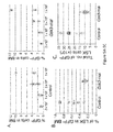
- FIG. 2 demonstrates that Gsk-3 depletion expands HSCs and HPCs in primary bone marrow (BM) transplants.
- BM bone marrow
- FIG. 2B depicts GFP + myeloid cells (Gr1 + CD11b + ) in peripheral blood for 10 control and 9 Gsk3-rnai-C2 recipients after BM transplantation (BMT; arrow).
- FIG. 2C Depicted in FIG. 2C are immunoblots for GSK-3alpha/beta and beta-catenin in BM from primary recipients 16 weeks after transplantation. Data represent independent replicates from 6 control and 6 Gsk3-rnai recipients.
- FIG. 2D depicts the percent GFP + LSK cells in control and Gsk3-rnai-C2 primary recipients.
- FIG. 2E depicts absolute number of GFP + LSK, LSK CD34 ⁇ Flk2 ⁇ , and LSK CD34 + Flk2 ⁇ cells.
- FIG. 2F depicts representative FCM showing GFP + cells in the HSC-containing LSK fraction (red gate) for control and Gsk3-rnai-C2 primary recipients.
- FIG. 2G depicts representative FCM using SLAM markers, where the difference between control and Gsk3-rnai was significant (P ⁇ 0.05). The numbers in FIGS. 2F and 2G indicate percent cells within gates.
- FIG. 2H depicts colony formation using GFP + cells plated in methylcellulose with cytokines and scored for CFU-C. Data represent mean colonies per well performed in duplicate groups for 5 mice per construct repeated in 3 separate experiments. *P ⁇ 0.05 versus respective control value.
- FIG. 3 depicts annexin V staining and cellularity of bone marrow harvested from transplant recipients.
- FIG. 3A depicts a flow cytometric detection of Annexin V using bone marrow cells harvested from 1° recipients after 4 month bone marrow transplantation from both control and Gsk3 RNAi Annexin V was measured in the GFP+LSK gated population.
- FIG. 3B depicts the total number of bone marrow cells and number of GFP+ cells recovered in bone marrow from 1° recipients after 4 month transplant.
- FIG. 3C depicts the total number and number of GFP+ cells recovered in bone marrow from 2° recipients after 4 months.
- FIG. 3D depicts the total number and number of GFP+ cells recovered in bone marrow from 3° recipients after 4 months.
- FIG. 4 demonstrates that Gsk-3 knockdown increases cycling of the HSC-enriched LSK cell population.
- sorted GFP + LSK and GFP + LSK Flk2 ⁇ cells from primary recipients of control and Gsk3-rnai 4 months after BM transplantation were stained with Hoechst and Pyronin and analyzed by FCM, as depicted in FIG. 4A .
- Representative FACS data are shown for control versus Gsk3-rnai-C2. As depicted in FIG.
- FIG. 5 demonstrates that Gsk3-depleted HSCs are functionally deficient.
- FIG. 5A limiting dilution experiments were performed with 4 doses (x axis) of GFP + test BM from vector-control and Gsk3-rnai primary recipients (4 donors per group) combined with a fixed number (2 ⁇ 10 5 ) unlabeled competing cells transplanted into groups of at least 5 recipients per dose. Chimerism at 4 months after transplantation for each dose is represented as the percentage of GFP + cells in BM for control and Gsk3-rnai. Depicted in FIG.
- FIG. 5B are percent donor-derived immunophenotypic HSCs/HPCs (as GFP + LSK cells) in the 1 ⁇ 10 6 test cell group 4 months after transplantation.
- FIG. 5C are absolute number of donor-derived immunophenotypic HSCs/HPCs (as GFP + LSK cells) in the 1 ⁇ 10 6 test cell group 4 months after transplantation.
- FIG. 6 demonstrates that Gsk-3 knockdown depletes HSCs in serial BM transplants.
- FIG. 6A noncompetitive serial transplants were performed by transplanting 2 ⁇ 10 5 sorted GFP + cells from primary recipients of control or Gsk3-rnai transduced BM into lethally irradiated recipients (10 mice per group). Survival of secondary recipients receiving control or Gsk3-rnai BM is shown as a Kaplan-Meier plot.
- FIG. 6B depicts percent HSC-containing LSK fraction in control and Gsk3-rnai secondary recipients.
- FIG. 6C depicts absolute number of GFP + LSK, LSK CD34 ⁇ Flk2 ⁇ , and LSK CD34 + Flk2 ⁇ cells in control and Gsk3-rnai secondary recipients.
- FIG. 6D depicts representative FCM data, presented as the distribution of CD34 ⁇ Flk2 ⁇ , CD34 + Flk2 ⁇ , and CD34 + Flk2 ⁇ , which immunophenotypically correspond to LT-HSCs, ST-HSCs, and MPPs in the LSK population, from control and Gsk3-rnai secondary recipients. Percent cells are shown for the indicated gates. As depicted in FIG.
- a colony formation assay with sorted GFP + cells from control and Gsk3-rnai secondary recipient BM was performed and scored as in FIG. 2 using GFP + BM from 5 control and 5 Gsk3-rnai mice.
- FIG. 6F the frequencies of common myeloid progenitor (CMP), granulocyte-monocyte progenitor (GMP), and megakaryocyte-erythroid progenitor (MEP) cells were measured by detection of CD16/32 and CD34 expression in the lineage ⁇ sca-1 ⁇ c-kit + gated population.
- CMP common myeloid progenitor
- GMP granulocyte-monocyte progenitor
- MEP megakaryocyte-erythroid progenitor
- FIG. 6G depicts lethally irradiated mice were reconstituted with 4 ⁇ 10 5 sorted GFP + BM cells from secondary recipients of vector or Gsk3-rnai transduced BM.
- the Kaplan-Meier survival curve shows the survival of tertiary recipients of BM from control or Gsk3-rnai mice.
- FIG. 6H depicts absolute number of immunophenotypic HSCs/HPCs, as LSK cells, in control and Gsk3-rnai tertiary recipients. *P ⁇ 0.05.
- FIG. 7 demonstrates that ⁇ -catenin is required for the increase in HSCs/HPCs induced by Gsk3-rnai.
- BM cells were harvested from Mx-Cre; ⁇ -caten fl/fl mice with or without injection of polyI:polyC for 14 days, transduced with control or Gsk3-rnai carrying lentivirus, and transplanted into lethally irradiated recipient mice. After 4 months, percentage and absolute number of HSC-containing LSK fraction were compared among the 4 groups. As depicted in FIG.
- FIG. 7B BM cells were harvested at 4 months from primary recipients of WT and Mx-Cre; ⁇ -catenin fl/fl mice transduced with vector control or Gsk3-rnai lentivirus (from primary recipient mice in FIG. 7A ) and transplanted into lethally irradiated secondary hosts. After 4 months, percentage and absolute number of HSC-containing LSK fraction were compared among the 4 groups.
- FIG. 7C depicts a summary of serial transplantation data in WT versus ⁇ -catenin CKO mice.
- FIG. 8 depicts increased colony formation induced by Gsk3-rnai requires ⁇ -catenin.
- a colony formation assay was performed using sorted GFP+ cells harvested from 1° recipients ( FIG. 8A ) or 2° recipients ( FIG. 8B ) of wild-type (left) and ⁇ -catenin KO (right) marrow transduced with control (open boxes) or Gsk3-rnai (filled boxes) lentivirus. Data represent mean number of colonies per well for five mice per construct repeated in three separate experiments.
- FIG. 9 demonstrates increased HSCs/HPCs in rapamycin-treated recipients of Gsk3-depleted BM and increased survival of tertiary recipients receiving Gsk3-depleted BM from rapamycin treated 2° hosts.
- BM was harvested from Mx-Cre; ⁇ -catenin fl/fl mice treated with or without polyI:polyC, transduced with control or Gsk3-rnai lentivirus, and transplanted into irradiated recipients.
- FIG. 9B depicts flow cytometric detection of phospho-ribosomal protein S6 with BM cells in FIG. 9A .
- FIG. 9C irradiated mice were reconstituted with BM transduced with control vector or Pten-rnai.
- phospho-GSK-3 and phospho-ribosomal protein S6 were assessed in GFP + cells by immunoblot. As depicted in FIG.
- FIG. 9D NIH3T3 cells were infected for 3 days with control or Pten-rnai lentivirus, and phospho-GSK-3 ⁇ / ⁇ in control and Pten-depleted cells was detected by immunoblot.
- FIG. 9E depicts increased percentage of LSK cells in bone marrow of 2° recipients of Gsk3-depleted BM that were treated with rapamycin for 8 weeks.
- GFP + cells (2 ⁇ 10 6 ) from primary recipients of control or Gsk3-rnai were transplanted into 10 irradiated recipients per group. After 1.5-2 months, secondary recipients were injected with rapamycin or vehicle every other day for 8 weeks. Percent GFP + LSK cells was compared among the 4 groups.
- FIG. 9F depicts absolute number of GFP + LSK cells as in FIG. 9E .
- FIG. 9G depicts colony formation with sorted GFP + cells from FIG. 9E .
- FIG. 9H depicts a Kaplan-Meier plot showing survival of tertiary recipients transplanted with BM from control- and rapamycin-treated secondary recipients in FIGS. 9E-9G . Illustrated is control vector BM from secondary recipients treated with vehicle or rapamycin and Gsk3-rnai-infected BM from secondary recipients treated with vehicle or rapamycin transplanted to lethally irradiated tertiary recipients. *P ⁇ 0.05.
- FIG. 10 demonstrates that Gsk3b KO depletes HSCs in serial BM transplants.
- Noncompetitive serial transplants were performed by transplanting 4 ⁇ 10 5 fetal liver cells (CD45.2) from E17.5 WT, Gsk3b +/ ⁇ , and Gsk3b ⁇ / ⁇ embryos into lethally irradiated recipient mice (CD45.1).
- FIGS. 10A and 10B depict reconstitution of peripheral blood, including B cells (B220 + ), T cells (CD3 + ), and myeloid cells (Mac-1 + GR-1 + ), in primary ( FIG. 10A ) and secondary ( FIG.
- FIG. 11 demonstrates that GSK-3 functions in 2 major pathways to regulate HSC self renewal and lineage commitment. Inhibition of GSK-3 activates Wnt and mTOR signaling. In the canonical Wnt pathway, GSK-3 and ⁇ -catenin bind to the Axin complex, along with APC. GSK-3 phosphorylates ⁇ -catenin, targeting it for rapid destruction. Wnt binding to the Fz/Lrp receptor complex causes inhibition of GSK-3, which in turn stabilizes ⁇ -catenin and activates Wnt target genes that promote progenitor proliferation and self renewal.
- GSK-3 phosphorylates and inhibits GSK-3.
- Tsc2 inhibiting the mTOR pathway.
- FIG. 12 demonstrates the effect of ⁇ -catenin conditional knockout on colony formation in Gsk3-depleted bone marrow.
- a colony formation assay was performed using sorted GFP+ cells from each group in 1° ( FIG. 12A ) and 2° ( FIG. 12B ) recipients. Data represent mean number of colonies per well for five mice per construct repeated in three separate experiments.
- FIG. 13 demonstrates ex vivo mouse HSC culturing and transplantation.
- c-kit+ cells were isolated from adult mouse bone marrow and cultured in serumfree, cytokine-free defined medium for 7 days with (triangle) or without (circle) our formulation. After 7 days, the entire culture was transplanted to lethally irradiated mice (5/group) and survival was monitored over more than 16 weeks. All control animals (cultured without additives or no transplant) died within 17 days. Mice receiving bone marrow cells cultured in cytokine-free, serum-free medium with additives survived over 16 weeks. The experiment has been repeated 3 times. Erythrocytic, myelocytic, lymphocytic, and megakaryocytic lineages were present in peripheral blood and bone marrow after 4 months.
- FIG. 14 demonstrates ex vivo culturing of HSCs from human umbilical cord blood.
- CD34+ cells from human UCB were isolated and either transplanted immediately to NSG mice (sample 1) or cultured in the cytokine-free, serum-free medium alone (sample 2), supplemented with our non-protein additives (sample 3), or with conventional cytokine cocktail (sample 4, includes IL3, SCF, FL, and Tpo) for either 4 days (shown) or 7 days (not shown).
- CD34+ cells increased 2-3 fold after one week when cultured with additives compared to no additives.
- Samples 2-4 were transplanted to NSG mice.
- mice were bled monthly for flow cytometric analysis of human/mouse chimerism in peripheral blood (not shown) and were sacrificed at 4 months for flow cytometric analysis of human/mouse chimerism in bone marrow.
- the percent of human CD45.1 over total mononuclear cells in the bone marrow effluent is shown for each group. There were 5 mice per group and the experiment has been repeated with similar results 3 times. Secondary transplantation showed that human cells in group 3, but none of the other groups, sustained long-term repopulating activity for myelocytic, erythrocytic, and lymphocytic lineages.
- the present invention provides a therapeutic approach to expand HSCs in vivo using currently available medications that target GSK-3 and mTOR.
- the present invention also provides a system and method for the ex vivo culturing of HSCs, where an mTOR inhibitor is combined with a GSK-3 inhibitor within the culturing conditions.
- the present invention is based on the discovery that Gsk-3 loss of function coupled with inhibition of mTOR provides for improved maintenance and expansion of HSCs in vivo and ex vivo.
- Gsk-3a/b (hereafter, Gsk-3 is used to refer to both genes) initially expand the HSC-enriched pool of lineage ⁇ sca-1 + c-kit + (LSK) cells, similar to the effects of lithium and other GSK-3 inhibitors, and this requires endogenous ⁇ -catenin function.
- Gsk3-deficient HSCs are progressively depleted, revealing an unexpected positive role for GSK-3 in the maintenance of HSC self renewal.
- an element means one element or more than one element.
- Allogeneic refers to a graft derived from a different animal of the same species.
- autologous is meant to refer to any material derived from the same individual to which it is re-introduced.
- progenitor cell refers either to a pluripotent, or lineage-uncommitted, progenitor cell, which is potentially capable of an unlimited number of mitotic divisions to either renew itself or to produce progeny cells which will differentiate into the desired cell type.
- pluripotent stem cells lineage-committed progenitor cells are generally considered to be incapable of giving rise to numerous cell types that phenotypically differ from each other. Instead, progenitor cells give rise to one or possibly two lineage-committed cell types.
- substantially purified cell is a cell that is essentially free of other cell types.
- a substantially purified cell refers to a cell which has been purified from other cell types with which it is normally associated in its naturally occurring state.
- Transplant refers to a biocompatible lattice or a donor tissue, organ or cell, to be transplanted.
- An example of a transplant may include but is not limited to a tissue, a stem cell, a neural stem cell, a skin cell, bone marrow, and solid organs such as heart, pancreas, kidney, lung and liver.
- a “therapeutically effective amount” is the amount of a therapeutic composition, such as a GSK-3 and/or mTOR inhibitor, sufficient to provide a beneficial effect to a mammal or culture or culturing system to which the composition is administered.
- endogenous refers to any material from or produced inside an organism, cell or system.
- Exogenous refers to any material introduced from or produced outside an organism, cell, or system.
- “Expandability” is used herein to refer to the capacity of a cell to proliferate for example to expand in number, or in the case of a cell population, to undergo population doublings.
- “Graft” refers to a cell, tissue, organ or otherwise any biological compatible lattice for transplantation.
- growth medium is meant to refer to a culture medium that promotes growth of cells.
- module is meant to refer to any change in biological state, i.e. increasing, decreasing, and the like.
- proliferation is used herein to refer to the reproduction or multiplication of similar forms, especially of cells. That is, proliferation encompasses production of a greater number of cells, and can be measured by, among other things, simply counting the numbers of cells, measuring incorporation of 3 H-thymidine into the cells, and the like.
- a “promotion factor” refers to any molecule that assists in maintaining and/or proliferating cells.
- an “instructional material” includes a publication, a recording, a diagram, or any other medium of expression which can be used to communicate the usefulness of the compositions and methods of the invention.
- the instructional material of the kit of the invention may, for example, be affixed to a container which contains components such as, without limitation, a nucleic acid, peptide, cells, one or more inhibitors and/or composition of the invention or be shipped together with a container which contains such components.
- the instructional material may be shipped separately from the container with the intention that the instructional material and the components be used cooperatively by the recipient.
- ranges throughout this disclosure, various aspects of the invention can be presented in a range format. It should be understood that the description in range format is merely for convenience and brevity and should not be construed as an inflexible limitation on the scope of the invention. Accordingly, the description of a range should be considered to have specifically disclosed all the possible subranges as well as individual numerical values within that range. For example, description of a range such as from 1 to 6 should be considered to have specifically disclosed subranges such as from 1 to 3, from 1 to 4, from 1 to 5, from 2 to 4, from 2 to 6, from 3 to 6 etc., as well as individual numbers within that range, for example, 1, 2, 2.7, 3, 4, 5, 5.3, and 6. This applies regardless of the breadth of the range.
- Hematopoietic stem cell (HSC) homeostasis depends on the balance between self renewal and lineage commitment, but what regulates this decision is not well understood.
- Gsk-3 glycogen synthase kinase-3
- BM bone marrow
- the present invention is based on the discovery that GSK-3 regulates both Wnt and mTOR signaling in mouse HSCs, where these pathways promote HSC self renewal and lineage commitment, respectively, such that inhibition of Gsk-3 in the presence of rapamycin expands the HSC pool in vivo. Therefore, the present invention provides a therapeutic approach to expand HSCs in vivo using currently available medications that target GSK-3 and mTOR, and provides a compelling explanation for the clinically prevalent hematopoietic effects observed in individuals prescribed the GSK-3 inhibitor lithium.
- the present invention also provides a system and method for the ex vivo culturing of HSCs, where an mTOR inhibitor is combined with a GSK-3 inhibitor within the culturing conditions. By simultaneously inhibiting GSK-3 and mTOR, HSCs can survive and increase in number within ex vivo culturing conditions.
- the present invention may also allow for expansion of human umbilical cord blood HSCs or adult donor HSCs, and improve the frequency of engraftment and shortened the time to hematopoietic recovery in recipient bone marrow transplant patients.
- the advantage of cord blood is that this can be transplanted with markedly reduced risk of graft versus host disease and the potential for reduced immunosuppressive therapy of bone marrow transplant recipients.
- the present invention also bypasses the need for expensive cytokines to diminish the potential side effects associated with introducing foreign DNA into bone marrow cells.
- treatment may include ex vivo culture or included in the treatment regiment for transplant recipients.
- HSCs bone marrow
- BM cells may be collected by puncturing a bone and flushing out the bone marrow cells with a syringe. About 1 in every 100,000 cells in the marrow is a long-term, blood-forming stem cell. Other cells present include stromal cells, stromal stem cells, blood progenitor cells, and mature and maturing white and red blood cells.
- donor cells may be harvested from peripheral, circulating blood. While a small number of stem and progenitor cells circulate in the bloodstream, improved cell counts may be obtained by injecting the donor with a cytokine, such as granulocyte-colony stimulating factor (GCSF). The donor is injected with GCSF a few days before the cell harvest. To collect the cells, an intravenous tube is inserted into the donor's vein and the blood is passed through a filtering system that extracts out CD34 + white blood cells and returns the red blood cells to the donor. Of the CD34 + cells collected, approximately 5 to 20 percent may be HSCs, where the remaining cells are a mixture of stem cells, progenitors, and white blood cells of varying degrees of maturity.
- a cytokine such as granulocyte-colony stimulating factor (GCSF).
- GCSF granulocyte-colony stimulating factor
- blood from the human umbilical cord and placenta may be collected and used as a rich source of HSCs.
- developing blood-producing tissues of fetal animals may provide a source of HSCs for research purposes, as hematopoietic cells appear early in the development of all vertebrates.
- an ex vivo culture system and process for the maintenance and expansion HSCs is provided, such that the expanded cells can be engrafted into patients without losing their capability for multilineage differentiation and reconstitution.
- HSCs have the capability of both self-renewal and the ability to differentiate into distinct hematopoietic cell lineages, such as myeloid, B-cell and T-cell lineages.
- the ex vivo maintenance and expansion of HSCs can be achieved by culturing HSCs in the presence of GSK-3 and mTOR inhibitors.
- mammalian HSCs preferably human HSCs
- the culture medium further comprises any culture medium suitable for culturing HSCs.
- the culturing medium may include serum, while in other embodiments, the culturing medium may be serum-free.
- Standard culture media for HSCs typically contains a variety of essential components required for cell viability, including inorganic salts, carbohydrates, hormones, essential amino acids, vitamins, and the like.
- the conditions for culturing the HSCs should be as close to physiological conditions as possible.
- the culturing medium may include components that are known to those of ordinary skill in the art and may comprise such components as RPMI 1640, HEPES, FCS, and common antibiotics. Any commercially available HSC culturing medium may be used, such as X-VIVOTM medium, such that the GSK-3 inhibitor and the mTOR inhibitor may be added to the commercially available HSC culturing medium in effective amounts as contemplated and described hereinthroughout.
- isolated HSC are cultured in a culture system which comprises a culture medium containing the GSK-3 inhibitor and the mTOR inhibitor.
- a culture system is suitable for achieving an expansion, such as about a 1-fold expansion, a 2-fold expansion, a 3-fold expansion, a 4-fold expansion, a 5-fold expansion, a 10-fold expansion, a 20-fold expansion, a 50-fold expansion, a 100-fold expansion, a 150-fold expansion, or a 200-fold expansion or more and any and all whole or partial increments there between of the HSCs.
- the expanded HSCs may retain their capability for multilineage differentiation upon introduction into the body of a patient, preferably a human patient.
- HSCs can be cultured in the presence of the at least one GSK-3 inhibitor and at least one mTOR inhibitor for approximately 7 days.
- the HSCs can be cultured in the presence of at least one GSK-3 inhibitor and at least one mTOR inhibitor for approximately 14 days, approximately 28 days, approximately 60 days or approximately 120 days or more, and any and all whole and partial increments there between.
- GSK-3 is involved in many cellular and physiological events, including Wnt and Hedgehog signaling, transcription, insulin action, cell-division cycle, response to DNA damage, cell death, cell survival, patterning and axial orientation during development, differentiation, neuronal functions, circadian rhythm and others.
- Wnt and Hedgehog signaling transcription, insulin action, cell-division cycle, response to DNA damage, cell death, cell survival, patterning and axial orientation during development, differentiation, neuronal functions, circadian rhythm and others.
- two genes which map to 19q13.2 and 3q13.3, encode two distinct but closely related GSK-3 forms, GSK-3a (51 kDa) and GSK-3b (47 kDa).
- GSK-3 is regulated at multiple levels.
- GSK-3b is regulated by post-translational phosphorylation of Ser9 (inhibitory) and Tyr216 (activating) (Ser21 and Tyr279, respectively, in GSK-3a).
- Phosphorylated Ser9 in the N-terminal domain of GSK-3b acts as a pseudo-substrate that blocks the access of substrates to the catalytic site.
- Unphosphorylated Tyr216 in the T-loop domain prevents access of substrates to the catalytic site, and phosphorylation releases this inhibition.
- GSK-3b is regulated by interactions with many other proteins.
- Axin and presenilin act as docking proteins that allow the substrates to make contact with the priming kinase [casein kinase (CK1) and protein kinase A, respectively] and GSK-3. Docking proteins might, thus, specify different GSK-3 functions in the cell.
- GSK-3 action requires the priming phosphorylation of its substrates by another kinase on a serine residue located four amino acids C-terminal to the GSK-3 phosphorylation site.
- GSK-3 is regulated through its intracellular distribution.
- lithium such as in the form of LiCl
- GSK-3 may be used as an inhibitor of GSK-3.
- any other GSK-3 inhibitor in any form may be used, either separately or in combination, as would be understood by those skilled in the art, provided such inhibitors do not orientate the differentiation of the HSCs into well-defined, committed cell lineages, unless such a result is desired.
- Other such GSK-3 inhibitors may include, without limitation, 6BIO, CHIR-911, DW21, AR-A014418, TDZD and their related compounds.
- mTOR is a kinase protein predominantly found in the cytoplasm of the cell. It acts as a central regulator of many biological processes that are essential for cell proliferation, angiogenesis, and cell metabolism. mTOR exerts its effects primarily by turning on the cell's translational machinery, and is therefore responsible for protein synthesis. mTOR is a key intracellular point of convergence for a number of cellular signaling pathways.
- rapamycin which is also known as sirolimus
- sirolimus may be used as an mTOR inhibitor.
- rapamycin binds to an abundant intracellular binding protein, FKBP-12, and the drug:FKPB-12 complex binds at the rapamycin binding domain.
- temsirolimus which is also known as CCI-779
- everolimus which is also known as RAD001
- Everolimus and temsirolimus are considered rapamycin derivatives, the primary difference being their pharmacokinetic and pharmacologic properties.
- Temsirolimus was created by adding an ester to a rapamycin carbon, and adding an ether created everolimus.
- any other mTOR inhibitor in any form may be used, either separately or in combination, as would be understood by those skilled in the art.
- an effective amount of each inhibitor is added to the culturing medium.
- the actual amount of inhibitor added to the culturing medium can vary according to the culturing volume, additional culturing components, and the particular inhibitor selected. For example, about 5 mM LiCl and about 5 nM rapamycin may be an effective amount of each inhibitor, and added to X-VIVO15 medium for culturing in multi-well plates.
- the culture medium may include at least one cytokine.
- cytokine may include interleukins 3 and 6 (Il-3 and Il-6), stem cell factor (SCF), granulocyte-macrophage colony stimulating factor (GM-CSF), Flt-3 ligand (FL), and thrombopoietin (TPO).
- SCF stem cell factor
- GM-CSF granulocyte-macrophage colony stimulating factor
- FL Flt-3 ligand
- TPO thrombopoietin
- the medium can include Il-3 or Il-6, or a combination thereof, or it may include TPO or CSF or a combination thereof. It is believed that the addition of at least one cytokine can enhance the expansion of HSC by at least about 50%, preferably at least about 100%.
- the culture medium may include at least one stem cell expansion promoting factor which is distinct from the aforementioned cytokines.
- HSCs can be maintained and expanded ex vivo in the presence of stromal cell medium containing such promotion factors, such as a culture medium collected from cultured murine stromal cells where the stromal cells were cultured in the presence of a leukemia inhibitory factor.
- the cells can be harvested and collected for immediate experimental or therapeutic use, or cryopreserved for use at a later time.
- HSCs as described herein, may be cryopreserved according to routine procedures.
- about one to ten million cells are cryopreserved in medium with about 10% DMSO in vapor phase of Liquid N 2 .
- Frozen cells can be thawed by swirling in a 37° C. bath, resuspended in fresh proliferation medium, and grown as usual.
- Cryopreservation is a procedure common in the art and as used herein encompasses all procedures currently used to cryopreserve cells for future analysis and use.
- the cells can be harvested and subjected to flow cytometry to evaluate cell surface markers to assess the change in phenotype of the cells in view of the culture conditions.
- HSCs may be characterized using any one of numerous methods in the art and methods disclosed herein.
- the cells may be characterized by the identification of surface and intracellular proteins, genes, and/or other markers indicative of the cells.
- the cells of the present invention can also be used to express a foreign protein or molecule for a therapeutic purpose or for a method of tracking their integration and differentiation in a patient's tissue.
- the invention encompasses expression vectors and methods for the introduction of exogenous DNA into the cells with concomitant expression of the exogenous DNA in the cells such as those described, for example, in Sambrook et al. (2001, Molecular Cloning: A Laboratory Manual, Cold Spring Harbor Laboratory, New York), and in Ausubel et al. (1997, Current Protocols in Molecular Biology, John Wiley & Sons, New York).
- the isolated nucleic acid can encode a molecule used to track the migration, integration, and survival of HSCs once they are placed in the patient, or they can be used to express a protein that is mutated, deficient, or otherwise dysfunctional in the patient.
- Proteins for tracking can include, but are not limited to green fluorescent protein (GFP), any of the other fluorescent proteins (i.e., enhanced green, cyan, yellow, blue and red fluorescent proteins; Clontech, Palo Alto, Calif.), or other tag proteins (i.e., LacZ, FLAG-tag, Myc, His 6 , and the like) disclosed elsewhere herein.
- GFP green fluorescent protein
- any of the other fluorescent proteins i.e., enhanced green, cyan, yellow, blue and red fluorescent proteins
- Clontech Palo Alto, Calif.
- other tag proteins i.e., LacZ, FLAG-tag, Myc, His 6 , and the like
- the present invention is also useful for obtaining HSCs that express an exogenous gene, so that the HSCs can be used, for example, for cell therapy or gene therapy. That is, the present invention allows for the production of large numbers of HSCs that express an exogenous gene.
- the exogenous gene can, for example, be an exogenous version of an endogenous gene (i.e., a wild type version of the same gene can be used to replace a defective allele comprising a mutation).
- the exogenous gene is usually, but not necessarily, covalently linked with (i.e., “fused with”) one or more additional genes.
- Exemplary “additional” genes include a gene used for “positive” selection to select cells that have incorporated the exogenous gene, and a gene used for “negative” selection to select cells that have incorporated the exogenous gene into the same chromosomal locus as the endogenous gene or both.
- An HSC expressing a desired exogenous gene can be used to provide the product of the exogenous gene to a cell, tissue, or whole mammal where a higher level of the gene product can be useful to treat or alleviate a disease, disorder or condition associated with abnormal expression, and/or activity. Therefore, the invention includes an HSC expressing an exogenous gene where increasing expression, protein level, and/or activity of the desired gene product can be useful to treat or alleviate a disease, disorder or condition.
- gene constructs which comprise nucleotide sequences that encode heterologous proteins are introduced into the HSCs. That is, the cells are genetically modified to introduce a gene whose expression has therapeutic effect in the individual.
- HSCs from the individual to be treated or from another individual, or from a non-human animal may be genetically modified to replace a defective gene and/or to introduce a gene whose expression has therapeutic effect in the individual being treated.
- genetic modification refers to the stable or transient alteration of the genotype of an HSC by intentional introduction of exogenous DNA.
- DNA may be synthetic, or naturally derived, and may contain genes, portions of genes, or other useful DNA sequences.
- genetic modification as used herein is not meant to include naturally occurring alterations such as that which occurs through natural viral activity, natural genetic recombination, or the like.
- Exogenous DNA may be introduced to an NSC using viral vectors (retrovirus, modified herpes viral, herpes-viral, adenovirus, adeno-associated virus, lentiviral, and the like) or by direct DNA transfection (lipofection, calcium phosphate transfection, DEAE-dextran, electroporation, and the like).
- viral vectors retrovirus, modified herpes viral, herpes-viral, adenovirus, adeno-associated virus, lentiviral, and the like
- direct DNA transfection lipofection, calcium phosphate transfection, DEAE-dextran, electroporation, and the like.
- the heterologous gene is operably linked to regulatory sequences required to achieve expression of the gene in the cell.
- regulatory sequences typically include a promoter and a polyadenylation signal.
- the gene construct is preferably provided as an expression vector that includes the coding sequence for a heterologous protein operably linked to essential regulatory sequences such that when the vector is transfected into the cell, the coding sequence will be expressed by the cell.
- the coding sequence is operably linked to the regulatory elements necessary for expression of that sequence in the cells.
- the nucleotide sequence that encodes the protein may be cDNA, genomic DNA, synthesized DNA or a hybrid thereof or an RNA molecule such as mRNA.
- the gene construct includes the nucleotide sequence encoding the beneficial protein operably linked to the regulatory elements and may remain present in the cell as a functioning cytoplasmic molecule, a functioning episomal molecule or it may integrate into the cell's chromosomal DNA.
- Exogenous genetic material may be introduced into cells where it remains as separate genetic material in the form of a plasmid.
- linear DNA which can integrate into the chromosome may be introduced into the cell.
- reagents which promote DNA integration into chromosomes may be added.
- DNA sequences which are useful to promote integration may also be included in the DNA molecule.
- RNA may be introduced into the cell.
- the regulatory elements for gene expression include: a promoter, an initiation codon, a stop codon, and a polyadenylation signal. It is preferred that these elements be operable in the cells of the present invention. Moreover, it is preferred that these elements be operably linked to the nucleotide sequence that encodes the protein such that the nucleotide sequence can be expressed in the cells and thus the protein can be produced. Initiation codons and stop codons are generally considered to be part of a nucleotide sequence that encodes the protein. However, it is preferred that these elements are functional in the cells. Similarly, promoters and polyadenylation signals used must be functional within the cells of the present invention.
- promoters useful to practice the present invention include but are not limited to promoters that are active in many cells such as the cytomegalovirus promoter, SV40 promoters and retroviral promoters.
- Other examples of promoters useful to practice the present invention include but are not limited to tissue-specific promoters, i.e. promoters that function in some tissues but not in others; also, promoters of genes normally expressed in the cells with or without specific or general enhancer sequences.
- promoters are used which constitutively express genes in the cells with or without enhancer sequences. Enhancer sequences are provided in such embodiments when appropriate or desirable.
- cells of the present invention can be transfected using well known techniques readily available to those having ordinary skill in the art. Exogenous genes may be introduced into the cells using standard methods where the cell expresses the protein encoded by the gene. In some embodiments, cells are transfected by calcium phosphate precipitation transfection, DEAE dextran transfection, electroporation, microinjection, liposome-mediated transfer, chemical-mediated transfer, ligand mediated transfer or recombinant viral vector transfer.
- recombinant viral vectors are used to introduce DNA with desired sequences into the cell.
- recombinant retrovirus vectors are used to introduce DNA with desired sequences into the cells.
- standard calcium phosphate, DEAE dextran or lipid carrier mediated transfection techniques are employed to incorporate desired DNA into dividing cells. Standard antibiotic resistance selection techniques can be used to identify and select transfected cells.
- DNA is introduced directly into cells by microinjection.
- well-known electroporation or particle bombardment techniques can be used to introduce foreign DNA into the cells.
- a second gene is usually co-transfected or linked to the therapeutic gene. The second gene is frequently a selectable antibiotic-resistance gene. Transfected cells can be selected by growing the cells in an antibiotic that will kill cells that do not take up the selectable gene. In most cases where the two genes are unlinked and co-transfected, the cells that survive the antibiotic treatment have both genes in them and express both of them.
- Isolated HSCs are useful in a variety of ways. These cells can be used to reconstitute cells in a mammal whose cells have been lost through disease or injury. Genetic diseases may be treated by genetic modification of autologous or allogeneic HSCs to correct a genetic defect or to protect against disease. Diseases related to the lack of a particular secreted product such as a hormone, an enzyme, a growth factor, or the like may also be treated using HSCs.
- HSCs isolated and cultured as described herein can be used as a source of progenitor cells and committed cells to treat selected diseases as would be understood by those skilled in the art, including such diseases and/or injuries where the replacement of tissue by the cells of the present invention can result in a treatment or alleviation of the disease and/or injuries.
- HSCs cultured or expanded as described herein can be used, as cultured, or they can be used following differentiation into selected cell types, to treat a variety of disorders known in the art to be treatable using HSCs.
- the HSCs that are useful in these treatment methods include those that have, and those that do not have, an exogenous gene inserted therein.
- the present invention encompasses methods for administering the cells of the present invention to an animal, including humans, in order to treat diseases where the introduction of new, undamaged cells will provide some form of therapeutic relief.
- the cells of the present invention can be administered as HSCs or HSCs that have been induced to differentiate to exhibit at least one characteristic of the targeted cell type.
- HSCs can be administered to a recipient as an undifferentiated cell and upon receiving signals and cues from the surrounding milieu, can differentiate into a desired cell type dictated by the neighboring cellular milieu.
- HSCs can be administered as a purified population of cells, or as a heterogeneous mixture of cells, that contains HSCs as the active agent or cell type.
- the cells can be prepared for grafting to ensure long term survival in the in vivo environment.
- cells are propagated in a suitable culture medium for growth and maintenance of the cells and are allowed to grow to confluency.
- Any osmotically balanced solution which is physiologically compatible with the host subject may be used to suspend and inject the donor cells into the host.
- Formulations of a pharmaceutical composition suitable for parenteral administration comprise the active ingredient, i.e. the cells, combined with a pharmaceutically acceptable carrier, such as sterile water or sterile isotonic saline.
- a pharmaceutically acceptable carrier such as sterile water or sterile isotonic saline.
- Such formulations may be prepared, packaged, or sold in a form suitable for bolus administration or for continuous administration.
- Injectable formulations may be prepared, packaged, or sold in unit dosage form, such as in ampules or in multi-dose containers containing a preservative.
- Formulations for parenteral administration include, but are not limited to, suspensions, solutions, emulsions in oily or aqueous vehicles, pastes, and implantable sustained-release or biodegradable formulations.
- Such formulations may further comprise one or more additional ingredients including, but not limited to, suspending, stabilizing, or dispersing agents.
- the invention also encompasses grafting HSCs (or differentiated HSCs) in combination with other therapeutic procedures to treat a disease or trauma.
- the cells of the invention may be co-grafted with other cells, both genetically modified or non-genetically modified cells which exert beneficial effects on the patient. Therefore the methods disclosed herein can be combined with other therapeutic procedures as would be understood by one skilled in the art once armed with the teachings provided herein.
- the mode of administration of the cells of the invention may vary depending on several factors including the type of disease being treated, the age of the mammal, whether the cells are differentiated or not, whether the cells have heterologous DNA introduced therein, and the like.
- Cells may be introduced to the desired site by direct injection, or by any other means used in the art for the introduction of compounds.
- the cells can be administered into a host in a wide variety of ways. Modes of administration include, but are not limited to, intravascular, intracerebral, parenteral, intraperitoneal, intravenous, epidural, intraspinal, intrastemal, intra-articular, intra-synovial, intrathecal, intra-arterial, intracardiac, or intramuscular.
- Transplantation of the cells of the present invention can be accomplished using techniques well known in the art as well as those described herein or as developed in the future.
- the present invention comprises a method for transplanting, grafting, infusing, or otherwise introducing HSCs into a mammal, preferably, a human.
- the cells are prepared as described herein.
- the cells are from the patient for which the cells are being transplanted into (autologous transplantation).
- the present invention further provides for methods for use in various therapeutic interventions.
- the methods of the present invention may be used in the treatment of bone marrow transplant donors and for recipients clinically by providing to either donors or recipients FDA approved medications that inhibit GSK-3 (such as lithium salts or other compounds that contain lithium) and mTOR (such as rapamycin) to expand HSCs and accelerate recovery from bone marrow suppression.
- GSK-3 such as lithium salts or other compounds that contain lithium
- mTOR such as rapamycin
- the present invention provides a method for expansion of HSCs in culture using GS K-3 inhibitors and mTOR inhibitors. As demonstrated herein, inhibition of GSK-3 will activate downstream Wnt signaling in HSCs and will promote expansion of HSCs.
- this can be a complex using FDA approved medications, such as lithium salts, which are already widely used clinically and are relatively safe.
- FDA approved medications such as lithium salts
- Applications apply to stem cells and other tissues, for example the skin and hair follicles (regeneration of skin and hair, burns, etc.), colon (regeneration of colonic epithelium, for example, after chemotherapy), and central nervous system (neuronal regeneration for neurodegenerative disorders).
- the present invention may provide for ex vivo expansion of HSCs and improve transplantation, such as bone marrow transplantation, by expanding either short-term or long-term HSC's, or both, and improving initial survival, such as during the marrow suppression/peripheral cytopenia phase of BMT.
- the method includes harvesting and culturing a patient's HSCs in culture medium containing at least one GSK-3 inhibitor and at least one mTOR inhibitor for treatment of cancers of the blood, such as leukemia and lymphoma, where the patient's cancerous hematopoietic cells were destroyed via radiation or chemotherapy and stem cells are replaced by stem cell transplantation (SCT).
- SCT stem cell transplantation
- the method includes replacing the cultured HSCs via a bone marrow transplant.
- stem cell transplant encompasses all forms of HSC transplant, including bone marrow, peripheral blood, and cord blood HSCs.
- the method includes the transplanting of HSCs collected from the peripheral circulation of a matched donor.
- the method includes the transplanting of HSCs collected from human umbilical cord blood (hUCB).
- HSCs collected from human umbilical cord blood
- the cultured HSCs may be used to treat cancers of the blood such as acute lymphoblastic leukemia, acute myeloblastic leukemia, chronic myelogenous leukemia (CML), Hodgkin's disease, multiple myeloma, and non-Hodgkin's lymphoma and neoplastic diseases of the blood including myelodysplasia and myeloproliferative disease.
- the aforementioned methods may be used in the treatment of bone marrow failure syndromes including aplastic anemia, whether acquired or inherited, and paroxysmal nocturnal hemoglobinuria.
- the aforementioned methods may be used in the treatment of hereditary blood disorders, such as different types of inherited anemia (failure to produce blood cells), and inborn errors of metabolism (genetic disorders characterized by defects in key enzymes need to produce essential body components or degrade chemical byproducts).
- blood disorders treatable by the methods of the present invention may include aplastic anemia, beta-thalassemia, Blackfan-Diamond syndrome, globoid cell leukodystrophy, sickle-cell anemia, severe combined immunodeficiency, X-linked lymphoproliferative syndrome, and Wiskott-Aldrich syndrome.
- inborn errors of metabolism that are treatable with the methods of the present invention include Hunter's syndrome, Hurler's syndrome, Lesch Nyhan syndrome, and osteopetrosis. Because bone marrow transplantation has carried a significant risk of death, this is usually a treatment of last resort for otherwise fatal diseases.
- the methods of the present invention may also be used in treating cancer patients with an autologous stem cell transplant to replace the cells destroyed by chemotherapy.
- the method may include mobilizing HSCs, collecting them from peripheral blood, and culturing the cells in culturing medium containing at least one GSK-3 inhibitor and at least one mTOR inhibitor.
- the cells may then be stored, such as via cryopreservation, while the patient undergoes intensive chemotherapy or radiotherapy to destroy the cancer cells. Once the chemotherapy or radiotherapy is completed, the patient may receive a transfusion of his or her stored HSCs.
- the methods of the present invention may be used for treating otherwise untreatable tumors, such as for the treatment of metastatic kidney cancer or solid tumors that resist standard therapy, including cancer of the lung, prostate, ovary, colon, esophagus, liver, and pancreas.
- the method may include an allogeneic stem cell transplant from an HLA-matched sibling whose HSCs are collected peripherally and cultured in a culturing medium containing at least one GSK-3 inhibitor and at least one mTOR inhibitor.
- the method includes transfusing the donor's cultured HSCs into the patient, followed by monitoring the patient's immune cells, such as via DNA fingerprinting, to follow the engraftment of the donor's cells and regrowth of the patient's own blood cells.
- the aforementioned methods may be used for treating other diseases, such as diabetes, rheumatoid arthritis, system lupus erythematosis and other autoimmune diseases.
- HSCs cultured in the presence of at least one GSK-3 inhibitor and at least one mTOR inhibitor may be genetically modified for delivery of genes to repair damaged cells.
- the methods of the present invention may be used for treating any disease, disorder or condition in which HSCs are used.
- the present invention further includes various kits which comprise a culturing medium, including at least one GSK-3 inhibitor and at least one mTOR inhibitor, and instructional materials which describe use of the culturing medium to perform the methods of the invention.
- a culturing medium including at least one GSK-3 inhibitor and at least one mTOR inhibitor
- instructional materials which describe use of the culturing medium to perform the methods of the invention.
- the invention includes a kit for maintaining and expanding HSCs ex vivo.
- the kit is used in the same manner as the methods disclosed herein for the present invention.
- the kit may be used to maintain and expand freshly harvested HSCs or cryopreserved HSCs in culture.
- the kit may further include cyropreserved HSCs.
- the cryopreserved HSCs may be provided in an appropriate amount as set forth elsewhere herein.
- the kit may further include genetically modified HSCs.
- the kit may further include at least one cytokine or a promotion factor.
- the kit comprises instructional material for the use of the kit. These instructions simply embody any of the examples and embodiments provided herein.
- compositions can be further approximated through analogy to compounds known to exert the desired effect.
- mice C57BL/6 WT (CD45.2), CD45.1 congenic (The Jackson Laboratory), and Mx-cre; ⁇ -catenin fl/fl mice were bred in house in a pathogen-free mouse facility of the University of Pennsylvania. Transplant recipients were female mice 10-12 weeks old. All work with mice was done according to a protocol reviewed and approved by the Institutional Animal Care and Use Committee at the University of Pennsylvania.
- BM cells were flushed from the long bones (tibias and femurs) of mice with Hank's buffered salt solution without calcium or magnesium, supplemented with 2% heat-inactivated calf serum.
- LSK Flk2 CD34 cells whole BM cells were incubated with Biotin-conjugated monoclonal antibodies to lineage markers, including B220 (6B2), CD4 (GK1.5), CD8 (53-6.7), Gr-1 (8C5), Mac-1 (M1/70), Teri 19, and IL-7R (A7R34), in addition to PE Cy5.5-conjugated anti-Sca-1 (Ly6A/E; D7), allophycocyanin-Alexa Fluor 750-conjugated anti-c-kit (ACK2), PE-conjugated anti-Flk2 (Ly-72/A2F10), and Alexa Fluor 647-conjugated anti-CD34.
- B220 6B2
- CD4 GK1.5
- Biotin-conjugated lineage markers were detected using streptavidin-conjugated PE-Texas Red. Nonviable cells were excluded from sorting and analyses using the viability dye DAPI (1 g/ml). Cells were sorted with a FACSAria (BD) or MoFlo (Cytomation) automated cell sorter. Analysis was performed on LSR II or FACSCalibur flow cytometer (BD). Data were analyzed using FlowJo software (Tree Star).
- shRNAs sequences were designed using software from the Broad Institute (http://www.broad.mit.edu/genome_bio/trc/publicSearchForHairpinsForm.php), which identified 5 potential shRNA sequences in Gsk-3 and PTEN.
- the shRNAs were cloned into the H1UG1 lentivirus (Balint, et al., 2005, J. Clin. Invest. 115:3166-3176), a 4-component, replication-incompetent system derived from FG12 (Qin, et al., 2003, Proc. Natl. Acad. Sci. U.S.A.
- H1UG1 was provided by A. Gewirtz (University of Pennsylvania School of Medicine). High-titer lentiviral supernatant was produced by transient transfection of 293T cells and was tested in NIH3T3 cells.
- BM transduction and transplantation Lentiviral transduction of 5-FU-treated BM cells and transplantation into lethally irradiated (10 Gy) recipients was performed as described previously (Pui, et al., 1999, Immunity. 11:299-308).
- BrdU labeling primary recipients of Gsk3-rnai or vector control were fed 0.5 mg/ml BrdU in the drinking water for the last 7 days of a 4-month transplant, and GFP + LSK Flk2 + and GFP + LSK Flk2 ⁇ cells were sorted. BrdU incorporation was determined by FACS analysis using APC-conjugated antibodies specific to BrdU and PI according to the manufacturer's protocol (BD Biosciences).
- decreasing numbers of tester GFP + cells were used (i.e., 1 ⁇ 10 6 , 5 ⁇ 10 5 , 1 ⁇ 10 5 , and 2 ⁇ 10 4 ). Beginning 4 weeks after transplantation and up to 16 weeks, blood was collected from the tail veins of recipient mice, red blood cells were lysed in Ack buffer, and the remaining cells were stained with directly conjugated antibodies to B220 (6B2), Mac-1 (M1/70), CD4 (L3T4), CD8 (Ly-3), and Gr-1 (8C5) to monitor engraftment by FCM. Blood and BM were analyzed 16 weeks after transplantation.
- the number of competitive repopulation units was calculated with L-Calc software (StemCell Technologies; Bersenev, et al., 2008, J. Clin Invest. 118:2832-2844; Maillard et al., 2008, Cell Stem Cell. 2:356-366).
- Methylcellulose culture Sorted GFP + BM cells were plated in individual wells of 6-well plates (Corning) containing 550 ⁇ l 1.0% methylcellulose (Stem Cell Technologies) as previously described (Trowbridge, et al., 2006, Nature. 441:518-522). The methylcellulose was supplemented with 1% penicillin/streptomycin (Gibco; Invitrogen), 50 ng/ml SCF, 10 ng/ml IL-3, 10 ng/ml IL-6, and 3 U/ml erythropoietin. Colonies were incubated at 37° C. in humidified incubators at 5% CO 2 . Colony formation was scored by counting all colonies of greater than 30 viable cells after 10-14 days of culture.
- polyI:polyC As described previously (Cobas, et al., 2004, J. Exp. Med. 199:221-229), polyI:polyC (Sigma-Aldrich) was resuspended in Dulbecco PBS at 2 mg/ml. Mice received 25 ⁇ g/g polyI:polyC every other day for 2 weeks.
- Rapamycin (LC Laboratories) was dissolved in absolute ethanol at 10 mg/ml and diluted in 5% Tween-80 (Sigma-Aldrich) and 5% PEG-400 (Hampton Research) before injection and was administered by intraperitoneal injection at 4 mg/kg rapamycin in 200 ⁇ l total volume/injection every other day for 8 weeks.
- Lithium Increases Hematopoietic Stem/Progenitor Cells
- Lithium increases circulating CD34+ stem cells (Ballin, et al., 1998, Br J Haematol 100:219-221) in humans, increases neutrophil count in a high percentage of treated patients, and may also stimulate other lineages (Shopsin, et al., 1971, Clin Pharmacol Ther 12:923-928; Boggs, et al., 1983, Seminars in Hematology 20:129-138; Barr, et al., 1983, Canadian Medical Association Journal 128:123-126; Tisman, et al., 1972, British Journal of Haematology 24:767-771; Joyce, 1984, British Journal of Haematology 56:307-321; Bille, et al., 1975, Acta Medica Scandinavica 198:281-286; Ricci, et al., 1981, Haematologica 66:627-633; Focosi, et al., 2009, Journal of Leukocyte Biology 85:20-28).
- mice received dietary LiCl (or NaCl as control) at a dose that achieves a serum lithium concentration of 1.0 mEq/L (O'Brien, et al., 2004, Journal of Neuroscience 24:6791-6798), similar to therapeutic concentrations in bipolar disorder patients (there was no change in the overall well being of the animals after two-three weeks on lithium).
- bone marrow was isolated and cells were analyzed by FCM. Lithium caused a significant increase in the number of LSK cells compared with NaCl treated animals, as depicted in FIG. 1A , consistent with an increase in HSCs and HPCs, and a doubling of the overall marrow cellularity, as depicted in FIG. 1B .
- 6-bromo-indirubin 3′-oxime has an IC50 for GSK-3 in the nanomolar range (Meijer, et al., 2003, Chem Biol 10:1255-1266). 6BIO caused a pronounced increase in the number of LSK cells after a 2-week treatment, as depicted in FIG. 1C . 6BIO also increased marrow cellularity, similar to lithium.
- CFU colony forming units
- c-Kit+ cells were purified from control mice and treated for three days with GSK-3 inhibitors including lithium, 6BIO (Meijer, et al., 2003, Chem Biol 10:1255-1266; Goessling, et al., 2009, Cell 136:1136-1147), AR-A014418 (Bhat, et al., 2003, Journal of Biological Chemistry 278:45937-45945), and the organometallic GSK-3 inhibitor DW21 (Williams, et al., 2005, Angew Chem Int Ed Engl. 44:1984-1987).
- the number of cells after three days was similar in each group, as depicted in FIG. 1E .
- Treated cells were washed and an equal number from each sample was then added to methylcellulose with hematopoietic cytokines and cultured for 10-14 days.
- Each of the GSK-3 inhibitors induced a marked increase in hematopoietic colony number, as depicted in FIG. 1D , strongly supporting the hypothesis that lithium expands the HPC population by inhibiting GSK-3 within hematopoietic cells.
- Therapeutic lithium increases the number of circulating CD34 + stem cells in humans (Ballin, et al., 1998, Br. J. Haematol. 100:219-221), increases peripheral blood counts, especially neutrophils, in a high percentage of treated patients, and enhances stem and progenitor cell numbers in rodents (Boggs, et al., 1983, Semin. Hematol. 20:129-138; Joyce, 1984, Br. J. Haematol. 56:307-321; Ricci, et al., 1981, Haematologica. 66:627-633; Focosi, et al., 2009, J. Leukoc. Biol.
- the selective GSK-3 inhibitor 6-bromo-indirubin 3′-oxime (6BIO) also increased the number of LSK cells after 2 weeks, as depicted in FIG. 1C , consistent with recent reports using 6BIO (Goessling, et al., 2009, Cell. 136:1136-1147) or the GSK-3 inhibitor CHIR-911 (Trowbridge, et al., 2006, Nat. Med. 12:89-98).
- Gsk3-rnai and control vector transplants were derived from GFP + donor cells, including T cells (CD4 + and CD8 + ), B cells (B220 + ), myeloid cells (Gr1 + CD11b + ), and erythroid cells (TER119 + ), indicating successful engraftment and reconstitution, as depicted in FIG. 2A (other data not shown).
- T cells CD4 + and CD8 +
- B cells B cells
- Myeloid cells Gr1 + CD11b +
- TER119 + erythroid cells
- GSK-3 ⁇ and GSK-3 ⁇ protein levels remained low in BM harvested from primary recipients 4 months after transplantation with both Gsk3-rnai vectors, but not with control lentivirus, as depicted in FIG. 2C .
- ⁇ -catenin protein levels were elevated in Gsk3-depleted BM cells harvested from primary recipients of Gsk3-rnai cells, also depicted in FIG. 2C , consistent with reduced GSK-3-dependent phosphorylation and activation of Wnt signaling. Because subsequent results with the Gsk3-rnai-C2 and Gsk3-rnai-C4 constructs were similar, only data using the Gsk3-rnai-C2 construct are shown.
- both percentage and number of immunophenotypic HSCs/HPCs increased greater than 4-fold in Gsk3-rnai-transduced cells compared with control cells, as assessed by the increase in GFP + LSK cells and depicted in FIGS. 2D-2F , as well as CD150 + CD48 ⁇ cells, as depicted in FIG. 2G .
- Multiparametric FCM analysis of CD34 and flk-2 showed an increase in immunophenotypic short-term HSCs (ST-HSCs) and long-term HSCs (LT-HSCs; FIG. 2E ). The total cellularity of the BM was only marginally increased, as depicted in FIG.
- BM harvested at 4 months from primary transplants of Gsk3-rnai-transduced BM yielded greater than 4-fold more colonies in methylcellulose culture than did control BM, as depicted in FIG. 2H .
- GFP + LSK or GFP + LSK Flk2 ⁇ cells were purified from control or Gsk3-rnai-transduced BM after 4-5 months in primary recipients and stained with pyronin and Hoechst followed by FCM (Bersenev, et al., 2008, J. Clin Invest. 118:2832-2844).
- Sorted GFP + cells were mixed at dilutions from 2 ⁇ 10 4 to 1 ⁇ 10 6 cells with a constant number (2 ⁇ 10 5 ) of GFP ⁇ recipient cells and transplanted to lethally irradiated recipient mice. After another 4 months, BM was harvested, and chimerism was analyzed as the percentage of GFP + cells. Despite the increase in phenotypic HSCs and HPCs observed in primary transplants, Gsk3-deficient cells were less efficient in competitive reconstitution than were control cells, as depicted in FIGS. 5B and 5C (see also Table 1, below), with approximately 3-fold fewer functional HSCs in the Gsk3-rnai group.
- BM was harvested from primary recipients after 4 months, and 2 ⁇ 10 5 sorted GFP + cells were transplanted into lethally irradiated secondary recipients.
- recipients of Gsk3-depleted cells 50% died within 1 month of transplantation, whereas 100% of vector control BM recipients survived, as depicted in FIG. 6A .
- All control recipients and surviving recipients of Gsk3-rnai-infected cells showed long-term multilineage reconstitution with high levels of GFP + donor cells contributing to multiple peripheral blood lineages (data not shown) and BM.
- BM harvested at 4 months from surviving Gsk3-rnai secondary recipients did not show an increase in GFP + LSK cells, as depicted in FIG. 6B , or CD150 + CD48 ⁇ cells (data not shown), despite the higher number of GFP + LSK CD150 + CD48 ⁇ cells originally present in the Gsk3-rnai donor BM (See FIGS. 2D and 2E ).
- Reduction in GSK-3 ⁇ and GSK-313 protein was confirmed by Western blot of GFP + BM cells harvested from secondary recipients after 4 months (data not shown).
- Gsk-3 is required for the maintenance of LT-HSCs and that prolonged loss of GSK-3 activity may promote exit of HSCs from the stem cell pool.
- GFP + cells recovered from secondary recipients was transplanted to lethally irradiated tertiary recipients, where 12 of 20 mice receiving Gsk3-depleted BM died within 4 months, as depicted in FIG. 6G .
- LSK cells were reduced 3- to 4-fold in Gsk3-depleted BM in surviving tertiary recipients, as depicted in FIG. 6H , and this marked reduction in HSCs was confirmed by the reduced level of CD150 + CD48 ⁇ cells (data not shown).
- Gsk-3 loss-of-function mutations result in stabilization of ⁇ -catenin protein and constitutive activation of Wnt signaling (Doble, et al., 2007, Dev. Cell. 12:957-971; Siegfried, et al., 1992, Cell. 71:1167-1179).
- Wnt signaling is active in HSCs under basal conditions (Trowbridge, et al., 2006, Nat. Med. 12:89-98; Goessling, et al., 2009, Cell.
- Cre was induced in Mx-cre; ⁇ -catenin fl/fl mice with polyinosine-polycytidine (polyI:polyC), and BM was harvested and infected with control or Gsk3-rnai lentivirus. After infection, cells were transplanted to lethally irradiated hosts, and after 4 months, BM from these primary recipients was harvested and analyzed by FCM. Depletion of Gsk-3 increased the number of GFP + LSK cells (as well as CD150 + CD48 ⁇ cells) derived from WT hosts, and loss of ⁇ -catenin blocked this effect in primary recipients, as depicted in FIG. 7A .
- ⁇ -catenin fl/fl conditional KO blocked the increase in colony formation observed in Gsk3-rnai; ⁇ -catenin +/+ BM (compare FIG. 8 and FIG. 2H ), demonstrating that ⁇ -catenin is required for the effect of Gsk-3 depletion in primary transplanted HSCs/HPCs.
- BM from Gsk3-rnai, WT, and ⁇ -catenin CKO primary recipients was also transplanted to secondary lethally irradiated recipients.
- transplantation of Gsk3-rnai; ⁇ -caten fl/fl CKO BM resulted in reduced survival.
- BM was harvested, and LSK cells were measured within the GFP + population. Loss of ⁇ -catenin resulted in a 3-fold reduction in LSK cells after secondary transplantation of Gsk3-depleted BM, as depicted in FIG. 7B .
- GSK-3 is a downstream target of PTEN-regulated pathways in hematopoietic cells.
- expression of Pten in BM cells was reduced by lentivirally mediated RNAi and harvested BM from primary hosts after 4 months.
- the inhibitory phosphorylation of GSK-3 ⁇ (serine-9) increased, as depicted in FIG. 6C , which was consistent with reduced PTEN activity and increased Akt-mediated GSK-3 ⁇ phosphorylation.
- Knockdown of Pten in NIH-3T3 cells also increased phosphorylation of GSK-3 ⁇ , as depicted in FIG. 9D .
- Colony formation also increased 2- to 3-fold in BM harvested from rapamycin-treated Gsk3-rnai recipients, as depicted in FIG. 9G , further demonstrating that rapamycin rescues the effect of Gsk3-rnai in secondary recipients.
- rapamycin treatment also rescued survival of tertiary recipients of Gsk3-depleted BM, as depicted in FIG. 6H , which indicates that inhibition of mTOR preserves LT-HSCs in Gsk3-depleted BM.
- Gsk3b is Required for Maintenance of the HSC-enriched LSK Cell Population
- Gsk3a and Gsk3b are structurally similar and are clearly redundant in Wnt signaling (Doble, et al., 2007, Dev. Cell. 12:957-971), the 2 genes are not redundant in all contexts (Hoeflich, et al., 2000, Nature. 406:86-90; MacAulay, et al., 2007, Cell Metab. 6:329-337).
- Gsk3b transplants were performed with hematopoietic cells derived from Gsk3b ⁇ / ⁇ embryos, which express WT levels of Gsk3a. Homozygous loss of Gsk3b in mice is lethal between 15 and 18 days of gestation (Hoeflich, et al., 2000, Nature. 406:86-90). Thus, fetal liver cells were harvested from E17.5 WT, Gsk3b +/ ⁇ , and Gsk3b ⁇ / ⁇ embryos and transplanted into lethally irradiated adult mice.
- Gsk3a and Gsk3b may differ in fetal versus adult HSCs, these observations, taken together with the selective phosphorylation of GSK-3 ⁇ when Pten expression is reduced in hematopoietic cells (or in fibroblasts), suggest that GSK-3 ⁇ is a selective target of PTEN-regulated pathways and is required for the maintenance of HSC self renewal.
- Gsk-3 plays an essential role for in the maintenance of LT-HSCs.
- Gsk-3 loss of function phenocopies Pten and Tsc1 mutations in HSCs/HPCs, supporting the belief that GSK-3 functions downstream of PTEN to suppress mTOR-dependent HSC activation and lineage commitment.
- inhibition of GSK-3 also stabilizes ⁇ -catenin within HSCs/HPCs to induce a ⁇ -catenin-dependent increase in phenotypic HSCs, and ⁇ -catenin KO accelerates the loss of LT-HSCs in Gsk3-depleted BM, consistent with prior reports with activators of Wnt signaling (Austin, et al., 1997, Blood.
- Gsk-3 functions in at least two apparently opposing processes within HSCs/HPCs.
- Gsk-3 plays an essential role in regulating the balance between self renewal and lineage commitment in HSC, and also supports the belief that the highly prevalent effects of lithium on hematopoiesis in bipolar patients are mediated by inhibition of GSK-3, and suggest a therapeutic approach, using currently approved GSK-3 and mTOR inhibitors, to expand HSCs in vivo.
- Gsk-3 reduced expression of Gsk-3, either through RNAi or by homozygous Gsk3b KO, yielded a similar hematopoietic phenotype.
- This Gsk-3 phenotype was also reversed by rapamycin, and GSK-3 ⁇ phosphorylation increased in Pten-depleted BM, which suggests that GSK-3 ⁇ functions downstream of PTEN to antagonize mTOR activation and maintain stem cell self renewal.
- Lithium's effects on hematopoiesis have been known for decades and affect more than 90% of patients taking it, yet the mechanism of lithium action in this setting had not previously been defined.
- Plausible targets of lithium in addition to GSK-3 include inositol monophosphatase, which may indirectly regulate inositol trisphosphate signaling, and related phosphomonoesterases that play important roles in cell metabolism (York, et al., 2001, Adv. Enzyme Regul. 41:57-71; Gurvich et al., 2002, Pharmacol. Ther. 96:45-66).
- GSK-3 is the biologically relevant target of lithium in HSCs, and it is essential to validate GSK-3 as the target in this setting.
- structurally diverse GSK-3 inhibitors mimicked lithium effects on the HSC pool and on progenitor cells ( FIG. 1 ; Trowbridge, et al., 2006, Nat. Med. 12:89-98; Goessling, et al., 2009, Cell. 136:1136-1147; Holmes, et a. 2008, Stem Cells. 26:1288-1297).
- depletion of Gsk3a and Gsk3b mimics lithium action in HSCs and HPCs as well as more differentiated myeloid cells.
- HSCs and HPCs hematopoietic stem and progenitor cells
- HSCs and HPCs hematopoietic stem and progenitor cells
- c-kit+ cells were purified according to established purification protocols and cultured in 12 well plates in (X-vivo15) medium with about 5 mM LiCl and about 5 nM rapamycin for 7 days. No cytokines or other growth factors were added, and no serum was added. Controls included no drug added and single treatment (LiCl alone or rapamycin alone) controls. All cells from one well were divided into three groups and transplanted to 3 lethally irradiated congenic recipients (B6/CD45.2).
- transplant recipients that received c-kit+ cells cultured for 7 days (without added drugs) died within 14 days, similar to known controls for recipients not transplanted with bone marrow.
- Transplant recipients that received c-kit+ cells cultured for seven days (with LiCl) died within 18 to 19 days.
- 5/6 recipients of the c-kit+ cells treated with LiCl and rapamycin survived up to six weeks, suggesting successful, durable transplant. This indicates expansion of short-term HSCs, long-term HSCs, or both.
- the transplanted cells contribute greater than 60% of peripheral blood, including myeloid and T and B cells, at 5 to 6 weeks post transplant.
- GSK-3 antagonizes the canonical Wnt pathway, and inhibition of GSK-3 activated the pathway to enhance HSC self renewal.
- This response to GSK-3 inhibition required ⁇ -catenin, as phenotypic HSCs were reduced in ⁇ -catenin CKO BM in both primary and secondary transplant recipients of Gsk3-depleted BM.
- GSK-3 also antagonizes mTOR signaling, and inhibition of Gsk-3 either by RNAi or by conventional gene KO activated mTOR (similar to Pten or Tsc1 KOs) and led to activation of HSCs, with an initial expansion of LSK cells followed by dramatic depletion of HSCs (as assessed by long-term reconstitution assays).
- the combination of lithium and rapamycin can be used to expand HSCs either in vivo or ex vivo in HSC transplants, in the therapy of hypoproliferative hematologic diseases, and any other such use as described herein.
- c-kit+ cells were isolated from adult mouse bone marrow and cultured in serumfree, cytokine-free defined medium for 7 days with (triangle) or without (circle) our formulation. After 7 days, the entire culture was transplanted to lethally irradiated mice (5/group) and survival was monitored over more than 16 weeks. All control animals (cultured without additives or no transplant) died within 17 days. Mice receiving bone marrow cells cultured in cytokine-free, serum-free medium with additives survived over 16 weeks. The experiment has been repeated 3 times. Erythrocytic, myelocytic, lymphocytic, and megakaryocytic lineages were present in peripheral blood and bone marrow after 4 months. These experiments demonstrated multilineage reconstitution, and further demonstrated that transplanted cells support long-term repopulation by transplanting to a secondary recipient. The results of these experiments are depicted in FIG. 13 .
- CD34+ cells isolated from human umbilical cord blood have been cultured for either 4 or 7 days in ex vivo-15 medium supplemented with GSK-3 inhibitor and rapamycin, and then transplanted to non-lethally irradiated NODSCID/NSG (immunocompromised) mice and evaluated human/mouse chimerism.
- CD34+ cells cultured without cytokines but with GSK-3 inhibitor+mTOR inhibitor were as effective in repopulating NSG mice as cells not cultured or cells cultured with cytokines.
- the cells cultured in GSK-3 inhibtor+rapamycin supported long-term repopulating activity in secondary transplantation, whereas the cytokine treated cells did not support secondary transplants
Abstract
The present invention provides a therapeutic approach to maintain and expand HSCs in vivo using currently available medications that target GSK-3 and mTOR. The present invention also provides a system and method for the ex vivo culturing of HSCs, where an mTOR inhibitor is combined with a GSK-3 inhibitor within the culturing conditions.
Description
The present application is entitled to priority under 35 U.S.C. §119(e) to U.S. Provisional Application No. 61/323,203, filed Apr. 12, 2010, which application is hereby incorporated by reference herein in its entirety.
This invention was made with government support under R01MH58324 awarded by the National Institute of Health. The government has certain rights in the invention.
It is well accepted that stem cells possess the unique ability to self renew and differentiate into a diverse range of specialized cell types. For example, hematopoietic stem cells (HSCs) have provided an important window into stem cell biology as well as a valuable clinical tool for treatment of hematopoietic malignancies and other disorders. However, the complex signaling network regulating the balance between HSC self renewal and differentiation is still not well understood.
One important regulator of HSC homeostasis is suggested by the highly prevalent clinical finding that therapeutic lithium increases circulating HSCs (as CD34+ cells; Ballin, et al., 1998. Br. J. Haematol. 100:219-221) and peripheral blood counts (Boggs, et al., 1983, Semin. Hematol. 20:129-138; Joyce, 1984, Br. J. Haematol. 56:307-321; Ricci, et al. 1981, Haematologica. 66:627-633) in greater than 90% of patients taking lithium, and the laboratory findings that lithium also increases transplantable HSCs in mice (Boggs, et al., 1983. Semin. Hematol. 20:129-138; Joyce, 1984, Br. J. Haematol. 56:307-321). Because lithium directly inhibits glycogen synthase kinase-3 (GSK-3) (GSK-3; Klein, et al., 1996, Proc. Natl. Acad. Sci. U.S.A. 93:8455-8459), activating critical signaling pathways such as the Wnt and PI3K/PTEN/Akt pathways (Hedgepeth, et al., 1997, Dev. Biol. 185:82-91; Stambolic, et al., 1996, Curr. Biol. 6:1664-1668), these clinical and laboratory observations implicate GSK-3 as an important regulator of HSC homeostasis (Hedgepeth, et al., 1997, Dev. Biol. 185:82-91; Phiel, et al., 2001, Annu Rev. Pharmacol. Toxicol. 41:789-813; Focosi, et al., 2009, J. Leukoc. Biol. 85:20-28). Support for this hypothesis comes from pharmacological studies showing that HSCs and hematopoietic progenitor cells (HPCs) are increased, and hematopoietic repopulation is enhanced when BM transplant recipient mice are treated with alternative GSK-3 inhibitors (Trowbridge, et al., 2006. Nat. Med. 12:89-98; Goessling, et al., 2009, Cell. 136:1136-1147; Holmes, et al., 2008, Stem Cells. 26:1288-1297). Furthermore, mouse ES cells treated with GSK-3 inhibitors maintain pluripotency (Sato, et al., 2004, Nat. Med. 10:55-63; Ying, et al., 2008, Nature. 453:519-523), and mouse ES cells lacking Gsk3a and Gsk3b maintain expression of markers of pluripotency under conditions that induce control ES cells to differentiate (Doble, et al., 2007, Dev. Cell. 12:957-971). These observations suggest a negative role for GSK-3 in ESC renewal. However, Gsk-3 loss of function in HSCs has not previously been performed, and the downstream pathways regulated by GSK-3 in HSCs have not yet been established.
Canonical Wnt signaling, which inhibits GSK-3 and thereby stabilizes beta-catenin, plays a central role in the self renewal of diverse stem cell populations (Sato, et al., 2004, Nat. Med. 10:55-63; Doble, et al., 2007, Dev. Cell. 12:957-971; Staal, et al., 1999, Int. Immunol. 11:317-323; Reya, et al., 2005, Nature. 434:843-850; Staal, et al., 2008, Eur. J. Immunol. 38:1788-1794; Malhotra, et al., 2009, Cell Stem Cell. 4:27-36). A role for Wnt signaling in hematopoiesis is supported by observations that Wnt ligands enhance proliferation of HSCs ex vivo (Austin, et al., 1997, Blood. 89:3624-3635; Van Den Berg, et al., 1998, Blood. 92:3189-3202; Willert, et al., 2003, Nature. 423:448-452) and that Wnt antagonists inhibit HSC proliferation and reconstitution (Reya, et al., 2003, Nature. 423:409-414; Jeannet, et al., 2008, Blood. 111:142-149). In addition, overexpression of stabilized β-catenin promotes HSC self renewal and proliferation ex vivo under certain conditions (Austin, et al., 1997, Blood. 89:3624-3635; Van Den Berg, et al., 1998, Blood. 92:3189-3202; Willert, et al., 2003, Nature. 423:448-452; Reya, et al., 2003, Nature. 423:409-414; Baba, et al., 2006, J. Immunol. 177:2294-2303), and conditional deletion of β-catenin using vav-cre impairs HSC function in competitive repopulation assays (Zhao, et al., 2007, Cancer Cell. 12:528-541). Furthermore, long-term reconstituting capacity in serial transplants is impaired in HSCs recovered from fetal liver of Wnt3a KO embryos (Austin, et al., 1997, Blood. 89:3624-3635; Van Den Berg, et al., 1998, Blood. 92:3189-3202; Willert, et al., 2003, Nature. 423:448-452; Reya, et al., 2003, Nature. 423:409-414; Baba, et al., 2006, J. Immunol. 177:2294-2303; Luis, et al., 2009, Blood. 113:546-554) or from adults overexpressing the Wnt inhibitor Dick in the hematopoietic niche (Fleming, et al., 2008, Cell Stem Cell. 2:274-283), which suggests that Wnt signaling is required to maintain the long-term repopulating activity of HSCs.
However, there are conflicting reports on the requirement for Wnt/β-catenin signaling in basal hematopoiesis: conditional disruption of β-catenin and γ-catenin/plakoglobin in adult HSCs does not affect their ability to self renew and reconstitute hematopoietic lineages (Jeannet, et al., 2008, Blood. 111:142-149; Cobas, et al., 2004, J. Exp. Med. 199:221-229; Koch, et al., 2008, Blood. 111:160-164). In addition, although overexpression of stabilized β-catenin increases immunophenotypic HSCs, this is associated with a loss of repopulating activity and hematopoietic failure in vivo (Scheller, et al., 2006, Nat. Immunol. 7:1037-1047; Kirstetter et al., 2006, Nat. Immunol. 7:1048-1056), findings that appear incompatible with a positive role for β-catenin in hematopoiesis. A general conclusion from these apparently conflicting reports is that the role of Wnt signaling in hematopoiesis is complex and context dependent (Staal, et al., 2008, Eur. J. Immunol. 38:1788-1794; Malhotra, et al., 2009, Cell Stem Cell. 4:27-36). However, although the β-catenin loss-of-function studies suggest that canonical Wnt signaling is not essential for basal hematopoiesis in adults, they do not rule out a possible role for the Wnt/β-catenin pathway under nonbasal conditions, and are still compatible with gain-of-function experiments in which the pathway is activated.
GSK-3 is also inhibited by Akt/PKB, which in turn requires the activity of PI3K and is antagonized by phosphatase and tensin homolog (PTEN), a PI3 phosphatase. Loss of Pten transiently increases HSCs, which is followed by progressive HSC depletion, increased lineage commitment resembling myeloproliferative disorder, and acute leukemia (Yilmaz, et al., 2006, Nature. 441:475-482; Zhang, et al., 2006, Nature. 441:518-522). This expansion and subsequent depletion in Pten KO HSCs is mediated through mammalian target of rapamycin (mTOR), as the phenotype is reversed by treatment with rapamycin (Yilmaz, et al. 2006, Nature. 441:475-482), and a similar HSC phenotype is observed with KO of tuberous sclerosis complex 1 (Tsc1), a negative regulator of mTOR (Chen, et al., 2008, J. Exp. Med. 205:2397-2408; Gan, et al., 2008, Proc. Natl. Acad. Sci. U.S.A. 105:19384-19389). As GSK-3 is an indirect target of PTEN and antagonizes mTOR through phosphorylation of Tsc2 (Inoki, et al., 2006, Cell. 126:955-968), inhibition of GSK-3 could mimic the hematopoietic phenotype of Pten and Tsc1 KOs.
Currently available pharmacological data from humans and mice suggest that GSK-3 is an important regulator of HSC homeostasis, but the pathways regulated by GSK-3 in HSCs have not been defined. Furthermore, Gsk-3 loss of function in HSCs/HPCs has not previously been reported for either Gsk3a or Gsk3b, and this is an essential step to defining the role of Gsk-3 within HSCs.
Without question, the maintenance and expansion of long-term transplantable HSCs under in vivo and ex vivo conditions is a crucial component and major challenge in stem cell research and therapeutic hematopoietic stem cell transplantation. Thus, the ability to maintain and expand HSCs in culture has long been deemed the holy grail of hematopoietic stem cell research. Unfortunately, the ability to expand HSCs consistently and effectively in ex vivo culture conditions has not yet been perfected. Therefore, there is a long felt need in the art for a culturing system and method for maintaining and expanding HSCs. By understanding the complex signaling network regulating the balance between HSC self renewal and differentiation, the present invention satisfies this need.
The invention includes a method for the maintenance and expansion of a hematopoietic stem cell (HSC). The method includes culturing the HSC in a medium including at least one glycogen synthase kinase-3 (GSK-3) inhibitor and at least one mammalian target of rapamycin (mTOR) inhibitor, wherein the multipotentiality of the HSC is retained during the culturing.
In one embodiment, the HSC is derived from a mammal. In another embodiment, the mammal is a human. In yet another embodiment, exogenous genetic material is introduced into the HSC. In yet another embodiment, the medium further includes at least one cytokine. In yet another embodiment, the medium further includes a promotion factor. In yet another embodiment, the GSK-3 inhibitor is lithium, or a salt thereof. In yet another embodiment, the mTOR inhibitor is rapamycin. In yet another embodiment, the culturing is ex vivo.
The invention also includes an isolated HSC prepared by a method of culturing the HSC in a medium comprising at least one GSK-3 inhibitor and at least one mTOR inhibitor, wherein the multipotentiality of the isolated HSC is retained during the culturing.
The invention further includes a method of treating a mammal having a disease, disorder or condition. The method includes obtaining an isolated HSC from a donor, culturing the HSC in a medium comprising at least one GSK-3 inhibitor and at least one mTOR inhibitor, and administering the cultured HSC to the mammal.
In one embodiment, the mammal is a human. In another embodiment, the isolated HSC is allogeneic with respect to the mammal. In yet another embodiment, the isolated HSC is autologous with respect to the mammal. In yet another embodiment, the disease, disorder or condition is selected from the group consisting of a genetic disease, acute lymphoblastic leukemia, acute myeloblastic leukemia, chronic myelogenous leukemia (CML), Hodgkin's disease, multiple myeloma, non-Hodgkin's lymphoma, anemia, aplastic anemia, beta-thalassemia, Blackfan-Diamond syndrome, globoid cell leukodystrophy, sickle-cell anemia, severe combined immunodeficiency, X-linked lymphoproliferative syndrome, Wiskott-Aldrich syndrome, Hunter's syndrome, Hurler's syndrome, Lesch Nyhan syndrome, and osteopetrosis. In yet another embodiment, the cultured HSC administered to the mammal remains present and/or replicates in the mammal. In yet another embodiment, prior to administering the HSC, the HSC is cultured ex vivo in the medium. In yet another embodiment, prior to administering the HSC, the HSC is genetically modified.
The invention further includes a culturing medium for expanding and maintaining HSC. The culturing medium includes at least one GSK-3 inhibitor and at least one mTOR inhibitor, wherein the multipotentiality of the HSC is retained while the HSC is maintained or expanded in the culturing medium.
The invention further includes a method for the in vivo maintenance and expansion of HSC in a mammal. The method includes transplanting the HSC into a tissue of the mammal and administering to the mammal at least one GSK-3 inhibitor and at least one mTOR inhibitor.
For the purpose of illustrating the invention, there are depicted in the drawings certain embodiments of the invention. However, the invention is not limited to the precise arrangements and instrumentalities of the embodiments depicted in the drawings.
The present invention provides a therapeutic approach to expand HSCs in vivo using currently available medications that target GSK-3 and mTOR. The present invention also provides a system and method for the ex vivo culturing of HSCs, where an mTOR inhibitor is combined with a GSK-3 inhibitor within the culturing conditions. The present invention is based on the discovery that Gsk-3 loss of function coupled with inhibition of mTOR provides for improved maintenance and expansion of HSCs in vivo and ex vivo. It is demonstrated herein that knockdown of Gsk-3a/b (hereafter, Gsk-3 is used to refer to both genes) initially expand the HSC-enriched pool of lineage−sca-1+c-kit+ (LSK) cells, similar to the effects of lithium and other GSK-3 inhibitors, and this requires endogenous β-catenin function. However, in assays of long-term stem cell function, Gsk3-deficient HSCs are progressively depleted, revealing an unexpected positive role for GSK-3 in the maintenance of HSC self renewal. Furthermore, the data suggest that GSK-3 functions downstream of PTEN to antagonize mTOR signaling in phenotypic HSCs (HSC-enriched LSK population) in addition to its role in antagonizing Wnt/β-catenin signaling. Based on these observations, Gsk-3 loss of function coupled with inhibition of mTOR expands phenotypic HSCs in vivo. These findings point to a critical role for GSK-3 in regulating the decision between self renewal and differentiation in HSCs.
Definitions
As used herein, each of the following terms has the meaning associated with it in this section.
The articles “a” and “an” are used herein to refer to one or to more than one (i.e. to at least one) of the grammatical object of the article. By way of example, “an element” means one element or more than one element.
The term “about” will be understood by persons of ordinary skill in the art and will vary to some extent on the context in which it is used.
“Allogeneic” refers to a graft derived from a different animal of the same species. As used herein, the term “autologous” is meant to refer to any material derived from the same individual to which it is re-introduced.
The terms “precursor cell,” “progenitor cell,” and “stem cell” are used interchangeably in the art and herein and refer either to a pluripotent, or lineage-uncommitted, progenitor cell, which is potentially capable of an unlimited number of mitotic divisions to either renew itself or to produce progeny cells which will differentiate into the desired cell type. Unlike pluripotent stem cells, lineage-committed progenitor cells are generally considered to be incapable of giving rise to numerous cell types that phenotypically differ from each other. Instead, progenitor cells give rise to one or possibly two lineage-committed cell types.
As used herein, a “substantially purified” cell is a cell that is essentially free of other cell types. Thus, a substantially purified cell refers to a cell which has been purified from other cell types with which it is normally associated in its naturally occurring state.
“Transplant” refers to a biocompatible lattice or a donor tissue, organ or cell, to be transplanted. An example of a transplant may include but is not limited to a tissue, a stem cell, a neural stem cell, a skin cell, bone marrow, and solid organs such as heart, pancreas, kidney, lung and liver.
As used herein, a “therapeutically effective amount” is the amount of a therapeutic composition, such as a GSK-3 and/or mTOR inhibitor, sufficient to provide a beneficial effect to a mammal or culture or culturing system to which the composition is administered.
As used herein “endogenous” refers to any material from or produced inside an organism, cell or system.
“Exogenous” refers to any material introduced from or produced outside an organism, cell, or system.
“Expandability” is used herein to refer to the capacity of a cell to proliferate for example to expand in number, or in the case of a cell population, to undergo population doublings.
“Graft” refers to a cell, tissue, organ or otherwise any biological compatible lattice for transplantation.
As used herein, the term “growth medium” is meant to refer to a culture medium that promotes growth of cells.
As used herein, the term “modulate” is meant to refer to any change in biological state, i.e. increasing, decreasing, and the like.
“Proliferation” is used herein to refer to the reproduction or multiplication of similar forms, especially of cells. That is, proliferation encompasses production of a greater number of cells, and can be measured by, among other things, simply counting the numbers of cells, measuring incorporation of 3H-thymidine into the cells, and the like.
As used herein, a “promotion factor” refers to any molecule that assists in maintaining and/or proliferating cells.
As used herein, an “instructional material” includes a publication, a recording, a diagram, or any other medium of expression which can be used to communicate the usefulness of the compositions and methods of the invention. The instructional material of the kit of the invention may, for example, be affixed to a container which contains components such as, without limitation, a nucleic acid, peptide, cells, one or more inhibitors and/or composition of the invention or be shipped together with a container which contains such components. Alternatively, the instructional material may be shipped separately from the container with the intention that the instructional material and the components be used cooperatively by the recipient.
Ranges: throughout this disclosure, various aspects of the invention can be presented in a range format. It should be understood that the description in range format is merely for convenience and brevity and should not be construed as an inflexible limitation on the scope of the invention. Accordingly, the description of a range should be considered to have specifically disclosed all the possible subranges as well as individual numerical values within that range. For example, description of a range such as from 1 to 6 should be considered to have specifically disclosed subranges such as from 1 to 3, from 1 to 4, from 1 to 5, from 2 to 4, from 2 to 6, from 3 to 6 etc., as well as individual numbers within that range, for example, 1, 2, 2.7, 3, 4, 5, 5.3, and 6. This applies regardless of the breadth of the range.
Description
Hematopoietic stem cell (HSC) homeostasis depends on the balance between self renewal and lineage commitment, but what regulates this decision is not well understood. Using loss-of-function approaches in mice, it is demonstrated herein that glycogen synthase kinase-3 (Gsk-3) plays a pivotal role in controlling the decision between self renewal and differentiation of HSCs. Disruption of Gsk-3 in bone marrow (BM) transiently expanded phenotypic HSCs in a β-catenin-dependent manner, consistent with a role for Wnt signaling in HSC homeostasis. However, in assays of long-term HSC function, disruption of Gsk-3 progressively depleted HSCs through activation of the mammalian target of rapamycin (mTOR). This long-term HSC depletion was prevented by mTOR inhibition, and exacerbated by β-catenin knockout. Thus, the present invention is based on the discovery that GSK-3 regulates both Wnt and mTOR signaling in mouse HSCs, where these pathways promote HSC self renewal and lineage commitment, respectively, such that inhibition of Gsk-3 in the presence of rapamycin expands the HSC pool in vivo. Therefore, the present invention provides a therapeutic approach to expand HSCs in vivo using currently available medications that target GSK-3 and mTOR, and provides a compelling explanation for the clinically prevalent hematopoietic effects observed in individuals prescribed the GSK-3 inhibitor lithium.
The present invention also provides a system and method for the ex vivo culturing of HSCs, where an mTOR inhibitor is combined with a GSK-3 inhibitor within the culturing conditions. By simultaneously inhibiting GSK-3 and mTOR, HSCs can survive and increase in number within ex vivo culturing conditions. As contemplated herein, the present invention may also allow for expansion of human umbilical cord blood HSCs or adult donor HSCs, and improve the frequency of engraftment and shortened the time to hematopoietic recovery in recipient bone marrow transplant patients. The advantage of cord blood is that this can be transplanted with markedly reduced risk of graft versus host disease and the potential for reduced immunosuppressive therapy of bone marrow transplant recipients. The present invention also bypasses the need for expensive cytokines to diminish the potential side effects associated with introducing foreign DNA into bone marrow cells. As contemplated herein, treatment may include ex vivo culture or included in the treatment regiment for transplant recipients.
Source of HSC
The classic source of HSCs is bone marrow (BM). In an embodiment of the present invention, HSCs may be harvested from BM. BM cells may be collected by puncturing a bone and flushing out the bone marrow cells with a syringe. About 1 in every 100,000 cells in the marrow is a long-term, blood-forming stem cell. Other cells present include stromal cells, stromal stem cells, blood progenitor cells, and mature and maturing white and red blood cells.
In another embodiment, donor cells may be harvested from peripheral, circulating blood. While a small number of stem and progenitor cells circulate in the bloodstream, improved cell counts may be obtained by injecting the donor with a cytokine, such as granulocyte-colony stimulating factor (GCSF). The donor is injected with GCSF a few days before the cell harvest. To collect the cells, an intravenous tube is inserted into the donor's vein and the blood is passed through a filtering system that extracts out CD34+ white blood cells and returns the red blood cells to the donor. Of the CD34+ cells collected, approximately 5 to 20 percent may be HSCs, where the remaining cells are a mixture of stem cells, progenitors, and white blood cells of varying degrees of maturity.
In other embodiments, blood from the human umbilical cord and placenta may be collected and used as a rich source of HSCs. In still other embodiments, developing blood-producing tissues of fetal animals may provide a source of HSCs for research purposes, as hematopoietic cells appear early in the development of all vertebrates. As contemplated herein, the there is no limitation to the source of HSCs, the manner in which they are harvested, or the timing of such harvesting.
HSC Culturing Conditions
According to an aspect of the present invention, an ex vivo culture system and process for the maintenance and expansion HSCs is provided, such that the expanded cells can be engrafted into patients without losing their capability for multilineage differentiation and reconstitution. HSCs have the capability of both self-renewal and the ability to differentiate into distinct hematopoietic cell lineages, such as myeloid, B-cell and T-cell lineages. As contemplated herein, the ex vivo maintenance and expansion of HSCs can be achieved by culturing HSCs in the presence of GSK-3 and mTOR inhibitors.
In one embodiment of the present invention, mammalian HSCs, preferably human HSCs, can be expanded ex vivo by culturing isolated HSCs in a culture medium which comprises the combination of at least one GSK-3 inhibitor and at least one mTOR inhibitor. In addition to the at least one GSK-3 inhibitor and at least one mTOR inhibitor, the culture medium further comprises any culture medium suitable for culturing HSCs. In some embodiments, the culturing medium may include serum, while in other embodiments, the culturing medium may be serum-free. Standard culture media for HSCs typically contains a variety of essential components required for cell viability, including inorganic salts, carbohydrates, hormones, essential amino acids, vitamins, and the like. Preferably, the conditions for culturing the HSCs should be as close to physiological conditions as possible. The culturing medium, as contemplated herein, may include components that are known to those of ordinary skill in the art and may comprise such components as RPMI 1640, HEPES, FCS, and common antibiotics. Any commercially available HSC culturing medium may be used, such as X-VIVO™ medium, such that the GSK-3 inhibitor and the mTOR inhibitor may be added to the commercially available HSC culturing medium in effective amounts as contemplated and described hereinthroughout.
As explained above, isolated HSC are cultured in a culture system which comprises a culture medium containing the GSK-3 inhibitor and the mTOR inhibitor. Such a culture system is suitable for achieving an expansion, such as about a 1-fold expansion, a 2-fold expansion, a 3-fold expansion, a 4-fold expansion, a 5-fold expansion, a 10-fold expansion, a 20-fold expansion, a 50-fold expansion, a 100-fold expansion, a 150-fold expansion, or a 200-fold expansion or more and any and all whole or partial increments there between of the HSCs. The expanded HSCs may retain their capability for multilineage differentiation upon introduction into the body of a patient, preferably a human patient.
HSCs can be cultured in the presence of the at least one GSK-3 inhibitor and at least one mTOR inhibitor for approximately 7 days. Alternatively, the HSCs can be cultured in the presence of at least one GSK-3 inhibitor and at least one mTOR inhibitor for approximately 14 days, approximately 28 days, approximately 60 days or approximately 120 days or more, and any and all whole and partial increments there between.
GSK-3 and mTOR Inhibitors
GSK-3 is involved in many cellular and physiological events, including Wnt and Hedgehog signaling, transcription, insulin action, cell-division cycle, response to DNA damage, cell death, cell survival, patterning and axial orientation during development, differentiation, neuronal functions, circadian rhythm and others. In humans, two genes, which map to 19q13.2 and 3q13.3, encode two distinct but closely related GSK-3 forms, GSK-3a (51 kDa) and GSK-3b (47 kDa).
GSK-3 is regulated at multiple levels. First, GSK-3b is regulated by post-translational phosphorylation of Ser9 (inhibitory) and Tyr216 (activating) (Ser21 and Tyr279, respectively, in GSK-3a). Phosphorylated Ser9 in the N-terminal domain of GSK-3b acts as a pseudo-substrate that blocks the access of substrates to the catalytic site. Unphosphorylated Tyr216 in the T-loop domain prevents access of substrates to the catalytic site, and phosphorylation releases this inhibition. Second, GSK-3b is regulated by interactions with many other proteins. Axin and presenilin act as docking proteins that allow the substrates to make contact with the priming kinase [casein kinase (CK1) and protein kinase A, respectively] and GSK-3. Docking proteins might, thus, specify different GSK-3 functions in the cell. Third, GSK-3 action requires the priming phosphorylation of its substrates by another kinase on a serine residue located four amino acids C-terminal to the GSK-3 phosphorylation site. Fourth, GSK-3 is regulated through its intracellular distribution.
In one exemplary embodiment, lithium, such as in the form of LiCl, may be used as an inhibitor of GSK-3. As contemplated herein, any other GSK-3 inhibitor in any form may be used, either separately or in combination, as would be understood by those skilled in the art, provided such inhibitors do not orientate the differentiation of the HSCs into well-defined, committed cell lineages, unless such a result is desired. Other such GSK-3 inhibitors may include, without limitation, 6BIO, CHIR-911, DW21, AR-A014418, TDZD and their related compounds.
mTOR is a kinase protein predominantly found in the cytoplasm of the cell. It acts as a central regulator of many biological processes that are essential for cell proliferation, angiogenesis, and cell metabolism. mTOR exerts its effects primarily by turning on the cell's translational machinery, and is therefore responsible for protein synthesis. mTOR is a key intracellular point of convergence for a number of cellular signaling pathways.
In one exemplary embodiment, rapamycin, which is also known as sirolimus, may be used as an mTOR inhibitor. To inhibit mTOR, rapamycin binds to an abundant intracellular binding protein, FKBP-12, and the drug:FKPB-12 complex binds at the rapamycin binding domain. In another embodiment, temsirolimus, which is also known as CCI-779, may be used as an mTOR inhibitor. In another embodiment, everolimus, which is also known as RAD001, may be used as an mTOR inhibitor. Everolimus and temsirolimus are considered rapamycin derivatives, the primary difference being their pharmacokinetic and pharmacologic properties. Temsirolimus was created by adding an ester to a rapamycin carbon, and adding an ether created everolimus. As contemplated herein, any other mTOR inhibitor in any form may be used, either separately or in combination, as would be understood by those skilled in the art.
As contemplated herein, an effective amount of each inhibitor is added to the culturing medium. The actual amount of inhibitor added to the culturing medium can vary according to the culturing volume, additional culturing components, and the particular inhibitor selected. For example, about 5 mM LiCl and about 5 nM rapamycin may be an effective amount of each inhibitor, and added to X-VIVO15 medium for culturing in multi-well plates.
Additional Culturing Components
In an alternative embodiment of the present invention, the culture medium may include at least one cytokine. However, it should be appreciated that the present invention does not require the inclusion of a cytokine, and thus the addition of at least one cytokine is optional. Without limitation, such cytokines may include interleukins 3 and 6 (Il-3 and Il-6), stem cell factor (SCF), granulocyte-macrophage colony stimulating factor (GM-CSF), Flt-3 ligand (FL), and thrombopoietin (TPO). A single cytokine can be added or a combination of two or more cytokines can be added to the culture system. For example, the medium can include Il-3 or Il-6, or a combination thereof, or it may include TPO or CSF or a combination thereof. It is believed that the addition of at least one cytokine can enhance the expansion of HSC by at least about 50%, preferably at least about 100%.
In another alternative embodiment of the present invention, the culture medium may include at least one stem cell expansion promoting factor which is distinct from the aforementioned cytokines. For example, HSCs can be maintained and expanded ex vivo in the presence of stromal cell medium containing such promotion factors, such as a culture medium collected from cultured murine stromal cells where the stromal cells were cultured in the presence of a leukemia inhibitory factor.
Characterization
At any time point during the culturing of the cells with at least one GSK-3 inhibitor and at least one mTOR inhibitor, the cells can be harvested and collected for immediate experimental or therapeutic use, or cryopreserved for use at a later time. HSCs, as described herein, may be cryopreserved according to routine procedures. Preferably, about one to ten million cells are cryopreserved in medium with about 10% DMSO in vapor phase of Liquid N2. Frozen cells can be thawed by swirling in a 37° C. bath, resuspended in fresh proliferation medium, and grown as usual. Cryopreservation is a procedure common in the art and as used herein encompasses all procedures currently used to cryopreserve cells for future analysis and use.
In another aspect, the cells can be harvested and subjected to flow cytometry to evaluate cell surface markers to assess the change in phenotype of the cells in view of the culture conditions. HSCs may be characterized using any one of numerous methods in the art and methods disclosed herein. The cells may be characterized by the identification of surface and intracellular proteins, genes, and/or other markers indicative of the cells.
Genetic Modification
The cells of the present invention can also be used to express a foreign protein or molecule for a therapeutic purpose or for a method of tracking their integration and differentiation in a patient's tissue. Thus, the invention encompasses expression vectors and methods for the introduction of exogenous DNA into the cells with concomitant expression of the exogenous DNA in the cells such as those described, for example, in Sambrook et al. (2001, Molecular Cloning: A Laboratory Manual, Cold Spring Harbor Laboratory, New York), and in Ausubel et al. (1997, Current Protocols in Molecular Biology, John Wiley & Sons, New York).
The isolated nucleic acid can encode a molecule used to track the migration, integration, and survival of HSCs once they are placed in the patient, or they can be used to express a protein that is mutated, deficient, or otherwise dysfunctional in the patient. Proteins for tracking can include, but are not limited to green fluorescent protein (GFP), any of the other fluorescent proteins (i.e., enhanced green, cyan, yellow, blue and red fluorescent proteins; Clontech, Palo Alto, Calif.), or other tag proteins (i.e., LacZ, FLAG-tag, Myc, His6, and the like) disclosed elsewhere herein.
The present invention is also useful for obtaining HSCs that express an exogenous gene, so that the HSCs can be used, for example, for cell therapy or gene therapy. That is, the present invention allows for the production of large numbers of HSCs that express an exogenous gene. The exogenous gene can, for example, be an exogenous version of an endogenous gene (i.e., a wild type version of the same gene can be used to replace a defective allele comprising a mutation). The exogenous gene is usually, but not necessarily, covalently linked with (i.e., “fused with”) one or more additional genes. Exemplary “additional” genes include a gene used for “positive” selection to select cells that have incorporated the exogenous gene, and a gene used for “negative” selection to select cells that have incorporated the exogenous gene into the same chromosomal locus as the endogenous gene or both.
An HSC expressing a desired exogenous gene can be used to provide the product of the exogenous gene to a cell, tissue, or whole mammal where a higher level of the gene product can be useful to treat or alleviate a disease, disorder or condition associated with abnormal expression, and/or activity. Therefore, the invention includes an HSC expressing an exogenous gene where increasing expression, protein level, and/or activity of the desired gene product can be useful to treat or alleviate a disease, disorder or condition.
According to the present invention, gene constructs which comprise nucleotide sequences that encode heterologous proteins are introduced into the HSCs. That is, the cells are genetically modified to introduce a gene whose expression has therapeutic effect in the individual. According to some aspects of the invention, HSCs from the individual to be treated or from another individual, or from a non-human animal, may be genetically modified to replace a defective gene and/or to introduce a gene whose expression has therapeutic effect in the individual being treated.
The term “genetic modification” as used herein refers to the stable or transient alteration of the genotype of an HSC by intentional introduction of exogenous DNA. DNA may be synthetic, or naturally derived, and may contain genes, portions of genes, or other useful DNA sequences. The term “genetic modification” as used herein is not meant to include naturally occurring alterations such as that which occurs through natural viral activity, natural genetic recombination, or the like.
Exogenous DNA may be introduced to an NSC using viral vectors (retrovirus, modified herpes viral, herpes-viral, adenovirus, adeno-associated virus, lentiviral, and the like) or by direct DNA transfection (lipofection, calcium phosphate transfection, DEAE-dextran, electroporation, and the like). The genetically modified cells of the present invention possess the added advantage of having the capacity to produce differentiated cells in a reproducible fashion using a number of differentiation protocols.
In all cases in which a gene construct is transfected into a cell, the heterologous gene is operably linked to regulatory sequences required to achieve expression of the gene in the cell. Such regulatory sequences typically include a promoter and a polyadenylation signal.
The gene construct is preferably provided as an expression vector that includes the coding sequence for a heterologous protein operably linked to essential regulatory sequences such that when the vector is transfected into the cell, the coding sequence will be expressed by the cell. The coding sequence is operably linked to the regulatory elements necessary for expression of that sequence in the cells. The nucleotide sequence that encodes the protein may be cDNA, genomic DNA, synthesized DNA or a hybrid thereof or an RNA molecule such as mRNA.
The gene construct includes the nucleotide sequence encoding the beneficial protein operably linked to the regulatory elements and may remain present in the cell as a functioning cytoplasmic molecule, a functioning episomal molecule or it may integrate into the cell's chromosomal DNA. Exogenous genetic material may be introduced into cells where it remains as separate genetic material in the form of a plasmid. Alternatively, linear DNA which can integrate into the chromosome may be introduced into the cell. When introducing DNA into the cell, reagents which promote DNA integration into chromosomes may be added. DNA sequences which are useful to promote integration may also be included in the DNA molecule. Alternatively, RNA may be introduced into the cell.
The regulatory elements for gene expression include: a promoter, an initiation codon, a stop codon, and a polyadenylation signal. It is preferred that these elements be operable in the cells of the present invention. Moreover, it is preferred that these elements be operably linked to the nucleotide sequence that encodes the protein such that the nucleotide sequence can be expressed in the cells and thus the protein can be produced. Initiation codons and stop codons are generally considered to be part of a nucleotide sequence that encodes the protein. However, it is preferred that these elements are functional in the cells. Similarly, promoters and polyadenylation signals used must be functional within the cells of the present invention. Examples of promoters useful to practice the present invention include but are not limited to promoters that are active in many cells such as the cytomegalovirus promoter, SV40 promoters and retroviral promoters. Other examples of promoters useful to practice the present invention include but are not limited to tissue-specific promoters, i.e. promoters that function in some tissues but not in others; also, promoters of genes normally expressed in the cells with or without specific or general enhancer sequences. In some embodiments, promoters are used which constitutively express genes in the cells with or without enhancer sequences. Enhancer sequences are provided in such embodiments when appropriate or desirable.
The cells of the present invention can be transfected using well known techniques readily available to those having ordinary skill in the art. Exogenous genes may be introduced into the cells using standard methods where the cell expresses the protein encoded by the gene. In some embodiments, cells are transfected by calcium phosphate precipitation transfection, DEAE dextran transfection, electroporation, microinjection, liposome-mediated transfer, chemical-mediated transfer, ligand mediated transfer or recombinant viral vector transfer.
In some embodiments, recombinant viral vectors are used to introduce DNA with desired sequences into the cell. In some embodiments, recombinant retrovirus vectors are used to introduce DNA with desired sequences into the cells. In some embodiments, standard calcium phosphate, DEAE dextran or lipid carrier mediated transfection techniques are employed to incorporate desired DNA into dividing cells. Standard antibiotic resistance selection techniques can be used to identify and select transfected cells. In some embodiments, DNA is introduced directly into cells by microinjection. Similarly, well-known electroporation or particle bombardment techniques can be used to introduce foreign DNA into the cells. A second gene is usually co-transfected or linked to the therapeutic gene. The second gene is frequently a selectable antibiotic-resistance gene. Transfected cells can be selected by growing the cells in an antibiotic that will kill cells that do not take up the selectable gene. In most cases where the two genes are unlinked and co-transfected, the cells that survive the antibiotic treatment have both genes in them and express both of them.
Methods and Uses of HSCs
Isolated HSCs are useful in a variety of ways. These cells can be used to reconstitute cells in a mammal whose cells have been lost through disease or injury. Genetic diseases may be treated by genetic modification of autologous or allogeneic HSCs to correct a genetic defect or to protect against disease. Diseases related to the lack of a particular secreted product such as a hormone, an enzyme, a growth factor, or the like may also be treated using HSCs. HSCs isolated and cultured as described herein can be used as a source of progenitor cells and committed cells to treat selected diseases as would be understood by those skilled in the art, including such diseases and/or injuries where the replacement of tissue by the cells of the present invention can result in a treatment or alleviation of the disease and/or injuries. HSCs cultured or expanded as described herein can be used, as cultured, or they can be used following differentiation into selected cell types, to treat a variety of disorders known in the art to be treatable using HSCs. The HSCs that are useful in these treatment methods include those that have, and those that do not have, an exogenous gene inserted therein.
The present invention encompasses methods for administering the cells of the present invention to an animal, including humans, in order to treat diseases where the introduction of new, undamaged cells will provide some form of therapeutic relief.
The cells of the present invention can be administered as HSCs or HSCs that have been induced to differentiate to exhibit at least one characteristic of the targeted cell type. The skilled artisan will readily understand that HSCs can be administered to a recipient as an undifferentiated cell and upon receiving signals and cues from the surrounding milieu, can differentiate into a desired cell type dictated by the neighboring cellular milieu. Additionally, HSCs can be administered as a purified population of cells, or as a heterogeneous mixture of cells, that contains HSCs as the active agent or cell type.
The cells can be prepared for grafting to ensure long term survival in the in vivo environment. For example, cells are propagated in a suitable culture medium for growth and maintenance of the cells and are allowed to grow to confluency. Any osmotically balanced solution which is physiologically compatible with the host subject may be used to suspend and inject the donor cells into the host. Formulations of a pharmaceutical composition suitable for parenteral administration comprise the active ingredient, i.e. the cells, combined with a pharmaceutically acceptable carrier, such as sterile water or sterile isotonic saline. Such formulations may be prepared, packaged, or sold in a form suitable for bolus administration or for continuous administration. Injectable formulations may be prepared, packaged, or sold in unit dosage form, such as in ampules or in multi-dose containers containing a preservative. Formulations for parenteral administration include, but are not limited to, suspensions, solutions, emulsions in oily or aqueous vehicles, pastes, and implantable sustained-release or biodegradable formulations. Such formulations may further comprise one or more additional ingredients including, but not limited to, suspending, stabilizing, or dispersing agents.
The invention also encompasses grafting HSCs (or differentiated HSCs) in combination with other therapeutic procedures to treat a disease or trauma. Thus, the cells of the invention may be co-grafted with other cells, both genetically modified or non-genetically modified cells which exert beneficial effects on the patient. Therefore the methods disclosed herein can be combined with other therapeutic procedures as would be understood by one skilled in the art once armed with the teachings provided herein.
The mode of administration of the cells of the invention may vary depending on several factors including the type of disease being treated, the age of the mammal, whether the cells are differentiated or not, whether the cells have heterologous DNA introduced therein, and the like. Cells may be introduced to the desired site by direct injection, or by any other means used in the art for the introduction of compounds. The cells can be administered into a host in a wide variety of ways. Modes of administration include, but are not limited to, intravascular, intracerebral, parenteral, intraperitoneal, intravenous, epidural, intraspinal, intrastemal, intra-articular, intra-synovial, intrathecal, intra-arterial, intracardiac, or intramuscular.
Transplantation of the cells of the present invention can be accomplished using techniques well known in the art as well as those described herein or as developed in the future. The present invention comprises a method for transplanting, grafting, infusing, or otherwise introducing HSCs into a mammal, preferably, a human.
In order to transplant the cells of the present invention into a human, the cells are prepared as described herein. Preferably, the cells are from the patient for which the cells are being transplanted into (autologous transplantation).
As contemplated herein, the present invention further provides for methods for use in various therapeutic interventions. For example, the methods of the present invention may be used in the treatment of bone marrow transplant donors and for recipients clinically by providing to either donors or recipients FDA approved medications that inhibit GSK-3 (such as lithium salts or other compounds that contain lithium) and mTOR (such as rapamycin) to expand HSCs and accelerate recovery from bone marrow suppression. In another example, the present invention provides a method for expansion of HSCs in culture using GS K-3 inhibitors and mTOR inhibitors. As demonstrated herein, inhibition of GSK-3 will activate downstream Wnt signaling in HSCs and will promote expansion of HSCs. As demonstrated herein, this can be a complex using FDA approved medications, such as lithium salts, which are already widely used clinically and are relatively safe. Applications apply to stem cells and other tissues, for example the skin and hair follicles (regeneration of skin and hair, burns, etc.), colon (regeneration of colonic epithelium, for example, after chemotherapy), and central nervous system (neuronal regeneration for neurodegenerative disorders). Further, the present invention may provide for ex vivo expansion of HSCs and improve transplantation, such as bone marrow transplantation, by expanding either short-term or long-term HSC's, or both, and improving initial survival, such as during the marrow suppression/peripheral cytopenia phase of BMT. Furthermore, these interventions make it possible to expand HSC's in human cord blood to improve the success of cord blood transplants. This approach can be more generally applicable to the ex vivo culture of human stem cells from somatic tissues, human embryonic stem cells, and induced pluripotent stem cells.
In one embodiment of the present invention, the method includes harvesting and culturing a patient's HSCs in culture medium containing at least one GSK-3 inhibitor and at least one mTOR inhibitor for treatment of cancers of the blood, such as leukemia and lymphoma, where the patient's cancerous hematopoietic cells were destroyed via radiation or chemotherapy and stem cells are replaced by stem cell transplantation (SCT). The method includes replacing the cultured HSCs via a bone marrow transplant. As used herein, the term “stem cell transplant” encompasses all forms of HSC transplant, including bone marrow, peripheral blood, and cord blood HSCs. In another embodiment, the method includes the transplanting of HSCs collected from the peripheral circulation of a matched donor. In another embodiment, the method includes the transplanting of HSCs collected from human umbilical cord blood (hUCB). As contemplated herein, and without limitation, the cultured HSCs may be used to treat cancers of the blood such as acute lymphoblastic leukemia, acute myeloblastic leukemia, chronic myelogenous leukemia (CML), Hodgkin's disease, multiple myeloma, and non-Hodgkin's lymphoma and neoplastic diseases of the blood including myelodysplasia and myeloproliferative disease.
In another embodiment, the aforementioned methods may be used in the treatment of bone marrow failure syndromes including aplastic anemia, whether acquired or inherited, and paroxysmal nocturnal hemoglobinuria.
In another embodiment, the aforementioned methods may be used in the treatment of hereditary blood disorders, such as different types of inherited anemia (failure to produce blood cells), and inborn errors of metabolism (genetic disorders characterized by defects in key enzymes need to produce essential body components or degrade chemical byproducts). Without limitation, such blood disorders treatable by the methods of the present invention may include aplastic anemia, beta-thalassemia, Blackfan-Diamond syndrome, globoid cell leukodystrophy, sickle-cell anemia, severe combined immunodeficiency, X-linked lymphoproliferative syndrome, and Wiskott-Aldrich syndrome. Without limitation, inborn errors of metabolism that are treatable with the methods of the present invention include Hunter's syndrome, Hurler's syndrome, Lesch Nyhan syndrome, and osteopetrosis. Because bone marrow transplantation has carried a significant risk of death, this is usually a treatment of last resort for otherwise fatal diseases.
As chemotherapeutic techniques aimed at rapidly dividing cancer cells inevitably hit rapidly dividing hematopoietic cells, the methods of the present invention may also be used in treating cancer patients with an autologous stem cell transplant to replace the cells destroyed by chemotherapy. The method may include mobilizing HSCs, collecting them from peripheral blood, and culturing the cells in culturing medium containing at least one GSK-3 inhibitor and at least one mTOR inhibitor. The cells may then be stored, such as via cryopreservation, while the patient undergoes intensive chemotherapy or radiotherapy to destroy the cancer cells. Once the chemotherapy or radiotherapy is completed, the patient may receive a transfusion of his or her stored HSCs.
In yet another embodiment, the methods of the present invention may be used for treating otherwise untreatable tumors, such as for the treatment of metastatic kidney cancer or solid tumors that resist standard therapy, including cancer of the lung, prostate, ovary, colon, esophagus, liver, and pancreas. For example, the method may include an allogeneic stem cell transplant from an HLA-matched sibling whose HSCs are collected peripherally and cultured in a culturing medium containing at least one GSK-3 inhibitor and at least one mTOR inhibitor. The method includes transfusing the donor's cultured HSCs into the patient, followed by monitoring the patient's immune cells, such as via DNA fingerprinting, to follow the engraftment of the donor's cells and regrowth of the patient's own blood cells.
In other embodiments, the aforementioned methods may be used for treating other diseases, such as diabetes, rheumatoid arthritis, system lupus erythematosis and other autoimmune diseases. For example, HSCs cultured in the presence of at least one GSK-3 inhibitor and at least one mTOR inhibitor may be genetically modified for delivery of genes to repair damaged cells. As contemplated herein, the methods of the present invention may be used for treating any disease, disorder or condition in which HSCs are used.
Kits
The present invention further includes various kits which comprise a culturing medium, including at least one GSK-3 inhibitor and at least one mTOR inhibitor, and instructional materials which describe use of the culturing medium to perform the methods of the invention. Although model kits are described below, the contents of other useful kits will be apparent to the skilled artisan in light of the present disclosure. Each of these kits is contemplated within the present invention.
In one aspect, the invention includes a kit for maintaining and expanding HSCs ex vivo. The kit is used in the same manner as the methods disclosed herein for the present invention. Briefly, the kit may be used to maintain and expand freshly harvested HSCs or cryopreserved HSCs in culture. The kit may further include cyropreserved HSCs. The cryopreserved HSCs may be provided in an appropriate amount as set forth elsewhere herein. The kit may further include genetically modified HSCs. The kit may further include at least one cytokine or a promotion factor. Additionally, the kit comprises instructional material for the use of the kit. These instructions simply embody any of the examples and embodiments provided herein.
These methods described herein are by no means all-inclusive, and further methods to suit the specific application will be apparent to the ordinary skilled artisan. Moreover, the effective amount of the compositions can be further approximated through analogy to compounds known to exert the desired effect.
The invention is now described with reference to the following Examples. These Examples are provided for the purpose of illustration only, and the invention is not limited to these Examples, but rather encompasses all variations which are evident as a result of the teachings provided herein.
The materials and methods employed in the experiments and examples disclosed herein are now described.
Mice: C57BL/6 WT (CD45.2), CD45.1 congenic (The Jackson Laboratory), and Mx-cre;β-cateninfl/fl mice were bred in house in a pathogen-free mouse facility of the University of Pennsylvania. Transplant recipients were female mice 10-12 weeks old. All work with mice was done according to a protocol reviewed and approved by the Institutional Animal Care and Use Committee at the University of Pennsylvania.
FCM and HSC isolation: BM cells were flushed from the long bones (tibias and femurs) of mice with Hank's buffered salt solution without calcium or magnesium, supplemented with 2% heat-inactivated calf serum. For detection of LSK Flk2 CD34 cells, whole BM cells were incubated with Biotin-conjugated monoclonal antibodies to lineage markers, including B220 (6B2), CD4 (GK1.5), CD8 (53-6.7), Gr-1 (8C5), Mac-1 (M1/70), Teri 19, and IL-7R (A7R34), in addition to PE Cy5.5-conjugated anti-Sca-1 (Ly6A/E; D7), allophycocyanin-Alexa Fluor 750-conjugated anti-c-kit (ACK2), PE-conjugated anti-Flk2 (Ly-72/A2F10), and Alexa Fluor 647-conjugated anti-CD34. Biotin-conjugated lineage markers were detected using streptavidin-conjugated PE-Texas Red. Nonviable cells were excluded from sorting and analyses using the viability dye DAPI (1 g/ml). Cells were sorted with a FACSAria (BD) or MoFlo (Cytomation) automated cell sorter. Analysis was performed on LSR II or FACSCalibur flow cytometer (BD). Data were analyzed using FlowJo software (Tree Star).
Constructs and lentiviruses: shRNAs sequences were designed using software from the Broad Institute (http://www.broad.mit.edu/genome_bio/trc/publicSearchForHairpinsForm.php), which identified 5 potential shRNA sequences in Gsk-3 and PTEN. The shRNAs were cloned into the H1UG1 lentivirus (Balint, et al., 2005, J. Clin. Invest. 115:3166-3176), a 4-component, replication-incompetent system derived from FG12 (Qin, et al., 2003, Proc. Natl. Acad. Sci. U.S.A. 100:183-188), which uses the human U1 promoter to drive shRNA expression and the human Ubiquitin-C promoter to drive expression of GFP for lineage tracing. H1UG1 was provided by A. Gewirtz (University of Pennsylvania School of Medicine). High-titer lentiviral supernatant was produced by transient transfection of 293T cells and was tested in NIH3T3 cells.
BM transduction and transplantation: Lentiviral transduction of 5-FU-treated BM cells and transplantation into lethally irradiated (10 Gy) recipients was performed as described previously (Pui, et al., 1999, Immunity. 11:299-308).
Cell cycle analysis and BrdU incorporation: Sorted GFP+ LSK and GFP+ LSK Flk2− cells from primary recipients of Gsk3-rnai or vector control for 4 months were incubated, as described previously (Bersenev, et al., 2008, J. Clin Invest. 118:2832-2844), with 5 μg/ml Hoechst 33342 (Invitrogen) in HBSS containing 20 mM HEPES, 5 mM glucose, and 10% FBS at 37° C. for 45 minutes, then incubated for 45 minutes with 1 μg/ml Pyronin (Sigma-Aldrich), and analyzed with an LSRII flow cytometer (BD Biosciences). For BrdU labeling, primary recipients of Gsk3-rnai or vector control were fed 0.5 mg/ml BrdU in the drinking water for the last 7 days of a 4-month transplant, and GFP+ LSK Flk2+ and GFP+ LSK Flk2− cells were sorted. BrdU incorporation was determined by FACS analysis using APC-conjugated antibodies specific to BrdU and PI according to the manufacturer's protocol (BD Biosciences).
Long-term noncompetitive repopulation assay: Adult recipient mice were irradiated with a Cs-137 Irradiator in 2 equal doses of 5 Gy separated by at least 2 hours. Cells were injected into the retroorbital venous sinus of anesthetized recipients. Each secondary recipient mouse received 2×10⁵ sorted GFP+ BM cells from primary recipients of Gsk3-rnai or vector control, and each tertiary recipient mouse received 4×10⁵ sorted GFP+ BM cells from secondary recipients. Beginning 4 weeks after transplantation and continuing for at least 16 weeks, blood was collected from the tail veins of recipient mice, red blood cells were lysed by ammonium chloride-potassium (Ack) buffer, and the remaining cells were stained with directly conjugated antibodies to B220 (6B2), Mac-1 (M1/70), CD4 (L3T4), CD8 (Ly-3), and Gr-1 (8C5) to monitor engraftment by FCM.
Long-term competitive repopulation assay: Sorted GFP+ cells from primary recipients of Gsk3-rnai or vector control (tester) were transplanted into lethally irradiated B6 recipients together with 2×10⁵ competitor B6 BM cells (CD45.2+; Bersenev, et al., 2008, J. Clin Invest. 118:2832-2844; Szilvassy, et al., 1990, Proc. Natl. Acad. Sci. U.S.A. 87:8736-8740; Maillard, et al., 2008, Cell Stem Cell. 2:356-366). In limiting dilution analyses, decreasing numbers of tester GFP+ cells were used (i.e., 1×10⁶, 5×10⁵, 1×10⁵, and 2×10⁴). Beginning 4 weeks after transplantation and up to 16 weeks, blood was collected from the tail veins of recipient mice, red blood cells were lysed in Ack buffer, and the remaining cells were stained with directly conjugated antibodies to B220 (6B2), Mac-1 (M1/70), CD4 (L3T4), CD8 (Ly-3), and Gr-1 (8C5) to monitor engraftment by FCM. Blood and BM were analyzed 16 weeks after transplantation. The number of competitive repopulation units was calculated with L-Calc software (StemCell Technologies; Bersenev, et al., 2008, J. Clin Invest. 118:2832-2844; Maillard et al., 2008, Cell Stem Cell. 2:356-366).
Methylcellulose culture: Sorted GFP+ BM cells were plated in individual wells of 6-well plates (Corning) containing 550 μl 1.0% methylcellulose (Stem Cell Technologies) as previously described (Trowbridge, et al., 2006, Nature. 441:518-522). The methylcellulose was supplemented with 1% penicillin/streptomycin (Gibco; Invitrogen), 50 ng/ml SCF, 10 ng/ml IL-3, 10 ng/ml IL-6, and 3 U/ml erythropoietin. Colonies were incubated at 37° C. in humidified incubators at 5% CO2. Colony formation was scored by counting all colonies of greater than 30 viable cells after 10-14 days of culture.
Administration of polyI:polyC and rapamycin: As described previously (Cobas, et al., 2004, J. Exp. Med. 199:221-229), polyI:polyC (Sigma-Aldrich) was resuspended in Dulbecco PBS at 2 mg/ml. Mice received 25 μg/g polyI:polyC every other day for 2 weeks. Rapamycin (LC Laboratories) was dissolved in absolute ethanol at 10 mg/ml and diluted in 5% Tween-80 (Sigma-Aldrich) and 5% PEG-400 (Hampton Research) before injection and was administered by intraperitoneal injection at 4 mg/kg rapamycin in 200 μl total volume/injection every other day for 8 weeks.
Statistics: All data are mean±SD. Statistical significance was determined by a 2-tailed Student's t test, and a P value less than 0.05 was considered significant.
The results of the experiments presented herein are now described.
Lithium increases circulating CD34+ stem cells (Ballin, et al., 1998, Br J Haematol 100:219-221) in humans, increases neutrophil count in a high percentage of treated patients, and may also stimulate other lineages (Shopsin, et al., 1971, Clin Pharmacol Ther 12:923-928; Boggs, et al., 1983, Seminars in Hematology 20:129-138; Barr, et al., 1983, Canadian Medical Association Journal 128:123-126; Tisman, et al., 1972, British Journal of Haematology 24:767-771; Joyce, 1984, British Journal of Haematology 56:307-321; Bille, et al., 1975, Acta Medica Scandinavica 198:281-286; Ricci, et al., 1981, Haematologica 66:627-633; Focosi, et al., 2009, Journal of Leukocyte Biology 85:20-28). In rodents, lithium increases peripheral blood counts and enhances stem and progenitor cell numbers in ex vivo and in vivo assays. These studies led to the hypothesis that the effects of lithium are mediated at the level of the HSC and/or HPC (Joyce, 1984, British Journal of Haematology 56:307-321; Gallicchio et al., 1992, Journal of Medicine 23:195-216; Gallicchio, et al., 1980, Blood 56:1150-1152). With immunophenotypic markers to detect diverse hematopoietic cell types, the effect of lithium has been reexamined on hematopoiesis in C57/B6 mice using FCM.
Mice received dietary LiCl (or NaCl as control) at a dose that achieves a serum lithium concentration of 1.0 mEq/L (O'Brien, et al., 2004, Journal of Neuroscience 24:6791-6798), similar to therapeutic concentrations in bipolar disorder patients (there was no change in the overall well being of the animals after two-three weeks on lithium). After 2 weeks, bone marrow was isolated and cells were analyzed by FCM. Lithium caused a significant increase in the number of LSK cells compared with NaCl treated animals, as depicted in FIG. 1A , consistent with an increase in HSCs and HPCs, and a doubling of the overall marrow cellularity, as depicted in FIG. 1B . To confirm this, the expression of the SLAM family receptors CD150, CD48, and CD244 was examined (Kiel, et al., 2005, Cell 121:1109-1121) and it was observed that there was an approximately 2.3-fold increase in the number of CD150+CD48−CD244− cells, an immunophenotypic population highly enriched for HSCs (data not shown). Histological analysis of bone marrow morphology showed no significant differences in maturity or cellular morphology in lithium-treated versus control marrows, as reported previously (not shown). Consistent with published observations in humans and rodents, the percentage of neutrophils in peripheral blood also increased 30-40% in lithium-treated mice (data not shown). Inclusion of CD34 and Flk2 in FCM analysis shows that lithium primarily increases the CD34+ Flk2− population, consistent with an increase in ST-HSCs, also depicted in FIG. 1A .
Inhibition of GSK-3 provides a compelling explanation for many of the known effects of lithium (Klein, et al., 1996, Proc. Nat'l. Acad. Sci. U.S.A. 93:8455-8459), but lithium also inhibits inositol monophosphatase and structurally related phosphomonoesterases, some of which are highly sensitive to lithium (Gurvich et al., 2002, Pharmacol Ther 96:45-66). To test further whether inhibition of GSK-3 explains the hematopoietic effects of lithium, selective GSK-3 inhibitors were used that are unlikely to have off-target effects that overlap with lithium (Bain, et al., 2003, Biochem J 371 (Pt. 1):199-204; Meijer, et al., 2003, Chem Biol 10:1255-1266; Williams, et al., 2005, Angew Chem Int Ed Engl. 44:1984-1987). The selective GSK-3 inhibitor, 6-bromo-indirubin 3′-oxime (6BIO) has an IC50 for GSK-3 in the nanomolar range (Meijer, et al., 2003, Chem Biol 10:1255-1266). 6BIO caused a pronounced increase in the number of LSK cells after a 2-week treatment, as depicted in FIG. 1C . 6BIO also increased marrow cellularity, similar to lithium. These observations are consistent with Trowbridge et al, who observed an approximately 50% increase in LSK cells in mice treated with the GSK-3 inhibitor CHIR-911 (Trowbridge, et al., 2006, Nat Med 12:89-98). Goessling et al also reported that 6BIO increases HSCs in mice as measured by long-term competitive repopulation assay (Goessling, et al., 2009, Cell 136:1136-1147), consistent with the observation of increased LSK cells after 6BIO treatment described herein. Taken together, these pharmacological data support the hypothesis that lithium increases HSCs through inhibition of GSK-3.
To test whether progenitor cells are increased by lithium treatment, colony formation assays were performed. Previous work in the 1980's with lithium-treated rodents demonstrated an increase in colony forming units (CFU) in ex vivo assays and by CFU-S formation in short-term transplants (Joyce, 1984, British Journal of Haematology 56:307-321; Levitt, et al., 1980, New England Journal of Medicine 302:713-719). To confirm these studies, bone marrow cells were isolated from lithium treated mice and cultured in methylcellulose with hematopoietic cytokines Marrow from lithiumtreated animals showed a two-fold increase in total colony initiating cells compared to control, similar to earlier reports (data not shown) and consistent with more recent observations with small molecule GSK-3 inhibitors, which increase CFUs ex vivo and CFU-S approximately 1.5 to 2-fold (Trowbridge, et al., 2006, Nat Med 12:89-98; Goessling, et al., 2009, Cell 136:1136-1147).
To test in a side-by-side comparison whether structurally diverse GSK-3 inhibitors expand HPCs similar to lithium, c-Kit+ cells were purified from control mice and treated for three days with GSK-3 inhibitors including lithium, 6BIO (Meijer, et al., 2003, Chem Biol 10:1255-1266; Goessling, et al., 2009, Cell 136:1136-1147), AR-A014418 (Bhat, et al., 2003, Journal of Biological Chemistry 278:45937-45945), and the organometallic GSK-3 inhibitor DW21 (Williams, et al., 2005, Angew Chem Int Ed Engl. 44:1984-1987). The number of cells after three days was similar in each group, as depicted in FIG. 1E . Treated cells were washed and an equal number from each sample was then added to methylcellulose with hematopoietic cytokines and cultured for 10-14 days. Each of the GSK-3 inhibitors induced a marked increase in hematopoietic colony number, as depicted in FIG. 1D , strongly supporting the hypothesis that lithium expands the HPC population by inhibiting GSK-3 within hematopoietic cells.
Therapeutic lithium increases the number of circulating CD34+ stem cells in humans (Ballin, et al., 1998, Br. J. Haematol. 100:219-221), increases peripheral blood counts, especially neutrophils, in a high percentage of treated patients, and enhances stem and progenitor cell numbers in rodents (Boggs, et al., 1983, Semin. Hematol. 20:129-138; Joyce, 1984, Br. J. Haematol. 56:307-321; Ricci, et al., 1981, Haematologica. 66:627-633; Focosi, et al., 2009, J. Leukoc. Biol. 85:20-28; Gallicchio, et al., 1992, J. Med. 23:195-216; Gallicchio, et al., 1980, Blood. 56:1150-1152). However, since these early studies were performed, immunophenotypic markers of HSCs and HPCs have become available (Purton, et al., 2007, Cell Stem Cell. 1:263-270). Flow cytometry (FCM) on BM from lithium-treated mice were used, and an increase in immunophenotypic HSCs/HPCs was found, as detected by LSK markers (Purton, et al., 2007, Cell Stem Cell. 1:263-270; See FIG. 1A ) or by detection of the SLAM marker immunophenotype (CD150+CD48−) characteristic of HSCs (Kiel, et al., 2005, Cell. 121:1109-1121; data not shown). The selective GSK-3 inhibitor 6-bromo-indirubin 3′-oxime (6BIO) also increased the number of LSK cells after 2 weeks, as depicted in FIG. 1C , consistent with recent reports using 6BIO (Goessling, et al., 2009, Cell. 136:1136-1147) or the GSK-3 inhibitor CHIR-911 (Trowbridge, et al., 2006, Nat. Med. 12:89-98). It was also observed herein that a parallel increase in BM cellularity after treatment with either lithium or 6BIO, with no observable change in BM architecture, as depicted in FIG. 1B (also data not shown). Lithium, 6BIO, AR-A014418, and other structurally distinct GSK-3 inhibitors also increased hematopoietic colony formation ex vivo, as depicted in FIG. 1D .
The parallel effects of lithium and alternative GSK-3 inhibitors support the hypothesis that GSK-3 is a significant target of lithium in HSCs/HPCs and suggest a critical function for GSK-3 in hematopoiesis. However, results based on systemically delivered inhibitors do not address whether GSK-3 functions cell autonomously in HSCs/HPCs; in addition, off-target effects remain a formal possibility. As Gsk-3 loss of function has not previously been reported in HSCs, depletion of Gsk-3 in BM cells was tested using RNAi and conventional Gsk3b KO (Hoeflich, et al., 2000, Nature. 406:86-90). Two distinct shRNAs that target sequences conserved in both Gsk3a and Gsk3b were cloned into a lentivirus that also expresses GFP (Balint, et al., 2005, J. Clin. Invest. 115:3166-3176). Donor BM cells were infected with control or shRNA constructs (Gsk3-rnai-C2 and Gsk3-rnai-C4) and transplanted into lethally irradiated primary recipients. Peripheral blood was sampled at 4-week intervals for 20 weeks to confirm engraftment. After 8 weeks, a majority of peripheral blood cells in both Gsk3-rnai and control vector transplants were derived from GFP+ donor cells, including T cells (CD4+ and CD8+), B cells (B220+), myeloid cells (Gr1+CD11b+), and erythroid cells (TER119+), indicating successful engraftment and reconstitution, as depicted in FIG. 2A (other data not shown). The contribution to the mature myeloid lineage, as marked by Gr1+CD11b+ cells, was increased at 8 and 12 weeks in hosts receiving Gsk3-rnai BM, as depicted in FIG. 2B , similar to known effects of lithium treatment (Boggs et al., 1983, Semin. Hematol. 20:129-138; Joyce, 1984, Br. J. Haematol. 56:307-321; Ricci, et al., 1981, Haematologica. 66:627-633; Focosi, et al., 2009, J. Leukoc. Biol. 85:20-28; Gallicchio, et al., 1992, J. Med. 23:195-216; Gallicchio, et al., 1980, Blood. 56:1150-1152). GSK-3α and GSK-3β protein levels remained low in BM harvested from primary recipients 4 months after transplantation with both Gsk3-rnai vectors, but not with control lentivirus, as depicted in FIG. 2C . Furthermore, β-catenin protein levels were elevated in Gsk3-depleted BM cells harvested from primary recipients of Gsk3-rnai cells, also depicted in FIG. 2C , consistent with reduced GSK-3-dependent phosphorylation and activation of Wnt signaling. Because subsequent results with the Gsk3-rnai-C2 and Gsk3-rnai-C4 constructs were similar, only data using the Gsk3-rnai-C2 construct are shown.
Importantly, both percentage and number of immunophenotypic HSCs/HPCs increased greater than 4-fold in Gsk3-rnai-transduced cells compared with control cells, as assessed by the increase in GFP+ LSK cells and depicted in FIGS. 2D-2F , as well as CD150+CD48− cells, as depicted in FIG. 2G . Multiparametric FCM analysis of CD34 and flk-2 showed an increase in immunophenotypic short-term HSCs (ST-HSCs) and long-term HSCs (LT-HSCs; FIG. 2E ). The total cellularity of the BM was only marginally increased, as depicted in FIG. 3B , in contrast to the effect of the systemically delivered GSK-3 inhibitors (FIG. 1B ). Furthermore, BM harvested at 4 months from primary transplants of Gsk3-rnai-transduced BM yielded greater than 4-fold more colonies in methylcellulose culture than did control BM, as depicted in FIG. 2H .
To explore the mechanism by which Gsk-3 depletion expands the size of the phenotypic HSC/HPC pool, the cell cycle and survival status of GFP-marked LSK cells was examined. GFP+ LSK or GFP+ LSK Flk2− cells were purified from control or Gsk3-rnai-transduced BM after 4-5 months in primary recipients and stained with pyronin and Hoechst followed by FCM (Bersenev, et al., 2008, J. Clin Invest. 118:2832-2844). Compared with controls, approximately 2-fold more Gsk3-depleted cells had entered the S/M/G2 phases of the cell cycle in both the GFP+ LSK and GFP+ LSK Flk2− populations, as depicted in FIG. 4A , demonstrating increased cycling of Gsk3-deficient LSK cells. Transplant recipients were also fed BrdU for 7 days prior to BM harvest. Incorporation of BrdU coupled with analysis of propidium iodide (PI) staining confirmed that Gsk-3 depletion increased the percentage of LSK cells in S/M/G2 more than 2-fold, as depicted in FIG. 4B Annexin V staining in Gsk3-deficient LSK cells was not significantly different from that of controls (FIG. 3A ), indicating that the increase in LSK cells is not caused by a change in the rate of cell death. These data indicate that loss of Gsk-3 results in accelerated cell cycle progression within the LSK cell population.
Because inhibition of GSK-3 activity or expression increased immunophenotypic HSCs and HPCs and increased functional HPCs within the LSK cell population, competitive repopulating units were measured (Szilvassy, et al., 1990, Proc. Natl. Acad. Sci. U.S.A. 87:8736-8740) as a functional test of HSCs in Gsk3-depleted versus control BM. Control and Gsk3-rnai-infected BM was transplanted to irradiated recipients and harvested after 4 months. Sorted GFP+ cells were mixed at dilutions from 2×10⁴ to 1×10⁶ cells with a constant number (2×10⁵) of GFP− recipient cells and transplanted to lethally irradiated recipient mice. After another 4 months, BM was harvested, and chimerism was analyzed as the percentage of GFP+ cells. Despite the increase in phenotypic HSCs and HPCs observed in primary transplants, Gsk3-deficient cells were less efficient in competitive reconstitution than were control cells, as depicted in FIGS. 5B and 5C (see also Table 1, below), with approximately 3-fold fewer functional HSCs in the Gsk3-rnai group.
| TABLE 1 |
| Functional HSCs are reduced by Gsk3-rnai in competitive |
| repopulation assay |
| No. Donor Cells | Vector Control | Gsk3- |
| 2 × 10⁴ | 1/5 | 0/5 |
| 1 × 10⁵ | 9/12 | 3/12 |
| 5 × 10⁵ | 10/10 | 10/10 |
| 1 × 10⁶ | 10/10 | 10/10 |
Cohorts of lethally irradiated mice were transplanted with the indicated numbers of GFP+ vector-control (H1UG) or Gsk3-rnai-transduced bone marrow cells combined with 2×10⁵ host-derived bone marrow cells. The percentage of donor-derived cells in bone marrow was analyzed 16 weeks after reconstitution; greater than 1% donor-derived cells was considered positive engraftment. Values show number of mice with positive engraftment/total mice transplanted. Data from the cohort of mice receiving 1×10⁶GFP+ cells are also included in
As a further test of LT-HSC function, serial, noncompetitive transplantation to secondary and tertiary lethally irradiated hosts was performed. BM was harvested from primary recipients after 4 months, and 2×10⁵ sorted GFP+ cells were transplanted into lethally irradiated secondary recipients. Of recipients of Gsk3-depleted cells, 50% died within 1 month of transplantation, whereas 100% of vector control BM recipients survived, as depicted in FIG. 6A . All control recipients and surviving recipients of Gsk3-rnai-infected cells showed long-term multilineage reconstitution with high levels of GFP+ donor cells contributing to multiple peripheral blood lineages (data not shown) and BM. However, in contrast to primary recipients, BM harvested at 4 months from surviving Gsk3-rnai secondary recipients did not show an increase in GFP+ LSK cells, as depicted in FIG. 6B , or CD150+CD48− cells (data not shown), despite the higher number of GFP+ LSK CD150+CD48− cells originally present in the Gsk3-rnai donor BM (See FIGS. 2D and 2E ). Reduction in GSK-3α and GSK-313 protein was confirmed by Western blot of GFP+ BM cells harvested from secondary recipients after 4 months (data not shown).
Analysis of CD34 and flk-2 further showed that immunophenotypic LT-HSCs were decreased in Gsk3-rnai secondary recipients, and the increase in ST-HSCs (LSK Flk2+CD34+) was attenuated approximately 2-fold, as depicted in FIGS. 6C and 6D , compared with that in primary recipients (See FIG. 2E ). Similarly, colony formation was reduced 2-fold in secondary recipients of Gsk3-rnai compared with vector control, as depicted in FIG. 6E . Furthermore, the proportion of granulocyte-monocyte progenitor cells in the GFP+ population increased, while the percentage of common lymphoid progenitor cells decreased significantly, as depicted in FIG. 6F . These observations suggest that Gsk-3 is required for the maintenance of LT-HSCs and that prolonged loss of GSK-3 activity may promote exit of HSCs from the stem cell pool. To extend this analysis, GFP+ cells recovered from secondary recipients was transplanted to lethally irradiated tertiary recipients, where 12 of 20 mice receiving Gsk3-depleted BM died within 4 months, as depicted in FIG. 6G . LSK cells were reduced 3- to 4-fold in Gsk3-depleted BM in surviving tertiary recipients, as depicted in FIG. 6H , and this marked reduction in HSCs was confirmed by the reduced level of CD150+CD48− cells (data not shown). These data indicate that loss of Gsk-3 leads to a progressive decline in HSC function and/or number in the course of long-term serial transplantation. This apparent positive function of Gsk-3 in HSC maintenance was further supported by serial transplantation of HSCs from Gsk3b KO mice (see below).
Gsk-3 loss-of-function mutations result in stabilization of β-catenin protein and constitutive activation of Wnt signaling (Doble, et al., 2007, Dev. Cell. 12:957-971; Siegfried, et al., 1992, Cell. 71:1167-1179). Thus, the effects of GSK-3 inhibitors or Gsk-3 depletion in HSCs/HPCs could be mediated by activation of downstream Wnt signaling. Wnt signaling is active in HSCs under basal conditions (Trowbridge, et al., 2006, Nat. Med. 12:89-98; Goessling, et al., 2009, Cell. 136:1136-1147; Reya, et al., 2003, Nature. 423:409-414; Fleming, et al., 2008, Cell Stem Cell. 2:274-283) and is further activated in HSCs isolated from lithium-treated Bat-gal (Maretto, et al., 2003, Proc. Natl. Acad. Sci. U.S.A. 100:3299-3304) reporter mice (data not shown). Furthermore, β-catenin protein levels were persistently elevated in Gsk3-rnai-infected BM cells, as depicted in FIG. 2C , which suggests that downstream Wnt signaling is activated in these Gsk3-depleted cells.
Although the requirement for Wnt signaling in basal hematopoiesis remains controversial, activation of Wnt signaling can enhance HSC self renewal and HPC function in vivo and ex vivo (Trowbridge, et al., 2006, Nat. Med. 12:89-98; Goessling, et al., 2009, Cell. 136:1136-1147; Austin, et al., 1997, Blood. 89:3624-3635; Van Den Berg, et al., 1998, Blood. 92:3189-3202; Willert, et al., 2003, Nature. 423:448-452; Reya, et al., 2003, Nature. 423:409-414; Baba, et al., 2006, J. Immunol. 177:2294-2303). It was therefore tested whether β-catenin is required for the effects of Gsk-3 depletion on hematopoiesis using a conditional β-catenin loss-of-function allele (β-cateninfl/fl) crossed to interferon-inducible Mx-cre mice (Cobas, et al. 2004, J. Exp. Med. 199:221-229). Cre was induced in Mx-cre;β-cateninfl/fl mice with polyinosine-polycytidine (polyI:polyC), and BM was harvested and infected with control or Gsk3-rnai lentivirus. After infection, cells were transplanted to lethally irradiated hosts, and after 4 months, BM from these primary recipients was harvested and analyzed by FCM. Depletion of Gsk-3 increased the number of GFP+ LSK cells (as well as CD150+CD48− cells) derived from WT hosts, and loss of β-catenin blocked this effect in primary recipients, as depicted in FIG. 7A . In addition, β-cateninfl/fl conditional KO (CKO) blocked the increase in colony formation observed in Gsk3-rnai;β-catenin+/+ BM (compare FIG. 8 and FIG. 2H ), demonstrating that β-catenin is required for the effect of Gsk-3 depletion in primary transplanted HSCs/HPCs.
BM from Gsk3-rnai, WT, and β-catenin CKO primary recipients was also transplanted to secondary lethally irradiated recipients. As with Gsk3-rnai;β-catenin+/+ BM, transplantation of Gsk3-rnai;β-catenfl/fl CKO BM resulted in reduced survival. After 4 months, BM was harvested, and LSK cells were measured within the GFP+ population. Loss of β-catenin resulted in a 3-fold reduction in LSK cells after secondary transplantation of Gsk3-depleted BM, as depicted in FIG. 7B . BM from each group of secondary recipients was also transplanted to tertiary recipients, but the survival of Gsk3-rnai;β-catenfl/fl recipients at 4 months was too low for analysis (data not shown). The serial BM from WT and β-catenin CKO transplantation data, also summarized in FIG. 7C , demonstrate that β-catenin contributes to HSC maintenance throughout successive transplants of Gsk3-deficient BM cells. These data, taken together with previously published observations from others (Goessling, et al., 2009, Cell. 136:1136-1147; Austin, et al., 1997, Blood. 89:3624-3635; Van Den Berg, et al., 1998, Blood. 92:3189-3202; Willert, et al., 2003, Nature. 423:448-452; Reya, et al., 2003, Nature. 423:409-414; Jeannet, et al., 2008, Blood. 111:142-149; Baba, et al., 2006, J. Immunol. 177:2294-2303; Zhao, et al., 2007, Cancer Cell. 12:528-541; Luis, et al., 2009, Blood. 113:546-554; Fleming, et al., 2008, Cell Stem Cell. 2:274-283), support a positive role for the Wnt/β-catenin pathway in HSC maintenance in the context of reduced GSK-3 activity, but also suggest that an additional GSK-3-regulated pathway plays a distinct and possibly antagonistic role in HSC maintenance.
Although GSK-3 regulates multiple pathways, the initial expansion and subsequent depletion of phenotypic HSCs—as well as the reduction in functional HSCs—observed with Gsk-3 knockdown was strikingly similar to the hematopoietic phenotype previously reported for Pten KO mice (Yilmaz, et al., 2006, Nature. 441:475-482; Zhang, et al., 2006, Nature. 441:518-522). PTEN is a negative regulator of the PI3K/Akt pathway and maintains GSK-3 activity by antagonizing Akt-dependent phosphorylation of GSK-3. Pten CKO within HSCs activates the mTOR pathway, and the transient expansion and subsequent depletion of HSCs in Pten KO cells can be rescued by treatment of mice with the mTOR inhibitor rapamycin. However, the mechanism for PTEN suppression of mTOR activity has not been fully elucidated. GSK-3 was recently shown to inhibit mTOR signaling through phosphorylation of Tsc2, which, together with Tsc1, antagonizes mTOR signaling (Inoki, et al., 2006, Cell. 126:955-968). Furthermore, deletion of Tsc1 increases proliferation and reduces self renewal of HSCs, similar to loss of Pten (Chen, et al., 2008, J. Exp. Med. 205:2397-2408; Gan, et al., 2008, Proc. Natl. Acad. Sci. U.S.A. 105:19384-19389) and reduced Gsk-3 expression. Thus, it is believed that the reduction in HSCs in Gsk3-depleted BM is caused by activation of mTOR. To test this Western blotting and FCM were used to examine phosphorylation of ribosomal protein S6, a marker for activation of mTOR, in Gsk3-deficient and control BM. S6 phosphorylation was modestly increased in primary recipients of Gsk3-rnai BM (data not shown) and more demonstrably increased in secondary recipients, as depicted in FIGS. 9A and 9B ). S6 phosphorylation was not affected by deletion of β-catenin in either primary or secondary recipients.
These observations suggest that GSK-3 is a downstream target of PTEN-regulated pathways in hematopoietic cells. To test this directly, expression of Pten in BM cells was reduced by lentivirally mediated RNAi and harvested BM from primary hosts after 4 months. The inhibitory phosphorylation of GSK-3β (serine-9) increased, as depicted in FIG. 6C , which was consistent with reduced PTEN activity and increased Akt-mediated GSK-3Γ phosphorylation. Knockdown of Pten in NIH-3T3 cells also increased phosphorylation of GSK-3β, as depicted in FIG. 9D . These data indicate that GSK-3β is indeed a downstream target of PI3K/PTEN regulation in hematopoietic cells.
Next, secondary recipients of Gsk3-rnai and control BM were treated with rapamycin, as this prevents HSC depletion in Pten KOs (Yilmaz, et al., 2006, Nature. 441:475-482). Rapamycin restored the LSK population in secondary recipients of Gsk3-depleted BM, with a 3- to 4-fold increase in LSK cells, compared with vehicle-injected controls, as depicted in FIGS. 9E and 9F , which suggests that Gsk-3 depletion increases the HSC population above baseline, perhaps through activation of Wnt/β-catenin signaling in the absence of the antagonistic influence of mTOR signaling in HSCs. Colony formation also increased 2- to 3-fold in BM harvested from rapamycin-treated Gsk3-rnai recipients, as depicted in FIG. 9G , further demonstrating that rapamycin rescues the effect of Gsk3-rnai in secondary recipients. Furthermore, rapamycin treatment also rescued survival of tertiary recipients of Gsk3-depleted BM, as depicted in FIG. 6H , which indicates that inhibition of mTOR preserves LT-HSCs in Gsk3-depleted BM. However, it will be important in future work to measure the number of functional HSCs after treatment with rapamycin using competitive repopulation assays.
Surprisingly, reduced expression of Pten had little effect on phosphorylation of GSK-3α, as depicted in FIG. 9D , raising the possibility that GSK-3β is a specific target of PI3K/PTEN regulation in HSCs. Although Gsk3a and Gsk3b are structurally similar and are clearly redundant in Wnt signaling (Doble, et al., 2007, Dev. Cell. 12:957-971), the 2 genes are not redundant in all contexts (Hoeflich, et al., 2000, Nature. 406:86-90; MacAulay, et al., 2007, Cell Metab. 6:329-337). To test the specific role of Gsk3b in HSC maintenance, transplants were performed with hematopoietic cells derived from Gsk3b−/− embryos, which express WT levels of Gsk3a. Homozygous loss of Gsk3b in mice is lethal between 15 and 18 days of gestation (Hoeflich, et al., 2000, Nature. 406:86-90). Thus, fetal liver cells were harvested from E17.5 WT, Gsk3b+/−, and Gsk3b−/− embryos and transplanted into lethally irradiated adult mice. After 4 months, recipients of WT, Gsk3b+/−, and Gsk3b−/− BM showed long-term multilineage reconstitution derived from donor cells, as depicted in FIGS. 10A and 10B . However, there was no significant increase in the percentage or absolute number of LSK or CD150+CD48− cells in primary hosts receiving Gsk3b−/− fetal liver cells, as depicted in FIG. 10C . This lack of increase in HSCs in primary recipients with selective loss of Gsk3b is in contrast to RNAi-mediated knockdown of both Gsk3a and Gsk3b. Because the increase in HSCs after Gsk-3 depletion required β-catenin, this observation is consistent with the well-established redundant roles for Gsk3a and Gsk3b in Wnt signaling (Doble, et al., 2007, Dev. Cell. 12:957-971). However, in secondary transplant recipients, Gsk3b−/− donor cells demonstrated a 3- to 4-fold reduction in LSK cells compared with WT cells, as depicted in FIG. 10D , which suggests that Gsk3b regulates HSC homeostasis through a Wnt/β-catenin-independent pathway that becomes evident in secondary transplants and is not compensated for by Gsk3a. Although the respective roles of Gsk3a and Gsk3b may differ in fetal versus adult HSCs, these observations, taken together with the selective phosphorylation of GSK-3β when Pten expression is reduced in hematopoietic cells (or in fibroblasts), suggest that GSK-3β is a selective target of PTEN-regulated pathways and is required for the maintenance of HSC self renewal.
As demonstrated herein, Gsk-3 plays an essential role for in the maintenance of LT-HSCs. Gsk-3 loss of function phenocopies Pten and Tsc1 mutations in HSCs/HPCs, supporting the belief that GSK-3 functions downstream of PTEN to suppress mTOR-dependent HSC activation and lineage commitment. However, inhibition of GSK-3 also stabilizes β-catenin within HSCs/HPCs to induce a β-catenin-dependent increase in phenotypic HSCs, and β-catenin KO accelerates the loss of LT-HSCs in Gsk3-depleted BM, consistent with prior reports with activators of Wnt signaling (Austin, et al., 1997, Blood. 89:3624-3635; Van Den Berg et al., 1998, Blood. 92:3189-3202; Willert et al. 2003, Nature. 423:448-452; Reya, et al., 2003, Nature. 423:409-414; Baba, et al., 2006, J. Immunol. 177:2294-2303; Fleming, et al., 2008, Cell Stem Cell. 2:274-283). Thus, as depicted in FIG. 11 , Gsk-3 functions in at least two apparently opposing processes within HSCs/HPCs. This indicates that Gsk-3 plays an essential role in regulating the balance between self renewal and lineage commitment in HSC, and also supports the belief that the highly prevalent effects of lithium on hematopoiesis in bipolar patients are mediated by inhibition of GSK-3, and suggest a therapeutic approach, using currently approved GSK-3 and mTOR inhibitors, to expand HSCs in vivo.
Pten KO in HSCs/HPCs leads to activation and subsequent depletion of HSCs, increased lineage commitment resembling myeloproliferative disorder, and leukemia (Yilmaz et al., 2006, Nature. 441:475-482; Zhang, et al., 2006, Nature. 441:518-522), and this is prevented by the mTOR inhibitor rapamycin, which suggests that PTEN-mediated suppression of mTOR is required for maintenance of quiescent HSCs. Similarly, deletion of the mTOR inhibitor Tsc1 shifts HSCs from a quiescent to a proliferative state and reduces HSC self renewal (Chen, et al., 2008, J. Exp. Med. 205:2397-2408; Gan, et al., 2008, Proc. Natl. Acad. Sci. U.S.A. 105:19384-19389). As demonstrated herein, reduced expression of Gsk-3, either through RNAi or by homozygous Gsk3b KO, yielded a similar hematopoietic phenotype. This Gsk-3 phenotype was also reversed by rapamycin, and GSK-3β phosphorylation increased in Pten-depleted BM, which suggests that GSK-3β functions downstream of PTEN to antagonize mTOR activation and maintain stem cell self renewal. In support of this, GSK-3 has previously been shown to antagonize mTOR activation in HEK293T cells by phosphorylating Tsc2 (Inoki, et al., 2006, Cell. 126:955-968). Interestingly, that work showed that Wnts could activate mTOR by inhibiting GSK-3, suggesting a bifurcation of the canonical Wnt pathway that could activate distinct and potentially opposing processes, as depicted in FIG. 8 . Furthermore, very recent work suggests that Wnt signaling through mTOR may also cause epidermal stem cell exhaustion, and this can also be prevented by rapamycin (Castilho, et al., 2009, Cell Stem Cell. 5:279-289). Although it is not yet known whether Wnts activate mTOR in HSCs/HPCs, activation of both mTOR- and β-catenin-dependent processes could explain some of the conflicting reports on Wnt effects in hematopoiesis (Staal, et al., 2008, Eur. J. Immunol. 38:1788-1794; Malhotra, et al., 2009, Cell Stem Cell. 4:27-36), if differing experimental conditions bias the effects toward either mTOR- or β-catenin-dependent responses.
Although the Gsk3-depletion phenotype was similar to the Pten KO, important differences should also be noted. Conditional deletion of Pten (or Tsc1) leads to rapid HSC exhaustion, whereas the reduction in HSCs observed with Gsk3-rnai became evident more slowly, through serial transplants and competitive repopulation assays. Indeed, the initial expansion in phenotypic HSCs was observed 4 months after primary transplant; the number of phenotypic HSCs declined to control levels in secondary recipients, and only fell below control levels in tertiary transplants, as depicted in FIG. 10C . It is believed that the delay is because the activation of HSCs and their subsequent exit from the HSC pool are balanced by enhanced Wnt signaling, which would slow the rate of HSC activation and depletion, ad depicted in FIG. 11 ; this idea is supported by the more rapid decline in phenotypic HSCs in Gsk3-rnai;β-catenin KO BM. Pten deletion also leads to leukemia in a substantial fraction of animals, which has not been observed so far with the Gsk3-rnai-transplanted mice. However, PTEN regulates multiple downstream effectors in addition to GSK-3, and modulation of these pathways could contribute to the Pten HSC phenotype independently of GSK-3 function. It is also possible that the acute leukemia observed in Pten KO mice is blocked in mice expressing nonphosphorylatable mutants of Gsk-3.
It was found that β-catenin was required for the initial increase in phenotypic HSCs/HPCs in response to Gsk-3 inhibition and for the maintenance of Gsk3-depleted HSCs in long-term transplant assays. These observations are consistent with previous studies showing that activation of canonical Wnt signaling can promote HSC self renewal and proliferation ex vivo (Austin, et al., 1997, Blood. 89:3624-3635; Van Den Berg, et al., 1998, Blood. 92:3189-3202; Willert, et al., 2003, Nature. 423:448-452; Reya, et al., 2003, Nature. 423:409-414; Baba, et al., 2006, J. Immunol. 177:2294-2303; Fleming, et al., 2008, Cell Stem Cell. 2:274-283). Although basal hematopoiesis was unaffected when β-catenin was deleted in adult BM, consistent with previous reports (Cobas, et al., 2004, J. Exp. Med. 199:221-229; Koch, et al., 2008, Blood. 111:160-164), inhibition of Gsk-3 can be considered a Wnt gain of function. The issue of whether canonical Wnt signaling is required for basal HSC homeostasis remains controversial (Staal, et al., 2008, Eur. J. Immunol. 38:1788-1794; Malhotra, et al., 2009, Cell Stem Cell. 4:27-36). A recent report showed that canonical Wnt signaling can function in HSCs in the absence of β-catenin (Jeannet, et al., 2008, Blood. 111:142-149), which suggests that loss of β-catenin does not necessarily block all Wnt signaling. In addition, conditional deletion of β-catenin with cre recombinase driven by the vav promoter impairs LT-HSC self renewal (Zhao, et al., 2007, Cancer Cell. 12:528-541). As vav expression begins in utero, whereas Mx-cre was used to delete β-catenin in adult BM (present disclosure; Cobas, et al., 2004, J. Exp. Med. 199:221-229; Koch, et al., 2008, Blood. 111:160-164), a reasonable explanation for these differences is that the role of Wnt/β-catenin signaling in basal hematopoiesis depends on the developmental context. In support of an early requirement for Wnt signaling in developing HSCs, long-term reconstituting capacity in serial transplants is impaired in HSCs recovered from fetal liver of Wnt3a KO embryos (Luis, et al., 2009, Blood. 113:546-554).
Lithium's effects on hematopoiesis have been known for decades and affect more than 90% of patients taking it, yet the mechanism of lithium action in this setting had not previously been defined. Plausible targets of lithium in addition to GSK-3 include inositol monophosphatase, which may indirectly regulate inositol trisphosphate signaling, and related phosphomonoesterases that play important roles in cell metabolism (York, et al., 2001, Adv. Enzyme Regul. 41:57-71; Gurvich et al., 2002, Pharmacol. Ther. 96:45-66). Therefore, a priori, it should not be considered obvious that GSK-3 is the biologically relevant target of lithium in HSCs, and it is essential to validate GSK-3 as the target in this setting. In support of this hypothesis, structurally diverse GSK-3 inhibitors mimicked lithium effects on the HSC pool and on progenitor cells (FIG. 1 ; Trowbridge, et al., 2006, Nat. Med. 12:89-98; Goessling, et al., 2009, Cell. 136:1136-1147; Holmes, et a. 2008, Stem Cells. 26:1288-1297). Importantly, depletion of Gsk3a and Gsk3b mimics lithium action in HSCs and HPCs as well as more differentiated myeloid cells. Although these observations suggest that targeting GSK-3 may be a fruitful approach to treating hypoproliferative hematopoietic disorders, the reduction in LT-HSCs with Gsk-3 loss of function suggests this should be approached with caution. Lithium has, in fact, been tested in clinical trials to enhance hematopoietic recovery after myelosuppressive chemotherapy, but this approach has not seen wide use, perhaps in part because of the risk of lithium side effects in already critically ill patients, but also because of limited success in restoring hematopoiesis in patients with reduced numbers of HSCs. It is also possible that the β-catenin-dependent increase in HSCs in response to lithium is offset by activation of mTOR and exit from the HSC pool, which would be consistent with the increase in more differentiated hematopoietic cells (especially those of myeloid lineage) commonly observed with lithium. As described herein, the combination of lithium and rapamycin in accordance with the present invention, both of which are now in wide clinical use, may achieve a more marked and durable increase in HSCs.
It was further determined whether the expansion of HSCs exposed to at least one GSK-3 inhibitor and at least one mTOR inhibitor increased the success of HSC transplants. First, bone marrow cells were harvested from SJL (CD45.1+) mice. The red blood cells were lysed, and c-kit positive cells were purified to enrich hematopoietic stem and progenitor cells (HSCs and HPCs). c-kit+ cells were purified according to established purification protocols and cultured in 12 well plates in (X-vivo15) medium with about 5 mM LiCl and about 5 nM rapamycin for 7 days. No cytokines or other growth factors were added, and no serum was added. Controls included no drug added and single treatment (LiCl alone or rapamycin alone) controls. All cells from one well were divided into three groups and transplanted to 3 lethally irradiated congenic recipients (B6/CD45.2).
The transplant recipients that received c-kit+ cells cultured for 7 days (without added drugs) died within 14 days, similar to known controls for recipients not transplanted with bone marrow. Transplant recipients that received c-kit+ cells cultured for seven days (with LiCl) died within 18 to 19 days. Significantly, 5/6 recipients of the c-kit+ cells treated with LiCl and rapamycin survived up to six weeks, suggesting successful, durable transplant. This indicates expansion of short-term HSCs, long-term HSCs, or both. Importantly, the transplanted cells contribute greater than 60% of peripheral blood, including myeloid and T and B cells, at 5 to 6 weeks post transplant.
Thus, it is demonstrated herein the dual functions of GSK-3 within hematopoietic cells. GSK-3 antagonizes the canonical Wnt pathway, and inhibition of GSK-3 activated the pathway to enhance HSC self renewal. This response to GSK-3 inhibition required β-catenin, as phenotypic HSCs were reduced in β-catenin CKO BM in both primary and secondary transplant recipients of Gsk3-depleted BM. GSK-3 also antagonizes mTOR signaling, and inhibition of Gsk-3 either by RNAi or by conventional gene KO activated mTOR (similar to Pten or Tsc1 KOs) and led to activation of HSCs, with an initial expansion of LSK cells followed by dramatic depletion of HSCs (as assessed by long-term reconstitution assays). Thus, the combination of lithium and rapamycin can be used to expand HSCs either in vivo or ex vivo in HSC transplants, in the therapy of hypoproliferative hematologic diseases, and any other such use as described herein.
c-kit+ cells were isolated from adult mouse bone marrow and cultured in serumfree, cytokine-free defined medium for 7 days with (triangle) or without (circle) our formulation. After 7 days, the entire culture was transplanted to lethally irradiated mice (5/group) and survival was monitored over more than 16 weeks. All control animals (cultured without additives or no transplant) died within 17 days. Mice receiving bone marrow cells cultured in cytokine-free, serum-free medium with additives survived over 16 weeks. The experiment has been repeated 3 times. Erythrocytic, myelocytic, lymphocytic, and megakaryocytic lineages were present in peripheral blood and bone marrow after 4 months. These experiments demonstrated multilineage reconstitution, and further demonstrated that transplanted cells support long-term repopulation by transplanting to a secondary recipient. The results of these experiments are depicted in FIG. 13 .
CD34+ cells isolated from human umbilical cord blood have been cultured for either 4 or 7 days in ex vivo-15 medium supplemented with GSK-3 inhibitor and rapamycin, and then transplanted to non-lethally irradiated NODSCID/NSG (immunocompromised) mice and evaluated human/mouse chimerism. As depicted in FIG. 14 , CD34+ cells cultured without cytokines but with GSK-3 inhibitor+mTOR inhibitor were as effective in repopulating NSG mice as cells not cultured or cells cultured with cytokines. Furthermore, the cells cultured in GSK-3 inhibtor+rapamycin supported long-term repopulating activity in secondary transplantation, whereas the cytokine treated cells did not support secondary transplants
The disclosures of each and every patent, patent application, and publication cited herein are hereby incorporated herein by reference in their entirety.
While this invention has been disclosed with reference to specific embodiments, it is apparent that other embodiments and variations of this invention may be devised by others skilled in the art without departing from the true spirit and scope of the invention. The appended claims are intended to be construed to include all such embodiments and equivalent variations.
Claims (7)
1. A method of treating a mammal having a disease, disorder or condition, the method comprising obtaining steps of an isolated hematopoietic stem cell (HSC) from a donor, culturing said HSC in a medium comprising at least one glycogen synthase kinase-3 (GSK-3) inhibitor and at least one mammalian target of rapamycin (mTOR) inhibitor to retain the multipotentiality of said cultured HSC, and administering said cultured HSC to said mammal.
2. The method of claim 1 , wherein said mammal is a human.
3. The method of claim 1 , wherein said isolated HSC is allogeneic with respect to said mammal.
4. The method of claim 1 , wherein said isolated HSC is autologous with respect to said mammal.
5. The method of claim 1 , wherein said disease, disorder or condition is selected from the group consisting of a genetic disease, acute lymphoblastic leukemia, acute myeloblastic leukemia, chronic myelogenous leukemia (CML), Hodgkin's disease, multiple myeloma, non-Hodgkin's lymphoma, anemia, aplastic anemia, beta-thalassemia, Blackfan-Diamond syndrome, globoid cell leukodystrophy, sickle-cell anemia, severe combined immunodeficiency, X-linked lymphoproliferative syndrome, Wiskott-Aldrich syndrome, Hunter's syndrome, Hurler's syndrome, Lesch Nyhan syndrome, and osteopetrosis.
6. The method of claim 1 , wherein said cultured HSC administered to said mammal replicates in said mammal.
7. The method of claim 1 , wherein prior to administering said HSC, said HSC is genetically modified.
Priority Applications (3)
| Application Number | Priority Date | Filing Date | Title |
|---|---|---|---|
| US14/673,125 US9452187B2 (en) | 2010-04-12 | 2015-03-30 | In vivo and ex vivo expansion of hematopoietic stem cells with a targeted combination of clinically tested, FDA approved drugs |
| US15/274,601 US20170065640A1 (en) | 2010-04-12 | 2016-09-23 | In Vivo and Ex Vivo Expansion of Hematopoietic Stem Cells With a Targeted Combination of Clinically Tested, FDA Approved Drugs |
| US16/263,060 US20190167728A1 (en) | 2010-04-12 | 2019-01-31 | In Vivo and Ex Vivo Expansion of Hematopoietic Stem Cells With a Targeted Combination of Clinically Tested, FDA Approved Drugs |
Applications Claiming Priority (3)
| Application Number | Priority Date | Filing Date | Title |
|---|---|---|---|
| US32320310P | 2010-04-12 | 2010-04-12 | |
| US13/084,180 US9050315B2 (en) | 2010-04-12 | 2011-04-11 | In vivo and ex vivo expansion of hematopoietic stem cells with a targeted combination of clinically tested, FDA approved drugs |
| US14/673,125 US9452187B2 (en) | 2010-04-12 | 2015-03-30 | In vivo and ex vivo expansion of hematopoietic stem cells with a targeted combination of clinically tested, FDA approved drugs |
Related Parent Applications (1)
| Application Number | Title | Priority Date | Filing Date |
|---|---|---|---|
| US13/084,180 Division US9050315B2 (en) | 2010-04-12 | 2011-04-11 | In vivo and ex vivo expansion of hematopoietic stem cells with a targeted combination of clinically tested, FDA approved drugs |
Related Child Applications (1)
| Application Number | Title | Priority Date | Filing Date |
|---|---|---|---|
| US15/274,601 Continuation US20170065640A1 (en) | 2010-04-12 | 2016-09-23 | In Vivo and Ex Vivo Expansion of Hematopoietic Stem Cells With a Targeted Combination of Clinically Tested, FDA Approved Drugs |
Publications (2)
| Publication Number | Publication Date |
|---|---|
| US20150272995A1 US20150272995A1 (en) | 2015-10-01 |
| US9452187B2 true US9452187B2 (en) | 2016-09-27 |
Family
ID=44761471
Family Applications (4)
| Application Number | Title | Priority Date | Filing Date |
|---|---|---|---|
| US13/084,180 Active 2032-01-29 US9050315B2 (en) | 2010-04-12 | 2011-04-11 | In vivo and ex vivo expansion of hematopoietic stem cells with a targeted combination of clinically tested, FDA approved drugs |
| US14/673,125 Active US9452187B2 (en) | 2010-04-12 | 2015-03-30 | In vivo and ex vivo expansion of hematopoietic stem cells with a targeted combination of clinically tested, FDA approved drugs |
| US15/274,601 Abandoned US20170065640A1 (en) | 2010-04-12 | 2016-09-23 | In Vivo and Ex Vivo Expansion of Hematopoietic Stem Cells With a Targeted Combination of Clinically Tested, FDA Approved Drugs |
| US16/263,060 Abandoned US20190167728A1 (en) | 2010-04-12 | 2019-01-31 | In Vivo and Ex Vivo Expansion of Hematopoietic Stem Cells With a Targeted Combination of Clinically Tested, FDA Approved Drugs |
Family Applications Before (1)
| Application Number | Title | Priority Date | Filing Date |
|---|---|---|---|
| US13/084,180 Active 2032-01-29 US9050315B2 (en) | 2010-04-12 | 2011-04-11 | In vivo and ex vivo expansion of hematopoietic stem cells with a targeted combination of clinically tested, FDA approved drugs |
Family Applications After (2)
| Application Number | Title | Priority Date | Filing Date |
|---|---|---|---|
| US15/274,601 Abandoned US20170065640A1 (en) | 2010-04-12 | 2016-09-23 | In Vivo and Ex Vivo Expansion of Hematopoietic Stem Cells With a Targeted Combination of Clinically Tested, FDA Approved Drugs |
| US16/263,060 Abandoned US20190167728A1 (en) | 2010-04-12 | 2019-01-31 | In Vivo and Ex Vivo Expansion of Hematopoietic Stem Cells With a Targeted Combination of Clinically Tested, FDA Approved Drugs |
Country Status (1)
| Country | Link |
|---|---|
| US (4) | US9050315B2 (en) |
Cited By (1)
| Publication number | Priority date | Publication date | Assignee | Title |
|---|---|---|---|---|
| WO2019232426A1 (en) | 2018-06-01 | 2019-12-05 | Memorial Sloan Kettering Cancer Center | Methods and compositions for the treatment of osteopetrosis |
Families Citing this family (3)
| Publication number | Priority date | Publication date | Assignee | Title |
|---|---|---|---|---|
| US9896659B2 (en) * | 2011-10-19 | 2018-02-20 | Stowers Institute For Medical Research | Methods, kits, and compositions for stem cell self-renewal |
| WO2014189781A1 (en) * | 2013-05-20 | 2014-11-27 | Icahn School Of Medicine At Mount Sinai | Enriched and expanded human cord blood stem cells for treatment of hematological disorders |
| CN113684180B (en) * | 2021-08-31 | 2023-05-26 | 山东大学第二医院 | NK cell preparation method for improving myeloma killing activity |
Citations (1)
| Publication number | Priority date | Publication date | Assignee | Title |
|---|---|---|---|---|
| US20100099186A1 (en) | 2007-04-23 | 2010-04-22 | Perry John M | Methods, kits, and compositions for stem cell self-renewal |
-
2011
- 2011-04-11 US US13/084,180 patent/US9050315B2/en active Active
-
2015
- 2015-03-30 US US14/673,125 patent/US9452187B2/en active Active
-
2016
- 2016-09-23 US US15/274,601 patent/US20170065640A1/en not_active Abandoned
-
2019
- 2019-01-31 US US16/263,060 patent/US20190167728A1/en not_active Abandoned
Patent Citations (1)
| Publication number | Priority date | Publication date | Assignee | Title |
|---|---|---|---|---|
| US20100099186A1 (en) | 2007-04-23 | 2010-04-22 | Perry John M | Methods, kits, and compositions for stem cell self-renewal |
Non-Patent Citations (40)
Cited By (2)
| Publication number | Priority date | Publication date | Assignee | Title |
|---|---|---|---|---|
| WO2019232426A1 (en) | 2018-06-01 | 2019-12-05 | Memorial Sloan Kettering Cancer Center | Methods and compositions for the treatment of osteopetrosis |
| EP3801589A4 (en) * | 2018-06-01 | 2022-03-02 | Memorial Sloan Kettering Cancer Center | Methods and compositions for the treatment of osteopetrosis |
Also Published As
| Publication number | Publication date |
|---|---|
| US9050315B2 (en) | 2015-06-09 |
| US20170065640A1 (en) | 2017-03-09 |
| US20150272995A1 (en) | 2015-10-01 |
| US20110251593A1 (en) | 2011-10-13 |
| US20190167728A1 (en) | 2019-06-06 |
Similar Documents
| Publication | Publication Date | Title |
|---|---|---|
| Huang et al. | Pivotal role for glycogen synthase kinase–3 in hematopoietic stem cell homeostasis in mice | |
| US20190167728A1 (en) | In Vivo and Ex Vivo Expansion of Hematopoietic Stem Cells With a Targeted Combination of Clinically Tested, FDA Approved Drugs | |
| Pierce et al. | Cholinergic signals from the CNS regulate G-CSF-mediated HSC mobilization from bone marrow via a glucocorticoid signaling relay | |
| Zou et al. | p57Kip2 and p27Kip1 cooperate to maintain hematopoietic stem cell quiescence through interactions with Hsc70 | |
| E Adair et al. | Lessons learned from two decades of clinical trial experience in gene therapy for Fanconi anemia | |
| WO2009011546A2 (en) | Method for promoting the self-renewal of adult stem cells using mesenchymal stromal cells | |
| BR112012005710B1 (en) | method for producing mature megakaryotic cells, method for producing a platelet product, method for producing a blood product, cell population, frozen cell composition and kit | |
| US8461127B2 (en) | p27 and p21 in gene therapies | |
| Kim et al. | Stress-associated erythropoiesis initiation is regulated by type 1 conventional dendritic cells | |
| US20040171559A1 (en) | Protection of stem cells from cytotoxic agents by modulation of beta-catenin signaling pathways | |
| JP2021503015A (en) | Methods and Compositions for Nonmyeloablative Bone Marrow Reconstruction | |
| JPH10510842A (en) | How to increase hematopoietic cells | |
| JP2009514967A (en) | Methods and compositions for regulating stem cell aging | |
| Charrier et al. | Wiskott-Aldrich syndrome protein-deficient hematopoietic cells can be efficiently mobilized by granulocyte colony-stimulating factor | |
| JP4834291B2 (en) | STAT3 activated stem cells | |
| US20230212516A1 (en) | Methods for expanding hematopoietic stem cells | |
| WO2015051069A1 (en) | Mouse model for engraftment potential | |
| US20220218753A1 (en) | Gaba agonists and antagonists affect differentiation of hematopoietic stem cells and megakaryocyte progenitors | |
| US20200347352A1 (en) | Methods and system of human hemogenic reprograming | |
| US20150273055A1 (en) | Methods and compositions for expanding hematopoietic stem and progenitor cells | |
| US20090035318A1 (en) | Method and composition for increasing the engraftment efficiency of stem cells | |
| WO2023176709A1 (en) | Method for treating patient having undergone pre-transplantation process involved in hematopoietic stem cell transplantation, and composition for use in said method | |
| US20190382728A1 (en) | Menstrual Blood Derived Angiogenesis Stimulatory Cells | |
| Alsolami | The Role of WNT-beta-Catenin Pathway in the Specification of Primitive and Definitive Hematopoiesis during Differentiation of Pluripotent Stem Cells | |
| Larochelle et al. | Mobilization for Gene Therapy |
Legal Events
| Date | Code | Title | Description |
|---|---|---|---|
| STCF | Information on status: patent grant |
Free format text: PATENTED CASE |
|
| CC | Certificate of correction | ||
| MAFP | Maintenance fee payment |
Free format text: PAYMENT OF MAINTENANCE FEE, 4TH YR, SMALL ENTITY (ORIGINAL EVENT CODE: M2551); ENTITY STATUS OF PATENT OWNER: SMALL ENTITY Year of fee payment: 4 |
|
| MAFP | Maintenance fee payment |
Free format text: PAYMENT OF MAINTENANCE FEE, 8TH YR, SMALL ENTITY (ORIGINAL EVENT CODE: M2552); ENTITY STATUS OF PATENT OWNER: SMALL ENTITY Year of fee payment: 8 |