US9408925B2 - Hyperpolarized lactate contrast agent for determination of LDH activity - Google Patents
Hyperpolarized lactate contrast agent for determination of LDH activity Download PDFInfo
- Publication number
- US9408925B2 US9408925B2 US14/979,072 US201514979072A US9408925B2 US 9408925 B2 US9408925 B2 US 9408925B2 US 201514979072 A US201514979072 A US 201514979072A US 9408925 B2 US9408925 B2 US 9408925B2
- Authority
- US
- United States
- Prior art keywords
- lactate
- ldh
- ldh activity
- disease
- vivo
- Prior art date
- Legal status (The legal status is an assumption and is not a legal conclusion. Google has not performed a legal analysis and makes no representation as to the accuracy of the status listed.)
- Active
Links
- JVTAAEKCZFNVCJ-UHFFFAOYSA-M Lactate Chemical compound CC(O)C([O-])=O JVTAAEKCZFNVCJ-UHFFFAOYSA-M 0.000 title claims abstract description 125
- 230000000694 effects Effects 0.000 title claims abstract description 64
- 239000002872 contrast media Substances 0.000 title abstract description 4
- 238000000034 method Methods 0.000 claims abstract description 80
- 238000003384 imaging method Methods 0.000 claims abstract description 52
- 238000001514 detection method Methods 0.000 claims abstract description 25
- 238000001727 in vivo Methods 0.000 claims description 38
- 229910052805 deuterium Inorganic materials 0.000 claims description 35
- YZCKVEUIGOORGS-OUBTZVSYSA-N Deuterium Chemical compound [2H] YZCKVEUIGOORGS-OUBTZVSYSA-N 0.000 claims description 30
- 208000037265 diseases, disorders, signs and symptoms Diseases 0.000 claims description 29
- 201000010099 disease Diseases 0.000 claims description 27
- 238000000338 in vitro Methods 0.000 claims description 15
- 239000003814 drug Substances 0.000 claims description 13
- 229940079593 drug Drugs 0.000 claims description 12
- 210000000056 organ Anatomy 0.000 claims description 7
- 238000004113 cell culture Methods 0.000 claims description 6
- 102000000562 Monocarboxylic Acid Transporters Human genes 0.000 claims description 4
- 108010041817 Monocarboxylic Acid Transporters Proteins 0.000 claims description 4
- 238000012544 monitoring process Methods 0.000 claims description 4
- 230000004044 response Effects 0.000 claims description 3
- 230000003449 preventive effect Effects 0.000 claims 1
- 102000003855 L-lactate dehydrogenase Human genes 0.000 abstract description 70
- 108700023483 L-lactate dehydrogenases Proteins 0.000 abstract description 70
- 229940039231 contrast media Drugs 0.000 abstract 1
- 229940001447 lactate Drugs 0.000 description 161
- 239000002609 medium Substances 0.000 description 33
- 150000003893 lactate salts Chemical class 0.000 description 31
- 210000001519 tissue Anatomy 0.000 description 27
- JVTAAEKCZFNVCJ-BCBQPROLSA-N 2-deuterio-2-hydroxy(113C)propanoic acid Chemical compound [2H]C(C)([13C](=O)O)O JVTAAEKCZFNVCJ-BCBQPROLSA-N 0.000 description 26
- 239000000203 mixture Substances 0.000 description 26
- 206010028980 Neoplasm Diseases 0.000 description 25
- LCTONWCANYUPML-UHFFFAOYSA-M Pyruvate Chemical compound CC(=O)C([O-])=O LCTONWCANYUPML-UHFFFAOYSA-M 0.000 description 23
- 238000001228 spectrum Methods 0.000 description 23
- 238000002474 experimental method Methods 0.000 description 22
- 150000001875 compounds Chemical class 0.000 description 15
- 125000002496 methyl group Chemical group [H]C([H])([H])* 0.000 description 14
- 210000004027 cell Anatomy 0.000 description 13
- IAZDPXIOMUYVGZ-UHFFFAOYSA-N Dimethylsulphoxide Chemical compound CS(C)=O IAZDPXIOMUYVGZ-UHFFFAOYSA-N 0.000 description 12
- 230000008878 coupling Effects 0.000 description 12
- 238000010168 coupling process Methods 0.000 description 12
- 238000005859 coupling reaction Methods 0.000 description 12
- DGAQECJNVWCQMB-PUAWFVPOSA-M Ilexoside XXIX Chemical compound C[C@@H]1CC[C@@]2(CC[C@@]3(C(=CC[C@H]4[C@]3(CC[C@@H]5[C@@]4(CC[C@@H](C5(C)C)OS(=O)(=O)[O-])C)C)[C@@H]2[C@]1(C)O)C)C(=O)O[C@H]6[C@@H]([C@H]([C@@H]([C@H](O6)CO)O)O)O.[Na+] DGAQECJNVWCQMB-PUAWFVPOSA-M 0.000 description 10
- 238000005259 measurement Methods 0.000 description 10
- 230000008569 process Effects 0.000 description 10
- 229910052708 sodium Inorganic materials 0.000 description 10
- 239000011734 sodium Substances 0.000 description 10
- 241000894007 species Species 0.000 description 10
- JVTAAEKCZFNVCJ-LBPDFUHNSA-N 2-oxidanylpropanoic acid Chemical compound CC(O)[13C](O)=O JVTAAEKCZFNVCJ-LBPDFUHNSA-N 0.000 description 9
- 239000003795 chemical substances by application Substances 0.000 description 9
- 239000000243 solution Substances 0.000 description 9
- 210000004369 blood Anatomy 0.000 description 8
- 239000008280 blood Substances 0.000 description 8
- 230000005588 protonation Effects 0.000 description 8
- 230000000155 isotopic effect Effects 0.000 description 7
- 230000005291 magnetic effect Effects 0.000 description 7
- OKKJLVBELUTLKV-UHFFFAOYSA-N Methanol Chemical compound OC OKKJLVBELUTLKV-UHFFFAOYSA-N 0.000 description 6
- 238000005481 NMR spectroscopy Methods 0.000 description 6
- 239000007789 gas Substances 0.000 description 6
- 230000005298 paramagnetic effect Effects 0.000 description 6
- 230000010287 polarization Effects 0.000 description 6
- 239000000523 sample Substances 0.000 description 6
- 239000002904 solvent Substances 0.000 description 6
- KCXVZYZYPLLWCC-UHFFFAOYSA-N EDTA Chemical compound OC(=O)CN(CC(O)=O)CCN(CC(O)=O)CC(O)=O KCXVZYZYPLLWCC-UHFFFAOYSA-N 0.000 description 5
- 238000000701 chemical imaging Methods 0.000 description 5
- 238000006243 chemical reaction Methods 0.000 description 5
- 230000004907 flux Effects 0.000 description 5
- 239000004310 lactic acid Substances 0.000 description 5
- 239000007788 liquid Substances 0.000 description 5
- 229910021645 metal ion Inorganic materials 0.000 description 5
- 238000002360 preparation method Methods 0.000 description 5
- 210000002307 prostate Anatomy 0.000 description 5
- 239000007787 solid Substances 0.000 description 5
- 238000012546 transfer Methods 0.000 description 5
- XLYOFNOQVPJJNP-UHFFFAOYSA-N water Substances O XLYOFNOQVPJJNP-UHFFFAOYSA-N 0.000 description 5
- 238000001644 13C nuclear magnetic resonance spectroscopy Methods 0.000 description 4
- BAWFJGJZGIEFAR-NNYOXOHSSA-O NAD(+) Chemical compound NC(=O)C1=CC=C[N+]([C@H]2[C@@H]([C@H](O)[C@@H](COP(O)(=O)OP(O)(=O)OC[C@@H]3[C@H]([C@@H](O)[C@@H](O3)N3C4=NC=NC(N)=C4N=C3)O)O2)O)=C1 BAWFJGJZGIEFAR-NNYOXOHSSA-O 0.000 description 4
- 238000001574 biopsy Methods 0.000 description 4
- 230000001419 dependent effect Effects 0.000 description 4
- 210000003743 erythrocyte Anatomy 0.000 description 4
- -1 methyl deuterated pyruvate Chemical class 0.000 description 4
- 229930027945 nicotinamide-adenine dinucleotide Natural products 0.000 description 4
- BOPGDPNILDQYTO-NNYOXOHSSA-N nicotinamide-adenine dinucleotide Chemical compound C1=CCC(C(=O)N)=CN1[C@H]1[C@H](O)[C@H](O)[C@@H](COP(O)(=O)OP(O)(=O)OC[C@@H]2[C@H]([C@@H](O)[C@@H](O2)N2C3=NC=NC(N)=C3N=C2)O)O1 BOPGDPNILDQYTO-NNYOXOHSSA-N 0.000 description 4
- 229910052756 noble gas Inorganic materials 0.000 description 4
- 238000006467 substitution reaction Methods 0.000 description 4
- 238000012360 testing method Methods 0.000 description 4
- OKTJSMMVPCPJKN-UHFFFAOYSA-N Carbon Chemical compound [C] OKTJSMMVPCPJKN-UHFFFAOYSA-N 0.000 description 3
- 206010061818 Disease progression Diseases 0.000 description 3
- 102000004190 Enzymes Human genes 0.000 description 3
- 108090000790 Enzymes Proteins 0.000 description 3
- VEXZGXHMUGYJMC-UHFFFAOYSA-N Hydrochloric acid Chemical compound Cl VEXZGXHMUGYJMC-UHFFFAOYSA-N 0.000 description 3
- JVTAAEKCZFNVCJ-REOHCLBHSA-N L-lactic acid Chemical compound C[C@H](O)C(O)=O JVTAAEKCZFNVCJ-REOHCLBHSA-N 0.000 description 3
- 241001465754 Metazoa Species 0.000 description 3
- 239000013543 active substance Substances 0.000 description 3
- 239000008365 aqueous carrier Substances 0.000 description 3
- 239000000872 buffer Substances 0.000 description 3
- 239000007853 buffer solution Substances 0.000 description 3
- 201000011510 cancer Diseases 0.000 description 3
- 229910052799 carbon Inorganic materials 0.000 description 3
- 230000005750 disease progression Effects 0.000 description 3
- 238000004090 dissolution Methods 0.000 description 3
- 239000011521 glass Substances 0.000 description 3
- XLYOFNOQVPJJNP-ZSJDYOACSA-N heavy water Substances [2H]O[2H] XLYOFNOQVPJJNP-ZSJDYOACSA-N 0.000 description 3
- 239000012216 imaging agent Substances 0.000 description 3
- JVTAAEKCZFNVCJ-UHFFFAOYSA-N lactic acid Chemical compound CC(O)C(O)=O JVTAAEKCZFNVCJ-UHFFFAOYSA-N 0.000 description 3
- 230000002503 metabolic effect Effects 0.000 description 3
- 238000001208 nuclear magnetic resonance pulse sequence Methods 0.000 description 3
- LCTONWCANYUPML-LBPDFUHNSA-N pyruvic acid-1-13c Chemical compound CC(=O)[13C](O)=O LCTONWCANYUPML-LBPDFUHNSA-N 0.000 description 3
- 238000005057 refrigeration Methods 0.000 description 3
- 238000000264 spin echo pulse sequence Methods 0.000 description 3
- 239000000758 substrate Substances 0.000 description 3
- 238000005160 1H NMR spectroscopy Methods 0.000 description 2
- 102000028526 Dihydrolipoamide Dehydrogenase Human genes 0.000 description 2
- 108010028127 Dihydrolipoamide Dehydrogenase Proteins 0.000 description 2
- LFQSCWFLJHTTHZ-UHFFFAOYSA-N Ethanol Chemical compound CCO LFQSCWFLJHTTHZ-UHFFFAOYSA-N 0.000 description 2
- 206010025323 Lymphomas Diseases 0.000 description 2
- 206010060862 Prostate cancer Diseases 0.000 description 2
- 208000000236 Prostatic Neoplasms Diseases 0.000 description 2
- 238000003556 assay Methods 0.000 description 2
- 239000013522 chelant Substances 0.000 description 2
- 238000001816 cooling Methods 0.000 description 2
- 238000001984 deuterium labelling Methods 0.000 description 2
- 208000035475 disorder Diseases 0.000 description 2
- 238000002592 echocardiography Methods 0.000 description 2
- 238000009472 formulation Methods 0.000 description 2
- 238000007710 freezing Methods 0.000 description 2
- 230000008014 freezing Effects 0.000 description 2
- 229910052734 helium Inorganic materials 0.000 description 2
- 210000003734 kidney Anatomy 0.000 description 2
- 229940116871 l-lactate Drugs 0.000 description 2
- 238000002595 magnetic resonance imaging Methods 0.000 description 2
- 230000005415 magnetization Effects 0.000 description 2
- 239000000463 material Substances 0.000 description 2
- 230000007246 mechanism Effects 0.000 description 2
- 238000002844 melting Methods 0.000 description 2
- 230000008018 melting Effects 0.000 description 2
- 230000004060 metabolic process Effects 0.000 description 2
- 230000000813 microbial effect Effects 0.000 description 2
- 210000003205 muscle Anatomy 0.000 description 2
- 238000006386 neutralization reaction Methods 0.000 description 2
- 238000000655 nuclear magnetic resonance spectrum Methods 0.000 description 2
- 239000000546 pharmaceutical excipient Substances 0.000 description 2
- 102000004169 proteins and genes Human genes 0.000 description 2
- 108090000623 proteins and genes Proteins 0.000 description 2
- NGSFWBMYFKHRBD-FJUFCODESA-M sodium;2-hydroxypropanoate Chemical compound [Na+].CC(O)[13C]([O-])=O NGSFWBMYFKHRBD-FJUFCODESA-M 0.000 description 2
- 239000000126 substance Substances 0.000 description 2
- 239000000725 suspension Substances 0.000 description 2
- 208000024891 symptom Diseases 0.000 description 2
- 238000002560 therapeutic procedure Methods 0.000 description 2
- 230000001988 toxicity Effects 0.000 description 2
- 231100000419 toxicity Toxicity 0.000 description 2
- JVTAAEKCZFNVCJ-UWTATZPHSA-M (R)-lactate Chemical compound C[C@@H](O)C([O-])=O JVTAAEKCZFNVCJ-UWTATZPHSA-M 0.000 description 1
- NWUYHJFMYQTDRP-UHFFFAOYSA-N 1,2-bis(ethenyl)benzene;1-ethenyl-2-ethylbenzene;styrene Chemical compound C=CC1=CC=CC=C1.CCC1=CC=CC=C1C=C.C=CC1=CC=CC=C1C=C NWUYHJFMYQTDRP-UHFFFAOYSA-N 0.000 description 1
- IHPYMWDTONKSCO-UHFFFAOYSA-N 2,2'-piperazine-1,4-diylbisethanesulfonic acid Chemical compound OS(=O)(=O)CCN1CCN(CCS(O)(=O)=O)CC1 IHPYMWDTONKSCO-UHFFFAOYSA-N 0.000 description 1
- JVTAAEKCZFNVCJ-HBCIKXDESA-N 2,3-dideuterio-2-hydroxy(113C)propanoic acid Chemical compound [2H]CC([2H])([13C](=O)O)O JVTAAEKCZFNVCJ-HBCIKXDESA-N 0.000 description 1
- JVTAAEKCZFNVCJ-HOEIAAJLSA-N 2,3-dideuterio-2-hydroxy(213C)propanoic acid Chemical compound [2H]C[13C]([2H])(C(=O)O)O JVTAAEKCZFNVCJ-HOEIAAJLSA-N 0.000 description 1
- JVTAAEKCZFNVCJ-WFMSDQAJSA-N 2,3-dideuterio-2-hydroxy(313C)propanoic acid Chemical compound [2H][13CH2]C([2H])(C(=O)O)O JVTAAEKCZFNVCJ-WFMSDQAJSA-N 0.000 description 1
- JKMHFZQWWAIEOD-UHFFFAOYSA-N 2-[4-(2-hydroxyethyl)piperazin-1-yl]ethanesulfonic acid Chemical compound OCC[NH+]1CCN(CCS([O-])(=O)=O)CC1 JKMHFZQWWAIEOD-UHFFFAOYSA-N 0.000 description 1
- AJTVSSFTXWNIRG-UHFFFAOYSA-N 2-[bis(2-hydroxyethyl)amino]ethanesulfonic acid Chemical compound OCC[NH+](CCO)CCS([O-])(=O)=O AJTVSSFTXWNIRG-UHFFFAOYSA-N 0.000 description 1
- JVTAAEKCZFNVCJ-MWINOUDCSA-N 2-deuterio-2-hydroxy(213C)propanoic acid Chemical compound [2H][13C](C)(C(=O)O)O JVTAAEKCZFNVCJ-MWINOUDCSA-N 0.000 description 1
- JVTAAEKCZFNVCJ-IIHJGDGPSA-N 2-deuterio-2-hydroxy(313C)propanoic acid Chemical compound [2H]C([13CH3])(C(=O)O)O JVTAAEKCZFNVCJ-IIHJGDGPSA-N 0.000 description 1
- JVTAAEKCZFNVCJ-VMNATFBRSA-N 2-deuterio-2-hydroxypropanoic acid Chemical compound [2H]C(C)(O)C(O)=O JVTAAEKCZFNVCJ-VMNATFBRSA-N 0.000 description 1
- JVTAAEKCZFNVCJ-SUEIGJEOSA-N 2-hydroxy(1,2-13C2)propanoic acid Chemical compound [13C]([13CH](O)C)(=O)O JVTAAEKCZFNVCJ-SUEIGJEOSA-N 0.000 description 1
- JVTAAEKCZFNVCJ-ZKDXJZICSA-N 2-hydroxy(1,3-13C2)propanoic acid Chemical compound [13C](C(O)[13CH3])(=O)O JVTAAEKCZFNVCJ-ZKDXJZICSA-N 0.000 description 1
- JVTAAEKCZFNVCJ-ZDOIIHCHSA-N 2-hydroxy(2,3-13C2)propanoic acid Chemical compound [13CH3][13CH](O)C(O)=O JVTAAEKCZFNVCJ-ZDOIIHCHSA-N 0.000 description 1
- JVTAAEKCZFNVCJ-VQEHIDDOSA-N 2-hydroxy(213C)propanoic acid Chemical compound C[13CH](O)C(O)=O JVTAAEKCZFNVCJ-VQEHIDDOSA-N 0.000 description 1
- JVTAAEKCZFNVCJ-OUBTZVSYSA-N 2-oxidanylpropanoic acid Chemical compound [13CH3]C(O)C(O)=O JVTAAEKCZFNVCJ-OUBTZVSYSA-N 0.000 description 1
- JVTAAEKCZFNVCJ-VMIGTVKRSA-N 2-oxidanylpropanoic acid Chemical compound [13CH3][13CH](O)[13C](O)=O JVTAAEKCZFNVCJ-VMIGTVKRSA-N 0.000 description 1
- HRGUSFBJBOKSML-UHFFFAOYSA-N 3',5'-di-O-methyltricetin Chemical compound COC1=C(O)C(OC)=CC(C=2OC3=CC(O)=CC(O)=C3C(=O)C=2)=C1 HRGUSFBJBOKSML-UHFFFAOYSA-N 0.000 description 1
- DVLFYONBTKHTER-UHFFFAOYSA-N 3-(N-morpholino)propanesulfonic acid Chemical compound OS(=O)(=O)CCCN1CCOCC1 DVLFYONBTKHTER-UHFFFAOYSA-N 0.000 description 1
- 239000007991 ACES buffer Substances 0.000 description 1
- 239000007992 BES buffer Substances 0.000 description 1
- 208000035143 Bacterial infection Diseases 0.000 description 1
- 208000003174 Brain Neoplasms Diseases 0.000 description 1
- VXUGVISSBXKUEL-UHFFFAOYSA-N CC(=O)C(=O)O.CC(O)C(=O)O Chemical compound CC(=O)C(=O)O.CC(O)C(=O)O VXUGVISSBXKUEL-UHFFFAOYSA-N 0.000 description 1
- OKTJSMMVPCPJKN-OUBTZVSYSA-N Carbon-13 Chemical compound [13C] OKTJSMMVPCPJKN-OUBTZVSYSA-N 0.000 description 1
- 229930182843 D-Lactic acid Natural products 0.000 description 1
- JVTAAEKCZFNVCJ-UWTATZPHSA-N D-lactic acid Chemical compound C[C@@H](O)C(O)=O JVTAAEKCZFNVCJ-UWTATZPHSA-N 0.000 description 1
- 108020005199 Dehydrogenases Proteins 0.000 description 1
- 238000004435 EPR spectroscopy Methods 0.000 description 1
- 229910052688 Gadolinium Inorganic materials 0.000 description 1
- 239000007995 HEPES buffer Substances 0.000 description 1
- OWXMKDGYPWMGEB-UHFFFAOYSA-N HEPPS Chemical compound OCCN1CCN(CCCS(O)(=O)=O)CC1 OWXMKDGYPWMGEB-UHFFFAOYSA-N 0.000 description 1
- 239000007996 HEPPS buffer Substances 0.000 description 1
- UFHFLCQGNIYNRP-UHFFFAOYSA-N Hydrogen Chemical compound [H][H] UFHFLCQGNIYNRP-UHFFFAOYSA-N 0.000 description 1
- 238000012404 In vitro experiment Methods 0.000 description 1
- QNAYBMKLOCPYGJ-REOHCLBHSA-N L-alanine Chemical compound C[C@H](N)C(O)=O QNAYBMKLOCPYGJ-REOHCLBHSA-N 0.000 description 1
- 102000000428 Lactate Dehydrogenases Human genes 0.000 description 1
- 108010080864 Lactate Dehydrogenases Proteins 0.000 description 1
- 239000007993 MOPS buffer Substances 0.000 description 1
- 241001529936 Murinae Species 0.000 description 1
- QPCDCPDFJACHGM-UHFFFAOYSA-N N,N-bis{2-[bis(carboxymethyl)amino]ethyl}glycine Chemical compound OC(=O)CN(CC(O)=O)CCN(CC(=O)O)CCN(CC(O)=O)CC(O)=O QPCDCPDFJACHGM-UHFFFAOYSA-N 0.000 description 1
- DBXNUXBLKRLWFA-UHFFFAOYSA-N N-(2-acetamido)-2-aminoethanesulfonic acid Chemical compound NC(=O)CNCCS(O)(=O)=O DBXNUXBLKRLWFA-UHFFFAOYSA-N 0.000 description 1
- JOCBASBOOFNAJA-UHFFFAOYSA-N N-tris(hydroxymethyl)methyl-2-aminoethanesulfonic acid Chemical compound OCC(CO)(CO)NCCS(O)(=O)=O JOCBASBOOFNAJA-UHFFFAOYSA-N 0.000 description 1
- 229910001454 Ni2+ Inorganic materials 0.000 description 1
- VEQPNABPJHWNSG-UHFFFAOYSA-N Nickel(2+) Chemical compound [Ni+2] VEQPNABPJHWNSG-UHFFFAOYSA-N 0.000 description 1
- DFPAKSUCGFBDDF-UHFFFAOYSA-N Nicotinamide Chemical group NC(=O)C1=CC=CN=C1 DFPAKSUCGFBDDF-UHFFFAOYSA-N 0.000 description 1
- 241000283973 Oryctolagus cuniculus Species 0.000 description 1
- 239000007990 PIPES buffer Substances 0.000 description 1
- IDDMFNIRSJVBHE-UHFFFAOYSA-N Piscigenin Natural products COC1=C(O)C(OC)=CC(C=2C(C3=C(O)C=C(O)C=C3OC=2)=O)=C1 IDDMFNIRSJVBHE-UHFFFAOYSA-N 0.000 description 1
- 102000001708 Protein Isoforms Human genes 0.000 description 1
- 108010029485 Protein Isoforms Proteins 0.000 description 1
- FAPWRFPIFSIZLT-UHFFFAOYSA-M Sodium chloride Chemical compound [Na+].[Cl-] FAPWRFPIFSIZLT-UHFFFAOYSA-M 0.000 description 1
- 239000007994 TES buffer Substances 0.000 description 1
- 239000007983 Tris buffer Substances 0.000 description 1
- CANRESZKMUPMAE-UHFFFAOYSA-L Zinc lactate Chemical compound [Zn+2].CC(O)C([O-])=O.CC(O)C([O-])=O CANRESZKMUPMAE-UHFFFAOYSA-L 0.000 description 1
- NGSFWBMYFKHRBD-OUIGHFSFSA-M [13C](C(O)(C)[2H])(=O)[O-].[Na+] Chemical compound [13C](C(O)(C)[2H])(=O)[O-].[Na+] NGSFWBMYFKHRBD-OUIGHFSFSA-M 0.000 description 1
- KVZLHPXEUGJPAH-YPAXDSTQSA-N [2H]C(C)(O)C(=O)O.[H]C(C)(O)C(=O)O Chemical compound [2H]C(C)(O)C(=O)O.[H]C(C)(O)C(=O)O KVZLHPXEUGJPAH-YPAXDSTQSA-N 0.000 description 1
- 239000002253 acid Substances 0.000 description 1
- 229910052768 actinide Inorganic materials 0.000 description 1
- 230000009471 action Effects 0.000 description 1
- 235000004279 alanine Nutrition 0.000 description 1
- 239000003125 aqueous solvent Substances 0.000 description 1
- 208000022362 bacterial infectious disease Diseases 0.000 description 1
- 239000000090 biomarker Substances 0.000 description 1
- 238000009835 boiling Methods 0.000 description 1
- 210000000481 breast Anatomy 0.000 description 1
- 239000000969 carrier Substances 0.000 description 1
- 210000000170 cell membrane Anatomy 0.000 description 1
- 239000006285 cell suspension Substances 0.000 description 1
- 210000001175 cerebrospinal fluid Anatomy 0.000 description 1
- 239000002738 chelating agent Substances 0.000 description 1
- 150000005829 chemical entities Chemical class 0.000 description 1
- 239000005515 coenzyme Substances 0.000 description 1
- 210000001072 colon Anatomy 0.000 description 1
- 239000012468 concentrated sample Substances 0.000 description 1
- 238000012937 correction Methods 0.000 description 1
- 230000002596 correlated effect Effects 0.000 description 1
- 230000000875 corresponding effect Effects 0.000 description 1
- 238000010580 coupled enzyme reaction Methods 0.000 description 1
- 238000002425 crystallisation Methods 0.000 description 1
- 230000008025 crystallization Effects 0.000 description 1
- 229940022769 d- lactic acid Drugs 0.000 description 1
- 230000006866 deterioration Effects 0.000 description 1
- 150000001975 deuterium Chemical class 0.000 description 1
- 238000003745 diagnosis Methods 0.000 description 1
- KCFYHBSOLOXZIF-UHFFFAOYSA-N dihydrochrysin Natural products COC1=C(O)C(OC)=CC(C2OC3=CC(O)=CC(O)=C3C(=O)C2)=C1 KCFYHBSOLOXZIF-UHFFFAOYSA-N 0.000 description 1
- 239000012738 dissolution medium Substances 0.000 description 1
- VHJLVAABSRFDPM-QWWZWVQMSA-N dithiothreitol Chemical compound SC[C@@H](O)[C@H](O)CS VHJLVAABSRFDPM-QWWZWVQMSA-N 0.000 description 1
- 239000003937 drug carrier Substances 0.000 description 1
- 238000007876 drug discovery Methods 0.000 description 1
- 238000007877 drug screening Methods 0.000 description 1
- 239000003792 electrolyte Substances 0.000 description 1
- 238000011067 equilibration Methods 0.000 description 1
- 230000005284 excitation Effects 0.000 description 1
- 238000001914 filtration Methods 0.000 description 1
- 238000004108 freeze drying Methods 0.000 description 1
- RYHQMKVRYNEBNJ-BMWGJIJESA-K gadoterate meglumine Chemical compound [Gd+3].CNC[C@H](O)[C@@H](O)[C@H](O)[C@H](O)CO.OC(=O)CN1CCN(CC([O-])=O)CCN(CC([O-])=O)CCN(CC([O-])=O)CC1 RYHQMKVRYNEBNJ-BMWGJIJESA-K 0.000 description 1
- 230000002496 gastric effect Effects 0.000 description 1
- 230000005283 ground state Effects 0.000 description 1
- 201000010536 head and neck cancer Diseases 0.000 description 1
- 239000001307 helium Substances 0.000 description 1
- SWQJXJOGLNCZEY-UHFFFAOYSA-N helium atom Chemical compound [He] SWQJXJOGLNCZEY-UHFFFAOYSA-N 0.000 description 1
- 238000013537 high throughput screening Methods 0.000 description 1
- IXCSERBJSXMMFS-UHFFFAOYSA-N hydrogen chloride Substances Cl.Cl IXCSERBJSXMMFS-UHFFFAOYSA-N 0.000 description 1
- 150000002460 imidazoles Chemical class 0.000 description 1
- 238000011534 incubation Methods 0.000 description 1
- 230000005764 inhibitory process Effects 0.000 description 1
- 238000002347 injection Methods 0.000 description 1
- 239000007924 injection Substances 0.000 description 1
- 238000011835 investigation Methods 0.000 description 1
- 239000003456 ion exchange resin Substances 0.000 description 1
- 229920003303 ion-exchange polymer Polymers 0.000 description 1
- 235000014655 lactic acid Nutrition 0.000 description 1
- 229910052747 lanthanoid Inorganic materials 0.000 description 1
- 150000002602 lanthanoids Chemical class 0.000 description 1
- 208000037841 lung tumor Diseases 0.000 description 1
- 239000008176 lyophilized powder Substances 0.000 description 1
- 239000003550 marker Substances 0.000 description 1
- 238000010309 melting process Methods 0.000 description 1
- 239000012528 membrane Substances 0.000 description 1
- 235000020938 metabolic status Nutrition 0.000 description 1
- 238000002156 mixing Methods 0.000 description 1
- 230000007935 neutral effect Effects 0.000 description 1
- 150000002835 noble gases Chemical class 0.000 description 1
- 231100000252 nontoxic Toxicity 0.000 description 1
- 230000003000 nontoxic effect Effects 0.000 description 1
- 230000002611 ovarian Effects 0.000 description 1
- 230000020477 pH reduction Effects 0.000 description 1
- 239000002907 paramagnetic material Substances 0.000 description 1
- UOURRHZRLGCVDA-UHFFFAOYSA-D pentazinc;dicarbonate;hexahydroxide Chemical compound [OH-].[OH-].[OH-].[OH-].[OH-].[OH-].[Zn+2].[Zn+2].[Zn+2].[Zn+2].[Zn+2].[O-]C([O-])=O.[O-]C([O-])=O UOURRHZRLGCVDA-UHFFFAOYSA-D 0.000 description 1
- 230000010412 perfusion Effects 0.000 description 1
- 239000008363 phosphate buffer Substances 0.000 description 1
- 229920001467 poly(styrenesulfonates) Polymers 0.000 description 1
- 239000011148 porous material Substances 0.000 description 1
- 235000018102 proteins Nutrition 0.000 description 1
- 238000000425 proton nuclear magnetic resonance spectrum Methods 0.000 description 1
- 230000005855 radiation Effects 0.000 description 1
- 150000003254 radicals Chemical class 0.000 description 1
- 238000001953 recrystallisation Methods 0.000 description 1
- 238000011160 research Methods 0.000 description 1
- 230000002441 reversible effect Effects 0.000 description 1
- 239000011780 sodium chloride Substances 0.000 description 1
- 239000012064 sodium phosphate buffer Substances 0.000 description 1
- 159000000000 sodium salts Chemical class 0.000 description 1
- 239000011877 solvent mixture Substances 0.000 description 1
- 230000003595 spectral effect Effects 0.000 description 1
- 238000004611 spectroscopical analysis Methods 0.000 description 1
- 239000007858 starting material Substances 0.000 description 1
- BMCJATLPEJCACU-UHFFFAOYSA-N tricin Natural products COc1cc(OC)c(O)c(c1)C2=CC(=O)c3c(O)cc(O)cc3O2 BMCJATLPEJCACU-UHFFFAOYSA-N 0.000 description 1
- OHSJPLSEQNCRLW-UHFFFAOYSA-N triphenylmethyl radical Chemical compound C1=CC=CC=C1[C](C=1C=CC=CC=1)C1=CC=CC=C1 OHSJPLSEQNCRLW-UHFFFAOYSA-N 0.000 description 1
- LENZDBCJOHFCAS-UHFFFAOYSA-N tris Chemical compound OCC(N)(CO)CO LENZDBCJOHFCAS-UHFFFAOYSA-N 0.000 description 1
- 210000004881 tumor cell Anatomy 0.000 description 1
- 210000003462 vein Anatomy 0.000 description 1
- 230000035899 viability Effects 0.000 description 1
- 150000003751 zinc Chemical class 0.000 description 1
- 229940050168 zinc lactate Drugs 0.000 description 1
- 235000000193 zinc lactate Nutrition 0.000 description 1
- 239000011576 zinc lactate Substances 0.000 description 1
Images


Classifications
-
- A—HUMAN NECESSITIES
- A61—MEDICAL OR VETERINARY SCIENCE; HYGIENE
- A61K—PREPARATIONS FOR MEDICAL, DENTAL OR TOILETRY PURPOSES
- A61K49/00—Preparations for testing in vivo
- A61K49/06—Nuclear magnetic resonance [NMR] contrast preparations; Magnetic resonance imaging [MRI] contrast preparations
- A61K49/08—Nuclear magnetic resonance [NMR] contrast preparations; Magnetic resonance imaging [MRI] contrast preparations characterised by the carrier
- A61K49/10—Organic compounds
-
- C—CHEMISTRY; METALLURGY
- C12—BIOCHEMISTRY; BEER; SPIRITS; WINE; VINEGAR; MICROBIOLOGY; ENZYMOLOGY; MUTATION OR GENETIC ENGINEERING
- C12Q—MEASURING OR TESTING PROCESSES INVOLVING ENZYMES, NUCLEIC ACIDS OR MICROORGANISMS; COMPOSITIONS OR TEST PAPERS THEREFOR; PROCESSES OF PREPARING SUCH COMPOSITIONS; CONDITION-RESPONSIVE CONTROL IN MICROBIOLOGICAL OR ENZYMOLOGICAL PROCESSES
- C12Q1/00—Measuring or testing processes involving enzymes, nucleic acids or microorganisms; Compositions therefor; Processes of preparing such compositions
- C12Q1/26—Measuring or testing processes involving enzymes, nucleic acids or microorganisms; Compositions therefor; Processes of preparing such compositions involving oxidoreductase
- C12Q1/32—Measuring or testing processes involving enzymes, nucleic acids or microorganisms; Compositions therefor; Processes of preparing such compositions involving oxidoreductase involving dehydrogenase
-
- G—PHYSICS
- G01—MEASURING; TESTING
- G01N—INVESTIGATING OR ANALYSING MATERIALS BY DETERMINING THEIR CHEMICAL OR PHYSICAL PROPERTIES
- G01N33/00—Investigating or analysing materials by specific methods not covered by groups G01N1/00 - G01N31/00
- G01N33/48—Biological material, e.g. blood, urine; Haemocytometers
- G01N33/50—Chemical analysis of biological material, e.g. blood, urine; Testing involving biospecific ligand binding methods; Immunological testing
- G01N33/58—Chemical analysis of biological material, e.g. blood, urine; Testing involving biospecific ligand binding methods; Immunological testing involving labelled substances
- G01N33/60—Chemical analysis of biological material, e.g. blood, urine; Testing involving biospecific ligand binding methods; Immunological testing involving labelled substances involving radioactive labelled substances
Definitions
- Embodiments of the present invention relate to a new hyperpolarized imaging agent and to a method of 13 C-MR detection, which can be used to determine lactate dehydrogenase (LDH) activity.
- LDH lactate dehydrogenase
- LDH catalyses a reaction that is near-to-equilibrium in the cell and that the mechanism of the enzyme is an ordered ternary complex mechanism, in which the coenzymes NAD+ and NADH bind before pyruvate and lactate respectively.
- LDH catalyzes the readily reversible interconversion of pyruvate and lactate with concomitant interconversion of NADH and NAD+, as shown in Scheme 1 below.
- Pyruvate is an excellent hyperpolarized substrate for measuring LDH-catalysed flux since it is non-toxic, it polarizes readily to high levels, the polarization is relatively long-lived and transport into the cell is fast.
- pyruvate has a number of important limitations. While it is an endogenous molecule that has shown no evidence of toxicity at the relatively high concentrations used for hyperpolarized 13 C imaging experiments in vivo, and in an embodiment lactate is used to measure LDH-catalysed flux, since lactate is present naturally at much higher concentrations than pyruvate and is also transported into the cell very rapidly.
- a further drawback of using labelled pyruvate to measure LDH activity is that the enzyme is inhibited by the high pyruvate concentrations used for hyperpolarized 13 C imaging experiments.
- attempts to use lactate have been relatively unsuccessful since very little label is detected in pyruvate. This is likely because the steady state pyruvate concentration in tissue is very low and therefore there is only a small pool for the hyperpolarized 13 C label in lactate to exchange into.
- a method for determining LDH activity by 13 C-MR detection comprises using an imaging medium comprising hyperpolarised [ 13 C, 2 H] lactate, and measuring the LDH-catalyzed exchange of deuterium label between the [ 13 C, 2 H] lactate and endogenous unlabelled lactate.
- a MR imaging medium comprising a [ 13 C, 2 H] lactate isotopomer.
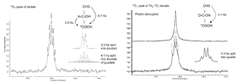
- FIG. 1A illustrates a 13 C spectrum of L-[1- 13 C] lactate
- FIG. 1B illustrates the spectrum following substitution of the C2 proton for a deuteron according to an embodiment of the present invention.
- Splitting of the 13 C resonance of L-[1- 13 C] lactate due to 1 H coupling to the C2 and C3 protons FIG. 1A ).
- Substitution of the C2 proton with deuteron leaves only the spin-spin coupling to the C3 protons ( FIG. 1B ).
- Spin-spin coupling between the 13 C and C2 deuteron is too small to observe in these relatively low resolution spectra.
- FIGS. 2A and 2B illustrate hetero-nuclear 13 C/ 1 H spin echo pulse sequences according to an embodiment of the present invention.
- the initial pulse is either a 90° pulse, for experiments with non-polarized material, or a low flip angle pulse for experiments with hyperpolarized L-[1- 13 C] lactate.
- CS indicates a chemical shift-selective pulse and HS indicates a hyperbolic secant pulse.
- FIG. 3 illustrates C2 proton selective proton pulse resulting in phase inversion of the C1 13 C resonance according to an embodiment of the present invention.
- Heteronuclear 13 C/ 1 H double echo spectra were acquired using a 10 ⁇ s 90° 13 C pulse, a 5 ms adiabatic 180° 13 C pulse (bandwidth ⁇ 2 kHz) and a 10 ms adiabatic 1 H pulse (bandwidth ⁇ 1 kHz), which was centered on either the C2 or C3 lactate proton resonances.
- FIG. 4 illustrates heteronuclear 13 C/ 1 H double echo spectra according to an embodiment of the present invention.
- FIG. 5 illustrates heteronuclear 13 C/ 1 H double echo spectra. Mixture of L-[1- 13 C] lactate and L-[1- 13 C, 2- 2 H 1 ] lactate according to an embodiment of the present invention.
- FIG. 6 illustrates multiple heteronuclear 13 C/ 1 H double echo spectra from an equimolar mixture of hyperpolarized L-[1- 13 C] lactate and L-[1- 13 C, 2- 2 H 1 ] lactate according to an embodiment of the present invention.
- FIG. 7 illustrates a plot of hyperpolarized L-[1- 13 C] lactate signal intensity versus time in vivo according to an embodiment of the present invention.
- FIG. 8 illustrates a decay curve from a multi-echo experiment used to estimate T 2 values in vivo for lactate, pyruvate and hydrate resonances according to an embodiment of the present invention.
- FIG. 9 illustrates the contribution of different T 2 components to the lactate signal as a function of echo time in vivo according to an embodiment of the present invention.
- the 13 C label can also be placed in the C2 or C3 positions, where it will also show spin-spin coupling with the C2 proton.
- the lactate methyl (C3) protons could also be exchanged for deuterium so that there is only spin-spin coupling between the C2 proton and the 13 C at the C1, C2 or C3 positions.
- the MR signal intensity of hyperpolarised 13 C-lactate is related to the concentration of this species and the degree of polarisation left at the time of detection, hence by monitoring the conversion of hyperpolarised 13 C-deuterium labelled lactate into hyperpolarised 13 C-protonated lactate it is possible to study LDH activity in vivo in the human or non-human animal body by using non-invasive MR imaging or MR spectroscopy.
- an aspect of the present invention provides an imaging medium comprising hyperpolarised [ 13 C, 2 H] lactate.
- Such isotopically labelled molecules generally named [ 13 C, 2 H] lactate are selected from the group of [1- 13 C, 2- 2 H] lactate, [2- 13 C, 2- 2 H] lactate, [3- 13 C, 2- 2 H] lactate, [1- 13 C, 2,3- 2 H] lactate, [2- 13 C, 2,3- 2 H] lactate and [3- 13 C, 2,3- 2 H] lactate, or from a group of molecules wherein more than one of the C1, C2 and C3 positions are 13C labelled and wherein the C2 position, or the C2 and C3 positions, are deuterium labelled.
- an imaging medium comprising [1- 13 C, 2- 2 H] lactate, and particularly, according to an embodiment, hyperpolarised [1- 13 C, 2- 2 H] lactate.
- Lactate is an endogenous compound and its concentration in human blood is relatively high (1-3 mM) with local concentrations of 10 mM and more. Hence, lactate should be very well tolerated and therefore using hyperpolarised [ 13 C, 2 H]-lactate, as an imaging agent is advantageous from a safety perspective.
- An aspect of the present invention provides a method of determining LDH activity by 13 C-MR detection using an imaging medium comprising hyperpolarised [ 13 C, 2 H] lactate, wherein the LDH-catalysed exchange of deuterium label between [ 13 C, 2 H] lactate and endogeneous unlabelled lactate is measured.
- the LDH-catalysed exchange of deuterium label at the C2 position between [1- 13 C, 2- 2 H] lactate, or other 2 H and 13 C-labeled lactate isotopomers that are deuterium labelled at the C2 position, and endogenous unlabelled lactate is measured.
- the LDH activity measured in cells and tissues by the method of the present invention is an apparent activity since this is dependent on the rate of lactate transport into the cell and the endogenous lactate concentration as well as the LDH activity. In vivo this rate may also depend on the rate of labelled lactate delivery to the tissue under study.
- determining LDH activity denotes measurement of the rate exchange of the C2 deuterium label with proton in hyperpolarised [1- 13 C, 2- 2 H] lactate, or other 2 H and 13 C-labeled lactate isotopomers that are deuterium labelled at the C2 position, and is dependent on the rate of lactate transport into the cell, the endogenous lactate concentration and LDH activity. In vivo this rate may also depend on the rate of labelled lactate delivery to the tissue under study.
- the protonation state of the lactate C2 carbon is detected via phase modulation of the spin-coupled hyperpolarized 13C label at C1, or at other lactate carbons, in a heteronuclear 13 C/ 1 H spin echo experiment.
- 13 C-MR detection denotes 13 C-MR imaging or 13 C-MR spectroscopy or combined 13 C-MR imaging and 13 C-MR spectroscopy, i.e. 13 C-MR spectroscopic imaging.
- the term further denotes 13 C-MR spectroscopic imaging at various time points.
- imaging medium denotes a liquid composition comprising hyperpolarised [ 13 C, 2 H], such as [1- 13 C, 2- 2 H] lactate, or other 2 H and 13 C-labeled lactate isotopomers that are deuterium labelled at the C2 position, as the MR active agent, i.e. imaging agent.
- the imaging medium used in the method of the present invention may be used as an imaging medium for in vivo 13 C-MR detection, i.e. in living human or non-human animal beings. Further, the imaging medium used in the method of the present invention may be used as an imaging medium for in vitro 13 C-MR detection, e.g. in cell cultures, body samples such as blood or cerebrospinal fluid, ex vivo tissue, for instance ex vivo tissue obtained from a biopsy or isolated organs, all of those derived from a human or non-human animal body.
- lactate and “lactic acid”, according to embodiments of the present invention denote the L-isomer (L-lactate, L-lactic acid), since this is the isomer used specifically by the mammalian LDH isoforms.
- the D-isomer (D-lactate, D-lactic acid) is used by microbial lactate dehydrogenases and therefore the D isomers of the labelled lactate isotopomers described above could be used to detect the presence of bacterial infection in a mammalian tissue. Therefore the imaging medium according to the present invention may thus comprise hyperpolarised 13 C-L-lactate or hyperpolarised 13 C-D-lactate, depending on whether the intention is to detect mammalian or microbial LDH activity respectively.
- the isotopic 13 C enrichment of the lactate isotopomers used in the method according to an embodiment of the present invention is at least 75%, in an embodiment is 80% and in an embodiment is at least 90%, in an embodiment an isotopic enrichment is over 90%. According to an embodiment the enrichment is 100%.
- 13 C-lactate used in the method of the present invention may be isotopically enriched at the C1-position (denoted [1- 13 C] lactate), at the C2-position (denoted [2- 13 C] lactate), at the C3-position (denoted [3- 13 C] lactate), at the C1- and the C2-position (denoted [1,2- 13 C] lactate), at the C1- and the C3-position (denoted [1,3- 13 C] lactate), at the C2- and the C3-position (denoted [2,3- 13 C] lactate) or at the C1-, C2- and C3-position (denoted [1,2,3- 13 C] lactate).
- isotopic enrichment is at the C1-position since [1- 13 C] lactate has a longer T 1 relaxation in human blood at 37° C. than 13 C-lactate which is isotopically enriched at other C-positions.
- the isotopic 2 H enrichment of the lactate used in the method according to an embodiment of the present invention is at least 75%, in an embodiment is at least 80% and in an embodiment is at least 90%, in an embodiment an isotopic enrichment is over 90%. According to an embodiment the enrichment is 100%. Lactate used in the method of the present invention is isotopically deuterium enriched at the C2 position or at the C2 position and the C3 position.
- the imaging medium according to the present invention comprises hyperpolarised sodium 13 C-lactate, and in an embodiment comprises sodium [1- 13 C, 2- 2 H] lactate or the other 2 H and 13 C-labeled lactate isotopomers that are deuterium labelled at the C2 position.
- Lipoamide dehydrogenase in this example from pig heart, catalyses exchange of the proton at the C4 position in the nicotinamide ring of NADH with solvent deuterium. This deuterium is then exchanged with the C2 proton in L-[1- 13 C 1 ] lactate in the reaction catalysed by LDH. This coupled enzyme reaction ensures that the proton at the C2 position of L-[1- 13 C 1 ] lactate exchanges with the much larger pool of deuterons in the solvent 2 H 2 O.
- the present invention provides a process for preparation of hyperpolarised [1- 13 C, 2- 2 H] lactate, including the steps of:
- hypopolarised and “polarised” are used interchangeably hereinafter for polarisation of the 13C-nuclei and denote a nuclear polarisation level in excess of 0.1%, in some embodiments in excess of 1% and in some embodiments in excess of 10%.
- the level of polarisation may for instance be determined by solid-state 13 C-NMR measurements in solid hyperpolarised 13 C-lactate, e.g. solid hyperpolarised 13 C-lactate obtained by dynamic nuclear polarisation (DNP) of 13 C-lactate.
- the solid-state 13 C-NMR measurement according an embodiment consists of a simple pulse-acquire NMR sequence using a low flip angle pulse.
- the signal intensity of the hyperpolarised 13 C-lactate in the NMR spectrum is compared with signal intensity of 13 C-lactate in a NMR spectrum acquired before the polarisation process.
- the level of polarisation is then calculated from the ratio of the signal intensities before and after polarisation.
- the level of polarisation for dissolved hyperpolarised 13 C-lactate may be determined by liquid state NMR measurements. Again the signal intensity of the dissolved hyperpolarised 13 C-lactate is compared with the signal intensity of the dissolved 13 C-lactate before polarisation or after the polarisation has decayed. The level of polarisation is then calculated from the ratio of the signal intensities of 13 C-lactate before and after polarisation.
- Hyperpolarisation of NMR active 13 C-nuclei may be achieved by different methods which are for instance described in WO-A-98/30918, WO-A-99/24080 and WO-A-99/35508, and which all are incorporated herein by reference, and also hyperpolarisation methods known in the art such as polarisation transfer from a noble gas, “brute force”, spin refrigeration, the parahydrogen method and dynamic nuclear polarisation (DNP). In an embodiment, DNP is used.
- the 13 C-lactate is polarised directly.
- 13 C-lactic acid may be polarised, however the polarised 13 C-lactic acid needs to be converted to polarised 13 C-lactate, e.g. by neutralisation with a base.
- 13 C-lactate salts are commercially available, e.g. sodium 13 C-lactate.
- 13 C-lactic acid is commercially available as well; it can also be obtained by protonating commercially available 13 C-lactate, e.g. commercially available sodium 13 C-lactate.
- hyperpolarised 13 C-lactate is by polarisation transfer from a hyperpolarised noble gas, which is described in WO-A-98/30918.
- Noble gases having non-zero nuclear spin can be hyperpolarised by the use of circularly polarised light.
- a hyperpolarised noble gas in an embodiment He or Xe, or a mixture of such gases, may be used to effect hyperpolarisation of 13 C-nuclei.
- the hyperpolarised gas may be in the gas phase, it may be dissolved in a liquid/solvent, or the hyperpolarised gas itself may serve as a solvent. Alternatively, the gas may be condensed onto a cooled solid surface and used in this form, or allowed to sublime. In an embodiment, there is an intimate mixing of the hyperpolarised gas with 13 C-lactate or 13 C-lactic acid.
- hyperpolarisation is imparted to 13 C-nuclei by thermodynamic equilibration at a very low temperature and high field.
- Hyperpolarisation compared to the operating field and temperature of the NMR spectrometer is effected by use of a very high field and very low temperature (“brute force”).
- the magnetic field strength used should be as high as possible, suitably higher than 1 T, in an embodiment higher than 5 T, in an embodiment 15 T or more and in an embodiment 20 T or more.
- the temperature should be very low, e.g. 4.2 K or less, in an embodiment 1.5 K or less, in an embodiment 1.0 K or less, and in an embodiment 100 mK or less.
- Another way for obtaining hyperpolarised 13 C-lactate is the spin refrigeration method.
- This method covers spin polarisation of a solid compound or system by spin refrigeration polarisation.
- the system is doped with or intimately mixed with suitable crystalline paramagnetic materials such as Ni 2+ , lanthanide or actinide ions with a symmetry axis of order three or more.
- suitable crystalline paramagnetic materials such as Ni 2+ , lanthanide or actinide ions with a symmetry axis of order three or more.
- the instrumentation is simpler than required for DNP with no need for a uniform magnetic field since no resonance excitation field is applied.
- the process is carried out by physically rotating the sample around an axis perpendicular to the direction of the magnetic field.
- the pre-requisite for this method is that the paramagnetic species has a highly anisotropic g-factor.
- the electron paramagnetic resonance will be brought into contact with the nuclear spins, leading to a decrease in the nuclear spin temperature
- DNP dynamic nuclear polarisation
- Sodium 13 C-lactate is a commercially available compound that may be directly used for DNP since it does not crystallize upon cooling/freezing. Since this eliminates the necessity of glass formers and/or high amounts of solvent(s) in the sample, a highly concentrated sample can be prepared and used in the DNP process. Further, sodium 13 C-lactate samples are pH neutral and hence a variety of DNP agents can be used.
- polarisation of MR active nuclei in a compound to be polarised is effected by a polarisation agent or so-called DNP agent, a compound comprising unpaired electrons.
- the DNP process energy, normally in the form of microwave radiation, is provided, which will initially excite the DNP agent. Upon decay to the ground state, there is a transfer of polarisation from the unpaired electron of the DNP agent to the NMR active nuclei of the compound to be polarised, e.g. to the 13 C nuclei in 13 C-lactate.
- a moderate or high magnetic field and a very low temperature are used in the DNP process, e.g. by carrying out the DNP process in liquid helium under vacuum and a magnetic field of about 1 T or above.
- a moderate magnetic field and any temperature at which sufficient polarisation enhancement is achieved may be employed.
- the DNP technique is for example further described in WO-A-98/58272 and in WO-A-01/96895, both of which are included by reference herein.
- a composition comprising the compound to be polarised and a DNP agent is prepared which is then frozen and inserted into a DNP polariser for polarisation.
- the frozen solid hyperpolarised composition is rapidly transferred into the liquid state, either by melting it or by dissolving it in a suitable dissolution medium.
- there is dissolution wherein the dissolution process of a frozen hyperpolarised composition and suitable devices therefore are described in detail in WO-A-02/37132.
- the melting process and suitable devices for the melting are for instance described in WO-A-02/36005.
- the sample to be polarised i.e. 13 C-lactate and a DNP agent may further comprise a paramagnetic metal ion.
- the presence of paramagnetic metal ions in the composition to be polarised by DNP has found to result in increased polarisation levels in 13 C-lactate, as described in detail in WO-A2-2007/064226, which is incorporated herein by reference.
- the MR imaging medium comprises [ 13 C, 2 H] lactate, such as [1- 13 C, 2- 2 H] lactate, a trityl radical and optionally a paramagnetic metal ion. When said paramagnetic metal ion is present this is present in the form of a paramagnetic chelate comprising Gd 3+ .
- Such MR imaging medium is in an embodiment obtained by dynamic nuclear polarisation.
- the imaging medium according to the present invention may be used as imaging medium for in vivo LDH activity determination by 13 C-MR detection, i.e. in living human or non-human animal beings.
- the imaging medium is provided as a composition that is suitable for being administered to a living human or non-human animal body.
- Such an imaging medium in an embodiment comprises in addition to the MR active agent, [ 13 C, 2 H]-lactate, an aqueous carrier, in an embodiment a physiologically tolerable and pharmaceutically accepted aqueous carrier like water, a buffer solution or saline.
- Such an imaging medium may further comprise conventional pharmaceutical or veterinary carriers or excipients, e.g. formulation aids such as are conventional for diagnostic compositions in human or veterinary medicine.
- the imaging medium according to the method of the present invention may be used as an imaging medium for in vitro LDH activity determination by 13 C-MR detection, i.e. in cell cultures, body samples such as blood samples, ex vivo tissues such as biopsy tissue or isolated organs.
- the imaging medium is provided as a composition that is suitable for being added to, for instance, cell cultures, blood samples, ex vivo tissues like biopsy tissue or isolated organs.
- Such an imaging medium in an embodiment comprises in addition to the MR active agent, 13 C-lactate, a solvent which is compatible with and used for in vitro cell or tissue assays, for instance DMSO or methanol or solvent mixtures comprising an aqueous carrier and a non aqueous solvent, for instance mixtures of DMSO and water or a buffer solution or methanol and water or a buffer solution.
- a solvent which is compatible with and used for in vitro cell or tissue assays for instance DMSO or methanol or solvent mixtures comprising an aqueous carrier and a non aqueous solvent, for instance mixtures of DMSO and water or a buffer solution or methanol and water or a buffer solution.
- a solvent which is compatible with and used for in vitro cell or tissue assays for instance DMSO or methanol or solvent mixtures comprising an aqueous carrier and a non aqueous solvent, for instance mixtures of DMSO and water or a buffer solution or methanol and water or
- the imaging medium used in the method of the present invention is used for in vivo determination of LDH activity, i.e. in a living human or non-human animal body, said imaging medium is in an embodiment administered to said body parenterally, and in an embodiment, intravenously.
- the body under examination is positioned in an MR magnet.
- Dedicated 13 C-MR and 1 H-MR, or double-tuned 13 C/ 1 H RF-coils are positioned to cover the area of interest. Exact dosage and concentration of the imaging medium will depend upon a range of factors such as toxicity and the administration route.
- an MR imaging sequence is applied, in an embodiment one that encodes the volume of interest in a combined frequency and spatially selective way.
- the exact time of applying an MR sequence is highly dependent on the volume of interest and the species.
- the imaging medium used in the method of the present invention is used for in vitro determination of LDH activity, said imaging medium is 1 mM to 100 mM in 13 C-lactate, in an embodiment 20 mM to 90 mM and in an embodiment 40 to 80 mM in 13 C-lactate.
- the LDH-catalysed exchange of deuterium label between [1- 13 C, 2- 2 H] lactate and endogenous unlabelled lactate is monitored using a heteronuclear 13 C/ 1 H spin echo experiment.
- the concentration of the [1- 13 C 1 , 2 - 1 H 1 ] lactate species at any point during the isotope exchange time course can be determined by acquiring heteronuclear spin echo difference spectra obtained in the presence and absence of the 180° 1 H pulse.
- a method of heteronuclear 13 C/ 1 H spin echo imaging is used to monitor the exchange of deuterium label between [1- 13 C, 2- 2 H] lactate and endogenous unlabelled lactate.
- the 1 H pulse must be frequency selective in order to avoid phase modulation due to spin-spin coupling between the 13 C nucleus and the C3 methyl protons.
- the requirement to use a frequency-selective 1 H pulse is removed if lactate isotopomers are used in which the methyl protons have also been exchanged for deuterium.
- a hetero-nuclear double echo imaging pulse sequence could be used to detect the C2 protonation state of [1- 13 C, 2- 2 H] lactate in vivo, in which the echoes are acquired with readout and phase encode gradients in a standard echo planar imaging sequence.
- 13 C images acquired without the 1 H pulse will have signal from 13 C-labelled C2 deuterated lactate plus 13 C-labelled C2 protonated lactate and 13 C images acquired with the 1 H pulse will have signal from 13 C-labelled C2 deuterated lactate minus 13 C-labelled C2 protonated lactate. Addition of these images will give the total 13 C-labelled C2 deuterated lactate and subtraction will give the 13 C-labelled C2 protonated lactate.
- heteronuclear double echo imaging is used to monitor the exchange of deuterium label between [1- 13 C, 2- 2 H] lactate and endogenous unlabelled lactate.
- a series of the above-mentioned images are acquired and pixel-by-pixel fitting of signal intensities in images of the C2 protonated and C2 deuterated lactate concentrations are fit to a kinetic model to obtain maps of LDH activity in the tissue.
- the apparent LDH activity may depend on the rate of delivery of labelled lactate to the tissue, the rate of lactate transport across the cell membrane, and the endogenous lactate concentration.
- said LDH activity maps may be derived from the whole body, e.g. obtained by whole body in vivo 13 C-MR detection.
- said LDH activity maps are generated from a region or volume of interest, i.e. a certain tissue, organ or part of said human or non-human animal body.
- the above-mentioned signal of 13 C-lactate is used to determine LDH activity of cells in a cell culture, of body samples such as blood samples, of ex vivo tissue like biopsy tissue or of an isolated organ derived from a human or non-human animal being. Said LDH activity is then generated by in vitro 13 C-MR detection.
- an embodiment of the present invention provides a method of determining LDH activity by 13 C-MR detection using an imaging medium comprising hyperpolarised [ 13 C, 2 H] lactate, such as [1- 13 C, 2- 2 H] lactate, wherein the C2 protonation state is detected and wherein said information is used to generate a map or point measurement of LDH activity.
- the LDH activity map or measurement generated in an embodiment of the method according to the present invention is indicative for the LDH activity of the body, part of the body, cells, tissue, body sample etc. under examination and said information obtained may be used in a subsequent step for various purposes.
- One of these purposes may be the assessment of compounds that alter LDH activity, in an embodiment, compounds that elevate LDH activity. These might be drugs that improve tissue viability, where one would expect to see an increase in LDH activity.
- the method of the present invention is carried out in vitro and the information obtained is used in assessing the efficacy of potential drugs that alter LDH activity, e.g. in a drug discovery and/or screening process.
- the method of the present invention may be carried out in suitable cell cultures or tissue.
- the cells or the tissue is contacted with the potential drug and LDH activity is determined by 13 C-MR detection according to the method of the present invention.
- Information about the efficacy of the potential drug may be obtained by comparing the LDH activity of the treated cells or tissue with the LDH activity of non-treated cells or tissue.
- the variation of LDH activity may be determined by determining the LDH activity of cells or tissue before and after treatment.
- Such a drug efficacy assessment may be carried out on for instance microplates, which would allow parallel testing of various potential drugs and/or various doses of potential drugs and thus would make this suitable for high-throughput screening.
- the method of the present invention is carried out in vivo and the information obtained is used in assessing the efficacy of potential drugs that alter LDH activity in vivo.
- the method of the present invention may be carried out in, for instance, test animals or in volunteers in a clinical trial.
- a potential drug is administered and LDH activity is determined by 13 C-MR detection according to the method of the present invention.
- Information about the efficacy of the potential drug may be obtained by determining the variation of LDH activity before and after treatment, e.g. over a certain time period with repeated treatment.
- Such a drug efficacy assessment may be carried out in pre-clinical research (test animals) or in clinical trials.
- the method of the present invention is carried out in vivo or in vitro and the information obtained is used to assess response to treatment and/or to determine treatment efficacy in diseased patients undergoing treatment for their disease.
- the information obtained by the method of the present invention may be used in a subsequent step for various purposes.
- Another purpose may be to gain insight into disease states, i.e. identifying patients at risk, early detection of diseases, evaluating disease progression, severity and complications related to a disease.
- the method of the present invention is carried out in vivo or in vitro and the information obtained is used to monitor progression of a disease. This may be useful for diseases or disorders where the disease has not progressed to a level where treatment is indicated or recommended, e.g. because of severe side-effects associated with said treatment. In such a situation the choice of action is “watchful waiting”, i.e. the patient is closely monitored for disease progression and early detection of deterioration.
- the method of the present invention may be used to determine the initial LDH activity and to make subsequent LDH activity determinations over a period of time at a certain frequency.
- a decrease in LDH activity may indicate progress and worsening of a disease and the said decrease can be used by the physician to decide on commencement of treatment.
- an increase in LDH activity may indicate worsening of disease, for example in cancer, specifically for prostate cancer, there may be increases in LDH and lactate concentration with disease progression, which would increase the apparent LDH activity that is measured by the method of the present invention.
- suitable samples from a patient under treatment are obtainable, e.g. tissue samples or body samples like blood samples.
- the method according to the present invention is used for in vivo MR tumour imaging, tumour therapy monitoring and/or tumour staging of brain tumours, breast tumours, colon tumours, lung tumours, kidney tumours, head and neck tumours, muscle tumours, ovarian tumours, gastric tumours, pancreatic tumours, esophageal tumours and prostate tumours. It has further been found that the method according to the present invention is especially useful for in vivo MR prostate tumour imaging, i.e. prostate tumour diagnosis and/or prostate tumour staging and/or prostate tumour therapy monitoring.
- the method of the present invention is carried out in vivo or in vitro and the information obtained is used for determining the severity of a disease.
- diseases progress from their onset over time.
- certain clinical markers diseases are characterized by certain stages, e.g. an early (mild) stage, a middle (moderate) stage and a severe (late) stage. More refined stages are common for certain diseases.
- a variety of clinical markers is known to be used for staging a disease including more specific ones like certain enzymes or protein expression but also more general ones like blood values, electrolyte levels etc.
- LDH activity may be such a clinical marker that can be used, alone or in combination with other markers and/or symptoms, to determine a disease stage and thus severity of a disease.
- LDH ranges which are characteristic for a certain disease stage may be established by determining LDH activity according to the method of the present invention in patients having for instance a disease in an early, middle and late stage and defining a range of LDH activity which is characteristic for a certain stage.
- the method of the present invention is carried out in vivo or in vitro and the information obtained is used for identifying and assessing complications related to a disease.
- LDH activity may be determined in an organ-specific way, for instance by in vivo 13 C-MR detection carried out with surface coils placed over the heart or the kidney.
- Anatomical and/or, where suitable, perfusion information may be included in the method of the present invention when carried out in vivo.
- Anatomical information may for instance be obtained by acquiring a proton or 13 C-MR image with or without employing a suitable contrast agent before or after the method of the present invention.
- the present invention provides use of an imaging medium comprising hyperpolarized [ 13 C, 2 H] lactate in the determination of LDH activity by 13 C-MR detection.
- the resulting solution was incubated at room temperature for approximately one week. The incubation was terminated by placing the solution in a boiling water bath for 10 min. The precipitated protein was removed by filtration through a 0.22 ⁇ m pore size membrane and the zinc salt afforded by acidification of the solution with concentrated hydrochloric acid, followed by neutralization with basic zinc carbonate (Sigma Aldrich, Gillingham, UK). The solution was then filtered, lyophilized and the resulting zinc lactate purified by recrystallisation from a water/ethanol mixture. The sodium salt was prepared using an excess of an ion-exchange resin (Dowex 50 W ⁇ 8, Na + form, Sigma Aldrich, Gillingham, UK) and isolated by lyophilization. The lactate was assayed spectrophotometrically in an NADH-linked assay and by 1 H NMR spectroscopy.
- FIG. 2A A heteronuclear 13 C/ 1 H spin echo pulse sequence is shown in FIG. 2A .
- Application of a 1 H 180° pulse at the same time as the 180° 13 C pulse, at ⁇ 1 ⁇ 2J, results in phase modulation of the observed 1 H coupled 13 C resonances.
- a double echo sequence ( FIG. 2B ) was used, since this ensures return of the polarization remaining along the z axis to the +z axis at the end of the experiment.
- the samples contained 10 mM of either L-[1- 13 C 1 , 2- 2 H 1 ] lactate or L-[1- 13 C 1 ] lactate (non-polarized).
- Heteronuclear 13 C/ 1 H double echo spectra were acquired using a 10 ⁇ s 90° 13 C pulse, a 5 ms adiabatic 180° 13 C pulse (bandwidth ⁇ 2 kHz) and a 10 ms adiabatic 1 H pulse (bandwidth ⁇ 1 kHz), which was centered on either the C2 or C3 lactate proton resonances ( FIG. 2B ).
- the TR was 20 s.
- Initial dummy scans produced a steady state z magnetization.
- the spectral bandwidth was 8 kHz and data were collected into 2048 complex points. Phase and amplitude corrected peak integrals for the lactate 13 C resonance were measured.
- the echo time is equivalent to 2 ⁇ .
- L-[1- 13 C 1 ] lactate was hyperpolarized using the DMSO preparation described in (Chen, A. P., et al. Feasibility of using hyperpolarized [1-C-13] lactate as a substrate for in vivo metabolic C-13 MRSI studies. Magnetic Resonance Imaging 26, 721-726 (2008)) with added Gd 3+ .
- the 13 C resonance must have sufficiently long T 1 and T 2 .
- the time to echo in the hetero-nuclear spin echo experiment is 300 ms (see FIG. 3 ), therefore it is essential that the 13 C resonance has a long T 2 .
- the echoes will be acquired with readout and phase encode gradients in a standard EPI sequence.
- adiabatic 13 C pulses we ensure that the unsampled 13 C polarization remains along the z axis and by using a double echo sequence we ensure that this polarization is returned to the +z axis at the end of the pulse sequence.
- the 1 H pulse will be switched off resonance on alternate acquisitions, which will turn off phase modulation of the 13 C signal on alternate acquisitions (see FIG. 3 ).
Abstract
Description


-
- i) deuterium labelling sodium [1-13C1] lactate to produce
- [1-13C1, 2-2H1] lactate,
- ii) hyperpolarising [1-13C1, 2-2H1] lactate.
- i) deuterium labelling sodium [1-13C1] lactate to produce
Claims (7)
Priority Applications (1)
| Application Number | Priority Date | Filing Date | Title |
|---|---|---|---|
| US14/979,072 US9408925B2 (en) | 2010-05-03 | 2015-12-22 | Hyperpolarized lactate contrast agent for determination of LDH activity |
Applications Claiming Priority (6)
| Application Number | Priority Date | Filing Date | Title |
|---|---|---|---|
| EP10161740.5 | 2010-05-03 | ||
| EP10161740 | 2010-05-03 | ||
| EP10161740 | 2010-05-03 | ||
| PCT/EP2011/056945 WO2011138269A1 (en) | 2010-05-03 | 2011-05-02 | Hyperpolarized lactate contrast agent for determination of ldh activity |
| US201213695872A | 2012-11-02 | 2012-11-02 | |
| US14/979,072 US9408925B2 (en) | 2010-05-03 | 2015-12-22 | Hyperpolarized lactate contrast agent for determination of LDH activity |
Related Parent Applications (2)
| Application Number | Title | Priority Date | Filing Date |
|---|---|---|---|
| PCT/EP2011/056945 Division WO2011138269A1 (en) | 2010-05-03 | 2011-05-02 | Hyperpolarized lactate contrast agent for determination of ldh activity |
| US13/695,872 Division US9259490B2 (en) | 2010-05-03 | 2011-05-02 | Hyperpolarized lactate contrast agent for determination of LDH activity |
Publications (2)
| Publication Number | Publication Date |
|---|---|
| US20160184462A1 US20160184462A1 (en) | 2016-06-30 |
| US9408925B2 true US9408925B2 (en) | 2016-08-09 |
Family
ID=44276181
Family Applications (2)
| Application Number | Title | Priority Date | Filing Date |
|---|---|---|---|
| US13/695,872 Active US9259490B2 (en) | 2010-05-03 | 2011-05-02 | Hyperpolarized lactate contrast agent for determination of LDH activity |
| US14/979,072 Active US9408925B2 (en) | 2010-05-03 | 2015-12-22 | Hyperpolarized lactate contrast agent for determination of LDH activity |
Family Applications Before (1)
| Application Number | Title | Priority Date | Filing Date |
|---|---|---|---|
| US13/695,872 Active US9259490B2 (en) | 2010-05-03 | 2011-05-02 | Hyperpolarized lactate contrast agent for determination of LDH activity |
Country Status (7)
| Country | Link |
|---|---|
| US (2) | US9259490B2 (en) |
| JP (1) | JP5879333B2 (en) |
| KR (1) | KR101858269B1 (en) |
| CN (1) | CN102858377B (en) |
| AU (1) | AU2011250012B2 (en) |
| CA (1) | CA2797472C (en) |
| WO (1) | WO2011138269A1 (en) |
Families Citing this family (3)
| Publication number | Priority date | Publication date | Assignee | Title |
|---|---|---|---|---|
| CN104981256B (en) | 2013-01-31 | 2020-02-18 | 伯拉考成像股份公司 | Hyperpolarised esters as metabolic markers in MR |
| EP2863229A1 (en) | 2013-10-15 | 2015-04-22 | Technische Universität München | pH-biosensors based on compounds with pH-sensitive enolic groups for magnetic resonance imaging and spectroscopy and their uses |
| CN117180458A (en) * | 2023-08-31 | 2023-12-08 | 中国科学院精密测量科学与技术创新研究院 | Application of oxaloacetic acid in melt dynamic nuclear polarization |
Family Cites Families (14)
| Publication number | Priority date | Publication date | Assignee | Title |
|---|---|---|---|---|
| US5439803A (en) | 1993-08-13 | 1995-08-08 | The Regents Of The University Of Michigan | Isotope and assay for glycolysis and the pentose phosphate pathway |
| ATE256293T1 (en) | 1997-01-08 | 2003-12-15 | Amersham Health As | METHOD FOR GENERATING IMAGE WITH MAGNETIC RESONANCE |
| CA2290808A1 (en) | 1997-06-19 | 1998-12-23 | Jan Henrik Ardenkjaer-Larsen | Overhauser magnetic resonance imaging (ormi) method comprising ex vivo polarization of a magnetic resonance (mr) imaging agent |
| JP2001522819A (en) | 1997-11-12 | 2001-11-20 | ナイコムド イメージング エーエス | Agents labeled with para-hydrogen and their use in magnetic resonance imaging |
| US6278893B1 (en) | 1998-01-05 | 2001-08-21 | Nycomed Imaging As | Method of magnetic resonance imaging of a sample with ex vivo polarization of an MR imaging agent |
| GB0014463D0 (en) | 2000-06-14 | 2000-08-09 | Nycomed Amersham Plc | NMR Method |
| CA2427732C (en) | 2000-11-03 | 2009-04-14 | Amersham Health As | Methods and devices for dissolving hyperpolarised solid material for nmr analyses |
| AU1403902A (en) | 2000-11-03 | 2002-05-15 | Amersham Health As | Methods and devices for polarised nmr samples |
| US7256047B2 (en) * | 2001-05-01 | 2007-08-14 | Board Of Regents, The University Of Texas System | Measurement of gluconeogenesis and intermediary metabolism using stable isotopes |
| US9023320B2 (en) | 2004-07-30 | 2015-05-05 | Ge Healthcare As | Method of producing a composition, composition and its use |
| WO2007064226A2 (en) | 2005-12-01 | 2007-06-07 | Ge Healthcare As | Method of dynamic nuclear polarisation (dnp) using a trityl radical and a paramagnetic metal ion |
| WO2007111515A2 (en) | 2006-03-29 | 2007-10-04 | Ge Healthcare As | Method to produce hyperpolarised carboxylates and sulphonates |
| WO2008020765A2 (en) * | 2006-08-18 | 2008-02-21 | Ge Healthcare As | Imaging medium comprising lactate and hyperpolarised 13c-pyruvate |
| EP2170407A2 (en) * | 2007-07-26 | 2010-04-07 | GE Healthcare UK Limited | Imaging medium comprising hyperpolarised 13c-lactate and use thereof |
-
2011
- 2011-05-02 US US13/695,872 patent/US9259490B2/en active Active
- 2011-05-02 WO PCT/EP2011/056945 patent/WO2011138269A1/en active Application Filing
- 2011-05-02 JP JP2013508451A patent/JP5879333B2/en not_active Expired - Fee Related
- 2011-05-02 CN CN201180022128.2A patent/CN102858377B/en not_active Expired - Fee Related
- 2011-05-02 KR KR1020127031410A patent/KR101858269B1/en active IP Right Grant
- 2011-05-02 CA CA2797472A patent/CA2797472C/en not_active Expired - Fee Related
- 2011-05-02 AU AU2011250012A patent/AU2011250012B2/en not_active Ceased
-
2015
- 2015-12-22 US US14/979,072 patent/US9408925B2/en active Active
Non-Patent Citations (1)
| Title |
|---|
| Rodrigues et al (Medicine 54:1014-1019, 2005). * |
Also Published As
| Publication number | Publication date |
|---|---|
| WO2011138269A1 (en) | 2011-11-10 |
| KR20130135727A (en) | 2013-12-11 |
| AU2011250012B2 (en) | 2016-02-25 |
| US9259490B2 (en) | 2016-02-16 |
| CA2797472A1 (en) | 2011-11-10 |
| CA2797472C (en) | 2018-07-03 |
| CN102858377A (en) | 2013-01-02 |
| CN102858377B (en) | 2015-04-15 |
| JP2013525464A (en) | 2013-06-20 |
| US20130052141A1 (en) | 2013-02-28 |
| AU2011250012A1 (en) | 2012-11-22 |
| KR101858269B1 (en) | 2018-05-15 |
| JP5879333B2 (en) | 2016-03-08 |
| US20160184462A1 (en) | 2016-06-30 |
Similar Documents
| Publication | Publication Date | Title |
|---|---|---|
| JP2008508023A5 (en) | ||
| US20100178249A1 (en) | Imaging medium comprising lactate and hyperpolarised 13c-pyruvate | |
| US20100196283A1 (en) | Method and imaging medium for use in the method | |
| EP2180902B1 (en) | Imaging medium comprising hyperpolarised ¹³c-acetate and use thereof | |
| US20100310467A1 (en) | Composition and method for generating a metabolic profile using 13c-mr detection | |
| AU2008294727B2 (en) | Method of determination of PDH activity and imaging media for use in said method | |
| US9408925B2 (en) | Hyperpolarized lactate contrast agent for determination of LDH activity | |
| EP2052273A1 (en) | 13c-mr imaging or spectroscopy of cell death | |
| US20110038804A1 (en) | Mr imaging agent, imaging medium and methods of imaging wherein such an imaging medium is used | |
| EP2268321B1 (en) | Method of determining alanine transaminase (ALT) activity by 13C-MR detection using hyperpolarised 13C-pyruvate | |
| US20110033390A1 (en) | Mr imaging agent or medium compressing hyperpolarised 13c alanine and methods of imaging wherein such an imaging medium is used | |
| US8968703B2 (en) | 13C-MR detection using hyperpolarised 13C-fructose | |
| US20130116547A1 (en) | Measurement of Anaplerotic Flux by Hyperpolarization Transfer | |
| EP2891500B1 (en) | Contrast agent for determination of aldehyde dehydrogenase (ALDH) activity | |
| Zaccagna et al. | BJR UNCORRECTED PROOFS | |
| US20140328766A1 (en) | Composition and method for generating a metabolic profile using 13c-mr detection | |
| WO2009150211A1 (en) | Dnp polariser and method of producing a hyperpolarised selected material |
Legal Events
| Date | Code | Title | Description |
|---|---|---|---|
| AS | Assignment |
Owner name: GE HEALTHCARE LIMITED, UNITED KINGDOM Free format text: ASSIGNMENT OF ASSIGNORS INTEREST;ASSIGNORS:BRINDLE, KEVIN M;MIKKO LIVARI KETTUNEN;KENNEDY, BRETT W.C.;REEL/FRAME:038129/0425 Effective date: 20110516 |
|
| AS | Assignment |
Owner name: GENERAL ELECTRIC COMPANY, NEBRASKA Free format text: ASSIGNMENT OF ASSIGNORS INTEREST;ASSIGNOR:GE HEALTHCARE UK LIMITED;REEL/FRAME:037684/0541 Effective date: 20121002 |
|
| AS | Assignment |
Owner name: GENERAL ELECTRIC COMPANY, NEW YORK Free format text: CORRECTIVE ASSIGNMENT TO CORRECT THE RECEIVING PARTY STATE FROM NEBRASKA TO NEW YORK PREVIOUSLY RECORDED AT REEL: 037684 FRAME: 0541. ASSIGNOR(S) HEREBY CONFIRMS THE ASSIGNMENT;ASSIGNOR:GE HEALTHCARE UK LIMITED;REEL/FRAME:039265/0388 Effective date: 20121002 |
|
| STCF | Information on status: patent grant |
Free format text: PATENTED CASE |
|
| MAFP | Maintenance fee payment |
Free format text: PAYMENT OF MAINTENANCE FEE, 4TH YEAR, LARGE ENTITY (ORIGINAL EVENT CODE: M1551); ENTITY STATUS OF PATENT OWNER: LARGE ENTITY Year of fee payment: 4 |
|
| FEPP | Fee payment procedure |
Free format text: MAINTENANCE FEE REMINDER MAILED (ORIGINAL EVENT CODE: REM.); ENTITY STATUS OF PATENT OWNER: LARGE ENTITY |