US9408828B2 - (+)-3′-angeloyloxy-4′-keto-3′,4′-dihydroseselin for treating inflammation - Google Patents
(+)-3′-angeloyloxy-4′-keto-3′,4′-dihydroseselin for treating inflammation Download PDFInfo
- Publication number
- US9408828B2 US9408828B2 US14/742,727 US201514742727A US9408828B2 US 9408828 B2 US9408828 B2 US 9408828B2 US 201514742727 A US201514742727 A US 201514742727A US 9408828 B2 US9408828 B2 US 9408828B2
- Authority
- US
- United States
- Prior art keywords
- dss
- day
- inflammatory
- lps
- mice
- Prior art date
- Legal status (The legal status is an assumption and is not a legal conclusion. Google has not performed a legal analysis and makes no representation as to the accuracy of the status listed.)
- Active
Links
- 206010061218 Inflammation Diseases 0.000 title claims abstract description 19
- 230000004054 inflammatory process Effects 0.000 title claims abstract description 18
- 150000001875 compounds Chemical class 0.000 claims abstract description 22
- 238000000034 method Methods 0.000 claims abstract description 13
- 230000037396 body weight Effects 0.000 claims description 17
- 206010009900 Colitis ulcerative Diseases 0.000 claims description 9
- 201000006704 Ulcerative Colitis Diseases 0.000 claims description 9
- 208000027866 inflammatory disease Diseases 0.000 claims description 8
- 208000011231 Crohn disease Diseases 0.000 claims description 3
- 206010039073 rheumatoid arthritis Diseases 0.000 claims description 2
- PGOMXBOHQUBUMI-YHYXMXQVSA-N (8,8-dimethyl-2,10-dioxo-9H-pyrano[2,3-f]chromen-9-yl) (Z)-2-methylbut-2-enoate Chemical compound C1=CC(=O)OC2=C1C=CC1=C2C(=O)C(OC(=O)C(\C)=C/C)C(C)(C)O1 PGOMXBOHQUBUMI-YHYXMXQVSA-N 0.000 abstract description 92
- 241001132282 Bupleurum malconense Species 0.000 abstract description 13
- 239000003814 drug Substances 0.000 abstract description 4
- 229940079593 drug Drugs 0.000 abstract description 2
- BLFLLBZGZJTVJG-UHFFFAOYSA-N benzocaine Chemical compound CCOC(=O)C1=CC=C(N)C=C1 BLFLLBZGZJTVJG-UHFFFAOYSA-N 0.000 description 53
- 210000004027 cell Anatomy 0.000 description 45
- 241000699670 Mus sp. Species 0.000 description 37
- MWUXSHHQAYIFBG-UHFFFAOYSA-N Nitric oxide Chemical compound O=[N] MWUXSHHQAYIFBG-UHFFFAOYSA-N 0.000 description 34
- 230000000694 effects Effects 0.000 description 34
- 239000002158 endotoxin Substances 0.000 description 33
- 229920006008 lipopolysaccharide Polymers 0.000 description 33
- 210000001072 colon Anatomy 0.000 description 31
- 102100029438 Nitric oxide synthase, inducible Human genes 0.000 description 23
- 101710089543 Nitric oxide synthase, inducible Proteins 0.000 description 23
- 206010009887 colitis Diseases 0.000 description 23
- 108060008682 Tumor Necrosis Factor Proteins 0.000 description 20
- 102100040247 Tumor necrosis factor Human genes 0.000 description 20
- MZOFCQQQCNRIBI-VMXHOPILSA-N (3s)-4-[[(2s)-1-[[(2s)-1-[[(1s)-1-carboxy-2-hydroxyethyl]amino]-4-methyl-1-oxopentan-2-yl]amino]-5-(diaminomethylideneamino)-1-oxopentan-2-yl]amino]-3-[[2-[[(2s)-2,6-diaminohexanoyl]amino]acetyl]amino]-4-oxobutanoic acid Chemical compound OC[C@@H](C(O)=O)NC(=O)[C@H](CC(C)C)NC(=O)[C@H](CCCN=C(N)N)NC(=O)[C@H](CC(O)=O)NC(=O)CNC(=O)[C@@H](N)CCCCN MZOFCQQQCNRIBI-VMXHOPILSA-N 0.000 description 18
- 230000003110 anti-inflammatory effect Effects 0.000 description 16
- 210000002540 macrophage Anatomy 0.000 description 16
- 108090000623 proteins and genes Proteins 0.000 description 16
- 102000004127 Cytokines Human genes 0.000 description 15
- 108090000695 Cytokines Proteins 0.000 description 15
- 230000014509 gene expression Effects 0.000 description 15
- 238000004519 manufacturing process Methods 0.000 description 15
- XEKOWRVHYACXOJ-UHFFFAOYSA-N Ethyl acetate Chemical compound CCOC(C)=O XEKOWRVHYACXOJ-UHFFFAOYSA-N 0.000 description 14
- 102000004169 proteins and genes Human genes 0.000 description 14
- 210000001519 tissue Anatomy 0.000 description 13
- 241000699666 Mus <mouse, genus> Species 0.000 description 12
- 102000003896 Myeloperoxidases Human genes 0.000 description 12
- 108090000235 Myeloperoxidases Proteins 0.000 description 12
- 239000003208 petroleum Substances 0.000 description 12
- 230000000770 proinflammatory effect Effects 0.000 description 12
- UETNIIAIRMUTSM-UHFFFAOYSA-N Jacareubin Natural products CC1(C)OC2=CC3Oc4c(O)c(O)ccc4C(=O)C3C(=C2C=C1)O UETNIIAIRMUTSM-UHFFFAOYSA-N 0.000 description 11
- NCEXYHBECQHGNR-QZQOTICOSA-N sulfasalazine Chemical compound C1=C(O)C(C(=O)O)=CC(\N=N\C=2C=CC(=CC=2)S(=O)(=O)NC=2N=CC=CC=2)=C1 NCEXYHBECQHGNR-QZQOTICOSA-N 0.000 description 11
- 229960001940 sulfasalazine Drugs 0.000 description 11
- NCEXYHBECQHGNR-UHFFFAOYSA-N sulfasalazine Natural products C1=C(O)C(C(=O)O)=CC(N=NC=2C=CC(=CC=2)S(=O)(=O)NC=2N=CC=CC=2)=C1 NCEXYHBECQHGNR-UHFFFAOYSA-N 0.000 description 11
- 102100037850 Interferon gamma Human genes 0.000 description 10
- 108010074328 Interferon-gamma Proteins 0.000 description 10
- 108090001005 Interleukin-6 Proteins 0.000 description 10
- 108010052419 NF-KappaB Inhibitor alpha Proteins 0.000 description 9
- 108010057466 NF-kappa B Proteins 0.000 description 9
- 102100039337 NF-kappa-B inhibitor alpha Human genes 0.000 description 9
- 102100023050 Nuclear factor NF-kappa-B p105 subunit Human genes 0.000 description 9
- 102100038280 Prostaglandin G/H synthase 2 Human genes 0.000 description 9
- 108050003267 Prostaglandin G/H synthase 2 Proteins 0.000 description 9
- 230000006378 damage Effects 0.000 description 9
- 238000002474 experimental method Methods 0.000 description 9
- RTZKZFJDLAIYFH-UHFFFAOYSA-N Diethyl ether Chemical compound CCOCC RTZKZFJDLAIYFH-UHFFFAOYSA-N 0.000 description 8
- 108090000978 Interleukin-4 Proteins 0.000 description 8
- 238000004458 analytical method Methods 0.000 description 8
- 239000003153 chemical reaction reagent Substances 0.000 description 8
- 231100000673 dose–response relationship Toxicity 0.000 description 8
- 239000012259 ether extract Substances 0.000 description 8
- 230000002757 inflammatory effect Effects 0.000 description 8
- 230000005764 inhibitory process Effects 0.000 description 8
- 239000002609 medium Substances 0.000 description 8
- 108020004999 messenger RNA Proteins 0.000 description 8
- 108091003079 Bovine Serum Albumin Proteins 0.000 description 7
- 102000004889 Interleukin-6 Human genes 0.000 description 7
- 239000000284 extract Substances 0.000 description 7
- 238000001262 western blot Methods 0.000 description 7
- LFQSCWFLJHTTHZ-UHFFFAOYSA-N Ethanol Chemical compound CCO LFQSCWFLJHTTHZ-UHFFFAOYSA-N 0.000 description 6
- 241001465754 Metazoa Species 0.000 description 6
- 229940098773 bovine serum albumin Drugs 0.000 description 6
- 230000001684 chronic effect Effects 0.000 description 6
- 238000010790 dilution Methods 0.000 description 6
- 239000012895 dilution Substances 0.000 description 6
- 235000019439 ethyl acetate Nutrition 0.000 description 6
- 238000003753 real-time PCR Methods 0.000 description 6
- 239000000126 substance Substances 0.000 description 6
- 239000006228 supernatant Substances 0.000 description 6
- 208000016261 weight loss Diseases 0.000 description 6
- 102000007469 Actins Human genes 0.000 description 5
- 108010085238 Actins Proteins 0.000 description 5
- 206010012735 Diarrhoea Diseases 0.000 description 5
- IAZDPXIOMUYVGZ-UHFFFAOYSA-N Dimethylsulphoxide Chemical compound CS(C)=O IAZDPXIOMUYVGZ-UHFFFAOYSA-N 0.000 description 5
- 208000032843 Hemorrhage Diseases 0.000 description 5
- 239000006180 TBST buffer Substances 0.000 description 5
- 230000000740 bleeding effect Effects 0.000 description 5
- 230000015556 catabolic process Effects 0.000 description 5
- 238000006731 degradation reaction Methods 0.000 description 5
- 201000010099 disease Diseases 0.000 description 5
- 208000037265 diseases, disorders, signs and symptoms Diseases 0.000 description 5
- 239000000469 ethanolic extract Substances 0.000 description 5
- 230000008595 infiltration Effects 0.000 description 5
- 238000001764 infiltration Methods 0.000 description 5
- 230000028709 inflammatory response Effects 0.000 description 5
- 230000003287 optical effect Effects 0.000 description 5
- 230000034190 positive regulation of NF-kappaB transcription factor activity Effects 0.000 description 5
- XLYOFNOQVPJJNP-UHFFFAOYSA-N water Chemical compound O XLYOFNOQVPJJNP-UHFFFAOYSA-N 0.000 description 5
- 241000202726 Bupleurum Species 0.000 description 4
- 102000004190 Enzymes Human genes 0.000 description 4
- 108090000790 Enzymes Proteins 0.000 description 4
- 206010030113 Oedema Diseases 0.000 description 4
- FAPWRFPIFSIZLT-UHFFFAOYSA-M Sodium chloride Chemical compound [Na+].[Cl-] FAPWRFPIFSIZLT-UHFFFAOYSA-M 0.000 description 4
- 238000010171 animal model Methods 0.000 description 4
- 230000000692 anti-sense effect Effects 0.000 description 4
- 230000035622 drinking Effects 0.000 description 4
- 230000002401 inhibitory effect Effects 0.000 description 4
- 210000004379 membrane Anatomy 0.000 description 4
- 239000012528 membrane Substances 0.000 description 4
- 230000005937 nuclear translocation Effects 0.000 description 4
- YBYRMVIVWMBXKQ-UHFFFAOYSA-N phenylmethanesulfonyl fluoride Chemical compound FS(=O)(=O)CC1=CC=CC=C1 YBYRMVIVWMBXKQ-UHFFFAOYSA-N 0.000 description 4
- 230000009467 reduction Effects 0.000 description 4
- LPXPTNMVRIOKMN-UHFFFAOYSA-M sodium nitrite Chemical compound [Na+].[O-]N=O LPXPTNMVRIOKMN-UHFFFAOYSA-M 0.000 description 4
- UCSJYZPVAKXKNQ-HZYVHMACSA-N streptomycin Chemical compound CN[C@H]1[C@H](O)[C@@H](O)[C@H](CO)O[C@H]1O[C@@H]1[C@](C=O)(O)[C@H](C)O[C@H]1O[C@@H]1[C@@H](NC(N)=N)[C@H](O)[C@@H](NC(N)=N)[C@H](O)[C@H]1O UCSJYZPVAKXKNQ-HZYVHMACSA-N 0.000 description 4
- 208000024891 symptom Diseases 0.000 description 4
- 239000006144 Dulbecco’s modified Eagle's medium Substances 0.000 description 3
- 241000196324 Embryophyta Species 0.000 description 3
- OKKJLVBELUTLKV-UHFFFAOYSA-N Methanol Chemical compound OC OKKJLVBELUTLKV-UHFFFAOYSA-N 0.000 description 3
- 229930040373 Paraformaldehyde Natural products 0.000 description 3
- 206010038063 Rectal haemorrhage Diseases 0.000 description 3
- 239000007983 Tris buffer Substances 0.000 description 3
- 208000027418 Wounds and injury Diseases 0.000 description 3
- 238000003556 assay Methods 0.000 description 3
- 238000012472 bioassay-guided isolation Methods 0.000 description 3
- 230000003833 cell viability Effects 0.000 description 3
- 230000008859 change Effects 0.000 description 3
- 231100000433 cytotoxic Toxicity 0.000 description 3
- 230000001472 cytotoxic effect Effects 0.000 description 3
- 230000009266 disease activity Effects 0.000 description 3
- 238000001378 electrochemiluminescence detection Methods 0.000 description 3
- 238000005516 engineering process Methods 0.000 description 3
- 239000002024 ethyl acetate extract Substances 0.000 description 3
- 230000006872 improvement Effects 0.000 description 3
- 208000014674 injury Diseases 0.000 description 3
- 230000000968 intestinal effect Effects 0.000 description 3
- 238000002955 isolation Methods 0.000 description 3
- 210000000440 neutrophil Anatomy 0.000 description 3
- 229920002866 paraformaldehyde Polymers 0.000 description 3
- 239000013641 positive control Substances 0.000 description 3
- 230000001105 regulatory effect Effects 0.000 description 3
- 235000020183 skimmed milk Nutrition 0.000 description 3
- 239000008399 tap water Substances 0.000 description 3
- 235000020679 tap water Nutrition 0.000 description 3
- LENZDBCJOHFCAS-UHFFFAOYSA-N tris Chemical compound OCC(N)(CO)CO LENZDBCJOHFCAS-UHFFFAOYSA-N 0.000 description 3
- 108091032973 (ribonucleotides)n+m Proteins 0.000 description 2
- FWBHETKCLVMNFS-UHFFFAOYSA-N 4',6-Diamino-2-phenylindol Chemical compound C1=CC(C(=N)N)=CC=C1C1=CC2=CC=C(C(N)=N)C=C2N1 FWBHETKCLVMNFS-UHFFFAOYSA-N 0.000 description 2
- KCXVZYZYPLLWCC-UHFFFAOYSA-N EDTA Chemical compound OC(=O)CN(CC(O)=O)CCN(CC(O)=O)CC(O)=O KCXVZYZYPLLWCC-UHFFFAOYSA-N 0.000 description 2
- 238000002965 ELISA Methods 0.000 description 2
- WZUVPPKBWHMQCE-UHFFFAOYSA-N Haematoxylin Chemical compound C12=CC(O)=C(O)C=C2CC2(O)C1C1=CC=C(O)C(O)=C1OC2 WZUVPPKBWHMQCE-UHFFFAOYSA-N 0.000 description 2
- MHAJPDPJQMAIIY-UHFFFAOYSA-N Hydrogen peroxide Chemical compound OO MHAJPDPJQMAIIY-UHFFFAOYSA-N 0.000 description 2
- -1 IL-1β Proteins 0.000 description 2
- 108090000174 Interleukin-10 Proteins 0.000 description 2
- 108010002616 Interleukin-5 Proteins 0.000 description 2
- 238000000134 MTT assay Methods 0.000 description 2
- 231100000002 MTT assay Toxicity 0.000 description 2
- 206010028124 Mucosal ulceration Diseases 0.000 description 2
- 241001529936 Murinae Species 0.000 description 2
- 244000183278 Nephelium litchi Species 0.000 description 2
- IOVCWXUNBOPUCH-UHFFFAOYSA-M Nitrite anion Chemical compound [O-]N=O IOVCWXUNBOPUCH-UHFFFAOYSA-M 0.000 description 2
- 208000002193 Pain Diseases 0.000 description 2
- 229930182555 Penicillin Natural products 0.000 description 2
- JGSARLDLIJGVTE-MBNYWOFBSA-N Penicillin G Chemical compound N([C@H]1[C@H]2SC([C@@H](N2C1=O)C(O)=O)(C)C)C(=O)CC1=CC=CC=C1 JGSARLDLIJGVTE-MBNYWOFBSA-N 0.000 description 2
- 206010035664 Pneumonia Diseases 0.000 description 2
- 229920001213 Polysorbate 20 Polymers 0.000 description 2
- VYPSYNLAJGMNEJ-UHFFFAOYSA-N Silicium dioxide Chemical compound O=[Si]=O VYPSYNLAJGMNEJ-UHFFFAOYSA-N 0.000 description 2
- 108010018242 Transcription Factor AP-1 Proteins 0.000 description 2
- 102100023132 Transcription factor Jun Human genes 0.000 description 2
- 102100035100 Transcription factor p65 Human genes 0.000 description 2
- HEDRZPFGACZZDS-MICDWDOJSA-N Trichloro(2H)methane Chemical compound [2H]C(Cl)(Cl)Cl HEDRZPFGACZZDS-MICDWDOJSA-N 0.000 description 2
- 239000013504 Triton X-100 Substances 0.000 description 2
- 229920004890 Triton X-100 Polymers 0.000 description 2
- 239000006286 aqueous extract Substances 0.000 description 2
- 230000009286 beneficial effect Effects 0.000 description 2
- 238000004166 bioassay Methods 0.000 description 2
- 238000004364 calculation method Methods 0.000 description 2
- 239000003795 chemical substances by application Substances 0.000 description 2
- 235000020940 control diet Nutrition 0.000 description 2
- 230000007423 decrease Effects 0.000 description 2
- 235000020188 drinking water Nutrition 0.000 description 2
- 239000003651 drinking water Substances 0.000 description 2
- 239000003480 eluent Substances 0.000 description 2
- 210000000981 epithelium Anatomy 0.000 description 2
- 230000007717 exclusion Effects 0.000 description 2
- 239000012091 fetal bovine serum Substances 0.000 description 2
- 238000010166 immunofluorescence Methods 0.000 description 2
- 238000011534 incubation Methods 0.000 description 2
- 230000006698 induction Effects 0.000 description 2
- 239000000463 material Substances 0.000 description 2
- 239000000203 mixture Substances 0.000 description 2
- 238000012986 modification Methods 0.000 description 2
- 230000004048 modification Effects 0.000 description 2
- 238000010172 mouse model Methods 0.000 description 2
- 210000004877 mucosa Anatomy 0.000 description 2
- 210000004400 mucous membrane Anatomy 0.000 description 2
- 210000004940 nucleus Anatomy 0.000 description 2
- 230000036407 pain Effects 0.000 description 2
- 229940049954 penicillin Drugs 0.000 description 2
- 239000000256 polyoxyethylene sorbitan monolaurate Substances 0.000 description 2
- 235000010486 polyoxyethylene sorbitan monolaurate Nutrition 0.000 description 2
- 239000000843 powder Substances 0.000 description 2
- 238000002360 preparation method Methods 0.000 description 2
- 238000000746 purification Methods 0.000 description 2
- 238000011002 quantification Methods 0.000 description 2
- 238000012552 review Methods 0.000 description 2
- 238000004904 shortening Methods 0.000 description 2
- 238000010898 silica gel chromatography Methods 0.000 description 2
- 239000011780 sodium chloride Substances 0.000 description 2
- 235000010288 sodium nitrite Nutrition 0.000 description 2
- 230000000638 stimulation Effects 0.000 description 2
- 229960005322 streptomycin Drugs 0.000 description 2
- 238000012360 testing method Methods 0.000 description 2
- 238000004809 thin layer chromatography Methods 0.000 description 2
- 238000005406 washing Methods 0.000 description 2
- 239000003643 water by type Substances 0.000 description 2
- NLMDJJTUQPXZFG-UHFFFAOYSA-N 1,4,10,13-tetraoxa-7,16-diazacyclooctadecane Chemical compound C1COCCOCCNCCOCCOCCN1 NLMDJJTUQPXZFG-UHFFFAOYSA-N 0.000 description 1
- 238000005160 1H NMR spectroscopy Methods 0.000 description 1
- AZKSAVLVSZKNRD-UHFFFAOYSA-M 3-(4,5-dimethylthiazol-2-yl)-2,5-diphenyltetrazolium bromide Chemical compound [Br-].S1C(C)=C(C)N=C1[N+]1=NC(C=2C=CC=CC=2)=NN1C1=CC=CC=C1 AZKSAVLVSZKNRD-UHFFFAOYSA-M 0.000 description 1
- UXTIAFYTYOEQHV-UHFFFAOYSA-N 4-(4-amino-3-methoxyphenyl)-2-methoxyaniline;hydron;dichloride Chemical compound [Cl-].[Cl-].C1=C([NH3+])C(OC)=CC(C=2C=C(OC)C([NH3+])=CC=2)=C1 UXTIAFYTYOEQHV-UHFFFAOYSA-N 0.000 description 1
- 108091093088 Amplicon Proteins 0.000 description 1
- 206010002091 Anaesthesia Diseases 0.000 description 1
- 241000208173 Apiaceae Species 0.000 description 1
- 108010039627 Aprotinin Proteins 0.000 description 1
- 208000023275 Autoimmune disease Diseases 0.000 description 1
- 238000009020 BCA Protein Assay Kit Methods 0.000 description 1
- 208000031648 Body Weight Changes Diseases 0.000 description 1
- 238000009010 Bradford assay Methods 0.000 description 1
- PGOMXBOHQUBUMI-BJMVGYQFSA-N C/C=C(\C)C(=O)OC1C(=O)C2=C(C=CC3=C2OC(=O)C=C3)OC1(C)C Chemical compound C/C=C(\C)C(=O)OC1C(=O)C2=C(C=CC3=C2OC(=O)C=C3)OC1(C)C PGOMXBOHQUBUMI-BJMVGYQFSA-N 0.000 description 1
- 238000011740 C57BL/6 mouse Methods 0.000 description 1
- MWIMRNSBFLDMGP-IZZDOVSWSA-N C=C(OC1C(=O)C2=C(C=CC3=C2OC(=O)C=C3)OC1(C)C)/C(C)=C/C Chemical compound C=C(OC1C(=O)C2=C(C=CC3=C2OC(=O)C=C3)OC1(C)C)/C(C)=C/C MWIMRNSBFLDMGP-IZZDOVSWSA-N 0.000 description 1
- 101150071146 COX2 gene Proteins 0.000 description 1
- 101100114534 Caenorhabditis elegans ctc-2 gene Proteins 0.000 description 1
- LZZYPRNAOMGNLH-UHFFFAOYSA-M Cetrimonium bromide Chemical compound [Br-].CCCCCCCCCCCCCCCC[N+](C)(C)C LZZYPRNAOMGNLH-UHFFFAOYSA-M 0.000 description 1
- 241001523681 Dendrobium Species 0.000 description 1
- 238000001057 Duncan's new multiple range test Methods 0.000 description 1
- 238000008157 ELISA kit Methods 0.000 description 1
- 102000007665 Extracellular Signal-Regulated MAP Kinases Human genes 0.000 description 1
- 108010007457 Extracellular Signal-Regulated MAP Kinases Proteins 0.000 description 1
- 101000997832 Homo sapiens Tyrosine-protein kinase JAK2 Proteins 0.000 description 1
- DGAQECJNVWCQMB-PUAWFVPOSA-M Ilexoside XXIX Chemical compound C[C@@H]1CC[C@@]2(CC[C@@]3(C(=CC[C@H]4[C@]3(CC[C@@H]5[C@@]4(CC[C@@H](C5(C)C)OS(=O)(=O)[O-])C)C)[C@@H]2[C@]1(C)O)C)C(=O)O[C@H]6[C@@H]([C@H]([C@@H]([C@H](O6)CO)O)O)O.[Na+] DGAQECJNVWCQMB-PUAWFVPOSA-M 0.000 description 1
- 208000022559 Inflammatory bowel disease Diseases 0.000 description 1
- 102000004388 Interleukin-4 Human genes 0.000 description 1
- 206010028116 Mucosal inflammation Diseases 0.000 description 1
- 101000574441 Mus musculus Alkaline phosphatase, germ cell type Proteins 0.000 description 1
- 238000005481 NMR spectroscopy Methods 0.000 description 1
- 229910020700 Na3VO4 Inorganic materials 0.000 description 1
- 235000015742 Nephelium litchi Nutrition 0.000 description 1
- 241000283973 Oryctolagus cuniculus Species 0.000 description 1
- 101150000187 PTGS2 gene Proteins 0.000 description 1
- QGMRQYFBGABWDR-UHFFFAOYSA-M Pentobarbital sodium Chemical compound [Na+].CCCC(C)C1(CC)C(=O)NC(=O)[N-]C1=O QGMRQYFBGABWDR-UHFFFAOYSA-M 0.000 description 1
- 244000124853 Perilla frutescens Species 0.000 description 1
- 235000004348 Perilla frutescens Nutrition 0.000 description 1
- 241001254606 Peucedanum praeruptorum Species 0.000 description 1
- 208000012287 Prolapse Diseases 0.000 description 1
- 229940124158 Protease/peptidase inhibitor Drugs 0.000 description 1
- 101150109738 Ptger4 gene Proteins 0.000 description 1
- 108700008625 Reporter Genes Proteins 0.000 description 1
- 108010017324 STAT3 Transcription Factor Proteins 0.000 description 1
- 102100024040 Signal transducer and activator of transcription 3 Human genes 0.000 description 1
- QAOWNCQODCNURD-UHFFFAOYSA-N Sulfuric acid Chemical compound OS(O)(=O)=O QAOWNCQODCNURD-UHFFFAOYSA-N 0.000 description 1
- 102100033444 Tyrosine-protein kinase JAK2 Human genes 0.000 description 1
- 238000002835 absorbance Methods 0.000 description 1
- 230000004913 activation Effects 0.000 description 1
- 230000003321 amplification Effects 0.000 description 1
- 230000037005 anaesthesia Effects 0.000 description 1
- 229940121363 anti-inflammatory agent Drugs 0.000 description 1
- 239000002260 anti-inflammatory agent Substances 0.000 description 1
- 230000006907 apoptotic process Effects 0.000 description 1
- 229960004405 aprotinin Drugs 0.000 description 1
- 230000008901 benefit Effects 0.000 description 1
- 229960001950 benzethonium chloride Drugs 0.000 description 1
- UREZNYTWGJKWBI-UHFFFAOYSA-M benzethonium chloride Chemical compound [Cl-].C1=CC(C(C)(C)CC(C)(C)C)=CC=C1OCCOCC[N+](C)(C)CC1=CC=CC=C1 UREZNYTWGJKWBI-UHFFFAOYSA-M 0.000 description 1
- ZYGHJZDHTFUPRJ-UHFFFAOYSA-N benzo-alpha-pyrone Natural products C1=CC=C2OC(=O)C=CC2=C1 ZYGHJZDHTFUPRJ-UHFFFAOYSA-N 0.000 description 1
- 238000010260 bioassay-guided fractionation Methods 0.000 description 1
- 238000010256 biochemical assay Methods 0.000 description 1
- 230000008827 biological function Effects 0.000 description 1
- 230000033228 biological regulation Effects 0.000 description 1
- 239000000090 biomarker Substances 0.000 description 1
- 230000015572 biosynthetic process Effects 0.000 description 1
- 208000034158 bleeding Diseases 0.000 description 1
- 230000000903 blocking effect Effects 0.000 description 1
- 230000004579 body weight change Effects 0.000 description 1
- 238000004113 cell culture Methods 0.000 description 1
- 238000003570 cell viability assay Methods 0.000 description 1
- 230000001413 cellular effect Effects 0.000 description 1
- 230000005754 cellular signaling Effects 0.000 description 1
- 238000005119 centrifugation Methods 0.000 description 1
- 238000006243 chemical reaction Methods 0.000 description 1
- 201000001352 cholecystitis Diseases 0.000 description 1
- 238000004587 chromatography analysis Methods 0.000 description 1
- 230000003475 colitic effect Effects 0.000 description 1
- 230000000112 colonic effect Effects 0.000 description 1
- 238000004737 colorimetric analysis Methods 0.000 description 1
- 230000000052 comparative effect Effects 0.000 description 1
- 239000002299 complementary DNA Substances 0.000 description 1
- 239000003636 conditioned culture medium Substances 0.000 description 1
- 239000000470 constituent Substances 0.000 description 1
- 235000001671 coumarin Nutrition 0.000 description 1
- 150000004775 coumarins Chemical class 0.000 description 1
- 238000012258 culturing Methods 0.000 description 1
- 230000016396 cytokine production Effects 0.000 description 1
- 229940009976 deoxycholate Drugs 0.000 description 1
- KXGVEGMKQFWNSR-LLQZFEROSA-N deoxycholic acid Chemical compound C([C@H]1CC2)[C@H](O)CC[C@]1(C)[C@@H]1[C@@H]2[C@@H]2CC[C@H]([C@@H](CCC(O)=O)C)[C@@]2(C)[C@@H](O)C1 KXGVEGMKQFWNSR-LLQZFEROSA-N 0.000 description 1
- 230000000994 depressogenic effect Effects 0.000 description 1
- 238000013461 design Methods 0.000 description 1
- 238000011161 development Methods 0.000 description 1
- 235000015872 dietary supplement Nutrition 0.000 description 1
- 239000012153 distilled water Substances 0.000 description 1
- 230000002222 downregulating effect Effects 0.000 description 1
- 229940000406 drug candidate Drugs 0.000 description 1
- 230000004064 dysfunction Effects 0.000 description 1
- YQGOJNYOYNNSMM-UHFFFAOYSA-N eosin Chemical compound [Na+].OC(=O)C1=CC=CC=C1C1=C2C=C(Br)C(=O)C(Br)=C2OC2=C(Br)C(O)=C(Br)C=C21 YQGOJNYOYNNSMM-UHFFFAOYSA-N 0.000 description 1
- 238000011156 evaluation Methods 0.000 description 1
- 238000013401 experimental design Methods 0.000 description 1
- 238000000605 extraction Methods 0.000 description 1
- 235000013305 food Nutrition 0.000 description 1
- 238000004108 freeze drying Methods 0.000 description 1
- 230000006870 function Effects 0.000 description 1
- 208000020694 gallbladder disease Diseases 0.000 description 1
- 239000000499 gel Substances 0.000 description 1
- 230000002068 genetic effect Effects 0.000 description 1
- 239000011521 glass Substances 0.000 description 1
- 150000004676 glycans Chemical class 0.000 description 1
- 238000010438 heat treatment Methods 0.000 description 1
- 238000007490 hematoxylin and eosin (H&E) staining Methods 0.000 description 1
- 235000008216 herbs Nutrition 0.000 description 1
- 238000004128 high performance liquid chromatography Methods 0.000 description 1
- 238000004896 high resolution mass spectrometry Methods 0.000 description 1
- 238000002114 high-resolution electrospray ionisation mass spectrometry Methods 0.000 description 1
- 238000010262 high-speed countercurrent chromatography Methods 0.000 description 1
- 238000010185 immunofluorescence analysis Methods 0.000 description 1
- 238000003125 immunofluorescent labeling Methods 0.000 description 1
- 238000000338 in vitro Methods 0.000 description 1
- 238000001727 in vivo Methods 0.000 description 1
- 230000002779 inactivation Effects 0.000 description 1
- 230000003960 inflammatory cascade Effects 0.000 description 1
- 210000004969 inflammatory cell Anatomy 0.000 description 1
- ZPNFWUPYTFPOJU-LPYSRVMUSA-N iniprol Chemical compound C([C@H]1C(=O)NCC(=O)NCC(=O)N[C@H]2CSSC[C@H]3C(=O)N[C@@H](CCCCN)C(=O)N[C@@H](C)C(=O)N[C@@H](CCCNC(N)=N)C(=O)N[C@H](C(N[C@H](C(=O)N[C@@H](CCCNC(N)=N)C(=O)N[C@@H](CC=4C=CC(O)=CC=4)C(=O)N[C@@H](CC=4C=CC=CC=4)C(=O)N[C@@H](CC=4C=CC(O)=CC=4)C(=O)N[C@@H](CC(N)=O)C(=O)N[C@@H](C)C(=O)N[C@@H](CCCCN)C(=O)N[C@@H](C)C(=O)NCC(=O)N[C@@H](CC(C)C)C(=O)N[C@@H](CSSC[C@H](NC(=O)[C@H](CC(O)=O)NC(=O)[C@H](CCC(O)=O)NC(=O)[C@H](C)NC(=O)[C@H](CO)NC(=O)[C@H](CCCCN)NC(=O)[C@H](CC=4C=CC=CC=4)NC(=O)[C@H](CC(N)=O)NC(=O)[C@H](CC(N)=O)NC(=O)[C@H](CCCNC(N)=N)NC(=O)[C@H](CCCCN)NC(=O)[C@H](C)NC(=O)[C@H](CCCNC(N)=N)NC2=O)C(=O)N[C@@H](CCSC)C(=O)N[C@@H](CCCNC(N)=N)C(=O)N[C@@H]([C@@H](C)O)C(=O)N[C@@H](CSSC[C@H](NC(=O)[C@H](CC=2C=CC=CC=2)NC(=O)[C@H](CC(O)=O)NC(=O)[C@H]2N(CCC2)C(=O)[C@@H](N)CCCNC(N)=N)C(=O)N[C@@H](CC(C)C)C(=O)N[C@@H](CCC(O)=O)C(=O)N2[C@@H](CCC2)C(=O)N2[C@@H](CCC2)C(=O)N[C@@H](CC=2C=CC(O)=CC=2)C(=O)N[C@@H]([C@@H](C)O)C(=O)NCC(=O)N2[C@@H](CCC2)C(=O)N3)C(=O)NCC(=O)NCC(=O)N[C@@H](C)C(O)=O)C(=O)N[C@@H](CCC(N)=O)C(=O)N[C@H](C(=O)N[C@@H](CC=2C=CC=CC=2)C(=O)N[C@H](C(=O)N1)C(C)C)[C@@H](C)O)[C@@H](C)CC)=O)[C@@H](C)CC)C1=CC=C(O)C=C1 ZPNFWUPYTFPOJU-LPYSRVMUSA-N 0.000 description 1
- 210000002490 intestinal epithelial cell Anatomy 0.000 description 1
- 208000002551 irritable bowel syndrome Diseases 0.000 description 1
- 210000002429 large intestine Anatomy 0.000 description 1
- 210000004185 liver Anatomy 0.000 description 1
- 239000012139 lysis buffer Substances 0.000 description 1
- 239000003550 marker Substances 0.000 description 1
- 230000007246 mechanism Effects 0.000 description 1
- 230000001404 mediated effect Effects 0.000 description 1
- 238000011880 melting curve analysis Methods 0.000 description 1
- 230000000116 mitigating effect Effects 0.000 description 1
- 230000009456 molecular mechanism Effects 0.000 description 1
- 230000004899 motility Effects 0.000 description 1
- 239000012120 mounting media Substances 0.000 description 1
- 239000013642 negative control Substances 0.000 description 1
- 231100000956 nontoxicity Toxicity 0.000 description 1
- 210000000633 nuclear envelope Anatomy 0.000 description 1
- 238000000655 nuclear magnetic resonance spectrum Methods 0.000 description 1
- 238000003199 nucleic acid amplification method Methods 0.000 description 1
- 238000001543 one-way ANOVA Methods 0.000 description 1
- 239000007800 oxidant agent Substances 0.000 description 1
- 229940094443 oxytocics prostaglandins Drugs 0.000 description 1
- 239000012188 paraffin wax Substances 0.000 description 1
- 230000037361 pathway Effects 0.000 description 1
- 229960002275 pentobarbital sodium Drugs 0.000 description 1
- 239000000137 peptide hydrolase inhibitor Substances 0.000 description 1
- 238000005325 percolation Methods 0.000 description 1
- 239000008363 phosphate buffer Substances 0.000 description 1
- 235000017807 phytochemicals Nutrition 0.000 description 1
- 229930000223 plant secondary metabolite Natural products 0.000 description 1
- 229920003023 plastic Polymers 0.000 description 1
- 210000002706 plastid Anatomy 0.000 description 1
- 229920002401 polyacrylamide Polymers 0.000 description 1
- 229920001282 polysaccharide Polymers 0.000 description 1
- 239000005017 polysaccharide Substances 0.000 description 1
- 229920000136 polysorbate Polymers 0.000 description 1
- 229920002981 polyvinylidene fluoride Polymers 0.000 description 1
- 239000008057 potassium phosphate buffer Substances 0.000 description 1
- 230000003389 potentiating effect Effects 0.000 description 1
- 150000003180 prostaglandins Chemical class 0.000 description 1
- 238000002731 protein assay Methods 0.000 description 1
- 238000000425 proton nuclear magnetic resonance spectrum Methods 0.000 description 1
- 230000007115 recruitment Effects 0.000 description 1
- 230000011514 reflex Effects 0.000 description 1
- 238000010992 reflux Methods 0.000 description 1
- 230000004044 response Effects 0.000 description 1
- 238000004007 reversed phase HPLC Methods 0.000 description 1
- 229930192014 saikosaponin Natural products 0.000 description 1
- 229930182490 saponin Natural products 0.000 description 1
- 235000017709 saponins Nutrition 0.000 description 1
- 150000007949 saponins Chemical class 0.000 description 1
- 238000000926 separation method Methods 0.000 description 1
- 230000011664 signaling Effects 0.000 description 1
- 239000000741 silica gel Substances 0.000 description 1
- 229910002027 silica gel Inorganic materials 0.000 description 1
- 239000011734 sodium Substances 0.000 description 1
- 229910052708 sodium Inorganic materials 0.000 description 1
- 239000000243 solution Substances 0.000 description 1
- 241000894007 species Species 0.000 description 1
- 230000003595 spectral effect Effects 0.000 description 1
- 238000010186 staining Methods 0.000 description 1
- 238000010561 standard procedure Methods 0.000 description 1
- 238000007619 statistical method Methods 0.000 description 1
- 208000011580 syndromic disease Diseases 0.000 description 1
- 210000004876 tela submucosa Anatomy 0.000 description 1
- 230000000451 tissue damage Effects 0.000 description 1
- 231100000827 tissue damage Toxicity 0.000 description 1
- 208000037816 tissue injury Diseases 0.000 description 1
- 230000002103 transcriptional effect Effects 0.000 description 1
- IHIXIJGXTJIKRB-UHFFFAOYSA-N trisodium vanadate Chemical compound [Na+].[Na+].[Na+].[O-][V]([O-])([O-])=O IHIXIJGXTJIKRB-UHFFFAOYSA-N 0.000 description 1
- 238000001195 ultra high performance liquid chromatography Methods 0.000 description 1
- 230000003827 upregulation Effects 0.000 description 1
- 230000035899 viability Effects 0.000 description 1
- 238000005303 weighing Methods 0.000 description 1
- 230000004580 weight loss Effects 0.000 description 1
Images


Classifications
-
- A—HUMAN NECESSITIES
- A61—MEDICAL OR VETERINARY SCIENCE; HYGIENE
- A61K—PREPARATIONS FOR MEDICAL, DENTAL OR TOILETRY PURPOSES
- A61K31/00—Medicinal preparations containing organic active ingredients
- A61K31/33—Heterocyclic compounds
- A61K31/335—Heterocyclic compounds having oxygen as the only ring hetero atom, e.g. fungichromin
- A61K31/365—Lactones
- A61K31/366—Lactones having six-membered rings, e.g. delta-lactones
- A61K31/37—Coumarins, e.g. psoralen
-
- A—HUMAN NECESSITIES
- A61—MEDICAL OR VETERINARY SCIENCE; HYGIENE
- A61K—PREPARATIONS FOR MEDICAL, DENTAL OR TOILETRY PURPOSES
- A61K9/00—Medicinal preparations characterised by special physical form
- A61K9/0012—Galenical forms characterised by the site of application
- A61K9/0053—Mouth and digestive tract, i.e. intraoral and peroral administration
Definitions
- the present invention is in the field of pharmaceuticals and chemical industries.
- the present invention relates to a compound, namely (+)-3′-Angeloyloxy-4′-keto-3,4′-dihydroseselin (Pd-Ib).
- the present invention also includes the methods of preparation and use thereof for treating inflammation, such as in alleviating the symptoms of dextran sulfate sodium (DSS)-induced chronic colitis and being used as an effective treatment against ulcerative colitis.
- DSS dextran sulfate sodium
- Saikosaponins are commonly recognized as the main components responsible for the bioactivity of Bupleurum species; however, it appears no saponins have been found in Bupleurum malconense.
- the aim of present invention is to identify the major anti-inflammatory component in the petroleum ether extract of Bupleurum malconense using bioassay-guided fractionation.
- the anti-inflammatory mechanisms of this active compound are then examined in vitro, e.g., in LPS-stimulated murine macrophage RAW-Blue cells.
- In vivo study of the anti-inflammatory effect of the major component is also performed in a disease animal model with inflammatory diseases such as ulcerative colitis to demonstrate the efficacy of the major anti-inflammatory component and determine its therapeutically effective amount.
- the objective of the present invention is to provide one compound, namely (+)-3′-Angeloyloxy-4′-keto-3,4′-dihydroseselin (Pd-Ib), for use as an anti-inflammation drug candidate through inhibition of NF- ⁇ B and iNOS activation.
- a method for treating inflammation comprising administering to a subject in needs thereof an effective amount of a compound of structure (I) is provided:
- the effective amount of said compound being administered according to the method of the present invention ranges from 2.43 to 9.72 mg/kg/day of the subject's body weight and said subject is human. Said effective amount of the compound of the present invention is orally administered daily to said subject for at least seven consecutive days after the onset of the inflammation or inflammatory diseases.
- the effective amount of said compound being administered according to the method of the present invention is about 9.72 mg/kg/day of the subject's body weight and said subject is human.
- Said effective amount of the compound of the present invention is orally administered daily to said subject for at least seven consecutive days after the onset of the inflammation or inflammatory diseases.
- the present invention includes all such variation and modifications.
- the present invention also includes all of the steps and features referred to or indicated in the specification, individually or collectively, and any and all combinations or any two or more of the steps or features.
- FIG. 1 shows NJ tree constructed by MEGA 4.0 based on psbA-trnH of 19 taxa of Dendrobium and one inspected species.
- FIG. 2 shows flow chart of bioassay-guided isolation anti-inflammatory compound of Bupleurum malconense .
- PE ext stands for petroleum ether extract
- EtOAc stands for ethyl acetate extract
- EtOH stands for ethanol extract
- H 2 O stands for water extract
- Fr stands for fraction.
- FIG. 3 shows HR-ESI-MS and 1 H NMR spectrum of Pd-Ib
- FIG. 4 shows effect of Pd-Ib on the viability of LPS-stimulated RAW-Blue cells.
- Cells are cultured for 24 hours in the presence of Pd-Ib at the concentrations indicated, from 1.25-100 ⁇ g/mL.
- Cell viability is assessed using MTT assay. Data are derived from three independent experiments and presented as mean ⁇ SEM. “con” stands for the vehicle control group.
- FIG. 5 shows inhibition effect of Pd-Ib on the I ⁇ B- ⁇ degradation in LPS-stimulated RAW-Blue cells.
- macrophages are treated with 1 ⁇ g/mL of LPS in absence or presence various concentrations of Pd-Ib (5, 10, 20 ⁇ g/mL) for 20 h
- the protein level of I ⁇ B- ⁇ is determined by Western blotting, ⁇ -actin is used as a quantity control:
- A) shows representative image of Western blotting.
- (B) shows protein levels of I ⁇ B- ⁇ is calculated with Image J software. Data are derived from three independent experiments and presented as mean ⁇ SEM. # Compared with control group. *P ⁇ 0.05 is compared with LPS-alone group.
- FIG. 6 shows effect of Pd-Ib on nuclear translocation of NF- ⁇ B as evaluated by immunofluorescence. Data are derived from three independent experiments. Original magnification, 40 ⁇ .
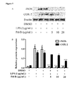
- FIG. 7 shows inhibitory effects of Pd-Ib on the iNOS and COX-2 in LPS-stimulated RAW-Blue cells.
- the protein levels of iNOS and COX-2 are determined by Western blotting, ⁇ -actin is used as a quantity control:
- A) shows representative images of Western blotting;
- B) shows protein levels of iNOS and COX-2 are calculated with Image J software. Data are derived from three independent experiments and presented as mean ⁇ SEM. # Compared with control group. *P ⁇ 0.05, **P ⁇ 0.01 and ***P ⁇ 0.001 are compared with LPS-alone group.
- FIG. 8 shows effects of pro-inflammatory factors release and nitric oxide production in LPS-stimulated RAW-Blue cells.
- macrophages are treated with 1 ⁇ g/mL of LPS in absence or presence various concentrations of Pd-Ib (5, 10, 20 ⁇ g/mL) for 20 hours, the mRNA levels of iNOS (A), TNF- ⁇ (B) and IL-1 ⁇ (C) are determined by real time PCR, while the production of NO (D) is determined by Griess reagent.
- Data are derived from three independent experiments and presented as mean ⁇ SEM, “con” stands for the vehicle control group. # Compared with control group. *P ⁇ 0.05, **P ⁇ 0.01 and ***P ⁇ 0.001 are compared with LPS-alone group.
- FIG. 9 shows the experimental design of animal model with DSS-induced colitis and treated with different concentrations of Pd-Ib or Sulfasalazine.
- FIG. 10 shows the effects of Pd-Ib on body weight change (A), disease activity index (B), and colon length (C and D) of mice with DSS-induced colitis.
- FIG. 11 shows effects of Pd-Ib on histological manifestation in DSS-induced chronic colitis in mice: (a) control; (b) 2% DSS-treated; (c) 2% DSS+sulfasalazine at 300 mg/kg/day for seven consecutive days; (d) 2% DSS+Pd-Ib at 30 mg/kg/day for seven consecutive days; (e) 2% DSS+Pd-Ib at 60 mg/kg/day for seven consecutive days; (f) 2% DSS+Pd-Ib at 120 mg/kg/day for seven consecutive days.
- FIG. 12 shows effects of Pd-Ib on suppress myeloperoxidase (MPO) activity in the colon of mice with DSS-induced colitis.
- FIG. 13 shows the effects of Pd-Ib on the production of cytokines in colon tissue of animals with DSS-induced colitis: (A) TNF- ⁇ levels; (B) IFN- ⁇ levels. (C) IL-6 levels; (D) IL-4 levels.
- LPS lipopolysaccharides, L3129
- MTT 3-[4,5-dimethylthiazol-2-yl]-2,5-diphenyltetrazoliumbromide
- DMSO dimethyl sulfoxide
- Griess reagent and all chemicals used are of HPLC grade from Sigma Chemical Co. (St. Louis, Mo., USA).
- iNOS IL-1 ⁇
- TNF- ⁇ Trizol
- SYBR Green Dulbecco's modified Eagle's medium
- ECL reagent ECL reagent
- fetal bovine serum penicillin and streptomycin
- BCA protein assay kit is supplied by Thermo Fisher Scientific (Waltham, Mass., USA).
- NF- ⁇ B, I ⁇ B- ⁇ , COX-2 and iNOS rabbit antibody are purchased from Cell Signaling Technology (Beverly, Mass., USA).
- ⁇ -Actin mouse antibody, anti-rabbit IgG and anti-mouse IgG are purchased from Santa Cruz Biotechnology (Santa Cruz, Calif., USA).
- QUANTI-Blue medium and Alexa Fluor568@ anti-rabbit IgG are purchased from Invivo Gen (San Diego, Calif., USA). Fractions are monitored and combined by thin layer chromatography (TLC). Spots are made visible by heating silica gel plates that had been immersed in 5% H 2 SO 4 in EtOH. Sulfasalazine is purchased from Sigma Corp. (Louis, USA). Dextran sulfate sodium (DSS; molecular weight: 36 to 50 kDa) is purchased from MP Biomedical (Santa Ana, Calif., USA).
- the dried roots of Bupleurum malconense are collected from Sichuan province of China.
- the plant material is identified and authenticated by Dr. Linfang Huang based on sequences of the plastid psbA-trnssel genetic region ( FIG. 1 )
- Air-dried pieces of Bupleurum malconense root (500 g) are extracted for three times by percolation in petroleum ether (1 L); the supernatant is concentrated and collected as the petroleum ether (PE) extract.
- the residue is extracted for three times by reflux in 80% ethanol at 60° C.; the solution is concentrated to yield 80% ethanol extract.
- the residue is further extracted by reflex in water (1 L) for three times to obtain the aqueous extract. Meanwhile, the 80% ethanol extract is dissolved in 200 mL of water in a separatory funnel, and then partitioned with ethyl acetate (200 mL ⁇ 3). The upper and lower layers are collected to yield the ethyl acetate extract and ethanol extract, respectively.
- the petroleum ether extract is subjected onto silica gel column chromatography (300-400 mesh, DAVISIL, Germany) using petroleum ether/ethyl acetate (PE/EtOAc) of increasing polarity as an eluent.
- Pd-Ib which is obtained from fraction 3 with 2.5% EtOAc in petroleum ether, is further purified using reverse phase HPLC (Waters system including a 2545 binary gradient module, a 2489 UV/Visible detector, a fraction collector III) on semi-preparative column Preparative RP-C 18 (Alltech Alltima-C 18 , 250 mm ⁇ 10.0 mm, 5 ⁇ m).
- the bioassay result and separation flow chart is showed in FIG. 2 .
- RAW-Blue cells (5 ⁇ 10 4 ) are cultured in 96-well plate for 24 hours, and then treated with LPS (1 ⁇ g/mL) alone or together with test samples for 20 hours.
- Secreted embryonic alkaline phosphatase (SEAP) activity in the conditioned medium is determined using QUANTI-Blue medium following manufacturer's manual. Briefly, 100 ⁇ L samples are added to 200 ⁇ L of QUANTI-Blue medium and incubated at 37° C. for 15 to 30 minutes. Absorbance is measured at 620 nm using a micro-plate reader (Benchmark plus Bio-Red, CA, USA) and fold change in SEAP activity is calculated accordingly.
- SEAP secreted embryonic alkaline phosphatase
- Raw-Blue cells are derived from RAW264.7 macrophages. Then the murine macrophage cell line of Raw-Blue cells is cultured in plastic dishes containing DMEM supplemented with 100 U/mL of penicillin, 100 ⁇ g/mL of streptomycin and 10% FBS in an incubator (5% CO 2 ) at 37° C. Cells are sub-cultured every 3 days at a dilution of 1:6.
- Cells (5 ⁇ 10 4 ) are cultured in 96-well plates for 24 hours. Then cells are cultured with various concentrations of Pd-Ib (1.25, 2.5, 5, 10, 20, 40, 80, 100 ⁇ g/mL) for 24 hours. After that, 10 ⁇ L of 5 mg/mL MTT is added to each well, and the cells are cultured in the dark for 3 hours. The medium is then discarded, and 100 ⁇ L of dimethyl sulfoxide (DMSO) is added into each well. After 15 min incubation, the optical density at 570 nm is read by using a micro-plate reader to determine the cell viability.
- Pd-Ib 1.25, 2.5, 5, 10, 20, 40, 80, 100 ⁇ g/mL
- 10 ⁇ L of 5 mg/mL MTT is added to each well, and the cells are cultured in the dark for 3 hours. The medium is then discarded, and 100 ⁇ L of dimethyl sulfoxide (DMSO) is added into each well. After 15 min incuba
- Total protein concentration is measured using a protein assay kit, followed by centrifugation at 13,500 rpm for 15 minutes at 4° C., then 10-25 ⁇ g of protein from the supernatants is then separated on 10% sodium dodecylsulphate-polyacrylamide gel (SDS-PAGE) and transferred on to polyvinylidene difluoride membranes.
- SDS-PAGE sodium dodecylsulphate-polyacrylamide gel
- the membrane After washing with TBST (3 ⁇ 10 min), the membrane is incubated with secondary antibody (1:2,000 dilution) in 5% skim milk in TBST for 1 h at room temperature. The membrane is rewashed (TBST; 3 ⁇ 10 min), and the immune-reactive proteins are detected by enhanced chemiluminescence using X-ray film and ECL reagent. The protein bands are quantified by measuring the relative intensity compared to the control using Image J Software (version 4.1.7, NIH, USA).
- Raw-Blue cells are cultured (20 ⁇ 10 4 cells/well) on glass coverslips and incubated for 24 hours. Cells are pretreated with various concentrations (5-20 ⁇ g/mL) of Pd-Ib for 2 hours. Then treated with LPS (1 ⁇ g/mL) for 20 hours. Subsequently, the coverslips are rinsed twice with PBS, and cells are fixed in 4% paraformaldehyde (PFA) in PBS at room temperature for 15 minutes. Cellular and nuclear membranes of the macrophages are permeabilized by treatment with 3% Triton X-100 in PBS for 15 minutes.
- PFA paraformaldehyde
- the cells After being blocked with 3% bovine serum albumin (BSA) in PBS for 1 hour, the cells is incubated with primary antibody in 3% BSA/PBS (1:500 dilution) at 4° C. overnight. After washing with PBS, the cells are incubated with the red-conjugated secondary antibody (1:500 dilution) in 3% BSA/PBS at room temperature for 1 hour and finally washed again for three time with PBS. Then, counter-staining is performed with DAPI (1:1,000 dilution) in 3% BSA/PBS nucleus with for 10 minutes. The cells are washed for three times with PBS, and then anti-fade mounting medium is added. Samples are observed under a fluorescence microscope.
- BSA bovine serum albumin
- Raw-Blue cells are plated at 20 ⁇ 10 5 cells/well in 12-well plate. After 24 hours of incubation, cells are pretreated with various concentrations (5-20 ⁇ g/mL) of Pd-Ib for 2 hours, followed by co-culturing with LPS (1 ⁇ g/mL) for 20 hours.
- Total RNA of Raw-Blue cells is extracted by Trizol reagent. Two micrograms (2 ⁇ g) of total RNA are reverse-transcribed with RT master mix to obtain cDNA.
- Real-time PCR is performed in a ViiA7 real-time PCR instrument (Applied Biosystems, Life Technologies, CA, USA) with the SYBR Green kit. A melting curve analysis is carried out after amplification to verify the accuracy of the amplicon. The comparative cycle of threshold ( ⁇ C T ) method of relative quantification is used to determine the fold-change in expression.
- Primer sequences for mRNA analysis of IL-1 ⁇ , TNF- ⁇ , iNOS and ⁇ -Actin are described in Table 1.
- Nitric oxide (NO) production is indirectly assessed by measuring the nitrite levels in the cultured medium determined by a colorimetric method based on the Griess reagent and sodium nitrite as a standard substance.
- the cells are pretreated with various concentrations of Pd-Ib (5-20 ⁇ g/mL). Two hours later, cells are incubated for another 20 hours in the presence of LPS (1 ⁇ g/mL) at 37° C. Then, 100 ⁇ L of each supernatant is mixed with the equal volume of Griess reagent. The samples are incubated at room temperature for 15 min. The optical densities are measured at 540 nm with a micro-plate reader and nitrite concentration is determined using a standard curve generated with known concentrations of sodium nitrite.
- 6-week-old male C57BL/6 mice weighing 18-20 g are purchased from the Laboratory Animal Services Center, the Chinese University of Hong Kong. The study protocols are approved by the committee for Care of Laboratory Animals in the School of Chinese Medicine at the Hong Kong Institution University.
- control group mice received drinking water and control diet
- DSS-treated group mice received 2.0% DSS in drinking and control diet
- positive control group mice received 2.0% DSS in drinking and ip, 300 mg/kg/day of sulfasalazine
- Pd-Ib treatment groups will be given different dosed of Pd-Ib (mice received 2.0% DSS in drinking and i.p., 30, 60, or 120 mg/kg/day, respectively).
- mice are given tap water for 6 days, and then be divided into 6 groups.
- DSS is dissolved in distilled water at a concentration of 2% (w/v) and administered to the mice.
- the first day and the last day of DSS treatment are designated as day 7 and day 11, respectively.
- Mice are marked and checked daily for body weight. On day 12, body weight, stool consistency, and gross bleeding of each mouse are assessed, and animals showing body weight loss, diarrhea, and bleeding are selected for further analysis. All colitis mice are then interventions once daily for seven days, with eight mice in each group.
- the DSS model group is administered with tap water as negative control; while sulfasalazine is given as positive control (i.p., 300 mg/kg/day) and Pd-Ib treatment groups are given different doses of Pd-Ib (i.p., 30, 60, or 120 mg/kg/day, respectively).
- a vehicle control group is also set up to receive drinking tap water without DSS throughout the entire experimental period. On day 19, after the mice are killed under anesthesia induced by pentobarbital sodium (i.p., 0.65 g/kg), the length of the large intestine is measured. All portions are stored at ⁇ 80° C. for biochemical assays.
- the DAI is determined by scoring changes in the body weight, diarrhea, colon length, and bleeding. Each score is given in Table 2. In brief, body weight, stool consistency, colon length and bleeding in the stool are monitored daily for determination of DAI. At the end of the experiment, mice are killed, and the colon is dissected from each mouse, and the length of which is measured from the ileocecal junction to the anal verge.
- the colons are opened longitudinally, gently washed with ice-cold PBS, fixed in 4% paraformaldehyde overnight, and embedded in paraffin. Five-micro-meter sections are stained with hematoxylin/eosin according to a standard procedure to evaluate colonic damage. The histological scoring system is shown in Table 3.
- MPO Myeloperoxidase
- neutrophil neutrophil
- MPO activity is measured as described in our previous study. Briefly, the colon tissues kept at ⁇ 80° C. are weighed and homogenized in 0.5% hexadecyltrimethylammonium bromide 1 mL per 100 mg of colon tissue. The homogenates are centrifuged at 19 000 rpm at 4° C. for 15 min. Aliquots of 80 iL supernatant are mixed with 120 ⁇ L potassium phosphate buffer (50 mmol, pH 6.0) with 0.0005% o-dianisidine dihydrochloride and 0.1% hydrogen peroxide.
- MPO activity is calculated from the rate of optical density changes and one unit of MPO activity is defined as the amount of enzyme present that produced a change in optical density of 1.0 U/min at 25° C. in the final reaction volume. The results are normalized to the wet weight of colon tissue and quantified as units/mg tissue.
- Colon levels of cytokines are assayed using commercially available ELISA kits. Briefly, colon samples are homogenized in phosphate buffer containing 0.05% Tween-20, 0.1 mM phenylmethylsulfonyl fluoride, 0.1 mM benzethonium chloride, 10 mM EDTA, and 20 IU aprotinin A. The homogenates are centrifuged at 16000 g at 4° C. for 15 min, and the supernatants are collected for the determination of levels of the cytokines, IL-6, IL-4, TNF- ⁇ , and IFN- ⁇ , according to the manufacturer's protocols. The amount of protein in each sample is measured by the Bradford method, using bovine serum albumin as a standard. The levels of each cytokine are evaluated in each sample and expressed in ⁇ g/mL.
- PE extract exerts significant inhibitory effect of SEAP activity compared with LPS group (18.19 ⁇ 0.967%).
- the PE extract is subjected to silica gel chromatography using PE/EtOAc as an eluent and 8 fractions are collected. Since the SEAP activity is significantly inhibited by Fr.3 (30.661 ⁇ 2.546%), further purification is carried out to isolate one pure compound.
- the spectral data ( FIG. 3 ) are identical to those reported in Wang X. B., et.
- the cytotoxic effect of Pd-Ib on Raw-Blue cells is tested with the MTT assay. As shown in FIG. 4 , the Pd-Ib of the present invention does not exhibit cytotoxic effect at dosages ranging from 1.56 to 20 ⁇ g/mL, while cell viability is reduced when macrophages are treated with Pd-Ib at doses of 40, 80, and 100 ⁇ g/mL.
- Pd-Ib Inhibits the Nuclear Translocation of NF-KB and Decreases Degradation of IKB- ⁇ in LPS-Stimulated Raw-Blue Cells
- the SEAP activity in the supernatants of LPS-stimulated RAW-Blue cells reflects the activation of NF- ⁇ B.
- the inhibition of the NF- ⁇ B activation can associate with mitigation of colon inflammation responses and apoptosis of intestinal epithelial cells in DSS mouse model.
- Western blotting and immunofluorescence analysis are performed to determine whether Pd-Ib depressed NF- ⁇ B activation.
- the effect of Pd-Ib on LPS-induced I ⁇ B- ⁇ degradation is investigated to examine the molecular mechanisms by which Pd-Ib inhibits NF- ⁇ B transcriptional activity. As shown in FIG. 5 , Pd-Ib inhibits I ⁇ B- ⁇ degradation in a dose-dependent manner compared to the LPS-stimulated macrophage.
- the immunofluorescence result shows that a low level of p65 activity is observed in the control group. It also revealed that treatment with Pd-Ib significantly inhibits the p65 nuclear translocation compared to the LPS-stimulated macrophage ( FIG. 6 ).
- Pd-Ib Suppresses the Expressions of iNOS and Cox-2 in LPS-Stimulated Raw-Blue Cell
- COX-2 and iNOS are the important enzymes that mediate inflammatory pathways.
- High expression levels will cause intestinal inflammation with motility dysfunction (Tajima et al., “EP2 and EP4 receptors on muscularis resident macrophages mediate LPS-induced intestinal dysmotility via iNOS upregulation through cAMP/ERK signals”, Am J Physiol Gastrointest Liver Physiol 2012, 302: G524-534).
- Western blotting analysis shows that Pd-Ib strongly down-regulates iNOS and COX-2 protein levels in a dose-dependent manner ( FIG. 7A ). The intensity of protein bands are analyzed and the results are shown in FIG. 7B .
- Pd-Ib Inhibits the mRNA Expressions of TNF- ⁇ and IL-1 ⁇ in LPS-Stimulated Raw-Blue Cells
- TNF- ⁇ and IL-1 ⁇ are produced in the early stage of inflammatory response and play important roles in varies inflammatory cascades.
- Real-time PCR experiments are performed to examine the expression of pro-inflammatory cytokines following LPS treatment. As shown in FIG. 8B and FIG. 8C .
- Pd-Ib could inhibit the mRNA expressions of TNF- ⁇ and IL-1 ⁇ in a dose-dependent manner, while LPS stimulation of macrophages causes an increase in their expressions.
- nitric oxide is a critical mediator of a variety of biological functions; however, production of excessive amounts of NO leads to inflammatory responses or tissue injury (Aktan F: iNOS-mediated nitric oxide production and its regulation. Life Sci 2004, 75: 639-653).
- Aktan F iNOS-mediated nitric oxide production and its regulation. Life Sci 2004, 75: 639-653.
- the effect of Pd-Ib on NO production is investigated using Griess reagent. As shown in FIG. 8D , stimulation with LPS results in a significant increase in NO production compared with the control group while treatment with Pd-Ib at 5, 10, 20 ⁇ g/mL led to 17.64 ⁇ 2.96%, 29.82 ⁇ 1.34% and 55.45 ⁇ 1.15% inhibition of NO production, respectively.
- Pd-Ib is isolated from Bupleurum malconense for the first time, and it is found to have excellent anti-inflammatory activities. To date, little is known about the role of Pd-Ib in anti-inflammatory effects. It is demonstrated that Pd-Ib suppresses LPS-induced inflammatory responses in macrophage Raw-Blue cells.
- SEAP is a widely use reporter gene widely used to screen immune-pharmacological activity upon the activation of NF- ⁇ B and activator protein 1 (AP-1).
- I ⁇ B- ⁇ which is the inhibitory subunit of NF- ⁇ B
- AP-1 activator protein 1
- NF- ⁇ B releases from the I ⁇ B- ⁇ subunit and translocates to the nucleus, where it increases the expression of the genes for many cytokines, enzymes and adhesion molecules.
- Pd-Ib is shown to significantly inhibit LPS-stimulated degradation of I ⁇ B- ⁇ and nuclear translocation of NF- ⁇ B.
- the pro-inflammatory cytokines including TNF- ⁇ and IL-1 ⁇ , play an important role in the mediation of inflammatory processes. When those mediators are overproduced, they lead to various diseases. Thus, the inhibition of pro-inflammatory cytokines release may help attenuate the inflammatory response.
- the result in the present invention indicates that Pd-Ib could significantly down-regulate the mRNAs expression of TNF- ⁇ and IL-1 ⁇ in a dose-dependent manner.
- the present invention firstly reports the isolation of Pd-Ib from Bupleurum malconense and demonstrates Pd-Ib's anti-inflammatory effect in LPS-stimulated macrophages.
- This beneficial effect in alleviating the inflammatory response of TNF- ⁇ , IL-1 ⁇ , NO and iNOS in LPS-stimulated Raw-Blue cells can be caused by down-regulating iNOS and COX-2 protein expression via the blockade of NF- ⁇ B activation. It is concluded that the petroleum ether extract of Bupleurum malconense is efficacious for the treatment of inflammatory disease; meanwhile, Pd-Ib could be a potential anti-inflammatory agent.
- the present invention will be undertaken to evaluate the possible effect of Pd-Ib in a model of chronic DSS-induced colitis in mice with the aid of macroscopic and histological analyses, to determine the key pro-inflammatory mediators involved in IBD development.
- DSS-induced colitis is one of the most common models with several characteristics resembling human ulcerative colitis.
- mice exposed to DSS drinking water develop typical symptoms of clinical colitis, including body weight loss, diarrhea, rectal bleeding and diarrhea.
- mice in groups treated with Pd-Ib at 120 mg/kg/day recover the body weight loss significantly compared with that of DSS model group after 10 days (p ⁇ 0.05).
- Pd-Ib is mildly effective in preventing body weight loss at 30 mg/kg/day and 60 mg/kg/day.
- Pd-Ib also leads to clinical improvement of DSS-induced colitis, as reflected in the DAI, colon length and the histological disease score.
- D human is equivalent dosage for adult human
- D mouse is dosage for mouse
- the lower dosage of Pd-Ib for mouse is 30 mg/kg/day; medium dosage for mouse is 60 mg/kg/day; higher dosage for mouse is 120 mg/kg/day, in which the dosage of 120 mg/kg/day for mouse is most effective in treating inflammation or inflammatory diseases in terms of preventing weight loss in the mouse model, having the lower DAI score which is comparable to untreated mice, preventing reduction in colon length, and preventing rectal bleeding found in stool.
- Colitis is induced in all groups except the control group.
- Pd-Ib and sulfasalazine are administered to mice from day 6 to day 12.
- the change in body weight is taken as the difference between the body weight before induction of colitis and that immediately before sacrifice on day 13.
- Hematoxylin and eosin staining images of representative colons are shown at magnifications of 10 ⁇ (a, control group; b, DSS model group; c, DSS plus sulfasalazine 300 mg/kg/day group; d, DSS plus Pd-Ib 30 mg/kg/day group; e, DSS plus Pd-Ib 60 mg/kg/day group; f, DSS plus Pd-Ib 120 mg/kg/day group).
- FIG. 11 the present invention shows that DSS treatment ( FIG. 11 b ) can cause typical inflammatory changes in colon architecture, including mucosal ulceration, crypt damage, edema, and cell infiltration into mucosal tissue, which compared with the control group ( FIG. 11 a ).
- the sulfasalazine treatment group can significantly decrease the damage of colon tissues.
- Pd-Ib exhibits a clinical improvement in DSS-induced colitis, which is reflected in the DAI score.
- Colon tissue sections from mice treated with Pd-Ib exhibits far fewer infiltrating cells, a significantly lower degree of mucosal injury, and less edema at different dosage treatment (as shown in FIGS. 11 d, e , and f ).
- Colitis is induced in all groups except the control group.
- Colitis is induced in all groups except the control group.
- the present invention indicates that Pd-Ib is effective in alleviating the symptoms of DSS-induced chronic colitis, proving that Pd-Ib is an effective agent in treatment against ulcerative colitis.
- the present invention is in the field of pharmaceuticals and chemical industries.
- the present invention relates to a compound, namely (+)-3′-Angeloyloxy-4′-keto-3,4′-dihydroseselin (Pd-Ib), which is isolated from Bupleurum malconense .
- the present invention also includes the methods of preparation and use thereof for treating inflammation, such as in alleviating the symptoms of DSS-induced chronic colitis and as an effective treatment against ulcerative colitis.
Landscapes
- Health & Medical Sciences (AREA)
- Chemical & Material Sciences (AREA)
- Medicinal Chemistry (AREA)
- Pharmacology & Pharmacy (AREA)
- Epidemiology (AREA)
- Life Sciences & Earth Sciences (AREA)
- Animal Behavior & Ethology (AREA)
- General Health & Medical Sciences (AREA)
- Public Health (AREA)
- Veterinary Medicine (AREA)
- Nutrition Science (AREA)
- Physiology (AREA)
- Pharmaceuticals Containing Other Organic And Inorganic Compounds (AREA)
Abstract
Description

| TABLE 1 |
| Sequence of primers used in real-time PCR |
| Gene | Primer | Sequence (5′-3′) | SEQ ID No. | |
| | Sense | CACCTTGGAGTTCACCCAGT | 1 | |
| | ACCACTCGTACTTGGGATGC | 2 | ||
| TNF- | Sense | CTGTGAAGGGAATGGGTGTT | 3 | |
| | GGTCACTGTCCCAGCATCTT | 4 | ||
| IL- | Sense | GCTGAAGGAGTTGCCAGAAA | 5 | |
| | GTGCAAGTGACTCAGGGTGA | 6 | ||
| β- | Sense | GGTGAAGGTCGGTGGAACG | 7 | |
| | CTCGCTCCTGGAAGATGGTG | 8 | ||
| TABLE 2 |
| Disease Activity Index Scores Based on Disease Marker Intensities |
| Body weight | Diarrhea | ||
| Score | loss (%) | (stool consistency) | Rectal bleeding |
| 0 | |
0 | negative |
| 1 | 1-5 | 1 | faintly blue |
| 2 | 6-10 | 2 | moderately blue |
| 3 | 10-15 | 3 | dark blue |
| 4 | >15 | 4 | dark blue, |
| prolapse | |||
| TABLE 3 |
| Histological Scoring System for DSS-Induced Colitis |
| Scoring of severity of histological damage |
| Feature | Score | Description |
| Severity of | 0 | |
| inflammation | ||
| 1 | mild | |
| 2 | moderate | |
| 3 | severe | |
| Extent of | 0 | |
| inflammation | ||
| 1 | |
|
| 2 | mucosa and |
|
| 3 | | |
| Crypt damage | ||
| 0 | |
|
| 1 | ⅓ damaged | |
| 2 | ⅔ damaged | |
| 3 | crypt loss by surface | |
| epithelium present | ||
| 4 | both crypt and surface | |
| epithelium lost | ||
D human =D mouse ×k(k=0.081)
Lower dosage of Pd-Ib for human=D mouse ×k=30 mg/kg/day×0.081=2.43 mg/kg/day;
Medium dosage for human=D mouse ×k=60 mg/kg/day×0.081=4.86 mg/kg/day;
Higher dosage for human=D mouse ×k=120 mg/kg/day×0.081=9.72 mg/kg/day.
Claims (6)

Priority Applications (1)
| Application Number | Priority Date | Filing Date | Title |
|---|---|---|---|
| US14/742,727 US9408828B2 (en) | 2014-06-18 | 2015-06-18 | (+)-3′-angeloyloxy-4′-keto-3′,4′-dihydroseselin for treating inflammation |
Applications Claiming Priority (3)
| Application Number | Priority Date | Filing Date | Title |
|---|---|---|---|
| US201462013556P | 2014-06-18 | 2014-06-18 | |
| US201462013566P | 2014-06-18 | 2014-06-18 | |
| US14/742,727 US9408828B2 (en) | 2014-06-18 | 2015-06-18 | (+)-3′-angeloyloxy-4′-keto-3′,4′-dihydroseselin for treating inflammation |
Publications (2)
| Publication Number | Publication Date |
|---|---|
| US20150366841A1 US20150366841A1 (en) | 2015-12-24 |
| US9408828B2 true US9408828B2 (en) | 2016-08-09 |
Family
ID=54868657
Family Applications (1)
| Application Number | Title | Priority Date | Filing Date |
|---|---|---|---|
| US14/742,727 Active US9408828B2 (en) | 2014-06-18 | 2015-06-18 | (+)-3′-angeloyloxy-4′-keto-3′,4′-dihydroseselin for treating inflammation |
Country Status (1)
| Country | Link |
|---|---|
| US (1) | US9408828B2 (en) |
Citations (1)
| Publication number | Priority date | Publication date | Assignee | Title |
|---|---|---|---|---|
| US20050222245A1 (en) * | 2002-04-09 | 2005-10-06 | Elcom Biotechnology, Co., Ltd. | Pyranocoumarin derivatives |
-
2015
- 2015-06-18 US US14/742,727 patent/US9408828B2/en active Active
Patent Citations (1)
| Publication number | Priority date | Publication date | Assignee | Title |
|---|---|---|---|---|
| US20050222245A1 (en) * | 2002-04-09 | 2005-10-06 | Elcom Biotechnology, Co., Ltd. | Pyranocoumarin derivatives |
Non-Patent Citations (39)
Also Published As
| Publication number | Publication date |
|---|---|
| US20150366841A1 (en) | 2015-12-24 |
Similar Documents
| Publication | Publication Date | Title |
|---|---|---|
| Cai et al. | Effect of mulberry leaf (Folium Mori) on insulin resistance via IRS-1/PI3K/Glut-4 signalling pathway in type 2 diabetes mellitus rats | |
| Nguyen et al. | Antipyretic, anti-inflammatory and analgesic activities of Periplaneta americana extract and underlying mechanisms | |
| Li et al. | Investigation of constituents from Cinnamomum camphora (L.) J. Presl and evaluation of their anti-inflammatory properties in lipopolysaccharide-stimulated RAW 264.7 macrophages | |
| Bao et al. | Genkwanin ameliorates adjuvant-induced arthritis in rats through inhibiting JAK/STAT and NF-κB signaling pathways | |
| Arçari et al. | Antiobesity effects of yerba maté extract (Ilex paraguariensis) in high‐fat diet–induced obese mice | |
| Zeng et al. | Alisol A 24-acetate prevents hepatic steatosis and metabolic disorders in HepG2 cells | |
| Michl et al. | LC-MS-and 1H NMR-based metabolomic analysis and in vitro toxicological assessment of 43 Aristolochia species | |
| Vijayakumar et al. | Hypolipidemic effect of fenugreek seeds is mediated through inhibition of fat accumulation and upregulation of LDL receptor | |
| Ahmad et al. | Naringin attenuates the development of carrageenan-induced acute lung inflammation through inhibition of NF-κb, STAT3 and pro-inflammatory mediators and enhancement of IκBα and anti-inflammatory cytokines | |
| Cui et al. | The inhibiting effect of the Coptis chinensis polysaccharide on the type II diabetic mice | |
| Du et al. | Anti-inflammatory properties of uvaol on DSS-induced colitis and LPS-stimulated macrophages | |
| Liu et al. | Cerasus humilis cherry polyphenol reduces high-fat diet-induced obesity in C57BL/6 mice by mitigating fat deposition, inflammation, and oxidation | |
| Choi et al. | Picroside II attenuates airway inflammation by downregulating the transcription factor GATA3 and Th2-related cytokines in a mouse model of HDM-induced allergic asthma | |
| Pan et al. | Ethanol extract of Liriodendron chinense (Hemsl.) Sarg barks attenuates hyperuricemic nephropathy by inhibiting renal fibrosis and inflammation in mice | |
| Liang et al. | Glucocorticoid receptor-mediated alleviation of inflammation by berberine: in vitro, in silico and in vivo investigations | |
| JP2020508287A (en) | New low-toxic raikoto glycoside, method for producing the same and use thereof | |
| Kang et al. | Wasabia japonica is a potential functional food to prevent colitis via inhibiting the NF-κB signaling pathway | |
| Park et al. | Prophylactic effects of Lonicera japonica extract on dextran sulphate sodium-induced colitis in a mouse model by the inhibition of the Th1/Th17 response | |
| Raciti et al. | Citrus aurantium L. dry extracts promote C/ebpβ expression and improve adipocyte differentiation in 3T3-L1 cells | |
| Yoon et al. | Anti-neuroinflammatory effects of quercetin-3-O-glucuronide isolated from the leaf of Vitis labruscana on LPS-induced neuroinflammation in BV2 cells | |
| Wu et al. | The extract of Sonneratia apetala leaves and branches ameliorates hyperuricemia in mice by regulating renal uric acid transporters and suppressing the activation of the JAK/STAT signaling pathway | |
| Zhang et al. | Hepatoprotective effect of Xiayuxue decoction ethyl acetate fraction against carbon tetrachloride-induced liver fibrosis in mice via inducing apoptosis and suppressing activation of hepatic stellate cells | |
| Kim et al. | Red pepper seed water extract inhibits preadipocyte differentiation and induces mature adipocyte apoptosis in 3T3-L1 cells | |
| Zhou et al. | Epiberberine regulates lipid synthesis through shp (nr0b2) to improve non-alcoholic steatohepatitis | |
| Yeh et al. | Identification of scoparone from Chinese olive fruit as a modulator of macrophage polarization |
Legal Events
| Date | Code | Title | Description |
|---|---|---|---|
| AS | Assignment |
Owner name: CHANGSHU RESEARCH INSTITUTE OF HONG KONG BAPTIST U Free format text: ASSIGNMENT OF ASSIGNORS INTEREST;ASSIGNORS:BIAN, ZHAOXIANG;MU, HUAIXUE;LIN, CHENGYUAN;AND OTHERS;SIGNING DATES FROM 20140529 TO 20150615;REEL/FRAME:035954/0348 Owner name: HONG KONG BAPTIST UNIVERSITY, HONG KONG Free format text: ASSIGNMENT OF ASSIGNORS INTEREST;ASSIGNORS:BIAN, ZHAOXIANG;MU, HUAIXUE;LIN, CHENGYUAN;AND OTHERS;SIGNING DATES FROM 20140529 TO 20150615;REEL/FRAME:035954/0348 |
|
| STCF | Information on status: patent grant |
Free format text: PATENTED CASE |
|
| AS | Assignment |
Owner name: CHANGSHU HKBU TECHNOLOGY COMPANY LIMITED, CHINA Free format text: CORRECTIVE ASSIGNMENT TO CORRECT THE SECOND ASSIGNEE'S NAME AND THE SECOND ASSIGNEE'S ADDRESS PREVIOUSLY RECORDED AT REEL: 035954 FRAME: 0348. ASSIGNOR(S) HEREBY CONFIRMS THE ASSIGNMENT;ASSIGNORS:BIAN, ZHAOXIANG;MU, HUAIXUE;LIN, CHENGYUAN;AND OTHERS;SIGNING DATES FROM 20140529 TO 20150615;REEL/FRAME:039484/0653 Owner name: HONG KONG BAPTIST UNIVERSITY, HONG KONG Free format text: CORRECTIVE ASSIGNMENT TO CORRECT THE SECOND ASSIGNEE'S NAME AND THE SECOND ASSIGNEE'S ADDRESS PREVIOUSLY RECORDED AT REEL: 035954 FRAME: 0348. ASSIGNOR(S) HEREBY CONFIRMS THE ASSIGNMENT;ASSIGNORS:BIAN, ZHAOXIANG;MU, HUAIXUE;LIN, CHENGYUAN;AND OTHERS;SIGNING DATES FROM 20140529 TO 20150615;REEL/FRAME:039484/0653 |
|
| CC | Certificate of correction | ||
| MAFP | Maintenance fee payment |
Free format text: PAYMENT OF MAINTENANCE FEE, 4TH YR, SMALL ENTITY (ORIGINAL EVENT CODE: M2551); ENTITY STATUS OF PATENT OWNER: SMALL ENTITY Year of fee payment: 4 |
|
| AS | Assignment |
Owner name: HONG KONG BAPTIST UNIVERSITY, HONG KONG Free format text: ASSIGNMENT OF ASSIGNORS INTEREST;ASSIGNOR:CHANGSHU HKBU TECHNOLOGY COMPANY LIMITED;REEL/FRAME:055114/0720 Effective date: 20210125 |
|
| MAFP | Maintenance fee payment |
Free format text: PAYMENT OF MAINTENANCE FEE, 8TH YR, SMALL ENTITY (ORIGINAL EVENT CODE: M2552); ENTITY STATUS OF PATENT OWNER: SMALL ENTITY Year of fee payment: 8 |