US9388450B2 - FRET based multiplex probes - Google Patents
FRET based multiplex probes Download PDFInfo
- Publication number
- US9388450B2 US9388450B2 US14/206,738 US201414206738A US9388450B2 US 9388450 B2 US9388450 B2 US 9388450B2 US 201414206738 A US201414206738 A US 201414206738A US 9388450 B2 US9388450 B2 US 9388450B2
- Authority
- US
- United States
- Prior art keywords
- fluorophore
- oligopeptide
- emission
- central body
- probe
- Prior art date
- Legal status (The legal status is an assumption and is not a legal conclusion. Google has not performed a legal analysis and makes no representation as to the accuracy of the status listed.)
- Active, expires
Links
- 239000000523 sample Substances 0.000 title claims abstract description 91
- 238000002866 fluorescence resonance energy transfer Methods 0.000 title claims description 7
- 238000000034 method Methods 0.000 claims abstract description 36
- 238000003776 cleavage reaction Methods 0.000 claims abstract description 27
- 230000007017 scission Effects 0.000 claims abstract description 27
- 102000004190 Enzymes Human genes 0.000 claims abstract description 16
- 108090000790 Enzymes Proteins 0.000 claims abstract description 16
- 102000015636 Oligopeptides Human genes 0.000 claims description 90
- 108010038807 Oligopeptides Proteins 0.000 claims description 90
- 102000035195 Peptidases Human genes 0.000 claims description 56
- 108091005804 Peptidases Proteins 0.000 claims description 56
- 230000005284 excitation Effects 0.000 claims description 38
- 108090000765 processed proteins & peptides Proteins 0.000 claims description 33
- 230000000694 effects Effects 0.000 claims description 29
- 235000019833 protease Nutrition 0.000 claims description 28
- 206010028980 Neoplasm Diseases 0.000 claims description 24
- 102100026802 72 kDa type IV collagenase Human genes 0.000 claims description 23
- 101710151806 72 kDa type IV collagenase Proteins 0.000 claims description 23
- 102000002274 Matrix Metalloproteinases Human genes 0.000 claims description 18
- 108010000684 Matrix Metalloproteinases Proteins 0.000 claims description 18
- 201000011510 cancer Diseases 0.000 claims description 15
- MHMNJMPURVTYEJ-UHFFFAOYSA-N fluorescein-5-isothiocyanate Chemical compound O1C(=O)C2=CC(N=C=S)=CC=C2C21C1=CC=C(O)C=C1OC1=CC(O)=CC=C21 MHMNJMPURVTYEJ-UHFFFAOYSA-N 0.000 claims description 14
- 238000010521 absorption reaction Methods 0.000 claims description 12
- 201000010099 disease Diseases 0.000 claims description 12
- 208000037265 diseases, disorders, signs and symptoms Diseases 0.000 claims description 12
- 238000012546 transfer Methods 0.000 claims description 11
- 108090001008 Avidin Proteins 0.000 claims description 8
- 239000000758 substrate Substances 0.000 claims description 7
- ABZLKHKQJHEPAX-UHFFFAOYSA-N tetramethylrhodamine Chemical compound C=12C=CC(N(C)C)=CC2=[O+]C2=CC(N(C)C)=CC=C2C=1C1=CC=CC=C1C([O-])=O ABZLKHKQJHEPAX-UHFFFAOYSA-N 0.000 claims description 7
- 125000006850 spacer group Chemical group 0.000 claims description 6
- 238000011282 treatment Methods 0.000 claims description 6
- 238000003745 diagnosis Methods 0.000 claims description 4
- TWVKGYNQQAUNRN-ACZMJKKPSA-N Ile-Ser Chemical compound CC[C@H](C)[C@H]([NH3+])C(=O)N[C@@H](CO)C([O-])=O TWVKGYNQQAUNRN-ACZMJKKPSA-N 0.000 claims description 3
- 210000001124 body fluid Anatomy 0.000 claims description 3
- 239000010839 body fluid Substances 0.000 claims description 3
- MPUMPERGHHJGRP-WEDXCCLWSA-N Thr-Gly-Lys Chemical compound C[C@H]([C@@H](C(=O)NCC(=O)N[C@@H](CCCCN)C(=O)O)N)O MPUMPERGHHJGRP-WEDXCCLWSA-N 0.000 claims description 2
- BQBCIBCLXBKYHW-CSMHCCOUSA-N Thr-Leu Chemical compound CC(C)C[C@@H](C([O-])=O)NC(=O)[C@@H]([NH3+])[C@@H](C)O BQBCIBCLXBKYHW-CSMHCCOUSA-N 0.000 claims description 2
- 238000011394 anticancer treatment Methods 0.000 claims description 2
- 239000002105 nanoparticle Substances 0.000 claims description 2
- 230000001225 therapeutic effect Effects 0.000 claims description 2
- 108010015302 Matrix metalloproteinase-9 Proteins 0.000 claims 2
- 102100030412 Matrix metalloproteinase-9 Human genes 0.000 claims 2
- 230000002255 enzymatic effect Effects 0.000 abstract description 21
- 239000000203 mixture Substances 0.000 abstract description 9
- 239000000370 acceptor Substances 0.000 description 34
- 239000004365 Protease Substances 0.000 description 28
- 239000000975 dye Substances 0.000 description 21
- 238000001514 detection method Methods 0.000 description 20
- 229940088598 enzyme Drugs 0.000 description 13
- 238000013459 approach Methods 0.000 description 11
- 238000003556 assay Methods 0.000 description 11
- 210000001519 tissue Anatomy 0.000 description 10
- 210000004027 cell Anatomy 0.000 description 8
- 230000008859 change Effects 0.000 description 8
- 230000008569 process Effects 0.000 description 8
- 238000011161 development Methods 0.000 description 6
- 230000018109 developmental process Effects 0.000 description 6
- 238000003384 imaging method Methods 0.000 description 6
- 108020004707 nucleic acids Proteins 0.000 description 6
- 150000007523 nucleic acids Chemical class 0.000 description 6
- 102000039446 nucleic acids Human genes 0.000 description 6
- 102000004196 processed proteins & peptides Human genes 0.000 description 6
- 230000004044 response Effects 0.000 description 6
- 238000004458 analytical method Methods 0.000 description 5
- 235000019419 proteases Nutrition 0.000 description 5
- 102000004169 proteins and genes Human genes 0.000 description 5
- 108090000623 proteins and genes Proteins 0.000 description 5
- 239000002096 quantum dot Substances 0.000 description 5
- YBJHBAHKTGYVGT-ZKWXMUAHSA-N (+)-Biotin Chemical compound N1C(=O)N[C@@H]2[C@H](CCCCC(=O)O)SC[C@@H]21 YBJHBAHKTGYVGT-ZKWXMUAHSA-N 0.000 description 4
- 102100030417 Matrilysin Human genes 0.000 description 4
- 108090000855 Matrilysin Proteins 0.000 description 4
- 230000001413 cellular effect Effects 0.000 description 4
- 239000003446 ligand Substances 0.000 description 4
- 102000005962 receptors Human genes 0.000 description 4
- 238000012360 testing method Methods 0.000 description 4
- 206010005003 Bladder cancer Diseases 0.000 description 3
- 238000001327 Förster resonance energy transfer Methods 0.000 description 3
- 206010061218 Inflammation Diseases 0.000 description 3
- 208000006265 Renal cell carcinoma Diseases 0.000 description 3
- 208000007097 Urinary Bladder Neoplasms Diseases 0.000 description 3
- 230000008901 benefit Effects 0.000 description 3
- 238000013461 design Methods 0.000 description 3
- 238000000338 in vitro Methods 0.000 description 3
- 238000001727 in vivo Methods 0.000 description 3
- 230000004054 inflammatory process Effects 0.000 description 3
- 238000012544 monitoring process Methods 0.000 description 3
- 238000007837 multiplex assay Methods 0.000 description 3
- 230000037361 pathway Effects 0.000 description 3
- 230000035945 sensitivity Effects 0.000 description 3
- 201000005112 urinary bladder cancer Diseases 0.000 description 3
- VKUYLANQOAKALN-UHFFFAOYSA-N 2-[benzyl-(4-methoxyphenyl)sulfonylamino]-n-hydroxy-4-methylpentanamide Chemical compound C1=CC(OC)=CC=C1S(=O)(=O)N(C(CC(C)C)C(=O)NO)CC1=CC=CC=C1 VKUYLANQOAKALN-UHFFFAOYSA-N 0.000 description 2
- 208000030507 AIDS Diseases 0.000 description 2
- WHVNXSBKJGAXKU-UHFFFAOYSA-N Alexa Fluor 532 Chemical compound [H+].[H+].CC1(C)C(C)NC(C(=C2OC3=C(C=4C(C(C(C)N=4)(C)C)=CC3=3)S([O-])(=O)=O)S([O-])(=O)=O)=C1C=C2C=3C(C=C1)=CC=C1C(=O)ON1C(=O)CCC1=O WHVNXSBKJGAXKU-UHFFFAOYSA-N 0.000 description 2
- OBMZMSLWNNWEJA-XNCRXQDQSA-N C1=CC=2C(C[C@@H]3NC(=O)[C@@H](NC(=O)[C@H](NC(=O)N(CC#CCN(CCCC[C@H](NC(=O)[C@@H](CC4=CC=CC=C4)NC3=O)C(=O)N)CC=C)NC(=O)[C@@H](N)C)CC3=CNC4=C3C=CC=C4)C)=CNC=2C=C1 Chemical compound C1=CC=2C(C[C@@H]3NC(=O)[C@@H](NC(=O)[C@H](NC(=O)N(CC#CCN(CCCC[C@H](NC(=O)[C@@H](CC4=CC=CC=C4)NC3=O)C(=O)N)CC=C)NC(=O)[C@@H](N)C)CC3=CNC4=C3C=CC=C4)C)=CNC=2C=C1 OBMZMSLWNNWEJA-XNCRXQDQSA-N 0.000 description 2
- 238000002965 ELISA Methods 0.000 description 2
- 102000005593 Endopeptidases Human genes 0.000 description 2
- 108010059378 Endopeptidases Proteins 0.000 description 2
- 102000010834 Extracellular Matrix Proteins Human genes 0.000 description 2
- 108010037362 Extracellular Matrix Proteins Proteins 0.000 description 2
- 102000016359 Fibronectins Human genes 0.000 description 2
- 108010067306 Fibronectins Proteins 0.000 description 2
- 206010027476 Metastases Diseases 0.000 description 2
- 101710176384 Peptide 1 Proteins 0.000 description 2
- 102100030416 Stromelysin-1 Human genes 0.000 description 2
- 101710108790 Stromelysin-1 Proteins 0.000 description 2
- 102000036861 Zinc-dependent endopeptidases Human genes 0.000 description 2
- 108091006982 Zinc-dependent endopeptidases Proteins 0.000 description 2
- 230000006907 apoptotic process Effects 0.000 description 2
- 210000002469 basement membrane Anatomy 0.000 description 2
- 229960002685 biotin Drugs 0.000 description 2
- 235000020958 biotin Nutrition 0.000 description 2
- 239000011616 biotin Substances 0.000 description 2
- 230000015556 catabolic process Effects 0.000 description 2
- 238000006731 degradation reaction Methods 0.000 description 2
- 230000009977 dual effect Effects 0.000 description 2
- 230000010113 energy transfer pathway Effects 0.000 description 2
- 210000002744 extracellular matrix Anatomy 0.000 description 2
- 238000001917 fluorescence detection Methods 0.000 description 2
- 238000012203 high throughput assay Methods 0.000 description 2
- 238000009396 hybridization Methods 0.000 description 2
- 230000005764 inhibitory process Effects 0.000 description 2
- 230000003993 interaction Effects 0.000 description 2
- 210000003734 kidney Anatomy 0.000 description 2
- 238000002372 labelling Methods 0.000 description 2
- 150000002632 lipids Chemical class 0.000 description 2
- 230000009401 metastasis Effects 0.000 description 2
- 230000002018 overexpression Effects 0.000 description 2
- 238000011160 research Methods 0.000 description 2
- 238000011896 sensitive detection Methods 0.000 description 2
- 238000000926 separation method Methods 0.000 description 2
- 238000002560 therapeutic procedure Methods 0.000 description 2
- 239000012103 Alexa Fluor 488 Substances 0.000 description 1
- 239000012114 Alexa Fluor 647 Substances 0.000 description 1
- 102000011727 Caspases Human genes 0.000 description 1
- 108010076667 Caspases Proteins 0.000 description 1
- 108010001857 Cell Surface Receptors Proteins 0.000 description 1
- 102000019034 Chemokines Human genes 0.000 description 1
- 108010012236 Chemokines Proteins 0.000 description 1
- 101000573945 Coccidioides posadasii (strain C735) Neutral protease 2 homolog MEP2 Proteins 0.000 description 1
- 102000008186 Collagen Human genes 0.000 description 1
- 108010035532 Collagen Proteins 0.000 description 1
- 102000004266 Collagen Type IV Human genes 0.000 description 1
- 108010042086 Collagen Type IV Proteins 0.000 description 1
- 102000004127 Cytokines Human genes 0.000 description 1
- 108090000695 Cytokines Proteins 0.000 description 1
- 102000016942 Elastin Human genes 0.000 description 1
- 108010014258 Elastin Proteins 0.000 description 1
- 208000008839 Kidney Neoplasms Diseases 0.000 description 1
- 102000007547 Laminin Human genes 0.000 description 1
- 108010085895 Laminin Proteins 0.000 description 1
- 101710163270 Nuclease Proteins 0.000 description 1
- 108020005187 Oligonucleotide Probes Proteins 0.000 description 1
- CYQQWUPHIZVCNY-GUBZILKMSA-N Pro-Arg-Ser Chemical compound [H]N1CCC[C@H]1C(=O)N[C@@H](CCCNC(N)=N)C(=O)N[C@@H](CO)C(O)=O CYQQWUPHIZVCNY-GUBZILKMSA-N 0.000 description 1
- ZSKJPKFTPQCPIH-RCWTZXSCSA-N Pro-Arg-Thr Chemical compound [H]N1CCC[C@H]1C(=O)N[C@@H](CCCNC(N)=N)C(=O)N[C@@H]([C@@H](C)O)C(O)=O ZSKJPKFTPQCPIH-RCWTZXSCSA-N 0.000 description 1
- 102000016611 Proteoglycans Human genes 0.000 description 1
- 108010067787 Proteoglycans Proteins 0.000 description 1
- 206010038389 Renal cancer Diseases 0.000 description 1
- 101000915480 Streptococcus pneumoniae serotype 4 (strain ATCC BAA-334 / TIGR4) Zinc metalloprotease ZmpC Proteins 0.000 description 1
- 102100028848 Stromelysin-2 Human genes 0.000 description 1
- 101710108792 Stromelysin-2 Proteins 0.000 description 1
- 108090000190 Thrombin Proteins 0.000 description 1
- 206010064390 Tumour invasion Diseases 0.000 description 1
- 230000004913 activation Effects 0.000 description 1
- 125000003275 alpha amino acid group Chemical group 0.000 description 1
- 239000012491 analyte Substances 0.000 description 1
- 230000033115 angiogenesis Effects 0.000 description 1
- 239000002870 angiogenesis inducing agent Substances 0.000 description 1
- 230000001640 apoptogenic effect Effects 0.000 description 1
- 230000004888 barrier function Effects 0.000 description 1
- 230000009286 beneficial effect Effects 0.000 description 1
- 239000008280 blood Substances 0.000 description 1
- 210000004369 blood Anatomy 0.000 description 1
- 210000004204 blood vessel Anatomy 0.000 description 1
- 230000009400 cancer invasion Effects 0.000 description 1
- 150000001720 carbohydrates Chemical class 0.000 description 1
- 235000014633 carbohydrates Nutrition 0.000 description 1
- 230000004663 cell proliferation Effects 0.000 description 1
- 239000003153 chemical reaction reagent Substances 0.000 description 1
- 229920001436 collagen Polymers 0.000 description 1
- 210000002808 connective tissue Anatomy 0.000 description 1
- 230000001276 controlling effect Effects 0.000 description 1
- 230000002596 correlated effect Effects 0.000 description 1
- 230000001351 cycling effect Effects 0.000 description 1
- 230000034994 death Effects 0.000 description 1
- 231100000517 death Toxicity 0.000 description 1
- 230000014155 detection of activity Effects 0.000 description 1
- 230000004069 differentiation Effects 0.000 description 1
- 229940079593 drug Drugs 0.000 description 1
- 239000003814 drug Substances 0.000 description 1
- 239000003596 drug target Substances 0.000 description 1
- 229920002549 elastin Polymers 0.000 description 1
- 238000000295 emission spectrum Methods 0.000 description 1
- 230000002121 endocytic effect Effects 0.000 description 1
- 238000011156 evaluation Methods 0.000 description 1
- 239000007850 fluorescent dye Substances 0.000 description 1
- 230000006870 function Effects 0.000 description 1
- 238000001502 gel electrophoresis Methods 0.000 description 1
- 230000014509 gene expression Effects 0.000 description 1
- 150000004676 glycans Chemical class 0.000 description 1
- 238000011065 in-situ storage Methods 0.000 description 1
- 230000002779 inactivation Effects 0.000 description 1
- 230000009545 invasion Effects 0.000 description 1
- 201000010982 kidney cancer Diseases 0.000 description 1
- 238000004811 liquid chromatography Methods 0.000 description 1
- 230000036210 malignancy Effects 0.000 description 1
- 239000000463 material Substances 0.000 description 1
- 238000005259 measurement Methods 0.000 description 1
- 230000001404 mediated effect Effects 0.000 description 1
- 210000004379 membrane Anatomy 0.000 description 1
- 239000012528 membrane Substances 0.000 description 1
- 102000006240 membrane receptors Human genes 0.000 description 1
- 230000005012 migration Effects 0.000 description 1
- 238000013508 migration Methods 0.000 description 1
- 238000012986 modification Methods 0.000 description 1
- 230000004048 modification Effects 0.000 description 1
- 239000003068 molecular probe Substances 0.000 description 1
- 230000004770 neurodegeneration Effects 0.000 description 1
- 208000015122 neurodegenerative disease Diseases 0.000 description 1
- 239000002751 oligonucleotide probe Substances 0.000 description 1
- 238000011275 oncology therapy Methods 0.000 description 1
- 230000008520 organization Effects 0.000 description 1
- 229920001282 polysaccharide Polymers 0.000 description 1
- 239000005017 polysaccharide Substances 0.000 description 1
- 238000002165 resonance energy transfer Methods 0.000 description 1
- 108091008146 restriction endonucleases Proteins 0.000 description 1
- 238000012216 screening Methods 0.000 description 1
- 230000004083 survival effect Effects 0.000 description 1
- 208000024891 symptom Diseases 0.000 description 1
- MPLHNVLQVRSVEE-UHFFFAOYSA-N texas red Chemical compound [O-]S(=O)(=O)C1=CC(S(Cl)(=O)=O)=CC=C1C(C1=CC=2CCCN3CCCC(C=23)=C1O1)=C2C1=C(CCC1)C3=[N+]1CCCC3=C2 MPLHNVLQVRSVEE-UHFFFAOYSA-N 0.000 description 1
- 229960004072 thrombin Drugs 0.000 description 1
- 230000002485 urinary effect Effects 0.000 description 1
- 210000002700 urine Anatomy 0.000 description 1
- 230000029663 wound healing Effects 0.000 description 1
Images


Classifications
-
- C—CHEMISTRY; METALLURGY
- C12—BIOCHEMISTRY; BEER; SPIRITS; WINE; VINEGAR; MICROBIOLOGY; ENZYMOLOGY; MUTATION OR GENETIC ENGINEERING
- C12Q—MEASURING OR TESTING PROCESSES INVOLVING ENZYMES, NUCLEIC ACIDS OR MICROORGANISMS; COMPOSITIONS OR TEST PAPERS THEREFOR; PROCESSES OF PREPARING SUCH COMPOSITIONS; CONDITION-RESPONSIVE CONTROL IN MICROBIOLOGICAL OR ENZYMOLOGICAL PROCESSES
- C12Q1/00—Measuring or testing processes involving enzymes, nucleic acids or microorganisms; Compositions therefor; Processes of preparing such compositions
- C12Q1/34—Measuring or testing processes involving enzymes, nucleic acids or microorganisms; Compositions therefor; Processes of preparing such compositions involving hydrolase
- C12Q1/37—Measuring or testing processes involving enzymes, nucleic acids or microorganisms; Compositions therefor; Processes of preparing such compositions involving hydrolase involving peptidase or proteinase
Definitions
- the present invention relates in general to the field of probes, and more particularly, to FRET based multiplex probes.
- the first pair of probes is capable of hybridizing in proximity to each other within a segment of the nucleic acid sample comprising the first locus and the second pair is capable of hybridizing in proximity to each other within a segment of the nucleic acid sample comprising the second locus.
- the first member of each probe pair comprises a FRET donor and the second member comprises a FRET acceptor, the FRET acceptor of the first probe pair member having a different emission spectrum from the FRET acceptor of the second probe pair.
- the proximity of the first and second member of each probe pair is sufficient to allow fluorescence resonance energy transfer between the FRET donor and the FRET acceptor.
- the present invention includes a fluorescence-based multiplex probe to simultaneously detect one or more enzymatic activities comprising: a first enzymatic target having a first end and a second end, wherein the first end of the first enzymatic target is attached to a central body and the second end of the first enzymatic target is attached to a first fluorophore; a second enzymatic target having a first end and a second end, wherein the first end of the second enzymatic target is attached to the central body and the second end of the second enzymatic target is attached to a third fluorophore; wherein the central body comprises at least one second fluorophore; wherein the first enzymatic target comprises a specific cleavage site of a first enzyme that cleaves the first enzymatic target; and wherein the second enzymatic target comprises a specific cleavage site of a second enzyme.
- the first fluorophore has a shorter wavelength of absorption then the second fluorophore, and the second fluorophore has a shorter wavelength of absorption then the third fluorophore.
- the fluorescent emission peak of the first fluorophore overlaps with an excitation peak of the second fluorophore, and wherein a fluorescent emission peak of the second fluorophore overlaps with an excitation peak of the third fluorophore.
- the first fluorophore is defined as donor chromophore for the second fluorophore; wherein the second fluorophore is defined as acceptor chromophore of the first fluorophore and further defined as donor chromophore for the third fluorophore; and wherein the third fluorophore is defined as acceptor chromophore for the second fluorophore.
- the first fluorophore comprises FITC
- the second fluorophore comprises TAMRA
- the third fluorophore comprises Cy5.
- the energy transfer from first fluorophore is forced to the second fluorophore by, for example, close proximity.
- the energy transfer from first fluorophore is forced to the second fluorophore through enhanced interaction with multiple second fluorophores positioned on the central body.
- the direct energy transfer from the first fluorophore to the third fluorophore is limited by for example spatial separation forced by the central body.
- the first or second enzyme is a matrix metalloproteinase.
- the first or second enzyme is an endopeptidase.
- the first or second enzyme is a nuclease, a restriction enzyme, a proteinase, a lipidase, or a saccharidase.
- the enzymatic target is a peptide, a nucleic acid, a lipid or a polysaccharide.
- Another embodiment of the present invention includes a fluorescence-based multiplex probe to simultaneously detect two or more proteinase activities comprising: at least one first oligopeptide having a first end of the first oligopeptide and a second end of the first oligopeptide, wherein the first end of the first oligopeptide is attached to a central body and the second end of the first oligopeptide is attached to a first fluorophore; at least one second oligopeptide having a first end of the second oligopeptide and a second end of the second oligopeptide, wherein the first end of the second oligopeptide is attached to the central body and the second end of the second oligopeptide is attached to a third fluorophore; wherein the central body comprises at least one second fluorophore; wherein the first oligopeptide comprises a specific cleavage site of a first proteinase; and wherein the second oligopeptide comprises a specific cleavage site of
- the first fluorophore has a shorter wavelength absorption then the second fluorophore, and the second fluorophore has a shorter wavelength absorption then the third fluorophore.
- the fluorescent emission peak of the first fluorophore overlaps with an excitation peak of the second fluorophore, and wherein a fluorescent emission peak of the second fluorophore overlaps with an excitation peak of the third fluorophore.
- the first fluorophore is defined as donor chromophore for the second fluorophore; wherein the second fluorophore is defined as acceptor chromophore of the first fluorophore and further defined as donor chromophore for the third fluorophore; and wherein the third fluorophore is defined as acceptor chromophore for the second fluorophore.
- the first fluorophore comprises FITC
- the second fluorophore comprises TAMRA
- the third fluorophore comprises Cy5.
- the direct energy transfer from the first fluorophore to the second fluorophore is disabled by an enzyme.
- the first or second proteinase is a matrix metalloproteinase (MMP). In another aspect, the first or second proteinase is an endopeptidase. In another aspect, the central body is a nanoparticle. In another aspect, the central body comprises a spacer that limits direct contact of the first and the second oligopeptide. In another aspect, the central body comprises a protein. In another aspect, the central body comprises avidin. In another aspect, the first or the second oligopeptide comprises a specific cleavage site for metalloproteinase-9 (MMP-9). In another aspect, the first or the second oligopeptide comprises a specific cleavage site for metalloproteinase-2 (MMP-2).
- MMP-9 metalloproteinase-9
- MMP-2 metalloproteinase-2
- the first or the second oligopeptide comprises at least one specific cleavage site selected from the group consisting of optimal peptide substrates for a consensus for MMP-2 (Lys-Gly-Ser-Gly-Pro-Tyr- ⁇ *-Val-Ile-Trp-Leu-Gly-Lys) (SEQ ID NO.: 6).
- the first or the second oligopeptide comprises at least one sequence selected from the group consisting of optimal substrate (peptide sequence) consensus for MMP-9 (Lys-Gly-Pro-Arg-Ser/Thr- ⁇ *-Leu/Ile-Ser/Thr-Gly-Lys) (SEQ ID NO.: 7).
- Yet another embodiment of the present invention includes a method of simultaneously detecting proteinase activity of at least two proteinases in a sample comprising: obtaining the sample; obtaining a fluorescence-based multiplex probe, the probe comprising at least one first oligopeptide having a first end of the first oligopeptide and a second end of the first oligopeptide, wherein the first end of the first oligopeptide is attached to a central body and the second end of the first oligopeptide is attached to a first fluorophore; at least one second oligopeptide having a first end of the second oligopeptide and a second end of the second oligopeptide, wherein the first end of the second oligopeptide is attached to the central body and the second end of the second oligopeptide is attached to a second fluorophore; wherein the central body comprises at least one third fluorophore; wherein the first oligopeptide comprises a specific cleavage site of a first protein
- Yet another embodiment of the present invention includes a method of diagnosing a patient suspected of being afflicted with a disease comprising: obtaining a sample of the patient; obtaining a fluorescence-based multiplex probe, the probe comprising at least one first oligopeptide having a first end of the first oligopeptide and a second end of the first oligopeptide, wherein the first end of the first oligopeptide is attached to a central body and the second end of the first oligopeptide is attached to a first fluorophore; at least one second oligopeptide having a first end of the second oligopeptide and a second end of the second oligopeptide, wherein the first end of the second oligopeptide is attached to the central body and the second end of the second oligopeptide is attached to a second fluorophore; wherein the central body comprises at least one third fluorophore; wherein the first oligopeptide comprises a specific cleavage site of a first
- the sample is a tumor sample or a tumor sample from a body fluid.
- the step of diagnosing comprises predicting a therapeutic outcome of an anti-cancer treatment or changing the treatment depending on the presence or absence of markers determined to determine the pharmacokinetics of a treatment for the disease.
- the disease is cancer.
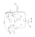
- FIG. 1 is an embodiment that shows the single multiplex probe for detecting the MMP-2 and MMP-9 activities.
- a single wavelength excitation of the first dye may yield information on relative proteases activities.
- Unequivocally two wavelength interleave excitation allows to monitor two step FRET pathway and independently detect cleavage of peptide MMP-2 and MMP-9.
- FIG. 2A is another embodiment of the multiplex probe based on the two-step FRET process (SEQ ID NOS.: 1 and 2), and FIG. 2B shows an alternative multiplex probe based on two-step FRET process using avidin.
- FIGS. 3A-D Concept for multiplex probe detection is shown schematically.
- Two excitation wavelengths in interleave mode can be used (for example 480 nm left and 532 right; the selection of excitation is not limited to these two and can be anywhere within UV/VIS/NIR range).
- the figure presents expected results for: FIG. 3A —intact probe; FIG. 3B —MMP-2 peptide cleaved; FIG. 3C —MMP-9 peptide cleaved; and FIG. 3D —both peptides cleaved.
- Third long wavelength excitation in our case 630/650 nm can be used for control. The long wavelength excitation will give information on the presence of second acceptor.
- FIG. 4 is an example of the FRET based probe for detecting MMP-9 activity.
- Specific oligopeptide sequence for example, SEQ ID NO.: 3, which is cleaved into SEQ ID NO.: 4 and 5
- donor on one end and acceptor on another end.
- acceptor on another end.
- FRET FRET based probe for detecting MMP-9 activity.
- Addition of MMP-9 cleaves the peptide separating donor and acceptor.
- Photography shows the color change induced by cleavage.
- the present invention includes compositions and methods for making and using the new probes that are constructed from a central body of the probe that promotes enhanced (and in some cases exclusive) energy transfer from donor one (first dye) to acceptor one (second dye). This energy transfer process is highly enhanced to compete with transfer from donor one to acceptor two (third dye) allowing for separate monitoring (and therefore the detectable distinction) between cleavage of individual sides.
- the central body acts as a physical spacer between donor one and acceptor two.
- a first fluorophore comprises Alexa Fluor 488
- a second fluorophore comprises Alexa Fluor 532
- a third fluorophore comprises Alexa Fluor 647.
- Other alternative include any two dyes system with the quantum dote as a central body (e.g. FITC, 550 QD, Cy5).
- a two dye system with the quantum dot (QD) as a central body e.g., FITC, 550 QD, Cy5
- QD can fulfill the role of a second dye (acceptor for the first dye and donor for the third dye).
- FIGS. 2A and 2B One example of a spacer for use with the present invention is shown schematically in FIGS. 2A and 2B , in which four binding sides for biotin may hold up to 4 or more peptides, and the significant size of avidin acts as a spacer, preventing the collapse of peptides onto neighbor peptides without the need for a rigid linker.
- FIGS. 2A and 2B the following exemplary amino acid sequences are depicted, however, the skilled artisan will recognize that the linker could be nucleic acids, carbohydrates, lipids, or other organic or inorganic modules that can be cleaved either specifically or non-specifically in situ. The following sequences are shown in FIGS.
- the spacer length will generally be selected to optimize the signal-to-noise ratio for the interaction of the two specific fluorophores and/or quenchers selected.
- Matrix metalloproteinases are involved in major human diseases such as cancer, AIDS, and inflammation [1-3]. Therefore, sensitive detection and imaging of MMPs activities offer great clinical value to cancer detection, treatment, and therapy monitoring.
- FRET Förster resonance energy transfer
- sequence targets can include, e.g., peptide substrates for a consensus for MMP-2 (Lys-Gly-Ser-Gly-Pro-Tyr-Val-Ile-Trp-Leu-Gly-Lys) (SEQ ID NO.: 6) (SEQ ID NO.: 7).
- MMP-2 Lys-Gly-Ser-Gly-Pro-Tyr-Val-Ile-Trp-Leu-Gly-Lys
- SEQ ID NO.: 7 SEQ ID NO.: 7
- a selective cleavage of this specific peptide sequence by MMP-9 protease results in the separation of the two segments of oligopeptide carrying the donor and acceptor and consequently leads to significant increase of donor signal and decrease in acceptor signal.
- a ratiometric approach allows excellent dynamic range for detection that produces an almost 60 fold change when going from intact to completely cleaved peptide [7, 8].
- n independent substrates for detecting n proteases presents one more fundamental problem for quantitative detection: accessibility of the place of interest (like a cell or cancerous tissue) can be different for various probes, and relative evaluation of actual concentrations of individual proteases could be difficult or even be miss-leading. Therefore constructing a single probe that can simultaneously detect two or more proteases is of principle interest for cellular and tissue imaging applications.
- the present invention can be used for a wide variety of detection purposes.
- One example of the detection is the development and use of the multiplex probes in cellular and tissue imaging.
- the present invention will dramatically speed-up and increase precision of enzyme activities detection. Also, these probes will be beneficial for high throughput assay applications substituting typical multiplex assays constructed from individual sensors.
- MMP-9 Matrix Metalloproteinases
- MMP-2 MMP-7, MMP-3, and more
- MMP-9 zinc-dependent endopeptidases.
- MMP-9 digests type IV collagen, laminin, and fibronectin; the major components of the basal lamina around blood vessels.
- MMP-9 is also known to be involved in the splitting of cell-surface receptors, the activation or inactivation of different cytokines and/or chemokines, and the release of apoptotic ligands.
- MMP-9 plays an important role in cell proliferation, migration, differentiation, angiogenesis, as well as apoptosis.
- Overexpression of MMPs in tumor tissue and stroma can result in increased levels of activity of these enzymes in various body fluids such as blood or urine.
- body fluids such as blood or urine.
- proteases activity constitutes attractive diagnostic tool.
- Such diagnostic tool becomes especially powerful when relative activities of different proteases can be simultaneously assessed. This has been achieved in in-vitro in multiplex format assays where two and more proteases are measured simultaneously in parallel manner.
- Such multiplex approach utilizes two or more sensors incorporated in one physical platform.
- a big advantage of multiplex analyses is the higher confidence in comparisons made between different samples, such as the comparison of two closely related proteases for example MMP-2 and MMP-9. This approach has been proved superior and much more precise than just simple sum of individual components.
- the present invention is a true multiplex probe that can simultaneously detect the activity of two proteases.
- the overall design is presented in FIG. 1 in which single multiplex probe for the detecting of the activity of MMP-2 and MMP-9.
- Two wavelength interleave excitation allows to monitor two step Förster Resonance Energy Transfer (FRET) pathway and detect cleavage of peptide MMP-2 and MMP-9.
- FRET Förster Resonance Energy Transfer
- the free end of peptide 1 is labeled with a single fluorophore (Dye 1) that has short wavelength absorption (e.g. FITC).
- the free end of the second peptide is labeled with a single fluorophore (Dye 3) with long wavelength absorption (e.g. Cy5).
- the middle part, connecting two peptides is labeled with Dye 2 of intermediate absorption (e.g. TAMRA).
- TAMRA intermediate absorption
- the relative fluorescence signal observed from the short, intermediate, and long wavelength fluorophores will change as some of the oligopeptides are cleaved, and the change will be specific to which oligopeptide has been cleaved (MMP-2, or MMP-9, or both).
- MMP-2 or MMP-9, or both.
- the specificity is achieved by a dual excitation scheme: 480 nm for FITC and 532 nm for TAMRA.
- the present inventors can design, develop, build, and test a fluorescent probe based on the avidin/biotin-oligopeptides construct.
- the labeling of avidin can be optimized with multiple dyes (e.g. TAMRA). Binding of labeled oligopeptides to avidin through biotin binding can also be optimized.
- the present inventors can also develop probe specificity of response to various proteases and test the response to MMP-2, MMP-9, and a mixture of both, and test if the response is specific and independent from other proteases (e.g. MMP-7, MMP-10, etc.).
- MMP-9 very effectively cleaves a peptide (Lys-Gly-Pro-Arg-Ser-Leu-Ser-Gly-Lys) [7, 8]. They have also shown that the peptide is very specific and is not digested by MMP-2 enzyme MMP-3 or MMP-7 enzymes [7, 8]. FRET sensitized fluorescence detection proves to be very effective detection approach. Similar specific peptide sequences have been developed for MMP-2, MMP-7 and more. To make highly confident assessment of various proteases activities directly in cells and tissue it is necessary to have single molecular probe capable for simultaneous analysis/detection of more than one protease.
- the present invention includes methods for developing and constructing a new probe that will be capable of simultaneous detection of activities of two proteases (like MMP-2 and MMP-9) at the same time and the same place by the same molecule (molecular construct).
- This approach uses Förster resonance energy transfer (FRET) that can be highly enhanced when multiple acceptors are used [11].
- FRET Förster resonance energy transfer
- the probe is constructed in a way to force two sequential steps of energy transfer process.
- First step (FRET 1) is mediated through the cleavable linker specific to enzyme 1 (for example MMP-9) and occurs between the first energy donor and first acceptor (acceptor is a central body).
- Second step (FRET 2) is between the first acceptor that becomes donor to the second acceptor.
- the second donor and second acceptor are separated by linker specific to enzyme 2 (for example MMP-2).
- linker specific to enzyme 2 for example MMP-2.
- a highly efficient well-defined sequential FRET process offers distinctive signatures for recognizing two proteins activities.
- Use of pulse interleaved excitation (independent excitations of the first donor and first acceptor/second donor) opens detection scheme that eliminates ubiquities typically associated with single wavelength excitation and multiple steps FRET process.
- the present invention overcomes a critical problem with pairs of fluorescence based probes could simultaneously be used for detecting more than one protease.
- the problem is that FRET pairs can enter cells differentially, that is, it is not possible guarantee that both separate probes are absorbed, bound to, or endo- or pinocytosed into the target cells.
- FIG. 2A This embodiment is presented in FIG. 2A in which two oligopeptides, one specific for MMP-9 and second to MMP-2 are attached to a central (protein) body.
- the free end of peptide 1 is labeled with single fluorophore that has short wavelength absorption (e.g. FITC).
- the free end of second peptide is labeled with single fluorophore with long wavelength absorption (e.g., Cy5).
- the middle part, protein core is labeled with few dyes of intermediate absorption (e.g., Texas Red, TR).
- the multiple labeling of central body ensure that after excitation of the short wavelength fluorophore (Donor 1) with 480 nm the energy transfer pathway will exclusively lead with very high efficiency (>90%) to the central part. From the central part the excitation energy is transferred to the long wavelength absorbing fluorophore (Cy5) with lower efficiency ( ⁇ 90%).
- the relative fluorescence signal observed for FITC, TR, and Cy5 will change as some of the oligopeptides are cleaved and the change will be specific to which oligopeptide has been cleaved (MMP-2, or MMP-9, or both).
- FIG. 2B shows an alternative multiplex probe based on two-step FRET process using Avidin.
- MMPs Matrix metalloproteinases
- ECM extracellular matrix
- Proteases play an important role in development, tissue remodelling, wound healing and inflammation [12, 13] and have been involved in major human diseases such as cancer, AIDS, inflammation, and neurodegenerative disease [1-3].
- the level and activities of various MMPs are frequently found to correlate with advanced tumor stage, increased invasion and metastasis [14-18]. Therefore, sensitive detection and imaging of MMPs activities offers a great clinical value to cancer detection and therapy monitoring.
- FIGS. 3A to 3D show schematic representations of how sequential FRET sensor will work.
- the principle assumption is that only (preferentially) sequential FRET 1 and FRET 2 occurs and the direct transfer from FITC to Cy5 is negligible.
- the probe is built in such way that both pathways (FRET 1 and FRET 2) will have high efficiency ( ⁇ 90%) and direct transfer from FITC to Cy5 will be very low (below 10%).
- FRET 1 and FRET 2 both pathways
- FITC to Cy5 will be very low (below 10%).
- FIG. 3A shows the signal from the intact probe.
- a dominant long wavelength emission from Dye 3 (Cy5) is shown in red.
- the peptide MMP-2 is cleaved (FRET 1 broken).
- Excitation 480 nm will show blue fluorescence of Dye 1 (FITC in our case) since there will not be FRET.
- FIG. 3C the signal is shown when the peptide MMP-9 is cleaved (FRET 2 broken).
- Dye 2 range emission
- Dye 2 red emission
- both peptides are cleaved (no FRET 1 or FRET 2).
- For 480 nm excitation we will see only blue fluorescence of Dye 1 and for 532 nm orange fluorescence of Dye 2.
- FIG. 4 shows a FRET based probe for detecting MMP-9 activity.
- a specific oligopeptide sequence was labeled with donor on one end and acceptor on another end. When intact, significant (>90%) FRET occurs.
- Addition of MMP-9 cleaves the peptide separating donor and acceptor.
- Photography shows the color change induced by cleavage.
- compositions of the invention can be used to achieve methods of the invention.
- the words “comprising” (and any form of comprising, such as “comprise” and “comprises”), “having” (and any form of having, such as “have” and “has”), “including” (and any form of including, such as “includes” and “include”) or “containing” (and any form of containing, such as “contains” and “contain”) are inclusive or open-ended and do not exclude additional, unrecited elements or method steps.
- A, B, C, or combinations thereof refers to all permutations and combinations of the listed items preceding the term.
- “A, B, C, or combinations thereof” is intended to include at least one of: A, B, C, AB, AC, BC, or ABC, and if order is important in a particular context, also BA, CA, CB, CBA, BCA, ACB, BAC, or CAB.
- expressly included are combinations that contain repeats of one or more item or term, such as BB, AAA, AB, BBC, AAABCCCC, CBBAAA, CABABB, and so forth.
- BB BB
- AAA AAA
- AB BBC
- AAABCCCCCC CBBAAA
- CABABB CABABB
- words of approximation such as, without limitation, “about”, “substantial” or “substantially” refers to a condition that when so modified is understood to not necessarily be absolute or perfect but would be considered close enough to those of ordinary skill in the art to warrant designating the condition as being present.
- the extent to which the description may vary will depend on how great a change can be instituted and still have one of ordinary skilled in the art recognize the modified feature as still having the required characteristics and capabilities of the unmodified feature.
- a numerical value herein that is modified by a word of approximation such as “about” may vary from the stated value by at least ⁇ 1, 2, 3, 4, 5, 6, 7, 10, 12 or 15%.
- compositions and/or methods disclosed and claimed herein can be made and executed without undue experimentation in light of the present disclosure. While the compositions and methods of this invention have been described in terms of preferred embodiments, it will be apparent to those of skill in the art that variations may be applied to the compositions and/or methods and in the steps or in the sequence of steps of the method described herein without departing from the concept, spirit and scope of the invention. All such similar substitutes and modifications apparent to those skilled in the art are deemed to be within the spirit, scope and concept of the invention as defined by the appended claims.
Abstract
Description
- 1. Overall, C. M.; Kleifeld, O. Validating matrix metalloproteinases as drug targets and anti-targets for cancer therapy.
Nat. Rev. Cancer 2006, 6, 227-239. - 2. Concha, N. O.; Abdel-Meguid, S. S. Controlling Apoptosis by Inhibition of Caspases. Curr. Med. Chem. 2002, 9, 713-726.
- 3. Schwienhorst, A. Direct thrombin inhibitors—a survey of recent developments. Cell. Mol. Life Sci. 2006, 63, 2773-2791.
- 4. Shi, L.; De Paoli, V.; Rosenzweig, N.; Rosenzweig, Z. Synthesis and Application of Quantum Dots FRET-Based Protease Sensors. J. Am. Chem. Soc. 2006, 128, 10378-10379.
- 5. Medintz, I. L.; Clapp, A. R.; Brunel, F. M.; Tiefenbrunn, T.; Uyeda, H. T.; Chang, E. L.; Deschamps, J. R.; Dawson, P. E.; Mattoussi, H. Proteolytic activity monitored by fluorescence resonance energy transfer through quantum-dot-peptide conjugates. Nat. Mater. 2006, 5, 581-589.
- 6. Chang, E.; Miller, J. S.; Sun, J.; Yu, W. W.; Colvin, V. L.; Drezek, R.; West, J. L. Protease-activated quantum dot probes. Biochem. Biophys. Res. Commun. 2005, 334, 1317-1321.
- 7. Fudala R, Ranjan A P, Mukerjee A, Vishwanatha J K, Gryczynski Z, Borejdo J, Sarkar P, Gryczynski I. Fluorescence detection of MMP-9. I—MMP-9 selectively cleaves Lys-Gly-Arg-Ser-Leu-Ser-Gly-Lys peptide. Current Pharmaceutical Biotechnology. 2011 May 1; 12(5):834-838.
- 8. Fudala R, Rich R, Mukerjee A, Ranjan A P, Vishwanatha J K, Kurdowska A K, Gryczynski Z, Borejdo J, Gryczynski I. Fluorescence Detection of MMP-9. II. Ratiometric FRET-Based Sensing With Dually Labeled Specific Peptide. Current Pharmaceutical Biotechnology. 2012 Feb. 20. [Epub ahead of print].
- 9. Bjurlin, M. A.; Bloomer, S.; Nelson, C. J. Characterization of proteolytic activity of proteases. Biotechnol. Lett. 2002, 24, 191-195.
- 10. Zhao, Z.; Raftery, M. J.; Niu, X. M.; Daja, M. M.; Russell, P. J. Application of in-gel protease assay in a biological sample: characterization and identification of urokinase-type plasminogen activator (uPA) in secreted proteins from a prostate cancer cell line PC-3. Electrophoresis 2004, 25, 1142-1148.
- 11. Maliwal B P, Raut S, Fudala R, D'Auria S, Marzullo V M, Luini A, Gryczynski I, Gryczynski Z. Extending FRET Measurements Beyond 100 Å; with Commonly Used Organic Fluorophores: Enhanced Transfer in the Presence of Multiple Acceptors. Journal of Biomedical Optics; (2012); 17(1), 011006, doi:10.1117/1.JBO.17.1.011006.
- 12. Werb Z. ECM and cell surface proteolysis: regulating cellular ecology. Cell 1997; 91:439-42. [PubMed: 9390552].
- 13. Mott J D, Werb Z. Regulation of matrix biology by matrix metalloproteinases. Curr Opin Cell Biol. 2004; 16:558-64. [PubMed: 15363807].
- 14. Fingleton B. Matrix metalloproteinases: Roles in cancer and metastasis. Front Biosci. 2006; 11:479-491. [PubMed: 16146745].
- 15. Egeblad M, Werb Z. New functions for the matrix metalloproteinases in cancer progression. Nat Rev Cancer 2002; 2:161-174. [PubMed: 11990853].
- 16. Brinckerhoff C E, Matrisian L M. Matrix metalloproteinases: a tail of a frog that became a prince. Nat Rev Mol Cell Biol 2002; 3:207-214. [PubMed: 11994741].
- 17. Bachmeier B E, Iancu C M, Jochum M, Nerlich A G. Matrix metalloproteinases in cancer: comparison of known and novel aspects of their inhibition as a therapeutic approach. Expert Rev Anticancer Ther 2005; 5:149-163.
- 18. Overall C M, Kleifeld O. Tumour microenvironment—opinion: validating matrix metalloproteinases as drug targets and anti-targets for cancer therapy Nature Rev Cancer 2006; 6:227-239. [PubMed: 16498445].
- 19. White, C. M. Thrombin-directed inhibitors: pharmacology and clinical use. Am. Heart J. 2005, 149, 54-60.
Claims (18)
Priority Applications (1)
| Application Number | Priority Date | Filing Date | Title |
|---|---|---|---|
| US14/206,738 US9388450B2 (en) | 2013-03-12 | 2014-03-12 | FRET based multiplex probes |
Applications Claiming Priority (2)
| Application Number | Priority Date | Filing Date | Title |
|---|---|---|---|
| US201361777727P | 2013-03-12 | 2013-03-12 | |
| US14/206,738 US9388450B2 (en) | 2013-03-12 | 2014-03-12 | FRET based multiplex probes |
Publications (2)
| Publication Number | Publication Date |
|---|---|
| US20140274797A1 US20140274797A1 (en) | 2014-09-18 |
| US9388450B2 true US9388450B2 (en) | 2016-07-12 |
Family
ID=51529840
Family Applications (1)
| Application Number | Title | Priority Date | Filing Date |
|---|---|---|---|
| US14/206,738 Active 2034-07-23 US9388450B2 (en) | 2013-03-12 | 2014-03-12 | FRET based multiplex probes |
Country Status (1)
| Country | Link |
|---|---|
| US (1) | US9388450B2 (en) |
Cited By (1)
| Publication number | Priority date | Publication date | Assignee | Title |
|---|---|---|---|---|
| CN105949281A (en) * | 2016-05-04 | 2016-09-21 | 新乡医学院 | Fluorescence resonance energy transfer probe, preparation method and application thereof |
Citations (2)
| Publication number | Priority date | Publication date | Assignee | Title |
|---|---|---|---|---|
| US6743325B1 (en) | 1999-07-13 | 2004-06-01 | Stirling Moulded Composites Limited | Flexible material |
| US8609329B2 (en) | 2007-02-05 | 2013-12-17 | Albany Medical College | FRET-based assay for screening modulators of receptor cycling |
-
2014
- 2014-03-12 US US14/206,738 patent/US9388450B2/en active Active
Patent Citations (2)
| Publication number | Priority date | Publication date | Assignee | Title |
|---|---|---|---|---|
| US6743325B1 (en) | 1999-07-13 | 2004-06-01 | Stirling Moulded Composites Limited | Flexible material |
| US8609329B2 (en) | 2007-02-05 | 2013-12-17 | Albany Medical College | FRET-based assay for screening modulators of receptor cycling |
Cited By (1)
| Publication number | Priority date | Publication date | Assignee | Title |
|---|---|---|---|---|
| CN105949281A (en) * | 2016-05-04 | 2016-09-21 | 新乡医学院 | Fluorescence resonance energy transfer probe, preparation method and application thereof |
Also Published As
| Publication number | Publication date |
|---|---|
| US20140274797A1 (en) | 2014-09-18 |
Similar Documents
| Publication | Publication Date | Title |
|---|---|---|
| Lowe et al. | Multiplex sensing of protease and kinase enzyme activity via orthogonal coupling of quantum dot–peptide conjugates | |
| Chen et al. | Lanthanide-doped luminescent nano-bioprobes for the detection of tumor markers | |
| Kim et al. | Analysis of protease activity using quantum dots and resonance energy transfer | |
| ES2399443T3 (en) | In vitro method to diagnose and monitor renal cell carcinoma (RCC) using MMP-7 as a smoke biomarker for RCC | |
| Lei et al. | Biosensors and bioassays for determination of matrix metalloproteinases: state of the art and recent advances | |
| Krizkova et al. | Assays for determination of matrix metalloproteinases and their activity | |
| Kirchhain et al. | Biosensors for measuring matrix metalloproteinases: An emerging research field | |
| KR20090097873A (en) | Enzyme detection techniques | |
| US8551764B2 (en) | Devices for the detection of the presence and/or activity of proteases in biological samples | |
| Yogo et al. | Development of an activatable fluorescent probe for prostate cancer imaging | |
| Fudala et al. | Fluorescence detection of MMP-9. I. MMP-9 selectively cleaves lys-gly-pro-arg-ser-leu-ser-gly-lys peptide | |
| Nagy et al. | Peptide-functionalized quantum dot biosensors | |
| Luo et al. | Proteolysis-responsive rolling circle transcription assay enabling femtomolar sensitivity detection of a target protease biomarker | |
| Ivry et al. | The lysosomal aminopeptidase tripeptidyl peptidase 1 displays increased activity in malignant pancreatic cysts | |
| Mumtaz et al. | Exploiting proteases for cancer theranostic through molecular imaging and drug delivery | |
| Oliveira-Silva et al. | Monitoring proteolytic activity in real time: A new world of opportunities for biosensors | |
| Ryu et al. | “One-step” detection of matrix metalloproteinase activity using a fluorogenic peptide probe-immobilized diagnostic kit | |
| US8758989B2 (en) | Enzymatic detection techniques | |
| Moin et al. | 3D/4D functional imaging of tumor-associated proteolysis: impact of microenvironment | |
| US9388450B2 (en) | FRET based multiplex probes | |
| US9914955B2 (en) | Genetically encoded FRET-based MMP-9 activity biosensor and use thereof | |
| CA2764775C (en) | Rapid bed-side measurement of neutrophil elastase activity in biological fluids | |
| Sloniec et al. | Photophysics and release kinetics of enzyme-activatable optical probes based on H-dimerized fluorophores on self-immolative linkers | |
| Wysocka et al. | Future of protease activity assays | |
| Maysinger et al. | Nanoparticle-based caspase sensors |
Legal Events
| Date | Code | Title | Description |
|---|---|---|---|
| AS | Assignment |
Owner name: UNIVERSITY OF NORTH TEXAS HEALTH SCIENCE CENTER, T Free format text: ASSIGNMENT OF ASSIGNORS INTEREST;ASSIGNORS:GRYCZYNSKI, ZYGMUNT;GRYCZYNSKI, IGNACY;FUDALA, RAFAL;AND OTHERS;SIGNING DATES FROM 20120810 TO 20120813;REEL/FRAME:032535/0796 |
|
| AS | Assignment |
Owner name: TEXAS CHRISTIAN UNIVERSITY, TEXAS Free format text: ASSIGNMENT OF ASSIGNORS INTEREST;ASSIGNOR:GRYCZYNSKI, ZYGMUNT;REEL/FRAME:038079/0141 Effective date: 20160322 Owner name: MALIWAL, BADRI P., TEXAS Free format text: ASSIGNMENT OF ASSIGNORS INTEREST;ASSIGNOR:UNIVERSITY OF NORTH TEXAS HEALTH SCIENCE CENTER;REEL/FRAME:038077/0755 Effective date: 20151221 Owner name: GRYCZYNSKI, ZYGMUNT, TEXAS Free format text: ASSIGNMENT OF ASSIGNORS INTEREST;ASSIGNOR:UNIVERSITY OF NORTH TEXAS HEALTH SCIENCE CENTER;REEL/FRAME:038079/0099 Effective date: 20151221 Owner name: TEXAS CHRISTIAN UNIVERSITY, TEXAS Free format text: ASSIGNMENT OF ASSIGNORS INTEREST;ASSIGNOR:MALIWAL, BADRI P.;REEL/FRAME:038077/0800 Effective date: 20151222 |
|
| STCF | Information on status: patent grant |
Free format text: PATENTED CASE |
|
| CC | Certificate of correction | ||
| FEPP | Fee payment procedure |
Free format text: MAINTENANCE FEE REMINDER MAILED (ORIGINAL EVENT CODE: REM.); ENTITY STATUS OF PATENT OWNER: SMALL ENTITY |
|
| FEPP | Fee payment procedure |
Free format text: SURCHARGE FOR LATE PAYMENT, SMALL ENTITY (ORIGINAL EVENT CODE: M2554); ENTITY STATUS OF PATENT OWNER: SMALL ENTITY |
|
| MAFP | Maintenance fee payment |
Free format text: PAYMENT OF MAINTENANCE FEE, 4TH YR, SMALL ENTITY (ORIGINAL EVENT CODE: M2551); ENTITY STATUS OF PATENT OWNER: SMALL ENTITY Year of fee payment: 4 |
|
| FEPP | Fee payment procedure |
Free format text: MAINTENANCE FEE REMINDER MAILED (ORIGINAL EVENT CODE: REM.); ENTITY STATUS OF PATENT OWNER: SMALL ENTITY |