US9224200B2 - Computer vision based method for extracting features relating to the developmental stages of Trichuris spp. eggs - Google Patents
Computer vision based method for extracting features relating to the developmental stages of Trichuris spp. eggs Download PDFInfo
- Publication number
- US9224200B2 US9224200B2 US13/796,660 US201313796660A US9224200B2 US 9224200 B2 US9224200 B2 US 9224200B2 US 201313796660 A US201313796660 A US 201313796660A US 9224200 B2 US9224200 B2 US 9224200B2
- Authority
- US
- United States
- Prior art keywords
- egg
- eggs
- trichuris spp
- structures
- extracted
- Prior art date
- Legal status (The legal status is an assumption and is not a legal conclusion. Google has not performed a legal analysis and makes no representation as to the accuracy of the status listed.)
- Expired - Fee Related, expires
Links
Images


Classifications
-
- G—PHYSICS
- G06—COMPUTING OR CALCULATING; COUNTING
- G06V—IMAGE OR VIDEO RECOGNITION OR UNDERSTANDING
- G06V20/00—Scenes; Scene-specific elements
- G06V20/60—Type of objects
- G06V20/69—Microscopic objects, e.g. biological cells or cellular parts
- G06V20/698—Matching; Classification
-
- G—PHYSICS
- G06—COMPUTING OR CALCULATING; COUNTING
- G06T—IMAGE DATA PROCESSING OR GENERATION, IN GENERAL
- G06T7/00—Image analysis
- G06T7/0002—Inspection of images, e.g. flaw detection
- G06T7/0012—Biomedical image inspection
-
- G06K9/00147—
-
- A—HUMAN NECESSITIES
- A61—MEDICAL OR VETERINARY SCIENCE; HYGIENE
- A61K—PREPARATIONS FOR MEDICAL, DENTAL OR TOILETRY PURPOSES
- A61K35/00—Medicinal preparations containing materials or reaction products thereof with undetermined constitution
- A61K35/56—Materials from animals other than mammals
- A61K35/62—Leeches; Worms, e.g. cestodes, tapeworms, nematodes, roundworms, earth worms, ascarids, filarias, hookworms, trichinella or taenia
Definitions
- the present invention relates to a computer vision based method for extracting features relating to the developmental stages of Trichuris spp. eggs, wherein for the final developmental stages a larva is present inside the egg. More particularly, the method may be used for extracting features for Trichuris suis eggs.
- helminths intestinal worms
- whipworms Exposure to helminths (intestinal worms) such as whipworms have been shown to have a mitigating effect on a number of autoimmune diseases such as Crohn's disease and Ulcerative colitis.
- This new treatment is known as helminthic therapy and it utilizes the immunoregulatory behavior of helminths in the intestines where orally administered whipworm eggs hatch into larvae which establish for a shorter period of time in a self-limiting intestinal infection.
- the presented invention enables an automated, non-invasive and cost-effective way of assessing the biological potency of a particular egg suspension.
- the extracted features are related to the contents of an egg, and may also be denoted egg-content-features.
- the Trichuris spp. eggs are Trichuris suis eggs.
- the stored digital images of the Trichuris spp. eggs comprises one or more bright-field images
- one or more features are extracted from an egg content image region being a bright-field egg content image region.
- one or more features are extracted from an egg content image region being extracted from an image or image region which includes a full representation of a detected Trichuris spp. egg.
- the bright-field egg content image region may also be extracted from a bright-field image or image region, which includes a full representation of a detected Trichuris spp. egg
- the extracted egg content image region excludes the polar plugs of the detected Trichuris spp. egg. It is also preferred that the extracted egg content image region excludes the shell of the detected Trichuris spp. egg.
- the extracted egg content image region may have a substantially elliptical shape, thereby defining a content ellipse image.
- the extraction of one or more features from the egg content image region includes one or more measurements of the direction-dependent structures of the egg contents.
- the extraction of one or more features from the egg content region may include one or more measurements of the longitudinal structures of the egg contents and/or one or more measurements of the transverse structures of the egg contents.
- One or more measurements of the longitudinal structures may be based on a measure of the linear structures and/or edge structures in the longitudinal direction
- one or more measurements of the transverse structures may be based on a measure of the linear structures and/or edge structures in the transverse direction.
- the linear structures and/or edge structures are measured at a predetermined scale.
- one or more measurements of the longitudinal structures may be based on a measure of the linear structures and/or edge structures in the longitudinal direction at one or more scales in a multi-scale representation of the image region from which the features are extracted.
- one or more measurements of the transverse structures may be based on a measure of the linear structures and/or edge structures in the transverse direction at one or more scales in a multi-scale representation of the image region from which the features are extracted.
- the multi-scale representation of the image region from which the features are extracted may be a pyramid representation or a scale space representation.
- one or more measurements of the longitudinal structures of the egg contents is based on a longitudinal comparison of pixels intensities obtained from similarly addressed pixels in first and second image parts representing at least part of the egg contents of a detected egg, with the second image part being obtained by shifting the first image part one or more pixels in a direction substantially following the longitudinal direction of the egg. It is also within one or more embodiments of the invention that one or more measurements of the transverse structures of the egg contents is based on a transverse comparison of pixel intensities obtained from similarly addressed pixels in the first image part and a third image part representing at least part of the egg contents of a detected egg, with the third image part being obtained by shifting the first image part one or more pixels in a direction substantially following the transverse direction of the egg.
- the longitudinal comparison of pixel intensities from the first and second image parts comprises calculating a longitudinal correlation coefficient ⁇ long for pixel intensities representing at least part of the similarly addressed pixels
- the transverse comparison of pixel intensities from the first and third image parts comprises calculating a transverse correlation coefficient ⁇ trans for pixel intensities representing at least part of the similarly addressed pixels.
- the feature extraction may further include a ratio measure based on the ratio between the longitudinal correlation coefficient ⁇ long and the transverse correlation coefficient ⁇ trans .
- edge detector algorithm may be selected from the following algorithms: the Canny edge detector algorithm, the Sobel edge detector algorithm, and the Prewitt edge detector algorithm.
- the expression or expressions representing the edge structures in the longitudinal direction, longitudinal edge count may be defined as the number of edge pixels of the egg contents, which is given by the edge detector algorithm, and which are oriented substantially in the longitudinal direction
- the expression or expressions representing the edge structures in the transverse direction, transverse edge count may be defined as the number of edge pixels of the egg contents, which is given by the edge detector algorithm, and which is oriented substantially in the transverse direction.
- the longitudinal edge count may be defined as the number of edge pixels of the egg contents, which is given by the edge detector algorithm and which are oriented in the longitudinal direction plus/minus an angle within the range of 10-45 degrees, such as within the range of 15-35 degrees, such about 22.5 degrees, and wherein the transverse edge count is defined as the number of edge pixels of the egg contents given by the edge detector algorithm and being oriented in the transverse direction plus/minus an angle within the range of 10-45 degrees, such as within the range of 15-35 degrees, such as about 22.5 degrees.
- the present invention also covers embodiments, wherein in step a) the stored digital images of the Trichuris spp. eggs comprises one or more dark-field images and wherein in step c) one or more features are extracted from an egg content image region being a dark-field egg content image region.
- one or more features may be extracted from a dark-field egg content image region being extracted from a dark-field image region which includes a full representation of a detected Trichuris spp. egg.
- the extracted dark-field egg content image region excludes the polar plugs of the detected Trichuris spp. egg.
- the extracted dark-field egg content image region excludes the shell of the detected Trichuris spp. egg, and here the extracted dark-field egg content image region may have a substantially elliptical shape, thereby defining a content ellipse image.
- the feature extraction of step c) may include dark-field features extracted from the dark-field egg content image region, where the dark-field feature extraction is based on variations in pixel intensities measured or extracted for at least part of the dark-field egg content image region.
- the dark-field feature extraction may comprise a computation of the average of the extracted pixel intensities.
- the dark-field feature extraction may comprise a computation of the mean of the extracted pixel intensities, mean scattering intensity, and/or a computation of the median of the extracted pixel intensities, median scattering intensity.
- the present invention also covers embodiments, which further comprises a classification step, wherein at least part of the features extracted from an egg content image region representing a detected egg are used for classifying the detected egg.
- the classification of the detected egg may be a binary classification with respect to the developmental stage of egg, and the detected egg may be classified as either containing a larva or not containing a larva.
- the classification of the detected egg may also be a multi-class classification with respect to the developmental stage of egg, where the multi-class classification comprises at least three classes of developmental stages.
- the classification may be at least partly based on extracted features, for which features the extraction includes one or more measurements representing longitudinal structures and transverse structures of the egg contents.
- the classification may be at least partly based on a ratio measure obtained from a measure representing the longitudinal structures of the egg contents and a measure representing the transverse structures of the egg contents.
- the one or more measurements representing the longitudinal structures are based on a measure of the linear structures and/or edge structures in the longitudinal direction
- the one or more measurements representing the transverse structures are based on a measure of the linear structures and/or edge structures in the transverse direction.
- the measurements of the linear structures and/or edge structures in the longitudinal and transverse directions may be measured according to one or more of the herein mentioned embodiments. It is within an embodiment of the invention that a measure representing the longitudinal structures of the egg contents have to exceed a corresponding measure representing the transverse structures of the egg contents by a predetermined factor being larger than one in order to have the egg classified as containing a larvae.
- the present invention also covers embodiments, wherein the classification is at least partly based on extracted dark-field features, where the dark-field features may be extracted according to one or more of the embodiments described herein.
- FIG. 1 Complete egg analysis system
- FIG. 2 Detection phase
- FIG. 3 Feature extraction phase
- FIG. 4 Classification phase
- FIG. 5 Image definitions
- FIG. 6 Orientation alignment
- FIG. 7 Shape profile computation
- FIG. 8 Examples of the seven egg categories
- FIG. 9 Egg content region extraction
- FIG. 10 Spatial autocorrelation computation
- FIG. 11 Correlation formula
- FIG. 12 Edge orientations
- FIG. 13 Classification graphs I
- FIG. 14 Classification graphs II
- FIG. 15 Developmental stages
- FIG. 16 Examples of corresponding brightfield and darkfield images
- Trichuris spp. eggs i.e. eggs from various species of the genus Trichuris , also known as whipworms.
- Trichuris spp. eggs in the following denoted Trichuris eggs, have a distinct, lemon-like shape and a smooth outer shell.
- the shape can be described as a prolate spheroid, also known as a prolate ellipsoid of revolution, with protruding, operculate plugs in each end (apex).
- prolate spheroid also known as a prolate ellipsoid of revolution
- protruding plugs in each end apex
- the shape of a i egg is oblong and elliptical with protruding plugs at the ends.
- Trichuris eggs The average length (major axis) and width (the two minor axes) of Trichuris eggs depend to a certain degree on the specific species.
- the sizes of some of the main species of Trichuris are:
- Trichuris trichiura human whipworm: 50-58 ⁇ 22-27 ⁇ m, 50-54 ⁇ 22-23 ⁇ m.
- Trichuris suis (pig whipworm): 50-68 ⁇ 21-31 ⁇ m, 60 ⁇ 25 ⁇ m
- Trichuris muris (mouse whipworm): 67-70 ⁇ 31-34 ⁇ m
- Trichuris vulpis (canine whipworm): 70-90 ⁇ 32-41 ⁇ m
- Trichuris ovis (ruminant whipworm): 70-80 ⁇ 30-42 ⁇ m
- a 40 ⁇ l sample of an egg suspension with approximately 40.000 eggs per ml is placed on a microscope slide or a similar container with a cover glass on top.
- the slide is placed under an upright or inverted microscope and images are acquired at around 100-200 ⁇ magnification, in both brightfield and darkfield illumination. Examples of corresponding brightfield and darkfield images can be seen in FIG. 16 .
- a lateral object is a prolate object that is placed on its side.
- a lateral egg is an egg lying on its side.
- the outline of such an egg is elliptical with protruding polar plugs at the ends.
- An upright object is a prolate object that is placed on one of its apices.
- An upright egg is an egg that is standing upright on one of its polar plugs.
- the outline of such an egg is circular or close to circular with a diameter corresponding to the width of a lateral egg.
- a singularized object is an object that does not touch or overlap with other objects, i.e. is clearly separated from nearby objects.
- a singularized egg is an egg that does not touch or overlap with other eggs or foreign particles.
- a touching object is an object that touches, but does not overlap with, other objects.
- a touching egg is an egg that touches one or more other eggs or impurities, but is not overlapping with them, i.e. its entire content region is clearly visible.
- a partly covered egg is an egg whose full outline is not distinguishable because the egg is covering or covered by one or more other objects, for instance other eggs.
- ign particle and ‘impurity’ are used interchangeably to describe all objects in the images that are not Trichuris eggs. This includes both organic and inorganic impurities such as dust particles, fibers, minerals, plant remnants, and pollen, as well as gas, air, or oil bubbles and non- Trichuris eggs.
- multiple objects is used to describe two or more objects that touch or overlap. In the analysis these are seen and treated as the same object until they are split into a number of separate objects.
- a ROI region-of-interest
- the blob binary large object
- subimages are collectively called ‘subimages’ to distinguish them from the input images of the overall system.
- the orientation of an egg is the angle between its longitudinal axis and the image x-axis. It is also called the direction of the egg. See left half of FIG. 6 for an illustration of the image coordinate system and the egg axes.
- Trichuris spp. eggs here referenced for Trichuris suis eggs
- the level of embryonation and larval development inside Trichuris spp. eggs can be assessed by following morphological changes inside the egg shell.
- the eggs can be classified as either unsegmented eggs, eggs undergoing cellular division (1, 2, 3, 6, and many blastomeres), eggs containing cylindrical embryo, and eggs with fully developed, defined infective L1 larva.
- the term ‘containing a larva’ is used to describe the latter two of these.
- the developmental stages are illustrated in FIG. 15 .
- FIGS. 1 through 4 The description of the presented invention is built around four main flowcharts that can be seen on FIGS. 1 through 4 . These are described one at a time in the following, and additional figures are introduced along the way when needed.
- FIG. 1 Complete Egg Analysis System
- the microscopic images are passed to the Detection phase (see FIG. 2 ).
- the microscopic images can be both multispectral (more than one wavelength) and multimodal (more than one illumination mode).
- An image can therefore consist of several bands, each representing a combination of a wavelength (for instance ultraviolet, a visible color, or near-infrared) and an illumination mode (for instance brightfield, darkfield, or phase contrast).
- Lateral singularized eggs Trichuris eggs that do not touch or overlap with other objects and lie down on their side.
- Lateral touching eggs Lateral Trichuris eggs that are touching but not covering or covered by other objects.
- Upright touching eggs Upright Trichuris eggs that are touching but not covering or covered by other objects.
- Partly covered eggs Trichuris eggs whose full outline is not distinguishable since the egg is covering or covered by other objects, for instance other eggs.
- an object can be assigned an additional, intermediate category:
- ROI and blob subimages of each category can be seen on FIG. 8 .
- the total number of detected eggs (categories 1 through 5) can be used to assess the concentration of the analyzed egg suspension, i.e. the number of eggs per ml.
- the analyzed sample should be representative of the entire suspension.
- the particle count (category 6) can be used to assess the purity of the analyzed egg suspension, for instance measured as the number of impurities or foreign particles per ml. Further analysis of the impurities, for instance a multiclass classification of the detected particles, is not presented here.
- the system finally produces a report of the analysis results, including detection results (e.g. listings and subimages of the detected objects and their categories), feature extraction results (e.g. feature lists), and classification results (e.g. assigned classes, certainty measures of the assigned classes etc.).
- detection results e.g. listings and subimages of the detected objects and their categories
- feature extraction results e.g. feature lists
- classification results e.g. assigned classes, certainty measures of the assigned classes etc.
- FIG. 2 Detection Phase
- FIG. 2 illustrate one possible way of detecting the Trichuris eggs in an input image.
- This gray-level image can be one of the following:
- a band from the original image (corresponding to one wavelength or one channel of RGB)
- a linear combination of bands e.g. a standard grayscale representation or a band from the output of a dimensionality reduction algorithm like principal component analysis (PCA) or canonical discriminant analysis (CDA).
- PCA principal component analysis
- CDA canonical discriminant analysis
- a non-linear combination of bands e.g. a channel from another color space, for instance the V-channel of HSV, or a band from the output of a nonlinear dimensionality reduction algorithm.
- the gray-level image is thresholded resulting in a binary image indicating all foreground pixels, in this case pixels with values below the threshold.
- the thresholding can be either fixed (for instance at 0.6), automatic (for instance using Otsu's method), or adaptive to local regions of the image.
- the morphological processing used to prepare the binary image for object detection is hole filling and morphological closing.
- Blobs (binary large objects)—connected groups of foreground pixels—are extracted using a connected-components labeling.
- Small blobs are removed if their area (pixel count) is under a predefined threshold (e.g. 1000 pixels). Each of the remaining blobs are then processed one at a time.
- a predefined threshold e.g. 1000 pixels
- a copy of the blob and the corresponding original gray-level image are created. These are then aligned with the image axes as illustrated on FIG. 6 , by being rotated the same number of degrees so that the longitudinal direction of the object is aligned with the vertical axis of the image coordinate system.
- the radial distance from the blob's centroid (center of mass) to the edge of the blob at angles from 0 to 360 degrees is computed.
- the resulting set of (angle,distance)-measurements are denoted the ‘shape profile’ of the blob.
- a similar approach is used in.
- the blob and its shape profile are then analyzed in order to assign a category to the blob.
- the analysis consists of the following steps:
- the blob is split into one or more smaller blobs using a clump splitting algorithm, e.g. using concavity analysis or template matching.
- a clump splitting algorithm e.g. using concavity analysis or template matching.
- Each of these new blobs is then categorized as either “Lateral touching egg” or “Upright touching egg” if the objects did not overlap, “Partly covered egg” if it did overlap with another object but still can be identified as an egg, or “Foreign particle” if none of the above.
- FIG. 3 Feature Extraction Phase
- the input to the feature extraction phase consists of at least two images, a ROI subimage and a blob subimage. Additionally it may include any number of other ROI subimages of the same region from other bands or illumination modes, like a darkfield ROI subimage used for darkfield-based features.
- the ‘egg content region’ i.e. the region inside the egg where the egg contents are located, is extracted for further analysis.
- One way to do this, which is independent of the egg orientation, is illustrated in FIG. 9 and consists of the following:
- One or more features are computed for each egg based on the extracted egg content region.
- Each feature is computed at a chosen scale that depends on the size of the details and structures that the feature is meant to measure. Besides choosing the scale directly and resizing the image to this scale, there are several ways to represent and work with images at multiple scales; three important ways being scale space representation, image pyramid representation, and multi-resolution analysis.
- the direction-dependent linear structures and/or edge structures of the egg contents are measured at this scale.
- the measurements are carried out according to the longitudinal and transverse directions of the egg, either by measuring them in-place at the eggs original orientation or by aligning the egg with the horizontal and vertical image axes via rotation, as illustrated on FIG. 6 .
- the measurement of longitudinal and transverse structures can sometimes be simplified by aligning the longitudinal and transverse axes of the egg with coordinate system as described above. If the aligned versions are used in the feature extraction it is recommended to align the subimages before extracting the egg content region.
- the idea behind these features is to measures of the longitudinal and transverse structures of the egg contents using spatial autocorrelation of the egg contents in the longitudinal and transverse direction.
- the longitudinal and transverse spatial autocorrelation coefficients of the egg contents are computed in the following way:
- the egg subimages are aligned with the image axes and the egg content region is extracted as described above.
- the extracted egg content region is then downscaled to a resolution of approximately 1.4 pixels per micrometer for eggs around 60 ⁇ m in length.
- the resulting downscaled egg content image denoted I, will form the basis of the spatial autocorrelation computations, which are explained in the following.
- I 1 , I 2 , and I 3 Three copies are made of I called I 1 , I 2 , and I 3 . From I 1 , the last row and the last column of pixels are discarded (cropped away). For I 2 , the first row and the last column of pixels are discarded. For I 3 , the last row and the first column of pixels are discarded. This is illustrated in FIG. 10 (top).
- An image region called Q is now computed. It is defined to be the intersection of the three egg content regions, i.e. all pixel locations (i,j) where I 1 (i,j), I 2 (i,j), and I 3 (i,j) all contain a pixel from the egg content region, as illustrated in FIG. 10 (middle). This way, the image region Q covers exactly the locations where the three egg content regions overlap.
- the set of pixels in I 1 that Q covers are called A, and similarly for I 2 with B, and I 3 with C, as illustrated in FIG. 10 (bottom).
- the longitudinal autocorrelation coefficient is now computed as the correlation between A and B
- the transverse autocorrelation coefficient is now computed as the correlation between A and C. The formula for this is explained in FIG. 11 .
- the longitudinal and transverse autocorrelation coefficients can be used directly as two separate features in the classification or they can be combined into a single feature, for instance the ratio between the two.
- the ratio between the two defined as the longitudinal autocorrelation coefficient divided by the transverse autocorrelation coefficient is hereby defined as the ‘longitudinal anisotropy’.
- a high longitudinal anisotropy corresponds to a relatively larger longitudinal autocorrelation coefficient, which indicates that the longitudinal, linear structures of the egg contents are more prevalent than the transverse, linear structures of the egg contents.
- the egg subimages are aligned with the image axes and the egg content region is extracted as described above.
- the extracted egg content region is then downscaled to a resolution of approximately 2.8 pixels per micrometer for eggs around 60 ⁇ m in length.
- the Canny edge detector is applied to the downscaled egg content region in order to locate and measure the prevalent edges of the egg contents.
- the standard deviation of the Gaussian filter is set to 1, and the high and low sensitivity thresholds are set to 0.15 and 0.05, respectively, although an automatic, heuristic determination of these could be used as well.
- the intermediate horizontal and vertical filter responses of the edge detector are used to compute the orientation of the detected prevalent edges. This is done using the default formula as seen in equation (10.2-11) of.
- a possible quantification of the measured edge structures is to compute a ‘longitudinal edge count’ and a ‘transverse edge count’ as defined in the following.
- the ‘longitudinal edge count’ is defined to be the number of edge pixels of the egg content region that are oriented primarily ‘north’ or ‘south’.
- the ‘transverse edge count’ is defined to be the number of edge pixels that are oriented primarily ‘east’ or ‘west’. Being oriented primarily in one direction is here defined to mean being oriented in that direction plus/minus a margin of 10 to 45 degrees, for instance 22.5 degrees, as illustrated in FIG. 12 .
- the longitudinal and transverse edge counts can be used directly as two separate features in the classification and/or they can be combined into a single feature, for instance the ratio between the two. It is suggested to use either the ratio as a single feature or use the two edge counts as two separate features.
- the light-scattering behavior/properties of the egg internals under darkfield illumination can be measured and quantified in one or more ways and used as features in the classification.
- the underlying idea is that the internal structures of eggs that do not contain a larva are different than the eggs that do contain a larva.
- the internal structures of the first group of eggs are generally not as coarse as those of the second group of eggs, and correspondingly seem to scatter the darkfield-illumination to a higher degree. A quantification of the internal scattering can therefore be used to distinguish between the two groups.
- One possible way of quantifying the internal scattering under darkfield illumination is by first extracting the egg content region from the darkfield image (as opposed to extracting it from the brightfield image as in the above-presented examples) and then computing statistics of the extracted pixel intensities.
- One suitable statistic would be the mean scattering intensity, i.e. the mean of the extracted pixel intensities, or the median scattering intensity, but also other statistics like the standard deviation or other image moments or order statistics could be used, including a weighted average or a contrast measure.
- FIG. 4 Classification Phase
- the classification of Trichuris eggs is a statistical classification problem, also known as a supervised learning problem.
- the classification problem can be either binary, where each egg is classified as either containing a larva or not, or multiclass where the developmental stage of each egg is sought determined.
- the developmental stages are defined in the Definitions section.
- the classification results can be used as an indicator of the biological potency of the egg suspension.
- the presented classification phase is illustrated on FIG. 4 . It consists of two parts; a model construction part and an actual classification part.
- the model construction part is used to build a classification model, which is later used for classifying the eggs.
- the model construction uses an annotated dataset consisting of a feature matrix and an annotation vector.
- the feature matrix consists of a number of feature vectors that each correspond to an egg.
- Each feature vector is a row vector containing one or more features for the given egg.
- the annotation vector is a column vector with one value for each egg, namely the manually determined class of that egg.
- the manual annotation is performed by an experienced technician who is used to classify the eggs based on microscopic inspection.
- the classification model is built on all or a subset of the features in the feature matrix.
- the number of features influences the choice of classification method.
- classification methods and algorithms There exist a plethora of classification methods and algorithms, from k-nearest neighbor classification via linear and quadratic classifiers to decision trees, supper vector machines and neural networks, just to name some of the common approaches.
- the choice of algorithm is not important in this context, so a simple threshold or a linear discriminant analysis is used in the examples later.
- the constructed classification model is applied to feature matrices of new sets of eggs with unknown classes.
- the result of a classification is a vector of assigned classes for each of the eggs.
- the classification results are presented in a report of the analysis results. This report can include listings and images of the detected objects and their assigned categories as well as feature scores, assigned class and possibly a measure of the class assignment certainty.
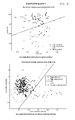
- the presented Canny edge detection based feature resulted in two quantities; the longitudinal edge count and the transverse edge count. Besides using them separately as features, the ratio could be used as a feature as well.
- edge count ratio be defined as the longitudinal edge count divided by the transverse edge count.
- a possible classification based on this feature alone is to use a single threshold value as classifier, for instance the value 1.8. All eggs with an edge count ratio above 1.8 are classified as containing a larva and the remaining are classified as not containing a larva.
- a graph of the edge count ratios of 100 eggs, presented in descending order, is seen on FIG. 13( a ). Notice that for a threshold of 1.8, only a single egg out of the 100 is misclassified based on this edge count ratio feature alone.
- FIG. 14( a ) shows a classification based on the individual correlation coefficients.
Landscapes
- Engineering & Computer Science (AREA)
- Physics & Mathematics (AREA)
- General Health & Medical Sciences (AREA)
- Health & Medical Sciences (AREA)
- Theoretical Computer Science (AREA)
- General Physics & Mathematics (AREA)
- Biomedical Technology (AREA)
- Life Sciences & Earth Sciences (AREA)
- Molecular Biology (AREA)
- Multimedia (AREA)
- Computer Vision & Pattern Recognition (AREA)
- Quality & Reliability (AREA)
- Radiology & Medical Imaging (AREA)
- Nuclear Medicine, Radiotherapy & Molecular Imaging (AREA)
- Medical Informatics (AREA)
- Image Analysis (AREA)
- Investigating Or Analysing Materials By Optical Means (AREA)
- Image Processing (AREA)
Priority Applications (1)
| Application Number | Priority Date | Filing Date | Title |
|---|---|---|---|
| US13/796,660 US9224200B2 (en) | 2012-04-27 | 2013-03-12 | Computer vision based method for extracting features relating to the developmental stages of Trichuris spp. eggs |
Applications Claiming Priority (2)
| Application Number | Priority Date | Filing Date | Title |
|---|---|---|---|
| US201261639229P | 2012-04-27 | 2012-04-27 | |
| US13/796,660 US9224200B2 (en) | 2012-04-27 | 2013-03-12 | Computer vision based method for extracting features relating to the developmental stages of Trichuris spp. eggs |
Publications (2)
| Publication Number | Publication Date |
|---|---|
| US20130287252A1 US20130287252A1 (en) | 2013-10-31 |
| US9224200B2 true US9224200B2 (en) | 2015-12-29 |
Family
ID=48050652
Family Applications (1)
| Application Number | Title | Priority Date | Filing Date |
|---|---|---|---|
| US13/796,660 Expired - Fee Related US9224200B2 (en) | 2012-04-27 | 2013-03-12 | Computer vision based method for extracting features relating to the developmental stages of Trichuris spp. eggs |
Country Status (6)
| Country | Link |
|---|---|
| US (1) | US9224200B2 (pl) |
| EP (1) | EP2852916B1 (pl) |
| DK (1) | DK2852916T3 (pl) |
| ES (1) | ES2754075T3 (pl) |
| PL (1) | PL2852916T3 (pl) |
| WO (1) | WO2013159856A1 (pl) |
Cited By (2)
| Publication number | Priority date | Publication date | Assignee | Title |
|---|---|---|---|---|
| US20160239953A1 (en) * | 2015-02-17 | 2016-08-18 | MatrixSpec Solutions Inc. | Systems, devices, and methods for detecting fertility and gender of unhatched eggs |
| US20170103504A1 (en) * | 2015-10-09 | 2017-04-13 | Universidad Nacional Autónoma de México | System for the identification and quantification of helminth eggs in environmental samples |
Families Citing this family (6)
| Publication number | Priority date | Publication date | Assignee | Title |
|---|---|---|---|---|
| US10216762B2 (en) * | 2014-06-04 | 2019-02-26 | Panasonic Corporation | Control method and non-transitory computer-readable recording medium for comparing medical images |
| US9684831B2 (en) * | 2015-02-18 | 2017-06-20 | Qualcomm Incorporated | Adaptive edge-like feature selection during object detection |
| CN108256419B (zh) * | 2017-12-05 | 2018-11-23 | 交通运输部规划研究院 | 一种利用多光谱解译提取港口码头图像的方法 |
| US12067793B2 (en) * | 2020-04-22 | 2024-08-20 | Mohd Amin Abu Qura | Enterobius vermicularis detection system |
| CN113450292B (zh) * | 2021-06-17 | 2022-08-16 | 重庆理工大学 | 一种pcba零部件高精度视觉定位方法 |
| CN119832328A (zh) * | 2024-12-26 | 2025-04-15 | 苏州才炬智能科技有限公司 | 一种脱壳后的禽蛋外观检测方法 |
Citations (6)
| Publication number | Priority date | Publication date | Assignee | Title |
|---|---|---|---|---|
| WO1999063057A1 (en) | 1998-06-01 | 1999-12-09 | Weyerhaeuser Company | Methods for classification of somatic embryos |
| US7771923B2 (en) | 2006-05-19 | 2010-08-10 | Ovamed Gmbh | Method for detecting the viability of trichuris suis eggs |
| US7833537B2 (en) | 1997-12-31 | 2010-11-16 | University Of Iowa Research Foundation | Use of parasitic biological agents for prevention and control of autoimmune diseases |
| US7951547B2 (en) | 2007-06-15 | 2011-05-31 | Idexx Laboratories, Inc. | Methods, devices, kits and compositions for detecting roundworm, whipworm, and hookworm |
| US20110191869A1 (en) | 2008-05-21 | 2011-08-04 | Bernhard Tewes | Method For Characterising The Biological Activity Of Helminth Eggs, In Particular Trichuris Eggs |
| US8105795B2 (en) | 2008-05-19 | 2012-01-31 | Idexx Laboratories, Inc. | Methods, devices, kits and compositions for detecting roundworm |
-
2013
- 2013-03-12 US US13/796,660 patent/US9224200B2/en not_active Expired - Fee Related
- 2013-03-21 PL PL13714850T patent/PL2852916T3/pl unknown
- 2013-03-21 EP EP13714850.8A patent/EP2852916B1/en active Active
- 2013-03-21 WO PCT/EP2013/000855 patent/WO2013159856A1/en not_active Ceased
- 2013-03-21 ES ES13714850T patent/ES2754075T3/es active Active
- 2013-03-21 DK DK13714850T patent/DK2852916T3/da active
Patent Citations (6)
| Publication number | Priority date | Publication date | Assignee | Title |
|---|---|---|---|---|
| US7833537B2 (en) | 1997-12-31 | 2010-11-16 | University Of Iowa Research Foundation | Use of parasitic biological agents for prevention and control of autoimmune diseases |
| WO1999063057A1 (en) | 1998-06-01 | 1999-12-09 | Weyerhaeuser Company | Methods for classification of somatic embryos |
| US7771923B2 (en) | 2006-05-19 | 2010-08-10 | Ovamed Gmbh | Method for detecting the viability of trichuris suis eggs |
| US7951547B2 (en) | 2007-06-15 | 2011-05-31 | Idexx Laboratories, Inc. | Methods, devices, kits and compositions for detecting roundworm, whipworm, and hookworm |
| US8105795B2 (en) | 2008-05-19 | 2012-01-31 | Idexx Laboratories, Inc. | Methods, devices, kits and compositions for detecting roundworm |
| US20110191869A1 (en) | 2008-05-21 | 2011-08-04 | Bernhard Tewes | Method For Characterising The Biological Activity Of Helminth Eggs, In Particular Trichuris Eggs |
Non-Patent Citations (34)
| Title |
|---|
| Abramowitz, et al., Darkfield Illumination, Olympus Microscopy Resource Center (Apr. 26, 2012) http://www.olympusmicro.com/primer/techniques/dark. |
| Avci, et al., An Expert Diagnosis System for Classification of Human Parasite Eggs Based on Multi-Class SVM, 36 Expert Systems with Applications 43 (2009). |
| Beer, Morphological Descriptions of the Egg and Larval Stages of Trichuris suis Schrank, 1788, Parasitology 263 (1973). |
| Berge, et al., Improved Red Blood Cell Counting in Thin Blood Smears, 204 (2011). |
| Black, et al., Survival Rates of Parasite Eggs in Sludge During Aerobic and Anaerobic Digestion, Applied and Environmental Microbiology 1138 (1982). |
| Brunn, et al., Classification of Parasite Eggs Used as an Active Pharmaceutical Ingredient (API), Technical University of Denmark (2011). |
| Brunn, et al., Detection and Classification of Parasite Eggs for use in Helminthic Therapy, IEEE International Symposium on Biomedical Imaging (IEEE 2012). |
| Brunn, et al., Detection and Classification of Parasite Eggs for use in Helminthic Therapy, IEEE International Symposium on Biomedical Imaging 1627 (IEEE 2012). |
| Bruun, et al., Classification of Parasite Eggs Used as an Active Pharmaceutical Ingredient (API), Danish Society for Parasiteology 7 (2012). |
| Burt, et al., The Laplacian Pyramid as a Compact Image Code, IEEE Transactions on Communications 532 (IEEE 1983). |
| Canny, A Computational Approach to Edge Detection, IEEE Transactions on Pattern Analysis and Machine Intelligence 679 (IEEE 1986). |
| Daugschies, et al., Autofluorescence Microscopy for the Detection of Nematode Eggs and Protozoa, in Particular Isospora Suis, in Swine Faeces 87 Parasitology Research 409 (2001). |
| Diaz, et al., Automatic Clump Splitting for Cell Quantification in Microscopical Images, Proceedings of the Congress on Pattern Recognition 12th Iberoamerican Conference on Progress in Pattern Recognition, Image Analysis and Applications 1 (2007). |
| Dillencourt, et al., A General Approach to Connected-Component Labeling for Arbitrary Image Representations, vol. 39 Journal of the Assocation for Computing Machinery 253 (1992). |
| Dogantekin, et al., A Robust Technique Based on Invariant Moments-ANFIS for Recognition of Human Parasite Eggs in Microscopic Images, 35 Expert Systems with Applications 728 (2008). |
| Geng, et al., Automated Worm Tracking and Classification, Conference Record of the Thirty-Seventh Asimolar Conference on Signals, Systems and Computers 2063 (2003). |
| Gonzalez, et al., Digital Image Processing, International Edition 706 (2008). |
| Mallat, A Theory for Multiresolution Signal Decomposition: The Wavelet Representation, IEEE Transactions on Pattern Analysis and Machine Intelligence 674 (IEEE 1989). |
| Measure Properties of Image Regions, MATLAB Regionprops (Apr. 26, 2012) http://www.mathworks.se/help/toolbox/images/ref/regionprops.html. |
| Nugent, et al., Using Active Learning to Annotate Microscope Images of Parasite Eggs, 26 Artificial Intelligence Review, 63 (Sep. 2007). |
| Otsu, A Threshold Selection Method from Gray-Level Histograms, IEEE Transactions on Systems, Man, and Cybernetics 62 (IEEE 1979). |
| Peng, et al., Engineering Research on the Automatic Identification of Helminth Egg Images, 2 Journal of Hunan Normal University (Feb. 2005). |
| Pittman, et al., Trichuris suis in Finishing Pigs: Case Report and Review, Journal of Swine Health and Production 306 (2010). |
| Reddy, et al., The Use of Trichuris suis and other Helminth Therapies to Treat Crohn's Disease, 100 Parasitol Research 921 (2007). |
| Roberts, et al., Nematodes: Trichurida and Dioctophymatida, Enoplean Parasites, Foundations of Parasitology, Seventh Edition 397 (2005). |
| Sommer, et al., Quantitative Characterization of Texture used for Identification of Eggs of Bovine Parasitic Nematodes, 72 Journal of Helminthology 179 (1998). |
| Summers, et al., Trichuris suis Therapy for Active Ulcerative Colitis: A Randomized Controlled Trial, 128 Gastroenterology 825 (2005). |
| Summers, et al., Trichuris suis Therapy in Crohn's Disease, 54 Gut 87 (2005). |
| Suzuki, et al., Automatic Segmentation and Classification of Human Intestinal Parasites from Microscopy Images, IEEE Transactions on Biomedical Engineering 1 (2011). |
| Thienpoint, et al., Diagnosing Helminthiasis by Coprological Examination, 48 (1986). |
| Witkin, Scale-Space Filtering: A New Approach to Multi-Scale Description, IEEE International Conference on ICASSP 150 (IEEE 1984). |
| Yang, et al., Automatic Identification of Human Helminth Eggs on Microscopic Fecal Specimens Using Digital Image Processing and an Artificial Neural Network, 48 IEEE Transaction on Biomedical Engineering 718 (IEEE 2001). |
| Yang, et al., Automatic Identification of Human Helminth Eggs on Microscopic Fecal Specimens Using Digital Image Processing and an Artificial Neural Network, IEEE Transaction on Biomedical Engineering 718 (IEEE 2001). |
| Zhao Yae, Automatic Recognition of Human Parasite Egg Pictures, 2 Chinese Journal of Stereology and Image Analysis 135 (Sep. 1997). |
Cited By (7)
| Publication number | Priority date | Publication date | Assignee | Title |
|---|---|---|---|---|
| US20160239953A1 (en) * | 2015-02-17 | 2016-08-18 | MatrixSpec Solutions Inc. | Systems, devices, and methods for detecting fertility and gender of unhatched eggs |
| US10607338B2 (en) * | 2015-02-17 | 2020-03-31 | MatrixSpec Solutions Inc. | Systems, devices, and methods for detecting fertility and gender of unhatched non-incubated eggs |
| US10713782B2 (en) | 2015-02-17 | 2020-07-14 | MatrixSpec Solutions Inc. | Systems, devices, and methods for detecting fertility and gender of unhatched eggs |
| US11302000B2 (en) | 2015-02-17 | 2022-04-12 | MatrixSpec Solutions Inc. | Systems, devices, and methods for detecting fertility and gender of unhatched eggs |
| US11688064B2 (en) | 2015-02-17 | 2023-06-27 | MatrixSpec Solutions Inc. | Systems, devices, and methods for detecting fertility and gender of unhatched eggs |
| US20170103504A1 (en) * | 2015-10-09 | 2017-04-13 | Universidad Nacional Autónoma de México | System for the identification and quantification of helminth eggs in environmental samples |
| US9773154B2 (en) * | 2015-10-09 | 2017-09-26 | Universidad Nacional Autónoma de México | System for the identification and quantification of helminth eggs in environmental samples |
Also Published As
| Publication number | Publication date |
|---|---|
| ES2754075T3 (es) | 2020-04-15 |
| EP2852916B1 (en) | 2019-08-07 |
| US20130287252A1 (en) | 2013-10-31 |
| WO2013159856A1 (en) | 2013-10-31 |
| DK2852916T3 (da) | 2019-11-18 |
| EP2852916A1 (en) | 2015-04-01 |
| PL2852916T3 (pl) | 2020-05-18 |
Similar Documents
| Publication | Publication Date | Title |
|---|---|---|
| US9224200B2 (en) | Computer vision based method for extracting features relating to the developmental stages of Trichuris spp. eggs | |
| Mehanian et al. | Computer-automated malaria diagnosis and quantitation using convolutional neural networks | |
| Pospiech et al. | Identification of pollen taxa by different microscopy techniques | |
| Rosado et al. | Automated detection of malaria parasites on thick blood smears via mobile devices | |
| Linder et al. | A malaria diagnostic tool based on computer vision screening and visualization of Plasmodium falciparum candidate areas in digitized blood smears | |
| Theodorakopoulos et al. | Hep-2 cells classification via fusion of morphological and textural features | |
| Tonti et al. | An automated approach to the segmentation of HEp-2 cells for the indirect immunofluorescence ANA test | |
| Irshad et al. | Multispectral band selection and spatial characterization: Application to mitosis detection in breast cancer histopathology | |
| Memeu et al. | Detection of plasmodium parasites from images of thin blood smears | |
| CN110032946A (zh) | 一种基于机器视觉的铝/铝泡罩包装药片识别与定位方法 | |
| Li et al. | MobileOne-YOLO: Improving the YOLOv7 network for the detection of unfertilized duck eggs and early duck embryo development-a novel approach | |
| KR20150108963A (ko) | 세포 분석 장치 및 세포 분석 방법 | |
| PP et al. | Automated quality assessment of cocoons using a smart camera based system | |
| Salami et al. | Evaluation of a self-supervised machine learning method for screening of particulate samples: a case study in liquid formulations | |
| Aris et al. | Robust image processing framework for intelligent multi-stage malaria parasite recognition of thick and thin smear images | |
| Bruun et al. | OvaSpec–A vision-based instrument for assessing concentration and developmental stage of Trichuris suis parasite egg suspensions | |
| Bruun et al. | Detection and classification of parasite eggs for use in helminthic therapy | |
| Soda et al. | A decision support system for Crithidia Luciliae image classification | |
| Jewani et al. | Detection of diseases via blood analysis using Image processing Techniques | |
| Yang et al. | Classification of rotifers with machine vision by shape moment invariants | |
| Sreelatha et al. | An improved automatic detection of true comets for DNA damage analysis | |
| Le et al. | An automated framework for counting lymphocytes from microscopic images | |
| Massarelli et al. | A Handy Open-Source Application Based on Computer Vision and Machine Learning Algorithms to Count and Classify Microplastics. Water. 2021; 13 (15) | |
| Alshut et al. | Robust identification of coagulated zebrafish eggs using image processing and classification techniques | |
| Hodgson et al. | Progress towards a system for the automatic recognition of pollen using light microscope images |
Legal Events
| Date | Code | Title | Description |
|---|---|---|---|
| AS | Assignment |
Owner name: PARASITE TECHNOLOGIES A/S, DENMARK Free format text: ASSIGNMENT OF ASSIGNORS INTEREST;ASSIGNORS:BRUUN, JOHAN MUSAEUS;CARSTENSEN, JENS MICHAEL;KAPEL, CHRISTIAN MOLIIN OUTZEN;REEL/FRAME:030567/0655 Effective date: 20130321 |
|
| STCF | Information on status: patent grant |
Free format text: PATENTED CASE |
|
| MAFP | Maintenance fee payment |
Free format text: PAYMENT OF MAINTENANCE FEE, 4TH YR, SMALL ENTITY (ORIGINAL EVENT CODE: M2551); ENTITY STATUS OF PATENT OWNER: SMALL ENTITY Year of fee payment: 4 |
|
| FEPP | Fee payment procedure |
Free format text: MAINTENANCE FEE REMINDER MAILED (ORIGINAL EVENT CODE: REM.); ENTITY STATUS OF PATENT OWNER: SMALL ENTITY |
|
| LAPS | Lapse for failure to pay maintenance fees |
Free format text: PATENT EXPIRED FOR FAILURE TO PAY MAINTENANCE FEES (ORIGINAL EVENT CODE: EXP.); ENTITY STATUS OF PATENT OWNER: SMALL ENTITY |
|
| STCH | Information on status: patent discontinuation |
Free format text: PATENT EXPIRED DUE TO NONPAYMENT OF MAINTENANCE FEES UNDER 37 CFR 1.362 |
|
| FP | Lapsed due to failure to pay maintenance fee |
Effective date: 20231229 |