US9075903B2 - User interface for medical image review workstation - Google Patents
User interface for medical image review workstation Download PDFInfo
- Publication number
- US9075903B2 US9075903B2 US13/302,465 US201113302465A US9075903B2 US 9075903 B2 US9075903 B2 US 9075903B2 US 201113302465 A US201113302465 A US 201113302465A US 9075903 B2 US9075903 B2 US 9075903B2
- Authority
- US
- United States
- Prior art keywords
- interface
- screen
- user
- display
- touchscreen
- Prior art date
- Legal status (The legal status is an assumption and is not a legal conclusion. Google has not performed a legal analysis and makes no representation as to the accuracy of the status listed.)
- Active, expires
Links
Images


Classifications
-
- G—PHYSICS
- G06—COMPUTING OR CALCULATING; COUNTING
- G06F—ELECTRIC DIGITAL DATA PROCESSING
- G06F3/00—Input arrangements for transferring data to be processed into a form capable of being handled by the computer; Output arrangements for transferring data from processing unit to output unit, e.g. interface arrangements
- G06F3/01—Input arrangements or combined input and output arrangements for interaction between user and computer
- G06F3/048—Interaction techniques based on graphical user interfaces [GUI]
- G06F3/0484—Interaction techniques based on graphical user interfaces [GUI] for the control of specific functions or operations, e.g. selecting or manipulating an object, an image or a displayed text element, setting a parameter value or selecting a range
- G06F3/04845—Interaction techniques based on graphical user interfaces [GUI] for the control of specific functions or operations, e.g. selecting or manipulating an object, an image or a displayed text element, setting a parameter value or selecting a range for image manipulation, e.g. dragging, rotation, expansion or change of colour
-
- G06F19/3406—
-
- G06F19/321—
-
- G—PHYSICS
- G06—COMPUTING OR CALCULATING; COUNTING
- G06F—ELECTRIC DIGITAL DATA PROCESSING
- G06F3/00—Input arrangements for transferring data to be processed into a form capable of being handled by the computer; Output arrangements for transferring data from processing unit to output unit, e.g. interface arrangements
- G06F3/01—Input arrangements or combined input and output arrangements for interaction between user and computer
- G06F3/03—Arrangements for converting the position or the displacement of a member into a coded form
- G06F3/041—Digitisers, e.g. for touch screens or touch pads, characterised by the transducing means
- G06F3/0416—Control or interface arrangements specially adapted for digitisers
-
- G—PHYSICS
- G06—COMPUTING OR CALCULATING; COUNTING
- G06F—ELECTRIC DIGITAL DATA PROCESSING
- G06F3/00—Input arrangements for transferring data to be processed into a form capable of being handled by the computer; Output arrangements for transferring data from processing unit to output unit, e.g. interface arrangements
- G06F3/01—Input arrangements or combined input and output arrangements for interaction between user and computer
- G06F3/048—Interaction techniques based on graphical user interfaces [GUI]
- G06F3/0481—Interaction techniques based on graphical user interfaces [GUI] based on specific properties of the displayed interaction object or a metaphor-based environment, e.g. interaction with desktop elements like windows or icons, or assisted by a cursor's changing behaviour or appearance
- G06F3/04817—Interaction techniques based on graphical user interfaces [GUI] based on specific properties of the displayed interaction object or a metaphor-based environment, e.g. interaction with desktop elements like windows or icons, or assisted by a cursor's changing behaviour or appearance using icons
-
- G—PHYSICS
- G06—COMPUTING OR CALCULATING; COUNTING
- G06F—ELECTRIC DIGITAL DATA PROCESSING
- G06F3/00—Input arrangements for transferring data to be processed into a form capable of being handled by the computer; Output arrangements for transferring data from processing unit to output unit, e.g. interface arrangements
- G06F3/01—Input arrangements or combined input and output arrangements for interaction between user and computer
- G06F3/048—Interaction techniques based on graphical user interfaces [GUI]
- G06F3/0487—Interaction techniques based on graphical user interfaces [GUI] using specific features provided by the input device, e.g. functions controlled by the rotation of a mouse with dual sensing arrangements, or of the nature of the input device, e.g. tap gestures based on pressure sensed by a digitiser
- G06F3/0488—Interaction techniques based on graphical user interfaces [GUI] using specific features provided by the input device, e.g. functions controlled by the rotation of a mouse with dual sensing arrangements, or of the nature of the input device, e.g. tap gestures based on pressure sensed by a digitiser using a touch-screen or digitiser, e.g. input of commands through traced gestures
- G06F3/04883—Interaction techniques based on graphical user interfaces [GUI] using specific features provided by the input device, e.g. functions controlled by the rotation of a mouse with dual sensing arrangements, or of the nature of the input device, e.g. tap gestures based on pressure sensed by a digitiser using a touch-screen or digitiser, e.g. input of commands through traced gestures for inputting data by handwriting, e.g. gesture or text
-
- G—PHYSICS
- G16—INFORMATION AND COMMUNICATION TECHNOLOGY [ICT] SPECIALLY ADAPTED FOR SPECIFIC APPLICATION FIELDS
- G16H—HEALTHCARE INFORMATICS, i.e. INFORMATION AND COMMUNICATION TECHNOLOGY [ICT] SPECIALLY ADAPTED FOR THE HANDLING OR PROCESSING OF MEDICAL OR HEALTHCARE DATA
- G16H30/00—ICT specially adapted for the handling or processing of medical images
- G16H30/20—ICT specially adapted for the handling or processing of medical images for handling medical images, e.g. DICOM, HL7 or PACS
-
- G—PHYSICS
- G16—INFORMATION AND COMMUNICATION TECHNOLOGY [ICT] SPECIALLY ADAPTED FOR SPECIFIC APPLICATION FIELDS
- G16H—HEALTHCARE INFORMATICS, i.e. INFORMATION AND COMMUNICATION TECHNOLOGY [ICT] SPECIALLY ADAPTED FOR THE HANDLING OR PROCESSING OF MEDICAL OR HEALTHCARE DATA
- G16H40/00—ICT specially adapted for the management or administration of healthcare resources or facilities; ICT specially adapted for the management or operation of medical equipment or devices
- G16H40/60—ICT specially adapted for the management or administration of healthcare resources or facilities; ICT specially adapted for the management or operation of medical equipment or devices for the operation of medical equipment or devices
- G16H40/63—ICT specially adapted for the management or administration of healthcare resources or facilities; ICT specially adapted for the management or operation of medical equipment or devices for the operation of medical equipment or devices for local operation
-
- G—PHYSICS
- G06—COMPUTING OR CALCULATING; COUNTING
- G06F—ELECTRIC DIGITAL DATA PROCESSING
- G06F2203/00—Indexing scheme relating to G06F3/00 - G06F3/048
- G06F2203/041—Indexing scheme relating to G06F3/041 - G06F3/045
- G06F2203/04104—Multi-touch detection in digitiser, i.e. details about the simultaneous detection of a plurality of touching locations, e.g. multiple fingers or pen and finger
Definitions
- This patent specification relates to medical imaging. More particularly, this patent specification relates to user interfaces for medical image review workstations.
- radiologist generically refers to a medical professional that analyzes medical images and makes clinical determinations therefrom, it being understood that such person or user of the review workstation might be titled differently, or might have differing qualifications, depending on the country or locality of their particular medical environment.
- a large body of information associated with a patient's medical image data would have substantially diminished value if the radiologist does not have sufficient time, inclination, or information technology (IT) sophistication to properly view that information. It is therefore crucial that the human-machine interface associated with medical image review workstations be as streamlined, appealing, and user-friendly as possible, while also allowing comprehensive access to the large amount of data available.
- IT information technology
- Embodiments address shortcomings of known methods and systems by providing a user interface (UI) system for a medical image review workstation that provides a streamlined, appealing, and user-friendly experience for the radiologist.
- Embodiments also provide such a UI system that is easily and inexpensively upgraded to accommodate new software versions, new capabilities, and/or new imaging modalities for the review workstation.
- Embodiments also provide such a UI system that is readily personalized and customizable for different users at a single review workstation, and/or readily customizable a single radiologist at multiple different review workstations, and allow for such a UI system to be layered upon existing UI systems without requiring substantial changes to current hardware configurations and without requiring substantial cost investment.
- One embodiment is directed a computer-implemented method executed by an interface processor and/or mobile communication device for controlling display of medical images and that comprises establishing a network connection between a mobile communication device and the interface processor operably coupled to a review workstation operable by a user to review medical images using a first UI.
- the method further comprises determining a first medical image selected by the user (e.g., based on the user highlighting or selecting an image or window containing an image).
- the first medical image is of a first type, e.g., one or more of being an image of a first imaging modality, a first review mode (e.g., a transverse view mode for a tomosynthesis image), and generated by a first type of imaging device.
- the method further comprises displaying or invoking for display on a screen of the mobile communication device, a second UI.
- the second UI comprises a first touchscreen interface for controlling display of the first medical image.
- the method further comprises determining a second medical image selected by the user, the second medical image is of a second type different than the first type, e.g., one or more of being an image of a second imaging modality, a second review mode (e.g., a transverse mode of a magnetic resonance image rather than a tomosynthesis image, or a single-breast MLO view of an x-ray image a transverse view mode for a tomosynthesis image), and generated by a second type of imaging device.
- the method further comprises displaying or invoking for display on the screen, a third UI that is also a touchscreen interface but different than the first touchscreen interface, for controlling display of the second medical image.
- a further embodiment is directed to a computer-implemented method for controlling display of medical images and that comprises displaying or invoking for display on a screen of a mobile communication device of a user, a first touchscreen interface for controlling display of a selected medical image.
- the first touchscreen interface has a number of interface elements and is considered to be an initial or primary touchscreen interface.
- the method further comprises detecting when the user has tapped the screen with a plurality of fingers simultaneously (e.g., with a “five finger tap”). In response to detecting a five finger tap, another touch screen interface is displayed or invoked for display on the mobile communication device screen for controlling display of the selected medical image.
- the second touchscreen interface consists of subset of the plurality of the interface elements of the first touchscreen interface (i.e., the number of elements of the second touchscreen interface is less than the number of elements of the first touchscreen interface).
- a first or primary touchscreen interface may include interface elements for all available controls, whereas the second or auxiliary touchscreen interface displayed or invoked after a simultaneous finger tap may include only as many interface elements as fingers that tapped the screen simultaneously for selected controls.
- a first touchscreen UI may include 24 or other numbers of interface elements for various display controls
- a second or auxiliary touchscreen interface displayed after a “five finger tap” includes only five interface elements, e.g., five interface elements previously selected by the user, determined to be the most popular or utilized most often, or elements positioned under fingers that tapped the screen.
- embodiments are directed to computer-implemented methods for controlling display of medical images generated by imaging devices of different vendors or manufactures and that may involve different types of interfaces displayed at a review workstation that receives image data from respective different imaging devices.
- embodiments may involve imaging devices of different manufacturers, which may be of the same imaging modality or different imaging modalities, and that provide different types of interfaces for viewing medical images of the same or different view modes.
- these different types interfaces can be transformed into corresponding touchscreen interfaces such that a first touchscreen interface is generated, displayed or invoked for display on a screen of a mobile communication device for a first medical image generated by a first imaging device manufactured or sold by a first source, vendor or manufacturer, whereas a second, different touchscreen interface is generated, displayed or invoked for display for a second medical image generated by another imaging device of the same modality (e.g., both imaging devices are tomosynthesis imaging devices), but with a different touchscreen UI.
- Embodiments allow users to utilize a touchscreen interface displayed on a mobile communication device to control display of medical images, acquired with imaging devices of the same or different manufacturers, which may be of the same or different imaging modalities.
- Yet other embodiments are directed to articles of manufacture, computer program products and native and downloadable applications executable on a mobile communication device such as a smartphone or tablet computing device capable of wireless communications and configured, operable or programmed to execute methods according to embodiments.
- a mobile communication device such as a smartphone or tablet computing device capable of wireless communications and configured, operable or programmed to execute methods according to embodiments.
- one embodiment is directed to a computer program product, which may comprise a non-transitory computer readable storage medium having stored thereupon a sequence of instructions which, when executed by a computer or mobile communication device, perform a process for controlling display of medical images by displaying or invoking for display on a screen of a mobile communication device a first touchscreen interface for controlling display of the first medical image of a first type, and displaying or invoking for display on the screen, another touchscreen interface different than the first touchscreen interface for controlling display of the second medical image of a second type different than the first type.
- embodiments are directed to articles of manufacture, computer program products or mobile applications which, when instructions thereof are executed, cause a computer or processing element to perform a process for controlling display of medical images by detecting and responding to a multi-finger or multi-digit tap (e.g., a “five finger tap”) by the user on a screen of the mobile communication device.
- a first touchscreen interface for controlling display of a selected medical image may be displayed on the screen and includes a plurality of interface elements, the user performs a simultaneous “multi-finger” tap, which is detected, and in response, a second touch screen interface is generated and consists of subset of the plurality of the interface elements of the first touchscreen interface.
- the subset may be selected by the user to provide for customization, or be determined based on criteria such as most frequent use or position of fingers when the screen is tapped.
- Embodiments may be part of or executed by a review workstation, which may include a non-touchscreen interface such as a keyboard and mouse, part of or executed by an interface processor operably coupled to or in communication between a review workstation and a mobile communication device, or part of (e.g., a native application) or downloaded to a mobile communication device and executed by a processor or computing element thereof.
- Touch screen interfaces may be displayed or invoked by components of a mobile communication device and/or an interface processor operable coupled between the mobile communication device and a review workstation.
- an application executing on a mobile communication device may perform various processing to determine how a touchscreen interface should be structured and displayed.
- Yet further embodiments are directed to systems configured or operable to analyze medical images using different touchscreen interfaces derived from or resulting from transformation of controls of a UI of a review workstation such that different touchscreen interfaces can be generated for different types of images, e.g., images of different imaging modalities, different view modes, and different imaging modalities and review modes.
- one embodiment is directed to a system for controlling display of medical images and comprises a review workstation and an interface processor, which may be an integral component of a review workstation or a separate component that can be connected or plugged into the review workstation.
- the review workstation is operated with a first UI controlled with a keyboard, mouse, trackball, joystick or other physical control element physically manipulated and moved by the user to select a first medical image of a first type and control how the first medical image is displayed on a screen of the review workstation.
- the interface processor receiving or determining the selected first medical image and review mode and imaging modality thereof, is configured to communicate with a mobile communication device, and display or invoke for display, on a screen of the mobile communication device, a second UI comprising a first touchscreen interface for controlling display of the first medical image.
- the interface processor also determines when the user has selected another, second medical image of a second type. Selection of another medical image may be done through the first UI of the review workstation or through the first touchscreen interface displayed on the mobile communication device screen (e.g., via a toggle mechanism that allows the user of the mobile communication device to select an image displayed on the screen of the review workstation).
- the interface processor displays or invokes for display on the screen, a third UI comprising a second touchscreen interface that is different than the first touchscreen interface for controlling display of the second medical image of the second type.
- System embodiments may involve or comprise only a review workstation configured to establish a wireless connection with a mobile communication device and to display or invoke display of touchscreen interfaces, only an interface processor configured to implement embodiments, only a mobile communication device configured to implement embodiments, or a combination of components such as a review workstation and interface processor, an interface processor and mobile communication device, and all of a review workstation, interface processor and mobile communication device.
- a medical image may be selected by the user manipulating the first UI of the review workstation or manipulating a touchscreen interface displayed on a mobile communication device, in response to which a touchscreen interface, or different touchscreen interface, is displayed or invoked to control display of the current or selected medical image.
- the interface processor and mobile communication device are in communication via a wireless network.
- a wireless connection can be established by the mobile communication device being placed in communication with a wireless access point that is in communication with a server hosting the interface processor.
- the interface processor receives data of the first UI for controlling display of the first selected image of a first type, transforms those controls into a single-handed or dual-handed touchscreen interface for real-time control of display of the first selected image using the mobile communication device, receives data of another type of image selected by the user, and transforms controls of the first UI into a different touchscreen interface for real-time control of display of the other medical image using the mobile communication device.
- Touchscreen interfaces have different numbers, shapes and/or spatial arrangements of interface elements depending on the type of image, e.g., the review mode, imaging modality and user preferences
- the medical images are presented within windows of the first UI.
- an interface with four windows may include four medical images, each of which is associated with respective view modes and/or imaging modalities.
- Medical images or windows may be generated by the same review workstation, or by different review workstations, e.g., review workstations of different manufacturers, which may involve different interfaces utilized at the review workstation.
- the interface processor is a part of or in communication with the review workstation such that the interface processor determines which window is identified or selected as an active window, e.g., based on user manipulation of a mouse or keyboard control at the review workstation or by use of a touchscreen interface displayed on the mobile communication device screen, to determine which medical image or window was selected.
- the user manipulating of the mobile communication device itself may be detected by an application executing on the mobile communication device.
- the application may invoke or display a different touchscreen interface for a given medical displayed, a touchscreen interface for a next image to be analyzed is displayed, or a medical image for a new, different patient, e.g., a randomly selected patient or a next patient in a patient list. If the interface or patient data is not available on the mobile communication device, the mobile communication device can communicate with the interface processor in response to detecting the shaking or jiggling motion.
- touchscreen interfaces can be translated to different hands of the user.
- the application detects the switch based on finger placement or arrangements and then displays or invokes for display elements of the touchscreen interface that are flipped for the other hand.
- Other customization features include touchscreen interfaces being spatially arranged to be customized to respective lengths of respective fingers of the user of the mobile communication device.
- the auxiliary touchscreen interface displayed following the multi-finger tap includes only a subset of the previously displayed or primary touchscreen interface.
- the number and/or arrangement of auxiliary interface elements may be based at least in part upon the number of fingers that tapped the screen simultaneously, which interface elements were selected by the user, or determined to be utilized most often. For example, if the user tapped the screen with five fingers (defined to include a thumb and four fingers), then the subset consists of five interface elements, which may be displayed at the same locations tapped by respective fingers, and that may be spatially arranged relative to each other in the same manner as the first or primary interface.
- the subset of interface elements in their spatial arrangement, can follow the user's hand as it slides or moves across the screen to a different screen location or as finger lengths and/or positions are adjusted.
- a user may be viewing a first medical image associated with a first review mode and first imaging modality using a first touchscreen interface (e.g., with 25 or other number of interface elements), perform a five finger tap, in response to which an auxiliary or secondary UI with only 5 elements is displayed, then perform another five finger tap to toggle back to the primary interface.
- a multi-finger tap can be used to switch between primary and secondary or auxiliary touchscreen interfaces, or to invoke some other type of action such as switching to display of medical images of another patient.
- Other user actions utilized by embodiments include tracking movement or positioning of a pre-determined digit such as the user's thumb such as when a finger contacts or does not contact (is lifted from) the screen, to then display or invoke a new touchscreen interface, and then displaying or invoking the first or prior touchscreen interface when it is detected that the thumb or other pre-determined finger contacts the screen or has returned to a pre-determined position on the screen.
- a pre-determined digit such as the user's thumb
- a new touchscreen interface such as when a finger contacts or does not contact (is lifted from) the screen
- displaying or invoking the first or prior touchscreen interface when it is detected that the thumb or other pre-determined finger contacts the screen or has returned to a pre-determined position on the screen.
- FIG. 1 illustrates an environment including components of one embodiment of a system operable to invoke touchscreen user interfaces on a screen of a mobile communication device adapted for controlling display of different types of medical images, wherein in the illustrated embodiment, an interface processor is operably coupled to a review workstation and in wireless communication with a mobile communication device;
- FIGS. 2A-D illustrate examples of output windows of a review workstation and identification or selection of an output window as an active window and respective touchscreen interfaces having respective interface elements configured according to respective spatial arrangements, and displayed or invoked for display on a screen of a mobile communication device to control display of different types of medical images;
- FIGS. 3A-B illustrate embodiments involving physical modifications to pre-determined screen locations by application of one or more textured material patches to the screen at locations at which particular touchscreen interface elements are displayed to provide haptic feedback to the user while using the touchscreen interface, wherein FIG. 3A illustrates textured patches applied to the screen, and FIG. 3B illustrates how the spatial arrangement of the applied textured patches shown in FIG. 3A corresponds to particular elements of a touchscreen interface generated according to embodiments;
- FIGS. 4A-B illustrates different template patterns defining voids in respective spatial arrangements for use in embodiments for providing haptic feedback to a user of a mobile communication device
- FIGS. 5A-B illustrate embodiments involving physical modifications to pre-determined screen locations by application templates defining respective voids to the screen such that the voids are at locations at which particular touchscreen interface elements are displayed to provide haptic feedback to the user while using the touchscreen interface
- FIG. 5A illustrates the template shown in FIG. 4A applied to the screen to outline groups of elements of a first touchscreen interface
- FIG. 5B illustrates the template shown in FIG. 4B applied to the screen to outline groups of elements of a second touchscreen interface
- FIGS. 6A-D illustrate embodiments involving a multi-finger tap of a screen of a mobile communication device by a user to invoke a touchscreen interface or a change or transformation to a touchscreen interface
- FIG. 6A illustrates a first or primary touchscreen user interface as shown in FIG. 2A and including a plurality of interface elements in a particular spatial arrangement and the user performing a multi-finger tap on the screen
- FIG. 6B illustrates the resulting second or auxiliary touchscreen interface following the multi-finger tap and that includes only a subset of the interface elements of the first or primary touchscreen user interface
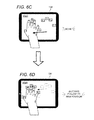
- FIG. 6C illustrates the user repositioning a hand after manipulating the second or auxiliary touchscreen user interface shown in FIG. 6B
- FIG. 6D illustrates how the auxiliary touchscreen interface is translated from one hand location to another location or follows the user's hand while maintaining the spatial arrangement of the subset of interface elements to allow the user to manipulate the same auxiliary touchscreen interface at a different screen location;
- FIG. 7 illustrates how embodiments may be applied to detect a change of hand and to transform or flip a touchscreen interface configured for one hand to a configuration for an opposite hand, wherein in the illustrated embodiment, the touchscreen interface that is flipped is an auxiliary touchscreen user interface;
- FIG. 8 illustrates how embodiments may be applied to generate a dual-handed touchscreen user interface
- FIG. 9 is a screenshot of a home screen displayed on a mobile communication device screen
- FIG. 10 is a mobile communication device screenshot that follows launching of an application for controlling display of medical images on the mobile communication device according to embodiments;
- FIG. 11 is a mobile communication device screenshot illustrating an example of a touchscreen user interface generally depicted in FIG. 2A and that is generated and displayed for a first active window corresponding to a tomosynthesis modality;
- FIG. 12 is a mobile communication device screenshot illustrating an example of a touchscreen user interface generally depicted in FIG. 2D and that is generated and displayed for a second active window corresponding to a MRI modality;
- FIG. 13 is a screenshot of an auxiliary touchscreen interface generated according to embodiments and resulting from the user performing a multi-finger tap on the screen of the mobile communication device while the touchscreen interface for the first active window corresponding to a tomosynthesis modality as shown in FIG. 11 was displayed;
- FIG. 14 is a screenshot of a selection or confirmation window that allows a user to assign display controls or functions to elements of the auxiliary touchscreen interface which in the illustrated example includes five interface elements as shown in FIG. 13 ;
- FIG. 15 is a screenshot of an expanded selection or configuration window illustrating in further detail display controls or functions that can be assigned to an element of an auxiliary touchscreen interface as shown in FIGS. 13-14 ;
- FIG. 16 is a screenshot of an auxiliary touchscreen interface generated according to embodiments and resulting from the user performing a multi-finger tap on the screen of the mobile communication device while the touchscreen interface for the first active window corresponding to a magnetic resonance imaging modality as shown in FIG. 12 was displayed,
- FIG. 17 is a screenshot of a selection or confirmation window that allows a user to assign display controls or functions to elements of the auxiliary touchscreen interface which in the illustrated example includes five interface elements as shown in FIG. 16 ;
- FIG. 18 is a screenshot of an expanded selection or configuration window illustrating in further detail display controls or functions that can be assigned to an element of an auxiliary touchscreen interface as shown in FIGS. 16-17 .
- Embodiments relate to computer-implemented methods, systems and computer program products or mobile applications for controlling how different types medical images are displayed on a mobile communication device such as a smartphone or tablet computing device capable of wireless communications.
- a user may utilize touchscreen interfaces that are derived from controls of user interfaces (UIs) of known review workstations, and which can be dynamically adapted in real-time for controlling display of different types of images, e.g., different types of view modes and/or imaging modalities, while the user is holding the mobile communication device.
- UIs user interfaces
- one touchscreen interface may be presented for viewing an image associated with a transverse view mode and an imaging modality of magnetic resonance imaging, whereas another touchscreen interface is generated for viewing the same image but with a different view mode, or for the same view but for an image generated using a different imaging modality.
- Embodiments also accommodate medical images generated by imaging devices of different manufacturers and respective different interfaces utilized to view images generated by such devices.
- Various embodiments also provide for the ability to customize touchscreen interfaces.
- Certain embodiments are directed to methods, systems and computer program products or mobile applications for determining which images types (of different views and/or acquired with different imaging modalities) are selected by a user with a review workstation UI or a UI displayed on a mobile communication device screen, and displaying or invoking touchscreen interfaces for respective image views and/or images acquired with different imaging modalities.
- a user may manipulate an existing UI of a review workstation, and also manipulate touchscreen interfaces that are adapted or customized for different image views or modalities.
- Certain other embodiments are directed to tapping a screen of a mobile communication device with multiple fingers simultaneously (e.g., with five fingers or a thumb and four fingers) to invoke or display an auxiliary UI, which includes only a subset of interface elements, which may be selected by the user or selected as being utilized the most often, of the originally displayed UI, and they can be positioned in the same arrangement as they were in the original UI such that positioning of fingers of the user remains unchanged.
- users can “five finger tap” between the primary or complete UI and one or more auxiliary interfaces. Screen tapping in this manner can also be used to advance to medical images of different patients.
- a medical imaging environment 100 includes a medical image review workstation or workstation 120 having an enhanced user UI including user control features implemented and/or actuated via a mobile communication device 134 such as a smartphone or tablet computing device capable of wireless communications. Shown in FIG.
- HIS/RIS Hospital Information System/Radiology Information System
- image acquisition devices examples of which include, but are not limited to, a mammogram acquisition device 102 , a tomosynthesis acquisition device 104 , an ultrasound acquisition device 106 , a magnetic resonance imaging (MRI) acquisition device 108 , and a generalized “other” medical imaging device 110 representative of, for example, one or more computerized tomography (CT) imaging or positron emission tomography (PET) acquisition devices.
- CT computerized tomography
- PET positron emission tomography
- a computer-aided detection (CAD) processor 112 coupled to the network 116 receives digital medical images from one or more of the devices 102 , 104 , 106 , 108 , and 110 .
- CAD computer-aided detection
- an additional tomosynthesis reconstruction processor (not shown in FIG. 1 ) can be coupled to the network 116 to generate and provide a plurality of tomosynthesis reconstructed image slices from x-ray tomosynthesis projection images provided by the tomosynthesis acquisition device 104 .
- the CAD processor 112 processes the medical images according to one or more CAD algorithms and provides CAD findings associated therewith.
- a UI implemented at the review workstation 120 in conjunction with the mobile communication device 134 interactively displays the medical images to a viewer or user in accordance with one or more of the systems and methods described further hereinbelow.
- the mobile communication device 134 communicates with the review workstation 120 by virtue of wireless communication (e.g., using the IEEE 802.11 “WiFi” protocol) with wireless access point 114 , which is, in turn, connected to the network 116 .
- wireless communication e.g., using the IEEE 802.11 “WiFi” protocol
- Various medical images and related information are communicated according to the DICOM (Digital Imaging and Communications in Medicine) standard and the network 116 supports the TCP/IP protocol, which is used as the transport protocol for the DICOM standard.
- a PACS archive 118 is also coupled to the network 116 , generally representing a repository for medical information associated with the medical imaging environment, including both current and archived images, current and archived CAD results, radiology reports for completed cases, and so forth.
- Embodiments described herein can be seamlessly layered upon an existing medical imaging workflow, in which the digital (or digitized) medical images are acquired, optionally processed by the CAD processor 112 , and displayed at the review workstation 120 (optionally in conjunction with the associated CAD results) to a radiologist, who makes a clinical determination therefrom.
- a UI implemented at the review workstation 120 interactively displays the medical images to a viewer or user (generally, “user”) of embodiments in accordance with one or more UI programs carried out on an interface processor 126 .
- an auxiliary host application program that, upon execution, communicates with the mobile communication device 134 and carries out the associated functionalities described further herein.
- an auxiliary remote application program that, upon execution, communicates with the auxiliary host application program on the UI processor 126 and carries out the associated functionalities described further herein.
- server application can be used interchangeably with “host application” to denote described software that executes on the interface processor 126 .
- a medical imaging environment 100 includes a medical image review workstation 120 having an enhanced UI including user control features implemented and/or actuated via a mobile communication device 134 such as a smartphone, tablet computing device or other mobile communication device 134 .
- mobile communication devices 134 include the IPHONE and IPAD available from Apple, Inc. and other communication or tablet computing device capable of communicating with a review workstation 120 or associated interface processor 126 according to a preferred embodiment.
- IPAD 134 represents one particularly advantageous mobile communication device including hardware, software, network, and development platform for implementing embodiments directed to control of medical image review with UIs described further herein, it is to be appreciated that other known or hereinafter developed mobile communication devices 134 and platforms having generic capabilities analogous to the IPAD 134 can be used in place of an IPAD 134 while remaining within the scope of the preferred embodiments.
- such other known or hereinafter developed mobile communication devices 134 and platforms would include a portable, programmable touchpad computer having a touch-sensitive screen that is of a size and shape to accommodate an open human hand, and would be capable of wireless communication with another computer or network node using Wi-Fi, BLUETOOTH, ZIGBEE, WiMAX, Wireless USB, or any of a host of other standard or non-standard wireless protocols or information transfer modalities (infrared, optical, ultrasonic, etc.). While limiting mobility compared to a wireless connection, embodiments may also involve data connectivity of the portable, programmable touchpad computer through a wired connection, such as wired USB, without necessarily departing from the scope of embodiments.
- the size of the touchscreen could be made smaller than that of an opened human hand, such as with the touchscreens of IPHONEs or similar portable phones, without necessarily departing from the scope of the present teachings.
- IPAD 134 as a mobile communication device 134 utilized in embodiments, but it will be understood that other mobile communication devices 134 may be utilized, and that such devices may communicate with review workstation 120 or associated interface processor 126 .
- auxiliary host application program carried out on the UI processor 126 is referred to hereinbelow as the “SVTouch host” or “SVTouch server” program.
- auxiliary remote application program carried out on the IPAD 134 is referred to hereinbelow as the “SVTouch remote app.”
- SECURVIEWTouchTM can be seen as being a shorthand term for SECURVIEWTouchTM.
- SECUREVIEW is a registered trademark of Hologic, Inc., of Bedford Mass., where SECURVIEW proprietarily identifies a highly successful and powerful medical image review workstation currently manufactured and sold by Hologic, Inc., the assignee of the present application.
- SECURVIEW TouchTM proprietarily identifies an extension of the SECURVIEW medical image review workstation that includes one or more aspects of the IPAD-implemented functionality described further herein, which one or more aspects will be publically introduced at the 2010 meeting of the Radiological Society of North America in Chicago, Ill. (RSNA 2010).
- Review workstation 120 implements an interactive UI using a diagnostic display 122 including first and second display monitors 122 a and 122 b , an administrative display 124 , and user input or control devices including a keyboard 128 , a mouse 132 , and an application-specific hardware auxiliary input device 130 , such as a workflow keypad provided in conjunction with a SECURVIEW medical image review workstation.
- a diagnostic display 122 including first and second display monitors 122 a and 122 b
- an administrative display 124 including a keyboard 128 , a mouse 132 , and an application-specific hardware auxiliary input device 130 , such as a workflow keypad provided in conjunction with a SECURVIEW medical image review workstation.
- the IPAD 134 also becomes part of the UI provided by the review workstation 120 .
- the described SVTouch functionalities can be provided as an add-on that operates side-by-side with the auxiliary hardware input device 130 , or alternatively the described SVTouch functionalities can be used to
- Administrative display 124 is used for input and output of a wide variety of information that may be associated with a particular set of medical images (e.g., listings, tables, plots, text descriptions, etc.), as well as for system installation, maintenance, updating, and related tasks. Often provided on the diagnostic display 122 at any particular time during case review by a radiologist are one or more diagnostic images displayed in one or more output windows A, B, and C.
- the medical imaging environment 100 of FIG. 1 is presented by way of example only and is not intended to limit the scope of the preferred embodiments to this particular scenario.
- different combinations of the devices 102 - 132 of FIG. 1 can be placed adjacently to each other or integrated into the same hardware boxes without departing from the scope of the preferred embodiments.
- the network 116 can be a wide-area network with the different nodes being distributed throughout a city, a country, or the world.
- some or all of the transfer of digital information among devices 102 - 132 can be achieved by physical transfer of disks, memory sticks, or other digital media devices without departing from the scope of the preferred embodiments.
- the SVTouch remote application can be developed using the Objective-C programming language, while the SVTouch host program can be developed using the C# (also termed C-Sharp) programming language.
- FIGS. 2A-D illustrate how embodiments are operable transform review workstation 120 UIs into touchscreen user interfaces for a mobile communication device to control display or invoke display of different types of medical images, e.g., medical images of different types of imaging modalities, different types of view modes, or different types of imaging modalities and different types of view modes. Further, embodiments may be operable to control different types of images in that they are generated by the same imaging device, which may generate medical images of the same imaging modality but different review modes, different imaging modalities but the same mode, or different imaging modalities and different respective view modes. Embodiments may also be operable to control display of different types of images generated by imaging devices of different manufacturers, and which may involve different UIs at the review workstation 120 .
- the output display 122 currently shows four output windows A, B, C, and D, which either correspond to different types of medical images in that they are of different modalities, or different review modes within a particular modality.
- active output window refers to the particular one of the output windows A, B, C, or D to which the radiologist (“user”) is focusing their attention, identified or selected. There will generally be no more than one active output window at any particular time.
- the identity or selection of the active output window can be established by a variety of different methods, for example, associating the active window with the current location of a cursor 202 or other control or UI element of the review workstation (which does not involve a touchscreen US as in a UI of an IPAD).
- a cursor 202 or other control or UI element of the review workstation which does not involve a touchscreen US as in a UI of an IPAD.
- the “A” window is identified or selected as the active output window.
- the active window can be detected by automated head movement detection or automated eye movement detection as used in heads-up displays for fighter aircraft or driver eye activity monitors in newer luxury cars. Any of a variety of other means (direct keyboard input, foot petals, audio inputs) can be used to establish the currently active window without departing from the scope of the present teachings.
- the SVTouch host application on the UI processor 126 which then communicates with the SVTouch remote application on the IPAD 134 to invoke a first touchpad control scheme 204 A for that type of medical image.
- the first touchpad control scheme 204 A is specially configured to correspond to the particular type, e.g., modality and/or review mode, of the active output window “A”, including a variety of visible touchpad controls A 1 -A 24 as shown.
- the UI elements and controls provided thereby of the review workstation 120 are transformed into touchpad or touchscreen elements or controls for that medical image type to provide the same or similar controls with a different arrangement of touchscreen UI elements presented on an IPAD 134 .
- the terms “interface element,” “touchscreen element” and “touchpad control” refer to any actuable user control input provided by the IPAD 134 , and can include visible touchpad controls and non-visible touchpad controls.
- a visible touchpad control is one that is evidenced by a viewable image of a touchpad key, knob, roller, slider, toggle switch, trackball, or the like that can be actuated at its displayed location on the touchscreen by a “virtual” pressing, turning, sliding, rolling, etc. on the touchpad.
- the visible touchpad controls A 1 -A 6 can be simple softbuttons, for example, while visible touchpad control A 24 can be a slider control, for example.
- non-visible touchpad controls are ones that do not have a particular location identified on the screen, but that are actuable by a user behavior, such as a single-finger linear swipe, double-finger linear swipe, circular swipe, double-finger separation swipe, and so forth.
- the SVTouch host application on the UI processor 126 and the SVTouch remote application on the IPAD 134 maintain a real-time communication therebetween, and operate and cooperate such that the IPAD 134 provides a current, up-to-date touchpad control scheme that corresponds in real time to the currently active output window on the output display 122 .
- a second touchpad control scheme 204 B for that type of image is provided.
- a third touchpad control scheme 204 C for that type of image is provided.
- a fourth touchpad control scheme 204 D is provided for that type of medical image, and so forth.
- the user is automatically provided with a control device that is optimized for the particular output window and corresponding type of image upon which they are focusing their attention at that moment.
- a control device that is optimized for the particular output window and corresponding type of image upon which they are focusing their attention at that moment.
- it can also be advantageous when changing focus from one particular view type to another particular view type within any particular modality (e.g., from a tomosynthesis projection-view window to a tomosynthesis reconstructed slice-view window), because the touchpad control scheme can be optimized and streamlined for each particular type of viewing mode.
- the SVTouch host and remote applications according to the preferred embodiments herein are just as advantageous in the context of single-modality review workstations as for multi-modality review workstations.
- touchpad controls provided in any particular touchpad control scheme 204 A- 204 D correspond to controls that would be provided by an application specific hardware device, such as the application-specific hardware auxiliary input device 130 shown in FIG. 1 , supra.
- an application specific hardware device such as the application-specific hardware auxiliary input device 130 shown in FIG. 1 , supra.
- a wide variety of default touch control schemes can be provided in software, hardware, or firmware forms from which the user, or system administrator, can pick and choose at SVTouch setup time.
- the touchpad control schemes can be customized by the user, both (i) in terms of the way any particular control is placed and/or actuated on the IPAD 134 and (ii) in terms of the corresponding function that is achieved in the active output window of the review workstation 120 upon actuation of that control.
- FIGS. 3A-3B illustrate physical modification of an IPAD to further optimize user experience according to a preferred embodiment, wherein textured material patches 355 , which are reasonably transparent to light, are physically placed on the touchscreen at locations corresponding to selected “home” touchpad controls.
- a haptic or tactile feedback sensation is provided when the user places their finger on the “home” touchpad controls, providing similar advantages to the “home” keys (F and J) on a mechanical QWERTY keyboard.
- the user can then develop a muscle memory (or, alternatively, preserve a muscle memory already developed on another mechanical or electronic input device) for those home keys and the relative locations of the other touchpad controls.
- the haptic feedback can prevent the need for the user to keep looking at the IPAD 134 (and away from the diagnostic output display 122 ) to see where the visible touchpad controls are located.
- the textured material patches 355 can be permanently affixed to the IPAD 134 screen or, more preferably, can be removably affixed thereto (e.g. using adhesives similar to those of POST-IT flags), or affixed to a screen protector or template that is itself removable from the IPAD, or created by making mechanical deformations in a removable screen protector.
- FIG. 4A illustrates a customized template 402 comprising a rigid material, such as PLEXIGLASS, into which void patterns 404 are formed in an arrangement corresponding to the touchscreen control scheme 204 A of FIG. 2A , supra.
- FIG. 4B illustrates a similarly customized template 406 having void patterns 408 corresponding to the touchscreen control scheme 204 C of FIG. 2C , supra.
- FIGS. 5A and 5B illustrate the customized templates 402 and 406 as placed on the IPAD 134 while it is providing touchscreen control schemes 204 A and 204 C, respectively.
- the preferred embodiment of FIGS. 4A-5B which can be used separately from or in conjunction with the preferred embodiment of FIGS.
- Non-rigid materials such as MYLAR, paper, cardboard, foam, etc., can alternatively be used for the templates 402 and 406 .
- the templates may be permanent or removable depending on the number of “layouts” of touchpad control schemes required.
- FIGS. 6A-6D illustrate a hand-specific, location-specific dynamic touchpad control scheme according to a preferred embodiment.
- the touchpad control scheme 204 A is configured such that, when a user taps the IPAD screen with the tips of all five fingers simultaneously, an auxiliary touchpad control scheme 654 A is actuated in which a preselected subset of visible touchpad controls 660 (in this example, touchpad controls A 6 , A 22 , A 3 , A 14 , and A 10 for the thumb, index finger, middle finger, ring finger, and little finger, respectively) are automatically and instantaneously placed at the detected locations of the five fingertips.
- a preselected subset of visible touchpad controls 660 in this example, touchpad controls A 6 , A 22 , A 3 , A 14 , and A 10 for the thumb, index finger, middle finger, ring finger, and little finger, respectively
- the preselected subset of visible touchpad controls 660 will follow the position of the hand if the user moves the location of their hand.
- the SVTouch system including the customizable selection of the touchpad controls 660 advantageously represents an easy to implement, cost effective solution for allowing a radiologist to customize the control inputs according their own personal proclivities.
- a first radiologist may have a proclivity to perform a lot of on-screen measurements, and so can select most of the touchpad controls 660 to be measurement tools or to instantiate measurement tools.
- a second radiologist may have an inclination perform a lot of on-screen magnifications and gamma adjustments, and so can select most of the touchpad controls 660 to be on-screen magnifications and gamma adjustment tools.
- the auxiliary touchpad scheme 654 A is configured to sense a lifting and dropping of the thumb while the other four fingers remain in contact with the screen, whereupon a functionality similar to “ALT” or “SHIFT” on a QWERTY keyboard is provided in which a secondary set of preselected visible touchpad controls automatically replace the preselected subset 660 . The user can then lift and drop their thumb again to bring back the original preselected subset 660 .
- Other suitable finger movements to actuate and de-actuate the “SHIFT” functionality can be used without departing from the scope of the present teachings.
- the operation of the active-window-driven selection of the current touchpad control scheme continues working in conjunction with the hand-specific, location-specific dynamic touchpad control scheme of FIGS. 6A-6D to adapt to different image types.
- the active window on the output display 122 is changed from the “A” window of FIG. 2A to the “C” window of FIG. 2C
- the five buttons appearing under the user's fingers in FIG. 6B will automatically change to five preselected “C” buttons (for example, buttons C 4 , C 2 , C 13 , C 15 , and C 10 ) (not shown) for that image type, or that the user has previously preselected.
- FIGS. 7-8 conceptually illustrate different options for customization and response with respect to different user hands.
- the SVTouch remote app software is configured to recognize, such as by virtue of fingertip location pattern, whether it is the right hand or the left hand that is touching the touchpad screen.
- the same sets of touchpad controls are used for the opposing hands, but appear in opposing spatial order.
- entirely different subsets of touchpad controls are used for the right and left hands.
- FIGS. 9-17 illustrate screen shots of how embodiments may be implemented with regard to active windows corresponding to tomosynthesis and MRI imaging modalities, and associated auxiliary touchscreen interfaces associated with different types of images associated with their respective different imaging modalities.
- FIG. 9 illustrates a home screen 900 of an IPAD upon which has been loaded the SVTouch remote app, which appears as an SVTouch program icon 902 just like any other application on the IPAD.
- the SVTouch app will be downloadable from a website or app store such as Apple's App Store.
- FIGS. 10-13 and FIG. 16 illustrate screenshots from an IPAD running an SVTouch remote application according to a preferred embodiment
- FIGS. 14-15 and FIGS. 17-18 illustrate screenshots from a processing unit associated with a medical image review workstation that is running an SVTouch host application according to a preferred embodiment.
- the IPAD 134 can belong the user in their personal capacity and can be used for a variety of different purposes or endeavors, such as listening to music, surfing the web, doing online banking, and so forth.
- the IPAD 134 does not need to have any particular acquisition-related association with any particular medical image review workstation or other aspect of the hospital information system, but rather simply needs to have the SVTouch application loaded onto it, placed in communication with the wireless access point 114 , and provided with the IP address of the server that is running the SVTouch host application.
- the same IPAD used for SVTouch functionality can be used to provide many conveniences and/or comforts for the radiologist as well, such as allowing them to bring music from home, use headphones, make telephone calls, check their e-mail, surf the web, control the ambient room lighting (using an IPAD app), and talk by intercom to other radiologists, technologists, or hospital personnel.
- an introductory screen 1000 providing a listing 1002 of review workstations having an SVTouch server (SVTouch host) application to which they can connect, which can be one of many different workstations (e.g., their clinic office, their hospital office, their residence or vacation home office, etc.) with which they are associated.
- SVTouch host SVTouch server
- FIGS. 11 and 12 illustrated how embodiments can generate different touchscreen interfaces for display on a mobile communication device for different types of images, which may involve one or more of different types of imaging modalities, different view modes, and different images generated by different imaging device manufacturers.
- FIG. 11 illustrates an example of a touchpad control scheme 1104 A for a first type of medical image or an active output window corresponding to an imaging modality type such as a tomosynthesis modality, analogous to the touchpad control scheme 204 A of FIG. 2A .
- FIG. 12 illustrates an example of a touchpad control scheme 1204 D for a second type of medical image or a second active output window corresponding to a different imaging modality type such as a MRI modality, analogous to the touchpad control scheme 204 D of FIG.
- FIGS. 2A-2D Shown as persistent controls in the upper right hand corner of the touchpad screen are a cursor control toggle 1106 and a microphone on/off toggle 1108 .
- the cursor control toggle 1106 When the cursor control toggle 1106 is set in an active state, the IPAD touchscreen (or, alternatively, some portion of the IPAD touchscreen) becomes operative as a regular touchpad mouse for controlling the cursor 202 (see FIGS. 2A-2D ) on the workstation display 122 to select a different window or type of image.
- the cursor control toggle 1106 is returned to an inactive state, the IPAD is returned to a mode in which the SVTouch functionalities described herein are resumed.
- FIG. 13 illustrates an auxiliary touchpad control scheme 1354 A that appears upon tapping the touchpad control scheme of FIG. 11 (tomosynthesis) with all five right-hand fingers simultaneously, analogous to the functionality described in FIGS. 6A-6D above, and featuring the five visible touchpad controls 1360 a - 1360 e as shown for a particular image type.
- FIGS. 14-15 illustrate a 5 button configuration window of the SVTouch host application for selecting the five “favorite” touchpad controls that will appear in FIG. 13 .
- a set of touchpad control selection windows 1360 a ′- 1360 e ′ are provided for allowing the user to choose the functionalities associated with the visible touchpad controls 1360 a - 1360 e , respectively, of FIG. 13 .
- a set of selection windows 1470 for allowing the user to choose the functionalities associated with the non-visible touchpad controls, including a 1-finger swipe left, 1-finger swipe right, 2-finger swipe left, and 2-finger swipe right. Shown in FIG. 15 is one of the selection windows expanded to show a listing 1580 of tomosynthesis-centric workstation functions from which the user can select to associate with the respective “touchpad controls.
- the column labeled “A” in FIGS. 14-15 is for designating the primary five favorite touchpad controls, while the column labeled “B” in FIGS. 14-15 is for designating the secondary five favorite touchpad controls that will appear in the “SHIFT” or “ALT” scenario described above (e.g., when the user lifts and drops their thumb while the other four fingers remain in contact with the screen).
- FIG. 16 illustrates an auxiliary touchpad control scheme that appears upon tapping the touchpad control scheme of FIG. 12 (MRI) with all five right-hand fingers simultaneously, analogous to the functionality described in FIGS. 6A-6D above, and featuring the five visible touchpad controls 1660 a - 1660 e as shown.
- FIGS. 17-18 illustrate the 5 button configuration window of the SVTouch host application for selecting the five “favorite” touchpad controls that will appear in FIG. 16 .
- Included in FIGS. 17-18 are a set of touchpad control selection windows 1660 a ′- 1660 e ′ corresponding respectively to the assignments for the visible touchpad controls 1660 a - 1660 e .
- FIGS. 17-18 Further included in FIGS. 17-18 is a set of selection windows 1770 for the non-visible touchpad control functional assignments, and a listing 1880 of MRI-centric workstation functions from which the user can select to associate with the respective touchpad controls.
- audio controls (“next CAD mark”, “next slab”) can be associated with visible or non-visible touchpad controls, such that those commands can be invoked either by the required touch/gesture or by a voice command input.
- a macro language is provided so that touchpad controls can be programmed to invoke specialized routines on the review workstation 120 .
- the macro language can provide the ability to specify particular cursor movements (in absolute screen coordinates and/or offset coordinates relative to the currently active window), cursor button clicks, particular keystroke sequences, and so forth.
- one particular macro can be typed in as the following ASCII sequence: (@100,@300,2) ⁇ $L1 ⁇ ⁇ #250 ⁇ ⁇ 100,450 ⁇ ⁇ $L2 ⁇ hj.
- the meaning of this ASCII sequences means: move to absolute position (100,300) on screen 2, then click left mouse button, then wait 250 milliseconds, then move to relative position (100,450), then click the right mouse button, then type the characters “hj”.
- a macro recording capability can also be provided that allows the user to record and store particular sequences of mouse movements, keyboard inputs, mouse clicks, and the like, to create a macro without needing to code it in the macro programming language.
- the macro programming, recording, and storage methodologies can be similar to those of a popular and easy to use Windows-based macro system called AutoHotKeys, information about which can be found on the World Wide Web at autohotkeys dot com (www.autohotkeys.com).
- the IPAD/SVTouch can be used as a UI device for a review workstation such as a Hologic SECUREVIEW DX, optionally to completely replace the SECURVIEW DX workflow keypad, to replace the mouse, the on-screen buttons, and/or to replace even the keyboard (since the IPAD on-screen keyboard is available.)
- the IPAD/SVTouch can be placed into an adjustable orientation for best use as a touch-screen controller, with a balance of ergonomics with visibility of the screen at the most common viewing angles.
- Soft wrist supports can be provided with the IPAD/SVTouch, and registered indents or touch tape used to allow the user to find a “home” position akin to the little nubs on QWERTY keyboard F and J keys.
- the IPAD/SVTouch can be customized by touching each finger to the IPAD to determine the length of fingers' reach, optionally to scale the location of control buttons to the length of reach, optionally to scale the size of control buttons according to length of reach, and optionally to scale the length of sliders.
- Seamless left hand versus right hand control can be provided, such as by optionally auto-flipping the presentation of visible touchscreen controls when the hand is changed.
- various functions of the administrative display 124 of FIG. 1 can be incorporated onto the IPAD/SVTouch display, such as the patient worklist, or optionally all of the non-imaging administration-type information can be displayed on the IPAD/SVTouch such that all the other screens are used exclusively for imaging information.
- the IPAD/SVTouch can be used to display RIS information, user documentation, and training materials. Tabbed controls can be provided for access to the keypad/RIS/worklist/reporting system.
- a non-diagnostic quality miniaturized version of a medical image with CAD markers located thereon can be displayed on the IPAD/SVTouch (i.e., an annotation road map), and the CAD markers can be checked off by touching them at their locations on the touchscreen.
- the IPAD/SVTouch can be used as a UI for clinical studies, the touch screen for recording finding locations on low-resolution images.
- the IPAD/SVTouch can be used to replace existing keypad controls and to provide new controls for displaying and viewing different types of medical images. For binary controls, the assignment of any SECURVIEW function to an IPAD button can be provided.
- the IPAD/SVTouch can use sliders and regional XY touch-sensitive areas for 1D and 2D controls, and optionally facilitate separation of some 2D controls into two 1D controls (such as WW/WC adjustment).
- the IPAD/SVTouch can use 2D multi-touch to control zooming, 1D multi-touch controls for stretching 1D things like contrast range or brightness range (setting simultaneous upper and lower ranges), slide controls to slide through temporal series of prior studies, multi-touch to control the two ends of cut-planes in reconstructed MR (and other) images in 3D data sets, and drag and drop controls (e.g., drag from a navigator on the IPAD to window icons on the IPAD rather than, or in addition to, moving the mouse on the workstation display screen).
- 1D multi-touch controls for stretching 1D things like contrast range or brightness range (setting simultaneous upper and lower ranges)
- slide controls to slide through temporal series of prior studies
- multi-touch to control the two ends of cut-planes in reconstructed MR (and other) images in 3D data sets
- drag and drop controls e.g., drag from a navigator on the IPAD to window icons on the IPAD rather than, or in addition to, moving the mouse on the workstation display screen
- the IPAD/SVTouch can incorporate connections to SECURVIEW DX such as wired USB for direct control, and wireless (802.11a/b/g/n) for wireless control, and wireless (BLUETOOTH) for proximity control (the ability to walk up to the SECURVIEW with an IPAD 134 and it connects automatically). That way doctors can bring their own IPAD/SVTouch and configure them to work with any appropriately equipped SECURVIEW DX.
- SECURVIEW DX such as wired USB for direct control
- wireless (802.11a/b/g/n) for wireless control
- BLUETOOTH wireless
- SVTouch remote app configuration associated with FIG. 1 is described as being placed in communication with the wireless access point 114 and being provided with the IP address of the server that is running the SVTouch host application, in other embodiments there can be provided automated or semi-automated proximity-sensing functionality, wherein the IPAD 134 can automatically recognize that it is next to a review workstation, using BLUETOOTH or infrared communications, for example, and can automatically connect to that review workstation and instantiate an SVTouch control session.
- non-visible touchpad controls that can be incorporated into the IPAD/SVTouch can include a gesture-based language, similar to the Graffiti single-stroke shorthand handwriting recognition system used in personal digital assistant devices based on the PALM OS.
- the functions of the SVTouch host (also called SVTouch server) program can be segregated and placed on different pieces of computing equipment.
- SVTouch host also called SVTouch server
- a centralized SVTouch registration and customization service run on a single computer in a HIS/RIS network, and this central server can feed the required information to the SVTouch host and remote apps at the beginning of each radiology review session.
- ASP application service provider
- the user can shake the IPAD to select a random patient from the unread patient worklist to read next, jiggle the IPAD to the left to return to the previous patient, jiggle the IPAD to the right to proceed to the next patient, and so on.
- the multiple output windows A-D with different types of images as shown in FIGS. 2A-2D supra can further be differentiated based on system component manufacturer in addition to modality type and review mode type.
- the review workstation 120 could be provided with the ability to run review workstation packages from two different manufacturers (e.g., GE and Hologic), with the user being able to display one output window from Hologic and another output window from GE, both windows being of the same modality (such as conventional mammography) and of a similar review modes (such as single-view CC images).
- the IPAD/SVTouch would be configured to present a first touchpad control scheme optimized for a first type of medical image, e.g., the GE mammo/single view CC window when the GE output window is active, and then a second touchpad control scheme optimized for a second type of medical image, e.g., the Hologic mammo/single view CC window when the Hologic output window is active.
- a first touchpad control scheme optimized for a first type of medical image e.g., the GE mammo/single view CC window when the GE output window is active
- a second touchpad control scheme optimized for a second type of medical image e.g., the Hologic mammo/single view CC window when the Hologic output window is active.
- different imaging devices may generate different medical images of the same imaging modalities, and different UIs at the review workstation for those different imaging devices can be transformed into respective touchscreen UIs for display on a mobile communication device.
- a first touchscreen UI may be generated, displayed or invoked for display for a tomosynthesis image generated with a first type of imaging device or a tomosynthesis imaging device of a first manufacturer or vendor
- a second touchscreen UI may be generated, displayed or invoked for display for another tomosynthesis image generated with a second type of imaging device or tomosynthesis imaging device of a second manufacturer or vendor.
- review workstation UIs may be transformed into touchscreen UIs generated, displayed and invoked for display for different types of imaging modalities, different types of view modes, different types of imaging modalities and view modes, and images generated using different types of imaging devices, e.g., imaging devices of the different manufacturers, which may be of the same or different imaging modality.
- embodiments may be directed to computer-implemented methods, involving and/or performed by a review workstation, interface processor and mobile communication device, systems, and non-transitory computer program products, articles of manufacture or mobile applications, including native and downloadable applications executed on a mobile communication device, and that such programs, instructions or applications may be stored in memory including one or more of cache, RAM, ROM, SRAM, DRAM, RDRAM, EEPROM and other types of volatile or non-volatile memory capable of storing data, and that a processor unit that executes instructions may be or include multiple processors, a single threaded processor, a multi-threaded processor, a multi-core processor, or other type of processor capable of processing data.
- Method embodiments may also be embodied in, or readable from, a computer-readable medium or carrier, e.g., one or more of the fixed and/or removable data storage data devices and/or data communications devices connected to a computer.
- Carriers may be, for example, magnetic storage medium, optical storage medium and magneto-optical storage medium. Examples of carriers include, but are not limited to, a floppy diskette, a memory stick or a flash drive, CD-R, CD-RW, CD-ROM, DVD-R, DVD-RW, or other carrier now known or later developed capable of storing data.
- the processor executes program instructions within memory and/or embodied on the carrier to implement method embodiments. Further, embodiments may reside and execute on a mobile communication device such as a Smartphone, tablet computing device and other mobile communication devices.
- the interface processor may be a stand-alone component or integrated into a review workstation, and that embodiments may be executed by the host program, by the remote program or application, or by both.
- embodiments may involve displaying touchscreen interfaces or invoking or providing touchscreen data to a mobile communication device to be processed and displayed on a screen of the mobile communication device.
- a touchscreen interface may be displayed or invoked by display by the mobile communication device and/or interface processor.
- a “five finger tap” embodiments may involve other numbers of fingers (defined to include fingers and thumb) simultaneously tapping an IPAD screen.
- tap functions may be utilized to display or invoke an auxiliary display and/or other functions such as switching to a different patient.
- the subset of interface elements displayed upon detecting a multi-finger tap may be selected by the user or be selected or identified (e.g., by the host or remote program or application) based at least in part upon interface elements determined to be the most popular or utilized most often and elements positioned under fingers that tapped the screen.
- a multi-finger tap with fingers arranged over a first set of keys would result in a subset of interface elements including only those tapped interface elements
- a multi-finger tap with fingers arranged over a second, different set of keys would result in a subset of interface elements including only those tapped interface elements.
- different subsets of UI elements displayed in response to an action such as a 5 finger tap can be displayed or invoked for display depending on the type of the currently displayed image, such that the subset of interface elements are adapted to and suitable for the particular image type being displayed before the action occurs.
- medical images being selected by a user, but medical images may also be automatically selected, e.g., based on a sequence or alphabetical listing.
- embodiments may also involve various combinations of features, for example, a combination displaying or invoking for display touchscreen interfaces in combination with one or more or all of the “five finger tap” functionality, selection of an image or active window using the review workstation or mobile communication device, patches or templates to provide haptic feedback, macro execution, customizing location of interface elements displayed according to the positioning and length of a user's fingers, translating or flipping touchscreen interfaces for use by different hands, generating a single handed or dual handed touchscreen interface on a mobile communication device, detecting shaking or jiggling to invoke some action such as switching from reviewing images of one patient to reviewing medical images of another patient, and allowing for control of review of images generated by different review workstation manufacturers using a mobile communication device.
- embodiments may involve any one of these or other embodiments or aspects thereof individually or in combination, and description of an embodiment or feature thereof individually is not intended to limit the scope of combinations of such embodiments or features.
Landscapes
- Engineering & Computer Science (AREA)
- General Engineering & Computer Science (AREA)
- Theoretical Computer Science (AREA)
- Health & Medical Sciences (AREA)
- General Physics & Mathematics (AREA)
- Physics & Mathematics (AREA)
- Human Computer Interaction (AREA)
- General Health & Medical Sciences (AREA)
- Medical Informatics (AREA)
- Biomedical Technology (AREA)
- Public Health (AREA)
- Primary Health Care (AREA)
- Epidemiology (AREA)
- Business, Economics & Management (AREA)
- General Business, Economics & Management (AREA)
- Radiology & Medical Imaging (AREA)
- Nuclear Medicine, Radiotherapy & Molecular Imaging (AREA)
- Measuring And Recording Apparatus For Diagnosis (AREA)
- Apparatus For Radiation Diagnosis (AREA)
- User Interface Of Digital Computer (AREA)
Abstract
Description
Claims (11)
Priority Applications (3)
| Application Number | Priority Date | Filing Date | Title |
|---|---|---|---|
| US13/302,465 US9075903B2 (en) | 2010-11-26 | 2011-11-22 | User interface for medical image review workstation |
| US14/790,259 US10444960B2 (en) | 2010-11-26 | 2015-07-02 | User interface for medical image review workstation |
| US14/790,271 US11775156B2 (en) | 2010-11-26 | 2015-07-02 | User interface for medical image review workstation |
Applications Claiming Priority (2)
| Application Number | Priority Date | Filing Date | Title |
|---|---|---|---|
| US41739410P | 2010-11-26 | 2010-11-26 | |
| US13/302,465 US9075903B2 (en) | 2010-11-26 | 2011-11-22 | User interface for medical image review workstation |
Related Child Applications (2)
| Application Number | Title | Priority Date | Filing Date |
|---|---|---|---|
| US14/790,259 Continuation US10444960B2 (en) | 2010-11-26 | 2015-07-02 | User interface for medical image review workstation |
| US14/790,271 Continuation US11775156B2 (en) | 2010-11-26 | 2015-07-02 | User interface for medical image review workstation |
Publications (2)
| Publication Number | Publication Date |
|---|---|
| US20120133601A1 US20120133601A1 (en) | 2012-05-31 |
| US9075903B2 true US9075903B2 (en) | 2015-07-07 |
Family
ID=45464078
Family Applications (4)
| Application Number | Title | Priority Date | Filing Date |
|---|---|---|---|
| US13/302,277 Abandoned US20120133600A1 (en) | 2010-11-26 | 2011-11-22 | User interface for medical image review workstation |
| US13/302,465 Active 2033-11-03 US9075903B2 (en) | 2010-11-26 | 2011-11-22 | User interface for medical image review workstation |
| US14/790,271 Active US11775156B2 (en) | 2010-11-26 | 2015-07-02 | User interface for medical image review workstation |
| US14/790,259 Active 2034-06-04 US10444960B2 (en) | 2010-11-26 | 2015-07-02 | User interface for medical image review workstation |
Family Applications Before (1)
| Application Number | Title | Priority Date | Filing Date |
|---|---|---|---|
| US13/302,277 Abandoned US20120133600A1 (en) | 2010-11-26 | 2011-11-22 | User interface for medical image review workstation |
Family Applications After (2)
| Application Number | Title | Priority Date | Filing Date |
|---|---|---|---|
| US14/790,271 Active US11775156B2 (en) | 2010-11-26 | 2015-07-02 | User interface for medical image review workstation |
| US14/790,259 Active 2034-06-04 US10444960B2 (en) | 2010-11-26 | 2015-07-02 | User interface for medical image review workstation |
Country Status (2)
| Country | Link |
|---|---|
| US (4) | US20120133600A1 (en) |
| WO (1) | WO2012071429A1 (en) |
Cited By (22)
| Publication number | Priority date | Publication date | Assignee | Title |
|---|---|---|---|---|
| US20160220218A1 (en) * | 2015-02-04 | 2016-08-04 | Kabushiki Kaisha Toshiba | X-ray diagnostic apparatus |
| US11403483B2 (en) | 2017-06-20 | 2022-08-02 | Hologic, Inc. | Dynamic self-learning medical image method and system |
| US11406332B2 (en) | 2011-03-08 | 2022-08-09 | Hologic, Inc. | System and method for dual energy and/or contrast enhanced breast imaging for screening, diagnosis and biopsy |
| US11419565B2 (en) | 2014-02-28 | 2022-08-23 | IIologic, Inc. | System and method for generating and displaying tomosynthesis image slabs |
| US11445993B2 (en) | 2017-03-30 | 2022-09-20 | Hologic, Inc. | System and method for targeted object enhancement to generate synthetic breast tissue images |
| US11455754B2 (en) | 2017-03-30 | 2022-09-27 | Hologic, Inc. | System and method for synthesizing low-dimensional image data from high-dimensional image data using an object grid enhancement |
| US11452486B2 (en) | 2006-02-15 | 2022-09-27 | Hologic, Inc. | Breast biopsy and needle localization using tomosynthesis systems |
| US11508340B2 (en) | 2011-11-27 | 2022-11-22 | Hologic, Inc. | System and method for generating a 2D image using mammography and/or tomosynthesis image data |
| US11589944B2 (en) | 2013-03-15 | 2023-02-28 | Hologic, Inc. | Tomosynthesis-guided biopsy apparatus and method |
| US11663780B2 (en) | 2012-02-13 | 2023-05-30 | Hologic Inc. | System and method for navigating a tomosynthesis stack using synthesized image data |
| US11701199B2 (en) | 2009-10-08 | 2023-07-18 | Hologic, Inc. | Needle breast biopsy system and method of use |
| US11768316B2 (en) | 2018-10-13 | 2023-09-26 | Preventa Medical Corporation | Illuminated contact lens and system for improved eye diagnosis, disease management and surgery |
| US11775156B2 (en) | 2010-11-26 | 2023-10-03 | Hologic, Inc. | User interface for medical image review workstation |
| US11957497B2 (en) | 2017-03-30 | 2024-04-16 | Hologic, Inc | System and method for hierarchical multi-level feature image synthesis and representation |
| US12029602B2 (en) | 2013-10-24 | 2024-07-09 | Hologic, Inc. | System and method for navigating x-ray guided breast biopsy |
| US12211608B2 (en) | 2013-03-15 | 2025-01-28 | Hologic, Inc. | System and method for navigating a tomosynthesis stack including automatic focusing |
| US12236597B2 (en) | 2021-11-29 | 2025-02-25 | Hologic, Inc. | Systems and methods for correlating objects of interest |
| US12236582B2 (en) | 2018-09-24 | 2025-02-25 | Hologic, Inc. | Breast mapping and abnormality localization |
| US12254586B2 (en) | 2021-10-25 | 2025-03-18 | Hologic, Inc. | Auto-focus tool for multimodality image review |
| US12505645B2 (en) | 2020-03-27 | 2025-12-23 | Hologic, Inc. | Systems and methods for correlating regions of interest in multiple imaging modalities |
| US12507980B2 (en) | 2020-03-27 | 2025-12-30 | Hologic, Inc. | Systems and methods for identifying regions of interest in multiple imaging modalities |
| US12530860B2 (en) | 2020-11-20 | 2026-01-20 | Hologic, Inc. | Systems and methods for using AI to identify regions of interest in medical images |
Families Citing this family (70)
| Publication number | Priority date | Publication date | Assignee | Title |
|---|---|---|---|---|
| US9402601B1 (en) * | 1999-06-22 | 2016-08-02 | Teratech Corporation | Methods for controlling an ultrasound imaging procedure and providing ultrasound images to an external non-ultrasound application via a network |
| KR100936456B1 (en) | 2006-12-07 | 2010-01-13 | 주식회사 메디슨 | Ultrasound systems |
| US8317744B2 (en) | 2008-03-27 | 2012-11-27 | St. Jude Medical, Atrial Fibrillation Division, Inc. | Robotic catheter manipulator assembly |
| US9161817B2 (en) | 2008-03-27 | 2015-10-20 | St. Jude Medical, Atrial Fibrillation Division, Inc. | Robotic catheter system |
| US8641663B2 (en) | 2008-03-27 | 2014-02-04 | St. Jude Medical, Atrial Fibrillation Division, Inc. | Robotic catheter system input device |
| US8641664B2 (en) | 2008-03-27 | 2014-02-04 | St. Jude Medical, Atrial Fibrillation Division, Inc. | Robotic catheter system with dynamic response |
| US9241768B2 (en) | 2008-03-27 | 2016-01-26 | St. Jude Medical, Atrial Fibrillation Division, Inc. | Intelligent input device controller for a robotic catheter system |
| US8343096B2 (en) | 2008-03-27 | 2013-01-01 | St. Jude Medical, Atrial Fibrillation Division, Inc. | Robotic catheter system |
| US8555201B2 (en) * | 2008-06-05 | 2013-10-08 | Qualcomm Incorporated | Wireless communication device having deterministic control of foreground access of the user interface |
| US9330497B2 (en) * | 2011-08-12 | 2016-05-03 | St. Jude Medical, Atrial Fibrillation Division, Inc. | User interface devices for electrophysiology lab diagnostic and therapeutic equipment |
| US9439736B2 (en) | 2009-07-22 | 2016-09-13 | St. Jude Medical, Atrial Fibrillation Division, Inc. | System and method for controlling a remote medical device guidance system in three-dimensions using gestures |
| US9888973B2 (en) | 2010-03-31 | 2018-02-13 | St. Jude Medical, Atrial Fibrillation Division, Inc. | Intuitive user interface control for remote catheter navigation and 3D mapping and visualization systems |
| JP5724344B2 (en) * | 2010-12-06 | 2015-05-27 | 株式会社リコー | Image forming apparatus, customization control method, and customization control program |
| US9323402B1 (en) | 2011-05-26 | 2016-04-26 | D.R. Systems, Inc. | Image navigation |
| WO2013009413A1 (en) * | 2011-06-06 | 2013-01-17 | Intellitact Llc | Relative touch user interface enhancements |
| DE102011083957B4 (en) * | 2011-10-04 | 2018-12-27 | Siemens Healthcare Gmbh | Device control with switchable touch user interfaces |
| USD674811S1 (en) * | 2011-10-18 | 2013-01-22 | VIZIO Inc. | Computer screen with graphical user interface |
| US8966404B2 (en) | 2011-11-11 | 2015-02-24 | Apollo Education Group, Inc. | Hierarchy-indicating graphical user interface for discussion threads |
| US20130125061A1 (en) * | 2011-11-11 | 2013-05-16 | Jongwoo LEE | Efficient Navigation Of Hierarchical Data Displayed In A Graphical User Interface |
| US8869041B2 (en) | 2011-11-11 | 2014-10-21 | Apollo Education Group, Inc. | Dynamic and local management of hierarchical discussion thread data |
| USD733166S1 (en) * | 2012-01-06 | 2015-06-30 | Samsung Electronics Co., Ltd. | Display screen or portion thereof with graphical user interface |
| JP5230827B1 (en) * | 2012-01-25 | 2013-07-10 | キヤノン株式会社 | Information processing apparatus, method thereof, and program |
| US8928593B2 (en) * | 2012-03-11 | 2015-01-06 | Beijing Hefengxin Keji Co. Ltd. | Selecting and updating location of virtual keyboard in a GUI layout in response to orientation change of a portable device |
| US9877699B2 (en) * | 2012-03-26 | 2018-01-30 | Teratech Corporation | Tablet ultrasound system |
| US10667790B2 (en) * | 2012-03-26 | 2020-06-02 | Teratech Corporation | Tablet ultrasound system |
| CN103379211A (en) * | 2012-04-23 | 2013-10-30 | 华为终端有限公司 | Method for automatically switching handheld modes and wireless handheld device |
| US8826128B2 (en) * | 2012-07-26 | 2014-09-02 | Cerner Innovation, Inc. | Multi-action rows with incremental gestures |
| US9387616B2 (en) | 2012-08-03 | 2016-07-12 | Otto Männer Innovation GmbH | Hot runner injection molding apparatus with additional controller |
| DE102012221118A1 (en) * | 2012-09-17 | 2014-03-20 | General Electric Company | Diagnostic station for the diagnosis of mammograms |
| US9122389B2 (en) * | 2013-01-11 | 2015-09-01 | Blackberry Limited | Apparatus and method pertaining to the stylus-initiated opening of an application |
| JP2014188082A (en) * | 2013-03-26 | 2014-10-06 | Fujifilm Corp | Portable console terminal |
| JP6366898B2 (en) * | 2013-04-18 | 2018-08-01 | キヤノンメディカルシステムズ株式会社 | Medical device |
| CN103513817B (en) * | 2013-04-26 | 2017-02-08 | 展讯通信(上海)有限公司 | Touch control equipment and method and device for controlling touch control equipment to configure operation mode |
| KR102163684B1 (en) * | 2013-07-19 | 2020-10-12 | 삼성전자주식회사 | Method and apparatus for constructing a home screen of the device |
| US9275349B2 (en) | 2013-07-19 | 2016-03-01 | Ricoh Company Ltd. | Healthcare system integration |
| KR102244258B1 (en) * | 2013-10-04 | 2021-04-27 | 삼성전자주식회사 | Display apparatus and image display method using the same |
| US9536106B2 (en) | 2013-10-08 | 2017-01-03 | D.R. Systems, Inc. | System and method for the display of restricted information on private displays |
| EP3080630B1 (en) * | 2013-12-13 | 2023-11-08 | Koninklijke Philips N.V. | Low cost magnetic resonance safe touchscreen display |
| DE102013226244B4 (en) * | 2013-12-17 | 2026-05-07 | Siemens Healthineers Ag | Medical device control |
| US10120451B1 (en) | 2014-01-09 | 2018-11-06 | D.R. Systems, Inc. | Systems and user interfaces for dynamic interaction with two- and three-dimensional medical image data using spatial positioning of mobile devices |
| WO2016126282A1 (en) * | 2015-02-04 | 2016-08-11 | Rath Matthias W | Method and system for real time visualization of individual health condition on a mobile device |
| US10265050B2 (en) * | 2015-10-01 | 2019-04-23 | Sonoscanner SARL | Dual display presentation apparatus for portable medical ultrasound scanning systems |
| KR20170093632A (en) * | 2016-02-05 | 2017-08-16 | 삼성전자주식회사 | Electronic device and operating method thereof |
| WO2018098329A1 (en) * | 2016-11-25 | 2018-05-31 | Hologic, Inc. | Healthcare information manipulation and visualization controllers |
| WO2019113193A1 (en) * | 2017-12-06 | 2019-06-13 | Medicapture, Inc. | Imaging system and method capable of processing multiple imaging formats |
| KR102161880B1 (en) * | 2018-06-28 | 2020-10-05 | 주식회사 힐세리온 | Apparatus and system for displaying of ultrasonic image, and method for detecting size of biological tissue using thereof |
| USD879820S1 (en) * | 2018-07-11 | 2020-03-31 | Hologic, Inc. | Display panel or portion thereof with a graphical user interface |
| KR102544251B1 (en) * | 2018-07-24 | 2023-06-16 | 삼성전자주식회사 | X-ray detector comprising shock detecting sensor, x-ray system comprising the x-ray detector, and method for operating the x-ray system |
| CN117796848A (en) * | 2018-08-29 | 2024-04-02 | 深圳迈瑞生物医疗电子股份有限公司 | A device for detecting liver based on ultrasound, ultrasound equipment and ultrasound imaging method |
| EP3629340A1 (en) * | 2018-09-28 | 2020-04-01 | Siemens Healthcare GmbH | Medical imaging device comprising a medical scanner unit and at least one display, and method for controlling at least one display of a medical imaging device |
| CN112770674B (en) | 2018-09-28 | 2024-08-06 | 豪洛捷公司 | System and method for synthetic image generation of breast tissue by high density element suppression |
| DE202019006040U1 (en) * | 2018-11-25 | 2024-08-06 | Hologic Inc. | Multimodality suspension protocols |
| US10832392B2 (en) * | 2018-12-19 | 2020-11-10 | Siemens Healthcare Gmbh | Method, learning apparatus, and medical imaging apparatus for registration of images |
| DK3669749T3 (en) * | 2018-12-20 | 2024-05-21 | Optos Plc | SCAN CONTROL OF OPTICAL COHERENCE TOMOGRAPHY |
| KR20210148132A (en) | 2019-03-29 | 2021-12-07 | 홀로직, 인크. | Generate snip-triggered digital image reports |
| DE102019003997A1 (en) * | 2019-06-07 | 2020-12-10 | Drägerwerk AG & Co. KGaA | Input system and method for controlling an electromedical device |
| US11307738B2 (en) | 2019-10-08 | 2022-04-19 | Visa International Service Association | Method and system for navigating through an application in a user device |
| US10884593B1 (en) * | 2019-11-26 | 2021-01-05 | GE Precision Healthcare LLC | Systems and methods for remote layout control of medical image viewers |
| US12451244B2 (en) * | 2020-08-06 | 2025-10-21 | Koninklijke Philips N.V. | System and method to intelligently adapt the arrangement of content displays according to workflow context |
| JP7571506B2 (en) * | 2020-12-03 | 2024-10-23 | コニカミノルタ株式会社 | Radiation image capturing apparatus, radiation image capturing system, control method and control program |
| CN112642057B (en) * | 2021-02-23 | 2024-07-02 | 北京品驰医疗设备有限公司 | Image data-assisted nerve regulation program control equipment and related system |
| USD995539S1 (en) | 2021-03-03 | 2023-08-15 | GE Precision Healthcare LLC | Display screen or portion thereof with graphical user interface |
| JP7830461B2 (en) * | 2021-06-14 | 2026-03-16 | 富士フイルム株式会社 | Ultrasonic system and method for controlling the ultrasonic system |
| US12186119B2 (en) | 2021-10-05 | 2025-01-07 | Hologic, Inc. | Interactive model interface for image selection in medical imaging systems |
| USD1030782S1 (en) * | 2021-11-11 | 2024-06-11 | Qualcomm Incorporated | Display system or portion thereof with graphical user interface |
| CN114494214B (en) * | 2022-01-29 | 2026-03-06 | 北京银河方圆科技有限公司 | Methods for displaying medical images and readable storage media |
| USD1043745S1 (en) * | 2022-11-14 | 2024-09-24 | Accuenergy (Canada), Inc | Touch screen or portion thereof with graphical user interface |
| JP2024072431A (en) * | 2022-11-16 | 2024-05-28 | 富士フイルムヘルスケア株式会社 | Ultrasound diagnostic device and data processing method |
| CN118866291A (en) * | 2023-04-28 | 2024-10-29 | 上海联影医疗科技股份有限公司 | A control method for medical equipment |
| JP1765093S (en) * | 2023-07-06 | 2024-03-07 | Medical information display image |
Citations (31)
| Publication number | Priority date | Publication date | Assignee | Title |
|---|---|---|---|---|
| US20020188466A1 (en) | 2001-04-18 | 2002-12-12 | Barrette Pierre Philip | Secure digital medical intellectual property (IP) distribution, market applications, and mobile devices |
| US20030048260A1 (en) | 2001-08-17 | 2003-03-13 | Alec Matusis | System and method for selecting actions based on the identification of user's fingers |
| US6901156B2 (en) | 2000-02-04 | 2005-05-31 | Arch Development Corporation | Method, system and computer readable medium for an intelligent search workstation for computer assisted interpretation of medical images |
| US7022075B2 (en) | 1999-08-20 | 2006-04-04 | Zonare Medical Systems, Inc. | User interface for handheld imaging devices |
| US20060098855A1 (en) | 2002-11-27 | 2006-05-11 | Gkanatsios Nikolaos A | Image handling and display in X-ray mammography and tomosynthesis |
| US20070046649A1 (en) | 2005-08-30 | 2007-03-01 | Bruce Reiner | Multi-functional navigational device and method |
| US20070118400A1 (en) | 2005-11-22 | 2007-05-24 | General Electric Company | Method and system for gesture recognition to drive healthcare applications |
| US7289825B2 (en) | 2004-03-15 | 2007-10-30 | General Electric Company | Method and system for utilizing wireless voice technology within a radiology workflow |
| US20080114614A1 (en) | 2006-11-15 | 2008-05-15 | General Electric Company | Methods and systems for healthcare application interaction using gesture-based interaction enhanced with pressure sensitivity |
| US20080125643A1 (en) | 2006-11-24 | 2008-05-29 | Qview, Inc. | Processing and displaying dynamic contrast-enhanced magnetic resonance imaging information |
| US20080165136A1 (en) | 2007-01-07 | 2008-07-10 | Greg Christie | System and Method for Managing Lists |
| US7406150B2 (en) | 2002-11-29 | 2008-07-29 | Hologic, Inc. | Distributed architecture for mammographic image acquisition and processing |
| US20080240533A1 (en) | 2007-03-27 | 2008-10-02 | Cameron Anthony Piron | Post-acquisition adaptive reconstruction of mri data |
| US20080297482A1 (en) * | 2007-05-30 | 2008-12-04 | Microsoft Corporation | Recognizing selection regions from multiple simultaneous inputs |
| US20090129644A1 (en) | 2007-11-21 | 2009-05-21 | Confirma Inc | System and method for efficient workflow in reading medical image data |
| US20090167702A1 (en) | 2008-01-02 | 2009-07-02 | Nokia Corporation | Pointing device detection |
| US20100079405A1 (en) * | 2008-09-30 | 2010-04-01 | Jeffrey Traer Bernstein | Touch Screen Device, Method, and Graphical User Interface for Moving On-Screen Objects Without Using a Cursor |
| US20100088346A1 (en) | 2008-10-08 | 2010-04-08 | General Electric Company | Method and system for attaching objects to a data repository |
| US7705830B2 (en) | 2001-02-10 | 2010-04-27 | Apple Inc. | System and method for packing multitouch gestures onto a hand |
| US20100131482A1 (en) | 2008-11-26 | 2010-05-27 | General Electric Company | Adaptive user interface systems and methods for healthcare applications |
| US20100131294A1 (en) | 2008-11-26 | 2010-05-27 | Medhi Venon | Mobile medical device image and series navigation |
| US7809175B2 (en) | 2005-07-01 | 2010-10-05 | Hologic, Inc. | Displaying and navigating computer-aided detection results on a review workstation |
| US20100260316A1 (en) | 2009-04-13 | 2010-10-14 | Jay Stein | Integrated Breast X-Ray and Molecular Imaging System |
| US20100259561A1 (en) | 2009-04-10 | 2010-10-14 | Qualcomm Incorporated | Virtual keypad generator with learning capabilities |
| US7828733B2 (en) | 2000-11-24 | 2010-11-09 | U-Systems Inc. | Coronal and axial thick-slice ultrasound images derived from ultrasonic scans of a chestwardly-compressed breast |
| US20110105879A1 (en) | 2009-09-15 | 2011-05-05 | Fujifilm Corporation | Diagnosis assisting system, computer readable recording medium having diagnosis assisting program recorded thereon, and diagnosis assisting method |
| US7974924B2 (en) | 2006-07-19 | 2011-07-05 | Mvisum, Inc. | Medical data encryption for communication over a vulnerable system |
| US20110310126A1 (en) * | 2010-06-22 | 2011-12-22 | Emil Markov Georgiev | Method and system for interacting with datasets for display |
| US20120131488A1 (en) * | 2010-11-23 | 2012-05-24 | David Karlsson | Gui controls with movable touch-control objects for alternate interactions |
| US20120133600A1 (en) | 2010-11-26 | 2012-05-31 | Hologic, Inc. | User interface for medical image review workstation |
| US8677282B2 (en) * | 2009-05-13 | 2014-03-18 | International Business Machines Corporation | Multi-finger touch adaptations for medical imaging systems |
Family Cites Families (382)
| Publication number | Priority date | Publication date | Assignee | Title |
|---|---|---|---|---|
| JP4054402B2 (en) | 1997-04-25 | 2008-02-27 | 株式会社東芝 | X-ray tomography equipment |
| US3502878A (en) | 1967-09-22 | 1970-03-24 | Us Health Education & Welfare | Automatic x-ray apparatus for limiting the field size of a projected x-ray beam in response to film size and to source-to-film distance |
| US7050611B2 (en) | 2001-05-29 | 2006-05-23 | Mevis Breastcare Gmbh Co. Kg | Method and computer system for screening of medical cases |
| US3863073A (en) | 1973-04-26 | 1975-01-28 | Machlett Lab Inc | Automatic system for precise collimation of radiation |
| US3971950A (en) | 1975-04-14 | 1976-07-27 | Xerox Corporation | Independent compression and positioning device for use in mammography |
| US4160906A (en) | 1977-06-23 | 1979-07-10 | General Electric Company | Anatomically coordinated user dominated programmer for diagnostic x-ray apparatus |
| DE2838901C2 (en) | 1978-09-06 | 1986-11-06 | Siemens AG, 1000 Berlin und 8000 München | Catapult drawer |
| FR2512024A1 (en) | 1981-08-27 | 1983-03-04 | Adir | TRICYCLIC ETHERS, PREPARATION THEREOF AND PHARMACEUTICAL COMPOSITIONS CONTAINING THEM |
| FR2549248B1 (en) | 1983-06-24 | 1986-01-31 | Thomson Csf | RETRACTABLE CASSETTE HOLDER FOR RADIOLOGICAL AND RADIOGRAPHIC EXAMINATION APPARATUS |
| DE3339775A1 (en) | 1983-11-03 | 1985-05-15 | Siemens AG, 1000 Berlin und 8000 München | X-RAY DIAGNOSTIC DEVICE WITH RADIATION FILTERS |
| JPS60129034A (en) | 1983-12-16 | 1985-07-10 | 横河メディカルシステム株式会社 | Operation table of x-ray tomographic apparatus |
| US4706269A (en) | 1985-03-11 | 1987-11-10 | Reina Leo J | Anti-scatter grid structure |
| US4773087A (en) | 1986-04-14 | 1988-09-20 | University Of Rochester | Quality of shadowgraphic x-ray images |
| USRE33634E (en) | 1986-09-23 | 1991-07-09 | Method and structure for optimizing radiographic quality by controlling X-ray tube voltage, current focal spot size and exposure time | |
| US4821727A (en) | 1986-10-30 | 1989-04-18 | Elscint Ltd. | Mammographic biopsy needle holder system |
| US4819258A (en) | 1986-11-28 | 1989-04-04 | Bennett X-Ray Corp. | Auto-setting of KV in an x-ray machine after selection of technic factors |
| US4907156A (en) | 1987-06-30 | 1990-03-06 | University Of Chicago | Method and system for enhancement and detection of abnormal anatomic regions in a digital image |
| US5051904A (en) | 1988-03-24 | 1991-09-24 | Olganix Corporation | Computerized dynamic tomography system |
| US5740270A (en) | 1988-04-08 | 1998-04-14 | Neuromedical Systems, Inc. | Automated cytological specimen classification system and method |
| DK654488A (en) | 1988-11-23 | 1990-05-24 | Nordisk Roentgen Tech App | ROENTGENAPPARAT |
| US5099846A (en) * | 1988-12-23 | 1992-03-31 | Hardy Tyrone L | Method and apparatus for video presentation from a variety of scanner imaging sources |
| FR2645006A1 (en) | 1989-03-29 | 1990-10-05 | Gen Electric Cgr | MAMMOGRAPH HAVING INTEGRATED STEREOTAXIC VIEWING DEVICE AND METHOD OF USING SUCH A MAMMOGRAPHER |
| FR2646340A1 (en) | 1989-04-28 | 1990-11-02 | Gen Electric Cgr | ADJUSTABLE CASSETTE HOLDER IN DIMENSION AND POSITION FOR MAMMOGRAPHY |
| EP0406455B1 (en) | 1989-07-03 | 1994-09-21 | Siemens Aktiengesellschaft | X-ray diagnostic apparatus for mammographies |
| US5133020A (en) | 1989-07-21 | 1992-07-21 | Arch Development Corporation | Automated method and system for the detection and classification of abnormal lesions and parenchymal distortions in digital medical images |
| US4969174A (en) | 1989-09-06 | 1990-11-06 | General Electric Company | Scanning mammography system with reduced scatter radiation |
| CA2014918A1 (en) | 1989-09-06 | 1991-03-06 | James A. Mcfaul | Scanning mammography system with improved skin line viewing |
| US5240011A (en) | 1991-11-27 | 1993-08-31 | Fischer Imaging Corporation | Motorized biopsy needle positioner |
| US5078142A (en) | 1989-11-21 | 1992-01-07 | Fischer Imaging Corporation | Precision mammographic needle biopsy system |
| US5415169A (en) | 1989-11-21 | 1995-05-16 | Fischer Imaging Corporation | Motorized mammographic biopsy apparatus |
| WO1991007922A1 (en) | 1989-11-27 | 1991-06-13 | Bard International, Inc. | Puncture guide for computer tomography |
| US5199056A (en) | 1989-11-28 | 1993-03-30 | Darrah Carol J | Mammography compression paddle |
| US5481623A (en) | 1990-04-19 | 1996-01-02 | Fuji Photo Film Co., Ltd. | Apparatus for determining an image position on imaging media |
| FR2668359B1 (en) | 1990-10-24 | 1998-02-20 | Gen Electric Cgr | MAMMOGRAPH PROVIDED WITH A PERFECTED NEEDLE HOLDER. |
| US5409497A (en) | 1991-03-11 | 1995-04-25 | Fischer Imaging Corporation | Orbital aiming device for mammo biopsy |
| US5129911A (en) | 1991-03-11 | 1992-07-14 | Siczek Bernard W | Orbital aiming device |
| US5279309A (en) | 1991-06-13 | 1994-01-18 | International Business Machines Corporation | Signaling device and method for monitoring positions in a surgical operation |
| US5163075A (en) | 1991-08-08 | 1992-11-10 | Eastman Kodak Company | Contrast enhancement of electrographic imaging |
| US5941832A (en) | 1991-09-27 | 1999-08-24 | Tumey; David M. | Method and apparatus for detection of cancerous and precancerous conditions in a breast |
| US5594769A (en) | 1991-11-27 | 1997-01-14 | Thermotrex Corporation | Method and apparatus for obtaining stereotactic mammographic guided needle breast biopsies |
| US5289520A (en) | 1991-11-27 | 1994-02-22 | Lorad Corporation | Stereotactic mammography imaging system with prone position examination table and CCD camera |
| US5343390A (en) | 1992-02-28 | 1994-08-30 | Arch Development Corporation | Method and system for automated selection of regions of interest and detection of septal lines in digital chest radiographs |
| US5359637A (en) | 1992-04-28 | 1994-10-25 | Wake Forest University | Self-calibrated tomosynthetic, radiographic-imaging system, method, and device |
| US5386447A (en) | 1992-09-23 | 1995-01-31 | Fischer Imaging Corporation | Mammographic screening and biopsy apparatus |
| US5596200A (en) | 1992-10-14 | 1997-01-21 | Primex | Low dose mammography system |
| FR2703237B1 (en) | 1993-03-29 | 1995-05-19 | Ge Medical Syst Sa | Mammograph equipped with a stereotaxic camera with digital detector and method of using such a mammograph. |
| US5491627A (en) | 1993-05-13 | 1996-02-13 | Arch Development Corporation | Method and system for the detection of microcalcifications in digital mammograms |
| US5878746A (en) | 1993-08-25 | 1999-03-09 | Lemelson; Jerome H. | Computerized medical diagnostic system |
| US5365562A (en) | 1993-09-20 | 1994-11-15 | Fischer Imaging Corporation | Digital imaging apparatus |
| US6075879A (en) | 1993-09-29 | 2000-06-13 | R2 Technology, Inc. | Method and system for computer-aided lesion detection using information from multiple images |
| US5526394A (en) | 1993-11-26 | 1996-06-11 | Fischer Imaging Corporation | Digital scan mammography apparatus |
| US5452367A (en) | 1993-11-29 | 1995-09-19 | Arch Development Corporation | Automated method and system for the segmentation of medical images |
| CA2113752C (en) | 1994-01-19 | 1999-03-02 | Stephen Michael Rooks | Inspection system for cross-sectional imaging |
| DE4414689C2 (en) | 1994-04-26 | 1996-08-29 | Siemens Ag | X-ray diagnostic device |
| US5499097A (en) | 1994-09-19 | 1996-03-12 | Neopath, Inc. | Method and apparatus for checking automated optical system performance repeatability |
| AU3371395A (en) | 1994-09-20 | 1996-04-19 | Neopath, Inc. | Biological specimen analysis system processing integrity checking apparatus |
| US5557097A (en) | 1994-09-20 | 1996-09-17 | Neopath, Inc. | Cytological system autofocus integrity checking apparatus |
| US5647025A (en) | 1994-09-20 | 1997-07-08 | Neopath, Inc. | Automatic focusing of biomedical specimens apparatus |
| US5553111A (en) | 1994-10-26 | 1996-09-03 | The General Hospital Corporation | Apparatus and method for improved tissue imaging |
| US5712890A (en) | 1994-11-23 | 1998-01-27 | Thermotrex Corp. | Full breast digital mammography device |
| US5506877A (en) | 1994-11-23 | 1996-04-09 | The General Hospital Corporation | Mammography breast compression device and method |
| US5657362A (en) | 1995-02-24 | 1997-08-12 | Arch Development Corporation | Automated method and system for computerized detection of masses and parenchymal distortions in medical images |
| US5671288A (en) | 1995-05-31 | 1997-09-23 | Neopath, Inc. | Method and apparatus for assessing slide and specimen preparation quality |
| US6216540B1 (en) | 1995-06-06 | 2001-04-17 | Robert S. Nelson | High resolution device and method for imaging concealed objects within an obscuring medium |
| JPH10505286A (en) | 1995-06-20 | 1998-05-26 | シン ング、ワン | Articulated arm for medical procedures |
| US5642433A (en) | 1995-07-31 | 1997-06-24 | Neopath, Inc. | Method and apparatus for image contrast quality evaluation |
| US5642441A (en) | 1995-10-24 | 1997-06-24 | Neopath, Inc. | Separation apparatus and method for measuring focal plane |
| US5818898A (en) | 1995-11-07 | 1998-10-06 | Kabushiki Kaisha Toshiba | X-ray imaging apparatus using X-ray planar detector |
| US5693948A (en) | 1995-11-21 | 1997-12-02 | Loral Fairchild Corporation | Advanced CCD-based x-ray image sensor system |
| US5627869A (en) | 1995-11-22 | 1997-05-06 | Thermotrex Corporation | Mammography apparatus with proportional collimation |
| FI955636A0 (en) | 1995-11-23 | 1995-11-23 | Planmed Oy | Foerfarande och system Foer styrning av funktionerna av en mammografiaanordning |
| DE69627183T2 (en) | 1995-11-30 | 2004-01-29 | Chromavision Med Sys Inc | PROCESS FOR THE AUTOMATIC IMAGE ANALYSIS OF BIOLOGICAL SAMPLES |
| US5769086A (en) | 1995-12-06 | 1998-06-23 | Biopsys Medical, Inc. | Control system and method for automated biopsy device |
| JPH09198490A (en) | 1996-01-22 | 1997-07-31 | Hitachi Medical Corp | Three-dimensional discrete data projection device |
| JPH09238934A (en) | 1996-03-11 | 1997-09-16 | Toshiba Medical Eng Co Ltd | Image display system |
| DE19619913C2 (en) | 1996-05-17 | 2001-03-15 | Sirona Dental Systems Gmbh | X-ray diagnostic device for tomosynthesis |
| DE19619924A1 (en) | 1996-05-17 | 1997-11-20 | Siemens Ag | Tomosynthetic image generating method |
| DE19619925C2 (en) | 1996-05-17 | 1999-09-09 | Sirona Dental Systems Gmbh | X-ray diagnostic device for tomosynthesis |
| DE19619915A1 (en) | 1996-05-17 | 1997-11-20 | Siemens Ag | Process for creating tomosynthesis images |
| US6067079A (en) | 1996-06-13 | 2000-05-23 | International Business Machines Corporation | Virtual pointing device for touchscreens |
| US5835079A (en) | 1996-06-13 | 1998-11-10 | International Business Machines Corporation | Virtual pointing device for touchscreens |
| US5841124A (en) | 1996-06-19 | 1998-11-24 | Neopath, Inc. | Cytological system autofocus integrity checking apparatus |
| US5872828A (en) | 1996-07-23 | 1999-02-16 | The General Hospital Corporation | Tomosynthesis system for breast imaging |
| JPH1033523A (en) | 1996-07-24 | 1998-02-10 | Hitachi Medical Corp | X-ray ct device |
| US5776062A (en) | 1996-10-15 | 1998-07-07 | Fischer Imaging Corporation | Enhanced breast imaging/biopsy system employing targeted ultrasound |
| US5986662A (en) | 1996-10-16 | 1999-11-16 | Vital Images, Inc. | Advanced diagnostic viewer employing automated protocol selection for volume-rendered imaging |
| US6293282B1 (en) | 1996-11-05 | 2001-09-25 | Jerome Lemelson | System and method for treating select tissue in living being |
| JP3878259B2 (en) | 1996-11-13 | 2007-02-07 | 東芝医用システムエンジニアリング株式会社 | Medical image processing device |
| US6137527A (en) | 1996-12-23 | 2000-10-24 | General Electric Company | System and method for prompt-radiology image screening service via satellite |
| US7117098B1 (en) | 1997-02-27 | 2006-10-03 | Cellomics, Inc. | Machine-readable storage medium for analyzing distribution of macromolecules between the cell membrane and the cell cytoplasm |
| US5999639A (en) | 1997-09-04 | 1999-12-07 | Qualia Computing, Inc. | Method and system for automated detection of clustered microcalcifications from digital mammograms |
| US20030135115A1 (en) | 1997-11-24 | 2003-07-17 | Burdette Everette C. | Method and apparatus for spatial registration and mapping of a biopsy needle during a tissue biopsy |
| US6442288B1 (en) | 1997-12-17 | 2002-08-27 | Siemens Aktiengesellschaft | Method for reconstructing a three-dimensional image of an object scanned in the context of a tomosynthesis, and apparatus for tomosynthesis |
| JP3554172B2 (en) | 1998-01-09 | 2004-08-18 | キヤノン株式会社 | Radiography equipment |
| US6175117B1 (en) | 1998-01-23 | 2001-01-16 | Quanta Vision, Inc. | Tissue analysis apparatus |
| US6289235B1 (en) | 1998-03-05 | 2001-09-11 | Wake Forest University | Method and system for creating three-dimensional images using tomosynthetic computed tomography |
| US6081577A (en) | 1998-07-24 | 2000-06-27 | Wake Forest University | Method and system for creating task-dependent three-dimensional images |
| US6375352B1 (en) | 1999-10-01 | 2002-04-23 | General Electric Company | Apparatus and method for obtaining x-ray tomosynthesis data for mammography |
| US6141398A (en) | 1998-08-25 | 2000-10-31 | General Electric Company | Protocol driven image reconstruction, display, and processing in a multislice imaging system |
| US6101236A (en) | 1998-10-02 | 2000-08-08 | University Of Iowa Research Foundation | Iterative method and apparatus for x-ray computed tomographic fluoroscopy |
| AU2706500A (en) | 1998-11-25 | 2000-09-21 | Fischer Imaging Corporation | User interface system for mammographic imager |
| FR2786388B1 (en) | 1998-11-27 | 2001-02-16 | Ge Medical Syst Sa | METHOD FOR DETECTING FABRIC OF A SPECIFIC NATURE IN DIGITAL RADIOLOGY AND ITS USE FOR ADJUSTING THE EXPOSURE PARAMETERS |
| US6149301A (en) | 1998-12-30 | 2000-11-21 | General Electric Company | X-ray target centering apparatus for radiographic imaging system |
| JP2000200340A (en) | 1999-01-06 | 2000-07-18 | Ge Yokogawa Medical Systems Ltd | Image display method, image display device, and CT device |
| US6424332B1 (en) * | 1999-01-29 | 2002-07-23 | Hunter Innovations, Inc. | Image comparison apparatus and method |
| NZ513856A (en) | 1999-01-29 | 2001-09-28 | American Superconducting Corp | Electric utility system with superconducting magnetic energy storage |
| US6233473B1 (en) | 1999-02-16 | 2001-05-15 | Hologic, Inc. | Determining body composition using fan beam dual-energy x-ray absorptiometry |
| US6272207B1 (en) | 1999-02-18 | 2001-08-07 | Creatv Microtech, Inc. | Method and apparatus for obtaining high-resolution digital X-ray and gamma ray images |
| US6256370B1 (en) | 2000-01-24 | 2001-07-03 | General Electric Company | Method and apparatus for performing tomosynthesis |
| US6689142B1 (en) | 1999-04-26 | 2004-02-10 | Scimed Life Systems, Inc. | Apparatus and methods for guiding a needle |
| US6292530B1 (en) | 1999-04-29 | 2001-09-18 | General Electric Company | Method and apparatus for reconstructing image data acquired by a tomosynthesis x-ray imaging system |
| DE19922346C2 (en) | 1999-05-14 | 2003-06-18 | Siemens Ag | X-ray diagnostic device for tomosynthesis or layering |
| US6243441B1 (en) | 1999-07-13 | 2001-06-05 | Edge Medical Devices | Active matrix detector for X-ray imaging |
| US6480565B1 (en) | 1999-11-18 | 2002-11-12 | University Of Rochester | Apparatus and method for cone beam volume computed tomography breast imaging |
| US6987831B2 (en) | 1999-11-18 | 2006-01-17 | University Of Rochester | Apparatus and method for cone beam volume computed tomography breast imaging |
| US6245028B1 (en) | 1999-11-24 | 2001-06-12 | Marconi Medical Systems, Inc. | Needle biopsy system |
| US6633674B1 (en) | 1999-11-24 | 2003-10-14 | General Electric Company | Picture archiving and communication system employing improved data compression |
| US6645520B2 (en) | 1999-12-16 | 2003-11-11 | Dermatrends, Inc. | Transdermal administration of nonsteroidal anti-inflammatory drugs using hydroxide-releasing agents as permeation enhancers |
| FR2803069B1 (en) | 1999-12-28 | 2002-12-13 | Ge Medical Syst Sa | METHOD AND SYSTEM FOR COMPENSATING THE THICKNESS OF AN ORGAN |
| US6411836B1 (en) | 1999-12-30 | 2002-06-25 | General Electric Company | Method and apparatus for user preferences configuring in an image handling system |
| US8352289B2 (en) * | 1999-12-30 | 2013-01-08 | Dhi Computing, Inc. | Systems and methods for providing and maintaining electronic medical records |
| US6744848B2 (en) | 2000-02-11 | 2004-06-01 | Brandeis University | Method and system for low-dose three-dimensional imaging of a scene |
| GB0006598D0 (en) | 2000-03-17 | 2000-05-10 | Isis Innovation | Three-dimensional reconstructions from images |
| US6351660B1 (en) | 2000-04-18 | 2002-02-26 | Litton Systems, Inc. | Enhanced visualization of in-vivo breast biopsy location for medical documentation |
| US6683934B1 (en) | 2000-06-05 | 2004-01-27 | General Electric Company | Dual energy x-ray imaging system and method for radiography and mammography |
| US6327336B1 (en) | 2000-06-05 | 2001-12-04 | Direct Radiography Corp. | Radiogram showing location of automatic exposure control sensor |
| US6389104B1 (en) | 2000-06-30 | 2002-05-14 | Siemens Corporate Research, Inc. | Fluoroscopy based 3-D neural navigation based on 3-D angiography reconstruction data |
| JP2002052018A (en) | 2000-08-11 | 2002-02-19 | Canon Inc | Image display device, image display method, and storage medium |
| JP2002109510A (en) | 2000-09-27 | 2002-04-12 | Fuji Photo Film Co Ltd | Possible abnormal shadow detecting and processing system |
| EP1267722A1 (en) | 2000-10-20 | 2003-01-02 | Koninklijke Philips Electronics N.V. | Tomosynthesis in a limited angular range |
| WO2002069808A2 (en) | 2000-11-06 | 2002-09-12 | Suros Surgical Systems, Inc. | Biopsy apparatus |
| US6758824B1 (en) | 2000-11-06 | 2004-07-06 | Suros Surgical Systems, Inc. | Biopsy apparatus |
| US6468226B1 (en) | 2000-11-22 | 2002-10-22 | Mcintyre, Iv John J. | Remote tissue biopsy apparatus and associated methods |
| US7103205B2 (en) | 2000-11-24 | 2006-09-05 | U-Systems, Inc. | Breast cancer screening with ultrasound image overlays |
| US7556602B2 (en) | 2000-11-24 | 2009-07-07 | U-Systems, Inc. | Breast cancer screening with adjunctive ultrasound mammography |
| US7597663B2 (en) | 2000-11-24 | 2009-10-06 | U-Systems, Inc. | Adjunctive ultrasound processing and display for breast cancer screening |
| US6650928B1 (en) | 2000-11-27 | 2003-11-18 | Ge Medical Systems Global Technology Company, Llc | Color parametric and composite maps for CT perfusion |
| US6501819B2 (en) | 2000-12-18 | 2002-12-31 | Ge Medical Systems Global Technology Company, Llc | Medical diagnostic method and apparatus to control dual energy exposure techniques based on image information |
| FR2818116B1 (en) | 2000-12-19 | 2004-08-27 | Ge Med Sys Global Tech Co Llc | MAMMOGRAPHY APPARATUS |
| WO2002052507A1 (en) | 2000-12-22 | 2002-07-04 | Koninklijke Philips Electronics N.V. | Stereoscopic viewing of a region between clipping planes |
| US6463181B2 (en) | 2000-12-22 | 2002-10-08 | The United States Of America As Represented By The Secretary Of The Navy | Method for optimizing visual display of enhanced digital images |
| WO2002065480A1 (en) | 2001-02-01 | 2002-08-22 | Creatv Microtech, Inc. | tNTI-SCATTER GRIDS AND COLLIMATOR DESIGNS, AND THEIR MOTION, FABRICATION AND ASSEMBLY |
| US6486764B2 (en) | 2001-02-16 | 2002-11-26 | Delphi Technologies, Inc. | Rotary position sensor |
| US6620111B2 (en) | 2001-04-20 | 2003-09-16 | Ethicon Endo-Surgery, Inc. | Surgical biopsy device having automatic rotation of the probe for taking multiple samples |
| US6954667B2 (en) | 2001-06-28 | 2005-10-11 | Chemimage Corporation | Method for Raman chemical imaging and characterization of calcification in tissue |
| US6611575B1 (en) | 2001-07-27 | 2003-08-26 | General Electric Company | Method and system for high resolution 3D visualization of mammography images |
| WO2003020114A2 (en) | 2001-08-31 | 2003-03-13 | Analogic Corporation | Image positioning method and system for tomosynthesis in a digital x-ray radiography system |
| US20030072478A1 (en) | 2001-10-12 | 2003-04-17 | Claus Bernhard Erich Hermann | Reconstruction method for tomosynthesis |
| WO2003037046A2 (en) | 2001-10-19 | 2003-05-01 | Hologic, Inc. | Mammography system and method employing offset compression paddles, automatic collimation, and retractable anti-scatter grid |
| US6626849B2 (en) | 2001-11-01 | 2003-09-30 | Ethicon Endo-Surgery, Inc. | MRI compatible surgical biopsy device |
| EP1313066B1 (en) | 2001-11-19 | 2008-08-27 | STMicroelectronics S.r.l. | A method for merging digital images to obtain a high dynamic range digital image |
| US6751285B2 (en) | 2001-11-21 | 2004-06-15 | General Electric Company | Dose management system for mammographic tomosynthesis |
| US6895077B2 (en) | 2001-11-21 | 2005-05-17 | University Of Massachusetts Medical Center | System and method for x-ray fluoroscopic imaging |
| US20030097055A1 (en) | 2001-11-21 | 2003-05-22 | Philips Medical Systems(Cleveland), Inc. | Method of reviewing tomographic scans with a large number of images |
| JP4099984B2 (en) | 2001-12-14 | 2008-06-11 | コニカミノルタホールディングス株式会社 | Abnormal shadow detection apparatus and image output apparatus |
| US6978040B2 (en) | 2001-12-19 | 2005-12-20 | Canon Kabushiki Kaisha | Optical recovery of radiographic geometry |
| US6647092B2 (en) | 2002-01-18 | 2003-11-11 | General Electric Company | Radiation imaging system and method of collimation |
| SE524458C2 (en) | 2002-03-01 | 2004-08-10 | Mamea Imaging Ab | Protective device by an X-ray apparatus |
| US7346381B2 (en) | 2002-11-01 | 2008-03-18 | Ge Medical Systems Global Technology Company Llc | Method and apparatus for medical intervention procedure planning |
| US6878115B2 (en) | 2002-03-28 | 2005-04-12 | Ultrasound Detection Systems, Llc | Three-dimensional ultrasound computed tomography imaging system |
| US20030194050A1 (en) | 2002-04-15 | 2003-10-16 | General Electric Company | Multi modality X-ray and nuclear medicine mammography imaging system and method |
| US7218766B2 (en) | 2002-04-15 | 2007-05-15 | General Electric Company | Computer aided detection (CAD) for 3D digital mammography |
| US6882700B2 (en) | 2002-04-15 | 2005-04-19 | General Electric Company | Tomosynthesis X-ray mammogram system and method with automatic drive system |
| US6752767B2 (en) | 2002-04-16 | 2004-06-22 | Vivant Medical, Inc. | Localization element with energized tip |
| US7139000B2 (en) | 2002-05-13 | 2006-11-21 | Ge Medical Systems Global Technology Company, Llc | Method, system and computer product for displaying axial images |
| US7295691B2 (en) | 2002-05-15 | 2007-11-13 | Ge Medical Systems Global Technology Company, Llc | Computer aided diagnosis of an image set |
| US11275405B2 (en) | 2005-03-04 | 2022-03-15 | Apple Inc. | Multi-functional hand-held device |
| US7599579B2 (en) | 2002-07-11 | 2009-10-06 | Ge Medical Systems Global Technology Company, Llc | Interpolated image filtering method and apparatus |
| US7450747B2 (en) | 2002-07-12 | 2008-11-11 | Ge Medical Systems Global Technology Company, Llc | System and method for efficiently customizing an imaging system |
| US20040036680A1 (en) | 2002-08-26 | 2004-02-26 | Mark Davis | User-interface features for computers with contact-sensitive displays |
| US6898331B2 (en) | 2002-08-28 | 2005-05-24 | Bae Systems Aircraft Controls, Inc. | Image fusion system and method |
| US6748044B2 (en) | 2002-09-13 | 2004-06-08 | Ge Medical Systems Global Technology Company, Llc | Computer assisted analysis of tomographic mammography data |
| US6574304B1 (en) | 2002-09-13 | 2003-06-03 | Ge Medical Systems Global Technology Company, Llc | Computer aided acquisition of medical images |
| US7260249B2 (en) | 2002-09-27 | 2007-08-21 | Confirma Incorporated | Rules-based approach for processing medical images |
| US7347829B2 (en) | 2002-10-07 | 2008-03-25 | Suros Surgical Systems, Inc. | Introduction system for minimally invasive surgical instruments |
| US6940943B2 (en) | 2002-10-07 | 2005-09-06 | General Electric Company | Continuous scan tomosynthesis system and method |
| US6825838B2 (en) | 2002-10-11 | 2004-11-30 | Sonocine, Inc. | 3D modeling system |
| US20040171933A1 (en) | 2002-11-25 | 2004-09-02 | Milton Stoller | Mammography needle biopsy system and method |
| US7760924B2 (en) | 2002-11-27 | 2010-07-20 | Hologic, Inc. | System and method for generating a 2D image from a tomosynthesis data set |
| US7616801B2 (en) | 2002-11-27 | 2009-11-10 | Hologic, Inc. | Image handling and display in x-ray mammography and tomosynthesis |
| US7831296B2 (en) | 2002-11-27 | 2010-11-09 | Hologic, Inc. | X-ray mammography with tomosynthesis |
| US6597762B1 (en) | 2002-11-27 | 2003-07-22 | Ge Medical Systems Global Technology Co., Llc | Method and apparatus of lesion detection and validation based on multiple reviews of a CT image |
| US7123684B2 (en) | 2002-11-27 | 2006-10-17 | Hologic, Inc. | Full field mammography with tissue exposure control, tomosynthesis, and dynamic field of view processing |
| US8571289B2 (en) | 2002-11-27 | 2013-10-29 | Hologic, Inc. | System and method for generating a 2D image from a tomosynthesis data set |
| US7904824B2 (en) | 2002-12-10 | 2011-03-08 | Siemens Medical Solutions Usa, Inc. | Medical imaging programmable custom user interface system and method |
| US7110490B2 (en) | 2002-12-10 | 2006-09-19 | General Electric Company | Full field digital tomosynthesis method and apparatus |
| EP1430835B1 (en) | 2002-12-17 | 2011-11-16 | Kabushiki Kaisha Toshiba | System for peripheral X-ray angiography |
| US7356113B2 (en) | 2003-02-12 | 2008-04-08 | Brandeis University | Tomosynthesis imaging system and method |
| JP4604451B2 (en) | 2003-02-24 | 2011-01-05 | コニカミノルタホールディングス株式会社 | Medical image processing apparatus and malignancy determination method |
| US7333644B2 (en) | 2003-03-11 | 2008-02-19 | Siemens Medical Solutions Usa, Inc. | Systems and methods for providing automatic 3D lesion segmentation and measurements |
| JP4497837B2 (en) | 2003-05-12 | 2010-07-07 | キヤノン株式会社 | Radiation imaging equipment |
| EP1636731A2 (en) | 2003-06-25 | 2006-03-22 | Siemens Medical Solutions USA, Inc. | Systems and methods for automated diagnosis and decision support for breast imaging |
| US6885724B2 (en) | 2003-08-22 | 2005-04-26 | Ge Medical Systems Global Technology Company, Llc | Radiographic tomosynthesis image acquisition utilizing asymmetric geometry |
| US8090164B2 (en) | 2003-08-25 | 2012-01-03 | The University Of North Carolina At Chapel Hill | Systems, methods, and computer program products for analysis of vessel attributes for diagnosis, disease staging, and surgical planning |
| US7424141B2 (en) | 2003-08-29 | 2008-09-09 | Agilent Technologies, Inc. | System and method for performing auto-focused tomosynthesis |
| US7578781B2 (en) | 2003-09-18 | 2009-08-25 | Wisconsin Alumni Research Foundation | Device for placement of needles and radioactive seeds in radiotherapy |
| US7869862B2 (en) | 2003-10-15 | 2011-01-11 | Varian Medical Systems, Inc. | Systems and methods for functional imaging using contrast-enhanced multiple-energy computed tomography |
| US20050089205A1 (en) | 2003-10-23 | 2005-04-28 | Ajay Kapur | Systems and methods for viewing an abnormality in different kinds of images |
| JP2005149107A (en) | 2003-11-14 | 2005-06-09 | Konica Minolta Medical & Graphic Inc | Medical image management system |
| DE10353611B4 (en) | 2003-11-17 | 2013-01-17 | Siemens Aktiengesellschaft | X-ray diagnostic device for mammography examinations |
| US8265728B2 (en) | 2003-11-26 | 2012-09-11 | University Of Chicago | Automated method and system for the evaluation of disease and registration accuracy in the subtraction of temporally sequential medical images |
| US8768026B2 (en) | 2003-11-26 | 2014-07-01 | Hologic, Inc. | X-ray imaging with x-ray markers that provide adjunct information but preserve image quality |
| ATE551660T1 (en) | 2003-11-26 | 2012-04-15 | Koninkl Philips Electronics Nv | OPTIMIZING WORKFLOW FOR A HIGH-THROUGHPUT IMAGING ENVIRONMENT |
| US7727151B2 (en) | 2003-11-28 | 2010-06-01 | U-Systems Inc. | Navigation among multiple breast ultrasound volumes |
| WO2005055803A2 (en) | 2003-12-03 | 2005-06-23 | The General Hospital Corporation D/B/A Massachusetts General Hospital | Multi-segment cone-beam reconstruction system and method for tomosynthesis imaging |
| US7653229B2 (en) | 2003-12-23 | 2010-01-26 | General Electric Company | Methods and apparatus for reconstruction of volume data from projection data |
| US20050135555A1 (en) | 2003-12-23 | 2005-06-23 | Claus Bernhard Erich H. | Method and system for simultaneously viewing rendered volumes |
| CN1933787A (en) | 2004-01-23 | 2007-03-21 | 特拉克斯医疗有限公司 | Methods and apparatus for performing procedures on target locations in the body |
| US7298881B2 (en) | 2004-02-13 | 2007-11-20 | University Of Chicago | Method, system, and computer software product for feature-based correlation of lesions from multiple images |
| US7142633B2 (en) | 2004-03-31 | 2006-11-28 | General Electric Company | Enhanced X-ray imaging system and method |
| US7699783B2 (en) | 2004-04-08 | 2010-04-20 | Techniscan, Inc. | Method for imaging and treating a breast |
| CN1953708B (en) | 2004-05-14 | 2010-06-16 | 皇家飞利浦电子股份有限公司 | Systems and methods for diagnosing breast cancer |
| GB0411402D0 (en) | 2004-05-21 | 2004-06-23 | Tissuomics Ltd | Penetrating radiation measurements |
| US7505838B2 (en) | 2004-07-09 | 2009-03-17 | Carfax, Inc. | System and method for determining vehicle odometer rollback |
| EP1782168A4 (en) | 2004-07-23 | 2009-01-07 | Learning Tree Int Inc | System and method for electronic presentations |
| US7835562B2 (en) | 2004-07-23 | 2010-11-16 | General Electric Company | Methods and apparatus for noise reduction filtering of images |
| FR2873835A1 (en) | 2004-07-29 | 2006-02-03 | Gen Electric | METHOD AND DEVICE FOR X-RAY IMAGING WITH CONTRAST PRODUCT FOR ENHANCED VISUALIZATION |
| US7323692B2 (en) | 2004-08-10 | 2008-01-29 | Research Foundation Of State University Of New York | Flat-panel detector with avalanche gain |
| US7725153B2 (en) | 2004-10-04 | 2010-05-25 | Hologic, Inc. | Estimating visceral fat by dual-energy x-ray absorptiometry |
| EP2602743B1 (en) | 2004-11-15 | 2014-11-05 | Hologic, Inc. | Matching geometry generation and display of mammograms and tomosynthesis images |
| EP3106094B1 (en) | 2004-11-26 | 2021-09-08 | Hologic, Inc. | Integrated multi-mode mammography/tomosynthesis x-ray system |
| US20060132508A1 (en) | 2004-12-16 | 2006-06-22 | Navid Sadikali | Multi-planar image viewing system and method |
| US7616793B2 (en) * | 2004-12-30 | 2009-11-10 | Hologic, Inc. | Medical image review workstation with integrated content-based resource retrieval |
| US9760214B2 (en) * | 2005-02-23 | 2017-09-12 | Zienon, Llc | Method and apparatus for data entry input |
| US7859549B2 (en) | 2005-03-08 | 2010-12-28 | Agfa Inc. | Comparative image review system and method |
| US20060210131A1 (en) | 2005-03-15 | 2006-09-21 | Wheeler Frederick W Jr | Tomographic computer aided diagnosis (CAD) with multiple reconstructions |
| JP5038643B2 (en) | 2005-04-06 | 2012-10-03 | 株式会社東芝 | Image display device |
| US8373652B2 (en) | 2005-04-06 | 2013-02-12 | Kabushiki Kaisha Toshiba | Image display apparatus and image display method |
| US10492749B2 (en) | 2005-05-03 | 2019-12-03 | The Regents Of The University Of California | Biopsy systems for breast computed tomography |
| DE102005022543A1 (en) | 2005-05-17 | 2006-11-23 | Siemens Ag | Mammography procedure and mammography device |
| CA2610345C (en) | 2005-06-02 | 2013-12-24 | The Medipattern Corporation | System and method of computer-aided detection |
| US7606801B2 (en) | 2005-06-07 | 2009-10-20 | Varonis Inc. | Automatic management of storage access control |
| US7245694B2 (en) | 2005-08-15 | 2007-07-17 | Hologic, Inc. | X-ray mammography/tomosynthesis of patient's breast |
| DE202005013910U1 (en) | 2005-09-02 | 2005-11-24 | Siemens Ag | Mammography unit has face shield moving within X-ray source head to provide withdrawn, protruding and transport positions |
| US20070052700A1 (en) | 2005-09-07 | 2007-03-08 | Wheeler Frederick W | System and method for 3D CAD using projection images |
| US10008184B2 (en) | 2005-11-10 | 2018-06-26 | Hologic, Inc. | System and method for generating a 2D image using mammography and/or tomosynthesis image data |
| US7342233B2 (en) | 2005-11-18 | 2008-03-11 | Sectra Mamea Ab | Method and arrangement relating to x-ray imaging |
| US20070236490A1 (en) | 2005-11-25 | 2007-10-11 | Agfa-Gevaert | Medical image display and review system |
| US20080029748A1 (en) * | 2006-08-03 | 2008-02-07 | Final Fence, Inc. | Arrangement and method for connecting fence sections |
| JP2009522005A (en) | 2005-12-29 | 2009-06-11 | ケアストリーム ヘルス インク | Cross-time and cross-modality medical diagnosis |
| US7581399B2 (en) | 2006-01-05 | 2009-09-01 | United Technologies Corporation | Damped coil pin for attachment hanger hinge |
| US20070156451A1 (en) | 2006-01-05 | 2007-07-05 | Gering David T | System and method for portable display of relevant healthcare information |
| WO2007095330A2 (en) | 2006-02-15 | 2007-08-23 | Hologic Inc | Breast biopsy and needle localization using tomosynthesis systems |
| US20070223651A1 (en) | 2006-03-21 | 2007-09-27 | Wagenaar Douglas J | Dual modality mammography device |
| US7489761B2 (en) | 2006-03-27 | 2009-02-10 | Hologic, Inc. | Breast compression for digital mammography, tomosynthesis and other modalities |
| US8090174B2 (en) | 2006-04-12 | 2012-01-03 | Nassir Navab | Virtual penetrating mirror device for visualizing virtual objects in angiographic applications |
| US7945083B2 (en) | 2006-05-25 | 2011-05-17 | Carestream Health, Inc. | Method for supporting diagnostic workflow from a medical imaging apparatus |
| JP2007330334A (en) | 2006-06-12 | 2007-12-27 | Toshiba Corp | X-ray imaging apparatus and method thereof |
| EP1870225A1 (en) * | 2006-06-21 | 2007-12-26 | Total Petrochemicals Research Feluy | Stretching/blowing conditions in one-stage injection-stretch-blow-moulding |
| US7802617B2 (en) * | 2006-07-14 | 2010-09-28 | Fu Zhun Precision Industry (Shen Zhen) Co., Ltd. | Heat dissipation apparatus |
| CN100444800C (en) | 2006-07-25 | 2008-12-24 | 倪湘申 | X-ray puncture positioning device and method for microtrauma operation |
| US20090080602A1 (en) | 2006-08-03 | 2009-03-26 | Kenneth Brooks | Dedicated breast radiation imaging/therapy system |
| US20080043905A1 (en) | 2006-08-21 | 2008-02-21 | Bamdad Hassanpourgol | Portable Prone Stereotactic Mammography System for Biopsies, Image Guided Lumpectomies, and Radiation Treatment |
| JP2008068032A (en) | 2006-09-15 | 2008-03-27 | Toshiba Corp | Image display device |
| US20080139896A1 (en) | 2006-10-13 | 2008-06-12 | Siemens Medical Solutions Usa, Inc. | System and Method for Graphical Annotation of Anatomical Images Using a Touch Screen Display |
| EP2074595A1 (en) | 2006-10-17 | 2009-07-01 | Koninklijke Philips Electronics N.V. | Visualization of 3d images in combination with 2d projection images |
| JP4851296B2 (en) | 2006-10-26 | 2012-01-11 | 富士フイルム株式会社 | Radiation tomographic image acquisition apparatus and radiation tomographic image acquisition method |
| US7769219B2 (en) | 2006-12-11 | 2010-08-03 | Cytyc Corporation | Method for assessing image focus quality |
| US8051386B2 (en) | 2006-12-21 | 2011-11-01 | Sectra Ab | CAD-based navigation of views of medical image data stacks or volumes |
| US8044972B2 (en) | 2006-12-21 | 2011-10-25 | Sectra Mamea Ab | Synchronized viewing of tomosynthesis and/or mammograms |
| US10682107B2 (en) | 2007-01-31 | 2020-06-16 | Philips Digital Mammography Sweden Ab | Method and arrangement relating to x-ray imaging |
| US20080221479A1 (en) | 2007-03-07 | 2008-09-11 | Ritchie Paul G | Integrated Imaging and Biopsy System with Integrated Utilities |
| JP4888165B2 (en) | 2007-03-12 | 2012-02-29 | 富士ゼロックス株式会社 | Image processing apparatus and program |
| JP5656339B2 (en) | 2007-03-28 | 2015-01-21 | Jsr株式会社 | Protein-immobilized carrier and method for producing the same |
| US7889175B2 (en) * | 2007-06-28 | 2011-02-15 | Panasonic Corporation | Touchpad-enabled remote controller and user interaction methods |
| US9427201B2 (en) | 2007-06-30 | 2016-08-30 | Accuray Incorporated | Non-invasive method for using 2D angiographic images for radiosurgical target definition |
| FR2919747B1 (en) | 2007-08-02 | 2009-11-06 | Gen Electric | METHOD AND SYSTEM FOR DISPLAYING TOMOSYNTHESIS IMAGES |
| US20090080604A1 (en) | 2007-08-23 | 2009-03-26 | Fischer Medical Technologies, Inc. | Computed tomography breast imaging and biopsy system |
| EP2203803A1 (en) | 2007-09-14 | 2010-07-07 | Panasonic Avionics Corporation | Portable user control device and method for vehicle information systems |
| US8126226B2 (en) | 2007-09-20 | 2012-02-28 | General Electric Company | System and method to generate a selected visualization of a radiological image of an imaged subject |
| US7630533B2 (en) | 2007-09-20 | 2009-12-08 | Hologic, Inc. | Breast tomosynthesis with display of highlighted suspected calcifications |
| US7929743B2 (en) | 2007-10-02 | 2011-04-19 | Hologic, Inc. | Displaying breast tomosynthesis computer-aided detection results |
| FR2924010B1 (en) | 2007-11-23 | 2010-11-26 | Gen Electric | IMPROVEMENTS ON MAMMOGRAPHIC DEVICES |
| US20090138280A1 (en) * | 2007-11-26 | 2009-05-28 | The General Electric Company | Multi-stepped default display protocols |
| EP2231016B1 (en) | 2007-12-21 | 2016-04-06 | Koning Corporation | Apparatus of cone beam ct imaging |
| JP5294654B2 (en) | 2008-02-29 | 2013-09-18 | 富士フイルム株式会社 | Image display method and apparatus |
| JP5558672B2 (en) | 2008-03-19 | 2014-07-23 | 株式会社東芝 | Image processing apparatus and X-ray computed tomography apparatus |
| KR100977385B1 (en) | 2008-04-10 | 2010-08-20 | 주식회사 팬택 | Mobile terminal capable of controlling a widget-type idle screen and a standby screen control method using the same |
| US20110178389A1 (en) | 2008-05-02 | 2011-07-21 | Eigen, Inc. | Fused image moldalities guidance |
| US20100177053A2 (en) | 2008-05-09 | 2010-07-15 | Taizo Yasutake | Method and apparatus for control of multiple degrees of freedom of a display |
| JP5224451B2 (en) | 2008-06-03 | 2013-07-03 | 富士フイルム株式会社 | Projection image creation apparatus, method and program |
| US8031835B2 (en) | 2008-08-07 | 2011-10-04 | Xcision Medical Systems Llc | Method and system for translational digital tomosynthesis mammography |
| US9848849B2 (en) * | 2008-08-21 | 2017-12-26 | General Electric Company | System and method for touch screen control of an ultrasound system |
| US7991106B2 (en) | 2008-08-29 | 2011-08-02 | Hologic, Inc. | Multi-mode tomosynthesis/mammography gain calibration and image correction using gain map information from selected projection angles |
| KR20110063659A (en) | 2008-09-04 | 2011-06-13 | 홀로직, 인크. | Integrated Multimode Abrasion / Tomosynthesis Spiral System and Method |
| US7940891B2 (en) | 2008-10-22 | 2011-05-10 | Varian Medical Systems, Inc. | Methods and systems for treating breast cancer using external beam radiation |
| JP2012510672A (en) | 2008-11-28 | 2012-05-10 | フジフイルム メディカル システムズ ユーエスエイ インコーポレイテッド | Active overlay system and method for accessing and manipulating an image display |
| US9146663B2 (en) | 2008-12-08 | 2015-09-29 | Hologic, Inc. | Displaying computer-aided detection information with associated breast tomosynthesis image information |
| JP2010137004A (en) | 2008-12-15 | 2010-06-24 | Fujifilm Corp | Radiation image processing system and processing method |
| US8184890B2 (en) | 2008-12-26 | 2012-05-22 | Three Palm Software | Computer-aided diagnosis and visualization of tomosynthesis mammography data |
| JP2010188003A (en) | 2009-02-19 | 2010-09-02 | Fujifilm Corp | Image displaying system and image capturing and displaying system |
| DE102009015007A1 (en) | 2009-03-26 | 2010-10-21 | Siemens Aktiengesellschaft | Method for evaluating a time series of two-dimensional images of a test bolus measurement and medical image recording device |
| JP5373450B2 (en) | 2009-03-31 | 2013-12-18 | 富士フイルム株式会社 | Biopsy device and method of operating biopsy device |
| US9113124B2 (en) | 2009-04-13 | 2015-08-18 | Linkedin Corporation | Method and system for still image capture from video footage |
| KR101728088B1 (en) | 2009-06-29 | 2017-04-18 | 톰슨 라이센싱 | Contrast enhancement |
| FR2948481B1 (en) | 2009-07-27 | 2012-01-20 | Gen Electric | IMAGING METHOD FOR THE PRODUCTION OF A TRIPLE ENERGY MODELING, AND DEVICE FOR IMPLEMENTING SUCH A METHOD |
| US8644644B2 (en) | 2009-09-14 | 2014-02-04 | Adobe Systems Incorporation | Methods and apparatus for blending images |
| KR101616874B1 (en) | 2009-09-23 | 2016-05-02 | 삼성전자주식회사 | Method and apparatus for blending multiple images |
| EP2486548B1 (en) | 2009-10-05 | 2017-11-22 | Koninklijke Philips N.V. | Interactive selection of a volume of interest in an image |
| EP2535829A3 (en) | 2009-10-07 | 2013-07-10 | Hologic, Inc. | Processing and displaying computer-aided detection information associated with breast x-ray images |
| EP2485651B1 (en) | 2009-10-08 | 2020-12-23 | Hologic, Inc. | Needle breast biopsy system |
| US9171130B2 (en) | 2009-11-24 | 2015-10-27 | Penrad Technologies, Inc. | Multiple modality mammography image gallery and clipping system |
| JP2011110175A (en) | 2009-11-26 | 2011-06-09 | Konica Minolta Medical & Graphic Inc | Medical image display system and program |
| US9289183B2 (en) | 2009-11-27 | 2016-03-22 | Qview Medical, Inc. | Interactive display of computer aided detection results in combination with quantitative prompts |
| US20120014578A1 (en) | 2010-07-19 | 2012-01-19 | Qview Medical, Inc. | Computer Aided Detection Of Abnormalities In Volumetric Breast Ultrasound Scans And User Interface |
| WO2011073864A2 (en) | 2009-12-17 | 2011-06-23 | Koninklijke Philips Electronics N.V. | Reconstructing an object of interest |
| US8027582B2 (en) | 2009-12-21 | 2011-09-27 | Sony Corporation | Autofocus with confidence measure |
| US9451924B2 (en) | 2009-12-30 | 2016-09-27 | General Electric Company | Single screen multi-modality imaging displays |
| US9201627B2 (en) | 2010-01-05 | 2015-12-01 | Rovi Guides, Inc. | Systems and methods for transferring content between user equipment and a wireless communications device |
| WO2011091300A2 (en) | 2010-01-24 | 2011-07-28 | Mistretta Medical, Llc | System and method for implementation of 4d time-energy subtraction computed tomography |
| US8559590B2 (en) | 2010-01-28 | 2013-10-15 | Varian Medical Systems, Inc. | Imaging breast cancerous lesions with microcalcifications |
| DE102010009295B4 (en) | 2010-02-25 | 2019-02-21 | Siemens Healthcare Gmbh | Method for displaying a region to be examined and / or treated |
| JP5340213B2 (en) | 2010-03-30 | 2013-11-13 | 富士フイルム株式会社 | Image display system |
| US9678059B2 (en) | 2010-05-23 | 2017-06-13 | Technion Research & Development Foundation Ltd. | Detection, staging and grading of benign and malignant tumors |
| WO2012001572A1 (en) | 2010-06-28 | 2012-01-05 | Koninklijke Philips Electronics N.V. | Medical tomosynthesis system |
| JP5654787B2 (en) | 2010-06-30 | 2015-01-14 | 富士フイルム株式会社 | Radiographic imaging display method and system |
| KR101687971B1 (en) | 2010-07-19 | 2016-12-21 | 삼성전자주식회사 | Apparatus and method for checking breast cancer |
| JP5650467B2 (en) | 2010-08-27 | 2015-01-07 | 富士フイルム株式会社 | Radiation imaging system |
| JP2012061196A (en) | 2010-09-17 | 2012-03-29 | Fujifilm Corp | Tomographic image displaying method and apparatus |
| DE102010041920A1 (en) | 2010-10-04 | 2012-04-05 | Siemens Aktiengesellschaft | Method for representing concentration of contrast agent in predetermined volume portion of female breast, involves subtracting two dimensional low-energy image of female breast from two dimensional high-energy image |
| JPWO2012063653A1 (en) | 2010-11-12 | 2014-05-12 | 株式会社日立メディコ | Medical image display device and medical image display method |
| AU2011329836B2 (en) | 2010-11-18 | 2016-05-12 | Hologic Inc. | Table for performing medical procedures |
| KR101670958B1 (en) | 2010-11-30 | 2016-11-01 | 삼성전자주식회사 | Data processing method and apparatus in heterogeneous multi-core environment |
| JP5170226B2 (en) | 2010-12-10 | 2013-03-27 | カシオ計算機株式会社 | Image processing apparatus, image processing method, and program |
| DE102011003137A1 (en) | 2011-01-25 | 2012-07-26 | Siemens Aktiengesellschaft | Imaging method with an improved representation of a tissue area |
| CN103582455B (en) | 2011-02-14 | 2016-12-28 | 罗切斯特大学 | Computer aided detection based on cone beam breast CT image and the method and apparatus of diagnosis |
| FR2971412B1 (en) | 2011-02-15 | 2014-01-17 | Gen Electric | METHOD OF ACQUIRING THE MORPHOLOGY OF A BREAST. |
| IT1404617B1 (en) | 2011-02-25 | 2013-11-29 | I M S Internaz Medicoscientifica S R L | EQUIPMENT FOR TOMOSYNTHESIS AND MAMMOGRAPHY. |
| CA2829349C (en) | 2011-03-08 | 2021-02-09 | Hologic, Inc. | System and method for dual energy and/or contrast enhanced breast imaging for screening, diagnosis and biopsy |
| DE202011004071U1 (en) | 2011-03-16 | 2011-07-27 | Siemens Aktiengesellschaft | Compression plate for tomosynthesis |
| US8526763B2 (en) | 2011-05-27 | 2013-09-03 | Adobe Systems Incorporated | Seamless image composition |
| CN103649961B (en) | 2011-06-27 | 2018-06-26 | 皇家飞利浦有限公司 | To the method for the anatomic landmarks of the discovery in image data |
| KR101477543B1 (en) | 2011-07-22 | 2014-12-31 | 삼성전자주식회사 | APPARATUS AND METHOD OF PHOTOGRAPHING USING X-ray |
| WO2013035026A1 (en) | 2011-09-07 | 2013-03-14 | Koninklijke Philips Electronics N.V. | Interactive live segmentation with automatic selection of optimal tomography slice |
| JP5439453B2 (en) | 2011-10-20 | 2014-03-12 | 株式会社東芝 | Image display device |
| DE102011087127B4 (en) | 2011-11-25 | 2015-11-19 | Siemens Aktiengesellschaft | Determination of acquisition parameters in a dual-energy tomosynthesis |
| JP2014534042A (en) | 2011-11-27 | 2014-12-18 | ホロジック, インコーポレイテッドHologic, Inc. | System and method for generating 2D images using mammography and / or tomosynthesis image data |
| US9317920B2 (en) | 2011-11-30 | 2016-04-19 | Rush University Medical Center | System and methods for identification of implanted medical devices and/or detection of retained surgical foreign objects from medical images |
| US8594407B2 (en) | 2012-02-03 | 2013-11-26 | Siemens Aktiengesellschaft | Plane-by-plane iterative reconstruction for digital breast tomosynthesis |
| US9805507B2 (en) | 2012-02-13 | 2017-10-31 | Hologic, Inc | System and method for navigating a tomosynthesis stack using synthesized image data |
| JP5745444B2 (en) | 2012-03-05 | 2015-07-08 | 富士フイルム株式会社 | MEDICAL IMAGE DISPLAY DEVICE, MEDICAL IMAGE DISPLAY METHOD, AND MEDICAL IMAGE DISPLAY PROGRAM |
| RU2014140480A (en) | 2012-03-08 | 2016-04-27 | Конинклейке Филипс Н.В. | INTELLECTUAL CHOICE OF GUIDELINES FOR INCREASING THE ACCURACY OF THE COMBINATION WHEN MERGING IMAGES RECEIVED BY VARIOUS DEVICES |
| JP5244250B1 (en) | 2012-03-28 | 2013-07-24 | パナソニック株式会社 | Power supply device |
| US8842806B2 (en) | 2012-04-03 | 2014-09-23 | Carestream Health, Inc. | Apparatus and method for breast imaging |
| ITBO20120227A1 (en) | 2012-04-24 | 2013-10-25 | I M S Internaz Medico Scient Ifica S R L | EQUIPMENT TO CARRY OUT AN EXAMINATION ON THE BREAST OF A PATIENT. |
| JP2013244211A (en) | 2012-05-25 | 2013-12-09 | Toshiba Corp | Medical image processor, medical image processing method and control program |
| DE102012213910A1 (en) * | 2012-08-06 | 2014-02-06 | Siemens Aktiengesellschaft | Control module and method for perspective determination in the rendering of medical image data sets |
| KR101479212B1 (en) | 2012-09-05 | 2015-01-06 | 삼성전자 주식회사 | X-ray image apparatus and x-ray image forming method |
| US8983156B2 (en) | 2012-11-23 | 2015-03-17 | Icad, Inc. | System and method for improving workflow efficiences in reading tomosynthesis medical image data |
| GB201221069D0 (en) | 2012-11-23 | 2013-01-09 | Univ Reading | Control method |
| US9113781B2 (en) | 2013-02-07 | 2015-08-25 | Siemens Aktiengesellschaft | Method and system for on-site learning of landmark detection models for end user-specific diagnostic medical image reading |
| EP2967473B1 (en) | 2013-03-15 | 2020-02-19 | Hologic, Inc. | System and method for navigating a tomosynthesis stack including automatic focusing |
| JP6388347B2 (en) | 2013-03-15 | 2018-09-12 | ホロジック, インコーポレイテッドHologic, Inc. | Tomosynthesis guided biopsy in prone position |
| US9129362B2 (en) | 2013-05-22 | 2015-09-08 | Siemens Aktiengesellschaft | Semantic navigation and lesion mapping from digital breast tomosynthesis |
| US9836872B2 (en) | 2013-06-28 | 2017-12-05 | Koninklijke Philips N.V. | Methods for generation of edge=preserving synthetic mammograms from tomosynthesis data |
| KR20150010515A (en) | 2013-07-19 | 2015-01-28 | 삼성전자주식회사 | Apparatus and method for photographing a medical image |
| US9668699B2 (en) | 2013-10-17 | 2017-06-06 | Siemens Healthcare Gmbh | Method and system for anatomical object detection using marginal space deep neural networks |
| EP4278977A3 (en) | 2013-10-24 | 2024-02-21 | Hologic, Inc. | System and method for navigating x-ray guided breast biopsy |
| US10978184B2 (en) | 2013-11-04 | 2021-04-13 | Terarecon, Inc. | Evolving contextual clinical data engine for medical information |
| US10610182B2 (en) | 2014-01-15 | 2020-04-07 | Alara Systems, Inc | Converting low-dose to higher dose 3D tomosynthesis images through machine-learning processes |
| JP6283850B2 (en) | 2014-01-31 | 2018-02-28 | 国立大学法人 熊本大学 | Image processing method, image processing apparatus, and program |
| JP6495003B2 (en) | 2014-02-04 | 2019-04-03 | キヤノンメディカルシステムズ株式会社 | Medical image processing apparatus, medical image diagnostic apparatus, and medical image processing method |
| JP6506769B2 (en) | 2014-02-28 | 2019-04-24 | ホロジック, インコーポレイテッドHologic, Inc. | System and method for generating and displaying tomosynthesis image slabs |
| US20150331995A1 (en) | 2014-05-14 | 2015-11-19 | Tiecheng Zhao | Evolving contextual clinical data engine for medical data processing |
| US20160000399A1 (en) | 2014-07-02 | 2016-01-07 | General Electric Company | Method and apparatus for ultrasound needle guidance |
| US9569864B2 (en) | 2014-09-12 | 2017-02-14 | Siemens Aktiengesellschaft | Method and apparatus for projection image generation from tomographic images |
| WO2016103094A1 (en) | 2014-12-24 | 2016-06-30 | Koninklijke Philips N.V. | Needle trajectory prediction for target biopsy |
| JP6383321B2 (en) | 2015-04-08 | 2018-08-29 | 株式会社エクスメディオ | Diagnosis support system |
| WO2016184746A1 (en) | 2015-05-18 | 2016-11-24 | Koninklijke Philips N.V. | Intra-procedural accuracy feedback for image-guided biopsy |
| KR101822404B1 (en) | 2015-11-30 | 2018-01-26 | 임욱빈 | diagnostics system for cell using Deep Neural Network learning |
| KR102790307B1 (en) | 2016-03-11 | 2025-04-01 | 매직 립, 인코포레이티드 | Structure learning in convolutional neural networks |
| TWI609674B (en) | 2016-05-12 | 2018-01-01 | 太豪生醫股份有限公司 | Breast image matching method and image processing apparatus |
| US9589374B1 (en) | 2016-08-01 | 2017-03-07 | 12 Sigma Technologies | Computer-aided diagnosis system for medical images using deep convolutional neural networks |
| US20180144244A1 (en) | 2016-11-23 | 2018-05-24 | Vital Images, Inc. | Distributed clinical workflow training of deep learning neural networks |
| EP3326535B1 (en) | 2016-11-25 | 2019-06-12 | ScreenPoint Medical | Displaying system for displaying digital breast tomosynthesis data |
| EP3600047A1 (en) | 2017-03-30 | 2020-02-05 | Hologic, Inc. | System and method for hierarchical multi-level feature image synthesis and representation |
| CN110662489B (en) | 2017-03-30 | 2024-08-02 | 豪洛捷公司 | Systems and methods for targeted object enhancement to generate synthetic breast tissue images |
| CN110621233B (en) | 2017-03-30 | 2023-12-12 | 豪洛捷公司 | Methods for processing breast tissue image data |
| WO2018236565A1 (en) | 2017-06-20 | 2018-12-27 | Hologic, Inc. | Dynamic self-learning medical image method and system |
| US12121304B2 (en) | 2018-05-04 | 2024-10-22 | Hologic, Inc. | Introducer and localization wire visualization |
| CA3091593A1 (en) | 2018-05-04 | 2019-11-07 | Hologic, Inc. | Biopsy needle visualization |
| EP3973539A1 (en) | 2019-07-31 | 2022-03-30 | Google LLC | System and method for interpretation of multiple medical images using deep learning |
-
2011
- 2011-11-22 US US13/302,277 patent/US20120133600A1/en not_active Abandoned
- 2011-11-22 WO PCT/US2011/061875 patent/WO2012071429A1/en not_active Ceased
- 2011-11-22 US US13/302,465 patent/US9075903B2/en active Active
-
2015
- 2015-07-02 US US14/790,271 patent/US11775156B2/en active Active
- 2015-07-02 US US14/790,259 patent/US10444960B2/en active Active
Patent Citations (31)
| Publication number | Priority date | Publication date | Assignee | Title |
|---|---|---|---|---|
| US7022075B2 (en) | 1999-08-20 | 2006-04-04 | Zonare Medical Systems, Inc. | User interface for handheld imaging devices |
| US6901156B2 (en) | 2000-02-04 | 2005-05-31 | Arch Development Corporation | Method, system and computer readable medium for an intelligent search workstation for computer assisted interpretation of medical images |
| US7828733B2 (en) | 2000-11-24 | 2010-11-09 | U-Systems Inc. | Coronal and axial thick-slice ultrasound images derived from ultrasonic scans of a chestwardly-compressed breast |
| US7705830B2 (en) | 2001-02-10 | 2010-04-27 | Apple Inc. | System and method for packing multitouch gestures onto a hand |
| US20020188466A1 (en) | 2001-04-18 | 2002-12-12 | Barrette Pierre Philip | Secure digital medical intellectual property (IP) distribution, market applications, and mobile devices |
| US20030048260A1 (en) | 2001-08-17 | 2003-03-13 | Alec Matusis | System and method for selecting actions based on the identification of user's fingers |
| US20060098855A1 (en) | 2002-11-27 | 2006-05-11 | Gkanatsios Nikolaos A | Image handling and display in X-ray mammography and tomosynthesis |
| US7406150B2 (en) | 2002-11-29 | 2008-07-29 | Hologic, Inc. | Distributed architecture for mammographic image acquisition and processing |
| US7289825B2 (en) | 2004-03-15 | 2007-10-30 | General Electric Company | Method and system for utilizing wireless voice technology within a radiology workflow |
| US7809175B2 (en) | 2005-07-01 | 2010-10-05 | Hologic, Inc. | Displaying and navigating computer-aided detection results on a review workstation |
| US20070046649A1 (en) | 2005-08-30 | 2007-03-01 | Bruce Reiner | Multi-functional navigational device and method |
| US20070118400A1 (en) | 2005-11-22 | 2007-05-24 | General Electric Company | Method and system for gesture recognition to drive healthcare applications |
| US7974924B2 (en) | 2006-07-19 | 2011-07-05 | Mvisum, Inc. | Medical data encryption for communication over a vulnerable system |
| US20080114614A1 (en) | 2006-11-15 | 2008-05-15 | General Electric Company | Methods and systems for healthcare application interaction using gesture-based interaction enhanced with pressure sensitivity |
| US20080125643A1 (en) | 2006-11-24 | 2008-05-29 | Qview, Inc. | Processing and displaying dynamic contrast-enhanced magnetic resonance imaging information |
| US20080165136A1 (en) | 2007-01-07 | 2008-07-10 | Greg Christie | System and Method for Managing Lists |
| US20080240533A1 (en) | 2007-03-27 | 2008-10-02 | Cameron Anthony Piron | Post-acquisition adaptive reconstruction of mri data |
| US20080297482A1 (en) * | 2007-05-30 | 2008-12-04 | Microsoft Corporation | Recognizing selection regions from multiple simultaneous inputs |
| US20090129644A1 (en) | 2007-11-21 | 2009-05-21 | Confirma Inc | System and method for efficient workflow in reading medical image data |
| US20090167702A1 (en) | 2008-01-02 | 2009-07-02 | Nokia Corporation | Pointing device detection |
| US20100079405A1 (en) * | 2008-09-30 | 2010-04-01 | Jeffrey Traer Bernstein | Touch Screen Device, Method, and Graphical User Interface for Moving On-Screen Objects Without Using a Cursor |
| US20100088346A1 (en) | 2008-10-08 | 2010-04-08 | General Electric Company | Method and system for attaching objects to a data repository |
| US20100131294A1 (en) | 2008-11-26 | 2010-05-27 | Medhi Venon | Mobile medical device image and series navigation |
| US20100131482A1 (en) | 2008-11-26 | 2010-05-27 | General Electric Company | Adaptive user interface systems and methods for healthcare applications |
| US20100259561A1 (en) | 2009-04-10 | 2010-10-14 | Qualcomm Incorporated | Virtual keypad generator with learning capabilities |
| US20100260316A1 (en) | 2009-04-13 | 2010-10-14 | Jay Stein | Integrated Breast X-Ray and Molecular Imaging System |
| US8677282B2 (en) * | 2009-05-13 | 2014-03-18 | International Business Machines Corporation | Multi-finger touch adaptations for medical imaging systems |
| US20110105879A1 (en) | 2009-09-15 | 2011-05-05 | Fujifilm Corporation | Diagnosis assisting system, computer readable recording medium having diagnosis assisting program recorded thereon, and diagnosis assisting method |
| US20110310126A1 (en) * | 2010-06-22 | 2011-12-22 | Emil Markov Georgiev | Method and system for interacting with datasets for display |
| US20120131488A1 (en) * | 2010-11-23 | 2012-05-24 | David Karlsson | Gui controls with movable touch-control objects for alternate interactions |
| US20120133600A1 (en) | 2010-11-26 | 2012-05-31 | Hologic, Inc. | User interface for medical image review workstation |
Non-Patent Citations (6)
| Title |
|---|
| "Now Playing: Radiology Images from Your Hospital PACS on your iPad," Felasfa Wodajo, MD, Mar. 17, 2010; web site: http://www.imedicalapps.com/2010/03/now-playing-radiology-images-from-your-hospital-pacs-on-your-ipad/, accessed on Nov. 31, 2011 (3 pages). |
| eFilm Mobile HD by Merge Healthcare, web site: http://itunes.apple.com/bw/app/efilm-mobile-hd/id405261243?mt=8, accessed on Nov. 31, 2011 (2 pages). |
| eFilm Solutions, eFilm Workstation (tm) 3.4, website: http://estore.merge.com/na/estore/content.aspx?productID=405, accessed on Nov. 31, 2011 (2 pages). |
| International Search Report mailed Mar. 22, 2012, in corresponding International Application No. PCT/US2011/061875, filed Nov. 22, 2011. |
| Office Action Dated Oct. 1, 2013, in corresponding U.S. Appl. No. 13/302,277, filed Nov. 22, 2011 36 pages. |
| Written Opinion mailed Mar. 22, 2012, in corresponding International Application No. PCT/US2011/061875, filed Nov. 22, 2011. |
Cited By (41)
| Publication number | Priority date | Publication date | Assignee | Title |
|---|---|---|---|---|
| US12193853B2 (en) | 2006-02-15 | 2025-01-14 | Hologic, Inc. | Breast biopsy and needle localization using tomosynthesis systems |
| US11918389B2 (en) | 2006-02-15 | 2024-03-05 | Hologic, Inc. | Breast biopsy and needle localization using tomosynthesis systems |
| US11452486B2 (en) | 2006-02-15 | 2022-09-27 | Hologic, Inc. | Breast biopsy and needle localization using tomosynthesis systems |
| US11701199B2 (en) | 2009-10-08 | 2023-07-18 | Hologic, Inc. | Needle breast biopsy system and method of use |
| US12193886B2 (en) | 2009-10-08 | 2025-01-14 | Hologic, Inc. | Needle breast biopsy system and method of use |
| US11775156B2 (en) | 2010-11-26 | 2023-10-03 | Hologic, Inc. | User interface for medical image review workstation |
| US12239471B2 (en) | 2011-03-08 | 2025-03-04 | Hologic, Inc. | System and method for dual energy and/or contrast enhanced breast imaging for screening, diagnosis and biopsy |
| US11406332B2 (en) | 2011-03-08 | 2022-08-09 | Hologic, Inc. | System and method for dual energy and/or contrast enhanced breast imaging for screening, diagnosis and biopsy |
| US12183309B2 (en) | 2011-11-27 | 2024-12-31 | Hologic, Inc. | System and method for generating a 2D image using mammography and/or tomosynthesis image data |
| US11508340B2 (en) | 2011-11-27 | 2022-11-22 | Hologic, Inc. | System and method for generating a 2D image using mammography and/or tomosynthesis image data |
| US11837197B2 (en) | 2011-11-27 | 2023-12-05 | Hologic, Inc. | System and method for generating a 2D image using mammography and/or tomosynthesis image data |
| US11663780B2 (en) | 2012-02-13 | 2023-05-30 | Hologic Inc. | System and method for navigating a tomosynthesis stack using synthesized image data |
| US12307604B2 (en) | 2012-02-13 | 2025-05-20 | Hologic, Inc. | System and method for navigating a tomosynthesis stack using synthesized image data |
| US11589944B2 (en) | 2013-03-15 | 2023-02-28 | Hologic, Inc. | Tomosynthesis-guided biopsy apparatus and method |
| US12475992B2 (en) | 2013-03-15 | 2025-11-18 | Hologic, Inc. | System and method for navigating a tomosynthesis stack including automatic focusing |
| US12324707B2 (en) | 2013-03-15 | 2025-06-10 | Hologic, Inc. | Tomosynthesis-guided biopsy in prone |
| US12064291B2 (en) | 2013-03-15 | 2024-08-20 | Hologic, Inc. | Tomosynthesis-guided biopsy in prone |
| US12211608B2 (en) | 2013-03-15 | 2025-01-28 | Hologic, Inc. | System and method for navigating a tomosynthesis stack including automatic focusing |
| US12029602B2 (en) | 2013-10-24 | 2024-07-09 | Hologic, Inc. | System and method for navigating x-ray guided breast biopsy |
| US11419565B2 (en) | 2014-02-28 | 2022-08-23 | IIologic, Inc. | System and method for generating and displaying tomosynthesis image slabs |
| US11801025B2 (en) | 2014-02-28 | 2023-10-31 | Hologic, Inc. | System and method for generating and displaying tomosynthesis image slabs |
| US12564365B2 (en) | 2014-02-28 | 2026-03-03 | Hologic, Inc. | System and method for generating and displaying tomosynthesis image slabs |
| US20160220218A1 (en) * | 2015-02-04 | 2016-08-04 | Kabushiki Kaisha Toshiba | X-ray diagnostic apparatus |
| US10285660B2 (en) * | 2015-02-04 | 2019-05-14 | Toshiba Medical Systems Corporation | X-ray diagnostic apparatus including processing circuitry assigning an input button with an instruction to implement a moving operation |
| US12211124B2 (en) | 2017-03-30 | 2025-01-28 | Hologic, Inc. | System and method for synthesizing low-dimensional image data from high-dimensional image data using an object grid enhancement |
| US11957497B2 (en) | 2017-03-30 | 2024-04-16 | Hologic, Inc | System and method for hierarchical multi-level feature image synthesis and representation |
| US11445993B2 (en) | 2017-03-30 | 2022-09-20 | Hologic, Inc. | System and method for targeted object enhancement to generate synthetic breast tissue images |
| US11455754B2 (en) | 2017-03-30 | 2022-09-27 | Hologic, Inc. | System and method for synthesizing low-dimensional image data from high-dimensional image data using an object grid enhancement |
| US12070349B2 (en) | 2017-03-30 | 2024-08-27 | Hologic, Inc. | System and method for targeted object enhancement to generate synthetic breast tissue images |
| US12446842B2 (en) | 2017-03-30 | 2025-10-21 | Hologic, Inc. | System and method for hierarchical multi-level feature image synthesis and representation |
| US11983799B2 (en) | 2017-03-30 | 2024-05-14 | Hologic, Inc. | System and method for synthesizing low-dimensional image data from high-dimensional image data using an object grid enhancement |
| US11850021B2 (en) | 2017-06-20 | 2023-12-26 | Hologic, Inc. | Dynamic self-learning medical image method and system |
| US11403483B2 (en) | 2017-06-20 | 2022-08-02 | Hologic, Inc. | Dynamic self-learning medical image method and system |
| US12236582B2 (en) | 2018-09-24 | 2025-02-25 | Hologic, Inc. | Breast mapping and abnormality localization |
| US12320951B2 (en) | 2018-10-13 | 2025-06-03 | Preventa Medical Corporation | Illuminated contact lens and system |
| US11768316B2 (en) | 2018-10-13 | 2023-09-26 | Preventa Medical Corporation | Illuminated contact lens and system for improved eye diagnosis, disease management and surgery |
| US12505645B2 (en) | 2020-03-27 | 2025-12-23 | Hologic, Inc. | Systems and methods for correlating regions of interest in multiple imaging modalities |
| US12507980B2 (en) | 2020-03-27 | 2025-12-30 | Hologic, Inc. | Systems and methods for identifying regions of interest in multiple imaging modalities |
| US12530860B2 (en) | 2020-11-20 | 2026-01-20 | Hologic, Inc. | Systems and methods for using AI to identify regions of interest in medical images |
| US12254586B2 (en) | 2021-10-25 | 2025-03-18 | Hologic, Inc. | Auto-focus tool for multimodality image review |
| US12236597B2 (en) | 2021-11-29 | 2025-02-25 | Hologic, Inc. | Systems and methods for correlating objects of interest |
Also Published As
| Publication number | Publication date |
|---|---|
| WO2012071429A1 (en) | 2012-05-31 |
| US20120133600A1 (en) | 2012-05-31 |
| US20120133601A1 (en) | 2012-05-31 |
| US20150302146A1 (en) | 2015-10-22 |
| US20150309712A1 (en) | 2015-10-29 |
| US11775156B2 (en) | 2023-10-03 |
| US10444960B2 (en) | 2019-10-15 |
Similar Documents
| Publication | Publication Date | Title |
|---|---|---|
| US10444960B2 (en) | User interface for medical image review workstation | |
| US10229753B2 (en) | Systems and user interfaces for dynamic interaction with two-and three-dimensional medical image data using hand gestures | |
| US10324528B2 (en) | System for gaze interaction | |
| US7694240B2 (en) | Methods and systems for creation of hanging protocols using graffiti-enabled devices | |
| US8421761B2 (en) | Imaging multi-modality touch pad interface systems, methods, articles of manufacture, and apparatus | |
| CN102411471B (en) | With the method and system that the data set supplying to show is mutual | |
| US10622108B2 (en) | Medical imaging apparatus for displaying x-ray images of different types | |
| Uddin et al. | HandMark Menus: Rapid command selection and large command sets on multi-touch displays | |
| US11372542B2 (en) | Method and system for providing a specialized computer input device | |
| KR102166330B1 (en) | Method and apparatus for providing user interface of medical diagnostic apparatus | |
| US20150278483A1 (en) | System and Technique To Document A Patient Encounter | |
| CN103177183A (en) | Medical apparatus and image displaying method using the same | |
| JP7232759B2 (en) | Healthcare information manipulation and visualization controller | |
| Hu et al. | FootUI: Assisting people with upper body motor impairments to use smartphones with foot gestures on the bed | |
| Biegel et al. | U can touch this: touchifying an ide | |
| Uddin | Improving Multi-Touch Interactions Using Hands as Landmarks | |
| Hu et al. | FootUI: Designing and Detecting Foot Gestures to Assist People with Upper Body Motor Impairments to Use Smartphones on the Bed | |
| US12237070B2 (en) | Method of determining and displaying an area of interest of a digital microscope tissue image, input/output system for navigating a patient-specific image record, and work place comprising such input/output system | |
| Panigrahi | On-arm body centered 3D user interfaces | |
| Stuij | Usability evaluation of the kinect in aiding surgeon computer interaction | |
| JP7130953B2 (en) | Medical image display system and medical image display device | |
| CN108700930A (en) | Touch display control method | |
| JP2022009606A (en) | Information processing equipment, information processing methods and programs | |
| ORHAN | TOUCH-SCREEN INTEGRATION IN DIGITAL MAMMOGRAPHY SCREENING | |
| Bilyea et al. | Data Collection with iPhone Web Apps |
Legal Events
| Date | Code | Title | Description |
|---|---|---|---|
| AS | Assignment |
Owner name: HOLOGIC, INC., MASSACHUSETTS Free format text: ASSIGNMENT OF ASSIGNORS INTEREST;ASSIGNORS:MARSHALL, JULIAN;MATUSKA, BRIAN T.;CHANG, CHINGHSANG;AND OTHERS;SIGNING DATES FROM 20111121 TO 20111122;REEL/FRAME:027297/0310 |
|
| AS | Assignment |
Owner name: GOLDMAN SACHS BANK USA, NEW YORK Free format text: SECURITY AGREEMENT;ASSIGNORS:HOLOGIC, INC.;BIOLUCENT, LLC;CYTYC CORPORATION;AND OTHERS;REEL/FRAME:028810/0745 Effective date: 20120801 |
|
| AS | Assignment |
Owner name: CYTYC SURGICAL PRODUCTS, LIMITED PARTNERSHIP, MASSACHUSETTS Free format text: SECURITY INTEREST RELEASE REEL/FRAME 028810/0745;ASSIGNOR:GOLDMAN SACHS BANK USA, AS COLLATERAL AGENT;REEL/FRAME:035820/0239 Effective date: 20150529 Owner name: SUROS SURGICAL SYSTEMS, INC., MASSACHUSETTS Free format text: SECURITY INTEREST RELEASE REEL/FRAME 028810/0745;ASSIGNOR:GOLDMAN SACHS BANK USA, AS COLLATERAL AGENT;REEL/FRAME:035820/0239 Effective date: 20150529 Owner name: THIRD WAVE TECHNOLOGIES, INC., MASSACHUSETTS Free format text: SECURITY INTEREST RELEASE REEL/FRAME 028810/0745;ASSIGNOR:GOLDMAN SACHS BANK USA, AS COLLATERAL AGENT;REEL/FRAME:035820/0239 Effective date: 20150529 Owner name: CYTYC CORPORATION, MASSACHUSETTS Free format text: SECURITY INTEREST RELEASE REEL/FRAME 028810/0745;ASSIGNOR:GOLDMAN SACHS BANK USA, AS COLLATERAL AGENT;REEL/FRAME:035820/0239 Effective date: 20150529 Owner name: HOLOGIC, INC., MASSACHUSETTS Free format text: SECURITY INTEREST RELEASE REEL/FRAME 028810/0745;ASSIGNOR:GOLDMAN SACHS BANK USA, AS COLLATERAL AGENT;REEL/FRAME:035820/0239 Effective date: 20150529 Owner name: CYTYC SURGICAL PRODUCTS, LIMITED PARTNERSHIP, MASS Free format text: SECURITY INTEREST RELEASE REEL/FRAME 028810/0745;ASSIGNOR:GOLDMAN SACHS BANK USA, AS COLLATERAL AGENT;REEL/FRAME:035820/0239 Effective date: 20150529 Owner name: GEN-PROBE INCORPORATED, MASSACHUSETTS Free format text: SECURITY INTEREST RELEASE REEL/FRAME 028810/0745;ASSIGNOR:GOLDMAN SACHS BANK USA, AS COLLATERAL AGENT;REEL/FRAME:035820/0239 Effective date: 20150529 Owner name: BIOLUCENT, LLC, MASSACHUSETTS Free format text: SECURITY INTEREST RELEASE REEL/FRAME 028810/0745;ASSIGNOR:GOLDMAN SACHS BANK USA, AS COLLATERAL AGENT;REEL/FRAME:035820/0239 Effective date: 20150529 |
|
| STCF | Information on status: patent grant |
Free format text: PATENTED CASE |
|
| AS | Assignment |
Owner name: BANK OF AMERICA, N.A., AS COLLATERAL AGENT, NORTH CAROLINA Free format text: SECURITY AGREEMENT;ASSIGNORS:HOLOGIC, INC.;BIOLUCENT, LLC;CYTYC CORPORATION;AND OTHERS;REEL/FRAME:036307/0199 Effective date: 20150529 Owner name: BANK OF AMERICA, N.A., AS COLLATERAL AGENT, NORTH Free format text: SECURITY AGREEMENT;ASSIGNORS:HOLOGIC, INC.;BIOLUCENT, LLC;CYTYC CORPORATION;AND OTHERS;REEL/FRAME:036307/0199 Effective date: 20150529 |
|
| AS | Assignment |
Owner name: CYTYC SURGICAL PRODUCTS, LIMITED PARTNERSHIP, MASSACHUSETTS Free format text: CORRECTIVE ASSIGNMENT TO CORRECT THE INCORRECT PATENT NO. 8081301 PREVIOUSLY RECORDED AT REEL: 035820 FRAME: 0239. ASSIGNOR(S) HEREBY CONFIRMS THE SECURITY INTEREST RELEASE;ASSIGNOR:GOLDMAN SACHS BANK USA, AS COLLATERAL AGENT;REEL/FRAME:044727/0529 Effective date: 20150529 Owner name: GOLDMAN SACHS BANK USA, NEW YORK Free format text: CORRECTIVE ASSIGNMENT TO CORRECT THE INCORRECT PATENT NO. 8081301 PREVIOUSLY RECORDED AT REEL: 028810 FRAME: 0745. ASSIGNOR(S) HEREBY CONFIRMS THE SECURITY AGREEMENT;ASSIGNORS:HOLOGIC, INC.;BIOLUCENT, LLC;CYTYC CORPORATION;AND OTHERS;REEL/FRAME:044432/0565 Effective date: 20120801 Owner name: CYTYC CORPORATION, MASSACHUSETTS Free format text: CORRECTIVE ASSIGNMENT TO CORRECT THE INCORRECT PATENT NO. 8081301 PREVIOUSLY RECORDED AT REEL: 035820 FRAME: 0239. ASSIGNOR(S) HEREBY CONFIRMS THE SECURITY INTEREST RELEASE;ASSIGNOR:GOLDMAN SACHS BANK USA, AS COLLATERAL AGENT;REEL/FRAME:044727/0529 Effective date: 20150529 Owner name: SUROS SURGICAL SYSTEMS, INC., MASSACHUSETTS Free format text: CORRECTIVE ASSIGNMENT TO CORRECT THE INCORRECT PATENT NO. 8081301 PREVIOUSLY RECORDED AT REEL: 035820 FRAME: 0239. ASSIGNOR(S) HEREBY CONFIRMS THE SECURITY INTEREST RELEASE;ASSIGNOR:GOLDMAN SACHS BANK USA, AS COLLATERAL AGENT;REEL/FRAME:044727/0529 Effective date: 20150529 Owner name: GEN-PROBE INCORPORATED, MASSACHUSETTS Free format text: CORRECTIVE ASSIGNMENT TO CORRECT THE INCORRECT PATENT NO. 8081301 PREVIOUSLY RECORDED AT REEL: 035820 FRAME: 0239. ASSIGNOR(S) HEREBY CONFIRMS THE SECURITY INTEREST RELEASE;ASSIGNOR:GOLDMAN SACHS BANK USA, AS COLLATERAL AGENT;REEL/FRAME:044727/0529 Effective date: 20150529 Owner name: THIRD WAVE TECHNOLOGIES, INC., MASSACHUSETTS Free format text: CORRECTIVE ASSIGNMENT TO CORRECT THE INCORRECT PATENT NO. 8081301 PREVIOUSLY RECORDED AT REEL: 035820 FRAME: 0239. ASSIGNOR(S) HEREBY CONFIRMS THE SECURITY INTEREST RELEASE;ASSIGNOR:GOLDMAN SACHS BANK USA, AS COLLATERAL AGENT;REEL/FRAME:044727/0529 Effective date: 20150529 Owner name: BIOLUCENT, LLC, MASSACHUSETTS Free format text: CORRECTIVE ASSIGNMENT TO CORRECT THE INCORRECT PATENT NO. 8081301 PREVIOUSLY RECORDED AT REEL: 035820 FRAME: 0239. ASSIGNOR(S) HEREBY CONFIRMS THE SECURITY INTEREST RELEASE;ASSIGNOR:GOLDMAN SACHS BANK USA, AS COLLATERAL AGENT;REEL/FRAME:044727/0529 Effective date: 20150529 Owner name: CYTYC SURGICAL PRODUCTS, LIMITED PARTNERSHIP, MASS Free format text: CORRECTIVE ASSIGNMENT TO CORRECT THE INCORRECT PATENT NO. 8081301 PREVIOUSLY RECORDED AT REEL: 035820 FRAME: 0239. ASSIGNOR(S) HEREBY CONFIRMS THE SECURITY INTEREST RELEASE;ASSIGNOR:GOLDMAN SACHS BANK USA, AS COLLATERAL AGENT;REEL/FRAME:044727/0529 Effective date: 20150529 Owner name: HOLOGIC, INC., MASSACHUSETTS Free format text: CORRECTIVE ASSIGNMENT TO CORRECT THE INCORRECT PATENT NO. 8081301 PREVIOUSLY RECORDED AT REEL: 035820 FRAME: 0239. ASSIGNOR(S) HEREBY CONFIRMS THE SECURITY INTEREST RELEASE;ASSIGNOR:GOLDMAN SACHS BANK USA, AS COLLATERAL AGENT;REEL/FRAME:044727/0529 Effective date: 20150529 |
|
| MAFP | Maintenance fee payment |
Free format text: PAYMENT OF MAINTENANCE FEE, 4TH YEAR, LARGE ENTITY (ORIGINAL EVENT CODE: M1551); ENTITY STATUS OF PATENT OWNER: LARGE ENTITY Year of fee payment: 4 |
|
| MAFP | Maintenance fee payment |
Free format text: PAYMENT OF MAINTENANCE FEE, 8TH YEAR, LARGE ENTITY (ORIGINAL EVENT CODE: M1552); ENTITY STATUS OF PATENT OWNER: LARGE ENTITY Year of fee payment: 8 |