CROSS-REFERENCE TO RELATED APPLICATIONS
This application is a continuation-in-part of U.S. Ser. No. 14/246,558, filed on Apr. 7, 2014, now allowed; which is a continuation-in-part of U.S. Ser. No. 14/001,313, filed under 37 C.F.R. §1.371 on Sep. 18, 2013 as the national phase application of PCT/US2012/026609, filed under the authority of the Patent Cooperation Treaty on Feb. 24, 2012, which claims priority to U.S. Provisional Application Ser. No. 61/446,354, filed under 35 U.S.C. §111(b) on Feb. 24, 2011. The disclosures of all priority applications are incorporated herein by reference for all purposes.
STATEMENT REGARDING FEDERALLY SPONSORED RESEARCH
This invention was made with government support under Grant Number R01 CA 115495 awarded by the National Institutes of Health. The government has certain rights in this invention.
FIELD OF THE INVENTION
This disclosure pertains to the field of biology, chemistry, and medicine. The disclosure specifically pertains to materials and methods to induce methuosis, which is a form of non-apoptotic cell death, to induce cell death substantially without vacuolization, or to induce vacuolization without cell death.
BACKGROUND OF THE INVENTION
Several different forms of non-apoptotic death have, based on specific morphological or molecular criteria. These include death associated with accumulation of autophagosomes, as well as several types of caspase-independent cell death that can represent specialized forms of necrosis; e.g., oncosis, necroptosis and paraptosis. A unique type of non-apoptotic cell death can be induced in glioblastoma and gastric carcinoma cells by constitutive stimulation of Ras signaling pathways. This unique form of cell death is distinct from other kinds of non-apoptotic death noted above. It involves stimulation of macropinocytosis (cell drinking), combined with defects in clathrin-independent endocytic vesicle trafficking, ultimately resulting in accumulation of large vacuoles that disrupt cellular membrane integrity. The unique form of cell death is termed “methuosis,” from the Greek methuo, to drink to intoxication. Mechanistically, the effects of Ras overexpression are related to activation of Rac1 and inactivation of Arf6, two GTPases implicated in macropinocytosis and endosome recycling, respectively.
Cancer cells typically harbor mutations in tumor suppressor genes that control programmed cell death, rendering them relatively insensitive to apoptosis. Moreover, many tumors that initially respond to treatment with chemotherapeutic drugs eventually develop multi-drug resistance due to increases in drug efflux mechanisms or DNA repair capacity. These challenges have stimulated interest in identifying alternative cell death pathways that can be used to kill tumor cells that have ceased to respond to drugs that depend on induction of apoptotic mechanisms.
SUMMARY OF THE INVENTION
Without wishing to be bound by a particular theory, embodiments of compounds, compositions and methods of the invention can act via methuosis to be effective in the treatment of cancer cells.
Disclosed herein is a chalcone-related compound that can rapidly induce cell death with the hallmarks of methuosis in both temozolomide-resistant and non-resistant glioblastoma cells, raising the possibility that it can serve as a prototype for a new class of therapeutic agents that can be used to treat tumors that are resistant to conventional drugs.
In one embodiment there are provided compounds having the structure of Formula I:
wherein X and Y are independently absent or halogen; oxygen; azide; nitrogen; (CO)O; O(CO); O(CO)O; (CO)N; NH(CO); NH(CO)N; NH(CO)O; or O(CO)N, wherein if X or Y is halogen or azide, then R is absent, and wherein X or Y is nitrogen, (CO)N, or O(CO)N, or NH(CO)N, then two R groups are present;
wherein R and R1 are independently hydrogen; alkyl, alkenyl, alkynyl, aryl or aralkyl;
wherein Ar is aryl;
wherein the dashed line is an optional double bond;
wherein the wavy line indicates that when said double bond is present, the resulting stereochemistry can be either cis or trans;
wherein when XR is hydrogen and Y is absent, then R is not hydrogen or methyl; and
wherein when XR is halide and Y is absent, then R is not hydrogen; or
pharmaceutically acceptable salts, hydrates, and optical isomers thereof.
Also provided herein are compounds of Formula I, wherein the wavy line indicates that a trans double bond is present; wherein XR is methoxy bound at the 5 position, YR is methyl, and R1 is hydrogen; wherein Ar is 3,4,5-trimethoxyyphenyl; and wherein Ar is 4-pyridyl.
In yet another embodiment, there are provided herein compounds having the structure of Formula II:
wherein X and Y are independently absent or halogen; oxygen; azide; nitrogen; (CO)O; O(CO); O(CO)O; (CO)N; NH(CO); NH(CO)N; NH(CO)O; or O(CO)N, wherein if X or Y is halogen or azide, then R is absent, and wherein X or Y is nitrogen, (CO)N, or O(CO)N, or NH(CO)N, then two R groups are present;
wherein R and R1 are independently hydrogen; alkyl, alkenyl, alkynyl, aryl or aralkyl;
wherein Ar is aryl;
wherein the dashed line is an optional double bond;
wherein the wavy line indicates that when said double bond is present, the resulting stereochemistry can be either cis or trans; or
pharmaceutically acceptable salts, hydrates, and optical isomers thereof.
Also provided herein are compounds of Formula II wherein the wavy line indicates that a trans double bond is present; wherein XR is methoxy bound at the 5 position, YR is methyl, and R1 is hydrogen; wherein Ar is 3,4,5-trimethoxyyphenyl; and wherein Ar is 4-pyridyl.
In yet another embodiment, there are provided methods of inducing cell death in at least one cell, comprising introducing a compound of claim 1 or claim 6 or both, to at least one cell and inducing cell death. Wherein the at least one cell is a mammalian cell; wherein the at least one cell is apoptosis-resistant; wherein the at least one cell is a cancer cell; wherein the at least one cell is in vitro; wherein the at least one cell is an animal research model for cancer; wherein the animal research model is for apoptosis-resistant cancer; and wherein the at least one cell is at least one human cell.
In another embodiment, there are provided methods of inducing cell death in a mammal in need of such induction administering to a subject a pharmacologically effective amount of a compound of Formula I or Formula II, or both. Wherein the mammal is selected from the group consisting of: mouse; rat; guinea pig; rabbit; cat; dog; monkey; goat; cow; horse; and human; and wherein the mammal is a human.
In yet another embodiment, there are provided methods of ameliorating the effects of cancer in a mammal in need of such amelioration, comprising administering to a subject a pharmacologically effective amount of a compound of Formula I or Formula II, or both. Also provided are methods wherein the cancer is selected from the group consisting of: brain, bladder, lung, liver, pancreas, bone, colon, stomach, breast, prostate, ovary, central nervous system, or skin cancer; wherein the mammal is selected from the group consisting of: mouse; rat; guinea pig; rabbit; cat; dog; monkey; goat; cow; horse; and human; wherein the mammal is a human.
Also provided are methods wherein the method further comprises administering a second compound, adjuvant or additional therapeutic to the mammal. Also provided are methods which further comprise physical removal of glioblastoma cells via a method selected from the group consisting of: surgery; aspiration; dissection; ablation; and electromagnetic fluctuations.
In yet another embodiment, there are provided compositions of matter comprising a compound of Formula I or Formula II, or both, and a cancer therapeutic. Also provided are compositions wherein the cancer therapeutic is selected from the group consisting of chemotherapeutic drug; toxin; immunological response modifier; enzyme; gamma radiation, and radioisotope.
In yet another embodiment, there are provided methods of ameliorating the effects of cellular proliferation disorder in a mammal in need of such amelioration, comprising administering to a subject a pharmacologically effective amount of a compound of Formula I or Formula II, or both.
In yet another embodiment, there are provided compounds selected from the group consisting of: trans-3-(5-methoxy-2-methyl-1H-indol-3-yl)-1-(4-pyridinyl)-2-propen-1-one (MOMIPP); 2-methylindole-3-carboxaldehyde (compound 2a); trans-3-(2-methyl-1H-indol-3-yl)-1-(4-pyridinyl)-2-propen-1-one (compound 2); trans-3-(1H-indol-3-yl)-1-phenyl-2-propen-1-one (compound 9); trans-3-(1H-indol-3-yl)-1-(2-pyridinyl)-2-propen-1-one (compound 10); trans-3-(1H-indol-3-yl)-1-(3-pyridinyl)-2-propen-1-one (compound 11); trans-3-(1H-indol-3-yl)-1-(4-pyridinyl)-2-propen-1-one (compound 12); trans-3-(5-methoxy-1H-indol-3-yl)-1-(4-pyridinyl)-2-propen-1-one (compound 13); trans-3-(5-phenylmethoxy-1H-indol-3-yl)-1-(4-pyridinyl)-2-propen-1-one (compound 14); trans-3-(5-hydroxy-1H-indol-3-yl)-1-(4-pyridinyl)-2-propen-1-one (compound 15); trans-3-(5-methoxy-1H-indol-3-yl)-1-(3-pyridinyl)-2-propen-1-one (compound 16); trans-3-(5-methoxy-1H-indol-3-yl)-1-(pyrazine)-2-propen-1-one (compound 17); 5-methoxy-2-methyl-1H-indole-3-carboxaldehyde (compound 18); trans-3-(5-methoxy-2-methyl-1H-indol-3-yl)-1-(4-pyridinyl)-2-propen-1-one (compound 19); trans-3-(5-methoxy-1-methyl-indol-3-yl)-1-(4-pyridinyl)-2-propen-1-one (compound 20); trans-3-(5-hydroxy-1H-indol-3-yl)-1-(4-pyridinyl)-2-propen-1-one (compound 21); 2-methyl-1H-indol-5-ol (compound 22); 5-(4-methylbenzoate)methoxy-2-methyl-1H-indole (compound 23); 5-(4-methylbenzoate)methoxy-2-methyl-1H-indole-3-carboxaldehyde (compound 24); 5-(4-benzoate)methoxy-2-methyl-1H-indole-3-carboxaldehyde (compound 25); trans-3-[5-((4-methylbenzoate)methoxy)-1H-Indol-3-yl)]-1-(4-pyridinyl)-2-propen-1-one (compound 26); trans-3-[5-((4-carboxyphenyl)-methoxy)-1H-indol-3-yl)]-1-(4-pyridinyl)-2-propen-1-one (compound 27); 2-methyl-5-benzoyl-indole-3-carboxaldehyde (compound 28); 2-methyl-6-benzoyl-indole-3-carboxaldehyde (compound 29); 2-methyl-5-benzoyl-indole-3-carboxaldehyde (compound 28); 2-methyl-6-benzoyl-indole-3-carboxaldehyde (compound 29); trans-3-(5-benzoyl-2-methyl-1H-indol-3-yl)-1-(4-pyridinyl)-2-propen-1-one (compound 30); trans-3-(6-benzoyl-2-methyl-1H-indol-3-yl)-1-(4-pyridinyl)-2-propen-1-one (compound 31); 2-methyl-5-nitro-1H-indole (compound 32); 2-methyl-5-amino-1H-indole (compound 33); 2-methyl-5-azido-1H-indole (compound 34); 2-methyl-3-carboxaldehyde-5-azido-1H-indole (compound 35); trans-3-(5-azido-2-methyl-1H-indol-3-yl)-1-(4-pyridinyl)-2-propen-1-one (compound 36); 2-methyl-5-methoxy-6-nitro-1H-indole (compound 37); 2-methyl-5-methoxy-6-amino-1H-indole (compound 38); 2-methyl-5-methoxy-6-azido-1H-indole (compound 39); 2-methyl-3-carboxaldehyde-5-methoxy-6-azido-1H-indole (compound 40); and trans-3-(6-azido-5-methoxy-2-methyl-1H-indol-3-yl)-1-(4-pyridinyl)-2-propen-1-one (compound 41), or pharmaceutically acceptable salts, hydrates, and optical isomers thereof.
Also provided are compounds selected from FIG. 48, or pharmaceutically acceptable salts, hydrates, and optical isomers thereof.
In particular, compound 2 (MIPP) of FIG. 48 is provided, or pharmaceutically acceptable salts, hydrates, and optical isomers thereof.
In particular, compound 2 (MIPP) of FIG. 48 is provided, or pharmaceutically acceptable salts, hydrates, and optical isomers thereof.
In particular, compound 12 of FIG. 48 is provided, or pharmaceutically acceptable salts, hydrates, and optical isomers thereof.
In particular, compound 13 of FIG. 48 is provided, or pharmaceutically acceptable salts, hydrates, and optical isomers thereof.
In particular, compound 14 of FIG. 48 is provided, or pharmaceutically acceptable salts, hydrates, and optical isomers thereof.
In particular, compound 19 (MOMIPP) of FIG. 48 is provided, or pharmaceutically acceptable salts, hydrates, and optical isomers thereof.
Further provided herein is a compound having the structural formula of Formula VIIB:
wherein O is attached at any one of
positions 4, 5, 6, or 7; R is alkyl having from 1 to 6 carbon atoms either linear or branched; R
1 is alkyl having 2 carbon atoms further substituted at its terminus with a (CO)O-alkyl, (CO)O-aryl, or (CO)O-aralkyl moiety, or is alkyl having 3 to 6 carbon atoms either linear or branched, aryl, heteroaryl, aralkyl, or heteroaralkyl having 6 to 12 carbon atoms; and Ar is aryl or heteroaryl. In certain embodiments, 0 is attached at the 5-position. In particular embodiments, R is methyl, R
1 is n-propyl, and Ar is 4-pyridyl.
In certain embodiments, the compound has the structural formula of Formula IIIA:
wherein R is selected from the group consisting of n-propyl, isopropyl, and isobutyl; and pharmaceutically acceptable salts, hydrates, and optical isomers thereof.
Further provided herein is a compound comprising the structural formula of Formula IIIB:
wherein R is an electron-withdrawing group selected from the group consisting of CF
3 and —COOR
2, wherein R
2 is methyl, ethyl, or n-propyl; and pharmaceutically acceptable salts, hydrates, and optical isomers thereof.
In certain embodiments, the compound has the structural formula of Formula IV:
wherein R is selected from the group consisting of methyl, ethyl, and n-propyl.
In certain embodiments, the compound has the structural formula of Formula V:
In certain embodiments, the compound has the structural formula of Formula VI:
Further provided is a pharmaceutical composition comprising an effective amount of a compound described herein and a pharmaceutically acceptable excipient, diluent, adjuvant, or carrier.
Further provided is a method of inducing vacuolization in at least one cell, the method comprising introducing an effective amount of a compound of Formula IIIB to at least one cell and causing vacuolization in the cell, wherein cell death does not occur. In certain embodiments, the cell is a cancer cell. In certain embodiments, the cell is a glioblastoma cell. In certain embodiments, the cell is a mammalian cell. In certain embodiments, the cell is a human cell.
Further provided is a method of inducing cell death in at least one cell, the method comprising introducing an effective amount of a compound of Formula IIIB to at least one cell and inducing cell death. In certain embodiments, the cell is a cancer cell. In certain embodiments, the cell is a glioblastoma cell. In certain embodiments, the cell is a mammalian cell. In certain embodiments, the cell is a human cell.
Further provided is a method of disrupting tubulin polymerization in a cell, the method comprising administering to a cell an effective amount of a compound of claim 4, and disrupting tubulin polymerization in the cell.
Further provided is a method of ameliorating the effects of cancer in a mammal in need of such amelioration, the method comprising administering to a subject a pharmacologically effective amount of a compound of Formula IIIA or Formula IIIB and ameliorating the effects of the cancer.
Further provided is a composition comprising a compound of Formula IIIA and a therapeutic agent capable of inducing cell death. In certain embodiments, the therapeutic agent comprises an anti-cancer agent.
Further provided is a method of ameliorating the effects of a protozoal disease comprising administering a pharmacologically effective amount of a compound of Formula IIIB to a subject with a protozoal disease in need of such amelioration, and ameliorating the effects of the protozoal disease.
Further provided is a compound of Formula VIIC, that contains a 2-position substituted by a propyl group with a hydroxyl substituent at its terminus, as opposed to the 2-methyl group in MOMIPP.
Further provided is a compound having a formula of Formula VIID:
wherein Ar is aryl or heteroaryl; 0 is attached at any one of positions 4, 5, 6, or 7; R is alkyl having from 1 to 6 carbon atoms either linear or branched; and R1 is either alkyl having 1 or 2 carbons, or (CH2)nX wherein n is an integer from 1 to 5, and X is selected from the group consisting of OR2 and NR2R2, wherein each R2 is independently either H or (CH2)nCH3 where n is from 0 to 4; wherein, optionally, one or more hydrogens are replaced by fluorine atoms in an alkyl chain attached to N. Also provided are pharmaceutically acceptable salts, hydrates, and optical isomers of such compound.
In certain embodiments, OR is 5-methoxy, R1 is (CH2)3OH, and Ar is 4-pyridyl. In certain embodiments, OR is 5-methoxy, R1 is (CH2)2CF2NH2, and Ar is 4-pyridyl. In certain embodiments, OR is 5-methoxy, R1 is (CH2)3NHCF3, and Ar is 4-pyridyl.
In certain embodiments, the compound has the structural formula of Formula VIII:
In certain embodiments, the compound has the structural formula of Formula IX:
In certain embodiments, the compound has the structural formula of Formula X:
In certain embodiments, the compound has the structural formula of Formula XI:
In certain embodiments, the compound has the structural formula of Formula XII:
In certain embodiments, the compound has the structural formula of Formula XIII:
In certain embodiments, the compound has the structural formula of Formula XIV:
In certain embodiments, the compound has the structural formula of Formula XV:
In certain embodiments, the compound has the structural formula of Formula XVI:
In certain embodiments, the compound has the structural formula of Formula XVII:
In certain embodiments, the compound has the structural formula of Formula XVIII:
In certain embodiments, the compound has the structural formula of Formula XIX:
In certain embodiments, the compound has the structural formula of Formula XX:
In certain embodiments, the compound has the structural formula of Formula XXI:
In certain embodiments, the compound has the structural formula of Formula XXII:
In certain embodiments, the compound has the structural formula of Formula XXIII:
Further provided is a compound having the structural formula of Formula XXIV:
Further provided is a compound comprising the structural formula of Formula VIIE:
wherein Ar is aryl or heteroaryl; 0 is attached at the 6-position and R1 is either: alkyl having 1 or 2 carbons; or (CH2)nX wherein n is an integer from 1 to 5, and X is selected from OR2 or NR2R2, wherein each R2 is independently either H, unsubstituted alkyl having from 1 to 5 carbons, or fluorinated alkyl having from 1 to 5 carbons and from 2 to 11 fluorine atoms. Also provided are pharmaceutically acceptable salts, hydrates, and optical isomers of such compound.
In certain embodiments, R is CH3; R1 is CH3; and Ar is 4-pyridyl. In certain embodiments, R is CH3; R1 is (CH2)3OH; and Ar is 4-pyridyl. In certain embodiments, R is CH3; R1 is (CH2)2CF2NH2; and Ar is 4-pyridyl.
Further provided is a compound consisting essentially of 3-(2-hydroxypropyl-5-methoxy-indole-3-yl)-1-(4-pyridinyl)-2-propen-1-one (402q). Also provided are pharmaceutically acceptable salts, hydrates, and optical isomers of such compound.
Further provided is a method of inducing cell death in at least one cell, the method comprising introducing an effective amount of a compound of Formula VIID to at least one cell and inducing cell death. In certain embodiments, the cell is a cancer cell. In certain embodiments, the cell is a glioblastoma cell. In certain embodiments, the cell is a mammal cell. In certain embodiments, the cell is a human cell.
Further provided is a method of ameliorating the effects of cancer in a mammal in need of such amelioration, comprising administering to a subject a pharmacologically effective amount of a compound of Formula VIM.
Further provided is a pharmaceutical composition comprising a pharmacologically effective amount of a compound of Formula VIID; and a pharmaceutically acceptable excipient, diluent, adjuvant, or carrier.
Further provided is a pharmaceutical composition comprising a pharmacologically effective amount of a compound of Formula VIID; and a pharmaceutically acceptable excipient, diluent, adjuvant, or carrier.
Various aspects of this invention will become apparent to those skilled in the art from the following detailed description of the preferred embodiment, when read in light of the accompanying drawings.
BRIEF DESCRIPTION OF THE FIGURES
The patent or application file can contain one or more drawings executed in color and/or one or more photographs. Copies of this patent or patent application publication with color drawing(s) and/or photograph(s) will be provided by the U.S. Patent and Trademark Office upon request and payment of the necessary fees.
FIG. 1: Compounds I and II induce extreme cytoplasmic vacuolization in U251 glioblastoma cells.
FIGS. 2-5: Vacuoles induced by MIPP are derived from macropinosomes that undergo progressive fusion events and accumulate at a pre-lysosomal stage. Time-lapse phase-contrast microscopy of U251 cells treated with MIPP. (FIG. 2.) The small two-headed arrows point to vesicles that have fused in the subsequent frame. The same field of cells is depicted in the matching phase-contrast and fluorescent images. (FIG. 3.) In the top left panel, the arrows indicate some of the specific vacuoles that have incorporated the Lucifer yellow. FIG. 4 shows phase-contrast images of vacuoles induced by MIPP, showing that Filipin blocks the induction of vacuoles. FIG. 5 shows phase-contrast images of Bafilomycin A1 (Baf-A) blocking the induction of vacuoles by MIPP.
FIG. 6: Vacuoles induced by MIPP acquire characteristics of late endosomes, but remain distinct from autophagosomes.
FIGS. 7-11: MIPP affects the activation states of Rab5 and Rab7, but not Rac1 or Arf6. In separate experiments U251 cells were treated with 10 μM MIPP for the indicated periods of time and then harvested for pull down assays to measure the relative amounts of active Rac1 (FIG. 7), Arf6 (FIG. 8), Rab5 (FIG. 9), or Rab7 (FIG. 10). As an additional control, the studies of Rab7 were conducted with cells treated with the inactive compound III instead of MIPP (FIG. 11).
FIGS. 12-17: MIPP induced vacuolation leads to non-apoptotic cell death in glioblastoma cells. (FIG. 12.) MTT assay of U251 cells treated over time with the indicated compounds (refer to FIG. 1 for structures) at a concentration of 10 μM. The arrows point to vacuolated cells that have rounded and detached from the surface of the dish. (FIG. 13.) ATP levels decline in cells treated with MIPP. (FIG. 14.) The arrows point to the vacuolated cells that have rounded and detached from the surface of the dish in cultures treated with MIPP. (FIG. 15.) U251 cells were treated with 10 μM MIPP or an equivalent volume of DMSO (control) for 2 days and colony forming assays were performed. (FIG. 16.) U251 cells were examined by electron microscopy after two days of treatment with 10 μM MIPP. The arrows point to regions of plasma membrane discontinuity indicative of cell rupture. (FIG. 17) Inhibition of caspase activity does not prevent MIPP-induced cell death.
FIGS. 18-21: MIPP has similar effects on cell morphology and viability in temozolomide-resistant (U251-TR) and parental (U251) glioblastoma cells. (FIG. 18.) Cells were treated with the indicated concentrations of temozolomide for 48 h. (FIG. 19.) Phase-contrast images of cells after treatment for 48 h with 10 μM MIPP or an equivalent volume of DMSO (control). (FIG. 20.) MTT assays were performed after treatment for 48 h with the indicated concentrations of MIPP or an equivalent volume of DMSO. (FIG. 21.) U251-TR cells were treated with 10 μM MIPP or an equivalent volume of DMSO (control) for 2 days and colony forming assays were performed.
FIG. 22: U251 cells were transfected with expression vectors encoding EGFP-Rab5 or EGFP-Rab7.
FIGS. 23-25: The Rac inhibitor, EHT 1864, does not block the induction of vacuoles by MIPP. (FIG. 23.) U251 cells were incubated with MIPP for 24 h in the presence or absence of 25 μM EHT 1864. (FIG. 24.) In a separate experiment, U251 cells were incubated with or without EHT 1864 following nucleofection with a vector encoding a constitutively active H-Ras (G12V). (FIG. 25.) Phase-contrast images were taken 24 h after addition of the Rac inhibitor. The scale bars are 10 microns.
FIGS. 26-28: MIPP induces vacuoles and inhibits growth and viability in multiple human cell lines. (FIG. 26.) Phase-contrast images of cells were acquired after two days of treatment with 10 μM MIPP. (FIG. 27.) MTT assays were performed on cells treated for the indicated number of days with 10 μM MIPP or an equivalent volume of DMSO. (FIG. 28.) Colony-forming assays for the transformed cell lines.
FIG. 29: Compounds related to compound 2 (MIPP) identified from database searches and purchased from commercial suppliers.
FIG. 30: Reaction scheme for condensation of various acetyl-pyridines or acetophenone with indole-3-carboxaldehyde to obtain compounds 9-12.
FIG. 31: Reaction scheme for demethylation of compound 13 to produce the 5-OH derivative, compound 15.
FIG. 32: Reaction scheme for synthesis of compound 19 via Vilsmeier-Haack formylation, followed by coupling with acetyl-pyridine.
FIG. 33: Reaction scheme for methylation of the indole nitrogen of compound 13 to yield compound 20.
FIG. 34: Reaction scheme for generating compound 21 by demethylation of compound 19.
FIG. 35: Reaction scheme for generating polar analogs of compound 19 by functionalizing the indole ring before introduction of the 4-pyridine moiety.
FIG. 36: Structure-activity relationships of a directed library of compounds related to compound 2 (MIPP). The R1, R2, R3, and Ar groups refer to positions designated in the structures depicted in FIGS. 30-35.
FIGS. 37A-37D: Dose-response comparison of the effects of MOMIPP versus MIPP, 5-azido MIPP and 6-azido-MIPP on the morphology and viability of U251 glioblastoma cells.
FIG. 38: Effects of MOMIPP versus MIPP on cell proliferation and viability determined by cell number.
FIG. 39: Comparison of the effects of MIPP versus MOMIPP on the morphology of U251 cells.
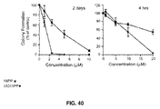
FIG. 40: Comparison of the effects of MOMIPP versus MIPP on colony-forming ability of U251 glioblastoma cells after long term (2 days) or short term (4 hours) treatment.
FIGS. 41A-41H: MOMIPP effectively inhibits the viability of drug-resistant GBM cells and breast cancer cells.
FIG. 42: Effects of MOMIPP on the morphology and viability of multiple human glioblastoma cell lines, including CD133+ GBM stem cells.
FIG. 43: Effects of MOMIPP on colony-forming ability of multiple human GBM cell lines.
FIG. 44: MOMIPP causes vacuolization and inhibits colony formation in cultured GBM stem cells isolated directly from a human tumor.
FIG. 45: Effects of MOMIPP on the morphology of wild-type MCF-7 and doxorubicin-resistant (MCF-7DOX) mammary carcinoma cells.
FIG. 46: Effects of MOMIPP on the short-term viability of wild-type MCF-7 and doxorubicin-resistant (MCF-7DOX) mammary carcinoma cells.
FIG. 47: Effects of MOMIPP on the colony-forming ability of wild-type MCF-7 and doxorubicin-resistant (MCF-7DOX) mammary carcinoma cells.
FIG. 48: Table of summary of SAR studies performed on MIPP (compound 2) and related compounds.
FIG. 49: Scheme showing the synthetic route of 2-substituted indole-based chalcones. Reagents and conditions: (a) THF, sec-butyllithium, −40° C. to −50° C.; (b) −50° C. to −10° C.; (c) TFA/DCM; (d) POCl3, DMF, 0° C.; (e) 4-acetylpyridine, piperidine, MeOH, reflux.
FIG. 50A: Dose response curves for various 2-substituted MOMIPP derivatives. The 2-position substituent is shown on each graph.
FIG. 50B: Effects of MOMIPP and related 2-indolyl-substituted pyridinylpropenones on viability of U251 glioblastoma cells.
FIG. 51: Effects of MOMIPP and related 2-indolyl-substituted pyridinylpropenones on morphology of U251 glioblastoma cells. Cells were observed by phase contrast microscopy on days one and two after the addition of the indicated compounds at 10 μM. Vacuoles appear as clusters of phase-lucent puncta within the cytoplasm of the cells. Rounded cells that are poorly focused in the cultures treated with MOMIPP and compound 107a have detached from the surface of the dish.
FIG. 52: Structural depictions of conformations considered during the computational analysis. The first two letters, “st” or “sc” (for s-trans and s-cis, respectively), refer to the relative position of the formal C2-C3 double bond of the indole and the C═C bone of the connecting chain. The second two letters refer to the relative conformational arrangement across the C═C—C═O system.
FIGS. 53A-53F: Cell morphology (left) and cellular viability (center) resulting from various 2-position substitutions (right) of MOMIPP. A 2-methyl, meta-N was used as the control (FIG. 53F).
FIGS. 54A-54C: Schemes showing synthesis of a microtubule inhibitor containing a 2-trifluoromethylindolyl substitution. This compound is referred to herein as MOFLIPP or compound 124.
FIG. 55: Scheme showing synthesis of microtubule disruptors containing 2-substituted indolyl carboxylate esters.
FIG. 56: Scheme showing synthesis of compound 125, the ethyl carboxylate ester from the scheme shown in FIG. 55. This compound is also referred to herein as MOCCAEEIPP.
FIGS. 57A-57B: Comparison of effects on cells between MOMIPP (FIG. 57A) and MOFLIPP (FIG. 57B). Cells treated with MOMIPP exhibit extensive cytoplasmic vacuolization, whereas cells treated with MOFLIPP round up and detach, with many cells having multiple micronuclei (shown by arrows in FIG. 57B).
FIGS. 58A-58B: Comparison of phase vs. DAPI (nuclear stain) after 24 hours in 10 of MOMIPP (FIG. 58A) or MOFLIPP (FIG. 58B). DMSO was used as a control.
FIGS. 59A-59C: Treatment of U251 glioblastoma cells for one day with MOFLIPP results in depolymerization of microtubules (FIG. 59C). MOMIPP causes vacuolization with microtubules remaining intact (FIG. 59B). DMSO is shown as a control (FIG. 59A). Blue is DAPI (nucleus), and red is tubulin.
FIGS. 60A-60B: Mitotic cell arrest (accumulation of cells in G2/M) is caused by MOFLIPP and compounds 125, but not MOMIPP. Colch=colchicines, a known inhibitor of microtubule assembly and mitosis.
FIG. 61: Compounds MOFLIPP and 125 inhibit tubulin polymerization in a cell-free assay.
FIG. 62: Western blot assay demonstrating that MOFLIPP and compound 125 disrupt tubulin polymerization in intact U251 glioblastoma cells.
FIG. 63: Compound 125 is more cytotoxic than MOMIPP. The graph shows cell viability of U251 cells treated with compound 125 and MOMIPP for 48 hours.
FIG. 64: Table 2, showing a summary of growth inhibition results of compounds generated by Schemes 4A-4G.
FIG. 65: Evaluation of the impact of increasing the indolyl 5-alkoxy chain length on the morphological effects of 2-methylindolyl-pyridinyl-propenones in U251 glioblastoma cells. Phase contrast images of live cells were obtained as described in the Examples. Compounds were added at the concentrations listed at the top of each panel. The control cells shown in the top panel received an equivalent volume of the DMSO vehicle.
FIG. 66: Evaluation of the impact of amine, acetamide, and N—BOC substitutions at the 5-position of the indole ring on the morphological effects of 2-methylindolyl-pyridinyl-propenones. Phase contrast images of live cells were obtained as described in the Examples. Compounds were added at the concentrations listed at the top of each panel.
FIG. 67: Evaluation of the impact of different 2-indolyl substitutions on the morphological effects of 5-methoxyindolyl-pyridinyl-propenones.
FIGS. 68A-68C: Effects of selected indolyl-pyridinyl-propenones on cell cycle distribution in U251 cells. FIG. 68A shows DNA histograms of cells treated with the indicated compounds at 3 μM for 24 h generated by flow cytometry as described in the Examples. FIG. 68B shows cells treated with escalating concentrations of each compound for 24 h and the percentage of cells registering as having less than the G1/G0 DNA content (an indication of non-viable cells) is depicted. FIG. 68C shows the percentage of viable cells in each phase of the cell cycle, determined after gating out the sub-G1/G0 counts. Values in FIG. 68B and FIG. 68C are mean (±S.D.) derived from three separate cultures. The G2/M-phase and the sub-G0/G1 cell populations in the cultures treated with 125 and 310 were significantly increased compared to the DMSO control at all concentrations (p≦0.05).
FIGS. 69A-69B: Effects of additional indolyl-pyridinyl-propenones on cell cycle distribution in U251 cells. DNA histograms of cells treated with the indicated compounds at 3 μM for 24 were generated by flow cytometry as described in the Examples. The results were analyzed to determine the percentage of non-viable cells with sub-G1/G0 DNA content (FIG. 69A), and the percentage of viable cells in each phase of the cell cycle (FIG. 69B). Values are mean (±S.D.) derived from three separate cultures. The G2/M-phase and sub-G0/G1 cell populations in the cultures treated with 124, 120, and colcemid were significantly decreased compared to the DMSO control (p≦0.05).
FIG. 70: Immunofluorescence imaging of tubulin (red fluorescence) in cells treated for 24 h with the methuosis-inducing compound MOMIPP and the 2-indolyl propyl ester 310. The nuclei are visualized with DAPI (blue fluorescence).
FIG. 71: Effects of selected indolyl-pyridinyl-propenones on tubulin polymerization in cultured U251 cells. Cells were treated for 4 h with the indicated compounds at a final concentration of 3 μM and then fractionated under conditions designed to preserve the native polymerization state of the microtubules. The percentage of polymerized versus soluble tubulin was determined by immunoblot analysis as described in the Examples. For each compound, the fraction of tubulin in the polymerized state was expressed as a percentage of the polymerized tubulin determined in a parallel control culture treated with DMSO alone. The results represent the mean (±S.D.) of determinations from three separate experiments. The decreases in the percentage of polymerized tubulin in cells treated with 124, 125, 310, and colchicines (at 3 μM) were significant at p≦0.05.
FIG. 72: Classification of biologically active indolyl-pyridinyl-propenones based on distinct cellular phenotypes elicited by modifications at R1 and R2. Without wishing to be bound by theory, Class 1 compounds interact with one or more Class 1 protein targets to induce perturbations in macropinosome trafficking, resulting in cellular vacuolization without impairment of cell proliferation or viability. Class 2 compounds interact with the same Class 1 targets, but acquire the ability to bind additional Class 2 protein targets that function in metabolic or pro-survival signaling pathways. The combination of effects on Class 1 and Class 2 proteins results in a distinct phenotype characterized by extreme vacuolization and cell death via methuosis. The compounds in Class 3 gain the ability to inhibit proteins involved in microtubule assembly in a concentration range where Class 1 or Class 2 compounds have no effect on tubulin polymerization. Hence, mitotic arrest, cell rounding, and death (it is believed by mitotic catastrophe) predominate as the main features of the Class 3 phenotype. It is also believed that certain Class 3 compounds retain the capacity to interact with Class 1 and Class 2 targets; the early detection of vacuoles prior to microtubule disruption and cell rounding indicates that there is some overlap.
FIG. 73: Growth inhibitor activities of the 2-indole carboxylate compounds, 125 and 310, are substantially reduced by changing the pyridinyl nitrogen from the 4-position to the 3-(meta) position. U251 cells were seeded in 96-well plates. After 24 h, compounds were added at the indicated concentrations. SRB assays were performed after an additional 48 h. Each point represented the mean±SEM of values from four wells.
FIG. 74: Scheme 4A, depicting the synthesis of 5-alkyloxy-2-methylindolyl substituted pyridinylpropenones 402a-402d. Reagents and conditions: (a) diethylsulfate in the case of 404a, or alkyl bromide, K2CO3, 2-butanone, reflux; (b) H2, Pd/C, EtOAc; (c) di-tert-butyl dicarbonate, THF, reflux; (d) 1. −40 to −50° C., sec-butyllithium, THF; 2. −50 to −10° C., 407; 3. TFA/DCM; (e) 1. POCl3, DMF; 2. 1N NaOH; (f) 4-acetylpyridine, piperidine, MeOH, reflux.
FIG. 75: Scheme 4B, depicting the synthesis of 5-amino-2-methylindolyl pyridinyl propenone (402g) and 5-substituted amides 402e and 402f. Reagents and conditions: (a) acetic anhydride, reflux; (b) di-tert-butyldicarbonate, CH3CN, reflux; (c) 1. POCl3, DMF; 2. 1N NaOH; (d) 4-acetylpyridine, MeOH, reflux; (e) 402f, TFA/MeOH.
FIG. 76: Scheme 4C, depicting the synthesis of 5-aminomethylindolyl pyridinyl propenones 402h and 402i. Reagents and conditions: (a) di-tert-butyl dicarbonate, CH3CN, rt; (b) 1. POCl3, DMF; 2. 1N NaOH; (c) 4-acetylpyridine, piperidine, MeOH, reflux; (d) TFA, MeOH, reflux.
FIG. 77: Scheme 4D, depicting the Synthesis of 5-methoxy-2-trifluoromethylindole-pyridinylpropenone 124. Reagents and conditions: (a) 1. THF, sec-butyllithium, −40° C. to −50° C.; 2. Weinreb amide 419, −50° C. to −10° C.; 3. TFA/DCM; (b) 1. POCl3, DMF; 2. 1N NaOH; (c) 4-acetylpyridine, piperidine, MeOH.
FIG. 78: Scheme 4E, depicting the synthesis of the analogous series of substituted alkyl 2-indolylcarboxylate-pyridinyl- propenones 120, 125, 310, 309, as well as the free acid analog 402o. Reagents and conditions: (a) HCl (g), R—OH, reflux; (b) 1. POCl3, DMF; 2. 1N NaOH; (c) 4-acetylpyridine, piperidine, R—OH, reflux; (d) MeOH, 1 N NaOH.
FIG. 79: Scheme 4F, depicting the synthesis of 2-hydroxymethyl-5-methoxyindole-pyridinylpropenone 402p. Reagents and conditions: (a) LiAlH4, THF, rt; (b) acetic anhydride, TEA, CH3CN, rt; (c) 1. POCl3, DMF; 2. 1N NaOH; (d) 4-acetylpyridine, piperidine, MeOH, reflux.
FIG. 80: Scheme 4G, depicting the synthesis of 2-hydroxypropyl-5-methoxyindole-pyridinylpropenone 402q. Reagents and conditions: (a) MnO2, EtOAc, reflux; (b) Ph3PCHCO2C2H5, THF, rt; (c) 1. LiAlH4, THF, 0° C. to rt; 2. H2, Pd/C, EtOAc/MeOH; (d) 430, acetic anhydride, TEA, CH3CN, rt; (e) 1. POCl3, DMF; 2. 1N NaOH; (f) 4-acetylpyridine, piperidine, MeOH, reflux.
DETAILED DESCRIPTION OF THE INVENTION
In general, the terms and phrases used herein have their art-recognized meaning, which can be found by reference to texts, journal references and contexts known to those skilled in the art.
It must be noted that as used herein and in the appended claims, the singular forms “a,” “an,” and “the” include plural reference unless the context clearly dictates otherwise. Thus, for example, reference to “a cell” includes a plurality of such cells and equivalents thereof known to those skilled in the art, and so forth. As well, the terms “a” (or “an”), “one or more” and “at least one” can be used interchangeably herein. It is also to be noted that the terms “comprising,” “including,” and “having” can be used interchangeably. The expression “of any of claims XX-YY” (wherein XX and YY refer to claim numbers) is intended to provide a multiple dependent claim in the alternative form, and in some embodiments is interchangeable with the expression “as in any one of claims XX-YY.”
Unless defined otherwise, all technical and scientific terms used herein have the same meanings as commonly understood by one of ordinary skill in the art. Although any methods and materials similar or equivalent to those described herein can be used in the practice or testing of the present disclosure, the preferred methods and materials are now described.
Every formulation or combination of components described or exemplified herein can be used to practice the materials and methods disclosed herein, unless otherwise stated.
When a group of substituents is disclosed herein, it is understood that all individual members of that group and all subgroups, including any isomers and enantiomers of the group members, are disclosed separately. When a Markush group or other grouping is used herein, all individual members of the group and all combinations and subcombinations possible of the group are intended to be individually included in the disclosure. It is intended that any one or more members of any Markush group or listing provided in the specification can be excluded if desired. When a compound is described herein such that a particular isomer or enantiomer of the compound is not specified, for example, in a formula or in a chemical name, that description is intended to include each isomers and enantiomer of the compound described individual or in any combination. Additionally, unless otherwise specified, all isotopic variants of compounds disclosed herein are intended to be encompassed by the disclosure. For example, it will be understood that any one or more hydrogens in a molecule disclosed can be replaced with deuterium or tritium. Isotopic variants of a molecule are generally useful as standards in assays for the molecule and in chemical and biological research related to the molecule or its use. Specific names of compounds are intended to be exemplary, as it is known that one of ordinary skill in the art can name the same compounds differently.
Every formulation or combination of components described or exemplified herein can be used to practice the materials and methods disclosed herein, unless otherwise stated.
All publications mentioned herein are incorporated herein by reference for the purpose of describing and disclosing components that are described in the publications that might be used in connection with the present disclosure.
The following abbreviations are applicable. Baf-A, bafilomycin A1; DMEM, Dulbecco's modified Eagle medium; DMSO, dimethyl sulfoxide; FBS, fetal bovine serum; GAP, GTPase activating protein; LAMP1, lysosomal-associated membrane protein 1; LC3, microtubule-associated protein light chain 3; MIPP, 3-(2-methyl-1H indol-3-yl)-1-(4-pyridinyl)-2-propen-1-one; MOMIPP, [trans-3-(5-methoxy-2-methyl-1H-indol-3-yl)-1-(4-pyridinyl)-2-propene-1-one]; MTT, 3-(4,5-dimethylthiazol-2-yl)-2,5-diphenyl tetrazolium bromide; SAR, structure-activity relationships; and TMZ, temozolomide.
Thus, as used herein, the term “alkyl” includes straight, branched and cyclic alkyl groups, having up to 10 carbon atoms. An analogous convention applies to other generic terms such as “alkenyl,” “alkynyl,” and the like. Furthermore, as used herein, the terms “alkyl,” “alkenyl,” “alkynyl,” and the like encompass both substituted and unsubstituted groups. In certain embodiments, as used herein, “lower alkyl” is used to indicate those alkyl groups (cyclic, acyclic, substituted, unsubstituted, branched or unbranched) having 1-6 carbon atoms. Non-limiting examples of alkyl groups include n-propyl, isopropyl, and isobutyl groups.
Illustrative aliphatic groups thus include, but are not limited to, for example, methyl, ethyl, n-propyl, isopropyl, cyclopropyl, —CH2-cyclopropyl, allyl, n-butyl, secbutyl, isobutyl, tert-butyl, cyclobutyl, —CH2-cyclobutyl, n-pentyl, sec-pentyl, isopentyl, tert-pentyl, cyclopentyl, —CH2-cyclopentyl-n, hexyl, sec-hexyl, cyclohexyl, —CH2cyclohexyl moieties and the like, which again, can bear one or more substituents. Illustrative alkynyl groups include, but are not limited to, for example propargyl.
“Aryl” refers to an unsaturated aromatic or heteroaromatic carbocyclic group of from 1 to 15 carbon atoms having a single ring (e.g. phenyl) or multiple condensed rings (e.g., naphthyl or anthryl). Preferred aryls include substituted aromatic C6-12 carbocycle; unsubstituted aromatic C1-10 heterocycle; substituted aromatic C1-10 heterocycle; wherein when substituted, the substitution is —XR.
Aralkyl refers to an alkyl connected to an aryl.
Unless otherwise constrained by the definition for the aryl substituent, such aryl groups can optionally be substituted with from 1 to 3 substituents selected from the group consisting of hydroxy, acyl, alkyl, alkoxy, alkenyl, alkynyl, substituted alkyl, substituted alkoxy, substituted alkenyl, substituted alkynyl, amino, aminoacyl, aminocarboxy esters, alkaryl, aryl, aryloxy, carboxyl, carboxylalkyl, acylamino, cyano, halo, nitro, heteroaryl, heterocyclic, oxyacyl, oxyacylamino, thioalkoxy, substituted thioalkoxy, trihalomethyl, mono- and di-alkylamino, mono- and di-(substituted alkyl)amino, mono- and di-arylamino, mono- and di-heteroarylamino, mono- and di-heterocyclic amino, and unsymmetric di-substituted amines having different substituents selected from alkyl, substituted alkyl, aryl, heteroaryl and heterocyclic, and the like.
“Halogen” refers to fluoro, chloro, bromo and iodo and preferably is either chloro or bromo.
“Heterocycle” or “heterocyclic” refers to a saturated or unsaturated group having a single ring or multiple condensed rings, from 1 to 8 carbon atoms and from 1 to 4 hetero atoms selected from nitrogen, sulfur or oxygen within the ring.
Unless otherwise constrained by the definition for the heterocyclic substituent, such heterocyclic groups can be optionally substituted with 1 to 3 substituents selected from the group consisting of hydroxy, acyl, alkyl, alkoxy, alkenyl, alkynyl, substituted alkyl, substituted alkoxy, substituted alkenyl, substituted alkynyl, amino, aminoacyl, aminocarboxy esters, alkaryl, aryl, aryloxy, carboxyl, carboxylalkyl, aminoacyl, cyano, halo, nitro, heteroaryl, heterocyclic, oxyacyl, oxyacylamino, thioalkoxy, substituted thioalkoxy, trihalomethyl, mono- and di-alkylamino, mono- and di-(substituted alkyl)amino, mono- and di-arylamino, mono- and di-heteroarylamino, mono- and di-heterocyclic amino, and unsymmetric di-substituted amines having different substituents selected from alkyl, substituted alkyl, aryl, heteroaryl and heterocyclic, and the like. Such heterocyclic groups can have a single ring or multiple condensed rings. Preferred saturated heterocyclics include morpholino, piperidinyl, and the like; and preferred unsaturated heterocycles include pyridyl and the like.
Examples of heterocycles and heteroaryls include, but are not limited to, pyrrole, imidazole, pyrazole, pyridine, pyrazine, pyrimidine, pyridazine, indolizine, isoindole, indole, indazole, purine, quinolizine, isoquinoline, quinoline, phthalazine, naphthylpyridine, quinoxaline, quinazoline, cinnoline, pteridine, carbazole, carboline, phenanthridine, acridine, phenanthroline, isothiazole, phenazine, isoxazole, phenoxazine, phenothiazine, imidazolidine, imidazoline, piperidine, piperazine, indoline, phthalimide, 1,2,3,4-tetrahydroisoquinoline, 4,5,6,7-tetrahydrobenzo[b]thiophene, thiazole, thiazolidine, thiophene, benzo[b]thiophene, morpholino, piperidinyl, pyrrolidine, tetrahydrofuranyl, and the like.
It will be appreciated by one of ordinary skill in the art that asymmetric centers can exist in the compounds of the present disclosure. Thus, the compounds and pharmaceutical compositions thereof can be in the form of an individual enantiomer, diastereomer or geometric isomer, or can be in the form of a mixture of stereoisomers. It is to be understood that the present disclosure encompasses all possible isomers such as geometric isomers, optical isomers, stereoisomers and tautomers based on an asymmetric carbon, which can occur in the structures of the compounds, and mixtures of such isomers and compositions comprising those compounds, and is not limited to the specific stereochemistry shown for the compounds disclosed in the present specification. It will be further appreciated that the absolute stereochemistry of some of the compounds recited in the Exemplification herein cannot have been determined, and that when a stereochemistry was assigned for those compounds it is meant to be tentative and to indicate that a set of diastereomers exists for those compounds and/or that a diastereomer was isolated in pure form. Furthermore, it will be appreciated that certain of the compounds disclosed herein contain one or more double bonds and these double bonds can be either Z or E, unless otherwise indicated. In certain embodiments, the compounds of the present disclosure are enantiopure compounds. In certain other embodiments, mixtures of stereoisomers or diastereomers are provided.
Additionally, the present disclosure provides pharmaceutically acceptable derivatives of the active compounds, and methods of treating a subject using these compounds, pharmaceutical compositions thereof, or either of these in combination with one or more additional therapeutic agents. The phrase, “pharmaceutically acceptable derivative,” as used herein, denotes any pharmaceutically acceptable salt, ester, or salt of such ester, of such compound, or any other adduct or derivative which, upon administration to a patient, is capable of providing (directly or indirectly) a compound as otherwise described herein, or a metabolite or residue thereof.
Furthermore, it will be appreciated by one of ordinary skill in the art that the synthetic methods, as described herein, utilize a variety of protecting groups. By the term “protecting group,” has used herein, it is meant that a particular functional moiety, e.g., O, S, or N, is temporarily blocked so that a reaction can be carried out selectively at another reactive site in a multifunctional compound. In preferred embodiments, a protecting group reacts selectively in good yield to give a protected substrate that is stable to the projected reactions; the protecting group must be selectively removed in good yield by readily available, preferably nontoxic reagents that do not attack the other functional groups; the protecting group forms an easily separable derivative (more preferably without the generation of new stereogenic centers); and the protecting group has a minimum of additional functionality to avoid further sites of reaction.
For example, oxygen, sulfur, nitrogen and carbon protecting groups can be utilized. Certain exemplary oxygen protecting groups include, but are not limited to methyl ethers, substituted methyl ethers (e.g., MOM (methoxymethyl ether), MTM (methylthiomethyl ether), BOM (benzyloxymethyl ether), PMBM (p-methoxybenzyloxymethyl ether), to name a few), substituted ethyl ethers, substituted benzyl ethers, silyl ethers (e.g., TMS (trimethylsilyl ether), TES (triethylsilylether), TIPS (triisopropylsilyl ether), TBDMS (t-butyldimethylsilyl ether), tribenzyl silyl ether, TBDPS (t-butyldiphenyl silyl ether), to name a few), esters (e.g., formate, acetate, benzoate (Bz), trifluoroacetate, dichloroacetate, to name a few), carbonates, cyclic acetals and ketals.
Exemplary nitrogen protecting groups include, but are not limited to, carbamates (including methyl, ethyl and substituted ethyl carbamates (e.g., Troc), to name a few) amides, cyclic imide derivatives, N-Alkyl and N-Aryl amines, inline derivatives, and enamine derivatives, to name a few. As will be appreciated by those of ordinary skill in the art, a variety of additional equivalent protecting groups can be utilized in accordance with the present disclosure.
It will be appreciated that the compounds, as described herein, can be substituted with any number of substituents or functional moieties. In general, the term “substituted” whether preceded by the term “optionally” or not, and substituents contained in formulas herein, refer to the replacement of hydrogen radicals in a given structure with the radical of a specified substituent. When more than one position in any given structure can be substituted with more than one substituent selected from a specified group, the substituent can be either the same or different at every position. As used herein, the term “substituted” is contemplated to include all permissible substituents of organic compounds. In a broad aspect, the permissible substituents include acyclic and cyclic, branched and unbranched, carbocyclic and heterocyclic, aromatic and nonaromatic substituents of organic compounds. For purposes of this disclosure, heteroatoms such as nitrogen can have hydrogen substituents and/or any permissible substituents of organic compounds described herein which satisfy the valencies of the heteroatoms.
Furthermore, this disclosure is not intended to be limited in any manner by the permissible substituents of organic compounds. The term “stable,” as used herein, preferably refers to compounds which possess stability sufficient to allow manufacture and which maintain the integrity of the compound for a sufficient period of time to be detected and preferably for a sufficient period of time to be useful for the purposes detailed herein.
As used herein, the term “patient” is intended to include living organisms in which certain conditions as described herein can occur. Examples include humans, monkeys, cows, sheep, goats, dogs, cats, mice, rats, and transgenic species thereof. In a preferred embodiment, the patient is a primate. In an even more preferred embodiment, the primate is a human. Other examples of subjects include experimental animals such as mice, rats, dogs, guinea pigs, cats, goats, sheep, pigs, and cows. The experimental animal can be an animal model for a disorder, e.g., a transgenic mouse with cancer.
As used herein, the term “IC50” refers to an inhibitory dose which is 50% of the maximum response obtained.
GENERAL DESCRIPTION
Disclosed herein is a specific chalcone-like molecule, 3-(2-methyl-1H-indol-3-yl)-1-(4-pyridinyl)-2-propen-1-one (MIPP) that is shown to induce cell death with the hallmarks of methuosis. MIPP causes rapid accumulation of vacuoles that can be labeled with extracellular fluid phase tracers. Vacuolization can be blocked by an inhibitor of the vacuolar-type H+-ATPase, bafilomycin A1, and by the cholesterol-interacting compound, filipin, consistent with the endosomal origin of the vacuoles. Although the vacuoles acquire some characteristics of late endosomes, they remain distinct from lysosomal and autophagosomal compartments, showing a block at the late endosome/lysosome boundary.
MIPP targets steps in the endosomal trafficking pathway mediated by Rab5 and Rab7, as shown by changes in the activation states of these GTPases. These effects are specific, as other GTPases (Rac1, Arf6) are unaffected by the compound. Cells treated with MIPP lose viability within 2-3 days, but their nuclei show no apoptotic changes Inhibition of caspase activity does not protect the cells, consistent with a non-apoptotic death mechanism. A U251 glioblastoma line selected for temozolomide resistance showed sensitivity to MIPP-induced methuosis that was comparable to the parental cell line. MIPP can serve as a prototype for new drugs that can be used to induce non-apoptotic death in cancers that have become refractory to agents that work through DNA damage and apoptotic mechanisms.
Glioblastomas are highly aggressive brain tumors that almost always recur after surgery. Treating these tumors is extremely challenging because the residual cells are highly invasive and they typically harbor genetic mutations that decrease their sensitivity to apoptosis induced by radiation or DNA alkylating agents. Thus, the discovery of cell death mechanisms that do not depend on activation of the classical mitochondrial or death receptor-mediated apoptotic pathways presents new opportunities to treat these devastating tumors. A non-conventional form of cell death termed methuosis can be triggered by ectopic expression of constitutively activated Ras in glioblastoma and other cancer cell lines. Methuosis occurs by displacement of much of the cytosolic space with vacuoles derived from macropinosomes. As the vacuoles begin to fuse and increase in size, the cell decreases its metabolism and eventually the cell membrane ruptures, killing the cell.
In this disclosure, a small molecule termed MIPP has been identified, which induces the hallmark cytopathological features of methuosis. Shortly after being exposed to MIPP, glioblastoma cells exhibit influx of multiple macropinocytotic vesicles. In the time-lapse studies, the latter can be seen undergoing fusion events to form larger vacuoles. As the vacuoles accumulate, they rapidly acquire some characteristics of late endosomes (Rab7, LAMP1), but they do not merge with lysosomal compartments. Ultimately, the displacement of much of the cytoplasmic space by the vacuoles is accompanied by a decline in metabolic activity and a necrosis-like rupture of the cell. Consistent with a non-apoptotic mechanism, these changes cannot be prevented by caspase inhibitors, and nuclear chromatin condensation typical of apoptosis is not observed. Further, in this disclosure it is shown that glioblastoma cells selected for resistance to the front-line chemotherapeutic drug, temozolomide, were susceptible to MIPP-induced methuosis by comparing doxorubicin-resistant versus non-resistant MCF-7 breast cancer cells treated with a MIPP analog. It is disclosed herein that MIPP can serve as a prototype for development of drugs that can be used to trigger death by methuosis in drug-resistant cancers.
Further disclosed herein is the ability of compounds 1 (BIPP) and 2 (MIPP) to induce methuosis. A similarity search of chemical databases was performed using the Chembridge Hit2Lead search engine, which provided compounds 3-8, as shown in FIG. 29, for analysis of whether certain similarly-structured compounds can induce methuosis. When testing these compounds in U251 glioblastoma cells at 10 μM, all compounds were completely inactive in inducing methuosis and limiting cell growth.
In certain embodiments, based on the data of compounds 3-8, the 3-indole-1-pyridinyl-2-propen-1-one scaffold was used to induce methuosis. Compounds 3-8 all possess this core, yet were inactive. The inactivity of compound 8 is most revealing when compared to active compound 1, indicating that addition of a para-methoxy in place of the pyridine nitrogen rendered the compound inactive.
Analogs based on the α,β-unsaturated ketone core were designed from Claisen-Schmidt condensations between indole-3-carboxaldehydes and aromatic ketones. Such aldol condensations are known to proceed well employing secondary amines as catalysts. Initially, the orientation of the pyridine nitrogen was investigated. Condensation of various acetyl-pyridines or acetophenone with indole-3-carboxaldehyde (FIG. 30) yielded compounds 9-12 (FIGS. 36, 48). The activities of these compounds were compared to compound 2 (i.e., MIPP), at a concentration of 10 μM using three criteria: 1) morphological vacuolization of live cells assessed by phase contrast microscopy at 24 h and 48 h, 2) cell viability, assessed at a 48 h end point by MTT assay, with fresh compound added after the first 24 h, and 3) colony forming assays (two week end-point) performed on cells exposed to the compounds for 48 h. The results of this analysis (FIGS. 36, 48) showed that a para-nitrogen orientation of the pyridine ring is a key feature required for activity. The meta and ortho analogs 10, 11, as well as the acetophenone analog 9 were all relatively ineffective at inducing methuosis compared with MIPP. In contrast, removal of the 2-methyl from the indole ring of MIPP reduced but did not eliminate activity.
Next, the consequences of functionalizing the 5-position of the indole ring with the suitable 5-methoxy and 5-benzyloxy indole-3-carboxaldehydes, compounds 13 and 14 was explored. The 5-methoxy compound 13 was demethylated with BBr3, affording the 5-OH compound 15 (FIG. 31). As shown in FIG. 36 and FIG. 48, the activities of compounds 13 and 14 were similar to compound 2 (MIPP) when compared by colony-forming assay and cell morphology, although they were not as effective in the short-term viability assay. In contrast, addition of the OH to the 5-position greatly reduced activity of the compound in all of the assays. To verify that the location of the pyridine nitrogen was still crucial in the 5-substituted compounds, analogs 16 and 17 were generated. Loss of activity confirmed the necessity of the para-nitrogen (FIGS. 36, 48).
After comparing compounds 13 and 14 to MIPP, which showed that modifications at the 5- and 2-positions of the indole ring both affect the activity of these compounds, a 2-methyl-5-methoxy analog was generated from available 2-methyl-5-methoxy-indole. Compound 19 was synthesized by a Vilsmeier-Haack formylation, followed by coupling with 4-acetyl-pyridine (FIG. 32). The activity of this compound exceeded that of compound 2 in both the MTT viability assay and the colony formation assay (FIGS. 36, 48).
The addition of the methyl at the indole's 2 position in compound 19 gave added potency versus compound 13, possessing only the 5-methoxy. Methylation of the indole-nitrogen of compound 13 with NaH/CH3I in DMF (FIG. 33) to yield 20, produced a compound that induced some cytoplasmic vacuolization but had modest effects on cell viability. Thus, after comparing compounds 11, 19 and 20, it is shown that optimal activity is achieved when the 1- and 2-position of the indole are occupied by H and methyl, respectively. The 5-OH compound 21, made by BBr3 demethylation of compound 19 (FIG. 34), showed a marked reduction in activity compared to the 5-methoxy compound 19, consistent with the detrimental effect of the 5-OH in compound 15.
Further disclosed herein are the effects of modifying the 5-position of the indole with a group that would add polarity at physiological pH. An analog of compound 14 was designed, that added charge and also included a methyl at the indole's 2-position. No appreciable amount of product was produced by direct alkylation of compound 21. Multiple bases were screened with varying pKa's, from K2CO3 and Cs2CO3, as well as triethylamine, DBU, TMG, and NaH. In all reactions, alkylation at the pyridine nitrogen was observed.
Stronger bases such as TMG and NaH, even using one equivalent, produced appreciable indole-N-alkylation as well as pyridine and indole-OH alkylation. The yield of singly alkylated product at the 5-indole position was negligible. Thus, a new route was designed by functionalizing the indole before introduction of the pyridine moiety (FIG. 35). Available 2-methyl-5-methoxy-indole was demethylated with BBr3 providing compound 22, followed by alkylation with 4-methyl-(bromomethyl)-benzoate under phase-transfer conditions to afford the singly O-alkylated product compound 23. After formylation with POCl3/DMF, compounds 24 and 25 were selectively made. Workup in mild base (NaHCO3) produced the ester compound 24, while workup in 5N NaOH gave acid compound 25. Condensations with 4-acetyl-pyridines provided the corresponding targets compounds 26 and 27. However, neither compound 26 nor compound 27 induced methuosis (FIGS. 36, 48).
Superior biological activity of MOMIPP versus MIPP was confirmed in studies with U251 glioblastoma cells, using MTT viability assays, cell growth assays, morphological assessment, and colony forming assays to compare the two compounds. FIGS. 37A-37D show the dose-response curves for the effects of the two drugs on cell viability. Each compound was added at the mentioned concentration for two days, with medium and compounds replenished after the first day. The relative IC50 for MOMIPP was 1.9 μM, versus 4.8 μM for MIPP.
To obtain a measure of the comparative duration of activity for each compound, their effects on cell growth and survival were assessed by counting the number of cells in parallel cultures treated for three consecutive days with 2.5 μM, 5 μM, or 10 μM compound (FIG. 38). Unlike the viability studies, in these experiments the compounds were added at the beginning of the study and were not replenished for the duration. Under these conditions, MOMIPP was shown to be more effective than MIPP in reducing cell growth and viability. The reduction of cell number in the cultures treated with MOMIPP coincided with massive early vacuolization of the cells and loss of nonviable cells from the substratum (FIG. 39).
In contrast, the cells treated with MIPP initially underwent vacuolization on days 1 and 2, but tended to recover, especially at the lower concentrations of the compound (FIG. 39). These studies show that a single application of MOMIPP has a more sustained effect than MIPP on cell morphology and cell viability. The difference between the two compounds was underscored when colony-forming assays were used to evaluate proliferative capacity and long-term cell viability (FIG. 40). MOMIPP was shown to be more effective than MIPP in reducing colony formation when cells were treated for 2 days (FIG. 40).
In yet another embodiment, it is shown that when treatment was shortened to just 4 h, MOMIPP was still more effective than MIPP, but higher concentrations of both compounds were required to reduce colony formation (FIG. 40).
The methuosis-inducing compounds are effective for targeting temozolomide (TMZ)-resistant glioblastoma. As shown in FIGS. 41A-41B, both MIPP and MOMIPP induced methuosis and reduced cell viability in the TMZ-resistant cells, but the efficacy of MOMIPP was shown to be superior to MIPP.
The ability of MOMIPP to kill drug-resistant tumor cells by methuosis extends beyond glioblastoma. For example, comparable levels of MOMIPP-induced methuosis was observed in parental and doxorubicin-resistant MCF-7 breast cancer cells, as shown in FIGS. 41C-41D, 45 and 46.
The methuosis-inducing activity of MOMIPP was further shown to extend to multiple human GBM cell lines with different genetic backgrounds, as shown in FIGS. 42-43.
The methuosis-inducing activity of MOMIPP was also shown to extend to human CD133+ GBM stem cell populations, as shown in FIGS. 42 and 44.
The mechanism by which MIPP causes cellular vacuolization and death can involve three steps: 1) a transient increase in macropinocytotic activity, 2) rapid homotypic fusion of the macropinosome-derived vesicles to form vacuoles that acquire late endosomal characteristics, and 3) failure of the resulting vacuoles to recycle or fuse with lysosomes, leading to their progressive accumulation. Similar steps can be involved in the death mechanism triggered by MOMIPP, as shown in FIGS. 37-44.
Ras-induced methuosis depends on activation of Rac1 and reciprocal inactivation of Arf6, leading to hyperstimulation of macropinocytosis and defects in macropinosome recycling and downstream trafficking. However, the measurements of the activation states of Rac1 and Arf6 did not reveal any changes in MIPP treated cells. On the other hand, MIPP is shown to have profound effects on the activities of Rab5 and Rab7, decreasing the amount of active Rab5 and increasing the amount of Rab7. This shows that the compound operates directly on the machinery that regulates the trafficking and fusion of endosomal vesicles.
Rab5 controls receptor-mediated (clathrin-dependent) endosome formation, homotypic fusion, and delivery of early endosomes to the sorting/recycling compartment. The latter serves as a “way station” from which early endosomes can either recycle to the cell surface or progress to late endosomes in a multi-step process involving Rab5-Rab7 conversion.
While not wishing to be bound by theory, it is further disclosed herein that the main consequence of the MIPP-induced decrease in active Rab5 can occur after the initial formation of macropinosomes. That is, the nascent vesicles formed during the initial burst of macropinocytotic activity fail to fuse with EEA1-positive early endosomes and thus do not enter the sorting/recycling compartment. Instead, they immediately acquire the capability to recruit LAMP1 and Rab7. The resulting vesicles then undergo abnormal homotypic fusions to form progressively larger vacuoles in a manner that requires activity of the H+-ATPase.
Despite their ability to undergo homotypic fusion, the apparent inability of the Rab7-positive vacuoles to merge with lysosomes showing that they can lack some of the key protein components of complexes that are required for heterotypic tethering and fusion of late endosomes with lysosomes and autophagosomes (e.g., HOPS, trans-SNARE). This can explain the progressive accumulation of the vacuoles and the overall increase in the amount of active Rab7, as GAP-mediated GTP-hydrolysis that would normally coincide with endosome-lysosome fusion fails to occur.
Based on the examination of several cancer cell lines, it is shown that the ability of MIPP to induce methuosis extends beyond glioblastoma. It is further disclosed herein that MIPP can also trigger vacuolization and a modest reduction of cell proliferation/viability in normal proliferating cells, which can lead to delivery of such compounds to tumors.
By way of non-limiting examples, compounds effective at inducing methuosis include compounds of Formula VIIA:
wherein O can be attached at any one of positions 5, 6, or 7; R is alkyl having 1 to 3 linear carbon atoms; R1 is alkyl having 1 to 2 carbon atoms, or alkyl having 3 linear carbon atoms further substituted at the terminus with OR2 or NHR2 where R2 is either H, alkyl, aryl, acylalkyl, or acylaryl; and Ar is a 4-pyridyl moiety. In certain embodiments, O is attached at the 5-position. In one example, R and R1 are each methyl.
Compounds that Induce Vacuolization but not Cell Death
A hallmark of cells undergoing methuosis is extensive cytoplasmic vacuolization. The vacuoles arise from the merger of fluid-filled macropinosomes with clathrin-independent endosomal compartments under conditions where endosomal recycling and/or lysosomal trafficking are defective. Consequently, large vacuoles accumulate until they fill much of the cytoplasmic space. The vacuolated cells eventually lose membrane integrity, detach from the substratum, and die without the typical features of apoptosis. Methuosis is caspase-independent because it can occur in the presence of caspase inhibitors.
As described above, MOMIPP induces methuosis in cultured glioblastoma cells at low micromolar concentrations. The indolyl and pyridinyl moieties demonstrate a high degree of structural specificity for induction of methuosis, as small modifications lead to compounds that are inactive (as illustrated in FIG. 48). For example, switching the position of the substituent from the para- to either the meta- or ortho-positions abolishes the induction of vacuolization and cell death. In general, certain structural modifications that prevent vacuolization also appear to minimize the loss of cell viability, indicating that vacuolization is a key factor in the cell death mechanism. This, coupled with the reversibility of the effects of MOMIPP upon removal of the drug from the test medium, indicate that MOMIPP exerts its biological activity by interacting with discrete protein targets in a reversible manner.
The SAR studies described above revealed that when a simple methyl group is added to the 2-position of the indole ring, there is a significant increase in methuosis-inducing activity compared to the compound bearing a hydrogen atom at this location. Therefore, the same manipulation was applied to the meta-pyridine analogue to confirm that the latter isomer still attenuates potency and that the combination can provide for a very weakly active control that is otherwise identical in structure to MOMIPP. In addition, the 2-position was further modified with hydrocarbons having increased length and branching in order to further explore the effects upon vacuolization and viability within U251 glioblastoma cells.
MOMIPP and its 2-methyl isomer were prepared from commercially available 5-methoxy-2-methylindole by formylation followed by condensations with 4-acetylpyridine and 3-acetylpyridine, respectively. MOMIPP's des-methyl predecessor 13 was prepared from commercially available 5-methoxyindole and 4-acetylpyridine. Alternatively, the series of probes having larger alkyl groups in the 2-position required synthesizing the indole-system for each of the desired substituents. Thus, the synthesis of compounds 107a-d (FIG. 49) began with 4-methoxy-2-methylaniline to which the addition of a Boc-group via di-tert-butyldicarbonate in refluxing THF was used to protect the amine 103. Acylation of the 2-methyl group was accomplished using sec-butyllithium as a base. The resulting dilithio species enabled regioselective acylation with various synthesized Weinreb amides 102a-d to produce intermediates 104. Derivatives of 104 were washed with 1 N HCl to neutralize base and then used directly in the next step. A more rigorous purification was not necessary as spontaneous cyclization was observed to occur slowly under neutral and ambient conditions. Removal of the N-Boc protecting group accompanied by controlled cyclization was performed in one-pot fashion using excess TFA to provide the key 2-substituted 5-methoxyindoles 105a-d. Formylation reactions were performed using Vilsmeier-Haack acylation conditions yielding aldehydes 106a-d, which were conveniently isolated as precipitates from basic solutions after treatment with 1 N NaOH. Claisen-Schmidt condensation of the aldehydes with 4-acetylpyridine yielded the series of 2-substituted indole-based pyridinylpropenones 107a-d.
Compounds 107a-d, MOMIPP, meta-MOMIPP, and the des-methyl MOMIPP predecessor were tested alongside a DMSO vehicle control in U251 glioblastoma cells using a sulforhodamine B (SRB) assay to assess their growth inhibition properties after two-day exposures of each test agent across a concentration range from 0 to 20 μM. Fifty-percent inhibition (GI50) values were derived from the resulting dose-response curves (FIG. 50) and are recorded in Table 1.
| TABLE 1 |
| |
| Cell growth, viability, and computational chemistry results. |
| cmpd |
substa |
GI50 b |
% viablec |
stscd |
ststd |
scscd |
scstd |
| |
| DMSOe |
NAf |
NA |
96.7 ± 0.6 |
NA |
NA |
NA |
NA |
| 13 |
H |
18.6 |
91.2 ± 0.7 |
1.1 |
2.0 |
0.0 |
0.5 |
| MOMIPP |
Me |
2.1g |
38.1 ± 3.7 |
0.0 |
0.7 |
0.5 |
1.1 |
| 107a |
Et |
3.2g |
34.6 ± 1.1 |
0.0 |
0.7 |
0.4 |
1.0 |
| 107b |
n-Pr |
15.3 |
83.9 ± 2.3 |
0.0 |
0.6 |
0.1 |
1.0 |
| 107c |
i-Pr |
>20 |
78.8 ± 1.1 |
0.0 |
1.3 |
1.1 |
1.9 |
| 107d |
i-Bu |
16.6 |
87.4 ± 1.5 |
0.0 |
0.5 |
0.7 |
1.5 |
| meta- |
Me |
16.9 |
96.2 ± 1.3 |
NA |
NA |
NA |
NA |
| MOMIPP |
| |
| aSubstitutents at the indole's 2-position. |
| bConcentration of test agent in μM causing 50% inhibition of cell growth compared to DMSO control. |
| cPercent of viable cells (mean ± SD), based on Trypan blue dye exclusion assays performed on three separate cultures teated with each compound at 10 μM for 2 days. |
| dEnergies in kcal/mol for each of the four conformations studied by computational chemistry. |
| eVehicle control for delivery of test agents via DMSO. |
| fNot applicable. |
| gDose-response curves for the Me and Et derivates were repeated six more times. The means (±SD) for all seven determinations were as follows: Me, 2.5 ± 0.6; Et, 2.8 ± 0.5. The difference was not significant at p < 0.2 (Student's t test). |
| hThis compound has a 3-pyridyl arrangement, while all others have the 4-pyridyl arrangement shown to be important for activity. |
The GI50 for the ethyl substitution, 107a, was not statistically different from the MOMIPP. In contrast, each of the other substitutions on the indole ring had a negative impact on growth inhibitory activity, with GI50 values increasing to >15 μM. The difference between MOMIPP and compound 107a compared to the other 2-substituted indole compounds was even more striking when evaluated by morpholigocal and cell viability criteria. The typical progression of methuosis involves the initial formation of vacuoles, which can be detected as early as 2-3 hours after addition of MOMIPP. Over the next 24-48 hours, the progressive accumulation and enlargement of vacuoles is followed by cell rounding and detachment, with the majority of the detached cells exhibiting signs of disrupted membrane integrity. In the previously described SAR studies, the compounds that most effectively triggered vacuolization also caused cell death, while those that failed to induce vacuoles were not cytotoxic. This led to the concept that accumulation of vacuoles is an important contributing factor in the methuosis death program. To evaluate the relationship between vacuolization and cell death using the current series of compounds, cells were examined by phase-contrast microscopy after one and two days of treatment (FIG. 51). At 10 μM, the ethyl derivative 107a produced morphological effects indistinguishable from those observed with the same concentration of MOMIPP. After one day, the cells were spares and heavily vacuolated, and by day two, many of the vacuolated cells had detached and lost membrane integrity. In contrast, cells treated with meta-MOMIPP, the 3-pyridinyl isomer of MOMIPP, did not accumulate vacuoles and resembled the vehicle control cells in their ability to proliferate to high density by day two. Cells treated with compounds containing the hydrogen (13), propyl (107b), isopropyl (107c), and isobutyl (107d) substituents at the 2-position of the indole ring were extensively vacuolated, but surprisingly, they remained attached and continued to increase in density. Trypan blue dye-exclusion assays clearly demonstrated that the progressive rounding and detachment of the vacuolated cells treated with MOMIPP and 107a reflected a substantial loss of cell viability (Table 1). However, viability was only minimally affected in the vacuolated cells treated with 13, 107b, 107c, and 107d.
The initial increase in cytotoxicity observed when substituting from H to either Me or Et, followed by the abrupt falloff thereafter when substituting to propyl, isopropyl, and isobutyl, is unexpected and intriguing. Without wishing to be bound by theory, it is believed such a SAR trend are is associated with switching the H to a more electron-donating and lipophilic alkyl group. Possible consequences from substitution include (i) a subtle but potentially broad-ranging enhancement in the electronic features of the central pharmacophore such as afavorable impact upon the structurally embedded double-vinylogous amide; (ii) an enhanced interaction with a hydrophobic pocket within this distinct locale of the target protein; or (iii) some combination of these factors. However, for all of these, it is clear that their benefit must be achieved by encompassing only a compact region of space before the presence of additional bulk becomes deleterious to potency. This requirement could arise from a limitation derived from either (iv) the existence of a tight steric boundary within this distinct pocket of the target, or (v) an influence upon the molecule itself in terms of sterically perturbing a conformational arrangement that is preferred for overall target interaction. Computational studies have been conducted on factor (v).
Table 1 lists the energies from molecular-mechanics calculations for the various conformations that can be adopted by MOMIPP and compounds 13 and 107a-d. An implicit solvent model was used to simulate the low polarity environment within a protein's ligand-binding pocket. Since NMR data indicate that the double bond located α,β to the ketone possesses trans-stereochemistry (discernible by higher coupling constants) in all compounds, only this arrangement was considered. To address the single bonds, these compounds can be treated as double rotors wherein there is rotation between the indole ring and the connecting chain, and again about the C—C bond in the C═C—C═O moiety. The conformational possibilities that can be derived from these arrangements and their appropriate nomenclature are depicted in FIG. 52. The first two letters, “st” or “sc” (s-trans and s-cis, respectively) refer to the relative position of the formal C2-C3 double bond of the indole and the C═C bond of the connecting chain. The second two letters refer to the relative conformational arrangement across the C═C—C═O system. From this molecular construct, the pyridine ring can be expected to adopt its most favorable position when optimizing the corresponding stsc, stst, scsc, and scst conformations. For the case of the 2-propyl substituent, a gauche conformation with respect to the C2 atom of the indole ring was accepted at the onset of the geometry optimization.
Only for the case of the smallest 2-substituent, namely, compound 13 having an H atom, is the most favorable conformation scsc. For MOMIPP and all of the other compounds 107a-d, the stsc conformer is the most stable. This conformation maintains an s-trans relationship between the C2-C3 bond of pyrrole and the C═C bond of the connecting chain, and an s-cis conformation across the C═C—C═O moiety. Apparently, the increasing size of the 2-substituent influences the connecting chain to turn farther away from the added bulk. Also notable is that the indole-CCCO substructure moiety is generally not coplanar. Torsion angles about the rotational bonds deviate by up to 30° from a fully coplanar system corresponding to 0° for sc and 180° for st, respectively. The C═C—C═O moiety is s-cis in all the sxsc (x=t or c) preferred structures, and the pyridine ring is generally out of the O═C—C4 (pyridine) plane by about 30°. While it is tempting to speculate that the noted conversion from an scsc to an stsc conformational preference when going from H to Me or E may represent yet another possible explanation for at least the initial portion of the SAR trend discussed above, the actual energy differences are rather small. Thus, the more appropriate conclusion to draw is that the modeling studies predict small, up to at most 2 kcal/mol, relative conformer energies across all of these arrangements. When taken within the context of simultaneously interacting with the ligand-binding region on a protein surface, such small energy differences likely would not prevent rotations in order to adopt any conformation (even if not of the lowest relative ligand energy) that could most favorably bind to the protein. This indicates that the second part of the SAR trend, wherein there is a falloff in cytotoxicity with increasing size of the 2-substituent, is likely not due to some conformational effect and, instead, may result from a steric boundary that resides in this specific locale of the ligand-binding pocket.
These results confirm that switching the pyridine N from the 4-position within MOMIPP to the 3-position so as to provide meta-MOMIPP eliminates the ability of the compound to induce vacuolization and substantially reduces potency in growth inhibition and cytotoxicity assays. This is important because it indicates that the compounds that elicit vacuolization and methuotic cell death are acting through one or more distinct protein targets that have very specific structural requirements for interaction with their ligands. In this regard, the SAR associated with growth inhibition and cytotoxicity indicates that when MOMIPP is bound to its protein target(s), there may be a pocket near the 2-position of the indole ring that can tolerate small groups capable of favorably influencing the electronics of the parent ligand or directly enhancing interactions with the target protein. While the addition of steric bulk to the 2-position can affect the preferred conformations adopted by the connecting chain, these effects are small and likely to be relevant only when the compounds are dissolved in nonpolar solvents. Thus, when considered within the context of the energies typically encountered when ligands associate with protein targets, all conformational possibilities are likely to be available to analogues having various levels of steric bulk at the 2-position, at least up to that of an isobutyl group. Taken together, the overall SAR indicates that maintain methuosis-inducing properties, the 2-position can be substituted with small substituents that have a range of electronic, lipophilic, and hydrogen bonding properties.
Without wishing to be bound by theory, the surprising dissociation of vacuolization and cell death revealed by this series of compounds can be explained by several possible explanations. One is that the vacuoles induced by the propyl, isopropyl, and isobutyl compounds are functionally different from those induced by the Me- or Et-substituted compounds, so that their accumulation has a less severe impact on intracellular vesicular trafficking and cellular metabolism. Alternatively, the cytotoxic compounds MOMIPP and 107a may have unique pleiotrophic effects on specific targets beyond those that lead to endosomal vacuolization. In this scenario, endosomal vacuolization remains an essential contributing factor to methuosis but must be combined with other cellular insults in order to cause eventual metabolic collapse and cell death.
The 2-indolyl position of MOMIPP offers a chemically accessible position that can be functionalized or substituted to alter the activity of the compound. In accordance with the findings described above, provided herein is a class of compounds that have a 2-indole group bulkier than ethyl. Certain of these compounds induce vacuolization without causing cell death. In certain embodiments, these compounds have the structural formula of Formula VIIB:
wherein O is attached at any one of
positions 4, 5, 6, or 7; R is alkyl having 1 to 6 carbon atoms either linear or branched; R
1 is alkyl having 2 carbon atoms further substituted at its terminus with a (CO)O-alkyl, (CO)O-aryl, or (CO)O-aralkyl moiety, or is alkyl having 3 to 6 carbon atoms either linear or branched, aryl, heteroaryl, aralkyl, or heteroaralkyl having 6 to 12 carbon atoms; and Ar is aryl or heteroaryl. In certain embodiments, 0 is attached at the 5-position. In a particular example, R is methyl, R
1 is n-propyl, and Ar is 4-pyridyl.
In certain embodiments, these compounds have the structural formula of Formula IIIA:
wherein R is an alkyl selected from the group consisting of n-propyl, isopropyl, and isobutyl. Though these alkyl groups are specified, it is to be understood that R can be other electron-donating groups, and such compounds are entirely encompassed within the present disclosure. By way of non-limiting examples, suitable other electron donating groups may include, but are not limited to, alkoxy groups, vinyl groups, complex-bound metals, amino groups, monoalkylamino groups, and dialkylamino groups. In embodiments wherein R is ethyl, the compound is about as potent against cancer cells as MOMIPP and causes both vacuolization and cell death. Increasing the size of the 2-indolyl substituent substantially reduces growth inhibitory activity and cytotoxicity, but does not prevent cell vacuolization. Without wishing to be bound by theory, it is believed that these effects are not due to steric-driven conformational effects. The unexpected uncoupling of vacuolization and cell death indicates that the relationship between endosomal perturbations and methuotic cell death is more complex than previously realized.
Because certain of these compounds do not cause cell death, they may be useful as antiprotozoal agents, allowing for the organism to be killed by vacuolization but not killing the host through cell death. Thus, the compounds that induce vacuolization but do not cause cell death may be useful for treating protozoal diseases caused by organisms such as trypanosomes or plasmodium. The compounds that induce vacuolization but do not cause cell death may also be useful for the treatment of various cancers either alone or when combined with other compounds that do cause cell death.
Microtubular Inhibitor Compounds that Induce Cell Death but not Vacuolization
Specific chemical modifications of the MOMIPP structure can substantially alter the biological activity of the compounds. In particular, provided herein are compounds having electron-withdrawing groups at the 2-indolyl position. In certain embodiments, these compounds have the structural formula of Formula VIIC:
wherein O is attached at any of
positions 4, 5, 6, or 7; R is alkyl having 1 to 6 carbons either linear or branched; R
1 is an electron withdrawing group selected from halide, fluorinated alkyl, nitro, cyano, and (CO)R
2 or (CO)NHR
2 when R
2 is either alkyl from 1 to 6 linear carbon atoms, or aryl, heteroaryl, or aralkyl or heteroalkyl having 6 to 12 carbon atoms; and Ar is aryl or heteroaryl. In certain embodiments, O is attached at the 5-position. In one example, R is methyl, R
1 is trifluoromethyl, and Ar is 4-pyridyl. In another example, R is methyl, R
1 is (CO)OR
2, R
2 is ethyl, and Ar is 4-pyridyl.
In certain embodiments, these compounds have the structural formula of Formula IIIB:
wherein R is an electron-withdrawing group. In particular embodiments, R is CF
3 or a carboxylate ester having the formula —COOR
2, wherein R
2 is selected from methyl, ester, and n-propyl. However, R can be other suitable electron-withdrawing groups such as, but are not limited to: nitro groups; sulfonate groups; halogens; lower alkenyl groups; and lower alkynyl groups.
In terms of their ability to kill cancer cells in culture, these modified compounds are significantly more potent than the methuosis-inducing MOMIPP. Notably, without wishing to be bound by theory, it is believed these compounds kill cancer cells by a different mechanism. Instead of inducing methuosis, they bind to the protein tubulin, and disrupt the microtubule network in the cancer cells. As a result, cell proliferation (cell cycling) is arrested at the mitotic stage, micronuclei accumulate, and cells die. The ability to inhibit tubulin polymerization and kill cancer cells has been described for drugs such as colcemid, vinblastine, and vincristine. However, colcemid, vinblastine, and vincristine can produce toxic side-effects in patients and they have difficulty penetrating the blood-brain barrier, making them less effective against brain tumors. The compounds described herein are remarkably different structures than colcemid, vinblastine, or vincristine. Furthermore, unlike vincristine and vinblastine, it is believed these compounds will be able to cross the blood-brain barrier, giving them an advantage in treating brain cancers as well as cancers outside of the nervous system. It is also possible that these compounds are less toxic than some of the known anti-tubulin drugs currently used in clinical practice.
As a non-limiting example, a compound having CF3 at the 2-indolyl position was synthesized and termed MOFLIPP. As shown in the examples herein, treatment of U251 glioblastoma cells with MOFLIPP results in depolymerization of microtubules. MOMIPP causes vacuolization but microtubules remain intact. Furthermore, mitotic cell cycle arrest (accumulation of cells in G2/M) is caused by MOFLIPP, but not MOMIPP.
Among other possible uses, these compounds can be used as anti-cancer agents or angiogenesis inhibitors. The microtubular inhibitors may be useful to impair the growth of new blood vessels in pathological states like cancer or diabetic retinopathy.
Indolyl-Pyridinyl-Propenones Having Either Methuosis or Microtuble Disruption Activity
The effects of chemical substitutions at the 2- and 5-indolyl positions on the compound MOMIPP were further evaluated. As a result, a number of compounds that induce methuosis at similar potencies to MOMIPP, including an analog having a hydroxypropyl substituent at the 2-position, are disclosed herein.
Without wishing to be bound by theory, certain substitutions on the 2-indolyl position redirect the mode of cytotoxicity from methuosis to microtubule disruption. This switch in activity is associated with an increase in potency as large as two orders of magnitude. These compounds are a useful class of potent microtubule-active anticancer agents.
Conventional cancer chemotherapy relies heavily on drugs that trigger programmed cell death via activation of apoptosis pathways. However, many cancers harbor mutations in tumor suppressor genes that function in apoptosis signaling. Combined with increased capacity for drug efflux and DNA repair, these mutations attenuate apoptosis and contribute to the chemoresistance frequently encountered in recurrent tumors. Glioblastoma multiforme (GBM) is the most common primary brain tumor in adults and it is a notorious example of cancer that is refractory to most forms of chemotherapy. Treatment for GBM typically includes surgical excision, followed by radiotherapy and administration of the DNA alkylating agent temozolomide. This standard-of-care therapy marginally increases patient survival time, but no curative treatment exists. Identification of molecules that can kill cancer cells via nonapoptotic cell death mechanisms may provide inroads to circumvent the insensitivity of GBM and other cancers to apoptosis. A number of non-apoptotic cell death pathways have been defined and associated small molecules capable of inducing these pathways have been reported.
First observed as a caspase-independent form of cell death triggered by overexpression of activated Ras in GBM cells, methuosis is characterized by the accumulation of large cytoplasmic vacuoles derived from macropinosomes and non-clathrincoated endosomes. The vacuolated cells eventually undergo metabolic failure and lose membrane integrity. In pursuit of small molecules that might be used to induce methuosis in a therapeutic context, indolyl-pyridinyl-propenones (also referred to as indole-based chalcones) were identified as being able to trigger this form of cell death in GBM cells, including those resistant to temozolomide. Preliminary structure-activity relationship (SAR) studies defined key requirements for methuosis-inducing activity, resulting in a lead compound, 3-(5-methoxy-2-methyl-1H-indol-3-yl)-1-(4-pyridinyl)-2-propene-1-one, abbreviated MOMIPP (19), with activity at low micromolar concentrations in cell-based assays. Without wishing to be bound by theory, the following features contributed to optimal activity of the pharmacophore: (i) an indole-propenone-pyridine scaffold where the pyridine's attachment is in the para (4′) position; (ii) a methoxy group at the 5-indolyl position; and, (iii) small alkyl substitutions at the 2-indolyl position, i.e. R=Me and Et.
More recent studies of longer-chain aliphatic substitutions on the 2-indolyl position identified compounds that retained the ability to induce cellular vacuolization but lost the ability to kill GBM cells, as exemplified by compound. These surprising findings suggest that induction of methuotic cell death by specific indolyl-pyridinyl-propenones may involve pleiotropic effects on cellular pathways beyond those responsible for perturbation of macropinosome trafficking and vacuolization.
Disclosed herein is a new series of compounds with additional modifications at the 2- and 5-indolyl positions. The results show that these compounds can either maintain good methuosis-inducing activity or uncouple vacuolization from cell death. An unexpected finding was that certain substitutions at the 2-indolyl position conferred a substantially altered biological profile, with disruption of microtubule polymerization becoming a prominent feature. This was accompanied by a dramatic increase in cytotoxicity. These compounds appear to represent a potent class of microtubule-active anticancer agents.
The 2- and 5-substituted indolyl-pyridinyl-proenones (FIG. 64, Table 2) were prepared by one of the following methodologies: 1) Regioselective acylation from 4-substituted N—BOC-protected 2-methylanilines (406a-406d, 418) and Weinreb amides (407, 419) followed by acid-catalyzed cyclization and deprotections (Schemes 4A and 4D, FIGS. 74 and 77) to afford 1H-indole rings (408a-d, 20); or 2) Synthetic manipulation at specific positions on the formed indole ring to obtain 5-substituted amides (411, 412, 416 in Schemes 4B and 4C, FIGS. 75-76), alkyl 2-indolylcarboxylates (301-304 in Scheme 4E) or functionalized hydroxyl-based 2-indole intermediates (425, 430, 431 in Schemes 4F and 4G, FIGS. 79-80). Synthesized indoles were then formylated under Vilsmeier-Haack conditions and subsequently condensed with 4-acetylpyridine producing final targets (402a-f, 402h, 124, 120, 125, 310, 309, 402p, 402q) in a manner similar to our previous reports. Targets 402g, 402i, and 402o were prepared from their corresponding indolyl-pyridinyl-propenone scaffolds.
Schemes 4A-4C (FIGS. 74-76) depict the specific preparations of several 5-substituted analogs designed to probe the effects of increasing the hydrocarbon chain length in the ether linkage or replacing methoxy with nitrogen-containing functionalities. The 5-alkyloxyindole analogs (Scheme 4A, FIG. 74) were synthesized from commercially available 3-methyl-4-nitrophenol (403) by reaction with various alkyl-bromides, as in the case of 404b-d, or diethylsulfate for 404a. Hydrogenation reduced the nitrogroup to amines 405a-405d, which were then protected with BOC (406a-406d). Regioselective acylation at the 2-methyl position was achieved using sec-butyllithium and Weinreb amide 407. The resulting ketone intermediate was cyclized and deprotected in a one-pot fashion utilizing excess TFA to yield the 5-alkyloxy-2-methylindoles 408a-408d. Reaction of these indoles with POCl3 and DMF, followed by treatment with 1 N NaOH provided aldehydes 409a-409d which conveniently precipitated from the reaction media. The aldehydes were condensed with 4-acetylpyridine to provide the 5-substituted target compounds 402a-402d.
MOMIPP (19) analogs containing either amide or amine substitution at the 5-indolyl position were prepared as shown in Schemes 4B and 4C. 5-Amino-2-methylindole (410, Scheme 4B, FIG. 75) was prepared by previously described methods and used to synthesize amide intermediates 411 and 412 by reaction with acetic anydride and di-tert-butyl dicarbonate, respectively. Interestingly, compound 411 was isolated as the di-acetylated amide under conditions of reflux and excess reagent. The literature provides precedent for diacylation to occur when aniline is subjected to similar conditions. Conveniently, this readily isolable material could be deployed directly in the next step because a single acetyl-group was hydrolyzed during the NaOH-water workup leading to 413. Aldehydes 413 and 414 were condensed with 4-acetylpyridine to provide target compounds 402e and 402f. TFA deprotection of 402f in MeOH at rt yielded the free amine probe 402g. The Desmethyl derivatives 402h and 402i (Scheme 4C, FIG. 76) were prepared from commercially available 415 in an analogous fashion to that for 402f and 402g but contain a methylene substitution between the amine and the indole ring.
Expanding previous SAR studies of 2-indolyl alkyl substitutions, additional functionalized probes were prepared as depicted in Schemes 4-7. Trifluoromethyl derivative 124 (Scheme 4D, FIG. 77), an isostere of MOMIPP, was synthesized by acylating the 2-methyl substituent on the N—BOCprotected aniline with trifluoromethyl amide 419. Excellent regioselectivity was achieved by the di-lithio species derived from 418 and sec-butyllithium in THF at −40° C. Cyclization and deprotection occurred in a one-pot fashion with excess TFA affording 5-methoxy-2-trifluoromethylindole (203). Synthesis of aldehyde 204 and condensation to the final trifluoromethyl target 124 were accomplished by standard methods. Alkyl carboxylates (301-304) were synthesized from commercially available acid 300 by Fisher esterification conditions using their corresponding alcohols (Scheme 4E, FIG. 78). Formylation reactions to yield aldehydes 305-308, followed by Claisen-Schmidt condensation to provide 120, 125, 310, 309, were conducted as previously described. Interestingly, the isopropyl ester 309 appeared to be the most sensitive to hydrolysis during workup and subsequent manipulation or storage. Without wishing to be bound by theory, it is believed this is owing to its better leaving group among this series. Nevertheless, after saponification in aqueous base and MeOH, free acid 402o was also readily obtained from 120 which was available in greater supply.
Derivatives substituted with hydroxyl-functionality were synthesized based upon the results obtained in previous SAR studies for 2-substituted indole. Subtle chemical modifications at this position can alter activity, and are sensitive to both steric and electronic properties. Hydroxymethyl probe 402p was obtained according to Scheme 4F (FIG. 79). Previously prepared intermediate ethyl 5-methoxyindole-2-carboxylate (302) was reduced by LAH to 425, which was then protected by acetylating the primary hydroxyl group (426). The acetyl group was then hydrolyzed during the alkaline conditions used to quench the acidic reagents generated during Vilsmeier's step that produce aldehyde 427. The aldehyde was condensed with 4-acetylpyridine to obtain 402p. This final target compound proved to be unstable when stored at rt in DMSO for 48 h, presumably due to the presence of a benzylic alcohol conjugated to an α,β-unsaturated ketone. To circumvent the inherent instability of 402p, we also synthesized an analog containing a hydroxypropyl functionality using the methodology in Scheme 4G (FIG. 80). Alcohol 425 was oxidized to 5-methoxyindole-2-carboxaldehyde (428) by MnO2 in EtOAc heated to reflux. The aldehyde was subjected toWittig conditions using phosphonium ylide (Ph3CHCO2C2H5) to produce the α,β-unsaturated ester 429, which was reduced to 2-hydroxypropylindole 430 using excess LAH. The primary hydroxyl group was protected as the O-acetate (431) under conditions of excess acetic anhydride, TEA and CH3CN. Standard reactions of formylation (432) and condensation afforded hydroxypropyl analog 402q. As anticipated, the O-acetyl group was hydrolyzed under these conditions.
Biological Activity—Substitutions at the 5-Position of the Indole Ring
Cells experiencing methuosis initially undergo extreme cytoplasmic vacuolization which can be readily assessed by phase contrast microscopy. As viability is compromised (usually between 24-48 h), the cells detach from the culture surface and lose membrane integrity. The sulforhodamine B (SRB) colorimetric assay, which measures protein associated with adherent cells, has been established as useful for evaluating the loss of viable cells and for ranking the relative potency of methuosis-inducing compounds. Although methuosis can occur in a broad spectrum of human tumor cell lines, SAR studies were carried out with U251 glioblastoma cells. Therefore, this cell line was used in the present evaluation. Growth inhibitory (GI) activities for all target compounds are recorded in Table 2 (FIG. 64) as the dose able to achieve 50% inhibition (GI50) compared to growth within control cultures treated with only vehicle.
When MOMIPP's methoxy-group is replaced by ethoxy (402a), isopropoxy (402c) or butoxy (402d) GI activity was markedly reduced. In contrast, the GI50 value for the 5-propoxy compound (402b) remained similar to that of MOMIPP. As shown in FIG. 65, the morphology of cells treated with 2.5 μM MOMIPP was consistent with methuosis. Robust cytoplasmic vacuolization was observed within 4 h. The vacuoles persisted for the duration of the 48 h time-course with no increase in cell density. By 48 h, some non-viable cells could be observed detaching from the dish. Cytotoxicity was more evident at 10 μM, with very few viable cells remaining on the dish. The effects of 402b at 2.5 μM were essentially the same as those observed with MOMIPP. In accord with its complete lack of GI activity, the morphological effect of the butoxy derivative 402d contrasted sharply with that of the propoxy 402b in that the former produced much fewer and smaller vacuoles and had little effect on cell density. Interestingly, the ethoxy (402a) and isopropoxy (402c) derivatives exhibited intermediate activity, with some induction of cellular vacuolization, but little or no inhibition of cell growth. This apparent dissociation between vacuolization and inhibition of cell growth/viability was similar to what was observed previously with certain of the 2-indolyl-substituted pyridinyl-propenones upon lengthening the alkyl chain.
The 5-amino derivative 402g had no GI activity in the concentration range tested (Table 2, FIG. 64). It also failed to induce vacuolization at 2.5 μM, although it did cause vacuolization after 48 h at 10 μM (FIG. 66). Likewise, neither the acetamide (402e), nor N—BOC (402f) analogs showed substantial growth inhibition below 10 μM (Table 2, FIG. 64), although both compounds induced vacuolization when applied at 2.5 μM and 10 μM (FIG. 66). Thus, the activities of 402e, 402f, and 402g were similar to 402a and 402c, insofar as all of these compounds triggered the accumulation of vacuoles without causing growth arrest or cytotoxicity. The 2-position des-methyl versions of the preceding N—BOC and free amine compounds, with a methylene spacer between the nitrogen and the indole ring at the 5-position (402h and 402i), gave mixed results. Primary amine 402i was completely inactive in both growth inhibition and vacuolization assays while the behavior of the N—BOC derivative 402h was somewhat anomalous. It did not induce vacuolization or cell death (FIG. 66) but caused a modest reduction in the growth rate of the cells at concentrations above 2.5 μM (Table 2, FIG. 64).
Biological Activity—Substitutions at the 2-Position of the Indole Ring
To further evaluate the influence of the 2-position on the biological activity of the pharmacophore, the methyl group of the prototype MOMIPP was substituted with a lipophilic CF3 group. Surprisingly, the GI activity of this probe (124) was increased by nearly an order of magnitude compared with MOMIPP (Table 2, FIG. 64). As shown in FIG. 67, the majority of the cells treated with 124 at 2.5 μM rounded up and detached by 24-48 h, and most of the floating cells were determined to be non-viable by Trypan blue dye exclusion. Similar morphological effects were observed at concentrations as low as 0.6 μM. In cultures examined at 4 h, before the cells began to detach from the surface, the formation of small cytoplasmic vacuoles and extensive blebbing of the plasma membrane were observed (FIG. 67). This phenotype is quite distinct from the extensive accumulation of large macropinocytotic vacuoles that have been associated with the onset of methuosis (e.g., FIG. 65, compounds 401a and 402b). Without wishing to be bound by theory, this indicates that the increased cytotoxicity of the 2-trifluoromethyl derivative may be related to a different biological activity.
In light of the preceding findings, the compound library was expanded to include other lipophilic-linked arrangements, such as, but not limited to, derivatives containing alkyl 2-indolecarboxylates. As noted in Table 2 (FIG. 64), the methyl (120), ethyl (125), and n-propyl (310) esters stood out as being much more potent inhibitors of cell growth and viability than any of the previously tested indolylpyridinyl-propenones, with the GI50 of 310 approaching 10 nM (Table 2, FIG. 64). Morphologically, the cells subjected to these probes resembled those exposed to the trifluoromethyl derivative, 124, with early membrane blebbing and cell rounding, followed by extensive loss of adherent cells by 24 h (FIG. 67). In the cases of the most potent ethyl and propyl esters (125 and 310), cytotoxic effects were observed as low as 0.1 μM, where the less potent methyl ester (120) and CF3 (124) analogs were no longer effective at this concentration. Interestingly, the GI activity and morphological effects of the isopropyl ester (309) and the carboxylic acid (402o) were either markedly attenuated or completely eliminated (Table 2, FIG. 64; FIG. 67). The striking influence of the nature of the aliphatic chain on the biological activity of this series of alkyl carboxylates underscores the previously observed sensitivity of the indolyl-pyridinyl-propenones to synthetic manipulations at the 2-position of the indole ring, although this time, in the context of lipophilic substituents, also appears to prompt a different mechanism.
A third type of 2-indolyl substitution is displayed by the hydroxymethyl (402p) and hydroxypropyl (402q) probes. Remarkably, reduction of the ester reverted the biological activity of these compounds to the methuosis phenotype, with GI values in the low micromolar range typical of the other methuosis-inducing compounds (e.g., MOMIPP and 402b, Table 2). Likewise, cell morphology was characterized by extensive accumulation of large cytoplasmic vacuoles, with loss of viable adherent cells at concentrations >2.5 μM.
Cell Cycle Effects
The striking increase in cytotoxicity and the distinct morphological effects of the alkyl esters 120, 125, 310 indicates that these compounds are affecting cell growth and viability via a mechanism distinct from the methuosis-inducing compounds like MOMIPP. Cell contraction, rounding, and detachment from the substratum are typically observed in cultured cells treated with mitotic inhibitors such as colchicine and vinblastine. Therefore, flow cytometry was utilized to generate DNA histograms from cells treated for 24 h with the two most potent agents, 125 and 310, at concentrations ranging from 0.1-3 μM. As shown in FIGS. 68A-68C, both compounds caused a substantial accumulation of cells in the G2/M phase of the cell cycle, even at the lowest concentration. The apparent mitotic arrest was accompanied by a significant increase in the percentage of cells in the Sub-G1/G0 compartment, indicative of cell death by mitotic catastrophe. In contrast, the cell cycle distribution of cells treated with methuosis-inducing compounds such as MOMIPP and 402b was similar to the DMSO-treated controls (FIGS. 68A-68C). The latter finding is consistent with previous evaluations of cells undergoing methuosis in response to overexpression of H-Ras(G12V), where it was observed that the cell cycle distribution of the vacuolated cells did not change significantly prior to the loss of viability. It should also be noted that the presence of substantial G1/G0 and S-phase populations in the cultures treated with 0.1-3 μM MOMIPP or 402b is understandable, since methuotic cell death triggered by MOMIPP typically takes longer than 24 h at drug concentrations below 10 μM.
As shown in FIGS. 69A-69B, several additional compounds were tested for cell cycle effects at a single concentration (3 μM). Arrest of U251 cells in the G2/M phase, with a corresponding accumulation of sub-G1/G0 cells, was clearly evident in cultures treated with the 2-trifluoromethyl (124) and methyl ester (120) derivatives (FIG. 6). The effects were very similar to those observed with colcemid, a known microtubule disruptor (FIG. 6). In contrast, free acid 402o had no detectable effect on cell cycle distribution, reflecting its lack of activity in the SRB and morphology assays.
Effects on Microtubular Polymerization
Given the apparent mitotic arrest of cells treated with 124, 120, 125, 310, the effects of this class of compounds on microtubule polymerization in intact cells were examined. First, the integrity of the microtubule network by immunofluorescence microscopy was assessed using an antibody against α-tubulin. As shown in FIG. 70, the DMSO-treated control cells displayed a well developed array of microtubules radiating from the juxtanuclear microtubule organizing center. In contrast, cells treated with 3 μM 310 had a tubulin staining pattern that was diffuse and disorganized. At a lower concentration (0.1 μM), 310 had a similar but somewhat less severe effect on microtubule organization. Cells treated with the methuosis-inducer MOMIPP appeared to contain intact cable-like microtubules, although their organization was distorted by the accumulation of large vacuoles throughout the cytoplasm.
To obtain a more direct indication of the effects of the compounds on tubulin polymerization, an established biochemical approach in which the drug-treated cells are lysed under conditions designed to preserve the native polymerization state of the microtubules was utilized. The polymerized tubulin was then separated from soluble tubulin by high-speed centrifugation and quantified by Western blot analysis. The results, depicted in FIG. 71, demonstrate that the three most potent compounds, which caused mitotic arrest in FIGS. 68-69 (124, 125, and 310), produced substantial declines in the relative amount of tubulin detected in the polymerized fraction. Lowering the concentration from 3 M to 0.1 μM diminished, but did not eliminate, the effect on tubulin polymerization, consistent with the immunofluorescence observations in FIG. 70. The results were very similar to those obtained with the positive control, colchicine, applied at the same concentrations. The proportion of polymerized tubulin in cells treated with the methuosis-inducing compounds, MOMIPP and 402b, and the inactive derivative meta-MOMIPP, was similar to that detected in the DMSO control.
DISCUSSION
The indolyl-pyridinyl-propenones described herein may somewhat resemble certain flavonoid precursors termed chalcones, which consist of an α,β-unsaturated ketone linking two non-heterocyclic aromatic rings. However, the indolyl-pyridinyl-propenones disclosed herein are a unique class of molecules wherein the aromatic ring systems consist of indolyl- and pyridinyl-moieties. In particular, certain members of this family, namely 3-(5-methoxy-2-methyl-1H-indol-3-yl)-1-(4-pyridinyl)-2-propene-1-one (MOMIPP) and its des-methoxy analog (MIPP), can induce methuosis and kill temozolomide-resistant glioma cells at low micromolar concentrations.
Without wishing to be bound by theory, two questions about methuosis-inducing compounds remain unresolved. The first relates to the identities of their protein targets. One possibility is that induction of methuosis is due to drug interaction with specific proteins, rather than general covalent protein modification by Michael addition. In particular, the reversibility of methuosis upon drug washout during the early stages and the strict dependency of the biological effects on the para configuration of the pyridinyl nitrogen support this concept. While reassuring in terms of highlighting specificity, the tendency of these compounds to be inactivated by many modifications on the pyridinyl or indolyl moieties has made the use of conventional affinity-based approaches for drug target identification quite challenging.
A second unresolved question concerns the mechanism(s) leading to cell death, particularly the relationship between the formation of cytoplasmic vacuoles and the ultimate loss of cell viability. Initial SAR studies with a directed library of compounds revealed that analogs that failed to induce vacuolization generally failed to cause cell death. Thus, it was inferred that vacuolization was an important contributing factor in the cell death program. However, the discovery of certain aliphatic 2-indolyl substitutions described herein, coupled with the discovery that some of the substituents at the indolyl 5-position (e.g., 402a, 402c, 402e, 402o, indicate that vacuolization does not necessarily lead to growth arrest and cell death in every case. Thus, without wishing to be bound by theory, it is believed that the cytotoxicity of certain indolyl-pyridinyl-propenones is due to pleiotropic effects, combining perturbations of macropinosome/endosome trafficking with alterations of cellular signaling or metabolic pathways that remain to be defined.
In the present disclosure, several indolyl-pyridinyl-propenones that exhibit robust biological activities have been described. The active compounds can be considered to fall into three discernible classes based on the distinct cellular phenotypes that they induce in cultured cells. (FIG. 72.) The compounds in Class 1 cause striking cellular vacuolization, but the cells subjected to them continue to proliferate and remain viable. Examples of Class 1 compounds include, but are not limited to, compounds 402a, 402c, 402e, and 402f, as well as compounds 13, 107b, 107c, and 107d. Without wishing to be bound by theory, it is believed that the Class 1 compounds target proteins involved in the formation of macropinosomes, the recycling of these structures, or the fusion of macropinosomes with lysosomes. The complexity of the phenotype indicates that there may be multiple Class 1 targets, though the possibility that perturbation of a single enzyme, structural protein, or signaling molecule could be responsible cannot be excluded.
Also identified in the present disclosure are several compounds with substantial methuosis-inducing activity (such as 402b, 402p, and 402q), comparable to that of the lead compound MOMIPP (19). In this context, methuosis is equated with the Class 2 phenotype. It is similar to the Class 1 phenotype in terms of massive cellular vacuolization, but in this phenotype the cells detach and undergo metabolic failure, culminating with rupture of the cell membrane (i.e., methuosis). Without wishing to be bound by theory, it is believed that in addition to interacting with Class 1 targets, the Class 2 compounds have affinity for a separate set of protein targets involved in metabolic and/or signaling pathways essential for cell viability. Through these combined effects, the Class 2 compounds trigger methuotic cell death. One striking observation from the present disclosure is that certain modifications at the 2-position of the indole ring increased the cytotoxic activity of the compounds by one or two orders of magnitude (e.g., 124, 120, 125, 310). The increase in potency was accompanied by notable changes in the phenotype observed in glioma cells treated with these compounds, which are termed Class 3. In contrast to the typical vacuolated morphology seen in cells undergoing methuosis, the cells treated with the 2-trifluoromethylindole and the alkyl 2-indolylcarboxylate esters underwent a rapid transition from early formation of small vacuoles to very active membrane blebbing and cell contraction. This was followed by microtubule depolymerization, mitotic arrest, and massive detachment of cells from the substratum. These changes indicate that compounds 124, 120, 125, 310, acquired the capacity to interact with one or more protein targets involved in microtubule assembly or maintenance (Class 3 targets). The early hint of vacuolization in cells treated with these compounds indicates that macropinocytosis may also have been affected, possibly via Class 1 or Class 2 targets. However, with the superimposition of potent microtubule-destabilizing activity, disruption of the cytoskeleton and consequent detachment of the cells occurs before the Class 1 or Class 2 phenotypes can develop.
In terms of summarizing specific SAR features that might be gleaned from the assembled compounds, several interesting points can be raised. First, it appears that the distinctive microtubule-related actions described above for the trifluoromethyl and ester analogs 124, 120, 125, 310 are prompted by the presence of an electron withdrawing substituent placed at the indolyl 2-position. In MOMIPP (19) the 2-methyl group is modestly electron donating and the biological profile is that of the Class 2 phenotype. Substitution with an electron withdrawing trifluoromethyl group (124) redirects the profile to that of Class 3, and substitution with electron withdrawing carbonyl systems (such as those present in esters 125 and 310) prompts Class 3 with even stronger potency. Alternatively, when the ester substituent is branched (309) or is removed entirely so as to expose a carboxylic acid moiety (402o), activity is significantly reduced or lost completely. This is consistent with the finding described above with respect to compounds that induce vacuolization but not cell death, that, upon exploration of this vicinity with various alkyl substituents, there may be a pocket in MOMIPP's methuosis-related protein targets that prefers to accommodate a methyl or ethyl group. For that series, larger groups at the indolyl 2-position still caused vacuolization, but the cells did not proceed to methuotic death. Given this pocket, the more lipophilic 124 was designed in order to further probe the importance of this feature for interaction with Class 2 targets. However, in addition to providing the desired increase in lipophilicity relative to MOMIPP, the CF3 substitution also confers a significant electron withdrawing effect. Consistent with the results from the esters 125 and 310, the latter appears to induce a shift in the biological activity of 124 from a Class 2 to a Class 3 phenotype.
Contributing to construction of a topological map near the indolyl 2-position, relative to interactions with methuosis-related protein targets, are the unanticipated results for the two alcohols 402p and 402q. These compounds are not complicated by having additional electron withdrawing effects and do not evoke mitotic arrest and cell death by interaction with microtubules (Class 3). Instead, the substituents on compounds 402p and 402q endow strong Class 2 effects even though they are less lipophilic than their corresponding alkyl systems. Furthermore, the high potency exhibited by the n-propyl alcohol 402q does not correspond to the previously limited size for simple alkyl arrangements wherein methyl or ethyl were preferred for methuosis activity. Indeed, the preference for a propyl in this case resembles the SAR for the Class 3 family of microtubule-interacting esters wherein the best alcohol adduct was also n-propyl. This indicates that there are some commonalities among the protein targets engaged by the Class 2 and Class 3 indolyl-pyridinyl-propenones. At this point, for the specific methuosis-related proteins, it is believed that the topology of the putative binding pocket for the indolyl's 2-position approximates a narrow groove that can accommodate simple (non-branched) alkyl groups that are lipophilic at the proximal end and capable of hydrogen bonding at the distal end, provided that ionizable moieties are not present anywhere along the alkyl chain.
Preferences for the topology around the indolyl 5-position appear to be more stringent, but the overall SAR is equally intriguing. Similar to the finding upon the indolyl 2-position being probed with various alkyl groups, many of the substituents placed at the 5-position were able to retain Class 1 activity, but only a few demonstrated Class 2 methuosis activity. A phenolic substitution is not active, while a methoxy, as is present in MOMIPP, prompts significant methuosis. Such activity is greatly reduced by ethoxy, is returned by n-propoxy, and then greatly reduced again by branched alkyl or n-butoxy (402a, 402b, 402c, and 402d, respectively). Using a model for the indolyl 5-position similar to that of the indolyl 2-position, a question arises of why a binding pocket that accommodates methoxy would optimally accept an n-propoxy substituent, but not an ethoxy or butoxy group. This is a surprising anomaly. In this regard, it is worth noting that similar anomalies have been reported for various other systems that have undergone extensive SAR studies. Computational studies on substituted indolyl-pyridinyl-propenones indicate that their side-chain substitutions remain quite flexible, with only subtle differences in energy across several conformational possibilities for each increment of added carbon atoms to the 2-position. Thus, various arrangements being uniquely adopted by only certain homologs during interaction with a protein surface cannot be ruled out.
Further insight regarding the possible topography around the indolyl 5-position comes from the finding that an ionizable free amine (402g, 402i), a neutral amide (402e), and a pair of carboxamides having hydrogen bonding capabilities (402f, 402h) are all unable to induce methuosis. Although weak GI activity was observed in the specific case where a BOC group was synthesized on an amine spaced one methylene unit away from the indolyl moiety (402h), this activity did not match the Class 2 or Class 3 cytotoxic phenotypes. Thus, similar to the model for the indolyl 2-position, it is believed that the protein region interacting with the 5-position also may have a restrictive groove that can accommodate a methoxy group or the special case of a n-propoxy group, with the provisos that the latter must remain lipophilic throughout, and that additional substituents capable of hydrogen bonding or ionization are excluded anywhere along the alkyl chain.
It is quite surprising that such profound changes in biological profiles can be unmasked around the indolyl-pyridinyl-propenone structural motif with subtle manipulations at either of just two positions. While various theories of what the topography may look like in the relevant binding domains of the unknown Class 2 protein targets, the theories are based upon the present state of SAR with a limited spectrum of structural probes. Nevertheless, until specific proteins are identified, these early theories can serves as useful conceptual tools for the design of new probes to map the structural determinants responsible for the distinct biological activities of the indolyl-pyridinyl-propenones. In any event, Applicants do not intend to be bound by theory.
The trifluoromethyl and ester analogs described herein represent a unique class of potent microtubule-active anticancer compounds because of their distinct structural profiles and potencies compared to chalcones and chalcone-derivatives. Among the various traditional chalcones investigated as potential anti-cancer agents, indole-based chalcones have traditionally received comparatively little attention. Without wishing to be bound by theory, it is possible the 2-trifluoromethylindole and ester derivatives disclosed herein may operate to disrupt microtubules in a manner similar to certain chalcones. However, the much higher potency and unique structural features of the indolyl-pyridinylpropenones indicate that the mechanism of action is different. In particular the Class 3 microtubule-disrupting compounds lack the N-methyl group on the indole ring found in certain chalcones and other anti-mitotic 1-(N-methylindolyl)-3-phenylpropenones. Another key difference between the compounds herein and other anti-mitotic chalcones relates to the pyridinyl moiety which is uniquely present in the scaffold and requisite for optimal activity. Specifically, cytotoxicity (via methuosis) was eliminated by switching the position of the pyridinyl nitrogen from para- to meta-. Altering the position of the pyridinyl nitrogen from para- to meta-in the context of 125 and 310 has a similar negative influence on the antiproliferative activity of these microtubule-disrupting compounds, raising their GI50's by approximately two orders of magnitude. (FIG. 73.)
Drugs that affect microtubule dynamics are employed as anti-cancer agents. Distinct classes of anti-mitotic compounds include the Vinca alkaloids (e.g., vincristine, vinblastine) and colchicine analogs (e.g, combretastatins), both of which are considered microtubule destabilizers, and the taxanes (e.g., paclitaxel), which function as microtubule over-stabilizing agents. Although they are effective against many types of cancer, these compounds are not without drawbacks in the clinic. Side effects, such as peripheral neuropathy and neutropenia, may limit the doses and treatment regiments that can be tolerated. Development of drug resistance is also a factor that limits efficacy in many cases. The latter may entail both induction of drug efflux pumps and alterations in the intrinsic properties of the microtubules (e.g., tubulin isotypes or posttranslational modifications). Finally, the Vinca alkaloids and taxanes exhibit low permeability through the blood-brain-barrier, minimizing efficacy for treating primary or metastatic tumors in the central nervous system. For these reasons, the identification of new microtubule-directed agents with unique profiles continues to be needed. Indolyl-pyridinyl-propenones are particularly intriguing candidates for cancer treatments because they are poor substrates for drug efflux pumps and are able to cross the blood-brain barrier. Without wishing to be bound by theory, it is believed the compounds described herein may also actively impair drug transporters like P-glycoprotein and Breast Cancer Resistance Protein. The trifluoromethyl and ester analogs are particularly attractive anti-cancer agents, as they may operate through a combination of mechanisms to induce features of both methuosis (disruption of macropinosome trafficking) and microtubule destabilization.
Dosing
The actual dosage amount of a composition described herein administered to an animal patient can be determined by physical and physiological factors such as body weight, severity of condition, the type of disease being treated, previous or concurrent therapeutic interventions, idiopathy of the patient and on the route of administration. Depending upon the dosage and the route of administration, the number of administrations of a preferred dosage and/or an effective amount can vary according to the response of the subject. The practitioner responsible for administration will, in any event, determine the concentration of active ingredient(s) in a composition and appropriate dose(s) for the individual subject.
In certain embodiments, pharmaceutical compositions can comprise, for example, at least about 0.1% of an active compound. In other embodiments, an active compound can comprise between about 2% to about 75% of the weight of the unit, or between about 25% to about 60%, for example, and any range derivable therein. Naturally, the amount of active compound(s) in each therapeutically useful composition can be prepared is such a way that a suitable dosage will be obtained in any given unit dose of the compound. Factors such as solubility, bioavailability, biological half-life, route of administration, product shelf life, as well as other pharmacological considerations will be contemplated by one skilled in the art of preparing such pharmaceutical formulations, and as such, a variety of dosages and treatment regimens can be desirable.
In other non-limiting examples, a dose can also comprise from about 1 microgram/kg/body weight, about 5 microgram/kg/body weight, about 10 microgram/kg/body weight, about 50 microgram/kg/body weight, about 100 microgram/kg/body weight, about 200 microgram/kg/body weight, about 350 microgram/kg/body weight, about 500 microgram/kg/body weight, about 1 milligram/kg/body weight, about 5 milligram/kg/body weight, about 10 milligram/kg/body weight, about 50 milligram/kg/body weight, about 100 milligram/kg/body weight, about 200 milligram/kg/body weight, about 350 milligram/kg/body weight, about 500 milligram/kg/body weight, to about 1000 mg/kg/body weight or more per administration, and any range derivable therein. In non-limiting examples of a derivable range from the numbers listed herein, a range of about 5 mg/kg/body weight to about 100 mg/kg/body weight, about 5 microgram/kg/body weight to about 500 milligram/kg/body weight, etc., can be administered, based on the numbers described above.
Identification of Active Molecules
The examples describe the particular assay systems used to identify methuosis-inducing, vacuolization-inducing, or cell death-inducing molecules as described herein. As shown in the Examples, and as would be readily appreciated by those of ordinary skill in the art, additional or alternative active molecules can readily be identified by applying the same screen to other chemical compounds.
For instance, extracts containing natural products are often used as sources of test compounds for biological assays. Alternatively or additionally, synthetic compounds can be utilized. As will be appreciated by those of ordinary skill in the art, the development of combinatorial chemistry and split-pool synthesis techniques have added to the repertoire complex compound libraries, the products of laboratory syntheses, as a source of small molecules to be screened for compounds with biological activity. However, small molecules synthesized by parallel synthesis methods and by traditional methods (one-at-a-time synthesis and modifications of these structures) can also be utilized in the compositions and methods of the present disclosure, as can naturally occurring compounds, or other collections of compounds.
As will be realized by one of ordinary skill in the art, in split-and-pool techniques, a mixture of related compounds can be made in the same reaction vessel, thus substantially reducing the number of containers required for the synthesis of very large libraries, such as those containing as many as or more than one million library members. As an example, a solid support bound scaffold can be divided into n vessels, where n represents the number of species of reagent A to be reacted with the support bound scaffold. After reaction, the contents from n vessels are combined and then split into m vessels, where m represents the number of species of reagent B to be reacted with the support bound scaffold. This procedure is repeated until the desired numbers of reagents are reacted with the scaffold structures to yield a desired library of compounds.
As mentioned above, the use of parallel synthesis methods are also applicable. Parallel synthesis techniques traditionally involve the separate assembly of products in their own reaction vessels. For example, a microtiter plate containing n rows and m columns of tiny wells which are capable of holding a small volume of solvent in which the reaction can occur, can be utilized. Thus, n variants of reactant type A can be reacted with m variants of reactant type B to obtain a library of n×m compounds.
Preparation of Active Molecules
Once identified, methuosis-inducing, vacuolization-inducing, or cell death-inducing molecules of the present disclosure can be prepared by any available means. In some cases, the active molecules can be prepared from a natural source using known purification and isolation technologies. In other cases, the active cannot be readily isolatable from a natural source, and therefore can instead be prepared using synthetic techniques, as is known in the art. Any appropriate synthetic techniques can be employed, including those that utilize only chemical reagents or those that utilize biological reagents such as synthetic enzymes. Alternatively or additionally, synthetic and isolationary techniques can be combined in the preparation of active methuosis-inducing, vacuolization-inducing, or cell death-inducing molecules. The terms “isolated” or “substantially purified” as used interchangeably herein refer to vacuolin compounds in a non-naturally occurring state. The compounds can be substantially free of cellular material or culture medium when naturally produced, or chemical precursors or other chemicals when chemically synthesized.
Therapeutic Uses
As described herein, the active molecules induce vacuolization and/or cell death. In this regard, the present disclosure is useful in a number of pathological applications. Certain applications are mentioned below; others will be apparent to those of ordinary skill in the art.
By way of non-limiting examples, the active compounds described herein are useful in treating cancers of the brain, lung, bladder, liver, spleen, pancreas, bone, colon, stomach, breast, prostate, ovary, central nervous system, or skin. For example, glioblastoma and breast carcinoma can be treated according to the present disclosure. Compounds that induce vacuolization but not cell death can be used for treating protozoal diseases or as anti-angiogenesis agents. The skilled practitioner will understand that the compounds may be useful in a variety of other therapeutic uses, including applications in veterinary medicine.
Research Uses
In addition to the various pharmaceutical uses described above, the compounds of the present disclosure have utility in a variety of research applications, e.g., in vitro assays, including, for example, as chemical probes. Those of ordinary skill in the art will appreciate that the field of chemical genetics attempts to identify chemical agents with definable effects on biological events, pathways, or products so that these agents can be used as tools to analyze the relevant biological events, pathways, or products. Certain embodiments of the presently described compounds are particularly well suited for such studies. Accordingly, the present disclosure also includes assays, e.g., in vitro assays, utilizing the methuosis-inducing, vacuolization-inducing, or cell death-inducing molecules of the present disclosure to analyze phenomena such as, but not limited to, vacuolization, intracellular trafficking, antigen presentation, membrane fusion events, and related cellular processes. Furthermore, the compounds of the present disclosure can also be used in screening assays to identify second generation methuosis-inducing, vacuolization-inducing, or cell death-inducing molecules, e.g., molecules having modified chemical structures which function as methuosis-inducing, vacuolization-inducing, or cell death-inducing molecules. Azide, propargyl, or other forms of the compounds herein can be used in a variety of cell-based or molecular screening assays to identify specific protein targets that bind to such molecules.
Formulations
As described herein, the compounds can be utilized in any of a variety of contexts, and can be formulated appropriately according to known principles and technologies.
For example, the compounds of the present disclosure can be provided in substantially pure form, in an organic solvent such as DMSO. Alternatively or additionally, the compounds can be formulated as a pharmaceutical composition, for example being combined with a pharmaceutically acceptable carrier, diluent, excipient, or adjuvant. It will also be appreciated that certain of the compounds of present disclosure can exist in free form for treatment, or where appropriate, as a pharmaceutically acceptable derivative thereof. A pharmaceutically acceptable derivative includes, but is not limited to, pharmaceutically acceptable salts, esters, salts of such esters, or a prodrug or other adduct or derivative of a compound of this disclosure which upon administration to a patient in need is capable of providing, directly or indirectly, a compound as otherwise described herein, or a metabolite or residue thereof.
As used herein, the term “pharmaceutically acceptable salt” refers to those salts which are, within the scope of sound medical judgment, suitable for use in contact with the tissues of humans and lower animals with little or no undue toxicity, irritation, allergic response and the like, and are commensurate with a reasonable benefit/risk ratio. Pharmaceutically acceptable salts of amines, carboxylic acids, and other types of compounds, are known in the art (see, for example, Berge, et 3.1. J. Pharmaceutical Sciences, 66:1-19, 1977, incorporated herein by reference).
The salts can be prepared in situ during the final isolation and purification of the compounds of the present disclosure, or separately by reacting a free base or free acid function with a suitable reagent, as described generally below. For example, a free base function can be reacted with a suitable acid. Furthermore, where the active compounds carry an acidic moiety, suitable pharmaceutically acceptable salts thereof can include metal salts such as alkali metal salts, e.g. sodium or potassium salts; and alkaline earth metal salts, e.g. calcium or magnesium salts. Examples of pharmaceutically acceptable, nontoxic acid addition salts are salts of an amino group formed with inorganic acids such as hydrochloric acid, hydrobromic acid, phosphoric acid, sulfuric acid and perchloric acid or with organic acids such as acetic acid, oxalic acid, maleic acid, tartaric acid, citric acid, succinic acid or malonic acid or by using other methods used in the art such as ion exchange. Other pharmaceutically acceptable salts include adipate, alginate, ascorbate, aspartate, benzenesulfonate, benzoate, bisulfate, borate, butyrate, camphorate, camphorsulfonate, citrate, cyclopentanepropionate, digluconate, dodecylsulfate, ethanesulfonate, formate, fumarate, glucoheptonate, glycerophosphate, gluconate, hernisulfate, heptanoate, hexanoate, hydroiodide, 2-hydroxy-ethanesulfonate, lactobionate, lactate, laurate, lauryl sulfate, malate, maleate, malonate, methanesulfonate, 2-naphthalenesulfonate, nicotinate, nitrate, oleate, oxalate, palmitate, pamoate, pectinate, persulfate, 3-phenylpropionate, phosphate, picrate, pivalate, propionate, stearate, succinate, sulfate, tartrate, thiocyanate, p-toluenesulfonate, undecanoate, valerate salts, and the like. Representative alkali or alkaline earth metal salts include sodium, lithium, potassium, calcium, magnesium, and the like. Further pharmaceutically acceptable salts include, when appropriate, nontoxic ammonium, quaternary ammonium, and amine cations formed using counterions such as halide, hydroxide, carboxylate, sulfate, phosphate, nitrate, lower alkyl sulfonate, and aryl sulfonate.
Additionally, as used herein, the term “pharmaceutically acceptable ester” refers to esters that hydrolyze in vivo and include those that break down readily in the human body to leave the parent compound or a salt thereof. Suitable ester groups include, for example, those derived from pharmaceutically acceptable aliphatic carboxylic acids, particularly alkanoic, alkenoic, cycloalkanoic and alkanedioic acids, in which each alkyl or alkenyl moiety advantageously has not more than 6 carbon atoms. Examples of particular esters include formates, acetates, propionates, butyrates, acrylates and ethylsuccinates.
As described above, the pharmaceutical compositions of the present disclosure additionally comprise a pharmaceutically acceptable carrier, which, as used herein, includes any and all solvents, diluents, or other liquid vehicle, dispersion or suspension aids, surface active agents, isotonic agents, thickening or emulsifying agents, preservatives, solid binders, lubricants and the like, as suited to the particular dosage form desired. Remington's Pharmaceutical Sciences, Sixteenth Edition, E. W. Martin (Mack Publishing Co., Easton, Pa., 1980) discloses various carriers used in formulating pharmaceutical compositions and known techniques for the preparation thereof. Except insofar as any conventional carrier medium is incompatible with the active compounds, such as by producing any undesirable biological effect or otherwise interacting in a deleterious manner with any other component(s) of the pharmaceutical composition, its use is contemplated to be within the scope of the present disclosure. Some examples of materials which can serve as pharmaceutically acceptable carriers include, but are not limited to, sugars such as lactose, glucose and sucrose; starches such as corn starch and potato starch; cellulose and its derivatives such as sodium carboxymethyl cellulose, ethyl cellulose and cellulose acetate; powdered tragacanth; malt; gelatine; talc; excipients such as cocoa butter and suppository waxes; oils such as peanut oil, cottonseed oil; safflower oil, sesame oil; olive oil; corn oil and soybean oil; glycols; such as propylene glycol; esters such as ethyl oleate and ethyl laurate; agar; buffering agents such as magnesium hydroxide and aluminum hydroxide; alginic acid; pyrogenfree water; isotonic saline; Ringer's solution; ethyl alcohol, and phosphate buffer solutions, as well as other nontoxic compatible lubricants such as sodium lauryl sulfate and magnesium stearate, as well as coloring agents, releasing agents, coating agents, sweetening, flavoring and perfuming agents, preservatives and antioxidants can also be present in the composition, according to the judgment of the formulator.
The active compounds disclosed herein can be formulated or administered together with any other known agent having a complementary biological effect. For example, the compounds can be combined with steroids or other immunomodulating agents in order to regulate immunological events as described herein.
It will be appreciated that the compounds and compositions, according to the methods herein, can be administered using any effective amount and any effective route of administration. Thus, the expression “effective amount” as used herein, refers to a sufficient amount of agent to result in vacuolization, cell death, and/or inhibition of compartment trafficking as described herein. The exact amount required can vary from subject to subject, depending on the species, age, and general condition of the subject, the severity of the infection, the particular therapeutic agent, its mode of administration, and the like. The compounds of the present disclosure are preferably formulated in dosage unit form for ease of administration and uniformity of dosage. The expression “dosage unit form” as used herein refers to a physically discrete unit of therapeutic agent appropriate for the patient to be treated. It will be understood, however, that the total daily usage of the compounds and compositions of the present disclosure will be decided by the attending physician within the scope of sound medical judgment.
The specific therapeutically effective dose level for any particular patient or organism can depend upon a variety of factors including the disorder being treated and the severity of the disorder; the activity of the specific compound employed; the specific composition employed; the age, body weight, general health, gender and diet of the patient; the time of administration, route of administration, and rate of excretion of the specific compound employed; the duration of the treatment; drugs used in combination or coincidental with the specific compound employed; and like factors well known in the medical arts.
Furthermore, after formulation with an appropriate pharmaceutically acceptable carrier in a desired dosage, the pharmaceutical compositions of this disclosure can be administered to humans and other animals orally, rectally, parenterally, intracisternally, intravaginally, intraperitoneally, topically (as by powders, ointments, or drops), bucally, as an oral or nasal spray, or the like, depending on the severity of the infection being treated. In certain embodiments, the compounds of the disclosure can be administered at dosage levels of about 0.001 mg/kg to about 50 mg/kg, from about 0.01 mg/kg to about 25 mg/kg, or from about 0.1 mg/kg to about 10 mg/kg of subject body weight per day, one or more times a day, to obtain the desired therapeutic effect. It will also be appreciated that dosages smaller than 0.001 mg/kg or greater than 50 mg/kg (for example 50-100 mg/kg) can be administered to a subject. In certain embodiments, compounds are administered orally or parenterally.
Liquid dosage forms for oral administration include, but are not limited to, pharmaceutically acceptable emulsions, microemulsions, solutions, suspensions, syrups, and elixirs. In addition to the active compounds, the liquid dosage forms can contain inert diluents commonly used in the art such as, for example, water or other solvents, solubilizing agents, and emulsifiers such as ethyl alcohol, isopropyl alcohol, ethyl carbonate, ethyl acetate, benzyl alcohol, benzyl benzoate, propylene glycol, 1,3-butylene glycol, dimethylformamide, oils (in particular, cottonseed, groundnut, corn, germ, olive, castor, and sesame oils), glycerol, tetrahydrofurfuryl alcohol, polyethylene glycols and fatty acid esters of sorbitan, and mixtures thereof. Besides inert diluents, the oral compositions can also include adjuvants such as wetting agents, emulsifying and suspending agents, sweetening, flavoring, and perfuming agents.
Injectable preparations, for example, sterile injectable aqueous or oleaginous suspensions can be formulated according to the known art using suitable dispersing or wetting agents and suspending agents. The sterile injectable preparation can also be a sterile injectable solution, suspension or emulsion in a nontoxic parenterally acceptable diluent or solvent, for example, as a solution in 1,3-butanediol. Among the acceptable vehicles and solvents that can be employed are water, Ringer's solution, U.S.R and isotonic sodium chloride solution. In addition, sterile, fixed oils are conventionally employed as a solvent or suspending medium. For this purpose any bland fixed oil can be employed including synthetic mono- or diglycerides. In addition, fatty acids such as oleic acid are used in the preparation of injectables.
The injectable formulations can be sterilized, for example, by filtration through a bacterial-retaining filter, or by incorporating sterilizing agents in the form of sterile solid compositions which can be dissolved or dispersed in sterile water or other sterile injectable medium prior to use.
In order to prolong the effect of a drug, it is often desirable to slow the absorption of the drug from subcutaneous or intramuscular injection. This can be accomplished by the use of a liquid suspension or crystalline or amorphous material with poor water solubility. The rate of absorption of the drug then depends upon its rate of dissolution that, in turn, can depend upon crystal size and crystalline form. Alternatively, delayed absorption of a parenterally administered drug form is accomplished by dissolving or suspending the drug in an oil vehicle. Injectable depot forms are made by forming microencapsule matrices of the drug in biodegradable polymers such as polylactide-polyglycolide. Depending upon the ratio of drug to polymer and the nature of the particular polymer employed, the rate of drug release can be controlled. Examples of other biodegradable polymers include (poly(orthoesters) and poly(anhydrides). Depot injectable formulations are also prepared by entrapping the drug in liposomes or microemulsions which are compatible with body tissues.
Compositions for rectal or vaginal administration are preferably suppositories which can be prepared by mixing the compounds of this disclosure with suitable nonirritating excipients or carriers such as cocoa butter, polyethylene glycol or a suppository wax which are solid at ambient temperature but liquid at body temperature and therefore melt in the rectum or vaginal cavity and release the active compound.
Solid dosage forms for oral administration include capsules, tablets, pills, powders, and granules. In such solid dosage forms, the active compound is mixed with at least one inert, pharmaceutically acceptable excipient or carrier such as sodium citrate or dicalcium phosphate and/or a) fillers or extenders such as starches, lactose, sucrose, glucose, mannitol, and silicic acid, b) binders such as, for example, carboxymethylcellulose, alginates, gelatin, polyvinylpyrrolidinone, sucrose, and acacia, c) humectants such as glycerol, d) disintegrating agents such as agar-agar, calcium carbonate, potato or tapioca starch, alginic acid, certain silicates, and sodium carbonate, e) solution retarding agents such as paraffin, f) absorption accelerators such as quaternary ammonium compounds, g) wetting agents such as, for example, cetyl alcohol and glycerol monostearate, h) absorbents such as kaolin and bentonite clay, and i) lubricants such as talc, calcium stearate, magnesium stearate, solid polyethylene glycols, sodium lauryl sulfate, and mixtures thereof. In the case of capsules, tablets and pills, the dosage form can also comprise buffering agents.
Solid compositions of a similar type can also be employed as fillers in soft and hard-filled gelatin capsules using such excipients as lactose or milk sugar as well as high molecular weight polyethylene glycols and the like. The solid dosage forms of tablets, dragees, capsules, pills, and granules can be prepared with coatings and shells such as enteric coatings and other coatings well known in the pharmaceutical formulating art. They can optionally contain opacifying agents and can also be of a composition that they release the active ingredient(s) only, or preferentially, in a certain part of the intestinal tract, optionally, in a delayed manner. Examples of embedding compositions that can be used include polymeric substances and waxes. Solid compositions of a similar type can also be employed as fillers in soft and hard-filled gelatin capsules using such excipients as lactose or milk sugar as well as high molecular weight polethylene glycols and the like.
The active compounds can also be in micro-encapsulated form with one or more excipients as noted above. The solid dosage forms of tablets, dragees, capsules, pills, and granules can be prepared with coatings and shells such as enteric coatings, release controlling coatings and other coatings known in the pharmaceutical formulating art.
In such solid dosage forms the active compound can be admixed with at least one inert diluent such as sucrose, lactose and starch Such dosage forms can also comprise, as in normal practice, additional substances other than inert diluents, e.g., tableting lubricants and other tableting aids such as magnesium stearate and microcrystalline cellulose. In the case of capsules, tablets and pills, the dosage forms can also comprise buffering agents. They can optionally contain opacifying agents and can also be of a composition that they release the active ingredient(s) only, or preferentially, in a certain part of the intestinal tract, optionally, in a delayed manner. Examples of embedding compositions which can be used include polymeric substances and waxes.
Dosage forms for topical or transdermal administration of a compound of this disclosure include ointments, pastes, creams, lotions, gels, powders, solutions, sprays, inhalants, or patches. The active component is admixed under sterile conditions with a pharmaceutically acceptable carrier and any needed preservatives or buffers as can be required. Ophthalmic formulation, ear drops, and eye drops are also contemplated as being within the scope of this disclosure. Additionally, the present disclosure contemplates the use of transdermal patches, which have the added advantage of providing controlled delivery of a compound to the body. Such dosage forms are made by dissolving or dispensing the compound in the proper medium. Absorption enhancers can also be used to increase the flux of the compound across the skin. The rate can be controlled by either providing a rate controlling membrane or by dispersing the compound in a polymer matrix or gel.
In other embodiments of the disclosure, active compounds, or compositions containing them are packaged into a kit for conveniently and effectively carrying out the methods in accordance with the present disclosure. In general, the pharmaceutical pack or kit comprises one or more containers filled with one or more of the ingredients of the pharmaceutical compositions of the present disclosure. Such kits are especially suited for the delivery of solid oral forms such as tablets or capsules. Such a kit preferably includes a number of unit dosages, and can also include a card having the dosages oriented in the order of their intended use. If desired, a memory aid can be provided, for example in the form of numbers, letters, or other markings or with a calendar insert, designating the days in the treatment schedule in which the dosages can be administered. Alternatively, placebo dosages, or calcium dietary supplements, either in a form similar to or distinct from the dosages of the pharmaceutical compositions, can be included to provide a kit in which a dosage is taken every day. Optionally associated with such container(s) can be a notice in the form prescribed by a governmental agency regulating the manufacture, use or sale of pharmaceutical products, which notice reflects approval by the agency of manufacture, use or sale for human administration.
EXAMPLES
Example 1
Methods and Materials
Test Compounds and Reagents
Compounds I, II (MIPP), and IV were purchased from Chembridge Corporation. Compound III was purchased from TimTec, LLC. Each of these compounds was stored at −20° C. as a 5 mg/ml stock solution in DMSO, and then diluted to the mentioned final concentration in cell culture medium. Filipin and bafilomycin A1 were obtained from Sigma-Aldrich. Filipin was stored at −20° C. as a 50 mg/ml stock in DMSO and bafilomycin A1 was stored at −20° C. as a 10 μM stock in DMSO. EHT 1864 was provided by Exonhit Therapeutics. z-VAD-fmk was purchased from Bachem.
Library Compounds
Compounds 1-8 used for initial screening of methuosis-inducing activity (FIG. 1) have identification numbers as follows: 1, 5224450; 2, 5224466; 3, 5312531; 4, 7995005; 5, 7916760; 6, 6161388; 7, 5267766; and 8, 6155359. All compounds are certified by the vendor to be at least 90% pure with NMR confirmation of structure.
General Methods
Reagents and starting materials were obtained from commercial suppliers without further purification. Thin layer chromatography (TLC) was done on 250 μm fluorescent silica gel 1B-F plates and visualized with UV light. Flash column chromatography was performed using silica gel 230-400 μm mesh size. Melting points (MP) are uncorrected. Nuclear magnetic resonance (NMR) spectra were recorded on, either, a 600, 400 or 200 MHz instrument. Peak locations were referenced using the residual solvent peak (7.26 and 77.16 for CDCl3 1H and 13C, respectively, and 2.50 and 39.51 for DMSO 1H and 13C). Proton coupling constants (J values) and signals are expressed in hertz using the following designations: s (singlet), d (doublet), br s (broad singlet), m (multiplet), t (triplet), dd (doublet of doublets) and qd (quintet of doublets).
Cell Culture
U251 human glioblastoma cells were purchased from the DCT Tumor Repository. All other cell lines were obtained from the American Type Culture Collection. Cell lines were passaged for fewer than six months prior to use. Normal human skin fibroblasts were derived from a skin biopsy. Unless stated otherwise, cell lines were maintained in Dulbecco's modified Eagle medium (DMEM) with 10% (v/v) fetal bovine serum (FBS) at 37° C. with 5% CO2/95% air. MCF-10A cells were maintained in DMEM+Ham's F12 (1:1) containing 5.0% horse serum, 20 ng/ml EGF, 0.5 μg/ml hydrocortisone, 100 ng/ml cholera toxin, and 10 μg/ml insulin, as described. Colonies of temozolomide-resistant U251 cells were selected by maintaining the parental U251 cells in medium containing 100 μM temozolomide for 24 days, with replenishment of the medium and drug every three days. Colonies were then picked and plated into a 12-well dish and exposed to escalating concentrations of temozolomide. The clone used in the present study (U251-TR) is maintained routinely in medium with 300 μM temozolomide. To generate cell growth curves, U251 cells were plated in 35 mm dishes (100,000 cells/dish, FIG. 4B) and allowed to attach for 24 h. Thereafter, cells were treated with the mentioned compounds dissolved in DMSO or with vehicle alone. At daily intervals, three parallel cultures were harvested from each group by trypsinization and aliquots of cell suspension were counted in a Coulter Z1 particle counter. Phase-contrast images of live cells were obtained using an Olympus IX70 inverted microscope equipped with a digital camera and SPOT imaging software.
Live Cell Imaging with Fluorescent Tracers
Lucifer yellow (LY) was purchased from Invitrogen/Molecular Probes. Labeling of endocytic compartments with this fluid-phase tracer was performed as previously described. Labeling of intracellular acidic compartments with Lysotracker Red DND-99 and staining for cathepsin B activity with Magic Red RR were performed as described previously. ER-Tracker Blue-White DPX was used to label the endoplasmic reticulum following the directions supplied by the manufacturer. Phase-contrast and fluorescent images of the living cells were acquired on an Olympus IX70 inverted microscope equipped with a digital camera and SPOT imaging software or on a Nikon Eclipse TE2000U fluorescence microscope with a digital camera and NIS-Elements AR software.
Time-Lapse Microscopy
200,000 U251 cells were plated in a 35 mm glass-bottom microwell culture dish. The day after plating, the cells were treated with 10 μM MIPP in phenol red-free DMEM supplemented with 10% FBS. The dish was immediately placed in a humidified Live Cell chamber equilibrated with 5% CO2 at 37° C. The chamber was placed on the stage of an Olympus IX80 inverted microscope, equipped with a digital camera and Slidebook software. The software was set to automatically acquire phase-contrast images every 30 sec for the designated period of time.
Treatment of Cells with Filipin, Bafilomycin A1 or EHT 1864.
To inhibit clathrin-independent endocytosis, U251 cells were washed twice with serum-free DMEM, then pretreated for 30 min with DMEM+0.5% BSA in the presence or absence of 12 μg/ml filipin. Following the pretreatment, MIPP was added to the dishes at a final concentration of 10 μM and phase-contrast images were acquired 100 min later.
To inhibit the vacuolar-type H+ ATPase, U251 cells were pretreated for 1 h with 0.1 μM bafilomycin A1 or an equivalent volume of DMSO. At the end of the hour, 10 μM MIPP, or an equivalent volume of DMSO, was added without a medium change. Phase-contrast images of the cells were acquired 1 h after the addition of MIPP.
To determine if inhibition of the Rac1 GTPase would block the accumulation of vacuoles, U251 cells were treated for 24 h with 10 μM MIPP in the presence or absence of 25 μM EHT 1864. The percentage of vacuolated cells in the population was determined by scoring at least 100 cells in multiple phase-contrast photomicrographs for each condition. Cells containing three or more phase-lucent vacuoles with diameters ≧0.5 μm or >10 smaller vacuoles (0.1-0.5 μm) were scored as positive. For comparison, the effect of EHT 1864 on Ras-induced vacuolization was also determined. U251 cells were nucleofected with pCMV5-Myc-H-Ras (G12V), then immediately plated into medium with or without 25 μM EHT 1864. After 24 h, phase-contrast images were taken and the number of vacuolated cells was scored as described above.
Pull-Down Assays to Measure the Activation States of GTPases
Assays for activated Rac1 and Arf6 were performed using EZ-Detect Rac1 or Arf6 activation kits. These assays employ either a GST-fusion protein containing the p21-binding domain of p21-activated protein kinase 1 (PAK1) to selectively bind active Rac1 in whole cell lysates, or GST-GGA3 (Golgi-associated gamma adaptin ear-containing Arf binding protein 3) to pull down active Arf6. The active Rac1 or Arf6 collected on the glutathione beads were subjected to western blot analysis and the chemiluminescence signals were quantified using Alpha Innotech FluorChem HD2 imaging system. The values for active protein in each sample were normalized to α-tubulin. Results were expressed as either the total active Rac1 or Arf6, or the ratio of the active GTPase to the total Rac1 or Arf6 measured in aliquots of the cell lysate.
GST-fusion proteins were produced in E. coli BL21 (DE3) pLysS and the fusion proteins were bound to glutathione-sepharose 4B beads. Pull-down assays for active Rab5 using the GST-Rabaptin-5 beads were performed. Assays for active Rab7 were performed. For each determination cell lysates were prepared from ten pooled 10 cm cultures. Monoclonal antibodies against Rab5 or Rab7 were used to probe western blots of the proteins collected on the beads. Results were quantified and normalized as described above for Rac1 and Arf6.
Confocal Fluorescence Microscopy
U251 cells were nucleofected with pEGFP-Rab7 or pEGFP-Rab5, then plated onto coverslips in 60 mm dishes. The day after nucleofection, the cells were treated with 10 μM MIPP. For colocalization experiments, cells that had been treated with MIPP for 24 h were prepared for immunofluorescence microscopy. All primary antibodies were detected by incubation with goat anti-mouse IgG conjugated to Alexa Fluor 568. Cells were examined by confocal microscopy using a Leica TCS SP5 system with 488- and 561-nm laser excitation. Images were acquired with the LASAF software on the system.
Electron Microscopy
U251 cells were exposed to 10 μM MIPP for 48 h, then prepared for electron microscopy. The cells were examined under a Philips CM 10 transmission electron microscope.
Western Blot Analysis
The antibody for PARP was purchased from BD Biosciences. Analysis methods sued were protein determination, SDS-PAGE and western blot analysis.
Cell Viability
Cell viability was measured using a 3-(4,5-dimethylthiazol-2-yl)-2,5-diphenyl tetrazolium bromide (MTT)-based assay. A 5 mg/ml MTT stock solution was prepared in phenol-red free RPMI 1640. Cells were seeded in 96-well plates, with four replicate wells for each culture condition. On the day of the assay, 10 μl of the MTT solution was added directly to 100 μl in each well and the cells were incubated for 3-4 h at 37° C., with 5% CO2. At the end of the incubation, 100 μl of MTT solvent (0.1N HCl in isopropanol, containing 0.1% NP-40) was added to each well. The plates were incubated for an additional 5 min at 37° C., 5% CO2, then quantified for absorbance at 570 nm on a SpectraMax Plus 384 plate reader.
For colony-forming assays, cells were plated in 100 mm dishes at 2,500 (U251 and U251-TR) or 1,500 (all other cell lines) cells per dish. Beginning on the day after plating, the cells were exposed to 10 μM MIPP for 2 days, with the medium and drug replenished after 1 day. The cells were fed fresh medium without drug every 2 to 3 d for a period of 10-21 days. To visualize the colonies, the dishes were washed with PBS, fixed for 10 min with ice-cold 100% methanol, and stained with 1% crystal violet in 35% methanol. After 2-3 washes with water, colonies containing at least 50 cells were counted using a dissecting microscope or a Protocol 2 colony counter.
To compare the levels of ATP in MIPP-treated glioblastoma cells versus controls treated with an equivalent volume of DMSO, the cells were harvested by trypsinization and assayed using the CellTiter Glo kit from Promega according to the manufacturer's instructions.
Statistical Significance
Statistical significance of differences in colony formation or other parameters (e.g., GTPase activation) involving comparisons between control and MIPP-treated cells were evaluated by Student's two tailed t-test.
Example 2
Small Molecules that Induce Cytoplasmic Vacuolization
Disclosed herein is Compound I which caused a striking accumulation of numerous phase-lucent cytoplasmic vacuoles within 4 h when applied to U251 glioblastoma cells (FIG. 1). When added at a concentration of 1 μM, the morphological effects of Compound I were transient, with most of the vacuoles dissipating by 24 h (FIG. 1). However, at 10 μM, the morphological effects of Compound I persisted for 24 h and beyond. A search of the broader 700,000 compound Chembridge collection yielded several additional compounds with >75% similarity to Compound I. Of these, 3-(2-methyl-1H-indol-3-yl)-1-(4-pyridinyl)-2-propen-1-one (FIG. 1, Compound II), behaved similarly to Compound I when tested at 1 μM, but induced vacuoles that were larger and more numerous than those induced by Compound I when tested at a concentration of 10 μM (FIG. 1).
Closely related compounds with similar or identical indole ring structures, but with variations in the second aryl ring (Compound III) or the enone linker (Compound IV), showed no vacuole-inducing activity (FIG. 1). This showed that the effects of Compounds I and II were due to their interactions with specific intracellular targets, rather than non-specific effects on cellular membranes or intracellular pH. Compound III had no vacuole-inducing activity despite the fact that it shared the characteristics of a Michael acceptor with Compounds I and II. Based on these initial data, Compound II was selected for further study as a small molecule inducer of methuosis. The same compound is compound 2 in FIG. 29 and FIGS. 36 and 48. Hereafter it will be referred to by the acronym MIPP: i.e., 3-(2-methyl-1H indol-3-yl)-1-(4-pyridinyl)-2-propen-1-one.
Example 3
The Origin of the Vacuoles Induced by MIPP is Consistent with Methuosis
It is further disclosed herein that the vacuoles induced by MIPP were derived from macropinosomes, since this is a hallmark feature of methuosis. Macropinocytosis is a form of clathrin-independent endocytosis wherein intracellular vesicles are initially generated from projections of the plasma membrane termed ruffles or lamellipodia, which surround and trap extracellular fluid. Time lapse phase-contrast microscopy covering the period between 13-80 min after addition of MIPP revealed waves of macropinocytotic vesicles entering the U251 cells from regions of active membrane ruffling. The nascent vesicles can be seen coalescing with each other to form progressively larger vacuoles within the cytoplasm as shown in FIG. 1. Time lapse studies performed after the first 95 min showed a decline in the initial burst of macropinocytotic activity, although the vesicles already formed within the cell continued to enlarge by undergoing occasional fusion events as shown in FIG. 2.
One of the features of methuosis is the incorporation of fluid-phase tracers like Lucifer yellow (LY) into large vacuoles that eventually fill the cytoplasm and disrupt the cells. Therefore, U251 cells were incubated with LY during the first 4 h after addition of the compound to show that the MIPP-induced vacuoles observed by phase-contrast microscopy were derived from macropinosomes. As shown in FIG. 3, LY was incorporated into most of the phase-lucent vacuoles.
Macropinocytosis, which is a clathrin-independent form of endocytosis, is dependent on cholesterol-rich membrane microdomains. U251 cells were pre-incubated with or without filipin for 30 mM prior to adding MIPP in order to establish that the MIPP-induced vacuoles were derived from clathrin-independent compartments. As shown in FIG. 4, cells treated with MIPP in the absence of filipin formed numerous vacuoles within the first 100 mM after addition of the compound. In contrast, the cells treated with filipin failed to show the typical morphological response to MIPP. Thus, the majority of the vacuoles induced by MIPP are shown to originate from clathrin-independent macropinosomes, consistent with the mechanism of methuosis.
Example 4
Relationship of MIPP-Induced Vacuoles to Other Subcellular Compartments
In Ras-induced methuosis, the accumulated vacuoles eventually acquire some characteristics of late endosomes, but remain separate from the endoplasmic reticulum and the lysosomal or autophagosomal degradative compartments. The data disclosed herein show that the vacuoles induced by MIPP fit this profile. Live cell imaging of the phase-lucent vacuoles induced by MIPP showed no overlap with compartments labeled with LysoTracker Red, which identifies lysosomes based on their acidic pH, or Magic Red (RR), a cell permeable substrate that fluoresces when cleaved by the lysosomal protease, cathepsin B, as shown in FIG. 3. Nor did the vacuoles incorporate ER-Tracker, a marker for the endoplasmic reticulum, as shown in FIG. 3.
Bafilomycin A1 (Baf-A) is a specific inhibitor of the vacuolar-type H+-ATPase, which plays an important role in the maintenance of endosomal membrane potential and luminal pH. Inhibition of H+-ATPase with Baf-A impedes the formation of vesicular intermediates between early and late endosomes. Moreover, Baf-A strongly inhibits homotypic endosome fusion in vitro.
As shown in FIG. 5, short-term incubation of U251 cells with Baf-A by itself had no morphological effect on the cells, but the inhibitor completely abrogated the ability of the cells to generate vacuoles when they were exposed to MIPP. The effect of Baf-A is shown to be specifically related to its inhibition of the proton pump and the maintenance of endosomal membrane potential rather than general alkalinization of the endosomal compartment, since incubating cells with 1-5 mM ammonium chloride did not replicate the effects of Baf-A (data not shown).
Further disclosed herein is an analysis of distribution of markers for early and late endosomes and autophagosomes in cells treated with MIPP. Confocal microscopy demonstrated that by 24 h almost all of the vacuoles contained late endosomal markers, GFP-Rab7 and LAMP1, but showed little or no overlap with early endosomes (EEA1), recycling endosomes (Rab11), or autophagosomes (LC3II), as shown in FIG. 6. The presence of the late endosome marker, Rab7, can be detected in membranes of the vacuoles as soon as 30 min after the addition of MIPP, whereas the early endosome marker, Rab5, is generally absent from the majority of the vacuoles, as shown in FIG. 22. These data show that cellular vacuolization induced by MIPP involves fusion of nascent macropinosomes to form large vacuoles that can rapidly mature to acquire some characteristics of late endosomes, but cannot merge with lysosomes or autophagosomes.
Example 5
MIPP Affects the Activities of Endosomal Rab GTPases, but not Rac1 or Arf6
To begin to explore the molecular mechanism(s) through which MIPP causes endosomal vacuolization, possible parallels with the mechanism of methuosis triggered by overexpression of activated H-Ras were considered. In the latter case, it is shown that development of the vacuolar phenotype requires activation of the Rac1 GTPase, with a concomitant reduction in the activation state of another GTPase, Arf6. However, when fusion proteins that bind specifically to the activated forms of Rac1 or Arf6 were used in pull-down assays to measure the activation states of these GTPases, it was shown that treatment of cells with MIPP had no significant effects on the amounts of active Rac1 (FIG. 7) or Arf6 (FIG. 8) at either 4 h or 24 h after addition of the compound. When U251 cells were incubated with MIPP in the presence of EHT 1864, a highly specific Rac inhibitor, there was no detectable effect on vacuole formation (FIG. 23). These data show that the mechanism of vacuolization induced by MIPP is different from that induced by Ras, in that it does not depend on activation of the Rac1 signaling pathway.
In light of the striking effects of MIPP on the clathrin-independent endosomal compartment, the Rab5 and Rab7 GTPases were next focused on, which are known to function in early and late endosomal trafficking steps, respectively. The active GTP-bound forms of these proteins were measured in pull-down assays using GST-fusion constructs containing the Rab binding domains of rabaptin-5, for Rab5, or RILP, for Rab7. As shown in FIG. 9, MIPP caused a striking decline in the amount of active Rab5 at both 4 h and 24 h after treatment. In contrast, MIPP had the opposite effect on Rab7, with the amount of active Rab7 more than doubling by 24 h, as shown in FIG. 10.
The changes in active Rab5 and Rab7 at 24 h were moderated somewhat (but still significant) when the results were normalized to the total Rab5 or Rab7 pools. This is due to the fact that the total amounts of Rab5 and Rab7 in the MIPP treated cells increased substantially by 24 h. It is important to note that the changes in the Rab activity were observed only in cells treated with the vacuole-inducing compound, MIPP. For example, in cells treated with the related but inactive Compound III (see FIG. 1), there was no change in the activation state of Rab7 (FIG. 11). This data shows that the vacuolization of macropinosome-derived endocytic compartments in MIPP-treated cells is related to opposite changes in the pools of active Rab5 and Rab7.
Example 6
MIPP Induces Non-Apoptotic Cell Death with the Characteristics of Methuosis
Further disclosed herein are a series of studies conducted to determine how closely the sequelae of MIPP treatment match the cell death phenotype associated with methuosis. The initial accumulation of vacuoles in glioblastoma cells undergoing Ras-induced methuosis is followed by a decline in cellular ATP levels, cell rounding, and detachment of cells from the substratum. Cell death ensues as the vacuoles expand to fill most of the cytoplasmic space and cell membrane integrity is disrupted. Characteristically, these alterations are not accompanied by morphological changes typical of apoptosis, such as nuclear chromatin condensation, nuclear fragmentation or cell shrinkage. Although caspase activation can be detected by examining PARP cleavage, cell death by methuosis cannot be prevented by treatment with caspase inhibitors. The data shown in FIGS. 12-17 show that the form of cell death induced by MIPP in U251 glioblastoma cells shares all of these characteristics.
As shown in FIG. 12, both of the vacuole-inducing compounds, MIPP and Compound I, caused a marked decrease in the relative cell viability, measured by MTT assay, during the first two days of treatment. In contrast, the structurally related compounds III and IV that did not cause cellular vacuolization (FIG. 1) had little effect on cell growth/viability during the same period (FIG. 12). By the second day after addition of MIPP, the cells exhibited a significant decrease in ATP, indicative of metabolic compromise (FIG. 13). Further, there was a substantial increase in the number of rounded and detached cells in the MIPP-treated cultures between days 2 and 3 as shown in FIG. 14. Cells that had been treated with 10 μM MIPP for 2 days were non-viable in colony-forming assays, as shown in FIG. 15.
Further evaluation of the MIPP-treated cells by electron microscopy revealed that both the attached and detached cells contained numerous large, mostly empty, vacuoles bounded by a single membrane (FIG. 16). These structures were indistinguishable morphologically from the vacuoles induced by activated Ras. In the detached cells, the vacuoles had expanded to the point where they displaced much of the cytoplasmic volume and, in many cases, the plasma membrane was disrupted (FIG. 16, arrows). Even in highly vacuolated cells that were on the verge of lysis, the nuclear chromatin remained diffuse and the nuclear membrane was intact (FIG. 16). This was showing of a non-apoptotic death mechanism. The dying cells treated with MIPP showed caspase activation (i.e., cleavage of full-length PARP to an 82 kDa fragment). However, even though zVAD-fmk was able to block this cleavage, it did not prevent the loss of viability in the MIPP-treated cells. When combined with the lack of a morphological signature for apoptosis, this data shows that MIPP-induced cell death is independent of caspase activation and is due to physical disruption of the highly vacuolated cells.
Example 7
MIPP Produces Similar Effects in a Broad Spectrum of Cancer Cells, Including Drug-Resistant Glioblastoma
Also disclosed herein is an analysis of the effects of the compound in several other cell lines. These include an additional glioma cell line (LN229), osteosarcoma cells (U2OS), and breast (MCF7), colon (SW480) and pancreatic (PANC-1) carcinoma cells. Similar to the results with U251 cells, 10 μM MIPP induced dramatic cytoplasmic vacuolization in all of the cell lines, as shown in FIG. 26. Although there were some differences in sensitivity, relative cell viability determined by MTT assays was generally reduced by 50-90% in all of the cancer cell lines treated with MIPP, as shown in FIG. 27. Colony forming assays confirmed that exposure to MIPP for 2 days significantly reduced long-term cell survival in all cases, as shown in FIG. 28.
Further disclosed herein is an examination of the effects of MIPP on normal human skin fibroblasts and an established mammary epithelial cell line (MCF-10A). Although these cell lines also underwent extensive cytoplasmic vacuolization, the reductions in cell viability (30% for MCF-10A and 40% for fibroblasts) were more moderate than observed for the cancer cell lines, as shown in FIG. 27.
Further disclosed herein are derived temozolomide-resistant clones from the U251 glioblastoma cell line. The survival study depicted in FIG. 18 shows an example of one such clone (U251-TR), which was highly resistant to temozolomide in comparison to the parental U251 cell line. When U251-TR cells were treated with 10 μM MIPP, they underwent extensive vacuolization identical to that observed in the parental U251 cells, as shown in FIG. 19. MTT dose-response curves showed that both the parental and temozolomide-resistant cells were sensitive to MIPP over a 2-day period (FIG. 20), although the IC50 value for the resistant cells (6.0 μM) was higher than for the parental cells (3.5 μM). As in the case of the parental U251 cells (FIG. 15), treatment of the U251-TR cells resulted in a significant decline in survival, assessed by colony forming assays (FIG. 21). Similar results were obtained with additional temozolomide-resistant clones (not shown).
Example 8
Comparisons of MIPP and MOMIPP
The foregoing SAR studies identified compound 19 as the most potent in the series for inducing methuosis. Hereinafter this new lead compound is referred to as MOMIPP; 3-(5-methoxy, 2-methyl-1H-indol-3-yl)-1-(4-pyridinyl)-2-propen-1-one. Enhanced activity of MOMIPP versus MIPP was confirmed in studies with U251 glioblastoma cells, using MTT viability assays, cell growth assays, morphological assessment, and colony forming assays to compare the two compounds. FIG. 37D shows the dose-response curves for the effects of the two drugs on cell viability.
Each compound was added at the designated concentration for two days, with medium and compound replenished after the first day. The IC50 for MOMIPP was 1.9 μM, versus 4.8 μM for MIPP. To obtain a measure of the relative stability and duration of the effects of each compound, their effects on cell growth and survival was tested by counting the number of cells in parallel cultures treated for three days with 2.5 μM, 5 μM, or 10 μM compound, as shown in FIG. 38. Unlike the viability studies in FIGS. 36, 37D and 48, in this instance each compound was added at the beginning of the experiment and the medium was not replenished for the duration. Under these conditions, MOMIPP was clearly more effective than MIPP in reducing cell growth. The reduction of cell number in the cultures treated with MOMIPP coincided with massive early vacuolization of the cells and loss of nonviable cells from the substratum (FIG. 39). In contrast, the cells treated with MIPP initially underwent vacuolization on days 1 and 2, but tended to recover, especially at the lower concentrations of the compound (FIG. 39).
The data from these studies demonstrate that MOMIPP has a much more sustained effect than MIPP on cell morphology and cell viability. Further, dose-response comparisons of MIPP versus MOMIPP using colony forming assays was performed to assess effects on long-term cell survival. As shown in FIG. 40, both MIPP and MOMIPP reduced colony formation of U251 glioblastoma cells when the cells were treated for 2 days, although MOMIPP was much more potent than MIPP. Both compounds also reduced colony formation when cells were treated for only 4 hours, although higher concentrations of compound were required. Again, MOMIPP was more effective than MIPP (FIG. 40).
Example 9
MOMIPP Kills Drug Resistant Glioblastoma Cells
Further disclosed herein is data showing that MIPP can effectively induce methuosis and kill temozolomide-resistant U251 glioblastoma cells (FIGS. 19-21). MOMIPP can induce methuosis and reduce the viability of temozolomide-resistant glioblastoma cells (FIGS. 41A-41B) and doxorubicin-resistant breast cancer cells (FIGS. 41C, 41D, 45 and 46). MOMIPP and structurally-related compounds are shown to be useful therapeutic agents for inducing non-apoptotic cell death (i.e., methuosis) in drug-resistant glioblastoma cells.
Example 10
MOMIPP Kills a Broad Spectrum of Glioblastoma Cells, Including Glioblastoma Stem Cells
The ability of MOMIPP to induce methuosis in GBM cells is not restricted to the U251 cell line. The compound induced characteristic cytoplasmic vacuolization in several additional GBM cell lines: U87MG, LN229, SF295 and T98G, as shown in FIG. 42. In all cases, 10 μM MOMIPP caused significant (p≦0.005) loss of cell viability detected by MTT assay after 2 d, with no replenishment of the drug (FIG. 42). Long term survival, measured by colony forming assays, was severely attenuated in GBM cells treated with 10 μM MOMIPP for 2d. (FIG. 43.) The affected cell lines have different genetic profiles. For example, U87MG is wt for p53, while the others have mutations in p53. LN229 is wt for PTEN, while the others are null or mutant at the PTEN locus. None of the cell lines harbor Ras mutations. Thus, it is shown that the ability of MOMIPP to induce methuosis in these GBM cell lines is consistent the compound acting at the level of the endosomal trafficking machinery, independent of variations in tumor-suppressor or oncogenic signaling pathways. GMB6 was chosen because it retains amplification of EGFRvIII and exhibits invasive behavior in vivo. The acute response of GBM6 to MOMIPP was the same as the established cell lines, as shown in FIG. 42, and colony formation was strongly inhibited (FIG. 43). Studies with CD133+ stem cells isolated from xenografts of primary human GBM were carried out. When treated with MOMIPP, these cells exhibited the typical vacuolar morphology of methuosis, with reduced viability after 2d (FIG. 42). When CD133+ stem cells isolated directly from a primary GBM specimen were treated with MOMIPP, the compound significantly inhibited colony formation at concentrations as low as 2.5 μM (FIG. 44).
Example 11
MOMIPP Kills Drug Resistant Breast Cancer Cells
MOMIPP can induce methuosis and reduce the viability of doxorubicin-resistant breast cancer cells, as is shown by severe cytoplasmic vacuolization in FIG. 45, reduced metabolic viability in FIG. 46 and decreased ability to form colonies in FIG. 47. Sensitivity of the drug resistant breast cancer cells to MOMIPP was comparable to the parental MCF7 cell line. MOMIPP and structurally-related compounds are shown to be useful therapeutic agents for inducing non-apoptotic cell death (i.e., methuosis) in drug-resistant breast cancer cells.
Example 12
MOMIPP Synthesis
Synthesis of 5-methoxy-2-methyl-1H-Indole-3-carboxaldehyde
To an oven dried 25 mL round bottom flask kept under a nitrogen atmosphere and maintained at 0° C., 0.069 mL (0.744 mmol, 1.2 equiv.) of phosphorous oxychloride (POCl3) was added to 0.25 mL dimethylformamide (DMF). After stirring for ten minutes, 100 mg of 5-methoxy-2-methyl-indole (0.620 mmol, 1.0 equiv.) dissolved in 0.75 mL DMF was added dropwise. After 45 minutes of stirring at 0° C., 1N NaOH was added (5 mL), forming a white precipitate which was extracted with ethyl acetate (3×10 mL). The extracts were washed with brine, dried with Na2SO4, filtered, concentrated, and dried under vacuum, affording 105 mg of white solid (90%). 1H NMR (600 MHz, d6-DMSO): δ 11.875 (s, 1H), 10.009 (s, 1H), 7.570-7.566 (d, J=2.4, 1H), 7.282-7.268 (d, J=8.4, 1H), 6.797-6.778 (dd, J1=9.0, J2=2.4, 1H), 3.764 (s, 3H), 2.646 (s, 3H). 13C NMR (150 MHz, d6-DMSO): δ 184.1, 155.5, 148.6, 130.0, 126.4, 113.7, 112.1, 111.8, 102.3, 55.3, 11.6. Melting point: 187-193° C. TLC (in 4:1 ethyl acetate:hexanes) Rf=0.37. Elemental analysis calculated for C11H11NO2: C, 69.83; H, 5.86; N, 7.40. found: C, 69.95; H, 5.95; N, 7.25.
Synthesis of MOMIPP, trans-3-(5-methoxy-2-methyl-1H-indol-3-yl)-1-(4-pyridinyl)-2-propen-1-one
To an oven dried 25 mL round bottom flask kept under a nitrogen atmosphere, 80 mg of 5-methoxy-2-methyl-1H-Indole-3-carboxaldehyde (0.42 mmol, 1.0 equiv.) was added, followed by 2 mL anhydrous methanol, 59 μL of 4-acetylpyridine (0.53 mmol, 1.25 equiv.) and 47 μL of piperidine (0.46 mmol, 1.1 equiv.) and refluxed for 16 hours. Upon cooling the reaction to room temperature, an orange precipitate formed, which was filtered under vacuum and washed with cold methanol. The orange solid was dried under vacuum, yielding 110 mg (90%) of an orange solid. NMR (600 MHz, d6-DMSO): δ 11.909 (s, 1H), 8.812-8.802 (dd, J1=4.2, J2=1.8, 2H), 8.097-8.072 (d, J=15, 1H), 7.947-7.937 (dd, =4.2, J2=1.8, 2H), 7.434-7.430 (d, J=2.4, 1H), 7.374-7.349 (d, J=15, 1H), 7.315-7.304 (d, J=8.4, 1H), 6.851-6.833 (dd, J, =8.4, J2=2.4, 1H), 3.860 (s, 3H), 2.572 (s, 3H). 13C NMR (150 MHz, d6-DMSO): δ 188.0, 155.2, 150.6, 145.8, 145.1, 136.6, 131.0, 126.6, 121.4, 112.8, 112.3, 110.9, 109.3, 103.5, 55.6, 12.2. Melting point: 255-257° C. TLC (in 4:1 ethyl acetate:hexanes) RF=0.16. Elemental analysis calculated for C18H16N2O2: C, 73.95; H, 5.52; N, 9.58. found: C, 73.76; H, 5.46; N, 9.47.
Example 13
Structure Activity Relationship Compound Synthesis
Synthesis of 2-methylindole-3-carboxaldehyde (2a)
To a dried 300 mL two-neck round bottom flask under argon at 0° C., N,N-dimethylformamide (3 mL) was added, followed by POCl3 (1.05 mL, 11.3 mmol). After stirring for ten minutes, 2-methylindole (1.23 g, 9.37 mmol) dissolved in DMF (6 mL) was added dropwise via an addition funnel under argon. After two hours, 1 N NaOH (70 mL) was added slowly, upon which a white precipitate formed. The solid was filtered and dried under vacuum, yielding 1.32 g (89%) of white solid. 1H NMR (600 MHz, d6-DMSO): δ 11.998 (s, 1H, N—H), 10.050 (s, 1H, CHO), 8.044-8.032 (d, J=7.2, 1H, indole-4H), 7.388-7.375 (d, J=7.8, 1H, indole-7H), 7.183-7.135 (m, 2H, indole-5,6H), 2.679 (s, 3H, methyl). 13C NMR (150 MHz, d6-DMSO): δ 184.3, 148.6, 135.4, 125.6, 122.7, 121.9, 120.0, 113.7, 111.4, 11.5. Melting Point: 195-200° C. TLC (ethyl acetate:hexanes 4:1) Rf=0.39. Elemental analysis calculated for C10H9NO: C, 75.45; H, 5.70; N, 8.80. found: C, 75.23; H, 5.70; N, 8.93.
Synthesis of trans-3-(2-methyl-1H-indol-3-yl)-1-(4-pyridinyl)-2-propen-1-one (2)
To a dried 500 mL round bottom flask under argon, 2-methylindole-3-carboxaldehyde (400 mg, 2.51 mmol) was dissolved in anhydrous MeOH (10 mL). 4-Acetyl-pyridine (305 μL, 2.76 mmol, 1.1 equiv.) and piperidine (82 μL, 0.83 mmol) were added and the reaction was stirred under reflux. A red-orange precipitate gradually formed, and after twelve hours this solid was isolated by filtration, rinsed with chilled methanol and dried under vacuum, producing 458 mg (69%) of yellow solid. NMR: 1H NMR (600 MHz, d6-DMSO): δ 12.017 (s, 1H, N—H), 8.816-8.806 (dd, J1=4.5, J2=2.1, 2H, pyr-2,6H), 8.121-8.095 (d, J=15.6, 1H, C═CH), 8.077-8.063 (m, 1H, indole-4H), 7.975-7.965 (dd, J1=4.5, J2=1.5, 2H, pyr-3,5H), 7.489-7.464 (d, J=15.6, 1H, C═CH), 7.415-7.400 (m, 1H, indole-7H), 7.220-7.200 (m, 2H, indole-5,6H), 2.596 (s, 3H, methyl). 13C NMR (150 MHz, d6-DMSO): δ 188.0, 150.7, 145.6, 144.9, 139.5, 136.3, 125.8, 122.4, 121.50, 121.46, 120.4, 113.1, 111.7, 109.4, 11.9. Melting Point: 268-272° C. TLC: (ethyl acetate:hexanes 4:1) Rf=0.18. Elemental analysis calculated for C17H14N2O: C, 77.84; H, 5.38; N, 10.68. found: C, 77.96; H, 5.28; N, 10.59.
Synthesis of trans-3-(1H-indol-3-yl)-1-phenyl-2-propen-1-one (9)
In a dried, 25 mL round bottom flask under argon, indole-3-carboxaldehyde (300 mg, 2.07 mmol) was dissolved in anhydrous methanol (8 mL). Acetophenone (240 μL, 2.07 mmol) and piperidine (100 μL, 1.00 mmol) was added. The reaction was stirred under reflux for 18 hours. 10% Acetic acid was added (10 mL), precipitating 248 mg of a crude yellow solid. This was recrystallized in 100% EtOH, filtered, and dried under vacuum, yielding a pure, yellow solid (198 mg, 39%, 247.29 MW). 1H NMR (600 MHz, d6-DMSO): δ 8.133-8.114 (m, 3H, phenyl-2,6H, indole-2H), 8.095-8.081 (m, 1H, indole-4H), 8.075-8.049 (d, J=15.6, 1H, C═CH), 7.668-7.642 (d, J=15.6, 1H, C═CH), 7.655-7.629 (m, 1H, phenyl-4H), 7.581-7.556 (t, J=1.5, 2H, phenyl-3,5H), 7.504-7.490 (m, 1H, indole-7H), 7.254-7.231 (m, 2H, indole-5,6H). 13C NMR (150 MHz, d6-DMSO): δ 188.8, 139.1, 138.5, 137.5, 133.4, 132.4, 128.7, 128.2, 125.1, 122.8, 121.2, 120.4, 115.3, 112.8, 112.5. Melting point: 167-170° C. TLC (in 2:1 ethyl acetate:hexanes) Rf=0.48. Elemental analysis calculated for C17H13NO: C, 82.57; H, 5.30; N, 5.66. found: C, 82.19; H, 5.35; N, 5.63.
Synthesis of trans-3-(1H-indol-3-yl)-1-(2-pyridinyl)-2-propen-1-one (10)
Indole-3-carboxaldehyde (200 mg, 1.38 mmol) was added to a dried 100 mL round bottom flask under argon and anhydrous methanol (8 mL) was added. 2-Acetyl-pyridine (232 μL, 2.07 mmol) and piperidine (69 μL, 0.7 mmol) were added and the reaction was stirred under reflux for 24 hours, after which still no precipitate had formed (AcOH did not lead to precipitation). The crude reaction mixture was concentrated and directly applied to a silica column for chromatography (ethyl acetate:hexanes 1:1). The product was partially separated from the aldehyde starting material (some product coeluted with aldehyde and was discarded), and 80 mg (23%) of purified product was isolated. NMR: 1H NMR (600 MHz, d6-DMSO): δ 11.989 (s, 1H, N—H), 8.831-8.819 (m, pyr-H2), 8.220-8.194 (d, J=15.6, 1H, C═CH), 8.154-8.105 (m, 3H, C═CH, indole-H2, pyr-H3), 8.052-8.027 (m, 1H, pyr-H4), 7.976-7.962 (m, 1H, indole-H4), 7.679-7.656 (m, 1H, pyr-H5), 7.522-7.508 (m, 1H, indole-H7), 7.285-7.262 (m, 2H, indole-H5,6). 13C NMR (150 MHz, d6-DMSO): δ 188.2, 154.3, 149.1, 139.3, 137.7, 137.6, 134.2, 127.1, 125.1, 122.3, 122.2, 121.4, 120.1, 114.3, 113.1, 112.7. Melting Point: 141-145° C. TLC: (ethyl acetate:hexanes 4:1) Rf=0.40. Elemental analysis calculated for C16H12N2O.0.1C4H8O2: C, 76.62; H, 5.02; N, 10.90. found: C, 76.41; H, 5.03; N, 10.80.
Synthesis of trans-3-(1H-indol-3-yl)-1-(3-pyridinyl)-2-propen-1-one (11)
Indole-3-carboxaldehyde (100 mg, 0.69 mmol) was added to a dried 100 mL round bottom flask and dissolved in anhydrous methanol (5 mL). 3-Acetyl-pyridine (113 μL, 1.03 mmol, 1.5 equiv.) and piperidine (69 μL, 0.7 mmol) were added and the reaction stirred under reflux. After twelve hours, the reaction was cooled to room temperature, upon which a precipitate formed. The solid was filtered, yielding 54 mg. However, there was significant aldehyde present on a crude NMR. This mixture was dry-loaded onto silica and purified by column chromatography (methylene chloride:methanol 9:1), producing 33 mg pure, yellow solid (19%). NMR: 1H NMR (600 MHz, d6-DMSO): δ 12.004 (s, 1H, N—H), 9.292-9.289 (d, J=1.8, 1H, pyr-H2), 8.808-8.797 (dd, J1=4.8, J2=1.8, 1H, pyr-H6), 8.467-8.447 (dt, J1=7.8, J2=2.1, 1H, pyr-H4), 8.174 (s, 1H, indole-H2), 8.154-8.140 (dd, J1=6.6, J2=1.8, 1H, indole-H4), 8.120-8.095 (d, J=15.0, 1H, C═CH), 7.665-7.639 (d, J=15.6, 1H, C═CH), 7.607-7.585 (m, 1H, pyr-H5), 7.506-7.492 (dd, J1=6.9, J2=1.5, 1H, indole-H7), 7.268-7.223 (qd, J1=6.6, J2=1.5, 1H, indole-H5,6). 13C NMR (150 MHz, d6-DMSO): δ 187.9, 152.7, 149.3, 139.9, 137.6, 135.7, 134.1, 133.7, 125.1, 123.9, 122.9, 121.3, 120.7, 115.0, 112.9, 112.5. Melting Point: 192-194° C. TLC (methylene chloride:methanol 9:1) Rf=0.26. Elemental analysis calculated for C16H12N2O: C, 77.40; H, 4.87; N, 11.28. found: C, 77.00; H, 4.80; N, 11.12.
Synthesis of trans-3-(1H-indol-3-yl)-1-(4-pyridinyl)-2-propen-1-one (12)
In a dried, 25 mL round bottom flask under argon, indole-3-carboxaldehyde (300 mg, 2.07 mmol) was dissolved in anhydrous methanol (8 mL). 4-Acetyl-pyridine (229 μL, 2.07 mmol) and piperidine (100 μL, 1.00 mmol) were added. The reaction was stirred under reflux; gradually, yellow product recipitated during the reaction. After 14 hours, the reaction was cooled to room temperature, filtered and washed with chilled methanol and hexanes. Drying under vacuum for two hours yielded a pure, yellow solid (343 mg, 67%). 1H NMR (600 MHz, d6-DMSO): δ 8.826-8.816 (dd, J1=4.2, J2=1.8, 2H, pyr-H2,6), 8.186-8.182 (d, J=2.4, 1H, indole-H2), 8.128-8.117 (m, 1H, indole-H4), 8.121-8.095 (d, J=15.6, 1H, C═CH), 7.978-7.968 (dd, J1=4.2, J2=1.8, 2H, pyr-H3,5), 7.592-7.566 (d, J=15.6, 1H, C═CH), 7.512-7.498 (m, 1H, indole-H7), 7.267-7.243 (m, 2H, indole H-5,6). 13C NMR (150 MHz, d6-DMSO): δ 188.4, 150.7, 144.7, 140.9, 137.6, 134.6, 125.0, 123.0, 121.5, 121.4, 120.6, 114.6, 112.9, 112.6. Melting point: 266-268° C. TLC (in 4:1 ethyl acetate:hexanes) Rf=0.23. Elemental analysis calculated for C16H12N2O: C, 77.40; H, 4.87; N, 11.28. found: C, 77.35; H, 4.84; N, 11.23.
Synthesis of trans-3-(5-methoxy-1H-indol-3-yl)-1-(4-pyridinyl)-2-propen-1-one (13)
In a dried, 25 mL round bottom flask under argon, 5-methoxyindole-3-carboxaldehyde (100 mg, 0.57 mmol) was dissolved in anhydrous methanol (3 mL). 4-Acetyl-pyridine (63 μL, 0.57 mmol) and piperidine (30 μL, 0.3 mmol) were added. The reaction was stirred under reflux, during which a crude yellow solid precipitated. After 15 hours, 10% acetic acid (10 mL) was added to further the precipitation. The solid was filtered and dried under vacuum, yielding a pure, yellow solid (121 mg, 76%). 1H NMR (600 MHz, d6-DMSO): δ 8.821-8.811 (dd, J1=4.5, J2=1.5, 2H, pyr-H2,6), 8.163-8.159 (d, J=2.4, 1H, indole-H2), 8.117-8.091 (d, J=15.6, 1H, C═CH), 7.953-7.943 (dd, J1=4.5, J2=1.5, 2H, pyr-H3,5), 7.532-7.506 (d, J=15.6, 1H, C═CH), 7.489-7.485 (d, J=2.4, 1H, indole-H4), 7.405-7.391 (d, J=8.4, 1H, indole-H7), 6.902-6.883 (dd, J1=9.0, J2=2.4, 1H, indole-H6), 3.866 (s, 3H, methyl). 13C NMR (150 MHz, d6-DMSO): δ 188.4, 155.2, 150.7, 144.9, 140.9, 134.2, 132.4, 126.0, 121.5, 114.3, 113.3, 112.7, 112.4, 102.4, 55.6. Melting point: 235-237° C. TLC (in 4:1 ethyl acetate:hexanes) Rf=0.20. Elemental analysis calculated for C17H14N2O2: C, 73.37; H, 5.07; N, 10.07. found: C, 73.55; H, 5.00; N, 10.04.
Synthesis of trans-3-(5-phenylmethoxy-1H-indol-3-yl)-1-(4-pyridinyl)-2-propen-1-one (14)
In a dried, 25 mL round bottom flask under argon, 5-benzyloxyindole-3-carboxaldehyde (100 mg, 0.40 mmol) was dissolved in anhydrous methanol (3 mL). 4-Acetyl-pyridine (75 μL, 0.68 mmol) and piperidine (20 μL, 0.2 mmol) were added. The reaction was stirred under reflux, during which a crude yellow solid precipitated. The solid was filtered, rinsed with cold methanol and dried under vacuum. This crude product (107 mg) was purified from residual aldehyde by column chromatography in ethyl acetate:hexanes (1:1-3:1 gradient), yielding pure, yellow solid (70 mg, 49%). 1H NMR (600 MHz, d6-DMSO): δ 8.837-8.827 (dd, J1=4.5, J2=1.5, 2H, pyr-2,6H), 8.148-8.143 (d, J=3.0, 1H, indole-2H), 8.095-8.069 (d, J=15.6, 1H, C═CH), 7.932-7.922 (dd, J1=4.5, J2=1.5, 2H, pyr-3,5H), 7.556-7.553 (d, J=1.8, 1H, indole-4H), 7.523-7.511 (d, J=7.2, 2H, phenyl-2,6H), 7.460-7.434 (d, J=15.6, 1H, C═CH), 7.411-7.370 (m, 3H, phenyl-3,5H, indole-7H), 7.330-7.306 (t, J=7.2, 1H, phenyl-4H), 6.979-6.961 (dd, J1=8.4, J2=2.4, 1H, indole-6H), 5.252 (s, 2H, methylene). 13C NMR (150 MHz, d6-DMSO): 188.4, 154.1, 150.7, 144.9, 140.9, 137.7, 134.6, 132.5, 128.4, 127.7, 127.6, 125.8, 121.5, 114.2, 113.3, 113.1, 112.7, 104.1, 69.8. Melting point: 218-221° C. TLC (in 4:1 ethyl acetate:hexanes) Rf=0.26. Elemental analysis calculated (for C23H19N2O2.0.2C6H14.0.05H2O): C, 78.02; H, 5.93; N, 7.52. found: C, 77.69; H, 5.62; N, 7.24.
Synthesis of trans-3-(5-hydroxy-1H-indol-3-yl)-1-(4-pyridinyl)-2-propen-1-one (15)
To a dried two-neck 250 mL round bottom flask under argon at −40° C., compound 13 (352 mg, 0.99 mmol) was partially dissolved in CH2Cl2 (30 mL). BBr3 (10 mL, 1.0 M in CH2Cl2, 10 mmol) was added dropwise via an addition funnel under argon. After four hours the reaction was poured onto ice and treated with 5 N NaOH until pH 12. The aqueous solution was isolated and treated with 5 N HCl until pH 7, forming a brown precipitate which was extracted with ethyl acetate (3×40 mL). Extracts were combined, dried with Na2SO4, filtered, concentrated and dried under vacuum to yield 158 mg of an orange solid (61%). NMR: 1H NMR (600 MHz, d6-DMSO): δ 11.840 (s, 1H, N—H), 9.109 (s, 1H, O—H), 8.826-8.816 (dd, J1=4.5, J2=1.5, 2H, pyr-2,6H), 8.063-8.058 (d, J=3.0, 1H, indole-2H), 8.048-8.022 (d, J=15.6, 1H, C═CH), 7.902-7.892 (dd, J1=4.5, J2=1.5, 2H, pyr-3,5H), 7.349-7.346 (d, J=1.8, 1H, indole-4H), 7.307-7.293 (d, J=8.4, 1H, indole-H7), 6.762-6.744 (dd, J1=8.4, J2=2.4, 1H, indole-H6). 13C NMR (150 MHz, d6-DMSO): δ 188.2, 152.9, 150.6, 145.0, 141.4, 134.8, 131.7, 126.0, 121.3, 113.6, 113.1, 112.7, 112.3, 104.9. Melting Point: 262-268° C. TLC (in ethyl acetate:hexane 4:1) Rf=0.35. Elemental analysis calculated for C16H12N2O2.0.1H2O: C, 72.22; H, 4.62; N, 10.53. found: C, 72.21; H, 4.42; N, 10.26.
Synthesis of trans-3-(5-methoxy-1H-indol-3-yl)-1-(3-pyridinyl)-2-propen-1-one (16)
In a dried, 50 mL round bottom flask under argon, 5-methoxyindole-3-carboxaldehyde (100 mg, 0.57 mmol) was dissolved in anhydrous methanol (4 mL). 3-Acetyl-pyridine 63 μL, 0.57 mmol) and piperidine (30 μL, 0.30 mmol) were added. The reaction was stirred under reflux, during which a crude orange solid precipitated. After twenty hours, the solid was isolated by vacuum filtration, rinsed with chilled methanol and dried under vacuum, yielding a pure, orange solid (98 mg, 60%). 1H NMR (600 MHz, d6-DMSO): δ 11.891 (s, 1H, N—H), 9.276-9.273 (d, J=1.8, 1H, pyr-2H), 8.798-8.790 (m, 1H, pyr-6H), 8.440-8.421 (m, 1H, pyr-4H), 8.155-8.150 (d, J=3.0, 1H, indole-2H), 8.113-8.088 (d, J=15.0, 1H, C═CH), 7.607-7.582 (d, J=15.0, 1H, C═CH), 7.598-7.584 (m, 1H, pyr-5H), 7.500-7.497 (d, J=1.8, 1H, indole-4H), 7.397-7.383 (d, J=8.4, 1H, indole-7H), 6.892-6.874 (dd, J1=8.4, J2=2.4, 1H, indole-6H), 3.867 (s, 3H, methyl). 13C NMR (150 MHz, d6-DMSO): δ 187.9, 155.1, 152.6, 149.3, 139.9, 135.7, 133.8, 133.7, 132.3, 126.1, 123.9, 114.7, 113.2, 112.7, 112.4, 102.6, 55.6. Melting point: 169-173° C. TLC (in 4:1 ethyl acetate:hexanes) Rf=0.17. Elemental analysis calculated C17H14N2O2: C, 73.37; H, 5.07; N, 10.07. found: C, 73.09; H, 5.10; N, 10.01.
Synthesis of trans-3-(5-methoxy-1H-indol-3-yl)-1-(pyrazine)-2-propen-1-one (17)
In a dried, 25 mL round bottom flask under argon, 5-methoxyindole-3-carboxaldehyde (75 mg, 0.43 mmol) was dissolved in anhydrous methanol (4 mL). Acetyl-pyrazine (52 mg, 0.43 mmol) and piperidine (23 μL, 0.23 mmol) were added. The reaction was stirred under reflux, during which a crude, yellow solid precipitated. After three hours, the solid was isolated by vacuum filtration, rinsed with chilled methanol and dried under vacuum. This crude product was recrystallized in EtOH (8 mL) to remove residual aldehyde, yielding a pure, yellow solid (28 mg, 24%). 1H NMR (600 MHz, d6-DMSO): δ 11.960 (s, 1H, N—H), 9.244 (s, 1H, pyr-2H), 8.905-8.873 (m, 2H, pyr-4,5H), 8.204-8.178 (d, J=15.6, 1H, C═CH), 8.155 (s, 1H, indole-2H), 7.987-7.961 (d, J=15.6, 1H, C═CH), 7.427-7.412 (m, 2H, indole-4,7H), 6.934-6.915 (dd, J1=9.0, J2=2.4, 1H, indole-6H), 3.853 (s, 3H, methyl). 13C NMR (150 MHz, d6-DMSO): δ 187.3, 155.1, 148.8, 147.6, 144.0, 143.7, 140.2, 134.6, 132.5, 126.0, 113.4, 113.2, 112.8, 111.9, 102.9, 55.6. Melting point: 176-180° C. TLC (in 4:1 ethyl acetate:hexanes) Rf=0.30. Elemental analysis calculated C16H13N3O2: C, 68.81; H, 4.69; N, 15.05. found: 68.76; H, 4.64; N, 14.99.
Synthesis of 5-methoxy-2-methyl-1H-indole-3-carboxaldehyde (18)
To a dried two-neck 25 mL round bottom flask at 0° C., POCl3 (1.00 mL, 10.8 mmol) was added to N,N-dimethylformamide (2.5 mL). After ten minutes of stirring, 2-methyl-5-methoxyindole (1.45 mg, 9.00 mmol) dissolved in DMF (5 mL) was added dropwise. After 45 minutes, 1N NaOH (50 mL) was slowly added, forming a white precipitate. The solid was isolated by filtration, washed with cold H2O and dried under vacuum, yielding 1.52 g of white solid (90%). 1H NMR (600 MHz, d6-DMSO): δ 11.875 (s, 1H, N—H), 10.009 (s, 1H, CHO), 7.570-7.566 (d, J=2.4, 1H, indole-4H), 7.282-7.268 (d, J=8.4, 1H, indole-7H), 6.797-6.778 (dd, J1=9.0, J2=2.4, 1H, indole-6H), 3.764 (s, 3H, o-methyl), 2.646 (s, 3H, c-methyl). 13C NMR (150 MHz, d6-DMSO): δ 184.1, 155.5, 148.6, 130.0, 126.4, 113.7, 112.1, 111.8, 102.3, 55.2, 11.5. Melting Point: 188-192° C. TLC (ethyl acetate:hexane 4:1) Rf=0.37. Elemental analysis calculated for C11H11NO2: C, 69.83; H, 5.86; N, 7.40. found: C, 69.65; H, 5.95; N, 7.25.
Synthesis of trans-3-(5-methoxy-2-methyl-1H-indol-3-yl)-1-(4-pyridinyl)-2-propen-1-one (19)
To a dried 250 mL two-neck round bottom flask under argon, 2-methyl-5-methoxy-1H-indole-3-carboxaldehyde (1.51 g, 7.98 mmol) was dissolved in anhydrous MeOH (30 mL). 4-Acetylpyridine (1.32 mL, 11.97 mmol, 1.5 equiv.) and piperidine (0.788 mL, 7.98 mmol) were added and the reaction was stirred under reflux. An orange solid gradually precipitated, and this was isolated by filtration, rinsed with chilled MeOH and dried under vacuum, yielding 2.08 g of orange solid (89%). 1H NMR (600 MHz, d6-DMSO): δ 11.909 (s, 1H, N—H), 8.812-8.802 (dd, J1=4.2, J2=1.8, 2H, pyr-2,6H), 8.097-8.072 (d, J=15.0, 1H, C═CH), 7.947-7.937 (dd, J1=4.2, J2=1.8, 2H pyr-3,5H), 7.434-7.430 (d, J=2.4, 1H, indole-4H), 7.374-7.349 (d, J=15.0, 1H, C═CH), 7.315-7.301 (d, J=8.4, 1H, indole-7H), 6.851-6.833 (dd, J1=8.4, J2=2.4, 1H, indole-6H), 3.860 (s, 3H, o-methyl), 2.572 (s, 3H, c-methyl). 13C NMR (150 MHz, d6-DMSO): δ 188.1, 155.2, 150.6, 145.8, 145.1, 136.6, 131.0, 126.6, 121.5, 112.8, 112.3, 110.9, 109.3, 103.5, 55.6, 12.2. Melting Point: 252-256° C. TLC (ethyl acetate:hexane 4:1) Rf=0.16. Elemental analysis calculated for C18H16N2O2: C, 73.95; H, 5.52; N, 9.58. found: C, 73.76; H, 5.46; N, 9.47.
Synthesis of trans-3-(5-methoxy-1-methyl-Indol-3-yl)-1-(4-pyridinyl)-2-propen-1-one (20)
N,N-dimethylformamide (3 mL) was added to a dried 100 mL two-neck round bottom flask under argon containing NaH (21 mg, 0.52 mmol, 60% in mineral oil, 1.2 equiv., unwashed). After stirring for five minutes, the starting material (120 mg, 0.43 mmol) dissolved in DMF (1 mL) was added slowly and stirred for five minutes until a homogenous red-solution was formed. Methyl iodide (40 μL, 0.65 mmol, 1.5 equiv.) was added slowly. After two hours, sat. NH4Cl (20 mL) and H2O (20 mL) was added; this was extracted with ethyl acetate (3×30 mL), dried with Na2SO4, filtered and concentrated. The crude product was purified by column chromatography (methylene chloride:methanol 95:5), providing 103 mg (82%) of solid. NMR: 1H NMR (400 MHz, d6-DMSO): δ 8.821-8.806 (dd, J1=4.4, J2=1.6, 2H, pyr-2,6H), 8.143 (s, 1H, indole-2H), 8.081-8.042 (d, J=15.6, 1H, C═CH), 7.950-7.935 (dd, J1=4.4, J2=1.6, 2H, pyr-3,5H), 7.517-7.473 (m, 3H, C═CH, indole-4,7H), 6.971-6.942 (dd, J1=9.2, J2=2.4, 1H, indole-6H), 3.875 (s, 3H, o-methyl), 3.838 (s, 3H, n-methyl). 13C NMR (100 MHz, d6-DMSO): δ 188.2, 155.5, 150.6, 144.9, 140.2, 137.3, 133.1, 126.5, 121.5, 114.2, 112.3, 111.9, 111.5, 102.9, 55.7, 33.4. Melting Point: 178-182° C. TLC (in methylene chloride:methanol 95:5) Rf=0.35. Elemental analysis calculated for C18H16N2O2.0.1H2O: C, 73.50; H, 5.55; N, 9.52. found: C, 73.34; H, 5.77; N, 9.25.
Synthesis of trans-3-(5-hydroxy-1H-indol-3-yl)-1-(4-pyridinyl)-2-propen-1-one (21)
In a dried 250 mL two-neck round bottom flask under argon, compound 19 (500 mg, 1.71 mmol) was partially dissolved in anhydrous CH2Cl2 (35 mL) and placed at −78° C. BBr3 (17 mL, 1.0 M in CH2Cl2, 17 mmol) was added drop-wise via an addition funnel under argon. After addition, the reaction was warmed to room temperature and stirring continued for one hour. Ice-water (50 mL) was added, followed by 1N NaOH until the pH 12 (˜60 mL). The CH2Cl2 was further extracted with 1N NaOH (3×20 mL). The basic extracts were neutralized with 8N HCl to a neutral pH (˜8 mL), upon which the product precipitated. The precipitate was filtered and dried under vacuum, providing 454 mg (95%) of a yellow solid. NMR: 1H NMR (600 MHz, d6-DMSO): δ 11.833 (s, 1H, N—H), 9.072 (s, 1H, O—H), 8.818 (s, 2H, pyr-2,6H), 8.064-8.039 (d, J=15.0, 1H, C═CH), 7.897 (s, 2H, pyr-3,5H), 7.342 (s, 1H, indole-4H), 7.282-7.257 (d, J=15.0, 1H, C═CH), 7.209-7.195 (d, J=8.4, 1H, indole-7H), 6.687-6.674 (d, J=7.8, 1H, indole-6H), 2.542 (s, 3H, methyl). 13C NMR (150 MHz, d6-DMSO): δ 187.7, 153.0, 150.7, 145.9, 145.4, 140.0, 130.2, 126.8, 121.3, 112.2, 112.0, 111.6, 109.1, 105.2, 12.0. Melting Point: 296-299° C. TLC (in ethyl acetate:hexane 4:1) Rf=0.19. Elemental analysis calculated for C17H14N2O2.0.1 CH2Cl2: C, 71.55; H, 4.99; N, 9.79. found: C, 71.89; H, 5.02; N, 9.67.
Synthesis of 2-methyl-1H-indol-5-ol (22)
A dried 250 mL two-neck round bottom flask was charged with 2-methyl-5-methoxyindole (1.00 g, 6.20 mmol), purged with argon, and CH2Cl2 (30 mL) was added and stirred vigorously until the indole was dissolved. After placing the reaction at −78° C., BBr3 (37.2 mL, 1.0 M in CH2Cl2, 37.2 mmol, 6 equiv.) was added dropwise via an addition funnel under argon. After addition, the reaction was allowed to slowly warm to room temperature. Thirty minutes after removing the cooling bath, the reaction was poured into ice-water (˜50 mL) and sat. NaHCO3 (50 mL), to a neutral pH. This was extracted with CH2Cl2 (3×50 mL); the aqueous phase retained a yellow color and was acidified to pH ˜3 (with 5N HCl) and extracted with ethyl acetate (2×50 mL). The combined organic extracts were washed with brine, dried with Na2SO4, filtered and concentrated to a brown oil. After drying undervacuum for 6 hours, 845 mg of a pure brown solid was isolated (93%, 147.17 MW). 1H NMR (600 MHz, d6-DMSO): δ 10.538 (s, 1H, N—H), 8.476 (s, 1H, O—H), 7.028-7.014 (d, J=8.4, 1H, indole-7H), 6.701-6.698 (d, J=1.8, 1H, indole-4H), 6.487-6.468 (dd, J1=9.0, J2=2.4, 1H, indole-6H), 5.906 (s, 1H, indole-3H), 2.305 (s, 3H, methyl). 13C NMR (150 MHz, d6-DMSO): δ 150.4, 135.8, 130.6, 129.4, 110.6, 109.8, 103.3, 98.5, 13.5. Melting point: 131-134° C. TLC (in 1:1 ethyl acetate:hexanes) Rf=0.28. Elemental analysis calculated for C9H8NO: C, 73.45; H, 6.16; N, 9.52. found: C, 73.09; H, 6.29; N, 9.28.
Synthesis of 5-(4-methylbenzoate)methoxy-2-methyl-1H-indole (23)
In a 250 mL round bottom flask, 2-methyl-1H-indole-5-ol (670 mg, 4.55 mmol) was partially dissolved in CH2Cl2 (50 mL). Tetran-butylammonium bromide (808 mg, 2.5 mmol) was added, followed by NaOH (50 mL of a 5 N solution, 250 mmol), and methyl 4-(bromomethyl)benzoate (1.15 g, 5.01 mmol, 1.1 equiv.). After 8 hours, the organic layer was removed, and the aqueous phase was extracted with CH2Cl2 (1×30 mL). The combined extracts were washed with brine, dried with Na2SO4, filtered and concentrated to an oil. Purification by column chromatography (1:3 ethyl acetate:hexane) provided 839 mg of pure product (63%, 295.33 MW) [followed by the 3,5-doubly alkylated product (115 mg, 5%)]. 1H NMR (600 MHz, d6-DMSO): δ 10.755 (s, 1H, N—H), 7.980-7.966 (d, J=8.4, 2H, phenyl-3,5H), 7.602-7.588 (d, J=8.4, 2H, phenyl-2,6H), 7.150-7.136 (d, J=8.4, 1H, indole-7H), 6.982-6.978 (d, J=2.4, 1H, indole-4H) 6.717-6.699 (dd, J1=8.4, J2=2.4, 1H, indole-6H), 6.005 (s, 1H, indole-3H), 5.157 (s, 2H, methylene), 3.849 (s, 3H, o-methyl), 2.331 (s, 3H, c-methyl). 13C NMR (150 MHz, d6-DMSO): δ 166.1, 151.9, 143.6, 136.3, 131.4, 129.3, 129.0, 128.7, 127.4, 111.0, 110.1, 102.8, 99.0, 69.0, 52.1, 13.4. Melting point: 151-154° C. TLC (in 1:1 ethyl acetate:hexanes) Rf=0.49 (doubly alkylated product Rf=0.42). Elemental analysis calculated for C19H17NO3: C, 73.20; H, 5.80; N, 4.74. found: C, 73.02; H, 5.82; N, 4.55.
Synthesis of 5-(4-methylbenzoate)methoxy-2-methyl-1H-indole-3-carboxaldehyde (24)
To a dried two-neck 250 mL round bottom flask under argon, POCl3 (325 μL, 3.5 mmol) was added to N,N-dimethylformamide (8 mL) at 0° C. After stirring for five minutes, compound 23 (320 mg, 1.08 mmol) dissolved in DMF (4 mL) was added dropwise. The yellow solution was slowly warmed to room temperature and after one hour sat. NaHCO3 was added (50 mL), producing a white precipitate, followed by 1N NaOH (20 mL) to complete precipitation (direct workup with NaOH resulted in roughly 1:1 mixture of ester product to ester-hydrolyzed analog). The solid was filtered, rinsed with cold H2O and dried under vacuum, yielding 315 mg of white solid (90%). 1H NMR (600 MHz, d6-DMSO): δ 11.902 (s, 1H, N—H), 10.000 (s, 1H, CHO), 7.991-7.977 (d, J=8.4, 2H, phenyl-3,5H), 7.664-7.660 (d, J=2.4, 1H, indole-4H), 7.633-7.619 (d, J=8.4, 2H, phenyl-2,6H), 7.304-7.290 (d, J=8.4, 1H, indole-7H), 6.906-6.887 (dd, J1=9.0, J2=2.4, 1H, indole-6H), 5.211 (s, 2H, methylene), 3.854 (s, 3H, o-methyl), 2.646 (s, 3H, c-methyl). 13C NMR (150 MHz, d6-DMSO): δ 184.0, 166.1, 154.3, 148.7, 143.2, 130.4, 129.3, 128.8, 127.4, 126.4, 113.6, 112.3, 112.2, 104.0, 69.0, 52.1, 11.5. Melting point: 224-227° C. TLC: (in ethyl acetate:hexanes 4:1) Rf=0.35. Elemental analysis calculated for C19H17NO4: C, 70.58; H, 5.30; N, 4.33. found: C, 70.45; H, 5.41; N, 4.51.
Synthesis of 5-(4-benzoate)methoxy-2-methyl-1H-indole-3-carboxaldehyde (25)
To a dried two-neck 250 mL round bottom flask under argon, POCl3 (565 μL, 6.1 mmol) was added to N,N-dimethylformamide (12 mL) at 0° C. After stirring for five minutes, compound 23 (600 mg, 2.03 mmol) dissolved in DMF (9 mL) was added dropwise. The yellow solution was slowly warmed to room temperature. After one hour, the reaction was cooled to 0° C. and 5N NaOH (90 mL) was added. After stirring for 30 minutes, 5N HCl (95 mL) was added to precipitate the product, which was filtered and dried overnight under vacuum, yielding 626 mg (99%) of white solid. 1H NMR (600 MHz, d6-DMSO): δ 11.944 (s, 1H, N—H), 10.001 (s, 1H, CHO), 7.961-7.947 (d, J=8.4, 2H, phenyl-3,5H), 7.664-7.660 (d, J=2.4, 1H, indole-4H), 7.590-7.576 (d, J=8.4, 2H, phenyl-2,6H), 7.304-7.290 (d, J=8.4, 1H, indole-7H), 6.902-6.884 (dd, J1=8.4, J2=2.4, 1H, indole-6H), 5.194 (s, 2H, methylene), 2.647 (s, 3H, methyl). 13C NMR (150 MHz, d6-DMSO): δ 184.0, 167.3, 154.4, 148.8, 142.5, 130.4, 129.4, 127.3, 127.2, 126.4, 113.6, 112.3, 112.2, 104.0, 69.1, 11.5. Melting point: 267-270° C. TLC: (in ethyl acetate:hexanes 4:1) Rf=0.25.
Synthesis of trans-3-[5-((4-methylbenzoate)methoxy)-1H-Indol-3-yl)]-1-(4-pyridinyl)-2-propen-1-one (26)
In a dried 100 mL two-neck round bottom flask under argon, compound 24 (50 mg, 0.15 mmol) was partially dissolved in anhydrous methanol (2 mL). 4-Acetyl-pyridine (26 μL, 0.23 mmol) and piperidine (7 μL, 0.075 mmol) were added and the reaction was refluxed. A yellow precipitate gradually formed, and after 24 hours the reaction was brought to room temperature and the solid was isolated by filtration, rinsed with cold methanol and dried under vacuum, yielding 36 mg. However, crude NMR showed 1:0.15 product:aldehyde. This mixture was dry loaded onto silica and purified by column chromatography (ethyl acetate:hexanes 2:1), providing 22 mg pure product (34%). 1H NMR (600 MHz, d6-DMSO): δ 11.937 (s, 1H, N—H), 8.829-8.819 (d, J=6.0, 2H, pyr-2,6H), 8.069-8.044 (d, J=15.0, 1H, C═CH), 7.971-7.957 (d, J=8.4, 2H, phenyl-3,5H), 7.905-7.896 (d, J=5.4, 2H, pyr-3,5H), 7.662-7.649 (d, J=7.8, 2H, phenyl-2,6H), 7.479-7.475 (d, J=2.4, 1H, indole-4H), 7.326-7.312 (d, J=8.4, 1H, indole-7H), 7.284-7.259 (d, J=15.0, 1H, C═CH), 6.938-6.920 (dd, J1=8.7, J2=2.1, 1H, indole-6H), 5.358 (s, 2H, methylene), 3.838 (s, 3H, o-methyl), 2.561 (s, 3H, c-methyl). 13C NMR (150 MHz, d6-DMSO): δ 188.1, 166.0, 153.9, 150.6, 146.0, 145.1, 143.5, 139.5, 131.3, 129.3, 128.8, 127.4, 126.4, 121.4, 112.8, 112.4, 111.9, 109.3, 104.8, 69.2, 52.2, 12.1. Melting Point: 236-239° C. TLC (ethyl acetate:hexanes4:1) Rf=0.26. Elemental analysis calculated for C26H22N2O4.0.25C4H8O2: C, 72.31; H, 5.39; N, 6.25. found: C, 72.62; H, 5.64; N, 5.95.
Synthesis of trans-3-[5-((4-carboxyphenyl)-methoxy)-1H-Indol-3-yl)]-1-(4-pyridinyl)-2-propen-1-one (27)
In a dried 100 mL round bottom flask under argon, compound 25 (200 mg, 0.65 mmol) was partially dissolved in anhydrous methanol (15 mL). 4-Acetyl-pyridine (107 μL, 0.97 mmol, 1.5 equiv.) and piperidine (256 μL, 0.65 mmol) were added and the reaction was refluxed. After 24 hours, the solid precipitate was isolated by filtration, yielding 75 mg of product contaminated with aldehyde. Due to both solubility and poor filtration, plenty of product remained in the filtrate; thus, the filtrate was dry loaded onto silica and purified by column chromatography (methylene chloride:methanol 9:1), yielding 85 mg of purified acid product (21%). 1H NMR (600 MHz, d6-DMSO): δ 11.947 (s, 1H, N—H), 8.831-8.821 (dd, J1=4.2, J2=1.8, 2H, pyr-2,6H), 8.073-8.047 (d, J=15.6, 1H, C═CH), 7.954-7.941 (d, J=7.8, 2H, phenyl-3,5H), 7.904-7.894 (dd, J1=4.2, J2=1.8, 2H, pyr-3,5H), 7.630-7.616 (d, J=8.4, 2H, phenyl-2,6H), 7.479-7.476 (d, J=1.8, 1H, indole-4H), 7.326-7.312 (d, J=8.4, 1H, indole-7H), 7.289-7.264 (d, J=15.0, 1H, C═CH), 6.939-6.920 (dd, J1=9.0, J2=2.4, 1H, indole-6H), 5.349 (s, 2H, methylene), 2.562 (s, 3H, methyl). 13C NMR (150 MHz, d6-DMSO): δ 188.1, 167.3, 154.0, 150.6, 146.0, 145.1, 142.8, 139.6, 131.2, 130.3, 129.5, 127.2, 126.4, 121.4, 112.8, 112.4, 111.9, 109.3, 104.8, 69.3, 12.1. Melting point: 269-273° C. TLC (methylene chloride:methanol 9:1) Rf=0.41. Elemental analysis calculated for C25H20N2O4.0.25 CH2Cl2.0.25 CH3OH: C, 69.34; H, 4.91; N, 6.34. found: 69.28; H, 5.22; N, 6.72.
Synthesis of 2-methyl-5-benzoyl-indole-3-carboxaldehyde (28) and 2-methyl-6-benzoyl-indole-3-carboxaldehyde (29)
In a dried two-neck 250 mL round bottom flask under argon, N,N-dimethylformamide (339 μL, 4.4 mmol) was added to 1,2-dichloroethane (8 mL). The reaction was cooled to 0° C. and oxalyl chloride (377 μL, 4.4 mmol) dissolved in 1,2-DCE (8 mL) was slowly added, forming a white heterogeneous mixture. The mixture was allowed to warm to room temperature while stirring. After fifteen minutes, the reaction was cooled to 0° C. and 2-methylindole (577 mg, 4.0 mmol) dissolved in 1,2-DCE (8 mL) was slowly added, forming a dark red solution. After one hour, AlCl3 (1.96 g, 14.7 mmol) was added and stirred vigorously. Benzoyl chloride (510 μL, 4.4 mmol) dissolved in 1,2-DCE (4 mL) was slowly added and the reaction was warmed to room temperature and stirred overnight. After 24 hours, cold H2O (50 mL) was added, followed by 5N NaOH (10 mL), and the mixture was stirred. After one hour, 5N HCl (18 mL) was added and this was extracted with methylene chloride (3×50 mL). The combined 1,2-DCE and methylene chloride extracts were combined, dried with Na2SO4, filtered and concentrated. The crude product mixture was purified by column chromatography (ethyl acetate:hexanes, 1:1-4:1), yielding 5-benzoyl product 28 (142 mg, 13%) and 6-benzoyl product 29 (429 mg, 41%) (1:3 regioselectivity for 5 vs. 6 benzoylation).
Synthesis of 2-methyl-5-benzoyl-indole-3-carboxaldehyde (28)
1H NMR (600 MHz, d6-DMSO): δ 12.371 (s, 1H, N—H), 10.073 (s, 1H), CHO, 8.462 (s, 1H, indole-4H), 7.723-7.711 (m, 2H, phenyl-2,6H), 7.673-7.656 (m, 2H, indole-6H, phenyl-4H), 7.587-7.561 (t, J=7.8, 2H, phenyl-3,5H), 7.553-7.539 (d, J=8.4, 1H, indole-7H), 2.728 (s, 3H, methyl). 13C NMR (150 MHz, d6-DMSO): δ 196.1, 184.8, 150.8, 138.3, 138.0, 132.1, 130.9, 129.5, 128.5, 124.9, 124.7, 123.4, 114.4, 111.6, 11.6. 1-D nOe: irradiation of peak at δ 12.371 (N—H): nOe signal enhancement seen at δ 7.549 (C—H) and 2.728 (CH3). The H peak at 7.549 has ortho coupling (J=8.4), proving the benzoyl group inserted at the 5 position, not 6. Separately, irradiation of peak at δ 7.540 (C—H with ortho coupling): nOe signal enhancement seen at δ 12.370 (N—H), again proving the proximity of N—H to an ortho-coupled C7-H; signal enhancement also seen at δ 7.66 for C6-H (the benzoyl 2H triplet peak at δ 7.574 was also irradiated by proximity, thus signal enhancement was seen for the other benzoyl C—H peaks at δ 7.72 and δ 7.67). Melting point: 227-230° C. TLC (ethyl acetate:hexanes 4:1) Rf=0.32. Elemental analysis calculated for C17H13NO2.0.2C4H8O2 (trace ethyl acetate): C, 76.11: H, 5.24; N, 4.99. found: C, 75.74; H, 4.94; N, 5.06.
Synthesis of 2-methyl-6-benzoyl-indole-3-carboxaldehyde (29)
1H NMR (600 MHz, d6-DMSO): δ 12.323 (s, 1H, N—H), 10.116 (s, 1H, CHO), 8.173-8.159 (d, J=8.4, 1H, indole-4H), 7.776-7.774 (d, J=1.2, 1H, indole-7H), 7.741-7.729 (m, 2H, phenyl-2,6H), 7.683-7.659 (t, J=7.2, 1H, phenyl-4H), 7.627-7.611 (dd, J1=7.8, J2=1.8, 1H, indole-5H), 7.583-7.558 (t, J=7.5, 2H, phenyl-3,5H), 2.744 (s, 3H, methyl). 13C NMR (150 MHz, d6-DMSO): δ 196.3, 185.4, 152.2, 138.7, 135.3, 132.8, 131.8, 130.1, 129.8, 129.1, 124.4, 120.3, 114.7, 114.4, 12.5. 1-D nOe: irradiation of peak at δ 12.323 (N—H): nOe signal enhancement seen at δ 7.775 (C—H with meta coupling, thus proving benzoyl addition to position 6 of indole) and δ 2.744 (CH3). Separately, irradiation of the peak at δ 7.775 (C—H with meta coupling: J=1.2) led to signal enhancement at δ 12.323 (N—H peak), thus proving proximity of meta-coupled C—H to N—H (the benzoyl 2,6 2H peak at 7.73 was also irradiated by proximity, leading to signal enhancement of benzoyl 3,5 2H peak at δ 7.57). Melting point: 192-196° C. TLC (ethyl acetate:hexanes 4:1) Rf=0.40. Elemental analysis calculated for C17H13NO2: C, 77.55; H, 4.98; N, 5.32. found: C, 77.28; H, 4.97; N, 5.15.
Synthesis of trans-3-(5-benzoyl-2-methyl-1H-indol-3-yl)-1-(4-pyridinyl)-2-propen-1-one (30)
In a dried 100 mL round bottom flask under argon, compound 28 (50 mg, 0.19 mmol) was dissolved in anhydrous methanol (3 mL). 4-Acetyl-pyridine (21 μL, 0.19 mmol) and piperidine (4 μL, 0.04 mmol) were added and the reaction was refluxed. After twelve hours, 0.3 equivalents of 4-acetylpyridine (6.3 μL, 0.06 mmol) was added. After a total of 24 hours reaction time, the reaction was cooled and the precipitate was isolated by filtration and rinsed with cold methanol, providing 37 mg of yellow solid. A crude 1H NMR showed a 1:3.5 ratio of product to aldehyde. The filtrate was concentrated and added to the product/aldehyde mixture, and 0.8 equivalents of both 4-acetyl-pyridine (17 μL, 0.15 mmol) and piperidine (15 μL, 0.15 mmol) were added to the reaction. This was refluxed for another 24 hours, after which the reaction was cooled, filtered and rinsed with cold methanol, providing 9 mg (13%) pure, yellow product. 1H NMR (600 MHz, d6-DMSO): δ 12.397 (s, 1H, N—H), 8.815-8.805 (m, 2H, pyr-2.6H), 8.321 (s, 1H, indole-4H), 8.081-8.056 (d, J=15.0, 1H, C═CH), 7.820-7.808 (d, J=7.2, 2H, phenyl-2,6H), 7.761-7.728 (m, 4H, phenyl-4H, indole-6H, pyr-3,5H), 7.644-7.619 (t, J=7.5, 2H, phenyl-3,5H), 7.587-7.573 (d, J=8.4, 1H, indole-7H), 7.347-7.321 (d, J=15.6, 1H, C═CH), 2.639 (s, 3H, methyl). 13C NMR (150 MHz, d6-DMSO): δ 195.7, 187.9, 150.6, 146.9, 144.7, 138.8, 138.5, 138.4, 131.9, 130.1, 129.4, 128.4, 125.2, 123.9, 123.6, 121.1, 114.6, 111.9, 110.1, 12.1. Melting point: 258-262° C. TLC (ethyl acetate:hexanes 4:1) Rf=0.24. Elemental analysis calculated for C24H18N2O2.0.65H2O: C, 76.24; H, 5.14; N, 7.41; C, 75.96; H, 4.75; N, 7.16.
Synthesis of trans-3-(6-benzoyl-2-methyl-1H-indol-3-yl)-1-(4-pyridinyl)-2-propen-1-one (31)
In a dried 100 mL round bottom flask under argon, compound 29 (50 mg, 0.19 mmol) was dissolved in anhydrous methanol (3 mL). 4-Acetyl-pyridine (21 μL, 0.19 mmol) and piperidine (19 μL, 0.19 mmol) were added and the reaction was refluxed. After 24 hours, the reaction was cooled, filtered and rinsed with cold hexanes and dried under vacuum, yielding 52 mg (74%) of pure yellow solid. 1H NMR (600 MHz, d6-DMSO): δ 12.350 (s, 1H, N—H), 8.832-8.222 (d, J=6.0, 2H, pyr-2,6H), 8.255-8.241 (d, J=8.4, 1H, indole-4H), 8.125-8.099 (d, J=15.6, 1H, C═CH), 8.008-7.999 (d, J=5.4, 2H, pyr-3,5H), 7.801 (s, 1H, indole-7H), 7.766-7.754 (d, J=7.2, 2 H, phenyl-2,6H), 7.683-7.642 (m, 2H, phenyl-4H, indole-5H), 7.585-7.563 (m, 3H, C═CH, phenyl-3,5H), 2.659 (s, 3H, methyl). 13C NMR (150 MHz, d6-DMSO): δ 195.5, 188.2, 150.6, 148.5, 144.6, 138.7, 138.2, 135.4, 132.0, 130.6, 129.4, 129.2, 128.4, 123.1, 121.5, 119.9, 114.5, 114.1, 109.6, 12.3. Melting point: 267-270° C. TLC (ethyl acetate:hexanes 4:1) Rf=0.29. Elemental analysis calculated for C24H18N2O2 0.05H2O: C, 78.48; H, 4.97; N, 7.63. found: C, 78.08; H, 5.01; N, 7.50.
Synthesis of 2-methyl-5-nitro-1H-indole (32)
In a 250 mL round bottom flask, 2.62 grams of 2-methyl-indole (20 mmol) was dissolved in 20 mL of H2SO4 after vigorous stirring. In a separate flask, 1.87 grams of NaNO3 (1.1×, 22 mmol, 84.99 MW) was dissolved in 20 mL of H2SO4, also after vigorous stirring, and added dropwise via addition funnel to the indole. After addition, the reaction was stirred for another 10 minutes, and then poured into 400 mL of ice-water, precipitating a yellow product. The product was isolated via filtration and washed with cold water. After 12 hours of drying under vacuum, 3.35 grams of yellow product was isolated (95%). 1H NMR (600 MHz, d6-DMSO): δ 11.703 (s, 1H, N—H), 8.4003-8.400 (d, J=1.8, 1H, indole-4H), 7.916-7.898 (dd, J1=9.0, J2=1.8, 1H, indole-6H), 7.422-7.407 (d, 1H, J=9.0, 1H, indole-7H), 6.408 (s, 1H, indole-3H), 2.419 (s, 1H, methyl). 13C NMR (150 MHz, d6-DMSO): δ 140.5, 140.0, 139.4, 128.0, 115.9, 115.7, 110.7, 101.6, 13.4. 1-D nOe: irradiation of peak at δ 7.422-7.407 (C7-H): nOe signal enhancement seen at δ 11.690 (N—H) and 7.916-7.897 (C6-H). Irradiation of peak at δ 11.690 (N—H): nOe signal enhancement seen at δ 7.422-7.407 (C7-H) and 2.419 (C2-CH3) (nOe observed between the C—H proton with ortho coupling and N—H proton, thus proving NO2 inserted at indole C-5 vs. C-6). Melting point: 166-169° C. TLC (in 1:1 ethyl acetate:hexanes) Rf=0.38. Elemental analysis calculated for C9H8N2O2: C, 61.36; H, 4.58; N, 15.90. found: C, 61.54; H, 4.63; N, 15.89.
Synthesis of 2-methyl-5-amino-1H-indole (33)
Compound 32 (1.00 grams, 5.68 mmol) was dissolved in 75 mL ethanol. 10% Pd/C was added (200 mg) and the mixture was subjected to H2 (38 psi) using a Parr hydrogenator for 3.5 hours. The mixture was filtered over celite, which was washed with methanol. After concentration and drying under vacuum, 801 mg of a brown powder (97%) was isolated. 1H NMR (600 MHz, d6-DMSO): δ 10.370 (s, 1H, N—H), 6.943-6.929 (d, J=8.4, 1H, indole-7H), 6.552-6.549 (d, J=1.8, 1H, indole-4H), 6.373-6.355 (dd, J1=8.4, J2=2.4, 1H), 5.815 (s, 1H, indole-6H), 4.436 (bs, 2H, NH2), 2.288 (s, 3H, methyl). 13C NMR (150 MHz, d6-DMSO): δ 140.8, 135.0, 129.9, 129.6, 110.5, 110.3, 102.9, 98.0, 13.5. Melting point: 147-150° C. TLC (in 3:1 ethyl acetate:hexanes) Rf=0.30; (in 1:1 ethyl acetate:hexanes) Rf=0.16. Elemental analysis calculated for C9H10N2, 0.05C2H6O: C, 73.61; H, 6.99; N, 18.87. found: C, 73.48; H, 6.77; N, 18.81.
Synthesis of 2-methyl-5-azido-1H-indole (34)
In an oven dried, 250 mL round bottom flask flushed with argon, compound 33 (400 mg, 2.74 mmol, 146.19 MW) was dissolved in 90% AcOH (20 mL). After complete solvation, the reaction was placed at 0° C. and protected from light with foil. NaNO2 (1.1×, 3.01 mmol, 208 mg, 69.00 MW) dissolved in cold H2O (2 mL) was added dropwise and stirred for ten minutes. NaN3 (1.1×, 3.01 mmol, 196 mg, 65.01 MW) dissolved in cold H2O (2 mL) was added dropwise. After one hour the reaction was poured into water (40 mL), which was extracted 3 times with CH2Cl2 (40 mL each). The extracts were washed with sodium bicarbonate and brine (100 mL each), dried with sodium sulfate, filtered and concentrated. Purification by silica flash chromatography (100% CH2Cl2) yielded the azide as a light brown solid (310 mg, 66%). 1H NMR (600 MHz, d6-DMSO): δ 11.025 (s, 1H, N—H), 7.295-7.281 (d, J=8.4, 1H, indole-7H), 7.131-7.127 (d, J=2.4, 1H, indole-4H), 6.737-6.719 (dd, J1=8.4, J2=2.4, 1H, indole-6H), 6.108-6.106 (d, J=1.2, 1H, indole-3H), 2.369 (s, 1H, methyl). 13C NMR (150 MHz, d6-DMSO): δ 137.5, 133.9, 130.0, 129.5, 111.7, 111.6, 108.5, 99.0, 13.4. Melting point: 57-60° C. TLC (in 1:1 ethyl acetate:hexanes) Rf=0.56. IR (film): 3409 cm−1, 2111 (aryl-azide). Elemental analysis calculated for C9H8N4: C, 62.78; H, 4.68; N, 32.54. found: C, 62.95; H, 4.68; N, 32.53.
Synthesis of 2-methyl-3-carboxaldehyde-5-azido-1H-indole (35)
In an oven dried, 100 mL round bottom flask purged with argon, POCl3 (1.5×, 3.14 mmol, 291 μL) was added to anhydrous DMF (2.0 mL) at 0° C. Compound 34 (dissolved in DMF, 2 mL) was added dropwise. The stirred reaction was slowly warmed to RT. 1N NaOH (25 mL) and water (25 mL) was added, forming a precipitate which was filtered and rinsed with cold water. The resulting white solid was dried under vacuum, affording 369 mg (88%). 1H NMR (600 MHz, d6-DMSO): δ 12.078 (s, 1H, N—H), 10.031 (s, 1H, CHO), 7.754-7.751 (d, J=1.8, 1H, indole-4H), 7.423-7.409 (d, J=8.4, 1H, indole-7H), 6.913-6.896 (dd, J1=8.4, J2=1.8, 1H, indole-6H), 2.676 (s, 3H, methyl). 13C NMR (150 MHz, d6-DMSO): δ 184.3, 149.7, 133.4, 133.0, 126.6, 114.4, 113.4, 112.8, 109.4, 11.5. Melting point: degradation ca. 150° C. TLC (in 1:1 ethyl acetate:hexanes) Rf=0.19. Elemental analysis calculated for C10H8N4O: C, 59.99; H, 4.03; N, 27.99. found: C, 60.03; H, 4.07; N, 27.96.
Synthesis of trans-3-(5-azido-2-methyl-1H-indol-3-yl)-1-(4-pyridinyl)-2-propen-1-one (36)
In a dried, 100 mL round bottom flask under argon and protected from light, compound 35 (80 mg, 0.40 mmol) was partially dissolved in anhydrous methanol (5 mL). 4-Acetyl-pyridine (1.5 equiv., 66 μL, 0.60 mmol) and piperidine (40 μL, 0.40 mmol) were added and the reaction was stirred at 40° C. After 24 hours, the reaction was cooled to RT and the yellow precipitate was isolated via filtration and rinsed with cold methanol. A crude 1H NMR of this product showed a ca. 1:3 ratio of product to the indole starting material. This crude product was redissolved in anhydrousmethanol (5 mL) along with the concentrated filtrate, and another equivalent of 4-acetyl-pyridine (44 μL, 2.5 equiv. total) and five equivalents of piperidine (200 μL, 6 equiv. total) were added. The stirred reaction was heated at 40° C. After 12 hours, a yellow precipitate was filtered and rinsed with cold methanol and dried under vacuum, affording 47 mg (39%) of pure product. 1H NMR (600 MHz, d6-DMSO): δ 12.105 (s, 1H, N—H), 8.816-8.806 (dd, J1=4.8, J2=1.8, 2H, pyr-2,6H), 8.066-8.041 (d, J=15, 1H, C═CH), 7.956-7.945 (dd, J1=4.8, J2=1.8, 2H, pyr-3,5H), 7.661-7.657 (d, J=2.4, 1H, indole-4H), 7.455-7.441 (d, J=8.4, 1H, indole-7H), 7.401-7.375 (d, J=15.6, 1H, C═CH), 7.001-6.984 (dd, J1=8.4, J2=1.8, indole-6H), 2.587 (s, 3H, methyl). 13C NMR (150 MHz, d6-DMSO): δ 188.2, 150.6, 146.5, 144.9, 139.0, 133.9, 132.9, 126.8, 121.5, 113.9, 113.8, 113.0, 110.3, 109.2, 12.2. Melting point: decomposes ca. 190° C. TLC (in 4:1 ethyl acetate:hexanes) Rf=0.21. Elemental analysis calculated for C17H13N5O: C, 67.32; H, 4.32; N, 23.09. found: C, 67.37; H, 4.33; N, 23.12.
Synthesis of 2-methyl-5-methoxy-6-nitro-1H-indole (37)
In a 100 mL round bottom flask, 2-methyl-5-methoxyindole (370 mg, 2.30 mmol) was dissolved in H2SO4 (4 mL) after vigorous stirring, then placed at 0° C. In a separate flask, 1.87 grams of NaNO3 (214 mg, 2.52 mmol) was dissolved in H2SO4, (4 mL) also after vigorous stirring, chilled to 0° C., and added dropwise to the indole. After addition, the reaction was stirred for another 10 minutes, and then poured into ice-water (200 mL), precipitating a yellow product, which was extracted with ethyl acetate (3×100 mL). Extracts were washed with sat. NaHCO3 and brine, dried with Na2SO4, filtered and concentrated. Purification by column chromatography (ethyl acetate:hexane, 2:3-3:2) yielded 296 mg of pure product (62%), (followed by 73 mg of 4-nitrated product, 15%; 4:1 regioselectivity). 1H NMR (600 MHz, CDCl3): δ 8.350 (s, 1H, N—H), 7.983 (s, 1H, indole-7H), 7.066 (s, 1H, indole-4H), 6.211 (s, 1H, indole-3H), 3.958 (s, 3H, o-methyl), 2.477 (s, 3H, c-methyl). 13C NMR (150 MHz, CDCl3): δ 148.7, 142.8, 134.7, 134.2, 129.1, 109.2, 102.7, 101.3, 57.1, 14.2. Melting point: 134-138° C. TLC (in 1:1 ethyl acetate:hexanes) Rf=0.37 (4-nitro product Rf=0.25). Elemental analysis calculated for C10H10N2O3.0.1C6H14: C, 59.27; H, 5.35; N, 13.04. found: C, 59.33; H, 5.10; N, 12.98.
Synthesis of 2-methyl-5-methoxy-6-amino-1H-indole (38)
In a hydrogenation flask, compound 37 (270 mg, 1.31 mmol) was dissolved in 100% EtOH (35 mL) and 10% Pd/C (40 mg) was added. The mixture was hydrogenated on a Parr hydrogenator at 40 p.s.i. for 45 minutes. The red-pink solution was filtered over celite and rinsed with MeOH, concentrated and dried under vacuum, yielding 230 mg of pure product (99%). 1H NMR (400 MHz, CDCl3): δ 7.516 (s, 1H, N—H), 6.907 (s, 1H), 6.607 (s, 1H), 6.052 (s, 1H, indole-3H), 3.873 (s, 3H, o-methyl), 3.733 (bs, 2H, NH2), 2.365 (s, 3H, c-methyl). 13C NMR (150 MHz, d6-DMSO): δ 142.8, 132.9, 131.5, 131.4, 119.5, 100.5, 98.7, 95.7, 55.6, 13.5. Melting point: 144-148° C. TLC (in 3:1 ethyl acetate:hexanes) Rf=0.33. Elemental analysis calculated for C10H10N2O3.0.1C6H14: C, 59.27; H, 5.35; N, 13.04. found: C, 59.33; H, 5.10; N, 12.98.
Synthesis of 2-methyl-5-methoxy-6-azido-1H-indole (39)
In a dried, 100 mL round bottom flask under argon, compound 38 (227 mg, 1.29 mmol) was dissolved in 90% AcOH (10 mL) and placed at 0° C. The flask was covered with foil and the reaction was conducted in low light. NaNO2 (98 mg, 1.43 mmol, 1.1 equiv) dissolved in H2O (1 mL) was added dropwise, and the mixture stirred for 10 minutes. NaN3 (92 mg, 1.43 mmol, 1.1 equiv.) dissolved in H2O (1 mL) was added dropwise. After 45 minutes, the mixture was slowly poured into H2O (25 mL) and sat. K2CO3 (19 mL) to form a neutral pH. This was extracted with ethyl acetate (4×50 mL), washed with brine, dried with Na2SO4, filtered and concentrated. The crude product was purified by column chromatography (ethyl acetate:hexanes 1:4), yielding 139 mg of pure oil (53%). 1H NMR (600 MHz, CDCl3): δ 7.782 (s, 1H, N—H), 7.007 (s, 1H), 6.883 (s, 1H), 6.145 (s, 1H, indole-3H), 3.890 (s, 3H, o-methyl), 2.388 (s, 3H, c-methyl). 13C NMR (150 MHz, CDCl3): δ 147.0, 136.1, 130.7, 126.5, 123.1, 102.4, 102.0, 100.4, 56.5, 13.7. TLC (in 1:1 ethyl acetate:hexanes) Rf=0.60.
Synthesis of 2-methyl-3-carboxaldehyde-5-methoxy-6-azido-1H-indole (40)
In an oven dried, 100 mL round bottom flask, purged with argon and protected from light with foil, POCl3 (84 μL, 0.90 mmol, 1.5 equiv.) was added to anhydrous DMF (1.0 mL) at 0° C. and stirred for 5 minutes. Next, compound 39 (dissolved in DMF, 1 mL) was added dropwise. The stirred reaction was slowly warmed to RT. 1N NaOH (10 mL) and water (10 mL) were added, and this was extracted with CH2Cl2 (3×20 mL). Extracts were washed with brine, dried with Na2SO4, filtered, concentrated and dried overnight under vacuum, producing 95 mg pure product (69%). 1H NMR (600 MHz, d6-DMSO): δ 11.865 (s, 1H, N—H), 10.004 (s, 1H, CHO), 7.645 (s, 1H, indole-4H), 7.032 (s, 1H, indole-7H), 3.857 (s, 3H, o-methyl), 2.645 (s, 3H, c-methyl). 13C NMR (150 MHz, d6-DMSO): δ 184.8, 149.5, 149.4, 130.3, 124.1, 124.0, 114.4, 104.2, 103.5, 56.9, 12.2. TLC (in 1:1 ethyl acetate:hexanes) Rf=0.12.
Synthesis of trans-3-(6-azido-5-methoxy-2-methyl-1H-indol-3-yl)-1-(4-pyridinyl)-2-propen-1-one (41)
In a dried, 100 mL round bottom flask under argon and protected from light, compound 40 (70 mg, 0.30 mmol) was partially dissolved in anhydrous methanol (5 mL). 4-Acetyl-pyridine (50 μL, 0.45 mmol, 1.5 equiv.) and piperidine (148 μL, 1.5 mmol, 5 equiv.) were added and the reaction was stirred at 40° C. After 7 hours, the reaction was cooled to RT and the precipitate was isolated via filtration and rinsed with cold methanol. A crude 1H NMR of this product showed a mixture of aldehyde with trace product. This mixture was redissolved in the filtrate and more 4-acetylpyridine (165 μL, 5 equiv.) and piperidine (148 μL, 5 equiv.) was added and the mixture was set to react at 40° C. After 48 hours, although starting materials are still seen on TLC, the crude reaction mixture was dry loaded onto silica and purified by column chromatography (ethyl acetate:hexane 3:1), eluting first the aldehyde, followed by the ketone and finally the product as a yellow solid (39 mg, 39%). 1H NMR (600 MHz, d6-DMSO): δ 11.784 (s, 1H, N—H), 8.815-8.805 (dd, J1=4.2, J2=1.8, 2H, pyr-2,6H), 8.073-8.047 (d, J=15.6, 1H, C═CH), 7.957-7.947 (dd, J1=4.2, J2=1.8, 2H, pyr-3,5H), 7.538 (s, 1H, indole-4H), 7.410-7.384 (d, J=15.6, 1H, indole C═CH), 7.051 (s, 1H, indole-7H), 3.982 (s, 3H, o-methyl), 2.569 (s, 3H, c-methyl). 13C NMR (150 MHz, d6-DMSO): δ 188.2, 150.6, 148.5, 145.6, 145.0, 139.1, 130.6, 123.6, 123.3, 121.5, 113.5, 109.5, 103.8, 103.7, 56.9, 12.3. Melting point: degrades to black substance ca. 160° C. TLC (in 4:1 ethyl acetate:hexanes) Rf=0.15. Elemental analysis calculated for C18H15N5O2.C4H8O2 (trace ethyl acetate): C, 64.72; H, 4.60; N, 20.74. found: C, 65.11; H, 4.71; N, 20.39.
Example 14
Synthesis of Compounds MOMIPP, 13, meta-MOMIPP, 103, 102a-d, 104, 105a-d, 106a-d, and 107a-d
Reactions were performed in washed and oven-dried glassware (≧110° C.) under an Ar (g) atmosphere. Reactions were stirred using a magnetic stirring apparatus with Teflon-coated stir bars. Reagents were obtained from either Acros Organics, Sigma-Aldrich, or VWR and purified when necessary according to instructions in Perrin's Purification of Laboratory Chemicals. Anhydrous solvents were purchased from Sigma-Aldrich, and dried over 3A molecular sieves if necessary. All reagent grade solvents (acetone, DCM, MeOH, ethyl acetate, and hexane) were purchased from VWR. Proton (1H) NMR and carbon (′3C) NMR were determined using either a Unity-400 spectrophotometer (400 mHz), Varian Inova-600 spectrophotometer (600 mHz) or a Bruker Avance spectrophotometer (600 mHz). Chemical shifts were reported in ppm (6) and were referenced to TMS. The chemical shifts for 1H NMR were reported to the second decimal place while 13C chemical shifts were reported to the first decimal place. When appropriate, 2 decimal places were reported. The following abbreviations are used to describe spin multiplicity: s=singlet, d=doublet, t=trip let, q=quartet, m=multiplet, dd=doublet of doublets. Coupling constants (J) were reported in Hertz (Hz). Melting points were determined on an electrothermal digital melting point apparatus and were uncorrected. Melting point determinations were performed in triplicate to ensure accurate measurement. Analytical TLC was performed on Baker-flex TLC plates (2.5×7.5 cm) with a 254 nm fluorescent indicator (IB-F). Plates were developed in a covered chamber, usually with 5-10 mL of mobile phase, and visualized by UV light. Flash chromatography was performed using Fisher Silica Gel 60, 200-425 mesh (40-60 mm) as a stationary phase. Flash columns were packed as described in the literature and generally require about 50 times the amount of compound to be purified. All synthesized compounds were at least 95% pure as determined by elemental analysis (Atlantic Microlabs, Norcross, Ga.). Results were within ±0.4% of the theoretical values.
Synthesis of trans-3-(5-methoxy-2-methylindol-3-yl)-1-(4-pyridinyl)-2-propen-1-one (MOMIPP)
To a dried two-neck flask purged with N2 (g), 5-methoxy-2-methylindole-3-carboxyaldehyde (232 mg, 1.22 mmol) was dissolved in anhydrous methanol (6 mL). 4-Acetylpyridine (222 mg, 1.83 mmol) and piperidine (103 mg, 1.22 mmol) were added and the mixture was allowed to reflux for 18 hrs. An orange precipitate slowly began to form. Upon completion, the orange precipitate was collected by vacuum filtration, washed with chilled methanol (50 mL), and dried for 36 hours in vacuo at 40° C. yielding an orange solid (314 mg, 88%): TLC Rf 0.25 in ethyl acetate:hexane (4:1). Melting point=256-259° C. (lit.3 252-256° C.). 1H NMR (400 mHz, d6-DMSO) δ 11.91 (s, 1H), 8.82-8.81 (d, 2H, J=6 Hz), 8.11-8.08 (d, 1H, J=15.6 Hz), 7.96-7.94 (d, 2H, J=6 Hz), 7.44 (d, 1H, J=2 Hz), 7.39-7.35 (d, 1H, J=15.2 Hz), 7.33-7.30 (d, 1H, J=8.4 Hz), 6.86-6.84 (dd, 1H, J1=8.8 Hz, J2=2.4 Hz), 3.87 (s, 3H), 2.58 (s, 3H); 13C NMR (150 MHz, d6-DMSO) δ 187.9, 155.0, 150.5, 145.7, 144.9, 139.5, 130.8, 126.4, 121.3, 112.6, 112.1, 110.8, 109.2, 103.4, 55.4, 12.0. Elemental analysis calculated for C18H16N2O2: C, 73.95; H, 5.52; N, 9.58. Found: C, 73.73; H, 5.54; N, 9.52.
Synthesis of trans-3-(5-methoxy-indol-3-yl)-1-(4-pyridinyl)-2-propen-1-one (13)
To a dried two-neck flask purged with Ar (g), 5-methoxy-indole-3-carboxyaldehyde (300 mg, 1.71 mmol) was dissolved in anhydrous methanol (15 mL). 4-Acetylpyridine (414 mg, 3.42 mmol) and piperidine (0.34 mL, 3.42 mmol) were added and the mixture was allowed to reflux for 18 hrs. A yellow precipitate slowly began to form. Upon completion, the precipitate was collected by vacuum filtration, washed with chilled methanol (40 mL), and dried for 24 hours in vacuo at 40° C. under an oil-driven vacuum pump yielding a yellow powder (418 mg, 88%): TLC Rf 0.32 in ethyl acetate:hexane (4:1). Melting point=236.5-239° C. (lit.3 235-237° C.). 1H NMR (600 mHz, d6-DMSO) δ 11.93 (s, 1H), 8.82 (m, 2H), 8.16 (s, 1H), 8.12-8.09 (d, 1H, J=15.48 Hz), 7.95 (m, 2H), 7.53-7.51 (d, 1H, J=15.42 Hz), 7.49 (d, 1H, J=2.34 Hz), 7.41-7.40 (d, 1H, J=8.76 Hz), 6.91-6.89 (dd, 1H, J1=8.76 Hz, J2=2.4 Hz), 3.87 (s, 3H); 13C NMR (150 MHz, d6-DMSO) δ 188.3, 155.0, 150.5, 144.8, 140.8, 134.0, 132.2, 125.9, 121.4, 114.2, 113.1, 112.6, 112.2, 102.6, 55.5.
Synthesis of trans-3-(5-methoxy-2-methylindol-3-yl)-1-(3-pyridinyl)-2-propen-1-one (meta-MOMIPP)
To a dried two-neck flask purged with N2 (g), 5-methoxy-2-methylindole-3-carboxyaldehyde (150 mg, 0.793 mmol) was dissolved in anhydrous methanol (10 mL). 3-Acetylpyridine (144 mg, 1.19 mmol) and piperidine (101 mg, 1.19 mmol) were added and the mixture was allowed to reflux for 18 hrs. A yellowish-orange precipitate slowly began to form. Upon completion, the precipitate was collected by vacuum filtration, washed with chilled methanol (50 mL), and dried for 24 hours in vacuo at 40° C. under an oil-driven vacuum pump yielding yellowish-orange solid (134 mg, 58%): TLC Rf 0.42 in ethyl acetate:hexane (4:1). Melting point=193-195° C. 1H NMR (600 mHz, d6-DMSO) δ 11.84 (s, 1H), 9.25 (d, 1H, J=1.62 Hz), 8.79-8.78 (dd, 1H, J1=4.74 Hz, J2=1.62 Hz), 8.44-8.42 (td, 1H, J1=7.98 Hz, J2=1.86 Hz), 8.10-8.07 (d, 1H, J=15.24 Hz), 7.60-7.58 (m, 1H), 7.46-7.45 (d, 1H, J=2.34 Hz), 7.44-7.41 (d, 1H, J=15.24 Hz), 7.31-7.30 (d, 1H, J=8.64 Hz), 6.85-6.83 (dd, 1H, J1=8.64 Hz, J2=2.34 Hz), 3.87 (s, 3H), 2.58 (s, 3H); 13C NMR (150 MHz, d6-DMSO) δ 187.6, 155.0, 152.4, 149.0, 145.1, 138.6, 135.5, 133.9, 130.8, 126.5, 123.7, 113.2, 112.1, 110.8, 109.1, 103.3, 55.45, 12.1. Elemental analysis calculated for C18H16N2O2: C, 73.95; H, 5.52; N, 9.58 Found: C, 73.56; H, 5.59; N, 9.43.
Synthesis of N-(t-Boc)-4-methoxy-2-methylaniline (103)
4-Methoxy-2-methylaniline (2.2 g, 16.05 mmol) was dissolved in THF (50 mL) under an Argon (g) atmosphere. Di-tert-butyl-dicarbonate (5.01 g, 23 mmol) was added to the stirred solution and refluxed overnight. The solvents were distilled in vacuo and the residue was redissolved in DCM (50 mL). The organic layer was then washed with saturated NaHCO3 (75 mL) and brine (75 mL). The organic layer was dried with sodium sulfate and concentrated in vacuo to yield a dark red oil which was purified by column chromatography using a gradient of 0-20% EtOAc/hexanes to yield an orange solid (0.77 g). The orange solid was recrystallized in hexanes to yield white crystals (2.86 g, 74%): TLC Rf 0.61 in 20% ethyl acetate/hexane. Melting point=90.5-92.5° C. (lit.4 80-82° C.). 1H NMR (600 MHz, CDCl3) δ 7.50 (s, 1H), 6.73-6.72 (m, 2H), 6.11 (s, 1H), 3.77 (s, 3H), 2.23 (s, 3H), 1.52 (s, 9H); 13C NMR (150 MHz, CDCl3) δ 156.5, 153.8, 129.2, 124.3, 115.9, 111.5, 80.1, 55.4, 25.4, 18.0. Elemental analysis calculated for C13H19NO3: C, 65.80; H, 8.07; N, 5.90 Found: C, 65.90; H, 8.09; N, 5.91.
Synthesis of N-methoxy-N-methylpropionamide (102a)
To a two-neck flask purged with Ar (g) was added O,N-dimethylhydroxylamine hydrochloride (1.02 g, 10.5 mmol) and propionyl chloride (0.93 g, 10 mmol) in anhydrous DCM (50 mL). The suspension was stirred for 15 minutes at 0° C. Pyridine (1.69 mL, 21 mmol) was added dropwise over the course of two hours at 0° C. After addition of pyridine was complete, the reaction was removed from the ice bath and stirred for 3.5 hours at ambient temperatures. The reaction mixture was washed twice with 0.5 N HCl (50 mL), then washed with saturated NaHCO3 (50 mL) and brine (50 mL). The organic layer was dried over sodium sulfate, which was filtered, and the DCM was distilled in vacuo leaving yellow tinted oil. Due to the product's relatively low boiling point, a bulb-to-bulb distillation was performed under water aspirator vacuum leaving colorless oil (905 mg, 77%): 1H NMR (600 MHz, CDCl3) δ 3.69 (s, 3H), 3.17 (s, 3H), 2.45-2.42 (q, 2H, J=7.2 Hz), 1.13-1.11 (t, 3H, J=7.5 Hz); 13C NMR (150 MHz, CDCl3) δ 175.6, 61.3, 32.4, 25.4, 8.9. Elemental analysis calculated for C5H11NO2. 0.075H2O: C, 50.68; H, 9.48; N, 11.82 Found: C, 50.57; H, 9.56; N, 11.43.
Synthesis of N-methoxy-N-methylbutanamide (102b)
O,N-Dimethylhydroxylamine (1.02 g, 10.5 mmol) was dissolved in dichloromethane (50 mL) under an atmosphere of Argon (g). Butyryl chloride (1.06 g, 10 mmol) was added to the reaction mixture and the reaction was cooled to 0° C. Pyridine (1.69 mL, 21 mmol) was added dropwise over 25 minutes. After the pyridine was added, the reaction mixture was allowed to warm to ambient temperatures and was stirred for an additional 3 hours. Upon completion, the DCM layer was washed twice with 0.5 N HCl (50 mL×2) followed by washing with saturated NaHCO3 (50 mL) and brine (50 mL). The organic layer was dried over anhydrous Na2SO4 and concentrated in vacuo to yield a colorless oil (1.28 g, 97%): 1H NMR (600 MHz, CDCl3) δ 3.67 (s, 3H), 2.91 (s, 3H), 2.40-2.38 (t, 2H, J=7.6 Hz), 1.68-1.62 (m, 2H) 0.96-0.94 (t, 3H, J=7.44 Hz); 13C NMR (150 MHz, CDCl3) δ 174.9, 61.4, 33.9, 32.3, 18.2, 14.1. Elemental analysis calculated for C6H13NO2. 0.05 CH2Cl2: C, 53.66; H, 9.75; N, 10.34 Found: C, 53.72; H, 9.97; N, 10.22.
Synthesis of N-methoxy-N-methylisopropanamide (102c)
O,N-Dimethylhydroxylamine (2.04 g, 21 mmol) was dissolved in dichloromethane (60 mL) under an atmosphere of Ar (g). Isobutyryl chloride (2.13 g, 20 mmol) was added to the reaction mixture and the reaction was cooled to 0° C. Pyridine (3.4 mL, 42 mmol) was added dropwise over 10 minutes. After the pyridine was added, the reaction mixture was allowed to warm to room temperature and was stirred for an additional 3 hours. Upon completion, the reaction mixture was washed twice with 0.5 N HCl (50 mL×2) followed by washing with saturated NaHCO3 (50 mL) and brine (50 mL). The organic layer was dried over anhydrous Na2SO4 and concentrated in vacuo to yield a colorless oil (2.01 g, 76%): 1H NMR (600 MHz, CDCl3) δ 3.70 (s, 3H), 3.19 (s, 3H), 2.95 (m, 1H), 1.14-1.12 (d, 6H, J=6.84 Hz); 13C NMR (150 MHz, CDCl3) δ 178.4, 172.8, 61.4, 35.1, 29.8, 19.1.
Synthesis of N-methoxy-N-methylisobutanamide (102d)
To a two-neck flask purged with Ar (g) was added O,N-dimethylhydroxylamine hydrochloride (1.70 g, 17.4 mmol) and isovaleryl chloride (2.0 g, 16.6 mmol) in anhydrous DCM (50 mL). The suspension was stirred for 15 minutes at 0° C. Pyridine (2.8 mL, 34.9 mmol) was added dropwise over the course of 20 minutes at 0° C. After addition of pyridine was complete, the reaction was removed from the ice bath and stirred for 3 hours at ambient temperatures. The reaction mixture was washed twice with 1 N HCl (75 mL), then washed with saturated NaHCO3 (75 mL) and brine (75 mL). The organic layer was dried over sodium sulfate, which was filtered off, and the DCM was distilled in vacuo leaving yellow tinted oil. Due to the product's low boiling point, a bulb-to-bulb distillation was performed under water aspirator vacuum leaving colorless oil (1.44 g, 60%): 1H NMR (600 MHz, CDCl3) δ 3.68 (s, 3H), 3.18 (s, 3H), 2.30 (d, 2H, J=6.7 Hz), 2.17 (m, 1H), 0.97 (d, 6H, J=6.7 Hz); 13C NMR (150 MHz, CDCl3) δ 174.2, 61.2, 40.7, 32.0, 25.2, 22.7. Elemental analysis calculated for C7H15NO2.0.05H2O.0.05C4H10O: C, 57.73; H, 10.50; N, 9.35 Found: C, 57.57; H, 10.35; N, 8.95.
Synthesis of 2-ethyl-5-methoxyindole (105a)
N-(t-Boc)-4-Methoxy-2-methylaniline (1.00 g, 4.2 mmol) was dissolved in anhydrous THF (15 mL) under an atmosphere of Ar (g). The solution was cooled to −40° C. and sec-butyllithium (1.4 M in cyclohexane, 6.65 mL, 0.60 g) was added slowly to maintain an internal temperature of <−25° C. After reaching 1 equivalent of sec-butyllithium (˜3.3 mL) the reaction mixture turned bright yellow signifying the deprotonation of the amide nitrogen. The reaction mixture was then cooled to −50° C. over 10 minutes and a solution of N-methoxy-N-methylpropanamide (520 mg, 4.4 mmol) in THF (3 mL) was added in a dropwise fashion. The reaction mixture was allowed to warm to −10° C. over 30 minutes while stirring. Upon completion, the mixture was partitioned between Et2O (50 mL) and 0.5 N HCl (50 mL). The aqueous layer was extracted once more with Et2O (25 mL). The combined ether layers were washed with brine (50 mL), dried over anhydrous Na2SO4 and concentrated in vacuo providing a yellowish-brown oil. The crude reaction mixture was dissolved in dichloromethane (20 mL) and trifluoroacetic acid (3 mL) was added to the reaction mixture and stirred at room temperature for 48 hours. When the reaction reached completion, DCM (50 mL) was added and the reaction mixture was transferred to a separatory funnel and washed with saturated NaHCO3 (50 mL) followed by brine (50 mL). The organic layer was dried over Na2SO4 and concentrated in vacuo giving a deep yellow oil which was purified by column chromatography (0-20% ethyl acetate/hexanes) to yield a yellow/brown oil (0.261 g, 35%): TLC Rf 0.43 in 20% EtOAc/hexanes. 1H NMR (600 MHz, CDCl3) δ 7.78 (s, 1H), 7.20-7.18 (d, 1H, J=8.64 Hz), 7.04 (d, 1H, J=2.4 Hz), 6.80-6.78 (dd, 1H J1=8.64 Hz, J2=2.4 Hz), 6.20 (s, 1H), 3.86 (s, 3H), 2.82-2.76 (q, 2H, J=7.62 Hz), 1.36-1.34 (t, 3H, J=7.62 Hz); 13C NMR (150 MHz, CDCl3) δ 154.3, 142.4, 131.2, 129.5, 111.09, 110.96, 102.3, 98.8, 56.1, 21.7, 13.5. Elemental analysis calculated for C11H13NO: C, 76.16; H, 7.99; N, 7.40. Found: C, 76.28; H, 7.97; N, 7.39.
Synthesis of 5-methoxy-2-propylindole (105b)
N-(t-Boc)-4-Methoxy-2-methylaniline (1.00 g, 4.2 mmol) was dissolved in THF (15 mL) under an atmosphere of Ar (g). The solution was cooled to −40° C. over 10 minutes and sec-butyllithium (1.4 M in cyclohexane, 6.66 mL) was added slowly to maintain an internal temperature of <−25° C. After reaching 1 equivalent of sec-butyllithium (3.33 mL) the reaction mixture turned a bright yellow signifying the total deprotonation of the amide nitrogen. The reaction mixture was then cooled to −50° C. and a solution of N-methoxy-N-methylbutanamide (580 mg, 4.4 mmol) in THF (3 mL) was added over 10 minutes in a dropwise fashion. The reaction mixture was warmed to −10° C. over 30 minutes. The mixture was partitioned between Et2O (50 mL) and 1 N HCl (50 mL). The aqueous layer was extracted an additional two times with Et2O (25 mL). The organic layer was washed with brine (50 mL), dried over anhydrous Na2SO4 and concentrated in vacuo to yield a brown solid. This above reaction was performed twice yielding crude brown solid (3 g), which was taken directly to the next step. Crude product (3 g) was dissolved in dichloromethane (20 mL) and trifluoroacetic acid (3 mL) was added to the solution and the reaction mixture was stirred at room temperature for 48 hours. When the reaction reached completion, the reaction mixture was added to a separatory funnel and washed with NaHCO3 (30 mL) followed by brine (30 mL). The organic layer was dried over NaSO4 and concentrated in vacuo giving a crude black oil (3.03 g) which was purified with column chromatography to yield a yellow/brown solid (1.1 g, 68%): TLC Rf 0.32 in 10% EtOAc/hexanes. Melting point=64-65° C. 1H NMR (600 MHz, CDCl3) δ 7.74 (s, 1H), 7.19-7.17 (d, 1H, J=8.7 Hz), 7.01 (d, 1H, J=2.46 Hz), 6.78-6.76 (dd, 1H J1=8.7 Hz, J2=2.46 Hz) 6.18-6.17 (m, 1H), 3.84 (s, 3H), 2.72-2.70 (t, 2H, J=7.34 Hz), 1.77-1.71 (m, 2H), 1.01-0.99 (t, 3H, J=7.32 Hz); 13C NMR (150 MHz, CDCl3) δ 154.3, 140.9, 131.1, 129.5, 111.08, 110.96, 102.16, 99.7, 56.1, 30.6, 22.7, 14.1. Elemental analysis calculated for C12H15NO: C, 76.16; H, 7.99; N, 7.40. Found: C, 76.28; H, 7.97; N, 7.39.
Synthesis of 2-isopropyl-5-methoxyindole (105c)
N-(t-Boc)-4-Methoxy-2-methylaniline (500 mg, 2.1 mmol) was dissolved in THF (10 mL) under an atmosphere of Ar (g). The solution was cooled to −40° C. over 10 minutes and sec-butyllithium (1.4 M in cyclohexane, 3.33 mL) was added slowly to maintain an internal temperature of <−25° C. After reaching 1 equivalent of sec-butyllithium (3.33 mL) the reaction mixture turned a bright yellow signifying the total deprotonation of the amide nitrogen. The reaction mixture was then cooled to −50° C. and a solution of N-Methoxy-N-methylisopropanamide (288 mg, 2.2 mmol) in THF (3 mL) was added over 5 minutes. The reaction mixture was warmed to −10° C. over 30 minutes. The mixture was partitioned between Et2O (50 mL) and 1N HCl (50 mL). The aqueous layer was extracted an additional two times with Et2O (25 mL). The Et2O was then washed with brine (50 mL), dried over anhydrous Na2SO4 and concentrated in vacuo to yield dark brown oil. The described reaction was performed 2 more times (3 total) times yielding crude ketone (2.2 g), which was taken directly to the next step. Crude intermediate (2.2 g) was dissolved in dichloromethane (20 mL) and trifluoroacetic acid (3 mL) was added to the reaction mixture and stirred at room temperature for 48 hours. Upon completion, the reaction mixture was added to a separatory funnel and washed with NaHCO3 (50 mL) followed by brine (50 mL). The organic layer was dried over NaSO4 and concentrated in vacuo giving a crude black oil (1.5 g) which was purified with column chromatography to yield a yellow solid (340 mg, 28%): TLC Rf 0.48 in 20% EtOAc/hexanes. Melting point=68-71° C. 1H NMR (600 MHz, CDCl3) δ 7.83 (s, 1H), 7.23-7.21 (d, 1H, J=8.7 Hz), 7.05 (d, 1H, J=2.46 Hz), 6.81-6.79 (dd, 1H, J1=2.46 Hz, J2=8.64 Hz), 6.21-6.20 (m, 1H), 3.87 (s, 3H), 3.10-3.05 (m, 1H), 1.35-1.34 (d, 6H, J=6.9 Hz); 13C NMR (150 MHz, CDCl3) δ 154.0, 146.8, 130.7, 129.0, 110.95, 110.83, 102.0, 97.3, 55.9, 36.6, 27.7, 24.7, 23.3. Elemental analysis calculated for C12H15NO: C, 76.16; H, 7.99; N, 7.40. Found: C, 75.95; H, 7.85; N, 7.17.
Synthesis of 2-isobutyl-5-methoxyindole (105d)
N-(t-Boc)-4-Methoxy-2-methylaniline (1.00 g, 4.2 mmol) was dissolved in anhydrous THF (15 mL) under an atmosphere of Argon (g). The solution was cooled to −40° C. and sec-butyllithium (1.4 M in cyclohexane, 6.61 mL, 0.59 g) was added slowly as to maintain an internal temperature of <−25° C. After reaching 1 equivalent of sec-butyllithium (˜3.3 mL) the reaction mixture turned bright yellow signifying the deprotonation of the amide nitrogen. Upon completion of the addition of sec-butyllithium, the reaction mixture was then cooled to −50° C. over 10 minutes and a solution of N-Methoxy-N-methylisobutanamide (0.67 g, 4.63 mmol) in THF (3 mL) was added. The reaction mixture was allowed to warm to −10° C. over 30 minutes while stirring. Upon completion, the mixture was partitioned between Et2O (50 mL) and 0.5 N HCl (50 mL). The aqueous layer was extracted once more with Et2O (25 mL). The combined ether layers were washed with brine (50 mL), dried over anhydrous Na2SO4 and concentrated in vacuo providing a yellowish-brown oil. The above reaction was performed a second time immediately after the first reaction was complete. Therefore, the total amount of starting material (N-(t-Boc)-4-methoxy-2-methylaniline, 2.0 g, 8.4 mmol) was doubled. The crude reaction mixture (from 2.0 g) was dissolved in dichloromethane (20 mL) and trifluoroacetic acid (3 mL) was added to the solution and stirred at room temperature for 48 hours. When the reaction reached completion, DCM (50 mL) was added and the reaction mixture was transferred to a separatory funnel and washed with saturated NaHCO3 (50 mL) followed by brine (50 mL). The organic layer was dried over Na2SO4 and concentrated in vacuo giving a deep yellow oil which was purified by column chromatography (0-20% ethyl acetate/hexanes) to yield an amber oil (1.21 g, 71%): TLC Rf 0.53 in 20% EtOAc/hexanes. 1H NMR (600 MHz, CDCl3) δ 7.73 (s, 1H), 7.18-7.17 (d, 1H, J=8.7 Hz), 7.01 (d, 1H, J=2.4 Hz), 6.78-6.76 (dd, 1H J1=8.7 Hz, J2=2.4 Hz), 6.16 (s, 1H), 3.84 (s, 3H), 2.60-2.58 (d, 2H, J=7.14 Hz), 1.97-1.95 (m, 1H), 0.97-0.96 (d, 6H, J=6.6 Hz). 13C NMR (150 MHz, CDCl3) δ 154.1, 139.8, 130.9, 129.3, 110.88, 110.72, 101.9, 100.4, 55.9, 37.8, 29.0, 22.5. Elemental analysis calculated for C13H17NO: C, 76.81; H, 8.43; N, 6.89. Found: C, 77.07; H, 8.50; N, 6.81.
Synthesis of 2-ethyl-5-methoxyindole-3-carboxaldehyde (106a)
Dimethylformamide (1 mL) was cooled to 0° C. Phosphorus oxychloride (0.25 mL) was added and the reaction mixture was stirred for 10 minutes. A solution of 2-ethyl-5-methoxyindole (226 mg, 1.7 mmol) in dimethylformamide (2 mL) was added to the reaction mixture dropwise over 10 minutes. The solution was stirred for an additional 40 minutes. The reaction mixture was added to ice cold 1 N NaOH (25 mL) and stirred for 10 minutes. The precipitate was collected, washed with cold water and dried overnight in a vacuum desiccator set at 40° C. equipped to an oil driven vacuum pump yielding a white solid (213 mg, 62%): TLC Rf 0.55 in 4:1 ethyl acetate/hexanes. Melting point=211-212° C. 1H NMR (600 MHz, CDCl3) δ 11.84 (s, 1H), 10.04 (s, 1H), 7.58 (d, 1H, J=2.4 Hz), 7.30-7.28 (d, 1H, J=9 Hz), 6.81-6.79 (dd, 1H, J1=9 Hz, J2=2.4 Hz), 3.77 (s, 3H), 3.08-3.04 (q, 2H, J=7.8 Hz), 1.33-1.28 (t, 3H, J=7.8 Hz). 13C NMR (150 MHz, CDCl3) δ 183.86, 155.48, 153.86, 130.09, 126.33, 112.68, 112.20, 111.95, 102.41, 55.25, 18.92, 14.54. Elemental analysis calculated for C13H15NO2: C, 71.87; H, 6.96; N, 6.45. Found: C, 71.92; H, 6.97; N, 6.34.
Synthesis of 5-methoxy-2-propylindole-3-carboxaldehyde (106b)
Dimethylformamide (2 mL) was cooled to 0° C. Phosphorus oxychloride (0.5 mL) was added and the reaction mixture was stirred for 10 minutes. A solution of 5-methoxy-2-propylindole (325 mg, 1.7 mmol) in dimethylformamide (3 mL) was added to the reaction mixture dropwise over 10 minutes. The solution was stirred for an additional 40 minutes. The reaction mixture was added to ice cold 1 N NaOH (50 mL) and stirred for 10 minutes. The precipitate was collected and dried overnight in an oil driven vacuum pump equipped to a vacuum desiccator set at 40° C. yielding a white solid (318 mg, 86%): TLC Rf 0.67 in 4:1 ethyl acetate/hexanes. Melting point=166-168° C. 1H NMR (600 MHz, CDCl3) δ 10.16 (s, 1H), 8.31 (s, 1H), 7.79 (d, 1H, J=2.46 Hz), 7.23-7.22 (d, 1H, J=8.82 Hz), 6.89-6.87 (dd, 1H, J1=2.54, J2=8.76 Hz), 3.89 (s, 3H), 3.06-3.04 (t, 2H, J=7.5 Hz), 1.84-1.80 (m, 2H) 1.05-1.03 (t, 3H, J=7.32 Hz); 13C NMR (150 MHz, CDCl3) δ 184.4, 156.5, 151.4401, 129.6, 126.7, 114.7, 113.5, 111.5, 103.0, 55.8, 28.3, 23.5, 13.8. Elemental analysis calculated for C13H15NO2: C, 71.87; H, 6.96; N, 6.45. Found: C, 71.92; H, 6.97; N, 6.34.
Synthesis of 2-isopropyl-5-methoxyindole-3-carboxaldehyde (106c)
Dimethylformamide (2 mL) was cooled to 0° C. Phosphorus oxychloride (0.5 mL) was added and the reaction mixture was stirred for 10 minutes. A solution of 2-isopropyl-5-methoxyindole (200 mg, 1.05 mmol) in dimethylformamide (3 mL) was added to the reaction mixture dropwise over 10 minutes. The solution was stirred for an additional 40 minutes. The reaction mixture was added to ice cold 1 N NaOH (50 mL) and stirred for 10 minutes. The precipitate was collected and dried overnight in an oil driven vacuum pump equipped to a vacuum desiccator set at 40° C. yielding a light brown solid (318 mg, 86%): TLC Rf 0.71 in 4:1 ethyl acetate/hexanes. Melting point=181-183° C. 1H NMR (600 MHz, CDCl3) δ 10.22 (s, 1H), 8.63 (s, 1H), 7.79 (d, 1H, J=2.46 Hz), 7.26-7.24 (d, 1H, J=8.94 Hz), 6.89-6.87 (dd, 1H, J1=2.52 Hz, J2=8.76 Hz), 3.87 (s, 3H), 3.79-3.74 (m, 1H), 1.46-1.45 (d, 6H, J=7.02 Hz); 13C NMR (150 MHz, CDCl3) δ 184.1, 156.52, 156.51, 129.4, 126.9, 113.5, 113.2, 111.6, 103.0, 55.8, 25.8, 22.7. Elemental analysis calculated for C13H15NO2: C, 71.87; H, 6.96; N, 6.45. Found: C, 71.59; H, 6.98; N, 6.39.
Synthesis of 2-isobutyl-5-methoxyindole-3-carboxaldehyde (106d)
Dimethylformamide (1 mL) was cooled to 0° C. Phosphorus oxychloride (0.5 mL) was added and the reaction mixture was stirred for 10 minutes. A solution of 2-isobutyl-5-methoxyindole (375 mg, 1.8 mmol) in dimethylformamide (2 mL) was added to the reaction mixture dropwise over 10 minutes. The solution was stirred for an additional 40 minutes. The reaction mixture was added to ice cold 1 N NaOH (35 mL) and stirred for 10 minutes. The precipitate was collected, washed with cold water (30 mL) and dried overnight in a vacuum desiccator set at 40° C. equipped to an oil driven vacuum pump yielding a tan solid (393 mg, 94%): TLC Rf 0.45 in 1:1 ethyl acetate/hexanes. Melting point=146-147° C. 1H NMR (600 MHz, CDCl3) δ 10.13 (s, 1H), 8.74 (s, 1H), 7.81 (d, 1H, J=2.5 Hz), 7.24-7.23 (d, 1H, J=8.7 Hz), 6.89-6.87 (dd, 1H, J1=8.7 Hz, J2=2.5 Hz), 3.87 (s, 3H), 2.93-2.92 (d, 2H, J=7.3 Hz), 2.11-2.05 (m, 1H), 1.02-1.01 (d, 6H, J=6.7 Hz); 13C NMR (150 MHz, CDCl3) δ 184.62, 156.48, 150.72, 129.69, 126.68, 115.22, 113.59, 111.55, 102.99, 55.80, 35.46, 30.08, 22.51. Elemental analysis calculated for C14H17NO2: C, 72.70; H, 7.41; N, 6.06. Found: C, 72.62; H, 7.52; N, 5.98.
Synthesis of trans-3-(2-ethyl-5-methoxyindol-3-yl)-1-(4-pyridinyl)-2-propen-1-one (107a)
To a dried two-neck flask purged with N2 (g), 2-ethyl-5-methoxyindole-3-carboxyaldehyde (191 mg, 0.940 mmol) was dissolved in anhydrous methanol (10 mL). 4-Acetylpyridine (200 mg, 1.65 mmol) and piperdine (135 mg, 1.58 mmol) were added and the mixture was allowed to reflux for 20 hrs. A yellow precipitate slowly began to form. Upon completion, the yellow precipitate was collected by vacuum filtration, washed with chilled methanol (40 mL), and dried for 36 hours in vacuo at 40° C. yielding a bright yellow solid (208 mg, 72%): TLC Rf 0.38 in ethyl acetate:hexane (4:1). Melting point=214-215° C. 1H NMR (600 mHz, d6-DMSO) δ 11.86 (s, 1H), 8.81 (m 2H), 8.11-8.08 (d, 1H, J=15.24 Hz), 7.94-7.93 (m, 2H), 7.45 (d, 1H, J=2.28 Hz), 7.39-7.37 (d, 1H, J=15.24 Hz), 7.34-7.33 (d, 1H, J=8.64 Hz), 6.87-6.86 (dd, 1H, J1=8.7 Hz, J2=2.34 Hz), 3.87 (s, 3H), 2.98-2.94 (q, 2H, J=7.62 Hz), 1.32-1.29 (t, 3H, J=7.62 Hz); 13C NMR (150 MHz, d6-DMSO) δ 188.1, 155.2, 151.2, 150.6, 145.1, 139.3, 131.2, 126.4, 121.4, 112.9, 112.4, 111.0, 108.3, 103.8, 55.6, 19.3, 14.2. Elemental analysis calculated for C19H18N2O2: C, 74.49; H, 5.92; N, 9.14. Found: C, 74.26; H, 5.92; N, 9.06.
Synthesis of trans-3-(5-methoxy-2-propylindol-3-yl)-1-(4-pyridinyl)-2-propen-1-one (107b)
5-Methoxy-2-propylindole-3-carboxaldehyde (200 mg, 0.92 mmol) was dissolved in anhydrous methanol (7.5 mL) under an atmosphere of Argon (g). 4-Acetylpyridine (167.17 mg, 1.38 mmol) and piperidine (117.5 mg, 1.38 mmol) were added to the reaction mixture and the solution was refluxed for 24 hours. A precipitate slowly formed which was collected, washed with cold MeOH (30 mL) and dried overnight in an oil driven vacuum pump equipped to a vacuum desiccator heated to 40° C. to yield a bright yellow powder (253 mg, 85%): TLC Rf 0.43 in 4:1 ethyl acetate/hexanes. Melting point=221-224° C. 1H NMR (600 MHz, d6-DMSO) δ 11.87 (s, 1H), 8.82-8.82 (m, 2H), 8.10-8.07 (d, 1H, J=15.24 Hz), 7.94-7.93 (m, 2H), 7.46-7.45 (d, 1H, J=2.34), 7.40-7.37 (d, 1H, J=15.24 Hz), 7.34-7.32 (d, 1H, J=8.7 Hz), 6.87-6.86 (dd, 1H, J1=2.4 Hz, J2=8.7 Hz), 3.87 (s, 3H), 2.92-2.89 (t, 2H, J=7.44 Hz) 1.74-1.70 (m, 2H) 0.95-0.92 (t, 3H, J=14.7 Hz); 13C NMR (150 MHz, d6-DMSO) δ 188.0, 155.1, 150.5, 149.7, 145.0, 139.4, 131.1, 126.2, 121.3, 112.8, 112.3, 111.0, 109.0, 103.7, 55.5, 27.6, 22.8, 13.5. Elemental analysis calculated for C20H20N2O2: C, 74.98; H, 6.29; N, 8.74. Found: C, 75.05; H, 6.32; N, 8.70.
Synthesis of trans-3-(2-isopropyl-5-methoxyindol-3-yl)-1-(4-pyridinyl)-2-propen-1-one (107c)
2-Isopropyl-5-methoxyindole-3-carboxaldehyde (105 mg, 0.48 mmol) was dissolved in anhydrous methanol (5 mL) under an atmosphere of Argon (g). 4-Acetylpyridine (87.2 mg, 0.72 mmol) and piperidine (61.3 mg, 0.72 mmol) were added to the reaction mixture and the solution was refluxed for 24 hours. A precipitate slowly formed which was collected, washed with cold MeOH (30 mL) and dried overnight in an oil driven vacuum pump equipped to a vacuum desiccator heated to 40° C. to yield a bright yellow powder (102 mg, 66%): TLC Rf 0.38 in 4:1 ethyl acetate/hexanes. Melting point=252-253° C. 1H NMR (600 MHz, d6-DMSO) δ 11.78 (s, 1H), 8.82-8.81 (m, 2H), 8.15-8.13 (d, 1H, J=15.24 Hz), 7.94 (m, 2H), 7.45 (d, 1H, J=2.28 Hz), 7.41-7.38 (d, 1H, J=15.18 Hz), 7.36-7.34 (d, 1H, J=8.7 Hz), 6.88-6.87 (dd, 1H, J1=2.34 Hz, J2=8.64 Hz), 3.87 (s, 3H), 3.54-3.49 (m, 1H), 1.35-1.34 (d, 6H, J=6.96 Hz); 13C NMR (150 MHz, d6-DMSO) δ 188.6, 155.7, 155.4, 151.1, 145.6, 139.6, 131.8, 126.6, 121.9, 113.5, 113.0, 111.6, 105.0, 104.4, 56.1, 26.0, 22.8. Elemental analysis calculated for C20H20N2O2: C, 74.98; H, 6.29; N, 8.74. Found: C, 74.78; H, 6.42; N, 8.68.
Synthesis of trans-3-(2-isobutyl-5-methoxyindol-3-yl)-1-(4-pyridinyl)-2-propen-1-one (107d)
To a dried two-neck flask purged with N2 (g), 2-isobutyl-5-methoxylindole-3-carboxyaldehyde (253 mg, 1.09 mmol) was dissolved in anhydrous methanol (10 mL). 4-Acetylpyridine (196 mg, 1.62 mmol) and piperdine (173 mg, 2.03 mmol) were added and the mixture was allowed to reflux for 20 hrs. A yellow precipitate slowly began to form. Upon completion, the yellow precipitate was collected by vacuum filtration, washed with chilled methanol (30 mL), and dried for 36 hours in vacuo at 40° C. yielding a bright yellow solid (302 mg, 83%): TLC Rf 0.38 in ethyl acetate:hexane (4:1). Melting point=217-218° C. 1H NMR (600 mHz, d6-DMSO) δ 11.87 (s, 1H), 8.82-8.81 (m 2H), 8.08-8.06 (d, 1H, J=15.24 Hz), 7.94-7.93 (m, 2H), 7.47-7.46 (d, 1H, J=2.34 Hz), 7.41-7.38 (d, 1H, J=15.24 Hz), 7.34-7.33 (d, 1H, J=8.7 Hz), 6.88-6.86 (dd, 1H, J1=8.7 Hz, J2=2.34 Hz), 3.87 (s, 3H), 2.80-2.79 (d, 2H, J=7.32 Hz), 2.03-2.01 (m, 1H), 0.93-0.92 (d, 6H); 13C NMR (150 MHz, d6-DMSO) δ 188.1, 155.1, 150.5, 148.9, 145.0, 139.6, 131.1, 126.1, 121.3, 112.9, 112.3, 111.0, 109.4, 103.8, 55.5, 34.7, 29.2, 22.2. Elemental analysis calculated for C21H22N2O2: C, 75.42; H, 6.63; N, 8.38. Found: C, 75.35; H, 6.65; N, 8.37.
Example 15
Biological Evaluation of Compounds 107a-d and 13 Compared to MOMIPP and meta-MOMIPP
Cell Culture
U251 human glioblastoma cells were purchased from the DCT Tumor Repository (National Cancer Institute, Frederick, Md.). Cells were maintained in Dulbecco's modified Eagle medium (DMEM) supplemented with 10% (v/v) fetal bovine serum (FBS) (Fisher Scientific, Wayne, Mich.) at 37° C. in an atmosphere of 5% CO2/95% air. Prior to addition to cell cultures, all compounds were dissolved in dimethyl sulfoxide (DMSO) and serial dilutions were prepared in DMSO. All of the stated drug concentrations were achieved by diluting the appropriate DMSO stock solution into the cell culture medium at 1/1000. Control cultures received medium containing 0.1% DMSO.
Cell Morphology
To determine the effects of the compounds on cell morphology, U251 cells were plated in 35 mm plastic tissue culture dishes (100,000 cells/dish) and allowed to attach for 24 h. Test compounds were then added and phase-contrast images of live cells were obtained at 24 h and 48 h, using an Olympus IX70 inverted microscope equipped with a digital camera and SPOT imaging software (Diagnostic Instruments, Inc., Sterling Heights, Mich.). Medium and test compounds were replenished after the first 24 h period.
Sulforhodamine B Assay
Total protein in adherent cells was measured by the sulforhodamine B (SRB) colorimetric assay, following standard procedures for anticancer drug screening used by the U.S. National Cancer Institute. Cells were seeded at 2,000 cells per well in 96-well plates, with four replicate wells for each drug concentration. On the day after plating, four wells were assayed to establish a pre-drug (time-0) baseline. Test compounds were diluted as described above and added to the remaining wells. The medium and compounds were replenished after 24 hr. SRB assays were performed at a 48 h endpoint as described, using a SpectraMax Plus 384 plate reader (Molecular Devices, Sunnyvale, Calif.) to determine absorbance at 515 nm. The concentration of each compound producing 50% growth inhibition (GI50) relative to the no-drug control was calculated according to the NCI recommendation (http://dtp.nci.nih.gov/branches/btb/ivclsp.html). To determine if the GI50's for compounds MOMIPP and 107a were significantly different, the dose-response curves for these compounds were repeated seven times, and the calculated GI50 values were compared by Student's t-Test (FIG. 50A).
Trypan Blue Viability Assay
The previous studies established that in adherent cell cultures treated with MOMIPP, detachment of cells accompanies the loss of cell viability. To directly compare the effects of different compounds on cell viability, the trypan blue dye-exclusion assay was utilized to distinguish live (unstained) from dead (blue) cells. U251 cells were seeded in 35 mm diameter dishes at a density of 100,000 cells/dish. Twenty-four hours later, test compounds were added in fresh medium at a final concentration of 10 Controls received an equivalent volume of vehicle (DMSO). All cultures were treated for 2 days, with no change of medium. At the end of the incubation, floating and adherent cells were combined and collected by centrifugation. Cells were washed with Hank's balanced salt solution (HBSS) and incubated with 0.2% trypan blue solution (Sigma, St. Louis, Mo.) for 5 min at room temperature. The percentage of dead cells was determined by light microscopy using a hemocytometer. Three separate cultures were assayed for each compound.
FIG. 50A shows the effects of MOMIPP and related 2-indolyl-substituted pyridinylpropenones on growth of U251 glioblastoma cells were evaluated. Cells were seeded in 96-well plates as described in the methods. After 24 h, each compound was added at the indicated concentrations and SRB assays were performed after an additional 48 h. Each point represents the mean±SD of values from four wells. The dotted line on each graph indicates the average absorbance of four wells sampled at time-0 (i.e., the time that the compounds were added). For each compound the concentration resulting in a 50% reduction in the net protein increase observed for the vehicle control (GI50) was determined by the formula [(Ti−Tz)/C−Tz)]×100=50, where Tz is the absorbance measurement at time-0, C is the absorbance in the vehicle-treated controls after 48 h, and Ti is the absorbance in the drug-treated cultures at 48 h.
Cell Viability
FIG. 50B shows the effects of MOMIPP and related 2-indolyl-substituted pyridinylpropenones on viability of U251 glioblastoma cells. Cells were seeded in 35 mm dishes and treated for 48 h with compounds at a final concentration of 10 Trypan blue dye-exclusion assays were performed as described in the methods. Values are the mean±SD of separate assays performed on three parallel cultures.
Computational Chemistry
Molecular modeling studies were performed using SYBYL 8.0 from Tripos Inc (St. Louis, Mo.). Energy minimizations were carried-out by applying general-purpose force-field parameters. Atomic charges were accepted on the basis of the MMFF94 parameterization. For solution or protein environments, an implicit solvent model was adopted by considering a 10 Å non-bonded cutoff and a distance-dependent dielectric function with a constant default value of ∈=4. NVT molecular dynamic simulations were conducted for durations of 1 ns with time-steps of 0.5 fs at a targeted temperature of T=310° K while applying a strong temperature coupling constant of 100 ps.
To validate the approach, both gas-phase and in-solution studies were first conducted on two simplified model compounds that display rotational properties relevant to our series of more structurally complex test agents. The two rotameric possibilities of interest were acrolein and 3-pentene-2-one. Importantly, acrolein has been well-studied in the literature and it has gas-phase experimental data that can be used to strictly assess the results from the computational analyses. By convention, the nomenclature ‘s-cis’ and ‘s-trans’ refers to the relative positions of the two double bonds in a molecule when they are separated by a single bond. Conformational equilibration is possible by rotation about this single bond, as is well known for the classical prototype, 1,3-butadiene.
The gas-phase studies of acrolein indicate that this molecule prefers the C═C—C═O strans conformation by 2.2 kcal/mol compared to its s-cis conformation. Furthermore, while the calculated energy barrier for rotation (approximated by the structure having a CCCO torsion angle of 90°) is considerably higher in energy, it is surmountable under practical conditions (e.g. exposed to reasonably elevated temperatures or when placed within the environment of a specific receptor interaction where the latter might prefer a given rotomer). Most importantly, the computed energies agree closely with the experimental values previously recorded in the literature, as shown in Table 2. Interestingly, the solvent effect in the applied implicit approximation reduces the s-trans to s-cis energy separation by 1.8 kcal/mol and its barrier by 1.3 kcal/mol, thus maintaining only a modest preference of 0.4/mol kcal for the s-trans arrangement.
| TABLE 2 |
| |
| Relative molecular-mechanics derived energiesa for |
| acrolein and 3-pentene-2-one and |
| experimentally determined energies reported for acrolein. |
| |
|
Gas Phase |
Implicit solventb |
Exp (gas phase) |
| |
|
| |
Acrolein |
|
|
|
| |
s-cis |
2.2 |
0.4 |
1.6-2.2 |
| |
CCCO = 90° |
6.9 |
5.6 |
6.4 |
| |
s-trans |
0.0 |
0.0 |
0.0 |
| |
3-Pentene-2-one |
|
|
|
| |
s-cis |
1.3 |
0.0 |
|
| |
CCCO = 90° |
6.4 |
4.7 |
|
| |
s-trans |
0.0 |
0.9 |
| |
|
| |
aEnergies are reported in kcal/mol. |
| |
bDistance-dependent dielectric function with ε (dielectric constant) of 4. |
Within the 3-pentene-2-one model, classical cis/trans isomerization about the CC double bond is additionally possible due to the 1,2-disubstitution of the ethylene substructure. However, because NMR analysis of each of the compounds in our target series clearly showed that they all possess the more favorable trans-conformation as anticipated after the Claisen-Schmidt condensation, the computational problem reduces to just the s-cis/s-trans possibilities as described and depicted above. This strategy was likewise adopted during the subsequent analyses of the target compound series. In the case of 3-pentene-2-one, the s-trans conformer is preferred over the s-cis by 1.3 kcal/mol in the gas phase. This reduction in relative energy compared to the parent acrolein system is in accord with the observation that the H and a methyl group adopt a nearly ‘cis’ relationship that can lead to their repulsion. The calculated S15 CCCO torsion angle for the s-trans conformation is 162-174° which is likely the consequence of relieving this interaction. In contrast, the heavy atom remains perfectly coplanar in the s-cis form. The relative height of the rotational barrier is likewise impacted by the higher energy of the s-trans conformer such that it becomes 6.4 kcal/mol. Upon performing a gas-phase molecular dynamics study utilizing the optimized s-cis conformation as a starting point, it was noted that the C═C—C═O torsion angle changes at about 0.4 ns and oscillates around 180°. This indicates that the structure is inclined toward adopting the s-trans arrangement. Alternatively, molecular simulations for the same molecule within the implicit solvent model and starting from the s-trans conformation turns into the s-cis arrangement, in accord with a very modest preference prompted by an optimized geometry that reflects only 0.9 kcal/mol.
Overall, these results are in accord with what can be expected for the unambiguous molecular models and, importantly, are in close agreement with the available experimentally-derived values recorded in the prior literature. Beyond this method validation, these results further indicate that while the presence of simple alkyl substituents across the ‘double-double-bond’ arrangements can have an impact upon the s-cis/s-trans equilibria, these effects are minimal when dealing with small substituents such as a methyl group.
Example 16
Synthesis of Microtubular Inhibitors
Microtubular inhibitors were synthesized according to schemes shown in FIGS. 54-56.
Synthesis of Intermediate Compound 203
As depicted in FIG. 54B, N-(t-Boc)-4-Methoxy-2-methylaniline (1.0 g, 4.2 mmol) was dissolved in anhydrous THF (20 mL) under an atmosphere of Argon (g). The solution was cooled to −40° C. and sec-butyllithium (1.4 M in cyclohexane, 6.65 mL, 0.60 g) was added slowly to maintain an internal temperature of <−25° C. After reaching 1 equivalent of sec-butyllithium (˜3.3 mL) the reaction mixture turned bright yellow signifying the deprotonation of the amide nitrogen. The reaction mixture was then cooled to −50° C. over 10 minutes and a solution of N-methoxy-N-methyltrifluoroacetamide (726 mg, 4.6 mmol) in THF (3 mL) was added dropwise. The reaction mixture was allowed to warm to −10° C. over 30 minutes while stirring. Upon completion, the mixture was partitioned between Et2O (50 mL) and 0.5N HCl (50 mL). The aqueous layer was extracted once more with Et2O (25 mL). The combined ether layers were washed with brine (50 mL), dried over anhydrous Na2SO4 and concentrated in vacuo providing a yellowish-brown oil. The crude reaction mixture was dissolved in dichloromethane (20 mL) and trifluoroacetic acid (3 mL) was added to the solution and the reaction mixture was stirred at room temperature for 48 hours. When the reaction reached completion, DCM (50 mL) was added and the reaction mixture was transferred to a separatory funnel and washed with saturated NaHCO3 (50 mL) followed by brine (50 mL). The organic layer was dried over Na2SO4 and concentrated in vacuo giving a deep yellow oil which was purified by column chromatography (0-20% ethyl acetate/hexanes) to yield a yellow powder (0.390 g, 43%): TLC Rf 0.52 in 20% EtOAc/hexanes. Melting point=55° C. 1H NMR (600 MHz, CDCl3) δ 8.30 (s, 1H), 7.32-7.31 (d, 1H, J=8.9 Hz), 7.10 (d, 1H, J=2.4 Hz), 7.00-6.98 (dd, 1H, J1=8.9 Hz, J2=2.4 Hz), 6.85 (s, 1H), 3.85 (s, 3H). 13C NMR (150 MHz, CDCl3) δ 154.93, 131.24, 127.09, 126.15 (q, 2JFC=38 Hz), 121.20 (q, 1JFC=266 Hz), 115.85, 112.57, 103.95 (q, 3JFC=3 Hz), 102.71, 55.76. 19F NMR (376 MHz, CDCl3) δ −60.9 (s, 3F). Elemental analysis calculated for C10H8F3NO.0.04 hexanes (FW): C, 56.26; H, 3.95; N, 6.41. Found: C, 56.63; H, 3.95; N, 6.37.
Synthesis of MOFLIPP, Compound 124
To a dried two-neck flask purged with N2 (g), 5-methoxy-2-trifluoromethylindole-3-carboxyaldehyde (156 mg, 0.641 mmol) was dissolved in anhydrous methanol (10 mL). 4-Acetylpyridine (157 mg, 1.30 mmol) and piperdine (111 mg, 1.30 mmol) were added and the mixture was allowed to reflux for 20 hrs. Upon completion, which was monitored by TLC and indicated by the corresponding loss of starting material, 1 g of silica gel was added to the solution and volatiles were distilled in vacuo. The sample was further dried on vacuum pump as to produce free flowing silica with product adsorbed onto the resin. The resin was applied to the top of a column packed with silica gel and purified by normal-phase column chromatography with ethyl acetate/hexanes (gradient from 30%-70% ethyl acetate in hexanes). The appropriate fractions were combined and the solvents were distilled in vacuo. The sample was further dried in a vacuum desiccator equipped to an oil-driven vacuum pump heated to 40° C. for 36 hours yielding a bright yellow solid (60 mg, 27%): TLC Rf 0.49 in ethyl acetate:hexane (3:1). 1H NMR (600 mHz, d6-DMSO) δ 13.03 (s, 1H), 8.86-8.85 (m 2H), 8.07-8.04 (dd, 1H, J, =15.7 Hz, J2=1.2 Hz), 7.99-7.98 (m, 2H), 7.75-7.72 (d, 1H, J=15.6 Hz), 7.60 (d, 1H, J=2.28 Hz), 7.52-7.51 (d, 1H, J=8.94 Hz), 7.12-7.10 (dd, 1H, J1=8.94 Hz, J2=2.34 Hz), 3.92 (s, 3H); 13C NMR (150 MHz, d6-DMSO) δ 188.9, 155.8, 150.6, 143.8, 135.2, 131.0, 127.2 (q, 2JCF=36.5 Hz) 125.0, 121.48, 121.23 (q, 1JCF=268.8 Hz), 119.9, 115.8, 114.2, 111.4, 103.3, 55.6; 19F NMR (376 MHz, d6-DMSO) δ −51.4 (s, 3F). Melting point=230-232° C. Elemental analysis calculated for C18H13F3N2O2C, 62.43; H, 3.78; N, 8.09. Found: C, 62.59; H, 3.91; N, 8.04.
Synthesis of Compound 125
To a dried two-neck flask purged with N2 (g), ethyl 5-methoxyindole-3-carboxyaldehyde-2-carboxylate (250 mg, 1.01 mmol) was dissolved in anhydrous ethanol (10 mL). 4-Acetylpyridine (245 mg, 2.02 mmol) and piperidine (172 mg, 2.02 mmol) were added and the mixture was allowed to reflux for 20 hrs. A yellow precipitate slowly began to form. Upon completion, the yellow precipitate was collected by vacuum filtration, washed with chilled ethanol (40 mL), and dried for 36 hours in vacuo at 40° C. yielding a bright yellow solid (303 mg, 86%): TLC Rf 0.42 in ethyl acetate:hexane (3:1). 1H NMR (600 mHz, d6-DMSO) δ 12.51 (s, 1H), 8.84-8.83 (m 2H), 8.75-8.72 (d, 1H, J=16.02 Hz), 7.94-7.93 (m, 2H), 7.68-7.66 (d, 1H, J=15.96 Hz), 7.54 (d, 1H, J=2.22 Hz), 7.50-7.49 (d, 1H, J=8.94 Hz), 7.10-7.08 (dd, 1H, J, =8.94 Hz, J2=2.34 Hz), 4.41-4.38 (q, 2H, J=7.14 Hz), 3.90 (s, 3H), 1.36-1.33 (t, 3H, J=7.14 Hz); 13C NMR (150 MHz, d6-DMSO) δ 189.8, 160.6, 155.7, 150.6, 144.3, 139.4, 131.7, 128.6, 125.3, 121.5, 120.2, 116.6, 115.4, 114.3, 103.0, 61.1, 55.4, 14.0. Melting point=245-247° C. Elemental analysis calculated for C20H18N2O4: C, 68.56; H, 5.18; N, 8.00. Found: C, 68.40; H, 5.07; N, 7.95.
Example 17
Biological Evaluation of MOFLIPP (124) and Compound 125
Cultured U251 human glioblastoma cells were treated with MOMIPP or MOFLIPP at a final concentration of 10 μM for 24 h. Live cells were examined by phase contrast microscopy. Whereas MOMIPP caused extensive cytoplasmic vacuolization, accompanied by cell detachment (FIG. 57A), the cells treated with MOFLIPP exhibited minimal vacuolization prior to detachment and death (FIG. 57B). Instead, the majority of the surviving cells treated with MOFLIPP contained multiple micronuclei (shown by arrows in FIG. 57B).
Cultured U251 human glioblastoma cells were treated with 10 μM MOMIPP or vehicle (DMSO) for 24 h. Cells were fixed in methanol and nuclei were stained with DAPI. The pictures in FIG. 58A show the same field of cells examined by phase contrast microscopy or fluorescence microscopy. Despite extensive vacuolization, the nuclei in the MOMIPP-treated cells were generally intact. The arrow in FIG. 58A shows a cell undergoing normal mitotic division (late anaphase).
Cultured U251 human glioblastoma cells were treated with 10 μM MOFLIPP or vehicle (DMSO) for 24 h. Cells were fixed in methanol and nuclei were stained with DAPI. The pictures in FIG. 58B show the same field of cells examined by phase contrast microscopy or fluorescence microscopy. Unlike the control cells treated with DMSO, or the cells treated with MOMIPP (FIG. 58A), individual cells treated with MOFLIPP contained extensive arrays of micronuclei (FIG. 58B). This characteristic is typically seen in cells treated with known microtubule inhibitors, such as colchicine.
Cultured U251 glioblastoma cells were treated with 3 μM MOMIPP (FIG. 59B), 3 μM MOFLIPP (FIG. 59C), or vehicle (DMSO) (FIG. 59A) for 24 h. Cells were fixed in methanol and nuclei were stained with DAPI (blue in FIGS. 59A-59C). Microtubules were stained with a primary antibody against α-tubulin, followed by a secondary antibody labeled with AlexaFluor568 (red in FIGS. 59A-59C).
U251 cells were treated for 24 h with MOMIPP (MM), MOFLIPP (MF), or colchicine (colch) at the concentrations indicated in FIG. 60A. Controls were treated with DMSO alone. Cells were harvested by trypsinization and nuclei were stained with propidium iodide. DNA histograms were obtained by flow cytometry, using a Guava personal flow cytometer. For each sample, the percentage of the total cell population in each phase of the cell cycle, as well as dead cells in the sub-G0 compartment, was calculated. Cells treated with MOFLIPP showed a concentration-dependent increase in cell death, and a concomitant increase in the percentage of the live cells arrested at the G2/M phase of the cell cycle (FIG. 60A). A similar study was performed with cells treated with the ethyl ester derivative, compound 125. As in the case of MOFLIPP, there was extensive cell death with a concentration-dependent increase in the percentage of live cells in the G2/M compartment (FIG. 60B).
Tubulin polymerization assays were carried out with the fluorescence-based porcine tubulin kit from Cytoskeleton, Inc. (Cat. # BK011P), according to the manufacturer's instructions. The compounds indicated in FIG. 61 (MOMIPP, compound 125, and MOFLIPP) were added to the assay to achieve a final concentration of 3 μM. Colcemid served as a positive control to demonstrate complete inhibition of tubulin polymerization. At this concentration, both MOFLIPP and compound 125 partially inhibited tubulin polymerization, whereas the results with MOMIPP were superimposable with the DMSO control (FIG. 61).
To study the effects of compounds on the tubulin polymerization in intact cells, cultured U251 cells were treated for 4 h with compounds at the concentrations indicated in FIG. 62. The cells were the lysed in tubulin stabilization buffer at 37° C., which is designed to preserve the polymerization state of tubulin as it exists in the intact cell (buffer formulation: 100 mM PIPES, pH 6.9, 5 mM MgCl2, 1 mM EGTA, 30% glycerol, 0.1% NP-40, 0.1% Triton X-100, 1% Tween-20, and 0.1% 2-mercaptoethanol). The cell lysates were then centrifuged at 100,000×g for 60 min at 37° C. to obtain a pellet of polymerized tubulin (P) and a supernatant containing soluble tubulin (S). Samples of each fraction equivalent to 1/60 of the total were subjected to SDS-PAGE and western blotting with an antibody against α-tubulin. At equal concentrations, MOFLIPP completely disrupted tubulin polymerization, whereas MOMIPP did not (FIG. 62). Compound 125 disrupted tubulin polymerization at concentration of 1 μM, comparable to the known microtubule-targeted anti-mitotic agent, colcemid (FIG. 62).
U251 cells were seeded in 96 well plates at an initial density of 2,000 cells/well. After one day, compounds were added at the concentrations indicated in FIG. 63. Cells were incubated for 2 days after addition of the compounds and the Sulforhodamine B (SRB) was performed at the end-point according to the protocol recommended by the NCI. The results at each drug concentration are the mean values for 4 parallel wells. The results clearly demonstrate parallel that the microtubule-targeted compound, compound 125, is a much more potent inhibitor of cell growth and viability than MOMIPP (FIG. 63).
Example 18
Synthesis of Compounds 402a-402i, 124, 120, 125, 310, 309, 402o, 402p, and 402q
Compounds 402a-402d were synthesized according to Scheme 4A, shown in FIG. 74. Compound 402g was synthesized according to Scheme 4B, shown in FIG. 75. Compound 402i was synthesized according to Scheme 4C, shown in FIG. 76. Compound 401j was synthesized according to Scheme 4D, shown in FIG. 77. Compound 402o was synthesized according to Scheme 4E, shown in FIG. 78. Compound 402p was synthesized according to Scheme 4F, shown in FIG. 79. Compound 402q was synthesized according to Scheme 4G, shown in FIG. 80.
Materials and Methods
All reactions were performed in oven-dried 2-neck round-bottom flasks under an atmosphere of either Ar or N2 and stirred with teflon-coated magnetic bars. TLC (silica gel F254 plates, Baker-flex) was used to monitor progress of all reactions with visualization performed under 254 nm UV light. Reagent grade and anhydrous solvents were purchased from Sigma-Aldrich and used without further purification unless otherwise noted. Silica gel sorbent (230-400 mesh) was purchased from Fisher Scientific. Samples to be purified by column chromatography were dissolved in a minimal volume of solvent and then adsorbed onto silica gel (5-10× the amount of sample by weight) by evaporation under reduced pressure until the solid composite was free flowing. The dry loaded sample was then applied to the top of the pre-packed column bed allowing the solvent line of the column to be above the added adsorbent. Unless indicated otherwise, chromatography was conducted by flash column methods as described previously utilizing a gradient of increasingly polar eluent specifically indicated for each compound. Gradients were performed in a step-wise fashion in 200 mL increments. For EtOAc/hexanes systems, the column was charged with the defined starting eluent percentage. After the sample was applied, 200 mL of the initial eluent was used, followed by 200 mL at 10% increments until the product eluted into the final gradient. For MeOH/DCM systems, the column was charged with the defined starting eluent percentage. After the sample was applied, 200 mL of the initial eluent was used, followed by 200 mL at 2.5% increments until the product eluted into the final gradient. Isocratic separations are denoted on an individual basis. TLC was used to monitor product elution during flash column chromatography. Appropriate fractions were combined, solvents were evaporated in vacuo (rotavap under water aspirator vacuum) and then further dried by a vacuum pump (0.5 mm Hg) for 24 h unless described otherwise. Samples that were heated in a vacuum desiccator were equipped to a vacuum pump (0.5 mm Hg) and dried for a specified time and temperature denoted for the individual procedure. Solvent solutions dried with Na2SO4 were stored in a sealed flask and allowed to sit for at least 12 h. Upon completion, the drying agent was removed by vacuum filtration and the solvents were evaporated in vacuo and then further dried by a vacuum pump (0.5 mm Hg) for 24 h. Samples reduced by hydrogenation were equipped to a PARR Hydrogenator. The psi of the H2 is provided in the individual experimental detail. Melting points were performed in triplicate on an Electrothermal digital melting point apparatus and are uncorrected. Proton (1H) and carbon (13C) NMR experiments were recorded on either a 600 MHz Bruker Avance, Inova 600 MHz or an Inova 400 MHz instrument. Samples were referenced to TMS when present, or the solvent residual peak for 1H and 13C, respectively: (CDCl3; 7.27, 77.13; d6-DMSO; 2.50, 39.51; d4-MeOH; 3.31, 49.15). 1H NMR chemical shifts were given in ppm and coupling constants (J values) were expressed in hertz (Hz) using the following designations: s (singlet), d (doublet), t (triplet), q (quartet), quin (quintet), sex (sextet), sep (septet), dd (doublet of doublets), m (multiplet). The 13C chemical shifts are reported for each compound in the experimental section and in all cases confirm structure. In a few cases 13C shifts were found to double-up in their peak locations. Fluorine (19F) NMR was recorded on an Inova 400 MHz instrument at 376 MHz. Samples were referenced externally to CFCl3. Purity for tested compounds (402a-402f, 402h, 124, 120, 125, 310, 309, 402o-402q) was determined by combustion analysis (Atlantic Microlabs, Norcross Ga.), or in the cases of 402g and 402i by HPLC. All tested compounds possess ≧95% purity. Intermediate compounds were determined to possess ≧95% purity by combustion analysis except 404a and 406a. For these intermediates, the mp and spectral data matched literature values. Observed values for combustion analysis were considered acceptable within 0.4% of calculated values.
Synthetic derivatives reported as solvates are denoted in the text. HPLC was performed on an AllianceR instrument (#2659) equipped with a quaternary pump, an inline membrane degasser, autosampler and Photodiode Array (PDA) Detector (#2996) from Waters Corporation (Milford, Mass.). The column was a Nova-PakRC18 column, 4 μm particle size (150 mm×3.9 mm) Details for HPLC analysis are described in the individual procedures for 402g and 402i. High resolution mass spectrometry (HRMS) was performed by the University of Michigan's Department of Chemistry as a technical service. Compounds 401a-401c, 410 and 418 were described previously. Compound 407 was prepared according to the literature. Compounds 403, 415 and, and 300 were purchased commercially from Sigma-Aldrich.
Synthesis of trans-3-(5-ethoxy-2-methylindole-3-yl)-1-(4-pyridinyl)-2-propen-1-one (402a)
5-Ethoxy-2-methylindole-3-carboxaldehyde 409a (188 mg, 0.93 mmol) was dissolved in anhydrous methanol (12 mL). 4-Acetylpyridine (168 mg, 1.38 mmol) and piperidine (118 mg, 1.38 mmol) were added and the solution was heated at reflux for 24 h. A precipitate slowly formed which was collected, washed with ice-cold MeOH (30 mL) and dried at 40° C. in a vacuum desiccator for 24 h to yield a bright yellow powder (244 mg, 86%): mp 255-257° C. TLC Rf 0.30 (80% EtOAc/hexanes). 1H NMR (600 MHz, d6-DMSO) δ 11.88 (s, 1H), 8.80 (m, 2H), 8.09-8.06 (d, 1H, J=15.3 Hz), 7.93-7.92 (m, 2H), 7.41 (d, 1H, J=2.22 Hz), 7.34-7.32 (d, 1H, J=15.3 Hz), 7.30-7.29 (d, 1H, J=8.64 Hz), 6.84-6.82 (dd, 1H, J1=8.7 Hz, J2=2.28 Hz), 4.14-4.11 (q, 2H, J=6.96 Hz), 2.57 (s, 3H), 1.37-1.36 (t, 3H, J=6.96 Hz). 13C NMR (150 MHz, d6-DMSO) δ 188.1, 154.4, 150.6, 145.8, 145.1, 139.6, 131.0, 126.6, 121.4, 112.82, 112.24, 111.3, 109.3, 104.4, 63.6, 14.9, 12.2. Elemental analysis calculated for C19H18N2O2: C, 74.49; H, 5.92; N, 9.14. Found: C, 74.30; H, 6.02; N, 9.04.
Synthesis of trans-3-(2-methyl-5-propoxyindole-3-yl)-1-(4-pyridinyl)-2-propen-1-one (402b)
2-Methyl-5-propoxyindole-3-carboxaldehyde 409b (200 mg, 0.920 mmol) was reacted in a similar manner to that for 402a to yield a bright yellow powder (204 mg, 69%): mp 228-229° C. TLC Rf 0.40 (80% EtOAc/hexanes). 1H NMR (600 MHz, d6-DMSO) δ 11.88 (s, 1H), 8.81-8.80 (m, 2H), 8.08-8.06 (d, 1H, J=15.3 Hz), 7.93-7.92 (m, 2H), 7.41 (d, 1H, J=2.22 Hz), 7.34-7.31 (d, 1H, J=15.24 Hz), 7.30-7.29 (d, 1H, J=8.64 Hz), 6.85-6.83 (dd, 1H, J1=8.64 Hz, J2=2.28 Hz), 4.04-4.02 (t, 2H, 6.54 Hz), 2.56 (s, 3H), 1.80-1.74 (sex, 2H, J=6.66 Hz), 1.03-1.01 (t, 3H, J=7.38 Hz). 13C NMR (150 MHz, d6-DMSO) δ 188.2, 154.6, 150.6, 145.75, 145.18, 139.6, 131.0, 126.6, 121.4, 112.90, 112.25, 111.4, 109.3, 104.4, 69.6, 22.3, 12.2, 10.6. Elemental analysis calculated for C20H20N2O2: C, 74.98; H, 6.29; N, 8.74. Found: C, 75.00; H, 6.23; N, 8.69.
Synthesis of trans-3-(5-isopropoxy-2-methylindole-3-yl)-1-(4-pyridinyl)-2-propen-1-one (402c)
5-Isopropyloxy-2-methylindole-3-carboxaldehyde 9c (225 mg, 1.04 mmol) was reacted in a similar manner to that for 2a to yield an orange powder (294 mg, 88%): mp 245-247° C. TLC Rf 0.38 (80% EtOAc/hexanes). 1H NMR (600 MHz, d6-DMSO) δ 11.88 (s, 1H), 8.81-8.80 (m, 2H), 8.08-8.05 (d, 1H, J=15.24 Hz), 7.92-7.91 (m, 2H), 7.42 (d, 1H, J=2.22 Hz), 7.33-7.30 (d, 1H, J=15.3 Hz), 7.30-7.28 (d, 1H, J=8.76 Hz), 6.84-6.82 (dd, 1H, J1=8.7 Hz, J2=2.28 Hz), 4.70-4.66 (quin, 1H, 6 Hz), 2.56 (s, 3H), 1.30-1.29 (d, 6H, J=6 Hz). 13C NMR (150 MHz, d6-DMSO) δ 188.2, 153.1, 150.6, 145.88, 145.17, 139.6, 131.1, 126.7, 121.4, 112.82, 112.74, 112.23, 109.2, 106.8, 70.1, 22.0, 12.1. Elemental analysis calculated for C20H20N2O2: C, 74.98; H, 6.29; N, 8.74. Found: C, 74.86; H, 6.22; N, 8.62.
Synthesis of trans-3-(5-butoxy-2-methylindole-3-yl)-1-(4-pyridinyl)-2-propen-1-one (402d)
5-Butoxy-2-methylindole-3-carboxaldehyde 409d (250 mg, 1.08 mmol) was reacted in a similar manner to that for 402a to yield a bright yellow powder (206 mg, 57%): mp 227-228° C. TLC Rf 0.33 (80% EtOAc/hexanes). 1H NMR (600 MHz, d6-DMSO) δ 11.39 (s, 1H), 8.81-8.80 (m, 2H), 8.08-8.06 (d, 1H, J=15.3 Hz), 7.93-7.92 (m, 2H), 7.41 (d, 1H, J=12.3 Hz), 7.34-7.31 (d, 1H, J=15.24 Hz), 7.30-7.28 (d, 1H, J=8.64 Hz), 6.84-6.82 (dd, 1H, J1=8.64 Hz, J2=2.34 Hz), 4.08-4.06 (t, 2H, J=6.48 Hz), 2.57 (s, 3H), 1.76-1.71 (m, 2H), 1.52-1.45 (m, 2H), 0.97-0.94 (t, 3H, J=7.38 Hz). 13C NMR (150 MHz, d6-DMSO) δ 188.2, 154.6, 150.6, 145.74, 145.19, 139.6, 131.0, 126.6, 121.4, 112.90, 112.25, 111.44, 109.3, 104.3, 64.8, 31.0, 18.9, 13.8, 12.2. Elemental analysis calculated for C21H22N2O2: C, 75.42; H, 6.63; N, 8.38. Found: C, 75.39; H, 6.55; N, 8.37.
Synthesis of trans-3-(5-acetamido-2-methylindol-3-yl)-1-(4-pyridinyl)-2-propen-1-one (402e)
5-Acetamido-2-methylindol-3-caboxaldehyde 413 (150 mg, 0.69 mmol) was dissolved in MeOH (6 mL) and stirred vigorously until dissolution was complete. Piperidine (91 mg, 1.07 mmol) and 4-acetylpyridine (208 mg, 1.72 mmol) were added and the mixture heated to reflux while stirring vigorously for 20 h. The resulting precipitate was filtered, washed with ice-cold MeOH (10 mL) and dried at 40° C. in a vacuum desiccator for 24 h to yield an orange solid (193 mg, 87%): mp>300° C. TLC Rf 0.54 (80% EtOAc/hexanes). 1H NMR (600 MHz, d6-DMSO) δ 11.94 (s, 1H), 9.89 (s, 1H), 8.84-8.83 (m, 2H), 8.26 (d, 1H, J=1.9 Hz), 8.07-8.05 (d, 1H, J=15 Hz), 7.87-7.86 (m, 1H), 7.48-7.46 (dd, 1H, J1=8.6 Hz, J2=1.9 Hz), 7.33-7.30 (m, 2H), 2.57 (s, 3H), 2.08 (s, 3H). 13C NMR (150 MHz, d6-DMSO) δ 188.0, 167.8, 150.6, 145.82, 145.17, 139.6, 134.0, 132.4, 125.7, 121.1, 114.9, 112.8, 111.5, 110.03, 109.26, 24.0, 11.9. Elemental analysis calculated for C19H17N3O2.0.25 MeOH: C, 70.63; H, 5.54; N, 12.84. Found: C, 70.25; H, 5.29; N, 12.83. HRMS ESI calculated m/z for C19H17N3O2 (M+H)+ 320.1394. Found (M+H)+ 320.1392.
Synthesis of trans-3-(5-N-Boc-amino-2-methylindol-3-yl)-1-(4-pyridinyl)-2-propen-1-one (402f)
5-(N-Boc-amino)-2-methylindol-3-carboxaldehyde 414 (250 mg, 0.91 mmol) was reacted in a similar manner to that for 402e to yield an orange solid (332 mg, 96%): mp>300° C. (263° C. color change). TLC Rf 0.37 (75% EtOAc/hexanes). 1H NMR (600 MHz, d6-DMSO) δ 11.92 (s, 1H), 9.28 (s, 1H), 8.85-8.84 (m, 2H), 8.08-8.05 (m, 2H), 7.92-7.91 (m, 2H), 7.50 (s, 1H), 7.39-7.37 (d, 1H, J=15 Hz), 7.30-7.29 (d, 1H, J=9 Hz), 2.57 (s, 3H), 1.52 (s, 9H); 13C NMR (150 MHz, d6-DMSO) δ 187.6, 153.0, 150.6, 145.8, 139.6, 134.1, 132.0, 125.9, 121.2, 114.0, 112.5, 111.4, 109.2, 78.8. Elemental analysis calculated for C22H23N3O3: C, 70.01; H, 6.14; N, 11.13. Found: C, 69.91; H, 6.10; N, 11.04.
Synthesis of trans-3-(5-amino-2-methylindol-3-yl)-1-(4-pyridinyl)-2-propen-1-one (402g)
Compound 402f (35 mg, 0.0927 mmol) was suspended in MeOH (5 mL) and TFA (1 mL) and heated to reflux for 24 h. The volatiles were evaporated in vacuo and the sample was redissolved in H2O (5 mL) and neutralized with 1 N NaOH (determined by pH paper). Purification by column chromatography (2.5% to 10% MeOH/DCM) and subsequent drying at 40° C. in a vacuum desiccator for 24 h yielded an orange solid (15 mg, 58%): mp 263-264° C. TLC Rf 0.52 (10% MeOH/DCM). 1H NMR (600 MHz, d4-MeOH) δ 8.75 (m, 2H), 8.26-8.23 (d, 1H, J=15.06 Hz), 7.96 (m, 2H), 7.40 (d, 1H, J=1.8 Hz), 7.37-7.35 (d, 1H, J=15 Hz), 7.16-7.14 (d, 1H, J=8.4 Hz), 6.74-6.72 (dd, 1H, J1=8.4 Hz, J2=1.8 Hz), 2.56 (s, 3H). 13C NMR (150 MHz, d4-MeOH) δ 190.8, 151.2, 148.4, 147.6, 143.66, 143.21, 132.6, 128.7, 123.4, 114.4, 113.2, 111.6, 108.1, 12.1. HRMS ESI calculated m/z for C17H16N3O (M+H)+ 278.1288. Found (M+H)+ 278.1286. HPLC analysis: retention time=2.642 min; peak area, 99.48%; eluent A, 10 mM TEA solution with 0.1% formic acid (pH 3); eluent B, CH3CN; isocratic (15% eluent B) over 10 min with a flow rate of 1 mL min-1 and detection at 280 nm; injection of 10 μL of 59 μM 402g.
Synthesis of trans-3-(5-N-Boc-aminomethylindol-3-yl)-1-(4-pyridinyl)-2-propen-1-one (2h)
5-N-Boc-(aminomethyl)-indole-3-carboxaldehyde 417 (152 mg, 0.554 mmol) was reacted in a similar manner to that for 402e to yield a yellow solid (150 mg, 72%): mp 227° C. TLC Rf 0.31 (80% EtOAc/hexanes). 1H NMR (600 MHz, d6-DMSO) δ 11.97 (s, 1H), 8.83-8.82 (m, 2H), 8.15-8.14 (d, 1H, J=2.6 Hz), 8.08-8.06 (d, 1H, J=15.6 Hz), 7.92-7.91 (m, 3H), 7.52-7.50 (d, 1H, J=15.6 Hz), 7.44-7.42 (m, 2H), 7.17-7.16 (d, 1H, J=8.4 Hz), 4.30-4.29 (d, 2H, J=6 Hz), 1.38 (s, 9H). 13C NMR (150 MHz, d6-DMSO) δ 188.6, 155.8, 150.5, 144.8, 141.0, 136.5, 134.5, 133.4, 125.0, 122.5, 121.3, 118.6, 114.7, 112.55, 112.22, 77.6, 43.7, 28.2. Elemental analysis calculated for C22H23N3O3: C, 70.01; H, 6.14; N, 11.13. Found: C, 70.20; H, 5.98; N, 11.16.
Synthesis of trans-3-(5-aminomethylindol-3-yl)-1-(4-pyridinyl)-2-propen-1-one (402i)
Compound 402h (100 mg, 0.265 mmol) was reacted in a similar manner to that for 402g except the reaction time was extended to 48 h and the isocratic chromatography eluent was DCM/MeOH/TEA (90:9:1). An orange/yellow solid was obtained (69 mg, 94%): mp 170-172° C. (darkened 155-158° C.). TLC Rf 0.22 DCM/MeOH/TEA (90:9:1). 1H NMR (600 MHz, d4-MeOH) δ 8.78-8.77 (m, 2H), 8.19-8.17 (d, 1H, J=15.6 Hz), 8.11 (d, J=0.6 Hz, 1H), 7.99-7.98 (m, 2H), 7.96 (s, 1H), 7.60-7.57 (d, 1H, J=15.6 Hz), 7.53-7.51 (d, 1H, J=8.4 Hz), 7.34-7.32 (dd, 1H, J1=8.4 Hz, J2=1.8 Hz), 4.19 (s, 2H). 13C NMR (150 MHz, d4-MeOH) δ 191.4, 151.3, 147.7, 143.3, 139.5, 135.7, 130.9, 127.3, 125.1, 123.5, 121.8, 116.7, 115.1, 114.1, 45.8. HRMS ESI+ calculated m/z for C17H16N3O (M+H)+ 278.1288. Found (M+H)+ 278.1291. HPLC analysis: retention time=2.869 min; peak area, 99.14%; eluent A, 10 mM TEA solution with 0.1% formic acid (pH 3); eluent B, CH3CN; isocratic (15% eluent B) over 10 min with a flow rate of 1 mL min-1 and detection at 280 nm; injection of 10 μL of 44 μM 402i.
Synthesis of trans-3-(5-methoxyindol-2-trifluoromethyl-3-yl)-1-(4-pyridinyl)-2-propen-1-one (124)
5-Methoxy-2-trifluoromethylindole-3-carboxyaldehyde 204 (156 mg, 0.641 mmol) was dissolved in anhydrous MeOH (10 mL). 4-Acetylpyridine (157 mg, 1.30 mmol) and piperdine (111 mg, 1.30 mmol) were added and the mixture heated to reflux for 20 h. Upon completion, volatiles were evaporated in vacuo and the sample was purified by column chromatography (30% to 70% EtOAc/hexanes). The resulting solid was dried at 40° C. in a vacuum desiccator for 36 h to yield a bright yellow solid (60 mg, 27%): mp 230-232° C. TLC Rf 0.49 (75% EtOAc/hexanes). 1H NMR (600 MHz, d6-DMSO) δ 13.03 (s, 1H), 8.86-8.85 (m 2H), 8.07-8.04 (dd, 1H, J1=15.7 Hz, J2=1.2 Hz), 7.99-7.98 (m, 2H), 7.75-7.72 (d, 1H, J=15.6 Hz), 7.60 (d, 1H, J=2.28 Hz), 7.52-7.51 (d, 1H, J=8.94 Hz), 7.12-7.10 (dd, 1H, J1=8.94 Hz, J2=2.34 Hz), 3.92 (s, 3H). 13C NMR (150 MHz, d6-DMSO) δ 188.9, 155.8, 150.6, 143.8, 135.2, 131.0, 127.2 (q, 2JCF=36.5 Hz) 125.0, 121.48, 121.23 (q, 1JCF=268.8 Hz), 119.9, 115.8, 114.2, 111.4, 103.3, 55.6; 19F NMR (376 MHz, d6-DMSO) δ −51.4 (s, 3F). Elemental analysis calculated for C18H13F3N2O2: C, 62.43; H, 3.78; N, 8.09. Found: C, 62.59; H, 3.91; N, 8.04.
Synthesis of methyl trans-3-(5-methoxyindol-2-carboxylate-3-yl)-1-(4-pyridinyl)-2-propen-1-one (120)
Methyl 5-methoxyindole-3-carboxyaldehyde-2-carboxylate 305 (100 mg, 0.404 mmol), 4-acetylpyridine (73 mg, 0.607 mmol), and piperidine (52 mg, 0.607 mmol) were dissolved in anhydrous MeOH (10 mL) and heated to reflux for 20 h. The resulting yellow precipitate was filtered, washed with ice-cold MeOH (20 mL) and then dried at 40° C. in a vacuum desiccator for 36 h (110 mg, 81%): mp 272-273° C. TLC Rf 0.36 (80% EtOAc/hexanes). 1H NMR (600 MHz, d6-DMSO) δ 12.58 (s, 1H), 8.85-8.84 (d, 2H, J=4.44 Hz), 8.74-8.71 (d, 2H, J=16.02 Hz), 7.95-7.94 (d, 2H, J=4.5 Hz), 7.71-7.68 (d, 1H, J=16.0 Hz), 7.54 (s, 1H), 7.49-7.48 (d, 1H, J=8.88 Hz), 7.10-7.09 (d, 1H, J=8.94 Hz), 3.94 (s, 3H), 3.90 (s, 3H). 13C NMR (150 MHz, d6-DMSO) δ 189.6, 161.0, 155.7, 150.6, 144.2, 139.1, 131.8, 128.4, 125.3, 121.5, 120.2, 116.6, 115.6, 114.4, 103.0, 55.5, 52.2. Elemental Analysis calculated for C19H16N2O4: C, 67.85; H, 4.80; N, 8.33. Found: C, 67.71; H, 4.76; N, 8.37.
Synthesis of ethyl trans-3-(5-methoxyindol-2-carboxylate-3-yl)-1-(4-pyridinyl)-2-propen-1-one (125)
Ethyl 5-methoxyindole-3-carboxyaldehyde-2-carboxylate 306 (250 mg, 1.01 mmol) was reacted in a similar manner to that for 120 except 2 equiv of 4-acetylpyridine and piperidine were added and cold EtOH (10 mL) was used to wash the precipitate to yield a bright yellow solid (303 mg, 86%): mp 245-247° C. TLC Rf 0.42 (75% EtOAc/hexanes). 1H NMR (600 MHz, d6-DMSO) δ 12.51 (s, 1H), 8.84-8.83 (m 2H), 8.75-8.72 (d, 1H, J=16.02 Hz), 7.94-7.93 (m, 2H), 7.68-7.66 (d, 1H, J=15.96 Hz), 7.54 (d, 1H, J=2.22 Hz), 7.50-7.49 (d, 1H, J=8.94 Hz), 7.10-7.08 (dd, 1H, J1=8.94 Hz, J2=2.34 Hz), 4.41-4.38 (q, 2H, J=7.14 Hz), 3.90 (s, 3H), 1.36-1.33 (t, 3H, J=7.14 Hz). 13C NMR (150 MHz, d6-DMSO) δ 189.8, 160.6, 155.7, 150.6, 144.3, 139.4, 131.7, 128.6, 125.3, 121.5, 120.2, 116.6, 115.4, 114.3, 103.0, 61.1, 55.4, 14.0. Elemental analysis calculated for C20H18N2O4: C, 68.56; H, 5.18; N, 8.00. Found: C, 68.40; H, 5.07; N, 7.95.
Synthesis of propyl trans-3-(5-methoxyindol-2-carboxylate-3-yl)-1-(4-pyridinyl)-2-propen-1-one (310)
Propyl 5-methoxyindole-3-carboxyaldehyde-2-carboxylate 308 (223 mg, 0.853 mmol) was dissolved in n-PrOH (20 mL, dried over 3 A molecular sieves). 4-Acetylpyridine (155 mg, 1.28 mmol) and piperidine (109 mg, 1.28 mmol) were added and the mixture heated to reflux for 24 h. An orange precipitate slowly began to form. Upon completion, the orange precipitate was collected, washed with ice-cold n-PrOH (20 mL) and dried at 40° C. in a vacuum desiccator for 36 h to yield an orange solid (245 mg, 79%): mp 221° C. TLC Rf 0.48 (80% EtOAc/hexanes). 1H NMR (600 MHz, d6-DMSO) δ 12.49 (s, 1H), 8.84-8.83 (m 2H), 8.74-8.71 (d, 1H, J=16.02 Hz), 7.94-7.92 (m, 2H), 7.68-7.65 (d, 1H, J=15.96 Hz), 7.54 (d, 1H, J=2.28 Hz), 7.51-7.49 (d, 1H, J=8.94 Hz), 7.10-7.08 (dd, 1H, J1=8.94 Hz, J2=2.34 Hz), 4.32-4.30 (t, 2H, J=6.48 Hz), 3.90 (s, 3H), 1.76-1.71 (sex, H, J=7.32 Hz), 1.00-0.98 (t, 3H, J=7.44 Hz). 13C NMR (150 MHz, d6-DMSO) δ 189.9, 160.8, 55.8, 150.7, 144.4, 139.4, 131.9, 128.8, 125.4, 121.66, 120.41, 116.70, 115.50, 114.47, 103.1, 66.7, 55.6, 21.6, 10.4; Elemental analysis calculated for C21H20N2O4: C, 69.22; H, 5.53; N, 7.69. Found: C, 68.93; H, 5.66; N, 7.75.
Synthesis of isopropyl trans-3-(5-methoxyindol-2-carboxylate-3-yl)-1-(4-pyridinyl)-2-propen-1-one (309)
iso-Propyl 5-methoxyindole-3-carboxyaldehyde-2-carboxylate 307 (210 mg, 0.804 mmol) was reacted in a similar manner to that for 310 except i-PrOH was used instead of n-PrOH to yield a bright yellow solid (238 mg, 81%): mp 233-236° C. TLC Rf 0.46 (80% EtOAc/hexanes). 1H NMR (600 mHz, d6-DMSO) δ 12.44 (s, 1H), 8.84-8.83 (m 2H), 8.74-8.71 (d, 1H, J=16.02 Hz), 7.92 (m, 2H), 7.65-7.62 (d, 1H, J=16.02 Hz), 7.53 (d, 1H, J=2.28 Hz), 7.50-7.49 (d, 1H, J=9 Hz), 7.10-7.08 (dd, 1H, J1=8.94 Hz, J2=2.34 Hz), 5.24-5.18 (sep, 1H, J=6.24 Hz), 3.90 (s, 3H), 1.34-1.33 (d, 6H, J=6.24 Hz). 13C NMR (150 MHz, d6-DMSO) δ 190.1, 160.3, 155.8, 150.7, 144.5, 139.7, 131.8, 129.2, 125.4, 121.7, 120.4, 116.61, 115.37, 114.47, 103.1, 69.0, 55.6, 21.7. Elemental analysis calculated for C21H20N2O4.0.25C3H8O: C, 68.85; H, 5.84; N, 7.38. Found: C, 68.51; H, 5.61; N, 7.62.
Synthesis of sodium trans-3-(5-methoxyindol-2-carboxylate-3-yl)-1-(4-pyridinyl)-2-propen-1-one (402o)
Compound 120 (52 mg, 0.18 mmol) was dissolved in methanol (3 mL) and NaOH (1 N, 2 mL). The reaction mixture was heated to reflux for 30 h. Upon completion, the final volume was reduced by approximately one-half of the original volume in vacuo and the sample was stored at 4° C. overnight resulting in the formation of an orange precipitate. The orange precipitate was collected, washed drop-wise with a 50% mixture of MeOH:H2O (10 mL, chilled at 4° C. for 1 h), and dried at 40° C. in a vacuum desiccator for 36 h to yield an orange salt (16 mg, 28%): mp>300° C. TLC Rf 0.55 DCM: MeOH: Acetic acid (90:9:1). 1H NMR (600 MHz, d6-DMSO) δ 11.72 (s, 1H), 9.34-9.32 (d, 1H, J=16.02 Hz), 8.79-8.78 (m, 2H), 7.88-7.87 (m, 2H), 7.43 (d, 1H, J=1.8 Hz), 7.40-7.37 (m, 2H), 6.88-6.87 (dd, 1H, J1=8.76 Hz, J2=2.16 Hz), 3.86 (s, 3H); 13C NMR (150 MHz, d6-DMSO) δ 189.2, 163.5, 155.0, 150.5, 145.6, 144.8, 130.2, 126.5, 121.6, 114.8, 113.6, 112.4, 110.8, 103.8, 55.5. Elemental analysis calculated for C18H13N2NaO4.2H2O: C, 56.84; H, 4.51; N, 7.37. Found: C, 56.64; H, 4.60; N, 7.27. HRMS ESI− calculated m/z for C18H13N2O4 (M−H)− 321.0881. Found (M−H)− 321.0884.
Synthesis of 3-(2-hydroxymethyl-5-methoxy-1H-indole-3-yl)-1-(4-pyridinyl)-2-propen-1-one (402p)
5-Methoxy-2-hydroxymethylindole-3-carboxaldehyde 427 (75 mg, 0.37 mmol), 4-acetylpyridine (118 mg, 0.37 mmol), and piperidine (82.6 mg, 0.37 mmol) were dissolved in methanol (1 mL) and heated to reflux. The reaction mixture was stirred for 45 min during which the solution became bright orange. The flask was then removed from heat and allowed to stir for 1 h. A precipitate formed which was collected, washed with ice-cold MeOH (5 mL) and dried in a vacuum desiccator for 24 h to yield an orange solid (28 mg, 24%). mp 229-230° C. TLC Rf 0.65 (6% MeOH/DCM), 0.21 (80% EtOAc/hexanes). 1H NMR (600 MHz, d6-DMSO) δ 11.96 (s, 1H), 8.82-8.81 (m, 2H), 8.17-8.14 (d, 1H, J=15.36 Hz), 7.94-7.93 (m, 2H), 7.45-7.44 (d, 1H, J=2.28 Hz), 7.38-7.34 (m, 2H), 6.88-6.86 (dd, 1H, J1=8.7 Hz, J2=2.34 Hz), 5.70-5.68 (t, 1H, J=5.46 Hz), 4.85-4.84 (d, 2H, J=5.46 Hz), 3.86 (s, 3H). 13C NMR (150 MHz, d6-DMSO) δ 188.2, 155.2, 150.6, 148.0, 145.0, 139.3, 131.0, 126.7, 121.4, 113.8, 113.0, 111.6, 108.3, 103.4, 55.57, 55.32. Elemental analysis calculated for C18H16N2O3.0.033 MeOH: C, 70.00; H, 5.26; N, 9.05. Found: C, 69.61; H, 5.09; N, 8.90. This compound showed significant degradation when stored in d6-DMSO for 48 h as determined by 1H NMR. After 48 h, ˜50% of 402p was degraded and the spectrum showed a number of unidentifiable compounds. All other derivatives (401a-c, 402a-o, 402q) were stable in prolonged exposure (up to 12 months) to DMSO. When stored in its isolated precipitate form, this compound is stable.
Synthesis of 3-(2-hydroxypropyl-5-methoxy-indole-3-yl)-1-(4-pyridinyl)-2-propen-1-one (2q)
2-Hydroxypropyl-5-methoxyindole-3-carboxaldehyde-O-acetate 432 (87 mg, 0.31 mmol) in MeOH (6 mL), piperidine (40 mg, 0.47 mmol), and 4-acetylpyridine (57 mg, 0.47 mmol) were heated to reflux and stirred for 20 h. The solvent was reduced by approximately one-half of the original volume in vacuo and the flask placed in a −20° C. freezer for 30 min. The resulting bright orange-red precipitate was collected, washed with ice-cold MeOH (5 mL) and dried in a vacuum desiccator for 24 h (16 mg, 15%): mp 221-223° C. TLC Rf 0.23 (4% MeOH/DCM). 1H NMR (600 MHz, d6-DMSO) δ 11.87 (s, 1H), 8.81-8.80 (m, 2H), 8.12-8.10 (d, 1H, J=15.24 Hz), 7.94-7.93 (m, 2H), 7.44 (d, 1H, J=2.28 Hz), 7.40-7.37 (d, 1H, J=15.24 Hz), 7.33-7.31 (d, 1H, J=8.64 Hz), 6.87-6.85 (dd, 1H, J1=8.7 Hz, J2=2.4 Hz), 4.68-4.66 (t, 1H, J=4.62 Hz), 3.86 (s, 3H), 3.48-3.45 (q, 2H, J=6.0 Hz), 2.99-2.96 (t, 2H, J=7.62 Hz), 1.86-1.81 (quin, 2H, J=6.24 Hz). 13C NMR (150 MHz, DMSO) δ 188.1, 155.2, 150.62, 149.80, 145.1, 139.5, 131.2, 126.4, 121.4, 113.01, 112.42, 111.1, 108.9, 103.7, 59.9, 55.6, 32.7, 22.7. Elemental Analysis for C20H20N2O3: C, 71.41; H, 5.99; N, 8.33. Found: C, 71.12; H, 6.05; N, 8.34.
Synthesis of 4-Ethoxy-2-methyl-1-nitrobenzene (404a)
3-Methyl-4-nitrophenol 403 (500 mg, 3.0 mmol) was dissolved in 2-butanone (10 mL). Potassium carbonate (903 mg, 6.5 mmol) was added to the solution followed by diethylsulfate (0.45 mL, 3.2 mmol). The reaction mixture was heated at 70° C. for 2 h. Upon completion, the mixture was cooled to rt and aqueous ammonia (30%, 1 mL) was added and stirred for 18 h at rt. The reaction mixture was filtered and washed with 2-butanone (30 mL). The filtrate was collected and evaporated in vacuo to yield a white solid (580 mg, 97%). mp 50-52° C. (lit.53 mp 51° C.). TLC Rf 0.65 (20% EtOAc/hexanes). 1H NMR (600 MHz, CDCl3) δ 8.11-8.09 (d, 1H, J=8.88 Hz), 6.82-6.78 (m, 2H), 4.14-4.10 (q, 2H, J=6.96 Hz), 2.65 (s, 3H), 1.48-1.46 (t, 3H, J=6.95 Hz). 13C NMR (150 MHz, CDCl3) δ 162.5, 142.0, 137.1, 127.6, 117.9, 112.2, 64.2, 21.8, 14.6.
Synthesis of 2-methyl-4-propoxy-1-nitrobenzene (404b)
Compound 403 (1.5 g, 9.8 mmol) was dissolved in 2-butanone (40 mL). Potassium carbonate (3.4 g, 24.5 mmol) was added and the solution heated at reflux. After 5 min, 1-bromopropane (18 μL, 5 mmol, 2 mL) was added drop-wise and the reaction mixture continued to stir for 18 h. The solvent was evaporated in vacuo and the crude residue was partitioned between EtOAc (150 mL) and saturated NaHCO3 (150 mL). The organic layer was separated, washed with brine (100 mL) and dried over Na2SO4. The resulting oil was purified by column chromatography (0% to 20% EtOAc/hexanes) to yield a yellow oil (1.89 g, 99%): TLC Rf 0.70 (20% EtOAc/hexanes). 1H NMR (600 MHz, CDCl3) δ 8.09-8.07 (d, 1H, J=8.76 Hz), 6.79-6.77 (m, 2H), 3.99-3.97 (t, 2H, J=6.54 Hz), 2.63 (s, 3H), 1.85-1.82 (sex, 2H, J=7.44 Hz), 1.06-1.04 (t, 3H, J=7.44 Hz). 13C NMR (150 MHz, CDCl3) δ 162.9, 142.2, 137.3, 127.8, 118.1, 112.4, 70.3, 22.60, 21.98, 10.6. Elemental analysis calculated for C10H13NO3.0.03 hexanes: C, 61.81; H, 6.84; N, 7.08. Found: C, 62.20; H, 6.78; N, 7.23.
Synthesis of 4-isopropoxy-2-methyl-1-nitrobenzene (404c)
This compound was prepared in a similar manner to that for 404b except that 2-bromopropane was deployed for the alkyl ether adduct and chromatography utilized a 0% to 15% gradient for elution to yield a yellow oil (1.87 g, 98%): TLC Rf 0.68 (20% EtOAc/hexanes). 1H NMR (600 MHz, CDCl3) δ 8.08-8.07 (d, 1H, J=8.82 Hz), 6.77-6.75 (m, 2H), 4.66-4.62 (sep, 1H, J=6.06 Hz), 2.62 (s, 3H), 1.38-1.37 (d, 6H, J=6.12 Hz). 13C NMR (150 MHz, CDCl3) δ 162.6, 141.7, 137.1, 127.6, 118.9, 112.8, 70.6, 21.84, 21.78. Elemental analysis calculated for C10H13NO3.0.06 hexanes: C, 62.10; H, 6.96; N, 6.99. Found: C, 62.45; H, 6.69; N, 7.09.
Synthesis of 4-butoxy-2-methyl-1-nitrobenzene (404d)
This compound was prepared in a similar manner to that for 404b except 1-bromobutane was deployed for the alkyl ether adduct and the partition step during workup utilized DCM (150 mL) and water (150 mL) to provide a yellow oil (2.0 g, 99%): TLC Rf 0.70 (20% EtOAc/hexanes). 1H NMR (600 MHz, CDCl3) δ 8.09-8.07 (d, 1H, J=8.88 Hz), 6.79-6.76 (m, 2H), 4.03-4.01 (t, 2H, J=6.48 Hz), 2.63 (s, 3H), 1.80-1.78 (quin, 2H, J=7.44 Hz), 1.53-1.47 (sex, 2H, J=7.44 Hz), 1.0-0.97 (t, 3H, J=7.38 Hz). 13C NMR (150 MHz, CDCl3) δ 162.7, 141.9, 137.1, 127.6, 117.9, 112.2, 68.3, 31.0, 21.8, 19.1, 13.8. Elemental analysis calculated for C11H15NO3: C, 63.14; H, 7.23; N, 6.69. Found: C, 63.25; H, 7.09; N, 6.69.
Synthesis of 4-ethoxy-2-methylaniline (405a)
Compound 404a (250 mg, 1.38 mmol) was dissolved in EtOAc (10 mL) and MeOH (10 mL) and transferred to a 250 mL hydrogenation flask. 10% Pd/C (25 mg, 10% w/w) was added and the sample was hydrogenated for 4 h at 35 psi H2. Upon completion, the mixture was filtered over a bed of Celite and then concentrated in vacuo. The residue was dissolved in EtOAc (50 mL) and washed with NaHCO3 (50 mL×3). The organic layer was dried over Na2SO4 to yield a dark-red oil (192 mg, 92%): TLC Rf 0.30 (20% EtOAc/hexanes). 1NMR (600 MHz, CDCl3) δ 6.67 (s, 1H), 6.62 (m, 2H), 3.97-3.93 (q, 2H, J=6.96 Hz), 3.42 (s, 2H), 2.16 (s, 3H), 1.38-1.35 (t, 3H, J=7.02 Hz). NMR (150 MHz, CDCl3) δ 152.3, 138.1, 124.4, 117.45, 116.34, 113.1, 64.2, 17.9, 15.2. Elemental analysis calculated for C9H13NO: C, 71.49; H, 8.67; N, 9.26. Found: C, 71.42; H, 8.76; N, 9.04.
Synthesis of 2-methyl-4-propoxyaniline (405b)
Compound 404b (3.78 g, 19.4 mmol) was dissolved in EtOAc (20 mL) and MeOH (20 mL) and transferred to a 250 mL hydrogenation flask. 10% Pd/C (378 mg, 10% w/w) was added and the sample was hydrogenated for 4 h at 35 psi H2. Upon completion, the mixture was filtered over a bed of Celite and then concentrated in vacuo. The residue was dissolved in EtOAc (100 mL) and washed with NaHCO3 (100 mL×3). The organic layer was separated and then dried over Na2SO4 to provide a crude brown oil which was further purified by chromatography (20% to 50% EtOAc/hexanes) to yield an amber oil (2.72 g, 85%): TLC Rf 0.27 (20% EtOAc/hexanes). 1NMR (600 MHz, CDCl3) δ 6.67 (s, 1H), 6.62 (m, 2H), 3.85-3.38 (t, 2H, J=6.66 Hz), 2.16 (s, 3H), 1.79-1.73 (sex, 2H, J=7.44 Hz), 1.02-1.00 (t, 3H, J=7.44 Hz). 13C NMR (150 MHz, CDCl3) δ 152.3, 137.8, 124.1, 117.2, 116.1, 112.9, 70.1, 22.7, 17.7, 10.5. Elemental analysis calculated for C10H15NO: C, 72.69; H, 9.15; N, 8.48. Found: C, 72.42; H, 9.14; N, 8.46.
Synthesis of 4-isopropoxy-2-methylaniline (405c)
This compound was prepared from 4c (3.64 g, 18.7 mmol) in a manner similar to that for 5b to yield an amber oil (2.5 g, 81%): TLC Rf 0.33 (20% EtOAc/hexanes). 1NMR (600 MHz, CDCl3) δ 6.67 (s, 1H), 6.62 (m, 2H), 4.39-4.35 (sep, 1H, J=6.06 Hz), 2.15 (s, 3H), 1.29-1.28 (d, 6H, J=6.06 Hz). 13C NMR (150 MHz, CDCl3) δ 150.8, 138.0, 124.1, 119.4, 116.10, 115.09, 71.0, 22.2, 17.7. Elemental analysis calculated for C10H15N0: C, 72.69; H, 9.15; N, 8.48. Found: C, 72.75; H, 9.11; N, 8.33.
Synthesis of 4-butoxy-2-methylaniline (405d)
This compound was prepared from 404d (1.92 g, 9.2 mmol) in a manner similar to that for 405b yielding an amber oil (1.48 g, 90%): TLC Rf 0.28 (20% EtOAc/hexanes). 1NMR (600 MHz, CDCl3) δ 6.67 (s, 1H), 6.62 (m, 2H), 3.89-3.87 (t, 2H, J=6.54 Hz), 2.16 (s, 3H), 1.73-1.70 (quin, 2H, J=6.6 Hz), 1.49-1.45 (sex, 2H, J=7.5 Hz), 0.97-0.95 (t, 3H, J=7.38 Hz). 13C NMR (150 MHz, CDCl3) δ 152.4, 137.8, 124.2, 117.24, 116.13, 112.9, 68.3, 31.5, 19.3, 17.7, 13.9. Elemental analysis calculated for C11H17NO: C, 73.70; H, 9.56; N, 7.81. Found: C, 73.55; H, 9.51; N, 7.96.
Synthesis of tert-butyl (4-ethoxy-2-methylphenyl)carbamate (406a)
Compound 405a (210 mg, 1.39 mmol) and di-tert-butyl-dicarbonate (334 mg, 1.53 mmol) in THF (10 mL) were heated to reflux for 20 h. The reaction mixture was concentrated in vacuo and re-dissolved in DCM (30 mL). This mixture was washed with saturated NaHCO3 (30 mL) and brine (30 mL). The organic layer was separated and dried over Na2SO4 to produce a dark-red oil which was purified by chromatography (0% to 20% EtOAc/hexanes) to yield an orange solid (293 mg, 84%): mp 66-68° C. (lit.53 mp 66° C.). TLC Rf 0.50 (20% EtOAc/hexanes). 1H NMR (600 MHz, CDCl3) δ 7.51 (s, 1H), 6.72 (m, 2H), 6.06 (s, 1H), 4.01-3.97 (q, 2H, J=6.96 Hz), 2.22 (s, 3H), 1.53 (s, 9H), 1.40-1.37 (t, 3H, J=6.9 Hz). 13C NMR (150 MHz, CDCl3) δ 155.8, 153.7, 146.7, 129.0, 124.0, 116.6, 112.2, 80.1, 63.6, 28.4, 18.1, 14.9.
Synthesis of tert-butyl (4-propoxy-2-methylphenyl)carbamate (506b)
Compound 505b (2.58 g, 15.6 mmol) and di-tert-butyl dicarbonate (5.11 g, 23.4 mmol) in THF (75 mL) were heated to reflux for 24 h. Volatiles were evaporated in vacuo and the residue purified by chromatography (0% to 20% EtOAc/hexanes) to yield a light-purple solid (4.06 g, 98%): mp 74-76° C. TLC Rf 0.60 (20% EtOAc/hexanes). NMR (600 MHz, CDCl3) δ 7.50 (s, 1H), 6.72-6.70 (m, 2H), 6.06 (s, 1H), 3.89-3.86 (t, 2H, J=6.6 Hz), 2.22 (s, 3H), 1.81-1.75 (sex, 2H, J=7.38 Hz), 1.50 (s, 9H), 1.03-1.00 (t, 3H, J=7.38 Hz). 13C NMR (150 MHz, CDCl3) δ 156.1, 153.8, 131.0, 129.0, 124.0, 116.6, 112.2, 80.1, 69.7, 28.4, 22.6, 18.1, 10.5. Elemental analysis calculated for C15H23NO3: C, 67.90; H, 8.74; N, 5.28. Found: C, 67.78; H, 8.68; N, 5.24.
Synthesis of tert-butyl (4-isopropoxy-2-methylphenyl)carbamate (506c)
This compound was prepared from 505c in a similar manner to that for 506b except that chromatography used a gradient of 0% to 15%. A white solid was obtained (3.78 g, 99%): mp 57-59° C. TLC Rf 0.57 (20% EtOAc/hexanes). NMR (600 MHz, CDCl3) δ 7.50 (s, 1H), 6.72-6.70 (m, 2H), 6.06 (s, 1H), 4.50-4.45 (sep, 1H, J=6.06 Hz), 2.21 (s, 3H), 1.59 (s, 9H), 1.31-1.30 (d, 6H, J=6.06 Hz). 13C NMR (150 MHz, CDCl3) δ 154.63, 153.72, 131.2, 129.0, 123.9, 118.2, 113.7, 80.1, 70.1, 28.4, 22.1, 18.0. Elemental analysis calculated for C15H23NO3: C, 67.90; H, 8.74; N, 5.28. Found: C, 67.97; H, 8.76; N, 5.24.
Synthesis of tert-butyl (4-butoxy-2-methylphenyl)carbamate (506d)
Compound 505d (1.18 g, 6.6 mmol) and di-tert-butyl dicarbonate (1.58 g, 7.24 mmol) in THF (50 mL) were heated to reflux for 24 h. The solvent was evaporated in vacuo, the residue dissolved in EtOAc (50 mL) and washed with brine (50 mL×3). The organic layer was separated and then dried over Na2SO4. The residue was purified by chromatography (0% to 20% EtOAc/hexanes) to provide a light-orange solid (4.06 g, 98%): mp 53-55° C. TLC Rf 0.65 (20% EtOAc/hexanes). 1H NMR (600 MHz, CDCl3) δ 7.50 (s, 1H), 6.72-6.70 (m, 2H), 6.06 (s, 1H), 3.93-3.91 (t, 2H, J=6.54 Hz), 2.22 (s, 3H), 1.76-1.71 (quin, 2H, J=6.6 Hz), 1.50 (s, 9H), 1.49-1.44 (m, 2H), 0.98-0.95 (t, 3H, J=7.38 Hz). 13C NMR (150 MHz, CDCl3) δ 156.0, 153.7, 131.1, 128.9, 124.0, 116.5, 112.2, 80.1, 67.8, 31.3, 28.4, 19.2, 18.0, 13.8. Elemental analysis calculated for C16H25NO3: C, 68.79; H, 9.02; N, 5.01. Found: C, 68.87; H, 8.94; N, 5.02.
Synthesis of 5-ethoxy-2-methylindole (508a)
Compound 506a (900 mg, 3.58 mmol) was dissolved in THF (15 mL) under an atmosphere of Argon. The solution was cooled to −40° C. over 10 min and secbutyllithium (1.4 M, 5.37 mL) was added slowly as to maintain an internal temperature of <−25° C. After reaching 1 equiv of sec-butyllithium (2.7 mL) the reaction mixture turned a bright yellow signifying deprotonation of the amide nitrogen. The reaction mixture was then cooled to −50° C. and a solution of N-methoxy-N-methylacetylamide 407 (442.8 mg, 4.78 mmol) in THF (3 mL) was added over 5 min. The reaction mixture was warmed to −10° C. over 30 min. The mixture was partitioned between Et2O (75 mL) and 0.5 N HCl (75 mL). The aqueous layer was separated and extracted an additional two times with Et2O (50 mL). The Et2O phases were combined and washed with brine (75 mL) and then dried over Na2SO4 to yield a dark-brown oil. The crude intermediate was dissolved in DCM (20 mL). TFA (3 mL) was added to the mixture which was then stirred at rt for 48 h. Upon completion, the reaction mixture was added to a separatory funnel and washed with NaHCO3 (50 mL) followed by brine (50 mL). The organic layer was separated and then dried over Na2SO4 to provide a crude black oil (1 g) which was purified by chromatography (0% to 20% EtOAc/hexanes) to yield a brown solid (335 mg, 53%): mp 88-90° C. TLC Rf 0.48 (20% EtOAc/hexanes). 1H NMR (600 MHz, CDCl3) δ 7.72 (s, 1H), 7.16-7.14 (d, 1H, J=8.64 Hz), 6.99 (d, 1H, J=2.4 Hz), 6.77-6.75 (dd, 1H, J1=8.7 Hz, J2=2.4 Hz), 6.13 (s, 1H), 4.07-4.04 (q, 2H, J=6.96 Hz), 2.41 (s, 3H), 1.43-1.40 (t, 3H, J=6.96 Hz). 13C NMR (150 MHz, CDCl3) δ 153.3, 135.8, 131.2, 129.5, 111.33, 110.75, 103.1, 100.3, 64.2, 15.1, 13.8. Elemental analysis calculated for C11H13NO: C, 75.40; H, 7.48; N, 7.99. Found: C, 75.11; H, 7.64; N, 7.46.
Synthesis of 2-methyl-5-propoxyindole (408b)
This compound was prepared from 406b (1.00 g, 3.77 mmol) in a similar manner to that for 408a except: after the addition of TFA the mixture was stirred for 24 h at rt then heated at reflux for an additional 2 h to ensure complete BOC indole deprotection; and the gradient for chromatography was 0% to 15%. A yellow solid was obtained (0.28 g, 39%): mp 66-67° C. TLC
Rf 0.50 (20% EtOAc/hexanes). 1H NMR (600 MHz, CDCl3) δ 7.72 (s, 1H), 7.16-7.14 (d, 1H, J=8.7 Hz), 6.99 (d, 1H, J=2.34 Hz), 6.77-6.76 (dd, 1H, J1=8.7 Hz, J2=2.4 Hz), 6.13 (s, 1H), 3.96-3.93 (t, 2H, J=6.66 Hz), 2.41 (s, 3H), 1.84-1.78 (sex, 2H, J=7.32 Hz), 1.06-1.03 (t, 3H, J=7.44 Hz). 13C NMR (150 MHz, CDCl3) δ 153.5, 135.8, 131.1, 129.5, 111.3, 110.7, 103.1, 100.3, 70.4, 22.8, 13.8, 10.6. Elemental analysis calculated for C12H15NO: C, 76.16; H, 7.99; N, 7.40. Found: C, 76.13; H, 7.90; N, 7.36.
Synthesis of 5-isopropoxy-2-methylindole (408c)
This compound was prepared from 406c (1.00 g, 3.77 mmol) in a manner similar to that for 408b to yield a yellow solid (0.43 g, 60%): mp 64-65° C. TLC Rf 0.52 (20% EtOAc/hexanes). 1H NMR (600 MHz, CDCl3) δ 7.69 (s, 1H), 7.14-7.13 (d, 1H, J=8.7 Hz), 7.01 (d, 1H, J=2.28 Hz), 6.75-6.74 (dd, 1H, J1=8.64 Hz, J2=2.34 Hz), 6.12 (s, 1H), 4.50-4.46 (sep, 1H, J=6.06 Hz), 2.40 (s, 3H), 1.33-1.32 (d, 6H, J=6.06 Hz). 13C NMR (150 MHz, CDCl3) δ 152.2, 135.8, 131.6, 129.7, 113.1, 110.6, 106.2, 100.3, 71.5, 22.3, 13.8. Elemental analysis calculated for C12H15NO: C, 76.16; H, 7.99; N, 7.40. Found: C, 76.12; H, 7.86; N, 7.29.
Synthesis of 5-butoxy-2-methylindole (408d)
This compound was prepared from 406d in a manner similar to that for 408a to yield a brown solid (0.356 g, 49%): mp 58-60° C. TLC Rf 0.50 (20% EtOAc/hexanes). 1H NMR (600 MHz, CDCl3) δ 7.72 (s, 1H), 7.16-7.14 (d, 1H, J=8.7 Hz), 6.99 (d, 1H, J=2.4 Hz), 6.77-6.75 (dd, 1H, J1=8.7 Hz, J2=2.4 Hz), 6.13 (s, 1H), 4.00-3.98 (t, 2H, J=6.6 Hz), 2.41 (s, 3H), 1.80-1.75 (quin, 2H, J=6.6 Hz), 1.54-1.49 (sex, 2H, J=7.5 Hz), 0.99-0.96 (t, 3H, J=7.44 Hz). 13C NMR (150 MHz, CDCl3) δ 153.6, 135.8, 131.1, 129.5, 111.3, 110.7, 103.0, 100.3, 68.5, 31.6, 19.3, 13.94, 13.81. Elemental analysis calculated for C13H17NO: C, 76.81; H, 8.43; N, 6.89. Found: C, 76.84; H, 8.31; N, 6.78.
Synthesis of 5-ethoxy-2-methylindole-3-carboxaldehyde (409a)
DMF (2 mL) was cooled to 0° C. POCl3 (0.5 mL) was added and the reaction mixture stirred for 10 min. A solution of compound 408a (250 mg, 1.43 mmol) in DMF (3 mL) was added drop-wise over 10 min and then stirred for an additional 40 min while warming to rt. The reaction mixture was added to ice-cold 1 N NaOH (35 mL) and the solution stirred for 30 min in an ice-bath. The resulting precipitate was collected, washed with ice-cold H2O (20 mL) and dried at 40° C. for 24 h in a vacuum desiccator to yield a light-brown solid (238 mg, 82%): mp 199-200° C. TLC Rf 0.51 (80% EtOAc/hexanes). 1H NMR (600 MHz, d6-DMSO) δ 11.85 (s, H) 10.00 (s, 1H), 7.55 (d, 1H, J=2.34 Hz), 7.27-7.25 (d, 1H, J=8.7 Hz), 6.78-6.76 (dd, 1H, J1=8.7 Hz J2=2.4 Hz), 4.03-3.99 (q, 2H, J=6.96 Hz), 2.64 (s, 3H), 1.35-1.33 (t, 3H, J=6.96 Hz). 13C NMR (150 MHz, d6-DMSO) δ 184.5, 155.2, 149.0, 130.5, 126.8, 114.1, 112.71, 112.52, 103.6, 63.7, 15.3, 12.0. Elemental analysis calculated for C12H13NO2: C, 70.92; H, 6.45; N, 6.89. Found: C, 70.66; H, 6.57; N, 6.97.
Synthesis of 2-methyl-5-propoxyindole-3-carboxaldehyde (409b)
This compound was prepared from 408b (263 mg, 1.39 mmol) in a manner similar to that for 409a except for after the drop-wise addition of substituted indole, the solution was stirred for 1 h. A cream-colored powder was obtained (228 mg, 75%): mp 156-158° C. TLC Rf 0.52 (75% EtOAc/hexanes). 1H NMR (600 MHz, d6-DMSO) δ 11.85 (s, 1H), 10.00 (s, 1H), 7.55 (d, 1H, J=2.46 Hz), 7.27-7.25 (d, 1H, J=8.7 Hz), 6.79-6.77 (dd, 1H, J1=8.7 Hz, J2=2.52 Hz), 3.92-3.90 (t, 2H, J=6.54 Hz), 2.64 (s, 3H), 1.77-1.71 (sex, 2H, J=7.38 Hz), 1.00-0.98 (t, 3H, J=7.38 Hz). NMR (150 MHz, d6-DMSO) δ 184.0, 154.9, 148.5, 130.0, 126.4, 113.6, 112.26, 112.04, 103.2, 69.3, 22.2, 11.5, 10.5. Elemental analysis calculated for C13H15NO2: C, 71.87; H, 6.96; N, 6.45. Found: C, 71.74; H, 6.81; N, 6.42.
Synthesis of 5-isopropoxy-2-methylindole-3-carboxaldehyde (409c)
This compound was prepared from 408c (347 mg, 1.83 mmol) in a manner similar to that for 409a except for after the drop-wise addition of substituted indole, the solution was stirred for 90 min A cream-colored powder was obtained (313 mg, 78%): mp 179-181° C. TLC Rf 0.43 (75% EtOAc/hexanes). 1H NMR (600 MHz, d6-DMSO) δ 11.84 (s, 1H), 10.00 (s, 1H), 7.56 (d, 1H, J=2.4 Hz), 7.26-7.25 (d, 1H, J=8.64 Hz), 6.77-6.76 (dd, 1H, J1=8.7 Hz, J2=2.46 Hz), 4.56-4.50 (sep, 1H, J=6 Hz), 2.64 (s, 3H), 1.27-1.26 (d, 6H, J=6 Hz). 13C NMR (150 MHz, d6-DMSO) δ 184.0, 153.4, 148.6, 130.1, 126.4, 113.58, 113.56, 112.0, 105.5, 69.9, 22.0, 11.5. Elemental analysis calculated for C13H15NO2: C, 71.87; H, 6.96; N, 6.45. Found: C, 71.70; H, 6.83; N, 6.60.
Synthesis of 5-butoxy-2-methylindole-3-carboxaldehyde (409d)
This compound was prepared from 408d (300 mg, 1.48 mmol) in a manner similar to that for 409b to yield a cream-colored powder (320 mg, 94%): mp 160-161° C. TLC Rf 0.45 (3:1 EtOAc/hexanes). 1H NMR (600 MHz, d6-DMSO) δ 11.85 (s, 1H), 10.00 (s, 1H), 7.55 (d, 1H, J=2.46 Hz), 7.26-7.25 (d, 1H, J=8.7 Hz), 6.79-6.77 (dd, 1H, J1=8.7 Hz, J2=2.46 Hz), 3.96-3.94 (t, 2H, J=6.48 Hz), 2.64 (s, 3H), 1.73-1.68 (quin, 2H, J=6.48 Hz), 1.49-1.42 (sex, 2H, J=7.38 Hz), 0.95-0.93 (t, 3H, J=7.38 Hz). 13C NMR (150 MHz, d6-DMSO) δ 185.0, 154.9, 148.5, 130.0, 126.4, 113.6, 112.26, 112.04, 103.2, 67.4, 31.0, 18.8, 13.8, 11.5. Elemental analysis calculated for C14H17NO2: C, 72.70; H, 7.41; N, 6.06. Found: C, 72.87; H, 7.47; N, 6.13.
Synthesis of 5-(N,N-di-acetyl-amino)-2-methylindole (411)
5-Amino-2-methylindole 410 (200 mg, 1.37 mmol) was dissolved in acetic anhydride (25 mL, 2.65 mmol) and the solution was heated at 60° C. for 20 h. Upon completion, the volatiles were evaporated and the oil partitioned between DCM (15 ml) and water (10 ml). The organic layer was separated, dried over Na2SO4 and purified by chromatography (isocratic 50% EtOAc/hexanes) to yield an orange-brown solid (200 mg, 64%): mp 130-134° C. TLC Rf 0.33 (1:1 EtOAc: hexanes). 1H NMR (400 mHz, CDCl3) δ 8.12 (s, 1H), 7.26 (s, 1H), 7.25-7.24 (d, 1H, J=0.8 Hz), 6.82-6.80 (dd, 1H, J1=8.4 Hz, J2=2 Hz), 6.21 (s, 1H), 2.42 (s, 3H), 2.31 (s, 6H); 13c NMR (150 MHz, CDCl3) δ 174.2, 137.3, 135.8, 130.9, 129.6, 120.3, 119.0, 111.6, 100.2, 27.1, 13.5. Elemental analysis calculated for C13H14N2O2: C, 67.81; H, 6.13; N, 12.17. Found: C, 67.73; H, 6.16; N, 11.99.
Synthesis of 5-(N-Boc-amino)-2-methylindole (412)
5-Amino-2-methylindole 410 (400 mg, 1.62 mmol) and di-tert-butyl-dicarbonate (712 mg, 3.26 mmol) were dissolved in CH3CN (12 mL) and stirred at rt for 20 h. The volatiles were evaporated in vacuo, the residue was purified by chromatography (isocratic 50% EtOAc/hexanes) and the product dried in a vacuum desiccator for 20 h to yield a light-brown solid (615 mg, 91%): mp 134-137° C. TLC Rf 0.66 (1:1 EtOAc/hexanes). 1H NMR (600 MHz, CDCl3) δ 7.85 (s, 1H), 7.54 (s, 1H), 7.14-7.13 (d, 1H, J=8.4 Hz), 7.01-6.99 (d, 1H, J=7.8 Hz), 6.44 (s, 1H), 6.13 (s, 1H), 2.39 (s, 3H), 1.53 (s, 9H); 13C NMR (150 MHz, CDCl3) δ 153.9, 136.4, 133.2, 130.5, 129.3, 114.5, 110.6, 100.2, 80.0, 28.6, 13.7. Elemental analysis calculated for C14H18N2O2: C, 68.27; H, 7.37; N, 11.37. Found: C, 68.30; H, 7.39; N, 11.21.
Synthesis of 5-acetamido-2-methylindole-3-carboxaldehyde (413)
This compound was prepared from 411 (320 mg, 1.39 mmol) in a manner similar to that for 409a except for after drop-wise addition of substituted indole, the solution was stirred for 2 h to yield an orange precipitate. The sample was further purified by chromatography (0% to 10% MeOH/DCM) to yield a white solid (222 mg, 74%): Mp 263° C. (darkening). TLC Rf 0.46 (10% MeOH/DCM). 1H NMR (600 MHz, CD3OD) δ 9.99 (s, 1H), 8.18 (d, 1H, J=1.8 Hz), 7.47-7.45 (dd, 1H, J1=8.4 Hz J2=1.8 Hz), 7.30-7.29 (d, 1H, J=9 Hz), 2.70 (s, 3H), 2.14 (s, 3H). 13C NMR (150 MHz, CD3OD) δ 186.2, 171.6, 151.4, 135.03, 134.29, 127.4, 118.6, 115.6, 113.8, 112.3, 23.7, 11.8. Elemental analysis calculated for C12H12N2O2.0.15H2O: C, 65.83; H, 5.66; N, 12.80. Found: C, 65.75; H, 5.59; N, 12.42.
Synthesis of 5-(N-Boc-amino)-2-methylindole-3-carboxaldehyde (414)
This compound was prepared from 412 (615 mg, 2.24 mmol) in a manner similar to that for 409a except for after the drop-wise addition of substituted indole, the solution was stirred for 4 h to provide an orange precipitate. The precipitate was further purified by chromatography (isocratic 5% MeOH/DCM) to yield a white solid (250 mg, 36%): mp 201-203° C. TLC Rf 0.33 (75% EtOAc/hexanes). 1H NMR (600 MHz, d6-DMSO) δ 9.70 (s, 1H), 8.08 (s, 1H), 7.26-7.24 (m, 2H), 2.69 (s, 3H), 1.52 (s, 9H). 13C NMR (150 MHz, d6-DMSO) δ 184.4, 153.4, 149.4, 134.5, 131.6, 125.9, 115.6, 113.9, 111.4, 110.3, 79.0, 28.5, 11.7. Elemental analysis calculated for C15H18N2O3.0.125H2O: C, 65.14; H, 6.65; N, 10.13. Found: C, 64.82; H, 6.81; N, 10.14. HRMS ESI calculated for C15H18N2O3 (M+H)+ 275.1390; (M+Na)+ 297.1210. Found (M+H)+ 275.1393; (M+Na)+ 297.1210.
Synthesis of N-Boc-5-(aminomethyl)indole (416)
Compound 415 (200 mg, 1.37 mmol) and di-tert-butyl dicarbonate (358 mg, 1.64 mmol) in CH3CN (10 mL) were stirred at rt for 48 h. Upon completion, solvents were evaporated in vacuo. The residue was purified by chromatography (10% to 50% EtOAc/hexanes) and dried at 40° C. in a vacuum desiccator for 24 h to yield a colorless oil (297 mg, 88%): TLC Rf 0.69 (50% EtOAc/hexanes). 1H NMR (600 MHz, CDCl3) δ 8.51 (s, 1H), 7.53 (s, 1H), 7.32-7.31 (d, 1H, J=8.4 Hz), 7.18 (s, 1H), 7.12-7.11 (d, 1H, J=7.8 Hz), 6.50 (s, 1H), 4.89 (s, 1H), 4.41-4.40 (d, 2H, J=4.2 Hz), 1.48 (s, 9H). 13C NMR (150 MHz, CDCl3) δ 156.2, 135.4, 130.2, 128.2, 125.0, 122.2, 120.0, 111.5, 102.6, 79.5, 45.5, 28.7. Elemental analysis calculated for C14H18N2O2: C, 68.27; H, 7.37; N, 11.37. Found: C, 68.11; H, 7.24; N, 11.17. Although a colorless oil was obtained, a previous report notes a light yellow solid with a mp 86-89° C.
Synthesis of N-Boc-5-(aminomethyl)indole-3-carboxaldehyde (417)
This compound was prepared from 416 (297 mg, 1.21 mmol) in a manner similar to that for 409a to yield a cream-colored solid (243 mg, 73%): mp 126-128° C. TLC Rf 0.50 (80% EtOAc/hexanes). 1H NMR (600 MHz, d6-DMSO) δ 12.10 (s, 1H), 9.91 (s, 1H), 8.27 (m, 1H), 8.00 (s, 1H), 7.45-7.43 (m, 2H), 7.17-7.15 (d, 1H, J=8.4 Hz), 4.22-4.20 (d, 2H, J=6 Hz), 1.40 (s, 9H). 13C NMR (150 MHz, CDCl3) δ 184.7, 155.7, 138.6, 136.0, 134.0, 124.0, 123.1, 119.2, 118.0, 112.0, 77.6, 43.7, 28.2. Elemental analysis calculated for C15H18N2O3: C, 65.68; H, 6.61; N, 10.21. Found: C, 65.75; H, 6.61; N, 10.20.
Synthesis of N-methoxy-N-methyltrifluoroacetamide (419)
O,N-dimethylhydroxylamine hydrochloride (0.77 g, 7.8 mmol) and trifluoroacetic anhydride (1.5 g, 1.0 mL, 7.1 mmol) in anhydrous DCM (25 mL) were stirred for 15 min at 0° C. Pyridine (1.27 mL, 15.7 mmol) was added drop-wise over 15 min. The reaction was then removed from the ice bath and stirred for 3.5 h at rt. The reaction mixture was washed with 0.5 N HCl (50 mL×2), saturated NaHCO3 (50 mL) and brine (50 mL). The organic layer was separated and dried over Na2SO4. Bulb-to-bulb distillation was performed on the sample under water aspirator vacuum to obtain a colorless oil (578 mg, 52%): 1H NMR (600 MHz, CDCl3) δ 3.78 (s, 3H), 3.30 (s, 3H). 13C NMR (150 MHz, CDCl3) δ 156.9 (q, 2JFC=39 Hz), 116.1 (q, JJFC=284 Hz), 62.2, 32.9. 19F NMR (376 MHz, CDCl3) δ −72.2 (s, 3F). Elemental analysis calculated for C4H6F3NO2: C, 30.58; H, 3.85; N, 8.92. Found: C, 30.65; H, 4.04; N, 8.83.
Synthesis of 5-methoxy-2-trifluoromethylindole (203)
This compound was prepared from 418 (1.0 g, 4.2 mmol) in a manner similar to that for 408a except Weinreb amide 19 (726 mg, 4.6 mmol) was utilized as the acylating agent to yield a yellow powder (0.390 g, 43%): mp 55° C. TLC Rf 0.52 (20% EtOAc/hexanes). 1H NMR (600 MHz, CDCl3) δ 8.30 (s, 1H), 7.32-7.31 (d, 1H, J=8.9 Hz), 7.10 (d, 1H, J=2.4 Hz), 7.00-6.98 (dd, 1H, J1=8.9 Hz, J2=2.4 Hz), 6.85 (s, 1H), 3.85 (s, 3H). 13C NMR (150 MHz, CDCl3) δ 154.9, 131.2, 127.09, 126.15 (q, 2JFC=38 Hz), 121.2 (q, 1JFC=266 Hz), 115.8, 112.6, 104.0 (q, 3JFC=3 Hz), 102.7, 55.8. 19F NMR (376 MHz, CDCl3) δ −60.9 (s, 3F). Elemental analysis calculated for C10H8F3N0-0.04 hexanes: C, 56.26; H, 3.95; N, 6.41. Found: C, 56.63; H, 3.95; N, 6.37.
Synthesis of 5-methoxy-2-trifluoromethylindole-3-carboxaldehyde (204)
DMF (1 mL) was cooled to 0° C. and POCl3 (0.25 mL) was added and stirred for 10 min Compound 203 (336 mg, 1.69 mmol) in DMF (2 mL) was added drop-wise over 10 min. The solution was stirred for an additional 1 h warming to rt then heated to 85° C. and allowed to react for an additional 4 h. The solution was poured into ice-cold 1 N NaOH (40 mL) and stirred for 30 minutes in an ice-bath resulting in the formation of a precipitate. The precipitate was collected, washed with cold H2O and dried overnight at 40° C. in a vacuum desiccator to yield a cream-colored solid. The sample was further purified by chromatography (7% to 20% EtOAc/hexanes) to obtain a white solid (178 mg, 43%): mp 236-239° C. TLC Rf 0.51 (30% EtOAc/hexanes). 1H NMR (600 MHz, d4-MeOH) δ 10.22 (s, 1H), 7.79 (d, 1H, J=2.46 Hz), 7.45-7.43 (d, 1H, J=9 Hz), 7.06-7.04 (dd, 1H, J1=8.94 Hz, J2=2.52 Hz), 3.86 (s, 3H). 13C NMR (150 MHz, d4-MeOH) δ 186.2, 159.0, 132.0, 127.1, 123.4, 121.6, 118.2, 117.2, 114.8, 104.1, 56.2. 19F NMR (376 MHz, CDCl3) δ −58.7 (s, 3F). Elemental analysis calculated for C11H8F3NO2: C, 54.33; H, 3.32; N, 5.76. Found: C, 54.53; H, 3.32; N, 5.76.
Synthesis of methyl 5-methoxyindole-2-carboxylate (301)
Methanolic HCl was prepared by bubbling anhydrous HCl (g) into anhydrous methanol (50 mL) for 5 min 5-Methoxyindole-2-carboxylic acid 300 (2.0 g, 10.5 mmol) was added to the solution and the reaction heated to reflux for 24 h. The mixture was concentrated in vacuo and partitioned between 1 N NaOH (15 mL)/saturated NaHCO3 solution (85 mL) and EtOAc (100 mL). The organic layer was separated, washed with brine (50 mL) and then dried over Na2SO4. The resulting crude brown solid was purified by chromatography (20% to 75% EtOAc/Hexane) to produce a light-brown solid (1.69 g, 78%). Mp 183-186° C. (lit.55 177° C.). TLC Rf 0.67 in 50% EtOAc/Hex. 1H NMR (600 MHz, d4-MeOH) δ 7.33-7.31 (d, 1H, J=9 Hz), 7.08 (m, 2H), 6.93-6.91 (dd, 1H, J1=9 Hz, J2=2.5 Hz), 3.90 (s, 3H), 3.80 (s, 3H). 13C NMR (150 MHz, d4-MeOH) δ 164.1, 156.1, 134.6, 129.07, 128.85, 117.9, 114.2, 109.1, 103.2, 56.1, 52.3. Elemental Analysis calculated for C11H11NO3: C, 64.38; H, 5.40; N, 6.83. Found: C, 64.55; H, 5.45; N; 6.95.
Synthesis of ethyl 5-methoxyindole-2-carboxylate (302)
This compound was prepared in a manner similar to that for 301 except ethanolic HCl was deployed as the reactive solvent to produce a lightbrown solid (1.74 g, 76%): mp 158-160° C. (lit.19 156-159° C.). TLC Rf 0.45 (25% EtOAc/hexanes). 1H NMR (600 MHz, CDCl3) δ 8.88 (s, 1H), 7.32-7.31 (d, 1H, J=8.9 Hz), 7.14 (m, 1H), 7.08 (d, 1H, J=2.3 Hz), 7.01-6.99 (dd, 1H, J1=8.9 Hz, J2=2.5 Hz), 4.42-4.39 (q, 2H, J=7.1 Hz), 3.85 (s, 3H), 1.42-1.40 (t, 3H, J=7.1 Hz). 13C NMR (150 MHz, CDCl3) δ 161.9, 154.7, 132.1, 127.87, 127.84, 117.0, 112.8, 108.2, 102.5, 61.0, 55.7, 14.4. Elemental analysis calculated for C12H13NO3: C, 65.74; H, 5.98; N, 6.39. Found: C, 66.02; H, 6.09; N; 6.45.
Synthesis of propyl 5-methoxyindole-2-carboxylate (304)
To a suspension of 300 (670 mg, 3.50 mmol) in anhydrous n-PrOH (20 mL) was added HCl (4 N in dioxane, 2.5 mL). The reaction mixture was heated at reflux for 36 h. Upon completion, volatiles were evaporated in vacuo and the resulting oil was purified by chromatography (0% to 20% EtOAc/hexanes) to produce a white powder (721 mg, 88%): mp 104-107° C. TLC Rf 0.51 (20% EtOAc/hexanes). 1H NMR (600 MHz, CDCl3) δ 8.82 (s, 1H), 7.32-7.31 (d, 1H, J=8.94 Hz), 7.15 (m, 1H), 7.08 (d, 1H, J=2.4 Hz), 7.01-6.99 (dd, 1H, J1=8.94 Hz, J2=2.46 Hz), 4.32-4.29 (t, 2H, J=6.72 Hz), 3.85 (s, 3H), 1.84-1.78 (sex, 2H, J=7.38 Hz), 1.05-1.03 (t, 3H, J=7.38 Hz). 13C NMR (150 MHz, CDCl3) δ 162.0, 154.7, 132.1, 127.88, 127.85, 117.0, 112.7, 108.2, 102.5, 66.5, 55.7, 22.2, 10.5. Elemental analysis calculated for C13H15NO3: C, 66.94; H, 6.48; N, 6.00. Found: C, 66.87; H, 6.44; N; 6.00.
Synthesis of isopropyl 5-methoxyindole-2-carboxylate (303)
This compound was prepared in a manner similar to that for 304 except anhydrous i-PrOH was deployed as the reactive solvent and the mixture was reacted under conditions of reflux for 4 d to produce a white powder (415 mg, 53%): mp 140-144° C. TLC Rf 0.52 (20% EtOAc/hexanes). 1H NMR (600 MHz, CDCl3) δ 8.78 (s, 1H), 7.32-7.30 (d, 1H, J=8.94 Hz), 7.13 (m, 1H), 7.07 (d, 1H, J=2.34 Hz), 7.00-6.98 (dd, 1H, J1=8.94 Hz, J2=2.4 Hz), 5.30-5.25 (sep, 1H, J=6.3 Hz), 3.85 (s, 3H), 1.39-1.38 (d, 6H, J=6.3 Hz). 13C NMR (150 MHz, CDCl3) δ 161.4, 154.7, 132.0, 128.31, 127.86, 116.8, 112.7, 108.0, 102.5, 68.5, 55.7, 22.0. Elemental analysis calculated for C13H15NO3: C, 66.94; H, 6.48; N, 6.00. Found: C, 66.80; H, 6.42; N; 6.16.
Synthesis of methyl 5-methoxyindole-3-carboxaldehyde-2-carboxylate (305)
POCl3 (0.75 mL, 8 mmol) and DMF (2 mL) were stirred at 0° C. for 10 min To this solution was added 301 (516 mg, 2.5 mmol) in DMF (2 mL) drop-wise over 10 min. The reaction was stirred for an additional 30 min, removed from the ice bath and warmed to rt. The reaction mixture was heated to 90° C. for 1.5 h then poured into ice-cold 1 N NaOH (40 mL). The crude precipitate was filtered, washed with ice-cold water (25 mL) and dried at 40° C. in a vacuum desiccator for 24 h. The sample was purified by chromatography (40% to 70% EtOAc/hexanes) to provide a yellow solid (265 mg, 45%): mp 243-246° C. TLC Rf 0.43 (50% EtOAc/hexanes). 1H NMR (600 MHz. d6-DMSO) δ 12.82 (s, 1H), 10.58 (s, 1H), 7.69-7.68 (d, 1H, J=2.4 Hz), 7.47-7.45 (d, 1H, J=9 Hz), 7.05-7.03 (dd, 1H, J1=9.6 Hz, J2=2.5 Hz, 1H), 3.98 (s, 3H), 3.80 (s, 3H). 13C NMR (150 MHz, d6-DMSO) δ 187.6, 160.6, 156.6, 132.1, 130.9, 125.6, 118.18, 117.28, 114.2, 102.3, 55.3, 52.7. Elemental analysis calculated for C12H11NO4: C, 61.80; H, 4.75; N, 6.01. Found: C, 61.83; H, 4.85; N, 6.18.
Synthesis of ethyl 5-methoxyindole-3-carboxaldehyde-2-carboxylate (306)
This compound was prepared from 302 (553 mg, 2.52 mmol) in a manner similar to that for 305 except no chromatographic purification was required for the precipitate formed upon quenching with 1 N NaOH. A creamcolored solid was obtained (580 mg, 93%): mp 233-237° C. (lit.56 231° C.). TLC Rf 0.50 (50% EtOAc/hexanes). 1H NMR (600 MHz, d6-DMSO) δ 12.77 (s, 1H), 10.60 (s, 1H), 7.70 (d, 1H, J=2.46 Hz), 7.48-7.47 (d, 1H, J=8.94 Hz), 7.05-7.04 (dd, 1H, J1=8.94 Hz, J2=2.52 Hz), 4.46-4.43 (q, 2H, J=7.08 Hz), 3.81 (s, 3H), 1.41-1.38 (t, 3H, J=7.08 Hz). 13C NMR (150 MHz, d6-DMSO) δ 187.4, 160.0, 156.5, 132.3, 130.7, 125.5, 118.0, 117.1, 114.1, 102.2, 61.6, 55.2, 14.0. Elemental analysis calculated for C13H13NO4: C, 63.15; H, 5.30; N, 5.67. Found: C, 62.93; H, 5.40; N, 5.76.
Synthesis of propyl 5-methoxyindole-3-carboxaldehyde-2-carboxylate (308)
This compound was prepared from 304 (310 mg, 1.33 mmol) in a manner similar to that for 306 except after warming to rt, the solution was stirred for 3 h to yield a cream-colored solid (322 mg, 92%): mp 198-201° C. TLC Rf 0.72 (50% EtOAc/hexanes). 1H NMR (600 MHz, d6-DMSO) δ 12.74 (s, 1H), 10.59 (s, 1H), 7.70 (d, 1H, J=2.46 Hz), 7.49-7.47 (d, 1H, J=8.94 Hz), 7.05-7.03 (dd, 1H, J1=8.94 Hz, J2=2.52 Hz), 4.37-4.35 (t, 2H, J=6.6 Hz), 3.80 (s, 3H), 1.82-1.76 (sex, 2H, J=7.38 Hz), 1.02-0.99 (t, 3H, J=7.38 Hz). 13C NMR (150 MHz, d6-DMSO) δ 187.9, 160.7, 157.1, 132.80, 131.33, 126.1, 118.59, 117.72, 114.7, 102.8, 67.6, 55.7, 22.0, 10.9. Elemental analysis calculated for C14H15NO4: C, 64.36; H, 5.79; N, 5.36. Found: C, 64.24; H, 5.84; N, 5.40.
Synthesis of isopropyl 5-methoxyindole-3-carboxaldehyde-2-carboxylate (307)
This compound was prepared from 303 (248 mg, 1.06 mmol) in a manner similar to that for 308 to yield a light yellow powder (261 mg, 93%): mp 201-203° C. TLC Rf 0.63 (50% EtOAc/hexanes). 1H NMR (600 MHz, d6-DMSO) δ 12.70 (s, 1H), 10.59 (s, 1H), 7.69 (d, 1H, J=2.46 Hz), 7.48-7.47 (d, 1H, J=8.94 Hz), 7.05-7.03 (dd, 1H, J1=8.94 Hz, J2=2.52 Hz), 5.28-5.24 (sep, 1H, J=6.3 Hz), 3.80 (s, 3H), 1.40-1.39 (d, 6H, J=6.3 Hz). 13C NMR (150 MHz, d6-DMSO) δ 187.5, 159.7, 156.6, 132.7, 130.8, 125.6, 118.04, 117.20, 114.2, 102.3, 69.7, 55.3, 21.6. Elemental analysis calculated for C14H15NO4.0.2H2O: C, 63.48; H, 5.86; N, 5.29. Found: C, 63.24; H, 5.73; N, 5.22.
Synthesis of 2-hydroxymethyl-5-methoxyindole (425)
Compound 302 (2.12 g, 9.67 mmol) in THF (40 mL) was stirred under Ar at 0° C. for 15 min 2 M LAH in THF (3 eq, 15 mL) was added to the solution drop-wise. The reaction mixture was stirred at rt for 1 hour before the solvent was evaporated in vacuo. The residue was carefully quenched with ice-cold 0.5 N HCl (200 mL) and then extracted with EtOAc (200 mL). The organic layer was separated and dried over Na2SO4. The resulting crude residue was purified by chromatography (30% to 70% EtOAc/hexanes) to yield a yellow oil. Drying at 40° C. in a vacuum desiccator for 24 h produced a yellow solid (1.35 g, 79%): mp 85-88° C. (lit.19 80-83° C.). TLC Rf 0.63 (EtOAc). 1H NMR (600 MHz, d6-DMSO) δ 10.81 (s, 1H), 7.20-7.18 (d, 1H, J=8.7 Hz), 6.95 (d, 1H, J=2.4 Hz), 6.67-6.65 (dd, 1H, J1=8.7 Hz, J2=2.46 Hz), 6.18 (t, 1H, J=1.2 Hz), 5.20-5.18 (t, 1H, J=5.64 Hz), 4.56-4.55 (d, 2H, J=5.58 Hz), 3.72 (s, 3H). 13C NMR (150 MHz, d6-DMSO) δ 153.0, 140.6, 131.1, 128.1, 111.5, 110.3, 101.4, 98.3, 56.8, 55.1; Elemental analysis calculated for C10H11NO2: C, 67.78; H, 6.26; N, 7.90. Found: C, 67.56; H, 6.23; N, 7.89.
Synthesis of 2-hydroxymethyl-5-methoxyindole-O-acetate (426)
Compound 425 (3.05 mmol, 540 mg), triethylamine (3.36 mmol, 0.47 mL) and acetic anhydride (3.66 mmol, 0.35 mL) in CH3CN (20 mL) was stirred at rt for 3 h. The solvent was evaporated in vacuo and partitioned between EtOAc (50 mL) and saturated NaHCO3 (75 mL). The organic layer was separated, washed with brine (75 mL) and dried over Na2SO4. The residue was purified by chromatography (10% to 40% EtOAc/hexanes) to provide a light-yellow solid (600 mg, 90%): mp 87-88° C. TLC Rf 0.28 (20% EtOAc/hexanes). 1H NMR (600 MHz, CDCl3) δ 8.47 (s, 1H), 7.24-7.23 (d, 1H, J=8.8 Hz), 7.05-7.04 (d, 1H, J=2.4 Hz), 6.88-6.86 (dd, 1H, J1=8.82 Hz, J2=2.5 Hz), 6.46-6.45 (d, 1H, J=1.6 Hz), 5.20 (s, 2H), 3.84 (s, 3H), 2.10 (s, 3H). 13C NMR (150 MHz, CDCl3) δ 172.3, 154.3, 133.6, 131.7, 127.9, 113.3, 111.9, 103.7, 102.3, 59.8, 55.8, 21.0. Elemental Analysis calculated for C12H13NO3: C, 65.74; H, 5.98; N, 6.39. Found: C, 65.91; H, 5.86; N, 6.37.
Synthesis of 5-methoxy-2-hydroxymethylindole-3-carboxaldehyde (427)
POCl3 (0.6 mL, 2.28 mmol) in DMF (2 mL) was stirred for 10 min at 0° C. Compound 426 (470 mg, 2.14 mmol) in DMF (2 mL) was added drop-wise over 10 min. The solution continued to stir at 0° C. for an additional 10 min and then warmed to rt, after which it was stirred for an additional 45 min. The mixture was poured into an ice-cold solution of 1 N NaOH (60 mL). The aqueous mixture was extracted with EtOAc (75 mL×2) and the latter dried over Na2SO4 to provide a crude brown solid which was purified by chromatography (0% to 5% MeOH/DCM) to yield an off-white solid (348 mg, 66%): mp 195-197° C. TLC Rf 0.57 (3% MeOH/DCM). 1H NMR (600 MHz, d6-DMSO) δ 11.98 (s, 1H), 10.07 (s, 1H), 7.57 (d, 1H, J=2.64 Hz), 7.35-7.33 (d, 1H, J=8.7 Hz), 6.83-6.81 (dd, 1H, J1=8.76 Hz, J2=2.52 Hz), 5.73-5.71 (t, 1H, J=5.58 Hz), 4.96-4.95 (d, 2H, J=5.52 Hz), 3.77 (s, 3H). 13C NMR (150 MHz, d6-DMSO) δ 184.1, 155.6, 151.2, 130.2, 126.4, 112.80, 112.52, 112.34, 102.3, 55.25, 55.16. Elemental analysis calculated for C11H11NO3: C, 64.38; H, 5.40; N, 6.83. Found: C, 64.18; H, 5.25; N, 6.73.
Synthesis of 5-methoxyindole-2-carboxaldehyde (428)
Compound 425 (100 mg, 0.56 mmol) and MnO2 (490 mg, 5.6 mmol) in EtOAc (5 mL) were heated to reflux for 24 h. The mixture was filtered over Celite, and the filtrate was concentrated in vacuo and purified by chromatography (0% to 10% EtOAc/hexanes) to yield a pale-yellow powder (75 mg, 76%): mp 143-144° C. (lit.58 140-141° C.). TLC Rf 0.28 (20% EtOAc/hexanes). 1H NMR (600 MHz, CDCl3) δ 9.81 (s, 1H), 9.05 (s, 1H), 7.36-7.34 (d, 1H, J=12 Hz), 7.20-7.19 (m, 1H), 7.12-7.11 (d, 1H, J=6 Hz), 7.09-7.07 (dd, 1H, J1=9 Hz, J2=2.5 Hz), 3.86 (s, 3H). 13C NMR (150 MHz, CDCl3) δ 181.8, 155.0, 136.3, 133.4, 127.7, 119.4, 114.15, 113.36, 102.8, 55.7. Elemental analysis calculated for C10H9NO2: C, 68.56; H, 5.18; N, 8.00. Found: C, 68.42; H, 5.34; N, 7.85.
Synthesis of ethyl 3-(5-Methoxyindol-2-yl)-2-propenolate (429)
To compound 428 (603 mg, 3.44 mmol) in THF (25 mL) was added (carbethoxymethylene)triphenylphosphorane (1.8 g, 5.16 mmol). The solution was stirred at rt for 20 h. The solvent was evaporated in vacuo and the residue dissolved in DCM (50 mL), washed with brine (50 mL) and then dried over Na2SO4. The resulting material was purified by chromatography (10% to 40% EtOAc/hexanes) to provide a pale-yellow powder (715 mg, 85%): mp 138-142° C. (lit.19 136-139° C.). TLC Rf 0.43 (30% EtOAc/hexanes). 1H NMR (600 MHz, CDCl3) δ 8.35 (s, 1H), 7.67-7.64 (d, 1H, J=16.02 Hz), 7.24 (s, 1H), 7.04-7.03 (d, 1H, J=2.34 Hz), 6.94-6.92 (dd, 1H, J1=8.82 Hz, J2=2.4 Hz), 6.74 (d, 1H, J=1.62 Hz), 6.22-6.20 (d, 1H, J=16.02 Hz), 4.30-4.26 (q, 2H, J=7.14 Hz), 3.84 (s, 3H), 1.36-1.33 (t, 3H, J=7.08 Hz). 13C NMR (150 MHz, CDCl3) δ 167.3, 154.9, 134.69, 134.23, 133.33, 129.2, 115.97, 115.53, 112.3, 108.8, 102.5, 60.9, 56.0, 14.7. Elemental analysis calculated for C14H15NO3.0.15 CH2Cl2: C, 65.87; H, 5.98; N, 5.43. Found: C, 66.07; H, 6.04; N, 5.45.
Synthesis of 2-hydroxypropyl-5-methoxyindole (430)
Compound 429 (631 mg, 2.57 mmol) was dissolved in THF (20 mL) and stirred at 0° C. under Argon. 2 M LAH in THF (3 eq, 15 mL) was added dropwise. The resulting mixture was warmed to rt for 1 h and volatiles were evaporated in vacuo. The residue was carefully quenched with ice-cold 0.5 N HCl (200 mL) and then extracted with EtOAc (200 mL). The organic layer was separated and dried over Na2SO4. The resulting crude oil was purified by chromatography (30% to 70% EtOAc/hexanes) to provide a crude yellow solid, which demonstrated ˜85% purity based on 1H NMR. The impurity was determined to be a partially reduced alkene intermediate. The yellow solid was further reacted by dissolving in EtOAc (10 mL) and MeOH (10 mL) in a glass hydrogenation flask. 10% Pd/C (58 mg, 10% w/w) was added and the sample was hydrogenated for 4 h at 35 psi of H2 to ensure complete reduction. The mixture was filtered over Celite and purified by chromatography (20% to 70% EtOAc/hexanes) to yield a light-yellow solid (216 mg, 41%): mp 85-88° C. (lit.19 62-65° C.). TLC Rf 0.22 (50% EtOAc/hexanes). 1H NMR (600 MHz, CDCl3) δ 8.07 (s, 1H), 7.18-7.17 (d, 1H, J=8.7 Hz), 7.01 (d, 1H, J=2.4 Hz), 6.78-6.77 (dd, 1H, J1=8.7 Hz, J2=2.46 Hz), 6.19-6.18 (d, 1H, J=1.98 Hz), 3.84 (s, 3H), 3.75-3.73 (t, 2H, J=6.06 Hz), 2.87-2.85 (t, 2H, J=7.2 Hz), 1.97-1.94 (quin, 2H, J=6.12 Hz). 13C NMR (150 MHz, CDCl3) δ 154.2, 140.0, 131.2, 129.3, 111.16, 111.04, 102.1, 99.7, 62.3, 56.0, 31.9, 24.9. Elemental analysis for C12H15NO2: C, 70.22; H, 7.37; N, 6.82. Found: C, 70.10; H, 7.23; N, 6.74.
Synthesis of 2-hydroxypropyl-5-methoxyindole-O-acetate (431)
To compound 430 (300 mg, 1.46 mmol) in CH3CN (10 mL) was added TEA (0.22 mL, 1.61 mmol) and acetic anhydride (0.18 mL, 1.75 mmol). The solution was stirred at rt for 20 h. Volatiles were evaporated in vacuo and the residue dissolved in EtOAc (50 mL), washed with saturated NaHCO3 (75 mL) and brine (50 mL). The organic layer was separated, dried over Na2SO4 and purified by chromatography (0% to 50% EtOAc/hexanes to yield a pale-yellow solid (190 mg, 53%): TLC Rf 0.18 (17% EtOAc/hexanes). mp 86-90° C. 1H NMR (600 MHz, CDCl3) δ 7.98 (s, 1H), 7.20-7.19 (d, 1H, J=8.7 Hz), 7.01 (d, 1H, J=2.46 Hz), 6.79-6.77 (dd, 1H, J1=8.7 Hz, J2=2.4 Hz), 6.19 (d, 1H, J=2.1 Hz), 4.18-4.16 (t, 2H, J=6.36 Hz), 3.84 (s, 3H), 2.82-2.80 (t, 2H, J=7.38 Hz), 2.07 (s, 3H), 2.06-2.02 (quin, 2H, J=6.36 Hz). 13C NMR (150 MHz, CDCl3) δ 171.6, 154.4, 139.4, 131.2, 129.4, 111.29, 111.25, 102.2, 100.0, 63.8, 56.1, 28.7, 24.9, 21.2. Elemental analysis calculated for C14H17NO3: C, 68.00; H, 6.93; N, 5.66. Found: C, 67.95; H, 6.91; N, 5.65.
Synthesis of 2-hydroxypropyl-5-methoxyindole-3-carboxaldehyde-O-acetate (432)
POCl3 (0.17 mL, 1.82 mmol) and DMF (1.5 mL) stirred at 0° C. for 10 min. Compound 431 (150 mg, 0.61 mmol) in DMF (2 mL) was added drop-wise at 0° C. for an additional 10 min. The sample was removed from the ice-bath and reacted for 2 h at rt. The reaction mixture was poured into ice-cold H2O (15 mL) and 1 N NaOH was added drop-wise until a pH of 10 was determined by pH paper (10 mL). The flask was refrigerated for 20 min to facilitate the formation of a precipitate. The precipitate was collected, washed with ice-cold H2O (20 mL) and dried at 40° C. in a vacuum desiccator for 24 h to provide a cream-colored solid (108 mg, 64%): mp 170-174° C. TLC Rf 0.45 (4:1 EtOAc/hexanes). 1H NMR (600 MHz, d6-DMSO) δ 11.91 (s, 1H), 10.03 (s, 1H), 7.58 (d, J=2.52 Hz, 1H), 7.30-7.29 (d, J=8.76 Hz, 1H), 6.82-6.80 (dd, J1=8.7 Hz, J2=2.52 Hz, 1H), 4.04-4.02 (t, J=6.42 Hz, 2H), 3.77 (s, 3H), 3.13-3.10 (t, J=7.5 Hz, 2H), 2.06-2.01 (quin, J=6.42 Hz, 2H), 1.97 (s, 3H). 13C NMR (150 MHz, d6-DMSO) δ 184.0, 170.4, 155.5, 151.3, 130.2, 126.3, 113.5, 112.26, 112.15, 102.3, 63.0, 55.3, 28.5, 22.2, 20.7. Elemental analysis calculated for C15H17NO4.0.19H2O: C, 64.64; H, 6.28; N, 5.03. Found: C, 64.25; H, 6.49; N, 5.04.
Example 19
Biological Evaluation of Compounds 402a-402i, 124, 120, 125, 310, 309, 402o, 402p, and 402q
Cell Proliferation
The human U251 glioblastoma cell line was obtained from the DCT Tumor Repository (National Cancer Institute, Fredrick, Md.). Cells were maintained in Dulbecco's modified Eagle medium supplemented with 10% fetal bovine serum, as described previously. The effects of compounds on cell growth were assessed using the sulphorhodamine B (SRB) colorimetric assay, as described previously.14 Cells were seeded at an initial density of 2,000 cells per well in 96-well plates, with four replicate wells for each drug concentration. All test compounds were dissolved in DMSO and serially diluted in DMSO so that the desired final drug concentrations could be achieved by making a 1/1000 dilution into the culture medium. One day after plating, four wells were assayed to establish a pre-drug (time-0) baseline. At the same time, fresh medium containing test compounds was added to the remaining wells. Control wells received DMSO alone. SRB assays were performed at a 48 h endpoint. To minimize the impact of potential variations in stability among the compounds, medium with test compound was replenished after the first 24 h. The concentration of each compound producing 50% growth inhibition (GI50) relative to the control without drug was calculated as described in the NCI-60 human cell line screening protocol.
Cell Morphology
For acquisition of phase-contrast images of live cells, U251 cells were seeded on 35 mm diameter plastic culture dishes at 105 cells per dish. After one day, fresh medium with test compound was added. Images were obtained at the indicated time intervals after addition of drug, using an Olympus IX70 inverted microscope equipped with a DP-80 digital camera and CellSens 1.9 imaging software (Olympus America, Center Valley, Pa.).
Cell Cycle Analysis
Guava Cell Cycle Reagent was purchased from EMD Millipore Corporation, Hayward, Calif. U251 cells were seeded at 200,000 cells per 35 mm dish one day prior to addition of test compounds. Cell cycle analyses were performed 24 h after addition of test compounds, using a Guava Personal Cytometer (EMD Millipore) according to the manufacturer's protocol for the Guava Cell Cycle Reagent. CytoSoft 2.1.4 software was used to analyze the resulting DNA histograms and calculate the percentage of cells in each phase of the cell cycle, based on a total of 5,000 events per sample. Statistical significance of differences in G2/M-phase cell populations between cells treated with specific compounds and control (DMSO-treated) cells was assessed using Student's two tailed t-test.
Immunofluorescence Microscopy
U251 cells were seeded on glass coverslips in 60 mm dishes at 350,000 cells dish. One day after plating, fresh medium was added with compounds at the indicated concentration. Cells were fixed with ice-cold methanol and α-tubulin was detected by immunofluorescence microscopy, using a primary mouse monoclonal antibody (Sigma Chemical Co., St. Louis, Mo.) followed by Alexa Fluor 568-labeled goat anti-mouse IgG (Life Technologies, Grand Island, N.Y.). After washing away excess antibodies, nuclear DNA was stained for 5 mM with 300 nM 4′,6-diamidino-2-phenyl-indole (DAPI) (Sigma Chemical Co.). Fluorescent images were obtained on an Olympus IX70 inverted microscope.
Tubulin Fractionation Assay
One day after seeding U251 cells at 1.4×10⁶ cells per 10 cm dish, cells were treated for 4 h with the indicated concentration of drugs. At the end of the incubation, the medium was aspirated and the cells were quickly scraped into 700 μM of lysis buffer consisting of: 100 mM PIPES, pH 6.9, 5 mM MgCl2, 1 mM EGTA, 30% glycerol, 0.1% NP-40, 0.1% Triton X-100, 0.1% Tween-20, 0.1% 2-mercaptoethanol, 2 mM GTP, and protease inhibitor cocktail (Cystoskeleton Inc., Denver, Colo.). The lysis buffer was briefly pre-warmed to 37° C. prior to addition to the cells. 600 μl of the lysate was transferred to a pre-warmed tube and centrifuged at 100,000×g for 1 h at 37° C. The resulting supernatant solution contained the nonpolymerized tubulin and the pellet contained the polymerized tubulin. 0.24% of each fraction was separated on a 10% SDS-polyacrylamide gel and tubulin was detected by immunoblot analysis using a mouse monoclonal antibody against α-tubulin (Sigma) using established methods. Chemiluminescent signals from the tubulin immunoblots were quantified using an Alpha Innotech FluorChem HD2 imaging system (San Leandro, Calif.). Statistical significance of differences in G2/M-phase cell populations between cells treated with specific compounds and control (DMSO-treated) cells was assessed using Student's two tailed t-test.
While the materials and methods have been described with reference to various and preferred embodiments, it should be understood by those skilled in the art that various changes can be made and equivalents can be substituted for elements thereof without departing from the essential scope of the invention. In addition, many modifications can be made to adapt a particular situation or material to the teachings of the invention without departing from the essential scope thereof.
Therefore, it is intended that the invention not be limited to the particular embodiment disclosed herein contemplated for carrying out this invention, but that the invention will include all embodiments falling within the scope of the claims.
The publication and other material used herein to illuminate the invention or provide additional details respecting the practice of the invention, are incorporated by reference herein, and for convenience are provided in the following bibliography.
Citation of the any of the documents recited herein is not intended as an admission that any of the foregoing is pertinent prior art. All statements as to the date or representation as to the contents of these documents is based on the information available to the applicant and does not constitute any admission as to the correctness of the dates or contents of these documents.