US9057105B2 - Method for detecting single nucleotide polymorphism in nucleic acid - Google Patents
Method for detecting single nucleotide polymorphism in nucleic acid Download PDFInfo
- Publication number
- US9057105B2 US9057105B2 US14/352,208 US201314352208A US9057105B2 US 9057105 B2 US9057105 B2 US 9057105B2 US 201314352208 A US201314352208 A US 201314352208A US 9057105 B2 US9057105 B2 US 9057105B2
- Authority
- US
- United States
- Prior art keywords
- bulge
- nucleic acid
- thymine
- cytosine
- single nucleotide
- Prior art date
- Legal status (The legal status is an assumption and is not a legal conclusion. Google has not performed a legal analysis and makes no representation as to the accuracy of the status listed.)
- Expired - Fee Related
Links
- 0 [1*]C1=CC=C2C=CC([2*])=NC2=N1 Chemical compound [1*]C1=CC=C2C=CC([2*])=NC2=N1 0.000 description 4
- OHYXXADVSCYHTA-UHFFFAOYSA-N [H]N(CCCCN)C1=CC=C2C=CC(N([H])CCCCC)=NC2=N1 Chemical compound [H]N(CCCCN)C1=CC=C2C=CC(N([H])CCCCC)=NC2=N1 OHYXXADVSCYHTA-UHFFFAOYSA-N 0.000 description 3
- VHYPGFMQPMFUAJ-UHFFFAOYSA-N [H]N(CCCCN)C1=CC=C2C=CC(N([H])CCCCN)=NC2=N1 Chemical compound [H]N(CCCCN)C1=CC=C2C=CC(N([H])CCCCN)=NC2=N1 VHYPGFMQPMFUAJ-UHFFFAOYSA-N 0.000 description 1
- QTUSILIWCNUGAV-UHFFFAOYSA-N [H]N(CCCN)C1=CC=C2C=CC(N([H])CCCC)=NC2=N1 Chemical compound [H]N(CCCN)C1=CC=C2C=CC(N([H])CCCC)=NC2=N1 QTUSILIWCNUGAV-UHFFFAOYSA-N 0.000 description 1
- NKZWHPWGLZLGMH-UHFFFAOYSA-N [H]N(CCCN)C1=CC=C2C=CC(N([H])CCCN)=NC2=N1 Chemical compound [H]N(CCCN)C1=CC=C2C=CC(N([H])CCCN)=NC2=N1 NKZWHPWGLZLGMH-UHFFFAOYSA-N 0.000 description 1
Images


Classifications
-
- C—CHEMISTRY; METALLURGY
- C12—BIOCHEMISTRY; BEER; SPIRITS; WINE; VINEGAR; MICROBIOLOGY; ENZYMOLOGY; MUTATION OR GENETIC ENGINEERING
- C12Q—MEASURING OR TESTING PROCESSES INVOLVING ENZYMES, NUCLEIC ACIDS OR MICROORGANISMS; COMPOSITIONS OR TEST PAPERS THEREFOR; PROCESSES OF PREPARING SUCH COMPOSITIONS; CONDITION-RESPONSIVE CONTROL IN MICROBIOLOGICAL OR ENZYMOLOGICAL PROCESSES
- C12Q1/00—Measuring or testing processes involving enzymes, nucleic acids or microorganisms; Compositions therefor; Processes of preparing such compositions
- C12Q1/68—Measuring or testing processes involving enzymes, nucleic acids or microorganisms; Compositions therefor; Processes of preparing such compositions involving nucleic acids
- C12Q1/6876—Nucleic acid products used in the analysis of nucleic acids, e.g. primers or probes
- C12Q1/6883—Nucleic acid products used in the analysis of nucleic acids, e.g. primers or probes for diseases caused by alterations of genetic material
-
- C—CHEMISTRY; METALLURGY
- C12—BIOCHEMISTRY; BEER; SPIRITS; WINE; VINEGAR; MICROBIOLOGY; ENZYMOLOGY; MUTATION OR GENETIC ENGINEERING
- C12Q—MEASURING OR TESTING PROCESSES INVOLVING ENZYMES, NUCLEIC ACIDS OR MICROORGANISMS; COMPOSITIONS OR TEST PAPERS THEREFOR; PROCESSES OF PREPARING SUCH COMPOSITIONS; CONDITION-RESPONSIVE CONTROL IN MICROBIOLOGICAL OR ENZYMOLOGICAL PROCESSES
- C12Q1/00—Measuring or testing processes involving enzymes, nucleic acids or microorganisms; Compositions therefor; Processes of preparing such compositions
- C12Q1/68—Measuring or testing processes involving enzymes, nucleic acids or microorganisms; Compositions therefor; Processes of preparing such compositions involving nucleic acids
- C12Q1/6813—Hybridisation assays
- C12Q1/6827—Hybridisation assays for detection of mutation or polymorphism
-
- C—CHEMISTRY; METALLURGY
- C12—BIOCHEMISTRY; BEER; SPIRITS; WINE; VINEGAR; MICROBIOLOGY; ENZYMOLOGY; MUTATION OR GENETIC ENGINEERING
- C12Q—MEASURING OR TESTING PROCESSES INVOLVING ENZYMES, NUCLEIC ACIDS OR MICROORGANISMS; COMPOSITIONS OR TEST PAPERS THEREFOR; PROCESSES OF PREPARING SUCH COMPOSITIONS; CONDITION-RESPONSIVE CONTROL IN MICROBIOLOGICAL OR ENZYMOLOGICAL PROCESSES
- C12Q1/00—Measuring or testing processes involving enzymes, nucleic acids or microorganisms; Compositions therefor; Processes of preparing such compositions
- C12Q1/68—Measuring or testing processes involving enzymes, nucleic acids or microorganisms; Compositions therefor; Processes of preparing such compositions involving nucleic acids
-
- G—PHYSICS
- G01—MEASURING; TESTING
- G01N—INVESTIGATING OR ANALYSING MATERIALS BY DETERMINING THEIR CHEMICAL OR PHYSICAL PROPERTIES
- G01N33/00—Investigating or analysing materials by specific methods not covered by groups G01N1/00 - G01N31/00
- G01N33/48—Biological material, e.g. blood, urine; Haemocytometers
- G01N33/50—Chemical analysis of biological material, e.g. blood, urine; Testing involving biospecific ligand binding methods; Immunological testing
- G01N33/53—Immunoassay; Biospecific binding assay; Materials therefor
- G01N33/536—Immunoassay; Biospecific binding assay; Materials therefor with immune complex formed in liquid phase
- G01N33/542—Immunoassay; Biospecific binding assay; Materials therefor with immune complex formed in liquid phase with steric inhibition or signal modification, e.g. fluorescent quenching
Definitions
- the present invention relates to a method for detecting a single nucleotide polymorphism in nucleic acids to be a target, and a kit therefor. More particularly, the present invention relates to a method for conveniently and highly sensitively detecting a single nucleotide polymorphism in nucleic acids to be a target and a kit therefor.
- a single nucleotide polymorphism is a difference among the individuals existing on a genomic DNA, which may cause differences in various diseases and various phenotypes in human and the like. Therefore, the SNP is utilized in analysis of genetic diseases, discriminations between the individuals, and the like.
- real-time PCR method used in the detection of a single nucleotide polymorphism include, for example, Taq Man (Registered Trademark) method and SYBR (Registered Trademark) Green method.
- Taq Man (Registered Trademark) method is a highly sensitive method: however, the design and synthesis of Taq Man (Registered Trademark) probe used in the detection are complicated, thereby making the detection cost high.
- the SYBR (Registered Trademark) Green method is a convenient method utilizing that fluorescent intensity increases by the binding of a double-stranded DNA; however, formation of double strands by nonspecific amplification would also be detected as being “positive,” the detection error is large, so that there is a problem of optimizing the primers used for increases allelic specificity.
- the primers in these methods fairly correct primers to some degree are designed using designing software, and thereafter the primer sequences and conditions for PCR are optimized so that the allelic specificity would be the highest. However, optimization on individual genomes would be necessitated, so much works are needed to find optimal conditions.
- the present inventors have reported a hairpin primer PCR (HP-PCR) method utilizing the fluorescent properties that compounds containing a naphthyridine ring specifically bind to bulge structures, thereby shifting from a wavelength of maximum absorbance before binding, and that the fluorescent intensity fluctuates depending upon the kinds of nucleotide residues pairing with nucleotides adjoining the bulge nucleotide (see Patent Publication 1).
- primers having a hairpin structure in which a bulge region to which the compounds containing a naphthyridine ring specifically bind is introduced at a 5′-terminal are prepared, and next, the subject nucleic acids are hybridized by PCR to form a duplex containing a bulge structure.
- the naphthyridine ring-containing compounds are added thereto and bound to the bulge structure, thereby shifting the wavelength of maximum absorbance, which is observed as fluctuations of fluorescent intensities.
- primers having a hairpin structure are inexpensive and readily available, and the method can be carried out according to simple procedures of simply mixing the primers, samples, and the naphthyridine ring-containing compounds.
- the method can be carried out according to simple procedures of simply mixing the primers, samples, and the naphthyridine ring-containing compounds.
- the fluorescence of the naphthyridine ring-containing compounds non-binding to the bulge structure is detected as a background, so that there is a disadvantage in lowering the detection sensitivity.
- An object of the present invention is to provide a method for conveniently and highly sensitively detecting a single nucleotide polymorphism and genes in the nucleic acids to be a target.
- a naphthyridine ring-containing compound is previously bound to a particular position of the primer having a hairpin structure used in PCR, whereby the hairpin structure opens by the progress of PCR and a bulge structure disappears, thereby resulting in freeing the naphthyridine ring-containing compound to recover fluorescence, making it possible to perform monitoring on changes in fluorescent intensities of only the fluorescent-labeled primers, so that the detection errors can be markedly reduced.
- the present invention has been perfected thereby.
- the present invention relates to the following [1] to [2]:
- the method for detecting a single nucleotide polymorphism in nucleic acids is a florescence-increasing type PCR detection method, in which the design of the primers is simple and competitive primers are present within the PCR tube, whereby allelic specificity can be greatly improved, thereby making it possible to solve conventional problems such as monitoring amplification of other genes, needing to study the conditions in order to increase allelic specificity, and further having difficulty in designing of the primers.
- FIG. 1 is a diagram showing a schematic view of a nucleic acid probe in the present invention.
- FIG. 2 is graphs showing optical properties of N,N′-bis-3-aminopropyl-2,7-diamino-1,8-naphthyridine (DANP).
- DANP N,N′-bis-3-aminopropyl-2,7-diamino-1,8-naphthyridine
- FIG. 3 is schematic view of immobilizing a 2,7-diaminonaphthyridine derivative compound to DNA.
- FIG. 4 is graphs showing optical properties during bulge DNA binding where an adjoining position of N,N′-bis-3-aminopropyl-2,7-diamino-1,8-naphthyridine is a guanine residue.
- FIG. 5 is diagrams showing the summary of the method for detecting a single nucleotide polymorphism of the present invention according to the present invention.
- FIG. 6 is a graph showing quenching of fluorescence depending upon the kinds of bulge DNA.
- FIG. 7 is a graph showing changes in fluorescent intensities accompanying progress of PCR using the method for detection of the present invention.
- FIG. 8 is a graph showing changes in fluorescent intensities accompanying progress of PCR when the amount of the evaluation subject nucleic acid is varied.
- FIG. 9 is a graph showing the number of PCR performed until a given fluorescent intensity is reached.
- FIG. 10 is a graph showing changes in fluorescent intensities accompanying progress of PCR when SNPs of the evaluation subject nucleic acid are different.
- FIG. 11 is a graph showing changes in fluorescent intensities accompanying progress of PCR using the method for detection of the present invention.
- the method for detecting a single nucleotide polymorphism of the present invention is PCR method using allele-specific primers having a hairpin structure at a 5′-terminal, which has a feature in that the primers are probes in which a naphthyridine ring-containing compound is previously bound at a particular position of the hairpin structure.
- the absolute amount of the naphthyridine ring-containing compound is equivalent to the amount of the primers, so that there would be no problem such as an increase in background level by fluorescent intensity of the naphthyridine ring-containing compound existing in excess in the solution, as compared to a case of free form.
- a phenomenon in which fluorescence is quenched when the naphthyridine ring-containing compound binds to the bulge in a case where the base pairs before and after bulge are particular base pairs, is utilized.
- positions before and after bulge are particular base pairs, thereby fluorescent intensity by the naphthyridine ring-containing compound is kept small before performing PCR.
- the reaction for PCR progresses and a hairpin structure is opened by a polymerase, a naphthyridine ring-containing compound previously bound to the bulge is migrated to external of a DNA double strand, so that inherently owned fluorescence would be observed.
- the method for detecting a single nucleotide polymorphism of the present invention includes mixing (A) a particular nucleic acid probe and (B) an evaluation subject nucleic acid, and detecting a signal caused when the nucleic acid probe and the evaluation subject nucleic acid are hybridized.
- the nucleic acid probe usable in the present invention includes the following embodiments.
- nucleotide refers to deoxyribonucleotide, unless specified otherwise. Therefore, the terms “cytosine” (C), “thymine” (T), “adenine” (A), and “guanine” (G) as used herein respectively mean each of the deoxyribonucleotides, namely “2′-deoxycytidine,” 2′-deoxythymidine,” “2′-deoxyadenosine” and “2′-deoxyguanosine,” respectively, unless specified otherwise.
- evaluation subject nucleic acid containing at least one single nucleotide polymorphism refers to a nucleic acid in which the presence of at least one, preferably one to five single nucleotide polymorphisms, is confirmed, and the nucleotide existing at a position of a single nucleotide polymorphism, unless specified otherwise, includes both wild-type nucleotides and mutant nucleotides.
- wild-type nucleotide at a single nucleotide polymorphism-existing position refers to a nucleotide at a site where a single nucleotide polymorphism is confirmed, which is a nucleotide at the site in a so-called normal type nucleotide sequence
- mutant nucleotide at a single nucleotide polymorphism-existing position refers to a nucleotide at the site where a single nucleotide polymorphism is confirmed, which is a nucleotide at the site in a so-called mutant nucleotide sequence.
- cytosine bulge or thymine bulge refers to a region that does not form a base pair in a DNA forming a double strand in which the other strand is in excess of cytosine or thymine as compared to one strand.
- the nucleotide sequence of the hairpin structure (also referred to as hairpin sequence) having the cytosine bulge or thymine bulge may be those having sequences in which a double strand is formed by hybridization in an autologous sequence in the order of terminal nucleotides themselves of 5′-terminal or 3′-terminal of the hairpin sequence, thereby forming the hairpin structure, and being capable of forming double strand (duplex) having a cytosine bulge or thymine bulge in the course of the double strand.
- both terminals of a single-stranded DNA themselves being hybridized to form a double strand having a cytosine bulge or thymine bulge, DNA located more centrally than the region of the cytosine bulge or thymine bulge existing in the form of single strand.
- the nucleic acid probe usable in the present invention contains a part having a nucleotide sequence complementarily hybridizable to the evaluation subject nucleic acid itself or an antisense strand thereof in the backbone thereof. More particularly, the nucleic acid probe includes, depending upon the kinds of the evaluation subject nucleic acid, those containing a part having a nucleotide sequence complementarily hybridizable to a wild-type (normal) evaluation subject nucleic acid and an antisense strand thereof, and a part having a nucleotide sequence complementarily hybridizable to a mutant evaluation subject nucleic acid and an antisense strand thereof.
- the nucleic acid probe includes those containing a wild-type nucleotide at a single nucleotide polymorphism-existing position and those containing a mutant nucleotide at a single nucleotide polymorphism-existing position.
- antisense strand refers to a nucleotide having a nucleotide sequence complementary to a particular nucleotide sequence (hereinafter referred to as sense sequence), and being capable of hybridizing to the sense strand.
- a nucleotide sequence of a hairpin structure having a cytosine bulge or thymine bulge at a 5′-terminal thereof is further tagged with a nucleotide sequence complementarily hybridizable to the evaluation subject nucleic acid or an antisense strand thereof (see FIG. 1 ).
- the tagged sequence is also written as “tag structure.”
- the nucleotide sequence of the nucleic acid probe is not particularly limited, and one having desired sequences can be prepared according to a known method such as a thiophosphite method or a phosphoamidite method.
- the nucleotide sequence can be prepared by designing a nucleotide sequence obtained by tagging a nucleotide sequence of a tag structure which can form a double strand by hybridization in the order from 5′-terminal and 3′-terminal nucleotides themselves in an autologous sequence, thereby forming a hairpin structure, and can form a double strand (duplex) having a cytosine bulge or thymine bulge in the course of the double strand, to a nucleotide sequence complementarily hybridizable to an evaluation subject thereof or an antisense strand thereof.
- the nucleotide sequence of tag structure is not particularly limited, so long as the nucleotide sequences forms the above hairpin structure, including, for example, a sequence as shown in:
- the tag structure in the present invention shows a hairpin structure containing a cytosine bulge or thymine bulge, and a 2,7-diaminonaphthyridine derivative compound is immobilized to the cytosine bulge or thymine bulge.
- the immobilization of the compound to the cytosine bulge or thymine bulge as used herein includes an embodiment of binding the compound to cytosine or thymine that does not form a base pair in the bulge to be immobilized, an embodiment of binding the compound to a nucleotide adjoining the bulge to be immobilized to a bulge region, and an embodiment of binding the compound to both the nucleotides to be immobilized.
- the 2,7-diaminonaphthyridine derivative compound in the present invention includes compounds represented by the formula (I):
- each of R 1 and R 2 is independently a primary amine residue, a secondary amine residue, or a tertiary amine residue.
- the above primary amine residue includes an —NH 2 group.
- the above secondary amine residue includes, for example, an NH(CH 2 ) 3 NH 2 group, an —NH(CH 2 ) 4 NH 2 group, an —NH(CH 2 ) 2 NH 2 group, an —NH(CH 2 ) 2 NH(CH 3 ) group, and the like.
- the above tertiary amine residue includes, for example, an —N(CH 3 )(CH 2 ) 2 NH 2 group, and the like.
- the secondary amine residue is preferred, and both R 1 and R 2 are more preferably secondary amine residues, from the viewpoint of formation of hydrogen bonding with a cytosine bulge or thymine bulge.
- the above 2,7-diaminonaphthyridine derivative compound concretely includes N,N′-bis-3-aminopropyl-2,7-diamino-1,8-naphthyridine represented by the following formula (II):
- DANP DAPN
- the N,N′-bis-3-aminopropyl-2,7-diamino-1,8-naphthyridine has a hydrogen bond on the nitrogen side at N1-position or N8-position of the naphthyridine ring in the order of donor, acceptor, acceptor, and donor, so that a double-stranded DNA, preferably a stable complex having a stoichiometric ratio of 1:1 with a cytosine bulge or thymine bulge is formed.
- DANP has properties that shows an absorption maximum at 364 nm when used alone, but has a reduced absorbance and shifts an absorption maximum on a long-wavelength side of that alone by 30 nm to 394 nm when, for example, binding to a cytosine bulge DNA, and also shifts an absorption maximum to 390 nm in the same manner as binding to a thymine bulge DNA.
- fluorescence spectrum excited at a wavelength 364 nm corresponding to the absorption maximum of DANP alone see FIG.
- the optical properties upon cytosine bulge or thymine bulge binding are not limited by the kinds of the nucleotides adjoining the cytosine bulge DNA or thymine bulge DNA, and the wavelength of the absorption maximum may fluctuate for about 2 to about 3 nm.
- DANP As DANP, one synthesized by the method described in Japanese Patent Laid-Open No. 2004-262827 may be used.
- a derivative compound of the above DANP can be used, so long as the derivative compound has properties equivalent to those of the above DANP, in other words, the properties such as, for example, fluorescence is generated, binding capacity to a bulge is possessed, and the absorption maximum wavelength shifts by binding to a bulge, and at the same time the fluorescent intensity changes depending upon allele.
- a derivative compound include a compound represented by the following formula (III):
- each of R 3 and R 4 is independently a hydrogen atom or an amino group, and each of l, m and n is independently a natural number of from 1 to 6;
- each of R 5 and R 6 is independently a hydrogen atom or an amino group, and each of o and p is independently a natural number of from 1 to 6;
- each of the above R 3 and R 4 is independently a hydrogen atom or an amino group, and it is desirable that one of them is preferably an amino group, from the viewpoint of fully exhibiting the properties such as fluorescence is generated, binding capacity to a bulge is possessed, and the absorption maximum wavelength shifts by binding to a bulge, and at the same time the fluorescent intensity changes depending upon alleles.
- each of the above 1, m and n is independently a natural number of from 1 to 6.
- the above 1 is preferably 2 or more, more preferably 3 or more, and preferably 6 or less, and more preferably 5 or less, and even more preferably 4 or less, and concretely, preferably from 2 to 6, more preferably from 3 to 5, and even more preferably from 3 to 4, from the viewpoint of fully exhibiting the properties such as fluorescence is generated, binding capacity to a bulge is possessed, and the absorption maximum wavelength shifts by binding to a bulge, and at the same time the fluorescent intensity changes depending upon alleles.
- the above m is preferably 2 or more, and more preferably 3 or more, and preferably 6 or less, more preferably 5 or less, and even more preferably 4 or less, and concretely, preferably from 2 to 6, more preferably from 3 to 5, and even more preferably from 3 to 4, from the viewpoint same as the above.
- the above n is preferably 2 or more, and more preferably 3 or more, and preferably 6 or less, more preferably 5 or less, and even more preferably 4 or less, and concretely, preferably from 2 to 6, more preferably from 3 to 5, and even more preferably from 3 to 4, from the same viewpoint as the above.
- each of the above R 5 and R 6 is independently a hydrogen atom or an amino group, and it is desirable that at least one of them is preferably an amino group, from the viewpoint of fully exhibiting the properties such as fluorescence is generated, binding capacity to a bulge is possessed, and the absorption maximum wavelength shifts by binding to a bulge, and at the same time the fluorescent intensity changes depending upon alleles.
- each of the above o and p is independently a natural number of from 1 to 6.
- the above o is preferably 2 or more, and more preferably 3 or more, and preferably 6 or less, more preferably 5 or less, and even more preferably 4 or less, and concretely, preferably from 2 to 6, more preferably from 3 to 5, and even more preferably from 3 to 4, from the viewpoint of fully exhibiting the properties such as fluorescence is generated, binding capacity to a bulge is possessed, and the absorption maximum wavelength shifts by binding to a bulge, and at the same time the fluorescent intensity changes depending upon alleles.
- the above p is preferably 2 or more, and more preferably 3 or more, and preferably 6 or less, more preferably 5 or less, and even more preferably 4 or less, and concretely, preferably from 2 to 6, more preferably from 3 to 5, and even more preferably from 3 to 4, from the same viewpoint as the above.
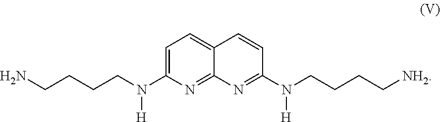
- N,N′-bis-3-aminobutyl-2,7-diamino-1,8-naphthyridine represented by the following formula (V):
- a compound represented by formula (V) also forms a stable complex having a stoichiometric ratio of 1:1 with a double-stranded DNA, preferably a cytosine bulge or thymine bulge, as in the same manner with DANP.
- the ultraviolet-visible light absorption spectrum of the compound represented by formula (V) is nearly the same as that of DANP, and fluctuations of the absorption maximum wavelength by binding to a bulge is also about the same level as that of DANP.
- the compound represented by the formula (V) can be synthesized according to a known method.
- the method for immobilizing the above 2,7-diaminonaphthyridine derivative compound to a cytosine bulge or thymine bulge is not limited specifically, but includes a known method for modifying a compound on a DNA, for example, a post-modification method using reaction of an active carboxylic acid and amine. Concretely, for example, as shown in FIG.
- the DNA is immersed in a solution of the 2,7-diaminonaphthyridine derivative compound (for example, an acetonitrile solution) at room temperature, preferably at from 15° to 30° C., for from 10 to 20 minutes, and purified as needed, whereby the 2,7-diaminonaphthyridine derivative compound can be immobilized to a cytosine bulge or thymine bulge by a hydrogen bond.
- a solution of the 2,7-diaminonaphthyridine derivative compound for example, an acetonitrile solution
- a guanine residue is introduced at a position adjoining 5′ or 3′-terminal side of the cytosine bulge or thymine bulge.
- a guanine-cytosine (G-C) base pair is formed at a position adjoining 5′ or 3′-terminal side of the cytosine bulge or thymine bulge to which the 2,7-diaminonaphthyridine derivative compound is immobilized.
- the 2,7-diaminonaphthyridine derivative compound binds to a cytosine bulge or thymine bulge.
- the 2,7-diaminonaphthyridine derivative compound is immobilized by an adjoining nucleotide on the opposite side to the side on which a guanine-cytosine (G-C) base pair is formed, and cytosine or thymine of the bulge, from the viewpoint of ensuring the quenching effect by the guanine residue.
- DANP alone shows the maximum luminescence at 393 nm and the intensity of 58.
- all the maximum luminescence of ssDNA, full-match and C-bulge are 410 nm, while the intensity is 16, 20 and 6, respectively, the intensity of C-bulge being the weakest and about one-tenth that of DANP alone.
- the length of the above nucleic acid probe is preferably 15 nucleotide residues or more, more preferably 20 nucleotide residues or more, and preferably 45 nucleotide residues or less, more preferably 40 nucleotide residues or less, from the viewpoint of fully exhibiting sufficient stability and high sequence specificity to a bulge DNA.
- a nucleic acid probe having the optical properties described above and (B) evaluation subject nucleic acids are mixed and hybridized, and a signal ascribed to the 2,7-diaminonaphthyridine derivative compound due to the disappearance of a cytosine bulge or thymine bulge is detected accompanying the progress of PCR.
- a signal ascribed to the 2,7-diaminonaphthyridine derivative compound due to the disappearance of a cytosine bulge or thymine bulge is detected accompanying the progress of PCR.
- the mixing of the (A) nucleic acid probe and the (B) evaluation subject nucleic acids is carried out in a manner that a molar ratio thereof, i.e. (A)/(B), is about 1/1, in order to sufficiently carry out the formation of a double strand and sufficiently obtain a fluorescent intensity, and the mixing is not unconditionally determined depending upon the kinds or amount of the evaluation subject nucleic acids.
- the pH conditions upon mixing is preferably 5 or more, more preferably 6 or more, and even more preferably 6.5 or more, from the viewpoint of efficiently carrying out detection of the signal of the 2,7-diaminonaphthyridine derivative compound, shift of the absorption maximum wavelength ascribed to binding of the compound to a bulge, fluorescent intensity and the like, and from the viewpoint of stability of nucleic acids, and preferably 9 or less, more preferably 8 or less, and even more preferably 7.5 or less, from the viewpoint of sufficiently releasing the 2,7-diaminonaphthyridine derivative compound from the bulge structure, and from the viewpoint of exhibiting sufficient fluorescent intensity.
- a phosphate buffer Tris-HCl buffer or the like can be used.
- hybridization means that one nucleic acid molecule having a certain sequence and another nucleic acid molecule complementary to at least a portion of the above nucleic acid molecule are associated via a hydrogen bond on the basis of nucleotide sequences complementary to each other. It is desirable that hybridization in the present invention is carried out in a buffer having a pH of from 5 to 8, and preferably a pH of from 6 to 7, containing from 1 mM to 1 M, and preferably from 10 to 100 mM sodium chloride, and the buffer is preferably phosphate buffer.
- the hybridizable conditions can be appropriately optimized according to a known technique.
- a fluorescent signal As a signal ascribed to the 2,7-diaminonaphthyridine derivative compound by disappearance of a cytosine bulge or thymine bulge, a fluorescent signal is preferred.
- the fluorescent signal has a fluorescent intensity at preferably from 400 to 480 nm, more preferably at around 450 nm, in both cases of a cytosine bulge and a thymine bulge, from the viewpoint of sufficiently increasing difference in the fluorescent intensity derived from a free 2,7-diaminonaphthyridine derivative compound and a 2,7-diaminonaphthyridine derivative compound bound to a cytosine bulge or thymine bulge and moreover quenched by guanine of an adjoining nucleotide.
- the excitation wavelength can be appropriately adjusted on the basis of the kind of the 2,7-diaminonaphthyridine derivative compound.
- a single nucleotide polymorphism can be detected without separately adding a labeling substance such as a fluorescent substance is exhibited, by using a 2,7-diaminonaphthyridine derivative compound. Therefore, in the detection method of the present invention, a single nucleotide polymorphism can be conveniently detected without carrying out a complicated step such as studies on conditions for elongation reaction, amplification reaction, and enzyme reaction (for example, the above elongation reaction, amplification reaction, degradation reaction of mismatched nucleic acids and the like), studies on electrophoresis conditions, and labeling with a labeling substance.
- the method includes identifying a single nucleotide polymorphism in an evaluation subject nucleic acid on the basis of a signal ascribed to the 2,7-diaminonaphthyridine derivative compound when hybridizing each of
- a wild-type evaluation subject nucleic acid containing a wild-type nucleotide in a single nucleotide polymorphism-existing position, and a nucleic acid probe complementarily hybridizable to the wild-type evaluation subject nucleic acid;
- mutant evaluation subject nucleic acid containing a mutant nucleotide in a single nucleotide polymorphism-existing position, and a nucleic acid probe complementarily hybridizable to the mutant evaluation subject nucleic acid;
- mutant evaluation subject nucleic acid and a nucleic acid probe complementarily hybridizable to the wild-type evaluation subject nucleic acid.
- fluorescence with high intensity ascribed to the 2,7-diaminonaphthyridine derivative compound is emitted, only in the case where hybridization is carried out to the nucleic acid probe in the present invention in a single nucleotide polymorphism-existing position of the evaluation subject nucleic acid.
- the 2,7-diaminonaphthyridine derivative compound binds to a cytosine bulge or thymine bulge within the probe, and the fluorescence is quenched. Therefore, evaluation of a single nucleotide polymorphism can be highly sensitively carried out, depending upon the kind of the base pair at the single nucleotide polymorphism-existing position in the probe.
- kits for use in the method for detecting a single nucleotide polymorphism in the nucleic acids of the present invention is provided.
- the kit of the present invention is characterized in that the kit contains the above nucleic acid probe, and exhibits excellent effects that the detection method of the present invention can be efficiently and conveniently carried out at low cost, and that a single nucleotide polymorphism in nucleic acids can be efficiently and highly sensitively detected at low cost according to simple procedures.
- i′ a nucleic acid probe containing a nucleotide sequence complementarily hybridizable to an evaluation subject nucleic acid containing at least one single nucleotide polymorphism, and containing a wild-type nucleotide at a single nucleotide polymorphism-existing position, and tagged with a nucleotide sequence of a hairpin structure having a cytosine bulge or thymine bulge at a 5′-terminal thereof, wherein a guanine residue is introduced at a position adjoining 5′ or 3′-terminal side of the cytosine bulge or thymine bulge, and wherein a 2,7-diaminonaphthyridine derivative compound is immobilized to the cytosine bulge or thymine bulge; and ii′) a nucleic acid probe containing a nucleotide sequence complementarily hybridizable to an antisense strand of an evaluation subject nucleic
- the kit of the present invention may appropriately contain a reagent for stably retaining the above nucleic acid probe, for example, a buffer or the like.
- the form for providing the kit may be a form provided as one container including all of the reagents appropriate for carrying out the detection method of the present invention such as an appropriate nucleic acid probe and a necessary reagent in a volume and/or form appropriate for carrying out the detection method of the present invention, or may be a form provided by containers, each of which independently contains a nucleic acid probe, a reagent or the like.
- a kit may include instructions in which the procedures and the like for carrying out the detection method of the present invention using the components contained in the kit are described.
- the fluorescent intensity of a nucleotide sequence wherein DANP is bound to a nucleotide sequence having a cytosine bulge, thymine bulge, guanine bulge or adenine bulge was measured.
- a 10 mer template nucleic acid (xcD1) as shown in SEQ ID NO: 2 those having a desired sequence were synthesized with a DNA synthesizer (manufactured by Applied Biocyctems Inc.), using NHS-Carboxy-dT amidite (manufactured by Glen Research Corporation).
- a DNA synthesizer manufactured by Applied Biocyctems Inc.
- NHS-Carboxy-dT amidite manufactured by Glen Research Corporation.
- Processing and deprotection were not carried out on the DNA synthesizer, and a column containing CPG beads was removed at a stage where a DNA was bound to CPG resin.
- a syringe was connected one each to both ends of this column, and 1 mL of the above DANP solution was repeatedly moved in the two syringes, and allowed to react in that state for 20 minutes. Thereafter, the reaction mixture was dried with a vacuum pump, and the beads were transferred to an Eppendorf (Registered Trademark) tube and treated with 1 mL of a 28% aqueous ammonia. The mixture was allowed to stand at room temperature for 3 hours, whereby processing and deprotection were carried out.
- a probe xcD1 (SEQ ID NO: 2), in which DANP was bound to position 5 of T located at a fifth base from the 5′-terminal.
- confirmation of the resulting probe was carried out using MALDI TOF/MS.
- a nucleotide sequence capable of forming a cytosine bulge, thymine bulge, guanine bulge or adenine bulge as shown in SEQ ID NOs: 3 to 6 was synthesized in the same manner.
- the resulting nucleic acid capable of forming a bulge (SEQ ID NOs: 3 to 6) and a template nucleic acid (SEQ ID NO: 2) were hybridized in an Eppendorf tube. Thereafter a solution of the hybridized product was prepared to have 4.5 ⁇ M in a 10 mM sodium phosphate buffer (pH 7.0), and the fluorescence spectrum excited at a wavelength 380 nm was measured. The results are shown in FIG. 6 .
- the fluorescent intensity is the weakest in a cytosine bulge, next are in a thymine bulge and a guanine bulge, and the strongest in an adenine bulge.
- N,N′-bis-3-aminopropyl-2,7-diamino-1,8-naphthyridine of which amino group was Boc-protected was synthesized, and thereafter deprotected with a 4 N HCl solution, to give DANP hydrochloride.
- the resulting DANP hydrochloride was dissolved in dimethyl sulfoxide, and the solution was then neutralized with N,N-diisopropylethylamine, to prepare a solution of DANP (concentration: 0.1 M).
- a probe capable of forming a cytosine bulge structure in which DANP was bound to position 5 of T located at a fifth base from the 5′-terminal [primer 1 (SEQ ID NO: 7)] was prepared using the DNA synthesizer in the same manner as Test Example 1.
- confirmation of the concentration of the probe was carried out using enzymatic degradation.
- PCR was carried out using the resulting nucleic acid probe (a primer having a hairpin tag containing a cytosine bulge structure at the 5′-terminal) (primer 1).
- primer 1 a primer having a hairpin tag containing a cytosine bulge structure at the 5′-terminal
- pUC18 as a template, each of Taq PCR Master Mix from QIAGEN, a sense primer having a hairpin tag (primer 1) and an antisense primer having no hairpin tag [M13M3 (SEQ ID NO: 8)] at final concentrations of 0.5 ⁇ M, and pUC18 were added, to prepare a PCR solution.
- the PCR solution was heated at 95° C. for 1 minute, and thereafter 40 cycles of reaction, wherein one cycle comprises 95° C. for 10 seconds, 55° C. for 30 seconds, and 72° C.
- PCR was carried out in the same manner as in Test Example 1 of PCR, except that only the amount of the template was changed.
- a graph obtained by plotting fluorescent intensity of every fifth cycle (excitation wavelength: 355 nm, emission wavelength: 450 nm) against the number of PCR cycles is shown in FIG. 8 .
- FIG. 9 is a graph obtained by plotting the amount of the template at a point where the fluorescent intensity was 380 on the axis of ordinates against PCR cycles on the axis of abscissas.
- FIG. 8 it can be seen that a profile of change in the fluorescent intensity reflecting the concentration of the template was observed when solutions only differing in template concentrations were prepared and PCR was carried out.
- FIG. 9 as a result of plotting the amount of the template at a point where the fluorescent intensity of FIG. 8 was 380 against PCR cycles, nearly a linear graph was obtained, suggesting that the fluorescent intensity could be quantified at least within the range of the concentration of the used template.
- PCR was carried out in the same manner as Test Example 1 of PCR, using a template of which SNP site was mutated from guanine to thymine in Test Example 1.
- a graph obtained by plotting fluorescent intensity of every fifth cycle (excitation wavelength: 355 nm, emission wavelength: 450 nm) against the number of PCR cycles is shown in FIG. 10 .
- FIG. 10 it is suggested that increase in the fluorescent intensity is clearly small as compared with the case where the template is matched with the 3′-terminal of the primer, and that SNP detection is made possible with this primer.
- N,N-bis-3-aminobutyl-2,7-diamino-1,8-naphthyridine (a compound represented by the formula (V)) synthesized referring to the synthesis method of DANP
- a nucleic acid probe in which a compound represented by the formula (V) was bound at the same position as that at which DANP was bound in the nucleotide sequence as shown in SEQ ID NO: 7 in Preparation Example 1 of Nucleic Acid Probe was prepared using a DNA synthesizer.
- PCR reaction was carried out in the same manner as in Test Example 1 of PCR, and the fluorescent intensity was measured.
- the relative fluorescent intensity in a case where the amount of increase in the fluorescent intensity of DANP after 40 cycles in Test Example 1 of PCR was regarded as 100% was calculated and shown ( FIG. 11 ).
- a single nucleotide polymorphism in nucleic acids can be highly sensitively detected with simple procedures at low costs.
- SEQ ID NO: 1 in the Sequence Listing is a nucleotide sequence of a synthetic DNA (a tag structure of a nucleic acid probe).
- SEQ ID NO: 2 in the Sequence Listing is a nucleotide sequence of a synthetic DNA (a template sequence).
- SEQ ID NO: 3 in the Sequence Listing is a nucleotide sequence of a synthetic DNA (a cytosine bulge).
- SEQ ID NO: 4 in the Sequence Listing is a nucleotide sequence of a synthetic DNA (a thymine bulge).
- SEQ ID NO: 5 in the Sequence Listing is a nucleotide sequence of a synthetic DNA (a guanine bulge).
- SEQ ID NO: 6 in the Sequence Listing is a nucleotide sequence of a synthetic DNA (an adenine bulge).
- SEQ ID NO: 7 in the Sequence Listing is a nucleotide sequence of a synthetic DNA (primer 1).
- SEQ ID NO: 8 in the Sequence Listing is a nucleotide sequence of a synthetic DNA (M13M3).
Landscapes
- Life Sciences & Earth Sciences (AREA)
- Chemical & Material Sciences (AREA)
- Health & Medical Sciences (AREA)
- Engineering & Computer Science (AREA)
- Organic Chemistry (AREA)
- Immunology (AREA)
- Proteomics, Peptides & Aminoacids (AREA)
- Molecular Biology (AREA)
- Zoology (AREA)
- Wood Science & Technology (AREA)
- Analytical Chemistry (AREA)
- General Health & Medical Sciences (AREA)
- Microbiology (AREA)
- Biotechnology (AREA)
- Physics & Mathematics (AREA)
- Biochemistry (AREA)
- Genetics & Genomics (AREA)
- Urology & Nephrology (AREA)
- Biomedical Technology (AREA)
- Hematology (AREA)
- Bioinformatics & Cheminformatics (AREA)
- Biophysics (AREA)
- General Engineering & Computer Science (AREA)
- Pathology (AREA)
- Cell Biology (AREA)
- Food Science & Technology (AREA)
- Medicinal Chemistry (AREA)
- General Physics & Mathematics (AREA)
- Measuring Or Testing Involving Enzymes Or Micro-Organisms (AREA)
- Investigating Or Analysing Materials By The Use Of Chemical Reactions (AREA)
Abstract
Description
- Patent Publication 1: WO 2006/082685
naphthyridine ring-containing compounds+DNA
so that excess naphthyridine ring-containing compounds would be required during PCR. Therefore, the fluorescence of the naphthyridine ring-containing compounds non-binding to the bulge structure is detected as a background, so that there is a disadvantage in lowering the detection sensitivity.
wherein a guanine residue is introduced at a position adjoining 5′ or 3′-terminal side of the cytosine bulge or thymine bulge, and wherein a 2,7-diaminonaphthyridine derivative compound is immobilized to the cytosine bulge or thymine bulge; or
ii) a nucleic acid probe containing a nucleotide sequence complementarily hybridizable to an antisense strand of an evaluation subject nucleic acid containing at least one single nucleotide polymorphism, and tagged with a nucleotide sequence of a hairpin structure having a cytosine bulge or thymine bulge at a 5′-terminal thereof, wherein a guanine residue is introduced at a position adjoining 5′ or 3′-terminal side of the cytosine bulge or thymine bulge, and wherein a 2,7-diaminonaphthyridine derivative compound is immobilized to the cytosine bulge or thymine bulge; and
(B) the above evaluation subject nucleic acids; and detecting a signal ascribed to the 2,7-diaminonaphthyridine derivative compound due to the disappearance of the above cytosine bulge or thymine bulge when the above nucleic acid probe and the evaluation subject nucleic acids are hybridized, thereby evaluating the above single nucleotide polymorphism.
[2] a kit for use in a method for detecting a single nucleotide polymorphism in nucleic acids of the above [1], containing:
i′) a nucleic acid probe containing a nucleotide sequence complementarily hybridizable to an evaluation subject nucleic acid containing at least one single nucleotide polymorphism, and containing a wild-type nucleotide at a single nucleotide polymorphism-existing position, and tagged with a nucleotide sequence of a hairpin structure having a cytosine bulge or thymine bulge at a 5′-terminal thereof,
wherein a guanine residue is introduced at a position adjoining 5′ or 3′-terminal side of the cytosine bulge or thymine bulge, and wherein a 2,7-diaminonaphthyridine derivative compound is immobilized to the cytosine bulge or thymine bulge; and
ii′) a nucleic acid probe containing a nucleotide sequence complementarily hybridizable to an antisense strand of an evaluation subject nucleic acid containing at least one single nucleotide polymorphism, and containing a wild-type nucleotide at a single nucleotide polymorphism-existing position, and tagged with a nucleotide sequence of a hairpin structure having a cytosine bulge or thymine bulge at a 5′-terminal thereof,
wherein a guanine residue is introduced at a position adjoining 5′ or 3′-terminal side of the cytosine bulge or thymine bulge, and wherein a 2,7-diaminonaphthyridine derivative compound is immobilized to the cytosine bulge or thymine bulge;
and
i″) a nucleic acid probe containing a nucleotide sequence complementarily hybridizable to an evaluation subject nucleic acid containing at least one single nucleotide polymorphism, and containing a mutant nucleotide at a single nucleotide polymorphism-existing position, and tagged with a nucleotide sequence of a hairpin structure having a cytosine bulge or thymine bulge at a 5′-terminal thereof,
wherein a guanine residue is introduced at a position adjoining 5′ or 3′-terminal side of the cytosine bulge or thymine bulge, and wherein a 2,7-diaminonaphthyridine derivative compound is immobilized to the cytosine bulge or thymine bulge; and
ii″) a nucleic acid probe containing a nucleotide sequence complementarily hybridizable to an antisense strand of an evaluation subject nucleic acid containing at least one single nucleotide polymorphism, and containing a mutant nucleotide at a single nucleotide polymorphism-existing position, and tagged with a nucleotide sequence of a hairpin structure having a cytosine bulge or thymine bulge at a 5′-terminal thereof,
wherein a guanine residue is introduced at a position adjoining 5′ or 3′-terminal side of the cytosine bulge or thymine bulge, and wherein a 2,7-diaminonaphthyridine derivative compound is immobilized to the cytosine bulge or thymine bulge.
wherein a guanine residue is introduced at a position adjoining 5′ or 3′-terminal side of the cytosine bulge or thymine bulge, and wherein a 2,7-diaminonaphthyridine derivative compound is introduced to the cytosine bulge or thymine bulge; and
ii) a nucleic acid probe containing a nucleotide sequence complementarily hybridizable to an antisense strand of an evaluation subject nucleic acid containing at least one single nucleotide polymorphism, and tagged with a nucleotide sequence of a hairpin structure having a cytosine bulge or thymine bulge at a 5′-terminal thereof, wherein a guanine residue is introduced at a position adjoining 5′ or 3% terminal side of the cytosine bulge or thymine bulge, and wherein a 2,7-diaminonaphthyridine derivative compound is immobilized to the cytosine bulge or thymine bulge.
| 5′- ATCATGCTTTTGCCATGAT- 3′ | (SEQ ID NO: 1) |
In the above sequence, for example, a 2,7-aminonaphthyridine derivative compound mentioned later is immobilized to the sequence at a position of fifth T from the 5′-terminal to allow sixth C from the 3′-terminal to form a bulge structure.


ii′) a nucleic acid probe containing a nucleotide sequence complementarily hybridizable to an antisense strand of an evaluation subject nucleic acid containing at least one single nucleotide polymorphism, and containing a wild-type nucleotide at a single nucleotide polymorphism-existing position, and tagged with a nucleotide sequence of a hairpin structure having a cytosine bulge or thymine bulge at a 5′-terminal thereof,
wherein a guanine residue is introduced at a position adjoining 5′ or 3′-terminal side of the cytosine bulge or thymine bulge, and wherein a 2,7-diaminonaphthyridine derivative compound is immobilized to the cytosine bulge or thymine bulge; and
i″) a nucleic acid probe containing a nucleotide sequence complementarily hybridizable to an evaluation subject nucleic acid containing at least one single nucleotide polymorphism, and containing a mutant nucleotide at a single nucleotide polymorphism-existing position, and tagged with a nucleotide sequence of a hairpin structure having a cytosine bulge or thymine bulge at a 5′-terminal thereof, wherein a guanine residue is introduced at a position adjoining 5′ or 3′-terminal side of the cytosine bulge or thymine bulge, and wherein a 2,7-diaminonaphthyridine derivative compound is immobilized to the cytosine bulge or thymine bulge; and
ii″) a nucleic acid probe containing a nucleotide sequence complementarily hybridizable to an antisense strand of an evaluation subject nucleic acid containing at least one single nucleotide polymorphism, and containing a mutant nucleotide at a single nucleotide polymorphism-existing position, and tagged with a nucleotide sequence of a hairpin structure having a cytosine bulge or thymine bulge at a 5′-terminal thereof,
wherein a guanine residue is introduced at a position adjoining 5′ or 3′-terminal side of the cytosine bulge or thymine bulge, and wherein a 2,7-diaminonaphthyridine derivative compound is immobilized to the cytosine bulge or thymine bulge.
| (primer 1): | |
| (SEQ ID NO: 7) | |
| 5′- ATCATGCTTTTGCCATGATCAGGAAACAGCTATGAC- 3′ |
| (primer 1): | |
| (SEQ ID NO: 7) | |
| 5′- ATCATGCTTTTGCCATGATCAGGAAACAGCTATGAC- 3′ | |
| (M13M3): | |
| (SEQ ID NO: 8) | |
| 5′- GTTGTAAAACGACGGCCAGT- 3′ |
Claims (7)


Applications Claiming Priority (3)
| Application Number | Priority Date | Filing Date | Title |
|---|---|---|---|
| JP2012051551 | 2012-03-08 | ||
| JP2012-051551 | 2012-03-08 | ||
| PCT/JP2013/056423 WO2013133402A1 (en) | 2012-03-08 | 2013-03-08 | Method for detecting single nucleotide polymorphism in nucleic acid |
Publications (2)
| Publication Number | Publication Date |
|---|---|
| US20140255938A1 US20140255938A1 (en) | 2014-09-11 |
| US9057105B2 true US9057105B2 (en) | 2015-06-16 |
Family
ID=49116870
Family Applications (1)
| Application Number | Title | Priority Date | Filing Date |
|---|---|---|---|
| US14/352,208 Expired - Fee Related US9057105B2 (en) | 2012-03-08 | 2013-03-08 | Method for detecting single nucleotide polymorphism in nucleic acid |
Country Status (3)
| Country | Link |
|---|---|
| US (1) | US9057105B2 (en) |
| JP (1) | JP5401634B1 (en) |
| WO (1) | WO2013133402A1 (en) |
Families Citing this family (1)
| Publication number | Priority date | Publication date | Assignee | Title |
|---|---|---|---|---|
| JP6440997B2 (en) * | 2014-08-22 | 2018-12-19 | 国立大学法人大阪大学 | PCR method and PCR kit |
Citations (6)
| Publication number | Priority date | Publication date | Assignee | Title |
|---|---|---|---|---|
| US5866336A (en) * | 1996-07-16 | 1999-02-02 | Oncor, Inc. | Nucleic acid amplification oligonucleotides with molecular energy transfer labels and methods based thereon |
| WO2001042498A1 (en) | 1999-12-10 | 2001-06-14 | Toyo Boseki Kabushiki Kaisha | Method of detecting nucleotide polymorphism |
| US20030148301A1 (en) | 1999-12-10 | 2003-08-07 | Toshiya Aono | Method of detecting nucleotide polymorphism |
| JP2004236655A (en) | 2002-08-05 | 2004-08-26 | Toyobo Co Ltd | Method for simply detecting genetic polymorph and reagent for detecting the same |
| WO2006082685A1 (en) | 2005-02-01 | 2006-08-10 | Kyoto University | Method for detecting single nucleotide polymorphism |
| US20100015618A1 (en) * | 2006-09-01 | 2010-01-21 | Osaka University | Dna fragment used as attached to 5' end of primer used in nucleic acid amplification reaction and use of dna fragment |
Family Cites Families (1)
| Publication number | Priority date | Publication date | Assignee | Title |
|---|---|---|---|---|
| JP4464614B2 (en) | 2003-02-28 | 2010-05-19 | 独立行政法人科学技術振興機構 | Bulge base recognition molecule and bulge base detection method |
-
2013
- 2013-03-08 US US14/352,208 patent/US9057105B2/en not_active Expired - Fee Related
- 2013-03-08 WO PCT/JP2013/056423 patent/WO2013133402A1/en active Application Filing
- 2013-03-08 JP JP2013532002A patent/JP5401634B1/en not_active Expired - Fee Related
Patent Citations (6)
| Publication number | Priority date | Publication date | Assignee | Title |
|---|---|---|---|---|
| US5866336A (en) * | 1996-07-16 | 1999-02-02 | Oncor, Inc. | Nucleic acid amplification oligonucleotides with molecular energy transfer labels and methods based thereon |
| WO2001042498A1 (en) | 1999-12-10 | 2001-06-14 | Toyo Boseki Kabushiki Kaisha | Method of detecting nucleotide polymorphism |
| US20030148301A1 (en) | 1999-12-10 | 2003-08-07 | Toshiya Aono | Method of detecting nucleotide polymorphism |
| JP2004236655A (en) | 2002-08-05 | 2004-08-26 | Toyobo Co Ltd | Method for simply detecting genetic polymorph and reagent for detecting the same |
| WO2006082685A1 (en) | 2005-02-01 | 2006-08-10 | Kyoto University | Method for detecting single nucleotide polymorphism |
| US20100015618A1 (en) * | 2006-09-01 | 2010-01-21 | Osaka University | Dna fragment used as attached to 5' end of primer used in nucleic acid amplification reaction and use of dna fragment |
Non-Patent Citations (8)
| Title |
|---|
| Fumie Takei et al.; "Hairpin Tag o Motsu Primer o Tsukatta Ichi Enki Takei no Keiko Kenshutsu" The 90th Annual Meeting of the Chemical Society of Japan in Spring (2010) Koen Yokoshu III, Mar. 12, 2010, p. 758. |
| International Search Report issued in PCT/JP2013/056423, mailed May 28, 2013. |
| Kobori et al., The SPR Sensor Detecting Cytosine-Cytosine Mismatches. JACS 126 :557 (2004). * |
| Nazarenko et al.A closed tube format for amplification and detection of DNA based on energy transfer. Nucleic Acids Research 25 (12) ::2516 (1997). * |
| Sobrino SNPs in forensic genetics: a review on SNP typing methodologies. Forensic Science Intl. 154 : 181 (2005). * |
| Suda et al. N,N′-Bis(3-aminopropyI)-2,7-diamino-1,8-naphthyridine stabilized a single pyrimidine bulge in duplex DNA. Bioorganic & Medicinal Chemistry 13 :4507 (2005). * |
| Suda et al. N,N'-Bis(3-aminopropyI)-2,7-diamino-1,8-naphthyridine stabilized a single pyrimidine bulge in duplex DNA. Bioorganic & Medicinal Chemistry 13 :4507 (2005). * |
| Takei et al., Allele Specific C-Bulge Probeswith One Unique Fluorescent Molecule Discriminate the Single Nucleotide Polymorphism in DNA. Chemistry A European Journal 13 : 4452(2007). * |
Also Published As
| Publication number | Publication date |
|---|---|
| JPWO2013133402A1 (en) | 2015-07-30 |
| JP5401634B1 (en) | 2014-01-29 |
| US20140255938A1 (en) | 2014-09-11 |
| WO2013133402A1 (en) | 2013-09-12 |
Similar Documents
| Publication | Publication Date | Title |
|---|---|---|
| AU2017203349B2 (en) | Compositions of toehold primer duplexes and methods of use | |
| US11225683B2 (en) | Photocoupling method using probe containing photoresponsive nucleic acids | |
| WO2018057999A1 (en) | Molecular hybridization probes for complex sequence capture and analysis | |
| US9944979B2 (en) | Inhibition method of nucleic acid amplification by photoirradiation and method of selective nucleic acid amplification with high sensitivity | |
| US20130084568A1 (en) | Probe, and polymorphism detection method using the same | |
| EP2495336B1 (en) | A new type of universal probes for the detection of genomic variants | |
| EP2450443B1 (en) | Target sequence amplification method, polymorphism detection method, and reagents for use in the methods | |
| JP4752071B2 (en) | Single nucleotide polymorphism detection method | |
| US20210262015A1 (en) | Method for detecting polynucleotide sequence having gene mutation | |
| KR101386918B1 (en) | Method for detecting polymorphism at nucleotide position -1639 of vkorc1 gene, and nucleic acid probe and kit therefor | |
| US9057105B2 (en) | Method for detecting single nucleotide polymorphism in nucleic acid | |
| JP2008182974A (en) | Nucleic acid probe set and method for using the same | |
| EP3690039B1 (en) | Method for detecting single base substitution using ion exchange chromatography | |
| KR101581388B1 (en) | Probe for detecting polymorphism, method of detecting polymorphism, method of evaluating drug efficacy and reagent kit for detecting polymorphism | |
| KR101569127B1 (en) | Probe for detecting polymorphism, method of detecting polymorphism, method of evaluating drug efficacy, disease prediction method and reagent kit for detecting polymorphism | |
| JP5597939B2 (en) | Method for detecting a target nucleotide sequence using a partially competitive probe | |
| US8748590B2 (en) | Oligonucleotides for detection test of polymorphism of EGFR exon 19 and use therof | |
| JP5670396B2 (en) | Nucleic acid probe set and method of using the same | |
| WO2009119647A1 (en) | Method for fluorescence labeling of nucleic acid | |
| JP4638419B2 (en) | Gene mutation detection method and gene mutation detection kit | |
| JP2003159100A (en) | Method for detecting mutation of improved gene | |
| WO2010113452A1 (en) | Method of distinguishing genotypes | |
| JP5504676B2 (en) | Genotype identification method | |
| Ranasinghe | Novel techniques for genetic analysis | |
| JP2011206017A (en) | Method for identifying target base sequence |
Legal Events
| Date | Code | Title | Description |
|---|---|---|---|
| AS | Assignment |
Owner name: OSAKA UNIVERSITY, JAPAN Free format text: ASSIGNMENT OF ASSIGNORS INTEREST;ASSIGNORS:NAKATANI, KAZUHIKO;TAKEI, FUMIE;DOHNO, CHIKARA;AND OTHERS;SIGNING DATES FROM 20140307 TO 20140314;REEL/FRAME:032698/0929 Owner name: FURUKAWA ELECTRIC ADVANCED ENGINEERING CO., LTD., Free format text: ASSIGNMENT OF ASSIGNORS INTEREST;ASSIGNORS:NAKATANI, KAZUHIKO;TAKEI, FUMIE;DOHNO, CHIKARA;AND OTHERS;SIGNING DATES FROM 20140307 TO 20140314;REEL/FRAME:032698/0929 |
|
| STCF | Information on status: patent grant |
Free format text: PATENTED CASE |
|
| FEPP | Fee payment procedure |
Free format text: PAYOR NUMBER ASSIGNED (ORIGINAL EVENT CODE: ASPN); ENTITY STATUS OF PATENT OWNER: LARGE ENTITY |
|
| MAFP | Maintenance fee payment |
Free format text: PAYMENT OF MAINTENANCE FEE, 4TH YEAR, LARGE ENTITY (ORIGINAL EVENT CODE: M1551); ENTITY STATUS OF PATENT OWNER: LARGE ENTITY Year of fee payment: 4 |
|
| FEPP | Fee payment procedure |
Free format text: MAINTENANCE FEE REMINDER MAILED (ORIGINAL EVENT CODE: REM.); ENTITY STATUS OF PATENT OWNER: LARGE ENTITY |
|
| LAPS | Lapse for failure to pay maintenance fees |
Free format text: PATENT EXPIRED FOR FAILURE TO PAY MAINTENANCE FEES (ORIGINAL EVENT CODE: EXP.); ENTITY STATUS OF PATENT OWNER: LARGE ENTITY |
|
| STCH | Information on status: patent discontinuation |
Free format text: PATENT EXPIRED DUE TO NONPAYMENT OF MAINTENANCE FEES UNDER 37 CFR 1.362 |
|
| FP | Lapsed due to failure to pay maintenance fee |
Effective date: 20230616 |