US8999635B2 - Filaggrin - Google Patents
Filaggrin Download PDFInfo
- Publication number
- US8999635B2 US8999635B2 US12/097,493 US9749306A US8999635B2 US 8999635 B2 US8999635 B2 US 8999635B2 US 9749306 A US9749306 A US 9749306A US 8999635 B2 US8999635 B2 US 8999635B2
- Authority
- US
- United States
- Prior art keywords
- filaggrin
- flg
- mutation
- repeat
- mutations
- Prior art date
- Legal status (The legal status is an assumption and is not a legal conclusion. Google has not performed a legal analysis and makes no representation as to the accuracy of the status listed.)
- Expired - Fee Related, expires
Links
Images


Classifications
-
- A—HUMAN NECESSITIES
- A01—AGRICULTURE; FORESTRY; ANIMAL HUSBANDRY; HUNTING; TRAPPING; FISHING
- A01K—ANIMAL HUSBANDRY; AVICULTURE; APICULTURE; PISCICULTURE; FISHING; REARING OR BREEDING ANIMALS, NOT OTHERWISE PROVIDED FOR; NEW BREEDS OF ANIMALS
- A01K67/00—Rearing or breeding animals, not otherwise provided for; New or modified breeds of animals
- A01K67/027—New or modified breeds of vertebrates
- A01K67/0275—Genetically modified vertebrates, e.g. transgenic
-
- C—CHEMISTRY; METALLURGY
- C12—BIOCHEMISTRY; BEER; SPIRITS; WINE; VINEGAR; MICROBIOLOGY; ENZYMOLOGY; MUTATION OR GENETIC ENGINEERING
- C12Q—MEASURING OR TESTING PROCESSES INVOLVING ENZYMES, NUCLEIC ACIDS OR MICROORGANISMS; COMPOSITIONS OR TEST PAPERS THEREFOR; PROCESSES OF PREPARING SUCH COMPOSITIONS; CONDITION-RESPONSIVE CONTROL IN MICROBIOLOGICAL OR ENZYMOLOGICAL PROCESSES
- C12Q1/00—Measuring or testing processes involving enzymes, nucleic acids or microorganisms; Compositions therefor; Processes of preparing such compositions
- C12Q1/68—Measuring or testing processes involving enzymes, nucleic acids or microorganisms; Compositions therefor; Processes of preparing such compositions involving nucleic acids
- C12Q1/6876—Nucleic acid products used in the analysis of nucleic acids, e.g. primers or probes
- C12Q1/6883—Nucleic acid products used in the analysis of nucleic acids, e.g. primers or probes for diseases caused by alterations of genetic material
-
- A—HUMAN NECESSITIES
- A61—MEDICAL OR VETERINARY SCIENCE; HYGIENE
- A61P—SPECIFIC THERAPEUTIC ACTIVITY OF CHEMICAL COMPOUNDS OR MEDICINAL PREPARATIONS
- A61P11/00—Drugs for disorders of the respiratory system
- A61P11/06—Antiasthmatics
-
- A—HUMAN NECESSITIES
- A61—MEDICAL OR VETERINARY SCIENCE; HYGIENE
- A61P—SPECIFIC THERAPEUTIC ACTIVITY OF CHEMICAL COMPOUNDS OR MEDICINAL PREPARATIONS
- A61P17/00—Drugs for dermatological disorders
-
- A—HUMAN NECESSITIES
- A61—MEDICAL OR VETERINARY SCIENCE; HYGIENE
- A61P—SPECIFIC THERAPEUTIC ACTIVITY OF CHEMICAL COMPOUNDS OR MEDICINAL PREPARATIONS
- A61P17/00—Drugs for dermatological disorders
- A61P17/04—Antipruritics
-
- A—HUMAN NECESSITIES
- A61—MEDICAL OR VETERINARY SCIENCE; HYGIENE
- A61P—SPECIFIC THERAPEUTIC ACTIVITY OF CHEMICAL COMPOUNDS OR MEDICINAL PREPARATIONS
- A61P17/00—Drugs for dermatological disorders
- A61P17/06—Antipsoriatics
-
- A—HUMAN NECESSITIES
- A61—MEDICAL OR VETERINARY SCIENCE; HYGIENE
- A61P—SPECIFIC THERAPEUTIC ACTIVITY OF CHEMICAL COMPOUNDS OR MEDICINAL PREPARATIONS
- A61P37/00—Drugs for immunological or allergic disorders
- A61P37/08—Antiallergic agents
-
- C—CHEMISTRY; METALLURGY
- C12—BIOCHEMISTRY; BEER; SPIRITS; WINE; VINEGAR; MICROBIOLOGY; ENZYMOLOGY; MUTATION OR GENETIC ENGINEERING
- C12Q—MEASURING OR TESTING PROCESSES INVOLVING ENZYMES, NUCLEIC ACIDS OR MICROORGANISMS; COMPOSITIONS OR TEST PAPERS THEREFOR; PROCESSES OF PREPARING SUCH COMPOSITIONS; CONDITION-RESPONSIVE CONTROL IN MICROBIOLOGICAL OR ENZYMOLOGICAL PROCESSES
- C12Q1/00—Measuring or testing processes involving enzymes, nucleic acids or microorganisms; Compositions therefor; Processes of preparing such compositions
- C12Q1/68—Measuring or testing processes involving enzymes, nucleic acids or microorganisms; Compositions therefor; Processes of preparing such compositions involving nucleic acids
- C12Q1/6813—Hybridisation assays
- C12Q1/6827—Hybridisation assays for detection of mutation or polymorphism
- C12Q1/683—Hybridisation assays for detection of mutation or polymorphism involving restriction enzymes, e.g. restriction fragment length polymorphism [RFLP]
-
- C—CHEMISTRY; METALLURGY
- C12—BIOCHEMISTRY; BEER; SPIRITS; WINE; VINEGAR; MICROBIOLOGY; ENZYMOLOGY; MUTATION OR GENETIC ENGINEERING
- C12Q—MEASURING OR TESTING PROCESSES INVOLVING ENZYMES, NUCLEIC ACIDS OR MICROORGANISMS; COMPOSITIONS OR TEST PAPERS THEREFOR; PROCESSES OF PREPARING SUCH COMPOSITIONS; CONDITION-RESPONSIVE CONTROL IN MICROBIOLOGICAL OR ENZYMOLOGICAL PROCESSES
- C12Q1/00—Measuring or testing processes involving enzymes, nucleic acids or microorganisms; Compositions therefor; Processes of preparing such compositions
- C12Q1/68—Measuring or testing processes involving enzymes, nucleic acids or microorganisms; Compositions therefor; Processes of preparing such compositions involving nucleic acids
- C12Q1/6844—Nucleic acid amplification reactions
- C12Q1/686—Polymerase chain reaction [PCR]
-
- G—PHYSICS
- G01—MEASURING; TESTING
- G01N—INVESTIGATING OR ANALYSING MATERIALS BY DETERMINING THEIR CHEMICAL OR PHYSICAL PROPERTIES
- G01N33/00—Investigating or analysing materials by specific methods not covered by groups G01N1/00 - G01N31/00
- G01N33/48—Biological material, e.g. blood, urine; Haemocytometers
- G01N33/50—Chemical analysis of biological material, e.g. blood, urine; Testing involving biospecific ligand binding methods; Immunological testing
- G01N33/53—Immunoassay; Biospecific binding assay; Materials therefor
-
- G—PHYSICS
- G01—MEASURING; TESTING
- G01N—INVESTIGATING OR ANALYSING MATERIALS BY DETERMINING THEIR CHEMICAL OR PHYSICAL PROPERTIES
- G01N33/00—Investigating or analysing materials by specific methods not covered by groups G01N1/00 - G01N31/00
- G01N33/48—Biological material, e.g. blood, urine; Haemocytometers
- G01N33/50—Chemical analysis of biological material, e.g. blood, urine; Testing involving biospecific ligand binding methods; Immunological testing
- G01N33/68—Chemical analysis of biological material, e.g. blood, urine; Testing involving biospecific ligand binding methods; Immunological testing involving proteins, peptides or amino acids
- G01N33/6881—Chemical analysis of biological material, e.g. blood, urine; Testing involving biospecific ligand binding methods; Immunological testing involving proteins, peptides or amino acids from skin
-
- C—CHEMISTRY; METALLURGY
- C12—BIOCHEMISTRY; BEER; SPIRITS; WINE; VINEGAR; MICROBIOLOGY; ENZYMOLOGY; MUTATION OR GENETIC ENGINEERING
- C12Q—MEASURING OR TESTING PROCESSES INVOLVING ENZYMES, NUCLEIC ACIDS OR MICROORGANISMS; COMPOSITIONS OR TEST PAPERS THEREFOR; PROCESSES OF PREPARING SUCH COMPOSITIONS; CONDITION-RESPONSIVE CONTROL IN MICROBIOLOGICAL OR ENZYMOLOGICAL PROCESSES
- C12Q2600/00—Oligonucleotides characterized by their use
- C12Q2600/156—Polymorphic or mutational markers
-
- C—CHEMISTRY; METALLURGY
- C12—BIOCHEMISTRY; BEER; SPIRITS; WINE; VINEGAR; MICROBIOLOGY; ENZYMOLOGY; MUTATION OR GENETIC ENGINEERING
- C12Q—MEASURING OR TESTING PROCESSES INVOLVING ENZYMES, NUCLEIC ACIDS OR MICROORGANISMS; COMPOSITIONS OR TEST PAPERS THEREFOR; PROCESSES OF PREPARING SUCH COMPOSITIONS; CONDITION-RESPONSIVE CONTROL IN MICROBIOLOGICAL OR ENZYMOLOGICAL PROCESSES
- C12Q2600/00—Oligonucleotides characterized by their use
- C12Q2600/158—Expression markers
-
- G—PHYSICS
- G01—MEASURING; TESTING
- G01N—INVESTIGATING OR ANALYSING MATERIALS BY DETERMINING THEIR CHEMICAL OR PHYSICAL PROPERTIES
- G01N2800/00—Detection or diagnosis of diseases
- G01N2800/12—Pulmonary diseases
- G01N2800/122—Chronic or obstructive airway disorders, e.g. asthma COPD
-
- G—PHYSICS
- G01—MEASURING; TESTING
- G01N—INVESTIGATING OR ANALYSING MATERIALS BY DETERMINING THEIR CHEMICAL OR PHYSICAL PROPERTIES
- G01N2800/00—Detection or diagnosis of diseases
- G01N2800/20—Dermatological disorders
- G01N2800/202—Dermatitis
-
- G—PHYSICS
- G01—MEASURING; TESTING
- G01N—INVESTIGATING OR ANALYSING MATERIALS BY DETERMINING THEIR CHEMICAL OR PHYSICAL PROPERTIES
- G01N2800/00—Detection or diagnosis of diseases
- G01N2800/20—Dermatological disorders
- G01N2800/205—Scaling palpular diseases, e.g. psoriasis, pytiriasis
-
- G—PHYSICS
- G01—MEASURING; TESTING
- G01N—INVESTIGATING OR ANALYSING MATERIALS BY DETERMINING THEIR CHEMICAL OR PHYSICAL PROPERTIES
- G01N2800/00—Detection or diagnosis of diseases
- G01N2800/24—Immunology or allergic disorders
Definitions
- the present invention relates to the identification of loss-of-function mutations in the filaggrin gene and their use in diagnosing ichthyosis vulgaris and/or susceptibility to other diseases including atopic dermatitis (eczema), asthma, psoriasis and allergies (including food allergy).
- diseases including atopic dermatitis (eczema), asthma, psoriasis and allergies (including food allergy).
- Ichthyosis vulgaris (IV; OMIM#146700) is the most common inherited disorder of keratinisation and one of the most frequent single gene disorders in humans. The most widely cited incidence figure is 1 in 250 based on a survey of 6051 healthy English schoolchildren 1 .
- the phenotypic characteristics of IV include palmar hyperlinearity, keratosis pilaris and a fine scale most markedly seen over the lower abdomen, arms and legs 2 .
- Filaggrin filament aggregating protein
- Keratohyalin granules in the granular layer of interfollicular epidermis are predominantly composed of the 400 kDa protein profilaggrin. Following a short, unique N-terminal domain, most of the profilaggrin molecule consists of 10-12 repeats of the 324 amino acid filaggrin sequence 6 .
- profilaggrin Upon terminal differentiation of granular cells, profilaggrin is proteolytically cleaved into ⁇ 37 kDa filaggrin peptides and the N-terminal domain containing an S100-like calcium binding domain. Filaggrin rapidly aggregates the keratin cytoskeleton, causing collapse of the granular cells into flattened anuclear squames. This condensed cytoskeleton is cross-linked by transglutaminases during formation of the cornified cell envelope (CCE). The CCE is the outermost barrier layer of the skin which not only prevents water loss but also impedes the entry of allergens and infectious agents 7 . Filaggrin is therefore a key protein in facilitating epidermal differentiation and maintaining barrier function.
- CCE cornified cell envelope
- the present invention is based on the identification for the first time of mutations in the human filaggrin (FLG) gene which lead to a loss or partial loss of protein function.
- the present invention provides a genetic test for ichthyosis vulgaris (IV) comprising the steps of:
- the test may be used to diagnose IV and/or to test if a subject is predisposed to developing IV. Additionally, due to an association of IV, in severe or mild forms, with other diseases, the test may be used to also detect whether or not a subject is likely to be predisposed or suffering from atopic dermatitis (eczema), asthma, psoriasis or allergies, such as of a contact type allergy and food allergies (for example, peanut allergy). With regards to skin conditions, low levels of filaggrin expression may lead to development of mild and/or sub-clinical disease. In this manner, the present invention may also relate to the identification and/or treatment of said mild and/or sub-clinical forms of disease. Indeed, many skin conditions go undiagnosed and as such treatments may be considered more as a cosmetic treatment.
- the present invention provides a genetic test for atopic dermatitis (eczema), asthma, psoriasis and/or allergies comprising the steps of:
- the sample to be tested may be any suitable sample from which nucleic acid may be obtained.
- the nucleic acid is a sample of genomic DNA or mRNA.
- the sample may be a sample of saliva, buccal scraping or blood sample.
- the sample may also be a tissue sample, such as a skin biopsy.
- the subject may be any subject requiring to be tested and may suitably be a newborn or even a foetus.
- the subject may however be at any stage of life, and therefore includes neonates, children and adults.
- said tests may be carried out on a subject in order to ascertain whether or not he/she is predisposed to developing a disease.
- a test may be carried out, for example, in order to test a subject's suitability for a particular job, where he/she may come into contact with agents which are known to lead in some cases to the development of eczema and/or allergy.
- said test may be carried out in order to categorise a subject and predict an “at risk” status for, for example, atopic disease. In this manner subjects may be tested so as to categorise or stratify subjects for therapeutic intervention, when appropriate, based on any results obtained, so as to prevent and/or treat atopic disease.
- ascertaining a subject's FLG status and therefore degree of expression or lack of expression of the FLG protein may find use in determining suitable treatment for a subject suffering or predisposed to suffering from IV and/or any of the other aforementioned diseases. For example, depending on the degree of severity, or expected degree of severity, the skilled artisan can decide on an appropriate therapeutic and/or cosmetic regime and as such tailored treatments can be based on a subjects FLG status.
- the present invention in one embodiment, is based on the identification of previously unidentified mutations in the FLG gene, which lead to a loss of function of the profilaggrin and consequently filaggrin proteins.
- the present invention however extends to any mutation in the FLG gene which leads to a loss or partial loss of function of the profilaggrin and/or filaggrin proteins.
- the mutation may be an addition, deletion, substitution or inversion. Typically, the mutation effects 1-10 nucleotides, such as a one-base substitution, or a 2-10 e.g. 4-base deletion.
- the mutation may also be due to a translocation.
- partial or total loss of profilaggrin or filaggrin protein function is understood to mean that the mutation or mutations result in incorrect processing and/or expression of the FLG gene such that one or more of the filaggrin peptides normally expressed, is not functionally expressed.
- 10-12 copies of the filaggrin peptide are expressed from a non-mutated FLG gene 6 . It is understood therefore that the mutant FLG genes of the present invention will result in the functional expression of less than 10-12 filaggrin peptides, typically less than 7, 5, 3 or 1 from one or both copies of the FLG gene, which are present in a genome.
- any reduction in functional filaggrin expression can be mild, e.g. a 1-5 reduction in functional filaggrin peptides; significant e.g. a 7-13 reduction in functional filaggrin peptides; or severe, e.g. a 15-20 reduction in functional filaggrin peptides.
- the mutation or mutations may be found in any of the exons 1, 2 and/or 3 of the FLG gene and may typically be found in exon 3. If the mutation or mutations is/are located in exon 3, the most detrimental mutations, with regards to functional filaggrin expression, will be found within the 5′ (N-terminal) portion of the 3rd exon, such as within the first 2000 bases, e.g. mutation(s) is/are found within the unique, partial repeat, or first filaggrin repeat portion of exon 3 (see FIG. 2 a ).
- a significant number of mutations have been identified by the present inventors, which lead to a loss of function and in some cases, a total loss of function of one of the FLG copies.
- One such mutation is a 1-base substitution at position 1501 of the FLG gene herein (as shown in FIG. 5 and SEQ ID NO.: 188).
- 1501C>T numbering from initiating ATG
- This mutation occurs in the first filaggrin repeat (see FIG. 2 ) and results in the generation of a stop codon, no functional copies of the filaggrin peptide are produced.
- a second mutation identified is a 4-base deletion starting at position 2282 (see FIG. 5 and SEQ ID NO.: 186).
- the mutation has been named 2282del4 and this causes a resulting frame-shift which leads to an alternative stop codon 107 bases downstream.
- this mutation occurs in the first filaggrin repeat and as such no functional copy of a filaggrin peptide is expressed, although a truncated mutant form of the peptide may be expressed, which possesses a unique C-terminal portion (see FIG. 4 and SEQ ID NO.: 187).
- a third mutation is a deletion of a G in the third filaggrin repeat.
- the deletion is at position 3702 and is shown in FIG. 5 .
- This mutation causes a frameshift in repeat 3, such that only 2 functional copies of filaggrin from repeats 1 and 2 are made.
- S3247X for example means that there is a mutation found at codon position 3247 which results in a codon change from a codon which encodes a serine, to a stop codon.
- Detection of a mutation in the FLG gene may be carried out by a variety of techniques including quantitative or semi-quantitative PCR, including real-time PCR, nucleic acid sequencing, hybridisation studies and/or restriction fragment length polymorphism (RFLP) analysis techniques, well known to the skilled addressee (see, for example, Sambrook & Russel, 2000).
- quantitative or semi-quantitative PCR including real-time PCR, nucleic acid sequencing, hybridisation studies and/or restriction fragment length polymorphism (RFLP) analysis techniques, well known to the skilled addressee (see, for example, Sambrook & Russel, 2000).
- the mutation or mutations it may be appropriate to amplify one or more exons or portions thereof. If the mutation(s) is/are located in exon 3, all or only a portion of exon 3 may be amplified using appropriate primers. If the mutation(s) is/are located in the first repeat, it may only be necessary to amplify the first repeat, or portion thereof comprising the mutation(s). By appropriate use of primers and optional labels, it can be possible to amplify a product and ascertain whether or not the product comprises a mutation.
- primers may be designed which incorporate at (or very close to) the 3′ terminal, a base capable of binding to the native or mutant base/sequence, such that only the native or mutant sequence will be amplified and detected.
- a selection of primers suitable for use in amplifying the entire exon 3, or certain specific regions of the repeated sequences of exon 3 are identified herein as SEQ ID NO.s: 1-182.
- SEQ ID NO.s 1-8 represent primers suitable for long range PCR and sequencing of the filaggrin repeats.
- SEQ ID NO.s 9-12 represent primers suitable for generating short PCR fragments for detection of the R501X mutation.
- SEQ ID NO.s 13-15 represent primers suitable for generating short PCR fragments for detection of mutation 2828del4.
- SEQ ID NO.s 15-18 represent primers suitable for generating short PCR fragments for detection of mutation 3702delG.
- SEQ ID NO.s 19-40 represent primers which are specific for repeat 0.
- SEQ ID NO.s 41-67 represent primers which are specific for repeat 1.
- SEQ ID NO.s 68-93 represent primers which are specific for repeat 2.
- SEQ ID NO.s 94-136 represent primers which are specific for repeat 3.
- SEQ ID NO.s 137-201 represent primers which are specific for repeat 4.
- SEQ ID NO.s 202-264 represent primers which are specific for repeat 5.
- SEQ ID NO.s 265-329 represent primers which are specific for repeat 6.
- SEQ ID NO.s 330-377 represent primers which are specific for repeat 7.
- SEQ ID NO.s 378-414 represent primers which are specific for repeat 8.
- SEQ ID NO.s 415-461 represent primers which are specific for repeat 9.
- SEQ ID NO.s 462-493 represent primers which are specific for repeat 10.
- SEQ ID NO.s 494-497 represent primers which are specific for repeat 8.1.
- SEQ ID NO.s 498-501 represent primers which are specific for repeat 8.2.
- SEQ ID NO.s 502-518 represent primers which are specific for repeat 10.1.
- SEQ ID NO.s 519-539 represent primers which are specific for repeat 10.2.
- SEQ ID NO.s 540-544 represent primers which are specific for repeat 11.
- SEQ ID NO. 545 represents a primer which is specific for the filaggrin tail.
- F at the end of a primer sequence shows that the primer is a forward primer and an R shows it is a reverse primer.
- the primers may be from 12-50 bases in length.
- 3′-terminal base is critical for correct primer extension and so the 3′-end of any primer should be identical to the sequences as identified herein.
- Labels such as fluorescent, chemiluminescent, bioluminescent or radio-labels may be incorporated into the PCR primers so as to allow detection of the native or mutant sequence.
- the skilled man will appreciate that two separately labelled primers may be used in a PCR reaction, designed to facilitate amplification of a product comprising either the native or mutant sequence and the sequence, native or mutant, detected based on the particular label being present in the product.
- Other labelling techniques such as the TagMan® system of Applied Biosystems Inc., CA, USA, may be employed.
- a fragment of DNA which includes the portion of DNA which may include the mutation, may simply be amplified and sequenced, in order to determine whether or not the FLG gene comprises a mutation.
- a fragment may be amplified and a hybridisation study carried out using an appropriate oligonucleotide and very stringent hybridisation conditions and washing conditions employed (see for example Sambrook et al, 2000 15 ) so that only exactly matching oligonucleotides bind to the amplified fragment in the region or regions comprising the mutation(s).
- a technique is commonly known as nested PCR.
- any particular mutation may generate a new restriction site which may be detected by RFLP analysis.
- a fragment which would encompass a mutation which, if present, can first be amplified using appropriate primers and the fragment thereafter subjected to RFLP analysis providing the mutation or native sequence has a restriction site which is not present in the corresponding native or mutant sequence.
- the exemplary mutations identified herein result in the generation of new restriction sites which can easily be detected by first amplifying a fragment comprising the mutation and thereafter restricting the fragment obtained using the appropriate restriction enzyme—only a fragment comprising the mutant sequence will be restricted (see Examples Section for further description).
- kits which comprise one or more of the aforementioned oligonucleotides/primers.
- the kits may also comprise other reagents to facilitate, for example, sequencing, conducting PCR and/or RFLP analysis.
- Such kits may also comprise instructions for their use to detect one or more mutations in a filaggrin gene and optionally how to interpret whether or not a mutation may lead to development or predisposition to developing IV and/or any of the other aforementioned diseases/conditions.
- oligonucleotides/primers of the present invention may also be used in multiplex PCR techniques, known to the skilled addressee, see for example. Kuperstein G, Jack E and Narod S A; Genet Test. 10(1):1-7 (2006). so as to identify mutations in the filaggrin sequence.
- the present inventors have now identified the specific repeat sequences which can lead to exon 3 of the filaggrin gene consisting of 11 or 12 full filaggrin repeats, as opposed to the “normal” 10 repeats.
- the inventors have identified that repeats 8 and/or 10 can be essentially duplicated in certain individuals, in order to generate 11 or 12 filaggrin repeats.
- the present invention also extends to identifying the number of filaggrin repeats in a subject as well as detecting one or more mutations.
- Heterozygous mutant subjects who possess one mutant allele which results in no or little filaggrin expression, but have a second wild-type allele encoding 11 or 12, filaggrin repeats, may express sufficient filaggrin to not develop disease, or only a mild form of disease.
- a carrier of a 12-repeat allele will express 20% more filaggrin than a 10-repeat carrier and this difference in expression may be significant in terms of disease development.
- the aforementioned 4-base deletion results in the expression of a unique peptide which comprises an N-terminal region corresponding to the N-terminal portion of the filaggrin peptide and a unique C-terminal region which has been expressed due to the frame-shift mutation.
- an appropriate binding agent such as a specific antibody. It is appreciated that any such binding agent/antibody should be specific for the unique peptide and not therefore be capable of binding the native filaggrin peptide.
- a suitable antibody such as a monoclonal antibody
- a suitable antibody such as a monoclonal antibody
- Antibodies so produced can thereafter be screened to ascertain their specificity, such that only those antibodies which are specific for the unique peptide may be selected.
- Such specifically reactive antibodies to the unique peptide can be optionally labelled and used in an immunoassay to detect for the presence of the unique peptide in a sample.
- the specifically reactive antibody can be used in an assay, such as an ELISA, to detect any of said unique peptide in a sample being tested.
- the sample may preferably be a skin tissue sample.
- the binding agent is an antibody, monoclonal antibody or fragment thereof, such as a Fab fragment. Detection may be carried out by detecting a label, such as a fluorescent, chemiluminescent, bio-luminescent or radio-label coupled to the binding agent/antibody/fragment.
- the binding agent, antibody or antibody fragment may be unlabelled and detected by way of an antibody specific for said binding agent, antibody or antibody fragment, such as in an ELISA assay.
- nucleic acid and mutant peptide tests described herein may be conducted individually or together.
- an FLG gene sequence or fragment thereof capable of encoding one or more copies of the FLG protein, in the manufacture of a medicament. It is understood that the medicament may be used for the prophylactic or therapeutic treatment of IV and/or diseases including atopic dermatitis (eczema), asthma, psoriasis and allergies.
- IV and/or diseases including atopic dermatitis (eczema), asthma, psoriasis and allergies.
- the present invention therefore also provides an FLG gene sequence or fragment thereof, which gene sequence or fragment thereof, is capable of expressing one or more copies of the filaggrin protein, for use in therapy or prophylaxis.
- the present invention also extends to methods of treating prophylactically or therapeutically any of the aforementioned diseases/conditions by administering to a patient suffering or predisposed to developing any of said aforementioned diseases a DNA construct comprising an FLG gene sequence or fragment thereof, which gene sequence or fragment thereof is capable of expressing one or more copies of the FLG protein, whereby expression of said one or more copies of the FLG protein treats or ameliorates said disease(s)/condition(s).
- the FLG sequence or fragment thereof will be administered to a subject in the form of a recombinant molecule comprising said FLG sequence or fragment under appropriate transcriptional/translational controls to allow expression of said filaggrin protein when administered to a subject.
- the FLG sequence or fragment may be under control of a suitable promoter, such as a constitutive and/or controllable promoter.
- suitable promoters include the native filaggrin promoter, or an appropriate late differentiation-specific keratin promoter.
- the present invention also therefore provides a recombinant molecule comprising an FLG sequence or fragment thereof for use in therapy.
- the recombinant molecule may be in the form of a plasmid, phagemid or viral vector.
- recombinantly expressed, or chemically synthesised filaggrin or profilaggrin protein, or functionally important fragments thereof may be produced and applied to the skin via a suitable ointment or other pharmaceutical vehicle, as a treatment or prophylatic measure for ichthyosis vulgaris and/or atopic diseases.
- Such a treatment may also be of cosmetic value as it may increase the barrier function and/or moisture-retention properties of the skin. Since filaggrin is prominently expressed in hair from early in development 16 , such a treatment may also improve cosmetic qualities of the hair, such as moisture retention, in individuals with either normal or reduced filaggrin expression.
- adenovirus vectors many different viral and non-viral vectors and methods of their delivery, for use in gene therapy, are known, such as adenovirus vectors, adeno-associated virus vectors, retrovirus vectors, lentiviral vectors, herpes virus vectors, liposomes, DNA vaccination and the like 17 .
- the present invention also provides a method of treating, preventing and/or ameliorating IV and/or any of the aforementioned diseases, comprising the steps of:
- the intention is to increase expression of FLG polypeptides in the skin. This may most easily be achieved in heterozygote subjects, i.e. those subjects who have one mutant copy of the FLG gene and one native copy.
- the expression of the FLG gene is known to be induced by UV light 18 .
- UV treatment of the skin of an individual carrying a heterozygous copy of a filaggrin loss-of-function mutation could increase the expression of the normal allele and thereby produce a beneficial effect.
- the present invention also relates to a transgenic non-human animal which possesses one or more mutations in one or both copies of the FLG gene which would lead to a loss or partial loss of function of the FLG protein encoded by the FLG gene.
- the FLG gene of the non-human animal may be replaced with a mutant human form of the gene, in order to “humanise” the non-human animal with respect to the FLG gene.
- transgenic animals of the present invention can be used for the development of various treatments for IV and/or the other diseases mentioned herein including the identification of various therapeutically active agents including but not limited to other proteins, peptides, peptidomimetic drugs, small molecule drugs, chemicals and nucleic acid-based agents.
- non-human animals is used herein to include all vertebrate animals, except humans. It also includes an individual animal in all stages of development, including embryonic and foetal stages.
- a transgenic animal is any animal containing one or more cells bearing genetic information altered or received, directly or indirectly, by deliberate genetic manipulation at a subcellular level, such as by targeted recombination or microinjection or infection with recombinant virus.
- the term transgenic animal is not intended to encompass classical cross-breading or in vitro fertilization, but rather is meant to encompass animals in which one or more cells are altered by, or receive, a recombinant DNA molecule.
- mutant FLG nucleic acid sequences are inserted into a germ line of the animal using standard techniques of oocyte microinjection or transfection or microinjection into stem cells.
- it is desired to replace the endogenous gene and homologous recombination using embryonic stem cells or foetal fibroblasts can be applied.
- mice are often used for transgenic animal models because they are easy to house, relatively inexpensive, and easy to breed.
- other non-human transgenic mammals can also be made in accordance with the present invention such as but not limited to monkeys, sheep, rabbits, dogs and rats.
- Transgenic animals are those which carry a transgene, that is, a cloned gene introduced and stably incorporated which is passed on to successive generations.
- one or more copies of the nucleic acid sequences encoding FLG can be inserted into the pronucleus of a just-fertilised mouse oocyte. This oocyte is then reimplanted into a pseudo-pregnant foster mother. The live born mice can then be screened for integrants using analysis of, for example, tail DNA for the presence of the mutant FLG sequences.
- mice disclosed herein can be crossed with a hairless or nude mouse background so that skin abnormalities are visible to facilitate monitoring of disease progression and/or potential therapies.
- An in vivo assay for identifying an agent which is useful for treating or preventing IV and/or any of the other aforementioned diseases associated with mutant FLG expression comprising the steps of administering a test agent to a FLG mutant transgenic animal; and measuring or determining whether the agent decreases or inhibits at least one sign or symptom of IV and/or any of the other aforementioned diseases which is indicative that the test agent is capable of treating or preventing IV and/or any of the other aforementioned diseases.
- the results of the screening assay can be compared with a control, e.g., an animal which has not been administered a test agent or an animal which has received an agent known to reduce a sign or symptom of said diseases.
- the route of administration of the test agent may vary. Examples of administration routes include, but are not limited, to oral, nasal, rectal, transmucosal, intestinal, parenteral, intravenous, intraperitoneal and topical.
- FIG. 1 Shows Pedigrees of IV Families Studied.
- FIG. 2 Shows FLG Mutation Detection and Confirmation.
- Exon 1 consists of a short 5′UTR sequence.
- Exon 2 and the 5′ end of exon 3 encode the profilaggrin N-terminal domain.
- the remainder of exon 3 consists of 10-12 repeats of approximately 1 kb, each encoding a filaggrin peptide separated by linker sequences, followed by a short unique coding sequence and the 3′UTR.
- each profilaggrin molecule is proteolytically cleaved to release 10-12 copies of filaggrin, which aggregate the keratin cytoskeleton and cause physical collapse of the granular cells to form squamous cells.
- FIG. 3 Shows Morphological Features of Filaggrin-Null Ichthyosis Vulgaris.
- profilaggrin is normally expressed (arrows) but in addition there is some patchy diffuse cytoplasmic staining throughout the epidermis (arrowheads).
- the epitope of this antibody is upstream of the mutation and so this shows that a truncated fragment of profilaggrin is synthesized in R501X homozygotes.
- FIG. 4 shows complete sequence of the filaggrin repeats and identifies the positions of the mutations identified by the inventors and positions of where the specific primers identified herein bind. These specific priming sites were identified by analysis of alignments of the individual filaggrin repeats, including the novel additional repeats identified by the inventors.
- FIG. 5 shows the pedigrees of a family with IV and corresponding atopy transmission
- FIG. 6 is a graph showing that filaggrin variants are associated with increased atopy
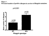
- FIG. 7 is a graph showing increased number of positive allergens in carriers of filaggrin mutations
- FIG. 8 is a graph showing filaggrin variants are stronger risk factors for eczema in the older asthmatics
- FIG. 9 shows a) immunostaining of skin biopsy material from a normal control and an IV patient with the R510X/R2447X genotype. b) immunoblotting of skin biopsy protein extracts from a normal control and IV patients with the genotypes R501X/R2447X and R501X/R501X;
- FIG. 10 is a schematic diagram of profilaggrin proteins encoded by size variant alleles of FLG;
- FIG. 11 shows the DNA sequence of FLG size variant allele FLG 8+ ;
- FIG. 12 shows the DNA sequence of FLG size variant allele FLG 10+ .
- FIG. 13 shows the sequence of a fragment from FLG size variant allele FLG 8+10 + , together with annotations as follows:
- N base pair shared with only one other filaggrin repeat sequence
- N* base pair essentially specific due to nearby differences in repeat N.
- a shorter PCR fragment was designed to amplify approximately 1.5 kb for mutation analysis of R501X.
- Primers FilF3 (+ strand) 5′ GCT GAT AAT GTG ATT CTG TCT G 3′ (SEQ ID NO: 1) and RPT1P6 ( ⁇ strand) 5′ ACC TGA GTG TCC AGA CCT ATT 3′ (SEQ ID NO: 52) were used in High Fidelity PCR buffer (Roche) containing 1.5 mM MgCl 2 , 4% DMSO and 1 U High Fidelity thermostable DNA polymerase mix (Roche). Reactions were amplified under the following conditions: (94° C. 5 min ⁇ 1); (94° C. 30 sec, 57° C. 1 min, 72° C.
- a PCR fragment amplifying 811 bp of genomic DNA was amplified with primers RPT1P7 (+ strand) 5′ AAT AGG TCT GGA CAC TCA GGT 3′ (SEQ ID NO: 51) and RPT2P1 ( ⁇ strand) 5′ GGG AGG ACT CAG ACT GTT T 3′ (SEQ ID NO: 75) using PCR buffer (Applied Biosystems) containing 1.5 mM MgCl 2 , 4% DMSO and 1 U Taq polymerase mix (Promega).
- PCR amplification conditions were: (94° C. 5 min ⁇ 1); (94° C. 30 sec, 57° C. 45 sec, 72° C. 1 min 30 sec) ⁇ 35; and (72° C. 5 min) ⁇ 1.
- Mutation 2282del4 creates a new Dra III restriction enzyme site which was used to screen samples for this mutation.
- PCR products were digested with 5 U Dra III for 4 hr at 37° C. Digests were resolved on 2% agarose gels.
- a PCR fragment from a heterozygous individual was cloned into vector pCR2.1 (Invitrogen). Clones were screened by Dra III digestion and sequenced to confirm the 4 bp deletion.
- H&E staining Routine hematoxylin and eosin (H&E) staining was performed to evaluate morphologic features of each specimen. Immunoperoxidase staining of frozen and paraffin-embedded sections utilized the Envision system (DakoCytomation, Denmark). Antibodies used were mouse monoclonal 15C10 against an epitope in the C-terminal portion of the human filaggrin repeat unit (Novocastra, Newcastle upon Tyne, UK) and rabbit polyclonal antiserum B1 raised against the N-terminus of profilaggnn 23 .
- Lod scores were calculated with MLINK algorithm of LINKAGE version 5.1, using a semidominant model of the disease where heterozygotes were assigned a mild phenotype with 90% penetrance and homozygotes or compound heterozygotes were assigned as a severe phenotype with 100% penetrance.
- FLG consists of three exons 25,26 .
- Exon 1 (15 bp) consists only of 5′UTR sequences and exon 2 (159 bp) contains the initiation codon.
- Exon 3 is unusually large (12,753 bp) and codes for most of the N-terminal domain and all filaggrin repeats ( FIG. 2 a ).
- the number of filaggrin repeats varies from 10-12 in the population 6 .
- the homology between the repeats at the DNA level is almost 100%, making conventional PCR-based sequencing for the internal regions of this exon almost impossible. No sequence changes were found in exons 1 or 2 in five IV families.
- the present inventors developed long-range PCR conditions to amplify a 12 kb genomic fragment covering exon 3 and therefore all the repeat domains ( FIG. 2 b ). Full sequencing of this fragment is on-going, but initial sequencing has revealed a homozygous nonsense mutation R501X near the start of repeat 1 in three affected individuals from Family 1 ( FIG. 2 c - e ). Using a smaller PCR fragment, segregation of R501X was confirmed in Family 1 and in addition, this mutation was identified in the other 14 IV kindreds studied. The mutation creates a new Nla III restriction enzyme site; this was used to confirm the mutation and screen populations ( FIG. 2 f ). By this means, the mutation was found to be present at relatively high allele frequencies in Irish, Scottish and North American Caucasian populations (combined frequency, 0.027; see Table 1).
- profilaggrin is the major component of keratohyalin granules, this explains the absent granular layer associated with the more severe cases of IV 27 ( FIG. 3 ).
- the presence of a truncated profilaggrin peptide in IV epidermis ( FIG. 3 b ) is consistent with previous studies demonstrating that a peptide containing the unique N-terminal domain and a small amount of filaggrin sequence is stable in vitro 23 .
- the N-terminal Ca 2+ -binding domain is cleaved from profilaggrin by a proprotein convertase, and subsequently localizes to different cell compartments including the nucleus 28,29 . Similar processing of the truncated peptide may occur in IV epidermis.
- IV appears to be predominantly caused by two frequent null-mutations in FLG, leading to loss of filaggrin production and impaired epidermal barrier formation.
- IV families studied most R501X mutations are in linkage disequilibrium with the same 156 bp allele of a microsatellite in intron 2 of FLG (data not shown), suggesting that, in human evolutionary terms, these are ancient mutations. Further analysis of polymorphisms near FLG will determine the approximate age of the mutations. Genetic drift may explain why these mutations have become so prevalent. Alternatively, a heterozygote advantage might explain the high frequencies of these alleles.
- filaggrin null-mutations may themselves be modifying factors in other ichthyotic skin conditions, including congenital ichthyoses, Netherton syndrome or disorders due to defects in suprabasal keratins, where intra- and interfamilial phenotypic variation is well documented 31-34 .
- the association of IV with the atopic diathesis is well established; 37-50% of people with IV have atopic diseases 1,36 and conversely around 8% of atopic dermatitis patients have classical features of IV 1,35 .
- filaggrin may be a factor in very common skin disorders known to have a major genetic component.
- filaggrin-null or heterozygous individuals also had atopic dermatitis (AD; “eczema”) and/or asthma.
- An example is shown in FIG. 5 , where 3/6 filaggrin-null heterozygotes and 5/5 homozygotes had atopic disease.
- the inventors therefore sought to examine the role of these variants in common atopy associated with asthma.
- the two filaggrin variants were genotyped in a cohort of 800 schoolchildren with unknown disease status (population cohort) and in 550 school children and adolescents with physician-diagnosed asthma from the Dundee BREATHE study.
- the asthma cohort was analysed for people with a recorded allergy to peanuts and/or other nuts.
- 23 individuals had a proven nut allergy and of these, 8 carried a filaggrin-null mutation (either 2282del4 or R501X), i.e. 35% were carriers.
- 60 out of 621 individuals carried a filaggrin-null mutation, i.e. 9.7%.
- the filaggrin mutation carrier frequency is greatly increased in people with asthma and nut allergy.
- filaggrin mutations are a highly significant risk factor for nut allergy and asthma.
- atopy is strongly associated with food allergy especially cow's milk, hen's egg, banana, kiwi, white fish, wheat, prawns/shrimp and strawberries 37-39 . Since nut allergy is regarded as the most reliable marker for food allergy, this strongly infers that filaggrin mutations are associated with food allergies in general.
- these further mutations were 3702delG (repeat 3), R2447X (repeat 7), 53247X (repeat 9), R1474X (repeat 4), Q1745X (repeat 4), Q3683X (repeat 10), 11029delCA (repeat 10) in Irish and Scottish patients; E2422X (repeat 6), 5360delG (repeat 5), 7267delCA (repeat 7) and 11033del4 (repeat 10) in Dutch patients; and 6867delAG (repeat 6) in an Austrian patient.
- the IV phenotype of the patients carrying these more 3′ mutations were essentially indistinguishable to patients carrying R501X or 2282del4 mutations in repeat 1.
- premature termination codon mutations essentially anywhere in the profilaggrin molecule appear to have similar or equivalent pathogenicity.
- the following mutations were recurrent and/or prevalent in the European population R501X, 2282del4, 3702delG, R2447X and 53247X.
- the Dutch and Austrian mutations were not detected in 188 Irish AD patients and may be population-specific or very rare. The remaining variants were not tested for prevalence.
- a cohort of 188 Irish paediatric cases of moderate-severe AD were genotyped for 5 mutations found to be prevalent and/or recurrent in this population, R501X, 2282del4, 3702delG, R2447X and S3247X. Comparison of allele frequencies was made to an unselected Irish control population of 736 individuals genotyped for the same 5 FLG variants. Pearson chi-square analysis revealed that all 5 mutations are independently associated with the AD phenotype, giving individual statistically significant P values of ⁇ 0.05 (Table 4). Combining the data for all 5 genotypes gave an extremely significant P value of 2.12 ⁇ 10 ⁇ 51 . About 48% of the patients in the AD cohort carried one or more of the 5 filaggrin variants. Thus, a wide range of FLG mutations contribute to genetic predisposition to atopy and this is a major gene in early-onset moderate-to-severe AD.
- the European-specific mutations R501X and 2282del4 were found to be absent from 253 Japanese individuals. We therefore sequenced the FLG gene in four Japanese families with IV and identified two novel mutations, 3321delA and S2554X.
- Japan there appears to be a distinct set of prevalent/recurrent FLG mutations that contribute to genetic predisposition to atopy. It is likely that other human populations will have their own spectrum of FLG mutations.
- exon 3 of the FLG gene is variable in size in the human population and these variant alleles were predicted, on the basis of their size, to consist of 10, 11 or 12 full filaggrin repeats in addition to the two partial repeats (Gan et al., 1990) 40 .
- the positions of these insertions within FLG exon 3 and the precise DNA sequences encoding these additional filaggrin repeats remained unknown.
- DNA fragments were generated from unrelated individuals that, from the public sequence of the FLG gene (Human Genome, March 2006 Assembly, hg18), would be predicted to be ⁇ 4.2 kb in size. For convenience, this allele is designated as FLG N . In some individuals, additional bands of ⁇ 5.2 and/or ⁇ 6.2 kb were observed. These larger alleles were cloned into plasmid vector pCR3.1 to allow full sequencing of these variant alleles. This revealed that some individual alleles contain a duplication of repeat 8, designated as FLG 8+ . Another allele consisted of what was essentially a duplication of repeat 10, which was designated as FLG 10+ .
- a heterozygous carrier of the R501X mutation may carry a second wild-type allele encoding either 10, 11 or 12 filaggrin repeats.
- the size of the heterozygous wild-type allele may influence the phenotype observed, for example, a carrier of the 12-repeat (FLG 8+10+ ) allele will express 20% more filaggrin than a R501X carrier carrying a 10-repeat wild-type allele in trans.
- detection of these size variants may be of prognostic value in ichthyosis vulgaris and atopic disease.
Landscapes
- Health & Medical Sciences (AREA)
- Life Sciences & Earth Sciences (AREA)
- Chemical & Material Sciences (AREA)
- Engineering & Computer Science (AREA)
- Organic Chemistry (AREA)
- Proteomics, Peptides & Aminoacids (AREA)
- General Health & Medical Sciences (AREA)
- Immunology (AREA)
- Zoology (AREA)
- Analytical Chemistry (AREA)
- Molecular Biology (AREA)
- Wood Science & Technology (AREA)
- Bioinformatics & Cheminformatics (AREA)
- Biotechnology (AREA)
- Microbiology (AREA)
- Physics & Mathematics (AREA)
- Genetics & Genomics (AREA)
- Biochemistry (AREA)
- Medicinal Chemistry (AREA)
- Chemical Kinetics & Catalysis (AREA)
- Hematology (AREA)
- Urology & Nephrology (AREA)
- Biomedical Technology (AREA)
- Pathology (AREA)
- Animal Behavior & Ethology (AREA)
- Veterinary Medicine (AREA)
- General Engineering & Computer Science (AREA)
- Biophysics (AREA)
- Public Health (AREA)
- General Chemical & Material Sciences (AREA)
- Pharmacology & Pharmacy (AREA)
- Nuclear Medicine, Radiotherapy & Molecular Imaging (AREA)
- General Physics & Mathematics (AREA)
- Food Science & Technology (AREA)
- Cell Biology (AREA)
- Pulmonology (AREA)
- Dermatology (AREA)
- Environmental Sciences (AREA)
- Animal Husbandry (AREA)
- Biodiversity & Conservation Biology (AREA)
Abstract
Description
(b) Long-range PCR product from genomic
(c) Normal sequence from
(d) The same region of the FLG as seen in (c), showing heterozygous transition mutation 1502C>T resulting in nonsense mutation R501X. (SEQ ID NO: 560)
(e) The same region of FLG as in (c) showing a homozygous mutation resulting in a nonsense codon, R501X. (SEQ ID NO: 561)
(f) Confirmation of mutation R501X by Nla III restriction digest and 2282del4 by Dra III restriction digest from some members of
(g) Normal sequence from
(h) The same region of FLG as in (f), showing overlapping peaks due to a heterozygous deletion mutation, 2282del4. (SEQ ID NO: 563)
(i) The same region of the FLG as in (f), derived from a mutant clone confirming mutation 2282del4. This mutation leads to a premature stop codon 107 bp downstream and terminates translation within
(c) Transmission electron micrograph of keratinocytes at the boundary of the granular layer and stratum corneum, from a normal individual, showing prominent keratohyalin granules (arrowheads). N=nucleus; K=keratinized material in stratum corneum. Original magnification=˜5,600×.
(d) Transmission electron micrograph of granular layer cells from the proband in
(e) Immunohistochemical staining of formaldehyde-fixed paraffin-embedded tissue using anti-filaggrin repeat monoclonal antibody 15C10 (Novocastra), visualized by the immunoperoxidase method. In skin biopsy material from a normal control, keratohyalin granules are strongly stained in the upper suprabasal layers of the epidermis (arrows).
(f) Immunoperoxidase staining of skin biopsy material from the proband in
(g) Immunoperoxidase staining of skin biopsy material from a normal control individual with polyclonal antibody B1, raised against an epitope within the N-terminal domain of profilaggrin, showing prominent staining of keratohyalin granules (arrows).
(h) Immunoperoxidase staining of skin biopsy material from the proband in
- 1. Wells, R. S. and Kerr C B, Br Med J., 1:947-949 (1966).
- 2. Judge, M. R., McLean, W. H. I. & Munro, C. S. Disorders of keratinization. in Rook's Textbook of Dermatology, Vol. 2 (eds. Burns, T., Breathnach, S., Cox, C. & Griffiths, C.) 34.54-34.56 (Blackwell Scientific Publishing, Oxford, 2004).
- 3. Steinert, P. M., Cantieri, J. S., Teller, D. C., Lonsdale-Eccles, J. D. & Dale, B. A. Characterization of a class of cationic proteins that specifically interact with intermediate filaments. Proc Natl Acad Sci 78, 4097-4101 (1981).
- 4. Dale, B. A., Resing, K. A. & Lonsdale-Ecccles, J. D. Filaggrin: a keratin filament associated protein. Ann. NY Acad. Sci. 455, 330-342 (1985).
- 5. Listwan, P. & Rothnagel, J. A. Keratin bundling proteins. Methods Cell Biol 78, 817-27 (2004).
- 6. Gan, S. Q., McBride, O. W., Idler, W. W., Markova, N. & Steinert, P. M. Organization, structure, and polymorphisms of the human profilaggrin gene. Biochemistry 29, 9432-40 (1990).
- 7. Candi, E., Schmidt, R. & Melino, G. The cornified envelope: a model of cell death in the skin. Nat Rev
Mol Cell Biol 6, 328-40 (2005). - 8. Fleckman, P., Holbrook, K. A., Dale, B. A. & Sybert, V. P. Keratinocytes cultured from subjects with ichthyosis vulgaris are phenotypically abnormal.
J Invest Dermatol 88, 640-5 (1987). - 9. Pena Penabad, C. et al. Differential patterns of filaggrin expression in lamellar ichthyosis. Br J Dermatol 139, 958-64 (1998).
- 10. Sybert, V. P., Dale, B. A. & Holbrook, K. A. Ichthyosis vulgaris: identification of a defect in synthesis of filaggrin correlated with an absence of keratohyaline granules. J Invest Dermatol 84, 191-4 (1985).
- 11. Nirunsuksiri, W., Zhang, S. H. & Fleckman, P. Reduced stability and bi-allelic, coequal expression of profilaggrin mRNA in keratinocytes cultured from subjects with ichthyosis vulgaris.
J Invest Dermatol 110, 854-61 (1998). - 12. Presland, R. B. et al. Loss of normal profilaggrin and filaggrin in flaky tail (ft/ft) mice: an animal model for the filaggrin-deficient skin disease ichthyosis vulgaris. J Invest Dermatol 115, 1072-81 (2000).
- 13. Lane, P. W. Two new mutations in linkage group XVI of the house mouse. Flaky tail and varitint-waddler-J. J Hered 63, 135-40 (1972).
- 14. Rothnagel, J. A. et al. Characterization of the mouse loricrin gene: linkage with profilaggrin and the flaky tail and soft coat mutant loci on
chromosome 3. Genomics 23, 450-6 (1994). - 15. Sambrook & Russel; “Molecular Cloning—A Laboratory Manual”, (2000); Cold Spring Harbor Laboratory Press.
- 16. Dale B A, Holbrook K A, Kimball J R, Hoff M, Sun T T., Expression of epidermal keratins and filaggrin during human fetal skin development., J Cell Biol. 1985 October; 101(4):1257-60.
- 17. Gene Therapy (2004) 11, S57-63.
- 18. Lee D S, Quan G, Choi J Y, Kim S Y, Lee S C. Chronic ultraviolet radiation modulates epidermal differentiation as it up-regulates
transglutaminase 1 and its substrates. Photodermatol Photoimmunol Photomed. 2005 February; 21(1):45-52. - 19. Wall, et al. (1992) J. Cell Biochem. 49(2): 113-20.
- 20. McCreath, et al. (2000) Nature 405: 1066-1069.
- 21. Lai, et al. (2002) Science 295: 1089-92.
- 22. Hogan, et al. (1986) In: Manipulating the mouse embryo. A Laboratory Manual.
- Cold Spring Harbour Laboratory Press, Cold Spring Harbor, N.Y.
- 23. Pearton, D. J., Dale, B. A. & Presland, R. B. Functional analysis of the profilaggrin N-terminal peptide: identification of domains that regulate nuclear and cytoplasmic distribution. J Invest Dermatol 119, 661-9 (2002).
- 24. Eady, R. A. J. Transmission electron microscopy. in Methods in Skin Research (eds. Skerrow, D. & Skerrow, C. J.) 1-36 (John Wiley & Sons, Chichester, 1985).
- 25. Presland, R. B., Haydock, P. V., Fleckman, P., Nirunsuksiri, W. & Dale, B. A. Characterization of the human epidermal profilaggrin gene. Genomic organization and identification of an S-100-like calcium binding domain at the amino terminus. J Biol Chem 267, 23772-81 (1992).
- 26. Markova, N. G. et al. Profilaggrin is a major epidermal calcium-binding protein. Mol Cell Riot 13, 613-25 (1993).
- 27. Compton, J. G., DiGiovanna, J. J., Johnston, K. A., Fleckman, P. & Bale, S. J. Mapping of the associated phenotype of an absent granular layer in ichthyosis vulgaris to the epidermal differentiation complex on
chromosome 1.Exp Dermatol 11, 518-26 (2002). - 28. Presland, R. B. et al. Evidence for specific proteolytic cleavage of the N-terminal domain of human profilaggrin during epidermal differentiation. J Invest Dermatol 108, 170-8 (1997).
- 29. Ishida-Yamamoto, A., Takahashi, H., Presland, R. B., Dale, B. A. & Iizuka, H. Translocation of profilaggrin N-terminal domain into keratinocyte nuclei with fragmented DNA in normal human skin and loricrin keratoderma. Lab Invest 78, 1245-53 (1998).
- 30. Rothnagel, J. A. & Steinert, P. M. The structure of the gene for mouse filaggrin and a comparison of the repeating units. J. Biol. Chem. 265, 1862-1865 (1990).
- 31. Bale, S. J., Compton, J. G., Russell, L. J. & DiGiovanna, J. J. Genetic heterogeneity in lamellar ichthyosis. J Invest Dermatol 107, 140-1 (1996).
- 32. Bitoun, E. et al. Netherton syndrome: disease expression and spectrum of SPINK5 mutations in 21 families. J Invest Dermatol 118, 352-61 (2002).
- 33. Smith, F. J. D. et al. Genomic organization and fine mapping of the keratin 2e gene (KRT2E): K2e V1 domain polymorphism and novel mutations in ichthyosis bullosa of Siemens. J. Invest. Dermatol. 111, 817-821 (1998).
- 34. McLean, W. H. I. et al. Mutations in the rod 1A domain of
keratins - 35. Tay, Y. K., Khoo, B. P. & Goh, C. L. The epidemiology of atopic dermatitis at a tertiary referral skin center in Singapore. Asian Pac J Allergy Immunol 17, 137-41 (1999).
- 36. Koukkanen K., Ichthyosis vulgaris. A clinical and histopathological study of patients and their close relatives in the autosomal dominant and sex-linked forms of the disease. Acta Derm Venereol Suppl (Stockh). 1969; 62:1-72.
- 37. Werfel T, Breuer K., Curr Opin Allergy Clin Immunol. 2004 October; 4(5): 379-85. Role of food allergy in atopic dermatitis.
- 38. Sicherer S. H., Sampson H. A., J. Allergy Clin Immunol. 1999 September; 104 (3 Pt 2): S114-22. Food hypersensitivity and atopic dermatitis: pathophysiology, epidemiology, diagnosis and management.
- 39. Ellman L. K., Chatchatee P., Sicherer S. H. & Sampson H. A. Pediatr Allergy Immunol. 2002 August; 13(4): 205-8. Food hypersensitivity in two groups of children and young adults with atopic dermatitis evaluated a decade apart.
- 40. Gan S. Q., McBride O. W., Idler W. W., Markova N., Steinert P. M. Organization, structure, and polymorphisms of the human profilaggrin gene.
Biochemistry 1990; 29: 9432-40.
| TABLE 1 |
| Allele frequencies of FLG mutations R501X and 2282del4 |
| Allele Frequency | Allele Frequency | ||||
| Population | R501X | 2282del4 | |||
| Irish Caucasian | 0.041 | (n = 97) | 0.005 | (n = 91) | ||
| Scottish Caucasian | 0.021 | (n = 145) | 0.012 | (n = 166) | ||
| US Caucasian | 0.024 | (n = 124) | 0.011 | (n = 133) | ||
| Combined | 0.027 | (n = 366) | 0.01 | (n = 390) | ||
| TABLE 2 |
| Both R501X and 2282del4 are overrepresented |
| in a childhood asthma cohort. |
| R501X | 2282del4 | Combined | ||||
| Population | Asthma | Population | | Population | Asthma | |
| 720 | 484 | 714 | 494 | 585 | 390 |
| 38 | 46 | 27 | 35 | 52 | 69 |
| 5 | 2 | 0 | 2 | 6 | 6 |
| 763 | 532 | 741 | 531 | 643 | 465 |
| p = 0.04 | p = 0.0038 | p = 00045 | |||
| TABLE 3 |
| Characteristics of asthmatic children with and without filaggrin variants |
| WT n = 390 | NULLcarriers n = 75 | p | ||
| SEX (% Male) | 59.2 | 59.0 | 0.522 |
| Age | 10.2(2.7-21.2) | 10.3(4.1-22) | |
| BMI | 19.1 | 18.8 | |
| PEFR % | 92.8 | 90.6 | |
| FEV1 % | 98.1 | 96.8 | |
| FVC % | 97.1 | 97.0 | |
| Eczema | 46.7 | 73.3 | 0.0001 |
| Perennial Rhinitis | 29 | 26.7 | 0.784 |
| Seasonal Rhinitis | 14.8 | 13.3 | 0.861 |
| Cold air trigger | 41.0 | 44.4 | 0.596 |
| Exercise trigger | 36.1 | 44.4 | 0.213 |
| Viral trigger | 44.9 | 46.8 | 0.787 |
| Tested for allergy | 16.6 | 30.1 | 0.035 |
| TABLE 4 |
| Case control association study for 5 FLG mutations (188 |
| Irish AD patients versus 736 Irish population controls) |
| R501X | 2282del4 | R2447X | ||||
| Genotype | Population | AD | Population | AD | Population | AD |
| AA | 717 | 137 | 717 | 152 | 734 | 181 |
| Aa | 19 | 51 | 19 | 35 | 2 | 7 |
| |
0 | 0 | 0 | 1 | 0 | 0 |
| Totals | 736 | 188 | 736 | 188 | 736 | 188 |
| p = 7.8 × 10⁻³⁰ | p = 7.8 × 10⁻¹⁷ | p = 1.7 × 10⁻⁵ | ||||
| S3247X | 3702delG | Combined | ||||
| Genotype | Population | Asthma | Population | AD | | AD |
| AA | ||||||
| 720 | 177 | 735 | 186 | 680 | 103 | |
| Aa | 16 | 11 | 1 | 2 | 55 | 62 |
| |
0 | 0 | 0 | 0 | 1 | 23 |
| 736 | 188 | 736 | 188 | 736 | 188 | |
| p = 0.008 | p = 0.046 | p = 2.12 × 10−51 | ||||
Claims (10)
Applications Claiming Priority (3)
| Application Number | Priority Date | Filing Date | Title |
|---|---|---|---|
| GBGB0525492.5A GB0525492D0 (en) | 2005-12-15 | 2005-12-15 | Filaggrin |
| GB0525492.5 | 2005-12-15 | ||
| PCT/GB2006/004707 WO2007068946A2 (en) | 2005-12-15 | 2006-12-15 | Identification of loss-of-function mutations in filaggrin causing ichthyosis vulgaris and predisposing to other diseases |
Publications (2)
| Publication Number | Publication Date |
|---|---|
| US20100017896A1 US20100017896A1 (en) | 2010-01-21 |
| US8999635B2 true US8999635B2 (en) | 2015-04-07 |
Family
ID=35736147
Family Applications (1)
| Application Number | Title | Priority Date | Filing Date |
|---|---|---|---|
| US12/097,493 Expired - Fee Related US8999635B2 (en) | 2005-12-15 | 2006-12-15 | Filaggrin |
Country Status (5)
| Country | Link |
|---|---|
| US (1) | US8999635B2 (en) |
| EP (1) | EP1960539B1 (en) |
| JP (1) | JP5305921B2 (en) |
| GB (2) | GB0525492D0 (en) |
| WO (1) | WO2007068946A2 (en) |
Families Citing this family (14)
| Publication number | Priority date | Publication date | Assignee | Title |
|---|---|---|---|---|
| GB0600948D0 (en) | 2006-01-18 | 2006-02-22 | Univ Dundee | Prevention/Treatment Of Ichthyosis Vulgaris, Atopy And Other Disorders |
| JP5451607B2 (en) * | 2007-06-28 | 2014-03-26 | リバー ダイアグノスティックス ベー.フェー. | How to determine the possibility of mutation |
| EP2322656A1 (en) * | 2009-11-17 | 2011-05-18 | Centre National de la Recherche Scientifique (C.N.R.S) | Methods for diagnosing skin diseases |
| EP2504357B1 (en) | 2009-11-23 | 2017-08-02 | Research Development Foundation | Recombinant filaggrin polypeptides for cell importation |
| JP5771123B2 (en) * | 2011-11-09 | 2015-08-26 | 株式会社 資生堂 | PROFILAGLIN C-TERMINAL DOMAIN SPECIFIC ANTIBODY AND USE THEREOF |
| GB201222881D0 (en) * | 2012-12-19 | 2013-01-30 | Univ Dundee | Animal model |
| JP5943945B2 (en) * | 2014-01-29 | 2016-07-05 | 株式会社 資生堂 | PROFILAGLIN C-TERMINAL DOMAIN SPECIFIC ANTIBODY AND USE THEREOF |
| JP6803113B2 (en) * | 2016-07-01 | 2020-12-23 | 共栄化学工業株式会社 | Evaluation method and external preparation for skin |
| RU2673804C1 (en) * | 2018-01-25 | 2018-11-30 | ФЕДЕРАЛЬНОЕ ГОСУДАРСТВЕННОЕ БЮДЖЕТНОЕ НАУЧНОЕ УЧРЕЖДЕНИЕ "ФЕДЕРАЛЬНЫЙ ИССЛЕДОВАТЕЛЬСКИЙ ЦЕНТР ИНСТИТУТ ЦИТОЛОГИИ И ГЕНЕТИКИ СИБИРСКОГО ОТДЕЛЕНИЯ РОССИЙСКОЙ АКАДЕМИИ НАУК" (ИЦиГ СО РАН) | METHOD OF IDENTIFICATION OF MUTATIONS 2282del4, R501X, R2447X IN GENES OF FILAGGRIN (FLG) WITH VULGAR ICHTYOSIS AND ATOPIC DERMATITIS |
| CN112236153B (en) * | 2018-04-05 | 2025-04-29 | 阿兹特拉公司 | Methods and compositions for treating skin diseases using recombinant microorganisms |
| US20210355192A1 (en) * | 2018-10-08 | 2021-11-18 | Yale University | Selective Binding and Aggregation of Human Keratin in the Skin Barrier Using Human Filaggrin Subdomains SD67 and SD150 |
| CN113092768A (en) * | 2019-12-23 | 2021-07-09 | 首都医科大学附属北京世纪坛医院 | Application of urine keratin, II type cytoskeleton 1 and polypeptide fragment thereof in allergic diseases |
| CN116874576B (en) * | 2023-06-28 | 2024-03-12 | 胡荣洋 | A recombinant humanized filaggrin and its preparation method and application |
| CN118460707B (en) * | 2024-07-15 | 2024-09-17 | 山东大学 | Primer set, kit and method for detecting FLG gene mutation associated with ichthyosis vulgaris |
Citations (7)
| Publication number | Priority date | Publication date | Assignee | Title |
|---|---|---|---|---|
| US6143502A (en) | 1999-03-31 | 2000-11-07 | University Of Utah Research Foundation | Dual-luciferase reporter system |
| WO2001044516A2 (en) | 1999-12-14 | 2001-06-21 | Tularik Inc. | Methods of assaying for compounds that inhibit premature translation termination and nonsense-mediated rna decay |
| US20030124553A1 (en) * | 2001-05-09 | 2003-07-03 | Unilever Home & Personal Care Usa, Division Of Conopco, Inc. | Skin treatment |
| WO2004010106A2 (en) | 2002-07-24 | 2004-01-29 | Ptc Therapeutics, Inc. | METHODS FOR IDENTIFYING SMALL MOLEDULES THAT MODULATE PREMATURE TRANSLATION TERMINATION AND NONSENSE MEDIATED mRNA DECAY |
| US20050014835A1 (en) | 2001-06-13 | 2005-01-20 | Masayuki Arakawa | Agents for Treating Diseases Caused by Nonsense Mutations |
| WO2005063261A1 (en) | 2003-12-31 | 2005-07-14 | Faron Pharmaceuticals Oy | Aminoglycoside antibiotics for use as vap-1/ssao inhibitors |
| US20100210578A1 (en) | 2006-01-18 | 2010-08-19 | University Court Of The University Of Dundee | Prevention/Treatment of Ichthyosis Vulgaris, Atopy and Other Disorders |
Family Cites Families (1)
| Publication number | Priority date | Publication date | Assignee | Title |
|---|---|---|---|---|
| CA2309534A1 (en) * | 1997-11-28 | 1999-06-10 | Innogenetics N.V. | Synthetic peptides containing citrulline recognized by rheumatoid arthritis sera as tools for diagnosis and treatment |
-
2005
- 2005-12-15 GB GBGB0525492.5A patent/GB0525492D0/en not_active Ceased
-
2006
- 2006-12-15 JP JP2008545095A patent/JP5305921B2/en not_active Expired - Fee Related
- 2006-12-15 EP EP06820543A patent/EP1960539B1/en not_active Not-in-force
- 2006-12-15 US US12/097,493 patent/US8999635B2/en not_active Expired - Fee Related
- 2006-12-15 WO PCT/GB2006/004707 patent/WO2007068946A2/en not_active Ceased
- 2006-12-15 GB GB0812834A patent/GB2447202A/en not_active Withdrawn
Patent Citations (7)
| Publication number | Priority date | Publication date | Assignee | Title |
|---|---|---|---|---|
| US6143502A (en) | 1999-03-31 | 2000-11-07 | University Of Utah Research Foundation | Dual-luciferase reporter system |
| WO2001044516A2 (en) | 1999-12-14 | 2001-06-21 | Tularik Inc. | Methods of assaying for compounds that inhibit premature translation termination and nonsense-mediated rna decay |
| US20030124553A1 (en) * | 2001-05-09 | 2003-07-03 | Unilever Home & Personal Care Usa, Division Of Conopco, Inc. | Skin treatment |
| US20050014835A1 (en) | 2001-06-13 | 2005-01-20 | Masayuki Arakawa | Agents for Treating Diseases Caused by Nonsense Mutations |
| WO2004010106A2 (en) | 2002-07-24 | 2004-01-29 | Ptc Therapeutics, Inc. | METHODS FOR IDENTIFYING SMALL MOLEDULES THAT MODULATE PREMATURE TRANSLATION TERMINATION AND NONSENSE MEDIATED mRNA DECAY |
| WO2005063261A1 (en) | 2003-12-31 | 2005-07-14 | Faron Pharmaceuticals Oy | Aminoglycoside antibiotics for use as vap-1/ssao inhibitors |
| US20100210578A1 (en) | 2006-01-18 | 2010-08-19 | University Court Of The University Of Dundee | Prevention/Treatment of Ichthyosis Vulgaris, Atopy and Other Disorders |
Non-Patent Citations (32)
Also Published As
| Publication number | Publication date |
|---|---|
| US20100017896A1 (en) | 2010-01-21 |
| GB0525492D0 (en) | 2006-01-25 |
| JP2009523411A (en) | 2009-06-25 |
| EP1960539B1 (en) | 2012-09-19 |
| WO2007068946A2 (en) | 2007-06-21 |
| EP1960539A2 (en) | 2008-08-27 |
| WO2007068946A3 (en) | 2007-12-21 |
| JP5305921B2 (en) | 2013-10-02 |
| GB0812834D0 (en) | 2008-08-20 |
| GB2447202A (en) | 2008-09-03 |
Similar Documents
| Publication | Publication Date | Title |
|---|---|---|
| Brown et al. | One remarkable molecule: filaggrin | |
| Eikenboom et al. | Linkage analysis in families diagnosed with type 1 von Willebrand disease in the European study, molecular and clinical markers for the diagnosis and management of type 1 VWD | |
| Sandilands et al. | Prevalent and rare mutations in the gene encoding filaggrin cause ichthyosis vulgaris and predispose individuals to atopic dermatitis | |
| Morar et al. | Filaggrin mutations in children with severe atopic dermatitis | |
| US8999635B2 (en) | Filaggrin | |
| Nomura et al. | Specific filaggrin mutations cause ichthyosis vulgaris and are significantly associated with atopic dermatitis in Japan | |
| McGrath et al. | Risk factors for schizophrenia: from conception to birth | |
| Tantawy | Molecular genetics of hemophilia A: Clinical perspectives | |
| DK2013365T3 (en) | Use of the ornithine transcarbamylase (OTC) as a marker for the diagnosis of brain damage | |
| De Maat et al. | Preeclampsia and its interaction with common variants in thrombophilia genes | |
| US9243285B2 (en) | Hair shape susceptibility gene | |
| Sproule et al. | Molecular identification of collagen 17a1 as a major genetic modifier of laminin gamma 2 mutation-induced junctional epidermolysis bullosa in mice | |
| Crisante et al. | The CAP1/Prss8 catalytic triad is not involved in PAR2 activation and protease nexin-1 (PN-1) inhibition. | |
| US9567637B2 (en) | Association of HTRA1 mutations and familial ischemic cerebral small-vessel disease | |
| US20100291560A1 (en) | Methods and compositions for diagnosis and treatment of dyskeratosis congenita and related disorders | |
| WO2016077078A1 (en) | Diagnosis and treatment of genetic alterations associated with eosinophilic esophagitis | |
| Lee et al. | A synonymous variation in protease-activated receptor-2 is associated with atopy in Korean children | |
| WO2009073167A2 (en) | Identification and diagnosis of pulmonary fibrosis using mucin genes, and related methods and compositions | |
| JP2006526986A (en) | Diagnosis method for inflammatory bowel disease | |
| WO2010108926A1 (en) | Method for diagnosing or predicting a non syndromic autosomal recessive optic atrophy, or a risk of a non syndromic autosomal recessive optic atrophy. | |
| US9157119B2 (en) | Methods for diagnosing skin diseases | |
| EP1666611A1 (en) | Diagnosis of arterial diseases by identification of a mutation in the MYH11 gene or protein | |
| PT1471143E (en) | Method and kit of judging the onset of rheumatoid arthritis | |
| CN105378105B (en) | Biomarkers for predicting the preventive efficacy of lithium salts in bipolar affective diseases | |
| WO2010009534A1 (en) | Methods for the treatment, prevention and diagnosis of lipid metabolism associated diseases |
Legal Events
| Date | Code | Title | Description |
|---|---|---|---|
| AS | Assignment |
Owner name: UNIVERSITY COURT OF THE UNIVERSITY OF DUNDEE,UNITE Free format text: ASSIGNMENT OF ASSIGNORS INTEREST;ASSIGNORS:MCLEAN, WILLIAM HENRY IRWIN;SMITH, FRANCES JANE DOROTHY;REEL/FRAME:021862/0289 Effective date: 20081021 Owner name: UNIVERSITY COURT OF THE UNIVERSITY OF DUNDEE, UNIT Free format text: ASSIGNMENT OF ASSIGNORS INTEREST;ASSIGNORS:MCLEAN, WILLIAM HENRY IRWIN;SMITH, FRANCES JANE DOROTHY;REEL/FRAME:021862/0289 Effective date: 20081021 |
|
| STCF | Information on status: patent grant |
Free format text: PATENTED CASE |
|
| MAFP | Maintenance fee payment |
Free format text: PAYMENT OF MAINTENANCE FEE, 4TH YR, SMALL ENTITY (ORIGINAL EVENT CODE: M2551); ENTITY STATUS OF PATENT OWNER: SMALL ENTITY Year of fee payment: 4 |
|
| FEPP | Fee payment procedure |
Free format text: MAINTENANCE FEE REMINDER MAILED (ORIGINAL EVENT CODE: REM.); ENTITY STATUS OF PATENT OWNER: SMALL ENTITY |
|
| LAPS | Lapse for failure to pay maintenance fees |
Free format text: PATENT EXPIRED FOR FAILURE TO PAY MAINTENANCE FEES (ORIGINAL EVENT CODE: EXP.); ENTITY STATUS OF PATENT OWNER: SMALL ENTITY |
|
| STCH | Information on status: patent discontinuation |
Free format text: PATENT EXPIRED DUE TO NONPAYMENT OF MAINTENANCE FEES UNDER 37 CFR 1.362 |
|
| FP | Lapsed due to failure to pay maintenance fee |
Effective date: 20230407 |