US8852592B2 - Systems and methods for anti-PAX8 antibodies - Google Patents
Systems and methods for anti-PAX8 antibodies Download PDFInfo
- Publication number
- US8852592B2 US8852592B2 US14/004,400 US201214004400A US8852592B2 US 8852592 B2 US8852592 B2 US 8852592B2 US 201214004400 A US201214004400 A US 201214004400A US 8852592 B2 US8852592 B2 US 8852592B2
- Authority
- US
- United States
- Prior art keywords
- antibody
- pax8
- tissue
- fragment
- protein
- Prior art date
- Legal status (The legal status is an assumption and is not a legal conclusion. Google has not performed a legal analysis and makes no representation as to the accuracy of the status listed.)
- Active
Links
Images


Classifications
-
- G—PHYSICS
- G01—MEASURING; TESTING
- G01N—INVESTIGATING OR ANALYSING MATERIALS BY DETERMINING THEIR CHEMICAL OR PHYSICAL PROPERTIES
- G01N33/00—Investigating or analysing materials by specific methods not covered by groups G01N1/00 - G01N31/00
- G01N33/48—Biological material, e.g. blood, urine; Haemocytometers
- G01N33/50—Chemical analysis of biological material, e.g. blood, urine; Testing involving biospecific ligand binding methods; Immunological testing
- G01N33/53—Immunoassay; Biospecific binding assay; Materials therefor
- G01N33/575—Immunoassay; Biospecific binding assay; Materials therefor for cancer
- G01N33/57557—Immunoassay; Biospecific binding assay; Materials therefor for cancer of other specific parts of the body, e.g. brain
-
- C—CHEMISTRY; METALLURGY
- C07—ORGANIC CHEMISTRY
- C07K—PEPTIDES
- C07K16/00—Immunoglobulins [IG], e.g. monoclonal or polyclonal antibodies
- C07K16/18—Immunoglobulins [IG], e.g. monoclonal or polyclonal antibodies against material from animals or humans
-
- C—CHEMISTRY; METALLURGY
- C07—ORGANIC CHEMISTRY
- C07K—PEPTIDES
- C07K16/00—Immunoglobulins [IG], e.g. monoclonal or polyclonal antibodies
- C07K16/18—Immunoglobulins [IG], e.g. monoclonal or polyclonal antibodies against material from animals or humans
- C07K16/32—Immunoglobulins [IG], e.g. monoclonal or polyclonal antibodies against material from animals or humans against translation products of oncogenes
-
- G01N33/574—
-
- G—PHYSICS
- G01—MEASURING; TESTING
- G01N—INVESTIGATING OR ANALYSING MATERIALS BY DETERMINING THEIR CHEMICAL OR PHYSICAL PROPERTIES
- G01N33/00—Investigating or analysing materials by specific methods not covered by groups G01N1/00 - G01N31/00
- G01N33/48—Biological material, e.g. blood, urine; Haemocytometers
- G01N33/50—Chemical analysis of biological material, e.g. blood, urine; Testing involving biospecific ligand binding methods; Immunological testing
- G01N33/53—Immunoassay; Biospecific binding assay; Materials therefor
- G01N33/575—Immunoassay; Biospecific binding assay; Materials therefor for cancer
- G01N33/57525—Immunoassay; Biospecific binding assay; Materials therefor for cancer of the liver or pancreas
-
- G—PHYSICS
- G01—MEASURING; TESTING
- G01N—INVESTIGATING OR ANALYSING MATERIALS BY DETERMINING THEIR CHEMICAL OR PHYSICAL PROPERTIES
- G01N33/00—Investigating or analysing materials by specific methods not covered by groups G01N1/00 - G01N31/00
- G01N33/48—Biological material, e.g. blood, urine; Haemocytometers
- G01N33/50—Chemical analysis of biological material, e.g. blood, urine; Testing involving biospecific ligand binding methods; Immunological testing
- G01N33/53—Immunoassay; Biospecific binding assay; Materials therefor
- G01N33/575—Immunoassay; Biospecific binding assay; Materials therefor for cancer
- G01N33/5753—Immunoassay; Biospecific binding assay; Materials therefor for cancer of the stomach or small intestine
-
- G—PHYSICS
- G01—MEASURING; TESTING
- G01N—INVESTIGATING OR ANALYSING MATERIALS BY DETERMINING THEIR CHEMICAL OR PHYSICAL PROPERTIES
- G01N33/00—Investigating or analysing materials by specific methods not covered by groups G01N1/00 - G01N31/00
- G01N33/48—Biological material, e.g. blood, urine; Haemocytometers
- G01N33/50—Chemical analysis of biological material, e.g. blood, urine; Testing involving biospecific ligand binding methods; Immunological testing
- G01N33/53—Immunoassay; Biospecific binding assay; Materials therefor
- G01N33/575—Immunoassay; Biospecific binding assay; Materials therefor for cancer
- G01N33/57555—Immunoassay; Biospecific binding assay; Materials therefor for cancer of the prostate
-
- G—PHYSICS
- G01—MEASURING; TESTING
- G01N—INVESTIGATING OR ANALYSING MATERIALS BY DETERMINING THEIR CHEMICAL OR PHYSICAL PROPERTIES
- G01N33/00—Investigating or analysing materials by specific methods not covered by groups G01N1/00 - G01N31/00
- G01N33/48—Biological material, e.g. blood, urine; Haemocytometers
- G01N33/50—Chemical analysis of biological material, e.g. blood, urine; Testing involving biospecific ligand binding methods; Immunological testing
- G01N33/53—Immunoassay; Biospecific binding assay; Materials therefor
- G01N33/575—Immunoassay; Biospecific binding assay; Materials therefor for cancer
- G01N33/57575—Immunoassay; Biospecific binding assay; Materials therefor for cancer involving oncogenic proteins
-
- C—CHEMISTRY; METALLURGY
- C07—ORGANIC CHEMISTRY
- C07K—PEPTIDES
- C07K2317/00—Immunoglobulins specific features
- C07K2317/20—Immunoglobulins specific features characterized by taxonomic origin
-
- C—CHEMISTRY; METALLURGY
- C07—ORGANIC CHEMISTRY
- C07K—PEPTIDES
- C07K2317/00—Immunoglobulins specific features
- C07K2317/20—Immunoglobulins specific features characterized by taxonomic origin
- C07K2317/23—Immunoglobulins specific features characterized by taxonomic origin from birds
-
- C—CHEMISTRY; METALLURGY
- C07—ORGANIC CHEMISTRY
- C07K—PEPTIDES
- C07K2317/00—Immunoglobulins specific features
- C07K2317/20—Immunoglobulins specific features characterized by taxonomic origin
- C07K2317/24—Immunoglobulins specific features characterized by taxonomic origin containing regions, domains or residues from different species, e.g. chimeric, humanized or veneered
-
- C—CHEMISTRY; METALLURGY
- C07—ORGANIC CHEMISTRY
- C07K—PEPTIDES
- C07K2317/00—Immunoglobulins specific features
- C07K2317/30—Immunoglobulins specific features characterized by aspects of specificity or valency
- C07K2317/33—Crossreactivity, e.g. for species or epitope, or lack of said crossreactivity
-
- C—CHEMISTRY; METALLURGY
- C07—ORGANIC CHEMISTRY
- C07K—PEPTIDES
- C07K2317/00—Immunoglobulins specific features
- C07K2317/30—Immunoglobulins specific features characterized by aspects of specificity or valency
- C07K2317/34—Identification of a linear epitope shorter than 20 amino acid residues or of a conformational epitope defined by amino acid residues
-
- C—CHEMISTRY; METALLURGY
- C07—ORGANIC CHEMISTRY
- C07K—PEPTIDES
- C07K2317/00—Immunoglobulins specific features
- C07K2317/50—Immunoglobulins specific features characterized by immunoglobulin fragments
- C07K2317/56—Immunoglobulins specific features characterized by immunoglobulin fragments variable (Fv) region, i.e. VH and/or VL
- C07K2317/565—Complementarity determining region [CDR]
Definitions
- This invention relates to novel PAX8 antibodies, compositions, and kits comprising the antibodies and methods for using the antibodies.
- IHC immunohistochemistry
- PAX genes are a family of cell-lineage transcription factors that may play fundamental roles during organogenesis and are regulatory proteins expressed in normal and neoplastic cells of the same lineage.
- PAX8 is a nephric-lineage transcription factor that may be a crucial transcription factor for organogenesis of the thyroid gland, kidney and Müllerian system. These proteins are required for cell growth and differentiation in embryonic tissues and can be expressed in adult tissues and in specific cell-lineage neoplastic tissues.
- PAX8 may have been shown to be a useful marker of several cancers, particularly kidney, ovarian, endometrial, and thyroid cancers. Detection of PAX8 by anti-PAX8 antibodies, using immunohistochemistry, may have been shown to be a valuable tool for detection and diagnosis of these cancers.
- PAX8 may be present in a high percentage of cases of these cancers; PAX8 can identify both primary and metastatic tumors of these types; and even nuclear expression of PAX8 may result in strong staining of the nucleus, which may ease interpretation and diagnosis.
- lymphocytes particularly B-cells
- PAX8 antibodies useful for immunohistochemistry may have the disadvantage that they cross-react with lymphocytes, particularly B-cells, which may frequently infiltrate into the site of a tumor.
- the pathologist may have to rely on other methods (e.g., morphological differences) to discriminate B-cell staining from tumor cells.
- staining of B-cells can be a significant disadvantage in the analysis of tissue samples from lymph nodes, a common scenario when evaluating the potential of metastasis into a lymph node.
- lymph nodes may naturally contain large numbers of B-cells
- staining a lymph node sample with one of the currently known PAX8 antibodies may result in extensive staining of B-cells and may make identification of metastatic tumor cells in the lymph node extremely difficult.
- a more specific anti-PAX8 antibody that does not cross-react and stain B-cells could offer a significant advantage by simplifying interpretation, resulting in clearer, more confident and even accurate diagnosis.
- mice monoclonal antibodies for recognizing PAX8, methods for their preparation, use in immunohistochemistry, and the like may include monoclonal antibodies for recognizing PAX8, methods for their preparation, use in immunohistochemistry, and the like.
- This mouse monoclonal anti-PAX8 antibody [BC12] may be useful for the detection of PAX8 in tissue samples, perhaps with several significant, but unexpected advantages over currently known PAX8 antibodies.
- the mouse PAX8 antibody [BC12] may result in nuclear staining of PAX8 with a sensitivity perhaps similar to that of known PAX8 antibodies.
- BC12 may exhibit increased specificity, perhaps as compared to past PAX8 antibodies, which may offer significant improvements.
- the present invention's mouse PAX8 antibody [BC12] may not stain B-cells.
- BC12 may offer a considerable advantage for diagnosis, since the interpretation of the sample may not be complicated by staining of infiltrating B-cells. With BC12, analysis of the sample may be simplified and PAX8 expression in tumor cells may be readily identifiable. Furthermore, evaluating the presence of metastatic tumor cells in lymph node samples may be uncomplicated and straightforward when using BC12, as BC12 may avoid the complications associated with staining of the B-cells that may be naturally present in the lymph node.
- BC12 may not stain normal or neoplastic pancreatic tissue and neuroendocrine cells in normal stomach.
- a common feature of past PAX8 antibodies may be the staining of normal pancreatic tissue and some cases of pancreatic cancer and in some cases, neuroendocrine cells in the stomach.
- Such additional reactivity and lack of specificity can lead to ambiguity in diagnosis, particularly in cases of metastatic disease, where the primary tumor may be unknown.
- the increased specificity of BC12 resulting from the lack of cross-reactivity and staining of pancreatic tissue and certain neuroendocrine cells may be an advantage for its use, leading to less ambiguous, more confident diagnosis.
- FIG. 1 shows an example of PAX8 staining on kidney tissue with PAX8 Mouse Monoclonal [BC12] at 20 ⁇ .
- FIG. 2 shows an example of PAX8 staining on kidney tissue with PAX8 Rabbit Polyclonal at 20 ⁇ .
- FIG. 3 shows an example of PAX8 staining on Ovarian Cancer tissue with PAX8 Mouse Monoclonal [BC12] at 10 ⁇ .
- FIG. 4 shows an example of PAX8 staining on Ovarian Cancer tissue with PAX8 Rabbit Polyclonal at 10 ⁇ .
- FIG. 5 shows an example of PAX8 staining on a Clear Cell Renal Carcinoma with PAX8 Mouse Monoclonal [BC12] at 20 ⁇ .
- FIG. 6 shows an example of PAX8 staining using PAX8 Mouse Monoclonal [BC12] on Tonsil at 10 ⁇ and having no stain on the B-cells.
- FIG. 7 shows an example of PAX8 staining using PAX8 Mouse Monoclonal [BC12] on Tonsil at 20 ⁇ and having no stain on the B-cells.
- FIG. 8 shows an example of PAX8 staining using PAX8 Rabbit Polyclonal on Tonsil at 10 ⁇ wherein staining of B-cells is present.
- FIG. 9 shows an example of PAX8 staining using PAX8 Rabbit Polyclonal on Tonsil cells at 20 ⁇ wherein staining of B-cells is present.
- FIG. 10 shows an example of PAX8 staining using PAX8 Mouse Monoclonal [BC12] on pancreas tissue at 10 ⁇ wherein no stain is present.
- FIG. 11 shows an example of PAX8 staining using PAX8 Mouse Monoclonal [BC12] on pancreas tissue at 20 ⁇ wherein no stain is present.
- FIG. 12 shows an example of PAX8 staining using PAX8 Rabbit Polyclonal on pancreas tissue at 10 ⁇ wherein stain is present.
- FIG. 13 shows an example of PAX8 staining using PAX8 Rabbit Polyclonal on pancreas tissue (islets of Langerhans) at 20 ⁇ wherein stain is present.
- FIG. 14 shows an example of PAX8 staining using PAX8 Mouse Monoclonal [BC12] on stomach tissue at 20 ⁇ wherein no stain is present.
- FIG. 15 shows an example of PAX8 staining using PAX8 Rabbit Polyclonal on stomach tissue at 20 ⁇ wherein stain is present. Note: A chromogranin IHC stain confirmed that the rabbit PAX8 staining was of neuroendocrine origin.
- FIG. 16 shows an example of staining of using PAX8+p63 antibody cocktail on renal cell carcinoma.
- FIG. 17 shows an example of staining of using PAX8+p63 antibody cocktail on normal bladder.
- FIG. 18 shows an example of staining of using PAX8+PSA+GATA-3 antibody cocktail on renal cell carcinoma.
- FIG. 19 shows an example of staining of using PAX8+PSA+GATA-3 antibody cocktail on prostate adenocarcinoma.
- FIG. 20 shows an example of staining of using PAX8+PSA+GATA-3 antibody cocktail on urothelial carcinoma.
- FIG. 21 shows an example of staining of using PAX8+ER+mammaglobin cocktail on renal cell carcinoma.
- FIG. 22 shows an example of staining of using PAX8+ER+mammaglobin cocktail on normal breast.
- FIG. 23 shows an example of staining of using PAX8+NKX3.1 antibody cocktail on renal cell carcinoma.
- FIG. 24 shows an example of staining of using PAX8+NKX3.1 antibody cocktail on prostate adenocarcinoma.
- FIG. 25 shows an example of staining of using PAX8+Napsin A antibody cocktail on renal cell carcinoma.
- FIG. 26 shows an example of staining of using PAX8+Napsin A antibody cocktail on normal lung.
- FIG. 27 shows an example of staining of using PAX8+CK20 antibody cocktail on renal cell carcinoma.
- FIG. 28 shows an example of staining of using PAX8+CK20 antibody cocktail on normal colon.
- FIG. 29 shows an example of staining of using PAX8+ERG+GATA-3 antibody cocktail on renal cell carcinoma.
- FIG. 30 shows an example of staining of using PAX8+ERG+GATA-3 antibody cocktail on prostatic adenocarcinoma.
- FIG. 31 shows an example of staining of using PAX8+ERG+GATA-3 antibody cocktail on urothelial carcinoma.
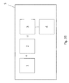
- FIG. 32 shows an example of a schematic summary of a kit in accordance with various embodiments of the present invention.
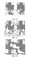
- FIG. 33 shows an example of a schematic summary of an immunoassay method in accordance with various embodiments of the present invention.
- the present invention includes a variety of aspects, which may be combined in different ways.
- the following descriptions are provided to list elements and describe some of the embodiments of the present invention. These elements are listed with initial embodiments, however it should be understood that they may be combined in any manner and in any number to create additional embodiments.
- the variously described examples and preferred embodiments should not be construed to limit the present invention to only the explicitly described systems, techniques, and applications. Further, this description should be understood to support and encompass descriptions and claims of all the various embodiments, systems, techniques, methods, devices, and applications with any number of the disclosed elements, with each element alone, and also with any and all various permutations and combinations of all elements in this or any subsequent application.
- Embodiments of the present invention may provide antibodies, monoclonal antibodies, any fragments thereof such as antigen binding fragments thereof, and methods thereof that specifically bind to PAX8 and may be used for the detection of PAX8 in the detection, diagnosis, prognosis, prediction of outcome of treatment, assessment of efficacy, assessment of recurrence, or the like for several types of cancers.
- the monoclonal antibody may be an antibody fragment, a mouse monoclonal antibody, a humanized monoclonal antibody, a human monoclonal antibody, an antibody conjugated with a label, an antibody labeled with a detectable signal or stain, an antibody labeled with a toxin, or the like.
- labels may include but are not limited to radioactive element, magnetic particles, radioisotope, fluorescent dye, enzyme, toxin, signal, stain, any combination thereof, or the like.
- Systems and methods of the present invention may relate to the monoclonal antibody or its antigen binding portion capable of binding to PAX8.
- the present invention may provide a monoclonal antibody or its antigen binding portion thereof capable of binding to PAX8 but which does not bind to B-cells.
- Mouse monoclonal antibodies may be commonly used in immunoassay methods to identify specific analytes, including as primary antibodies in immunohistochemistry procedures.
- Mouse monoclonal antibodies specific for the protein target of interest can typically be produced using generally known procedures.
- exposing a mouse to the antigen of interest e.g. a peptide fragment of the desired target or the full-length protein target
- the antigen of interest e.g. a peptide fragment of the desired target or the full-length protein target
- B-cells may be isolated from the mouse spleen and the antibodies produced may be evaluated for their suitability as primary antibodies in IHC.
- the associated B-cell may be fused with a tumor cell using known procedures, perhaps resulting in a hybridoma, a new cell line that can endlessly replicate and may continuously produce the desired antibody.
- Monoclonal antibodies may be preferred over polyclonal antibodies for several reasons.
- monoclonal antibodies may be derived from a single B-cell and as such may recognize a single epitope, perhaps resulting in greater specificity.
- Monoclonal antibodies may also be conveniently and reproducibly generated in cell culture, perhaps resulting in a constant supply of the desired antibody.
- a monoclonal antibody may include but is not limited to an isolated monoclonal antibody, a mouse monoclonal antibody, a rabbit monoclonal antibody, a goat monoclonal antibody, a horse monoclonal antibody, a chicken monoclonal antibody, a humanized monoclonal antibody, a chimeric antibody, and any combination thereof, or the like.
- Embodiments of the present invention may provide a kit (5) and methods of using such kit which may be a diagnostic or prognostic kit that includes an antibody or fragment thereof or even portion thereof as discussed herein with perhaps an antibody detection element of the antibody, fragment, or portion thereof when bound to an antigen
- a biological sample (2) may be contacted with the antibody, fragment, or portion thereof and detection of the bound antibody-antigen may be determined.
- a biological sample may include but is not limited to a normal tissue, neoplastic tissue, kidney tissue, ovarian tissue, thyroid tissue, endometrial tissue, renal tissue, tonsil tissue, pancreas tissue, colon tissue, lymph node tissue, neoplastic pancreatic tissue, stomach tissue, bladder tissue, prostate tissue, lung tissue, breast tissue, or the like.
- use of the antibody or fragment thereof or portion thereof or composition may be performed on an automated staining device such as with methods including but not limited to immunoassay, immunohistochemistry (IHC), IHC of FFPE, ICH of frozen-tissue sections, and ELISA.
- detection of the antibody-antigen binding may be made manually, automatically, via image analysis or the like and may even be made via an automated staining device.
- embodiments of the present invention may provide obtaining tissue from an animal or human to be tested (6), fixing or freezing said tissue (7), treating said fixed or frozen tissue to unmask epitopes to PAX8 (8), contacting said treated tissue with an antibody or fragment thereof as discussed herein in an amount and under conditions such that said antibody or fragment thereof binds to a PAX8 protein if said protein is present in said tissue (9); and perhaps even detecting the presences of said bound antibodies (10), as schematically represented in FIG. 33 .
- FIG. 32 shows a schematic summary of various embodiments of the present invention including a kit (5) which may provide an antibody, fragment thereof, portion thereof, in a composition or even in a cocktail (1), perhaps even provided from a hybridoma, the antibody (1) or the like may be contact with a biological sample (2) to form at least one antibody-antigen complex (3) which may then be detected with a detector (4).
- a kit (5) which may provide an antibody, fragment thereof, portion thereof, in a composition or even in a cocktail (1), perhaps even provided from a hybridoma, the antibody (1) or the like may be contact with a biological sample (2) to form at least one antibody-antigen complex (3) which may then be detected with a detector (4).
- the PAX8 antibody clone BC12 can be obtained by immunizing Balb/C mice with a full length human PAX8 recombinant protein obtained by E. coli expression.
- the PAX8 proteins may be injected into the BALB/c mice, with an adjuvant, via subcutaneous and intraperitoneal injections alternatively, about 5 times at about three week intervals.
- the immune reactivity to PAX8 may be assessed by direct ELISA on recombinant PAX8 protein. Mice with the highest titer may be chosen for developing hybridomas by cell fusion.
- a hybridoma clone demonstrating the best reactivity to PAX8 on human tissues may be chosen and may be designated as BC12.
- the BC12 clone may be tested for isotype and may be identified as a mouse IgG1/kappa.
- the BC12 antibody may be produced by large scale tissue culture of the hybridoma cells and by ascites in BALB/c mice. The supernatant and antibody ascites may be collected and the antibody may be purified by Protein A affinity column. BC12 demonstrated specific reactivates to human PAX8 protein by ELISA, Western blotting, and even human tissues.
- Mouse monoclonal anti-PAX8 antibody [BC12] may be produced using these general procedures and may be evaluated by immunohistochemistry for sensitivity and specificity on a variety of normal and neoplastic tissues, particularly in comparison to the previously known rabbit polyclonal PAX8 antibody that is widely used.
- PAX8 recombinant protein may be cloned and expressed from E. Coli .
- PAX8 cDNA may be cloned and purified.
- the PAX8 cDNA may be digested by restriction enzymes and ligated into the pET30a-GST vector.
- BL21 cells may be transformed with the construct.
- the colonies expressing the correct size of recombinant protein may be selected and sequenced.
- a further scale up production may be performed by culturing the E. coli in LB media containing 0.5 mM IPTG.
- the final PAX8 recombinant protein may be purified and analyzed by SDS-PAGE.
- mice Female BALB/c (about 6 to about 8 weeks old) mice may be immunized intraperitoneally (i.p.) with about 100 ⁇ g human PAX8 protein per mouse in complete Freund's adjuvant. About three weeks later, the mice may be boosted with another 100 ⁇ g human PAX8 per mouse in incomplete Freund's adjuvant about 4 more times in about 3 week intervals. Mice may be bled from the tails, and sera may be collected and stored at ⁇ 20° C. for later analysis of antibody titers by enzyme-linked immunosorbent assay (ELISA).
- ELISA enzyme-linked immunosorbent assay
- Hybridomas producing antibodies to PAX8 may be generated by standard techniques from splenocytes of PAX8-immunized BALB/c mice. Briefly, splenocytes from PAX8-immunized mice may be fused to P3-X63-Ag 8.653 myeloma cells (non-secreting myeloma derived from SP2/0 Balb/c myeloma cells) by incubation with about 50% polyethylene glycol at a ratio of about 4:1.
- cells may be pelleted by centrifugation at about 3000 ⁇ g for about 10 minutes, washed in about 25 ml of PBS, recentrifuged, and cell pellet may be resuspended in about 100 ml of fresh Dulbecco's Medium containing about 20% fetal bovine serum (Hyclone, Utah, Co). Aliquots of about 100 ⁇ l can be added to each well of ten 96-well microtiter plates (Corning, Lowell, Mass.).
- DMEM culture medium supplemented with about 1M hypoxanthine (HT), about 4 mM aminopterin and about 160 mM thymidine (HAT) can be added to each microtiter well.
- Media may be replaced after about 4 days with complete media (perhaps containing HAT and HT). Over the following about 10 days, media may be removed and replaced with fresh media with reduced or perhaps even no HAT and HT added.
- Hybridoma supernatants may be screened by ELISA for antibody reactivity to PAX8, and hybridoma clones may then be selected and stabilized by cloning twice by limiting dilution.
- Hybridoma cells referred to as Anti PAX8 Mouse hybridoma clone BC12 Lot: 042811 have been deposited at American Type Culture Collection (ATCC) in Manassas, Va. on May 4, 2011 and the he deposit received ATCC Patent Deposit Designation No. PTA-11873.
- ATCC American Type Culture Collection
- Host anti-sera immune responses to PAX8 may be measured by ELISA. Briefly, a solution of PAX8 (1 ⁇ g/ml) in phosphate-buffered saline (PBS) may be used to coat 96-well flat bottom polystyrene plates. The plates may then be blocked with about 1% bovine serum albumin (BSA)-PBS. Either diluted immune sera or hybridoma supernatants may be added and incubated at about 37° C. for about 1 hour. After washing the plates with PBS, the plates may be incubated with goat anti mouse-HRP reagents (Jackson Labs). Incubations may be done at about 37° C. for about 30 minutes. ABTS substrate may be added to develop color and the absorbance at about 405 nm (A405) may be measured in a microtiter plate reader.
- BSA bovine serum albumin
- the BC12 monoclonal antibody may be isotyped using a mouse monoclonal antibody isotyping kit (Invitrogen, Carlsbad Calif.). Briefly, about 100 ⁇ l of supernatant from mouse monoclonal antibody [BC12] cells may be added to the plate coated goat anti mouse IgG1, IgG2A, IgG2B, IgG3, IgM, and IgA. After about 30 minutes incubation, the plate may be washed 3 times with PBS and may be incubated with goat anti mouse Ig-HRP reagent. ABTS substrate may be added to develop color and the absorbance at about 405 nm (A405) may be measured in a microtiter plate reader.
- a mouse monoclonal antibody isotyping kit Invitrogen, Carlsbad Calif.
- the selected hybridoma cells from clone BC12 may be cultured with DMEM culture medium supplemented with about 10% FBS.
- the culture supernatants may be further purified by protein A affinity column.
- the hybridoma cells may also be injected into pristane-primed BALB/c mice to produce antibody ascites.
- the antibody ascites may be further purified by protein A affinity column.
- IgG concentration may be measured spectrophotometrically using the extinction coefficient for human IgG of about 1.4 (about 0.1% at about 280 nm).
- the purity of IgG may be determined by SDS-PAGE.
- the purified monoclonal antibody [BC 12] may be characterized by Western Blotting.
- Whole-cell lysates may be generated from OVCAR3, HEK293 cells with lysis buffer (about 1% NP40, about 0.5% sodium deoxycholate, and about 0.1% SDS in PBS) in the presence of protease inhibitors. Lysate (between about 20 and about 30 ⁇ g/lane) was subjected to protein gel electrophoresis using about 4 to about 12% SDS-PAGE with Tris-glycine buffer and may be transferred onto nitrocellulose filters in Tris-glycine buffer. Proteins on the blots may be visualized by incubating BC12 antibody for about 60 minutes in room temperature after blocking with blocking buffer, followed by incubating with peroxidase-conjugated goat anti-mouse immunoglobulins.
- Total RNA may be extracted from hybridomas using Qiagen kit (USA, Gaithersburg, Md.) as per the manufacturer's instructions.
- First-round RT-PCR may be carried out with QIAGEN® OneStep RT-PCR Kit.
- RT-PCR may be performed with primer sets specific for the heavy and light chains. For each RNA sample, about 12 individual heavy chain and about 11 light chain RT-PCR reactions can be set up using degenerate forward primer mixtures covering the leader sequences of variable regions. Reverse primers may be located in the constant regions of heavy and light chains. No restriction sites may be engineered into the primers.
- the RT-PCR products from the first-round reactions may be amplified in the second-round PCR. About 12 individual heavy chain and about 11 light chain RT-PCR reactions can be set up using semi-nested primer sets specific for antibody variable regions.
- the amplified cDNAs can be gel purified and may then be sequenced.
- Variable Domains were sequenced to provide isolated polynucleotides that comprise nucleic acid sequences encoding the amino acid sequences of one or more of the CDRs of the light and/or heavy chain variable regions of a monoclonal antibody described herein that binds to the PAX8 EQGLYPLPLLNSTLD epitope identified as SEQ ID NO: 3.
- the sequence of the variable region of the heavy chain is identified as SEQ ID NO: 1 and the sequence of the variable region of the light chain is identified as SEQ ID NO: 2
- embodiments of the present invention may provide a hybridoma, antibodies or fragments thereof or even portions thereof produced by the hybridoma deposited with the ATCC under ATCC Patent Deposit Designation No. PTA-11873.
- a method for producing a monoclonal antibody from the hybridoma may include culturing a hybridoma which produces a monoclonal antibody capable of specifically recognizing PAX8; and perhaps even allowing said hybridoma to produce the monoclonal antibody.
- Other embodiments may include providing an antibody which has a binding specificity of PAX8 and which does not bind to B-cells.
- an antibody or fragment thereof may have a polypeptide of the amino acid sequence encoded by the nucleic acid sequence of SEQ ID NO: 1 and/or SEQ ID NO: 2.
- Embodiments may include an antibody or fragment thereof that specifically binds to at least one polypeptide with an amino acid sequence of SEQ ID NO:3.
- an antibody may include an amino acid sequence which may be at least about 70% identical to an amino acid sequence encoded by a nucleic acid sequence of SEQ ID NO: 1 and/or SEQ ID NO: 2.
- an isolated and purified nucleic acid sequence may include a nucleic acid sequence that may be at least about 70% identical to SEQ ID NO: 1 and/or SEQ ID NO: 2.
- an antibody or fragment thereof may be provided that specifically binds to at least one polypeptide with an amino acid sequence that may be at least 70% identical to residues of SEQ ID NO: 3
- SEQ ID NO:1, SEQ ID NO:2, or perhaps even SEQ ID NO:3 may be modified in some way (e.g., change a few residues or the like). The amount of modifications may vary depending on what modifications are made and perhaps even how the modified sequence performs.
- Other modifications may include: at least about 71%, at least about 72%, at least about 73%, at least about 74%, at least about 75%, at least about 76%, at least about 77%, at least about 78%, at least about 79%, at least about 80%, at least about 81%, at least about 82%, at least about 83%, at least about 84%, at least about 85%, at least about 86%, at least about 87%, at least about 88%, at least about 89%, at least about 90%, at least about 91%, at least about 92%, at least about 93%, at least about 94%, at least about 95%, at least about 96%, at least about 97%, at least about 98%, and at least about 99%.
- epitope mapping was conducted using two assays: direct ELISA and dot blot.
- ELISA assay the sensitivity and specificity of the anti-PAX8 [BC12] antibody was determined by measuring the antibody titer at 1:500 and 1:1000. Overlapping peptides at a length of 15 amino acids each, covering the full length of the human PAX8 protein, were used to determine the preferred sequence of [BC12] binding.
- the anti-PAX8 [BC12] binds specifically to a peptide corresponding to residues 258-272 of PAX8, which is EQGLYPLPLLNSTLD SEQ ID NO:3. The result was further confirmed by dot blot assay.
- the plates were first coated with 100 ⁇ l of PAX8 peptides at 5 ⁇ g/mL in coating buffer (pH 9.5) overnight at 4° C., followed by blocking (3% BSA) at 200 ⁇ l/well for 1 hour at room temperature.
- the plates were incubated with purified PAX8 antibody at 100 ng/mL and 200 ng/mL separately for 1 hour at room temperature on an ELISA-plate shaker. Then the plates were washed five times with PBST (300 ⁇ l/well) followed by the addition of goat anti-mouse IgG-HRP to the plates and incubation for 1 hour on a plate-shaker.
- the plates were then washed with PBST (300 ⁇ l/well) and blotted to dry, and TMB was added at 100 ⁇ l/well, developed for 5 min on a shaker, followed by a stop solution (50 ⁇ l/well). Absorbance was measured at 450 nm on an ELISA plate reader according to the manufacturer's recommendation.
- a nitrocellulose membrane was blotted with 1 ⁇ l at a concentration of 1 mg/ml the peptide, quadruplicates per peptide. This membrane was incubated for 1 hour at room temperature until it was completely dry. The membrane was blocked with 3% BSA in TBST (50 mM Tris, 0.5 M NaCl, 0.05% Tween-20, pH 7.4) for 1 hour at room temperature, then mouse anti PAX8 antibody [BC12] was added at 200 ng/ml for 1 hr at RT in TBST.
- the membrane was washed for 3 times (10 minutes each) in TBST on an orbital shaker, followed by incubating with secondary antibody goat anti mouse IgG1-AP for 1 hour at room temperature in TBST.
- the membrane was washed 3 times (10 minutes each) in TBST on a rocker.
- the binding was detected by adding Western Glo Chemiluminescent detection reagents and exposing to film.
- Immunohistochemistry using the mouse monoclonal PAX8 antibody [BC12] may be performed on formalin-fixed paraffin embedded (FFPE) tissue samples using procedures generally known to those in the art, as generally exemplified by the following non-limiting examples (washes with Tris-buffered saline, pH about 7.6, between steps):
- FIGS. 1-5 show several examples of the PAX8 staining on normal and tumor tissues for kidney and ovarian examples.
- the monoclonal mouse PAX8 antibody may offer distinct advantages with its improved specificity and even particularly its lack of cross-reactivity with lymphocytes, pancreatic tissue (islets of Langerhans), and neuroendocrine cells of the stomach.
- FIGS. 6 , 7 , 8 , and 9 show comparisons of the mouse monoclonal antibody [BC12] with the rabbit polyclonal antibody, demonstrating the similarities in positive staining for PAX8 by both antibodies, but the greater specificity of BC12, which may not stain B-cells, as compared to the rabbit monoclonal antibody, where B-cell staining may be observed.
- the mouse monoclonal PAX8 antibody [BC12] may have an advantage of specificity versus pancreatic tissue and certain neuroendocrine tumors such as carcinoids; whereas the rabbit polyclonal antibody may stain normal and neoplastic pancreatic tissues, and may also stain certain carcinoids.
- BC12 may not stain pancreatic tissue as may be understood in FIGS. 10 , 11 , 12 , and 13 .
- BC12 has demonstrated increased specificity on stomach tissue as well.
- the rabbit polyclonal PAX8 antibody may stain a subset of cells in stomach tissue that may not be stained by BC12 as can be understood in FIGS. 14 and 15 . Cells stained in FIG. 15 stained positive by a chromogranin IHC assay; and thus, confirmed as neuroendocrine origin.
- the mouse monoclonal PAX8 antibody [BC12] may be suitable for use in many variations of the above protocols and other methods known to those in the art. Specimens stained with BC12 may be archived using a permanent mounting media and a coverslip. The antibody [BC12] may also be used in an automated staining instrument, using standard protocols.
- detection methods e.g., fluorescence
- detection enzymes e.g., alkaline phosphatase (AP), beta-galactosidase
- chromogens e.g., 3-amino-9-ethylcarbazole, 5-bromo-4-chloro-3-indolyl phosphate, 3,3′,5,5′-tetramethylbenzidine, 5-bromo-4-chloro-3-indolyl- ⁇ -D-glucuronide
- the epitope for BC12 was shown to be included in the residues 258-272 of PAX8, which is EQGLYPLPLLNSTLD SEQ ID NO:3.
- the epitope of the mouse monoclonal PAX8 antibody, or a portion thereof, may be a useful antigen for the production of new monoclonal antibodies, including production in species other than mouse (e.g. rabbit, goat, horse, chicken, etc.).
- BC12 in immunohistochemistry of formalin-fixed paraffin embedded tissues is described here, its utility in other immunoassays may be readily envisioned and are meant to be included in this application. In particular, it may be well known that many of the same reagents used in IHC of FFPE may also be used in IHC of frozen-tissue sections. BC12 may also be useful in other immunoassays, including ELISA, perhaps using generally known methods.
- a PAX8 antibody may be used in conjunction with one or more additional primary antibodies as part of a cocktail, to perform a “double-stain” procedure (also described as multi-stain or even multiplex).
- double-stain procedures may be generally well known in the art; however, the best combinations of primary antibodies for a particular diagnostic application may not be known.
- a mouse monoclonal PAX8 antibody [BC12] could be combined with one or more antibodies in a single primary antibody cocktail.
- At least one of the additional antibodies could be derived from a species perhaps even other than mouse such as a rabbit antibody.
- Species may include but is not limited to mouse, rabbit, goat, horse, chicken, human, any combinations thereof, or the like.
- the multiple antibodies in the primary antibody cocktail may be differentiated in the subsequent detection and even visualization steps. For example, following incubation of the tissue sample with the primary antibody cocktail, a cocktail of goat anti-mouse antibody conjugated to HRP and a goat anti-rabbit antibody conjugated to AP may be applied. Subsequently, chromogens specific for HRP and AP may be sequentially applied.
- mouse primary antibodies including BC12
- rabbit primary antibodies could result in red (Fast Red) staining
- Multiplex IHC may also employ primary antibodies from the same host species, resulting the staining of the same color; however, the antibodies may be distinguished by different cellular localization patterns (cytoplasmic, membrane or nuclear).
- multiplex IHC may be performed by preparing FFPE tissues for staining in the usual manner, including dewaxing, hydration, and antigen retrieval.
- a cocktail of primary antibodies in a buffered diluent with carrier protein [e.g. BSA] and preservative [e.g. sodium azide]) is applied to the tissue sample for a period of typically 30 minutes.
- the primary antibodies in the cocktail are isolated from two different host-species (e.g. mouse and rabbit).
- a primary antibody cocktail containing mouse anti-p63 and rabbit anti-P504S is commonly used in prostate diagnosis.
- Secondary antibodies conjugated to enzyme for detection e.g.
- alkaline phosphatase [AP], horseradish peroxidase [HRP]) may then be applied to the tissue sample, typically for a period of 30 minutes.
- These secondary antibody conjugates are typically raised in goat and bind the mouse or rabbit IgG of the primary antibodies previously applied.
- a cocktail of goat anti-mouse-HRP and goat anti-rabbit-AP is often applied to the mouse anti-p63 and rabbit anti-P504S described above.
- p63 a mouse antibody
- P504S a rabbit antibody
- the presence and localization of both antibodies may then be distinguished by the sequential application of chromogens resulting in different colors.
- each of the applied chromogens reacts with only one of the two detection enzymes (AP or HRP) to produce a colored stain at the site of the antibody complex.
- AP detection enzyme
- HRP two detection enzymes
- DAB 3,3′-diaminobenzidine
- Fast Red a diazonium salt and naphthol phosphate
- Multiplex IHC has several advantages over traditional single-stain methods. For example, doubles-staining may take less time and use less reagents than a single-stain method. The opportunity to visualize perhaps by color results of two or more antigens in the same tissue section may greatly ease the pathologist's interpretation. Double-staining also has the advantage of consuming less of a tissue sample (i.e. a single section), thus conserving precious tissue for other tests.
- Double-staining method including but not limited to the use of more than two antibodies, the use of species other than mouse and rabbit, other chromogens and detection systems, a different order of detection steps, the sequential application of antibody regents instead of the use of cocktails, the use of goat anti-mouse-AP and goat anti-rabbit-HRP secondary antibodies, and perhaps even modifications resulting in three or more colors (which may require a denaturing step).
- An anti-PAX8 antibody such as, but not limited to BC12, may be used in the primary antibody cocktail of double-stain procedures in an method that may be useful for clinical diagnosis. For example, in cases where a tumor of unknown origin is being investigated, or a differential diagnosis between a kidney cancer and another cancer is being considered, the combination of PAX8 with another antibody may aid in the diagnosis.
- An antibody cocktail may include a composition with at least two antibodies or fragments thereof where at least one antibody binds specifically to PAX8 and at least one other antibody binds to an antigen including but not limited to: GATA-3, p63, PSA, ER, Mammaglobin, GCDFP-15, NKX3.1, Napsin A, TTF-1, CK20, CDX2, ERG, or the like, and any combination thereof.
- Compositions may include combination such as but not limited to PAX8 and GATA-3; PAX8 and p63; PAX8 and PSA; PAX8 and PSA and GATA-3; PAX8 and ER and Mammoglobin and GCDFP-15; PAX8 and ER; PAX8 and Mammoglobin; PAX8 and GCDFP-15; PAX8 and NKX3.1; PAX8 and Napsin A and TTF-1; PAX8 and Napsin A; PAX8 and TTF-1; PAX8 and CD 2 O and CDX2; PAX8 and CD20; PAX8 and CDX2; PAX8 and ERG and GATA-3; PAX8 and ERG; or the like, or any combination thereof.
- the antibody cocktail may include any antibody which specifically binds to PAX8; may be an antibody which specifically binds to PAX8 but not to B-cells; may be the BC12 antibody or portions thereof, fragments thereof, or the like; may be an antibody which specifically binds to PAX8 but not to neuroendocrine cells, pancreatic cells, or any combination thereof; or the like.
- Methods for antibody cocktails may include detecting at least two different proteins in a biological sample, comprising the steps of contacting a biological sample (2) with a composition comprising at least two antibodies or fragments thereof, wherein at least one of said at least two antibodies or fragments thereof binds specifically to at least PAX8, to form an antigen-antibody complex (3); and perhaps even detecting said antigen-antibody complex.
- Antibodies that may be useful for diagnosis when combined with a mouse monoclonal PAX8 antibody [BC12] in a primary antibody cocktail for use in multi-stain procedures may include:
- example cocktails were stained on normal tissues where the antigen is known to be present in both normal and neoplastic tissues.
- a double-stain IHC primary antibody cocktail containing mouse monoclonal PAX8+rabbit monoclonal GATA-3 may be useful for diagnosis, including for the discrimination of renal carcinomas (PAX8) from breast or urothelial carcinomas (GATA-3).
- p53 homologue p63 encodes for different isotypes able to either transactivate p53 reporter genes (TAp63) or act as p53-dominant-negatives.
- p63 is detected in prostatic basal cells in normal prostate, however, it is negative in malignant tumors of the prostate gland.
- p63 is a useful differential marker for benign and malignant tumors of the prostate gland.
- p63 is also a marker of myoepithelial cells in breast ducts and may be useful in identifying breast carcinoma.
- As a marker or urothelial carcinoma p63 may also stain renal pelvis urothelial carcinoma.
- a double-stain of PAX8+p63 may be useful in the differential diagnosis of carcinoma of the renal pelvis from renal cell carcinoma.
- FIGS. 16 and 17 show an example of multiplex IHC staining of mouse monoclonal PAX8 [BC12]+rabbit monoclonal p63 [EPR5701] (Cell Signaling) on renal cell carcinoma ( FIG. 16 ) and normal bladder ( FIG. 17 ).
- PSA is a chymotrypsin-like serine protease produced by the prostate epithelium. PSA is used to confirm prostatic acinar cell origin in primary and metastatic carcinomas and to rule out non-prostatic carcinoma mimics. A triple-stain of PAX8+PSA+GATA-3 may be useful in the differential diagnosis of renal cell carcinoma, prostate adenocarcinoma and urothelial carcinoma.
- GATA-3 (GATA binding protein 3) is a member of the GATA family of transcription factors. Among several other roles, GATA-3 has is as a key player in luminal cell differentiation in the mammary gland. The expression of GATA-3 has a strong association with the expression of estrogen receptor-alpha (ER) in breast cancer, and there is mounting evidence that GATA-3 can be used as a clinical marker to determine response to hormonal therapy and to refine the prognosis of breast cancer patients. GATA-3 has also been shown to be a novel marker for bladder cancer. In one study, GATA-3 stained 67% of 308 urothelial carcinomas, but none for prostate or renal carcinomas. A double-stain of PAX8+PSA+GATA-3 may be useful in the differential diagnosis of renal cell carcinoma, prostate adenocarcinoma and urothelial carcinoma, see FIGS. 18 , 19 , and 20 .
- FIGS. 18 , 19 , and 20 show an example of multiplex IHC staining of mouse monoclonal PAX8 [BC12]+rabbit monoclonal PSA [EP1588Y] (e.g., Biocare Medical)+rabbit monoclonal GATA-3 [D13C9] (Cell Signaling) on renal cell carcinoma ( FIG. 18 ), prostate adenocarcinoma ( FIG. 19 ) and urothelial carcinoma ( FIG. 20 ).
- Estrogen receptor alpha is a nuclear transcription factor and a member of the steroid hormone receptor family. ER is routinely used in the diagnosis, prognosis and prediction of response to hormonal therapy for breast cancer patients.
- Mammaglobin is a mammary-specific member of the uteroglobin family and is known to be overexpressed in human breast cancer. In normal breast tissue, mammaglobin labels breast ductal and lobular epithelial cells. However, mammaglobin is expressed in a higher percentage of lobular carcinoma versus ductal cell carcnimoma. A double-stain or perhaps even a triple-stain of PAX8+ER+mammaglobin may be useful in the diganosis of renal cell carcinoma versus breast carcinoma.
- FIGS. 21 and 22 show staining of the PAX8+ER+mammaglobin cocktail.
- FIGS. 21 and 22 show multiplex IHC staining of a mouse monoclonal PAX8 [BC12]+rabbit monoclonal ER[SP1] (Biocare Medical)+rabbit monoclonal Mammaglobin [31-A5] (Zeta) on renal cell carcinoma ( FIG. 21 ) and normal breast ( FIG. 22 ).
- NKX3.1 The homeodomain containing transcription factor NKX3.1 is a putative prostate tumor suppressor that is expressed in a largely prostate-specific and androgen-regulated manner.
- NKX3.1 protein has been found to be positive in the vast majority of primary pro static adenocarcinomas. The sensitivity for identifying metastatic prostatic adenocarcinomas overall was 98.6% (68/69 cases positive) for NKX3.1.
- NKX3.1 stains nuclei in both normal and prostate cancer.
- a double-stain of PAX8+NKX3.1 may be useful in the differential diagnosis of renal cell carcinoma from prostate adenocarcinoma, see FIGS. 23 and 24 .
- FIGS. 23 and 24 show multiplex IHC staining of mouse monoclonal PAX8 [BC12]+rabbit polyclonal NKX3.1 (e.g., Biocare Medical, CP422) on renal cell carcinoma ( FIG. 23 ) and prostate adenocarcinoma ( FIG. 24 ).
- Napsin A is an aspartic detected in the cytoplasm of type 2 pneumocytes and alveolar macrophages. Napsin A is a sensitive and specific marker for lung adenocarcinomas. In one study, Napsin A identified 70 of 83 (84%) lung adenocarcinomas. A double-stain of PAX8+Napsin A may be useful in the differential diagnosis of renal cell carcinoma from lung adenocarcinoma.
- FIGS. 25 and 25 show staining of a PAX8+Napsin A antibody cocktail.
- FIGS. 25 and 26 show multiplex IHC staining of mouse monoclonal PAX8[BC12]+rabbit polyclonal Napsin A (Biocare Medical, PP434) on renal cell carcinoma ( FIG. 25 ) and normal lung ( FIG. 26 ).
- Cytokeratin 20 is a 46 kDa intermediate filament protein that is a useful marker in the identification of colon adenocarcinoma.
- a double-stain of PAX8+CK20 may be useful in the differential diagnosis of renal cell carcinoma from colon adenocarcinoma.
- FIGS. 27 and 28 show staining of a PAX8+CK20 antibody cocktail.
- FIGS. 27 and 28 show multiplex IHC staining of mouse monoclonal PAX8 [BC12]+rabbit monoclonal CK20 [EP23] (Epitomics) on renal cell carcinoma ( FIG. 27 ) and normal colon ( FIG. 28 ).
- FIGS. 29 , 30 , and 31 show multiplex IHC staining of mouse monoclonal PAX8 [BC12]+rabbit monoclonal ERG [ER3863] (Epitomics)+rabbit monoclonal GATA-3 [D13C9] (Cell Signaling) on renal cell carcinoma ( FIG. 29 ), prostatic adenocarcinoma ( FIG. 30 ) and urothelial carcinoma ( FIG. 31 ).
- ERG and GATA-3 staining may be distinguished by morphology. (Note that normal endothelial cells present in most tissues are known to stain with ERG.)
- the mouse monoclonal PAX8 antibody [BC12] may be specific for detection of PAX8 and may be useful in immunohistochemical procedures for diagnosis of several types of cancers in human tissue samples.
- BC12, nucleic acid sequences SEQ ID NO: 1 and/or SEQ ID NO: 2, and amino acid SEQ ID NO: 3 can be used and have advantages over previously known PAX8 antibodies, including greater specificity versus B-cells, as well as a lack of cross-reactivity and staining of pancreatic tissues and neuroendocrine cells of the stomach.
- the basic concepts of the present invention may be embodied in a variety of ways. It involves both antibody techniques as well as devices to accomplish the appropriate antibody.
- the antibody techniques are disclosed as part of the results shown to be achieved by the various devices described and as steps which are inherent to utilization. They are simply the natural result of utilizing the devices as intended and described.
- the devices are disclosed, it should be understood that these not only accomplish certain methods but also can be varied in a number of ways. Importantly, as to all of the foregoing, all of these facets should be understood to be encompassed by this disclosure.
- each of the various elements of the invention and claims may also be achieved in a variety of manners.
- an element is to be understood as encompassing individual as well as plural structures that may or may not be physically connected.
- This disclosure should be understood to encompass each such variation, be it a variation of an embodiment of any apparatus embodiment, a method or process embodiment, or even merely a variation of any element of these.
- the words for each element may be expressed by equivalent apparatus terms or method terms—even if only the function or result is the same. Such equivalent, broader, or even more generic terms should be considered to be encompassed in the description of each element or action.
- the disclosure of a “detection” or “detector” should be understood to encompass disclosure of the act of “detecting”—whether explicitly discussed or not—and, conversely, were there effectively disclosure of the act of “detecting”, such a disclosure should be understood to encompass disclosure of a “detector” and even a “means for detecting.”
- Such changes and alternative terms are to be understood to be explicitly included in the description.
- each such means should be understood as encompassing all elements that can perform the given function, and all descriptions of elements that perform a described function should be understood as a non-limiting example of means for performing that function.
- Ovarian carcinoma subtypes are different diseases : implications for biomarker studies .
- the value of tumours marker CA 125 in surgical pathology Histopathology . 1987 March; 11(3): 287-94 Kuehn, A. et al.
- Expression analysis of kidney-specific cadherin in a wide spectrum of traditional and newly recognized renal epithelial neoplasms diagnostic and histogenetic implications . Am J Surg Pathol.
- each of the antibody devices as herein disclosed and described ii) the related methods disclosed and described, iii) similar, equivalent, and even implicit variations of each of these devices and methods, iv) those alternative designs which accomplish each of the functions shown as are disclosed and described, v) those alternative designs and methods which accomplish each of the functions shown as are implicit to accomplish that which is disclosed and described, vi) each feature, component, and step shown as separate and independent inventions, vii) the applications enhanced by the various systems or components disclosed, viii) the resulting products produced by such systems or components, ix) each system, method, and element shown or described as now applied to any specific field or devices mentioned, x) methods and apparatuses substantially as described hereinbefore and with reference to any of the accompanying examples, xi) an apparatus for performing the methods described herein comprising means for performing the steps, xii) the various combinations and permutations of each of the elements disclosed,
- any claims set forth at any time are hereby incorporated by reference as part of this description of the invention, and the applicant expressly reserves the right to use all of or a portion of such incorporated content of such claims as additional description to support any of or all of the claims or any element or component thereof, and the applicant further expressly reserves the right to move any portion of or all of the incorporated content of such claims or any element or component thereof from the description into the claims or vice-versa as necessary to define the matter for which protection is sought by this application or by any subsequent continuation, division, or continuation-in-part application thereof, or to obtain any benefit of, reduction in fees pursuant to, or to comply with the patent laws, rules, or regulations of any country or treaty, and such content incorporated by reference shall survive during the entire pendency of this application including any subsequent continuation, division, or continuation-in-part application thereof or any reissue or extension thereon.
Landscapes
- Health & Medical Sciences (AREA)
- Life Sciences & Earth Sciences (AREA)
- Chemical & Material Sciences (AREA)
- Immunology (AREA)
- Engineering & Computer Science (AREA)
- Molecular Biology (AREA)
- Urology & Nephrology (AREA)
- Hematology (AREA)
- Biomedical Technology (AREA)
- General Health & Medical Sciences (AREA)
- Biochemistry (AREA)
- Medicinal Chemistry (AREA)
- Organic Chemistry (AREA)
- Physics & Mathematics (AREA)
- Food Science & Technology (AREA)
- Analytical Chemistry (AREA)
- Microbiology (AREA)
- Cell Biology (AREA)
- General Physics & Mathematics (AREA)
- Pathology (AREA)
- Biotechnology (AREA)
- Proteomics, Peptides & Aminoacids (AREA)
- Genetics & Genomics (AREA)
- Biophysics (AREA)
- Oncology (AREA)
- Peptides Or Proteins (AREA)
- Hospice & Palliative Care (AREA)
- Preparation Of Compounds By Using Micro-Organisms (AREA)
- Gastroenterology & Hepatology (AREA)
Abstract
Description
- 1) Sections (˜5 μm) of formalin fixed paraffin-embedded tissues may be mounted on commercially available microscope slides coated with polylysine.
- 2) Sections may be deparaffinized (using xylenes or a xylene-substitute) and may be rehydrated through a series of alcohol/water solutions, followed by blocking of endogenous peroxidases with about 3% hydrogen peroxide solution.
- 3) Samples may be subjected to heat-induced antigen retrieval using a citrate buffer in a pressure cooker (Reveal, Decloaking Chamber; Biocare Medical) and may be heated to about 125° C. for about 30 seconds. [Other antigen retrieval methods known to those skilled in the art (e.g., steamer, microwave oven, and enzyme) may also be acceptable.] Tissues may be allowed to cool for about 10 minutes and then may be rinsed with deionized water.
- 4) The PAX8 antibody [BC12] may be applied in a Tris-buffered solution (pH about 6.2) with bovine serum albumin as carrier protein for about 30 minutes.
- 5) Detection of the PAX8 antibody with a horseradish peroxidase (HRP) conjugated secondary antibody (
MACH 4 Universal HRP-Polymer Detection, Biocare Medical) may be accomplished in two steps. An initial application of a rabbit anti-mouse IGg antibody for about 10 minutes may be followed by incubation with a goat anti-rabbit-HRP conjugate for about 10 minutes. - 6) In a final detection step, 3,3′-diaminobenzidine (DAB) in buffer containing about 0.02% hydrogen peroxide (Betazoid DAB, Biocare Medical) may be applied. The oxidation of DAB through an HRP-mediated mechanism may result in precipitation of a brown, chromogenic product, perhaps allowing identification of sites of PAX8 expression.
- 7) Slides may be briefly counterstained in a modified Mayer's hematoxylin.
| Diagnosis of tumor of | |
| unknown origin or | |
| Antibody Cocktail | differential diagnosis |
| PAX8 + GATA-3 | Renal vs Bladder or Breast |
| PAX8 + p63 | Renal vs Renal Pelvis |
| PAX8 + PSA + GATA-3 | Renal vs Prostate vs Bladder |
| PAX8 + ER and/or Mammaglobin and/or | Renal vs Breast |
| GCDFP-15 | |
| PAX8 + NKX3.1 | Renal vs Prostate |
| PAX8 + Napsin A and/or TTF-1 | Renal vs. Lung |
| PAX8 + CK20 and/or CDX2 | Renal vs. Colon |
| PAX8 + ERG + GATA-3 | Renal vs Prostate vs Bladder |
| Date of | |||
| Pat. No. | Kind | Patent | Inventor |
| 6,051,693 | 2000 Apr. 18 | Handley et al. | |
| 7,422,739 | B2 | 2008 Sep. 09 | Anderson et al. |
| 7,785,803 | B2 | 2010 Aug. 31 | Achen et al. |
| 7,875,705 | B2 | 2011 Jan. 25 | Iwanari et al. |
| 7,935,794 | B2 | 2011 May 03 | Pullen |
| 7,935,795 | B2 | 2011 May 03 | Nakajima |
| 7,935,796 | B2 | 2011 May 03 | Lee et al. |
| 7,973,138 | B2 | 2011 Jul. 05 | Liang et al. |
| 8,153,126 | B2 | 2012 Apr. 10 | Violette et al. |
| Publication No. | Kind | Pub Date | Assignee |
| 20050186642 | A1 | 2005 Aug. 25 | Tacha |
| 20100004782 | A1 | 2010 Feb. 25 | Tacha |
| 20100047825 | A1 | 2010 Feb. 25 | Biocare Medical, |
| Inc. | |||
| Country | ||||
| Foreign Patents | Code | Kind | Pub. Date | Patentee/Applicant |
| 2005083802 | WO | A1 | 2005 Sep. 09 | Biocare Medical, |
| LLC | ||||
| Nonpatent Literature |
| Albadine, R., Schultz, L., Illei, P., Ertoy, D., Hicks, J., Sharma, R., Epstein, J., Netto, G.; |
| PAX8(+)/p63(−) Immunostaining Pattern in Renal Collecting Duct Carcinoma (CDC), A |
| Useful Immunoprofile in the Differential Diagnosis of CDC Versus Urothelial Carcinoma |
| of Upper Unirary Tract; Am/Surg Pathol, Vol. 34, No. 7, July 2010, pp 965-969. |
| Avery, A. K. et al. Use of antibodies to RCC and CD10 in the differential diagnosis of |
| renal neoplasms. Am J Surg Pathol. 2000 February; 24(2): 203-10 |
| Bowen, N. J. et al. Emerging roles for PAX8 in ovarian cancer and endosalpingeal |
| development. Gynecol Oncol. 2007, February; 104(2): 331-7 |
| Buchwalow, I. et al. Immunohistochemistry: Basics and Methods. Springer Press. 1st ed. |
| 2010 |
| Carson, F. L. et al. Histotechnology: A Self-Instructional Text. American Society for |
| Clinical Pathology; 3rd ed. 2009 |
| Geramizadeh, B. et al. Useful markers for differential diagnosis of oncocytoma, |
| chromophobe renal cell carcinoma and conventional renal cell carcinoma, Indian J |
| Pathol Microbiol. 2008 April-June; 51(2): 167-71 |
| Haynes, C., Sangoi, A., Pai, R.; PAX8 Is Expressed in Pancreatic Well-Differentiated |
| Neuroendocrine Tumors and in Extrapancreatic Poorly Differentiated Neuroendocrine |
| Carcinomas in Fine-Needle Aspiration Biopsy Specimens; Cancer Cytopathology, Jun. |
| 25, 2011, pp 193-201. |
| Köbel, M. et al. Ovarian carcinoma subtypes are different diseases: implications for |
| biomarker studies. PLoS Med. 2008, Dec. 2; 5(12): e232 |
| Koelma, I. A. et al. The value of tumours marker CA 125 in surgical pathology, |
| Histopathology. 1987 March; 11(3): 287-94 |
| Kuehn, A. et al. Expression analysis of kidney-specific cadherin in a wide spectrum of |
| traditional and newly recognized renal epithelial neoplasms: diagnostic and histogenetic |
| implications. Am J Surg Pathol. 2007 October; 31(10): 1528-33 |
| Laury, A., Hornick, J., Perets, R., Krane, J., Corson, J., Drapkin, R., Hirsch, M.; PAX8 |
| Reliably Distinguishes Ovarian Serous Tumors from Malignant Mesothelioma; Am/Surg |
| Pathol, Vol. 34, No. 5, May 2010, pp 627-635. |
| Laury, A., Perets, R., Piao, H., Krane, J., Barletta, J., French, C., Chirieac, L., Lis, R., |
| Loda, M., Hornick, J., Drapkin, R., Hirsch, M.; A Comprehensive Analysis of PAX8 |
| Expression in Human Epithelial Tumors; Am/Surg Pathol, Vol. 35, No. 6, June 2011, pp |
| 816-826 |
| Leake, J. et al. Immunocytochemical and serological expression of CA 125: a |
| clinicopathological study of 40 malignant ovarian epithelial tumours. Histopathology. |
| 1994 January; 24(1): 57-64 |
| Lee, A. H. et al. The expression of Wilms' tumour-1 and CA125 in invasive micropapillary |
| carcinoma of the breast. Histopathology. 2007 December; 51(6): 824-8 |
| Long, K., Srivastava, A., Hirsch, M., Hornick, J.; PAX8 Expression in Well-differentiated |
| Pancreatic Endocrine Tumors: Correlation With Clinicopathologic Features and |
| Comparison With Gastrointestinal and Pulmonary Carcinoid Tumors; Am/Surg Pathol, |
| Vol. 34, No. 5., May 2010; pp 723-729. |
| Lorenzo, P., Moreno, C., Delgado, I., Cobo-Vuilleumier, N., Meier, R., Gomez-Izquierdo, |
| L., Berney, T., Garcia-Carbonero, R., Rojas, A., Gauthier, B.; Immunohistochemical |
| assessment of Pax8 expression during pancreatic islet development and in human |
| neuroendocrine tumors; Histochem Cell Biol (2011) 136: 595-607. |
| Mazal, P. R. et al. Expression of kidney-specific cadherin distinguishes chromophobe |
| renal cell carcinoma from renal oncocytoma. Hum Pathol. 2005 January; 36(1): 22-8 |
| Mazal, P. R. et al. Expression of aquaporins and PAX-2 compared to CD10 and |
| |
| April; 18(4): 535-40. |
| Moretti, L., Medeiros, L., Kunkalla, K., Williams, M., Singh, R., Vega, F.; N-terminal |
| PAX8 polyclonal antibody shows cross-reactivity with N-terminal region of PAX5 and is |
| responsible for reports of PAX8 positivity in malignant lymphomas; Modern Pathology |
| (2011), 1-6. |
| Nonaka, D. et al. Diagnostic utility of thyroid transcription factors Pax8 and TTF-2 |
| (FoxE1) in thyroid epithelial neoplasms. Mod Pathol. 2008 February; 21(2): 192-2004 |
| Nonaka, D. et al. Expression of pax8 as a useful marker in distinguishing ovarian |
| carcinomas from mammary carcinomas. Am J Surg Pathol. 2008 October; 32(10): 1566-71 |
| Ozcan, A., Shen, S., Hamilton, C., Anjana, K., Coffey, D., Krishnan, B., Truong, L.; |
| PAX8 expression in non-neoplastic tissues, primary tumors, and metastatic tumors: a |
| comprehensive immunohistochemical study.; Modern Pathology (2011) 24, 751-764. |
| Reid-Nicholson, M. et al. Immunophenotypic diversity of endometrial adenocarcinomas: |
| implications for differential diagnosis. Mod Pathol. 2006 August; 19(8): 1091-100 |
| Sangoi, A., Ohgami, R., Pai, R., Beck, A., McKenney, J., Pai, R.; PAX8 expression |
| reliably distinguishes pancreatic well-differentiated neuroendocrine tumors and |
| pancreatic acinar cell carcinoma; Modern Pathology (2011) 24, 412-424. |
| Sosa-Pineda, B. The gene Pax4 is an essential regulator of pancreatic beta-cell |
| development. Mol Cells. 2004 Dec. 31; 18(3): 289-94 |
| Tacha, D., Zhou, D., Cheng, L.; Expression of PAX8 in Normal and Neoplastic Tissues-A |
| Comprehensive Immunohistochemical Study; Appl Immunohistochem Mol Morphol, Vol. |
| 19, No. 4, July 2011, pp 293-299. |
Claims (33)
Priority Applications (1)
| Application Number | Priority Date | Filing Date | Title |
|---|---|---|---|
| US14/004,400 US8852592B2 (en) | 2011-05-10 | 2012-05-10 | Systems and methods for anti-PAX8 antibodies |
Applications Claiming Priority (4)
| Application Number | Priority Date | Filing Date | Title |
|---|---|---|---|
| US201161484579P | 2011-05-10 | 2011-05-10 | |
| US201261588035P | 2012-01-18 | 2012-01-18 | |
| US14/004,400 US8852592B2 (en) | 2011-05-10 | 2012-05-10 | Systems and methods for anti-PAX8 antibodies |
| PCT/US2012/037367 WO2012154983A2 (en) | 2011-05-10 | 2012-05-10 | Systems and methods for anti-pax8 antibodies |
Related Parent Applications (1)
| Application Number | Title | Priority Date | Filing Date |
|---|---|---|---|
| PCT/US2012/037367 A-371-Of-International WO2012154983A2 (en) | 2011-05-10 | 2012-05-10 | Systems and methods for anti-pax8 antibodies |
Related Child Applications (1)
| Application Number | Title | Priority Date | Filing Date |
|---|---|---|---|
| US14/507,667 Continuation US9417243B2 (en) | 2011-05-10 | 2014-10-06 | Systems and methods for anti-PAX8 antibodies |
Publications (2)
| Publication Number | Publication Date |
|---|---|
| US20140004542A1 US20140004542A1 (en) | 2014-01-02 |
| US8852592B2 true US8852592B2 (en) | 2014-10-07 |
Family
ID=47140013
Family Applications (3)
| Application Number | Title | Priority Date | Filing Date |
|---|---|---|---|
| US14/004,400 Active US8852592B2 (en) | 2011-05-10 | 2012-05-10 | Systems and methods for anti-PAX8 antibodies |
| US14/507,667 Active US9417243B2 (en) | 2011-05-10 | 2014-10-06 | Systems and methods for anti-PAX8 antibodies |
| US15/222,690 Abandoned US20160370370A1 (en) | 2011-05-10 | 2016-07-28 | Systems and Methods for Anti-PAX8 Antibodies |
Family Applications After (2)
| Application Number | Title | Priority Date | Filing Date |
|---|---|---|---|
| US14/507,667 Active US9417243B2 (en) | 2011-05-10 | 2014-10-06 | Systems and methods for anti-PAX8 antibodies |
| US15/222,690 Abandoned US20160370370A1 (en) | 2011-05-10 | 2016-07-28 | Systems and Methods for Anti-PAX8 Antibodies |
Country Status (2)
| Country | Link |
|---|---|
| US (3) | US8852592B2 (en) |
| WO (1) | WO2012154983A2 (en) |
Cited By (6)
| Publication number | Priority date | Publication date | Assignee | Title |
|---|---|---|---|---|
| US20150056635A1 (en) * | 2011-05-10 | 2015-02-26 | Biocare Medical, Llc | Systems and Methods for Anti-PAX8 Antibodies |
| US9428576B2 (en) | 2013-02-28 | 2016-08-30 | Biocare Medical, Llc | Anti-p40 antibodies systems and methods |
| US9429577B2 (en) | 2012-09-27 | 2016-08-30 | Biocare Medical, Llc | Anti-uroplakin II antibodies systems and methods |
| US9816997B2 (en) | 2013-10-03 | 2017-11-14 | Biocare Medical, Llc | Anti-SOX10 antibody systems and methods |
| US10316103B1 (en) | 2012-03-30 | 2019-06-11 | Biocare Medical, Llc | Systems and methods for anti-Uroplakin III antibodies |
| US10429390B2 (en) | 2012-12-18 | 2019-10-01 | Biocare Medical, Llc | Antibody cocktail systems and methods for classification of histologic subtypes in lung cancer |
Families Citing this family (8)
| Publication number | Priority date | Publication date | Assignee | Title |
|---|---|---|---|---|
| BR112015023120A2 (en) | 2013-03-15 | 2017-11-21 | Genentech Inc | method for identifying an individual with a disease or dysfunction, method for predicting the responsiveness of an individual with a disease or dysfunction, method for determining the likelihood that an individual with a disease or dysfunction will exhibit benefit from treatment, method for selecting a therapy, Uses of a pd-11 Axis Binding Antagonist, Assay to Identify an Individual with a Disease, Diagnostic Kit, Method to Evaluate a Treatment Response, and Method to Monitor the Response of a Treated Individual |
| RU2715038C2 (en) | 2014-07-11 | 2020-02-21 | Дженентек, Инк. | Anti-pd-l1 antibodies and methods for their diagnostic use |
| CN104297481A (en) * | 2014-10-22 | 2015-01-21 | 卢洪胜 | Double-label immunohistochemical staining kit used for differential diagnosis of metastatic renal cell carcinoma |
| JP6841609B2 (en) | 2015-07-10 | 2021-03-10 | 3スキャン インコーポレイテッド | Spatial multiplexing of histological staining |
| CN109021101A (en) * | 2018-09-01 | 2018-12-18 | 无锡傲锐东源生物科技有限公司 | Anti- PAX8 protein monoclonal antibody and application thereof |
| US11976282B2 (en) | 2020-06-23 | 2024-05-07 | Qatar University | GATA3 inhibitors for the promotion of subcutaneous fat deposition |
| WO2025188810A1 (en) * | 2024-03-08 | 2025-09-12 | Agilent Technologies, Inc. | Anti-human pax8 antibody for in vitro diagnostic use by immunohistochemistry |
| CN118930648B (en) * | 2024-08-22 | 2025-02-28 | 武汉爱博泰克生物科技有限公司 | Rabbit monoclonal antibody against human PAX8 protein, antibody conjugate and its application |
Citations (12)
| Publication number | Priority date | Publication date | Assignee | Title |
|---|---|---|---|---|
| US6051693A (en) | 1982-05-21 | 2000-04-18 | The Regents Of The University Of California | CLNH11-specific antibodies |
| US6723506B2 (en) | 2000-01-20 | 2004-04-20 | Brigham And Women's Hospital | Method of identifying PAX8-PPAR gamma-nucleic acid molecules |
| US20050186642A1 (en) | 2004-02-24 | 2005-08-25 | Biocare Medical, Inc. | Immunoassay reagents and methods of use thereof |
| US7422739B2 (en) | 1992-11-13 | 2008-09-09 | Biogen Idec Inc. | Anti-CD20 antibodies |
| US7785803B2 (en) | 1996-08-23 | 2010-08-31 | Vegenics Limited | Antibody diagnostic kits and methods of use |
| US7875705B2 (en) | 2007-05-28 | 2011-01-25 | The University Of Tokyo | Tumor diagnostic agent used in PET comprising anti-ROBO1 antibody |
| US7935795B2 (en) | 2005-10-26 | 2011-05-03 | Evec Inc. | Human monoclonal antibody binding to hGM-CSF and its antigen binding portion |
| US7935794B2 (en) | 2005-07-08 | 2011-05-03 | Pfizer Ltd. | MAdCAM antibodies |
| US7935796B2 (en) | 2006-12-08 | 2011-05-03 | Lexicon Pharmaceuticals, Inc. | Monoclonal antibodies against ANGPTL3 |
| US7973138B2 (en) | 2007-09-26 | 2011-07-05 | Genentech, Inc. | Antibodies |
| US8153126B2 (en) | 2002-03-13 | 2012-04-10 | Violette Shelia M | Anti-αvβ6 antibodies |
| WO2012154983A2 (en) | 2011-05-10 | 2012-11-15 | Biocare Medical, Llc | Systems and methods for anti-pax8 antibodies |
Family Cites Families (55)
| Publication number | Priority date | Publication date | Assignee | Title |
|---|---|---|---|---|
| US4145406A (en) | 1977-04-11 | 1979-03-20 | Miles Laboratories, Inc. | Specific binding - adsorbent assay method and test means |
| US4254082A (en) | 1978-06-12 | 1981-03-03 | Miles Laboratories, Inc. | Specific binding-adsorbent assay test means |
| US4687732A (en) | 1983-06-10 | 1987-08-18 | Yale University | Visualization polymers and their application to diagnostic medicine |
| US4690890A (en) | 1984-04-04 | 1987-09-01 | Cetus Corporation | Process for simultaneously detecting multiple antigens using dual sandwich immunometric assay |
| US4792521A (en) | 1985-08-15 | 1988-12-20 | Immunomedics, Inc. | Non-enzymatic immunohistochemical staining system and reagents |
| US4863875A (en) | 1985-08-26 | 1989-09-05 | Gia Research Company, L.P. | Dye labelled antibodies as reagents for use in immunoassay systems |
| GB8607101D0 (en) | 1986-03-21 | 1986-04-30 | Serono Diagnostics Ltd | Immunoassay |
| US5089423A (en) | 1987-05-06 | 1992-02-18 | Cyberfluor Inc. | Immunoassay methods and reagents and methods for producing the latter |
| US5620845A (en) | 1988-06-06 | 1997-04-15 | Ampcor, Inc. | Immunoassay diagnostic kit |
| US5252487A (en) | 1989-05-19 | 1993-10-12 | Cell Analysis Systems, Inc. | Method and apparatus for determining the amount of oncogene protein product in a cell sample |
| US6008057A (en) | 1989-08-25 | 1999-12-28 | Roche Diagnostics Corporation | Immunoassay system |
| US5719063A (en) | 1989-08-25 | 1998-02-17 | Boehringer Mannheim Corporation | Multiplex immunoassay system |
| EP0513332A4 (en) | 1990-11-14 | 1993-03-17 | Cargill, Incorporated | Conjugates of poly(vinylsaccharide) with proteins for the stabilization of proteins |
| US5565332A (en) | 1991-09-23 | 1996-10-15 | Medical Research Council | Production of chimeric antibodies - a combinatorial approach |
| US5482698A (en) | 1993-04-22 | 1996-01-09 | Immunomedics, Inc. | Detection and therapy of lesions with biotin/avidin polymer conjugates |
| US5487975A (en) | 1993-11-15 | 1996-01-30 | Ventana Medical Systems, Inc. | Biotin/avidin formulation |
| EP0742830B1 (en) | 1994-02-01 | 2001-07-18 | THE UNITED STATES OF AMERICA, as represented by the Secretary of the Department of Health and Human Services | Fusion proteins that include antibody and nonantibody portions |
| US5869274A (en) | 1995-08-09 | 1999-02-09 | Zymed Laboratories, Inc. | Immuno-histochemical method that reduces background staining |
| US5891658A (en) | 1996-06-27 | 1999-04-06 | FCI--FiberChem, Inc. | Single-step, solid-state competitive immunoassay |
| US7196164B2 (en) | 1997-07-08 | 2007-03-27 | Human Genome Sciences, Inc. | Secreted protein HHTLF25 |
| AU9498098A (en) | 1997-09-22 | 1999-04-12 | Chiron Corporation | Buffers for stabilizing antigens |
| US7294459B1 (en) | 1997-10-15 | 2007-11-13 | President And Fellows Of Harvard College | Cell regulatory genes, encoded products, and uses related thereto |
| US6476206B1 (en) | 1998-03-27 | 2002-11-05 | The Johns Hopkins University | p40 protein acts as an oncogene |
| JP3524401B2 (en) | 1998-09-16 | 2004-05-10 | 株式会社ニチレイ | Enzyme-antibody complex and method for producing the same |
| EP1178838B1 (en) | 1999-05-14 | 2004-09-29 | The Regents of the University of California | Macromolecular carrier based on dextran for drug and diagnostic agent delivery |
| US20030017491A1 (en) | 2000-09-14 | 2003-01-23 | Zuo-Rong Shi | Chromogenic in situ hybridization methods, kits, and compositions |
| DE10063179A1 (en) | 2000-12-18 | 2002-06-20 | Bayer Ag | Detecting tumor cells and their precursors in cervical smears, by simultaneous detection of two markers, suitable for automation and providing an objective diagnosis |
| US20030133939A1 (en) | 2001-01-17 | 2003-07-17 | Genecraft, Inc. | Binding domain-immunoglobulin fusion proteins |
| US20020173053A1 (en) | 2001-04-27 | 2002-11-21 | Bassam Damaj | Multiple simultaneous antigen detection by immunohistochemistry |
| US20040076955A1 (en) | 2001-07-03 | 2004-04-22 | Eos Biotechnology, Inc. | Methods of diagnosis of bladder cancer, compositions and methods of screening for modulators of bladder cancer |
| US6580056B1 (en) | 2001-09-21 | 2003-06-17 | Biocare Medical | Biological specimen heating device and quality control method for immunohistochemistry heat retrieval procedures |
| EP1480520A4 (en) | 2002-02-12 | 2006-02-15 | Raven Biotechnologies Inc | Human fetal bladder-derived epithelial cells |
| US7850912B2 (en) | 2003-05-14 | 2010-12-14 | Dako Denmark A/S | Method and apparatus for automated pre-treatment and processing of biological samples |
| JP2007501021A (en) | 2003-05-30 | 2007-01-25 | アレクシオン ファーマシューティカルズ, インコーポレイテッド | Antibodies and fusion proteins containing genetically engineered constant regions |
| JP4949038B2 (en) | 2003-12-01 | 2012-06-06 | ダコ デンマーク アクティーゼルスカブ | Methods and compositions for immunohistochemical detection |
| WO2005076005A2 (en) | 2004-01-30 | 2005-08-18 | Medizinische Universität Wien | A method for classifying a tumor cell sample based upon differential expression of at least two genes |
| MY146381A (en) | 2004-12-22 | 2012-08-15 | Amgen Inc | Compositions and methods relating relating to anti-igf-1 receptor antibodies |
| US8691224B2 (en) | 2005-11-30 | 2014-04-08 | Abbvie Inc. | Anti-Aβ globulomer 5F7 antibodies |
| CA2652703C (en) | 2006-06-07 | 2018-08-28 | Bioalliance C.V. | Antibodies recognizing a carbohydrate containing epitope on cd-43 and cea expressed on cancer cells and methods using same |
| EP2050466B1 (en) | 2006-08-14 | 2015-10-28 | Chugai Seiyaku Kabushiki Kaisha | Diagnosis and treatment of cancer using anti-desmoglein-3 antibodies |
| WO2010022736A2 (en) | 2008-08-29 | 2010-03-04 | Symphogen A/S | Recombinant anti-epidermal growth factor receptor antibody compositions |
| AU2009290544B2 (en) | 2008-09-12 | 2015-07-16 | Oxford University Innovation Limited | PD-1 specific antibodies and uses thereof |
| US7846762B2 (en) | 2008-09-22 | 2010-12-07 | Applied Materials, Inc. | Integrated emitter formation and passivation |
| US9040667B2 (en) | 2009-05-01 | 2015-05-26 | Dako Denmark A/S | Antibody cocktail |
| NZ596274A (en) | 2009-05-12 | 2013-11-29 | Pfizer | Blocking anti-dkk-1 antibodies and their uses |
| WO2011047019A1 (en) | 2009-10-13 | 2011-04-21 | Yale University | An objective, quantitative method to predict histological subtype in non-small cell lung cancer |
| US20120321557A1 (en) | 2010-02-26 | 2012-12-20 | Forerunner Pharma Research Co., Ltd. | Anti-icam3 antibody and use thereof |
| EP2402370A1 (en) | 2010-06-29 | 2012-01-04 | Pierre Fabre Médicament | Novel antibody for the diagnosis and/or prognosis of cancer |
| AU2011295715B9 (en) | 2010-09-03 | 2017-02-23 | Abbvie Stemcentrx Llc | Novel modulators and methods of use |
| US9156915B2 (en) | 2012-04-26 | 2015-10-13 | Thomas Jefferson University | Anti-GCC antibody molecules |
| EP2900265B1 (en) | 2012-09-27 | 2018-05-30 | Biocare Medical, LLC | Anti-uroplakin ii antibodies systems and methods |
| JP6104927B2 (en) | 2012-10-09 | 2017-03-29 | 日本バルカー工業株式会社 | Composite seal |
| US10429390B2 (en) | 2012-12-18 | 2019-10-01 | Biocare Medical, Llc | Antibody cocktail systems and methods for classification of histologic subtypes in lung cancer |
| DK2962113T3 (en) | 2013-02-28 | 2019-07-01 | Biocare Medical Llc | Systems and Methods with Anti-p40 Antibodies |
| US9816997B2 (en) | 2013-10-03 | 2017-11-14 | Biocare Medical, Llc | Anti-SOX10 antibody systems and methods |
-
2012
- 2012-05-10 US US14/004,400 patent/US8852592B2/en active Active
- 2012-05-10 WO PCT/US2012/037367 patent/WO2012154983A2/en not_active Ceased
-
2014
- 2014-10-06 US US14/507,667 patent/US9417243B2/en active Active
-
2016
- 2016-07-28 US US15/222,690 patent/US20160370370A1/en not_active Abandoned
Patent Citations (14)
| Publication number | Priority date | Publication date | Assignee | Title |
|---|---|---|---|---|
| US6051693A (en) | 1982-05-21 | 2000-04-18 | The Regents Of The University Of California | CLNH11-specific antibodies |
| US7422739B2 (en) | 1992-11-13 | 2008-09-09 | Biogen Idec Inc. | Anti-CD20 antibodies |
| US7785803B2 (en) | 1996-08-23 | 2010-08-31 | Vegenics Limited | Antibody diagnostic kits and methods of use |
| US6723506B2 (en) | 2000-01-20 | 2004-04-20 | Brigham And Women's Hospital | Method of identifying PAX8-PPAR gamma-nucleic acid molecules |
| US8153126B2 (en) | 2002-03-13 | 2012-04-10 | Violette Shelia M | Anti-αvβ6 antibodies |
| WO2005083802A1 (en) | 2004-02-24 | 2005-09-09 | Biocare Medical, Llc. | Immunoassay reagents and methods of use thereof |
| US20100047825A1 (en) | 2004-02-24 | 2010-02-25 | Biocare Medical, Inc. | Immunoassay reagents and methods of use thereof |
| US20050186642A1 (en) | 2004-02-24 | 2005-08-25 | Biocare Medical, Inc. | Immunoassay reagents and methods of use thereof |
| US7935794B2 (en) | 2005-07-08 | 2011-05-03 | Pfizer Ltd. | MAdCAM antibodies |
| US7935795B2 (en) | 2005-10-26 | 2011-05-03 | Evec Inc. | Human monoclonal antibody binding to hGM-CSF and its antigen binding portion |
| US7935796B2 (en) | 2006-12-08 | 2011-05-03 | Lexicon Pharmaceuticals, Inc. | Monoclonal antibodies against ANGPTL3 |
| US7875705B2 (en) | 2007-05-28 | 2011-01-25 | The University Of Tokyo | Tumor diagnostic agent used in PET comprising anti-ROBO1 antibody |
| US7973138B2 (en) | 2007-09-26 | 2011-07-05 | Genentech, Inc. | Antibodies |
| WO2012154983A2 (en) | 2011-05-10 | 2012-11-15 | Biocare Medical, Llc | Systems and methods for anti-pax8 antibodies |
Non-Patent Citations (41)
| Title |
|---|
| Albadine R. et al. PAX8 (+)/p63 (-) immunostaining pattern in renal collecting duct carcinoma (CDC): a useful immunoprofile in the differential diagnosis of CDC versus urothelial carcinoma of upper urinary tract. Am J Surg Pathol 2010;34:965-969. |
| Albadine R. et al. PAX8 (+)/p63 (−) immunostaining pattern in renal collecting duct carcinoma (CDC): a useful immunoprofile in the differential diagnosis of CDC versus urothelial carcinoma of upper urinary tract. Am J Surg Pathol 2010;34:965-969. |
| Avery A. K. et al. Use of antibodies to RCC and CD10 in the differential diagnosis of renal neoplasms. Am J Surg Pathol. Feb. 2000; 24(2):203-10. |
| Bowen N.J. et al. Emerging roles for PAX8 in ovarian cancer and endosalpingeal development. Gynecol Oncol. Feb. 2007;104(2):331-7. |
| Brown et al (J. Immunol. May 1996; 156(9):3285-3291. * |
| Casset et al. (2003) BBRC 307, 198-205. * |
| Haynes C. M. et al. PAX8 is expressed in pancreatic well-differentiated neuroendocrine tumors and in extrapancreatic poorly differentiated neuroendocrine carcinomas in fine-needle aspiration biopsy specimens. Cancer Cytopathol 2011;119:193-201. |
| International Application No. PCT/US2012/021317, Filed Jan. 13, 2012, International Search Reported dated Feb. 15, 2013. |
| International Application No. PCT/US2012/021317, Filed Jan. 13, 2012, Written Opinion of the International Searching Authority dated Feb. 15, 2013. |
| Janicke et al Fibrinolysis vol. 4 p. 69 (1990). * |
| Köbel M. et al. Ovarian carcinoma subtypes are different diseases: implications for biomarker studies. PLoS Med. Dec. 2, 2008;5(12):e232. |
| Kuehn A. et al. Expression analysis of kidney-specific cadherin in a wide spectrum of traditional and newly recognized renal epithelial neoplasms: diagnostic and histogenetic implications. Am J Surg Pathol. Oct. 2007; 31(10):1528-33. |
| Laury A.R. et al. A comprehensive analysis of PAX8 expression in human epithelial tumors. Am J Surg Pathol 2011;35:816-826. |
| Laury A.R. et al. PAX8 reliably distinguishes ovarian serous tumors from malignant mesothelioma. Am J Surg Pathol 2010;34:627-635. |
| Lee A. H. et al. The expression of Wilms' tumour-1 and CA125 in invasive micropapillary carcinoma of the breast. Histopathology. Dec. 2007; 51(6):824-8. |
| Long K. B. et al. PAX8 Expression in well-differentiated pancreatic endocrine tumors: correlation with clinicopathologic features and comparison with gastrointestinal and pulmonary carcinoid tumors. Am J Surg Pathol 2010;34:723-729. |
| Lorenzo P.I. et al. Immunohistochemical assessment of Pax8 expression during pancreatic islet development and in human neuroendocrine tumors. Histochem Cell Biol 2011;136:595-607. |
| Mazal P. R. et al. Expression of kidney-specific cadherin distinguishes chromophobe renal cell carcinoma from renal oncocytoma. Hum Pathol. Jan. 2005; 36(1):22-8. |
| Mazal P.R. et al. Expression of aquaporins and PAX-2 compared to CD10 and cytokeratin 7 in renal neoplasms: a tissue microarray study. Mod Pathol. Apr. 2005;18(4):535-40. |
| Moretti L. et al. N-terminal PAX8 polyclonal antibody shows cross-reactivity with N-terminal region of PAX5 and is responsible for reports of PAX8 positivity in malignant lymphomas. Mod Pathol 2011. |
| Nonaka D. et al. Diagnostic utility of thyroid transcription factors Pax8 and TTF-2 (FoxE1) in thyroid epithelial neoplasms. Mod Pathol. Feb. 2008; 21(2):192-200. |
| Nonaka D. et al. Expression of pax8 as a useful marker in distinguishing ovarian carcinomas from mammary carcinomas. Am J Surg Pathol. Oct. 2008; 32(10):1566-71. |
| Ozcan A. et al. PAX 8 expression in non-neoplastic tissues, primary tumors, and metastatic tumors: a comprehensive immunohistochemical study. Mod Pathol 2011;24:751-764. |
| Pascalis et al (The Journal of Immunology (2002) 169, 3076-3084). * |
| Paul, Fundamental Immunology, 3rd Edition, 1993, pp. 292-295. * |
| Reid-Nicholson M. et al. Immunophenotypic diversity of endometrial adenocarcinomas: implications for differential diagnosis. Mod Pathol. Aug. 2006; 19(8):1091-100. |
| Rudikoff et al(Proc. Natl. Acad. Sci. USA 1982 vol. 79: p. 1979). * |
| Sangoi A. R. et al. PAX8 expression reliably distinguishes pancreatic well-differentiated neuroendocrine tumors from ileal and pulmonary well-differentiated neuroendocrine tumors and pancreatic acinar cell carcinoma. Mod Pathol 2011;24:412-424. |
| Tacha D. et. al. Expression of PAX8 in Normal and Neoplastic Tissues: A Comprehensive Immunohistochemical Study. Appl. Immun. Mol. Morph. 2011. In press. doi: 10.1097/PAI.0b013e3182025f66. |
| Tockman et al, Cancer Research vol. 52 p. 2711s (1992). * |
| Tong G. X. et al. Expression of PAX8 in nephrogenic adenoma and clear cell adenocarcinoma of the lower urinary tract: evidence of related histogenesis? Am J Surg Pathol. Sep. 2008; 32(9):1380-7. |
| Tong G. X. et al. Expression of PAX8 in normal and neoplastic renal tissues: an immunohistochemical study. Mod Pathol. Sep. 2009; 22(9):1218-27. |
| Tornos C. et al. Expression of WT1, CA 125, and GCDFP-15 as useful markers in the differential diagnosis of primary ovarian carcinomas versus metastatic breast cancer to the ovary. Am J Surg Pathol. Nov. 2005; 29(11):1482-9. |
| Turque N. et al. Pax-QNR/Pax-6, a paired box- and homeobox-containing gene expressed in neurons, is also expressed in pancreatic endocrine cells. Mol Endocrinol 1994;8:929-938. |
| U.S. Appl. No. 61/484,579, filed May 10, 2011, Entitled Systems and Methods for Anti-PAX8 Antibodies. |
| U.S. Appl. No. 61/588,035, filed Jan. 18, 2012, Entitled Anti-PAX8 Antibodies Systems and Methods. |
| Vajdos et al (J. Mol. Biol. Jul. 5, 2002;320(2); 415-428). * |
| Ye J. et al. Diagnostic utility of PAX8, TTF-1 and napsin A for discriminating metastatic carcinoma from primary adenocarcinoma of the lung. Biotech Histochem 2011. |
| Zhang P. et al. Immunohistochemical analysis of thyroid-specific transcription factors in thyroid tumors. Pathol Int 2006; 56:240-245. |
| Zhou M. et al. The usefulness of immunohistochemical markers in the differential diagnosis of renal neoplasms. Clin Lab Med. Jun. 2005; 25(2):247-57. |
| Zhu W. et al. WT1, monoclonal CEA, TTF1, and CA125 antibodies in the differential diagnosis of lung, breast, and ovarian adenocarcinomas in serous effusions. Diag Cytopathol. Jun. 2007; 35(6):370-5. |
Cited By (10)
| Publication number | Priority date | Publication date | Assignee | Title |
|---|---|---|---|---|
| US20150056635A1 (en) * | 2011-05-10 | 2015-02-26 | Biocare Medical, Llc | Systems and Methods for Anti-PAX8 Antibodies |
| US9417243B2 (en) * | 2011-05-10 | 2016-08-16 | Biocare Medical, Llc | Systems and methods for anti-PAX8 antibodies |
| US10316103B1 (en) | 2012-03-30 | 2019-06-11 | Biocare Medical, Llc | Systems and methods for anti-Uroplakin III antibodies |
| US9429577B2 (en) | 2012-09-27 | 2016-08-30 | Biocare Medical, Llc | Anti-uroplakin II antibodies systems and methods |
| US9823251B2 (en) | 2012-09-27 | 2017-11-21 | Biocare Medical, Llc | Anti-Uroplakin II antibodies systems and methods |
| US10429390B2 (en) | 2012-12-18 | 2019-10-01 | Biocare Medical, Llc | Antibody cocktail systems and methods for classification of histologic subtypes in lung cancer |
| US9428576B2 (en) | 2013-02-28 | 2016-08-30 | Biocare Medical, Llc | Anti-p40 antibodies systems and methods |
| US9708395B2 (en) | 2013-02-28 | 2017-07-18 | Biocare Medical, Llc | Anti-p40 antibodies systems and methods |
| US9816997B2 (en) | 2013-10-03 | 2017-11-14 | Biocare Medical, Llc | Anti-SOX10 antibody systems and methods |
| US10295542B2 (en) | 2013-10-03 | 2019-05-21 | Biocare Medical, Llc | Systems and methods for anti-SOX10 antibodies |
Also Published As
| Publication number | Publication date |
|---|---|
| US20140004542A1 (en) | 2014-01-02 |
| WO2012154983A2 (en) | 2012-11-15 |
| WO2012154983A3 (en) | 2013-04-11 |
| US20150056635A1 (en) | 2015-02-26 |
| US9417243B2 (en) | 2016-08-16 |
| US20160370370A1 (en) | 2016-12-22 |
Similar Documents
| Publication | Publication Date | Title |
|---|---|---|
| US9417243B2 (en) | Systems and methods for anti-PAX8 antibodies | |
| US9823251B2 (en) | Anti-Uroplakin II antibodies systems and methods | |
| US9428576B2 (en) | Anti-p40 antibodies systems and methods | |
| JP6506267B2 (en) | Anti-SOX 10 Antibody Systems and Methods | |
| JP2008539271A (en) | csPCNA isotype antibodies and uses thereof | |
| EP3221352B1 (en) | Monoclonal antibodies binding mcm5 | |
| KR101374758B1 (en) | A marker comprising anti-CK8/18 complex autoantibodies and a composition comprising antigen thereof for diagnosing cancer | |
| CN101302254A (en) | Anti-liver cancer-derived growth factor monoclonal antibody and its application | |
| US10316103B1 (en) | Systems and methods for anti-Uroplakin III antibodies | |
| US20230340147A1 (en) | Antibodies specific for alpha-1,6-core-fucosylated psa and fucosylated fragments thereof | |
| KR102688094B1 (en) | Antibody specific for cobll1 protein or antigen binding fragment thereof, and its use | |
| EP4725964A1 (en) | Antibody binding to nectin-4 and use thereof | |
| CN116234823A (en) | Antibodies for diagnosing cancer in immunohistochemistry (IHC) protocols | |
| BRPI0607676B1 (en) | ANTIBODIES AGAINST CSPCNA ISOFORMS AND USES THEREOF | |
| JP2004024195A (en) | Monoclonal antibody against human snk protein and method for determining cancer of thyroid gland |
Legal Events
| Date | Code | Title | Description |
|---|---|---|---|
| AS | Assignment |
Owner name: BIOCARE MEDICAL, LLC, CALIFORNIA Free format text: NUNC PRO TUNC ASSIGNMENT;ASSIGNORS:QI, WEIMIN;TACHA, DAVID;REEL/FRAME:031182/0135 Effective date: 20120521 |
|
| AS | Assignment |
Owner name: BIOCARE MEDICAL, LLC, CALIFORNIA Free format text: NUNC PRO TUNC ASSIGNMENT;ASSIGNORS:QI, WEIMIN;TACHA, DAVID;SIGNING DATES FROM 20120521 TO 20130521;REEL/FRAME:031252/0562 |
|
| STCF | Information on status: patent grant |
Free format text: PATENTED CASE |
|
| AS | Assignment |
Owner name: BIOCARE MEDICAL, LLC, CALIFORNIA Free format text: CORRECTIVE ASSIGNMENT TO CORRECT THE INCORRECT BUSINESS ENTITY TYPE ON THE PREVIOUSLY RECORDED DOCUMENT PREVIOUSLY RECORDED AT REEL: 031182 FRAME: 0135. ASSIGNOR(S) HEREBY CONFIRMS THE NUNC PRO TUNC ASSIGNMENT;ASSIGNORS:QI, WEIMIN;TACHA, DAVID;REEL/FRAME:034535/0698 Effective date: 20120521 Owner name: BIOCARE MEDICAL, LLC, CALIFORNIA Free format text: CORRECTIVE ASSIGNMENT TO CORRECT THE INCORRECT BUSINESS ENTITY TYPE ON THE PREVIOUSLY RECORDED ASSIGNMENT DOCUMENT PREVIOUSLY RECORDED AT REEL: 028251 FRAME: 0209. ASSIGNOR(S) HEREBY CONFIRMS THE ASSIGNMENT;ASSIGNORS:QI, WEIMIN;TACHA, DAVID;REEL/FRAME:034535/0798 Effective date: 20120521 |
|
| AS | Assignment |
Owner name: VARAGON CAPITAL PARTNERS AGENT, LLC, AS ADMINISTRATIVE AGENT, NEW YORK Free format text: SECURITY INTEREST;ASSIGNOR:BIOCARE MEDICAL LLC;REEL/FRAME:043401/0661 Effective date: 20170825 Owner name: VARAGON CAPITAL PARTNERS AGENT, LLC, AS ADMINISTRA Free format text: SECURITY INTEREST;ASSIGNOR:BIOCARE MEDICAL LLC;REEL/FRAME:043401/0661 Effective date: 20170825 |
|
| MAFP | Maintenance fee payment |
Free format text: PAYMENT OF MAINTENANCE FEE, 4TH YR, SMALL ENTITY (ORIGINAL EVENT CODE: M2551) Year of fee payment: 4 |
|
| AS | Assignment |
Owner name: BIOCARE MEDICAL LLC, CALIFORNIA Free format text: RELEASE BY SECURED PARTY;ASSIGNOR:VARAGON CAPITAL PARTNERS AGENT, LLC, AS ADMINISTRATIVE AGENT;REEL/FRAME:058351/0058 Effective date: 20211209 Owner name: VARAGON CAPITAL PARTNERS AGENT, LLC, AS ADMINISTRATIVE AGENT, ILLINOIS Free format text: SECURITY INTEREST;ASSIGNOR:BIOCARE MEDICAL LLC;REEL/FRAME:058344/0799 Effective date: 20211209 |
|
| MAFP | Maintenance fee payment |
Free format text: PAYMENT OF MAINTENANCE FEE, 8TH YR, SMALL ENTITY (ORIGINAL EVENT CODE: M2552); ENTITY STATUS OF PATENT OWNER: SMALL ENTITY Year of fee payment: 8 |
|
| MAFP | Maintenance fee payment |
Free format text: PAYMENT OF MAINTENANCE FEE, 12TH YR, SMALL ENTITY (ORIGINAL EVENT CODE: M2553); ENTITY STATUS OF PATENT OWNER: SMALL ENTITY Year of fee payment: 12 |