US8778304B2 - TSPO-targeting compounds and uses thereof - Google Patents
TSPO-targeting compounds and uses thereof Download PDFInfo
- Publication number
- US8778304B2 US8778304B2 US13/034,320 US201113034320A US8778304B2 US 8778304 B2 US8778304 B2 US 8778304B2 US 201113034320 A US201113034320 A US 201113034320A US 8778304 B2 US8778304 B2 US 8778304B2
- Authority
- US
- United States
- Prior art keywords
- tspo
- imaging
- sandhoff
- mice
- vol
- Prior art date
- Legal status (The legal status is an assumption and is not a legal conclusion. Google has not performed a legal analysis and makes no representation as to the accuracy of the status listed.)
- Active, expires
Links
- UQCNFWIHAQRYID-UHFFFAOYSA-N CCN(CC)C(=O)CC1=C2N=C(C)C=C(C)N2N=C1C1=CC=C(OC)C(C)=C1 Chemical compound CCN(CC)C(=O)CC1=C2N=C(C)C=C(C)N2N=C1C1=CC=C(OC)C(C)=C1 UQCNFWIHAQRYID-UHFFFAOYSA-N 0.000 description 8
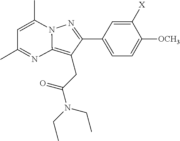
- 0 [1*]OC1=CC=C(C2=NN3C(C)=CC(C)=NC3=C2CC(=O)N(CC)CC)C=C1C Chemical compound [1*]OC1=CC=C(C2=NN3C(C)=CC(C)=NC3=C2CC(=O)N(CC)CC)C=C1C 0.000 description 6
- NNZPWXJYWKODMZ-UHFFFAOYSA-N CCN(CC)C(=O)CC1=C2N=C(C)C=C(C)N2N=C1C1=CC=C(O)C(C)=C1 Chemical compound CCN(CC)C(=O)CC1=C2N=C(C)C=C(C)N2N=C1C1=CC=C(O)C(C)=C1 NNZPWXJYWKODMZ-UHFFFAOYSA-N 0.000 description 2
- XNPWVZZOTNFVLY-UHFFFAOYSA-N CCN(CC)C(=O)CC1=C2N=C(C)C=C(C)N2N=C1C1=CC=C(O)C=C1 Chemical compound CCN(CC)C(=O)CC1=C2N=C(C)C=C(C)N2N=C1C1=CC=C(O)C=C1 XNPWVZZOTNFVLY-UHFFFAOYSA-N 0.000 description 2
- HJKSQGDTIOCHNH-OGEVTHJCSA-N CCN(CC)C(=O)CC1=C2N=C(C)C=C(C)N2N=C1C1=CC=C(C)C([125I])=C1.CCN(CC)C(=O)CC1=C2N=C(C)C=C(C)N2N=C1C1=CC=C(C)C=C1.CCN(CC)C(=O)CC1=C2N=C(C)C=C(C)N2N=C1C1=CC=C(OC)C([125I])=C1 Chemical compound CCN(CC)C(=O)CC1=C2N=C(C)C=C(C)N2N=C1C1=CC=C(C)C([125I])=C1.CCN(CC)C(=O)CC1=C2N=C(C)C=C(C)N2N=C1C1=CC=C(C)C=C1.CCN(CC)C(=O)CC1=C2N=C(C)C=C(C)N2N=C1C1=CC=C(OC)C([125I])=C1 HJKSQGDTIOCHNH-OGEVTHJCSA-N 0.000 description 1
- CWOJTJUAIWJQQP-TVZJARBGSA-N CCN(CC)C(=O)CC1=C2N=C(C)C=C(C)N2N=C1C1=CC=C(O)C(I)=C1.CCN(CC)C(=O)CC1=C2N=C(C)C=C(C)N2N=C1C1=CC=C(O)C([125I])=C1.CCN(CC)C(=O)CC1=C2N=C(C)C=C(C)N2N=C1C1=CC=C(O)C=C1.CCN(CC)C(=O)CC1=C2N=C(C)C=C(C)N2N=C1C1=CC=C(OC)C(I)=C1.CCN(CC)C(=O)CC1=C2N=C(C)C=C(C)N2N=C1C1=CC=C(OC)C([125I])=C1.CCN(CC)C(=O)CC1=C2N=C(C)C=C(C)N2N=C1C1=CC=C(OC)C=C1.COC(=O)C1=CC=C(CO)C=C1 Chemical compound CCN(CC)C(=O)CC1=C2N=C(C)C=C(C)N2N=C1C1=CC=C(O)C(I)=C1.CCN(CC)C(=O)CC1=C2N=C(C)C=C(C)N2N=C1C1=CC=C(O)C([125I])=C1.CCN(CC)C(=O)CC1=C2N=C(C)C=C(C)N2N=C1C1=CC=C(O)C=C1.CCN(CC)C(=O)CC1=C2N=C(C)C=C(C)N2N=C1C1=CC=C(OC)C(I)=C1.CCN(CC)C(=O)CC1=C2N=C(C)C=C(C)N2N=C1C1=CC=C(OC)C([125I])=C1.CCN(CC)C(=O)CC1=C2N=C(C)C=C(C)N2N=C1C1=CC=C(OC)C=C1.COC(=O)C1=CC=C(CO)C=C1 CWOJTJUAIWJQQP-TVZJARBGSA-N 0.000 description 1
- ILZWUAWCTNWSFZ-UHFFFAOYSA-N CCN(CC)C(=O)CC1=C2N=C(C)C=C(C)N2N=C1C1=CC=C(OC)C=C1 Chemical compound CCN(CC)C(=O)CC1=C2N=C(C)C=C(C)N2N=C1C1=CC=C(OC)C=C1 ILZWUAWCTNWSFZ-UHFFFAOYSA-N 0.000 description 1
Images


Classifications
-
- A—HUMAN NECESSITIES
- A61—MEDICAL OR VETERINARY SCIENCE; HYGIENE
- A61K—PREPARATIONS FOR MEDICAL, DENTAL OR TOILETRY PURPOSES
- A61K51/00—Preparations containing radioactive substances for use in therapy or testing in vivo
- A61K51/02—Preparations containing radioactive substances for use in therapy or testing in vivo characterised by the carrier, i.e. characterised by the agent or material covalently linked or complexing the radioactive nucleus
- A61K51/04—Organic compounds
- A61K51/041—Heterocyclic compounds
- A61K51/044—Heterocyclic compounds having nitrogen as a ring hetero atom, e.g. guanethidine, rifamycins
- A61K51/0459—Heterocyclic compounds having nitrogen as a ring hetero atom, e.g. guanethidine, rifamycins having six-membered rings with two nitrogen atoms as the only ring hetero atoms, e.g. piperazine
-
- C—CHEMISTRY; METALLURGY
- C07—ORGANIC CHEMISTRY
- C07B—GENERAL METHODS OF ORGANIC CHEMISTRY; APPARATUS THEREFOR
- C07B59/00—Introduction of isotopes of elements into organic compounds ; Labelled organic compounds per se
- C07B59/002—Heterocyclic compounds
-
- C—CHEMISTRY; METALLURGY
- C07—ORGANIC CHEMISTRY
- C07D—HETEROCYCLIC COMPOUNDS
- C07D487/00—Heterocyclic compounds containing nitrogen atoms as the only ring hetero atoms in the condensed system, not provided for by groups C07D451/00 - C07D477/00
- C07D487/02—Heterocyclic compounds containing nitrogen atoms as the only ring hetero atoms in the condensed system, not provided for by groups C07D451/00 - C07D477/00 in which the condensed system contains two hetero rings
- C07D487/04—Ortho-condensed systems
-
- G—PHYSICS
- G01—MEASURING; TESTING
- G01N—INVESTIGATING OR ANALYSING MATERIALS BY DETERMINING THEIR CHEMICAL OR PHYSICAL PROPERTIES
- G01N33/00—Investigating or analysing materials by specific methods not covered by groups G01N1/00 - G01N31/00
- G01N33/48—Biological material, e.g. blood, urine; Haemocytometers
- G01N33/50—Chemical analysis of biological material, e.g. blood, urine; Testing involving biospecific ligand binding methods; Immunological testing
- G01N33/58—Chemical analysis of biological material, e.g. blood, urine; Testing involving biospecific ligand binding methods; Immunological testing involving labelled substances
- G01N33/60—Chemical analysis of biological material, e.g. blood, urine; Testing involving biospecific ligand binding methods; Immunological testing involving labelled substances involving radioactive labelled substances
-
- G—PHYSICS
- G01—MEASURING; TESTING
- G01N—INVESTIGATING OR ANALYSING MATERIALS BY DETERMINING THEIR CHEMICAL OR PHYSICAL PROPERTIES
- G01N2333/00—Assays involving biological materials from specific organisms or of a specific nature
- G01N2333/435—Assays involving biological materials from specific organisms or of a specific nature from animals; from humans
- G01N2333/705—Assays involving receptors, cell surface antigens or cell surface determinants
-
- G—PHYSICS
- G01—MEASURING; TESTING
- G01N—INVESTIGATING OR ANALYSING MATERIALS BY DETERMINING THEIR CHEMICAL OR PHYSICAL PROPERTIES
- G01N2800/00—Detection or diagnosis of diseases
- G01N2800/70—Mechanisms involved in disease identification
- G01N2800/7095—Inflammation
Definitions
- PBR peripheral benzodiazepine receptor
- Imaging agents are also being sought to study autoimmune disease and the inflammatory arthritides, the cardiovascular system and cancer.
- the standard radiopharmaceutical for imaging inflammation has been the isoquinoline [ 11 C]PK11195, a ligand for TSPO, which is upregulated in activated glial and immune cells (Chen et al., Pharmacol. Ther., vol. 118, pp.
- DPA-713 is 10-fold less lipophilic than PK11195 and has nearly twice the affinity for TSPO. James et al.
- Reactive gliosis characterized by the activation of microglia and astrocytes, is the hallmark response of the central nervous system (CNS) when there is injury or inflammation.
- CNS central nervous system
- TSPO central nervous system
- TSPO is highly expressed in glial cells at specific regions that have been injured (Chen et al., Pharmacol. Ther. Vol. 118, no. 1, pp. 1-17, 2008; Papadopoulos et al., Exp. Neurol., vol. 219, no. 1, pp. 53-57, 2009; Veenman et al., Drug Dev. Res., vol. 50, no. 3-4, pp. 355-370, 2000).
- TSPO colocalization with microglia persisted 30 days after ethanol injection, but astrocytes showed colocalization with TSPO only 7 days after ethanol injection.
- TSPO in astrocytes was not seen 30 days after injection even though GFAP-positive astrocytes were still highly detectable (Maeda et al., Brain Res., vol. 1157, pp. 100-111, 2007).
- Increased TSPO expression has been demonstrated to be colocalized in microglia and astrocytes in a chemically-induced mouse model of demyelination. In this model, it appeared that there is a temporal shift in the TSPO increase from microglia to astrocytes.
- TSPO levels decrease as the gliosis resolves (Chen et al., Brain, vol. 127, no. 6, pp. 1379-1392, 2004).
- TSPO colocalization with both microglia and astrocytes has been shown to be present in brain tissues of HIV encephalitis patients (Cosenza-Nashat et al., Neuropathol. Applied Neurobiol., vol. 35, no. 3, pp. 306-328, 2009).
- TSPO is able to track both increased gliosis and the resolution of gliosis when injured tissue recovers, it serves as a sensitive marker to neurodegenerative changes and inflammation.
- the use of TSPO as a biomarker of brain injury has been demonstrated with chemical-induced neurotoxicity (Guilarte et al., Neurotoxicology, vol. 16, no. 3, pp. 441-450, 1995; Kuhlmann et al., Brain Research, vol. 751, no. 2, pp. 281-288, 1997; Kuhlmann et al., Toxicol. Sci., vol. 48, no. 1, pp. 107-116, 1999; Kuhlmann et al., J. Neurochem., vol. 74, no. 4, pp.
- Sandhoff disease is an autosomal recessive neurodegenerative disease characterized by excess glycolipid storage due to the lack of lysosomal ⁇ -hexosaminidase, resulting in impaired degradation of G M2 and G A2 gangliosides and other glycolipids (Jeyakumar et al., Neuropathol. Applied Neurobiol., vol. 28, no. 5, pp. 343-357, 2002). Because gangliosides are expressed in high amount in the central nervous system (CNS), the brain is one of the most affected organs (Jeyakumar et al., Neuropathol. Applied Neurobiol., vol. 28, no. 5, pp. 343-357, 2002).
- ⁇ -hexosaminidase is formed by the dimerization of two subunits, ⁇ - and ⁇ -subunits to form ⁇ -hexosaminidase A ( ⁇ ) or two ⁇ subunits to form ⁇ -hexosaminidase B ( ⁇ ).
- ⁇ -hexosaminidase A ⁇
- ⁇ -hexosaminidase B ⁇
- the gene encoding for the ⁇ subunit of ⁇ -hexosaminidase (HexB) is disrupted, resulting in the deficiency of ⁇ -hexosaminidase A and B (Yamanaka et al., Genomics, vol. 21, no. 3, pp. 588-596, 1994).
- microglia become activated in both the mouse model and in human cases of Sandhoff disease (Wada et al., Proc. Natl. Acad. Sci. USA, vol. 97, no. 20, pp. 10954-10959, 2000; Visigalli et al., Neurobiol. Dis., vol. 34, no. 1, pp. 51-62, 2009). Further, it appears that microglia activation precedes neuronal degeneration. cDNA microarray analysis has also shown increased TSPO gene expression in the Sandhoff disease mice (Wada et al, Proc. Natl. Acad. Sci. USA, vol. 97, no. 20, pp.
- the present invention satisfies the long standing and unmet need for new imaging compounds for imaging inflammation.
- Embodiments of the invention include compounds having the structure
- X is I, 123 I, 124 I, 125 I, or 131 I.
- Further embodiments of the invention include any of the above compounds isotopically enriched in one of 123 I, 124 I, 125 I, or 131 I.
- the method may further include reacting
- X is I, 123 I, 124 I, 125 I, or 131 I with a methylating agent.
- Other embodiments include methods for imaging cells, tissues, a sample, an organ or a subject by imaging cells, a sample, an organ or a subject with elevated levels of translocator protein, TSPO, after administration of a detectably sufficient amount of a radioisotopically-enriched compound having the structure
- the method may further include administering, before imaging, a detectably sufficient amount of a radioisotopically-enriched compound having the structure
- X is 123 I, 124 I, 125 I, or 131 I.
- Embodiments include methods where imaging is performed by autoradiography, single photon emission computed tomography, or positron emission tomography.
- the imaging is autoradiography.
- imaging is single photon emission computed tomography and the compound is radioisotopically-enriched with 123 I or 125 I.
- the imaging is positron emission tomography and the compound is radioisotopically-enriched with 124 I.
- the cells being imaged are glial cells or immune cells.
- the organ being imaged is the brain. In some embodiments, the organ being imaged is the lungs. In some embodiments, the organ being imaged is the heart.
- the subject being imaged has inflammation. In some embodiments, the subject being imaged has an autoimmune disease. In some embodiments, the subject being imaged has inflammatory arthritides. In some embodiments, the subject being imaged has a neurodegenerative disease. In some embodiments, the subject being imaged has atherosclerosis. In some embodiments, the subject being imaged has myocarditis. In some embodiments the subject being imaged has pneumonitis. In some embodiments the subject being imaged has pneumonia.
- FIG. 1 shows High-performance liquid chromatography (HPLC) traces in the synthesis of N,N-Diethyl-2-[2-(4-hydroxy-3-[ 125 I]-iodophenyl)-5,7-dimethylpyrazolo[1,5-a]pyrimidin-3-yl]acetamide
- [ 125 I]4 from 3 FIG. 1A
- [ 125 I]iodoDPA-713 [ 125 I]1
- Retention times for [ 125 I]4 and [ 125 I]1 were 22.8 and 15.5 min, respectively.
- the radiochemical yield of [ 125 I]1 was 44 ⁇ 6% with a specific radioactivity of 51.8 GBq/ ⁇ mol (1400 mCi/ ⁇ mol) and >99% radiochemical purity.
- FIG. 2 shows autoradiographic images of [ 125 I]1 to TSPO in the rat brain.
- the image in FIG. 2C is representative of [ 125 I]1 non-specific binding using 10 ⁇ M R-PK11195 as the blocking agent in a neurotoxicant-treated rat.
- FIG. 3 shows SPECT-CT imaging of a mouse at 1 h postinjection.
- the lung signal to background ratio in the control mouse (top row) was 14.6 to 1 and that in the LPS treated mouse (bottom row) was 21.2 to 1.
- FIG. 5 shows locomotor activities of Sandhoff mice measured using the open field activity chamber at 2 months and at 3 months. At 2 months, Sandhoff mice showed a slight but statistically significant decrease in total activity ( FIG. 5A ) and rearing ( FIG. 5C ) over 1 hour period.
- FIG. 5E At 3 months, Sandhoff mice exhibited dramatic decrease in locomotor activity ( FIG. 5E ) and rearing ( FIG. 5G ) over 1 hour.
- the average values of locomotor activity ( FIG. 5F ) and rearing ( FIG. 5H ) over 1 hour between wildtype and 3-month Sandhoff mice showed the significant decrease. Data is expressed as mean ⁇ SEM (n 13-14 male mice per group, p ⁇ 0.05 compared to wildtype).
- FIG. 7 shows longitudinal assessment of TSPO levels in Sandhoff disease mice using [ 125 I]-IodoDPA713 quantitative autoradiography.
- FIG. 7A shows representative autoradiograms of [ 125 I]-IodoDPA713 binding to TSPO in horizontal mouse brain sections for wildtype, 2-month Sandhoff, and 3-month Sandhoff mice. TSPO binding increased in thalamus and
- FIG. 8 shows In vivo microSPECT imaging showed higher uptake of [ 125 I]-IodoDPA713 in Sandhoff disease mice.
- the CT indicates that slices ( FIGS. 8A , 8 D, 8 G) are at the level of the thalamus.
- Tracer uptake appears higher in the brain of the Sandhoff mouse ( FIGS. 8E , 8 F), and the uptake was blocked by co-injection of non-radiolabeled IodoDPA-713 ( FIGS.
- FIG. 8(G , H) shows time-activity curve showing the uptake of [ 125 I]IodoDPA-713 in the thalamus of wildtype and of Sandhoff mice. Three-month Sandhoff mouse had consistently higher uptake of [ 125 I]-IodoDPA713 than wildtype.
- FIG. 5K shows quantitative analysis of tracer uptake showed that Sandhoff mice showed a trend of higher [ 125 I]-IodoDPA713 uptake compared to wildtype. For 2-month Sandhoff mice, there was a significant increase in [ 125 I]-IodoDPA713 uptake in the thalamus.
- FIG. 9 shows neurodegeneration in Sandhoff disease mice using silver staining.
- Representative horizontal brain photomicrographs of silver staining indicates ongoing neurodegeneration in the thalamus ( FIGS. 9A-9F ), cerebellum ( FIGS. 9G-9L ), and brainstem ( FIGS. 9M-9R ).
- Boxes inside the low magnification of thalamus ( FIGS. 9A-9C ), cerebellum ( FIGS. 9G-9I ), and brainstem ( FIGS. 9M-90 ) indicate the area of which the high-magnification images were generated.
- Sandhoff mice showed no significant silver accumulation in the thalamus ( FIGS. 9B , 9 E)) and brainstem ( FIGS.
- FIGS. 9N , 9 Q compared to wildtype ( FIGS. 9A and 9D for thalamus; 9 M and 9 P for brainstem).
- FIGS. 9H , 9 K 2-month Sandhoff mice
- FIGS. 9G , 9 J wildtype mice
- Sandhoff mice showed robust silver accumulation in the fiber tracks from the thalamus ( FIGS. 9C , 9 F) and in the cell bodies of the cerebellum ( FIGS. 9I , 9 L)) as well as the brainstem ( FIGS. 9O , 9 R)), indicative of neurodegeneration.
- scale bar 100 ⁇ m.
- scale bar 10 ⁇ m.
- FIG. 10 shows regional and temporal expression of microglia activation using CD11b immunohistochemistry.
- FIGS. 10A , 10 D, 10 G, 10 J, 10 M, 10 P exhibited little to no detection of activated microglia.
- 2-month Sandhoff mice FIGS. 10B , 10 E
- FIGS. 10C , 10 F 3-month Sandhoff mice
- FIGS. 10I , 10 L In the cerebellum, both 2-month ( FIGS. 10H , 10 K) and 3-month ( FIGS. 10I , 10 L) Sandhoff mice exhibited increased CD11b labeling compared to wildtype ( FIGS. 10G , 10 J).
- 3-month Sandhoff mice FIGS.
- 10O , 10 R appeared to exhibit higher CD11b labeling than 2-month Sandhoff mice ( FIGS. 10N , 10 Q) and wildtype ( FIGS. 10M , 10 P)).
- scale bar 100 ⁇ m.
- scale bar 20 ⁇ m.
- FIG. 11 shows temporal response of astrocyte activation in Sandhoff disease mice.
- FIGS. 11B , 11 E, 11 H, 11 K, 11 N, 11 Q activated astrocytes were seen at 2-month Sandhoff mice ( FIGS. 11B , 11 E, 11 H, 11 K, 11 N, 11 Q) but were more pronounced at 3 months Sandhoff mice ( FIGS. 11C , 11 F, 11 I, 11 L, 11 O, 11 R).
- scale bar 100 ⁇ m.
- scale bar 20 ⁇ m.
- FIG. 12 shows TSPO binding is co-localized in activated microglia in regions affected by Sandhoff disease.
- Wildtype mice showed little to no silver grain nor detection of activated microglia in thalamus ( FIG. 12A ), cerebellum ( FIG. 12D ), and brainstem ( FIG. 12G ).
- Sandhoff mice showed increased silver grain density, indicative of increased TSPO binding, co-localized in activated microglia in the thalamus ( FIG. 12B ), cerebellum ( FIG.
- FIG. 13 shows TSPO binding is co-localized in activated astrocytes in regions affected by Sandhoff disease.
- Wildtype mice showed little to no silver grain nor detection of activated astrocytes in thalamus ( FIG. 13A ), cerebellum ( FIG. 13D ), and brainstem ( FIG. 13G ).
- Sandhoff mice showed increased silver grain density, indicative of increased TSPO binding, co-localized in activated astrocytes in the thalamus ( FIG. 13B ), cerebellum ( FIG.
- FIG. 14 shows in vivo microSPECT imaging showing higher uptake of [ 125 I]-IodoDPA-713 in the heart of a mouse model of acute coxsackievirus-induced myocarditis.
- FIG. 14 shows representative transverse slices showing the difference of [ 125 I]IodoDPA-713 accumulation between PBS-injected control mouse (top panel) and coxsackievirus-injected mouse (bottom panel).
- the CT left, for orientation purposes, indicates that slices are at the level of the heart (solid line). Tracer uptake appears higher in the heart of coxsackievirus-injected mouse than the control mouse.
- FIG. 15 shows quantitative analysis of tracer uptake demonstrating that coxsackievirus-injected mice showed a statistically significant increase of [ 125 I]-IodoDPA713 uptake than the PBS-injected control mice.
- the tracer uptake in the lungs and liver were also analyzed to indicate that the increased [ 125 I]-IodoDPA-713 uptake is heart-specific.
- agent is a non-peptide, small molecule compound.
- control is meant a standard or reference condition.
- disease is meant any condition or disorder that damages or interferes with the normal function of a cell, tissue, organ or subject.
- an effective amount is meant a quantity sufficient to produce a measurable difference, when compared with a control. For example, an amount sufficient to produce a measurable image, when the compound is used for imaging. Ultimately, the attending clinician will decide the appropriate amount and dosage regimen.
- subject is meant a mammal, including, but not limited to, a human or non-human mammal, such as a primate, rodent, bovine, equine, canine, ovine, or feline.
- the compounds herein described may have one or more charged atoms.
- the compounds may be zwitterionic, but may be neutral overall.
- Other embodiments may have one or more charged groups, depending on the pH and other factors.
- the compound may be associated with a suitable counter-ion.
- salts or exchange counter-ions can be prepared by reacting free acid forms of these compounds with a stoichiometric amount of the appropriate base (such as Na, Ca, Mg, or K hydroxide, carbonate, bicarbonate, or the like), or by reacting free base forms of these compounds with a stoichiometric amount of the appropriate acid.
- Counter-ions may be changed, for example, by ion-exchange techniques such as ion-exchange chromatography. All zwitterions, salts and counter-ions are intended, unless the counter-ion or salt is specifically indicated.
- the salt or counter-ion may be pharmaceutically acceptable, for administration to a subject. Pharmaceutically acceptable salts are discussed later.
- X is I, 123 I, 124 I, 125 I, or 131 I.
- Further embodiments of the invention include any of the above compounds isotopically enriched in one of 123 I, 124 I, 125 I, or 131 I.
- “isotopically enriched,” means that the amount of 123 I, 124 I, 125 I, or 131 I is greater than the natural abundance.
- the isotopically enriched compound is not necessarily 100% enriched. One of ordinary skill understands that 100% enrichment is impractical and unlikely.
- the percent enrichment should be sufficient to be detected using radioimaging methods.
- the amount of enrichment may also be expressed by specific radioactivity, or units of radioactivity per mol, for example GBq/ ⁇ mol, or Ci/mmol, or variation thereof. The specific radioactivity will vary based on the isotope used. The specific radioactivity should be sufficient to be detected using radioimaging methods.
- the compound may have one of the structures shown below.
- Radiolabeled compounds may be used for diagnostic or imaging purposes. In general, the suitability of a particular radioisotope for a particular purpose (i.e. imaging) is well understood in the art. Other embodiments are compounds used as precursors for radiolabeled compounds. Some compounds according to the invention are intermediates for forming other compounds of the invention.
- iodide means the anion of iodine (I ⁇ ), and is associated with a counterion. Any suitable counterion may be used, including but not limited to alkali metal cations, alkaline earth metal cations, and substituted amine cations.
- the iodide is sodium iodide (NaI), or isotopically enriched forms thereof.
- the reaction may be performed in any suitable solvent, including water, organic solvent or mixtures thereof.
- Organic solvents include, for example, acetonitrile (CH 3 CN) or methanol (CH 3 OH), or mixtures thereof.
- the method may further include reacting
- X is I, 123 I, 124 I, 125 I, or 131 I.
- Any methylating reagents that reacts with the phenol hydroxyl without reacting with other portions of the molecule may be used.
- Many methylating reagents are known, such as, for example methyl iodide, dimethyl sulfate, dimethyl carbonate, methyl trifluoromethanesulfonate, or methyl fluorosulfonate.
- the methylating agent is methyl iodide.
- the reaction may also include a base. Any base may be used that does not otherwise interfere with the reaction. Examples include carbonate (CO 3 2 ⁇ ), associated with a suitable cation, such as potassium (K + ) or sodium (Na + ). In some embodiments, the base is potassium carbonate (K 2 CO 3 ). Imaging
- [ 11 C]-DPA-713 has been used previously for in vivo PET imaging in rodent models of neuroinflammation and brain injury (Boutin et al., J. Nucl. Med., vol. 48, pp. 573-581, 2007; Doorduin et al., Mol. Imag. Biol., vol. 11, no. 6, pp. 386-398, 2009) as well as in humans (Endres et al., J. Nucl. Med., vol. 50, pp. 1276-1282, 2009). Consistently, [ 11 C]-DPA-713 imaging showed higher brain uptake and better signal-to-noise ratio than [ 11 C]-PK11195 (James et al., Bioorg. Med.
- PET imaging is also limited to the availability of a cyclotron on site, which, in conjunction with the short half-lives of the radionuclide, can be difficult and costly for small animal imaging (Wang et al., Biochem. Biophy. Res. Comm., vol. 389, no. 1, pp. 80-83, 2009).
- syntheses of SPECT radioligands such as iodination of tracers are easier than syntheses of PET radioligands, and the half-lives of radionuclides such as [ 125 I] (59 days) are much longer (Meikle et al., Phys. Med. Biol., vol. 50, no. 22, pp. R45-R61, 2005).
- Embodiments include methods for imaging cells, tissues, a sample, an organ or a subject by imaging the cells, the tissues, a sample, an organ or a subject which has or is suspected of having increased levels of translocator protein, TSPO, after administration of a detectably sufficient amount of a radioisotopically-enriched compound having the structure
- the method may further include administering, before imaging, a detectably sufficient amount of a radioisotopically-enriched compound having the structure
- X is 123 I, 124 I, 125 I, or 131 I.
- isotope chosen will depend upon the imaging technique to be used.
- the image indicates the level of TSPO protein, relative to the background. Regions of increased TSPO protein may be visualized by comparing them with the background level of TSPO protein in a particular culture, sample, organ, or subject.
- the background may be produced, for example, by non-specific binding of the compound to other proteins.
- TSPO protein levels below the level of detection will not be imaged.
- TSPO levels below the level of background will not be imaged.
- Compounds of the invention are highly specific for TSPO, have minimal non-specific binding, and therefore a low background.
- Increased TSPO may be caused by increased expression of TSPO protein in the cells, tissues, sample, organ or subject being imaged. In other instances, TSPO protein may be produced elsewhere and migrate to the imaged cells, sample, organ or subject. Where elevated levels of TSPO are associated with cells, tissues, samples or organs, they may be imaged using the compounds and methods of the invention.
- Embodiments include methods of imaging one or more cells, organs, tissues, samples or subjects by exposing cells to or administering to a subject a detectably effective amount of a compound with an isotopic label suitable for imaging.
- the cells, organs, tissues or samples may be imaged while within an organism, either by whole body imaging or intraoperative imaging, or may be excised from the organism for imaging.
- Cells may be imaged, for example, in culture, in a tissue, organ, or even in a subject. Cells may be imaged collectively. Cells or a sample may imaged be in vivo or in vitro, and may be, for example, a sample of an organ or other portion of an organism, or tissue samples grown in culture. In some cases, the sample may be removed from an organism prior to imaging. In some embodiments, organs or portions of organs may be removed prior to imaging, or imaged in vivo. Imaging organs means detecting or visualizing the organ, or portion of the organ associated with elevated levels of TSPO protein. In some inflammation, TSPO may be expressed by immune cells associated with the inflamed organ or tissue, but imaging these cells produces an image of the inflamed portion of the organ itself. All such uses are envisioned.
- Imaging includes, for example, autoradiography, single photon emission computed tomography, or positron emission tomography. Different isotopes may be used for different types of imaging, as known in the art.
- the imaging is autoradiography.
- imaging is single photon emission computed tomography (SPECT) and the compound is radioisotopically-enriched with 123 I or 125 I.
- SPECT is associated with computed tomography (CT) to produce a hybrid image by SPECT-CT.
- CT computed tomography
- PET positron emission tomography
- the compound is radioisotopically-enriched with 124 I.
- any cells expressing or overexpressing TSPO may be imaged.
- the cells are glial cells or immune cells.
- Glial cells include, for example, microglia, astrocytes, oligodendrocytes, ependymal cells or ependymocytes, radial glia, and Schwann cells.
- Immune cells include, for example, macrophages, monocytes, leukocytes, and lymphocytes.
- Other cells that may express or overexpress TSPO include, for example, steroidogenic cells such as testicular, adrenocortical, and brain glial tumor cells, and cancerous tissues of the breast, ovary, colon, prostate, and brain.
- the organ being imaged is the brain. In some embodiments, the organ being imaged is the lungs. In some embodiments, the organ being imaged is the heart. In other embodiments, organs (e.g. spleen) and tissues of the lymphatic system may be imaged.
- any condition associated with TSPO expression may be imaged in a subject.
- the subject has inflammation.
- the subject has an autoimmune disease.
- the subject has inflammatory arthritides.
- the subject has a neurodegenerative disease.
- the subject has atherosclerosis.
- Other conditions that may be associated with TSPO expression include neuropathological conditions including stroke, herpes and HIV encephalitis, and neurodegenerative disorders such as Alzheimer's disease, multiple sclerosis, amyotrophic lateral sclerosis, Parkinson's disease, and Huntington's disease, and other conditions such as myocarditis, pneumonitis, and pneumonia.
- the subject is a human, rat, mouse, cat, dog, horse, sheep, cow, monkey, avian, or amphibian.
- the cell is in vivo or in vitro.
- Typical subjects to which compounds of the invention may be administered will be mammals, particularly primates, especially humans.
- livestock such as cattle, sheep, goats, cows, swine and the like; poultry such as chickens, ducks, geese, turkeys, and the like; and domesticated animals particularly pets such as dogs and cats.
- rodents e.g.
- mice, rats, hamsters), rabbits, primates, and swine such as inbred pigs and the like.
- body fluids and cell samples of the above subjects will be suitable for use such as mammalian, particularly primate such as human, blood, urine or tissue samples, or blood, urine or tissue samples of the animals mentioned for veterinary applications.
- the cells or tissues are present in culture or in suspension.
- Imaging agents of the invention may be used in accordance with the methods of the invention by one of skill in the art. Images can be generated by virtue of differences in the spatial distribution of the imaging agents which accumulate at a site when contacted with TSPO.
- the spatial distribution may be measured using any means suitable for the particular label, for example, a gamma camera, a PET apparatus, a SPECT apparatus, and the like.
- the extent of accumulation of the imaging agent may be quantified using known methods for quantifying radioactive emissions.
- a detectably effective amount of the imaging agent is administered to a subject.
- a detectably effective amount of the imaging agent is defined as an amount sufficient to yield an acceptable image using equipment which is available for clinical use.
- a detectably effective amount of the imaging agent may be administered in more than one dose.
- the imaging agent can be administered in any suitable to result in delivery to the site where TSPO accumulation may be expected to occur. Examples of administration include ingestion or injection, including, for example, interperitoneal injection or intravenous injection.
- the detectably effective amount of the imaging agent can vary according to factors such as the degree of susceptibility of the individual, the age, sex, and weight of the individual, idiosyncratic responses of the individual, and the dosimetry. Detectably effective amounts of the imaging agent can also vary according to instrument and film-related factors. Optimization of such factors is well within the level of skill in the art.
- the amount of imaging agent used for diagnostic purposes and the duration of the imaging study will depend upon the radionuclide used to label the agent, the body mass of the patient, the nature and severity of the condition being treated, the nature of therapeutic treatments which the patient has undergone, and on the idiosyncratic responses of the patient. Ultimately, the attending physician will decide the amount of imaging agent to administer to each individual patient and the duration of the imaging study.
- the compounds are excreted from tissues of the body of the subject quickly to prevent prolonged exposure to the radiation of the radiolabeled compound administered to the patient.
- the compounds are excreted from tissues of the body slowly enough to allow sufficient time for imaging or other use.
- compounds of the invention are eliminated from the body in less than about 24 hours. More typically, compounds of the invention are eliminated from the body in less than about 16 hours, 12 hours, 8 hours, 6 hours, 4 hours, 2 hours, 90 minutes, or 60 minutes. Compounds may be eliminated in between about 60 minutes and about 120 minutes.
- compositions e.g. pharmaceutical compositions
- a pharmaceutical composition comprises an effective amount (e.g., a detectably effective amount) of a compound described above.
- a composition of the invention can be formulated as a pharmaceutical composition, which comprises a compound of the invention and pharmaceutically acceptable carrier.
- a pharmaceutically acceptable carrier is meant a material that is not biologically or otherwise undesirable, i.e., the material may be administered to a subject without causing any undesirable biological effects or interacting in a deleterious manner with any of the other components of the pharmaceutical composition in which it is contained.
- the carrier would naturally be selected to minimize any degradation of the active ingredient and to minimize any adverse side effects in the subject, as would be well known to one of skill in the art.
- pharmaceutically acceptable carriers and other components of pharmaceutical compositions see, e.g., Remington's Pharmaceutical Sciences, 18 th ed., Mack Publishing Company, 1990.
- the pharmaceutically acceptable carrier may also contain stabilizers, preservatives, antioxidants, or other additives, which are well known to one of skill in the art, or other vehicle as known in the art.
- Suitable pharmaceutical carriers will be evident to a skilled worker and include, e.g., water (including sterile and/or deionized water), suitable buffers (such as PBS), physiological saline, cell culture medium (such as DMEM), artificial cerebral spinal fluid, or the like.
- “pharmaceutically acceptable carrier” refers to a biocompatible solution, having due regard to sterility, p[Eta], isotonicity, stability, and the like and can include any and all solvents, diluents (including sterile saline, Sodium Chloride Injection, Ringer's Injection, Dextrose Injection, Dextrose and Sodium Chloride Injection, Lactated Ringer's Injection and other aqueous buffer solutions), dispersion media, coatings, antibacterial and antifungal agents, isotonic agents, and the like.
- diluents including sterile saline, Sodium Chloride Injection, Ringer's Injection, Dextrose Injection, Dextrose and Sodium Chloride Injection, Lactated Ringer's Injection and other aqueous buffer solutions
- dispersion media including sterile saline, Sodium Chloride Injection, Ringer's Injection, Dextrose Injection, Dextrose and Sodium
- a pharmaceutical composition or kit of the invention can contain other pharmaceuticals or imaging agents, in addition to the compounds described herein.
- the other agent(s) can be administered at any suitable time during the imaging process, either concurrently or sequentially.
- compositions of the present invention will depend, in part, upon the particular agent that is employed, and the chosen route of administration. Accordingly, there is a wide variety of suitable formulations of compositions of the present invention.
- compositions of the invention can be in unit dosage form.
- unit dosage form refers to physically discrete units suitable as unitary dosages for animal (e.g. human) subjects, each unit containing a predetermined quantity of an agent of the invention, alone or in combination with other therapeutic agents, calculated in an amount sufficient to produce the desired effect in association with a pharmaceutically acceptable diluent, carrier, or vehicle.
- One skilled in the art can easily determine the appropriate dose, schedule, and method of administration for the exact formulation of the composition being used, in order to achieve the desired effective amount or effective concentration of the agent in the individual patient.
- the dose of a composition of the invention, administered to an animal, particularly a human, in the context of the present invention should be sufficient to produce at least a detectable amount of a diagnostic or imaging response in the individual over a reasonable time frame.
- the dose used to achieve a desired effect will be determined by a variety of factors, including the potency of the particular agent being administered, the pharmacodynamics associated with the agent in the host, the severity of the disease state of infected individuals, other medications being administered to the subject, etc.
- the size of the dose also will be determined by the existence of any adverse side effects that may accompany the particular agent, or composition thereof, employed. It is generally desirable, whenever possible, to keep adverse side effects to a minimum.
- the dose of the biologically active material will vary; suitable amounts for each particular agent will be evident to a skilled worker.
- kits including a compound according to the invention.
- the kit provides packaged pharmaceutical compositions having a pharmaceutically acceptable carrier and a compound of the invention.
- the packaged pharmaceutical composition will include the reaction precursors necessary to generate the compound of the invention upon combination with a radionuclide.
- Other packaged pharmaceutical compositions provided by the present invention further include indicia such as at least one of: instructions for preparing compounds according to the invention from supplied precursors, instructions for using the composition to image cells or tissues expressing TSPO, or instructions for using the composition to image inflammation or neurodegeneration in a patient suffering, for example, an autoimmune disease, an inflammatory arthritides, a neurodegenerative disease, or atherosclerosis.
- a kit according to the invention may contain, for example, from about 1 mCi to about 30 mCi of the radionuclide-labeled imaging agent described above, in combination with a pharmaceutically acceptable carrier.
- the imaging agent and carrier may be provided in solution or in lyophilized form.
- the kit may optionally contain a sterile and physiologically acceptable reconstitution medium such as water, saline, buffered saline, and the like.
- the kit may provide a compound of the invention in solution or in lyophilized form, and these components of the kit of the invention may optionally contain stabilizers such as NaCl, silicate, phosphate buffers, ascorbic acid, gentisic acid, and the like. Additional stabilization of kit components may be provided in this embodiment, for example, by providing the reducing agent in an oxidation-resistant form. Determination and optimization of such stabilizers and stabilization methods are well within the level of skill in the art.
- stabilizers such as NaCl, silicate, phosphate buffers, ascorbic acid, gentisic acid, and the like. Additional stabilization of kit components may be provided in this embodiment, for example, by providing the reducing agent in an oxidation-resistant form. Determination and optimization of such stabilizers and stabilization methods are well within the level of skill in the art.
- Iodine-125 ( 125 I) was obtained as a 0.1 N solution of NaOH (high concentration) from MP Biomedicals (Solon, Ohio).
- Analytical thin-layer chromatography (TLC) was performed using Aldrich aluminum-backed 0.2 mm silica gel plates and visualized by UV light (254 nm) and I 2 . Flash column chromatography was performed on silica gel (60 ⁇ ) from MP Biomedicals.
- Radio-HPLC purification was performed using a Waters (Milford, Mass.) system consisting of two Waters 510 pumps, a Waters 490E variable wavelength UV/Vis detector set at 254 nm, a BioScan FlowCount radioactivity detector, a Waters radial-PAK C 18 reverse phase analytical column (8 ⁇ 100 mm) with H 2 O/CH 3 CN/TFA solvent systems, and WinFlow (LabLogic) chromatography software.
- 1 H NMR was recorded on a Bruker (Billerica, Mass.) UltrashieldTM 400 MHz spectrometer.
- ESI mass spectra were obtained with a Bruker Daltonics Esquire 300 plus spectrometer.
- Radioactivity was measured in a Capintec CRC-12 dose calibrator.
- the specific radioactivity was calculated as the radioactivity eluting at the retention time of [ 125 I]1 during HPLC purification divided by the mass corresponding to the area under the curve of UV absorption.
- iodoDPA-713 1 N,N-Diethyl-2-[2-(3-iodo-4-methoxyphenyl)-5,7-dimethylpyrazolo[1,5-a]pyrimidin-3-yl]acetamide (iodoDPA-713) 1 was synthesized according to the scheme below in three steps from N-diethyl-2-[2-(4-methoxy-phenyl)-5,7-dimethylpyrazolo[1,5-a]pyrimidin-3-yl]-acetamide 2 (DPA-713) (Wang et al., Biochem. Biophy. Res. Comm. Vol. 389, no. 1, pp. 80-83). Compound 2 was prepared by the procedure of James et al.
- the reaction mixture was incubated at room temperature for 1.5 h and purified by reverse phase HPLC using 70% H 2 O/30% CH 3 CN/0.1% TFA with a flow rate of 2 mL/min on a Waters Radial-Pak analytical column (8 ⁇ 10 mm).
- the retention time of [ 125 I]4 was 22.8 min ( FIG. 1A ).
- the radioactive fraction corresponding to [ 125 I]4 was collected, diluted with water and passed through a pre-conditioned C 18 light Sep-Pak cartridge (Waters Corp, Milford, Mass.) eluted with 0.5 mL of ether.
- the reaction mixture was stirred overnight and diluted with 0.2 mL of water and purified by reverse phase HPLC using 50% H 2 O/50% CH 3 CN/0.1% TFA with a flow rate of 1 mL/min on a Waters Radial-Pak analytical column (8 ⁇ 100 mm).
- the retention time of [ 125 I]1 was 15.5 min ( FIG. 1B ).
- the radioactive fraction corresponding to [ 125 I]1 was collected, diluted with water and passed through a pre-conditioned C 18 light Sep-Pak and eluted with 0.5 mL of ethanol. Yield: 0.035 GBq (0.95 mCi), 86% from [ 125 I]4 and 47% from 3. Specific radioactivity was 51.8 GBq/ ⁇ mol (1400 Ci/mmol).
- Fresh-frozen brains were sectioned (20 ⁇ m) on a freezing cryostat in the horizontal plane. Brain sections were thaw-mounted onto poly-L-lysine-coated slides (Sigma) and stored at ⁇ 20° C. until used. Autoradiography using [ 125 I]IodoDPA-713 ([ 125 I]1) was performed on adjacent brain sections using the following procedures. Slides were thawed and dried at 37° C. for 30 min and prewashed in 50 mM Tris-HCl buffer (pH 7.4) for 5 min at room temperature. Sections were then incubated in 1.4 nM [ 125 I]1 in 50 mM Tris-HCl buffer for 30 min at room temperature.
- the first study involved a model of brain inflammation in which rats were exposed to a seizure-inducing neurotoxicant ( FIG. 2 ). Note that higher levels of [ 125 I]1 were present in the neurotoxicant-treated rat brain ( FIG. 2B ) than in control brain ( FIG. 2A ). Exposure of the tissue specimens to [ 125 I]1 to generate these images required only 60 min, as opposed to 4-5 weeks often needed for tritium-labeled compounds when exposing the samples to film.
- Rapid images can also be obtained with tritium-labeled compounds, including [ 3 H]DPA-713, using an automated system such as a Beta-Imager (Roberts et al., “Autoradiographical imaging of PPARgamma agonist effects on PBR/TSPO binding in TASTPM mice,” Exp. Neurol., vol. 216, pp. 549-470, 2009), however the resolution of such images is generally inferior to that which can be obtained by exposing the tissue slices to film. Also note that uptake of [ 125 I]1 could be almost completely blocked in the neurotoxicant-treated rat upon treatment with PK11195 ( FIG. 2C ), demonstrating that most binding seen in this in vitro study was specific for TSPO.
- SPECT-CT single photon emission computed tomography-computed tomography
- the mouse was positioned on the X-SPECT (Gamma Medica, Northridge, Calif.) gantry and was scanned using two opposing low energy pinhole collimators (Gamma Medica) rotating through 360° in 3° increments for 20 s per increment. Images were reconstructed using LumaGem software that accompanies the X-SPECT. Immediately following SPECT acquisition, the mice were scanned by CT over a 3-cm field-of-view using a 600- ⁇ A, 50 kV beam. The SPECT and CT data were then co-registered using the X-SPECT software and displayed using AMIDE (http://amide.sourceforge.net/). Data were reconstructed using the ordered subsets-expectation maximization (OS-EM) algorithm. The signal to background ratio was calculated from the images by drawing regions of interest over the lungs (signal) and muscle, (background).
- OS-EM ordered subsets-expectation maximization
- [ 125 I]IodoDPA-713 [ 125 I]1 can be readily synthesized from DPA-713 in high radiochemical yield and specific radioactivity. It demonstrates specific binding to TSPO in vitro and in vivo, showing a higher level of uptake in inflamed than in the corresponding normal tissue.
- This agent provides a convenient and inexpensive alternative to other radiolabeled analogs of the pyrazolopyrimidine series for autoradiographic and preclinical imaging studies. Together these two studies suggest that [ 125 I]1 may be useful to study preclinical models of inflammation.
- Compound [ 125 I]1 is particularly relevant as it is patterned after compounds that are currently undergoing early clinical testing (Endres et al., J. Nucl. Med., vol. 50, pp. 1276-1282, 2009; Chauveau et al., J. Nucl. Med., vol. 50, pp. 468-476, 2009).
- mice heterozygous for HexB were generously donated by Richard Proia (National Institute of Kidney & Digestive Diseases, Bethesda, Md.). Wildtype (HexB +/+) or Sandhoff disease (HexB ⁇ / ⁇ ) mice were sacrificed at 2 months or 3 months of age and euthanized by either decapitation to obtain fresh-frozen brain tissue for receptor autoradiography or by transcardiac perfusion for immunohistochemistry. Fresh frozen brains were stored at ⁇ 80° C. until used.
- mice were deeply anesthetized with pentobarbital (100 mg/kg body weight) and perfused with 4% paraformaldehyde (PFA) in 0.1M phosphate buffer (pH 7.4 at 4° C.) or a paraformaldehyde-lysine-periodate (PLP) solution that consisted of 2% paraformaldehyde, 75 mM L-lysine and 10 mM sodium metaperiodate in 37 mM phosphate buffer (pH 7.4 at 4° C.).
- PFA paraformaldehyde
- PFP paraformaldehyde-lysine-periodate
- the PFA solution was used to perfuse animals whose brains were used for glial fibrillary acidic protein (GFAP) or CD11b (Mac-1) immunohistochemistry or silver staining for neurodegeneration, and the PLP solution was used to perfuse animals whose brains were used for the [ 3 H]-(R)-PK11195 emulsion autoradiography in conjunction with GFAP or Mac-1 immunohistochemistry (see sections below).
- Perfused brains were post-fixed overnight in the same fixative, cryoprotected with 25% sucrose for at least 48 h, snap-frozen in dry-ice-cooled isopentane, and stored at ⁇ 80° C. until used. All animal studies were reviewed and approved by the Johns Hopkins University Animal Care and Use Committee.
- mice The body weights of mice were taken weekly starting at 6 weeks and ending at 13 weeks (3-month) when brains were extracted. Following extraction, the brain weights of wildtype and Sandhoff disease mice (HexB knockout) were measured at 3 months of age. Brain weights were not significantly different between wildtype (425 ⁇ 15.1 mg) and Sandhoff (448.4 ⁇ 20.3 mg) mice (data not shown).
- mice were placed in open field activity chambers with infrared beams (San Diego Instruments Inc., San Diego, Calif., USA) for 1 h, and during this time, horizontal and vertical (rearing) activities were automatically recorded.
- infrared beams San Diego Instruments Inc., San Diego, Calif., USA
- TSPO TSPO was examined in a Sandhoff disease mouse model of neurodegeneration using a longitudinal design. Two months was defined as the time point prior to the severe behavioral expression of disease because slight function deficits were observed between wildtype and Sandhoff disease mice ( FIG. 5 ). Later, at 3 months, significant impairments in locomotor activities and motor skills were prominent between wildtype and Sandhoff mice ( FIGS. 5 and 6 ). This is consistent with other reports on the behavioral expression of this disease in mice (Sango et al., Nature Genetics, vol. 11, no. 2, pp. 170-176, 1995; Wada et al., Proc. Natl. Acad. Sci. USA, vol. 97, no. 20, pp. 10654-10959, 2000; Jeyakumar et al., Ann. Neurol., vol. 56, no. 5, pp. 642-649, 2004).
- An X-SPECT small-animal SPECT/CT system (Gamma Medica-Ideas) was used for image acquisition. Each mouse was anesthetized with isofluorane prior to imaging. About 1-2 mCi of [ 125 I]-IodoDPA713 was injected into each mouse via intravenous injection, and images were acquired immediately after injection.
- the SPECT projection data were acquired using 2 low-energy, high-resolution parallel-hole collimators with a radius of rotation of 4.65 cm (spatial resolution, 1.6 mm).
- the tomographic data were acquired in 64 projections over 360° at 20 s per projection. After tomography, CT was acquired in 512 projections to allow anatomic coregistration.
- [ 125 I]-IodoDPA713 microSPECT was used to assess TSPO expression in vivo in Sandhoff mice.
- Co-registered microSPECT images FIGS. 8B , 8 E, 8 H
- microCT FIGS. 8A , 8 D, 8 G
- FIG. 8F shows that there is higher uptake of [ 125 I]-IodoDPA713 in the Sandhoff mouse
- FIG. 8C wildtype mouse
- Time-activity curve of the thalamus further showed that Sandhoff mouse consistently had higher uptake of [ 125 I]-IodoDPA713 than wildtype mouse ( FIG. 8J ).
- [ 125 I]-IodoDPA713 was then used to image TSPO levels in vivo by microSPECT. There is a trend of increased [ 125 I]-IodoDPA713 uptake in Sandhoff mice with significant increase in thalamus of 2-month Sandhoff mice and in the brainstem of 3-month Sandhoff mice ( FIG. 5K ). Furthermore, the time-activity curve indicated that Sandhoff mice consistently had a higher uptake of [ 125 I]-IodoDPA713 in the thalamus than wildtype mice ( FIG. 5J ).
- Fresh-frozen brains were sectioned (20 ⁇ m) on a freezing cryostat in the horizontal plane. Brain sections were thaw-mounted onto poly-L-lysine-coated slides (Sigma-Aldrich, St. Louis, Mo.) and stored at ⁇ 20° C. until used. [ 125 I]-IodoDPA713 autoradiography to measure TSPO levels were performed on adjacent brain sections using the same procedures. Slides were thawed and dried at 37° C. for 30 min and prewashed in 50 mM Tris-HCl buffer (pH 7.4) for 5 min at room temperature. Sections were then incubated in 0.5 nM [ 125 I]-IodoDPA713 in buffer for 30 min at room temperature.
- Quantitative autoradiography with [ 125 I]-IodoDPA713 was used to measure TSPO levels in different brain regions in Sandhoff mice at 2 and 3 months of age. The goal was to assess whether TSPO levels are significantly increased prior to severe expression of disease indicating its potential use as an early biomarker of disease and to assess the expression of TSPO as a function of disease progression.
- Free-floating sections from PFA-perfused animals were further fixed in 4% PFA in 0.1M phosphate buffer for at least 48 h before staining. Silver staining was performed using the FD Neurotech NeuroSilver Kit (Ellicott City, Md.) according to manufacturer's instructions.
- Reactive Gliosis Occurs Prior to Neurodegeneration and has a Temporal Response in Sandhoff Disease.
- FIGS. 9B , 9 E Silver staining was used to assess the progression of neurodegeneration in the principal regions known to be affected in Sandhoff disease. These included the thalamus, brainstem, and the cerebellum. 2-month Sandhoff mice did not show any substantial sign of ongoing neurodegeneration in the thalamus ( FIGS. 9B , 9 E), in the cerebellum ( FIGS. 9H , 9 K), or in the brainstem ( FIGS. 9N , 9 Q) compared to wildtype ( FIGS. 9A , 9 D, 9 G, 9 J, 9 M, 9 P). At the thalamus, 2-month Sandhoff mice exhibit little to no silver accumulation ( FIGS. 9B , 9 E) while 3-month Sandhoff mice exhibited silver accumulation in the fiber tracks of the thalamus ( FIGS.
- FIGS. 9C , 9 F At the brainstem, 2-month Sandhoff mice exhibited no substantial silver accumulation ( FIGS. 9N , 9 Q) while 3-month Sandhoff mice exhibited robust silver accumulation ( FIGS. 9O , 9 R), indicative of neurodegeneration. Finally, at the cerebellum, slight silver accumulation was observed in the cerebellum of 2-month Sandhoff mice ( FIGS. 9H , 9 K) but robust silver accumulation in cerebellum of 3-month Sandhoff mice ( FIGS. 9I , 9 L). Consistent with the behavioral data, at 3 months, there were prominent argyrophilic neurons consistent with ongoing neurodegeneration in all examined regions ( FIGS. 9C , 9 F, 9 I, 9 L, 9 O, 9 R).
- Free-floating brain sections from PFA-perfused animals were sectioned (40 ⁇ m) using a freezing microtome (Leica Microsystems Inc., Bannockburn, Ill.). Sections were incubated in 0.6% H 2 O 2 for 15 min followed by blocking solution with either 5% normal goat serum containing 0.2% Triton X-100 for 1 h. Sections were incubated with rabbit anti-GFAP antibody (1:1000, Dako, Carpinteria, Calif., USA) or rat anti-mouse CD11b antibody (1:250, BD PharMingen, San Diego, Calif., USA) at 4° C. overnight.
- Mac-1 and GFAP immunohistochemistry were used to detect microglia and astrocytes, respectively. Increased Mac-1 immunoreactivity was observed at 2-month but a more robust Mac-1 immunoreactivity at 3-month. The microglia also appeared hypertrophic in the brainstem at 3-month compared to wildtype. Similarly, 3-month Sandhoff mice showed higher GFAP immunoreactivity than 2-month Sandhoff mice. While 2-month Sandhoff mice exhibited activated microglia and astrocyte, both responses appeared stronger in the cerebellum of 3-month Sandhoff mice.
- FIGS. 10B , 10 E more activated microglia were seen in 2-month Sandhoff mice ( FIGS. 10B , 10 E) than in 3-month Sandhoff mice ( FIGS. 10C , 10 F). Meanwhile, activated astrocytes appeared higher in the thalamus of 3-month Sandhoff mice ( FIGS. 11C , 11 F) than 2-month Sandhoff mice ( FIGS. 11B , 11 E).
- FIGS. 10I , 10 L for microglia; FIGS. 11I , 11 L for astrocytes more activated microglia and astrocytes were detected at 3-month Sandhoff mice ( FIGS. 10I , 10 L for microglia; FIGS. 11I , 11 L for astrocytes) for than 2-month Sandhoff mice ( FIGS. 10H , 10 K for microglia; FIGS.
- FIGS. 10G , 10 J for microglia; FIGS. 11G , 11 J for astrocytes were detected at 3-month Sandhoff mice ( FIGS. 10O , 10 R for microglia; FIGS. 11O , 11 R for astrocytes) for than 2-month Sandhoff mice ( FIGS. 10N , 10 Q for microglia; FIGS. 11N , 11 Q for astrocytes) and wildtype ( FIGS. 10M , 10 P for microglia; FIGS. 11M , 11 P for astrocytes).
- FIGS. 10M , 10 P for microglia; FIGS. 11M , 11 P for astrocytes This suggests that there is a regional and temporal response in reactive gliosis demonstrating microglial activation preceding astrocyte activation.
- PLP-perfused brain tissue was used to determine the co-localization of TSPO with microglia or astrocytes. It has been shown that there are no differences in [ 3 H]-(R)-PK11195 binding to TSPO between fresh-frozen and PLP-fixed brain tissue (Kuhlmann et al. Toxicol. Sci., vol. 48, no. 1, pp. 107-116, 1999). PLP-fixed brains were sectioned (18 ⁇ m) on a freezing cryostat in the horizontal plane. Brain sections were thaw-mounted onto poly-L-lysine-coated slides and stored at ⁇ 20° C. until used. Immunohistochemistry of GFAP or Mac-1 was performed as described above.
- High-resolution emulsion autoradiography of [ 3 H]-(R)-PK11195 binding to TSPO was performed to determine the cellular localization of increased TSPO binding in Sandhoff mice.
- the silver grain density represents the levels of [ 3 H]-(R)-PK11195 binding to TSPO.
- Sandhoff mice showed higher [ 3 H]-(R)-PK11195 binding in the thalamus ( FIGS. 12B and 13B ), cerebellum ( FIGS. 12E and 13E ), and brainstem ( FIGS. 12H and 13H ) compared to wildtype ( FIGS. 12A , 12 D, 12 G and FIGS. 13A , 13 D, 13 G).
- the microglia response in the thalamus appears to be maximal at 2 months since immunostaining of 3-month Sandhoff mice exhibited a reduced level of microglia staining in the thalamus ( FIGS. 10C , 10 F). Both 2- and 3-month Sandhoff mice exhibited enhanced astrocytic response in the thalamus as shown by the increased GFAP immunoreactivity relative to wildtype mice ( FIG. 11A-11F ). An increased Mac-1 immunoreactivity at 2-month but a more robust Mac-1 immunoreactivity at 3-month was observed. The microglia also appeared hypertrophic in the brainstem at 3-month compared to wildtype. Similarly, 3-month Sandhoff mice showed higher GFAP immunoreactivity than 2-month Sandhoff mice.
- ANOVA Repeated measure analysis of variance
- TSPO levels remained elevated at both time points, but an increase of activated microglia at 2 months and an increase of activated astrocytes at 3 months was observed. This might be a result of the shift of TSPO expression from microglia to astrocytes as reactive gliosis persists in Sandhoff disease.
- Previous studies have also shown this similar temporal response of TSPO expression, which was first associated with microglia activation and then followed by TSPO associated with astrocytosis (Kuhlmann et al., J. Neurochem., vol. 74, no. 4, pp. 1694-1704, 2000; Chen et al., Brain, vol. 127, no. 6, pp.
- [ 125 I]-IodoDPA-713 microSPECT imaging of the brain and in vivo [ 125 I]-IodoDPA-713 microSPECT imaging also showed increased uptake of [ 125 I]-IodoDPA-713 in Sandhoff mice at both time points.
- [ 125 I]-IodoDPA-713 is an attractive radioligand for TSPO imaging using SPECT, and secondly, TSPO is a sensitive biomarker of brain injury such that increased TSPO binding can be detected prior to demonstration of behavioral changes and significant levels of neurodegeneration.
- TSPO can be used as an early biomarker of neurodegeneration.
- this study examined TSPO binding longitudinally using Sandhoff disease Sandhoff mice at time points prior to and during neurodegeneration.
- TSPO binding was detected in specific regions both in vitro and in vivo at the time points prior to and during neurodegeneration as well as an increased glial response marked by microglia and astrocyte activation.
- the glial response also appeared to be regional and temporal as microglia activation was detected at an earlier time point before astrocyte activation.
- TSPO levels were visualized and measured using both in vitro quantitative autoradiography and in vivo imaging using microSPECT at 2 months and at 3 months of age. It was found that at 2 months, when Sandhoff disease mice show slight deficits in behavioral endpoints and without severe neurodegeneration as measured by silver staining, increased TSPO expression was detected in brain regions known to undergo neurodegeneration in Sandhoff disease mice. This increase in TSPO binding appeared to co-localize with activated microglia and astrocytes.
- An X-SPECT small-animal SPECT/CT system (Gamma Medica-Ideas) was used for image acquisition. Each mouse was anesthetized with isofluorane prior to imaging. About 1-2 mCi of [ 125 I]-IodoDPA713 was injected into each mouse via intravenous injection, and images were acquired immediately after injection.
- the SPECT projection data were acquired using 2 low-energy, high-resolution parallel-hole collimators with a radius of rotation of 4.65 cm (spatial resolution, 1.6 mm).
- the tomographic data were acquired in 64 projections over 360° at 20 s per projection. After tomography, CT was acquired in 512 projections to allow anatomic coregistration.
- FIG. 14 shows representative transverse slices showing the difference of [ 125 I]IodoDPA-713 accumulation between PBS-injected control mouse (top panel) and coxsackievirus-injected mouse (bottom panel).
- the CT (left), for orientation purposes, indicates that slices are at the level of the heart (solid line). Tracer uptake appears higher in the heart of coxsackievirus-injected mouse than the control mouse.
- FIG. 15 shows quantitative analysis of tracer uptake showed that coxsackievirus-injected mice showed a statistically significant increase of [ 125 I]-IodoDPA713 uptake than the PBS-injected control mice.
Landscapes
- Chemical & Material Sciences (AREA)
- Health & Medical Sciences (AREA)
- Organic Chemistry (AREA)
- Life Sciences & Earth Sciences (AREA)
- Engineering & Computer Science (AREA)
- General Health & Medical Sciences (AREA)
- Medicinal Chemistry (AREA)
- Biomedical Technology (AREA)
- Hematology (AREA)
- Immunology (AREA)
- Physics & Mathematics (AREA)
- Molecular Biology (AREA)
- Urology & Nephrology (AREA)
- Cell Biology (AREA)
- Pathology (AREA)
- Microbiology (AREA)
- Analytical Chemistry (AREA)
- Biochemistry (AREA)
- Biotechnology (AREA)
- General Physics & Mathematics (AREA)
- Food Science & Technology (AREA)
- Proteomics, Peptides & Aminoacids (AREA)
- Optics & Photonics (AREA)
- Pharmacology & Pharmacy (AREA)
- Epidemiology (AREA)
- Animal Behavior & Ethology (AREA)
- Public Health (AREA)
- Veterinary Medicine (AREA)
- Medicines Containing Antibodies Or Antigens For Use As Internal Diagnostic Agents (AREA)
Abstract
Description

where X is H, I, 123I, 124I, 125I, or 131I, and R1 is H or CH3, with the proviso that when X is H, R1 is not CH3. Embodiments of the invention include compounds having the structure

where X is I, 123I, 124I, 125I, or 131I. Further embodiments of the invention include any of the above compounds isotopically enriched in one of 123I, 124I, 125I, or 131I.

where X is I, 123I, 124I, 125I, or 131I; and R1 is H or CH3, by reacting

with iodide in the presence of iodogen or chloramine-T, where the iodide may be isotopically enriched in one of 123I, 124I, 125I, or 131I. The method may further include reacting

where X is I, 123I, 124I, 125I, or 131I with a methylating agent.

where X is 123I, 124I, 125I, or 131I. The method may further include administering, before imaging, a detectably sufficient amount of a radioisotopically-enriched compound having the structure

where X is 123I, 124I, 125I, or 131I.

where X is H, I, 123I, 124I, 125I, or 131I, and R1 is H or CH3, with the proviso that when X is H, R1 is not CH3. In other words, the compound

is excluded.

where X is I, 123I, 124I, 125I, or 131I. Further embodiments of the invention include any of the above compounds isotopically enriched in one of 123I, 124I, 125I, or 131I. As used herein, “isotopically enriched,” means that the amount of 123I, 124I, 125I, or 131I is greater than the natural abundance. The isotopically enriched compound is not necessarily 100% enriched. One of ordinary skill understands that 100% enrichment is impractical and unlikely. The percent enrichment should be sufficient to be detected using radioimaging methods. The amount of enrichment may also be expressed by specific radioactivity, or units of radioactivity per mol, for example GBq/μmol, or Ci/mmol, or variation thereof. The specific radioactivity will vary based on the isotope used. The specific radioactivity should be sufficient to be detected using radioimaging methods.

Uses

where X is I, 123I, 124I, 125I, or 131I; and R1 is H or CH3, by reacting, in part, a compound of the structure

with iodide in the presence of iodogen or chloramine-T, where the iodide may be isotopically enriched in one of 123I, 124I, 125I, or 131I. As used herein, “iodide” means the anion of iodine (I−), and is associated with a counterion. Any suitable counterion may be used, including but not limited to alkali metal cations, alkaline earth metal cations, and substituted amine cations. In some embodiments, the iodide is sodium iodide (NaI), or isotopically enriched forms thereof. The reaction may be performed in any suitable solvent, including water, organic solvent or mixtures thereof. Organic solvents include, for example, acetonitrile (CH3CN) or methanol (CH3OH), or mixtures thereof.

where X is I, 123I, 124I, 125I, or 131I with a methylating agent. The product of the reaction has the structure

where X is I, 123I, 124I, 125I, or 131I. Any methylating reagents that reacts with the phenol hydroxyl without reacting with other portions of the molecule may be used. Many methylating reagents are known, such as, for example methyl iodide, dimethyl sulfate, dimethyl carbonate, methyl trifluoromethanesulfonate, or methyl fluorosulfonate. In some embodiments, the methylating agent is methyl iodide. The reaction may also include a base. Any base may be used that does not otherwise interfere with the reaction. Examples include carbonate (CO3 2−), associated with a suitable cation, such as potassium (K+) or sodium (Na+). In some embodiments, the base is potassium carbonate (K2CO3).
Imaging

where X is 123I, 124I, 125I, or 131I. The method may further include administering, before imaging, a detectably sufficient amount of a radioisotopically-enriched compound having the structure
where X is 123I, 124I, 125I, or 131I. As will be appreciated, the isotope chosen will depend upon the imaging technique to be used.

Claims (17)


Priority Applications (2)
| Application Number | Priority Date | Filing Date | Title |
|---|---|---|---|
| US13/034,320 US8778304B2 (en) | 2010-02-24 | 2011-02-24 | TSPO-targeting compounds and uses thereof |
| US14/330,633 US9233178B2 (en) | 2010-02-24 | 2014-07-14 | TSPO-targeting compounds and uses thereof |
Applications Claiming Priority (2)
| Application Number | Priority Date | Filing Date | Title |
|---|---|---|---|
| US30755710P | 2010-02-24 | 2010-02-24 | |
| US13/034,320 US8778304B2 (en) | 2010-02-24 | 2011-02-24 | TSPO-targeting compounds and uses thereof |
Related Child Applications (1)
| Application Number | Title | Priority Date | Filing Date |
|---|---|---|---|
| US14/330,633 Division US9233178B2 (en) | 2010-02-24 | 2014-07-14 | TSPO-targeting compounds and uses thereof |
Publications (2)
| Publication Number | Publication Date |
|---|---|
| US20110212025A1 US20110212025A1 (en) | 2011-09-01 |
| US8778304B2 true US8778304B2 (en) | 2014-07-15 |
Family
ID=44505387
Family Applications (2)
| Application Number | Title | Priority Date | Filing Date |
|---|---|---|---|
| US13/034,320 Active 2032-01-20 US8778304B2 (en) | 2010-02-24 | 2011-02-24 | TSPO-targeting compounds and uses thereof |
| US14/330,633 Active 2031-02-25 US9233178B2 (en) | 2010-02-24 | 2014-07-14 | TSPO-targeting compounds and uses thereof |
Family Applications After (1)
| Application Number | Title | Priority Date | Filing Date |
|---|---|---|---|
| US14/330,633 Active 2031-02-25 US9233178B2 (en) | 2010-02-24 | 2014-07-14 | TSPO-targeting compounds and uses thereof |
Country Status (1)
| Country | Link |
|---|---|
| US (2) | US8778304B2 (en) |
Families Citing this family (5)
| Publication number | Priority date | Publication date | Assignee | Title |
|---|---|---|---|---|
| WO2013028664A1 (en) | 2011-08-22 | 2013-02-28 | Siemens Medical Solutions Usa, Inc. | Psma imaging agents |
| US9265458B2 (en) | 2012-12-04 | 2016-02-23 | Sync-Think, Inc. | Application of smooth pursuit cognitive testing paradigms to clinical drug development |
| US9380976B2 (en) | 2013-03-11 | 2016-07-05 | Sync-Think, Inc. | Optical neuroinformatics |
| KR102723428B1 (en) * | 2020-06-23 | 2024-10-30 | 가천대학교 산학협력단 | Pyrazolopyrimidine compounds and uses thereof |
| CN115956538A (en) * | 2023-01-17 | 2023-04-14 | 天津市肿瘤医院(天津医科大学肿瘤医院) | Method for establishing local radioactive brain necrosis mouse model |
Citations (3)
| Publication number | Priority date | Publication date | Assignee | Title |
|---|---|---|---|---|
| WO2007134362A1 (en) | 2006-05-19 | 2007-11-29 | The University Of Sydney | 2-ARYLPYRAZOLO[L,5-α]PYRIMIDIN-3-YL ACETAMIDE DERIVATIVES AS LIGANDS FOR TRANSLOCATOR PROTEIN (18 KDA) |
| WO2009004621A1 (en) | 2007-07-02 | 2009-01-08 | Technion Research & Development Foundation Ltd. | Compositions, articles and methods comprising tspo ligands for preventing or reducing tobacco-associated damage |
| WO2009079683A1 (en) | 2007-12-21 | 2009-07-02 | The University Of Sydney | Translocator protein ligands |
-
2011
- 2011-02-24 US US13/034,320 patent/US8778304B2/en active Active
-
2014
- 2014-07-14 US US14/330,633 patent/US9233178B2/en active Active
Patent Citations (5)
| Publication number | Priority date | Publication date | Assignee | Title |
|---|---|---|---|---|
| WO2007134362A1 (en) | 2006-05-19 | 2007-11-29 | The University Of Sydney | 2-ARYLPYRAZOLO[L,5-α]PYRIMIDIN-3-YL ACETAMIDE DERIVATIVES AS LIGANDS FOR TRANSLOCATOR PROTEIN (18 KDA) |
| AU2007252273A1 (en) | 2006-05-19 | 2007-11-29 | The University Of Sydney | 2-Arylpyrazolo[l,5-alpha]pyrimidin-3-yl acetamide derivatives as ligands for translocator protein (18 kDa) |
| US20090311176A1 (en) | 2006-05-19 | 2009-12-17 | The University Of Sydney | 2-Arylpyrazolo[L,5-Alpha] Pyrimidin-3-YL Acetamide Derivatives as Ligands for Translocator Protein (18 Kda) |
| WO2009004621A1 (en) | 2007-07-02 | 2009-01-08 | Technion Research & Development Foundation Ltd. | Compositions, articles and methods comprising tspo ligands for preventing or reducing tobacco-associated damage |
| WO2009079683A1 (en) | 2007-12-21 | 2009-07-02 | The University Of Sydney | Translocator protein ligands |
Non-Patent Citations (55)
| Title |
|---|
| Banati et al., Brain, vol. 123, No. 11, pp. 2321-2337, 2000. |
| Boutin et al., J. Nucl. Med., vol. 48, No. 4, pp. 573-581, 2007. |
| Cagnin et al., Lancet, vol. 358, No. 9280, pp. 461-467, 2001. |
| Cagnin et al., Neurotherapeutics, vol. 4, pp. 443-452, 2007. |
| Chauveau et al., J. Nucl. Med., vol. 50, pp. 468-476, 2009. |
| Chen et al., Brain, vol. 127, No. 6, pp. 1379-1392, 2004. |
| Chen et al., Pharmacol. Ther., vol. 118, pp. 1-17, 2008. |
| Chen et al., Toxicol. Sci., vol. 91, No. 2, pp. 532-539, 2006. |
| Cosenza-Nashat et al., Neuropathol. Applied Neurobiol., vol. 35, No. 3, pp. 306-328, 2009. |
| Damont et al. (2008) Radiosynthesis of [18F]DPA-714, a selective radioligand for imaging the translocator protein (18 kDa) with PET. Journal of Labelled Compounds and Radiopharmaceuticals, 51(7), 286-292. |
| Doorduin et al., Mol. Imag. Biol., vol. 11, No. 6, pp. 386-398, 2009. |
| Endres et al., J. Nucl. Med., vol. 50, No. 8, pp. 1276-1282, 2009. |
| Gerhard et al., Neurobiol. Dis., vol. 21, No. 2, pp. 404-412, 2006. |
| Gerhard et al., NeuroImage, vol. 24, No. 2, pp. 591-595, 2005. |
| Gerhard et al., Neuroreport., vol. 11 No. 13, pp. 2957-2960, 2000. |
| Guilarte et al., Neuroscience, vol. 122, No. 2, pp. 499-513, 2003. |
| Guilarte et al., Neurotoxicology, vol. 16, No. 3, pp. 441-450, 1995. |
| Haofan Wang et al., Synthesis of [125I] iodoDPA-713: A new probe for imaging inflammation, Biochemical and Biophysical Research Communication, 389, 80-83, 2009. * |
| Huang et al., Hum. Mol. Genet., vol. 6, No. 11, pp. 1879-1885, 1997. |
| James ML, Selleri S, Kassiou M. Development of ligands for the peripheral benzodiazepine receptor. Curr Med Chem. 2006;13(17):1991-2001. |
| James, M. L., Fulton, R. R., Henderson, D. J., Eberl, S., Meikle, S. R., Thomson, S., et al. (2005). Synthesis and in vivo evaluation of a novel peripheral benzodiazepine receptor PET radioligand. Bioorganic and Medicinal Chemistry, 13(22), 6188-6194. Retrieved from www.scopus.com . |
| James, M. L., Fulton, R. R., Henderson, D. J., Eberl, S., Meikle, S. R., Thomson, S., et al. (2005). Synthesis and in vivo evaluation of a novel peripheral benzodiazepine receptor PET radioligand. Bioorganic and Medicinal Chemistry, 13(22), 6188-6194. Retrieved from www.scopus.com <http://www.scopus.com>. |
| James, M. L., Fulton, R. R., Vercoullie, J., Henderson, D. J., Garreau, L., Chalon, S., et al. (2008). DPA-714, a new translocator protein-specific ligand: Synthesis, radiofluorination, and pharmacologic characterization. Journal of Nuclear Medicine, 49(5), 814-822. |
| Jeyakumar et al., Ann. Neurol., vol. 56, No. 5, pp. 642-649, 2004. |
| Jeyakumar et al., Neuropathol. Applied Neurobiol., vol. 28, No. 5, pp. 343-357, 2002. |
| Kuhlmann et al. Toxicol. Sci., vol. 48, No. 1, pp. 107-116, 1999. |
| Kuhlmann et al., Brain Research, vol. 751, No. 2, pp. 281-288, 1997. |
| Kuhlmann et al., J. Neurochem., vol. 74, No. 4, pp. 1694-1704, 2000. |
| Lucignani, G. (2007). Rubor, calor, tumor, dolor, functio laesa . . . or molecular imaging. European Journal of Nuclear Medicine and Molecular Imaging, 34(12), 2135-2141. |
| Maeda et al., Brain Res., vol. 1157, pp. 100-111, 2007. |
| Maegawa et al., Pediatrics, vol. 118, No. 5, pp. e1550-1562, 2006. |
| Meikle et al., Phys. Med. Biol., vol. 50, No. 22, pp. R45-R61, 2005. |
| Miyazawa et al., Acta Neurochir., vol. 137, No. 3-4, pp. 207-216, 1995. |
| Ouchi et al., Ann. Neurol., vol. 57, No. 2, pp. 168-175, 2005. |
| Papadopoulos et al., Exp. Neurol., vol. 219, No. 1, pp. 53-57, 2009. |
| Pappata et al. Neurology, vol. 55, No. 7, pp. 1052-1054, 2000. |
| Price et a., Stroke, vol. 37, No. 7, pp. 1749-1753, 2006. |
| Rao et al., Exp. Neurol., vol. 161, No. 1, pp. 102-114, 2000. |
| Roberts, J. C., Friel, S. L, Roman, S., Perren, M., Harper, A., Davis, J. B., et al. (2009). Autoradiographical imaging of PPARy agonist effects on PBR/TSPO binding in TASTPM mice. Experimental Neurology, 216(2), 459-470. |
| Sango et al., "Mouse models of Tay-Sachs and Sandhoff diseases differ in neurologic phenotype and ganglioside metabolism," Nature Genetics, vol. 11, No. 2, pp. 170-176, 1995. |
| Scarf et al., J. Med. Chem., vol. 52, pp. 581-592, 2009. |
| Szarka et al., "A murine model of pulmonary damage induced by lipopolysaccharide via intranasal instillation," J. Immunol. Methods, vol. 202, pp. 49-57, 1997. |
| Tai et al., Brain, vol. 130, No. 7, pp. 1759-1766, 2007. |
| Thominiaux, C., Done, F., James, M. L., Bramoulle, Y., Boutin, H., Besret, L., et al. (2006). Improved synthesis of the peripheral benzodiazepine receptor ligand [ 11C]DPA-713 using [11C]methyl triflate. Applied Radiation and Isotopes, 64(5), 570-573. |
| Tifft et al., Annals of Medicine, vol. 29, No. 6, pp. 557-561, 1997. |
| Veenman et al., Drug Dev. Res., vol. 50, No. 3-4, pp. 355-370, 2000. |
| Veiga et al. Glia, vol. 55, No. 14, pp. 1426-1436, 2007. |
| Venneti et al., J. Clin. Invest., vol. 113, No. 7, pp. 981-989, 2004. |
| Versijpt et al., Eur. Neurol., vol. 50, No. 1, pp. 39-47, 2003. |
| Visigalli et al., Neurobiol. Dis., vol. 34, No. 1, pp. 51-62, 2009. |
| Vowinckel et al., J. Neurosci. Res., vol. 50, No. 2, pp. 345-353, 1997. |
| Wada et al, Proc. Natl. Acad. Sci. USA, vol. 97, No. 20, pp. 10954-10959, 2000. |
| Wang et al., Biochem. Biophy. Res. Comm., vol. 389, No. 1, pp. 80-83, 2009. |
| Wunder et al., Neuroscience, vol. 158, pp. 1161-1173, 2009. |
| Yamanaka et al., Genomics, vol. 21, No. 3, pp. 588-596, 1994. |
Also Published As
| Publication number | Publication date |
|---|---|
| US20140322133A1 (en) | 2014-10-30 |
| US9233178B2 (en) | 2016-01-12 |
| US20110212025A1 (en) | 2011-09-01 |
Similar Documents
| Publication | Publication Date | Title |
|---|---|---|
| US9233178B2 (en) | TSPO-targeting compounds and uses thereof | |
| Goswami et al. | Fluoroalkyl derivatives of dihydrotetrabenazine as positron emission tomography imaging agents targeting vesicular monoamine transporters | |
| Kalidindi et al. | A simple strategy to reduce the salivary gland and kidney uptake of PSMA-targeting small molecule radiopharmaceuticals | |
| Van Camp et al. | In vivo imaging of neuroinflammation: a comparative study between [18F] PBR111,[11C] CLINME and [11C] PK11195 in an acute rodent model | |
| US20160331852A1 (en) | Radioligands for pretargeted pet imaging and methods of their therapeutic use | |
| Lesche et al. | Preclinical evaluation of BAY 1075553, a novel 18F-labelled inhibitor of prostate-specific membrane antigen for PET imaging of prostate cancer | |
| US20210030899A1 (en) | Imaging histone deacetylases with a radiotracer using positron emission tomography | |
| US7390902B2 (en) | Sigma-2 receptor radiotracers for imaging the proliferative status of solid tumors | |
| Ibrahim et al. | Radioiodinated anastrozole and epirubicin as potential targeting radiopharmaceuticals for solid tumor imaging | |
| EP2198040B1 (en) | In vivo imaging of myelin | |
| Drake et al. | Strategies for PET imaging of the receptor for advanced glycation endproducts (RAGE) | |
| Bouhlel et al. | Synthesis, radiolabeling, and biological evaluation of (R)-and (S)-2-amino-5-[18F] fluoro-2-methylpentanoic acid ((R)-,(S)-[18F] FAMPe) as potential positron emission tomography tracers for brain tumors | |
| Yanamoto et al. | Evaluation of N‐benzyl‐N‐[11C] methyl‐2‐(7‐methyl‐8‐oxo‐2‐phenyl‐7, 8‐dihydro‐9H‐purin‐9‐yl) acetamide ([11C] DAC) as a novel translocator protein (18 kDa) radioligand in kainic acid‐lesioned rat | |
| Pickel et al. | Synthesis, Radiolabeling, and Biological Evaluation of the trans-Stereoisomers of 1-Amino-3-(fluoro-18F)-4-fluorocyclopentane-1-carboxylic Acid as PET Imaging Agents | |
| ZHENG et al. | Biological characters of [18F] O‐FEt–PIB in a rat model of Alzheimer's disease using micro‐PET imaging 1 | |
| Saw et al. | [11C] MODAG 005–a novel PET tracer targeting alpha-synuclein aggregates in the brain | |
| Zhu et al. | Efficient and automatic synthesis of TSPO PET ligand [18F]-GE-180 and its application in rheumatoid arthritis model | |
| Wall et al. | SPECT imaging of peripheral amyloid in mice by targeting hyper-sulfated heparan sulfate proteoglycans with specific scFv antibodies | |
| US20250352676A1 (en) | N-Cyclopropyl-1-(4-(4-(Fluoro-18f)Phenyl)Pyrimidin-5-Yl)-N-Methylpiperidine- 4-Carboxamide and Uses in PET Imaging | |
| KR20180037916A (en) | Radioactive halogen-labeled pyrido[1,2-a]benzimidazole derivative compound | |
| US11224668B2 (en) | Compositions and methods to detect GluA1 in brain and to identify the presence of GluA1-mediated PTSD | |
| US8802053B2 (en) | Radio-methyl vorozole and methods for making and using the same | |
| US20180064742A1 (en) | Pet imaging tracer for imaging prostate cancer | |
| Kumar et al. | Radiolabeling and preclinical evaluation of technetium-99m labeled colistin | |
| US20070092442A1 (en) | F-18-fluorinated phosphonium cation imaging agents and methods of synthesis |
Legal Events
| Date | Code | Title | Description |
|---|---|---|---|
| AS | Assignment |
Owner name: THE JOHNS HOPKINS UNIVERSITY, MARYLAND Free format text: ASSIGNMENT OF ASSIGNORS INTEREST;ASSIGNORS:POMPER, MARTIN G.;WANG, HAOFAN;GUILARTE, TOMAS R.;SIGNING DATES FROM 20110405 TO 20110421;REEL/FRAME:026281/0440 |
|
| STCF | Information on status: patent grant |
Free format text: PATENTED CASE |
|
| MAFP | Maintenance fee payment |
Free format text: PAYMENT OF MAINTENANCE FEE, 4TH YR, SMALL ENTITY (ORIGINAL EVENT CODE: M2551) Year of fee payment: 4 |
|
| MAFP | Maintenance fee payment |
Free format text: PAYMENT OF MAINTENANCE FEE, 8TH YR, SMALL ENTITY (ORIGINAL EVENT CODE: M2552); ENTITY STATUS OF PATENT OWNER: SMALL ENTITY Year of fee payment: 8 |
|
| FEPP | Fee payment procedure |
Free format text: ENTITY STATUS SET TO UNDISCOUNTED (ORIGINAL EVENT CODE: BIG.); ENTITY STATUS OF PATENT OWNER: LARGE ENTITY |
|
| FEPP | Fee payment procedure |
Free format text: ENTITY STATUS SET TO SMALL (ORIGINAL EVENT CODE: SMAL); ENTITY STATUS OF PATENT OWNER: SMALL ENTITY |
|
| MAFP | Maintenance fee payment |
Free format text: PAYMENT OF MAINTENANCE FEE, 12TH YR, SMALL ENTITY (ORIGINAL EVENT CODE: M2553); ENTITY STATUS OF PATENT OWNER: SMALL ENTITY Year of fee payment: 12 |