US8748403B2 - Modulation of HSV infection - Google Patents
Modulation of HSV infection Download PDFInfo
- Publication number
- US8748403B2 US8748403B2 US11/041,065 US4106505A US8748403B2 US 8748403 B2 US8748403 B2 US 8748403B2 US 4106505 A US4106505 A US 4106505A US 8748403 B2 US8748403 B2 US 8748403B2
- Authority
- US
- United States
- Prior art keywords
- tlr2
- hsv
- infection
- subject
- cell
- Prior art date
- Legal status (The legal status is an assumption and is not a legal conclusion. Google has not performed a legal analysis and makes no representation as to the accuracy of the status listed.)
- Active, expires
Links
Images


Classifications
-
- A—HUMAN NECESSITIES
- A61—MEDICAL OR VETERINARY SCIENCE; HYGIENE
- A61K—PREPARATIONS FOR MEDICAL, DENTAL OR TOILETRY PURPOSES
- A61K49/00—Preparations for testing in vivo
- A61K49/0004—Screening or testing of compounds for diagnosis of disorders, assessment of conditions, e.g. renal clearance, gastric emptying, testing for diabetes, allergy, rheuma, pancreas functions
- A61K49/0008—Screening agents using (non-human) animal models or transgenic animal models or chimeric hosts, e.g. Alzheimer disease animal model, transgenic model for heart failure
-
- A—HUMAN NECESSITIES
- A61—MEDICAL OR VETERINARY SCIENCE; HYGIENE
- A61P—SPECIFIC THERAPEUTIC ACTIVITY OF CHEMICAL COMPOUNDS OR MEDICINAL PREPARATIONS
- A61P1/00—Drugs for disorders of the alimentary tract or the digestive system
- A61P1/02—Stomatological preparations, e.g. drugs for caries, aphtae, periodontitis
-
- A—HUMAN NECESSITIES
- A61—MEDICAL OR VETERINARY SCIENCE; HYGIENE
- A61P—SPECIFIC THERAPEUTIC ACTIVITY OF CHEMICAL COMPOUNDS OR MEDICINAL PREPARATIONS
- A61P1/00—Drugs for disorders of the alimentary tract or the digestive system
- A61P1/14—Prodigestives, e.g. acids, enzymes, appetite stimulants, antidyspeptics, tonics, antiflatulents
-
- A—HUMAN NECESSITIES
- A61—MEDICAL OR VETERINARY SCIENCE; HYGIENE
- A61P—SPECIFIC THERAPEUTIC ACTIVITY OF CHEMICAL COMPOUNDS OR MEDICINAL PREPARATIONS
- A61P1/00—Drugs for disorders of the alimentary tract or the digestive system
- A61P1/16—Drugs for disorders of the alimentary tract or the digestive system for liver or gallbladder disorders, e.g. hepatoprotective agents, cholagogues, litholytics
-
- A—HUMAN NECESSITIES
- A61—MEDICAL OR VETERINARY SCIENCE; HYGIENE
- A61P—SPECIFIC THERAPEUTIC ACTIVITY OF CHEMICAL COMPOUNDS OR MEDICINAL PREPARATIONS
- A61P13/00—Drugs for disorders of the urinary system
- A61P13/02—Drugs for disorders of the urinary system of urine or of the urinary tract, e.g. urine acidifiers
-
- A—HUMAN NECESSITIES
- A61—MEDICAL OR VETERINARY SCIENCE; HYGIENE
- A61P—SPECIFIC THERAPEUTIC ACTIVITY OF CHEMICAL COMPOUNDS OR MEDICINAL PREPARATIONS
- A61P17/00—Drugs for dermatological disorders
-
- A—HUMAN NECESSITIES
- A61—MEDICAL OR VETERINARY SCIENCE; HYGIENE
- A61P—SPECIFIC THERAPEUTIC ACTIVITY OF CHEMICAL COMPOUNDS OR MEDICINAL PREPARATIONS
- A61P17/00—Drugs for dermatological disorders
- A61P17/02—Drugs for dermatological disorders for treating wounds, ulcers, burns, scars, keloids, or the like
-
- A—HUMAN NECESSITIES
- A61—MEDICAL OR VETERINARY SCIENCE; HYGIENE
- A61P—SPECIFIC THERAPEUTIC ACTIVITY OF CHEMICAL COMPOUNDS OR MEDICINAL PREPARATIONS
- A61P17/00—Drugs for dermatological disorders
- A61P17/04—Antipruritics
-
- A—HUMAN NECESSITIES
- A61—MEDICAL OR VETERINARY SCIENCE; HYGIENE
- A61P—SPECIFIC THERAPEUTIC ACTIVITY OF CHEMICAL COMPOUNDS OR MEDICINAL PREPARATIONS
- A61P25/00—Drugs for disorders of the nervous system
-
- A—HUMAN NECESSITIES
- A61—MEDICAL OR VETERINARY SCIENCE; HYGIENE
- A61P—SPECIFIC THERAPEUTIC ACTIVITY OF CHEMICAL COMPOUNDS OR MEDICINAL PREPARATIONS
- A61P25/00—Drugs for disorders of the nervous system
- A61P25/02—Drugs for disorders of the nervous system for peripheral neuropathies
-
- A—HUMAN NECESSITIES
- A61—MEDICAL OR VETERINARY SCIENCE; HYGIENE
- A61P—SPECIFIC THERAPEUTIC ACTIVITY OF CHEMICAL COMPOUNDS OR MEDICINAL PREPARATIONS
- A61P25/00—Drugs for disorders of the nervous system
- A61P25/08—Antiepileptics; Anticonvulsants
-
- A—HUMAN NECESSITIES
- A61—MEDICAL OR VETERINARY SCIENCE; HYGIENE
- A61P—SPECIFIC THERAPEUTIC ACTIVITY OF CHEMICAL COMPOUNDS OR MEDICINAL PREPARATIONS
- A61P25/00—Drugs for disorders of the nervous system
- A61P25/18—Antipsychotics, i.e. neuroleptics; Drugs for mania or schizophrenia
-
- A—HUMAN NECESSITIES
- A61—MEDICAL OR VETERINARY SCIENCE; HYGIENE
- A61P—SPECIFIC THERAPEUTIC ACTIVITY OF CHEMICAL COMPOUNDS OR MEDICINAL PREPARATIONS
- A61P25/00—Drugs for disorders of the nervous system
- A61P25/28—Drugs for disorders of the nervous system for treating neurodegenerative disorders of the central nervous system, e.g. nootropic agents, cognition enhancers, drugs for treating Alzheimer's disease or other forms of dementia
-
- A—HUMAN NECESSITIES
- A61—MEDICAL OR VETERINARY SCIENCE; HYGIENE
- A61P—SPECIFIC THERAPEUTIC ACTIVITY OF CHEMICAL COMPOUNDS OR MEDICINAL PREPARATIONS
- A61P27/00—Drugs for disorders of the senses
- A61P27/02—Ophthalmic agents
-
- A—HUMAN NECESSITIES
- A61—MEDICAL OR VETERINARY SCIENCE; HYGIENE
- A61P—SPECIFIC THERAPEUTIC ACTIVITY OF CHEMICAL COMPOUNDS OR MEDICINAL PREPARATIONS
- A61P27/00—Drugs for disorders of the senses
- A61P27/16—Otologicals
-
- A—HUMAN NECESSITIES
- A61—MEDICAL OR VETERINARY SCIENCE; HYGIENE
- A61P—SPECIFIC THERAPEUTIC ACTIVITY OF CHEMICAL COMPOUNDS OR MEDICINAL PREPARATIONS
- A61P29/00—Non-central analgesic, antipyretic or antiinflammatory agents, e.g. antirheumatic agents; Non-steroidal antiinflammatory drugs [NSAID]
- A61P29/02—Non-central analgesic, antipyretic or antiinflammatory agents, e.g. antirheumatic agents; Non-steroidal antiinflammatory drugs [NSAID] without antiinflammatory effect
-
- A—HUMAN NECESSITIES
- A61—MEDICAL OR VETERINARY SCIENCE; HYGIENE
- A61P—SPECIFIC THERAPEUTIC ACTIVITY OF CHEMICAL COMPOUNDS OR MEDICINAL PREPARATIONS
- A61P31/00—Antiinfectives, i.e. antibiotics, antiseptics, chemotherapeutics
- A61P31/12—Antivirals
- A61P31/20—Antivirals for DNA viruses
- A61P31/22—Antivirals for DNA viruses for herpes viruses
-
- C—CHEMISTRY; METALLURGY
- C12—BIOCHEMISTRY; BEER; SPIRITS; WINE; VINEGAR; MICROBIOLOGY; ENZYMOLOGY; MUTATION OR GENETIC ENGINEERING
- C12Q—MEASURING OR TESTING PROCESSES INVOLVING ENZYMES, NUCLEIC ACIDS OR MICROORGANISMS; COMPOSITIONS OR TEST PAPERS THEREFOR; PROCESSES OF PREPARING SUCH COMPOSITIONS; CONDITION-RESPONSIVE CONTROL IN MICROBIOLOGICAL OR ENZYMOLOGICAL PROCESSES
- C12Q1/00—Measuring or testing processes involving enzymes, nucleic acids or microorganisms; Compositions therefor; Processes of preparing such compositions
- C12Q1/02—Measuring or testing processes involving enzymes, nucleic acids or microorganisms; Compositions therefor; Processes of preparing such compositions involving viable microorganisms
- C12Q1/18—Testing for antimicrobial activity of a material
-
- G—PHYSICS
- G01—MEASURING; TESTING
- G01N—INVESTIGATING OR ANALYSING MATERIALS BY DETERMINING THEIR CHEMICAL OR PHYSICAL PROPERTIES
- G01N33/00—Investigating or analysing materials by specific methods not covered by groups G01N1/00 - G01N31/00
- G01N33/48—Biological material, e.g. blood, urine; Haemocytometers
- G01N33/50—Chemical analysis of biological material, e.g. blood, urine; Testing involving biospecific ligand binding methods; Immunological testing
- G01N33/5005—Chemical analysis of biological material, e.g. blood, urine; Testing involving biospecific ligand binding methods; Immunological testing involving human or animal cells
- G01N33/5008—Chemical analysis of biological material, e.g. blood, urine; Testing involving biospecific ligand binding methods; Immunological testing involving human or animal cells for testing or evaluating the effect of chemical or biological compounds, e.g. drugs, cosmetics
- G01N33/5044—Chemical analysis of biological material, e.g. blood, urine; Testing involving biospecific ligand binding methods; Immunological testing involving human or animal cells for testing or evaluating the effect of chemical or biological compounds, e.g. drugs, cosmetics involving specific cell types
- G01N33/5047—Cells of the immune system
-
- G—PHYSICS
- G01—MEASURING; TESTING
- G01N—INVESTIGATING OR ANALYSING MATERIALS BY DETERMINING THEIR CHEMICAL OR PHYSICAL PROPERTIES
- G01N2333/00—Assays involving biological materials from specific organisms or of a specific nature
- G01N2333/005—Assays involving biological materials from specific organisms or of a specific nature from viruses
- G01N2333/01—DNA viruses
- G01N2333/03—Herpetoviridae, e.g. pseudorabies virus
- G01N2333/035—Herpes simplex virus I or II
Definitions
- This invention relates to methods and compounds for treating viral infection.
- Herpes simplex virus 1 (HSV-1) causes life-long infection and periodic disease in the majority of the world's human population (see, e.g., Corey, Herpes Simplex Viruses, in Braunwald et al., Eds., Harrison's Principles of Internal Medicine. 15th ed. New York: McGraw Hill, 2001, Chap. 182, pp. 1100-1106; and Whitley et al., N. Engl. J. Med., 324: 450-4 (1991)).
- Herpes group viruses including HSV, VZV, and CMV
- HSV-1 is usually acquired in childhood, and often presents as a self-limiting pharyngitis. Reactivation of HSV-1 infection is associated with peri-oral lesions, sometimes termed “cold sores” or “fever blisters.” Neonates (most often at less than one week of age) with HSV (either HSV-1 or HSV-2) infections, however, may present with a “sepsis-like” syndrome that, although rare, can be devastating, and is often characterized by blood pressure instability, shock, fever, jaundice, hepatosplenomegaly, and the development of disseminated intravascular coagulation, symptoms commonly seen in serious TORCH infections (Toxoplasmosis, Other infections, Rubella, Cytomegalovirus, and Herpes) (Id.), as well as lethal encephalitis (Whitely, 2001, in Fields' Virology , Knipe, ed., Chapter 73, Lippincott, Williams, & Wilkins, Philadelphia, Pa.;
- Herpes simplex virus type 2 (HSV-2) is a major cause of neonatal encephalitis.
- the primary route of infection is via the maternal birth canal, although, infrequently, hematogenous transplacental in utero infection can occur.
- the CNS is involved in approximately 30% of infected infants. Infection can result in seizures, microcephaly, microphthalmia, ventriculomegaly, multicystic encephalomalacia and death.
- Pathological examination demonstrates acute and chronic parenchymal and leptomeningeal inflammation. In contrast to the temporal/frontal predilection seen in adults, HSV neonatal infection is diffuse and may therefore result in widespread brain destruction.
- Herpes simplex 1 (HSV-1) is also the most common diagnosed cause of sporadic (non-epidemic) encephalitis in humans. Without early treatment, HSV-1 encephalitis is a devastating disease that is typically fatal. Among survivors, serious residual defects are commonly seen. While HSV causes a variety of illnesses in immunocompromised hosts, including disseminated infection, pneumonia, and hepatitis, encephalitis is also commonly seen in patients with normal immune responses.
- the invention is based, in part, on the finding that Toll-like receptor 2 (TLR2) mediates the inflammatory cytokine response to Herpes Simplex Virus (HSV)-1 and HSV-2, and that TLR2 expression is associated with lethal viral encephalitis resulting from HSV-1 infection. Further, the invention is based, in part, on the finding that the neonatal HSV-induced sepsis is related to TLR2 activation. Thus, the invention includes compounds for the treatment of disorders associated with HSV infection, methods for identifying such compounds, and methods for using such compounds.
- TLR2 Toll-like receptor 2
- HSV Herpes Simplex Virus
- the invention relates to methods of identifying candidate compounds for treating HSV (e.g., HSV-1 or HSV-2) infection.
- the methods include the steps of identifying a test compound that inhibits TLR2 expression or activity, administering the test compound to a cell infected with HSV, and determining whether the compound inhibits at least one indicator of HSV infection in the cell, such that a compound that inhibits HSV infection in the cell is a candidate compound for treating HSV infection.
- the invention includes methods for identifying candidate therapeutic compounds for the treatment of herpes simplex virus (HSV) infection.
- the methods include obtaining a candidate compound that is known to inhibit TLR2 expression or activity; administering the candidate compound to a cell, e.g., a mammalian cell such as a human or murine cell, that is infected with HSV, e.g., HSV-1 or HSV-2; and determining whether the candidate compound inhibits at least one indicator of HSV infection in the cell, e.g., transcription factor activation (e.g., Nuclear Factor-kappa B (NF- ⁇ B)), chemokine secretion or cytokine secretion (e.g., one or more of IL-1, IL-6, IL-8, Tumor Necrosis Factor (TNF), Monocyte Chemoattractant Protein-I (MCP-1), or Macrophage Inflammatory Protein-I (MIP-1)).
- the cell is in a living mammal, e.g., a mouse or a human, and the method is carried out by administering the candidate compound to the mammal.
- the symptom is a symptom of encephalopathy, e.g., one or more of malaise, fever, headache, nausea, lethargy, confusion, delirium, seizures, aphasia, cranial nerve deficits, and hemiparesis.
- encephalopathy e.g., one or more of malaise, fever, headache, nausea, lethargy, confusion, delirium, seizures, aphasia, cranial nerve deficits, and hemiparesis.
- the mammal is a neonate and the symptom is a symptom of TORCH syndrome, e.g., fever, difficulties feeding, hepatosplenomegaly, cutaneous manifestations (e.g., petechiae, purpura, jaundice, and dermal erythropoiesis), hearing impairment, and abnormalities of the eyes.
- TORCH syndrome e.g., fever, difficulties feeding, hepatosplenomegaly, cutaneous manifestations (e.g., petechiae, purpura, jaundice, and dermal erythropoiesis), hearing impairment, and abnormalities of the eyes.
- the symptom is selected from the group consisting of blisters on the cornea, skin or mucous membranes, itching, burning, soreness, skin ulcers, enlarged and/or painful lymph nodes in the groin, blurred vision, headache, fever, burning during urination, and general malaise.
- the cell is in a test population of mammals (e.g., the method is carried out in a test population), and the indicator of HSV infection in the mammal is at least one symptom of encephalopathy or TORCH syndrome, wherein a decrease in the severity of the symptom in the population of mammals compared to a control population of mammals indicates that the compound is a candidate compound for treating HSV infection.
- the candidate compound is a TLR2 antisense oligonucleotide or small interfering RNA (siRNA), a TLR2 dominant negative polypeptide, a TLR2 extracellular binding domain, or an anti-TLR2 antibody or antigen-binding fragment thereof. In some embodiments, the candidate compound specifically binds to TLR2.
- siRNA small interfering RNA
- the candidate compound specifically binds to TLR2.
- obtaining a candidate compound that is known to inhibit TLR2 expression or activity includes obtaining a sample comprising TLR2; contacting the sample with a test compound; evaluating expression or activity of the TLR2 in the sample; and selecting a test compound that inhibits TLR2 expression or activity in the sample.
- the test compounds can be, e.g., nucleic acids, polypeptides, peptides, peptoids, antibodies, non-peptide oligomers, and small molecules.
- the invention includes methods for treating herpes simplex virus (HSV, e.g., HSV-1 or HSV-2) infection in a subject, by identifying a subject in need of treatment for HSV infection, and administering to the subject a therapeutically effective amount of a candidate compound that decreases TLR2 expression or activity, thereby treating HSV infection in the subject.
- HSV herpes simplex virus
- the subject is a mammal, e.g., a human, e.g., a neonate, a child, or an adult. In some embodiments, the subject has at least one symptom of encephalopathy.
- the invention features methods for inhibiting or treating a herpes simplex virus (HSV) infection in a cell, e.g., a cell in a mammal, e.g., a human.
- the methods include identifying a cell that is susceptible to HSV infection or a cell that is HSV infected, e.g., infected with HSV-1 or HSV-2; and contacting the cell with a candidate compound that can inhibit TLR-2 expression or activity in an amount and for a time sufficient to inhibit TLR-2 activity, thereby inhibiting or treating HSV infection in the cell.
- HSV herpes simplex virus
- Cells suitable for use in the methods described herein can be, e.g., mammalian cells (e.g., human or murine cells).
- the cells can be in culture or in a living animal (e.g., a mammal such as a mouse or human).
- the cells are in a living mammal and the sign of HSV-1 infection is at least one symptom of encephalopathy.
- the cell is in a neonatal mammal and the indicator of HSV infection is at least one symptom of TORCH infection, e.g., one or more of blood pressure instability, shock, fever, jaundice, hepatosplenomegaly, and the development of disseminated intravascular coagulation.
- the methods can also be carried out in a test population of mammals.
- the indication of HSV infection in the mammals is at least one symptom of encephalopathy or TORCH infection, such that a decrease in the severity of the symptom in the population of mammals compared to a control population of mammals indicates that the compound is a candidate compound for treating HSV infection.
- TLR2 antisense nucleic acids small interfering RNAs (siRNA), or TLR2 dominant negative polypeptides (e.g., a mutated full-length TLR2, or fragment, e.g., the TLR2 extracellular binding domain).
- siRNA small interfering RNAs
- TLR2 dominant negative polypeptides e.g., a mutated full-length TLR2, or fragment, e.g., the TLR2 extracellular binding domain.
- the compound can specifically bind to a TLR2 polypeptide (e.g., anti-TLR2 antibodies or TLR2-binding fragments thereof).
- the invention also relates to methods of inhibiting or treating an HSV infection in a cell.
- the methods include the steps of providing a cell, e.g., a cell that is susceptible to HSV infection or a cell that is HSV infected, and contacting the cell with a compound that can inhibit TLR-2 expression or activity in an amount and for a time sufficient to inhibit TLR-2 expression or activity, thereby inhibiting or treating HSV infection in the cell.
- the HSV can be, e.g., HSV-1 or HSV-2.
- the cell can be in a living mammal (e.g., a mouse or a human). In certain embodiments, the mammal is a neonate, a child, or an adult.
- a test compound that has been screened by a method described herein and determined to be useful in inhibiting HSV infection in a cell can be considered a candidate compound.
- a candidate compound that has been screened, e.g., in an in vivo model of HSV infection, e.g., in a model of HSV-TORCH syndrome or encephalitis, and determined to have a desirable effect on the disorder, e.g., on one or more symptoms of the disorder can be considered a candidate therapeutic agent.
- Candidate therapeutic agents once screened in a clinical setting, can be used as therapeutic agents.
- Candidate therapeutic agents and therapeutic agents can be optionally optimized and/or derivatized, and formulated with physiologically acceptable excipients to form pharmaceutical compositions.
- the invention in another aspect, relates to methods of treating HSV infection in a subject.
- the methods include identifying a subject (e.g., a neonate, a child, or an adult) in need of treatment for HSV infection (e.g., HSV-1 or HSV-2 infection), and administering to the subject a therapeutically effective amount of a compound that decreases TLR2 expression or activity, thereby treating HSV infection in the subject.
- the subject can be a neonate, a child, or an adult mammal (e.g., a mouse or a human).
- the subject may have at least one symptom of encephalopathy or TORCH infection as described herein.
- TORCH Syndrome refers to infection of a developing fetus or newborn by any of a group of infectious agents, e.g., Toxoplasmosis, Other infections, Rubella, Cytomegalovirus, or Herpes.
- other infections can include syphilis, hepatitis B, Coxsackie's virus, Epstein-Barr virus, varicella-zoster virus, and human parvovirus.
- HSV infections include infection with either or both of HSV-1 or HSV-2.
- “treating an HSV infection” can include ameliorating an adverse effect of HSV infection, e.g., effects on the central nervous system such as encephalitis, and/or sepsis-like symptoms of TORCH infection in neonates, e.g., as described herein.
- a treatment as described herein can be used to decrease the effects of HSV infection (e.g., in neonates, children, and adults).
- TLR2 means a TLR2 polypeptide.
- a TLR2 polypeptide can be a full-length TLR2 protein (e.g., Genbank accession no. NP — 003255) (gene NM — 003264; human TLR2) or AAH14693 (gene NM 011905; murine TLR2), and are hereby incorporated by reference in their entirety.
- a “dominant negative” TLR2 is a TLR2 variant polypeptide that, when co-expressed with a functional TLR2, significantly decreases the activity of the functional TLR2.
- an “isolated” or “purified” polypeptide or protein is substantially free of cellular material or other contaminating proteins from the cell or tissue source from which the protein is derived, or substantially free from chemical precursors or other chemicals when chemically synthesized.
- the language “substantially free” means preparation of the polypeptide having less than about 30% (by dry weight), e.g., about 20%, 10%, or 5%, of other polypeptides (also referred to herein as a “contaminating protein”), or of chemical precursors or chemicals.
- culture medium represents less than about 20% of the volume of the protein preparation, e.g., less than about 10%, or 5%.
- a “biologically active portion” of a TLR2 includes a fragment of a TLR2 protein that has at least one biological activity of a naturally occurring TLR2, e.g., the fragment can participate in an interaction between a TLR2 molecule and a molecule that is a naturally occurring binding partner (ligand) of TLR2, can activate a transcription factor (e.g., Nuclear Factor-kappa B (NF- ⁇ B)), and/or can induce or increase chemokine secretion or cytokine secretion (e.g., secretion of one or more of IL-1, IL-6, IL-8, Tumor Necrosis Factor (TNF), Monocyte Chemoattractant Protein-1 (MCP-1), and/or Macrophage Inflammatory Protein-I (MIP-1)).
- a transcription factor e.g., Nuclear Factor-kappa B (NF- ⁇ B)
- chemokine secretion or cytokine secretion e.g., secret
- Biologically active portions of a TLR2 protein include polypeptides comprising amino acid sequences sufficiently homologous to or derived from the amino acid sequence of a TLR2 protein, which include fewer amino acids than the full length TLR2 protein, and exhibit at least one activity of a TLR2 protein (e.g., binding to a TLR2 ligand, e.g., HSV).
- biologically active portions comprise a domain (e.g., an extracellular domain) or motif with at least one activity of the TLR2 protein, e.g., a domain or motif capable of binding to a TLR2 ligand such as zymosan.
- a biologically active portion of a TLR2 protein can be a polypeptide that is, for example, 10 or more amino acids in length, e.g., about 25, 50, 100, 200 or more amino acids.
- Biologically active portions of a TLR2 protein can be used as targets for developing agents that modulate a TLR2-mediated activity, e.g., a biological activity described herein such as decreasing the deleterious effects of HSV-1 infection in a neonate.
- TLR2 and fragments thereof, and derivatives and other variants of a TLR2 sequence are collectively referred to as “TLR2 polypeptides.”
- Nucleic acid molecules encoding such polypeptides or proteins are collectively referred to as “TLR2 nucleic acids.”
- TLR2 molecules refers to TLR2 nucleic acids, polypeptides, and TLR2 antibodies (antibodies that specifically bind to a TLR2).
- nucleic acid molecule includes DNA molecules (e.g., a cDNA or genomic DNA), RNA molecules (e.g., an mRNA), and analogs of the DNA or RNA generated, e.g., by the use of nucleotide analogs.
- the nucleic acid molecule can be single-stranded or double-stranded.
- isolated and purified nucleic acid molecule includes nucleic acid molecules that are separated from other nucleic acid molecules that are present in the natural source of the nucleic acid. In general, such sequences are separated from flanking genes. When the nucleic acid molecule is from a synthetic source, such a molecule is in a substantially homogeneous preparation with respect to sequence. A substantially homogeneous preparation has less than 10%, e.g., less than 5%, or less than 1%, contaminating sequence.
- a molecule that specifically binds to a second molecule is one that binds to the second molecule, but does not substantially bind to other molecules in a sample, e.g., a biological sample that naturally contains the second molecule.
- a molecule that specifically binds to TLR2 is a molecule that binds to a TLR2, but does not substantially bind other molecules in a sample, e.g., a biological sample, that contains the TLR2.
- Subject refers to a mammal, e.g., a human, or an experimental, animal, or disease model.
- the subject can be a non-human animal, e.g., a mouse, rat, dog, horse, cow, goat, or other domestic animal.
- a “neonate” is a subject that is approximately less than one month old.
- a human neonate is about 28 days or less after birth.
- the infant is a neonate up to and including 28 days from its predicted birth date.
- a neonate is less than a week old.
- a neonatal mouse is up to and including 3 weeks old.
- a subject that was infected with HSV e.g., HSV-1 or HSV-2
- HSV e.g., HSV-1 or HSV-2
- treatment of or use of a neonate (e.g., in a method described herein) infected with HSV means that the neonate was infected with HSV during the neonatal period, although the treatment or use of the neonate may extend beyond the neonatal period.
- a “therapeutically effective” amount or dose refers to that amount of a compound sufficient to result in amelioration of at least one symptom of HSV infection. Such symptoms are described herein and known in the art (for example, see Berkow et al., The Merck Manual , Merck Research Laboratories, Rahway, N.J., 1992).
- FIG. 1A is a bar graph depicting the results of experiments in which human embryonic kidney cells (HEK293) expressing human TLR2, TLR3, or TLR4 ⁇ MD2 were transfected with an NF- ⁇ B-driven firefly luciferase reporter plasmid and stimulated for six hours with herpes simplex virus 1 (KOS strain) at a multiplicity of infection (MOI) of 100 or with IL-1 ⁇ (100 ng/ml) as a positive control.
- Luciferase activity was calculated in RLU (relative luciferase units) as a ratio of NF- ⁇ B-dependent firefly luciferase activity to NF- ⁇ B independent Renilla luciferase activity. The results are shown as the mean ⁇ SD of triplicate wells. Each cell line was tested in 3-10 independent experiments.
- FIG. 1B is a graph illustrating the results of experiments in which HEK293 cells expressing human TLR2 or TLR9 were challenged with HSV-1 KOS (MOI 3-100), CpG DNA (0.1-3 ⁇ M), GpC control DNA (0.1-3 ⁇ M), or medium alone. NF- ⁇ B luciferase activity was measured as described above.
- FIG. 1C is a bar graph depicting the results of experiments in which peritoneal exudate cells from wild type or TLR2 ⁇ / ⁇ or TLR4 ⁇ / ⁇ mice were stimulated with medium alone or with HSV-1 KOS at MOIs of 1, 10, and 100. IL-6 levels were measured in supernatants collected 16 hours after stimulation. The results are shown as the mean ⁇ SD of duplicate wells. Each mouse strain was tested in at least three independent experiments.
- FIG. 1D is a bar graph illustrating the results of experiments in which wild type, TLR6 ⁇ / ⁇ , or TLR2 ⁇ / ⁇ peritoneal exudate cells were challenged with HSV-1 KOS (MOI 100), Pam 2 CSK 4 (100 ng/ml, a TLR2/TLR6 ligand), or LPS (10 ng/ml a TLR4 ligand). IL-6 levels were measured as described above.
- FIG. 2 is a line graph depicting the results of experiments in which groups of adult wild type (filled squares) or TLR2 ⁇ / ⁇ (open circles) mice were challenged with 10 9 pfu of HSV-1 KOS virus that was delivered i.p. Mice were observed for one week following challenge. Symptoms of HSV-1 infection seen in mice included lethargy, ruffled fur, hindlimb paralysis, and seizures. All surviving mice were free of symptoms. Each group included 8 mice, P ⁇ 0.03 for wild type versus TLR2 ⁇ / ⁇ mice at day four.
- FIG. 3 is a line graph depicting the results of experiments in which groups of four-day-old wild type (filled squares), TLR4 ⁇ / ⁇ (open triangles) or TLR2 ⁇ / ⁇ (open circles) mice were challenged with 10 4 pfu of HSV-1 KOS virus that was delivered i.p. Mice were observed for three weeks following the challenge.
- FIG. 5A is a graph depicting the results of experiments in which levels of MCP-1 in the brains of HSV-1 KOS infected wild-type (open triangles), TLR2 ⁇ / ⁇ (open circles), and TLR4 ⁇ / ⁇ (open squares) mice were determined using ELISA. MCP-1 levels in individual brains are shown. Geometric mean levels of MCP-1 are indicated by the bar.
- FIG. 5B is a graph depicting the results of experiments in which virus titers were determined in the brains of infected wild-type (open squares), TLR2 ⁇ / ⁇ (open circles), and TLR4 ⁇ / ⁇ (open triangles) mice. The results are expressed as the number of plaque forming units (pfu) of HSV-1 KOS in the brains of infected mice on day 4 post-challenge with virus that was delivered i.p. The levels of virus in individual brains are shown. Geometric mean pfu is indicated by the bar.
- FIGS. 6A-I are a series of histopathology brain sections from HSV-1 KOS infected mice. The sections are reproductions of micrographs of hematoxylin/eosin stained sections from the cerebellums of wild type ( 6 A, D and G), TLR4 ⁇ / ⁇ ( 6 B, E, and H) TLR2 ⁇ / ⁇ ( 6 C, F, and I) mice four days post-infection with HSV that was delivered i.p.
- FIGS. 6A-C are micrographs of meninges illustrating mononuclear cell infiltrates in wild type and TLR4 knockouts. The meninges of TLR2 knockout mice was normal.
- FIGS. 6G-I are micrographs illustrating blood vessels with accumulating mononuclear cells along the endothelial surface as well as perivascular cuffing in wild type and TLR4 knockout brain. Blood vessels in the brains of TLR2 knockout mice were normal with no evidence of inflammatory mononuclear cell accumulation.
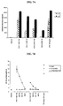
- FIG. 7A is a bar graph showing human peripheral blood mononuclear cells stimulated with UV-inactivated HSV-1, UV-inactivated HSV-2 or LPS (positive control) for 18 hours.
- IL-8 and IL-6 levels in the culture supernatants were measured by ELISA. All experiments have been repeated multiple times with identical results. Error bars shown are based on triplicate wells.
- FIG. 7B is a line graph showing human embryonic kidney (HEK) 293 cells expressing human TLR2/CD14 or TLR4/MD2 and control HEK cells transfected with an NF-kappaB driven luciferase reporter gene and a control Renilla luciferase gene.
- the cells were stimulated with varying multiplicities of infection (MOI) of HSV-1 (KOS strain) and HSV-2 (186 strain) or with TNF-alpha (positive control) for 6 hours.
- Viruses were exposed to UV light prior to stimulating the cells to eliminate infectivity.
- the MOIs shown were based on titers prior to UV-inactivation. Luciferase activity was measured using DualGloTM reagents and normalized using the Renilla luciferase activity.
- FIGS. 8A and 8B are dot plots showing the results of a comparison of adult and neonatal cytokine responses to HSV-1.
- Cord blood cells from 10 healthy newborns and peripheral blood from four healthy adults were stimulated with HSV-1 (MOI 40) for 18 hours. Medium alone (background) values were subtracted. P value is calculated using the rank-sum test (Mann-Whitney) to compare the groups.
- 8 A: IL-6 levels (P 0.033) measure by ELISA.
- TLR2 mediates the inflammatory cytokine response to HSV-1, and is responsible for severe symptoms that are associated with HSV infection in neonates and adults.
- Loss-of-function studies with macrophages demonstrated an essential role for TLR2 in the production of inflammatory cytokines after HSV-1 challenge, while gain-of-function studies demonstrated that expression of TLR2 in HEK293 cells was sufficient to confer responsiveness to HSV-1.
- infection with HSV-1 induced a blunted cytokine response in TLR2 ⁇ / ⁇ mice compared to wild type or TLR4 ⁇ / ⁇ mice both in the serum and within the brain. This attenuated cytokine response was paralleled by a reduction in symptoms of encephalitis in TLR2 ⁇ / ⁇ mice, as compared to wild type and TLR4 ⁇ / ⁇ mice.
- HSV-1 infected TLR2 ⁇ / ⁇ neonatal mice developed mild symptoms, and mortality was less than 40% over a 21 day period.
- wild type and TLR4 ⁇ / ⁇ neonates rapidly succumbed to HSV-1 infection with >90% mortality by day 6.
- the data provided herein demonstrate that the TLR2-mediated cytokine response to HSV-1 is not protective, but rather is detrimental to the host, particularly within the brain.
- TLR2 deficient mice are less likely to die of HSV-1 challenge than wild type mice demonstrates that neonatal animals, rather than being less able to contain the virus, die at least in part, because of their exuberant cytokine responses to viral antigens.
- drugs or other therapies that dampen the innate immune response e.g., the innate immune response that is mediated by TLR2 signaling
- HSV e.g., HSV-1 or HSV-2 infection in neonates, children, and adults.
- the new methods described herein relate to the identification of compounds that are useful for treating HSV infection (e.g., HSV-1 and HSV-2 infection), including adverse effects of infection on the central nervous system such as encephalitis, and symptoms of TORCH syndrome in neonates.
- HSV infection e.g., HSV-1 and HSV-2 infection
- the methods relate to identifying compounds that both decrease TLR2 expression or activity (e.g., by binding to TLR2) and that decrease the effects of HSV infection (e.g., in neonates, children, and adults).
- Compounds identified using such methods are useful for treating subjects at risk of HSV infection or subjects infected with HSV.
- the new methods include screening assays for the identification of compounds that decrease TLR2-mediated signaling for use in the treatment of HSV infection.
- modulators i.e., test compounds or agents (e.g., proteins, peptides, peptidomimetics, peptoids, small inorganic molecules, small non-nucleic acid organic molecules, nucleic acids (e.g., anti-sense nucleic acids, siRNA, oligonucleotides, synthetic oligonucleotides), or other drugs) that inhibit TLR2 signaling, in particular, TLR2 signaling that is associated with HSV infection, e.g., in a neonate.
- test compounds or agents e.g., proteins, peptides, peptidomimetics, peptoids, small inorganic molecules, small non-nucleic acid organic molecules, nucleic acids (e.g., anti-sense nucleic acids, siRNA, oligonucleotides, synthetic oligonucleo
- such a compound may bind to a TLR2 polypeptide and have an inhibitory effect on indicators of HSV infection that are associated with TLR2 signaling, e.g., by inhibiting expression or activity of TLR2.
- Compounds thus identified can be used to modulate the effects of HSV-1 infection, for example, in a neonate in a therapeutic protocol. Such compounds are also useful to elaborate the biological function of TLR2.
- an assay involves the identification of a compound that inhibits TLR2 expression or activity, and determining whether the compound can decrease one or more undesirable effects of HSV infection in a cell or in a subject.
- Methods of identifying a compound that inhibits expression or activity of TLR2 are known in the art and described herein.
- Compounds previously identified as able to inhibit the expression or activity of a TLR2 can also be used in certain methods.
- an assay for identifying an inhibitor of TLR2 expression or activity is a cell-based assay in which a cell that expresses a TLR2 protein or biologically active portion thereof is contacted with a test compound, and the ability of the test compound to modulate TLR2 activity is determined. Determining the ability of the test compound to modulate TLR2 activity can be accomplished by monitoring, for example, IL-6 activity.
- the cell for example, can be of mammalian origin, e.g., murine or human. In general, useful cell types are those that can be infected with HSV (e.g., HSV-1 or HSV-2).
- TLR2 ligands include LTA (lipoteichoic acids), zymosan, peptidoglycan, ara-lipoarabinomannan, and human cytomegalovirus.
- TLR2 co-receptors are known in the art and include TLR1 and TLR6. Such compounds can then be tested for their ability to inhibit expression or activity (e.g., activation of one or more components of the TLR2 signaling pathway such as IL-6 and NF- ⁇ B).
- a compound that binds to TLR2 can be identified, for example, by coupling the compound with a radioisotope or enzymatic label such that binding of the compound to TLR2 can be determined by detecting the labeled compound in a complex.
- a component of the assay can be coupled with a radioisotope or enzymatic label to monitor the ability of a test compound to modulate TLR2 binding to a TLR2 ligand in a complex.
- a compound can be labeled with 125 I, 35 S, 14 C, or 3 H, either directly or indirectly, and the radioisotope detected by direct counting of radioemission or by scintillation counting.
- compounds can be enzymatically labeled with, for example, horseradish peroxidase, alkaline phosphatase, or luciferase, and the enzymatic label detected by determination of conversion of an appropriate substrate to product.
- a microphysiometer can be used to detect the interaction of a compound with TLR2 without the labeling of either the compound or the TLR2 (McConnell et al., Science 257: 1906-1912 (1992)).
- a “microphysiometer” e.g., Cytosensor®
- LAPS light-addressable potentiometric sensor
- a cell-free assay is also provided in which a TLR2 protein or biologically active portion thereof is contacted with a test compound and the ability of the test compound to bind to the TLR2 protein or biologically active portion thereof is evaluated.
- biologically active portions of the TLR2 polypeptides to be used in assays include fragments that participate in interactions with non-TLR2 molecules, e.g., fragments with high surface probability scores.
- Soluble and/or membrane-bound forms of isolated proteins can be used in the cell-free assays.
- membrane-bound forms of the protein it may be desirable to utilize a solubilizing agent.
- solubilizing agents include non-ionic detergents such as n-octylglucoside, n-dodecylglucoside, n-dodecylmaltoside, octanoyl-N-methylglucamide, decanoyl-N-methylglucamide, Triton® X-100, Triton® X-114, Thesit®, Isotridecypoly(ethylene glycol ether) n , 3-[(3-cholamidopropyl)dimethylamminio]-1-propane sulfonate (CHAPS), 3-[(3-cholamidopropyl)dimethylamminio]-2-hydroxy-1-propane sulfonate (CHAPSO), or N-dodecyl-N,N-dimethyl-3-ammonio-1-propane sulfonate.
- non-ionic detergents such as n-octylglucoside,
- Cell-free assays involve preparing a reaction mixture of a TLR2 polypeptide and the test compound under conditions and for a time sufficient to allow the two components to interact and bind, thus forming a complex that can be removed and/or detected.
- the ability of a test compound to inhibit the binding between a TLR2 polypeptide and a TLR2 ligand is determined.
- the interaction between two molecules can also be detected, e.g., using fluorescence energy transfer (FET) (for example, Lakowicz et al., U.S. Pat. No. 5,631,169; Stavrianopoulos et al., U.S. Pat. No. 4,868,103).
- FET fluorescence energy transfer
- a fluorophore label on the first, ‘donor’ molecule is selected such that its emitted fluorescent energy will be absorbed by a fluorescent label on a second, ‘acceptor’ molecule, which in turn is able to fluoresce due to the absorbed energy.
- the ‘donor’ protein molecule may utilize the natural fluorescent energy of tryptophan residues.
- Labels are chosen that emit different wavelengths of light, such that the ‘acceptor’ molecule label may be differentiated from that of the ‘donor’. Since the efficiency of energy transfer between the labels is related to the distance separating the molecules, the spatial relationship between the molecules can be assessed. In a situation in which binding occurs between the molecules, the fluorescent emission of the ‘acceptor’ molecule label in the assay should be maximal.
- An FET binding event can be conveniently measured through standard fluorometric detection means well known in the art (e.g., using a fluorimeter).
- determining the ability of a TLR2 polypeptide to bind to a target molecule can be accomplished using real-time Biomolecular Interaction Analysis (BIA) (e.g., Sjolander et al., Anal. Chem. 63: 2338-2345 (1991) and Szabo et al., Curr. Opin. Struct. Biol. 5: 699-705 (1995)).
- Biomolecular Interaction Analysis e.g., Sjolander et al., Anal. Chem. 63: 2338-2345 (1991) and Szabo et al., Curr. Opin. Struct. Biol. 5: 699-705 (1995)
- “Surface plasmon resonance” or “BIA” detects biospecific interactions in real time, without labeling any of the interactants (e.g., BIAcore).
- the TLR2 or the test substance is anchored onto a solid phase.
- the TLR2/test compound complexes anchored on the solid phase can be detected at the end of the reaction.
- the target gene product is anchored onto a solid surface, and the test compound (which is not anchored) can be labeled, either directly or indirectly, with detectable labels discussed herein.
- TLR2 polypeptide an anti-TLR2 antibody, or a TLR2 binding molecule (e.g., a TLR2 ligand) to facilitate separation of complexed from uncomplexed forms of one or both of the proteins, as well as to accommodate automation of the assay.
- a TLR2 binding molecule e.g., a TLR2 ligand
- Binding of a test compound to a TLR2 polypeptide, or interaction of a TLR2 polypeptide with a target molecule in the presence and absence of a test compound can be accomplished in any vessel suitable for containing the reactants. Examples of such vessels include microtiter plates, test tubes, and micro-centrifuge tubes.
- a fusion protein can be provided, which adds a domain that allows one or both of the proteins to be bound to a matrix.
- glutathione-S-transferase/TLR2 fusion proteins or glutathione-S-transferase/target fusion proteins can be adsorbed onto glutathione SepharoseTM beads (Sigma Chemical, St. Louis, Mo.) or glutathione derivatized microtiter plates, which are then combined with the test compound or the test compound and either the non-adsorbed TLR2 binding molecule or TLR2 polypeptide, and the mixture incubated under conditions conducive to complex formation (e.g., at physiological conditions for salt and pH).
- the beads or microtiter plate wells are washed to remove any unbound components, the matrix immobilized in the case of beads, complex determined either directly or indirectly, for example, as described above.
- the complexes can be dissociated from the matrix, and the level of TLR2 binding or activity determined using standard techniques.
- TLR2 protein or a target molecule on matrices include using conjugation of biotin and streptavidin.
- Biotinylated TLR2 protein or target molecules can be prepared from biotin-NHS(N-hydroxy-succinimide) using techniques known in the art (e.g., biotinylation kit, Pierce Chemicals, Rockford, Ill.), and immobilized in the wells of streptavidin-coated 96 well plates (Pierce Chemical).
- the non-immobilized component is added to the coated surface containing the anchored component. After the reaction is complete, unreacted components are removed (e.g., by washing) under conditions such that any complexes formed will remain immobilized on the solid surface.
- the detection of complexes anchored on the solid surface can be accomplished in a number of ways. Where the previously non-immobilized component is pre-labeled, the detection of label immobilized on the surface indicates that complexes were formed.
- an indirect label can be used to detect complexes anchored on the surface; e.g., using a labeled antibody specific for the immobilized component (the antibody, in turn, can be directly labeled or indirectly labeled with, e.g., a labeled anti-Ig antibody).
- the assay is performed utilizing antibodies reactive with TLR2 polypeptide or ligand, but which do not interfere with binding of the TLR2 polypeptide to the ligand.
- antibodies can be derivatized to the wells of the plate, and unbound ligand or TLR2 polypeptide trapped in the wells by antibody conjugation.
- Methods for detecting such complexes include immunodetection of complexes using antibodies reactive with the TLR2 polypeptide or ligand, as well as enzyme-linked assays which rely on detecting an enzymatic activity associated with the TLR2 polypeptide or ligand.
- cell-free assays can be conducted in a liquid phase.
- the reaction products are separated from unreacted components, by any of a number of known techniques, including but not limited to: differential centrifugation (see, for example, Rivas and Minton, Trends Biochem Sci 18: 284-7 (1993)); chromatography (gel filtration chromatography, ion-exchange chromatography); electrophoresis (see, e.g., Ausubel et al., eds. Current Protocols in Molecular Biology 1999, J. Wiley: New York.); and immunoprecipitation (see, for example, Ausubel et al., eds., 1999 , Current Protocols in Molecular Biology , J.
- the assay can include contacting the TLR2 polypeptide with a known compound that binds to TLR2 to form an assay mixture, contacting the assay mixture with a test compound, and determining the ability of the test compound to interact with a TLR2 polypeptide, e.g., by determining the ability of the test compound to preferentially bind to TLR2 or a biologically active portion thereof, or to modulate the activity of TLR2, as compared to the known compound.
- Useful assays also include methods for determining the ability of the test compound to modulate the activity of a TLR2 protein through binding to the TLR2 or by modulation of the activity of a downstream effector of a TLR2 target molecule, e.g., NF- ⁇ B.
- a downstream effector of a TLR2 target molecule e.g., NF- ⁇ B.
- the activity of the effector molecule on an appropriate target can be determined, or the binding of the effector to an appropriate target can be determined, as previously described.
- a reaction mixture containing the TLR2 and the binding partner is prepared, under conditions and for a time sufficient, to allow the two products to form complex.
- the reaction mixture is provided in the presence and absence of the test compound.
- the test compound can be initially included in the reaction mixture, or can be added at a time subsequent to the addition of the target gene and its cellular or extracellular binding partner.
- Control reaction mixtures are incubated without the test compound or with a placebo.
- the formation of any complexes between the TLR2 polypeptide and the cellular or extracellular binding partner is then detected.
- the formation of a complex in the control reaction but not in the reaction mixture containing the test compound, indicates that the compound interferes with the interaction of the TLR2 polypeptide and the interactive binding partner.
- Another method of identifying compounds that are effective for treating HSV infection in a subject is to determine the effect of the compound on TLR2 activity, e.g., by assaying production of inflammatory cytokines or chemokines (e.g., IL-1, IL-6, IL-8, MCP-1, MIP-1, and/or TNF), or the activation of gene expression, e.g., by NF- ⁇ B, in a cell expressing functional TLR2 (and, in some cases, infected with HSV), in the presence of the test compound.
- inflammatory cytokines or chemokines e.g., IL-1, IL-6, IL-8, MCP-1, MIP-1, and/or TNF
- TNF- ⁇ B e.g., IL-1, IL-6, IL-8, MCP-1, MIP-1, and/or TNF
- Methods for performing such assays are known in the art; some are described herein.
- Test compounds that reduce TLR2 activity can be considered effective compounds for the treatment of a subject infected with HSV.
- An effective compound is expected, in an HSV-1 infected subject, to decrease levels of inflammatory cytokines that are induced by the TLR2 signaling pathway as compared to the levels of inflammatory cytokines that are induced in a subject that was not treated with the compound, thereby reducing the negative effects of HSV infection.
- a compound that is useful for treating TLR2-mediated effects of HSV infection will inhibit the induction of inflammatory cytokines associated with activation of the TLR2 signaling pathway in a cell that is infected with HSV and contacted with the compound compared to a control cell that was not contacted with the compound.
- the assays described herein can be conducted in a heterogeneous or homogeneous format.
- Heterogeneous assays involve anchoring either the target gene product or the binding partner onto a solid phase, and detecting complexes anchored on the solid phase at the end of the reaction.
- homogeneous assays the entire reaction is carried out in a liquid phase.
- the order of addition of reactants can be varied to obtain different information about the compounds being tested.
- test compounds that interfere with the interaction between the TLR2 polypeptide and a binding partner e.g., a TLR2 ligand
- competition can be identified by conducting the reaction in the presence of the test substance.
- test compounds that disrupt preformed complexes e.g., compounds with higher binding constants that displace one of the components from the complex
- test compounds that disrupt preformed complexes can be tested by adding the test compound to the reaction mixture after complexes have been formed. Examples of the various formats are briefly described below.
- either the TLR2 or the interactive cellular or extracellular binding partner is anchored onto a solid surface (e.g., a microtiter plate), while the non-anchored species is labeled, either directly or indirectly.
- the anchored species can be immobilized by non-covalent or covalent attachments.
- an immobilized antibody specific for the species to be anchored can be used to anchor the species to the solid surface.
- the partner of the immobilized species is exposed to the coated surface with or without the test compound. After the reaction is complete, unreacted components are removed (e.g., by washing) and any complexes formed will remain immobilized on the solid surface.
- the detection of label immobilized on the surface indicates that complexes were formed.
- an indirect label can be used to detect complexes anchored on the surface; e.g., using a labeled antibody specific for the initially non-immobilized species (the antibody, in turn, can be directly labeled or indirectly labeled with, e.g., a labeled anti-Ig antibody).
- test compounds that inhibit complex formation or that disrupt preformed complexes can be detected.
- the reaction can be conducted in a liquid phase in the presence or absence of the test compound, the reaction products separated from unreacted components, and complexes detected; e.g., using an immobilized antibody specific for one of the binding components to anchor any complexes formed in solution, and a labeled antibody specific for the other partner to detect anchored complexes.
- test compounds that inhibit complex or that disrupt preformed complexes can be identified.
- a homogeneous assay can be used.
- a preformed complex of a TLR2 polypeptide and the interactive cellular or extracellular binding partner of TLR2 is prepared in that either the TLR2 or their binding partners are labeled, but the signal generated by the label is quenched due to complex formation (see, e.g., U.S. Pat. No. 4,109,496 that utilizes this approach for immunoassays).
- the addition of a test substance that competes with and displaces one of the species from the preformed complex will result in the generation of a signal above background. In this way, test substances that disrupt TLR2-binding partner interaction can be identified.
- test compounds used in the methods described herein can include those obtained using any of the numerous approaches in combinatorial library methods known in the art, including: biological libraries (e.g., peptides, polypeptides, or nucleic acids); peptoid libraries (libraries of molecules having the functionalities of peptides, but with a novel, non-peptide backbone which are resistant to enzymatic degradation but which nevertheless remain bioactive; e.g., Zuckermann et al., J. Med. Chem., 37: 2678-2685 (1994)); spatially addressable parallel solid phase or solution phase libraries; synthetic library methods requiring deconvolution; the “one-bead one-compound” library method; and synthetic library methods using affinity chromatography selection.
- biological libraries e.g., peptides, polypeptides, or nucleic acids
- peptoid libraries libraries of molecules having the functionalities of peptides, but with a novel, non-peptide backbone which are resistant to enzymatic
- the biological library and peptoid library approaches are limited to peptide libraries, while the other four approaches are applicable to peptide, non-peptide oligomer, or small molecule libraries of compounds (Lam, Anticancer Drug Des. 12: 145 (1997)).
- the new methods employ an isolated TLR2 polypeptide (a full-length TLR2 protein or a variant, e.g., a mutant or fragment, thereof), e.g., a biologically active portion such as an extracellular domain.
- TLR2 polypeptides are useful as described herein, for example, as immunogens or antigens to raise or test (or more generally to bind) anti-TLR2 antibodies (e.g., antigenic fragments of TLR2), or to serve as inhibitory compounds (e.g., TLR2 dominant negatives).
- Such polypeptides can be isolated from cells or tissue sources using standard protein purification techniques.
- a TLR2 polypeptide can be isolated from a natural source, can be produced by recombinant DNA methods, or can be synthesized chemically.
- TLR2 polypeptides include polypeptides consisting of the amino acid sequences of Genbank Accession Nos. NP — 003255 (gene NM — 003264; human TLR2) and AAH14693 (gene NM — 011905; murine TLR2), and are hereby incorporated by reference in their entirety.
- a TLR2 polypeptide includes an amino acid sequence that has at least 80% sequence identity with a known TLR2 polypeptide, e.g., at least 85%, 90%, 95% or more identity, and has at least one biological activity of a known TLR2 (e.g., can bind to a naturally-occurring TLR2 ligand or co-receptor, can induce activation of gene transcription (e.g., via NF- ⁇ B), or induce secretion of a chemokine or cytokine, e.g., IL — 1, IL-6, IL-8, TNF, MCP-1, or MIP-1).
- Naturally-occurring TLR2 ligands and co-receptors include TLR1, TLR6, zymosan, peptidoglycan, lipotechoic acid, and ara-lipoarabinomannan.
- Variants of TLR2 polypeptide useful in methods described herein can also include fragments including at least an extracellular binding domain of the TLR2 or a mutant thereof. Such fragments have been shown to have inhibitory activity, see, e.g., LeBouder et al., J. Immunol., 171: 6680-9 (2003). Additional useful polypeptide fragments can be identified, e.g., by the ability to compete with full length TLR2 for binding of a TLR2 ligand (e.g., HSV). Variants of naturally occurring TLR2 amino acid sequences can be tested for their ability to inhibit TLR2 activity, e.g., signaling that is associated with HSV infection. Other appropriate sequences besides those described herein are known in the art and can be used in the methods disclosed herein.
- TLR2 ligand e.g., HSV
- TLR2 dominant negative polypeptides i.e., polypeptides whose expression significantly inhibits the activity of wild-type, e.g., endogenous, TLR2 are also useful in the methods described herein. Dominant negatives, and methods for making and testing them, are known in the art. Examples of TLR2 dominant negative polypeptides include fragments and deletion mutants, e.g., less than the full-length TLR2, that are useful in the methods described herein.
- Dominant negative polypeptides typically encompass the extracellular domain of a TLR2 (e.g., a polypeptide including about amino acids 1-587), and can include TLR2 deletion mutants lacking the conserved intracellular Toll/Interleukin-1 receptor (TIR) domain (e.g., a polypeptide including amino acids 1-642), (see, e.g., Sandor et al., J. Cell Biol. 162 (6): 1099-1110 (2003); Xu et al., Nature 408 (6808): 111-5 (2000)) or the amino acids from Ser40-Ile64 (Fujita et al., J. Immunol.
- TIR Toll/Interleukin-1 receptor
- Dominant negative mutants can include point mutants, e.g., the P681H (Xu et al., 2000, supra), L107E, L112E, and L115E mutants (Fujita et al., 2003, supra).
- “conservative amino acid substitution” means a substitution of an amino acid in a polypeptide within an amino acid family. Families of amino acids are recognized in the art and are based on physical and chemical properties of the amino acid side chains. Families include the following: amino acids with basic side chains (e.g., lysine, arginine, and histidine); amino acids with acidic side chains (e.g., aspartic acid and glutamic acid); amino acids with uncharged polar side chains (e.g., glycine, asparagine, glutamine, serine, threonine, tyrosine, and cysteine); amino acids with nonpolar side chains (e.g., alanine, valine, leucine, isoleucine, proline, phenylalanine, methionine, and tryptophan); amino acids with branched side chains (e.g., threonine, valine, and isoleucine); and amino acids with aromatic side chains (e.g.,
- antibodies are provided that are anti-TLR2 antibodies (also referred to as TLR2 antibodies).
- antibody refers to an immunoglobulin molecule or immunologically active portion thereof, i.e., an antigen-binding portion.
- immunologically active portions of immunoglobulin molecules include F(ab) and F(ab′) 2 , fragments, which can be generated by treating the antibody with an enzyme such as pepsin.
- the antibody can be a polyclonal, monoclonal, monospecific, recombinant, e.g., a chimeric or humanized, fully human, non-human, e.g., murine, or single chain antibody. In some cases, the antibody has effector function and can fix complement.
- the antibody can be coupled to a toxin or imaging agent.
- a full-length TLR2 protein or antigenic peptide fragment of a TLR2 can be used as an immunogen or can be used to identify anti-TLR2 antibodies (TLR2 antibodies) made with other immunogens, e.g., cells, membrane preparations, and the like.
- the antigenic peptide of a TLR2 polypeptide should include at least 8 amino acid residues of a TLR2 protein. In general, the polypeptide will encompass an epitope of TLR2, e.g., an extracellular domain.
- the antigenic peptide can include at least 10 amino acid residues, at least 15 amino acid residues, at least 20 amino acid residues, or at least 30 amino acid residues.
- Fragments of a TLR2 can also be used to make an antibody against an extracellular region of the TLR2.
- Epitopes encompassed by the antigenic peptide are generally regions of TLR2 that are located on the surface of the protein, e.g., hydrophilic regions, as well as regions with high antigenicity.
- an Emini surface probability analysis of the human TLR2 protein sequence can be used to indicate the regions that have a particularly high probability of being localized to the surface of the TLR2 protein and are thus likely to constitute surface residues useful for targeting antibody production.
- the antibody can bind to the extracellular portion of the TLR2 protein, e.g., it can bind to a whole cell that expresses the TLR2 protein. In another embodiment, the antibody binds to an intracellular portion of the TLR2 protein.
- Chimeric, humanized, e.g., completely human, antibodies are desirable for applications that include repeated administration, e.g., therapeutic treatment (and some diagnostic applications) of a human subject.
- the anti-TLR2 antibody can be a single chain antibody.
- a single-chain antibody (scFV) may be engineered (see, for example, Colcher et al., Ann. N.Y. Acad. Sci. 880: 263-80 (1999); and Reiter, Clin. Cancer Res. 2: 245-52 (1996)).
- the single chain antibody can be dimerized or multimerized to generate multivalent antibodies having specificities for different epitopes of the same target TLR2 protein.
- the antibody has reduced or no ability to bind to an Fc receptor.
- the antibody can be an isotype or subtype, fragment or other mutant, which does not support binding to an Fc receptor, e.g., it has a mutagenized or deleted Fc receptor binding region.
- An anti-TLR2 antibody (e.g., monoclonal antibody) can be used in methods of inhibiting TLR2 signaling associated with HSV infection. Such antibodies can also be used to monitor TLR2 protein levels or localization in cells or tissue as part of a screening procedure to identify compounds that inhibit TLR2 expression or activity (e.g., during HSV infection). Detection can be facilitated by coupling (i.e., physically linking) the antibody to a detectable substance (i.e., antibody labeling). Examples of detectable substances include various enzymes, prosthetic groups, fluorescent materials, luminescent materials, bioluminescent materials, and radioactive materials.
- suitable enzymes include horseradish peroxidase, alkaline phosphatase, P-galactosidase, or acetylcholinesterase;
- suitable prosthetic group complexes include streptavidin/biotin and avidin/biotin;
- suitable fluorescent materials include umbelliferone, fluorescein, fluorescein isothiocyanate, rhodamine, dichlorotriazinylamine fluorescein, dansyl chloride or phycoerythrin;
- an example of a luminescent material includes luminol;
- examples of bioluminescent materials include luciferase, luciferin, and aequorin, and examples of suitable radioactive material include 125 I, 131 I, 35 S or 3 H.
- the smallest inhibitory fragment that binds to the target antigen e.g., TLR2
- TLR2 target antigen
- peptides having an amino acid sequence corresponding to the Fv region of the antibody can be used.
- the antibody is an inhibitory antibody, e.g., the antibody can significantly inhibit the activity of TLR2.
- Inhibitory antibodies are known in the art, e.g., as described in Sandor et al., 2003, supra, and Meng et al., J. Clin. Inv. 113 (10): 1473-1481 (2004).
- An antisense nucleic acid can include a nucleotide sequence that is complementary to a “sense” nucleic acid encoding a protein, e.g., complementary to the coding strand of a double-stranded cDNA molecule or complementary to an mRNA sequence.
- the antisense nucleic acid can be complementary to an entire TLR2 coding strand, or to only a portion thereof (e.g., the coding region of human TLR2).
- the antisense nucleic acid molecule is antisense to a “noncoding region” of the coding strand of a nucleotide sequence encoding TLR2 (e.g., the 5′ and 3′ untranslated regions).
- an antisense nucleic acid can be designed such that it is complementary to the entire coding region of TLR2 mRNA or a portion thereof.
- the antisense molecule is an oligonucleotide that is antisense to only a portion of the coding or noncoding region of TLR2 mRNA.
- the antisense oligonucleotide can be complementary to the region surrounding the translation start site of TLR2 mRNA, e.g., between the ⁇ 10 and +10 regions of the target gene nucleotide sequence of interest.
- An antisense oligonucleotide can be, for example, about 20, 25, 30, 35, 40, 45, 50, 55, 60, 65, 70, 75, 80, or more nucleotides in length. Based upon sequences known in the art and disclosed herein, one of skill in the art can easily choose and synthesize any of a number of appropriate antisense molecules for use in accordance with the present invention. For example, a “gene walk” comprising a series of oligonucleotides of 15-30 nucleotides spanning the length of a target nucleic acid can be prepared, followed by testing for inhibition of target gene expression. Optionally, gaps of 5-10 nucleotides can be left between the oligonucleotides to reduce the number of oligonucleotides synthesized and tested.
- an antisense nucleic acid can be constructed using chemical synthesis and enzymatic ligation reactions using procedures known in the art.
- an antisense nucleic acid e.g., an antisense oligonucleotide
- an antisense nucleic acid can be chemically synthesized, e.g., using naturally occurring nucleotides or variously modified nucleotides designed to increase the biological stability of the molecules or to increase the physical stability of the duplex formed between the antisense and sense nucleic acids, e.g., phosphorothioate derivatives and acridine substituted nucleotides can be used.
- the antisense nucleic acid also can be produced biologically using an expression vector into which a nucleic acid has been subcloned in an antisense orientation (i.e., RNA transcribed from the inserted nucleic acid will be of an antisense orientation to a target nucleic acid of interest, described further in the following subsection).
- the new antisense nucleic acid molecules are typically administered to a subject (e.g., by direct injection at a tissue site), or generated in situ (e.g., using an expression vector that contains a sequence encoding the antisense molecule) such that they hybridize with or bind to cellular mRNA and/or genomic DNA encoding a TLR2 protein to thereby inhibit expression of the protein, e.g., by inhibiting transcription and/or translation.
- antisense nucleic acid molecules can be modified to target selected cells and then administered systemically.
- antisense molecules can be modified such that they specifically bind to receptors or antigens expressed on a selected cell surface, e.g., by linking the antisense nucleic acid molecules to peptides or antibodies that bind to cell surface receptors or antigens.
- the antisense nucleic acid molecules can also be delivered to cells using the vectors described herein.
- vector constructs in which the antisense nucleic acid molecule is placed under the control of a strong pol II or pol III promoter are generally used.
- the antisense nucleic acid molecule can be an ⁇ -anomeric nucleic acid molecule.
- An ⁇ -anomeric nucleic acid molecule forms specific double-stranded hybrids with complementary RNA in which, contrary to the usual ⁇ -units, the strands run parallel to each other (Gaultier et al., Nucleic Acids., Res. 15: 6625-6641 (1987)).
- the antisense nucleic acid molecule can also comprise a 2′-o-methylribonucleotide (Inoue et al., Nucleic Acids Res., 15: 6131-6148 (1987)) or a chimeric RNA-DNA analogue (Inoue et al., FEBS Lett., 215: 327-330 (1987)).
- a 2′-o-methylribonucleotide Inoue et al., Nucleic Acids Res., 15: 6131-6148 (1987)
- a chimeric RNA-DNA analogue Inoue et al., FEBS Lett., 215: 327-330 (1987)
- a ribozyme having specificity for a TLR2-encoding nucleic acid can include one or more sequences complementary to the nucleotide sequence of a TLR2 cDNA, and a sequence having known catalytic sequence responsible for mRNA cleavage (see U.S. Pat. No. 5,093,246 or Haselhoff and Gerlach, 1988, Nature, 334: 585-591).
- a derivative of a Tetrahymena L-19 IVS RNA can be constructed in which the nucleotide sequence of the active site is complementary to the nucleotide sequence to be cleaved in a TLR2-encoding mRNA (e.g., Cech et al., U.S. Pat. No. 4,987,071; and Cech et al., U.S. Pat. No. 5,116,742.
- TLR2 mRNA can be used to select a catalytic RNA having a specific ribonuclease activity from a pool of RNA molecules (e.g., Bartel and Szostak, Science, 261: 1411-1418 (1993)).
- the antisense nucleic acid is a morpholino oligonucleotide (see, e.g., Heasman, Dev. Biol., 243: 209-14 (2002); Iversen, Curr. Opin. Mol. Ther., 3: 235-8 (2001); Summerton, Biochim. Biophys. Acta., 1489: 141-58 (1999).
- TLR2 gene expression can be inhibited by targeting nucleotide sequences complementary to the regulatory region of the TLR2 (e.g., the TLR2 promoter and/or enhancers) to form triple helical structures that prevent transcription of the TLR2 gene in target cells (e.g., Helene, Anticancer Drug Des., 6: 569-84 (1991); Helene, Ann. N.Y. Acad. Sci., 660: 27-36 (1992); and Maher, Bioassays, 14: 807-15 (1992)).
- the potential sequences that can be targeted for triple helix formation can be increased by creating a so-called “switchback” nucleic acid molecule.
- Switchback molecules are synthesized in an alternating 5′-3′,3′-5′ manner, such that they base pair with first one strand of a duplex and then the other, eliminating the necessity for a sizeable stretch of either purines or pyrimidines to be present on one strand of a duplex.
- a TLR2 nucleic acid molecule can be modified at the base moiety, sugar moiety, or phosphate backbone to improve, e.g., the stability, hybridization, or solubility of the molecule.
- the deoxyribose phosphate backbone of the nucleic acid molecules can be modified to generate peptide nucleic acids (see Hyrup et al., Bioorganic & Medicinal Chemistry, 4: 5-23 (1996)).
- peptide nucleic acid refers to a nucleic acid mimic, e.g., a DNA mimic, in which the deoxyribose phosphate backbone is replaced by a pseudopeptide backbone and only the four natural nucleobases are retained.
- the neutral backbone of a PNA can allow for specific hybridization to DNA and RNA under conditions of low ionic strength.
- the synthesis of PNA oligomers can be performed using standard solid phase peptide synthesis protocols as described, e.g., in Hyrup et al., 1996, supra and Perry-O'Keefe et al., Proc. Natl. Acad. Sci. USA, 93: 14670-675 (1996).
- PNAs of TLR2 nucleic acid molecules can be used in therapeutic and diagnostic applications.
- PNAs can be used as antisense or antigene agents for sequence-specific modulation of gene expression by, for example, inducing transcription or translation arrest or inhibiting replication.
- Such compounds are useful for treating undesirable TLR2-mediated effects that result from HSV infection.
- the oligonucleotide can include other appended groups such as peptides (e.g., for targeting host cell receptors in vivo), or agents facilitating transport across the cell membrane (e.g., Letsinger et al., Proc. Natl. Acad. Sci. USA, 86: 6553-6556 (1989); Lemaitre et al., Proc. Natl. Acad. Sci. USA, 84: 648-652 (1987); PCT Publication No. WO88/09810) or the blood-brain barrier (e.g., PCT Publication No. WO89/10134).
- peptides e.g., for targeting host cell receptors in vivo
- agents facilitating transport across the cell membrane e.g., Letsinger et al., Proc. Natl. Acad. Sci. USA, 86: 6553-6556 (1989); Lemaitre et al., Proc. Natl. Acad. Sci. USA,
- oligonucleotides can be modified with hybridization-triggered cleavage agents (e.g., Krol et al., Bio-Techniques, 6: 958-976 (1988)) or intercalating agents (e.g., Zon, Pharm. Res., 5: 539-549 (1988)).
- the oligonucleotide may be conjugated to another molecule, (e.g., a peptide, hybridization triggered cross-linking agent, transport agent, or hybridization-triggered cleavage agent).
- Molecular beacon oligonucleotide primer and probe molecules having at least one region which is complementary to a TLR2 nucleic acid and two complementary regions, one having a fluorophore and one a quencher, such that the molecular beacon is useful for quantitating the presence of TLR2 nucleic acid in a sample, can also be used.
- Molecular beacon nucleic acids are described, for example, in Lizardi et al., U.S. Pat. No. 5,854,033; Nazarenko et al., U.S. Pat. No. 5,866,336, and Livak et al., U.S. Pat. No. 5,876,930.
- the oligonucleotide is a small interfering RNA (siRNA) that is directed against a TLR2.
- siRNA small interfering RNA
- dsRNA double stranded RNA molecules generally comprise 16-30, e.g., 16, 17, 18, 19, 20, 21, 22, 23, 24, 25, 26, 27, 28, 29, or 30 nucleotides in each strand, wherein one of the strands is substantially identical, e.g., at least 80% (or more, e.g., 85%, 90%, 95%, or 100%) identical, e.g., having 3, 2, 1, or 0 mismatched nucleotide(s), to a target region in the mRNA, and the other strand is complementary to the first strand.
- the dsRNA molecules can be chemically synthesized, or can transcribed be in vitro from a DNA template, or in vivo from, e.g., small hairpin RNA (shRNA).
- shRNA small hairpin RNA
- Negative control siRNAs should have the same nucleotide composition as the selected siRNA, but without significant sequence complementarity to the appropriate genome.
- Such negative controls can be designed by randomly scrambling the nucleotide sequence of the selected siRNA; a homology search can be performed to ensure that the negative control lacks homology to any other gene in the appropriate genome.
- negative control siRNAs can be designed by introducing one or more base mismatches into the sequence.
- the nucleic acid compositions that are useful in the methods described herein include both siRNA and crosslinked siRNA derivatives.
- Crosslinking can be employed to alter the pharmacokinetics of the composition, for example, to increase half-life in the body.
- the invention includes siRNA derivatives that include siRNA having two complementary strands of nucleic acid, such that the two strands are crosslinked. For example, a 3′ OH terminus of one of the strands can be modified, or the two strands can be crosslinked and modified at the 3′OH terminus.
- the siRNA derivative can contain a single crosslink (e.g., a psoralen crosslink).
- the siRNA derivative has at its 3′ terminus a biotin molecule (e.g., a photocleavable biotin), a peptide (e.g., a Tat peptide), a nanoparticle, a peptidomimetic, organic compounds (e.g., a dye such as a fluorescent dye), or dendrimer.
- a biotin molecule e.g., a photocleavable biotin
- a peptide e.g., a Tat peptide
- a nanoparticle e.g., a peptidomimetic
- organic compounds e.g., a dye such as a fluorescent dye
- the nucleic acid compositions can be unconjugated or can be conjugated to another moiety, such as a nanoparticle, to enhance a property of the compositions, e.g., a pharmacokinetic parameter such as absorption, efficacy, bioavailability, and/or half-life.
- the conjugation can be accomplished by methods known in the art, e.g., using the methods of Lambert et al., Drug Deliv. Rev., 47: 99-112 (2001) (describes nucleic acids loaded to polyalkylcyanoacrylate (PACA) nanoparticles); Fattal et al., J.
- the nucleic acid molecules can also be labeled using methods known in the art.
- the nucleic acid compositions can be labeled with a fluorophore, e.g., Cy3, fluorescein, or rhodamine.
- the labeling can be carried out using a kit, e.g., the SILENCERTM siRNA labeling kit (Ambion).
- the siRNA can be radiolabeled, e.g., using 3 H, 32 P, or other appropriate isotope.
- Synthetic siRNAs or other oligonucleotides or nucleic acids described herein can be delivered into cells by cationic liposome transfection and electroporation. In general, exogenous siRNAs only show short-term persistence of the silencing effect (about 4-5 days).
- Several strategies for expressing siRNA duplexes within cells from recombinant DNA constructs allow longer-term target gene suppression in cells, including mammalian Pol III promoter systems (e.g., HI or U6/snRNA promoter systems (Tuschl, Nature Biotechnol., 20: 440-448 (2002)) capable of expressing functional double-stranded siRNAs; (Bagella et al., J. Cell.
- RNA Pol III Transcriptional termination by RNA Pol III occurs at runs of four consecutive T residues in the DNA template, providing a mechanism to end the siRNA transcript at a specific sequence.
- the siRNA is complementary to the sequence of the target gene in 5′-3′ and 3′-5′ orientations, and the two strands of the siRNA can be expressed in the same construct or in separate constructs.
- Hairpin siRNAs, driven by HI or U6 snRNA promoter and expressed in cells, can inhibit target gene expression.
- Constructs containing siRNA sequence under the control of T7 promoter also make functional siRNAs when cotransfected into the cells with a vector expression T7 RNA polymerase (Jacque, Nature, 418: 435-438 (2002)).
- RNAs Animal cells express a range of noncoding RNAs of approximately 22 nucleotides termed micro RNA (mRNAs) and can regulate gene expression at the post transcriptional or translational level during animal development.
- mRNAs micro RNA
- a vector construct that expresses the novel mRNA can be used to produce siRNAs to initiate RNAi against specific mRNA targets in mammalian cells (Zeng, Mol. Cell, 9: 1327-1333 (2002)).
- micro-RNA designed hairpins When expressed by DNA vectors containing polymerase III promoters, micro-RNA designed hairpins can silence gene expression (McManus, 2002, supra). Viral-mediated delivery mechanisms can also be used to induce specific silencing of targeted genes through expression of siRNA, for example, by generating recombinant adenoviruses harboring siRNA under RNA Pol II promoter transcription control (Xia et al., Nat. Biotechnol., 20: 1006-1010 (2002)). Infection of HeLa cells by such recombinant adenoviruses allows for diminished endogenous target gene expression.
- Injection of the recombinant adenovirus vectors into transgenic mice expressing the target genes of the siRNA results in in vivo reduction of target gene expression (id).
- whole-embryo electroporation can efficiently deliver synthetic siRNA into post-implantation mouse embryos (Calegari et al., Proc. Natl. Acad. Sci. USA, 99: 14236-14240 (2002)).
- siRNA In adult mice, efficient delivery of siRNA can be accomplished by “high-pressure” delivery technique, a rapid injection (within 5 seconds) of a large volume of siRNA containing solution into animal via the tail vein (Liu, Gene Ther., 6: 1258-1266 (1999); McCaffrey, Nature, 418: 38-39 (2002); Lewis, Nature Genetics, 32: 107-108 (2002)). Nanoparticles and liposomes can also be used to deliver siRNA into animals.
- Engineered RNA precursors, introduced into cells or whole organisms as described herein, will lead to the production of a desired oligonucleotide such as an siRNA molecule.
- a desired oligonucleotide such as an siRNA molecule.
- Such an siRNA molecule will then associate with endogenous protein components of the RNAi pathway to bind to and target a specific mRNA sequence for cleavage and destruction.
- the mRNA to be targeted by the siRNA generated from the engineered RNA precursor will be depleted from the cell or organism, leading to a decrease in the concentration of the protein encoded by that mRNA in the cell or organism.
- Aptamer molecules specific for TLR2 protein can also be used.
- Aptamers are nucleic acid molecules having a tertiary structure that permits them to specifically bind to protein ligands (e.g., Osborne, et al., Curr. Opin. Chem. Biol., 1: 5-9 (1997); and Patel, Curr. Opin. Chem. Biol., 1: 32-46 (1997)). Since nucleic acid molecules may, in many cases, be more conveniently introduced into target cells than therapeutic protein molecules, aptamers offer a method by which TLR2 activity can be specifically decreased without the introduction of drugs or other molecules which may have pluripotent effects.
- TLR2 signaling As described herein, it is the host response (i.e., induction of TLR2 signaling) that causes the deleterious TLR2-mediated effects of HSV infection such as those observed in HSV infected neonates.
- Certain methods described herein relate to identification of compounds that inhibit TLR2 signaling in an HSV infected cell. In some embodiments such compounds would not be expected to significantly affect the infectivity of HSV into a cell treated with the compound, and thus can be useful for treating HSV infection. Accordingly, compounds that inhibit TLR2 signaling in an HSV infected cell can also be tested for their effect on infectivity.
- the number of plaque forming units (pfu) in a tissue or cell that is contacted with a test compound that can decrease TLR2 signaling is determined and compared to the number of pfu in an infected cell that was not contacted with the compound.
- Infectivity can be measured as described herein or using other methods known in the art.
- a compound that affects TLR2 signaling, but does not affect the amount of virus, is thus a TLR2-targeted compound that can bes useful for treating HSV infection.
- a compound that is acceptable for such a use may or may not decrease infectivity.
- Compounds that are identified as useful for treating HSV infection can also be tested, e.g., using methods described herein, for their ability to inhibit TLR2 signaling that is associated with HSV infection.
- the methods include the identification of compounds that are particularly useful for treating HSV infection, e.g., in neonates.
- a compound is tested for its ability to ameliorate one or more symptoms of HSV infection.
- Symptoms of HSV infection in neonates, children, and adults are described herein and known in the art, e.g., The Merck Manual, Seventeenth Edition , Rahway, N.J., 1999-2004.
- HSV infections typically produce an eruption of tiny blisters on the skin or mucous membranes.
- Oral infection with HSV usually causes herpetic gingivostomatitis (mouth sores).
- mouth sores affected persons generally feel sick and have a fever, headache, and body aches.
- the mouth sores can last 10 to 14 days and are often very severe, making eating and drinking extremely uncomfortable.
- swollen gums are the only symptom; occasionally, no symptoms develop.
- Herpetic gingivostomatitis most commonly develops in children.
- HSV-2 symptoms are usually mild and can include itching, burning, soreness and small blisters in the genital area; small skin ulcers, which form when the blisters break; localized pain, e.g., if urine touches the genital ulcers; enlarged and/or painful lymph nodes in the groin; and headache, fever and a general malaise.
- the first genital HSV infection can be severe and prolonged, with multiple painful blisters in the genital region. Fever and general malaise are common, with burning during urination in some cases. Occasionally, the infected person may have no symptoms.
- Recurring attacks of genital herpes generally begin with typical symptoms (e.g., local tingling, discomfort, itching, or aching in the groin) that precede the blisters by several hours up to 2-3 days. Painful blisters, surrounded by a reddish rim, appear on the skin or mucous membranes of the genitals. The blisters quickly break open, leaving sores. In some cases, blisters also appear on the thighs, buttocks, or around the anus.
- Genital blisters may develop on the vulva in women; these blisters are usually obvious and very painful. Internal blisters may develop in the vagina or on the cervix; they are less painful and are not visible. Typical episodes of recurring genital herpes last about a week.
- HSV-1 or HSV-2 occasionally will enter through a break in the skin, e.g., of a finger, causing a swollen, painful, red fingertip; this condition is known as herpetic whitlow.
- HSV-1 sometimes infects the cornea of the eye, a condition known as herpes simplex keratitis, which features painful sores and blurred vision. Over time, the cornea can become cloudy, causing a significant loss of vision and requiring corneal transplantation.
- TORCH Syndrome refers to infection of a developing fetus or newborn by any of a group of infectious agents, e.g., Toxoplasmosis, Other infections, Rubella, Cytomegalovirus, or Herpes.
- infectious agents e.g., Toxoplasmosis, Other infections, Rubella, Cytomegalovirus, or Herpes.
- other infections can include syphilis, hepatitis B, Coxsackie's virus, Epstein-Barr virus, varicella-zoster virus, and human parvovirus.
- Infection with any of these agents may cause a constellation of similar symptoms in affected neonates, including fever; difficulties feeding; small areas of bleeding under the skin, causing the appearance of small reddish or purplish spots; hepatosplenomegaly (enlargement of the liver and spleen); cutaneous manifestations, including petechiae, purpura, jaundice, and dermal erythropoiesis, hearing impairment; abnormalities of the eyes; and/or other symptoms and findings.
- Each infectious agent may also result in additional abnormalities that may be variable, depending upon a number of factors (e.g., stage of fetal development).
- symptoms can include single or grouped cutaneous vesicles, oral ulcers, or conjunctivitis. See, e.g., Epps et al., Semin. Dermatol. 14 (2): 179-86 (1995).
- Subjects most at risk of HSV infection include immunosuppressed individuals (e.g., individuals on an immunosuppressive regime or individuals infected with HIV).
- a subject is infected with HSV.
- Such methods are known in the art (for example, The Merck Manual, Seventeenth Edition , Section. 19, Chapter 260; Merck & Co, Inc., Rahway, N.J., 1999-2004).
- infection can be confirmed by infecting a cultured cell with a sample from a suspected infection site and isolating virus from the culture.
- Various cell lines of human or nonhuman origin can be used in this method.
- Retrieval of virus from a subject can be, e.g., from skin vesicles, the mouth, eye, or CSF (cerebrospinal fluid). In some neonates presenting with encephalitis, virus is found only in the brain.
- Testing can also be done using, e.g., polymerase chain reaction. Cytopathologic effects usually can be demonstrated in tissue culture within 24 to 48 hours after inoculation. The determination of HSV-1 infection can also be confirmed, for example, by neutralization with appropriate high-titer antiserum; immunofluorescence of lesion scrapings (e.g., using monoclonal antibodies that specifically bind to HSV-1); and electron microscopy. Such methods are useful in assays that relate to determining whether a compound (i.e., a compound that decreases TLR2 signaling) can decrease symptoms of HSV infection.
- a compound i.e., a compound that decreases TLR2 signaling
- Animal models of HSV infection are known in the art, e.g., as described in Weber, Animal Models in Virology, in Schmidt and Weber (eds), Animal Testing in Infectiology . Contrib Microbiol. Basel, Karger, 2001, 9:15-30.
- the murine model discussed herein can be used.
- Rodents, e.g., rats, mice, and guinea pigs, and lagomorphs, e.g., rabbits also make good models of HSV infection, when exposed to an HSV.
- the methods described herein also pertain to the field of predictive medicine in which diagnostic assays, prognostic assays, and monitoring clinical trials are used for prognostic (predictive) purposes to thereby treat an individual infected with HSV.
- a patient infected with or exposed to HSV and at risk for a TLR2-mediated effect of HSV infection such as encephalitis
- Increased TLR2 activity in a subject infected with HSV indicates that the subject is at risk of developing a TLR2-mediated effect of HSV infection.
- Such subjects may then be monitored for early detection of adverse symptoms associated with TLR2 activity during HSV infection or administered prophylactic treatment to decrease TLR2 activity or treat symptoms associated with such infections.
- Methods of determining TLR2 activity in a subject include assaying indicia of TLR2 activity, e.g., NF- ⁇ B activation, cytokine and/or chemokine production, e.g., IL-1, IL-6, tumor necrosis factor (TNF), MCP-1, MIP-1 ⁇ , and/or IL-8, e.g., by blood leukocytes or other tissues or serum cytokine/chemokine levels. Other indicia of TLR2 activity can also be assayed. Methods of assaying such indicia are known in the art.
- compositions typically include the compound (e.g., nucleic acid molecule, peptide, or antibody) and a pharmaceutically acceptable carrier.
- pharmaceutically acceptable carrier includes solvents, dispersion media, coatings, antibacterial and antifungal agents, isotonic and absorption delaying agents, and the like, compatible with pharmaceutical administration.
- Supplementary active compounds such as antiviral drugs can also be incorporated into the compositions.
- a pharmaceutical composition is formulated to be compatible with its intended route of administration.
- routes of administration include parenteral (e.g., intravenous, intradermal, subcutaneous, inhalation, transdermal (topical), transmucosal, and rectal) or oral administration.
- Solutions or suspensions used for parenteral application can include the following components: a sterile diluent such as water for injection, saline solution, fixed oils, polyethylene glycols, glycerine, propylene glycol or other synthetic solvents; antibacterial agents such as benzyl alcohol or methyl parabens; antioxidants such as ascorbic acid or sodium bisulfite; chelating agents such as ethylenediaminetetraacetic acid; buffers such as acetates, citrates or phosphates and agents for the adjustment of tonicity such as sodium chloride or dextrose. pH can be adjusted with acids or bases, such as hydrochloric acid or sodium hydroxide.
- the parenteral preparation can be enclosed in ampoules, disposable syringes or multiple dose vials made of glass or plastic.
- compositions suitable for injectable use include sterile aqueous solutions (where water soluble) or dispersions and sterile powders for the extemporaneous preparation of sterile injectable solutions or dispersion.
- suitable carriers include physiological saline, bacteriostatic water, Cremophor ELTM (BASF, Parsippany, N.J.) or phosphate buffered saline (PBS).
- the composition must be sterile and should be fluid to the extent that easy syringability exists. It should be stable under the conditions of manufacture and storage and must be preserved against the contaminating action of microorganisms such as bacteria and fungi.
- the carrier can be a solvent or dispersion medium containing, for example, water, ethanol, polyol (for example, glycerol, propylene glycol, and liquid polyetheylene glycol, and the like), and suitable mixtures thereof.
- the proper fluidity can be maintained, for example, by the use of a coating such as lecithin, by the maintenance of the required particle size in the case of dispersion and by the use of surfactants.
- Prevention of the action of microorganisms can be achieved by various antibacterial and antifungal agents, for example, parabens, chlorobutanol, phenol, ascorbic acid, thimerosal, and the like.
- isotonic agents for example, sugars or polyalcohols such as manitol, sorbitol, or sodium chloride in the composition.
- Prolonged absorption of the injectable compositions can be brought about by including in the composition an agent which delays absorption, for example, aluminum monostearate and gelatin.
- Sterile injectable solutions can be prepared by incorporating the active compound in the required amount in an appropriate solvent with one or a combination of ingredients enumerated above, as required, followed by filtered sterilization.
- dispersions are prepared by incorporating the active compound into a sterile vehicle that contains a basic dispersion medium and the required other ingredients from those enumerated above.
- the preferred methods of preparation are vacuum drying and freeze-drying which yields a powder of the active ingredient plus any additional desired ingredient from a previously sterile-filtered solution thereof.
- Oral compositions generally include an inert diluent or an edible carrier.
- the active compound can be incorporated with excipients and used in the form of tablets, troches, or capsules, e.g., gelatin capsules.
- Oral compositions can also be prepared using a fluid carrier for use as a mouthwash.
- Pharmaceutically compatible binding agents, and/or adjuvant materials can be included as part of the composition.
- the tablets, pills, capsules, troches and the like can contain any of the following ingredients, or compounds of a similar nature: a binder such as microcrystalline cellulose, gum tragacanth or gelatin; an excipient such as starch or lactose, a disintegrating agent such as alginic acid, Primogel, or corn starch; a lubricant such as magnesium stearate or Sterotes; a glidant such as colloidal silicon dioxide; a sweetening agent such as sucrose or saccharin; or a flavoring agent such as peppermint, methyl salicylate, or orange flavoring.
- a binder such as microcrystalline cellulose, gum tragacanth or gelatin
- an excipient such as starch or lactose, a disintegrating agent such as alginic acid, Primogel, or corn starch
- a lubricant such as magnesium stearate or Sterotes
- a glidant such as colloidal silicon dioxide
- the compounds are delivered in the form of an aerosol spray from pressured container or dispenser that contains a suitable propellant, e.g., a gas such as carbon dioxide, or a nebulizer.
- a suitable propellant e.g., a gas such as carbon dioxide, or a nebulizer.
- Lipofectin® or liposomes can be used to deliver a compound (e.g., a polypeptide or an antibody or a fragment of the Fab region that binds TLR2) into cells.
- a compound e.g., a polypeptide or an antibody or a fragment of the Fab region that binds TLR2
- Polypeptides, antibodies and fragments thereof, e.g., single chain neutralizing antibodies that bind to TLR2 can also be administered, for example, by expressing nucleotide sequences encoding the polypeptides within the target cell population (see, e.g., Marasco et al., Proc. Natl. Acad. Sci. USA, 90: 7889-7893 (1993)).
- Systemic administration can also be by transmucosal or transdermal means.
- penetrants appropriate to the barrier to be permeated are used in the formulation.
- penetrants are generally known in the art, and include, for example, for transmucosal administration, detergents, bile salts, and fusidic acid derivatives.
- Transmucosal administration can be accomplished through the use of nasal sprays or suppositories.
- the active compounds are formulated into ointments, salves, gels, or creams as generally known in the art.
- the compounds can also be prepared in the form of suppositories (e.g., with conventional suppository bases such as cocoa butter and other glycerides) or retention enemas for rectal delivery.
- suppositories e.g., with conventional suppository bases such as cocoa butter and other glycerides
- retention enemas for rectal delivery.
- the active compounds are prepared with carriers that will protect the compound against rapid elimination from the body, such as a controlled release formulation, including implants and microencapsulated delivery systems.
- a controlled release formulation including implants and microencapsulated delivery systems.
- Biodegradable, biocompatible polymers can be used, such as ethylene vinyl acetate, polyanhydrides, polyglycolic acid, collagen, polyorthoesters, and polylactic acid. Methods for preparation of such formulations will be apparent to those skilled in the art.
- the materials can also be obtained from commercial sources such as Alza Corporation (Mountain View, Calif.).
- Liposomal suspensions (including liposomes targeted to infected cells with monoclonal antibodies to viral antigens) can also be used as pharmaceutically acceptable carriers. These can be prepared according to methods known to those skilled in the art, for example, as described in U.S. Pat. No. 4,522,811.
- Dosage unit form refers to physically discrete units suited as unitary dosages for the subject to be treated; each unit containing a predetermined quantity of active compound calculated to produce the desired therapeutic effect in association with the required pharmaceutical carrier.
- an agent can be a small molecule.
- small molecules include, but are not limited to, peptides, peptidomimetics (e.g., peptoids), amino acids, amino acid analogs, polynucleotides, polynucleotide analogs, nucleotides, nucleotide analogs, organic or inorganic compounds (i.e., including heteroorganic and organometallic compounds) having a molecular weight less than about 10,000 grams per mole, organic or inorganic compounds having a molecular weight less than about 5,000 grams per mole, organic or inorganic compounds having a molecular weight less than about 1,000 grams per mole, organic or inorganic compounds having a molecular weight less than about 500 grams per mole, and salts, esters, and other pharmaceutically acceptable forms of such compounds.
- An antibody (or fragment thereof) that can be used in the methods described herein can be conjugated to a second antibody to form an antibody heteroconjugate as described by Segal in U.S. Pat. No. 4,676,980.
- nucleic acid molecules described herein can be inserted into vectors and used to produce the oligonucleotides or nucleic acid molecules or for use as gene therapy vectors.
- Gene therapy vectors can be delivered to a subject by, for example, intravenous injection, local administration (see U.S. Pat. No. 5,328,470) or by stereotactic injection (see e.g., Chen et al., Proc. Natl. Acad. Sci.
- the pharmaceutical preparation of the gene therapy vector can include the gene therapy vector in an acceptable diluent, or can comprise a slow release matrix in which the gene delivery vehicle is imbedded.
- the pharmaceutical preparation can include one or more cells which produce the gene delivery system.
- compositions can be included in a container, pack, or dispenser together with instructions for administration.
- the new methods include both prophylactic and therapeutic methods of treating a subject at risk of (or susceptible to) HSV-1 infection or having a disorder associated with HSV-1 infection and effects of the disorder associated with TLR2 activity.
- treatment refers to the application or administration of a therapeutic agent to a subject (e.g., a human patient), or application or administration of a therapeutic agent to an isolated tissue or cell line from a subject, who has a disease, a symptom of disease or a predisposition toward a disease, with the purpose to cure, heal, alleviate, relieve, alter, remedy, ameliorate, improve or affect the disease, the symptoms of disease or the predisposition toward disease.
- a therapeutic agent includes, but is not limited to, small molecules, peptides, antibodies, ribozymes, and antisense oligonucleotides as described supra.
- the invention provides a method for preventing in a subject, a disease or condition associated with an aberrant or unwanted TLR2 expression or activity that is associated with HSV infection, by administering to the subject an agent that modulates TLR2 expression or at least one TLR2 activity.
- Subjects at risk for indications that are caused or contributed to by unwanted TLR2 expression or activity can be identified by, for example, any or a combination of diagnostic or prognostic assays as described herein.
- Administration of a prophylactic agent can occur prior to the manifestation of symptoms characteristics of the TLR2 expression or activity, such that undesirable effects of TLR2 activity (e.g., caused by HSV infection) are prevented or, alternatively, delayed in their progression.
- a compound e.g., an agent that inhibits TLR2 activity, e.g., identified using a method described herein, that proves to exhibit TLR2 inhibitory activity, can be used in accordance with the invention to prevent and/or ameliorate TLR2-mediated symptoms associated with HSV infection.
- Such molecules can include, but are not limited to: nucleic acids, polypeptides, peptides, phosphopeptides, small organic or inorganic molecules, or antibodies (including, for example, polyclonal, monoclonal, humanized, anti-idiotypic, chimeric or single chain antibodies, and Fab, F(ab′) 2 and Fab expression library fragments, scFV molecules, and epitope-binding fragments thereof).
- Antibodies can be generated that are both specific for TLR2 and that decrease TLR2 activity. Antibodies or antigen-binding fragments thereof that decrease TLR2 activity are useful for treatment of undesirable TLR2-mediated effects that are associated with HSV-1 infection.
- Antisense and ribozyme molecules that inhibit expression of TLR2 can also be used in accordance with the methods described herein to reduce the level of TLR2 expression, thus effectively reducing the level of TLR2 activity.
- Triple helix molecules and aptamers can also be utilized in reducing the level of TLR2 activity. Antisense, ribozyme, aptamers, and triple helix molecules are discussed above.
- Compounds that inhibit TLR2 expression or activity can be administered to a patient at therapeutically effective doses to prevent, treat, or ameliorate HSV infection, e.g., in a neonate.
- a therapeutically effective dose refers to an amount of the compound sufficient to treat HSV infection as described herein. Toxicity and therapeutic efficacy of such compounds can be determined by standard pharmaceutical procedures as described herein.
- the dosage of such compounds lies generally within a range of circulating concentrations that include the ED50 with little or no toxicity.
- the dosage may vary within this range depending upon the dosage form employed and the route of administration utilized.
- the therapeutically effective dose can be estimated initially from cell culture assays.
- a dose may be formulated in animal models (e.g., in mouse or a primate) to achieve a circulating plasma concentration range that includes the IC50 (i.e., the concentration of the test compound which achieves a half-maximal inhibition of symptoms) as determined in cell culture.
- IC50 i.e., the concentration of the test compound which achieves a half-maximal inhibition of symptoms
- levels in plasma can be measured, for example, by high performance liquid chromatography.
- a therapeutically effective amount of a therapeutic compound ranges from about 0.001 to 30 mg/kg body weight/day, about 0.01 to 25 mg/kg body weight/per day, about 0.1 to 20 mg/kg body weight/day, about 1 to 10 mg/kg body weight/day, 2 to 9 mg/kg body weight/day, 3 to 8 mg/kg body weight/day, 4 to 7 mg/kg body weight/day, or 5 to 6 mg/kg body weight/day.
- the compound can be administered once, twice, or three times per day; or can be provided as a continuous infusion.
- the compound is administered one time per week for between about 1 to 10 weeks, generally between 2 to 8 weeks, between about 3 to 7 weeks, or for about 4, 5, or 6 weeks.
- the dosage and timing required to effectively treat a subject including but not limited to the severity of the disease or disorder, previous treatments, the general health and/or age of the subject, and other diseases present.
- treatment of a subject with a therapeutically effective amount of a compound can include a single treatment or can include a series of treatments.
- the dosage can generally be about 0.1 mg/kg of body weight/day (generally 10 mg/kg to 20 mg/kg). If the antibody is to act in the brain, a dosage of 50 mg/kg/day to 100 mg/kg/day may be appropriate. Generally, partially human antibodies and fully human antibodies have a longer half-life within the human body than other antibodies. Accordingly, lower dosages and less frequent administration are often possible with such compounds. Modifications such as lipidation can be used to stabilize antibodies and to enhance uptake and tissue penetration (e.g., into the brain). A method for lipidation of antibodies is described in Cruikshank et al. (J. Acquir. Immune. Defic. Syndr. Hum. Retrovirol., 14 (3): 193-203 (1997)).
- Another example of determination of effective dose for an individual is the ability to directly assay levels of “free” and “bound” compound in the serum of the test subject.
- Such assays may utilize antibody mimics and/or “biosensors” that have been created through molecular imprinting techniques.
- the compound that can modulate TLR2 activity e.g., in an HSV infected cell
- the subsequent removal of the imprinted molecule leaves a polymer matrix that contains a repeated “negative image” of the compound and is able to selectively rebind the molecule under biological assay conditions (e.g., Ansell et al., Curr.
- Such “imprinted” affinity matrixes can also be designed to include fluorescent groups whose photon-emitting properties measurably change upon local and selective binding of target compound. These changes can be readily assayed in real time using appropriate fiber optic devices, in turn allowing the dose in a test subject to be quickly optimized based on its individual IC 50 .
- An rudimentary example of such a “biosensor” is discussed in Kriz et al. (Anal. Chem., 67: 2142-2144 (1995)).
- Appropriate doses of a compound generally depend upon the potency of the compound with respect to the expression or activity to be modulated.
- a physician, veterinarian, or researcher may, for example, prescribe a relatively low dose at first, subsequently increasing the dose until an appropriate response is obtained.
- the specific dose level for a particular subject will depend upon a variety of factors including the activity of the specific compound employed, the age, body weight, general health, gender, and diet of the subject, the time of administration, the route of administration, the rate of excretion, any drug combination, and the degree of expression or activity to be modulated.
- Toxicity and therapeutic efficacy of such compounds can be determined by standard pharmaceutical procedures in cell cultures or experimental animals, e.g., for determining the LD50 (the dose lethal to 50% of the population) and the ED50 (the dose therapeutically effective in 50% of the population).
- the dose ratio between toxic and therapeutic effects is the therapeutic index and it can be expressed as the ratio LD50/ED50.
- Compounds that exhibit high therapeutic indices are preferred. While compounds that exhibit toxic side effects may be used, it is generally desirable, although not required, to design a delivery system that targets such compounds to the site of affected tissue in to minimize potential damage to uninfected cells and, thereby, reduce side effects.
- direct delivery of an inhibitor of TLR2 expression or activity to the CNS is a method of delivery that can be used.
- the modulatory method of the invention involves contacting a cell (e.g., an HSV infected cell) with a compound that that decreases one or more of the activities of TLR2.
- a compound that decreases TLR2 activity can be a compound as described herein, such as a nucleic acid or a polypeptide.
- the compound can be an antibody that specifically binds a TLR2 or a fragment thereof (a TLR2 antibody), a TLR2 antagonist, a peptidomimetic of a TLR2 antagonist, or other small molecule.
- inhibitory agents include antisense nucleic acid molecules or siRNA molecules that are directed against TLR2. These modulatory methods can be performed in vitro (e.g., by culturing the cell with the agent) or, alternatively, in vivo (e.g., by administering the agent to a subject). As such, the present invention provides methods of treating an individual afflicted with an HSV infection that is characterized by aberrant or unwanted expression or activity of a TLR2 or nucleic acid molecule. For example, such activity is undesirable in a neonate. In one embodiment, the method involves administering an agent (e.g., an agent identified by a screening assay described herein), or combination of agents that modulates (e.g., down regulates) TLR2 expression or activity.
- an agent e.g., an agent identified by a screening assay described herein
- agents that modulates e.g., down regulates
- HEK293 cell line expressing human TLR2 was cloned as described in Kurt-Jones et al. (Blood, 100: 860-868 (2002)).
- HEK293 cell lines expressing human TLR3, TLR9, TLR4, and MD2 were described in Latz et al. (J. Biol. Chem., 277: 47834-47843 (2002)).
- the HEK293 cell lines were derived from the same parental HEK293 cells described in Lien et al. (J. Biol. Chem., 274: 33419-33425 (1999)).
- HSV-1 KOS strain was used in the experiments described herein.
- Virus was grown in Vero cells (a cell line derived from African Green Monkey) and collected from cell supernatants as described in Brockman et al., J. Virol., 76: 3678-3687 (2002).
- Vero cells a cell line derived from African Green Monkey
- Serum was collected in Gel Sep® tubes. Brains were homogenized in ice-cold sterile phosphate buffered saline containing 1% FCS (fetal calf serum) and 0.1% glucose.
- FCS fetal calf serum
- homogenates were diluted 1:1 in PBS containing CompleteTM protease inhibitors (Roche, Indianapolis, Ind.) and analyzed by ELISA. Homogenates were stored frozen at ⁇ 70° C. prior to analysis.
- the survival curves were compared using a generalized Wilcoxon test of Breslow as described in Stata Survival Analysis and Epidemiological Tables Reference Manual (Stata Statistical Software: Release 8.0, Stata Press, 2003 College Station Tex.). Cytokine levels were compared using the Kolmogorov Smimov test as described in Stata Base Reference Manual (Stata Press, 2003).
- HSV-1 activates NF- ⁇ B through TLR2 ( FIG. 1A ) since stable TLR2 transfectants, but neither TLR3 nor TLR4 transfectants activated NF- ⁇ B in response to HSV-1 challenge.
- TLR9 transfectants were also unresponsive to HSV-1 ( FIG. 1B ) although recent reports suggests that HSV-1 and HSV-2 may signal through TLR9 Lund et al., J Exp Med, 198: 513-520 (2003); Krug et al., Blood (2003). These data demonstrate that TLR2 signaling is activated by HSV-1 infection.
- TLRs as signal transducers for HSV-1
- cytokine production by peritoneal exudate cells from wild type, TLR4 knockout, or TLR2 knockout mice was examined.
- mice deficient in TLR2, TLR4, or TLR6 were generated by gene targeting as described in Takeuchi et al., J. Immunol., 165: 5392-5396 (2000); Takeuchi et al., Int. Immunol., 13: 933-940 (2001); Takeuchi et al., J. Immunol., 169: 10-14 (2002); and Takeuchi et al., Immunity, 11: 443-451 (1999) and were provided as F2 interbred 129 ⁇ C57BL/6 mice. Control mice were bred from C57BL/6 ⁇ 129 F2 mice obtained from Jackson Laboratories (B6129F2/J., Bar Harbor, Me.). All mice were bred and housed for at least three generations (and housed within the same room in the Animal Facility) prior to their inclusion in these experiments to minimize the effects of the environment on their susceptibility to infection.
- TLR2 ⁇ / ⁇ mice Wild type and TLR4 ⁇ / ⁇ mouse peritoneal macrophages produced IL-6 in response to challenge with HSV-1. In contrast, peritoneal macrophages from TLR2 ⁇ / ⁇ mice produced very little IL-6 in response to HSV-1 challenge ( FIG. 1C ). In control cultures, TLR2 ⁇ / ⁇ macrophages also failed to respond to zymosan (TLR2 ligand) but did secrete IL-6 when challenged with LPS, a TLR4 ligand. These data confirm that TLR2 signaling is stimulated in response to HSV-1 infection. Thus, compounds that disrupt TLR2 signaling are useful for treating HSV-induced symptoms that are mediated by TLR2 signaling.
- TLR2 is thought to signal as a heterodimer in combination with either TLR1 or TLR6.
- mice were injected with 4% thioglycollate and peritoneal exudate cells (PECs) were harvested 4 days later.
- PECs were plated at 10 6 per well in 24-well plates and challenged with virus, phenol extracted LPS (lipopolysaccharide, 10 ng/ml, TLR4 ligand), zymosan (10 ⁇ g/ml, TLR2 ligand), IL-1 ⁇ (100 ng/ml) or medium alone.
- IL-6 and MCP-1 levels were determined by ELISA using BD Pharmingen OptEIATM (San Diego, Calif.) and R&D DuoSet® (Minneapolis, Minn.) kits. All assays were done in duplicate.
- TLR1/TLR2 heterodimers are the mediators of TLR2 signaling associated with HSV-1 infection.
- compounds that disrupt the association between TLR1 and TLR2 are candidate compounds for treating HSV infection.
- HSV-1 In addition to IL-6, challenge with HSV-1 induced MCP-1 secretion from peritoneal macrophages from wild type and TLR4 ⁇ / ⁇ , but not TLR2 ⁇ / ⁇ mice.
- the induction of cytokines by HSV-1 was dose-dependent at multiplicites of infection up to 100 ( FIGS. 1B and 1C ). Both live and UV-irradiated HSV-1 induced IL-6 secretion from murine macrophages, indicating that viral replication was not required for cytokine induction.
- TLR2 Deficient Mice are Resistant to Lethal HSV-1 Challenge
- TLR2 ⁇ / ⁇ mice Symptoms were also reduced in TLR2 ⁇ / ⁇ mice compared to wild type or TLR4 ⁇ / ⁇ mice.
- 6 of 8 wild type and 6 of 8 TLR4 ⁇ / ⁇ mice showed partial or total paralysis and/or seizures, while only 3 of 8 TLR2 ⁇ / ⁇ mice were symptomatic.
- the symptoms in the TLR2 ⁇ / ⁇ mice were milder than the wild type and TLR4 ⁇ / ⁇ mice. For example, none of the TLR2 ⁇ / ⁇ mice had either total paralysis or seizures.
- TLR2 signaling is involved in the severe symptoms of HSV infection that are observed in infected animals. Furthermore, they demonstrate that elimination of TLR2 signaling causes significant improvement in the ability of an animal to survive infection and decreases the severity of symptoms.
- Neonatal HSV-1 Mortality is Greater in Wild Type than in TLR2 ⁇ / ⁇ Mice
- mice are less likely to succumb to HSV-1 challenge.
- neonates are highly susceptible to lethal HSV-1. Therefore, we examined the response to 4 day old mice to HSV-1 challenge. Neonates were injected with 10 4 pfu HSV-1 i.p. and monitored for at least 14 days ( FIG. 3 ). All neonates were well and developed normally for the first 4 to 5 days post-infection. Wild type and TLR4 ⁇ / ⁇ neonates succumbed to HSV-1 challenge on day 6 post-infection (>90% lethality). In contrast, greater than 60% of the TLR2 ⁇ / ⁇ neonates survived HSV-1 challenge.
- TLR2 ⁇ / ⁇ mice were symptom-free for the entire 2 week course of study and another 12% of the TLR2 ⁇ / ⁇ neonates exhibited only mild, transient symptoms.
- TLR4 ⁇ / ⁇ mice were indistinguishable from wild type mice, with rapid onset of paralysis and death on day 6 post-infection ( FIG. 3 ).
- TLR2 signaling mediates severe deleterious effects of HSV infection in neonates and that reduction of TLR2 signaling improves survival and decreases the severity of symptoms associated with HSV infection in neonates.
- TLR2 deficiency was examined in HSV-1 infected animals. Wild type mice and TLR2 ⁇ / ⁇ mice were challenged with HSV-1 (2 ⁇ 10 9 pfu HSV-KOS i.p.). Elevated levels of IL-6 were found in the serum one day post-infection in wild type mice, whereas TLR2 ⁇ / ⁇ mice had very little IL-6 in their serum ( FIG. 4 , P ⁇ 0.004). Similarly, MCP-1 was detected in serum day 1 post-infection. Serum cytokine levels returned to baseline by day 2 post-infection.
- MCP-1 chemokines
- TLR2 ⁇ / ⁇ TLR2 ⁇ / ⁇ mice
- TLR4 ⁇ / ⁇ mice that were infected with HSV-1 KOS that was delivered i.p. (109 pfu HSV i.p.). This dose was 50% lethal on day 4 in wild type, but not in TLR2 ⁇ / ⁇ mice. Wild-type mice, but not TLR2 ⁇ / ⁇ mice, were found to be moribund with brain hemorrhage at day 4. MCP-1 levels were measured in brain homogenates by ELISA.
- Wild type and TLR4 ⁇ / ⁇ mice demonstrated high MCP-1 levels in their brains at day 1 after challenge. In all mice MCP-1 levels returned to baseline ( ⁇ 10 pg/ml) on days 2 and 3 post-challenge. When symptoms of encephalitis (e.g., animals were moribund, hemorrage, have seizures, paralysis, spasmodic limb movement, or bloating) appeared on day 4, a second wave of MCP-1 production in the brains was detected in wild type and TLR4 knockout mice ( FIG. 5A ). Brain MCP-1 levels in TLR4 ⁇ / ⁇ mice were indistinguishable from levels in wild type mice (P 1.00).
- compounds that inhibit the activation of TLR2 signaling in response to HSV-1 infection will decrease MCP-1 expression or activity compared to controls that are infected with HSV-1, but are not treated with the compound. Also, such compounds decrease hemorrage compared to controls and the gross brain morphology of an infected animal treated with the compound is improved (e.g., normal) compared to a control.
- HSV-1 virus was detected in the brains of TLR2 ⁇ / ⁇ , as well as the wild type and TLR4 ⁇ / ⁇ mice but levels of virus did not differ significantly between groups ( FIG. 5B ). These data demonstrate that while viral infectivity is not significantly affected by the lack of TLR2, the absence of TLR2 signaling can ameliorate the effects of viral infection.
- mice Microscopic examination of the of brains from HSV infected mice revealed mononuclear cell infiltrates and prominent perivascular cuffing in the medulla of wild type and TLR4 knockout mice. In contrast, TLR2 knockout animals had normal appearing brain tissue ( FIGS. 6A-I ). The lack of apparent pathology in the TLR2 knockout brains was particularly remarkable as virus titers in the brains of TLR2 ⁇ / ⁇ mice did not differ from those in the brains of TLR4 ⁇ / ⁇ and wild type mice ( FIG. 5B ). Thus, the host response rather than the virus itself is responsible for the pathologic changes in the brain in response to HIV infection.
- mice Microscopic examination of brain pathology in HSV infected mice provides an additional assay for determining whether a compound that decreases TLR2 signaling is effective for decreasing deleterious effects of HSV infection in the brain.
- FIG. 7A To examine the effect of HSV infection on the transcription of host proteins (particularly cytokines and chemokines), and to compare the response to HSV-1 and HSV-2, the ability of HSV-2 as well as HSV-1 to induce a cytokine (chemokine) response from human peripheral blood mononuclear cells was investigated ( FIG. 7A ).
- HSV-1 and HSV-2 each induced NF- ⁇ B activation in TLR2-transfected HEK cells, but neither virus activated NF- ⁇ B in the TLR4-expressing or control HEK cells ( FIG. 7B ).
- virus was UV-inactivated prior to cell challenge, indicating that virus replication was not necessary for either activation of NF- ⁇ B or stimulation of cytokine secretion.
- experiments performed with knock-out mice revealed an absolute requirement for TLR2 for cytokine production induced by HSV-1 and HSV-2.
- cytokine (IL-6 and IL-8) secretion in response to HSV-1 challenge was quantified using whole blood assays.
- whole blood assays FIGS. 8A-B
- the levels of cytokine secretion are lower on a per cell basis than the levels in cultures of isolated mononuclear cells ( FIGS. 7A-B ).
- both polymorphonuclear and mononuclear cells are represented in whole blood.
- whole blood cultures use autologous serum rather than exogenous fetal calf serum as the source of soluble accessory proteins such as CD 14.
- the clinical constellation of findings that typify disseminated neonatal herpes virus infections includes fever, tachycardia, hemodynamic instability, and laboratory abnormalities (including leukocytosis and thrombocytopenia). These clinical and laboratory findings are commonly associated with production of inflammatory cytokines.
- Toxoplasmosis (Mun et al., Int. Immunol., 15: 1081-7 (2003)), Cytomegalovirus (Compton et al., J.
- TLR2 ligands suggest that the common clinical features of the TORCH diseases (McIntosh, Viral Infections of the Fetus and Newborn, in Avery and Taeusch, Eds., Schaffer's Diseases of the Newborn. 5 th ed., Philadelphia, Pa., W. B. Saunders, 1984; Overall, Jr. and Glasgow, J. Pediatr., 77: 315-33 (1970)) may relate to the common interaction of these pathogens with TLR2.
- therapies that bind and block TLR proteins on the surface are likely to be useful in the treatment of these diseases.
Landscapes
- Health & Medical Sciences (AREA)
- Life Sciences & Earth Sciences (AREA)
- Engineering & Computer Science (AREA)
- Chemical & Material Sciences (AREA)
- General Health & Medical Sciences (AREA)
- Organic Chemistry (AREA)
- Bioinformatics & Cheminformatics (AREA)
- Medicinal Chemistry (AREA)
- Veterinary Medicine (AREA)
- Public Health (AREA)
- Animal Behavior & Ethology (AREA)
- Nuclear Medicine, Radiotherapy & Molecular Imaging (AREA)
- General Chemical & Material Sciences (AREA)
- Pharmacology & Pharmacy (AREA)
- Chemical Kinetics & Catalysis (AREA)
- Biomedical Technology (AREA)
- Immunology (AREA)
- Molecular Biology (AREA)
- Urology & Nephrology (AREA)
- Toxicology (AREA)
- Zoology (AREA)
- Neurology (AREA)
- Neurosurgery (AREA)
- Biotechnology (AREA)
- Physics & Mathematics (AREA)
- Proteomics, Peptides & Aminoacids (AREA)
- Rheumatology (AREA)
- Hematology (AREA)
- Biochemistry (AREA)
- Wood Science & Technology (AREA)
- Cell Biology (AREA)
- Microbiology (AREA)
- Analytical Chemistry (AREA)
- Pathology (AREA)
- Gastroenterology & Hepatology (AREA)
- Virology (AREA)
- Pain & Pain Management (AREA)
- Dermatology (AREA)
- Diabetes (AREA)
- Tropical Medicine & Parasitology (AREA)
Abstract
Description
Claims (16)
Priority Applications (1)
| Application Number | Priority Date | Filing Date | Title |
|---|---|---|---|
| US11/041,065 US8748403B2 (en) | 2004-01-22 | 2005-01-21 | Modulation of HSV infection |
Applications Claiming Priority (2)
| Application Number | Priority Date | Filing Date | Title |
|---|---|---|---|
| US53832404P | 2004-01-22 | 2004-01-22 | |
| US11/041,065 US8748403B2 (en) | 2004-01-22 | 2005-01-21 | Modulation of HSV infection |
Publications (2)
| Publication Number | Publication Date |
|---|---|
| US20050164172A1 US20050164172A1 (en) | 2005-07-28 |
| US8748403B2 true US8748403B2 (en) | 2014-06-10 |
Family
ID=34807176
Family Applications (1)
| Application Number | Title | Priority Date | Filing Date |
|---|---|---|---|
| US11/041,065 Active 2029-03-15 US8748403B2 (en) | 2004-01-22 | 2005-01-21 | Modulation of HSV infection |
Country Status (7)
| Country | Link |
|---|---|
| US (1) | US8748403B2 (en) |
| EP (1) | EP1713935B1 (en) |
| JP (1) | JP2007523641A (en) |
| AT (1) | ATE527379T1 (en) |
| AU (1) | AU2005206566A1 (en) |
| CA (1) | CA2554203A1 (en) |
| WO (1) | WO2005071116A1 (en) |
Families Citing this family (7)
| Publication number | Priority date | Publication date | Assignee | Title |
|---|---|---|---|---|
| JP5536334B2 (en) * | 2005-10-14 | 2014-07-02 | マリーナ バイオテック,インコーポレイテッド | Compounds and methods for RNA therapeutic peptide ribonucleic acid condensate particles |
| WO2009137505A2 (en) * | 2008-05-05 | 2009-11-12 | University Of Louisville Research Foundation, Inc. | Anti-inflammatory mirnas and uses therefor |
| JP2010099015A (en) * | 2008-10-24 | 2010-05-06 | Shiseido Co Ltd | Method for screening agent for ameliorating pimple and agent for ameliorating atopic dermatitis |
| KR20110081336A (en) * | 2008-11-04 | 2011-07-13 | 이데라 파마슈티칼즈, 인코포레이티드 | Modulation of TOL-Like Receptor 2 Expression by Antisense Oligonucleotides |
| JP6105838B2 (en) * | 2010-08-24 | 2017-03-29 | 国立大学法人 岡山大学 | Pain treatment |
| CN115960966B (en) * | 2016-04-29 | 2026-02-06 | 复诺健生物科技加拿大有限公司 | HSV vectors with enhanced replication in cancer cells |
| CN107058631A (en) * | 2017-05-08 | 2017-08-18 | 广州和实生物技术有限公司 | Four color TORCH fluorescence detection reagent kits |
Citations (3)
| Publication number | Priority date | Publication date | Assignee | Title |
|---|---|---|---|---|
| WO2001036488A1 (en) | 1999-11-18 | 2001-05-25 | Leiv Eiriksson Nyfotek As | Antibody against the human toll-like receptor-2 (tlr2) and uses thereof |
| WO2004083455A1 (en) | 2003-03-21 | 2004-09-30 | The Murdoch Childrens Research Institute | Therapeutic, prophylactic and diagnostic agents |
| JP2006503970A (en) | 2002-10-22 | 2006-02-02 | イー・アイ・デュポン・ドウ・ヌムール・アンド・カンパニー | Scratch-resistant scratch-resistant soft ethylene elastomer composition |
-
2005
- 2005-01-21 EP EP05711884A patent/EP1713935B1/en not_active Expired - Lifetime
- 2005-01-21 AT AT05711884T patent/ATE527379T1/en not_active IP Right Cessation
- 2005-01-21 CA CA002554203A patent/CA2554203A1/en not_active Abandoned
- 2005-01-21 US US11/041,065 patent/US8748403B2/en active Active
- 2005-01-21 WO PCT/US2005/002128 patent/WO2005071116A1/en not_active Ceased
- 2005-01-21 JP JP2006551353A patent/JP2007523641A/en active Pending
- 2005-01-21 AU AU2005206566A patent/AU2005206566A1/en not_active Abandoned
Patent Citations (3)
| Publication number | Priority date | Publication date | Assignee | Title |
|---|---|---|---|---|
| WO2001036488A1 (en) | 1999-11-18 | 2001-05-25 | Leiv Eiriksson Nyfotek As | Antibody against the human toll-like receptor-2 (tlr2) and uses thereof |
| JP2006503970A (en) | 2002-10-22 | 2006-02-02 | イー・アイ・デュポン・ドウ・ヌムール・アンド・カンパニー | Scratch-resistant scratch-resistant soft ethylene elastomer composition |
| WO2004083455A1 (en) | 2003-03-21 | 2004-09-30 | The Murdoch Childrens Research Institute | Therapeutic, prophylactic and diagnostic agents |
Non-Patent Citations (28)
| Title |
|---|
| Agrawal et al. Antisense therapeutics: is it as simple as complementary base recognition? Molecular Medicine Today, vol. 6, p. 72-81, Feb. 2000. * |
| Boivin et al., "Intranasal herpes simplex virus 2 inoculation causes a profound thymidine kinase dependant cerebral inflammatory response in the mouse hindbrain," European Journal of Neuroscience, 16(1):29-43 (2002). |
| Braasch, D. A. Novel antisense and peptide nucleic acid strategies for controlling gene expression. Biochemistry. Apr. 2002; 41(14): 4503-4510. * |
| Branch, A. D. A good antisense molecule is hard to find. Trends Biochem Sci. Feb. 1998;23(2):45-50. * |
| Canadian Office Action; Application No. 2,554,203, mailed Dec. 7, 2011, Applicant: University of Massachusetts; entitled: Modulation of HSV Infection; pp. 1-2. |
| Compton et al., "Human cytomegalovirus activates inflammatory cytokine responses via CD14 and Toll-like receptor 2," J. Virol. 77(8):4588-96 (2003). |
| Downward, J. Science, medicine, and the future. RNA interference. BMJ, 2004 vol. 328:1245-1248. * |
| Elbashir et al., "Analysis of gene function in somatic mammalian cells using small interfering RNAs," Methods, 26:199-213, 2002. |
| European Office Action; Application No. 05711884.6-1223; mailed Jul. 23, 2008; Applicant: University of Massachusetts; entitled Modulation of HSV Infection; pp. 1-4. |
| European Office Action; Application No. 05711884.6-1223; mailed Mar. 25, 2008, Applicant: University of Massachusetts; entitled Modulation of HSV Infection; pp. 1-6. |
| European Office Action; Application No. 05711884.6-1223; mailed Nov. 2, 2009, Applicant: University of Massachusetts; entitled Modulation of HSV Infection; pp. 1-5. |
| European Office Action; Application No: 05711884.6-1223; mailed Jul. 23, 2008; Applicant: University of Massachusetts; entitled Modulation of HSV Infection; pp. 1-4. |
| Gewirtz et al. Facilitating oligonucleotide delivery: Helping antisense deliver on its promise. Proc. Natl. Acad. Sci. 1996. v 93, pp. 3161-3163. * |
| Goyenvalle et al., "Rescue of Dystrophic Muscle Through U7 snRNA-Mediated Exon Skipping," Science, 306:1796-1799, 2004. |
| Haynes et al., "Involvement of toll-like receptor 4 in innate immunity to respiratory syncytial virus," J. Virol. 75(22):10730-737 (2001). |
| Homo sapiens toll-like receptor 2 (TLR2) mRNA, NCBI sequence for NM-003264.2. Downloaded on Sep. 6, 2011 from http://www.ncbi.nlm.nih.gov/nuccore/NM-003264.2. * |
| Homo sapiens toll-like receptor 2 (TLR2) mRNA, NCBI sequence for NM-003264.2. Downloaded on Sep. 8, 2011 from http://www.ncbi.nlm.nih.gov/nuccore/NM-003264.3. * |
| International Search Report mailed Jun. 14, 2005 from PCT/US05/02128. |
| Japanese Office Action; Application No. 2006-551353; mailed Nov. 8, 2012; Applicant: University of Massachusetts; entitled Modulation of HSV Infection; pp. 1-8. |
| Koedel et al., "Toll-like receptor 2 participates in mediation of immune response in experimental pneumococcal meningitis," J. Immunol. 170(1):438-44 (2003). |
| Kurt-Jones et al., "Herpes simplex virus 1 interaction with Toll-like receptor 2 contributes to lethal encephalitis," PNAS, 101(5):1315-1320 (2004). |
| Kurt-Jones et al., "Pattern recognition receptors TLR4 and CD14 mediate response to respiratory syncytial virus," Nat. Immunol. 1(5):398-401 (2000). |
| Lu et al. (2005). Deliverying siRNA in vivo for functional genomics and novel therapeutics. From RNA Interference Technology (Cambridge, Appasani, ed., pp. 303-317). * |
| Nielsen, PE. Systemic delivery. The last hurdle? Gene Therapy, 2005 vol. 12:956-957. * |
| Reiling et al., "Cutting edge: Toll-like receptor (Tlr)2- and TLR4-mediated pathogen recognition in resistance to airborne infection with Mycobacterium tuberculosis," J. Immunol. 169(7):3480-4 (2002). |
| Soutschek et al., "Therapeutic silencing of an endogenous gene by systemic administration of modified siRNAs," Nature, 432:173-178, 2004. |
| Tamm, I. et al. Antisense therapy in oncology: new hope for an old idea. The Lancet. Aug. 2001 358: 489-497. * |
| Wang et al., "Varicella-Zoster Virus Activates Inflammatory Cytokines in Human Monocytes and Macrophages via Toll-Like Receptor 2," J. Virol., 79:12658-12666, 2005. |
Also Published As
| Publication number | Publication date |
|---|---|
| CA2554203A1 (en) | 2005-08-04 |
| AU2005206566A1 (en) | 2005-08-04 |
| US20050164172A1 (en) | 2005-07-28 |
| WO2005071116A1 (en) | 2005-08-04 |
| JP2007523641A (en) | 2007-08-23 |
| EP1713935A4 (en) | 2008-04-23 |
| ATE527379T1 (en) | 2011-10-15 |
| EP1713935A1 (en) | 2006-10-25 |
| EP1713935B1 (en) | 2011-10-05 |
Similar Documents
| Publication | Publication Date | Title |
|---|---|---|
| US9879257B2 (en) | Modulators of alpha-synuclein toxicity | |
| US10526651B2 (en) | Modulators of alpha-synuclein toxicity | |
| US20160313349A1 (en) | Method of diagnosis and treatment | |
| US8748403B2 (en) | Modulation of HSV infection | |
| US20070031882A1 (en) | Methods and compositions for treating AIDS and HIV-related disorders using 1414, 1481,1553, 34021, 1720, 1683, 1552, 1682, 1675, 12825, 9952, 5816, 10002, 1611, 1371, 14324, 126, 270, 312, 167, 326, 18926, 6747, 1793, 1784 or 2045 molecules | |
| US20080019909A1 (en) | Modulation of Programmed Necrosis | |
| EP1451369A2 (en) | Methods and compositions to treat cardiovascular disease usin g 1419, 58765 and 2201 | |
| US20040180332A1 (en) | Methods and compositions for treating aids and HIV-related disorders using 9145, 1725, 311, 837, 58305, 156, 14175, 50352, 32678, 5560, 7240, 8865, 12396, 12397, 13644, 19938, 2077, 1735, 1786, 10220, 17822, 33945, 43748, 47161, 81982 or 46777 | |
| WO2006070860A1 (en) | Novel screening method for inflammatory cytokine inhibitor | |
| US20030119742A1 (en) | Methods and compositions to treat cardiovascular disease using 139, 258, 1261, 1486, 2398, 2414, 7660, 8587,10183, 10550, 12680, 17921, 32248, 60489 or 93804 | |
| JP2006519983A (en) | 16386, 15402, 21165, 1423, 636, 12303, 21425, 27410, 38554, 35555, 55633, 57145, 59914, 94921, 16852, 33260, 58573, 30911, 85913, 14303, 16816, 17727, or 32620, Methods and compositions for treating pain and pain disorders | |
| US20020151480A1 (en) | Methods and compositions for treating cardiovascular disease using 10218 | |
| WO2006021892A2 (en) | Hm74 and hm74a in cuboidal endothelial cells as associated with inflammation |
Legal Events
| Date | Code | Title | Description |
|---|---|---|---|
| AS | Assignment |
Owner name: UNIVERSITY OF MASSACHUSETTS, MASSACHUSETTS Free format text: ASSIGNMENT OF ASSIGNORS INTEREST;ASSIGNORS:KURT-JONES, EVELYN A.;FINBERG, ROBERT W.;REEL/FRAME:015992/0009 Effective date: 20050329 |
|
| STCF | Information on status: patent grant |
Free format text: PATENTED CASE |
|
| CC | Certificate of correction | ||
| CC | Certificate of correction | ||
| AS | Assignment |
Owner name: NATIONAL INSTITUTES OF HEALTH (NIH), U.S. DEPT. OF Free format text: CONFIRMATORY LICENSE;ASSIGNOR:UNIVERSITY OF MASSACHUSETTS MEDICAL SCH;REEL/FRAME:036742/0180 Effective date: 20150928 |
|
| MAFP | Maintenance fee payment |
Free format text: PAYMENT OF MAINTENANCE FEE, 4TH YR, SMALL ENTITY (ORIGINAL EVENT CODE: M2551) Year of fee payment: 4 |
|
| MAFP | Maintenance fee payment |
Free format text: PAYMENT OF MAINTENANCE FEE, 8TH YR, SMALL ENTITY (ORIGINAL EVENT CODE: M2552); ENTITY STATUS OF PATENT OWNER: SMALL ENTITY Year of fee payment: 8 |
|
| MAFP | Maintenance fee payment |
Free format text: PAYMENT OF MAINTENANCE FEE, 12TH YR, SMALL ENTITY (ORIGINAL EVENT CODE: M2553); ENTITY STATUS OF PATENT OWNER: SMALL ENTITY Year of fee payment: 12 |