US8685634B2 - Neural scaffolds - Google Patents
Neural scaffolds Download PDFInfo
- Publication number
- US8685634B2 US8685634B2 US12/718,227 US71822710A US8685634B2 US 8685634 B2 US8685634 B2 US 8685634B2 US 71822710 A US71822710 A US 71822710A US 8685634 B2 US8685634 B2 US 8685634B2
- Authority
- US
- United States
- Prior art keywords
- tissue
- scaffold
- cells
- polymer
- solution
- Prior art date
- Legal status (The legal status is an assumption and is not a legal conclusion. Google has not performed a legal analysis and makes no representation as to the accuracy of the status listed.)
- Active, expires
Links
Images


Classifications
-
- A—HUMAN NECESSITIES
- A61—MEDICAL OR VETERINARY SCIENCE; HYGIENE
- A61K—PREPARATIONS FOR MEDICAL, DENTAL OR TOILETRY PURPOSES
- A61K35/00—Medicinal preparations containing materials or reaction products thereof with undetermined constitution
- A61K35/12—Materials from mammals; Compositions comprising non-specified tissues or cells; Compositions comprising non-embryonic stem cells; Genetically modified cells
- A61K35/30—Nerves; Brain; Eyes; Corneal cells; Cerebrospinal fluid; Neuronal stem cells; Neuronal precursor cells; Glial cells; Oligodendrocytes; Schwann cells; Astroglia; Astrocytes; Choroid plexus; Spinal cord tissue
-
- C—CHEMISTRY; METALLURGY
- C12—BIOCHEMISTRY; BEER; SPIRITS; WINE; VINEGAR; MICROBIOLOGY; ENZYMOLOGY; MUTATION OR GENETIC ENGINEERING
- C12N—MICROORGANISMS OR ENZYMES; COMPOSITIONS THEREOF; PROPAGATING, PRESERVING, OR MAINTAINING MICROORGANISMS; MUTATION OR GENETIC ENGINEERING; CULTURE MEDIA
- C12N5/00—Undifferentiated human, animal or plant cells, e.g. cell lines; Tissues; Cultivation or maintenance thereof; Culture media therefor
- C12N5/06—Animal cells or tissues; Human cells or tissues
- C12N5/0602—Vertebrate cells
- C12N5/0618—Cells of the nervous system
-
- C—CHEMISTRY; METALLURGY
- C12—BIOCHEMISTRY; BEER; SPIRITS; WINE; VINEGAR; MICROBIOLOGY; ENZYMOLOGY; MUTATION OR GENETIC ENGINEERING
- C12N—MICROORGANISMS OR ENZYMES; COMPOSITIONS THEREOF; PROPAGATING, PRESERVING, OR MAINTAINING MICROORGANISMS; MUTATION OR GENETIC ENGINEERING; CULTURE MEDIA
- C12N2533/00—Supports or coatings for cell culture, characterised by material
- C12N2533/90—Substrates of biological origin, e.g. extracellular matrix, decellularised tissue
Definitions
- Tissue scaffolds for neural tissue regeneration and replacement are disclosed herein.
- scaffolds are derived from spinal cord tissue that has been decellularized.
- Such decellularized neural scaffolds can be manufactured using enzymatic and chemical treatment protocols, for example, to remove cellular and extracellular materials from spinal cord tissue.
- the neural scaffolds are at least partially in a gel state. The gel properties of such scaffolds allow direct placement or injection into tissues, including the brain and spinal cord, to create a local niche/environment conducive to regeneration.
- the scaffolds are biodegradable, elastomeric, porous and biocompatible.
- neural scaffolds can be lyophilized for storage. In yet other embodiments, the scaffolds can be used as a sheet or made into a powder. In certain other embodiments, dehydrated neural scaffolds can be reconstituted as either a solution or a gel for use. In various embodiments, neural scaffolds can be sterilized using irradiation, ethylene oxide, or other methods.
- neural scaffolds can also be chemically to act as a drug delivery system at a site of neural injury or disease.
- Neural scaffolds are particularly useful for inducing, supporting, and guiding the growth of neuronal cells into sites of neural disease or injury, such as in the central nervous system (CNS) and spinal cord.
- a neural scaffold is grafted at, around or near a site in need of wound healing, tissue remodeling and/or tissue regeneration.
- such a scaffold comprises cells.
- such a method comprises culturing cells in and/or on a biodegradable elastomeric scaffold in vitro and implanting the scaffold.
- the biodegradable elastomeric scaffold comprises bioactive or therapeutic agents, such as, without limitation growth factors, antibiotics, and anti-inflammatory agents.
- neural scaffolds can be seeded with stem/progenitor cells and/or ensheathing glia (such as olfactory ensheathing glia, oligodendrocyte lineage cells and Schwann cells).
- stem/progenitor cells and/or ensheathing glia such as olfactory ensheathing glia, oligodendrocyte lineage cells and Schwann cells.
- FIG. 1 is a digital image showing the gel-like quality of the spinal cord parenchyma-derived scaffold.
- the thick arrow indicates the gel-like substance and the thin arrow indicates the pia matter (6 ⁇ );
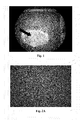
- FIGS. 2A-B are fluorescence photomicrographs of the acellular spinal cord parenchyma-derived scaffold showing lack of nuclei and cells within the scaffold, which has been stained with nuclei marker DAPI (200 ⁇ );
- FIG. 3 is a photomicrograph of Masson's trichrome stained acellular spinal cord parenchyma-derived scaffold (200 ⁇ );
- FIGS. 4A-4D are photomicrographs of acellular spinal cord parenchyma-derived scaffold.
- FIG. 4A shows hematoxylin and eosin (H&E) stained acellular porcine spinal cord scaffold (100 ⁇ ).
- FIG. 4B shows Masson's trichrome stained acellular porcine spinal cord-derived scaffold (100 ⁇ ).
- FIG. 4C shows Masson's trichrome stained normal adult porcine spinal cord, grey matter (100 ⁇ ).
- FIG. 4D shows Masson's trichrome stained normal adult porcine spinal cord, white matter (100 ⁇ );
- FIGS. 5A-5B are scanning electron micrographs of acellular spinal cord parenchyma-derived scaffold.
- FIG. 5A shows the scaffold at 600 ⁇ magnification and
- FIG. 5B shows the scaffold at 2,000 ⁇ magnification;
- FIGS. 6A-6C are photomicrographs of Masson's trichrome stained acellular spinal cord parenchyma-derived scaffold implanted with neuronal cells (PC 12).
- FIGS. 6A-6C show neuronal cell growth and migration into the scaffold, where neuronal cells (red, thin arrows) grow on and into the scaffold (blue, thick arrows). Photomicrographs are shown at the following magnifications: 100 ⁇ ( FIG. 6A ), 200 ⁇ ( FIG. 6B ), and 400 ⁇ ( FIG. 6C );
- FIGS. 7A-7B are photomicrographs of Masson's trichrome stained acellular spinal cord parenchyma-derived scaffold implanted with neuronal cells (PC 12).
- FIGS. 7A-7B show neuronal cell growth and migration into the scaffold (blue, thick arrows), where neuronal cells grow on the surface of the spinal cord scaffold (red, thin arrows) and migration into the scaffold (arrowheads).
- Photomicrographs are shown at the following magnifications: 200 ⁇ ( FIG. 7A ) and 400 ⁇ ( FIG. 7B ); and
- FIGS. 8A-8D are photomicrographs of acellular dura mater-derived scaffold.
- FIG. 8A shows a fluorescence photomicrographs of the acellular dura mater scaffold showing lack of nuclei and cells within the scaffold, which has been stained with nuclei marker DAPI (200 ⁇ ).
- FIG. 8B shows Masson's trichrome stained acellular porcine dura mater scaffold (100 ⁇ ).
- FIGS. 8C-8D show Masson's trichrome stained photomicrographs of neuronal cell growth (red) on the acellular dura mater scaffold (blue); photomicrograph magnification: 200 ⁇ ( FIG. 8C ) and 400 ⁇ ( FIG. 8D ).
- the scaffold comprises decellularized spinal cord tissue. Such tissue can be in a gel state.
- the scaffold comprises bioactive or therapeutic agents.
- Scaffolds can be used for a large number of medical applications including, but not limited to, wound healing, tissue remodeling, and tissue regeneration.
- such scaffolds can be used for wound healing.
- the scaffold comprises bioactive agents to facilitate tissue healing, tissue remodeling and/or angiogenesis.
- the scaffold comprises bioactive agents to ward off bacteria and other pathogens.
- the scaffold comprises pores to allow a wound to drain.
- the scaffold comprises combinations of cells and bioactive agents.
- combinations of cells and bioactive agents are added to the scaffold before or during implantation at a site in a patient.
- the teen “polymer” refers to both synthetic polymeric components and biological polymeric components.
- the scaffolds described herein can comprise any suitable combination of synthetic polymeric components and biological polymeric components.
- Biological polymer(s) are polymers that can be obtained from biological sources, such as, without limitation, mammalian or vertebrate tissue, as in the case of certain extracellular matrix-derived (ECM-derived) compositions.
- extracellular matrix refers to any polymer remaining after decellularization included gels and solids. Biological polymers can be modified by additional processing steps.
- Polymer(s), in general include, for example and without limitation, mono-polymer(s), copolymer(s), polymeric blend(s), block polymer(s), block copolymer(s), cross-linked polymer(s), non-cross-linked polymer(s), linear-, branched-, comb-, star-, and/or dendrite-shaped polymer(s), where polymer(s) can be formed into any useful form, for example and without limitation, a hydrogel, a porous mesh, a fiber, woven mesh, or non-woven mesh, such as, for example and without limitation, a non-woven mesh formed by electrodeposition.
- the polymeric components suitable for the scaffold described herein may be any polymer that is biodegradable and biocompatible.
- biodegradable it is meant that a polymer, once implanted and placed in contact with bodily fluids and/or tissues, will degrade either partially or completely through chemical, biochemical and/or enzymatic processes.
- Non-limiting examples of such chemical reactions include acid/base reactions, hydrolysis reactions, and enzymatic cleavage.
- the biodegradable polymers may comprise homopolymers, copolymers, and/or polymeric blends comprising, without limitation, one or more of the following monomers: glycolide, lactide, caprolactone, dioxanone, and trimethylene carbonate.
- the polymer(s) comprise labile chemical moieties, non-limiting examples of which include esters, anhydrides, polyanhydrides, or amides, which can be useful in, for example and without limitation, controlling the degradation rate of the scaffold and/or the release rate of therapeutic agents from the scaffold.
- the polymer(s) may contain peptides or biomacromolecules as building blocks which are susceptible to chemical reactions once placed in situ.
- the polymer is a polypeptide comprising the amino acid sequence alanine-alanine-lysine, which confers enzymatic lability to the polymer.
- the polymer composition may comprise a biomacromolecular component derived from an ECM.
- the polymer composition may comprise the biomacromolecule collagen so that collagenase, which is present in situ, can degrade the collagen.
- the polymer components may be selected so that they degrade in situ on a timescale that is similar to an expected rate of healing of the wound or tissue.
- in situ degradation rates include between one week and one year or increments therebetween for instance, between two weeks and 10 months, and between one month and six month.
- the polymeric components used to make the devices disclosed herein are preferably biocompatible.
- biocompatible it is meant that a polymer composition and its normal degradation in vivo products are cytocompatible and are substantially non-toxic and non-carcinogenic in a patient within useful, practical and/or acceptable tolerances.
- cytocompatible it is meant that the polymer can sustain a population of cells and/or the polymer composition, device, and degradation products, thereof are not cytotoxic and/or carcinogenic within useful, practical and/or acceptable tolerances.
- the polymer when placed in a human cell culture does not adversely affect the viability, growth, adhesion, and number of cells.
- the compositions, and/or devices are “biocompatible” to the extent they are acceptable for use in a human patient according to applicable regulatory standards in a given jurisdiction.
- the biocompatible polymer when implanted in a patient, does not cause a substantial adverse reaction or substantial harm to cells and tissues in the body, for instance, the polymer composition or device does not cause necrosis or an infection resulting in harm to tissues from the implanted scaffold.
- the mechanical properties of a biodegradable elastomeric scaffold can be optimized to operate under the normal strain and stress on the native tissue at the site of implantation.
- the mechanical properties of the scaffold are optimized similar to or identical to that of native tissue.
- the mechanical properties of the scaffold also may be optimized to be suitable for surgical handling.
- the scaffold is a gel and has gel like properties that can be controlled by the degree of hydration.
- the gel can be a hydrogel and be semi-solid, thus having a three dimensional structure.
- the scaffold is a flexible sheet and can be sutured to the site.
- the scaffold is foldable and can be delivered to the site by minimally invasive laparoscopic methods.
- Variables that can be optimized include without limitation, the extent of physical cross-linking in a network comprising polymeric components, the ratio of polymeric components within the network, the distribution of molecular weight of the polymeric components, and the method of processing the polymers.
- Polymers are typically semicrystalline and their physical properties and/or morphology are dependent upon a large number of factors, including monomer composition, polydispersity, average molecular weight, cross-linking, and melting/crystallization conditions. For example, flow and/or shear conditions during cooling of a polymer melt are known to affect formation of crystalline structures in the composition.
- the scaffold comprises a polymeric component that provides strength and durability to the scaffold, yet is elastomeric so that the mechanical properties of the scaffold are similar to the native tissue surrounding the wound or site in need of tissue regeneration.
- the extracellular matrix is useful for promoting cell growth on the elastomeric scaffold, extracting appropriate host cells for construction, remodeling, and/or enhancement of biocompatibility.
- the biological polymeric component comprises and includes an extracellular matrix-derived material.
- extracellular matrix and “ECM” refer to a complex mixture of structural and functional biomolecules and/or biomacromolecules including, but not limited to, structural proteins, specialized proteins, proteoglycans, glycosaminoglycans, and growth factors that surround and support cells within mammalian tissues.
- ECM-derived material it is meant a composition that is prepared from a natural tissue source or from an in vitro source wherein the ECM is produced by cultured cells and comprises one or more polymeric components (constituents) of native ECM. Additionally, “decellularized” ECM refers to ECM in which the cells have been removed through processes described herein and known in the art.
- ECM is isolated from a vertebrate animal, for example, from a warm blooded mammalian vertebrate animal including, but not limited to, human, monkey, pig, cow, sheep, etc.
- the ECM is isolated from spinal cord, which may or may not include the dura mater.
- the ECM includes at least a portion of the dura mater.
- the material that serves as the biological component of the scaffold consists primarily (e.g., greater than 70%, 80%, or 90%) of ECM.
- the biodegradable elastomeric scaffold may contain at least 50% ECM, at least 60% ECM, at least 70% ECM, and at least 80% ECM. In yet another non-limiting embodiment, the biodegradable elastomeric scaffold comprises at least 10% ECM.
- the ECM material may or may not retain some of the cellular elements that comprised the original tissue.
- the type of ECM used in the scaffold can vary depending on the intended cell types to be recruited during wound healing or tissue regeneration, the native tissue architecture of the tissue organ to be replaced, the availability of the tissue source of ECM, or other factors that affect the quality of the final scaffold and the possibility of manufacturing the scaffold.
- the ECM may contain both a basement membrane surface and a non-basement membrane surface, which would be useful for promoting the reconstruction of tissue.
- an implantable device can comprise either a smooth basement membrane surface (luminal) or a rough non-basement surface (abluminal).
- neural scaffolds are made by first removing the cells from excised CNS tissue and then removing any remaining lipids and DNA.
- excised CNS tissue is ensheathed in the dura mater membrane.
- the membrane can be removed and processed independently of the neural tissue according to the methods disclosed.
- a gel can be made from cellular spinal cord material; whereas, processing of acellular dura mater creates a fibrous sheet.
- the decellularization process comprises incubating the tissue (whether the dura mater, spinal cord parenchymal tissue, or a combination thereof) in a solution of non-ionic detergent.
- Non-ionic detergents are capable of lysing cells and solubilizing the cell membrane as well as many of the cellular components. It is contemplated that various detergents can be used.
- TRITON X-100TM (4-octylphenol polyethoxylate) can be used.
- the methods disclosed herein may be adapted to use any other octylphenol polyethoxylate as well as other detergents such as n-dodecylmaltoside, NONIDET P40TM, n-octylglucoside, TWEEN 20, and others.
- the neural tissue is placed into a cassette.
- cassette is intended to mean any three-dimensional hollow structure in which tissue can be place into for processing, such as a tissue cassette or other similar device.
- tissue cassette helps maintain the tissue structure as well as simplifies handling procedures.
- the neural tissue can be pre-processed.
- the tissue is digested using trypsin-EDTA, a protease, before the decellularization process begins.
- trypsin-EDTA a protease
- incubation times and temperatures can be varied depending on the amount of tissue.
- tissue can be digested for 30 minutes at 37° C.
- tissue can be processed in a non-ionic detergent such as described herein.
- the tissue can be placed in increasing amounts of TRITON X-100TM solutions.
- the tissue can be incubated in 3%-6%-9%, TRITON X-100TM solutions for periods up to 48-72 hours for each percentage. In certain embodiments, the incubation is performed at 4° C. The solutions may be changed as often as needed based on monitoring of the cell cellular removal process.
- an emulsifier can be used.
- lecithin or lecithin-deoxycholate can be used, although any suitable emulsifier can be used including for example, emulsifying wax, cetearyl alcohol, polysorbate 20, and ceteareth 20, among others.
- the tissue is incubated in lecithin overnight at 4° C. and washed with phosphate buffered saline (PBS) three times (15 minutes per wash).
- PBS phosphate buffered saline
- the tissue can be treated with a DNase such as DNase I. Times and temperatures can be varied according the type of enzyme and the amount of tissues being processed.
- the tissue is incubated in a solution of DNase I for 1 hour at room temperature and washed in PBS three times for 15 minutes at room temperature.
- the tissue can be washed in deionized water.
- the tissue is washed in deionized water three times for 15 minutes at room temperature
- the ECM can be sterilized by any of a number of standard methods without loss of function.
- the material can be sterilized by propylene oxide or ethylene oxide treatment, gamma irradiation treatment (0.05 to 4 mRad), gas plasma sterilization, peracetic acid sterilization, ethanol sterilization, or electron beam treatment.
- Treatment with glutaraldehyde results in sterilization as well as increased cross-linking of the ECM. This treatment substantially alters the material such that it is slowly resorbed or not resorbed at all and incites a different type of host remodeling, which more closely resembles scar tissue formation or encapsulation rather than constructive remodeling.
- cross-linking of the protein material within the ECM can also be induced with, for example and without limitation, carbodiimide isocyanate treatments, dehydrothermal methods, and photooxidation methods.
- the ECM is disinfected by overnight gamma irradiation treatment with a total exposure of 2 mRad.
- the ECM-derived material may be further processed by optional drying, desiccation, lyophilization, freeze drying, and/or glassification.
- the ECM-derived material optionally can be further digested or processed, for example and without limitation by hydration, acidification, alkalinization, enzymatic treatment with, for example and without limitation, trypsin or chondroitinase, and neutralization.
- the biodegradable elastomeric scaffolds as described herein may take many different forms.
- the scaffold is substantially planar (having much greater dimension in two dimensions and a substantially smaller dimension in a third, comparable to bandages, gauze, and other substantially flexible, flat items).
- the biodegradable elastomeric scaffold comprises a non-woven fibrous article formed by electrodeposition of a suspension containing the synthetic polymeric component and the biological polymeric component.
- the biodegradable elastomeric scaffold comprises a porous composite formed by thermally induced phase separation.
- the biodegradable elastomeric scaffold can also have three-dimensional shapes useful for treating wounds and tissue deficiencies, such as plugs, rings, wires, cylinders, tubes, or disks.
- the biodegradable elastomeric scaffolds may be porous. Porosity may be accomplished by a variety of methods. Although the biodegradable elastomeric scaffolds may be porous or non-porous, it is often advantageous to use a process that produces a porous elastomeric scaffold. Non-limiting examples of such processes include solvent casting/salt leaching, electrodeposition, and thermally induced phase separation. In other examples, porosity may be accomplished by creating a mesh of fibers, such as by the aforementioned electrodeposition or by any suitable method of producing a woven or non-woven fiber matrix. As used herein, the term “porosity” refers to a ratio between a volume of all the pores within the polymer composition and a volume of the whole polymer composition.
- a polymer composition with porosity of 85% would have 85% of its volume containing pores and 15% of its volume containing the polymer.
- the porosity of the scaffold is at least 60%, 65%, 70%, 75%, 80%, 85%, or 90%, or increments therebetween.
- the average pore size of the scaffold is between 0.1 and 300 microns, including increments therebetween.
- a biodegradable elastomeric scaffold that acts as a barrier to bacteria and other pathogens may have an average pore size of less than 0.5 microns or less than 0.2 microns.
- the average pore size may be increased by increasing the amount of polymeric components within the suspension used for electrodeposition, which results in larger fiber diameters and therefore larger pore sizes.
- the average pore size can be increased by increasing spinning distance from the nozzle to the target, which results in less adherence between fibers and a looser matrix.
- the biodegradable elastomeric scaffold is made by using solvent casting and salt leaching. This method involves dissolving the polymeric components that constitute the scaffold into a suitable organic solvent and then casting the solution into a mold containing small particles of predetermined size (known as porogens). Examples of suitable porogens include inorganic salts, crystals of saccharose, gelatin spheres or paraffin spheres. By adjusting the porogen size and/or the ratio of porogen to solvent, the porosity of the final elastomeric scaffold may be adjusted. After casting, the solvent is evaporated, and the resulting polymer composition is immersed into a second solvent that dissolves the porogen, but not the polymer, to produce a porous, sheet-like structure.
- solvent casting and salt leaching involves dissolving the polymeric components that constitute the scaffold into a suitable organic solvent and then casting the solution into a mold containing small particles of predetermined size (known as porogens). Examples of suitable porogens include inorganic salts, crystals of saccharose, gelatin
- electrodeposition is used to fabricate the elastomeric scaffold.
- the process of electrodeposition involves placing a polymer-containing fluid (for example, a polymer solution, a polymer suspension, or a polymer melt) in a reservoir equipped with a small orifice, such as a needle or pipette tip and a metering pump.
- a polymer-containing fluid for example, a polymer solution, a polymer suspension, or a polymer melt
- a small orifice such as a needle or pipette tip and a metering pump.
- One electrode of a high voltage source is also placed in electrical contact with the polymer-containing fluid or orifice, while the other electrode is placed in electrical contact with a target (typically a collector screen or rotating mandrel).
- a target typically a collector screen or rotating mandrel
- the polymer-containing fluid is charged by the application of high voltage to the solution or orifice (for example, about 3-15 kV) and then forced through the small orifice by the metering pump that provides steady flow. While the polymer-containing fluid at the orifice normally would have a hemispherical shape due to surface tension, the application of the high voltage causes the otherwise hemispherically shaped polymer-containing fluid at the orifice to elongate to form a conical shape known as a Taylor cone.
- high voltage for example, about 3-15 kV
- the repulsive electrostatic force of the charged polymer-containing fluid overcomes the surface tension and a charged jet of fluid is ejected from the tip of the Taylor cone and accelerated towards the target, which typically is biased between ⁇ 2 to ⁇ 10 kV.
- a focusing ring with an applied bias can be used to direct the trajectory of the charged jet of polymer-containing fluid.
- the charged jet of fluid travels towards the biased target, it undergoes a complicated whipping and bending motion. If the fluid is a polymer solution or suspension, the solvent typically evaporates during mid-flight, leaving behind a polymer fiber on the biased target.
- the molten polymer cools and solidifies in mid-flight and is collected as a polymer fiber on the biased target.
- a non-woven, porous mesh is formed on the biased target.
- the properties of the electrodeposited elastomeric scaffolds can be tailored by varying the electrodeposition conditions. For example, when the biased target is relatively close to the orifice, the resulting electrodeposited mesh tends to contain unevenly thick fibers, such that some areas of the fiber have a “bead-like” appearance. However, as the biased target is moved further away from the orifice, the fibers of the non-woven mesh tend to be more unifoiin in thickness.
- the biased target can be moved relative to the orifice.
- the biased target is moved back and forth in a regular, periodic fashion, such that fibers of the non-woven mesh are substantially parallel to each other.
- the resulting non-woven mesh may have a higher resistance to strain in the direction parallel to the fibers, compared to the direction perpendicular to the fibers.
- the biased target is moved randomly relative to the orifice, so that the resistance to strain in the plane of the non-woven mesh is isotropic.
- the properties of the electrodeposited elastomeric scaffold may also be varied by changing the magnitude of the voltages applied to the electrodeposition system.
- the electrodeposition apparatus includes an orifice biased to 12 kV, a target biased to ⁇ 7 kV, and a focusing ring biased to 3 kV.
- a useful orifice diameter is 0.047′′ (I.D.) and a useful target distance is about 23 cm.
- Other electrodeposition conditions that can be varied include, for example and without limitation, the feed rate of the polymer solutions, the solution concentrations, and the polymer molecular weight.
- Non-limiting examples of useful range of high-voltage to be applied to the polymer suspension is from 0.5 to 30 kV, from 5 to 25 kV, and from 10 to 15 kV.
- thermally induced phase separation is used to fabricate the biodegradable elastomeric scaffold.
- This method involves dispersing the polymeric components in a solvent (for example and without limitation, DMSO-dimethyl sulfoxide) and then casting, for example by injecting or otherwise placing the composition into a mold.
- the mold can have any useful shape, such as a sheet or net.
- a pre-formed mold is cooled to low temperature (for example and without limitation-80° C.), which causes the polymeric components to separate out of the solvent. The mold is then transferred to ethanol to extract the DMSO.
- Fabrication and modification of the biodegradable elastomeric scaffold can comprise multiple steps using multiple techniques using polymer compositions that are the same or different.
- TIPS is used to fabricate the biodegradable elastomeric scaffold and electrodeposition is used to form a fiber coating onto or around the scaffold.
- solvent casting/salt leaching is used to fabricate the biodegradable elastomeric scaffold and electrodeposition is used to form a fiber coating onto or around the scaffold.
- the electrodeposition solution can contain one or more of any polymeric components, including synthetic polymeric components, biological polymeric components, or mixtures of both.
- the fiber coating formed by electrodeposition can be coated onto or around the entire scaffold or portions of the scaffold.
- the planar or three-dimensional surface of the scaffold may be functionally modified (functionalized) for any purpose, such as, without limitation, to promote cellular adhesion and migration onto and/or into the scaffold.
- the surface is first treated to introduce a reactive group on the surface by any useful process, such as one of the many processes known in the art.
- the activated surface is reacted with an adhesion-promoting peptide or group.
- the reactive group on the surface can be, for example and without limitation, a hydroxyl group or an amine group.
- radio-frequency glow discharge is used to produce plasma containing ammonia gas and amine groups are introduced to the surface by treatment with the plasma.
- radio-frequency glow discharge is used to introduce hydroxyl groups to the surface by treatment with plasma.
- the activated surface can be modified with an adhesion-promoting oligopeptide to promote cellular ingrowth into and/or onto the scaffold.
- adhesion-promoting oligopeptides include: RGD or RGDS (SEQ ID NO.: 1), a recognition site for fibronectin, vitronectin, fibrinogen, von Willebrand factor, and collagen; LDV, REDV (SEQ ID NO.: 2), PHSRN (SEQ ID NO.: 3), and KNEED (SEQ ID NO.: 4), which are recognition sites for fibronectin; YIGSR (SEQ ID NO.: 5) and IKVAV (SEQ ID NO.: 6), which are recognition sites for laminin; and DGEA (SEQ ID NO.: 7), a recognition site for collagen.
- the scaffold is functionalized to present the peptide RGDS (SEQ ID NO.: 1) on its surface.
- the surface is treated with radio-frequency glow discharge containing ammonia gas to introduce amine groups.
- Ammonia-containing gas is generated by connecting a flask containing ammonium hydroxide (30 wt % solution) to the glow discharge reactor and maintaining pressure at 3 ⁇ 10 ⁇ 3 Torr.
- the surface is further treated with 1,4-diisocyanatobutane to provide a reactive isocyanate group.
- RGDS (SEQ ID NO.: 1) is attached to the activated surface.
- the activated surface is immersed in a solution of 20 ⁇ g/mL RGDS (SEQ ID NO.: 1) in PBS for 10 hours and then rinsed with PBS.
- One or more of therapeutic agents can be introduced into the biodegradable elastomeric scaffold by any useful method, such as, without limitation absorption, adsorption, deposition, admixture with a polymer composition used to manufacture the scaffold and linkage of the agent to a component of the scaffold.
- the therapeutic agent is introduced into a backbone of a polymer used in the scaffold.
- the rate of release of the therapeutic agent may be controlled by the rate of polymer degradation.
- the therapeutic agent is introduced when the scaffold is being made. For instance, during a solvent casting or TIPS process, the therapeutic agent can be added to the solvent with the polymer in the pre-formed mold.
- the therapeutic agent can be electrosprayed onto the polymer being spun.
- the therapeutic agent is introduced into the scaffold after the patch is made.
- the scaffold may be “loaded” with therapeutic agent(s) by using static methods.
- the scaffold can be immersed into a solution containing the therapeutic agent, permitting the agent to absorb into and/or adsorb onto the scaffold.
- the scaffold may also be loaded by using dynamic methods. For instance, a solution containing the therapeutic agent can be perfused or electrodeposited into the scaffold.
- a therapeutic agent can be added to the biodegradable elastomeric scaffold before it is implanted in the patient.
- Therapeutic agents within the biodegradable elastomeric scaffold can be used in any number of ways.
- a therapeutic agent is released from the scaffold.
- anti-inflammatory drugs are released from the scaffold to decrease an immune response.
- a therapeutic agent is intended to substantially remain within the scaffold.
- chemoattractants are maintained within the scaffold to promote cellular migration and/or cellular infiltration into the scaffold.
- the biodegradable elastomeric scaffolds release therapeutic agents when the polymeric components degrade within the patient's body.
- the individual building blocks of the polymers may be chosen such that the building blocks themselves provide a therapeutic benefit when released in situ through the degradation process.
- one of the polymer building blocks is putrescine, which has been implicated as a substance that causes cell growth and cell differentiation.
- At least one therapeutic agent is added to the biodegradable elastomeric scaffold before it is implanted in the patient.
- the therapeutic agents include any substance that can be coated on, embedded into, absorbed into, adsorbed onto, or otherwise attached to or incorporated onto or into the biodegradable elastomeric scaffold that would provide a therapeutic benefit to a patient.
- Non-limiting examples of such therapeutic agents include antimicrobial agents, growth factors, emollients, retinoids, and topical steroids. Each therapeutic agent may be used alone or in combination with other therapeutic agents.
- a biodegradable elastomeric scaffold comprising neurotrophic agents or cells that express neurotrophic agents may be applied to a wound that is near a critical region of the central nervous system, such as the spine.
- the therapeutic agent may be blended with the polymer while the polymer is being processed.
- the therapeutic agent may be dissolved in a solvent (e.g., DMSO) and added to the polymer blend during processing.
- the therapeutic agent is mixed with a carrier polymer (e.g., polylactic-glycolic acid microparticles) which is subsequently processed with an elastomeric polymer.
- a carrier polymer e.g., polylactic-glycolic acid microparticles
- the therapeutic agent is a growth factor, such as a neurotrophic or angiogenic factor, which optionally may be prepared using recombinant techniques.
- growth factors include basic fibroblast growth factor (bFGF), acidic fibroblast growth factor (aFGF), vascular endothelial growth factor (VEGF), hepatocyte growth factor (HGF), insulin-like growth factors 1 and 2 (IGF-1 and IGF-2), platelet derived growth factor (PDGF), stromal derived factor 1 alpha (SDF-1 alpha), nerve growth factor (NGF), ciliary neurotrophic factor (CNTF), neurotrophin-3, neurotrophin-4, neurotrophin-5, pleiotrophin protein (neurite growth-promoting factor 1), midkine protein (neurite growth-promoting factor 2), brain-derived neurotrophic factor (BDNF), tumor angiogenesis factor (TAF), corticotrophin releasing factor (CRF), transforming growth factors ⁇ and ⁇ (TGF- ⁇ and TGF- ⁇
- bFGF basic fibroblast
- Methods of promoting wound healing or tissue generation or regeneration in a patient comprise; without limitation, implanting an elastomeric scaffold as described herein at or near a site for wound healing or tissue generation or regeneration in the patient.
- the elastomeric scaffold may comprise a therapeutic agent as described herein.
- the therapeutic agent is an antimicrobial agent, such as, without limitation, isoniazid, ethambutol, pyrazinamide, streptomycin, clofazimine, rifabutin, fluoroquinolones, ofloxacin, sparfloxacin, rifampin, azithromycin, clarithromycin, dapsone, tetracycline, erythromycin, ciprofloxacin, doxycycline, ampicillin, amphotericin B, ketoconazole, fluconazole, pyrimethamine, sulfadiazine, clindamycin, lincomycin, pentamidine, atovaquone, paromomycin, diclazaril, acyclovir, trifluorouridine, foscarnet, penicillin, gentamicin, ganciclovir, iatroconazole, miconazole, Zn-pyrithione, and silver salts such as chloride, acyclovir,
- the therapeutic agent is an anti-inflammatory agent, such as, without limitation, a NSAID, such as salicylic acid, indomethacin, sodium indomethacin trihydrate, salicylamide, naproxen, colchicine, fenoprofen, sulindac, diflunisal, diclofenac, indoprofen, sodium salicylamide; an anti-inflammatory cytokine; an anti-inflammatory protein; a steroidal anti-inflammatory agent; or an anti-clotting agents, such as heparin.
- a NSAID such as salicylic acid, indomethacin, sodium indomethacin trihydrate, salicylamide, naproxen, colchicine, fenoprofen, sulindac, diflunisal, diclofenac, indoprofen, sodium salicylamide
- an anti-inflammatory cytokine an anti-inflammatory protein
- a steroidal anti-inflammatory agent a steroidal anti-inflammatory agent
- the therapeutic agent comprises cells that are added to the biodegradable elastomeric scaffold before or at the time of implantation.
- a porous biodegradable elastomeric scaffold so that the cells may be incorporated into the porous structure of the scaffold (a condition referred to as “microintegration”).
- microintegration a condition referred to as “microintegration”.
- the cells that are microintegrated may remain after the biodegradable elastomeric scaffold has fully disintegrated within the patient.
- the microintegrated cells may also be merely cells that act as precursors to the final tissue that is formed when the biodegradable elastomeric scaffold has fully degraded.
- Cells may be autologous (obtained from the patient to receive the scaffold), from an allogeneic or xenogeneic source or from any useful cell line, such as, without limitation, stem cells that are capable of cellular growth, remodeling, and/or differentiation.
- the cells that may be incorporated onto or into the biodegradable scaffold include stem cells, precursor cells, smooth muscle cells, skeletal myoblasts, myocardial cells, endothelial cells, and genetically modified cells.
- Various commercially available cell lines include Clonetics® Primary Cell Systems (Lonza Group, Inc., Switzerland), ATCC.
- Cells may be microintegrated with the biodegradable elastomeric scaffold using a variety of methods.
- the elastomeric scaffold may be placed in a suitable growth medium for the cells of interest, and then exposed to the cells. The cells are allowed to proliferate on the surface and interstices of the elastomeric scaffold. The elastomeric scaffold is then removed from the growth medium, washed if necessary, and implanted.
- the cells may be placed in a suitable buffer or liquid growth medium and drawn through the scaffold by using vacuum filtration.
- the cells of interest are dissolved into an appropriate solution (e.g., a growth medium or buffer) and then sprayed onto a biodegradable elastomeric scaffold while the scaffold is being formed by electrodeposition.
- the cells are placed in a solution that is biased and then electrosprayed onto the biodegradable elastomeric scaffold while it is being electrodeposited.
- the cells that may be incorporated on or into the biodegradable scaffold include chondrocytes, stem cells, precursor cells, smooth muscle cells, skeletal myoblasts, myocardial cells, endothelial cells, and genetically modified cells.
- the genetically modified cells are capable of expressing a therapeutic substance, such as a growth factor.
- a therapeutic substance such as a growth factor.
- Cells can be modified by any useful method in the art.
- the therapeutic agent is a growth factor that is released by cells transfected with cDNA encoding for the growth factor.
- Therapeutic agents that can be released from cells include, without limitation, a neurotrophic factor, such as nerve growth factor, brain-derived neurotrophic factor, neurotrophin-3, neurotrophin-4, neurotrophin-5, and ciliary neurotrophic factor; a growth factor, such as basic fibroblast growth factor (bFGF), acidic fibroblast growth factor (aFGF), vascular endothelial growth factor (VEGF), hepatocyte growth factor (HGF), insulin-like growth factors (IGF), platelet derived growth factor (PDGF), transforming growth factor-beta (TGF- ⁇ ), pleiotrophin protein (neurite growth-promoting factor 1), and midkine protein (neurite growth-promoting factor 2); an anti-inflammatory cytokine; and an anti-inflammatory protein.
- the cells may be autologous, allogeneic, etc.
- a biodegradable elastomeric scaffold can be implanted by using any suitable medical procedure that facilitates use of the scaffold to provide a therapeutic benefit.
- the terms “implanted” and “implantation” and like terms refer to an act of delivering a biodegradable elastomeric scaffold to a site within the patient.
- the site of implantation in a patient typically is “at or near a site for wound healing or tissue generation or regeneration in the patient,” meaning the scaffold-containing device is implanted in, on, onto, adjacent to or in proximity to a desired site of delivery to facilitate healing and/or tissue generation or regeneration to repair an injury or defect in the patient and/or to achieve a desired effect in the patient, such as wound drainage.
- the delivery method may also include minimally invasive methods such as by catheter based technology or by needle injection.
- the patient may be human or animal.
- the scaffold may be delivered by any surgical procedure, including minimally invasive techniques, such as laparoscopic surgery, as well as invasive techniques such as thoracic surgery and fasciotomy.
- the elastomeric scaffolds are used as surgical fabrics.
- the biodegradable elastomeric scaffold may be implanted alone or implanted in conjunction with surgical fasteners, such as sutures, staples, adhesives, screws, pins, and the like. Additionally, biocompatible adhesives, such as, without limitation, fibrin-based glue may be used to fasten the elastomeric scaffolds as well.
- the scaffold can be in the form of a powder or fine particles (for example, formed by shredding a non-woven mesh formed by electrodeposition or TIPS).
- a powder or fine particles for example, formed by shredding a non-woven mesh formed by electrodeposition or TIPS.
- porcine spinal cord was obtained. Using forceps, scissors and a scalpel, dura mater was removed from the spinal cord. The inner dura mater surface was scrapped with scalpel blade to remove any debris.
- the spinal cord and dura were placed in separate containers and treated in the same manner as listed below.
- the spinal cord was cut either longitudinally or in cross-section (to increase surface area) and placed in a cassette (e.g., safety container to protect 3-D structure of cord throughout process).
- tissue was enzymatically treated using trypsin-EDTA for 30 minutes at 37° C. The tissue was incubated in TRITON X-100TM (4-octylphenol polyethoxylate) solutions at 3% for periods up to 2-3 days at 4° C.
- tissue was washed in deionized water three times for 15 minutes at room temperature.
- the procedure produced a gel-like acellular spinal cord material, and a sheet of acellular dura mater material. Sections of each material were sent for histology to confirm lack of cells. Biocompatibility was tested by culturing the scaffolds with either primary neuronal cells or a neuronal cell line (e.g. PC 12 cell line)
- a neuronal cell line e.g. PC 12 cell line
- FIG. 1 is a digital image showing the gel-like quality of the scaffold.
- the thick arrow indicates the gel-like substance and the thin arrow indicates the pia matter.
- the scaffold After forming the neuronal scaffolds, the scaffold is acellular.
- Various methods were used to determine whether the scaffold was acellular.
- fluorescent stains such as DAPI (4′,6-diamidino-2-phenylindole), were used to determine whether cells are present.
- FIGS. 2A-2B and FIG. 8A are fluorescence photomicrographs of the scaffold stained with DAPI, where lack of fluorescence correlated with lack of nuclei and cells within the scaffold.
- histological stains such as Masson's trichrome, were also be used.
- FIG. 3 and FIG. 8B show photomicrographs of Masson's trichrome stained scaffolds, which show that cells are not present within the scaffolds.
- FIGS. 4A-4D provide comparisons between the acellular spinal cord parenchyma-derived neuronal scaffolds and native adult porcine spinal cord.
- FIGS. 4A-4B show an acellular porcine spinal cord parenchyma-derived neuronal scaffold that has been stained with hematoxylin and eosin ( FIG. 4A ) or with Masson's trichrome ( FIG. 4B ).
- FIGS. 4C-4D show Masson's trichrome stained grey matter ( FIG. 4C ) and white matter ( FIG. 4D ) of a normal adult porcine spinal cord.
- FIGS. 5A-5B are scanning electron micrographs of acellular spinal cord parenchyma-derived neuronal scaffold at 600 ⁇ magnification ( FIG. 5A ) and at 2,000 ⁇ magnification ( FIG. 5B ).
- Rat adrenal pheochromocytoma (PC 12) cells were obtained from ATCC (American Type Culture Collections, Manassas, Va.), ATCC catalog number CRL-1721.
- the PC12 cells were grown in DMEM with 10% heat-inactivated horse serum, 5% fetal bovine serum, 1% penicillin-streptomycin solution (stock solution: 10,000 units penicillin and 10,000 ⁇ g streptomycin/ml) in a humidified incubator at 37° C. supplemented with 5% CO 2 in plastic cell culture-treated flasks.
- the neural scaffold-PC12 biomaterials were fixed in 10% (neutral buffered) formalin for 24 hours, mounted in paraffin, sectioned (5 ⁇ m sections), mounted on glass slides, stained (H&E, Masson's trichrome) and coverslipped.
- These neural scaffold-PC12 cultures were not supplemented with NGF (typically 50 ng/ml), but the growth factor can be added to this cell line to stimulate and evaluate neurite outgrowth.
- FIGS. 6A-6C and 7 A- 7 B shows photomicrographs of Masson's trichrome stained acellular spinal cord parenchyma-derived neural scaffold implanted with neuronal cells (PC12).
- FIGS. 6A-6C show neuronal cell growth and migration into this neural scaffold, where neuronal cells (red, thin arrows) grew on and into the scaffold (blue, thick arrows).
- FIGS. 7A-7B show neuronal cell growth and migration in this scaffold (blue, thick arrows). Neuronal cells grew on the surface of the scaffold (red, thin arrows) and migrated into the scaffold (arrowheads).
- FIGS. 8C-8D show neuronal cells (red) growing on the surface of the dura mater-derived neural scaffold (blue).
Abstract
Description
Claims (22)
Priority Applications (2)
| Application Number | Priority Date | Filing Date | Title |
|---|---|---|---|
| US12/718,227 US8685634B2 (en) | 2009-03-06 | 2010-03-05 | Neural scaffolds |
| US14/183,920 US9421229B2 (en) | 2009-03-06 | 2014-02-19 | Neural scaffolds |
Applications Claiming Priority (2)
| Application Number | Priority Date | Filing Date | Title |
|---|---|---|---|
| US15799909P | 2009-03-06 | 2009-03-06 | |
| US12/718,227 US8685634B2 (en) | 2009-03-06 | 2010-03-05 | Neural scaffolds |
Related Child Applications (1)
| Application Number | Title | Priority Date | Filing Date |
|---|---|---|---|
| US14/183,920 Division US9421229B2 (en) | 2009-03-06 | 2014-02-19 | Neural scaffolds |
Publications (2)
| Publication Number | Publication Date |
|---|---|
| US20100226895A1 US20100226895A1 (en) | 2010-09-09 |
| US8685634B2 true US8685634B2 (en) | 2014-04-01 |
Family
ID=42678445
Family Applications (2)
| Application Number | Title | Priority Date | Filing Date |
|---|---|---|---|
| US12/718,227 Active 2032-05-17 US8685634B2 (en) | 2009-03-06 | 2010-03-05 | Neural scaffolds |
| US14/183,920 Active 2030-04-11 US9421229B2 (en) | 2009-03-06 | 2014-02-19 | Neural scaffolds |
Family Applications After (1)
| Application Number | Title | Priority Date | Filing Date |
|---|---|---|---|
| US14/183,920 Active 2030-04-11 US9421229B2 (en) | 2009-03-06 | 2014-02-19 | Neural scaffolds |
Country Status (1)
| Country | Link |
|---|---|
| US (2) | US8685634B2 (en) |
Cited By (1)
| Publication number | Priority date | Publication date | Assignee | Title |
|---|---|---|---|---|
| US20160250385A1 (en) * | 2013-11-04 | 2016-09-01 | The Trustees Of The University Of Pennsylvania | Neuronal replacement and reestablishment of axonal connections |
Families Citing this family (8)
| Publication number | Priority date | Publication date | Assignee | Title |
|---|---|---|---|---|
| US20140356331A1 (en) * | 2011-07-08 | 2014-12-04 | University Of Pittsburgh-Of The Commonwealth System Of Higher Education | Injectable cns-derived ecm for tissue reconstruction |
| US20140023722A1 (en) * | 2012-07-20 | 2014-01-23 | Alan V. Boruch | Neural Scaffold Biomaterial |
| WO2014165602A1 (en) * | 2013-04-02 | 2014-10-09 | University Of Florida Research Foundation, Inc. | Compositions and methods for induction and modulation of angiogenesis and methods and assays for identifying angiogenesis modulators |
| US8916339B1 (en) * | 2013-10-31 | 2014-12-23 | Vivex Biomedical, Inc. | Spinal cord tissue dehydrated and micronized |
| KR102624370B1 (en) | 2014-03-21 | 2024-01-15 | 유니버시티 오브 피츠버그 - 오브 더 커먼웰쓰 시스템 오브 하이어 에듀케이션 | Methods for preparation of a terminally sterilized hydrogel derived from extracellular matrix |
| ES2873519T3 (en) | 2015-09-18 | 2021-11-03 | Univ Pittsburgh Commonwealth Sys Higher Education | Non-gelling soluble extracellular matrix with biological activity |
| WO2017189986A1 (en) | 2016-04-28 | 2017-11-02 | University Of Pittsburgh - Of The Commonwealth System Of Higher Education | Compositions comprising extracellular matrix of primitive animal species and related methods |
| ES2931299T3 (en) | 2017-03-02 | 2022-12-28 | Univ Pittsburgh Commonwealth Sys Higher Education | Extracellular matrix (ECM) hydrogel and soluble fraction thereof for use in the treatment of cancer |
Citations (4)
| Publication number | Priority date | Publication date | Assignee | Title |
|---|---|---|---|---|
| US6448076B2 (en) | 1998-09-15 | 2002-09-10 | The Regents Of The University Of Michigan | Method for chemically acellularizing a biological tissue sample |
| US6962814B2 (en) * | 2000-08-16 | 2005-11-08 | Duke University | Decellularized tissue engineered constructs and tissues |
| US7402319B2 (en) | 2002-09-27 | 2008-07-22 | Board Of Regents, The University Of Texas System | Cell-free tissue replacement for tissue engineering |
| WO2011072393A1 (en) | 2009-12-17 | 2011-06-23 | Queen's University At Kingston | Decellularized adipose tissue |
Family Cites Families (1)
| Publication number | Priority date | Publication date | Assignee | Title |
|---|---|---|---|---|
| US6734018B2 (en) * | 1999-06-07 | 2004-05-11 | Lifenet | Process for decellularizing soft-tissue engineered medical implants, and decellularized soft-tissue medical implants produced |
-
2010
- 2010-03-05 US US12/718,227 patent/US8685634B2/en active Active
-
2014
- 2014-02-19 US US14/183,920 patent/US9421229B2/en active Active
Patent Citations (4)
| Publication number | Priority date | Publication date | Assignee | Title |
|---|---|---|---|---|
| US6448076B2 (en) | 1998-09-15 | 2002-09-10 | The Regents Of The University Of Michigan | Method for chemically acellularizing a biological tissue sample |
| US6962814B2 (en) * | 2000-08-16 | 2005-11-08 | Duke University | Decellularized tissue engineered constructs and tissues |
| US7402319B2 (en) | 2002-09-27 | 2008-07-22 | Board Of Regents, The University Of Texas System | Cell-free tissue replacement for tissue engineering |
| WO2011072393A1 (en) | 2009-12-17 | 2011-06-23 | Queen's University At Kingston | Decellularized adipose tissue |
Non-Patent Citations (5)
| Title |
|---|
| Gulati, Adarsh K., "Evaluation of acellular and cellular nerve grafts in repair of rat peripheral nerve", J Neurosurg., 1988, pp. 117-123, vol. 68. |
| Ribatti, Domenico et al., "Angiogenic response induced by acellular brain scaffolds grafted onto the chick embryo chorioallantoic membrane", Brain Research, 2003, pp. 9-15, vol. 989. |
| Sondell et al, Brain Research, 1998, vol. 795, pp. 44-54. * |
| Yin et al, Nan fang yi ke da xue xue bao (Journal of Southern Medical University), Oct. 2008 vol. 28, No. 10, pp. 1748-1751. Abstract Only. * |
| Yin et al, Nan fang yi ke da xue xue bao (Journal of Southern Medical University), Oct. 2008, vol. 28, No. 10, pp. 1748-1751. (Full translation). * |
Cited By (1)
| Publication number | Priority date | Publication date | Assignee | Title |
|---|---|---|---|---|
| US20160250385A1 (en) * | 2013-11-04 | 2016-09-01 | The Trustees Of The University Of Pennsylvania | Neuronal replacement and reestablishment of axonal connections |
Also Published As
| Publication number | Publication date |
|---|---|
| US20100226895A1 (en) | 2010-09-09 |
| US20140170121A1 (en) | 2014-06-19 |
| US9421229B2 (en) | 2016-08-23 |
Similar Documents
| Publication | Publication Date | Title |
|---|---|---|
| US9421229B2 (en) | Neural scaffolds | |
| US8535719B2 (en) | Biohybrid elastomeric scaffolds and methods of use thereof | |
| US10092676B2 (en) | Biohybrid composite scaffold | |
| US9848987B2 (en) | Joint bioscaffolds | |
| Costa et al. | Biologic scaffolds | |
| JP3709487B2 (en) | Compositions and methods for naturally secreted extracellular matrix | |
| US20080109070A1 (en) | Biodegradable elastomeric scaffolds containing microintegrated cells | |
| CA2743871C (en) | Extracellular matrix compositions for the treatment of cancer | |
| CN110169959B (en) | Growth factor slow-release microsphere, tissue engineering cartilage composite scaffold and preparation method | |
| US20190060517A1 (en) | Scaffold for cardiac patch | |
| WO2011150328A1 (en) | Wet-electrospun biodegradable scaffold and uses therefor | |
| US8673640B2 (en) | Porous scaffold, method of producing the same and method of using the porous scaffold | |
| US20210085834A1 (en) | Multi-Layered Graft for Tissue Engineering Applications | |
| Becker et al. | “UroMaix” Scaffolds: Novel Collagen Matrices for Application in Tissue Engineering of the Urinary Tract | |
| KR102364686B1 (en) | A Novel Porous Scaffold and Methods for Preparing the Same | |
| US20140023722A1 (en) | Neural Scaffold Biomaterial | |
| JP2022552097A (en) | Novel porous scaffold and method of making same | |
| Kuevda et al. | Application of recellularized non-woven materials from collagen-enriched polylactide for creation of tissue-engineered diaphragm constructs | |
| Sumanasingh et al. | The applications of biotextiles in tissue engineering | |
| Niu et al. | Making a Hydrophilic Interfacial Scaffold with Hierarchical Nanofibre Architecture to Enhance the Phenotypic Expression of Epithelial and Smooth Muscle Cells for Urethral Reconstruction | |
| Wagner et al. | Biohybrid composite scaffold | |
| Wagner et al. | Biohybrid elastomeric scaffolds and methods of use thereof | |
| JP2007145844A (en) | Compositions and methods to create vascularized environment for cellular transplantation | |
| Kumar et al. | Macroporous Polymeric Scaffolds for Tissue Engineering Applications |
Legal Events
| Date | Code | Title | Description |
|---|---|---|---|
| AS | Assignment |
Owner name: NATIONAL INSTITUTES OF HEALTH (NIH), U.S. DEPT. OF Free format text: CONFIRMATORY LICENSE;ASSIGNOR:UNIVERSITY OF PITTSBURGH - OF THE COMMONWEALTH SYSTEM OF HIGHER EDUCATION;REEL/FRAME:025647/0895 Effective date: 20110112 |
|
| AS | Assignment |
Owner name: UNIVERSITY OF PITTSBURGH-OF THE COMMONWEALTH SYSTE Free format text: ASSIGNMENT OF ASSIGNORS INTEREST;ASSIGNOR:BORUCH, ALAN V.;REEL/FRAME:026161/0545 Effective date: 20090311 |
|
| AS | Assignment |
Owner name: BORUCH, ALAN V, DR., PENNSYLVANIA Free format text: NUNC PRO TUNC ASSIGNMENT;ASSIGNOR:PITTSBURGH, UNIVERSITY OF;REEL/FRAME:028592/0895 Effective date: 20100301 |
|
| AS | Assignment |
Owner name: UNIVERSITY OF PITTSBURGH - OF THE COMMONWEALTH SYS Free format text: CORRECTIVE ASSIGNMENT TO CORRECT THE DECLARATION OF INCORRECT RECORDING, PREVIOUSLY RECORDED ON REEL 028592 FRAME 0395. ASSIGNOR(S) HEREBY CONFIRMS THE ASSIGNMENT;ASSIGNOR:UNIVERSITY OF PITTSBURGH - OF THE COMMONWEALTH SYSTEM OF HIGHER EDUCATION;REEL/FRAME:032399/0576 Effective date: 20121220 Owner name: UNIVERSITY OF PITTSBURGH-OF THE COMMONWEALTH SYSTE Free format text: CORRECTON BY DECLARATION OF INCORRECT RECORDING AT REEL/FRAME 028592/0895;ASSIGNOR:UNIVERSITY OF PITTSBURGH - OF THE COMMONWEALTH SYSTEM OF HIGHER EDUCATION;REEL/FRAME:032361/0594 Effective date: 20131220 |
|
| STCF | Information on status: patent grant |
Free format text: PATENTED CASE |
|
| CC | Certificate of correction | ||
| MAFP | Maintenance fee payment |
Free format text: PAYMENT OF MAINTENANCE FEE, 4TH YR, SMALL ENTITY (ORIGINAL EVENT CODE: M2551) Year of fee payment: 4 |
|
| CC | Certificate of correction | ||
| CC | Certificate of correction | ||
| MAFP | Maintenance fee payment |
Free format text: PAYMENT OF MAINTENANCE FEE, 8TH YR, SMALL ENTITY (ORIGINAL EVENT CODE: M2552); ENTITY STATUS OF PATENT OWNER: SMALL ENTITY Year of fee payment: 8 |