US8673638B2 - Cell culture support and cell culture method - Google Patents
Cell culture support and cell culture method Download PDFInfo
- Publication number
- US8673638B2 US8673638B2 US13/204,324 US201113204324A US8673638B2 US 8673638 B2 US8673638 B2 US 8673638B2 US 201113204324 A US201113204324 A US 201113204324A US 8673638 B2 US8673638 B2 US 8673638B2
- Authority
- US
- United States
- Prior art keywords
- cell culture
- support
- well
- cells
- mesenchymal stem
- Prior art date
- Legal status (The legal status is an assumption and is not a legal conclusion. Google has not performed a legal analysis and makes no representation as to the accuracy of the status listed.)
- Active, expires
Links
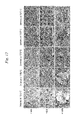
Images


Classifications
-
- C—CHEMISTRY; METALLURGY
- C12—BIOCHEMISTRY; BEER; SPIRITS; WINE; VINEGAR; MICROBIOLOGY; ENZYMOLOGY; MUTATION OR GENETIC ENGINEERING
- C12N—MICROORGANISMS OR ENZYMES; COMPOSITIONS THEREOF; PROPAGATING, PRESERVING, OR MAINTAINING MICROORGANISMS; MUTATION OR GENETIC ENGINEERING; CULTURE MEDIA
- C12N5/00—Undifferentiated human, animal or plant cells, e.g. cell lines; Tissues; Cultivation or maintenance thereof; Culture media therefor
- C12N5/06—Animal cells or tissues; Human cells or tissues
- C12N5/0602—Vertebrate cells
- C12N5/0652—Cells of skeletal and connective tissues; Mesenchyme
- C12N5/0662—Stem cells
-
- C—CHEMISTRY; METALLURGY
- C12—BIOCHEMISTRY; BEER; SPIRITS; WINE; VINEGAR; MICROBIOLOGY; ENZYMOLOGY; MUTATION OR GENETIC ENGINEERING
- C12N—MICROORGANISMS OR ENZYMES; COMPOSITIONS THEREOF; PROPAGATING, PRESERVING, OR MAINTAINING MICROORGANISMS; MUTATION OR GENETIC ENGINEERING; CULTURE MEDIA
- C12N5/00—Undifferentiated human, animal or plant cells, e.g. cell lines; Tissues; Cultivation or maintenance thereof; Culture media therefor
- C12N5/0068—General culture methods using substrates
-
- C—CHEMISTRY; METALLURGY
- C12—BIOCHEMISTRY; BEER; SPIRITS; WINE; VINEGAR; MICROBIOLOGY; ENZYMOLOGY; MUTATION OR GENETIC ENGINEERING
- C12N—MICROORGANISMS OR ENZYMES; COMPOSITIONS THEREOF; PROPAGATING, PRESERVING, OR MAINTAINING MICROORGANISMS; MUTATION OR GENETIC ENGINEERING; CULTURE MEDIA
- C12N5/00—Undifferentiated human, animal or plant cells, e.g. cell lines; Tissues; Cultivation or maintenance thereof; Culture media therefor
- C12N5/06—Animal cells or tissues; Human cells or tissues
- C12N5/0602—Vertebrate cells
- C12N5/0652—Cells of skeletal and connective tissues; Mesenchyme
- C12N5/0662—Stem cells
- C12N5/0663—Bone marrow mesenchymal stem cells (BM-MSC)
-
- C—CHEMISTRY; METALLURGY
- C12—BIOCHEMISTRY; BEER; SPIRITS; WINE; VINEGAR; MICROBIOLOGY; ENZYMOLOGY; MUTATION OR GENETIC ENGINEERING
- C12N—MICROORGANISMS OR ENZYMES; COMPOSITIONS THEREOF; PROPAGATING, PRESERVING, OR MAINTAINING MICROORGANISMS; MUTATION OR GENETIC ENGINEERING; CULTURE MEDIA
- C12N5/00—Undifferentiated human, animal or plant cells, e.g. cell lines; Tissues; Cultivation or maintenance thereof; Culture media therefor
- C12N5/06—Animal cells or tissues; Human cells or tissues
- C12N5/0602—Vertebrate cells
- C12N5/0652—Cells of skeletal and connective tissues; Mesenchyme
- C12N5/0662—Stem cells
- C12N5/0664—Dental pulp stem cells, Dental follicle stem cells
-
- C—CHEMISTRY; METALLURGY
- C12—BIOCHEMISTRY; BEER; SPIRITS; WINE; VINEGAR; MICROBIOLOGY; ENZYMOLOGY; MUTATION OR GENETIC ENGINEERING
- C12N—MICROORGANISMS OR ENZYMES; COMPOSITIONS THEREOF; PROPAGATING, PRESERVING, OR MAINTAINING MICROORGANISMS; MUTATION OR GENETIC ENGINEERING; CULTURE MEDIA
- C12N5/00—Undifferentiated human, animal or plant cells, e.g. cell lines; Tissues; Cultivation or maintenance thereof; Culture media therefor
- C12N5/06—Animal cells or tissues; Human cells or tissues
- C12N5/0602—Vertebrate cells
- C12N5/0652—Cells of skeletal and connective tissues; Mesenchyme
- C12N5/0662—Stem cells
- C12N5/0665—Blood-borne mesenchymal stem cells, e.g. from umbilical cord blood
-
- C—CHEMISTRY; METALLURGY
- C12—BIOCHEMISTRY; BEER; SPIRITS; WINE; VINEGAR; MICROBIOLOGY; ENZYMOLOGY; MUTATION OR GENETIC ENGINEERING
- C12N—MICROORGANISMS OR ENZYMES; COMPOSITIONS THEREOF; PROPAGATING, PRESERVING, OR MAINTAINING MICROORGANISMS; MUTATION OR GENETIC ENGINEERING; CULTURE MEDIA
- C12N5/00—Undifferentiated human, animal or plant cells, e.g. cell lines; Tissues; Cultivation or maintenance thereof; Culture media therefor
- C12N5/06—Animal cells or tissues; Human cells or tissues
- C12N5/0602—Vertebrate cells
- C12N5/0652—Cells of skeletal and connective tissues; Mesenchyme
- C12N5/0662—Stem cells
- C12N5/0666—Mesenchymal stem cells from hair follicles
-
- C—CHEMISTRY; METALLURGY
- C12—BIOCHEMISTRY; BEER; SPIRITS; WINE; VINEGAR; MICROBIOLOGY; ENZYMOLOGY; MUTATION OR GENETIC ENGINEERING
- C12N—MICROORGANISMS OR ENZYMES; COMPOSITIONS THEREOF; PROPAGATING, PRESERVING, OR MAINTAINING MICROORGANISMS; MUTATION OR GENETIC ENGINEERING; CULTURE MEDIA
- C12N5/00—Undifferentiated human, animal or plant cells, e.g. cell lines; Tissues; Cultivation or maintenance thereof; Culture media therefor
- C12N5/06—Animal cells or tissues; Human cells or tissues
- C12N5/0602—Vertebrate cells
- C12N5/0652—Cells of skeletal and connective tissues; Mesenchyme
- C12N5/0662—Stem cells
- C12N5/0667—Adipose-derived stem cells [ADSC]; Adipose stromal stem cells
-
- C—CHEMISTRY; METALLURGY
- C12—BIOCHEMISTRY; BEER; SPIRITS; WINE; VINEGAR; MICROBIOLOGY; ENZYMOLOGY; MUTATION OR GENETIC ENGINEERING
- C12N—MICROORGANISMS OR ENZYMES; COMPOSITIONS THEREOF; PROPAGATING, PRESERVING, OR MAINTAINING MICROORGANISMS; MUTATION OR GENETIC ENGINEERING; CULTURE MEDIA
- C12N5/00—Undifferentiated human, animal or plant cells, e.g. cell lines; Tissues; Cultivation or maintenance thereof; Culture media therefor
- C12N5/06—Animal cells or tissues; Human cells or tissues
- C12N5/0602—Vertebrate cells
- C12N5/0652—Cells of skeletal and connective tissues; Mesenchyme
- C12N5/0662—Stem cells
- C12N5/0668—Mesenchymal stem cells from other natural sources
-
- C—CHEMISTRY; METALLURGY
- C12—BIOCHEMISTRY; BEER; SPIRITS; WINE; VINEGAR; MICROBIOLOGY; ENZYMOLOGY; MUTATION OR GENETIC ENGINEERING
- C12N—MICROORGANISMS OR ENZYMES; COMPOSITIONS THEREOF; PROPAGATING, PRESERVING, OR MAINTAINING MICROORGANISMS; MUTATION OR GENETIC ENGINEERING; CULTURE MEDIA
- C12N5/00—Undifferentiated human, animal or plant cells, e.g. cell lines; Tissues; Cultivation or maintenance thereof; Culture media therefor
- C12N5/06—Animal cells or tissues; Human cells or tissues
- C12N5/0602—Vertebrate cells
- C12N5/0676—Pancreatic cells
- C12N5/0677—Three-dimensional culture, tissue culture or organ culture; Encapsulated cells
-
- C—CHEMISTRY; METALLURGY
- C12—BIOCHEMISTRY; BEER; SPIRITS; WINE; VINEGAR; MICROBIOLOGY; ENZYMOLOGY; MUTATION OR GENETIC ENGINEERING
- C12N—MICROORGANISMS OR ENZYMES; COMPOSITIONS THEREOF; PROPAGATING, PRESERVING, OR MAINTAINING MICROORGANISMS; MUTATION OR GENETIC ENGINEERING; CULTURE MEDIA
- C12N5/00—Undifferentiated human, animal or plant cells, e.g. cell lines; Tissues; Cultivation or maintenance thereof; Culture media therefor
- C12N5/06—Animal cells or tissues; Human cells or tissues
- C12N5/0602—Vertebrate cells
- C12N5/0679—Cells of the gastro-intestinal tract
- C12N5/068—Stem cells; Progenitors
-
- C—CHEMISTRY; METALLURGY
- C12—BIOCHEMISTRY; BEER; SPIRITS; WINE; VINEGAR; MICROBIOLOGY; ENZYMOLOGY; MUTATION OR GENETIC ENGINEERING
- C12N—MICROORGANISMS OR ENZYMES; COMPOSITIONS THEREOF; PROPAGATING, PRESERVING, OR MAINTAINING MICROORGANISMS; MUTATION OR GENETIC ENGINEERING; CULTURE MEDIA
- C12N2533/00—Supports or coatings for cell culture, characterised by material
- C12N2533/10—Mineral substrates
- C12N2533/14—Ceramic
-
- C—CHEMISTRY; METALLURGY
- C12—BIOCHEMISTRY; BEER; SPIRITS; WINE; VINEGAR; MICROBIOLOGY; ENZYMOLOGY; MUTATION OR GENETIC ENGINEERING
- C12N—MICROORGANISMS OR ENZYMES; COMPOSITIONS THEREOF; PROPAGATING, PRESERVING, OR MAINTAINING MICROORGANISMS; MUTATION OR GENETIC ENGINEERING; CULTURE MEDIA
- C12N2535/00—Supports or coatings for cell culture characterised by topography
Definitions
- the present invention relates to a cell culture support which may be appropriately used to culture mesenchymal stem cell aggregates, and a cell culture method using the same.
- Mesenchymal stem cells are undifferentiated cells that exist in the mesenchymal tissues, and are known to have self-proliferation ability and differentiation ability into mesodermal cells such as osteocytes, adipocytes, and chondrocytes. Furthermore, it has been reported that mesenchymal stem cells have multipotency because they may be differentiated into non-mesodermal cells such as hepatocytes or myocardium where the heart is pulsating.
- mesenchymal stem cells are expected to be used in the regenerative medicine, and clinical studies are being performed on cell therapy to transplant mesenchymal stem cells into sites where the self-renewal may not be easily achieved.
- the induction of the differentiation of mesenchymal stem calls is achieved by ding cells onto a petri dish and culturing them under two-dimensional conditions.
- mesenchymal stem cells induced to differentiate into chondrocytes the differentiation of mesenchymal stem cells in which three-dimensional aggregates are formed must be induced.
- mesenchymal stem cells are proliferated flatways and singularly only on pew dishes, and the self-aggregating properties are not exhibited as in hepatocytes.
- Non-Patent Document 1 is a method for aggregating cells by adding a suspension of the cells to a 15 ml centrifuge tube and precipitating the cells forcibly by centrifugation, and it has been difficult to obtain good cell aggregates because mechanical stimuli on cells are strong so that the cells are damaged.
- Type II collagen or aggrecan which is a gene expressed by in vivo chondrocytes (hyaline chondrocytes) may be identified.
- Type X collagen which is exp by further differentiating hypertrophic chondrocytesor CD105 specific to mesenchymal stem cells may be also identified and thus there is a problem in that the aggregate cannot be induced to differentiate into uniform cartilage tissues having the same properties in an in vivo tissue.
- a cell culture support and a cell culture method in which a cell aggregate can be formed by stationary culture of mesenchymal stem cells to thereby be induced to differentiate into a tissue which has the same properties and has uniform differentiation conditions in in vivo tissues.
- Non-Patent Document 1 Mark F, Pitternger et al., Science, 284, 1999, p. 143-146
- An object of the present invention is to provide a cell culture support which may efficiently obtain a great amount of cell aggregates which have the same properties in an in vivo tissue and are uniform in terms of differentiation state by aggregating mesenchymal stem cells three-dimensionally into a simple and uniform shape, and a cell culture method using the same.
- the present invention rtes to a cell culture support for culturing mesenchymal stem cells, which comprises an upper surface comprising a plurality of wells, wherein the upper surface has a root mean square surface roughness Rq of 100 to 280 nm and a linear density of 1.6 to 10 per 1 ⁇ m length.
- FIG. 1 is a scanning electron microscopy (SEM) photo (x25,000) of the surface skeleton of a zirconia cell culture support (sintered at 1,050° C. to 1,500° C.) manufactured by using a zirconia raw material powder (unsintered powder) according to Test Example 1-1.
- SEM scanning electron microscopy
- FIG. 2 is a graph showing a pore size distribution of a zirconia cell culture support (sintered at 1,050° C. to 1,500° C.) manufactured by using a zirconia raw material powder (unsintered powder) according to Test Example 1-1, measured by the mercury intrusion porosimetry.
- FIG. 3 is a group of SEM photos (x100, x250, and x1,000) of human mesenchymal stem cells (hMSCs) cultured on a zirconia cell culture support (sintered at 1,050° C. to 1,500° C.) with wells having an opening size of 100 ⁇ m arranged according to Test Example 1-1.
- hMSCs human mesenchymal stem cells
- FIG. 4 is an SEM photo (x25,000) of the surface skeleton of a zirconia cell culture support (sintered at 1,050° C. to 1,500° C.) manufactured by using a zirconia raw material powder (powder pre-sintered at 1,150° C.) according to Test Example 1-2.
- FIG. 5 is a pore size distribution of a zirconia cell culture support (sintered at 1,050° C. to 1,500° C.) manufactured by using a zirconia raw material powder (powder pre-sintered at 1,150° C.) according to Test Example 1-2, measured by mercury intrusion porosimetry.
- FIG. 6 is a group of SEM photos (x100, x250, and x1,000) of hMSCs cultured on a zirconia cell culture support (sintered at 1,050° C. to 1,500° C.) manufactured by using a zirconia raw material powder (powder pre-sintered at 1,150° C.) according to Test ample 1-2.
- FIG. 7 is an SEM photo (x25,000) of the surface skeleton of a zirconia cell culture support (sintered at 1,050° C. to 1,500° C.) manufactured by using a zirconia raw material powder (powder pre-sintered at 1,250° C.) according to Test Example 1-3.
- FIG. 8 is a pore size distribution of a zirconia cell culture support (sintered at 1,050° C. to 1,500° C.) manufactured by using a zirconia raw material powder (powder pre-sintered at 1,250° C.) according to Test Example 1-2, measured by mercury intrusion porosimetry.
- FIG. 9 is a group of SEM photos (x100, x250, and x1,000) of hMSCs cultured on a zirconia cell culture support (sintered at 1,050° C. to 1,500° C.) manufactured by using a zirconia raw material powder (powder pre-sintered at 1,250° C.) according to Test Example 1-3.
- FIG. 10 is a SEM photo (x25,000) of the surface skeleton of a zirconia cell culture support (sintered at 1,050° C. to 1,500° C.) manufactured by using a zirconia raw material powder (powder pre-sintered at 1,350° C.) according to Test Example 1-4-4.
- FIG. 11 is a pore size distribution of a zirconia cell culture support (sintered at 1,050° C. to 1,500° C.) manufactured by using a zirconia raw material powder (powder pre-sintered at 1,350° C.) according to Test Example 1-4, measured by mercury intrusion porosimetry.
- FIG. 12 is a group of SEM photos (x100, x250, and x1,000) of hMSCs cultured in a zirconia cell culture support (sintered at 1,050° C. to 1,500° C.) manufactured by using a zirconia raw material powder (powder pre-sintered at 1,350° C.) according to Test Example 1-4.
- FIG. 13 of SEM photos (well opening sizes of 78 ⁇ m and 175 ⁇ m: x500 and a well opening size of 510 ⁇ m: x100) of human mesenchymal stem cells (hMSCs) cultured on a zirconia cell culture support (sintered at 1,150° C.) with wells having opening sizes of 78 ⁇ m, 175 ⁇ m, and 510 ⁇ m) arranged according to Example 1.
- hMSCs human mesenchymal stem cells
- FIG. 14 is a group of SEM photos (whole regions of the support: x100, a well concave portion: x500, and a well convex portion: x1,000) of HepG2 cultured on a cell culture support (sintered at 1,150° C.) with wells having an opening size of 175 ⁇ m arranged according to Comparative Example 1.
- FIG. 15 is microscopic photo (x100) of hMSCs cultured in a petri dish according to Comparative Example 2.
- FIG. 16 is an SEM photo of cells cultured on a zirconia plate (at 150° C.) according to Comparative Example 3,
- FIG. 17 illustrates a comparison of a marker for an expression gene in each tissue, which induces the differentiation of hMSC aggregates formed on a zirconia cell culture support or by a pellet method into chondrocytes for 1, 2, and 3 weeks (1W, 2W, 3W) with a marker for an expression gene of a human-derived hyaline chondrocyte, in Example 2 and Comparative Example 4.
- FIG. 18 illustrates a comparison of markers for an expression gene in each tissue, which induces the differentiation of hMSC aggregates formed on zirconia cell culture supports with well opening sizes of 78 ⁇ m, 175 ⁇ m, 350 ⁇ m, and 510 ⁇ m or by a pellet method into chondrocytes, in Example 3.
- FIG. 19 is a group of microscopic photos (a. pellet method: x100, b. pellet method cartilage tissue central portion): x400, c. pellet method (outer side of a cartilage tissue): x400, d. a cell culture support with a well opening size of 510 ⁇ m: x100, and e. a cell culture support with a well opening size of 510 ⁇ m: x400) of cartilage tissue which are obtained by inducing the differentiation of hMSC aggregates formed on a zirconia call culture support or by a pellet method, stained with Safranin O in Example 4 and Comparative Example 5.
- FIG. 20 is a group of microscopic photos (a. pellet method: x100, b. pellet method (cartilage tissue central portion): x400, c. pellet method (outer side of a cartilage tissue): x400, d. a cell culture support with a well opening size of 510 ⁇ m: x100, and e. a call culture support with a well opening size of 510 ⁇ m: x400) of cartilage tissue which are obtained by inducing the differentiation of hMSC aggregates formed on a zirconia cell culture support or by a pellet method, stained with toluidine blue in Example 4 and Comparative Example 5.
- FIG. 21 is a group of SEM photos (well opening sizes of 30 ⁇ m and 70 ⁇ m: x250 and well opening sizes of 540 ⁇ m and 1,410 ⁇ m: x100) of hMSCs cultured on cell culture supports (well opening sizes of 30 ⁇ m, 70 ⁇ m, and 1,410 ⁇ m).
- FIG. 22 is an SEM photo (x25,000) of the surface skeleton of an alumina cell culture support manufactured by using an alma raw material powder according to Comparative Example 6.
- FIG. 23 is a group of SEM photos (x100, x250, and x 00) of hMSCs cultured on an alumina cell culture support with wells having an opening size of 80 ⁇ m arranged according to Comparative Example 6.
- FIG. 24 illustrates a comparison of markers for an expression gene in each tissue, which induces the differentiation of hMSC aggregates formed on a zirconia cell culture support or on a petri dish into adipocytes. in Example 5 and Comparative Example 7.
- FIG. 25 is a microscopic photo (x100) of adipocytes obtained by inducing the differentiation of hMSC aggregates formed on a zirconia cell culture support, stained with Oil Rod O in Example 5.
- FIG. 26 is a microscopic photo x100) of adipocytes obtained by inducing the differentiation of hMSC aggregates formed on a petri dish, stained with Oil Red O in Comparative Example 7.
- FIG. 27 illustrates a comparison of markers for an expression gene each tissue, which induces the differentiation of hMSC aggregates formed on a zirconia cell culture support or on a petri dish into osteoblasts, in Example 6 and Comparative Example 8.
- a spherical shape may be maintained because a support having this surface state at the upper surface is used and thus squamous mesenchymal stem cells are not attached to the upper surface. Otherwise, even in the case of mesenchymal stem cells attached to the upper surface to be planarized in the initial phase of the culturing of mesenchymal stem cells, the squamous mesenchymal stem cells become spherical when a certain time passes.
- spherical mesenchymal stem cells may migrate to the wells and be aggregated to efficiently culture a great amount of mesenchymal stem cell aggregates, and mesenchymal stem cells may be efficiently induced to differentiate into tissue cells such as hyaline chondrocytes, adipocytes or osteoblasts, which are similar to in vivo tissues in properties.
- the wells preferably have openings with a circular or rectangular shape and an opening size of 70 to 550 ⁇ m range.
- These well shapes and well opening sizes may be used to control the three dimensional structuring of mesenchymal stem cell aggregates and their sizes more appropriately during the culturing in the well.
- in vivo tissues such as chondrocytes which are more uniform in the differentiation state may be obtained.
- the bottom surfaces of the wells preferably have a root mean square average roughness Rq of 100 to 280 nm and a linear density of 1.6 to 3.0 per 1 ⁇ m length.
- a distance from a central point of the wells disposed adjacent to each other is preferably 80 to 700 ⁇ m.
- the distance may allow cells to place wells efficiently and stably supply oxygen or nourishment to aggregated cell aggregates in the well.
- the cell culture support is formed of sintered ceramics, and the sinter ceramics have an average pore size of 0.15 to 0.45 ⁇ m.
- the upper surface having the above surface roughness can be easily formed.
- the above average pore size can provide cell aggregates formed in the well with differentiation-inducing factors, nourishment, oxygen, and the like to cell aggregates formed in the well efficiently from the other surface of the support.
- zirconia is preferably used as the ceramics.
- the resistance to modification of mesenchymal stem cells can be suppressed in such conditions that a culture medium is present on the surface of the support.
- the upper surface of the cell culture support is preferably a non-processed surface after sintering of ceramics with less planarization, considering properties of cells which recognize the unevenness of ceramics particles contacted to change their shapes.
- the cell culture method according to the present invention relates to a cell culture method using the above cell culture support, which comprises:
- a third culture medium for inducing of differentiation of the mesenchymal stem cells which are aggregated into tissue cells, such as hyaline chondrocytes, adipocytes and osteoblasts, to the container to immerse the cell culture support as a whole in the third culture medium to induce differentiation of the mesenchymal stem cells into any tissue cell of hyaline chondrocytes, adipocytes and osteoblasts in the well.
- the formation of mesenchymal stem cell aggregates and the induction of the differentiation from the aggregates into tissue cells may be conducted simply or efficiently as in the same support.
- the number of mesenchymal stem cells in the second culture medium is preferably from 1 ⁇ 10 4 to 1 ⁇ 10 6 per 1 cm 2 of the support
- mesenchymal stem cells can be simply aggregated in a uniform three dimensional shape and obtain a great amount of cell aggregates with the same properties in an in vivo tissue.
- using the cell culture support may be used to conduct the formation of mesenchymal stem cell aggregates and the induction of the differentiation from the aggregates into tissue cells such as hyaline chondrocytes, adipocytes and osteoblasts can be carried out in the same support simply and efficiently, and cartilage tissue in a uniform differentiation state may be obtained.
- tissue cells such as hyaline chondrocytes, adipocytes and osteoblasts
- the cell culture support according to the present invention is used for culturing mesenchymal stem cells and has a plurality of wells for culturing cells formed on the upper surface.
- the upper surface has a predetermined surface roughness.
- mesenchymal stem cells and specifically mesenchymal stem cells which may be obtained from bona marrow, umbilical cord blood, fat, and the like or tissue-derived progenitor cells, and immortalized cells thereof.
- the cell culture support according to the present invention may form a great amount of aggregates of mesenchymal stem cells in this uniform shape efficiently and induce the differentiation of mesenchymal stem cells into tissue cells such as hyaline chondrocytes, adipocytes and osteoblasts efficiently.
- the upper surface of the support has a root mean square average roughness Rq of 100 to 280 nm and a linear density of 1.6 to 3.0 per 1 ⁇ m length.
- a spherical shape may be maintained because a support having this surface state at the upper surface is used and thus squamous mesenchymal stem calls are not attached to the upper surface. Otherwise, even in the case of mesenchymal stem cells attached to the upper surface to be planarized in the initial phase of the culturing of mesenchymal stem cells, the squamous mesenchymal stem cells become spherical when a certain time passes.
- stem cells may migrate to the wells and be aggregated to efficiently form and culture a great amount of mesenchymal stem cell aggregates, and the differentiation into tissue cells such as hyaline chondrocytes, adipocytes or osteoblasts, which corresponds to in vivo tissues in properties, may be efficiently induced from mosenchymal stem cells.
- the cell tends to be planarized and adhered to the upper surface of the support and not to form cell aggregates.
- mesenchymal stem cells are planarized and closely adhered.
- a spherical shape may be maintained with mesenchymal stem cells without being planarized.
- a plurality of wells are also present at the upper surface of the support, and thus their spherical mesenchymal stem cells migrate to the wells and are aggregated.
- the root mean square average roughness Rq and linear density are parameters for defining the surface roughness.
- the linear density is a parameter in a surface direction, and indicates the number of a roughness curve crossing an average surface per ⁇ m of the length.
- the root mean square average roughness Rq is measured by JIS B 0601.
- linear dimity is measured with a scale of 0.8 ⁇ m and a scan size of 10 ⁇ m ⁇ 10 ⁇ m by atomic force microscopy (AFM).
- AFM atomic force microscopy
- the well formed at the upper surface has an opening in a circular or rectangular shape, and an opening size of 70 to 550 ⁇ m range.
- the well opening is preferably a circular or rectangular shape in view of easiness of seeding of cells and forming of cell aggregates, easiness of processing of wells, and the like.
- the opening size of the well when the opening is in the form, of a circle and a rectangle refers to a diameter of the circle and a distance between edges facing each other, respectively.
- the opening size refers to a distance between an edge and a parallel line through an opposite apex.
- aggregates of mesenchymal stem cells are induced to differentiate into tissue cells such as chondrocytes, adipocytes and osteoblasts by culturing on a support with a well opening size in the above range, the differentiation of the aggregates of the mesenchymal stem cells may be induced while their size is being controlled more precisely as it is.
- tissue cells such as chondrocytes, adipocytes and osteoblasts
- the differentiation of the aggregates of the mesenchymal stem cells may be induced while their size is being controlled more precisely as it is.
- aggregates of mesenchymal stem cells are close to tissue cells such as in vivo chondrocytes in properties, and thus various kinds of cells are not incorporated and in vivo tissues such as cartilage tissue with a more uniform differentiation state may be obtained.
- the depth of the well is preferably from 70 to 500 ⁇ m in the same manner as the well opening.
- the well opening size is less than 70 ⁇ m or more than 550 ⁇ m, it is difficult for mesenchymal stem cells to form a desired three-dimensional cell aggregate.
- the bottom surface is also rough as in the surface of the upper surface of the support, and preferably has a root mean square average roughness Rq of 100 to 280 ⁇ m and a linear density of 1.6 to 3.0 per 1 ⁇ m length.
- the well bottom surface also has the surface state as described above, it is easy to form cell aggregates in a space in the well because cells maintain a spherical shape easily.
- the cell When the root mean square average roughness Rq and the linear density are out of the ranges, the cell are easily maintained in a spherical shape and it is easy to form cell aggregates in a space in the well.
- the well of the support, the bottom surface, and the side wall portion also preferably have the same root mean square average roughness Rq and linear density as those of the upper surface of the support, which are in each numerical range.
- the cell culture support preferably have a patterned arrangement with a distance of 80 to 700 ⁇ m from a central point of the well, which is adjacent to the upper surface with a predetermined surface roughness.
- the distance from the central point of the well is less than 80 ⁇ m, the distance between cell aggregates formed becomes so close that it is difficult to supply oxygen or nourishment in a medium to cells.
- the distance from the central point of the well is more than 700 ⁇ m, it is difficult for circular cells to migrate to the well.
- a wall higher than the upper surface at the outer perimeter of the support may be also formed.
- a material for the cell culture support is preferably sintered ceramics.
- the upper surface of the cell culture support may simply have the surface state as described above only with a concave-convex structure that ceramics particles form by constructing a cell culture support of a ceramics sintered body.
- the sintered ceramics have pores between ceramics particles adjacent to each other, and these pores allows nourishment or inducing factors to permeate into the well from the bottom surface (rear surface) of the cell culture support by the capillary action.
- the porosity of e sintered ceramics preferably from 10 to 50%, considering that permeation of the nourishment or inducing factors is secured and the strength of the support is mated.
- the sintered ceramics preferably have an average pore size of 0.15 to 0 45 ⁇ m.
- the average pore size may be measured by the mammy intrusion porosimetry using a mercury porosimeter.
- the cells recognize the unevenness of ceramics particles which contact with each other to change their shapes, and the sintered ceramics are affected by the surface state or damages when the surface of the sintered ceramics is processed.
- the surface of the support may be maintained in the surface state as it is after sintering the ceramics, that is, in the state when the process is not performed.
- the secondary particles of the ceramics particles contacting cells preferably has an average particle size of 0.6 to 1.2 ⁇ m.
- the average particle size of the secondary particles is obtained by suspending ceramics raw materials in pure water, subjecting the materials to sonication for 10 minutes, and using an ELSZ-2 apparatus from Otsuka Electronics Co., Ltd. to calculate the average value of the particle size distributions (laser Doppler method).
- the surface roughness, average pore size, and average particle size of the support formed of the sintered ceramics may be appropriately controlled by adjusting the pre-sintering of ceramics raw material powder, the sintering temperature of the molded material and the like during the manufacture of the sintered body. For stable sintering, a stabilizer and the like may be added, if necessary.
- zirconia As a material for the ceramics, zirconia, titania, alumina, or hydroxyapatite, which are excellent in bio-affinity and biocompatibility and appropriate as a foothold of cells, may be used. Among them, zirconia is preferably wed to form cell aggregates of mesenchymal stem cells efficiently.
- one or more of yttria, magnesium oxide, calcium oxide or serine oxide is preferably included as a stabilizer for zirconia in an amount of 3 to 15% by weight.
- These stabilizers may be added to suppress the phase transition of zirconia and perform a sintering stably, and thus a support having the surface roughness as described above may be appropriately obtained.
- yttria may be added as a stabilizer to a zirconia raw material powder with an average particle size of 0.8 ⁇ m and sintered at 1,05° C. to 1,150° C. to manufacture a cell culture support having an appropriate surface roughness.
- the well bottom surface preferably has a curved shape or a semicircular shape with the central portion depressed.
- a plurality of cells permeated to the well may be aggregated, and thus cell aggregates may be formed easily.
- the cell culture method according to the present invention is performed by using the support, and comprises:
- a third cult medium for inducing differentiation of the mesenchymal stem cells which are aggregated into tissue cells such as hyaline chondrocytes, adipocytes and osteoblasts, to the container to immerse the support as a whole in the third culture medium to induce differentiation of mesenchymal stem cells into any tissue cell of hyaline chondrocytes, adipocytes and osteoblasts in the well.
- tissue cells such as hyaline chondrocytes, adipocytes and osteoblasts
- the support according to the present invention as described above may be used and subjected to these steps to perform the formation of the aggregates of mesenchymal stem cells and the induction of the differentiation of the cell aggregates into tissue cells, such as hyaline chondrocytes, and adipocytes on the same support as it is without migrating the cells on the support, and thus the culture of tissue cells, such as hyaline chondrocytes, adipocytes and osteoblasts may be simply and efficiently performed.
- the number of mesenchymal stem cells in the second culture medium is preferably from 1 ⁇ 10 4 to 1 ⁇ 10 6 per 1 cm 2 of the support.
- the cell culture support may be used to culture three-dimensional cell aggregates with a desired size more efficiently.
- the upper surface of the support is disposed upwardly in a container to supply a first culture medium to a gap between the container and the support. Subsequently, the culture medium is permeated to the well opening of the support by the capillary action.
- the first culture medium may be supplied as described above to permeate the culture medium to the support through pores in the support from the bottom surface (rear surface) of the support by the capillary action without directly supplying the culture medium to the upper surface of the support, and thus the culture medium may be spread into every part of a plurality of wells without including bubbles which may be an inhibitory factor to culturing in the culture medium.
- the type of the first culture medium is not particularly limited. However, for example, MEM, ⁇ -MEM, DMEM, Eagle's medium, and the like may be used for culturing mesenchymal stem cells. Materials which are necessary to maintain cells, such as FBS (bovine serum) and glutamic acid, may be added to the medium.
- FBS bovine serum
- glutamic acid glutamic acid
- mesenchymal stem cells are used as a tool far cell therapy
- a commercially available serum-free medium is preferably used.
- a second culture medium including undifferentiated mesenchymal stem cells is added dropwise to an upper surface of the support to seed mesenchymal stem cells on the support.
- mesenchymal stem cells which are suspended in the culture medium may be added dropwise to an upper surface of the support to seed mesenchymal stem cells, and thus the cell aggregation may be achieved smoothly in the well because mesenchymal stem cells may be precipitated on the upper surface of the support without any load.
- the type of the medium used here is the same as that of the first culture medium.
- the number of mesenchymal stem cells to be seeded on the support is preferably from 1 ⁇ 10 4 to 1 ⁇ 10 6 per 1 cm of the support.
- Mesenchymal stem cells may be seeded at the density to culture three-dimensional cell aggregates with a desired size more efficiently by using the cell culture support.
- the fast culture medium is further supplied to the container to immerse the entire support in the first culture medium and perform the aggregation of mesenchymal stem cells in the cell.
- the entire support is immersed in the first culture medium and may be allowed to stand for 48 hours or more, and thus cells attached to the upper surface of the support out of the well migrate to the well without being separated from the support to form aggregates in the well.
- first culture medium and the second culture medium except for the mesenchymal stem cells are discharged and a third culture medium is supplied to the container to immerse the entire support in the third culture medium, and induce the differentiation of mesenchymal stem cells which form a gates in the well into tissue cells, such as hyaline chondrocytes, adipocytes and osteoblasts.
- a new culture medium may be supplied to the support which is used for culturing mesenchymal stem cells as it is, to induce the differentiation of mesenchymal stem cells into tissue cells, such as hyaline chondrocytes, adipocytes and osteoblasts in the well.
- the third culture medium is a medium for inducing the differentiation of aggregated mesenchymal stun cells into tissue cells, such as hyaline chondrocytes, adipocytes and osteoblasts, and DMEM may be used as a basic medium and appropriately modified.
- tissue cells such as hyaline chondrocytes, adipocytes and osteoblasts, and DMEM may be used as a basic medium and appropriately modified.
- TGF ⁇ , BMP, and the like may be added, and additives, such as ascorbic acid, praline, dexamethasone, insulin, transferrin and selenious acid may be added.
- TGF ⁇ may be used as long as it belongs to the TGF ⁇ family, and TGF ⁇ -3 is preferably used. Instead, a low molecular weight compound which shows the same action as TGF ⁇ may be used.
- TGF ⁇ to be added is preferably from 1 to 50 ng/ml, and more preferably 10 ng/ml.
- BMP2 BMP2, BMP4, BMP6, and the like are used and BMP6 is preferably used for example. Also, a low molecular weight compound may be used. BMP to be added is preferably from 100 to 500 ng/ml, and more preferably 200 ng/ml.
- ascorbic acid preferably ascorbic acid 2-phosphoric acid
- ascorbic acid 2-phosphoric acid is preferably from 10 to 100 ⁇ g/ml, and more preferably 50 ⁇ g/ml.
- proline to be added is preferably from 10 to 100 ⁇ g/ml, and more preferably 40 ⁇ g/ml.
- dexamethasone to be added is preferably from free 10 to 500 nM, and prefer 100 nM.
- insulin, transferrin, and selenous acid may be added to have a suitable limit of a commercially available ITS solution.
- Differentiation-inducing medium which induce mesenchymal stem cells cartilage, fat and osteoblasts are commercially available, and these may be used as well.
- a zirconia raw material powder with an average particle size of 0.5 to 0.9 ⁇ m was molded by using a special cast with a plurality of patterned convex shapes, whose size is slightly smaller dun the size of a desired well shape, was used to mold a zirconia cell culture support (diameter 15 mm) with wells having an opening size of 100 ⁇ m and a depth of 100 ⁇ m arranged, and allowed to stand at room temperature, and then the molded material was removed from the cast, dried at a predetermined temperature for 24 hours, and sintered at 1,050° C., 1,150° C., 1,250° C., 1,350° C., and 1,500° C. for 2 hours, respectively, to prepare a support.
- the average opening size of wells in each support manufactured was 82 ⁇ m, 78 ⁇ m, 64 ⁇ m, 60 ⁇ m, and 55 ⁇ m, respectively.
- An average value of opening sizes calculated by a calculation method of the opening size including the step of randomly drawing eight diameter lines passing through the central point of a well. The result was each applied to twenty wells to calculate an average value of opening sizes in each well.
- FIG. 1 illustrates a scanning electron microscopy (SEM) photo of the surface skeleton
- FIG. 2 illustrates a pore size distribution, measured by mercury intrusion porosimetry.
- the average particle size was obtained by suspending ceramics raw materials in pure water, subjecting the materials to sonication for 10 min, and using an ELSZ-2 apparatus from Otsuka Electronics Co, Ltd. to calculate the average value of particle size distributions (laser Doppler method).
- each support manufactured above was sterilized and placed in a 24 well plates.
- An immortalized human mesenchymal stem cell (hMSC) was seeded on this at the density of 1 ⁇ 10 4 cells, and cultured at 37° C. under 5% CO2 in a DMEM including 10% FBS (bovine serum).
- FIG. 3 illustrates a group of SEM days of seeding.
- a zirconia raw material powder used in Test Example 1-1 was pre-sintered at 1,150° C. to obtain secondary particles with an average particle size of 0.75 to 1.2 ⁇ m. These secondary particles were used to mold a zirconia cell culture support (diameter 15 mm) with wells having an opening size of 100 ⁇ m and a depth of 100 ⁇ m arranged in the same manner as in Test Example 1-1, and thus the support was altered at 1,050° C., 1,150° C., 1,250° C., 1,350° C., and 1,500° C., respectively for 2 hours to manufacture the support.
- the average opening sizes of wells in each support manufactured were 90 ⁇ m, 85 ⁇ m, 75 ⁇ m, 65 ⁇ m, and 55 ⁇ m, respectively.
- the calculation method of the opening size is as described above.
- FIG. 4 illustrates an SEM photo of the surface skeleton
- FIG. 5 illustrates a pore size distribution, measured by mercury intrusion porosimetry.
- the support manufactured by using secondary particles pre-sintered at 1,150° C. was formed of larger particles than the support manufactured by using an unsintered mw material powder (Test Example 1-1), and it is acknowledged that particles were sintered and densified as the sintering temperature it creased.
- FIG. 6 illustrates a group of SEM photos after 3 days of seeding.
- a zirconia raw material powder used in Test Example 1-1 was pre-sintered at 1,250° C. to obtain secondary particles with an average particle size of 0.8 to 1.2 ⁇ m. These secondary particles were used to mold a zirconia cell culture support (diameter 15 mm) with wells having an opening size of 100 ⁇ m and a depth of 100 ⁇ m arranged in the same manner as in Test Example 1-1, and thus the support was sintered at 1,050° C., 1,150° C., 1,250° C., 1,350° C., and 1,500° C., respectively, for 2 hours to manufacture the support.
- the average opening sizes of wells in each support manufactured were 92 ⁇ m, 88 ⁇ m, 77 ⁇ m, 65 ⁇ m, and 57 ⁇ m, respectively.
- the calculation method of the opening size is as described above.
- FIG. 7 illustrates an SEM photo of the surface skeleton
- FIG. 8 illustrates a pore size distribution, measured by the mercury intrusion porosimetry.
- the support manufactured by using a powder pre-sintered at 1,250° C. was formed of larger particles than the support manufactured by using an unsintered raw material powder or a powder pre-sintered at 1,150° C. (Test Examples 1-1 and 1-2), and it is acknowledged that particles were sintered and densified as the sintering temperature increased.
- FIG. 9 illustrates a group of SEM photos alter 3 days of seeding.
- a zirconia raw material powder used in Test Example 1-1 was pre-sintered at 1,350° C. to obtain secondary particles with an average particle size of 1.0 to 2.5 ⁇ m. These secondary particles were used to mold a zirconia cell culture support (diameter 15 mm) with wells having an opening size of 100 ⁇ m and a depth of 100 ⁇ m arranged in the same manner as in Test Example 1-1, and thus the support was sintered at 1,050° C., 1,150° C., 1,250° C., 1,350° C., and 500° C., respectively for 2 hours to manufacture the support.
- the average opening sizes of wells in each support manufactured were 85 ⁇ m, 82 ⁇ m, 75 ⁇ m, 67 ⁇ m, and 58 ⁇ m, respectively.
- the calculation method of the opening size is as described above.
- FIG. 10 illustrates au SEM photo of the surface
- FIG. 11 illustrates a pore size distribution, measured by mercury intrusion porosimetry.
- the support manufactured by using secondary particles pre-sintered at 1,350° C. was formed of larger panicles than the support manufactured by using an unsintered raw material powder, secondary particles pre-sintered at 1,150° C., or secondary particles pre-sintered at 1,250° C. (Test Examples 1-1, 1-2, and 1-3), and it is acknowledged that particles were sintered and densified as the sintering temperature increased.
- FIG. 12 illustrates a group of SEM photos after 3 days of seeding.
- Table 1 shows a relationship with the root mean square average roughness Rq of the upper surface in each support and Table 2 shows a relationship with the linear density of the upper surface in each support.
- the root mean square average roughness Rq is measured by JIS B 0601.
- the linear density is also measured with a scale of 0.8 ⁇ m and a scan size of 10 ⁇ 10 ⁇ m by atomic force microscopy (AFM).
- AFM atomic force microscopy
- a zirconia cell culture support was prepared by sintering zirconia-based powder with average pore size of 0.6 to 0.9 ⁇ m at 1,150° C.
- hMSCs having the cell number of 0.5 ⁇ 10 4 , ⁇ 1 ⁇ 10 4 , ⁇ 2.5 ⁇ 10 4 , and 5 ⁇ 10 4 were seeded and cultured as described in Experiment 1-1.
- FIG. 13 illustrates SEM photos ager 7 days of seeding.
- the hMSCs were seeded and cultured in following cell culture supports; zirconia cell culture support lined with wells with opening size and depth of 70 ⁇ m, 175 ⁇ m, and 550 ⁇ m prepared from zirconia-based powder with average pore sizes of 0.6 to 0.9 ⁇ m and sintered at 1,050° C.; the zirconia cell culture support lined with wells with opening size and depth of 70 ⁇ m, 175 ⁇ m, and 550 ⁇ m prepared with the zirconia-based powder with average pore sizes of 0.75 to 1.2 ⁇ m pre-sintered at 1,150° C. and further sintered at 1,050° C.
- the zirconia cell culture support lined with wells with opening size and depth of 70 ⁇ m, 175 ⁇ m, and 550 ⁇ m prepared with the zirconia powder with average pore sizes of 0.8 to 1.2 ⁇ m presintered at 1,250° C. and further sintered at 1,050° C. 1,150° C., and 1,250° C.
- the zirconia cell culture support lined with wells with opening size and depth of 70 ⁇ m, 175 ⁇ m, and 550 ⁇ m prepared with the zirconia powder with average pore sizes of 0.8 to 1.2 ⁇ m presintered at 1,250° C. and further sintered at 1,050° C. 1,150° C., and 1,250° C.
- a zirconia cell culture support lined with wells with opening size and depth of 175 ⁇ m was manufactured using zirconia powder having the average pore sizes of 0.6 to 0.9 ⁇ m.
- the surface of the cell culture support had root mean square roughness Rq of 103.22 nm, and a linear density of 2.71.
- FIG. 14 illustrates SEM photos after 7 days of seeding.
- the HepG2 did not form aggregates inside the well (concave), but found to be attached on the convex (upper surface).
- Immortalized hMSCs at the density of 3 ⁇ 10 5 cells were seeded on a gelatin coded 10 cm petri dish in a DMEM, containing 10% FBS and cultured at 37° C. under 5% CO 2 .
- FIG. 15 illustrates microscopic photos of the petri dish after 3 days of seeding.
- the hMSCs did not form cell aggregates on the petri dish surface but was attached in the form of flat layer.
- a zirconia flat surface was manufactured according to Comparative example 1, except for the mold being a flat surface rather than a concave-convex shape.
- the surface of the cell culture support had root moan square roughness Rq of 100.8 nm and a linear density of 2.01.
- HepG2 Human liver cancer derived cells
- FIG. 16 illustrates SEM photos after 3 days of seeding.
- the well surface should be a concave-convex form instead of a flat surface.
- a zirconia cell culture support lined with wells with opening size and depth of 100 ⁇ m (diameter 15 mm) were molded, and than sintered for 2 hours at 1,150° C. to make a support with an opening size and depth of 70 ⁇ m.
- DMEM chondrogenic differentiation-inducing medium
- RNAisoPlus (Takara Co.) according to manufacturer's protocols.
- RNA PCR kit (Takara Co.), than the expression of chondrocyte differentiation markers, CD29, CD44, CD105, Type X collagen, Type II collagen, COMP, Aggrecan, SOX9, Lunx2, and ChMI were identified by PCR.
- chondrocyte differentiation markers are shown in FIG. 17 .
- normal cell markers for cartilage tissue and hyaline cartilage derived by inducing the differentiation of hMSC aggregates were shown as well.
- markers represented as + for expression, and as ⁇ for non-expression include: in hMSC, CD29+, CD44+, CD105+, Type X collagen ⁇ , Type II collagen ⁇ , COMP ⁇ , Aggrecan ⁇ , Sox9 ⁇ , Lunx2 ⁇ , ChMI ⁇ ; in human derived chondrocyte (hyaline chondrocytes) CD29+, CD44+, CD105 ⁇ , Type X collagen ⁇ , Type II collagen+, COMP+, Aggrecan+, Sox9+, Lunx2 ⁇ , ChMI ⁇ ; in mature-hypertrophic chondrocytes, Type X collagen+, Type II collagen ⁇ , COMP ⁇ , Aggrecan ⁇ , Sox9 ⁇ , Lunx2+, arid ChMI+.
- the hMSCs were seeded and incubated in following cell culture supports; zirconia cell culture support lined with wells with opening size and depth of 175 ⁇ m and 550 ⁇ m which was prepared with zirconia powder with average pore sizes of 006 to 0.9 ⁇ m and sintered at 1,050° C.; the zirconia cell culture support lined with wells with opening size and depth of 175 and 550 ⁇ m which was prepared with the zirconia powder with average pore sizes of 0.75 to 1.2 ⁇ m, presintered at 1,150° C. and further sintered at 1,050° C., 1,150° C.
- the zirconia cell culture support lined with wells with opening size and depth of 175 ⁇ m and 550 ⁇ m which was prepared with the zirconia powder with average pore sizes of 0.8 to 12 ⁇ m, presintered at 1,250° C. and further sintered at 1,050° C., 1,150° C., and 1,250° C.
- these cultured cells showed similar gene expression pattern as hyaline chondrocytes from human body.
- DMEM medium was aspirated, then replaced with DMEM (chondrogenic differentiation-inducing medium) containing 10 ng/ml of TGF ⁇ , 100 nM of DEX, 50 ⁇ g/ml of ascorbic acid, 40 ⁇ g/ml of proline, ITS, and pyruvic acid end hMSCs induced for 3 weeks.
- the culture medium was replaced every 14 days.
- each cell culture support (diameter 15 mm) lined with wells with opening size and depth of 100 ⁇ m, 200 ⁇ m, 400 ⁇ m, 680 ⁇ m were molded, then sintered for 2 hours at 1,150° C. to produce support with opening size and depth of 78 ⁇ m, 175 ⁇ m, 350 ⁇ m, and 510 ⁇ m.
- hMSC was cultured and hMSC was induced according to Example 2.
- the RNA was extracted by using RNAisoPlus (Takara) according to manufacturer's protocols.
- RNA PCR kit (Takara Co.), then the expression of mesenchymal stem cell marker CD105, mature/hypertrophic chondrocytes marker Type X collagen, hyaline chondrocyte specific marker Type II collagen, was identified by PCR.
- cartilage tissue markers obtained by inducing the differentiation of hMSC cell aggregates formed by pellet method were shown as well.
- the cartilage tissue derived from hMSC aggregate formed by incubating in a cell support with the well opening sizes of 78 to 510 ⁇ m showed expression of Type II collagen, but not of Type X collagen and DC105. This result confirms that homogeneous hyaline chondrocytes has been induced.
- cartilage tissue obtained by pellet method showed expression of Type II collagen, Type X collagen and CD105, suggesting that this tissue consisted of mixtures of mesenchymal stem cells, hyaline chondrocytes and mature/hypertrophic chondrocytes.
- hMSCs were seeded and incubated in following cell culture supports; zirconia cell culture support lined with wells with opening size and depth of 70 ⁇ m, 175 ⁇ m, 350 ⁇ m, and 510 ⁇ m prepared with zirconia powder with average pore sizes of 0.6 to 0.9 ⁇ m and sintered at 1,050° C.; the zirconia cell culture support lined with, wells with opening size and depth of 70 ⁇ m, 175 ⁇ m, 350 ⁇ m, and 510 ⁇ m prepared with the zirconia powder with average pore sizes of 0.75 to 1.2 ⁇ m, presintered at 1,150° C.
- the zirconia cell culture support lined with wells with opening size and depth of 70 ⁇ m, 175 ⁇ m, 350 ⁇ m, and 510 ⁇ m prepared with the zirconia powder with average pore sizes of 0.8 to 1.2 ⁇ m, presintered at 1,250° C. and further sintered at 1,050° C., 1,150° C., and 1,250° C.
- these cultured cells showed similar gene expression pattern as hyaline chondrocytes from human body.
- differentiation-inducing medium was removed from cell culture support (well opening size and depth is 510 ⁇ m), which was differentiation-induced for 3 weeks, washed with. PBS, and fixed with 4% paraform aldehyde (Wako Co.). The cells were then dehydrated with 70, 30, 90, and 100% ethanol, followed by immersion in xylene.
- the cartilage tissue was recovered from the well of the supp by pipetteting then the tissue was paraffin embedded.
- the paraffin block was cut into 5 ⁇ m thickness sections using microtome (Leica Co.). The sections were attached to slide glass, deparffinized by immersing in following solutions, Xylene, 100, 90, 80, and 70% ethanol, and prepared for further staining.
- the sections were stained for 5 min with saphranine O or toluidine blue, which specifically stains the extracellular substrate glucosaminoglycan produced by the chondrocytes.
- the sections were dehydrated with alcohol and xylene, and then mounted with mounting solution.
- FIGS. 19 and 20 The microscopic photos of saphranine O and toluidine blue stained tissues am presented in FIGS. 19 and 20 , respectively.
- the images of cartilage tissue derived from the induction of the differentiation of hMSC aggregates formed by pellet method are shown as comparison.
- the cartilage tissue derived from induction of the differentiation of hMSC aggregates was stained evenly to the central region by saphranine O (red-violet) and toluidine blue (blue-violet). This result confirms that the cartilage tissue in the present invention has formed externally and internally uniform chondrocyte tissue.
- cell differentiation inducing medium was removed from tissue which was differentiation-induced for 3-weeks from hMSC aggregates that was by pellet method. Following the method described in Example 4, tissue was stained with saphranine O and toluidine blue.
- FIGS. 19 and 20 The microscopic photos of saphranine O and toluidine blue stained tissues are shown in FIGS. 19 and 20 , along with the result from Example 4.
- the cartilage tissue derived from differentiation-induced hMSC aggregates formed by pellet method showed staining of only the peripheral region and not so much of the central region of the tissue. This result indicates that there was a difference in the distribution of external and internal chondrocyte differentiation, hence confirming an uneven cartilage tissue was formed.
- zirconia raw material powder with average pore sizes of 0.6 to 0.9 ⁇ m
- zirconia cell culture support lined with wells with opening size and depth of 30 ⁇ m, 70 ⁇ m, 540 ⁇ m, and 1410 ⁇ m molded and produced by sintering at 1,150° C. for 2 hours.
- the support was sterilized and inserted in the 24 well plates. Immortalized hMSC were seeded at the density of 5 ⁇ 10 4 is DMEM containing 10% FBS, and incubated at 37° C. under 5% CO 2 .
- FIG. 21 illustrates SEM photos after 3 days of seeding.
- cell culture supports with well opening size of 70 ⁇ m, 540 ⁇ m showed that cells were forming a cell aggregate only inside the well.
- Alumina cell culture support (diameter 15 cm) lined with wells with opening size and depth of 100 ⁇ m was molded and produced by sintering at 1,000° C. for 2 hours, as described in Example 1, except for using alumina powder as the raw material and using spray dryer for secondary pore formation (identical alumina ceramics porous material described in Example 1 of Patent Document 1).
- the average opening size of 10 wells formed on the support surface after sintering was 80 ⁇ m.
- the SEM images of the surface structure of the Alumina cell culture support are shown in FIG. 22 .
- the surface of the cell culture support had root mean square roughness Rq of 47.40 nm, and the linear density of 5.01, as described by the method for measuring the carries in Test Examples 1-1 to 1-4.
- the support was sterilized and inserted in the 24 well plates.
- Immortalized hMSCs were seeded at the density of 1 ⁇ 10 4 cells in DMEM containing 10% FBS, and in bat at 37° C. under 5% CO 2 .
- FIG. 23 illustrates SEM photos (x100, x200, and x1000) after 3 days of seeding.
- mesenchymal stem cells were attached flat on the concavo-convex surface, thus not forming the cell aggregate.
- zirconia cell culture support lined with wells with opening size and depth of 100, 200, 400, 600 ⁇ m ware molded and sintered at 1,150° C. for 2 hours. Each zirconia cell culture support was reduced to wells with opening size and depth of 75, 175, 350, and 510 ⁇ m after sintering.
- the supports were sterilized and inserted in the 24 well plates.
- Immortalized hMSC were seeded at the density of 1 ⁇ 10 5 (a well opening size of 75 ⁇ m), 2 ⁇ 10 5 (a well opening size of 175 ⁇ m), 3 ⁇ 10 5 (a well opening size of 350 ⁇ m), 4 ⁇ 10 5 cells (a well opening size of 510 ⁇ m) in DMEM containing 10% FBS, and incubated at 37° C. under 5% CO 2 .
- hMSC was induced by replacing the medium with adipogenic induction medium (Gibco Co.).
- RNAisoPlus (Takara Co.).
- RNA PCR kit (Takara), and the expression of early adipocytes marker PPAR ⁇ (Peroxisame Proliferator-Activated Receptor ⁇ ) and MSC marker CD105 was detected by PCR.
- PPAR ⁇ Peroxisame Proliferator-Activated Receptor ⁇
- MSC marker CD105 was detected by PCR.
- markers are shown in FIG. 24 .
- markers for the adipocytes derived from differentiation-induced hMSC aggregates grown on petri dishs are shown as well.
- the inducing medium was removed and the cells were washed with PBS, then fixed.
- the cells were stained with Oil red O, which is used for staining fat deposits in adipocytes.
- the light microscopic images of cell staining are shown in FIG. 25 .
- the images represented in FIG. 25 indicate staining of the cell aggregate in the support well, which confirms the induction of adipocytes.
- hMSCs were seeded and incubated in following cell culture supports; zirconia cell culture support lined with wells with opening size and depth of 70 ⁇ m, 175 ⁇ m, and 550 ⁇ m prepared with zirconia powder with average pore sizes of 0.6 to 0.9 ⁇ m and sintered at 1,150° C.; zirconia cell culture support lined with wells with opening size and depth of 70 ⁇ m, 175 ⁇ m, and 550 ⁇ m prepared with zirconia powder with average pore sizes of 0.6 to 0.9 ⁇ m and sintered at 1,050° C.; zirconia cell culture support lined with wells with opening size end depth of 70 ⁇ m, 175 ⁇ m, and 550 ⁇ m prepared with the zirconia powder with, average pore sizes of 0.75 to 12 ⁇ m presintered ax 1,150° C.
- the cell aggregates in each of the support well were stained with the dye, which confirmed the induction of adipocytes.
- Immortalized hMSC at the density 7 ⁇ 10 4 cells were seeded on a gelatin coated petri dish with a diameter of 10 cm in a DMEM containing 10% PBS and incubated at 37° C. under 5% CO 2 .
- hMSC was induced by replacing the medium to adipogenic inducing medium (Gibco Co).
- the cell culture medium was replaced every 4 days.
- the cell culture support in the present invention is proven to induce hMSC into adipocytes efficiently.
- the differentiation-inducing medium was removed from the cell culture support.
- the cells were washed with PBS and fixed.
- the cells were stained with Oil red O, which is used for staining fat deposits in adipocytes.
- the microscopic photos from cell staining are shown in FIG. 26 .
- each support was sterilized and inserted into 24 well-plates.
- immortalized hMSC were seeded at the density of 1 ⁇ 10 5 (a well opening size of 75 ⁇ m), 2 ⁇ 10 5 (a well opening size of 175 ⁇ m), 3 ⁇ 10 5 (a well opening size of 350 ⁇ m), 4 ⁇ 10 5 (a well opening size of 510 ⁇ m) in DMEM containing 10% PBS. and incubated at 37° C. under 5% CO 2 .
- the medium was replaced with osteogenic-inducing medium (GIBCO) for osteoblastic differentiation.
- GEBCO osteogenic-inducing medium
- RNA PCR kit (Takara), and the expression of osteoblast markers, ColI (type I collagen), SppI (osteopontin) and MSC marker CD105 were detected by PCR.
- markers for the osteoblasts derived from differentiation-induced hMSC aggregates from petri dish are shown as well.
- hMSCs were seeded and incubated in following cell culture supports: zirconia cell culture support lined with wells with opening size and depth of 70 ⁇ m, 175 ⁇ m, and 550 ⁇ m prepared with zirconia powder with average pore sizes of 0.6 to 0.9 ⁇ m and sintered at 1,150° C.; zircon/a cell culture support lined with wells with opening size and depth of 70 ⁇ m, 175 ⁇ m, and 550 ⁇ m prepared with zirconia powder with average pore sizes of 0.6 to 0.9 ⁇ m and sintered at 1,050° C.; zirconia cell culture support lined with wells with opening size and depth of 70 ⁇ m, 175 ⁇ m, and 550 ⁇ m prepared with the zirconia powder with average pore sizes of 0.75 to 1.2 ⁇ m presintered at 1,150° C.
- Immortalized hMSC at the density of 7 ⁇ 10 4 were seeded on a gelatin coated petri dish (diameter 10 cm) in a DMEM containing 10% FBS and incubated at 37° C. under 5% CO 2 .
- the medium was replaced with osteogenic-induction medium (GIBCO Co.) for osteoblastic differentiation.
- the medium was replaced every 4 days.
- the osteogenic induction medium was removed from the support, washed with PBS, and then RNA was extracted immediately.
- the RNA extraction and detection of the markers by PCR was performed as described previously in Example 5.
- the cell culture support of the present invention confirmed the function of an effective osteogenic-induction.
Landscapes
- Health & Medical Sciences (AREA)
- Life Sciences & Earth Sciences (AREA)
- Engineering & Computer Science (AREA)
- Biomedical Technology (AREA)
- Biotechnology (AREA)
- Bioinformatics & Cheminformatics (AREA)
- Genetics & Genomics (AREA)
- Organic Chemistry (AREA)
- Wood Science & Technology (AREA)
- Zoology (AREA)
- Chemical & Material Sciences (AREA)
- Developmental Biology & Embryology (AREA)
- Biochemistry (AREA)
- General Engineering & Computer Science (AREA)
- General Health & Medical Sciences (AREA)
- Microbiology (AREA)
- Cell Biology (AREA)
- Rheumatology (AREA)
- Hematology (AREA)
- Immunology (AREA)
- Gastroenterology & Hepatology (AREA)
- Micro-Organisms Or Cultivation Processes Thereof (AREA)
- Apparatus Associated With Microorganisms And Enzymes (AREA)
- Immobilizing And Processing Of Enzymes And Microorganisms (AREA)
Abstract
Description
| TABLE 1 | ||||||
| Pre-sintering | Particle | |||||
| temperature of | size | |||||
| raw material | (μm) | 1,050° C. | 1,150° C. | 1,250° C. | 1,350° C. | 1,500° C. |
| n/a | 0.6 to 0.9 | ◯ | ◯ | Δ | X | X |
| 133.48 nm | 101.35 nm | 77.93 nm | 69.83 nm | 64.19 nm | ||
| 1,150° C. | 0.75 to 1.2 | ◯ | ◯ | ◯ | X | X |
| 173.45 nm | 155.348 nm | 136.03 nm | 70.12 nm | 63.59 nm | ||
| 1,250° C. | 0.8 to 1.2 | ◯ | ◯ | ◯ | X | X |
| 272.61 nm | 252.24 nm | 230.44 nm | 68.12 nm | 62.62 nm | ||
| 1,350° C. | 1.0 to 2.5 | X | X | X | X | X |
| 232.16 nm | 229.72 nm | 197.48 nm | 75.87 nm | 59.47 nm | ||
| ◯: Circular aggregate of cells, Δ: Circular aggregate and planarized shape of cells, X: Planarized shape of cells | ||||||
| TABLE 2 | ||||||
| Pre-sintering | Particle | |||||
| temperature of | size | |||||
| raw material | (μm) | 1,050° C. | 1,150° C. | 1,250° C. | 1,350° C. | 1,500° C. |
| n/a | 0.6 to 0.9 | ◯ 2.75 | ◯ 2.8 | Δ 2.25 | X 1.55 | X 1.15 |
| 1,150° C. | 0.75 to 1.2 | ◯ 2.45 | ◯ 2.3 | ◯ 2.4 | X 1.5 | X 1.2 |
| 1,250° C. | 0.8 to 1.2 | ◯ 1.6 | ◯ 1.8 | ◯ 2.25 | X 1.65 | X 1.25 |
| 1,350° C. | 1.0 to 2.5 | X 1.35 | X 1.4 | X 1.45 | X 1.4 | X 1.15 |
| ◯: Circular aggregate of cells, Δ: Circular aggregate and planarized shape of cells, X: Planarized shape of cells | ||||||
Claims (11)
Applications Claiming Priority (4)
| Application Number | Priority Date | Filing Date | Title |
|---|---|---|---|
| JP2010-178181 | 2010-08-06 | ||
| JP2010178181 | 2010-08-06 | ||
| JP2011-130953 | 2011-06-13 | ||
| JP2011130953A JP5791176B2 (en) | 2010-08-06 | 2011-06-13 | Cell culture carrier and cell culture method |
Publications (2)
| Publication Number | Publication Date |
|---|---|
| US20120034694A1 US20120034694A1 (en) | 2012-02-09 |
| US8673638B2 true US8673638B2 (en) | 2014-03-18 |
Family
ID=44735593
Family Applications (1)
| Application Number | Title | Priority Date | Filing Date |
|---|---|---|---|
| US13/204,324 Active 2031-12-01 US8673638B2 (en) | 2010-08-06 | 2011-08-05 | Cell culture support and cell culture method |
Country Status (3)
| Country | Link |
|---|---|
| US (1) | US8673638B2 (en) |
| JP (1) | JP5791176B2 (en) |
| GB (1) | GB2482612B (en) |
Families Citing this family (7)
| Publication number | Priority date | Publication date | Assignee | Title |
|---|---|---|---|---|
| JP5868244B2 (en) * | 2012-03-30 | 2016-02-24 | クアーズテック株式会社 | Cell culture carrier |
| ES2609242T3 (en) | 2012-06-28 | 2017-04-19 | Wolfgang Parak | Biomimetic cell culture support |
| JP6081276B2 (en) * | 2013-04-10 | 2017-02-15 | クアーズテック株式会社 | Cell culture carrier |
| JP6223178B2 (en) * | 2013-12-27 | 2017-11-01 | クアーズテック株式会社 | Cell culture module and cell culture method |
| JP2017104106A (en) * | 2015-12-07 | 2017-06-15 | 株式会社Cells Power | Single species stem cells |
| JP7003763B2 (en) * | 2018-03-19 | 2022-02-04 | 凸版印刷株式会社 | Reagent cartridge |
| CN115197902B (en) * | 2022-06-29 | 2024-06-04 | 华中科技大学 | Method for accelerating acquisition of dedifferentiated adipocytes based on surface roughness culture substrate |
Citations (6)
| Publication number | Priority date | Publication date | Assignee | Title |
|---|---|---|---|---|
| US5407001A (en) * | 1993-07-08 | 1995-04-18 | Precision Castparts Corporation | Yttria-zirconia slurries and mold facecoats for casting reactive metals |
| WO2001011007A2 (en) | 1999-08-10 | 2001-02-15 | Acorda Therapeutics, Inc. | Bioartificial device for propagation of tissue, preparation and uses thereof |
| EP1270533A2 (en) | 2001-06-21 | 2003-01-02 | Zellwerk GmbH | Ceramic materials, method for their production and use thereof |
| US20070117204A1 (en) * | 2005-11-22 | 2007-05-24 | Toshiba Ceramics Co., Ltd. | Culture substrate and culture method for undifferentiated cell and undifferentiated cultured cell |
| EP1970436A1 (en) | 2007-03-12 | 2008-09-17 | Covalent Materials Corporation | Carrier for undifferentiated cell culture and subculture method thereof |
| WO2009154466A1 (en) | 2008-06-20 | 2009-12-23 | Universiteit Twente | Self-assembling tissue modules |
Family Cites Families (4)
| Publication number | Priority date | Publication date | Assignee | Title |
|---|---|---|---|---|
| JP2501500B2 (en) * | 1991-09-30 | 1996-05-29 | 積水化学工業株式会社 | Granulocyte adsorption carrier and granulocyte removal device |
| JP4888808B2 (en) * | 2005-11-22 | 2012-02-29 | コバレントマテリアル株式会社 | Culture carrier and culture method for undifferentiated cells |
| JP4936937B2 (en) * | 2006-11-21 | 2012-05-23 | コバレントマテリアル株式会社 | Undifferentiated cell culture carrier for mouse ES cell culture |
| JP2010029065A (en) * | 2008-07-25 | 2010-02-12 | Covalent Materials Corp | Cell culture carrier |
-
2011
- 2011-06-13 JP JP2011130953A patent/JP5791176B2/en active Active
- 2011-08-05 US US13/204,324 patent/US8673638B2/en active Active
- 2011-08-05 GB GB1113641.3A patent/GB2482612B/en active Active
Patent Citations (7)
| Publication number | Priority date | Publication date | Assignee | Title |
|---|---|---|---|---|
| US5407001A (en) * | 1993-07-08 | 1995-04-18 | Precision Castparts Corporation | Yttria-zirconia slurries and mold facecoats for casting reactive metals |
| WO2001011007A2 (en) | 1999-08-10 | 2001-02-15 | Acorda Therapeutics, Inc. | Bioartificial device for propagation of tissue, preparation and uses thereof |
| EP1270533A2 (en) | 2001-06-21 | 2003-01-02 | Zellwerk GmbH | Ceramic materials, method for their production and use thereof |
| US20070117204A1 (en) * | 2005-11-22 | 2007-05-24 | Toshiba Ceramics Co., Ltd. | Culture substrate and culture method for undifferentiated cell and undifferentiated cultured cell |
| EP1970436A1 (en) | 2007-03-12 | 2008-09-17 | Covalent Materials Corporation | Carrier for undifferentiated cell culture and subculture method thereof |
| JP2008306987A (en) | 2007-06-15 | 2008-12-25 | Covalent Materials Corp | Cell culture carrier and cell culture method |
| WO2009154466A1 (en) | 2008-06-20 | 2009-12-23 | Universiteit Twente | Self-assembling tissue modules |
Non-Patent Citations (12)
| Title |
|---|
| Bosetti, M. et al., In vitro characterisation of zirconia coated by bioactive glass. Biomaterials (2001) 22: 987-994. pp. 987-990, 993. * |
| Burns, Jorge S., et al.: "Parameters in Three-Dimensional Osteospheroids of Telomerized Human Mesenchymal (Stromal) Stem Cells Grown on Osteoconductive Scaffolds That Predict In Vivo Bone-Forming Potential," Tissue Engineering: Part A, vol. 16(7), pp. 2331-2342, 2010. |
| Chin, V.I. et al., Microfabricated platform for studying stem cell fates. Biotech. and Bioeng. (2004) 33(3):399-415; pp. 399, 404, 407. * |
| Definition-Linear density, Wikipedia, Linear Density, pp. 1-2; http://en.wikipedia.org/w/index.php?title=LInear-density&oldid=548303233; downloaded Apr. 12, 2013. * |
| Diederichs, S. et al., Dynamic cultivation of human mesenchymal stem cells in a rotating bed bioreactor system based on the ZRP Platform. Biotechnology Progress 25(6):1762-1771. p. 1763. * |
| Patents Act 1977: Search Report Under Section 17 issued in corresponding United Kingdom Application No. GB1113641.3, dated Dec. 6, 2011, 5 pages. |
| Pittenger, Mark F., et al.: "Multillneage Potential of Adult Human Mesenchymal Stem Cells," Science, vol. 284, pp. 143-147, 1999. |
| Silva, T.S.N. et al., Effect of titanium surface roughness on human bone marrow cell proliferation and differentiation. An experimental study. Acta Cirurgica Brasileira 24(3):200-205 p. 200, 202, 205. * |
| Silva, Tais Somacal Novaes, et al.: "Effect of Titanium Surface Roughness on Human Bone Marrow Cell Proliferation and Differentiation. An Experimental Study," Acta Cirurgica Brasileira, vol. 24(3), pp. 200-205, 2009. |
| Surface Roughness Conversion Chart (online), Copyright Engineers Edge, LLC (2000-2013). pp. 1-2; http://www.engineersedge.com/manufacturing/surface-roughness-conversion.htm; downloaded Apr. 12, 2013. * |
| Takamori, E.R. et al., Effect of roughness of zirconia and titanium on fibroblast adhesion. Artificial Organs (2008) 32(4):305-309. p. 305, 308, 309. * |
| Zhang, Nianli, et al.: "Effects of Psuedowollastonite (CaSiO3) Bioceramic on In Vitro Activity of Human Mesenchymal Stem Cells," Biomaterials, vol. 31, pp. 7653-7665, 2010. |
Also Published As
| Publication number | Publication date |
|---|---|
| JP5791176B2 (en) | 2015-10-07 |
| GB201113641D0 (en) | 2011-09-21 |
| GB2482612A (en) | 2012-02-08 |
| JP2012050426A (en) | 2012-03-15 |
| GB2482612B (en) | 2018-01-24 |
| US20120034694A1 (en) | 2012-02-09 |
Similar Documents
| Publication | Publication Date | Title |
|---|---|---|
| US8673638B2 (en) | Cell culture support and cell culture method | |
| Mata et al. | A three-dimensional scaffold with precise micro-architecture and surface micro-textures | |
| Brammer et al. | Hydrophobic nanopillars initiate mesenchymal stem cell aggregation and osteo-differentiation | |
| Wang et al. | Modulation of osteogenic, adipogenic and myogenic differentiation of mesenchymal stem cells by submicron grooved topography | |
| Huang et al. | Conductive nanostructured Si biomaterials enhance osteogeneration through electrical stimulation | |
| JP2015511482A (en) | 3D in vitro biphasic cartilage structure | |
| KR102446764B1 (en) | Spheroids Containing Biologically-Related Materials and Related Methods | |
| Cha et al. | Enhanced osteogenic fate and function of MC3T3-E1 cells on nanoengineered polystyrene surfaces with nanopillar and nanopore arrays | |
| Jafar et al. | hPL promotes osteogenic differentiation of stem cells in 3D scaffolds | |
| EP1970436B1 (en) | Carrier for undifferentiated cell culture and subculture method thereof | |
| Kim et al. | Post microtextures accelerate cell proliferation and osteogenesis | |
| Wang et al. | High-throughput characterisation of osteogenic differentiation of human mesenchymal stem cells using pore size gradients on porous alumina | |
| Wang et al. | Heterogeneity of mesenchymal and pluripotent stem cell populations grown on nanogrooves and nanopillars | |
| TW201014914A (en) | Materials and methods for cell growth | |
| Jhala et al. | Extracellular matrix mimicking polycaprolactone-chitosan nanofibers promote stemness maintenance of mesenchymal stem cells via spheroid formation | |
| JP2012080874A (en) | Three-dimensional tissue-constructing method in pseudo-microgravity environment | |
| CN111263807A (en) | Artificial adipose tissue, method for producing same, method for producing artificial skin, and culture reagent for adipocytes | |
| Kotobuki et al. | Observation and quantitative analysis of rat bone marrow stromal cells cultured in vitro on newly formed transparent β-tricalcium phosphate | |
| WO2010094944A1 (en) | Retention of a stem cell phenotype | |
| US9428728B2 (en) | Carrier for undifferentiated cell culture and subculture method thereof | |
| JP2010022366A (en) | Method for preparing spheroid | |
| Wang et al. | Layered biomimetic nanocomposites replicate bone surface in three-dimensional cell cultures | |
| JP2017209081A (en) | Cell culture carrier and cell culture module | |
| US20200332252A1 (en) | Hollow microcarrier for shear-free culture of adherent cells in bioreactors | |
| Hong et al. | Seeding cells on calcium phosphate scaffolds using hydrogel enhanced osteoblast proliferation and differentiation |
Legal Events
| Date | Code | Title | Description |
|---|---|---|---|
| AS | Assignment |
Owner name: COVALENT MATERIALS CORPORATION, JAPAN Free format text: ASSIGNMENT OF ASSIGNORS INTEREST;ASSIGNORS:KITAGAWA, FUMIHIKO;IMAIZUMI, TAKAFUMI;TAKEI, SHUNSUKE;AND OTHERS;REEL/FRAME:026985/0720 Effective date: 20110907 |
|
| STCF | Information on status: patent grant |
Free format text: PATENTED CASE |
|
| AS | Assignment |
Owner name: COORSTEK KK, JAPAN Free format text: CHANGE OF NAME & ADDRESS;ASSIGNOR:COVALENT MATERIALS CORPORATION;REEL/FRAME:038399/0880 Effective date: 20151001 |
|
| MAFP | Maintenance fee payment |
Free format text: PAYMENT OF MAINTENANCE FEE, 4TH YEAR, LARGE ENTITY (ORIGINAL EVENT CODE: M1551) Year of fee payment: 4 |
|
| MAFP | Maintenance fee payment |
Free format text: PAYMENT OF MAINTENANCE FEE, 8TH YEAR, LARGE ENTITY (ORIGINAL EVENT CODE: M1552); ENTITY STATUS OF PATENT OWNER: LARGE ENTITY Year of fee payment: 8 |
|
| AS | Assignment |
Owner name: COORSTEK GK, JAPAN Free format text: CHANGE OF NAME;ASSIGNOR:COORSTEK KK;REEL/FRAME:067565/0354 Effective date: 20240101 |
|
| MAFP | Maintenance fee payment |
Free format text: PAYMENT OF MAINTENANCE FEE, 12TH YEAR, LARGE ENTITY (ORIGINAL EVENT CODE: M1553); ENTITY STATUS OF PATENT OWNER: LARGE ENTITY Year of fee payment: 12 |