US8545550B2 - Drug-delivery endovascular stent and method for treating restenosis - Google Patents
Drug-delivery endovascular stent and method for treating restenosis Download PDFInfo
- Publication number
- US8545550B2 US8545550B2 US13/556,960 US201213556960A US8545550B2 US 8545550 B2 US8545550 B2 US 8545550B2 US 201213556960 A US201213556960 A US 201213556960A US 8545550 B2 US8545550 B2 US 8545550B2
- Authority
- US
- United States
- Prior art keywords
- stent
- coating
- polymer
- poly
- lactide
- Prior art date
- Legal status (The legal status is an assumption and is not a legal conclusion. Google has not performed a legal analysis and makes no representation as to the accuracy of the status listed.)
- Expired - Lifetime
Links
- 0 *OC1CCC(CC(C)C2CC(=O)C(C)/C([H])=C(\C)C(O)C(CO)C(=O)C(C)CC(C)/C([H])=C([H])/C([H])=C([H])/C([H])=C(/C)C(CO)CC3CCC(C)C(O)(O3)C(=O)C(=O)N3CCCCC3C(=O)O2)CC1OC Chemical compound *OC1CCC(CC(C)C2CC(=O)C(C)/C([H])=C(\C)C(O)C(CO)C(=O)C(C)CC(C)/C([H])=C([H])/C([H])=C([H])/C([H])=C(/C)C(CO)CC3CCC(C)C(O)(O3)C(=O)C(=O)N3CCCCC3C(=O)O2)CC1OC 0.000 description 6
Images


Classifications
-
- A—HUMAN NECESSITIES
- A61—MEDICAL OR VETERINARY SCIENCE; HYGIENE
- A61L—METHODS OR APPARATUS FOR STERILISING MATERIALS OR OBJECTS IN GENERAL; DISINFECTION, STERILISATION OR DEODORISATION OF AIR; CHEMICAL ASPECTS OF BANDAGES, DRESSINGS, ABSORBENT PADS OR SURGICAL ARTICLES; MATERIALS FOR BANDAGES, DRESSINGS, ABSORBENT PADS OR SURGICAL ARTICLES
- A61L31/00—Materials for other surgical articles, e.g. stents, stent-grafts, shunts, surgical drapes, guide wires, materials for adhesion prevention, occluding devices, surgical gloves, tissue fixation devices
- A61L31/08—Materials for coatings
- A61L31/10—Macromolecular materials
-
- A—HUMAN NECESSITIES
- A61—MEDICAL OR VETERINARY SCIENCE; HYGIENE
- A61F—FILTERS IMPLANTABLE INTO BLOOD VESSELS; PROSTHESES; DEVICES PROVIDING PATENCY TO, OR PREVENTING COLLAPSING OF, TUBULAR STRUCTURES OF THE BODY, e.g. STENTS; ORTHOPAEDIC, NURSING OR CONTRACEPTIVE DEVICES; FOMENTATION; TREATMENT OR PROTECTION OF EYES OR EARS; BANDAGES, DRESSINGS OR ABSORBENT PADS; FIRST-AID KITS
- A61F2/00—Filters implantable into blood vessels; Prostheses, i.e. artificial substitutes or replacements for parts of the body; Appliances for connecting them with the body; Devices providing patency to, or preventing collapsing of, tubular structures of the body, e.g. stents
- A61F2/82—Devices providing patency to, or preventing collapsing of, tubular structures of the body, e.g. stents
- A61F2/86—Stents in a form characterised by the wire-like elements; Stents in the form characterised by a net-like or mesh-like structure
- A61F2/90—Stents in a form characterised by the wire-like elements; Stents in the form characterised by a net-like or mesh-like structure characterised by a net-like or mesh-like structure
- A61F2/91—Stents in a form characterised by the wire-like elements; Stents in the form characterised by a net-like or mesh-like structure characterised by a net-like or mesh-like structure made from perforated sheets or tubes, e.g. perforated by laser cuts or etched holes
- A61F2/915—Stents in a form characterised by the wire-like elements; Stents in the form characterised by a net-like or mesh-like structure characterised by a net-like or mesh-like structure made from perforated sheets or tubes, e.g. perforated by laser cuts or etched holes with bands having a meander structure, adjacent bands being connected to each other
-
- A—HUMAN NECESSITIES
- A61—MEDICAL OR VETERINARY SCIENCE; HYGIENE
- A61K—PREPARATIONS FOR MEDICAL, DENTAL OR TOILETRY PURPOSES
- A61K9/00—Medicinal preparations characterised by special physical form
- A61K9/0012—Galenical forms characterised by the site of application
- A61K9/0019—Injectable compositions; Intramuscular, intravenous, arterial, subcutaneous administration; Compositions to be administered through the skin in an invasive manner
- A61K9/0024—Solid, semi-solid or solidifying implants, which are implanted or injected in body tissue
-
- A—HUMAN NECESSITIES
- A61—MEDICAL OR VETERINARY SCIENCE; HYGIENE
- A61L—METHODS OR APPARATUS FOR STERILISING MATERIALS OR OBJECTS IN GENERAL; DISINFECTION, STERILISATION OR DEODORISATION OF AIR; CHEMICAL ASPECTS OF BANDAGES, DRESSINGS, ABSORBENT PADS OR SURGICAL ARTICLES; MATERIALS FOR BANDAGES, DRESSINGS, ABSORBENT PADS OR SURGICAL ARTICLES
- A61L31/00—Materials for other surgical articles, e.g. stents, stent-grafts, shunts, surgical drapes, guide wires, materials for adhesion prevention, occluding devices, surgical gloves, tissue fixation devices
- A61L31/14—Materials characterised by their function or physical properties, e.g. injectable or lubricating compositions, shape-memory materials, surface modified materials
- A61L31/16—Biologically active materials, e.g. therapeutic substances
-
- A—HUMAN NECESSITIES
- A61—MEDICAL OR VETERINARY SCIENCE; HYGIENE
- A61P—SPECIFIC THERAPEUTIC ACTIVITY OF CHEMICAL COMPOUNDS OR MEDICINAL PREPARATIONS
- A61P17/00—Drugs for dermatological disorders
- A61P17/02—Drugs for dermatological disorders for treating wounds, ulcers, burns, scars, keloids, or the like
-
- A—HUMAN NECESSITIES
- A61—MEDICAL OR VETERINARY SCIENCE; HYGIENE
- A61P—SPECIFIC THERAPEUTIC ACTIVITY OF CHEMICAL COMPOUNDS OR MEDICINAL PREPARATIONS
- A61P29/00—Non-central analgesic, antipyretic or antiinflammatory agents, e.g. antirheumatic agents; Non-steroidal antiinflammatory drugs [NSAID]
-
- A—HUMAN NECESSITIES
- A61—MEDICAL OR VETERINARY SCIENCE; HYGIENE
- A61P—SPECIFIC THERAPEUTIC ACTIVITY OF CHEMICAL COMPOUNDS OR MEDICINAL PREPARATIONS
- A61P35/00—Antineoplastic agents
-
- A—HUMAN NECESSITIES
- A61—MEDICAL OR VETERINARY SCIENCE; HYGIENE
- A61P—SPECIFIC THERAPEUTIC ACTIVITY OF CHEMICAL COMPOUNDS OR MEDICINAL PREPARATIONS
- A61P9/00—Drugs for disorders of the cardiovascular system
- A61P9/10—Drugs for disorders of the cardiovascular system for treating ischaemic or atherosclerotic diseases, e.g. antianginal drugs, coronary vasodilators, drugs for myocardial infarction, retinopathy, cerebrovascula insufficiency, renal arteriosclerosis
-
- A—HUMAN NECESSITIES
- A61—MEDICAL OR VETERINARY SCIENCE; HYGIENE
- A61P—SPECIFIC THERAPEUTIC ACTIVITY OF CHEMICAL COMPOUNDS OR MEDICINAL PREPARATIONS
- A61P9/00—Drugs for disorders of the cardiovascular system
- A61P9/14—Vasoprotectives; Antihaemorrhoidals; Drugs for varicose therapy; Capillary stabilisers
-
- A—HUMAN NECESSITIES
- A61—MEDICAL OR VETERINARY SCIENCE; HYGIENE
- A61F—FILTERS IMPLANTABLE INTO BLOOD VESSELS; PROSTHESES; DEVICES PROVIDING PATENCY TO, OR PREVENTING COLLAPSING OF, TUBULAR STRUCTURES OF THE BODY, e.g. STENTS; ORTHOPAEDIC, NURSING OR CONTRACEPTIVE DEVICES; FOMENTATION; TREATMENT OR PROTECTION OF EYES OR EARS; BANDAGES, DRESSINGS OR ABSORBENT PADS; FIRST-AID KITS
- A61F2/00—Filters implantable into blood vessels; Prostheses, i.e. artificial substitutes or replacements for parts of the body; Appliances for connecting them with the body; Devices providing patency to, or preventing collapsing of, tubular structures of the body, e.g. stents
- A61F2/02—Prostheses implantable into the body
- A61F2/04—Hollow or tubular parts of organs, e.g. bladders, tracheae, bronchi or bile ducts
- A61F2/06—Blood vessels
- A61F2/07—Stent-grafts
- A61F2002/072—Encapsulated stents, e.g. wire or whole stent embedded in lining
-
- A—HUMAN NECESSITIES
- A61—MEDICAL OR VETERINARY SCIENCE; HYGIENE
- A61F—FILTERS IMPLANTABLE INTO BLOOD VESSELS; PROSTHESES; DEVICES PROVIDING PATENCY TO, OR PREVENTING COLLAPSING OF, TUBULAR STRUCTURES OF THE BODY, e.g. STENTS; ORTHOPAEDIC, NURSING OR CONTRACEPTIVE DEVICES; FOMENTATION; TREATMENT OR PROTECTION OF EYES OR EARS; BANDAGES, DRESSINGS OR ABSORBENT PADS; FIRST-AID KITS
- A61F2/00—Filters implantable into blood vessels; Prostheses, i.e. artificial substitutes or replacements for parts of the body; Appliances for connecting them with the body; Devices providing patency to, or preventing collapsing of, tubular structures of the body, e.g. stents
- A61F2/82—Devices providing patency to, or preventing collapsing of, tubular structures of the body, e.g. stents
- A61F2/86—Stents in a form characterised by the wire-like elements; Stents in the form characterised by a net-like or mesh-like structure
- A61F2/90—Stents in a form characterised by the wire-like elements; Stents in the form characterised by a net-like or mesh-like structure characterised by a net-like or mesh-like structure
- A61F2/91—Stents in a form characterised by the wire-like elements; Stents in the form characterised by a net-like or mesh-like structure characterised by a net-like or mesh-like structure made from perforated sheets or tubes, e.g. perforated by laser cuts or etched holes
- A61F2/915—Stents in a form characterised by the wire-like elements; Stents in the form characterised by a net-like or mesh-like structure characterised by a net-like or mesh-like structure made from perforated sheets or tubes, e.g. perforated by laser cuts or etched holes with bands having a meander structure, adjacent bands being connected to each other
- A61F2002/91533—Stents in a form characterised by the wire-like elements; Stents in the form characterised by a net-like or mesh-like structure characterised by a net-like or mesh-like structure made from perforated sheets or tubes, e.g. perforated by laser cuts or etched holes with bands having a meander structure, adjacent bands being connected to each other characterised by the phase between adjacent bands
-
- A—HUMAN NECESSITIES
- A61—MEDICAL OR VETERINARY SCIENCE; HYGIENE
- A61F—FILTERS IMPLANTABLE INTO BLOOD VESSELS; PROSTHESES; DEVICES PROVIDING PATENCY TO, OR PREVENTING COLLAPSING OF, TUBULAR STRUCTURES OF THE BODY, e.g. STENTS; ORTHOPAEDIC, NURSING OR CONTRACEPTIVE DEVICES; FOMENTATION; TREATMENT OR PROTECTION OF EYES OR EARS; BANDAGES, DRESSINGS OR ABSORBENT PADS; FIRST-AID KITS
- A61F2/00—Filters implantable into blood vessels; Prostheses, i.e. artificial substitutes or replacements for parts of the body; Appliances for connecting them with the body; Devices providing patency to, or preventing collapsing of, tubular structures of the body, e.g. stents
- A61F2/82—Devices providing patency to, or preventing collapsing of, tubular structures of the body, e.g. stents
- A61F2/86—Stents in a form characterised by the wire-like elements; Stents in the form characterised by a net-like or mesh-like structure
- A61F2/90—Stents in a form characterised by the wire-like elements; Stents in the form characterised by a net-like or mesh-like structure characterised by a net-like or mesh-like structure
- A61F2/91—Stents in a form characterised by the wire-like elements; Stents in the form characterised by a net-like or mesh-like structure characterised by a net-like or mesh-like structure made from perforated sheets or tubes, e.g. perforated by laser cuts or etched holes
- A61F2/915—Stents in a form characterised by the wire-like elements; Stents in the form characterised by a net-like or mesh-like structure characterised by a net-like or mesh-like structure made from perforated sheets or tubes, e.g. perforated by laser cuts or etched holes with bands having a meander structure, adjacent bands being connected to each other
- A61F2002/9155—Adjacent bands being connected to each other
- A61F2002/91575—Adjacent bands being connected to each other connected peak to trough
-
- A—HUMAN NECESSITIES
- A61—MEDICAL OR VETERINARY SCIENCE; HYGIENE
- A61F—FILTERS IMPLANTABLE INTO BLOOD VESSELS; PROSTHESES; DEVICES PROVIDING PATENCY TO, OR PREVENTING COLLAPSING OF, TUBULAR STRUCTURES OF THE BODY, e.g. STENTS; ORTHOPAEDIC, NURSING OR CONTRACEPTIVE DEVICES; FOMENTATION; TREATMENT OR PROTECTION OF EYES OR EARS; BANDAGES, DRESSINGS OR ABSORBENT PADS; FIRST-AID KITS
- A61F2230/00—Geometry of prostheses classified in groups A61F2/00 - A61F2/26 or A61F2/82 or A61F9/00 or A61F11/00 or subgroups thereof
- A61F2230/0002—Two-dimensional shapes, e.g. cross-sections
- A61F2230/0028—Shapes in the form of latin or greek characters
- A61F2230/0054—V-shaped
-
- A—HUMAN NECESSITIES
- A61—MEDICAL OR VETERINARY SCIENCE; HYGIENE
- A61F—FILTERS IMPLANTABLE INTO BLOOD VESSELS; PROSTHESES; DEVICES PROVIDING PATENCY TO, OR PREVENTING COLLAPSING OF, TUBULAR STRUCTURES OF THE BODY, e.g. STENTS; ORTHOPAEDIC, NURSING OR CONTRACEPTIVE DEVICES; FOMENTATION; TREATMENT OR PROTECTION OF EYES OR EARS; BANDAGES, DRESSINGS OR ABSORBENT PADS; FIRST-AID KITS
- A61F2240/00—Manufacturing or designing of prostheses classified in groups A61F2/00 - A61F2/26 or A61F2/82 or A61F9/00 or A61F11/00 or subgroups thereof
- A61F2240/001—Designing or manufacturing processes
-
- A—HUMAN NECESSITIES
- A61—MEDICAL OR VETERINARY SCIENCE; HYGIENE
- A61F—FILTERS IMPLANTABLE INTO BLOOD VESSELS; PROSTHESES; DEVICES PROVIDING PATENCY TO, OR PREVENTING COLLAPSING OF, TUBULAR STRUCTURES OF THE BODY, e.g. STENTS; ORTHOPAEDIC, NURSING OR CONTRACEPTIVE DEVICES; FOMENTATION; TREATMENT OR PROTECTION OF EYES OR EARS; BANDAGES, DRESSINGS OR ABSORBENT PADS; FIRST-AID KITS
- A61F2250/00—Special features of prostheses classified in groups A61F2/00 - A61F2/26 or A61F2/82 or A61F9/00 or A61F11/00 or subgroups thereof
- A61F2250/0058—Additional features; Implant or prostheses properties not otherwise provided for
- A61F2250/0067—Means for introducing or releasing pharmaceutical products into the body
-
- A—HUMAN NECESSITIES
- A61—MEDICAL OR VETERINARY SCIENCE; HYGIENE
- A61L—METHODS OR APPARATUS FOR STERILISING MATERIALS OR OBJECTS IN GENERAL; DISINFECTION, STERILISATION OR DEODORISATION OF AIR; CHEMICAL ASPECTS OF BANDAGES, DRESSINGS, ABSORBENT PADS OR SURGICAL ARTICLES; MATERIALS FOR BANDAGES, DRESSINGS, ABSORBENT PADS OR SURGICAL ARTICLES
- A61L2300/00—Biologically active materials used in bandages, wound dressings, absorbent pads or medical devices
- A61L2300/40—Biologically active materials used in bandages, wound dressings, absorbent pads or medical devices characterised by a specific therapeutic activity or mode of action
- A61L2300/416—Anti-neoplastic or anti-proliferative or anti-restenosis or anti-angiogenic agents, e.g. paclitaxel, sirolimus
-
- A—HUMAN NECESSITIES
- A61—MEDICAL OR VETERINARY SCIENCE; HYGIENE
- A61L—METHODS OR APPARATUS FOR STERILISING MATERIALS OR OBJECTS IN GENERAL; DISINFECTION, STERILISATION OR DEODORISATION OF AIR; CHEMICAL ASPECTS OF BANDAGES, DRESSINGS, ABSORBENT PADS OR SURGICAL ARTICLES; MATERIALS FOR BANDAGES, DRESSINGS, ABSORBENT PADS OR SURGICAL ARTICLES
- A61L2300/00—Biologically active materials used in bandages, wound dressings, absorbent pads or medical devices
- A61L2300/60—Biologically active materials used in bandages, wound dressings, absorbent pads or medical devices characterised by a special physical form
- A61L2300/602—Type of release, e.g. controlled, sustained, slow
-
- A—HUMAN NECESSITIES
- A61—MEDICAL OR VETERINARY SCIENCE; HYGIENE
- A61L—METHODS OR APPARATUS FOR STERILISING MATERIALS OR OBJECTS IN GENERAL; DISINFECTION, STERILISATION OR DEODORISATION OF AIR; CHEMICAL ASPECTS OF BANDAGES, DRESSINGS, ABSORBENT PADS OR SURGICAL ARTICLES; MATERIALS FOR BANDAGES, DRESSINGS, ABSORBENT PADS OR SURGICAL ARTICLES
- A61L2300/00—Biologically active materials used in bandages, wound dressings, absorbent pads or medical devices
- A61L2300/60—Biologically active materials used in bandages, wound dressings, absorbent pads or medical devices characterised by a special physical form
- A61L2300/606—Coatings
Definitions
- the present invention relates to an endovascular drug-delivery stent and to a method for treating restenosis.
- a stent is a type of endovascular implant, usually generally tubular in shape, typically having a lattice, connected-wire tubular construction which is expandable to be permanently inserted into a blood vessel to provide mechanical support to the vessel and to maintain or re-establish a flow channel during or following angioplasty.
- the support structure of the stent is designed to prevent early collapse of a vessel that has been weakened and damaged by angioplasty. Insertion of stents has been shown to prevent negative remodeling and spasm of the vessel while healing of the damaged vessel wall proceeds over a period of months.
- inflammation caused by angioplasty and stent implant injury often causes smooth muscle cell proliferation and regrowth inside the stent, thus partially closing the flow channel, and thereby reducing or eliminating the beneficial effect of the angioplasty/stenting procedure. This process is called restenosis. Blood clots may also form inside of the newly implanted stent due to the thrombotic nature of the stent surfaces, even when biocompatible materials are used to form the stent.
- Stent coatings which contain bioactive agents that are designed to reduce or eliminate thrombosis or restenosis.
- bioactive agents may be dispersed or dissolved in either a bio-durable or bio-erodable polymer matrix that is attached to the surface of the stent wires prior to implant. After implantation, the bioactive agent diffuses out of the polymer matrix and preferably into the surrounding tissue over a period lasting at least four weeks, and in some cases up to one year or longer, ideally matching the time course of restenosis, smooth muscle cell proliferation, thrombosis or a combination thereof.
- the bioactive agent may also be released as the polymer degrades or dissolves, making the agent more readily available to the surrounding tissue environment.
- Bioerodable stents and biodurable stents are known where the outer surfaces or even the entire bulk of polymer material is porous.
- PCT Publication No. WO 99/07308, which is commonly owned with the present application discloses such stents, and is expressly incorporated by reference herein.
- porosity is variously claimed to aid tissue ingrowth, make the erosion of the polymer more predictable, or to regulate or enhance the rate of drug release, as, for example, disclosed in U.S. Pat. Nos. 6,099,562, 5,873,904, 5,342,348, 5,873,904, 5,707,385, 5,824,048, 5,527,337, 5,306,286, and 6,013,853.
- Heparin, as well as other anti-platelet or anti-thrombolytic surface coatings, are known which are chemically bound to the surface of the stent to reduce thrombosis.
- a heparinized surface is known to interfere with the blood-clotting cascade in humans, preventing attachment of platelets (a precursor to thrombin) on the stent surface.
- Stents have been described which include both a heparin surface and an active agent stored inside of a coating (see U.S. Pat. Nos. 6,231,600 and 5,288,711, for example).
- rapamycin an immunosuppressant reported to suppress both smooth muscle cell and endothelial cell growth, has been shown to have improved effectiveness against restenosis, when delivered from a polymer coating on a stent. See, for example, U.S. Pat. Nos. 5,288,711 and 6,153,252. Also, in PCT Publication No. WO 97/35575, the macrocyclic triene immunosuppressive compound everolimus and related compounds have been proposed for treating restenosis, via systemic delivery.
- a compound selected for inhibiting restenosis, by drug release from a stent should have three properties.
- the stent should have a low profile, meaning a thin polymer matrix
- the compound should be sufficiently active to produce a continuous therapeutic dose for a minimum period of 4-8 weeks when released from a thin polymer coating.
- the compound should be effective, at a low dose, in inhibiting smooth muscle cell proliferation.
- endothelial cells which line the inside surface of the vessel lumen are normally damaged by the process of angioplasty and/or stenting. The compound should allow for regrowth of endothelial cells inside the vessel lumen, to provide a return to vessel homeostasis and to promote normal and critical interactions between the vessel walls and blood flowing through the vessel.
- the invention includes, in one aspect, a method for inhibiting restenosis at a vascular injury site.
- the method comprises delivering to the vascular injury site an endovascular stent having an open-lattice structure formed of linked filaments, and carried on the one or more filaments, a drug-release coating.
- the drug release coating has a thickness of between 3-30 microns and is composed of (i) 20 and 70 weight percent polymer substrate and (ii) 30-80 weight percent macrocyclic triene compound having the form:
- the stent is expanded at the vascular injury site to bring the drug-release coating in contact with the vessel at the injury site, where the coating is effective to release an amount of the compound to inhibit restenosis at the site.
- the stent body is a metal-filament structure
- the polymer substrate in the coating is selected from the group consisting of polymethylmethacrylate, ethylene vinyl alcohol, poly-lactide polymers, ⁇ -caprolactone, ethyl vinyl acetate, polyvinyl alcohol, and polyethylene oxide.
- the polymer substrate in the coating is formed of poly-dl-lactide having a thickness between 3-20 microns and the compound is present in the coating at an initial concentration of between 35 and 80 weight percent of coating.
- the stent for use in the method further includes a polymer undercoat disposed between the filaments of the stent body and the drug-release coating.
- exemplary polymers for the undercoat include ethylene vinyl alcohol, parylast, silicone, a fluoropolymer, and parylene.
- a parylene polymer undercoat having a thickness of between 1-3 microns is deposited, the underlayer disposed between the filaments of the stent body and a poly-dl-lactide coating substrate.
- the compound can be present in the coating in an amount between 50% and 75% by weight.
- the drug release coating has a drug-to-polymer ratio of 54% drug and 46% polymer by weight.
- the polymer coating on the stent can further include a bioactive agent selected from the group consisting of an antiplatelet agent, a fibrinolytic agent, and a thrombolytic agent.
- the invention includes an improvement in a method for inhibiting restenosis at a vascular injury site, by placement at the site an intravascular stent designed to release a macrocyclic triene compound over an extended period.
- the improvement comprises employing as the macrocyclic triene compound, a compound having the formula:
- R is CH 2 —CH 2 —OH.
- the improvement is for use where the vascular injury is produced during an angiographic procedure in which a vessel region is overstretched at least 30% in diameter.
- the compound is carried on the stent in a drug-release coating composed of a polymer substrate and having between 30-80 weight percent of the compound.
- the invention includes an endovascular stent for placement at a vascular injury site, for inhibiting restenosis at the site.
- the stent is comprised of a body having an open-lattice structure formed of linked filaments, and carried on the one or more filaments, a drug-release coating having a thickness of between 3-30 microns, and composed of (i) 20 and 70 weight percent polymer substrate and (ii) 30-80 weight percent macrocyclic triene compound having the form:
- the stent is expandable from a contracted condition in which the stent can be delivered to a vascular injury site via catheter, and an expanded condition in which the stent coating can be placed in contact with the vessel at the injury site, where the coating is effective to release an amount of the compound to inhibit restenosis at the site.
- the stent body is a metal-filament structure
- the polymer substrate in the coating is selected from the group consisting of polymethylmethacrylate, ethylene vinyl alcohol, poly-lactide polymers, ⁇ -caprolactone, ethyl vinyl acetate, polyvinyl alcohol, and polyethylene oxide.
- the polymer substrate in the coating is formed of poly-dl-lactide having a thickness between 3-20 microns and the compound is present in the coating at an initial concentration of between 35 and 80 weight percent of coating.
- the stent in another embodiment, includes a parylene polymer undercoat having a thickness of between 1-3 microns, disposed between the filaments of the stent body and a poly-dl-lactide coating substrate.
- the compound can be present at an initial concentration of between 50 and 75 weight percent of coating.
- the stent can comprise a polymer undercoat disposed between the filaments of the stent body and said drug-release coating.
- exemplary materials for the polymer undercoat include ethylene vinyl alcohol, parylast, silicone, a fluoropolymer, and parylene.
- the stent coating further includes a second bioactive agent selected from the group consisting of antiplatelet agents, fibrinolytic agents, and thrombolytic agents.
- the stent body filaments are comprised of a biodegradable polymer.
- the invention also contemplates an apparatus for delivery of a stent as described above, the apparatus comprised of a catheter suitable for delivery of the stent and the stent.
- FIGS. 1 and 2 illustrate an endovascular stent having a metal-filament body, and formed in accordance with one embodiment of the present invention, showing the stent in its contracted ( FIG. 1 ) and expanded ( FIG. 2 ) conditions;
- FIG. 3 is an enlarged cross-sectional view of a coated metal filament in the stent of FIG. 1 ;
- FIG. 4 is an enlarged cross-sectional view of coated polymer stent
- FIGS. 5A and 5B are schematic illustrations of a polymer coating method suitable for use in producing the coated stent of the invention.
- FIGS. 6A and 6B are plots showing release of everolimus from stents constructed in accordance with the invention.
- FIG. 7 is a cross-sectional view of a stent in the invention deployed at a vascular site
- FIGS. 8A-8C are histological sections of a vessel 28 days after implantation of a bare-metal stent
- FIGS. 9A-9C are histological sections of a vessel 28 days after implantation of a metal-filament stent with a polymer coating
- FIGS. 10A-10C and 11 A- 11 C are histological sections of a vessel 28 days after implantation of a metal-filament stent with a polymer coating containing everolimus;
- FIG. 12 is an enlarged histological section of a vessel seen with a filament of the stent employed in FIGS. 10A-10C , which been overgrown by new tissue forming a healed vessel wall;
- FIG. 13 is a plot of area of stenosis at 28 days post-implant, as a function of injury score, with a variety of different stents, including those constructed in accordance with the invention.
- FIG. 14 shows a correlation plot between injury score (Y axis) and B/A (balloon/artery) ratio at time of stent implantation.
- FIGS. 1 and 2 show a stent 20 constructed in accordance with the invention, in the stent's contracted and expanded states, respectively.
- the stent includes a structural member or body 22 and an outer coating for holding and releasing an anti-restenosis compound, as will be described further below with reference to FIGS. 3 and 4 .
- the stent body is formed of a plurality of linked tubular members by filaments, such as members 24 , 26 .
- Each member has an expandable zig-zag, sawtooth, or sinusoidal wave structure.
- the members are linked by axial links, such as links 28 , 30 joining the peaks and troughs of adjacent members.
- this construction allows the stent to be expanded from a contracted condition, shown in FIG. 1 , to an expanded condition, shown in FIG. 2 , with little or no change in the length of the stent.
- the relatively infrequent links between peaks and troughs of adjacent tubular members allows the stent to accommodate bending.
- the stent has a typical contracted-state diameter ( FIG. 1 ) of between 0.5-2 mm, more preferably 0.71 to 1.65 mm, and a length of between 5-100 mm.
- FIG. 1 In its expanded state, shown in FIG. 2 , the stent diameter is at least twice and up to 8-9 times that of the stent in its contracted state.
- a stent with a contracted diameter of between 0.7 to 1.5 mm may expand radially to a selected expanded state of between 2-8 mm or more.
- Stents having this general stent-body architecture of linked, expandable tubular members are known, for example, as described in PCT Publication No. WO 99/07308, which is commonly owned with the present application, and which is expressly incorporated by reference herein. Further examples are described in U.S. Pat. Nos. 6,190,406, 6,042,606, 5,860,999, 6,129,755, or 5,902,317, which patents are incorporated by reference herein.
- the structural member in the stent may have a continuous helical ribbon construction, that is, where the stent body is formed of a single continuous ribbon-like coil.
- the stent body prefferably, it be expandable, upon deployment at a vascular injury site, and that it is suitable for receiving a drug-containing coating on its outer surface, for delivering drug contained in the coating into the vessel wall (i.e. medial, adventitial, and endothelial layers of tissue) lining the vascular target site.
- the body also has a lattice or open structure, allowing endothelial cell wall ingrowth “through” the stent from outside to inside.
- the stent filaments are coated with a drug-release coating composed of a polymer matrix and an anti-restenosis compound (active compound) distributed within the matrix for release from the stent over an at least a several week period, typically 4-8 weeks, and optionally over a 2-3-month period or more.
- a drug-release coating composed of a polymer matrix and an anti-restenosis compound (active compound) distributed within the matrix for release from the stent over an at least a several week period, typically 4-8 weeks, and optionally over a 2-3-month period or more.
- FIG. 3 shows, in enlarged sectional view, a stent filament 24 having a coating 32 that covers the filament completely on all sides, that is, on top (the filament side forming the outer surface of the stent body) bottom (the filament side forming the interior surface of the stent) and the opposing filament sides.
- the coating has a thickness typically between 3 and 30 microns, depending on the nature of the polymer matrix material forming the coating and the relative amounts of polymer matrix and active compound.
- the coating is made as thin as possible, e.g., 15 microns or less, to minimize the stent profile in the vessel at the injury site.
- the coating should also be relatively uniform in thickness across the upper (outer) surfaces, to promote even distribution of released drug at the target site. Methods for producing a relatively even coating thickness on stent filaments are discussed below in Section II.
- a polymer underlayer 34 disposed between the stent filament and the coating.
- the purpose of the underlayer is to help bond the coating to the stent-body filaments, that is, to help stabilize the coating on the filaments.
- this function is particularly valuable where the coating is formed of a polymer substrate containing a high percentage of anti-restenosis compound, e.g. between 35-80 weight percent compound.
- One exemplary underlayer polymer is parylene, and in one embodiment parylene is used in conjunction with a polymer substrate formed of bioerodable (poly-dl-lactide).
- Suitable polymer underlayers are ethylene vinyl alcohol (EVOH), paryLASTTM, silicone, TEFLONTM and other fluoropolymers, that may be deposited on the metal stent surfaces by plasma-coating or other coating or deposition processes.
- the underlayer has a typical thickness between 1-5 microns.
- the polymer forming the substrate may be any biocompatible polymer material from which entrapped compound can be released by diffusion and/or released by erosion of the polymer matrix.
- Two well-known non-erodable polymers for the coating substrate are polymethylmethacrylate and ethylene vinyl alcohol. Methods for preparing these polymers in a form suitable for application to a stent body are described for example, in US 2001/0027340A1 and WO00145763A1, incorporated herein by reference. In general, the limit of drug addition to the polymers is about in the range of 20-40 weight percent.
- Bioerodable polymers are also suitable for coating substrate material.
- the coating is a bioerodable poly-dl-lactide polymer substrate, poly-dl-lactic acid polymer, that may contain up to 80% by dry weight of the active compound distributed within the polymer substrate. More generally, the coating contains 35-80% dry weight active compound and 20-65% percent by dry weight of the polymer. Exemplary coatings include 25-50% dry weight polymer matrix and 50-75 weight percent active compound.
- the polymer is formulated with the active compound for deposition on the stent filaments as detailed in Section II below.
- anti-restenosis compounds may be employed in the embodiment, including anti-proliferative agents, such as taxol, antisense compounds, doxorubicin, and most particularly, macrocyclic triene immunosuppressive compounds having the general structure indicated below.
- anti-proliferative agents such as taxol, antisense compounds, doxorubicin, and most particularly, macrocyclic triene immunosuppressive compounds having the general structure indicated below.
- the latter class of compounds, and their synthesis are described, for example in U.S. Pat. Nos. 4,650,803, 5,288,711, 5,516,781, 5,665,772 and 6,153,252, in PCT Publication No. WO 97/35575, and in published U.S. patent applications U.S. Pat. No. 6,273,913B1, 60/176,086, 20000212/17, and 2001002935/A1, all of which are incorporated herein by reference.
- An exemplary macrocyclic triene immunosuppressive compound has the
- R is H or CH 2 —X—OH, and X is CH 2 .
- This compound is known as everolimus. Where R ⁇ H, the compound is known as rapamycin.
- One preferred coating is formed of 25-50 weight percent poly-dl-lactide polymer substrate, and 50-75 weight percent macrocyclic triene immunosuppressant compound, having a coating thickness of between 3-15 microns.
- the underlayer is formed of parylene, and has a thickness between 1-5 microns. This embodiment typically contains an amount of compound equal to about 15 micrograms drug/mm of stent length.
- the coating is formed of 15-35 weight percent of an erodable or non-erodable polymer substrate, and 65-85 weight percent of the macrocyclic triene compound.
- the coating thickness is preferably 10-30 microns, and the stent may include a 1-5 micron polymer underlayer, e.g., parylene underlayer.
- This embodiment typically contains an amount of compound equal to about 15 micrograms drug/mm of stent length.
- the coating may additionally include a second bioactive agent effective to minimize blood-related events, such as clotting, that may be stimulated by the original vascular injury, the presence of the stent; or to improve vascular healing at the injury site.
- second agents include anti-platelet, fibrinolytic, or thrombolytic agents in soluble crystalline form.
- Exemplary anti-platelet, fibrinolytic, or thrombolytic agents are heparin, aspirin, hirudin, ticlopidine, eptifibatide, urokinase, streptokinase, tissue plasminogen activator (TPA), or mixtures thereof.
- TPA tissue plasminogen activator
- the amount of second-agent included in the stent coating will be determined by the period over which the agent will need to provide therapeutic benefit. Typically, the agent will be beneficial over the first few days after vascular injury and stent implantation, although for some agents, longer period of release of the agent will be required.
- the second agent may be included in the coating formulation that is applied to the stent-body filaments, according to known methods.
- both the stent body and polymer coating are formed of a bioerodable polymer, allowing complete resorption of the stent over time.
- the stent preferably is an expandable coiled stent having a helical-ribbon filament forming the stent body (not shown).
- Self-expandable coil stents are described in U.S. Pat. No. 4,990,155 for implantation into blood vessels and are incorporated herein by reference.
- a coiled stent may be formed using a preform with the final expanded diameter of the preform specified to be slightly larger than the internal lumen size of the blood vessel to be treated with the coil (3.5 mm OD ⁇ 1 mm would be common for a coronary artery). More generally, the stent may be formed by molding, in its expanded shape, and placed in its contracted state by twisting around the stent's long axis or forcing the stent radially into a contracted condition for delivery to the blood vessel when mounted on the tip of a catheter.
- the stent has a total thickness preferably between about 100 and 1000 microns, and a total length of between 0.4 and 10 cm.
- an important advantage of a bioerodable stent of this type is that relatively long stents, e.g., over 3 cm in length, can be readily delivered and deployed at a vascular injury site.
- a preferred polymer material for forming the stent is poly-l- or poly-dl-lactide (U.S. Pat. No. 6,080,177).
- the stent body and coating may be formed integrally as a single expandable filament stent having anti-restenosis compound contained throughout.
- a bioerodable coating may be applied to a preformed bioerodable body, as detailed in Section II below.
- the stent body may be formed of one bioerodable polymer, such as poly-l-lactide polymer, and the coating from a second polymer, such as poly-dl-lactide polymer.
- the coating if applied to a preformed stent, may have substantially the same compositional and thickness characteristics described above.
- FIG. 4 shows a cross section of a filament, e.g., helical ribbon, in a bioerodable stent of the type just described, having separately formed polymer body and coating.
- the figure shows an internal polymer stent filament 36 coated on all sides with a bioerodable coating 38 .
- An exemplary coating is formed of poly-dl-lactide and contains between 20-40 weight percent anti-restenosis drug, such as the macrocyclic triene immunosuppressant compound everolimus, and 60-80 weight percent polymer substrate. In another general embodiment, the coating contains 45-75 weight percent compound, and 25-55 weight percent polymer matrix.
- Other types of anti-restenosis compounds, such as listed above, may be employed in either embodiment.
- the bioerodable stent has the unique advantage of treating the entire vessel with one device, either in conjunction with pre-dilatation of the vessel with balloon angioplasty if large obstructions are present, or as a prophylactic implant in patients of high risk of developing significant future blockages. Since the stent is fully biodegradable, it does not affect the patient's chances for later uncomplicated surgery on the vessel, as does a “full metal jacket,” i.e., a string of drug eluting stents containing metal substrates.
- a secondary agent such as indicated above, may be incorporated into the coating for release from the coating over a desired time period after implantation.
- a secondary agent may be incorporated into the stent-body filament if the coating applied to the stent body does not cover the interior surfaces of the stent body.
- the coating methods described below in Section II with respect to a metal-filament stent body are also suitable for use in coating a polymer-filament stent body.
- a polymer solution 40 is made by dissolving a polymer in a compatible solvent. At least one anti-restenosis compound, and if desired, a secondary agent, is added to the solution, either as a suspension or in solution using the same solvent or a different solvent. The completed mixture is placed in a pressurizable reservoir 42 . Connected to the reservoir is a fluid pressurization pump 44 .
- the pressurization pump may be any source of pressure capable of urging the solvent mixture to move at a programmed rate through a solution delivery tube 46 .
- the pressure pump 44 is under the control of a microcontroller (not shown), as is well known in the field of precision dispensing systems.
- a microcontroller may comprise 4-Axis Dispensing Robot Model numbers I&J500-R and I&J750-R available from I&J Fisnar Inc, of Fair Lawn, N.J., which are controllable through an RS-232C communications interface by a personal computer, or precision dispensing systems such as Automove A-400, from Asymtek, of Carlsbad, Calif.
- a suitable software program for controlling an RS232C interface may comprise the Fluidmove system, also available from Asymtek Inc, Carlsbad, Calif.
- a solution delivery tube 48 for delivery of the solvent mixture to the surface of the stent.
- the pressurizable reservoir 42 and delivery tube 48 are mounted to a moveable support (not shown) which is capable of moving the solvent delivery tube in small steps such as 0.2 mm per step, or continuously, along the longitudinal axis of the stent as is illustrated by arrow X 1 .
- the moveable support for pressurizable reservoir 42 and delivery tube 46 is also capable of moving the tip (distal end) of the delivery tube closer to the microfilament surface or up away from the microfilament surface in small steps as shown by arrow Y 1 .
- the uncoated stent is gripped by a rotating chuck contacting the inner surface of the stent at least one end.
- Axial rotation of the stent can be accomplished in small degree steps, such as 0.5 degree per step, to reposition the uppermost surface of the stent structure for coating by the delivery tube by attachment of a stepper motor to the chuck as is well known in the art. If desirable, the stent can be rotated continuously.
- the method of precisely positioning a low volume fluid delivery device is well known in the field of X-Y-Z solvent dispensing systems and can be incorporated into the present invention.
- the action of the fluid pressurizing pump, X1 and Y1 positioning of the fluid delivery tube, and R1 positioning of the stent are typically coordinated by a digital controller and computer software program, such that the precisely required amount of solution is deposited wherever desired on the surfaces of the stent, whereupon the solvent is allowed to escape, leaving a hardened coating of polymer and agent on the stent surfaces.
- the viscosity of the solvent mixture is prepared by varying the amount of solvent, and it ranges from 2 centipoise to 2000 centipoise, and typically can be 300 to 700 centipoise.
- the delivery tube can be held at a fixed position and, in addition to the rotation movement, the stent is moved along its longitudinal direction to accomplish the coating process.
- the X-Y-Z positioning table and moveable support may be purchased from I&J. Fisnar.
- the solution delivery tube preferred dimensions are preferably between 18-28 gauge stainless steel hypotubes mounted to a suitable locking connector.
- Such delivery tubes may be obtained from EFD Inc of East Buffalo, R.I. See EFD's selection guide for Special Purpose Tips.
- the preferred tips are reorder #'s 5118-1 ⁇ 4-B through 5121-1 ⁇ 4-B “Burr-free passivated stainless steel tips with 1 ⁇ 4” length for fast point-to-point dispensing of particle-filled or thick materials”, reorder #'s 51150VAL-B “Oval stainless steel tips apply thick pastes, sealants, and epoxies in flat ribbon deposits”, and reorder #'s 5121-TLC-B through 5125-TLC-B “Resists clogging of cyanoacrylates and provides additional deposit control for low viscosity fluids. Crimped and Teflon lined”.
- a disposable pressurizable solution reservoir is also available from EFD, stock number 1000Y5148 through 1000Y 5152F.
- An alternate tip for use with the invention is a glass micro-capillary with an I.D. of about 0.0005 to 0.002 inch, such as about 0.001 inch, which is available from VWR Catalog No. 15401-560 “Microhematocrit Tubes”, 60 mm length, I.D. 0.5-0.6 mm.
- the tubes are further drawn under a Bunsen burner to achieve the desired I.D. for precise application of the polymer/drug/solvent mixture.
- the programmable microcontroller to operate the stepper motor, and XYZ table is available from Asymtek, Inc. It is within the scope of the invention to use more than one of the fluid dispensing tube types working in concert to form the coating, or alternately to use more than one moveable solution reservoir equipped with different tips, or containing different viscosity solutions or different chemical makeup of the multiple solutions in the same process to form the coating.
- the chuck and stepper motor system may be purchased from Edmund Scientific of Barrington, N.J.
- the coating is applied directly onto the outside support surface(s) of the stent, and may or may not cover the entire or a portion(s) of the inside surface(s) of the stent depending on how control is applied to the above described coating system of the present invention, as illustrated in FIGS. 5A and 5B .
- the latter figure shows application of a coating material 52 to top and side regions of a filament 50 .
- the coating or coating mixture can also be applied directly onto the inside surface of the stent.
- a thin delivery tip may penetrate through one or more of the cut out areas (i.e. windows) in the wall of the stent structure, and thereby apply the coating mixture directly onto the inside surfaces at desired areas.
- the coating on the outer filament surfaces could contain an anti-restenosis compound, and the coating of the inner filament surfaces, one of the above secondary agents, such an anti-thrombotic or anti-clotting compound.
- a thin “L-shaped” delivery tip can be inserted into the stent open ends along the longitudinal axis of the stent for the purpose of applying coating to the inside surfaces.
- the polymer for use in the invention includes, but is not limited to, poly(d,l-lactic acid), poly(l-lactic acid), poly(d-lactic acid), ethylene vinyl alcohol (EVOH), e-caprolactone, ethylvinyl hydroxylated acetate (EVA), polyvinyl alcohol (PVA), polyethylene oxides (PEO), and co-polymers thereof and mixtures thereof, dissolved in a suitable solvent or solvent mixture
- Exemplary solvents include chloroform, ethyl acetate, acetone, and the like, and mixtures of these solvents.
- a non-polymer coating such as everolimus which has been ionically bound to the metal stent surface can also be used in the present invention.
- the viscosity of the solution can be adjusted such that some of the solution will migrate down the sides of the strut and actually inhabit the bottom surface before solidifying, as shown in FIG. 5B .
- the amount of polymer coating the edges or bottom of the stent can be increased or reduced.
- an underlayer 34 of pure polymer and solvent is applied to the stent surfaces 24 first using the coating system of the invention and the solvent is allowed to evaporate. Then a second layer of polymer 32 is applied containing the bioactive agent.
- a secondary agent may be incorporated into the polymer mixture.
- heparin in crystalline form may be incorporated into the coating.
- the heparin crystals are micronized to a particle size of approximately 1-5 microns and added in suspension to the polymer solution.
- Suitable forms of heparin are those of crystalline form that exhibit bioactivity in mammalian hosts when applied according to the process of the invention, including heparin salts (i.e. sodium heparin and low molecular weight forms of heparin and their salts).
- the heparin crystals near the surface of the coating of cured polymer 66 begin to dissolve, increasing the porosity of the polymer. As the polymer slowly dissolves, more heparin and bioactive agent are released in a controlled manner, as indicated by arrows 68 .
- the inside surfaces of the stent (indicated at 64 in FIG. 7 ).
- coating the inside surface of the stent increases the crimped delivery profile of the device, making it less maneuverable in small vessels.
- the inside surfaces are directly washed by the flow of blood through the stent, causing any drug released on the inside surface to be lost to the systemic circulation. Therefore, in the embodiments shown in FIGS. 3 and 4 , the bulk of the cured polymer and agent is deployed on the outside circumference of the stent supports, and secondarily on the sides.
- only a minimum amount of polymer and agent is applied on the inside surfaces of the stent. If desired, it is also possible to have at least a portion of the inside surfaces of the stent uncoated or exposed.
- the coating of FIGS. 3 and 4 may be placed onto the stent filament surfaces in a selective manner.
- the depth of the coated section may correspond to the volume of bioactive coating to be available for presentation to the tissue. It may be advantageous to restrict the coating from certain areas, such as those which could incur high strain levels during stent deployment.
- a uniform underlayer may be first placed on the stent surface to promote adhesion of the coating that contains the bioactive agent, and/or to help stabilize the polymer coating on the stent.
- the primer coat may be applied by using any of the methods as already known in the art, or by the precision dispensing system of the invention. It is also within the scope of the invention to apply a primer coat using a different polymer material, such as parylene (poly(dichloro-para-xylylene)) or a parylene derivative or analog, or any other material which exhibits good adhesion to both the base metal substrate and the coating which contains the bioactive agent.
- Parylene may be deposited via plasma deposition or vapor deposition techniques, as is well known in the art and described, for example, in U.S. Pat. No. 6,299,604, the portions of which relating to plasma and vapor deposition of parylene are incorporated by reference herein.
- islands or a layer of a coating containing heparin are formed on inside surface(s) of a stent and an anti-proliferation coating containing the drugs of the present invention as described above is formed on outside surface(s) of the stent.
- a coating with a high drug/polymer substrate ratio e.g., where the drug constitutes 40-80 weight percent of the coating on a metal stent substrate
- an underlayer on the stent filaments to stabilize and firmly attach the coating to the substrate.
- the underlayer may be further processed, prior to deposition of the coating material, by swelling in a suitable solvent, e.g., acetone, chloroform, ethyl acetate, xylene, or mixtures thereof. This approach is described in Example 5 for preparing a stent having a high ratio of everolimus to poly-dl-lactide.
- a parylene underlayer is formed on the stent filaments by plasma deposition, and the underlayer then allowed to swell in xylene prior to final deposition of the coating material.
- the method was effective in producing coating containing 50% drug in one case and 75% drug in another case in a poly-dl-lactide polymer substrate, in a coating having a thickness of only 5-10 microns.
- This section describes vascular treatment methods in accordance with the invention, and the performance characteristics of stents constructed in accordance with the invention.
- the methods of the invention are designed to minimize the risk and/or extent of restenosis in a patient who has received localized vascular injury, or who is at risk of vascular occlusion.
- the vascular injury is produced during an angiographic procedure to open a partially occluded vessel, such as a coronary or peripheral vascular artery.
- a balloon catheter is placed at the occlusion site, and a distal-end balloon is inflated and deflated one or more times to force the occluded vessel open.
- This vessel expansion particularly involving surface trauma at the vessel wall where plaque may be dislodged, often produces enough localized injury that the vessel responds over time by cell proliferation and reocclusion.
- restenosis is often related to the extent of vessel stretching involved in the angiographic procedure. Particularly where overstretching is 35% or more, restenosis occurs with high frequency and often with substantial severity, i.e., vascular occlusion.
- the stent is placed in its contracted state typically at the distal end of a catheter, either within the catheter lumen, or in a contracted state on a distal end balloon.
- the distal catheter end is then guided to the injury site, or the site of potential occlusion, and released from the catheter, e.g., by using a trip wire to release the stent into the site, if the stent is self-expanding, or by expanding the stent on a balloon by balloon inflation, until the stent contacts the vessel walls, in effect, implanting the stent into the tissue wall at the site.
- FIG. 6A shows everolimus release kinetics from two stents constructed in accordance with the invention, each having an approximately 10 micron thick coating (closed squares). Drug-release kinetics were obtained by submerging the stent in a 25% ethanol solution, which greatly accelerates rate of drug release from the stent coating.
- the graphs indicate the type of drug release kinetics that can be expected in vivo, but over a much longer time scale.
- FIG. 6B shows drug release of everolimus from coatings of the present invention on metal stent substrates.
- the upper set of curves show drug release where the coating has been applied directly to the metal surface.
- the lower set of curves (showing slower release) were obtained by applying an underlayer or primer coat of parylene to the metal stent surface, followed by coating of the surface with the coating system of the invention. As seen, the primer increases the mechanical adhesion of the coating to the sent surface, resulting in slower breakdown of the bioerodeable coating and slower release of drug.
- Such a configuration is useful where it desired to have a strongly attached stent coating which can withstand repeated abrasions during tortuous maneuvering of the drug eluting stent inside the guide catheter and/or vessel, and/or where it is desired to slow down the drug release for extended treatment of the athersclerosis disease process at the implant site following implantation of the device.
- FIG. 7 shows in cross-section, a vascular region 60 having an implanted stent 62 whose coated filaments, such as filament 64 with coating 66 , are seen in cross section.
- the figure illustrates the release of anti-restenosis compound from each filament region into the surrounding vascular wall region.
- the smooth muscle cells forming the vascular wall begin to grow into and through the lattice or helical openings in the stent, ultimately forming a continuous inner cell layer that engulfs the stent on both sides. If the stent implantation has been successful, the extent of late vascular occlusion at the site will be less than 50%, that is, the cross-sectional diameter of flow channel remaining inside the vessel will be at least 50% of expanded stent diameter at time of implant.
- the studies compare the extent of restenosis at 28 days following stent implantation, in bare metal stents, polymer-coated stents, and polymer-coated stents containing high or low concentrations of sirolimus (rapamycin) and everolimus.
- Table 1 in Example 4 shows that both rapamycin (Rapa-high or Rapa-low) and everolimus stents (C-high or C-low) greatly reduced levels of restenosis, with the smallest amount of restenosis being observed in the high-dose everolimus stent. Similar results were obtained in studies on animals with low injury (Table 2).
- FIGS. 8A-8C are examples of stent cross-sections of neointimal formation at 28 days in a bare metal S-Stent (available from Biosensors International Inc, Newport Beach, Calif.).
- FIGS. 9A-9C are examples of neointimal formation in a polymer-coated (no drug) S-Stent; and
- the vessels with everolimus-coated stent treatment appeared to be well-healed with a well established endothelial layer, evidence of complete healing and vessel homeostasis at 28 days.
- FIG. 12 is an example of vessel cross-section at 91 ⁇ magnification showing healing and establishment of an endothelial layer on the inside of the vessel lumen at 28 days post implant.
- the data predicts a 50% reduction in restenosis compared to a currently marketed bare metal stent (the S-Stent) at 28 days follow-up in outbred juvenile swine.
- the data also shows that the drug everolimus is better than, or at least equivalent to the 180 microgram dosage of sirolimus on the same stent/polymer delivery platform.
- FIG. 13 is a plot showing “best fit” linear regression curves of the chosen dosings of agents in polymers, coated on the S-Stent, relating injury score to area stenosis at follow-up.
- Area stenosis is an accurate indicator of neointimal formation which is determined by morphometric analysis.
- the high everolimus stent was the only coating in the group of samples tested that exhibited a negative slope vs. increasing injury score. This analysis suggests that the C-high coating may be capable of controlling restenosis in an injured coronary artery which is virtually independent of injury score. None of the other coating formulations tried exhibited this unique characteristic.
- FIG. 14 shows the relationship between balloon overstretch of the vessel, as measured by balloon/artery ration (B/A Ratio), and vessel injury, in the animal experiment. This data shows that use of an over-expanded angioplasty balloon to create a high controlled vessel injury is a reasonably accurate method of creating a predictable and known vascular injury in the porcine model.
- the invention provides a bioerodable stent coating with high drug/polymer ratios, e.g., 40-80% drug by weight.
- This feature allows continuous delivery of an anti-restenosis compound over an extended period from a low-profile stent.
- the total amount of polymer breakdown components such as lactide and lactic acid released during bioerosion is relatively small, minimizing possible side effects, such as irritation, that may result from bioerosion of the stent coating.
- the invention provides an improved method for treating or inhibiting restenosis.
- the method which involves a novel combination of macrocyclic triene immunosuppressant compound in a stent polymer coating, provides at least the effectiveness against restenosis as the best stent in the prior art, but with the added advantage over the prior art that the efficacy of the method appears to be independent of the extent of injury, and the method may offer a greater degree of endothelialization of the stented vessel.
- the method provides a completely bioerodable stent that has the advantageous features just mentioned and the more design flexibility than a metal-body stent, particularly in total stent length and future operability on the treated vessel.
- the product is concentrated by evaporation of the ether in vacuo and purified by flash chromatography using a 27 ⁇ 5.75 cm column charged with silica gel using a hexanes/Et 2 O (75:25v/v) solvent system.
- the product is stored at 0° C.
- the resulting solution is dried with sodium sulfate and concentrated in vacua
- a small quantity of a hexanes/EtOAc (40:60 v/v) solvent is used to dissolve the product, and purification is achieved using a 33 ⁇ 2 cm flash chromatography column packed with silica gel, and developed with the same solvent. The solvent is removed in vacuo and the product stored at 5° C.
- Step D Synthesis process of 40-0-(2-hydroxyl)ethyl-rapamycin (everolimus)
- a pyrex glass dish (150 ⁇ 75 mm) is filled with ice and placed on a stirring plate. A small amount of water is added to provide an ice slurry.
- 60-65 mg of TBS-Rap is first dissolved in a glass vial by adding 8 mL methanol. 0.8 mL 1N HCI is added to the vial, the solution is stirred for 45 minutes and then neutralized by adding 3 mL aqueous saturated NaHCO 3 . 5 mL brine is added to the solution, followed with 20 mL EtoAc, resulting in the formation of two phases. After mixing of the phases, a separatory funnel is used to draw off the aqueous layer.
- the remaining solvent is washed with brine to a final pH of 6-7, and dried with sodium sulfate.
- the sodium sulfate is removed using a porous glass filter, and the solvent removed in vacuo.
- the resulting concentrate is dissolved in EtoAc/methanol (97:3) and then purified using in a 23 ⁇ 2 cm flash chromatography column packed with silica gel, and developed using the same solvent system. The solvent is removed in vacuo and the product stored at 5° C.
- a 15 ⁇ L volume was used in a similar manner to coat the stent top and side strut surfaces, resulting in a single layer coating on the stent strut top and sides.
- FIG. 6 illustrates drug release from two similar stents coated with a single polymer layer microdispensed in this manner.
- Bare stent An 18.7 mm bare metal stent of a corrugated ring design (i.e. a currently marketed “S-Stent” as manufactured by Biosensors Intl., Inc).
- C-high An 18.7 mm long stent carrying 325 micrograms of everolimus in a PDLA (poly-dl-lactate) polymer coating.
- C-low An 18.7 mm long stent carrying 180 micrograms of everolimus in a PDLA polymer coating.
- Rap-high An 18.7 mm long stent carrying 325 micrograms of sirolimus in a PLA polymer coating.
- Rap-low An 18.7 mm long stent carrying 180 micrograms of sirolimus in a PDLA polymer coating.
- C-Ulight An 18.7 mm long stent carrying 275 micrograms of Everolimus in an ultrathin coating of PDLA polymer (37% drug to polymer weight ratio).
- C-Ulow An 18.7 mm long stent carrying 180 micrograms of Everolimus or equivalent in an ultrathin coating of PDLA polymer (37% drug to polymer weight ratio).
- Polymer stent An 18.7 mm S-Stent stent covered by PDLA polymer coating only.
- B/A is the final inflated balloon-to-artery ratio, an indication of the extent of overstretching of the vessel.
- Mean Lumen Loss (MLL)-Average of 3 measurements taken inside the stent internal lumen at time of implant minus average of 3 measurements at follow-up angiography indicates the amount of neointima that has formed inside the stent.
- Drug-eluting stents using a metal wire-mesh scaffold of a corrugated ring design (i.e. S-Stent) and polymer coating were implanted in out-bred juvenile swine (alternately Yucatan Minipigs for implant studies lasting longer than 28 days), using different dosings of either the drug everolimus or the drug sirolimus.
- Quantitative Coronary Angiography was performed to measure the diameter of the vessels both before and after stent implantation.
- the animals were again subjected to QCA in the area of the stent, prior to euthanization.
- the hearts were removed from the animals and pressurized formaldehyde solution was infused into the coronary arteries.
- the coronary segments containing the stents were then surgically removed from the surface of the heart and subsequently fixed in acrylic plastic blocks for transverse sectioning with a diamond saw. Sections of the acrylic material, 50 ⁇ m in thickness, containing cross-sections of the vessels located proximally, center, and distally were then optically polished and mounted to microscope slides.
- a microscope containing a digital camera was used to generate high resolution images of the vessel cross-sections which had been mounted to slides.
- the images were subjected to histomorphometric analysis by the procedure as follows:
- the mean cross sectional area and lumen thickness (area circumscribed by the intima/neointimal-luminal border); neointimal (area between the lumen and the internal elastic lamina, IEL, and when the IEL was missing, the area between the lumen and the remnants of media or the external elastic lamina, EEL); media (area between the IEL and EEL); vessel size (area circumscribed by the EEL but excluding the adventitial area); and adventitia area (area between the periadventitial tissues, adipose tissue and myocardium, and EEL).
- the injury score To quantify the degree of vascular injury, a score based on the amount and length of tear of the different wall structures was used. The degree of injury was calculated as follows:
- everolimus-eluting stents were implanted to create moderate to low overstretch injury (approximately 15%). Farm swine were used for a 30 day experiment, and adult Yucatan minipigs were implanted for a 3 month safety study.
- the angiographic results were as follows:
- the total cross-sectional area inside each stent, and cross-sectional area of new tissue (neo-intima) that had formed inside the stent were measured by computer, and the percent area stenosis computed.
- the average vessel injury score, neo-intimal area, and % area stenosis for each formulation of drug and polymer, averaging three slices per stent, is shown in the table below.
- Morphometric analysis is considered a highly accurate method of measuring in-stent restenosis in the pig coronary model.
- the C-High formulation produced the lowest amounts of neointima formation in the ‘High Injury’ experiment at 28 days, however, the C-Uhigh had the highest injury score of the group, and still managed a very low % Area Stenosis of 0.45. Therefore, the data independently confirm the findings of the QCA analysis, and supports the choice of C-Uhigh as the preferred formulation for human trials.
- FIG. 12 is an example of vessel cross-section at 91 ⁇ magnification showing healing and establishment of an endothelial layer on the inside of the vessel lumen at 28 days post-implant.
- Carter et al. have published results of sirolimus-coated stents using the Palmaz Schatz metal stent in swine. A table comparing the published results of Carter to Biosensors' experimental results is shown below:
- As-marketed metal corrugated-ring stents (“S-stent, corrugated ring design, Biosensors Intl), 14.6 mm in length, were coated with an approximately 2 micron thick layer of parylene ‘C’ primer coating using a plasma deposition process. Parylene coated stents were placed in xylene overnight at ambient temperature.
- a stock poly(dl)-lactic acid solution containing 50 ⁇ g/ ⁇ L polylactic acid (PDLA) was prepared by dissolving 100 mg PDLA in 2 mL acetone.
- stents containing a drug to polymer ratio of 50% 5 mg everolimus was dissolved in 100 ⁇ L of the PDLA stock solution. An additional 20 ⁇ L acetone was added to aid in dispensing the solution. The stents were removed from the xylene and carefully blotted to remove solvent. A total of 5.1 ⁇ L coating solution was dispensed onto the outer surface of each stent. The stents were dried at ambient temperature and placed into overnight desiccation. This resulted in a total of 212 ⁇ g everolimus contained in 212 ⁇ g PDLA per stent.
- stents containing a drug to polymer ratio of 75% 5 mg everolimus and 33.3 ⁇ L stock PDLA solution were mixed. An additional 33.3 ⁇ L acetone was added and the mixture was dissolved. Stents were removed from the xylene and blotted similar to above. A total of 2.8 ⁇ L coating solution was dispensed onto the outer surface of each stent. The stents were dried at ambient temperature and placed into overnight desiccation. This resulted in a total of 212 ⁇ g everolimus contained in 70 ⁇ g PDLA per stent.
- the finished stents exhibited an approximately 5 microns-thick coating of everolimus/PDLA, or slightly milky appearance, which was smoothly distributed on the top and side surfaces, and firmly attached to the metal strut surfaces.
Landscapes
- Health & Medical Sciences (AREA)
- Life Sciences & Earth Sciences (AREA)
- Public Health (AREA)
- Engineering & Computer Science (AREA)
- Animal Behavior & Ethology (AREA)
- General Health & Medical Sciences (AREA)
- Veterinary Medicine (AREA)
- Medicinal Chemistry (AREA)
- Chemical & Material Sciences (AREA)
- Biomedical Technology (AREA)
- Heart & Thoracic Surgery (AREA)
- Vascular Medicine (AREA)
- Pharmacology & Pharmacy (AREA)
- Epidemiology (AREA)
- Chemical Kinetics & Catalysis (AREA)
- Nuclear Medicine, Radiotherapy & Molecular Imaging (AREA)
- General Chemical & Material Sciences (AREA)
- Organic Chemistry (AREA)
- Bioinformatics & Cheminformatics (AREA)
- Surgery (AREA)
- Cardiology (AREA)
- Dermatology (AREA)
- Physics & Mathematics (AREA)
- Molecular Biology (AREA)
- Neurosurgery (AREA)
- Optics & Photonics (AREA)
- Oral & Maxillofacial Surgery (AREA)
- Transplantation (AREA)
- Urology & Nephrology (AREA)
- Rheumatology (AREA)
- Pain & Pain Management (AREA)
- Materials For Medical Uses (AREA)
- Media Introduction/Drainage Providing Device (AREA)
Abstract
An intravascular stent and method for inhibiting restenosis, following vascular injury, is disclosed. The stent has an expandable, linked-filament body and a drug-release coating formed on the stent-body filaments, for contacting the vessel injury site when the stent is placed in-situ in an expanded condition. The coating releases, for a period of at least 4 weeks, a restenosis-inhibiting amount of the macrocyclic triene immunosuppressive compound everolimus. The stent, when used to treat a vascular injury, gives good protection against clinical restenosis, even when the extent of vascular injury involves vessel overstretching by more than 30% diameter. Also disclosed is a stent having a drug-release coating composed of (i) 10 and 60 weight percent poly-dl-lactide polymer substrate and (ii) 40-90 weight percent of an anti-restenosis compound, and a polymer undercoat having a thickness of between 1-5 microns.
Description
This application is a continuation-in-part of U.S. application Ser. No. 10/133,814 filed Apr. 24, 2002, incorporated herein by reference.
The present invention relates to an endovascular drug-delivery stent and to a method for treating restenosis.
A stent is a type of endovascular implant, usually generally tubular in shape, typically having a lattice, connected-wire tubular construction which is expandable to be permanently inserted into a blood vessel to provide mechanical support to the vessel and to maintain or re-establish a flow channel during or following angioplasty. The support structure of the stent is designed to prevent early collapse of a vessel that has been weakened and damaged by angioplasty. Insertion of stents has been shown to prevent negative remodeling and spasm of the vessel while healing of the damaged vessel wall proceeds over a period of months.
During the healing process, inflammation caused by angioplasty and stent implant injury often causes smooth muscle cell proliferation and regrowth inside the stent, thus partially closing the flow channel, and thereby reducing or eliminating the beneficial effect of the angioplasty/stenting procedure. This process is called restenosis. Blood clots may also form inside of the newly implanted stent due to the thrombotic nature of the stent surfaces, even when biocompatible materials are used to form the stent.
While large blood clots may not form during the angioplasty procedure itself or immediately post-procedure due to the current practice of injecting powerful anti-platelet drugs into the blood circulation, some thrombosis is always present, at least on a microscopic level on stent surfaces, and it is thought to play a significant role in the early stages of restenosis by establishing a biocompatible matrix on the surfaces of the stent whereupon smooth muscle cells may subsequently attach and multiply.
Stent coatings are known which contain bioactive agents that are designed to reduce or eliminate thrombosis or restenosis. Such bioactive agents may be dispersed or dissolved in either a bio-durable or bio-erodable polymer matrix that is attached to the surface of the stent wires prior to implant. After implantation, the bioactive agent diffuses out of the polymer matrix and preferably into the surrounding tissue over a period lasting at least four weeks, and in some cases up to one year or longer, ideally matching the time course of restenosis, smooth muscle cell proliferation, thrombosis or a combination thereof.
If the polymer is bioerodable, in addition to release of the drug through the process of diffusion, the bioactive agent may also be released as the polymer degrades or dissolves, making the agent more readily available to the surrounding tissue environment. Bioerodable stents and biodurable stents are known where the outer surfaces or even the entire bulk of polymer material is porous. For example, PCT Publication No. WO 99/07308, which is commonly owned with the present application, discloses such stents, and is expressly incorporated by reference herein. When bioerodable polymers are used as drug delivery coatings, porosity is variously claimed to aid tissue ingrowth, make the erosion of the polymer more predictable, or to regulate or enhance the rate of drug release, as, for example, disclosed in U.S. Pat. Nos. 6,099,562, 5,873,904, 5,342,348, 5,873,904, 5,707,385, 5,824,048, 5,527,337, 5,306,286, and 6,013,853.
Heparin, as well as other anti-platelet or anti-thrombolytic surface coatings, are known which are chemically bound to the surface of the stent to reduce thrombosis. A heparinized surface is known to interfere with the blood-clotting cascade in humans, preventing attachment of platelets (a precursor to thrombin) on the stent surface. Stents have been described which include both a heparin surface and an active agent stored inside of a coating (see U.S. Pat. Nos. 6,231,600 and 5,288,711, for example).
A variety of agents specifically claimed to inhibit smooth muscle-cell proliferation, and thus inhibit restenosis, have been proposed for release from endovascular stents. As examples, U.S. Pat. No. 6,159,488 describes the use of a quinazolinone derivative; U.S. Pat. No. 6,171,609, the use of taxol, and U.S. Pat. No. 5,716,981, the use of paclitaxel, a cytotoxic agent thought to be the active ingredient in the agent taxol. The metal silver is cited in U.S. Pat. No. 5,873,904. Tranilast, a membrane stabilizing agent thought to have anti-inflammatory properties is disclosed in U.S. Pat. No. 5,733,327.
More recently, rapamycin, an immunosuppressant reported to suppress both smooth muscle cell and endothelial cell growth, has been shown to have improved effectiveness against restenosis, when delivered from a polymer coating on a stent. See, for example, U.S. Pat. Nos. 5,288,711 and 6,153,252. Also, in PCT Publication No. WO 97/35575, the macrocyclic triene immunosuppressive compound everolimus and related compounds have been proposed for treating restenosis, via systemic delivery.
Ideally, a compound selected for inhibiting restenosis, by drug release from a stent, should have three properties. First, because the stent should have a low profile, meaning a thin polymer matrix, the compound should be sufficiently active to produce a continuous therapeutic dose for a minimum period of 4-8 weeks when released from a thin polymer coating. Secondly, the compound should be effective, at a low dose, in inhibiting smooth muscle cell proliferation. Finally, endothelial cells which line the inside surface of the vessel lumen are normally damaged by the process of angioplasty and/or stenting. The compound should allow for regrowth of endothelial cells inside the vessel lumen, to provide a return to vessel homeostasis and to promote normal and critical interactions between the vessel walls and blood flowing through the vessel.
The invention includes, in one aspect, a method for inhibiting restenosis at a vascular injury site. The method comprises delivering to the vascular injury site an endovascular stent having an open-lattice structure formed of linked filaments, and carried on the one or more filaments, a drug-release coating. The drug release coating has a thickness of between 3-30 microns and is composed of (i) 20 and 70 weight percent polymer substrate and (ii) 30-80 weight percent macrocyclic triene compound having the form:

where R is CH2—CH2—OH. The stent is expanded at the vascular injury site to bring the drug-release coating in contact with the vessel at the injury site, where the coating is effective to release an amount of the compound to inhibit restenosis at the site.
In one embodiment, the stent body is a metal-filament structure, and the polymer substrate in the coating is selected from the group consisting of polymethylmethacrylate, ethylene vinyl alcohol, poly-lactide polymers, ε-caprolactone, ethyl vinyl acetate, polyvinyl alcohol, and polyethylene oxide. In one preferred embodiment, the polymer substrate in the coating is formed of poly-dl-lactide having a thickness between 3-20 microns and the compound is present in the coating at an initial concentration of between 35 and 80 weight percent of coating.
In another embodiment, the stent for use in the method further includes a polymer undercoat disposed between the filaments of the stent body and the drug-release coating. Exemplary polymers for the undercoat include ethylene vinyl alcohol, parylast, silicone, a fluoropolymer, and parylene. In an exemplary stent, a parylene polymer undercoat having a thickness of between 1-3 microns is deposited, the underlayer disposed between the filaments of the stent body and a poly-dl-lactide coating substrate.
The compound can be present in the coating in an amount between 50% and 75% by weight. In a preferred embodiment, the drug release coating has a drug-to-polymer ratio of 54% drug and 46% polymer by weight.
The polymer coating on the stent can further include a bioactive agent selected from the group consisting of an antiplatelet agent, a fibrinolytic agent, and a thrombolytic agent.
In another aspect, the invention includes an improvement in a method for inhibiting restenosis at a vascular injury site, by placement at the site an intravascular stent designed to release a macrocyclic triene compound over an extended period. The improvement comprises employing as the macrocyclic triene compound, a compound having the formula:

where R is CH2—CH2—OH.
In one embodiment, the improvement is for use where the vascular injury is produced during an angiographic procedure in which a vessel region is overstretched at least 30% in diameter.
In another embodiment, the compound is carried on the stent in a drug-release coating composed of a polymer substrate and having between 30-80 weight percent of the compound.
In yet another aspect, the invention includes an endovascular stent for placement at a vascular injury site, for inhibiting restenosis at the site. The stent is comprised of a body having an open-lattice structure formed of linked filaments, and carried on the one or more filaments, a drug-release coating having a thickness of between 3-30 microns, and composed of (i) 20 and 70 weight percent polymer substrate and (ii) 30-80 weight percent macrocyclic triene compound having the form:

where R is CH2—CH2—OH. The stent is expandable from a contracted condition in which the stent can be delivered to a vascular injury site via catheter, and an expanded condition in which the stent coating can be placed in contact with the vessel at the injury site, where the coating is effective to release an amount of the compound to inhibit restenosis at the site.
In one embodiment of this aspect, the stent body is a metal-filament structure, and the polymer substrate in the coating is selected from the group consisting of polymethylmethacrylate, ethylene vinyl alcohol, poly-lactide polymers, ε-caprolactone, ethyl vinyl acetate, polyvinyl alcohol, and polyethylene oxide. In one exemplary embodiment, the polymer substrate in the coating is formed of poly-dl-lactide having a thickness between 3-20 microns and the compound is present in the coating at an initial concentration of between 35 and 80 weight percent of coating.
The stent, in another embodiment, includes a parylene polymer undercoat having a thickness of between 1-3 microns, disposed between the filaments of the stent body and a poly-dl-lactide coating substrate. In this embodiment, the compound can be present at an initial concentration of between 50 and 75 weight percent of coating.
More generally, the stent can comprise a polymer undercoat disposed between the filaments of the stent body and said drug-release coating. Exemplary materials for the polymer undercoat include ethylene vinyl alcohol, parylast, silicone, a fluoropolymer, and parylene.
In another embodiment, the stent coating further includes a second bioactive agent selected from the group consisting of antiplatelet agents, fibrinolytic agents, and thrombolytic agents.
In another embodiment, the stent body filaments are comprised of a biodegradable polymer.
The invention also contemplates an apparatus for delivery of a stent as described above, the apparatus comprised of a catheter suitable for delivery of the stent and the stent.
These and other objects and features of the invention will become more fully apparent when the following detailed description of the invention is read in conjunction with the accompanying drawings.
I. Endovascular Stent
A. Stent Body
In the embodiment shown, the stent body is formed of a plurality of linked tubular members by filaments, such as members 24, 26. Each member has an expandable zig-zag, sawtooth, or sinusoidal wave structure. The members are linked by axial links, such as links 28, 30 joining the peaks and troughs of adjacent members. As can be appreciated, this construction allows the stent to be expanded from a contracted condition, shown in FIG. 1 , to an expanded condition, shown in FIG. 2 , with little or no change in the length of the stent. At the same time, the relatively infrequent links between peaks and troughs of adjacent tubular members allows the stent to accommodate bending. This feature may be particularly important when the stent is being delivered to a vascular site in its contracted state, in or on a catheter. The stent has a typical contracted-state diameter (FIG. 1 ) of between 0.5-2 mm, more preferably 0.71 to 1.65 mm, and a length of between 5-100 mm. In its expanded state, shown in FIG. 2 , the stent diameter is at least twice and up to 8-9 times that of the stent in its contracted state. Thus, a stent with a contracted diameter of between 0.7 to 1.5 mm may expand radially to a selected expanded state of between 2-8 mm or more.
Stents having this general stent-body architecture of linked, expandable tubular members are known, for example, as described in PCT Publication No. WO 99/07308, which is commonly owned with the present application, and which is expressly incorporated by reference herein. Further examples are described in U.S. Pat. Nos. 6,190,406, 6,042,606, 5,860,999, 6,129,755, or 5,902,317, which patents are incorporated by reference herein. Alternatively, the structural member in the stent may have a continuous helical ribbon construction, that is, where the stent body is formed of a single continuous ribbon-like coil. The basic requirement of the stent body is that it be expandable, upon deployment at a vascular injury site, and that it is suitable for receiving a drug-containing coating on its outer surface, for delivering drug contained in the coating into the vessel wall (i.e. medial, adventitial, and endothelial layers of tissue) lining the vascular target site. Preferably, the body also has a lattice or open structure, allowing endothelial cell wall ingrowth “through” the stent from outside to inside.
B. Stent Coatings
According to an important feature of the invention, the stent filaments are coated with a drug-release coating composed of a polymer matrix and an anti-restenosis compound (active compound) distributed within the matrix for release from the stent over an at least a several week period, typically 4-8 weeks, and optionally over a 2-3-month period or more.
The coating should also be relatively uniform in thickness across the upper (outer) surfaces, to promote even distribution of released drug at the target site. Methods for producing a relatively even coating thickness on stent filaments are discussed below in Section II.
Also shown in FIG. 3 is a polymer underlayer 34 disposed between the stent filament and the coating. The purpose of the underlayer is to help bond the coating to the stent-body filaments, that is, to help stabilize the coating on the filaments. As will be seen below, this function is particularly valuable where the coating is formed of a polymer substrate containing a high percentage of anti-restenosis compound, e.g. between 35-80 weight percent compound. One exemplary underlayer polymer is parylene, and in one embodiment parylene is used in conjunction with a polymer substrate formed of bioerodable (poly-dl-lactide). Other suitable polymer underlayers are ethylene vinyl alcohol (EVOH), paryLAST™, silicone, TEFLON™ and other fluoropolymers, that may be deposited on the metal stent surfaces by plasma-coating or other coating or deposition processes. The underlayer has a typical thickness between 1-5 microns.
The polymer forming the substrate may be any biocompatible polymer material from which entrapped compound can be released by diffusion and/or released by erosion of the polymer matrix. Two well-known non-erodable polymers for the coating substrate are polymethylmethacrylate and ethylene vinyl alcohol. Methods for preparing these polymers in a form suitable for application to a stent body are described for example, in US 2001/0027340A1 and WO00145763A1, incorporated herein by reference. In general, the limit of drug addition to the polymers is about in the range of 20-40 weight percent.
Bioerodable polymers, particularly poly-dl-lactide polymer, are also suitable for coating substrate material. In one general embodiment, of the invention, the coating is a bioerodable poly-dl-lactide polymer substrate, poly-dl-lactic acid polymer, that may contain up to 80% by dry weight of the active compound distributed within the polymer substrate. More generally, the coating contains 35-80% dry weight active compound and 20-65% percent by dry weight of the polymer. Exemplary coatings include 25-50% dry weight polymer matrix and 50-75 weight percent active compound. The polymer is formulated with the active compound for deposition on the stent filaments as detailed in Section II below.
A variety of anti-restenosis compounds may be employed in the embodiment, including anti-proliferative agents, such as taxol, antisense compounds, doxorubicin, and most particularly, macrocyclic triene immunosuppressive compounds having the general structure indicated below. The latter class of compounds, and their synthesis, are described, for example in U.S. Pat. Nos. 4,650,803, 5,288,711, 5,516,781, 5,665,772 and 6,153,252, in PCT Publication No. WO 97/35575, and in published U.S. patent applications U.S. Pat. No. 6,273,913B1, 60/176,086, 20000212/17, and 2001002935/A1, all of which are incorporated herein by reference. An exemplary macrocyclic triene immunosuppressive compound has the form:

where (i) R is H or CH2—X—OH, and X is CH2. This compound is known as everolimus. Where R═H, the compound is known as rapamycin.
One preferred coating is formed of 25-50 weight percent poly-dl-lactide polymer substrate, and 50-75 weight percent macrocyclic triene immunosuppressant compound, having a coating thickness of between 3-15 microns. The underlayer is formed of parylene, and has a thickness between 1-5 microns. This embodiment typically contains an amount of compound equal to about 15 micrograms drug/mm of stent length.
In another exemplary embodiment, the coating is formed of 15-35 weight percent of an erodable or non-erodable polymer substrate, and 65-85 weight percent of the macrocyclic triene compound. The coating thickness is preferably 10-30 microns, and the stent may include a 1-5 micron polymer underlayer, e.g., parylene underlayer. This embodiment typically contains an amount of compound equal to about 15 micrograms drug/mm of stent length.
The coating may additionally include a second bioactive agent effective to minimize blood-related events, such as clotting, that may be stimulated by the original vascular injury, the presence of the stent; or to improve vascular healing at the injury site. Exemplary second agents include anti-platelet, fibrinolytic, or thrombolytic agents in soluble crystalline form. Exemplary anti-platelet, fibrinolytic, or thrombolytic agents are heparin, aspirin, hirudin, ticlopidine, eptifibatide, urokinase, streptokinase, tissue plasminogen activator (TPA), or mixtures thereof. The amount of second-agent included in the stent coating will be determined by the period over which the agent will need to provide therapeutic benefit. Typically, the agent will be beneficial over the first few days after vascular injury and stent implantation, although for some agents, longer period of release of the agent will be required.
The second agent may be included in the coating formulation that is applied to the stent-body filaments, according to known methods.
C. Bioerodable Stent
In another general embodiment, both the stent body and polymer coating are formed of a bioerodable polymer, allowing complete resorption of the stent over time. The stent preferably is an expandable coiled stent having a helical-ribbon filament forming the stent body (not shown). Self-expandable coil stents are described in U.S. Pat. No. 4,990,155 for implantation into blood vessels and are incorporated herein by reference.
A coiled stent, may be formed using a preform with the final expanded diameter of the preform specified to be slightly larger than the internal lumen size of the blood vessel to be treated with the coil (3.5 mm OD±1 mm would be common for a coronary artery). More generally, the stent may be formed by molding, in its expanded shape, and placed in its contracted state by twisting around the stent's long axis or forcing the stent radially into a contracted condition for delivery to the blood vessel when mounted on the tip of a catheter. The stent has a total thickness preferably between about 100 and 1000 microns, and a total length of between 0.4 and 10 cm. In fact, an important advantage of a bioerodable stent of this type is that relatively long stents, e.g., over 3 cm in length, can be readily delivered and deployed at a vascular injury site.
Methods for forming balloon-expandable stents formed of a knitted, bioerodable polymer filament such as poly-l-lactide have been reported (U.S. Pat. No. 6,080,177). A version of the device has also been adapted to release drugs (U.S. Pat. No. 5,733,327).
A preferred polymer material for forming the stent is poly-l- or poly-dl-lactide (U.S. Pat. No. 6,080,177). As indicated above, the stent body and coating may be formed integrally as a single expandable filament stent having anti-restenosis compound contained throughout. Alternatively, a bioerodable coating may be applied to a preformed bioerodable body, as detailed in Section II below. In the latter case, the stent body may be formed of one bioerodable polymer, such as poly-l-lactide polymer, and the coating from a second polymer, such as poly-dl-lactide polymer. The coating, if applied to a preformed stent, may have substantially the same compositional and thickness characteristics described above.
The bioerodable stent has the unique advantage of treating the entire vessel with one device, either in conjunction with pre-dilatation of the vessel with balloon angioplasty if large obstructions are present, or as a prophylactic implant in patients of high risk of developing significant future blockages. Since the stent is fully biodegradable, it does not affect the patient's chances for later uncomplicated surgery on the vessel, as does a “full metal jacket,” i.e., a string of drug eluting stents containing metal substrates.
A secondary agent, such as indicated above, may be incorporated into the coating for release from the coating over a desired time period after implantation. Alternatively, if a secondary agent is used, it may be incorporated into the stent-body filament if the coating applied to the stent body does not cover the interior surfaces of the stent body. The coating methods described below in Section II with respect to a metal-filament stent body are also suitable for use in coating a polymer-filament stent body.
II. Stent Coating Methods
Referring now to FIGS. 5A and 5B , schematic illustrations of a stent coating process according to the invention are shown. A polymer solution 40 is made by dissolving a polymer in a compatible solvent. At least one anti-restenosis compound, and if desired, a secondary agent, is added to the solution, either as a suspension or in solution using the same solvent or a different solvent. The completed mixture is placed in a pressurizable reservoir 42. Connected to the reservoir is a fluid pressurization pump 44.
The pressurization pump may be any source of pressure capable of urging the solvent mixture to move at a programmed rate through a solution delivery tube 46. The pressure pump 44 is under the control of a microcontroller (not shown), as is well known in the field of precision dispensing systems. For example, such a microcontroller may comprise 4-Axis Dispensing Robot Model numbers I&J500-R and I&J750-R available from I&J Fisnar Inc, of Fair Lawn, N.J., which are controllable through an RS-232C communications interface by a personal computer, or precision dispensing systems such as Automove A-400, from Asymtek, of Carlsbad, Calif. A suitable software program for controlling an RS232C interface may comprise the Fluidmove system, also available from Asymtek Inc, Carlsbad, Calif.
Attached to reservoir 42, for example, at the bottom of the reservoir, is a solution delivery tube 48 for delivery of the solvent mixture to the surface of the stent. The pressurizable reservoir 42 and delivery tube 48 are mounted to a moveable support (not shown) which is capable of moving the solvent delivery tube in small steps such as 0.2 mm per step, or continuously, along the longitudinal axis of the stent as is illustrated by arrow X1. The moveable support for pressurizable reservoir 42 and delivery tube 46 is also capable of moving the tip (distal end) of the delivery tube closer to the microfilament surface or up away from the microfilament surface in small steps as shown by arrow Y1.
The uncoated stent is gripped by a rotating chuck contacting the inner surface of the stent at least one end. Axial rotation of the stent can be accomplished in small degree steps, such as 0.5 degree per step, to reposition the uppermost surface of the stent structure for coating by the delivery tube by attachment of a stepper motor to the chuck as is well known in the art. If desirable, the stent can be rotated continuously. The method of precisely positioning a low volume fluid delivery device is well known in the field of X-Y-Z solvent dispensing systems and can be incorporated into the present invention.
The action of the fluid pressurizing pump, X1 and Y1 positioning of the fluid delivery tube, and R1 positioning of the stent are typically coordinated by a digital controller and computer software program, such that the precisely required amount of solution is deposited wherever desired on the surfaces of the stent, whereupon the solvent is allowed to escape, leaving a hardened coating of polymer and agent on the stent surfaces. Typically, the viscosity of the solvent mixture is prepared by varying the amount of solvent, and it ranges from 2 centipoise to 2000 centipoise, and typically can be 300 to 700 centipoise. Alternatively, the delivery tube can be held at a fixed position and, in addition to the rotation movement, the stent is moved along its longitudinal direction to accomplish the coating process.
The X-Y-Z positioning table and moveable support may be purchased from I&J. Fisnar. The solution delivery tube preferred dimensions are preferably between 18-28 gauge stainless steel hypotubes mounted to a suitable locking connector. Such delivery tubes may be obtained from EFD Inc of East Providence, R.I. See EFD's selection guide for Special Purpose Tips. The preferred tips are reorder #'s 5118-¼-B through 5121-¼-B “Burr-free passivated stainless steel tips with ¼” length for fast point-to-point dispensing of particle-filled or thick materials”, reorder #'s 51150VAL-B “Oval stainless steel tips apply thick pastes, sealants, and epoxies in flat ribbon deposits”, and reorder #'s 5121-TLC-B through 5125-TLC-B “Resists clogging of cyanoacrylates and provides additional deposit control for low viscosity fluids. Crimped and Teflon lined”. A disposable pressurizable solution reservoir is also available from EFD, stock number 1000Y5148 through 1000Y 5152F. An alternate tip for use with the invention is a glass micro-capillary with an I.D. of about 0.0005 to 0.002 inch, such as about 0.001 inch, which is available from VWR Catalog No. 15401-560 “Microhematocrit Tubes”, 60 mm length, I.D. 0.5-0.6 mm.
The tubes are further drawn under a Bunsen burner to achieve the desired I.D. for precise application of the polymer/drug/solvent mixture. The programmable microcontroller to operate the stepper motor, and XYZ table is available from Asymtek, Inc. It is within the scope of the invention to use more than one of the fluid dispensing tube types working in concert to form the coating, or alternately to use more than one moveable solution reservoir equipped with different tips, or containing different viscosity solutions or different chemical makeup of the multiple solutions in the same process to form the coating. The chuck and stepper motor system may be purchased from Edmund Scientific of Barrington, N.J.
Typically, as described above, the coating is applied directly onto the outside support surface(s) of the stent, and may or may not cover the entire or a portion(s) of the inside surface(s) of the stent depending on how control is applied to the above described coating system of the present invention, as illustrated in FIGS. 5A and 5B . The latter figure shows application of a coating material 52 to top and side regions of a filament 50. Alternatively, the coating or coating mixture can also be applied directly onto the inside surface of the stent. A thin delivery tip may penetrate through one or more of the cut out areas (i.e. windows) in the wall of the stent structure, and thereby apply the coating mixture directly onto the inside surfaces at desired areas. In this method, it is possible to apply different coating materials having different drug components to outer and inner sides of the filaments. For example, the coating on the outer filament surfaces could contain an anti-restenosis compound, and the coating of the inner filament surfaces, one of the above secondary agents, such an anti-thrombotic or anti-clotting compound. If the stent has a large enough diameter, a thin “L-shaped” delivery tip can be inserted into the stent open ends along the longitudinal axis of the stent for the purpose of applying coating to the inside surfaces.
The polymer for use in the invention includes, but is not limited to, poly(d,l-lactic acid), poly(l-lactic acid), poly(d-lactic acid), ethylene vinyl alcohol (EVOH), e-caprolactone, ethylvinyl hydroxylated acetate (EVA), polyvinyl alcohol (PVA), polyethylene oxides (PEO), and co-polymers thereof and mixtures thereof, dissolved in a suitable solvent or solvent mixture Exemplary solvents include chloroform, ethyl acetate, acetone, and the like, and mixtures of these solvents. These polymers all have a history of safe and low inflammatory use in the systemic circulation.
A non-polymer coating such as everolimus which has been ionically bound to the metal stent surface can also be used in the present invention.
Using the coating system as described, it has been discovered that it is feasible to coat all of the top, side, and inside surfaces of the stent. By the careful selection of a suitable ratio of solvent to polymer, the viscosity of the solution can be adjusted such that some of the solution will migrate down the sides of the strut and actually inhabit the bottom surface before solidifying, as shown in FIG. 5B . By controlling the dwell time of the delivery tube close to the edge of the stent, the amount of polymer coating the edges or bottom of the stent can be increased or reduced. In the embodiment illustrated in FIG. 3 , an underlayer 34 of pure polymer and solvent is applied to the stent surfaces 24 first using the coating system of the invention and the solvent is allowed to evaporate. Then a second layer of polymer 32 is applied containing the bioactive agent.
As noted above, a secondary agent may be incorporated into the polymer mixture. As an example, heparin in crystalline form may be incorporated into the coating. The heparin crystals are micronized to a particle size of approximately 1-5 microns and added in suspension to the polymer solution. Suitable forms of heparin are those of crystalline form that exhibit bioactivity in mammalian hosts when applied according to the process of the invention, including heparin salts (i.e. sodium heparin and low molecular weight forms of heparin and their salts). Upon deployment of the drug delivering stent 62 into vessel wall 60, as seen in FIG. 7 , the heparin crystals near the surface of the coating of cured polymer 66 begin to dissolve, increasing the porosity of the polymer. As the polymer slowly dissolves, more heparin and bioactive agent are released in a controlled manner, as indicated by arrows 68.
It should be appreciated however, with reference to FIG. 7 , that it is not always desirable to coat the inside surfaces of the stent (indicated at 64 in FIG. 7 ). For example, coating the inside surface of the stent increases the crimped delivery profile of the device, making it less maneuverable in small vessels. And, after implantation, the inside surfaces are directly washed by the flow of blood through the stent, causing any drug released on the inside surface to be lost to the systemic circulation. Therefore, in the embodiments shown in FIGS. 3 and 4 , the bulk of the cured polymer and agent is deployed on the outside circumference of the stent supports, and secondarily on the sides. In a preferred embodiment, only a minimum amount of polymer and agent is applied on the inside surfaces of the stent. If desired, it is also possible to have at least a portion of the inside surfaces of the stent uncoated or exposed.
Further, the coating of FIGS. 3 and 4 , may be placed onto the stent filament surfaces in a selective manner. The depth of the coated section may correspond to the volume of bioactive coating to be available for presentation to the tissue. It may be advantageous to restrict the coating from certain areas, such as those which could incur high strain levels during stent deployment.
A uniform underlayer may be first placed on the stent surface to promote adhesion of the coating that contains the bioactive agent, and/or to help stabilize the polymer coating on the stent. The primer coat may be applied by using any of the methods as already known in the art, or by the precision dispensing system of the invention. It is also within the scope of the invention to apply a primer coat using a different polymer material, such as parylene (poly(dichloro-para-xylylene)) or a parylene derivative or analog, or any other material which exhibits good adhesion to both the base metal substrate and the coating which contains the bioactive agent. Parylene (poly(dichloro-para-xylylene)) may be deposited via plasma deposition or vapor deposition techniques, as is well known in the art and described, for example, in U.S. Pat. No. 6,299,604, the portions of which relating to plasma and vapor deposition of parylene are incorporated by reference herein. In one embodiment of the present invention, islands or a layer of a coating containing heparin are formed on inside surface(s) of a stent and an anti-proliferation coating containing the drugs of the present invention as described above is formed on outside surface(s) of the stent.
Where it is desired to form a coating with a high drug/polymer substrate ratio, e.g., where the drug constitutes 40-80 weight percent of the coating on a metal stent substrate, it is advantageous to form an underlayer on the stent filaments to stabilize and firmly attach the coating to the substrate. The underlayer may be further processed, prior to deposition of the coating material, by swelling in a suitable solvent, e.g., acetone, chloroform, ethyl acetate, xylene, or mixtures thereof. This approach is described in Example 5 for preparing a stent having a high ratio of everolimus to poly-dl-lactide.
Here a parylene underlayer is formed on the stent filaments by plasma deposition, and the underlayer then allowed to swell in xylene prior to final deposition of the coating material. The method was effective in producing coating containing 50% drug in one case and 75% drug in another case in a poly-dl-lactide polymer substrate, in a coating having a thickness of only 5-10 microns.
III. Methods of Use and Performance Characteristics
This section describes vascular treatment methods in accordance with the invention, and the performance characteristics of stents constructed in accordance with the invention.
A. Methods
The methods of the invention are designed to minimize the risk and/or extent of restenosis in a patient who has received localized vascular injury, or who is at risk of vascular occlusion. Typically the vascular injury is produced during an angiographic procedure to open a partially occluded vessel, such as a coronary or peripheral vascular artery. In the angiographic procedure, a balloon catheter is placed at the occlusion site, and a distal-end balloon is inflated and deflated one or more times to force the occluded vessel open. This vessel expansion, particularly involving surface trauma at the vessel wall where plaque may be dislodged, often produces enough localized injury that the vessel responds over time by cell proliferation and reocclusion. Not surprisingly, the occurrence or severity of restenosis is often related to the extent of vessel stretching involved in the angiographic procedure. Particularly where overstretching is 35% or more, restenosis occurs with high frequency and often with substantial severity, i.e., vascular occlusion.
In practicing the present invention, the stent is placed in its contracted state typically at the distal end of a catheter, either within the catheter lumen, or in a contracted state on a distal end balloon. The distal catheter end is then guided to the injury site, or the site of potential occlusion, and released from the catheter, e.g., by using a trip wire to release the stent into the site, if the stent is self-expanding, or by expanding the stent on a balloon by balloon inflation, until the stent contacts the vessel walls, in effect, implanting the stent into the tissue wall at the site.
Once deployed at the site, the stent begins to release active compound into the cells lining the vascular site, to inhibit cellular proliferation. FIG. 6A shows everolimus release kinetics from two stents constructed in accordance with the invention, each having an approximately 10 micron thick coating (closed squares). Drug-release kinetics were obtained by submerging the stent in a 25% ethanol solution, which greatly accelerates rate of drug release from the stent coating. The graphs indicate the type of drug release kinetics that can be expected in vivo, but over a much longer time scale.
Trials in a swine restenosis animal model as generally described by Schwartz et al. (“Restenosis After Balloon Angioplasty-A Practical Proliferative Model in Porcine Coronary Arteries”, Circulation 82:(6) 2190-2200, December 1990.) demonstrate the ability of the stent of this invention to limit the extent of restenosis, and the advantages of the stent over currently proposed and tested stents, particularly in cases of severe vascular injury, i.e., greater than 35% vessel stretching. The studies are summarized in Example 4.
Briefly, the studies compare the extent of restenosis at 28 days following stent implantation, in bare metal stents, polymer-coated stents, and polymer-coated stents containing high or low concentrations of sirolimus (rapamycin) and everolimus.
Table 1 in Example 4 shows that both rapamycin (Rapa-high or Rapa-low) and everolimus stents (C-high or C-low) greatly reduced levels of restenosis, with the smallest amount of restenosis being observed in the high-dose everolimus stent. Similar results were obtained in studies on animals with low injury (Table 2).
The photographs indicate that the most favorable combination for elimination of restenosis at 28 days is the C-high, or C-Ulight formulation (see Example 4), which contained 325 microgram and 275 microgram dosages of everolimus, respectively, on a 18.7 mm length stent. The data predicts a 50% reduction in restenosis compared to a currently marketed bare metal stent (the S-Stent) at 28 days follow-up in outbred juvenile swine. The data also shows that the drug everolimus is better than, or at least equivalent to the 180 microgram dosage of sirolimus on the same stent/polymer delivery platform. These results are supported by morphometric analysis (Example 4).
From the foregoing, it can be seen how various objects and features of the invention are met. In one aspect, the invention provides a bioerodable stent coating with high drug/polymer ratios, e.g., 40-80% drug by weight. This feature allows continuous delivery of an anti-restenosis compound over an extended period from a low-profile stent. At the same time, the total amount of polymer breakdown components such as lactide and lactic acid released during bioerosion is relatively small, minimizing possible side effects, such as irritation, that may result from bioerosion of the stent coating.
In another aspect, the invention provides an improved method for treating or inhibiting restenosis. The method, which involves a novel combination of macrocyclic triene immunosuppressant compound in a stent polymer coating, provides at least the effectiveness against restenosis as the best stent in the prior art, but with the added advantage over the prior art that the efficacy of the method appears to be independent of the extent of injury, and the method may offer a greater degree of endothelialization of the stented vessel.
Finally, the method provides a completely bioerodable stent that has the advantageous features just mentioned and the more design flexibility than a metal-body stent, particularly in total stent length and future operability on the treated vessel.
The following examples illustrate various aspects of the making and using the stent invention herein. They are not intended to limit the scope of the invention.
154 mL of dry THF and 1.88 g NaH are stirred under in a nitrogen atmosphere in a 500 mL round bottom flask condenser. 4.4 mL dry ethylene glycol are added into the flask, resulting in a large precipitate after 45 minutes of stirring. 11.8 g tert-butyldimethylsilyl chloride is added to the flask and vigorous stirring is continued for 45 minutes. The resulting mixture is poured into 950 mL ethylether. The ether is washed with 420 mL brine and solution is dried with sodium sulfate. The product is concentrated by evaporation of the ether in vacuo and purified by flash chromatography using a 27×5.75 cm column charged with silica gel using a hexanes/Et2O (75:25v/v) solvent system. The product is stored at 0° C.
4.22 g TBS glycol and 5.2 g 2,6-lutidine are combined in a double-necked 100 mL flask with condenser under nitrogen with vigorous stirring. 10.74 g of trifluoromethane sulfonic anhydride is added slowly to the flask over a period of 35-45 minutes to yield a yellowish-brown solution. The reaction is then quenched by adding 1 mL of brine, and the solution washed 5 times in 100 mL brine to a final pH value of between 6-7. The solution is dried using sodium sulfate, and concentrated by evaporation of the methylene chloride in vacuo. The product is purified using a flash chromatography column of approximately 24×3 cm packed with silica gel using hexane/Et2O (85:15v/v) solvent system, then stored at 0° C.
400 mg rapamycin, 10 mL of toluene, and 1.9 mL 2,6-lutidine are combined and stirred in a 50 mL flask maintained at 55-57° C. In a separate 3 mL septum vial, 940 μL 2,6-lutidine is added to 1 mL toluene, followed by addition of 2.47 g TBS glycol Trif. The contents of the vial are added to the 50 mL flask and the reaction allowed to proceed for 1.5 hours with stirring. 480 μL 2,6-lutidine plus an additional 1.236 g TBS glycol Trif is added to the reaction flask. Stirring is continued for an additional hour. Finally, a second portion of 480 μL 2,6-lutidine and 1.236 g TBS glycol Trif is added to the mixture, and the mixture is allowed to stir for an additional 1-1.5 hours. The resulting brown solution is poured through a porous glass filter-using vacuum. The crystal like precipitate is washed with toluene until all color has been removed. The filtrate is then washed with 60 mL saturated NaHCO3 solution twice and then washed again with brine. The resulting solution is dried with sodium sulfate and concentrated in vacua A small quantity of a hexanes/EtOAc (40:60 v/v) solvent is used to dissolve the product, and purification is achieved using a 33×2 cm flash chromatography column packed with silica gel, and developed with the same solvent. The solvent is removed in vacuo and the product stored at 5° C.
A pyrex glass dish (150×75 mm) is filled with ice and placed on a stirring plate. A small amount of water is added to provide an ice slurry. 60-65 mg of TBS-Rap is first dissolved in a glass vial by adding 8 mL methanol. 0.8 mL 1N HCI is added to the vial, the solution is stirred for 45 minutes and then neutralized by adding 3 mL aqueous saturated NaHCO3. 5 mL brine is added to the solution, followed with 20 mL EtoAc, resulting in the formation of two phases. After mixing of the phases, a separatory funnel is used to draw off the aqueous layer. The remaining solvent is washed with brine to a final pH of 6-7, and dried with sodium sulfate. The sodium sulfate is removed using a porous glass filter, and the solvent removed in vacuo. The resulting concentrate is dissolved in EtoAc/methanol (97:3) and then purified using in a 23×2 cm flash chromatography column packed with silica gel, and developed using the same solvent system. The solvent is removed in vacuo and the product stored at 5° C.
100 mg poly (dl-lactide) was dissolved into 2 mL acetone at room temperature. 5 mg everolimus was placed in a vial and 400 μL lactide solution added. A microprocessor-controlled syringe pump was used to precision dispense 10 μL of the drug containing lactide solution to the stent strut top surfaces. Evaporation of the solvent resulted in a uniform, drug containing single polymer layer on the stent.
A 15 μL volume was used in a similar manner to coat the stent top and side strut surfaces, resulting in a single layer coating on the stent strut top and sides.
In vitro drug release was conducted by placing the coated stents into 2 mL pH 7.4 phosphate buffered saline solution containing 25% ETOH, and preserved with 0.05% (w/v) sodium azide and maintained at 37° C. Sampling was periodically conducted by withdrawing the total buffer volume for drug measurement while replacing solution with a similar volume of fresh buffer (infinite sink). FIG. 6 illustrates drug release from two similar stents coated with a single polymer layer microdispensed in this manner.
A. QCA Results of Safety and Dose-Finding Studies in Swine Rationale:
It was reasoned that the most challenging treatment condition for the drug eluting stent is a severely injured vessel, as it is known that the degree of restenosis (neointimal formation) increases directly with extent of vessel injury. Experiments were conducted in pigs, and a substantial number of the vessels which were the target of drug-coated stent implants were seriously injured (averaging approximately 36% overstretch injury of the vessel) using an angioplasty balloon. This caused severe tearing and stretching of the vessel's intimal and medial layers, resulting in exuberant restenosis at 28 days post implant. In this way, it was possible to assess the relative effectiveness of various dosings of drug, and drug to polymer weight ratios on the same metal stent/polymer platform for reduction of restenosis at 28 days post-implant.
1. Bare stent: An 18.7 mm bare metal stent of a corrugated ring design (i.e. a currently marketed “S-Stent” as manufactured by Biosensors Intl., Inc).
2. C-high: An 18.7 mm long stent carrying 325 micrograms of everolimus in a PDLA (poly-dl-lactate) polymer coating.
3. C-low: An 18.7 mm long stent carrying 180 micrograms of everolimus in a PDLA polymer coating.
4. Rap-high: An 18.7 mm long stent carrying 325 micrograms of sirolimus in a PLA polymer coating.
5. Rap-low: An 18.7 mm long stent carrying 180 micrograms of sirolimus in a PDLA polymer coating.
6. C-Ulight: An 18.7 mm long stent carrying 275 micrograms of Everolimus in an ultrathin coating of PDLA polymer (37% drug to polymer weight ratio).
7. C-Ulow: An 18.7 mm long stent carrying 180 micrograms of Everolimus or equivalent in an ultrathin coating of PDLA polymer (37% drug to polymer weight ratio).
8. Polymer stent: An 18.7 mm S-Stent stent covered by PDLA polymer coating only.
9. B/A is the final inflated balloon-to-artery ratio, an indication of the extent of overstretching of the vessel.
10. Mean Lumen Loss (MLL)-Average of 3 measurements taken inside the stent internal lumen at time of implant minus average of 3 measurements at follow-up angiography indicates the amount of neointima that has formed inside the stent.
Methods:
Drug-eluting stents using a metal wire-mesh scaffold of a corrugated ring design (i.e. S-Stent) and polymer coating were implanted in out-bred juvenile swine (alternately Yucatan Minipigs for implant studies lasting longer than 28 days), using different dosings of either the drug everolimus or the drug sirolimus. At time of implant, Quantitative Coronary Angiography (QCA) was performed to measure the diameter of the vessels both before and after stent implantation. At 28 days, or longer when specified in the table below, the animals were again subjected to QCA in the area of the stent, prior to euthanization.
Following euthanasia of animals according to approved protocols, the hearts were removed from the animals and pressurized formaldehyde solution was infused into the coronary arteries. The coronary segments containing the stents were then surgically removed from the surface of the heart and subsequently fixed in acrylic plastic blocks for transverse sectioning with a diamond saw. Sections of the acrylic material, 50 μm in thickness, containing cross-sections of the vessels located proximally, center, and distally were then optically polished and mounted to microscope slides.
A microscope containing a digital camera was used to generate high resolution images of the vessel cross-sections which had been mounted to slides. The images were subjected to histomorphometric analysis by the procedure as follows:
A computerized imaging system Image Pro Plus 4.0 through an A.G. Heinze slide microscope for a PC-based system was used for histomorphometric measurements of:
1. The mean cross sectional area and lumen thickness (area circumscribed by the intima/neointimal-luminal border); neointimal (area between the lumen and the internal elastic lamina, IEL, and when the IEL was missing, the area between the lumen and the remnants of media or the external elastic lamina, EEL); media (area between the IEL and EEL); vessel size (area circumscribed by the EEL but excluding the adventitial area); and adventitia area (area between the periadventitial tissues, adipose tissue and myocardium, and EEL).
2. The injury score. To quantify the degree of vascular injury, a score based on the amount and length of tear of the different wall structures was used. The degree of injury was calculated as follows:
-
- 0=intact IEL
- 1=ruptured IEL with exposure to superficial medial layers (minor injury)
- 2=ruptured IEL with exposure to deeper medial layers (medial dissection)
- 3=ruptured EEL with exposure to the adventitia.
The following table shows the results of the QCA analysis (measurements of mean late loss due to restenosis) at follow-up QCA. The data in the tables below under column heading “Neo-intimal area” report the results of morphometric analysis of stents and vessels removed from the pigs at follow-up (f/u):
| TABLE 1 |
| Results of “high injury” experiment |
| Mean | Neo- | ||||
| B/A | Lumen | Intima | |||
| Device | Ratio | Days | Loss, mm | Area | |
| Description | (avg) | f/u | (avg) | (mm2) | Stent numbers |
| Bare Metal | 1.33 | 28 | 1.69 | 5.89 | 31, 39, 40, 45, 47, 50 |
| Stent | |||||
| Polymer | 1.36 | 28 | 2.10 | 5.82 | 32, 41, 43, 48, 51, 60 |
| Coated | |||||
| Rapa-high | 1.39 | 28 | 1.07 | 3.75 | 42, 44, 49, 65, 69, 73 |
| Rapa-low | 1.42 | 28 | 0.99 | 2.80 | 52, 56, 61, 64, 68, 72 |
| C-high | 1.37 | 28 | 0.84 | 3.54 | 54, 55, 59, 63 |
| C-low | 1.36 | 28 | 1.54 | 3.41 | 53, 57, 58, 62, 66, 70, |
| 74 | |||||
| C-Uhigh | 1.36 | 28 | 0.85 | 2.97 | 67, 75, 92, 103 |
B. Low-Injury Studies
To further determine which dosage of everolimus would be best in a lightly injured vessel, more typical of the patient with uncomplicated coronary disease and a single denovo lesion, the everolimus-eluting stents were implanted to create moderate to low overstretch injury (approximately 15%). Farm swine were used for a 30 day experiment, and adult Yucatan minipigs were implanted for a 3 month safety study. The angiographic results were as follows:
| TABLE 2 |
| QCA Results of “low injury” experiments |
| Neo- | |||||
| Days | Mean | Intima | |||
| Device | B/A | post | Lumen | Area | |
| Description | ratio | implant | Loss | (mm2) | Stent numbers |
| Bare Metal | 1.14 | 28 | 0.95 | 2.89 | 20, 22, 26, 29 |
| Stent | |||||
| Bare Metal | 1.13 | 90 | 76, 80, 84, 87, 91 | ||
| Stent | |||||
| C-Uhigh | 1.15 | 28 | 0.60 | 2.14 | 94, 96, 98, 102 |
| C-Ulow | 1.09 | 28 | 0.49 | 2.26 | 93, 95, 97, 100, 101 |
| C-Uhigh | 1.15 | 90 | 77, 81, 85, 86, 90 | ||
The above data predict that with either the C-Ulow or C-Uhigh doses of everolimus will produce a 45-48% reduction in neointimal formation in a low to moderately injured vessel.
C. Morphometric Analysis
The total cross-sectional area inside each stent, and cross-sectional area of new tissue (neo-intima) that had formed inside the stent were measured by computer, and the percent area stenosis computed. The average vessel injury score, neo-intimal area, and % area stenosis for each formulation of drug and polymer, averaging three slices per stent, is shown in the table below.
| TABLE 3 |
| Results of “high injury” experiment |
| Neo- | |||||
| Intimal | |||||
| Device | Injury | Days | Area | % Area | |
| Description | Score | f/u | (mm2) | Stenosis | Stent numbers |
| Bare Metal | 1.9 | 28 | 5.89 | 0.72 | 31, 39, 40, 45, 47, 50 |
| Stent | |||||
| Polymer | 2.11 | 28 | 5.82 | 0.70 | 32, 41, 43, 48, 51, 60 |
| Coated | |||||
| Rapa-high | 2.10 | 28 | 3.75 | 0.55 | 42, 44, 49, 65, 69, 73 |
| Rapa-low | 1.90 | 28 | 2.80 | 0.43 | 52, 56, 61, 64, 68, 72 |
| C-high | 1.89 | 28 | 3.54 | 0.38 | 54, 55, 59, 63 |
| C-low | 2.1 | 28 | 3.41 | 0.53 | 53, 57, 58, 62, 66, 70, |
| 74 | |||||
| C-Uhigh | 2.13 | 28 | 2.97 | 0.45 | 67, 75, 92, 103 |
Morphometric analysis is considered a highly accurate method of measuring in-stent restenosis in the pig coronary model. In the high injury model, the C-High formulation produced the lowest amounts of neointima formation in the ‘High Injury’ experiment at 28 days, however, the C-Uhigh had the highest injury score of the group, and still managed a very low % Area Stenosis of 0.45. Therefore, the data independently confirm the findings of the QCA analysis, and supports the choice of C-Uhigh as the preferred formulation for human trials.
D. Histological Analysis
The slides for the C-Uhigh and Sirolimus Low were submitted to an experienced cardiac pathologist, who reviewed the vessel cross-sections for evidence of inflammation, fibrin, and endothelialization of the newly healed vessel lumen. No difference was found between the histological changes caused by the sirolimus and everolimus eluting stents. In general, the vessels appeared to be well-healed with a well established endothelial layer, evidence of complete healing and vessel homeostasis at 28 days. FIG. 12 is an example of vessel cross-section at 91× magnification showing healing and establishment of an endothelial layer on the inside of the vessel lumen at 28 days post-implant.
E. Comparison to Published Results
Carter et al. have published results of sirolimus-coated stents using the Palmaz Schatz metal stent in swine. A table comparing the published results of Carter to Biosensors' experimental results is shown below:
| TABLE 4 | ||||
| Mean | Std | Neointima | ||
| Vessel | Late Loss | Deviation | Cross-Sectional | |
| Overstretch | (mm) | (mm) | Area (mm2) | |
| DEVICE DESCRIPTION | % | mm | mm | Mm2 |
| S-Stent BARE METAL | 33.5% ± 9.2% | 1.80 | ±0.5 | 7.6 |
| control | ||||
| S-Stent Polymer-Only | 34.9% ± 4.8% | 2.02 | ±0.8 | 8.5 |
| Coated | ||||
| S-Stent | 32.9% ± 10.1% | 0.66 | ±0.2 | 3.27 (−57% vs |
| Polymer/Rapamycin | control) | |||
| 325 microGrams | ||||
| S-Stent | 36.8% ± 8.5% | 0.74 | ±0.3 | 3.61 (−50% vs |
| Polymer/Everolimus | control) | |||
| 325 microGrams | ||||
| PS Stent BARE* control | 10-20% | 1.19 | — | 4.5 |
| PS Stent Polymer-only | 10-20% | 1.38 | — | 5.0 |
| PS Rapamycin-eluting | 10-20% | 0.70 | — | 2.9 (−35.5% vs |
| Stent* | control) | |||
| 166 microGrams | ||||
| PS Rapamycin-eluting | 10-20% | 0.67 | — | 2.8 (−37.7% vs |
| Stent* | control) | |||
| 166 microGrams | ||||
| (Slow Release) | ||||
| PS Rapamycin-eluting Stent* | 10-20% | 0.75 | — | 3.1 (−31.1% vs |
| 450 microGrams | control) | |||
As-marketed metal corrugated-ring stents (“S-stent, corrugated ring design, Biosensors Intl), 14.6 mm in length, were coated with an approximately 2 micron thick layer of parylene ‘C’ primer coating using a plasma deposition process. Parylene coated stents were placed in xylene overnight at ambient temperature. A stock poly(dl)-lactic acid solution containing 50 μg/μL polylactic acid (PDLA) was prepared by dissolving 100 mg PDLA in 2 mL acetone.
To prepare stents containing a drug to polymer ratio of 50%, 5 mg everolimus was dissolved in 100 μL of the PDLA stock solution. An additional 20 μL acetone was added to aid in dispensing the solution. The stents were removed from the xylene and carefully blotted to remove solvent. A total of 5.1 μL coating solution was dispensed onto the outer surface of each stent. The stents were dried at ambient temperature and placed into overnight desiccation. This resulted in a total of 212 μg everolimus contained in 212 μg PDLA per stent.
To prepare stents containing a drug to polymer ratio of 75%, 5 mg everolimus and 33.3 μL stock PDLA solution were mixed. An additional 33.3 μL acetone was added and the mixture was dissolved. Stents were removed from the xylene and blotted similar to above. A total of 2.8 μL coating solution was dispensed onto the outer surface of each stent. The stents were dried at ambient temperature and placed into overnight desiccation. This resulted in a total of 212 μg everolimus contained in 70 μg PDLA per stent.
The finished stents exhibited an approximately 5 microns-thick coating of everolimus/PDLA, or slightly milky appearance, which was smoothly distributed on the top and side surfaces, and firmly attached to the metal strut surfaces.
Claims (20)
1. A method for inhibiting restenosis at a vascular injury site, comprising delivering to the vascular injury site, an endovascular stent having an open-lattice structure formed of linked filaments, and carried on the one or more filaments, a drug-release coating of poly-lactide polymers having a thickness of between 3-30 microns, and composed of (i) 25-55 weight percent polymer substrate and (ii) 45-75 weight percent macrocyclic triene compound having the form:
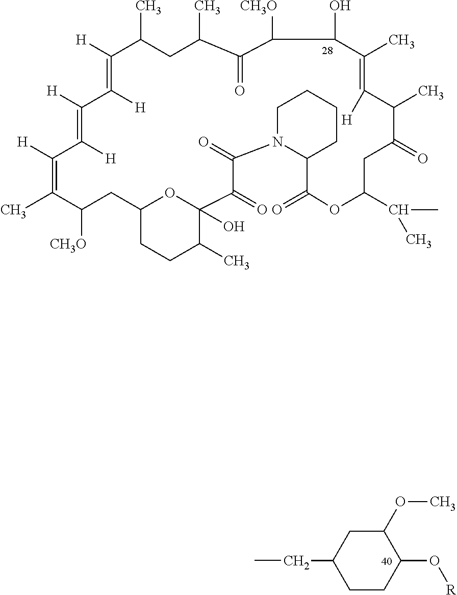
where R is H or CH2—CH2—OH: and
expanding the stent at the vascular injury site, to bring the drug-release coating in contact with the vessel at the injury site,
said coating being effective to release an amount of the compound to inhibit restenosis at the site.
2. The method of claim 1 , wherein the filaments are poly-l-lactide and the polymer substrate in the coating is selected from the group consisting of poly-l-lactide or poly-dl-lactide.
3. The method of claim 2 , wherein the polymer substrate in the coating is formed of poly-dl-lactide having a thickness between 3-20 microns.
4. The method of claim 1 , wherein the stent further includes a polymer undercoat disposed between the filaments of the stent body and said drug-release coating.
5. The method of 4, wherein said polymer undercoat is formed of a polymer selected from the group consisting of ethylene vinyl alcohol, parylast, silicone, a fluoropolymer, and parylene.
6. The method of claim 3 , wherein the stent further includes a parylene polymer undercoat having a thickness of between 1-3 microns, disposed between the filaments of the stent body and said poly-dl-lactide coating substrate.
7. The method of claim 6 , wherein said compound is present in the coating in an amount between 50% and 75% by weight.
8. The method of claim 1 , wherein said coating further includes a bioactive agent selected from the group consisting of an antiplatelet agent, a fibrinolytic agent, and a thrombolytic agent.
9. The method of claim 1 , wherein the polymer substrate in the coating is formed of poly-dl-lactide.
10. The method of claim 1 , for use where the vascular injury is produced during an angiographic procedure in which a vessel region is overstretched at least 30% in diameter.
11. An endovascular stent for placement at a vascular injury site, for inhibiting restenosis at the site, comprising a body having an open-lattice structure formed of linked filaments, and carried on the one or more filaments, a drug-release coating of poly-lactide polymers having a thickness of between 3-30 microns, and composed of (i) 25-55 weight percent polymer substrate and (ii) 45-75 weight percent macrocyclic triene compound having the form:

where R is H or is CH2—CH2—OH,
said stent being expandable from a contracted condition in which the stent can be delivered to a vascular injury site via catheter, and an expanded condition in which the stent coating can be placed in contact with the vessel at the injury site,
said coating being effective to release an amount of the compound to inhibit restenosis at the site.
12. The stent of claim 11 , wherein the filaments are poly-l-lactide, and the polymer substrate in the coating is selected from the group consisting of poly-l-lactide or poly-dl-lactide.
13. The stent of claim 11 , wherein the polymer substrate in the coating is formed of poly-dl-lactide having a thickness between 3-20 microns.
14. The stent of claim 11 , which further includes a parylene polymer undercoat having a thickness of between 1-3 microns, disposed between the filaments of the stent body and said poly-dl-lactide coating substrate.
15. The stent of claim 11 , wherein said coating includes the compound at an initial concentration of between 50 and 75 weight percent of coating.
16. The stent of claim 11 , further comprising a polymer undercoat disposed between the filaments of the stent body and said drug-release coating.
17. The stent of claim 16 , wherein said polymer undercoat is formed of a polymer selected from the group consisting of ethylene vinyl alcohol, parylast, silicone, a fluoropolymer, and parylene.
18. The stent of claim 11 , wherein said coating further includes a bioactive agent selected from the group consisting of antiplatelet agents, fibrinolytic agents, and thrombolytic agents.
19. The stent of claim 13 , wherein the polymer substrate in the coating is formed of poly-dl-lactide.
20. The stent of claim 13 wherein filaments are metal filaments.
Priority Applications (1)
| Application Number | Priority Date | Filing Date | Title |
|---|---|---|---|
| US13/556,960 US8545550B2 (en) | 2002-04-24 | 2012-07-24 | Drug-delivery endovascular stent and method for treating restenosis |
Applications Claiming Priority (4)
| Application Number | Priority Date | Filing Date | Title |
|---|---|---|---|
| US10/133,814 US6939376B2 (en) | 2001-11-05 | 2002-04-24 | Drug-delivery endovascular stent and method for treating restenosis |
| US10/382,426 US7682387B2 (en) | 2002-04-24 | 2003-03-05 | Drug-delivery endovascular stent and method for treating restenosis |
| US12/638,740 US8252046B2 (en) | 2002-04-24 | 2009-12-15 | Drug-delivery endovascular stent and method for treating restenosis |
| US13/556,960 US8545550B2 (en) | 2002-04-24 | 2012-07-24 | Drug-delivery endovascular stent and method for treating restenosis |
Related Parent Applications (1)
| Application Number | Title | Priority Date | Filing Date |
|---|---|---|---|
| US12/638,740 Continuation US8252046B2 (en) | 2002-04-24 | 2009-12-15 | Drug-delivery endovascular stent and method for treating restenosis |
Publications (2)
| Publication Number | Publication Date |
|---|---|
| US20130006350A1 US20130006350A1 (en) | 2013-01-03 |
| US8545550B2 true US8545550B2 (en) | 2013-10-01 |
Family
ID=46150286
Family Applications (4)
| Application Number | Title | Priority Date | Filing Date |
|---|---|---|---|
| US10/382,426 Expired - Lifetime US7682387B2 (en) | 2001-11-05 | 2003-03-05 | Drug-delivery endovascular stent and method for treating restenosis |
| US11/423,418 Abandoned US20060229706A1 (en) | 2001-11-05 | 2006-06-09 | Drug-Delivery Endovascular Stent and Method for Treating Restenosis |
| US12/638,740 Expired - Fee Related US8252046B2 (en) | 2002-04-24 | 2009-12-15 | Drug-delivery endovascular stent and method for treating restenosis |
| US13/556,960 Expired - Lifetime US8545550B2 (en) | 2002-04-24 | 2012-07-24 | Drug-delivery endovascular stent and method for treating restenosis |
Family Applications Before (3)
| Application Number | Title | Priority Date | Filing Date |
|---|---|---|---|
| US10/382,426 Expired - Lifetime US7682387B2 (en) | 2001-11-05 | 2003-03-05 | Drug-delivery endovascular stent and method for treating restenosis |
| US11/423,418 Abandoned US20060229706A1 (en) | 2001-11-05 | 2006-06-09 | Drug-Delivery Endovascular Stent and Method for Treating Restenosis |
| US12/638,740 Expired - Fee Related US8252046B2 (en) | 2002-04-24 | 2009-12-15 | Drug-delivery endovascular stent and method for treating restenosis |
Country Status (1)
| Country | Link |
|---|---|
| US (4) | US7682387B2 (en) |
Cited By (7)
| Publication number | Priority date | Publication date | Assignee | Title |
|---|---|---|---|---|
| US20110238162A1 (en) * | 2009-05-20 | 2011-09-29 | Arsenal Medical | Medical implant |
| US9387282B2 (en) | 2010-03-31 | 2016-07-12 | Abbott Cardiovascular Systems Inc. | Absorbable coating for implantable device |
| US9566373B2 (en) | 2004-06-30 | 2017-02-14 | Abbott Cardiovascular Systems Inc. | Anti-proliferative and anti-inflammatory agent combination for treatment of vascular disorders with an implantable medical device |
| US9592323B2 (en) | 2008-05-21 | 2017-03-14 | Abbott Cardiovascular Systems Inc. | Coating comprising an amorphous primer layer and a semi-crystalline reservoir layer |
| US10064982B2 (en) | 2001-06-27 | 2018-09-04 | Abbott Cardiovascular Systems Inc. | PDLLA stent coating |
| US10568994B2 (en) | 2009-05-20 | 2020-02-25 | 480 Biomedical Inc. | Drug-eluting medical implants |
| US11179255B2 (en) | 2018-03-29 | 2021-11-23 | Sahajanand Medical Technologies Limited | Medical devices |
Families Citing this family (91)
| Publication number | Priority date | Publication date | Assignee | Title |
|---|---|---|---|---|
| CA2178541C (en) * | 1995-06-07 | 2009-11-24 | Neal E. Fearnot | Implantable medical device |
| US6306166B1 (en) * | 1997-08-13 | 2001-10-23 | Scimed Life Systems, Inc. | Loading and release of water-insoluble drugs |
| EP1174157B1 (en) * | 1998-04-27 | 2005-06-29 | Surmodics Inc. | Bioactive agent release coating |
| US20020188037A1 (en) * | 1999-04-15 | 2002-12-12 | Chudzik Stephen J. | Method and system for providing bioactive agent release coating |
| CA2358296A1 (en) * | 1999-01-05 | 2000-07-13 | Anthony P. Adamis | Targeted transscleral controlled release drug delivery to the retina and choroid |
| WO2002089767A1 (en) | 2001-05-03 | 2002-11-14 | Massachusetts Eye And Ear Infirmary | Implantable drug delivery device and use thereof |
| GB0121980D0 (en) * | 2001-09-11 | 2001-10-31 | Cathnet Science Holding As | Expandable stent |
| US6939376B2 (en) * | 2001-11-05 | 2005-09-06 | Sun Biomedical, Ltd. | Drug-delivery endovascular stent and method for treating restenosis |
| US7682387B2 (en) * | 2002-04-24 | 2010-03-23 | Biosensors International Group, Ltd. | Drug-delivery endovascular stent and method for treating restenosis |
| US7147656B2 (en) * | 2001-12-03 | 2006-12-12 | Xtent, Inc. | Apparatus and methods for delivery of braided prostheses |
| US7270668B2 (en) * | 2001-12-03 | 2007-09-18 | Xtent, Inc. | Apparatus and methods for delivering coiled prostheses |
| US7182779B2 (en) * | 2001-12-03 | 2007-02-27 | Xtent, Inc. | Apparatus and methods for positioning prostheses for deployment from a catheter |
| US7137993B2 (en) * | 2001-12-03 | 2006-11-21 | Xtent, Inc. | Apparatus and methods for delivery of multiple distributed stents |
| US20040186551A1 (en) | 2003-01-17 | 2004-09-23 | Xtent, Inc. | Multiple independent nested stent structures and methods for their preparation and deployment |
| US7351255B2 (en) * | 2001-12-03 | 2008-04-01 | Xtent, Inc. | Stent delivery apparatus and method |
| US20030135266A1 (en) * | 2001-12-03 | 2003-07-17 | Xtent, Inc. | Apparatus and methods for delivery of multiple distributed stents |
| US8080048B2 (en) | 2001-12-03 | 2011-12-20 | Xtent, Inc. | Stent delivery for bifurcated vessels |
| US7309350B2 (en) * | 2001-12-03 | 2007-12-18 | Xtent, Inc. | Apparatus and methods for deployment of vascular prostheses |
| US7294146B2 (en) | 2001-12-03 | 2007-11-13 | Xtent, Inc. | Apparatus and methods for delivery of variable length stents |
| US7892273B2 (en) * | 2001-12-03 | 2011-02-22 | Xtent, Inc. | Custom length stent apparatus |
| WO2003092665A2 (en) * | 2002-05-02 | 2003-11-13 | Massachusetts Eye And Ear Infirmary | Ocular drug delivery systems and use thereof |
| US7097850B2 (en) * | 2002-06-18 | 2006-08-29 | Surmodics, Inc. | Bioactive agent release coating and controlled humidity method |
| US20030232087A1 (en) * | 2002-06-18 | 2003-12-18 | Lawin Laurie R. | Bioactive agent release coating with aromatic poly(meth)acrylates |
| US9770349B2 (en) * | 2002-11-13 | 2017-09-26 | University Of Virginia Patent Foundation | Nanoporous stents with enhanced cellular adhesion and reduced neointimal formation |
| US20040111144A1 (en) * | 2002-12-06 | 2004-06-10 | Lawin Laurie R. | Barriers for polymeric coatings |
| WO2004073551A2 (en) * | 2003-02-18 | 2004-09-02 | Massachusetts Eye And Ear Infirmary | Transscleral drug delivery device and related methods |
| US7241308B2 (en) * | 2003-06-09 | 2007-07-10 | Xtent, Inc. | Stent deployment systems and methods |
| US8016816B2 (en) | 2003-09-09 | 2011-09-13 | Convatec Technologies Inc. | Fecal management appliance and method and apparatus for introducing same |
| US7553324B2 (en) * | 2003-10-14 | 2009-06-30 | Xtent, Inc. | Fixed stent delivery devices and methods |
| US20050080475A1 (en) * | 2003-10-14 | 2005-04-14 | Xtent, Inc. A Delaware Corporation | Stent delivery devices and methods |
| US7749981B2 (en) * | 2003-10-21 | 2010-07-06 | Inspire Pharmaceuticals, Inc. | Drug-eluting stents coated with non-nucleotide P2Y12 receptor antagonist compound |
| US7326236B2 (en) * | 2003-12-23 | 2008-02-05 | Xtent, Inc. | Devices and methods for controlling and indicating the length of an interventional element |
| US20070156225A1 (en) * | 2003-12-23 | 2007-07-05 | Xtent, Inc. | Automated control mechanisms and methods for custom length stent apparatus |
| US20050197687A1 (en) * | 2004-03-02 | 2005-09-08 | Masoud Molaei | Medical devices including metallic films and methods for making same |
| US7323006B2 (en) * | 2004-03-30 | 2008-01-29 | Xtent, Inc. | Rapid exchange interventional devices and methods |
| WO2005097228A2 (en) * | 2004-04-06 | 2005-10-20 | Surmodics, Inc. | Coating compositions for bioactive agents |
| US20050228477A1 (en) * | 2004-04-09 | 2005-10-13 | Xtent, Inc. | Topographic coatings and coating methods for medical devices |
| US8317859B2 (en) | 2004-06-28 | 2012-11-27 | J.W. Medical Systems Ltd. | Devices and methods for controlling expandable prostheses during deployment |
| US20050288766A1 (en) * | 2004-06-28 | 2005-12-29 | Xtent, Inc. | Devices and methods for controlling expandable prostheses during deployment |
| US7901451B2 (en) * | 2004-09-24 | 2011-03-08 | Biosensors International Group, Ltd. | Drug-delivery endovascular stent and method for treating restenosis |
| US20060069424A1 (en) * | 2004-09-27 | 2006-03-30 | Xtent, Inc. | Self-constrained segmented stents and methods for their deployment |
| US7147659B2 (en) * | 2004-10-28 | 2006-12-12 | Cordis Neurovascular, Inc. | Expandable stent having a dissolvable portion |
| US7402168B2 (en) * | 2005-04-11 | 2008-07-22 | Xtent, Inc. | Custom-length stent delivery system with independently operable expansion elements |
| US20070196423A1 (en) * | 2005-11-21 | 2007-08-23 | Med Institute, Inc. | Implantable medical device coatings with biodegradable elastomer and releasable therapeutic agent |
| US20070179587A1 (en) * | 2006-01-30 | 2007-08-02 | Xtent, Inc. | Apparatus and methods for deployment of custom-length prostheses |
| WO2007109621A2 (en) | 2006-03-20 | 2007-09-27 | Xtent, Inc. | Apparatus and methods for deployment of linked prosthetic segments |
| US20070254003A1 (en) * | 2006-05-01 | 2007-11-01 | Pu Zhou | Non-sticky coatings with therapeutic agents for medical devices |
| US20070281117A1 (en) * | 2006-06-02 | 2007-12-06 | Xtent, Inc. | Use of plasma in formation of biodegradable stent coating |
| US20080095918A1 (en) * | 2006-06-14 | 2008-04-24 | Kleiner Lothar W | Coating construct with enhanced interfacial compatibility |
| US20070299511A1 (en) * | 2006-06-27 | 2007-12-27 | Gale David C | Thin stent coating |
| EP3047860B1 (en) | 2006-07-20 | 2024-08-28 | OrbusNeich Medical Pte. Ltd. | Bioabsorbable polymeric composition for a medical device |
| US20080091262A1 (en) * | 2006-10-17 | 2008-04-17 | Gale David C | Drug delivery after biodegradation of the stent scaffolding |
| US8016879B2 (en) | 2006-08-01 | 2011-09-13 | Abbott Cardiovascular Systems Inc. | Drug delivery after biodegradation of the stent scaffolding |
| US20080269865A1 (en) * | 2006-08-07 | 2008-10-30 | Xtent, Inc. | Custom Length Stent Apparatus |
| US9289279B2 (en) * | 2006-10-06 | 2016-03-22 | Promethean Surgical Devices, Llc | Apparatus and method for limiting surgical adhesions |
| US8067055B2 (en) * | 2006-10-20 | 2011-11-29 | Biosensors International Group, Ltd. | Drug-delivery endovascular stent and method of use |
| US7959942B2 (en) | 2006-10-20 | 2011-06-14 | Orbusneich Medical, Inc. | Bioabsorbable medical device with coating |
| US20080097591A1 (en) * | 2006-10-20 | 2008-04-24 | Biosensors International Group | Drug-delivery endovascular stent and method of use |
| US8691321B2 (en) | 2006-10-20 | 2014-04-08 | Orbusneich Medical, Inc. | Bioabsorbable polymeric composition and medical device background |
| US20080103584A1 (en) * | 2006-10-25 | 2008-05-01 | Biosensors International Group | Temporal Intraluminal Stent, Methods of Making and Using |
| JP2010512947A (en) * | 2006-12-20 | 2010-04-30 | ボストン サイエンティフィック リミテッド | Stent with coating for delivering therapeutic agent |
| KR101198980B1 (en) | 2007-01-21 | 2012-11-07 | 헤모텍 아게 | Medical product for treating stenosis of body passages and for preventing threatening restenosis |
| US20080199510A1 (en) | 2007-02-20 | 2008-08-21 | Xtent, Inc. | Thermo-mechanically controlled implants and methods of use |
| US8486132B2 (en) | 2007-03-22 | 2013-07-16 | J.W. Medical Systems Ltd. | Devices and methods for controlling expandable prostheses during deployment |
| US9192697B2 (en) | 2007-07-03 | 2015-11-24 | Hemoteq Ag | Balloon catheter for treating stenosis of body passages and for preventing threatening restenosis |
| DE102007034364A1 (en) * | 2007-07-24 | 2009-01-29 | Biotronik Vi Patent Ag | Degradable metal stent with active ingredient-containing coating |
| KR100930167B1 (en) * | 2007-09-19 | 2009-12-07 | 삼성전기주식회사 | Ultra wide angle optical system |
| US20090076584A1 (en) * | 2007-09-19 | 2009-03-19 | Xtent, Inc. | Apparatus and methods for deployment of multiple custom-length prostheses |
| WO2009089518A2 (en) * | 2008-01-11 | 2009-07-16 | Massachusetts Institute Of Technology | Endovascular platforms for the differential targeting of molecules to vessel wall and vessel lumen |
| US9101503B2 (en) * | 2008-03-06 | 2015-08-11 | J.W. Medical Systems Ltd. | Apparatus having variable strut length and methods of use |
| US8562669B2 (en) * | 2008-06-26 | 2013-10-22 | Abbott Cardiovascular Systems Inc. | Methods of application of coatings composed of hydrophobic, high glass transition polymers with tunable drug release rates |
| US8642063B2 (en) * | 2008-08-22 | 2014-02-04 | Cook Medical Technologies Llc | Implantable medical device coatings with biodegradable elastomer and releasable taxane agent |
| US12324756B2 (en) | 2008-09-25 | 2025-06-10 | Advanced Bifurcation Systems Inc. | System and methods for treating a bifurcation |
| US8828071B2 (en) | 2008-09-25 | 2014-09-09 | Advanced Bifurcation Systems, Inc. | Methods and systems for ostial stenting of a bifurcation |
| US8821562B2 (en) | 2008-09-25 | 2014-09-02 | Advanced Bifurcation Systems, Inc. | Partially crimped stent |
| JP5635515B2 (en) | 2008-09-25 | 2014-12-03 | アドバンスド バイファケーション システムズ, インコーポレイテッド | Partially crimped stent |
| US12076258B2 (en) | 2008-09-25 | 2024-09-03 | Advanced Bifurcation Systems Inc. | Selective stent crimping |
| US11298252B2 (en) | 2008-09-25 | 2022-04-12 | Advanced Bifurcation Systems Inc. | Stent alignment during treatment of a bifurcation |
| US20100286765A1 (en) * | 2009-05-07 | 2010-11-11 | Paul Zamecnik | Medical Device Coatings and Coated Stents |
| US10369256B2 (en) | 2009-07-10 | 2019-08-06 | Boston Scientific Scimed, Inc. | Use of nanocrystals for drug delivery from a balloon |
| EP2453938B1 (en) | 2009-07-17 | 2015-08-19 | Boston Scientific Scimed, Inc. | Nucleation of drug delivery balloons to provide improved crystal size and density |
| CN103037815B (en) | 2010-03-24 | 2015-05-13 | 高级分支系统股份有限公司 | Method and system for treating furcations by temporarily opening side branches |
| EP2549951B1 (en) | 2010-03-24 | 2017-05-10 | Advanced Bifurcation Systems, Inc. | Stent alignment during treatment of a bifurcation |
| AU2011232362B2 (en) | 2010-03-24 | 2015-12-10 | Advanced Bifurcation Systems Inc. | System and methods for treating a bifurcation |
| US8889211B2 (en) | 2010-09-02 | 2014-11-18 | Boston Scientific Scimed, Inc. | Coating process for drug delivery balloons using heat-induced rewrap memory |
| WO2012109382A2 (en) | 2011-02-08 | 2012-08-16 | Advanced Bifurcation Systems, Inc. | Multi-stent and multi-balloon apparatus for treating bifurcations and methods of use |
| EP4424283A3 (en) | 2011-02-08 | 2024-12-25 | Advanced Bifurcation Systems Inc. | System and methods for treating a bifurcation with a fully crimped stent |
| US8669360B2 (en) | 2011-08-05 | 2014-03-11 | Boston Scientific Scimed, Inc. | Methods of converting amorphous drug substance into crystalline form |
| US9056152B2 (en) | 2011-08-25 | 2015-06-16 | Boston Scientific Scimed, Inc. | Medical device with crystalline drug coating |
| CN104707185A (en) * | 2015-04-03 | 2015-06-17 | 上海市普陀区中心医院 | Ginkgolide B composite medicine eluting stent |
| CN114680950B (en) * | 2020-12-29 | 2024-12-31 | 先健科技(深圳)有限公司 | Implantable device and method for preparing the same |
Citations (84)
| Publication number | Priority date | Publication date | Assignee | Title |
|---|---|---|---|---|
| US3929992A (en) | 1972-09-29 | 1975-12-30 | Ayerst Mckenna & Harrison | Rapamycin and process of preparation |
| US4316885A (en) | 1980-08-25 | 1982-02-23 | Ayerst, Mckenna And Harrison, Inc. | Acyl derivatives of rapamycin |
| US4650803A (en) | 1985-12-06 | 1987-03-17 | University Of Kansas | Prodrugs of rapamycin |
| US4885171A (en) | 1978-11-03 | 1989-12-05 | American Home Products Corporation | Use of rapamycin in treatment of certain tumors |
| US4990155A (en) | 1989-05-19 | 1991-02-05 | Wilkoff Howard M | Surgical stent method and apparatus |
| EP0470246A1 (en) | 1990-02-28 | 1992-02-12 | Medtronic Inc | Intralumenal drug eluting prosthesis. |
| US5100899A (en) | 1989-06-06 | 1992-03-31 | Roy Calne | Methods of inhibiting transplant rejection in mammals using rapamycin and derivatives and prodrugs thereof |
| US5120842A (en) | 1991-04-01 | 1992-06-09 | American Home Products Corporation | Silyl ethers of rapamycin |
| US5151413A (en) | 1991-11-06 | 1992-09-29 | American Home Products Corporation | Rapamycin acetals as immunosuppressant and antifungal agents |
| EP0551182A1 (en) | 1992-01-09 | 1993-07-14 | American Home Products Corporation | Method of treating hyperproliferative vascular disease using rapamycin, eventually in combination with mycophenolic acid |
| US5252579A (en) | 1993-02-16 | 1993-10-12 | American Home Products Corporation | Macrocyclic immunomodulators |
| US5258389A (en) | 1992-11-09 | 1993-11-02 | Merck & Co., Inc. | O-aryl, O-alkyl, O-alkenyl and O-alkynylrapamycin derivatives |
| US5288711A (en) | 1992-04-28 | 1994-02-22 | American Home Products Corporation | Method of treating hyperproliferative vascular disease |
| US5306286A (en) | 1987-06-25 | 1994-04-26 | Duke University | Absorbable stent |
| US5342348A (en) | 1992-12-04 | 1994-08-30 | Kaplan Aaron V | Method and device for treating and enlarging body lumens |
| US5362718A (en) | 1994-04-18 | 1994-11-08 | American Home Products Corporation | Rapamycin hydroxyesters |
| WO1995016691A1 (en) | 1993-12-17 | 1995-06-22 | Sandoz Ltd. | Rapamycin derivatives useful as immunosuppressants |
| US5443458A (en) | 1992-12-22 | 1995-08-22 | Advanced Cardiovascular Systems, Inc. | Multilayered biodegradable stent and method of manufacture |
| US5447724A (en) | 1990-05-17 | 1995-09-05 | Harbor Medical Devices, Inc. | Medical device polymer |
| US5464650A (en) | 1993-04-26 | 1995-11-07 | Medtronic, Inc. | Intravascular stent and method |
| US5516781A (en) | 1992-01-09 | 1996-05-14 | American Home Products Corporation | Method of treating restenosis with rapamycin |
| US5527337A (en) | 1987-06-25 | 1996-06-18 | Duke University | Bioabsorbable stent and method of making the same |
| US5545208A (en) | 1990-02-28 | 1996-08-13 | Medtronic, Inc. | Intralumenal drug eluting prosthesis |
| US5607467A (en) | 1990-09-14 | 1997-03-04 | Froix; Michael | Expandable polymeric stent with memory and delivery apparatus and method |
| US5609629A (en) | 1995-06-07 | 1997-03-11 | Med Institute, Inc. | Coated implantable medical device |
| US5649977A (en) | 1994-09-22 | 1997-07-22 | Advanced Cardiovascular Systems, Inc. | Metal reinforced polymer stent |
| US5665772A (en) | 1992-10-09 | 1997-09-09 | Sandoz Ltd. | O-alkylated rapamycin derivatives and their use, particularly as immunosuppressants |
| WO1997035575A1 (en) | 1996-03-27 | 1997-10-02 | Novartis Ag | Use of rapamycin derivatives in vasculopathies and xenotransplantation |
| WO1997037617A1 (en) | 1996-04-08 | 1997-10-16 | Swaminathan Jayaraman | Multiple interconnected stents and method of coating stents |
| US5707385A (en) | 1994-11-16 | 1998-01-13 | Advanced Cardiovascular Systems, Inc. | Drug loaded elastic membrane and method for delivery |
| US5733327A (en) | 1994-10-17 | 1998-03-31 | Igaki; Keiji | Stent for liberating drug |
| JPH10192413A (en) | 1996-12-20 | 1998-07-28 | Schneider Usa Inc | Manufacture of coating emitting pharmaceutical object |
| WO1998036784A1 (en) | 1997-02-20 | 1998-08-27 | Cook Incorporated | Coated implantable medical device |
| US5824048A (en) | 1993-04-26 | 1998-10-20 | Medtronic, Inc. | Method for delivering a therapeutic substance to a body lumen |
| US5824049A (en) | 1995-06-07 | 1998-10-20 | Med Institute, Inc. | Coated implantable medical device |
| US5837313A (en) | 1995-04-19 | 1998-11-17 | Schneider (Usa) Inc | Drug release stent coating process |
| US5860999A (en) | 1993-02-04 | 1999-01-19 | Angiomed Gmbh & Co.Medizintechnik Kg | Stent and method of using same |
| US5869127A (en) | 1995-02-22 | 1999-02-09 | Boston Scientific Corporation | Method of providing a substrate with a bio-active/biocompatible coating |
| WO1999007308A1 (en) | 1997-08-08 | 1999-02-18 | Sunscope International, Inc. | Microporous stent and implantation device |
| US5902317A (en) | 1994-06-01 | 1999-05-11 | Nitinol Medical Technologies, Inc. | Stent and method and apparatus for forming and delivering the same |
| EP0950386A2 (en) | 1998-04-16 | 1999-10-20 | Cordis Corporation | Stent with local rapamycin delivery |
| US6013853A (en) | 1992-02-14 | 2000-01-11 | The University Of Texas System | Continuous release polymeric implant carrier |
| EP0970711A2 (en) | 1998-06-30 | 2000-01-12 | Ethicon, Inc. | Process for coating stents |
| US6042875A (en) | 1997-04-30 | 2000-03-28 | Schneider (Usa) Inc. | Drug-releasing coatings for medical devices |
| US6042606A (en) | 1997-09-29 | 2000-03-28 | Cook Incorporated | Radially expandable non-axially contracting surgical stent |
| WO2000032255A1 (en) | 1998-12-03 | 2000-06-08 | Boston Scientific Limited | Polymeric coatings with controlled delivery of active agents |
| US6080177A (en) | 1991-03-08 | 2000-06-27 | Igaki; Keiji | Luminal stent, holding structure therefor and device for attaching luminal stent |
| US6099562A (en) | 1996-06-13 | 2000-08-08 | Schneider (Usa) Inc. | Drug coating with topcoat |
| US6120536A (en) | 1995-04-19 | 2000-09-19 | Schneider (Usa) Inc. | Medical devices with long term non-thrombogenic coatings |
| US6129755A (en) | 1998-01-09 | 2000-10-10 | Nitinol Development Corporation | Intravascular stent having an improved strut configuration |
| US6174329B1 (en) | 1996-08-22 | 2001-01-16 | Advanced Cardiovascular Systems, Inc. | Protective coating for a stent with intermediate radiopaque coating |
| US6179817B1 (en) | 1995-02-22 | 2001-01-30 | Boston Scientific Corporation | Hybrid coating for medical devices |
| US6190406B1 (en) | 1998-01-09 | 2001-02-20 | Nitinal Development Corporation | Intravascular stent having tapered struts |
| WO2001014387A1 (en) | 1999-08-24 | 2001-03-01 | Ariad Gene Therapeutics, Inc. | 28-epirapalogs |
| US6197781B1 (en) | 1995-07-14 | 2001-03-06 | Novartis Ag | Pharmaceutical compositions |
| US6214901B1 (en) | 1998-04-27 | 2001-04-10 | Surmodics, Inc. | Bioactive agent release coating |
| US6231600B1 (en) | 1995-02-22 | 2001-05-15 | Scimed Life Systems, Inc. | Stents with hybrid coating for medical devices |
| US6239124B1 (en) | 1996-07-30 | 2001-05-29 | Novartis Ag | Pharmaceutical compositions for the treatment of transplant rejection or autoimmune or inflammatory conditions comprising cyclosporin A and 40-0-(2-hydroxyethyl)-rapamycin |
| WO2001045763A1 (en) | 1999-12-23 | 2001-06-28 | Advanced Cardiovascular Systems, Inc. | Biocomptabible coating |
| US6277983B1 (en) | 2000-09-27 | 2001-08-21 | American Home Products Corporation | Regioselective synthesis of rapamycin derivatives |
| US6299604B1 (en) | 1998-08-20 | 2001-10-09 | Cook Incorporated | Coated implantable medical device |
| US20010029351A1 (en) | 1998-04-16 | 2001-10-11 | Robert Falotico | Drug combinations and delivery devices for the prevention and treatment of vascular disease |
| US20020038146A1 (en) | 1998-07-29 | 2002-03-28 | Ulf Harry | Expandable stent with relief cuts for carrying medicines and other materials |
| EP1192957A2 (en) | 2000-09-29 | 2002-04-03 | Ethicon, Inc. | Coating for medical devices |
| US6368658B1 (en) | 1999-04-19 | 2002-04-09 | Scimed Life Systems, Inc. | Coating medical devices using air suspension |
| US20020051730A1 (en) | 2000-09-29 | 2002-05-02 | Stanko Bodnar | Coated medical devices and sterilization thereof |
| US20020111590A1 (en) | 2000-09-29 | 2002-08-15 | Davila Luis A. | Medical devices, drug coatings and methods for maintaining the drug coatings thereon |
| US20020156022A1 (en) | 1997-05-14 | 2002-10-24 | Edwards David B. | Compounds and methods for treating transplant rejection |
| US20030059494A1 (en) | 1997-06-09 | 2003-03-27 | Keng P. Ang | Fish feed and method of using same |
| US20030059454A1 (en) | 2001-09-24 | 2003-03-27 | Barry James J. | Optimized dosing for drug coated stents |
| US20030068355A1 (en) | 2001-08-20 | 2003-04-10 | Shanley John F. | Therapeutic agent delivery device with protective separating layer |
| US20030077310A1 (en) | 2001-10-22 | 2003-04-24 | Chandrashekhar Pathak | Stent coatings containing HMG-CoA reductase inhibitors |
| US20030088307A1 (en) | 2001-11-05 | 2003-05-08 | Shulze John E. | Potent coatings for stents |
| US20030143315A1 (en) | 2001-05-16 | 2003-07-31 | Pui David Y H | Coating medical devices |
| US6607598B2 (en) | 1999-04-19 | 2003-08-19 | Scimed Life Systems, Inc. | Device for protecting medical devices during a coating process |
| US6641611B2 (en) | 2001-11-26 | 2003-11-04 | Swaminathan Jayaraman | Therapeutic coating for an intravascular implant |
| US20030225450A1 (en) | 2001-11-05 | 2003-12-04 | Shulze John E. | Drug-delivery endovascular stent and method for treating restenosis |
| US6663881B2 (en) | 1993-01-28 | 2003-12-16 | Neorx Corporation | Therapeutic inhibitor of vascular smooth muscle cells |
| US20040010002A1 (en) | 2000-01-14 | 2004-01-15 | The Trustees Of The University Of Pennsylvania | O-methylated rapamycin derivatives for alleviation and inhibition of lymphoproliferative disorders |
| US20040024450A1 (en) | 2002-04-24 | 2004-02-05 | Sun Biomedical, Ltd. | Drug-delivery endovascular stent and method for treating restenosis |
| US20040030380A1 (en) | 2002-04-24 | 2004-02-12 | Sun Biomedical, Ltd. | Drug-delivery endovascular stent and method for treating restenosis |
| US6743463B2 (en) | 2002-03-28 | 2004-06-01 | Scimed Life Systems, Inc. | Method for spray-coating a medical device having a tubular wall such as a stent |
| US6790228B2 (en) | 1999-12-23 | 2004-09-14 | Advanced Cardiovascular Systems, Inc. | Coating for implantable devices and a method of forming the same |
| US7179283B2 (en) | 2001-11-02 | 2007-02-20 | Scimed Life Systems, Inc. | Vapor deposition process for producing a stent-graft and a stent-graft produced therefrom |
Family Cites Families (12)
| Publication number | Priority date | Publication date | Assignee | Title |
|---|---|---|---|---|
| US6273252B1 (en) * | 1999-01-20 | 2001-08-14 | Burke H. Mitchell | Protective covering for a hand-held device |
| US7220276B1 (en) | 2000-03-06 | 2007-05-22 | Surmodics, Inc. | Endovascular graft coatings |
| DK1301221T3 (en) | 2000-05-16 | 2006-07-03 | Ortho Mcneil Pharm Inc | Method of coating medical articles using supercritical carbon dioxide |
| AU2001288809A1 (en) | 2000-09-26 | 2002-04-08 | Advanced Cardiovascular Systems Inc. | A method of loading a substance onto an implantable device |
| AU1129902A (en) | 2000-09-29 | 2002-04-08 | Cordis Corp | Coated medical devices |
| EP1498084B1 (en) | 2000-10-16 | 2014-06-18 | Innovational Holdings, LLC | Expandable medical device for delivery of beneficial agent |
| US7083642B2 (en) * | 2000-12-22 | 2006-08-01 | Avantec Vascular Corporation | Delivery of therapeutic capable agents |
| US20030033007A1 (en) * | 2000-12-22 | 2003-02-13 | Avantec Vascular Corporation | Methods and devices for delivery of therapeutic capable agents with variable release profile |
| US20020082679A1 (en) * | 2000-12-22 | 2002-06-27 | Avantec Vascular Corporation | Delivery or therapeutic capable agents |
| US7077859B2 (en) * | 2000-12-22 | 2006-07-18 | Avantec Vascular Corporation | Apparatus and methods for variably controlled substance delivery from implanted prostheses |
| US20030050692A1 (en) * | 2000-12-22 | 2003-03-13 | Avantec Vascular Corporation | Delivery of therapeutic capable agents |
| US6682771B2 (en) * | 2001-07-02 | 2004-01-27 | Scimed Life Systems, Inc. | Coating dispensing system and method using a solenoid head for coating medical devices |
-
2003
- 2003-03-05 US US10/382,426 patent/US7682387B2/en not_active Expired - Lifetime
-
2006
- 2006-06-09 US US11/423,418 patent/US20060229706A1/en not_active Abandoned
-
2009
- 2009-12-15 US US12/638,740 patent/US8252046B2/en not_active Expired - Fee Related
-
2012
- 2012-07-24 US US13/556,960 patent/US8545550B2/en not_active Expired - Lifetime
Patent Citations (100)
| Publication number | Priority date | Publication date | Assignee | Title |
|---|---|---|---|---|
| US3929992A (en) | 1972-09-29 | 1975-12-30 | Ayerst Mckenna & Harrison | Rapamycin and process of preparation |
| US4885171A (en) | 1978-11-03 | 1989-12-05 | American Home Products Corporation | Use of rapamycin in treatment of certain tumors |
| US4316885A (en) | 1980-08-25 | 1982-02-23 | Ayerst, Mckenna And Harrison, Inc. | Acyl derivatives of rapamycin |
| US4650803A (en) | 1985-12-06 | 1987-03-17 | University Of Kansas | Prodrugs of rapamycin |
| US5306286A (en) | 1987-06-25 | 1994-04-26 | Duke University | Absorbable stent |
| US5527337A (en) | 1987-06-25 | 1996-06-18 | Duke University | Bioabsorbable stent and method of making the same |
| US4990155A (en) | 1989-05-19 | 1991-02-05 | Wilkoff Howard M | Surgical stent method and apparatus |
| US5100899A (en) | 1989-06-06 | 1992-03-31 | Roy Calne | Methods of inhibiting transplant rejection in mammals using rapamycin and derivatives and prodrugs thereof |
| US5545208A (en) | 1990-02-28 | 1996-08-13 | Medtronic, Inc. | Intralumenal drug eluting prosthesis |
| EP0470246A1 (en) | 1990-02-28 | 1992-02-12 | Medtronic Inc | Intralumenal drug eluting prosthesis. |
| US5447724A (en) | 1990-05-17 | 1995-09-05 | Harbor Medical Devices, Inc. | Medical device polymer |
| US5607467A (en) | 1990-09-14 | 1997-03-04 | Froix; Michael | Expandable polymeric stent with memory and delivery apparatus and method |
| US6080177A (en) | 1991-03-08 | 2000-06-27 | Igaki; Keiji | Luminal stent, holding structure therefor and device for attaching luminal stent |
| US5120842A (en) | 1991-04-01 | 1992-06-09 | American Home Products Corporation | Silyl ethers of rapamycin |
| US5120842B1 (en) | 1991-04-01 | 1993-07-06 | A Failli Amedeo | |
| US5151413A (en) | 1991-11-06 | 1992-09-29 | American Home Products Corporation | Rapamycin acetals as immunosuppressant and antifungal agents |
| US5516781A (en) | 1992-01-09 | 1996-05-14 | American Home Products Corporation | Method of treating restenosis with rapamycin |
| EP0551182A1 (en) | 1992-01-09 | 1993-07-14 | American Home Products Corporation | Method of treating hyperproliferative vascular disease using rapamycin, eventually in combination with mycophenolic acid |
| US6013853A (en) | 1992-02-14 | 2000-01-11 | The University Of Texas System | Continuous release polymeric implant carrier |
| US5288711A (en) | 1992-04-28 | 1994-02-22 | American Home Products Corporation | Method of treating hyperproliferative vascular disease |
| US5665772A (en) | 1992-10-09 | 1997-09-09 | Sandoz Ltd. | O-alkylated rapamycin derivatives and their use, particularly as immunosuppressants |
| EP0867438A1 (en) | 1992-10-09 | 1998-09-30 | Novartis AG | Rapamycin derivatives and their use, particularly as immunosuppressants |
| US5258389A (en) | 1992-11-09 | 1993-11-02 | Merck & Co., Inc. | O-aryl, O-alkyl, O-alkenyl and O-alkynylrapamycin derivatives |
| US5342348A (en) | 1992-12-04 | 1994-08-30 | Kaplan Aaron V | Method and device for treating and enlarging body lumens |
| US5443458A (en) | 1992-12-22 | 1995-08-22 | Advanced Cardiovascular Systems, Inc. | Multilayered biodegradable stent and method of manufacture |
| US6663881B2 (en) | 1993-01-28 | 2003-12-16 | Neorx Corporation | Therapeutic inhibitor of vascular smooth muscle cells |
| US5860999A (en) | 1993-02-04 | 1999-01-19 | Angiomed Gmbh & Co.Medizintechnik Kg | Stent and method of using same |
| US5252579A (en) | 1993-02-16 | 1993-10-12 | American Home Products Corporation | Macrocyclic immunomodulators |
| US5464650A (en) | 1993-04-26 | 1995-11-07 | Medtronic, Inc. | Intravascular stent and method |
| US5776184A (en) | 1993-04-26 | 1998-07-07 | Medtronic, Inc. | Intravasoular stent and method |
| US5824048A (en) | 1993-04-26 | 1998-10-20 | Medtronic, Inc. | Method for delivering a therapeutic substance to a body lumen |
| US5912253A (en) | 1993-12-17 | 1999-06-15 | Novartis Ag | Rapamycin derivatives |
| WO1995016691A1 (en) | 1993-12-17 | 1995-06-22 | Sandoz Ltd. | Rapamycin derivatives useful as immunosuppressants |
| US5362718A (en) | 1994-04-18 | 1994-11-08 | American Home Products Corporation | Rapamycin hydroxyesters |
| US5902317A (en) | 1994-06-01 | 1999-05-11 | Nitinol Medical Technologies, Inc. | Stent and method and apparatus for forming and delivering the same |
| US5649977A (en) | 1994-09-22 | 1997-07-22 | Advanced Cardiovascular Systems, Inc. | Metal reinforced polymer stent |
| US5733327A (en) | 1994-10-17 | 1998-03-31 | Igaki; Keiji | Stent for liberating drug |
| US5707385A (en) | 1994-11-16 | 1998-01-13 | Advanced Cardiovascular Systems, Inc. | Drug loaded elastic membrane and method for delivery |
| US5869127A (en) | 1995-02-22 | 1999-02-09 | Boston Scientific Corporation | Method of providing a substrate with a bio-active/biocompatible coating |
| US6231600B1 (en) | 1995-02-22 | 2001-05-15 | Scimed Life Systems, Inc. | Stents with hybrid coating for medical devices |
| US6179817B1 (en) | 1995-02-22 | 2001-01-30 | Boston Scientific Corporation | Hybrid coating for medical devices |
| US5837313A (en) | 1995-04-19 | 1998-11-17 | Schneider (Usa) Inc | Drug release stent coating process |
| US6120536A (en) | 1995-04-19 | 2000-09-19 | Schneider (Usa) Inc. | Medical devices with long term non-thrombogenic coatings |
| US5824049A (en) | 1995-06-07 | 1998-10-20 | Med Institute, Inc. | Coated implantable medical device |
| US5873904A (en) | 1995-06-07 | 1999-02-23 | Cook Incorporated | Silver implantable medical device |
| US5609629A (en) | 1995-06-07 | 1997-03-11 | Med Institute, Inc. | Coated implantable medical device |
| US6197781B1 (en) | 1995-07-14 | 2001-03-06 | Novartis Ag | Pharmaceutical compositions |
| US6384046B1 (en) | 1996-03-27 | 2002-05-07 | Novartis Ag | Use of 40-O-(2-hydroxy)ethylrapamycin for treatment of restenosis and other disorders |
| WO1997035575A1 (en) | 1996-03-27 | 1997-10-02 | Novartis Ag | Use of rapamycin derivatives in vasculopathies and xenotransplantation |
| WO1997037617A1 (en) | 1996-04-08 | 1997-10-16 | Swaminathan Jayaraman | Multiple interconnected stents and method of coating stents |
| US6099562A (en) | 1996-06-13 | 2000-08-08 | Schneider (Usa) Inc. | Drug coating with topcoat |
| US6239124B1 (en) | 1996-07-30 | 2001-05-29 | Novartis Ag | Pharmaceutical compositions for the treatment of transplant rejection or autoimmune or inflammatory conditions comprising cyclosporin A and 40-0-(2-hydroxyethyl)-rapamycin |
| US6174329B1 (en) | 1996-08-22 | 2001-01-16 | Advanced Cardiovascular Systems, Inc. | Protective coating for a stent with intermediate radiopaque coating |
| JPH10192413A (en) | 1996-12-20 | 1998-07-28 | Schneider Usa Inc | Manufacture of coating emitting pharmaceutical object |
| WO1998036784A1 (en) | 1997-02-20 | 1998-08-27 | Cook Incorporated | Coated implantable medical device |
| US20010027340A1 (en) | 1997-04-18 | 2001-10-04 | Carol Wright | Stent with therapeutically active dosage of rapamycin coated thereon |
| US6585764B2 (en) | 1997-04-18 | 2003-07-01 | Cordis Corporation | Stent with therapeutically active dosage of rapamycin coated thereon |
| US6273913B1 (en) | 1997-04-18 | 2001-08-14 | Cordis Corporation | Modified stent useful for delivery of drugs along stent strut |
| US6042875A (en) | 1997-04-30 | 2000-03-28 | Schneider (Usa) Inc. | Drug-releasing coatings for medical devices |
| US20020156022A1 (en) | 1997-05-14 | 2002-10-24 | Edwards David B. | Compounds and methods for treating transplant rejection |
| US6670398B2 (en) | 1997-05-14 | 2003-12-30 | Atherogenics, Inc. | Compounds and methods for treating transplant rejection |
| US20030059494A1 (en) | 1997-06-09 | 2003-03-27 | Keng P. Ang | Fish feed and method of using same |
| WO1999007308A1 (en) | 1997-08-08 | 1999-02-18 | Sunscope International, Inc. | Microporous stent and implantation device |
| US6042606A (en) | 1997-09-29 | 2000-03-28 | Cook Incorporated | Radially expandable non-axially contracting surgical stent |
| US6190406B1 (en) | 1998-01-09 | 2001-02-20 | Nitinal Development Corporation | Intravascular stent having tapered struts |
| US6129755A (en) | 1998-01-09 | 2000-10-10 | Nitinol Development Corporation | Intravascular stent having an improved strut configuration |
| US20010029351A1 (en) | 1998-04-16 | 2001-10-11 | Robert Falotico | Drug combinations and delivery devices for the prevention and treatment of vascular disease |
| EP0950386A2 (en) | 1998-04-16 | 1999-10-20 | Cordis Corporation | Stent with local rapamycin delivery |
| US6214901B1 (en) | 1998-04-27 | 2001-04-10 | Surmodics, Inc. | Bioactive agent release coating |
| US6153252A (en) | 1998-06-30 | 2000-11-28 | Ethicon, Inc. | Process for coating stents |
| EP0970711A2 (en) | 1998-06-30 | 2000-01-12 | Ethicon, Inc. | Process for coating stents |
| US20020038146A1 (en) | 1998-07-29 | 2002-03-28 | Ulf Harry | Expandable stent with relief cuts for carrying medicines and other materials |
| US6299604B1 (en) | 1998-08-20 | 2001-10-09 | Cook Incorporated | Coated implantable medical device |
| WO2000032255A1 (en) | 1998-12-03 | 2000-06-08 | Boston Scientific Limited | Polymeric coatings with controlled delivery of active agents |
| US6368658B1 (en) | 1999-04-19 | 2002-04-09 | Scimed Life Systems, Inc. | Coating medical devices using air suspension |
| US6607598B2 (en) | 1999-04-19 | 2003-08-19 | Scimed Life Systems, Inc. | Device for protecting medical devices during a coating process |
| WO2001014387A1 (en) | 1999-08-24 | 2001-03-01 | Ariad Gene Therapeutics, Inc. | 28-epirapalogs |
| WO2001045763A1 (en) | 1999-12-23 | 2001-06-28 | Advanced Cardiovascular Systems, Inc. | Biocomptabible coating |
| US6790228B2 (en) | 1999-12-23 | 2004-09-14 | Advanced Cardiovascular Systems, Inc. | Coating for implantable devices and a method of forming the same |
| US20040010002A1 (en) | 2000-01-14 | 2004-01-15 | The Trustees Of The University Of Pennsylvania | O-methylated rapamycin derivatives for alleviation and inhibition of lymphoproliferative disorders |
| US6277983B1 (en) | 2000-09-27 | 2001-08-21 | American Home Products Corporation | Regioselective synthesis of rapamycin derivatives |
| EP1192957A2 (en) | 2000-09-29 | 2002-04-03 | Ethicon, Inc. | Coating for medical devices |
| US6746773B2 (en) | 2000-09-29 | 2004-06-08 | Ethicon, Inc. | Coatings for medical devices |
| US20020051730A1 (en) | 2000-09-29 | 2002-05-02 | Stanko Bodnar | Coated medical devices and sterilization thereof |
| US20020111590A1 (en) | 2000-09-29 | 2002-08-15 | Davila Luis A. | Medical devices, drug coatings and methods for maintaining the drug coatings thereon |
| US20030143315A1 (en) | 2001-05-16 | 2003-07-31 | Pui David Y H | Coating medical devices |
| US20030068355A1 (en) | 2001-08-20 | 2003-04-10 | Shanley John F. | Therapeutic agent delivery device with protective separating layer |
| US6908622B2 (en) | 2001-09-24 | 2005-06-21 | Boston Scientific Scimed, Inc. | Optimized dosing for drug coated stents |
| US20030059454A1 (en) | 2001-09-24 | 2003-03-27 | Barry James J. | Optimized dosing for drug coated stents |
| US20030077310A1 (en) | 2001-10-22 | 2003-04-24 | Chandrashekhar Pathak | Stent coatings containing HMG-CoA reductase inhibitors |
| US7179283B2 (en) | 2001-11-02 | 2007-02-20 | Scimed Life Systems, Inc. | Vapor deposition process for producing a stent-graft and a stent-graft produced therefrom |
| US20030225450A1 (en) | 2001-11-05 | 2003-12-04 | Shulze John E. | Drug-delivery endovascular stent and method for treating restenosis |
| US20050038505A1 (en) | 2001-11-05 | 2005-02-17 | Sun Biomedical Ltd. | Drug-delivery endovascular stent and method of forming the same |
| US20030088307A1 (en) | 2001-11-05 | 2003-05-08 | Shulze John E. | Potent coatings for stents |
| US6939376B2 (en) | 2001-11-05 | 2005-09-06 | Sun Biomedical, Ltd. | Drug-delivery endovascular stent and method for treating restenosis |
| US20060229706A1 (en) | 2001-11-05 | 2006-10-12 | Shulze John E | Drug-Delivery Endovascular Stent and Method for Treating Restenosis |
| US6641611B2 (en) | 2001-11-26 | 2003-11-04 | Swaminathan Jayaraman | Therapeutic coating for an intravascular implant |
| US6743463B2 (en) | 2002-03-28 | 2004-06-01 | Scimed Life Systems, Inc. | Method for spray-coating a medical device having a tubular wall such as a stent |
| US20040024450A1 (en) | 2002-04-24 | 2004-02-05 | Sun Biomedical, Ltd. | Drug-delivery endovascular stent and method for treating restenosis |
| US20040030380A1 (en) | 2002-04-24 | 2004-02-12 | Sun Biomedical, Ltd. | Drug-delivery endovascular stent and method for treating restenosis |
Non-Patent Citations (10)
| Title |
|---|
| Denny W.A. and Cain, B.F., "Potential antitumor agents. 27. Quantitative structure-antileukemic (L1210) activity relationships for the omega-[4-(9-acridinylamino)phenyl]alkanoic acids," 1978, Journal of Medicinal Chemistry, 21(5); 430-437. |
| European Office Action Dated Sep. 5, 2012, issued in European Patent Application No. 11185222.4. |
| European Search Report dated Apr. 5, 2011, issued in related European Patent Application No. 10185483.4. |
| European Search Report from the EP Appln. No. 11185222.4, dated Jan. 18, 2012. |
| Ichihashi, T., et al., "A quantitative concept of the mechanism of intestinal lymphatic transfer of lipophilic molecules," 1994, Pharmaceutical Research, 11(4); 508-512. |
| Japanese Office Action dated Nov. 8, 2011, issued in related Japanese Application No. 2008-209129. |
| Marx, S. et al., "Bench to Bedside: The Development of Rapamycin and Its Application to Stent Restenosis," 2001, Circulation, 104, pp. 852-855. |
| Nishimura, T. et al., "40-O-(2-Hydroxyethyl)-rapamycin Attenuates Pulmonary Arterial Hypertension and Neointimal Formation in Rats," 2001, Am. J. Respir. Crit. Care Med, 163(2): 498-502. |
| Schwartz et al., "Restenosis after balloon angioplasty. A practical proliferative model in porcine coronary arteries," 1990, Circulation. |
| Sousa et al., "Lack of Neointimal Proliferation After Implantation of Sirolimus-Coated Stents in Human Coronary Arteries : A Quantitative Coronary Angiography and Three-Dimensional Intravascular Ultrasound Study," 2001, Circulation, 103(2): 192-195. |
Cited By (10)
| Publication number | Priority date | Publication date | Assignee | Title |
|---|---|---|---|---|
| US10064982B2 (en) | 2001-06-27 | 2018-09-04 | Abbott Cardiovascular Systems Inc. | PDLLA stent coating |
| US9566373B2 (en) | 2004-06-30 | 2017-02-14 | Abbott Cardiovascular Systems Inc. | Anti-proliferative and anti-inflammatory agent combination for treatment of vascular disorders with an implantable medical device |
| US9592323B2 (en) | 2008-05-21 | 2017-03-14 | Abbott Cardiovascular Systems Inc. | Coating comprising an amorphous primer layer and a semi-crystalline reservoir layer |
| US20110238162A1 (en) * | 2009-05-20 | 2011-09-29 | Arsenal Medical | Medical implant |
| US9278016B2 (en) * | 2009-05-20 | 2016-03-08 | 480 Biomedical, Inc. | Medical implant |
| US10568994B2 (en) | 2009-05-20 | 2020-02-25 | 480 Biomedical Inc. | Drug-eluting medical implants |
| US10617796B2 (en) | 2009-05-20 | 2020-04-14 | Lyra Therapeutics, Inc. | Drug eluting medical implant |
| US9387282B2 (en) | 2010-03-31 | 2016-07-12 | Abbott Cardiovascular Systems Inc. | Absorbable coating for implantable device |
| US10076591B2 (en) | 2010-03-31 | 2018-09-18 | Abbott Cardiovascular Systems Inc. | Absorbable coating for implantable device |
| US11179255B2 (en) | 2018-03-29 | 2021-11-23 | Sahajanand Medical Technologies Limited | Medical devices |
Also Published As
| Publication number | Publication date |
|---|---|
| US20100094408A1 (en) | 2010-04-15 |
| US20130006350A1 (en) | 2013-01-03 |
| US20040030380A1 (en) | 2004-02-12 |
| US20060229706A1 (en) | 2006-10-12 |
| US7682387B2 (en) | 2010-03-23 |
| US8252046B2 (en) | 2012-08-28 |
Similar Documents
| Publication | Publication Date | Title |
|---|---|---|
| US8545550B2 (en) | Drug-delivery endovascular stent and method for treating restenosis | |
| US8715341B2 (en) | Drug-delivery endovascular stent and method of forming the same | |
| US20040024450A1 (en) | Drug-delivery endovascular stent and method for treating restenosis | |
| HK1166942A (en) | Drug-delivery endovascular stent and method for treating restenosis | |
| HK1177882B (en) | Drug-delivery endovascular stent and method for treating restenosis | |
| HK1137921A (en) | Drug-delivery endovascular stent and method for treating restenosis | |
| HK1156494B (en) | Drug-delivery endovascular stent |
Legal Events
| Date | Code | Title | Description |
|---|---|---|---|
| STCF | Information on status: patent grant |
Free format text: PATENTED CASE |
|
| FPAY | Fee payment |
Year of fee payment: 4 |
|
| MAFP | Maintenance fee payment |
Free format text: PAYMENT OF MAINTENANCE FEE, 8TH YEAR, LARGE ENTITY (ORIGINAL EVENT CODE: M1552); ENTITY STATUS OF PATENT OWNER: LARGE ENTITY Year of fee payment: 8 |
|
| FEPP | Fee payment procedure |
Free format text: MAINTENANCE FEE REMINDER MAILED (ORIGINAL EVENT CODE: REM.); ENTITY STATUS OF PATENT OWNER: LARGE ENTITY |