US8534278B2 - Endoscopic bite block - Google Patents
Endoscopic bite block Download PDFInfo
- Publication number
- US8534278B2 US8534278B2 US12/085,594 US8559408A US8534278B2 US 8534278 B2 US8534278 B2 US 8534278B2 US 8559408 A US8559408 A US 8559408A US 8534278 B2 US8534278 B2 US 8534278B2
- Authority
- US
- United States
- Prior art keywords
- oral
- bite block
- subject
- tubular portion
- breath
- Prior art date
- Legal status (The legal status is an assumption and is not a legal conclusion. Google has not performed a legal analysis and makes no representation as to the accuracy of the status listed.)
- Active, expires
Links
Images


Classifications
-
- A—HUMAN NECESSITIES
- A61—MEDICAL OR VETERINARY SCIENCE; HYGIENE
- A61M—DEVICES FOR INTRODUCING MEDIA INTO, OR ONTO, THE BODY; DEVICES FOR TRANSDUCING BODY MEDIA OR FOR TAKING MEDIA FROM THE BODY; DEVICES FOR PRODUCING OR ENDING SLEEP OR STUPOR
- A61M16/00—Devices for influencing the respiratory system of patients by gas treatment, e.g. mouth-to-mouth respiration; Tracheal tubes
- A61M16/04—Tracheal tubes
- A61M16/0488—Mouthpieces; Means for guiding, securing or introducing the tubes
- A61M16/049—Mouthpieces
- A61M16/0493—Mouthpieces with means for protecting the tube from damage caused by the patient's teeth, e.g. bite block
-
- A—HUMAN NECESSITIES
- A61—MEDICAL OR VETERINARY SCIENCE; HYGIENE
- A61B—DIAGNOSIS; SURGERY; IDENTIFICATION
- A61B1/00—Instruments for performing medical examinations of the interior of cavities or tubes of the body by visual or photographical inspection, e.g. endoscopes; Illuminating arrangements therefor
- A61B1/00147—Holding or positioning arrangements
- A61B1/00154—Holding or positioning arrangements using guiding arrangements for insertion
-
- A—HUMAN NECESSITIES
- A61—MEDICAL OR VETERINARY SCIENCE; HYGIENE
- A61B—DIAGNOSIS; SURGERY; IDENTIFICATION
- A61B1/00—Instruments for performing medical examinations of the interior of cavities or tubes of the body by visual or photographical inspection, e.g. endoscopes; Illuminating arrangements therefor
- A61B1/24—Instruments for performing medical examinations of the interior of cavities or tubes of the body by visual or photographical inspection, e.g. endoscopes; Illuminating arrangements therefor for the mouth, i.e. stomatoscopes, e.g. with tongue depressors; Instruments for opening or keeping open the mouth
-
- A—HUMAN NECESSITIES
- A61—MEDICAL OR VETERINARY SCIENCE; HYGIENE
- A61B—DIAGNOSIS; SURGERY; IDENTIFICATION
- A61B1/00—Instruments for performing medical examinations of the interior of cavities or tubes of the body by visual or photographical inspection, e.g. endoscopes; Illuminating arrangements therefor
- A61B1/267—Instruments for performing medical examinations of the interior of cavities or tubes of the body by visual or photographical inspection, e.g. endoscopes; Illuminating arrangements therefor for the respiratory tract, e.g. laryngoscopes, bronchoscopes
- A61B1/2676—Bronchoscopes
-
- A—HUMAN NECESSITIES
- A61—MEDICAL OR VETERINARY SCIENCE; HYGIENE
- A61B—DIAGNOSIS; SURGERY; IDENTIFICATION
- A61B1/00—Instruments for performing medical examinations of the interior of cavities or tubes of the body by visual or photographical inspection, e.g. endoscopes; Illuminating arrangements therefor
- A61B1/273—Instruments for performing medical examinations of the interior of cavities or tubes of the body by visual or photographical inspection, e.g. endoscopes; Illuminating arrangements therefor for the upper alimentary canal, e.g. oesophagoscopes, gastroscopes
- A61B1/2736—Gastroscopes
-
- A—HUMAN NECESSITIES
- A61—MEDICAL OR VETERINARY SCIENCE; HYGIENE
- A61B—DIAGNOSIS; SURGERY; IDENTIFICATION
- A61B5/00—Measuring for diagnostic purposes; Identification of persons
- A61B5/08—Detecting, measuring or recording devices for evaluating the respiratory organs
- A61B5/082—Evaluation by breath analysis, e.g. determination of the chemical composition of exhaled breath
-
- A—HUMAN NECESSITIES
- A61—MEDICAL OR VETERINARY SCIENCE; HYGIENE
- A61B—DIAGNOSIS; SURGERY; IDENTIFICATION
- A61B5/00—Measuring for diagnostic purposes; Identification of persons
- A61B5/08—Detecting, measuring or recording devices for evaluating the respiratory organs
- A61B5/097—Devices for facilitating collection of breath or for directing breath into or through measuring devices
-
- A—HUMAN NECESSITIES
- A61—MEDICAL OR VETERINARY SCIENCE; HYGIENE
- A61M—DEVICES FOR INTRODUCING MEDIA INTO, OR ONTO, THE BODY; DEVICES FOR TRANSDUCING BODY MEDIA OR FOR TAKING MEDIA FROM THE BODY; DEVICES FOR PRODUCING OR ENDING SLEEP OR STUPOR
- A61M16/00—Devices for influencing the respiratory system of patients by gas treatment, e.g. mouth-to-mouth respiration; Tracheal tubes
- A61M16/04—Tracheal tubes
- A61M16/0488—Mouthpieces; Means for guiding, securing or introducing the tubes
- A61M16/0497—Tube stabilizer
-
- A—HUMAN NECESSITIES
- A61—MEDICAL OR VETERINARY SCIENCE; HYGIENE
- A61M—DEVICES FOR INTRODUCING MEDIA INTO, OR ONTO, THE BODY; DEVICES FOR TRANSDUCING BODY MEDIA OR FOR TAKING MEDIA FROM THE BODY; DEVICES FOR PRODUCING OR ENDING SLEEP OR STUPOR
- A61M16/00—Devices for influencing the respiratory system of patients by gas treatment, e.g. mouth-to-mouth respiration; Tracheal tubes
- A61M16/06—Respiratory or anaesthetic masks
- A61M16/0666—Nasal cannulas or tubing
- A61M16/0672—Nasal cannula assemblies for oxygen therapy
-
- A—HUMAN NECESSITIES
- A61—MEDICAL OR VETERINARY SCIENCE; HYGIENE
- A61M—DEVICES FOR INTRODUCING MEDIA INTO, OR ONTO, THE BODY; DEVICES FOR TRANSDUCING BODY MEDIA OR FOR TAKING MEDIA FROM THE BODY; DEVICES FOR PRODUCING OR ENDING SLEEP OR STUPOR
- A61M16/00—Devices for influencing the respiratory system of patients by gas treatment, e.g. mouth-to-mouth respiration; Tracheal tubes
- A61M16/06—Respiratory or anaesthetic masks
- A61M16/0683—Holding devices therefor
-
- A—HUMAN NECESSITIES
- A61—MEDICAL OR VETERINARY SCIENCE; HYGIENE
- A61M—DEVICES FOR INTRODUCING MEDIA INTO, OR ONTO, THE BODY; DEVICES FOR TRANSDUCING BODY MEDIA OR FOR TAKING MEDIA FROM THE BODY; DEVICES FOR PRODUCING OR ENDING SLEEP OR STUPOR
- A61M16/00—Devices for influencing the respiratory system of patients by gas treatment, e.g. mouth-to-mouth respiration; Tracheal tubes
- A61M16/08—Bellows; Connecting tubes ; Water traps; Patient circuits
- A61M16/0816—Joints or connectors
- A61M16/0841—Joints or connectors for sampling
- A61M16/085—Gas sampling
-
- A—HUMAN NECESSITIES
- A61—MEDICAL OR VETERINARY SCIENCE; HYGIENE
- A61M—DEVICES FOR INTRODUCING MEDIA INTO, OR ONTO, THE BODY; DEVICES FOR TRANSDUCING BODY MEDIA OR FOR TAKING MEDIA FROM THE BODY; DEVICES FOR PRODUCING OR ENDING SLEEP OR STUPOR
- A61M16/00—Devices for influencing the respiratory system of patients by gas treatment, e.g. mouth-to-mouth respiration; Tracheal tubes
- A61M16/08—Bellows; Connecting tubes ; Water traps; Patient circuits
- A61M16/0875—Connecting tubes
-
- A—HUMAN NECESSITIES
- A61—MEDICAL OR VETERINARY SCIENCE; HYGIENE
- A61M—DEVICES FOR INTRODUCING MEDIA INTO, OR ONTO, THE BODY; DEVICES FOR TRANSDUCING BODY MEDIA OR FOR TAKING MEDIA FROM THE BODY; DEVICES FOR PRODUCING OR ENDING SLEEP OR STUPOR
- A61M16/00—Devices for influencing the respiratory system of patients by gas treatment, e.g. mouth-to-mouth respiration; Tracheal tubes
- A61M16/20—Valves specially adapted to medical respiratory devices
-
- A—HUMAN NECESSITIES
- A61—MEDICAL OR VETERINARY SCIENCE; HYGIENE
- A61M—DEVICES FOR INTRODUCING MEDIA INTO, OR ONTO, THE BODY; DEVICES FOR TRANSDUCING BODY MEDIA OR FOR TAKING MEDIA FROM THE BODY; DEVICES FOR PRODUCING OR ENDING SLEEP OR STUPOR
- A61M39/00—Tubes, tube connectors, tube couplings, valves, access sites or the like, specially adapted for medical use
- A61M39/22—Valves or arrangement of valves
-
- A—HUMAN NECESSITIES
- A61—MEDICAL OR VETERINARY SCIENCE; HYGIENE
- A61M—DEVICES FOR INTRODUCING MEDIA INTO, OR ONTO, THE BODY; DEVICES FOR TRANSDUCING BODY MEDIA OR FOR TAKING MEDIA FROM THE BODY; DEVICES FOR PRODUCING OR ENDING SLEEP OR STUPOR
- A61M16/00—Devices for influencing the respiratory system of patients by gas treatment, e.g. mouth-to-mouth respiration; Tracheal tubes
- A61M16/08—Bellows; Connecting tubes ; Water traps; Patient circuits
- A61M16/0816—Joints or connectors
- A61M16/0833—T- or Y-type connectors, e.g. Y-piece
-
- A—HUMAN NECESSITIES
- A61—MEDICAL OR VETERINARY SCIENCE; HYGIENE
- A61M—DEVICES FOR INTRODUCING MEDIA INTO, OR ONTO, THE BODY; DEVICES FOR TRANSDUCING BODY MEDIA OR FOR TAKING MEDIA FROM THE BODY; DEVICES FOR PRODUCING OR ENDING SLEEP OR STUPOR
- A61M16/00—Devices for influencing the respiratory system of patients by gas treatment, e.g. mouth-to-mouth respiration; Tracheal tubes
- A61M16/04—Tracheal tubes
- A61M16/0402—Special features for tracheal tubes not otherwise provided for
- A61M16/0411—Special features for tracheal tubes not otherwise provided for with means for differentiating between oesophageal and tracheal intubation
- A61M2016/0413—Special features for tracheal tubes not otherwise provided for with means for differentiating between oesophageal and tracheal intubation with detectors of CO2 in exhaled gases
-
- A—HUMAN NECESSITIES
- A61—MEDICAL OR VETERINARY SCIENCE; HYGIENE
- A61M—DEVICES FOR INTRODUCING MEDIA INTO, OR ONTO, THE BODY; DEVICES FOR TRANSDUCING BODY MEDIA OR FOR TAKING MEDIA FROM THE BODY; DEVICES FOR PRODUCING OR ENDING SLEEP OR STUPOR
- A61M2202/00—Special media to be introduced, removed or treated
- A61M2202/02—Gases
- A61M2202/0208—Oxygen
-
- A—HUMAN NECESSITIES
- A61—MEDICAL OR VETERINARY SCIENCE; HYGIENE
- A61M—DEVICES FOR INTRODUCING MEDIA INTO, OR ONTO, THE BODY; DEVICES FOR TRANSDUCING BODY MEDIA OR FOR TAKING MEDIA FROM THE BODY; DEVICES FOR PRODUCING OR ENDING SLEEP OR STUPOR
- A61M2230/00—Measuring parameters of the user
- A61M2230/40—Respiratory characteristics
- A61M2230/43—Composition of exhalation
- A61M2230/432—Composition of exhalation partial CO2 pressure (P-CO2)
Definitions
- the present invention relates to the field of bite blocks for endoscopic use and specifically to endoscopic bite blocks suitable for use with gas sampling or delivery cannulae.
- the present invention seeks to provide a new endoscopic bite block.
- a bite block assembly adapted for capnography and oxygen delivery to a subject, the bite block assembly including a first capnography passageway adapted for passage therethrough of exhaled breath from the subject to a capnograph, and a second oxygen delivery passageway, separate from the first passageway, adapted for passage therethrough of oxygen from an oxygen source to the mouth of the subject.
- the bite block assembly also includes a gas collection cannula having formed therein the first capnography passageway. Additionally the gas collection cannula also includes an oxygen delivery cannula adapted to deliver oxygen from the oxygen source to the nostrils of the subject. More preferably the oxygen delivery cannula is connected to the oxygen source by a gas delivery tube.
- the bite block assembly also includes a bite block having formed therein the second oxygen delivery passageway.
- the bite block assembly also includes a tube element adapted to connect the oxygen delivery cannula to the second oxygen delivery passageway.
- the tube element includes a branch of the gas delivery tube, and is adapted to connect to the second oxygen delivery passageway.
- the tube element is sealed by a normally closed valve.
- the normally closed valve includes a luer valve. Additionally a mating luer portion of the luer valve is mounted onto the oxygen delivery passageway.
- the tube element is permanently mounted onto the bite block and is adapted to connect to the gas delivery tube at a connection point formed therein. Additionally the connection point is sealed by a normally closed valve.
- the normally closed valve includes a luer valve. More preferably a mating luer portion of the luer valve is mounted onto the tube element.
- a capnography system including a capnograph, a bite block adapted to maintain the mouth of a subject open during a medical procedure, an exhaled breath sampling element which is connectable to the capnograph and mountable onto the bite block, and an oral oxygen delivery passageway which is connectable to the bite block for delivering oxygen from an oxygen source to the mouth of the subject.
- the exhaled breath-sampling element has at least one gas collection passageway, formed therein, the gas collection passageway being configured to collect exhaled breath of the subject.
- the at least one gas collection passageway includes a nasal gas collection passageway configured for collecting breath exhaled through at least one nostril of the subject.
- the at least one gas collection passageway includes an oral gas collection passageway configured for collecting breath exhaled through the mouth of the subject.
- the capnography system also includes a nasal gas delivery passageway for delivering oxygen from the oxygen source to at least one nostril of the subject.
- the nasal gas delivery passageway is connected to the oxygen source by a gas delivery tube.
- the oral oxygen delivery passageway includes a tubular branch of the gas delivery tube.
- the oral oxygen delivery passageway is sealed by a normally closed valve.
- the normally closed valve includes a luer valve. More preferably a mating luer portion of the luer valve is mounted onto the oral oxygen delivery passageway.
- the oral oxygen delivery passageway is permanently mounted onto the bite block and is adapted to connect to the gas delivery tube at a connection point formed therein. Additionally the connection point is sealed by a normally closed valve. More preferably the normally closed valve includes a luer valve. Additionally a mating luer portion of the luer valve is mounted onto the oral oxygen delivery passageway.
- FIGS. 1A and 1B are simplified pictorial illustrations of an oral nasal sampling cannula forming part of an endoscopic bite block assembly, constructed and operative in accordance with a preferred embodiment of the present invention, in retracted and extended orientations respectively;
- FIGS. 2A and 2B are front-view and rear-view simplified pictorial illustrations of an endoscopic bite block forming part of an endoscopic bite block assembly, constructed and operative in accordance with a preferred embodiment of the present invention
- FIG. 3 is a simplified sectional pictorial illustration of the endoscopic bite block of FIGS. 2A and 2B , taken along sections lines III-III in FIG. 2B ;
- FIG. 4 is a simplified schematic illustration of the connection between the oral nasal cannula of FIGS. 1A and 1B and the endoscopic bite block of FIGS. 2A-3 ;
- FIGS. 5A , 5 B, 5 C, 5 D, 5 E, 5 F and 5 G are pictorial illustrations of various stages of typical use of the endoscopic bite block assembly of FIGS. 1A-4 ;
- FIGS. 6A and 6B are simplified pictorial illustrations of an oral nasal cannula forming part of an endoscopic bite block assembly, constructed and operative in accordance with another preferred embodiment of the present invention, in retracted and extended orientations respectively;
- FIGS. 7A and 7B are front-view and rear-view simplified pictorial illustrations of an endoscopic bite block forming part of an endoscopic bite block assembly, constructed and operative in accordance with another preferred embodiment of the present invention
- FIG. 8 is a simplified sectional pictorial illustration of the endoscopic bite block of FIGS. 7A and 7B , taken along sections lines VIII-VIII in FIG. 7B ;
- FIG. 9 is a simplified schematic illustration of the connection between the oral nasal cannula of FIGS. 6A and 6B and the endoscopic bite block of FIGS. 7A-8 ;
- FIGS. 10A , 10 B, 10 C, 10 D, 10 E, 10 F and 10 G are pictorial illustrations of various stages of typical use of the endoscopic bite block assembly of FIGS. 5A-9 ;
- FIGS. 11A and 11B are simplified pictorial illustrations of an oral nasal cannula forming part of an endoscopic bite block assembly, constructed and operative in accordance with yet another preferred embodiment of the present invention, in retracted and extended orientations respectively;
- FIGS. 12A and 12B are front-view and rear-view simplified pictorial illustrations of an endoscopic bite block forming part of an endoscopic bite block assembly, constructed and operative in accordance with yet another preferred embodiment of the present invention
- FIG. 13 is a simplified sectional pictorial illustration of the endoscopic bite block of FIGS. 12A and 12B , taken along sections lines XIII-XIII in FIG. 12B ;
- FIG. 14 is a simplified schematic illustration of the connection between the oral nasal cannula of FIGS. 11A and 11B and the endoscopic bite block of FIGS. 12A-13 ;
- FIGS. 15A , 15 B, 15 C, 15 D, 15 E, 15 F and 15 G are pictorial illustrations of various stages of typical use of the endoscopic bite block assembly of FIGS. 11A-14 .
- a bite block is a device commonly used during upper gastro-intestinal endoscopic procedures to facilitate passage of an esophago-gastro-duodenoscopy (EGD) endoscope.
- the purpose of the bite block is to allow the physician to perform the procedure without the subject interfering by biting and damaging the endoscope tubing inserted via his mouth, whether voluntarily or involuntarily.
- the upper gastro-intestinal endoscopic procedure itself together with the use of a bite block, is often highly uncomfortable for the subject and therefore it is very common for the subject to be sedated during the procedure. Despite this, it is common for the subject to show opposition to the procedure.
- CO2 monitoring is often performed using a separate nasal or oral/nasal cannula in conjunction with a bite block.
- Concomitant use of bite blocks and cannulae may noticeably affect capnographic performance for a number of reasons, including inter alia misalignment between the cannula and the bite block and inefficient oral sampling due to the space taken up by the endoscope.
- the present invention provides a solution that generally does not affect the capnographic performance.
- FIGS. 1A and 1B are simplified pictorial illustrations of an oral nasal sampling cannula forming part of an endoscopic bite block assembly, constructed and operative in accordance with a preferred embodiment of the present invention, in retracted and extended orientations respectively.
- FIGS. 1A and 1B show an oral nasal sampling cannula 10 , which is adapted for collection of gases, such as carbon dioxide, exhaled by a subject, and for supplying oxygen to the subject.
- gases such as carbon dioxide
- the oral nasal sampling cannula 10 comprises a main body portion 12 , having formed therein an exhaled breath collection bore 14 and an oxygen delivery bore 16 .
- a pair of hollow nasal prongs 18 which are adapted for insertion into the nostrils of the subject, is integrally formed with the main body portion 12 .
- a hollow oral prong 22 which is formed with a limiting rib 23 and a cut-away tip 24 , is mounted onto a bottom surface of main body portion 12 .
- An oral breath directing element 26 which is preferably in the shape of a cut-away tube, is slidably mounted onto oral prong 22 by a mounting portion 28 , and positioning of the oral breath directing element 26 is limited by the limiting rib 23 of oral prong 22 .
- a channel formed in oral prong 22 is in fluid flow connection with channels formed in nasal prongs 18 , thereby forming a single junction 32 .
- Single junction 32 is in fluid flow communication with exhaled breath collection bore 14 , which in turn is in fluid flow communication with an exhaled breath collection tube 34 , which is adapted to be connected to a breath test analyzer or a capnograph (not shown), such as Microcap® which is commercially available from Oridion Medical LTD. of Jerusalem, Israel.
- Main body portion 12 is formed with oxygen delivery openings 36 , which are in fluid flow communication with oxygen delivery bore 16 , which in turn is in fluid flow communication with an oxygen delivery tube 38 .
- oxygen delivery openings 36 may be used instead of at least one nasal oxygen delivery prong, adapted for insertion into the subject's nostril, instead of oxygen delivery openings 36 .
- Oxygen delivery tube 38 is adapted to be connected to a source of oxygen (not shown).
- Oxygen delivery tube 38 and exhaled breath collection tube 34 may optionally be placed around the ears of the subject, thereby stabilizing the oral nasal sampling cannula 10 on the subject's face, such that any movement of the subject will have a negligible effect on the placement of the oral nasal sampling cannula 10 .
- oral breath directing element 26 may be in a retracted orientation as shown in FIG. 1A , or in an extended orientation as shown in FIG. 1B , thereby allowing the oral nasal sampling cannula 10 to be suited to the facial dimensions of the subject, resulting in more efficient collection of exhaled breath.
- FIGS. 2A and 2B are front-view and rear-view simplified pictorial illustrations of an endoscopic bite block forming part of an endoscopic bite block assembly constructed and operative in accordance with a preferred embodiment of the present invention and to FIG. 3 , which is a simplified sectional pictorial illustration thereof.
- FIGS. 2A , 2 B and 3 show an endoscopic bite block 50 , which is adapted to be inserted into the mouth of a subject while the subject is sedated, to ensure that the mouth of the subject is maintained open during the endoscopy process and that the subject does not interfere with the process by biting on the medical instruments used.
- the endoscopic bite block 50 includes a main body portion 52 , having formed therein a central opening 54 .
- a hollow tubular portion 56 extends distally from main body portion 52 , such that the opening of tubular portion 56 is an extension of central opening 54 .
- Central opening 54 is of a first height, indicated by H 1 in FIG. 3 , which is typically 16 to 20 mm in bite blocks for adult use, which is the height required by medical personnel for performing an endoscopy.
- H 1 in FIG. 3 which is typically 16 to 20 mm in bite blocks for adult use, which is the height required by medical personnel for performing an endoscopy.
- the height of tubular portion 56 is greater than the height H 1 of the central opening 54 as indicated by H 2 in FIG. 3 , and is typically 2 to 4 mm more than height H 1 (18 to 24 mm).
- An outer surface 58 of tubular portion 56 is formed with top and bottom teeth engagement surfaces 60 and 62 , such that top teeth engagement surface 60 is relatively forward of bottom teeth engagement surface 62 .
- This structure facilitates easy and accurate biting of the bite block 50 by a subject, as it is suited to the jaw morphology of a closed human mouth.
- Surface 58 is additionally formed with jaw engagement recesses 64 , which are formed forwardly of teeth engagement surfaces 60 and 62 , respectively.
- a top inner surface 70 of main body portion 52 is formed with a longitudinal groove 72 having a transverse surface 73 , which is adapted to accommodate oral prong 22 and oral breath directing element 26 of the oral nasal sampling cannula 10 ( FIGS. 1A and 1B ), as described with more detail herein below with reference to FIG. 4 .
- a flexible barrier 76 preferably comprised of several flaps 78 , is disposed within central opening 54 , thereby substantially closing off the central opening and preventing dilution of exhaled breath by ambient air during sampling.
- An opening 80 is preferably maintained within flexible barrier 76 , thereby ensuring a small part of central opening 54 remains open in order to enable the subject to inhale external air.
- the flexible barrier 76 ensures that a majority of the subject's orally exhaled breath will be directed toward oral prong 22 ( FIGS. 1A and 1B ) thereby ensuring accurate sampling of the subject's breath.
- Opening 80 is preferably placed at a top part of central opening 54 near the cut-away tip 24 of oral prong 22 ( FIGS. 1A and 1B ), thereby directing exhaled breath toward the oral prong 22 as it is the only substantial exit.
- the flaps 78 are preferably formed of a plastic material selected to be of suitable thickness to maintain their position when undisturbed, yet bend readily when pushed by an endoscope probe, and thus do not limit the actions of the medical personnel performing the endoscopy. However, the flaps 78 preferably close back around the endoscope probe, thus maintaining a substantially closed oral cavity volume and allowing most of the exchange of gases to occur close to the opening 80 of the flexible barrier 76 which is close to the cut-away tip 24 of oral prong 22 ( FIGS. 1A and 1B ) from which capnographic sampling can be performed accurately. Additionally, the flaps 78 are preferably transparent, thus enabling medical personnel to see into the oral cavity during the endoscopy procedure.
- Two attachment surfaces 82 each formed with a slit 84 , extend horizontally outwardly from main body portion 52 .
- Slits 84 are adapted to connect to a band which is placed around the subject's head and is used to maintain the endoscopic bite block 50 firmly in position during the endoscopy procedure.
- slits 84 are located above a horizontal centerline of main body portion 52 , such that the connected band will tend to exert a stronger pull to the top of the main body portion 52 , thus assisting in overcoming the subject's tendency to tilt the bite block 50 outward during the endoscopy procedure and in maintaining the bite block 50 upright in the subject's mouth.
- FIG. 4 is a simplified schematic illustration of the connection between the oral nasal sampling cannula of FIGS. 1A and 1B and the endoscopic bite block of FIGS. 2A-3 .
- oral prong 22 of oral nasal sampling cannula 10 is accommodated within groove 72 of bite block 50 , such that a bottom surface of oral breath directing element 26 engages transverse surface 73 of the groove 72 . It is appreciated that transverse surface 73 is located below an inner surface of tubular portion 56 in order to ensure that air exhaled by the subject into tubular portion 56 will be directed toward groove 72 and oral prong 22 .
- FIGS. 5A , 5 B, 5 C, 5 D, 5 E, 5 F and 5 G are pictorial illustrations of various stages of typical use of the endoscopic bite block assembly of FIGS. 1A-4 .
- the nasal prongs 18 of the oral nasal sampling cannula 10 are placed in the subjects nostrils, preferably before the subject is sedated.
- the exhaled breath collection tube 34 and the oxygen delivery tube 38 are placed around the subject's ears, in order to ensure the stability of the oral nasal sampling cannula 10 on the subject's face.
- the oral breath-directing element 26 is in its retracted orientation, indicated by the length H 3 .
- the oral breath directing element 26 is extended to accommodate the facial dimensions of the subject, revealing part of oral prong 22 .
- the oral breath-directing element is moved down to a point in which a bottom end thereof is at the height of the top of the bottom lip of the subject, its new length being indicated by H 4 .
- This action is preferably preformed by medical personnel, but may alternatively be performed by the subject himself, a family member, or any other person.
- FIG. 5C illustrates the insertion of bite block 50 into the mouth of the subject, such that main body portion 52 engages the outer surface of the subject's lips and the tubular portion 56 is inside the subject's mouth.
- a strap indicated by reference numeral 90 , is attached to slits 84 of attachment surfaces 82 and is placed around the subject's head, thereby securing the bite block 50 in place. This stage is preferably performed when the subject is sedated, but may alternatively be performed prior thereto.
- the oral breath directing element 26 and the oral prong 22 are accommodated in groove 72 , such that a bottom surface of the oral breath directing element 26 engages transverse surface 73 of groove 72 . Additionally, if oral breath directing element 26 has been extended more than necessary for the facial features of the subject, the transverse surface 73 pushes the oral breath-directing element 26 back, until it is optimally positioned.
- the lips of the subject indicated by reference numeral 92 preferably engage jaw engagement recesses 64
- the top and bottom teeth of the subject indicated by reference numerals 94 and 96 engage top and bottom teeth engagement surfaces 60 and 62 , respectively.
- FIG. 5D it is seen that air exhaled orally by the subject, indicated by arrows, passes through the bore of tubular portion 56 , and is directed toward oral breath directing element 26 and oral prong 22 by the flaps 78 of flexible barrier 76 . Air that is exhaled nasally by the subject passes through nasal prongs 18 .
- FIG. 5E illustrates the sedated subject, having the nasal prongs 18 of the oral nasal sampling cannula 10 in his nostrils and the endoscopic bite block 50 placed in his mouth and strapped to his head.
- oxygen is supplied to the nose of the subject via oxygen delivery openings 36 of oral nasal sampling cannula 10 , as indicated by arrows in the enlarged portion of FIG. 5E .
- the oxygen is supplied to oxygen delivery openings 36 via oxygen delivery bore 16 ( FIGS. 1A and 1B ) and oxygen delivery tube 38 .
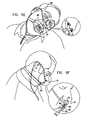
- FIG. 5F it is seen that when the subject is sedated, he tends to move or slump his head, thereby moving oral nasal sampling cannula 10 relative to bite block 50 , as indicated by angle a in the enlarged portion of FIG. 5F .
- the feature of the present invention which provides oral nasal sampling cannula 10 which is physically separated from bite block 50 and the placement of oral breath directing element 26 and oral prong 22 within groove 72 , ensure that even when the subject moves or slumps his head, the oral prong 22 and nasal prongs 18 will be maintained in their respective places, and accurate sampling will continue. Additionally, the placement of oral prong 22 within groove 72 provides a counter force to force applied by the subject's tongue to push at least the top portion of the bite block 50 out of the subject's mouth, thus ensuring accurate placement of the bite block.
- an endoscope probe 98 is inserted into the bore of tubular portion 56 of bite block 50 , for performing an endoscopy procedure.
- flaps 78 of flexible barrier 76 bend slightly inward to allow the passage of the endoscope probe 98 , as seen with particular clarity in the enlarged portion of FIG. 5G .
- the central opening 54 of bite block 50 remains substantially closed by flaps 78 , thereby separating the exhaled breath of the subject which is in the bore of tubular portion 56 from the ambient air.
- the sampling may continue during the presence of the endoscope probe 98 in the pharynx of the subject, as the tubular portion 56 is of a slightly larger diameter than the central opening 54 , thereby ensuring that medical personnel have the space required for the endoscopy procedure and sampling can take place from the space defined by the difference between heights H 2 and H 1 ( FIG. 3 ), as indicated by arrows in the enlarged portion of FIG. 5G .
- the bite block 50 may be removed from the subject's mouth, preferably by medical personnel. However, the sampling of exhaled breath through nasal prongs 18 which remain in the subject's nostrils and through oral prong 22 which remains near the subject's mouth, preferably continues until the subject has awaken from the sedation. This is necessary because the subject's breath must be monitored as long as the subject is sedated.
- FIGS. 6A and 6B are simplified pictorial illustrations of an oral nasal sampling cannula forming part of an endoscopic bite block assembly, constructed and operative in accordance with another preferred embodiment of the present invention, in retracted and extended orientations respectively.
- FIGS. 6A and 6B show an oral nasal sampling cannula 110 , which is adapted for collection of gases, such as carbon dioxide, exhaled by a subject, and for supplying oxygen to the subject.
- gases such as carbon dioxide
- the oral nasal sampling cannula 110 comprises a main body portion 112 , having formed therein an exhaled breath collection bore 114 and an oxygen delivery bore 116 .
- a pair of hollow nasal prongs 118 which are adapted for insertion into the nostrils of the subject, is integrally formed with the main body portion 112 .
- a hollow oral prong 122 which is formed with a limiting rib 123 and a cut-away tip 124 , is mounted onto a bottom surface of main body portion 112 .
- An oral breath directing element 126 which is preferably in the shape of a cut-away tube, is slidably mounted onto oral prong 122 by a mounting portion 128 , and positioning of the oral breath directing element 126 is limited by the limiting rib 123 of oral prong 122 .
- a channel formed in oral prong 122 is in fluid flow connection with channels formed in nasal prongs 118 , thereby forming a single junction 132 .
- Single junction 132 is in fluid flow communication with exhaled breath collection bore 114 , which in turn is in fluid flow communication with an exhaled breath collection tube 134 , which is adapted to be connected to a breath test analyzer or a capnograph (not shown), such as Microcap® which is commercially available from Oridion Medical LTD. of Jerusalem, Israel.
- Main body portion 112 is formed with oxygen delivery openings 136 , which are in fluid flow communication with oxygen delivery bore 116 , which in turn is in fluid flow communication with an oxygen delivery tube 138 .
- at least one nasal oxygen delivery prong which is adapted to be inserted into the nostril of the subject, may be used instead of oxygen delivery openings 136 .
- Oxygen delivery tube 138 is preferably formed with a T-element 140 , connecting the oxygen delivery tube 138 to an oral oxygen delivery tube 142 .
- Oxygen delivery tube 138 is adapted to be connected to a source of oxygen (not shown).
- Oral oxygen delivery tube 142 is preferably normally closed by a valve element 144 .
- the valve is a luer type valve.
- Oxygen delivery tube 138 and exhaled breath collection tube 134 may optionally be placed around the ears of the subject, thereby stabilizing the oral nasal sampling cannula 110 on the subject's face, such that any movement of the subject will have negligible effect on the placement of the oral nasal sampling cannula 110 .
- oral breath directing element 126 may be in a retracted orientation as shown in FIG. 6A , or in an extended orientation as shown in FIG. 6B , thereby allowing the oral nasal sampling cannula 110 to be suited to the facial dimensions of the subject, resulting in more efficient collection of exhaled breath.
- FIGS. 7A and 7B are front-view and rear-view simplified pictorial illustrations of an endoscopic bite block forming part of an endoscopic bite block assembly constructed and operative in accordance with a preferred embodiment of the present invention and to FIG. 8 , which is a simplified sectional pictorial illustration thereof.
- FIGS. 7A , 7 B and 8 show an endoscopic bite block 150 , which is adapted to be inserted into the mouth of a subject while the subject is sedated, to ensure that the mouth of the subject is maintained open during the endoscopy process and that the subject does not interfere with the process by biting on the medical instruments used.
- the endoscopic bite block 150 includes a main body portion 152 , having formed therein a central opening 154 .
- a hollow tubular portion 156 extends distally from main body portion 152 , such that the opening of tubular portion 156 is an extension of central opening 154 .
- Central opening 154 is of a first height, indicated by H 1 in FIG. 8 , which is typically 16 to 20 mm in bite blocks for adult use, which is the height required by medical personnel for performing an endoscopy.
- H 1 in FIG. 8 which is typically 16 to 20 mm in bite blocks for adult use, which is the height required by medical personnel for performing an endoscopy.
- the height of tubular portion 156 is greater than the height H 1 of central opening 154 as indicated by H 2 in FIG. 8 , and is typically 2 to 4 mm more than height H 1 (18 to 24 mm).
- An outer surface 158 of tubular portion 156 is formed with top and bottom teeth engagement surfaces 160 and 162 , such that top teeth engagement surface 160 is relatively forward of bottom teeth engagement surface 162 .
- This structure facilitates easy and accurate biting of the bite block 150 by a subject, as it is suited to the jaw morphology of a closed human mouth.
- Surface 158 is additionally formed with jaw engagement recesses 164 , which are formed forwardly of teeth engagement surfaces 160 and 162 , respectively.
- a top inner surface 170 of main body portion 152 is formed with a longitudinal groove 172 having a transverse surface 173 , which is adapted to accommodate oral prong 122 and oral breath directing element 126 of the oral nasal sampling cannula 110 ( FIGS. 6A and 6B ), as described with more detail hereinbelow with reference to FIG. 9 .
- a tubular portion 174 is formed on a side of outer surface 158 of tubular portion 156 .
- Tubular portion 174 is adapted to threadably engage oral oxygen delivery tube 142 ( FIGS. 6A and 6B ), thereby opening valve 144 to the passage of gases and thus supplying oxygen directly to the oral cavity of the subject.
- tubular portion 174 includes a luer portion corresponding to luer valve element 144 . It is appreciated that tubular portion 174 is formed on outer surface 158 of tubular portion 156 , in order to ensure that the oral oxygen delivery does not interfere with the procedure performed by the medical personnel and so that the oxygen flow does not directly interfere with the CO2 sampling.
- a flexible barrier 176 preferably comprised of several flaps 178 , is disposed within central opening 154 , thereby substantially closing off the central opening and preventing dilution of exhaled breath by ambient air during sampling.
- An opening 180 is preferably maintained within flexible barrier 176 , thereby ensuring a small part of central opening 154 to remain open in order to enable the subject to inhale external air.
- the flexible barrier 176 ensures that a majority of the subject's orally exhaled breath will be directed toward oral prong 122 ( FIGS. 6A and 6B ) thereby ensuring accurate sampling of the subject's breath.
- Opening 180 is preferably placed at a top part of central opening 154 near the cut-away tip 124 of oral prong 122 ( FIGS. 6A and 6B ), thereby directing exhaled breath toward the oral prong 122 as it is the only substantial exit.
- the flaps 178 are preferably formed of a plastic material selected to be of suitable thickness to maintain their position when undisturbed, yet bend readily when pushed by an endoscope probe, and thus do not limit the actions of the medical personnel performing the endoscopy. However, the flaps 178 preferably close back around the endoscope probe, thus maintaining a substantially closed oral cavity volume, and allowing most of the exchange of gases to occur close to the opening 180 of the flexible barrier 176 , which opening is close to the cut-away tip 124 of oral prong 122 from which capnographic sampling can be performed accurately. Additionally, the flaps 178 are preferably transparent, thus enabling medical personnel to see into the oral cavity during the endoscopy procedure.
- Two attachment surfaces 182 each formed with a slit 184 , extend horizontally outwardly from main body portion 152 .
- Slits 184 are adapted to connect to a band which is place around the subject's head and is used to maintain the endoscopic bite block 150 firmly in position during the endoscopy procedure.
- slits 184 are located above a horizontal centerline of main body portion 152 , such that the connected band will tend to exert a stronger pull to the top of the main body portion 152 , thus assisting in overcoming the subject's tendency to tilt the bite block 150 outward during the endoscopy procedure and in maintaining the bite block 150 upright in the subject's mouth.
- FIG. 9 is a simplified schematic illustration of the connection between the oral nasal sampling cannula of FIGS. 6A and 6B and the endoscopic bite block of FIGS. 7A-8 .
- oral prong 122 of oral nasal sampling cannula 110 is accommodated within groove 172 of bite block 150 , such that a bottom surface of oral breath directing element 126 engages transverse surface 173 of the groove 172 . It is appreciated that transverse surface 173 is located below an inner surface of tubular portion 156 in order to ensure that air exhaled by the subject into tubular portion 156 will be directed toward groove 172 and oral prong 122 .
- valve 144 ( FIGS. 6A and 6B ) of oral oxygen delivery tube 142 is accommodated in tubular portion 174 of endoscopic bite block 150 , thereby opening the valve element and forming a fluid flow engagement between oxygen delivery tube 138 and tubular portion 174 of endoscopic bite block 150 , which is in fluid flow engagement with the oral cavity of the subject.
- FIGS. 10A , 10 B, 10 C, 10 D, 10 E, 10 F and 10 G are pictorial illustrations of various stages of typical use of the endoscopic bite block assembly of FIGS. 6A-9 .
- the nasal prongs 118 of the oral nasal sampling cannula 110 are placed in the subjects nostrils, preferably before the subject is sedated.
- the exhaled breath collection tube 134 and the oxygen delivery tube 138 are placed around the subject's ears, in order to ensure the stability of the oral nasal sampling cannula 110 on the subject's face.
- the oral breath-directing element 126 is in its retracted orientation, indicated by the length H 3 .
- oral oxygen delivery tube 142 is not connected to the bite block 150 ( FIGS. 7A-8 ).
- the oral breath directing element 126 is extended to accommodate the facial dimensions of the subject, revealing part of oral prong 122 .
- the oral breath-directing element is moved down to a point in which a bottom end thereof is at the height of the top of the bottom lip of the subject, its new length being indicated by H 4 .
- This action is preferably preformed by medical personnel, but may alternatively be performed by the subject himself, a family member, or any other person.
- FIG. 10C illustrates the insertion of bite block 150 into the mouth of the subject, such that main body portion 152 engages the outer surface of the subject's lips and the tubular portion 156 ( FIGS. 7A-8 ) is inside the subject's mouth. Additionally, valve 144 of oral oxygen delivery tube 142 is inserted, preferably by medical personnel, into tubular portion 174 of endoscopic bite block 150 , as indicated by an arrow in the enlarged portion of FIG. 10C , thereby opening the valve and allowing passage of fluids from the oral oxygen delivery tube 142 into the oral cavity of the subject.
- a strap is attached to slits 184 of attachment surfaces 182 and is placed around the subject's head, thereby securing the bite block 150 in place. This stage is preferably performed when the subject is sedated, but may alternatively be performed prior thereto.
- FIG. 10D it is seen that air exhaled orally by the subject, indicated by arrows, passes through the bore of tubular portion 156 , and is directed toward oral breath directing element 126 and oral prong 122 by the flaps 178 of flexible barrier 176 . Air that is exhaled nasally by the subject passes through nasal prongs 118 .
- FIG. 10D illustrates the oral breath directing element 126 and the oral prong 122 being accommodated in groove 172 , such that a bottom surface of the oral breath directing element 126 engages transverse surface 173 of groove 172 . Additionally, if oral breath directing element 126 has been extended more than necessary for the facial features of the subject, the transverse surface 173 pushes the oral breath-directing element 126 back until it is optimally positioned.
- the lips of the subject indicated by reference numeral 192 preferably engage jaw engagement recesses 164
- the top and bottom teeth of the subject indicated by reference numerals 194 and 196 engage top and bottom teeth engagement surfaces 160 and 162 , respectively.
- FIG. 10E illustrates the sedated subject, having the nasal prongs 118 of the oral nasal sampling cannula 110 in his nostrils and the endoscopic bite block 150 placed in his mouth and strapped to his head.
- oxygen is supplied to the nose of the subject via oxygen delivery openings 136 of oral nasal sampling cannula 110 , and to the mouth of the subject via oral oxygen delivery tube 142 and tubular portion 174 , as indicated by arrows.
- the oxygen is supplied to the oxygen delivery openings 136 via oxygen delivery bore 116 ( FIGS. 6A and 6B ) and to oral oxygen delivery tube 142 via oxygen delivery tube 138 and T-element 140 .
- FIG. 10F it is seen that when the subject is sedated, he tends to move or slump his head, thereby moving oral nasal sampling cannula 110 relative to bite block 150 , as indicated by angle a in the enlarged portion of FIG. 10F .
- the feature of the present invention which provides oral nasal sampling cannula 110 which is physically separated from bite block 150 and the placement of oral breath directing element 126 and oral prong 122 within groove 172 , ensure that even when the subject moves or slumps his head, the oral prong 122 and nasal prongs 118 will be maintained in their respective places, and accurate sampling will continue. Additionally, the placement of oral prong 122 within groove 172 provides a counter force to force applied by the subject's tongue to push at least the top portion of the bite block 150 out of the subject's mouth, thus ensuring accurate placement of the bite block.
- an endoscope probe 198 is inserted into the bore of tubular portion 156 of bite block 150 , for performing the endoscopy procedure.
- flaps 178 of flexible barrier 176 bend slightly inward to allow the passage of the endoscope probe 198 , as seen with particular clarity in the enlarged portion of FIG. 10G .
- the central opening 154 of bite block 150 remains substantially closed by flaps 178 , thereby separating the exhaled breath of the subject which is in bore of tubular portion 156 from the ambient air.
- the sampling may continue during the presence of the endoscope probe 198 in the pharynx of the subject, as the tubular portion 156 is of a slightly larger diameter than the central opening 154 , thereby ensuring that medical personnel have the space defined by the difference between heights H 2 and H 1 ( FIG. 8 ), as indicated by arrows in the enlarged portion of FIG. 10G .
- the bite block 150 may be removed from the subject's mouth, preferably by medical personnel.
- the valve 144 of oral oxygen delivery tube 142 is removed from tubular portion 174 thereby closing the valve and thus fully decoupling the oral nasal sampling cannula 110 from the endoscopic bite block 150 .
- the sampling of exhaled breath through nasal prongs 118 which remain in the subject's nostrils and through oral prong 122 which remains near the subject's mouth preferably continues until the subject has awaken from the sedation. This is necessary because the subject's breath must be monitored as long as the subject is sedated.
- FIGS. 11A and 11B are simplified pictorial illustrations of an oral nasal sampling cannula forming part of an endoscopic bite block assembly, constructed and operative in accordance with yet another preferred embodiment of the present invention, in retracted and extended orientations respectively.
- FIGS. 11A and 11B show an oral nasal sampling cannula 210 , which is adapted for collection of gases, such as carbon dioxide, exhaled by a subject, and for supplying oxygen to the subject.
- gases such as carbon dioxide
- the oral nasal sampling cannula 210 comprises a main body portion 212 , having formed therein an exhaled breath collection bore 214 and an oxygen delivery bore 216 .
- a pair of hollow nasal prongs 218 which are adapted for insertion into the nostrils of the subject, is integrally formed with the main body portion 212 .
- a hollow oral prong 222 which is formed with a limiting rib 223 and a cut-away tip 224 , is mounted onto a bottom surface of main body portion 212 .
- An oral breath directing element 226 which is preferably in the shape of a cut-away tube, is slidably mounted onto oral prong 222 by a mounting portion 228 , and positioning of the oral breath directing element 226 is limited by the limiting rib 223 of oral prong 222 .
- a channel formed in oral prong 222 is in fluid flow connection with channels formed in nasal prongs 218 , thereby forming a single junction 232 .
- Single junction 232 is in fluid flow communication with exhaled breath collection bore 214 , which in turn is in fluid flow communication with an exhaled breath collection tube 234 , which is adapted to be connected to a breath test analyzer or a capnograph (not shown), such as Microcap® which is commercially available from Oridion Medical LTD. of Jerusalem, Israel.
- Main body portion 212 is formed with oxygen delivery openings 236 , which are in fluid flow communication with oxygen delivery bore 216 , which in turn is in fluid flow communication with an oxygen delivery tube 238 .
- oxygen delivery tube 238 is preferably formed with a T-element 240 , preferably terminating at an end thereof in a normally closed valve element 244 , which is preferably a luer valve.
- Oxygen delivery tube 238 is adapted to be connected to a source of oxygen (not shown).
- Oxygen delivery tube 238 and exhaled breath collection tube 234 may optionally be placed around the ears of the subject, thereby stabilizing the oral nasal sampling cannula 210 on the subject's face, such that any movement of the subject will have negligible effect on the placement of the oral nasal sampling cannula 210 .
- oral breath directing element 226 may be in a retracted orientation as shown in FIG. 11A , or in an extended orientation as shown in FIG. 11B , thereby allowing the oral nasal sampling cannula 210 to be suited to the facial dimensions of the subject, resulting in more efficient collection of exhaled breath.
- FIGS. 12A and 12B are front-view and rear-view simplified pictorial illustrations of an endoscopic bite block forming part of an endoscopic bite block assembly constructed and operative in accordance with yet another preferred embodiment of the present invention and to FIG. 13 , which is a simplified sectional pictorial illustration thereof.
- FIGS. 12A , 12 B and 13 show an endoscopic bite block 250 , which is adapted to be inserted into the mouth of a subject while the subject is sedated, to ensure that the mouth of the subject is maintained open during the endoscopy process, and that the subject does not interfere with the process by biting on medical instruments used.
- the endoscopic bite block 250 includes a main body portion 252 , having formed therein a central opening 254 .
- a hollow tubular portion 256 extends distally from main body portion 252 , such that the opening of tubular portion 256 is an extension of central opening 254 .
- Central opening 254 is of a first height, indicated by H 1 in FIG. 13 , which is typically 16 to 20 mm in bite blocks for adult use, which is the height required by medical personnel for performing an endoscopy.
- H 1 in FIG. 13 which is typically 16 to 20 mm in bite blocks for adult use, which is the height required by medical personnel for performing an endoscopy.
- the height of tubular portion 256 is greater than the height H 1 of central opening 254 as indicated by H 2 in FIG. 13 , and is typically 2 to 4 mm more than height H 1 (18 to 24 mm).
- An outer surface 258 of tubular portion 256 is formed with top and bottom teeth engagement surfaces 260 and 262 , such that top teeth engagement surface 260 is relatively forward of bottom teeth engagement surface 262 .
- This structure facilitates easy and accurate biting of the bite block 250 by a subject, as it is suited to the jaw morphology of a closed human mouth.
- Surface 258 is additionally formed with jaw engagement recesses 264 , which are formed forwardly of teeth engagement surfaces 260 and 262 , respectively.
- a top inner surface 270 of main body portion 252 is formed with a longitudinal groove 272 having a transverse surface 273 , which is adapted to accommodate oral prong 222 and oral breath directing element 226 of the oral nasal sampling cannula 210 ( FIGS. 12A and 12B ), as described with more detail hereinbelow with reference to FIG. 14 .
- a tubular portion 274 is formed on a side of outer surface 258 of tubular portion 256 .
- Extending out of tubular portion 274 is an oral oxygen delivery tube 275 including a tip 276 , which is adapted to engage valve 244 ( FIGS. 11A and 11B ), thereby supplying oxygen directly to the oral cavity of the subject.
- tip 276 comprises a luer corresponding to luer valve 244 .
- tubular portion 274 is formed on outer surface 258 of tubular portion 256 , in order to ensure that the oral oxygen delivery does not interfere with the procedure performed by the medical personnel.
- a flexible barrier 277 preferably comprised of several flaps 278 , is disposed within central opening 254 , thereby substantially closing off the central opening and preventing dilution of exhaled breath by ambient air during sampling.
- An opening 280 is preferably maintained within flexible barrier 277 , thereby ensuring a small part of central opening 254 remains open in order to enable the subject to inhale external air.
- the flexible barrier 277 ensures that a majority of the subject's orally exhaled breath will be directed toward oral prong 222 ( FIGS. 11A and 11B ) thereby ensuring accurate sampling of the subject's breath.
- Opening 280 is preferably placed at a top part of central opening 254 near the cut-away tip 224 of oral prong 222 ( FIGS. 11A and 11B ), thereby directing and amplifying exhaled breath toward the oral prong 222 as it is the only substantial exit.
- the flaps 278 are preferably formed of a plastic material selected to be of suitable thickness to maintain their position when undisturbed, yet bend readily when pushed by an endoscope probe, and thus do not limit the actions of the medical personnel performing the endoscopy. However, the flaps 278 preferably close back around the endoscope probe, thus maintaining a substantially closed oral cavity volume, and allowing most of the exchange of gases to occur close to the opening 280 of flexible barrier 277 , which opening is close to the cut-away tip 224 of oral prong 222 ( FIGS. 11A and 11B ) from which capnographic sampling can be performed accurately.
- the flaps 278 are preferably transparent, thus enabling medical personnel to see into the oral cavity during the endoscopy procedure.
- Two attachment surfaces 282 each formed with a slit 284 , extend horizontally outwardly from main body portion 252 .
- Slits 284 are adapted to connect to a band which is placed around the subject's head and is used to maintain the endoscopic bite block 250 firmly in position during the endoscopy procedure.
- slits 284 are located above a horizontal centerline of main body portion 252 , such that the connected band will tend to exert a stronger pull to the top of the main body portion 252 , thus assisting in overcoming the subject's tendency to tilt the bite block 250 outward during the endoscopy procedure and in maintaining the bite block 250 upright in the subject's mouth.
- FIG. 14 is a simplified schematic illustration of the connection between the oral nasal sampling cannula of FIGS. 11A and 11B and the endoscopic bite block of FIGS. 12A-13 .
- oral prong 222 of oral nasal sampling cannula 210 is accommodated within groove 272 of bite block 250 , such that a bottom surface of oral breath directing element 226 engages transverse surface 273 of the groove 272 . It is appreciated that transverse surface 273 is located below an inner surface of tubular portion 256 in order to ensure that air exhaled by the subject into tubular portion 256 will be directed toward groove 272 and oral prong 222 .
- tip 276 of oral oxygen delivery tube 275 engages valve 244 ( FIGS. 11A and 11B ) of T-element 240 of oral nasal sampling cannula 210 , thereby opening the valve 244 and forming a fluid flow engagement between oxygen delivery tube 238 and tubular portion 274 of endoscopic bite block 250 , which is in fluid flow engagement with the oral cavity of the subject.
- FIGS. 15A , 15 B, 15 C, 15 D, 15 E, 15 F and 15 G are pictorial illustrations of various stages of typical use of the endoscopic bite block assembly of FIGS. 11A-14 .
- the nasal prongs 218 of the oral nasal sampling cannula 210 are placed in the subjects nostrils, preferably before the subject is sedated.
- the exhaled breath collection tube 234 and the oxygen delivery tube 238 are placed around the subject's ears, in order to ensure the stability of the oral nasal sampling cannula 210 on the subject's face.
- the oral breath-directing element 226 is in its retracted orientation, indicated by the length H 3 .
- oral oxygen delivery tube 275 ( FIGS. 12A-13 ) is not connected to the T-element 240 of oral nasal sampling cannula 210 .
- oxygen delivery tube 238 there is no oxygen leakage, as the T-element 240 is sealed by valve 244 .
- the oral breath directing element 226 is extended to accommodate the facial dimensions of the subject, revealing part of oral prong 222 .
- the oral breath-directing element 226 is moved down to a point in which a bottom end thereof is at the height of the top of the bottom lip of the subject, its new length being indicated by H 4 .
- This action is preferably preformed by medical personnel, but may alternatively be performed by the subject himself, a family member, or any other person.
- FIG. 15C illustrates the insertion of bite block 250 into the mouth of the subject, such that main body portion 252 engages the outer surface of the subject's lips and the tubular portion 256 is inside the subject's mouth. Additionally, tip 276 of oral oxygen delivery tube 275 is inserted, preferably by medical personnel, into valve 244 of T-element 240 of oral nasal sampling cannula 210 , as indicated by an arrow in the enlarged portion of FIG. 15C , thereby opening the valve 244 .
- a strap is attached to slits 284 of attachment surfaces 282 and is placed around the subject's head, thereby securing the bite block 250 in place. This stage is preferably performed when the subject is sedated, but may alternatively be performed prior thereto.
- FIG. 15D it is seen that air exhaled orally by the subject, indicated by arrows, passes through the bore of tubular portion 256 , and is directed toward oral breath directing element 226 and oral prong 222 by the flaps 278 of flexible barrier 277 . Air that is exhaled nasally by the subject passes through nasal prongs 218 .
- FIG. 15D illustrates the oral breath directing element 226 and the oral prong 222 being accommodated in groove 272 , such that a bottom surface of the oral breath directing element 226 engages transverse surface 273 of groove 272 . Additionally, if oral breath directing element 226 has been extended more than necessary for the facial features of the subject, the transverse surface 273 pushes the oral breath-directing element 226 back until it is optimally positioned.
- the lips of the subject, indicated by reference numeral 292 preferably engage jaw engagement recesses 264
- the top and bottom teeth of the subject, indicated by reference numerals 294 and 296 engage top and bottom teeth engagement surfaces 260 and 262 , respectively.
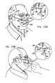
- FIG. 15E illustrates the sedated subject, having the nasal prongs 218 of the oral nasal sampling cannula 210 in his nostrils and the endoscopic bite block 250 placed in his mouth and strapped to his head.
- oxygen is supplied to the nose of the subject via oxygen delivery openings 236 of oral nasal sampling cannula 210 , and to the mouth of the subject via oral oxygen delivery tube 275 and tubular portion 274 , as indicated by arrows.
- the oxygen is supplied to the oxygen delivery openings 236 via oxygen delivery bore 216 ( FIGS. 11A and 11B ) and to oral oxygen delivery tube 275 via oxygen delivery tube 238 and T-element 240 .
- FIG. 15F it is seen that when the subject is sedated, he tends to move or slump his head, thereby moving oral nasal sampling cannula 210 relative to bite block 250 , as indicated by angle a in the enlarged portion of FIG. 15F .
- the feature of the present invention which provides oral nasal sampling cannula 210 which is physically separated from bite block 250 and the placement of oral breath directing element 226 and oral prong 222 within groove 272 , ensure that even when the subject moves or slumps his head, the oral prong 222 and nasal prongs 218 will be maintained in their respective places, and accurate sampling will continue. Additionally, the placement of oral prong 222 within groove 272 provides a counter force to force applied by the subject's tongue to push at least the top portion of the bite block 250 out of the subject's mouth, thus ensuring accurate placement of the bite block.
- an endoscope probe 298 is inserted into the bore of tubular portion 256 of bite block 250 , for performing the endoscopy procedure.
- flaps 278 of flexible barrier 277 bend slightly inward to allow the passage of the endoscope probe 298 , as seen with particular clarity in the enlarged portion of FIG. 15G .
- the central opening 254 of bite block 250 remains substantially closed by flaps 278 , thereby separating the exhaled breath of the subject which is in the bore of tubular portion 256 from the ambient air.
- the sampling may continue during the presence of the endoscope probe 298 in the pharynx of the subject, as the tubular portion 256 is of a slightly larger diameter than the central opening 254 , thereby ensuring that medical personnel have the space defined by the difference between heights H 2 and H 1 ( FIG. 13 ), as indicated by arrows in the enlarged portion of FIG. 15G .
- the bite block 250 may be removed from the subject's mouth, preferably by medical personnel.
- the tip 276 of oral oxygen delivery tube 275 is removed from valve 244 ( FIGS. 11A and 11B ) of T-element 240 , thereby closing the valve and fully decoupling the oral nasal sampling cannula 210 from the endoscopic bite block 250 .
- the sampling of exhaled breath through nasal prongs 218 which remain in the subject's nostrils and through oral prong 222 which remains near the subject's mouth preferably continues until the subject has awaken from the sedation. This is necessary because the subject's breath must be monitored as long as the subject is sedated.
Landscapes
- Health & Medical Sciences (AREA)
- Life Sciences & Earth Sciences (AREA)
- Pulmonology (AREA)
- Heart & Thoracic Surgery (AREA)
- Veterinary Medicine (AREA)
- Public Health (AREA)
- General Health & Medical Sciences (AREA)
- Animal Behavior & Ethology (AREA)
- Engineering & Computer Science (AREA)
- Biomedical Technology (AREA)
- Emergency Medicine (AREA)
- Anesthesiology (AREA)
- Hematology (AREA)
- Surgery (AREA)
- Otolaryngology (AREA)
- Biophysics (AREA)
- Medical Informatics (AREA)
- Molecular Biology (AREA)
- Physics & Mathematics (AREA)
- Pathology (AREA)
- Nuclear Medicine, Radiotherapy & Molecular Imaging (AREA)
- Radiology & Medical Imaging (AREA)
- Optics & Photonics (AREA)
- Physiology (AREA)
- Gastroenterology & Hepatology (AREA)
- Dentistry (AREA)
- Oral & Maxillofacial Surgery (AREA)
- Endoscopes (AREA)
- Infusion, Injection, And Reservoir Apparatuses (AREA)
- Surgical Instruments (AREA)
Priority Applications (5)
| Application Number | Priority Date | Filing Date | Title |
|---|---|---|---|
| US13/968,737 US8770189B2 (en) | 2005-12-01 | 2013-08-16 | Endoscopic bite block |
| US14/292,811 US9060680B2 (en) | 2005-12-01 | 2014-05-30 | Endoscopic bite block |
| US14/719,308 US9867957B2 (en) | 2005-12-01 | 2015-05-21 | Endoscopic bite block |
| US15/212,956 US9687623B2 (en) | 2005-12-01 | 2016-07-18 | Endoscopic bite block |
| US15/610,279 US10300234B2 (en) | 2005-12-01 | 2017-05-31 | Endoscopic bite block |
Applications Claiming Priority (1)
| Application Number | Priority Date | Filing Date | Title |
|---|---|---|---|
| PCT/IL2005/001291 WO2007063532A2 (fr) | 2005-12-01 | 2005-12-01 | Cire d'occlusion endoscopique |
Related Parent Applications (1)
| Application Number | Title | Priority Date | Filing Date |
|---|---|---|---|
| PCT/IL2005/001291 A-371-Of-International WO2007063532A2 (fr) | 2005-12-01 | 2005-12-01 | Cire d'occlusion endoscopique |
Related Child Applications (1)
| Application Number | Title | Priority Date | Filing Date |
|---|---|---|---|
| US13/968,737 Continuation US8770189B2 (en) | 2005-12-01 | 2013-08-16 | Endoscopic bite block |
Publications (2)
| Publication Number | Publication Date |
|---|---|
| US20090275851A1 US20090275851A1 (en) | 2009-11-05 |
| US8534278B2 true US8534278B2 (en) | 2013-09-17 |
Family
ID=38092652
Family Applications (6)
| Application Number | Title | Priority Date | Filing Date |
|---|---|---|---|
| US12/085,594 Active 2028-06-09 US8534278B2 (en) | 2005-12-01 | 2005-12-01 | Endoscopic bite block |
| US13/968,737 Active US8770189B2 (en) | 2005-12-01 | 2013-08-16 | Endoscopic bite block |
| US14/292,811 Active US9060680B2 (en) | 2005-12-01 | 2014-05-30 | Endoscopic bite block |
| US14/719,308 Active US9867957B2 (en) | 2005-12-01 | 2015-05-21 | Endoscopic bite block |
| US15/212,956 Active US9687623B2 (en) | 2005-12-01 | 2016-07-18 | Endoscopic bite block |
| US15/610,279 Active 2026-05-25 US10300234B2 (en) | 2005-12-01 | 2017-05-31 | Endoscopic bite block |
Family Applications After (5)
| Application Number | Title | Priority Date | Filing Date |
|---|---|---|---|
| US13/968,737 Active US8770189B2 (en) | 2005-12-01 | 2013-08-16 | Endoscopic bite block |
| US14/292,811 Active US9060680B2 (en) | 2005-12-01 | 2014-05-30 | Endoscopic bite block |
| US14/719,308 Active US9867957B2 (en) | 2005-12-01 | 2015-05-21 | Endoscopic bite block |
| US15/212,956 Active US9687623B2 (en) | 2005-12-01 | 2016-07-18 | Endoscopic bite block |
| US15/610,279 Active 2026-05-25 US10300234B2 (en) | 2005-12-01 | 2017-05-31 | Endoscopic bite block |
Country Status (4)
| Country | Link |
|---|---|
| US (6) | US8534278B2 (fr) |
| EP (1) | EP1959857B1 (fr) |
| AT (1) | ATE544485T1 (fr) |
| WO (1) | WO2007063532A2 (fr) |
Cited By (23)
| Publication number | Priority date | Publication date | Assignee | Title |
|---|---|---|---|---|
| US8770199B2 (en) | 2012-12-04 | 2014-07-08 | Ino Therapeutics Llc | Cannula for minimizing dilution of dosing during nitric oxide delivery |
| USD744637S1 (en) * | 2013-10-08 | 2015-12-01 | Koninklijke Philips N.V. | Cannula nosepiece |
| USD747471S1 (en) | 2012-08-10 | 2016-01-12 | Fisher & Paykel Healthcare Limited | Connector |
| US20170196512A1 (en) * | 2013-10-09 | 2017-07-13 | Nihon Kohden Corporation | Mask |
| US9795756B2 (en) | 2012-12-04 | 2017-10-24 | Mallinckrodt Hospital Products IP Limited | Cannula for minimizing dilution of dosing during nitric oxide delivery |
| US10010313B2 (en) | 2015-05-18 | 2018-07-03 | Richard L. Arden | Mandibular subluxation device and method |
| US10252018B2 (en) * | 2016-09-06 | 2019-04-09 | H. Lee Moffitt Cancer Center And Research Institute, Inc. | Anesthesia gas delivery and monitoring system |
| US10258319B2 (en) | 2015-05-18 | 2019-04-16 | Richard L. Arden | Airway assist device and method |
| USD848614S1 (en) | 2017-11-21 | 2019-05-14 | Fisher & Paykel Healthcare Limited | Pad for nasal cannula assembly |
| USD849243S1 (en) | 2017-11-21 | 2019-05-21 | Fisher & Paykel Healthcare Limited | Nasal cannula |
| USD849242S1 (en) | 2017-11-21 | 2019-05-21 | Fisher & Paykel Healthcare Limited | Nasal cannula assembly |
| US10342526B2 (en) | 2015-07-01 | 2019-07-09 | Richard L. Arden | Airway assist device and method |
| USD878549S1 (en) | 2017-11-21 | 2020-03-17 | Fisher & Paykel Healthcare Limited | Connector for nasal cannula assembly |
| US20200268999A1 (en) * | 2019-02-27 | 2020-08-27 | Mackay Memorial Hospital | Respiratory mask |
| US10835733B1 (en) | 2011-08-10 | 2020-11-17 | Fisher & Paykel Healthcare Limited | Conduit connector for a patient breathing device |
| US11065410B1 (en) | 2021-02-01 | 2021-07-20 | Leonard Feld | Dental appliance using airway dialation for treating covid related breathing disorders |
| USD948027S1 (en) | 2019-09-10 | 2022-04-05 | Fisher & Paykel Healthcare Limited | Connector for a breathing conduit |
| US11446462B2 (en) | 2015-03-31 | 2022-09-20 | Fisher & Paykel Healthcare Limited | Apparatus for use in a respiratory support system |
| USD974551S1 (en) | 2020-12-09 | 2023-01-03 | Fisher & Paykel Healthcare Limited | Connector assembly and connector |
| USD995758S1 (en) | 2021-06-11 | 2023-08-15 | Fisher & Paykel Healthcare Limited | Tube assembly and connector |
| USD1006981S1 (en) | 2019-09-06 | 2023-12-05 | Fisher & Paykel Healthcare Limited | Breathing conduit |
| USD1026221S1 (en) | 2020-03-03 | 2024-05-07 | Fisher & Paykel Healthcare Limited | Connector for a respiratory system conduit |
| USD1027165S1 (en) | 2016-06-10 | 2024-05-14 | Fisher & Paykel Healthcare Limited | Connector for a breathing circuit |
Families Citing this family (29)
| Publication number | Priority date | Publication date | Assignee | Title |
|---|---|---|---|---|
| US20080275357A1 (en) * | 2004-11-22 | 2008-11-06 | Ron Porat | Oral/nasal cannula |
| US7946288B2 (en) * | 2006-11-10 | 2011-05-24 | Encompas Unlimited, Inc. | Bite block system and method |
| JP4968647B2 (ja) * | 2007-03-09 | 2012-07-04 | 日本光電工業株式会社 | 呼気情報収集用アダプタおよび生体情報処理システム |
| EP2227282A1 (fr) | 2007-11-25 | 2010-09-15 | Oridion Medical (1987) Ltd. | Pièce endoscopique à mordre endoscopique amélioré |
| US9044565B2 (en) | 2008-10-30 | 2015-06-02 | Oridion Medical (1987) Ltd. | Oral-nasal cannula system enabling CO2 and breath flow measurement |
| US9399106B2 (en) * | 2010-01-13 | 2016-07-26 | Thomas Julius Borody | Mask for use with a patient undergoing a sedated endoscopic procedure |
| US10238827B2 (en) * | 2012-03-12 | 2019-03-26 | Mark T. Holtzapple | Air-delivery system for breathing-assist devices |
| FR2992223B1 (fr) * | 2012-06-21 | 2015-05-01 | Deltamedics | Canule oropharyngee comprenant une arrivee de dioxygene et une extraction de dioxyde de carbone |
| WO2014078034A1 (fr) * | 2012-11-14 | 2014-05-22 | The Trustees Of The University Of Pennsylvania | Pièce de morsure à ventilation destiné à être utilisé dans des interventions d'endoscopie |
| US20140246030A1 (en) * | 2013-03-01 | 2014-09-04 | Amitab Puri | Endoscopic bite block shield |
| GB201317499D0 (en) * | 2013-10-03 | 2013-11-20 | Flexicare Medical Ltd | Naso-Oral Device |
| US20150107600A1 (en) * | 2013-10-22 | 2015-04-23 | Gi Supply, Inc. | Medical Bite Blocks or Mouthpieces |
| CA2974374C (fr) | 2015-01-23 | 2024-01-09 | Masimo Sweden Ab | Systeme de canule nasale/buccale et son procede de fabrication |
| CA2980539A1 (fr) * | 2015-03-31 | 2016-10-06 | Fisher & Paykel Healthcare Limited | Interface utilisateur pour la fourniture de plusieurs gaz a une voie aerienne |
| US9968341B2 (en) | 2015-04-21 | 2018-05-15 | Ascentcare Dental Labs, Llc | Dental bite block assembly |
| US11331446B2 (en) | 2015-06-11 | 2022-05-17 | Revolutionary Medical Devices, Inc. | Ventilation mask |
| US10265487B2 (en) | 2015-12-01 | 2019-04-23 | B&T Healthcare Solutions Llc | Oxygenation mask with integrated end-tidal carbon dioxide monitoring |
| USD787070S1 (en) | 2016-04-20 | 2017-05-16 | Ascentcare Dental Labs | Illuminated dental accessory with tongue suppressor |
| USD817492S1 (en) | 2016-04-20 | 2018-05-08 | Ascentcare Dental Labs, Llc | Dental accessory with tongue suppressor |
| USD787069S1 (en) | 2016-04-20 | 2017-05-16 | Ascentcare Dental Labs | Illuminated dental accessory for holding saliva ejection tube |
| USD782048S1 (en) | 2016-04-20 | 2017-03-21 | Ascentcare Dental Labs, Llc | Dental bite block |
| USD782047S1 (en) | 2016-04-20 | 2017-03-21 | Ascentcare Dental Labs, Llc | Dental accessory for holding a saliva ejection tube |
| JP7064920B2 (ja) * | 2018-03-27 | 2022-05-11 | 日本光電工業株式会社 | 呼吸補助具 |
| USD885556S1 (en) * | 2018-04-13 | 2020-05-26 | Fisher & Paykel Healthcare Limited | Tip, tube and clip assembly for a gas sampling apparatus |
| CN109349994A (zh) * | 2018-11-28 | 2019-02-19 | 大连医雅科技有限公司 | 一种医疗用胃镜及控制方法 |
| JP7545876B2 (ja) * | 2020-12-02 | 2024-09-05 | 日本光電工業株式会社 | 医療器具、ネーザルアダプタ、およびバイトブロック |
| CN112691273A (zh) * | 2021-03-25 | 2021-04-23 | 上海埃立孚医疗科技有限公司 | 一种内窥镜专用口咽通道 |
| SE545552C2 (en) * | 2021-09-10 | 2023-10-17 | Stairway Medical Ab | A procedural sedation mouthpiece |
| WO2024112715A1 (fr) * | 2022-11-21 | 2024-05-30 | Sedateuk Ltd | Dispositif de surveillance respiratoire circonférentiel buccal |
Citations (25)
| Publication number | Priority date | Publication date | Assignee | Title |
|---|---|---|---|---|
| US4989599A (en) * | 1989-01-26 | 1991-02-05 | Puritan-Bennett Corporation | Dual lumen cannula |
| US5046491A (en) * | 1990-03-27 | 1991-09-10 | Derrick Steven J | Apparatus and method for respired gas collection and analysis |
| US5174284A (en) | 1991-09-05 | 1992-12-29 | G.I. Supply, Inc. | Endoscopic bite block |
| US5273032A (en) | 1989-12-01 | 1993-12-28 | Gastro Services Pty Ltd. | Oxygenating oral medical appliance |
| US5335656A (en) * | 1988-04-15 | 1994-08-09 | Salter Laboratories | Method and apparatus for inhalation of treating gas and sampling of exhaled gas for quantitative analysis |
| US5413095A (en) | 1994-04-15 | 1995-05-09 | Arrow Precision Products, Inc. | Mouthpiece with oxygen receiving and directing structure |
| US5480410A (en) * | 1994-03-14 | 1996-01-02 | Advanced Surgical, Inc. | Extracorporeal pneumoperitoneum access bubble |
| US5513634A (en) | 1994-05-06 | 1996-05-07 | Chek-Med Systems, Inc. | Combination integral bite block airway and nasal cannula |
| US5699787A (en) | 1996-06-20 | 1997-12-23 | Thompson; Clarence | Mouthpiece for endotracheal tube |
| US5752510A (en) * | 1996-11-14 | 1998-05-19 | Goldstein; Joseph | Nasal and oral air passageway delivery management apparatus |
| US6257238B1 (en) | 2000-05-25 | 2001-07-10 | Nizam M. Meah | Bite block for upper gastrointestinal endoscopy with tongue depressor |
| US6379312B2 (en) * | 1999-12-28 | 2002-04-30 | O'toole James | End tidal carbon dioxide sampling device |
| US6422240B1 (en) | 1998-01-29 | 2002-07-23 | Oridion Medical Ltd. | Oral/nasal cannula |
| US6561192B2 (en) | 2000-03-03 | 2003-05-13 | The Penn State Research Foundation | Nasal oral respiratory interface |
| WO2004030723A2 (fr) | 2002-10-03 | 2004-04-15 | Scott Laboratories, Inc. | Canule buccale et methode destinees a etre utilisees avec un systeme de sedation et d'analgesie |
| US20040129272A1 (en) * | 2002-09-24 | 2004-07-08 | Thomas Jefferson University | Oropharyngeal airway |
| US6792943B2 (en) * | 2001-09-05 | 2004-09-21 | Minnesota High-Tech Resources, Llc | Intubating ventilatory face mask |
| US6805126B2 (en) * | 1998-12-01 | 2004-10-19 | Edward P. Dutkiewicz | Oxygen delivery and gas sensing nasal cannula system |
| WO2004103199A2 (fr) | 2003-05-20 | 2004-12-02 | Oridion Medical (1987) Ltd. | Bloc de morsure endoscopique |
| WO2005016142A1 (fr) | 2003-08-13 | 2005-02-24 | Borody Thomas J | Appareil buccal d'oxygenation ameliore |
| US20070006878A1 (en) | 2005-07-06 | 2007-01-11 | Mackey Mark F | Capnographic-oxygenating oro-fiberscopic biteblock |
| US20070113844A1 (en) * | 2005-11-22 | 2007-05-24 | Garren Mary L | Endoscopic bite block |
| US7727194B2 (en) * | 2004-08-31 | 2010-06-01 | Ethicon Endo-Surgery, Inc. | Drug delivery cassette |
| US7832400B2 (en) * | 2001-01-04 | 2010-11-16 | Salter Labs | Nasal and oral cannula having two capabilities and method of producing same |
| US7946288B2 (en) * | 2006-11-10 | 2011-05-24 | Encompas Unlimited, Inc. | Bite block system and method |
Family Cites Families (1)
| Publication number | Priority date | Publication date | Assignee | Title |
|---|---|---|---|---|
| EP2227282A1 (fr) * | 2007-11-25 | 2010-09-15 | Oridion Medical (1987) Ltd. | Pièce endoscopique à mordre endoscopique amélioré |
-
2005
- 2005-12-01 EP EP05812952A patent/EP1959857B1/fr active Active
- 2005-12-01 AT AT05812952T patent/ATE544485T1/de active
- 2005-12-01 US US12/085,594 patent/US8534278B2/en active Active
- 2005-12-01 WO PCT/IL2005/001291 patent/WO2007063532A2/fr active Application Filing
-
2013
- 2013-08-16 US US13/968,737 patent/US8770189B2/en active Active
-
2014
- 2014-05-30 US US14/292,811 patent/US9060680B2/en active Active
-
2015
- 2015-05-21 US US14/719,308 patent/US9867957B2/en active Active
-
2016
- 2016-07-18 US US15/212,956 patent/US9687623B2/en active Active
-
2017
- 2017-05-31 US US15/610,279 patent/US10300234B2/en active Active
Patent Citations (28)
| Publication number | Priority date | Publication date | Assignee | Title |
|---|---|---|---|---|
| US5335656A (en) * | 1988-04-15 | 1994-08-09 | Salter Laboratories | Method and apparatus for inhalation of treating gas and sampling of exhaled gas for quantitative analysis |
| US4989599A (en) * | 1989-01-26 | 1991-02-05 | Puritan-Bennett Corporation | Dual lumen cannula |
| US5273032A (en) | 1989-12-01 | 1993-12-28 | Gastro Services Pty Ltd. | Oxygenating oral medical appliance |
| US5046491A (en) * | 1990-03-27 | 1991-09-10 | Derrick Steven J | Apparatus and method for respired gas collection and analysis |
| US5174284A (en) | 1991-09-05 | 1992-12-29 | G.I. Supply, Inc. | Endoscopic bite block |
| US5480410A (en) * | 1994-03-14 | 1996-01-02 | Advanced Surgical, Inc. | Extracorporeal pneumoperitoneum access bubble |
| US5413095A (en) | 1994-04-15 | 1995-05-09 | Arrow Precision Products, Inc. | Mouthpiece with oxygen receiving and directing structure |
| US5513634A (en) | 1994-05-06 | 1996-05-07 | Chek-Med Systems, Inc. | Combination integral bite block airway and nasal cannula |
| US5699787A (en) | 1996-06-20 | 1997-12-23 | Thompson; Clarence | Mouthpiece for endotracheal tube |
| US5752510A (en) * | 1996-11-14 | 1998-05-19 | Goldstein; Joseph | Nasal and oral air passageway delivery management apparatus |
| US6422240B1 (en) | 1998-01-29 | 2002-07-23 | Oridion Medical Ltd. | Oral/nasal cannula |
| US6805126B2 (en) * | 1998-12-01 | 2004-10-19 | Edward P. Dutkiewicz | Oxygen delivery and gas sensing nasal cannula system |
| US6379312B2 (en) * | 1999-12-28 | 2002-04-30 | O'toole James | End tidal carbon dioxide sampling device |
| US6561192B2 (en) | 2000-03-03 | 2003-05-13 | The Penn State Research Foundation | Nasal oral respiratory interface |
| US6257238B1 (en) | 2000-05-25 | 2001-07-10 | Nizam M. Meah | Bite block for upper gastrointestinal endoscopy with tongue depressor |
| US7832400B2 (en) * | 2001-01-04 | 2010-11-16 | Salter Labs | Nasal and oral cannula having two capabilities and method of producing same |
| US6792943B2 (en) * | 2001-09-05 | 2004-09-21 | Minnesota High-Tech Resources, Llc | Intubating ventilatory face mask |
| US20040129272A1 (en) * | 2002-09-24 | 2004-07-08 | Thomas Jefferson University | Oropharyngeal airway |
| WO2004030723A2 (fr) | 2002-10-03 | 2004-04-15 | Scott Laboratories, Inc. | Canule buccale et methode destinees a etre utilisees avec un systeme de sedation et d'analgesie |
| US20040129273A1 (en) * | 2002-10-03 | 2004-07-08 | Scott Laboratories, Inc. | Bite block apparatus and method for use with a sedation and analgesia system |
| WO2004103199A2 (fr) | 2003-05-20 | 2004-12-02 | Oridion Medical (1987) Ltd. | Bloc de morsure endoscopique |
| US20070068535A1 (en) | 2003-05-20 | 2007-03-29 | Colman Joshua L | Endoscopic bite block |
| WO2005016142A1 (fr) | 2003-08-13 | 2005-02-24 | Borody Thomas J | Appareil buccal d'oxygenation ameliore |
| US7624736B2 (en) * | 2003-08-13 | 2009-12-01 | Borody Thomas J | Oral oxygenating appliance |
| US7727194B2 (en) * | 2004-08-31 | 2010-06-01 | Ethicon Endo-Surgery, Inc. | Drug delivery cassette |
| US20070006878A1 (en) | 2005-07-06 | 2007-01-11 | Mackey Mark F | Capnographic-oxygenating oro-fiberscopic biteblock |
| US20070113844A1 (en) * | 2005-11-22 | 2007-05-24 | Garren Mary L | Endoscopic bite block |
| US7946288B2 (en) * | 2006-11-10 | 2011-05-24 | Encompas Unlimited, Inc. | Bite block system and method |
Non-Patent Citations (4)
| Title |
|---|
| Corresponding EP 05812952 European Search Report dated Jul. 1, 2010. |
| Corresponding EP 05812952 Supplementary European Search Report dated Jun. 23, 2010. |
| International Search Report for PCT/IL04/00430 dated Mar. 4, 2005. |
| International Search Report for PCT/IL05/01291 dated May 24, 2007. |
Cited By (47)
| Publication number | Priority date | Publication date | Assignee | Title |
|---|---|---|---|---|
| US10835733B1 (en) | 2011-08-10 | 2020-11-17 | Fisher & Paykel Healthcare Limited | Conduit connector for a patient breathing device |
| US11690995B2 (en) | 2011-08-10 | 2023-07-04 | Fisher & Paykel Healthcare Limited | Conduit connector for a patient breathing device |
| USD834712S1 (en) | 2012-08-10 | 2018-11-27 | Fisher & Paykel Healthcare Limited | Connector |
| USD861162S1 (en) | 2012-08-10 | 2019-09-24 | Fisher & Paykel Healthcare Limited | Connector |
| USD747471S1 (en) | 2012-08-10 | 2016-01-12 | Fisher & Paykel Healthcare Limited | Connector |
| USD928948S1 (en) | 2012-08-10 | 2021-08-24 | Fisher & Paykel Healthcare Limited | Connector |
| USD928949S1 (en) | 2012-08-10 | 2021-08-24 | Fisher & Paykel Healthcare Limited | Connector |
| USD816216S1 (en) | 2012-08-10 | 2018-04-24 | Fisher & Paykel Healthcare Limited | Connector |
| USD899590S1 (en) | 2012-08-10 | 2020-10-20 | Fisher & Paykel Healthcare Limited | Connector |
| US9550039B2 (en) | 2012-12-04 | 2017-01-24 | Mallinckrodt Hospital Products IP Limited | Cannula for minimizing dilution of dosing during nitric oxide delivery |
| US10130783B2 (en) | 2012-12-04 | 2018-11-20 | Mallinckrodt Hospital Products IP Limited | Cannula for minimizing dilution of dosing during nitric oxide delivery |
| US10918819B2 (en) | 2012-12-04 | 2021-02-16 | Mallinckrodt Hospital Products IP Limited | Cannula for minimizing dilution of dosing during nitric oxide delivery |
| US8770199B2 (en) | 2012-12-04 | 2014-07-08 | Ino Therapeutics Llc | Cannula for minimizing dilution of dosing during nitric oxide delivery |
| US9795756B2 (en) | 2012-12-04 | 2017-10-24 | Mallinckrodt Hospital Products IP Limited | Cannula for minimizing dilution of dosing during nitric oxide delivery |
| US10556082B2 (en) | 2012-12-04 | 2020-02-11 | Mallinckrodt Hospital Products IP Limited | Cannula for minimizing dilution of dosing during nitric oxide delivery |
| US9032959B2 (en) | 2012-12-04 | 2015-05-19 | Ino Therapeutics Llc | Cannula for minimizing dilution of dosing during nitric oxide delivery |
| USD744637S1 (en) * | 2013-10-08 | 2015-12-01 | Koninklijke Philips N.V. | Cannula nosepiece |
| US10952639B2 (en) * | 2013-10-09 | 2021-03-23 | Nihon Kohden Corporation | Mask |
| US20170196512A1 (en) * | 2013-10-09 | 2017-07-13 | Nihon Kohden Corporation | Mask |
| US11446462B2 (en) | 2015-03-31 | 2022-09-20 | Fisher & Paykel Healthcare Limited | Apparatus for use in a respiratory support system |
| US10258319B2 (en) | 2015-05-18 | 2019-04-16 | Richard L. Arden | Airway assist device and method |
| US10010313B2 (en) | 2015-05-18 | 2018-07-03 | Richard L. Arden | Mandibular subluxation device and method |
| US10342526B2 (en) | 2015-07-01 | 2019-07-09 | Richard L. Arden | Airway assist device and method |
| USD1027165S1 (en) | 2016-06-10 | 2024-05-14 | Fisher & Paykel Healthcare Limited | Connector for a breathing circuit |
| USD1028213S1 (en) | 2016-06-10 | 2024-05-21 | Fisher & Paykel Healthcare Limited | Connector for a breathing circuit |
| US10252018B2 (en) * | 2016-09-06 | 2019-04-09 | H. Lee Moffitt Cancer Center And Research Institute, Inc. | Anesthesia gas delivery and monitoring system |
| USD893015S1 (en) | 2017-11-21 | 2020-08-11 | Fisher & Paykel Healthcare Limited | Nasal cannula |
| USD893016S1 (en) | 2017-11-21 | 2020-08-11 | Fisher & Paykel Healthcare Limited | Nasal cannula assembly |
| USD916276S1 (en) | 2017-11-21 | 2021-04-13 | Fisher & Paykel Healthcare Limited | Connector for nasal cannula assembly |
| USD848614S1 (en) | 2017-11-21 | 2019-05-14 | Fisher & Paykel Healthcare Limited | Pad for nasal cannula assembly |
| USD878549S1 (en) | 2017-11-21 | 2020-03-17 | Fisher & Paykel Healthcare Limited | Connector for nasal cannula assembly |
| USD870878S1 (en) | 2017-11-21 | 2019-12-24 | Fisher & Paykel Healthcare Limited | Nasal cannula assembly |
| USD937407S1 (en) | 2017-11-21 | 2021-11-30 | Fisher & Paykel Healthcare Limited | Nasal cannula |
| USD849243S1 (en) | 2017-11-21 | 2019-05-21 | Fisher & Paykel Healthcare Limited | Nasal cannula |
| USD865943S1 (en) | 2017-11-21 | 2019-11-05 | Fisher & Paykel Healthcare Limited | Nasal Cannula |
| USD849242S1 (en) | 2017-11-21 | 2019-05-21 | Fisher & Paykel Healthcare Limited | Nasal cannula assembly |
| US11938272B2 (en) * | 2019-02-27 | 2024-03-26 | Mackay Memorial Hospital | Respiratory mask |
| US20200268999A1 (en) * | 2019-02-27 | 2020-08-27 | Mackay Memorial Hospital | Respiratory mask |
| USD1006981S1 (en) | 2019-09-06 | 2023-12-05 | Fisher & Paykel Healthcare Limited | Breathing conduit |
| USD983353S1 (en) | 2019-09-10 | 2023-04-11 | Fisher & Paykel Healthcare Limited | Connector for a breathing conduit |
| USD948027S1 (en) | 2019-09-10 | 2022-04-05 | Fisher & Paykel Healthcare Limited | Connector for a breathing conduit |
| USD1037433S1 (en) | 2019-09-10 | 2024-07-30 | Fisher & Paykel Healthcare Limited | Connector for a breathing conduit |
| USD1026221S1 (en) | 2020-03-03 | 2024-05-07 | Fisher & Paykel Healthcare Limited | Connector for a respiratory system conduit |
| USD974551S1 (en) | 2020-12-09 | 2023-01-03 | Fisher & Paykel Healthcare Limited | Connector assembly and connector |
| US11065410B1 (en) | 2021-02-01 | 2021-07-20 | Leonard Feld | Dental appliance using airway dialation for treating covid related breathing disorders |
| USD995758S1 (en) | 2021-06-11 | 2023-08-15 | Fisher & Paykel Healthcare Limited | Tube assembly and connector |
| USD1039134S1 (en) | 2021-06-11 | 2024-08-13 | Fisher & Paykel Healthcare Limited | Tube assembly and connector |
Also Published As
| Publication number | Publication date |
|---|---|
| US20150250968A1 (en) | 2015-09-10 |
| US20160325064A1 (en) | 2016-11-10 |
| US20090275851A1 (en) | 2009-11-05 |
| WO2007063532A3 (fr) | 2007-07-19 |
| US10300234B2 (en) | 2019-05-28 |
| US20130338522A1 (en) | 2013-12-19 |
| US9687623B2 (en) | 2017-06-27 |
| US20170333656A1 (en) | 2017-11-23 |
| EP1959857A4 (fr) | 2010-08-04 |
| EP1959857B1 (fr) | 2012-02-08 |
| EP1959857A2 (fr) | 2008-08-27 |
| US8770189B2 (en) | 2014-07-08 |
| ATE544485T1 (de) | 2012-02-15 |
| US9060680B2 (en) | 2015-06-23 |
| WO2007063532A2 (fr) | 2007-06-07 |
| US20140276179A1 (en) | 2014-09-18 |
| US9867957B2 (en) | 2018-01-16 |
Similar Documents
| Publication | Publication Date | Title |
|---|---|---|
| US10300234B2 (en) | Endoscopic bite block | |
| AU2022203246B2 (en) | Ventilation mask | |
| US9849259B2 (en) | Endoscopic bite block | |
| US8555886B2 (en) | Endoscopic bite block | |
| AU2014228824B2 (en) | Medical breathing apparatus | |
| US6098617A (en) | Device for administering/sampling inhalant/expired gases in an oro/nasopharyngeal airway | |
| US5937858A (en) | Oro/nasopharyngeal airway for administering/sampling inhalent/expired gases | |
| US9271631B2 (en) | Oral airway devices and methods for making and using them | |
| US5699787A (en) | Mouthpiece for endotracheal tube | |
| US10219686B2 (en) | Medical bite block | |
| US20070267024A1 (en) | Kremer's Oral Airway | |
| US20240009414A1 (en) | Airway device | |
| US20240075232A1 (en) | Bite block device | |
| IL191648A (en) | Endoscopic bite | |
| US20230390517A1 (en) | Ventilation accessory device and related method | |
| AU728099B2 (en) | Mouthpiece for endotracheal tube | |
| IL205940A (en) | Endoscopic bite block |
Legal Events
| Date | Code | Title | Description |
|---|---|---|---|
| AS | Assignment |
Owner name: ORIDION MEDICAL (1987) LTD., ISRAEL Free format text: ASSIGNMENT OF ASSIGNORS INTEREST;ASSIGNORS:COLMAN, JOSHUA LEWIS;LEVITSKY, GERSHON;PORAT, RON;REEL/FRAME:021319/0254 Effective date: 20080403 |
|
| AS | Assignment |
Owner name: ORIDION MEDICAL (1987) LTD., ISRAEL Free format text: ASSIGNMENT OF ASSIGNORS INTEREST;ASSIGNORS:COLMAN, JOSHUA LEWIS;LEVITSKY, GERSHON;PORAT, RON;REEL/FRAME:023395/0340 Effective date: 20091020 |
|
| STCF | Information on status: patent grant |
Free format text: PATENTED CASE |
|
| FPAY | Fee payment |
Year of fee payment: 4 |
|
| MAFP | Maintenance fee payment |
Free format text: PAYMENT OF MAINTENANCE FEE, 8TH YEAR, LARGE ENTITY (ORIGINAL EVENT CODE: M1552); ENTITY STATUS OF PATENT OWNER: LARGE ENTITY Year of fee payment: 8 |